WO2018212136A1 - Anti-cdh6 antibody and anti-cdh6 antibody-drug conjugate - Google Patents
Anti-cdh6 antibody and anti-cdh6 antibody-drug conjugate Download PDFInfo
- Publication number
- WO2018212136A1 WO2018212136A1 PCT/JP2018/018572 JP2018018572W WO2018212136A1 WO 2018212136 A1 WO2018212136 A1 WO 2018212136A1 JP 2018018572 W JP2018018572 W JP 2018018572W WO 2018212136 A1 WO2018212136 A1 WO 2018212136A1
- Authority
- WO
- WIPO (PCT)
- Prior art keywords
- antibody
- amino acid
- seq
- acid sequence
- set forth
- Prior art date
Links
- 229940049595 antibody-drug conjugate Drugs 0.000 title claims abstract description 188
- 239000000611 antibody drug conjugate Substances 0.000 title claims abstract description 182
- 102100029756 Cadherin-6 Human genes 0.000 claims abstract description 160
- 101000794604 Homo sapiens Cadherin-6 Proteins 0.000 claims abstract description 157
- 230000027455 binding Effects 0.000 claims abstract description 144
- 239000003814 drug Substances 0.000 claims abstract description 139
- 238000009739 binding Methods 0.000 claims abstract description 138
- 229940079593 drug Drugs 0.000 claims abstract description 128
- 238000000034 method Methods 0.000 claims abstract description 98
- 206010028980 Neoplasm Diseases 0.000 claims abstract description 83
- 230000000259 anti-tumor effect Effects 0.000 claims abstract description 55
- 210000004027 cell Anatomy 0.000 claims description 245
- 150000001413 amino acids Chemical group 0.000 claims description 203
- 239000012634 fragment Substances 0.000 claims description 101
- 125000005647 linker group Chemical group 0.000 claims description 64
- 235000001014 amino acid Nutrition 0.000 claims description 59
- 239000013604 expression vector Substances 0.000 claims description 57
- 108091033319 polynucleotide Proteins 0.000 claims description 57
- 102000040430 polynucleotide Human genes 0.000 claims description 57
- 239000002157 polynucleotide Substances 0.000 claims description 57
- 238000004519 manufacturing process Methods 0.000 claims description 54
- 150000001875 compounds Chemical class 0.000 claims description 45
- 238000012217 deletion Methods 0.000 claims description 32
- 230000037430 deletion Effects 0.000 claims description 32
- 208000006265 Renal cell carcinoma Diseases 0.000 claims description 27
- 206010061535 Ovarian neoplasm Diseases 0.000 claims description 25
- 239000008194 pharmaceutical composition Substances 0.000 claims description 22
- 125000003178 carboxy group Chemical group [H]OC(*)=O 0.000 claims description 18
- 150000003839 salts Chemical class 0.000 claims description 15
- 125000003396 thiol group Chemical group [H]S* 0.000 claims description 15
- 125000001500 prolyl group Chemical group [H]N1C([H])(C(=O)[*])C([H])([H])C([H])([H])C1([H])[H] 0.000 claims description 13
- 230000002401 inhibitory effect Effects 0.000 claims description 12
- 239000002246 antineoplastic agent Substances 0.000 claims description 11
- 238000012258 culturing Methods 0.000 claims description 11
- 208000030808 Clear cell renal carcinoma Diseases 0.000 claims description 10
- 206010033128 Ovarian cancer Diseases 0.000 claims description 10
- 206010073251 clear cell renal cell carcinoma Diseases 0.000 claims description 10
- 230000001419 dependent effect Effects 0.000 claims description 10
- 230000004048 modification Effects 0.000 claims description 10
- 238000012986 modification Methods 0.000 claims description 10
- 201000011330 nonpapillary renal cell carcinoma Diseases 0.000 claims description 10
- 230000003647 oxidation Effects 0.000 claims description 10
- 238000007254 oxidation reaction Methods 0.000 claims description 10
- 208000024770 Thyroid neoplasm Diseases 0.000 claims description 9
- 210000004899 c-terminal region Anatomy 0.000 claims description 9
- 201000010279 papillary renal cell carcinoma Diseases 0.000 claims description 9
- 201000002510 thyroid cancer Diseases 0.000 claims description 9
- 239000002253 acid Substances 0.000 claims description 8
- ZDXPYRJPNDTMRX-UHFFFAOYSA-N glutamine Natural products OC(=O)C(N)CCC(N)=O ZDXPYRJPNDTMRX-UHFFFAOYSA-N 0.000 claims description 8
- WHUUTDBJXJRKMK-UHFFFAOYSA-N Glutamic acid Natural products OC(=O)C(N)CCC(O)=O WHUUTDBJXJRKMK-UHFFFAOYSA-N 0.000 claims description 7
- CKLJMWTZIZZHCS-REOHCLBHSA-N L-aspartic acid Chemical compound OC(=O)[C@@H](N)CC(O)=O CKLJMWTZIZZHCS-REOHCLBHSA-N 0.000 claims description 7
- FFEARJCKVFRZRR-BYPYZUCNSA-N L-methionine Chemical compound CSCC[C@H](N)C(O)=O FFEARJCKVFRZRR-BYPYZUCNSA-N 0.000 claims description 7
- 206010041067 Small cell lung cancer Diseases 0.000 claims description 7
- 125000003277 amino group Chemical group 0.000 claims description 7
- 235000003704 aspartic acid Nutrition 0.000 claims description 7
- OQFSQFPPLPISGP-UHFFFAOYSA-N beta-carboxyaspartic acid Natural products OC(=O)C(N)C(C(O)=O)C(O)=O OQFSQFPPLPISGP-UHFFFAOYSA-N 0.000 claims description 7
- 208000006990 cholangiocarcinoma Diseases 0.000 claims description 7
- 230000001472 cytotoxic effect Effects 0.000 claims description 7
- 235000013922 glutamic acid Nutrition 0.000 claims description 7
- 239000004220 glutamic acid Substances 0.000 claims description 7
- 229930182817 methionine Natural products 0.000 claims description 7
- 125000001360 methionine group Chemical group N[C@@H](CCSC)C(=O)* 0.000 claims description 7
- 208000012988 ovarian serous adenocarcinoma Diseases 0.000 claims description 7
- 208000000587 small cell lung carcinoma Diseases 0.000 claims description 7
- NPWMTBZSRRLQNJ-VKHMYHEASA-N (3s)-3-aminopiperidine-2,6-dione Chemical compound N[C@H]1CCC(=O)NC1=O NPWMTBZSRRLQNJ-VKHMYHEASA-N 0.000 claims description 6
- 206010004593 Bile duct cancer Diseases 0.000 claims description 6
- 206010058467 Lung neoplasm malignant Diseases 0.000 claims description 6
- 206010027406 Mesothelioma Diseases 0.000 claims description 6
- 206010029260 Neuroblastoma Diseases 0.000 claims description 6
- 206010061902 Pancreatic neoplasm Diseases 0.000 claims description 6
- 208000002495 Uterine Neoplasms Diseases 0.000 claims description 6
- 208000008383 Wilms tumor Diseases 0.000 claims description 6
- 208000026900 bile duct neoplasm Diseases 0.000 claims description 6
- 208000005017 glioblastoma Diseases 0.000 claims description 6
- 125000000291 glutamic acid group Chemical group N[C@@H](CCC(O)=O)C(=O)* 0.000 claims description 6
- 125000000404 glutamine group Chemical group N[C@@H](CCC(N)=O)C(=O)* 0.000 claims description 6
- 201000005202 lung cancer Diseases 0.000 claims description 6
- 208000020816 lung neoplasm Diseases 0.000 claims description 6
- 201000008026 nephroblastoma Diseases 0.000 claims description 6
- 229910052757 nitrogen Inorganic materials 0.000 claims description 6
- 230000009435 amidation Effects 0.000 claims description 5
- 238000007112 amidation reaction Methods 0.000 claims description 5
- 210000003527 eukaryotic cell Anatomy 0.000 claims description 5
- 208000015486 malignant pancreatic neoplasm Diseases 0.000 claims description 5
- 125000004433 nitrogen atom Chemical group N* 0.000 claims description 5
- 201000002528 pancreatic cancer Diseases 0.000 claims description 5
- 208000008443 pancreatic carcinoma Diseases 0.000 claims description 5
- 230000001105 regulatory effect Effects 0.000 claims description 5
- 206010046766 uterine cancer Diseases 0.000 claims description 5
- PTUJJIPXBJJLLV-UHFFFAOYSA-N 2-[[2-[[2-[[2-[(2-methylpropan-2-yl)oxycarbonylamino]acetyl]amino]acetyl]amino]-3-phenylpropanoyl]amino]acetic acid Chemical compound CC(C)(C)OC(=O)NCC(=O)NCC(=O)NC(C(=O)NCC(O)=O)CC1=CC=CC=C1 PTUJJIPXBJJLLV-UHFFFAOYSA-N 0.000 claims description 4
- OBVVKJDJORDBCH-CZDIJEQGSA-N 2-aminoacetic acid (2S)-2-amino-3-phenylpropanoic acid Chemical compound NCC(O)=O.NCC(O)=O.NCC(O)=O.OC(=O)[C@@H](N)CC1=CC=CC=C1 OBVVKJDJORDBCH-CZDIJEQGSA-N 0.000 claims description 3
- 125000002915 carbonyl group Chemical group [*:2]C([*:1])=O 0.000 claims description 3
- 230000002860 competitive effect Effects 0.000 claims description 3
- 125000001570 methylene group Chemical group [H]C([H])([*:1])[*:2] 0.000 claims description 2
- 230000000694 effects Effects 0.000 abstract description 78
- 230000001225 therapeutic effect Effects 0.000 abstract description 8
- 125000003275 alpha amino acid group Chemical group 0.000 description 448
- 241000282414 Homo sapiens Species 0.000 description 101
- 239000002773 nucleotide Substances 0.000 description 80
- 125000003729 nucleotide group Chemical group 0.000 description 80
- 241000700159 Rattus Species 0.000 description 73
- 108091007433 antigens Proteins 0.000 description 68
- 102000036639 antigens Human genes 0.000 description 66
- 239000000243 solution Substances 0.000 description 65
- 239000000427 antigen Substances 0.000 description 61
- 108090000623 proteins and genes Proteins 0.000 description 50
- 229940024606 amino acid Drugs 0.000 description 44
- 239000013642 negative control Substances 0.000 description 43
- 210000004881 tumor cell Anatomy 0.000 description 39
- 125000000539 amino acid group Chemical group 0.000 description 36
- 108010056045 K cadherin Proteins 0.000 description 35
- 239000002299 complementary DNA Substances 0.000 description 33
- 210000004408 hybridoma Anatomy 0.000 description 30
- 230000014509 gene expression Effects 0.000 description 29
- 238000011156 evaluation Methods 0.000 description 27
- 239000000562 conjugate Substances 0.000 description 26
- 238000002360 preparation method Methods 0.000 description 26
- 239000007864 aqueous solution Substances 0.000 description 25
- 108010047041 Complementarity Determining Regions Proteins 0.000 description 24
- 201000011510 cancer Diseases 0.000 description 23
- 239000000126 substance Substances 0.000 description 23
- 108020004414 DNA Proteins 0.000 description 22
- 239000000203 mixture Substances 0.000 description 21
- 230000008033 biological extinction Effects 0.000 description 20
- -1 calchemamicin Chemical compound 0.000 description 20
- 238000006243 chemical reaction Methods 0.000 description 20
- 241001465754 Metazoa Species 0.000 description 19
- 229910002091 carbon monoxide Inorganic materials 0.000 description 19
- 235000018102 proteins Nutrition 0.000 description 18
- 102000004169 proteins and genes Human genes 0.000 description 18
- 241000699666 Mus <mouse, genus> Species 0.000 description 17
- 239000012228 culture supernatant Substances 0.000 description 17
- 238000000684 flow cytometry Methods 0.000 description 17
- 238000000746 purification Methods 0.000 description 17
- IAZDPXIOMUYVGZ-UHFFFAOYSA-N Dimethylsulphoxide Chemical compound CS(C)=O IAZDPXIOMUYVGZ-UHFFFAOYSA-N 0.000 description 16
- 108010076504 Protein Sorting Signals Proteins 0.000 description 16
- 238000002835 absorbance Methods 0.000 description 16
- 239000003153 chemical reaction reagent Substances 0.000 description 16
- 238000010276 construction Methods 0.000 description 16
- 230000036961 partial effect Effects 0.000 description 15
- 238000005406 washing Methods 0.000 description 15
- MHMNJMPURVTYEJ-UHFFFAOYSA-N fluorescein-5-isothiocyanate Chemical compound O1C(=O)C2=CC(N=C=S)=CC=C2C21C1=CC=C(O)C=C1OC1=CC(O)=CC=C21 MHMNJMPURVTYEJ-UHFFFAOYSA-N 0.000 description 14
- FAPWRFPIFSIZLT-UHFFFAOYSA-M Sodium chloride Chemical compound [Na+].[Cl-] FAPWRFPIFSIZLT-UHFFFAOYSA-M 0.000 description 13
- 239000006285 cell suspension Substances 0.000 description 13
- 239000013598 vector Substances 0.000 description 13
- KCXVZYZYPLLWCC-UHFFFAOYSA-N EDTA Chemical compound OC(=O)CN(CC(O)=O)CCN(CC(O)=O)CC(O)=O KCXVZYZYPLLWCC-UHFFFAOYSA-N 0.000 description 11
- PWKSKIMOESPYIA-BYPYZUCNSA-N L-N-acetyl-Cysteine Chemical compound CC(=O)N[C@@H](CS)C(O)=O PWKSKIMOESPYIA-BYPYZUCNSA-N 0.000 description 11
- 239000000872 buffer Substances 0.000 description 11
- 108090000765 processed proteins & peptides Proteins 0.000 description 11
- PBVAJRFEEOIAGW-UHFFFAOYSA-N 3-[bis(2-carboxyethyl)phosphanyl]propanoic acid;hydrochloride Chemical compound Cl.OC(=O)CCP(CCC(O)=O)CCC(O)=O PBVAJRFEEOIAGW-UHFFFAOYSA-N 0.000 description 10
- 102000000905 Cadherin Human genes 0.000 description 10
- 108050007957 Cadherin Proteins 0.000 description 10
- 241000283707 Capra Species 0.000 description 10
- 238000001514 detection method Methods 0.000 description 10
- 238000005516 engineering process Methods 0.000 description 10
- 238000004128 high performance liquid chromatography Methods 0.000 description 10
- 239000002609 medium Substances 0.000 description 10
- 230000002829 reductive effect Effects 0.000 description 10
- 239000003053 toxin Substances 0.000 description 10
- 231100000765 toxin Toxicity 0.000 description 10
- 108700012359 toxins Proteins 0.000 description 10
- 241000699670 Mus sp. Species 0.000 description 9
- 108010084592 Saporins Proteins 0.000 description 9
- 239000007853 buffer solution Substances 0.000 description 9
- 230000003833 cell viability Effects 0.000 description 9
- 238000007405 data analysis Methods 0.000 description 9
- 239000013612 plasmid Substances 0.000 description 9
- 230000009467 reduction Effects 0.000 description 9
- XLYOFNOQVPJJNP-UHFFFAOYSA-N water Substances O XLYOFNOQVPJJNP-UHFFFAOYSA-N 0.000 description 9
- 241000282693 Cercopithecidae Species 0.000 description 8
- 241000282567 Macaca fascicularis Species 0.000 description 8
- 108091028043 Nucleic acid sequence Proteins 0.000 description 8
- 238000010171 animal model Methods 0.000 description 8
- 238000003556 assay Methods 0.000 description 8
- 230000010261 cell growth Effects 0.000 description 8
- 230000021615 conjugation Effects 0.000 description 8
- ZVYVPGLRVWUPMP-FYSMJZIKSA-N exatecan Chemical compound C1C[C@H](N)C2=C(CN3C4=CC5=C(C3=O)COC(=O)[C@]5(O)CC)C4=NC3=CC(F)=C(C)C1=C32 ZVYVPGLRVWUPMP-FYSMJZIKSA-N 0.000 description 8
- 238000000338 in vitro Methods 0.000 description 8
- DHMQDGOQFOQNFH-UHFFFAOYSA-N Glycine Natural products NCC(O)=O DHMQDGOQFOQNFH-UHFFFAOYSA-N 0.000 description 7
- 241000282412 Homo Species 0.000 description 7
- 239000012097 Lipofectamine 2000 Substances 0.000 description 7
- 101100438153 Mus musculus Cdh6 gene Proteins 0.000 description 7
- FXHOOIRPVKKKFG-UHFFFAOYSA-N N,N-Dimethylacetamide Chemical compound CN(C)C(C)=O FXHOOIRPVKKKFG-UHFFFAOYSA-N 0.000 description 7
- 206010035226 Plasma cell myeloma Diseases 0.000 description 7
- 238000010367 cloning Methods 0.000 description 7
- 238000000502 dialysis Methods 0.000 description 7
- 230000003053 immunization Effects 0.000 description 7
- 201000000050 myeloid neoplasm Diseases 0.000 description 7
- 125000002924 primary amino group Chemical group [H]N([H])* 0.000 description 7
- 238000012545 processing Methods 0.000 description 7
- 239000000047 product Substances 0.000 description 7
- 239000011780 sodium chloride Substances 0.000 description 7
- 239000006228 supernatant Substances 0.000 description 7
- 229940124597 therapeutic agent Drugs 0.000 description 7
- 230000014616 translation Effects 0.000 description 7
- QTBSBXVTEAMEQO-UHFFFAOYSA-N Acetic acid Chemical compound CC(O)=O QTBSBXVTEAMEQO-UHFFFAOYSA-N 0.000 description 6
- 238000002965 ELISA Methods 0.000 description 6
- KFZMGEQAYNKOFK-UHFFFAOYSA-N Isopropanol Chemical compound CC(C)O KFZMGEQAYNKOFK-UHFFFAOYSA-N 0.000 description 6
- ZMXDDKWLCZADIW-UHFFFAOYSA-N N,N-Dimethylformamide Chemical compound CN(C)C=O ZMXDDKWLCZADIW-UHFFFAOYSA-N 0.000 description 6
- 101100438155 Rattus norvegicus Cdh6 gene Proteins 0.000 description 6
- 101000916512 Rattus norvegicus Zinc finger CCCH-type antiviral protein 1 Proteins 0.000 description 6
- HEMHJVSKTPXQMS-UHFFFAOYSA-M Sodium hydroxide Chemical compound [OH-].[Na+] HEMHJVSKTPXQMS-UHFFFAOYSA-M 0.000 description 6
- DTQVDTLACAAQTR-UHFFFAOYSA-N Trifluoroacetic acid Chemical compound OC(=O)C(F)(F)F DTQVDTLACAAQTR-UHFFFAOYSA-N 0.000 description 6
- 230000003321 amplification Effects 0.000 description 6
- 238000004458 analytical method Methods 0.000 description 6
- 229940125644 antibody drug Drugs 0.000 description 6
- 230000004071 biological effect Effects 0.000 description 6
- 210000000170 cell membrane Anatomy 0.000 description 6
- 238000004520 electroporation Methods 0.000 description 6
- 229950009429 exatecan Drugs 0.000 description 6
- 230000006870 function Effects 0.000 description 6
- 230000005917 in vivo anti-tumor Effects 0.000 description 6
- 238000005259 measurement Methods 0.000 description 6
- 238000003199 nucleic acid amplification method Methods 0.000 description 6
- 102000004196 processed proteins & peptides Human genes 0.000 description 6
- 238000001243 protein synthesis Methods 0.000 description 6
- 230000010473 stable expression Effects 0.000 description 6
- 239000000725 suspension Substances 0.000 description 6
- JKMHFZQWWAIEOD-UHFFFAOYSA-N 2-[4-(2-hydroxyethyl)piperazin-1-yl]ethanesulfonic acid Chemical compound OCC[NH+]1CCN(CCS([O-])(=O)=O)CC1 JKMHFZQWWAIEOD-UHFFFAOYSA-N 0.000 description 5
- FBPFZTCFMRRESA-FSIIMWSLSA-N D-Glucitol Natural products OC[C@H](O)[C@H](O)[C@@H](O)[C@H](O)CO FBPFZTCFMRRESA-FSIIMWSLSA-N 0.000 description 5
- 102000004190 Enzymes Human genes 0.000 description 5
- 108090000790 Enzymes Proteins 0.000 description 5
- 239000007995 HEPES buffer Substances 0.000 description 5
- VEXZGXHMUGYJMC-UHFFFAOYSA-N Hydrochloric acid Chemical compound Cl VEXZGXHMUGYJMC-UHFFFAOYSA-N 0.000 description 5
- 108700026244 Open Reading Frames Proteins 0.000 description 5
- 238000001042 affinity chromatography Methods 0.000 description 5
- 238000004587 chromatography analysis Methods 0.000 description 5
- 230000002950 deficient Effects 0.000 description 5
- ZPWVASYFFYYZEW-UHFFFAOYSA-L dipotassium hydrogen phosphate Chemical compound [K+].[K+].OP([O-])([O-])=O ZPWVASYFFYYZEW-UHFFFAOYSA-L 0.000 description 5
- 229940088598 enzyme Drugs 0.000 description 5
- BEBCJVAWIBVWNZ-UHFFFAOYSA-N glycinamide Chemical compound NCC(N)=O BEBCJVAWIBVWNZ-UHFFFAOYSA-N 0.000 description 5
- 230000013595 glycosylation Effects 0.000 description 5
- 238000006206 glycosylation reaction Methods 0.000 description 5
- 125000003104 hexanoyl group Chemical group O=C([*])C([H])([H])C([H])([H])C([H])([H])C([H])([H])C([H])([H])[H] 0.000 description 5
- 238000002649 immunization Methods 0.000 description 5
- 238000001727 in vivo Methods 0.000 description 5
- 238000002347 injection Methods 0.000 description 5
- 239000007924 injection Substances 0.000 description 5
- 239000007788 liquid Substances 0.000 description 5
- 125000002496 methyl group Chemical group [H]C([H])([H])* 0.000 description 5
- 239000002243 precursor Substances 0.000 description 5
- 239000000600 sorbitol Substances 0.000 description 5
- 230000008685 targeting Effects 0.000 description 5
- 230000001988 toxicity Effects 0.000 description 5
- 231100000419 toxicity Toxicity 0.000 description 5
- 210000003462 vein Anatomy 0.000 description 5
- 108091032973 (ribonucleotides)n+m Proteins 0.000 description 4
- 239000006144 Dulbecco’s modified Eagle's medium Substances 0.000 description 4
- 239000004471 Glycine Substances 0.000 description 4
- 241000699660 Mus musculus Species 0.000 description 4
- 241000283973 Oryctolagus cuniculus Species 0.000 description 4
- 101710120037 Toxin CcdB Proteins 0.000 description 4
- 239000002585 base Substances 0.000 description 4
- 230000006240 deamidation Effects 0.000 description 4
- 238000013461 design Methods 0.000 description 4
- 238000010353 genetic engineering Methods 0.000 description 4
- HNDVDQJCIGZPNO-UHFFFAOYSA-N histidine Natural products OC(=O)C(N)CC1=CN=CN1 HNDVDQJCIGZPNO-UHFFFAOYSA-N 0.000 description 4
- 238000009396 hybridization Methods 0.000 description 4
- 238000001802 infusion Methods 0.000 description 4
- 230000003834 intracellular effect Effects 0.000 description 4
- 210000004962 mammalian cell Anatomy 0.000 description 4
- 108010082117 matrigel Proteins 0.000 description 4
- 238000011580 nude mouse model Methods 0.000 description 4
- 229920001184 polypeptide Polymers 0.000 description 4
- 238000012163 sequencing technique Methods 0.000 description 4
- 238000010186 staining Methods 0.000 description 4
- 238000006467 substitution reaction Methods 0.000 description 4
- 230000004083 survival effect Effects 0.000 description 4
- 230000035899 viability Effects 0.000 description 4
- WEVYAHXRMPXWCK-UHFFFAOYSA-N Acetonitrile Chemical compound CC#N WEVYAHXRMPXWCK-UHFFFAOYSA-N 0.000 description 3
- 239000012114 Alexa Fluor 647 Substances 0.000 description 3
- DCXYFEDJOCDNAF-UHFFFAOYSA-N Asparagine Natural products OC(=O)C(N)CC(N)=O DCXYFEDJOCDNAF-UHFFFAOYSA-N 0.000 description 3
- PEDCQBHIVMGVHV-UHFFFAOYSA-N Glycerine Chemical compound OCC(O)CO PEDCQBHIVMGVHV-UHFFFAOYSA-N 0.000 description 3
- DCXYFEDJOCDNAF-REOHCLBHSA-N L-asparagine Chemical compound OC(=O)[C@@H](N)CC(N)=O DCXYFEDJOCDNAF-REOHCLBHSA-N 0.000 description 3
- 238000011579 SCID mouse model Methods 0.000 description 3
- IEDXPSOJFSVCKU-HOKPPMCLSA-N [4-[[(2S)-5-(carbamoylamino)-2-[[(2S)-2-[6-(2,5-dioxopyrrolidin-1-yl)hexanoylamino]-3-methylbutanoyl]amino]pentanoyl]amino]phenyl]methyl N-[(2S)-1-[[(2S)-1-[[(3R,4S,5S)-1-[(2S)-2-[(1R,2R)-3-[[(1S,2R)-1-hydroxy-1-phenylpropan-2-yl]amino]-1-methoxy-2-methyl-3-oxopropyl]pyrrolidin-1-yl]-3-methoxy-5-methyl-1-oxoheptan-4-yl]-methylamino]-3-methyl-1-oxobutan-2-yl]amino]-3-methyl-1-oxobutan-2-yl]-N-methylcarbamate Chemical group CC[C@H](C)[C@@H]([C@@H](CC(=O)N1CCC[C@H]1[C@H](OC)[C@@H](C)C(=O)N[C@H](C)[C@@H](O)c1ccccc1)OC)N(C)C(=O)[C@@H](NC(=O)[C@H](C(C)C)N(C)C(=O)OCc1ccc(NC(=O)[C@H](CCCNC(N)=O)NC(=O)[C@@H](NC(=O)CCCCCN2C(=O)CCC2=O)C(C)C)cc1)C(C)C IEDXPSOJFSVCKU-HOKPPMCLSA-N 0.000 description 3
- 210000001015 abdomen Anatomy 0.000 description 3
- 230000002378 acidificating effect Effects 0.000 description 3
- 239000000654 additive Substances 0.000 description 3
- 238000012197 amplification kit Methods 0.000 description 3
- 210000004102 animal cell Anatomy 0.000 description 3
- 210000000628 antibody-producing cell Anatomy 0.000 description 3
- 229960001230 asparagine Drugs 0.000 description 3
- 235000009582 asparagine Nutrition 0.000 description 3
- 230000015572 biosynthetic process Effects 0.000 description 3
- 238000004364 calculation method Methods 0.000 description 3
- VSJKWCGYPAHWDS-FQEVSTJZSA-N camptothecin Chemical class C1=CC=C2C=C(CN3C4=CC5=C(C3=O)COC(=O)[C@]5(O)CC)C4=NC2=C1 VSJKWCGYPAHWDS-FQEVSTJZSA-N 0.000 description 3
- 239000012830 cancer therapeutic Substances 0.000 description 3
- 230000004663 cell proliferation Effects 0.000 description 3
- 238000003570 cell viability assay Methods 0.000 description 3
- 238000012054 celltiter-glo Methods 0.000 description 3
- 238000007385 chemical modification Methods 0.000 description 3
- 239000003638 chemical reducing agent Substances 0.000 description 3
- 239000003795 chemical substances by application Substances 0.000 description 3
- 210000004978 chinese hamster ovary cell Anatomy 0.000 description 3
- 230000004540 complement-dependent cytotoxicity Effects 0.000 description 3
- XUJNEKJLAYXESH-UHFFFAOYSA-N cysteine Natural products SCC(N)C(O)=O XUJNEKJLAYXESH-UHFFFAOYSA-N 0.000 description 3
- 235000018417 cysteine Nutrition 0.000 description 3
- 125000000151 cysteine group Chemical group N[C@@H](CS)C(=O)* 0.000 description 3
- 230000003013 cytotoxicity Effects 0.000 description 3
- 231100000135 cytotoxicity Toxicity 0.000 description 3
- 230000006378 damage Effects 0.000 description 3
- 239000000539 dimer Substances 0.000 description 3
- 238000010494 dissociation reaction Methods 0.000 description 3
- 230000005593 dissociations Effects 0.000 description 3
- VHJLVAABSRFDPM-QWWZWVQMSA-N dithiothreitol Chemical compound SC[C@@H](O)[C@H](O)CS VHJLVAABSRFDPM-QWWZWVQMSA-N 0.000 description 3
- 239000007850 fluorescent dye Substances 0.000 description 3
- 238000009472 formulation Methods 0.000 description 3
- 229910052588 hydroxylapatite Inorganic materials 0.000 description 3
- 229940127121 immunoconjugate Drugs 0.000 description 3
- 239000004615 ingredient Substances 0.000 description 3
- 238000010255 intramuscular injection Methods 0.000 description 3
- 239000007927 intramuscular injection Substances 0.000 description 3
- 238000001990 intravenous administration Methods 0.000 description 3
- 238000006317 isomerization reaction Methods 0.000 description 3
- 210000001165 lymph node Anatomy 0.000 description 3
- 108010093470 monomethyl auristatin E Proteins 0.000 description 3
- XYJRXVWERLGGKC-UHFFFAOYSA-D pentacalcium;hydroxide;triphosphate Chemical compound [OH-].[Ca+2].[Ca+2].[Ca+2].[Ca+2].[Ca+2].[O-]P([O-])([O-])=O.[O-]P([O-])([O-])=O.[O-]P([O-])([O-])=O XYJRXVWERLGGKC-UHFFFAOYSA-D 0.000 description 3
- 239000012521 purified sample Substances 0.000 description 3
- 239000002994 raw material Substances 0.000 description 3
- 230000009257 reactivity Effects 0.000 description 3
- 108091008146 restriction endonucleases Proteins 0.000 description 3
- 239000000523 sample Substances 0.000 description 3
- 238000012216 screening Methods 0.000 description 3
- 239000001488 sodium phosphate Substances 0.000 description 3
- 229910000162 sodium phosphate Inorganic materials 0.000 description 3
- 239000002904 solvent Substances 0.000 description 3
- 241000894007 species Species 0.000 description 3
- 210000000130 stem cell Anatomy 0.000 description 3
- 210000001519 tissue Anatomy 0.000 description 3
- RYFMWSXOAZQYPI-UHFFFAOYSA-K trisodium phosphate Chemical compound [Na+].[Na+].[Na+].[O-]P([O-])([O-])=O RYFMWSXOAZQYPI-UHFFFAOYSA-K 0.000 description 3
- 241001430294 unidentified retrovirus Species 0.000 description 3
- DGVVWUTYPXICAM-UHFFFAOYSA-N β‐Mercaptoethanol Chemical compound OCCS DGVVWUTYPXICAM-UHFFFAOYSA-N 0.000 description 3
- MTCFGRXMJLQNBG-REOHCLBHSA-N (2S)-2-Amino-3-hydroxypropansäure Chemical compound OC[C@H](N)C(O)=O MTCFGRXMJLQNBG-REOHCLBHSA-N 0.000 description 2
- MFRNYXJJRJQHNW-DEMKXPNLSA-N (2s)-2-[[(2r,3r)-3-methoxy-3-[(2s)-1-[(3r,4s,5s)-3-methoxy-5-methyl-4-[methyl-[(2s)-3-methyl-2-[[(2s)-3-methyl-2-(methylamino)butanoyl]amino]butanoyl]amino]heptanoyl]pyrrolidin-2-yl]-2-methylpropanoyl]amino]-3-phenylpropanoic acid Chemical compound CN[C@@H](C(C)C)C(=O)N[C@@H](C(C)C)C(=O)N(C)[C@@H]([C@@H](C)CC)[C@H](OC)CC(=O)N1CCC[C@H]1[C@H](OC)[C@@H](C)C(=O)N[C@H](C(O)=O)CC1=CC=CC=C1 MFRNYXJJRJQHNW-DEMKXPNLSA-N 0.000 description 2
- FUHCFUVCWLZEDQ-UHFFFAOYSA-N 1-(2,5-dioxopyrrolidin-1-yl)oxy-1-oxo-4-(pyridin-2-yldisulfanyl)butane-2-sulfonic acid Chemical compound O=C1CCC(=O)N1OC(=O)C(S(=O)(=O)O)CCSSC1=CC=CC=N1 FUHCFUVCWLZEDQ-UHFFFAOYSA-N 0.000 description 2
- DVVGIUUJYPYENY-UHFFFAOYSA-N 1-methylpyridin-2-one Chemical compound CN1C=CC=CC1=O DVVGIUUJYPYENY-UHFFFAOYSA-N 0.000 description 2
- YXHLJMWYDTXDHS-IRFLANFNSA-N 7-aminoactinomycin D Chemical compound C[C@H]1OC(=O)[C@H](C(C)C)N(C)C(=O)CN(C)C(=O)[C@@H]2CCCN2C(=O)[C@@H](C(C)C)NC(=O)[C@H]1NC(=O)C1=C(N)C(=O)C(C)=C2OC(C(C)=C(N)C=C3C(=O)N[C@@H]4C(=O)N[C@@H](C(N5CCC[C@H]5C(=O)N(C)CC(=O)N(C)[C@@H](C(C)C)C(=O)O[C@@H]4C)=O)C(C)C)=C3N=C21 YXHLJMWYDTXDHS-IRFLANFNSA-N 0.000 description 2
- 108700012813 7-aminoactinomycin D Proteins 0.000 description 2
- 108010027164 Amanitins Proteins 0.000 description 2
- IJGRMHOSHXDMSA-UHFFFAOYSA-N Atomic nitrogen Chemical compound N#N IJGRMHOSHXDMSA-UHFFFAOYSA-N 0.000 description 2
- BHPQYMZQTOCNFJ-UHFFFAOYSA-N Calcium cation Chemical compound [Ca+2] BHPQYMZQTOCNFJ-UHFFFAOYSA-N 0.000 description 2
- 230000007018 DNA scission Effects 0.000 description 2
- BWGNESOTFCXPMA-UHFFFAOYSA-N Dihydrogen disulfide Chemical compound SS BWGNESOTFCXPMA-UHFFFAOYSA-N 0.000 description 2
- AOJJSUZBOXZQNB-TZSSRYMLSA-N Doxorubicin Chemical compound O([C@H]1C[C@@](O)(CC=2C(O)=C3C(=O)C=4C=CC=C(C=4C(=O)C3=C(O)C=21)OC)C(=O)CO)[C@H]1C[C@H](N)[C@H](O)[C@H](C)O1 AOJJSUZBOXZQNB-TZSSRYMLSA-N 0.000 description 2
- 102000003886 Glycoproteins Human genes 0.000 description 2
- 108090000288 Glycoproteins Proteins 0.000 description 2
- 101100326598 Homo sapiens CDH6 gene Proteins 0.000 description 2
- 108010003272 Hyaluronate lyase Proteins 0.000 description 2
- 102000001974 Hyaluronidases Human genes 0.000 description 2
- 108060003951 Immunoglobulin Proteins 0.000 description 2
- 102100022337 Integrin alpha-V Human genes 0.000 description 2
- 108010040765 Integrin alphaV Proteins 0.000 description 2
- 102000008607 Integrin beta3 Human genes 0.000 description 2
- 108010020950 Integrin beta3 Proteins 0.000 description 2
- XUJNEKJLAYXESH-REOHCLBHSA-N L-Cysteine Chemical compound SC[C@H](N)C(O)=O XUJNEKJLAYXESH-REOHCLBHSA-N 0.000 description 2
- QNAYBMKLOCPYGJ-REOHCLBHSA-N L-alanine Chemical compound C[C@H](N)C(O)=O QNAYBMKLOCPYGJ-REOHCLBHSA-N 0.000 description 2
- ODKSFYDXXFIFQN-BYPYZUCNSA-N L-arginine Chemical compound OC(=O)[C@@H](N)CCCN=C(N)N ODKSFYDXXFIFQN-BYPYZUCNSA-N 0.000 description 2
- WHUUTDBJXJRKMK-VKHMYHEASA-N L-glutamic acid Chemical compound OC(=O)[C@@H](N)CCC(O)=O WHUUTDBJXJRKMK-VKHMYHEASA-N 0.000 description 2
- ZDXPYRJPNDTMRX-VKHMYHEASA-N L-glutamine Chemical compound OC(=O)[C@@H](N)CCC(N)=O ZDXPYRJPNDTMRX-VKHMYHEASA-N 0.000 description 2
- AGPKZVBTJJNPAG-WHFBIAKZSA-N L-isoleucine Chemical compound CC[C@H](C)[C@H](N)C(O)=O AGPKZVBTJJNPAG-WHFBIAKZSA-N 0.000 description 2
- ROHFNLRQFUQHCH-YFKPBYRVSA-N L-leucine Chemical compound CC(C)C[C@H](N)C(O)=O ROHFNLRQFUQHCH-YFKPBYRVSA-N 0.000 description 2
- COLNVLDHVKWLRT-QMMMGPOBSA-N L-phenylalanine Chemical compound OC(=O)[C@@H](N)CC1=CC=CC=C1 COLNVLDHVKWLRT-QMMMGPOBSA-N 0.000 description 2
- AYFVYJQAPQTCCC-GBXIJSLDSA-N L-threonine Chemical compound C[C@@H](O)[C@H](N)C(O)=O AYFVYJQAPQTCCC-GBXIJSLDSA-N 0.000 description 2
- QIVBCDIJIAJPQS-VIFPVBQESA-N L-tryptophane Chemical compound C1=CC=C2C(C[C@H](N)C(O)=O)=CNC2=C1 QIVBCDIJIAJPQS-VIFPVBQESA-N 0.000 description 2
- OUYCCCASQSFEME-QMMMGPOBSA-N L-tyrosine Chemical compound OC(=O)[C@@H](N)CC1=CC=C(O)C=C1 OUYCCCASQSFEME-QMMMGPOBSA-N 0.000 description 2
- KZSNJWFQEVHDMF-BYPYZUCNSA-N L-valine Chemical compound CC(C)[C@H](N)C(O)=O KZSNJWFQEVHDMF-BYPYZUCNSA-N 0.000 description 2
- ROHFNLRQFUQHCH-UHFFFAOYSA-N Leucine Natural products CC(C)CC(N)C(O)=O ROHFNLRQFUQHCH-UHFFFAOYSA-N 0.000 description 2
- KDXKERNSBIXSRK-UHFFFAOYSA-N Lysine Natural products NCCCCC(N)C(O)=O KDXKERNSBIXSRK-UHFFFAOYSA-N 0.000 description 2
- 239000004472 Lysine Substances 0.000 description 2
- 239000012515 MabSelect SuRe Substances 0.000 description 2
- YNAVUWVOSKDBBP-UHFFFAOYSA-N Morpholine Chemical compound C1COCCN1 YNAVUWVOSKDBBP-UHFFFAOYSA-N 0.000 description 2
- NWIBSHFKIJFRCO-WUDYKRTCSA-N Mytomycin Chemical compound C1N2C(C(C(C)=C(N)C3=O)=O)=C3[C@@H](COC(N)=O)[C@@]2(OC)[C@@H]2[C@H]1N2 NWIBSHFKIJFRCO-WUDYKRTCSA-N 0.000 description 2
- QPCDCPDFJACHGM-UHFFFAOYSA-N N,N-bis{2-[bis(carboxymethyl)amino]ethyl}glycine Chemical compound OC(=O)CN(CC(O)=O)CCN(CC(=O)O)CCN(CC(O)=O)CC(O)=O QPCDCPDFJACHGM-UHFFFAOYSA-N 0.000 description 2
- ONIBWKKTOPOVIA-UHFFFAOYSA-N Proline Natural products OC(=O)C1CCCN1 ONIBWKKTOPOVIA-UHFFFAOYSA-N 0.000 description 2
- MTCFGRXMJLQNBG-UHFFFAOYSA-N Serine Natural products OCC(N)C(O)=O MTCFGRXMJLQNBG-UHFFFAOYSA-N 0.000 description 2
- VMHLLURERBWHNL-UHFFFAOYSA-M Sodium acetate Chemical compound [Na+].CC([O-])=O VMHLLURERBWHNL-UHFFFAOYSA-M 0.000 description 2
- AYFVYJQAPQTCCC-UHFFFAOYSA-N Threonine Natural products CC(O)C(N)C(O)=O AYFVYJQAPQTCCC-UHFFFAOYSA-N 0.000 description 2
- 239000004473 Threonine Substances 0.000 description 2
- QIVBCDIJIAJPQS-UHFFFAOYSA-N Tryptophan Natural products C1=CC=C2C(CC(N)C(O)=O)=CNC2=C1 QIVBCDIJIAJPQS-UHFFFAOYSA-N 0.000 description 2
- KZSNJWFQEVHDMF-UHFFFAOYSA-N Valine Natural products CC(C)C(N)C(O)=O KZSNJWFQEVHDMF-UHFFFAOYSA-N 0.000 description 2
- 239000000370 acceptor Substances 0.000 description 2
- 239000008351 acetate buffer Substances 0.000 description 2
- 230000009471 action Effects 0.000 description 2
- 235000004279 alanine Nutrition 0.000 description 2
- 125000001931 aliphatic group Chemical group 0.000 description 2
- CIORWBWIBBPXCG-JZTFPUPKSA-N amanitin Chemical compound O=C1N[C@@H](CC(N)=O)C(=O)N2CC(O)C[C@H]2C(=O)N[C@@H](C(C)[C@@H](O)CO)C(=O)N[C@@H](C2)C(=O)NCC(=O)N[C@@H](C(C)CC)C(=O)NCC(=O)N[C@H]1CS(=O)C1=C2C2=CC=C(O)C=C2N1 CIORWBWIBBPXCG-JZTFPUPKSA-N 0.000 description 2
- 239000003708 ampul Substances 0.000 description 2
- 230000002141 anti-parasite Effects 0.000 description 2
- 239000003096 antiparasitic agent Substances 0.000 description 2
- 239000012736 aqueous medium Substances 0.000 description 2
- 229960003589 arginine hydrochloride Drugs 0.000 description 2
- 210000001106 artificial yeast chromosome Anatomy 0.000 description 2
- 125000004429 atom Chemical group 0.000 description 2
- 230000008901 benefit Effects 0.000 description 2
- 210000004369 blood Anatomy 0.000 description 2
- 239000008280 blood Substances 0.000 description 2
- 229910021538 borax Inorganic materials 0.000 description 2
- 229960000455 brentuximab vedotin Drugs 0.000 description 2
- 229910001424 calcium ion Inorganic materials 0.000 description 2
- 230000032823 cell division Effects 0.000 description 2
- 230000007910 cell fusion Effects 0.000 description 2
- 238000005119 centrifugation Methods 0.000 description 2
- 239000000919 ceramic Substances 0.000 description 2
- 230000008859 change Effects 0.000 description 2
- 239000002738 chelating agent Substances 0.000 description 2
- 238000012790 confirmation Methods 0.000 description 2
- 230000003247 decreasing effect Effects 0.000 description 2
- 238000011161 development Methods 0.000 description 2
- 230000018109 developmental process Effects 0.000 description 2
- 238000000113 differential scanning calorimetry Methods 0.000 description 2
- 239000012153 distilled water Substances 0.000 description 2
- 239000003937 drug carrier Substances 0.000 description 2
- 239000012636 effector Substances 0.000 description 2
- 238000010828 elution Methods 0.000 description 2
- 238000002474 experimental method Methods 0.000 description 2
- 239000013613 expression plasmid Substances 0.000 description 2
- 210000003917 human chromosome Anatomy 0.000 description 2
- 229960002773 hyaluronidase Drugs 0.000 description 2
- 230000028993 immune response Effects 0.000 description 2
- 102000018358 immunoglobulin Human genes 0.000 description 2
- 230000002637 immunotoxin Effects 0.000 description 2
- 229940051026 immunotoxin Drugs 0.000 description 2
- 239000002596 immunotoxin Substances 0.000 description 2
- 231100000608 immunotoxin Toxicity 0.000 description 2
- 208000014674 injury Diseases 0.000 description 2
- 229960000310 isoleucine Drugs 0.000 description 2
- AGPKZVBTJJNPAG-UHFFFAOYSA-N isoleucine Natural products CCC(C)C(N)C(O)=O AGPKZVBTJJNPAG-UHFFFAOYSA-N 0.000 description 2
- 210000003734 kidney Anatomy 0.000 description 2
- 238000002372 labelling Methods 0.000 description 2
- 150000002596 lactones Chemical group 0.000 description 2
- 230000014759 maintenance of location Effects 0.000 description 2
- 125000005439 maleimidyl group Chemical group C1(C=CC(N1*)=O)=O 0.000 description 2
- 238000000691 measurement method Methods 0.000 description 2
- 229960005558 mertansine Drugs 0.000 description 2
- 238000000302 molecular modelling Methods 0.000 description 2
- 108010059074 monomethylauristatin F Proteins 0.000 description 2
- 210000003205 muscle Anatomy 0.000 description 2
- 208000002154 non-small cell lung carcinoma Diseases 0.000 description 2
- 239000003921 oil Substances 0.000 description 2
- 235000019198 oils Nutrition 0.000 description 2
- 239000003960 organic solvent Substances 0.000 description 2
- 229960003330 pentetic acid Drugs 0.000 description 2
- 238000002823 phage display Methods 0.000 description 2
- 239000000546 pharmaceutical excipient Substances 0.000 description 2
- COLNVLDHVKWLRT-UHFFFAOYSA-N phenylalanine Natural products OC(=O)C(N)CC1=CC=CC=C1 COLNVLDHVKWLRT-UHFFFAOYSA-N 0.000 description 2
- BASFCYQUMIYNBI-UHFFFAOYSA-N platinum Chemical compound [Pt] BASFCYQUMIYNBI-UHFFFAOYSA-N 0.000 description 2
- 230000004481 post-translational protein modification Effects 0.000 description 2
- 230000000069 prophylactic effect Effects 0.000 description 2
- 238000011403 purification operation Methods 0.000 description 2
- 230000001177 retroviral effect Effects 0.000 description 2
- 238000000926 separation method Methods 0.000 description 2
- 150000003384 small molecules Chemical class 0.000 description 2
- 239000001632 sodium acetate Substances 0.000 description 2
- 235000017281 sodium acetate Nutrition 0.000 description 2
- 239000001509 sodium citrate Substances 0.000 description 2
- 235000010339 sodium tetraborate Nutrition 0.000 description 2
- 238000007920 subcutaneous administration Methods 0.000 description 2
- 125000001424 substituent group Chemical group 0.000 description 2
- JJAHTWIKCUJRDK-UHFFFAOYSA-N succinimidyl 4-(N-maleimidomethyl)cyclohexane-1-carboxylate Chemical compound C1CC(CN2C(C=CC2=O)=O)CCC1C(=O)ON1C(=O)CCC1=O JJAHTWIKCUJRDK-UHFFFAOYSA-N 0.000 description 2
- 230000002194 synthesizing effect Effects 0.000 description 2
- 150000003568 thioethers Chemical class 0.000 description 2
- 150000003573 thiols Chemical class 0.000 description 2
- VZCYOOQTPOCHFL-UHFFFAOYSA-N trans-butenedioic acid Natural products OC(=O)C=CC(O)=O VZCYOOQTPOCHFL-UHFFFAOYSA-N 0.000 description 2
- 230000009466 transformation Effects 0.000 description 2
- 230000001131 transforming effect Effects 0.000 description 2
- 102000035160 transmembrane proteins Human genes 0.000 description 2
- 108091005703 transmembrane proteins Proteins 0.000 description 2
- 230000008733 trauma Effects 0.000 description 2
- BSVBQGMMJUBVOD-UHFFFAOYSA-N trisodium borate Chemical compound [Na+].[Na+].[Na+].[O-]B([O-])[O-] BSVBQGMMJUBVOD-UHFFFAOYSA-N 0.000 description 2
- 230000004614 tumor growth Effects 0.000 description 2
- 208000029729 tumor suppressor gene on chromosome 11 Diseases 0.000 description 2
- OUYCCCASQSFEME-UHFFFAOYSA-N tyrosine Natural products OC(=O)C(N)CC1=CC=C(O)C=C1 OUYCCCASQSFEME-UHFFFAOYSA-N 0.000 description 2
- 239000004474 valine Substances 0.000 description 2
- JSHOVKSMJRQOGY-UHFFFAOYSA-N (2,5-dioxopyrrolidin-1-yl) 4-(pyridin-2-yldisulfanyl)butanoate Chemical compound O=C1CCC(=O)N1OC(=O)CCCSSC1=CC=CC=N1 JSHOVKSMJRQOGY-UHFFFAOYSA-N 0.000 description 1
- RWZVMMQNDHPRQD-SFTDATJTSA-N (6as)-3-[3-[[(6as)-2-methoxy-8-methylidene-11-oxo-7,9-dihydro-6ah-pyrrolo[2,1-c][1,4]benzodiazepin-3-yl]oxy]propoxy]-2-methoxy-8-methylidene-7,9-dihydro-6ah-pyrrolo[2,1-c][1,4]benzodiazepin-11-one Chemical compound N1=C[C@@H]2CC(=C)CN2C(=O)C(C=C2OC)=C1C=C2OCCCOC1=CC(N=C[C@H]2N(CC(=C)C2)C2=O)=C2C=C1OC RWZVMMQNDHPRQD-SFTDATJTSA-N 0.000 description 1
- QKNYBSVHEMOAJP-UHFFFAOYSA-N 2-amino-2-(hydroxymethyl)propane-1,3-diol;hydron;chloride Chemical compound Cl.OCC(N)(CO)CO QKNYBSVHEMOAJP-UHFFFAOYSA-N 0.000 description 1
- GOJUJUVQIVIZAV-UHFFFAOYSA-N 2-amino-4,6-dichloropyrimidine-5-carbaldehyde Chemical group NC1=NC(Cl)=C(C=O)C(Cl)=N1 GOJUJUVQIVIZAV-UHFFFAOYSA-N 0.000 description 1
- NVVPMZUGELHVMH-UHFFFAOYSA-N 3-ethyl-4-[4-[4-(1-methylpyrazol-4-yl)imidazol-1-yl]-3-propan-2-ylpyrazolo[3,4-b]pyridin-1-yl]benzamide Chemical compound CCC1=CC(C(N)=O)=CC=C1N1C2=NC=CC(N3C=C(N=C3)C3=CN(C)N=C3)=C2C(C(C)C)=N1 NVVPMZUGELHVMH-UHFFFAOYSA-N 0.000 description 1
- PFMYVADTFJKUJF-UHFFFAOYSA-N 4-[(4-nitrophenyl)diazenyl]benzene-1,2,3-triol Chemical compound [N+](=O)([O-])C1=CC=C(C=C1)N=NC1=C(C(=C(C=C1)O)O)O PFMYVADTFJKUJF-UHFFFAOYSA-N 0.000 description 1
- XPHFWNCBAPUQCG-UHFFFAOYSA-N 4-[[1-(2,5-dioxopyrrolidin-1-yl)-2h-pyridin-2-yl]disulfanyl]pentanoic acid Chemical compound OC(=O)CCC(C)SSC1C=CC=CN1N1C(=O)CCC1=O XPHFWNCBAPUQCG-UHFFFAOYSA-N 0.000 description 1
- FWMNVWWHGCHHJJ-SKKKGAJSSA-N 4-amino-1-[(2r)-6-amino-2-[[(2r)-2-[[(2r)-2-[[(2r)-2-amino-3-phenylpropanoyl]amino]-3-phenylpropanoyl]amino]-4-methylpentanoyl]amino]hexanoyl]piperidine-4-carboxylic acid Chemical compound C([C@H](C(=O)N[C@H](CC(C)C)C(=O)N[C@H](CCCCN)C(=O)N1CCC(N)(CC1)C(O)=O)NC(=O)[C@H](N)CC=1C=CC=CC=1)C1=CC=CC=C1 FWMNVWWHGCHHJJ-SKKKGAJSSA-N 0.000 description 1
- STQGQHZAVUOBTE-UHFFFAOYSA-N 7-Cyan-hept-2t-en-4,6-diinsaeure Natural products C1=2C(O)=C3C(=O)C=4C(OC)=CC=CC=4C(=O)C3=C(O)C=2CC(O)(C(C)=O)CC1OC1CC(N)C(O)C(C)O1 STQGQHZAVUOBTE-UHFFFAOYSA-N 0.000 description 1
- QTBSBXVTEAMEQO-UHFFFAOYSA-M Acetate Chemical compound CC([O-])=O QTBSBXVTEAMEQO-UHFFFAOYSA-M 0.000 description 1
- HJCMDXDYPOUFDY-WHFBIAKZSA-N Ala-Gln Chemical compound C[C@H](N)C(=O)N[C@H](C(O)=O)CCC(N)=O HJCMDXDYPOUFDY-WHFBIAKZSA-N 0.000 description 1
- 239000012103 Alexa Fluor 488 Substances 0.000 description 1
- 206010073478 Anaplastic large-cell lymphoma Diseases 0.000 description 1
- 239000004475 Arginine Substances 0.000 description 1
- 208000023275 Autoimmune disease Diseases 0.000 description 1
- 238000011729 BALB/c nude mouse Methods 0.000 description 1
- 244000063299 Bacillus subtilis Species 0.000 description 1
- 235000014469 Bacillus subtilis Nutrition 0.000 description 1
- 229920001342 Bakelite® Polymers 0.000 description 1
- 108010006654 Bleomycin Proteins 0.000 description 1
- 206010006187 Breast cancer Diseases 0.000 description 1
- 208000026310 Breast neoplasm Diseases 0.000 description 1
- NIXKACOKLPRDMY-UHFFFAOYSA-N CC(CC(N1C)=O)C1=O Chemical compound CC(CC(N1C)=O)C1=O NIXKACOKLPRDMY-UHFFFAOYSA-N 0.000 description 1
- 229940124293 CD30 monoclonal antibody Drugs 0.000 description 1
- 229940124297 CDK 4/6 inhibitor Drugs 0.000 description 1
- OKTJSMMVPCPJKN-UHFFFAOYSA-N Carbon Chemical compound [C] OKTJSMMVPCPJKN-UHFFFAOYSA-N 0.000 description 1
- BVKZGUZCCUSVTD-UHFFFAOYSA-L Carbonate Chemical compound [O-]C([O-])=O BVKZGUZCCUSVTD-UHFFFAOYSA-L 0.000 description 1
- 102000016289 Cell Adhesion Molecules Human genes 0.000 description 1
- 108010067225 Cell Adhesion Molecules Proteins 0.000 description 1
- 208000037088 Chromosome Breakage Diseases 0.000 description 1
- KRKNYBCHXYNGOX-UHFFFAOYSA-K Citrate Chemical compound [O-]C(=O)CC(O)(CC([O-])=O)C([O-])=O KRKNYBCHXYNGOX-UHFFFAOYSA-K 0.000 description 1
- FEWJPZIEWOKRBE-JCYAYHJZSA-N Dextrotartaric acid Chemical compound OC(=O)[C@H](O)[C@@H](O)C(O)=O FEWJPZIEWOKRBE-JCYAYHJZSA-N 0.000 description 1
- BWLUMTFWVZZZND-UHFFFAOYSA-N Dibenzylamine Chemical class C=1C=CC=CC=1CNCC1=CC=CC=C1 BWLUMTFWVZZZND-UHFFFAOYSA-N 0.000 description 1
- XBPCUCUWBYBCDP-UHFFFAOYSA-N Dicyclohexylamine Chemical class C1CCCCC1NC1CCCCC1 XBPCUCUWBYBCDP-UHFFFAOYSA-N 0.000 description 1
- 102000016607 Diphtheria Toxin Human genes 0.000 description 1
- 108010053187 Diphtheria Toxin Proteins 0.000 description 1
- 241000196324 Embryophyta Species 0.000 description 1
- 241000588724 Escherichia coli Species 0.000 description 1
- PIICEJLVQHRZGT-UHFFFAOYSA-N Ethylenediamine Chemical class NCCN PIICEJLVQHRZGT-UHFFFAOYSA-N 0.000 description 1
- KRHYYFGTRYWZRS-UHFFFAOYSA-N Fluorane Chemical compound F KRHYYFGTRYWZRS-UHFFFAOYSA-N 0.000 description 1
- YCKRFDGAMUMZLT-UHFFFAOYSA-N Fluorine atom Chemical compound [F] YCKRFDGAMUMZLT-UHFFFAOYSA-N 0.000 description 1
- BDAGIHXWWSANSR-UHFFFAOYSA-M Formate Chemical compound [O-]C=O BDAGIHXWWSANSR-UHFFFAOYSA-M 0.000 description 1
- 239000012739 FreeStyle 293 Expression medium Substances 0.000 description 1
- VZCYOOQTPOCHFL-OWOJBTEDSA-N Fumaric acid Chemical compound OC(=O)\C=C\C(O)=O VZCYOOQTPOCHFL-OWOJBTEDSA-N 0.000 description 1
- WQZGKKKJIJFFOK-GASJEMHNSA-N Glucose Natural products OC[C@H]1OC(O)[C@H](O)[C@@H](O)[C@@H]1O WQZGKKKJIJFFOK-GASJEMHNSA-N 0.000 description 1
- 208000017604 Hodgkin disease Diseases 0.000 description 1
- 208000021519 Hodgkin lymphoma Diseases 0.000 description 1
- 208000010747 Hodgkins lymphoma Diseases 0.000 description 1
- 101000945318 Homo sapiens Calponin-1 Proteins 0.000 description 1
- 101001012157 Homo sapiens Receptor tyrosine-protein kinase erbB-2 Proteins 0.000 description 1
- 101000652736 Homo sapiens Transgelin Proteins 0.000 description 1
- UFHFLCQGNIYNRP-UHFFFAOYSA-N Hydrogen Chemical compound [H][H] UFHFLCQGNIYNRP-UHFFFAOYSA-N 0.000 description 1
- CPELXLSAUQHCOX-UHFFFAOYSA-N Hydrogen bromide Chemical compound Br CPELXLSAUQHCOX-UHFFFAOYSA-N 0.000 description 1
- AVXURJPOCDRRFD-UHFFFAOYSA-N Hydroxylamine Chemical compound ON AVXURJPOCDRRFD-UHFFFAOYSA-N 0.000 description 1
- DGAQECJNVWCQMB-PUAWFVPOSA-M Ilexoside XXIX Chemical compound C[C@@H]1CC[C@@]2(CC[C@@]3(C(=CC[C@H]4[C@]3(CC[C@@H]5[C@@]4(CC[C@@H](C5(C)C)OS(=O)(=O)[O-])C)C)[C@@H]2[C@]1(C)O)C)C(=O)O[C@H]6[C@@H]([C@H]([C@@H]([C@H](O6)CO)O)O)O.[Na+] DGAQECJNVWCQMB-PUAWFVPOSA-M 0.000 description 1
- 229940076838 Immune checkpoint inhibitor Drugs 0.000 description 1
- 108010021625 Immunoglobulin Fragments Proteins 0.000 description 1
- 102000008394 Immunoglobulin Fragments Human genes 0.000 description 1
- 102000006496 Immunoglobulin Heavy Chains Human genes 0.000 description 1
- 108010019476 Immunoglobulin Heavy Chains Proteins 0.000 description 1
- 102000013463 Immunoglobulin Light Chains Human genes 0.000 description 1
- 108010065825 Immunoglobulin Light Chains Proteins 0.000 description 1
- 102000037984 Inhibitory immune checkpoint proteins Human genes 0.000 description 1
- 108091008026 Inhibitory immune checkpoint proteins Proteins 0.000 description 1
- ONIBWKKTOPOVIA-BYPYZUCNSA-N L-Proline Chemical compound OC(=O)[C@@H]1CCCN1 ONIBWKKTOPOVIA-BYPYZUCNSA-N 0.000 description 1
- ODKSFYDXXFIFQN-BYPYZUCNSA-P L-argininium(2+) Chemical compound NC(=[NH2+])NCCC[C@H]([NH3+])C(O)=O ODKSFYDXXFIFQN-BYPYZUCNSA-P 0.000 description 1
- HNDVDQJCIGZPNO-YFKPBYRVSA-N L-histidine Chemical compound OC(=O)[C@@H](N)CC1=CN=CN1 HNDVDQJCIGZPNO-YFKPBYRVSA-N 0.000 description 1
- KDXKERNSBIXSRK-YFKPBYRVSA-N L-lysine Chemical compound NCCCC[C@H](N)C(O)=O KDXKERNSBIXSRK-YFKPBYRVSA-N 0.000 description 1
- FBOZXECLQNJBKD-ZDUSSCGKSA-N L-methotrexate Chemical compound C=1N=C2N=C(N)N=C(N)C2=NC=1CN(C)C1=CC=C(C(=O)N[C@@H](CCC(O)=O)C(O)=O)C=C1 FBOZXECLQNJBKD-ZDUSSCGKSA-N 0.000 description 1
- 239000005517 L01XE01 - Imatinib Substances 0.000 description 1
- 239000002147 L01XE04 - Sunitinib Substances 0.000 description 1
- 208000032004 Large-Cell Anaplastic Lymphoma Diseases 0.000 description 1
- NNJVILVZKWQKPM-UHFFFAOYSA-N Lidocaine Chemical compound CCN(CC)CC(=O)NC1=C(C)C=CC=C1C NNJVILVZKWQKPM-UHFFFAOYSA-N 0.000 description 1
- 241000124008 Mammalia Species 0.000 description 1
- 102000018697 Membrane Proteins Human genes 0.000 description 1
- 108010052285 Membrane Proteins Proteins 0.000 description 1
- 206010027476 Metastases Diseases 0.000 description 1
- AFVFQIVMOAPDHO-UHFFFAOYSA-N Methanesulfonic acid Chemical compound CS(O)(=O)=O AFVFQIVMOAPDHO-UHFFFAOYSA-N 0.000 description 1
- 101000822667 Mus musculus Something about silencing protein 10 Proteins 0.000 description 1
- 108050000637 N-cadherin Proteins 0.000 description 1
- MBBZMMPHUWSWHV-BDVNFPICSA-N N-methylglucamine Chemical class CNC[C@H](O)[C@@H](O)[C@H](O)[C@H](O)CO MBBZMMPHUWSWHV-BDVNFPICSA-N 0.000 description 1
- 229930193140 Neomycin Natural products 0.000 description 1
- 108091034117 Oligonucleotide Proteins 0.000 description 1
- MUBZPKHOEPUJKR-UHFFFAOYSA-N Oxalic acid Chemical compound OC(=O)C(O)=O MUBZPKHOEPUJKR-UHFFFAOYSA-N 0.000 description 1
- 238000012408 PCR amplification Methods 0.000 description 1
- 229910019142 PO4 Inorganic materials 0.000 description 1
- 229930012538 Paclitaxel Natural products 0.000 description 1
- 235000019483 Peanut oil Nutrition 0.000 description 1
- 206010057249 Phagocytosis Diseases 0.000 description 1
- 241000532841 Platanus orientalis Species 0.000 description 1
- 241000276498 Pollachius virens Species 0.000 description 1
- 229920002873 Polyethylenimine Polymers 0.000 description 1
- XBDQKXXYIPTUBI-UHFFFAOYSA-M Propionate Chemical compound CCC([O-])=O XBDQKXXYIPTUBI-UHFFFAOYSA-M 0.000 description 1
- 101100272807 Rattus norvegicus Btg2 gene Proteins 0.000 description 1
- 102100030086 Receptor tyrosine-protein kinase erbB-2 Human genes 0.000 description 1
- 229920005654 Sephadex Polymers 0.000 description 1
- 239000012507 Sephadex™ Substances 0.000 description 1
- 229920002684 Sepharose Polymers 0.000 description 1
- 238000012300 Sequence Analysis Methods 0.000 description 1
- 239000005708 Sodium hypochlorite Substances 0.000 description 1
- PZBFGYYEXUXCOF-UHFFFAOYSA-N TCEP Chemical compound OC(=O)CCP(CCC(O)=O)CCC(O)=O PZBFGYYEXUXCOF-UHFFFAOYSA-N 0.000 description 1
- 108010022394 Threonine synthase Proteins 0.000 description 1
- 102100031013 Transgelin Human genes 0.000 description 1
- ZMANZCXQSJIPKH-UHFFFAOYSA-N Triethylamine Chemical class CCN(CC)CC ZMANZCXQSJIPKH-UHFFFAOYSA-N 0.000 description 1
- DTQVDTLACAAQTR-UHFFFAOYSA-M Trifluoroacetate Chemical compound [O-]C(=O)C(F)(F)F DTQVDTLACAAQTR-UHFFFAOYSA-M 0.000 description 1
- 108091079639 Type I family Proteins 0.000 description 1
- 108091068141 Type II family Proteins 0.000 description 1
- JXLYSJRDGCGARV-WWYNWVTFSA-N Vinblastine Natural products O=C(O[C@H]1[C@](O)(C(=O)OC)[C@@H]2N(C)c3c(cc(c(OC)c3)[C@]3(C(=O)OC)c4[nH]c5c(c4CCN4C[C@](O)(CC)C[C@H](C3)C4)cccc5)[C@@]32[C@H]2[C@@]1(CC)C=CCN2CC3)C JXLYSJRDGCGARV-WWYNWVTFSA-N 0.000 description 1
- 238000010521 absorption reaction Methods 0.000 description 1
- 229960004308 acetylcysteine Drugs 0.000 description 1
- 150000007513 acids Chemical class 0.000 description 1
- 239000008186 active pharmaceutical agent Substances 0.000 description 1
- 239000013543 active substance Substances 0.000 description 1
- 238000005377 adsorption chromatography Methods 0.000 description 1
- 239000007801 affinity label Substances 0.000 description 1
- 230000002776 aggregation Effects 0.000 description 1
- 238000004220 aggregation Methods 0.000 description 1
- 230000032683 aging Effects 0.000 description 1
- 229910052783 alkali metal Inorganic materials 0.000 description 1
- 229910052784 alkaline earth metal Inorganic materials 0.000 description 1
- 150000001408 amides Chemical class 0.000 description 1
- 150000001412 amines Chemical class 0.000 description 1
- 150000003863 ammonium salts Chemical class 0.000 description 1
- 239000012491 analyte Substances 0.000 description 1
- 229950000242 ancitabine Drugs 0.000 description 1
- KZOWNALBTMILAP-JBMRGDGGSA-N ancitabine hydrochloride Chemical compound Cl.N=C1C=CN2[C@@H]3O[C@H](CO)[C@@H](O)[C@@H]3OC2=N1 KZOWNALBTMILAP-JBMRGDGGSA-N 0.000 description 1
- 230000000844 anti-bacterial effect Effects 0.000 description 1
- 230000001093 anti-cancer Effects 0.000 description 1
- 230000000843 anti-fungal effect Effects 0.000 description 1
- 239000002260 anti-inflammatory agent Substances 0.000 description 1
- 229940121375 antifungal agent Drugs 0.000 description 1
- 229940041181 antineoplastic drug Drugs 0.000 description 1
- 239000012062 aqueous buffer Substances 0.000 description 1
- ODKSFYDXXFIFQN-UHFFFAOYSA-N arginine Natural products OC(=O)C(N)CCCNC(N)=N ODKSFYDXXFIFQN-UHFFFAOYSA-N 0.000 description 1
- 229960003121 arginine Drugs 0.000 description 1
- 125000003118 aryl group Chemical group 0.000 description 1
- 125000005228 aryl sulfonate group Chemical group 0.000 description 1
- 239000012298 atmosphere Substances 0.000 description 1
- QVGXLLKOCUKJST-UHFFFAOYSA-N atomic oxygen Chemical compound [O] QVGXLLKOCUKJST-UHFFFAOYSA-N 0.000 description 1
- JUHORIMYRDESRB-UHFFFAOYSA-N benzathine Chemical class C=1C=CC=CC=1CNCCNCC1=CC=CC=C1 JUHORIMYRDESRB-UHFFFAOYSA-N 0.000 description 1
- RIIWUGSYXOBDMC-UHFFFAOYSA-N benzene-1,2-diamine;hydron;dichloride Chemical compound Cl.Cl.NC1=CC=CC=C1N RIIWUGSYXOBDMC-UHFFFAOYSA-N 0.000 description 1
- 229940077388 benzenesulfonate Drugs 0.000 description 1
- SRSXLGNVWSONIS-UHFFFAOYSA-M benzenesulfonate Chemical compound [O-]S(=O)(=O)C1=CC=CC=C1 SRSXLGNVWSONIS-UHFFFAOYSA-M 0.000 description 1
- WQZGKKKJIJFFOK-VFUOTHLCSA-N beta-D-glucose Chemical compound OC[C@H]1O[C@@H](O)[C@H](O)[C@@H](O)[C@@H]1O WQZGKKKJIJFFOK-VFUOTHLCSA-N 0.000 description 1
- 210000000013 bile duct Anatomy 0.000 description 1
- ACWZRVQXLIRSDF-UHFFFAOYSA-N binimetinib Chemical compound OCCONC(=O)C=1C=C2N(C)C=NC2=C(F)C=1NC1=CC=C(Br)C=C1F ACWZRVQXLIRSDF-UHFFFAOYSA-N 0.000 description 1
- 229960001561 bleomycin Drugs 0.000 description 1
- OYVAGSVQBOHSSS-UAPAGMARSA-O bleomycin A2 Chemical compound N([C@H](C(=O)N[C@H](C)[C@@H](O)[C@H](C)C(=O)N[C@@H]([C@H](O)C)C(=O)NCCC=1SC=C(N=1)C=1SC=C(N=1)C(=O)NCCC[S+](C)C)[C@@H](O[C@H]1[C@H]([C@@H](O)[C@H](O)[C@H](CO)O1)O[C@@H]1[C@H]([C@@H](OC(N)=O)[C@H](O)[C@@H](CO)O1)O)C=1N=CNC=1)C(=O)C1=NC([C@H](CC(N)=O)NC[C@H](N)C(N)=O)=NC(N)=C1C OYVAGSVQBOHSSS-UAPAGMARSA-O 0.000 description 1
- 210000004556 brain Anatomy 0.000 description 1
- 238000009395 breeding Methods 0.000 description 1
- 230000001488 breeding effect Effects 0.000 description 1
- 159000000007 calcium salts Chemical class 0.000 description 1
- 239000002775 capsule Substances 0.000 description 1
- 150000001720 carbohydrates Chemical group 0.000 description 1
- 229910052799 carbon Inorganic materials 0.000 description 1
- 230000003197 catalytic effect Effects 0.000 description 1
- 230000005779 cell damage Effects 0.000 description 1
- 230000030833 cell death Effects 0.000 description 1
- 230000024245 cell differentiation Effects 0.000 description 1
- 208000037887 cell injury Diseases 0.000 description 1
- 230000022534 cell killing Effects 0.000 description 1
- 210000003169 central nervous system Anatomy 0.000 description 1
- 210000000349 chromosome Anatomy 0.000 description 1
- 230000005886 chromosome breakage Effects 0.000 description 1
- DQLATGHUWYMOKM-UHFFFAOYSA-L cisplatin Chemical compound N[Pt](N)(Cl)Cl DQLATGHUWYMOKM-UHFFFAOYSA-L 0.000 description 1
- 229960004316 cisplatin Drugs 0.000 description 1
- 229960002173 citrulline Drugs 0.000 description 1
- 230000005757 colony formation Effects 0.000 description 1
- 238000004040 coloring Methods 0.000 description 1
- 238000004440 column chromatography Methods 0.000 description 1
- 230000024203 complement activation Effects 0.000 description 1
- 230000000295 complement effect Effects 0.000 description 1
- 239000012141 concentrate Substances 0.000 description 1
- 230000001268 conjugating effect Effects 0.000 description 1
- 238000012937 correction Methods 0.000 description 1
- 238000010168 coupling process Methods 0.000 description 1
- 230000009260 cross reactivity Effects 0.000 description 1
- 210000004748 cultured cell Anatomy 0.000 description 1
- NZNMSOFKMUBTKW-UHFFFAOYSA-N cyclohexanecarboxylic acid Chemical compound OC(=O)C1CCCCC1 NZNMSOFKMUBTKW-UHFFFAOYSA-N 0.000 description 1
- 150000003946 cyclohexylamines Chemical class 0.000 description 1
- 230000001085 cytostatic effect Effects 0.000 description 1
- 229940127089 cytotoxic agent Drugs 0.000 description 1
- STQGQHZAVUOBTE-VGBVRHCVSA-N daunorubicin Chemical compound O([C@H]1C[C@@](O)(CC=2C(O)=C3C(=O)C=4C=CC=C(C=4C(=O)C3=C(O)C=21)OC)C(C)=O)[C@H]1C[C@H](N)[C@H](O)[C@H](C)O1 STQGQHZAVUOBTE-VGBVRHCVSA-N 0.000 description 1
- 229960000975 daunorubicin Drugs 0.000 description 1
- 230000034994 death Effects 0.000 description 1
- 238000004925 denaturation Methods 0.000 description 1
- 230000036425 denaturation Effects 0.000 description 1
- 239000008121 dextrose Substances 0.000 description 1
- 239000000032 diagnostic agent Substances 0.000 description 1
- 229940039227 diagnostic agent Drugs 0.000 description 1
- 238000002059 diagnostic imaging Methods 0.000 description 1
- ZBCBWPMODOFKDW-UHFFFAOYSA-N diethanolamine Chemical class OCCNCCO ZBCBWPMODOFKDW-UHFFFAOYSA-N 0.000 description 1
- 150000005332 diethylamines Chemical class 0.000 description 1
- 230000029087 digestion Effects 0.000 description 1
- 102000004419 dihydrofolate reductase Human genes 0.000 description 1
- KZNICNPSHKQLFF-UHFFFAOYSA-N dihydromaleimide Natural products O=C1CCC(=O)N1 KZNICNPSHKQLFF-UHFFFAOYSA-N 0.000 description 1
- 238000010790 dilution Methods 0.000 description 1
- 239000012895 dilution Substances 0.000 description 1
- 201000010099 disease Diseases 0.000 description 1
- 208000037265 diseases, disorders, signs and symptoms Diseases 0.000 description 1
- BNIILDVGGAEEIG-UHFFFAOYSA-L disodium hydrogen phosphate Chemical compound [Na+].[Na+].OP([O-])([O-])=O BNIILDVGGAEEIG-UHFFFAOYSA-L 0.000 description 1
- 125000002228 disulfide group Chemical group 0.000 description 1
- AMRJKAQTDDKMCE-UHFFFAOYSA-N dolastatin Chemical compound CC(C)C(N(C)C)C(=O)NC(C(C)C)C(=O)N(C)C(C(C)C)C(OC)CC(=O)N1CCCC1C(OC)C(C)C(=O)NC(C=1SC=CN=1)CC1=CC=CC=C1 AMRJKAQTDDKMCE-UHFFFAOYSA-N 0.000 description 1
- 229930188854 dolastatin Natural products 0.000 description 1
- 239000002552 dosage form Substances 0.000 description 1
- 229960004679 doxorubicin Drugs 0.000 description 1
- 229960005501 duocarmycin Drugs 0.000 description 1
- 229930184221 duocarmycin Natural products 0.000 description 1
- 239000003995 emulsifying agent Substances 0.000 description 1
- 230000002708 enhancing effect Effects 0.000 description 1
- 210000002919 epithelial cell Anatomy 0.000 description 1
- 238000011067 equilibration Methods 0.000 description 1
- CCIVGXIOQKPBKL-UHFFFAOYSA-M ethanesulfonate Chemical compound CCS([O-])(=O)=O CCIVGXIOQKPBKL-UHFFFAOYSA-M 0.000 description 1
- DNJIEGIFACGWOD-UHFFFAOYSA-N ethyl mercaptane Natural products CCS DNJIEGIFACGWOD-UHFFFAOYSA-N 0.000 description 1
- 230000001747 exhibiting effect Effects 0.000 description 1
- 210000002950 fibroblast Anatomy 0.000 description 1
- 238000001914 filtration Methods 0.000 description 1
- 229910052731 fluorine Inorganic materials 0.000 description 1
- 239000011737 fluorine Substances 0.000 description 1
- 238000001641 gel filtration chromatography Methods 0.000 description 1
- 238000002523 gelfiltration Methods 0.000 description 1
- 229930195712 glutamate Natural products 0.000 description 1
- 125000003630 glycyl group Chemical group [H]N([H])C([H])([H])C(*)=O 0.000 description 1
- PCHJSUWPFVWCPO-UHFFFAOYSA-N gold Chemical compound [Au] PCHJSUWPFVWCPO-UHFFFAOYSA-N 0.000 description 1
- 239000010931 gold Substances 0.000 description 1
- 229910052737 gold Inorganic materials 0.000 description 1
- 230000002710 gonadal effect Effects 0.000 description 1
- 230000012010 growth Effects 0.000 description 1
- 239000003102 growth factor Substances 0.000 description 1
- 239000001963 growth medium Substances 0.000 description 1
- 230000036541 health Effects 0.000 description 1
- 239000003481 heat shock protein 90 inhibitor Substances 0.000 description 1
- 208000014951 hematologic disease Diseases 0.000 description 1
- 239000001257 hydrogen Substances 0.000 description 1
- 229910052739 hydrogen Inorganic materials 0.000 description 1
- 125000004435 hydrogen atom Chemical group [H]* 0.000 description 1
- XMBWDFGMSWQBCA-UHFFFAOYSA-N hydrogen iodide Chemical compound I XMBWDFGMSWQBCA-UHFFFAOYSA-N 0.000 description 1
- 230000002209 hydrophobic effect Effects 0.000 description 1
- 125000002887 hydroxy group Chemical group [H]O* 0.000 description 1
- 239000012216 imaging agent Substances 0.000 description 1
- KTUFNOKKBVMGRW-UHFFFAOYSA-N imatinib Chemical compound C1CN(C)CCN1CC1=CC=C(C(=O)NC=2C=C(NC=3N=C(C=CN=3)C=3C=NC=CC=3)C(C)=CC=2)C=C1 KTUFNOKKBVMGRW-UHFFFAOYSA-N 0.000 description 1
- 229960002411 imatinib Drugs 0.000 description 1
- 230000001900 immune effect Effects 0.000 description 1
- 239000012274 immune-checkpoint protein inhibitor Substances 0.000 description 1
- 230000036039 immunity Effects 0.000 description 1
- 238000003018 immunoassay Methods 0.000 description 1
- 229940072221 immunoglobulins Drugs 0.000 description 1
- 238000010348 incorporation Methods 0.000 description 1
- 230000001939 inductive effect Effects 0.000 description 1
- 208000015181 infectious disease Diseases 0.000 description 1
- 239000003112 inhibitor Substances 0.000 description 1
- 230000008611 intercellular interaction Effects 0.000 description 1
- 238000007918 intramuscular administration Methods 0.000 description 1
- 238000007912 intraperitoneal administration Methods 0.000 description 1
- 238000010253 intravenous injection Methods 0.000 description 1
- 238000004255 ion exchange chromatography Methods 0.000 description 1
- 229960005386 ipilimumab Drugs 0.000 description 1
- 238000001155 isoelectric focusing Methods 0.000 description 1
- 238000002955 isolation Methods 0.000 description 1
- 229960004194 lidocaine Drugs 0.000 description 1
- 239000003446 ligand Substances 0.000 description 1
- 239000002502 liposome Substances 0.000 description 1
- 238000004811 liquid chromatography Methods 0.000 description 1
- 229910003002 lithium salt Inorganic materials 0.000 description 1
- 159000000002 lithium salts Chemical class 0.000 description 1
- 239000003589 local anesthetic agent Substances 0.000 description 1
- 210000004698 lymphocyte Anatomy 0.000 description 1
- 239000012931 lyophilized formulation Substances 0.000 description 1
- 239000008176 lyophilized powder Substances 0.000 description 1
- 125000003588 lysine group Chemical group [H]N([H])C([H])([H])C([H])([H])C([H])([H])C([H])([H])C([H])(N([H])[H])C(*)=O 0.000 description 1
- 159000000003 magnesium salts Chemical class 0.000 description 1
- 229940049920 malate Drugs 0.000 description 1
- VZCYOOQTPOCHFL-UPHRSURJSA-N maleic acid Chemical compound OC(=O)\C=C/C(O)=O VZCYOOQTPOCHFL-UPHRSURJSA-N 0.000 description 1
- BJEPYKJPYRNKOW-UHFFFAOYSA-N malic acid Chemical compound OC(=O)C(O)CC(O)=O BJEPYKJPYRNKOW-UHFFFAOYSA-N 0.000 description 1
- 230000007246 mechanism Effects 0.000 description 1
- 230000001404 mediated effect Effects 0.000 description 1
- 108020004999 messenger RNA Proteins 0.000 description 1
- 230000004060 metabolic process Effects 0.000 description 1
- 230000009401 metastasis Effects 0.000 description 1
- 229960000485 methotrexate Drugs 0.000 description 1
- SNVLJLYUUXKWOJ-UHFFFAOYSA-N methylidenecarbene Chemical group C=[C] SNVLJLYUUXKWOJ-UHFFFAOYSA-N 0.000 description 1
- 244000005700 microbiome Species 0.000 description 1
- 239000002829 mitogen activated protein kinase inhibitor Substances 0.000 description 1
- 229960004857 mitomycin Drugs 0.000 description 1
- 238000002156 mixing Methods 0.000 description 1
- 238000010369 molecular cloning Methods 0.000 description 1
- 239000003068 molecular probe Substances 0.000 description 1
- 230000035772 mutation Effects 0.000 description 1
- 229960004927 neomycin Drugs 0.000 description 1
- 210000000885 nephron Anatomy 0.000 description 1
- 150000002823 nitrates Chemical class 0.000 description 1
- 229960003301 nivolumab Drugs 0.000 description 1
- 102000039446 nucleic acids Human genes 0.000 description 1
- 108020004707 nucleic acids Proteins 0.000 description 1
- 150000007523 nucleic acids Chemical class 0.000 description 1
- 238000002515 oligonucleotide synthesis Methods 0.000 description 1
- 230000003287 optical effect Effects 0.000 description 1
- 229940127084 other anti-cancer agent Drugs 0.000 description 1
- 230000002611 ovarian Effects 0.000 description 1
- 239000001301 oxygen Substances 0.000 description 1
- 229910052760 oxygen Inorganic materials 0.000 description 1
- 239000006179 pH buffering agent Substances 0.000 description 1
- 238000004806 packaging method and process Methods 0.000 description 1
- 229960001592 paclitaxel Drugs 0.000 description 1
- 238000007911 parenteral administration Methods 0.000 description 1
- 239000002245 particle Substances 0.000 description 1
- 231100000915 pathological change Toxicity 0.000 description 1
- 230000036285 pathological change Effects 0.000 description 1
- 239000000312 peanut oil Substances 0.000 description 1
- 229960002621 pembrolizumab Drugs 0.000 description 1
- VLTRZXGMWDSKGL-UHFFFAOYSA-N perchloric acid Chemical class OCl(=O)(=O)=O VLTRZXGMWDSKGL-UHFFFAOYSA-N 0.000 description 1
- 239000003208 petroleum Substances 0.000 description 1
- 230000008782 phagocytosis Effects 0.000 description 1
- 238000011170 pharmaceutical development Methods 0.000 description 1
- 229940124531 pharmaceutical excipient Drugs 0.000 description 1
- 230000000144 pharmacologic effect Effects 0.000 description 1
- 235000021317 phosphate Nutrition 0.000 description 1
- 239000008363 phosphate buffer Substances 0.000 description 1
- 150000003013 phosphoric acid derivatives Chemical class 0.000 description 1
- 230000000704 physical effect Effects 0.000 description 1
- 230000001766 physiological effect Effects 0.000 description 1
- 239000002504 physiological saline solution Substances 0.000 description 1
- 150000004885 piperazines Chemical class 0.000 description 1
- 229910052697 platinum Inorganic materials 0.000 description 1
- 238000002264 polyacrylamide gel electrophoresis Methods 0.000 description 1
- 238000010837 poor prognosis Methods 0.000 description 1
- 159000000001 potassium salts Chemical class 0.000 description 1
- 210000001236 prokaryotic cell Anatomy 0.000 description 1
- 238000012514 protein characterization Methods 0.000 description 1
- 238000001742 protein purification Methods 0.000 description 1
- 230000005180 public health Effects 0.000 description 1
- YUOCYTRGANSSRY-UHFFFAOYSA-N pyrrolo[2,3-i][1,2]benzodiazepine Chemical compound C1=CN=NC2=C3C=CN=C3C=CC2=C1 YUOCYTRGANSSRY-UHFFFAOYSA-N 0.000 description 1
- 230000002285 radioactive effect Effects 0.000 description 1
- 230000035484 reaction time Effects 0.000 description 1
- 230000006798 recombination Effects 0.000 description 1
- 238000005215 recombination Methods 0.000 description 1
- 238000001953 recrystallisation Methods 0.000 description 1
- 230000000306 recurrent effect Effects 0.000 description 1
- 230000008929 regeneration Effects 0.000 description 1
- 238000011069 regeneration method Methods 0.000 description 1
- 238000011160 research Methods 0.000 description 1
- 238000004366 reverse phase liquid chromatography Methods 0.000 description 1
- 238000012552 review Methods 0.000 description 1
- 239000012146 running buffer Substances 0.000 description 1
- 238000005185 salting out Methods 0.000 description 1
- 210000004911 serous fluid Anatomy 0.000 description 1
- 239000011734 sodium Substances 0.000 description 1
- 229910052708 sodium Inorganic materials 0.000 description 1
- NLJMYIDDQXHKNR-UHFFFAOYSA-K sodium citrate Chemical compound O.O.[Na+].[Na+].[Na+].[O-]C(=O)CC(O)(CC([O-])=O)C([O-])=O NLJMYIDDQXHKNR-UHFFFAOYSA-K 0.000 description 1
- SUKJFIGYRHOWBL-UHFFFAOYSA-N sodium hypochlorite Chemical compound [Na+].Cl[O-] SUKJFIGYRHOWBL-UHFFFAOYSA-N 0.000 description 1
- 235000011008 sodium phosphates Nutrition 0.000 description 1
- 159000000000 sodium salts Chemical class 0.000 description 1
- 239000007787 solid Substances 0.000 description 1
- 230000009870 specific binding Effects 0.000 description 1
- 210000000952 spleen Anatomy 0.000 description 1
- 210000004989 spleen cell Anatomy 0.000 description 1
- 239000007858 starting material Substances 0.000 description 1
- 239000008227 sterile water for injection Substances 0.000 description 1
- 238000003756 stirring Methods 0.000 description 1
- 238000003860 storage Methods 0.000 description 1
- 238000012916 structural analysis Methods 0.000 description 1
- 239000000758 substrate Substances 0.000 description 1
- KDYFGRWQOYBRFD-UHFFFAOYSA-L succinate(2-) Chemical compound [O-]C(=O)CCC([O-])=O KDYFGRWQOYBRFD-UHFFFAOYSA-L 0.000 description 1
- 229960002317 succinimide Drugs 0.000 description 1
- 150000003467 sulfuric acid derivatives Chemical class 0.000 description 1
- WINHZLLDWRZWRT-ATVHPVEESA-N sunitinib Chemical compound CCN(CC)CCNC(=O)C1=C(C)NC(\C=C/2C3=CC(F)=CC=C3NC\2=O)=C1C WINHZLLDWRZWRT-ATVHPVEESA-N 0.000 description 1
- 229960001796 sunitinib Drugs 0.000 description 1
- 230000001629 suppression Effects 0.000 description 1
- 239000004094 surface-active agent Substances 0.000 description 1
- 239000008399 tap water Substances 0.000 description 1
- 235000020679 tap water Nutrition 0.000 description 1
- 229940095064 tartrate Drugs 0.000 description 1
- RCINICONZNJXQF-MZXODVADSA-N taxol Chemical compound O([C@@H]1[C@@]2(C[C@@H](C(C)=C(C2(C)C)[C@H](C([C@]2(C)[C@@H](O)C[C@H]3OC[C@]3([C@H]21)OC(C)=O)=O)OC(=O)C)OC(=O)[C@H](O)[C@@H](NC(=O)C=1C=CC=CC=1)C=1C=CC=CC=1)O)C(=O)C1=CC=CC=C1 RCINICONZNJXQF-MZXODVADSA-N 0.000 description 1
- 238000012360 testing method Methods 0.000 description 1
- QEMXHQIAXOOASZ-UHFFFAOYSA-N tetramethylammonium Chemical class C[N+](C)(C)C QEMXHQIAXOOASZ-UHFFFAOYSA-N 0.000 description 1
- ATGUDZODTABURZ-UHFFFAOYSA-N thiolan-2-ylideneazanium;chloride Chemical compound Cl.N=C1CCCS1 ATGUDZODTABURZ-UHFFFAOYSA-N 0.000 description 1
- JOXIMZWYDAKGHI-UHFFFAOYSA-N toluene-4-sulfonic acid Chemical compound CC1=CC=C(S(O)(=O)=O)C=C1 JOXIMZWYDAKGHI-UHFFFAOYSA-N 0.000 description 1
- 230000024033 toxin binding Effects 0.000 description 1
- 238000013518 transcription Methods 0.000 description 1
- 230000035897 transcription Effects 0.000 description 1
- 238000001890 transfection Methods 0.000 description 1
- 238000012546 transfer Methods 0.000 description 1
- 230000009261 transgenic effect Effects 0.000 description 1
- 238000013519 translation Methods 0.000 description 1
- 238000002054 transplantation Methods 0.000 description 1
- 229960001612 trastuzumab emtansine Drugs 0.000 description 1
- ITMCEJHCFYSIIV-UHFFFAOYSA-M triflate Chemical compound [O-]S(=O)(=O)C(F)(F)F ITMCEJHCFYSIIV-UHFFFAOYSA-M 0.000 description 1
- LENZDBCJOHFCAS-UHFFFAOYSA-N tris Chemical class OCC(N)(CO)CO LENZDBCJOHFCAS-UHFFFAOYSA-N 0.000 description 1
- HRXKRNGNAMMEHJ-UHFFFAOYSA-K trisodium citrate Chemical compound [Na+].[Na+].[Na+].[O-]C(=O)CC(O)(CC([O-])=O)C([O-])=O HRXKRNGNAMMEHJ-UHFFFAOYSA-K 0.000 description 1
- 229940038773 trisodium citrate Drugs 0.000 description 1
- 210000005239 tubule Anatomy 0.000 description 1
- 230000004565 tumor cell growth Effects 0.000 description 1
- 239000000439 tumor marker Substances 0.000 description 1
- 229940121358 tyrosine kinase inhibitor Drugs 0.000 description 1
- 239000005483 tyrosine kinase inhibitor Substances 0.000 description 1
- 238000004704 ultra performance liquid chromatography Methods 0.000 description 1
- 238000000108 ultra-filtration Methods 0.000 description 1
- 235000013311 vegetables Nutrition 0.000 description 1
- 239000003981 vehicle Substances 0.000 description 1
- 229960003048 vinblastine Drugs 0.000 description 1
- JXLYSJRDGCGARV-XQKSVPLYSA-N vincaleukoblastine Chemical compound C([C@@H](C[C@]1(C(=O)OC)C=2C(=CC3=C([C@]45[C@H]([C@@]([C@H](OC(C)=O)[C@]6(CC)C=CCN([C@H]56)CC4)(O)C(=O)OC)N3C)C=2)OC)C[C@@](C2)(O)CC)N2CCC2=C1NC1=CC=CC=C21 JXLYSJRDGCGARV-XQKSVPLYSA-N 0.000 description 1
- OGWKCGZFUXNPDA-XQKSVPLYSA-N vincristine Chemical compound C([N@]1C[C@@H](C[C@]2(C(=O)OC)C=3C(=CC4=C([C@]56[C@H]([C@@]([C@H](OC(C)=O)[C@]7(CC)C=CCN([C@H]67)CC5)(O)C(=O)OC)N4C=O)C=3)OC)C[C@@](C1)(O)CC)CC1=C2NC2=CC=CC=C12 OGWKCGZFUXNPDA-XQKSVPLYSA-N 0.000 description 1
- 229960004528 vincristine Drugs 0.000 description 1
- OGWKCGZFUXNPDA-UHFFFAOYSA-N vincristine Natural products C1C(CC)(O)CC(CC2(C(=O)OC)C=3C(=CC4=C(C56C(C(C(OC(C)=O)C7(CC)C=CCN(C67)CC5)(O)C(=O)OC)N4C=O)C=3)OC)CN1CCC1=C2NC2=CC=CC=C12 OGWKCGZFUXNPDA-UHFFFAOYSA-N 0.000 description 1
- 239000013603 viral vector Substances 0.000 description 1
- 230000003612 virological effect Effects 0.000 description 1
- 230000000007 visual effect Effects 0.000 description 1
- 239000003643 water by type Substances 0.000 description 1
Images


Classifications
-
- A—HUMAN NECESSITIES
- A61—MEDICAL OR VETERINARY SCIENCE; HYGIENE
- A61K—PREPARATIONS FOR MEDICAL, DENTAL OR TOILETRY PURPOSES
- A61K47/00—Medicinal preparations characterised by the non-active ingredients used, e.g. carriers or inert additives; Targeting or modifying agents chemically bound to the active ingredient
- A61K47/50—Medicinal preparations characterised by the non-active ingredients used, e.g. carriers or inert additives; Targeting or modifying agents chemically bound to the active ingredient the non-active ingredient being chemically bound to the active ingredient, e.g. polymer-drug conjugates
- A61K47/51—Medicinal preparations characterised by the non-active ingredients used, e.g. carriers or inert additives; Targeting or modifying agents chemically bound to the active ingredient the non-active ingredient being chemically bound to the active ingredient, e.g. polymer-drug conjugates the non-active ingredient being a modifying agent
- A61K47/68—Medicinal preparations characterised by the non-active ingredients used, e.g. carriers or inert additives; Targeting or modifying agents chemically bound to the active ingredient the non-active ingredient being chemically bound to the active ingredient, e.g. polymer-drug conjugates the non-active ingredient being a modifying agent the modifying agent being an antibody, an immunoglobulin or a fragment thereof, e.g. an Fc-fragment
- A61K47/6801—Drug-antibody or immunoglobulin conjugates defined by the pharmacologically or therapeutically active agent
- A61K47/6803—Drugs conjugated to an antibody or immunoglobulin, e.g. cisplatin-antibody conjugates
-
- A—HUMAN NECESSITIES
- A61—MEDICAL OR VETERINARY SCIENCE; HYGIENE
- A61K—PREPARATIONS FOR MEDICAL, DENTAL OR TOILETRY PURPOSES
- A61K9/00—Medicinal preparations characterised by special physical form
- A61K9/0012—Galenical forms characterised by the site of application
- A61K9/0019—Injectable compositions; Intramuscular, intravenous, arterial, subcutaneous administration; Compositions to be administered through the skin in an invasive manner
-
- A—HUMAN NECESSITIES
- A61—MEDICAL OR VETERINARY SCIENCE; HYGIENE
- A61K—PREPARATIONS FOR MEDICAL, DENTAL OR TOILETRY PURPOSES
- A61K31/00—Medicinal preparations containing organic active ingredients
- A61K31/33—Heterocyclic compounds
- A61K31/395—Heterocyclic compounds having nitrogen as a ring hetero atom, e.g. guanethidine or rifamycins
- A61K31/435—Heterocyclic compounds having nitrogen as a ring hetero atom, e.g. guanethidine or rifamycins having six-membered rings with one nitrogen as the only ring hetero atom
- A61K31/47—Quinolines; Isoquinolines
- A61K31/4738—Quinolines; Isoquinolines ortho- or peri-condensed with heterocyclic ring systems
- A61K31/4745—Quinolines; Isoquinolines ortho- or peri-condensed with heterocyclic ring systems condensed with ring systems having nitrogen as a ring hetero atom, e.g. phenantrolines
-
- A—HUMAN NECESSITIES
- A61—MEDICAL OR VETERINARY SCIENCE; HYGIENE
- A61K—PREPARATIONS FOR MEDICAL, DENTAL OR TOILETRY PURPOSES
- A61K47/00—Medicinal preparations characterised by the non-active ingredients used, e.g. carriers or inert additives; Targeting or modifying agents chemically bound to the active ingredient
- A61K47/06—Organic compounds, e.g. natural or synthetic hydrocarbons, polyolefins, mineral oil, petrolatum or ozokerite
- A61K47/08—Organic compounds, e.g. natural or synthetic hydrocarbons, polyolefins, mineral oil, petrolatum or ozokerite containing oxygen, e.g. ethers, acetals, ketones, quinones, aldehydes, peroxides
- A61K47/10—Alcohols; Phenols; Salts thereof, e.g. glycerol; Polyethylene glycols [PEG]; Poloxamers; PEG/POE alkyl ethers
-
- A—HUMAN NECESSITIES
- A61—MEDICAL OR VETERINARY SCIENCE; HYGIENE
- A61K—PREPARATIONS FOR MEDICAL, DENTAL OR TOILETRY PURPOSES
- A61K47/00—Medicinal preparations characterised by the non-active ingredients used, e.g. carriers or inert additives; Targeting or modifying agents chemically bound to the active ingredient
- A61K47/06—Organic compounds, e.g. natural or synthetic hydrocarbons, polyolefins, mineral oil, petrolatum or ozokerite
- A61K47/26—Carbohydrates, e.g. sugar alcohols, amino sugars, nucleic acids, mono-, di- or oligo-saccharides; Derivatives thereof, e.g. polysorbates, sorbitan fatty acid esters or glycyrrhizin
-
- A—HUMAN NECESSITIES
- A61—MEDICAL OR VETERINARY SCIENCE; HYGIENE
- A61K—PREPARATIONS FOR MEDICAL, DENTAL OR TOILETRY PURPOSES
- A61K47/00—Medicinal preparations characterised by the non-active ingredients used, e.g. carriers or inert additives; Targeting or modifying agents chemically bound to the active ingredient
- A61K47/50—Medicinal preparations characterised by the non-active ingredients used, e.g. carriers or inert additives; Targeting or modifying agents chemically bound to the active ingredient the non-active ingredient being chemically bound to the active ingredient, e.g. polymer-drug conjugates
- A61K47/51—Medicinal preparations characterised by the non-active ingredients used, e.g. carriers or inert additives; Targeting or modifying agents chemically bound to the active ingredient the non-active ingredient being chemically bound to the active ingredient, e.g. polymer-drug conjugates the non-active ingredient being a modifying agent
- A61K47/68—Medicinal preparations characterised by the non-active ingredients used, e.g. carriers or inert additives; Targeting or modifying agents chemically bound to the active ingredient the non-active ingredient being chemically bound to the active ingredient, e.g. polymer-drug conjugates the non-active ingredient being a modifying agent the modifying agent being an antibody, an immunoglobulin or a fragment thereof, e.g. an Fc-fragment
- A61K47/6801—Drug-antibody or immunoglobulin conjugates defined by the pharmacologically or therapeutically active agent
- A61K47/6803—Drugs conjugated to an antibody or immunoglobulin, e.g. cisplatin-antibody conjugates
- A61K47/68033—Drugs conjugated to an antibody or immunoglobulin, e.g. cisplatin-antibody conjugates the drug being a maytansine
-
- A—HUMAN NECESSITIES
- A61—MEDICAL OR VETERINARY SCIENCE; HYGIENE
- A61K—PREPARATIONS FOR MEDICAL, DENTAL OR TOILETRY PURPOSES
- A61K47/00—Medicinal preparations characterised by the non-active ingredients used, e.g. carriers or inert additives; Targeting or modifying agents chemically bound to the active ingredient
- A61K47/50—Medicinal preparations characterised by the non-active ingredients used, e.g. carriers or inert additives; Targeting or modifying agents chemically bound to the active ingredient the non-active ingredient being chemically bound to the active ingredient, e.g. polymer-drug conjugates
- A61K47/51—Medicinal preparations characterised by the non-active ingredients used, e.g. carriers or inert additives; Targeting or modifying agents chemically bound to the active ingredient the non-active ingredient being chemically bound to the active ingredient, e.g. polymer-drug conjugates the non-active ingredient being a modifying agent
- A61K47/68—Medicinal preparations characterised by the non-active ingredients used, e.g. carriers or inert additives; Targeting or modifying agents chemically bound to the active ingredient the non-active ingredient being chemically bound to the active ingredient, e.g. polymer-drug conjugates the non-active ingredient being a modifying agent the modifying agent being an antibody, an immunoglobulin or a fragment thereof, e.g. an Fc-fragment
- A61K47/6801—Drug-antibody or immunoglobulin conjugates defined by the pharmacologically or therapeutically active agent
- A61K47/6803—Drugs conjugated to an antibody or immunoglobulin, e.g. cisplatin-antibody conjugates
- A61K47/68037—Drugs conjugated to an antibody or immunoglobulin, e.g. cisplatin-antibody conjugates the drug being a camptothecin [CPT] or derivatives
-
- A—HUMAN NECESSITIES
- A61—MEDICAL OR VETERINARY SCIENCE; HYGIENE
- A61K—PREPARATIONS FOR MEDICAL, DENTAL OR TOILETRY PURPOSES
- A61K47/00—Medicinal preparations characterised by the non-active ingredients used, e.g. carriers or inert additives; Targeting or modifying agents chemically bound to the active ingredient
- A61K47/50—Medicinal preparations characterised by the non-active ingredients used, e.g. carriers or inert additives; Targeting or modifying agents chemically bound to the active ingredient the non-active ingredient being chemically bound to the active ingredient, e.g. polymer-drug conjugates
- A61K47/51—Medicinal preparations characterised by the non-active ingredients used, e.g. carriers or inert additives; Targeting or modifying agents chemically bound to the active ingredient the non-active ingredient being chemically bound to the active ingredient, e.g. polymer-drug conjugates the non-active ingredient being a modifying agent
- A61K47/68—Medicinal preparations characterised by the non-active ingredients used, e.g. carriers or inert additives; Targeting or modifying agents chemically bound to the active ingredient the non-active ingredient being chemically bound to the active ingredient, e.g. polymer-drug conjugates the non-active ingredient being a modifying agent the modifying agent being an antibody, an immunoglobulin or a fragment thereof, e.g. an Fc-fragment
- A61K47/6835—Medicinal preparations characterised by the non-active ingredients used, e.g. carriers or inert additives; Targeting or modifying agents chemically bound to the active ingredient the non-active ingredient being chemically bound to the active ingredient, e.g. polymer-drug conjugates the non-active ingredient being a modifying agent the modifying agent being an antibody, an immunoglobulin or a fragment thereof, e.g. an Fc-fragment the modifying agent being an antibody or an immunoglobulin bearing at least one antigen-binding site
- A61K47/6849—Medicinal preparations characterised by the non-active ingredients used, e.g. carriers or inert additives; Targeting or modifying agents chemically bound to the active ingredient the non-active ingredient being chemically bound to the active ingredient, e.g. polymer-drug conjugates the non-active ingredient being a modifying agent the modifying agent being an antibody, an immunoglobulin or a fragment thereof, e.g. an Fc-fragment the modifying agent being an antibody or an immunoglobulin bearing at least one antigen-binding site the antibody targeting a receptor, a cell surface antigen or a cell surface determinant
-
- A—HUMAN NECESSITIES
- A61—MEDICAL OR VETERINARY SCIENCE; HYGIENE
- A61K—PREPARATIONS FOR MEDICAL, DENTAL OR TOILETRY PURPOSES
- A61K47/00—Medicinal preparations characterised by the non-active ingredients used, e.g. carriers or inert additives; Targeting or modifying agents chemically bound to the active ingredient
- A61K47/50—Medicinal preparations characterised by the non-active ingredients used, e.g. carriers or inert additives; Targeting or modifying agents chemically bound to the active ingredient the non-active ingredient being chemically bound to the active ingredient, e.g. polymer-drug conjugates
- A61K47/51—Medicinal preparations characterised by the non-active ingredients used, e.g. carriers or inert additives; Targeting or modifying agents chemically bound to the active ingredient the non-active ingredient being chemically bound to the active ingredient, e.g. polymer-drug conjugates the non-active ingredient being a modifying agent
- A61K47/68—Medicinal preparations characterised by the non-active ingredients used, e.g. carriers or inert additives; Targeting or modifying agents chemically bound to the active ingredient the non-active ingredient being chemically bound to the active ingredient, e.g. polymer-drug conjugates the non-active ingredient being a modifying agent the modifying agent being an antibody, an immunoglobulin or a fragment thereof, e.g. an Fc-fragment
- A61K47/6835—Medicinal preparations characterised by the non-active ingredients used, e.g. carriers or inert additives; Targeting or modifying agents chemically bound to the active ingredient the non-active ingredient being chemically bound to the active ingredient, e.g. polymer-drug conjugates the non-active ingredient being a modifying agent the modifying agent being an antibody, an immunoglobulin or a fragment thereof, e.g. an Fc-fragment the modifying agent being an antibody or an immunoglobulin bearing at least one antigen-binding site
- A61K47/6851—Medicinal preparations characterised by the non-active ingredients used, e.g. carriers or inert additives; Targeting or modifying agents chemically bound to the active ingredient the non-active ingredient being chemically bound to the active ingredient, e.g. polymer-drug conjugates the non-active ingredient being a modifying agent the modifying agent being an antibody, an immunoglobulin or a fragment thereof, e.g. an Fc-fragment the modifying agent being an antibody or an immunoglobulin bearing at least one antigen-binding site the antibody targeting a determinant of a tumour cell
- A61K47/6861—Medicinal preparations characterised by the non-active ingredients used, e.g. carriers or inert additives; Targeting or modifying agents chemically bound to the active ingredient the non-active ingredient being chemically bound to the active ingredient, e.g. polymer-drug conjugates the non-active ingredient being a modifying agent the modifying agent being an antibody, an immunoglobulin or a fragment thereof, e.g. an Fc-fragment the modifying agent being an antibody or an immunoglobulin bearing at least one antigen-binding site the antibody targeting a determinant of a tumour cell the tumour determinant being from kidney or bladder cancer cell
-
- A—HUMAN NECESSITIES
- A61—MEDICAL OR VETERINARY SCIENCE; HYGIENE
- A61K—PREPARATIONS FOR MEDICAL, DENTAL OR TOILETRY PURPOSES
- A61K47/00—Medicinal preparations characterised by the non-active ingredients used, e.g. carriers or inert additives; Targeting or modifying agents chemically bound to the active ingredient
- A61K47/50—Medicinal preparations characterised by the non-active ingredients used, e.g. carriers or inert additives; Targeting or modifying agents chemically bound to the active ingredient the non-active ingredient being chemically bound to the active ingredient, e.g. polymer-drug conjugates
- A61K47/51—Medicinal preparations characterised by the non-active ingredients used, e.g. carriers or inert additives; Targeting or modifying agents chemically bound to the active ingredient the non-active ingredient being chemically bound to the active ingredient, e.g. polymer-drug conjugates the non-active ingredient being a modifying agent
- A61K47/68—Medicinal preparations characterised by the non-active ingredients used, e.g. carriers or inert additives; Targeting or modifying agents chemically bound to the active ingredient the non-active ingredient being chemically bound to the active ingredient, e.g. polymer-drug conjugates the non-active ingredient being a modifying agent the modifying agent being an antibody, an immunoglobulin or a fragment thereof, e.g. an Fc-fragment
- A61K47/6835—Medicinal preparations characterised by the non-active ingredients used, e.g. carriers or inert additives; Targeting or modifying agents chemically bound to the active ingredient the non-active ingredient being chemically bound to the active ingredient, e.g. polymer-drug conjugates the non-active ingredient being a modifying agent the modifying agent being an antibody, an immunoglobulin or a fragment thereof, e.g. an Fc-fragment the modifying agent being an antibody or an immunoglobulin bearing at least one antigen-binding site
- A61K47/6851—Medicinal preparations characterised by the non-active ingredients used, e.g. carriers or inert additives; Targeting or modifying agents chemically bound to the active ingredient the non-active ingredient being chemically bound to the active ingredient, e.g. polymer-drug conjugates the non-active ingredient being a modifying agent the modifying agent being an antibody, an immunoglobulin or a fragment thereof, e.g. an Fc-fragment the modifying agent being an antibody or an immunoglobulin bearing at least one antigen-binding site the antibody targeting a determinant of a tumour cell
- A61K47/6869—Medicinal preparations characterised by the non-active ingredients used, e.g. carriers or inert additives; Targeting or modifying agents chemically bound to the active ingredient the non-active ingredient being chemically bound to the active ingredient, e.g. polymer-drug conjugates the non-active ingredient being a modifying agent the modifying agent being an antibody, an immunoglobulin or a fragment thereof, e.g. an Fc-fragment the modifying agent being an antibody or an immunoglobulin bearing at least one antigen-binding site the antibody targeting a determinant of a tumour cell the tumour determinant being from a cell of the reproductive system: ovaria, uterus, testes, prostate
-
- A—HUMAN NECESSITIES
- A61—MEDICAL OR VETERINARY SCIENCE; HYGIENE
- A61K—PREPARATIONS FOR MEDICAL, DENTAL OR TOILETRY PURPOSES
- A61K9/00—Medicinal preparations characterised by special physical form
- A61K9/0012—Galenical forms characterised by the site of application
- A61K9/0019—Injectable compositions; Intramuscular, intravenous, arterial, subcutaneous administration; Compositions to be administered through the skin in an invasive manner
- A61K9/0021—Intradermal administration, e.g. through microneedle arrays, needleless injectors
-
- A—HUMAN NECESSITIES
- A61—MEDICAL OR VETERINARY SCIENCE; HYGIENE
- A61K—PREPARATIONS FOR MEDICAL, DENTAL OR TOILETRY PURPOSES
- A61K9/00—Medicinal preparations characterised by special physical form
- A61K9/08—Solutions
-
- A—HUMAN NECESSITIES
- A61—MEDICAL OR VETERINARY SCIENCE; HYGIENE
- A61K—PREPARATIONS FOR MEDICAL, DENTAL OR TOILETRY PURPOSES
- A61K9/00—Medicinal preparations characterised by special physical form
- A61K9/14—Particulate form, e.g. powders, Processes for size reducing of pure drugs or the resulting products, Pure drug nanoparticles
- A61K9/19—Particulate form, e.g. powders, Processes for size reducing of pure drugs or the resulting products, Pure drug nanoparticles lyophilised, i.e. freeze-dried, solutions or dispersions
-
- A—HUMAN NECESSITIES
- A61—MEDICAL OR VETERINARY SCIENCE; HYGIENE
- A61P—SPECIFIC THERAPEUTIC ACTIVITY OF CHEMICAL COMPOUNDS OR MEDICINAL PREPARATIONS
- A61P13/00—Drugs for disorders of the urinary system
- A61P13/12—Drugs for disorders of the urinary system of the kidneys
-
- A—HUMAN NECESSITIES
- A61—MEDICAL OR VETERINARY SCIENCE; HYGIENE
- A61P—SPECIFIC THERAPEUTIC ACTIVITY OF CHEMICAL COMPOUNDS OR MEDICINAL PREPARATIONS
- A61P15/00—Drugs for genital or sexual disorders; Contraceptives
- A61P15/08—Drugs for genital or sexual disorders; Contraceptives for gonadal disorders or for enhancing fertility, e.g. inducers of ovulation or of spermatogenesis
-
- A—HUMAN NECESSITIES
- A61—MEDICAL OR VETERINARY SCIENCE; HYGIENE
- A61P—SPECIFIC THERAPEUTIC ACTIVITY OF CHEMICAL COMPOUNDS OR MEDICINAL PREPARATIONS
- A61P35/00—Antineoplastic agents
-
- C—CHEMISTRY; METALLURGY
- C07—ORGANIC CHEMISTRY
- C07K—PEPTIDES
- C07K16/00—Immunoglobulins [IGs], e.g. monoclonal or polyclonal antibodies
- C07K16/18—Immunoglobulins [IGs], e.g. monoclonal or polyclonal antibodies against material from animals or humans
- C07K16/28—Immunoglobulins [IGs], e.g. monoclonal or polyclonal antibodies against material from animals or humans against receptors, cell surface antigens or cell surface determinants
-
- C—CHEMISTRY; METALLURGY
- C12—BIOCHEMISTRY; BEER; SPIRITS; WINE; VINEGAR; MICROBIOLOGY; ENZYMOLOGY; MUTATION OR GENETIC ENGINEERING
- C12N—MICROORGANISMS OR ENZYMES; COMPOSITIONS THEREOF; PROPAGATING, PRESERVING, OR MAINTAINING MICROORGANISMS; MUTATION OR GENETIC ENGINEERING; CULTURE MEDIA
- C12N5/00—Undifferentiated human, animal or plant cells, e.g. cell lines; Tissues; Cultivation or maintenance thereof; Culture media therefor
- C12N5/10—Cells modified by introduction of foreign genetic material
- C12N5/12—Fused cells, e.g. hybridomas
- C12N5/16—Animal cells
- C12N5/163—Animal cells one of the fusion partners being a B or a T lymphocyte
-
- A—HUMAN NECESSITIES
- A61—MEDICAL OR VETERINARY SCIENCE; HYGIENE
- A61K—PREPARATIONS FOR MEDICAL, DENTAL OR TOILETRY PURPOSES
- A61K39/00—Medicinal preparations containing antigens or antibodies
- A61K2039/505—Medicinal preparations containing antigens or antibodies comprising antibodies
-
- C—CHEMISTRY; METALLURGY
- C07—ORGANIC CHEMISTRY
- C07K—PEPTIDES
- C07K2317/00—Immunoglobulins specific features
- C07K2317/20—Immunoglobulins specific features characterized by taxonomic origin
- C07K2317/24—Immunoglobulins specific features characterized by taxonomic origin containing regions, domains or residues from different species, e.g. chimeric, humanized or veneered
-
- C—CHEMISTRY; METALLURGY
- C07—ORGANIC CHEMISTRY
- C07K—PEPTIDES
- C07K2317/00—Immunoglobulins specific features
- C07K2317/30—Immunoglobulins specific features characterized by aspects of specificity or valency
- C07K2317/33—Crossreactivity, e.g. for species or epitope, or lack of said crossreactivity
-
- C—CHEMISTRY; METALLURGY
- C07—ORGANIC CHEMISTRY
- C07K—PEPTIDES
- C07K2317/00—Immunoglobulins specific features
- C07K2317/40—Immunoglobulins specific features characterized by post-translational modification
-
- C—CHEMISTRY; METALLURGY
- C07—ORGANIC CHEMISTRY
- C07K—PEPTIDES
- C07K2317/00—Immunoglobulins specific features
- C07K2317/40—Immunoglobulins specific features characterized by post-translational modification
- C07K2317/41—Glycosylation, sialylation, or fucosylation
-
- C—CHEMISTRY; METALLURGY
- C07—ORGANIC CHEMISTRY
- C07K—PEPTIDES
- C07K2317/00—Immunoglobulins specific features
- C07K2317/50—Immunoglobulins specific features characterized by immunoglobulin fragments
- C07K2317/56—Immunoglobulins specific features characterized by immunoglobulin fragments variable (Fv) region, i.e. VH and/or VL
- C07K2317/565—Complementarity determining region [CDR]
-
- C—CHEMISTRY; METALLURGY
- C07—ORGANIC CHEMISTRY
- C07K—PEPTIDES
- C07K2317/00—Immunoglobulins specific features
- C07K2317/70—Immunoglobulins specific features characterized by effect upon binding to a cell or to an antigen
- C07K2317/73—Inducing cell death, e.g. apoptosis, necrosis or inhibition of cell proliferation
- C07K2317/732—Antibody-dependent cellular cytotoxicity [ADCC]
-
- C—CHEMISTRY; METALLURGY
- C07—ORGANIC CHEMISTRY
- C07K—PEPTIDES
- C07K2317/00—Immunoglobulins specific features
- C07K2317/70—Immunoglobulins specific features characterized by effect upon binding to a cell or to an antigen
- C07K2317/76—Antagonist effect on antigen, e.g. neutralization or inhibition of binding
-
- C—CHEMISTRY; METALLURGY
- C07—ORGANIC CHEMISTRY
- C07K—PEPTIDES
- C07K2317/00—Immunoglobulins specific features
- C07K2317/70—Immunoglobulins specific features characterized by effect upon binding to a cell or to an antigen
- C07K2317/77—Internalization into the cell
-
- C—CHEMISTRY; METALLURGY
- C07—ORGANIC CHEMISTRY
- C07K—PEPTIDES
- C07K2317/00—Immunoglobulins specific features
- C07K2317/90—Immunoglobulins specific features characterized by (pharmaco)kinetic aspects or by stability of the immunoglobulin
- C07K2317/92—Affinity (KD), association rate (Ka), dissociation rate (Kd) or EC50 value
-
- C—CHEMISTRY; METALLURGY
- C12—BIOCHEMISTRY; BEER; SPIRITS; WINE; VINEGAR; MICROBIOLOGY; ENZYMOLOGY; MUTATION OR GENETIC ENGINEERING
- C12N—MICROORGANISMS OR ENZYMES; COMPOSITIONS THEREOF; PROPAGATING, PRESERVING, OR MAINTAINING MICROORGANISMS; MUTATION OR GENETIC ENGINEERING; CULTURE MEDIA
- C12N2510/00—Genetically modified cells
- C12N2510/02—Cells for production
Definitions
- the present invention relates to an anti-CDH6 antibody that binds to CDH6 and has an internalizing action, a method for producing the anti-CDH6 antibody, an antibody-drug conjugate containing the antibody, an antitumor agent containing the antibody-drug conjugate, and the like .
- Cadherin is a glycoprotein that exists on the surface of cell membranes. It binds to the extracellular domains on the N-terminal side in a calcium ion-dependent manner. Function.
- the molecular group classified as classic cadherin is a single transmembrane protein composed of 5 extracellular domains (EC domain), 1 transmembrane region, and intracellular domain. It is.
- Classic cadherins are classified into a type I family represented by E-cadherin and N-cadherin and a type II family based on homology of amino acid sequences.
- Cadherin-6 (Cadherin-6, CDH6) is a single-transmembrane protein consisting of 790 amino acids classified into the type II cadherin family, and has an N-terminal side extracellular and a C-terminal side intracellularly.
- the human CDH6 gene was first cloned in 1995 (Non-patent Document 1) and can be referred to by accession numbers such as NM_004932 and NP_004923 (NCBI).
- CDH6 is specifically expressed in the brain and kidney at the developmental stage, and when the circuit formation of the central nervous system (Non-patent document 2, Non-patent document 3) or nephron generation in the kidney (Non-patent document 4, Non-patent document 5) ) Is reported to play an important role. In normal adult tissues, CDH6 expression is limited to renal tubules and bile duct epithelial cells.
- Non-patent Documents 6 and 7 High expression of CDH6 has also been reported in human ovarian cancer (Non-patent document 8), and there is also a report that CDH6 is involved in epithelial-mesenchymal transformation and metastasis of human thyroid cancer (Non-patent document 9). CDH6 has also been reported to be expressed in human cholangiocarcinoma and human small cell lung cancer (Non-patent Documents 12 and 13).
- Cancer is the top cause of death, and the number of affected cases is expected to increase with the aging of the population, but the treatment needs are not yet fully met.
- Conventional chemotherapeutic agents are sufficiently selective for their side effects due to their toxicity to not only tumor cells but also normal cells due to their low selectivity. The problem is that it cannot be done. For this reason, in recent years, more selective molecular targeting drugs and antibody drugs have been developed, targeting molecules that exhibit mutations and high expression characteristic of cancer cells, and specific molecules involved in canceration of cells. It has been broken.
- Antibody has high blood stability and is expected to reduce side effects because it binds specifically to the target antigen, and many antibody drugs against molecules highly expressed on the surface of cancer cells have been developed.
- One technique that utilizes the antigen-specific binding ability of an antibody is an antibody-drug conjugate (ADC).
- ADC is obtained by binding a drug having cytotoxic activity to an antibody that binds to an antigen expressed on the surface of a cancer cell and can internalize the antigen by the binding.
- the ADC can be expected to accumulate the drug in the cancer cell and kill the cancer cell by being able to efficiently deliver the drug to the cancer cell (Non-patent Document 10, Patent Documents 1 and 2).
- ADCETRIS (trademark) (brentuximab vedotin) in which monomethyl auristatin E is bound to an anti-CD30 monoclonal antibody is approved as an ADC as a therapeutic agent for Hodgkin lymphoma and anaplastic large cell lymphoma.
- Cadsila (trademark) (trastuzumab emtansine) obtained by binding emtansine to an anti-HER2 monoclonal antibody is used for the treatment of HER2-positive progression and recurrent breast cancer.
- the characteristics of the target antigen suitable for ADC as an antitumor drug are that it is specifically expressed at high levels on the surface of cancer cells, is low or not expressed in normal cells, can be internalized in cells, and antigens are cells For example, it is not secreted from the surface.
- An important feature of an antibody suitable for ADC is that it has high internalization ability in addition to specifically binding to a target antigen.
- the internalization ability of an antibody depends on the properties of both the target antigen and the antibody.
- the antigen binding site suitable for internalization can be estimated from the molecular structure of the target, and it can be easily determined from the binding strength and physical properties of the antibody. It is difficult to guess an antibody with high chemical ability. For this reason, obtaining an antibody having high internalization ability against a target antigen is an important issue in developing highly effective ADCs (Non-patent Document 11).
- An object of the present invention is to provide an antibody having high internalization activity that specifically binds to CDH6, an antibody-drug conjugate having high antitumor activity comprising the antibody, and a tumor using the antibody-drug conjugate It is to provide a drug having a therapeutic effect and a method for treating a tumor using the antibody, antibody-drug conjugate or drug.
- an antibody that specifically binds to the extracellular domain 3 of CDH6 expresses CDH6. It has been found that it has extremely high internalization activity in cells that are useful as ADC antibodies. Furthermore, an anti-CDH6 antibody-drug conjugate in which a drug that exhibits intracellular toxicity is bound to the anti-CDH6 antibody via a linker having a specific structure has a stronger antitumor activity than conventional CDH6 drug conjugates. Found to show.
- the present invention includes the following inventions; [1] An antibody or a functional fragment of the antibody that specifically binds to the amino acid sequence of SEQ ID NO: 4 and has an internalization ability to be taken into cells; [2] For binding to the amino acid sequence shown in SEQ ID NO: 4, the following (1) to (5): (1) an antibody having a light chain consisting of the amino acid sequence described in the 21st to 233rd positions of SEQ ID NO: 53 and a heavy chain consisting of the amino acid sequence described in the 20th to 471st positions of SEQ ID NO: 56; (2) an antibody having a light chain consisting of the amino acid sequence described in the 21st to 233rd positions of SEQ ID NO: 61 and a heavy chain consisting of the amino acid sequence described in the 20th to 471st positions of SEQ ID NO: 69; (3) an antibody having a light chain consisting of the amino acid sequence described in the 21st to 233rd positions of SEQ ID NO: 61 and a heavy chain consisting of the amino acid
- polynucleotide according to [14] comprising the polynucleotide according to any one selected from the group consisting of: [16] a polynucleotide encoding a light chain consisting of amino acid sequences 21 to 233 of SEQ ID NO: 61 and a polynucleotide encoding a heavy chain consisting of amino acids 20 to 471 of SEQ ID NO: 69 [14] or [15] the polynucleotide according to [15]; [17] a polynucleotide encoding a light chain consisting of amino acid sequences 21 to 233 of SEQ ID NO: 61 and a polynucleotide
- the antibody and the drug are represented by the following formulas (a) to (f): (A)-(Succinimid-3-yl-N) —CH 2 CH 2 —C ( ⁇ O) —GGFG-NH—CH 2 CH 2 CH 2 —C ( ⁇ O) —, (B)-(Succinimid-3-yl-N) —CH 2 CH 2 CH 2 CH 2 CH 2 —C ( ⁇ O) —GGFG-NH—CH 2 CH 2 CH 2 —C ( ⁇ O) —, (C)-(Succinimid-3-yl-N) —CH 2 CH 2 CH 2 CH 2 CH 2 —C ( ⁇ O) —GGFG—NH—CH 2 —O—CH 2 —C ( ⁇ O) —, (D)-(Succinimid-3-yl-N) —CH 2 CH 2 CH 2 CH 2 CH 2 —C ( ⁇
- the antitumor compound has a nitrogen atom of the amino group at position 1 as a binding site, and (a), (b), The —CH 2 CH 2 CH 2 —C ( ⁇ O) — moiety of (e) or (f), the CH 2 —O—CH 2 —C ( ⁇ O) — moiety of (c) or the CH 2 of (d). It binds to the carbonyl group of the CH 2 —O—CH 2 —C ( ⁇ O) — moiety, where GGFG represents an amino acid sequence linked by a peptide bond consisting of glycine-glycine-phenylalanine-glycine. -(Succinimid-3-yl-N)-is the following formula:
- AB represents an antibody or a functional fragment of the antibody.
- n represents the average number of bonds per antibody of the drug-linker structure bonded to the antibody.
- the antibody and linker are linked via a sulfhydryl group from the antibody; [36] The following formula:
- AB represents an antibody or a functional fragment of the antibody.
- n represents the average number of bonds per antibody of the drug-linker structure bonded to the antibody.
- the antibody and linker are linked via a sulfhydryl group from the antibody; [37]
- the antibody is any of the following (1) to (4): (1) a light chain consisting of amino acid sequences 21 to 233 of SEQ ID NO: 61 and a heavy chain consisting of amino acid sequences 20 to 471 of SEQ ID NO: 69; (2) a light chain consisting of amino acid sequences 21 to 233 of SEQ ID NO: 61 and a heavy chain consisting of amino acid sequences 20 to 471 of SEQ ID NO: 73; (3) a light chain comprising the 21st to 233rd amino acid sequence of SEQ ID NO: 65 and a heavy chain comprising the 20th to 471st amino acid sequence of SEQ ID NO: 73, or (4) the 21st to 233rd amino acid sequence of SEQ ID NO: 61 A heavy chain consisting of the 20th to 471st amino acid sequence of SEQ ID NO: 77
- the antibody-drug according to any one of [29] to
- the antibody-drug conjugate according to [37]; [39] The antibody is an antibody comprising a light chain consisting of the 21st to 233rd amino acid sequence of SEQ ID NO: 61 and a heavy chain consisting of the 20th to 471st amino acid sequence of SEQ ID NO: 77, or a functional fragment of the antibody.
- the anti-CDH6 antibody of the present invention is characterized by specifically recognizing EC domain 3 (EC3) of CDH6 and having a high internalization activity.
- EC3 EC domain 3
- the anti-CDH6 antibody-drug conjugate in which an agent that exhibits intracellular toxicity is bound to the anti-CDH6 antibody of the present invention via a linker having a specific structure is administered to a patient having a cancer cell that expresses CDH6. Therefore, it can be expected to achieve an excellent antitumor effect and safety. That is, the anti-CDH6 antibody-drug conjugate of the present invention is useful as an antitumor agent.
- FIG. 1 shows the results of flow cytometry in which the binding of 4 types of rat anti-CDH6 monoclonal antibodies (clone numbers are rG019, rG055, rG056 and rG061) or rat IgG control to control cells or hCDH6-introduced 293T cells.
- the horizontal axis represents the FITC fluorescence intensity representing the amount of antibody binding, and the vertical axis represents the number of cells.
- FIG. 2-1 shows the binding of rat anti-CDH6 monoclonal antibodies (rG019, rG055, rG056 and rG061) or the negative control antibody RatIgG2b to control cells or full-length hCDH6-introduced 293 cells.
- FIG. 2-2 shows the binding of rat anti-CDH6 monoclonal antibodies (rG019, rG055, rG056 and rG061) or rat IgG control to control cells or EC1-deleted hCDH6-introduced 293 cells.
- the horizontal axis represents the FITC fluorescence intensity representing the amount of antibody binding, and the vertical axis represents the number of cells.
- FIG. 2-3 shows the binding of rat anti-CDH6 monoclonal antibodies (rG019, rG055, rG056 and rG061) or rat IgG control to control cells or EC2-deleted hCDH6-introduced 293 cells.
- the horizontal axis represents the FITC fluorescence intensity representing the amount of antibody binding, and the vertical axis represents the number of cells.
- FIG. 2-4 shows the binding of rat anti-CDH6 monoclonal antibodies (rG019, rG055, rG056 and rG061) or rat IgG control to control cells or EC3-deficient hCDH6-introduced 293 cells.
- the horizontal axis represents the FITC fluorescence intensity representing the amount of antibody binding, and the vertical axis represents the number of cells.
- FIG. 2-5 shows the binding of rat anti-CDH6 monoclonal antibodies (rG019, rG055, rG056 and rG061) or rat IgG control to control cells or EC4-deficient hCDH6-introduced 293 cells.
- the horizontal axis represents the FITC fluorescence intensity representing the amount of antibody binding, and the vertical axis represents the number of cells.
- FIG. 2-6 shows the binding of rat anti-CDH6 monoclonal antibodies (rG019, rG055, rG056 and rG061) or rat IgG control to control cells or EC5-deficient hCDH6-introduced 293 cells.
- the horizontal axis represents the FITC fluorescence intensity representing the amount of antibody binding, and the vertical axis represents the number of cells.
- FIG. 3 shows the expression of CDH6 on the cell membrane surface of four types of human tumor cell lines (human ovarian tumor cell line NIH: OVCAR-3, PA-1, ES-2 and human renal cell tumor cell line 786-O). The results of the evaluated flow cytometry are shown.
- FIG. 4 shows the internalization activity of four types of rat anti-CDH6 antibodies (rG019, rG055, rG056 and rG061) or rat IgG control, anti-rat IgG reagent Rat-ZAP conjugated with a toxin (saporin) that inhibits protein synthesis.
- rat anti-CDH6 antibodies rG019, rG055, rG056 and rG061
- rat IgG control anti-rat IgG reagent Rat-ZAP conjugated with a toxin (saporin) that inhibits protein synthesis.
- saporin toxin
- graphs evaluated in NIH: OVCAR-3 cells and 786-O cells using Goat Anti-Rat IgG, Fc (gamma) Fragment Specific without toxin binding as negative controls are shown.
- the vertical axis of the graph represents ATP activity (RLU).
- FIG. 5 shows the binding of human chimeric anti-CDH6 antibody chG019 to human CDH6 and monkey CDH6.
- the horizontal axis indicates the antibody concentration, and the vertical axis indicates the amount of binding in terms of Mean Fluorescence Intensity (average fluorescence intensity).
- FIGS. 6-1 and 6-2 show the binding of humanized hG019 antibodies (H01L02, H02L02, H02L03 and H04L02) or the negative control antibody human IgG1 to human CDH6, monkey CDH6, mouse CDH6 and rat CDH6. .
- FIGS. 6-1 and 6-2 show the binding of humanized hG019 antibodies (H01L02, H02L02, H02L03 and H04L02) or the negative control antibody human IgG1 to human CDH6, monkey CDH6, mouse CDH6 and rat CDH6. .
- the horizontal axis indicates the antibody concentration, and the vertical axis indicates the amount of binding in terms of Mean Fluorescence Intensity (average fluorescence intensity).
- FIG. 7-1 shows the binding of humanized hG019 antibodies (H01L02, H02L02, H02L03 and H04L02), anti-CDH6 antibody NOV0712 or negative control antibody hIgG1 to control cells or full-length hCDH6 introduced 293 ⁇ cells.
- the horizontal axis represents the fluorescence intensity of APC representing the amount of antibody binding, and the vertical axis represents the number of cells.
- FIG. 7-2 shows the binding of 4 types of humanized hG019 antibodies (H01L02, H02L02, H02L03 and H04L02), anti-CDH6 antibody NOV0712 or negative control antibody hIgG1 to control cells or EC1-deleted hCDH6-introduced 293 ⁇ cells.
- the horizontal axis represents the fluorescence intensity of APC representing the amount of antibody binding, and the vertical axis represents the number of cells.
- FIG. 7-3 shows the binding of 4 types of humanized hG019 antibodies (H01L02, H02L02, H02L03 and H04L02), anti-CDH6 antibody NOV0712 or negative control antibody hIgG1 to control cells or EC2-deleted hCDH6-introduced 293 ⁇ cells.
- the horizontal axis represents the fluorescence intensity of APC representing the amount of antibody binding, and the vertical axis represents the number of cells.
- FIG. 7-4 shows the binding of humanized hG019 antibodies (H01L02, H02L02, H02L03 and H04L02), anti-CDH6 antibody NOV0712 or negative control antibody hIgG1 to control cells or EC3-deleted hCDH6-introduced 293 ⁇ cells.
- the horizontal axis represents the fluorescence intensity of APC representing the amount of antibody binding, and the vertical axis represents the number of cells.
- FIG. 7-5 shows the binding of humanized hG019 antibodies (H01L02, H02L02, H02L03 and H04L02), anti-CDH6 antibody NOV0712 or negative control hIgG1 to control cells or EC4-deficient hCDH6-introduced 293 ⁇ cells.
- the horizontal axis represents the fluorescence intensity of APC representing the amount of antibody binding, and the vertical axis represents the number of cells.
- FIG. 7-6 shows the binding of 4 types of humanized hG019 antibodies (H01L02, H02L02, H02L03 and H04L02), anti-CDH6 antibody NOV0712 or negative control hIgG1 to control cells or EC5-deleted hCDH6-introduced 293 ⁇ cells.
- the horizontal axis represents the fluorescence intensity of APC representing the amount of antibody binding, and the vertical axis represents the number of cells.
- FIG. 8 shows the results of flow cytometry in which the expression of human CDH6 in the 786-O / hCDH6 stable expression cell line and the parent cell line 786-O was examined.
- FIG. 9 shows (a) labeled NOV0712 or (b) four unlabeled humanized hG019 antibodies (H01L02, H02L02, H02L03 and H04L02) using labeled H01L02, anti-CDH6 antibody NOV0712 or negative control hIgG1. Binding competition assay with is shown.
- the horizontal axis indicates the final concentration at the time of addition of the unlabeled antibody, and the vertical axis indicates the binding amount by Mean Fluorescence Intensity (average fluorescence intensity).
- 10-1 shows the internalization activities of four humanized hG019 antibodies (H01L02, H02L02, H02L03 and H04L02), anti-CDH6 antibody NOV0712 and a negative control antibody, which are bound to a toxin (saporin) that inhibits protein synthesis.
- the vertical axis of the graph represents ATP activity (RLU).
- FIG. 10-2 shows the internalization activities of 4 types of humanized hG019 antibodies (H01L02, H02L02, H02L03 and H04L02), anti-CDH6 antibody NOV0712 and a negative control antibody with an anti-protein binding inhibitor (saporin).
- the graph shows evaluation in 786-0 cells using human IgG reagent Hum-ZAP or F (ab ′) 2 Fragment Goat Anti-human IgG, Fc (gamma) Fragment Specific without toxin as a negative control.
- FIG. 10-3 shows the internalization activities of four humanized hG019 antibodies (H01L02, H02L02, H02L03 and H04L02), anti-CDH6 antibody NOV0712 and a negative control antibody with an anti-protein-binding toxin (saporin) bound thereto.
- the graph shows evaluation in PA-1 cells using human IgG reagent Hum-ZAP or F (ab ′) 2 Fragment Goat Anti-human IgG, Fc (gamma) Fragment Specific without toxin as a negative control.
- the vertical axis of the graph represents ATP activity (RLU).
- RLU ATP activity
- FIG. 11 shows the in vitro cytostatic activity evaluation of four humanized hG019-drug conjugates (H01L02-DXd, H02L02-DXd, H02L03-DXd, and H04L02-DXd) or NOV0712-DM4 on PA-1 cells. .
- the horizontal axis indicates the concentration of the antibody-drug conjugate, and the vertical axis indicates the cell viability (%).
- FIG. 12 shows the in vivo antitumor effects of four humanized hG019-drug conjugates (H01L02-DXd, H02L02-DXd, H02L03-DXd and H04L02-DXd), or NOV0712-DM4.
- FIG. 13 shows the in vivo anti-tumor effect of humanized hG019-drug conjugate H01L02-DXd or NOV0712-DM4 or NOV0712-DXd. Evaluation was made using an animal model in which the CDH6-positive human ovarian tumor cell line PA-1 was transplanted into immunodeficient mice. The horizontal axis represents the number of days, the vertical axis represents the tumor volume, and the error range represents the SE value.
- FIG. 14 shows the in vivo anti-tumor effect of humanized hG019-drug conjugate H01L02-DXd or NOV0712-DM4. Evaluation was performed using an animal model in which the CDH6-positive human ovarian tumor cell line NIH: OVCAR-3 was transplanted into immunodeficient mice. The horizontal axis represents the number of days, the vertical axis represents the tumor volume, and the error range represents the SE value.
- FIG. 15 shows the in vivo anti-tumor effect of humanized hG019-drug conjugate H01L02-DXd or NOV0712-DM4.
- FIG. 16 shows the in vivo anti-tumor effect of humanized hG019-drug conjugate H01L02-DXd or NOV0712-DM4. Evaluation was made using an animal model in which the CDH6-negative human ovarian tumor cell line ES-2 was transplanted into immunodeficient mice. The horizontal axis represents the number of days, the vertical axis represents the tumor volume, and the error range represents the SE value.
- cancer and “tumor” are used interchangeably.
- the term “gene” includes not only DNA but also mRNA, cDNA and cRNA thereof.
- polynucleotide or “nucleotide” is used in the same meaning as nucleic acid, and includes DNA, RNA, probe, oligonucleotide, and primer. In the present specification, “polynucleotide” and “nucleotide” may be used interchangeably unless otherwise specified.
- polypeptide and “protein” may be used interchangeably.
- cell includes a cell in an animal individual and a cultured cell.
- CDH6 may be used in the same meaning as CDH6 protein.
- human CDH6 is sometimes referred to as “hCDH6”.
- cytotoxic activity refers to causing pathological changes in cells in some form, and is not limited to direct trauma, but also includes DNA cleavage, base dimer formation, chromosomes. It causes structural and functional damages such as cell breakage, damage to cell division apparatus, and reduction of various enzyme activities.
- demonstrating toxicity in a cell means to exhibit toxicity in a cell in some form, and is not limited to direct trauma, but also DNA cleavage and base dimer formation. It means that it affects the structure and function of all cells and metabolism, such as chromosome breakage, damage to cell division apparatus, reduction of various enzyme activities, and suppression of cell growth factor action.
- the “functional fragment of an antibody” is also referred to as “antigen-binding fragment of an antibody” and means a partial fragment of an antibody having binding activity with an antigen.
- Fab ' which is a monovalent fragment of the variable region of an antibody obtained by treating F (ab') 2 under reducing conditions, is also included in the antigen-binding fragment of an antibody.
- the molecule is not limited to these molecules as long as it has the ability to bind to an antigen.
- These antigen-binding fragments include not only those obtained by treating full-length antibody protein molecules with appropriate enzymes, but also proteins produced in appropriate host cells using genetically engineered antibody genes. It is.
- epitope means a partial peptide or partial three-dimensional structure of CDH6 to which a specific anti-CDH6 antibody binds.
- the epitope that is a partial peptide of CDH6 can be determined by methods well known to those skilled in the art such as immunoassay.
- various partial structures of the antigen are prepared. In preparing the partial structure, a known oligonucleotide synthesis technique can be used.
- epitopes can be determined by synthesizing shorter peptides and examining their reactivity with those peptides.
- an antibody that binds to a membrane protein composed of a plurality of extracellular domains uses a three-dimensional structure composed of a plurality of domains as an epitope, the conformation is modified by modifying the amino acid sequence of a specific extracellular domain. It is possible to determine which domain to bind to.
- An epitope that is a partial three-dimensional structure of an antigen to which a specific antibody binds can also be determined by specifying amino acid residues of the antigen adjacent to the antibody by X-ray structural analysis.
- binding to the same epitope means an antibody that binds to a common epitope. If the second antibody binds to the partial peptide or the three-dimensional structure to which the first antibody binds, it can be determined that the first antibody and the second antibody bind to the same epitope. Alternatively, a specific epitope sequence can be determined by confirming that the second antibody competes for binding of the first antibody to the antigen (ie, the second antibody prevents binding of the first antibody to the antigen). Alternatively, even if the structure is not determined, it can be determined that it binds to the same epitope of the first antibody and the second antibody.
- binding to the same epitope refers to the case where it is determined that the first antibody and the second antibody bind to a common epitope by one or both of the determination methods.
- the first antibody and the second antibody bind to the same epitope and the first antibody has a special effect such as antitumor activity or internalization activity, the second antibody also has the same activity Can be expected.
- CDR means Complementarity Determining Region (CDR). It is known that there are three CDRs in each of the heavy and light chains of an antibody molecule. CDRs, also called hypervariable regions, are sites in the variable regions of antibody heavy and light chains that have particularly high primary structure variability, and heavy and light chain polypeptide chains. In the primary structure of each, it is separated into three locations.
- CDRH1, CDRH2, CDRH3 from the amino terminal side of the heavy chain amino acid sequence
- CDRL1 from the amino terminal side of the light chain amino acid sequence.
- CDRL2 and CDRL3 These sites are close to each other on the three-dimensional structure and determine the specificity for the antigen to be bound.
- hybridize under stringent conditions means to hybridize at 68 ° C. in a commercially available hybridization solution ExpressHyb Hybridization Solution (Clontech) or using a filter on which DNA is fixed. After hybridization at 68 ° C. in the presence of 0.7 to 1.0 M NaCl, a 0.1 to 2 fold concentration SSC solution (1 fold concentration SSC is composed of 150 mM NaCl and 15 mM sodium citrate). , Hybridization under conditions that can be identified by washing at 68 ° C. or equivalent conditions.
- 1 to several means 1 to 10, 1 to 9, 1 to 8, 1 to 7, 1 to 6, 1 to 5, 1 to 4, 1 Means ⁇ 3 or 1-2.
- CDH6 Cadherin is a glycoprotein present on the cell membrane surface, and functions as an intercellular adhesion molecule and a signal molecule responsible for cell-cell interaction by binding N-terminal extracellular domains in a calcium ion-dependent manner.
- cadherin superfamily the molecular group classified as classic cadherin is a single-transmembrane protein composed of five extracellular domains (EC domain), one transmembrane region, and an intracellular domain. It is.
- CDH6 (Cadherin-6) is a 790 amino acid single transmembrane protein classified into the type II cadherin family, and has an N-terminal side extracellular and a C-terminal side intracellularly.
- the human CDH6 gene was first cloned in 1995 (Non-patent Document 1) and can be referred to by accession numbers such as NM_004932 and NP_004923 (NCBI).
- the CDH6 protein used in the present invention may be directly purified from CDH6-expressing cells of humans and non-human mammals (rats, mice, monkeys, etc.) or may be used by preparing a cell membrane fraction of the cells. It can also be obtained by synthesizing CDH6 in vitro or by producing it in a host cell by genetic manipulation. Specifically, in genetic manipulation, CDH6 cDNA is incorporated into a vector capable of expression, and then synthesized in a solution containing enzymes, substrates and energy substances necessary for transcription and translation, or other prokaryotic or eukaryotic organisms. The protein can be obtained by expressing CDH6 by transforming a host cell of an organism.
- CDH6-expressing cells obtained by the above-described gene manipulation or cell lines expressing CDH6 can be used as the CDH6 protein.
- an expression vector incorporating CDH6 cDNA can be directly administered to an immunized animal to cause CDH6 to be expressed in the immunized animal.
- a protein having a biological activity equivalent to that of the protein consisting of an amino acid sequence in which one or several amino acids are substituted, deleted, and / or added in the amino acid sequence of CDH6 is also included in CDH6.
- the human CDH6 protein has the amino acid sequence set forth in SEQ ID NO: 1.
- the extracellular region of the human CDH6 protein is an extracellular domain 1 (also referred to herein as EC1) having an amino acid sequence of amino acids 54 to 159 of the amino acid sequence shown in SEQ ID NO: 1, and the amino acid sequence shown in SEQ ID NO: 1
- Extracellular domain 2 having the amino acid sequence of 160 to 268 (also referred to herein as EC2), and extracellular domain 3 having the amino acid sequence of 269 to 383 of the amino acid sequence of SEQ ID NO: 1 (this In the specification, it is also referred to as EC3.)
- An extracellular domain 4 having an amino acid sequence of 384 to 486 of the amino acid sequence described in SEQ ID NO: 1 (also referred to herein as EC4), and described in SEQ ID NO: 1.
- extracellular domain 5 also referred to herein as EC5
- EC5 extracellular domain 5
- the amino acid sequences of EC1 to EC5 are shown as SEQ ID NOs: 2 to 6, respectively (Table 1).
- an anti-CDH6 antibody that recognizes an amino acid sequence containing the amino acid sequence shown in SEQ ID NO: 4 and has internalization activity can be mentioned.
- an anti-CDH6 antibody that specifically recognizes an amino acid sequence containing the amino acid sequence shown in SEQ ID NO: 4 and has internalization activity can be mentioned.
- an anti-CDH6 antibody that recognizes the amino acid sequence consisting of the amino acid sequence shown in SEQ ID NO: 4 and has internalization activity can be mentioned.
- an example of the anti-CDH6 antibody of the present invention is an anti-CDH6 antibody that specifically recognizes the amino acid sequence consisting of the amino acid sequence shown in SEQ ID NO: 4 and has internalization activity.
- an antibody “recognizes specifically an amino acid sequence including the amino acid sequence shown in SEQ ID NO: 4” or “specifically recognizes an EC3 domain”
- the antibody binds the EC3 domain of CDH6 to another extracellular domain of CDH6. Compared to strongly recognize or strongly bind.
- the anti-CDH6 antibody of the present invention may be derived from any species, but preferably human, monkey, rat, mouse or rabbit can be exemplified. When derived from species other than human, it is preferable to chimerize or humanize using well-known techniques.
- the antibody of the present invention may be a polyclonal antibody or a monoclonal antibody, but a monoclonal antibody is preferred.
- the anti-CDH6 antibody of the present invention is an antibody that can target a tumor cell, that is, has the property of recognizing a tumor cell, the property of binding to a tumor cell, and / or the property of being taken up and internalized in the tumor cell. Yes. Therefore, the anti-CDH6 antibody of the present invention and a compound having antitumor activity can be bound via a linker to form an antibody-drug conjugate.
- the binding property of the antibody to tumor cells can be confirmed using flow cytometry. Incorporation of antibodies into tumor cells is performed by (1) an assay that visualizes antibodies taken into cells using a secondary antibody (fluorescent label) that binds to a therapeutic antibody with a fluorescence microscope (Cell Death and Differentiation, 2008, 15, 751-761), (2) Assay for measuring the amount of fluorescence incorporated into cells using a secondary antibody (fluorescent label) that binds to a therapeutic antibody (Molecular Biology of the Cell Vol. 15, 5268-5282). , December 2004) or (3) Mab-ZAP assay (Bio Technologies 28: 162-165) that uses immunotoxin that binds to a therapeutic antibody to release the toxin when incorporated into the cell and suppress cell proliferation. January 000) can be confirmed using. As the immunotoxin, a recombinant complex protein protein of the catalytic domain of diphtheria toxin and protein G can also be used.
- high internalization ability refers to the survival rate of CDH6-expressing cells administered with the antibody and saporin-labeled anti-rat IgG antibody (relative rate with the cell survival rate when no antibody is added as 100%). Is preferably 70% or less, more preferably 60% or less.
- the antibody-drug conjugate is bound with a compound that exhibits an antitumor effect, it is preferable but not essential that the antibody itself has an antitumor effect.
- the antibody For the purpose of specifically and / or selectively exerting the cytotoxicity of the antitumor compound in tumor cells, it is important and preferable that the antibody has the property of internalizing and transferring into the tumor cells.
- the anti-CDH6 antibody can be obtained by immunizing an animal with a polypeptide serving as an antigen, and collecting and purifying the antibody produced in the living body, using a method commonly practiced in this field.
- CDH6 retaining a three-dimensional structure is preferably used as an antigen.
- An example of such a method is a DNA immunization method.
- the origin of the antigen is not limited to humans, and animals can be immunized with antigens derived from animals other than humans such as mice and rats.
- an antibody applicable to a human disease can be selected by examining the cross-reactivity between an antibody that binds to the obtained heterologous antigen and a human antigen.
- a hybridoma can be established by fusing an antibody-producing cell that produces an antibody against myeloma cells and a monoclonal antibody.
- An antigen can be obtained by causing a host cell to produce a gene encoding an antigen protein by genetic manipulation. Specifically, a vector capable of expressing an antigen gene may be prepared, introduced into a host cell to express the gene, and the expressed antigen may be purified. An antibody can also be obtained by using a method for immunizing an animal with an antigen-expressing cell or a cell line expressing the antigen by genetic manipulation as described above.
- the antigen protein cDNA is incorporated into an expression vector and administered to the immunized animal, and the antigen protein is expressed in the body of the immunized animal to produce an antibody against the antigen protein. Can be acquired.
- the anti-CDH6 antibody used in the present invention is not particularly limited.
- an antibody specified by the amino acid sequence shown in the sequence listing of the present application can be preferably used. it can.
- the anti-CDH6 antibody used in the present invention preferably has the following characteristics. (1) an antibody having the following characteristics; (A) specifically binding to CDH6 (b) having an activity of internalizing in a CDH6-expressing cell by binding to CDH6 (2) The antibody according to (1) or the antibody, wherein CDH6 is human CDH6.
- the method for obtaining an antibody against CDH6 of the present invention that specifically recognizes EC3 of human CDH6 and has internalization activity is not particularly limited as long as an anti-CDH6 antibody can be obtained, but retains a higher-order structure. It is preferable to use CDH6 as an antigen.
- the DNA immunization method is a technique for inducing immunity to an antigen by introducing an antigen expression plasmid into an individual animal such as a mouse or a rat and expressing the antigen in the individual.
- Gene transfer methods include direct injection of plasmids into muscle, intravenous injection of liposomes and polyethyleneimine and other introduction reagents, viral vector methods, and gold particles with plasmids attached to them by Gene Gun.
- an in vivo electroporation technique As a technique for improving gene expression by intramuscular injection of an expression plasmid, an in vivo electroporation technique is known (Aihara H, Miyazaki J., et al.) For adding electroporation to the same site after intramuscular injection of the plasmid. Nat Biotechnol.1998 Sep; 16 (9): 867-70 or Mir LM, Bureau MF, Gehl J, Rangara R, Rouy D, Cailaud JM, Delaele P, Branelec D, Schwartz B. 1999 Apr 13; 96 (8): 4262-7.).
- the amount of expression is further improved by treating the muscle with hyaluronidase before intramuscular injection of the plasmid (McMahon JM1, Signori E, Wells KE, Fazio VM, Wells DJ. Gene Ther. 2001 Aug; 8 (16) : 1264-70).
- the hybridoma can also be produced by a known method, for example, using a Hybridune Hybridoma Production System (Cyto Pulse Sciences).
- the CDH6 cDNA is incorporated into an expression vector (for example, pcDNA3.1: Thermo Fisher Scientific), and the vector is directly administered to an immunized animal (for example, rat or mouse) by a method such as electroporation or gene gun.
- an immune response can be induced by expressing CDH6 in the animal body.
- Administration of the vector by electroporation or the like may be performed once or a plurality of times, preferably a plurality of times, if necessary to increase the antibody titer; (B) collecting a tissue (for example, lymph node) containing antibody-producing cells from the above-mentioned animal in which an immune response has been induced; (C) Preparation of myeloma cells (hereinafter referred to as “myeloma”) (eg, mouse myeloma SP2 / 0-ag14 cells); (D) cell fusion between antibody-producing cells and myeloma; (E) selection of a hybridoma group producing the target antibody; (F) Division into single cell clones (cloning); (G) In some cases, culturing hybridomas for producing monoclonal antibodies in large quantities, or breeding animals transplanted with hybridomas; and / or (h) physiological activities (internalization of the monoclonal antibodies thus produced) Activity) and its binding specific
- Examples of the antibody titer measurement method used here include, but are not limited to, flow cytometry and cell-ELISA.
- hybridoma strains thus established include anti-CDH6 antibody-producing hybridomas rG019, rG055, rG056, and rG061.
- the antibody produced by the anti-CDH6 antibody-producing hybridoma rG019 is referred to as “rG019 antibody” or simply “rG019”
- the antibody produced by the hybridoma rG055 is referred to as “rG055 antibody” or simply “rG055”.
- the antibody produced by the hybridoma rG056 is referred to as “rG056 antibody” or simply “rG056”, and the antibody produced by the hybridoma rG061 is referred to as “rG061 antibody” or simply “rG061”.
- the light chain variable region of the rG019 antibody consists of the amino acid sequence shown in SEQ ID NO: 10.
- the amino acid sequence of the light chain variable region of the rG019 antibody is encoded by the nucleotide sequence shown in SEQ ID NO: 11.
- the light chain variable region of the rG019 antibody has CDRL1 consisting of the amino acid sequence shown in SEQ ID NO: 12, CDRL2 consisting of the amino acid sequence shown in SEQ ID NO: 13, and CDRL3 consisting of the amino acid sequence shown in SEQ ID NO: 14.
- the heavy chain variable region of the rG019 antibody consists of the amino acid sequence shown in SEQ ID NO: 15.
- the amino acid sequence of the heavy chain variable region of the rG019 antibody is encoded by the nucleotide sequence shown in SEQ ID NO: 16.
- the heavy chain variable region of the rG019 antibody has CDRH1 consisting of the amino acid sequence shown in SEQ ID NO: 17, CDRH2 consisting of the amino acid sequence shown in SEQ ID NO: 18, and CDRH3 consisting of the amino acid sequence shown in SEQ ID NO: 19.
- the sequence of rG019 antibody is shown in Table 1.
- the light chain variable region of the rG055 antibody consists of the amino acid sequence shown in SEQ ID NO: 20.
- the amino acid sequence of the light chain variable region of the rG055 antibody is encoded by the nucleotide sequence shown in SEQ ID NO: 21.
- the light chain variable region of the rG055 antibody has CDRL1 consisting of the amino acid sequence shown in SEQ ID NO: 22, CDRL2 consisting of the amino acid sequence shown in SEQ ID NO: 23, and CDRL3 consisting of the amino acid sequence shown in SEQ ID NO: 24.
- the heavy chain variable region of the rG055 antibody consists of the amino acid sequence shown in SEQ ID NO: 25.
- the amino acid sequence of the heavy chain variable region of the rG055 antibody is encoded by the nucleotide sequence shown in SEQ ID NO: 26.
- the heavy chain variable region of the rG055 antibody has CDRH1 consisting of the amino acid sequence shown in SEQ ID NO: 27, CDRH2 consisting of the amino acid sequence shown in SEQ ID NO: 28, and CDRH3 consisting of the amino acid sequence shown in SEQ ID NO: 29.
- the sequence of rG055 antibody is shown in Table 1.
- the light chain variable region of the rG056 antibody consists of the amino acid sequence shown in SEQ ID NO: 30.
- the amino acid sequence of the light chain variable region of the rG056 antibody is encoded by the nucleotide sequence shown in SEQ ID NO: 31.
- the light chain variable region of the rG056 antibody has CDRL1 consisting of the amino acid sequence shown in SEQ ID NO: 32, CDRL2 consisting of the amino acid sequence shown in SEQ ID NO: 33, and CDRL3 consisting of the amino acid sequence shown in SEQ ID NO: 34.
- the heavy chain variable region of the rG056 antibody consists of the amino acid sequence shown in SEQ ID NO: 35.
- the amino acid sequence of the heavy chain variable region of the rG056 antibody is encoded by the nucleotide sequence shown in SEQ ID NO: 36.
- the heavy chain variable region of the rG056 antibody has CDRH1 consisting of the amino acid sequence shown in SEQ ID NO: 37, CDRH2 consisting of the amino acid sequence shown in SEQ ID NO: 38, and CDRH3 consisting of the amino acid sequence shown in SEQ ID NO: 39.
- the sequence of rG056 antibody is shown in Table 1.
- the light chain variable region of the rG061 antibody consists of the amino acid sequence shown in SEQ ID NO: 40.
- the amino acid sequence of the light chain variable region of the rG061 antibody is encoded by the nucleotide sequence shown in SEQ ID NO: 41.
- the light chain variable region of the rG061 antibody has CDRL1 consisting of the amino acid sequence shown in SEQ ID NO: 42, CDRL2 consisting of the amino acid sequence shown in SEQ ID NO: 43, and CDRL3 consisting of the amino acid sequence shown in SEQ ID NO: 44.
- the heavy chain variable region of the rG061 antibody consists of the amino acid sequence shown in SEQ ID NO: 45.
- the amino acid sequence of the heavy chain variable region of the rG061 antibody is encoded by the nucleotide sequence shown in SEQ ID NO: 46.
- the heavy chain variable region of the rG061 antibody has CDRH1 consisting of the amino acid sequence shown in SEQ ID NO: 47, CDRH2 consisting of the amino acid sequence shown in SEQ ID NO: 48, and CDRH3 consisting of the amino acid sequence shown in SEQ ID NO: 49.
- the sequence of rG061 antibody is shown in Table 1.
- the monoclonal antibody binds to a partial peptide or a partial structure to which rG019 antibody, rG055 antibody, rG056 antibody or rG061 antibody binds
- the monoclonal antibody becomes rG019 antibody, rG055 antibody, rG056 antibody or rG061 antibody. It can be determined that it binds to the same epitope.
- the monoclonal antibody competes with the binding of rG019 antibody, rG055 antibody, rG056 antibody or rG061 antibody to CDH6 (ie, the monoclonal antibody binds rG019 antibody, rG055 antibody, rG056 antibody or rG061 antibody to CDH6).
- the monoclonal antibody binds to the same epitope as the anti-CDH6 antibody, the sequence or structure of the specific epitope has not been determined.
- the monoclonal antibody strongly has antigen binding ability, biological activity and / or internalization activity equivalent to rG019 antibody, rG055 antibody, rG056 antibody or rG061 antibody. Be expected.
- antibodies of the present invention include genetically engineered antibodies that have been artificially modified for the purpose of reducing the heterologous antigenicity against humans, such as chimera (Chimeric ) Antibodies, humanized antibodies, human antibodies, and the like. These antibodies can be produced using known methods.
- chimeric antibody examples include antibodies in which the variable region and the constant region of the antibody are different from each other, for example, a chimeric antibody in which the variable region of a mouse or rat-derived antibody is joined to a human-derived constant region (Proc. Natl. Acad). Sci.U.S.A., 81, 6851-6855, (1984)).
- Examples of chimeric antibodies derived from rat anti-human CDH6 antibody include, for example, each light chain variable region of rat anti-human CDH6 antibody (eg, rG019 antibody, rG055 antibody, rG056 antibody or rG061 antibody) described herein and human.
- Examples include an antibody comprising a light chain comprising a constant region derived from, and a heavy chain comprising each heavy chain variable region and a human-derived constant region.
- a chimeric antibody derived from a rat anti-human CDH6 antibody is, for example, each light chain variable region of a rat anti-human CDH6 antibody (eg, rG019 antibody, rG055 antibody, rG056 antibody, or rG061 antibody) described herein.
- a rat anti-human CDH6 antibody eg, rG019 antibody, rG055 antibody, rG056 antibody, or rG061 antibody
- a light chain comprising a light chain variable region in which one to several residues, 1 to 3 residues, 1 to 2 residues, preferably 1 residue amino acid is substituted with another amino acid residue, and each heavy chain
- An antibody comprising a heavy chain comprising a heavy chain variable region in which one to several residues, one to three residues, one to two residues, preferably one amino acid in the variable region is substituted with another amino acid residue
- the antibody may have a constant region derived from any human.
- a chimeric antibody derived from a rat anti-human CDH6 antibody is, for example, each light chain variable region of a rat anti-human CDH6 antibody (eg, rG019 antibody, rG055 antibody, rG056 antibody, or rG061 antibody) described herein.
- a light chain comprising a light chain variable region in which one to two, preferably one, of the amino acids in any one of the three CDRs are replaced with another amino acid residue
- each heavy chain variable region An antibody comprising a heavy chain comprising a heavy chain variable region in which one to two, preferably one, amino acid residues in any one of the three CDRs are substituted with another amino acid residue,
- the antibody may have a constant region derived from any human.
- the chimeric antibody derived from rG019 antibody includes, for example, a light chain comprising a light chain variable region consisting of the amino acid sequence shown in SEQ ID NO: 10 and a heavy chain comprising a heavy chain variable region consisting of the amino acid sequence shown in SEQ ID NO: 15
- the antibody may have a constant region derived from any human.
- chimeric antibody derived from rG019 antibody is, for example, 1 to several residues, 1 to 3 residues, 1 to 2 residues in the light chain variable region consisting of the amino acid sequence shown in SEQ ID NO: 10, preferably Is a light chain comprising a light chain variable region in which one amino acid residue is replaced with another amino acid residue, and one to several residues in the heavy chain variable region consisting of the amino acid sequence shown in SEQ ID NO: 15, 1 to An antibody comprising a heavy chain comprising a heavy chain variable region in which 3 amino acids, 1 to 2 amino acids, preferably 1 amino acid residue is substituted with another amino acid residue, can be mentioned. It may have a region.
- chimeric antibody derived from rG019 antibody is, for example, 1 or 2 residues (preferably 1 in 1 to 3 CDRs in the light chain variable region consisting of the amino acid sequence shown in SEQ ID NO: 10).
- any one to three CDRs in the light chain comprising the light chain variable region in which the amino acid of (residue) is substituted with another amino acid residue, and in the heavy chain variable region comprising the amino acid sequence shown in SEQ ID NO: 15
- an antibody consisting of a heavy chain comprising a heavy chain variable region in which one or two amino acid residues (preferably one residue) is substituted with another amino acid residue You may have.
- the chimeric antibody derived from the rG019 antibody for example, a light chain comprising a light chain variable region consisting of the amino acid sequence shown in SEQ ID NO: 10 and a heavy chain variable region consisting of the amino acid sequence shown in SEQ ID NO: 58
- the antibody which consists of a heavy chain containing is mentioned, The said antibody may have a constant region derived from arbitrary humans.
- the amino acid sequence shown in SEQ ID NO: 58 is a sequence in which a cysteine residue in CDRH2 in the amino acid sequence shown in SEQ ID NO: 15 is substituted with a proline residue.
- rG019 antibody-derived chimeric antibody examples include a light chain consisting of the light chain full-length amino acid sequence shown in SEQ ID NO: 53 and an antibody consisting of the heavy chain consisting of the heavy chain full-length amino acid sequence shown in SEQ ID NO: 56.
- the chimeric anti-human CDH6 antibody is referred to as “chimeric G019 antibody”, “chG019 antibody” or “chG019” in the present specification.
- the light chain full-length amino acid sequence of chG019 antibody is encoded by the nucleotide sequence shown in SEQ ID NO: 54
- the heavy chain full-length amino acid sequence of chG019 antibody is encoded by the nucleotide sequence shown in SEQ ID NO: 57.
- the light chain variable region amino acid sequence of chG019 antibody is the same as the light chain variable region amino acid sequence of rG019 antibody, and consists of the amino acid sequence shown in SEQ ID NO: 10.
- the light chain of the chG019 antibody has CDRL1 consisting of the amino acid sequence shown in SEQ ID NO: 12, CDRL2 consisting of the amino acid sequence shown in SEQ ID NO: 13, and CDRL3 consisting of the amino acid sequence shown in SEQ ID NO: 14, each of rG019 It is identical to the light chain CDRL1, CDRL2 and CDRL3.
- the light chain variable region amino acids of chG019 antibody are encoded by the nucleotide sequence shown in SEQ ID NO: 55.
- the heavy chain variable region amino acid sequence of chG019 antibody consists of the amino acid sequence shown in SEQ ID NO: 58.
- the heavy chain of the chG019 antibody has CDRH1 consisting of the amino acid sequence shown in SEQ ID NO: 17, CDRH2 consisting of the amino acid sequence shown in SEQ ID NO: 60, and CDRH3 consisting of the amino acid sequence shown in SEQ ID NO: 19.
- the amino acid sequence shown in SEQ ID NO: 58 is a sequence in which a cysteine residue in CDRH2 in the amino acid sequence shown in SEQ ID NO: 15 is substituted with a proline residue.
- CDRH2 consisting of the amino acid sequence shown in SEQ ID NO: 60 is a sequence in which a cysteine residue in rG019 CDRH2 shown in SEQ ID NO: 18 is substituted with a proline residue.
- the heavy chain variable region amino acid sequence of the chG019 antibody is encoded by the nucleotide sequence shown in SEQ ID NO: 59.
- chG019 antibody The sequence of chG019 antibody is shown in Table 1.
- Examples of the chimeric antibody derived from the rat anti-human CDH6 antibody rG055 antibody include a light chain comprising a light chain variable region consisting of the amino acid sequence shown in SEQ ID NO: 20 and a heavy chain variable consisting of the amino acid sequence shown in SEQ ID NO: 25.
- a chimeric antibody consisting of a heavy chain containing a region can be mentioned, and the antibody may have a constant region derived from any human.
- Examples of chimeric antibodies derived from the rat anti-human CDH6 antibody rG056 antibody include a light chain comprising a light chain variable region consisting of the amino acid sequence shown in SEQ ID NO: 30 and a heavy chain variable consisting of the amino acid sequence shown in SEQ ID NO: 35.
- a chimeric antibody consisting of a heavy chain containing a region can be mentioned, and the antibody may have a constant region derived from any human.
- Examples of the chimeric antibody derived from the rat anti-human CDH6 antibody rG061 antibody include a light chain comprising a light chain variable region consisting of the amino acid sequence shown in SEQ ID NO: 40 and a heavy chain variable consisting of the amino acid sequence shown in SEQ ID NO: 45.
- a chimeric antibody consisting of a heavy chain containing a region can be mentioned, and the antibody may have a constant region derived from any human.
- a humanized antibody an antibody in which only a complementarity determining region (CDR; Complementary Determining Region) is incorporated into a human-derived antibody (see Nature (1986) 321, p.522-525), a CDR sequence by CDR grafting
- CDR complementarity determining region
- the rG019 antibody, the rG055 antibody, the rG056 antibody, the rG061 antibody or the humanized antibody derived from the chG019 antibody is unique to each of the rG019 antibody, the rG055 antibody, the rG056 antibody, the rG061 antibody, or the chG019 antibody.
- Any humanized antibody that retains all six CDR sequences and has internalization activity may be used, and is not limited to a specific humanized antibody. As long as the said humanized antibody has internalization activity, the one part amino acid sequence of one part CDR may be further modified.
- Examples of the humanized antibody of chG019 antibody include (1) the amino acid sequence set forth in SEQ ID NO: 63 or 67, and (2) an amino acid sequence having at least 95% identity to the amino acid sequence of (1) above ( Preferably, an amino acid sequence having at least 95% sequence identity to the sequence of the framework region other than each CDR sequence), and (3) one or several amino acids are missing in the amino acid sequence of (1) above.
- a light chain comprising the light chain variable region of any one selected from the group consisting of lost, substituted or added amino acid sequences, and (4) the amino acid sequence of SEQ ID NO: 71, 75 or 79, 5) An amino acid sequence having at least 95% identity to the amino acid sequence of (4) above (preferably a framework region other than each CDR sequence An amino acid sequence having at least 95% sequence identity to the sequence), and (6) a group consisting of amino acid sequences in which one or several amino acids are deleted, substituted or added in the amino acid sequence of (4) above Any combination of heavy chains comprising the heavy chain variable region according to any one selected from can be mentioned.
- an antibody in which one of the heavy chain or the light chain is humanized and the other is a rat antibody or a chimeric antibody light chain or heavy chain can be used.
- antibodies include (1) the amino acid sequence set forth in SEQ ID NO: 63 or 67, (2) an amino acid sequence having at least 95% identity to the amino acid sequence of (1) above (preferably , An amino acid sequence having at least 95% sequence identity to the sequence of the framework region other than each CDR sequence), and (3) deletion of one or several amino acids in the amino acid sequence of (1) above, A light chain comprising the light chain variable region of any one selected from the group consisting of substituted or added amino acid sequences, and (4) the amino acid sequence of SEQ ID NO: 15, 25, 35, 45 or 58 (5) an amino acid sequence having at least 95% identity to the amino acid sequence of (4) above (preferably in the framework region other than each CDR sequence) And (6) selected from the group consisting of amino acid sequences in which one or several amino acids are deleted, substituted or added in the amino acid sequence of (4) above
- amino acid sequence set forth in SEQ ID NO: 10, 20, 30 or 40 examples include (1) the amino acid sequence set forth in SEQ ID NO: 10, 20, 30 or 40, and (2) an amino acid sequence having at least 95% identity to the amino acid sequence of (1) above (preferably Is an amino acid sequence having at least 95% sequence identity to the sequence of the framework region other than each CDR sequence), and (3) one or several amino acids are deleted in the amino acid sequence of (1) above A light chain comprising the light chain variable region of any one selected from the group consisting of a substituted or added amino acid sequence, and (4) the amino acid sequence of SEQ ID NO: 71, 75 or 79, (5 ) Amino acid sequence having at least 95% identity to the amino acid sequence of (4) above (preferably less than the framework region sequence other than each CDR sequence) (6) an amino acid sequence having 95% or more sequence identity), and (6) selected from the group consisting of amino acid sequences in which one or several amino acids are deleted, substituted or added in the amino acid sequence of (4) above Mention may be made of any combination of heavy chains
- amino acid substitution in the present specification, conservative amino acid substitution is preferable.
- Conservative amino acid substitutions are those that take place within a group of amino acids that are related to an amino acid side chain.
- Such amino acid substitution is preferably performed within a range that does not deteriorate the properties of the substance having the original amino acid sequence.
- a light chain having the light chain variable region amino acid sequence shown in SEQ ID NO: 63 also referred to herein as hL02 light chain variable region amino acid sequence
- SEQ ID NO: A light chain having the light chain variable region amino acid sequence shown in 67 also referred to herein as the hL03 light chain variable region amino acid sequence
- the heavy chain variable region amino acid sequence shown in SEQ ID NO: 71 in the present specification
- a heavy chain having the heavy chain variable region amino acid sequence shown in SEQ ID NO: 75 also referred to herein as the hH02 heavy chain variable region amino acid sequence.
- Preferred examples include an antibody comprising a light chain having the light chain variable region amino acid sequence shown in SEQ ID NO: 63 and a heavy chain having the heavy chain variable region amino acid sequence shown in SEQ ID NO: 71; the light chain shown in SEQ ID NO: 63 An antibody comprising a light chain having a variable region amino acid sequence and a heavy chain having a heavy chain variable region amino acid sequence set forth in SEQ ID NO: 75; a light chain having a light chain variable region amino acid sequence set forth in SEQ ID NO: 63 and SEQ ID NO: 79 An antibody comprising a heavy chain having the heavy chain variable region amino acid sequence shown; a light chain having the light chain variable region amino acid sequence shown in SEQ ID NO: 67 and a heavy chain having the heavy chain variable region amino acid sequence shown in SEQ ID NO: 71 A light chain having the light chain variable region amino acid sequence shown in SEQ ID NO: 67 and the heavy chain variable region amino acid sequence shown in SEQ ID NO: 71 A light chain having the light chain variable region amino acid sequence
- More preferred examples include an antibody comprising a light chain having the light chain variable region amino acid sequence shown in SEQ ID NO: 63 and a heavy chain having the heavy chain variable region amino acid sequence shown in SEQ ID NO: 71; An antibody comprising a light chain having a chain variable region amino acid sequence and a heavy chain having a heavy chain variable region amino acid sequence set forth in SEQ ID NO: 75; a light chain having a light chain variable region amino acid sequence set forth in SEQ ID NO: 63 and SEQ ID NO: 79 An antibody comprising a heavy chain having the heavy chain variable region amino acid sequence shown in SEQ ID NO: 67; or a light chain having the light chain variable region amino acid sequence shown in SEQ ID NO: 67 and the heavy chain variable region amino acid sequence shown in SEQ ID NO: 75 Examples include antibodies consisting of heavy chains.
- Another example of an antibody of a suitable combination of the above light chain and heavy chain is the 21st to 233rd positions of the light chain full-length amino acid sequence shown in SEQ ID NO: 61 (also referred to herein as the hL02 light chain full-length amino acid sequence).
- a light chain consisting of the amino acid sequence of or a light chain consisting of the 21st to 233rd amino acid sequences of the light chain full-length amino acid sequence shown in SEQ ID NO: 65 also referred to herein as the hL03 light chain full-length amino acid sequence
- a sequence A heavy chain consisting of the 20th to 471st amino acid sequences of the heavy chain full-length amino acid sequence shown in No.
- hH01 heavy chain full-length amino acid sequence also referred to herein as the hH01 heavy chain full-length amino acid sequence
- the heavy chain full-length amino acid sequence shown in SEQ ID No. 73 The heavy chain consisting of the 20th to 471st amino acid sequence of hH02 heavy chain full-length amino acid sequence in this specification) or the heavy chain full-length sequence shown in SEQ ID NO: 77 Acid sequence (herein, HH04 heavy chain full length called amino acid sequence of both), and the antibody consisting of a heavy chain consisting of 20-471 amino acid sequence.
- Preferable examples include a light chain consisting of amino acid sequences 21 to 233 of the light chain full-length amino acid sequence shown in SEQ ID NO: 61 and a 20-471st amino acid sequence of the heavy chain full-length amino acid sequence shown in SEQ ID NO: 69.
- More preferable examples include a light chain consisting of amino acid sequences 21 to 233 of the light chain full-length amino acid sequence shown in SEQ ID NO: 61 and a 20-471st amino acid sequence of the heavy chain full-length amino acid sequence shown in SEQ ID NO: 69.
- An antibody consisting of a heavy chain also referred to herein as “H01L02 antibody” or “H01L02”); a light chain consisting of amino acid sequences 21 to 233 of the full-length amino acid sequence of SEQ ID NO: 61 and SEQ ID NO:
- An antibody comprising a heavy chain consisting of the 20th to 471st amino acid sequences of the full-length heavy chain amino acid sequence shown in 73 also referred to herein as “H02L02 antibody” or “H02L02”
- light chain shown in SEQ ID NO: 61 A light chain consisting of amino acid sequences 21 to 233 of the full length amino acid sequence and a heavy chain full length amino acid sequence represented by SEQ ID NO: 77
- An antibody consisting of a heavy chain consisting of the 20th to 471st amino acid sequences also referred to herein as “H04L02 antibody” or “H04L02”
- H02L03 antibody also referred to herein as “H02L03 antibody” or “H02L03”).
- Table 1 shows the sequences of the H01L02 antibody, H02L02 antibody, H02L03 antibody or H04L02 antibody.
- an antibody having a biological activity equivalent to that of each of the above antibodies by combining a sequence showing high identity with the above heavy chain amino acid sequence and light chain amino acid sequence. Such identity is generally 80% or more, preferably 90% or more, more preferably 95% or more, and most preferably 99% or more. It is identity.
- An antibody having biological activity equivalent to that of each of the above antibodies can also be selected by combining an amino acid sequence in which one to several amino acid residues are substituted, deleted, or added to the heavy chain or light chain amino acid sequence. Is possible.
- ClustalW version 2 The identity between the two types of amino acid sequences is ClustalW version 2 (Larkin MA, Blackshields G, Brown NP, ChinaR, McGettigan PA, McWilliamT, ValentinH, ValentinH, ValentinH, ValentinH, ValentinH, ValentinH, ValentinH, ValentinH, ValentinH, ValentinH, ValentinH, ValentinH, ValentinH, ValentinH, ValentinH, ValentinH, ValentinH, ValentinH, ValentinH, ValentinH, ValentinH, ValentinH, ValentinH, ValentinH, ValentinH, ValentinH, ValentinH, ValentinH, ValentinH.
- It can be determined by aligning the sequences using the default parameters of Higgins DG (2007), “Clustal W and Clustal X version 2.0”, Bioinformatics.23 (21): 2947-2948).
- the amino acid sequence consisting of amino acid residues 1 to 20 is a signal sequence
- the amino acid sequence consisting of amino acid residues 21 to 128 is a variable region.
- the amino acid sequence consisting of amino acid residues 129 to 233 is a constant region.
- nucleotide sequence consisting of nucleotides 1 to 60 encodes a signal sequence
- nucleotide sequence consisting of nucleotides 61 to 384 encodes a variable region
- 385 to The nucleotide sequence consisting of the 699th nucleotide encodes the constant region.
- the amino acid sequence consisting of amino acid residues 1 to 20 is a signal sequence
- the amino acid sequence consisting of amino acid residues 21 to 128 is a variable region
- the amino acid sequence consisting of the 129th to 233rd amino acid residues is a constant region.
- nucleotide sequence consisting of nucleotides 1 to 60 encodes a signal sequence
- nucleotide sequence consisting of nucleotides 61 to 384 encodes a variable region
- 385 to The nucleotide sequence consisting of the 699th nucleotide encodes the constant region.
- the amino acid sequence consisting of amino acid residues 1 to 19 is a signal sequence
- the amino acid sequence consisting of amino acid residues 20 to 141 is a variable region
- the amino acid sequence consisting of amino acid residues 142 to 471 is a constant region.
- nucleotide sequence consisting of nucleotides 1 to 57 encodes a signal sequence
- nucleotide sequence consisting of nucleotides 58 to 423 encodes a variable region
- 424 to The nucleotide sequence consisting of the 1413th nucleotide encodes the constant region.
- amino acid sequence consisting of amino acid residues 1 to 19 is a signal sequence
- amino acid sequence consisting of amino acid residues 20 to 141 is a variable region
- amino acid sequence consisting of amino acid residues 142 to 471 is a constant region.
- nucleotide sequence consisting of nucleotides 1 to 57 encodes a signal sequence
- nucleotide sequence consisting of nucleotides 58 to 423 encodes a variable region
- 424 to The nucleotide sequence consisting of the 1413th nucleotide encodes the constant region.
- the amino acid sequence consisting of amino acid residues 1 to 19 is a signal sequence
- the amino acid sequence consisting of amino acid residues 20 to 141 is a variable region
- the amino acid sequence consisting of amino acid residues 142 to 471 is a constant region.
- the nucleotide sequence consisting of nucleotides 1 to 57 is a signal sequence
- the nucleotide sequence consisting of nucleotides 58 to 423 encodes a variable region.
- a nucleotide sequence consisting of nucleotides ⁇ 1413 encodes a constant region.
- Examples of the antibody of the present invention further include a human antibody that binds to CDH6.
- An anti-CDH6 human antibody means a human antibody having only the gene sequence of an antibody derived from a human chromosome.
- the anti-CDH6 human antibody is obtained by a method using a human antibody-producing mouse having a human chromosome fragment containing the heavy and light chain genes of a human antibody (Tomizuka, K. et al., Nature Genetics (1997) 16, p. 133. Kuroiwa, Y. et al., Nucl. Acids Res. (1998) 26, p.3447-3448; Yoshida, H.
- the endogenous immunoglobulin heavy chain and light chain loci are disrupted, and instead, a human immunoglobulin is produced via a yeast artificial chromosome (YAC) vector or the like.
- YAC yeast artificial chromosome
- genetically modified animals into which heavy and light chain loci have been introduced they can be produced by creating knockout animals and transgenic animals and crossing these animals together.
- eukaryotic cells are transformed with cDNA encoding each of the heavy and light chains of such a human antibody, preferably a vector containing the cDNA, to produce a gene recombinant human monoclonal antibody.
- This antibody can also be obtained from the culture supernatant by culturing the transformed cells.
- eukaryotic cells preferably CHO cells
- mammalian cells such as lymphocytes and myeloma can be used.
- a method for obtaining a human antibody derived from phage display selected from a human antibody library (Wormstone, IM et al, Investigative Ophthalmology & Visual Science. (2002) 43 (7), p. 2301-2308; Mé, S. et.al., Briefings in Functional Genomics and Proteomics (2002), 1 (2), p.189-203; Siriwardena, D. et., Ophthalmology (2002) 109 (3), p. 427-431 etc.) are also known.
- a phage display method (Nature Biotechnology (2005), 23, (9), p. 1105) in which a variable region of a human antibody is expressed on a phage surface as a single chain antibody (scFv) and a phage that binds to an antigen is selected. ⁇ 1116) can be used.
- the DNA sequence encoding the variable region of the human antibody that binds to the antigen can be determined.
- a human antibody can be obtained by preparing an expression vector having the sequence, introducing it into a suitable host and expressing it (WO 92/01047). No. 92/20791, 93/06213, 93/11236, 93/19172, 95/01438, 95/15388, Annu. Rev. Immunol (1994) 12, p. 433-455, Nature Biotechnology (2005) 23 (9), pp. 1105-1116).
- the newly produced human antibody is a rat anti-human CDH6 antibody, chimeric anti-human CDH6 antibody or humanized anti-human CDH6 antibody described herein (eg, rG019 antibody, rG055 antibody, rG056 antibody, rG061 antibody, chG019 antibody).
- H01L02 antibody, H02L02 antibody, H02L03 antibody or H04L02 antibody binds to the partial peptide or partial conformation to which the human antibody binds to the rat anti-human CDH6 antibody, chimeric anti-human CDH6 antibody or humanized It can be determined that it binds to the same epitope as the anti-human CDH6 antibody.
- the rat anti-human CDH6 antibody, chimeric anti-human CDH6 antibody or humanized anti-human CDH6 antibody described herein eg, rG019 antibody, rG055 antibody, rG056 antibody, rG061 antibody, chG019 antibody, H01L02 antibody, H02L02 antibody
- the human antibody competes for binding to CDH6 of H02L03 antibody or H04L02 antibody (for example, the human antibody is rG019 antibody, rG055 antibody, rG056 antibody, rG061 antibody, chG019 antibody, H01L02 antibody, H02L02 antibody, H02L03 antibody)
- the H04L02 antibody and CDH6, preferably preventing the binding of CDH6 to EC3, so that the human antibody is described herein even though the specific epitope sequence or structure has not been determined.
- a newly produced human antibody is a rat anti-human CDH6 antibody described in the present specification. It can be said that it "binds to the same epitope" as the chimeric anti-human CDH6 antibody or humanized anti-human CDH6 antibody.
- the human antibody is a rat anti-human CDH6 antibody, a chimeric anti-human CDH6 antibody or a humanized anti-human CDH6 antibody (eg, rG019 antibody, rG055 antibody, rG056 antibody, rG061 antibody, chG019 antibody).
- Antibody, H01L02 antibody, H02L02 antibody, H02L03 antibody or H04L02 antibody is expected to have the same biological activity.
- the chimeric antibody, humanized antibody, or human antibody obtained by the above method can be evaluated for binding to an antigen by a known method or the like, and a suitable antibody can be selected.
- An example of another index for comparing the properties of antibodies is antibody stability.
- DSC Differential scanning calorimetry
- Tm thermal denaturation midpoint
- the difference in thermal stability can be compared by measuring the Tm value using DSC and comparing the values.
- Tm thermal denaturation midpoint
- a suitable antibody can be selected using stability as an index.
- Other indicators for selecting antibodies include high yields in suitable host cells and low aggregation in aqueous solutions. For example, since the antibody with the highest yield does not necessarily exhibit the highest thermal stability, it is necessary to select the most suitable antibody for human administration based on a comprehensive judgment based on the above-mentioned indicators. .
- the antibody of the present invention includes a modified antibody.
- the modified product means a product obtained by chemically or biologically modifying the antibody of the present invention.
- Chemical modifications include chemical modifications to the amino acid backbone, chemical modifications of N-linked or O-linked carbohydrate chains, and the like.
- Biological modifications include post-translational modifications (eg, glycosylation to N- or O-linkages, N- or C-terminal processing, deamidation, aspartic acid isomerization, methionine oxidation, Or N-terminal glutamine or pyroglutamine oxidation of N-terminal glutamic acid), and those obtained by adding a methionine residue to the N-terminal by expression using a prokaryotic host cell.
- modified products of the antibody of the present invention for example, an enzyme label, a fluorescent label, and an affinity label are also included in the meaning of such a modified product.
- a modified product of the antibody of the present invention is useful for improving antibody stability and blood retention, reducing antigenicity, detecting or isolating an antibody or antigen, and the like.
- the antibody of the present invention includes an antibody whose sugar chain modification is regulated.
- an antibody gene When an antibody gene is once isolated and then introduced into an appropriate host to produce an antibody, a combination of an appropriate host and an expression vector can be used.
- Specific examples of the antibody gene include a combination of a gene encoding the heavy chain sequence of the antibody described herein and a gene encoding the light chain sequence.
- the heavy chain sequence gene and the light chain sequence gene When transforming a host cell, the heavy chain sequence gene and the light chain sequence gene can be inserted into the same expression vector, or can be inserted into separate expression vectors. is there.
- animal cells When eukaryotic cells are used as hosts, animal cells, plant cells, and eukaryotic microorganisms can be used.
- animal cells mammalian cells such as COS cells (Gluzman, Y. Cell (1981) 23, p.175-182, ATCC CRL-1650) which are monkey cells, mouse fibroblasts NIH3T3 (ATCC No. CRL-1658) and Chinese hamster ovary cells (CHO cells, ATCC CCL-61) dihydrofolate reductase-deficient strain (Urlauub, G. and Chasin, LA Proc. Natl. Acad. Sci. US A. (1980) 77, p.4126-4220), FreeStyle 293F cells (Invitrogen).
- COS cells Gluzman, Y. Cell (1981) 23, p.175-182, ATCC CRL-1650
- mouse fibroblasts NIH3T3 ATCC No. CRL-1658
- an antibody can be obtained by introducing a desired antibody gene into these cells by transformation, and culturing the transformed cells in vitro.
- the yield may vary depending on the sequence of the antibody, and it is possible to select an antibody having an equivalent binding activity that can be easily produced as a drug using the yield as an index. Therefore, the antibody of the present invention includes a step of culturing the transformed host cell and a step of collecting the target antibody or a functional fragment of the antibody from the culture obtained in the step. An antibody obtained by the method for producing the antibody is also included.
- the antibody according to the present invention also includes the antibody subjected to the modification and the functional fragment of the antibody, a deletion in which one or two amino acids are deleted at the heavy chain carboxyl terminus, and amidated Such deletion forms (for example, heavy chain in which the proline residue at the carboxyl terminal site is amidated) and the like are also included.
- the carboxyl-terminal deletion of the heavy chain of the antibody according to the present invention is not limited to the above type.
- the two heavy chains constituting the antibody according to the present invention may be either one of the heavy chains selected from the group consisting of the full length and the above-mentioned deletion, or a combination of any two of them. It may be a thing.
- the amount ratio of each deletion can be influenced by the type and culture conditions of cultured mammalian cells producing the antibody according to the present invention, but the main component of the antibody according to the present invention is the carboxyl in both of the two heavy chains. A case where one terminal amino acid residue is deleted can be mentioned.
- Examples of the isotype of the antibody of the present invention include IgG (IgG1, IgG2, IgG3, IgG4) and the like, and preferably IgG1 or IgG4.
- the biological activity of an antibody generally includes an antigen-binding activity, an activity that is internalized in a cell that expresses the antigen by binding to the antigen, an activity that neutralizes the activity of the antigen, an activity that enhances the activity of the antigen, Examples include antibody-dependent cytotoxicity (ADCC) activity, complement-dependent cytotoxicity (CDC) activity, and antibody-dependent cell-mediated phagocytosis (ADCP).
- ADCC antibody-dependent cytotoxicity
- CDC complement-dependent cytotoxicity
- ADCP antibody-dependent cell-mediated phagocytosis
- the function of the antibody according to the present invention is directed against CDH6. It is a binding activity, preferably an activity that is internalized in a CDH6-expressing cell by binding to CDH6.
- the antibody of the present invention may have ADCC activity, CDC activity and / or ADCP activity in addition to cell internalization activity.
- the obtained antibody can be purified to homogeneity.
- separation and purification methods used for ordinary proteins may be used.
- antibodies can be separated and purified by appropriately selecting and combining column chromatography, filter filtration, ultrafiltration, salting out, dialysis, preparative polyacrylamide gel electrophoresis, isoelectric focusing, etc. (Stratesies) for Protein Purification and Characterization: A Laboratory Course Manual, Daniel R.Marshak et al.eds, Cold Spring Harbor Laboratory Press (1996); Antibodies:. A Laboratory Manual.Ed Harlow and David Lane, Cold Spring Harbor Laboratory ( 988)) it is not intended to be limited thereto.
- chromatography examples include affinity chromatography, ion exchange chromatography, hydrophobic chromatography, gel filtration chromatography, reverse phase chromatography, and adsorption chromatography.
- chromatography can be performed using liquid chromatography such as HPLC or FPLC.
- columns used for affinity chromatography include protein A columns and protein G columns.
- protein A columns As a column using a protein A column, Hyper D, POROS, Sepharose F.R. F. (Pharmacia) and the like.
- Anti-CDH6 antibody-drug conjugate (1) Drug The anti-CDH6 antibody-drug conjugate obtained by binding the drug to the anti-CDH6 antibody obtained in “2. Production of anti-CDH6 antibody” via a linker structure moiety. It can be.
- the drug is not particularly limited as long as it has a substituent or a partial structure that can be bonded to the linker structure.
- Anti-CDH6 antibody-drug conjugates can be used in a variety of applications depending on the drug to be bound.
- drugs examples include substances having anti-tumor activity, substances having an effect on blood diseases, substances having an effect on autoimmune diseases, anti-inflammatory substances, antibacterial substances, antifungal substances, antiparasitic substances, anti-parasitic substances, Examples include viral substances and anti-anesthetic substances.
- the antitumor compound is not particularly limited as long as it is a compound having an antitumor effect and has a substituent and a partial structure that can be bonded to a linker structure.
- a part or all of the linker is cleaved in the tumor cell to release the antitumor compound portion and to exhibit an antitumor effect.
- the linker is cleaved at the binding site with the drug, the antitumor compound is released in its original structure, and its original antitumor effect is exhibited.
- the anti-CDH6 antibody obtained in “2. Production of anti-CDH6 antibody” can be made into an anti-CDH6 antibody-drug conjugate by binding an antitumor compound via a linker structure moiety.
- an antitumor compound used in the present invention is exetecan ((1S, 9S) -1-amino-9-ethyl-5-fluoro-2,3-dihydro-9-hydroxy-, a camptothecin derivative. 4-methyl-1H, 12H-benzo [de] pyrano [3 ′, 4 ′: 6,7] indolidino [1,2-b] quinoline-10,13 (9H, 15H) -dione;
- Exatecan is a compound that exhibits an excellent antitumor effect even in such a state, although it may be released in tumor cells in a state where a part of the linker is bound.
- Exatecan has a camptothecin structure, so that in an acidic aqueous medium (for example, about pH 3), the equilibrium is biased to a structure in which a lactone ring is formed (ring-closed), whereas in a basic aqueous medium (for example, about pH 10), the lactone ring is It is known that the equilibrium is biased to the ring-opened structure (ring-opened body). Even drug conjugates into which exatecan residues corresponding to such a ring-closed structure and ring-opened structure are introduced are expected to have an equivalent antitumor effect, and any of them are included in the scope of the present invention. Nor.
- antitumor compounds examples include antitumor compounds described in the literature (Pharmacological Reviews, 68, p3-19, 2016), and examples thereof include doxorubicin, calchemamicin, and dolastatin. ) 10, auristatins (Auristatins) such as monomethyl auristatin E (MMAE), monomethyl auristatin F (MMAF), maytansinoids (Maytansinoids) such as DM1, DM4, SG of 2000 dimer of pyrrolobenzodiazepine (Pyrrobenzodiazepine) (SJG-136), SN-38 and CC-1065, which are derivatives of camptothecin.
- auristatins such as monomethyl auristatin E (MMAE), monomethyl auristatin F (MMAF)
- maytansinoids Maytansinoids
- Ocarmycins Duocarmycins
- amanitin Amanitin
- daunorubicin mitomycin C
- bleomycin amanitin
- cyclocytidine amanitin-amanitin-amanitin-amanitin-amanitin-amanitin-amanitin-amanitin-amanitin-amanitin-amanitin-amanitin, daunorubicin, mitomycin C, bleomycin, cyclocytidine, vincristine, vinblastine, methotrexate, platinum antitumor agents (cisplatin or derivatives thereof), taxol or derivatives thereof, etc. .
- the number of drugs bound to one antibody molecule is an important factor affecting the effectiveness and safety of the antibody-drug conjugate.
- Antibody-drug conjugates are manufactured by specifying reaction conditions such as the amount of raw materials and reagents to be reacted so that the number of drug bonds is constant. What is a chemical reaction of low molecular weight compounds? Unlike, it is usually obtained as a mixture of different numbers of drugs combined.
- the number of drugs bound to one antibody molecule is specified and expressed as an average value, that is, the average number of drug bonds.
- the number of drug bindings is shown unless an antibody-drug conjugate having a specific drug binding number contained in an antibody-drug conjugate mixture having a different drug binding number is indicated.
- the number of binding of exatecan to the antibody molecule is controllable, and about 1 to 10 exatecans can be bound as the average number of drugs bound per antibody, but preferably 2 to 8, 3 to 8 4-8, 5-8, 6-8, 7-8, more preferably 5-8, still more preferably 7-8, and even more preferably 8. is there.
- a person skilled in the art can design a reaction for binding the required number of drugs to the antibody from the description of the examples of the present application, and can obtain an antibody-drug conjugate in which the number of bindings of exatecan is controlled. .
- Linker structure A linker structure for binding a drug to an anti-CDH6 antibody in the anti-CDH6 antibody-drug conjugate of the present invention will be described.
- the linker structure that binds the anti-CDH6 antibody and the drug is not particularly limited as long as it can be used as the antibody-drug conjugate, and should be appropriately selected according to the purpose of use. You can also.
- linker structure known document (Pharmacol Rev 68: 3 - 19 , January 2016, Protein Cell DOI 10.1007 / s13238-016-0323-0 etc.) can be mentioned linkers described in more detail Examples include VC (valine-citrulline), MC (maleinamide caproyl), SMCC (succinimidyl 4- (N-maleimidomethyl) cyclohexane-1-carboxylate: succinimidyl 4- (N- maleimidotyl) cyclohexane-1-carboxylate), SPP (N-succinimidyl 4- (2-pyridyldithio) pentanoic acid: N-su cinimidyl 4- (2-pyridyldithio) pentanoate, SS (disulfide), SPDB (N-succimidyl 4- (2-pyridyldithio) butyrate) N-succinimidyl 4- (2-
- a linker structure described in US Patent Publication US2016 / 0297890 (for example, those described in paragraphs [0260] to [0289]) can be mentioned, and those having the following structures are preferable.
- Can be used for The left end of the structure shown below is a binding site with an antibody, and the right end is a binding site with a drug.
- GGFG in the following linker structure represents an amino acid sequence connected by a peptide bond consisting of glycine-glycine-phenylalanine-glycine (GGFG).
- the antitumor compound is bonded to the carbonyl group of CH 2 —O—CH 2 —C ( ⁇ O) — at the terminal opposite to (-Succinimid-3-yl-N) (in the above example, the right terminal).
- “-(Succinimid-3-yl-N)-” has the following formula:
- the 3rd position in this partial structure is a binding site to the anti-CDH6 antibody.
- This binding to the antibody at the 3-position is characterized by binding by forming a thioether.
- the nitrogen atom at position 1 of this structural moiety is bound to the methylene carbon atom present in the linker containing the structure.
- a drug-linker structure portion having the following structure is preferably bound to the antibody.
- These drug-linker structure moieties may be bound to 1 to 10 as the average number of bonds per antibody, preferably 2 to 8, more preferably 5 to 8, and still more preferably 7 To 8, more preferably 8.
- An antibody that can be used in the antibody-drug conjugate of the present invention has an internalization activity described in the above-mentioned section “2. Production of anti-CDH6 antibody” and Examples. There is no particular limitation as long as it is an anti-CDH6 antibody and a functional fragment of the antibody.
- AB represents an antibody having a sulfhydryl group.
- L 1 is It is represented by the structure of-(Succinimid-3-yl-N)-.
- L 1 ′ represents a maleimidyl group represented by the following formula.
- the antibody-drug conjugate (1) is described as a structure in which one structural portion from the drug to the linker end is bound to one antibody. In many cases, a plurality of the structural parts are actually bound to one antibody molecule. This situation is the same in the following description of the manufacturing method.
- compound (2) that can be obtained by a known method (for example, obtainable by the method described in US2016 / 297890 published patent publication (for example, obtainable by the method described in paragraphs [0336] to [0374])) And an antibody (3a) having a sulfhydryl group can be reacted to produce an antibody-drug conjugate (1).
- An antibody (3a) having a sulfhydryl group can be obtained by a method well known to those skilled in the art (Hermanson, GT, Bioconjugate Technologies, pp. 56-136, pp. 456-493, Academic Press (1996)).
- Traut's reagent is allowed to act on the amino group of the antibody;
- N-succinimidyl S-acetylthioalkanoates are allowed to act on the amino group of the antibody, followed by hydroxylamine;
- N-succinimidyl 3- (pyridyldithio) ) Propionate is allowed to act, and then a reducing agent is allowed to act;
- a reducing agent such as dithiothreitol, 2-mercaptoethanol, tris (2-carboxyethyl) phosphine hydrochloride (TCEP) is allowed to act on the antibody, and the intra-antibody chain
- TCEP (2-carboxyethyl) phos
- TCEP as a reducing agent is used in an amount of 0.3 to 3 molar equivalents per inter-chain disulfide in the antibody, and reacted with the antibody in a buffer containing a chelating agent, whereby the antibody inner chain Antibodies in which the interdisulfide is partially or completely reduced can be obtained.
- the chelating agent include ethylenediaminetetraacetic acid (EDTA) and diethylenetriaminepentaacetic acid (DTPA). These may be used at a concentration of 1 mM to 20 mM.
- EDTA ethylenediaminetetraacetic acid
- DTPA diethylenetriaminepentaacetic acid
- the buffer solution sodium phosphate, sodium borate, sodium acetate solution and the like can be used.
- the antibody (3a) having a partially or completely reduced sulfhydryl group can be obtained by reacting the antibody with TCEP at 4 to 37 ° C. for 1 to 4 hours.
- a drug-linker moiety can be bound by a thioether bond by carrying out a reaction for adding a sulfhydryl group to the drug-linker moiety.
- antibody-drug conjugate (1 ) can be manufactured.
- a solution in which the compound (2) is dissolved may be added to a buffer solution containing the antibody (3a) having a sulfhydryl group and reacted.
- a buffer solution a sodium acetate solution, sodium phosphate, sodium borate, or the like may be used.
- the pH during the reaction is 5 to 9, and more preferably, the reaction may be performed in the vicinity of pH 7.
- an organic solvent such as dimethylsulfoxide (DMSO), dimethylformamide (DMF), dimethylacetamide (DMA), N-methyl-2-pyridone (NMP) can be used.
- the organic solvent solution in which the compound (2) is dissolved may be reacted by adding 1 to 20% v / v to a buffer solution containing the antibody (3a) having a sulfhydryl group.
- the reaction temperature is 0 to 37 ° C., more preferably 10 to 25 ° C., and the reaction time is 0.5 to 2 hours.
- the reaction can be terminated by deactivating the reactivity of the unreacted compound (2) with a thiol-containing reagent.
- a thiol-containing reagent is, for example, cysteine or N-acetyl-L-cysteine (NAC). More specifically, the reaction can be completed by adding 1 to 2 molar equivalents of NAC to the compound (2) used and incubating at room temperature for 10 to 30 minutes.
- the produced antibody-drug conjugate (1) was measured for concentration, buffer exchange, purification, antibody concentration, and average number of drugs per antibody molecule by the following common operations.
- the antibody-drug conjugate (1) can be identified.
- the total absorbance at a certain wavelength is equal to the sum of the absorbances of all absorbing species present in the system [addition of absorbance], so the molar extinction coefficient of the antibody and drug changes before and after conjugation of the antibody and drug. Assuming that there is no antibody, the antibody concentration and the drug concentration in the antibody-drug conjugate are expressed by the following relational expression.
- a Formula (2)
- a 280 represents the absorbance of the antibody-drug conjugate aqueous solution at 280 nm
- a 370 represents the absorbance of the antibody-drug conjugate aqueous solution at 370 nm
- a A, 280 represents the absorbance of the antibody at 280 nm
- a A , 370 indicates the absorbance of the antibody at 370 nm
- AD, 280 indicates the absorbance of the conjugate precursor at 280 nm
- AD, 370 indicates the absorbance of the conjugate precursor at 370 nm
- ⁇ A, 280 is at 280 nm.
- ⁇ A, 370 shows the molar extinction coefficient of the antibody at 370 nm
- ⁇ D, 280 shows the molar extinction coefficient of the conjugate precursor at 280 nm
- ⁇ D, 370 shows the conjugate at 370 nm.
- C A antibody - indicates antibody concentration in drug conjugates
- C D antibody - shows the drug concentration in the drug conjugate.
- ⁇ A, 280, ⁇ A, 370, ⁇ D, 280, ⁇ D, 370 values prepared in advance (calculated estimated values or actual values obtained from UV measurement of compounds) are used.
- ⁇ A, 280 can be estimated from the amino acid sequence of an antibody by a known calculation method (Protein Science, 1995, vol. 4, 2411-2423).
- ⁇ A, 370 is usually zero.
- C A and C D can be obtained by measuring A 280 and A 370 of the antibody-drug conjugate aqueous solution and substituting these values into equations (1) and (2) to solve the simultaneous equations. Furthermore C D can be drug average binding per antibody determined by the dividing in C A.
- HPLC analysis HPLC analysis is performed under the following measurement conditions.
- HPLC system Agilent 1290 HPLC system (Agilent Technologies) Detector: UV absorbance meter (measurement wavelength: 280 nm)
- Mobile phase A aqueous solution containing 0.10% trifluoroacetic acid (TFA), 15% 2-propanol
- Mobile phase B acetonitrile solution containing 0.075% TFA, 15% 2-propanol
- Gradient program 14% -36% (0 min-15 min), 36% -80% (15 min-17 min), 80% -14% (17 min-17.01 min), 14% (17.01 min-25 min)
- Sample injection volume 10 ⁇ L F-3.
- the detection peak can be assigned to any one of L0, L1, H0, H1, H2, and H3 by comparing the holding times with L0 and H0.
- the number of drug bonds can be defined by those skilled in the art, but is preferably L0, L1, H0, H1, H2, H3.
- the peak area value is corrected according to the following formula using the molar extinction coefficient of the light chain, heavy chain, and drug linker according to the number of bonds of the drug linker.
- the molar extinction coefficient (280 nm) of the light and heavy chains in each antibody is determined by the known calculation method (Protein Science, 1995, vol. 4, 2411-2423). Values deduced from the sequence can be used. In the case of H01L02, 31710 was used as the molar extinction coefficient for the light chain and 79990 as the estimated molar extinction coefficient for the heavy chain according to its amino acid sequence.
- the molar extinction coefficient (280 nm) of the drug linker is the measured molar extinction coefficient (280 nm) of the compound obtained by reacting each drug linker with mercaptoethanol or N-acetylcysteine and converting the maleimide group to succinimide thioether. It was.
- the wavelength at which the absorbance is measured can be appropriately set by those skilled in the art, but is preferably a wavelength at which the peak of the antibody can be measured, and more preferably 280 nm.
- F-3-3 Calculate the peak area ratio (%) of each chain with respect to the total peak area correction value according to the following formula.
- Drug Mean number of bonds (L 0 peak area ratio x0 + L 1 peak area ratio x1 + H 0 peak area ratio x0 + H 1 peak area ratio x1 + H 2 peak area ratio x2 + H 3 Peak area ratio x3) / 100X2
- a plurality of antibody-drug conjugates (for example, about ⁇ 1) having the same average number of drugs prepared under the same conditions are mixed and newly added. You can make a lot. In that case, the average number of drugs falls within the average number of drugs before mixing.
- AB represents the anti-CDH6 antibody disclosed in the present specification, and is bound to a binding linker via a sulfhydryl group derived from the antibody.
- n is synonymous with so-called DAR (Drug-to-Antibody Ratio) and indicates a drug antibody ratio per antibody. That is, it shows the number of drugs bound to one molecule of antibody, which is a numerical value specified and expressed as an average value, that is, an average drug binding number.
- n may be 2 to 8, preferably 5 to 8, as measured by common operation F. Preferably it is 7 to 8, and even more preferably 8.
- the antibody represented by AB is selected from the group consisting of the following (a) to (g) An antibody-drug conjugate or a pharmacologically acceptable salt thereof comprising the heavy chain and light chain antibody or functional fragment thereof according to any one of the following: (A) a light chain comprising the 21st to 233rd amino acid sequences of the light chain full length amino acid sequence represented by SEQ ID NO: 61 and a heavy chain comprising the 20th to 471st amino acid sequences of the heavy chain full length amino acid sequence represented by SEQ ID NO: 69 An antibody consisting of; (B) a light chain consisting of the 21st to 233rd amino acid sequences of the light chain full length amino acid sequence shown in SEQ ID NO: 61 and a heavy chain consisting of the 20th to 471st amino acid sequences of the heavy chain full length amino acid sequence shown
- the anti-CDH6 antibody of the present invention and the functional fragment of the antibody described in the section of “2. Production of anti-CDH6 antibody” and Examples above bind to CDH6 on the surface of tumor cells and have internalization activity. Therefore, as a medicine, alone or in combination with other drugs, cancers such as renal cell tumors and ovarian tumors, such as renal cell carcinoma, clear cell renal cell carcinoma, papillary renal cell carcinoma, ovarian cancer, ovarian serous fluid Use as a therapeutic agent for gonadal cancer, thyroid cancer, bile duct cancer, lung cancer (eg, small cell lung cancer or non-small cell lung cancer), glioblastoma, mesothelioma, uterine cancer, pancreatic cancer, Wilms tumor or neuroblastoma Can do.
- cancers such as renal cell tumors and ovarian tumors, such as renal cell carcinoma, clear cell renal cell carcinoma, papillary renal cell carcinoma, ovarian cancer, ovarian serous fluid
- gonadal cancer thyroid cancer
- anti-CDH6 antibody and the functional fragment of the antibody of the present invention have internalization activity, they can be used as antibodies used in antibody-drug conjugates.
- Anti-CDH6 antibody-drug conjugate a drug having antitumor activity such as cytotoxic activity is used as a drug.
- a conjugate of an anti-CDH6 antibody having internalization activity and / or a functional fragment of the antibody and a drug having anti-tumor activity such as cytotoxic activity which is directed against cancer cells expressing CDH6. Since it exhibits antitumor activity, it can be used as a pharmaceutical, particularly as a therapeutic and / or prophylactic agent for cancer.
- the anti-CDH6 antibody-drug conjugate of the present invention can be hydrated by absorbing moisture or adsorbed water by being left in the atmosphere, or by recrystallization or purification operation. Such water-containing compounds or pharmacologically acceptable salts are also encompassed by the present invention.
- a pharmacologically acceptable acid addition salt can be formed as desired.
- acid addition salts include hydrohalides such as hydrofluoride, hydrochloride, hydrobromide and hydroiodide; nitrates, perchlorates, sulfates and phosphates.
- Inorganic acid salts such as methane sulfonate, trifluoromethane sulfonate, lower alkane sulfonate such as ethane sulfonate; aryl sulfonate such as benzene sulfonate and p-toluene sulfonate; formate , Acetate, trifluoroacetate, malate, fumarate, succinate, citrate, tartrate, oxalate, maleate and other organic acid salts; ornithate, glutamate, asparagine Examples thereof include amino acid salts such as acid salts.
- a pharmacologically acceptable base addition salt can be formed as desired.
- base addition salts include alkali metal salts such as sodium salts, potassium salts and lithium salts; alkaline earth metal salts such as calcium salts and magnesium salts; inorganic salts such as ammonium salts; dibenzylamine salts and morpholine.
- phenylglycine alkyl ester salt ethylenediamine salt, N-methylglucamine salt, diethylamine salt, triethylamine salt, cyclohexylamine salt, dicyclohexylamine salt, N, N'-dibenzylethylenediamine salt, diethanolamine salt, N-benzyl-N
- organic amine salts such as-(2-phenylethoxy) amine salt, piperazine salt, tetramethylammonium salt, and tris (hydroxymethyl) aminomethane salt.
- the present invention can also include anti-CDH6 antibody-drug conjugates in which one or more of the atoms making up the antibody-drug conjugate is replaced with an isotope of that atom.
- isotopes There are two types of isotopes, radioactive isotopes and stable isotopes. Examples of isotopes include isotopes of hydrogen (2H and 3H), isotopes of carbon (11C, 13C and 14C), nitrogen Isotopes (13N and 15N), oxygen isotopes (15O, 17O and 18O), fluorine isotopes (18F), and the like.
- compositions comprising isotope-labeled antibody-drug conjugates are useful, for example, as therapeutic agents, prophylactic agents, research reagents, assay reagents, diagnostic agents, in vivo diagnostic imaging agents, and the like.
- Isotope-labeled antibody-drug conjugates and any ratio mixtures of isotope-labeled antibody-drug conjugates are all encompassed by the present invention.
- the isotope-labeled antibody-drug conjugate is produced by a method known in the art, for example, by using an isotope-labeled raw material instead of the raw material in the production method of the present invention described later. Can do.
- In vitro cell killing activity can be measured, for example, by cell growth inhibitory activity.
- a cancer cell line overexpressing CDH6 can be cultured, anti-CDH6 antibody-drug conjugates can be added to the culture system at various concentrations, and the focus activity, colony formation and spheroid growth inhibitory activity can be measured. it can.
- the cell growth inhibitory activity against the renal cell tumor and the ovarian tumor can be examined.
- the therapeutic effect on cancer using experimental animals is, for example, that anti-CDH6 antibody-drug conjugate is administered to nude mice transplanted with a tumor cell line that highly expresses CDH6, and changes in cancer cells Can be measured.
- a tumor cell line that highly expresses CDH6, and changes in cancer cells
- the therapeutic effect on cancer, clear cell renal cell carcinoma, papillary renal cell carcinoma, ovarian cancer, ovarian serous adenocarcinoma or thyroid cancer can be measured.
- the type of cancer to which the anti-CDH6 antibody-drug conjugate of the present invention is applied is not particularly limited as long as it is a cancer that expresses CDH6 in the cancer cells to be treated.
- Clear cell or papillary renal cell carcinoma ovarian cancer, ovarian serous adenocarcinoma, thyroid cancer, bile duct cancer, lung cancer (eg, small cell lung cancer or non-small cell lung cancer), glioblastoma, mesothelioma Tumors, uterine cancers, pancreatic cancers, Wilms tumors, or neuroblastomas, but are not limited thereto as long as they express CDH6.
- More preferable examples of the cancer include renal cell carcinoma (eg, clear cell renal cell carcinoma, papillary renal cell carcinoma) or ovarian cancer.
- the anti-CDH6 antibody-drug conjugate of the present invention can be suitably administered to a mammal, but is more preferably a human.
- the substance used in the pharmaceutical composition containing the anti-CDH6 antibody-drug conjugate of the present invention is appropriately selected from dosage additives and other pharmaceutical additives commonly used in this field at the dosage and concentration. Can do.
- the anti-CDH6 antibody-drug conjugate of the present invention can be administered as a pharmaceutical composition comprising one or more pharmaceutically compatible ingredients.
- the pharmaceutical composition typically includes one or more pharmaceutical carriers (eg, sterile liquids (eg, water and oils (petroleum, animal, vegetable, or synthetic origin oils (eg, peanut oil)). Water) is a more typical carrier when the pharmaceutical composition is administered intravenously, saline solution, and aqueous dextrose and glycerol solutions. Can also be used as a liquid carrier, in particular for injectable solutions Suitable pharmaceutical excipients are known in the art. Alternatively, emulsifiers or pH buffering agents can be included Examples of suitable pharmaceutical carriers are “Remington's Pharmaceutical Sciences” by EW Martin. Described s ". As a formulation corresponds to the mode of administration.
- Various delivery systems are known and can be used to administer the anti-CDH6 antibody-drug conjugates of the invention.
- Introduction methods include, but are not limited to, intradermal, intramuscular, intraperitoneal, intravenous, and subcutaneous routes. Administration can be, for example, by infusion or bolus injection. In certain preferred embodiments, administration of the antibody-drug conjugate is by infusion. Parenteral administration is the preferred route of administration.
- the pharmaceutical composition is formulated according to routine procedures as a pharmaceutical composition adapted for intravenous administration to humans.
- compositions for intravenous administration are solutions in sterile isotonic aqueous buffer.
- the medicament may also include a solubilizing agent and a local anesthetic (eg, lignocaine) to relieve pain at the site of injection.
- a local anesthetic eg, lignocaine
- the ingredients are combined separately or in unit dosage form (eg, as a dry lyophilized powder or anhydrous concentrate in a hermetically sealed container such as an ampoule or sachet indicating the amount of active agent).
- the medicament can be dispensed, for example, in an infusion bottle containing sterile pharmaceutical grade water or saline. If administered by injection, an ampoule of sterile water for injection or saline can be provided, eg, so that the ingredients can be mixed prior to administration.
- the pharmaceutical composition of the present invention may be a pharmaceutical composition containing only the anti-CDH6 antibody-drug conjugate of the present application, or may contain an anti-CDH6 antibody-drug conjugate and at least one other cancer therapeutic agent. It may be a pharmaceutical composition.
- the anti-CDH6 antibody-drug conjugate of the present invention can also be administered together with other cancer therapeutic agents, thereby enhancing the anticancer effect.
- Other anticancer agents used for such purposes may be administered to the individual simultaneously or separately with the antibody-drug conjugate, or at different administration intervals. Also good.
- cancer therapeutic agents include tyrosine kinase inhibitors including Imatinib, Sunitinib, Regofenib and the like, CDK4 / 6 inhibitors including Palociclib and the like, HSP90 inhibitors including TAS-116 and the like, MEK inhibitors including MEK162 and the like, Examples include immune checkpoint inhibitors including nivolumab, pembrolizumab, ipilimumab and the like, but are not limited as long as they have antitumor activity.
- Such a pharmaceutical composition may be formulated as a freeze-dried preparation or a liquid preparation as a preparation having the selected composition and the required purity.
- a pharmaceutical composition When formulated as a lyophilized formulation, it may be a formulation containing appropriate formulation additives used in this field.
- liquid preparations can be formulated as liquid preparations containing various preparation additives used in this field.
- the anti-CDH6 antibody-drug conjugate contained in the pharmaceutical composition of the present invention is the affinity of the antibody-drug conjugate for the antigen, that is, dissociation for the antigen.
- Kd value the higher the affinity (the lower the Kd value), the more effective the drug can be exerted even with a small dose. Therefore, in determining the dose of the antibody-drug conjugate, the dose can be set based on the affinity state between the antibody-drug conjugate and the antigen.
- the antibody-drug conjugate of the present invention is administered to humans, for example, about 0.001 to 100 mg / kg may be administered once or multiple times at intervals of 1 to 180 days.
- 0.1 to 50 mg / kg may be administered once every 1 to 4 weeks, preferably once every 2 to 3 weeks.
- Example 1 Acquisition of rat anti-human CDH6 antibody having internalization activity
- 1) -1 Construction of human, mouse, rat and cynomolgus monkey CDH6 expression vectors Using a cDNA expression vector (OriGene, RC217889) encoding human CDH6 protein (NP_004923), a vector for mammalian expression according to methods known to those skilled in the art
- a vector for mammalian expression a vector for mammalian expression according to methods known to those skilled in the art
- pcDNA3.1-hCDH6 The amino acid sequence of human CDH6 ORF (Open Reading Frame) is shown in SEQ ID NO: 1.
- mouse CDH6 expression vector pcDNA3.1-mCDH6, and p3xFLAG -CMV-9-mCDH6 was produced.
- the amino acid sequence of mouse CDH6 ORF is shown in SEQ ID NO: 7.
- the human CDH6 expression vector pcDNA3.1- by using each cDNA portion of a cDNA expression vector (OriGene, RN21850) encoding rat CDH6 protein (NP — 037059) and incorporating it into a mammalian expression vector according to a method known to those skilled in the art.
- rCDH6 and p3xFLAG-CMV-9-rCDH6 were generated.
- the amino acid sequence of rat CDH6 ORF is shown in SEQ ID NO: 8.
- the cDNA encoding cynomolgus monkey CDH6 protein is primer 1 (5'-CACCCATGAGAACTTACCCCTACTTCTTGCTCTCTC-3 ') (SEQ ID NO: 85) and primer 2 (5'-TTTAGTCTCTGTCTCTGTCTCTGTCTCTCTCTCTCTCTCTCTCTCTCTCTCTC Cloning was performed using SEQ ID NO: 86). It was confirmed that the obtained sequence was consistent with the extracellular region of cynomolgus monkey CDH6 (NCBI, XP_005556691.1). In addition, it was confirmed that the full-length sequence coincided with cynomolgus CDH6 (EHH54180.1) registered in EMBL.
- a cynomolgus monkey CDH6 expression vector pcDNA3.1-cynoCDH6 was constructed by incorporating it into a mammalian expression vector according to methods known to those skilled in the art.
- the amino acid sequence of cynomolgus monkey CDH6 ORF is shown in SEQ ID NO: 9.
- the large amount of the prepared plasmid DNA was prepared using EndoFree Plasmid Giga Kit (QIAGEN).
- pcDNA3.1-hCDH6 or pcDNA3.1-cynoCDH6 or pcDNA3.1 DNA as a negative control was introduced into this 293 ⁇ cell, 96- After dispensing 100 ⁇ L each into well plate (Corning), the cells were cultured in DMEM medium containing 10% FBS under conditions of 37 ° C. and 5% CO 2 for 24 to 27 hours. The obtained transfected cells were used in the Cell-ELISA while still attached.
- OPD coloring solution 0.05 M trisodium citrate, 0.1 M disodium hydrogenphosphate.12 water, pH 4. 5
- OPD coloring solution 0.05 M trisodium citrate, 0.1 M disodium hydrogenphosphate.12 water, pH 4. 5
- o-phenylenediamine dihydrochloride o-phenylenediamine dihydrochloride (Wako Pure Chemical Industries, Ltd.) and H 2 O 2 dissolved in 0.4 mg / mL and 0.6% (v / v), respectively, were added at 100 ⁇ L / well. did.
- the color development reaction was carried out with occasional stirring, 1M HCl was added at 100 ⁇ L / well to stop the color development reaction, and then the absorbance at 490 nm was measured with a plate reader (ENVISION: PerkinElmer).
- hybridomas producing culture supernatants showing higher absorbance in pcDNA3.1-hCDH6 and pcDNA3.1-cynoCDH6 expression vector-introduced 293 ⁇ cells were human and cynomolgus CDH6. Were selected as antibody-producing hybridomas that bind to.
- the 293T cells into which each vector was introduced were treated with TrypLE Express (Thermo Fisher Scientific), and the cells were washed with DMEM containing 10% FBS and then suspended in PBS containing 5% FBS. The resulting cell suspension was used for flow cytometry analysis.
- Example 1 The binding specificity of cynomolgus CDH6 for the antibody produced by an antibody-producing hybridoma that binds to cynomolgus CDH6, which is selected by the Cell-ELISA in Example 1) -4, is a flow site. Further confirmation was made by a measurement method.
- Example 1) The suspension of transiently expressed 293T cells prepared in -5-1 was centrifuged, and the supernatant was removed. Then, the hybridoma culture supernatant was added to each suspension, and suspended at 4 ° C. for 1 hour. Left to stand.
- Anti-Rat IgG FITC conjugate SIGMA diluted 500 times with PBS containing 5% FBS was added and suspended, and the mixture was allowed to stand at 4 ° C. for 1 hour. After washing twice with 5% FBS-containing PBS, the suspension was resuspended in 5% FBS-containing PBS containing 2 ⁇ g / mL 7-aminoactinomycin D (Molecular Probes) and detected with a flow cytometer (FC500: Beckman Coulter). Data analysis was performed with FlowJo (TreeStar).
- Rat anti-human CDH6 monoclonal antibody was purified from the hybridoma culture supernatant.
- a rat anti-CDH6 monoclonal antibody-producing hybridoma was grown to a sufficient amount with ClonCell-HY Selection Medium E (StemCell Technologies), and then Ultra Low IgG FBS (ThermofisfidM with 20% of Thermo Fisher ScififiM) was added.
- the culture medium was changed to Scientific) and cultured for 4-5 days.
- the main culture supernatant was collected, passed through a 0.8 ⁇ m filter, and then passed through a 0.2 ⁇ m filter to remove insoluble matters.
- rat anti-CDH6 antibody (rG019, rG055, rG056, rG061)) was purified from the culture supernatant of the hybridoma prepared in -7-1 by Protein G affinity chromatography. Purified by chromatography. The antibody was adsorbed onto a Protein G column (GE Healthcare Bioscience), and the column was washed with PBS and then eluted with 0.1 M glycine / hydrochloric acid aqueous solution (pH 2.7).
- Example 2 In vitro evaluation of rat anti-CDH6 antibody
- Example 2 In vitro evaluation of rat anti-CDH6 antibody
- Example 2 Evaluation of binding ability of rat anti-CDH6 antibody by flow cytometry
- the human CDH6 binding ability of the rat anti-CDH6 antibody prepared in Example 1) -7 was evaluated by flow cytometry.
- the pcDNA3.1-hCDH6 prepared in Example 1) -1 was transiently introduced into 293T cells (ATCC) using Lipofectamine 2000 (Thermo Fisher Scientific), and was temporarily introduced under conditions of 37 ° C. and 5% CO 2. After overnight culture, a cell suspension was prepared.
- rat anti-CDH6 monoclonal antibodies prepared in Example 1) -7 (clone numbers are rG019, rG055, rG056 and rG061) or rat An IgG control (R & D Systems) was added at a final concentration of 10 ng / mL, suspended, and allowed to stand at 4 ° C. for 1 hour.
- FIG. 1 In the histogram of FIG. 1, the horizontal axis represents the FITC fluorescence intensity representing the amount of antibody binding, and the vertical axis represents the number of cells.
- the histogram shown by shading shows the case where negative control 293T cells into which hCDH6 has not been introduced is used, and the histogram shown by a solid white line shows the case where hCDH6 introduced 293T cells are used. It shows that the fluorescence intensity was enhanced by binding of the antibody to hCDH6 on the cell surface. Rat IgG control does not bind to any cells. As a result, it was confirmed that the four rat anti-CDH6 monoclonal antibodies prepared bind to pcDNA3.1-hCDH6-introduced 293T cells.
- rat anti-CDH6 monoclonal antibodies prepared in Example 1) -7 (clone numbers are rG019, rG055, rG056 and rG061) or rat IgG control (R & D Systems) was added and suspended at a final concentration of 20 nM and allowed to stand at 4 ° C. for 1 hour.
- Anti-Rat IgG (hole molecule) -FITC antibody produced in rabbit (SIGMA) diluted 50-fold with PBS containing 5% FBS was added and suspended at 4 ° C. Let stand for hours.
- FIGS. 2-1 to 2-6 After washing twice with PBS containing 5% FBS, detection was performed with a flow cytometer (Canto II: BD Biosciences). Data analysis was performed with FlowJo (TreeStar). The results are shown in FIGS. 2-1 to 2-6.
- the horizontal axis represents the fluorescence intensity of FITC representing the amount of antibody binding
- the vertical axis represents the number of cells.
- the shaded histogram shows the case where negative control 293 ⁇ cells into which no gene was introduced were used, and the shaded solid line shows the case where full-length hCDH6 or each EC domain-deleted 293 cell was used.
- the fluorescence intensity is enhanced.
- Rat IgG control does not bind to any transduced cells.
- the four rat anti-CDH6 monoclonal antibodies prepared bind to full-length hCDH6, EC1 deletion, EC2 deletion, EC4 deletion, and EC5 deletion, but do not bind to EC3 deletion. From these results, it was shown that the four rat anti-CDH6 monoclonal antibodies specifically bind EC3 of hCDH6 as an epitope.
- FIG. 3 In the histogram of FIG. 3, the horizontal axis indicates the FITC fluorescence intensity representing the amount of antibody binding, and the vertical axis indicates the number of cells.
- the histogram shown by shading shows the case of staining with negative control mIgG1, and the histogram shown by the open solid line shows the case of staining with anti-human CDH6 antibody. It shows that the fluorescence intensity was enhanced by binding of the antibody to hCDH6 on the cell surface.
- the mIgG1 control does not bind to any cells.
- the NIH: OVCAR-3, PA-1 and 786-0 cell lines endogenously express CDH6 on the cell surface.
- the ES-2 cell line was shown not to express CDH6 at all.
- rat anti-CDH6 antibody was determined by anti-rat IgG reagent Rat-ZAP (ADVANCED TARGETING SYSTEMS) bound with a toxin (saporin) that inhibits protein synthesis.
- Rat-ZAP ADVANCED TARGETING SYSTEMS
- a toxin saporin
- human CDH6-positive ovarian tumor cell line NIH: OVCAR-3 ATCC
- Human CDH6-positive renal cell tumor cell line 786-O was seeded on 96-well plates at 1 ⁇ 10 3 cells / well and cultured overnight.
- rat anti-CDH6 antibody final concentration: 1 nM
- rat IgG2b antibody R & D Systems
- Rat-ZAP final concentration: 0.5 nM
- Goat Anti-Rat IgG, Fc gamma
- Fragment Specific JACKSON IMMUNORESEARCH
- Rat-ZAP is incorporated into cells depending on the internalization activity of rat anti-CDH6 antibody, and saporin that inhibits protein synthesis is released into the cells, thereby suppressing cell proliferation.
- the cell growth inhibitory effect by addition of anti-CDH6 antibody was expressed as a relative survival rate with the number of viable cells in a well added with a negative control instead of Rat-ZAP as 100%.
- FIG. 4 shows a graph and a table of cell viability. As a result, it was shown that the rat anti-CDH6 antibody binds to CDH6 and causes internalization.
- Example 3 Determination of nucleotide sequence of cDNA encoding variable region of rat anti-CDH6 antibody] 3) -1 Amplification and sequencing of rG019 heavy chain variable region and light chain variable region gene fragments 3) -1-1 Preparation of total RNA from G019 TRIzol Reagent from G019 to amplify cDNA containing the variable region of rG019 Total RNA was prepared using (Ambion).
- Example 3 3) -1-2 Amplification of cDNA containing heavy chain variable region of rG019 and determination of nucleotide sequence by 5′-RACE PCR Amplification of cDNA containing heavy chain variable region was prepared in Example 3) -1-1 It was carried out using about 1 ⁇ g of total RNA and SMARTER RACE cDNA Amplification Kit (Clontech). Designed as a primer for amplifying the variable region cDNA of rG019 heavy chain gene by PCR, UPM (attached to Universal Primer A Mix: SMARTER RACE cDNA Amplification Kit), and known rat heavy chain constant region sequence Primers were used.
- the cDNA containing the heavy chain variable region amplified by 5'-RACE PCR was cloned into a plasmid, and then the sequence analysis of the nucleotide sequence of the heavy chain variable region cDNA was performed.
- nucleotide sequence of the cDNA encoding the variable region of the determined heavy chain of rG019 is shown in SEQ ID NO: 16, and the amino acid sequence is shown in SEQ ID NO: 15.
- nucleotide sequence of the cDNA encoding the determined variable region of the light chain of rG019 is shown in SEQ ID NO: 11, and the amino acid sequence is shown in SEQ ID NO: 10.
- the nucleotide sequence of the cDNA encoding the determined variable region of the heavy chain of rG055 is shown in SEQ ID NO: 26, and the amino acid sequence is shown in SEQ ID NO: 25.
- the nucleotide sequence of cDNA encoding the variable region of the light chain is shown in SEQ ID NO: 21, and the amino acid sequence is shown in SEQ ID NO: 20.
- the nucleotide sequence of the cDNA encoding the determined variable region of the heavy chain of rG056 is shown in SEQ ID NO: 36, and the amino acid sequence is shown in SEQ ID NO: 35.
- the nucleotide sequence of cDNA encoding the variable region of the light chain is shown in SEQ ID NO: 31, and the amino acid sequence is shown in SEQ ID NO: 30.
- the nucleotide sequence of the cDNA encoding the variable region of the determined heavy chain of rG061 is shown in SEQ ID NO: 46, and the amino acid sequence is shown in SEQ ID NO: 45.
- the nucleotide sequence of cDNA encoding the variable region of the light chain is shown in SEQ ID NO: 41, and the amino acid sequence is shown in SEQ ID NO: 40.
- Example 4 Production of human chimerized anti-CDH6 antibody chG019] 4) -1 Construction of expression vector of human chimerized anti-CDH6 antibody chG019 4) -1-1 Construction of chimera and humanized light chain expression vector pCMA-LK Plasmid pcDNA3.3-TOPO / LacZ (Invitrogen) was used as restriction enzyme XbaI And an approximately 5.4 kb fragment obtained by digestion with PmeI, and a DNA fragment containing the human light chain signal sequence shown in SEQ ID NO: 50 and the DNA sequence encoding the human kappa chain constant region, the In-Fusion Advantage PCR cloning kit ( Was used to prepare pcDNA3.3 / LK.
- PCMA-LK was constructed by removing the neomycin expression unit from pcDNA3.3 / LK.
- chG019 heavy chain expression vector A DNA fragment represented by nucleotide numbers 36 to 440 of the nucleotide sequence of chG019 heavy chain represented by SEQ ID NO: 57 was synthesized (GENEART). Using the In-Fusion HD PCR cloning kit (Clontech), a chG019 heavy chain expression vector was constructed by inserting the synthesized DNA fragment into the site where pCMA-G1 was cleaved with the restriction enzyme BlpI. The chG019 heavy chain used a sequence in which cysteine in CDR was replaced with proline in order to prevent an unexpected disulfide bond.
- chG019 light chain expression vector A DNA fragment containing the DNA sequence encoding the chG019 light chain shown in SEQ ID NO: 52 was synthesized (GENEART). Using the In-Fusion HD PCR cloning kit (Clontech) to bind the synthesized DNA fragment and pCMA-LK with XbaI and PmeI to remove the light chain signal sequence and human ⁇ chain constant region. Thus, a chG019 light chain expression vector was constructed.
- Example 4 4) -2-2 Purification of chG019
- the culture supernatant obtained in Example 4) -2-1 was purified in one step of rProtein A affinity chromatography.
- the culture supernatant was applied to a column filled with MabSelectSuRe equilibrated with PBS (manufactured by GE Healthcare Bioscience), and then the column was washed with PBS twice the column volume. Next, elution was performed with a 2M arginine hydrochloride solution (pH 4.0), and fractions containing the antibody were collected.
- the fraction was subjected to buffer replacement with HBSor (25 mM histidine / 5% sorbitol, pH 6.0) by dialysis (Thermo Scientific, Slide-A-Lyzer Dialysis cassette).
- the antibody was concentrated with Centrifugal UF Filter Device VIVASPIN 20 (fractionated molecular weight UF10K, Sartorius) to prepare an IgG concentration of 5 mg / ml or more. Finally, it was filtered through a Minisart-Plus filter (Sartorius) to obtain a purified sample.
- chG019 did not bind to pcDNA3.1-introduced 293T cells, which are negative controls, but bound to pcDNA3.1-hCDH6 and pcDNA3.1-cynoCDH6-introduced 293T cells in an antibody concentration-dependent manner.
- the horizontal axis indicates the antibody concentration
- the vertical axis indicates the amount of binding in terms of Mean Fluorescence Intensity (average fluorescence intensity). From this result, it can be seen that chG019 specifically binds to human and cynomolgus monkey CDH6, and the binding activity is almost the same.
- the heavy chain full-length amino acid sequence of hH01 is set forth in SEQ ID NO: 69.
- the nucleotide sequence encoding the amino acid sequence of SEQ ID NO: 69 is set forth in SEQ ID NO: 70.
- the heavy chain full-length amino acid sequence of hH02 is set forth in SEQ ID NO: 73.
- the nucleotide sequence encoding the amino acid sequence of SEQ ID NO: 73 is set forth in SEQ ID NO: 74.
- the full length amino acid sequence of hH04 is set forth in SEQ ID NO: 77.
- the nucleotide sequence encoding the amino acid sequence of SEQ ID NO: 77 is set forth in SEQ ID NO: 78.
- the two designed light chains were named hL02 and hL03.
- the light chain full-length amino acid sequence of hL02 is set forth in SEQ ID NO: 61.
- the nucleotide sequence encoding the amino acid sequence of SEQ ID NO: 61 is set forth in SEQ ID NO: 62.
- the light chain full-length amino acid sequence of hL03 is set forth in SEQ ID NO: 65.
- the nucleotide sequence encoding the amino acid sequence of SEQ ID NO: 65 is set forth in SEQ ID NO: 66.
- H01L02 antibody The antibody consisting of hH01 and hL02 is referred to as “H01L02”.
- the antibody consisting of hH02 and hL02 is referred to as “H02L02 antibody” or “H02L02”.
- the antibody consisting of hH02 and hL03 is referred to as “H02L03 antibody” or “H02L03”.
- the antibody consisting of hH04 and hL02 is referred to as “H04L02 antibody” or “H04L02”.
- the antibody-containing fraction was subjected to buffer replacement with PBS by dialysis (Thermo Scientific, Slide-A-Lyzer Dialysis cassette) and diluted 5-fold with a buffer of 5 mM sodium phosphate / 50 mM MES / pH 7.0. This was applied to a ceramic hydroxyapatite column (Nippon Bio-Rad, Bio-Scale CHT Type-1 Hydroxyapatite Column) equilibrated with a buffer of 5 mM NaPi / 50 mM MES / 30 mM NaCl / pH 7.0. Linear gradient elution with sodium chloride was performed and the fractions containing antibody were collected.
- the fraction was subjected to buffer replacement with HBSor (25 mM histidine / 5% sorbitol, pH 6.0) by dialysis (Thermo Scientific, Slide-A-Lyzer Dialysis cassette).
- HBSor 25 mM histidine / 5% sorbitol, pH 6.0
- the antibody was concentrated with Centrifugal UF Filter Device VAVISPIN 20 (fractionated molecular weight UF10K, Sartorius) to prepare an IgG concentration of 20 mg / ml. Finally, it was filtered through a Minisart-Plus filter (Sartorius) to obtain a purified sample.
- the anti-CDH6 antibody NOV0712 used in the Examples is the amino acid sequence of the full-length light chain and the full-length heavy chain of NOV0712 described in International Publication No. 2016/024195 (SEQ ID NO: 235 and SEQ ID NO: 234).
- Example 1 -1 anti-CDH6 antibody NOV0712 Reference Example 1) -1-1 Construction of heavy chain expression vector of anti-CDH6 antibody NOV0712 Fragments were synthesized (GENEART).
- Example 4 A heavy chain expression vector of NOV0712 was constructed in the same manner as in 1-3.
- the amino acid sequence of the heavy chain of NOV0712 expressed by the heavy chain expression vector of NOV0712 is shown in SEQ ID NO: 83.
- the amino acid sequence consisting of the 1st to 19th amino acid residues in the amino acid sequence shown in SEQ ID NO: 83 is a signal sequence.
- Example 1 Construction of light chain expression vector of anti-CDH6 antibody NOV0712 encodes the variable region of the light chain of NOV0712 shown in nucleotide numbers 37 to 405 of the nucleotide sequence of the light chain of NOV0712 shown in SEQ ID NO: 82 A DNA fragment containing the DNA sequence was synthesized (GENEART).
- Example 5 A NOV0712 light chain expression vector was constructed in the same manner as in 5-2-1.
- the amino acid sequence of the NOV0712 light chain expressed by the NOV0712 light chain expression vector is shown in SEQ ID NO: 81.
- the amino acid sequence consisting of the 1st to 20th amino acid residues in the amino acid sequence shown in SEQ ID NO: 81 is a signal sequence.
- Example 6 In vitro evaluation of humanized hG019 and NOV0712] 6) -1 Evaluation of binding ability of humanized hG019 6) -1-1
- the human CDH6 antigen-binding ability antibody of humanized hG019 and the antigen were measured by Biacore T200 ( GE Healthcare Bioscience) was used to capture (capture) an immobilized anti-His antibody as an antigen as a ligand, and the capture method was to measure the antibody as an analyte.
- An anti-histidine antibody (His capture kit, GE Healthcare Bioscience) was covalently bound to a sensor chip CM5 (GE Healthcare Bioscience) by about 1000 RU by the amine coupling method.
- the reference cell was similarly fixed.
- HBS-P + (10 mM HEPES pH 7.4, 0.15 M NaCl, 0.05% Surfactant P20) supplemented with 1 mM CaCl 2 was used as a running buffer. After adding the antigen for 60 seconds onto the chip on which the anti-histidine antibody is immobilized, the antibody dilution series solution (0.391-100 nM) is added for 300 seconds at a flow rate of 30 ⁇ l / min, and then the dissociation phase for 600 seconds is monitored. did.
- FIGS. 6A and 6B the horizontal axis indicates the antibody concentration, and the vertical axis indicates the binding amount by Mean Fluorescent Intensity (average fluorescence intensity).
- the human IgG1 control which is a negative control, does not bind to any CDH6 gene-introduced cells.
- humanized hG019 antibodies (clone numbers H01L02, H02L02, H02L03 and H04L02) bind to human and cynomolgus CDH6 but not to mouse and rat CDH6. Neither antibody binds to the empty vector pcDNA3.1-introduced cell, which is a negative control.
- the NOV0712 antibody exhibits binding activity to all human, cynomolgus monkey, mouse, and rat CDH6.
- the four humanized hG019 antibodies obtained in the present specification are anti-CDH6 antibodies that exhibit binding properties different from those of the NOV0712 antibody.
- FIGS. 7-1 to 7-6 The results are shown in FIGS. 7-1 to 7-6.
- the horizontal axis represents the fluorescence intensity of APC representing the amount of antibody binding
- the vertical axis represents the number of cells.
- the shaded histogram shows the case where negative control 293 ⁇ cells into which no gene was introduced were used, and the shaded solid line shows the case where full-length hCDH6 or each EC domain-deleted 293 ⁇ cell was used.
- the human IgG1 control does not bind to any transfected cells.
- humanized hG019 antibodies (clone numbers H01L02, H02L02, H02L03 and H04L02) bind to full length hCDH6, EC1 deletion, EC2 deletion, EC4 deletion, and EC5 deletion, It does not bind to EC3 deletions. That is, four humanized hG019 antibodies were shown to specifically bind using EC3 of hCDH6 as an epitope.
- the anti-CDH6 antibody NOV0712 binds to full-length hCDH6, EC1 deletion, EC2 deletion, EC3 deletion, and EC4 deletion, but does not bind to EC5 deletion. That is, the anti-CDH6 antibody NOV0712 antibody was shown to specifically bind EC5 of hCDH6 as an epitope, which is consistent with the epitope information of NOV0712 described in WO2016 / 024195. From these results, it was shown that NOV0712 and the four humanized hG019 antibodies obtained in the present specification are anti-CDH6 antibodies exhibiting different properties.
- 786-O / hCDH6 Stable Expression Cell Line is human in 786-O cells (ATCC). It was prepared by infecting a recombinant retrovirus for CDH6 full-length expression.
- a human CDH6 expression retroviral vector (pQCXIN-hCDH6) is incorporated into a retroviral vector pQCXIN (CLONTECH) according to a method known to those skilled in the art using a cDNA expression vector (OriGene RC217889) encoding a human CDH6 protein (NP_004923). It was made by.
- pQCXIN-hCDH6 was transiently introduced into retrovirus packaging cells RetroPack PT67 (CLONTECH), and after 48 hours, the culture supernatant containing the recombinant retrovirus was collected and 786-0 cells The cells were infected by adding to the culture system. 3 days after infection, human CDH6 is stably expressed by culturing in a medium supplemented with G418 (Gibco) at a final concentration of 50 mg / mL under conditions of 37 ° C. and 5% CO 2 and selecting drugs for infected cells. A cell line 786-O / hCDH6 was established.
- human IgG1 (Calbiochem) was added and suspended at the final concentration indicated by the horizontal axis in FIG. 9 and allowed to stand at 4 ° C. for 1 hour. After washing twice with PBS containing 5% FBS, detection was performed with a flow cytometer (Canto II: BD Biosciences). Data analysis was performed with FlowJo (TreeStar). The result is shown in FIG.
- the horizontal axis indicates the final concentration at the time of addition of the unlabeled antibody, and the vertical axis indicates the amount of binding in terms of mean fluorescent intensity (average fluorescence intensity).
- mean fluorescent intensity average fluorescence intensity
- Human CDH6-positive renal cell tumor cell line 786-O (ATCC) was seeded on 96-well plates at 1 ⁇ 10 3 cells / well and cultured overnight.
- Human CDH6-positive ovarian tumor cell line PA-1 (ATCC) was seeded in 96-well plates at 1 ⁇ 10 3 cells / well and cultured overnight at 37 ° C. and 5% CO 2.
- anti-CDH6 antibody final concentration: 1 nM
- human IgG1 antibody Calbiochem
- Hum-ZAP final concentration: 0.5 nM
- F (ab ′) 2 Fragment Goat Anti-human IgG, Fc (gamma) Fragment Specific (JACKSON IMMUNORESEARCH) final concentration: as a negative control
- the number of viable cells was measured by quantifying ATP activity (RLU) by CellTiter-Glo TM Luminescent Cell Viability Assay.
- Hum-ZAP is taken into cells depending on the internalization activity of the humanized anti-CDH6 antibody, and saporin that inhibits protein synthesis is released into the cells, thereby suppressing cell proliferation.
- FIGS. 10-1 to 10-3 show graphs and tables of cell viability.
- NOV0712 showed an extremely high internalization activity with an internalization rate predicted from the cell viability of 4 types of humanized hG019 antibodies in all three cell lines, showing a very high internalization activity. In comparison, it shows a higher intrinsic activity. From the medicinal mechanism of ADC, an antibody having high internalization activity is considered to be more suitable as an ADC-ized antibody.
- Example 7 Production of humanized hG019-drug conjugate 7) -1 Preparation of antibody-drug conjugate H01L02-DXd Step 1: Antibody-drug conjugate (1)
- H01L02 produced in Example 5 was added to PBS 6.0 / EDTA using common procedure B (using 1.53 mLmg ⁇ 1 cm ⁇ 1 as the 280 nm extinction coefficient) and C described in Production Method 1.
- B using 1.53 mLmg ⁇ 1 cm ⁇ 1 as the 280 nm extinction coefficient
- C described in Production Method 1.
- a 10 mM TCEP Tokyo Chemical Industry Co., Ltd.
- aqueous solution 0.231 mL; 6.0 equivalents per antibody molecule
- a 1 M dipotassium hydrogen phosphate aqueous solution Nacalai Tesque, Inc .; 0 0.0855 mL
- H02L02 prepared in Example 5 was added to PBS 6.0 / EDTA using the common procedure B (using 1.51 mLmg ⁇ 1 cm ⁇ 1 as the 280 nm extinction coefficient) and C described in Production Method 1.
- B using 1.51 mLmg ⁇ 1 cm ⁇ 1 as the 280 nm extinction coefficient
- C described in Production Method 1.
- a 10 mM TCEP Tokyo Chemical Industry Co., Ltd.
- aqueous solution 0.234 mL; 6.0 equivalents per antibody molecule
- a 1 M dipotassium hydrogen phosphate aqueous solution Nacalai Tesque, Inc .; 0 0.0855 mL
- H02L03 produced in Example 5 was added to PBS 6.0 / EDTA using the common procedure B (using 1.53 mLmg ⁇ 1 cm ⁇ 1 as the 280 nm extinction coefficient) and C described in Production Method 1.
- B using 1.53 mLmg ⁇ 1 cm ⁇ 1 as the 280 nm extinction coefficient
- C described in Production Method 1.
- a 10 mM TCEP Tokyo Chemical Industry Co., Ltd.
- aqueous solution (0.270 mL; 7.0 equivalent to one antibody molecule
- a 1 M dipotassium hydrogen phosphate aqueous solution Nacalai Tesque, Inc .; 0 0.0855 mL
- H04L02 produced in Example 5 was added to PBS 6.0 / EDTA using the common procedure B (using 1.53 mL mg ⁇ 1 cm ⁇ 1 as the 280 nm extinction coefficient) and C described in Production Method 1.
- B using 1.53 mL mg ⁇ 1 cm ⁇ 1 as the 280 nm extinction coefficient
- C described in Production Method 1.
- a 10 mM TCEP Tokyo Chemical Industry Co., Ltd.
- aqueous solution (0.232 mL; 6.0 equivalents for one antibody molecule
- a 1 M dipotassium hydrogen phosphate aqueous solution Nacalai Tesque, Inc .; 0 0.0855 mL
- DMA of 10 mM 1- (2,5-dioxopyrrolidin-1-yloxy) -1-oxo-4- (pyridin-2-yldisulfanyl) butane-2-sulfonic acid described in WO2016 / 024195 published patent publication Solution (0.366 mL; 5.2 equivalents per antibody molecule)
- 0.243 mL of DMA were added and incubated at 20 ° C. for 16 hours to attach the drug linker to the antibody.
- 1M aqueous acetic acid solution was added to adjust the pH to 5.0, and the mixture was further stirred at room temperature for 20 minutes to stop the reaction of the drug linker.
- DMA of 10 mM 1- (2,5-dioxopyrrolidin-1-yloxy) -1-oxo-4- (pyridin-2-yldisulfanyl) butane-2-sulfonic acid described in WO2016 / 024195 published patent publication Solution (0.062 mL; 11.5 equivalents per antibody molecule)
- 10 mM N2-deacetyl-N2- (4-methyl-4-mercapto-1-oxopentyl) -maytansine (DM4) in DMA solution (0. 082 mL; 15.1 equivalents per antibody molecule) was added and incubated at 20 ° C. for 18 hours to attach the drug linker to the antibody.
- 1M aqueous acetic acid solution was added to adjust the pH to 5.0, and the mixture was further stirred at room temperature for 20 minutes to stop the reaction of the drug linker.
- Example 8 In vitro activity evaluation of antibody-drug conjugate
- 8) -1 Evaluation of In Vitro Cell Growth Inhibitory Activity of Antibody-Drug Conjugate against CDH6-positive Human Tumor Cell Line
- CDH6-positive human ovarian tumor cell line PA-1 was added to 2 ⁇ 10 3 cells / 100 ⁇ L / well in MEM medium containing 10% FBS. The cells were seeded in a 96-well plate and cultured overnight at 37 ° C. and 5% CO 2 .
- Example 7 The next day, four types of humanized hG019-drug conjugates prepared in Example 7 (clone names are H01L02-DXd, H02L02-DXd, H02L03-DXd and H04L02-DXd), or NOV0712-drug conjugates prepared in Reference Example 2 A gate (NOV0712-DM4) was added so that the final concentration was 0.0001 (nM) to 100 (nM). After 4 days of culturing, the number of viable cells was determined by quantifying ATP with CellTiter-Glo TM Luminescent Cell Viability Assay (Promega). FIG. 11 shows the concentration-dependent cell growth inhibitory activity when each antibody-drug conjugate is added. As a result, the four humanized hG019-drug conjugates show tumor cell growth inhibitory activity from a lower addition concentration and higher antitumor activity compared to NOV0712-drug conjugate.
- Example 9 In vivo antitumor effect of antibody-drug conjugate
- the anti-tumor effect of the antibody-drug conjugate was evaluated using an animal model in which cells of a CDH6-positive human tumor cell line were transplanted into immunodeficient mice. 4-5 week old BALB / c nude mice (CAnN.Cg-Foxnl [nu] / CrlCrlj [Foxnlnu / Foxnlnu], Charles River, Japan) and SCID mice (CB17 / Icr-Prkdc [scid] / CrlCrlj, Japan Charles River) was acclimated for more than 3 days under SPF conditions prior to experimental use.
- mice were fed a sterilized chow (FR-2, Funabashi Farms Co., Ltd) and sterilized tap water (prepared by adding 5-15 ppm sodium hypochlorite solution).
- the major axis and minor axis of the transplanted tumor were measured twice a week with an electronic digital caliper (CD-15CX, Mitutoyo Corp.), and the tumor volume was calculated according to the formula shown below.
- Tumor volume (mm 3 ) 1/2 ⁇ major axis (mm) ⁇ [minor axis (mm)] 2
- All antibody-drug conjugates were diluted with ABS buffer (10 mM-Acetate Buffer, 5% Sorbitol, pH 5.5) (NACALAI) and administered into the tail vein at the dose shown in each Example.
- an ABS buffer was administered in the same manner as a control group (vehicle group). Six mice per group were used in the experiment.
- Example 2 CDH6-positive human renal cell tumor cell line 786-O (ATCC), whose CDH6 expression was confirmed in 3-1 was suspended in Matrigel (Corning), and 5 ⁇ 10 6 cells were isolated from male SCID mice. Subcutaneous transplantation was performed on the right flank (Day 0), and grouping was performed randomly on Day 18. On the day of grouping, 4 types of antibody-drug conjugates prepared in Example 7 (clone names are H01L02-DXd, H02L02-DXd, H02L03-DXd and H04L02-DXd) or NOV0712 produced in Reference Example 2 DM4 was administered intravenously at a dose of 3 mg / kg. The results are shown in FIG. The horizontal axis represents the number of days, the vertical axis represents the tumor volume, and the error range represents the SE value.
- NOV0712-DM4 did not show a significant antitumor effect in this tumor model. All four antibody-drug conjugates prepared in Example 7 showed a decrease in tumor volume after administration, and marked tumor regression, and the tumor regression effect persisted for 24 days after administration (FIG. 12). .
- Example 2 CDH6-positive human ovarian tumor cell line PA-1 (ATCC) whose CDH6 expression was confirmed in 3-1 was suspended in Matrigel (Corning), and 8.5 ⁇ 10 6 cells were female nude mice. The right abdomen was subcutaneously transplanted (Day 0), and a random grouping was performed on Day 11. On the day of grouping, the antibody-drug conjugate H01L02-DXd prepared in Example 7 or NOV0712-DM4 or NOV0712-DXd prepared in Reference Example 2 was administered into the tail vein at a dose of 1 and 3 mg / kg. The results are shown in FIG. The horizontal axis represents the number of days, the vertical axis represents the tumor volume, and the error range represents the SE value.
- NOV0712-DM4 showed no antitumor effect at doses of 1 and 3 mg / kg in this tumor model.
- H01L02-DXd significantly reduced the tumor volume after administration at both doses of 1 and 3 mg / kg and exhibited the effect of degenerating the tumor (FIG. 13).
- H01L02-DXd was stronger than NOV0712-DXd at both doses of 1 and 3 mg / kg.
- An anti-tumor effect was demonstrated. That is, it was shown that the H01L02 antibody of the present invention is a superior antibody as an antibody-drug conjugate antibody as an antitumor agent compared with the NOV0712 antibody (FIG. 13).
- Example 2 CDH6-positive human ovarian tumor cell line NIH: OVCAR-3 (ATCC) whose CDH6 expression was confirmed in 3-1 was suspended in Matrigel (Corning), and 1 ⁇ 10 7 cells were female nude mice The mice were implanted subcutaneously on the right flank (Day 0) and randomized into Day 22 groups. On the day of grouping, the antibody-drug conjugate H01L02-DXd prepared in Example 7 or NOV0712-DM4 prepared in Reference Example 2 was administered into the tail vein at a dose of 1 and 3 mg / kg. The results are shown in FIG. The horizontal axis represents the number of days, the vertical axis represents the tumor volume, and the error range represents the SE value.
- NOV0712-DM4 showed no antitumor effect at 1 mg / kg, but showed antitumor effect at 3 mg / kg, but tumor regrowth was observed 2 weeks after administration.
- H01L02-DXd significantly suppressed the increase in tumor volume after administration at both doses of 1 and 3 mg / kg, and particularly at 3 mg / kg, the tumor growth inhibitory effect was maintained for a long period of 31 days after administration. (FIG. 14).
- H01L02-DM4 reduced the tumor volume more than NOV0712-DM4, and the H01L02 antibody of the present invention was superior to the NOV0712 antibody as an antibody-drug conjugate antibody as an antitumor agent.
- Example 2 CDH6-positive human renal cell tumor cell line 786-O (ATCC), whose CDH6 expression was confirmed in 3-1 was suspended in Matrigel (Corning), and 5 ⁇ 10 6 cells were isolated from male SCID mice. The right abdomen was subcutaneously transplanted (Day 0), and the grouping was performed randomly on Day 20. On the day of grouping, the antibody-drug conjugate H01L02-DXd prepared in Example 7 or NOV0712-DM4 prepared in Reference Example 2 was administered into the tail vein at a dose of 1 and 3 mg / kg. The results are shown in FIG. The horizontal axis represents the number of days, the vertical axis represents the tumor volume, and the error range represents the SE value.
- NOV0712-DM4 did not show a significant antitumor effect in this tumor model at doses of 1 and 3 mg / kg.
- tumor volume decreased after administration of H01L02-DXd at both doses of 1 and 3 mg / kg, particularly significant tumor regression was observed at 3 mg / kg, and the tumor regression effect continued for 20 days after administration. (FIG. 15).
- Example 2 CDH6-negative human ovarian tumor cell line ES-2 (ATCC) confirmed to not express CDH6 in 3-1 was suspended in physiological saline, and 1 ⁇ 10 6 cells were cultured in female nude mice. The right abdomen was subcutaneously transplanted (Day 0), and Day 7 was randomly grouped. On the day of grouping, the antibody-drug conjugate H01L02-DXd prepared in Example 7 or NOV0712-DM4 prepared in Reference Example 2 was administered into the tail vein at a dose of 1 and 3 mg / kg. The results are shown in FIG. The horizontal axis represents the number of days, the vertical axis represents the tumor volume, and the error range represents the SE value.
- the anti-tumor effect of the antibody-drug conjugate in the CDH6-positive tumor model shown in Examples 9) -1, 9) -2, 9) -3, 9) -4 is shown to be CDH6 of tumor cells. It is an effect that occurs depending on the expression, and is considered to be a selective and safe antitumor agent that exhibits an antitumor effect specifically for CDH6-positive tumors and does not cause cell damage in CDH6-negative normal tissues (FIG. 16). ).
- an anti-CDH6 antibody having internalization activity and an antibody-drug conjugate containing the antibody are provided.
- the antibody-drug conjugate can be used as a therapeutic agent for cancer.
Abstract
Description
[1]配列番号4に記載のアミノ酸配列に特異的に結合し、細胞内に取り込まれる内在化能を有する、抗体又は当該抗体の機能性断片;
[2]配列番号4に記載のアミノ酸配列への結合に対して、以下の(1)~(5):
(1)配列番号53の21~233番目に記載のアミノ酸配列からなる軽鎖及び配列番号56の20~471番目に記載のアミノ酸配列からなる重鎖を有する抗体、
(2)配列番号61の21~233番目に記載のアミノ酸配列からなる軽鎖及び配列番号69の20~471番目に記載のアミノ酸配列からなる重鎖を有する抗体、
(3)配列番号61の21~233番目に記載のアミノ酸配列からなる軽鎖及び配列番号73の20~471番目に記載のアミノ酸配列からなる重鎖を有する抗体、
(4)配列番号65の21~233番目に記載のアミノ酸配列からなる軽鎖及び配列番号73の20~471番目に記載のアミノ酸配列からなる重鎖を有する抗体、及び
(5)配列番号61の21~233番目に記載のアミノ酸配列からなる軽鎖及び配列番号77の20~471番目に記載のアミノ酸配列からなる重鎖を有する抗体、
からなる群から選択される抗体のうち少なくともいずれか1つと競合阻害活性を有する[1]に記載の抗体又は当該抗体の機能性断片;
[3]以下の(1)~(4):
(1)配列番号12に記載のアミノ酸配列からなるCDRL1、配列番号13に記載のアミノ酸配列からなるCDRL2及び配列番号14に記載のアミノ酸配列からなるCDRL3、
(2)配列番号22に記載のアミノ酸配列からなるCDRL1、配列番号23に記載のアミノ酸配列からなるCDRL2及び配列番号24に記載のアミノ酸配列からなるCDRL3、
(3)配列番号32に記載のアミノ酸配列からなるCDRL1、配列番号33に記載のアミノ酸配列からなるCDRL2及び配列番号34に記載のアミノ酸配列からなるCDRL3、及び
(4)配列番号42に記載のアミノ酸配列からなるCDRL1、配列番号43に記載のアミノ酸配列からなるCDRL2及び配列番号44に記載のアミノ酸配列からなるCDRL3、
からなる群から選択されるいずれか1つに記載のCDRL1、CDRL2及びCDRL3、並びに、
以下の(5)~(9):
(5)配列番号17に記載のアミノ酸配列からなるCDRH1、配列番号18に記載のアミノ酸配列からなるCDRH2及び配列番号19に記載のアミノ酸配列からなるCDRH3、
(6)配列番号27に記載のアミノ酸配列からなるCDRH1、配列番号28に記載のアミノ酸配列からなるCDRH2及び配列番号29に記載のアミノ酸配列からなるCDRH3、
(7)配列番号37に記載のアミノ酸配列からなるCDRH1、配列番号38に記載のアミノ酸配列からなるCDRH2及び配列番号39に記載のアミノ酸配列からなるCDRH3、
(8)配列番号47に記載のアミノ酸配列からなるCDRH1、配列番号48に記載のアミノ酸配列からなるCDRH2及び配列番号49に記載のアミノ酸配列からなるCDRH3、及び
(9)配列番号17に記載のアミノ酸配列からなるCDRH1、配列番号60に記載のアミノ酸配列からなるCDRH2及び配列番号19に記載のアミノ酸配列からなるCDRH3、
からなる群から選択されるいずれか1つに記載のCDRH1、CDRH2及びCDRH3を含む、
[1]又は[2]のいずれか1項に記載の抗体又は当該抗体の機能性断片;
[4]以下の(1)~(5):
(1)配列番号12に記載のアミノ酸配列からなるCDRL1、配列番号13に記載のアミノ酸配列からなるCDRL2及び配列番号14に記載のアミノ酸配列からなるCDRL3、及び、配列番号17に記載のアミノ酸配列からなるCDRH1、配列番号18に記載のアミノ酸配列からなるCDRH2及び配列番号19に記載のアミノ酸配列からなるCDRH3、
(2)配列番号22に記載のアミノ酸配列からなるCDRL1、配列番号23に記載のアミノ酸配列からなるCDRL2及び配列番号24に記載のアミノ酸配列からなるCDRL3、及び、配列番号27に記載のアミノ酸配列からなるCDRH1、配列番号28に記載のアミノ酸配列からなるCDRH2及び配列番号29に記載のアミノ酸配列からなるCDRH3、
(3)配列番号32に記載のアミノ酸配列からなるCDRL1、配列番号33に記載のアミノ酸配列からなるCDRL2及び配列番号34に記載のアミノ酸配列からなるCDRL3、及び、配列番号37に記載のアミノ酸配列からなるCDRH1、配列番号38に記載のアミノ酸配列からなるCDRH2及び配列番号39に記載のアミノ酸配列からなるCDRH3、
(4)配列番号42に記載のアミノ酸配列からなるCDRL1、配列番号43に記載のアミノ酸配列からなるCDRL2及び配列番号44に記載のアミノ酸配列からなるCDRL3、及び、配列番号47に記載のアミノ酸配列からなるCDRH1、配列番号48に記載のアミノ酸配列からなるCDRH2及び配列番号49に記載のアミノ酸配列からなるCDRH3、及び
(5)配列番号12に記載のアミノ酸配列からなるCDRL1、配列番号13に記載のアミノ酸配列からなるCDRL2及び配列番号14に記載のアミノ酸配列からなるCDRL3、及び、配列番号17に記載のアミノ酸配列からなるCDRH1、配列番号60に記載のアミノ酸配列からなるCDRH2及び配列番号19に記載のアミノ酸配列からなるCDRH3、
からなる群から選択されるCDRL1、CDRL2及びCDRL3、並びに、CDRH1、CDRH2及びCDRH3を含む、
[1]~[3]のいずれか1項に記載の抗体又は当該抗体の機能性断片;
[5]ヒト化されている[1]~[4]のいずれか1項に記載の抗体又は当該抗体の機能性断片;
[6]以下の(1)~(4):
(1)配列番号63に記載のアミノ酸配列からなる軽鎖可変領域、
(2)配列番号67に記載のアミノ酸配列からなる軽鎖可変領域、
(3)(1)~(2)のアミノ酸配列において各CDR配列以外のフレームワーク領域の配列に対して少なくとも95%以上の配列同一性を有するアミノ酸配列、及び
(4)(1)~(3)のアミノ酸配列において各CDR配列以外のフレームワーク領域の配列において1又は数個のアミノ酸が欠失、置換又は付加されたアミノ酸配列
からなる群から選択されるいずれか1つに記載の軽鎖可変領域、並びに、
以下の(5)~(9):
(5)配列番号71に記載のアミノ酸配列からなる重鎖可変領域、
(6)配列番号75に記載のアミノ酸配列からなる重鎖可変領域、
(7)配列番号79に記載のアミノ酸配列からなる重鎖可変領域、
(8)(5)~(7)のアミノ酸配列において各CDR配列以外のフレームワーク領域の配列に対して少なくとも95%以上の配列相同性を有するアミノ酸配列、及び
(9)(5)~(8)のアミノ酸配列において各CDR配列以外のフレームワーク領域の配列において1又は数個のアミノ酸が欠失、置換又は付加されたアミノ酸配列
からなる群から選択されるいずれか1つに記載の重鎖可変領域、
を有する[1]~[5]のいずれか1項に記載の抗体又は当該抗体の機能性断片;
[7]以下の(1)~(4):
(1)配列番号63に記載のアミノ酸配列からなる軽鎖可変領域及び配列番号71に記載のアミノ酸配列からなる重鎖可変領域、
(2)配列番号63に記載のアミノ酸配列からなる軽鎖可変領域及び配列番号75に記載のアミノ酸配列からなる重鎖可変領域、
(3)配列番号67に記載のアミノ酸配列からなる軽鎖可変領域及び配列番号75に記載のアミノ酸配列からなる重鎖可変領域、又は
(4)配列番号63に記載のアミノ酸配列からなる軽鎖可変領域及び配列番号79に記載のアミノ酸配列からなる重鎖可変領域、
のいずれかの軽鎖可変領域及び重鎖可変領域、
を含む[1]~[6]のいずれか1項に記載の抗体又は当該抗体の機能性断片;
[8]以下の(1)~(4):
(1)配列番号61の21~233番目のアミノ酸配列からなる軽鎖及び配列番号69の20~471番目のアミノ酸配列からなる重鎖、
(2)配列番号61の21~233番目のアミノ酸配列からなる軽鎖及び配列番号73の20~471番目のアミノ酸配列からなる重鎖、
(3)配列番号65の21~233番目のアミノ酸配列からなる軽鎖及び配列番号73の20~471番目のアミノ酸配列からなる重鎖、又は
(4)配列番号61の21~233番目のアミノ酸配列からなる軽鎖及び配列番号77の20~471番目のアミノ酸配列からなる重鎖、
のいずれかを有する[1]~[7]のいずれか1項に記載の抗体又は当該抗体の機能性断片;
[9]配列番号61の21~233番目のアミノ酸配列からなる軽鎖及び配列番号69の20~471番目のアミノ酸配列からなる重鎖を有する[8]に記載の抗体又は当該抗体の機能性断片;
[10]配列番号61の21~233番目のアミノ酸配列からなる軽鎖及び配列番号73の20~471番目のアミノ酸配列からなる重鎖を有する[8]に記載の抗体又は当該抗体の機能性断片;
[11]配列番号65の21~233番目のアミノ酸配列からなる軽鎖及び配列番号73の20~471番目のアミノ酸配列からなる重鎖を有する[8]に記載の抗体又は当該抗体の機能性断片;
[12]配列番号61の21~233番目のアミノ酸配列からなる軽鎖及び配列番号77の20~471番目のアミノ酸配列からなる重鎖を有する[8]に記載の抗体又は当該抗体の機能性断片;
[13]機能性断片がFab、F(ab’)2、Fab’及びFvからなる群から選択される[1]~[12]のいずれか1項に記載の抗体の機能性断片;
[14][1]~[13]のいずれか1項に記載の抗体又は当該抗体の機能性断片をコードするポリヌクレオチド;
[15]以下の(1)~(5):
(1)配列番号12に記載のアミノ酸配列からなるCDRL1、配列番号13に記載のアミノ酸配列からなるCDRL2及び配列番号14に記載のアミノ酸配列からなるCDRL3を含む軽鎖可変領域をコードするポリヌクレオチド、及び、配列番号17に記載のアミノ酸配列からなるCDRH1、配列番号18に記載のアミノ酸配列からなるCDRH2及び配列番号19に記載のアミノ酸配列からなるCDRH3を含む重鎖可変領域をコードするポリヌクレオチド、
(2)配列番号22に記載のアミノ酸配列からなるCDRL1、配列番号23に記載のアミノ酸配列からなるCDRL2及び配列番号24に記載のアミノ酸配列からなるCDRL3を含む軽鎖可変領域をコードするポリヌクレオチド、及び、配列番号27に記載のアミノ酸配列からなるCDRH1、配列番号28に記載のアミノ酸配列からなるCDRH2及び配列番号29に記載のアミノ酸配列からなるCDRH3を含む重鎖可変領域をコードするポリヌクレオチド、
(3)配列番号32に記載のアミノ酸配列からなるCDRL1、配列番号33に記載のアミノ酸配列からなるCDRL2及び配列番号34に記載のアミノ酸配列からなるCDRL3を含む軽鎖可変領域をコードするポリヌクレオチド、及び、配列番号37に記載のアミノ酸配列からなるCDRH1、配列番号38に記載のアミノ酸配列からなるCDRH2及び配列番号39に記載のアミノ酸配列からなるCDRH3を含む重鎖可変領域をコードするポリヌクレオチド、
(4)配列番号42に記載のアミノ酸配列からなるCDRL1、配列番号43に記載のアミノ酸配列からなるCDRL2及び配列番号44に記載のアミノ酸配列からなるCDRL3を含む軽鎖可変領域をコードするポリヌクレオチド、及び、配列番号47に記載のアミノ酸配列からなるCDRH1、配列番号48に記載のアミノ酸配列からなるCDRH2及び配列番号49に記載のアミノ酸配列からなるCDRH3を含む重鎖可変領域をコードするポリヌクレオチド、並びに
(5)配列番号12に記載のアミノ酸配列からなるCDRL1、配列番号13に記載のアミノ酸配列からなるCDRL2及び配列番号14に記載のアミノ酸配列からなるCDRL3を含む軽鎖可変領域をコードするポリヌクレオチド、及び、配列番号17に記載のアミノ酸配列からなるCDRH1、配列番号60に記載のアミノ酸配列からなるCDRH2及び配列番号19に記載のアミノ酸配列からなるCDRH3を含む重鎖可変領域をコードするポリヌクレオチド、
からなる群から選択されるいずれか1つに記載のポリヌクレオチドを含む[14]に記載のポリヌクレオチド;
[16]配列番号61の21~233番目のアミノ酸配列からなる軽鎖をコードするポリヌクレオチド及び配列番号69の20~471番目のアミノ酸配列からなる重鎖をコードするポリヌクレオチドを含む[14]又は[15]に記載のポリヌクレオチド;
[17]配列番号61の21~233番目のアミノ酸配列からなる軽鎖をコードするポリヌクレオチド及び配列番号73の20~471番目のアミノ酸配列からなる重鎖をコードするポリヌクレオチドを含む[14]又は[15]に記載のポリヌクレオチド;
[18]配列番号65の21~233番目のアミノ酸配列からなる軽鎖をコードするポリヌクレオチド及び配列番号73の20~471番目のアミノ酸配列からなる重鎖をコードするポリヌクレオチドを含む[14]又は[15]に記載のポリヌクレオチド;
[19]配列番号61の21~233番目のアミノ酸配列からなる軽鎖をコードするポリヌクレオチド及び配列番号77の20~471番目のアミノ酸配列からなる重鎖をコードするポリヌクレオチドを含む[14]又は[15]に記載のポリヌクレオチド;
[20][14]~[19]のいずれか1項に記載のポリヌクレオチドを含有する発現ベクター;
[21][20]に記載の発現ベクターにより形質転換された宿主細胞;
[22]宿主細胞が真核細胞である[21]に記載の宿主細胞;
[23][21]又は[22]に記載の宿主細胞を培養する工程、及び当該工程で得られた培養物から目的の抗体又は当該抗体の機能性断片を採取する工程を含むことを特徴とする当該抗体又は当該抗体の機能性断片の製造方法;
[24]重鎖又は軽鎖が、N-結合への糖鎖付加、O-結合への糖鎖付加、N末のプロセッシング、C末のプロセッシング、脱アミド化、アスパラギン酸の異性化、メチオニンの酸化、N末にメチオニン残基の付加、プロリン残基のアミド化、N末グルタミンもしくはN末グルタミン酸のピログルタミン酸化、及びカルボキシル末端における1つ又は2つのアミノ酸欠失からなる群より選択される1又は2以上の修飾をうけた、[1]~[13]のいずれか1項に記載の抗体又は当該抗体の機能性断片;
[25]重鎖のカルボキシル末端において1つ又は2つのアミノ酸が欠失している[24]に記載の抗体;
[26]2本の重鎖の双方でカルボキシル末端において1つのアミノ酸が欠失している[25]に記載の抗体;
[27]重鎖のカルボキシル末端のプロリン残基が更にアミド化されている[24]~[26]のいずれか1項に記載の抗体;
[28]抗体依存性細胞傷害活性を増強させるために糖鎖修飾が調節されている[1]~[13]及び[24]~[27]からなる群から選択されるいずれか1項に記載の抗体又は当該抗体の機能性断片;
[29][1]~[13]及び[24]~[28]からなる群から選択されるいずれか1項に記載の抗体又は当該抗体の機能性断片に薬物が結合している抗体-薬物コンジュゲート;
[30]薬物が抗腫瘍性化合物である、[29]に記載の抗体-薬物コンジュゲート;
[31]抗腫瘍性化合物が次式: The present invention includes the following inventions;
[1] An antibody or a functional fragment of the antibody that specifically binds to the amino acid sequence of SEQ ID NO: 4 and has an internalization ability to be taken into cells;
[2] For binding to the amino acid sequence shown in SEQ ID NO: 4, the following (1) to (5):
(1) an antibody having a light chain consisting of the amino acid sequence described in the 21st to 233rd positions of SEQ ID NO: 53 and a heavy chain consisting of the amino acid sequence described in the 20th to 471st positions of SEQ ID NO: 56;
(2) an antibody having a light chain consisting of the amino acid sequence described in the 21st to 233rd positions of SEQ ID NO: 61 and a heavy chain consisting of the amino acid sequence described in the 20th to 471st positions of SEQ ID NO: 69;
(3) an antibody having a light chain consisting of the amino acid sequence described in the 21st to 233rd positions of SEQ ID NO: 61 and a heavy chain consisting of the amino acid sequence described in the 20th to 471st positions of SEQ ID NO: 73;
(4) an antibody having a light chain consisting of the amino acid sequence described in the 21st to 233rd positions of SEQ ID NO: 65 and a heavy chain consisting of the amino acid sequence described in the 20th to 471st positions of SEQ ID NO: 73; An antibody having a light chain consisting of the amino acid sequence described in positions 21 to 233 and a heavy chain consisting of the amino acid sequence described in
The antibody or the functional fragment of the antibody according to [1], which has a competitive inhibitory activity with at least one of antibodies selected from the group consisting of:
[3] The following (1) to (4):
(1) CDRL1 consisting of the amino acid sequence set forth in SEQ ID NO: 12, CDRL2 consisting of the amino acid sequence set forth in SEQ ID NO: 13 and CDRL3 consisting of the amino acid sequence set forth in SEQ ID NO: 14,
(2) CDRL1 consisting of the amino acid sequence set forth in SEQ ID NO: 22, CDRL2 consisting of the amino acid sequence set forth in SEQ ID NO: 23, and CDRL3 consisting of the amino acid sequence set forth in SEQ ID NO: 24,
(3) CDRL1 consisting of the amino acid sequence set forth in SEQ ID NO: 32, CDRL2 consisting of the amino acid sequence set forth in SEQ ID NO: 33 and CDRL3 consisting of the amino acid sequence set forth in SEQ ID NO: 34, and (4) the amino acid set forth in SEQ ID NO: 42 CDRL1 consisting of the sequence, CDRL2 consisting of the amino acid sequence set forth in SEQ ID NO: 43, and CDRL3 consisting of the amino acid sequence set forth in SEQ ID NO: 44,
CDRL1, CDRL2 and CDRL3 according to any one selected from the group consisting of:
The following (5) to (9):
(5) CDRH1 consisting of the amino acid sequence set forth in SEQ ID NO: 17, CDRH2 consisting of the amino acid sequence set forth in SEQ ID NO: 18 and CDRH3 consisting of the amino acid sequence set forth in SEQ ID NO: 19,
(6) CDRH1 consisting of the amino acid sequence set forth in SEQ ID NO: 27, CDRH2 consisting of the amino acid sequence set forth in SEQ ID NO: 28, and CDRH3 consisting of the amino acid sequence set forth in SEQ ID NO: 29,
(7) CDRH1 consisting of the amino acid sequence set forth in SEQ ID NO: 37, CDRH2 consisting of the amino acid sequence set forth in SEQ ID NO: 38, and CDRH3 consisting of the amino acid sequence set forth in SEQ ID NO: 39,
(8) CDRH1 consisting of the amino acid sequence set forth in SEQ ID NO: 47, CDRH2 consisting of the amino acid sequence set forth in SEQ ID NO: 48 and CDRH3 consisting of the amino acid sequence set forth in SEQ ID NO: 49, and (9) the amino acid set forth in SEQ ID NO: 17 CDRH1 comprising the sequence, CDRH2 comprising the amino acid sequence set forth in SEQ ID NO: 60, and CDRH3 comprising the amino acid sequence set forth in SEQ ID NO: 19,
CDRH1, CDRH2 and CDRH3 according to any one selected from the group consisting of
[1] or the antibody according to any one of [2] or a functional fragment of the antibody;
[4] The following (1) to (5):
(1) From CDRL1 consisting of the amino acid sequence set forth in SEQ ID NO: 12, CDRL2 consisting of the amino acid sequence set forth in SEQ ID NO: 13 and CDRL3 consisting of the amino acid sequence set forth in SEQ ID NO: 14, and from the amino acid sequence set forth in SEQ ID NO: 17 CDRH1, CDRH2 consisting of the amino acid sequence set forth in SEQ ID NO: 18, and CDRH3 consisting of the amino acid sequence set forth in SEQ ID NO: 19,
(2) From CDRL1 consisting of the amino acid sequence set forth in SEQ ID NO: 22, CDRL2 consisting of the amino acid sequence set forth in SEQ ID NO: 23 and CDRL3 consisting of the amino acid sequence set forth in SEQ ID NO: 24, and from the amino acid sequence set forth in SEQ ID NO: 27 CDRH1, CDRH2 consisting of the amino acid sequence set forth in SEQ ID NO: 28, and CDRH3 consisting of the amino acid sequence set forth in SEQ ID NO: 29,
(3) From CDRL1 consisting of the amino acid sequence set forth in SEQ ID NO: 32, CDRL2 consisting of the amino acid sequence set forth in SEQ ID NO: 33 and CDRL3 consisting of the amino acid sequence set forth in SEQ ID NO: 34, and from the amino acid sequence set forth in SEQ ID NO: 37 CDRH1, CDRH2 consisting of the amino acid sequence set forth in SEQ ID NO: 38, and CDRH3 consisting of the amino acid sequence set forth in SEQ ID NO: 39,
(4) From CDRL1 consisting of the amino acid sequence set forth in SEQ ID NO: 42, CDRL2 consisting of the amino acid sequence set forth in SEQ ID NO: 43 and CDRL3 consisting of the amino acid sequence set forth in SEQ ID NO: 44, and from the amino acid sequence set forth in SEQ ID NO: 47 CDRH1, CDRH2 consisting of the amino acid sequence set forth in SEQ ID NO: 48, CDRH3 consisting of the amino acid sequence set forth in SEQ ID NO: 49, and (5) CDRRL1 consisting of the amino acid sequence set forth in SEQ ID NO: 12, amino acid set forth in SEQ ID NO: 13 CDRRL2 consisting of the sequence and CDRRL3 consisting of the amino acid sequence set forth in SEQ ID NO: 14, CDRH1 consisting of the amino acid sequence set forth in SEQ ID NO: 17, CDRH2 consisting of the amino acid sequence set forth in SEQ ID NO: 60, and the amino acid set forth in SEQ ID NO: 19 CDRH3 consisting of a sequence,
Comprising CDRL1, CDRL2 and CDRL3 selected from the group consisting of: CDRH1, CDRH2 and CDRH3,
[1] to [3] the antibody or the functional fragment of the antibody according to any one of the above;
[5] The antibody or the functional fragment of the antibody according to any one of [1] to [4], which is humanized;
[6] The following (1) to (4):
(1) a light chain variable region comprising the amino acid sequence set forth in SEQ ID NO: 63,
(2) a light chain variable region comprising the amino acid sequence set forth in SEQ ID NO: 67,
(3) an amino acid sequence having at least 95% sequence identity to the sequence of the framework region other than each CDR sequence in the amino acid sequence of (1) to (2), and (4) (1) to (3 The light chain variable according to any one of the following groups selected from the group consisting of amino acid sequences in which one or several amino acids are deleted, substituted or added in the framework region sequence other than each CDR sequence: Area, and
The following (5) to (9):
(5) a heavy chain variable region comprising the amino acid sequence set forth in SEQ ID NO: 71,
(6) a heavy chain variable region comprising the amino acid sequence set forth in SEQ ID NO: 75;
(7) a heavy chain variable region comprising the amino acid sequence set forth in SEQ ID NO: 79;
(8) An amino acid sequence having at least 95% sequence homology to the sequence of the framework region other than each CDR sequence in the amino acid sequences of (5) to (7), and (9) (5) to (8 The heavy chain variable according to any one selected from the group consisting of amino acid sequences in which one or several amino acids are deleted, substituted, or added in the framework region sequence other than each CDR sequence region,
The antibody or the functional fragment of the antibody according to any one of [1] to [5] having
[7] The following (1) to (4):
(1) a light chain variable region consisting of the amino acid sequence set forth in SEQ ID NO: 63 and a heavy chain variable region consisting of the amino acid sequence set forth in SEQ ID NO: 71;
(2) a light chain variable region comprising the amino acid sequence set forth in SEQ ID NO: 63 and a heavy chain variable region comprising the amino acid sequence set forth in SEQ ID NO: 75;
(3) a light chain variable region comprising the amino acid sequence set forth in SEQ ID NO: 67 and a heavy chain variable region comprising the amino acid sequence set forth in SEQ ID NO: 75, or (4) a light chain variable comprising the amino acid sequence set forth in SEQ ID NO: 63 A heavy chain variable region comprising the region and the amino acid sequence set forth in SEQ ID NO: 79;
A light chain variable region and a heavy chain variable region of any of
The antibody or the functional fragment of the antibody according to any one of [1] to [6],
[8] The following (1) to (4):
(1) a light chain consisting of amino acid sequences 21 to 233 of SEQ ID NO: 61 and a heavy chain consisting of
(2) a light chain consisting of amino acid sequences 21 to 233 of SEQ ID NO: 61 and a heavy chain consisting of
(3) a light chain comprising the 21st to 233rd amino acid sequence of SEQ ID NO: 65 and a heavy chain comprising the 20th to 471st amino acid sequence of SEQ ID NO: 73, or (4) the 21st to 233rd amino acid sequence of SEQ ID NO: 61 A heavy chain consisting of the 20th to 471st amino acid sequence of SEQ ID NO: 77,
The antibody or the functional fragment of the antibody according to any one of [1] to [7] having any of the following:
[9] The antibody or the functional fragment of the antibody according to [8], which has a light chain consisting of amino acid sequences 21 to 233 of SEQ ID NO: 61 and a heavy chain consisting of
[10] The antibody or the functional fragment of the antibody according to [8], which has a light chain consisting of amino acid sequences 21 to 233 of SEQ ID NO: 61 and a heavy chain consisting of
[11] The antibody or the functional fragment of the antibody according to [8], which has a light chain consisting of amino acid sequences 21 to 233 of SEQ ID NO: 65 and a heavy chain consisting of
[12] The antibody or the functional fragment of the antibody according to [8], which has a light chain consisting of amino acid sequences 21 to 233 of SEQ ID NO: 61 and a heavy chain consisting of
[13] The functional fragment of the antibody according to any one of [1] to [12], wherein the functional fragment is selected from the group consisting of Fab, F (ab ′) 2, Fab ′, and Fv;
[14] A polynucleotide encoding the antibody or the functional fragment of the antibody according to any one of [1] to [13];
[15] The following (1) to (5):
(1) a polynucleotide encoding a light chain variable region comprising CDRL1 comprising the amino acid sequence set forth in SEQ ID NO: 12, CDRL2 comprising the amino acid sequence set forth in SEQ ID NO: 13 and CDRL3 comprising the amino acid sequence set forth in SEQ ID NO: 14; And a polynucleotide encoding a heavy chain variable region comprising CDRH1 consisting of the amino acid sequence set forth in SEQ ID NO: 17, CDRH2 consisting of the amino acid sequence set forth in SEQ ID NO: 18 and CDRH3 consisting of the amino acid sequence set forth in SEQ ID NO: 19.
(2) a polynucleotide encoding a light chain variable region comprising CDRL1 comprising the amino acid sequence set forth in SEQ ID NO: 22, CDRL2 comprising the amino acid sequence set forth in SEQ ID NO: 23 and CDRL3 comprising the amino acid sequence set forth in SEQ ID NO: 24; And a polynucleotide encoding a heavy chain variable region comprising CDRH1 consisting of the amino acid sequence set forth in SEQ ID NO: 27, CDRH2 consisting of the amino acid sequence set forth in SEQ ID NO: 28, and CDRH3 consisting of the amino acid sequence set forth in SEQ ID NO: 29,
(3) a polynucleotide encoding a light chain variable region comprising CDRL1 consisting of the amino acid sequence set forth in SEQ ID NO: 32, CDRL2 consisting of the amino acid sequence set forth in SEQ ID NO: 33 and CDRL3 consisting of the amino acid sequence set forth in SEQ ID NO: 34; A polynucleotide encoding a heavy chain variable region comprising CDRH1 consisting of the amino acid sequence set forth in SEQ ID NO: 37, CDRH2 consisting of the amino acid sequence set forth in SEQ ID NO: 38, and CDRH3 consisting of the amino acid sequence set forth in SEQ ID NO: 39;
(4) a polynucleotide encoding a light chain variable region comprising CDRL1 consisting of the amino acid sequence set forth in SEQ ID NO: 42, CDRL2 consisting of the amino acid sequence set forth in SEQ ID NO: 43, and CDRL3 consisting of the amino acid sequence set forth in SEQ ID NO: 44; And a polynucleotide encoding a heavy chain variable region comprising CDRH1 consisting of the amino acid sequence set forth in SEQ ID NO: 47, CDRH2 consisting of the amino acid sequence set forth in SEQ ID NO: 48 and CDRH3 consisting of the amino acid sequence set forth in SEQ ID NO: 49, and (5) a polynucleotide encoding a light chain variable region comprising CDRL1 comprising the amino acid sequence set forth in SEQ ID NO: 12, CDRL2 comprising the amino acid sequence set forth in SEQ ID NO: 13 and CDRL3 comprising the amino acid sequence set forth in SEQ ID NO: 14. And the amino acid of SEQ ID NO: 17 CDRH1 consisting acid sequence encoding a heavy chain variable region comprising a CDRH3 comprising the amino acid sequence set forth in CDRH2 and SEQ ID NO: 19 comprising the amino acid sequence set forth in SEQ ID NO: 60 polynucleotide,
The polynucleotide according to [14], comprising the polynucleotide according to any one selected from the group consisting of:
[16] a polynucleotide encoding a light chain consisting of amino acid sequences 21 to 233 of SEQ ID NO: 61 and a polynucleotide encoding a heavy chain consisting of
[17] a polynucleotide encoding a light chain consisting of amino acid sequences 21 to 233 of SEQ ID NO: 61 and a polynucleotide encoding a heavy chain consisting of
[18] A polynucleotide encoding a light chain consisting of the 21st to 233rd amino acid sequences of SEQ ID NO: 65 and a polynucleotide encoding a heavy chain consisting of the 20th to 471st amino acid sequences of SEQ ID NO: 73 [14] or [15] the polynucleotide according to [15];
[19] a polynucleotide encoding a light chain consisting of the 21st to 233rd amino acid sequence of SEQ ID NO: 61 and a polynucleotide encoding a heavy chain consisting of the 20th to 471st amino acid sequence of SEQ ID NO: 77 [14] or [15] the polynucleotide according to [15];
[20] An expression vector containing the polynucleotide according to any one of [14] to [19];
[21] A host cell transformed with the expression vector according to [20];
[22] The host cell according to [21], wherein the host cell is a eukaryotic cell;
[23] The method comprising culturing the host cell according to [21] or [22], and collecting a target antibody or a functional fragment of the antibody from the culture obtained in the step A method for producing the antibody or functional fragment of the antibody;
[24] Heavy chain or light chain is glycosylated to N-linked, glycosylated to O-linked, N-terminal processing, C-terminal processing, deamidation, isomerization of aspartic acid,
[25] The antibody according to [24], wherein one or two amino acids are deleted at the carboxyl terminus of the heavy chain;
[26] The antibody according to [25], wherein one amino acid is deleted at the carboxyl terminus of both of the two heavy chains;
[27] The antibody according to any one of [24] to [26], wherein the proline residue at the carboxyl terminus of the heavy chain is further amidated;
[28] The method according to any one of [1] to [13] and [24] to [27], wherein glycosylation is regulated to enhance antibody-dependent cytotoxic activity. Or a functional fragment of the antibody;
[29] An antibody-drug in which a drug is bound to the antibody or the functional fragment of the antibody according to any one selected from the group consisting of [1] to [13] and [24] to [28] Conjugates;
[30] The antibody-drug conjugate according to [29], wherein the drug is an antitumor compound;
[31] The antitumor compound has the following formula:

[32]抗体と薬物が、次式(a)~(f):
(a)-(Succinimid-3-yl-N)-CH2CH2-C(=O)-GGFG-NH-CH2CH2CH2-C(=O)-、
(b)-(Succinimid-3-yl-N)-CH2CH2CH2CH2CH2-C(=O)-GGFG-NH-CH2CH2CH2-C(=O)-、
(c)-(Succinimid-3-yl-N)-CH2CH2CH2CH2CH2-C(=O)-GGFG-NH-CH2-O-CH2-C(=O)-、
(d)-(Succinimid-3-yl-N)-CH2CH2CH2CH2CH2-C(=O)-GGFG-NH-CH2CH2-O-CH2-C(=O)-、
(e)-(Succinimid-3-yl-N)-CH2CH2-C(=O)-NH-CH2CH2O-CH2CH2O-CH2CH2-C(=O)-GGFG-NH-CH2CH2CH2-C(=O)-、及び
(f)-(Succinimid-3-yl-N)-CH2CH2-C(=O)-NH-CH2CH2O-CH2CH2O-CH2CH2O-CH2CH2O-CH2CH2-C(=O)-GGFG-NH-CH2CH2CH2-C(=O)-、
からなる群から選択されるいずれかの構造のリンカーを介して結合している、[29]~[31]のいずれか1項に記載の抗体-薬物コンジュゲート。
(ここで、抗体は、-(Succinimid-3-yl-N)の末端において結合する。抗腫瘍性化合物は、1位のアミノ基の窒素原子を結合部位として、(a)、(b)、(e)又は(f)の-CH2CH2CH2-C(=O)-部分、(c)のCH2-O-CH2-C(=O)-部分又は(d)のCH2CH2-O-CH2-C(=O)-部分のカルボニル基に結合する。上記式中GGFGは、グリシン-グリシン-フェニルアラニン-グリシンからなるペプチド結合でつながっているアミノ酸配列を示す。
-(Succinimid-3-yl-N)-は次式: The antibody-drug conjugate according to [30], which is an antitumor compound represented by:
[32] The antibody and the drug are represented by the following formulas (a) to (f):
(A)-(Succinimid-3-yl-N) —CH 2 CH 2 —C (═O) —GGFG-NH—CH 2 CH 2 CH 2 —C (═O) —,
(B)-(Succinimid-3-yl-N) —CH 2 CH 2 CH 2 CH 2 CH 2 —C (═O) —GGFG-NH—CH 2 CH 2 CH 2 —C (═O) —,
(C)-(Succinimid-3-yl-N) —CH 2 CH 2 CH 2 CH 2 CH 2 —C (═O) —GGFG—NH—CH 2 —O—CH 2 —C (═O) —,
(D)-(Succinimid-3-yl-N) —CH 2 CH 2 CH 2 CH 2 CH 2 —C (═O) —GGFG-NH—CH 2 CH 2 —O—CH 2 —C (═O) -,
(E)-(Succinimid-3-yl-N) —CH 2 CH 2 —C (═O) —NH—CH 2 CH 2 O—CH 2 CH 2 O—CH 2 CH 2 —C (═O) — GGFG—NH—CH 2 CH 2 CH 2 —C (═O) —, and (f)-(Succinimid-3-yl-N) —CH 2 CH 2 —C (═O) —NH—CH 2 CH 2 O—CH 2 CH 2 O—CH 2 CH 2 O—CH 2 CH 2 O—CH 2 CH 2 —C (═O) —GGFG—NH—CH 2 CH 2 CH 2 —C (═O) —,
The antibody-drug conjugate according to any one of [29] to [31], which is bound via a linker having any structure selected from the group consisting of:
(Here, the antibody binds at the terminal of-(Succinimid-3-yl-N). The antitumor compound has a nitrogen atom of the amino group at
-(Succinimid-3-yl-N)-is the following formula:

[33]リンカーが以下の(c)、(d)及び(e)からなる群から選択されるいずれかの式で示される[29]~[32]のいずれか1項に記載の抗体-薬物コンジュゲート:
(c)-(Succinimid-3-yl-N)-CH2CH2CH2CH2CH2-C(=O)-GGFG-NH-CH2-O-CH2-C(=O)-、
(d)-(Succinimid-3-yl-N)-CH2CH2CH2CH2CH2-C(=O)-GGFG-NH-CH2CH2-O-CH2-C(=O)-、
(e)-(Succinimid-3-yl-N)-CH2CH2-C(=O)-NH-CH2CH2O-CH2CH2O-CH2CH2-C(=O)-GGFG-NH-CH2CH2CH2-C(=O)-;
[34]リンカーが以下の(c)又は(e)の式で示される[29]~[33]のいずれか1項に記載の抗体-薬物コンジュゲート:
(c)-(Succinimid-3-yl-N)-CH2CH2CH2CH2CH2-C(=O)-GGFG-NH-CH2-O-CH2-C(=O)-、
(e)-(Succinimid-3-yl-N)-CH2CH2-C(=O)-NH-CH2CH2O-CH2CH2O-CH2CH2-C(=O)-GGFG-NH-CH2CH2CH2-C(=O)-;
[35]
以下の式: It binds to the antibody at the 3-position of this, and to the methylene group in the linker structure containing it on the nitrogen atom at the 1-position. );
[33] The antibody-drug according to any one of [29] to [32], wherein the linker is represented by any formula selected from the group consisting of the following (c), (d) and (e): Conjugate:
(C)-(Succinimid-3-yl-N) —CH 2 CH 2 CH 2 CH 2 CH 2 —C (═O) —GGFG—NH—CH 2 —O—CH 2 —C (═O) —,
(D)-(Succinimid-3-yl-N) —CH 2 CH 2 CH 2 CH 2 CH 2 —C (═O) —GGFG-NH—CH 2 CH 2 —O—CH 2 —C (═O) -,
(E)-(Succinimid-3-yl-N) —CH 2 CH 2 —C (═O) —NH—CH 2 CH 2 O—CH 2 CH 2 O—CH 2 CH 2 —C (═O) — GGFG-NH—CH 2 CH 2 CH 2 —C (═O) —;
[34] The antibody-drug conjugate according to any one of [29] to [33], wherein the linker is represented by the following formula (c) or (e):
(C)-(Succinimid-3-yl-N) —CH 2 CH 2 CH 2 CH 2 CH 2 —C (═O) —GGFG—NH—CH 2 —O—CH 2 —C (═O) —,
(E)-(Succinimid-3-yl-N) —CH 2 CH 2 —C (═O) —NH—CH 2 CH 2 O—CH 2 CH 2 O—CH 2 CH 2 —C (═O) — GGFG-NH—CH 2 CH 2 CH 2 —C (═O) —;
[35]
The following formula:
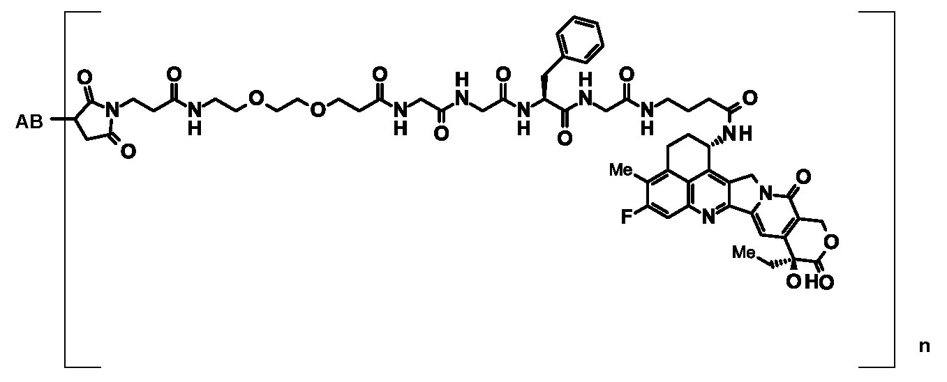
ここで、ABは抗体又は該抗体の機能性断片を示す。nは抗体と結合している薬物-リンカー構造の1抗体あたりの平均結合数を示す。抗体とリンカーは抗体由来のスルフヒドリル基を介して結合している;
[36]以下の式: The antibody-drug conjugate according to any one of [29] to [34], which has a structure represented by:
Here, AB represents an antibody or a functional fragment of the antibody. n represents the average number of bonds per antibody of the drug-linker structure bonded to the antibody. The antibody and linker are linked via a sulfhydryl group from the antibody;
[36] The following formula:

ここで、ABは抗体又は該抗体の機能性断片を示す。nは抗体と結合している薬物-リンカー構造の1抗体あたりの平均結合数を示す。抗体とリンカーは抗体由来のスルフヒドリル基を介して結合している;
[37]抗体が以下の(1)~(4):
(1)配列番号61の21~233番目のアミノ酸配列からなる軽鎖及び配列番号69の20~471番目のアミノ酸配列からなる重鎖、
(2)配列番号61の21~233番目のアミノ酸配列からなる軽鎖及び配列番号73の20~471番目のアミノ酸配列からなる重鎖、
(3)配列番号65の21~233番目のアミノ酸配列からなる軽鎖及び配列番号73の20~471番目のアミノ酸配列からなる重鎖、又は
(4)配列番号61の21~233番目のアミノ酸配列からなる軽鎖及び配列番号77の20~471番目のアミノ酸配列からなる重鎖、
からなる群から選択されるいずれか1つに記載の軽鎖及び重鎖を含む抗体又は当該抗体の機能性断片である、[29]~[36]のいずれか1項に記載の抗体-薬物コンジュゲート;
[38]抗体が、配列番号61の21~233番目のアミノ酸配列からなる軽鎖及び配列番号69の20~471番目のアミノ酸配列からなる重鎖を含む抗体又は当該抗体の機能性断片である、[37]に記載の抗体-薬物コンジュゲート;
[39]抗体が、配列番号61の21~233番目のアミノ酸配列からなる軽鎖及び配列番号77の20~471番目のアミノ酸配列からなる重鎖を含む抗体又は当該抗体の機能性断片である、[37]に記載の抗体-薬物コンジュゲート;
[40]重鎖又は軽鎖が、N-結合への糖鎖付加、O-結合への糖鎖付加、N末のプロセッシング、C末のプロセッシング、脱アミド化、アスパラギン酸の異性化、メチオニンの酸化、N末にメチオニン残基の付加、プロリン残基のアミド化、N末グルタミンもしくはN末グルタミン酸のピログルタミン酸化及びカルボキシル末端における1つ又は2つのアミノ酸欠失からなる群から選択される1又は2以上の修飾をうけた、[29]~[39]のいずれか1項に記載の抗体-薬物コンジュゲート;
[41]選択された1種の薬物-リンカー構造の1抗体あたりの平均結合数が1~10個の範囲である[29]~[40]のいずれか1項に記載の抗体-薬物コンジュゲート;
[42]選択された1種の薬物-リンカー構造の1抗体あたりの平均結合数が2~8個の範囲である[41]に記載の抗体-薬物コンジュゲート;
[43]選択された1種の薬物-リンカー構造の1抗体あたりの平均結合数が5~8個の範囲である[42]に記載の抗体-薬物コンジュゲート;
[44]選択された1種の薬物-リンカー構造の1抗体あたりの平均結合数が7~8個である[43]に記載の抗体-薬物コンジュゲート;
[45][29]~[44]のいずれか1項に記載の抗体-薬物コンジュゲート、その塩、又はそれらの水和物を含むことを特徴とする医薬組成物;
[46]抗腫瘍薬であることを特徴とする、[45]に記載の医薬組成物;
[47]腫瘍がCDH6を発現する腫瘍であることを特徴とする、[46]に記載の医薬組成物;
[48]腫瘍が、腎細胞癌、腎淡明細胞癌、乳頭状腎細胞癌、卵巣癌、卵巣漿液性腺癌、甲状腺癌、胆管癌、肺癌、小細胞肺癌、神経膠芽腫、中皮腫、子宮癌、膵臓癌、ウィルムス腫瘍又は神経芽腫であることを特徴とする、[46]又は[47]に記載の医薬組成物;
[49][29]~[44]のいずれか1項に記載の抗体-薬物コンジュゲート、その塩、又はそれらの水和物から選択されるいずれかを個体に投与することを特徴とする腫瘍の治療方法;
[50]腫瘍がCDH6が発現している腫瘍であることを特徴とする、[49]に記載の治療方法;
[51]腫瘍が、腎細胞癌、腎淡明細胞癌、乳頭状腎細胞癌、卵巣癌、卵巣漿液性腺癌、甲状腺癌、胆管癌、肺癌、小細胞肺癌、神経膠芽腫、中皮腫、子宮癌、膵臓癌、ウィルムス腫瘍又は神経芽腫であることを特徴とする、[49]又は[50]に記載の治療方法;
[52][29]~[44]のいずれか1項に記載の抗体-薬物コンジュゲート、その塩、又はそれらの水和物から選択される少なくとも一つを含む医薬組成物及び少なくとも一つの抗腫瘍薬を、同時に、別々に又は連続して個体に投与することを特徴とする腫瘍の治療方法;
[53][1]~[13]及び[24]~[28]からなる群から選択されるいずれか1項に記載の抗体又は当該抗体の機能性断片或いは[23]に記載の製造方法で得られる抗体又は当該抗体の機能性断片と、薬物-リンカー中間化合物を反応させる工程を含むことを特徴とする、抗体-薬物コンジュゲートの製造方法;又は
[54][21]又は[22]に記載の宿主細胞を培養する工程、当該工程で得られた培養物から目的の抗体又は当該抗体の機能性断片を採取する工程、及び当該工程で得られた抗体又は当該抗体の機能性断片と薬物-リンカー中間化合物を反応させる工程を含むことを特徴とする、抗体-薬物コンジュゲートの製造方法。 The antibody-drug conjugate according to any one of [29] to [34], which has a structure represented by:
Here, AB represents an antibody or a functional fragment of the antibody. n represents the average number of bonds per antibody of the drug-linker structure bonded to the antibody. The antibody and linker are linked via a sulfhydryl group from the antibody;
[37] The antibody is any of the following (1) to (4):
(1) a light chain consisting of amino acid sequences 21 to 233 of SEQ ID NO: 61 and a heavy chain consisting of
(2) a light chain consisting of amino acid sequences 21 to 233 of SEQ ID NO: 61 and a heavy chain consisting of
(3) a light chain comprising the 21st to 233rd amino acid sequence of SEQ ID NO: 65 and a heavy chain comprising the 20th to 471st amino acid sequence of SEQ ID NO: 73, or (4) the 21st to 233rd amino acid sequence of SEQ ID NO: 61 A heavy chain consisting of the 20th to 471st amino acid sequence of SEQ ID NO: 77
The antibody-drug according to any one of [29] to [36], which is an antibody comprising a light chain and a heavy chain according to any one selected from the group consisting of: Conjugates;
[38] The antibody is an antibody or a functional fragment of the antibody comprising a light chain consisting of the 21st to 233rd amino acid sequence of SEQ ID NO: 61 and a heavy chain consisting of the 20th to 471st amino acid sequence of SEQ ID NO: 69. The antibody-drug conjugate according to [37];
[39] The antibody is an antibody comprising a light chain consisting of the 21st to 233rd amino acid sequence of SEQ ID NO: 61 and a heavy chain consisting of the 20th to 471st amino acid sequence of SEQ ID NO: 77, or a functional fragment of the antibody. The antibody-drug conjugate according to [37];
[40] Heavy chain or light chain is glycosylated to N-linked, glycosylated to O-linked, N-terminal processing, C-terminal processing, deamidation, isomerization of aspartic acid,
[41] The antibody-drug conjugate according to any one of [29] to [40], wherein the average number of bonds per antibody of one selected drug-linker structure is in the range of 1 to 10 ;
[42] The antibody-drug conjugate according to [41], wherein the average number of bonds per antibody of one selected drug-linker structure is in the range of 2 to 8;
[43] The antibody-drug conjugate according to [42], wherein the average number of bonds per antibody of one selected drug-linker structure is in the range of 5 to 8;
[44] The antibody-drug conjugate according to [43], wherein the average number of bonds per antibody of one selected drug-linker structure is 7 to 8;
[45] A pharmaceutical composition comprising the antibody-drug conjugate according to any one of [29] to [44], a salt thereof, or a hydrate thereof;
[46] The pharmaceutical composition according to [45], which is an antitumor agent;
[47] The pharmaceutical composition according to [46], wherein the tumor is a tumor that expresses CDH6;
[48] The tumor is renal cell carcinoma, clear cell renal cell carcinoma, papillary renal cell carcinoma, ovarian cancer, ovarian serous adenocarcinoma, thyroid cancer, bile duct cancer, lung cancer, small cell lung cancer, glioblastoma, mesothelioma A pharmaceutical composition according to [46] or [47], which is uterine cancer, pancreatic cancer, Wilms tumor or neuroblastoma;
[49] A tumor characterized by administering to an individual one of the antibody-drug conjugates according to any one of [29] to [44], a salt thereof, or a hydrate thereof Methods of treatment;
[50] The treatment method according to [49], wherein the tumor is a tumor expressing CDH6;
[51] Tumor is renal cell carcinoma, clear cell renal cell carcinoma, papillary renal cell carcinoma, ovarian cancer, ovarian serous adenocarcinoma, thyroid cancer, bile duct cancer, lung cancer, small cell lung cancer, glioblastoma, mesothelioma A method of treatment according to [49] or [50], which is uterine cancer, pancreatic cancer, Wilms tumor or neuroblastoma;
[52] A pharmaceutical composition comprising at least one selected from the antibody-drug conjugate according to any one of [29] to [44], a salt thereof, or a hydrate thereof, and at least one anti-antibody A method for treating a tumor, comprising administering a tumor drug to an individual simultaneously, separately or sequentially;
[53] The antibody according to any one of the above items selected from the group consisting of [1] to [13] and [24] to [28], or the functional fragment of the antibody, or the production method according to [23] A method for producing an antibody-drug conjugate, comprising a step of reacting the obtained antibody or a functional fragment of the antibody with a drug-linker intermediate compound; or [54] [21] or [22] A step of culturing the described host cell, a step of collecting a target antibody or a functional fragment of the antibody from the culture obtained in the step, and an antibody or a functional fragment of the antibody and a drug obtained in the step A method for producing an antibody-drug conjugate, comprising a step of reacting a linker intermediate compound.
カドヘリンは、細胞膜表面に存在する糖タンパク質で、カルシウムイオン依存的にN末端側の細胞外ドメイン同士が結合することで、細胞間接着分子として、また細胞間相互作用を担うシグナル分子として機能する。カドヘリンスーパーファミリーの内、クラシックカドヘリンに分類される分子群は、細胞外に5個の細胞外ドメイン(ECドメイン)と1個の膜貫通領域、及び細胞内ドメインから構成される1回膜貫通タンパク質である。 1. CDH6
Cadherin is a glycoprotein present on the cell membrane surface, and functions as an intercellular adhesion molecule and a signal molecule responsible for cell-cell interaction by binding N-terminal extracellular domains in a calcium ion-dependent manner. Among the cadherin superfamily, the molecular group classified as classic cadherin is a single-transmembrane protein composed of five extracellular domains (EC domain), one transmembrane region, and an intracellular domain. It is.
本発明の抗CDH6抗体の一例として、配列番号4に示すアミノ酸配列を含むアミノ酸配列を認識し、かつ内在化活性を有する抗CDH6抗体を挙げることができる。本発明の抗CDH6抗体の一例として、配列番号4に示すアミノ酸配列を含むアミノ酸配列を特異的に認識し、かつ内在化活性を有する抗CDH6抗体を挙げることができる。本発明の抗CDH6抗体の一例として、配列番号4に示すアミノ酸配列からなるアミノ酸配列を認識し、かつ内在化活性を有する抗CDH6抗体を挙げることができる。本発明の抗CDH6抗体の一例として、配列番号4に示すアミノ酸配列からなるアミノ酸配列を特異的に認識し、かつ内在化活性を有する抗CDH6抗体を挙げることができる。抗体が「配列番号4に示すアミノ酸配列を含むアミノ酸配列を特異的に認識」する又は「EC3ドメインを特異的に認識」するとは、抗体が、CDH6のEC3ドメインをCDH6の他の細胞外ドメインと比較して強く認識する又は強く結合することをいう。 2. Production of Anti-CDH6 Antibody As an example of the anti-CDH6 antibody of the present invention, an anti-CDH6 antibody that recognizes an amino acid sequence containing the amino acid sequence shown in SEQ ID NO: 4 and has internalization activity can be mentioned. As an example of the anti-CDH6 antibody of the present invention, an anti-CDH6 antibody that specifically recognizes an amino acid sequence containing the amino acid sequence shown in SEQ ID NO: 4 and has internalization activity can be mentioned. As an example of the anti-CDH6 antibody of the present invention, an anti-CDH6 antibody that recognizes the amino acid sequence consisting of the amino acid sequence shown in SEQ ID NO: 4 and has internalization activity can be mentioned. An example of the anti-CDH6 antibody of the present invention is an anti-CDH6 antibody that specifically recognizes the amino acid sequence consisting of the amino acid sequence shown in SEQ ID NO: 4 and has internalization activity. When an antibody “recognizes specifically an amino acid sequence including the amino acid sequence shown in SEQ ID NO: 4” or “specifically recognizes an EC3 domain”, the antibody binds the EC3 domain of CDH6 to another extracellular domain of CDH6. Compared to strongly recognize or strongly bind.
抗原は抗原タンパク質をコードする遺伝子を遺伝子操作によって宿主細胞に産生させることによって得ることができる。具体的には、抗原遺伝子を発現可能なベクターを作製し、これを宿主細胞に導入して該遺伝子を発現させ、発現した抗原を精製すればよい。上記の遺伝子操作による抗原発現細胞、あるいは抗原を発現している細胞株を動物に免疫する方法を用いることによっても抗体を取得できる。 (1) Preparation of antigen An antigen can be obtained by causing a host cell to produce a gene encoding an antigen protein by genetic manipulation. Specifically, a vector capable of expressing an antigen gene may be prepared, introduced into a host cell to express the gene, and the expressed antigen may be purified. An antibody can also be obtained by using a method for immunizing an animal with an antigen-expressing cell or a cell line expressing the antigen by genetic manipulation as described above.
本発明で使用される抗CDH6抗体は、特に制限はないが、例えば、本願の配列表で示されたアミノ酸配列で特定される抗体を好適に使用することができる。本発明において使用される抗CDH6抗体としては、以下の特性を有するものが望ましい。
(1)以下の特性を有することを特徴とする抗体;
(a)CDH6に特異的に結合する
(b)CDH6と結合することによってCDH6発現細胞に内在化する活性を有する
(2)CDH6がヒトCDH6である上記(1)に記載の抗体又は当該抗体。
(3)ヒトCDH6のEC3を特異的に認識し、かつ内在化活性を有する
本発明のCDH6に対する抗体の取得方法は、抗CDH6抗体を取得できる限りにおいて、特に制限されないが、高次構造を保持したCDH6を抗原として用いることが好ましい。 (2) Production of anti-CDH6 monoclonal antibody The anti-CDH6 antibody used in the present invention is not particularly limited. For example, an antibody specified by the amino acid sequence shown in the sequence listing of the present application can be preferably used. it can. The anti-CDH6 antibody used in the present invention preferably has the following characteristics.
(1) an antibody having the following characteristics;
(A) specifically binding to CDH6 (b) having an activity of internalizing in a CDH6-expressing cell by binding to CDH6 (2) The antibody according to (1) or the antibody, wherein CDH6 is human CDH6.
(3) The method for obtaining an antibody against CDH6 of the present invention that specifically recognizes EC3 of human CDH6 and has internalization activity is not particularly limited as long as an anti-CDH6 antibody can be obtained, but retains a higher-order structure. It is preferable to use CDH6 as an antigen.
(a)CDH6のcDNAを発現ベクター(例えば、pcDNA3.1:Thermo Fisher Scientific)に組み込み、エレクトロポレーションや遺伝子銃等の方法によって、そのベクターを直接被免疫動物(例えば、ラットやマウス)に投与することによって、動物体内においてCDH6を発現させることによって、免疫反応を誘起させることができる。エレクトロポレーション等によるベクターの投与は抗体価を上げるために必要であれば、1回でも複数回でもよく、好ましくは複数回である;
(b)免疫反応を誘起された上述の動物より、抗体産生細胞を含む組織(例えばリンパ節)を採取する;
(c)骨髄腫細胞(以下「ミエローマ」という)(例えば、マウスミエローマSP2/0-ag14細胞)の調製;
(d)抗体産生細胞とミエローマとの細胞融合;
(e)目的とする抗体を産生するハイブリドーマ群の選別;
(f)単一細胞クローンへの分割(クローニング);
(g)場合によっては、モノクローナル抗体を大量に製造するためのハイブリドーマの培養、又はハイブリドーマを移植した動物の飼育;及び/又は
(h)このようにして製造されたモノクローナル抗体の生理活性(内在化活性)、及びその結合特異性の検討、あるいは標識試薬としての特性の検定。 Specific examples of obtaining monoclonal antibodies include the following.
(A) The CDH6 cDNA is incorporated into an expression vector (for example, pcDNA3.1: Thermo Fisher Scientific), and the vector is directly administered to an immunized animal (for example, rat or mouse) by a method such as electroporation or gene gun. Thus, an immune response can be induced by expressing CDH6 in the animal body. Administration of the vector by electroporation or the like may be performed once or a plurality of times, preferably a plurality of times, if necessary to increase the antibody titer;
(B) collecting a tissue (for example, lymph node) containing antibody-producing cells from the above-mentioned animal in which an immune response has been induced;
(C) Preparation of myeloma cells (hereinafter referred to as “myeloma”) (eg, mouse myeloma SP2 / 0-ag14 cells);
(D) cell fusion between antibody-producing cells and myeloma;
(E) selection of a hybridoma group producing the target antibody;
(F) Division into single cell clones (cloning);
(G) In some cases, culturing hybridomas for producing monoclonal antibodies in large quantities, or breeding animals transplanted with hybridomas; and / or (h) physiological activities (internalization of the monoclonal antibodies thus produced) Activity) and its binding specificity, or assay of properties as a labeling reagent.
本発明の抗体には、上記CDH6に対するモノクローナル抗体に加え、ヒトに対する異種抗原性を低下させること等を目的として人為的に改変した遺伝子組換え型抗体、例えば、キメラ(Chimeric)抗体、ヒト化(Humanized)抗体又はヒト抗体等も含まれる。これらの抗体は、既知の方法を用いて製造することができる。 (3) Other antibodies In addition to the monoclonal antibody against CDH6, the antibodies of the present invention include genetically engineered antibodies that have been artificially modified for the purpose of reducing the heterologous antigenicity against humans, such as chimera (Chimeric ) Antibodies, humanized antibodies, human antibodies, and the like. These antibodies can be produced using known methods.


本発明の抗体としては、さらに、CDH6に結合する、ヒト抗体を挙げることができる。抗CDH6ヒト抗体とは、ヒト染色体由来の抗体の遺伝子配列のみを有するヒト抗体を意味する。抗CDH6ヒト抗体は、ヒト抗体の重鎖と軽鎖の遺伝子を含むヒト染色体断片を有するヒト抗体産生マウスを用いた方法(Tomizuka,K.et al.,Nature Genetics(1997)16,p.133-143,;Kuroiwa,Y.et.al.,Nucl.Acids Res.(1998)26,p.3447-3448;Yoshida,H.et.al.,Animal Cell Technology:Basic and Applied Aspects vol.10,p.69-73(Kitagawa,Y.,Matsuda,T.and Iijima,S.eds.),Kluwer Academic Publishers,1999.;Tomizuka,K.et.al.,Proc.Natl.Acad.Sci.USA(2000)97,p.722-727等を参照。)によって取得することができる。 (In this specification, Table 1-1 to Table 1-15 may be collectively referred to as Table 1.)
Examples of the antibody of the present invention further include a human antibody that binds to CDH6. An anti-CDH6 human antibody means a human antibody having only the gene sequence of an antibody derived from a human chromosome. The anti-CDH6 human antibody is obtained by a method using a human antibody-producing mouse having a human chromosome fragment containing the heavy and light chain genes of a human antibody (Tomizuka, K. et al., Nature Genetics (1997) 16, p. 133. Kuroiwa, Y. et al., Nucl. Acids Res. (1998) 26, p.3447-3448; Yoshida, H. et al., Animal Cell Technology: Basic and Applied Aspects vol.10. p. 69-73 (Kitagawa, Y., Matsuda, T. and Iijima, S. eds.), Kluwer Academic Publishers, 1999.; Tomizuka, K. et., Proc. l.Acad.Sci.USA (2000) 97, can be obtained by reference.) The p.722-727 like.
(1)薬物
上記「2.抗CDH6抗体の製造」にて取得された抗CDH6抗体はリンカー構造部分を介して薬物を結合させることによって、抗CDH6抗体-薬物コンジュゲートとすることができる。薬物としては、リンカー構造に結合できる置換基、部分構造を有するものであれば特に制限はない。抗CDH6抗体-薬物コンジュゲートは結合する薬物に応じて種々の用途に用いることができる。そのような薬物の例としては、抗腫瘍活性を有する物質、血液疾患に対する効果を有する物質、自己免疫疾患に対する効果を有する物質、抗炎症物質、抗菌物質、抗真菌物質、抗寄生虫物質、抗ウイルス物質、抗麻酔物質等を挙げることができる。 3. Anti-CDH6 antibody-drug conjugate (1) Drug The anti-CDH6 antibody-drug conjugate obtained by binding the drug to the anti-CDH6 antibody obtained in “2. Production of anti-CDH6 antibody” via a linker structure moiety. It can be. The drug is not particularly limited as long as it has a substituent or a partial structure that can be bonded to the linker structure. Anti-CDH6 antibody-drug conjugates can be used in a variety of applications depending on the drug to be bound. Examples of such drugs include substances having anti-tumor activity, substances having an effect on blood diseases, substances having an effect on autoimmune diseases, anti-inflammatory substances, antibacterial substances, antifungal substances, antiparasitic substances, anti-parasitic substances, Examples include viral substances and anti-anesthetic substances.
本発明の抗CDH6抗体-薬物コンジュゲートに結合される化合物として抗腫瘍性化合物を用いる例について以下に述べる。抗腫瘍性化合物としては、抗腫瘍効果を有する化合物であって、リンカー構造に結合できる置換基、部分構造を有するものであれば特に制限はない。抗腫瘍性化合物は、リンカーの一部又は全部が腫瘍細胞内で切断されて抗腫瘍性化合物部分が遊離されて抗腫瘍効果が発現される。リンカーが薬物との結合部分で切断されれば抗腫瘍性化合物が本来の構造で遊離され、その本来の抗腫瘍効果が発揮される。 (1) -1 Antitumor Compound An example in which an antitumor compound is used as a compound bound to the anti-CDH6 antibody-drug conjugate of the present invention will be described below. The antitumor compound is not particularly limited as long as it is a compound having an antitumor effect and has a substituent and a partial structure that can be bonded to a linker structure. In the antitumor compound, a part or all of the linker is cleaved in the tumor cell to release the antitumor compound portion and to exhibit an antitumor effect. When the linker is cleaved at the binding site with the drug, the antitumor compound is released in its original structure, and its original antitumor effect is exhibited.

本発明の抗CDH6抗体-薬物コンジュゲートにおいて薬物を抗CDH6抗体に結合させるリンカー構造について述べる。 (2) Linker structure A linker structure for binding a drug to an anti-CDH6 antibody in the anti-CDH6 antibody-drug conjugate of the present invention will be described.
-(Succinimid-3-yl-N)-CH2CH2-C(=O)-GGFG-NH-CH2CH2CH2-C(=O)-、
-(Succinimid-3-yl-N)-CH2CH2CH2CH2CH2-C(=O)-GGFG-NH-CH2CH2CH2-C(=O)-、
-(Succinimid-3-yl-N)-CH2CH2CH2CH2CH2-C(=O)-GGFG-NH-CH2-O-CH2-C(=O)-、
-(Succinimid-3-yl-N)-CH2CH2CH2CH2CH2-C(=O)-GGFG-NH-CH2CH2-O-CH2-C(=O)-、
-(Succinimid-3-yl-N)-CH2CH2-C(=O)-NH-CH2CH2O-CH2CH2O-CH2CH2-C(=O)-GGFG-NH-CH2CH2CH2-C(=O)-、
-(Succinimid-3-yl-N)-CH2CH2-C(=O)-NH-CH2CH2O-CH2CH2O-CH2CH2O-CH2CH2O-CH2CH2-C(=O)-GGFG-NH-CH2CH2CH2-C(=O)-。 As another example, for example, a linker structure described in US Patent Publication US2016 / 0297890 (for example, those described in paragraphs [0260] to [0289]) can be mentioned, and those having the following structures are preferable. Can be used for The left end of the structure shown below is a binding site with an antibody, and the right end is a binding site with a drug. Further, GGFG in the following linker structure represents an amino acid sequence connected by a peptide bond consisting of glycine-glycine-phenylalanine-glycine (GGFG).
- (Succinimid-3-yl- N) -
- (Succinimid-3-yl- N) -
-(Succinimid-3-yl-N) -CH 2 CH 2 CH 2 CH 2 CH 2 -C (= O) -GGFG-NH-CH 2 -O-CH 2 -C (= O)-,
- (Succinimid-3-yl- N) -
— (Succinimid-3-yl-N) —CH 2 CH 2 —C (═O) —NH—CH 2 CH 2 O—CH 2 CH 2 O—CH 2 CH 2 —C (═O) —GGFG-NH —CH 2 CH 2 CH 2 —C (═O) —,
— (Succinimid-3-yl-N) —CH 2 CH 2 —C (═O) —NH—CH 2 CH 2 O—CH 2 CH 2 O—CH 2 CH 2 O—CH 2 CH 2 O—CH 2 CH 2 -C (= O) -GGFG -NH-
-(Succinimid-3-yl-N)-CH2CH2CH2CH2CH2-C(=O)-GGFG-NH-CH2-O-CH2-C(=O)-、
-(Succinimid-3-yl-N)-CH2CH2CH2CH2CH2-C(=O)-GGFG-NH-CH2CH2-O-CH2-C(=O)-、
-(Succinimid-3-yl-N)-CH2CH2-C(=O)-NH-CH2CH2O-CH2CH2O-CH2CH2-C(=O)-GGFG-NH-CH2CH2CH2-C(=O)-。
さらにより好ましくは、
-(Succinimid-3-yl-N)-CH2CH2CH2CH2CH2-C(=O)-GGFG-NH-CH2-O-CH2-C(=O)-、
-(Succinimid-3-yl-N)-CH2CH2-C(=O)-NH-CH2CH2O-CH2CH2O-CH2CH2-C(=O)-GGFG-NH-CH2CH2CH2-C(=O)-。
を挙げることができる。 More preferably, the following can be mentioned.
-(Succinimid-3-yl-N) -CH 2 CH 2 CH 2 CH 2 CH 2 -C (= O) -GGFG-NH-CH 2 -O-CH 2 -C (= O)-,
- (Succinimid-3-yl- N) -
— (Succinimid-3-yl-N) —CH 2 CH 2 —C (═O) —NH—CH 2 CH 2 O—CH 2 CH 2 O—CH 2 CH 2 —C (═O) —GGFG-NH —CH 2 CH 2 CH 2 —C (═O) —.
Even more preferably,
-(Succinimid-3-yl-N) -CH 2 CH 2 CH 2 CH 2 CH 2 -C (= O) -GGFG-NH-CH 2 -O-CH 2 -C (= O)-,
— (Succinimid-3-yl-N) —CH 2 CH 2 —C (═O) —NH—CH 2 CH 2 O—CH 2 CH 2 O—CH 2 CH 2 —C (═O) —GGFG-NH —CH 2 CH 2 CH 2 —C (═O) —.
Can be mentioned.

-(Succinimid-3-yl-N)-CH2CH2-C(=O)-GGFG-NH-CH2CH2CH2-C(=O)-(NH-DX)、
-(Succinimid-3-yl-N)-CH2CH2CH2CH2CH2-C(=O)-GGFG-NH-CH2CH2CH2-C(=O)-(NH-DX)、
-(Succinimid-3-yl-N)-CH2CH2CH2CH2CH2-C(=O)-GGFG-NH-CH2-O-CH2-C(=O)-(NH-DX)、
-(Succinimid-3-yl-N)-CH2CH2CH2CH2CH2-C(=O)-GGFG-NH-CH2CH2-O-CH2-C(=O)-(NH-DX)、
-(Succinimid-3-yl-N)-CH2CH2-C(=O)-NH-CH2CH2O-CH2CH2O-CH2CH2-C(=O)-GGFG-NH-CH2CH2CH2-C(=O)-(NH-DX)、
-(Succinimid-3-yl-N)-CH2CH2-C(=O)-NH-CH2CH2O-CH2CH2O-CH2CH2O-CH2CH2O-CH2CH2-C(=O)-GGFG-NH-CH2CH2CH2-C(=O)-(NH-DX)。 In the antibody-drug conjugate of the present invention in which the drug is exatecan, a drug-linker structure portion having the following structure is preferably bound to the antibody. These drug-linker structure moieties may be bound to 1 to 10 as the average number of bonds per antibody, preferably 2 to 8, more preferably 5 to 8, and still more preferably 7 To 8, more preferably 8.
- (Succinimid-3-yl- N) -
-(Succinimid-3-yl-N) -CH 2 CH 2 CH 2 CH 2 CH 2 -C (= O) -GGFG-NH-CH 2 CH 2 CH 2 -C (= O)-(NH-DX) ,
-(Succinimid-3-yl-N) -CH 2 CH 2 CH 2 CH 2 CH 2 -C (= O) -GGFG-NH-CH 2 -O-CH 2 -C (= O)-(NH-DX ),
— (Succinimid-3-yl-N) —CH 2 CH 2 CH 2 CH 2 CH 2 —C (═O) —GGFG—NH—CH 2 CH 2 —O—CH 2 —C (═O) — (NH -DX),
— (Succinimid-3-yl-N) —CH 2 CH 2 —C (═O) —NH—CH 2 CH 2 O—CH 2 CH 2 O—CH 2 CH 2 —C (═O) —GGFG-NH —CH 2 CH 2 CH 2 —C (═O) — (NH—DX),
— (Succinimid-3-yl-N) —CH 2 CH 2 —C (═O) —NH—CH 2 CH 2 O—CH 2 CH 2 O—CH 2 CH 2 O—CH 2 CH 2 O—CH 2 CH 2 -C (= O) -GGFG -NH-
-(Succinimid-3-yl-N)-CH2CH2CH2CH2CH2-C(=O)-GGFG-NH-CH2-O-CH2-C(=O)-(NH-DX)、
-(Succinimid-3-yl-N)-CH2CH2CH2CH2CH2-C(=O)-GGFG-NH-CH2CH2-O-CH2-C(=O)-(NH-DX)、
-(Succinimid-3-yl-N)-CH2CH2-C(=O)-NH-CH2CH2O-CH2CH2O-CH2CH2-C(=O)-GGFG-NH-CH2CH2CH2-C(=O)-(NH-DX)。 More preferably, it is as follows.
-(Succinimid-3-yl-N) -CH 2 CH 2 CH 2 CH 2 CH 2 -C (= O) -GGFG-NH-CH 2 -O-CH 2 -C (= O)-(NH-DX ),
— (Succinimid-3-yl-N) —CH 2 CH 2 CH 2 CH 2 CH 2 —C (═O) —GGFG—NH—CH 2 CH 2 —O—CH 2 —C (═O) — (NH -DX),
— (Succinimid-3-yl-N) —CH 2 CH 2 —C (═O) —NH—CH 2 CH 2 O—CH 2 CH 2 O—CH 2 CH 2 —C (═O) —GGFG-NH —CH 2 CH 2 CH 2 —C (═O) — (NH—DX).
-(Succinimid-3-yl-N)-CH2CH2CH2CH2CH2-C(=O)-GGFG-NH-CH2-O-CH2-C(=O)-(NH-DX)
-(Succinimid-3-yl-N)-CH2CH2-C(=O)-NH-CH2CH2O-CH2CH2O-CH2CH2-C(=O)-GGFG-NH-CH2CH2CH2-C(=O)-(NH-DX)。 More preferably, the following can be mentioned.
-(Succinimid-3-yl-N) -CH 2 CH 2 CH 2 CH 2 CH 2 -C (= O) -GGFG-NH-CH 2 -O-CH 2 -C (= O)-(NH-DX )
— (Succinimid-3-yl-N) —CH 2 CH 2 —C (═O) —NH—CH 2 CH 2 O—CH 2 CH 2 O—CH 2 CH 2 —C (═O) —GGFG-NH —CH 2 CH 2 CH 2 —C (═O) — (NH—DX).

本発明の抗体ー薬物コンジュゲートに用いることができる抗体は、上記「2.抗CDH6抗体の製造」の項及び実施例に記載の内在化活性を有する抗CDH6抗体及び該抗体の機能性断片であれば特に制限がない。 (3) Method for Producing Antibody-Drug Conjugate An antibody that can be used in the antibody-drug conjugate of the present invention has an internalization activity described in the above-mentioned section “2. Production of anti-CDH6 antibody” and Examples. There is no particular limitation as long as it is an anti-CDH6 antibody and a functional fragment of the antibody.
下記式(1)で示される抗体-薬物コンジュゲートのうち、チオエーテルを介して抗CDH6抗体とリンカー構造が結合しているものは抗CDH6抗体を還元してジスルフィド結合をスルヒドリル基に変換した抗体に対して、既知の方法によって入手しうる化合物(2)(例えば、US2016/297890号公開特許公報に記載の方法(例えば段落[0336]~[0374]に記載の方法)で入手可能)を反応させることによって製造することができる。例えば下記の方法によって製造することができる。 (3) -1
Among the antibody-drug conjugates represented by the following formula (1), those in which the anti-CDH6 antibody and the linker structure are bonded via a thioether are reduced to anti-CDH6 antibodies to convert the disulfide bonds into sulfhydryl groups. On the other hand, the compound (2) that can be obtained by a known method (for example, the method described in US2016 / 297890 published patent publication (for example, the method described in paragraphs [0336] to [0374]) is reacted. Can be manufactured. For example, it can be produced by the following method.

ここで、L1は、
-(Succinimid-3-yl-N)-の構造で示される。
L1’は次式で示される、マレイミジル基を示す。] [Wherein, AB represents an antibody having a sulfhydryl group.
Where L 1 is
It is represented by the structure of-(Succinimid-3-yl-N)-.
L 1 ′ represents a maleimidyl group represented by the following formula. ]
-(Succinimid-3-yl-N)-CH2CH2-C(=O)-GGFG-NH-CH2CH2CH2-C(=O)-、
-(Succinimid-3-yl-N)-CH2CH2CH2CH2CH2-C(=O)-GGFG-NH-CH2CH2CH2-C(=O)-、
-(Succinimid-3-yl-N)-CH2CH2CH2CH2CH2-C(=O)-GGFG-NH-CH2-O-CH2-C(=O)-、
-(Succinimid-3-yl-N)-CH2CH2CH2CH2CH2-C(=O)-GGFG-NH-CH2CH2-O-CH2-C(=O)-、
-(Succinimid-3-yl-N)-CH2CH2-C(=O)-NH-CH2CH2O-CH2CH2O-CH2CH2-C(=O)-GGFG-NH-CH2CH2CH2-C(=O)-、
-(Succinimid-3-yl-N)-CH2CH2-C(=O)-NH-CH2CH2O-CH2CH2O-CH2CH2O-CH2CH2O-CH2CH2-C(=O)-GGFG-NH-CH2CH2CH2-C(=O)-。 -L 1 -L X has any structure represented by the following formula.
- (Succinimid-3-yl- N) -
- (Succinimid-3-yl- N) -
-(Succinimid-3-yl-N) -CH 2 CH 2 CH 2 CH 2 CH 2 -C (= O) -GGFG-NH-CH 2 -O-CH 2 -C (= O)-,
- (Succinimid-3-yl- N) -
— (Succinimid-3-yl-N) —CH 2 CH 2 —C (═O) —NH—CH 2 CH 2 O—CH 2 CH 2 O—CH 2 CH 2 —C (═O) —GGFG-NH —CH 2 CH 2 CH 2 —C (═O) —,
— (Succinimid-3-yl-N) —CH 2 CH 2 —C (═O) —NH—CH 2 CH 2 O—CH 2 CH 2 O—CH 2 CH 2 O—CH 2 CH 2 O—CH 2 CH 2 -C (= O) -GGFG -NH-
-(Succinimid-3-yl-N)-CH2CH2CH2CH2CH2-C(=O)-GGFG-NH-CH2-O-CH2-C(=O)-、
-(Succinimid-3-yl-N)-CH2CH2CH2CH2CH2-C(=O)-GGFG-NH-CH2CH2-O-CH2-C(=O)-、
-(Succinimid-3-yl-N)-CH2CH2-C(=O)-NH-CH2CH2O-CH2CH2O-CH2CH2-C(=O)-GGFG-NH-CH2CH2CH2-C(=O)-。 Of these, the following are more preferable.
-(Succinimid-3-yl-N) -CH 2 CH 2 CH 2 CH 2 CH 2 -C (= O) -GGFG-NH-CH 2 -O-CH 2 -C (= O)-,
- (Succinimid-3-yl- N) -
— (Succinimid-3-yl-N) —CH 2 CH 2 —C (═O) —NH—CH 2 CH 2 O—CH 2 CH 2 O—CH 2 CH 2 —C (═O) —GGFG-NH —CH 2 CH 2 CH 2 —C (═O) —.
-(Succinimid-3-yl-N)-CH2CH2CH2CH2CH2-C(=O)-GGFG-NH-CH2-O-CH2-C(=O)-、
-(Succinimid-3-yl-N)-CH2CH2-C(=O)-NH-CH2CH2O-CH2CH2O-CH2CH2-C(=O)-GGFG-NH-CH2CH2CH2-C(=O)-。 Furthermore, preferably, the following can be mentioned.
-(Succinimid-3-yl-N) -CH 2 CH 2 CH 2 CH 2 CH 2 -C (= O) -GGFG-NH-CH 2 -O-CH 2 -C (= O)-,
— (Succinimid-3-yl-N) —CH 2 CH 2 —C (═O) —NH—CH 2 CH 2 O—CH 2 CH 2 O—CH 2 CH 2 —C (═O) —GGFG-NH —CH 2 CH 2 CH 2 —C (═O) —.
製造した抗体-薬物コンジュゲート(1)は、以下の共通操作によって濃縮、バッファー交換、精製、抗体濃度及び抗体一分子あたりの薬物平均結合数の測定を行い、抗体-薬物コンジュゲート(1)の同定を行うことができる。 (4) Identification of antibody-drug conjugate The produced antibody-drug conjugate (1) was measured for concentration, buffer exchange, purification, antibody concentration, and average number of drugs per antibody molecule by the following common operations. The antibody-drug conjugate (1) can be identified.
Amicon Ultra(50,000 MWCO,Millipore Corporation)の容器内に抗体もしくは抗体-薬物コンジュゲート溶液を入れ、遠心機(Allegra X-15R,Beckman Coulter,Inc.)を用いた遠心操作(2000G乃至3800Gにて5乃至20分間遠心)にて、抗体もしくは抗体-薬物コンジュゲート溶液を濃縮した。 (4) -1 Common operation A: Concentration of antibody or antibody-drug conjugate aqueous solution Place the antibody or antibody-drug conjugate solution in a container of Amicon Ultra (50,000 MWCO, Millipore Corporation), and centrifuge (Allegra X The antibody or antibody-drug conjugate solution was concentrated by centrifugation using -15R, Beckman Coulter, Inc. (centrifuged at 2000 G to 3800 G for 5 to 20 minutes).
UV測定器(Nanodrop 1000,Thermo Fisher Scientific Inc.)を用いて、メーカー規定の方法に従い、抗体濃度の測定を行った。その際に、抗体ごとに異なる280nm吸光係数(1.3mLmg-1cm-1乃至1.8mLmg-1cm-1)を用いた。 (4) -2 Common operation B: Antibody concentration measurement The antibody concentration was measured using a UV measuring instrument (
Sephadex G-25担体を使用したNAP-25カラム(Cat.No.17-0852-02,GE Healthcare Japan Corporation)を、メーカー規定の方法に従い、塩化ナトリウム(50mM)及びEDTA(2mM)を含むリン酸緩衝液(50mM,pH6.0)(本明細書でPBS6.0/EDTAと称する。)にて平衡化させた。このNAP-25カラム一本につき、抗体水溶液2.5mLをのせたのち、PBS6.0/EDTA3.5mLで溶出させた画分(3.5mL)を分取した。この画分を共通操作Aによって濃縮し、共通操作Bを用いて抗体濃度の測定を行ったのちに、PBS6.0/EDTAを用いて20mg/mLに抗体濃度を調整した。 (4) -3 Common operation C: Buffer exchange of antibody A NAP-25 column (Cat. No. 17-0852-02, GE Healthcare Japan Corporation) using Sephadex G-25 carrier was chlorinated according to the method specified by the manufacturer. Equilibration was performed with a phosphate buffer (50 mM, pH 6.0) containing sodium (50 mM) and EDTA (2 mM) (referred to herein as PBS 6.0 / EDTA). For each NAP-25 column, 2.5 mL of the antibody aqueous solution was placed, and then the fraction eluted with 3.5 mL of PBS 6.0 / EDTA (3.5 mL) was collected. This fraction was concentrated by common operation A, the antibody concentration was measured using common operation B, and then the antibody concentration was adjusted to 20 mg / mL using PBS 6.0 / EDTA.
市販のSorbitol(5%)を含む酢酸緩衝液(10mM,pH5.5;本明細書でABSと称する。)のいずれかの緩衝液でNAP-25カラムを平衡化させた。このNAP-25カラムに、抗体-薬物コンジュゲート反応水溶液(約2.5mL)をのせ、メーカー規定の量の緩衝液で溶出させることで、抗体画分を分取した。この分取画分を再びNAP-25カラムにのせ緩衝液で溶出させるゲルろ過精製操作を計2乃至3回繰り返すことで、未結合の薬物リンカーや低分子化合物(トリス(2-カルボキシエチル)ホスフィン塩酸塩(TCEP),N-アセチル-L-システイン(NAC),ジメチルスルホキシド)を除いた抗体-薬物コンジュゲートを得た。 (4) -4 Common procedure D: Purification of antibody-drug conjugate Any buffer solution of acetate buffer (10 mM, pH 5.5; referred to herein as ABS) containing commercially available Sorbitol (5%). Equilibrated the NAP-25 column. An antibody-drug conjugate aqueous solution (about 2.5 mL) was placed on this NAP-25 column, and eluted with a buffer solution specified by the manufacturer to fractionate the antibody fraction. The gel filtration purification operation, in which this fractionated fraction is again applied to the NAP-25 column and eluted with a buffer solution, is repeated 2 to 3 times in total, thereby allowing unbound drug linkers and low molecular weight compounds (tris (2-carboxyethyl) phosphine). Antibody-drug conjugates without the hydrochloride (TCEP), N-acetyl-L-cysteine (NAC), dimethyl sulfoxide) were obtained.
抗体-薬物コンジュゲートにおける結合薬物濃度は、抗体-薬物コンジュゲート水溶液の280nm及び370nmの二波長におけるUV吸光度を測定したのちに下記の計算を行うことで、算出することができる。 (4) -5 Common Operation E: Measurement of Antibody Concentration in Antibody-Drug Conjugate and Average Drug Binding Number per Antibody Molecule The binding drug concentration in the antibody-drug conjugate is 280 nm and 370 nm of the antibody-drug conjugate aqueous solution. It can be calculated by performing the following calculation after measuring the UV absorbance at the two wavelengths.
A280=AD,280+AA,280=εD,280CD+εA,280CA 式(1)
A370=AD,370+AA,370=εD,370CD+εA,370CA 式(2)
ここで、A280は280nmにおける抗体-薬物コンジュゲート水溶液の吸光度を示し、A370は370nmにおける抗体-薬物コンジュゲート水溶液の吸光度を示し、AA,280は280nmにおける抗体の吸光度を示し、AA,370は370nmにおける抗体の吸光度を示し、AD,280は280nmにおけるコンジュゲート前駆体の吸光度を示し、AD,370は370nmにおけるコンジュゲート前駆体の吸光度を示し、εA,280は280nmにおける抗体のモル吸光係数を示し、εA,370は370nmにおける抗体のモル吸光係数を示し、εD,280は280nmにおけるコンジュゲート前駆体のモル吸光係数を示し、εD,370は370nmにおけるコンジュゲート前駆体のモル吸光係数を示し、CAは抗体-薬物コンジュゲートにおける抗体濃度を示し、CDは抗体-薬物コンジュゲートにおける薬物濃度を示す。 The total absorbance at a certain wavelength is equal to the sum of the absorbances of all absorbing species present in the system [addition of absorbance], so the molar extinction coefficient of the antibody and drug changes before and after conjugation of the antibody and drug. Assuming that there is no antibody, the antibody concentration and the drug concentration in the antibody-drug conjugate are expressed by the following relational expression.
A 280 = A D, 280 + A A, 280 = ε D, 280 C D + ε A, 280 C A Formula (1)
A 370 = A D, 370 + A A, 370 = ε D, 370 C D + ε A, 370 C A Formula (2)
Here, A 280 represents the absorbance of the antibody-drug conjugate aqueous solution at 280 nm , A 370 represents the absorbance of the antibody-drug conjugate aqueous solution at 370 nm, A A, 280 represents the absorbance of the antibody at 280 nm, and A A , 370 indicates the absorbance of the antibody at 370 nm, AD, 280 indicates the absorbance of the conjugate precursor at 280 nm, AD, 370 indicates the absorbance of the conjugate precursor at 370 nm, and ε A, 280 is at 280 nm. Shows the molar extinction coefficient of the antibody, ε A, 370 shows the molar extinction coefficient of the antibody at 370 nm, ε D, 280 shows the molar extinction coefficient of the conjugate precursor at 280 nm, and ε D, 370 shows the conjugate at 370 nm. Shows the molar extinction coefficient of the precursor, C A antibody - indicates antibody concentration in drug conjugates, C D antibody - shows the drug concentration in the drug conjugate.
抗体-薬物コンジュゲートにおける抗体一分子あたりの薬物平均結合数は、前述の「(4)-5 共通操作E」に加え、以下の方法を用いる高速液体クロマトグラフィー(HPLC)分析によっても求めることができる。以下に、抗体と薬物リンカーがジスルフィド結合している場合のHPLCによる薬物平均結合数の測定方法を記載する。当業者は、この方法を参照して、抗体と薬物リンカーとの結合様式に依存して適宜、HPLCにより薬物平均結合数を測定し得る。 (4) -6 Common operation F: Measurement of average drug binding number per antibody molecule in antibody-drug conjugate (2)
The average number of drugs per antibody molecule in the antibody-drug conjugate can be determined by high performance liquid chromatography (HPLC) analysis using the following method in addition to the above-mentioned “(4) -5 common operation E”. it can. The following describes a method for measuring the average number of drugs bonded by HPLC when the antibody and drug linker are disulfide bonded. One skilled in the art can refer to this method and determine the average number of drugs bound by HPLC as appropriate depending on the mode of binding between the antibody and the drug linker.
抗体-薬物コンジュゲート溶液(約1mg/mL、60μL)をジチオトレイトール(DTT)水溶液(100mM、15μL)と混合する。混合物を37℃で30分インキュベートすることで、抗体-薬物コンジュゲートの軽鎖及び重鎖間のジスルフィド結合を切断したサンプルを、HPLC分析に用いる。 F-1. Sample preparation for HPLC analysis (reduction of antibody-drug conjugate)
Antibody-drug conjugate solution (approximately 1 mg / mL, 60 μL) is mixed with an aqueous dithiothreitol (DTT) solution (100 mM, 15 μL). Samples that cleave the disulfide bond between the light and heavy chains of the antibody-drug conjugate by incubating the mixture at 37 ° C. for 30 minutes are used for HPLC analysis.
HPLC分析を、下記の測定条件にて行う。 F-2. HPLC analysis HPLC analysis is performed under the following measurement conditions.
検出器:紫外吸光度計(測定波長:280nm)
カラム:ACQUITY UPLC BEH Phenyl(2.1×50mm、1.7μm、130Å;Waters、P/N 186002884)
カラム温度:80℃
移動相A:0.10%トリフルオロ酢酸(TFA)、15%2-プロパノールを含む水溶液
移動相B:0.075%TFA、15%2-プロパノールを含むアセトニトリル溶液
グラジエントプログラム:14%-36%(0分-15分)、36%-80%(15分-17分)、80%-14%(17分―17.01分)、14%(17.01分―25分)
サンプル注入量:10μL
F-3.データ解析
F-3-1 薬物の結合していない抗体の軽鎖(L0)及び重鎖(H0)に対して、薬物の結合した軽鎖(薬物がi個結合した軽鎖:Li)及び重鎖(薬物がi個結合した重鎖:Hi)は、結合した薬物の数に比例して疎水性が増し保持時間が大きくなることから、例えば、L0、L1、H0、H1、H2、H3の順に溶出される。L0及びH0との保持時間比較により検出ピークをL0、L1、H0、H1、H2、H3のいずれかに割り当てることができる。薬物結合数は、当業者によって定義され得るが、好ましくは、L0、L1、H0、H1、H2、H3である。 HPLC system: Agilent 1290 HPLC system (Agilent Technologies)
Detector: UV absorbance meter (measurement wavelength: 280 nm)
Column: ACQUITY UPLC BEH Phenyl (2.1 × 50 mm, 1.7 μm, 130 cm; Waters, P / N 186002884)
Column temperature: 80 ° C
Mobile phase A: aqueous solution containing 0.10% trifluoroacetic acid (TFA), 15% 2-propanol Mobile phase B: acetonitrile solution containing 0.075% TFA, 15% 2-propanol Gradient program: 14% -36% (0 min-15 min), 36% -80% (15 min-17 min), 80% -14% (17 min-17.01 min), 14% (17.01 min-25 min)
Sample injection volume: 10 μL
F-3. Data Analysis F-3-1 The light chain (L0) and heavy chain (H0) of the antibody to which no drug is bound, and the light chain to which the drug is bound (light chain to which i drugs are bound: L i ) and Since the heavy chain (heavy chain with i number of drugs: H i ) increases in hydrophobicity and increases the retention time in proportion to the number of drugs bound, for example, L0, L1, H0, H1, H2, It is eluted in the order of H3. The detection peak can be assigned to any one of L0, L1, H0, H1, H2, and H3 by comparing the holding times with L0 and H0. The number of drug bonds can be defined by those skilled in the art, but is preferably L0, L1, H0, H1, H2, H3.


なお、抗体-薬物コンジュゲートの量を確保するために、同様な条件で作製して得られた平均薬物数が同程度の複数の抗体-薬物コンジュゲート(例えば±1程度)を混合して新たなロットにすることができる。その場合、平均薬物数は混合前の平均薬物数の間に収まる。 Drug Mean number of bonds = (L 0 peak area ratio x0 + L 1 peak area ratio x1 + H 0 peak area ratio x0 + H 1 peak area ratio x1 + H 2 peak area ratio x2 + H 3 Peak area ratio x3) / 100X2
In order to secure the amount of antibody-drug conjugate, a plurality of antibody-drug conjugates (for example, about ± 1) having the same average number of drugs prepared under the same conditions are mixed and newly added. You can make a lot. In that case, the average number of drugs falls within the average number of drugs before mixing.


(a)配列番号61に示される軽鎖全長アミノ酸配列の21~233番目のアミノ酸配列からなる軽鎖及び配列番号69に示される重鎖全長アミノ酸配列の20~471番目のアミノ酸配列からなる重鎖からなる抗体;
(b)配列番号61に示される軽鎖全長アミノ酸配列の21~233番目のアミノ酸配列からなる軽鎖及び配列番号73に示される重鎖全長アミノ酸配列の20~471番目のアミノ酸配列からなる重鎖からなる抗体;
(c)配列番号61に示される軽鎖全長アミノ酸配列の21~233番目のアミノ酸配列からなる軽鎖及び配列番号77に示される重鎖全長アミノ酸配列の20~471番目のアミノ酸配列からなる重鎖からなる抗体;
(d)配列番号65に示される軽鎖全長アミノ酸配列の21~233番目のアミノ酸配列からなる軽鎖及び配列番号69に示される重鎖全長アミノ酸配列の20~471番目のアミノ酸配列からなる重鎖からなる抗体;
(e)配列番号65に示される軽鎖全長アミノ酸配列の21~233番目のアミノ酸配列からなる軽鎖及び配列番号73に示される重鎖全長アミノ酸配列の20~471番目のアミノ酸配列からなる重鎖からなる抗体;
(f)配列番号65に示される軽鎖全長アミノ酸配列の21~233番目のアミノ酸配列からなる軽鎖及び配列番号77に示される重鎖全長アミノ酸配列の20~471番目のアミノ酸配列からなる重鎖からなる抗体;又は
(g)重鎖又は軽鎖がN-結合への糖鎖付加、O-結合への糖鎖付加、N末のプロセッシング、C末のプロセッシング、脱アミド化、アスパラギン酸の異性化、メチオニンの酸化、N末にメチオニン残基の付加、プロリン残基のアミド化、N末グルタミンやN末グルタミン酸のピログルタミン酸化などに代表される翻訳後修飾、及びカルボキシル末端における1つ又は2つのアミノ酸欠失からなる群より選択される1又は2以上の修飾を含む、(a)~(f)からなる群から選択されるいずれか1つに記載の抗体。 As an example of the antibody drug-conjugate of the present invention, in the structure represented by the above formula [Chemical Formula 9] or [Chemical Formula 10], the antibody represented by AB is selected from the group consisting of the following (a) to (g) An antibody-drug conjugate or a pharmacologically acceptable salt thereof comprising the heavy chain and light chain antibody or functional fragment thereof according to any one of the following:
(A) a light chain comprising the 21st to 233rd amino acid sequences of the light chain full length amino acid sequence represented by SEQ ID NO: 61 and a heavy chain comprising the 20th to 471st amino acid sequences of the heavy chain full length amino acid sequence represented by SEQ ID NO: 69 An antibody consisting of;
(B) a light chain consisting of the 21st to 233rd amino acid sequences of the light chain full length amino acid sequence shown in SEQ ID NO: 61 and a heavy chain consisting of the 20th to 471st amino acid sequences of the heavy chain full length amino acid sequence shown in SEQ ID NO: 73 An antibody consisting of;
(C) a light chain comprising the 21st to 233rd amino acid sequences of the light chain full length amino acid sequence represented by SEQ ID NO: 61 and a heavy chain comprising the 20th to 471st amino acid sequences of the heavy chain full length amino acid sequence represented by SEQ ID NO: 77 An antibody consisting of;
(D) a light chain consisting of the 21st to 233rd amino acid sequences of the light chain full length amino acid sequence shown in SEQ ID NO: 65 and a heavy chain consisting of the 20th to 471st amino acid sequences of the heavy chain full length amino acid sequence shown in SEQ ID NO: 69 An antibody consisting of;
(E) a light chain consisting of the 21st to 233rd amino acid sequences of the light chain full length amino acid sequence shown in SEQ ID NO: 65 and a heavy chain consisting of the 20th to 471st amino acid sequences of the heavy chain full length amino acid sequence shown in SEQ ID NO: 73 An antibody consisting of;
(F) a light chain consisting of the 21st to 233rd amino acid sequences of the light chain full length amino acid sequence shown in SEQ ID NO: 65 and a heavy chain consisting of the 20th to 471st amino acid sequences of the heavy chain full length amino acid sequence shown to SEQ ID NO: 77 Or (g) glycosylation of heavy chain or light chain to N-bond, glycosylation to O-linkage, N-terminal processing, C-terminal processing, deamidation, aspartic acid isomerism , Oxidation of methionine, addition of methionine residue to N-terminal, amidation of proline residue, post-translational modification represented by pyroglutamine oxidation of N-terminal glutamine and N-terminal glutamic acid, and one or two at the carboxyl terminus The antibody according to any one of the groups selected from the group consisting of (a) to (f), comprising one or more modifications selected from the group consisting of one amino acid deletion.
上記、「2.抗CDH6抗体の製造」の項及び実施例に記載された本発明の抗CDH6抗体及び当該抗体の機能性断片は、腫瘍細胞表面のCDH6に結合し、内在化活性を有することから、単独であるいは他の薬剤と組み合わせて、医薬として、腎細胞腫瘍及び卵巣腫瘍等のがん、例えば、腎細胞癌、腎淡明細胞癌、乳頭状腎細胞癌、卵巣癌、卵巣漿液性腺癌、甲状腺癌、胆管癌、肺癌(例えば、小細胞肺癌又は非小細胞肺癌)、神経膠芽腫、中皮腫、子宮癌、膵臓癌、ウィルムス腫瘍又は神経芽腫の治療剤として用いることができる。 4). The anti-CDH6 antibody of the present invention and the functional fragment of the antibody described in the section of “2. Production of anti-CDH6 antibody” and Examples above bind to CDH6 on the surface of tumor cells and have internalization activity. Therefore, as a medicine, alone or in combination with other drugs, cancers such as renal cell tumors and ovarian tumors, such as renal cell carcinoma, clear cell renal cell carcinoma, papillary renal cell carcinoma, ovarian cancer, ovarian serous fluid Use as a therapeutic agent for gonadal cancer, thyroid cancer, bile duct cancer, lung cancer (eg, small cell lung cancer or non-small cell lung cancer), glioblastoma, mesothelioma, uterine cancer, pancreatic cancer, Wilms tumor or neuroblastoma Can do.
1)-1 ヒト、マウス、ラットおよびカニクイザルCDH6発現ベクターの構築
ヒトCDH6タンパク質(NP_004923)をコードするcDNA発現ベクター(OriGene社、RC217889)を用いて、当業者に公知な方法に従い哺乳動物発現用ベクターに組み込むことによってヒトCDH6発現ベクターpcDNA3.1-hCDH6が作製された。ヒトCDH6 ORF(Open Reading Frame)のアミノ酸配列を配列番号1に示す。 [Example 1: Acquisition of rat anti-human CDH6 antibody having internalization activity]
1) -1 Construction of human, mouse, rat and cynomolgus monkey CDH6 expression vectors Using a cDNA expression vector (OriGene, RC217889) encoding human CDH6 protein (NP_004923), a vector for mammalian expression according to methods known to those skilled in the art Into the human CDH6 expression vector pcDNA3.1-hCDH6. The amino acid sequence of human CDH6 ORF (Open Reading Frame) is shown in SEQ ID NO: 1.
免疫にはWKY/Izmラットの雌(日本エスエルシー社)を使用した。まずラット両足下腿部をHyaluronidase(SIGMA-ALDRICH社)にて前処理後、同部位に実施例1)- 1で作製したヒトCDH6発現ベクターpcDNA3.1-hCDH6を筋肉内注射した。続けて、ECM830(BTX社)を使用し、2ニードル電極を用いて、同部位にインビボエレクトロポレーションを実施した。約二週間に一度、同様のインビボエレクトロポーレーションを繰り返した後、ラットのリンパ節又は脾臓を採取しハイブリドーマ作製に用いた。 1) -2 Immunization WKY / Izm rat females (Japan SLC) were used for immunization. First, the lower legs of both legs were pretreated with Hyaluronidase (SIGMA-ALDRICH), and the human CDH6 expression vector pcDNA3.1-hCDH6 prepared in Example 1) -1 was injected intramuscularly at the same site. Subsequently, in vivo electroporation was performed at the same site using ECM830 (BTX) and a 2-needle electrode. After repeating the same in vivo electroporation about once every two weeks, the lymph nodes or spleens of rats were collected and used for hybridoma production.
リンパ節細胞あるいは脾臓細胞とマウスミエローマSP2/0-ag14細胞(ATCC, No.CRL-1 581)とをLF301 Cell Fusion Unit(BEX社)を用いて電気細胞融合した後、ClonaCell-HY Selection Medium D(StemCell Technologies社)に懸濁し、希釈して37℃、5% CO2の条件下で培養した。出現した各々のハイブリドーマコロニーは、モノクローンとして回収され、ClonaCell-HY Selection Medium E(StemCell Technologies社)に懸濁して37℃、5% CO2の条件下で培養した。適度に細胞が増殖した後、各々のハイブリドーマ細胞の凍結ストックを作製すると共に、得られたハイブリドーマ培養上清を抗ヒトCDH6抗体産生ハイブリドーマのスクリーニングに用いた。 1) -3 Hybridoma preparation After lymph node cells or spleen cells and mouse myeloma SP2 / 0-ag14 cells (ATCC, No. CRL-1 581) were electrofused using LF301 Cell Fusion Unit (BEX), The suspension was suspended in ClonCell-HY Selection Medium D (StemCell Technologies), diluted, and cultured under conditions of 37 ° C. and 5% CO 2 . Each appearing hybridoma colony was recovered as a monoclone, suspended in ClonCell-HY Selection Medium E (StemCell Technologies), and cultured under conditions of 37 ° C. and 5% CO 2 . After moderate cell growth, a frozen stock of each hybridoma cell was prepared, and the obtained hybridoma culture supernatant was used for screening of an anti-human CDH6 antibody-producing hybridoma.
1)-4-1 Cell-ELISA用抗原遺伝子発現細胞の調製
293α細胞(インテグリンαv及びインテグリンβ3を発現するHEK293由来の安定発現細胞株)を10% FBS含有DMEM培地中5x10⁵細胞/mLになるよう調製した。Lipofectamine 2000(Thermo Fisher Scienftific社)を用いた形質移入手順に従って、この293α細胞に対して、pcDNA3.1-hCDH6もしくはpcDNA3.1-cynoCDH6または、陰性コントロールとしてpcDNA3.1のDNAを導入し、96-well plate(Corning社)に100μLずつ分注後、10% FBS含有DMEM培地中で37℃、5% CO2の条件下で24から27時間培養した。得られた形質移入細胞を接着状態のまま、Cell-ELISAに使用した。 1) -4 Screening of antibody-producing hybridoma by Cell-ELISA method 1) -4-1 Preparation of antigen gene expression cells for Cell-ELISA 293α cells (a HEK293-derived stable expression cell line expressing integrin αv and integrin β3) Prepared to be 5 × 10 5 cells / mL in DMEM medium containing 10% FBS. According to the transfection procedure using Lipofectamine 2000 (Thermo Fisher Scientific), pcDNA3.1-hCDH6 or pcDNA3.1-cynoCDH6 or pcDNA3.1 DNA as a negative control was introduced into this 293α cell, 96- After dispensing 100 μL each into well plate (Corning), the cells were cultured in DMEM medium containing 10% FBS under conditions of 37 ° C. and 5% CO 2 for 24 to 27 hours. The obtained transfected cells were used in the Cell-ELISA while still attached.
実施例1)-4-1で調製した発現ベクター導入293α細胞の培養上清を除去後、pcDNA3.1-hCDH6もしくはpcDNA3.1-cynoCDH6またはpcDNA3.1導入293α細胞の各々に対しハイブリドーマ培養上清を添加し、4℃で1時間静置した。well中の細胞を5% FBS含有PBS(+)で1回洗浄後、5% FBS含有PBS(+)で500倍に希釈したAnti-Rat IgG-Peroxidase antibody produced in rabbit(SIGMA社)を加えて、4℃で1時間静置した。well中の細胞を5% FBS含有PBS(+)で3回洗浄した後、OPD発色液(OPD溶解液(0.05 M クエン酸3ナトリウム、0.1M リン酸水素2ナトリウム・12水 pH4.5)にo-フェニレンジアミン二塩酸塩(和光純薬社)、H2O2をそれぞれ0.4mg/mL、0.6%(v/v)になるように溶解)を100μL/wellで添加した。時々攪拌しながら発色反応を行い、1M HClを100μL/wellで添加して発色反応を停止させた後、プレートリーダー(ENVISION:PerkinElmer社)で490nmの吸光度を測定した。コントロールのpcDNA3.1導入293α細胞と比較し、pcDNA3.1-hCDH6ならびにpcDNA3.1-cynoCDH6発現ベクター導入293α細胞の方でより高い吸光度を示す培養上清を産生するハイブリドーマをヒト、かつ、カニクイサルCDH6に結合する抗体産生ハイブリドーマとして選択した。 1) -4-2 Cell-ELISA
Example 1) After removing the culture supernatant of the expression vector-introduced 293α cells prepared in 4-4-1, the hybridoma culture supernatant for each of pcDNA3.1-hCDH6 or pcDNA3.1-cynoCDH6 or pcDNA3.1-introduced 293α cells Was added and allowed to stand at 4 ° C. for 1 hour. The cells in the well were washed once with 5% FBS-containing PBS (+), and then added with Anti-Rat IgG-Peroxidase antibody produced in rabbit (SIGMA) diluted 500 times with PBS containing 5% FBS (+). It left still at 4 degreeC for 1 hour. The cells in the well were washed 3 times with 5% FBS-containing PBS (+), and then OPD coloring solution (OPD solution (0.05 M trisodium citrate, 0.1 M disodium hydrogenphosphate.12 water, pH 4. 5) o-phenylenediamine dihydrochloride (Wako Pure Chemical Industries, Ltd.) and H 2 O 2 dissolved in 0.4 mg / mL and 0.6% (v / v), respectively, were added at 100 μL / well. did. The color development reaction was carried out with occasional stirring, 1M HCl was added at 100 μL / well to stop the color development reaction, and then the absorbance at 490 nm was measured with a plate reader (ENVISION: PerkinElmer). Compared with control pcDNA3.1-introduced 293α cells, hybridomas producing culture supernatants showing higher absorbance in pcDNA3.1-hCDH6 and pcDNA3.1-cynoCDH6 expression vector-introduced 293α cells were human and cynomolgus CDH6. Were selected as antibody-producing hybridomas that bind to.
1)-5-1 フローサイトメトリー解析用抗原遺伝子発現細胞の調製
293T細胞を5×10⁴細胞/cm2になるよう225cm2フラスコ(住友ベークライト社)に播種し、10% FBS含有DMEM培地中で37℃、5% CO2の条件下で一晩培養した。この293T細胞に、pcDNA3.1-cynoCDH6または、陰性コントロールとしてpcDNA3.1をLipofectamine 2000を用いて導入し、37℃、5% CO2の条件下でさらに一晩培養した。各ベクターを導入した293T細胞をTrypLE Express(Thermo Fisher Scienftific社)で処理し、10% FBS含有DMEMで細胞を洗浄した後、5% FBS含有PBSに懸濁した。得られた細胞懸濁液をフローサイトメトリー解析に使用した。 1) -5 Screening of antibody selectively binding to cynomolgus monkey CDH6 by flow cytometry 1) -5-1 Preparation of antigen gene-expressing cells for flow cytometry analysis 293T cells are 225 cm to 5 × 10 4 cells / cm 2 Two flasks (Sumitomo Bakelite Co., Ltd.) were inoculated and cultured overnight in a DMEM medium containing 10% FBS under conditions of 37 ° C. and 5% CO 2 . To this 293T cell, pcDNA3.1-cynoCDH6 or pcDNA3.1 as a negative control was introduced using
実施例1)-4のCell-ELISAで選択されたヒト、かつ、カニクイサルCDH6に結合する抗体産生ハイブリドーマが産生する抗体のカニクイサルCDH6に対する結合特異性をフローサイトメトリー法によりさらに確認した。実施例1)-5-1で調製した一過性発現293T細胞の懸濁液を遠心し、上清を除去した後、各々に対しハイブリドーマ培養上清を加えて懸濁し、4℃で1時間静置した。5% FBS含有PBSで2回洗浄した後、5% FBS含有PBSで500倍に希釈したAnti-Rat IgG FITC conjugate(SIGMA社)を加えて懸濁し、4℃で1時間静置した。5% FBS含有PBSで2回洗浄した後、2μg/mL 7-aminoactinomycin D(Molecular Probes社)を含む5% FBS含有PBSに再懸濁し、フローサイトメーター(FC500:BeckmanCoulter社)で検出した。データ解析はFlowJo(TreeStar社)で行った。7-aminoactinomycin D陽性の死細胞をゲートで除外した後、生細胞のFITC蛍光強度のヒストグラムを作成した。コントロールであるpcDNA3.1導入293T細胞の蛍光強度ヒストグラムに対しpcDNA3.1-cynoCDH6導入293T細胞のヒストグラムが強蛍光強度側にシフトしている抗体を産生するハイブリドーマを細胞膜表面上に発現するカニクイサルCDH6に特異的に結合する抗体産生ハイブリドーマとして選択した。 1) -5-2 Flow Cytometry Analysis The binding specificity of cynomolgus CDH6 for the antibody produced by an antibody-producing hybridoma that binds to cynomolgus CDH6, which is selected by the Cell-ELISA in Example 1) -4, is a flow site. Further confirmation was made by a measurement method. Example 1) The suspension of transiently expressed 293T cells prepared in -5-1 was centrifuged, and the supernatant was removed. Then, the hybridoma culture supernatant was added to each suspension, and suspended at 4 ° C. for 1 hour. Left to stand. After washing twice with PBS containing 5% FBS, Anti-Rat IgG FITC conjugate (SIGMA) diluted 500 times with PBS containing 5% FBS was added and suspended, and the mixture was allowed to stand at 4 ° C. for 1 hour. After washing twice with 5% FBS-containing PBS, the suspension was resuspended in 5% FBS-containing PBS containing 2 μg / mL 7-aminoactinomycin D (Molecular Probes) and detected with a flow cytometer (FC500: Beckman Coulter). Data analysis was performed with FlowJo (TreeStar). After 7-aminoactinomycin D-positive dead cells were excluded at the gate, a histogram of FITC fluorescence intensity of live cells was prepared. A hybridoma producing an antibody in which the histogram of pcDNA3.1-cynoCDH6 introduced 293T cells is shifted to the strong fluorescence intensity side with respect to the fluorescence intensity histogram of pcDNA3.1-introduced 293T cells as a control is expressed in cynomolgus CDH6 expressed on the cell membrane surface. It was selected as an antibody-producing hybridoma that specifically binds.
実施例1)- 5で選択したラット抗CDH6抗体産生ハイブリドーマの中から、ヒトならびにサルCDH6に強く特異的に結合することが示唆されたクローンrG019、rG055、rG056、rG061を選抜し、各抗体のアイソタイプを同定した。抗体の重鎖のサブクラス、軽鎖のタイプは、RAT MONOCLONAL ANTIBODY ISOTYPING TEST KIT(DSファーマバイオメディカル社)により決定された。その結果、rG019、rG055、rG056、rG061の4クローンともサブクラスは、IgG2b、タイプはκ鎖であることが確認された。 1) -6 Determination of Isotype of Rat Monoclonal Antibody Clone rG019, rG055 suggested to bind strongly and specifically to human and monkey CDH6 among the rat anti-CDH6 antibody-producing hybridomas selected in Example 1) -5 , RG056 and rG061 were selected and the isotype of each antibody was identified. The antibody heavy chain subclass and light chain type were determined by RAT MONOCLONAL ANTIBODY ISOTYPING TEST KIT (DS Pharma Biomedical). As a result, rG019, rG055, rG056, and rG061 clones were confirmed to have IgG2b as the subclass and κ chain as the type.
1)-7-1 培養上清作製
ラット抗ヒトCDH6モノクローナル抗体は、ハイブリドーマ培養上清から精製した。まず、ラット抗CDH6モノクローナル抗体産生ハイブリドーマをClonaCell-HY Selection Medium E(StemCell Technologies社)で充分量まで増殖させた後、Ultra Low IgG FBS(Thermo Fisher Scienftific社)を20%添加したHybridoma SFM(Thermo Fisher Scienftific社)に培地交換し、4-5日間培養した。本培養上清を回収し、0.8μmのフィルターに通した後、さらに0.2μmのフィルターに通して不溶物を除去した。 1) -7 Preparation of rat anti-CDH6 antibody 1) -7-1 Preparation of culture supernatant
Rat anti-human CDH6 monoclonal antibody was purified from the hybridoma culture supernatant. First, a rat anti-CDH6 monoclonal antibody-producing hybridoma was grown to a sufficient amount with ClonCell-HY Selection Medium E (StemCell Technologies), and then Ultra Low IgG FBS (ThermofisfidM with 20% of Thermo Fisher ScififiM) was added. The culture medium was changed to Scientific) and cultured for 4-5 days. The main culture supernatant was collected, passed through a 0.8 μm filter, and then passed through a 0.2 μm filter to remove insoluble matters.
実施例1)-7-1で作製したハイブリドーマの培養上清から抗体(ラット抗CDH6抗体(rG019,rG055,rG056,rG061))をProtein Gアフィニティークロマトグラフィーで精製した。Protein Gカラム(GE Healthcare Bioscience社)に抗体を吸着させ、PBSでカラムを洗浄後に0.1M グリシン/塩酸水溶液(pH2.7)で溶出した。溶出液に1M Tris-HCl(pH9.0)を加えてpH7.0~7.5に調整した後に、Centrifugal UF Filter Device VIVASPIN20(分画分子量UF30K、Sartorius社)にてHBSor(25mM ヒスチジン/5% ソルビトール、pH6.0)へのバッファー置換を行うとともに抗体の濃縮を行い、抗体濃度を1mg/mLに調製した。最後にMinisart-Plus filter(Sartorius社)でろ過し、精製サンプルとした。 1) -7-2 Purification of rat anti-CDH6 antibody Example 1) An antibody (rat anti-CDH6 antibody (rG019, rG055, rG056, rG061)) was purified from the culture supernatant of the hybridoma prepared in -7-1 by Protein G affinity chromatography. Purified by chromatography. The antibody was adsorbed onto a Protein G column (GE Healthcare Bioscience), and the column was washed with PBS and then eluted with 0.1 M glycine / hydrochloric acid aqueous solution (pH 2.7). After adjusting the pH to 7.0 to 7.5 by adding 1 M Tris-HCl (pH 9.0) to the eluate, HBSor (25 mM histidine / 5%) with Centrifugal UF Filter Device VIVASPIN 20 (fractionated molecular weight UF30K, Sartorius) The buffer was replaced with sorbitol (pH 6.0) and the antibody was concentrated to adjust the antibody concentration to 1 mg / mL. Finally, it was filtered through a Minisart-Plus filter (Sartorius) to obtain a purified sample.
[実施例2:ラット抗CDH6抗体のin vitro評価]
2)-1 フローサイトメトリーによるラット抗CDH6抗体の結合能評価
実施例1)-7で作製したラット抗CDH6抗体のヒトCDH6結合性をフローサイトメトリー法により評価した。実施例1)-1で作製したpcDNA3.1-hCDH6を293T細胞(ATCC)にLipofectamine 2000(Thermo Fisher Scientific)を用いて一過性に導入し、37℃、5% CO2の条件下で一晩培養した後、細胞懸濁液を調製した。遺伝子導入した293T細胞懸濁液を遠心し、上清を除去した後、実施例1)-7で調製したラット抗CDH6モノクローナル抗体4種(クローン番号は、rG019、rG055、rG056及びrG061)又はラットIgGコントロール(R&D Systems)を終濃度10ng/mL加えて懸濁し、4℃で1時間静置した。5% FBS含有PBSで2回洗浄した後、5% FBS含有PBSで50倍に希釈したAnti-Rat IgG(whole molecule)-FITC antibody produced in rabbit(SIGMA)を加えて懸濁し、4℃で1時間静置した。5% FBS含有PBSで2回洗浄した後、フローサイトメーター(FC500:Beckman Coulter)で検出を行った。データ解析はFlowJo(TreeStar)で行った。その結果を図1に示す。図1のヒストグラムにおいて、横軸は抗体結合量を表すFITCの蛍光強度、縦軸は細胞数を示す。網掛けで示すヒストグラムはhCDH6を導入していない陰性コントロール293T細胞を用いた場合を示し、白抜き実線で示すヒストグラムは、hCDH6導入293T細胞を用いた場合を示す。細胞表面のhCDH6に抗体が結合したことによって蛍光強度が増強したことを示す。ラットIgGコントロールはいずれの細胞にも結合しない。この結果、作製したラット抗CDH6モノクローナル抗体4種はpcDNA3.1-hCDH6導入293T細胞に結合することを確認した。
[Example 2: In vitro evaluation of rat anti-CDH6 antibody]
2) -1 Evaluation of binding ability of rat anti-CDH6 antibody by flow cytometry The human CDH6 binding ability of the rat anti-CDH6 antibody prepared in Example 1) -7 was evaluated by flow cytometry. The pcDNA3.1-hCDH6 prepared in Example 1) -1 was transiently introduced into 293T cells (ATCC) using Lipofectamine 2000 (Thermo Fisher Scientific), and was temporarily introduced under conditions of 37 ° C. and 5% CO 2. After overnight culture, a cell suspension was prepared. After centrifuging the gene-transferred 293T cell suspension and removing the supernatant, four rat anti-CDH6 monoclonal antibodies prepared in Example 1) -7 (clone numbers are rG019, rG055, rG056 and rG061) or rat An IgG control (R & D Systems) was added at a final concentration of 10 ng / mL, suspended, and allowed to stand at 4 ° C. for 1 hour. After washing twice with PBS containing 5% FBS, Anti-Rat IgG (hole molecule) -FITC antibody produced in rabbit (SIGMA) diluted 50-fold with PBS containing 5% FBS was added and suspended at 4 ° C. Let stand for hours. After washing twice with PBS containing 5% FBS, detection was performed with a flow cytometer (FC500: Beckman Coulter). Data analysis was performed with FlowJo (TreeStar). The result is shown in FIG. In the histogram of FIG. 1, the horizontal axis represents the FITC fluorescence intensity representing the amount of antibody binding, and the vertical axis represents the number of cells. The histogram shown by shading shows the case where negative control 293T cells into which hCDH6 has not been introduced is used, and the histogram shown by a solid white line shows the case where hCDH6 introduced 293T cells are used. It shows that the fluorescence intensity was enhanced by binding of the antibody to hCDH6 on the cell surface. Rat IgG control does not bind to any cells. As a result, it was confirmed that the four rat anti-CDH6 monoclonal antibodies prepared bind to pcDNA3.1-hCDH6-introduced 293T cells.
2)-2-1 ヒトCDH6各ドメイン欠失体発現ベクターの構築
ヒトCDH6の細胞外全長には5つの細胞外ドメイン、EC1(配列番号2)、EC2(配列番号3)、EC3(配列番号4)、EC4(配列番号5)、EC5(配列番号6)が存在する。ヒトCDH6全長から、5つのECドメインを各一箇所ずつ欠失発現した遺伝子をGeneArt社で合成し、当業者に公知な方法に従い哺乳動物発現用ベクターp3xFLAG-CMV-9ベクター(SIGMA-ALDRICH社)へ組み込み、EC1からEC5をそれぞれ欠失した、各ドメイン欠失体発現ベクターを作製した。 2) -2 Analysis of CDH6 binding site of rat anti-CDH6 antibody by flow cytometry 2) -2-1 Construction of human CDH6 domain deletion form expression vector Human extracellular CDH6 has 5 extracellular domains, EC1 (SEQ ID NO: 2), EC2 (SEQ ID NO: 3), EC3 (SEQ ID NO: 4), EC4 (SEQ ID NO: 5), and EC5 (SEQ ID NO: 6) are present. A gene in which 5 EC domains are deleted and expressed from each of the full length human CDH6 was synthesized by GeneArt, and a mammalian expression vector p3xFLAG-CMV-9 vector (SIGMA-ALDRICH) according to a method known to those skilled in the art. Each domain-deleted expression vector in which EC1 to EC5 were deleted was prepared.
各ECドメイン欠失ベクターを導入した293α細胞株を用いたフローサイトメトリー解析により、ラット抗ヒトCDH6抗体の結合エピトープを同定した。インテグリンαvおよびインテグリンβ3発現ベクターをHEK293細胞内に安定形質移入した細胞株293α細胞株に対して、実施例2)-2-1で作成した各ドメイン欠失体発現ベクター、及び全長ヒトCDH6を発現するpcDNA3.1-hCDH6をLipofectamine 2000(Thermo Fisher Scientific)を用いて一過性に導入し、37℃、5% CO2の条件下で一晩培養した後、細胞懸濁液を調製した。導入293α細胞懸濁液を遠心し、上清を除去した後、実施例1)-7で調製したラット抗CDH6モノクローナル抗体4種(クローン番号は、rG019、rG055、rG056及びrG061)又はラットIgGコントロール(R&D Systems)を終濃度20nMで加えて懸濁し、4℃で1時間静置した。5% FBS含有PBSで2回洗浄した後、5% FBS含有PBSで50倍に希釈したAnti-Rat IgG(whole molecule)-FITC antibody produced in rabbit(SIGMA)を加えて懸濁し、4℃で1時間静置した。5% FBS含有PBSで2回洗浄した後、フローサイトメーター(CantoII:BD Biosciences)で検出を行った。データ解析はFlowJo(TreeStar)で行った。その結果を図2-1~図2-6に示す。図2-1~図2-6のヒストグラムにおいて、横軸は抗体結合量を表すFITCの蛍光強度、縦軸は細胞数を示す。網掛けで示すヒストグラムは遺伝子を導入していない陰性コントロール293α細胞を用いた場合を示し、白抜き実線で示すヒストグラムは、全長hCDH6又は各ECドメイン欠失293細胞を用いた場合を示す。細胞表面の全長hCDH6又は各ECドメイン欠失体に抗体が結合した場合は、蛍光強度が増強する。ラットIgGコントロールはいずれの導入細胞にも結合しない。作製したラット抗CDH6モノクローナル抗体4種は、全長hCDH6、EC1欠失体、EC2欠失体、EC4欠失体、及びEC5欠失体には結合するが、EC3欠失体には結合しない。この結果から、ラット抗CDH6モノクローナル抗体4種はhCDH6のEC3をエピトープとして特異的に結合することが示された。 2) -2-2 Epitope analysis of rat anti-CDH6 antibody using domain deletion form by flow cytometry Rat anti-human CDH6 antibody was analyzed by flow cytometry analysis using 293α cell line into which each EC domain deletion vector was introduced. Of binding epitopes were identified. For the cell line 293α cell line in which the integrin αv and integrin β3 expression vectors were stably transfected into HEK293 cells, each domain-deleted expression vector prepared in Example 2) -2-1 and full-length human CDH6 were expressed. PcDNA3.1-hCDH6 was transiently introduced using Lipofectamine 2000 (Thermo Fisher Scientific) and cultured overnight at 37 ° C. under 5% CO 2 to prepare a cell suspension. After centrifuging the introduced 293α cell suspension and removing the supernatant, four rat anti-CDH6 monoclonal antibodies prepared in Example 1) -7 (clone numbers are rG019, rG055, rG056 and rG061) or rat IgG control (R & D Systems) was added and suspended at a final concentration of 20 nM and allowed to stand at 4 ° C. for 1 hour. After washing twice with PBS containing 5% FBS, Anti-Rat IgG (hole molecule) -FITC antibody produced in rabbit (SIGMA) diluted 50-fold with PBS containing 5% FBS was added and suspended at 4 ° C. Let stand for hours. After washing twice with PBS containing 5% FBS, detection was performed with a flow cytometer (Canto II: BD Biosciences). Data analysis was performed with FlowJo (TreeStar). The results are shown in FIGS. 2-1 to 2-6. In the histograms of FIGS. 2-1 to 2-6, the horizontal axis represents the fluorescence intensity of FITC representing the amount of antibody binding, and the vertical axis represents the number of cells. The shaded histogram shows the case where negative control 293α cells into which no gene was introduced were used, and the shaded solid line shows the case where full-length hCDH6 or each EC domain-deleted 293 cell was used. When the antibody binds to full-length hCDH6 on the cell surface or each EC domain-deleted product, the fluorescence intensity is enhanced. Rat IgG control does not bind to any transduced cells. The four rat anti-CDH6 monoclonal antibodies prepared bind to full-length hCDH6, EC1 deletion, EC2 deletion, EC4 deletion, and EC5 deletion, but do not bind to EC3 deletion. From these results, it was shown that the four rat anti-CDH6 monoclonal antibodies specifically bind EC3 of hCDH6 as an epitope.
2)-3-1 ヒト腫瘍細胞株におけるCDH6の発現確認
取得抗体の評価に用いるCDH6陽性ヒト腫瘍細胞株を選抜するため、公知データベースからCDH6発現情報を検索し、フローサイトメトリー法により細胞膜表面でのCDH6の発現を評価した。ヒト卵巣腫瘍細胞株NIH:OVCAR-3,PA-1,ES-2及びヒト腎細胞腫瘍細胞株786-O(全てATCCから入手)を37℃、5% CO2の条件下で培養した後、細胞懸濁液を調製した。細胞を遠心し、上清を除去した後、市販抗ヒトCDH6抗体(MABU2715,R&D Systems)又は陰性コントロールとしてマウスIgG1(BD Pharmingen)を終濃度50ug/mL加えて懸濁し、4℃で1時間静置した。5% FBS含有PBSで2回洗浄した後、5% FBS含有PBSで50倍に希釈したF(ab’)2 Fragment of FITC-conjugated Goat Anti-mouse immunoglobulins(Dako)を加えて懸濁し、4℃で1時間静置した。5% FBS含有PBSで2回洗浄した後、フローサイトメーター(CantoII:BD Biosciences)で検出を行った。データ解析はFlowJo(TreeStar)で行った。その結果を図3に示す。図3のヒストグラムにおいて、横軸は抗体結合量を表すFITCの蛍光強度、縦軸は細胞数を示す。網掛けで示すヒストグラムは陰性コントロールmIgG1で染色した場合を示し、白抜き実線で示すヒストグラムは抗ヒトCDH6抗体で染色した場合を示す。細胞表面のhCDH6に抗体が結合したことによって蛍光強度が増強したことを示す。mIgG1コントロールはいずれの細胞にも結合しない。この結果、NIH:OVCAR-3,PA-1及び786-O細胞株は、内在的にCDH6を細胞表面上に発現することが示された。一方、ES-2細胞株はCDH6を全く発現しないことが示された。 2) -3 Internalization activity of rat anti-CDH6 antibody 2) -3-1 Confirmation of expression of CDH6 in human tumor cell line In order to select CDH6 positive human tumor cell line used for evaluation of acquired antibody, CDH6 expression information from a known database And the expression of CDH6 on the cell membrane surface was evaluated by flow cytometry. After culturing human ovarian tumor cell line NIH: OVCAR-3, PA-1, ES-2 and human renal cell tumor cell line 786-O (all obtained from ATCC) under conditions of 37 ° C. and 5% CO 2 , A cell suspension was prepared. After centrifugation of the cells and removal of the supernatant, a commercially available anti-human CDH6 antibody (MABU2715, R & D Systems) or mouse IgG1 (BD Pharmingen) as a negative control was added and suspended at a final concentration of 50 ug / mL, and the suspension was allowed to stand at 4 ° C. for 1 hour. I put it. After washing twice with 5% FBS-containing PBS, F (ab ′) 2 Fragment of FITC-conjugated Goat Anti-mouse immunoglobulins (Dako) diluted 50-fold with 5% FBS-containing PBS was added and suspended at 4 ° C. And left for 1 hour. After washing twice with PBS containing 5% FBS, detection was performed with a flow cytometer (Canto II: BD Biosciences). Data analysis was performed with FlowJo (TreeStar). The result is shown in FIG. In the histogram of FIG. 3, the horizontal axis indicates the FITC fluorescence intensity representing the amount of antibody binding, and the vertical axis indicates the number of cells. The histogram shown by shading shows the case of staining with negative control mIgG1, and the histogram shown by the open solid line shows the case of staining with anti-human CDH6 antibody. It shows that the fluorescence intensity was enhanced by binding of the antibody to hCDH6 on the cell surface. The mIgG1 control does not bind to any cells. As a result, it was shown that the NIH: OVCAR-3, PA-1 and 786-0 cell lines endogenously express CDH6 on the cell surface. On the other hand, the ES-2 cell line was shown not to express CDH6 at all.
ラット抗CDH6抗体の内在化活性は、タンパク質合成を阻害する毒素(サポリン)を結合させた抗ラットIgG試薬Rat-ZAP(ADVANCED TARGETING SYSTEMS)を用いて評価した。すなわち、ヒトCDH6陽性卵巣腫瘍細胞株NIH:OVCAR-3(ATCC)を4x10³ cells/wellで96wellプレートに播種して37℃、5% CO2の条件下で一晩培養した。ヒトCDH6陽性腎細胞腫瘍細胞株786-O(ATCC)は1x10³ cells/wellで96wellプレートに播種して一晩培養した。翌日、ラット抗CDH6抗体(終濃度:1nM)又は、陰性コントロール抗体としてラットIgG2b抗体(R&D Systems)を添加した。さらに、Rat-ZAP(終濃度:0.5nM)または、陰性コントロールとして毒素を結合していないGoat Anti-Rat IgG, Fc(gamma) Fragment Specific(JACKSON IMMUNORESEARCH)(終濃度:0.5nM)を添加し、3日間37℃、5%CO2条件下で培養した。生存細胞数は、CellTiter-GloTM Luminescent Cell Viability Assay(Promega)によるATP活性(RLU)の定量で測定した。この評価では、ラット抗CDH6抗体の内在化活性に依存してRat-ZAPが細胞内に取り込まれ、タンパク合成を阻害するサポリンが細胞内に放出されることで、細胞増殖が抑制される。抗CDH6抗体添加による細胞増殖抑制作用は、Rat-ZAPの代わりに陰性コントロールを添加したウェルの生存細胞数を100%とした相対生存率で表記した。図4にグラフおよび細胞生存率の表を示す。この結果、ラット抗CDH6抗体はCDH6に結合して内在化を引き起こすことが示された。 2) -3-2 Evaluation of internalization activity of rat anti-CDH6 antibody The internalization activity of rat anti-CDH6 antibody was determined by anti-rat IgG reagent Rat-ZAP (ADVANCED TARGETING SYSTEMS) bound with a toxin (saporin) that inhibits protein synthesis. Was used to evaluate. Specifically, human CDH6-positive ovarian tumor cell line NIH: OVCAR-3 (ATCC) was seeded on 96-well plates at 4 × 10 3 cells / well and cultured overnight at 37 ° C. and 5
[実施例3:ラット抗CDH6抗体の可変領域をコードするcDNAのヌクレオチド配列の決定]
3)-1 rG019重鎖可変領域及び軽鎖可変領域の遺伝子断片の増幅および配列決定
3)-1-1 G019からのtotal RNAの調製
rG019の可変領域を含むcDNAを増幅するためG019よりTRIzol Reagent(Ambion社)を用いてtotal RNAを調製した。
[Example 3: Determination of nucleotide sequence of cDNA encoding variable region of rat anti-CDH6 antibody]
3) -1 Amplification and sequencing of rG019 heavy chain variable region and light chain variable region gene fragments 3) -1-1 Preparation of total RNA from G019 TRIzol Reagent from G019 to amplify cDNA containing the variable region of rG019 Total RNA was prepared using (Ambion).
重鎖可変領域を含むcDNAの増幅は、実施例3)-1-1で調製したtotal RNAの約1μgとSMARTer RACE cDNA Amplification Kit(Clontech社)を用いて実施した。rG019の重鎖遺伝子の可変領域のcDNAをPCRで増幅するためのプライマーとして、UPM (Universal Primer A Mix:SMARTer RACE cDNA Amplification Kitに付属)、及び公知のラット重鎖の定常領域の配列から設計したプライマーを用いた。 3) -1-2 Amplification of cDNA containing heavy chain variable region of rG019 and determination of nucleotide sequence by 5′-RACE PCR Amplification of cDNA containing heavy chain variable region was prepared in Example 3) -1-1 It was carried out using about 1 μg of total RNA and SMARTER RACE cDNA Amplification Kit (Clontech). Designed as a primer for amplifying the variable region cDNA of rG019 heavy chain gene by PCR, UPM (attached to Universal Primer A Mix: SMARTER RACE cDNA Amplification Kit), and known rat heavy chain constant region sequence Primers were used.
実施例3)-1-2と同様の方法で実施した。ただし、rG019の軽鎖遺伝子の可変領域のcDNAをPCRで増幅するためのプライマーとして、UPM (Universal Primer A Mix:SMARTer RACE cDNA Amplification Kitに付属)、及び公知のラット軽鎖の定常領域の配列から設計したプライマーを用いた。 3) -1-3 Amplification of cDNA containing the light chain variable region of rG019 by 5′-RACE PCR and determination of nucleotide sequence The same procedure as in Example 3) -1-2 was performed. However, as primers for amplifying the variable region cDNA of the light chain gene of rG019 by PCR, from the sequence of the constant region of UPM (Universal Primer A Mix: SMARTER RACE cDNA Amplification Kit) and known rat light chain The designed primer was used.
実施例3)-1と同様の方法で配列を決定した。 3) -2 Amplification and sequencing of rG055 heavy chain variable region and light chain variable region gene fragments The sequences were determined in the same manner as in Example 3) -1.
実施例3)-1と同様の方法で配列を決定した。 3) -3 Amplification and sequencing of gene fragments of rG056 heavy chain variable region and light chain variable region The sequences were determined in the same manner as in Example 3) -1.
実施例3)-1と同様の方法で配列を決定した。 3) -4 Amplification and sequencing of rG061 heavy chain variable region and light chain variable region gene fragments The sequences were determined in the same manner as in Example 3) -1.
[実施例4:ヒトキメラ化抗CDH6抗体chG019の作製]
4)-1 ヒトキメラ化抗CDH6抗体chG019の発現ベクターの構築
4)-1-1 キメラ及びヒト化軽鎖発現ベクターpCMA-LKの構築
プラスミドpcDNA3.3-TOPO/LacZ(Invitrogen社)を制限酵素XbaI及びPmeIで消化して得られる約5.4kbのフラグメントと、配列番号50に示すヒト軽鎖シグナル配列及びヒトκ鎖定常領域をコードするDNA配列を含むDNA断片をIn-Fusion Advantage PCRクローニングキット(Clontech社)を用いて結合して、pcDNA3.3/LKを作製した。
[Example 4: Production of human chimerized anti-CDH6 antibody chG019]
4) -1 Construction of expression vector of human chimerized anti-CDH6 antibody chG019 4) -1-1 Construction of chimera and humanized light chain expression vector pCMA-LK Plasmid pcDNA3.3-TOPO / LacZ (Invitrogen) was used as restriction enzyme XbaI And an approximately 5.4 kb fragment obtained by digestion with PmeI, and a DNA fragment containing the human light chain signal sequence shown in SEQ ID NO: 50 and the DNA sequence encoding the human kappa chain constant region, the In-Fusion Advantage PCR cloning kit ( Was used to prepare pcDNA3.3 / LK.
pCMA-LKをXbaI及びPmeIで消化して軽鎖シグナル配列及びヒトκ鎖定常領域を取り除いたDNA断片と、配列番号51で示されるヒト重鎖シグナル配列及びヒトIgG1定常領域をコードするDNA配列を含むDNA断片をIn-Fusion Advantage PCRクローニングキット(Clontech社)を用いて結合して、pCMA-G1を構築した。 4) -1-2 Construction of chimeric and humanized IgG1-type heavy chain expression vector pCMA-G1 DNA fragment obtained by digesting pCMA-LK with XbaI and PmeI to remove the light chain signal sequence and human κ chain constant region A DNA fragment containing the human heavy chain signal sequence indicated by No. 51 and a DNA sequence encoding the human IgG1 constant region was ligated using an In-Fusion Advantage PCR cloning kit (Clontech) to construct pCMA-G1.
配列番号57に示すchG019重鎖のヌクレオチド配列のヌクレオチド番号36乃至440に示されるDNA断片を合成した(GENEART社)。In-Fusion HD PCRクローニングキット(Clontech社)を用いて、pCMA-G1を制限酵素BlpIで切断した箇所に合成したDNA断片を挿入することによりchG019重鎖発現ベクターを構築した。なお、chG019重鎖は予期せぬジスルフィド結合を防ぐため、CDR中のシステインをプロリンに置換した配列を用いた。 4) -1-3 Construction of chG019 heavy chain expression vector A DNA fragment represented by nucleotide numbers 36 to 440 of the nucleotide sequence of chG019 heavy chain represented by SEQ ID NO: 57 was synthesized (GENEART). Using the In-Fusion HD PCR cloning kit (Clontech), a chG019 heavy chain expression vector was constructed by inserting the synthesized DNA fragment into the site where pCMA-G1 was cleaved with the restriction enzyme BlpI. The chG019 heavy chain used a sequence in which cysteine in CDR was replaced with proline in order to prevent an unexpected disulfide bond.
配列番号52に示すchG019軽鎖をコードするDNA配列を含むDNA断片を合成した(GENEART社)。In-Fusion HD PCRクローニングキット(Clontech社)を用いて、合成したDNA断片とpCMA-LKをXbaI及びPmeIで消化して軽鎖シグナル配列及びヒトκ鎖定常領域を取り除いたDNA断片を結合することにより、chG019軽鎖発現ベクターを構築した。 4) -1-4 Construction of chG019 light chain expression vector A DNA fragment containing the DNA sequence encoding the chG019 light chain shown in SEQ ID NO: 52 was synthesized (GENEART). Using the In-Fusion HD PCR cloning kit (Clontech) to bind the synthesized DNA fragment and pCMA-LK with XbaI and PmeI to remove the light chain signal sequence and human κ chain constant region. Thus, a chG019 light chain expression vector was constructed.
4)-2-1 chG019の生産
FreeStyle 293F細胞(Invitrogen社)はマニュアルに従い、継代、培養をおこなった。対数増殖期の1.2×10⁹個のFreeStyle 293F細胞(Invitrogen社)を3L Fernbach Erlenmeyer Flask(CORNING社)に播種し、FreeStyle293 expression medium(Invitrogen社)で希釈して2.0×10⁶細胞/mlに調製した。40mlのOpti-Pro SFM培地(Invitrogen社)に0.24mgの重鎖発現ベクターと0.36mgの軽鎖発現ベクターと1.8mgのPolyethyleneimine(Polyscience #24765)を加えて穏やかに攪拌し、さらに5分間放置した後にFreeStyle 293F細胞に添加した。37℃、8%CO2インキュベーターで4時間、90rpmで振とう培養後に600mlのEX-CELL VPRO培地(SAFC Biosciences社)、18mlのGlutaMAX I(GIBCO社)、及び30mlのYeastolate Ultrafiltrate(GIBCO社)を添加し、37℃、8%CO2インキュベーターで7日間、90rpmで振とう培養して得られた培養上清をDisposable Capsule Filter (Advantec #CCS-045-E1H)でろ過した。 4) -2 Production and purification of human chimerized anti-CDH6 antibody chG019 4) -2-1 Production of chG019 FreeStyle 293F cells (Invitrogen) were subcultured and cultured according to the manual. Logarithmically growing 1.2 × 10 9 FreeStyle 293F cells (Invitrogen) were seeded in 3L Fernbach Erlenmeyer Flask (CORNING), FreeStyle 293 expression medium (Invitrogen 6 ) diluted with Invitrogen 2.0 / Ml. To 40 ml of Opti-Pro SFM medium (Invitrogen), 0.24 mg of the heavy chain expression vector, 0.36 mg of the light chain expression vector, and 1.8 mg of polyethylenemine (Polyscience # 24765) were added, and the mixture was stirred gently. After standing for minutes, it was added to FreeStyle 293F cells. After shaking culture at 90 rpm for 4 hours in an 8% CO 2 incubator at 37 ° C., 600 ml of EX-CELL VPRO medium (SAFC Biosciences), 18 ml of GlutaMAX I (GIBCO), and 30 ml of Yeastolate Ultrafiltrate (GIBCO) After addition, the culture supernatant obtained by shaking culture at 90 rpm for 7 days in a 37 ° C., 8% CO 2 incubator was filtered with a Disposable Capsule Filter (Advantec # CCS-045-E1H).
実施例4)-2-1で得られた培養上清をrProtein Aアフィニティークロマトグラフィーの1段階工程で精製した。培養上清をPBSで平衡化したMabSelectSuReが充填されたカラム(GE Healthcare Bioscience社製)にアプライしたのちに、カラム容量の2倍以上のPBSでカラムを洗浄した。次に2Mアルギニン塩酸塩溶液(pH4.0)で溶出し、抗体の含まれる画分を集めた。その画分を透析(Thermo Scientific社、Slide-A-Lyzer Dialysis Cassette)によりHBSor(25mM ヒスチジン/5% ソルビトール、pH6.0)へのバッファー置換を行った。Centrifugal UF Filter Device VIVASPIN20(分画分子量UF10K,Sartorius社)で抗体を濃縮し、IgG濃度を5mg/ml以上に調製した。最後にMinisart-Plus filter(Sartorius社)でろ過し、精製サンプルとした。 4) -2-2 Purification of chG019 The culture supernatant obtained in Example 4) -2-1 was purified in one step of rProtein A affinity chromatography. The culture supernatant was applied to a column filled with MabSelectSuRe equilibrated with PBS (manufactured by GE Healthcare Bioscience), and then the column was washed with PBS twice the column volume. Next, elution was performed with a 2M arginine hydrochloride solution (pH 4.0), and fractions containing the antibody were collected. The fraction was subjected to buffer replacement with HBSor (25 mM histidine / 5% sorbitol, pH 6.0) by dialysis (Thermo Scientific, Slide-A-Lyzer Dialysis cassette). The antibody was concentrated with Centrifugal UF Filter Device VIVASPIN 20 (fractionated molecular weight UF10K, Sartorius) to prepare an IgG concentration of 5 mg / ml or more. Finally, it was filtered through a Minisart-Plus filter (Sartorius) to obtain a purified sample.
4)-2で精製したヒトキメラ化抗CDH6抗体chG019のCDH6結合性をフローサイトメトリー法により確認した。実施例1)-1で作製したpcDNA3.1-hCDH6、又はpcDNA3.1-cynoCDH6、又はpcDNA3.1をそれぞれ293α細胞にLipofectamine 2000を用いて一過性に導入し、37℃、5% CO2の条件下で一晩培養した後、細胞懸濁液を調製した。これらの細胞懸濁液にchG019を加えて4℃で1時間静置した後、5% FBS含有PBSで2回洗浄し、5% FBS含有PBSで500倍に希釈したPE標識F(ab’)2 Fragment抗ヒトIgG,Fcγ抗体(JACKSON IMMUNORESEARCH)を加えて懸濁し、4℃で1時間静置した。5% FBS含有PBSで2回洗浄した後、5% FBS含有PBSに再懸濁し、フローサイトメーター(CantoII:BD Biosciences)で検出を行った。データ解析はFlowJo(TreeStar)で行った。図5に示すとおり、chG019は陰性コントロールであるpcDNA3.1導入293T細胞には結合せず、pcDNA3.1-hCDH6及びpcDNA3.1-cynoCDH6導入293T細胞に抗体濃度依存的に結合した。図5において横軸は抗体濃度を示し、縦軸は結合量をMeanFluorescent Intensity(平均蛍光強度)で示す。この結果から、chG019が、ヒトおよびカニクイザルCDH6に対して特異的に結合し、結合活性はほぼ同等であることがわかる。 4) -3 Evaluation of binding ability of human chimerized anti-CDH6 antibody chG019 The CDH6 binding ability of human chimeric anti-CDH6 antibody chG019 purified in 4) -2 was confirmed by flow cytometry. PcDNA3.1-hCDH6, pcDNA3.1-cynoCDH6, or pcDNA3.1 prepared in Example 1) -1 was transiently introduced into 293α
[実施例5:ヒト化抗CDH6抗体の作製]
5)-1 抗CDH6抗体のヒト化体デザイン
5)-1-1 chG019の可変領域の分子モデリング
chG019の可変領域の分子モデリングは、ホモロジーモデリングとして公知の方法(Methods in Enzymology,203,121-153,(1991))を利用した。chG019の重鎖と軽鎖の可変領域に対して高い配列同一性を有するProtein Data Bank(Nuc.Acid Res.35,D301-D303(2007))に登録されている構造(PDB ID:2I9L)を鋳型に、市販のタンパク質立体構造解析プログラムBioLuminate(Schrodinger社製)を用いて行った。
[Example 5: Production of humanized anti-CDH6 antibody]
5) -1 Humanized design of anti-CDH6 antibody 5) -1-1 Molecular modeling of chG019 variable region Molecular modeling of the variable region of chG019 is a method known as homology modeling (Methods in Enzymology, 203, 121-153). , (1991)). A structure (PDB ID: 2I9L) registered in Protein Data Bank (Nuc. Acid Res. 35, D301-D303 (2007)) having high sequence identity to the variable region of the heavy chain and light chain of chG019 A commercially available protein tertiary structure analysis program BioLuminate (manufactured by Schrodinger) was used as a template.
chG019は、CDRグラフティング(Proc.Natl.Acad.Sci.USA 86,10029-10033(1989))によりヒト化した。KABAT et al.(Sequences of Proteins of Immunological Interest,5th Ed.Public Health Service National Institutes of Health,Bethesda,MD.(1991))において既定されるヒトのgamma鎖サブグループ1およびkappa鎖サブグループ1のコンセンサス配列が、chG019のフレームワーク領域に対して高い同一性を有することから、それぞれ、重鎖と軽鎖のアクセプターとして選択された。アクセプター上に移入すべきドナー残基は、Queen et al.(Proc.Natl.Acad.Sci.USA 86,10029-10033(1989))によって与えられる基準などを参考に三次元モデルを分析することで選択された。 5) -1-2 Design of amino acid sequence for humanized hG019 chG019 was humanized by CDR grafting (Proc. Natl. Acad. Sci. USA 86, 1000029-10033 (1989)). KABAT et al. (Sequences of Proteins of Immunological Interest, 5th Ed. Public Health Service National Institutes of Health, Bethesda, MD. (1991)). Were selected as acceptors for heavy and light chains, respectively. Donor residues to be transferred onto the acceptor are described by Queen et al. (Proc. Natl. Acad. Sci. USA 86, 1000029-10033 (1989)).
設計された3種の重鎖をhH01、hH02及びhH04と命名した。hH01の重鎖全長アミノ酸配列を、配列番号69に記載する。配列番号69のアミノ酸配列をコードするヌクレオチド配列を、配列番号70に記載する。hH02の重鎖全長アミノ酸配列を、配列番号73に記載する。配列番号73のアミノ酸配列をコードするヌクレオチド配列を、配列番号74に記載する。hH04の重鎖全長アミノ酸配列を、配列番号77に記載する。配列番号77のアミノ酸配列をコードするヌクレオチド配列を、配列番号78に記載する。 5) -2 Humanization of chG019 heavy chain The three designed heavy chains were named hH01, hH02 and hH04. The heavy chain full-length amino acid sequence of hH01 is set forth in SEQ ID NO: 69. The nucleotide sequence encoding the amino acid sequence of SEQ ID NO: 69 is set forth in SEQ ID NO: 70. The heavy chain full-length amino acid sequence of hH02 is set forth in SEQ ID NO: 73. The nucleotide sequence encoding the amino acid sequence of SEQ ID NO: 73 is set forth in SEQ ID NO: 74. The full length amino acid sequence of hH04 is set forth in SEQ ID NO: 77. The nucleotide sequence encoding the amino acid sequence of SEQ ID NO: 77 is set forth in SEQ ID NO: 78.
設計された2種の軽鎖をhL02およびhL03と命名した。hL02の軽鎖全長アミノ酸配列を、配列番号61に記載する。配列番号61のアミノ酸配列をコードするヌクレオチド配列を、配列番号62に記載する。hL03の軽鎖全長アミノ酸配列を、配列番号65に記載する。配列番号65のアミノ酸配列をコードするヌクレオチド配列は、配列番号66に記載する。 5) -3 Humanization of chG019 light chain The two designed light chains were named hL02 and hL03. The light chain full-length amino acid sequence of hL02 is set forth in SEQ ID NO: 61. The nucleotide sequence encoding the amino acid sequence of SEQ ID NO: 61 is set forth in SEQ ID NO: 62. The light chain full-length amino acid sequence of hL03 is set forth in SEQ ID NO: 65. The nucleotide sequence encoding the amino acid sequence of SEQ ID NO: 65 is set forth in SEQ ID NO: 66.
hH01及びhL02からなる抗体を「H01L02抗体」又は「H01L02」と称する。hH02及びhL02からなる抗体を「H02L02抗体」又は「H02L02」と称する。hH02及びhL03からなる抗体を「H02L03抗体」又は「H02L03」と称する。hH04及びhL02からなる抗体を「H04L02抗体」又は「H04L02」と称する。 5) -4 Design of humanized hG019 by combination of heavy chain and light chain The antibody consisting of hH01 and hL02 is referred to as “H01L02 antibody” or “H01L02”. The antibody consisting of hH02 and hL02 is referred to as “H02L02 antibody” or “H02L02”. The antibody consisting of hH02 and hL03 is referred to as “H02L03 antibody” or “H02L03”. The antibody consisting of hH04 and hL02 is referred to as “H04L02 antibody” or “H04L02”.
5)-5-1 ヒト化hG019の重鎖発現ベクターの構築
5)-5-1-1 ヒト化hG019-H01タイプ重鎖発現ベクターの構築
配列番号70に示すヒト化hG019-H01タイプ重鎖のヌクレオチド配列のヌクレオチド番号36乃至440に示されるDNA断片を合成した(GENEART社)。実施例4)-1-3と同様の方法でヒト化hG019-H01タイプ重鎖発現ベクターを構築した。 5) -5 Expression of humanized anti-CDH6 antibody 5) -5-1 Construction of heavy chain expression vector of humanized hG019 5) -5-1-1 Construction of humanized hG019-H01 type heavy chain expression vector SEQ ID NO: 70 A DNA fragment represented by nucleotide numbers 36 to 440 of the nucleotide sequence of the humanized hG019-H01 type heavy chain shown in FIG. 1 was synthesized (GENEART). Example 4) A humanized hG019-H01 type heavy chain expression vector was constructed in the same manner as in 1-3.
配列番号74に示すヒト化hG019-H02タイプ重鎖のヌクレオチド配列のヌクレオチド番号36乃至440に示されるDNA断片を合成した(GENEART社)。実施例4)-1-3と同様の方法でヒト化hG019-H02タイプ重鎖発現ベクターを構築した。 5) -5-1-2 Construction of humanized hG019-H02 type heavy chain expression vector A DNA fragment represented by nucleotide numbers 36 to 440 of the nucleotide sequence of the humanized hG019-H02 type heavy chain represented by SEQ ID NO: 74 was synthesized. (GENEART). Example 4) A humanized hG019-H02 type heavy chain expression vector was constructed in the same manner as in 1-3.
配列番号78に示すヒト化hG019-H04タイプ重鎖のヌクレオチド配列のヌクレオチド番号36乃至440に示されるDNA断片を合成した(GENEART社)。実施例4)-1-3と同様の方法でヒト化hG019-H04タイプ重鎖発現ベクターを構築した。 5) -5-1-3 Construction of humanized hG019-H04 type heavy chain expression vector A DNA fragment represented by nucleotide numbers 36 to 440 of the nucleotide sequence of humanized hG019-H04 type heavy chain shown in SEQ ID NO: 78 was synthesized. (GENEART). Example 4) A humanized hG019-H04 type heavy chain expression vector was constructed in the same manner as in 1-3.
5)-5-2-1 ヒト化hG019-L02タイプ軽鎖発現ベクターの構築
配列番号62に示すヒト化hG019-L02タイプ軽鎖のヌクレオチド配列のヌクレオチド番号37乃至399に示されるヒト化hG019-L02タイプ軽鎖の可変領域をコードするDNA配列を含むDNA断片を合成した(GENEART社)。In-Fusion HD PCRクローニングキット(Clontech社)を用いて、pCMA-LKを制限酵素BsiWIで切断した箇所に合成したDNA断片を挿入することによりヒト化hG019-L02タイプ軽鎖発現ベクターを構築した。 5) -5-2 Construction of humanized hG019 light chain expression vector 5) -5-2-1 Construction of humanized hG019-L02 type light chain expression vector Humanized hG019-L02 type light chain represented by SEQ ID NO: 62 A DNA fragment containing a DNA sequence encoding the variable region of the humanized hG019-L02 type light chain represented by nucleotide numbers 37 to 399 of the nucleotide sequence was synthesized (GENEART). Using an In-Fusion HD PCR cloning kit (Clontech), a humanized hG019-L02 type light chain expression vector was constructed by inserting the synthesized DNA fragment into the site cut from pCMA-LK with the restriction enzyme BsiWI.
配列番号66に示すヒト化hG019-L03タイプ軽鎖のヌクレオチド配列のヌクレオチド番号37乃至399に示されるヒト化hG019-L03タイプ軽鎖の可変領域をコードするDNA配列を含むDNA断片を合成した(GENEART社)。実施例5)-5-2-1と同様の方法でヒト化hG019-L03タイプ軽鎖発現ベクターを構築した。 5) -5-2-2 Construction of humanized hG019-L03 type light chain expression vector Humanized hG019-L03 shown in nucleotide numbers 37 to 399 of the nucleotide sequence of humanized hG019-L03 type light chain shown in SEQ ID NO: 66 A DNA fragment containing a DNA sequence encoding the variable region of the type light chain was synthesized (GENEART). Example 5) A humanized hG019-L03 type light chain expression vector was constructed in the same manner as in 5-2-1.
5)-5-3-1 H01L02、H02L02、H02L03、H04L02の生産
実施例4)-2-1と同様の方法で生産した。実施例5)-4に示した重鎖と軽鎖の組み合わせにより、H01L02、H02L02、H02L03、H04L02を生産した。 5) -5-3 Preparation of humanized hG019 5) -5-3-1 Production of H01L02, H02L02, H02L03, H04L02 Production was performed in the same manner as in Example 4) -2-1. H01L02, H02L02, H02L03, H04L02 were produced by the combination of heavy chain and light chain shown in Example 5) -4.
実施例5)-5-3-1で得られた培養上清をrProtein Aアフィニティークロマトグラフィーとセラミックハイドロキシアパタイトの2段階工程で精製した。培養上清をPBSで平衡化したMabSelectSuReが充填されたカラム(GE Healthcare Bioscience社製)にアプライした後に、カラム容量の2倍以上のPBSでカラムを洗浄した。次に2Mアルギニン塩酸塩溶液(pH4.0)で抗体を溶出した。抗体の含まれる画分を透析(Thermo Scientific社、Slide-A-Lyzer Dialysis Cassette)によりPBSへのバッファー置換を行い、5mMリン酸ナトリウム/50mM MES/pH7.0のバッファーで5倍希釈した後に、5mM NaPi/50mM MES/30mM NaCl/pH7.0のバッファーで平衡化したセラミックハイドロキシアパタイトカラム(日本バイオラッド、Bio-Scale CHT Type―1 Hydroxyapatite Column)にアプライした。塩化ナトリウムによる直線的濃度勾配溶出を実施し、抗体の含まれる画分を集めた。その画分を透析(Thermo Scientific社、Slide-A-Lyzer Dialysis Cassette)によりHBSor(25mM ヒスチジン/5% ソルビトール、pH6.0)へのバッファー置換を行った。Centrifugal UF Filter Device VIVASPIN20(分画分子量UF10K,Sartorius社)にて抗体を濃縮し、IgG濃度を20mg/mlに調製した。最後にMinisart-Plus filter(Sartorius社)でろ過し、精製サンプルとした。 5) -5-3-2 Two-step purification of H01L02, H02L02, H02L03, H04L02 Example 5) The culture supernatant obtained in 5-3-1 was subjected to two steps of rProtein A affinity chromatography and ceramic hydroxyapatite. Purified. The culture supernatant was applied to a column (manufactured by GE Healthcare Bioscience) filled with MabSelectSuRe equilibrated with PBS, and then the column was washed with PBS twice the column volume. Next, the antibody was eluted with 2M arginine hydrochloride solution (pH 4.0). The antibody-containing fraction was subjected to buffer replacement with PBS by dialysis (Thermo Scientific, Slide-A-Lyzer Dialysis cassette) and diluted 5-fold with a buffer of 5 mM sodium phosphate / 50 mM MES / pH 7.0. This was applied to a ceramic hydroxyapatite column (Nippon Bio-Rad, Bio-Scale CHT Type-1 Hydroxyapatite Column) equilibrated with a buffer of 5 mM NaPi / 50 mM MES / 30 mM NaCl / pH 7.0. Linear gradient elution with sodium chloride was performed and the fractions containing antibody were collected. The fraction was subjected to buffer replacement with HBSor (25 mM histidine / 5% sorbitol, pH 6.0) by dialysis (Thermo Scientific, Slide-A-Lyzer Dialysis cassette). The antibody was concentrated with Centrifugal UF Filter Device VAVISPIN 20 (fractionated molecular weight UF10K, Sartorius) to prepare an IgG concentration of 20 mg / ml. Finally, it was filtered through a Minisart-Plus filter (Sartorius) to obtain a purified sample.
[参考例1:抗CDH6抗体NOV0712の作製]
実施例で用いた抗CDH6抗体NOV0712は、国際公開第2016/024195号に記載のNOV0712の軽鎖全長、及び重鎖全長のアミノ酸配列(それぞれ、国際公開第2016/024195の配列番号235及び配列番号234)を参照して作製した。
[Reference Example 1: Production of anti-CDH6 antibody NOV0712]
The anti-CDH6 antibody NOV0712 used in the Examples is the amino acid sequence of the full-length light chain and the full-length heavy chain of NOV0712 described in International Publication No. 2016/024195 (SEQ ID NO: 235 and SEQ ID NO: 234).
参考例1)-1-1 抗CDH6抗体NOV0712の重鎖発現ベクターの構築
配列番号84に示すNOV0712の重鎖のヌクレオチド配列のヌクレオチド番号36乃至428に示されるNOV0712の重鎖の可変領域を含むDNA断片を合成した(GENEART社)。実施例4)-1-3と同様の方法でNOV0712の重鎖発現ベクターを構築した。当該NOV0712の重鎖発現ベクターにより発現するNOV0712の重鎖のアミノ酸配列を、配列番号83に示す。配列番号83に示されるアミノ酸配列中、1~19番目のアミノ酸残基からなるアミノ酸配列はシグナル配列である。 Reference Example 1) -1 anti-CDH6 antibody NOV0712
Reference Example 1) -1-1 Construction of heavy chain expression vector of anti-CDH6 antibody NOV0712 Fragments were synthesized (GENEART). Example 4) A heavy chain expression vector of NOV0712 was constructed in the same manner as in 1-3. The amino acid sequence of the heavy chain of NOV0712 expressed by the heavy chain expression vector of NOV0712 is shown in SEQ ID NO: 83. The amino acid sequence consisting of the 1st to 19th amino acid residues in the amino acid sequence shown in SEQ ID NO: 83 is a signal sequence.
配列番号82に示すNOV0712の軽鎖のヌクレオチド配列のヌクレオチド番号37乃至405に示されるNOV0712の軽鎖の可変領域をコードするDNA配列を含むDNA断片を合成した(GENEART社)。実施例5)-5-2-1と同様の方法でNOV0712の軽鎖発現ベクターを構築した。当該NOV0712の軽鎖発現ベクターにより発現するNOV0712の軽鎖のアミノ酸配列を、配列番号81に示す。配列番号81に示されるアミノ酸配列中、1~20番目のアミノ酸残基からなるアミノ酸配列はシグナル配列である。 Reference Example 1) -1-2 Construction of light chain expression vector of anti-CDH6 antibody NOV0712 encodes the variable region of the light chain of NOV0712 shown in nucleotide numbers 37 to 405 of the nucleotide sequence of the light chain of NOV0712 shown in SEQ ID NO: 82 A DNA fragment containing the DNA sequence was synthesized (GENEART). Example 5) A NOV0712 light chain expression vector was constructed in the same manner as in 5-2-1. The amino acid sequence of the NOV0712 light chain expressed by the NOV0712 light chain expression vector is shown in SEQ ID NO: 81. The amino acid sequence consisting of the 1st to 20th amino acid residues in the amino acid sequence shown in SEQ ID NO: 81 is a signal sequence.
参考例1)-2-1 抗CDH6抗体NOV0712の生産
実施例4)-2-1と同様の方法でNOV0712を生産した。 Reference Example 1) -2 Preparation of Anti-CDH6 Antibody NOV0712 Reference Example 1) -2-1 Production of Anti-CDH6 Antibody NOV0712 NOV0712 was produced in the same manner as Example 4) -2-1.
参考例1)-2-1で得られた培養上清から実施例4)-2-2と同様の方法で抗CDH6抗体NOV0712を精製した(抗体濃度HBSorで5mg/l)。 Reference Example 1) -2-2 Purification of 1-step of anti-CDH6 antibody NOV0712 Reference Example 1) Purification of anti-CDH6 antibody NOV0712 in the same manner as in Example 4) -2-2 from the culture supernatant obtained in 2-1 (
[実施例6:ヒト化hG019およびNOV0712のin vitro評価]
6)-1 ヒト化hG019の結合性評価
6)-1-1 ヒト化hG019のヒトCDH6抗原結合能
抗体と抗原(Recombinant Human CDH6 Fc His chimera, R&D Systems)との解離定数測定は、Biacore T200(GEヘルスケアバイオサイエンス)を使用し、固定化した抗His抗体に抗原をリガンドとして捕捉(キャプチャー)し、抗体をアナライトとして測定するキャプチャー法にて行った。抗ヒスチジン抗体(His capture kit、GEヘルスケアバイオサイエンス)は、センサーチップCM5(GEヘルスケアバイオサイエンス)へ、アミンカップリング法にて約1000RU共有結合させた。リファレンスセルにも同様に固定化した。ランニングバッファーとして1mM CaCl2を添加したHBS-P+(10mM HEPES pH7.4、0.15M NaCl、0.05% Surfactant P20)を用いた。抗ヒスチジン抗体を固定化したチップ上に、抗原を60秒間添加した後、抗体の希釈系列溶液(0.391-100nM)を流速30μl/分で300秒間添加し、引き続き600秒間の解離相をモニターした。再生溶液として、5M MgCl2を添加したGlycine溶液pH1.5を流速10μl/分で30秒間、2回添加した。データの解析は、分析ソフトウェア(BIAevaluation software, version 4.1)のSteady State Affinityモデルを用いて、解離定数(KD)を算出した。結果を表2に示す。
[Example 6: In vitro evaluation of humanized hG019 and NOV0712]
6) -1 Evaluation of binding ability of humanized hG019 6) -1-1 The human CDH6 antigen-binding ability antibody of humanized hG019 and the antigen (Recombinant Human CDH6 Fc His chimera, R & D Systems) were measured by Biacore T200 ( GE Healthcare Bioscience) was used to capture (capture) an immobilized anti-His antibody as an antigen as a ligand, and the capture method was to measure the antibody as an analyte. An anti-histidine antibody (His capture kit, GE Healthcare Bioscience) was covalently bound to a sensor chip CM5 (GE Healthcare Bioscience) by about 1000 RU by the amine coupling method. The reference cell was similarly fixed. HBS-P + (10 mM HEPES pH 7.4, 0.15 M NaCl, 0.05% Surfactant P20) supplemented with 1 mM CaCl 2 was used as a running buffer. After adding the antigen for 60 seconds onto the chip on which the anti-histidine antibody is immobilized, the antibody dilution series solution (0.391-100 nM) is added for 300 seconds at a flow rate of 30 μl / min, and then the dissociation phase for 600 seconds is monitored. did. As a regeneration solution, Glycine solution pH 1.5 to which

実施例1)-1で作製したpcDNA3.1-hCDH6、pcDNA3.1-cynoCDH6、p3xFLAG-CMV-9-mCDH6、p3xFLAG-CMV-9-rCDH6を、293α細胞にLipofectamine 2000を用いて一過性に導入し、37℃、5% CO2の条件下で一晩培養した後、細胞懸濁液を調製した。陰性コントロールとして、遺伝子導入していない293α細胞を用いた。上記にて作製した293α細胞懸濁液を遠心し、上清を除去した後、実施例5)-5-3で調製したヒト化hG019抗体4種(クローン番号は、H01L02、H02L02、H02L03およびH04L02)、又は、ヒトIgG1コントロール(Calbiochem)を加えて懸濁し、4℃で1時間静置した。5% FBS含有PBSで2回洗浄した後、5% FBS含有PBSで500倍に希釈したanti-human Fcg PE goat F(ab’)(Jackson laboratory)を加えて懸濁し、4℃で1時間静置した。5% FBS含有PBSで2回洗浄した後、フローサイトメーター(CantoII:BD Biosciences)で検出を行った。データ解析はFlowJo(TreeStar)で行った。図6-1及び図6-2において横軸は抗体濃度を示し、縦軸は結合量をMeanFluorescent Intensity(平均蛍光強度)で示す。図6-1及び図6-2に示すとおり、陰性コントロールであるヒトIgG1コントロールは、いずれのCDH6遺伝子導入細胞に対しても結合しない。ヒト化hG019抗体4種(クローン番号は、H01L02、H02L02、H02L03およびH04L02)はヒト及びカニクイザルCDH6に結合するが、マウス及びラットCDH6には結合しない。いずれの抗体も、陰性コントロールである空ベクターpcDNA3.1導入細胞には結合しない。一方、NOV0712抗体はヒト、カニクイザル、マウス、及びラットCDH6全てに結合活性を示すことが国際公開第2016/024195号に示されている。この結果、本明細書で取得したヒト化hG019抗体4種は、NOV0712抗体と異なる結合性質を示す抗CDH6抗体であることが示された。 6) -1-2 Binding to human, monkey, mouse and rat CDH6 pcDNA3.1-hCDH6, pcDNA3.1-cynoCDH6, p3xFLAG-CMV-9-mCDH6, p3xFLAG-CMV- prepared in Example 1) -1 9-rCDH6 was transiently introduced into 293α
6)-2-1 ドメイン欠失体を用いたエピトープ解析
実施例2)-2-1で作成した各ドメイン欠失体発現ベクター、及び全長hCDH6を発現するpcDNA3.1-hCDH6をLipofectamine 2000(Thermo Fisher Scientific)を用いて一過性に導入し、37℃、5% CO2の条件下で一晩培養した後、細胞懸濁液を調製した。遺伝子導入した293α細胞懸濁液を遠心し、上清を除去した後、実施例5)-5-3で調製したヒト化hG019抗体4種(クローン番号は、H01L02、H02L02、H02L03およびH04L02)、又は、参考例1で調製した抗CDH6抗体NOV0712、又は、陰性コントロールとしてヒトIgG1(Calbiochem)を加えて懸濁し、4℃で1時間静置した。5% FBS含有PBSで2回洗浄した後、5% FBS含有PBSで500倍に希釈したAPC-anti-human IgG goat F(ab’)2(Jackson laboratory)を加えて懸濁し、4℃で1時間静置した。5% FBS含有PBSで2回洗浄した後、フローサイトメーター(CantoII:BD Biosciences)で検出を行った。データ解析はFlowJo(TreeStar)で行った。その結果を図7-1~図7-6に示す。図7-1~図7-6のヒストグラムにおいて、横軸は抗体結合量を表すAPCの蛍光強度、縦軸は細胞数を示す。網掛けで示すヒストグラムは遺伝子を導入していない陰性コントロール293α細胞を用いた場合を示し、白抜き実線で示すヒストグラムは、全長hCDH6又は各ECドメイン欠失293α細胞を用いた場合を示す。細胞表面の全長hCDH6又は各ECドメイン欠失体に抗体が結合した場合は、蛍光強度が増強する。ヒトIgG1コントロールはいずれの導入細胞にも結合しない。ヒト化hG019抗体4種(クローン番号は、H01L02、H02L02、H02L03およびH04L02)は、全長hCDH6、EC1欠失体、EC2欠失体、EC4欠失体、及びEC5欠失体には結合するが、EC3欠失体には結合しない。すなわち、ヒト化hG019抗体4種はhCDH6のEC3をエピトープとして特異的に結合することが示された。一方、抗CDH6抗体NOV0712は、全長hCDH6、EC1欠失体、EC2欠失体、EC3欠失体、及びEC4欠失体には結合するが、EC5欠失体には結合しない。すなわち、抗CDH6抗体NOV0712抗体はhCDH6のEC5をエピトープとして特異的に結合することが示され、国際公開第2016/024195号に記載されているNOV0712のエピトープ情報と一致する。この結果から、NOV0712と本明細書で取得したヒト化hG019抗体4種は、異なる性質を示す抗CDH6抗体であることが示された。 6) -2 Analysis of CDH6 binding site of humanized hG019 and NOV0712 6) -2-1 Epitope analysis using domain deletion body Example 2) Each domain deletion body expression vector prepared in 2-1 and PcDNA3.1-hCDH6 expressing full-length hCDH6 was transiently introduced using Lipofectamine 2000 (Thermo Fisher Scientific) and cultured overnight at 37 ° C. and 5% CO 2 , and then the cell suspension was Prepared. After centrifuging the gene-transferred 293α cell suspension and removing the supernatant, four types of humanized hG019 antibodies prepared in Example 5) -5-3 (clone numbers are H01L02, H02L02, H02L03 and H04L02), Alternatively, the anti-CDH6 antibody NOV0712 prepared in Reference Example 1 or human IgG1 (Calbiochem) as a negative control was added and suspended, and left at 4 ° C. for 1 hour. After washing twice with PBS containing 5% FBS, APC-anti-human IgG goat F (ab ′) 2 (Jackson laboratory) diluted 500 times with PBS containing 5% FBS was added and suspended. Let stand for hours. After washing twice with PBS containing 5% FBS, detection was performed with a flow cytometer (Canto II: BD Biosciences). Data analysis was performed with FlowJo (TreeStar). The results are shown in FIGS. 7-1 to 7-6. In the histograms of FIGS. 7-1 to 7-6, the horizontal axis represents the fluorescence intensity of APC representing the amount of antibody binding, and the vertical axis represents the number of cells. The shaded histogram shows the case where negative control 293α cells into which no gene was introduced were used, and the shaded solid line shows the case where full-length hCDH6 or each EC domain-deleted 293α cell was used. When the antibody binds to full-length hCDH6 on the cell surface or each EC domain-deleted product, the fluorescence intensity is enhanced. The human IgG1 control does not bind to any transfected cells. Four humanized hG019 antibodies (clone numbers H01L02, H02L02, H02L03 and H04L02) bind to full length hCDH6, EC1 deletion, EC2 deletion, EC4 deletion, and EC5 deletion, It does not bind to EC3 deletions. That is, four humanized hG019 antibodies were shown to specifically bind using EC3 of hCDH6 as an epitope. On the other hand, the anti-CDH6 antibody NOV0712 binds to full-length hCDH6, EC1 deletion, EC2 deletion, EC3 deletion, and EC4 deletion, but does not bind to EC5 deletion. That is, the anti-CDH6 antibody NOV0712 antibody was shown to specifically bind EC5 of hCDH6 as an epitope, which is consistent with the epitope information of NOV0712 described in WO2016 / 024195. From these results, it was shown that NOV0712 and the four humanized hG019 antibodies obtained in the present specification are anti-CDH6 antibodies exhibiting different properties.
6)-2-2-1 786-O/hCDH6安定発現細胞株の作製
786-O/hCDH6安定発現細胞株は、786-O細胞(ATCC)にヒトCDH6全長発現用組換えレトロウイルスを感染させることにより作製した。ヒトCDH6発現レトロウイルスベクター(pQCXIN-hCDH6)は、ヒトCDH6タンパク質(NP_004923)をコードするcDNA発現ベクター(OriGene社RC217889)を用いて、当業者に公知な方法に従いレトロウイルスベクターpQCXIN(CLONTECH)に組み込むことにより作製された。FuGene HD(Promega)を用いてレトロウイルスパッケージング細胞RetroPack PT67(CLONTECH)にpQCXIN-hCDH6を一過性に導入し、48時間後に組換えレトロウイルスを含む培養上清を回収し、786-O細胞培養系に添加することで同細胞に感染させた。感染3日後から、G418(Gibco)を終濃度50mg/mLで添加した培地で37℃、5% CO2の条件下で培養を行い感染細胞の薬剤選抜を行うことで、ヒトCDH6を安定的に発現する細胞株786-O/hCDH6を樹立した。実施例2)-3-1と同様にフローサイトメトリーにて安定発現株でのヒトCDH6の高発現を確認した(図8)。検出用抗体には5% FBS含有PBSで500倍に希釈したGoat anti-Mouse IgG1 Secondary Antibody Alexa Fluor 647(Thermo Fisher Scientific)を使用した。その結果を図8に示す。図8のヒストグラムにおいて、横軸は抗体結合量を表すAlexa Fluor 647の蛍光強度、縦軸は細胞数を示す。網掛けで示すヒストグラムは陰性コントロールmIgG1で染色した場合を示し、白抜き実線で示すヒストグラムは抗ヒトCDH6抗体で染色した場合を示す。細胞表面のhCDH6に抗体が結合したことによって蛍光強度が増強したことを示す。mIgG1コントロールはいずれの細胞にも結合しない。この結果、786-O/hCDH6安定発現細胞株では親株786-O細胞と比較して、ヒトCDH6を高発現することが示された。 6) -2-2 Antibody Binding Competition Assay 6) -2-2-1 Preparation of 786-O / hCDH6 Stable Expression Cell Line 786-O / hCDH6 stably expressing cell line is human in 786-O cells (ATCC). It was prepared by infecting a recombinant retrovirus for CDH6 full-length expression. A human CDH6 expression retroviral vector (pQCXIN-hCDH6) is incorporated into a retroviral vector pQCXIN (CLONTECH) according to a method known to those skilled in the art using a cDNA expression vector (OriGene RC217889) encoding a human CDH6 protein (NP_004923). It was made by. Using FuGene HD (Promega), pQCXIN-hCDH6 was transiently introduced into retrovirus packaging cells RetroPack PT67 (CLONTECH), and after 48 hours, the culture supernatant containing the recombinant retrovirus was collected and 786-0 cells The cells were infected by adding to the culture system. 3 days after infection, human CDH6 is stably expressed by culturing in a medium supplemented with G418 (Gibco) at a final concentration of 50 mg / mL under conditions of 37 ° C. and 5
Alexa Fluor 488 Monoclonal Antibody Labeling Kit(Thermo Fisher)を用いて、ラベル化H01L02及びラベル化NOV0712を作製した。7)-2-2-1で作製した786-O/hCDH6安定発現細胞株の細胞懸濁液を遠心し、上清を除去した後、ラベル化NOV0712又はラベル化H01L02を終濃度5nMで添加し、さらに実施例5)-5-3で調製したヒト化hG019抗体4種(クローン番号は、H01L02、H02L02、H02L03およびH04L02)、又は、参考例1で調製した抗CDH6抗体NOV0712、又は、陰性コントロールとしてヒトIgG1(Calbiochem)を図9の横軸で示す終濃度で添加して懸濁し、4℃で1時間静置した。5% FBS含有PBSで2回洗浄した後、フローサイトメーター(CantoII:BD Biosciences)で検出を行った。データ解析はFlowJo(TreeStar)で行った。その結果を図9に示す。横軸はラベル化していない抗体の添加時の終濃度を示し、縦軸は結合量をMeanFluorescent Intensity(平均蛍光強度)で示す。ラベル化NOV0712を添加した細胞にラベル化していないNOV0712を添加した場合は、同じエピトープを持ち結合が競合するため、添加濃度依存的にラベル化していない抗体に置き換わりラベル化抗体の結合量が減少する。一方、ラベル化NOV0712を添加した細胞にヒト化hG019抗体4種、又は陰性コントロールとしてヒトIgG1を添加しても、ラベル化抗体の結合量に変化は無いことから、これらの抗体はエピトープが異なり結合が競合しないことが示される。同様に、ラベル化H01L02を添加した細胞にラベル化していないヒト化hG019抗体4種を添加した場合は、同じエピトープを持ち結合が競合するため、添加濃度依存的にラベル化していない抗体に置き換わりラベル化抗体の結合量が減少する。一方、ラベル化H01L02を添加した細胞にNOV0712、又は陰性コントロールとしてヒトIgG1を添加しても、ラベル化抗体の結合量に変化は無いことから、これらの抗体はエピトープが異なり結合が競合しないことが示される。 6) -2-2-2 Binding competition assay using labeled H01L02 and labeled NOV0712 Labeled H01L02 and labeled NOV0712 were generated using Alexa Fluor 488 Monoclonal Antibody Labeling Kit (Thermo Fisher). 7) The cell suspension of the 786-O / hCDH6 stable expression cell line prepared in 2-2-1 was centrifuged, the supernatant was removed, and then labeled NOV0712 or labeled H01L02 was added at a final concentration of 5 nM. Further, 4 types of humanized hG019 antibodies prepared in Example 5) -5-3 (clone numbers are H01L02, H02L02, H02L03 and H04L02), or anti-CDH6 antibody NOV0712 prepared in Reference Example 1, or negative control As an example, human IgG1 (Calbiochem) was added and suspended at the final concentration indicated by the horizontal axis in FIG. 9 and allowed to stand at 4 ° C. for 1 hour. After washing twice with PBS containing 5% FBS, detection was performed with a flow cytometer (Canto II: BD Biosciences). Data analysis was performed with FlowJo (TreeStar). The result is shown in FIG. The horizontal axis indicates the final concentration at the time of addition of the unlabeled antibody, and the vertical axis indicates the amount of binding in terms of mean fluorescent intensity (average fluorescence intensity). When unlabeled NOV0712 is added to the cells to which labeled NOV0712 has been added, binding will compete with the same epitope, so the unlabeled antibody will be replaced and the amount of labeled antibody bound will decrease depending on the concentration added. . On the other hand, even when 4 types of humanized hG019 antibody or human IgG1 was added as a negative control to cells to which labeled NOV0712 had been added, the amount of labeled antibody binding did not change. Shows no conflict. Similarly, when 4 types of unlabeled humanized hG019 antibody are added to the cells to which labeled H01L02 has been added, the binding is competitive because the same epitope is possessed and binding is competed. The amount of conjugated antibody is decreased. On the other hand, even when NOV0712 or human IgG1 was added as a negative control to cells to which labeled H01L02 was added, the amount of labeled antibody binding did not change, so these antibodies may have different epitopes and do not compete for binding. Indicated.
ヒト化hG019およびNOV0712の内在化活性は、タンパク質合成を阻害する毒素(サポリン)を結合させた抗ヒトIgG試薬Hum-ZAP(ADVANCED TARGETING SYSTEMS)を用いて評価した。すなわち、ヒトCDH6陽性卵巣腫瘍細胞株NIH:OVCAR-3(ATCC)を4x10³ cells/wellで96wellプレートに播種して37℃、5% CO2の条件下で一晩培養した。ヒトCDH6陽性腎細胞腫瘍細胞株786-O(ATCC)は1x10³ cells/wellで96wellプレートに播種して一晩培養した。ヒトCDH6陽性卵巣腫瘍細胞株PA-1(ATCC)を1x103 cells/wellで96wellプレートに播種して37℃、5% CO2の条件下で一晩培養した。翌日、抗CDH6抗体(終濃度:1nM)、又は、陰性コントロール抗体としてヒトIgG1抗体(Calbiochem)を添加した。さらにHum-ZAP(終濃度:0.5nM)または、陰性コントロールとして毒素を結合していないF(ab’)2 Fragment Goat Anti-human IgG,Fc(gamma) Fragment Specific(JACKSON IMMUNORESEARCH)(終濃度:0.5nM)を添加し、3日間37℃、5%CO2条件下で培養した。生存細胞数は、CellTiter-GloTM Luminescent Cell Viability AssayによるATP活性(RLU)の定量で測定した。この評価では、ヒト化抗CDH6抗体の内在化活性に依存してHum-ZAPが細胞内に取り込まれ、タンパク合成を阻害するサポリンが細胞内に放出されることで、細胞増殖が抑制される。抗CDH6抗体添加による細胞増殖抑制作用は、Hum-ZAPの代わりに陰性コントロールを添加したウェルの生存細胞数を100%とした相対生存率で表記した。図10-1~図10-3にグラフおよび細胞生存率の表を示す。本実験において内在化活性が強い抗体は低い細胞生存率を示すと考えられる。この結果、NOV0712はヒト化hG019抗体4種は3種の細胞株いずれにおいても細胞生存率から予測される内在化率は約50~75%であり、非常に高い内在化活性を示し、NOV0712と比較して、さらに高い内在活性を示す。ADCの薬効メカニズムから、高い内在化活性を有する抗体は、ADC化抗体をして、より適していると考えられる。 6) -3 Evaluation of internalization activity of humanized hG019 and NOV0712 The internalization activity of humanized hG019 and NOV0712 is based on the anti-human IgG reagent Hum-ZAP (ADVANCED TARGETING SYSTEMS) conjugated with a toxin (saporin) that inhibits protein synthesis. Was used to evaluate. Specifically, human CDH6-positive ovarian tumor cell line NIH: OVCAR-3 (ATCC) was seeded on 96-well plates at 4 × 10 3 cells / well and cultured overnight at 37 ° C. and 5
[実施例7:ヒト化hG019-薬物コンジュゲートの作製]
7)-1 抗体-薬物コンジュゲートの作製 H01L02-DXd
工程1:抗体-薬物コンジュゲート(1)
Example 7: Production of humanized hG019-drug conjugate
7) -1 Preparation of antibody-drug conjugate H01L02-DXd
Step 1: Antibody-drug conjugate (1)

抗体濃度:2.26mg/mL,抗体収量:42.9mg(76%),共通操作Eにて測定された抗体一分子あたりの薬物平均結合数(n):5.9;共通操作Fにて測定された抗体一分子あたりの薬物平均結合数(n):7.7。 Characteristic evaluation: Using the common operation E (using ε D, 280 = 5440 , ε D, 370 = 21240) described in the
Antibody concentration: 2.26 mg / mL, antibody yield: 42.9 mg (76%), average number of drugs per antibody molecule measured in common operation E (n): 5.9; common operation F Measured average drug binding number per antibody molecule (n): 7.7.
工程1:抗体-薬物コンジュゲート(2) 7) -2 Preparation of antibody-drug conjugate H02L02-DXd
Step 1: Antibody-drug conjugate (2)
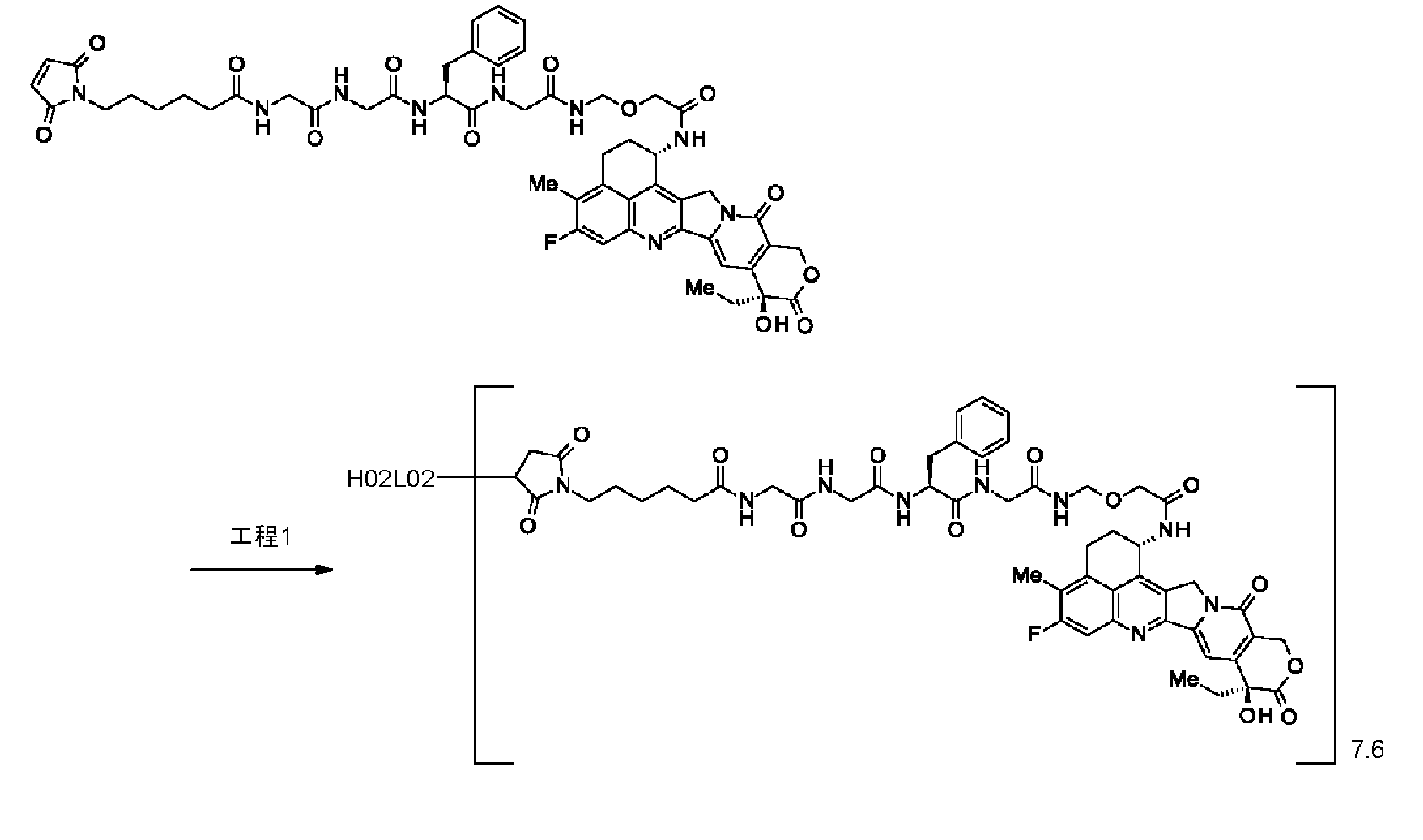
抗体濃度:2.61mg/mL,抗体収量:49.6mg(87%),共通操作Eにて測定された抗体一分子あたりの薬物平均結合数(n):5.9;共通操作Fにて測定された抗体一分子あたりの薬物平均結合数(n):7.6。 Characteristic evaluation: Using the common operation E (using ε D, 280 = 5440 , ε D, 370 = 21240) described in the
Antibody concentration: 2.61 mg / mL, antibody yield: 49.6 mg (87%), drug average binding number per molecule of antibody measured in common operation E (n): 5.9; common operation F Measured average drug binding number per antibody molecule (n): 7.6.
工程1:抗体-薬物コンジュゲート(3) 7) -3 Preparation of antibody-drug conjugate H02L03-DXd
Step 1: Antibody-drug conjugate (3)

抗体濃度:2.71mg/mL,抗体収量:51.4mg(91%),共通操作Eにて測定された抗体一分子あたりの薬物平均結合数(n):5.7;共通操作Fにて測定された抗体一分子あたりの薬物平均結合数(n):7.6。 Characteristic evaluation: Using the common operation E (using ε D, 280 = 5440 , ε D, 370 = 21240) described in the
Antibody concentration: 2.71 mg / mL, antibody yield: 51.4 mg (91%), average number of drugs per antibody molecule measured in common operation E (n): 5.7; in common operation F Measured average drug binding number per antibody molecule (n): 7.6.
工程1:抗体-薬物コンジュゲート(4) 7) -4 Preparation of antibody-drug conjugate H04L02-DXd
Step 1: Antibody-drug conjugate (4)

抗体濃度:2.56mg/mL,抗体収量:48.7mg(87%),共通操作Eにて測定された抗体一分子あたりの薬物平均結合数(n):5.8;共通操作Fにて測定された抗体一分子あたりの薬物平均結合数(n):7.6。 Characteristic evaluation: Using the common operation E (using ε D, 280 = 5440 , ε D, 370 = 21240) described in the
Antibody concentration: 2.56 mg / mL, antibody yield: 48.7 mg (87%), average number of drugs per antibody molecule measured in common operation E (n): 5.8; common operation F Measured average drug binding number per antibody molecule (n): 7.6.
[参考例2:NOV0712-薬物コンジュゲートの作製]
参考例2)-1 抗体-薬物コンジュゲートの作製 NOV0712-DM4
抗体-薬物コンジュゲート(5)
抗体と薬物リンカーのコンジュゲーション:参考例1にて作製したNOV0712を、製造方法1に記載した共通操作B(280nm吸光係数として1.51mLmg-1cm-1を使用)及びCを用いて、20mM HEPES8.1(LIFE TECHNOLOGIES社製 HEPES, 1M Buffer Solution(20mL)を1M水酸化ナトリウムでpH8.1とした後、蒸留水にて1Lとした)にて9.7mg/mLに調製し、20℃で10分間インキュベートした。次いでWO2016/024195号公開特許公報に記載の10mM 1-(2,5-ジオキソピロリジン-1-イルオキシ)-1-オキソ-4-(ピリジン-2-イルジスルファニル)ブタン-2-スルフォン酸のDMA溶液(0.366mL;抗体一分子に対して5.2当量)、10mM N2-デアセチル-デアセチル-N2-(4-メチル-4-メルカプト-1-オキソペンチル)-メイタンシン(DM4)のDMA溶液(0.366mL;抗体一分子に対して6.8当量)、及び0.243mLのDMAを加え、20℃で16時間インキュベートし、薬物リンカーを抗体へ結合させた。次に、1M 酢酸水溶液を加えpH5.0とし、さらに室温にて20分間撹拌し、薬物リンカーの反応を停止させた。
[Reference Example 2: Preparation of NOV0712-drug conjugate]
Reference Example 2) -1 Preparation of antibody-drug conjugate NOV0712-DM4
Antibody-drug conjugate (5)
Conjugation of antibody and drug linker: NOV0712 produced in Reference Example 1 was treated with 20 mM using the common procedure B (using 1.51 mLmg −1 cm −1 as the 280 nm extinction coefficient) and C described in
抗体濃度:2.58mg/mL,抗体収量:72.2mg(93%),共通操作Eにて測定された抗体一分子あたりの薬物平均結合数(n):3.0。 Characteristic evaluation: Using the common operation E described in manufacturing method 1 (using ε A, 280 = 200500 , ε A, 252 = 76295, ε D, 280 = 43170 , and ε D, 252 = 23224) The characteristic value was obtained.
Antibody concentration: 2.58 mg / mL, antibody yield: 72.2 mg (93%), average drug binding number per antibody molecule (n) measured in common operation E: 3.0.
工程1:抗体-薬物コンジュゲート(6) Reference Example 2) -2 Preparation of antibody-drug conjugate NOV0712-DXd
Step 1: Antibody-drug conjugate (6)

抗体濃度:2.26mg/mL,抗体収量:56.4mg(92%),共通操作Eにて測定された抗体一分子あたりの薬物平均結合数(n):6.4;共通操作Fにて測定された抗体一分子あたりの薬物平均結合数(n):7.8。 Characteristic evaluation: Using the common operation E (using ε D, 280 = 5440 , ε D, 370 = 21240) described in the
Antibody concentration: 2.26 mg / mL, antibody yield: 56.4 mg (92%), average number of drugs per antibody molecule measured in common operation E (n): 6.4; common operation F Average number of drugs bound per molecule of antibody measured (n): 7.8.
[参考例3:H01L02-DM4の作製]
参考例3)-1 抗体-薬物コンジュゲートの作製 H01L02-DM4
抗体-薬物コンジュゲート(7)
抗体と薬物リンカーのコンジュゲーション:実施例5にて作製したH01L02を、製造方法1に記載した共通操作B(280nm吸光係数として1.53mLmg-1cm-1を使用)及びCを用いて、20mM HEPES8.1(LIFE TECHNOLOGIES社製 HEPES, 1M Buffer Solution(20mL)を1M水酸化ナトリウムでpH8.1とした後、蒸留水にて1Lとした)にて9.8mg/mLに調製し、20℃で10分間インキュベートした。次いでWO2016/024195号公開特許公報に記載の10mM 1-(2,5-ジオキソピロリジン-1-イルオキシ)-1-オキソ-4-(ピリジン-2-イルジスルファニル)ブタン-2-スルフォン酸のDMA溶液(0.062mL;抗体一分子に対して11.5当量)、10mM N2-デアセチル-N2-(4-メチル-4-メルカプト-1-オキソペンチル)-メイタンシン(DM4)のDMA溶液(0.082mL;抗体一分子に対して15.1当量)を加え、20℃で18時間インキュベートし、薬物リンカーを抗体へ結合させた。次に、1M 酢酸水溶液を加えpH5.0とし、さらに室温にて20分間撹拌し、薬物リンカーの反応を停止させた。
[Reference Example 3: Production of H01L02-DM4]
Reference Example 3) -1 Preparation of antibody-drug conjugate H01L02-DM4
Antibody-drug conjugate (7)
Antibody-drug linker conjugation: H01L02 produced in Example 5 was treated with 20 mM using common procedure B (using 1.53 mL mg −1 cm −1 as the 280 nm extinction coefficient) and C described in
抗体濃度:1.97mg/mL,抗体収量:6.90mg(88%),共通操作Eにて測定された抗体一分子あたりの薬物平均結合数(n):3.6。 Characteristic evaluation: Using the common operation E (using εA, 280 = 223400, εA, 252 = 85646, εD, 280 = 4317, and εD, 252 = 23224) described in the
Antibody concentration: 1.97 mg / mL, antibody yield: 6.90 mg (88%), average drug binding number per antibody molecule (n) measured in common procedure E: 3.6.
[実施例8:抗体-薬物コンジュゲートのin vitro活性評価]
8)-1 CDH6陽性ヒト腫瘍細胞株に対する抗体-薬物コンジュゲートのin vitro細胞増殖抑制活性評価
CDH6陽性ヒト卵巣腫瘍細胞株PA-1を10% FBS含有MEM培地中2x10³細胞/100μL/wellになるよう96ウェルプレートに播種し、37℃、5% CO2の条件下で一晩培養した。翌日、実施例7で作製したヒト化hG019-薬物コンジュゲート4種(クローン名は、H01L02-DXd、H02L02-DXd、H02L03-DXd及びH04L02-DXd)、又は参考例2で作製したNOV0712-薬物コンジュゲート(NOV0712-DM4)を終濃度0.0001(nM)から100(nM)となるように添加した。4日間培養後に生存細胞数をCellTiter-GloTM Luminescent Cell Viability Assay(Promega)によるATPの定量で測定した。図11に抗体-薬物コンジュゲートを各々添加した際の濃度依存的な細胞増殖抑制活性を示す。この結果、ヒト化hG019-薬物コンジュゲート4種は、NOV0712-薬物コンジュゲートと比較して、より低い添加濃度から腫瘍細胞の増殖抑制活性を示し、抗腫瘍活性が高いことが示される。
[Example 8: In vitro activity evaluation of antibody-drug conjugate]
8) -1 Evaluation of In Vitro Cell Growth Inhibitory Activity of Antibody-Drug Conjugate against CDH6-positive Human Tumor Cell Line CDH6-positive human ovarian tumor cell line PA-1 was added to 2 × 10 3 cells / 100 μL / well in MEM medium containing 10% FBS. The cells were seeded in a 96-well plate and cultured overnight at 37 ° C. and 5% CO 2 . The next day, four types of humanized hG019-drug conjugates prepared in Example 7 (clone names are H01L02-DXd, H02L02-DXd, H02L03-DXd and H04L02-DXd), or NOV0712-drug conjugates prepared in Reference Example 2 A gate (NOV0712-DM4) was added so that the final concentration was 0.0001 (nM) to 100 (nM). After 4 days of culturing, the number of viable cells was determined by quantifying ATP with CellTiter-Glo ™ Luminescent Cell Viability Assay (Promega). FIG. 11 shows the concentration-dependent cell growth inhibitory activity when each antibody-drug conjugate is added. As a result, the four humanized hG019-drug conjugates show tumor cell growth inhibitory activity from a lower addition concentration and higher antitumor activity compared to NOV0712-drug conjugate.
[実施例9:抗体-薬物コンジュゲートのin vivo抗腫瘍効果]
抗体-薬物コンジュゲートの抗腫瘍効果は、CDH6陽性ヒト腫瘍細胞株の細胞を免疫不全マウスに移植した動物モデルを用いて評価した。4-5週齢のBALB/c ヌードマウス(CAnN.Cg-Foxnl[nu]/CrlCrlj[Foxnlnu/Foxnlnu]、日本チャールス・リバー)及び、SCIDマウス(CB17/Icr-Prkdc[scid]/CrlCrlj、日本チャールス・リバー)を実験使用前にSPF条件化で3日間以上馴化した。マウスには滅菌した固形飼料(FR-2,Funabashi Farms Co.,Ltd)を給餌し、滅菌した水道水(5-15ppm次亜塩素酸ナトリウム溶液を添加して調製)を与えた。移植した腫瘍の長径及び短径を電子式デジタルノギス(CD-15CX,Mitutoyo Corp.)で1週間に2回測定し、以下に示す計算式により腫瘍体積を算出した。
腫瘍体積(mm3)=1/2×長径(mm)×[短径(mm)]2
抗体-薬物コンジュゲートは全てABS緩衝液(10mM-Acetate Buffer, 5% Sorbitol, pH5.5)(NACALAI)で希釈し、各実施例に示す用量にて尾静脈内投与した。また、コントロール群(Vehicle群)としてABS緩衝液を同様に投与した。1群あたり6匹のマウスを実験に用いた。
[Example 9: In vivo antitumor effect of antibody-drug conjugate]
The anti-tumor effect of the antibody-drug conjugate was evaluated using an animal model in which cells of a CDH6-positive human tumor cell line were transplanted into immunodeficient mice. 4-5 week old BALB / c nude mice (CAnN.Cg-Foxnl [nu] / CrlCrlj [Foxnlnu / Foxnlnu], Charles River, Japan) and SCID mice (CB17 / Icr-Prkdc [scid] / CrlCrlj, Japan Charles River) was acclimated for more than 3 days under SPF conditions prior to experimental use. Mice were fed a sterilized chow (FR-2, Funabashi Farms Co., Ltd) and sterilized tap water (prepared by adding 5-15 ppm sodium hypochlorite solution). The major axis and minor axis of the transplanted tumor were measured twice a week with an electronic digital caliper (CD-15CX, Mitutoyo Corp.), and the tumor volume was calculated according to the formula shown below.
Tumor volume (mm 3 ) = 1/2 × major axis (mm) × [minor axis (mm)] 2
All antibody-drug conjugates were diluted with ABS buffer (10 mM-Acetate Buffer, 5% Sorbitol, pH 5.5) (NACALAI) and administered into the tail vein at the dose shown in each Example. In addition, an ABS buffer was administered in the same manner as a control group (vehicle group). Six mice per group were used in the experiment.
実施例2)-3-1にてCDH6発現を確認した、CDH6陽性ヒト腎細胞腫瘍細胞株786-O(ATCC)をマトリゲル(コーニング社)に懸濁し、5×10⁶cellsを雄SCIDマウスの右側腹部に皮下移植し(Day0)、Day18に無作為に群分けを実施した。群分け実施日に、実施例7で作製した抗体-薬物コンジュゲート4種(クローン名は、H01L02-DXd、H02L02-DXd、H02L03-DXd及びH04L02-DXd)、又は参考例2で作製したNOV0712-DM4を3mg/kgの用量で尾静脈内投与した。結果を図12に示す。横軸は日数、縦軸は腫瘍体積、誤差範囲はSE値を示す。 9) -1 Antitumor effect (1)
Example 2) CDH6-positive human renal cell tumor cell line 786-O (ATCC), whose CDH6 expression was confirmed in 3-1 was suspended in Matrigel (Corning), and 5 × 10 6 cells were isolated from male SCID mice. Subcutaneous transplantation was performed on the right flank (Day 0), and grouping was performed randomly on Day 18. On the day of grouping, 4 types of antibody-drug conjugates prepared in Example 7 (clone names are H01L02-DXd, H02L02-DXd, H02L03-DXd and H04L02-DXd) or NOV0712 produced in Reference Example 2 DM4 was administered intravenously at a dose of 3 mg / kg. The results are shown in FIG. The horizontal axis represents the number of days, the vertical axis represents the tumor volume, and the error range represents the SE value.
実施例2)-3-1にてCDH6発現を確認した、CDH6陽性ヒト卵巣腫瘍細胞株PA-1(ATCC)をマトリゲル(コーニング社)に懸濁し、8.5×10⁶cellsを雌ヌードマウスの右側腹部に皮下移植し(Day0)、Day11に無作為に群分けを実施した。群分け実施日に、実施例7で作製した抗体-薬物コンジュゲートH01L02-DXd又は参考例2で作製したNOV0712-DM4もしくはNOV0712-DXdを、1及び3mg/kgの用量で尾静脈内投与した。結果を図13に示す。横軸は日数、縦軸は腫瘍体積、誤差範囲はSE値を示す。 9) -2 Antitumor effect (2)
Example 2) CDH6-positive human ovarian tumor cell line PA-1 (ATCC) whose CDH6 expression was confirmed in 3-1 was suspended in Matrigel (Corning), and 8.5 × 10 6 cells were female nude mice. The right abdomen was subcutaneously transplanted (Day 0), and a random grouping was performed on Day 11. On the day of grouping, the antibody-drug conjugate H01L02-DXd prepared in Example 7 or NOV0712-DM4 or NOV0712-DXd prepared in Reference Example 2 was administered into the tail vein at a dose of 1 and 3 mg / kg. The results are shown in FIG. The horizontal axis represents the number of days, the vertical axis represents the tumor volume, and the error range represents the SE value.
実施例2)-3-1にてCDH6発現を確認した、CDH6陽性ヒト卵巣腫瘍細胞株NIH:OVCAR-3(ATCC)をマトリゲル(コーニング社)に懸濁し、1×10⁷cellsを雌ヌードマウスの右側腹部に皮下移植し(Day0)、Day22に無作為に群分けを実施した。群分け実施日に、実施例7で作製した抗体-薬物コンジュゲートH01L02-DXd又は参考例2で作製したNOV0712-DM4を、1及び3mg/kgの用量で尾静脈内投与した。結果を図14に示す。横軸は日数、縦軸は腫瘍体積、誤差範囲はSE値を示す。 9) -3 Antitumor effect (3)
Example 2) CDH6-positive human ovarian tumor cell line NIH: OVCAR-3 (ATCC) whose CDH6 expression was confirmed in 3-1 was suspended in Matrigel (Corning), and 1 × 10 7 cells were female nude mice The mice were implanted subcutaneously on the right flank (Day 0) and randomized into Day 22 groups. On the day of grouping, the antibody-drug conjugate H01L02-DXd prepared in Example 7 or NOV0712-DM4 prepared in Reference Example 2 was administered into the tail vein at a dose of 1 and 3 mg / kg. The results are shown in FIG. The horizontal axis represents the number of days, the vertical axis represents the tumor volume, and the error range represents the SE value.
実施例2)-3-1にてCDH6発現を確認した、CDH6陽性ヒト腎細胞腫瘍細胞株786-O(ATCC)をマトリゲル(コーニング社)に懸濁し、5×10⁶cellsを雄SCIDマウスの右側腹部に皮下移植し(Day0)、Day20に無作為に群分けを実施した。群分け実施日に、実施例7で作製した抗体-薬物コンジュゲートH01L02-DXd又は参考例2で作製したNOV0712-DM4を、1及び3mg/kgの用量で尾静脈内投与した。結果を図15に示す。横軸は日数、縦軸は腫瘍体積、誤差範囲はSE値を示す。 9) -4 Antitumor effect (4)
Example 2) CDH6-positive human renal cell tumor cell line 786-O (ATCC), whose CDH6 expression was confirmed in 3-1 was suspended in Matrigel (Corning), and 5 × 10 6 cells were isolated from male SCID mice. The right abdomen was subcutaneously transplanted (Day 0), and the grouping was performed randomly on
実施例2)-3-1にてCDH6を発現しないことを確認した、CDH6陰性ヒト卵巣腫瘍細胞株ES-2(ATCC)を生理食塩水に懸濁し、1×10⁶cellsを雌ヌードマウスの右側腹部に皮下移植し(Day0)、Day7に無作為に群分けを実施した。群分け実施日に、実施例7で作成した抗体-薬物コンジュゲートH01L02-DXd又は参考例2で作製したNOV0712-DM4を、1及び3mg/kgの用量で尾静脈内投与した。結果を図16に示す。横軸は日数、縦軸は腫瘍体積、誤差範囲はSE値を示す。 9) -5 Antitumor effect (5)
Example 2) CDH6-negative human ovarian tumor cell line ES-2 (ATCC) confirmed to not express CDH6 in 3-1 was suspended in physiological saline, and 1 × 10 6 cells were cultured in female nude mice. The right abdomen was subcutaneously transplanted (Day 0), and
Claims (53)
- 配列番号4に記載のアミノ酸配列に特異的に結合し、細胞内に取り込まれる内在化能を有する、抗体又は当該抗体の機能性断片。 An antibody or a functional fragment of the antibody, which specifically binds to the amino acid sequence shown in SEQ ID NO: 4 and has an internalizing ability to be taken up into cells.
- 配列番号4に記載のアミノ酸配列への結合に対して、以下の(1)~(5):
(1)配列番号53の21~233番目に記載のアミノ酸配列からなる軽鎖及び配列番号56の20~471番目に記載のアミノ酸配列からなる重鎖を有する抗体、
(2)配列番号61の21~233番目に記載のアミノ酸配列からなる軽鎖及び配列番号69の20~471番目に記載のアミノ酸配列からなる重鎖を有する抗体、
(3)配列番号61の21~233番目に記載のアミノ酸配列からなる軽鎖及び配列番号73の20~471番目に記載のアミノ酸配列からなる重鎖を有する抗体、
(4)配列番号65の21~233番目に記載のアミノ酸配列からなる軽鎖及び配列番号73の20~471番目に記載のアミノ酸配列からなる重鎖を有する抗体、及び
(5)配列番号61の21~233番目に記載のアミノ酸配列からなる軽鎖及び配列番号77の20~471番目に記載のアミノ酸配列からなる重鎖を有する抗体、
からなる群から選択される抗体のうち少なくともいずれか1つと競合阻害活性を有する請求項1に記載の抗体又は当該抗体の機能性断片。 For binding to the amino acid sequence set forth in SEQ ID NO: 4, the following (1) to (5):
(1) an antibody having a light chain consisting of the amino acid sequence described in the 21st to 233rd positions of SEQ ID NO: 53 and a heavy chain consisting of the amino acid sequence described in the 20th to 471st positions of SEQ ID NO: 56;
(2) an antibody having a light chain consisting of the amino acid sequence described in the 21st to 233rd positions of SEQ ID NO: 61 and a heavy chain consisting of the amino acid sequence described in the 20th to 471st positions of SEQ ID NO: 69;
(3) an antibody having a light chain consisting of the amino acid sequence described in the 21st to 233rd positions of SEQ ID NO: 61 and a heavy chain consisting of the amino acid sequence described in the 20th to 471st positions of SEQ ID NO: 73;
(4) an antibody having a light chain consisting of the amino acid sequence described in the 21st to 233rd positions of SEQ ID NO: 65 and a heavy chain consisting of the amino acid sequence described in the 20th to 471st positions of SEQ ID NO: 73; An antibody having a light chain consisting of the amino acid sequence described in positions 21 to 233 and a heavy chain consisting of the amino acid sequence described in positions 20 to 471 of SEQ ID NO: 77;
The antibody or the functional fragment of the antibody according to claim 1, which has competitive inhibitory activity with at least one of antibodies selected from the group consisting of: - 以下の(1)~(4):
(1)配列番号12に記載のアミノ酸配列からなるCDRL1、配列番号13に記載のアミノ酸配列からなるCDRL2及び配列番号14に記載のアミノ酸配列からなるCDRL3、
(2)配列番号22に記載のアミノ酸配列からなるCDRL1、配列番号23に記載のアミノ酸配列からなるCDRL2及び配列番号24に記載のアミノ酸配列からなるCDRL3、
(3)配列番号32に記載のアミノ酸配列からなるCDRL1、配列番号33に記載のアミノ酸配列からなるCDRL2及び配列番号34に記載のアミノ酸配列からなるCDRL3、及び
(4)配列番号42に記載のアミノ酸配列からなるCDRL1、配列番号43に記載のアミノ酸配列からなるCDRL2及び配列番号44に記載のアミノ酸配列からなるCDRL3、
からなる群から選択されるいずれか1つに記載のCDRL1、CDRL2及びCDRL3、並びに、
以下の(5)~(9):
(5)配列番号17に記載のアミノ酸配列からなるCDRH1、配列番号18に記載のアミノ酸配列からなるCDRH2及び配列番号19に記載のアミノ酸配列からなるCDRH3、
(6)配列番号27に記載のアミノ酸配列からなるCDRH1、配列番号28に記載のアミノ酸配列からなるCDRH2及び配列番号29に記載のアミノ酸配列からなるCDRH3、
(7)配列番号37に記載のアミノ酸配列からなるCDRH1、配列番号38に記載のアミノ酸配列からなるCDRH2及び配列番号39に記載のアミノ酸配列からなるCDRH3、
(8)配列番号47に記載のアミノ酸配列からなるCDRH1、配列番号48に記載のアミノ酸配列からなるCDRH2及び配列番号49に記載のアミノ酸配列からなるCDRH3、及び
(9)配列番号17に記載のアミノ酸配列からなるCDRH1、配列番号60に記載のアミノ酸配列からなるCDRH2及び配列番号19に記載のアミノ酸配列からなるCDRH3、
からなる群から選択されるいずれか1つに記載のCDRH1、CDRH2及びCDRH3を含む、
請求項1又2のいずれか1項に記載の抗体又は当該抗体の機能性断片。 The following (1) to (4):
(1) CDRL1 consisting of the amino acid sequence set forth in SEQ ID NO: 12, CDRL2 consisting of the amino acid sequence set forth in SEQ ID NO: 13 and CDRL3 consisting of the amino acid sequence set forth in SEQ ID NO: 14,
(2) CDRL1 consisting of the amino acid sequence set forth in SEQ ID NO: 22, CDRL2 consisting of the amino acid sequence set forth in SEQ ID NO: 23, and CDRL3 consisting of the amino acid sequence set forth in SEQ ID NO: 24,
(3) CDRL1 consisting of the amino acid sequence set forth in SEQ ID NO: 32, CDRL2 consisting of the amino acid sequence set forth in SEQ ID NO: 33 and CDRL3 consisting of the amino acid sequence set forth in SEQ ID NO: 34, and (4) the amino acid set forth in SEQ ID NO: 42 CDRL1 consisting of the sequence, CDRL2 consisting of the amino acid sequence set forth in SEQ ID NO: 43, and CDRL3 consisting of the amino acid sequence set forth in SEQ ID NO: 44,
CDRL1, CDRL2 and CDRL3 according to any one selected from the group consisting of:
The following (5) to (9):
(5) CDRH1 consisting of the amino acid sequence set forth in SEQ ID NO: 17, CDRH2 consisting of the amino acid sequence set forth in SEQ ID NO: 18 and CDRH3 consisting of the amino acid sequence set forth in SEQ ID NO: 19,
(6) CDRH1 consisting of the amino acid sequence set forth in SEQ ID NO: 27, CDRH2 consisting of the amino acid sequence set forth in SEQ ID NO: 28, and CDRH3 consisting of the amino acid sequence set forth in SEQ ID NO: 29,
(7) CDRH1 consisting of the amino acid sequence set forth in SEQ ID NO: 37, CDRH2 consisting of the amino acid sequence set forth in SEQ ID NO: 38, and CDRH3 consisting of the amino acid sequence set forth in SEQ ID NO: 39,
(8) CDRH1 consisting of the amino acid sequence set forth in SEQ ID NO: 47, CDRH2 consisting of the amino acid sequence set forth in SEQ ID NO: 48 and CDRH3 consisting of the amino acid sequence set forth in SEQ ID NO: 49, and (9) the amino acid set forth in SEQ ID NO: 17 CDRH1 comprising the sequence, CDRH2 comprising the amino acid sequence set forth in SEQ ID NO: 60, and CDRH3 comprising the amino acid sequence set forth in SEQ ID NO: 19,
CDRH1, CDRH2 and CDRH3 according to any one selected from the group consisting of
The antibody or the functional fragment of the antibody according to any one of claims 1 and 2. - 以下の(1)~(5):
(1)配列番号12に記載のアミノ酸配列からなるCDRL1、配列番号13に記載のアミノ酸配列からなるCDRL2及び配列番号14に記載のアミノ酸配列からなるCDRL3、及び、配列番号17に記載のアミノ酸配列からなるCDRH1、配列番号18に記載のアミノ酸配列からなるCDRH2及び配列番号19に記載のアミノ酸配列からなるCDRH3、
(2)配列番号22に記載のアミノ酸配列からなるCDRL1、配列番号23に記載のアミノ酸配列からなるCDRL2及び配列番号24に記載のアミノ酸配列からなるCDRL3、及び、配列番号27に記載のアミノ酸配列からなるCDRH1、配列番号28に記載のアミノ酸配列からなるCDRH2及び配列番号29に記載のアミノ酸配列からなるCDRH3、
(3)配列番号32に記載のアミノ酸配列からなるCDRL1、配列番号33に記載のアミノ酸配列からなるCDRL2及び配列番号34に記載のアミノ酸配列からなるCDRL3、及び、配列番号37に記載のアミノ酸配列からなるCDRH1、配列番号38に記載のアミノ酸配列からなるCDRH2及び配列番号39に記載のアミノ酸配列からなるCDRH3、
(4)配列番号42に記載のアミノ酸配列からなるCDRL1、配列番号43に記載のアミノ酸配列からなるCDRL2及び配列番号44に記載のアミノ酸配列からなるCDRL3、及び、配列番号47に記載のアミノ酸配列からなるCDRH1、配列番号48に記載のアミノ酸配列からなるCDRH2及び配列番号49に記載のアミノ酸配列からなるCDRH3、及び
(5)配列番号12に記載のアミノ酸配列からなるCDRL1、配列番号13に記載のアミノ酸配列からなるCDRL2及び配列番号14に記載のアミノ酸配列からなるCDRL3、及び、配列番号17に記載のアミノ酸配列からなるCDRH1、配列番号60に記載のアミノ酸配列からなるCDRH2及び配列番号19に記載のアミノ酸配列からなるCDRH3、
からなる群から選択されるCDRL1、CDRL2及びCDRL3、並びに、CDRH1、CDRH2及びCDRH3を含む、
請求項1~3のいずれか1項に記載の抗体又は当該抗体の機能性断片。 The following (1) to (5):
(1) From CDRL1 consisting of the amino acid sequence set forth in SEQ ID NO: 12, CDRL2 consisting of the amino acid sequence set forth in SEQ ID NO: 13 and CDRL3 consisting of the amino acid sequence set forth in SEQ ID NO: 14, and from the amino acid sequence set forth in SEQ ID NO: 17 CDRH1, CDRH2 consisting of the amino acid sequence set forth in SEQ ID NO: 18, and CDRH3 consisting of the amino acid sequence set forth in SEQ ID NO: 19,
(2) From CDRL1 consisting of the amino acid sequence set forth in SEQ ID NO: 22, CDRL2 consisting of the amino acid sequence set forth in SEQ ID NO: 23 and CDRL3 consisting of the amino acid sequence set forth in SEQ ID NO: 24, and from the amino acid sequence set forth in SEQ ID NO: 27 CDRH1, CDRH2 consisting of the amino acid sequence set forth in SEQ ID NO: 28, and CDRH3 consisting of the amino acid sequence set forth in SEQ ID NO: 29,
(3) From CDRL1 consisting of the amino acid sequence set forth in SEQ ID NO: 32, CDRL2 consisting of the amino acid sequence set forth in SEQ ID NO: 33 and CDRL3 consisting of the amino acid sequence set forth in SEQ ID NO: 34, and from the amino acid sequence set forth in SEQ ID NO: 37 CDRH1, CDRH2 consisting of the amino acid sequence set forth in SEQ ID NO: 38, and CDRH3 consisting of the amino acid sequence set forth in SEQ ID NO: 39,
(4) From CDRL1 consisting of the amino acid sequence set forth in SEQ ID NO: 42, CDRL2 consisting of the amino acid sequence set forth in SEQ ID NO: 43 and CDRL3 consisting of the amino acid sequence set forth in SEQ ID NO: 44, and from the amino acid sequence set forth in SEQ ID NO: 47 CDRH1, CDRH2 consisting of the amino acid sequence set forth in SEQ ID NO: 48, CDRH3 consisting of the amino acid sequence set forth in SEQ ID NO: 49, and (5) CDRRL1 consisting of the amino acid sequence set forth in SEQ ID NO: 12, amino acid set forth in SEQ ID NO: 13 CDRRL2 consisting of the sequence and CDRRL3 consisting of the amino acid sequence set forth in SEQ ID NO: 14, CDRH1 consisting of the amino acid sequence set forth in SEQ ID NO: 17, CDRH2 consisting of the amino acid sequence set forth in SEQ ID NO: 60, and the amino acid set forth in SEQ ID NO: 19 CDRH3 consisting of a sequence,
Comprising CDRL1, CDRL2 and CDRL3 selected from the group consisting of: CDRH1, CDRH2 and CDRH3,
The antibody or the functional fragment of the antibody according to any one of claims 1 to 3. - ヒト化されている請求項1~4のいずれか1項に記載の抗体又は当該抗体の機能性断片。 The antibody or the functional fragment of the antibody according to any one of claims 1 to 4, which is humanized.
- 以下の(1)~(4):
(1)配列番号63に記載のアミノ酸配列からなる軽鎖可変領域、
(2)配列番号67に記載のアミノ酸配列からなる軽鎖可変領域、
(3)(1)~(2)のアミノ酸配列において各CDR配列以外のフレームワーク領域の配列に対して少なくとも95%以上の配列同一性を有するアミノ酸配列、及び
(4)(1)~(3)のアミノ酸配列において各CDR配列以外のフレームワーク領域の配列において1又は数個のアミノ酸が欠失、置換又は付加されたアミノ酸配列
からなる群から選択されるいずれか1つに記載の軽鎖可変領域、並びに、
以下の(5)~(9):
(5)配列番号71に記載のアミノ酸配列からなる重鎖可変領域、
(6)配列番号75に記載のアミノ酸配列からなる重鎖可変領域、
(7)配列番号79に記載のアミノ酸配列からなる重鎖可変領域、
(8)(5)~(7)のアミノ酸配列において各CDR配列以外のフレームワーク領域の配列に対して少なくとも95%以上の配列相同性を有するアミノ酸配列、及び
(9)(5)~(8)のアミノ酸配列において各CDR配列以外のフレームワーク領域の配列において1又は数個のアミノ酸が欠失、置換又は付加されたアミノ酸配列
からなる群から選択されるいずれか1つに記載の重鎖可変領域、
を有する請求項1~5のいずれか1項に記載の抗体又は当該抗体の機能性断片。 The following (1) to (4):
(1) a light chain variable region comprising the amino acid sequence set forth in SEQ ID NO: 63,
(2) a light chain variable region comprising the amino acid sequence set forth in SEQ ID NO: 67,
(3) an amino acid sequence having at least 95% sequence identity to the sequence of the framework region other than each CDR sequence in the amino acid sequence of (1) to (2), and (4) (1) to (3 The light chain variable according to any one of the following groups selected from the group consisting of amino acid sequences in which one or several amino acids are deleted, substituted or added in the framework region sequence other than each CDR sequence: Area, and
The following (5) to (9):
(5) a heavy chain variable region comprising the amino acid sequence set forth in SEQ ID NO: 71,
(6) a heavy chain variable region comprising the amino acid sequence set forth in SEQ ID NO: 75;
(7) a heavy chain variable region comprising the amino acid sequence set forth in SEQ ID NO: 79;
(8) An amino acid sequence having at least 95% sequence homology to the sequence of the framework region other than each CDR sequence in the amino acid sequences of (5) to (7), and (9) (5) to (8 The heavy chain variable according to any one selected from the group consisting of amino acid sequences in which one or several amino acids are deleted, substituted, or added in the framework region sequence other than each CDR sequence region,
The antibody or the functional fragment of the antibody according to any one of claims 1 to 5, which comprises - 以下の(1)~(4):
(1)配列番号63に記載のアミノ酸配列からなる軽鎖可変領域及び配列番号71に記載のアミノ酸配列からなる重鎖可変領域、
(2)配列番号63に記載のアミノ酸配列からなる軽鎖可変領域及び配列番号75に記載のアミノ酸配列からなる重鎖可変領域、
(3)配列番号67に記載のアミノ酸配列からなる軽鎖可変領域及び配列番号75に記載のアミノ酸配列からなる重鎖可変領域、又は
(4)配列番号63に記載のアミノ酸配列からなる軽鎖可変領域及び配列番号79に記載のアミノ酸配列からなる重鎖可変領域、
のいずれかの軽鎖可変領域及び重鎖可変領域、
を含む請求項1~6のいずれか1項に記載の抗体又は当該抗体の機能性断片。 The following (1) to (4):
(1) a light chain variable region consisting of the amino acid sequence set forth in SEQ ID NO: 63 and a heavy chain variable region consisting of the amino acid sequence set forth in SEQ ID NO: 71;
(2) a light chain variable region comprising the amino acid sequence set forth in SEQ ID NO: 63 and a heavy chain variable region comprising the amino acid sequence set forth in SEQ ID NO: 75;
(3) a light chain variable region comprising the amino acid sequence set forth in SEQ ID NO: 67 and a heavy chain variable region comprising the amino acid sequence set forth in SEQ ID NO: 75, or (4) a light chain variable comprising the amino acid sequence set forth in SEQ ID NO: 63 A heavy chain variable region comprising the region and the amino acid sequence set forth in SEQ ID NO: 79;
A light chain variable region and a heavy chain variable region of any of
The antibody or the functional fragment of the antibody according to any one of claims 1 to 6, comprising - 以下の(1)~(4):
(1)配列番号61の21~233番目のアミノ酸配列からなる軽鎖及び配列番号69の20~471番目のアミノ酸配列からなる重鎖、
(2)配列番号61の21~233番目のアミノ酸配列からなる軽鎖及び配列番号73の20~471番目のアミノ酸配列からなる重鎖、
(3)配列番号65の21~233番目のアミノ酸配列からなる軽鎖及び配列番号73の20~471番目のアミノ酸配列からなる重鎖、又は
(4)配列番号61の21~233番目のアミノ酸配列からなる軽鎖及び配列番号77の20~471番目のアミノ酸配列からなる重鎖、
のいずれかを有する請求項1~7のいずれか1項に記載の抗体又は当該抗体の機能性断片。 The following (1) to (4):
(1) a light chain consisting of amino acid sequences 21 to 233 of SEQ ID NO: 61 and a heavy chain consisting of amino acid sequences 20 to 471 of SEQ ID NO: 69;
(2) a light chain consisting of amino acid sequences 21 to 233 of SEQ ID NO: 61 and a heavy chain consisting of amino acid sequences 20 to 471 of SEQ ID NO: 73;
(3) a light chain comprising the 21st to 233rd amino acid sequence of SEQ ID NO: 65 and a heavy chain comprising the 20th to 471st amino acid sequence of SEQ ID NO: 73, or (4) the 21st to 233rd amino acid sequence of SEQ ID NO: 61 A heavy chain consisting of the 20th to 471st amino acid sequence of SEQ ID NO: 77,
The antibody or the functional fragment of the antibody according to any one of claims 1 to 7, which has any one of - 配列番号61の21~233番目のアミノ酸配列からなる軽鎖及び配列番号69の20~471番目のアミノ酸配列からなる重鎖を有する請求項8に記載の抗体又は当該抗体の機能性断片。 The antibody or the functional fragment of the antibody according to claim 8, which has a light chain consisting of the 21st to 233rd amino acid sequences of SEQ ID NO: 61 and a heavy chain consisting of the 20th to 471st amino acid sequences of SEQ ID NO: 69.
- 配列番号61の21~233番目のアミノ酸配列からなる軽鎖及び配列番号73の20~471番目のアミノ酸配列からなる重鎖を有する請求項8に記載の抗体又は当該抗体の機能性断片。 The antibody or the functional fragment of the antibody according to claim 8, which has a light chain consisting of the 21st to 233rd amino acid sequences of SEQ ID NO: 61 and a heavy chain consisting of the 20th to 471st amino acid sequences of SEQ ID NO: 73.
- 配列番号65の21~233番目のアミノ酸配列からなる軽鎖及び配列番号73の20~471番目のアミノ酸配列からなる重鎖を有する請求項8に記載の抗体又は当該抗体の機能性断片。 The antibody or the functional fragment of the antibody according to claim 8, which has a light chain consisting of the 21st to 233rd amino acid sequences of SEQ ID NO: 65 and a heavy chain consisting of the 20th to 471st amino acid sequences of SEQ ID NO: 73.
- 配列番号61の21~233番目のアミノ酸配列からなる軽鎖及び配列番号77の20~471番目のアミノ酸配列からなる重鎖を有する請求項8に記載の抗体又は当該抗体の機能性断片。 The antibody or the functional fragment of the antibody according to claim 8, which has a light chain consisting of amino acid sequences 21 to 233 of SEQ ID NO: 61 and a heavy chain consisting of amino acid sequences 20 to 471 of SEQ ID NO: 77.
- 機能性断片がFab、F(ab’)2、Fab’及びFvからなる群から選択される請求項1~12のいずれか1項に記載の抗体の機能性断片。 The functional fragment of an antibody according to any one of claims 1 to 12, wherein the functional fragment is selected from the group consisting of Fab, F (ab ') 2, Fab' and Fv.
- 請求項1~13のいずれか1項に記載の抗体又は当該抗体の機能性断片をコードするポリヌクレオチド。 A polynucleotide encoding the antibody or the functional fragment of the antibody according to any one of claims 1 to 13.
- 以下の(1)~(5):
(1)配列番号12に記載のアミノ酸配列からなるCDRL1、配列番号13に記載のアミノ酸配列からなるCDRL2及び配列番号14に記載のアミノ酸配列からなるCDRL3を含む軽鎖可変領域をコードするポリヌクレオチド、及び、配列番号17に記載のアミノ酸配列からなるCDRH1、配列番号18に記載のアミノ酸配列からなるCDRH2及び配列番号19に記載のアミノ酸配列からなるCDRH3を含む重鎖可変領域をコードするポリヌクレオチド、
(2)配列番号22に記載のアミノ酸配列からなるCDRL1、配列番号23に記載のアミノ酸配列からなるCDRL2及び配列番号24に記載のアミノ酸配列からなるCDRL3を含む軽鎖可変領域をコードするポリヌクレオチド、及び、配列番号27に記載のアミノ酸配列からなるCDRH1、配列番号28に記載のアミノ酸配列からなるCDRH2及び配列番号29に記載のアミノ酸配列からなるCDRH3を含む重鎖可変領域をコードするポリヌクレオチド、
(3)配列番号32に記載のアミノ酸配列からなるCDRL1、配列番号33に記載のアミノ酸配列からなるCDRL2及び配列番号34に記載のアミノ酸配列からなるCDRL3を含む軽鎖可変領域をコードするポリヌクレオチド、及び、配列番号37に記載のアミノ酸配列からなるCDRH1、配列番号38に記載のアミノ酸配列からなるCDRH2及び配列番号39に記載のアミノ酸配列からなるCDRH3を含む重鎖可変領域をコードするポリヌクレオチド、
(4)配列番号42に記載のアミノ酸配列からなるCDRL1、配列番号43に記載のアミノ酸配列からなるCDRL2及び配列番号44に記載のアミノ酸配列からなるCDRL3を含む軽鎖可変領域をコードするポリヌクレオチド、及び、配列番号47に記載のアミノ酸配列からなるCDRH1、配列番号48に記載のアミノ酸配列からなるCDRH2及び配列番号49に記載のアミノ酸配列からなるCDRH3を含む重鎖可変領域をコードするポリヌクレオチド、並びに
(5)配列番号12に記載のアミノ酸配列からなるCDRL1、配列番号13に記載のアミノ酸配列からなるCDRL2及び配列番号14に記載のアミノ酸配列からなるCDRL3を含む軽鎖可変領域をコードするポリヌクレオチド、及び、配列番号17に記載のアミノ酸配列からなるCDRH1、配列番号60に記載のアミノ酸配列からなるCDRH2及び配列番号19に記載のアミノ酸配列からなるCDRH3を含む重鎖可変領域をコードするポリヌクレオチド、
からなる群から選択されるいずれか1つに記載のポリヌクレオチドを含む請求項14に記載のポリヌクレオチド。 The following (1) to (5):
(1) a polynucleotide encoding a light chain variable region comprising CDRL1 comprising the amino acid sequence set forth in SEQ ID NO: 12, CDRL2 comprising the amino acid sequence set forth in SEQ ID NO: 13 and CDRL3 comprising the amino acid sequence set forth in SEQ ID NO: 14; And a polynucleotide encoding a heavy chain variable region comprising CDRH1 consisting of the amino acid sequence set forth in SEQ ID NO: 17, CDRH2 consisting of the amino acid sequence set forth in SEQ ID NO: 18 and CDRH3 consisting of the amino acid sequence set forth in SEQ ID NO: 19.
(2) a polynucleotide encoding a light chain variable region comprising CDRL1 comprising the amino acid sequence set forth in SEQ ID NO: 22, CDRL2 comprising the amino acid sequence set forth in SEQ ID NO: 23 and CDRL3 comprising the amino acid sequence set forth in SEQ ID NO: 24; And a polynucleotide encoding a heavy chain variable region comprising CDRH1 consisting of the amino acid sequence set forth in SEQ ID NO: 27, CDRH2 consisting of the amino acid sequence set forth in SEQ ID NO: 28, and CDRH3 consisting of the amino acid sequence set forth in SEQ ID NO: 29,
(3) a polynucleotide encoding a light chain variable region comprising CDRL1 consisting of the amino acid sequence set forth in SEQ ID NO: 32, CDRL2 consisting of the amino acid sequence set forth in SEQ ID NO: 33 and CDRL3 consisting of the amino acid sequence set forth in SEQ ID NO: 34; A polynucleotide encoding a heavy chain variable region comprising CDRH1 consisting of the amino acid sequence set forth in SEQ ID NO: 37, CDRH2 consisting of the amino acid sequence set forth in SEQ ID NO: 38, and CDRH3 consisting of the amino acid sequence set forth in SEQ ID NO: 39;
(4) a polynucleotide encoding a light chain variable region comprising CDRL1 consisting of the amino acid sequence set forth in SEQ ID NO: 42, CDRL2 consisting of the amino acid sequence set forth in SEQ ID NO: 43, and CDRL3 consisting of the amino acid sequence set forth in SEQ ID NO: 44; And a polynucleotide encoding a heavy chain variable region comprising CDRH1 consisting of the amino acid sequence set forth in SEQ ID NO: 47, CDRH2 consisting of the amino acid sequence set forth in SEQ ID NO: 48 and CDRH3 consisting of the amino acid sequence set forth in SEQ ID NO: 49, and (5) a polynucleotide encoding a light chain variable region comprising CDRL1 comprising the amino acid sequence set forth in SEQ ID NO: 12, CDRL2 comprising the amino acid sequence set forth in SEQ ID NO: 13 and CDRL3 comprising the amino acid sequence set forth in SEQ ID NO: 14. And the amino acid of SEQ ID NO: 17 CDRH1 consisting acid sequence encoding a heavy chain variable region comprising a CDRH3 comprising the amino acid sequence set forth in CDRH2 and SEQ ID NO: 19 comprising the amino acid sequence set forth in SEQ ID NO: 60 polynucleotide,
The polynucleotide according to claim 14, comprising the polynucleotide according to any one selected from the group consisting of: - 配列番号61の21~233番目のアミノ酸配列からなる軽鎖をコードするポリヌクレオチド及び配列番号69の20~471番目のアミノ酸配列からなる重鎖をコードするポリヌクレオチドを含む請求項14又は15に記載のポリヌクレオチド。 16. The polynucleotide encoding a light chain consisting of the 21st to 233rd amino acid sequences of SEQ ID NO: 61 and the polynucleotide encoding a heavy chain consisting of the 20th to 471st amino acid sequences of SEQ ID NO: 69. Polynucleotides.
- 配列番号61の21~233番目のアミノ酸配列からなる軽鎖をコードするポリヌクレオチド及び配列番号73の20~471番目のアミノ酸配列からなる重鎖をコードするポリヌクレオチドを含む請求項14又は15に記載のポリヌクレオチド。 The polynucleotide according to claim 14 or 15, comprising a polynucleotide encoding a light chain consisting of the 21st to 233rd amino acid sequences of SEQ ID NO: 61 and a polynucleotide encoding a heavy chain consisting of the 20th to 471st amino acid sequences of SEQ ID NO: 73. Polynucleotides.
- 配列番号65の21~233番目のアミノ酸配列からなる軽鎖をコードするポリヌクレオチド及び配列番号73の20~471番目のアミノ酸配列からなる重鎖をコードするポリヌクレオチドを含む請求項14又は15に記載のポリヌクレオチド。 The polynucleotide according to claim 14 or 15, comprising a polynucleotide encoding a light chain consisting of the 21st to 233rd amino acid sequences of SEQ ID NO: 65 and a polynucleotide encoding a heavy chain consisting of the 20th to 471st amino acid sequences of SEQ ID NO: 73. Polynucleotides.
- 配列番号61の21~233番目のアミノ酸配列からなる軽鎖をコードするポリヌクレオチド及び配列番号77の20~471番目のアミノ酸配列からなる重鎖をコードするポリヌクレオチドを含む請求項14又は15に記載のポリヌクレオチド。 The polynucleotide according to claim 14 or 15, comprising a polynucleotide encoding a light chain consisting of the 21st to 233rd amino acid sequence of SEQ ID NO: 61 and a polynucleotide encoding a heavy chain consisting of the 20th to 471st amino acid sequence of SEQ ID NO: 77. Polynucleotides.
- 請求項14~19のいずれか1項に記載のポリヌクレオチドを含有する発現ベクター。 An expression vector containing the polynucleotide according to any one of claims 14 to 19.
- 請求項20に記載の発現ベクターにより形質転換された宿主細胞。 A host cell transformed with the expression vector according to claim 20.
- 宿主細胞が真核細胞である請求項21に記載の宿主細胞。 The host cell according to claim 21, wherein the host cell is a eukaryotic cell.
- 請求項21又は22に記載の宿主細胞を培養する工程、及び当該工程で得られた培養物から目的の抗体又は当該抗体の機能性断片を採取する工程を含むことを特徴とする当該抗体又は当該抗体の機能性断片の製造方法。 The step of culturing the host cell according to claim 21 or 22, and the step of collecting the antibody of interest or a functional fragment of the antibody from the culture obtained in the step, A method for producing a functional fragment of an antibody.
- 重鎖又は軽鎖が、N-結合への糖鎖付加、O-結合への糖鎖付加、N末のプロセッシング、C末のプロセッシング、脱アミド化、アスパラギン酸の異性化、メチオニンの酸化、N末にメチオニン残基の付加、プロリン残基のアミド化、N末グルタミンもしくはN末グルタミン酸のピログルタミン酸化、及びカルボキシル末端における1つ又は2つのアミノ酸欠失からなる群より選択される1又は2以上の修飾をうけた、請求項1~13のいずれか1項に記載の抗体又は当該抗体の機能性断片。 Heavy chain or light chain is added to the N-link, added to the O-link, added to the N-terminal, processed at the N-terminal, processed at the C-terminal, deamidated, isomerized aspartic acid, oxidized methionine, N One or more selected from the group consisting of addition of a methionine residue at the end, amidation of a proline residue, pyroglutamine oxidation of N-terminal glutamine or N-terminal glutamic acid, and one or two amino acid deletions at the carboxyl terminus The antibody or the functional fragment of the antibody according to any one of claims 1 to 13, which has been modified as described above.
- 重鎖のカルボキシル末端において1つ又は2つのアミノ酸が欠失している請求項24に記載の抗体。 25. The antibody of claim 24, wherein one or two amino acids are deleted at the carboxyl terminus of the heavy chain.
- 2本の重鎖の双方でカルボキシル末端において1つのアミノ酸が欠失している請求項25に記載の抗体。 26. The antibody of claim 25, wherein one amino acid is deleted at the carboxyl terminus in both of the two heavy chains.
- 重鎖のカルボキシル末端のプロリン残基が更にアミド化されている請求項24~26のいずれか1項に記載の抗体。 The antibody according to any one of claims 24 to 26, wherein the proline residue at the carboxyl terminus of the heavy chain is further amidated.
- 抗体依存性細胞傷害活性を増強させるために糖鎖修飾が調節されている請求項1~13及び請求項24~27からなる群から選択されるいずれか1項に記載の抗体又は当該抗体の機能性断片。 The antibody or the function of the antibody according to any one of claims 1 to 13 and 24 to 27, wherein the sugar chain modification is regulated to enhance antibody-dependent cytotoxic activity. Sex fragment.
- 請求項1~13及び請求項24~28からなる群から選択されるいずれか1項に記載の抗体又は当該抗体の機能性断片に薬物が結合している抗体-薬物コンジュゲート。 An antibody-drug conjugate in which a drug is bound to the antibody according to any one of claims 1 to 13 and claims 24 to 28 or a functional fragment of the antibody.
- 薬物が抗腫瘍性化合物である、請求項29に記載の抗体-薬物コンジュゲート。 30. The antibody-drug conjugate of claim 29, wherein the drug is an antitumor compound.
- 抗腫瘍性化合物が次式:
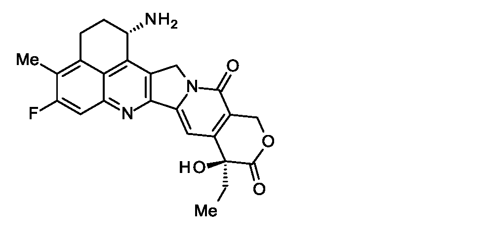
- 抗体と薬物が、次式(a)~(f):
(a)-(Succinimid-3-yl-N)-CH2CH2-C(=O)-GGFG-NH-CH2CH2CH2-C(=O)-、
(b)-(Succinimid-3-yl-N)-CH2CH2CH2CH2CH2-C(=O)-GGFG-NH-CH2CH2CH2-C(=O)-、
(c)-(Succinimid-3-yl-N)-CH2CH2CH2CH2CH2-C(=O)-GGFG-NH-CH2-O-CH2-C(=O)-、
(d)-(Succinimid-3-yl-N)-CH2CH2CH2CH2CH2-C(=O)-GGFG-NH-CH2CH2-O-CH2-C(=O)-、
(e)-(Succinimid-3-yl-N)-CH2CH2-C(=O)-NH-CH2CH2O-CH2CH2O-CH2CH2-C(=O)-GGFG-NH-CH2CH2CH2-C(=O)-、及び
(f)-(Succinimid-3-yl-N)-CH2CH2-C(=O)-NH-CH2CH2O-CH2CH2O-CH2CH2O-CH2CH2O-CH2CH2-C(=O)-GGFG-NH-CH2CH2CH2-C(=O)-、
からなる群から選択されるいずれかの構造のリンカーを介して結合している、請求項29~31のいずれか1項に記載の抗体-薬物コンジュゲート。
(ここで、抗体は、-(Succinimid-3-yl-N)の末端において結合する。抗腫瘍性化合物は、1位のアミノ基の窒素原子を結合部位として、(a)、(b)、(e)又は(f)の-CH2CH2CH2-C(=O)-部分、(c)のCH2-O-CH2-C(=O)-部分又は(d)のCH2CH2-O-CH2-C(=O)-部分のカルボニル基に結合する。上記式中GGFGは、グリシン-グリシン-フェニルアラニン-グリシンからなるペプチド結合でつながっているアミノ酸配列を示す。
-(Succinimid-3-yl-N)-は次式:
(A)-(Succinimid-3-yl-N) —CH 2 CH 2 —C (═O) —GGFG-NH—CH 2 CH 2 CH 2 —C (═O) —,
(B)-(Succinimid-3-yl-N) —CH 2 CH 2 CH 2 CH 2 CH 2 —C (═O) —GGFG-NH—CH 2 CH 2 CH 2 —C (═O) —,
(C)-(Succinimid-3-yl-N) —CH 2 CH 2 CH 2 CH 2 CH 2 —C (═O) —GGFG—NH—CH 2 —O—CH 2 —C (═O) —,
(D)-(Succinimid-3-yl-N) —CH 2 CH 2 CH 2 CH 2 CH 2 —C (═O) —GGFG-NH—CH 2 CH 2 —O—CH 2 —C (═O) -,
(E) - (Succinimid-3 -yl-N) -CH 2 CH 2 -C (= O) -NH-CH 2 CH 2 O-CH 2 CH 2 O-CH 2 CH 2 -C (= O) - GGFG—NH—CH 2 CH 2 CH 2 —C (═O) —, and (f)-(Succinimid-3-yl-N) —CH 2 CH 2 —C (═O) —NH—CH 2 CH 2 O—CH 2 CH 2 O—CH 2 CH 2 O—CH 2 CH 2 O—CH 2 CH 2 —C (═O) —GGFG—NH—CH 2 CH 2 CH 2 —C (═O) —,
The antibody-drug conjugate according to any one of claims 29 to 31, which is bound via a linker having any structure selected from the group consisting of:
(Here, the antibody binds at the terminal of-(Succinimid-3-yl-N). The antitumor compound has a nitrogen atom of the amino group at position 1 as a binding site, and (a), (b), The —CH 2 CH 2 CH 2 —C (═O) — moiety of (e) or (f), the CH 2 —O—CH 2 —C (═O) — moiety of (c) or the CH 2 of (d). It binds to the carbonyl group of the CH 2 —O—CH 2 —C (═O) — moiety, where GGFG represents an amino acid sequence linked by a peptide bond consisting of glycine-glycine-phenylalanine-glycine.
-(Succinimid-3-yl-N)-is the following formula:
- リンカーが以下の(c)、(d)及び(e)からなる群から選択されるいずれかの式で示される請求項29~32のいずれか1項に記載の抗体-薬物コンジュゲート:
(c)-(Succinimid-3-yl-N)-CH2CH2CH2CH2CH2-C(=O)-GGFG-NH-CH2-O-CH2-C(=O)-、
(d)-(Succinimid-3-yl-N)-CH2CH2CH2CH2CH2-C(=O)-GGFG-NH-CH2CH2-O-CH2-C(=O)-、
(e)-(Succinimid-3-yl-N)-CH2CH2-C(=O)-NH-CH2CH2O-CH2CH2O-CH2CH2-C(=O)-GGFG-NH-CH2CH2CH2-C(=O)-。 The antibody-drug conjugate according to any one of claims 29 to 32, wherein the linker is represented by any formula selected from the group consisting of the following (c), (d) and (e):
(C)-(Succinimid-3-yl-N) —CH 2 CH 2 CH 2 CH 2 CH 2 —C (═O) —GGFG—NH—CH 2 —O—CH 2 —C (═O) —,
(D)-(Succinimid-3-yl-N) —CH 2 CH 2 CH 2 CH 2 CH 2 —C (═O) —GGFG-NH—CH 2 CH 2 —O—CH 2 —C (═O) -,
(E)-(Succinimid-3-yl-N) —CH 2 CH 2 —C (═O) —NH—CH 2 CH 2 O—CH 2 CH 2 O—CH 2 CH 2 —C (═O) — GGFG-NH—CH 2 CH 2 CH 2 —C (═O) —. - リンカーが以下の(c)又は(e)の式で示される請求項29~33のいずれか1項に記載の抗体-薬物コンジュゲート:
(c)-(Succinimid-3-yl-N)-CH2CH2CH2CH2CH2-C(=O)-GGFG-NH-CH2-O-CH2-C(=O)-、
(e)-(Succinimid-3-yl-N)-CH2CH2-C(=O)-NH-CH2CH2O-CH2CH2O-CH2CH2-C(=O)-GGFG-NH-CH2CH2CH2-C(=O)-。 The antibody-drug conjugate according to any one of claims 29 to 33, wherein the linker is represented by the following formula (c) or (e):
(C)-(Succinimid-3-yl-N) —CH 2 CH 2 CH 2 CH 2 CH 2 —C (═O) —GGFG—NH—CH 2 —O—CH 2 —C (═O) —,
(E)-(Succinimid-3-yl-N) —CH 2 CH 2 —C (═O) —NH—CH 2 CH 2 O—CH 2 CH 2 O—CH 2 CH 2 —C (═O) — GGFG-NH—CH 2 CH 2 CH 2 —C (═O) —. - 以下の式:

ここで、ABは抗体又は該抗体の機能性断片を示す。nは抗体と結合している薬物-リンカー構造の1抗体あたりの平均結合数を示す。抗体とリンカーは抗体由来のスルフヒドリル基を介して結合している。 The following formula:
Here, AB represents an antibody or a functional fragment of the antibody. n represents the average number of bonds per antibody of the drug-linker structure bonded to the antibody. The antibody and the linker are bound via an antibody-derived sulfhydryl group. - 以下の式:
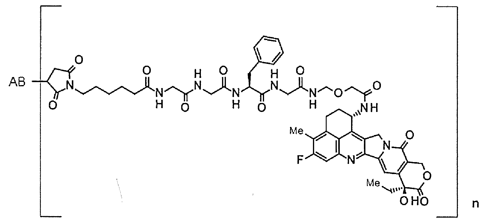
ここで、ABは抗体又は該抗体の機能性断片を示す。nは抗体と結合している薬物-リンカー構造の1抗体あたりの平均結合数を示す。抗体とリンカーは抗体由来のスルフヒドリル基を介して結合している。 The following formula:
Here, AB represents an antibody or a functional fragment of the antibody. n represents the average number of bonds per antibody of the drug-linker structure bonded to the antibody. The antibody and the linker are bound via an antibody-derived sulfhydryl group. - 抗体が以下の(1)~(4):
(1)配列番号61の21~233番目のアミノ酸配列からなる軽鎖及び配列番号69の20~471番目のアミノ酸配列からなる重鎖、
(2)配列番号61の21~233番目のアミノ酸配列からなる軽鎖及び配列番号73の20~471番目のアミノ酸配列からなる重鎖、
(3)配列番号65の21~233番目のアミノ酸配列からなる軽鎖及び配列番号73の20~471番目のアミノ酸配列からなる重鎖、又は
(4)配列番号61の21~233番目のアミノ酸配列からなる軽鎖及び配列番号77の20~471番目のアミノ酸配列からなる重鎖、
からなる群から選択されるいずれか1つに記載の軽鎖及び重鎖を含む抗体又は当該抗体の機能性断片である、請求項29~36のいずれか1項に記載の抗体-薬物コンジュゲート。 The antibodies are the following (1) to (4):
(1) a light chain consisting of amino acid sequences 21 to 233 of SEQ ID NO: 61 and a heavy chain consisting of amino acid sequences 20 to 471 of SEQ ID NO: 69;
(2) a light chain consisting of amino acid sequences 21 to 233 of SEQ ID NO: 61 and a heavy chain consisting of amino acid sequences 20 to 471 of SEQ ID NO: 73;
(3) a light chain comprising the 21st to 233rd amino acid sequence of SEQ ID NO: 65 and a heavy chain comprising the 20th to 471st amino acid sequence of SEQ ID NO: 73, or (4) the 21st to 233rd amino acid sequence of SEQ ID NO: 61 A heavy chain consisting of the 20th to 471st amino acid sequence of SEQ ID NO: 77,
The antibody-drug conjugate according to any one of claims 29 to 36, which is an antibody or a functional fragment of the antibody comprising the light chain and the heavy chain according to any one selected from the group consisting of . - 抗体が、配列番号61の21~233番目のアミノ酸配列からなる軽鎖及び配列番号69の20~471番目のアミノ酸配列からなる重鎖を含む抗体又は当該抗体の機能性断片である、請求項37に記載の抗体-薬物コンジュゲート。 The antibody is an antibody comprising a light chain consisting of the 21st to 233rd amino acid sequence of SEQ ID NO: 61 and a heavy chain consisting of the 20th to 471st amino acid sequence of SEQ ID NO: 69, or a functional fragment of the antibody. An antibody-drug conjugate according to 1.
- 抗体が、配列番号61の21~233番目のアミノ酸配列からなる軽鎖及び配列番号77の20~471番目のアミノ酸配列からなる重鎖を含む抗体又は当該抗体の機能性断片である、請求項37に記載の抗体-薬物コンジュゲート。 The antibody is an antibody or a functional fragment of the antibody comprising a light chain consisting of the 21st to 233rd amino acid sequences of SEQ ID NO: 61 and a heavy chain consisting of the 20th to 471st amino acid sequences of SEQ ID NO: 77. An antibody-drug conjugate according to 1.
- 重鎖又は軽鎖が、N-結合への糖鎖付加、O-結合への糖鎖付加、N末のプロセッシング、C末のプロセッシング、脱アミド化、アスパラギン酸の異性化、メチオニンの酸化、N末にメチオニン残基の付加、プロリン残基のアミド化、N末グルタミンもしくはN末グルタミン酸のピログルタミン酸化及びカルボキシル末端における1つ又は2つのアミノ酸欠失からなる群から選択される1又は2以上の修飾をうけた、請求項29~39のいずれか1項に記載の抗体-薬物コンジュゲート。 Heavy chain or light chain is added to the N-link, added to the O-link, added to the N-terminal, processed at the N-terminal, processed at the C-terminal, deamidated, isomerized aspartic acid, oxidized methionine, N One or more selected from the group consisting of addition of a methionine residue at the end, amidation of a proline residue, pyroglutamine oxidation of N-terminal glutamine or N-terminal glutamic acid, and one or two amino acid deletions at the carboxyl terminus The antibody-drug conjugate according to any one of claims 29 to 39, which has been modified.
- 選択された1種の薬物-リンカー構造の1抗体あたりの平均結合数が1~10個の範囲である請求項29~40のいずれか1項に記載の抗体-薬物コンジュゲート。 The antibody-drug conjugate according to any one of claims 29 to 40, wherein the average number of bonds per antibody of one selected drug-linker structure is in the range of 1 to 10.
- 選択された1種の薬物-リンカー構造の1抗体あたりの平均結合数が2~8個の範囲である請求項41に記載の抗体-薬物コンジュゲート。 The antibody-drug conjugate according to claim 41, wherein the average number of bonds per antibody of one selected drug-linker structure is in the range of 2-8.
- 選択された1種の薬物-リンカー構造の1抗体あたりの平均結合数が5~8個の範囲である請求項42に記載の抗体-薬物コンジュゲート。 The antibody-drug conjugate according to claim 42, wherein the average number of bonds per antibody of one selected drug-linker structure is in the range of 5-8.
- 選択された1種の薬物-リンカー構造の1抗体あたりの平均結合数が7~8個である請求項43に記載の抗体-薬物コンジュゲート。 44. The antibody-drug conjugate according to claim 43, wherein the average number of bonds per antibody of one selected drug-linker structure is 7-8.
- 請求項29~44のいずれか1項に記載の抗体-薬物コンジュゲート、その塩、又はそれらの水和物を含むことを特徴とする医薬組成物。 A pharmaceutical composition comprising the antibody-drug conjugate according to any one of claims 29 to 44, a salt thereof, or a hydrate thereof.
- 抗腫瘍薬であることを特徴とする、請求項45に記載の医薬組成物。 46. The pharmaceutical composition according to claim 45, characterized in that it is an antitumor agent.
- 腫瘍がCDH6を発現する腫瘍であることを特徴とする、請求項46に記載の医薬組成物。 47. The pharmaceutical composition according to claim 46, characterized in that the tumor is a tumor that expresses CDH6.
- 腫瘍が、腎細胞癌、腎淡明細胞癌、乳頭状腎細胞癌、卵巣癌、卵巣漿液性腺癌、甲状腺癌、胆管癌、肺癌、小細胞肺癌、神経膠芽腫、中皮腫、子宮癌、膵臓癌、ウィルムス腫瘍又は神経芽腫であることを特徴とする、請求項46又は47に記載の医薬組成物。 Tumor is renal cell carcinoma, clear cell renal cell carcinoma, papillary renal cell carcinoma, ovarian cancer, ovarian serous adenocarcinoma, thyroid cancer, bile duct cancer, lung cancer, small cell lung cancer, glioblastoma, mesothelioma, uterine cancer 48. The pharmaceutical composition according to claim 46 or 47, which is pancreatic cancer, Wilms tumor or neuroblastoma.
- 請求項29~44のいずれか1項に記載の抗体-薬物コンジュゲート、その塩、又はそれらの水和物から選択されるいずれかを個体に投与することを特徴とする腫瘍の治療方法。 A method for treating a tumor, which comprises administering to an individual any one selected from the antibody-drug conjugate according to any one of claims 29 to 44, a salt thereof, or a hydrate thereof.
- 腫瘍がCDH6が発現している腫瘍であることを特徴とする、請求項49に記載の治療方法。 The method according to claim 49, wherein the tumor is a tumor in which CDH6 is expressed.
- 腫瘍が、腎細胞癌、腎淡明細胞癌、乳頭状腎細胞癌、卵巣癌、卵巣漿液性腺癌、甲状腺癌、胆管癌、肺癌、小細胞肺癌、神経膠芽腫、中皮腫、子宮癌、膵臓癌、ウィルムス腫瘍又は神経芽腫であることを特徴とする、請求項49又は50に記載の治療方法。 Tumor is renal cell carcinoma, clear cell renal cell carcinoma, papillary renal cell carcinoma, ovarian cancer, ovarian serous adenocarcinoma, thyroid cancer, bile duct cancer, lung cancer, small cell lung cancer, glioblastoma, mesothelioma, uterine cancer The method according to claim 49 or 50, which is pancreatic cancer, Wilms tumor or neuroblastoma.
- 請求項29~44のいずれか1項に記載の抗体-薬物コンジュゲート、その塩、又はそれらの水和物から選択される少なくとも一つを含む医薬組成物及び少なくとも一つの抗腫瘍薬を、同時に、別々に又は連続して個体に投与することを特徴とする腫瘍の治療方法。 A pharmaceutical composition comprising at least one selected from the antibody-drug conjugate according to any one of claims 29 to 44, a salt thereof, or a hydrate thereof and at least one antitumor agent, A method for treating a tumor, which comprises administering to an individual separately or sequentially.
- 請求項1~13及び請求項24~28からなる群から選択されるいずれか1項に記載の抗体又は当該抗体の機能性断片或いは請求項23に記載の製造方法で得られる抗体又は当該抗体の機能性断片と、薬物-リンカー中間化合物を反応させる工程を含むことを特徴とする、抗体-薬物コンジュゲートの製造方法。 An antibody according to any one of claims 1 to 13 and claims 24 to 28, a functional fragment of the antibody, an antibody obtained by the production method according to claim 23, or an antibody A method for producing an antibody-drug conjugate, comprising a step of reacting a functional fragment with a drug-linker intermediate compound.
Priority Applications (13)
| Application Number | Priority Date | Filing Date | Title |
|---|---|---|---|
| RU2019141268A RU2798285C2 (en) | 2017-05-15 | 2018-05-14 | Anti-cdh6-antibody and anti-cdh6-antibody-drug conjugate |
| EP18802536.5A EP3626825A4 (en) | 2017-05-15 | 2018-05-14 | Anti-cdh6 antibody and anti-cdh6 antibody-drug conjugate |
| CN201880032336.2A CN110651045A (en) | 2017-05-15 | 2018-05-14 | anti-CDH 6 antibodies and anti-CDH 6 antibody-drug conjugates |
| US16/613,203 US11446386B2 (en) | 2017-05-15 | 2018-05-14 | Anti-CDH6 antibody and method of producing an anti-CDH6 antibody-drug conjugate |
| CA3063827A CA3063827A1 (en) | 2017-05-15 | 2018-05-14 | Anti-cdh6 antibody and anti-cdh6 antibody-drug conjugate |
| BR112019023832-8A BR112019023832A2 (en) | 2017-05-15 | 2018-05-14 | antibody, functional antibody fragment, polynucleotide, expression vector, host cells, methods for producing an antibody of interest or a functional fragment of the antibody, for treating a tumor and for producing an antibody-drug conjugate, antibody-drug conjugate, and , pharmaceutical composition |
| KR1020197033607A KR20200006061A (en) | 2017-05-15 | 2018-05-14 | Anti-CDH6 Antibody and Anti-CDH6 Antibody-Drug Conjugate |
| JP2019518773A JP6827534B2 (en) | 2017-05-15 | 2018-05-14 | Anti-CDH6 antibody and anti-CDH6 antibody-drug conjugate |
| AU2018270961A AU2018270961A1 (en) | 2017-05-15 | 2018-05-14 | Anti-CDH6 antibody and anti-CDH6 antibody-drug conjugate |
| PH12019502545A PH12019502545A1 (en) | 2017-05-15 | 2019-11-14 | Anti-cdh6 antibody and anti-cdh6 antibody-drug conjugate |
| CONC2019/0013020A CO2019013020A2 (en) | 2017-05-15 | 2019-11-21 | Anti-cdh6 antibody and anti-DH6 antibody-drug conjugate |
| US17/010,162 US11077202B2 (en) | 2017-05-15 | 2020-09-02 | Anti-CDH6 antibody and anti-CDH6 antibody-drug conjugate |
| US17/874,239 US20230226205A1 (en) | 2017-05-15 | 2022-07-26 | Anti-cdh6 antibody and anti-cdh6 antibody-drug conjugate |
Applications Claiming Priority (2)
| Application Number | Priority Date | Filing Date | Title |
|---|---|---|---|
| JP2017096749 | 2017-05-15 | ||
| JP2017-096749 | 2017-05-15 |
Related Child Applications (3)
| Application Number | Title | Priority Date | Filing Date |
|---|---|---|---|
| US16/613,203 A-371-Of-International US11446386B2 (en) | 2017-05-15 | 2018-05-14 | Anti-CDH6 antibody and method of producing an anti-CDH6 antibody-drug conjugate |
| US17/010,162 Continuation US11077202B2 (en) | 2017-05-15 | 2020-09-02 | Anti-CDH6 antibody and anti-CDH6 antibody-drug conjugate |
| US17/874,239 Division US20230226205A1 (en) | 2017-05-15 | 2022-07-26 | Anti-cdh6 antibody and anti-cdh6 antibody-drug conjugate |
Publications (1)
| Publication Number | Publication Date |
|---|---|
| WO2018212136A1 true WO2018212136A1 (en) | 2018-11-22 |
Family
ID=64273811
Family Applications (1)
| Application Number | Title | Priority Date | Filing Date |
|---|---|---|---|
| PCT/JP2018/018572 WO2018212136A1 (en) | 2017-05-15 | 2018-05-14 | Anti-cdh6 antibody and anti-cdh6 antibody-drug conjugate |
Country Status (13)
| Country | Link |
|---|---|
| US (3) | US11446386B2 (en) |
| EP (1) | EP3626825A4 (en) |
| JP (4) | JP6827534B2 (en) |
| KR (1) | KR20200006061A (en) |
| CN (1) | CN110651045A (en) |
| AU (1) | AU2018270961A1 (en) |
| BR (1) | BR112019023832A2 (en) |
| CA (1) | CA3063827A1 (en) |
| CO (1) | CO2019013020A2 (en) |
| MX (2) | MX2023001686A (en) |
| PH (1) | PH12019502545A1 (en) |
| TW (2) | TWI794230B (en) |
| WO (1) | WO2018212136A1 (en) |
Cited By (27)
| Publication number | Priority date | Publication date | Assignee | Title |
|---|---|---|---|---|
| WO2020022475A1 (en) | 2018-07-27 | 2020-01-30 | 第一三共株式会社 | Protein recognizing drug moiety of antibody-drug conjugate |
| WO2020022363A1 (en) | 2018-07-25 | 2020-01-30 | 第一三共株式会社 | Effective method for manufacturing antibody-drug conjugate |
| WO2020027100A1 (en) | 2018-07-31 | 2020-02-06 | 第一三共株式会社 | Treatment of metastatic brain tumor by administration of antibody-drug conjugate |
| WO2020031936A1 (en) | 2018-08-06 | 2020-02-13 | 第一三共株式会社 | Combination of antibody-drug conjugate and tubulin inhibitor |
| WO2020040245A1 (en) | 2018-08-23 | 2020-02-27 | 第一三共株式会社 | Sensitivity marker for antibody-drug conjugate |
| WO2020050406A1 (en) | 2018-09-06 | 2020-03-12 | 第一三共株式会社 | Novel cyclic dinucleotide derivative and antibody-drug conjugate thereof |
| WO2020100954A1 (en) * | 2018-11-14 | 2020-05-22 | 第一三共株式会社 | (anti-cdh6 antibody)-(pyrrolobenzodiazepine derivative) conjugate |
| WO2020122034A1 (en) | 2018-12-11 | 2020-06-18 | 第一三共株式会社 | Combination of antibody-drug conjugate with parp inhibitor |
| WO2020130125A1 (en) | 2018-12-21 | 2020-06-25 | 第一三共株式会社 | Combination of antibody-drug conjugate and kinase inhibitor |
| US11077202B2 (en) | 2017-05-15 | 2021-08-03 | Daiichi Sankyo Company, Limited | Anti-CDH6 antibody and anti-CDH6 antibody-drug conjugate |
| WO2021260579A1 (en) | 2020-06-24 | 2021-12-30 | Astrazeneca Uk Limited | Combination of antibody-drug conjugate and atr inhibitor |
| WO2021260580A1 (en) | 2020-06-24 | 2021-12-30 | Astrazeneca Uk Limited | Combination of antibody-drug conjugate and atm inhibitor |
| WO2021260583A1 (en) | 2020-06-24 | 2021-12-30 | Astrazeneca Uk Limited | Combination of antibody-drug conjugate and dna-pk inhibitor |
| WO2021260582A1 (en) | 2020-06-24 | 2021-12-30 | Astrazeneca Uk Limited | Combination of antibody-drug conjugate and aurora b inhibitor |
| WO2021260578A1 (en) | 2020-06-24 | 2021-12-30 | Astrazeneca Uk Limited | Combination of antibody-drug conjugate and cdk9 inhibitor |
| WO2022074617A1 (en) | 2020-10-09 | 2022-04-14 | Astrazeneca Uk Limited | Combination of antibody-drug conjugate and parp1 selective inhibitor |
| WO2022102634A1 (en) | 2020-11-11 | 2022-05-19 | 第一三共株式会社 | COMBINATION OF ANTIBODY-DRUG CONJUGATE WITH ANTI-SIRPα ANTIBODY |
| WO2022163846A1 (en) | 2021-02-01 | 2022-08-04 | 第一三共株式会社 | Novel method for producing antibody-immunostimulator conjugate |
| WO2022191313A1 (en) | 2021-03-12 | 2022-09-15 | 第一三共株式会社 | Glycan, and method for producing medicine containing glycan |
| WO2022228494A1 (en) | 2021-04-29 | 2022-11-03 | 上海汇连生物医药有限公司 | Preparation method for and application of antibody conjugated drug |
| US11583590B2 (en) | 2017-09-29 | 2023-02-21 | Daiichi Sankyo Company, Limited | Antibody-pyrrolobenzodiazepine derivative conjugate and method of use thereof for treating a tumor |
| WO2023042097A1 (en) | 2021-09-15 | 2023-03-23 | Daiichi Sankyo Company, Limited | Antibody-drug conjugate for use in methods of treating chemotherapy-resistant cancer |
| WO2023126822A1 (en) | 2021-12-28 | 2023-07-06 | Astrazeneca Uk Limited | Combination of antibody-drug conjugate and rasg12c inhibitor |
| WO2023209591A1 (en) | 2022-04-27 | 2023-11-02 | Daiichi Sankyo Company, Limited | Combination of antibody-drug conjugate with ezh1 and/or ezh2 inhibitor |
| WO2023218378A1 (en) | 2022-05-11 | 2023-11-16 | Daiichi Sankyo Company, Limited | Combination of an antibody specific for a tumor antigen and a cd47 inhibitor |
| WO2023228095A1 (en) | 2022-05-24 | 2023-11-30 | Daiichi Sankyo Company, Limited | Dosage regimen of an anti-cdh6 antibody-drug conjugate |
| WO2024023750A1 (en) | 2022-07-28 | 2024-02-01 | Astrazeneca Uk Limited | Combination of antibody-drug conjugate and bispecific checkpoint inhibitor |
Families Citing this family (5)
| Publication number | Priority date | Publication date | Assignee | Title |
|---|---|---|---|---|
| US20220211863A1 (en) | 2019-03-29 | 2022-07-07 | Medimmune Limited | Compounds and conjugates thereof |
| WO2023009891A2 (en) * | 2021-07-30 | 2023-02-02 | Janssen Biotech, Inc. | Materials and methods of making or using il-23r binding proteins |
| TW202330618A (en) * | 2021-12-10 | 2023-08-01 | 大陸商普衆發現醫藥科技(上海)有限公司 | Anti-cdh6 antibodies and antibody-drug conjugates thereof |
| WO2023102875A1 (en) * | 2021-12-10 | 2023-06-15 | Multitude Therapeutics Inc. | Anti-cdh6 antibody drug conjugate |
| WO2023143387A1 (en) * | 2022-01-26 | 2023-08-03 | Beigene , Ltd. | ANTI-DXd ANTIBODIES AND METHODS OF USE |
Citations (14)
| Publication number | Priority date | Publication date | Assignee | Title |
|---|---|---|---|---|
| WO1990007861A1 (en) | 1988-12-28 | 1990-07-26 | Protein Design Labs, Inc. | CHIMERIC IMMUNOGLOBULINS SPECIFIC FOR p55 TAC PROTEIN OF THE IL-2 RECEPTOR |
| WO1992001047A1 (en) | 1990-07-10 | 1992-01-23 | Cambridge Antibody Technology Limited | Methods for producing members of specific binding pairs |
| WO1992020791A1 (en) | 1990-07-10 | 1992-11-26 | Cambridge Antibody Technology Limited | Methods for producing members of specific binding pairs |
| WO1993006213A1 (en) | 1991-09-23 | 1993-04-01 | Medical Research Council | Production of chimeric antibodies - a combinatorial approach |
| WO1993011236A1 (en) | 1991-12-02 | 1993-06-10 | Medical Research Council | Production of anti-self antibodies from antibody segment repertoires and displayed on phage |
| WO1993019172A1 (en) | 1992-03-24 | 1993-09-30 | Cambridge Antibody Technology Limited | Methods for producing members of specific binding pairs |
| WO1995001438A1 (en) | 1993-06-30 | 1995-01-12 | Medical Research Council | Sbp members with a chemical moiety covalently bound within the binding site; production and selection thereof |
| WO1995015388A1 (en) | 1993-12-03 | 1995-06-08 | Medical Research Council | Recombinant binding proteins and peptides |
| WO1999054342A1 (en) | 1998-04-20 | 1999-10-28 | Pablo Umana | Glycosylation engineering of antibodies for improving antibody-dependent cellular cytotoxicity |
| WO2000061739A1 (en) | 1999-04-09 | 2000-10-19 | Kyowa Hakko Kogyo Co., Ltd. | Method for controlling the activity of immunologically functional molecule |
| WO2002031140A1 (en) | 2000-10-06 | 2002-04-18 | Kyowa Hakko Kogyo Co., Ltd. | Cells producing antibody compositions |
| WO2014057687A1 (en) | 2012-10-11 | 2014-04-17 | 第一三共株式会社 | Antibody-drug conjugate |
| WO2016024195A1 (en) | 2014-08-12 | 2016-02-18 | Novartis Ag | Anti-cdh6 antibody drug conjugates |
| US20160297890A1 (en) | 2013-12-25 | 2016-10-13 | Daiichi Sankyo Company, Limited | Anti-trop2 antibody-drug conjugate |
Family Cites Families (76)
| Publication number | Priority date | Publication date | Assignee | Title |
|---|---|---|---|---|
| WO1989006692A1 (en) | 1988-01-12 | 1989-07-27 | Genentech, Inc. | Method of treating tumor cells by inhibiting growth factor receptor function |
| US5183884A (en) | 1989-12-01 | 1993-02-02 | United States Of America | Dna segment encoding a gene for a receptor related to the epidermal growth factor receptor |
| US5658920A (en) | 1991-01-16 | 1997-08-19 | Daiichi Pharmaceutical Co., Ltd. | Substituted 1H,12H-benz-[DE]pyrano[3',4':6,7] indolizino[1,2-B]quinoline-10,13(9H,15H)-dione compound |
| JP3008226B2 (en) | 1991-01-16 | 2000-02-14 | 第一製薬株式会社 | Hexacyclic compounds |
| LU91067I2 (en) | 1991-06-14 | 2004-04-02 | Genentech Inc | Trastuzumab and its variants and immunochemical derivatives including immotoxins |
| US6214345B1 (en) | 1993-05-14 | 2001-04-10 | Bristol-Myers Squibb Co. | Lysosomal enzyme-cleavable antitumor drug conjugates |
| US6504029B1 (en) | 1995-04-10 | 2003-01-07 | Daiichi Pharmaceutical Co., Ltd. | Condensed-hexacyclic compounds and a process therefor |
| JPH08337584A (en) | 1995-04-10 | 1996-12-24 | Dai Ichi Seiyaku Co Ltd | Condensed six-membered cyclic amino compound, medicine containing the same and production of the same |
| SG50747A1 (en) | 1995-08-02 | 1998-07-20 | Tanabe Seiyaku Co | Comptothecin derivatives |
| JPH1095802A (en) | 1995-12-28 | 1998-04-14 | Tanabe Seiyaku Co Ltd | Camptothecine derivative |
| CA2192725C (en) | 1995-12-28 | 2004-04-20 | Kenji Tsujihara | Camptothecin derivatives |
| US5968511A (en) | 1996-03-27 | 1999-10-19 | Genentech, Inc. | ErbB3 antibodies |
| TW527183B (en) | 1996-06-06 | 2003-04-11 | Daiichi Seiyaku Co | Drug complex |
| TW409058B (en) | 1996-06-06 | 2000-10-21 | Daiichi Seiyaku Co | Method for preparation of a drug complex |
| JPH1171280A (en) | 1997-06-26 | 1999-03-16 | Tanabe Seiyaku Co Ltd | Medicine composition |
| JPH1192405A (en) | 1997-09-19 | 1999-04-06 | Dai Ichi Seiyaku Co Ltd | Medicinal complex |
| CA2333321A1 (en) | 1998-05-22 | 1999-12-02 | Daiichi Pharmaceutical Co., Ltd. | Drug complex |
| MXPA01004239A (en) | 1998-10-30 | 2002-06-04 | Daiichi Seiyaku Co | Dds compounds and method for assaying the same. |
| EP1191944A2 (en) | 1999-06-25 | 2002-04-03 | Genentech, Inc. | METHODS OF TREATMENT USING ANTI-ErbB ANTIBODY-MAYTANSINOID CONJUGATES |
| JP2002060351A (en) | 2000-03-22 | 2002-02-26 | Dai Ichi Seiyaku Co Ltd | Dds compound comprising drug having hydroxy group |
| MXPA02012791A (en) | 2000-06-29 | 2003-12-11 | Daiichi Seiyaku Co | Dds compound and process for the preparation thereof. |
| BR0112417A (en) | 2000-07-13 | 2003-07-01 | Daiichi Seiyaku Co | Pharmaceutical compositions containing dds compound |
| EP1283053A1 (en) | 2001-08-09 | 2003-02-12 | Max-Planck-Gesellschaft zur Förderung der Wissenschaften e.V. | Inhibitors of HER3 activity |
| TWI313609B (en) | 2001-08-21 | 2009-08-21 | Mitsubishi Tanabe Pharma Corp | Pharmaceutical composition for inhibiting the metastasis or preventing the recurrence of malignant tumor |
| JP2005511627A (en) | 2001-11-20 | 2005-04-28 | シアトル ジェネティクス,インコーポレーテッド | Treatment of immunological diseases using anti-CD30 antibodies |
| US8877901B2 (en) | 2002-12-13 | 2014-11-04 | Immunomedics, Inc. | Camptothecin-binding moiety conjugates |
| US9745380B2 (en) | 2002-03-01 | 2017-08-29 | Immunomedics, Inc. | RS7 antibodies |
| JP4959136B2 (en) | 2002-12-13 | 2012-06-20 | イミューノメディクス、インコーポレイテッド | Immunoconjugates with cleavable linkages in cells |
| US20040202666A1 (en) | 2003-01-24 | 2004-10-14 | Immunomedics, Inc. | Anti-cancer anthracycline drug-antibody conjugates |
| EP2033645A1 (en) | 2004-02-26 | 2009-03-11 | Inotek Pharmaceuticals Corporation | Isoquinoline derivatives and methods of use thereof |
| EP1725586B1 (en) | 2004-03-02 | 2015-01-14 | Seattle Genetics, Inc. | Partially loaded antibodies and methods of their conjugation |
| CA2564076C (en) | 2004-05-19 | 2014-02-18 | Medarex, Inc. | Chemical linkers and conjugates thereof |
| CN102973947A (en) | 2004-06-01 | 2013-03-20 | 健泰科生物技术公司 | Antibody-drug conjugates and methods |
| BRPI0513681A (en) | 2004-07-22 | 2008-05-13 | Genentech Inc | compositions comprising her2 antibody, method, pharmaceutical formulations, polypeptide, antibody and cancer treatment method |
| US8802820B2 (en) | 2004-11-12 | 2014-08-12 | Xencor, Inc. | Fc variants with altered binding to FcRn |
| WO2006065533A2 (en) | 2004-11-29 | 2006-06-22 | Seattle Genetics, Inc. | Engineered antibodies and immunoconjugates |
| JP2008528486A (en) | 2005-01-21 | 2008-07-31 | ジェネンテック・インコーポレーテッド | Fixed dose of HER antibody |
| DE102005009099A1 (en) | 2005-02-28 | 2006-08-31 | Ktb Tumorforschungsgesellschaft Mbh | New camptothecin-peptide derivatives, useful for treating cancer |
| DE102005009084A1 (en) | 2005-02-28 | 2006-08-31 | Ktb Tumorforschungsgesellschaft Mbh | New anthracyclin-peptide derivatives, useful for treating cancer, especially of the prostate, are cleaved, in the tumor, by prostate-specific antigen to release active antitumor agent and are transported by serum albumen |
| US7833979B2 (en) | 2005-04-22 | 2010-11-16 | Amgen Inc. | Toxin peptide therapeutic agents |
| AR056857A1 (en) | 2005-12-30 | 2007-10-24 | U3 Pharma Ag | DIRECTED ANTIBODIES TO HER-3 (RECEIVER OF THE HUMAN EPIDERMAL GROWTH FACTOR-3) AND ITS USES |
| US20080131428A1 (en) | 2006-02-24 | 2008-06-05 | Arius Research, Inc. | Cytotoxicity mediation of cells evidencing surface expression of TROP-2 |
| ZA200809776B (en) | 2006-05-30 | 2010-03-31 | Genentech Inc | Antibodies and immunoconjugates and uses therefor |
| MX2008015132A (en) | 2006-05-30 | 2008-12-10 | Genentech Inc | Antibodies and immunoconjugates and uses therefor. |
| ITMI20061473A1 (en) | 2006-07-26 | 2008-01-27 | Indena Spa | DERIVATIVES OF CAMPTOTECIN WITH ANTITUMORAL ACTIVITY |
| EP2109607A1 (en) | 2006-12-21 | 2009-10-21 | Schering Corporation | Pyrrolo[3, 2-a]pyridine derivatives for inhibiting ksp kinesin activity |
| PL2716301T3 (en) | 2007-02-16 | 2017-10-31 | Merrimack Pharmaceuticals Inc | Antibodies against erbb3 and uses thereof |
| EP2121008A4 (en) | 2007-03-22 | 2010-03-31 | Sloan Kettering Inst Cancer | Uses of monoclonal antibody 8h9 |
| MY186725A (en) | 2008-04-30 | 2021-08-13 | Immunogen Inc | Cross-linkers and their uses |
| JP5539971B2 (en) | 2008-05-13 | 2014-07-02 | ジェネンテック, インコーポレイテッド | Analysis of antibody drug conjugates by bead-based affinity capture and mass spectrometry |
| JP5677972B2 (en) | 2008-11-18 | 2015-02-25 | メリマック ファーマシューティカルズ インコーポレーティッド | Human serum albumin linker and its conjugates |
| PL2396036T3 (en) | 2009-02-13 | 2017-12-29 | Immunomedics, Inc. | Immunoconjugates with an intracellularly-cleavable linkage |
| WO2011011474A1 (en) | 2009-07-22 | 2011-01-27 | Enzon Pharmaceuticals, Inc. | Methods of treating her2 positive cancer with her2 receptor antagonist in combination with multi-arm polymeric conjugates of 7-ethyl-10-hydroxycamptothecin |
| JP5829520B2 (en) | 2009-08-20 | 2015-12-09 | 国立大学法人 千葉大学 | Colchicine derivatives |
| CA2775350A1 (en) | 2009-09-24 | 2011-03-31 | Seattle Genetics, Inc. | Dr5 ligand drug conjugates |
| TWI630916B (en) | 2009-11-13 | 2018-08-01 | 第一三共歐洲公司 | Materials and methods for treating or preventing her-3 associated diseases |
| CA2782194C (en) | 2009-12-02 | 2018-01-16 | Immunomedics, Inc. | Combination of radiolabelled antibodies (rait) and antibody-drug conjugates (adc) for treatment of pancreatic cancer |
| JP6121323B2 (en) | 2010-05-13 | 2017-05-10 | インディアナ ユニバーシティー リサーチ アンド テクノロジー コーポレーションIndiana University Research And Technology Corporation | Peptides of the glucagon superfamily exhibiting nuclear hormone receptor activity |
| EA031043B1 (en) | 2010-05-17 | 2018-11-30 | Ливтех, Инк. | Anti-human trop-2 antibody having antitumor activity in vivo |
| AU2011255647A1 (en) | 2010-05-18 | 2012-11-15 | Cerulean Pharma Inc. | Compositions and methods for treatment of autoimmune and other diseases |
| JPWO2011155579A1 (en) | 2010-06-10 | 2013-08-15 | 北海道公立大学法人 札幌医科大学 | Anti-Trop-2 antibody |
| AR082205A1 (en) | 2010-07-12 | 2012-11-21 | Covx Technologies Ireland Ltd | CONJUGATES OF MULTIFUNCTIONAL ANTIBODIES |
| WO2012019024A2 (en) | 2010-08-04 | 2012-02-09 | Immunogen, Inc. | Her3-binding molecules and immunoconjugates thereof |
| CA2815154A1 (en) | 2010-08-06 | 2012-02-09 | U3 Pharma Gmbh | Use of her3 binding agents in prostate treatment |
| US20130330350A1 (en) | 2010-11-09 | 2013-12-12 | Medimmune, Llc | Antibody Scaffold For Homogenous Conjugation |
| JP5889912B2 (en) | 2010-11-17 | 2016-03-22 | ジェネンテック, インコーポレイテッド | Alaninyl maytansinol antibody conjugate |
| AU2012335205A1 (en) | 2011-11-11 | 2014-05-29 | Rinat Neuroscience Corp. | Antibodies specific for Trop-2 and their uses |
| US9427464B2 (en) | 2011-11-22 | 2016-08-30 | Chiome Bioscience Inc. | Anti-human TROP-2 antibody having an antitumor activity in vivo |
| SG11201403085PA (en) | 2011-12-14 | 2014-10-30 | Seattle Genetics Inc | Fgfr antibody drug conjugates (adcs) and the use thereof |
| US20130280282A1 (en) | 2012-04-24 | 2013-10-24 | Daiichi Sankyo Co., Ltd. | Dr5 ligand drug conjugates |
| MX2014015205A (en) | 2012-06-14 | 2015-08-14 | Ambrx Inc | Anti-psma antibodies conjugated to nuclear receptor ligand polypeptides. |
| ES2782248T3 (en) | 2012-10-19 | 2020-09-11 | Daiichi Sankyo Co Ltd | Antibody-drug conjugate produced by binding through a linker having a hydrophilic structure |
| US9814784B2 (en) | 2013-01-03 | 2017-11-14 | Celltrion, Inc. | Antibody-linker-drug conjugate, preparation method therefor, and anticancer drug composition containing same |
| LT3129063T (en) | 2014-04-10 | 2021-06-25 | Daiichi Sankyo Company, Limited | Anti-her3 antibody-drug conjugate |
| WO2018185618A1 (en) | 2017-04-03 | 2018-10-11 | Novartis Ag | Anti-cdh6 antibody drug conjugates and anti-gitr antibody combinations and methods of treatment |
| TWI794230B (en) | 2017-05-15 | 2023-03-01 | 日商第一三共股份有限公司 | Anti cdh6 antibodies and anti cdh6 antibody drug conjugates, as well as manufacturing method thereof |
-
2018
- 2018-05-11 TW TW107116035A patent/TWI794230B/en active
- 2018-05-11 TW TW112104379A patent/TW202330036A/en unknown
- 2018-05-14 AU AU2018270961A patent/AU2018270961A1/en active Pending
- 2018-05-14 CN CN201880032336.2A patent/CN110651045A/en active Pending
- 2018-05-14 US US16/613,203 patent/US11446386B2/en active Active
- 2018-05-14 CA CA3063827A patent/CA3063827A1/en active Pending
- 2018-05-14 EP EP18802536.5A patent/EP3626825A4/en active Pending
- 2018-05-14 WO PCT/JP2018/018572 patent/WO2018212136A1/en unknown
- 2018-05-14 KR KR1020197033607A patent/KR20200006061A/en not_active Application Discontinuation
- 2018-05-14 BR BR112019023832-8A patent/BR112019023832A2/en unknown
- 2018-05-14 JP JP2019518773A patent/JP6827534B2/en active Active
-
2019
- 2019-11-13 MX MX2023001686A patent/MX2023001686A/en unknown
- 2019-11-13 MX MX2023001685A patent/MX2023001685A/en unknown
- 2019-11-14 PH PH12019502545A patent/PH12019502545A1/en unknown
- 2019-11-21 CO CONC2019/0013020A patent/CO2019013020A2/en unknown
-
2020
- 2020-09-02 US US17/010,162 patent/US11077202B2/en active Active
-
2021
- 2021-01-12 JP JP2021002874A patent/JP7064629B2/en active Active
-
2022
- 2022-04-22 JP JP2022070921A patent/JP7445699B2/en active Active
- 2022-07-26 US US17/874,239 patent/US20230226205A1/en active Pending
-
2024
- 2024-02-26 JP JP2024026117A patent/JP2024045758A/en active Pending
Patent Citations (14)
| Publication number | Priority date | Publication date | Assignee | Title |
|---|---|---|---|---|
| WO1990007861A1 (en) | 1988-12-28 | 1990-07-26 | Protein Design Labs, Inc. | CHIMERIC IMMUNOGLOBULINS SPECIFIC FOR p55 TAC PROTEIN OF THE IL-2 RECEPTOR |
| WO1992001047A1 (en) | 1990-07-10 | 1992-01-23 | Cambridge Antibody Technology Limited | Methods for producing members of specific binding pairs |
| WO1992020791A1 (en) | 1990-07-10 | 1992-11-26 | Cambridge Antibody Technology Limited | Methods for producing members of specific binding pairs |
| WO1993006213A1 (en) | 1991-09-23 | 1993-04-01 | Medical Research Council | Production of chimeric antibodies - a combinatorial approach |
| WO1993011236A1 (en) | 1991-12-02 | 1993-06-10 | Medical Research Council | Production of anti-self antibodies from antibody segment repertoires and displayed on phage |
| WO1993019172A1 (en) | 1992-03-24 | 1993-09-30 | Cambridge Antibody Technology Limited | Methods for producing members of specific binding pairs |
| WO1995001438A1 (en) | 1993-06-30 | 1995-01-12 | Medical Research Council | Sbp members with a chemical moiety covalently bound within the binding site; production and selection thereof |
| WO1995015388A1 (en) | 1993-12-03 | 1995-06-08 | Medical Research Council | Recombinant binding proteins and peptides |
| WO1999054342A1 (en) | 1998-04-20 | 1999-10-28 | Pablo Umana | Glycosylation engineering of antibodies for improving antibody-dependent cellular cytotoxicity |
| WO2000061739A1 (en) | 1999-04-09 | 2000-10-19 | Kyowa Hakko Kogyo Co., Ltd. | Method for controlling the activity of immunologically functional molecule |
| WO2002031140A1 (en) | 2000-10-06 | 2002-04-18 | Kyowa Hakko Kogyo Co., Ltd. | Cells producing antibody compositions |
| WO2014057687A1 (en) | 2012-10-11 | 2014-04-17 | 第一三共株式会社 | Antibody-drug conjugate |
| US20160297890A1 (en) | 2013-12-25 | 2016-10-13 | Daiichi Sankyo Company, Limited | Anti-trop2 antibody-drug conjugate |
| WO2016024195A1 (en) | 2014-08-12 | 2016-02-18 | Novartis Ag | Anti-cdh6 antibody drug conjugates |
Non-Patent Citations (46)
| Title |
|---|
| "Antibodies: A Laboratory Manual", 1988, COLD SPRING HARBOR LABORATORY |
| "NCBI", Database accession no. XP_005556691.1 |
| "Protein Cell", PHARMACOL REV, vol. 68, January 2016 (2016-01-01), pages 3 - 19 |
| AIHARA HMIYAZAKI J, NAT BIOTECHNOL., vol. 16, no. 9, September 1998 (1998-09-01), pages 867 - 70 |
| ANALYTICAL BIOCHEMISTRY, vol. 360, 2007, pages 75 - 83 |
| ANNU. REV. IMMUNOL, vol. 12, 1994, pages 433 - 455 |
| BIO TECHNIQUES, vol. 28, January 2000 (2000-01-01), pages 162 - 165 |
| CARMEN, S. ET AL., BRIEFINGS IN FUNCTIONAL GENOMICS AND PROTEOMICS, vol. 1, no. 2, 2002, pages 189 - 203 |
| CELL DEATH AND DIFFERENTIATION, vol. 15, 2008, pages 751 - 761 |
| CHO E A ET AL., DEVELOPMENT, vol. 125, 1998, pages 803 - 812 |
| GLUZMAN, Y., CELL, vol. 23, 1981, pages 175 - 182 |
| GOEPPERT B ET AL., EPIGENETICS, vol. 11, no. 11, 2016, pages 780 - 790 |
| GUGNONI M ET AL., ONCOGENE, vol. 36, 2017, pages 667 - 677 |
| HERMANSON, G.T: "Strategies for Protein Purification and Characterization: A Laboratory Course Manual", 1996, COLD SPRING HARBOR LABORATORY PRESS, pages: 56 - 136,456-493 |
| INOUE T ET AL., DEVELOPMENTAL BIOLOGY, 1997, pages 183 - 194 |
| JOURNAL OF CHROMATOGRAPHY A, vol. 705, 1995, pages 129 - 134 |
| KABAT ET AL.: "Sequences of Proteins of Immunological Interest", 1991, PUBLIC HEALTH SERVICE NATIONAL INSTITUTES OF HEALTH |
| KOEBEL M ET AL., PLOS MEDICINE, vol. 5, no. 12, December 2008 (2008-12-01), pages 1749 - 1760 |
| KOHLERMILSTEIN, NATURE, vol. 256, 1975, pages 495 - 497 |
| KUROIWA, Y. ET AL., NUCL. ACIDS RES., vol. 26, 1998, pages 3447 - 3448 |
| LARKIN MABLACKSHIELDS GBROWN NPCHENNA RMCGETTIGAN PAMCWILLIAM HVALENTIN FWALLACE IMWILMLOPEZ R: "Clustal W and Clustal X version 2.0", BIOINFORMATICS, vol. 23, no. 21, 2007, pages 2947 - 2948 |
| LORI BURTON ET AL., PHARMACEUTICAL DEVELOPMENT AND TECHNOLOGY, vol. 12, 2007, pages 265 - 273 |
| MAH S P ET AL., DEVELOPMENTAL BIOLOGY, vol. 38-53, 2000, pages 223 |
| MCMAHON JM1SIGNORI EWELLS KEFAZIO VMWELLS DJ., GENE THER., vol. 8, no. 16, August 2001 (2001-08-01), pages 1264 - 70 |
| METHODS IN ENZYMOLOGY, vol. 203, 1991, pages 121 - 153 |
| MIR LMBUREAU MFGEHL JRANGARA RROUY DCAILLAUD JMDELAERE PBRANELLEC DSCHWARTZ BSCHERMAN D, PROC NATL ACAD SCI USA, vol. 96, no. 8, 13 April 1999 (1999-04-13), pages 4262 - 7 |
| MOLECULAR BIOLOGY OF THE CELL, vol. 15, December 2004 (2004-12-01), pages 5268 - 5282 |
| NATURE BIOTECHNOLOGY, vol. 23, no. 9, 2005, pages 1105 - 1116 |
| NATURE, vol. 321, 1986, pages 522 - 525 |
| NUC. ACID RES., vol. 35, 2007, pages D301 - D303 |
| OSTERHOUT J A ET AL., NEURON, vol. 71, 25 August 2011 (2011-08-25), pages 632 - 639 |
| PAUL R ET AL., CANCER RESEARCH, vol. 2741-2748, 1 July 1997 (1997-07-01), pages 57 |
| PETERS C ET AL., BIOSCIENCE REPORTS, vol. 35, 2015, pages 1 - 20 |
| POLAKIS P., PHARMACOLOGICAL REVIEWS, vol. 68, 2016, pages 3 - 19 |
| PROC. NATL. ACAD. SCI. U.S.A., vol. 81, 1984, pages 6851 - 6855 |
| PROTEIN SCIENCE, vol. 4, 1995, pages 2411 - 2423 |
| QUEEN ET AL., PROC. NATL. ACAD. SCI. USA, vol. 86, 1989, pages 10029 - 10033 |
| SHIMAZUI T ET AL., CANCER, vol. 101, no. 5, 1 September 2004 (2004-09-01), pages 963 - 968 |
| SHIMOYAMA Y ET AL., CANCER RESEARCH, vol. 55, 15 May 1995 (1995-05-15), pages 2206 - 2211 |
| SIRIWARDENA, D. ET AL., OPHTHALMOLOGY, vol. 109, no. 3, 2002, pages 427 - 431 |
| TOMIZUKA, K. ET AL., NATURE GENETICS, vol. 16, 1997, pages 133 - 143 |
| TOMIZUKA, K. ET AL., PROC. NATL. ACAD. SCI. USA, vol. 97, 2000, pages 722 - 727 |
| URLAUB, G.CHASIN, L. A., PROC. NATL. ACAD. SCI. U.S.A., vol. 77, 1980, pages 4126 - 4220 |
| WORMSTONE, I. M. ET AL., INVESTIGATIVE OPHTHALMOLOGY & VISUAL SCIENCE, vol. 43, no. 7, 2002, pages 2301 - 2308 |
| YOKOI S ET AL., AMERICAN JOURNAL OF PATHOLOGY, vol. 161, no. 1, 2002, pages 207 - 216 |
| YOSHIDA, H. ET AL.: "Animal Cell Technology: Basic and Applied Aspects", vol. 10, 1999, KLUWER ACADEMIC PUBLISHERS, pages: 69 - 73 |
Cited By (36)
| Publication number | Priority date | Publication date | Assignee | Title |
|---|---|---|---|---|
| US11446386B2 (en) | 2017-05-15 | 2022-09-20 | Daiichi Sankyo Company, Limited | Anti-CDH6 antibody and method of producing an anti-CDH6 antibody-drug conjugate |
| US11077202B2 (en) | 2017-05-15 | 2021-08-03 | Daiichi Sankyo Company, Limited | Anti-CDH6 antibody and anti-CDH6 antibody-drug conjugate |
| US11628223B2 (en) | 2017-09-29 | 2023-04-18 | Daiichi Sankyo Company, Limited | Antibody-drug conjugates comprising substituted benzo[e]pyrrolo[1,2-α][1,4]diazepines |
| US11583590B2 (en) | 2017-09-29 | 2023-02-21 | Daiichi Sankyo Company, Limited | Antibody-pyrrolobenzodiazepine derivative conjugate and method of use thereof for treating a tumor |
| WO2020022363A1 (en) | 2018-07-25 | 2020-01-30 | 第一三共株式会社 | Effective method for manufacturing antibody-drug conjugate |
| WO2020022475A1 (en) | 2018-07-27 | 2020-01-30 | 第一三共株式会社 | Protein recognizing drug moiety of antibody-drug conjugate |
| WO2020027100A1 (en) | 2018-07-31 | 2020-02-06 | 第一三共株式会社 | Treatment of metastatic brain tumor by administration of antibody-drug conjugate |
| WO2020031936A1 (en) | 2018-08-06 | 2020-02-13 | 第一三共株式会社 | Combination of antibody-drug conjugate and tubulin inhibitor |
| WO2020040245A1 (en) | 2018-08-23 | 2020-02-27 | 第一三共株式会社 | Sensitivity marker for antibody-drug conjugate |
| WO2020050406A1 (en) | 2018-09-06 | 2020-03-12 | 第一三共株式会社 | Novel cyclic dinucleotide derivative and antibody-drug conjugate thereof |
| WO2020100954A1 (en) * | 2018-11-14 | 2020-05-22 | 第一三共株式会社 | (anti-cdh6 antibody)-(pyrrolobenzodiazepine derivative) conjugate |
| EP3882349A4 (en) * | 2018-11-14 | 2023-04-05 | Daiichi Sankyo Company, Limited | (anti-cdh6 antibody)-(pyrrolobenzodiazepine derivative) conjugate |
| WO2020122034A1 (en) | 2018-12-11 | 2020-06-18 | 第一三共株式会社 | Combination of antibody-drug conjugate with parp inhibitor |
| CN113271942A (en) * | 2018-12-11 | 2021-08-17 | 第一三共株式会社 | Combination of antibody-drug conjugates with PARP inhibitors |
| KR20210102341A (en) | 2018-12-11 | 2021-08-19 | 다이이찌 산쿄 가부시키가이샤 | Combination of antibody-drug conjugates and PARP inhibitors |
| CN113195000A (en) * | 2018-12-21 | 2021-07-30 | 第一三共株式会社 | Combination of antibody-drug conjugates and kinase inhibitors |
| WO2020130125A1 (en) | 2018-12-21 | 2020-06-25 | 第一三共株式会社 | Combination of antibody-drug conjugate and kinase inhibitor |
| KR20210107069A (en) | 2018-12-21 | 2021-08-31 | 다이이찌 산쿄 가부시키가이샤 | Combination of antibody-drug conjugates and kinase inhibitors |
| WO2021260579A1 (en) | 2020-06-24 | 2021-12-30 | Astrazeneca Uk Limited | Combination of antibody-drug conjugate and atr inhibitor |
| WO2021260582A1 (en) | 2020-06-24 | 2021-12-30 | Astrazeneca Uk Limited | Combination of antibody-drug conjugate and aurora b inhibitor |
| WO2021260578A1 (en) | 2020-06-24 | 2021-12-30 | Astrazeneca Uk Limited | Combination of antibody-drug conjugate and cdk9 inhibitor |
| WO2021260580A1 (en) | 2020-06-24 | 2021-12-30 | Astrazeneca Uk Limited | Combination of antibody-drug conjugate and atm inhibitor |
| WO2021260583A1 (en) | 2020-06-24 | 2021-12-30 | Astrazeneca Uk Limited | Combination of antibody-drug conjugate and dna-pk inhibitor |
| WO2022074617A1 (en) | 2020-10-09 | 2022-04-14 | Astrazeneca Uk Limited | Combination of antibody-drug conjugate and parp1 selective inhibitor |
| WO2022102634A1 (en) | 2020-11-11 | 2022-05-19 | 第一三共株式会社 | COMBINATION OF ANTIBODY-DRUG CONJUGATE WITH ANTI-SIRPα ANTIBODY |
| WO2022163846A1 (en) | 2021-02-01 | 2022-08-04 | 第一三共株式会社 | Novel method for producing antibody-immunostimulator conjugate |
| WO2022191313A1 (en) | 2021-03-12 | 2022-09-15 | 第一三共株式会社 | Glycan, and method for producing medicine containing glycan |
| WO2022228495A1 (en) | 2021-04-29 | 2022-11-03 | 上海汇连生物医药有限公司 | Preparation method for antibody-drug conjugate, and application |
| WO2022228493A1 (en) | 2021-04-29 | 2022-11-03 | 上海汇连生物医药有限公司 | Preparation method and application of antibody drug conjugate |
| WO2022228494A1 (en) | 2021-04-29 | 2022-11-03 | 上海汇连生物医药有限公司 | Preparation method for and application of antibody conjugated drug |
| WO2023042097A1 (en) | 2021-09-15 | 2023-03-23 | Daiichi Sankyo Company, Limited | Antibody-drug conjugate for use in methods of treating chemotherapy-resistant cancer |
| WO2023126822A1 (en) | 2021-12-28 | 2023-07-06 | Astrazeneca Uk Limited | Combination of antibody-drug conjugate and rasg12c inhibitor |
| WO2023209591A1 (en) | 2022-04-27 | 2023-11-02 | Daiichi Sankyo Company, Limited | Combination of antibody-drug conjugate with ezh1 and/or ezh2 inhibitor |
| WO2023218378A1 (en) | 2022-05-11 | 2023-11-16 | Daiichi Sankyo Company, Limited | Combination of an antibody specific for a tumor antigen and a cd47 inhibitor |
| WO2023228095A1 (en) | 2022-05-24 | 2023-11-30 | Daiichi Sankyo Company, Limited | Dosage regimen of an anti-cdh6 antibody-drug conjugate |
| WO2024023750A1 (en) | 2022-07-28 | 2024-02-01 | Astrazeneca Uk Limited | Combination of antibody-drug conjugate and bispecific checkpoint inhibitor |
Also Published As
| Publication number | Publication date |
|---|---|
| JP7445699B2 (en) | 2024-03-07 |
| JP2022090057A (en) | 2022-06-16 |
| RU2019141268A (en) | 2021-06-16 |
| CO2019013020A2 (en) | 2020-01-17 |
| CN110651045A (en) | 2020-01-03 |
| US11077202B2 (en) | 2021-08-03 |
| JP2024045758A (en) | 2024-04-02 |
| EP3626825A4 (en) | 2021-02-17 |
| MX2023001686A (en) | 2023-02-22 |
| US20200171163A1 (en) | 2020-06-04 |
| JP2021059599A (en) | 2021-04-15 |
| BR112019023832A2 (en) | 2020-07-28 |
| US11446386B2 (en) | 2022-09-20 |
| AU2018270961A1 (en) | 2019-12-19 |
| TWI794230B (en) | 2023-03-01 |
| TW202330036A (en) | 2023-08-01 |
| US20230226205A1 (en) | 2023-07-20 |
| JP7064629B2 (en) | 2022-05-10 |
| EP3626825A1 (en) | 2020-03-25 |
| CA3063827A1 (en) | 2019-12-09 |
| US20200390900A1 (en) | 2020-12-17 |
| TW201900683A (en) | 2019-01-01 |
| PH12019502545A1 (en) | 2021-01-25 |
| JP6827534B2 (en) | 2021-02-10 |
| JPWO2018212136A1 (en) | 2020-02-27 |
| RU2019141268A3 (en) | 2021-09-23 |
| KR20200006061A (en) | 2020-01-17 |
| MX2023001685A (en) | 2023-02-22 |
Similar Documents
| Publication | Publication Date | Title |
|---|---|---|
| JP7445699B2 (en) | Anti-CDH6 antibodies and anti-CDH6 antibody-drug conjugates | |
| JP7064635B2 (en) | Anti-GPR20 antibody and anti-GPR20 antibody-drug conjugate | |
| JP6449366B2 (en) | Anti-TROP2 antibody-drug conjugate | |
| WO2023068226A1 (en) | Anti-cd37 antibody-drug conjugate | |
| CA3231632A1 (en) | Antibody-drug conjugate for use in methods of treating chemotherapy-resistant cancer | |
| AU2022347380A1 (en) | Antibody-drug conjugate for use in methods of treating chemotherapy-resistant cancer |
Legal Events
| Date | Code | Title | Description |
|---|---|---|---|
| 121 | Ep: the epo has been informed by wipo that ep was designated in this application |
Ref document number: 18802536 Country of ref document: EP Kind code of ref document: A1 |
|
| ENP | Entry into the national phase |
Ref document number: 2019518773 Country of ref document: JP Kind code of ref document: A |
|
| ENP | Entry into the national phase |
Ref document number: 20197033607 Country of ref document: KR Kind code of ref document: A |
|
| NENP | Non-entry into the national phase |
Ref country code: DE |
|
| REG | Reference to national code |
Ref country code: BR Ref legal event code: B01A Ref document number: 112019023832 Country of ref document: BR |
|
| ENP | Entry into the national phase |
Ref document number: 2018270961 Country of ref document: AU Date of ref document: 20180514 Kind code of ref document: A |
|
| ENP | Entry into the national phase |
Ref document number: 2018802536 Country of ref document: EP Effective date: 20191216 |
|
| ENP | Entry into the national phase |
Ref document number: 112019023832 Country of ref document: BR Kind code of ref document: A2 Effective date: 20191112 |