WO2018092727A1 - がんの検査方法、がんの検査装置、検査プログラムおよびがんの検査システム - Google Patents
がんの検査方法、がんの検査装置、検査プログラムおよびがんの検査システム Download PDFInfo
- Publication number
- WO2018092727A1 WO2018092727A1 PCT/JP2017/040786 JP2017040786W WO2018092727A1 WO 2018092727 A1 WO2018092727 A1 WO 2018092727A1 JP 2017040786 W JP2017040786 W JP 2017040786W WO 2018092727 A1 WO2018092727 A1 WO 2018092727A1
- Authority
- WO
- WIPO (PCT)
- Prior art keywords
- cancer
- predetermined
- determination
- signal corresponding
- biological sample
- Prior art date
- Legal status (The legal status is an assumption and is not a legal conclusion. Google has not performed a legal analysis and makes no representation as to the accuracy of the status listed.)
- Ceased
Links
Images




Classifications
-
- G—PHYSICS
- G01—MEASURING; TESTING
- G01N—INVESTIGATING OR ANALYSING MATERIALS BY DETERMINING THEIR CHEMICAL OR PHYSICAL PROPERTIES
- G01N21/00—Investigating or analysing materials by the use of optical means, i.e. using sub-millimetre waves, infrared, visible or ultraviolet light
- G01N21/62—Systems in which the material investigated is excited whereby it emits light or causes a change in wavelength of the incident light
- G01N21/63—Systems in which the material investigated is excited whereby it emits light or causes a change in wavelength of the incident light optically excited
- G01N21/65—Raman scattering
-
- G—PHYSICS
- G01—MEASURING; TESTING
- G01N—INVESTIGATING OR ANALYSING MATERIALS BY DETERMINING THEIR CHEMICAL OR PHYSICAL PROPERTIES
- G01N33/00—Investigating or analysing materials by specific methods not covered by groups G01N1/00 - G01N31/00
- G01N33/48—Biological material, e.g. blood, urine; Haemocytometers
-
- G—PHYSICS
- G01—MEASURING; TESTING
- G01N—INVESTIGATING OR ANALYSING MATERIALS BY DETERMINING THEIR CHEMICAL OR PHYSICAL PROPERTIES
- G01N33/00—Investigating or analysing materials by specific methods not covered by groups G01N1/00 - G01N31/00
- G01N33/48—Biological material, e.g. blood, urine; Haemocytometers
- G01N33/483—Physical analysis of biological material
-
- G—PHYSICS
- G01—MEASURING; TESTING
- G01N—INVESTIGATING OR ANALYSING MATERIALS BY DETERMINING THEIR CHEMICAL OR PHYSICAL PROPERTIES
- G01N33/00—Investigating or analysing materials by specific methods not covered by groups G01N1/00 - G01N31/00
- G01N33/48—Biological material, e.g. blood, urine; Haemocytometers
- G01N33/50—Chemical analysis of biological material, e.g. blood, urine; Testing involving biospecific ligand binding methods; Immunological testing
- G01N33/68—Chemical analysis of biological material, e.g. blood, urine; Testing involving biospecific ligand binding methods; Immunological testing involving proteins, peptides or amino acids
Definitions
- the present invention relates to a cancer inspection method, a cancer inspection device, an inspection program, and a cancer inspection system.
- the cancer testing method is a method for testing cancer in a biological sample, wherein a signal corresponding to the amount of a predetermined amino acid and a predetermined amount of interatomic bonds in the biological sample. And determining whether the biological sample contains cancer cells based on the step of measuring a signal corresponding to.
- the cancer test apparatus calculates a ratio between a signal corresponding to a predetermined amount of amino acid in a biological sample and a signal corresponding to a predetermined amount of interatomic bonds. And a cancer determination unit that determines whether or not the biological sample contains cancer cells based on the ratio.
- the test program calculates a ratio between a signal corresponding to a predetermined amount of amino acid in a biological sample and a signal corresponding to a predetermined amount of interatomic bonds, and the ratio Or an output process for outputting a determination result obtained by determining whether or not the biological sample contains cancer cells based on the ratio.
- a cancer testing system includes a measuring device that measures a signal corresponding to a predetermined amino acid and a signal corresponding to a predetermined interatomic bond in a biological sample, Calculating a ratio between a signal corresponding to the amount and a signal corresponding to the amount of the predetermined interatomic bond, and outputting the ratio, or whether the biological sample contains cancer cells based on the ratio And an output device that outputs a result of the determination.
- FIG. 4A is an explanatory diagram for determining whether the measurement target is cancer based on one threshold
- FIG. 4B is whether the measurement target is cancer based on two thresholds. It is explanatory drawing in the case of determining whether or not. It is a figure for demonstrating the point which determines the reliability of a test result based on the detection amount of an embedding agent.
- cancer testing method of the present embodiment light from a biological sample, for example, light from a sample collected from a living body is measured, and based on the measurement result, the possibility that the biological sample contains cancer cells. Presentation of information and determination of whether or not cancer cells are included (hereinafter referred to as “cancer determination” as appropriate) are performed.
- cancer includes cancer (carcinoma), sarcoma, blood tumor and other malignant tumors.
- biological sample includes cells, tissues, and organs in a living body in addition to cells, tissues, organs, pathological sections and the like collected from the living body.
- FIG. 1 is a diagram showing a configuration of an inspection apparatus 100 used in the cancer inspection method of the present embodiment.
- the inspection apparatus 100 is a cancer inspection apparatus and measures the spectrum of Raman scattered light.
- the inspection device 100 includes a measurement unit 2 and an information processing device 40.
- the measurement unit 2 includes an irradiation optical system 10, an objective optical system 20, and a detection optical system 30.
- the irradiation optical system 10 includes a light source device 11, a galvano scanner 12, relay lenses 13a and 13b, and an optical mirror 14.
- the objective optical system 20 includes an optical mirror 21, an objective lens 22, and a stage 23. On the stage 23, a section 9 which is cut out from a biological sample and appropriately adjusted as a specimen is placed.
- the detection optical system 30 includes a dichroic mirror 31, relay lenses 32 a and 32 b, and a spectrometer 33.
- the irradiation light and the detection light are schematically shown using a one-dot chain line.
- the configuration of the inspection apparatus 100 is not particularly limited.
- the light source device 11 includes a laser oscillation device such as a semiconductor laser.
- the wavelength of the light oscillated by the light source device 11 is not particularly limited as long as the spectrum of scattered light can be measured, but from the viewpoint of measurement sensitivity and the like, a wavelength of 532 nm, 785 nm, 1064 nm, or the like can be used as appropriate.
- Light emitted from the light source device 11 enters the galvano scanner 12.
- the galvano scanner 12 includes a movable mirror (not shown) and the like, and scans by changing the emitting direction of the laser beam.
- the light emitted from the galvano scanner 12 enters the relay lenses 13a and 13b.
- the light that has passed through the relay lenses 13 a and 13 b is reflected by the optical mirror 14 and the dichroic mirror 31 and then enters the objective optical system 20.
- the light incident on the objective optical system 20 is reflected by the optical mirror 21, refracted by the objective lens 22, and enters the region of interest of the slice 9.
- FIG. 2 is a diagram showing a region of interest 90.
- the region of interest 90 is a region that is included in the section 9 and is a measurement target. It is preferable that the region of interest 90 is set to have the same size as the size of the cells included in the section 9 or a larger area in order to prevent the influence of the uneven distribution of the substance in the cells.
- the width W of the region of interest 90 can be set to 1 ⁇ m to 1 mm or the like.
- the region of interest 90 includes a plurality of unit regions 91, and a determination parameter, which will be described later, is calculated from an arithmetic average or the like of a result of performing spectroscopic measurement on each unit region 91.
- each unit region 91 can be set as appropriate based on a point spread function (PSF) or the like of the irradiation light from a range in which a spectral spectrum can be measured, for example, 1 ⁇ m ⁇ 1 ⁇ m, etc. Can be set in the area.
- PSF point spread function
- the number, shape, and position of the region of interest 90 in the section 9 are not particularly limited, and the number, shape, and position of the unit region 91 in the region of interest 90 are not particularly limited.
- the light scattered by the region of interest 90 of the slice 9 passes through the objective lens 22, is reflected by the optical mirror 21, and enters the detection optical system 30.
- the light incident on the detection optical system 30 passes through the dichroic mirror 31, passes through the relay lenses 32a and 32b, and enters the spectroscope 33.
- the spectroscope 33 is configured to include a detector such as a grating or a CCD, and obtains the intensity for each wavelength by splitting the incident light. If the intensity
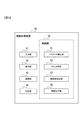
- FIG. 3 is a diagram illustrating the configuration of the information processing apparatus 40.
- the information processing apparatus 40 includes an input unit 41, a display unit 42, a communication unit 43, a storage unit 44, and a control unit 50.
- the control unit 50 includes a parameter calculation unit 51, a cancer determination unit 52, a reliability determination unit 53, and an information output unit 54.
- the input unit 41 is configured by an input device such as a keyboard or a touch panel, and is used as a reference for cancer determination such as a first threshold value T1, a second threshold value T2, a third threshold value T3, and / or a fourth threshold value T4, which will be described later.
- the input data including the numerical value is received.
- the input unit 41 stores the received input data in the storage unit 44 described later.
- the input data may be acquired via the communication unit 43 described later.
- the display unit 42 includes a display device such as a liquid crystal monitor, and displays a test result serving as an index for cancer determination and a determination result for cancer determination. Further, the display unit 42 appropriately displays a spectrum, a statistical value obtained from the spectrum, and the like.
- the display unit 42 may be configured to print and output the determination result on a paper medium such as thermal paper.
- the communication unit 43 is configured by a communication device that performs communication using a communication network such as the Internet.
- the communication unit 43 transmits a test result serving as an index of cancer determination, a determination result of cancer determination, and appropriately stores necessary data. Send and receive.
- the storage unit 44 is configured by a non-volatile memory or the like, a program for causing the control unit 50 to perform processing, measurement data output from the spectrometer 33, and a first threshold value T1, a second threshold value T2, and a third threshold value, which will be described later.
- a numerical value or the like serving as a reference in cancer determination, such as T3 and / or the fourth threshold T4 is stored.
- the control unit 50 is constituted by a processing device such as a CPU, functions as a main body for controlling the inspection device 100, and executes a program installed in the storage unit 44 or a storage medium (not shown), thereby causing cancer. Various processes such as calculation processing and output processing in determination are performed.
- the parameter calculation unit 51 of the control unit 50 calculates determination parameters used for cancer determination from the spectrum of the measurement data stored in the storage unit 44.
- the determination parameter of the present embodiment is a ratio between a signal corresponding to a predetermined amount of amino acid and a signal corresponding to a predetermined amount of interatomic bonds.
- the predetermined amino acid is preferably an aromatic amino acid and more preferably phenylalanine because of its high sensitivity in a spectroscopic spectrum, particularly Raman spectroscopy.
- the predetermined interatomic bond is preferably an interatomic bond that serves as an indicator of the amount of the organic compound, and includes interatomic bonds containing carbon atoms, particularly CH bonds in saturated hydrocarbons, double bonds or amide bonds between carbon atoms. Is more preferable.
- the signal corresponding to the amount of the amino acid or interatomic bond is measurement data such as a spectrum, and particularly when calculating the ratio, the peak corresponding to the predetermined amino acid or interatomic bond or the like. Is an intensity at a certain wavelength, an integrated value of the peak intensity, and the like.
- the ratio in the determination parameter may be expressed as, for example, 1: 2, or may be expressed as one numerical value as the ratio.
- a method for calculating a signal corresponding to a predetermined amino acid or a predetermined amount of interatomic bond is not particularly limited, and various statistical values can be used.
- the parameter calculation unit 51 can calculate the intensity of the wave number of the Stokes region of Raman scattered light corresponding to a predetermined amino acid or a predetermined interatomic bond as the signal.
- Parameter calculation unit 51 corresponds to an intensity of wave number 1003 / cm corresponding to phenylalanine, an intensity of 1450 / cm corresponding to CH 2 and CH 3 bonds of saturated hydrocarbon, or a double bond or amide bond between carbon atoms.
- the intensity of wave number 1659 / cm can be calculated as the signal.
- the parameter calculation unit 51 can use the maximum intensity, area, etc. of the peak corresponding to a predetermined amino acid or a predetermined interatomic bond as the signal. In the Stokes scattering region, the parameter calculation unit 51 calculates the maximum intensity of the peak including 1003 / cm in the half width range, the peak including 1450 / cm in the half width range, or the peak including 1659 / cm in the half width range. , Area, etc. can be calculated as the signal. When calculating the peak intensity, area, and the like, the parameter calculation unit 51 obtains the background by appropriately subtracting backgrounds such as noise and autofluorescence.
- the parameter calculation unit 51 determines a signal corresponding to the amount of the embedding agent (hereinafter referred to as a reliability determination parameter) in order to determine the reliability of cancer determination (hereinafter referred to as reliability determination), which will be described later. ).
- the parameter calculation unit 51 can calculate various statistical indexes as the reliability determination parameter. For example, the parameter calculation unit 51 calculates the intensity of the wave number in the Stokes region corresponding to the embedding agent in the Raman scattered light.
- the parameter calculation unit 51 calculates, as the reliability determination parameter, the intensity at a wave number of 1294 / cm corresponding to the paraffin in the Stokes region, or the ratio between the intensity of 1294 / cm and the intensity in the vicinity of 1294 / cm.
- the parameter calculation part 51 calculates the maximum intensity
- the parameter calculation unit 51 calculates the maximum intensity and area of the peak including 1294 / cm in the range of the half width in the Stokes scattering region.
- the parameter calculation unit 51 obtains the background by appropriately subtracting backgrounds such as noise and autofluorescence.
- the cancer determination unit 52 of the control unit 50 compares the determination parameter calculated by the parameter calculation unit 51 with a threshold value (hereinafter referred to as a determination threshold value) stored in the storage unit 44 and serving as a determination reference. Thus, it is determined whether or not the region of interest 90 includes cancer cells.
- a threshold value hereinafter referred to as a determination threshold value
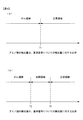
- FIG. 4A is a diagram for explaining a determination method in the case where there is one determination threshold value.
- a signal corresponding to a predetermined interatomic bond is called a reference signal.
- the horizontal axis of FIG. 4 shows the ratio of the detection amount of a predetermined amino acid to the detection amount with respect to the reference signal, that is, the determination parameter.
- T1 the determination threshold value
- the determination parameter is in the normal region, and the corresponding region of interest 90 is determined not to contain cancer cells, that is, “normal”.
- the determination parameter is equal to or less than the determination threshold T1
- the determination parameter is in the cancer region, and the corresponding region of interest 90 is determined to include cancer cells, that is, “cancer” or “malignant”.
- FIG. 4B is a diagram for explaining a determination method when there are a plurality of determination threshold values.
- a determination threshold T2 and a determination threshold T3 that is larger than the determination threshold T2 are used.
- the determination parameter is larger than the determination threshold T3
- the determination parameter is in the normal region, and the corresponding region of interest 90 is determined not to contain cancer cells, that is, “normal”.
- the determination parameter is equal to or less than the determination threshold T2
- the determination parameter is in the cancer region, and the corresponding region of interest 90 is determined to include cancer cells, that is, “cancer” or “malignant”.
- the determination parameter is greater than T2 and equal to or less than T3, the determination parameter is in an intermediate region between the cancer region and the normal region, and the corresponding region of interest 90 may not be subjected to cancer determination or may include cancer cells. It is determined that the state is indefinite, such as having a certain degree of sex. In the above description, it is assumed that the value of the determination parameter is larger in the normal region than in the cancer region, but may be appropriately reversed according to the statistical data based on the determination parameter. In the above description, the two determination thresholds T2 and T3 are used. However, three or more determination thresholds may be used, and the region may be set stepwise in accordance with the possibility of cancer. Good.
- the determination threshold values T1, T2, and T3 are calculated from past statistical data and the like and are set in advance.
- the determination threshold is preferably set for precise determination based on the type of cancer, the site, the gene involved, and the like.
- cancer cancer (carcinoma) is preferable, and cancer of the digestive system, particularly pancreatic cancer is preferable.
- the determination threshold values T1, T2, and T3 can be appropriately set as to which of the determination threshold values and the measurement values are included. The same applies to the following reliability determination threshold value T4.
- FIG. 5 is a diagram for explaining a reliability determination method based on the detection amount of the embedding agent.
- the horizontal axis in FIG. 5 represents the detected amount of the embedding agent, that is, the reliability determination parameter.
- the reliability determination parameter is a value larger than a determination threshold for reliability (hereinafter referred to as a reliability determination threshold) T4
- a reliability determination threshold for reliability
- the information output unit 54 of the control unit 50 creates a character string or an image including the obtained determination result of cancer determination and the determination result of reliability determination, and outputs it to the display unit 42.
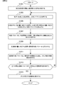
- FIG. 6 is a flowchart showing the flow of the cancer testing method of the present embodiment. With this inspection method, it is possible to accurately determine whether or not cancer exists using optical means.
- step S1001 a section 9 is created from the organ, organ, etc. of the measurement subject by a medical worker or the like.
- step S1001 ends, the process proceeds to step S1003.
- step S ⁇ b> 1003 the inspection apparatus 100 irradiates the region of interest 90 of the slice 9 with the light generated by the light source device 11 and acquires a spectral spectrum of the light from the slice 9.
- step S1003 ends, the process proceeds to step S1005.
- step S1005 the parameter calculation unit 51 determines from the spectrum that the signal corresponding to the amount of a predetermined amino acid such as phenylalanine and the signal corresponding to the amount of a predetermined interatomic bond such as a C—H bond of a saturated hydrocarbon. The ratio (determination parameter) is calculated.
- step S1005 ends, the process proceeds to step S1007.
- step S1007 the determination parameter calculated in step S1005 is compared with the determination threshold stored in the storage unit 44 to determine whether the region of interest 90 includes cancer cells.
- step S1007 ends, the process proceeds to step S1009.
- step S1009 the parameter calculation unit 51 calculates a signal (reliability determination parameter) corresponding to the amount of the embedding agent from the spectrum.
- step S1011 the reliability determination unit 53 compares the reliability determination parameter calculated in step S1009 with the reliability determination threshold stored in the storage unit 44, and determines the reliability of cancer determination.
- step S1011 ends, the process proceeds to step S1013.
- step S1013 the control unit 50 determines whether or not the cancer determination and the reliability determination have been completed for all the regions of interest 90 set in the section 9. When the cancer determination and the reliability determination are finished, an affirmative determination is made in step S1013 and the process proceeds to step S1015. If the cancer determination and the reliability determination are not completed, a negative determination is made in step S1013 and the process returns to step S1003.
- the information output unit 54 displays the determination result of the cancer determination using the display unit 42.
- the information output unit 54 determines that at least one region of interest 90 in which cancer determination has been performed includes a cancer cell, and if there is a region of interest 90 in which reliability is determined to be high by the reliability determination, 9 or the relevant part from which the section 9 is taken out is output as containing cancer cells. Further, the information output unit 54 may cause the display unit 42 to display an image showing the determination result of the cancer determination for each region of interest 90 of the section 9. In this case, for the region of interest 90 with low reliability, a point that cannot be measured can be displayed.
- the cancer testing method of this embodiment includes a step of measuring a signal corresponding to a predetermined amount of amino acid in a biological sample and a signal corresponding to a predetermined amount of interatomic bond, and a ratio between the two signals. Based on this, it is determined whether or not the biological sample contains cancer cells.
- the cancer inspection apparatus 100 includes a parameter calculation unit that calculates the ratio and the biological sample based on the ratio. A cancer determination unit that determines whether or not cancer cells are included. Thereby, it can be determined whether cancer exists correctly.
- the signal corresponding to the predetermined amount of interatomic bonds is a signal corresponding to the amount of organic compound in the biological sample.
- atoms bonded by a predetermined interatomic bond include carbon atoms.
- the predetermined interatomic bond is a C—H bond in a saturated hydrocarbon, a double bond between carbon atoms, or an amide bond.
- the predetermined amino acid is phenylalanine. Thereby, it can be determined whether there is a cancer related to phenylalanine.
- the determination parameter is based on the data of the intensity corresponding to the predetermined amino acid and the intensity corresponding to the predetermined interatomic bond in the spectrum. Is calculated. Thereby, it is possible to accurately determine whether or not cancer exists using optical means.
- the spectral spectrum is a spectral spectrum of Raman scattered light.
- a peak including 1003 / cm in the range of half width and 1450 / in the range of half width is calculated based on a statistical value calculated from data with a peak including cm or 1659 / cm.
- the biological sample to be subjected to cancer determination is pancreatic tissue, and the cancer testing apparatus 100 tests pancreatic cancer. Thereby, it can be determined whether pancreatic cancer exists correctly.
- cancer determination is performed based on whether or not the determination parameter is larger than the determination threshold T1. Thereby, it can be determined whether the cancer exists simply and correctly.
- the cancer determination is based on whether the determination parameter is smaller than the determination threshold T2 or larger than the determination threshold T3 larger than the determination threshold T2. Is done. Thereby, it can prevent that the determination result of a false positive or a false negative comes out.
- the reliability of cancer determination is determined based on a signal corresponding to the amount of the embedding agent in the biological sample, and the information output unit of the cancer inspection apparatus 100 54 outputs information on the reliability of cancer determination.
- the inspection apparatus 100 includes the measurement unit 2 and the information processing apparatus 40.
- FIG. 7 is a diagram showing a configuration of the cancer inspection system 1.
- the cancer testing system 1 includes a measuring device 3 and an information processing device 40.
- the measurement device 3 includes a measurement-side communication device 39 in addition to the configuration of the measurement unit 2 of the above-described embodiment, and processes measurement data such as a spectrum acquired by the measurement device 3 via a communication network 8 such as the Internet. Transmit to device 40.
- the information processing apparatus 40 of this modification has the same configuration as the information processing apparatus 40 of the above-described embodiment.
- the cancer testing system 1 includes a measuring device 3 that measures a signal corresponding to a predetermined amino acid in a biological sample and a signal corresponding to a predetermined interatomic bond, and a signal corresponding to the amount of the predetermined amino acid in the biological sample. Calculate a ratio with a signal corresponding to a predetermined amount of interatomic bonds, and output the ratio, or determine whether a biological sample contains cancer cells based on the ratio And an information processing apparatus 40 for outputting. Thereby, even when the person who analyzes the measurement result such as a pathologist is located remotely from the measuring apparatus 3, it is possible to quickly determine whether or not cancer exists.
- Modification 2 A program for realizing the information processing function of the information processing apparatus 40 of the present embodiment is recorded on a computer-readable recording medium, and the above-described determination parameter calculation process, determination process, and the like recorded on the recording medium A test program for cancer may be loaded into a computer system and executed.
- the “computer system” includes an OS (Operating System) and hardware of peripheral devices.
- the “computer-readable recording medium” refers to a portable recording medium such as a flexible disk, a magneto-optical disk, an optical disk, and a memory card, and a storage device such as a hard disk built in the computer system.
- the “computer-readable recording medium” dynamically holds a program for a short time like a communication line when transmitting a program via a network such as the Internet or a communication line such as a telephone line.
- a volatile memory in a computer system serving as a server or a client in that case may be included and a program that holds a program for a certain period of time may be included.
- the above program may be for realizing a part of the functions described above, or may be realized by a combination with the program already recorded in the computer system. .
- FIG. 8 shows the state.
- the PC 950 is provided with a program via the CD-ROM 953. Further, the PC 950 has a connection function with the communication line 951.
- a computer 952 is a server computer that provides the program, and stores the program in a recording medium such as a hard disk.
- the communication line 951 is a communication line such as the Internet or personal computer communication, or a dedicated communication line.
- the computer 952 reads the program using the hard disk and transmits the program to the PC 950 via the communication line 951. That is, the program is transmitted as a data signal by a carrier wave and transmitted via the communication line 951.
- the program can be supplied as a computer-readable computer program product in various forms such as a recording medium and a carrier wave.
- Section 9 was a human pancreas section. That is, the sample containing the above cancer cells is a pancreatic cancer sample.
- a sample was fixed using paraffin, a specimen was prepared, and then a known deparaffinization treatment was performed.
- a region of interest 90 having an area of about 10 to 1000 ⁇ m 2 is set for the sample subjected to deparaffinization processing, the region of interest 90 is divided into a plurality of unit regions, and a wavelength of a beam diameter of about 1 ⁇ m for each unit region.
- the Raman scattered light was measured by irradiating a laser beam of 532 nm.
- a plurality of spectra acquired for each unit region was averaged, and peaks corresponding to glass and autofluorescence were removed by a known algorithm.
- the ratio of the intensity of 1003 / cm corresponding to phenylalanine to the intensity of 1450 / cm corresponding to saturated hydrocarbon CH bond (I 1003 / I 1450 , Hereinafter referred to as determination parameter A).
- the ratio of the intensity of 1294 / cm corresponding to paraffin to the intensity of 1316 / cm near the peak corresponding to paraffin (I 1294 / I 1316 , hereinafter, reliability determination parameter) was calculated.
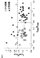
- FIG. 9 is a graph showing the determination parameter A of each sample on the horizontal axis and the reliability determination parameter on the vertical axis. Based on this result, points having a reliability determination parameter of T4a or higher can be excluded from the cancer determination, and cancer determination can be performed using the determination parameter only for points lower than T4a.
- the region where the determination parameter A is less than T1 and the reliability parameter is less than T4a corresponds to the sample of cancer tissue. Since there are only measurement points, this region is defined as a cancer region Dc, and a sample in which a value that falls within the numerical range can be determined to be composed of cancer tissue.
- the determination parameter A is T1 or more and the reliability parameter is less than T4a
- this region is set as the normal region Dn, and a value that falls within the numerical range is measured. It can be determined that the prepared sample consists of normal tissue.
- the reliability parameter is T4a or higher, it is difficult to define the boundary between the cancer area and the normal area. Therefore, if the value is set as the low reliability area De and a value that falls within the numerical range is measured, the cancer Judgment reliability is low or impossible.
- the determination threshold values T1 and T4a are set based on the coordinate values of the normal tissue measurement points P1 and P2 so that only the cancer tissue measurement points are included in the cancer region.
- the determination threshold values T1, T2, T3, and the reliability determination threshold value T4 from a given measurement value is not particularly limited, and any statistical value regarding cancer tissue and normal tissue can be used.
- the distribution of each parameter may be approximated by a normal distribution, and the determination threshold may be set at a position such as 1 ⁇ point, 2 ⁇ point, 3 ⁇ point, etc. from the average value. Further, in FIG. 9, one measurement point is excluded as an outlier by the test.
- the ratio of the intensity of 1003 / cm corresponding to phenylalanine to the intensity of 1659 / cm corresponding to the double bond and amide bond between carbon atoms in the Stokes scattering region. (I 1003 / I 1659 , hereinafter referred to as determination parameter B) was calculated.
- the ratio of the intensity of 1294 / cm corresponding to paraffin to the intensity of 1316 / cm near the peak corresponding to paraffin (I 1294 / I 1316 , reliability determination parameter) was calculated.
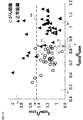
- FIG. 10 is a graph showing the determination parameter B of each sample on the horizontal axis and the reliability determination parameter on the vertical axis.
- a region where the determination parameter B is less than T2 and the reliability parameter is less than T4b corresponds to a sample of cancer tissue. Since there are only measurement points, this region is defined as a cancer region Dc, and a sample in which a value that falls within the numerical range can be determined to be composed of cancer tissue.
- a normal tissue sample and a cancer tissue sample are included in a region where the determination parameter B is T2 or more and less than T3 and the reliability parameter is less than T4b (a region surrounded by a broken line 84b, a broken line 82, a broken line 83, and a horizontal axis). Since both of the corresponding measurement points are included, this region is defined as an intermediate region Di, and a sample whose value is within the numerical range can be determined to be composed of tissue suspected of having cancer. . In the region where the determination parameter B is equal to or greater than T3 and the reliability parameter is less than T4b, there are only measurement points corresponding to the normal tissue sample.
- this region is defined as the normal region Dn, and a value that falls within the numerical range is measured. It can be determined that the prepared sample consists of normal tissue.
- the reliability parameter is T4b or higher, it is difficult to define the boundary between the cancer region and the normal region. Therefore, if the value is set as the low reliability region De and a value that falls within the numerical range is measured, the cancer Judgment reliability is low or impossible.
- the determination threshold values T2 and T3 are set to the coordinate values of the measurement points P3 and P4 so that only the measurement point of the cancer tissue is included in the cancer region Dc and only the measurement point of the normal tissue is included in the normal region Dn. Set based on.
- the present invention is not limited to the contents of the above embodiment.
- Other embodiments conceivable within the scope of the technical idea of the present invention are also included in the scope of the present invention.
Landscapes
- Life Sciences & Earth Sciences (AREA)
- Health & Medical Sciences (AREA)
- Engineering & Computer Science (AREA)
- Chemical & Material Sciences (AREA)
- Immunology (AREA)
- Biomedical Technology (AREA)
- Physics & Mathematics (AREA)
- General Health & Medical Sciences (AREA)
- Pathology (AREA)
- Hematology (AREA)
- Molecular Biology (AREA)
- Urology & Nephrology (AREA)
- General Physics & Mathematics (AREA)
- Biochemistry (AREA)
- Analytical Chemistry (AREA)
- Medicinal Chemistry (AREA)
- Food Science & Technology (AREA)
- Biotechnology (AREA)
- Proteomics, Peptides & Aminoacids (AREA)
- Cell Biology (AREA)
- Microbiology (AREA)
- Biophysics (AREA)
- Nuclear Medicine, Radiotherapy & Molecular Imaging (AREA)
- Investigating Or Analysing Biological Materials (AREA)
- Investigating, Analyzing Materials By Fluorescence Or Luminescence (AREA)
Abstract
がんの検査方法は、生体試料における所定のアミノ酸の量に対応した信号と所定の原子間結合の量に対応する信号とを計測する工程と、信号に基づいて、生体試料が、がん細胞を含むか否かの判定を行う工程とを備える。
Description
本発明は、がんの検査方法、がんの検査装置、検査プログラムおよびがんの検査システムに関する。
生体組織等からの光を解析することにより、当該生体組織等にがん化した部位があるか否かを調べる検査方法が提案されている(特許文献1参照)。
本発明の第1の態様によると、がんの検査方法は、生体試料のがんの検査方法であって、前記生体試料における所定のアミノ酸の量に対応する信号と所定の原子間結合の量に対応する信号を計測する工程と、前記信号に基づいて、前記生体試料が、がん細胞を含むか否かの判定を行う。
本発明の第2の態様によると、がんの検査装置は、生体試料における所定のアミノ酸の量に対応する信号と所定の原子間結合の量に対応する信号との比を算出するパラメータ算出部と、前記比に基づいて、前記生体試料が、がん細胞を含むか否かの判定を行うがん判定部とを備える。
本発明の第3の態様によると、検査プログラムは、生体試料における所定のアミノ酸の量に対応する信号と所定の原子間結合の量に対応する信号との比を算出する算出処理と、前記比を出力する、または、前記比に基づいて前記生体試料ががん細胞を含むか否かの判定を行った判定結果を出力する出力処理と、を処理装置に行わせる。
本発明の第4の態様によると、がんの検査システムは、生体試料における所定のアミノ酸に対応する信号と所定の原子間結合に対応する信号とを測定する測定装置と、前記所定のアミノ酸の量に対応する信号と、前記所定の原子間結合の量に対応する信号との比を算出し、前記比を出力する、または、前記比に基づいて前記生体試料ががん細胞を含むか否かの判定を行った判定結果を出力する出力装置と、を備える。
本発明の第2の態様によると、がんの検査装置は、生体試料における所定のアミノ酸の量に対応する信号と所定の原子間結合の量に対応する信号との比を算出するパラメータ算出部と、前記比に基づいて、前記生体試料が、がん細胞を含むか否かの判定を行うがん判定部とを備える。
本発明の第3の態様によると、検査プログラムは、生体試料における所定のアミノ酸の量に対応する信号と所定の原子間結合の量に対応する信号との比を算出する算出処理と、前記比を出力する、または、前記比に基づいて前記生体試料ががん細胞を含むか否かの判定を行った判定結果を出力する出力処理と、を処理装置に行わせる。
本発明の第4の態様によると、がんの検査システムは、生体試料における所定のアミノ酸に対応する信号と所定の原子間結合に対応する信号とを測定する測定装置と、前記所定のアミノ酸の量に対応する信号と、前記所定の原子間結合の量に対応する信号との比を算出し、前記比を出力する、または、前記比に基づいて前記生体試料ががん細胞を含むか否かの判定を行った判定結果を出力する出力装置と、を備える。
以下では、適宜図面を参照しながら、一実施形態のがんの検査方法、がんの検査装置、検査プログラム、がんの検査システム等について説明する。本実施形態のがんの検査方法では、生体試料からの光、例えば、生体から採取した試料からの光を測定し、測定結果に基づいて、上記生体試料ががん細胞を含む可能性についての情報の提示や、がん細胞を含むか否かの判定(以下、適宜「がん判定」と呼ぶ)を行う。
なお、本実施形態において、「がん」とは、癌(癌腫)、肉腫、血液腫瘍およびその他の悪性腫瘍を含む。また、「生体試料」とは、生体から採取された細胞、組織、臓器、病理切片等に加え、生体内の細胞、組織、臓器を含む。
なお、本実施形態において、「がん」とは、癌(癌腫)、肉腫、血液腫瘍およびその他の悪性腫瘍を含む。また、「生体試料」とは、生体から採取された細胞、組織、臓器、病理切片等に加え、生体内の細胞、組織、臓器を含む。
以下では、本実施形態のがんの検査方法におけるがん判定の一態様として、目的の組織の規格化されたアミノ酸の量を示していると考えられる、分光スペクトルにおける信号強度やピーク比等を算出し、このピーク比等に基づいてがん判定を行う。これにより、分光スペクトルから得られる少数のパラメータに基づいて、正確な判定結果を得ることができ、医療等の様々な局面で利用することが可能である。
図1は、本実施形態のがんの検査方法に用いられる検査装置100の構成を示す図である。本実施形態において、検査装置100は、がんの検査装置であり、ラマン散乱光の分光スペクトルを測定する。検査装置100は、測定部2と、情報処理装置40とを備える。測定部2は、照射光学系10と、対物光学系20と、検出光学系30とを備える。照射光学系10は、光源装置11と、ガルバノスキャナ12と、リレーレンズ13a、13bと、光学ミラー14とを備える。対物光学系20は、光学ミラー21と、対物レンズ22と、ステージ23とを備える。ステージ23には、生体試料を切り出し適宜標本として調整された切片9が載置されている。検出光学系30は、ダイクロイックミラー31と、リレーレンズ32a、32bと、分光器33とを備える。図1では、照射光および検出光を一点鎖線を用いて模式的に示した。
なお、図1では後方散乱された光を解析する構成になっているが、前方散乱された光を解析する構成にしてもよい。切片9からの光から解析可能な分光スペクトルが得られれば、検査装置100の構成は特に限定されない。また、ラマン散乱光以外の赤外分光法等により測定部2を構成してもよい。
なお、図1では後方散乱された光を解析する構成になっているが、前方散乱された光を解析する構成にしてもよい。切片9からの光から解析可能な分光スペクトルが得られれば、検査装置100の構成は特に限定されない。また、ラマン散乱光以外の赤外分光法等により測定部2を構成してもよい。
光源装置11は、半導体レーザ等のレーザ発振装置等を含んで構成される。光源装置11が発振する光の波長は、散乱光の分光スペクトルが測定可能なら特に限定されないが、測定感度等の観点から適宜532nm、785nm、1064nm等の波長を用いることができる。光源装置11から出射された光は、ガルバノスキャナ12に入射する。
ガルバノスキャナ12は、不図示の可動ミラー等を含んで構成され、レーザ光の出射方向を変化させ走査させる。ガルバノスキャナ12を出射した光は、リレーレンズ13aおよび13bに入射する。リレーレンズ13a、13bを通過した光は光学ミラー14、ダイクロイックミラー31で反射された後、対物光学系20に入射する。
対物光学系20に入射した光は、光学ミラー21で反射され、対物レンズ22で屈折され、切片9の関心領域に入射する。
図2は、関心領域90を示した図である。関心領域90は、切片9に含まれ測定対象となる領域である。関心領域90は、切片9に含まれる細胞の大きさと同程度か、より広い面積を持つように設定することが、細胞内の物質分布の偏りの影響を防ぐために好ましい。関心領域90の幅Wは、1μm~1mm等に設定することができる。関心領域90は、複数の単位領域91を含み、それぞれの単位領域91に対して分光計測を行った結果の算術平均等から後述の判定パラメータが算出される。単位領域91のそれぞれの大きさは、照射光の点像分布関数(Point Spread Function:PSF)等に基づいて分光スペクトルが測定可能な範囲等から適宜設定することができ、例えば、1μm×1μm等の領域に設定することができる。
切片9における関心領域90の数、形状および位置は特に限定されず、関心領域90における単位領域91の数、形状および位置は特に限定されない。
切片9における関心領域90の数、形状および位置は特に限定されず、関心領域90における単位領域91の数、形状および位置は特に限定されない。
切片9の関心領域90で散乱された光は、対物レンズ22を通過して光学ミラー21で反射され、検出光学系30に入射する。
検出光学系30に入射した光は、ダイクロイックミラー31を透過してリレーレンズ32a、32bを透過し、分光器33に入射する。分光器33は、グレーティングやCCD等の検出器等を含んで構成され、入射した光を分光して波長ごとの強度を取得する。分光器33で波長ごとの強度が取得されたら、当該強度等のデータは情報処理装置40に出力(図1の実線矢印で情報の流れを示した)され、後述の記憶部44に記憶される。
図3は、情報処理装置40の構成を示した図である。情報処理装置40は、入力部41と、表示部42と、通信部43と、記憶部44と、制御部50とを備える。制御部50は、パラメータ算出部51と、がん判定部52と、信頼度判定部53と、情報出力部54とを備える。
入力部41は、キーボードやタッチパネル等の入力装置により構成され、後述の第1閾値T1、第2閾値T2、第3閾値T3、および/または第4閾値T4等の、がん判定の際に基準となる数値を含む入力データを受け付ける。入力部41は、受け付けた入力データを、後述の記憶部44に記憶させる。
なお、入力データは、後述の通信部43を介して取得してもよい。
なお、入力データは、後述の通信部43を介して取得してもよい。
表示部42は、液晶モニタ等の表示装置により構成され、がん判定の指標となる検査結果や、がん判定の判定結果を表示する。また、表示部42は、分光スペクトルや当該分光スペクトルから得た統計値等を適宜表示する。
なお、表示部42は、感熱紙等の紙媒体に判定結果等を印字して出力する構成にしてもよい。
なお、表示部42は、感熱紙等の紙媒体に判定結果等を印字して出力する構成にしてもよい。
通信部43は、インターネット等の通信網を利用して通信を行う通信装置により構成され、がん判定の指標となる検査結果や、がん判定の判定結果を送信したり、適宜必要なデータを送受信する。記憶部44は、不揮発性メモリ等により構成され、制御部50に処理を行わせるプログラム、分光器33から出力された測定データ、ならびに、後述の第1閾値T1、第2閾値T2、第3閾値T3、および/または第4閾値T4等の、がん判定の際に基準となる数値等を記憶する。
制御部50は、CPU等の処理装置により構成され、検査装置100を制御する動作の主体として機能し、記憶部44または不図示の記憶媒体に搭載されているプログラムを実行することにより、がん判定における計算処理、出力処理等の各種処理を行う。
制御部50のパラメータ算出部51は、記憶部44に記憶されている測定データの分光スペクトルから、がん判定に用いる判定パラメータを算出する。本実施形態の判定パラメータは、所定のアミノ酸の量に対応する信号と、所定の原子間結合の量に対応する信号との比である。所定のアミノ酸としては、分光スペクトル、特にラマン分光法における感度が高いことから、芳香族アミノ酸が好ましく、フェニルアラニンがより好ましい。所定の原子間結合としては、有機化合物の量の指標となる原子間結合が好ましく、炭素原子を含む原子間結合、特に飽和炭化水素におけるC-H結合、炭素原子間の二重結合またはアミド結合がより好ましい。ここで、上記のアミノ酸や原子間結合の量に対応する信号とは、分光スペクトル等の測定データであり、特に比を算出する場合は、上記の所定のアミノ酸や原子間結合等に対応するピークについてのある波長での強度や、当該ピーク強度の積分値等を指す。
なお、上記判定パラメータにおける比は、例えば1:2のように表してもよいし、比率として1つの数値で表してもよい。
なお、上記判定パラメータにおける比は、例えば1:2のように表してもよいし、比率として1つの数値で表してもよい。
所定のアミノ酸、または所定の原子間結合の量に対応する信号の算出方法は、特に限定されず、様々な統計値を用いることができる。パラメータ算出部51は、所定のアミノ酸または所定の原子間結合に対応する、ラマン散乱光のストークス領域の波数の強度を上記信号として算出することができる。パラメータ算出部51は、フェニルアラニンに対応する波数1003/cmの強度、飽和炭化水素のCH2およびCH3結合に対応する1450/cmの強度、または炭素原子間の二重結合若しくはアミド結合に対応する波数1659/cmの強度を上記信号として算出することができる。
あるいは、パラメータ算出部51は、所定のアミノ酸または所定の原子間結合に対応するピークの最大強度、面積等を上記信号として用いることができる。パラメータ算出部51は、ストークス散乱域において、半値幅の範囲に1003/cmを含むピーク、半値幅の範囲に1450/cmを含むピーク、または半値幅の範囲に1659/cmを含むピークの最大強度、面積等を上記信号として算出することができる。パラメータ算出部51は、ピークの強度、面積等を算出する場合は、ノイズや自家蛍光等のバックグラウンドを適宜差し引いて求める。
パラメータ算出部51は、後述される、がん判定の信頼度の判定(以下、信頼度判定と呼ぶ)のため、包埋剤の量に対応する信号をパラメータ(以下、信頼度判定パラメータと呼ぶ)として算出する。パラメータ算出部51は、信頼度判定パラメータとして様々な統計的指標を算出することができるが、例えば、ラマン散乱光における包埋剤に対応するストークス領域の波数の強度を算出する。パラメータ算出部51は、信頼度判定パラメータとして、ストークス領域のパラフィンに対応する波数1294/cmの強度や、1294/cmの強度と1294/cmの近傍の強度との比を算出する。または、パラメータ算出部51は、包埋剤に対応するピークの最大強度や面積等を信頼度判定パラメータとして算出する。この場合、パラメータ算出部51は、ストークス散乱域において、半値幅の範囲に1294/cmを含むピークの最大強度や面積等を算出することが好ましい。パラメータ算出部51は、ピークの強度、面積等を算出する場合は、ノイズや自家蛍光等のバックグラウンドを適宜差し引いて求める。
制御部50のがん判定部52は、パラメータ算出部51で算出された判定パラメータと、記憶部44に記憶されており判定の基準となる閾値(以下、判定閾値と呼ぶ)とを比較することにより、関心領域90ががん細胞を含むか否かを判定する。
図4(a)は、判定閾値が1つの場合の判定方法を説明するための図である。図4では、所定の原子間結合に対応する信号を基準信号と呼ぶ。図4の横軸は、所定のアミノ酸の検出量の、基準信号についての検出量に対する比率、すなわち判定パラメータを示した。判定パラメータが判定閾値T1よりも大きい値の場合、判定パラメータは正常領域にあり、対応する関心領域90はがん細胞を含まない、すなわち「正常」と判定される。判定パラメータが判定閾値T1以下の場合、判定パラメータはがん領域にあり、対応する関心領域90はがん細胞を含む、すなわち「がん」または「悪性」等と判定される。
図4(b)は、判定閾値が複数の場合の判定方法を説明するための図である。この判定方法では、判定閾値T2と、判定閾値T2より大きい値の判定閾値T3を用いる。判定パラメータが判定閾値T3よりも大きい値の場合、判定パラメータは正常領域にあり、対応する関心領域90はがん細胞を含まない、すなわち「正常」と判定される。判定パラメータが判定閾値T2以下の場合、判定パラメータはがん領域にあり、対応する関心領域90はがん細胞を含む、すなわち「がん」または「悪性」等と判定される。判定パラメータがT2より大きくT3以下の場合、判定パラメータはがん領域と正常領域との間の中間領域にあり、対応する関心領域90については、がん判定をしないか、がん細胞を含む可能性が一定程度存在する等の不確定な状態と判定する。
なお、上記ではがん領域よりも正常領域の方が、判定パラメータの値が大きいとしたが、判定パラメータが基づく統計データに合わせて適宜逆に設定され得る。また、上記では2つの判定閾値T2、T3を用いたが、3以上の判定閾値を用いてもよく、それに伴いがんが存在する可能性の高さに応じ段階的に領域を設定してもよい。
なお、上記ではがん領域よりも正常領域の方が、判定パラメータの値が大きいとしたが、判定パラメータが基づく統計データに合わせて適宜逆に設定され得る。また、上記では2つの判定閾値T2、T3を用いたが、3以上の判定閾値を用いてもよく、それに伴いがんが存在する可能性の高さに応じ段階的に領域を設定してもよい。
判定閾値T1、T2、T3は、過去の統計データ等から算出され、予め設定されたものを用いる。判定閾値は、がんの種類や、部位、関与する遺伝子等に基づいてそれぞれ設定されることが精密な判定のために好ましい。がんの種類または部位としては、癌(癌腫)が好ましく、また、消化器系のがん、特にすい臓がんが好ましい。
なお、判定閾値T1、T2、T3について、判定閾値と測定値とが等しい場合についてどちらに含めるかは適宜設定することができる。以下の信頼度判定閾値T4についても同様である。
なお、判定閾値T1、T2、T3について、判定閾値と測定値とが等しい場合についてどちらに含めるかは適宜設定することができる。以下の信頼度判定閾値T4についても同様である。
制御部50の信頼度判定部53は、関心領域90で測定した包埋剤に対応する信号から得られた包埋剤の量に対応する信号に基づいて、がん判定部52が行ったがん判定の信頼度を判定する。包埋剤の測定では、切片9を固定する際に用いられたパラフィン、OCT(Optimal Cutting Temperature)コンパウンド等を測定する。残存するパラフィンが信頼性の判定に大きく影響するため、例えばパラフィンの量に対応する信号に基づいて、がん判定部52が行ったがん判定の信頼度を判定することが特に有用である。
なお、包埋剤に限らず、ノイズ要素、特に判定パラメータの測定に影響を与えるバックグラウンドとなる要素の量に対応する信号に基づいて、がん判定部52が行ったがん判定の信頼度を判定する構成にしてもよい。
なお、包埋剤に限らず、ノイズ要素、特に判定パラメータの測定に影響を与えるバックグラウンドとなる要素の量に対応する信号に基づいて、がん判定部52が行ったがん判定の信頼度を判定する構成にしてもよい。
図5は、包埋剤の検出量に基づく信頼度の判定方法を説明するための図である。図5の横軸は、包埋剤の検出量、すなわち信頼度判定パラメータである。信頼度判定パラメータが信頼度についての判定閾値(以下、信頼度判定閾値と呼ぶ)T4よりも大きい値の場合、対応する関心領域90についてのがん判定の信頼度は低いと判断される。信頼度判定パラメータが信頼度判定閾値T4以下の場合、対応する関心領域90についてのがん判定の信頼度は高いと判定される。
制御部50の情報出力部54は、得られたがん判定の判定結果および信頼度判定の判定結果を含む、文字列または画像を作成し、表示部42に出力する。
図6は、本実施形態のがんの検査方法の流れを示すフローチャートである。この検査方法により、光学的手段を用いてがんが存在するか否かの判定を正確に行うことができる。
ステップS1001において、医療従事者等により、測定対象者の臓器、器官等から切片9が作成される。ステップS1001が終了したら、ステップS1003に進む。ステップS1003において、検査装置100は、切片9の関心領域90に光源装置11が生成した光を照射し、切片9からの光の分光スペクトルを取得する。ステップS1003が終了したら、ステップS1005に進む。
ステップS1005において、パラメータ算出部51は、上記分光スペクトルから、フェニルアラニン等の所定のアミノ酸の量に対応する信号と、飽和炭化水素のC-H結合等の所定の原子間結合の量に対応する信号との比(判定パラメータ)を算出する。ステップS1005が終了したら、ステップS1007に進む。ステップS1007において、ステップS1005で算出した判定パラメータと、記憶部44に記憶された判定閾値とを比較し、関心領域90ががん細胞を含むか否かを判定する。ステップS1007が終了したら、ステップS1009に進む。
ステップS1009において、パラメータ算出部51は、上記分光スペクトルから、包埋剤の量に対応する信号(信頼度判定パラメータ)を算出する。ステップS1009が終了したら、ステップS1011に進む。ステップS1011において、信頼度判定部53は、ステップS1009で算出した信頼度判定パラメータと、記憶部44に記憶された信頼度判定閾値とを比較し、がん判定の信頼度を決定する。ステップS1011が終了したら、ステップS1013に進む。
ステップS1013において、制御部50は、切片9に設定された全ての関心領域90についてがん判定および信頼度判定が終了したか否かを判定する。がん判定および信頼度判定が終了している場合、ステップS1013を肯定判定してステップS1015に進む。がん判定および信頼度判定が終了していない場合、ステップS1013を否定判定してステップS1003に戻る。
ステップS1015において、情報出力部54は、がん判定の判定結果を表示部42を用いて表示する。情報出力部54は、がん判定を行った関心領域90のうち、一つでもがん細胞を含むと判定され、信頼度判定により信頼度も高いと判定された関心領域90がある場合、切片9、または切片9を取り出した対象の当該部位はがん細胞を含むとして出力する。また、情報出力部54は、切片9の各関心領域90に対してがん判定の判定結果を示した画像を表示部42に表示させてもよい。この場合、信頼度の低い関心領域90については、測定不能である点を表示させることができる。
上述の実施の形態によれば、次の作用効果が得られる。
(1)本実施形態のがんの検査方法は、生体試料における所定のアミノ酸の量に対応する信号と所定の原子間結合の量に対応する信号とを計測する工程と、両信号の比に基づいて、生体試料が、がん細胞を含むか否かの判定を行うものであり、がんの検査装置100は、上記比を算出するパラメータ算出部と、上記比に基づいて、生体試料ががん細胞を含むか否か判定するがん判定部とを備える。これにより、正確にがんが存在するか否かの判定を行うことができる。
(1)本実施形態のがんの検査方法は、生体試料における所定のアミノ酸の量に対応する信号と所定の原子間結合の量に対応する信号とを計測する工程と、両信号の比に基づいて、生体試料が、がん細胞を含むか否かの判定を行うものであり、がんの検査装置100は、上記比を算出するパラメータ算出部と、上記比に基づいて、生体試料ががん細胞を含むか否か判定するがん判定部とを備える。これにより、正確にがんが存在するか否かの判定を行うことができる。
(2)本実施形態のがんの検査方法または検査装置100において、所定の原子間結合の量に対応する信号は、生体試料における有機化合物の量に対応する信号である。これにより、特定の化合物等のスペクトルを基準にする場合に比べ、大きなピークを基準にするため、判定パラメータのばらつきを抑えることができる。
(3)本実施形態のがんの検査方法またはがんの検査装置100において、所定の原子間結合で結合されている原子は、炭素原子を含む。これにより、有機的な原子間結合による大きなピークを基準にするため、判定パラメータのばらつきを抑えることができる。
(4)本実施形態のがんの検査方法またはがんの検査装置100において、所定の原子間結合は、飽和炭化水素におけるC-H結合、炭素原子間の二重結合、またはアミド結合である。これにより、分光スペクトルにおける大きなピークを基準にするため、より正確にがんが存在するか否かの判定を行うことができる。
(5)本実施形態のがんの検査方法またはがんの検査装置100において、所定のアミノ酸は、フェニルアラニンである。これにより、フェニルアラニンに関連するがんが存在するか否かの判定を行うことができる。
(6)本実施形態のがんの検査方法またはがんの検査装置100において、分光スペクトルにおける、所定のアミノ酸に対応する強度と所定の原子間結合に対応する強度とのデータに基づいて判定パラメータが算出される。これにより、光学的手段を用いて、正確にがんが存在するか否かの判定を行うことができる。
(7)本実施形態のがんの検査方法またはがんの検査装置100において、分光スペクトルは、ラマン散乱光の分光スペクトルである。これにより、HE(ヘマトキシリン・エオジン)染色等を行わなくても分光スペクトルの測定を行うことができるため、切片9の調整が容易になる。
(8)本実施形態のがんの検査方法またはがんの検査装置100では、ラマン散乱光のストークス散乱域において、半値幅の範囲に1003/cmを含むピークと、半値幅の範囲に1450/cmまたは1659/cmを含むピークとのデータから算出された統計値に基づいて判定パラメータを算出する。これにより、ラマン散乱光の分光スペクトルにおける大きなピークを用いてより正確にがんが存在するか否かの判定を行うことができる。
(9)本実施形態のがんの検査方法において、がん判定の対象となる生体試料は、すい臓組織であり、がんの検査装置100は、すい臓がんの検査を行う。これにより、正確にすい臓がんが存在するか否かの判定を行うことができる。
(10)本実施形態のがんの検査方法またはがんの検査装置100において、判定パラメータが、判定閾値T1より大きいか否かに基づいてがん判定が行われる。これにより、簡便かつ正確にがんが存在するか否かの判定を行うことができる。
(11)本実施形態のがんの検査方法またはがんの検査装置100において、判定パラメータが、判定閾値T2より小さいか、または判定閾値T2より大きい判定閾値T3より大きいかに基づいてがん判定が行われる。これにより、偽陽性や、偽陰性の判定結果が出ることを防ぐことができる。
(12)本実施形態のがんの検査方法において、生体試料における包埋剤の量に対応する信号に基づいて、がん判定の信頼性が判断され、がんの検査装置100の情報出力部54は、がん判定の信頼性に関する情報を出力する。これにより、ノイズの大きい場合において、誤った判定結果を表示することを防ぐことができる。
次のような変形も本発明の範囲内であり、上述の実施形態と組み合わせることが可能である。
(変形例1)
上述の実施形態においては、検査装置100が測定部2と情報処理装置40とを備える構成とした。しかし、測定部2に対応する装置と、情報処理装置40に対応する装置とが遠隔等にあり、通信により結ばれるシステムとしてもよい。
(変形例1)
上述の実施形態においては、検査装置100が測定部2と情報処理装置40とを備える構成とした。しかし、測定部2に対応する装置と、情報処理装置40に対応する装置とが遠隔等にあり、通信により結ばれるシステムとしてもよい。
図7は、がんの検査システム1の構成を示す図である。がんの検査システム1は、測定装置3と、情報処理装置40とを備える。測定装置3は、上述の実施形態の測定部2の構成に加え、測定側通信装置39を備え、測定装置3が取得した分光スペクトル等の測定データをインターネット等の通信網8を介して情報処理装置40に送信する。本変形例の情報処理装置40は、上述の実施形態の情報処理装置40と同様の構成を備える。
がんの検査システム1は、生体試料における所定のアミノ酸に対応する信号と所定の原子間結合に対応する信号とを測定する測定装置3と、生体試料における所定のアミノ酸の量に対応する信号と所定の原子間結合の量に対応する信号との比を算出し、上記比を出力する、または、上記比に基づいて生体試料ががん細胞を含むか否かの判定を行った判定結果を出力する情報処理装置40と、を備える。これにより、病理担当者等の測定結果の解析を行う者が測定装置3の遠隔にある場合でも、迅速にがんが存在するか否かの判定を行うことができる。
(変形例2)
本実施形態の情報処理装置40の情報処理機能を実現するためのプログラムをコンピュータにより読み取り可能な記録媒体に記録して、この記録媒体に記録された、上述した判定パラメータの算出処理および判定処理等に関するがんの検査プログラムをコンピュータシステムに読み込ませ、実行させてもよい。なお、ここでいう「コンピュータシステム」とは、OS(Operating System)や周辺機器のハードウェアを含むものとする。また、「コンピュータ読み取り可能な記録媒体」とは、フレキシブルディスク、光磁気ディスク、光ディスク、メモリカード等の可搬型記録媒体、コンピュータシステムに内蔵されるハードディスク等の記憶装置のことをいう。さらに「コンピュータ読み取り可能な記録媒体」とは、インターネット等のネットワークや電話回線等の通信回線を介してプログラムを送信する場合の通信線のように、短時間の間、動的にプログラムを保持するもの、その場合のサーバやクライアントとなるコンピュータシステム内部の揮発性メモリのように、一定時間プログラムを保持するものを含んでもよい。また上記のプログラムは、前述した機能の一部を実現するためのものであってもよく、さらに前述した機能をコンピュータシステムにすでに記録されているプログラムとの組み合わせにより実現するものであってもよい。
本実施形態の情報処理装置40の情報処理機能を実現するためのプログラムをコンピュータにより読み取り可能な記録媒体に記録して、この記録媒体に記録された、上述した判定パラメータの算出処理および判定処理等に関するがんの検査プログラムをコンピュータシステムに読み込ませ、実行させてもよい。なお、ここでいう「コンピュータシステム」とは、OS(Operating System)や周辺機器のハードウェアを含むものとする。また、「コンピュータ読み取り可能な記録媒体」とは、フレキシブルディスク、光磁気ディスク、光ディスク、メモリカード等の可搬型記録媒体、コンピュータシステムに内蔵されるハードディスク等の記憶装置のことをいう。さらに「コンピュータ読み取り可能な記録媒体」とは、インターネット等のネットワークや電話回線等の通信回線を介してプログラムを送信する場合の通信線のように、短時間の間、動的にプログラムを保持するもの、その場合のサーバやクライアントとなるコンピュータシステム内部の揮発性メモリのように、一定時間プログラムを保持するものを含んでもよい。また上記のプログラムは、前述した機能の一部を実現するためのものであってもよく、さらに前述した機能をコンピュータシステムにすでに記録されているプログラムとの組み合わせにより実現するものであってもよい。
また、パーソナルコンピュータ(以下、PCと呼ぶ)等に適用する場合、上述した制御に関するプログラムは、CD-ROMなどの記録媒体やインターネット等のデータ信号を通じて提供することができる。図8はその様子を示す図である。PC950は、CD-ROM953を介してプログラムの提供を受ける。また、PC950は通信回線951との接続機能を有する。コンピュータ952は上記プログラムを提供するサーバーコンピュータであり、ハードディスク等の記録媒体にプログラムを格納する。通信回線951は、インターネット、パソコン通信などの通信回線、あるいは専用通信回線などである。コンピュータ952はハードディスクを使用してプログラムを読み出し、通信回線951を介してプログラムをPC950に送信する。すなわち、プログラムをデータ信号として搬送波により搬送して、通信回線951を介して送信する。このように、プログラムは、記録媒体や搬送波などの種々の形態のコンピュータ読み込み可能なコンピュータプログラム製品として供給できる。
(実施例)
がん組織からなることが既知の試料と、正常組織からなることが既知の試料とについて、ラマン散乱光の分光スペクトルを取得して判定パラメータおよび信頼度判定パラメータを測定した。切片9は、ヒトのすい臓の切片を用いた。すなわち、上記のがん細胞を含む試料はすい臓がんの試料である。試料をパラフィンを用いて固定し、標本を作成した後、公知の脱パラフィン処理を行った。脱パラフィン処理がされた試料に対し、10~1000μm2程度の面積の関心領域90を設定し、関心領域90を複数の単位領域に分割し、それぞれの単位領域に対し1μm程度のビーム径の波長532nmのレーザー光を照射してラマン散乱光を測定した。
がん組織からなることが既知の試料と、正常組織からなることが既知の試料とについて、ラマン散乱光の分光スペクトルを取得して判定パラメータおよび信頼度判定パラメータを測定した。切片9は、ヒトのすい臓の切片を用いた。すなわち、上記のがん細胞を含む試料はすい臓がんの試料である。試料をパラフィンを用いて固定し、標本を作成した後、公知の脱パラフィン処理を行った。脱パラフィン処理がされた試料に対し、10~1000μm2程度の面積の関心領域90を設定し、関心領域90を複数の単位領域に分割し、それぞれの単位領域に対し1μm程度のビーム径の波長532nmのレーザー光を照射してラマン散乱光を測定した。
各単位領域について取得した複数のスペクトルを平均し、ガラスに対応するピークと自家蛍光とを公知のアルゴリズムにより除去した。本実施形態のがんの検査方法により、ストークス散乱域において、フェニルアラニンに対応する1003/cmの強度の、飽和炭化水素C-H結合に対応する1450/cmの強度に対する比率(I1003/I1450、以下、判定パラメータAと呼ぶ)を算出した。また、同じ標本において、パラフィンに対応する1294/cmの強度の、パラフィンに対応するピークの近傍の1316/cmの強度に対する比率(I1294/I1316、以下、信頼度判定パラメータ)を算出した。
図9は、各試料の判定パラメータAを横軸に、信頼度判定パラメータを縦軸にとったグラフを示した図である。この結果を基に、信頼度判定パラメータがT4a以上の点はがん判定から除外し、T4a未満の点についてのみ判定パラメータによりがん判定をすることができる。図9のグラフ中、判定パラメータAがT1未満かつ、信頼度パラメータがT4a未満の領域(破線84a、破線81、縦軸および横軸で囲われた領域)では、がん組織の試料に対応する測定点しか存在しないため、この領域をがん領域Dcとし、当該数値範囲に入るような値が測定された試料をがん組織からなるものと判定することができる。判定パラメータAがT1以上かつ、信頼度パラメータがT4a未満の領域では、正常組織の試料に対応する測定点しか存在しないため、この領域を正常領域Dnとし、当該数値範囲に入るような値が測定された試料を正常組織からなるものと判定することができる。信頼度パラメータがT4a以上の点では、がん領域と正常領域との境界を定義するのが難しいため、低信頼度領域Deとし、当該数値範囲に入るような値が測定された場合ではがん判定の信頼度は低いか、判定不能とする。ここで、判定閾値T1、T4aは、がん領域にがん組織の測定点のみが含まれるように正常組織の測定点P1,P2の座標値に基づいて設定した。
なお、与えられた測定値からどのように判定閾値T1、T2、T3、信頼度判定閾値T4を設定するかは特に限定されず、がん組織および正常組織に関する任意の統計値を用いることができる。例えば、各パラメータの分布を正規分布で近似し、平均値から1σ点、2σ点、3σ点等の位置に判定閾値を設定してもよい。また、図9では測定点の一点を外れ値として検定により除いた。
なお、与えられた測定値からどのように判定閾値T1、T2、T3、信頼度判定閾値T4を設定するかは特に限定されず、がん組織および正常組織に関する任意の統計値を用いることができる。例えば、各パラメータの分布を正規分布で近似し、平均値から1σ点、2σ点、3σ点等の位置に判定閾値を設定してもよい。また、図9では測定点の一点を外れ値として検定により除いた。
次に、本実施形態のがんの検査方法により、ストークス散乱域において、フェニルアラニンに対応する1003/cmの強度の、炭素原子間の二重結合およびアミド結合に対応する1659/cmの強度に対する比率(I1003/I1659、以下、判定パラメータBと呼ぶ)を算出した。また、同じ標本において、パラフィンに対応する1294/cmの強度の、パラフィンに対応するピークの近傍の1316/cmの強度に対する比率(I1294/I1316、信頼度判定パラメータ)を算出した。
図10は、各試料の判定パラメータBを横軸に、信頼度判定パラメータを縦軸にとったグラフを示した図である。図10のグラフ中、判定パラメータBがT2未満かつ、信頼度パラメータがT4b未満の領域(破線84b、破線82、縦軸および横軸で囲われた領域)では、がん組織の試料に対応する測定点しか存在しないため、この領域をがん領域Dcとし、当該数値範囲に入るような値が測定された試料をがん組織からなるものと判定することができる。判定パラメータBがT2以上かつT3未満、信頼度パラメータがT4b未満の領域(破線84b、破線82、破線83および横軸で囲われた領域)では、正常組織の試料とがん組織の試料とに対応する測定点の両方を含むため、この領域を中間領域Diとし、当該数値範囲に入るような値が測定された試料をがんの存在が疑われる組織からなるもの等と判定することができる。判定パラメータBがT3以上かつ、信頼度パラメータがT4b未満の領域では、正常組織の試料に対応する測定点しか存在しないため、この領域を正常領域Dnとし、当該数値範囲に入るような値が測定された試料を正常組織からなるものと判定することができる。信頼度パラメータがT4b以上の点では、がん領域と正常領域との境界を定義するのが難しいため、低信頼度領域Deとし、当該数値範囲に入るような値が測定された場合ではがん判定の信頼度は低いか、判定不能とする。ここで、判定閾値T2、T3は、がん領域Dcにがん組織の測定点のみが含まれ、正常領域Dnに正常組織の測定点のみが含まれるように測定点P3、P4の座標値に基づいて設定した。
本発明は上記実施形態の内容に限定されるものではない。本発明の技術的思想の範囲内で考えられるその他の態様も本発明の範囲内に含まれる。
次の優先権基礎出願の開示内容は引用文としてここに組み込まれる。
日本国特許出願2016年第222985号(2016年11月16日出願)
日本国特許出願2016年第222985号(2016年11月16日出願)
1…がんの検査システム、9…切片、40…情報処理装置、50…制御部、51…パラメータ算出部、52…がん判定部、53…信頼度判定部、54…情報出力部、90…関心領域、100…検査装置、T1,T2,T3,T4,T4a,T4b…判定閾値。
Claims (23)
- 生体試料のがんの検査方法であって、
前記生体試料における所定のアミノ酸の量に対応する信号と所定の原子間結合の量に対応する信号とを計測する工程と、
前記信号に基づいて、前記生体試料が、がん細胞を含むか否かの判定を行う工程とを備えるがんの検査方法。 - 請求項1に記載のがんの検査方法において、
前記所定のアミノ酸の量に対応する信号と、前記所定の原子間結合の量に対応する信号との比を算出する工程を備えるがんの検査方法。 - 請求項2に記載のがんの検査方法において、
前記所定の原子間結合で結合されている原子は、炭素原子を含むがんの検査方法。 - 請求項2または3に記載のがんの検査方法において、
前記所定の原子間結合は、飽和炭化水素におけるC-H結合、炭素原子間の二重結合、またはアミド結合であるがんの検査方法。 - 請求項2から4までのいずれか一項に記載のがんの検査方法において、
前記生体試料における所定のアミノ酸の量に対応する信号と所定の原子間結合の量に対応する信号とを計測する工程は、
前記生体試料に励起光を照射し、前記生体試料からの光の分光スペクトルを取得する工程を含み、
前記所定のアミノ酸の量に対応する信号と、前記所定の原子間結合の量に対応する信号との比を算出する工程は、
前記分光スペクトルにおける、前記所定のアミノ酸に対応する強度と前記所定の原子間結合に対応する強度とのデータに基づいて前記比を算出する工程を含む、がんの検査方法。 - 請求項5に記載のがんの検査方法において、
前記分光スペクトルは、ラマン散乱光の分光スペクトルであるがんの検査方法。 - 請求項6に記載のがんの検査方法において、
前記所定のアミノ酸に対応する強度は、前記ラマン散乱光のストークス散乱域における、半値幅の範囲に1003/cmを含むピークの強度であり、前記所定の原子間結合に対応する強度は、半値幅の範囲に1450/cmまたは1659/cmを含むピークの強度であるがんの検査方法。 - 請求項2から7までのいずれか一項に記載のがんの検査方法において、
(i)閾値を一つ設定し、前記比が、該閾値より大きいか否かに基づいて前記判定を行う、または
(ii)閾値を二つ設定し、前記比が、小さい閾値より小さいか、または大きい閾値より大きいかに基づいて判定を行う、がんの検査方法。 - 請求項1から8までのいずれか一項に記載のがんの検査方法において、
前記生体試料における包埋剤の量に対応する信号に基づいて、前記判定の信頼性を判断するがんの検査方法。 - 請求項1から9までのいずれか一項に記載のがんの検査方法において、
前記所定のアミノ酸は、フェニルアラニンであるがんの検査方法。 - 請求項1から10までのいずれか一項に記載のがんの検査方法において、
前記生体試料は、すい臓の組織であるがんの検査方法。 - 生体試料における所定のアミノ酸の量に対応する信号と所定の原子間結合の量に対応する信号との比を算出するパラメータ算出部と、
前記比に基づいて、前記生体試料が、がん細胞を含むか否かの判定を行うがん判定部と、を備えるがんの検査装置。 - 請求項12に記載のがんの検査装置において、
前記所定の原子間結合で結合されている原子は、炭素原子を含むがんの検査装置。 - 請求項12または13に記載のがんの検査装置において、
前記所定の原子間結合は、飽和炭化水素におけるC-H結合、炭素原子間の二重結合、またはアミド結合であるがんの検査装置。 - 請求項12から14までのいずれか一項に記載のがんの検査装置において、
前記所定のアミノ酸は、フェニルアラニンであるがんの検査装置。 - 請求項12から15までのいずれか一項に記載のがんの検査装置において、
前記生体試料に励起光を照射し、前記生体試料からの光の分光スペクトルを取得する測定部を備え、
前記パラメータ算出部は、前記分光スペクトルにおける、前記所定のアミノ酸に対応する強度と前記所定の原子間結合に対応する強度とのデータに基づいて前記比を算出するがんの検査装置。 - 請求項16に記載のがんの検査装置において、
前記分光スペクトルは、ラマン散乱光の分光スペクトルであるがんの検査装置。 - 請求項17に記載のがんの検査装置において、
前記所定のアミノ酸に対応する強度は、前記ラマン散乱光のストークス散乱域において、半値幅の範囲に1003/cmを含むピークの強度であり、前記所定の原子間結合に対応する強度は、半値幅の範囲に1450/cmまたは1659/cmを含むピークの強度であるがんの検査装置。 - 請求項12から18までのいずれか一項に記載のがんの検査装置において、
前記検査装置は、すい臓がんの検査を行うがんの検査装置。 - 請求項12から19までのいずれか一項に記載のがんの検査装置において、
前記判定部は、
(i)閾値を一つ設定し、前記比が、該閾値より大きいか否かに基づいて前記判定を行う、または
(ii)閾値を二つ設定し、前記比が、小さい閾値より小さいか、または大きい閾値より大きいかに基づいて判定を行う、がんの検査装置。 - 請求項12から20までのいずれか一項に記載のがんの検査装置において、
前記生体試料における包埋剤の量に対応する信号に基づいて得られた前記判定の信頼性を判定する信頼度判定部を備えるがんの検査装置。 - 生体試料における所定のアミノ酸の量に対応する信号と所定の原子間結合の量に対応する信号との比を算出する算出処理と、
前記比を出力する、または、前記比に基づいて前記生体試料ががん細胞を含むか否かの判定を行った判定結果を出力する出力処理と、を処理装置に行わせる検査プログラム。 - 生体試料における所定のアミノ酸に対応する信号と所定の原子間結合に対応する信号とを測定する測定装置と、
前記所定のアミノ酸の量に対応する信号と、前記所定の原子間結合の量に対応する信号との比を算出し、前記比を出力する、または、前記比に基づいて前記生体試料ががん細胞を含むか否かの判定を行った判定結果を出力する出力装置と、
を備えるがんの検査システム。
Priority Applications (1)
| Application Number | Priority Date | Filing Date | Title |
|---|---|---|---|
| JP2018551621A JP7099324B2 (ja) | 2016-11-16 | 2017-11-13 | がんの検査方法、がんの検査装置、検査プログラムおよびがんの検査システム |
Applications Claiming Priority (2)
| Application Number | Priority Date | Filing Date | Title |
|---|---|---|---|
| JP2016-222985 | 2016-11-16 | ||
| JP2016222985 | 2016-11-16 |
Publications (1)
| Publication Number | Publication Date |
|---|---|
| WO2018092727A1 true WO2018092727A1 (ja) | 2018-05-24 |
Family
ID=62146433
Family Applications (1)
| Application Number | Title | Priority Date | Filing Date |
|---|---|---|---|
| PCT/JP2017/040786 Ceased WO2018092727A1 (ja) | 2016-11-16 | 2017-11-13 | がんの検査方法、がんの検査装置、検査プログラムおよびがんの検査システム |
Country Status (2)
| Country | Link |
|---|---|
| JP (1) | JP7099324B2 (ja) |
| WO (1) | WO2018092727A1 (ja) |
Citations (6)
| Publication number | Priority date | Publication date | Assignee | Title |
|---|---|---|---|---|
| WO2013069561A1 (ja) * | 2011-11-10 | 2013-05-16 | Hoya株式会社 | ラマン分光測定装置、ラマン分光測定プログラム、及びそれを格納した記録媒体 |
| JP2015007621A (ja) * | 2013-05-31 | 2015-01-15 | キヤノン株式会社 | データの処理方法及び装置 |
| JP2016510109A (ja) * | 2013-02-20 | 2016-04-04 | スローン − ケタリング・インスティテュート・フォー・キャンサー・リサーチ | 広視野ラマン撮像装置および関連方法 |
| JP2016510245A (ja) * | 2013-02-19 | 2016-04-07 | ナショナル ユニヴァーシティー オブ シンガポール | 診断装置及び方法 |
| US20160243261A1 (en) * | 2013-06-18 | 2016-08-25 | The Trustees Of Columbia University In The City Of New York | Devices, compositions and methods for imaging with raman scattering |
| WO2017175811A1 (ja) * | 2016-04-05 | 2017-10-12 | 株式会社ニコン | がん検査方法、がん検査装置、および、がん検査プログラム |
-
2017
- 2017-11-13 WO PCT/JP2017/040786 patent/WO2018092727A1/ja not_active Ceased
- 2017-11-13 JP JP2018551621A patent/JP7099324B2/ja active Active
Patent Citations (6)
| Publication number | Priority date | Publication date | Assignee | Title |
|---|---|---|---|---|
| WO2013069561A1 (ja) * | 2011-11-10 | 2013-05-16 | Hoya株式会社 | ラマン分光測定装置、ラマン分光測定プログラム、及びそれを格納した記録媒体 |
| JP2016510245A (ja) * | 2013-02-19 | 2016-04-07 | ナショナル ユニヴァーシティー オブ シンガポール | 診断装置及び方法 |
| JP2016510109A (ja) * | 2013-02-20 | 2016-04-04 | スローン − ケタリング・インスティテュート・フォー・キャンサー・リサーチ | 広視野ラマン撮像装置および関連方法 |
| JP2015007621A (ja) * | 2013-05-31 | 2015-01-15 | キヤノン株式会社 | データの処理方法及び装置 |
| US20160243261A1 (en) * | 2013-06-18 | 2016-08-25 | The Trustees Of Columbia University In The City Of New York | Devices, compositions and methods for imaging with raman scattering |
| WO2017175811A1 (ja) * | 2016-04-05 | 2017-10-12 | 株式会社ニコン | がん検査方法、がん検査装置、および、がん検査プログラム |
Non-Patent Citations (2)
| Title |
|---|
| BITAR R. A. ET AL.: "Study of human breast tissues biochemistry by FT-Raman spectroscopy", PROCEEDINGS OF SPIE, vol. 6093, 2006, pages 60930Y - 1 - 60930Y-10, XP055504283 * |
| DEPCIUCH, J. ET AL.: "Comparing paraffined and deparaffinized breast cancer tissue samples and an analysis of Raman spectroscopy and infrared methods", INFRARED PHYSICS & TECHNOLOGY, vol. 76, 27 February 2016 (2016-02-27), pages 217 - 226, XP029540089 * |
Also Published As
| Publication number | Publication date |
|---|---|
| JP7099324B2 (ja) | 2022-07-12 |
| JPWO2018092727A1 (ja) | 2019-10-17 |
Similar Documents
| Publication | Publication Date | Title |
|---|---|---|
| Baker et al. | Clinical applications of infrared and Raman spectroscopy: state of play and future challenges | |
| Ivanov et al. | Colon cancer detection by using Poincaré sphere and 2D polarimetric mapping of ex vivo colon samples | |
| JP5201920B2 (ja) | 測定装置 | |
| JP5519152B2 (ja) | 光学顕微鏡法を用いて解剖学的サンプルに関わる情報を取得するための装置 | |
| Gavgiotaki et al. | Third Harmonic Generation microscopy distinguishes malignant cell grade in human breast tissue biopsies | |
| Varkentin et al. | Trimodal system for in vivo skin cancer screening with combined optical coherence tomography‐Raman and colocalized optoacoustic measurements | |
| US20150320319A1 (en) | Near infrared photonic prostatoscopy analyzer | |
| CN101601581A (zh) | 生物体观测装置以及方法 | |
| US11555742B2 (en) | Image guided micro-Raman spectroscopy | |
| CN112384786B (zh) | 组织识别装置、组织识别系统、组织识别方法及记录介质 | |
| CN102348406A (zh) | 测量体内光信号的光学探针 | |
| JP6512756B2 (ja) | 光源装置およびこれを用いた情報取得装置 | |
| WO2019073666A1 (ja) | 判定装置、判定方法、および判定プログラム | |
| US11262312B2 (en) | Laser microscope with ablation function | |
| US8868158B2 (en) | Optical analyzer | |
| Fedorov Kukk et al. | Four-modal device comprising optical coherence tomography, photoacoustic tomography, ultrasound, and Raman spectroscopy developed for in vivo skin lesion assessment | |
| Hall et al. | Simultaneous determination of the second-harmonic generation emission directionality and reduced scattering coefficient from three-dimensional imaging of thick tissues | |
| JP2017029610A (ja) | 光音響装置、信頼度取得方法、プログラム | |
| Kreiss et al. | Label‐free analysis of inflammatory tissue remodeling in murine lung tissue based on multiphoton microscopy, Raman spectroscopy and machine learning | |
| US10433731B2 (en) | Continuous diode laser stimulated Raman gain/loss vibrational microscope | |
| JP7099324B2 (ja) | がんの検査方法、がんの検査装置、検査プログラムおよびがんの検査システム | |
| Nogueira et al. | Diffuse reflectance spectroscopy for colorectal cancer surgical guidance: towards real-time tissue characterization and new biomarkers | |
| JP2014224724A (ja) | ラマン散乱を用いた心臓組織の識別方法及び装置 | |
| WO2017217534A1 (ja) | 被験対象可視化装置 | |
| JP2022122738A (ja) | ラマン分光法による皮膚内部の水のイメージング方法 |
Legal Events
| Date | Code | Title | Description |
|---|---|---|---|
| 121 | Ep: the epo has been informed by wipo that ep was designated in this application |
Ref document number: 17872789 Country of ref document: EP Kind code of ref document: A1 |
|
| ENP | Entry into the national phase |
Ref document number: 2018551621 Country of ref document: JP Kind code of ref document: A |
|
| NENP | Non-entry into the national phase |
Ref country code: DE |
|
| 122 | Ep: pct application non-entry in european phase |
Ref document number: 17872789 Country of ref document: EP Kind code of ref document: A1 |