WO2018002762A1 - Materials and methods for treatment of amyotrophic lateral sclerosis (als) and other related disorders - Google Patents
Materials and methods for treatment of amyotrophic lateral sclerosis (als) and other related disorders Download PDFInfo
- Publication number
- WO2018002762A1 WO2018002762A1 PCT/IB2017/053575 IB2017053575W WO2018002762A1 WO 2018002762 A1 WO2018002762 A1 WO 2018002762A1 IB 2017053575 W IB2017053575 W IB 2017053575W WO 2018002762 A1 WO2018002762 A1 WO 2018002762A1
- Authority
- WO
- WIPO (PCT)
- Prior art keywords
- cell
- sequence
- gene
- dna
- sod1
- Prior art date
Links
Classifications
-
- C—CHEMISTRY; METALLURGY
- C12—BIOCHEMISTRY; BEER; SPIRITS; WINE; VINEGAR; MICROBIOLOGY; ENZYMOLOGY; MUTATION OR GENETIC ENGINEERING
- C12N—MICROORGANISMS OR ENZYMES; COMPOSITIONS THEREOF; PROPAGATING, PRESERVING, OR MAINTAINING MICROORGANISMS; MUTATION OR GENETIC ENGINEERING; CULTURE MEDIA
- C12N9/00—Enzymes; Proenzymes; Compositions thereof; Processes for preparing, activating, inhibiting, separating or purifying enzymes
- C12N9/14—Hydrolases (3)
- C12N9/16—Hydrolases (3) acting on ester bonds (3.1)
- C12N9/22—Ribonucleases RNAses, DNAses
-
- A—HUMAN NECESSITIES
- A61—MEDICAL OR VETERINARY SCIENCE; HYGIENE
- A61K—PREPARATIONS FOR MEDICAL, DENTAL OR TOILETRY PURPOSES
- A61K48/00—Medicinal preparations containing genetic material which is inserted into cells of the living body to treat genetic diseases; Gene therapy
-
- A—HUMAN NECESSITIES
- A61—MEDICAL OR VETERINARY SCIENCE; HYGIENE
- A61K—PREPARATIONS FOR MEDICAL, DENTAL OR TOILETRY PURPOSES
- A61K9/00—Medicinal preparations characterised by special physical form
- A61K9/10—Dispersions; Emulsions
- A61K9/127—Liposomes
-
- A—HUMAN NECESSITIES
- A61—MEDICAL OR VETERINARY SCIENCE; HYGIENE
- A61K—PREPARATIONS FOR MEDICAL, DENTAL OR TOILETRY PURPOSES
- A61K9/00—Medicinal preparations characterised by special physical form
- A61K9/14—Particulate form, e.g. powders, Processes for size reducing of pure drugs or the resulting products, Pure drug nanoparticles
-
- C—CHEMISTRY; METALLURGY
- C12—BIOCHEMISTRY; BEER; SPIRITS; WINE; VINEGAR; MICROBIOLOGY; ENZYMOLOGY; MUTATION OR GENETIC ENGINEERING
- C12N—MICROORGANISMS OR ENZYMES; COMPOSITIONS THEREOF; PROPAGATING, PRESERVING, OR MAINTAINING MICROORGANISMS; MUTATION OR GENETIC ENGINEERING; CULTURE MEDIA
- C12N15/00—Mutation or genetic engineering; DNA or RNA concerning genetic engineering, vectors, e.g. plasmids, or their isolation, preparation or purification; Use of hosts therefor
- C12N15/09—Recombinant DNA-technology
- C12N15/11—DNA or RNA fragments; Modified forms thereof; Non-coding nucleic acids having a biological activity
- C12N15/113—Non-coding nucleic acids modulating the expression of genes, e.g. antisense oligonucleotides; Antisense DNA or RNA; Triplex- forming oligonucleotides; Catalytic nucleic acids, e.g. ribozymes; Nucleic acids used in co-suppression or gene silencing
-
- C—CHEMISTRY; METALLURGY
- C12—BIOCHEMISTRY; BEER; SPIRITS; WINE; VINEGAR; MICROBIOLOGY; ENZYMOLOGY; MUTATION OR GENETIC ENGINEERING
- C12N—MICROORGANISMS OR ENZYMES; COMPOSITIONS THEREOF; PROPAGATING, PRESERVING, OR MAINTAINING MICROORGANISMS; MUTATION OR GENETIC ENGINEERING; CULTURE MEDIA
- C12N15/00—Mutation or genetic engineering; DNA or RNA concerning genetic engineering, vectors, e.g. plasmids, or their isolation, preparation or purification; Use of hosts therefor
- C12N15/09—Recombinant DNA-technology
- C12N15/63—Introduction of foreign genetic material using vectors; Vectors; Use of hosts therefor; Regulation of expression
- C12N15/79—Vectors or expression systems specially adapted for eukaryotic hosts
- C12N15/85—Vectors or expression systems specially adapted for eukaryotic hosts for animal cells
- C12N15/86—Viral vectors
-
- C—CHEMISTRY; METALLURGY
- C12—BIOCHEMISTRY; BEER; SPIRITS; WINE; VINEGAR; MICROBIOLOGY; ENZYMOLOGY; MUTATION OR GENETIC ENGINEERING
- C12N—MICROORGANISMS OR ENZYMES; COMPOSITIONS THEREOF; PROPAGATING, PRESERVING, OR MAINTAINING MICROORGANISMS; MUTATION OR GENETIC ENGINEERING; CULTURE MEDIA
- C12N15/00—Mutation or genetic engineering; DNA or RNA concerning genetic engineering, vectors, e.g. plasmids, or their isolation, preparation or purification; Use of hosts therefor
- C12N15/09—Recombinant DNA-technology
- C12N15/87—Introduction of foreign genetic material using processes not otherwise provided for, e.g. co-transformation
- C12N15/90—Stable introduction of foreign DNA into chromosome
- C12N15/902—Stable introduction of foreign DNA into chromosome using homologous recombination
- C12N15/907—Stable introduction of foreign DNA into chromosome using homologous recombination in mammalian cells
-
- C—CHEMISTRY; METALLURGY
- C12—BIOCHEMISTRY; BEER; SPIRITS; WINE; VINEGAR; MICROBIOLOGY; ENZYMOLOGY; MUTATION OR GENETIC ENGINEERING
- C12N—MICROORGANISMS OR ENZYMES; COMPOSITIONS THEREOF; PROPAGATING, PRESERVING, OR MAINTAINING MICROORGANISMS; MUTATION OR GENETIC ENGINEERING; CULTURE MEDIA
- C12N2310/00—Structure or type of the nucleic acid
- C12N2310/10—Type of nucleic acid
- C12N2310/20—Type of nucleic acid involving clustered regularly interspaced short palindromic repeats [CRISPRs]
-
- C—CHEMISTRY; METALLURGY
- C12—BIOCHEMISTRY; BEER; SPIRITS; WINE; VINEGAR; MICROBIOLOGY; ENZYMOLOGY; MUTATION OR GENETIC ENGINEERING
- C12N—MICROORGANISMS OR ENZYMES; COMPOSITIONS THEREOF; PROPAGATING, PRESERVING, OR MAINTAINING MICROORGANISMS; MUTATION OR GENETIC ENGINEERING; CULTURE MEDIA
- C12N2750/00—MICROORGANISMS OR ENZYMES; COMPOSITIONS THEREOF; PROPAGATING, PRESERVING, OR MAINTAINING MICROORGANISMS; MUTATION OR GENETIC ENGINEERING; CULTURE MEDIA ssDNA viruses
- C12N2750/00011—Details
- C12N2750/14011—Parvoviridae
- C12N2750/14111—Dependovirus, e.g. adenoassociated viruses
- C12N2750/14123—Virus like particles [VLP]
-
- C—CHEMISTRY; METALLURGY
- C12—BIOCHEMISTRY; BEER; SPIRITS; WINE; VINEGAR; MICROBIOLOGY; ENZYMOLOGY; MUTATION OR GENETIC ENGINEERING
- C12N—MICROORGANISMS OR ENZYMES; COMPOSITIONS THEREOF; PROPAGATING, PRESERVING, OR MAINTAINING MICROORGANISMS; MUTATION OR GENETIC ENGINEERING; CULTURE MEDIA
- C12N2750/00—MICROORGANISMS OR ENZYMES; COMPOSITIONS THEREOF; PROPAGATING, PRESERVING, OR MAINTAINING MICROORGANISMS; MUTATION OR GENETIC ENGINEERING; CULTURE MEDIA ssDNA viruses
- C12N2750/00011—Details
- C12N2750/14011—Parvoviridae
- C12N2750/14111—Dependovirus, e.g. adenoassociated viruses
- C12N2750/14141—Use of virus, viral particle or viral elements as a vector
- C12N2750/14143—Use of virus, viral particle or viral elements as a vector viral genome or elements thereof as genetic vector
-
- C—CHEMISTRY; METALLURGY
- C12—BIOCHEMISTRY; BEER; SPIRITS; WINE; VINEGAR; MICROBIOLOGY; ENZYMOLOGY; MUTATION OR GENETIC ENGINEERING
- C12Y—ENZYMES
- C12Y115/00—Oxidoreductases acting on superoxide as acceptor (1.15)
- C12Y115/01—Oxidoreductases acting on superoxide as acceptor (1.15) with NAD or NADP as acceptor (1.15.1)
- C12Y115/01001—Superoxide dismutase (1.15.1.1)
Definitions
- the present disclosure relates to the field of gene editing and specifically to the alteration of the Superoxidase Dismutase 1, soluble (SOD1) gene.
- Genome engineering refers to the strategies and techniques for the targeted, specific modification of the genetic information (genome) of living organisms. Genome engineering is a very active field of research because of the wide range of possible applications, particularly in the areas of human health. For example, genome engineering can be used to alter (e.g., correct or knock-out) a gene carrying a harmful mutation or to explore the function of a gene. Early technologies developed to insert a transgene into a living cell were often limited by the random nature of the insertion of the new sequence into the genome. Random insertions into the genome may result in disrupting normal regulation of neighboring genes leading to severe unwanted effects. Furthermore, random integration technologies offer little reproducibility, as there is no guarantee that the sequence would be inserted at the same place in two different cells.
- ZFNs zinc finger nucleases
- TALENs transcription activator like effector nucleases
- HEs homing endonucleases
- MegaTALs enable a specific area of the DNA to be modified, thereby increasing the precision of the alteration compared to early technologies.
- the resulting therapy may completely remedy certain SOD1 related indications and/or diseases.
- cellular, ex vivo and in vivo methods for creating permanent changes to the genome by introducing insertions, deletions or mutations of at least one nucleotide, or otherwise correcting one or more mutations or replacing one or more exons and/or introns within or near the Superoxidase Dismutase 1, soluble (SOD1) gene or other DNA sequences that encode regulatory elements of the SOD1 gene by genome editing.
- SOD1 Superoxidase Dismutase 1, soluble
- Such methods can reduce or eliminate the expression or function of aberrant SOD1 gene products and/or restore the wild-type SOD1 protein activity, which can be used to treat a SOD1 related condition or disorder such as Amyotrophic Lateral Sclerosis (ALS).
- ALS Amyotrophic Lateral Sclerosis
- components and compositions, and vectors for performing such methods are also provided herein.
- SOD1 Superoxidase Dismutase 1, soluble (SOD1) gene in a cell by genome editing comprising: introducing into the cell one or more
- deoxyribonucleic acid (DNA) endonucleases to effect one or more single-strand breaks (SSBs) or double-strand breaks (DSBs) within or near the SOD1 gene or SOD1 regulatory elements that results in one or more permanent insertions, deletions or mutations of at least one nucleotide, correction of one or more mutations or replacement of one or more nucleotide bases, or one or more exons and/or introns within or near the SOD1 gene, thereby reducing or eliminating the expression or function of aberrant SOD1 gene products and/or restoring the wild-type SOD1 protein activity.
- SSBs single-strand breaks
- DSBs double-strand breaks
- an ex vivo method for treating a patient having a SOD1 related condition or disorder comprising: creating a patient specific induced pluripotent stem cell (iPSC); editing within or near a Superoxidase Dismutase 1, soluble (SOD1) gene or other DNA sequences that encode regulatory elements of the SOD1 gene of the iPSC; differentiating the genome-edited iPSC into a neuron or glial cell of the Central Nervous System (CNS), or a neuron of the Peripheral Nervous System (PNS); and implanting the neuron or glial cell of the Central Nervous System (CNS), or neuron of the Peripheral Nervous System (PNS) into the patient.
- iPSC patient specific induced pluripotent stem cell
- SOD1 Superoxidase Dismutase 1, soluble
- the editing step comprises introducing into the iPSC one or more deoxyribonucleic acid (DNA) endonucleases to effect one or more single-strand breaks (SSBs) or double-strand breaks (DSBs) within or near the SOD1 gene or SOD1 regulatory elements that results in one or more permanent insertions, deletions or mutations of at least one nucleotide, correction of one or more mutations or replacement of one or more nucleotide bases, or one or more exons and/or introns within or near the SOD1 gene, thereby reducing or eliminating the expression or function of aberrant SOD1 gene products and/or restoring the wild-type SOD1 protein activity.
- DNA deoxyribonucleic acid
- an ex vivo method for treating a patient having a SOD1 related condition or disorder comprising: isolating a mesenchymal stem cell from the patient; editing within or near a Superoxidase Dismutase 1, soluble (SOD1) gene or other DNA sequences that encode regulatory elements of the SOD1 gene of the mesenchymal stem cell; differentiating the genome-edited mesenchymal stem cell into a neuron or glial cell of the Central Nervous System (CNS), or a neuron of the Peripheral Nervous System (PNS); and implanting the neuron or glial cell of the Central Nervous System (CNS), or neuron of the Peripheral Nervous System (PNS) into the patient.
- CNS Central Nervous System
- PNS Peripheral Nervous System
- the editing step comprises introducing into the mesenchymal stem cell one or more deoxyribonucleic acid (DNA) endonucleases to effect one or more single-strand breaks (SSBs) or double-strand breaks (DSBs) within or near the SOD1 gene or SOD1 regulatory elements that results in one or more permanent insertions, deletions or mutations of at least one nucleotide, correction of one or more mutations or replacement of one or more nucleotide bases, or one or more exons and/or introns within or near the SOD1 gene, thereby reducing or eliminating the expression or function of aberrant SOD1 gene products and/or restoring the wild-type SOD1 protein activity.
- DNA deoxyribonucleic acid
- Also provided herein is an in vivo method for treating a patient with a SOD1 related disorder comprising: editing the Superoxidase Dismutase 1, soluble (SOD1) gene in a cell of the patient.
- SOD1 Superoxidase Dismutase 1, soluble
- the editing step comprises introducing into the cell one or more deoxyribonucleic acid (DNA) endonucleases to effect one or more single-strand breaks (SSBs) or double-strand breaks (DSBs) within or near the SOD1 gene or SOD1 regulatory elements that results in one or more permanent insertions, deletions or mutations of at least one nucleotide, correction of one or more mutations or replacement of one or more nucleotide bases, or one or more exons and/or introns within or near the SOD1 gene, thereby reducing or eliminating the expression or function of aberrant SOD1 gene products and/or restoring the wild-type SOD1 protein activity.
- DNA deoxyribonucleic acid
- the cell is a cell of the Central Nervous System (CNS).
- the cell of the Central Nervous System (CNS) is a neuron.
- the cell of the Central Nervous System (CNS) is a glial cell.
- DNA endonucleic acid endonuclease is delivered to the cell of the Central Nervous System (CNS) via intraparenchymal, intravenous, intra-arterial, intracerebroventricular, intracisternal, intrathecal, intracranial or intraperitoneal administration routes.
- CNS Central Nervous System
- Also provided herein is a method of altering the contiguous genomic sequence of a SOD1 gene in a cell comprising: contacting the cell with one or more deoxyribonucleic acid (DNA) endonuclease to effect one or more single-strand breaks (SSBs) or double-strand breaks (DSBs).
- DNA deoxyribonucleic acid
- SSBs single-strand breaks
- DSBs double-strand breaks
- the alteration of the contiguous genomic sequence occurs in one or more exons of the SOD1 gene.
- the alteration of the contiguous genomic sequence occurs 5', 3' or at the site of one or more SNPs of the SOD1 gene.
- the one or more deoxyribonucleic acid (DNA) endonuclease is selected from any of the sequences in SEQ ID NOs: 1-620 and variants having at least 90% homology to any of the sequences disclosed in SEQ ID NOs: 1-620.
- the one or more deoxyribonucleic acid (DNA) endonuclease is one or more protein or polypeptide. In some aspects, the one or more deoxyribonucleic acid (DNA) endonuclease is one or more polynucleotide encoding the one or more DNA endonuclease. In some aspects, the one or more deoxyribonucleic acid (DNA) endonuclease is one or more ribonucleic acid (RNA) encoding the one or more DNA endonuclease. In some aspects, the one or more ribonucleic acid (RNA) is one or more chemically modified RNA. In some aspects, the one or more ribonucleic acid (RNA) is chemically modified in the coding region. In some aspects, the one or more polynucleotide or one or more ribonucleic acid (RNA) is codon optimized.
- the methods further comprise introducing into the cell one or more gRNA or one or more sgRNA.
- the one or more gRNA or one or more sgRNA comprises a spacer sequence that is complementary to a segment of the coding sequence of the SOD1 gene.
- the one or more gRNA or one or more sgRNA comprises a spacer sequence that is complementary to the flanking sequence of the SOD1 gene.
- the one or more gRNA or one or more sgRNA is chemically modified.
- the one or more gRNA or one or more sgRNA is pre-complexed with the one or more deoxyribonucleic acid (DNA) endonuclease.
- the pre- complexing involves a covalent attachment of the one or more gRNA or one or more sgRNA to the one or more deoxyribonucleic acid (DNA) endonuclease.
- the one or more deoxyribonucleic acid (DNA) endonuclease is formulated in a liposome or lipid nanoparticle. In some aspects, the one or more
- deoxyribonucleic acid (DNA) endonuclease is formulated in a liposome or a lipid nanoparticle which also comprises the one or more gRNA or one or more sgRNA.
- the one or more deoxyribonucleic acid (DNA) endonuclease is encoded in an AAV vector particle.
- the one or more gRNA or one or more sgRNA is encoded in an AAV vector particle.
- the one or more gRNA or one or more sgRNA is encoded in an AAV vector particle.
- deoxyribonucleic acid (DNA) endonuclease is encoded in an AAV vector particle which also encodes the one or more gRNA or one or more sgRNA.
- the AAV vector particle is selected from the group consisting of any of those disclosed in SEQ ID NOs: 4734- 5302 and Table 2.
- the methods further comprise introducing into the cell a donor template comprising at least a portion of the wild-type SOD1 gene.
- at least a portion of the wild-type SOD1 gene comprises one or more sequences selected from a SOD1 exon, a SOD1 intron, and a sequence comprising an exon:intron junction of SOD1.
- the donor template comprises homologous arms to the genomic locus of the SOD1 gene.
- the donor template may be either a single or double stranded polynucleotide.
- the donor template is encoded in an AAV vector particle, where the AAV vector particle is selected from the group consisting of any of those disclosed in SEQ ID NOs: 4734-5302 and Table 2.
- the one or more polynucleotide encoding one or more deoxyribonucleic acid (DNA) endonuclease is formulated into a lipid nanoparticle, and the one or more gRNA or one or more sgRNA is delivered to the cell ex vivo by electroporation and the donor template is delivered to the cell by an adeno-associated virus (AAV) vector.
- AAV adeno-associated virus
- the one or more polynucleotide encoding one or more deoxyribonucleic acid (DNA) endonuclease is formulated into a liposome or lipid nanoparticle which also comprises the one or more gRNA or one or more sgRNA and the donor template.
- a single-molecule guide polynucleotide comprising at least a spacer sequence that is an RNA sequence corresponding to any of SEQ ID NOs: 5305-17976.
- the single-molecule guide polynucleotide further comprises a spacer extension region.
- the single-molecule guide polynucleotide further comprises a tracrRNA extension region.
- the single-molecule guide polynucleotide is chemically modified.
- a non-naturally occurring CRISPR/Cas system comprising a polynucleotide encoding a Cas9 or Cpf 1 enzyme and at least one single-molecule guide polynucleotide described herein.
- the polynucleotide encoding a Cas9 or Cpfl enzyme is selected from S. pyogenes Cas9, S. aureus Cas9, N. meningitides Cas9, S.
- thermophilus CRISPR1 Cas9 S. thermophilus CRISPR 3 Cas9, T. denticola Cas9, L. bacterium ND2006 Cpfl and Acidaminococcus sp. BV3L6 Cpfl, and variants having at least 90% homology to these enzymes.
- the polynucleotide encoding a Cas9 or Cpfl enzyme comprises one or more nuclear localization signals (NLSs).
- At least one NLS is at or within 50 amino acids of the amino-terminus of the polynucleotide encoding a Cas9 or Cpfl enzyme and/or at least one NLS is at or within 50 amino acids of the carboxy-terminus of the polynucleotide encoding a Cas9 or Cpfl enzyme.
- polynucleotide encoding a Cas9 or Cpfl enzyme is codon optimized for expression in a eukaryotic cell.
- RNA encoding the single-molecule guide polynucleotide described herein is also provided herein.
- RNA encoding the CRISPR/Cas system described herein is also provided herein.
- a vector comprising a DNA encoding the single-molecule guide polynucleotide and CRISPR/Cas system.
- the vector is a plasmid.
- the vector is an AAV vector particle.
- the AAV vector particle is selected from the group consisting of any of those disclosed in SEQ ID NOs: 4734-5302 and Table 2.
- Figure 1 A is a depiction of the type II CRISPR/Cas system including gRNA
- Figure IB is another depiction of the type II CRISPR/Cas system including sgRNA
- Figures 2A-I describe the cutting efficiencies of S.pyogenes gRNAs selected via an in-vitro transcribed (IVT) gRNA screen in HEK293T cells;
- Figure 2A describes the cutting efficiencies in the range of 95.4 - 99.6% of
- gRNAs selected via an in-vitro transcribed (IVT) gRNA screen in HEK293T cells;
- Figure 2B describes the cutting efficiencies in the range of 91.7 - 95.3% of
- gRNAs selected via an in-vitro transcribed (IVT) gRNA screen in HEK293T cells;
- Figure 2C describes the cutting efficiencies in the range of 88.4 - 91.5% of
- gRNAs selected via an in-vitro transcribed (IVT) gRNA screen in HEK293T cells;
- Figure 2D describes the cutting efficiencies in the range of 83.3 - 88.4% of
- gRNAs selected via an in-vitro transcribed (IVT) gRNA screen in HEK293T cells;
- Figure 2E describes the cutting efficiencies in the range of 77 - 83.3% of S.pyogenes gRNAs selected via an in-vitro transcribed (IVT) gRNA screen in HEK293T cells;
- Figure 2F describes the cutting efficiencies in the range of 69.1 - 77% of S.pyogenes gRNAs selected via an in-vitro transcribed (IVT) gRNA screen in HEK293T cells;
- Figure 2G describes the cutting efficiencies in the range of 52.8 - 68.7% of
- gRNAs selected via an in-vitro transcribed (IVT) gRNA screen in HEK293T cells;
- Figure 2H describes the cutting efficiencies in the range of 22.4 - 51.9% of
- gRNAs selected via an in-vitro transcribed (IVT) gRNA screen in HEK293T cells;
- Figure 21 describes the cutting efficiencies in the range of 0 - 22% of S.pyogenes gRNAs selected via an in-vitro transcribed (IVT) gRNA screen in HEK293T cells;
- Figures 3 A-C describe the cutting efficiency of S.pyogenes gRNAs in HEK293T cells
- Figure 3A describes the cutting efficiency in the range of 88.5 - 99.6% of S.pyogenes gRNAs in HEK293T cells
- Figure 3B describes the cutting efficiency in the range of 70.6 - 88.4% of S.pyogenes gRNAs in HEK293T cells.
- Figure 3C describes the cutting efficiency in the range of 0 - 69.4% of S.pyogenes gRNAs in HEK293T cells. Brief Description of Sequence Listing
- SEQ ID Nos: 1-620 are Cas endonuclease ortholog sequences.
- SEQ ID NOs: 621-631 do not include sequences.
- SEQ ID NOs: 632-4715 are microRNA sequences.
- SEQ ID Nos: 4716-4733 do not include sequences.
- SEQ ID Nos: 4734-5302 are AAV serotype sequences.
- SEQ ID NO: 5303 is a SODl nucleotide sequence.
- SEQ ID NO: 5304 is a gene sequence including 1-5 kilobase pairs upstream and/or downstream of the SODl gene.
- SEQ ID NOs: 5305 - 5400 are 20 bp spacer sequences for targeting within or near a SODl gene or other DNA sequence that encodes a regulatory element of the SODl gene with a T. denticola Cas9 endonuclease.
- SEQ ID NOs: 5401 - 5563 are 20 bp spacer sequences for targeting within or near a SODl gene or other DNA sequence that encodes a regulatory element of the SODl gene with a S. thermophilics Cas9 endonuclease.
- SEQ ID NOs: 5564 -6085 are 20 bp spacer sequences for targeting within or near a SODl gene or other DNA sequence that encodes a regulatory element of the SODl gene with a S. aureus Cas9 endonuclease.
- SEQ ID NOs: 6086 - 6592 are 20 bp spacer sequences for targeting within or near a SODl gene or other DNA sequence that encodes a regulatory element of the SODl gene with a N. meningitides Cas9 endonuclease.
- SEQ ID NOs: 6593 - 11615 are 20 bp spacer sequences for targeting within or near a
- SEQ ID NOs: 11616 - 17976 are 22 bp spacer sequences for targeting within or near a
- SODl gene or other DNA sequence that encodes a regulatory element of the SODl gene with an
- Acidaminococcus a Lachnospiraceae, and a Franciscella Novicida Cpf 1 endonuclease.
- SEQ ID NOs: 17977-18006 do not include sequences.
- SEQ ID NO: 18007 is a sample guide RNA (gRNA) for a S. pyogenes Cas9 endonuclease.
- SEQ ID NOs: 18008-18010 show sample sgRNA sequences. Detailed Description
- alteration refers to any change in the genome of a cell. In the context of treating genetic disorders, alterations may include, but are not limited to, insertion, deletion and correction.
- insertion refers to an addition of one or more nucleotides in a DNA sequence. Insertions can range from small insertions of a few nucleotides to insertions of large segments such as a cDNA or a gene.
- deletion refers to a loss or removal of one or more nucleotides in a DNA sequence or a loss or removal of the function of a gene. In some cases, a deletion can include, for example, a loss of a few
- deletion of a gene refers to the elimination or reduction of the function or expression of a gene or its gene product. This can result from not only a deletion of sequences within or near the gene, but also other events ⁇ e.g., insertion, nonsense mutation) that disrupt the expression of the gene.
- correction refers to a change of one or more nucleotides of a genome in a cell, whether by insertion, deletion or substitution. Such correction may result in a more favorable genotypic or phenotypic outcome, whether in structure or function, to the genomic site which was corrected.
- a "correction" includes the correction of a mutant or defective sequence to a wild-type sequence which restores structure or function to a gene or its gene product(s). Depending on the nature of the mutation, correction may be achieved via various strategies disclosed herein. In one non-limiting example, a missense mutation may be corrected by replacing the region containing the mutation with its wild-type counterpart. As another example, duplication mutations ⁇ e.g., repeat expansions) in a gene may be corrected by removing the extra sequences.
- alterations may also include a gene knock-in, knock-out or knockdown.
- knock-in refers to an addition of a DNA sequence, or fragment thereof into a genome.
- DNA sequences to be knocked-in may include an entire gene or genes, may include regulatory sequences associated with a gene or any portion or fragment of the foregoing.
- a cDNA encoding the wild-type protein may be inserted into the genome of a cell carrying a mutant gene. Knock-in strategies need not replace the defective gene, in whole or in part.
- a knock-in strategy may further involve substitution of an existing sequence with the provided sequence, e.g., substitution of a mutant allele with a wild- type copy.
- the term “knock-out” refers to the elimination of a gene or the expression of a gene.
- a gene can be knocked out by either a deletion or an addition of a nucleotide sequence that leads to a disruption of the reading frame.
- a gene may be knocked out by replacing a part of the gene with an irrelevant sequence.
- knock-down refers to reduction in the expression of a gene or its gene product(s). As a result of a gene knock-down, the protein activity or function may be attenuated or the protein levels may be reduced or eliminated.
- Genome editing generally refers to the process of modifying the nucleotide sequence of a genome, preferably in a precise or pre-determined manner.
- methods of genome editing described herein include methods of using site-directed nucleases to cut deoxyribonucleic acid (DNA) at precise target locations in the genome, thereby creating single-strand or double- strand DNA breaks at particular locations within the genome.
- breaks can be and regularly are repaired by natural, endogenous cellular processes, such as homology-directed repair (FIDR) and non-homologous end joining (NHEJ), as reviewed in Cox et al., Nature Medicine 21(2), 121-31 (2015).
- FIDR homology-directed repair
- NHEJ non-homologous end joining
- NIFEJ directly joins the DNA ends resulting from a double-strand break, sometimes with the loss or addition of nucleotide sequence, which may disrupt or enhance gene expression.
- FIDR utilizes a homologous sequence, or donor sequence, as a template for inserting a defined DNA sequence at the break point.
- the homologous sequence can be in the endogenous genome, such as a sister chromatid.
- the donor can be an exogenous nucleic acid, such as a plasmid, a single-strand oligonucleotide, a double-stranded oligonucleotide, a duplex
- a third repair mechanism can be microhomology- mediated end joining (MMEJ), also referred to as "Alternative NHEJ,” in which the genetic outcome is similar to NF£EJ in that small deletions and insertions can occur at the cleavage site.
- MMEJ microhomology- mediated end joining
- MMEJ can make use of homologous sequences of a few base pairs flanking the DNA break site to drive a more favored DNA end joining repair outcome, and recent reports have further elucidated the molecular mechanism of this process; see, e.g., Cho and Greenberg, Nature 518, 174-76 (2015); Kent et al., Nature Structural and Molecular Biology, Adv. Online
- a step in the genome editing process can be to create one or two DNA breaks, the latter as double-strand breaks or as two single-stranded breaks, in the target locus as near the site of intended mutation. This can be achieved via the use of site-directed polypeptides, as described and illustrated herein.
- a CRISPR (Clustered Regularly Interspaced Short Palindromic Repeats) genomic locus can be found in the genomes of many prokaryotes (e.g., bacteria and archaea). In prokaryotes, the CRISPR locus encodes products that function as a type of immune system to help defend the prokaryotes against foreign invaders, such as virus and phage. There are three stages of CRISPR locus function: integration of new sequences into the CRISPR locus, expression of CRISPR RNA (crRNA), and silencing of foreign invader nucleic acid. Five types of CRISPR systems (e.g., Type I, Type II, Type III, Type U, and Type V) have been identified.
- a CRISPR locus includes a number of short repeating sequences referred to as "repeats.” When expressed, the repeats can form secondary structures (e.g., hairpins) and/or comprise unstructured single-stranded sequences.
- the repeats usually occur in clusters and frequently diverge between species.
- the repeats are regularly interspaced with unique intervening sequences referred to as "spacers," resulting in a repeat-spacer-repeat locus architecture.
- the spacers are identical to or have high homology with known foreign invader sequences.
- a spacer-repeat unit encodes a crisprRNA (crRNA), which is processed into a mature form of the spacer-repeat unit.
- crRNA crisprRNA
- a crRNA comprises a "seed” or spacer sequence that is involved in targeting a target nucleic acid (in the naturally occurring form in prokaryotes, the spacer sequence targets the foreign invader nucleic acid).
- a spacer sequence is located at the 5' or 3' end of the crRNA.
- a CRISPR locus also comprises polynucleotide sequences encoding CRISPR Associated (Cas) genes.
- Cas genes encode endonucleases involved in the biogenesis and the interference stages of crRNA function in prokaryotes. Some Cas genes comprise homologous secondary and/or tertiary structures.
- crRNA biogenesis in a Type II CRISPR system in nature requires a trans-activating CRISPR RNA (tracrRNA).
- tracrRNA trans-activating CRISPR RNA
- Figures 1 A and IB Non-limiting examples of Type II CRISPR systems are shown in Figures 1 A and IB.
- the tracrRNA can be modified by endogenous RNaselll, and then hybridizes to a crRNA repeat in the pre-crRNA array. Endogenous RNaselll can be recruited to cleave the pre-crRNA. Cleaved crRNAs can be subjected to exoribonuclease trimming to produce the mature crRNA form (e.g., 5' trimming).
- the tracrRNA can remain hybridized to the crRNA, and the tracrRNA and the crRNA associate with a site-directed polypeptide (e.g., Cas9).
- the crRNA of the crRNA-tracrRNA-Cas9 complex can guide the complex to a target nucleic acid to which the crRNA can hybridize. Hybridization of the crRNA to the target nucleic acid can activate Cas9 for targeted nucleic acid cleavage.
- the target nucleic acid in a Type II CRISPR system is referred to as a protospacer adjacent motif (PAM).
- PAM protospacer adjacent motif
- the PAM is essential to facilitate binding of a site-directed polypeptide (e.g., Cas9) to the target nucleic acid.
- Type II systems also referred to as Nmeni or CASS4 are further subdivided into Type II-A (CASS4) and II-B (CASS4a).
- CASS4a Type II-A
- WO2013/176772 provides numerous examples and applications of the CRISPR/Cas endonuclease system for site-specific gene editing.
- Type V CRISPR systems have several important differences from Type II systems.
- Cpfl is a single RNA-guided endonuclease that, in contrast to Type II systems, lacks tracrRNA.
- Cpfl -associated CRISPR arrays can be processed into mature crRNAs without the requirement of an additional trans-activating tracrRNA.
- the Type V CRISPR array can be processed into short mature crRNAs of 42-44 nucleotides in length, with each mature crRNA beginning with 19 nucleotides of direct repeat followed by 23-25 nucleotides of spacer sequence.
- mature crRNAs in Type II systems can start with 20-24 nucleotides of spacer sequence followed by about 22 nucleotides of direct repeat.
- Cpfl can utilize a T- rich protospacer-adjacent motif such that Cpfl -crRNA complexes efficiently cleave target DNA preceded by a short T-rich PAM, which is in contrast to the G-rich PAM following the target DNA for Type II systems.
- Type V systems cleave at a point that is distant from the PAM
- Type II systems cleave at a point that is adjacent to the PAM.
- Cpfl cleaves DNA via a staggered DNA double-stranded break with a 4 or 5 nucleotide 5' overhang.
- Type II systems cleave via a blunt double-stranded break.
- Cpfl contains a predicted RuvC-like endonuclease domain, but lacks a second HNH endonuclease domain, which is in contrast to Type II systems.
- Exemplary CRISPR/Cas polypeptides include the Cas9 polypeptides as published in Fonfara et al, Nucleic Acids Research, 42: 2577-2590 (2014).
- the CRISPR/Cas gene naming system has undergone extensive rewriting since the Cas genes were discovered.
- Fonfara et al. also provides PAM sequences for the Cas9 polypeptides from various species (see also SEQ ID NOs: 1-37).
- cellular, ex vivo and in vivo methods for using genome engineering tools to create permanent changes to the genome by: 1) reducing or eliminating the expression of the mutant SOD1 gene by deleting or mutating the SOD1 gene or other DNA sequences that encode regulatory elements of the SOD1 gene, 2) correcting one or more mutations within or near the SOD1 gene or other DNA sequences that encode regulatory elements of the SOD1 gene, by HDR, or 3) deleting the mutant gene and inserting a wild-type SOD1 gene, a cDNA or a minigene (comprised of one or more exons and introns or natural or synthetic introns) into the SOD1 gene locus or a safe harbor locus.
- Such methods use endonucleases, such as CRISPR-associated (Cas9, Cpf 1 and the like) nucleases, to permanently edit within or near the genomic locus of the SOD1 gene or other DNA sequences that encode regulatory elements of the SOD1 gene.
- endonucleases such as CRISPR-associated (Cas9, Cpf 1 and the like) nucleases
- Cas9, Cpf 1 and the like nucleases to permanently edit within or near the genomic locus of the SOD1 gene or other DNA sequences that encode regulatory elements of the SOD1 gene.
- a site-directed polypeptide is a nuclease used in genome editing to cleave DNA.
- the site-directed polypeptide can be administered to a cell or a patient as either: one or more polypeptides, or one or more mRNAs encoding the polypeptide. Any of the enzymes or orthologs listed in SEQ ID NOs: 1-620, or disclosed herein, may be utilized in the methods herein.
- the site-directed polypeptide can bind to a guide RNA that, in turn, specifies the site in the target DNA to which the polypeptide is directed.
- the site-directed polypeptide can be an endonuclease, such as a DNA endonuclease.
- a site-directed polypeptide can comprise a plurality of nucleic acid-cleaving (i.e., nuclease) domains. Two or more nucleic acid-cleaving domains can be linked together via a linker.
- the linker can comprise a flexible linker. Linkers can comprise 1, 2, 3, 4, 5, 6, 7, 8, 9, 10, 11, 12, 13, 14, 15, 16, 17, 18, 19, 20, 21, 22, 23, 24, 25, 30, 35, 40 or more amino acids in length.
- Naturally-occurring wild-type Cas9 enzymes comprise two nuclease domains, a FINH nuclease domain and a RuvC domain.
- Cas9 refers to both a naturally- occurring and a recombinant Cas9.
- Cas9 enzymes contemplated herein can comprise a FINH or HNH-like nuclease domain, and/or a RuvC or RuvC-like nuclease domain.
- HNH or HNH-like domains comprise a McrA-like fold.
- HNH or HNH-like domains comprises two antiparallel ⁇ -strands and an a-helix.
- HNH or HNH-like domains comprises a metal binding site (e.g., a divalent cation binding site).
- HNH or HNH-like domains can cleave one strand of a target nucleic acid (e.g., the complementary strand of the crRNA targeted strand).
- RuvC or RuvC-like domains comprise an RNaseH or RNaseH-like fold.
- RuvC/RNaseH domains are involved in a diverse set of nucleic acid-based functions including acting on both RNA and DNA.
- the RNaseH domain comprises 5 ⁇ -strands surrounded by a plurality of a-helices.
- RuvC/RNaseH or RuvC/RNaseH-like domains comprise a metal binding site (e.g., a divalent cation binding site).
- RuvC/RNaseH or RuvC/RNaseH-like domains can cleave one strand of a target nucleic acid (e.g., the non-complementary strand of a double- stranded target DNA).
- Site-directed polypeptides can introduce double-strand breaks or single-strand breaks in nucleic acids, e.g., genomic DNA.
- the double-strand break can stimulate a cell's endogenous DNA-repair pathways (e.g., homology-dependent repair (HDR) or NHEJ or alternative nonhomologous end joining (A-NHEJ) or microhomology-mediated end joining (MMEJ)).
- NHEJ can repair cleaved target nucleic acid without the need for a homologous template. This can sometimes result in small deletions or insertions (indels) in the target nucleic acid at the site of cleavage, and can lead to disruption or alteration of gene expression.
- HDR can occur when a homologous repair template, or donor, is available.
- the homologous donor template can comprise sequences that are homologous to sequences flanking the target nucleic acid cleavage site.
- the sister chromatid can be used by the cell as the repair template.
- the repair template can be supplied as an exogenous nucleic acid, such as a plasmid, duplex oligonucleotide, single-strand oligonucleotide or viral nucleic acid.
- an additional nucleic acid sequence such as a transgene
- modification such as a single or multiple base change or a deletion
- MMEJ can result in a genetic outcome that is similar to HEJ in that small deletions and insertions can occur at the cleavage site.
- MMEJ can make use of homologous sequences of a few base pairs flanking the cleavage site to drive a favored end-joining DNA repair outcome. In some instances, it may be possible to predict likely repair outcomes based on analysis of potential microhomologies in the nuclease target regions.
- homologous recombination can be used to insert an exogenous polynucleotide sequence into the target nucleic acid cleavage site.
- An exogenous polynucleotide sequence is termed a "donor polynucleotide" (or donor or donor sequence) herein.
- the donor polynucleotide, a portion of the donor polynucleotide, a copy of the donor polynucleotide, or a portion of a copy of the donor polynucleotide can be inserted into the target nucleic acid cleavage site.
- the donor polynucleotide can be an exogenous polynucleotide sequence, i.e., a sequence that does not naturally occur at the target nucleic acid cleavage site.
- the modifications of the target DNA due to NHEJ and/or HDR can lead to, for example, mutations, deletions, alterations, integrations, gene correction, gene replacement, gene tagging, transgene insertion, nucleotide deletion, gene disruption, translocations and/or gene mutation.
- the processes of deleting genomic DNA and integrating non-native nucleic acid into genomic DNA are examples of genome editing.
- the site-directed polypeptide can comprise an amino acid sequence having at least 10%, at least 15%, at least 20%, at least 30%, at least 40%, at least 50%, at least 60%, at least 70%, at least 75%, at least 80%, at least 85%, at least 90%, at least 95%, at least 99%, or 100% amino acid sequence identity to a wild-type exemplary site-directed polypeptide [e.g., Cas9 from S. pyogenes, US2014/0068797 Sequence ID No. 8 or Sapranauskas et al, Nucleic Acids Res, 39(21): 9275-9282 (2011)], and various other site-directed polypeptides.
- a wild-type exemplary site-directed polypeptide e.g., Cas9 from S. pyogenes, US2014/0068797 Sequence ID No. 8 or Sapranauskas et al, Nucleic Acids Res, 39(21): 9275-9282 (2011)
- the site-directed polypeptide can comprise at least 70, 75, 80, 85, 90, 95, 97, 99, or 100%) identity to a wild-type site-directed polypeptide ⁇ e.g., Cas9 from S. pyogenes, supra) over 10 contiguous amino acids.
- the site-directed polypeptide can comprise at most: 70, 75, 80, 85, 90, 95, 97, 99, or 100% identity to a wild-type site-directed polypeptide (e.g., Cas9 from S. pyogenes, supra) over 10 contiguous amino acids.
- the site-directed polypeptide can comprise at least: 70, 75, 80, 85, 90, 95, 97, 99, or 100% identity to a wild-type site-directed polypeptide (e.g., Cas9 from S.
- the site-directed polypeptide can comprise at most: 70, 75, 80, 85, 90, 95, 97, 99, or 100% identity to a wild-type site-directed polypeptide (e.g., Cas9 from S. pyogenes, supra) over 10 contiguous amino acids in a HNH nuclease domain of the site-directed polypeptide.
- the site-directed polypeptide can comprise at least: 70, 75, 80, 85, 90, 95, 97, 99, or 100%> identity to a wild-type site-directed polypeptide (e.g., Cas9 from S.
- the site-directed polypeptide can comprise at most: 70, 75, 80, 85, 90, 95, 97, 99, or 100% identity to a wild-type site-directed polypeptide (e.g., Cas9 from S. pyogenes, supra) over 10 contiguous amino acids in a RuvC nuclease domain of the site-directed polypeptide.
- a wild-type site-directed polypeptide e.g., Cas9 from S. pyogenes, supra
- the site-directed polypeptide can comprise a modified form of a wild-type exemplary site-directed polypeptide.
- the modified form of the wild- type exemplary site-directed polypeptide can comprise a mutation that reduces the nucleic acid-cleaving activity of the site- directed polypeptide.
- the modified form of the wild-type exemplary site-directed polypeptide can have less than 90%, less than 80%, less than 70%, less than 60%, less than 50%, less than 40%), less than 30%, less than 20%, less than 10%, less than 5%, or less than 1% of the nucleic acid-cleaving activity of the wild-type exemplary site-directed polypeptide (e.g., Cas9 from S. pyogenes, supra).
- the modified form of the site-directed polypeptide can have no substantial nucleic acid-cleaving activity.
- a site-directed polypeptide is a modified form that has no substantial nucleic acid-cleaving activity, it is referred to herein as "enzymatically inactive.”
- the modified form of the site-directed polypeptide can comprise a mutation such that it can induce a single-strand break (SSB) on a target nucleic acid (e.g., by cutting only one of the sugar-phosphate backbones of a double-strand target nucleic acid).
- the mutation can result in less than 90%, less than 80%, less than 70%, less than 60%, less than 50%, less than 40%, less than 30%, less than 20%, less than 10%, less than 5%, or less than 1% of the nucleic acid-cleaving activity in one or more of the plurality of nucleic acid-cleaving domains of the wild-type site directed polypeptide (e.g., Cas9 from S. pyogenes, supra).
- the mutation can result in one or more of the plurality of nucleic acid-cleaving domains retaining the ability to cleave the complementary strand of the target nucleic acid, but reducing its ability to cleave the non-complementary strand of the target nucleic acid.
- the mutation can result in one or more of the plurality of nucleic acid-cleaving domains retaining the ability to cleave the non- complementary strand of the target nucleic acid, but reducing its ability to cleave the
- residues in the wild-type exemplary S. pyogenes Cas9 polypeptide such as Asp 10, His840, Asn854 and Asn856, are mutated to inactivate one or more of the plurality of nucleic acid-cleaving domains (e.g., nuclease domains).
- the residues to be mutated can correspond to residues Asp 10, His840, Asn854 and Asn856 in the wild-type exemplary S. pyogenes Cas9 polypeptide (e.g., as determined by sequence and/or structural alignment).
- Non-limiting examples of mutations include D10A, H840A, N854A or N856A.
- mutations other than alanine substitutions can be suitable.
- a D10A mutation can be combined with one or more of H840A, N854A, or N856A mutations to produce a site-directed polypeptide substantially lacking DNA cleavage activity.
- a H840A mutation can be combined with one or more of D10A, N854A, or N856A mutations to produce a site-directed polypeptide substantially lacking DNA cleavage activity.
- a N854A mutation can be combined with one or more of H840A, D10A, or N856A mutations to produce a site-directed polypeptide substantially lacking DNA cleavage activity.
- a N856A mutation can be combined with one or more of H840A, N854A, or D10A mutations to produce a site-directed polypeptide substantially lacking DNA cleavage activity.
- Site-directed polypeptides that comprise one substantially inactive nuclease domain are referred to as
- RNA-guided endonucleases for example Cas9
- Wild type Cas9 is typically guided by a single guide RNA designed to hybridize with a specified -20 nucleotide sequence in the target sequence (such as an endogenous genomic locus).
- nickase variants of Cas9 each only cut one strand, in order to create a double-strand break it is necessary for a pair of nickases to bind in close proximity and on opposite strands of the target nucleic acid, thereby creating a pair of nicks, which is the equivalent of a double-strand break.
- RNA sites - if they exist - are unlikely to be sufficiently close to each other to enable the double- strand break to form.
- nickases can also be used to promote HDR versus HEJ.
- HDR can be used to introduce selected changes into target sites in the genome through the use of specific donor sequences that effectively mediate the desired changes.
- Mutations contemplated can include substitutions, additions, and deletions, or any combination thereof.
- the mutation converts the mutated amino acid to alanine.
- the mutation converts the mutated amino acid to another amino acid (e.g., glycine, serine, threonine, cysteine, valine, leucine, isoleucine, methionine, proline, phenylalanine, tyrosine, tryptophan, aspartic acid, glutamic acid, asparagine, glutamine, histidine, lysine, or arginine).
- the mutation converts the mutated amino acid to a non-natural amino acid (e.g., selenomethionine).
- the mutation converts the mutated amino acid to amino acid mimics (e.g., phosphomimics).
- the mutation can be a conservative mutation.
- the mutation converts the mutated amino acid to amino acids that resemble the size, shape, charge, polarity, conformation, and/or rotamers of the mutated amino acids (e.g., cysteine/serine mutation, lysine/asparagine mutation,
- the mutation can cause a shift in reading frame and/or the creation of a premature stop codon. Mutations can cause changes to regulatory regions of genes or loci that affect expression of one or more genes.
- the site-directed polypeptide (e.g., variant, mutated, enzymatically inactive and/or conditionally enzymatically inactive site-directed polypeptide) can target nucleic acid.
- the site- directed polypeptide (e.g., variant, mutated, enzymatically inactive and/or conditionally enzymatically inactive endoribonuclease) can target DNA.
- the site-directed polypeptide e.g., variant, mutated, enzymatically inactive and/or conditionally enzymatically inactive
- endoribonuclease can target RNA.
- the site-directed polypeptide can comprise one or more non-native sequences (e.g., the site-directed polypeptide is a fusion protein).
- the site-directed polypeptide can comprise an amino acid sequence comprising at least 15% amino acid identity to a Cas9 from a bacterium (e.g., S. pyogenes), a nucleic acid binding domain, and two nucleic acid cleaving domains (i.e., a HNH domain and a RuvC domain).
- a Cas9 from a bacterium e.g., S. pyogenes
- a nucleic acid binding domain e.g., S. pyogenes
- two nucleic acid cleaving domains i.e., a HNH domain and a RuvC domain
- the site-directed polypeptide can comprise an amino acid sequence comprising at least 15% amino acid identity to a Cas9 from a bacterium (e.g., S. pyogenes), and two nucleic acid cleaving domains (i.e., a HNH domain and a RuvC domain).
- a Cas9 from a bacterium e.g., S. pyogenes
- two nucleic acid cleaving domains i.e., a HNH domain and a RuvC domain.
- the site-directed polypeptide can comprise an amino acid sequence comprising at least 15%) amino acid identity to a Cas9 from a bacterium (e.g., S. pyogenes), and two nucleic acid cleaving domains, wherein one or both of the nucleic acid cleaving domains comprise at least 50% amino acid identity to a nuclease domain from Cas9 from a bacterium (e.g., S.
- the site-directed polypeptide can comprise an amino acid sequence comprising at least 15% amino acid identity to a Cas9 from a bacterium (e.g., S. pyogenes), two nucleic acid cleaving domains (i.e., a HNH domain and a RuvC domain), and a non-native sequence (for example, a nuclear localization signal) or a linker linking the site-directed polypeptide to a non- native sequence.
- a bacterium e.g., S. pyogenes
- two nucleic acid cleaving domains i.e., a HNH domain and a RuvC domain
- a non-native sequence for example, a nuclear localization signal
- the site-directed polypeptide can comprise an amino acid sequence comprising at least 15%) amino acid identity to a Cas9 from a bacterium (e.g., S. pyogenes), two nucleic acid cleaving domains (i.e., a HNH domain and a RuvC domain), wherein the site-directed polypeptide comprises a mutation in one or both of the nucleic acid cleaving domains that reduces the cleaving activity of the nuclease domains by at least 50%.
- a bacterium e.g., S. pyogenes
- two nucleic acid cleaving domains i.e., a HNH domain and a RuvC domain
- the site-directed polypeptide can comprise an amino acid sequence comprising at least 15%) amino acid identity to a Cas9 from a bacterium (e.g., S. pyogenes), and two nucleic acid cleaving domains (i.e., a HNH domain and a RuvC domain), wherein one of the nuclease domains comprises mutation of aspartic acid 10, and/or wherein one of the nuclease domains can comprise a mutation of histidine 840, and wherein the mutation reduces the cleaving activity of the nuclease domain(s) by at least 50%.
- a Cas9 from a bacterium
- two nucleic acid cleaving domains i.e., a HNH domain and a RuvC domain
- one of the nuclease domains comprises mutation of aspartic acid 10
- one of the nuclease domains can comprise a mutation of histidine 840, and wherein the mutation reduces the cle
- the one or more site-directed polypeptides can comprise two nickases that together effect one double-strand break at a specific locus in the genome, or four nickases that together effect or cause two double-strand breaks at specific loci in the genome.
- one site-directed polypeptide e.g. DNA endonuclease
- Non-limiting examples of Cas9 orthologs from other bacterial strains include but are not limited to, Cas proteins identified in Acaryochloris marina MBIC 11017; Acetohalobium arabaticum DSM 5501; Acidithiobacillus caldus; Acidithiobacillus ferrooxidans ATCC 23270; Alicyclobacillus acidocaldarius LAA1; Alicyclobacillus acidocaldarius subsp. acidocaldarius DSM 446; Allochromatium vinosum DSM 180; Ammonifex degensii KC4; Anabaena variabilis ATCC 29413; Arthrospira maxima CS-328; Arthrospira platensis str. Paraca; Arthrospira sp. PCC 8005; Bacillus pseudomycoides DSM 12442; Bacillus selenitireducens MLSIO;
- Cas9 orthologs In addition to Cas9 orthologs, other Cas9 variants such as fusion proteins of inactive dCas9 and effector domains with different functions may be served as a platform for genetic modulation. Any of the foregoing enzymes may be useful in the present disclosure.
- endonucleases which may be utilized in the present disclosure are given in SEQ ID NOs: 1-620. These proteins may be modified before use or may be encoded in a nucleic acid sequence such as a DNA, RNA or mRNA or within a vector construct such as the plasmids or AAV vectors taught herein. Further, they may be codon optimized.
- SEQ ID NOs: 1-620 disclose a non-exhaustive listing of endonuclease sequences.
- the present disclosure provides a genome-targeting nucleic acid that can direct the activities of an associated polypeptide ⁇ e.g., a site-directed polypeptide) to a specific target sequence within a target nucleic acid.
- the genome-targeting nucleic acid can be an RNA.
- a genome-targeting RNA is referred to as a "guide RNA" or "gRNA" herein.
- a guide RNA can comprise at least a spacer sequence that hybridizes to a target nucleic acid sequence of interest, and a CRISPR repeat sequence.
- the gRNA also comprises a second RNA called the tracrRNA sequence.
- the CRISPR repeat sequence and tracrRNA sequence hybridize to each other to form a duplex.
- the crRNA forms a duplex.
- the duplex can bind a site-directed polypeptide, such that the guide RNA and site-direct polypeptide form a complex.
- the genome- targeting nucleic acid can provide target specificity to the complex by virtue of its association with the site-directed polypeptide. The genome-targeting nucleic acid thus can direct the activity of the site-directed polypeptide.
- Exemplary guide RNAs include the spacer sequences 15-200 bases wherein the genome location is based on the GRCh38 human genome assembly.
- each guide RNA can be designed to include a spacer sequence complementary to its genomic target sequence.
- each of the spacer sequences can be put into a single RNA chimera or a crRNA (along with a corresponding tracrRNA). See Jinek et al, Science, 337, 816-821 (2012) and Deltcheva et al, Nature, 471, 602-607 (2011).
- the genome-targeting nucleic acid can be a double-molecule guide RNA.
- the genome-targeting nucleic acid can be a single-molecule guide RNA.
- a double-molecule guide RNA can comprise two strands of RNA.
- the first strand comprises in the 5' to 3' direction, an optional spacer extension sequence, a spacer sequence and a minimum CRISPR repeat sequence.
- the second strand can comprise a minimum tracrRNA sequence (complementary to the minimum CRISPR repeat sequence), a 3' tracrRNA sequence and an optional tracrRNA extension sequence.
- a single-molecule guide RNA (sgRNA) in a Type II system can comprise, in the 5' to 3' direction, an optional spacer extension sequence, a spacer sequence, a minimum CRISPR repeat sequence, a single-molecule guide linker, a minimum tracrRNA sequence, a 3' tracrRNA sequence and an optional tracrRNA extension sequence.
- the optional tracrRNA extension can comprise elements that contribute additional functionality ⁇ e.g., stability) to the guide RNA.
- the single-molecule guide linker can link the minimum CRISPR repeat and the minimum tracrRNA sequence to form a hairpin structure.
- the optional tracrRNA extension can comprise one or more hairpins.
- a single-molecule guide RNA (sgRNA) in a Type V system can comprise, in the 5' to 3' direction, a minimum CRISPR repeat sequence and a spacer sequence.
- the sgRNA can comprise a 20 nucleotide spacer sequence at the 5' end of the sgRNA sequence.
- the sgRNA can comprise a less than a 20 nucleotide spacer sequence at the 5' end of the sgRNA sequence.
- the sgRNA can comprise a more than 20 nucleotide spacer sequence at the 5' end of the sgRNA sequence.
- the sgRNA can comprise a variable length spacer sequence with 17-30 nucleotides at the 5' end of the sgRNA sequence (see Table 1).
- the sgRNA can comprise no uracil at the 3 'end of the sgRNA sequence, such as in SEQ ID NO: 18009 of Table 1.
- the sgRNA can comprise one or more uracil at the 3 'end of the sgRNA sequence, such as in SEQ ID NO: 18010 in Table 1.
- the sgRNA can comprise 1 uracil (U) at the 3' end of the sgRNA sequence.
- the sgRNA can comprise 2 uracil (UU) at the 3' end of the sgRNA sequence.
- the sgRNA can comprise 3 uracil (UU) at the 3' end of the sgRNA sequence.
- the sgRNA can comprise 4 uracil (UUUU) at the 3' end of the sgRNA sequence.
- the sgRNA can comprise 5 uracil (UUUUU) at the 3' end of the sgRNA sequence.
- the sgRNA can comprise 6 uracil (UUUUUU) at the 3' end of the sgRNA sequence.
- the sgRNA can comprise 7 uracil (ULJLJUUUU) at the 3' end of the sgRNA sequence.
- the sgRNA can comprise 8 uracil (UUUUUUUU) at the 3' end of the sgRNA sequence.
- modified sgRNAs can comprise one or more 2'-0-methyl phosphorothioate nucleotides.
- guide RNAs used in the CRISPR/Cas or CRISPR/Cpfl system can be readily synthesized by chemical means, as illustrated below and described in the art. While chemical synthetic procedures are continually expanding, purifications of such RNAs by procedures such as high performance liquid chromatography (UPLC, which avoids the use of gels such as PAGE) tends to become more challenging as polynucleotide lengths increase significantly beyond a hundred or so nucleotides.
- UPLC high performance liquid chromatography
- PAGE gels
- One approach used for generating RNAs of greater length is to produce two or more molecules that are ligated together.
- RNAs such as those encoding a Cas9 or Cpf 1 endonuclease
- RNA modifications can be introduced during or after chemical synthesis and/or enzymatic generation of RNAs, e.g., modifications that enhance stability, reduce the likelihood or degree of innate immune response, and/or enhance other attributes, as described in the art.
- a spacer extension sequence can modify activity, provide stability and/or provide a location for modifications of a genome- targeting nucleic acid.
- a spacer extension sequence can modify on- or off-target activity or specificity.
- a spacer extension sequence can be provided.
- the spacer extension sequence can have a length of more than 1, 5, 10, 15, 20, 25, 30, 35, 40, 45, 50, 60, 70, 80, 90, 100, 120, 140, 160, 180, 200, 220, 240, 260, 280, 300, 320, 340, 360, 380, 400, 1000, 2000, 3000, 4000, 5000, 6000, or 7000 or more nucleotides.
- the spacer extension sequence can have a length of less than 1, 5, 10, 15, 20, 25, 30, 35, 40, 45, 50, 60, 70, 80, 90, 100, 120, 140, 160, 180, 200, 220, 240, 260, 280, 300, 320, 340, 360, 380, 400, 1000, 2000, 3000, 4000, 5000, 6000, 7000 or more nucleotides.
- the spacer extension sequence can be less than 10 nucleotides in length.
- the spacer extension sequence can be between 10-30 nucleotides in length.
- the spacer extension sequence can be between 30-70 nucleotides in length.
- the spacer extension sequence can comprise another moiety ⁇ e.g., a stability control sequence, an endoribonuclease binding sequence, a ribozyme).
- the moiety can decrease or increase the stability of a nucleic acid targeting nucleic acid.
- the moiety can be a transcriptional terminator segment ⁇ i.e., a transcription termination sequence).
- the moiety can function in a eukaryotic cell.
- the moiety can function in a prokaryotic cell.
- the moiety can function in both eukaryotic and prokaryotic cells.
- Non-limiting examples of suitable moieties include: a 5' cap ⁇ e.g., a 7-methylguanylate cap (m7 G)), a riboswitch sequence ⁇ e.g., to allow for regulated stability and/or regulated accessibility by proteins and protein complexes), a sequence that forms a dsRNA duplex ⁇ i.e., a hairpin), a sequence that targets the RNA to a subcellular location ⁇ e.g., nucleus, mitochondria, chloroplasts, and the like), a modification or sequence that provides for tracking ⁇ e.g., direct conjugation to a fluorescent molecule, conjugation to a moiety that facilitates fluorescent detection, a sequence that allows for fluorescent detection, etc.), and/or a modification or sequence that provides a binding site for proteins ⁇ e.g., proteins that act on DNA, including transcriptional activators, transcriptional repressors, DNA methyltransferases, DNA demethylases, histone acetyltransferases
- the spacer sequence hybridizes to a sequence in a target nucleic acid of interest.
- the spacer of a genome-targeting nucleic acid can interact with a target nucleic acid in a sequence- specific manner via hybridization (i.e., base pairing).
- the nucleotide sequence of the spacer can vary depending on the sequence of the target nucleic acid of interest.
- the spacer sequence can be designed to hybridize to a target nucleic acid that is located 5' of a PAM of the Cas9 enzyme used in the system.
- the spacer may perfectly match the target sequence or may have mismatches.
- Each Cas9 enzyme has a particular PAM sequence that it recognizes in a target DNA.
- S. pyogenes recognizes in a target nucleic acid a PAM that comprises the sequence 5'- RG-3', where R comprises either A or G, where N is any nucleotide and N is immediately 3' of the target nucleic acid sequence targeted by the spacer sequence.
- the target nucleic acid sequence can comprise 20 nucleotides.
- the target nucleic acid can comprise less than 20 nucleotides.
- the target nucleic acid can comprise more than 20 nucleotides.
- the target nucleic acid can comprise at least: 5, 10, 15, 16, 17, 18, 19, 20, 21, 22, 23, 24, 25, 30 or more nucleotides.
- the target nucleic acid can comprise at most: 5, 10, 15, 16, 17, 18, 19, 20, 21, 22, 23, 24, 25, 30 or more nucleotides.
- the target nucleic acid sequence can comprise 20 bases immediately 5' of the first nucleotide of the PAM.
- the target nucleic acid in a sequence comprising 5'-NNNNNNNNNNNNNNNNNNNNNNNRG-3' (SEQ ID NO: 18007), can comprise the sequence that corresponds to the Ns, wherein N is any nucleotide, and the underlined NRG sequence is the S. pyogenes PAM.
- This target nucleic acid sequence is often referred to as the PAM strand, and the complementary nucleic acid sequence is often referred to the non-PAM strand.
- the spacer sequence hybridizes to the non-PAM strand of the target nucleic acid ( Figures 1 A and IB).
- the spacer sequence that hybridizes to the target nucleic acid can have a length of at least about 6 nucleotides (nt).
- the spacer sequence can be at least about 6 nt, at least about 10 nt, at least about 15 nt, at least about 18 nt, at least about 19 nt, at least about 20 nt, at least about 25 nt, at least about 30 nt, at least about 35 nt or at least about 40 nt, from about 6 nt to about 80 nt, from about 6 nt to about 50 nt, from about 6 nt to about 45 nt, from about 6 nt to about 40 nt, from about 6 nt to about 35 nt, from about 6 nt to about 30 nt, from about 6 nt to about 25 nt, from about 6 nt to about 20 nt, from about 6 nt to about 19 nt, from about 10 nt to about 50 nt, from
- the percent complementarity between the spacer sequence and the target nucleic acid is at least about 30%, at least about 40%, at least about 50%, at least about 60%), at least about 65%>, at least about 70%, at least about 75%, at least about 80%, at least about 85%), at least about 90%, at least about 95%, at least about 97%, at least about 98%, at least about 99%, or 100%.
- the percent complementarity between the spacer sequence and the target nucleic acid is at most about 30%, at most about 40%, at most about 50%), at most about 60%, at most about 65%, at most about 70%, at most about 75%, at most about 80%), at most about 85%, at most about 90%, at most about 95%, at most about 97%, at most about 98%, at most about 99%, or 100%. In some examples, the percent complementarity between the spacer sequence and the target nucleic acid is 100% over the six contiguous 5'-most nucleotides of the target sequence of the complementary strand of the target nucleic acid.
- the percent complementarity between the spacer sequence and the target nucleic acid can be at least 60%) over about 20 contiguous nucleotides.
- the length of the spacer sequence and the target nucleic acid can differ by 1 to 6 nucleotides, which may be thought of as a bulge or bulges.
- the spacer sequence can be designed or chosen using a computer program.
- the computer program can use variables, such as predicted melting temperature, secondary structure formation, predicted annealing temperature, sequence identity, genomic context, chromatin accessibility, % GC, frequency of genomic occurrence (e.g., of sequences that are identical or are similar but vary in one or more spots as a result of mismatch, insertion or deletion), methylation status, presence of S Ps, and the like.
- a minimum CRISPR repeat sequence is a sequence with at least about 30%, about 40%, about 50%, about 60%, about 65%, about 70%, about 75%, about 80%, about 85%), about 90%, about 95%, or 100% sequence identity to a reference CRISPR repeat sequence ⁇ e.g., crRNA from S. pyogenes).
- a minimum CRISPR repeat sequence comprises nucleotides that can hybridize to a minimum tracrRNA sequence in a cell.
- the minimum CRISPR repeat sequence and a minimum tracrRNA sequence can form a duplex, i.e. a base-paired double-stranded structure. Together, the minimum CRISPR repeat sequence and the minimum tracrRNA sequence can bind to the site-directed polypeptide. At least a part of the minimum CRISPR repeat sequence can hybridize to the minimum tracrRNA sequence.
- At least a part of the minimum CRISPR repeat sequence can comprise at least about 30%, about 40%, about 50%, about 60%, about 65%, about 70%, about 75%, about 80%, about 85%, about 90%, about 95%, or 100%) complementary to the minimum tracrRNA sequence. In some aspects, at least a part of the minimum CRISPR repeat sequence comprises at most about 30%, about 40%, about 50%, about 60%, about 65%, about 70%, about 75%, about 80%, about 85%, about 90%, about 95%, or 100% complementary to the minimum tracrRNA sequence.
- the minimum CRISPR repeat sequence can have a length from about 7 nucleotides to about 100 nucleotides.
- the length of the minimum CRISPR repeat sequence is from about 7 nucleotides (nt) to about 50 nt, from about 7 nt to about 40 nt, from about 7 nt to about 30 nt, from about 7 nt to about 25 nt, from about 7 nt to about 20 nt, from about 7 nt to about 15 nt, from about 8 nt to about 40 nt, from about 8 nt to about 30 nt, from about 8 nt to about 25 nt, from about 8 nt to about 20 nt, from about 8 nt to about 15 nt, from about 15 nt to about 100 nt, from about 15 nt to about 80 nt, from about 15 nt to about 50 nt, from about 15 nt to about 40 nt, from about 15 nt to
- the minimum CRISPR repeat sequence can be at least about 60% identical to a reference minimum CRISPR repeat sequence (e.g., wild-type crRNA from S. pyogenes) over a stretch of at least 6, 7, or 8 contiguous nucleotides.
- a reference minimum CRISPR repeat sequence e.g., wild-type crRNA from S. pyogenes
- the minimum CRISPR repeat sequence can be at least about 65% identical, at least about 70% identical, at least about 75% identical, at least about 80% identical, at least about 85% identical, at least about 90% identical, at least about 95% identical, at least about 98% identical, at least about 99% identical or 100% identical to a reference minimum CRISPR repeat sequence over a stretch of at least 6, 7, or 8 contiguous nucleotides.
- a minimum tracrRNA sequence can be a sequence with at least about 30%, about 40%, about 50%, about 60%, about 65%, about 70%, about 75%, about 80%, about 85%, about 90%), about 95%, or 100% sequence identity to a reference tracrRNA sequence (e.g., wild type tracrRNA from S. pyogenes).
- a reference tracrRNA sequence e.g., wild type tracrRNA from S. pyogenes.
- a minimum tracrRNA sequence can comprise nucleotides that hybridize to a minimum CRISPR repeat sequence in a cell.
- a minimum tracrRNA sequence and a minimum CRISPR repeat sequence form a duplex, i.e. a base-paired double-stranded structure. Together, the minimum tracrRNA sequence and the minimum CRISPR repeat can bind to a site-directed polypeptide. At least a part of the minimum tracrRNA sequence can hybridize to the minimum CRISPR repeat sequence.
- the minimum tracrRNA sequence can be at least about 30%, about 40%, about 50%, about 60%, about 65%, about 70%, about 75%, about 80%, about 85%, about 90%), about 95%), or 100%) complementary to the minimum CRISPR repeat sequence.
- the minimum tracrRNA sequence can have a length from about 7 nucleotides to about 100 nucleotides.
- the minimum tracrRNA sequence can be from about 7 nucleotides (nt) to about 50 nt, from about 7 nt to about 40 nt, from about 7 nt to about 30 nt, from about 7 nt to about 25 nt, from about 7 nt to about 20 nt, from about 7 nt to about 15 nt, from about 8 nt to about 40 nt, from about 8 nt to about 30 nt, from about 8 nt to about 25 nt, from about 8 nt to about 20 nt, from about 8 nt to about 15 nt, from about 15 nt to about 100 nt, from about 15 nt to about 80 nt, from about 15 nt to about 50 nt, from about 15 nt to about 40 nt, from about 15 nt to about 30
- the minimum tracrRNA sequence can be approximately 9 nucleotides in length.
- the minimum tracrRNA sequence can be approximately 12 nucleotides.
- the minimum tracrRNA can consist of tracrRNA nt 23-48 described in Jinek et al, supra.
- the minimum tracrRNA sequence can be at least about 60%> identical to a reference minimum tracrRNA ⁇ e.g., wild type, tracrRNA from S. pyogenes) sequence over a stretch of at least 6, 7, or 8 contiguous nucleotides.
- the minimum tracrRNA sequence can be at least about 65%> identical, about 70% identical, about 75% identical, about 80%> identical, about 85%) identical, about 90% identical, about 95% identical, about 98% identical, about 99% identical or 100% identical to a reference minimum tracrRNA sequence over a stretch of at least 6, 7, or 8 contiguous nucleotides.
- the duplex between the minimum CRISPR RNA and the minimum tracrRNA can comprise a double helix.
- the duplex between the minimum CRISPR RNA and the minimum tracrRNA can comprise at least about 1, 2, 3, 4, 5, 6, 7, 8, 9, or 10 or more nucleotides.
- the duplex between the minimum CRISPR RNA and the minimum tracrRNA can comprise at most about 1, 2, 3, 4, 5, 6, 7, 8, 9, or 10 or more nucleotides.
- the duplex can comprise a mismatch (i.e., the two strands of the duplex are not 100% complementary).
- the duplex can comprise at least about 1, 2, 3, 4, or 5 or mismatches.
- the duplex can comprise at most about 1, 2, 3, 4, or 5 or mismatches.
- the duplex can comprise no more than 2 mismatches.
- a bulge is an unpaired region of nucleotides within the duplex.
- a bulge can contribute to the binding of the duplex to the site-directed polypeptide.
- the bulge can comprise, on one side of the duplex, an unpaired 5'-XXXY-3' where X is any purine and Y comprises a nucleotide that can form a wobble pair with a nucleotide on the opposite strand, and an unpaired nucleotide region on the other side of the duplex.
- the number of unpaired nucleotides on the two sides of the duplex can be different.
- the bulge can comprise an unpaired purine ⁇ e.g., adenine) on the minimum CRISPR repeat strand of the bulge.
- the bulge can comprise an unpaired 5'-AAGY-3' of the minimum tracrRNA sequence strand of the bulge, where Y comprises a nucleotide that can form a wobble pairing with a nucleotide on the minimum
- a bulge on the minimum CRISPR repeat side of the duplex can comprise at least 1, 2, 3, 4, or 5 or more unpaired nucleotides.
- a bulge on the minimum CRISPR repeat side of the duplex can comprise at most 1, 2, 3, 4, or 5 or more unpaired nucleotides.
- a bulge on the minimum CRISPR repeat side of the duplex can comprise 1 unpaired nucleotide.
- a bulge on the minimum tracrRNA sequence side of the duplex can comprise at least
- a bulge on the minimum tracrRNA sequence side of the duplex can comprise at most 1, 2, 3, 4, 5, 6, 7, 8, 9, or 10 or more unpaired nucleotides.
- a bulge on a second side of the duplex ⁇ e.g., the minimum tracrRNA sequence side of the duplex) can comprise 4 unpaired nucleotides.
- a bulge can comprise at least one wobble pairing. In some examples, a bulge can comprise at most one wobble pairing. A bulge can comprise at least one purine nucleotide. A bulge can comprise at least 3 purine nucleotides. A bulge sequence can comprise at least 5 purine nucleotides. A bulge sequence can comprise at least one guanine nucleotide. In some examples, a bulge sequence can comprise at least one adenine nucleotide.
- one or more hairpins can be located 3' to the minimum tracrRNA in the 3' tracrRNA sequence.
- the hairpin can start at least about 1, 2, 3, 4, 5, 6, 7, 8, 9, 10, 15, or 20 or more nucleotides 3' from the last paired nucleotide in the minimum CRISPR repeat and minimum tracrRNA sequence duplex.
- the hairpin can start at most about 1, 2, 3, 4, 5, 6, 7, 8, 9 or 10 or more nucleotides 3' of the last paired nucleotide in the minimum CRISPR repeat and minimum tracrRNA sequence duplex.
- the hairpin can comprise at least about 1, 2, 3, 4, 5, 6, 7, 8, 9, 10, 15, or 20 or more consecutive nucleotides.
- the hairpin can comprise at most about 1, 2, 3, 4, 5, 6, 7, 8, 9, 10, 15, or more consecutive nucleotides.
- the hairpin can comprise a CC dinucleotide (i.e., two consecutive cytosine nucleotides).
- the hairpin can comprise duplexed nucleotides (e.g., nucleotides in a hairpin, hybridized together).
- a hairpin can comprise a CC dinucleotide that is hybridized to a GG dinucleotide in a hairpin duplex of the 3' tracrRNA sequence.
- One or more of the hairpins can interact with guide RNA-interacting regions of a site- directed polypeptide.
- a 3' tracrRNA sequence can comprise a sequence with at least about 30%, about 40%, about 50%, about 60%, about 65%, about 70%, about 75%, about 80%, about 85%, about 90%), about 95%), or 100% sequence identity to a reference tracrRNA sequence (e.g., a tracrRNA from S. pyogenes).
- a reference tracrRNA sequence e.g., a tracrRNA from S. pyogenes.
- the 3' tracrRNA sequence can have a length from about 6 nucleotides to about 100 nucleotides.
- the 3' tracrRNA sequence can have a length from about 6 nucleotides (nt) to about 50 nt, from about 6 nt to about 40 nt, from about 6 nt to about 30 nt, from about 6 nt to about 25 nt, from about 6 nt to about 20 nt, from about 6 nt to about 15 nt, from about 8 nt to about 40 nt, from about 8 nt to about 30 nt, from about 8 nt to about 25 nt, from about 8 nt to about 20 nt, from about 8 nt to about 15 nt, from about 15 nt to about 100 nt, from about 15 nt to about 80 nt, from about 15 nt to about 50 nt, from about 15 nt to about 40 nt, from about 15 nt to about
- the 3' tracrRNA sequence can be at least about 60%> identical to a reference 3' tracrRNA sequence (e.g., wild type 3' tracrRNA sequence from S. pyogenes) over a stretch of at least 6, 7, or 8 contiguous nucleotides.
- the 3' tracrRNA sequence can be at least about 60%) identical, about 65%> identical, about 70%> identical, about 75%> identical, about 80%> identical, about 85%> identical, about 90%> identical, about 95%> identical, about 98%> identical, about 99% identical, or 100% identical, to a reference 3' tracrRNA sequence (e.g., wild type 3' tracrRNA sequence from S. pyogenes) over a stretch of at least 6, 7, or 8 contiguous nucleotides.
- a reference 3' tracrRNA sequence e.g., wild type 3' tracrRNA sequence from S. pyogenes
- the 3' tracrRNA sequence can comprise more than one duplexed region (e.g., hairpin, hybridized region).
- the 3' tracrRNA sequence can comprise two duplexed regions.
- the 3' tracrRNA sequence can comprise a stem loop structure.
- the stem loop structure in the 3' tracrRNA can comprise at least 1, 2, 3, 4, 5, 6, 7, 8, 9, 10, 15 or 20 or more nucleotides.
- the stem loop structure in the 3' tracrRNA can comprise at most 1, 2, 3, 4, 5, 6, 7, 8, 9 or 10 or more nucleotides.
- the stem loop structure can comprise a functional moiety.
- the stem loop structure can comprise an aptamer, a ribozyme, a protein-interacting hairpin, a CRISPR array, an intron, or an exon.
- the stem loop structure can comprise at least about 1, 2, 3, 4, or 5 or more functional moieties.
- the stem loop structure can comprise at most about 1, 2, 3, 4, or 5 or more functional moieties.
- the hairpin in the 3' tracrRNA sequence can comprise a P-domain.
- the P-domain can comprise a double-stranded region in the hairpin.
- a tracrRNA extension sequence may be provided whether the tracrRNA is in the context of single-molecule guides or double-molecule guides.
- the tracrRNA extension sequence can have a length from about 1 nucleotide to about 400 nucleotides.
- the tracrRNA extension sequence can have a length of more than 1, 5, 10, 15, 20, 25, 30, 35, 40, 45, 50, 60, 70, 80, 90, 100, 120, 140, 160, 180, 200, 220, 240, 260, 280, 300, 320, 340, 360, 380, or 400 nucleotides.
- the tracrRNA extension sequence can have a length from about 20 to about 5000 or more nucleotides.
- the tracrRNA extension sequence can have a length of more than 1000 nucleotides.
- the tracrRNA extension sequence can have a length of less than 1, 5, 10, 15, 20, 25, 30, 35, 40, 45, 50, 60, 70, 80, 90, 100, 120, 140, 160, 180, 200, 220, 240, 260, 280, 300, 320, 340, 360, 380, 400 or more nucleotides.
- the tracrRNA extension sequence can have a length of less than 1000 nucleotides.
- the tracrRNA extension sequence can comprise less than 10 nucleotides in length.
- the tracrRNA extension sequence can be 10-30 nucleotides in length.
- the tracrRNA extension sequence can be 30-70 nucleotides in length.
- the tracrRNA extension sequence can comprise a functional moiety (e.g., a stability control sequence, ribozyme, endoribonuclease binding sequence).
- the functional moiety can comprise a transcriptional terminator segment (i.e., a transcription termination sequence).
- the functional moiety can have a total length from about 10 nucleotides (nt) to about 100
- nucleotides from about 10 nt to about 20 nt, from about 20 nt to about 30 nt, from about 30 nt to about 40 nt, from about 40 nt to about 50 nt, from about 50 nt to about 60 nt, from about 60 nt to about 70 nt, from about 70 nt to about 80 nt, from about 80 nt to about 90 nt, or from about 90 nt to about 100 nt, from about 15 nt to about 80 nt, from about 15 nt to about 50 nt, from about 15 nt to about 40 nt, from about 15 nt to about 30 nt, or from about 15 nt to about 25 nt.
- the functional moiety can function in a eukaryotic cell.
- the functional moiety can function in a prokaryotic cell.
- the functional moiety can function in both eukaryotic and prokaryotic cells.
- Non-limiting examples of suitable tracrRNA extension functional moieties include a 3' poly-adenylated tail, a riboswitch sequence (e.g., to allow for regulated stability and/or regulated accessibility by proteins and protein complexes), a sequence that forms a dsRNA duplex (i.e., a hairpin), a sequence that targets the RNA to a subcellular location (e.g., nucleus, mitochondria, chloroplasts, and the like), a modification or sequence that provides for tracking (e.g., direct conjugation to a fluorescent molecule, conjugation to a moiety that facilitates fluorescent detection, a sequence that allows for fluorescent detection, etc.), and/or a
- the tracrRNA extension sequence can comprise a primer binding site or a molecular index (e.g., barcode sequence).
- the tracrRNA extension sequence can comprise one or more affinity tags.
- the linker sequence of a single-molecule guide nucleic acid can have a length from about 3 nucleotides to about 100 nucleotides.
- a simple 4 nucleotide "tetraloop" (-GAAA-) was used, Science, 337(6096):816-821 (2012).
- An illustrative linker has a length from about 3 nucleotides (nt) to about 90 nt, from about 3 nt to about 80 nt, from about 3 nt to about 70 nt, from about 3 nt to about 60 nt, from about 3 nt to about 50 nt, from about 3 nt to about 40 nt, from about 3 nt to about 30 nt, from about 3 nt to about 20 nt, from about 3 nt to about 10 nt.
- nt nucleotides
- the linker can have a length from about 3 nt to about 5 nt, from about 5 nt to about 10 nt, from about 10 nt to about 15 nt, from about 15 nt to about 20 nt, from about 20 nt to about 25 nt, from about 25 nt to about 30 nt, from about 30 nt to about 35 nt, from about 35 nt to about 40 nt, from about 40 nt to about 50 nt, from about 50 nt to about 60 nt, from about 60 nt to about 70 nt, from about 70 nt to about 80 nt, from about 80 nt to about 90 nt, or from about 90 nt to about 100 nt.
- the linker of a single-molecule guide nucleic acid can be between 4 and 40 nucleotides.
- the linker can be at least about 100, 500, 1000, 1500, 2000, 2500, 3000, 3500, 4000, 4500, 5000, 5500, 6000, 6500, or 7000 or more nucleotides.
- the linker can be at most about 100, 500, 1000, 1500, 2000, 2500, 3000, 3500, 4000, 4500, 5000, 5500, 6000, 6500, or 7000 or more nucleotides.
- Linkers can comprise any of a variety of sequences, although in some examples the linker will not comprise sequences that have extensive regions of homology with other portions of the guide RNA, which might cause intramolecular binding that could interfere with other functional regions of the guide.
- a simple 4 nucleotide sequence -GAAA- was used, Science, 337(6096):816-821 (2012), but numerous other sequences, including longer sequences can likewise be used.
- the linker sequence can comprise a functional moiety.
- the linker sequence can comprise one or more features, including an aptamer, a ribozyme, a protein- interacting hairpin, a protein binding site, a CRISPR array, an intron, or an exon.
- the linker sequence can comprise at least about 1, 2, 3, 4, or 5 or more functional moieties. In some examples, the linker sequence can comprise at most about 1, 2, 3, 4, or 5 or more functional moieties.
- polynucleotides introduced into cells can comprise one or more modifications that can be used individually or in combination, for example, to enhance activity, stability or specificity, alter delivery, reduce innate immune responses in host cells, or for other enhancements, as further described herein and known in the art.
- modified polynucleotides can be used in the CRISPR/Cas9 or CRISPR/Cpfl system, in which case the guide RNAs (either single-molecule guides or double- molecule guides) and/or a DNA or an RNA encoding a Cas9 or Cpfl endonuclease introduced into a cell can be modified, as described and illustrated below.
- modified polynucleotides can be used in the CRISPR/Cas9 or CRISPR/Cpfl system to edit any one or more genomic loci.
- modifications of guide RNAs can be used to enhance the formation or stability of the CRISPR/Cas9 or CRISPR/Cpfl genome editing complex comprising guide RNAs, which can be single-molecule guides or double-molecule, and a Cas9 or Cpfl
- Modifications of guide RNAs can also or alternatively be used to enhance the initiation, stability or kinetics of interactions between the genome editing complex with the target sequence in the genome, which can be used, for example, to enhance on-target activity.
- Modifications of guide RNAs can also or alternatively be used to enhance specificity, e.g., the relative rates of genome editing at the on-target site as compared to effects at other (off-target) sites.
- Modifications can also or alternatively be used to increase the stability of a guide RNA, e.g., by increasing its resistance to degradation by ribonucleases (RNases) present in a cell, thereby causing its half-life in the cell to be increased.
- RNases ribonucleases
- Modifications enhancing guide RNA half-life can be particularly useful in aspects in which a Cas9 or Cpfl endonuclease is introduced into the cell to be edited via an RNA that needs to be translated in order to generate
- RNA interference including small-interfering RNAs (siRNAs), as described below and in the art, tend to be associated with reduced half-life of the RNA and/or the elicitation of cytokines or other factors associated with immune responses.
- RNAs encoding an endonuclease that are introduced into a cell including, without limitation, modifications that enhance the stability of the RNA (such as by increasing its degradation by RNases present in the cell), modifications that enhance translation of the resulting product (i.e. the endonuclease), and/or modifications that decrease the likelihood or degree to which the RNAs introduced into cells elicit innate immune responses.
- modifications such as the foregoing and others, can likewise be used.
- CRISPR/Cas9 or CRISPR/Cpfl for example, one or more types of modifications can be made to guide RNAs (including those exemplified above), and/or one or more types of modifications can be made to RNAs encoding Cas endonuclease (including those exemplified above).
- RNAs used in the CRISPR/Cas9 or CRISPR/Cpfl system can be readily synthesized by chemical means, enabling a number of modifications to be readily incorporated, as illustrated below and described in the art. While chemical synthetic procedures are continually expanding, purifications of such RNAs by procedures such as high-performance liquid chromatography (HPLC, which avoids the use of gels such as PAGE) tends to become more challenging as polynucleotide lengths increase significantly beyond a hundred or so nucleotides.
- HPLC high-performance liquid chromatography
- One approach that can be used for generating chemically-modified RNAs of greater length is to produce two or more molecules that are ligated together. Much longer RNAs, such as those encoding a Cas9 endonuclease, are more readily generated enzymatically. While fewer types of modifications are available for use in
- RNAs there are still modifications that can be used to, e.g., enhance stability, reduce the likelihood or degree of innate immune response, and/or enhance other attributes, as described further below and in the art; and new types of modifications are regularly being developed.
- modifications can comprise one or more nucleotides modified at the 2' position of the sugar, in some aspects a 2'-0-alkyl, 2'-0-alkyl-0- alkyl, or 2'-fluoro-modified nucleotide.
- RNA modifications include 2'-fluoro, 2'-amino or 2'-0-methyl modifications on the ribose of pyrimidines, abasic residues, or an inverted base at the 3' end of the RNA.
- modified oligonucleotides include those comprising modified backbones, for example, phosphorothioates, phosphotriesters, methyl phosphonates, short chain alkyl or cycloalkyl intersugar linkages or short chain heteroatomic or heterocyclic intersugar linkages.
- modified backbones for example, phosphorothioates, phosphotriesters, methyl phosphonates, short chain alkyl or cycloalkyl intersugar linkages or short chain heteroatomic or heterocyclic intersugar linkages.
- Some oligonucleotides are oligonucleotides with phosphorothioate backbones and those with heteroatom backbones, particularly CH 2 -NH-O-CH 2 ,
- CH, ⁇ N(CH 3 ) ⁇ 0 ⁇ CH 2 (known as a methylene(methylimino) or MMI backbone), CH 2 -0-N (CH 3 )-CH 2 , CH 2 -N (CH 3 )-N (CH 3 )-CH 2 and O-N (CH 3 )- CH 2 -CH 2 backbones, wherein the native phosphodiester backbone is represented as O- P- O- CH,); amide backbones [see De Mesmaeker et al., Ace. Chem. Res., 28:366-374 (1995)]; morpholino backbone structures (see Summerton and Weller, U.S. Patent No.
- PNA peptide nucleic acid
- Phosphorus-containing linkages include, but are not limited to, phosphorothioates, chiral phosphorothioates,
- phosphorodithioates phosphotriesters, aminoalkylphosphotriesters, methyl and other alkyl phosphonates comprising 3'alkylene phosphonates and chiral phosphonates, phosphinates, phosphoramidates comprising 3'-amino phosphoramidate and aminoalkylphosphoramidates, thionophosphoramidates, thionoalkylphosphonates, thionoalkylphosphotriesters, and
- boranophosphates having normal 3'-5' linkages, 2 -5' linked analogs of these, and those having inverted polarity wherein the adjacent pairs of nucleoside units are linked 3'-5' to 5'-3' or 2'-5' to 5'-2'; see U.S. Patent Nos. 3,687,808; 4,469,863; 4,476,301; 5,023,243; 5,177, 196; 5, 188,897; 5,264,423; 5,276,019; 5,278,302; 5,286,717; 5,321,131; 5,399,676; 5,405,939; 5,453,496;
- Morpholino-based oligomeric compounds are described in Braasch and David Corey, Biochemistry, 41(14): 4503-4510 (2002); Genesis, Volume 30, Issue 3, (2001); Heasman, Dev. Biol., 243 : 209-214 (2002); Nasevicius et al., Nat. Genet., 26:216-220 (2000); Lacerra et al., Proc. Natl. Acad. Sci., 97: 9591-9596 (2000); and U.S. Patent No. 5,034,506, issued Jul. 23, 1991.
- Modified oligonucleotide backbones that do not include a phosphorus atom therein have backbones that are formed by short chain alkyl or cycloalkyl internucleoside linkages, mixed heteroatom and alkyl or cycloalkyl internucleoside linkages, or one or more short chain heteroatomic or heterocyclic internucleoside linkages.
- These comprise those having morpholino linkages (formed in part from the sugar portion of a nucleoside); siloxane backbones; sulfide, sulfoxide and sulfone backbones; formacetyl and thioformacetyl backbones; methylene formacetyl and thioformacetyl backbones; alkene containing backbones; sulfamate backbones; methyleneimino and methylenehydrazino backbones; sulfonate and sulfonamide backbones; amide backbones; and others having mixed N, O, S, and CH 2 component parts; see U.S. Patent Nos.
- One or more substituted sugar moieties can also be included, e.g., one of the following at the 2' position: OH, SH, SCH 3 , F, OCN, OCH 3 OCH 3 , OCH 3 0(CH 2 )n CH 3 ,
- n is from 1 to about 10; CI to CIO lower alkyl, alkoxyalkoxy, substituted lower alkyl, alkaryl or aralkyl; CI; Br; CN; CF 3 ; OCF 3 ; 0-, S-, or N- alkyl; 0-, S-, or N-alkenyl; SOCH 3 ; S0 2 CH 3 ; ON0 2 ; N0 2 ; N 3 ; H 2 ; heterocycloalkyl;
- a modification includes 2'- methoxyethoxy (2'-0-CH 2 CH 2 OCH 3 , also known as 2'-0-(2-methoxyethyl)) (Martin et al, Helv. Chim. Acta, 1995, 78, 486).
- both a sugar and an internucleoside linkage, i.e., the backbone, of the nucleotide units can be replaced with novel groups.
- the base units can be maintained for hybridization with an appropriate nucleic acid target compound.
- an oligomeric compound an oligonucleotide mimetic that has been shown to have excellent hybridization properties, is referred to as a peptide nucleic acid (PNA).
- PNA peptide nucleic acid
- the sugar- backbone of an oligonucleotide can be replaced with an amide containing backbone, for example, an aminoethylglycine backbone.
- the nucleobases can be retained and bound directly or indirectly to aza nitrogen atoms of the amide portion of the backbone.
- Representative U.S. patents that teach the preparation of PNA compounds comprise, but are not limited to, U.S.
- Guide RNAs can also include, additionally or alternatively, nucleobase (often referred to in the art simply as “base”) modifications or substitutions.
- nucleobases include adenine (A), guanine (G), thymine (T), cytosine (C), and uracil
- Modified nucleobases include nucleobases found only infrequently or transiently in natural nucleic acids, e.g., hypoxanthine, 6-methyladenine, 5-Me pyrimidines, particularly 5- methylcytosine (also referred to as 5-methyl-2' deoxy cytosine and often referred to in the art as 5-Me-C), 5-hydroxymethylcytosine (HMC), glycosyl HMC and gentobiosyl HMC, as well as synthetic nucleobases, e.g., 2-aminoadenine, 2-(methylamino)adenine, 2-
- a "universal" base known in the art e.g., inosine, can also be included.
- 5-Me-C substitutions have been shown to increase nucleic acid duplex stability by 0.6- 1.2 °C. (Sanghvi, Y. S., in Crooke, S. T. and Lebleu, B., eds., Antisense Research and
- Modified nucleobases can comprise other synthetic and natural nucleobases, such as 5-methylcytosine (5-me-C), 5-hydroxymethyl cytosine, xanthine, hypoxanthine, 2- aminoadenine, 6-methyl and other alkyl derivatives of adenine and guanine, 2-propyl and other alkyl derivatives of adenine and guanine, 2-thiouracil, 2-thiothymine and 2-thiocytosine, 5- halouracil and cytosine, 5-propynyl uracil and cytosine, 6-azo uracil, cytosine and thymine, 5- uracil (pseudo-uracil), 4-thiouracil, 8-halo, 8-amino, 8-thiol, 8- thioalkyl, 8-hydroxyl and other 8-substituted adenines and guanines, 5-halo particularly 5- bromo, 5-trifluor
- nucleobases can comprise those disclosed in U.S. Patent No. 3,687,808, those disclosed in 'The Concise Encyclopedia of Polymer Science And Engineering', pages 858- 859, Kroschwitz, J.I., ed. John Wiley & Sons, 1990, those disclosed by Englisch et al.,
- nucleobases are particularly useful for increasing the binding affinity of the oligomeric compounds of the present disclosure. These include 5-substituted pyrimidines, 6-azapyrimidines and N-2, N-6 and 0-6 substituted purines, comprising 2-aminopropyladenine, 5-propynyluracil and 5-propynylcytosine.
- 5-methylcytosine substitutions have been shown to increase nucleic acid duplex stability by 0.6-1.2°C (Sanghvi, Y.S., Crooke, S.T. and Lebleu, B., eds, 'Antisense Research and Applications', CRC Press, Boca Raton, 1993, pp. 276-278) and are aspects of base substitutions, even more particularly when combined with 2'-0-methoxyethyl sugar modifications. Modified nucleobases are described in U.S. Patent Nos.
- modified refers to a non-natural sugar, phosphate, or base that is incorporated into a guide RNA, an endonuclease, or both a guide RNA and an endonuclease. It is not necessary for all positions in a given oligonucleotide to be uniformly modified, and in fact more than one of the aforementioned modifications can be incorporated in a single
- oligonucleotide or even in a single nucleoside within an oligonucleotide.
- the guide RNAs and/or mRNA (or DNA) encoding an endonuclease can be chemically linked to one or more moieties or conjugates that enhance the activity, cellular distribution, or cellular uptake of the oligonucleotide.
- moieties comprise, but are not limited to, lipid moieties such as a cholesterol moiety [Letsinger et a/., Proc. Natl. Acad. Sci. USA, 86: 6553-6556 (1989)]; cholic acid [Manoharan et al., Bioorg. Med. Chem.
- Sugars and other moieties can be used to target proteins and complexes comprising nucleotides, such as cationic polysomes and liposomes, to particular sites.
- nucleotides such as cationic polysomes and liposomes
- hepatic cell directed transfer can be mediated via asialoglycoprotein receptors (ASGPRs); see, e.g., Hu, et al, Protein Pept Lett. 21(10): 1025-30 (2014).
- ASGPRs asialoglycoprotein receptors
- Other systems known in the art and regularly developed can be used to target biomolecules of use in the present case and/or complexes thereof to particular target cells of interest.
- These targeting moieties or conjugates can include conjugate groups covalently bound to functional groups, such as primary or secondary hydroxyl groups.
- Conjugate groups of the present disclosure include intercalators, reporter molecules, polyamines, polyamides, polyethylene glycols, polyethers, groups that enhance the pharmacodynamic properties of oligomers, and groups that enhance the pharmacokinetic properties of oligomers.
- Typical conjugate groups include cholesterols, lipids, phospholipids, biotin, phenazine, folate, phenanthridine, anthraquinone, acridine, fluoresceins, rhodamines, coumarins, and dyes.
- Groups that enhance the pharmacodynamic properties include groups that improve uptake, enhance resistance to degradation, and/or strengthen sequence-specific hybridization with the target nucleic acid.
- Groups that enhance the pharmacokinetic properties include groups that improve uptake, distribution, metabolism or excretion of the compounds of the present disclosure. Representative conjugate groups are disclosed in International Patent Application No. PCT/US92/09196, filed Oct. 23, 1992
- Conjugate moieties include, but are not limited to, lipid moieties such as a cholesterol moiety, cholic acid, a thioether, e.g., hexyl- 5-tritylthiol, a thiocholesterol, an aliphatic chain, e.g., dodecandiol or undecyl residues, a phospholipid, e.g., di-hexadecyl-rac- glycerol or triethylammonium 1,2-di-O-hexadecyl-rac- glycero-3-H-phosphonate, a polyamine or a polyethylene glycol chain, or adamantane acetic acid, a palmityl moiety, or an octadecylamine or hexylamino-carbonyl-oxy cholesterol moiety.
- lipid moieties such as a cholesterol moiety, cholic acid, a thioether
- Longer polynucleotides that are less amenable to chemical synthesis and are typically produced by enzymatic synthesis can also be modified by various means. Such modifications can include, for example, the introduction of certain nucleotide analogs, the incorporation of particular sequences or other moieties at the 5' or 3' ends of molecules, and other modifications.
- the mRNA encoding Cas9 is approximately 4 kb in length and can be synthesized by in vitro transcription.
- Modifications to the mRNA can be applied to, e.g., increase its translation or stability (such as by increasing its resistance to degradation with a cell), or to reduce the tendency of the RNA to elicit an innate immune response that is often observed in cells following introduction of exogenous RNAs, particularly longer RNAs such as that encoding Cas9.
- TriLink Biotech AxoLabs, Bio-Synthesis Inc., Dharmacon and many others.
- TriLink for example, 5-Methyl-CTP can be used to impart desirable characteristics, such as increased nuclease stability, increased translation or reduced interaction of innate immune receptors with in vitro transcribed RNA.
- 5-Methylcytidine-5'-Triphosphate 5-Methyl-CTP
- N6-Methyl-ATP 5-Methyl-ATP
- Pseudo-UTP and 2-Thio-UTP have also been shown to reduce innate immune stimulation in culture and in vivo while enhancing translation, as illustrated in publications by Kormann et al. and Warren et al. referred to below.
- iPSCs induced pluripotency stem cells
- RNA incorporating 5-Methyl-CTP, Pseudo-UTP and an Anti Reverse Cap Analog (ARCA) could be used to effectively evade the cell's antiviral response; see, e.g., Warren et al, supra.
- polynucleotides described in the art include, for example, the use of polyA tails, the addition of 5' cap analogs (such as m7G(5')ppp(5')G (mCAP)), modifications of 5' or 3' untranslated regions (UTRs), or treatment with phosphatase to remove 5' terminal phosphates - and new approaches are regularly being developed.
- 5' cap analogs such as m7G(5')ppp(5')G (mCAP)
- UTRs untranslated regions
- treatment with phosphatase to remove 5' terminal phosphates - and new approaches are regularly being developed.
- RNA interference including small-interfering RNAs (siRNAs).
- siRNAs present particular challenges in vivo because their effects on gene silencing via mRNA interference are generally transient, which can require repeat administration.
- siRNAs are double-stranded RNAs (dsRNA) and mammalian cells have immune responses that have evolved to detect and neutralize dsRNA, which is often a by-product of viral infection.
- dsRNA double-stranded RNAs
- mammalian cells have immune responses that have evolved to detect and neutralize dsRNA, which is often a by-product of viral infection.
- PKR dsRNA-responsive kinase
- RIG-I retinoic acid-inducible gene I
- TLR3, TLR7 and TLR8 Toll-like receptors
- RNAs can enhance their delivery and/or uptake by cells, including for example, cholesterol, tocopherol and folic acid, lipids, peptides, polymers, linkers and aptamers; see, e.g., the review by Winkler, Ther. Deliv. 4:791- 809 (2013), and references cited therein.
- a polynucleotide encoding a site-directed polypeptide can be codon-optimized according to methods standard in the art for expression in the cell containing the target DNA of interest. For example, if the intended target nucleic acid is in a human cell, a human codon- optimized polynucleotide encoding Cas9 is contemplated for use for producing the Cas9 polypeptide.
- a genome-targeting nucleic acid interacts with a site-directed polypeptide (e.g., a nucleic acid-guided nuclease such as Cas9), thereby forming a complex.
- the genome-targeting nucleic acid guides the site-directed polypeptide to a target nucleic acid.
- Ribonucleoprotein complexes RNPs
- the site-directed polypeptide and genome-targeting nucleic acid can each be administered separately to a cell or a patient.
- the site-directed polypeptide can be pre-complexed with one or more guide RNAs, or one or more crRNA together with a tracrRNA.
- the pre-complexed material can then be administered to a cell or a patient.
- Such pre-complexed material is known as a ribonucleoprotein particle (R P).
- the present disclosure provides a nucleic acid comprising a nucleotide sequence encoding a genome-targeting nucleic acid of the disclosure, a site-directed polypeptide of the disclosure, and/or any nucleic acid or proteinaceous molecule necessary to carry out the aspects of the methods of the disclosure.
- the nucleic acid encoding a genome-targeting nucleic acid of the disclosure, a site- directed polypeptide of the disclosure, and/or any nucleic acid or proteinaceous molecule necessary to carry out the aspects of the methods of the disclosure can comprise a vector ⁇ e.g., a recombinant expression vector).
- vector refers to a nucleic acid molecule capable of transporting another nucleic acid to which it has been linked.
- plasmid refers to a circular double-stranded DNA loop into which additional nucleic acid segments can be ligated.
- viral vector Another type of vector is a viral vector, wherein additional nucleic acid segments can be ligated into the viral genome.
- Certain vectors are capable of autonomous replication in a host cell into which they are introduced ⁇ e.g., bacterial vectors having a bacterial origin of replication and episomal mammalian vectors).
- Other vectors ⁇ e.g., non-episomal mammalian vectors) are integrated into the genome of a host cell upon introduction into the host cell, and thereby are replicated along with the host genome.
- vectors can be capable of directing the expression of nucleic acids to which they are operatively linked. Such vectors are referred to herein as “recombinant expression vectors", or more simply “expression vectors”, which serve equivalent functions.
- operably linked means that the nucleotide sequence of interest is linked to regulatory sequence(s) in a manner that allows for expression of the nucleotide sequence.
- regulatory sequence is intended to include, for example, promoters, enhancers and other expression control elements ⁇ e.g., polyadenylation signals).
- Such regulatory sequences are well known in the art and are described, for example, in Goeddel; Gene Expression Technology: Methods in Enzymology 185, Academic Press, San Diego, CA (1990).
- Regulatory sequences include those that direct constitutive expression of a nucleotide sequence in many types of host cells, and those that direct expression of the nucleotide sequence only in certain host cells (e.g., tissue-specific regulatory sequences). It will be appreciated by those skilled in the art that the design of the expression vector can depend on such factors as the choice of the target cell, the level of expression desired, and the like.
- Expression vectors contemplated include, but are not limited to, viral vectors based on vaccinia virus, poliovirus, adenovirus, adeno-associated virus, SV40, herpes simplex virus, human immunodeficiency virus, retrovirus (e.g., Murine Leukemia Virus, spleen necrosis virus, and vectors derived from retroviruses such as Rous Sarcoma Virus, Harvey Sarcoma Virus, avian leukosis virus, a lentivirus, human immunodeficiency virus, myeloproliferative sarcoma virus, and mammary tumor virus) and other recombinant vectors.
- retrovirus e.g., Murine Leukemia Virus, spleen necrosis virus, and vectors derived from retroviruses such as Rous Sarcoma Virus, Harvey Sarcoma Virus, avian leukosis virus, a lentivirus, human immunodeficiency virus, myeloprolif
- vectors contemplated for eukaryotic target cells include, but are not limited to, the vectors pXTl, pSG5, pSVK3, pBPV, pMSG, and pSVLSV40 (Pharmacia). Other vectors can be used so long as they are compatible with the host cell.
- a vector can comprise one or more transcription and/or translation control elements.
- any of a number of suitable transcription and translation control elements including constitutive and inducible promoters, transcription enhancer elements, transcription terminators, etc. can be used in the expression vector.
- the vector can be a self-inactivating vector that either inactivates the viral sequences or the components of the CRISPR machinery or other elements.
- Non-limiting examples of suitable eukaryotic promoters include those from cytomegalovirus (CMV) immediate early, herpes simplex virus (HSV) thymidine kinase, early and late SV40, long terminal repeats (LTRs) from retrovirus, human elongation factor-1 promoter (EF1), a hybrid construct comprising the cytomegalovirus (CMV) enhancer fused to the chicken beta-actin promoter (CAG), murine stem cell virus promoter (MSCV), phosphogly cerate kinase- 1 locus promoter (PGK), and mouse metallothionein-I.
- CMV cytomegalovirus
- HSV herpes simplex virus
- LTRs long terminal repeats
- EF1 human elongation factor-1 promoter
- CAG chicken beta-actin promoter
- MSCV murine stem cell virus promoter
- PGK phosphogly cerate kinase- 1 locus promoter
- RNA polymerase III promoters for example U6 and HI
- the expression vector can also contain a ribosome binding site for translation initiation and a transcription terminator.
- the expression vector can also comprise appropriate sequences for amplifying expression.
- the expression vector can also include nucleotide sequences encoding non-native tags (e.g., histidine tag, hemagglutinin tag, green fluorescent protein, etc.) that are fused to the site-directed polypeptide, thus resulting in a fusion protein.
- non-native tags e.g., histidine tag, hemagglutinin tag, green fluorescent protein, etc.
- a promoter can be an inducible promoter (e.g., a heat shock promoter, tetracycline- regulated promoter, steroid-regulated promoter, metal-regulated promoter, estrogen receptor- regulated promoter, etc.).
- the promoter can be a constitutive promoter (e.g., CMV promoter, UBC promoter).
- the promoter can be a spatially restricted and/or temporally restricted promoter (e.g., a tissue specific promoter, a cell type specific promoter, etc.).
- nucleic acid encoding a genome-targeting nucleic acid of the disclosure and/or a site-directed polypeptide can be packaged into or on the surface of delivery vehicles for delivery to cells.
- Delivery vehicles contemplated include, but are not limited to, nanospheres, liposomes, quantum dots, nanoparticles, polyethylene glycol particles, hydrogels, and micelles.
- targeting moieties can be used to enhance the preferential interaction of such vehicles with desired cell types or locations.
- Introduction of the complexes, polypeptides, and nucleic acids of the disclosure into cells can occur by viral or bacteriophage infection, transfection, conjugation, protoplast fusion, lipofection, electroporation, nucleofection, calcium phosphate precipitation, polyethyleneimine (PEI)-mediated transfection, DEAE-dextran mediated transfection, liposome-mediated transfection, particle gun technology, calcium phosphate precipitation, direct micro-injection, nanoparticle-mediated nucleic acid delivery, and the like.
- PEI polyethyleneimine
- ALS Amyotrophic Lateral Sclerosis
- a patient specific induced pluripotent stem cell iPSC
- the chromosomal DNA of these iPS cells can be edited using the materials and methods described herein.
- the genome-edited iPSCs can be differentiated into cells of the central nervous system (e.g., a neuron or a glial cell) or cells of the peripheral nervous system (e.g., a neuron or a glial cell such as Schwann cell in nerves or satellite glial cell in ganglia).
- the differentiated cells of the central nervous system e.g., a neuron or a glial cell
- cells of the peripheral nervous system e.g., a neuron or a glial cell such as Schwann cell in nerves or satellite glial cell in ganglia
- a neuron or a glial cell such as Schwann cell in nerves or satellite glial cell in ganglia
- a mesenchymal stem cell can be isolated from the patient, which can be isolated from the patient's bone marrow or peripheral blood.
- the chromosomal DNA of these mesenchymal stem cells can be edited using the materials and methods described herein.
- the genome-edited mesenchymal stem cells can be differentiated into cells of the central nervous system (e.g., a neuron or a glial cell) or cells of the peripheral nervous system (e.g., a neuron or a glial cell such as Schwann cell in nerves or satellite glial cell in ganglia).
- the differentiated cells of the central nervous system e.g., a neuron or a glial cell
- cells of the peripheral nervous system e.g., a neuron or a glial cell such as Schwann cell in nerves or satellite glial cell in ganglia
- a neuron or a glial cell such as Schwann cell in nerves or satellite glial cell in ganglia
- Nuclease-based therapeutics can have some level of off-target effects.
- Performing gene editing ex vivo allows one to characterize the edited cell population prior to implantation.
- the present disclosure includes sequencing the entire genome of the edited cells to ensure that the off-target effects, if any, can be in genomic locations associated with minimal risk to the patient.
- populations of specific cells, including clonal populations can be isolated prior to implantation.
- iPSCs are prolific, making it easy to obtain the large number of cells that will be required for a cell-based therapy.
- iPSCs are an ideal cell type for performing clonal isolations. This allows screening for the correct genomic modification, without risking a decrease in viability.
- other primary cells such as neurons, are viable for only a few passages and difficult to clonally expand.
- manipulation of iPSCs for the treatment of Amyotrophic Lateral Sclerosis (ALS) can be much easier, and can shorten the amount of time needed to make the desired genetic modification.
- ALS Amyotrophic Lateral Sclerosis
- Methods can also include an in vivo based therapy. Chromosomal DNA of the cells in the patient is edited using the materials and methods described herein.
- RNA and protein remain in the cell can also be adjusted using treatments or domains added to change the half-life.
- In vivo treatment would eliminate a number of treatment steps, but a lower rate of delivery can require higher rates of editing. In vivo treatment can eliminate problems and losses from ex vivo treatment and engraftment.
- An advantage of in vivo gene therapy can be the ease of therapeutic production and administration.
- the same therapeutic approach and therapy will have the potential to be used to treat more than one patient, for example a number of patients who share the same or similar genotype or allele.
- ex vivo cell therapy typically requires using a patient's own cells, which are isolated, manipulated and returned to the same patient.
- a cellular method for editing the SOD1 gene in a cell by genome editing For example, a cell can be isolated from a patient or animal. Then, the chromosomal DNA of the cell can be edited using the materials and methods described herein.
- the methods provided herein can involve one or a combination of the following: 1) reducing (knock-down) or eliminating (knock-out) the expression of the mutant SOD1 gene by introducing one or more insertions, deletions or mutations within or near the SOD1 gene or other DNA sequences that encode regulatory elements of the SOD1 gene, 2) correcting one or more mutations within or near the SOD1 gene or other DNA sequences that encode regulatory elements of the SOD1 gene, and 3) deleting the mutant gene and inserting a wild-type SOD1 gene, cDNA or a minigene (comprised of one or more exons and introns or natural or synthetic introns) into the SOD1 gene locus or a safe harbor locus.
- HDR Homology-Directed Repair
- Correction strategies inserting a partial or full SOD1 sequence may also utilize HDR- independent insertion as described in Suzuki et al. Nature, 540, 144-149 (2016).
- the knock-down or knock-out strategy can involve disrupting the reading frame in the SOD1 gene by introducing random insertions or deletions (indels) that arise due to the imprecise NHEJ repair pathway within or near the SOD1 gene or other DNA sequences that encode regulatory elements of the SOD1 gene.
- indels random insertions or deletions
- gRNA e.g., crRNA + tracrRNA, or sgRNA
- the knock-down or knock-out strategy can also involve deletion of one or more segments within or near the SODl gene or other DNA sequences that encode regulatory elements of the SODl gene.
- This deletion strategy requires at least a pair of gRNAs (e.g., crRNA + tracrRNA, or sgRNA) capable of binding to two different sites within or near the SODl gene and one or more CRISPR endonucleases.
- the CRISPR endonucleases configured with the two gRNAs, induce two double stranded breaks at the desired locations. After cleavage, the two ends, regardless of whether blunt or with overhangs, can be joined by NHEJ, leading to the deletion of the intervening fragment.
- NHEJ repair pathways can lead to insertions, deletions or mutations at the joints.
- the mutational correction strategy can involve replacing one or more mutated sequences in the SODl gene with wild-type sequences by inducing one single stranded break or double stranded break in the SODl gene with one or more CRISPR endonucleases and a gRNA (e.g., crRNA + tracrRNA, or sgRNA), or two or more single stranded breaks or double stranded breaks in the SODl gene with one or more CRISPR endonucleases and two or more gRNAs, in the presence of a donor DNA template introduced exogenously to direct the cellular DSB response to Homology-Directed Repair.
- gRNA e.g., crRNA + tracrRNA, or sgRNA
- This strategy can be used to correct one or more mutations in one or more exons, introns, intron:exon junctions, or other DNA sequences encoding regulatory elements of the SODl gene, or combination thereof.
- This strategy may be employed to correct mutational hotspots within or near the SODl gene.
- the term "mutational hotspot" used herein refers to a region in a genome that exhibits higher frequency of mutations or has higher propensity to mutate compared to the average mutation frequency or mutation rate in said genome.
- the donor DNA template can be a short single stranded oligonucleotide, a short double stranded oligonucleotide, a long single or double stranded DNA molecule.
- the donor template may contain a part of wild-type SODl gene sequence corresponding to the mutated sequence and homologous arms to the neighboring sequences of the targeted region. This approach can require development and optimization of gRNAs and donor DNA molecules for the SODl gene.
- the whole gene correction strategy involves deletion of the endogenous, mutated SODl gene and insertion of a wild-type SODl gene, a cDNA or a minigene (comprised of one or more exons and introns or natural or synthetic introns) into the locus of the SODl gene or in a safe harbor locus.
- gRNAs e.g., crRNA + tracrRNA, or sgRNA
- a donor DNA that contains the desired sequence and homology arms to the flanking regions of the target locus.
- the cytogenetic location of the SODl gene is 21q22.11.
- a "safe harbor locus” refers to a region of the genome where the integrated material can be adequately expressed without perturbing endogenous gene structure or function.
- the safe harbor loci include, but are not limited to, AAVS1 (PPP1R12C), ALB, Angpt , ApoC3, ASGR2, CCR5, FIX (F9), Gys2, HGD, Lp(a), Pcsk9, Serpinal, TF, and/or TTR.
- the target sites within the safe harbor loci can be selected from the group consisting of exon 1-2 of AAVS1 (PPP1R12C), exon 1-2 of ALB, exon 1-2 of AngptB, exon 1-2 of ApoC3, exon 1-2 of ASGR2, exon 1-2 of CCR5, exon 1-2 of FIX (F9), exon 1-2 of Gys2, exon 1-2 of HGD, exon 1-2 of Lp(a), exon 1-2 of Pcsk9, exon 1-2 of
- the donor DNA can be single or double stranded DNA.
- the donor template can have homologous arms to the 21q22.11 region.
- the donor template can have homologous arms to a safe harbor locus.
- the donor template can have homologous arms to an AAVS1 safe harbor locus, such as, intron 1 of the PPP1R12C gene.
- the donor template can have homologous arms to a CCR5 safe harbor locus.
- Another strategy involves modulating expression, function, or activity of SODl by editing in the regulatory sequence.
- Cas9 or similar proteins can be used to target effector domains to the same target sites that can be identified for editing, or additional target sites within range of the effector domain.
- a range of chromatin modifying enzymes, methylases or demethylases can be used to alter expression of the target gene.
- One possibility is reducing the expression of the SODl protein if the mutation leads to undesirable activity.
- a number of types of genomic target sites can be present in addition to the coding and splicing sequences.
- the regulation of transcription and translation implicates a number of different classes of sites that interact with cellular proteins or nucleotides. Often the DNA binding sites of transcription factors or other proteins can be targeted for mutation or deletion to study the role of the site, though they can also be targeted to change gene expression. Sites can be added through non-homologous end joining NHEJ or direct genome editing by homology directed repair (HDR). Increased use of genome sequencing, RNA expression and genome-wide studies of transcription factor binding have increased our ability to identify how the sites lead to
- microRNA Another class of gene regulatory regions having these features is microRNA
- miRNAs are non-coding RNAs that play key roles in post- transcriptional gene regulation. miRNA can regulate the expression of 30% of all mammalian protein-encoding genes. Specific and potent gene silencing by double stranded RNA (RNAi) was discovered, plus additional small non-coding RNA (Canver, M.C. et al., Nature (2015)). The largest class of non-coding RNAs important for gene silencing are miRNAs. In mammals, miRNAs are first transcribed as a long RNA transcript, which can be separate transcriptional units, part of protein introns, or other transcripts. The long transcripts are called primary miRNA (pri-miRNA) that include imperfectly base-paired hairpin structures. These pri-miRNA can be cleaved into one or more shorter precursor miRNAs (pre-miRNAs) by Microprocessor, a protein complex in the nucleus, involving Drosha.
- pri-miRNA primary miRNA
- Pre-miRNAs are short stem loops -70 nucleotides in length with a 2-nucleotide 3'- overhang that are exported, into the mature 19-25 nucleotide miRNA:miRNA* duplexes.
- the miRNA strand with lower base pairing stability (the guide strand) can be loaded onto the RNA- induced silencing complex (RISC).
- the passenger strand (marked with *), can be functional, but is usually degraded.
- miRNAs can be important in development, differentiation, cell cycle and growth control, and in virtually all biological pathways in mammals and other multicellular organisms. miRNAs can also be involved in cell cycle control, apoptosis and stem cell differentiation, hematopoiesis, hypoxia, muscle development, neurogenesis, insulin secretion, cholesterol metabolism, aging, viral replication and immune responses.
- a single miRNA can target hundreds of different mRNA transcripts, while an individual miRNA transcript can be targeted by many different miRNAs. More than 28645 microRNAs have been annotated in the latest release of miRBase (v.21). Some miRNAs can be encoded by multiple loci, some of which can be expressed from tandemly co-transcribed clusters. The features allow for complex regulatory networks with multiple pathways and feedback controls. miRNAs can be integral parts of these feedback and regulatory circuits and can help regulate gene expression by keeping protein production within limits (Herranz, H. & Cohen, S.M. Genes Dev 24, 1339-1344 (2010); Posadas, D M. & Carthew, R.W. Curr Opin Genet Dev 27, 1-6 (2014)).
- miRNA can also be important in a large number of human diseases that are associated with abnormal miRNA expression. This association underscores the importance of the miRNA regulatory pathway. Recent miRNA deletion studies have linked miRNA with regulation of the immune responses (Stern-Ginossar, N. et al., Science 317, 376-381 (2007)).
- miRNA also has a strong link to cancer and can play a role in different types of cancer. miRNAs have been found to be downregulated in a number of tumors. miRNA can be important in the regulation of key cancer-related pathways, such as cell cycle control and the DNA damage response, and can therefore be used in diagnosis and can be targeted clinically. MicroRNAs can delicately regulate the balance of angiogenesis, such that experiments depleting all microRNAs suppresses tumor angiogenesis (Chen, S. et al., Genes Dev 28, 1054-1067 (2014)).
- miRNA genes can also be subject to epigenetic changes occurring with cancer. Many miRNA loci can be associated with CpG islands increasing their opportunity for regulation by DNA methylation (Weber, B., Stresemann, C, Brueckner, B. & Lyko, F. Cell Cycle 6, 1001-1005 (2007)). The majority of studies have used treatment with chromatin remodeling drugs to reveal epigenetically silenced miRNAs. [000233] In addition to their role in RNA silencing, miRNA can also activate translation (Posadas, D M. & Carthew, R.W. Curr Opin Genet Dev 27, 1-6 (2014)). Knocking out miRNA sites may lead to decreased expression of the targeted gene, while introducing these sites may increase expression.
- miRNA could also be inhibited by specific targeting of the special loop region adjacent to the palindromic sequence.
- Catalytically inactive Cas9 can also be used to inhibit shRNA expression (Zhao, Y. et al., Sci Rep 4, 3943 (2014)).
- the binding sites can also be targeted and mutated to prevent the silencing by miRNA.
- any of the microRNA (miRNA) or their binding sites may be incorporated into the compositions of the invention.
- compositions may have a region such as, but not limited to, a region comprising the sequence of any of the microRNAs listed in SEQ ID NOs: 632-4715, the reverse complement of the microRNAs listed in SEQ ID NOs: 632-4715, or the microRNA anti-seed region of any of the microRNAs listed in SEQ ID NOs: 632-4715.
- compositions of the present disclosure comprise one or more microRNA target sequences, microRNA sequences, or microRNA seeds.
- Such sequences may correspond to any known microRNA such as those taught in U.S. Publication U.S.2005/0261218 and U.S.
- microRNAs As a non-limiting example, known microRNAs, their sequences and their binding site sequences in the human genome are listed below in SEQ ID NOs: 632- 4715.
- a microRNA sequence comprises a "seed" sequence, i.e., a sequence in the region of positions 2-8 of the mature microRNA, which sequence has perfect Watson-Crick
- a microRNA seed may comprise positions 2-8 or 2-7 of the mature microRNA.
- a microRNA seed may comprise 7 nucleotides (e.g., nucleotides 2-8 of the mature microRNA), wherein the seed-complementary site in the corresponding miRNA target is flanked by an adenine (A) opposed to microRNA position 1.
- a microRNA seed may comprise 6 nucleotides (e.g., nucleotides 2-7 of the mature microRNA), wherein the seed-complementary site in the corresponding miRNA target is flanked by an adenine (A) opposed to microRNA position 1.
- miR-122 a microRNA abundant in liver, can inhibit the expression of the sequence delivered if one or multiple target sites of miR-122 are engineered into the
- polynucleotide encoding that target sequence Introduction of one or multiple binding sites for different microRNA can be engineered to further decrease the longevity, stability, and protein translation hence providing an additional layer of tenability.
- microRNA site refers to a microRNA target site or a microRNA recognition site, or any nucleotide sequence to which a microRNA binds or associates. It should be understood that “binding” may follow traditional Watson-Crick hybridization rules or may reflect any stable association of the microRNA with the target sequence at or adjacent to the microRNA site.
- microRNA binding sites can be engineered out of (i.e. removed from) sequences in which they naturally occur in order to increase protein expression in specific tissues.
- miR-137 binding sites may be removed to improve protein expression in the brain.
- microRNAs are known to be differentially expressed in immune cells (also called hematopoietic cells), such as antigen presenting cells (APCs) (e.g. dendritic cells and macrophages), macrophages, monocytes, B lymphocytes, T lymphocytes, granulocytes, natural killer cells, etc.
- APCs antigen presenting cells
- microRNAs are involved in immunogenicity
- Immune cells specific microRNAs also regulate many aspects of development, proliferation, differentiation and apoptosis of hematopoietic cells (immune cells).
- miR-142 and miR-146 are exclusively expressed in the immune cells, particularly abundant in myeloid dendritic cells.
- Introducing the miR-142 binding site into the 3'-UTR of a polypeptide of the present disclosure can selectively suppress the gene expression in the antigen presenting cells through miR-142 mediated mRNA degradation, limiting antigen presentation in professional APCs (e.g. dendritic cells) and thereby preventing antigen-mediated immune response after gene delivery (see, Annoni A et al., blood, 2009, 114, 5152-5161).
- microRNAs binding sites that are known to be expressed in immune cells in particular, the antigen presenting cells, can be engineered into the polynucleotides to suppress the expression of the polynucleotide in APCs through microRNA mediated RNA degradation, subduing the antigen-mediated immune response, while the expression of the polynucleotide is maintained in non-immune cells where the immune cell specific microRNAs are not expressed.
- microRNA expression studies have been conducted, and are described in the art, to profile the differential expression of microRNAs in various cancer cells /tissues and other diseases. Some microRNAs are abnormally over-expressed in certain cancer cells and others are under-expressed. For example, microRNAs are differentially expressed in cancer cells
- WO2009/070653, US2010/0323357 cutaneous T cell lymphoma (WO2013/011378); colorectal cancer cells (WO2011/0281756, WO2011/076142); cancer positive lymph nodes
- microRNA sequences and the targeted tissues and/or cells are described in SEQ ID NOs: 632-4715.
- the methods of the present disclosure can involve editing one or both of the mutant alleles.
- Gene editing to knock-out the mutant alleles or correct the mutation has the advantage of alteration of expression levels and temporal control. Sequencing the patient's SOD1 alleles allows for design of the gene editing strategy to best correct the identified mutation(s).
- a step of the ex vivo methods of the present disclosure can comprise
- a step of the ex vivo methods of the present disclosure can comprise editing/correcting the patient specific iPSC cells or mesenchymal stem cells.
- a step of the in vivo methods of the present disclosure involves editing/correcting the cells in an Amyotrophic Lateral Sclerosis (ALS) patient using genome engineering.
- a step in the cellular methods of the present disclosure can comprise editing/correcting the SOD1 gene in a human cell by genome engineering.
- the mutant SOD1 gene expression may be disrupted or eliminated by introducing random insertions or deletions (indels) that arise due to the imprecise HEJ repair pathway.
- the target region may be the coding sequences of the SOD1 gene (i.e., exons). Inserting or deleting nucleotides into the coding sequence of a gene may cause a "frame shift" where the normal 3-letter codon pattern is disturbed. In this way, gene expression and therefore mutant protein production can be reduced or eliminated.
- This approach may also be used to target any intron, intron:exon junction, or regulatory DNA element of the SOD1 gene where sequence alteration may interfere with the expression of the SOD1 gene. This approach can require development and optimization of sgRNAs for the SOD1 gene.
- NHEJ can also be used to delete segments of the gene, either directly or by altering splice donor or acceptor sites through cleavage by one gRNA targeting several locations, or several gRNAs. Pairs of guide strands have been used for this type of deletions.
- ALS Amyotrophic Lateral Sclerosis
- Any CRISPR endonuclease may be used in the methods of the present disclosure, each CRISPR endonuclease having its own associated PAM, which may or may not be disease specific.
- the mutation can be corrected by the insertions or deletions that arise due to the imprecise NHEJ repair pathway. If the patient's SOD1 gene has an inserted or deleted base, a targeted cleavage can result in a NHEJ-mediated insertion or deletion that restores the frame. Missense mutations can also be corrected through NHEJ-mediated correction using one or more guide RNA. The ability or likelihood of the cut(s) to correct the mutation can be designed or evaluated based on the local sequence and micro-homologies.
- the donor for correction by HDR contains the corrected sequence with small or large flanking homology arms to allow for annealing.
- HDR is essentially an error-free mechanism that uses a supplied homologous DNA sequence as a template during DSB repair.
- the rate of homology directed repair (HDR) is a function of the distance between the mutation and the cut site so choosing overlapping or nearest target sites is important. Templates can include extra sequences flanked by the homologous regions or can contain a sequence that differs from the genomic sequence, thus allowing sequence editing.
- a cDNA can be knocked in that contains the exons affected.
- a full length cDNA can be knocked into any "safe harbor", but must use a supplied or an endogenous promoter. If this construct is knocked into the SODl gene locus, it will have physiological control, similar to the normal gene. Pairs of nucleases can be used to delete mutated gene regions, though a donor would usually have to be provided to restore function. In this case two gRNA would be supplied and one donor sequence.
- HDR homology directed repair
- homologous recombination can be one strategy for treating patients that have one or more mutations in or near the SODl gene. These strategies can restore the SODl gene and reverse, treat, and/or mitigate the diseased state. These strategies can require a more custom approach based on the location of the patient's mutation(s).
- Donor nucleotides for correcting mutations often are small ( ⁇ 300 bp). This is advantageous, as HDR efficiencies may be inversely related to the size of the donor molecule. Also, it is expected that the donor templates can fit into size constrained adeno-associated virus (AAV) molecules, which have been shown to be an effective means of donor template delivery.
- AAV size constrained adeno-associated virus
- Homology directed repair is a cellular mechanism for repairing double-stranded breaks (DSBs). The most common form is homologous recombination. There are additional pathways for HDR, including single-strand annealing and alternative-HDR. Genome
- Supplied donors for editing by HDR vary markedly but can contain the intended sequence with small or large flanking homology arms to allow annealing to the genomic DNA.
- the homology regions flanking the introduced genetic changes can be 30 bp or smaller, or as large as a multi-kilobase cassette that can contain promoters, cDNAs, etc.
- Both single-stranded and double-stranded oligonucleotide donors have been used. These oligonucleotides range in size from less than 100 nt to over many kb, though longer ssDNA can also be generated and used. Double-stranded donors can be used, including PCR amplicons, plasmids, and mini- circles.
- an AAV vector can be a very effective means of delivery of a donor template, though the packaging limits for individual donors is ⁇ 5kb. Active transcription of the donor increased HDR three-fold, indicating the inclusion of promoter may increase conversion. Conversely, CpG methylation of the donor decreased gene expression and HDR.
- nickase variants exist that have one or the other nuclease domain inactivated resulting in cutting of only one DNA strand.
- HDR can be directed from individual Cas nickases or using pairs of nickases that flank the target area.
- Donors can be single-stranded, nicked, or dsDNA.
- the donor DNA can be supplied with the nuclease or independently by a variety of different methods, for example by transfection, nano-particle, micro-injection, or viral transduction.
- a range of tethering options has been proposed to increase the availability of the donors for HDR. Examples include attaching the donor to the nuclease, attaching to DNA binding proteins that bind nearby, or attaching to proteins that are involved in DNA end binding or repair.
- the repair pathway choice can be guided by a number of culture conditions, such as those that influence cell cycling, or by targeting of DNA repair and associated proteins.
- key NHEJ molecules can be suppressed, such as KU70, KU80 or DNA ligase IV.
- the ends from a DNA break or ends from different breaks can be joined using the several nonhomologous repair pathways in which the DNA ends are joined with little or no base-pairing at the junction.
- there are similar repair mechanisms such as alt-NHEJ. If there are two breaks, the intervening segment can be deleted or inverted. NHEJ repair pathways can lead to insertions, deletions or mutations at the joints.
- NHEJ was used to insert a 15-kb inducible gene expression cassette into a defined locus in human cell lines after nuclease cleavage.
- a 15-kb inducible gene expression cassette into a defined locus in human cell lines after nuclease cleavage.
- NHEJ may prove effective for ligation in the intron, while the error-free HDR may be better suited in the coding region.
- the SODl gene contains a number of exons as shown in Table 6. Any one or more of these exons or nearby introns can be targeted in order to create one or more insertions or deletions that disrupt the reading frame and eventually eliminate undesirable SODl protein activity. Alternatively, the mutations can be repaired in order to restore the inactive SODl . As a further alternative, a wild-type SODl gene or cDNA can be knocked-in to the locus of the corresponding gene to replace the mutant gene or knocked-in to a safe harbor site, such as
- the methods can provide one gRNA or a pair of gRNAs that can be used to facilitate incorporation of a new sequence from a polynucleotide donor template to correct one or more mutations or to knock-in a part of or the entire SODl gene or cDNA.
- the methods can provide gRNA pairs that make a deletion by cutting the gene twice, one gRNA cutting at the 5' end of one or more mutations and the other gRNA cutting at the 3' end of one or more mutations that facilitates insertion of a new sequence from a polynucleotide donor template to replace the one or more mutations.
- the cutting can be accomplished by a pair of DNA endonucleases that each makes a DSB in the genome, or by multiple nickases that together make a DSB in the genome.
- the methods can provide one gRNA to make one double-strand cut around one or more mutations that facilitates insertion of a new sequence from a polynucleotide donor template to replace the one or more mutations.
- the double-strand cut can be made by a single DNA endonuclease or multiple nickases that together make a DSB in the genome.
- Illustrative modifications within the SOD1 gene include replacements within or near (proximal) to the mutations referred to above, such as within the region of less than 3 kb, less than 2kb, less than 1 kb, less than 0.5 kb upstream or downstream of the specific mutation. Given the relatively wide variations of mutations in the SOD1 gene, it will be appreciated that numerous variations of the replacements referenced above (including without limitation larger as well as smaller deletions), would be expected to result in restoration of the SOD1 gene expression.
- Such variants can include replacements that are larger in the 5' and/or 3' direction than the specific mutation in question, or smaller in either direction. Accordingly, by “near” or “proximal” with respect to specific replacements, it is intended that the SSB or DSB locus associated with a desired replacement boundary (also referred to herein as an endpoint) can be within a region that is less than about 3 kb from the reference locus noted. The SSB or DSB locus can be more proximal and within 2 kb, within 1 kb, within 0.5 kb, or within 0.1 kb.
- the desired endpoint can be at or "adjacent to" the reference locus, by which it is intended that the endpoint can be within 100 bp, within 50 bp, within 25 bp, or less than about 10 bp to 5 bp from the reference locus.
- Examples comprising larger or smaller replacements can be expected to provide the same benefit, as long as the SOD1 protein activity is restored. It is thus expected that many variations of the replacements described and illustrated herein can be effective for ameliorating Amyotrophic Lateral Sclerosis (ALS).
- ALS Amyotrophic Lateral Sclerosis
- the surrounding splicing signals can be deleted.
- Splicing donor and acceptors are generally within 100 base pairs of the neighboring intron. Therefore, in some examples, methods can provide all gRNAs that cut approximately +/- 100-3100 bp with respect to each exon/intron junction of interest.
- gene editing can be confirmed by sequencing or PCR analysis.
- Shifts in the location of the 5' boundary and/or the 3' boundary relative to particular reference loci can be used to facilitate or enhance particular applications of gene editing, which depend in part on the endonuclease system selected for the editing, as further described and illustrated herein.
- many endonuclease systems have rules or criteria that can guide the initial selection of potential target sites for cleavage, such as the requirement of a PAM sequence motif in a particular position adjacent to the DNA cleavage sites in the case of CRISPR Type II or Type V endonucleases.
- the frequency of off-target activity for a particular combination of target sequence and gene editing endonuclease i.e. the frequency of DSBs occurring at sites other than the selected target sequence
- the frequency of off-target activity for a particular combination of target sequence and gene editing endonuclease can be assessed relative to the frequency of on-target activity.
- cells that have been correctly edited at the desired locus can have a selective advantage relative to other cells.
- a selective advantage include the acquisition of attributes such as enhanced rates of replication, persistence, resistance to certain conditions, enhanced rates of successful engraftment or persistence in vivo following
- cells that have been correctly edited at the desired locus can be positively selected for by one or more screening methods used to identify, sort or otherwise select for cells that have been correctly edited. Both selective advantage and directed selection methods can take advantage of the phenotype associated with the alteration.
- cells can be edited two or more times in order to create a second modification that creates a new phenotype that is used to select or purify the intended population of cells. Such a second modification could be created by adding a second gRNA enabling expression of a selectable or screenable marker.
- cells can be correctly edited at the desired locus using a DNA fragment that contains the cDNA and also a selectable marker.
- target sequence selection can also be guided by consideration of off- target frequencies in order to enhance the effectiveness of the application and/or reduce the potential for undesired alterations at sites other than the desired target.
- off-target activity can be influenced by a number of factors including similarities and dissimilarities between the target site and various off-target sites, as well as the particular endonuclease used.
- Bioinformatics tools are available that assist in the prediction of off-target activity, and frequently such tools can also be used to identify the most likely sites of off-target activity, which can then be assessed in experimental settings to evaluate relative frequencies of off-target to on-target activity, thereby allowing the selection of sequences that have higher relative on-target activities. Illustrative examples of such techniques are provided herein, and others are known in the art.
- Another aspect of target sequence selection relates to homologous recombination events. Sequences sharing regions of homology can serve as focal points for homologous recombination events that result in deletion of intervening sequences. Such recombination events occur during the normal course of replication of chromosomes and other DNA sequences, and also at other times when DNA sequences are being synthesized, such as in the case of repairs of double-strand breaks (DSBs), which occur on a regular basis during the normal cell replication cycle but can also be enhanced by the occurrence of various events (such as UV light and other inducers of DNA breakage) or the presence of certain agents (such as various chemical inducers).
- various events such as UV light and other inducers of DNA breakage
- certain agents such as various chemical inducers
- DSBs can be regularly induced and repaired in normal cells.
- indels small insertions or deletions
- DSBs can also be specifically induced at particular locations, as in the case of the endonucleases systems described herein, which can be used to cause directed or preferential gene modification events at selected chromosomal locations.
- the tendency for homologous sequences to be subject to recombination in the context of DNA repair (as well as replication) can be taken advantage of in a number of circumstances, and is the basis for one application of gene editing systems, such as CRISPR, in which homology directed repair is used to insert a sequence of interest, provided through use of a "donor" polynucleotide, into a desired chromosomal location.
- Regions of homology between particular sequences which can be small regions of "microhomology” that can comprise as few as ten base pairs or less, can also be used to bring about desired deletions.
- a single DSB can be introduced at a site that exhibits microhomology with a nearby sequence.
- a result that occurs with high frequency is the deletion of the intervening sequence as a result of recombination being facilitated by the DSB and concomitant cellular repair process.
- target sequences within regions of homology can also give rise to much larger deletions, including gene fusions (when the deletions are in coding regions), which may or may not be desired given the particular circumstances.
- the examples provided herein further illustrate the selection of various target regions for the creation of DSBs designed to induce insertions, deletions or replacements that result in the desired alteration of SOD1 protein activity, as well as the selection of specific target sequences within such regions that are designed to minimize off-target events relative to on- target events.
- the principal targets for gene editing are human cells.
- the human cells can be somatic cells, which after being modified using the techniques as described, can give rise to differentiated cells, e.g., cells of central nervous system or cells of the peripheral nervous system or progenitor cells.
- the human cells may be cells of central nervous system, cells of the peripheral nervous system, or cells from other affected organs.
- Stem cells are capable of both proliferation and giving rise to more progenitor cells, these in turn having the ability to generate a large number of mother cells that can in turn give rise to differentiated or differentiable daughter cells.
- the daughter cells themselves can be induced to proliferate and produce progeny that subsequently differentiate into one or more mature cell types, while also retaining one or more cells with parental developmental potential.
- stem cell refers then, to a cell with the capacity or potential, under particular circumstances, to differentiate to a more specialized or differentiated phenotype, and which retains the capacity, under certain circumstances, to proliferate without substantially
- progenitor or stem cell refers to a generalized mother cell whose descendants (progeny) specialize, often in different directions, by differentiation, e.g., by acquiring completely individual characters, as occurs in progressive diversification of embryonic cells and tissues.
- Cellular differentiation is a complex process typically occurring through many cell divisions.
- a differentiated cell may derive from a multipotent cell that itself is derived from a multipotent cell, and so on. While each of these multipotent cells may be considered stem cells, the range of cell types that each can give rise to may vary considerably.
- differentiated cells also have the capacity to give rise to cells of greater developmental potential. Such capacity may be natural or may be induced artificially upon treatment with various factors.
- stem cells can also be "multipotent” because they can produce progeny of more than one distinct cell type, but this is not required for "stem-ness.”
- Self-renewal can be another important aspect of the stem cell. In theory, self-renewal can occur by either of two major mechanisms. Stem cells can divide asymmetrically, with one daughter retaining the stem state and the other daughter expressing some distinct other specific function and phenotype. Alternatively, some of the stem cells in a population can divide symmetrically into two stems, thus maintaining some stem cells in the population as a whole, while other cells in the population give rise to differentiated progeny only. Generally,
- progenitor cells have a cellular phenotype that is more primitive (i.e., is at an earlier step along a developmental pathway or progression than is a fully differentiated cell). Often, progenitor cells also have significant or very high-proliferative potential. Progenitor cells can give rise to multiple distinct differentiated cell types or to a single differentiated cell type, depending on the developmental pathway and on the environment in which the cells develop and differentiate.
- differentiated In the context of cell ontogeny, the adjective “differentiated,” or “differentiating” is a relative term.
- a “differentiated cell” is a cell that has progressed further down the developmental pathway than the cell to which it is being compared.
- stem cells can differentiate into lineage-restricted precursor cells (such as a myocyte progenitor cell), which in turn can differentiate into other types of precursor cells further down the pathway (such as a myocyte precursor), and then to an end-stage differentiated cell, such as a myocyte, which plays a characteristic role in a certain tissue type, and may or may not retain the capacity to proliferate further.
- the genetically engineered human cells described herein can be induced pluripotent stem cells (iPSCs).
- iPSCs induced pluripotent stem cells
- An advantage of using iPSCs is that the cells can be derived from the same subject to which the progenitor cells are to be administered. That is, a somatic cell can be obtained from a subject, reprogrammed to an induced pluripotent stem cell, and then re- differentiated into a progenitor cell to be administered to the subject ⁇ e.g., autologous cells). Because the progenitors are essentially derived from an autologous source, the risk of engraftment rejection or allergic response can be reduced compared to the use of cells from another subject or group of subjects. In addition, the use of iPSCs negates the need for cells obtained from an embryonic source. Thus, in one aspect, the stem cells used in the disclosed methods are not embryonic stem cells.
- reprogramming refers to a process that alters or reverses the differentiation state of a differentiated cell (e.g., a somatic cell). Stated another way,
- reprogramming refers to a process of driving the differentiation of a cell backwards to a more undifferentiated or more primitive type of cell. It should be noted that placing many primary cells in culture can lead to some loss of fully differentiated characteristics. Thus, simply culturing such cells included in the term differentiated cells does not render these cells non- differentiated cells (e.g., undifferentiated cells) or pluripotent cells. The transition of a differentiated cell to pluripotency requires a reprogramming stimulus beyond the stimuli that lead to partial loss of differentiated character in culture. Reprogrammed cells also have the characteristic of the capacity of extended passaging without loss of growth potential, relative to primary cell parents, which generally have capacity for only a limited number of divisions in culture.
- the cell to be reprogrammed can be either partially or terminally differentiated prior to reprogramming.
- Reprogramming can encompass complete reversion of the differentiation state of a differentiated cell (e.g., a somatic cell) to a pluripotent state or a multipotent state.
- Reprogramming can encompass complete or partial reversion of the differentiation state of a differentiated cell (e.g., a somatic cell) to an undifferentiated cell (e.g., an embryonic-like cell).
- Reprogramming can result in expression of particular genes by the cells, the expression of which further contributes to reprogramming.
- reprogramming of a differentiated cell e.g., a somatic cell
- undifferentiated state e.g., is an undifferentiated cell
- the resulting cells are referred to as "reprogrammed cells,” or “induced pluripotent stem cells (iPSCs or iPS cells). "iPSCs or iPS cells).
- Reprogramming can involve alteration, e.g., reversal, of at least some of the heritable patterns of nucleic acid modification (e.g., methylation), chromatin condensation, epigenetic changes, genomic imprinting, etc., that occur during cellular differentiation.
- Reprogramming is distinct from simply maintaining the existing undifferentiated state of a cell that is already pluripotent or maintaining the existing less than fully differentiated state of a cell that is already a multipotent cell (e.g., a myogenic stem cell).
- Reprogramming is also distinct from promoting the self-renewal or proliferation of cells that are already pluripotent or multipotent, although the compositions and methods described herein can also be of use for such purposes, in some examples.
- Many methods are known in the art that can be used to generate pluripotent stem cells from somatic cells. Any such method that reprograms a somatic cell to the pluripotent phenotype would be appropriate for use in the methods described herein.
- Mouse somatic cells can be converted to ES cell-like cells with expanded developmental potential by the direct transduction of Oct4, Sox2, Klf4, and c-Myc; see, e.g., Takahashi and Yamanaka, Cell 126(4): 663-76 (2006).
- iPSCs resemble ES cells, as they restore the pluripotency-associated transcriptional circuitry and much of the epigenetic landscape.
- mouse iPSCs satisfy all the standard assays for pluripotency: specifically, in vitro differentiation into cell types of the three germ layers, teratoma formation, contribution to chimeras, germline transmission [see, e.g., Maherali and Hochedlinger, Cell Stem Cell. 3(6):595-605 (2008)], and tetraploid complementation.
- iPSCs can be obtained using similar transduction methods, and the transcription factor trio, OCT4, SOX2, and NANOG, has been established as the core set of transcription factors that govern pluripotency; see, e.g., Budniatzky and Gepstein, Stem Cells Transl Med. 3(4):448-57 (2014); Barrett et al, Stem Cells Trans Med 3 : 1-6 sctm.2014-0121 (2014); Focosi et al., Blood Cancer Journal 4: e211 (2014); and references cited therein.
- the production of iPSCs can be achieved by the introduction of nucleic acid sequences encoding stem cell-associated genes into an adult, somatic cell, historically using viral vectors.
- iPSCs can be generated or derived from terminally differentiated somatic cells, as well as from adult stem cells, or somatic stem cells. That is, a non-pluripotent progenitor cell can be rendered pluripotent or multipotent by reprogramming. In such instances, it may not be necessary to include as many reprogramming factors as required to reprogram a terminally differentiated cell.
- reprogramming can be induced by the non-viral introduction of reprogramming factors, e.g., by introducing the proteins themselves, or by introducing nucleic acids that encode the reprogramming factors, or by introducing messenger RNAs that upon translation produce the reprogramming factors (see e.g., Warren et al., Cell Stem Cell, 7(5):618- 30 (2010).
- Reprogramming can be achieved by introducing a combination of nucleic acids encoding stem cell-associated genes, including, for example, Oct-4 (also known as Oct-3/4 or Pouf51), Soxl, Sox2, Sox3, Sox 15, Sox 18, NANOG, KM, Klf2, Klf4, Klf5, NR5A2, c-Myc, 1- Myc, n-Myc, Rem2, Tert, and LIN28.
- Reprogramming using the methods and compositions described herein can further comprise introducing one or more of Oct-3/4, a member of the Sox family, a member of the Klf family, and a member of the Myc family to a somatic cell.
- the methods and compositions described herein can further comprise introducing one or more of each of Oct-4, Sox2, Nanog, c-MYC and Klf4 for reprogramming.
- the exact method used for reprogramming is not necessarily critical to the methods and compositions described herein.
- the reprogramming is not effected by a method that alters the genome.
- reprogramming can be achieved, e.g., without the use of viral or plasmid vectors.
- the efficiency of reprogramming i.e., the number of reprogrammed cells derived from a population of starting cells can be enhanced by the addition of various agents, e.g., small molecules, as shown by Shi et al, Cell-Stem Cell 2:525-528 (2008); Huangfu et al, Nature Biotechnology 26(7):795-797 (2008) and Marson et al, Cell-Stem Cell 3 : 132-135 (2008).
- various agents e.g., small molecules, as shown by Shi et al, Cell-Stem Cell 2:525-528 (2008); Huangfu et al, Nature Biotechnology 26(7):795-797 (2008) and Marson et al, Cell-Stem Cell 3 : 132-135 (2008).
- pluripotent stem cell production can be used in the production of patient-specific or disease- specific iPSCs.
- agents that enhance reprogramming efficiency include soluble Wnt, Wnt conditioned media, BIX-01294 (a G9a histone methyltransferase),
- PD0325901 (a MEK inhibitor), DNA methyltransferase inhibitors, histone deacetylase (HDAC) inhibitors, valproic acid, 5'-azacytidine, dexamethasone, suberoylanilide, hydroxamic acid (SAHA), vitamin C, and trichostatin (TSA), among others.
- HDAC histone deacetylase
- valproic acid 5'-azacytidine
- dexamethasone dexamethasone
- SAHA hydroxamic acid
- TSA trichostatin
- reprogramming enhancing agents include:
- SAHA Suberoylanilide Hydroxamic Acid
- BML-210 Depudecin (e.g., (-)-Depudecin)
- HC Toxin Nullscript (4-(l,3-Dioxo-lH,3H- benzo[de]isoquinolin-2-yl)-N-hydroxybutanamide), Phenylbutyrate (e.g., sodium
- Valproic Acid (VP A) and other short chain fatty acids
- Scriptaid Suramin Sodium, Trichostatin A (TSA), APHA Compound 8, Apicidin, Sodium Butyrate,
- pi valoyloxy methyl butyrate Pivanex, AN-9
- Trapoxin B Chlamydocin
- Depsipeptide also known as FR901228 or FK2248
- benzamides e.g., CI-994 (e.g., N-acetyl dinaline) and MS-27- 275)
- MGCD0103 NVP-LAQ-824
- CBHA m-carboxycinnaminic acid bishydroxamic acid
- JNJ16241199 JNJ16241199
- Tubacin A-161906, proxamide, oxamflatin
- 3-Cl-UCHA e.g., 6-(3- chlorophenylureido
- AOE 2-amino-8-oxo-9, 10-epoxydecanoic acid
- CHAP31 and CHAP 50 CHAP31 and CHAP 50.
- reprogramming enhancing agents include, for example, dominant negative forms of the HDACs (e.g., catalytically inactive forms), siRNA inhibitors of the HDACs, and antibodies that specifically bind to the HDACs.
- HDACs e.g., catalytically inactive forms
- siRNA inhibitors of the HDACs e.g., siRNA inhibitors of the HDACs
- antibodies that specifically bind to the HDACs are available, e.g., from BIOMOL International, Fukasawa, Merck Biosciences, Novartis, Gloucester Pharmaceuticals, Titan Pharmaceuticals, MethylGene, and Sigma Aldrich.
- isolated clones can be tested for the expression of a stem cell marker.
- a stem cell marker can be selected from the non-limiting group including SSEA3, SSEA4, CD9, Nanog, Fbxl5, Ecatl, Esgl, Eras, Gdf3, Fgf4, Cripto, Daxl, Zpf296, Slc2a3, Rexl, Utfl, and Natl.
- a cell that expresses Oct4 or Nanog is identified as pluripotent.
- Methods for detecting the expression of such markers can include, for example, RT-PCR and immunological methods that detect the presence of the encoded polypeptides, such as Western blots or flow cytometric analyses. Detection can involve not only RT-PCR, but can also include detection of protein markers. Intracellular markers may be best identified via RT-PCR, or protein detection methods such as immunocytochemistry, while cell surface markers are readily identified, e.g., by immunocytochemi stry .
- the pluripotent stem cell character of isolated cells can be confirmed by tests evaluating the ability of the iPSCs to differentiate into cells of each of the three germ layers.
- teratoma formation in nude mice can be used to evaluate the pluripotent character of the isolated clones.
- the cells can be introduced into nude mice and histology and/or immunohistochemistry can be performed on a tumor arising from the cells.
- the growth of a tumor comprising cells from all three germ layers, for example, further indicates that the cells are pluripotent stem cells.
- the genetically engineered human cells described herein are cells of the central nervous system.
- Neurons which process information
- glial cells or neuroglia
- neuroglia which provide mechanical and metabolic support to the nervous system and modulate information processed by neurons
- Non-limiting examples of neurons include sensory neurons (also referred to as afferent neurons) that transfer information from the external environment to the central nervous system, motor neurons (also referred to as efferent neurons) that transfer information from the central nervous system to the external environment, and interneurons (also referred to as association neurons) that process information in the central nervous system and transfer the information from one neuron to the other within the central nervous system.
- glial cells include astrocytes, oligodendrocytes, microglia and Schwann cells.
- CNS progenitor cells can be neural progenitor cells or glial progenitor cells.
- the typical neuron transmits electrical signals from one cell to another.
- Neurons contain a cell body, dendrite, axon hillock, axon, nerve ending and neuromuscular junction. Neurons may be named according to shape or the nature of the dendritic tree.
- Neuroglia differ from neurons in several general ways in that they: do not form synapses, have essentially only one type of process, retain the ability to divide, and are less electrically excitable than neurons.
- Neuroglia are classified based on size and shape of their nucleus and distinguished from neurons, at the light microscopic level. Neuroglia are divided into two major categories based on size, the macroglia and the microglia.
- the macroglia are of ectodermal origin and consist of astrocytes, oligodendrocytes and ependymal cells. Microglia cells are probably of mesodermal origin.
- the genetically engineered human cells described herein are neurons and nerves outside of the brain and spinal cord.
- Neurons which process information
- glial cells which provide mechanical and metabolic support to the nervous system, are the two main classes of cells of the peripheral nervous system.
- Non-limiting examples of neurons include sensory neurons (collect impulses from the sensory receptors in areas such as skin, muscles, and organs and carries those impulses through the nerves to the CNS) and motor neurons (collect outgoing messages from the CNS and delivers them to the appropriate body organs, instructing them what action needs to be taken).
- Non-limiting examples of glial cells include Schwann cells in nerves or satellite cells in ganglia.
- One step of the ex vivo methods of the present disclosure can involve creating a patient specific iPS cell, patient specific iPS cells, or a patient specific iPS cell line.
- the creating step can comprise: a) isolating a somatic cell, such as a skin cell or fibroblast, from the patient; and b) introducing a set of pluripotency-associated genes into the somatic cell in order to induce the cell to become a pluripotent stem cell.
- the set of pluripotency-associated genes can be one or more of the genes selected from the group consisting of OCT4, SOXl, SOX2, SOX3, SOX15, SOX18, NANOG, KLF1, KLF2, KLF4, KLF5, c-MYC, n-MYC, REM2, TERT and LIN28. Isolating a mesenchymal stem cell
- Mesenchymal stem cells can be isolated according to any method known in the art, such as from a patient's bone marrow or peripheral blood. For example, marrow aspirate can be collected into a syringe with heparin. Cells can be washed and centrifuged on a Percoll. The cells can be cultured in Dulbecco's modified Eagle's medium (DMEM) (low glucose) containing 10% fetal bovine serum (FBS) (Pittinger MF, Mackay AM, Beck SC et al., Science 1999;
- DMEM Dulbecco's modified Eagle's medium
- FBS fetal bovine serum
- genetically modified cell refers to a cell that comprises at least one genetic modification introduced by genome editing (e.g., using the CRISPR/Cas9 or
- the genetically modified cell can be genetically modified progenitor cell.
- the genetically modified cell can be a genetically modified cell of the central nervous system (e.g., a neuron or a glial cell) or a genetically modified cell of the peripheral nervous system (e.g., a neuron or a glial cell such as Schwann cell in nerves or satellite glial cell in ganglia).
- a genetically modified cell comprising an exogenous genome-targeting nucleic acid and/or an exogenous nucleic acid encoding a genome-targeting nucleic acid is contemplated herein.
- control treated population describes a population of cells that has been treated with identical media, viral induction, nucleic acid sequences, temperature, confluency, flask size, pH, etc., with the exception of the addition of the genome editing components. Any method known in the art can be used to measure transcription of SOD1 gene or protein expression or activity, for example Western Blot analysis of the SOD1 protein or quantifying SOD1 mRNA.
- isolated cell refers to a cell that has been removed from an organism in which it was originally found, or a descendant of such a cell.
- the cell can be cultured in vitro, e.g., under defined conditions or in the presence of other cells.
- the cell can be later introduced into a second organism or re-introduced into the organism from which it (or the cell from which it is descended) was isolated.
- isolated population refers to a population of cells that has been removed and separated from a mixed or heterogeneous population of cells.
- the isolated population can be a substantially pure population of cells, as compared to the heterogeneous population from which the cells were isolated or enriched.
- the isolated population can be an isolated population of human progenitor cells, e.g., a substantially pure population of human progenitor cells, as compared to a heterogeneous population of cells comprising human progenitor cells and cells from which the human progenitor cells were derived.
- substantially enhanced refers to a population of cells in which the occurrence of a particular type of cell is increased relative to pre-existing or reference levels, by at least 2-fold, at least 3-, at least 4-, at least 5-, at least 6-, at least 7-, at least 8-, at least 9, at least 10-, at least 20-, at least 50-, at least 100-, at least 400-, at least 1000-, at least 5000-, at least 20000-, at least 100000- or more fold depending, e.g., on the desired levels of such cells for ameliorating Amyotrophic Lateral Sclerosis (ALS).
- ALS Amyotrophic Lateral Sclerosis
- substantially enriched with respect to a particular cell population, refers to a population of cells that is at least about 10%, about 20%, about 30%, about 40%, about 50%, about 60%, about 70% or more with respect to the cells making up a total cell population.
- substantially pure with respect to a particular cell population, refers to a population of cells that is at least about 75%, at least about 85%, at least about 90%, or at least about 95% pure, with respect to the cells making up a total cell population. That is, the terms “substantially pure” or “essentially purified,” with regard to a population of progenitor cells, refers to a population of cells that contain fewer than about 20%, about 15%, about 10%, about 9%, about 8%, about 7%, about 6%, about 5%, about 4%, about 3%, about 2%, about 1%, or less than 1%, of cells that are not progenitor cells as defined by the terms herein.
- Another step of the ex vivo methods of the present disclosure can comprise differentiating the genome-edited iPSC into a cell of the central nervous system (e.g., a neuron or a glial cell) or a cell of the peripheral nervous system (e.g., a neuron or a glial cell such as Schwann cell in nerves or satellite glial cell in ganglia).
- the differentiating step may be performed according to any method known in the art.
- Another step of the ex vivo methods of the present disclosure can comprise differentiating the genome-edited mesenchymal stem cells into cells of the central nervous system (e.g., a neuron or a glial cell) or the peripheral nervous system (e.g., a neuron or a glial cell such as Schwann cell in nerves or satellite glial cell in ganglia).
- the differentiating step may be performed according to any method known in the art. Implanting cells into patients
- Another step of the ex vivo methods of the present disclosure involves implanting the cells of the central nervous system (e.g., a neuron or a glial cell) or the peripheral nervous system (e.g., a neuron or a glial cell such as Schwann cell in nerves or satellite glial cell in ganglia) into patients.
- This implanting step may be accomplished using any method of implantation known in the art.
- the genetically modified neurons of the central nervous system may be administered to the patient via intraparenchymal, vascular (e.g., intravenous, intra-arterial), or ventricular (e.g., intracerebroventricular, intracisternal, intrathecal) routes or other routes such as intracranial or intraperitoneal injection.
- vascular e.g., intravenous, intra-arterial
- ventricular e.g., intracerebroventricular, intracisternal, intrathecal
- routes e.g., intracranial or intraperitoneal injection.
- ex vivo methods of administering progenitor cells to a subject contemplated herein involve the use of therapeutic compositions comprising progenitor cells.
- Therapeutic compositions can contain a physiologically tolerable carrier together with the cell composition, and optionally at least one additional bioactive agent as described herein, dissolved or dispersed therein as an active ingredient. In some cases, the therapeutic
- composition is not substantially immunogenic when administered to a mammal or human patient for therapeutic purposes, unless so desired.
- the progenitor cells described herein can be administered as a suspension with a pharmaceutically acceptable carrier.
- a pharmaceutically acceptable carrier to be used in a cell composition will not include buffers, compounds, cryopreservation agents, preservatives, or other agents in amounts that substantially interfere with the viability of the cells to be delivered to the subject.
- a formulation comprising cells can include e.g., osmotic buffers that permit cell membrane integrity to be maintained, and optionally, nutrients to maintain cell viability or enhance engraftment upon administration.
- Such formulations and suspensions are known to those of skill in the art and/or can be adapted for use with the progenitor cells, as described herein, using routine experimentation.
- a cell composition can also be emulsified or presented as a liposome composition, provided that the emulsification procedure does not adversely affect cell viability.
- the cells and any other active ingredient can be mixed with excipients that are pharmaceutically acceptable and compatible with the active ingredient, and in amounts suitable for use in the therapeutic methods described herein.
- Additional agents included in a cell composition can include pharmaceutically acceptable salts of the components therein.
- Pharmaceutically acceptable salts include the acid addition salts (formed with the free amino groups of the polypeptide) that are formed with inorganic acids, such as, for example, hydrochloric or phosphoric acids, or such organic acids as acetic, tartaric, mandelic and the like.
- Salts formed with the free carboxyl groups can also be derived from inorganic bases, such as, for example, sodium, potassium, ammonium, calcium or ferric hydroxides, and such organic bases as isopropylamine, trimethylamine, 2-ethylamino ethanol, histidine, procaine and the like.
- inorganic bases such as, for example, sodium, potassium, ammonium, calcium or ferric hydroxides
- organic bases such as isopropylamine, trimethylamine, 2-ethylamino ethanol, histidine, procaine and the like.
- Physiologically tolerable carriers are well known in the art.
- Exemplary liquid carriers are sterile aqueous solutions that contain no materials in addition to the active ingredients and water, or contain a buffer such as sodium phosphate at physiological pH value, physiological saline or both, such as phosphate-buffered saline.
- aqueous carriers can contain more than one buffer salt, as well as salts such as sodium and potassium chlorides, dextrose, polyethylene glycol and other solutes.
- Liquid compositions can also contain liquid phases in addition to and to the exclusion of water. Exemplary of such additional liquid phases are glycerin, vegetable oils such as cottonseed oil, and water-oil emulsions.
- the amount of an active compound used in the cell compositions that is effective in the treatment of a particular disorder or condition can depend on the nature of the disorder or condition, and can be determined by standard clinical techniques.
- Guide RNAs of the present disclosure can be formulated with pharmaceutically acceptable excipients such as carriers, solvents, stabilizers, adjuvants, diluents, etc., depending upon the particular mode of administration and dosage form.
- Guide RNA compositions can be formulated to achieve a physiologically compatible pH, and range from a pH of about 3 to a pH of about 11, about pH 3 to about pH 7, depending on the formulation and route of administration.
- the pH can be adjusted to a range from about pH 5.0 to about pH 8.
- the compositions can comprise a therapeutically effective amount of at least one compound as described herein, together with one or more pharmaceutically acceptable excipients.
- compositions can comprise a combination of the compounds described herein, or can include a second active ingredient useful in the treatment or prevention of bacterial growth (for example and without limitation, anti -bacterial or anti -microbial agents), or can include a combination of reagents of the present disclosure.
- Suitable excipients include, for example, carrier molecules that include large, slowly metabolized macromolecules such as proteins, polysaccharides, polylactic acids, polyglycolic acids, polymeric amino acids, amino acid copolymers, and inactive virus particles.
- exemplary excipients can include antioxidants (for example and without limitation, ascorbic acid), chelating agents (for example and without limitation, EDTA), carbohydrates (for example and without limitation, dextrin, hydroxyalkylcellulose, and hydroxyalkylmethylcellulose), stearic acid, liquids (for example and without limitation, oils, water, saline, glycerol and ethanol), wetting or emulsifying agents, pH buffering substances, and the like.
- antioxidants for example and without limitation, ascorbic acid
- chelating agents for example and without limitation, EDTA
- carbohydrates for example and without limitation, dextrin, hydroxyalkylcellulose, and hydroxyalkylmethylcellulose
- stearic acid for example and without limitation, oils, water, saline, glycerol and ethanol
- wetting or emulsifying agents for example and without limitation, oils, water, saline, glycerol and ethanol
- Guide RNA polynucleotides and/or endonuclease polynucleotide(s) can be delivered by viral or non-viral delivery vehicles known in the art.
- endonuclease can be delivered by viral or non-viral delivery vehicles known in the art.
- polypeptide(s) can be delivered by viral or non-viral delivery vehicles known in the art, such as electroporation or lipid nanoparticles.
- the DNA endonuclease can be delivered as one or more polypeptides, either alone or pre-complexed with one or more guide RNAs, or one or more crRNA together with a tracrRNA.
- Polynucleotides can be delivered by non-viral delivery vehicles including, but not limited to, nanoparticles, liposomes, ribonucleoproteins, positively charged peptides, small molecule RNA-conjugates, aptamer-RNA chimeras, and RNA-fusion protein complexes.
- non-viral delivery vehicles including, but not limited to, nanoparticles, liposomes, ribonucleoproteins, positively charged peptides, small molecule RNA-conjugates, aptamer-RNA chimeras, and RNA-fusion protein complexes.
- the formulation may be selected from any of those taught, for example, in International Application PCT/US2012/069610.
- Polynucleotides such as guide RNA, sgRNA, and mRNA encoding an endonuclease, may be delivered to a cell or a patient by a lipid nanoparticle (LNP).
- LNP lipid nanoparticle
- a LNP refers to any particle having a diameter of less than 1000 nm, 500 nm, 250 nm, 200 nm, 150 nm, 100 nm, 75 nm, 50 nm, or 25 nm.
- a nanoparticle may range in size from 1-1000 nm, 1-500 nm, 1-250 nm, 25-200 nm, 25-100 nm, 35-75 nm, or 25-60 nm.
- LNPs may be made from cationic, anionic, or neutral lipids.
- Neutral lipids such as the fusogenic phospholipid DOPE or the membrane component cholesterol, may be included in LNPs as 'helper lipids' to enhance transfection activity and nanoparticle stability.
- Limitations of cationic lipids include low efficacy owing to poor stability and rapid clearance, as well as the generation of inflammatory or anti-inflammatory responses.
- LNPs may also be comprised of hydrophobic lipids, hydrophilic lipids, or both hydrophobic and hydrophilic lipids.
- lipids used to produce LNPs are: DOTMA, DOSPA, DOTAP, DMRIE, DC- cholesterol, DOTAP-cholesterol, GAP-DMORIE-DPyPE, and GL67A-DOPE-DMPE- polyethylene glycol (PEG).
- cationic lipids are: 98N12-5, C12-200, DLin-KC2- DMA (KC2), DLin-MC3-DMA (MC3), XTC, MD1, and 7C1.
- neutral lipids are: DPSC, DPPC, POPC, DOPE, and SM.
- PEG-modified lipids are: PEG-DMG, PEG- CerC14, and PEG-CerC20.
- the lipids can be combined in any number of molar ratios to produce a LNP.
- the polynucleotide(s) can be combined with lipid(s) in a wide range of molar ratios to produce a LNP.
- the site-directed polypeptide and genome-targeting nucleic acid can each be administered separately to a cell or a patient.
- the site-directed polypeptide can be pre-complexed with one or more guide RNAs, or one or more crRNA together with a tracrRNA.
- the pre-complexed material can then be administered to a cell or a patient.
- Such pre-complexed material is known as a ribonucleoprotein particle (RNP).
- RNA is capable of forming specific interactions with RNA or DNA. While this property is exploited in many biological processes, it also comes with the risk of promiscuous interactions in a nucleic acid-rich cellular environment.
- One solution to this problem is the formation of ribonucleoprotein particles (RNPs), in which the RNA is pre-complexed with an endonuclease.
- RNPs ribonucleoprotein particles
- Another benefit of the RNP is protection of the RNA from degradation.
- the endonuclease in the RNP can be modified or unmodified.
- the gRNA, crRNA, tracrRNA, or sgRNA can be modified or unmodified. Numerous modifications are known in the art and can be used.
- the endonuclease and sgRNA can be generally combined in a 1 : 1 molar ratio.
- the endonuclease, crRNA and tracrRNA can be generally combined in a 1 : 1 : 1 molar ratio.
- a wide range of molar ratios can be used to produce a RNP.
- a recombinant adeno-associated virus (AAV) vector can be used for delivery.
- rAAV particles in which an AAV genome to be packaged that includes the polynucleotide to be delivered, rep and cap genes, and helper virus functions are provided to a cell are standard in the art. Production of rAAV typically requires that the following components are present within a single cell (denoted herein as a packaging cell): a rAAV genome, AAV rep and cap genes separate from (i.e., not in) the rAAV genome, and helper virus functions.
- the AAV rep and cap genes may be from any AAV serotype for which recombinant virus can be derived, and may be from a different AAV serotype than the rAAV genome ITRs, including, but not limited to, AAV serotypes described herein. Production of pseudotyped rAAV is disclosed in, for example, international patent application publication number WO 01/83692.
- AAV particles packaging polynucleotides encoding compositions of the present disclosure may comprise or be derived from any natural or recombinant AAV serotype.
- the AAV particles may utilize or be based on a serotype selected from any of the following serotypes, and variants thereof including but not limited to AAV1, AAV10, AAV106.1/hu.37, AAV11, AAV114.3/hu.40, AAV 12, AAV127.2/hu.41, AAV127.5/hu.42, AAV128.1/hu.43, AAV128.3/hu.44, AAV130.4/hu.48, AAV145.1/hu.53, AAV145.5/hu.54, AAV145.6/hu.55, AAV16.12/hu. l l, AAV16.3, AAV16.8/hu. lO,
- AAVA3.5 AAV A3.7, AAV-b, AAVCl, AAVC2, AAVC5, AAVCh.5, AAVCh.5Rl, AAVcy.2, AAVcy.3, AAVcy.4, AAVcy.5, AAVCy.5Rl, AAVCy.5R2, AAVCy.5R3, AAVCy.5R4, AAVcy.6, AAV-DJ, AAV-DJ8, AAVF3, AAVF5, AAV-h, AAVH-l/hu. l, AAVH2, AAVH- 5/hu.3, AAVH6, AAVhEl .
- AAVhEr1.14 AAVhErl .16, AAVhErl .18, AAVhER1.23, AAVhErl .35, AAVhErl .36, AAVhErl .5, AAVhErl .7, AAVhErl .8, AAVhEr2.16,
- the AAV serotype may be, or have, a mutation in the AAV9 sequence as described by N Pulichla et al. (Molecular Therapy 19(6): 1070-1078 (2011)), such as but not limited to, AAV9.9, AAV9.11, AAV9.13, AAV9.16, AAV9.24, AAV9.45, AAV9.47, AAV9.61, AAV9.68, AAV9.84.
- the AAV serotype may be, or have, a sequence as described in U.S. No. US 6156303, such as, but not limited to, AAV3B (SEQ ID NO: 1 and 10 of U.S. 6156303), AAV6 (SEQ ID NO: 2, 7 and 11 of U.S. 6156303), AAV2 (SEQ ID NO: 3 and 8 of U.S.
- AAV3A SEQ ID NO: 4 and 9, of U.S. 6156303, or derivatives thereof.
- the serotype may be AAVDJ or a variant thereof, such as AAVDJ8 (or AAV-DJ8), as described by Grimm et al. (Journal of Virology 82(12): 5887-5911 (2008)).
- the amino acid sequence of AAVDJ8 may comprise two or more mutations in order to remove the heparin binding domain (HBD).
- HBD heparin binding domain
- 7,588,772 may comprise two mutations: (1) R587Q where arginine (R; Arg) at amino acid 587 is changed to glutamine (Q; Gin) and (2) R590T where arginine (R; Arg) at amino acid 590 is changed to threonine (T; Thr).
- R587Q where arginine (R; Arg) at amino acid 587 is changed to glutamine (Q; Gin)
- R590T where arginine (R; Arg) at amino acid 590 is changed to threonine (T; Thr.
- the AAV serotype may be, or have, a sequence as described in International Publication No. WO2015121501, such as, but not limited to, true type AAV (ttAAV) (SEQ ID NO: 2 of WO2015121501), "UPenn AAV10" (SEQ ID NO: 8 of
- WO2015121501 Japanese AAV10 (SEQ ID NO: 9 of WO2015121501), or variants thereof.
- AAV capsid serotype selection or use may be from a variety of species.
- the AAV may be an avian AAV (AAAV).
- the AAAV serotype may be, or have, a sequence as described in United States Patent No. U.S. 9,238,800, such as, but not limited to, AAAV (SEQ ID NO: 1, 2, 4, 6, 8, 10, 12, and 14 of U.S. 9,238,800), or variants thereof.
- the AAV may be a bovine AAV (B AAV).
- B AAV bovine AAV
- the BAAV serotype may be, or have, a sequence as described in U. S. Patent No. U.S. 9,193,769, such as, but not limited to, BAAV (SEQ ID NO: 1 and 6 of U.S. 9193769), or variants thereof.
- the BAAV serotype may be or have a sequence as described in U.S. Patent No. U.S.7427396 such as, but not limited to, BAAV (SEQ ID NO: 5 and 6 of U.S.7427396), or variants thereof.
- the AAV may be a caprine AAV.
- the caprine AAV serotype may be, or have, a sequence as described in U.S. Patent No. U.S.7427396, such as, but not limited to, caprine AAV (SEQ ID NO: 3 of U.S.7427396), or variants thereof.
- the AAV may be engineered as a hybrid AAV from two or more parental serotypes.
- the AAV may be AAV2G9 which comprises sequences from AAV2 and AAV9.
- the AAV2G9 AAV serotype may be, or have, a sequence as described in U.S. Patent Publication No. US20160017005.
- the AAV may be a serotype generated by the AAV9 capsid library with mutations in amino acids 390-627 (VP1 numbering) as described by Pulichla et al.
- the serotype and corresponding nucleotide and amino acid substitutions may be, but is not limited to, AAV9.1 (G1594C; D532H), AAV6.2 (T1418A and T1436X; V473D and I479K), AAV9.3 (T1238A; F413Y), AAV9.4 (T1250C and A1617T; F417S), AAV9.5 (A1235G, A1314T, A1642G, C1760T; Q412R, T548A, A587V), AAV9.6 (T1231A; F411I), AAV9.9 (G1203A, G1785T; W595C), AAV9.10 (A1500G, T1676C; M559T), AAV9.11 (A1425T, A1702C, A1769T; T568P, Q590L), AAV9.13 (A1369C, A1720T; N457H
- AAV9.83 (C1402A, A1500T; P468T, E500D), AAV9.87 (T1464C, T1468C; S490P), AAV9.90 (A1196T; Y399F), AAV9.91 (T1316G, A1583T, C1782G, T1806C; L439R, K528I), AAV9.93 (A1273G, A1421G, A1638C, C1712T, G1732A, A1744T, A1832T; S425G, Q474R, Q546H, P571L, G578R, T582S, D611V), AAV9.94 (A1675T; M559L) and AAV9.95 (T1605A; F535L).
- the AAV may be a serotype comprising at least one AAV capsid CD8+ T-cell epitope.
- the serotype may be AAV1, AAV2 or AAV8.
- the AAV may be a variant, such as PHP. A or PHP.B as described in Deverman. 2016. Nature Biotechnology. 34(2): 204-209.
- the AAV may be a serotype selected from any of those found in SEQ ID NOs: 4734-5302 and Table 2.
- the AAV may be encoded by a sequence, fragment or variant as described in SEQ ID NOs: 4734-5302 and Table 2.
- AAV vector serotypes can be matched to target cell types.
- the following exemplary cell types can be transduced by the indicated AAV serotypes among others.
- viral vectors include, but are not limited to, lentivirus, alphavirus, enterovirus, pestivirus, baculovirus, herpesvirus, Epstein Barr virus, papovavirus, poxvirus, vaccinia virus, and herpes simplex virus.
- Cas9 mRNA, sgRNA targeting one or two loci in SOD1 gene, and donor DNA can each be separately formulated into lipid nanoparticles, or are all co-formulated into one lipid nanoparticle.
- Cas9 mRNA can be formulated in a lipid nanoparticle, while sgRNA and donor DNA can be delivered in an AAV vector.
- the guide RNA can be expressed from the same DNA, or can also be delivered as an RNA.
- the RNA can be chemically modified to alter or improve its half-life, or decrease the likelihood or degree of immune response.
- the endonuclease protein can be complexed with the gRNA prior to delivery.
- Viral vectors allow efficient delivery; split versions of Cas9 and smaller orthologs of Cas9 can be packaged in AAV, as can donors for HDR.
- a range of non-viral delivery methods also exist that can deliver each of these components, or non-viral and viral methods can be employed in tandem. For example, nano-particles can be used to deliver the protein and guide RNA, while AAV can be used to deliver a donor DNA.
- cells e.g., progenitor cells
- a desired site such as a site of injury or repair
- the cells e.g., progenitor cells, or their differentiated progeny can be administered by any appropriate route that results in delivery to a desired location in the subject where at least a portion of the implanted cells or components of the cells remain viable.
- the period of viability of the cells after administration to a subject can be as short as a few hours, e.g., twenty -four hours, to a few days, to as long as several years, or even the life time of the patient, i.e., long-term engraftment.
- an effective amount of neural progenitor cells is administered via a systemic route of administration, such as an intraperitoneal or intravenous route.
- a systemic route of administration such as an intraperitoneal or intravenous route.
- progenitor cells described herein can be any progenitor cells described herein.
- ALS Amyotrophic Lateral Sclerosis
- a progenitor cell population being administered according to the methods described herein can comprise allogeneic progenitor cells obtained from one or more donors.
- Such progenitors may be of any cellular or tissue origin, e.g., liver, muscle, cardiac, brain, etc.
- Allogeneic refers to a progenitor cell or biological samples comprising progenitor cells obtained from one or more different donors of the same species, where the genes at one or more loci are not identical.
- a liver progenitor cell population being administered to a subject can be derived from one more unrelated donor subjects, or from one or more non- identical siblings.
- syngeneic progenitor cell populations can be used, such as those obtained from genetically identical animals, or from identical twins.
- the progenitor cells can be autologous cells; that is, the progenitor cells are obtained or isolated from a subject and administered to the same subject, i.e., the donor and recipient are the same.
- the term "effective amount” refers to the amount of a population of progenitor cells or their progeny needed to prevent or alleviate at least one or more signs or symptoms of Amyotrophic Lateral Sclerosis (ALS), and relates to a sufficient amount of a composition to provide the desired effect, e.g., to treat a subject having Amyotrophic Lateral Sclerosis (ALS).
- the term “therapeutically effective amount” therefore refers to an amount of progenitor cells or a composition comprising progenitor cells that is sufficient to promote a particular effect when administered to a typical subject, such as one who has or is at risk for Amyotrophic Lateral Sclerosis (ALS).
- An effective amount would also include an amount sufficient to prevent or delay the development of a symptom of the disease, alter the course of a symptom of the disease (for example but not limited to, slow the progression of a symptom of the disease), or reverse a symptom of the disease. It is understood that for any given case, an appropriate "effective amount" can be determined by one of ordinary skill in the art using routine experimentation.
- an effective amount of progenitor cells comprises at least 10 2 progenitor cells, at least 5 X 10 2 progenitor cells, at least 10 3 progenitor cells, at least 5 X 10 3 progenitor cells, at least 10 4 progenitor cells, at least 5 X 10 4 progenitor cells, at least 10 5 progenitor cells, at least 2 X 10 5 progenitor cells, at least 3 X 10 5 progenitor cells, at least 4 X 10 5 progenitor cells, at least 5 X 10 5 progenitor cells, at least 6 X 10 5 progenitor cells, at least 7 X 10 5 progenitor cells, at least 8 X 10 5 progenitor cells, at least 9 X 10 5 progenitor cells, at least 1 X 10 6 progenitor cells, at least 2 X 10 6 progenitor cells, at least 3 X 10 6 progenitor cells, at least 4 X 10 6 progenitor progenitor cells, at least 9 X
- modest and incremental decreases in the levels of mutant SOD1 expressed in cells of patients having a SOD1 related disorder can be beneficial for ameliorating one or more symptoms of the disease, for increasing long-term survival, and/or for reducing side effects associated with other treatments.
- the presence of progenitors that are producing decreased levels of mutant SOD1 is beneficial.
- effective treatment of a subject gives rise to at least about 3%, 5% or 7% reduction in SOD1 relative to total SOD1 in the treated subject.
- the reduction in SOD1 will be at least about 10% of total SOD1.
- the reduction in SOD1 will be at least about 20% to 30% of total SOD1.
- ALS Amyotrophic Lateral Sclerosis
- modest and incremental increases in the levels of functional SOD1 expressed in cells of patients having a SOD1 related disorder can be beneficial for ameliorating one or more symptoms of the disease, for increasing long-term survival, and/or for reducing side effects associated with other treatments.
- the presence of progenitors that are producing increased levels of functional SOD1 is beneficial.
- effective treatment of a subject gives rise to at least about 3%, 5% or 7% functional SOD1 relative to total SOD1 in the treated subject.
- functional SODl will be at least about 10% of total SODl .
- functional SODl will be at least about 20% to 30% of total SODl .
- ALS Amyotrophic Lateral Sclerosis
- administering refers to the delivery of a progenitor cell composition into a subject by a method or route that results in at least partial localization of the cell composition at a desired site.
- a cell composition can be administered by any appropriate route that results in effective treatment in the subject, i.e. administration results in delivery to a desired location in the subject where at least a portion of the composition delivered, i.e. at least 1 x 10 4 cells are delivered to the desired site for a period of time.
- the pharmaceutical composition may be administered via a route such as, but not limited to, enteral (into the intestine), gastroenteral, epidural (into the dura matter), oral (by way of the mouth), transdermal, peridural, intracerebral (into the cerebrum), intracerebroventricular (into the cerebral ventricles), epicutaneous (application onto the skin), intradermal, (into the skin itself), subcutaneous (under the skin), nasal administration (through the nose), intravenous (into a vein), intravenous bolus, intravenous drip, intraarterial (into an artery), intramuscular (into a muscle), intracardiac (into the heart), intraosseous infusion (into the bone marrow), intrathecal (into the spinal canal), intraperitoneal, (infusion or injection into the peritoneum), intravesical infusion, intravitreal, (through the eye), intracavernous injection (into a pathologic cavity
- intragingival within the gingivae
- intraileal within the distal portion of the small intestine
- intralesional within or introduced directly to a localized lesion
- intraluminal within a lumen of a tube
- intralymphatic within the lymph
- intramedullary within the marrow cavity of a bone
- intrameningeal within the meninges
- intramyocardial within the myocardium
- intraocular within the eye
- intraovarian within the ovary
- intrapericardial within the pericardium
- intrapleural within the pleura
- intraprostatic within the prostate gland
- intrapulmonary within the lungs or its bronchi
- intrasinal within the nasal or periorbital sinuses
- intraspinal within the vertebral column
- intrasynovial within the synovial cavity of a joint
- intratendinous within a tendon
- intratesticular within the test
- ophthalmic to the external eye
- oropharyngeal directly to the mouth and pharynx
- parenteral percutaneous, periarticular, peridural, perineural, periodontal, rectal, respiratory (within the respiratory tract by inhaling orally or nasally for local or systemic effect), retrobulbar (behind the pons or behind the eyeball), intramyocardial (entering the myocardium), soft tissue, subarachnoid, subconjunctival, submucosal, topical, transplacental (through or across the placenta), transtracheal (through the wall of the trachea), transtympanic (across or through the tympanic cavity), ureteral (to the ureter), urethral (to the urethra), vaginal, caudal block, diagnostic, nerve block, biliary perfusion, cardiac perfusion, photo
- Modes of administration include injection, infusion, instillation, and/or ingestion.
- injection includes, without limitation, intravenous, intramuscular, intra-arterial, intrathecal, intraventricular, intracapsular, intraorbital, intracardiac, intradermal, intraperitoneal,
- transtracheal subcutaneous, subcuticular, intraarticular, sub capsular, subarachnoid, intraspinal, intracerebro spinal, and intrasternal injection and infusion.
- the route is intravenous.
- administration by injection or infusion can be made.
- the cells can be administered systemically.
- systemic administration refers to the administration of a population of progenitor cells other than directly into a target site, tissue, or organ, such that it enters, instead, the subject's circulatory system and, thus, is subject to metabolism and other like processes.
- Amyotrophic Lateral Sclerosis can be determined by the skilled clinician. However, a treatment is considered "effective treatment,” if any one or all of the signs or symptoms of, as but one example, levels of functional SOD1 are altered in a beneficial manner (e.g., increased by at least 10%), or other clinically accepted symptoms or markers of disease are improved or ameliorated. Efficacy can also be measured by failure of an individual to worsen as assessed by hospitalization or need for medical interventions (e.g., progression of the disease is halted or at least slowed). Methods of measuring these indicators are known to those of skill in the art and/or described herein.
- Treatment includes any treatment of a disease in an individual or an animal (some non-limiting examples include a human, or a mammal) and includes: (1) inhibiting the disease, e.g., arresting, or slowing the progression of symptoms; or (2) relieving the disease, e.g., causing regression of symptoms; and (3) preventing or reducing the likelihood of the
- the treatment according to the present disclosure can ameliorate one or more symptoms associated with Amyotrophic Lateral Sclerosis (ALS) by increasing, decreasing or altering the amount of SOD1 protein in the individual.
- ALS Amyotrophic Lateral Sclerosis
- SOD1 has been associated with diseases and disorders such as, but not limited to, Congenital Abnormality, Adenocarcinoma, Albuminuria, Chronic Alcoholic Intoxication, Alzheimer's Disease, Amnesia, Amyloidosis, Amyotrophic Lateral Sclerosis, Anemia,
- Atherosclerosis Autistic Disorder, Autoimmune Diseases, Barrett Esophagus, Behcet Syndrome, Malignant neoplasm of urinary bladder, Brain Neoplasms, Malignant neoplasm of breast, Oral candidiasis, Malignant tumor of colon, Bronchogenic Carcinoma, Non-Small Cell Lung
- Fibromyalgia Fibrosarcoma, Fragile X Syndrome, Giardiasis, Glioblastoma, Glioma, Headache, Partial Hearing Loss, Cardiac Arrest, Heart failure, Atrial Septal Defects, Helminthiasis, Hemochromatosis, Hemolysis (disorder), Chronic Hepatitis, HIV Infections, Huntington
- Hyperthyroidism Hypopituitarism, Hypoproteinemia, Hypotension, natural Hypothermia, Hypothyroidism, Immunologic Deficiency Syndromes, Immune System Diseases, Inflammation, Inflammatory Bowel Diseases, Influenza, Intestinal Diseases, Ischemia, Kearns-Sayre syndrome, Keratoconus, Kidney Calculi, Kidney Diseases, Acute Kidney Failure, Chronic Kidney Failure, Polycystic Kidney Diseases, leukemia, Myeloid Leukemia, Acute Promyelocytic Leukemia, Liver Cirrhosis, Liver diseases, Liver neoplasms, Locked-In Syndrome, Chronic Obstructive Airway Disease, Lung Neoplasms, Systemic Lupus Erythematosus, Non-Hodgkin Lymphoma, Machado- Joseph Disease, Malaria, Malignant neoplasm of stomach, Animal Mammary
- Neoplasms Marfan Syndrome, Meningomyelocele, Mental Retardation, Mitral Valve Stenosis, Acquired Dental Fluorosis, Movement Disorders, Multiple Sclerosis, Muscle Rigidity, Muscle Spasticity, Muscular Atrophy, Spinal Muscular Atrophy, Myopathy, Mycoses, Myocardial Infarction, Myocardial Reperfusion Injury, Necrosis, Nephrosis, Nephrotic Syndrome, Nerve Degeneration, nervous system disorder, Neuralgia, Neuroblastoma, Neuroma, Neuromuscular Diseases, Obesity, Occupational Diseases, Ocular Hypertension, Oligospermia, Degenerative polyarthritis, Osteoporosis, Ovarian Carcinoma, Pain, Pancreatitis, Papillon-Lefevre Disease, Paresis, Parkinson Disease, Phenylketonurias, Pituitary Diseases, Pre-Eclampsia, Prostatic Neoplasms, Protein Deficiency, Proteinuria,
- Neurofibrillary degeneration (morphologic abnormality), Burning sensation, Apathy, Mood swings, Synovial Cyst, Cataract, Migraine Disorders, Sciatic Neuropathy, Sensory neuropathy, Atrophic condition of skin, Muscle Weakness, Esophageal carcinoma, Lingual-Facial-Buccal Dyskinesia, Idiopathic pulmonary hypertension, Lateral Sclerosis, Migraine with Aura, Mixed Conductive-Sensorineural Hearing Loss, Iron deficiency anemia, Malnutrition, Prion Diseases, Mitochondrial Myopathies, MELAS Syndrome, Chronic progressive external ophthalmoplegia, General Paralysis, Premature aging syndrome, Fibrillation, Psychiatric symptom, Memory impairment, Muscle degeneration, Neurologic Symptoms, Gastric hemorrhage, Pancreatic carcinoma, Pick Disease of the Brain, Liver Fibrosis, Malignant neoplasm of lung, Age related macular degeneration, Parkinsonian Disorders,
- Cytochrome-c Oxidase Deficiency Essential Tremor, Familial Motor Neuron Disease, Lower Motor Neuron Disease, Degenerative myelopathy, Diabetic Polyneuropathies, Liver and Intrahepatic Biliary Tract Carcinoma, Persian Gulf Syndrome, Senile Plaques, Atrophic, Frontotemporal dementia, Semantic Dementia, Common Migraine, Impaired cognition, Malignant neoplasm of liver, Malignant neoplasm of pancreas, Malignant neoplasm of prostate, Pure Autonomic Failure, Motor symptoms, Spastic, Dementia, Neurodegenerative Disorders, Chronic Hepatitis C, Guam Form Amyotrophic Lateral Sclerosis, Stiff limbs, Multisystem disorder, Loss of scalp hair, Prostate carcinoma, Hepatopulmonary Syndrome, Hashimoto Disease, Progressive Neoplastic Disease, Breast Carcinoma, Terminal illness, Carcinoma of lung, Tardive Dyskinesia, Secondary malignant neoplasm of lymph node, Colon
- Stomach Carcinoma Central neuroblastoma, Dissecting aneurysm of the thoracic aorta, Diabetic macular edema, Microalbuminuria, Middle Cerebral Artery Occlusion, Middle Cerebral Artery Infarction, Upper motor neuron signs, Frontotemporal Lobar Degeneration, Memory Loss, Classical phenylketonuria, CADASIL Syndrome, Neurologic Gait Disorders, Spinocerebellar Ataxia Type 2, Spinal Cord Ischemia, Lewy Body Disease, Muscular Atrophy, Spinobulbar, Chromosome 21 monosomy, Thrombocytosis, Spots on skin, Drug-Induced Liver Injury, Hereditary Leber Optic Atrophy, Cerebral Ischemia, ovarian neoplasm, Tauopathies,
- Macroangiopathy Persistent pulmonary hypertension, Malignant neoplasm of ovary, Myxoid cyst, Drusen, Sarcoma, Weight decreased, Major Depressive Disorder, Mild cognitive disorder, Degenerative disorder, Partial Trisomy, Cardiovascular morbidity, hearing impairment,
- Mitochondrial pathology and Hearing Loss. Editing the SOD1 gene using any of the methods described herein may be used to treat, prevent and/or mitigate the symptoms of the diseases and disorders described herein.
- the SOD1 enzyme is a homodimeric copper- and zinc- containing cytoplasmic antioxidant that metabolizes superoxide radicals. SOD1 plays an important role in oxidative stress and apoptotic signaling. Mutations of the SOD1 gene are primarily associated with Amyotrophic lateral sclerosis (ALS) but have also been linked to a variety of disorders, including other neurodegenerative disorders such as Alzheimer disease, Huntington disease,
- Parkinson disease Frontotemporal dementia, motor neuron disease, spinal and bulbar muscular atrophy of Kennedy, cardiac and vascular indications such as ischemia, myocardial stunning, tetralogy of fallot, and cerebral hemorrhage, disorders of the eye such as senile cataract, retinal ischemia, retinal vasculitis, and septooptic dysplasia, and other indications such as
- aceruloplasminemia deficiency of acid-labile subunit, acute vascular insufficiency of the intestine, adenoid hypertrophy, anoxia, anthracosis, avascular necrosis of the femoral head, bronchopulmonary dysplasia, Brown- Vialetto- Van Laere syndrome, chronic kidney failure, methemoglobinemia, Eales Disease, head injury, hyperphenylalaninemia, isolated cytochrome c oxidase deficiency, meningitis and encephalitis, meningococcal infection, mild pre-eclampsia, mucopolysaccharidoses, paraquat poisoning, Peyronie's disease, pneumoconiosis, post-traumatic stress disorder, postcholecystectomy syndrome, recurrent acute pancreatitis, tetrahydrobiopterin deficiency, and tonsillitis.
- ALS is a progressive deterioration of motor neurons that leads to increasing muscle weakness, loss of muscle mass, and an inability to control movement, ultimately leading to paralysis and/or respiratory failure.
- ALS is usually diagnosed between the ages of 40 and 70, with an average life expectancy of 3-5 years subsequent to diagnosis. Approximately 30,000 people in the United States at any given time have been diagnosed with ALS, with an estimated 15 new cases diagnosed every day. No cure for ALS exists and treatments only provide temporary symptomatic relief.
- Riluzole has been approved by the United States Food and Drug Administration for treatment of ALS and has been shown to extend the lifespan by several months, especially in patients with difficulty swallowing. It is thought that Riluzole reduces continued damage to motor neurons (cannot reverse previous damage) by decreasing the release of glutamate.
- the target tissue for the compositions and methods described herein is central nervous system tissue or peripheral nervous system tissue.
- the gene is Superoxidase Dismutase 1, soluble (SODl) which may also be referred to as hSodl, Amyotrophic lateral sclerosis (ALS), ALSl, homodimer, SOD, HEL-S-44, EC 1.15.1.1, epididymis secretory protein Li 44, Superoxide dismutase, cystolic, Cu/Zn Superoxide dismutase, Idophenol oxidase A (IPO A).
- SODl has a cytogenetic location of 21q22.11 and the genomic coordinates are on Chromosome 21 on the forward strand at position 31,659,622-31,668,931.
- the nucleotide sequence of SODl is shown as SEQ ID NO: 5303.
- AP000251.3 is the gene upstream of SODl on the forward strand and HMGN1P2 is the gene downstream of SODl on the forward strand. Contained within the SODl gene on the forward strand is the SNORA81 gene. AP000254.8 is the gene located on the reverse strand opposite of SODl .
- SODl has a NCBI gene ID of 6647, Uniprot ID of P0041 and Ensembl Gene ID of ENSG00000142168.
- SODl has 78 SNPs, 12 introns and 14 exons. The exon identifier from Ensembl and the start/stop sites of the introns and exons are shown in Table 3.
- Table 4 provides information on all of the transcripts for the SOD1 gene based on the Ensembl database. Provided in Table 4 are the transcript ID from Ensembl and corresponding NCBI RefSeq ID for the transcript, the translation ID from Ensembl and the corresponding NCBI RefSeq ID for the protein, the biotype of the transcript sequence as classified by Ensembl and the exons and introns in the transcript based on the information in Table 3.
- SOD 1 has 78 SNPs and UniProt VAR numbers for the SNPs of the SOD 1 gene are VAR_007130, VAR_007131, VAR_007132, VAR_007133, VAR_007134, VAR_007135, VAR_007136, VAR_007137, VAR_007138, VAR_007139, VAR_007140, VAR_007141, VAR_007142, VAR_007143, VAR_007144, VAR_007145, VAR_007146, VAR_007147, VAR_007148, VAR_007149, VAR_007150, VAR_007151, VAR_007152, VAR_007153, VAR 007154, VAR 007155, VAR 007156, VAR 007157, VAR 007158, VAR 007159,
- VAR 013524 VAR 013525, VAR 013526, VAR 013527, VAR 013528, VAR 013529,
- VAR 013530 VAR 013531, VAR 013532, VAR 013533, VAR 013534, VAR 013535,
- the guide RNA used in the present disclosure may comprise at least one 20 nucleotide (nt) target nucleic acid sequence listed in Table 5.
- nt nucleotide
- Table 5 Provided in Table 5 are the gene symbol and the sequence identifier of the gene (Gene SEQ ID NO), the gene sequence including 1-5 kilobase pairs upstream and/or downstream of the target gene (Extended Gene SEQ ID NO), and the 20 nt target nucleic acid sequence (20 nt Target Sequence SEQ ID NO).
- the strand for targeting the gene (noted by a (+) strand or (-) strand in the sequence listing)
- the associated PAM type and the PAM sequence are described for each of the 20 nt target nucleic acid sequences (SEQ ID NO: 5305-17976). It is understood in the art that the spacer sequence, where "T” is "U,” may be an RNA sequence corresponding to the 20 nt sequences listed in Table 5.
- the guide RNA used in the present disclosure may comprise at least one spacer sequence that, where "T” is "U”, may be an RNA sequence corresponding to a 20 nucleotide (nt) target sequence such as, but not limited to, any of SEQ ID NO: 5305-17976.
- the guide RNA used in the present disclosure may comprise at least one spacer sequence which, where "T” is "U,” is an RNA sequence corresponding to the 20 nt sequences such as, but not limited to, any of SEQ ID NO: 5305-17976.
- a guide RNA may comprise a 20 nucleotide (nt) target nucleic acid sequence associated with the PAM type such as, but not limited to, NAAAAC, NNAGAAW, NNGRRT, NNNNGHTT, NRG, or YTN.
- the 20 nt target nucleic acid sequence for a specific target gene and a specific PAM type may be, where "T" is "U,” the RNA sequence corresponding to any one of the 20 nt nucleic acid sequences in Table 6. Table 6. Nucleic Acid Sequences by PAM Type
- a guide RNA may comprise a 22 nucleotide (nt) target nucleic acid sequence associated with the YTN PAM type.
- the 22 nt target nucleic acid sequence for a specific target gene may comprise a 20 nt core sequence where the 20 nt core sequence, where "T" is "U,” may be the RNA sequence corresponding to SEQ ID NO: 11616-17976.
- the 22 nt target nucleic acid sequence for a specific target gene may comprise a core sequence where the core sequence, where "T" is "U,” may be a fragment, segment or region of the RNA sequence corresponding to any of SEQ ID NO: 11616-17976.
- nucleases engineered to target specific sequences there are four major types of nucleases: meganucleases and their derivatives, zinc finger nucleases (ZFNs), transcription activator like effector nucleases (TALENs), and
- CRISPR-Cas9 nuclease systems vary in difficulty of design, targeting density and mode of action, particularly as the specificity of ZFNs and TALENs is through protein-DNA interactions, while RNA-DNA interactions primarily guide Cas9.
- CRISPR endonucleases such as Cas9
- Cas9 can be used in the methods of the present disclosure.
- teachings described herein, such as therapeutic target sites could be applied to other forms of endonucleases, such as ZFNs, TALENs, HEs, or MegaTALs, or using combinations of nucleases.
- endonucleases such as ZFNs, TALENs, HEs, or MegaTALs, or using combinations of nucleases.
- Additional binding domains can be fused to the Cas9 protein to increase specificity.
- the target sites of these constructs would map to the identified gRNA specified site, but would require additional binding motifs, such as for a zinc finger domain.
- a meganuclease can be fused to a TALE DNA-binding domain.
- the meganuclease domain can increase specificity and provide the cleavage.
- inactivated or dead Cas9 dCas9
- dCas9 inactivated or dead Cas9
- dCas9 can be fused to a cleavage domain and require the sgRNA/Cas9 target site and adjacent binding site for the fused DNA-binding domain. This likely would require some protein engineering of the dCas9, in addition to the catalytic inactivation, to decrease binding without the additional binding site.
- Zinc finger nucleases are modular proteins comprised of an engineered zinc finger DNA binding domain linked to the catalytic domain of the type II endonuclease Fokl. Because Fokl functions only as a dimer, a pair of ZFNs must be engineered to bind to cognate target "half-site" sequences on opposite DNA strands and with precise spacing between them to enable the catalytically active Fokl dimer to form. Upon dimerization of the Fokl domain, which itself has no sequence specificity per se, a DNA double-strand break is generated between the ZFN half-sites as the initiating step in genome editing.
- each ZFN is typically comprised of 3-6 zinc fingers of the abundant Cys2-His2 architecture, with each finger primarily recognizing a triplet of nucleotides on one strand of the target DNA sequence, although cross-strand interaction with a fourth nucleotide also can be important. Alteration of the amino acids of a finger in positions that make key contacts with the DNA alters the sequence specificity of a given finger. Thus, a four-finger zinc finger protein will selectively recognize a 12 bp target sequence, where the target sequence is a composite of the triplet preferences contributed by each finger, although triplet preference can be influenced to varying degrees by neighboring fingers.
- ZFNs can be readily re-targeted to almost any genomic address simply by modifying individual fingers, although considerable expertise is required to do this well.
- proteins of 4-6 fingers are used, recognizing 12-18 bp respectively.
- a pair of ZFNs will typically recognize a combined target sequence of 24-36 bp, not including the typical 5-7 bp spacer between half-sites.
- the binding sites can be separated further with larger spacers, including 15-17 bp.
- a target sequence of this length is likely to be unique in the human genome, assuming repetitive sequences or gene homologs are excluded during the design process.
- the ZFN protein-DNA interactions are not absolute in their specificity so off-target binding and cleavage events do occur, either as a heterodimer between the two ZFNs, or as a homodimer of one or the other of the ZFNs.
- TALENs Transcription Activator-Like Effector Nucleases
- TALENs represent another format of modular nucleases whereby, as with ZFNs, an engineered DNA binding domain is linked to the Fokl nuclease domain, and a pair of TALENs operates in tandem to achieve targeted DNA cleavage.
- the major difference from ZFNs is the nature of the DNA binding domain and the associated target DNA sequence recognition properties.
- the TALEN DNA binding domain derives from TALE proteins, which were originally described in the plant bacterial pathogen Xanthomonas sp.
- TALEs are comprised of tandem arrays of 33-35 amino acid repeats, with each repeat recognizing a single base pair in the target DNA sequence that is typically up to 20 bp in length, giving a total target sequence length of up to 40 bp.
- Nucleotide specificity of each repeat is determined by the repeat variable diresidue (RVD), which includes just two amino acids at positions 12 and 13.
- RVD repeat variable diresidue
- the bases guanine, adenine, cytosine and thymine are predominantly recognized by the four RVDs: Asn- Asn, Asn-Ile, His-Asp and Asn-Gly, respectively.
- ZFNs the protein-DNA interactions of TALENs are not absolute in their specificity, and TALENs have also benefitted from the use of obligate heterodimer variants of the Fokl domain to reduce off-target activity.
- Fokl domains have been created that are deactivated in their catalytic function. If one half of either a TALEN or a ZFN pair contains an inactive Fokl domain, then only single-strand DNA cleavage (nicking) will occur at the target site, rather than a DSB. The outcome is comparable to the use of CRISPR/Cas9 or CRISPR/Cpfl "nickase" mutants in which one of the Cas9 cleavage domains has been deactivated. DNA nicks can be used to drive genome editing by FIDR, but at lower efficiency than with a DSB.
- Homing endonucleases are sequence-specific endonucleases that have long recognition sequences (14-44 base pairs) and cleave DNA with high specificity - often at sites unique in the genome.
- HEs can be used to create a DSB at a target locus as the initial step in genome editing.
- some natural and engineered HEs cut only a single strand of DNA, thereby functioning as site-specific nickases.
- the large target sequence of HEs and the specificity that they offer have made them attractive candidates to create site-specific DSBs.
- the MegaTAL platform and Tev-mTALEN platform use a fusion of TALE DNA binding domains and catalytically active HEs, taking advantage of both the tunable DNA binding and specificity of the TALE, as well as the cleavage sequence specificity of the HE; see, e.g., Boissel et al., NAR 42: 2591-2601 (2014); Kleinstiver et al., G3 4: 1155-65 (2014); and Boissel and Scharenberg, Methods Mol. Biol. 1239: 171-96 (2015).
- the MegaTev architecture is the fusion of a meganuclease (Mega) with the nuclease domain derived from the GIY-YIG homing endonuclease I-Tevl (Tev).
- the two active sites are positioned ⁇ 30 bp apart on a DNA substrate and generate two DSBs with non-compatible cohesive ends; see, e.g., Wolfs et al., NAR 42, 8816-29 (2014). It is anticipated that other combinations of existing nuclease-based approaches will evolve and be useful in achieving the targeted genome modifications described herein.
- the CRISPR genome editing system typically uses a single Cas9 endonuclease to create a DSB.
- the specificity of targeting is driven by a 20 or 24 nucleotide sequence in the guide RNA that undergoes Watson-Crick base-pairing with the target DNA (plus an additional 2 bases in the adjacent NAG or NGG PAM sequence in the case of Cas9 from S. pyogenes).
- RNA/DNA interaction is not absolute, with significant promiscuity sometimes tolerated, particularly in the 5' half of the target sequence, effectively reducing the number of bases that drive specificity.
- One solution to this has been to completely deactivate the Cas9 or Cpfl catalytic function - retaining only the RNA-guided DNA binding function - and instead fusing a Fokl domain to the deactivated Cas9; see, e.g., Tsai et al., Nature Biotech 32: 569-76 (2014); and Guilinger et al., Nature Biotech. 32: 577-82 (2014).
- Fokl must dimerize to become catalytically active, two guide RNAs are required to tether two Fokl fusions in close proximity to form the dimer and cleave DNA. This essentially doubles the number of bases in the combined target sites, thereby increasing the stringency of targeting by CRISPR-based systems.
- fusion of the TALE DNA binding domain to a catalytically active FIE, such as I-Tevl takes advantage of both the tunable DNA binding and specificity of the TALE, as well as the cleavage sequence specificity of I-Tevl, with the expectation that off- target cleavage can be further reduced.
- kits for carrying out the methods described herein.
- a kit can include one or more of a genome-targeting nucleic acid, a polynucleotide encoding a genome-targeting nucleic acid, a site-directed polypeptide, a polynucleotide encoding a site- directed polypeptide, and/or any nucleic acid or proteinaceous molecule necessary to carry out the aspects of the methods described herein, or any combination thereof.
- a kit can comprise: (1) a vector comprising a nucleotide sequence encoding a genome-targeting nucleic acid, (2) the site-directed polypeptide or a vector comprising a nucleotide sequence encoding the site-directed polypeptide, and (3) a reagent for reconstitution and/or dilution of the vector(s) and or polypeptide.
- a kit can comprise: (1) a vector comprising (i) a nucleotide sequence encoding a genome-targeting nucleic acid, and (ii) a nucleotide sequence encoding the site-directed polypeptide; and (2) a reagent for reconstitution and/or dilution of the vector.
- the kit can comprise a single-molecule guide genome- targeting nucleic acid.
- the kit can comprise a double-molecule genome- targeting nucleic acid.
- the kit can comprise two or more double- molecule guides or single-molecule guides.
- the kits can comprise a vector that encodes the nucleic acid targeting nucleic acid.
- the kit can further comprise a polynucleotide to be inserted to effect the desired genetic modification.
- kits can be in separate containers, or combined in a single container.
- any kit described above can further comprise one or more additional reagents, where such additional reagents are selected from a buffer, a buffer for introducing a polypeptide or polynucleotide into a cell, a wash buffer, a control reagent, a control vector, a control RNA polynucleotide, a reagent for in vitro production of the polypeptide from DNA, adaptors for sequencing and the like.
- a buffer can be a stabilization buffer, a reconstituting buffer, a diluting buffer, or the like.
- a kit can also comprise one or more components that can be used to facilitate or enhance the on-target binding or the cleavage of DNA by the endonuclease, or improve the specificity of targeting.
- a kit can further comprise instructions for using the components of the kit to practice the methods.
- the instructions for practicing the methods can be recorded on a suitable recording medium.
- the instructions can be printed on a substrate, such as paper or plastic, etc.
- the instructions can be present in the kits as a package insert, in the labeling of the container of the kit or components thereof (i.e., associated with the packaging or subpackaging), etc.
- the instructions can be present as an electronic storage data file present on a suitable computer readable storage medium, e.g. CD-ROM, diskette, flash drive, etc.
- the actual instructions are not present in the kit, but means for obtaining the instructions from a remote source (e.g. via the Internet), can be provided.
- An example of this case is a kit that comprises a web address where the instructions can be viewed and/or from which the instructions can be downloaded. As with the instructions, this means for obtaining the instructions can be recorded on a suitable substrate.
- the present disclosure relates in particular to the following non-limiting methods according to the present disclosure: in a first method, Method 1, the present disclosure provides a method for editing a Superoxidase Dismutase 1, soluble (SOD1) gene in a cell by genome editing comprising: introducing into the cell one or more deoxyribonucleic acid (DNA) endonucleases to effect one or more single-strand breaks (SSBs) or double-strand breaks (DSBs) within or near the SOD1 gene or SOD1 regulatory elements that results in one or more permanent insertions, deletions or mutations of at least one nucleotide, correction of one or more mutations or replacement of one or more nucleotide bases, or one or more exons and/or introns within or near the SOD1 gene, thereby reducing or eliminating the expression or function of aberrant SOD1 gene products and/or restoring the wild-type SOD1 protein activity.
- DNA deoxyribonucleic acid
- DSBs double-strand breaks
- Method 2 provides an ex vivo method for treating a patient having a SOD1 related condition or disorder comprising: creating a patient specific induced plunpotent stem cell (iPSC); editing within or near a Superoxidase Dismutase 1, soluble (SOD1) gene or other DNA sequences that encode regulatory elements of the SOD1 gene of the iPSC; differentiating the genome-edited iPSC into a neuron or glial cell of the Central Nervous System (CNS), or a neuron of the Peripheral Nervous System (PNS); and implanting the neuron or glial cell of the Central Nervous System (CNS), or neuron of the Peripheral Nervous System (PNS) into the patient.
- iPSC patient specific induced plunpotent stem cell
- SOD1 Superoxidase Dismutase 1, soluble
- SOD1 Superoxidase Dismutase 1, soluble
- Method 3 provides the method of Method 2, wherein the editing step comprises introducing into the iPSC one or more deoxyribonucleic acid (DNA) endonucleases to effect one or more single-strand breaks (SSBs) or double-strand breaks (DSBs) within or near the SOD1 gene or SOD1 regulatory elements that results in one or more permanent insertions, deletions or mutations of at least one nucleotide, correction of one or more mutations or replacement of one or more nucleotide bases, or one or more exons and/or introns within or near the SOD1 gene, thereby reducing or eliminating the expression or function of aberrant SOD1 gene products and/or restoring the wild-type SOD1 protein activity.
- DNA deoxyribonucleic acid
- Method 4 provides an ex vivo method for treating a patient having a SOD1 related condition or disorder comprising: isolating a
- mesenchymal stem cell from the patient; editing within or near a Superoxidase Dismutase 1, soluble (SOD1) gene or other DNA sequences that encode regulatory elements of the SOD1 gene of the mesenchymal stem cell; differentiating the genome-edited mesenchymal stem cell into a neuron or glial cell of the Central Nervous System (CNS), or a neuron of the Peripheral Nervous System (PNS); and implanting the neuron or glial cell of the Central Nervous System (CNS), or neuron of the Peripheral Nervous System (PNS) into the patient.
- SOD1 Superoxidase Dismutase 1, soluble
- Method 5 provides the method of Method 4, wherein the editing step comprises introducing into the mesenchymal stem cell one or more deoxyribonucleic acid (DNA) endonucleases to effect one or more single-strand breaks (SSBs) or double-strand breaks (DSBs) within or near the SOD1 gene or SOD1 regulatory elements that results in one or more permanent insertions, deletions or mutations of at least one nucleotide, correction of one or more mutations or replacement of one or more nucleotide bases, or one or more exons and/or introns within or near the SOD1 gene, thereby reducing or eliminating the expression or function of aberrant SOD1 gene products and/or restoring the wild-type SOD1 protein activity.
- DNA deoxyribonucleic acid
- Method 6 provides an in vivo method for treating a patient with a SOD1 related disorder comprising: editing the Superoxidase Dismutase 1, soluble (SOD1) gene in a cell of the patient.
- Method 7 provides the method of Method 6, wherein the editing step comprises introducing into the cell one or more deoxyribonucleic acid (DNA) endonucleases to effect one or more single-strand breaks (SSBs) or double-strand breaks (DSBs) within or near the SOD1 gene or SOD1 regulatory elements that results in one or more permanent insertions, deletions or mutations of at least one nucleotide, correction of one or more mutations or replacement of one or more nucleotide bases, or one or more exons and/or introns within or near the SOD1 gene, thereby reducing or eliminating the expression or function of aberrant SOD1 gene products and/or restoring the wild-type SOD1 protein activity.
- DNA deoxyribonucleic acid
- DSBs double-strand breaks
- Method 8 the present disclosure provides the method of any one of Methods 6-7, wherein the cell is a cell of the Central Nervous System (CNS).
- CNS Central Nervous System
- Method 9 the present disclosure provides the method of Method 8, wherein the cell of the Central Nervous System (CNS) is a neuron.
- CNS Central Nervous System
- Method 10 provides the method of Method 8, wherein the cell of the Central Nervous System (CNS) is a glial cell.
- CNS Central Nervous System
- Method 11 provides the method of any one of Methods 8-10, wherein the one or more deoxyribonucleic acid (DNA) endonuclease is delivered to the cell of the Central Nervous System (CNS) via intraparenchymal, intravenous, intra-arterial, intracerebroventricular, intracistemal, intrathecal, intracranial or intraperitoneal administration routes.
- DNS Central Nervous System
- Method 12 provides a method of altering the contiguous genomic sequence of a SOD1 gene in a cell comprising contacting the cell with one or more deoxyribonucleic acid (DNA) endonuclease to effect one or more single-strand breaks (SSBs) or double-strand breaks (DSBs).
- DNA deoxyribonucleic acid
- SSBs single-strand breaks
- DSBs double-strand breaks
- Method 13 the present disclosure provides the method of Method 12, wherein the alteration of the contiguous genomic sequence occurs in one or more exons of the SOD1 gene.
- Method 14 provides the method of Method 12, wherein the alteration of the contiguous genomic sequence occurs 5', 3' or at the site of one or more SNPs of the SOD1 gene.
- Method 15 provides the method of any one of Methods 1-14, wherein the one or more deoxyribonucleic acid (DNA) endonuclease is selected from any of the sequences in SEQ ID NOs: 1-620, and variants having at least 90% homology to any of the sequences disclosed in SEQ ID NOs: 1-620.
- DNA deoxyribonucleic acid
- Method 16 the present disclosure provides the method of Method 15, wherein the one or more deoxyribonucleic acid (DNA) endonuclease is one or more protein or polypeptide.
- DNA deoxyribonucleic acid
- Method 17 the present disclosure provides the method of Method 15, wherein the one or more deoxyribonucleic acid (DNA) endonuclease is one or more polynucleotide encoding the one or more DNA endonuclease.
- DNA deoxyribonucleic acid
- Method 18 provides the method of Method 17, wherein the one or more deoxyribonucleic acid (DNA) endonuclease is one or more ribonucleic acid (RNA) encoding the one or more DNA endonuclease.
- DNA deoxyribonucleic acid
- RNA ribonucleic acid
- Method 19 provides the method of Method 18, wherein the one or more ribonucleic acid (RNA) is one or more chemically modified RNA.
- RNA ribonucleic acid
- Method 20 provides the method of Method 19, wherein the one or more ribonucleic acid (RNA) is chemically modified in the coding region.
- RNA ribonucleic acid
- Method 21 the present disclosure provides the method of any one of Methods 17-20, wherein the one or more polynucleotide or one or more ribonucleic acid (RNA) is codon optimized.
- Method 22 the present disclosure provides the method of any one of Methods 1-21, wherein the method further comprises introducing into the cell one or more gRNA or one or more sgRNA.
- Method 23 provides the method of Method 22, wherein the one or more gRNA or one or more sgRNA comprises a spacer sequence that is complementary to a segment of the coding sequence of the SOD1 gene.
- Method 24 provides the method of Method 22, wherein the one or more gRNA or one or more sgRNA comprises a spacer sequence that is complementary to the flanking sequence of the SOD1 gene.
- Method 25 provides the method of any of the Methods 22-24, wherein the one or more gRNA or one or more sgRNA is chemically modified.
- Method 26 provides the method of any one of Methods 22-24, wherein the one or more gRNA or one or more sgRNA is pre-complexed with the one or more deoxyribonucleic acid (DNA) endonuclease.
- DNA deoxyribonucleic acid
- Method 27 provides the method of Method 26, wherein the pre-complexing involves a covalent attachment of the one or more gRNA or one or more sgRNA to the one or more deoxyribonucleic acid (DNA) endonuclease.
- DNA deoxyribonucleic acid
- Method 28 provides the method of any one of Methods 15-27, wherein the one or more deoxyribonucleic acid (DNA) endonuclease is formulated in a liposome or lipid nanoparticle.
- DNA deoxyribonucleic acid
- Method 29 provides the method of any one of Methods 22-27, wherein the one or more deoxyribonucleic acid (DNA) endonuclease is formulated in a liposome or lipid nanoparticle which also comprises the one or more gRNA or one or more sgRNA.
- DNA deoxyribonucleic acid
- Method 30 provides the method of any one of Methods 15, or 22-24, wherein the one or more deoxyribonucleic acid (DNA) endonuclease is encoded in an AAV vector particle.
- DNA deoxyribonucleic acid
- Method 31 provides the method of any of the Methods 22-24, wherein the one or more gRNA or one or more sgRNA is encoded in an AAV vector particle.
- Method 32 provides the method of any of the Methods 22-24, wherein the one or more deoxyribonucleic acid (DNA) endonuclease is encoded in an AAV vector particle which also encodes the one or more gRNA or one or more sgRNA.
- DNA deoxyribonucleic acid
- Method 33 the present disclosure provides the method of any of the Methods 30-32, wherein the AAV vector particle is selected from the group consisting of any of those disclosed in SEQ ID NOs: 4734-5302 and Table 2.
- Method 34 provides the method of any of Methods 1-33, wherein the method further comprises introducing into the cell a donor template comprising at least a portion of the wild-type SOD1 gene.
- Method 35 the present disclosure provides the method of Method 34, wherein the at least a portion of the wild-type SOD1 gene comprises one or more sequences selected from a SOD1 exon, a SOD1 intron, and a sequence comprising an exomintr on junction of SOD 1.
- Method 36 provides the method of any one of Methods 34-35, wherein the donor template comprises homologous arms to the genomic locus of the SOD1 gene.
- Method 37 the present disclosure provides the method of any one of Methods 34-36, wherein the donor template is either a single or double stranded
- Method 38 provides the method of any one of Methods 34-37, wherein the donor template is encoded in an AAV vector particle, where the AAV vector particle is selected from those disclosed in SEQ ID NOs: 4734-5302 and Table 2.
- Method 39 the present disclosure provides the method of any one of Methods 34-37, wherein the one or more polynucleotide encoding one or more
- deoxyribonucleic acid (DNA) endonuclease is formulated into a lipid nanoparticle, and the one or more gRNA or one or more sgRNA is delivered to the cell ex vivo by electroporation and the donor template is delivered to the cell by an adeno-associated virus (AAV) vector.
- AAV adeno-associated virus
- Method 40 provides the method of any one of Methods 34-37, wherein the one or more polynucleotide encoding one or more
- deoxyribonucleic acid (DNA) endonuclease is formulated into a liposome or lipid nanoparticle which also comprises the one or more gRNA or one or more sgRNA and the donor template.
- compositions comprising a single-molecule guide polynucleotide comprising at least a spacer sequence that is an RNA sequence corresponding to any of SEQ ID NOs: 5305-17976.
- composition 2 the present disclosure provides the single- molecule guide polynucleotide of Composition 1, wherein the single-molecule guide
- polynucleotide further comprises a spacer extension region.
- composition 3 provides the single- molecule guide polynucleotide of Composition 1, wherein the single-molecule guide
- polynucleotide further comprises a tracrRNA extension region.
- composition 4 provides the single- molecule guide polynucleotide of Compositions 1-3, wherein the single-molecule guide polynucleotide is chemically modified.
- composition 5 provides a non- naturally occurring CRISPR/Cas system comprising a polynucleotide encoding a Cas9 or Cpfl enzyme and at least one single-molecule guide polynucleotide of Compositions 1-4.
- Composition 6 provides CRISPR/Cas system of Composition 5, wherein the polynucleotide encoding a Cas9 or Cpfl enzyme is selected from S. pyogenes Cas9, S. aureus Cas9, N. meningitides Cas9, S. thermophilus
- CRISPRl Cas9 S. thermophilus CRISPR 3 Cas9, T. denticola Cas9, L. bacterium ND2006 Cpfl and Acidaminococcus sp. BV3L6 Cpfl, and variants having at least 90% homology to these enzymes.
- composition 7 provides CRISPR/Cas system of Composition 6, wherein the polynucleotide encoding a Cas9 or Cpfl enzyme comprises one or more nuclear localization signals (NLSs).
- NLSs nuclear localization signals
- Composition 8 provides CRISPR/Cas system of Composition 7, wherein at least one NLS is at or within 50 amino acids of the amino- terminus of the polynucleotide encoding a Cas9 or Cpfl enzyme and/or at least one NLS is at or within 50 amino acids of the carboxy-terminus of the polynucleotide encoding a Cas9 or Cpfl enzyme.
- composition 9 provides CRISPR/Cas system of Composition 8, wherein the polynucleotide encoding a Cas9 or Cpfl enzyme is codon optimized for expression in a eukaryotic cell.
- composition 10 In another composition, Composition 10, the present disclosure provides a DNA encoding the single-molecule guide polynucleotide of Compositions 1-4.
- composition 11 the present disclosure provides a DNA encoding the CRISPR/Cas system of Compositions 5-9.
- Composition 12 the present disclosure provides a vector comprising the DNA of Compositions of 10 or 11.
- composition 13 the present disclosure provides the vector of Composition 12, wherein the vector is a plasmid.
- composition 14 provides the vector of Composition 13, wherein the vector is an AAV vector particle, and the AAV vector particle is selected from the group consisting of those sequences disclosed in SEQ ID NOs: 4734-5302 and Table 2. IX. DEFINITIONS
- compositions, methods, and respective component(s) thereof are essential to the present disclosure, yet open to the inclusion of unspecified elements, whether essential or not.
- compositions, methods, and respective components thereof as described herein, which are exclusive of any element not recited in that description of the aspect.
- any numerical range recited in this specification describes all sub-ranges of the same numerical precision (i.e., having the same number of specified digits) subsumed within the recited range.
- a recited range of "1.0 to 10.0” describes all sub-ranges between (and including) the recited minimum value of 1.0 and the recited maximum value of 10.0, such as, for example, "2.4 to 7.6," even if the range of "2.4 to 7.6" is not expressly recited in the text of the specification.
- the Applicant reserves the right to amend this specification, including the claims, to expressly recite any sub-range of the same numerical precision subsumed within the ranges expressly recited in this specification.
- genomic modifications in the SOD1 gene that lead to permanent correction of mutations in the genomic locus, or knock-out of the mutant SOD1 gene, that restore functional SOD1 protein activity and/or reduce or eliminate the undesirable SOD1 protein activity.
- All tested gRNAs can be used for an HDR/correction based editing approach.
- Single gRNAs can also be used to induce insertions and deletions disrupting the expression of the mutant SOD1 gene.
- Selected pairs of gRNAs can be used to make deletions in the mutant SOD1 gene that disrupt expression.
- gRNAs are delivered as RNA and expressed from the U6 promoter in plasmids.
- the corresponding Cas protein is either knocked into the cell line of interest and constitutively expressed, delivered as mRNA, or delivered as protein.
- the activity of the gRNAs in all formats is evaluated using TIDE analysis in HEK293T cells.
- Example 1 CRISPR/SpCas9 target sites for the SOD1 gene
- Regions of the SOD1 gene were scanned for target sites. Each area was scanned for a protospacer adjacent motif (PAM) having the sequence NRG. gRNA 20 bp spacer sequences corresponding to the PAM were then identified, as shown in SEQ ID NOs: 6593 - 11615 of the Sequence Listing.
- PAM protospacer adjacent motif
- Regions of the SOD1 gene were scanned for target sites. Each area was scanned for a protospacer adjacent motif (PAM) having the sequence NNGRRT. gRNA 20 bp spacer sequences corresponding to the PAM were then identified, as shown in SEQ ID NOs: 5564 - 6085 of the Sequence Listing.
- PAM protospacer adjacent motif
- Regions of the SOD1 gene were scanned for target sites. Each area was scanned for a protospacer adjacent motif (PAM) having the sequence NNAGAAW. gRNA 20 bp spacer sequences corresponding to the PAM were then identified, as shown in SEQ ID NOs: 5401 - 5563 of the Sequence Listing.
- PAM protospacer adjacent motif
- Regions of the SOD1 gene were scanned for target sites. Each area was scanned for a protospacer adjacent motif (PAM) having the sequence NAAAAC. gRNA 20 bp spacer sequences corresponding to the PAM were then identified, as shown in SEQ ID NOs: 5305 - 5400 of the Sequence Listing.
- PAM protospacer adjacent motif
- Regions of the SOD1 gene were scanned for target sites. Each area was scanned for a protospacer adjacent motif (PAM) having the sequence NNNNGHTT. gRNA 20 bp spacer sequences corresponding to the PAM were then identified, as shown in SEQ ID NOs: 6086 - 6592 of the Sequence Listing.
- PAM protospacer adjacent motif
- Regions of the SOD1 gene were scanned for target sites. Each area was scanned for a protospacer adjacent motif (PAM) having the sequence YTN. gRNA 22 bp spacer sequences corresponding to the PAM were then identified, as shown in SEQ ID NOs: 11616 - 17976 of the Sequence Listing.
- PAM protospacer adjacent motif
- Candidate guides will then screened and selected in a single process or multi-step process that involves both theoretical binding and experimentally assessed activity at both on- target and off-target sites.
- candidate guides having sequences that match a particular on-target site, such as a site within the SOD1 gene, with adjacent PAM can be assessed for their potential to cleave at off-target sites having similar sequences, using one or more of a variety of bioinformatics tools available for assessing off-target binding, as described and illustrated in more detail below, in order to assess the likelihood of effects at chromosomal positions other than those intended.
- Candidates predicted to have relatively lower potential for off-target activity can then be assessed experimentally to measure their on-target activity, and then off-target activities at various sites.
- Preferred guides have sufficiently high on-target activity to achieve desired levels of gene editing at the selected locus, and relatively lower off-target activity to reduce the likelihood of alterations at other chromosomal loci.
- the ratio of on-target to off-target activity is often referred to as the "specificity" of a guide.
- COSMID CRISPR Off-target Sites with Mismatches, Insertions and Deletions
- Other bioinformatics tools include, but are not limited to
- Bioinformatics were used to minimize off-target cleavage in order to reduce the detrimental effects of mutations and chromosomal rearrangements.
- Studies on CRISPR/Cas9 systems suggested the possibility of off-target activity due to non-specific hybridization of the guide strand to DNA sequences with base pair mismatches and/or bulges, particularly at positions distal from the PAM region. Therefore, it is important to have a bioinformatics tool that can identify potential off-target sites that have insertions and/or deletions between the RNA guide strand and genomic sequences, in addition to base-pair mismatches.
- Bioinformatics tools based upon the off-target prediction algorithm CCTop were used to search genomes for potential CRISPR off-target sites (CCTop is available on the web at crispr.cos.uni-heidelberg.de/).
- Additional bioinformatics pipelines were employed that weigh the estimated on- and/or off-target activity of gRNA targeting sites in a region.
- Other features that may be used to predict activity include information about the cell type in question, DNA accessibility, chromatin state, transcription factor binding sites, transcription factor binding data, and other CHIP-seq data. Additional factors are weighed that predict editing efficiency, such as relative positions and directions of pairs of gRNAs, local sequence features and micro-homologies.
- gRNAs within the exon sequences can be used to create indels leading to loss of function of the protein through a change in the protein sequence and/or truncation of the protein.
- gRNAs in the 5'UTR sequence can be used either alone or in combination with gRNAs within exons to remove the translation start site and prevent protein synthesis.
- gRNAs in the introns can be used alone or in combination with gRNAs within exons to remove splice donor and acceptor sites leading to the production of truncated proteins and consequent loss of function.
- gRNAs in exons and introns can be used to insert a synthetic exon to replace the mutant coding sequence.
- gRNAs can also be used to edit point mutations when supplied with a donor template.
- Appropriately selected gRNAs when used in combination with dCas9 and DNA or chromatin modifying enzymes can be used to correct point mutations or prevent the expression of the mutant protein.
- gRNAs targeting the upstream sequence Exon 1 and Intron 1 can be used to insert full length cDNA of SOD1, replacing the mutated endogenous SOD1 gene and restoring protein expression.
- hSODl-lntl-Ex2-lnt2_T33 A AAGTG GTC AG CCTG G G ATT
Abstract
The present disclosure provides materials and methods for treating a patient with one or more conditions associated with SOD1 whether ex vivo or in vivo. In addition, the present disclosure provides materials and methods for editing and/or modulating the expression of SOD1 gene in a cell by genome editing.
Description
MATERIALS AND METHODS FOR TREATMENT OF AMYOTROPHIC LATERAL
SCLEROSIS (ALS) AND OTHER RELATED DISORDERS
Field
[0001] The present disclosure relates to the field of gene editing and specifically to the alteration of the Superoxidase Dismutase 1, soluble (SOD1) gene.
Related Applications
[0002] This application claims the benefit of U.S. Provisional Application No. 62/355,921 filed June 29, 2016 and U.S. Provisional Application No. 62/461,862 filed February 22, 2017, both of which are incorporated herein in their entirety by reference.
Incorporation by Reference of Sequence Listing
[0003] This application contains a Sequence Listing in computer readable form (filename: 170149PCT sequence listing (SOD1): 14,145,022 bytes - ASCII text file; created June 1, 2017), which is incorporated herein by reference in its entirety and forms part of the disclosure.
Background
[0004] Genome engineering refers to the strategies and techniques for the targeted, specific modification of the genetic information (genome) of living organisms. Genome engineering is a very active field of research because of the wide range of possible applications, particularly in the areas of human health. For example, genome engineering can be used to alter (e.g., correct or knock-out) a gene carrying a harmful mutation or to explore the function of a gene. Early technologies developed to insert a transgene into a living cell were often limited by the random nature of the insertion of the new sequence into the genome. Random insertions into the genome may result in disrupting normal regulation of neighboring genes leading to severe unwanted effects. Furthermore, random integration technologies offer little reproducibility, as there is no guarantee that the sequence would be inserted at the same place in two different cells. Recent genome engineering strategies, such as zinc finger nucleases (ZFNs), transcription activator like effector nucleases (TALENs), homing endonucleases (HEs) and MegaTALs, enable a specific area of the DNA to be modified, thereby increasing the precision of the alteration compared to
early technologies. These newer platforms offer a much larger degree of reproducibility, but still have their limitations.
[0005] Despite efforts from researchers and medical professionals worldwide who have been trying to address genetic disorders, and despite the promise of genome engineering approaches, there still remains a critical need for developing safe and effective treatments involving SOD1 related indications.
[0006] By using genome engineering tools to create permanent changes to the genome that can address the SOD1 related disorders or conditions with as few as a single treatment, the resulting therapy may completely remedy certain SOD1 related indications and/or diseases.
Summary
[0007] Provided herein are cellular, ex vivo and in vivo methods for creating permanent changes to the genome by introducing insertions, deletions or mutations of at least one nucleotide, or otherwise correcting one or more mutations or replacing one or more exons and/or introns within or near the Superoxidase Dismutase 1, soluble (SOD1) gene or other DNA sequences that encode regulatory elements of the SOD1 gene by genome editing. Such methods can reduce or eliminate the expression or function of aberrant SOD1 gene products and/or restore the wild-type SOD1 protein activity, which can be used to treat a SOD1 related condition or disorder such as Amyotrophic Lateral Sclerosis (ALS). Also provided herein are components and compositions, and vectors for performing such methods.
[0008] Provided herein is a method for editing a Superoxidase Dismutase 1, soluble (SOD1) gene in a cell by genome editing comprising: introducing into the cell one or more
deoxyribonucleic acid (DNA) endonucleases to effect one or more single-strand breaks (SSBs) or double-strand breaks (DSBs) within or near the SOD1 gene or SOD1 regulatory elements that results in one or more permanent insertions, deletions or mutations of at least one nucleotide, correction of one or more mutations or replacement of one or more nucleotide bases, or one or more exons and/or introns within or near the SOD1 gene, thereby reducing or eliminating the expression or function of aberrant SOD1 gene products and/or restoring the wild-type SOD1 protein activity.
[0009] Also provided herein is an ex vivo method for treating a patient having a SOD1 related condition or disorder comprising: creating a patient specific induced pluripotent stem cell (iPSC); editing within or near a Superoxidase Dismutase 1, soluble (SOD1) gene or other DNA sequences that encode regulatory elements of the SOD1 gene of the iPSC; differentiating the
genome-edited iPSC into a neuron or glial cell of the Central Nervous System (CNS), or a neuron of the Peripheral Nervous System (PNS); and implanting the neuron or glial cell of the Central Nervous System (CNS), or neuron of the Peripheral Nervous System (PNS) into the patient.
[00010] In some aspects, the editing step comprises introducing into the iPSC one or more deoxyribonucleic acid (DNA) endonucleases to effect one or more single-strand breaks (SSBs) or double-strand breaks (DSBs) within or near the SOD1 gene or SOD1 regulatory elements that results in one or more permanent insertions, deletions or mutations of at least one nucleotide, correction of one or more mutations or replacement of one or more nucleotide bases, or one or more exons and/or introns within or near the SOD1 gene, thereby reducing or eliminating the expression or function of aberrant SOD1 gene products and/or restoring the wild-type SOD1 protein activity.
[00011] Also provided herein is an ex vivo method for treating a patient having a SOD1 related condition or disorder comprising: isolating a mesenchymal stem cell from the patient; editing within or near a Superoxidase Dismutase 1, soluble (SOD1) gene or other DNA sequences that encode regulatory elements of the SOD1 gene of the mesenchymal stem cell; differentiating the genome-edited mesenchymal stem cell into a neuron or glial cell of the Central Nervous System (CNS), or a neuron of the Peripheral Nervous System (PNS); and implanting the neuron or glial cell of the Central Nervous System (CNS), or neuron of the Peripheral Nervous System (PNS) into the patient.
[00012] In some aspects, the editing step comprises introducing into the mesenchymal stem cell one or more deoxyribonucleic acid (DNA) endonucleases to effect one or more single-strand breaks (SSBs) or double-strand breaks (DSBs) within or near the SOD1 gene or SOD1 regulatory elements that results in one or more permanent insertions, deletions or mutations of at least one nucleotide, correction of one or more mutations or replacement of one or more nucleotide bases, or one or more exons and/or introns within or near the SOD1 gene, thereby reducing or eliminating the expression or function of aberrant SOD1 gene products and/or restoring the wild-type SOD1 protein activity.
[00013] Also provided herein is an in vivo method for treating a patient with a SOD1 related disorder comprising: editing the Superoxidase Dismutase 1, soluble (SOD1) gene in a cell of the patient.
[00014] In some aspects, the editing step comprises introducing into the cell one or more deoxyribonucleic acid (DNA) endonucleases to effect one or more single-strand breaks (SSBs)
or double-strand breaks (DSBs) within or near the SOD1 gene or SOD1 regulatory elements that results in one or more permanent insertions, deletions or mutations of at least one nucleotide, correction of one or more mutations or replacement of one or more nucleotide bases, or one or more exons and/or introns within or near the SOD1 gene, thereby reducing or eliminating the expression or function of aberrant SOD1 gene products and/or restoring the wild-type SOD1 protein activity.
[00015] In some aspects, the cell is a cell of the Central Nervous System (CNS). In some aspects, the cell of the Central Nervous System (CNS) is a neuron. In some aspects, the cell of the Central Nervous System (CNS) is a glial cell. In some aspects, the one or more
deoxyribonucleic acid (DNA) endonuclease is delivered to the cell of the Central Nervous System (CNS) via intraparenchymal, intravenous, intra-arterial, intracerebroventricular, intracisternal, intrathecal, intracranial or intraperitoneal administration routes.
[00016] Also provided herein is a method of altering the contiguous genomic sequence of a SOD1 gene in a cell comprising: contacting the cell with one or more deoxyribonucleic acid (DNA) endonuclease to effect one or more single-strand breaks (SSBs) or double-strand breaks (DSBs). In some aspects, the alteration of the contiguous genomic sequence occurs in one or more exons of the SOD1 gene. In some aspects, the alteration of the contiguous genomic sequence occurs 5', 3' or at the site of one or more SNPs of the SOD1 gene.
[00017] In some aspects, wherein the one or more deoxyribonucleic acid (DNA) endonuclease is selected from any of the sequences in SEQ ID NOs: 1-620 and variants having at least 90% homology to any of the sequences disclosed in SEQ ID NOs: 1-620.
[00018] In some aspects, the one or more deoxyribonucleic acid (DNA) endonuclease is one or more protein or polypeptide. In some aspects, the one or more deoxyribonucleic acid (DNA) endonuclease is one or more polynucleotide encoding the one or more DNA endonuclease. In some aspects, the one or more deoxyribonucleic acid (DNA) endonuclease is one or more ribonucleic acid (RNA) encoding the one or more DNA endonuclease. In some aspects, the one or more ribonucleic acid (RNA) is one or more chemically modified RNA. In some aspects, the one or more ribonucleic acid (RNA) is chemically modified in the coding region. In some aspects, the one or more polynucleotide or one or more ribonucleic acid (RNA) is codon optimized.
[00019] In some aspects, the methods further comprise introducing into the cell one or more gRNA or one or more sgRNA. In some aspects, the one or more gRNA or one or more sgRNA comprises a spacer sequence that is complementary to a segment of the coding sequence of the
SOD1 gene. In some aspects, the one or more gRNA or one or more sgRNA comprises a spacer sequence that is complementary to the flanking sequence of the SOD1 gene. In some aspects, the one or more gRNA or one or more sgRNA is chemically modified.
[00020] In some aspects, the one or more gRNA or one or more sgRNA is pre-complexed with the one or more deoxyribonucleic acid (DNA) endonuclease. In some aspects, the pre- complexing involves a covalent attachment of the one or more gRNA or one or more sgRNA to the one or more deoxyribonucleic acid (DNA) endonuclease.
[00021] In some aspects, the one or more deoxyribonucleic acid (DNA) endonuclease is formulated in a liposome or lipid nanoparticle. In some aspects, the one or more
deoxyribonucleic acid (DNA) endonuclease is formulated in a liposome or a lipid nanoparticle which also comprises the one or more gRNA or one or more sgRNA.
[00022] In some aspects, the one or more deoxyribonucleic acid (DNA) endonuclease is encoded in an AAV vector particle. In some aspects, the one or more gRNA or one or more sgRNA is encoded in an AAV vector particle. In some aspects, the one or more
deoxyribonucleic acid (DNA) endonuclease is encoded in an AAV vector particle which also encodes the one or more gRNA or one or more sgRNA. In some aspects, the AAV vector particle is selected from the group consisting of any of those disclosed in SEQ ID NOs: 4734- 5302 and Table 2.
[00023] In some aspects, the methods further comprise introducing into the cell a donor template comprising at least a portion of the wild-type SOD1 gene. In some aspects, at least a portion of the wild-type SOD1 gene comprises one or more sequences selected from a SOD1 exon, a SOD1 intron, and a sequence comprising an exon:intron junction of SOD1. In some aspects, the donor template comprises homologous arms to the genomic locus of the SOD1 gene. The donor template may be either a single or double stranded polynucleotide.
[00024] In some aspects, the donor template is encoded in an AAV vector particle, where the AAV vector particle is selected from the group consisting of any of those disclosed in SEQ ID NOs: 4734-5302 and Table 2. In some aspects, the one or more polynucleotide encoding one or more deoxyribonucleic acid (DNA) endonuclease is formulated into a lipid nanoparticle, and the one or more gRNA or one or more sgRNA is delivered to the cell ex vivo by electroporation and the donor template is delivered to the cell by an adeno-associated virus (AAV) vector. In some aspects, the one or more polynucleotide encoding one or more deoxyribonucleic acid (DNA) endonuclease is formulated into a liposome or lipid nanoparticle which also comprises the one or more gRNA or one or more sgRNA and the donor template.
[00025] Also provided herein is a single-molecule guide polynucleotide comprising at least a spacer sequence that is an RNA sequence corresponding to any of SEQ ID NOs: 5305-17976. In some aspects, the single-molecule guide polynucleotide further comprises a spacer extension region. In some aspects, the single-molecule guide polynucleotide further comprises a tracrRNA extension region. In some aspects, the single-molecule guide polynucleotide is chemically modified.
[00026] Also provided herein is a non-naturally occurring CRISPR/Cas system comprising a polynucleotide encoding a Cas9 or Cpf 1 enzyme and at least one single-molecule guide polynucleotide described herein. In some aspects, the polynucleotide encoding a Cas9 or Cpfl enzyme is selected from S. pyogenes Cas9, S. aureus Cas9, N. meningitides Cas9, S.
thermophilus CRISPR1 Cas9, S. thermophilus CRISPR 3 Cas9, T. denticola Cas9, L. bacterium ND2006 Cpfl and Acidaminococcus sp. BV3L6 Cpfl, and variants having at least 90% homology to these enzymes. In some aspects, the polynucleotide encoding a Cas9 or Cpfl enzyme comprises one or more nuclear localization signals (NLSs). In some aspects, at least one NLS is at or within 50 amino acids of the amino-terminus of the polynucleotide encoding a Cas9 or Cpfl enzyme and/or at least one NLS is at or within 50 amino acids of the carboxy-terminus of the polynucleotide encoding a Cas9 or Cpfl enzyme. In some aspects, polynucleotide encoding a Cas9 or Cpfl enzyme is codon optimized for expression in a eukaryotic cell.
[00027] Also provided herein is RNA encoding the single-molecule guide polynucleotide described herein.
[00028] Also provided herein is RNA encoding the CRISPR/Cas system described herein.
[00029] Also provided herein is a DNA encoding the single-molecule guide polynucleotide described herein.
[00030] Also provided herein is a DNA encoding the CRISPR/Cas system described herein.
[00031] Also provided herein is a vector comprising a DNA encoding the single-molecule guide polynucleotide and CRISPR/Cas system. In some aspects, the vector is a plasmid. In some aspects, the vector is an AAV vector particle. In some aspects, the AAV vector particle is selected from the group consisting of any of those disclosed in SEQ ID NOs: 4734-5302 and Table 2.
Brief Description of the Drawings
[00032] Various aspects of materials and methods disclosed and described in this specification can be better understood by reference to the accompanying figures, in which:
[00033] Figures 1 A-B depict the type II CRISPR/Cas system;
[00034] Figure 1 A is a depiction of the type II CRISPR/Cas system including gRNA;
[00035] Figure IB is another depiction of the type II CRISPR/Cas system including sgRNA;
[00036] Figures 2A-I describe the cutting efficiencies of S.pyogenes gRNAs selected via an in-vitro transcribed (IVT) gRNA screen in HEK293T cells;
[00037] Figure 2A describes the cutting efficiencies in the range of 95.4 - 99.6% of
S.pyogenes gRNAs selected via an in-vitro transcribed (IVT) gRNA screen in HEK293T cells;
[00038] Figure 2B describes the cutting efficiencies in the range of 91.7 - 95.3% of
S.pyogenes gRNAs selected via an in-vitro transcribed (IVT) gRNA screen in HEK293T cells;
[00039] Figure 2C describes the cutting efficiencies in the range of 88.4 - 91.5% of
S.pyogenes gRNAs selected via an in-vitro transcribed (IVT) gRNA screen in HEK293T cells;
[00040] Figure 2D describes the cutting efficiencies in the range of 83.3 - 88.4% of
S.pyogenes gRNAs selected via an in-vitro transcribed (IVT) gRNA screen in HEK293T cells;
[00041] Figure 2E describes the cutting efficiencies in the range of 77 - 83.3% of S.pyogenes gRNAs selected via an in-vitro transcribed (IVT) gRNA screen in HEK293T cells;
[00042] Figure 2F describes the cutting efficiencies in the range of 69.1 - 77% of S.pyogenes gRNAs selected via an in-vitro transcribed (IVT) gRNA screen in HEK293T cells;
[00043] Figure 2G describes the cutting efficiencies in the range of 52.8 - 68.7% of
S.pyogenes gRNAs selected via an in-vitro transcribed (IVT) gRNA screen in HEK293T cells;
[00044] Figure 2H describes the cutting efficiencies in the range of 22.4 - 51.9% of
S.pyogenes gRNAs selected via an in-vitro transcribed (IVT) gRNA screen in HEK293T cells;
[00045] Figure 21 describes the cutting efficiencies in the range of 0 - 22% of S.pyogenes gRNAs selected via an in-vitro transcribed (IVT) gRNA screen in HEK293T cells;
[00046] Figures 3 A-C describe the cutting efficiency of S.pyogenes gRNAs in HEK293T cells;
[00047] Figure 3A describes the cutting efficiency in the range of 88.5 - 99.6% of S.pyogenes gRNAs in HEK293T cells;
[00048] Figure 3B describes the cutting efficiency in the range of 70.6 - 88.4% of S.pyogenes gRNAs in HEK293T cells; and
[00049] Figure 3C describes the cutting efficiency in the range of 0 - 69.4% of S.pyogenes gRNAs in HEK293T cells.
Brief Description of Sequence Listing
[00050] SEQ ID NOs: 1-620 are Cas endonuclease ortholog sequences.
[00051] SEQ ID NOs: 621-631 do not include sequences.
[00052] SEQ ID NOs: 632-4715 are microRNA sequences.
[00053] SEQ ID NOs: 4716-4733 do not include sequences.
[00054] SEQ ID NOs: 4734-5302 are AAV serotype sequences.
[00055] SEQ ID NO: 5303 is a SODl nucleotide sequence.
[00056] SEQ ID NO: 5304 is a gene sequence including 1-5 kilobase pairs upstream and/or downstream of the SODl gene.
[00057] SEQ ID NOs: 5305 - 5400 are 20 bp spacer sequences for targeting within or near a SODl gene or other DNA sequence that encodes a regulatory element of the SODl gene with a T. denticola Cas9 endonuclease.
[00058] SEQ ID NOs: 5401 - 5563 are 20 bp spacer sequences for targeting within or near a SODl gene or other DNA sequence that encodes a regulatory element of the SODl gene with a S. thermophilics Cas9 endonuclease.
[00059] SEQ ID NOs: 5564 -6085 are 20 bp spacer sequences for targeting within or near a SODl gene or other DNA sequence that encodes a regulatory element of the SODl gene with a S. aureus Cas9 endonuclease.
[00060] SEQ ID NOs: 6086 - 6592 are 20 bp spacer sequences for targeting within or near a SODl gene or other DNA sequence that encodes a regulatory element of the SODl gene with a N. meningitides Cas9 endonuclease.
[00061] SEQ ID NOs: 6593 - 11615 are 20 bp spacer sequences for targeting within or near a
SODl gene or other DNA sequence that encodes a regulatory element of the SODl gene with a
S. pyogenes Cas9 endonuclease.
[00062] SEQ ID NOs: 11616 - 17976 are 22 bp spacer sequences for targeting within or near a
SODl gene or other DNA sequence that encodes a regulatory element of the SODl gene with an
Acidaminococcus, a Lachnospiraceae, and a Franciscella Novicida Cpf 1 endonuclease.
[00063] SEQ ID NOs: 17977-18006 do not include sequences.
[00064] SEQ ID NO: 18007 is a sample guide RNA (gRNA) for a S. pyogenes Cas9 endonuclease.
[00065] SEQ ID NOs: 18008-18010 show sample sgRNA sequences.
Detailed Description
I. INTRODUCTION
Genome Editing
[00066] The present disclosure provides strategies and techniques for the targeted, specific alteration of the genetic information (genome) of living organisms. As used herein, the term "alteration" or "alteration of genetic information" refers to any change in the genome of a cell. In the context of treating genetic disorders, alterations may include, but are not limited to, insertion, deletion and correction. As used herein, the term "insertion" refers to an addition of one or more nucleotides in a DNA sequence. Insertions can range from small insertions of a few nucleotides to insertions of large segments such as a cDNA or a gene. The term "deletion" refers to a loss or removal of one or more nucleotides in a DNA sequence or a loss or removal of the function of a gene. In some cases, a deletion can include, for example, a loss of a few
nucleotides, an exon, an intron, a gene segment, or the entire sequence of a gene. In some cases, deletion of a gene refers to the elimination or reduction of the function or expression of a gene or its gene product. This can result from not only a deletion of sequences within or near the gene, but also other events {e.g., insertion, nonsense mutation) that disrupt the expression of the gene. The term "correction" as used herein, refers to a change of one or more nucleotides of a genome in a cell, whether by insertion, deletion or substitution. Such correction may result in a more favorable genotypic or phenotypic outcome, whether in structure or function, to the genomic site which was corrected. One non-limiting example of a "correction" includes the correction of a mutant or defective sequence to a wild-type sequence which restores structure or function to a gene or its gene product(s). Depending on the nature of the mutation, correction may be achieved via various strategies disclosed herein. In one non-limiting example, a missense mutation may be corrected by replacing the region containing the mutation with its wild-type counterpart. As another example, duplication mutations {e.g., repeat expansions) in a gene may be corrected by removing the extra sequences.
[00067] In some aspects, alterations may also include a gene knock-in, knock-out or knockdown. As used herein, the term "knock-in" refers to an addition of a DNA sequence, or fragment thereof into a genome. Such DNA sequences to be knocked-in may include an entire gene or genes, may include regulatory sequences associated with a gene or any portion or fragment of the foregoing. For example, a cDNA encoding the wild-type protein may be inserted into the genome of a cell carrying a mutant gene. Knock-in strategies need not replace the defective gene, in whole or in part. In some cases, a knock-in strategy may further involve substitution of an
existing sequence with the provided sequence, e.g., substitution of a mutant allele with a wild- type copy. On the other hand, the term "knock-out" refers to the elimination of a gene or the expression of a gene. For example, a gene can be knocked out by either a deletion or an addition of a nucleotide sequence that leads to a disruption of the reading frame. As another example, a gene may be knocked out by replacing a part of the gene with an irrelevant sequence. Finally, the term "knock-down" as used herein refers to reduction in the expression of a gene or its gene product(s). As a result of a gene knock-down, the protein activity or function may be attenuated or the protein levels may be reduced or eliminated.
[00068] Genome editing generally refers to the process of modifying the nucleotide sequence of a genome, preferably in a precise or pre-determined manner. Examples of methods of genome editing described herein include methods of using site-directed nucleases to cut deoxyribonucleic acid (DNA) at precise target locations in the genome, thereby creating single-strand or double- strand DNA breaks at particular locations within the genome. Such breaks can be and regularly are repaired by natural, endogenous cellular processes, such as homology-directed repair (FIDR) and non-homologous end joining (NHEJ), as reviewed in Cox et al., Nature Medicine 21(2), 121-31 (2015). These two main DNA repair processes consist of a family of alternative pathways. NIFEJ directly joins the DNA ends resulting from a double-strand break, sometimes with the loss or addition of nucleotide sequence, which may disrupt or enhance gene expression. FIDR utilizes a homologous sequence, or donor sequence, as a template for inserting a defined DNA sequence at the break point. The homologous sequence can be in the endogenous genome, such as a sister chromatid. Alternatively, the donor can be an exogenous nucleic acid, such as a plasmid, a single-strand oligonucleotide, a double-stranded oligonucleotide, a duplex
oligonucleotide or a virus, that has regions of high homology with the nuclease-cleaved locus, but which can also contain additional sequence or sequence changes including deletions that can be incorporated into the cleaved target locus. A third repair mechanism can be microhomology- mediated end joining (MMEJ), also referred to as "Alternative NHEJ," in which the genetic outcome is similar to NF£EJ in that small deletions and insertions can occur at the cleavage site. MMEJ can make use of homologous sequences of a few base pairs flanking the DNA break site to drive a more favored DNA end joining repair outcome, and recent reports have further elucidated the molecular mechanism of this process; see, e.g., Cho and Greenberg, Nature 518, 174-76 (2015); Kent et al., Nature Structural and Molecular Biology, Adv. Online
doi: 10.1038/nsmb.2961(2015); Mateos-Gomez et al, Nature 518, 254-57 (2015); Ceccaldi et al,
Nature 528, 258-62 (2015). In some instances, it may be possible to predict likely repair outcomes based on analysis of potential microhomologies at the site of the DNA break.
[00069] Each of these genome editing mechanisms can be used to create desired genomic alterations. A step in the genome editing process can be to create one or two DNA breaks, the latter as double-strand breaks or as two single-stranded breaks, in the target locus as near the site of intended mutation. This can be achieved via the use of site-directed polypeptides, as described and illustrated herein.
CRISPR Endonuclease System
[00070] A CRISPR (Clustered Regularly Interspaced Short Palindromic Repeats) genomic locus can be found in the genomes of many prokaryotes (e.g., bacteria and archaea). In prokaryotes, the CRISPR locus encodes products that function as a type of immune system to help defend the prokaryotes against foreign invaders, such as virus and phage. There are three stages of CRISPR locus function: integration of new sequences into the CRISPR locus, expression of CRISPR RNA (crRNA), and silencing of foreign invader nucleic acid. Five types of CRISPR systems (e.g., Type I, Type II, Type III, Type U, and Type V) have been identified.
[00071] A CRISPR locus includes a number of short repeating sequences referred to as "repeats." When expressed, the repeats can form secondary structures (e.g., hairpins) and/or comprise unstructured single-stranded sequences. The repeats usually occur in clusters and frequently diverge between species. The repeats are regularly interspaced with unique intervening sequences referred to as "spacers," resulting in a repeat-spacer-repeat locus architecture. The spacers are identical to or have high homology with known foreign invader sequences. A spacer-repeat unit encodes a crisprRNA (crRNA), which is processed into a mature form of the spacer-repeat unit. A crRNA comprises a "seed" or spacer sequence that is involved in targeting a target nucleic acid (in the naturally occurring form in prokaryotes, the spacer sequence targets the foreign invader nucleic acid). A spacer sequence is located at the 5' or 3' end of the crRNA.
[00072] A CRISPR locus also comprises polynucleotide sequences encoding CRISPR Associated (Cas) genes. Cas genes encode endonucleases involved in the biogenesis and the interference stages of crRNA function in prokaryotes. Some Cas genes comprise homologous secondary and/or tertiary structures.
Type II CRISPR Systems
[00073] crRNA biogenesis in a Type II CRISPR system in nature requires a trans-activating CRISPR RNA (tracrRNA). Non-limiting examples of Type II CRISPR systems are shown in
Figures 1 A and IB. The tracrRNA can be modified by endogenous RNaselll, and then hybridizes to a crRNA repeat in the pre-crRNA array. Endogenous RNaselll can be recruited to cleave the pre-crRNA. Cleaved crRNAs can be subjected to exoribonuclease trimming to produce the mature crRNA form (e.g., 5' trimming). The tracrRNA can remain hybridized to the crRNA, and the tracrRNA and the crRNA associate with a site-directed polypeptide (e.g., Cas9). The crRNA of the crRNA-tracrRNA-Cas9 complex can guide the complex to a target nucleic acid to which the crRNA can hybridize. Hybridization of the crRNA to the target nucleic acid can activate Cas9 for targeted nucleic acid cleavage. The target nucleic acid in a Type II CRISPR system is referred to as a protospacer adjacent motif (PAM). In nature, the PAM is essential to facilitate binding of a site-directed polypeptide (e.g., Cas9) to the target nucleic acid. Type II systems (also referred to as Nmeni or CASS4) are further subdivided into Type II-A (CASS4) and II-B (CASS4a). Jinek et al, Science, 337(6096): 816-821 (2012) showed that the CRISPR/Cas9 system is useful for RNA-programmable genome editing, and international patent application publication number WO2013/176772 provides numerous examples and applications of the CRISPR/Cas endonuclease system for site-specific gene editing.
Type V CRISPR Systems
[00074] Type V CRISPR systems have several important differences from Type II systems. For example, Cpfl is a single RNA-guided endonuclease that, in contrast to Type II systems, lacks tracrRNA. In fact, Cpfl -associated CRISPR arrays can be processed into mature crRNAs without the requirement of an additional trans-activating tracrRNA. The Type V CRISPR array can be processed into short mature crRNAs of 42-44 nucleotides in length, with each mature crRNA beginning with 19 nucleotides of direct repeat followed by 23-25 nucleotides of spacer sequence. In contrast, mature crRNAs in Type II systems can start with 20-24 nucleotides of spacer sequence followed by about 22 nucleotides of direct repeat. Also, Cpfl can utilize a T- rich protospacer-adjacent motif such that Cpfl -crRNA complexes efficiently cleave target DNA preceded by a short T-rich PAM, which is in contrast to the G-rich PAM following the target DNA for Type II systems. Thus, Type V systems cleave at a point that is distant from the PAM, while Type II systems cleave at a point that is adjacent to the PAM. In addition, in contrast to Type II systems, Cpfl cleaves DNA via a staggered DNA double-stranded break with a 4 or 5 nucleotide 5' overhang. Type II systems cleave via a blunt double-stranded break. Similar to Type II systems, Cpfl contains a predicted RuvC-like endonuclease domain, but lacks a second HNH endonuclease domain, which is in contrast to Type II systems.
Cas Genes/Polypeptides and Protospacer Adjacent Motifs
[00075] Exemplary CRISPR/Cas polypeptides include the Cas9 polypeptides as published in Fonfara et al, Nucleic Acids Research, 42: 2577-2590 (2014). The CRISPR/Cas gene naming system has undergone extensive rewriting since the Cas genes were discovered. Fonfara et al. also provides PAM sequences for the Cas9 polypeptides from various species (see also SEQ ID NOs: 1-37).
II. COMPOSITIONS AND METHODS OF THE DISCLOSURE
[00076] Provided herein are cellular, ex vivo and in vivo methods for using genome engineering tools to create permanent changes to the genome by: 1) reducing or eliminating the expression of the mutant SOD1 gene by deleting or mutating the SOD1 gene or other DNA sequences that encode regulatory elements of the SOD1 gene, 2) correcting one or more mutations within or near the SOD1 gene or other DNA sequences that encode regulatory elements of the SOD1 gene, by HDR, or 3) deleting the mutant gene and inserting a wild-type SOD1 gene, a cDNA or a minigene (comprised of one or more exons and introns or natural or synthetic introns) into the SOD1 gene locus or a safe harbor locus. Such methods use endonucleases, such as CRISPR-associated (Cas9, Cpf 1 and the like) nucleases, to permanently edit within or near the genomic locus of the SOD1 gene or other DNA sequences that encode regulatory elements of the SOD1 gene. In this way, examples set forth in the present disclosure can help to reduce or eliminate the expression of the SOD1 gene or otherwise correct the SOD1 gene with as few as a single treatment (rather than deliver potential therapies for the lifetime of the patient).
Site-Directed Polypeptides (endonucleases, enzymes)
[00077] A site-directed polypeptide is a nuclease used in genome editing to cleave DNA. The site-directed polypeptide can be administered to a cell or a patient as either: one or more polypeptides, or one or more mRNAs encoding the polypeptide. Any of the enzymes or orthologs listed in SEQ ID NOs: 1-620, or disclosed herein, may be utilized in the methods herein.
[00078] In the context of a CRISPR/Cas9 or CRISPR/Cpfl system, the site-directed polypeptide can bind to a guide RNA that, in turn, specifies the site in the target DNA to which the polypeptide is directed. In the CRISPR/Cas9 or CRISPR/Cpfl systems disclosed herein, the site-directed polypeptide can be an endonuclease, such as a DNA endonuclease.
[00079] A site-directed polypeptide can comprise a plurality of nucleic acid-cleaving (i.e., nuclease) domains. Two or more nucleic acid-cleaving domains can be linked together via a linker. For example, the linker can comprise a flexible linker. Linkers can comprise 1, 2, 3, 4, 5, 6, 7, 8, 9, 10, 11, 12, 13, 14, 15, 16, 17, 18, 19, 20, 21, 22, 23, 24, 25, 30, 35, 40 or more amino acids in length.
[00080] Naturally-occurring wild-type Cas9 enzymes comprise two nuclease domains, a FINH nuclease domain and a RuvC domain. Herein, the term "Cas9" refers to both a naturally- occurring and a recombinant Cas9. Cas9 enzymes contemplated herein can comprise a FINH or HNH-like nuclease domain, and/or a RuvC or RuvC-like nuclease domain.
[00081] HNH or HNH-like domains comprise a McrA-like fold. HNH or HNH-like domains comprises two antiparallel β-strands and an a-helix. HNH or HNH-like domains comprises a metal binding site (e.g., a divalent cation binding site). HNH or HNH-like domains can cleave one strand of a target nucleic acid (e.g., the complementary strand of the crRNA targeted strand).
[00082] RuvC or RuvC-like domains comprise an RNaseH or RNaseH-like fold.
RuvC/RNaseH domains are involved in a diverse set of nucleic acid-based functions including acting on both RNA and DNA. The RNaseH domain comprises 5 β-strands surrounded by a plurality of a-helices. RuvC/RNaseH or RuvC/RNaseH-like domains comprise a metal binding site (e.g., a divalent cation binding site). RuvC/RNaseH or RuvC/RNaseH-like domains can cleave one strand of a target nucleic acid (e.g., the non-complementary strand of a double- stranded target DNA).
[00083] Site-directed polypeptides can introduce double-strand breaks or single-strand breaks in nucleic acids, e.g., genomic DNA. The double-strand break can stimulate a cell's endogenous DNA-repair pathways (e.g., homology-dependent repair (HDR) or NHEJ or alternative nonhomologous end joining (A-NHEJ) or microhomology-mediated end joining (MMEJ)). NHEJ can repair cleaved target nucleic acid without the need for a homologous template. This can sometimes result in small deletions or insertions (indels) in the target nucleic acid at the site of cleavage, and can lead to disruption or alteration of gene expression. HDR can occur when a homologous repair template, or donor, is available. The homologous donor template can comprise sequences that are homologous to sequences flanking the target nucleic acid cleavage site. The sister chromatid can be used by the cell as the repair template. However, for the purposes of genome editing, the repair template can be supplied as an exogenous nucleic acid, such as a plasmid, duplex oligonucleotide, single-strand oligonucleotide or viral nucleic acid. With exogenous donor templates, an additional nucleic acid sequence (such as a transgene) or
modification (such as a single or multiple base change or a deletion) can be introduced between the flanking regions of homology so that the additional or altered nucleic acid sequence also becomes incorporated into the target locus. MMEJ can result in a genetic outcome that is similar to HEJ in that small deletions and insertions can occur at the cleavage site. MMEJ can make use of homologous sequences of a few base pairs flanking the cleavage site to drive a favored end-joining DNA repair outcome. In some instances, it may be possible to predict likely repair outcomes based on analysis of potential microhomologies in the nuclease target regions.
[00084] Thus, in some cases, homologous recombination can be used to insert an exogenous polynucleotide sequence into the target nucleic acid cleavage site. An exogenous polynucleotide sequence is termed a "donor polynucleotide" (or donor or donor sequence) herein. The donor polynucleotide, a portion of the donor polynucleotide, a copy of the donor polynucleotide, or a portion of a copy of the donor polynucleotide can be inserted into the target nucleic acid cleavage site. The donor polynucleotide can be an exogenous polynucleotide sequence, i.e., a sequence that does not naturally occur at the target nucleic acid cleavage site.
[00085] The modifications of the target DNA due to NHEJ and/or HDR can lead to, for example, mutations, deletions, alterations, integrations, gene correction, gene replacement, gene tagging, transgene insertion, nucleotide deletion, gene disruption, translocations and/or gene mutation. The processes of deleting genomic DNA and integrating non-native nucleic acid into genomic DNA are examples of genome editing.
[00086] The site-directed polypeptide can comprise an amino acid sequence having at least 10%, at least 15%, at least 20%, at least 30%, at least 40%, at least 50%, at least 60%, at least 70%, at least 75%, at least 80%, at least 85%, at least 90%, at least 95%, at least 99%, or 100% amino acid sequence identity to a wild-type exemplary site-directed polypeptide [e.g., Cas9 from S. pyogenes, US2014/0068797 Sequence ID No. 8 or Sapranauskas et al, Nucleic Acids Res, 39(21): 9275-9282 (2011)], and various other site-directed polypeptides. The site-directed polypeptide can comprise at least 70, 75, 80, 85, 90, 95, 97, 99, or 100%) identity to a wild-type site-directed polypeptide {e.g., Cas9 from S. pyogenes, supra) over 10 contiguous amino acids. The site-directed polypeptide can comprise at most: 70, 75, 80, 85, 90, 95, 97, 99, or 100% identity to a wild-type site-directed polypeptide (e.g., Cas9 from S. pyogenes, supra) over 10 contiguous amino acids. The site-directed polypeptide can comprise at least: 70, 75, 80, 85, 90, 95, 97, 99, or 100% identity to a wild-type site-directed polypeptide (e.g., Cas9 from S.
pyogenes, supra) over 10 contiguous amino acids in a HNH nuclease domain of the site-directed polypeptide. The site-directed polypeptide can comprise at most: 70, 75, 80, 85, 90, 95, 97, 99,
or 100% identity to a wild-type site-directed polypeptide (e.g., Cas9 from S. pyogenes, supra) over 10 contiguous amino acids in a HNH nuclease domain of the site-directed polypeptide. The site-directed polypeptide can comprise at least: 70, 75, 80, 85, 90, 95, 97, 99, or 100%> identity to a wild-type site-directed polypeptide (e.g., Cas9 from S. pyogenes, supra) over 10 contiguous amino acids in a RuvC nuclease domain of the site-directed polypeptide. The site-directed polypeptide can comprise at most: 70, 75, 80, 85, 90, 95, 97, 99, or 100% identity to a wild-type site-directed polypeptide (e.g., Cas9 from S. pyogenes, supra) over 10 contiguous amino acids in a RuvC nuclease domain of the site-directed polypeptide.
[00087] The site-directed polypeptide can comprise a modified form of a wild-type exemplary site-directed polypeptide. The modified form of the wild- type exemplary site-directed polypeptide can comprise a mutation that reduces the nucleic acid-cleaving activity of the site- directed polypeptide. The modified form of the wild-type exemplary site-directed polypeptide can have less than 90%, less than 80%, less than 70%, less than 60%, less than 50%, less than 40%), less than 30%, less than 20%, less than 10%, less than 5%, or less than 1% of the nucleic acid-cleaving activity of the wild-type exemplary site-directed polypeptide (e.g., Cas9 from S. pyogenes, supra). The modified form of the site-directed polypeptide can have no substantial nucleic acid-cleaving activity. When a site-directed polypeptide is a modified form that has no substantial nucleic acid-cleaving activity, it is referred to herein as "enzymatically inactive."
[00088] The modified form of the site-directed polypeptide can comprise a mutation such that it can induce a single-strand break (SSB) on a target nucleic acid (e.g., by cutting only one of the sugar-phosphate backbones of a double-strand target nucleic acid). In some aspects, the mutation can result in less than 90%, less than 80%, less than 70%, less than 60%, less than 50%, less than 40%, less than 30%, less than 20%, less than 10%, less than 5%, or less than 1% of the nucleic acid-cleaving activity in one or more of the plurality of nucleic acid-cleaving domains of the wild-type site directed polypeptide (e.g., Cas9 from S. pyogenes, supra). In some aspects, the mutation can result in one or more of the plurality of nucleic acid-cleaving domains retaining the ability to cleave the complementary strand of the target nucleic acid, but reducing its ability to cleave the non-complementary strand of the target nucleic acid. The mutation can result in one or more of the plurality of nucleic acid-cleaving domains retaining the ability to cleave the non- complementary strand of the target nucleic acid, but reducing its ability to cleave the
complementary strand of the target nucleic acid. For example, residues in the wild-type exemplary S. pyogenes Cas9 polypeptide, such as Asp 10, His840, Asn854 and Asn856, are mutated to inactivate one or more of the plurality of nucleic acid-cleaving domains (e.g.,
nuclease domains). The residues to be mutated can correspond to residues Asp 10, His840, Asn854 and Asn856 in the wild-type exemplary S. pyogenes Cas9 polypeptide (e.g., as determined by sequence and/or structural alignment). Non-limiting examples of mutations include D10A, H840A, N854A or N856A. One skilled in the art will recognize that mutations other than alanine substitutions can be suitable.
[00089] In some aspects, a D10A mutation can be combined with one or more of H840A, N854A, or N856A mutations to produce a site-directed polypeptide substantially lacking DNA cleavage activity. A H840A mutation can be combined with one or more of D10A, N854A, or N856A mutations to produce a site-directed polypeptide substantially lacking DNA cleavage activity. A N854A mutation can be combined with one or more of H840A, D10A, or N856A mutations to produce a site-directed polypeptide substantially lacking DNA cleavage activity. A N856A mutation can be combined with one or more of H840A, N854A, or D10A mutations to produce a site-directed polypeptide substantially lacking DNA cleavage activity. Site-directed polypeptides that comprise one substantially inactive nuclease domain are referred to as
"nickases."
[00090] Nickase variants of RNA-guided endonucleases, for example Cas9, can be used to increase the specificity of CRISPR-mediated genome editing. Wild type Cas9 is typically guided by a single guide RNA designed to hybridize with a specified -20 nucleotide sequence in the target sequence (such as an endogenous genomic locus). However, several mismatches can be tolerated between the guide RNA and the target locus, effectively reducing the length of required homology in the target site to, for example, as little as 13 nt of homology, and thereby resulting in elevated potential for binding and double-strand nucleic acid cleavage by the CRISPR/Cas9 complex elsewhere in the target genome - also known as off-target cleavage. Because nickase variants of Cas9 each only cut one strand, in order to create a double-strand break it is necessary for a pair of nickases to bind in close proximity and on opposite strands of the target nucleic acid, thereby creating a pair of nicks, which is the equivalent of a double-strand break. This requires that two separate guide RNAs - one for each nickase - must bind in close proximity and on opposite strands of the target nucleic acid. This requirement essentially doubles the minimum length of homology needed for the double-strand break to occur, thereby reducing the likelihood that a double-strand cleavage event will occur elsewhere in the genome, where the two guide
RNA sites - if they exist - are unlikely to be sufficiently close to each other to enable the double- strand break to form. As described in the art, nickases can also be used to promote HDR versus
HEJ. HDR can be used to introduce selected changes into target sites in the genome through the use of specific donor sequences that effectively mediate the desired changes.
[00091] Mutations contemplated can include substitutions, additions, and deletions, or any combination thereof. The mutation converts the mutated amino acid to alanine. The mutation converts the mutated amino acid to another amino acid (e.g., glycine, serine, threonine, cysteine, valine, leucine, isoleucine, methionine, proline, phenylalanine, tyrosine, tryptophan, aspartic acid, glutamic acid, asparagine, glutamine, histidine, lysine, or arginine). The mutation converts the mutated amino acid to a non-natural amino acid (e.g., selenomethionine). The mutation converts the mutated amino acid to amino acid mimics (e.g., phosphomimics). The mutation can be a conservative mutation. For example, the mutation converts the mutated amino acid to amino acids that resemble the size, shape, charge, polarity, conformation, and/or rotamers of the mutated amino acids (e.g., cysteine/serine mutation, lysine/asparagine mutation,
histidine/phenylalanine mutation). The mutation can cause a shift in reading frame and/or the creation of a premature stop codon. Mutations can cause changes to regulatory regions of genes or loci that affect expression of one or more genes.
[00092] The site-directed polypeptide (e.g., variant, mutated, enzymatically inactive and/or conditionally enzymatically inactive site-directed polypeptide) can target nucleic acid. The site- directed polypeptide (e.g., variant, mutated, enzymatically inactive and/or conditionally enzymatically inactive endoribonuclease) can target DNA. The site-directed polypeptide (e.g., variant, mutated, enzymatically inactive and/or conditionally enzymatically inactive
endoribonuclease) can target RNA.
[00093] The site-directed polypeptide can comprise one or more non-native sequences (e.g., the site-directed polypeptide is a fusion protein).
[00094] The site-directed polypeptide can comprise an amino acid sequence comprising at least 15% amino acid identity to a Cas9 from a bacterium (e.g., S. pyogenes), a nucleic acid binding domain, and two nucleic acid cleaving domains (i.e., a HNH domain and a RuvC domain).
[00095] The site-directed polypeptide can comprise an amino acid sequence comprising at least 15% amino acid identity to a Cas9 from a bacterium (e.g., S. pyogenes), and two nucleic acid cleaving domains (i.e., a HNH domain and a RuvC domain).
[00096] The site-directed polypeptide can comprise an amino acid sequence comprising at least 15%) amino acid identity to a Cas9 from a bacterium (e.g., S. pyogenes), and two nucleic acid cleaving domains, wherein one or both of the nucleic acid cleaving domains comprise at
least 50% amino acid identity to a nuclease domain from Cas9 from a bacterium (e.g., S.
pyogenes).
[00097] The site-directed polypeptide can comprise an amino acid sequence comprising at least 15% amino acid identity to a Cas9 from a bacterium (e.g., S. pyogenes), two nucleic acid cleaving domains (i.e., a HNH domain and a RuvC domain), and a non-native sequence (for example, a nuclear localization signal) or a linker linking the site-directed polypeptide to a non- native sequence.
[00098] The site-directed polypeptide can comprise an amino acid sequence comprising at least 15%) amino acid identity to a Cas9 from a bacterium (e.g., S. pyogenes), two nucleic acid cleaving domains (i.e., a HNH domain and a RuvC domain), wherein the site-directed polypeptide comprises a mutation in one or both of the nucleic acid cleaving domains that reduces the cleaving activity of the nuclease domains by at least 50%.
[00099] The site-directed polypeptide can comprise an amino acid sequence comprising at least 15%) amino acid identity to a Cas9 from a bacterium (e.g., S. pyogenes), and two nucleic acid cleaving domains (i.e., a HNH domain and a RuvC domain), wherein one of the nuclease domains comprises mutation of aspartic acid 10, and/or wherein one of the nuclease domains can comprise a mutation of histidine 840, and wherein the mutation reduces the cleaving activity of the nuclease domain(s) by at least 50%.
[000100] The one or more site-directed polypeptides, e.g. DNA endonucleases, can comprise two nickases that together effect one double-strand break at a specific locus in the genome, or four nickases that together effect or cause two double-strand breaks at specific loci in the genome. Alternatively, one site-directed polypeptide, e.g. DNA endonuclease, can effect or cause one double-strand break at a specific locus in the genome.
[000101] Non-limiting examples of Cas9 orthologs from other bacterial strains include but are not limited to, Cas proteins identified in Acaryochloris marina MBIC 11017; Acetohalobium arabaticum DSM 5501; Acidithiobacillus caldus; Acidithiobacillus ferrooxidans ATCC 23270; Alicyclobacillus acidocaldarius LAA1; Alicyclobacillus acidocaldarius subsp. acidocaldarius DSM 446; Allochromatium vinosum DSM 180; Ammonifex degensii KC4; Anabaena variabilis ATCC 29413; Arthrospira maxima CS-328; Arthrospira platensis str. Paraca; Arthrospira sp. PCC 8005; Bacillus pseudomycoides DSM 12442; Bacillus selenitireducens MLSIO;
Burkholderiales bacterium 1 1 47; Caldicelulosiruptor becscii DSM 6725; Candidatus Desulforudis audaxviator MP104C; Caldicellulosiruptor hydrothermalis 108; Clostridium phage c-st; Clostridium botulinum A3 str. Loch Maree; Clostridium botulinum Ba4 str. 657;
Clostridium difficile QCD-63q42; Crocosphaera watsonii WH 8501; Cyanothece sp. ATCC 51142; Cyanothece sp. CCY0110; Cyanothece sp. PCC 7424; Cyanothece sp. PCC 7822;
Exiguobacterium sibiricum 255-15; Finegoldia magna ATCC 29328; Ktedonobacter racemifer DSM 44963; Lactobacillus delbrueckii subsp. bulgaricus PB2003/044-T3-4; Lactobacillus salivarius ATCC 11741; Listeria innocua; Lyngbya sp. PCC 8106; Marinobacter sp. ELB17; Methanohalobium evestigatum Z-7303; Microcystis phage Ma-LMMO 1 ; Microcystis aeruginosa NIES-843 ; Microscilla marina ATCC 23134; Microcoleus chthonoplastes PCC 7420; Neisseria meningitidis; Nitrosococcus halophilus Nc4; Nocardiopsis dassonvillei subsp. dassonvillei DSM 43111; Nodularia spumigena CCY9414; Nostoc sp. PCC 7120; Oscillatoria sp. PCC 6506; Pelotomaculum thermopropionicum SI; Petrotoga mobilis SJ95; Polaromonas
naphthalenivorans CJ2; Polaromonas sp. JS666; Pseudoalteromonas haloplanktis TAC125; Streptomyces pristinae spiralis ATCC 25486; Streptomyces pristinaespiralis ATCC 25486; Streptococcus thermophilus; Streptomyces viridochromogenes DSM 40736; Streptosporangium roseum DSM 43021; Synechococcus sp. PCC 7335; and Thermosipho africanus TCF52B (Chylinski et al., RNA Biol, 2013; 10(5): 726-737).
[000102] In addition to Cas9 orthologs, other Cas9 variants such as fusion proteins of inactive dCas9 and effector domains with different functions may be served as a platform for genetic modulation. Any of the foregoing enzymes may be useful in the present disclosure.
[000103] Further examples of endonucleases which may be utilized in the present disclosure are given in SEQ ID NOs: 1-620. These proteins may be modified before use or may be encoded in a nucleic acid sequence such as a DNA, RNA or mRNA or within a vector construct such as the plasmids or AAV vectors taught herein. Further, they may be codon optimized.
[000104] SEQ ID NOs: 1-620 disclose a non-exhaustive listing of endonuclease sequences. Genome-targeting Nucleic Acid
[000105] The present disclosure provides a genome-targeting nucleic acid that can direct the activities of an associated polypeptide {e.g., a site-directed polypeptide) to a specific target sequence within a target nucleic acid. The genome-targeting nucleic acid can be an RNA. A genome-targeting RNA is referred to as a "guide RNA" or "gRNA" herein. A guide RNA can comprise at least a spacer sequence that hybridizes to a target nucleic acid sequence of interest, and a CRISPR repeat sequence. In Type II systems, the gRNA also comprises a second RNA called the tracrRNA sequence. In the Type II guide RNA (gRNA), the CRISPR repeat sequence and tracrRNA sequence hybridize to each other to form a duplex. In the Type V guide RNA (gRNA), the crRNA forms a duplex. In both systems, the duplex can bind a site-directed
polypeptide, such that the guide RNA and site-direct polypeptide form a complex. The genome- targeting nucleic acid can provide target specificity to the complex by virtue of its association with the site-directed polypeptide. The genome-targeting nucleic acid thus can direct the activity of the site-directed polypeptide.
[000106] Exemplary guide RNAs include the spacer sequences 15-200 bases wherein the genome location is based on the GRCh38 human genome assembly. As is understood by the person of ordinary skill in the art, each guide RNA can be designed to include a spacer sequence complementary to its genomic target sequence. For example, each of the spacer sequences can be put into a single RNA chimera or a crRNA (along with a corresponding tracrRNA). See Jinek et al, Science, 337, 816-821 (2012) and Deltcheva et al, Nature, 471, 602-607 (2011).
[000107] The genome-targeting nucleic acid can be a double-molecule guide RNA. The genome-targeting nucleic acid can be a single-molecule guide RNA.
[000108] A double-molecule guide RNA can comprise two strands of RNA. The first strand comprises in the 5' to 3' direction, an optional spacer extension sequence, a spacer sequence and a minimum CRISPR repeat sequence. The second strand can comprise a minimum tracrRNA sequence (complementary to the minimum CRISPR repeat sequence), a 3' tracrRNA sequence and an optional tracrRNA extension sequence.
[000109] A single-molecule guide RNA (sgRNA) in a Type II system can comprise, in the 5' to 3' direction, an optional spacer extension sequence, a spacer sequence, a minimum CRISPR repeat sequence, a single-molecule guide linker, a minimum tracrRNA sequence, a 3' tracrRNA sequence and an optional tracrRNA extension sequence. The optional tracrRNA extension can comprise elements that contribute additional functionality {e.g., stability) to the guide RNA. The single-molecule guide linker can link the minimum CRISPR repeat and the minimum tracrRNA sequence to form a hairpin structure. The optional tracrRNA extension can comprise one or more hairpins.
[000110] A single-molecule guide RNA (sgRNA) in a Type V system can comprise, in the 5' to 3' direction, a minimum CRISPR repeat sequence and a spacer sequence.
[000111] The sgRNA can comprise a 20 nucleotide spacer sequence at the 5' end of the sgRNA sequence. The sgRNA can comprise a less than a 20 nucleotide spacer sequence at the 5' end of the sgRNA sequence. The sgRNA can comprise a more than 20 nucleotide spacer sequence at the 5' end of the sgRNA sequence. The sgRNA can comprise a variable length spacer sequence with 17-30 nucleotides at the 5' end of the sgRNA sequence (see Table 1).
[000112] The sgRNA can comprise no uracil at the 3 'end of the sgRNA sequence, such as in SEQ ID NO: 18009 of Table 1. The sgRNA can comprise one or more uracil at the 3 'end of the sgRNA sequence, such as in SEQ ID NO: 18010 in Table 1. For example, the sgRNA can comprise 1 uracil (U) at the 3' end of the sgRNA sequence. The sgRNA can comprise 2 uracil (UU) at the 3' end of the sgRNA sequence. The sgRNA can comprise 3 uracil (UUU) at the 3' end of the sgRNA sequence. The sgRNA can comprise 4 uracil (UUUU) at the 3' end of the sgRNA sequence. The sgRNA can comprise 5 uracil (UUUUU) at the 3' end of the sgRNA sequence. The sgRNA can comprise 6 uracil (UUUUUU) at the 3' end of the sgRNA sequence. The sgRNA can comprise 7 uracil (ULJLJUUUU) at the 3' end of the sgRNA sequence. The sgRNA can comprise 8 uracil (UUUUUUUU) at the 3' end of the sgRNA sequence.
[000113] The sgRNA can be unmodified or modified. For example, modified sgRNAs can comprise one or more 2'-0-methyl phosphorothioate nucleotides.
Table 1

[000114] By way of illustration, guide RNAs used in the CRISPR/Cas or CRISPR/Cpfl system, or other smaller RNAs can be readily synthesized by chemical means, as illustrated below and described in the art. While chemical synthetic procedures are continually expanding, purifications of such RNAs by procedures such as high performance liquid chromatography (UPLC, which avoids the use of gels such as PAGE) tends to become more challenging as polynucleotide lengths increase significantly beyond a hundred or so nucleotides. One approach used for generating RNAs of greater length is to produce two or more molecules that are ligated together. Much longer RNAs, such as those encoding a Cas9 or Cpf 1 endonuclease, are more readily generated enzymatically. Various types of RNA modifications can be introduced during or after chemical synthesis and/or enzymatic generation of RNAs, e.g., modifications that
enhance stability, reduce the likelihood or degree of innate immune response, and/or enhance other attributes, as described in the art.
Spacer Extension Sequence
[000115] In some examples of genome-targeting nucleic acids, a spacer extension sequence can modify activity, provide stability and/or provide a location for modifications of a genome- targeting nucleic acid. A spacer extension sequence can modify on- or off-target activity or specificity. In some examples, a spacer extension sequence can be provided. The spacer extension sequence can have a length of more than 1, 5, 10, 15, 20, 25, 30, 35, 40, 45, 50, 60, 70, 80, 90, 100, 120, 140, 160, 180, 200, 220, 240, 260, 280, 300, 320, 340, 360, 380, 400, 1000, 2000, 3000, 4000, 5000, 6000, or 7000 or more nucleotides. The spacer extension sequence can have a length of less than 1, 5, 10, 15, 20, 25, 30, 35, 40, 45, 50, 60, 70, 80, 90, 100, 120, 140, 160, 180, 200, 220, 240, 260, 280, 300, 320, 340, 360, 380, 400, 1000, 2000, 3000, 4000, 5000, 6000, 7000 or more nucleotides. The spacer extension sequence can be less than 10 nucleotides in length. The spacer extension sequence can be between 10-30 nucleotides in length. The spacer extension sequence can be between 30-70 nucleotides in length.
[000116] The spacer extension sequence can comprise another moiety {e.g., a stability control sequence, an endoribonuclease binding sequence, a ribozyme). The moiety can decrease or increase the stability of a nucleic acid targeting nucleic acid. The moiety can be a transcriptional terminator segment {i.e., a transcription termination sequence). The moiety can function in a eukaryotic cell. The moiety can function in a prokaryotic cell. The moiety can function in both eukaryotic and prokaryotic cells. Non-limiting examples of suitable moieties include: a 5' cap {e.g., a 7-methylguanylate cap (m7 G)), a riboswitch sequence {e.g., to allow for regulated stability and/or regulated accessibility by proteins and protein complexes), a sequence that forms a dsRNA duplex {i.e., a hairpin), a sequence that targets the RNA to a subcellular location {e.g., nucleus, mitochondria, chloroplasts, and the like), a modification or sequence that provides for tracking {e.g., direct conjugation to a fluorescent molecule, conjugation to a moiety that facilitates fluorescent detection, a sequence that allows for fluorescent detection, etc.), and/or a modification or sequence that provides a binding site for proteins {e.g., proteins that act on DNA, including transcriptional activators, transcriptional repressors, DNA methyltransferases, DNA demethylases, histone acetyltransferases, histone deacetylases, and the like).
Spacer Sequence
[000117] The spacer sequence hybridizes to a sequence in a target nucleic acid of interest. The spacer of a genome-targeting nucleic acid can interact with a target nucleic acid in a sequence-
specific manner via hybridization (i.e., base pairing). The nucleotide sequence of the spacer can vary depending on the sequence of the target nucleic acid of interest.
[000118] In a CRISPR/Cas system herein, the spacer sequence can be designed to hybridize to a target nucleic acid that is located 5' of a PAM of the Cas9 enzyme used in the system. The spacer may perfectly match the target sequence or may have mismatches. Each Cas9 enzyme has a particular PAM sequence that it recognizes in a target DNA. For example, S. pyogenes recognizes in a target nucleic acid a PAM that comprises the sequence 5'- RG-3', where R comprises either A or G, where N is any nucleotide and N is immediately 3' of the target nucleic acid sequence targeted by the spacer sequence.
[000119] The target nucleic acid sequence can comprise 20 nucleotides. The target nucleic acid can comprise less than 20 nucleotides. The target nucleic acid can comprise more than 20 nucleotides. The target nucleic acid can comprise at least: 5, 10, 15, 16, 17, 18, 19, 20, 21, 22, 23, 24, 25, 30 or more nucleotides. The target nucleic acid can comprise at most: 5, 10, 15, 16, 17, 18, 19, 20, 21, 22, 23, 24, 25, 30 or more nucleotides. The target nucleic acid sequence can comprise 20 bases immediately 5' of the first nucleotide of the PAM. For example, in a sequence comprising 5'-NNNNNNNNNNNNNNNNNNNNNRG-3' (SEQ ID NO: 18007), the target nucleic acid can comprise the sequence that corresponds to the Ns, wherein N is any nucleotide, and the underlined NRG sequence is the S. pyogenes PAM. This target nucleic acid sequence is often referred to as the PAM strand, and the complementary nucleic acid sequence is often referred to the non-PAM strand. One of skill in the art would recognize that the spacer sequence hybridizes to the non-PAM strand of the target nucleic acid (Figures 1 A and IB).
[000120] The spacer sequence that hybridizes to the target nucleic acid can have a length of at least about 6 nucleotides (nt). The spacer sequence can be at least about 6 nt, at least about 10 nt, at least about 15 nt, at least about 18 nt, at least about 19 nt, at least about 20 nt, at least about 25 nt, at least about 30 nt, at least about 35 nt or at least about 40 nt, from about 6 nt to about 80 nt, from about 6 nt to about 50 nt, from about 6 nt to about 45 nt, from about 6 nt to about 40 nt, from about 6 nt to about 35 nt, from about 6 nt to about 30 nt, from about 6 nt to about 25 nt, from about 6 nt to about 20 nt, from about 6 nt to about 19 nt, from about 10 nt to about 50 nt, from about 10 nt to about 45 nt, from about 10 nt to about 40 nt, from about 10 nt to about 35 nt, from about 10 nt to about 30 nt, from about 10 nt to about 25 nt, from about 10 nt to about 20 nt, from about 10 nt to about 19 nt, from about 19 nt to about 25 nt, from about 19 nt to about 30 nt, from about 19 nt to about 35 nt, from about 19 nt to about 40 nt, from about 19 nt to about 45 nt, from about 19 nt to about 50 nt, from about 19 nt to about 60 nt, from about 20 nt to about 25 nt,
from about 20 nt to about 30 nt, from about 20 nt to about 35 nt, from about 20 nt to about 40 nt, from about 20 nt to about 45 nt, from about 20 nt to about 50 nt, or from about 20 nt to about 60 nt. In some examples, the spacer sequence can comprise 20 nucleotides. In some examples, the spacer can comprise 19 nucleotides. In some examples, the spacer can comprise 22 nucleotides.
[000121] In some examples, the percent complementarity between the spacer sequence and the target nucleic acid is at least about 30%, at least about 40%, at least about 50%, at least about 60%), at least about 65%>, at least about 70%, at least about 75%, at least about 80%, at least about 85%), at least about 90%, at least about 95%, at least about 97%, at least about 98%, at least about 99%, or 100%. In some examples, the percent complementarity between the spacer sequence and the target nucleic acid is at most about 30%, at most about 40%, at most about 50%), at most about 60%, at most about 65%, at most about 70%, at most about 75%, at most about 80%), at most about 85%, at most about 90%, at most about 95%, at most about 97%, at most about 98%, at most about 99%, or 100%. In some examples, the percent complementarity between the spacer sequence and the target nucleic acid is 100% over the six contiguous 5'-most nucleotides of the target sequence of the complementary strand of the target nucleic acid. The percent complementarity between the spacer sequence and the target nucleic acid can be at least 60%) over about 20 contiguous nucleotides. The length of the spacer sequence and the target nucleic acid can differ by 1 to 6 nucleotides, which may be thought of as a bulge or bulges.
[000122] The spacer sequence can be designed or chosen using a computer program. The computer program can use variables, such as predicted melting temperature, secondary structure formation, predicted annealing temperature, sequence identity, genomic context, chromatin accessibility, % GC, frequency of genomic occurrence (e.g., of sequences that are identical or are similar but vary in one or more spots as a result of mismatch, insertion or deletion), methylation status, presence of S Ps, and the like.
Minimum CRISPR Repeat Sequence
[000123] In some aspects, a minimum CRISPR repeat sequence is a sequence with at least about 30%, about 40%, about 50%, about 60%, about 65%, about 70%, about 75%, about 80%, about 85%), about 90%, about 95%, or 100% sequence identity to a reference CRISPR repeat sequence {e.g., crRNA from S. pyogenes).
[000124] In some aspects, a minimum CRISPR repeat sequence comprises nucleotides that can hybridize to a minimum tracrRNA sequence in a cell. The minimum CRISPR repeat sequence and a minimum tracrRNA sequence can form a duplex, i.e. a base-paired double-stranded structure. Together, the minimum CRISPR repeat sequence and the minimum tracrRNA
sequence can bind to the site-directed polypeptide. At least a part of the minimum CRISPR repeat sequence can hybridize to the minimum tracrRNA sequence. At least a part of the minimum CRISPR repeat sequence can comprise at least about 30%, about 40%, about 50%, about 60%, about 65%, about 70%, about 75%, about 80%, about 85%, about 90%, about 95%, or 100%) complementary to the minimum tracrRNA sequence. In some aspects, at least a part of the minimum CRISPR repeat sequence comprises at most about 30%, about 40%, about 50%, about 60%, about 65%, about 70%, about 75%, about 80%, about 85%, about 90%, about 95%, or 100% complementary to the minimum tracrRNA sequence.
[000125] The minimum CRISPR repeat sequence can have a length from about 7 nucleotides to about 100 nucleotides. For example, the length of the minimum CRISPR repeat sequence is from about 7 nucleotides (nt) to about 50 nt, from about 7 nt to about 40 nt, from about 7 nt to about 30 nt, from about 7 nt to about 25 nt, from about 7 nt to about 20 nt, from about 7 nt to about 15 nt, from about 8 nt to about 40 nt, from about 8 nt to about 30 nt, from about 8 nt to about 25 nt, from about 8 nt to about 20 nt, from about 8 nt to about 15 nt, from about 15 nt to about 100 nt, from about 15 nt to about 80 nt, from about 15 nt to about 50 nt, from about 15 nt to about 40 nt, from about 15 nt to about 30 nt, or from about 15 nt to about 25 nt. In some aspects, the minimum CRISPR repeat sequence is approximately 9 nucleotides in length. In some aspects, the minimum CRISPR repeat sequence is approximately 12 nucleotides in length.
[000126] The minimum CRISPR repeat sequence can be at least about 60% identical to a reference minimum CRISPR repeat sequence (e.g., wild-type crRNA from S. pyogenes) over a stretch of at least 6, 7, or 8 contiguous nucleotides. For example, the minimum CRISPR repeat sequence can be at least about 65% identical, at least about 70% identical, at least about 75% identical, at least about 80% identical, at least about 85% identical, at least about 90% identical, at least about 95% identical, at least about 98% identical, at least about 99% identical or 100% identical to a reference minimum CRISPR repeat sequence over a stretch of at least 6, 7, or 8 contiguous nucleotides.
Minimum tracrRNA Sequence
[000127] A minimum tracrRNA sequence can be a sequence with at least about 30%, about 40%, about 50%, about 60%, about 65%, about 70%, about 75%, about 80%, about 85%, about 90%), about 95%, or 100% sequence identity to a reference tracrRNA sequence (e.g., wild type tracrRNA from S. pyogenes).
[000128] A minimum tracrRNA sequence can comprise nucleotides that hybridize to a minimum CRISPR repeat sequence in a cell. A minimum tracrRNA sequence and a minimum
CRISPR repeat sequence form a duplex, i.e. a base-paired double-stranded structure. Together, the minimum tracrRNA sequence and the minimum CRISPR repeat can bind to a site-directed polypeptide. At least a part of the minimum tracrRNA sequence can hybridize to the minimum CRISPR repeat sequence. The minimum tracrRNA sequence can be at least about 30%, about 40%, about 50%, about 60%, about 65%, about 70%, about 75%, about 80%, about 85%, about 90%), about 95%), or 100%) complementary to the minimum CRISPR repeat sequence.
[000129] The minimum tracrRNA sequence can have a length from about 7 nucleotides to about 100 nucleotides. For example, the minimum tracrRNA sequence can be from about 7 nucleotides (nt) to about 50 nt, from about 7 nt to about 40 nt, from about 7 nt to about 30 nt, from about 7 nt to about 25 nt, from about 7 nt to about 20 nt, from about 7 nt to about 15 nt, from about 8 nt to about 40 nt, from about 8 nt to about 30 nt, from about 8 nt to about 25 nt, from about 8 nt to about 20 nt, from about 8 nt to about 15 nt, from about 15 nt to about 100 nt, from about 15 nt to about 80 nt, from about 15 nt to about 50 nt, from about 15 nt to about 40 nt, from about 15 nt to about 30 nt or from about 15 nt to about 25 nt long. The minimum tracrRNA sequence can be approximately 9 nucleotides in length. The minimum tracrRNA sequence can be approximately 12 nucleotides. The minimum tracrRNA can consist of tracrRNA nt 23-48 described in Jinek et al, supra.
[000130] The minimum tracrRNA sequence can be at least about 60%> identical to a reference minimum tracrRNA {e.g., wild type, tracrRNA from S. pyogenes) sequence over a stretch of at least 6, 7, or 8 contiguous nucleotides. For example, the minimum tracrRNA sequence can be at least about 65%> identical, about 70% identical, about 75% identical, about 80%> identical, about 85%) identical, about 90% identical, about 95% identical, about 98% identical, about 99% identical or 100% identical to a reference minimum tracrRNA sequence over a stretch of at least 6, 7, or 8 contiguous nucleotides.
[000131] The duplex between the minimum CRISPR RNA and the minimum tracrRNA can comprise a double helix. The duplex between the minimum CRISPR RNA and the minimum tracrRNA can comprise at least about 1, 2, 3, 4, 5, 6, 7, 8, 9, or 10 or more nucleotides. The duplex between the minimum CRISPR RNA and the minimum tracrRNA can comprise at most about 1, 2, 3, 4, 5, 6, 7, 8, 9, or 10 or more nucleotides.
[000132] The duplex can comprise a mismatch (i.e., the two strands of the duplex are not 100% complementary). The duplex can comprise at least about 1, 2, 3, 4, or 5 or mismatches. The duplex can comprise at most about 1, 2, 3, 4, or 5 or mismatches. The duplex can comprise no more than 2 mismatches.
Bulges
[000133] In some cases, there can be a "bulge" in the duplex between the minimum CRISPR RNA and the minimum tracrRNA. A bulge is an unpaired region of nucleotides within the duplex. A bulge can contribute to the binding of the duplex to the site-directed polypeptide. The bulge can comprise, on one side of the duplex, an unpaired 5'-XXXY-3' where X is any purine and Y comprises a nucleotide that can form a wobble pair with a nucleotide on the opposite strand, and an unpaired nucleotide region on the other side of the duplex. The number of unpaired nucleotides on the two sides of the duplex can be different.
[000134] In one example, the bulge can comprise an unpaired purine {e.g., adenine) on the minimum CRISPR repeat strand of the bulge. In some examples, the bulge can comprise an unpaired 5'-AAGY-3' of the minimum tracrRNA sequence strand of the bulge, where Y comprises a nucleotide that can form a wobble pairing with a nucleotide on the minimum
CRISPR repeat strand.
[000135] A bulge on the minimum CRISPR repeat side of the duplex can comprise at least 1, 2, 3, 4, or 5 or more unpaired nucleotides. A bulge on the minimum CRISPR repeat side of the duplex can comprise at most 1, 2, 3, 4, or 5 or more unpaired nucleotides. A bulge on the minimum CRISPR repeat side of the duplex can comprise 1 unpaired nucleotide.
[000136] A bulge on the minimum tracrRNA sequence side of the duplex can comprise at least
1, 2, 3, 4, 5, 6, 7, 8, 9, or 10 or more unpaired nucleotides. A bulge on the minimum tracrRNA sequence side of the duplex can comprise at most 1, 2, 3, 4, 5, 6, 7, 8, 9, or 10 or more unpaired nucleotides. A bulge on a second side of the duplex {e.g., the minimum tracrRNA sequence side of the duplex) can comprise 4 unpaired nucleotides.
[000137] A bulge can comprise at least one wobble pairing. In some examples, a bulge can comprise at most one wobble pairing. A bulge can comprise at least one purine nucleotide. A bulge can comprise at least 3 purine nucleotides. A bulge sequence can comprise at least 5 purine nucleotides. A bulge sequence can comprise at least one guanine nucleotide. In some examples, a bulge sequence can comprise at least one adenine nucleotide.
Hairpins
[000138] In various examples, one or more hairpins can be located 3' to the minimum tracrRNA in the 3' tracrRNA sequence.
[000139] The hairpin can start at least about 1, 2, 3, 4, 5, 6, 7, 8, 9, 10, 15, or 20 or more nucleotides 3' from the last paired nucleotide in the minimum CRISPR repeat and minimum tracrRNA sequence duplex. The hairpin can start at most about 1, 2, 3, 4, 5, 6, 7, 8, 9 or 10 or
more nucleotides 3' of the last paired nucleotide in the minimum CRISPR repeat and minimum tracrRNA sequence duplex.
[000140] The hairpin can comprise at least about 1, 2, 3, 4, 5, 6, 7, 8, 9, 10, 15, or 20 or more consecutive nucleotides. The hairpin can comprise at most about 1, 2, 3, 4, 5, 6, 7, 8, 9, 10, 15, or more consecutive nucleotides.
[000141] The hairpin can comprise a CC dinucleotide (i.e., two consecutive cytosine nucleotides).
[000142] The hairpin can comprise duplexed nucleotides (e.g., nucleotides in a hairpin, hybridized together). For example, a hairpin can comprise a CC dinucleotide that is hybridized to a GG dinucleotide in a hairpin duplex of the 3' tracrRNA sequence.
[000143] One or more of the hairpins can interact with guide RNA-interacting regions of a site- directed polypeptide.
[000144] In some examples, there are two or more hairpins, and in other examples there are three or more hairpins.
3 ' tracrRNA sequence
[000145] A 3' tracrRNA sequence can comprise a sequence with at least about 30%, about 40%, about 50%, about 60%, about 65%, about 70%, about 75%, about 80%, about 85%, about 90%), about 95%), or 100% sequence identity to a reference tracrRNA sequence (e.g., a tracrRNA from S. pyogenes).
[000146] The 3' tracrRNA sequence can have a length from about 6 nucleotides to about 100 nucleotides. For example, the 3' tracrRNA sequence can have a length from about 6 nucleotides (nt) to about 50 nt, from about 6 nt to about 40 nt, from about 6 nt to about 30 nt, from about 6 nt to about 25 nt, from about 6 nt to about 20 nt, from about 6 nt to about 15 nt, from about 8 nt to about 40 nt, from about 8 nt to about 30 nt, from about 8 nt to about 25 nt, from about 8 nt to about 20 nt, from about 8 nt to about 15 nt, from about 15 nt to about 100 nt, from about 15 nt to about 80 nt, from about 15 nt to about 50 nt, from about 15 nt to about 40 nt, from about 15 nt to about 30 nt, or from about 15 nt to about 25 nt. The 3' tracrRNA sequence can have a length of approximately 14 nucleotides.
[000147] The 3' tracrRNA sequence can be at least about 60%> identical to a reference 3' tracrRNA sequence (e.g., wild type 3' tracrRNA sequence from S. pyogenes) over a stretch of at least 6, 7, or 8 contiguous nucleotides. For example, the 3' tracrRNA sequence can be at least about 60%) identical, about 65%> identical, about 70%> identical, about 75%> identical, about 80%> identical, about 85%> identical, about 90%> identical, about 95%> identical, about 98%> identical,
about 99% identical, or 100% identical, to a reference 3' tracrRNA sequence (e.g., wild type 3' tracrRNA sequence from S. pyogenes) over a stretch of at least 6, 7, or 8 contiguous nucleotides.
[000148] The 3' tracrRNA sequence can comprise more than one duplexed region (e.g., hairpin, hybridized region). The 3' tracrRNA sequence can comprise two duplexed regions.
[000149] The 3' tracrRNA sequence can comprise a stem loop structure. The stem loop structure in the 3' tracrRNA can comprise at least 1, 2, 3, 4, 5, 6, 7, 8, 9, 10, 15 or 20 or more nucleotides. The stem loop structure in the 3' tracrRNA can comprise at most 1, 2, 3, 4, 5, 6, 7, 8, 9 or 10 or more nucleotides. The stem loop structure can comprise a functional moiety. For example, the stem loop structure can comprise an aptamer, a ribozyme, a protein-interacting hairpin, a CRISPR array, an intron, or an exon. The stem loop structure can comprise at least about 1, 2, 3, 4, or 5 or more functional moieties. The stem loop structure can comprise at most about 1, 2, 3, 4, or 5 or more functional moieties.
[000150] The hairpin in the 3' tracrRNA sequence can comprise a P-domain. In some examples, the P-domain can comprise a double-stranded region in the hairpin.
tracrRNA Extension Sequence
[000151] A tracrRNA extension sequence may be provided whether the tracrRNA is in the context of single-molecule guides or double-molecule guides. The tracrRNA extension sequence can have a length from about 1 nucleotide to about 400 nucleotides. The tracrRNA extension sequence can have a length of more than 1, 5, 10, 15, 20, 25, 30, 35, 40, 45, 50, 60, 70, 80, 90, 100, 120, 140, 160, 180, 200, 220, 240, 260, 280, 300, 320, 340, 360, 380, or 400 nucleotides. The tracrRNA extension sequence can have a length from about 20 to about 5000 or more nucleotides. The tracrRNA extension sequence can have a length of more than 1000 nucleotides. The tracrRNA extension sequence can have a length of less than 1, 5, 10, 15, 20, 25, 30, 35, 40, 45, 50, 60, 70, 80, 90, 100, 120, 140, 160, 180, 200, 220, 240, 260, 280, 300, 320, 340, 360, 380, 400 or more nucleotides. The tracrRNA extension sequence can have a length of less than 1000 nucleotides. The tracrRNA extension sequence can comprise less than 10 nucleotides in length. The tracrRNA extension sequence can be 10-30 nucleotides in length. The tracrRNA extension sequence can be 30-70 nucleotides in length.
[000152] The tracrRNA extension sequence can comprise a functional moiety (e.g., a stability control sequence, ribozyme, endoribonuclease binding sequence). The functional moiety can comprise a transcriptional terminator segment (i.e., a transcription termination sequence). The functional moiety can have a total length from about 10 nucleotides (nt) to about 100
nucleotides, from about 10 nt to about 20 nt, from about 20 nt to about 30 nt, from about 30 nt to
about 40 nt, from about 40 nt to about 50 nt, from about 50 nt to about 60 nt, from about 60 nt to about 70 nt, from about 70 nt to about 80 nt, from about 80 nt to about 90 nt, or from about 90 nt to about 100 nt, from about 15 nt to about 80 nt, from about 15 nt to about 50 nt, from about 15 nt to about 40 nt, from about 15 nt to about 30 nt, or from about 15 nt to about 25 nt. The functional moiety can function in a eukaryotic cell. The functional moiety can function in a prokaryotic cell. The functional moiety can function in both eukaryotic and prokaryotic cells.
[000153] Non-limiting examples of suitable tracrRNA extension functional moieties include a 3' poly-adenylated tail, a riboswitch sequence (e.g., to allow for regulated stability and/or regulated accessibility by proteins and protein complexes), a sequence that forms a dsRNA duplex (i.e., a hairpin), a sequence that targets the RNA to a subcellular location (e.g., nucleus, mitochondria, chloroplasts, and the like), a modification or sequence that provides for tracking (e.g., direct conjugation to a fluorescent molecule, conjugation to a moiety that facilitates fluorescent detection, a sequence that allows for fluorescent detection, etc.), and/or a
modification or sequence that provides a binding site for proteins (e.g., proteins that act on DNA, including transcriptional activators, transcriptional repressors, DNA methyltransferases, DNA demethylases, histone acetyltransferases, histone deacetylases, and the like). The tracrRNA extension sequence can comprise a primer binding site or a molecular index (e.g., barcode sequence). The tracrRNA extension sequence can comprise one or more affinity tags.
Single-Molecule Guide Linker Sequence
[000154] The linker sequence of a single-molecule guide nucleic acid can have a length from about 3 nucleotides to about 100 nucleotides. In Jinek et al, supra, for example, a simple 4 nucleotide "tetraloop" (-GAAA-) was used, Science, 337(6096):816-821 (2012). An illustrative linker has a length from about 3 nucleotides (nt) to about 90 nt, from about 3 nt to about 80 nt, from about 3 nt to about 70 nt, from about 3 nt to about 60 nt, from about 3 nt to about 50 nt, from about 3 nt to about 40 nt, from about 3 nt to about 30 nt, from about 3 nt to about 20 nt, from about 3 nt to about 10 nt. For example, the linker can have a length from about 3 nt to about 5 nt, from about 5 nt to about 10 nt, from about 10 nt to about 15 nt, from about 15 nt to about 20 nt, from about 20 nt to about 25 nt, from about 25 nt to about 30 nt, from about 30 nt to about 35 nt, from about 35 nt to about 40 nt, from about 40 nt to about 50 nt, from about 50 nt to about 60 nt, from about 60 nt to about 70 nt, from about 70 nt to about 80 nt, from about 80 nt to about 90 nt, or from about 90 nt to about 100 nt. The linker of a single-molecule guide nucleic acid can be between 4 and 40 nucleotides. The linker can be at least about 100, 500, 1000, 1500, 2000, 2500, 3000, 3500, 4000, 4500, 5000, 5500, 6000, 6500, or 7000 or more nucleotides. The
linker can be at most about 100, 500, 1000, 1500, 2000, 2500, 3000, 3500, 4000, 4500, 5000, 5500, 6000, 6500, or 7000 or more nucleotides.
[000155] Linkers can comprise any of a variety of sequences, although in some examples the linker will not comprise sequences that have extensive regions of homology with other portions of the guide RNA, which might cause intramolecular binding that could interfere with other functional regions of the guide. In Jinek et al, supra, a simple 4 nucleotide sequence -GAAA- was used, Science, 337(6096):816-821 (2012), but numerous other sequences, including longer sequences can likewise be used.
[000156] The linker sequence can comprise a functional moiety. For example, the linker sequence can comprise one or more features, including an aptamer, a ribozyme, a protein- interacting hairpin, a protein binding site, a CRISPR array, an intron, or an exon. The linker sequence can comprise at least about 1, 2, 3, 4, or 5 or more functional moieties. In some examples, the linker sequence can comprise at most about 1, 2, 3, 4, or 5 or more functional moieties.
Nucleic acid modifications (chemical and structural modifications)
[000157] In some aspects, polynucleotides introduced into cells can comprise one or more modifications that can be used individually or in combination, for example, to enhance activity, stability or specificity, alter delivery, reduce innate immune responses in host cells, or for other enhancements, as further described herein and known in the art.
[000158] In certain examples, modified polynucleotides can be used in the CRISPR/Cas9 or CRISPR/Cpfl system, in which case the guide RNAs (either single-molecule guides or double- molecule guides) and/or a DNA or an RNA encoding a Cas9 or Cpfl endonuclease introduced into a cell can be modified, as described and illustrated below. Such modified polynucleotides can be used in the CRISPR/Cas9 or CRISPR/Cpfl system to edit any one or more genomic loci.
[000159] Using the CRISPR/Cas9 or CRISPR/Cpfl system for purposes of non-limiting illustrations of such uses, modifications of guide RNAs can be used to enhance the formation or stability of the CRISPR/Cas9 or CRISPR/Cpfl genome editing complex comprising guide RNAs, which can be single-molecule guides or double-molecule, and a Cas9 or Cpfl
endonuclease. Modifications of guide RNAs can also or alternatively be used to enhance the initiation, stability or kinetics of interactions between the genome editing complex with the target sequence in the genome, which can be used, for example, to enhance on-target activity.
Modifications of guide RNAs can also or alternatively be used to enhance specificity, e.g., the
relative rates of genome editing at the on-target site as compared to effects at other (off-target) sites.
[000160] Modifications can also or alternatively be used to increase the stability of a guide RNA, e.g., by increasing its resistance to degradation by ribonucleases (RNases) present in a cell, thereby causing its half-life in the cell to be increased. Modifications enhancing guide RNA half-life can be particularly useful in aspects in which a Cas9 or Cpfl endonuclease is introduced into the cell to be edited via an RNA that needs to be translated in order to generate
endonuclease, because increasing the half-life of guide RNAs introduced at the same time as the RNA encoding the endonuclease can be used to increase the time that the guide RNAs and the encoded Cas9 or Cpfl endonuclease co-exist in the cell.
[000161] Modifications can also or alternatively be used to decrease the likelihood or degree to which RNAs introduced into cells elicit innate immune responses. Such responses, which have been well characterized in the context of RNA interference (RNAi), including small-interfering RNAs (siRNAs), as described below and in the art, tend to be associated with reduced half-life of the RNA and/or the elicitation of cytokines or other factors associated with immune responses.
[000162] One or more types of modifications can also be made to RNAs encoding an endonuclease that are introduced into a cell, including, without limitation, modifications that enhance the stability of the RNA (such as by increasing its degradation by RNases present in the cell), modifications that enhance translation of the resulting product (i.e. the endonuclease), and/or modifications that decrease the likelihood or degree to which the RNAs introduced into cells elicit innate immune responses.
[000163] Combinations of modifications, such as the foregoing and others, can likewise be used. In the case of CRISPR/Cas9 or CRISPR/Cpfl, for example, one or more types of modifications can be made to guide RNAs (including those exemplified above), and/or one or more types of modifications can be made to RNAs encoding Cas endonuclease (including those exemplified above).
[000164] By way of illustration, guide RNAs used in the CRISPR/Cas9 or CRISPR/Cpfl system, or other smaller RNAs can be readily synthesized by chemical means, enabling a number of modifications to be readily incorporated, as illustrated below and described in the art. While chemical synthetic procedures are continually expanding, purifications of such RNAs by procedures such as high-performance liquid chromatography (HPLC, which avoids the use of gels such as PAGE) tends to become more challenging as polynucleotide lengths increase significantly beyond a hundred or so nucleotides. One approach that can be used for generating
chemically-modified RNAs of greater length is to produce two or more molecules that are ligated together. Much longer RNAs, such as those encoding a Cas9 endonuclease, are more readily generated enzymatically. While fewer types of modifications are available for use in
enzymatically produced RNAs, there are still modifications that can be used to, e.g., enhance stability, reduce the likelihood or degree of innate immune response, and/or enhance other attributes, as described further below and in the art; and new types of modifications are regularly being developed.
[000165] By way of illustration of various types of modifications, especially those used frequently with smaller chemically synthesized RNAs, modifications can comprise one or more nucleotides modified at the 2' position of the sugar, in some aspects a 2'-0-alkyl, 2'-0-alkyl-0- alkyl, or 2'-fluoro-modified nucleotide. In some examples, RNA modifications include 2'-fluoro, 2'-amino or 2'-0-methyl modifications on the ribose of pyrimidines, abasic residues, or an inverted base at the 3' end of the RNA. Such modifications are routinely incorporated into oligonucleotides and these oligonucleotides have been shown to have a higher Tm (i.e., higher target binding affinity) than 2'-deoxy oligonucleotides against a given target.
[000166] A number of nucleotide and nucleoside modifications have been shown to make the oligonucleotide into which they are incorporated more resistant to nuclease digestion than the native oligonucleotide; these modified oligos survive intact for a longer time than unmodified oligonucleotides. Specific examples of modified oligonucleotides include those comprising modified backbones, for example, phosphorothioates, phosphotriesters, methyl phosphonates, short chain alkyl or cycloalkyl intersugar linkages or short chain heteroatomic or heterocyclic intersugar linkages. Some oligonucleotides are oligonucleotides with phosphorothioate backbones and those with heteroatom backbones, particularly CH2-NH-O-CH2,
CH,~N(CH3)~0~CH2 (known as a methylene(methylimino) or MMI backbone), CH2-0-N (CH3)-CH2, CH2 -N (CH3)-N (CH3)-CH2 and O-N (CH3)- CH2 -CH2 backbones, wherein the native phosphodiester backbone is represented as O- P- O- CH,); amide backbones [see De Mesmaeker et al., Ace. Chem. Res., 28:366-374 (1995)]; morpholino backbone structures (see Summerton and Weller, U.S. Patent No. 5,034,506); peptide nucleic acid (PNA) backbone (wherein the phosphodiester backbone of the oligonucleotide is replaced with a polyamide backbone, the nucleotides being bound directly or indirectly to the aza nitrogen atoms of the polyamide backbone, see Nielsen et al., Science 1991, 254, 1497). Phosphorus-containing linkages include, but are not limited to, phosphorothioates, chiral phosphorothioates,
phosphorodithioates, phosphotriesters, aminoalkylphosphotriesters, methyl and other alkyl
phosphonates comprising 3'alkylene phosphonates and chiral phosphonates, phosphinates, phosphoramidates comprising 3'-amino phosphoramidate and aminoalkylphosphoramidates, thionophosphoramidates, thionoalkylphosphonates, thionoalkylphosphotriesters, and
boranophosphates having normal 3'-5' linkages, 2 -5' linked analogs of these, and those having inverted polarity wherein the adjacent pairs of nucleoside units are linked 3'-5' to 5'-3' or 2'-5' to 5'-2'; see U.S. Patent Nos. 3,687,808; 4,469,863; 4,476,301; 5,023,243; 5,177, 196; 5, 188,897; 5,264,423; 5,276,019; 5,278,302; 5,286,717; 5,321,131; 5,399,676; 5,405,939; 5,453,496;
5,455,233; 5,466,677; 5,476,925; 5,519, 126; 5,536,821; 5,541,306; 5,550,111; 5,563,253;
5,571,799; 5,587,361; and 5,625,050.
[000167] Morpholino-based oligomeric compounds are described in Braasch and David Corey, Biochemistry, 41(14): 4503-4510 (2002); Genesis, Volume 30, Issue 3, (2001); Heasman, Dev. Biol., 243 : 209-214 (2002); Nasevicius et al., Nat. Genet., 26:216-220 (2000); Lacerra et al., Proc. Natl. Acad. Sci., 97: 9591-9596 (2000); and U.S. Patent No. 5,034,506, issued Jul. 23, 1991.
[000168] Cyclohexenyl nucleic acid oligonucleotide mimetics are described in Wang et al., J. Am. Chem. Soc, 122: 8595-8602 (2000).
[000169] Modified oligonucleotide backbones that do not include a phosphorus atom therein have backbones that are formed by short chain alkyl or cycloalkyl internucleoside linkages, mixed heteroatom and alkyl or cycloalkyl internucleoside linkages, or one or more short chain heteroatomic or heterocyclic internucleoside linkages. These comprise those having morpholino linkages (formed in part from the sugar portion of a nucleoside); siloxane backbones; sulfide, sulfoxide and sulfone backbones; formacetyl and thioformacetyl backbones; methylene formacetyl and thioformacetyl backbones; alkene containing backbones; sulfamate backbones; methyleneimino and methylenehydrazino backbones; sulfonate and sulfonamide backbones; amide backbones; and others having mixed N, O, S, and CH2 component parts; see U.S. Patent Nos. 5,034,506; 5,166,315; 5, 185,444; 5,214,134; 5,216, 141; 5,235,033; 5,264,562; 5,264,564; 5,405,938; 5,434,257; 5,466,677; 5,470,967; 5,489,677; 5,541,307; 5,561,225; 5,596,086;
5,602,240; 5,610,289; 5,602,240; 5,608,046; 5,610,289; 5,618,704; 5,623,070; 5,663,312;
5,633,360; 5,677,437; and 5,677,439.
[000170] One or more substituted sugar moieties can also be included, e.g., one of the following at the 2' position: OH, SH, SCH3, F, OCN, OCH3 OCH3, OCH3 0(CH2)n CH3,
0(CH2)n NH2, or 0(CH2)n CH3, where n is from 1 to about 10; CI to CIO lower alkyl, alkoxyalkoxy, substituted lower alkyl, alkaryl or aralkyl; CI; Br; CN; CF3; OCF3; 0-, S-, or N-
alkyl; 0-, S-, or N-alkenyl; SOCH3; S02 CH3; ON02; N02; N3; H2; heterocycloalkyl;
heterocycloalkaryl; aminoalkylamino; polyalkylamino; substituted silyl; an RNA cleaving group; a reporter group; an intercalator; a group for improving the pharmacokinetic properties of an oligonucleotide; or a group for improving the pharmacodynamic properties of an oligonucleotide and other substituents having similar properties. In some aspects, a modification includes 2'- methoxyethoxy (2'-0-CH2CH2OCH3, also known as 2'-0-(2-methoxyethyl)) (Martin et al, Helv. Chim. Acta, 1995, 78, 486). Other modifications include 2'-methoxy (2'-0-CH3), 2'-propoxy (2'- OCH2 CH2CH3) and 2'-fluoro (2'-F). Similar modifications may also be made at other positions on the oligonucleotide, particularly the 3' position of the sugar on the 3' terminal nucleotide and the 5' position of 5' terminal nucleotide. Oligonucleotides may also have sugar mimetics, such as cyclobutyls in place of the pentofuranosyl group.
[000171] In some examples, both a sugar and an internucleoside linkage, i.e., the backbone, of the nucleotide units can be replaced with novel groups. The base units can be maintained for hybridization with an appropriate nucleic acid target compound. One such oligomeric compound, an oligonucleotide mimetic that has been shown to have excellent hybridization properties, is referred to as a peptide nucleic acid (PNA). In PNA compounds, the sugar- backbone of an oligonucleotide can be replaced with an amide containing backbone, for example, an aminoethylglycine backbone. The nucleobases can be retained and bound directly or indirectly to aza nitrogen atoms of the amide portion of the backbone. Representative U.S. patents that teach the preparation of PNA compounds comprise, but are not limited to, U.S.
Patent Nos. 5,539,082; 5,714,331; and 5,719,262. Further teaching of PNA compounds can be found in Nielsen et al, Science, 254: 1497-1500 (1991).
[000172] Guide RNAs can also include, additionally or alternatively, nucleobase (often referred to in the art simply as "base") modifications or substitutions. As used herein, "unmodified" or "natural" nucleobases include adenine (A), guanine (G), thymine (T), cytosine (C), and uracil
(U). Modified nucleobases include nucleobases found only infrequently or transiently in natural nucleic acids, e.g., hypoxanthine, 6-methyladenine, 5-Me pyrimidines, particularly 5- methylcytosine (also referred to as 5-methyl-2' deoxy cytosine and often referred to in the art as 5-Me-C), 5-hydroxymethylcytosine (HMC), glycosyl HMC and gentobiosyl HMC, as well as synthetic nucleobases, e.g., 2-aminoadenine, 2-(methylamino)adenine, 2-
(imidazolylalkyl)adenine, 2-(aminoalklyamino) adenine or other heterosubstituted alkyladenines, 2-thiouracil, 2-thiothymine, 5-bromouracil, 5-hydroxymethyluracil, 8-azaguanine, 7- deazaguanine, N6 (6-aminohexyl) adenine, and 2,6-diaminopurine. Kornberg, A., DNA
Replication, W. H. Freeman & Co., San Francisco, pp 75-77 (1980); Gebeyehu et al., Nucl. Acids Res. 15:4513 (1997). A "universal" base known in the art, e.g., inosine, can also be included. 5-Me-C substitutions have been shown to increase nucleic acid duplex stability by 0.6- 1.2 °C. (Sanghvi, Y. S., in Crooke, S. T. and Lebleu, B., eds., Antisense Research and
Applications, CRC Press, Boca Raton, 1993, pp. 276-278) and are aspects of base substitutions.
[000173] Modified nucleobases can comprise other synthetic and natural nucleobases, such as 5-methylcytosine (5-me-C), 5-hydroxymethyl cytosine, xanthine, hypoxanthine, 2- aminoadenine, 6-methyl and other alkyl derivatives of adenine and guanine, 2-propyl and other alkyl derivatives of adenine and guanine, 2-thiouracil, 2-thiothymine and 2-thiocytosine, 5- halouracil and cytosine, 5-propynyl uracil and cytosine, 6-azo uracil, cytosine and thymine, 5- uracil (pseudo-uracil), 4-thiouracil, 8-halo, 8-amino, 8-thiol, 8- thioalkyl, 8-hydroxyl and other 8-substituted adenines and guanines, 5-halo particularly 5- bromo, 5-trifluoromethyl and other 5- substituted uracils and cytosines, 7-methylquanine and 7-methyladenine, 8-azaguanine and 8- azaadenine, 7-deazaguanine and 7-deazaadenine, and 3-deazaguanine and 3-deazaadenine.
[000174] Further, nucleobases can comprise those disclosed in U.S. Patent No. 3,687,808, those disclosed in 'The Concise Encyclopedia of Polymer Science And Engineering', pages 858- 859, Kroschwitz, J.I., ed. John Wiley & Sons, 1990, those disclosed by Englisch et al.,
Angewandle Chemie, International Edition', 1991, 30, page 613, and those disclosed by Sanghvi, Y. S., Chapter 15, Antisense Research and Applications', pages 289- 302, Crooke, S.T. and Lebleu, B. ea., CRC Press, 1993. Certain of these nucleobases are particularly useful for increasing the binding affinity of the oligomeric compounds of the present disclosure. These include 5-substituted pyrimidines, 6-azapyrimidines and N-2, N-6 and 0-6 substituted purines, comprising 2-aminopropyladenine, 5-propynyluracil and 5-propynylcytosine. 5-methylcytosine substitutions have been shown to increase nucleic acid duplex stability by 0.6-1.2°C (Sanghvi, Y.S., Crooke, S.T. and Lebleu, B., eds, 'Antisense Research and Applications', CRC Press, Boca Raton, 1993, pp. 276-278) and are aspects of base substitutions, even more particularly when combined with 2'-0-methoxyethyl sugar modifications. Modified nucleobases are described in U.S. Patent Nos. 3,687,808, as well as 4,845,205; 5, 130,302; 5,134,066; 5,175,273; 5,367,066; 5,432,272; 5,457,187; 5,459,255; 5,484,908; 5,502,177; 5,525,711; 5,552,540; 5,587,469;
5,596,091; 5,614,617; 5,681,941; 5,750,692; 5,763,588; 5,830,653; 6,005,096; and U.S. Patent Application Publication 2003/0158403.
[000175] Thus, the term "modified" refers to a non-natural sugar, phosphate, or base that is incorporated into a guide RNA, an endonuclease, or both a guide RNA and an endonuclease. It
is not necessary for all positions in a given oligonucleotide to be uniformly modified, and in fact more than one of the aforementioned modifications can be incorporated in a single
oligonucleotide, or even in a single nucleoside within an oligonucleotide.
[000176] The guide RNAs and/or mRNA (or DNA) encoding an endonuclease can be chemically linked to one or more moieties or conjugates that enhance the activity, cellular distribution, or cellular uptake of the oligonucleotide. Such moieties comprise, but are not limited to, lipid moieties such as a cholesterol moiety [Letsinger et a/., Proc. Natl. Acad. Sci. USA, 86: 6553-6556 (1989)]; cholic acid [Manoharan et al., Bioorg. Med. Chem. Let., 4: 1053- 1060 (1994)]; a thioether, e.g., hexyl-S- tntylthiol [Manoharan et al, Ann. N. Y. Acad. Sci., 660: 306-309 (1992) and Manoharan et al, Bioorg. Med. Chem. Let., 3 : 2765-2770 (1993)]; a thiocholesterol [Oberhauser et a/., Nucl. Acids Res., 20: 533-538 (1992)]; an aliphatic chain, e.g., dodecandiol or undecyl residues [Kabanov et al., FEBS Lett., 259: 327-330 (1990) and Svinarchuk et al., Biochimie, 75: 49- 54 (1993)]; a phospholipid, e.g., di-hexadecyl-rac-glycerol or triethylammonium 1 ,2-di-O-hexadecyl- rac-glycero-3-H-phosphonate [Manoharan et al., Tetrahedron Lett., 36: 3651-3654 (1995) and Shea et al, Nucl. Acids Res., 18: 3777-3783 (1990)]; a polyamine or a polyethylene glycol chain [Mancharan et al, Nucleosides &
Nucleotides, 14: 969-973 (1995)]; adamantane acetic acid [Manoharan et al, Tetrahedron Lett., 36: 3651-3654 (1995)]; a palmityl moiety [(Mishra et al., Biochim. Biophys. Acta, 1264: 229- 237 (1995)]; or an octadecylamine or hexylamino-carbonyl-t oxycholesterol moiety [Crooke et al, J. Pharmacol. Exp. Ther., 277: 923-937 (1996)]. See also U.S. Patent Nos. 4,828,979;
4,948,882; 5,218,105; 5,525,465; 5,541,313; 5,545,730; 5,552,538; 5,578,717; 5,580,731;
5,580,731; 5,591,584; 5,109, 124; 5,118,802; 5, 138,045; 5,414,077; 5,486,603; 5,512,439;
5,578,718; 5,608,046; 4,587,044; 4,605,735; 4,667,025; 4,762,779; 4,789,737; 4,824,941;
4,835,263; 4,876,335; 4,904,582; 4,958,013; 5,082,830; 5,112,963; 5,214,136; 5,082,830;
5,112,963; 5,214,136; 5,245,022; 5,254,469; 5,258,506; 5,262,536; 5,272,250; 5,292,873;
5,317,098; 5,371,241; 5,391,723; 5,416,203; 5,451,463; 5,510,475; 5,512,667; 5,514,785;
5,565,552; 5,567,810; 5,574, 142; 5,585,481; 5,587,371; 5,595,726; 5,597,696; 5,599,923; 5,599, 928 and 5,688,941.
[000177] Sugars and other moieties can be used to target proteins and complexes comprising nucleotides, such as cationic polysomes and liposomes, to particular sites. For example, hepatic cell directed transfer can be mediated via asialoglycoprotein receptors (ASGPRs); see, e.g., Hu, et al, Protein Pept Lett. 21(10): 1025-30 (2014). Other systems known in the art and regularly
developed can be used to target biomolecules of use in the present case and/or complexes thereof to particular target cells of interest.
[000178] These targeting moieties or conjugates can include conjugate groups covalently bound to functional groups, such as primary or secondary hydroxyl groups. Conjugate groups of the present disclosure include intercalators, reporter molecules, polyamines, polyamides, polyethylene glycols, polyethers, groups that enhance the pharmacodynamic properties of oligomers, and groups that enhance the pharmacokinetic properties of oligomers. Typical conjugate groups include cholesterols, lipids, phospholipids, biotin, phenazine, folate, phenanthridine, anthraquinone, acridine, fluoresceins, rhodamines, coumarins, and dyes. Groups that enhance the pharmacodynamic properties, in the context of this disclosure, include groups that improve uptake, enhance resistance to degradation, and/or strengthen sequence-specific hybridization with the target nucleic acid. Groups that enhance the pharmacokinetic properties, in the context of this disclosure, include groups that improve uptake, distribution, metabolism or excretion of the compounds of the present disclosure. Representative conjugate groups are disclosed in International Patent Application No. PCT/US92/09196, filed Oct. 23, 1992
(published as WO1993007883), and U.S. Patent No. 6,287,860. Conjugate moieties include, but are not limited to, lipid moieties such as a cholesterol moiety, cholic acid, a thioether, e.g., hexyl- 5-tritylthiol, a thiocholesterol, an aliphatic chain, e.g., dodecandiol or undecyl residues, a phospholipid, e.g., di-hexadecyl-rac- glycerol or triethylammonium 1,2-di-O-hexadecyl-rac- glycero-3-H-phosphonate, a polyamine or a polyethylene glycol chain, or adamantane acetic acid, a palmityl moiety, or an octadecylamine or hexylamino-carbonyl-oxy cholesterol moiety. See, e.g., U.S. Patent Nos. 4,828,979; 4,948,882; 5,218,105; 5,525,465; 5,541,313; 5,545,730; 5,552,538; 5,578,717, 5,580,731; 5,580,731; 5,591,584; 5, 109,124; 5,118,802; 5, 138,045;
5,414,077; 5,486,603; 5,512,439; 5,578,718; 5,608,046; 4,587,044; 4,605,735; 4,667,025;
4,762,779; 4,789,737; 4,824,941; 4,835,263; 4,876,335; 4,904,582; 4,958,013; 5,082,830;
5,112,963; 5,214,136; 5,082,830; 5,112,963; 5,214,136; 5,245,022; 5,254,469; 5,258,506;
5,262,536; 5,272,250; 5,292,873; 5,317,098; 5,371,241; 5,391,723; 5,416,203, 5,451,463;
5,510,475; 5,512,667; 5,514,785; 5,565,552; 5,567,810; 5,574, 142; 5,585,481; 5,587,371;
5,595,726; 5,597,696; 5,599,923; 5,599,928 and 5,688,941.
[000179] Longer polynucleotides that are less amenable to chemical synthesis and are typically produced by enzymatic synthesis can also be modified by various means. Such modifications can include, for example, the introduction of certain nucleotide analogs, the incorporation of particular sequences or other moieties at the 5' or 3' ends of molecules, and other modifications.
By way of illustration, the mRNA encoding Cas9 is approximately 4 kb in length and can be synthesized by in vitro transcription. Modifications to the mRNA can be applied to, e.g., increase its translation or stability (such as by increasing its resistance to degradation with a cell), or to reduce the tendency of the RNA to elicit an innate immune response that is often observed in cells following introduction of exogenous RNAs, particularly longer RNAs such as that encoding Cas9.
[000180] Numerous such modifications have been described in the art, such as polyA tails, 5' cap analogs {e.g., Anti Reverse Cap Analog (ARCA) or m7G(5')ppp(5')G (mCAP)), modified 5' or 3' untranslated regions (UTRs), use of modified bases (such as Pseudo-UTP, 2-Thio-UTP, 5- Methylcytidine-5'-Triphosphate (5-Methyl-CTP) or N6-Methyl-ATP), or treatment with phosphatase to remove 5' terminal phosphates. These and other modifications are known in the art, and new modifications of RNAs are regularly being developed.
[000181] There are numerous commercial suppliers of modified RNAs, including for example, TriLink Biotech, AxoLabs, Bio-Synthesis Inc., Dharmacon and many others. As described by TriLink, for example, 5-Methyl-CTP can be used to impart desirable characteristics, such as increased nuclease stability, increased translation or reduced interaction of innate immune receptors with in vitro transcribed RNA. 5-Methylcytidine-5'-Triphosphate (5-Methyl-CTP), N6-Methyl-ATP, as well as Pseudo-UTP and 2-Thio-UTP, have also been shown to reduce innate immune stimulation in culture and in vivo while enhancing translation, as illustrated in publications by Kormann et al. and Warren et al. referred to below.
[000182] It has been shown that chemically modified mRNA delivered in vivo can be used to achieve improved therapeutic effects; see, e.g., Kormann et al., Nature Biotechnology 29, 154— 157 (2011). Such modifications can be used, for example, to increase the stability of the RNA molecule and/or reduce its immunogenicity. Using chemical modifications such as Pseudo-U, N6-Methyl-A, 2-Thio-U and 5 -Methyl -C, it was found that substituting just one quarter of the uridine and cytidine residues with 2-Thio-U and 5-Methyl-C respectively resulted in a significant decrease in toll-like receptor (TLR) mediated recognition of the mRNA in mice. By reducing the activation of the innate immune system, these modifications can be used to effectively increase the stability and longevity of the mRNA in vivo; see, e.g., Kormann et al., supra.
[000183] It has also been shown that repeated administration of synthetic messenger RNAs incorporating modifications designed to bypass innate anti-viral responses can reprogram differentiated human cells to pluripotency. See, e.g., Warren, et al., Cell Stem Cell, 7(5):618-30 (2010). Such modified mRNAs that act as primary reprogramming proteins can be an efficient
means of reprogramming multiple human cell types. Such cells are referred to as induced pluripotency stem cells (iPSCs), and it was found that enzymatically synthesized RNA incorporating 5-Methyl-CTP, Pseudo-UTP and an Anti Reverse Cap Analog (ARCA) could be used to effectively evade the cell's antiviral response; see, e.g., Warren et al, supra.
[000184] Other modifications of polynucleotides described in the art include, for example, the use of polyA tails, the addition of 5' cap analogs (such as m7G(5')ppp(5')G (mCAP)), modifications of 5' or 3' untranslated regions (UTRs), or treatment with phosphatase to remove 5' terminal phosphates - and new approaches are regularly being developed.
[000185] A number of compositions and techniques applicable to the generation of modified RNAs for use herein have been developed in connection with the modification of RNA interference (RNAi), including small-interfering RNAs (siRNAs). siRNAs present particular challenges in vivo because their effects on gene silencing via mRNA interference are generally transient, which can require repeat administration. In addition, siRNAs are double-stranded RNAs (dsRNA) and mammalian cells have immune responses that have evolved to detect and neutralize dsRNA, which is often a by-product of viral infection. Thus, there are mammalian enzymes such as PKR (dsRNA-responsive kinase), and potentially retinoic acid-inducible gene I (RIG-I), that can mediate cellular responses to dsRNA, as well as Toll-like receptors (such as TLR3, TLR7 and TLR8) that can trigger the induction of cytokines in response to such molecules; see, e.g., the reviews by Angart et al., Pharmaceuticals (Basel) 6(4): 440-468 (2013); Kanasty et al, Molecular Therapy 20(3): 513-524 (2012); Burnett et al, Biotechnol J.
6(9): 1130-46 (2011); Judge and MacLachlan, Hum Gene Ther 19(2): 111-24 (2008); and references cited therein.
[000186] A large variety of modifications have been developed and applied to enhance RNA stability, reduce innate immune responses, and/or achieve other benefits that can be useful in connection with the introduction of polynucleotides into human cells, as described herein; see, e.g., the reviews by Whitehead KA et al., Annual Review of Chemical and Biomolecular Engineering, 2: 77-96 (2011); Gaglione and Messere, Mini Rev Med Chem, 10(7):578-95 (2010); Chernolovskaya et al, Curr Opin Mol Ther., 12(2): 158-67 (2010); Deleavey et al, Curr Protoc Nucleic Acid Chem Chapter 16:Unit 16.3 (2009); Behlke, Oligonucleotides 18(4):305-19 (2008); Fucini et al, Nucleic Acid Ther 22(3): 205-210 (2012); Bremsen et al, Front Genet 3 : 154 (2012).
[000187] As noted above, there are a number of commercial suppliers of modified RNAs, many of which have specialized in modifications designed to improve the effectiveness of
siRNAs. A variety of approaches are offered based on various findings reported in the literature. For example, Dharmacon notes that replacement of a non-bridging oxygen with sulfur
(phosphorothioate, PS) has been extensively used to improve nuclease resistance of siRNAs, as reported by Kole, Nature Reviews Drug Discovery 11 : 125-140 (2012). Modifications of the 2'- position of the ribose have been reported to improve nuclease resistance of the internucleotide phosphate bond while increasing duplex stability (Tm), which has also been shown to provide protection from immune activation. A combination of moderate PS backbone modifications with small, well-tolerated 2'-substitutions (2'-0-Methyl, 2'-Fluoro, 2'-Hydro) have been associated with highly stable siRNAs for applications in vivo, as reported by Soutschek et al. Nature 432: 173-178 (2004); and 2'-0-Methyl modifications have been reported to be effective in improving stability as reported by Volkov, Oligonucleotides 19: 191-202 (2009). With respect to decreasing the induction of innate immune responses, modifying specific sequences with 2'-0- Methyl, 2'-Fluoro, 2'-Hydro have been reported to reduce TLR7/TLR8 interaction while generally preserving silencing activity; see, e.g., Judge et al., Mol. Ther. 13 :494-505 (2006); and Cekaite et al., J. Mol. Biol. 365:90-108 (2007). Additional modifications, such as 2-thiouracil, pseudouracil, 5-methylcytosine, 5-methyluracil, and N6-methyladenosine have also been shown to minimize the immune effects mediated by TLR3, TLR7, and TLR8; see, e.g., Kariko, K. et al, Immunity 23 : 165-175 (2005).
[000188] As is also known in the art, and commercially available, a number of conjugates can be applied to polynucleotides, such as RNAs, for use herein that can enhance their delivery and/or uptake by cells, including for example, cholesterol, tocopherol and folic acid, lipids, peptides, polymers, linkers and aptamers; see, e.g., the review by Winkler, Ther. Deliv. 4:791- 809 (2013), and references cited therein.
Codon- Optimization
[000189] A polynucleotide encoding a site-directed polypeptide can be codon-optimized according to methods standard in the art for expression in the cell containing the target DNA of interest. For example, if the intended target nucleic acid is in a human cell, a human codon- optimized polynucleotide encoding Cas9 is contemplated for use for producing the Cas9 polypeptide.
Complexes of a Genome-targeting Nucleic Acid and a Site-Directed Polypeptide
[000190] A genome-targeting nucleic acid interacts with a site-directed polypeptide (e.g., a nucleic acid-guided nuclease such as Cas9), thereby forming a complex. The genome-targeting nucleic acid guides the site-directed polypeptide to a target nucleic acid.
Ribonucleoprotein complexes (RNPs)
[000191] The site-directed polypeptide and genome-targeting nucleic acid can each be administered separately to a cell or a patient. On the other hand, the site-directed polypeptide can be pre-complexed with one or more guide RNAs, or one or more crRNA together with a tracrRNA. The pre-complexed material can then be administered to a cell or a patient. Such pre-complexed material is known as a ribonucleoprotein particle (R P).
Nucleic Acids Encoding System Components
[000192] The present disclosure provides a nucleic acid comprising a nucleotide sequence encoding a genome-targeting nucleic acid of the disclosure, a site-directed polypeptide of the disclosure, and/or any nucleic acid or proteinaceous molecule necessary to carry out the aspects of the methods of the disclosure.
[000193] The nucleic acid encoding a genome-targeting nucleic acid of the disclosure, a site- directed polypeptide of the disclosure, and/or any nucleic acid or proteinaceous molecule necessary to carry out the aspects of the methods of the disclosure can comprise a vector {e.g., a recombinant expression vector).
[000194] The term "vector" refers to a nucleic acid molecule capable of transporting another nucleic acid to which it has been linked. One type of vector is a "plasmid", which refers to a circular double-stranded DNA loop into which additional nucleic acid segments can be ligated. Another type of vector is a viral vector, wherein additional nucleic acid segments can be ligated into the viral genome. Certain vectors are capable of autonomous replication in a host cell into which they are introduced {e.g., bacterial vectors having a bacterial origin of replication and episomal mammalian vectors). Other vectors {e.g., non-episomal mammalian vectors) are integrated into the genome of a host cell upon introduction into the host cell, and thereby are replicated along with the host genome.
[000195] In some examples, vectors can be capable of directing the expression of nucleic acids to which they are operatively linked. Such vectors are referred to herein as "recombinant expression vectors", or more simply "expression vectors", which serve equivalent functions.
[000196] The term "operably linked" means that the nucleotide sequence of interest is linked to regulatory sequence(s) in a manner that allows for expression of the nucleotide sequence. The term "regulatory sequence" is intended to include, for example, promoters, enhancers and other expression control elements {e.g., polyadenylation signals). Such regulatory sequences are well known in the art and are described, for example, in Goeddel; Gene Expression Technology: Methods in Enzymology 185, Academic Press, San Diego, CA (1990). Regulatory sequences include those that direct constitutive expression of a nucleotide sequence in many types of host
cells, and those that direct expression of the nucleotide sequence only in certain host cells (e.g., tissue-specific regulatory sequences). It will be appreciated by those skilled in the art that the design of the expression vector can depend on such factors as the choice of the target cell, the level of expression desired, and the like.
[000197] Expression vectors contemplated include, but are not limited to, viral vectors based on vaccinia virus, poliovirus, adenovirus, adeno-associated virus, SV40, herpes simplex virus, human immunodeficiency virus, retrovirus (e.g., Murine Leukemia Virus, spleen necrosis virus, and vectors derived from retroviruses such as Rous Sarcoma Virus, Harvey Sarcoma Virus, avian leukosis virus, a lentivirus, human immunodeficiency virus, myeloproliferative sarcoma virus, and mammary tumor virus) and other recombinant vectors. Other vectors contemplated for eukaryotic target cells include, but are not limited to, the vectors pXTl, pSG5, pSVK3, pBPV, pMSG, and pSVLSV40 (Pharmacia). Other vectors can be used so long as they are compatible with the host cell.
[000198] In some examples, a vector can comprise one or more transcription and/or translation control elements. Depending on the host/vector system utilized, any of a number of suitable transcription and translation control elements, including constitutive and inducible promoters, transcription enhancer elements, transcription terminators, etc. can be used in the expression vector. The vector can be a self-inactivating vector that either inactivates the viral sequences or the components of the CRISPR machinery or other elements.
[000199] Non-limiting examples of suitable eukaryotic promoters (i.e., promoters functional in a eukaryotic cell) include those from cytomegalovirus (CMV) immediate early, herpes simplex virus (HSV) thymidine kinase, early and late SV40, long terminal repeats (LTRs) from retrovirus, human elongation factor-1 promoter (EF1), a hybrid construct comprising the cytomegalovirus (CMV) enhancer fused to the chicken beta-actin promoter (CAG), murine stem cell virus promoter (MSCV), phosphogly cerate kinase- 1 locus promoter (PGK), and mouse metallothionein-I.
[000200] For expressing small RNAs, including guide RNAs used in connection with Cas endonuclease, various promoters such as RNA polymerase III promoters, including for example U6 and HI, can be advantageous. Descriptions of and parameters for enhancing the use of such promoters are known in art, and additional information and approaches are regularly being described; see, e.g., Ma, H. et al, Molecular Therapy - Nucleic Acids 3, el 61 (2014) doi: 10.1038/mtna.2014.12.
[000201] The expression vector can also contain a ribosome binding site for translation initiation and a transcription terminator. The expression vector can also comprise appropriate sequences for amplifying expression. The expression vector can also include nucleotide sequences encoding non-native tags (e.g., histidine tag, hemagglutinin tag, green fluorescent protein, etc.) that are fused to the site-directed polypeptide, thus resulting in a fusion protein.
[000202] A promoter can be an inducible promoter (e.g., a heat shock promoter, tetracycline- regulated promoter, steroid-regulated promoter, metal-regulated promoter, estrogen receptor- regulated promoter, etc.). The promoter can be a constitutive promoter (e.g., CMV promoter, UBC promoter). In some cases, the promoter can be a spatially restricted and/or temporally restricted promoter (e.g., a tissue specific promoter, a cell type specific promoter, etc.).
[000203] The nucleic acid encoding a genome-targeting nucleic acid of the disclosure and/or a site-directed polypeptide can be packaged into or on the surface of delivery vehicles for delivery to cells. Delivery vehicles contemplated include, but are not limited to, nanospheres, liposomes, quantum dots, nanoparticles, polyethylene glycol particles, hydrogels, and micelles. As described in the art, a variety of targeting moieties can be used to enhance the preferential interaction of such vehicles with desired cell types or locations.
[000204] Introduction of the complexes, polypeptides, and nucleic acids of the disclosure into cells can occur by viral or bacteriophage infection, transfection, conjugation, protoplast fusion, lipofection, electroporation, nucleofection, calcium phosphate precipitation, polyethyleneimine (PEI)-mediated transfection, DEAE-dextran mediated transfection, liposome-mediated transfection, particle gun technology, calcium phosphate precipitation, direct micro-injection, nanoparticle-mediated nucleic acid delivery, and the like.
Therapeutic approach
[000205] Provided herein are methods for treating a patient with Amyotrophic Lateral Sclerosis (ALS). An aspect of such method is an ex vivo cell-based therapy.
[000206] Another aspect of such method is an ex vivo cell-based therapy. For example, a patient specific induced pluripotent stem cell (iPSC) can be created. Then, the chromosomal DNA of these iPS cells can be edited using the materials and methods described herein. Next, the genome-edited iPSCs can be differentiated into cells of the central nervous system (e.g., a neuron or a glial cell) or cells of the peripheral nervous system (e.g., a neuron or a glial cell such as Schwann cell in nerves or satellite glial cell in ganglia). Finally, the differentiated cells of the central nervous system (e.g., a neuron or a glial cell) or cells of the peripheral nervous system
(e.g., a neuron or a glial cell such as Schwann cell in nerves or satellite glial cell in ganglia) are implanted into the patient.
[000207] Yet another aspect of such method is an ex vivo cell-based therapy. For example, a mesenchymal stem cell can be isolated from the patient, which can be isolated from the patient's bone marrow or peripheral blood. Next, the chromosomal DNA of these mesenchymal stem cells can be edited using the materials and methods described herein. Next, the genome-edited mesenchymal stem cells can be differentiated into cells of the central nervous system (e.g., a neuron or a glial cell) or cells of the peripheral nervous system (e.g., a neuron or a glial cell such as Schwann cell in nerves or satellite glial cell in ganglia). Finally, the differentiated cells of the central nervous system (e.g., a neuron or a glial cell) or cells of the peripheral nervous system (e.g., a neuron or a glial cell such as Schwann cell in nerves or satellite glial cell in ganglia) are implanted into the patient.
[000208] One advantage of an ex vivo cell therapy approach is the ability to conduct a comprehensive analysis of the therapeutic prior to administration. Nuclease-based therapeutics can have some level of off-target effects. Performing gene editing ex vivo allows one to characterize the edited cell population prior to implantation. The present disclosure includes sequencing the entire genome of the edited cells to ensure that the off-target effects, if any, can be in genomic locations associated with minimal risk to the patient. Furthermore, populations of specific cells, including clonal populations, can be isolated prior to implantation.
[000209] Another advantage of ex vivo cell therapy relates to genetic modification in iPSCs compared to other primary cell sources. iPSCs are prolific, making it easy to obtain the large number of cells that will be required for a cell-based therapy. Furthermore, iPSCs are an ideal cell type for performing clonal isolations. This allows screening for the correct genomic modification, without risking a decrease in viability. In contrast, other primary cells, such as neurons, are viable for only a few passages and difficult to clonally expand. Thus, manipulation of iPSCs for the treatment of Amyotrophic Lateral Sclerosis (ALS) can be much easier, and can shorten the amount of time needed to make the desired genetic modification.
[000210] Methods can also include an in vivo based therapy. Chromosomal DNA of the cells in the patient is edited using the materials and methods described herein.
[000211] Although certain cells present an attractive target for ex vivo treatment and therapy, increased efficacy in delivery may permit direct in vivo delivery to such cells. Ideally the targeting and editing would be directed to the relevant cells. Cleavage in other cells can also be prevented by the use of promoters only active in certain cells and or developmental stages.
Additional promoters are inducible, and therefore can be temporally controlled if the nuclease is delivered as a plasmid. The amount of time that delivered RNA and protein remain in the cell can also be adjusted using treatments or domains added to change the half-life. In vivo treatment would eliminate a number of treatment steps, but a lower rate of delivery can require higher rates of editing. In vivo treatment can eliminate problems and losses from ex vivo treatment and engraftment.
[000212] An advantage of in vivo gene therapy can be the ease of therapeutic production and administration. The same therapeutic approach and therapy will have the potential to be used to treat more than one patient, for example a number of patients who share the same or similar genotype or allele. In contrast, ex vivo cell therapy typically requires using a patient's own cells, which are isolated, manipulated and returned to the same patient.
[000213] Also provided herein is a cellular method for editing the SOD1 gene in a cell by genome editing. For example, a cell can be isolated from a patient or animal. Then, the chromosomal DNA of the cell can be edited using the materials and methods described herein.
[000214] The methods provided herein, regardless of whether a cellular or ex vivo or in vivo method, can involve one or a combination of the following: 1) reducing (knock-down) or eliminating (knock-out) the expression of the mutant SOD1 gene by introducing one or more insertions, deletions or mutations within or near the SOD1 gene or other DNA sequences that encode regulatory elements of the SOD1 gene, 2) correcting one or more mutations within or near the SOD1 gene or other DNA sequences that encode regulatory elements of the SOD1 gene, and 3) deleting the mutant gene and inserting a wild-type SOD1 gene, cDNA or a minigene (comprised of one or more exons and introns or natural or synthetic introns) into the SOD1 gene locus or a safe harbor locus. Both the mutational correction and whole gene correction strategies utilize a donor DNA template in Homology-Directed Repair (HDR). HDR in either strategy may be accomplished by making one or more single-stranded breaks (SSBs) or double-stranded breaks (DSBs) at specific sites in the genome by using one or more endonucleases.
[000215] Correction strategies inserting a partial or full SOD1 sequence may also utilize HDR- independent insertion as described in Suzuki et al. Nature, 540, 144-149 (2016).
[000216] For example, the knock-down or knock-out strategy can involve disrupting the reading frame in the SOD1 gene by introducing random insertions or deletions (indels) that arise due to the imprecise NHEJ repair pathway within or near the SOD1 gene or other DNA sequences that encode regulatory elements of the SOD1 gene. This can be achieved by inducing one single stranded break or double stranded break in the SOD1 gene with one or more CRISPR
endonucleases and a gRNA (e.g., crRNA + tracrRNA, or sgRNA), or two or more single stranded breaks or double stranded breaks in the SODl gene with two or more CRISPR endonucleases and two or more sgRNAs.
[000217] Alternatively, the knock-down or knock-out strategy can also involve deletion of one or more segments within or near the SODl gene or other DNA sequences that encode regulatory elements of the SODl gene. This deletion strategy requires at least a pair of gRNAs (e.g., crRNA + tracrRNA, or sgRNA) capable of binding to two different sites within or near the SODl gene and one or more CRISPR endonucleases. The CRISPR endonucleases, configured with the two gRNAs, induce two double stranded breaks at the desired locations. After cleavage, the two ends, regardless of whether blunt or with overhangs, can be joined by NHEJ, leading to the deletion of the intervening fragment. NHEJ repair pathways can lead to insertions, deletions or mutations at the joints.
[000218] For example, the mutational correction strategy can involve replacing one or more mutated sequences in the SODl gene with wild-type sequences by inducing one single stranded break or double stranded break in the SODl gene with one or more CRISPR endonucleases and a gRNA (e.g., crRNA + tracrRNA, or sgRNA), or two or more single stranded breaks or double stranded breaks in the SODl gene with one or more CRISPR endonucleases and two or more gRNAs, in the presence of a donor DNA template introduced exogenously to direct the cellular DSB response to Homology-Directed Repair. This strategy can be used to correct one or more mutations in one or more exons, introns, intron:exon junctions, or other DNA sequences encoding regulatory elements of the SODl gene, or combination thereof. This strategy may be employed to correct mutational hotspots within or near the SODl gene. The term "mutational hotspot" used herein refers to a region in a genome that exhibits higher frequency of mutations or has higher propensity to mutate compared to the average mutation frequency or mutation rate in said genome. The donor DNA template can be a short single stranded oligonucleotide, a short double stranded oligonucleotide, a long single or double stranded DNA molecule. The donor template may contain a part of wild-type SODl gene sequence corresponding to the mutated sequence and homologous arms to the neighboring sequences of the targeted region. This approach can require development and optimization of gRNAs and donor DNA molecules for the SODl gene.
[000219] For example, the whole gene correction strategy involves deletion of the endogenous, mutated SODl gene and insertion of a wild-type SODl gene, a cDNA or a minigene (comprised of one or more exons and introns or natural or synthetic introns) into the locus of the SODl gene
or in a safe harbor locus. It can be achieved by delivering into the cell one or more CRISPR endonucleases, a pair of gRNAs (e.g., crRNA + tracrRNA, or sgRNA) targeting upstream and downstream of or in the first and last exon and/or intron of the SODl gene, and a donor DNA that contains the desired sequence and homology arms to the flanking regions of the target locus. The cytogenetic location of the SODl gene is 21q22.11. A "safe harbor locus" refers to a region of the genome where the integrated material can be adequately expressed without perturbing endogenous gene structure or function. The safe harbor loci include, but are not limited to, AAVS1 (PPP1R12C), ALB, Angpt , ApoC3, ASGR2, CCR5, FIX (F9), Gys2, HGD, Lp(a), Pcsk9, Serpinal, TF, and/or TTR. The target sites within the safe harbor loci can be selected from the group consisting of exon 1-2 of AAVS1 (PPP1R12C), exon 1-2 of ALB, exon 1-2 of AngptB, exon 1-2 of ApoC3, exon 1-2 of ASGR2, exon 1-2 of CCR5, exon 1-2 of FIX (F9), exon 1-2 of Gys2, exon 1-2 of HGD, exon 1-2 of Lp(a), exon 1-2 of Pcsk9, exon 1-2 of
Serpinal, exon 1-2 of TF, and/or exon 1-2 of TTR.
[000220] The donor DNA can be single or double stranded DNA. The donor template can have homologous arms to the 21q22.11 region. The donor template can have homologous arms to a safe harbor locus. For example, the donor template can have homologous arms to an AAVS1 safe harbor locus, such as, intron 1 of the PPP1R12C gene. For example, the donor template can have homologous arms to a CCR5 safe harbor locus.
[000221] The advantages for the above strategies (knock-down/knock-out or correction) are similar, including in principle both short and long term beneficial clinical and laboratory effects. The whole gene correction approach does provide one advantage over the mutational correction approach - the ability to treat all patients versus only a subset of patients.
[000222] In addition to the above genome editing strategies, another strategy involves modulating expression, function, or activity of SODl by editing in the regulatory sequence.
[000223] In addition to the editing options listed above, Cas9 or similar proteins can be used to target effector domains to the same target sites that can be identified for editing, or additional target sites within range of the effector domain. A range of chromatin modifying enzymes, methylases or demethylases can be used to alter expression of the target gene. One possibility is reducing the expression of the SODl protein if the mutation leads to undesirable activity. These types of epigenetic regulation have some advantages, particularly as they are limited in possible off-target effects.
[000224] A number of types of genomic target sites can be present in addition to the coding and splicing sequences.
[000225] The regulation of transcription and translation implicates a number of different classes of sites that interact with cellular proteins or nucleotides. Often the DNA binding sites of transcription factors or other proteins can be targeted for mutation or deletion to study the role of the site, though they can also be targeted to change gene expression. Sites can be added through non-homologous end joining NHEJ or direct genome editing by homology directed repair (HDR). Increased use of genome sequencing, RNA expression and genome-wide studies of transcription factor binding have increased our ability to identify how the sites lead to
developmental or temporal gene regulation. These control systems can be direct or can involve extensive cooperative regulation that can require the integration of activities from multiple enhancers. Transcription factors typically bind 6-12 bp-long degenerate DNA sequences. The low level of specificity provided by individual sites suggests that complex interactions and rules are involved in binding and the functional outcome. Binding sites with less degeneracy can provide simpler means of regulation. Artificial transcription factors can be designed to specify longer sequences that have less similar sequences in the genome and have lower potential for off-target cleavage. Any of these types of binding sites can be mutated, deleted or even created to enable changes in gene regulation or expression (Canver, M.C. et al., Nature (2015)).
[000226] Another class of gene regulatory regions having these features is microRNA
(miRNA) binding sites. miRNAs are non-coding RNAs that play key roles in post- transcriptional gene regulation. miRNA can regulate the expression of 30% of all mammalian protein-encoding genes. Specific and potent gene silencing by double stranded RNA (RNAi) was discovered, plus additional small non-coding RNA (Canver, M.C. et al., Nature (2015)). The largest class of non-coding RNAs important for gene silencing are miRNAs. In mammals, miRNAs are first transcribed as a long RNA transcript, which can be separate transcriptional units, part of protein introns, or other transcripts. The long transcripts are called primary miRNA (pri-miRNA) that include imperfectly base-paired hairpin structures. These pri-miRNA can be cleaved into one or more shorter precursor miRNAs (pre-miRNAs) by Microprocessor, a protein complex in the nucleus, involving Drosha.
[000227] Pre-miRNAs are short stem loops -70 nucleotides in length with a 2-nucleotide 3'- overhang that are exported, into the mature 19-25 nucleotide miRNA:miRNA* duplexes. The miRNA strand with lower base pairing stability (the guide strand) can be loaded onto the RNA- induced silencing complex (RISC). The passenger strand (marked with *), can be functional, but is usually degraded. The mature miRNA tethers RISC to partly complementary sequence motifs in target mRNAs predominantly found within the 3' untranslated regions (UTRs) and induces
posttranscriptional gene silencing (Bartel, D.P. Cell 136, 215-233 (2009); Saj, A. & Lai, E.C. Curr Opin Genet Dev 21, 504-510 (2011)).
[000228] miRNAs can be important in development, differentiation, cell cycle and growth control, and in virtually all biological pathways in mammals and other multicellular organisms. miRNAs can also be involved in cell cycle control, apoptosis and stem cell differentiation, hematopoiesis, hypoxia, muscle development, neurogenesis, insulin secretion, cholesterol metabolism, aging, viral replication and immune responses.
[000229] A single miRNA can target hundreds of different mRNA transcripts, while an individual miRNA transcript can be targeted by many different miRNAs. More than 28645 microRNAs have been annotated in the latest release of miRBase (v.21). Some miRNAs can be encoded by multiple loci, some of which can be expressed from tandemly co-transcribed clusters. The features allow for complex regulatory networks with multiple pathways and feedback controls. miRNAs can be integral parts of these feedback and regulatory circuits and can help regulate gene expression by keeping protein production within limits (Herranz, H. & Cohen, S.M. Genes Dev 24, 1339-1344 (2010); Posadas, D M. & Carthew, R.W. Curr Opin Genet Dev 27, 1-6 (2014)).
[000230] miRNA can also be important in a large number of human diseases that are associated with abnormal miRNA expression. This association underscores the importance of the miRNA regulatory pathway. Recent miRNA deletion studies have linked miRNA with regulation of the immune responses (Stern-Ginossar, N. et al., Science 317, 376-381 (2007)).
[000231] miRNA also has a strong link to cancer and can play a role in different types of cancer. miRNAs have been found to be downregulated in a number of tumors. miRNA can be important in the regulation of key cancer-related pathways, such as cell cycle control and the DNA damage response, and can therefore be used in diagnosis and can be targeted clinically. MicroRNAs can delicately regulate the balance of angiogenesis, such that experiments depleting all microRNAs suppresses tumor angiogenesis (Chen, S. et al., Genes Dev 28, 1054-1067 (2014)).
[000232] As has been shown for protein coding genes, miRNA genes can also be subject to epigenetic changes occurring with cancer. Many miRNA loci can be associated with CpG islands increasing their opportunity for regulation by DNA methylation (Weber, B., Stresemann, C, Brueckner, B. & Lyko, F. Cell Cycle 6, 1001-1005 (2007)). The majority of studies have used treatment with chromatin remodeling drugs to reveal epigenetically silenced miRNAs.
[000233] In addition to their role in RNA silencing, miRNA can also activate translation (Posadas, D M. & Carthew, R.W. Curr Opin Genet Dev 27, 1-6 (2014)). Knocking out miRNA sites may lead to decreased expression of the targeted gene, while introducing these sites may increase expression.
[000234] Individual miRNA can be knocked out most effectively by mutating the seed sequence (bases 2-8 of the microRNA), which can be important for binding specificity.
Cleavage in this region, followed by mis-repair by NHEJ can effectively abolish miRNA function by blocking binding to target sites. miRNA could also be inhibited by specific targeting of the special loop region adjacent to the palindromic sequence. Catalytically inactive Cas9 can also be used to inhibit shRNA expression (Zhao, Y. et al., Sci Rep 4, 3943 (2014)). In addition to targeting the miRNA, the binding sites can also be targeted and mutated to prevent the silencing by miRNA.
[000235] According to the present disclosure, any of the microRNA (miRNA) or their binding sites may be incorporated into the compositions of the invention.
[000236] The compositions may have a region such as, but not limited to, a region comprising the sequence of any of the microRNAs listed in SEQ ID NOs: 632-4715, the reverse complement of the microRNAs listed in SEQ ID NOs: 632-4715, or the microRNA anti-seed region of any of the microRNAs listed in SEQ ID NOs: 632-4715.
[000237] The compositions of the present disclosure comprise one or more microRNA target sequences, microRNA sequences, or microRNA seeds. Such sequences may correspond to any known microRNA such as those taught in U.S. Publication U.S.2005/0261218 and U.S.
Publication U.S.2005/0059005. As a non-limiting example, known microRNAs, their sequences and their binding site sequences in the human genome are listed below in SEQ ID NOs: 632- 4715.
[000238] A microRNA sequence comprises a "seed" sequence, i.e., a sequence in the region of positions 2-8 of the mature microRNA, which sequence has perfect Watson-Crick
complementarity to the miRNA target sequence. A microRNA seed may comprise positions 2-8 or 2-7 of the mature microRNA. In some aspects, a microRNA seed may comprise 7 nucleotides (e.g., nucleotides 2-8 of the mature microRNA), wherein the seed-complementary site in the corresponding miRNA target is flanked by an adenine (A) opposed to microRNA position 1. In some aspects, a microRNA seed may comprise 6 nucleotides (e.g., nucleotides 2-7 of the mature microRNA), wherein the seed-complementary site in the corresponding miRNA target is flanked by an adenine (A) opposed to microRNA position 1. See for example, Grimson A, Farh KK,
Johnston WK, Garrett-Engele P, Lim LP, Bartel DP; Mol Cell. 2007 Jul 6;27(1):91-105. The bases of the microRNA seed have complete complementarity with the target sequence.
[000239] Identification of microRNA, microRNA target regions, and their expression patterns and role in biology have been reported (Bonauer et al., Curr Drug Targets 2010 11 :943-949; Anand and Cheresh Curr Opin Hematol 2011 18: 171-176; Contreras and Rao Leukemia 2012 26:404-413 (2011 Dec 20. doi: 10.1038/leu.2011.356); Bartel Cell 2009 136:215-233; Landgraf et al, Cell, 2007 129: 1401-1414; Gentner and Naldini, Tissue Antigens. 2012 80:393-403).
[000240] For example, if the composition is not intended to be delivered to the liver but ends up there, then miR-122, a microRNA abundant in liver, can inhibit the expression of the sequence delivered if one or multiple target sites of miR-122 are engineered into the
polynucleotide encoding that target sequence. Introduction of one or multiple binding sites for different microRNA can be engineered to further decrease the longevity, stability, and protein translation hence providing an additional layer of tenability.
[000241] As used herein, the term "microRNA site" refers to a microRNA target site or a microRNA recognition site, or any nucleotide sequence to which a microRNA binds or associates. It should be understood that "binding" may follow traditional Watson-Crick hybridization rules or may reflect any stable association of the microRNA with the target sequence at or adjacent to the microRNA site.
[000242] Conversely, for the purposes of the compositions of the present disclosure, microRNA binding sites can be engineered out of (i.e. removed from) sequences in which they naturally occur in order to increase protein expression in specific tissues. For example, miR-137 binding sites may be removed to improve protein expression in the brain.
[000243] Specifically, microRNAs are known to be differentially expressed in immune cells (also called hematopoietic cells), such as antigen presenting cells (APCs) (e.g. dendritic cells and macrophages), macrophages, monocytes, B lymphocytes, T lymphocytes, granulocytes, natural killer cells, etc. Immune cell specific microRNAs are involved in immunogenicity,
autoimmunity, the immune -response to infection, inflammation, as well as unwanted immune response after gene therapy and tissue/organ transplantation. Immune cells specific microRNAs also regulate many aspects of development, proliferation, differentiation and apoptosis of hematopoietic cells (immune cells). For example, miR-142 and miR-146 are exclusively expressed in the immune cells, particularly abundant in myeloid dendritic cells. Introducing the miR-142 binding site into the 3'-UTR of a polypeptide of the present disclosure can selectively suppress the gene expression in the antigen presenting cells through miR-142 mediated mRNA
degradation, limiting antigen presentation in professional APCs (e.g. dendritic cells) and thereby preventing antigen-mediated immune response after gene delivery (see, Annoni A et al., blood, 2009, 114, 5152-5161).
[000244] In one example, microRNAs binding sites that are known to be expressed in immune cells, in particular, the antigen presenting cells, can be engineered into the polynucleotides to suppress the expression of the polynucleotide in APCs through microRNA mediated RNA degradation, subduing the antigen-mediated immune response, while the expression of the polynucleotide is maintained in non-immune cells where the immune cell specific microRNAs are not expressed.
[000245] Many microRNA expression studies have been conducted, and are described in the art, to profile the differential expression of microRNAs in various cancer cells /tissues and other diseases. Some microRNAs are abnormally over-expressed in certain cancer cells and others are under-expressed. For example, microRNAs are differentially expressed in cancer cells
(WO2008/154098, US2013/0059015, US2013/0042333, WO2011/157294); cancer stem cells (US2012/0053224); pancreatic cancers and diseases (US2009/0131348, US2011/0171646, US2010/0286232, US8389210); asthma and inflammation (US8415096); prostate cancer (US2013/0053264); hepatocellular carcinoma (WO2012/151212, US2012/0329672,
WO2008/054828, US8252538); lung cancer cells (WO2011/076143, WO2013/033640,
WO2009/070653, US2010/0323357); cutaneous T cell lymphoma (WO2013/011378); colorectal cancer cells (WO2011/0281756, WO2011/076142); cancer positive lymph nodes
(WO2009/100430, US2009/0263803); nasopharyngeal carcinoma (EP2112235); chronic obstructive pulmonary disease (US2012/0264626, US2013/0053263); thyroid cancer
(WO2013/066678); ovarian cancer cells ( US2012/0309645, WO2011/095623); breast cancer cells (WO2008/154098, WO2007/081740, US2012/0214699), leukemia and lymphoma
(WO2008/073915, US2009/0092974, US2012/0316081, US2012/0283310, WO2010/018563).
[000246] Non-limiting examples of microRNA sequences and the targeted tissues and/or cells are described in SEQ ID NOs: 632-4715.
Genome engineering strategies
[000247] The methods of the present disclosure can involve editing one or both of the mutant alleles. Gene editing to knock-out the mutant alleles or correct the mutation has the advantage of alteration of expression levels and temporal control. Sequencing the patient's SOD1 alleles allows for design of the gene editing strategy to best correct the identified mutation(s).
[000248] A step of the ex vivo methods of the present disclosure can comprise
editing/correcting the patient's cells of the central nervous system (e.g., a neuron or a glial cell) or cells of the peripheral nervous system (e.g., a neuron or a glial cell such as Schwann cell in nerves or satellite glial cell in ganglia) using genome engineering. Alternatively, a step of the ex vivo methods of the present disclosure can comprise editing/correcting the patient specific iPSC cells or mesenchymal stem cells. Likewise, a step of the in vivo methods of the present disclosure involves editing/correcting the cells in an Amyotrophic Lateral Sclerosis (ALS) patient using genome engineering. Similarly, a step in the cellular methods of the present disclosure can comprise editing/correcting the SOD1 gene in a human cell by genome engineering.
[000249] As one example, the mutant SOD1 gene expression may be disrupted or eliminated by introducing random insertions or deletions (indels) that arise due to the imprecise HEJ repair pathway. The target region may be the coding sequences of the SOD1 gene (i.e., exons). Inserting or deleting nucleotides into the coding sequence of a gene may cause a "frame shift" where the normal 3-letter codon pattern is disturbed. In this way, gene expression and therefore mutant protein production can be reduced or eliminated. This approach may also be used to target any intron, intron:exon junction, or regulatory DNA element of the SOD1 gene where sequence alteration may interfere with the expression of the SOD1 gene. This approach can require development and optimization of sgRNAs for the SOD1 gene.
[000250] As another example, NHEJ can also be used to delete segments of the gene, either directly or by altering splice donor or acceptor sites through cleavage by one gRNA targeting several locations, or several gRNAs. Pairs of guide strands have been used for this type of deletions.
[000251] In some aspects, Amyotrophic Lateral Sclerosis (ALS) patients may exhibit a wide range of other mutations in the SOD1 gene. Therefore, different patients may require different correction strategies. Any CRISPR endonuclease may be used in the methods of the present disclosure, each CRISPR endonuclease having its own associated PAM, which may or may not be disease specific.
[000252] For example, the mutation can be corrected by the insertions or deletions that arise due to the imprecise NHEJ repair pathway. If the patient's SOD1 gene has an inserted or deleted base, a targeted cleavage can result in a NHEJ-mediated insertion or deletion that restores the frame. Missense mutations can also be corrected through NHEJ-mediated correction using one
or more guide RNA. The ability or likelihood of the cut(s) to correct the mutation can be designed or evaluated based on the local sequence and micro-homologies.
[000253] Alternatively, the donor for correction by HDR contains the corrected sequence with small or large flanking homology arms to allow for annealing. HDR is essentially an error-free mechanism that uses a supplied homologous DNA sequence as a template during DSB repair. The rate of homology directed repair (HDR) is a function of the distance between the mutation and the cut site so choosing overlapping or nearest target sites is important. Templates can include extra sequences flanked by the homologous regions or can contain a sequence that differs from the genomic sequence, thus allowing sequence editing.
[000254] In addition to correcting mutations by HEJ or HDR, a range of other options are possible. If there are small or large deletions or multiple mutations, a cDNA can be knocked in that contains the exons affected. A full length cDNA can be knocked into any "safe harbor", but must use a supplied or an endogenous promoter. If this construct is knocked into the SODl gene locus, it will have physiological control, similar to the normal gene. Pairs of nucleases can be used to delete mutated gene regions, though a donor would usually have to be provided to restore function. In this case two gRNA would be supplied and one donor sequence.
[000255] Some genome engineering strategies involve correction of one or more mutations in or near the SODl gene, or inserting a wild-type SODl gene, cDNA or a minigene (comprised of one or more exons and introns or natural or synthetic introns) into the locus of the SODl gene to replace the mutant gene by homology directed repair (HDR), which is also known as
homologous recombination (HR). Homology directed repair can be one strategy for treating patients that have one or more mutations in or near the SODl gene. These strategies can restore the SODl gene and reverse, treat, and/or mitigate the diseased state. These strategies can require a more custom approach based on the location of the patient's mutation(s). Donor nucleotides for correcting mutations often are small (< 300 bp). This is advantageous, as HDR efficiencies may be inversely related to the size of the donor molecule. Also, it is expected that the donor templates can fit into size constrained adeno-associated virus (AAV) molecules, which have been shown to be an effective means of donor template delivery.
[000256] Homology directed repair is a cellular mechanism for repairing double-stranded breaks (DSBs). The most common form is homologous recombination. There are additional pathways for HDR, including single-strand annealing and alternative-HDR. Genome
engineering tools allow researchers to manipulate the cellular homologous recombination pathways to create site-specific modifications to the genome. It has been found that cells can
repair a double-stranded break using a synthetic donor molecule provided in trans. Therefore, by introducing a double-stranded break near a specific mutation and providing a suitable donor, targeted changes can be made in the genome. Specific cleavage increases the rate of HDR more than 1,000 fold above the rate of 1 in 106 cells receiving a homologous donor alone. The rate of homology directed repair (HDR) at a particular nucleotide is a function of the distance to the cut site, so choosing overlapping or nearest target sites is important. Gene editing offers the advantage over gene addition, as correcting in situ leaves the rest of the genome unperturbed.
[000257] Supplied donors for editing by HDR vary markedly but can contain the intended sequence with small or large flanking homology arms to allow annealing to the genomic DNA. The homology regions flanking the introduced genetic changes can be 30 bp or smaller, or as large as a multi-kilobase cassette that can contain promoters, cDNAs, etc. Both single-stranded and double-stranded oligonucleotide donors have been used. These oligonucleotides range in size from less than 100 nt to over many kb, though longer ssDNA can also be generated and used. Double-stranded donors can be used, including PCR amplicons, plasmids, and mini- circles. In general, it has been found that an AAV vector can be a very effective means of delivery of a donor template, though the packaging limits for individual donors is <5kb. Active transcription of the donor increased HDR three-fold, indicating the inclusion of promoter may increase conversion. Conversely, CpG methylation of the donor decreased gene expression and HDR.
[000258] In addition to wild-type endonucleases, such as Cas9, nickase variants exist that have one or the other nuclease domain inactivated resulting in cutting of only one DNA strand. HDR can be directed from individual Cas nickases or using pairs of nickases that flank the target area. Donors can be single-stranded, nicked, or dsDNA.
[000259] The donor DNA can be supplied with the nuclease or independently by a variety of different methods, for example by transfection, nano-particle, micro-injection, or viral transduction. A range of tethering options has been proposed to increase the availability of the donors for HDR. Examples include attaching the donor to the nuclease, attaching to DNA binding proteins that bind nearby, or attaching to proteins that are involved in DNA end binding or repair.
[000260] The repair pathway choice can be guided by a number of culture conditions, such as those that influence cell cycling, or by targeting of DNA repair and associated proteins. For example, to increase HDR, key NHEJ molecules can be suppressed, such as KU70, KU80 or DNA ligase IV.
[000261] Without a donor present, the ends from a DNA break or ends from different breaks can be joined using the several nonhomologous repair pathways in which the DNA ends are joined with little or no base-pairing at the junction. In addition to canonical NHEJ, there are similar repair mechanisms, such as alt-NHEJ. If there are two breaks, the intervening segment can be deleted or inverted. NHEJ repair pathways can lead to insertions, deletions or mutations at the joints.
[000262] NHEJ was used to insert a 15-kb inducible gene expression cassette into a defined locus in human cell lines after nuclease cleavage. (See e.g., Maresca, M., Lin, V.G., Guo, N. & Yang, Y., Genome Res 23, 539-546 (2013); Cristea et al. Biotechnology and Bioengineering, 110 (3):871-80 (2013); Suzuki et al. Nature, 540, 144-149 (2016)).
[000263] In addition to genome editing by NHEJ or HDR, site-specific gene insertions have been conducted that use both the NHEJ pathway and HR. A combination approach may be applicable in certain settings, possibly including intron/exon borders. NHEJ may prove effective for ligation in the intron, while the error-free HDR may be better suited in the coding region.
[000264] The SODl gene contains a number of exons as shown in Table 6. Any one or more of these exons or nearby introns can be targeted in order to create one or more insertions or deletions that disrupt the reading frame and eventually eliminate undesirable SODl protein activity. Alternatively, the mutations can be repaired in order to restore the inactive SODl . As a further alternative, a wild-type SODl gene or cDNA can be knocked-in to the locus of the corresponding gene to replace the mutant gene or knocked-in to a safe harbor site, such as
AAVS1. In some examples, the methods can provide one gRNA or a pair of gRNAs that can be used to facilitate incorporation of a new sequence from a polynucleotide donor template to correct one or more mutations or to knock-in a part of or the entire SODl gene or cDNA.
[000265] The methods can provide gRNA pairs that make a deletion by cutting the gene twice, one gRNA cutting at the 5' end of one or more mutations and the other gRNA cutting at the 3' end of one or more mutations that facilitates insertion of a new sequence from a polynucleotide donor template to replace the one or more mutations. The cutting can be accomplished by a pair of DNA endonucleases that each makes a DSB in the genome, or by multiple nickases that together make a DSB in the genome.
[000266] Alternatively, the methods can provide one gRNA to make one double-strand cut around one or more mutations that facilitates insertion of a new sequence from a polynucleotide donor template to replace the one or more mutations. The double-strand cut can be made by a single DNA endonuclease or multiple nickases that together make a DSB in the genome.
[000267] Illustrative modifications within the SOD1 gene include replacements within or near (proximal) to the mutations referred to above, such as within the region of less than 3 kb, less than 2kb, less than 1 kb, less than 0.5 kb upstream or downstream of the specific mutation. Given the relatively wide variations of mutations in the SOD1 gene, it will be appreciated that numerous variations of the replacements referenced above (including without limitation larger as well as smaller deletions), would be expected to result in restoration of the SOD1 gene expression.
[000268] Such variants can include replacements that are larger in the 5' and/or 3' direction than the specific mutation in question, or smaller in either direction. Accordingly, by "near" or "proximal" with respect to specific replacements, it is intended that the SSB or DSB locus associated with a desired replacement boundary (also referred to herein as an endpoint) can be within a region that is less than about 3 kb from the reference locus noted. The SSB or DSB locus can be more proximal and within 2 kb, within 1 kb, within 0.5 kb, or within 0.1 kb. In the case of small replacement, the desired endpoint can be at or "adjacent to" the reference locus, by which it is intended that the endpoint can be within 100 bp, within 50 bp, within 25 bp, or less than about 10 bp to 5 bp from the reference locus.
[000269] Examples comprising larger or smaller replacements can be expected to provide the same benefit, as long as the SOD1 protein activity is restored. It is thus expected that many variations of the replacements described and illustrated herein can be effective for ameliorating Amyotrophic Lateral Sclerosis (ALS).
[000270] In order to ensure that the pre-mRNA is properly processed following deletion, the surrounding splicing signals can be deleted. Splicing donor and acceptors are generally within 100 base pairs of the neighboring intron. Therefore, in some examples, methods can provide all gRNAs that cut approximately +/- 100-3100 bp with respect to each exon/intron junction of interest.
[000271] For any of the genome editing strategies, gene editing can be confirmed by sequencing or PCR analysis.
Target Sequence Selection
[000272] Shifts in the location of the 5' boundary and/or the 3' boundary relative to particular reference loci can be used to facilitate or enhance particular applications of gene editing, which depend in part on the endonuclease system selected for the editing, as further described and illustrated herein.
[000273] In a first non-limiting example of such target sequence selection, many endonuclease systems have rules or criteria that can guide the initial selection of potential target sites for cleavage, such as the requirement of a PAM sequence motif in a particular position adjacent to the DNA cleavage sites in the case of CRISPR Type II or Type V endonucleases.
[000274] In another non-limiting example of target sequence selection or optimization, the frequency of off-target activity for a particular combination of target sequence and gene editing endonuclease (i.e. the frequency of DSBs occurring at sites other than the selected target sequence) can be assessed relative to the frequency of on-target activity. In some cases, cells that have been correctly edited at the desired locus can have a selective advantage relative to other cells. Illustrative, but non-limiting, examples of a selective advantage include the acquisition of attributes such as enhanced rates of replication, persistence, resistance to certain conditions, enhanced rates of successful engraftment or persistence in vivo following
introduction into a patient, and other attributes associated with the maintenance or increased numbers or viability of such cells. In other cases, cells that have been correctly edited at the desired locus can be positively selected for by one or more screening methods used to identify, sort or otherwise select for cells that have been correctly edited. Both selective advantage and directed selection methods can take advantage of the phenotype associated with the alteration. In some cases, cells can be edited two or more times in order to create a second modification that creates a new phenotype that is used to select or purify the intended population of cells. Such a second modification could be created by adding a second gRNA enabling expression of a selectable or screenable marker. In some cases, cells can be correctly edited at the desired locus using a DNA fragment that contains the cDNA and also a selectable marker.
[000275] Whether any selective advantage is applicable or any directed selection is to be applied in a particular case, target sequence selection can also be guided by consideration of off- target frequencies in order to enhance the effectiveness of the application and/or reduce the potential for undesired alterations at sites other than the desired target. As described further and illustrated herein and in the art, the occurrence of off-target activity can be influenced by a number of factors including similarities and dissimilarities between the target site and various off-target sites, as well as the particular endonuclease used. Bioinformatics tools are available that assist in the prediction of off-target activity, and frequently such tools can also be used to identify the most likely sites of off-target activity, which can then be assessed in experimental settings to evaluate relative frequencies of off-target to on-target activity, thereby allowing the
selection of sequences that have higher relative on-target activities. Illustrative examples of such techniques are provided herein, and others are known in the art.
[000276] Another aspect of target sequence selection relates to homologous recombination events. Sequences sharing regions of homology can serve as focal points for homologous recombination events that result in deletion of intervening sequences. Such recombination events occur during the normal course of replication of chromosomes and other DNA sequences, and also at other times when DNA sequences are being synthesized, such as in the case of repairs of double-strand breaks (DSBs), which occur on a regular basis during the normal cell replication cycle but can also be enhanced by the occurrence of various events (such as UV light and other inducers of DNA breakage) or the presence of certain agents (such as various chemical inducers). Many such inducers cause DSBs to occur indiscriminately in the genome, and DSBs can be regularly induced and repaired in normal cells. During repair, the original sequence can be reconstructed with complete fidelity, however, in some cases, small insertions or deletions (referred to as "indels") are introduced at the DSB site.
[000277] DSBs can also be specifically induced at particular locations, as in the case of the endonucleases systems described herein, which can be used to cause directed or preferential gene modification events at selected chromosomal locations. The tendency for homologous sequences to be subject to recombination in the context of DNA repair (as well as replication) can be taken advantage of in a number of circumstances, and is the basis for one application of gene editing systems, such as CRISPR, in which homology directed repair is used to insert a sequence of interest, provided through use of a "donor" polynucleotide, into a desired chromosomal location.
[000278] Regions of homology between particular sequences, which can be small regions of "microhomology" that can comprise as few as ten base pairs or less, can also be used to bring about desired deletions. For example, a single DSB can be introduced at a site that exhibits microhomology with a nearby sequence. During the normal course of repair of such DSB, a result that occurs with high frequency is the deletion of the intervening sequence as a result of recombination being facilitated by the DSB and concomitant cellular repair process.
[000279] In some circumstances, however, selecting target sequences within regions of homology can also give rise to much larger deletions, including gene fusions (when the deletions are in coding regions), which may or may not be desired given the particular circumstances.
[000280] The examples provided herein further illustrate the selection of various target regions for the creation of DSBs designed to induce insertions, deletions or replacements that result in the desired alteration of SOD1 protein activity, as well as the selection of specific target
sequences within such regions that are designed to minimize off-target events relative to on- target events.
Human Cells
[000281] For ameliorating Amyotrophic Lateral Sclerosis (ALS) or any disorder associated with SOD1, as described and illustrated herein, the principal targets for gene editing are human cells. For example, in the ex vivo methods, the human cells can be somatic cells, which after being modified using the techniques as described, can give rise to differentiated cells, e.g., cells of central nervous system or cells of the peripheral nervous system or progenitor cells. For example, in the in vivo methods, the human cells may be cells of central nervous system, cells of the peripheral nervous system, or cells from other affected organs.
[000282] By performing gene editing in autologous cells that are derived from and therefore already completely matched with the patient in need, it is possible to generate cells that can be safely re-introduced into the patient, and effectively give rise to a population of cells that will be effective in ameliorating one or more clinical conditions associated with the patient's disease.
[000283] Stem cells are capable of both proliferation and giving rise to more progenitor cells, these in turn having the ability to generate a large number of mother cells that can in turn give rise to differentiated or differentiable daughter cells. The daughter cells themselves can be induced to proliferate and produce progeny that subsequently differentiate into one or more mature cell types, while also retaining one or more cells with parental developmental potential. The term "stem cell" refers then, to a cell with the capacity or potential, under particular circumstances, to differentiate to a more specialized or differentiated phenotype, and which retains the capacity, under certain circumstances, to proliferate without substantially
differentiating. In one aspect, the term progenitor or stem cell refers to a generalized mother cell whose descendants (progeny) specialize, often in different directions, by differentiation, e.g., by acquiring completely individual characters, as occurs in progressive diversification of embryonic cells and tissues. Cellular differentiation is a complex process typically occurring through many cell divisions. A differentiated cell may derive from a multipotent cell that itself is derived from a multipotent cell, and so on. While each of these multipotent cells may be considered stem cells, the range of cell types that each can give rise to may vary considerably. Some
differentiated cells also have the capacity to give rise to cells of greater developmental potential. Such capacity may be natural or may be induced artificially upon treatment with various factors. In many biological instances, stem cells can also be "multipotent" because they can produce progeny of more than one distinct cell type, but this is not required for "stem-ness."
[000284] Self-renewal can be another important aspect of the stem cell. In theory, self-renewal can occur by either of two major mechanisms. Stem cells can divide asymmetrically, with one daughter retaining the stem state and the other daughter expressing some distinct other specific function and phenotype. Alternatively, some of the stem cells in a population can divide symmetrically into two stems, thus maintaining some stem cells in the population as a whole, while other cells in the population give rise to differentiated progeny only. Generally,
"progenitor cells" have a cellular phenotype that is more primitive (i.e., is at an earlier step along a developmental pathway or progression than is a fully differentiated cell). Often, progenitor cells also have significant or very high-proliferative potential. Progenitor cells can give rise to multiple distinct differentiated cell types or to a single differentiated cell type, depending on the developmental pathway and on the environment in which the cells develop and differentiate.
[000285] In the context of cell ontogeny, the adjective "differentiated," or "differentiating" is a relative term. A "differentiated cell" is a cell that has progressed further down the developmental pathway than the cell to which it is being compared. Thus, stem cells can differentiate into lineage-restricted precursor cells (such as a myocyte progenitor cell), which in turn can differentiate into other types of precursor cells further down the pathway (such as a myocyte precursor), and then to an end-stage differentiated cell, such as a myocyte, which plays a characteristic role in a certain tissue type, and may or may not retain the capacity to proliferate further.
Induced Pluripotent Stem Cells
[000286] The genetically engineered human cells described herein can be induced pluripotent stem cells (iPSCs). An advantage of using iPSCs is that the cells can be derived from the same subject to which the progenitor cells are to be administered. That is, a somatic cell can be obtained from a subject, reprogrammed to an induced pluripotent stem cell, and then re- differentiated into a progenitor cell to be administered to the subject {e.g., autologous cells). Because the progenitors are essentially derived from an autologous source, the risk of engraftment rejection or allergic response can be reduced compared to the use of cells from another subject or group of subjects. In addition, the use of iPSCs negates the need for cells obtained from an embryonic source. Thus, in one aspect, the stem cells used in the disclosed methods are not embryonic stem cells.
[000287] Although differentiation is generally irreversible under physiological contexts, several methods have been recently developed to reprogram somatic cells to iPSCs. Exemplary methods are known to those of skill in the art and are described briefly herein below.
[000288] The term "reprogramming" refers to a process that alters or reverses the differentiation state of a differentiated cell (e.g., a somatic cell). Stated another way,
reprogramming refers to a process of driving the differentiation of a cell backwards to a more undifferentiated or more primitive type of cell. It should be noted that placing many primary cells in culture can lead to some loss of fully differentiated characteristics. Thus, simply culturing such cells included in the term differentiated cells does not render these cells non- differentiated cells (e.g., undifferentiated cells) or pluripotent cells. The transition of a differentiated cell to pluripotency requires a reprogramming stimulus beyond the stimuli that lead to partial loss of differentiated character in culture. Reprogrammed cells also have the characteristic of the capacity of extended passaging without loss of growth potential, relative to primary cell parents, which generally have capacity for only a limited number of divisions in culture.
[000289] The cell to be reprogrammed can be either partially or terminally differentiated prior to reprogramming. Reprogramming can encompass complete reversion of the differentiation state of a differentiated cell (e.g., a somatic cell) to a pluripotent state or a multipotent state. Reprogramming can encompass complete or partial reversion of the differentiation state of a differentiated cell (e.g., a somatic cell) to an undifferentiated cell (e.g., an embryonic-like cell). Reprogramming can result in expression of particular genes by the cells, the expression of which further contributes to reprogramming. In certain examples described herein, reprogramming of a differentiated cell (e.g., a somatic cell) can cause the differentiated cell to assume an
undifferentiated state (e.g., is an undifferentiated cell). The resulting cells are referred to as "reprogrammed cells," or "induced pluripotent stem cells (iPSCs or iPS cells). "
[000290] Reprogramming can involve alteration, e.g., reversal, of at least some of the heritable patterns of nucleic acid modification (e.g., methylation), chromatin condensation, epigenetic changes, genomic imprinting, etc., that occur during cellular differentiation. Reprogramming is distinct from simply maintaining the existing undifferentiated state of a cell that is already pluripotent or maintaining the existing less than fully differentiated state of a cell that is already a multipotent cell (e.g., a myogenic stem cell). Reprogramming is also distinct from promoting the self-renewal or proliferation of cells that are already pluripotent or multipotent, although the compositions and methods described herein can also be of use for such purposes, in some examples.
[000291] Many methods are known in the art that can be used to generate pluripotent stem cells from somatic cells. Any such method that reprograms a somatic cell to the pluripotent phenotype would be appropriate for use in the methods described herein.
[000292] Reprogramming methodologies for generating pluripotent cells using defined combinations of transcription factors have been described. Mouse somatic cells can be converted to ES cell-like cells with expanded developmental potential by the direct transduction of Oct4, Sox2, Klf4, and c-Myc; see, e.g., Takahashi and Yamanaka, Cell 126(4): 663-76 (2006). iPSCs resemble ES cells, as they restore the pluripotency-associated transcriptional circuitry and much of the epigenetic landscape. In addition, mouse iPSCs satisfy all the standard assays for pluripotency: specifically, in vitro differentiation into cell types of the three germ layers, teratoma formation, contribution to chimeras, germline transmission [see, e.g., Maherali and Hochedlinger, Cell Stem Cell. 3(6):595-605 (2008)], and tetraploid complementation.
[000293] Human iPSCs can be obtained using similar transduction methods, and the transcription factor trio, OCT4, SOX2, and NANOG, has been established as the core set of transcription factors that govern pluripotency; see, e.g., Budniatzky and Gepstein, Stem Cells Transl Med. 3(4):448-57 (2014); Barrett et al, Stem Cells Trans Med 3 : 1-6 sctm.2014-0121 (2014); Focosi et al., Blood Cancer Journal 4: e211 (2014); and references cited therein. The production of iPSCs can be achieved by the introduction of nucleic acid sequences encoding stem cell-associated genes into an adult, somatic cell, historically using viral vectors.
[000294] iPSCs can be generated or derived from terminally differentiated somatic cells, as well as from adult stem cells, or somatic stem cells. That is, a non-pluripotent progenitor cell can be rendered pluripotent or multipotent by reprogramming. In such instances, it may not be necessary to include as many reprogramming factors as required to reprogram a terminally differentiated cell. Further, reprogramming can be induced by the non-viral introduction of reprogramming factors, e.g., by introducing the proteins themselves, or by introducing nucleic acids that encode the reprogramming factors, or by introducing messenger RNAs that upon translation produce the reprogramming factors (see e.g., Warren et al., Cell Stem Cell, 7(5):618- 30 (2010). Reprogramming can be achieved by introducing a combination of nucleic acids encoding stem cell-associated genes, including, for example, Oct-4 (also known as Oct-3/4 or Pouf51), Soxl, Sox2, Sox3, Sox 15, Sox 18, NANOG, KM, Klf2, Klf4, Klf5, NR5A2, c-Myc, 1- Myc, n-Myc, Rem2, Tert, and LIN28. Reprogramming using the methods and compositions described herein can further comprise introducing one or more of Oct-3/4, a member of the Sox family, a member of the Klf family, and a member of the Myc family to a somatic cell. The
methods and compositions described herein can further comprise introducing one or more of each of Oct-4, Sox2, Nanog, c-MYC and Klf4 for reprogramming. As noted above, the exact method used for reprogramming is not necessarily critical to the methods and compositions described herein. However, where cells differentiated from the reprogrammed cells are to be used in, e.g., human therapy, in one aspect the reprogramming is not effected by a method that alters the genome. Thus, in such examples, reprogramming can be achieved, e.g., without the use of viral or plasmid vectors.
[000295] The efficiency of reprogramming (i.e., the number of reprogrammed cells) derived from a population of starting cells can be enhanced by the addition of various agents, e.g., small molecules, as shown by Shi et al, Cell-Stem Cell 2:525-528 (2008); Huangfu et al, Nature Biotechnology 26(7):795-797 (2008) and Marson et al, Cell-Stem Cell 3 : 132-135 (2008).
Thus, an agent or combination of agents that enhance the efficiency or rate of induced
pluripotent stem cell production can be used in the production of patient-specific or disease- specific iPSCs. Some non-limiting examples of agents that enhance reprogramming efficiency include soluble Wnt, Wnt conditioned media, BIX-01294 (a G9a histone methyltransferase),
PD0325901 (a MEK inhibitor), DNA methyltransferase inhibitors, histone deacetylase (HDAC) inhibitors, valproic acid, 5'-azacytidine, dexamethasone, suberoylanilide, hydroxamic acid (SAHA), vitamin C, and trichostatin (TSA), among others.
[000296] Other non-limiting examples of reprogramming enhancing agents include:
Suberoylanilide Hydroxamic Acid (SAHA (e.g., MK0683, vorinostat) and other hydroxamic acids), BML-210, Depudecin (e.g., (-)-Depudecin), HC Toxin, Nullscript (4-(l,3-Dioxo-lH,3H- benzo[de]isoquinolin-2-yl)-N-hydroxybutanamide), Phenylbutyrate (e.g., sodium
phenylbutyrate) and Valproic Acid ((VP A) and other short chain fatty acids), Scriptaid, Suramin Sodium, Trichostatin A (TSA), APHA Compound 8, Apicidin, Sodium Butyrate,
pi valoyloxy methyl butyrate (Pivanex, AN-9), Trapoxin B, Chlamydocin, Depsipeptide (also known as FR901228 or FK228), benzamides (e.g., CI-994 (e.g., N-acetyl dinaline) and MS-27- 275), MGCD0103, NVP-LAQ-824, CBHA (m-carboxycinnaminic acid bishydroxamic acid), JNJ16241199, Tubacin, A-161906, proxamide, oxamflatin, 3-Cl-UCHA (e.g., 6-(3- chlorophenylureido) caproic hydroxamic acid), AOE (2-amino-8-oxo-9, 10-epoxydecanoic acid), CHAP31 and CHAP 50. Other reprogramming enhancing agents include, for example, dominant negative forms of the HDACs (e.g., catalytically inactive forms), siRNA inhibitors of the HDACs, and antibodies that specifically bind to the HDACs. Such inhibitors are available, e.g.,
from BIOMOL International, Fukasawa, Merck Biosciences, Novartis, Gloucester Pharmaceuticals, Titan Pharmaceuticals, MethylGene, and Sigma Aldrich.
[000297] To confirm the induction of pluripotent stem cells for use with the methods described herein, isolated clones can be tested for the expression of a stem cell marker. Such expression in a cell derived from a somatic cell identifies the cells as induced pluripotent stem cells. Stem cell markers can be selected from the non-limiting group including SSEA3, SSEA4, CD9, Nanog, Fbxl5, Ecatl, Esgl, Eras, Gdf3, Fgf4, Cripto, Daxl, Zpf296, Slc2a3, Rexl, Utfl, and Natl. In one case, for example, a cell that expresses Oct4 or Nanog is identified as pluripotent. Methods for detecting the expression of such markers can include, for example, RT-PCR and immunological methods that detect the presence of the encoded polypeptides, such as Western blots or flow cytometric analyses. Detection can involve not only RT-PCR, but can also include detection of protein markers. Intracellular markers may be best identified via RT-PCR, or protein detection methods such as immunocytochemistry, while cell surface markers are readily identified, e.g., by immunocytochemi stry .
[000298] The pluripotent stem cell character of isolated cells can be confirmed by tests evaluating the ability of the iPSCs to differentiate into cells of each of the three germ layers. As one example, teratoma formation in nude mice can be used to evaluate the pluripotent character of the isolated clones. The cells can be introduced into nude mice and histology and/or immunohistochemistry can be performed on a tumor arising from the cells. The growth of a tumor comprising cells from all three germ layers, for example, further indicates that the cells are pluripotent stem cells.
Cells of the Central Nervous System
[000299] In some aspects, the genetically engineered human cells described herein are cells of the central nervous system. Neurons, which process information, and glial cells (or neuroglia), which provide mechanical and metabolic support to the nervous system and modulate information processed by neurons, are the two main classes of cells of the central nervous system. Non-limiting examples of neurons include sensory neurons (also referred to as afferent neurons) that transfer information from the external environment to the central nervous system, motor neurons (also referred to as efferent neurons) that transfer information from the central nervous system to the external environment, and interneurons (also referred to as association neurons) that process information in the central nervous system and transfer the information from one neuron to the other within the central nervous system. Non-limiting examples of glial
cells include astrocytes, oligodendrocytes, microglia and Schwann cells. CNS progenitor cells can be neural progenitor cells or glial progenitor cells.
[000300] The typical neuron transmits electrical signals from one cell to another. Neurons contain a cell body, dendrite, axon hillock, axon, nerve ending and neuromuscular junction. Neurons may be named according to shape or the nature of the dendritic tree.
[000301] Neuroglia differ from neurons in several general ways in that they: do not form synapses, have essentially only one type of process, retain the ability to divide, and are less electrically excitable than neurons. Neuroglia are classified based on size and shape of their nucleus and distinguished from neurons, at the light microscopic level. Neuroglia are divided into two major categories based on size, the macroglia and the microglia. The macroglia are of ectodermal origin and consist of astrocytes, oligodendrocytes and ependymal cells. Microglia cells are probably of mesodermal origin.
Cells of the Peripheral Nervous System
[000302] In some aspects, the genetically engineered human cells described herein are neurons and nerves outside of the brain and spinal cord. Neurons, which process information, and glial cells, which provide mechanical and metabolic support to the nervous system, are the two main classes of cells of the peripheral nervous system. Non-limiting examples of neurons include sensory neurons (collect impulses from the sensory receptors in areas such as skin, muscles, and organs and carries those impulses through the nerves to the CNS) and motor neurons (collect outgoing messages from the CNS and delivers them to the appropriate body organs, instructing them what action needs to be taken). Non-limiting examples of glial cells include Schwann cells in nerves or satellite cells in ganglia.
Creating patient specific iPSCs
[000303] One step of the ex vivo methods of the present disclosure can involve creating a patient specific iPS cell, patient specific iPS cells, or a patient specific iPS cell line. There are many established methods in the art for creating patient specific iPS cells, as described in Takahashi and Yamanaka 2006; Takahashi, Tanabe et al. 2007. For example, the creating step can comprise: a) isolating a somatic cell, such as a skin cell or fibroblast, from the patient; and b) introducing a set of pluripotency-associated genes into the somatic cell in order to induce the cell to become a pluripotent stem cell. The set of pluripotency-associated genes can be one or more of the genes selected from the group consisting of OCT4, SOXl, SOX2, SOX3, SOX15, SOX18, NANOG, KLF1, KLF2, KLF4, KLF5, c-MYC, n-MYC, REM2, TERT and LIN28.
Isolating a mesenchymal stem cell
[000304] Mesenchymal stem cells can be isolated according to any method known in the art, such as from a patient's bone marrow or peripheral blood. For example, marrow aspirate can be collected into a syringe with heparin. Cells can be washed and centrifuged on a Percoll. The cells can be cultured in Dulbecco's modified Eagle's medium (DMEM) (low glucose) containing 10% fetal bovine serum (FBS) (Pittinger MF, Mackay AM, Beck SC et al., Science 1999;
284: 143-147).
Genetically Modified Cells
[000305] The term "genetically modified cell" refers to a cell that comprises at least one genetic modification introduced by genome editing (e.g., using the CRISPR/Cas9 or
CRISPR/Cpfl system). In some ex vivo examples herein, the genetically modified cell can be genetically modified progenitor cell. In some in vivo examples herein, the genetically modified cell can be a genetically modified cell of the central nervous system (e.g., a neuron or a glial cell) or a genetically modified cell of the peripheral nervous system (e.g., a neuron or a glial cell such as Schwann cell in nerves or satellite glial cell in ganglia). A genetically modified cell comprising an exogenous genome-targeting nucleic acid and/or an exogenous nucleic acid encoding a genome-targeting nucleic acid is contemplated herein.
[000306] The term "control treated population" describes a population of cells that has been treated with identical media, viral induction, nucleic acid sequences, temperature, confluency, flask size, pH, etc., with the exception of the addition of the genome editing components. Any method known in the art can be used to measure transcription of SOD1 gene or protein expression or activity, for example Western Blot analysis of the SOD1 protein or quantifying SOD1 mRNA.
[000307] The term "isolated cell" refers to a cell that has been removed from an organism in which it was originally found, or a descendant of such a cell. Optionally, the cell can be cultured in vitro, e.g., under defined conditions or in the presence of other cells. Optionally, the cell can be later introduced into a second organism or re-introduced into the organism from which it (or the cell from which it is descended) was isolated.
[000308] The term "isolated population" with respect to an isolated population of cells refers to a population of cells that has been removed and separated from a mixed or heterogeneous population of cells. In some cases, the isolated population can be a substantially pure population of cells, as compared to the heterogeneous population from which the cells were isolated or enriched. In some cases, the isolated population can be an isolated population of human
progenitor cells, e.g., a substantially pure population of human progenitor cells, as compared to a heterogeneous population of cells comprising human progenitor cells and cells from which the human progenitor cells were derived.
[000309] The term "substantially enhanced," with respect to a particular cell population, refers to a population of cells in which the occurrence of a particular type of cell is increased relative to pre-existing or reference levels, by at least 2-fold, at least 3-, at least 4-, at least 5-, at least 6-, at least 7-, at least 8-, at least 9, at least 10-, at least 20-, at least 50-, at least 100-, at least 400-, at least 1000-, at least 5000-, at least 20000-, at least 100000- or more fold depending, e.g., on the desired levels of such cells for ameliorating Amyotrophic Lateral Sclerosis (ALS).
[000310] The term "substantially enriched" with respect to a particular cell population, refers to a population of cells that is at least about 10%, about 20%, about 30%, about 40%, about 50%, about 60%, about 70% or more with respect to the cells making up a total cell population.
[000311] The term "substantially pure" with respect to a particular cell population, refers to a population of cells that is at least about 75%, at least about 85%, at least about 90%, or at least about 95% pure, with respect to the cells making up a total cell population. That is, the terms "substantially pure" or "essentially purified," with regard to a population of progenitor cells, refers to a population of cells that contain fewer than about 20%, about 15%, about 10%, about 9%, about 8%, about 7%, about 6%, about 5%, about 4%, about 3%, about 2%, about 1%, or less than 1%, of cells that are not progenitor cells as defined by the terms herein.
Differentiation of genome-edited iPSCs into cells of the central nervous system (CNS) or the peripheral nervous system (PNS)
[000312] Another step of the ex vivo methods of the present disclosure can comprise differentiating the genome-edited iPSC into a cell of the central nervous system (e.g., a neuron or a glial cell) or a cell of the peripheral nervous system (e.g., a neuron or a glial cell such as Schwann cell in nerves or satellite glial cell in ganglia). The differentiating step may be performed according to any method known in the art.
Differentiation of genome-edited mesenchymal stem cells into cells of the central nervous system (CNS) or the peripheral nervous system (PNS)
[000313] Another step of the ex vivo methods of the present disclosure can comprise differentiating the genome-edited mesenchymal stem cells into cells of the central nervous system (e.g., a neuron or a glial cell) or the peripheral nervous system (e.g., a neuron or a glial cell such as Schwann cell in nerves or satellite glial cell in ganglia). The differentiating step may be performed according to any method known in the art.
Implanting cells into patients
[000314] Another step of the ex vivo methods of the present disclosure involves implanting the cells of the central nervous system (e.g., a neuron or a glial cell) or the peripheral nervous system (e.g., a neuron or a glial cell such as Schwann cell in nerves or satellite glial cell in ganglia) into patients. This implanting step may be accomplished using any method of implantation known in the art. For example, the genetically modified neurons of the central nervous system may be administered to the patient via intraparenchymal, vascular (e.g., intravenous, intra-arterial), or ventricular (e.g., intracerebroventricular, intracisternal, intrathecal) routes or other routes such as intracranial or intraperitoneal injection.
III. FORMULATIONS AND DELIVERY
Pharmaceutically Acceptable Carriers
[000315] The ex vivo methods of administering progenitor cells to a subject contemplated herein involve the use of therapeutic compositions comprising progenitor cells.
[000316] Therapeutic compositions can contain a physiologically tolerable carrier together with the cell composition, and optionally at least one additional bioactive agent as described herein, dissolved or dispersed therein as an active ingredient. In some cases, the therapeutic
composition is not substantially immunogenic when administered to a mammal or human patient for therapeutic purposes, unless so desired.
[000317] In general, the progenitor cells described herein can be administered as a suspension with a pharmaceutically acceptable carrier. One of skill in the art will recognize that a pharmaceutically acceptable carrier to be used in a cell composition will not include buffers, compounds, cryopreservation agents, preservatives, or other agents in amounts that substantially interfere with the viability of the cells to be delivered to the subject. A formulation comprising cells can include e.g., osmotic buffers that permit cell membrane integrity to be maintained, and optionally, nutrients to maintain cell viability or enhance engraftment upon administration. Such formulations and suspensions are known to those of skill in the art and/or can be adapted for use with the progenitor cells, as described herein, using routine experimentation.
[000318] A cell composition can also be emulsified or presented as a liposome composition, provided that the emulsification procedure does not adversely affect cell viability. The cells and any other active ingredient can be mixed with excipients that are pharmaceutically acceptable and compatible with the active ingredient, and in amounts suitable for use in the therapeutic methods described herein.
[000319] Additional agents included in a cell composition can include pharmaceutically acceptable salts of the components therein. Pharmaceutically acceptable salts include the acid addition salts (formed with the free amino groups of the polypeptide) that are formed with inorganic acids, such as, for example, hydrochloric or phosphoric acids, or such organic acids as acetic, tartaric, mandelic and the like. Salts formed with the free carboxyl groups can also be derived from inorganic bases, such as, for example, sodium, potassium, ammonium, calcium or ferric hydroxides, and such organic bases as isopropylamine, trimethylamine, 2-ethylamino ethanol, histidine, procaine and the like.
[000320] Physiologically tolerable carriers are well known in the art. Exemplary liquid carriers are sterile aqueous solutions that contain no materials in addition to the active ingredients and water, or contain a buffer such as sodium phosphate at physiological pH value, physiological saline or both, such as phosphate-buffered saline. Still further, aqueous carriers can contain more than one buffer salt, as well as salts such as sodium and potassium chlorides, dextrose, polyethylene glycol and other solutes. Liquid compositions can also contain liquid phases in addition to and to the exclusion of water. Exemplary of such additional liquid phases are glycerin, vegetable oils such as cottonseed oil, and water-oil emulsions. The amount of an active compound used in the cell compositions that is effective in the treatment of a particular disorder or condition can depend on the nature of the disorder or condition, and can be determined by standard clinical techniques.
Guide RNA Formulation
[000321] Guide RNAs of the present disclosure can be formulated with pharmaceutically acceptable excipients such as carriers, solvents, stabilizers, adjuvants, diluents, etc., depending upon the particular mode of administration and dosage form. Guide RNA compositions can be formulated to achieve a physiologically compatible pH, and range from a pH of about 3 to a pH of about 11, about pH 3 to about pH 7, depending on the formulation and route of administration. In some cases, the pH can be adjusted to a range from about pH 5.0 to about pH 8. In some cases, the compositions can comprise a therapeutically effective amount of at least one compound as described herein, together with one or more pharmaceutically acceptable excipients. Optionally, the compositions can comprise a combination of the compounds described herein, or can include a second active ingredient useful in the treatment or prevention of bacterial growth (for example and without limitation, anti -bacterial or anti -microbial agents), or can include a combination of reagents of the present disclosure.
[000322] Suitable excipients include, for example, carrier molecules that include large, slowly metabolized macromolecules such as proteins, polysaccharides, polylactic acids, polyglycolic acids, polymeric amino acids, amino acid copolymers, and inactive virus particles. Other exemplary excipients can include antioxidants (for example and without limitation, ascorbic acid), chelating agents (for example and without limitation, EDTA), carbohydrates (for example and without limitation, dextrin, hydroxyalkylcellulose, and hydroxyalkylmethylcellulose), stearic acid, liquids (for example and without limitation, oils, water, saline, glycerol and ethanol), wetting or emulsifying agents, pH buffering substances, and the like.
Delivery
[000323] Guide RNA polynucleotides and/or endonuclease polynucleotide(s) can be delivered by viral or non-viral delivery vehicles known in the art. Alternatively, endonuclease
polypeptide(s) can be delivered by viral or non-viral delivery vehicles known in the art, such as electroporation or lipid nanoparticles. In further alternative aspects, the DNA endonuclease can be delivered as one or more polypeptides, either alone or pre-complexed with one or more guide RNAs, or one or more crRNA together with a tracrRNA.
[000324] Polynucleotides can be delivered by non-viral delivery vehicles including, but not limited to, nanoparticles, liposomes, ribonucleoproteins, positively charged peptides, small molecule RNA-conjugates, aptamer-RNA chimeras, and RNA-fusion protein complexes. Some exemplary non-viral delivery vehicles are described in Peer and Lieberman, Gene Therapy, 18: 1127-1133 (2011) (which focuses on non-viral delivery vehicles for siRNA that are also useful for delivery of other polynucleotides).
[000325] For polynucleotides of the present disclosure, the formulation may be selected from any of those taught, for example, in International Application PCT/US2012/069610.
[000326] Polynucleotides, such as guide RNA, sgRNA, and mRNA encoding an endonuclease, may be delivered to a cell or a patient by a lipid nanoparticle (LNP).
[000327] A LNP refers to any particle having a diameter of less than 1000 nm, 500 nm, 250 nm, 200 nm, 150 nm, 100 nm, 75 nm, 50 nm, or 25 nm. Alternatively, a nanoparticle may range in size from 1-1000 nm, 1-500 nm, 1-250 nm, 25-200 nm, 25-100 nm, 35-75 nm, or 25-60 nm.
[000328] LNPs may be made from cationic, anionic, or neutral lipids. Neutral lipids, such as the fusogenic phospholipid DOPE or the membrane component cholesterol, may be included in LNPs as 'helper lipids' to enhance transfection activity and nanoparticle stability. Limitations of cationic lipids include low efficacy owing to poor stability and rapid clearance, as well as the generation of inflammatory or anti-inflammatory responses.
[000329] LNPs may also be comprised of hydrophobic lipids, hydrophilic lipids, or both hydrophobic and hydrophilic lipids.
[000330] Any lipid or combination of lipids that are known in the art can be used to produce a LNP. Examples of lipids used to produce LNPs are: DOTMA, DOSPA, DOTAP, DMRIE, DC- cholesterol, DOTAP-cholesterol, GAP-DMORIE-DPyPE, and GL67A-DOPE-DMPE- polyethylene glycol (PEG). Examples of cationic lipids are: 98N12-5, C12-200, DLin-KC2- DMA (KC2), DLin-MC3-DMA (MC3), XTC, MD1, and 7C1. Examples of neutral lipids are: DPSC, DPPC, POPC, DOPE, and SM. Examples of PEG-modified lipids are: PEG-DMG, PEG- CerC14, and PEG-CerC20.
[000331] The lipids can be combined in any number of molar ratios to produce a LNP. In addition, the polynucleotide(s) can be combined with lipid(s) in a wide range of molar ratios to produce a LNP.
[000332] As stated previously, the site-directed polypeptide and genome-targeting nucleic acid can each be administered separately to a cell or a patient. On the other hand, the site-directed polypeptide can be pre-complexed with one or more guide RNAs, or one or more crRNA together with a tracrRNA. The pre-complexed material can then be administered to a cell or a patient. Such pre-complexed material is known as a ribonucleoprotein particle (RNP).
[000333] RNA is capable of forming specific interactions with RNA or DNA. While this property is exploited in many biological processes, it also comes with the risk of promiscuous interactions in a nucleic acid-rich cellular environment. One solution to this problem is the formation of ribonucleoprotein particles (RNPs), in which the RNA is pre-complexed with an endonuclease. Another benefit of the RNP is protection of the RNA from degradation.
[000334] The endonuclease in the RNP can be modified or unmodified. Likewise, the gRNA, crRNA, tracrRNA, or sgRNA can be modified or unmodified. Numerous modifications are known in the art and can be used.
[000335] The endonuclease and sgRNA can be generally combined in a 1 : 1 molar ratio.
Alternatively, the endonuclease, crRNA and tracrRNA can be generally combined in a 1 : 1 : 1 molar ratio. However, a wide range of molar ratios can be used to produce a RNP.
1 1 V (adeno associated virus)
[000336] A recombinant adeno-associated virus (AAV) vector can be used for delivery.
Techniques to produce rAAV particles, in which an AAV genome to be packaged that includes the polynucleotide to be delivered, rep and cap genes, and helper virus functions are provided to a cell are standard in the art. Production of rAAV typically requires that the following
components are present within a single cell (denoted herein as a packaging cell): a rAAV genome, AAV rep and cap genes separate from (i.e., not in) the rAAV genome, and helper virus functions. The AAV rep and cap genes may be from any AAV serotype for which recombinant virus can be derived, and may be from a different AAV serotype than the rAAV genome ITRs, including, but not limited to, AAV serotypes described herein. Production of pseudotyped rAAV is disclosed in, for example, international patent application publication number WO 01/83692.
1 1 V Serotypes
[000337] AAV particles packaging polynucleotides encoding compositions of the present disclosure, e.g., endonucleases, donor sequences, or RNA guide molecules, of the present disclosure may comprise or be derived from any natural or recombinant AAV serotype.
According to the present disclosure, the AAV particles may utilize or be based on a serotype selected from any of the following serotypes, and variants thereof including but not limited to AAV1, AAV10, AAV106.1/hu.37, AAV11, AAV114.3/hu.40, AAV 12, AAV127.2/hu.41, AAV127.5/hu.42, AAV128.1/hu.43, AAV128.3/hu.44, AAV130.4/hu.48, AAV145.1/hu.53, AAV145.5/hu.54, AAV145.6/hu.55, AAV16.12/hu. l l, AAV16.3, AAV16.8/hu. lO,
AAV161.10/hu.60, AAV161.6/hu.61, AAVl-7/rh.48, AAVl-8/rh.49, AAV2, AAV2.5T, AAV2- 15/rh.62, AAV223.1, AAV223.2, AAV223.4, AAV223.5, AAV223.6, AAV223.7, AAV2- 3/rh.61, AAV24.1, AAV2-4/rh.50, AAV2-5/rh.51, AAV27.3, AAV29.3/bb. l, AAV29.5/bb.2, AAV2G9, AAV-2-pre-miRNA-101, AAV3, AAV3.1/hu.6, AAV3.1/hu.9, AAV3-1 l/rh.53,
AAV3-3, AAV33.12/hu. l7, AAV33.4/hu. l5, AAV33.8/hu. l6, AAV3-9/rh.52, AAV3a, AAV3b, AAV4, AAV4-19/rh.55, AAV42.12, AAV42-10, AAV42-11, AAV42-12, AAV42-13, AAV42- 15, AAV42-lb, AAV42-2, AAV42-3a, AAV42-3b, AAV42-4, AAV42-5a, AAV42-5b, AAV42- 6b, AAV42-8, AAV42-aa, AAV43-1, AAV43-12, AAV43-20, AAV43-21, AAV43-23, AAV43- 25, AAV43-5, AAV4-4, AAV44.1, AAV44.2, AAV44.5, AAV46.2/hu.28, AAV46.6/hu.29,
AAV4-8/r 11.64, AAV4-8/rh.64, AAV4-9/rh.54, AAV5, AAV52.1/hu.20, AAV52/hu. l9, AAV5- 22/rh.58, AAV5-3/rh.57, AAV54.1/hu.21, AAV54.2/hu.22, AAV54.4R/hu.27, AAV54.5/hu.23, AAV54.7/hu.24, AAV58.2/hu.25, AAV6, AAV6.1, AAV6.1.2, AAV6.2, AAV7, AAV7.2, AAV7.3/hu.7, AAV8, AAV-8b, AAV-8h, AAV9, AAV9.11, AAV9.13, AAV9.16, AAV9.24, AAV9.45, AAV9.47, AAV9.61, AAV9.68, AAV9.84, AAV9.9, AAVA3.3, AAVA3.4,
AAVA3.5, AAV A3.7, AAV-b, AAVCl, AAVC2, AAVC5, AAVCh.5, AAVCh.5Rl, AAVcy.2, AAVcy.3, AAVcy.4, AAVcy.5, AAVCy.5Rl, AAVCy.5R2, AAVCy.5R3, AAVCy.5R4, AAVcy.6, AAV-DJ, AAV-DJ8, AAVF3, AAVF5, AAV-h, AAVH-l/hu. l, AAVH2, AAVH-
5/hu.3, AAVH6, AAVhEl . l, AAVhER1.14, AAVhErl .16, AAVhErl .18, AAVhER1.23, AAVhErl .35, AAVhErl .36, AAVhErl .5, AAVhErl .7, AAVhErl .8, AAVhEr2.16,
AAVhEr2.29, AAVhEr2.30, AAVhEr2.31, AAVhEr2.36, AAVhEr2.4, AAVhEr3.1, AAVhu. l, AAVhu. lO, AAVhu. l l, AAVhu. l l, AAVhu.12, AAVhu.13, AAVhu.14/9, AAVhu.15,
AAVhu.16, AAVhu.17, AAVhu.18, AAVhu.19, AAVhu.2, AAVhu.20, AAVhu.21 , AAVhu.22, AAVhu.23.2, AAVhu.24, AAVhu.25, AAVhu.27, AAVhu.28, AAVhu.29, AAVhu.29R, AAVhu.3, AAVhu.31, AAVhu.32, AAVhu.34, AAVhu.35, AAVhu.37, AAVhu.39, AAVhu.4, AAVhu.40, AAVhu.41, AAVhu.42, AAVhu.43, AAVhu.44, AAVhu.44Rl, AAVhu.44R2, AAVhu.44R3, AAVhu.45, AAVhu.46, AAVhu.47, AAVhu.48, AAVhu.48Rl, AAVhu.48R2, AAVhu.48R3, AAVhu.49, AAVhu.5, AAVhu.51, AAVhu.52, AAVhu.53, AAVhu.54,
AAVhu.55, AAVhu.56, AAVhu.57, AAVhu.58, AAVhu.6, AAVhu.60, AAVhu.61, AAVhu.63, AAVhu.64, AAVhu.66, AAVhu.67, AAVhu.7, AAVhu.8, AAVhu.9, AAVhu.t 19, AAVLG- 10/rh.40, AAVLG-4/rh.38, AAVLG-9/hu.39, AAVLG-9/hu.39, AAV-LK01, AAV-LK02, AAVLK03, AAV-LK03, AAV-LK04, AAV-LK05, AAV-LK06, AAV-LK07, AAV-LK08, AAV-LK09, AAV-LK10, AAV-LK11, AAV-LK12, AAV-LK13, AAV-LK14, AAV-LK15,
AAV-LK17, AAV-LK18, AAV-LK19, AAVN721-8/rh.43, AAV-PAEC, AAV-PAECl l, AAV- PAEC12, AAV-PAEC2, AAV-PAEC4, AAV-PAEC6, AAV-PAEC7, AAV-PAEC 8, AAVpi. l, AAVpi.2, AAVpi.3, AAVrh. lO, AAVrh.12, AAVrh.13, AAVrh. l3R, AAVrh.14, AAVrh.17, AAVrh.18, AAVrh.19, AAVrh.2, AAVrh.20, AAVrh.21, AAVrh.22, AAVrh.23, AAVrh.24, AAVrh.25, AAVrh.2R, AAVrh.31, AAVrh.32, AAVrh.33, AAVrh.34, AAVrh.35, AAVrh.36, AAVrh.37, AAVrh.37R2, AAVrh.38, AAVrh.39, AAVrh.40, AAVrh.43, AAVrh.44, AAVrh.45, AAVrh.46, AAVrh.47, AAVrh.48, AAVrh.48, AAVrh.48.1, AAVrh.48.1.2, AAVrh.48.2, AAVrh.49, AAVrh.50, AAVrh.51, AAVrh.52, AAVrh.53, AAVrh.54, AAVrh.55, AAVrh.56, AAVrh.57, AAVrh.58, AAVrh.59, AAVrh.60, AAVrh.61, AAVrh.62, AAVrh.64, AAVrh.64Rl, AAVrh.64R2, AAVrh.65, AAVrh.67, AAVrh.68, AAVrh.69, AAVrh.70, AAVrh.72, AAVrh.73, AAVrh.74, AAVrh.8, AAVrh.8R, AAVrh8R, AAVrh8R A586R mutant, AAVrh8R R533A mutant, BAAV, B P61 AAV, B P62 AAV, B P63 AAV, bovine AAV, caprine AAV, Japanese AAV 10, true type AAV (ttAAV), UPENN AAV 10, AAV-LK16, AAAV, AAV Shuffle 100-1, AAV Shuffle 100-2, AAV Shuffle 100-3, AAV Shuffle 100-7, AAV Shuffle 10- 2, AAV Shuffle 10-6, AAV Shuffle 10-8, AAV SM 100-10, AAV SM 100-3, AAV SM 10-1, AAV SM 10-2, and/or AAV SM 10-8.
[000338] In some aspects, the AAV serotype may be, or have, a mutation in the AAV9 sequence as described by N Pulicherla et al. (Molecular Therapy 19(6): 1070-1078 (2011)), such
as but not limited to, AAV9.9, AAV9.11, AAV9.13, AAV9.16, AAV9.24, AAV9.45, AAV9.47, AAV9.61, AAV9.68, AAV9.84.
[000339] In some aspects, the AAV serotype may be, or have, a sequence as described in U.S. No. US 6156303, such as, but not limited to, AAV3B (SEQ ID NO: 1 and 10 of U.S. 6156303), AAV6 (SEQ ID NO: 2, 7 and 11 of U.S. 6156303), AAV2 (SEQ ID NO: 3 and 8 of U.S.
6156303), AAV3A (SEQ ID NO: 4 and 9, of U.S. 6156303), or derivatives thereof.
[000340] In some aspects, the serotype may be AAVDJ or a variant thereof, such as AAVDJ8 (or AAV-DJ8), as described by Grimm et al. (Journal of Virology 82(12): 5887-5911 (2008)). The amino acid sequence of AAVDJ8 may comprise two or more mutations in order to remove the heparin binding domain (HBD). As a non-limiting example, the AAV-DJ sequence described as SEQ ID NO: 1 in U.S. Patent No. 7,588,772, may comprise two mutations: (1) R587Q where arginine (R; Arg) at amino acid 587 is changed to glutamine (Q; Gin) and (2) R590T where arginine (R; Arg) at amino acid 590 is changed to threonine (T; Thr). As another non-limiting example, may comprise three mutations: (1) K406R where lysine (K; Lys) at amino acid 406 is changed to arginine (R; Arg), (2) R587Q where arginine (R; Arg) at amino acid 587 is changed to glutamine (Q; Gin) and (3) R590T where arginine (R; Arg) at amino acid 590 is changed to threonine (T; Thr).
[000341] In some aspects, the AAV serotype may be, or have, a sequence as described in International Publication No. WO2015121501, such as, but not limited to, true type AAV (ttAAV) (SEQ ID NO: 2 of WO2015121501), "UPenn AAV10" (SEQ ID NO: 8 of
WO2015121501), "Japanese AAV10" (SEQ ID NO: 9 of WO2015121501), or variants thereof.
[000342] According to the present disclosure, AAV capsid serotype selection or use may be from a variety of species. In one example, the AAV may be an avian AAV (AAAV). The AAAV serotype may be, or have, a sequence as described in United States Patent No. U.S. 9,238,800, such as, but not limited to, AAAV (SEQ ID NO: 1, 2, 4, 6, 8, 10, 12, and 14 of U.S. 9,238,800), or variants thereof.
[000343] In one example, the AAV may be a bovine AAV (B AAV). The BAAV serotype may be, or have, a sequence as described in U. S. Patent No. U.S. 9,193,769, such as, but not limited to, BAAV (SEQ ID NO: 1 and 6 of U.S. 9193769), or variants thereof. The BAAV serotype may be or have a sequence as described in U.S. Patent No. U.S.7427396 such as, but not limited to, BAAV (SEQ ID NO: 5 and 6 of U.S.7427396), or variants thereof.
[000344] In one example, the AAV may be a caprine AAV. The caprine AAV serotype may be, or have, a sequence as described in U.S. Patent No. U.S.7427396, such as, but not limited to, caprine AAV (SEQ ID NO: 3 of U.S.7427396), or variants thereof.
[000345] In other examples, the AAV may be engineered as a hybrid AAV from two or more parental serotypes. In one example, the AAV may be AAV2G9 which comprises sequences from AAV2 and AAV9. The AAV2G9 AAV serotype may be, or have, a sequence as described in U.S. Patent Publication No. US20160017005.
[000346] In one example, the AAV may be a serotype generated by the AAV9 capsid library with mutations in amino acids 390-627 (VP1 numbering) as described by Pulicherla et al.
(Molecular Therapy 19(6): 1070-1078 (2011)). The serotype and corresponding nucleotide and amino acid substitutions may be, but is not limited to, AAV9.1 (G1594C; D532H), AAV6.2 (T1418A and T1436X; V473D and I479K), AAV9.3 (T1238A; F413Y), AAV9.4 (T1250C and A1617T; F417S), AAV9.5 (A1235G, A1314T, A1642G, C1760T; Q412R, T548A, A587V), AAV9.6 (T1231A; F411I), AAV9.9 (G1203A, G1785T; W595C), AAV9.10 (A1500G, T1676C; M559T), AAV9.11 (A1425T, A1702C, A1769T; T568P, Q590L), AAV9.13 (A1369C, A1720T; N457H, T574S), AAV9.14 (T1340A, T1362C, T1560C, G1713A; L447H), AAV9.16 (A1775T; Q592L), AAV9.24 (T1507C, T1521G; W503R), AAV9.26 (A1337G, A1769C; Y446C, Q590P), AAV9.33 (A1667C; D556A), AAV9.34 (A1534G, C1794T; N512D), AAV9.35 (A1289T, T1450A, C1494T, A1515T, C1794A, G1816A; Q430L, Y484N, N98K, V606I), AAV9.40 (A1694T, E565V), AAV9.41 (A1348T, T1362C; T450S), AAV9.44 (A1684C, A1701T,
A1737G; N562H, K567N), AAV9.45 (A1492T, C1804T; N498Y, L602F), AAV9.46 (G1441C, T1525C, T1549G; G481R, W509R, L517V), 9.47 (G1241A, G1358A, A1669G, C1745T;
S414N, G453D, K557E, T582I), AAV9.48 (C1445T, A1736T; P482L, Q579L), AAV9.50 (A1638T, C1683T, T1805A; Q546H, L602H), AAV9.53 (G1301A, A1405C, C1664T, G1811T; R134Q, S469R, A555V, G604V), AAV9.54 (CI 531 A, T1609A; L511I, L537M), AAV9.55
(T1605A; F535L), AAV9.58 (C1475T, C1579A; T492I, H527N), AAV.59 (T1336C; Y446H), AAV9.61 (A1493T; N498I), AAV9.64 (C1531A, A1617T; L511I), AAV9.65 (C1335T,
T1530C, C1568A; A523D), AAV9.68 (C1510A; P504T), AAV9.80 (G1441A,;G481R),
AAV9.83 (C1402A, A1500T; P468T, E500D), AAV9.87 (T1464C, T1468C; S490P), AAV9.90 (A1196T; Y399F), AAV9.91 (T1316G, A1583T, C1782G, T1806C; L439R, K528I), AAV9.93 (A1273G, A1421G, A1638C, C1712T, G1732A, A1744T, A1832T; S425G, Q474R, Q546H, P571L, G578R, T582S, D611V), AAV9.94 (A1675T; M559L) and AAV9.95 (T1605A; F535L).
[000347] In one example, the AAV may be a serotype comprising at least one AAV capsid CD8+ T-cell epitope. As a non-limiting example, the serotype may be AAV1, AAV2 or AAV8.
[000348] In one example, the AAV may be a variant, such as PHP. A or PHP.B as described in Deverman. 2016. Nature Biotechnology. 34(2): 204-209.
[000349] In one example, the AAV may be a serotype selected from any of those found in SEQ ID NOs: 4734-5302 and Table 2.
[000350] In one example, the AAV may be encoded by a sequence, fragment or variant as described in SEQ ID NOs: 4734-5302 and Table 2.
[000351] General principles of rAAV production are reviewed in, for example, Carter, 1992, Current Opinions in Biotechnology, 1533-539; and Muzyczka, 1992, Curr. Topics in Microbial, and Immunol., 158:97-129). Various approaches are described in Ratschin et al, Mol. Cell. Biol. 4:2072 (1984); Hermonat et al., Proc. Natl. Acad. Sci. USA, 81 :6466 (1984); Tratschin et al, Mol . Cell. Biol. 5:3251 (1985); McLaughlin et al, J. Virol., 62: 1963 (1988); and Lebkowski et al, 1988 Mol. Cell. Biol., 7:349 (1988). Samulski et al. (1989, J. Virol., 63 :3822-3828); U.S. Patent No. 5, 173,414; WO 95/13365 and corresponding U.S. Patent No. 5,658,776; WO
95/13392; WO 96/17947; PCT/US98/18600; WO 97/09441 (PCT/US96/14423); WO 97/08298 (PCT/US96/13872); WO 97/21825 (PCT/US96/20777); WO 97/06243 (PCT/FR96/01064); WO 99/11764; Perrin et al. (1995) Vaccine 13 : 1244-1250; Paul et al. (1993) Human Gene Therapy 4:609-615; Clark et al. (1996) Gene Therapy 3 : 1124-1132; U.S. Patent. No. 5,786,211; U.S. Patent No. 5,871,982; and U.S. Patent. No. 6,258,595.
[000352] AAV vector serotypes can be matched to target cell types. For example, the following exemplary cell types can be transduced by the indicated AAV serotypes among others.
Table 2. Tissue/Cell Types and Serotypes
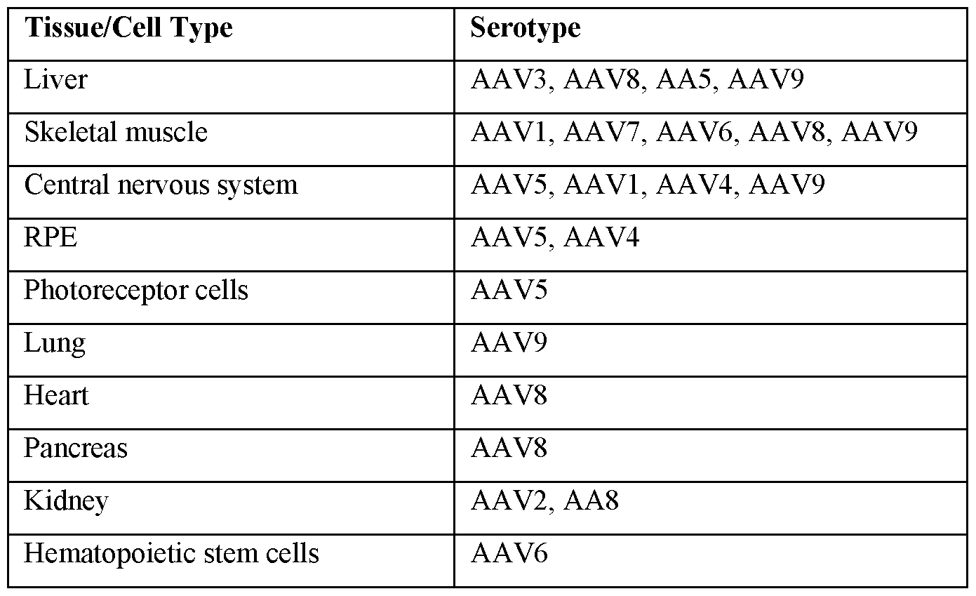
[000354] In some aspects, Cas9 mRNA, sgRNA targeting one or two loci in SOD1 gene, and donor DNA can each be separately formulated into lipid nanoparticles, or are all co-formulated into one lipid nanoparticle.
[000355] In some aspects, Cas9 mRNA can be formulated in a lipid nanoparticle, while sgRNA and donor DNA can be delivered in an AAV vector.
[000356] Options are available to deliver the Cas9 nuclease as a DNA plasmid, as mRNA or as a protein. The guide RNA can be expressed from the same DNA, or can also be delivered as an RNA. The RNA can be chemically modified to alter or improve its half-life, or decrease the likelihood or degree of immune response. The endonuclease protein can be complexed with the gRNA prior to delivery. Viral vectors allow efficient delivery; split versions of Cas9 and smaller orthologs of Cas9 can be packaged in AAV, as can donors for HDR. A range of non-viral delivery methods also exist that can deliver each of these components, or non-viral and viral methods can be employed in tandem. For example, nano-particles can be used to deliver the protein and guide RNA, while AAV can be used to deliver a donor DNA.
IV. DOSING AND ADMINISTRATION
[000357] The terms "administering," "introducing" and "transplanting" are used
interchangeably in the context of the placement of cells, e.g., progenitor cells, into a subject, by a method or route that results in at least partial localization of the introduced cells at a desired site, such as a site of injury or repair, such that a desired effect(s) is produced. The cells e.g., progenitor cells, or their differentiated progeny can be administered by any appropriate route that results in delivery to a desired location in the subject where at least a portion of the implanted cells or components of the cells remain viable. The period of viability of the cells after administration to a subject can be as short as a few hours, e.g., twenty -four hours, to a few days, to as long as several years, or even the life time of the patient, i.e., long-term engraftment. For example, in some aspects described herein, an effective amount of neural progenitor cells is administered via a systemic route of administration, such as an intraperitoneal or intravenous route.
[000358] The terms "individual," "subject," "host" and "patient" are used interchangeably herein and refer to any subject for whom diagnosis, treatment or therapy is desired. In some aspects, the subject is a mammal. In some aspects, the subject is a human being.
[000359] When provided prophylactically, progenitor cells described herein can be
administered to a subject in advance of any symptom of Amyotrophic Lateral Sclerosis (ALS). Accordingly, the prophylactic administration of a progenitor cell population serves to prevent Amyotrophic Lateral Sclerosis (ALS).
[000360] A progenitor cell population being administered according to the methods described herein can comprise allogeneic progenitor cells obtained from one or more donors. Such progenitors may be of any cellular or tissue origin, e.g., liver, muscle, cardiac, brain, etc.
"Allogeneic" refers to a progenitor cell or biological samples comprising progenitor cells obtained from one or more different donors of the same species, where the genes at one or more loci are not identical. For example, a liver progenitor cell population being administered to a subject can be derived from one more unrelated donor subjects, or from one or more non- identical siblings. In some cases, syngeneic progenitor cell populations can be used, such as those obtained from genetically identical animals, or from identical twins. The progenitor cells can be autologous cells; that is, the progenitor cells are obtained or isolated from a subject and administered to the same subject, i.e., the donor and recipient are the same.
[000361] The term "effective amount" refers to the amount of a population of progenitor cells or their progeny needed to prevent or alleviate at least one or more signs or symptoms of Amyotrophic Lateral Sclerosis (ALS), and relates to a sufficient amount of a composition to provide the desired effect, e.g., to treat a subject having Amyotrophic Lateral Sclerosis (ALS). The term "therapeutically effective amount" therefore refers to an amount of progenitor cells or a composition comprising progenitor cells that is sufficient to promote a particular effect when administered to a typical subject, such as one who has or is at risk for Amyotrophic Lateral Sclerosis (ALS). An effective amount would also include an amount sufficient to prevent or delay the development of a symptom of the disease, alter the course of a symptom of the disease (for example but not limited to, slow the progression of a symptom of the disease), or reverse a symptom of the disease. It is understood that for any given case, an appropriate "effective amount" can be determined by one of ordinary skill in the art using routine experimentation.
[000362] For use in the various aspects described herein, an effective amount of progenitor cells comprises at least 102 progenitor cells, at least 5 X 10² progenitor cells, at least 103 progenitor cells, at least 5 X 10³ progenitor cells, at least 104 progenitor cells, at least 5 X 10⁴
progenitor cells, at least 105 progenitor cells, at least 2 X 10⁵ progenitor cells, at least 3 X 10⁵ progenitor cells, at least 4 X 10⁵ progenitor cells, at least 5 X 10⁵ progenitor cells, at least 6 X 10⁵ progenitor cells, at least 7 X 10⁵ progenitor cells, at least 8 X 10⁵ progenitor cells, at least 9 X 10⁵ progenitor cells, at least 1 X 10⁶ progenitor cells, at least 2 X 10⁶ progenitor cells, at least 3 X 10⁶ progenitor cells, at least 4 X 10⁶ progenitor cells, at least 5 X 10⁶ progenitor cells, at least 6 X 10⁶ progenitor cells, at least 7 X 10⁶ progenitor cells, at least 8 X 10⁶ progenitor cells, at least 9 X 10⁶ progenitor cells, or multiples thereof. The progenitor cells can be derived from one or more donors, or can be obtained from an autologous source. In some examples described herein, the progenitor cells can be expanded in culture prior to administration to a subject in need thereof.
[000363] In some aspects, modest and incremental decreases in the levels of mutant SOD1 expressed in cells of patients having a SOD1 related disorder can be beneficial for ameliorating one or more symptoms of the disease, for increasing long-term survival, and/or for reducing side effects associated with other treatments. Upon administration of such cells to human patients, the presence of progenitors that are producing decreased levels of mutant SOD1 is beneficial. In some cases, effective treatment of a subject gives rise to at least about 3%, 5% or 7% reduction in SOD1 relative to total SOD1 in the treated subject. In some examples, the reduction in SOD1 will be at least about 10% of total SOD1. In some examples, the reduction in SOD1 will be at least about 20% to 30% of total SOD1. Similarly, the introduction of even relatively limited subpopulations of cells having significantly reduced levels of mutant SOD1 can be beneficial in various patients because in some situations normalized cells will have a selective advantage relative to diseased cells. However, even modest levels of progenitors with reduced levels of mutant SOD1 can be beneficial for ameliorating one or more aspects of Amyotrophic Lateral Sclerosis (ALS) in patients. In some examples, about 10%, about 20%, about 30%, about 40%, about 50%), about 60%, about 70%, about 80%, about 90% or more of the neural progenitors in patients to whom such cells are administered are producing decreased levels of mutant SOD1.
[000364] In some aspects, modest and incremental increases in the levels of functional SOD1 expressed in cells of patients having a SOD1 related disorder can be beneficial for ameliorating one or more symptoms of the disease, for increasing long-term survival, and/or for reducing side effects associated with other treatments. Upon administration of such cells to human patients, the presence of progenitors that are producing increased levels of functional SOD1 is beneficial. In some cases, effective treatment of a subject gives rise to at least about 3%, 5% or 7% functional SOD1 relative to total SOD1 in the treated subject. In some examples, functional
SODl will be at least about 10% of total SODl . In some examples, functional SODl will be at least about 20% to 30% of total SODl . Similarly, the introduction of even relatively limited subpopulations of cells having significantly elevated levels of functional SODl can be beneficial in various patients because in some situations normalized cells will have a selective advantage relative to diseased cells. However, even modest levels of progenitors with elevated levels of functional SODl can be beneficial for ameliorating one or more aspects of Amyotrophic Lateral Sclerosis (ALS) in patients. In some examples, about 10%, about 20%, about 30%, about 40%, about 50%), about 60%, about 70%, about 80%, about 90% or more of the neural progenitors in patients to whom such cells are administered are producing increased levels of functional SODl .
[000365] "Administered" refers to the delivery of a progenitor cell composition into a subject by a method or route that results in at least partial localization of the cell composition at a desired site. A cell composition can be administered by any appropriate route that results in effective treatment in the subject, i.e. administration results in delivery to a desired location in the subject where at least a portion of the composition delivered, i.e. at least 1 x 10⁴ cells are delivered to the desired site for a period of time.
[000366] In one aspect of the method, the pharmaceutical composition may be administered via a route such as, but not limited to, enteral (into the intestine), gastroenteral, epidural (into the dura matter), oral (by way of the mouth), transdermal, peridural, intracerebral (into the cerebrum), intracerebroventricular (into the cerebral ventricles), epicutaneous (application onto the skin), intradermal, (into the skin itself), subcutaneous (under the skin), nasal administration (through the nose), intravenous (into a vein), intravenous bolus, intravenous drip, intraarterial (into an artery), intramuscular (into a muscle), intracardiac (into the heart), intraosseous infusion (into the bone marrow), intrathecal (into the spinal canal), intraperitoneal, (infusion or injection into the peritoneum), intravesical infusion, intravitreal, (through the eye), intracavernous injection (into a pathologic cavity) intracavitary (into the base of the penis), intravaginal administration, intrauterine, extra-amniotic administration, transdermal (diffusion through the intact skin for systemic distribution), transmucosal (diffusion through a mucous membrane), transvaginal, insufflation (snorting), sublingual, sublabial, enema, eye drops (onto the conjunctiva), in ear drops, auricular (in or by way of the ear), buccal (directed toward the cheek), conjunctival, cutaneous, dental (to a tooth or teeth), electro-osmosis, endocervical, endosinusial, endotracheal, extracorporeal, hemodialysis, infiltration, interstitial, intra-abdominal, intra- amniotic, intra-articular, intrabiliary, intrabronchial, intrabursal, intracartilaginous (within a cartilage), intracaudal (within the cauda equine), intraci sternal (within the cisterna magna
cerebellomedularis), intracorneal (within the cornea), dental intracornal, intracoronary (within the coronary arteries), intracorporus cavernosum (within the dilatable spaces of the corporus cavernosa of the penis), intradiscal (within a disc), intraductal (within a duct of a gland), intraduodenal (within the duodenum), intradural (within or beneath the dura), intraepidermal (to the epidermis), intraesophageal (to the esophagus), intragastric (within the stomach),
intragingival (within the gingivae), intraileal (within the distal portion of the small intestine), intralesional (within or introduced directly to a localized lesion), intraluminal (within a lumen of a tube), intralymphatic (within the lymph), intramedullary (within the marrow cavity of a bone), intrameningeal (within the meninges), intramyocardial (within the myocardium), intraocular (within the eye), intraovarian (within the ovary), intrapericardial (within the pericardium), intrapleural (within the pleura), intraprostatic (within the prostate gland), intrapulmonary (within the lungs or its bronchi), intrasinal (within the nasal or periorbital sinuses), intraspinal (within the vertebral column), intrasynovial (within the synovial cavity of a joint), intratendinous (within a tendon), intratesticular (within the testicle), intrathecal (within the cerebrospinal fluid at any level of the cerebrospinal axis), intrathoracic (within the thorax), intratubular (within the tubules of an organ), intratumor (within a tumor), intratympanic (within the aurus media), intravascular (within a vessel or vessels), intraventricular (within a ventricle), iontophoresis (by means of electric current where ions of soluble salts migrate into the tissues of the body), irrigation (to bathe or flush open wounds or body cavities), laryngeal (directly upon the larynx), nasogastric (through the nose and into the stomach), occlusive dressing technique (topical route
administration, which is then covered by a dressing that occludes the area), ophthalmic (to the external eye), oropharyngeal (directly to the mouth and pharynx), parenteral, percutaneous, periarticular, peridural, perineural, periodontal, rectal, respiratory (within the respiratory tract by inhaling orally or nasally for local or systemic effect), retrobulbar (behind the pons or behind the eyeball), intramyocardial (entering the myocardium), soft tissue, subarachnoid, subconjunctival, submucosal, topical, transplacental (through or across the placenta), transtracheal (through the wall of the trachea), transtympanic (across or through the tympanic cavity), ureteral (to the ureter), urethral (to the urethra), vaginal, caudal block, diagnostic, nerve block, biliary perfusion, cardiac perfusion, photopheresis and spinal.
[000367] Modes of administration include injection, infusion, instillation, and/or ingestion. "Injection" includes, without limitation, intravenous, intramuscular, intra-arterial, intrathecal, intraventricular, intracapsular, intraorbital, intracardiac, intradermal, intraperitoneal,
transtracheal, subcutaneous, subcuticular, intraarticular, sub capsular, subarachnoid, intraspinal,
intracerebro spinal, and intrasternal injection and infusion. In some examples, the route is intravenous. For the delivery of cells, administration by injection or infusion can be made.
[000368] The cells can be administered systemically. The phrases "systemic administration," "administered systemically", "peripheral administration" and "administered peripherally" refer to the administration of a population of progenitor cells other than directly into a target site, tissue, or organ, such that it enters, instead, the subject's circulatory system and, thus, is subject to metabolism and other like processes.
[000369] The efficacy of a treatment comprising a composition for the treatment of
Amyotrophic Lateral Sclerosis (ALS) can be determined by the skilled clinician. However, a treatment is considered "effective treatment," if any one or all of the signs or symptoms of, as but one example, levels of functional SOD1 are altered in a beneficial manner (e.g., increased by at least 10%), or other clinically accepted symptoms or markers of disease are improved or ameliorated. Efficacy can also be measured by failure of an individual to worsen as assessed by hospitalization or need for medical interventions (e.g., progression of the disease is halted or at least slowed). Methods of measuring these indicators are known to those of skill in the art and/or described herein. Treatment includes any treatment of a disease in an individual or an animal (some non-limiting examples include a human, or a mammal) and includes: (1) inhibiting the disease, e.g., arresting, or slowing the progression of symptoms; or (2) relieving the disease, e.g., causing regression of symptoms; and (3) preventing or reducing the likelihood of the
development of symptoms.
[000370] The treatment according to the present disclosure can ameliorate one or more symptoms associated with Amyotrophic Lateral Sclerosis (ALS) by increasing, decreasing or altering the amount of SOD1 protein in the individual. V. FEATURES AND PROPERTIES OF THE SUPEROXIDASE DISMUTASE 1,
SOLUBLE (SOD1) GENE
[000371] SOD1 has been associated with diseases and disorders such as, but not limited to, Congenital Abnormality, Adenocarcinoma, Albuminuria, Chronic Alcoholic Intoxication, Alzheimer's Disease, Amnesia, Amyloidosis, Amyotrophic Lateral Sclerosis, Anemia,
Autoimmune hemolytic anemia, Sickle Cell Anemia, Anoxia, Anxiety Disorders, Aortic
Diseases, Arteriosclerosis, Rheumatoid Arthritis, Asphyxia Neonatorum, Asthma,
Atherosclerosis, Autistic Disorder, Autoimmune Diseases, Barrett Esophagus, Behcet Syndrome, Malignant neoplasm of urinary bladder, Brain Neoplasms, Malignant neoplasm of breast, Oral
candidiasis, Malignant tumor of colon, Bronchogenic Carcinoma, Non-Small Cell Lung
Carcinoma, Squamous cell carcinoma, Transitional Cell Carcinoma, Cardiovascular Diseases, Carotid Artery Thrombosis, Neoplastic Cell Transformation, Cerebral Infarction, Brain
Ischemia, Transient Ischemic Attack, Charcot-Marie-Tooth Disease, Cholera, Colitis, Colorectal Carcinoma, Coronary Arteriosclerosis, Coronary heart disease, Infection by Cryptococcus neoformans, Deafness, Cessation of life, Deglutition Disorders, Presenile dementia, Depressive disorder, Contact Dermatitis, Diabetes, Diabetes Mellitus, Experimental Diabetes Mellitus, Insulin-Dependent Diabetes Mellitus, Non-Insulin-Dependent Diabetes Mellitus, Diabetic Angiopathies, Diabetic Nephropathy, Diabetic Retinopathy, Down Syndrome, Dwarfism, Edema, Japanese Encephalitis, Toxic Epidermal Necrolysis, Temporal Lobe Epilepsy,
Exanthema, Muscular fasciculation, Alcoholic Fatty Liver, Fetal Growth Retardation,
Fibromyalgia, Fibrosarcoma, Fragile X Syndrome, Giardiasis, Glioblastoma, Glioma, Headache, Partial Hearing Loss, Cardiac Arrest, Heart failure, Atrial Septal Defects, Helminthiasis, Hemochromatosis, Hemolysis (disorder), Chronic Hepatitis, HIV Infections, Huntington
Disease, Hypercholesterolemia, Hyperglycemia, Hyperplasia, Hypertensive disease,
Hyperthyroidism, Hypopituitarism, Hypoproteinemia, Hypotension, natural Hypothermia, Hypothyroidism, Immunologic Deficiency Syndromes, Immune System Diseases, Inflammation, Inflammatory Bowel Diseases, Influenza, Intestinal Diseases, Ischemia, Kearns-Sayre syndrome, Keratoconus, Kidney Calculi, Kidney Diseases, Acute Kidney Failure, Chronic Kidney Failure, Polycystic Kidney Diseases, leukemia, Myeloid Leukemia, Acute Promyelocytic Leukemia, Liver Cirrhosis, Liver diseases, Liver neoplasms, Locked-In Syndrome, Chronic Obstructive Airway Disease, Lung Neoplasms, Systemic Lupus Erythematosus, Non-Hodgkin Lymphoma, Machado- Joseph Disease, Malaria, Malignant neoplasm of stomach, Animal Mammary
Neoplasms, Marfan Syndrome, Meningomyelocele, Mental Retardation, Mitral Valve Stenosis, Acquired Dental Fluorosis, Movement Disorders, Multiple Sclerosis, Muscle Rigidity, Muscle Spasticity, Muscular Atrophy, Spinal Muscular Atrophy, Myopathy, Mycoses, Myocardial Infarction, Myocardial Reperfusion Injury, Necrosis, Nephrosis, Nephrotic Syndrome, Nerve Degeneration, nervous system disorder, Neuralgia, Neuroblastoma, Neuroma, Neuromuscular Diseases, Obesity, Occupational Diseases, Ocular Hypertension, Oligospermia, Degenerative polyarthritis, Osteoporosis, Ovarian Carcinoma, Pain, Pancreatitis, Papillon-Lefevre Disease, Paresis, Parkinson Disease, Phenylketonurias, Pituitary Diseases, Pre-Eclampsia, Prostatic Neoplasms, Protein Deficiency, Proteinuria, Psoriasis, Pulmonary Fibrosis, Renal Artery Obstruction, Reperfusion Injury, Retinal Degeneration, Retinal Diseases, Retinoblastoma,
Schistosomiasis, Schistosomiasis mansoni, Schizophrenia, Scrapie, Seizures, Age-related cataract, Compression of spinal cord, Cerebrovascular accident, Subarachnoid Hemorrhage, Progressive supranuclear palsy, Tetanus, Trisomy, Turner Syndrome, Unipolar Depression, Urticaria, Vitiligo, Vocal Cord Paralysis, Intestinal Volvulus, Weight Gain, HMN (Hereditary Motor Neuropathy) Proximal Type I, Holoprosencephaly, Motor Neuron Disease,
Neurofibrillary degeneration (morphologic abnormality), Burning sensation, Apathy, Mood swings, Synovial Cyst, Cataract, Migraine Disorders, Sciatic Neuropathy, Sensory neuropathy, Atrophic condition of skin, Muscle Weakness, Esophageal carcinoma, Lingual-Facial-Buccal Dyskinesia, Idiopathic pulmonary hypertension, Lateral Sclerosis, Migraine with Aura, Mixed Conductive-Sensorineural Hearing Loss, Iron deficiency anemia, Malnutrition, Prion Diseases, Mitochondrial Myopathies, MELAS Syndrome, Chronic progressive external ophthalmoplegia, General Paralysis, Premature aging syndrome, Fibrillation, Psychiatric symptom, Memory impairment, Muscle degeneration, Neurologic Symptoms, Gastric hemorrhage, Pancreatic carcinoma, Pick Disease of the Brain, Liver Fibrosis, Malignant neoplasm of lung, Age related macular degeneration, Parkinsonian Disorders, Disease Progression, Hypocupremia,
Cytochrome-c Oxidase Deficiency, Essential Tremor, Familial Motor Neuron Disease, Lower Motor Neuron Disease, Degenerative myelopathy, Diabetic Polyneuropathies, Liver and Intrahepatic Biliary Tract Carcinoma, Persian Gulf Syndrome, Senile Plaques, Atrophic, Frontotemporal dementia, Semantic Dementia, Common Migraine, Impaired cognition, Malignant neoplasm of liver, Malignant neoplasm of pancreas, Malignant neoplasm of prostate, Pure Autonomic Failure, Motor symptoms, Spastic, Dementia, Neurodegenerative Disorders, Chronic Hepatitis C, Guam Form Amyotrophic Lateral Sclerosis, Stiff limbs, Multisystem disorder, Loss of scalp hair, Prostate carcinoma, Hepatopulmonary Syndrome, Hashimoto Disease, Progressive Neoplastic Disease, Breast Carcinoma, Terminal illness, Carcinoma of lung, Tardive Dyskinesia, Secondary malignant neoplasm of lymph node, Colon Carcinoma,
Stomach Carcinoma, Central neuroblastoma, Dissecting aneurysm of the thoracic aorta, Diabetic macular edema, Microalbuminuria, Middle Cerebral Artery Occlusion, Middle Cerebral Artery Infarction, Upper motor neuron signs, Frontotemporal Lobar Degeneration, Memory Loss, Classical phenylketonuria, CADASIL Syndrome, Neurologic Gait Disorders, Spinocerebellar Ataxia Type 2, Spinal Cord Ischemia, Lewy Body Disease, Muscular Atrophy, Spinobulbar, Chromosome 21 monosomy, Thrombocytosis, Spots on skin, Drug-Induced Liver Injury, Hereditary Leber Optic Atrophy, Cerebral Ischemia, ovarian neoplasm, Tauopathies,
Macroangiopathy, Persistent pulmonary hypertension, Malignant neoplasm of ovary, Myxoid
cyst, Drusen, Sarcoma, Weight decreased, Major Depressive Disorder, Mild cognitive disorder, Degenerative disorder, Partial Trisomy, Cardiovascular morbidity, hearing impairment,
Cognitive changes, Ureteral Calculi, Mammary Neoplasms, Colorectal Cancer, Chronic Kidney Diseases, Minimal Change Nephrotic Syndrome, Non-Neoplastic Disorder, X-Linked Bulbo- Spinal Atrophy, Mammographic Density, Normal Tension Glaucoma Susceptibility To
(Finding), Vitiligo-Associated Multiple Autoimmune Disease Susceptibility 1 (Finding), Amyotrophic Lateral Sclerosis And/Or Frontotemporal Dementia 1, Amyotrophic Lateral Sclerosis 1, Sporadic Amyotrophic Lateral Sclerosis, monomelic Amyotrophy, Coronary Artery Disease, Transformed migraine, Regurgitation, Urothelial Carcinoma, Motor disturbances, Liver carcinoma, Protein Misfolding Disorders, TDP-43 Proteinopathies, Promyelocytic leukemia, Weight Gain Adverse Event, Mitochondrial cytopathy, Idiopathic pulmonary arterial hypertension, Progressive cGVHD, Infection, GRN-related frontotemporal dementia,
Mitochondrial pathology, and Hearing Loss. Editing the SOD1 gene using any of the methods described herein may be used to treat, prevent and/or mitigate the symptoms of the diseases and disorders described herein.
[000372] The SOD1 enzyme is a homodimeric copper- and zinc- containing cytoplasmic antioxidant that metabolizes superoxide radicals. SOD1 plays an important role in oxidative stress and apoptotic signaling. Mutations of the SOD1 gene are primarily associated with Amyotrophic lateral sclerosis (ALS) but have also been linked to a variety of disorders, including other neurodegenerative disorders such as Alzheimer disease, Huntington disease,
Parkinson disease, Frontotemporal dementia, motor neuron disease, spinal and bulbar muscular atrophy of Kennedy, cardiac and vascular indications such as ischemia, myocardial stunning, tetralogy of fallot, and cerebral hemorrhage, disorders of the eye such as senile cataract, retinal ischemia, retinal vasculitis, and septooptic dysplasia, and other indications such as
aceruloplasminemia, deficiency of acid-labile subunit, acute vascular insufficiency of the intestine, adenoid hypertrophy, anoxia, anthracosis, avascular necrosis of the femoral head, bronchopulmonary dysplasia, Brown- Vialetto- Van Laere syndrome, chronic kidney failure, methemoglobinemia, Eales Disease, head injury, hyperphenylalaninemia, isolated cytochrome c oxidase deficiency, meningitis and encephalitis, meningococcal infection, mild pre-eclampsia, mucopolysaccharidoses, paraquat poisoning, Peyronie's disease, pneumoconiosis, post-traumatic stress disorder, postcholecystectomy syndrome, recurrent acute pancreatitis, tetrahydrobiopterin deficiency, and tonsillitis.
[000373] ALS is a progressive deterioration of motor neurons that leads to increasing muscle weakness, loss of muscle mass, and an inability to control movement, ultimately leading to paralysis and/or respiratory failure. There are several variants of ALS, with different underlying pathobiology and clinical presentation. ALS is usually diagnosed between the ages of 40 and 70, with an average life expectancy of 3-5 years subsequent to diagnosis. Approximately 30,000 people in the United States at any given time have been diagnosed with ALS, with an estimated 15 new cases diagnosed every day. No cure for ALS exists and treatments only provide temporary symptomatic relief. Riluzole has been approved by the United States Food and Drug Administration for treatment of ALS and has been shown to extend the lifespan by several months, especially in patients with difficulty swallowing. It is thought that Riluzole reduces continued damage to motor neurons (cannot reverse previous damage) by decreasing the release of glutamate.
[000374] In one example, the target tissue for the compositions and methods described herein is central nervous system tissue or peripheral nervous system tissue.
[000375] In one example, the gene is Superoxidase Dismutase 1, soluble (SODl) which may also be referred to as hSodl, Amyotrophic lateral sclerosis (ALS), ALSl, homodimer, SOD, HEL-S-44, EC 1.15.1.1, epididymis secretory protein Li 44, Superoxide dismutase, cystolic, Cu/Zn Superoxide dismutase, Idophenol oxidase A (IPO A). SODl has a cytogenetic location of 21q22.11 and the genomic coordinates are on Chromosome 21 on the forward strand at position 31,659,622-31,668,931. The nucleotide sequence of SODl is shown as SEQ ID NO: 5303.
AP000251.3 is the gene upstream of SODl on the forward strand and HMGN1P2 is the gene downstream of SODl on the forward strand. Contained within the SODl gene on the forward strand is the SNORA81 gene. AP000254.8 is the gene located on the reverse strand opposite of SODl . SODl has a NCBI gene ID of 6647, Uniprot ID of P0041 and Ensembl Gene ID of ENSG00000142168. SODl has 78 SNPs, 12 introns and 14 exons. The exon identifier from Ensembl and the start/stop sites of the introns and exons are shown in Table 3.
Table 3. Introns and Exons for SODl

EX6 ENSE00001898140 31,659,709 - INT6 Intron ENSE00003492976 - 31,663,887 - 31,660,708 ENSE00003624439 31,666,448
EX7 ENSE00003492976 31,663,790 - INT7 Intron ENSE00003528357 - 31,666,519 - 31,663,886 ENSE00001892860 31,667,257
EX8 ENSE00003528357 31,666,449 - INT8 Intron ENSE00003528357 - 31,666,519 - 31,666,518 ENSE00003551618 31,667,257
EX9 ENSE00003551618 31,667,258 - INT9 Intron ENSE00003551618 - 31,667,376 - 31,667,375 ENSE00003567152 31,668,470
EX10 ENSE00003555033 31,667,258 - INT 10 Intron ENSE00003555033 - 31,667,376 - 31,667,375 ENSE00003683357 31,668,470
EX11 ENSE00003562612 31,663,790 - INT 11 Intron ENSE00003562612 - 31,663,887 - 31,663,886 ENSE00003528357 31,666,448
EX12 ENSE00003567152 31,668,471 - INT 12 Intron ENSE00003624439 - 31,666,519 - 31,668,931 ENSE00003555033 31,667,257
EX13 ENSE00003624439 31,666,449 - 31,666,518
EX14 ENSE00003683357 31,668,471 - 31,668,931
[000376] Table 4 provides information on all of the transcripts for the SOD1 gene based on the Ensembl database. Provided in Table 4 are the transcript ID from Ensembl and corresponding NCBI RefSeq ID for the transcript, the translation ID from Ensembl and the corresponding NCBI RefSeq ID for the protein, the biotype of the transcript sequence as classified by Ensembl and the exons and introns in the transcript based on the information in Table 3.
Table 4. Transcript Information for SOD1

VAR 007160, VAR 007161, VAR 007162, VAR 007163, VAR 007164, VAR 008717,
VAR 008718, VAR 008719, VAR 008720, VAR 008722, VAR 008724, VAR 008725,
VAR 013518, VAR 013519, VAR 013520, VAR 013521, VAR 013522, VAR 013523,
VAR 013524, VAR 013525, VAR 013526, VAR 013527, VAR 013528, VAR 013529,
VAR 013530, VAR 013531, VAR 013532, VAR 013533, VAR 013534, VAR 013535,
VAR 013536, VAR 013537, VAR 013538, VAR 013539, VAR 013540, VAR 016874,
VAR 045876, VAR 045877, VAR 045878, VAR 045879, VAR 045880, VAR 045881,
VAR 045882, VAR 045883, VAR 045884, VAR 045885, VAR 065194, and VAR_065560
[000378] In one example, the guide RNA used in the present disclosure may comprise at least one 20 nucleotide (nt) target nucleic acid sequence listed in Table 5. Provided in Table 5 are the gene symbol and the sequence identifier of the gene (Gene SEQ ID NO), the gene sequence including 1-5 kilobase pairs upstream and/or downstream of the target gene (Extended Gene SEQ ID NO), and the 20 nt target nucleic acid sequence (20 nt Target Sequence SEQ ID NO). In the sequence listing the respective target gene, the strand for targeting the gene (noted by a (+) strand or (-) strand in the sequence listing), the associated PAM type and the PAM sequence are described for each of the 20 nt target nucleic acid sequences (SEQ ID NO: 5305-17976). It is understood in the art that the spacer sequence, where "T" is "U," may be an RNA sequence corresponding to the 20 nt sequences listed in Table 5.
Table 5. Nucleic Acid Sequences

[000379] In one example, the guide RNA used in the present disclosure may comprise at least one spacer sequence that, where "T" is "U", may be an RNA sequence corresponding to a 20 nucleotide (nt) target sequence such as, but not limited to, any of SEQ ID NO: 5305-17976.
[000380] In one example, the guide RNA used in the present disclosure may comprise at least one spacer sequence which, where "T" is "U," is an RNA sequence corresponding to the 20 nt sequences such as, but not limited to, any of SEQ ID NO: 5305-17976.
[000381] In one example, a guide RNA may comprise a 20 nucleotide (nt) target nucleic acid sequence associated with the PAM type such as, but not limited to, NAAAAC, NNAGAAW, NNGRRT, NNNNGHTT, NRG, or YTN. As a non-limiting example, the 20 nt target nucleic acid sequence for a specific target gene and a specific PAM type may be, where "T" is "U," the RNA sequence corresponding to any one of the 20 nt nucleic acid sequences in Table 6.
Table 6. Nucleic Acid Sequences by PAM Type

[000382] In one example, a guide RNA may comprise a 22 nucleotide (nt) target nucleic acid sequence associated with the YTN PAM type. As a non-limiting example, the 22 nt target nucleic acid sequence for a specific target gene may comprise a 20 nt core sequence where the 20 nt core sequence, where "T" is "U," may be the RNA sequence corresponding to SEQ ID NO: 11616-17976. As another non-limiting example, the 22 nt target nucleic acid sequence for a specific target gene may comprise a core sequence where the core sequence, where "T" is "U," may be a fragment, segment or region of the RNA sequence corresponding to any of SEQ ID NO: 11616-17976.
VI. OTHER THERAPEUTIC APPROACHES
[000383] Gene editing can be conducted using nucleases engineered to target specific sequences. To date there are four major types of nucleases: meganucleases and their derivatives, zinc finger nucleases (ZFNs), transcription activator like effector nucleases (TALENs), and
CRISPR-Cas9 nuclease systems. The nuclease platforms vary in difficulty of design, targeting density and mode of action, particularly as the specificity of ZFNs and TALENs is through protein-DNA interactions, while RNA-DNA interactions primarily guide Cas9.
[000384] CRISPR endonucleases, such as Cas9, can be used in the methods of the present disclosure. However, the teachings described herein, such as therapeutic target sites, could be applied to other forms of endonucleases, such as ZFNs, TALENs, HEs, or MegaTALs, or using combinations of nucleases. However, in order to apply the teachings of the present disclosure to such endonucleases, one would need to, among other things, engineer proteins directed to the specific target sites.
[000385] Additional binding domains can be fused to the Cas9 protein to increase specificity. The target sites of these constructs would map to the identified gRNA specified site, but would require additional binding motifs, such as for a zinc finger domain. In the case of Mega-TAL, a meganuclease can be fused to a TALE DNA-binding domain. The meganuclease domain can increase specificity and provide the cleavage. Similarly, inactivated or dead Cas9 (dCas9) can be
fused to a cleavage domain and require the sgRNA/Cas9 target site and adjacent binding site for the fused DNA-binding domain. This likely would require some protein engineering of the dCas9, in addition to the catalytic inactivation, to decrease binding without the additional binding site.
Zinc Finger Nucleases
[000386] Zinc finger nucleases (ZFNs) are modular proteins comprised of an engineered zinc finger DNA binding domain linked to the catalytic domain of the type II endonuclease Fokl. Because Fokl functions only as a dimer, a pair of ZFNs must be engineered to bind to cognate target "half-site" sequences on opposite DNA strands and with precise spacing between them to enable the catalytically active Fokl dimer to form. Upon dimerization of the Fokl domain, which itself has no sequence specificity per se, a DNA double-strand break is generated between the ZFN half-sites as the initiating step in genome editing.
[000387] The DNA binding domain of each ZFN is typically comprised of 3-6 zinc fingers of the abundant Cys2-His2 architecture, with each finger primarily recognizing a triplet of nucleotides on one strand of the target DNA sequence, although cross-strand interaction with a fourth nucleotide also can be important. Alteration of the amino acids of a finger in positions that make key contacts with the DNA alters the sequence specificity of a given finger. Thus, a four-finger zinc finger protein will selectively recognize a 12 bp target sequence, where the target sequence is a composite of the triplet preferences contributed by each finger, although triplet preference can be influenced to varying degrees by neighboring fingers. An important aspect of ZFNs is that they can be readily re-targeted to almost any genomic address simply by modifying individual fingers, although considerable expertise is required to do this well. In most applications of ZFNs, proteins of 4-6 fingers are used, recognizing 12-18 bp respectively.
Hence, a pair of ZFNs will typically recognize a combined target sequence of 24-36 bp, not including the typical 5-7 bp spacer between half-sites. The binding sites can be separated further with larger spacers, including 15-17 bp. A target sequence of this length is likely to be unique in the human genome, assuming repetitive sequences or gene homologs are excluded during the design process. Nevertheless, the ZFN protein-DNA interactions are not absolute in their specificity so off-target binding and cleavage events do occur, either as a heterodimer between the two ZFNs, or as a homodimer of one or the other of the ZFNs. The latter possibility has been effectively eliminated by engineering the dimerization interface of the Fokl domain to create "plus" and "minus" variants, also known as obligate heterodimer variants, which can only dimerize with each other, and not with themselves. Forcing the obligate heterodimer prevents
formation of the homodimer. This has greatly enhanced specificity of ZFNs, as well as any other nuclease that adopts these Fokl variants.
[000388] A variety of ZFN-based systems have been described in the art, modifications thereof are regularly reported, and numerous references describe rules and parameters that are used to guide the design of ZFNs; see, e.g., Segal et al., Proc Natl Acad Sci USA 96(6):2758-63 (1999); Dreier B et al., J Mol Biol. 303(4):489-502 (2000); Liu Q et al., J Biol Chem. 277(6):3850-6 (2002); Dreier et al., J Biol Chem 280(42):35588-97 (2005); and Dreier et al., J Biol Chem. 276(31):29466-78 (2001).
Transcription Activator-Like Effector Nucleases (TALENs)
[000389] TALENs represent another format of modular nucleases whereby, as with ZFNs, an engineered DNA binding domain is linked to the Fokl nuclease domain, and a pair of TALENs operates in tandem to achieve targeted DNA cleavage. The major difference from ZFNs is the nature of the DNA binding domain and the associated target DNA sequence recognition properties. The TALEN DNA binding domain derives from TALE proteins, which were originally described in the plant bacterial pathogen Xanthomonas sp. TALEs are comprised of tandem arrays of 33-35 amino acid repeats, with each repeat recognizing a single base pair in the target DNA sequence that is typically up to 20 bp in length, giving a total target sequence length of up to 40 bp. Nucleotide specificity of each repeat is determined by the repeat variable diresidue (RVD), which includes just two amino acids at positions 12 and 13. The bases guanine, adenine, cytosine and thymine are predominantly recognized by the four RVDs: Asn- Asn, Asn-Ile, His-Asp and Asn-Gly, respectively. This constitutes a much simpler recognition code than for zinc fingers, and thus represents an advantage over the latter for nuclease design. Nevertheless, as with ZFNs, the protein-DNA interactions of TALENs are not absolute in their specificity, and TALENs have also benefitted from the use of obligate heterodimer variants of the Fokl domain to reduce off-target activity.
[000390] Additional variants of the Fokl domain have been created that are deactivated in their catalytic function. If one half of either a TALEN or a ZFN pair contains an inactive Fokl domain, then only single-strand DNA cleavage (nicking) will occur at the target site, rather than a DSB. The outcome is comparable to the use of CRISPR/Cas9 or CRISPR/Cpfl "nickase" mutants in which one of the Cas9 cleavage domains has been deactivated. DNA nicks can be used to drive genome editing by FIDR, but at lower efficiency than with a DSB. The main benefit is that off-target nicks are quickly and accurately repaired, unlike the DSB, which is prone to NHEJ-mediated mis-repair.
[000391] A variety of TALEN-based systems have been described in the art, and modifications thereof are regularly reported; see, e.g., Boch, Science 326(5959): 1509-12 (2009); Mak et al., Science 335(6069):716-9 (2012); and Moscou et al., Science 326(5959): 1501 (2009). The use of TALENs based on the "Golden Gate" platform, or cloning scheme, has been described by multiple groups; see, e.g., Cermak et al., Nucleic Acids Res. 39(12):e82 (2011); Li et al., Nucleic Acids Res. 39(14):6315-25(2011); Weber et al., PLoS One. 6(2):el6765 (2011); Wang et al., J Genet Genomics 41(6):339-47, Epub 2014 May 17 (2014); and Cermak T et al., Methods Mol Biol. 1239: 133-59 (2015).
Homing Endonucleases
[000392] Homing endonucleases (HEs) are sequence-specific endonucleases that have long recognition sequences (14-44 base pairs) and cleave DNA with high specificity - often at sites unique in the genome. There are at least six known families of HEs as classified by their structure, including GIY-YIG, His-Cis box, H-N-H, PD-(D/E)xK, and Vsr-like that are derived from a broad range of hosts, including eukarya, protists, bacteria, archaea, cyanobacteria and phage. As with ZFNs and TALENs, HEs can be used to create a DSB at a target locus as the initial step in genome editing. In addition, some natural and engineered HEs cut only a single strand of DNA, thereby functioning as site-specific nickases. The large target sequence of HEs and the specificity that they offer have made them attractive candidates to create site-specific DSBs.
[000393] A variety of HE-based systems have been described in the art, and modifications thereof are regularly reported; see, e.g., the reviews by Steentoft et al., Glycobiology 24(8):663- 80 (2014); Belfort and Bonocora, Methods Mol Biol. 1123 : 1-26 (2014); Hafez and Hausner, Genome 55(8):553-69 (2012); and references cited therein.
MegaTAL / Tev-mTALEN / MegaTev
[000394] As further examples of hybrid nucleases, the MegaTAL platform and Tev-mTALEN platform use a fusion of TALE DNA binding domains and catalytically active HEs, taking advantage of both the tunable DNA binding and specificity of the TALE, as well as the cleavage sequence specificity of the HE; see, e.g., Boissel et al., NAR 42: 2591-2601 (2014); Kleinstiver et al., G3 4: 1155-65 (2014); and Boissel and Scharenberg, Methods Mol. Biol. 1239: 171-96 (2015).
[000395] In a further variation, the MegaTev architecture is the fusion of a meganuclease (Mega) with the nuclease domain derived from the GIY-YIG homing endonuclease I-Tevl (Tev). The two active sites are positioned ~30 bp apart on a DNA substrate and generate two DSBs
with non-compatible cohesive ends; see, e.g., Wolfs et al., NAR 42, 8816-29 (2014). It is anticipated that other combinations of existing nuclease-based approaches will evolve and be useful in achieving the targeted genome modifications described herein.
dCas9-FokI or dCpfl-Fokl and Other Nucleases
[000396] Combining the structural and functional properties of the nuclease platforms described above offers a further approach to genome editing that can potentially overcome some of the inherent deficiencies. As an example, the CRISPR genome editing system typically uses a single Cas9 endonuclease to create a DSB. The specificity of targeting is driven by a 20 or 24 nucleotide sequence in the guide RNA that undergoes Watson-Crick base-pairing with the target DNA (plus an additional 2 bases in the adjacent NAG or NGG PAM sequence in the case of Cas9 from S. pyogenes). Such a sequence is long enough to be unique in the human genome, however, the specificity of the RNA/DNA interaction is not absolute, with significant promiscuity sometimes tolerated, particularly in the 5' half of the target sequence, effectively reducing the number of bases that drive specificity. One solution to this has been to completely deactivate the Cas9 or Cpfl catalytic function - retaining only the RNA-guided DNA binding function - and instead fusing a Fokl domain to the deactivated Cas9; see, e.g., Tsai et al., Nature Biotech 32: 569-76 (2014); and Guilinger et al., Nature Biotech. 32: 577-82 (2014). Because Fokl must dimerize to become catalytically active, two guide RNAs are required to tether two Fokl fusions in close proximity to form the dimer and cleave DNA. This essentially doubles the number of bases in the combined target sites, thereby increasing the stringency of targeting by CRISPR-based systems.
[000397] As further example, fusion of the TALE DNA binding domain to a catalytically active FIE, such as I-Tevl, takes advantage of both the tunable DNA binding and specificity of the TALE, as well as the cleavage sequence specificity of I-Tevl, with the expectation that off- target cleavage can be further reduced.
VII. KITS
[000398] The present disclosure provides kits for carrying out the methods described herein. A kit can include one or more of a genome-targeting nucleic acid, a polynucleotide encoding a genome-targeting nucleic acid, a site-directed polypeptide, a polynucleotide encoding a site- directed polypeptide, and/or any nucleic acid or proteinaceous molecule necessary to carry out the aspects of the methods described herein, or any combination thereof.
[000399] A kit can comprise: (1) a vector comprising a nucleotide sequence encoding a genome-targeting nucleic acid, (2) the site-directed polypeptide or a vector comprising a nucleotide sequence encoding the site-directed polypeptide, and (3) a reagent for reconstitution and/or dilution of the vector(s) and or polypeptide.
[000400] A kit can comprise: (1) a vector comprising (i) a nucleotide sequence encoding a genome-targeting nucleic acid, and (ii) a nucleotide sequence encoding the site-directed polypeptide; and (2) a reagent for reconstitution and/or dilution of the vector.
[000401] In any of the above kits, the kit can comprise a single-molecule guide genome- targeting nucleic acid. In any of the above kits, the kit can comprise a double-molecule genome- targeting nucleic acid. In any of the above kits, the kit can comprise two or more double- molecule guides or single-molecule guides. The kits can comprise a vector that encodes the nucleic acid targeting nucleic acid.
[000402] In any of the above kits, the kit can further comprise a polynucleotide to be inserted to effect the desired genetic modification.
[000403] Components of a kit can be in separate containers, or combined in a single container.
[000404] Any kit described above can further comprise one or more additional reagents, where such additional reagents are selected from a buffer, a buffer for introducing a polypeptide or polynucleotide into a cell, a wash buffer, a control reagent, a control vector, a control RNA polynucleotide, a reagent for in vitro production of the polypeptide from DNA, adaptors for sequencing and the like. A buffer can be a stabilization buffer, a reconstituting buffer, a diluting buffer, or the like. A kit can also comprise one or more components that can be used to facilitate or enhance the on-target binding or the cleavage of DNA by the endonuclease, or improve the specificity of targeting.
[000405] In addition to the above-mentioned components, a kit can further comprise instructions for using the components of the kit to practice the methods. The instructions for practicing the methods can be recorded on a suitable recording medium. For example, the instructions can be printed on a substrate, such as paper or plastic, etc. The instructions can be present in the kits as a package insert, in the labeling of the container of the kit or components thereof (i.e., associated with the packaging or subpackaging), etc. The instructions can be present as an electronic storage data file present on a suitable computer readable storage medium, e.g. CD-ROM, diskette, flash drive, etc. In some instances, the actual instructions are not present in the kit, but means for obtaining the instructions from a remote source (e.g. via the Internet), can be provided. An example of this case is a kit that comprises a web address where
the instructions can be viewed and/or from which the instructions can be downloaded. As with the instructions, this means for obtaining the instructions can be recorded on a suitable substrate.
VIII. SPECIFIC METHODS AND COMPOSITIONS OF THE INVENTION
[000406] Accordingly, the present disclosure relates in particular to the following non-limiting methods according to the present disclosure: in a first method, Method 1, the present disclosure provides a method for editing a Superoxidase Dismutase 1, soluble (SOD1) gene in a cell by genome editing comprising: introducing into the cell one or more deoxyribonucleic acid (DNA) endonucleases to effect one or more single-strand breaks (SSBs) or double-strand breaks (DSBs) within or near the SOD1 gene or SOD1 regulatory elements that results in one or more permanent insertions, deletions or mutations of at least one nucleotide, correction of one or more mutations or replacement of one or more nucleotide bases, or one or more exons and/or introns within or near the SOD1 gene, thereby reducing or eliminating the expression or function of aberrant SOD1 gene products and/or restoring the wild-type SOD1 protein activity.
[000407] In another method, Method 2, the present disclosure provides an ex vivo method for treating a patient having a SOD1 related condition or disorder comprising: creating a patient specific induced plunpotent stem cell (iPSC); editing within or near a Superoxidase Dismutase 1, soluble (SOD1) gene or other DNA sequences that encode regulatory elements of the SOD1 gene of the iPSC; differentiating the genome-edited iPSC into a neuron or glial cell of the Central Nervous System (CNS), or a neuron of the Peripheral Nervous System (PNS); and implanting the neuron or glial cell of the Central Nervous System (CNS), or neuron of the Peripheral Nervous System (PNS) into the patient.
[000408] In another method, Method 3, the present disclosure provides the method of Method 2, wherein the editing step comprises introducing into the iPSC one or more deoxyribonucleic acid (DNA) endonucleases to effect one or more single-strand breaks (SSBs) or double-strand breaks (DSBs) within or near the SOD1 gene or SOD1 regulatory elements that results in one or more permanent insertions, deletions or mutations of at least one nucleotide, correction of one or more mutations or replacement of one or more nucleotide bases, or one or more exons and/or introns within or near the SOD1 gene, thereby reducing or eliminating the expression or function of aberrant SOD1 gene products and/or restoring the wild-type SOD1 protein activity.
[000409] In another method, Method 4, the present disclosure provides an ex vivo method for treating a patient having a SOD1 related condition or disorder comprising: isolating a
mesenchymal stem cell from the patient; editing within or near a Superoxidase Dismutase 1,
soluble (SOD1) gene or other DNA sequences that encode regulatory elements of the SOD1 gene of the mesenchymal stem cell; differentiating the genome-edited mesenchymal stem cell into a neuron or glial cell of the Central Nervous System (CNS), or a neuron of the Peripheral Nervous System (PNS); and implanting the neuron or glial cell of the Central Nervous System (CNS), or neuron of the Peripheral Nervous System (PNS) into the patient.
[000410] In another method, Method 5, the present disclosure provides the method of Method 4, wherein the editing step comprises introducing into the mesenchymal stem cell one or more deoxyribonucleic acid (DNA) endonucleases to effect one or more single-strand breaks (SSBs) or double-strand breaks (DSBs) within or near the SOD1 gene or SOD1 regulatory elements that results in one or more permanent insertions, deletions or mutations of at least one nucleotide, correction of one or more mutations or replacement of one or more nucleotide bases, or one or more exons and/or introns within or near the SOD1 gene, thereby reducing or eliminating the expression or function of aberrant SOD1 gene products and/or restoring the wild-type SOD1 protein activity.
[000411] In another method, Method 6, the present disclosure provides an in vivo method for treating a patient with a SOD1 related disorder comprising: editing the Superoxidase Dismutase 1, soluble (SOD1) gene in a cell of the patient.
[000412] In another method, Method 7, the present disclosure provides the method of Method 6, wherein the editing step comprises introducing into the cell one or more deoxyribonucleic acid (DNA) endonucleases to effect one or more single-strand breaks (SSBs) or double-strand breaks (DSBs) within or near the SOD1 gene or SOD1 regulatory elements that results in one or more permanent insertions, deletions or mutations of at least one nucleotide, correction of one or more mutations or replacement of one or more nucleotide bases, or one or more exons and/or introns within or near the SOD1 gene, thereby reducing or eliminating the expression or function of aberrant SOD1 gene products and/or restoring the wild-type SOD1 protein activity.
[000413] In another method, Method 8, the present disclosure provides the method of any one of Methods 6-7, wherein the cell is a cell of the Central Nervous System (CNS).
[000414] In another method, Method 9, the present disclosure provides the method of Method 8, wherein the cell of the Central Nervous System (CNS) is a neuron.
[000415] In another method, Method 10, the present disclosure provides the method of Method 8, wherein the cell of the Central Nervous System (CNS) is a glial cell.
[000416] In another method, Method 11, the present disclosure provides the method of any one of Methods 8-10, wherein the one or more deoxyribonucleic acid (DNA) endonuclease is
delivered to the cell of the Central Nervous System (CNS) via intraparenchymal, intravenous, intra-arterial, intracerebroventricular, intracistemal, intrathecal, intracranial or intraperitoneal administration routes.
[000417] In another method, Method 12, the present disclosure provides a method of altering the contiguous genomic sequence of a SOD1 gene in a cell comprising contacting the cell with one or more deoxyribonucleic acid (DNA) endonuclease to effect one or more single-strand breaks (SSBs) or double-strand breaks (DSBs).
[000418] In another method, Method 13, the present disclosure provides the method of Method 12, wherein the alteration of the contiguous genomic sequence occurs in one or more exons of the SOD1 gene.
[000419] In another method, Method 14, the present disclosure provides the method of Method 12, wherein the alteration of the contiguous genomic sequence occurs 5', 3' or at the site of one or more SNPs of the SOD1 gene.
[000420] In another method, Method 15, the present disclosure provides the method of any one of Methods 1-14, wherein the one or more deoxyribonucleic acid (DNA) endonuclease is selected from any of the sequences in SEQ ID NOs: 1-620, and variants having at least 90% homology to any of the sequences disclosed in SEQ ID NOs: 1-620.
[000421] In another method, Method 16, the present disclosure provides the method of Method 15, wherein the one or more deoxyribonucleic acid (DNA) endonuclease is one or more protein or polypeptide.
[000422] In another method, Method 17, the present disclosure provides the method of Method 15, wherein the one or more deoxyribonucleic acid (DNA) endonuclease is one or more polynucleotide encoding the one or more DNA endonuclease.
[000423] In another method, Method 18, the present disclosure provides the method of Method 17, wherein the one or more deoxyribonucleic acid (DNA) endonuclease is one or more ribonucleic acid (RNA) encoding the one or more DNA endonuclease.
[000424] In another method, Method 19, the present disclosure provides the method of Method 18, wherein the one or more ribonucleic acid (RNA) is one or more chemically modified RNA.
[000425] In another method, Method 20, the present disclosure provides the method of Method 19, wherein the one or more ribonucleic acid (RNA) is chemically modified in the coding region.
[000426] In another method, Method 21, the present disclosure provides the method of any one of Methods 17-20, wherein the one or more polynucleotide or one or more ribonucleic acid (RNA) is codon optimized.
[000427] In another method, Method 22, the present disclosure provides the method of any one of Methods 1-21, wherein the method further comprises introducing into the cell one or more gRNA or one or more sgRNA.
[000428] In another method, Method 23, the present disclosure provides the method of Method 22, wherein the one or more gRNA or one or more sgRNA comprises a spacer sequence that is complementary to a segment of the coding sequence of the SOD1 gene.
[000429] In another method, Method 24, the present disclosure provides the method of Method 22, wherein the one or more gRNA or one or more sgRNA comprises a spacer sequence that is complementary to the flanking sequence of the SOD1 gene.
[000430] In another method, Method 25, the present disclosure provides the method of any of the Methods 22-24, wherein the one or more gRNA or one or more sgRNA is chemically modified.
[000431] In another method, Method 26, the present disclosure provides the method of any one of Methods 22-24, wherein the one or more gRNA or one or more sgRNA is pre-complexed with the one or more deoxyribonucleic acid (DNA) endonuclease.
[000432] In another method, Method 27, the present disclosure provides the method of Method 26, wherein the pre-complexing involves a covalent attachment of the one or more gRNA or one or more sgRNA to the one or more deoxyribonucleic acid (DNA) endonuclease.
[000433] In another method, Method 28, the present disclosure provides the method of any one of Methods 15-27, wherein the one or more deoxyribonucleic acid (DNA) endonuclease is formulated in a liposome or lipid nanoparticle.
[000434] In another method, Method 29, the present disclosure provides the method of any one of Methods 22-27, wherein the one or more deoxyribonucleic acid (DNA) endonuclease is formulated in a liposome or lipid nanoparticle which also comprises the one or more gRNA or one or more sgRNA.
[000435] In another method, Method 30, the present disclosure provides the method of any one of Methods 15, or 22-24, wherein the one or more deoxyribonucleic acid (DNA) endonuclease is encoded in an AAV vector particle.
[000436] In another method, Method 31, the present disclosure provides the method of any of the Methods 22-24, wherein the one or more gRNA or one or more sgRNA is encoded in an AAV vector particle.
[000437] In another method, Method 32, the present disclosure provides the method of any of the Methods 22-24, wherein the one or more deoxyribonucleic acid (DNA) endonuclease is
encoded in an AAV vector particle which also encodes the one or more gRNA or one or more sgRNA.
[000438] In another method, Method 33, the present disclosure provides the method of any of the Methods 30-32, wherein the AAV vector particle is selected from the group consisting of any of those disclosed in SEQ ID NOs: 4734-5302 and Table 2.
[000439] In another method, Method 34, the present disclosure provides the method of any of Methods 1-33, wherein the method further comprises introducing into the cell a donor template comprising at least a portion of the wild-type SOD1 gene.
[000440] In another method, Method 35, the present disclosure provides the method of Method 34, wherein the at least a portion of the wild-type SOD1 gene comprises one or more sequences selected from a SOD1 exon, a SOD1 intron, and a sequence comprising an exomintr on junction of SOD 1.
[000441] In another method, Method 36, the present disclosure provides the method of any one of Methods 34-35, wherein the donor template comprises homologous arms to the genomic locus of the SOD1 gene.
[000442] In another method, Method 37, the present disclosure provides the method of any one of Methods 34-36, wherein the donor template is either a single or double stranded
polynucleotide.
[000443] In another method, Method 38, the present disclosure provides the method of any one of Methods 34-37, wherein the donor template is encoded in an AAV vector particle, where the AAV vector particle is selected from those disclosed in SEQ ID NOs: 4734-5302 and Table 2.
[000444] In another method, Method 39, the present disclosure provides the method of any one of Methods 34-37, wherein the one or more polynucleotide encoding one or more
deoxyribonucleic acid (DNA) endonuclease is formulated into a lipid nanoparticle, and the one or more gRNA or one or more sgRNA is delivered to the cell ex vivo by electroporation and the donor template is delivered to the cell by an adeno-associated virus (AAV) vector.
[000445] In another method, Method 40, the present disclosure provides the method of any one of Methods 34-37, wherein the one or more polynucleotide encoding one or more
deoxyribonucleic acid (DNA) endonuclease is formulated into a liposome or lipid nanoparticle which also comprises the one or more gRNA or one or more sgRNA and the donor template.
[000446] The present disclosure also provides a composition, Composition 1, comprising a single-molecule guide polynucleotide comprising at least a spacer sequence that is an RNA sequence corresponding to any of SEQ ID NOs: 5305-17976.
[000447] In another composition, Composition 2, the present disclosure provides the single- molecule guide polynucleotide of Composition 1, wherein the single-molecule guide
polynucleotide further comprises a spacer extension region.
[000448] In another composition, Composition 3, the present disclosure provides the single- molecule guide polynucleotide of Composition 1, wherein the single-molecule guide
polynucleotide further comprises a tracrRNA extension region.
[000449] In another composition, Composition 4, the present disclosure provides the single- molecule guide polynucleotide of Compositions 1-3, wherein the single-molecule guide polynucleotide is chemically modified.
[000450] In another composition, Composition 5, the present disclosure provides a non- naturally occurring CRISPR/Cas system comprising a polynucleotide encoding a Cas9 or Cpfl enzyme and at least one single-molecule guide polynucleotide of Compositions 1-4.
[000451] In another composition, Composition 6, the present disclosure provides CRISPR/Cas system of Composition 5, wherein the polynucleotide encoding a Cas9 or Cpfl enzyme is selected from S. pyogenes Cas9, S. aureus Cas9, N. meningitides Cas9, S. thermophilus
CRISPRl Cas9, S. thermophilus CRISPR 3 Cas9, T. denticola Cas9, L. bacterium ND2006 Cpfl and Acidaminococcus sp. BV3L6 Cpfl, and variants having at least 90% homology to these enzymes.
[000452] In another composition, Composition 7, the present disclosure provides CRISPR/Cas system of Composition 6, wherein the polynucleotide encoding a Cas9 or Cpfl enzyme comprises one or more nuclear localization signals (NLSs).
[000453] In another composition, Composition 8, the present disclosure provides CRISPR/Cas system of Composition 7, wherein at least one NLS is at or within 50 amino acids of the amino- terminus of the polynucleotide encoding a Cas9 or Cpfl enzyme and/or at least one NLS is at or within 50 amino acids of the carboxy-terminus of the polynucleotide encoding a Cas9 or Cpfl enzyme.
[000454] In another composition, Composition 9, the present disclosure provides CRISPR/Cas system of Composition 8, wherein the polynucleotide encoding a Cas9 or Cpfl enzyme is codon optimized for expression in a eukaryotic cell.
[000455] In another composition, Composition 10, the present disclosure provides a DNA encoding the single-molecule guide polynucleotide of Compositions 1-4.
[000456] In another composition, Composition 11, the present disclosure provides a DNA encoding the CRISPR/Cas system of Compositions 5-9.
[000457] In another composition, Composition 12, the present disclosure provides a vector comprising the DNA of Compositions of 10 or 11.
[000458] In another composition, Composition 13, the present disclosure provides the vector of Composition 12, wherein the vector is a plasmid.
[000459] In another composition, Composition 14, the present disclosure provides the vector of Composition 13, wherein the vector is an AAV vector particle, and the AAV vector particle is selected from the group consisting of those sequences disclosed in SEQ ID NOs: 4734-5302 and Table 2. IX. DEFINITIONS
[000460] The term "comprising" or "comprises" is used in reference to compositions, methods, and respective component(s) thereof, that are essential to the present disclosure, yet open to the inclusion of unspecified elements, whether essential or not.
[000461] The term "consisting essentially of refers to those elements required for a given aspect. The term permits the presence of additional elements that do not materially affect the basic and novel or functional characteristic(s) of that aspect of the present disclosure.
[000462] The term "consisting of refers to compositions, methods, and respective components thereof as described herein, which are exclusive of any element not recited in that description of the aspect.
[000463] The singular forms "a," "an," and "the" include plural references, unless the context clearly dictates otherwise.
[000464] Any numerical range recited in this specification describes all sub-ranges of the same numerical precision (i.e., having the same number of specified digits) subsumed within the recited range. For example, a recited range of "1.0 to 10.0" describes all sub-ranges between (and including) the recited minimum value of 1.0 and the recited maximum value of 10.0, such as, for example, "2.4 to 7.6," even if the range of "2.4 to 7.6" is not expressly recited in the text of the specification. Accordingly, the Applicant reserves the right to amend this specification, including the claims, to expressly recite any sub-range of the same numerical precision subsumed within the ranges expressly recited in this specification. All such ranges are inherently described in this specification such that amending to expressly recite any such sub-ranges will comply with written description, sufficiency of description, and added matter requirements, including the requirements under 35 U.S.C. § 112(a) and Article 123(2) EPC. Also, unless expressly specified or otherwise required by context, all numerical parameters described in this
specification (such as those expressing values, ranges, amounts, percentages, and the like) may be read as if prefaced by the word "about," even if the word "about" does not expressly appear before a number. Additionally, numerical parameters described in this specification should be construed in light of the number of reported significant digits, numerical precision, and by applying ordinary rounding techniques. It is also understood that numerical parameters described in this specification will necessarily possess the inherent variability characteristic of the underlying measurement techniques used to determine the numerical value of the parameter.
[000465] The details of one or more examples of the present disclosure are set forth in the accompanying description below. Although any materials and methods similar or equivalent to those described herein can be used in the practice or testing of the present disclosure, the preferred materials and methods are now described. Other features, objects and advantages of the present disclosure will be apparent from the description. In the description, the singular forms also include the plural unless the context clearly dictates otherwise. Unless defined otherwise, all technical and scientific terms used herein have the same meaning as commonly understood by one of ordinary skill in the art to which this disclosure belongs. In the case of conflict, the present description will control.
[000466] The present disclosure is further illustrated by the following non-limiting examples. X. EXAMPLES
[000467] The present disclosure will be more fully understood by reference to the following examples, which provide illustrative non-limiting aspects of the invention.
[000468] The examples describe the use of the CRISPR system as an illustrative genome editing technique to create defined therapeutic genomic deletions, insertions, or replacements, termed "genomic modifications" herein, in the SOD1 gene that lead to permanent correction of mutations in the genomic locus, or knock-out of the mutant SOD1 gene, that restore functional SOD1 protein activity and/or reduce or eliminate the undesirable SOD1 protein activity.
[000469] All tested gRNAs can be used for an HDR/correction based editing approach. Single gRNAs can also be used to induce insertions and deletions disrupting the expression of the mutant SOD1 gene. Selected pairs of gRNAs can be used to make deletions in the mutant SOD1 gene that disrupt expression.
[000470] Various Cas orthologs are evaluated for cutting. gRNAs are delivered as RNA and expressed from the U6 promoter in plasmids. The corresponding Cas protein is either knocked into the cell line of interest and constitutively expressed, delivered as mRNA, or delivered as
protein. The activity of the gRNAs in all formats is evaluated using TIDE analysis in HEK293T cells.
[000471] Introduction of the defined therapeutic modifications described above represents a novel therapeutic strategy for the potential amelioration of ALS and related disorders, as described and illustrated herein.
Example 1 - CRISPR/SpCas9 target sites for the SOD1 gene
[000472] Regions of the SOD1 gene were scanned for target sites. Each area was scanned for a protospacer adjacent motif (PAM) having the sequence NRG. gRNA 20 bp spacer sequences corresponding to the PAM were then identified, as shown in SEQ ID NOs: 6593 - 11615 of the Sequence Listing.
Example 2 - CRISPR/SaCas9 target sites for the SOD1 gene
[000473] Regions of the SOD1 gene were scanned for target sites. Each area was scanned for a protospacer adjacent motif (PAM) having the sequence NNGRRT. gRNA 20 bp spacer sequences corresponding to the PAM were then identified, as shown in SEQ ID NOs: 5564 - 6085 of the Sequence Listing.
Example 3 - CRISPR/StCas9 target sites for the SOD1 gene
[000474] Regions of the SOD1 gene were scanned for target sites. Each area was scanned for a protospacer adjacent motif (PAM) having the sequence NNAGAAW. gRNA 20 bp spacer sequences corresponding to the PAM were then identified, as shown in SEQ ID NOs: 5401 - 5563 of the Sequence Listing.
Example 4 - CRISPR/TdCas9 target sites for the SOD1 gene
[000475] Regions of the SOD1 gene were scanned for target sites. Each area was scanned for a protospacer adjacent motif (PAM) having the sequence NAAAAC. gRNA 20 bp spacer sequences corresponding to the PAM were then identified, as shown in SEQ ID NOs: 5305 - 5400 of the Sequence Listing.
Example 5 - CRISPR/NmCas9 target sites for the SOD1 gene
[000476] Regions of the SOD1 gene were scanned for target sites. Each area was scanned for a protospacer adjacent motif (PAM) having the sequence NNNNGHTT. gRNA 20 bp spacer sequences corresponding to the PAM were then identified, as shown in SEQ ID NOs: 6086 - 6592 of the Sequence Listing.
Example 6 - CRISPR/Cpfl target sites for the SOD1 gene
[000477] Regions of the SOD1 gene were scanned for target sites. Each area was scanned for a protospacer adjacent motif (PAM) having the sequence YTN. gRNA 22 bp spacer sequences
corresponding to the PAM were then identified, as shown in SEQ ID NOs: 11616 - 17976 of the Sequence Listing.
Example 7 - Bioinformatics analysis of the guide strands
[000478] Candidate guides will then screened and selected in a single process or multi-step process that involves both theoretical binding and experimentally assessed activity at both on- target and off-target sites. By way of illustration, candidate guides having sequences that match a particular on-target site, such as a site within the SOD1 gene, with adjacent PAM can be assessed for their potential to cleave at off-target sites having similar sequences, using one or more of a variety of bioinformatics tools available for assessing off-target binding, as described and illustrated in more detail below, in order to assess the likelihood of effects at chromosomal positions other than those intended.
[000479] Candidates predicted to have relatively lower potential for off-target activity can then be assessed experimentally to measure their on-target activity, and then off-target activities at various sites. Preferred guides have sufficiently high on-target activity to achieve desired levels of gene editing at the selected locus, and relatively lower off-target activity to reduce the likelihood of alterations at other chromosomal loci. The ratio of on-target to off-target activity is often referred to as the "specificity" of a guide.
[000480] For initial screening of predicted off-target activities, there are a number of bioinformatics tools known and publicly available that can be used to predict the most likely off- target sites; and since binding to target sites in the CRISPR/Cas9 or CRISPR/Cpfl nuclease system is driven by Watson-Crick base pairing between complementary sequences, the degree of dissimilarity (and therefore reduced potential for off-target binding) is essentially related to primary sequence differences: mismatches and bulges, i.e. bases that are changed to a non- complementary base, and insertions or deletions of bases in the potential off-target site relative to the target site. An exemplary bioinformatics tool called COSMID (CRISPR Off-target Sites with Mismatches, Insertions and Deletions) (available on the web at crispr.bme.gatech.edu) compiles such similarities. Other bioinformatics tools include, but are not limited to
autoCOSMID and CCTop.
[000481] Bioinformatics were used to minimize off-target cleavage in order to reduce the detrimental effects of mutations and chromosomal rearrangements. Studies on CRISPR/Cas9 systems suggested the possibility of off-target activity due to non-specific hybridization of the guide strand to DNA sequences with base pair mismatches and/or bulges, particularly at positions distal from the PAM region. Therefore, it is important to have a bioinformatics tool
that can identify potential off-target sites that have insertions and/or deletions between the RNA guide strand and genomic sequences, in addition to base-pair mismatches. Bioinformatics tools based upon the off-target prediction algorithm CCTop were used to search genomes for potential CRISPR off-target sites (CCTop is available on the web at crispr.cos.uni-heidelberg.de/). The output ranked lists of the potential off-target sites based on the number and location of mismatches, allowing more informed choice of target sites, and avoiding the use of sites with more likely off-target cleavage.
[000482] Additional bioinformatics pipelines were employed that weigh the estimated on- and/or off-target activity of gRNA targeting sites in a region. Other features that may be used to predict activity include information about the cell type in question, DNA accessibility, chromatin state, transcription factor binding sites, transcription factor binding data, and other CHIP-seq data. Additional factors are weighed that predict editing efficiency, such as relative positions and directions of pairs of gRNAs, local sequence features and micro-homologies.
[000483] Initial evaluation and screening of CRISPR/Cas9 target sites focused on the 300bp upstream region of SOD1, Exons 1-4 and Introns 1-3.
[000484] gRNAs within the exon sequences can be used to create indels leading to loss of function of the protein through a change in the protein sequence and/or truncation of the protein. gRNAs in the 5'UTR sequence can be used either alone or in combination with gRNAs within exons to remove the translation start site and prevent protein synthesis. gRNAs in the introns can be used alone or in combination with gRNAs within exons to remove splice donor and acceptor sites leading to the production of truncated proteins and consequent loss of function. gRNAs in exons and introns can be used to insert a synthetic exon to replace the mutant coding sequence. Appropriate gRNAs can also be used to edit point mutations when supplied with a donor template. Appropriately selected gRNAs when used in combination with dCas9 and DNA or chromatin modifying enzymes can be used to correct point mutations or prevent the expression of the mutant protein.
[000485] gRNAs targeting the upstream sequence Exon 1 and Intron 1 can be used to insert full length cDNA of SOD1, replacing the mutated endogenous SOD1 gene and restoring protein expression.
[000486] Initial bioinformatics analysis identified, 204 gRNAs targeting the 300bp upstream, Exon 1, and 300 bp of proximal region of Intron 1 region. We evaluated and selected 139 guide RNAs based on their predicted Off Target activity.
[000487] Initial bioinformatics analysis further identified: 57 gRNAs targeting Exon 2 and 100- 200bp of Intronl and Intron2 sequences flanking Exon2, of which 33 were selected for screening; 20 gRNAs targeting Exon 3 and 100-200bp of Intron2 and Intron3 flanking Exon3, of which 13 o were selected for screening; 16 gRNAs targeting the region of Exon 4 and flanking lOObp of Intron3, of which 6 gRNAs were selected for screening.
[000488] In total, a prioritized list of 191 single gRNAs targeting the SOD1 gene were tested for cutting efficiencies using spCas9 (Table 7).
[000489] Table 7

hSODl-Up-UTR-Exl-lntl_T24 CGGCAGCCTCGGGTCGCGAG
8391
hSODl-lnt3-Ex4_T2 GTGTGCGGCCAATGATGCAA
10428
hSODl-lntl-Ex2-lnt2_T12 G G A A AGTA ATG G ACC AGTG A
10429
hSODl-lntl-Ex2-lnt2_T9 GTAATGGACCAGTGAAGGTG
9796
hSODl-Up-UTR-Exl-lntl_T105 GGGCGGGCTGAGCGCGTGCG
7427
hSODl-Up-UTR-Exl-lntl_T48 CTGGGGGCGAAGGCACCGAC
9821
hSODl-Up-UTR-Exl-lntl_T58 TCGTAGTCTCCTGCAGCGTC
7384
hSODl-Up-UTR-Exl-lntl_T4 TAACTCGCTAGGCCACGCCG
9858
hSODl-Up-UTR-Exl-lntl_T71 TCCCCCCGCGCACCTTTGCT
9878
hSODl-Up-UTR-Exl-lntl_T115 GTGCCCAAGTGCTGAGTCAC
9874
hSODl-Up-UTR-Exl-lntl_T73 GTCGGTGCCTTCGCCCCCAG
9798
hSODl-Up-UTR-Exl-lntl_T9 CGCGTGCGAGGCGATTGGTT
7433
hSODl-Up-UTR-Exl-lntl_T23 GCACTTGGGCACCGCACCGC
7403
hSODl-Up-UTR-Exl-lntl_T15 GCTCCTAGCAAAGGTGCGCG
7430
hSODl-Up-UTR-Exl-lntl_T36 CTTG GGCACCGCACCG CTG G
9827
hSODl-Up-UTR-Exl-lntl_T31 CCGTTGCAGTCCTCGGAACC
10791
hSODl-lnt2-Ex3-lnt3_T6 CCTCTATCCAGAAAACACGG
10436
hSODl-lntl-Ex2-lnt2_T27 AGCATTAAAGGACTGACTGA
9866
hSODl-Up-UTR-Exl-lntl_T84 GGGTCGCCCGCCAGGCCTCG
8287
hSODl-lnt2-Ex3-lnt3_T10 CTGGATAGAGGATTAAAGTG
10804
hSODl-lnt2-Ex3-lnt3_T13 AT AG CTTTCTG G AGTTC ATA
9786
hSODl-Up-UTR-Exl-lntl_T40 TCCCGGCCACTCGCGACCCG
10788
hSODl-lnt2-Ex3-lnt3_T2 TA AATAG GCTGTACC AGTG C
7412
hSODl-Up-UTR-Exl-lntl_T133 GCTGGACCAGGGCGGCCCCG
10800
hSODl-lnt2-Ex3-lnt3_T12 ATG CTTA ACTCTTGTA ATAA
7373
hSODl-Up-UTR-Exl-lntl_T6 CGTCTCCGCGACTACTTTAT
9817
hSODl-Up-UTR-Exl-lntl_T8 ATAAAGTAGTCGCGGAGACG
7377
hSODl-Up-UTR-Exl-lntl_T89 ACGGAAACCCCAGACGCTGC
9875
hSODl-Up-UTR-Exl-lntl_T118 TGCCTTCGCCCCCAGCGGTG
8290
hSODl-lnt2-Ex3-lnt3_T7 CCACCGTGTTTTCTGGATAG
7937
hSODl-lntl-Ex2-lnt2_T3 GTTGAGGGG 1 1 1 1 AACGTTT
9900
hSODl-Up-UTR-Exl-lntl_T132 GCCTGCGCGTGGCGGGAGCG
10802
hSODl-lnt2-Ex3-lnt3_Tl T A AT A ATG G CG AT AG CTTTC
9789
hSODl-Up-UTR-Exl-lntl_T50 CGCGACCCGAGGCTGCCGCA
7932
hSODl-lntl-Ex2-lnt2_Tll CTCTACTGTTTATCTAACTA
10406
hSODl-lntl-Ex2-lnt2_T26 GAGGTTCACTGGCTAGAAAG
9829
hSODl-Up-UTR-Exl-lntl_T22 TCGGAACCAGGACCTCGGCG
10444
hSODl-lntl-Ex2-lnt2_T31 TTTGGAGATAATACAGCAGG
7399
hSODl-Up-UTR-Exl-lntl_T102 AAGGTGCGCGGGGGGACGAG
7426
hSODl-Up-UTR-Exl-lntl_T54 TGGGGGCGAAGGCACCGACC
10446
hSODl-lntl-Ex2-lnt2_T23 AG GTG G GTGTTGTG CTGTG C
10877
hSODl-lnt3-Ex4_T7 GACTGCTGACAAAGATGGTG
7921
hSODl-lntl-Ex2-lnt2_T20 ATGCTTCCCCACACCTTCAC
7933
hSODl-lntl-Ex2-lnt2_T14 ACTCTACTGTTTATCTAACT
10410
hSODl-lntl-Ex2-lnt2_T33 A AAGTG GTC AG CCTG G G ATT
10874
hSODl-lnt3-Ex4_T5 ATTAGGCATGTTGGAGACTT
7371
hSODl-Up-UTR-Exl-lntl_T164 CCTCCGCGCCTCGCCCACTC
7892
hSODl-lntl-Ex2-lnt2_T18 CT AG G CCTGTGTCCTC A AA A
7929
hSODl-lntl-Ex2-lnt2_T4 TA ACTAG G GTG A AC A AGTAT
7428
hSODl-Up-UTR-Exl-lntl_T62 CACCGCACCGCTGGGGGCGA
9815
hSODl-Up-UTR-Exl-lntl_T18 CTATAAAGTAGTCGCGGAGA
7435
hSODl-Up-UTR-Exl-lntl_T91 CCGCCCGGTGACTCAGCACT
- I l l -
9832
hSODl-Up-UTR-Exl-lntl_T5 CGGCGTGGCCTAGCGAGTTA
7432
hSODl-Up-UTR-Exl-lntl_T14 CACTTGGGCACCGCACCGCT
7924
hSODl-lntl-Ex2-lnt2_T6 CATGAACATGGAATCCATGC
7383
hSODl-Up-UTR-Exl-lntl_T69 GCTAGGCCACGCCGAGGTCC
10430
hSODl-lntl-Ex2-lnt2_T8 TAATGGACCAGTGAAGGTGT
10443
hSODl-lntl-Ex2-lnt2_T13 GAGTTTGGAGATAATACAGC
9838
hSODl-Up-UTR-Exl-lntl_T76 GTGTGCGTGCTGAAGGGCGA
10776
hSODl-lnt2-Ex3-lnt3_T9 CAGGTCAGCACTTTCTCCAT
10415
hSODl-lntl-Ex2-lnt2_T35 CTGC I 1 1 1 1 ACTTCACTGTG
7386
hSODl-Up-UTR-Exl-lntl_T7 CTTCGTCGCCATAACTCGCT
10882
hSODl-lnt3-Ex4_T3 TCAGGAGACCATTGCATCAT
9849
hSODl-Up-UTR-Exl-lntl_T160 CAATTTCGAGCAGAAGGCAA
10792
hSODl-lnt2-Ex3-lnt3_T3 CTCTATCCAGAAAACACGGT
9803
hSODl-Up-UTR-Exl-lntl_T129 CGATTGGTTTGGGGCCAGAG
7380
hSODl-Up-UTR-Exl-lntl_T64 CCTGGTTCCGAGGACTGCAA
9806
hSODl-Up-UTR-Exl-lntl_T147 GTTTGGGGCCAGAGTGGGCG
7391
hSODl-Up-UTR-Exl-lntl_T143 AAATTGATGATGCCCTGCAC
9879
hSODl-Up-UTR-Exl-lntl_T131 TGCCCAAGTGCTGAGTCACC
9902
hSODl-Up-UTR-Exl-lntl_T117 CTGCGCGTGGCGGGAGCGCG
10463
hSODl-lntl-Ex2-lnt2_T34 G C AGTG G CG CTGTG CG ATC A
9836
hSODl-Up-UTR-Exl-lntl_T90 GAAGGCCGTGTGCGTGCTGA
7398
hSODl-Up-UTR-Exl-lntl_T109 AGGTGCGCGGGGGGACGAGC
7441
hSODl-Up-UTR-Exl-lntl_T169 GCGCAGGCCCAGCCCCGCGG
9841
hSODl-Up-UTR-Exl-lntl_T82 GAAGGGCGACGGCCCAGTGC
9828
hSODl-Up-UTR-Exl-lntl_T96 AGTCCTCGGAACCAGGACCT
7396
hSODl-Up-UTR-Exl-lntl_T98 GCGCGGGGGGACGAGCGGGT
9790
hSODl-Up-UTR-Exl-lntl_T128 GCGACCCGAGGCTGCCGCAG
10794
hSODl-lnt2-Ex3-lnt3_T4 C AG AA AAC ACG GTG G G CC A A
7893
hSODl-lntl-Ex2-lnt2_T29 TCTAG G CCTGTGTCCTC AA A
7438
hSODl-Up-UTR-Exl-lntl_T146 CGGTCCCACGCCCCGCGCCC
9880
hSODl-Up-UTR-Exl-lntl_T97 CCAAGTGCTGAGTCACCGGG
9870
hSODl-Up-UTR-Exl-lntl_T108 CCTGGTCCAGCGCCCGGTCC
9804
hSODl-Up-UTR-Exl-lntl_T151 GATTGGTTTGGGGCCAGAGT
7431
hSODl-Up-UTR-Exl-lntl_T38 ACTTGGGCACCGCACCGCTG
7936
hSODl-lntl-Ex2-lnt2_T10 TTGAGGGG 1 1 1 1 AACGTTTA
9791
hSODl-Up-UTR-Exl-lntl_T119 CGACCCGAGGCTGCCGCAGG
7362
hSODl-Up-UTR-Exl-lntl_T35 GCCTCGGGTCGCGAGTGGCC
9891
hSODl-Up-UTR-Exl-lntl_T88 GCGTGGGACCGAGGCCGCCG
9793
hSODl-Up-UTR-Exl-lntl_T158 CCGAGGCTGCCGCAGGGGGC
9913
hSODl-Up-UTR-Exl-lntl_T125 GGATTGCCGCGGGCCGGGGA
10439
hSODl-lntl-Ex2-lnt2_T16 GGATTCCATGTTCATGAGTT
10419
hSODl-lntl-Ex2-lnt2_T5 TACTTCACTGTGAGGGGTAA
9869
hSODl-Up-UTR-Exl-lntl_T126 GGCCGCCCTGGTCCAGCGCC
9892
hSODl-Up-UTR-Exl-lntl_T75 CGTGGGACCGAGGCCGCCGC
7439
hSODl-Up-UTR-Exl-lntl_T85 TCGGTCCCACGCCCCGCGCC
7415
hSODl-Up-UTR-Exl-lntl_T86 CGGGACCGGGCGCTGGACCA
7420
hSODl-Up-UTR-Exl-lntl_T152 CGGGCGGCACGGGCCGGGAC
9906
hSODl-Up-UTR-Exl-lntl_T87 AGCGCGGGGAGGGATTGCCG
9788
hSODl-Up-UTR-Exl-lntl_T46 TCGCGACCCGAGGCTGCCGC
9810
hSODl-Up-UTR-Exl-lntl_T107 GTGGGCGAGGCGCGGAGGTC
9871
hSODl-Up-UTR-Exl-lntl_T114 CGGTCCCGGCCCGTGCCGCC
10409
hSODl-lntl-Ex2-lnt2_T17 GGCTAGAAAGTGGTCAGCCT
8293
hSODl-lnt2-Ex3-lnt3_T5 CTTTGGCCCACCGTG 1 1 1 1 C
7359
hSODl-Up-UTR-Exl-lntl_T74 CGAGTGGCCGGGAATGACTG
7360
hSODl-Up-UTR-Exl-lntl_T59 GCGAGTGGCCGGGAATGACT
9907
hSODl-Up-UTR-Exl-lntl_T41 GCGCGGGGAGGGATTGCCGC
9912
hSODl-Up-UTR-Exl-lntl_T149 GGGATTGCCGCGGGCCGGGG
7358
hSODl-Up-UTR-Exl-lntl_T140 GAGTGGCCGGGAATGACTGG
9851
hSODl-Up-UTR-Exl-lntl_T157 TCGAGCAGAAGGCAAGGGCT
9881
hSODl-Up-UTR-Exl-lntl_T83 CAAGTGCTGAGTCACCGGGC
9797
hSODl-Up-UTR-Exl-lntl_T12 CTGAGCGCGTGCGAGGCGAT
7406
hSODl-Up-UTR-Exl-lntl_T16 GGGCGACCCGCTCCTAGCAA
9813
hSODl-Up-UTR-Exl-lntl_T13 TCTG G CCT ATA AAGTAGTCG
7366
hSODl-Up-UTR-Exl-lntl_T165 CCGCCCCCTGCGGCAGCCTC
9914
hSODl-Up-UTR-Exl-lntl_T159 GATTGCCGCGGGCCGGGGAG
9842
hSODl-Up-UTR-Exl-lntl_T101 AAGGGCGACGGCCCAGTGCA
7390
hSODl-Up-UTR-Exl-lntl_T104 AATTGATGATGCCCTGCACT
10873
hSODl-lnt3-Ex4_T8 TATTAGGCATGTTGGAGACT
7361
hSODl-Up-UTR-Exl-lntl_T45 CGCGAGTGGCCGGGAATGAC
9792
hSODl-Up-UTR-Exl-lntl_T162 CCCGAGGCTGCCGCAGGGGG
9863
hSODl-Up-UTR-Exl-lntl_T26 CTAGGAGCGGGTCGCCCGCC
7442
hSODl-Up-UTR-Exl-lntl_T173 CACGCGCAGGCCCAGCCCCG
9818
hSODl-Up-UTR-Exl-lntl_T43 TAGTCGCGGAGACGGGGTGC
9822
hSODl-Up-UTR-Exl-lntl_T56 CGT AGTCTCCTG C AG CGTCT
9850
hSODl-Up-UTR-Exl-lntl_T150 TTCGAGCAGAAGGCAAGGGC
9867
hSODl-Up-UTR-Exl-lntl_T144 GCCAGGCCTCGGGGCCGCCC
7397
hSODl-Up-UTR-Exl-lntl_T134 TGCGCGGGGGGACGAGCGGG
7416
hSODl-Up-UTR-Exl-lntl_Tlll CCGGGACCGGGCGCTGGACC
9860
hSODl-Up-UTR-Exl-lntl_T52 CCG CG C ACCTTTG CTAG GAG
9894
hSODl-Up-UTR-Exl-lntl_T166 GACCGAGGCCGCCGCGGGGC
9890
hSODl-Up-UTR-Exl-lntl_T153 GGGCGCGGGGCGTGGGACCG
10434
hSODl-lntl-Ex2-lnt2_T30 AAGGTGTGGGGAAGCATTAA
7418
hSODl-Up-UTR-Exl-lntl_T142 CACGGGCCGGGACCGGGCGC
9898
hSODl-Up-UTR-Exl-lntl_T156 GGGCTGGGCCTGCGCGTGGC
7369
hSODl-Up-UTR-Exl-lntl_T116 CGCGCTCAGCCCGCCCCCTG
9883
hSODl-Up-UTR-Exl-lntl_T106 CTGAGTCACCGGGCGGGCCC
9901
hSODl-Up-UTR-Exl-lntl_T113 CCTGCGCGTGGCGGGAGCGC
9908
hSODl-Up-UTR-Exl-lntl_T63 GGGGAGGGATTGCCGCGGGC
7930
hSODl-lntl-Ex2-lnt2_Tl CT AACTAG G GTG AAC A AGTA
7444
hSODl-Up-UTR-Exl-lntl_T103 CCCGCGCTCCCGCCACGCGC
7405
hSODl-Up-UTR-Exl-lntl_T29 CCGCTCCTAGCAAAGGTGCG
9910
hSODl-Up-UTR-Exl-lntl_T80 GGAGGGATTGCCGCGGGCCG
7935
hSODl-lntl-Ex2-lnt2_T2 TGAGGGG 1 1 1 1 AACGTTTAG
7414
hSODl-Up-UTR-Exl-lntl_T127 GACCGGGCGCTGGACCAGGG
7401
hSODl-Up-UTR-Exl-lntl_T44 TCCTAGCAAAGGTGCGCGGG
9865
hSODl-Up-UTR-Exl-lntl_T95 CGGGTCGCCCGCCAGGCCTC
7421
hSODl-Up-UTR-Exl-lntl_T99 CCGACCGGGCGGCACGGGCC
9895
hSODl-Up-UTR-Exl-lntl_T121 ACCGAGGCCGCCGCGGGGCT
7411
hSODl-Up-UTR-Exl-lntl_T168 ACCAGGGCGGCCCCGAGGCC
9909
hSODl-Up-UTR-Exl-lntl_T161 GGGAGGGATTGCCGCGGGCC
9896
hSODl-Up-UTR-Exl-lntl_T167 CGCGGGGCTGGGCCTGCGCG
9884
hSODl-Up-UTR-Exl-lntl_T148 TCACCGGGCGGGCCCGGGCG
9861
hSODl-Up-UTR-Exl-lntl_T55 CGCGCACCTTTGCTAGGAGC
7404
hSODl-Up-UTR-Exl-lntl_T21 CG CTCCTAG C A AAG GTG CG C
9848
hSODl-Up-UT -Exl-lntl_T145 TCAATTTCGAGCAGAAGGCA
10403
hSODl-lntl-Ex2-lnt2_T7 GCAGCTTGCTGGAGGTTCAC
9856
hSODl-Up-UTR-Exl-lntl_T32 GGGACGGAGGCTTGTTTGCG
10408
hSODl-lntl-Ex2-lnt2_T21 TGGCTAGAAAGTGGTCAGCC
9885
hSODl-Up-UTR-Exl-lntl_T170 CACCGGGCGGGCCCGGGCGC
9816
hSODl-Up-UTR-Exl-lntl_T20 TATAAAGTAGTCGCGGAGAC
7422
hSODl-Up-UTR-Exl-lntl_T66 ACCGACCGGGCGGCACGGGC
7363
hSODl-Up-UTR-Exl-lntl_T39 AGCCTCGGGTCGCGAGTGGC
9864
hSODl-Up-UTR-Exl-lntl_T123 GCGGGTCGCCCGCCAGGCCT
9846
hSODl-Up-UTR-Exl-lntl_T79 CATCATCAATTTCGAGCAGA
7440
hSODl-Up-UTR-Exl-lntl_T136 GCCCAGCCCCGCGGCGGCCT
9882
hSODl-Up-UTR-Exl-lntl_T100 GCTGAGTCACCGGGCGGGCC
7367
hSODl-Up-UTR-Exl-lntl_T154 CCCGCCCCCTGCGGCAGCCT
7402
hSODl-Up-UTR-Exl-lntl_T27 CTCCTAGCAAAGGTGCGCGG
7424
hSODl-Up-UTR-Exl-lntl_T25 AAGGCACCGACCGGGCGGCA
7425
hSODl-Up-UTR-Exl-lntl_T47 GGGCGAAGGCACCGACCGGG
9872
hSODl-Up-UTR-Exl-lntl_T49 CCCGGCCCGTGCCGCCCGGT
7423
hSODl-Up-UTR-Exl-lntl_T19 AGGCACCGACCGGGCGGCAC
[000490] Note that the SEQ ID NOs represent the DNA sequence of the genomic target, while the gRNA or sgRNA spacer sequence will be the RNA version of the DNA sequence.
Example 8 - Testing of preferred guides in in vitro transcribed (TVT) gRNA screen
[000491] To identify a large spectrum of pairs of gRNAs able to edit the cognate DN A target region, an in vitro transcribed (TVT) gRNA screen was conducted. The relevant genomic sequence was submitted for analysis using a gRNA design software. The resulting list of gRNAs was narrowed to a select list of gRNAs as described above based on uniqueness of sequence (only gRNAs without a perfect match somewhere else in the genome were screened) and minimal predicted off targets. This set of gRNAs was in vitro transcribed, and transfected using Lipofectamine MessengerMAX into HEK293T cells that constitutively express Cas9. Cells were
harvested 48 hours post transfection, the genomic DNA was isolated, and cutting efficiency was evaluated using TIDE analysis. (Figures 2A-I; Figures 3A-C).
[000492] The gRNA or pairs of gRNA with significant activity can then be followed up in cultured cells to measure correction of the SODl mutation. Off-target events can be followed again. A variety of cells can be transfected and the level of gene correction and possible off- target events measured. These experiments allow optimization of nuclease and donor design and delivery.
Example 9 - Testing of preferred guides in cells for off-target activity
[000493] The gRNAs having the best on-target activity from the IVT screen in the above example will then be tested for off-target activity using Hybrid capture assays, GUIDE Seq. and whole genome sequencing in addition to other methods.
Example 10 - Testing different approaches for HDR gene editing
[000494] After testing the gRNAs for both on-target activity and off-target activity, mutational correction and whole gene correction strategies will be tested for HDR gene editing.
[000495] For the mutational correction approach, donor DNA template will be provided as a short single-stranded oligonucleotide, a short double-stranded oligonucleotide (PAM sequence intact/PAM sequence mutated), a long single-stranded DNA molecule (PAM sequence intact/PAM sequence mutated) or a long double-stranded DNA molecule (PAM sequence intact/PAM sequence mutated). In addition, the donor DNA template will be delivered by AAV.
[000496] For the whole gene correction approach, a single-stranded or double-stranded DNA having homologous arms to the SODl chromosomal region may include more than 40 nt of the first exon (the first coding exon) of the SODl gene, the complete CDS of the SODl gene and 3' UTR of the SODl gene, and at least 40 nt of the following intron. The single-stranded or double-stranded DNA having homologous arms to the SODl chromosomal region may include more than 80 nt of the first exon of the SODl gene, the complete CDS of the SODl gene and 3' UTR of the SODl gene, and at least 80 nt of the following intron. The single-stranded or double-stranded DNA having homologous arms to the SODl chromosomal region may include more than 100 nt of the first exon of the SODl gene, the complete CDS of the SODl gene and 3' UTR of the SODl gene, and at least 100 nt of the following intron. The single-stranded or double-stranded DNA having homologous arms to the SODl chromosomal region may include more than 150 nt of the first exon of the SODl gene, the complete CDS of the SODl gene and 3' UTR of the SODl gene, and at least 150 nt of the following intron. The single-stranded or double-stranded DNA having homologous arms to the SODl chromosomal region may include
more than 300 nt of the first exon of the SOD1 gene, the complete CDS of the SOD1 gene and 3' UTR of the SOD1 gene, and at least 300 nt of the following intron. The single-stranded or double-stranded DNA having homologous arms to the SOD1 chromosomal region may include more than 400 nt of the first exon of the SOD1 gene, the complete CDS of the SOD1 gene and 3' UTR of the SOD1 gene, and at least 400 nt of the following intron.
[000497] Alternatively, the DNA template will be delivered by a recombinant AAV particle such as those taught herein.
[000498] A knock-in of SOD1 cDNA can be performed into any selected chromosomal location, including the SOD1 gene locus or in a "safe-harbor" locus, i.e., AAVS1 (PPP1R12C), ALB, Angptl3, ApoC3, ASGR2, CCR5, FIX (F9), Gys2, HGD, Lp(a), Pcsk9, Serpinal, TF, and/or TTR. Assessment of efficiency of FIDR mediated knock-in of cDNA into the first exon can utilize cDNA knock-in into "safe harbor" sites such as: single-stranded or double-stranded DNA having homologous arms to one of the following regions, for example: exon 1-2 of AAVS1 (PPP1R12C), exon 1-2 of ALB, exon 1-2 of Angptl3, exon 1-2 of ApoC3, exon 1-2 of ASGR2, exon 1-2 of CCR5, exon 1-2 of FIX (F9), exon 1-2 of Gys2, exon 1-2 of HGD, exon 1- 2 of Lp(a), exon 1-2 of Pcsk9, exon 1-2 of Serpinal, exon 1-2 of TF, or exon 1-2 of TTR; 5'UTR correspondent to SOD1 or alternative 5' UTR, complete CDS of SOD1 and 3' UTR of SOD1 or modified 3' UTR and at least 80 nt of the first intron, alternatively same DNA template sequence will be delivered by AAV.
Example 11 - Re-assessment of lead CRISPR-Cas9 DNA donor combinations
[000499] After testing the different strategies for gene editing, the lead CRISPR-Cas9/DNA donor combinations will be re-assessed in cells for efficiency of deletion, recombination, and off-target specificity. Cas9 mRNA or RNP will be formulated into lipid nanoparticles for delivery, sgRNAs will be formulated into nanoparticles or delivered as a recombinant AAV particle, and donor DNA will be formulated into nanoparticles or delivered as recombinant AAV particle.
Example 12 - In vivo testing in relevant animal model
[000500] After the CRISPR-Cas9/DNA donor combinations have been re-assessed, the lead formulations will be tested in vivo in an animal model.
[000501] Culture in human cells allows direct testing on the human target and the background human genome, as described above.
[000502] Preclinical efficacy and safety evaluations can be observed through engraftment of modified mouse or human neurons in a mouse model. The modified cells can be observed in the months after engraftment. XL Equivalents and Scope
[000503] Those skilled in the art will recognize, or be able to ascertain using no more than routine experimentation, many equivalents to the specific examples in accordance with the invention described herein. The scope of the present disclosure is not intended to be limited to the above Description, but rather is as set forth in the appended claims.
[000504] Claims or descriptions that include "or" between one or more members of a group are considered satisfied if one, more than one, or all of the group members are present in, employed in, or otherwise relevant to a given product or process unless indicated to the contrary or otherwise evident from the context. The present disclosure includes examples in which exactly one member of the group is present in, employed in, or otherwise relevant to a given product or process. The present disclosure includes examples in which more than one, or the entire group members are present in, employed in, or otherwise relevant to a given product or process.
[000505] In addition, it is to be understood that any particular example of the present disclosure that falls within the prior art may be explicitly excluded from any one or more of the
claims. Since such examples are deemed to be known to one of ordinary skill in the art, they may be excluded even if the exclusion is not set forth explicitly herein. Any particular example of the compositions of the present disclosure (e.g., any antibiotic, therapeutic or active ingredient; any method of production; any method of use; etc.) can be excluded from any one or more claims, for any reason, whether or not related to the existence of prior art.
[000506] It is to be understood that the words which have been used are words of description rather than limitation, and that changes may be made within the purview of the appended claims without departing from the true scope and spirit of the present disclosure in its broader aspects.
[000507] While the present invention has been described at some length and with some particularity with respect to the several described examples, it is not intended that it should be limited to any such particulars or examples or any particular example, but it is to be construed with references to the appended claims so as to provide the broadest possible interpretation of such claims in view of the prior art and, therefore, to effectively encompass the intended scope of the invention.
Note Regarding Illustrative Examples
[000508] While the present disclosure provides descriptions of various specific aspects for the purpose of illustrating various aspects of the present disclosure and/or its potential applications, it is understood that variations and modifications will occur to those skilled in the art.
Accordingly, the invention or inventions described herein should be understood to be at least as broad as they are claimed, and not as more narrowly defined by particular illustrative aspects provided herein.
[000509] Any patent, publication, or other disclosure material identified herein is incorporated by reference into this specification in its entirety unless otherwise indicated, but only to the extent that the incorporated material does not conflict with existing descriptions, definitions, statements, or other disclosure material expressly set forth in this specification. As such, and to the extent necessary, the express disclosure as set forth in this specification supersedes any conflicting material incorporated by reference. Any material, or portion thereof, that is said to be incorporated by reference into this specification, but which conflicts with existing definitions, statements, or other disclosure material set forth herein, is only incorporated to the extent that no conflict arises between that incorporated material and the existing disclosure material.
Applicants reserve the right to amend this specification to expressly recite any subject matter, or portion thereof, incorporated by reference herein.
Claims
A method for editing a Superoxidase Dismutase 1, soluble (SOD1) gene in a cell by genome editing comprising: introducing into the cell one or more deoxyribonucleic acid (DNA) endonucleases to effect one or more single-strand breaks (SSBs) or double-strand breaks (DSBs) within or near the SOD1 gene or SOD1 regulatory elements that results in one or more permanent insertions, deletions or mutations of at least one nucleotide, correction of one or more mutations or replacement of one or more nucleotide bases, or one or more exons and/or introns within or near the SOD1 gene, thereby reducing or eliminating the expression or function of aberrant SOD1 gene products and/or restoring the wild-type SOD1 protein activity.
An ex vivo method for treating a patient having a SOD1 related condition or disorder comprising:
(a) creating a patient specific induced pluripotent stem cell (iPSC);
(b) editing within or near a Superoxidase Dismutase 1, soluble (SOD1) gene or other DNA sequences that encode regulatory elements of the SOD1 gene of the iPSC;
(c) differentiating the genome-edited iPSC into a neuron or glial cell of the Central Nervous System (CNS), or a neuron of the Peripheral Nervous System (PNS); and
(d) implanting said neuron or glial cell of the Central Nervous System (CNS), or neuron of the Peripheral Nervous System (PNS) into the patient.
The method of claim 2, wherein the editing step comprises introducing into the iPSC one or more deoxyribonucleic acid (DNA) endonucleases to effect one or more single-strand breaks (SSBs) or double-strand breaks (DSBs) within or near the SOD1 gene or SOD1 regulatory elements that results in one or more permanent insertions, deletions or mutations of at least one nucleotide, correction of one or more mutations or replacement of one or more nucleotide bases, or one or more exons and/or introns within or near the SOD1 gene, thereby reducing or eliminating the expression or function of aberrant SOD1 gene products and/or restoring the wild-type SOD1 protein activity.
An ex vivo method for treating a patient having a SODl related condition or disorder comprising:
(a) isolating a mesenchymal stem cell from the patient;
(b) editing within or near a Superoxidase Dismutase 1, soluble (SODl) gene or other DNA sequences that encode regulatory elements of the SODl gene of the mesenchymal stem cell;
(c) differentiating the genome-edited mesenchymal stem cell into a neuron or glial cell of the Central Nervous System (CNS), or a neuron of the Peripheral Nervous System (PNS); and
(d) implanting the neuron or glial cell of the Central Nervous System (CNS), or neuron of the Peripheral Nervous System (PNS) into the patient.
The method of claim 4, wherein the editing step comprises introducing into the mesenchymal stem cell one or more deoxyribonucleic acid (DNA) endonucleases to effect one or more single-strand breaks (SSBs) or double-strand breaks (DSBs) within or near the SODl gene or SODl regulatory elements that results in one or more permanent insertions, deletions or mutations of at least one nucleotide, correction of one or more mutations or replacement of one or more nucleotide bases, or one or more exons and/or introns within or near the SODl gene, thereby reducing or eliminating the expression or function of aberrant SODl gene products and/or restoring the wild-type SODl protein activity.
An in vivo method for treating a patient with a SODl related disorder comprising: editing the Superoxidase Dismutase 1, soluble (SODl) gene in a cell of the patient.
The method of claim 6, wherein the editing step comprises introducing into the cell one or more deoxyribonucleic acid (DNA) endonucleases to effect one or more single-strand breaks (SSBs) or double-strand breaks (DSBs) within or near the SODl gene or SODl regulatory elements that results in one or more permanent insertions, deletions or mutations of at least one nucleotide, correction of one or more mutations or replacement of one or more nucleotide bases, or one or more exons and/or introns within or near the SODl gene, thereby reducing or eliminating the expression or function of aberrant SODl gene products and/or restoring the wild-type SODl protein activity.
8. The method of any one of claims 6-7, wherein the cell is a cell of the Central Nervous
System (CNS).
9. The method of claim 8, wherein the cell of the Central Nervous System (CNS) is a neuron.
10. The method of claim 8, wherein the cell of the Central Nervous System (CNS) is a glial cell.
11. The method of any one of claims 8-10, wherein the one or more deoxyribonucleic acid
(DNA) endonuclease is delivered to the cell of the Central Nervous System (CNS) via any administration route selected from the group consisting of intraparenchymal, intravenous, intra-arterial, intracerebroventricular, intracistemal, intrathecal, intracranial or intraperitoneal routes.
12. A method of altering the contiguous genomic sequence of a SOD1 gene in a cell
comprising: contacting said cell with one or more deoxyribonucleic acid (DNA)
endonuclease to effect one or more single-strand breaks (SSBs) or double-strand breaks (DSBs).
13. The method of claim 12, wherein the alteration of the contiguous genomic sequence
occurs in one or more exons of the SOD1 gene.
14. The method of claim 12, wherein the alteration of the contiguous genomic sequence
occurs 5', 3' or at the site of one or more SNPs of the SOD1 gene.
15. The method of any one of claims 1-14, wherein the one or more deoxyribonucleic acid
(DNA) endonuclease is selected from any of those sequences in SEQ ID NOs: 1-620 and variants having at least 90% homology to any of those sequences disclosed in SEQ ID NOs: 1-620.
16. The method of claim 15, wherein the one or more deoxyribonucleic acid (DNA)
endonuclease is one or more protein or polypeptide.
17. The method of claim 15, wherein the one or more deoxyribonucleic acid (DNA)
endonuclease is one or more polynucleotide encoding the one or more DNA
endonuclease.
18. The method of claim 17, wherein the one or more deoxyribonucleic acid (DNA)
endonuclease is one or more ribonucleic acid (RNA) encoding the one or more DNA endonuclease.
19. The method of claim 18, wherein the one or more ribonucleic acid (RNA) is one or more chemically modified RNA.
20. The method of claim 19, wherein the one or more ribonucleic acid (RNA) is chemically modified in the coding region.
21. The method of any one of claims 17-20, wherein the one or more polynucleotide or one or more ribonucleic acid (RNA) is codon optimized.
22. The method of any one of claims 1-21, wherein the method further comprises introducing into the cell one or more gRNA or one or more sgRNA.
23. The method of claim 22, wherein the one or more gRNA or one or more sgRNA
comprises a spacer sequence that is complementary to a segment of the coding sequence of the SODl gene.
24. The method of claim 22, wherein the one or more gRNA or one or more sgRNA
comprises a spacer sequence that is complementary to the flanking sequence of the SODl gene.
25. The method of any of the claims 22-24, wherein said one or more gRNA or one or more sgRNA is chemically modified.
26. The method of any one of claims 22-24, wherein said or more gRNA or one or more
sgRNA is pre-complexed with the one or more deoxyribonucleic acid (DNA)
endonuclease.
27. The method of claim 26, wherein the pre-complexing involves a covalent attachment of said one or more gRNA or one or more sgRNA to the one or more deoxyribonucleic acid (DNA) endonuclease.
28. The method of any one of claims 15-27, wherein the one or more deoxyribonucleic acid (DNA) endonuclease is formulated in a liposome or lipid nanoparticle.
29. The method of any one of claims 22-27, wherein the one or more deoxyribonucleic acid (DNA) endonuclease is formulated in a liposome or lipid nanoparticle which also
comprises the one or more gRNA or one or more sgRNA.
30. The method of any one of claims 15, or 22-24, wherein the one or more deoxyribonucleic acid (DNA) endonuclease is encoded in an AAV vector particle.
31. The method of any of the claims 22-24, wherein the one or more gRNA or one or more
sgRNA is encoded in an AAV vector particle.
32. The method of any of the claims 22-24, wherein the one or more deoxyribonucleic acid
(DNA) endonuclease is encoded in an AAV vector particle which also encodes the one or more gRNA or one or more sgRNA.
33. The method of any one of claims 30-32, wherein the AAV vector particle is selected from the group consisting of any of those disclosed in SEQ ID NOs: 4734-5302 and Table 2.
34. The method of any of claims 1-33, wherein the method further comprises introducing into the cell a donor template comprising at least a portion of the wild-type SOD1 gene.
35. The method of claim 34, wherein the at least a portion of the wild-type SOD1 gene
comprises one or more sequences selected from the group consisting of a SOD1 exon, a SOD1 intron, a sequence comprising an exon:intron junction of SOD1.
36. The method of any one of claims 34-35, wherein the donor template comprises
homologous arms to the genomic locus of the SOD1 gene.
37. The method of any one of claims 34-36, wherein the donor template is either a single or double stranded polynucleotide.
38. The method of any one of claims 34-37, wherein the donor template is encoded in an
AAV vector particle, where the AAV vector particle is selected from the group consisting of any of those disclosed in SEQ ID NOs: 4734-5302 and Table 2.
39. The method of any one of claims 34-37, wherein the one or more polynucleotide
encoding one or more deoxyribonucleic acid (DNA) endonuclease is formulated into a lipid nanoparticle, and the one or more gRNA or one or more sgRNA is delivered to the
cell ex vivo by electroporation and the donor template is delivered to the cell by an adeno- associated virus (AAV) vector.
40. The method of any one of claims 34-37, wherein the one or more polynucleotide
encoding one or more deoxyribonucleic acid (DNA) endonuclease is formulated into a liposome or lipid nanoparticle which also comprises the one or more gRNA or one or more sgRNA and the donor template.
41. A single-molecule guide polynucleotide comprising at least a spacer sequence that is an RNA sequence corresponding to any of SEQ ID NOs: 5305-17976.
42. The single-molecule guide polynucleotide of claim 41, wherein the single-molecule guide polynucleotide further comprises a spacer extension region.
43. The single-molecule guide polynucleotide of claim 41, wherein the single-molecule guide polynucleotide further comprises a tracrRNA extension region.
44. The single-molecule guide polynucleotide of claim 41-43, wherein the single-molecule guide polynucleotide is chemically modified.
45. A non-naturally occurring CRISPR/Cas system comprising a polynucleotide encoding a Cas9 or Cpf 1 enzyme and at least one single-molecule guide polynucleotide from any one of claims 41-44.
46. The CRISPR/Cas system of claim 45, wherein the polynucleotide encoding a Cas9 or
Cpfl enzyme is selected from the group consisting of S. pyogenes Cas9, S. aureus Cas9, N. meningitides Cas9, S. thermophilus CRISPR1 Cas9, S. thermophilus CRISPR 3 Cas9, T. denticola Cas9, L. bacterium ND2006 Cpfl and Acidaminococcus sp. BV3L6 Cpfl, and variants having at least 90% homology to said enzymes.
47. The CRISPR/Cas system of claim 46, wherein the polynucleotide encoding a Cas9 or
Cpfl enzyme comprises one or more nuclear localization signals (NLSs).
48. The CRISPR/Cas system of claim 47, wherein at least one NLS is at or within 50 amino acids of the amino-terminus of the polynucleotide encoding a Cas9 or Cpfl enzyme and/or at least one NLS is at or within 50 amino acids of the carboxy-terminus of the polynucleotide encoding a Cas9 or Cpfl enzyme.
49. The CRISPR/Cas system of claim 48, wherein the polynucleotide encoding a Cas9 or Cpfl enzyme is codon optimized for expression in a eukaryotic cell.
50. A DNA encoding the single-molecule guide polynucleotide of any one of claims 41-44.
51. A DNA encoding the CRISPR/Cas system of any one of claims 45-49.
52. A vector comprising the DNA of any one of claims 50-51.
53. The vector of claim 52, wherein the vector is a plasmid.
54. The vector of claim 53, wherein the vector is an AAV vector particle, said AAV vector particle is selected from the group consisting of any of those disclosed in SEQ ID NOs: 4734-5302 and Table 2.
Priority Applications (2)
| Application Number | Priority Date | Filing Date | Title |
|---|---|---|---|
| US16/312,619 US11174469B2 (en) | 2016-06-29 | 2017-06-15 | Materials and methods for treatment of Amyotrophic Lateral Sclerosis (ALS) and other related disorders |
| EP17735632.6A EP3478313B1 (en) | 2016-06-29 | 2017-06-15 | Materials and methods for treatment of amyotrophic lateral sclerosis (als) and other related disorders |
Applications Claiming Priority (4)
| Application Number | Priority Date | Filing Date | Title |
|---|---|---|---|
| US201662355921P | 2016-06-29 | 2016-06-29 | |
| US62/355,921 | 2016-06-29 | ||
| US201762461862P | 2017-02-22 | 2017-02-22 | |
| US62/461,862 | 2017-02-22 |
Publications (1)
| Publication Number | Publication Date |
|---|---|
| WO2018002762A1 true WO2018002762A1 (en) | 2018-01-04 |
Family
ID=59285280
Family Applications (1)
| Application Number | Title | Priority Date | Filing Date |
|---|---|---|---|
| PCT/IB2017/053575 WO2018002762A1 (en) | 2016-06-29 | 2017-06-15 | Materials and methods for treatment of amyotrophic lateral sclerosis (als) and other related disorders |
Country Status (3)
| Country | Link |
|---|---|
| US (1) | US11174469B2 (en) |
| EP (1) | EP3478313B1 (en) |
| WO (1) | WO2018002762A1 (en) |
Cited By (3)
| Publication number | Priority date | Publication date | Assignee | Title |
|---|---|---|---|---|
| WO2021050593A1 (en) * | 2019-09-09 | 2021-03-18 | Scribe Therapeutics Inc. | Compositions and methods for the targeting of sod1 |
| WO2023024504A1 (en) * | 2021-08-22 | 2023-03-02 | Huigene Therapeutics Co., Ltd. | Crispr-cas13 system for treating sod1-associated diseases |
| US11912997B2 (en) | 2022-06-15 | 2024-02-27 | Arrowhead Pharmaceuticals, Inc. | RNAi agents for inhibiting expression of Superoxide Dismutase 1 (SOD1), compositions thereof, and methods of use |
Families Citing this family (2)
| Publication number | Priority date | Publication date | Assignee | Title |
|---|---|---|---|---|
| WO2023025103A1 (en) * | 2021-08-22 | 2023-03-02 | Huigene Therapeutics Co., Ltd. | Crispr-cas13 system for treating sod1-associated diseases |
| WO2024015329A1 (en) * | 2022-07-11 | 2024-01-18 | Vita Therapeutics, Inc. | Manufacturing of therapeutic satellite cells for treating muscular dystrophy |
Citations (169)
| Publication number | Priority date | Publication date | Assignee | Title |
|---|---|---|---|---|
| US3687808A (en) | 1969-08-14 | 1972-08-29 | Univ Leland Stanford Junior | Synthetic polynucleotides |
| US4469863A (en) | 1980-11-12 | 1984-09-04 | Ts O Paul O P | Nonionic nucleic acid alkyl and aryl phosphonates and processes for manufacture and use thereof |
| US4476301A (en) | 1982-04-29 | 1984-10-09 | Centre National De La Recherche Scientifique | Oligonucleotides, a process for preparing the same and their application as mediators of the action of interferon |
| US4587044A (en) | 1983-09-01 | 1986-05-06 | The Johns Hopkins University | Linkage of proteins to nucleic acids |
| US4605735A (en) | 1983-02-14 | 1986-08-12 | Wakunaga Seiyaku Kabushiki Kaisha | Oligonucleotide derivatives |
| US4667025A (en) | 1982-08-09 | 1987-05-19 | Wakunaga Seiyaku Kabushiki Kaisha | Oligonucleotide derivatives |
| US4762779A (en) | 1985-06-13 | 1988-08-09 | Amgen Inc. | Compositions and methods for functionalizing nucleic acids |
| US4824941A (en) | 1983-03-10 | 1989-04-25 | Julian Gordon | Specific antibody to the native form of 2'5'-oligonucleotides, the method of preparation and the use as reagents in immunoassays or for binding 2'5'-oligonucleotides in biological systems |
| US4828979A (en) | 1984-11-08 | 1989-05-09 | Life Technologies, Inc. | Nucleotide analogs for nucleic acid labeling and detection |
| US4835263A (en) | 1983-01-27 | 1989-05-30 | Centre National De La Recherche Scientifique | Novel compounds containing an oligonucleotide sequence bonded to an intercalating agent, a process for their synthesis and their use |
| US4845205A (en) | 1985-01-08 | 1989-07-04 | Institut Pasteur | 2,N6 -disubstituted and 2,N6 -trisubstituted adenosine-3'-phosphoramidites |
| US4876335A (en) | 1986-06-30 | 1989-10-24 | Wakunaga Seiyaku Kabushiki Kaisha | Poly-labelled oligonucleotide derivative |
| US4904582A (en) | 1987-06-11 | 1990-02-27 | Synthetic Genetics | Novel amphiphilic nucleic acid conjugates |
| US4948882A (en) | 1983-02-22 | 1990-08-14 | Syngene, Inc. | Single-stranded labelled oligonucleotides, reactive monomers and methods of synthesis |
| US4958013A (en) | 1989-06-06 | 1990-09-18 | Northwestern University | Cholesteryl modified oligonucleotides |
| US5023243A (en) | 1981-10-23 | 1991-06-11 | Molecular Biosystems, Inc. | Oligonucleotide therapeutic agent and method of making same |
| US5034506A (en) | 1985-03-15 | 1991-07-23 | Anti-Gene Development Group | Uncharged morpholino-based polymers having achiral intersubunit linkages |
| US5082830A (en) | 1988-02-26 | 1992-01-21 | Enzo Biochem, Inc. | End labeled nucleotide probe |
| US5109124A (en) | 1988-06-01 | 1992-04-28 | Biogen, Inc. | Nucleic acid probe linked to a label having a terminal cysteine |
| US5112963A (en) | 1987-11-12 | 1992-05-12 | Max-Planck-Gesellschaft Zur Foerderung Der Wissenschaften E.V. | Modified oligonucleotides |
| US5118802A (en) | 1983-12-20 | 1992-06-02 | California Institute Of Technology | DNA-reporter conjugates linked via the 2' or 5'-primary amino group of the 5'-terminal nucleoside |
| US5130302A (en) | 1989-12-20 | 1992-07-14 | Boron Bilogicals, Inc. | Boronated nucleoside, nucleotide and oligonucleotide compounds, compositions and methods for using same |
| US5134066A (en) | 1989-08-29 | 1992-07-28 | Monsanto Company | Improved probes using nucleosides containing 3-dezauracil analogs |
| US5138045A (en) | 1990-07-27 | 1992-08-11 | Isis Pharmaceuticals | Polyamine conjugated oligonucleotides |
| US5166315A (en) | 1989-12-20 | 1992-11-24 | Anti-Gene Development Group | Sequence-specific binding polymers for duplex nucleic acids |
| US5173414A (en) | 1990-10-30 | 1992-12-22 | Applied Immune Sciences, Inc. | Production of recombinant adeno-associated virus vectors |
| US5175273A (en) | 1988-07-01 | 1992-12-29 | Genentech, Inc. | Nucleic acid intercalating agents |
| US5177196A (en) | 1990-08-16 | 1993-01-05 | Microprobe Corporation | Oligo (α-arabinofuranosyl nucleotides) and α-arabinofuranosyl precursors thereof |
| US5185444A (en) | 1985-03-15 | 1993-02-09 | Anti-Gene Deveopment Group | Uncharged morpolino-based polymers having phosphorous containing chiral intersubunit linkages |
| US5188897A (en) | 1987-10-22 | 1993-02-23 | Temple University Of The Commonwealth System Of Higher Education | Encapsulated 2',5'-phosphorothioate oligoadenylates |
| WO1993007883A1 (en) | 1991-10-24 | 1993-04-29 | Isis Pharmaceuticals, Inc. | Derivatized oligonucleotides having improved uptake and other properties |
| US5214136A (en) | 1990-02-20 | 1993-05-25 | Gilead Sciences, Inc. | Anthraquinone-derivatives oligonucleotides |
| US5214134A (en) | 1990-09-12 | 1993-05-25 | Sterling Winthrop Inc. | Process of linking nucleosides with a siloxane bridge |
| US5216141A (en) | 1988-06-06 | 1993-06-01 | Benner Steven A | Oligonucleotide analogs containing sulfur linkages |
| US5218105A (en) | 1990-07-27 | 1993-06-08 | Isis Pharmaceuticals | Polyamine conjugated oligonucleotides |
| US5235033A (en) | 1985-03-15 | 1993-08-10 | Anti-Gene Development Group | Alpha-morpholino ribonucleoside derivatives and polymers thereof |
| US5245022A (en) | 1990-08-03 | 1993-09-14 | Sterling Drug, Inc. | Exonuclease resistant terminally substituted oligonucleotides |
| US5254469A (en) | 1989-09-12 | 1993-10-19 | Eastman Kodak Company | Oligonucleotide-enzyme conjugate that can be used as a probe in hybridization assays and polymerase chain reaction procedures |
| US5258506A (en) | 1984-10-16 | 1993-11-02 | Chiron Corporation | Photolabile reagents for incorporation into oligonucleotide chains |
| US5262536A (en) | 1988-09-15 | 1993-11-16 | E. I. Du Pont De Nemours And Company | Reagents for the preparation of 5'-tagged oligonucleotides |
| US5264423A (en) | 1987-03-25 | 1993-11-23 | The United States Of America As Represented By The Department Of Health And Human Services | Inhibitors for replication of retroviruses and for the expression of oncogene products |
| US5264562A (en) | 1989-10-24 | 1993-11-23 | Gilead Sciences, Inc. | Oligonucleotide analogs with novel linkages |
| US5264564A (en) | 1989-10-24 | 1993-11-23 | Gilead Sciences | Oligonucleotide analogs with novel linkages |
| US5272250A (en) | 1992-07-10 | 1993-12-21 | Spielvogel Bernard F | Boronated phosphoramidate compounds |
| US5276019A (en) | 1987-03-25 | 1994-01-04 | The United States Of America As Represented By The Department Of Health And Human Services | Inhibitors for replication of retroviruses and for the expression of oncogene products |
| US5278302A (en) | 1988-05-26 | 1994-01-11 | University Patents, Inc. | Polynucleotide phosphorodithioates |
| US5292873A (en) | 1989-11-29 | 1994-03-08 | The Research Foundation Of State University Of New York | Nucleic acids labeled with naphthoquinone probe |
| US5317098A (en) | 1986-03-17 | 1994-05-31 | Hiroaki Shizuya | Non-radioisotope tagging of fragments |
| US5321131A (en) | 1990-03-08 | 1994-06-14 | Hybridon, Inc. | Site-specific functionalization of oligodeoxynucleotides for non-radioactive labelling |
| US5367066A (en) | 1984-10-16 | 1994-11-22 | Chiron Corporation | Oligonucleotides with selectably cleavable and/or abasic sites |
| US5371241A (en) | 1991-07-19 | 1994-12-06 | Pharmacia P-L Biochemicals Inc. | Fluorescein labelled phosphoramidites |
| US5391723A (en) | 1989-05-31 | 1995-02-21 | Neorx Corporation | Oligonucleotide conjugates |
| US5399676A (en) | 1989-10-23 | 1995-03-21 | Gilead Sciences | Oligonucleotides with inverted polarity |
| US5405939A (en) | 1987-10-22 | 1995-04-11 | Temple University Of The Commonwealth System Of Higher Education | 2',5'-phosphorothioate oligoadenylates and their covalent conjugates with polylysine |
| US5405938A (en) | 1989-12-20 | 1995-04-11 | Anti-Gene Development Group | Sequence-specific binding polymers for duplex nucleic acids |
| US5414077A (en) | 1990-02-20 | 1995-05-09 | Gilead Sciences | Non-nucleoside linkers for convenient attachment of labels to oligonucleotides using standard synthetic methods |
| WO1995013365A1 (en) | 1993-11-09 | 1995-05-18 | Targeted Genetics Corporation | Generation of high titers of recombinant aav vectors |
| WO1995013392A1 (en) | 1993-11-09 | 1995-05-18 | Medical College Of Ohio | Stable cell lines capable of expressing the adeno-associated virus replication gene |
| US5432272A (en) | 1990-10-09 | 1995-07-11 | Benner; Steven A. | Method for incorporating into a DNA or RNA oligonucleotide using nucleotides bearing heterocyclic bases |
| US5434257A (en) | 1992-06-01 | 1995-07-18 | Gilead Sciences, Inc. | Binding compentent oligomers containing unsaturated 3',5' and 2',5' linkages |
| US5451463A (en) | 1989-08-28 | 1995-09-19 | Clontech Laboratories, Inc. | Non-nucleoside 1,3-diol reagents for labeling synthetic oligonucleotides |
| US5455233A (en) | 1989-11-30 | 1995-10-03 | University Of North Carolina | Oligoribonucleoside and oligodeoxyribonucleoside boranophosphates |
| US5457187A (en) | 1993-12-08 | 1995-10-10 | Board Of Regents University Of Nebraska | Oligonucleotides containing 5-fluorouracil |
| US5459255A (en) | 1990-01-11 | 1995-10-17 | Isis Pharmaceuticals, Inc. | N-2 substituted purines |
| US5466677A (en) | 1993-03-06 | 1995-11-14 | Ciba-Geigy Corporation | Dinucleoside phosphinates and their pharmaceutical compositions |
| US5470967A (en) | 1990-04-10 | 1995-11-28 | The Dupont Merck Pharmaceutical Company | Oligonucleotide analogs with sulfamate linkages |
| US5476925A (en) | 1993-02-01 | 1995-12-19 | Northwestern University | Oligodeoxyribonucleotides including 3'-aminonucleoside-phosphoramidate linkages and terminal 3'-amino groups |
| US5484908A (en) | 1991-11-26 | 1996-01-16 | Gilead Sciences, Inc. | Oligonucleotides containing 5-propynyl pyrimidines |
| US5486603A (en) | 1990-01-08 | 1996-01-23 | Gilead Sciences, Inc. | Oligonucleotide having enhanced binding affinity |
| US5489677A (en) | 1990-07-27 | 1996-02-06 | Isis Pharmaceuticals, Inc. | Oligonucleoside linkages containing adjacent oxygen and nitrogen atoms |
| US5502177A (en) | 1993-09-17 | 1996-03-26 | Gilead Sciences, Inc. | Pyrimidine derivatives for labeled binding partners |
| US5510475A (en) | 1990-11-08 | 1996-04-23 | Hybridon, Inc. | Oligonucleotide multiple reporter precursors |
| US5512439A (en) | 1988-11-21 | 1996-04-30 | Dynal As | Oligonucleotide-linked magnetic particles and uses thereof |
| US5512667A (en) | 1990-08-28 | 1996-04-30 | Reed; Michael W. | Trifunctional intermediates for preparing 3'-tailed oligonucleotides |
| US5514785A (en) | 1990-05-11 | 1996-05-07 | Becton Dickinson And Company | Solid supports for nucleic acid hybridization assays |
| US5519126A (en) | 1988-03-25 | 1996-05-21 | University Of Virginia Alumni Patents Foundation | Oligonucleotide N-alkylphosphoramidates |
| US5525465A (en) | 1987-10-28 | 1996-06-11 | Howard Florey Institute Of Experimental Physiology And Medicine | Oligonucleotide-polyamide conjugates and methods of production and applications of the same |
| US5525711A (en) | 1994-05-18 | 1996-06-11 | The United States Of America As Represented By The Secretary Of The Department Of Health And Human Services | Pteridine nucleotide analogs as fluorescent DNA probes |
| WO1996017947A1 (en) | 1994-12-06 | 1996-06-13 | Targeted Genetics Corporation | Packaging cell lines for generation of high titers of recombinant aav vectors |
| US5539082A (en) | 1993-04-26 | 1996-07-23 | Nielsen; Peter E. | Peptide nucleic acids |
| US5541307A (en) | 1990-07-27 | 1996-07-30 | Isis Pharmaceuticals, Inc. | Backbone modified oligonucleotide analogs and solid phase synthesis thereof |
| US5545730A (en) | 1984-10-16 | 1996-08-13 | Chiron Corporation | Multifunctional nucleic acid monomer |
| US5550111A (en) | 1984-07-11 | 1996-08-27 | Temple University-Of The Commonwealth System Of Higher Education | Dual action 2',5'-oligoadenylate antiviral derivatives and uses thereof |
| US5552540A (en) | 1987-06-24 | 1996-09-03 | Howard Florey Institute Of Experimental Physiology And Medicine | Nucleoside derivatives |
| US5561225A (en) | 1990-09-19 | 1996-10-01 | Southern Research Institute | Polynucleotide analogs containing sulfonate and sulfonamide internucleoside linkages |
| US5565552A (en) | 1992-01-21 | 1996-10-15 | Pharmacyclics, Inc. | Method of expanded porphyrin-oligonucleotide conjugate synthesis |
| US5571799A (en) | 1991-08-12 | 1996-11-05 | Basco, Ltd. | (2'-5') oligoadenylate analogues useful as inhibitors of host-v5.-graft response |
| US5574142A (en) | 1992-12-15 | 1996-11-12 | Microprobe Corporation | Peptide linkers for improved oligonucleotide delivery |
| US5578718A (en) | 1990-01-11 | 1996-11-26 | Isis Pharmaceuticals, Inc. | Thiol-derivatized nucleosides |
| US5580731A (en) | 1994-08-25 | 1996-12-03 | Chiron Corporation | N-4 modified pyrimidine deoxynucleotides and oligonucleotide probes synthesized therewith |
| US5585481A (en) | 1987-09-21 | 1996-12-17 | Gen-Probe Incorporated | Linking reagents for nucleotide probes |
| US5587371A (en) | 1992-01-21 | 1996-12-24 | Pharmacyclics, Inc. | Texaphyrin-oligonucleotide conjugates |
| US5587361A (en) | 1991-10-15 | 1996-12-24 | Isis Pharmaceuticals, Inc. | Oligonucleotides having phosphorothioate linkages of high chiral purity |
| US5595726A (en) | 1992-01-21 | 1997-01-21 | Pharmacyclics, Inc. | Chromophore probe for detection of nucleic acid |
| US5596091A (en) | 1994-03-18 | 1997-01-21 | The Regents Of The University Of California | Antisense oligonucleotides comprising 5-aminoalkyl pyrimidine nucleotides |
| US5596086A (en) | 1990-09-20 | 1997-01-21 | Gilead Sciences, Inc. | Modified internucleoside linkages having one nitrogen and two carbon atoms |
| US5597696A (en) | 1994-07-18 | 1997-01-28 | Becton Dickinson And Company | Covalent cyanine dye oligonucleotide conjugates |
| US5599923A (en) | 1989-03-06 | 1997-02-04 | Board Of Regents, University Of Tx | Texaphyrin metal complexes having improved functionalization |
| US5602240A (en) | 1990-07-27 | 1997-02-11 | Ciba Geigy Ag. | Backbone modified oligonucleotide analogs |
| WO1997006243A1 (en) | 1995-08-10 | 1997-02-20 | Pasteur Merieux Serums Et Vaccins | Method for purifying viruses by chromatography |
| US5608046A (en) | 1990-07-27 | 1997-03-04 | Isis Pharmaceuticals, Inc. | Conjugated 4'-desmethyl nucleoside analog compounds |
| WO1997008298A1 (en) | 1995-08-30 | 1997-03-06 | Genzyme Corporation | Chromatographic purification of adenovirus and aav |
| US5610289A (en) | 1990-07-27 | 1997-03-11 | Isis Pharmaceuticals, Inc. | Backbone modified oligonucleotide analogues |
| WO1997009441A2 (en) | 1995-09-08 | 1997-03-13 | Genzyme Corporation | Improved aav vectors for gene therapy |
| US5614617A (en) | 1990-07-27 | 1997-03-25 | Isis Pharmaceuticals, Inc. | Nuclease resistant, pyrimidine modified oligonucleotides that detect and modulate gene expression |
| US5618704A (en) | 1990-07-27 | 1997-04-08 | Isis Pharmacueticals, Inc. | Backbone-modified oligonucleotide analogs and preparation thereof through radical coupling |
| US5623070A (en) | 1990-07-27 | 1997-04-22 | Isis Pharmaceuticals, Inc. | Heteroatomic oligonucleoside linkages |
| US5625050A (en) | 1994-03-31 | 1997-04-29 | Amgen Inc. | Modified oligonucleotides and intermediates useful in nucleic acid therapeutics |
| US5633360A (en) | 1992-04-14 | 1997-05-27 | Gilead Sciences, Inc. | Oligonucleotide analogs capable of passive cell membrane permeation |
| WO1997021825A1 (en) | 1995-12-15 | 1997-06-19 | Systemix, Inc. | Method for obtaining retroviral packaging cell lines producing high transducing efficiency retroviral supernatant |
| US5663312A (en) | 1993-03-31 | 1997-09-02 | Sanofi | Oligonucleotide dimers with amide linkages replacing phosphodiester linkages |
| US5677439A (en) | 1990-08-03 | 1997-10-14 | Sanofi | Oligonucleotide analogues containing phosphate diester linkage substitutes, compositions thereof, and precursor dinucleotide analogues |
| US5677437A (en) | 1990-07-27 | 1997-10-14 | Isis Pharmaceuticals, Inc. | Heteroatomic oligonucleoside linkages |
| US5681941A (en) | 1990-01-11 | 1997-10-28 | Isis Pharmaceuticals, Inc. | Substituted purines and oligonucleotide cross-linking |
| US5688941A (en) | 1990-07-27 | 1997-11-18 | Isis Pharmaceuticals, Inc. | Methods of making conjugated 4' desmethyl nucleoside analog compounds |
| US5714331A (en) | 1991-05-24 | 1998-02-03 | Buchardt, Deceased; Ole | Peptide nucleic acids having enhanced binding affinity, sequence specificity and solubility |
| US5719262A (en) | 1993-11-22 | 1998-02-17 | Buchardt, Deceased; Ole | Peptide nucleic acids having amino acid side chains |
| US5750692A (en) | 1990-01-11 | 1998-05-12 | Isis Pharmaceuticals, Inc. | Synthesis of 3-deazapurines |
| US5786211A (en) | 1994-06-06 | 1998-07-28 | Children's Hospital, Inc. | Adeno-associated virus materials and methods |
| US5830653A (en) | 1991-11-26 | 1998-11-03 | Gilead Sciences, Inc. | Methods of using oligomers containing modified pyrimidines |
| US5871982A (en) | 1994-10-28 | 1999-02-16 | The Trustees Of The University Of Pennsylvania | Hybrid adenovirus-AAV virus and methods of use thereof |
| WO1999011764A2 (en) | 1997-09-05 | 1999-03-11 | Targeted Genetics Corporation | Methods for generating high titer helper-free preparations of recombinant aav vectors |
| US6156303A (en) | 1997-06-11 | 2000-12-05 | University Of Washington | Adeno-associated virus (AAV) isolates and AAV vectors derived therefrom |
| US6258595B1 (en) | 1999-03-18 | 2001-07-10 | The Trustees Of The University Of Pennsylvania | Compositions and methods for helper-free production of recombinant adeno-associated viruses |
| US6287860B1 (en) | 2000-01-20 | 2001-09-11 | Isis Pharmaceuticals, Inc. | Antisense inhibition of MEKK2 expression |
| WO2001083692A2 (en) | 2000-04-28 | 2001-11-08 | The Trustees Of The University Of Pennsylvania | Recombinant aav vectors with aav5 capsids and aav5 vectors pseudotyped in heterologous capsids |
| US20030158403A1 (en) | 2001-07-03 | 2003-08-21 | Isis Pharmaceuticals, Inc. | Nuclease resistant chimeric oligonucleotides |
| US20050059005A1 (en) | 2001-09-28 | 2005-03-17 | Thomas Tuschl | Microrna molecules |
| US20050261218A1 (en) | 2003-07-31 | 2005-11-24 | Christine Esau | Oligomeric compounds and compositions for use in modulation small non-coding RNAs |
| WO2007081740A2 (en) | 2006-01-05 | 2007-07-19 | The Ohio State University Research Foundation | Micrornarna-based methods and compositions for the diagnosis and treatment of solid cancers |
| WO2008054828A2 (en) | 2006-11-01 | 2008-05-08 | The Ohio State University Research Foundation | Microrna expression signature for predicting survival and metastases in hepatocellular carcinoma |
| WO2008073915A2 (en) | 2006-12-08 | 2008-06-19 | Asuragen, Inc. | Micrornas differentially expressed in leukemia and uses thereof |
| US7427396B2 (en) | 2004-06-03 | 2008-09-23 | Genzyme Corporation | AAV vectors for gene delivery to the lung |
| WO2008154098A2 (en) | 2007-06-07 | 2008-12-18 | Wisconsin Alumni Research Foundation | Reagents and methods for mirna expression analysis and identification of cancer biomarkers |
| US20090131348A1 (en) | 2006-09-19 | 2009-05-21 | Emmanuel Labourier | Micrornas differentially expressed in pancreatic diseases and uses thereof |
| WO2009070653A1 (en) | 2007-11-30 | 2009-06-04 | The Ohio State University Research Foundation | Microrna expression profiling and targeting in peripheral blood in lung cancer |
| WO2009100430A2 (en) | 2008-02-08 | 2009-08-13 | Asuragen, Inc | miRNAs DIFFERENTIALLY EXPRESSED IN LYMPH NODES FROM CANCER PATIENTS |
| US7588772B2 (en) | 2006-03-30 | 2009-09-15 | Board Of Trustees Of The Leland Stamford Junior University | AAV capsid library and AAV capsid proteins |
| EP2112235A1 (en) | 2008-04-24 | 2009-10-28 | Max-Planck-Gesellschaft zur Förderung der Wissenschaften e.V. | Compositions and methods for microRNA expression profiling of nasopharyngeal carcinoma |
| WO2010018563A2 (en) | 2008-08-12 | 2010-02-18 | Rosetta Genomics Ltd. | Compositions and methods for the prognosis of lymphoma |
| US20100286232A1 (en) | 2006-03-02 | 2010-11-11 | The Ohio State University | Microrna expression profile associated with pancreatic cancer |
| WO2011076142A1 (en) | 2009-12-24 | 2011-06-30 | Fudan University | Compositions and methods for microrna expession profiling in plasma of colorectal cancer |
| WO2011076143A1 (en) | 2009-12-24 | 2011-06-30 | Fudan University | Compositions and methods for microrna expression profiling of lung cancer |
| WO2011095623A2 (en) | 2010-02-05 | 2011-08-11 | Febit Holding Gmbh | miRNA IN THE DIAGNOSIS OF OVARIAN CANCER |
| US20110281756A1 (en) | 2008-11-13 | 2011-11-17 | Ying Wu | Compositions and methods for micro-rna expression profiling of colorectal cancer |
| WO2011157294A1 (en) | 2010-06-16 | 2011-12-22 | Universita' Degli Studi Di Padova | Compositions for use in treating or preventing cancer, breast cancer, lung cancer, ovarian cancer, metastasis, heart failure, cardiac remodelling, dilated cardiomyopathy, autoimmune diseases, or diseases or disorders related thereto |
| US20120053224A1 (en) | 2008-12-10 | 2012-03-01 | Universitat Regensburg | Compositions and methods for micro-rna expression profiling of cancer stem cells |
| US20120264626A1 (en) | 2009-05-08 | 2012-10-18 | The Ohio State University Research Foundation | MicroRNA Expression Profiling and Targeting in Chronic Obstructive Pulmonary Disease (COPD) Lung Tissue and Methods of Use Thereof |
| WO2012151212A1 (en) | 2011-05-01 | 2012-11-08 | University Of Rochester | Multifocal hepatocellular carcinoma microrna expression patterns and uses thereof |
| US20120283310A1 (en) | 2008-02-28 | 2012-11-08 | Croce Carlo M | MicroRNA Signatures Associated with Human Chronic Lymphocytic Leukemia (CLL) and Uses Thereof |
| US20120316081A1 (en) | 2010-01-29 | 2012-12-13 | H. Lee Moffitt Cancer Center And Research Institute, Inc. | Method of Identifying Myelodysplastic Syndromes |
| WO2013011378A1 (en) | 2011-07-15 | 2013-01-24 | Leo Pharma A/S | Diagnostic microrna profiling in cutaneous t-cell lymphoma (ctcl) |
| US20130042333A1 (en) | 2011-05-06 | 2013-02-14 | Jean-Gabriel JUDDE | Markers for cancer prognosis and therapy and methods of use |
| US20130053263A1 (en) | 2009-12-30 | 2013-02-28 | Febit Holding Gmbh | miRNA FINGERPRINT IN THE DIAGNOSIS OF COPD |
| US8389210B2 (en) | 2006-01-05 | 2013-03-05 | The Ohio State University Research Foundation | MicroRNA expression abnormalities in pancreatic endocrine and acinar tumors |
| US20130059015A1 (en) | 2010-03-11 | 2013-03-07 | H. Lee Moffitt Cancer Center & Research Institute | Human Cancer micro-RNA Expression Profiles Predictive of Chemo-Response |
| WO2013033640A1 (en) | 2011-09-01 | 2013-03-07 | Allegro Diagnostics Corp. | Methods and compositions for detecting cancer based on mirna expression profiles |
| US8415096B2 (en) | 2007-05-23 | 2013-04-09 | University Of South Florida | Micro-RNAs modulating immunity and inflammation |
| WO2013066678A1 (en) | 2011-10-26 | 2013-05-10 | Georgetown University | Microrna expression profiling of thyroid cancer |
| WO2013176772A1 (en) | 2012-05-25 | 2013-11-28 | The Regents Of The University Of California | Methods and compositions for rna-directed target dna modification and for rna-directed modulation of transcription |
| WO2015077290A2 (en) * | 2013-11-19 | 2015-05-28 | President And Fellows Of Harvard College | Large gene excision and insertion |
| WO2015089351A1 (en) * | 2013-12-12 | 2015-06-18 | The Broad Institute Inc. | Compositions and methods of use of crispr-cas systems in nucleotide repeat disorders |
| WO2015121501A1 (en) | 2014-02-17 | 2015-08-20 | King's College London | Adeno-associated virus vector |
| US9193769B2 (en) | 2003-12-04 | 2015-11-24 | The United States Of America, As Represented By The Secretary, Department Of Health And Human Services | Bovine adeno-associated viral (BAAV) vector and uses thereof |
| US9238800B2 (en) | 2003-05-19 | 2016-01-19 | The United States Of America, As Represented By The Secretary, Department Of Health And Human Services | Avian adenoassociated virus and uses thereof |
| US20160017005A1 (en) | 2013-03-15 | 2016-01-21 | The University Of North Carolina At Chapel Hill | Methods and compositions for dual glycan binding aav vectors |
| WO2016030899A1 (en) * | 2014-08-28 | 2016-03-03 | Yeda Research And Development Co. Ltd. | Methods of treating amyotrophic lateral scleroses |
| WO2016130600A2 (en) * | 2015-02-09 | 2016-08-18 | Duke University | Compositions and methods for epigenome editing |
| US9818600B2 (en) | 2014-03-21 | 2017-11-14 | Hitachi Kokusai Electric, Inc. | Substrate processing apparatus and method of manufacturing semiconductor device |
Family Cites Families (3)
| Publication number | Priority date | Publication date | Assignee | Title |
|---|---|---|---|---|
| US20200286625A1 (en) * | 2017-07-25 | 2020-09-10 | Insilico Medicine Ip Limited | Biological data signatures of aging and methods of determining a biological aging clock |
| WO2020068304A2 (en) * | 2018-08-20 | 2020-04-02 | The Broad Institute, Inc. | Inhibitors of rna-guided nuclease target binding and uses thereof |
| US11384344B2 (en) * | 2018-12-17 | 2022-07-12 | The Broad Institute, Inc. | CRISPR-associated transposase systems and methods of use thereof |
-
2017
- 2017-06-15 EP EP17735632.6A patent/EP3478313B1/en active Active
- 2017-06-15 US US16/312,619 patent/US11174469B2/en active Active
- 2017-06-15 WO PCT/IB2017/053575 patent/WO2018002762A1/en unknown
Patent Citations (196)
| Publication number | Priority date | Publication date | Assignee | Title |
|---|---|---|---|---|
| US3687808A (en) | 1969-08-14 | 1972-08-29 | Univ Leland Stanford Junior | Synthetic polynucleotides |
| US4469863A (en) | 1980-11-12 | 1984-09-04 | Ts O Paul O P | Nonionic nucleic acid alkyl and aryl phosphonates and processes for manufacture and use thereof |
| US5023243A (en) | 1981-10-23 | 1991-06-11 | Molecular Biosystems, Inc. | Oligonucleotide therapeutic agent and method of making same |
| US4476301A (en) | 1982-04-29 | 1984-10-09 | Centre National De La Recherche Scientifique | Oligonucleotides, a process for preparing the same and their application as mediators of the action of interferon |
| US4789737A (en) | 1982-08-09 | 1988-12-06 | Wakunaga Seiyaku Kabushiki Kaisha | Oligonucleotide derivatives and production thereof |
| US4667025A (en) | 1982-08-09 | 1987-05-19 | Wakunaga Seiyaku Kabushiki Kaisha | Oligonucleotide derivatives |
| US4835263A (en) | 1983-01-27 | 1989-05-30 | Centre National De La Recherche Scientifique | Novel compounds containing an oligonucleotide sequence bonded to an intercalating agent, a process for their synthesis and their use |
| US4605735A (en) | 1983-02-14 | 1986-08-12 | Wakunaga Seiyaku Kabushiki Kaisha | Oligonucleotide derivatives |
| US5541313A (en) | 1983-02-22 | 1996-07-30 | Molecular Biosystems, Inc. | Single-stranded labelled oligonucleotides of preselected sequence |
| US4948882A (en) | 1983-02-22 | 1990-08-14 | Syngene, Inc. | Single-stranded labelled oligonucleotides, reactive monomers and methods of synthesis |
| US4824941A (en) | 1983-03-10 | 1989-04-25 | Julian Gordon | Specific antibody to the native form of 2'5'-oligonucleotides, the method of preparation and the use as reagents in immunoassays or for binding 2'5'-oligonucleotides in biological systems |
| US4587044A (en) | 1983-09-01 | 1986-05-06 | The Johns Hopkins University | Linkage of proteins to nucleic acids |
| US5118802A (en) | 1983-12-20 | 1992-06-02 | California Institute Of Technology | DNA-reporter conjugates linked via the 2' or 5'-primary amino group of the 5'-terminal nucleoside |
| US5550111A (en) | 1984-07-11 | 1996-08-27 | Temple University-Of The Commonwealth System Of Higher Education | Dual action 2',5'-oligoadenylate antiviral derivatives and uses thereof |
| US5258506A (en) | 1984-10-16 | 1993-11-02 | Chiron Corporation | Photolabile reagents for incorporation into oligonucleotide chains |
| US5545730A (en) | 1984-10-16 | 1996-08-13 | Chiron Corporation | Multifunctional nucleic acid monomer |
| US5367066A (en) | 1984-10-16 | 1994-11-22 | Chiron Corporation | Oligonucleotides with selectably cleavable and/or abasic sites |
| US5552538A (en) | 1984-10-16 | 1996-09-03 | Chiron Corporation | Oligonucleotides with cleavable sites |
| US5578717A (en) | 1984-10-16 | 1996-11-26 | Chiron Corporation | Nucleotides for introducing selectably cleavable and/or abasic sites into oligonucleotides |
| US4828979A (en) | 1984-11-08 | 1989-05-09 | Life Technologies, Inc. | Nucleotide analogs for nucleic acid labeling and detection |
| US4845205A (en) | 1985-01-08 | 1989-07-04 | Institut Pasteur | 2,N6 -disubstituted and 2,N6 -trisubstituted adenosine-3'-phosphoramidites |
| US5185444A (en) | 1985-03-15 | 1993-02-09 | Anti-Gene Deveopment Group | Uncharged morpolino-based polymers having phosphorous containing chiral intersubunit linkages |
| US5235033A (en) | 1985-03-15 | 1993-08-10 | Anti-Gene Development Group | Alpha-morpholino ribonucleoside derivatives and polymers thereof |
| US5034506A (en) | 1985-03-15 | 1991-07-23 | Anti-Gene Development Group | Uncharged morpholino-based polymers having achiral intersubunit linkages |
| US4762779A (en) | 1985-06-13 | 1988-08-09 | Amgen Inc. | Compositions and methods for functionalizing nucleic acids |
| US5317098A (en) | 1986-03-17 | 1994-05-31 | Hiroaki Shizuya | Non-radioisotope tagging of fragments |
| US4876335A (en) | 1986-06-30 | 1989-10-24 | Wakunaga Seiyaku Kabushiki Kaisha | Poly-labelled oligonucleotide derivative |
| US5264423A (en) | 1987-03-25 | 1993-11-23 | The United States Of America As Represented By The Department Of Health And Human Services | Inhibitors for replication of retroviruses and for the expression of oncogene products |
| US5286717A (en) | 1987-03-25 | 1994-02-15 | The United States Of America As Represented By The Department Of Health And Human Services | Inhibitors for replication of retroviruses and for the expression of oncogene products |
| US5276019A (en) | 1987-03-25 | 1994-01-04 | The United States Of America As Represented By The Department Of Health And Human Services | Inhibitors for replication of retroviruses and for the expression of oncogene products |
| US4904582A (en) | 1987-06-11 | 1990-02-27 | Synthetic Genetics | Novel amphiphilic nucleic acid conjugates |
| US5552540A (en) | 1987-06-24 | 1996-09-03 | Howard Florey Institute Of Experimental Physiology And Medicine | Nucleoside derivatives |
| US5585481A (en) | 1987-09-21 | 1996-12-17 | Gen-Probe Incorporated | Linking reagents for nucleotide probes |
| US5188897A (en) | 1987-10-22 | 1993-02-23 | Temple University Of The Commonwealth System Of Higher Education | Encapsulated 2',5'-phosphorothioate oligoadenylates |
| US5405939A (en) | 1987-10-22 | 1995-04-11 | Temple University Of The Commonwealth System Of Higher Education | 2',5'-phosphorothioate oligoadenylates and their covalent conjugates with polylysine |
| US5525465A (en) | 1987-10-28 | 1996-06-11 | Howard Florey Institute Of Experimental Physiology And Medicine | Oligonucleotide-polyamide conjugates and methods of production and applications of the same |
| US5112963A (en) | 1987-11-12 | 1992-05-12 | Max-Planck-Gesellschaft Zur Foerderung Der Wissenschaften E.V. | Modified oligonucleotides |
| US5082830A (en) | 1988-02-26 | 1992-01-21 | Enzo Biochem, Inc. | End labeled nucleotide probe |
| US5519126A (en) | 1988-03-25 | 1996-05-21 | University Of Virginia Alumni Patents Foundation | Oligonucleotide N-alkylphosphoramidates |
| US5453496A (en) | 1988-05-26 | 1995-09-26 | University Patents, Inc. | Polynucleotide phosphorodithioate |
| US5278302A (en) | 1988-05-26 | 1994-01-11 | University Patents, Inc. | Polynucleotide phosphorodithioates |
| US5109124A (en) | 1988-06-01 | 1992-04-28 | Biogen, Inc. | Nucleic acid probe linked to a label having a terminal cysteine |
| US5216141A (en) | 1988-06-06 | 1993-06-01 | Benner Steven A | Oligonucleotide analogs containing sulfur linkages |
| US5175273A (en) | 1988-07-01 | 1992-12-29 | Genentech, Inc. | Nucleic acid intercalating agents |
| US5262536A (en) | 1988-09-15 | 1993-11-16 | E. I. Du Pont De Nemours And Company | Reagents for the preparation of 5'-tagged oligonucleotides |
| US5512439A (en) | 1988-11-21 | 1996-04-30 | Dynal As | Oligonucleotide-linked magnetic particles and uses thereof |
| US5599923A (en) | 1989-03-06 | 1997-02-04 | Board Of Regents, University Of Tx | Texaphyrin metal complexes having improved functionalization |
| US5391723A (en) | 1989-05-31 | 1995-02-21 | Neorx Corporation | Oligonucleotide conjugates |
| US4958013A (en) | 1989-06-06 | 1990-09-18 | Northwestern University | Cholesteryl modified oligonucleotides |
| US5416203A (en) | 1989-06-06 | 1995-05-16 | Northwestern University | Steroid modified oligonucleotides |
| US5451463A (en) | 1989-08-28 | 1995-09-19 | Clontech Laboratories, Inc. | Non-nucleoside 1,3-diol reagents for labeling synthetic oligonucleotides |
| US5134066A (en) | 1989-08-29 | 1992-07-28 | Monsanto Company | Improved probes using nucleosides containing 3-dezauracil analogs |
| US5254469A (en) | 1989-09-12 | 1993-10-19 | Eastman Kodak Company | Oligonucleotide-enzyme conjugate that can be used as a probe in hybridization assays and polymerase chain reaction procedures |
| US5399676A (en) | 1989-10-23 | 1995-03-21 | Gilead Sciences | Oligonucleotides with inverted polarity |
| US5264564A (en) | 1989-10-24 | 1993-11-23 | Gilead Sciences | Oligonucleotide analogs with novel linkages |
| US5264562A (en) | 1989-10-24 | 1993-11-23 | Gilead Sciences, Inc. | Oligonucleotide analogs with novel linkages |
| US5292873A (en) | 1989-11-29 | 1994-03-08 | The Research Foundation Of State University Of New York | Nucleic acids labeled with naphthoquinone probe |
| US5455233A (en) | 1989-11-30 | 1995-10-03 | University Of North Carolina | Oligoribonucleoside and oligodeoxyribonucleoside boranophosphates |
| US5130302A (en) | 1989-12-20 | 1992-07-14 | Boron Bilogicals, Inc. | Boronated nucleoside, nucleotide and oligonucleotide compounds, compositions and methods for using same |
| US5405938A (en) | 1989-12-20 | 1995-04-11 | Anti-Gene Development Group | Sequence-specific binding polymers for duplex nucleic acids |
| US5166315A (en) | 1989-12-20 | 1992-11-24 | Anti-Gene Development Group | Sequence-specific binding polymers for duplex nucleic acids |
| US5486603A (en) | 1990-01-08 | 1996-01-23 | Gilead Sciences, Inc. | Oligonucleotide having enhanced binding affinity |
| US5578718A (en) | 1990-01-11 | 1996-11-26 | Isis Pharmaceuticals, Inc. | Thiol-derivatized nucleosides |
| US5587469A (en) | 1990-01-11 | 1996-12-24 | Isis Pharmaceuticals, Inc. | Oligonucleotides containing N-2 substituted purines |
| US5750692A (en) | 1990-01-11 | 1998-05-12 | Isis Pharmaceuticals, Inc. | Synthesis of 3-deazapurines |
| US5459255A (en) | 1990-01-11 | 1995-10-17 | Isis Pharmaceuticals, Inc. | N-2 substituted purines |
| US5681941A (en) | 1990-01-11 | 1997-10-28 | Isis Pharmaceuticals, Inc. | Substituted purines and oligonucleotide cross-linking |
| US5214136A (en) | 1990-02-20 | 1993-05-25 | Gilead Sciences, Inc. | Anthraquinone-derivatives oligonucleotides |
| US5414077A (en) | 1990-02-20 | 1995-05-09 | Gilead Sciences | Non-nucleoside linkers for convenient attachment of labels to oligonucleotides using standard synthetic methods |
| US5563253A (en) | 1990-03-08 | 1996-10-08 | Worcester Foundation For Biomedical Research | Linear aminoalkylphosphoramidate oligonucleotide derivatives |
| US5541306A (en) | 1990-03-08 | 1996-07-30 | Worcester Foundation For Biomedical Research | Aminoalkylphosphotriester oligonucleotide derivatives |
| US5536821A (en) | 1990-03-08 | 1996-07-16 | Worcester Foundation For Biomedical Research | Aminoalkylphosphorothioamidate oligonucleotide deratives |
| US5321131A (en) | 1990-03-08 | 1994-06-14 | Hybridon, Inc. | Site-specific functionalization of oligodeoxynucleotides for non-radioactive labelling |
| US5470967A (en) | 1990-04-10 | 1995-11-28 | The Dupont Merck Pharmaceutical Company | Oligonucleotide analogs with sulfamate linkages |
| US5514785A (en) | 1990-05-11 | 1996-05-07 | Becton Dickinson And Company | Solid supports for nucleic acid hybridization assays |
| US5608046A (en) | 1990-07-27 | 1997-03-04 | Isis Pharmaceuticals, Inc. | Conjugated 4'-desmethyl nucleoside analog compounds |
| US5602240A (en) | 1990-07-27 | 1997-02-11 | Ciba Geigy Ag. | Backbone modified oligonucleotide analogs |
| US5677437A (en) | 1990-07-27 | 1997-10-14 | Isis Pharmaceuticals, Inc. | Heteroatomic oligonucleoside linkages |
| US5489677A (en) | 1990-07-27 | 1996-02-06 | Isis Pharmaceuticals, Inc. | Oligonucleoside linkages containing adjacent oxygen and nitrogen atoms |
| US5218105A (en) | 1990-07-27 | 1993-06-08 | Isis Pharmaceuticals | Polyamine conjugated oligonucleotides |
| US5623070A (en) | 1990-07-27 | 1997-04-22 | Isis Pharmaceuticals, Inc. | Heteroatomic oligonucleoside linkages |
| US5618704A (en) | 1990-07-27 | 1997-04-08 | Isis Pharmacueticals, Inc. | Backbone-modified oligonucleotide analogs and preparation thereof through radical coupling |
| US5614617A (en) | 1990-07-27 | 1997-03-25 | Isis Pharmaceuticals, Inc. | Nuclease resistant, pyrimidine modified oligonucleotides that detect and modulate gene expression |
| US5688941A (en) | 1990-07-27 | 1997-11-18 | Isis Pharmaceuticals, Inc. | Methods of making conjugated 4' desmethyl nucleoside analog compounds |
| US5610289A (en) | 1990-07-27 | 1997-03-11 | Isis Pharmaceuticals, Inc. | Backbone modified oligonucleotide analogues |
| US5541307A (en) | 1990-07-27 | 1996-07-30 | Isis Pharmaceuticals, Inc. | Backbone modified oligonucleotide analogs and solid phase synthesis thereof |
| US5138045A (en) | 1990-07-27 | 1992-08-11 | Isis Pharmaceuticals | Polyamine conjugated oligonucleotides |
| US5677439A (en) | 1990-08-03 | 1997-10-14 | Sanofi | Oligonucleotide analogues containing phosphate diester linkage substitutes, compositions thereof, and precursor dinucleotide analogues |
| US5245022A (en) | 1990-08-03 | 1993-09-14 | Sterling Drug, Inc. | Exonuclease resistant terminally substituted oligonucleotides |
| US5567810A (en) | 1990-08-03 | 1996-10-22 | Sterling Drug, Inc. | Nuclease resistant compounds |
| US5177196A (en) | 1990-08-16 | 1993-01-05 | Microprobe Corporation | Oligo (α-arabinofuranosyl nucleotides) and α-arabinofuranosyl precursors thereof |
| US5512667A (en) | 1990-08-28 | 1996-04-30 | Reed; Michael W. | Trifunctional intermediates for preparing 3'-tailed oligonucleotides |
| US5214134A (en) | 1990-09-12 | 1993-05-25 | Sterling Winthrop Inc. | Process of linking nucleosides with a siloxane bridge |
| US5561225A (en) | 1990-09-19 | 1996-10-01 | Southern Research Institute | Polynucleotide analogs containing sulfonate and sulfonamide internucleoside linkages |
| US5596086A (en) | 1990-09-20 | 1997-01-21 | Gilead Sciences, Inc. | Modified internucleoside linkages having one nitrogen and two carbon atoms |
| US5432272A (en) | 1990-10-09 | 1995-07-11 | Benner; Steven A. | Method for incorporating into a DNA or RNA oligonucleotide using nucleotides bearing heterocyclic bases |
| US5173414A (en) | 1990-10-30 | 1992-12-22 | Applied Immune Sciences, Inc. | Production of recombinant adeno-associated virus vectors |
| US5510475A (en) | 1990-11-08 | 1996-04-23 | Hybridon, Inc. | Oligonucleotide multiple reporter precursors |
| US5714331A (en) | 1991-05-24 | 1998-02-03 | Buchardt, Deceased; Ole | Peptide nucleic acids having enhanced binding affinity, sequence specificity and solubility |
| US5371241A (en) | 1991-07-19 | 1994-12-06 | Pharmacia P-L Biochemicals Inc. | Fluorescein labelled phosphoramidites |
| US5571799A (en) | 1991-08-12 | 1996-11-05 | Basco, Ltd. | (2'-5') oligoadenylate analogues useful as inhibitors of host-v5.-graft response |
| US5587361A (en) | 1991-10-15 | 1996-12-24 | Isis Pharmaceuticals, Inc. | Oligonucleotides having phosphorothioate linkages of high chiral purity |
| WO1993007883A1 (en) | 1991-10-24 | 1993-04-29 | Isis Pharmaceuticals, Inc. | Derivatized oligonucleotides having improved uptake and other properties |
| US5484908A (en) | 1991-11-26 | 1996-01-16 | Gilead Sciences, Inc. | Oligonucleotides containing 5-propynyl pyrimidines |
| US5830653A (en) | 1991-11-26 | 1998-11-03 | Gilead Sciences, Inc. | Methods of using oligomers containing modified pyrimidines |
| US5565552A (en) | 1992-01-21 | 1996-10-15 | Pharmacyclics, Inc. | Method of expanded porphyrin-oligonucleotide conjugate synthesis |
| US5587371A (en) | 1992-01-21 | 1996-12-24 | Pharmacyclics, Inc. | Texaphyrin-oligonucleotide conjugates |
| US5595726A (en) | 1992-01-21 | 1997-01-21 | Pharmacyclics, Inc. | Chromophore probe for detection of nucleic acid |
| US5633360A (en) | 1992-04-14 | 1997-05-27 | Gilead Sciences, Inc. | Oligonucleotide analogs capable of passive cell membrane permeation |
| US5434257A (en) | 1992-06-01 | 1995-07-18 | Gilead Sciences, Inc. | Binding compentent oligomers containing unsaturated 3',5' and 2',5' linkages |
| US5272250A (en) | 1992-07-10 | 1993-12-21 | Spielvogel Bernard F | Boronated phosphoramidate compounds |
| US5574142A (en) | 1992-12-15 | 1996-11-12 | Microprobe Corporation | Peptide linkers for improved oligonucleotide delivery |
| US5476925A (en) | 1993-02-01 | 1995-12-19 | Northwestern University | Oligodeoxyribonucleotides including 3'-aminonucleoside-phosphoramidate linkages and terminal 3'-amino groups |
| US5466677A (en) | 1993-03-06 | 1995-11-14 | Ciba-Geigy Corporation | Dinucleoside phosphinates and their pharmaceutical compositions |
| US5663312A (en) | 1993-03-31 | 1997-09-02 | Sanofi | Oligonucleotide dimers with amide linkages replacing phosphodiester linkages |
| US5539082A (en) | 1993-04-26 | 1996-07-23 | Nielsen; Peter E. | Peptide nucleic acids |
| US5502177A (en) | 1993-09-17 | 1996-03-26 | Gilead Sciences, Inc. | Pyrimidine derivatives for labeled binding partners |
| US6005096A (en) | 1993-09-17 | 1999-12-21 | Gilead Sciences, Inc. | Pyrimidine derivatives |
| US5763588A (en) | 1993-09-17 | 1998-06-09 | Gilead Sciences, Inc. | Pyrimidine derivatives for labeled binding partners |
| US5658776A (en) | 1993-11-09 | 1997-08-19 | Targeted Genetics Corporation | Generation of high titers of recombinant AAV vectors |
| WO1995013365A1 (en) | 1993-11-09 | 1995-05-18 | Targeted Genetics Corporation | Generation of high titers of recombinant aav vectors |
| WO1995013392A1 (en) | 1993-11-09 | 1995-05-18 | Medical College Of Ohio | Stable cell lines capable of expressing the adeno-associated virus replication gene |
| US5719262A (en) | 1993-11-22 | 1998-02-17 | Buchardt, Deceased; Ole | Peptide nucleic acids having amino acid side chains |
| US5457187A (en) | 1993-12-08 | 1995-10-10 | Board Of Regents University Of Nebraska | Oligonucleotides containing 5-fluorouracil |
| US5599928A (en) | 1994-02-15 | 1997-02-04 | Pharmacyclics, Inc. | Texaphyrin compounds having improved functionalization |
| US5596091A (en) | 1994-03-18 | 1997-01-21 | The Regents Of The University Of California | Antisense oligonucleotides comprising 5-aminoalkyl pyrimidine nucleotides |
| US5625050A (en) | 1994-03-31 | 1997-04-29 | Amgen Inc. | Modified oligonucleotides and intermediates useful in nucleic acid therapeutics |
| US5525711A (en) | 1994-05-18 | 1996-06-11 | The United States Of America As Represented By The Secretary Of The Department Of Health And Human Services | Pteridine nucleotide analogs as fluorescent DNA probes |
| US5786211A (en) | 1994-06-06 | 1998-07-28 | Children's Hospital, Inc. | Adeno-associated virus materials and methods |
| US5597696A (en) | 1994-07-18 | 1997-01-28 | Becton Dickinson And Company | Covalent cyanine dye oligonucleotide conjugates |
| US5580731A (en) | 1994-08-25 | 1996-12-03 | Chiron Corporation | N-4 modified pyrimidine deoxynucleotides and oligonucleotide probes synthesized therewith |
| US5591584A (en) | 1994-08-25 | 1997-01-07 | Chiron Corporation | N-4 modified pyrimidine deoxynucleotides and oligonucleotide probes synthesized therewith |
| US5871982A (en) | 1994-10-28 | 1999-02-16 | The Trustees Of The University Of Pennsylvania | Hybrid adenovirus-AAV virus and methods of use thereof |
| WO1996017947A1 (en) | 1994-12-06 | 1996-06-13 | Targeted Genetics Corporation | Packaging cell lines for generation of high titers of recombinant aav vectors |
| WO1997006243A1 (en) | 1995-08-10 | 1997-02-20 | Pasteur Merieux Serums Et Vaccins | Method for purifying viruses by chromatography |
| WO1997008298A1 (en) | 1995-08-30 | 1997-03-06 | Genzyme Corporation | Chromatographic purification of adenovirus and aav |
| WO1997009441A2 (en) | 1995-09-08 | 1997-03-13 | Genzyme Corporation | Improved aav vectors for gene therapy |
| WO1997021825A1 (en) | 1995-12-15 | 1997-06-19 | Systemix, Inc. | Method for obtaining retroviral packaging cell lines producing high transducing efficiency retroviral supernatant |
| US6156303A (en) | 1997-06-11 | 2000-12-05 | University Of Washington | Adeno-associated virus (AAV) isolates and AAV vectors derived therefrom |
| WO1999011764A2 (en) | 1997-09-05 | 1999-03-11 | Targeted Genetics Corporation | Methods for generating high titer helper-free preparations of recombinant aav vectors |
| US6258595B1 (en) | 1999-03-18 | 2001-07-10 | The Trustees Of The University Of Pennsylvania | Compositions and methods for helper-free production of recombinant adeno-associated viruses |
| US6287860B1 (en) | 2000-01-20 | 2001-09-11 | Isis Pharmaceuticals, Inc. | Antisense inhibition of MEKK2 expression |
| WO2001083692A2 (en) | 2000-04-28 | 2001-11-08 | The Trustees Of The University Of Pennsylvania | Recombinant aav vectors with aav5 capsids and aav5 vectors pseudotyped in heterologous capsids |
| US20030158403A1 (en) | 2001-07-03 | 2003-08-21 | Isis Pharmaceuticals, Inc. | Nuclease resistant chimeric oligonucleotides |
| US20050059005A1 (en) | 2001-09-28 | 2005-03-17 | Thomas Tuschl | Microrna molecules |
| US9238800B2 (en) | 2003-05-19 | 2016-01-19 | The United States Of America, As Represented By The Secretary, Department Of Health And Human Services | Avian adenoassociated virus and uses thereof |
| US20050261218A1 (en) | 2003-07-31 | 2005-11-24 | Christine Esau | Oligomeric compounds and compositions for use in modulation small non-coding RNAs |
| US9193769B2 (en) | 2003-12-04 | 2015-11-24 | The United States Of America, As Represented By The Secretary, Department Of Health And Human Services | Bovine adeno-associated viral (BAAV) vector and uses thereof |
| US7427396B2 (en) | 2004-06-03 | 2008-09-23 | Genzyme Corporation | AAV vectors for gene delivery to the lung |
| WO2007081740A2 (en) | 2006-01-05 | 2007-07-19 | The Ohio State University Research Foundation | Micrornarna-based methods and compositions for the diagnosis and treatment of solid cancers |
| US8389210B2 (en) | 2006-01-05 | 2013-03-05 | The Ohio State University Research Foundation | MicroRNA expression abnormalities in pancreatic endocrine and acinar tumors |
| US20120214699A1 (en) | 2006-01-05 | 2012-08-23 | The Ohio State University | Methods for Diagnosing Breast Cancer Using MicroRNA Signatures |
| US20100286232A1 (en) | 2006-03-02 | 2010-11-11 | The Ohio State University | Microrna expression profile associated with pancreatic cancer |
| US20110171646A1 (en) | 2006-03-02 | 2011-07-14 | The Ohio State University Research Foundation | Microrna expression profile associated with pancreatic cancer |
| US7588772B2 (en) | 2006-03-30 | 2009-09-15 | Board Of Trustees Of The Leland Stamford Junior University | AAV capsid library and AAV capsid proteins |
| US20090131348A1 (en) | 2006-09-19 | 2009-05-21 | Emmanuel Labourier | Micrornas differentially expressed in pancreatic diseases and uses thereof |
| WO2008054828A2 (en) | 2006-11-01 | 2008-05-08 | The Ohio State University Research Foundation | Microrna expression signature for predicting survival and metastases in hepatocellular carcinoma |
| US20120329672A1 (en) | 2006-11-01 | 2012-12-27 | Croce Carlo M | MicroRNA Expression Signature for Predicting Survival and Metastases in Hepatocellular Carcinoma |
| US8252538B2 (en) | 2006-11-01 | 2012-08-28 | The Ohio State University | MicroRNA expression signature for predicting survival and metastases in hepatocellular carcinoma |
| WO2008073915A2 (en) | 2006-12-08 | 2008-06-19 | Asuragen, Inc. | Micrornas differentially expressed in leukemia and uses thereof |
| US20090092974A1 (en) | 2006-12-08 | 2009-04-09 | Asuragen, Inc. | Micrornas differentially expressed in leukemia and uses thereof |
| US8415096B2 (en) | 2007-05-23 | 2013-04-09 | University Of South Florida | Micro-RNAs modulating immunity and inflammation |
| WO2008154098A2 (en) | 2007-06-07 | 2008-12-18 | Wisconsin Alumni Research Foundation | Reagents and methods for mirna expression analysis and identification of cancer biomarkers |
| WO2009070653A1 (en) | 2007-11-30 | 2009-06-04 | The Ohio State University Research Foundation | Microrna expression profiling and targeting in peripheral blood in lung cancer |
| US20100323357A1 (en) | 2007-11-30 | 2010-12-23 | The Ohio State University Research Foundation | MicroRNA Expression Profiling and Targeting in Peripheral Blood in Lung Cancer |
| WO2009100430A2 (en) | 2008-02-08 | 2009-08-13 | Asuragen, Inc | miRNAs DIFFERENTIALLY EXPRESSED IN LYMPH NODES FROM CANCER PATIENTS |
| US20090263803A1 (en) | 2008-02-08 | 2009-10-22 | Sylvie Beaudenon | Mirnas differentially expressed in lymph nodes from cancer patients |
| US20120283310A1 (en) | 2008-02-28 | 2012-11-08 | Croce Carlo M | MicroRNA Signatures Associated with Human Chronic Lymphocytic Leukemia (CLL) and Uses Thereof |
| EP2112235A1 (en) | 2008-04-24 | 2009-10-28 | Max-Planck-Gesellschaft zur Förderung der Wissenschaften e.V. | Compositions and methods for microRNA expression profiling of nasopharyngeal carcinoma |
| WO2010018563A2 (en) | 2008-08-12 | 2010-02-18 | Rosetta Genomics Ltd. | Compositions and methods for the prognosis of lymphoma |
| US20110281756A1 (en) | 2008-11-13 | 2011-11-17 | Ying Wu | Compositions and methods for micro-rna expression profiling of colorectal cancer |
| US20120053224A1 (en) | 2008-12-10 | 2012-03-01 | Universitat Regensburg | Compositions and methods for micro-rna expression profiling of cancer stem cells |
| US20120264626A1 (en) | 2009-05-08 | 2012-10-18 | The Ohio State University Research Foundation | MicroRNA Expression Profiling and Targeting in Chronic Obstructive Pulmonary Disease (COPD) Lung Tissue and Methods of Use Thereof |
| WO2011076143A1 (en) | 2009-12-24 | 2011-06-30 | Fudan University | Compositions and methods for microrna expression profiling of lung cancer |
| WO2011076142A1 (en) | 2009-12-24 | 2011-06-30 | Fudan University | Compositions and methods for microrna expession profiling in plasma of colorectal cancer |
| US20130053264A1 (en) | 2009-12-30 | 2013-02-28 | Febit Holding Gmbh | Mirna fingerprint in the diagnosis of prostate cancer |
| US20130053263A1 (en) | 2009-12-30 | 2013-02-28 | Febit Holding Gmbh | miRNA FINGERPRINT IN THE DIAGNOSIS OF COPD |
| US20120316081A1 (en) | 2010-01-29 | 2012-12-13 | H. Lee Moffitt Cancer Center And Research Institute, Inc. | Method of Identifying Myelodysplastic Syndromes |
| US20120309645A1 (en) | 2010-02-05 | 2012-12-06 | Febit Holding Gmbh | miRNA IN THE DIAGNOSIS OF OVARIAN CANCER |
| WO2011095623A2 (en) | 2010-02-05 | 2011-08-11 | Febit Holding Gmbh | miRNA IN THE DIAGNOSIS OF OVARIAN CANCER |
| US20130059015A1 (en) | 2010-03-11 | 2013-03-07 | H. Lee Moffitt Cancer Center & Research Institute | Human Cancer micro-RNA Expression Profiles Predictive of Chemo-Response |
| WO2011157294A1 (en) | 2010-06-16 | 2011-12-22 | Universita' Degli Studi Di Padova | Compositions for use in treating or preventing cancer, breast cancer, lung cancer, ovarian cancer, metastasis, heart failure, cardiac remodelling, dilated cardiomyopathy, autoimmune diseases, or diseases or disorders related thereto |
| WO2012151212A1 (en) | 2011-05-01 | 2012-11-08 | University Of Rochester | Multifocal hepatocellular carcinoma microrna expression patterns and uses thereof |
| US20130042333A1 (en) | 2011-05-06 | 2013-02-14 | Jean-Gabriel JUDDE | Markers for cancer prognosis and therapy and methods of use |
| WO2013011378A1 (en) | 2011-07-15 | 2013-01-24 | Leo Pharma A/S | Diagnostic microrna profiling in cutaneous t-cell lymphoma (ctcl) |
| WO2013033640A1 (en) | 2011-09-01 | 2013-03-07 | Allegro Diagnostics Corp. | Methods and compositions for detecting cancer based on mirna expression profiles |
| WO2013066678A1 (en) | 2011-10-26 | 2013-05-10 | Georgetown University | Microrna expression profiling of thyroid cancer |
| WO2013176772A1 (en) | 2012-05-25 | 2013-11-28 | The Regents Of The University Of California | Methods and compositions for rna-directed target dna modification and for rna-directed modulation of transcription |
| US20140068797A1 (en) | 2012-05-25 | 2014-03-06 | University Of Vienna | Methods and compositions for rna-directed target dna modification and for rna-directed modulation of transcription |
| US20160017005A1 (en) | 2013-03-15 | 2016-01-21 | The University Of North Carolina At Chapel Hill | Methods and compositions for dual glycan binding aav vectors |
| WO2015077290A2 (en) * | 2013-11-19 | 2015-05-28 | President And Fellows Of Harvard College | Large gene excision and insertion |
| WO2015089351A1 (en) * | 2013-12-12 | 2015-06-18 | The Broad Institute Inc. | Compositions and methods of use of crispr-cas systems in nucleotide repeat disorders |
| WO2015121501A1 (en) | 2014-02-17 | 2015-08-20 | King's College London | Adeno-associated virus vector |
| US9818600B2 (en) | 2014-03-21 | 2017-11-14 | Hitachi Kokusai Electric, Inc. | Substrate processing apparatus and method of manufacturing semiconductor device |
| WO2016030899A1 (en) * | 2014-08-28 | 2016-03-03 | Yeda Research And Development Co. Ltd. | Methods of treating amyotrophic lateral scleroses |
| WO2016130600A2 (en) * | 2015-02-09 | 2016-08-18 | Duke University | Compositions and methods for epigenome editing |
Non-Patent Citations (136)
| Title |
|---|
| ANAND; CHERESH, CURR OPIN HEMATOL, vol. 18, 2011, pages 171 - 176 |
| ANGART ET AL., PHARMACEUTICALS (BASEL, vol. 6, no. 4, 2013, pages 440 - 468 |
| ANNONI A ET AL., BLOOD, vol. 114, 2009, pages 5152 - 5161 |
| BARRETT ET AL., STEM CELLS TRANS MED, vol. 3, 2014, pages 1 - 6 |
| BARTEL, CELL, vol. 136, 2009, pages 215 - 233 |
| BARTEL, D.P., CELL, vol. 136, 2009, pages 215 - 233 |
| BEHLKE, OLIGONUCLEOTIDES, vol. 18, no. 4, 2008, pages 305 - 19 |
| BELFORT; BONOCORA, METHODS MOL BIOL., vol. 1123, 2014, pages 1 - 26 |
| BOCH, SCIENCE, vol. 326, no. 5959, 2009, pages 1509 - 12 |
| BOISSEL ET AL., NAR, vol. 42, 2014, pages 2591 - 2601 |
| BOISSEL; SCHARENBERG, METHODS MOL. BIOL., vol. 1239, 2015, pages 171 - 96 |
| BONAUER ET AL., CURR DRUG TARGETS, vol. 11, 2010, pages 943 - 949 |
| BRAASCH; DAVID COREY, BIOCHEMISTRY, vol. 41, no. 14, 2002, pages 4503 - 4510 |
| BREMSEN ET AL., FRONT GENET, vol. 3, 2012, pages 154 |
| BUDNIATZKY; GEPSTEIN, STEM CELLS TRANSL MED., vol. 3, no. 4, 2014, pages 448 - 57 |
| BURNETT ET AL., BIOTECHNOL J., vol. 6, no. 9, 2011, pages 1130 - 46 |
| CANVER, M.C. ET AL., NATURE, 2015 |
| CARTER, CURRENT OPINIONS IN BIOTECHNOLOGY, 1992, pages 1533 - 539 |
| CECCALDI ET AL., NATURE, vol. 528, 2015, pages 258 - 62 |
| CEKAITE ET AL., J. MOL. BIOL., vol. 365, 2007, pages 90 - 108 |
| CERMAK ET AL., NUCLEIC ACIDS RES., vol. 39, no. 12, 2011, pages e82 |
| CERMAK T ET AL., METHODS MOL BIOL, vol. 1239, 2015, pages 133 - 59 |
| CHEN, S. ET AL., GENES DEV, vol. 28, 2014, pages 1054 - 1067 |
| CHERNOLOVSKAYA ET AL., CURR OPIN MOL THER., vol. 12, no. 2, 2010, pages 158 - 67 |
| CHO; GREENBERG, NATURE, vol. 518, 2015, pages 174 - 76 |
| CHYLINSKI ET AL., RNA BIOL., vol. 10, no. 5, 2013, pages 726 - 737 |
| CLARK ET AL., GENE THERAPY, vol. 3, 1996, pages 1124 - 1132 |
| CONTRERAS; RAO, LEUKEMIA, vol. 26, 20 December 2011 (2011-12-20), pages 404 - 413 |
| COX ET AL., NATURE MEDICINE, vol. 21, no. 2, 2015, pages 121 - 31 |
| CRISTEA ET AL., BIOTECHNOLOGY AND BIOENGINEERING, vol. 110, no. 3, 2013, pages 871 - 80 |
| CROOKE ET AL., J. PHARMACOL. EXP. THER., vol. 277, 1996, pages 923 - 937 |
| DE MESMAEKER ET AL., ACE. CHEM. RES., vol. 28, 1995, pages 366 - 374 |
| DELEAVEY ET AL.: "Curr Protoc Nucleic Acid Chem", 2009, article "Chapter 16:Unit 16.3" |
| DELTCHEVA ET AL., NATURE, vol. 471, 2011, pages 602 - 607 |
| DEVERMAN, NATURE BIOTECHNOLOGY, vol. 34, no. 2, 2016, pages 204 - 209 |
| DREIER B ET AL., J MOL BIOL., vol. 303, no. 4, 2000, pages 489 - 502 |
| DREIER ET AL., J BIOL CHEM, vol. 280, no. 42, 2005, pages 35588 - 97 |
| DREIER ET AL., J BIOL CHEM., vol. 276, no. 31, 2001, pages 29466 - 78 |
| ENGLISCH ET AL., ANGEWANDLE CHEMIE, INTERNATIONAL EDITION, vol. 30, 1991, pages 613 |
| EVANGELOS KISKINIS ET AL: "Pathways Disrupted in Human ALS Motor Neurons Identified through Genetic Correction of Mutant SOD1", CELL STEM CELL, vol. 14, no. 6, 1 June 2014 (2014-06-01), AMSTERDAM, NL, pages 781 - 795, XP055403141, ISSN: 1934-5909, DOI: 10.1016/j.stem.2014.03.004 * |
| FOCOSI ET AL., BLOOD CANCER JOURNAL, vol. 4, 2014, pages e211 |
| FONFARA, NUCLEIC ACIDS RESEARCH, vol. 42, 2014, pages 2577 - 2590 |
| FUCINI ET AL., NUCLEIC ACID THER, vol. 22, no. 3, 2012, pages 205 - 210 |
| GAGLIONE; MESSERE, MINI REV MED CHEM, vol. 10, no. 7, 2010, pages 578 - 95 |
| GEBEYEHU ET AL., NUCL. ACIDS RES., vol. 15, 1997, pages 4513 |
| GENESIS, vol. 30, no. 3, 2001 |
| GENTNER; NALDINI, TISSUE ANTIGENS, vol. 80, 2012, pages 393 - 403 |
| GOEDDEL: "Gene Expression Technology: Methods in Enzymology", vol. 185, 1990, ACADEMIC PRESS |
| GRIMM ET AL., JOURNAL OF VIROLOGY, vol. 82, no. 12, 2008, pages 5887 - 5911 |
| GRIMSON A; FARH KK; JOHNSTON WK; GARRETT-ENGELE P; LIM LP; BARTEL DP, MOL CELL., vol. 27, no. 1, 6 July 2007 (2007-07-06), pages 91 - 105 |
| GUILINGER ET AL., NATURE BIOTECH, vol. 32, 2014, pages 577 - 82 |
| HAFEZ; HAUSNER, GENOME, vol. 55, no. 8, 2012, pages 553 - 69 |
| HEASMAN, DEV. BIOL., vol. 243, 2002, pages 209 - 214 |
| HENDRIKS WILLIAM T ET AL: "Genome Editing in Human Pluripotent Stem Cells: Approaches, Pitfalls, and Solutions", CELL STEM CELL, vol. 18, no. 1, 7 January 2016 (2016-01-07), pages 53 - 65, XP029381742, ISSN: 1934-5909, DOI: 10.1016/J.STEM.2015.12.002 * |
| HERMONAT ET AL., PROC. NATL. ACAD. SCI. USA, vol. 81, 1984, pages 6466 |
| HERRANZ, H.; COHEN, S.M., GENES DEV, vol. 24, 2010, pages 1339 - 1344 |
| HU ET AL., PROTEIN PEPT LETT, vol. 21, no. 10, 2014, pages 1025 - 30 |
| HUANGFU ET AL., NATURE BIOTECHNOLOGY, vol. 26, no. 7, 2008, pages 795 - 797 |
| I V CHESTKOV ET AL: "Patient-Specific Induced Pluripotent Stem Cells for SOD1-Associated Amyotrophic Lateral Sclerosis Pathogenesis Studies", ACTA NATURAE, 1 January 2014 (2014-01-01), Russia (Federation), pages 54, XP055403330, Retrieved from the Internet <URL:https://www.ncbi.nlm.nih.gov/pmc/articles/PMC3999466/pdf/AN20758251-20-054.pdf> * |
| JINEK ET AL., SCIENCE, vol. 337, 2012, pages 816 - 821 |
| JINEK ET AL., SCIENCE, vol. 337, no. 6096, 2012, pages 816 - 821 |
| JUDGE ET AL., MOL. THER, vol. 13, 2006, pages 494 - 505 |
| JUDGE; MACLACHLAN, HUM GENE THER, vol. 19, no. 2, 2008, pages 111 - 24 |
| KABANOV ET AL., FEBS LETT., vol. 259, 1990, pages 327 - 330 |
| KANASTY ET AL., MOLECULAR THERAPY, vol. 20, no. 3, 2012, pages 513 - 524 |
| KARIKO, K. ET AL., IMMUNITY, vol. 23, 2005, pages 165 - 175 |
| KENT ET AL.: "Nature Structural and Molecular Biology", ADV. ONLINE, 2015 |
| KLEINSTIVER ET AL., G3, vol. 4, 2014, pages 1155 - 65 |
| KOLE, NATURE REVIEWS DRUG DISCOVERY, vol. 11, 2012, pages 125 - 140 |
| KORMANN ET AL., NATURE BIOTECHNOLOGY, vol. 29, 2011, pages 154 - 157 |
| KORNBERG, A.: "DNA Replication", 1980, W. H. FREEMAN & CO, pages: 75 - 77 |
| KROSCHWITZ, J.I.,: "The Concise Encyclopedia of Polymer Science And Engineering", 1990, JOHN WILEY & SONS, pages: 858 - 859 |
| LACERRA ET AL., PROC. NATL. ACAD. SCI., vol. 97, 2000, pages 9591 - 9596 |
| LANDGRAF ET AL., CELL, vol. 129, 2007, pages 1401 - 1414 |
| LEBKOWSKI ET AL., MOL. CELL. BIOL., vol. 7, 1988, pages 349 |
| LETSINGER ET AL., PROC. NATL. ACAD. SCI. USA, vol. 86, 1989, pages 6553 - 6556 |
| LI ET AL., NUCLEIC ACIDS RES., vol. 39, no. 14, 2011, pages 6315 - 25 |
| LIU Q ET AL., J BIOL CHEM., vol. 277, no. 6, 2002, pages 3850 - 6 |
| MA, H. ET AL., MOLECULAR THERAPY - NUCLEIC ACIDS, vol. 3, 2014, pages e161 |
| MAHERALI; HOCHEDLINGER, CELL STEM CELL, vol. 3, no. 6, 2008, pages 595 - 605 |
| MAK ET AL., SCIENCE, vol. 335, no. 6069, 2012, pages 716 - 9 |
| MANCHARAN ET AL., NUCLEOSIDES & NUCLEOTIDES, vol. 14, 1995, pages 969 - 973 |
| MANOHARAN ET AL., ANN. N. Y. ACAD. SCI., vol. 660, 1992, pages 306 - 309 |
| MANOHARAN ET AL., BIOORG. MED. CHEM. LET., vol. 3, 1993, pages 2765 - 2770 |
| MANOHARAN ET AL., BIOORG. MED. CHEM. LET., vol. 4, 1994, pages 1053 - 1060 |
| MANOHARAN ET AL., TETRAHEDRON LETT., vol. 36, 1995, pages 3651 - 3654 |
| MARESCA, M.; LIN, V.G.; GUO, N.; YANG, Y., GENOME RES, vol. 23, 2013, pages 539 - 546 |
| MARSON ET AL., CELL-STEM CELL, vol. 3, 2008, pages 132 - 135 |
| MARTIN ET AL., HELV. CHIM. ACTA, vol. 78, 1995, pages 486 |
| MATEOS-GOMEZ ET AL., NATURE, vol. 518, 2015, pages 254 - 57 |
| MCLAUGHLIN ET AL., J. VIROL., vol. 62, 1988, pages 1963 |
| MISHRA ET AL., BIOCHIM. BIOPHYS. ACTA, vol. 1264, 1995, pages 229 - 237 |
| MOSCOU ET AL., SCIENCE, vol. 326, no. 5959, 2009, pages 1501 |
| MUZYCZKA, CURR. TOPICS IN MICROBIAL. AND IMMUNOL., vol. 158, 1992, pages 97 - 129 |
| N PULICHERLA ET AL., MOLECULAR THERAPY, vol. 19, no. 6, 2011, pages 1070 - 1078 |
| NAOKI ICHIYANAGI ET AL: "Establishment of In Vitro FUS-Associated Familial Amyotrophic Lateral Sclerosis Model Using Human Induced Pluripotent Stem Cells", STEM CELL REPORTS, vol. 6, no. 4, 1 April 2016 (2016-04-01), United States, pages 496 - 510, XP055403116, ISSN: 2213-6711, DOI: 10.1016/j.stemcr.2016.02.011 * |
| NASEVICIUS ET AL., NAT. GENET., vol. 26, 2000, pages 216 - 220 |
| NIELSEN ET AL., SCIENCE, vol. 254, 1991, pages 1497 |
| NIELSEN ET AL., SCIENCE, vol. 254, 1991, pages 1497 - 1500 |
| OBERHAUSER ET AL., NUCL. ACIDS RES, vol. 20, 1992, pages 533 - 538 |
| PAUL ET AL., HUMAN GENE THERAPY, vol. 4, 1993, pages 609 - 615 |
| PEER; LIEBERMAN, GENE THERAPY, vol. 18, 2011, pages 1127 - 1133 |
| PERRIN ET AL., VACCINE, vol. 13, 1995, pages 1244 - 1250 |
| PITTINGER MF; MACKAY AM; BECK SC ET AL., SCIENCE, vol. 284, 1999, pages 143 - 147 |
| POSADAS, D.M.; CARTHEW, R.W., CURR OPIN GENET DEV, vol. 27, 2014, pages 1 - 6 |
| PULICHERLA ET AL., MOLECULAR THERAPY, vol. 19, no. 6, 2011, pages 1070 - 1078 |
| RATSCHIN ET AL., MOL. CELL. BIOL., vol. 4, 1984, pages 2072 |
| SAJ, A.; LAI, E.C., CURR OPIN GENET DEV, vol. 21, 2011, pages 504 - 510 |
| SAMULSKI ET AL., J. VIROL., vol. 63, 1989, pages 3822 - 3828 |
| SANGHVI, Y. S.: "Antisense Research and Applications", 1993, CRC PRESS, pages: 276 - 278 |
| SANGHVI, Y. S.: "Antisense Research and Applications", 1993, CRC PRESS, pages: 289 - 302 |
| SANGHVI, Y.S., CROOKE, S.T. AND LEBLEU, B.,: "Antisense Research and Applications", 1993, CRC PRESS, pages: 276 - 278 |
| SAPRANAUSKAS ET AL., NUCLEIC ACIDS RES, vol. 39, no. 21, 2011, pages 9275 - 9282 |
| SCIENCE, vol. 337, no. 6096, 2012, pages 816 - 821 |
| SEGAL ET AL., PROC NATL ACAD SCI USA, vol. 96, no. 6, 1999, pages 2758 - 63 |
| SHEA ET AL., NUCL. ACIDS RES., vol. 18, 1990, pages 3777 - 3783 |
| SHI ET AL., CELL-STEM CELL, vol. 2, 2008, pages 525 - 528 |
| SOUTSCHEK ET AL., NATURE, vol. 432, 2004, pages 173 - 178 |
| STEENTOFT ET AL., GLYCOBIOLOGY, vol. 24, no. 8, 2014, pages 663 - 80 |
| STERN-GINOSSAR, N. ET AL., SCIENCE, vol. 317, 2007, pages 376 - 381 |
| SUZUKI ET AL., NATURE, vol. 540, 2016, pages 144 - 149 |
| SVINARCHUK ET AL., BIOCHIMIE, vol. 75, 1993, pages 49 - 54 |
| TAKAHASHI; YAMANAKA, CELL, vol. 126, no. 4, 2006, pages 663 - 76 |
| TRATSCHIN ET AL., MOL. CELL. BIOL., vol. 5, 1985, pages 3251 |
| TSAI ET AL., NATURE BIOTECH, vol. 32, 2014, pages 569 - 76 |
| VOLKOV, OLIGONUCLEOTIDES, vol. 19, 2009, pages 191 - 202 |
| WANG ET AL., J GENET GENOMICS, vol. 41, no. 6, 17 May 2014 (2014-05-17), pages 339 - 47 |
| WANG ET AL., J. AM. CHEM. SOC., vol. 122, 2000, pages 8595 - 8602 |
| WANG LIXIA ET AL: "CRISPR/Cas9-mediated targeted gene correction in amyotrophic lateral sclerosis patient iPSCs", PROTEIN & CELL, SPRINGER ASIA, BEIJING, CN, vol. 8, no. 5, 11 April 2017 (2017-04-11), pages 365 - 378, XP036225307, ISSN: 1674-800X, [retrieved on 20170411], DOI: 10.1007/S13238-017-0397-3 * |
| WARREN ET AL., CELL STEM CELL, vol. 7, no. 5, 2010, pages 618 - 30 |
| WEBER ET AL., PLOS ONE., vol. 6, no. 2, 2011, pages e16765 |
| WEBER, B.; STRESEMANN, C.; BRUECKNER, B.; LYKO, F., CELL CYCLE, vol. 6, 2007, pages 1001 - 1005 |
| WHITEHEAD KA ET AL., ANNUAL REVIEW OF CHEMICAL AND BIOMOLECULAR ENGINEERING, vol. 2, 2011, pages 77 - 96 |
| WINKLER, THER. DELIV, vol. 4, 2013, pages 791 - 809 |
| WOLFS ET AL., NAR, vol. 42, 2014, pages 8816 - 29 |
| ZHAO, Y. ET AL., SCI REP, vol. 4, 2014, pages 3943 |
Cited By (4)
| Publication number | Priority date | Publication date | Assignee | Title |
|---|---|---|---|---|
| WO2021050593A1 (en) * | 2019-09-09 | 2021-03-18 | Scribe Therapeutics Inc. | Compositions and methods for the targeting of sod1 |
| US11613742B2 (en) | 2019-09-09 | 2023-03-28 | Scribe Therapeutics Inc. | Compositions and methods for the targeting of SOD1 |
| WO2023024504A1 (en) * | 2021-08-22 | 2023-03-02 | Huigene Therapeutics Co., Ltd. | Crispr-cas13 system for treating sod1-associated diseases |
| US11912997B2 (en) | 2022-06-15 | 2024-02-27 | Arrowhead Pharmaceuticals, Inc. | RNAi agents for inhibiting expression of Superoxide Dismutase 1 (SOD1), compositions thereof, and methods of use |
Also Published As
| Publication number | Publication date |
|---|---|
| EP3478313A1 (en) | 2019-05-08 |
| EP3478313B1 (en) | 2022-05-04 |
| US20190330604A1 (en) | 2019-10-31 |
| US11174469B2 (en) | 2021-11-16 |
Similar Documents
| Publication | Publication Date | Title |
|---|---|---|
| US20230279439A1 (en) | Materials and methods for treatment of pain related disorders | |
| US11407997B2 (en) | Materials and methods for treatment of primary hyperoxaluria type 1 (PH1) and other alanine-glyoxylate aminotransferase (AGXT) gene related conditions or disorders | |
| AU2017286835B2 (en) | Compositions and methods for gene editing | |
| US11427838B2 (en) | Materials and methods for treatment of myotonic dystrophy type 1 (DM1) and other related disorders | |
| US20200216857A1 (en) | Materials and methods for treatment of spinocerebellar ataxia type 2 (sca2) and other spinocerebellar ataxia type 2 protein (atxn2) gene related conditions or disorders | |
| US20200095579A1 (en) | Materials and methods for treatment of merosin-deficient cogenital muscular dystrophy (mdcmd) and other laminin, alpha 2 (lama2) gene related conditions or disorders | |
| EP3585807A1 (en) | Materials and methods for treatment of early onset parkinson's disease (park1) and other synuclein, alpha (snca) gene related conditions or disorders | |
| US20230330270A1 (en) | Materials and methods for treatment of friedreich ataxia and other related disorders | |
| US20240066150A1 (en) | Materials and methods for treatment of pain related disorders | |
| EP3478313B1 (en) | Materials and methods for treatment of amyotrophic lateral sclerosis (als) and other related disorders | |
| WO2018002886A1 (en) | Materials and methods for treatment of spinocerebellar ataxia 3 (sca3) and other related disorders | |
| AU2017286843B2 (en) | Materials and methods for treatment of apolipoprotein c3 (APOCIII)-related disorders |
Legal Events
| Date | Code | Title | Description |
|---|---|---|---|
| 121 | Ep: the epo has been informed by wipo that ep was designated in this application |
Ref document number: 17735632 Country of ref document: EP Kind code of ref document: A1 |
|
| NENP | Non-entry into the national phase |
Ref country code: DE |
|
| ENP | Entry into the national phase |
Ref document number: 2017735632 Country of ref document: EP Effective date: 20190129 |