WO2017221616A1 - 培養細胞の撮像方法及び装置 - Google Patents
培養細胞の撮像方法及び装置 Download PDFInfo
- Publication number
- WO2017221616A1 WO2017221616A1 PCT/JP2017/019233 JP2017019233W WO2017221616A1 WO 2017221616 A1 WO2017221616 A1 WO 2017221616A1 JP 2017019233 W JP2017019233 W JP 2017019233W WO 2017221616 A1 WO2017221616 A1 WO 2017221616A1
- Authority
- WO
- WIPO (PCT)
- Prior art keywords
- observation image
- imaging
- brightness
- image
- focal position
- Prior art date
- Legal status (The legal status is an assumption and is not a legal conclusion. Google has not performed a legal analysis and makes no representation as to the accuracy of the status listed.)
- Ceased
Links
Images


Classifications
-
- C—CHEMISTRY; METALLURGY
- C12—BIOCHEMISTRY; BEER; SPIRITS; WINE; VINEGAR; MICROBIOLOGY; ENZYMOLOGY; MUTATION OR GENETIC ENGINEERING
- C12M—APPARATUS FOR ENZYMOLOGY OR MICROBIOLOGY; APPARATUS FOR CULTURING MICROORGANISMS FOR PRODUCING BIOMASS, FOR GROWING CELLS OR FOR OBTAINING FERMENTATION OR METABOLIC PRODUCTS, i.e. BIOREACTORS OR FERMENTERS
- C12M1/00—Apparatus for enzymology or microbiology
- C12M1/34—Measuring or testing with condition measuring or sensing means, e.g. colony counters
-
- G—PHYSICS
- G01—MEASURING; TESTING
- G01N—INVESTIGATING OR ANALYSING MATERIALS BY DETERMINING THEIR CHEMICAL OR PHYSICAL PROPERTIES
- G01N21/00—Investigating or analysing materials by the use of optical means, i.e. using sub-millimetre waves, infrared, visible or ultraviolet light
- G01N21/17—Systems in which incident light is modified in accordance with the properties of the material investigated
- G01N21/25—Colour; Spectral properties, i.e. comparison of effect of material on the light at two or more different wavelengths or wavelength bands
- G01N21/27—Colour; Spectral properties, i.e. comparison of effect of material on the light at two or more different wavelengths or wavelength bands using photo-electric detection ; circuits for computing concentration
-
- G—PHYSICS
- G02—OPTICS
- G02B—OPTICAL ELEMENTS, SYSTEMS OR APPARATUS
- G02B21/00—Microscopes
- G02B21/36—Microscopes arranged for photographic purposes or projection purposes or digital imaging or video purposes including associated control and data processing arrangements
Definitions
- the present invention relates to an imaging method and apparatus for cultured cells, and more particularly to an imaging method and apparatus capable of optimizing imaging conditions for cultured cells.
- time-lapse imaging in which cells in culture are imaged every predetermined time may be performed.
- Image processing is performed on the captured observation image in order to extract various information about the cultured cells.
- the observation image needs to have an image brightness and focus suitable for image processing.
- Patent Document 1 describes an imaging condition for adjusting the brightness of an observation image and a technique for imaging by adjusting a focal position.
- luminance control S64 in FIG. 13
- autofocus control S65 in FIG. 13
- LED control LED control or the like is performed so that the average luminance of the observation image falls within a predetermined range set in advance (FIG. 14).
- the observed image may not be suitable for image processing even if the average luminance of the observed image is within a predetermined range.
- the turbidity and color tone of the culture solution also change because the cell concentration and the pH of the culture solution change.
- the observed image may not be suitable for image processing even if the average luminance of the observed image is within a predetermined range.
- the focal position of the observation image changes.
- imaging is performed with the cultured cells settled in the culture solution, but it is not necessarily located on the same plane in the culture solution, and it may be difficult to focus on all the cultured cells at the same time. Can occur.
- the present invention has been made in view of the above circumstances, and an object thereof is to provide a culture cell imaging method and apparatus capable of optimizing the culture cell imaging conditions.
- the first cultured cell imaging method of the present invention is an imaging method for imaging an observation image of a cultured cell in a culture vessel, and sets imaging conditions for adjusting the brightness of the observation image
- Imaging the cultured cells calculating the average luminance and average saturation of the observation image in the HLS color space for each of the captured observation images, and calculating the average luminance and the average saturation for each observation image.
- a step of setting an imaging condition for adjusting brightness when an observation image having the maximum value is taken as an optimum value.
- the second cultured cell imaging method of the present invention is an imaging method for imaging an observation image of a cultured cell in a culture container, and includes a focal position setting step for setting a focal position of the observation image, and an observation image.
- An image acquisition step of acquiring, and the focus position setting step includes: a step of imaging the cultured cells by changing the focus position within a predetermined change range with a predetermined step size; and observing each of the captured observation images.
- the method includes a step of measuring the number of particles in the image and a step of setting the focal position when an observation image having the maximum number of particles is captured as an optimum value.
- the third cultured cell imaging method of the present invention is an imaging method for capturing an observation image of a cultured cell in a culture vessel, and a brightness setting step for setting an imaging condition for adjusting the brightness of the observation image And a focus position setting step for setting the focus position of the observation image, and an image acquisition step for acquiring the observation image, wherein the brightness setting step changes the imaging condition for adjusting the brightness of the observation image with a predetermined change.
- the step of imaging the cultured cells with a predetermined step size within the range, the step of calculating the average luminance and the average saturation of the observation image in the HLS color space for each of the captured observation images, and the observation image A step of calculating an index based on the average luminance and the average saturation, and a step of setting an imaging condition for adjusting brightness when an observation image having the maximum value of the index is captured as an optimum value.
- the focal position setting step A step of imaging the cultured cells by changing the focal position by a predetermined step within a predetermined change range, a step of measuring the number of particles in the observation image for each of the captured observation images, and the maximum number of particles And a step of setting, as an optimum value, a focal position when an observation image that is a value is captured.
- the first cultured cell imaging apparatus of the present invention is an imaging apparatus that captures an observation image of a cultured cell in a culture container, and illuminates the cultured cell and images the cultured cell.
- An imaging unit that generates an observation image; and a brightness control unit that sets an imaging condition for adjusting the brightness of the observation image.
- the brightness control unit sets an imaging condition for adjusting the brightness of the observation image to a predetermined value.
- the imaging unit is caused to capture the cultured cells with a predetermined step size within a change range, and the average luminance and average saturation of the observation image in the HLS color space are calculated for each observation image captured by the imaging unit.
- an index is calculated based on the average luminance and the average saturation, and an imaging condition for adjusting brightness when the observation image having the maximum index is captured is set as an optimal value. It is characterized by that.
- the second cultured cell imaging device of the present invention is an imaging device that captures an observation image of a cultured cell in a culture container, and illuminates the cultured cell and images the cultured cell.
- An imaging unit that generates an observation image; and a focal position control unit that sets a focal position of the observation image.
- the focal position control unit changes the focal position within a predetermined change range with a predetermined step size and performs the imaging.
- Optimum focus position when the number of particles in the observed image is measured for each observation image captured by the imaging unit and the observation image with the maximum number of particles is captured. It is characterized by being set as a value.
- the third cultured cell imaging device of the present invention is an imaging device that captures an observation image of a cultured cell in a culture container, and illuminates the cultured cell and images the cultured cell.
- the imaging condition for adjusting the brightness of the observation image is changed within a predetermined change range by a predetermined step size, and the cultured cell is imaged by the imaging unit, and an HLS color is obtained for each observation image captured by the imaging unit.
- the focal position control unit changes the focal position by a predetermined step size within a predetermined change range, causes the imaging unit to image the cultured cells, and for each observation image captured by the imaging unit, an observation image The number of particles therein is measured, and the focal position when an observation image having the maximum number of particles is captured is set as an optimum value.
- the average luminance and average color of the observed image in the HLS color space are obtained for each observed image captured by changing the imaging condition for adjusting the brightness of the observed image.
- An imaging condition for adjusting the brightness when an observation image having a maximum index based on the degree is captured is set as an optimal value, and the observation image is acquired with the optimal value.
- the focal position when the observation image with the maximum number of particles in the observation image captured by changing the focal position is captured is the optimum value. And an observation image is acquired with the optimum value.
- FIG. 1 shows a schematic diagram of an imaging device for cultured cells according to the first embodiment of the present invention.
- FIG. 2 shows a flowchart of a method for imaging cultured cells according to the first embodiment of the present invention.
- FIG. 3 is a flowchart for setting the imaging condition for adjusting the brightness of the observation image.
- FIG. 4A to FIG. 4E show observation images picked up by changing the image pickup condition for adjusting the brightness of the observation image.
- FIG. 5 is a graph showing the relationship between the imaging condition for adjusting the brightness of the observation image and the index.
- FIG. 6 shows a flowchart for setting the focal position of the observation image.
- FIGS. 7A to 7E show observation images picked up by changing the focal position.
- FIG. 8 is a graph showing the relationship between the focal position and the number of particles.
- FIGS. 9A to 9C show observation images showing the relationship between the focal position and the number of particles.
- FIG. 10 shows a flowchart of a method for imaging cultured cells according to the second embodiment of the present invention.
- FIGS. 11A to 11E show observation images obtained by alternately repeating brightness setting and focus position setting.
- FIG. 12A shows a graph showing the relationship between the imaging condition for adjusting the brightness and the index in the first brightness setting step.
- FIG. 12B shows a graph showing the relationship between the imaging condition for adjusting the brightness in the second brightness setting step and the index.
- FIG. 12C is a graph showing the relationship between the imaging condition for adjusting the brightness in the third brightness setting step and the index.
- FIG. 13A shows a graph showing the relationship between the focal position and the number of particles in the first focal position setting step.
- FIG. 13B shows a graph showing the relationship between the focal position and the number of particles in the second focal
- FIG. 1 is a schematic diagram of an imaging apparatus according to the present embodiment.
- the imaging device performs time-lapse imaging of the cultured cells 11 in the culture solution 10 accommodated in the culture vessel 1 every predetermined time (for example, 60 minutes).
- the imaging device of the present embodiment is an imaging device that captures an observation image of a cultured cell 11 in a culture vessel 1, and an LED lamp 2 as an illumination unit that illuminates the cultured cell 11;
- a CCD camera 3 is provided as an imaging unit that images the cultured cells 11 and generates an observation image.
- the CCD camera 3 is disposed on the opposite side of the LED lamp 2 with the culture vessel 1 in between, and takes a transmission image.
- the imaging apparatus includes a dimmer 4 that adjusts the brightness of the LED lamp 2, an actuator 5 that moves the CCD camera 3, and a control unit 6 that is a computer that controls the dimmer 4 and the actuator 5. Is further provided.
- the control unit 6 controls the CCD camera 3 to automatically execute time-lapse imaging.
- the control unit 6 also functions as a brightness control unit that sets an imaging condition for adjusting the brightness of the observation image.
- the current value applied to the LED lamp 2 is adjusted, and the brightness of the observation image is adjusted. Therefore, in the present embodiment, the current value of the LED lamp 2 is an imaging condition for adjusting the brightness of the observation image.
- the brightness of the observation image may be adjusted not only by the current value of the LED lamp 2 but also by the aperture of the diaphragm and the shutter speed of the CCD camera, or may be adjusted in combination.
- the control unit 6 also functions as a focal position control unit that sets the focal position of the observation image.
- the position of the CCD camera 3 is adjusted by the control unit 6 controlling the actuator 5.
- the distance between the CCD camera 3 and the culture vessel 1 is adjusted by moving the CCD camera 3 in the direction of the arrow in FIG. Therefore, in the present embodiment, the focal position is adjusted according to the position of the CCD camera 3.
- the focal position may be adjusted not only by moving the CCD camera 3, but also by an optical system such as a lens of the CCD camera 3, or may be adjusted by moving the culture vessel 1, or a combination thereof. May be adjusted.
- Imaging method Next, with reference to FIG. 2, the imaging method by the imaging device of this embodiment is demonstrated.
- the imaging condition for adjusting the brightness of the observation image the current value of the LED lamp 2 is It is set (S1).
- the CCD camera 3 is moved to set the focal position (S2).
- the observation image imaged on the imaging conditions which optimized the electric current value of LED lamp 2, and the focus position of CCD camera 3 is acquired (S3).
- FIGS. 4A to 4E show five observation images when the current set value is “10” to “50” among the ten observation images.
- Each observation image shown in FIG. 4 is a black-and-white image, but in the actual image, the culture solution 10 is yellow.
- the current setting value of the LED lamp 2 is too low, it becomes difficult to distinguish the particles of the cultured cells 11 in the observation image as shown in FIG.
- the current setting value of the LED lamp 2 is too high, as shown in FIG. 4E, the entire observation image becomes white and small particles disappear, making it difficult to distinguish the particles.
- the current setting value of the LED lamp 2 is further increased, the entire observation image becomes whiter and particle discrimination becomes more difficult.
- control unit 6 uses the average luminance (L) and average saturation of the observation image in the HLS color space. (S) is calculated (S12).
- the HLS color space (also referred to as HSL color space) refers to a color space represented by three components of hue, luminance (lightness / luminance), and saturation.
- Hue represents the hue with an angle of 360 degrees.
- Brightness has a different meaning from “luminance” described in Patent Document 1, and represents the brightness of a color with white as a maximum value and black as a minimum value, and when the brightness is half of the maximum value and the minimum value. It becomes a pure color.
- Saturation represents the vividness of a color, with a pure color as the maximum value and gray as the minimum value.
- the average luminance (L) and average saturation (S) of the observation image are values obtained by averaging the data for each pixel of the RGB color system representing the received light intensity of the RGB light receiving elements of the CCD camera 3 over the entire observation image. Is converted into the HLS color system. Table 1 below shows the average luminance (L) and average saturation (S) of the entire observation image for each current setting value of the LED lamp 2. In Table 1, the average luminance (L) and the average saturation (S) are represented by 256 gradations from 0 to 255.
- the control unit 6 calculates an index based on the average luminance (L) and the average saturation (S) (S13).
- the index is the difference (255 ⁇ L) between the maximum luminance (255), which is the maximum value of the luminance gradation, and the average luminance (L) of the observation image, and the average luminance as in the following equation (1) It includes the product of (L) and the sum (L + S) of average saturation (S).
- Index (I) (255 ⁇ L) ⁇ (L + S) (1)
- the above formula (1) has been found empirically by the present inventor through various experiments and studies.
- Table 1 shows the value of the index (I) for each current setting value of the LED lamp 2. As shown in Table 1, when the current setting value of the LED lamp 2 is “20”, the index (I) is the maximum value “25652”.
- the index (I) for each current set value of the LED lamp 2 is shown in the graph of FIG. FIG. 5 shows that the index (I) is maximum when the current setting value of the LED lamp 2 is “20”.
- the current setting value of the LED lamp 2 when the observation image having the maximum index (I) is taken is set as the optimum value (S14).
- the current set value “20” of the LED lamp 2 is set as the optimum value of the imaging condition for adjusting the brightness.
- the cultured cell 11 is imaged while changing the focal position within a predetermined change range with a predetermined step size, and a plurality of observation images are generated (S21).
- the actuator 5 moves the CCD camera 3 to change the Z coordinate (arbitrary unit) representing the focal position in increments of “250” from “0” to “9750”.
- the cultured cells 11 were imaged at the coordinates, and a total of 40 observation images were generated.
- FIG. 7A to FIG. 7E show 5 when the Z coordinate of the focal position is “0”, “3000”, “4250”, “6000”, and “9750” out of 40 observation images. A representative observation image is shown.
- the number of particles in the observation image is measured (S22). Individual particles in the observation image are measured by image processing.
- the graph of FIG. 8 shows the number of particles for each set value of the Z coordinate.
- FIG. 8 shows that the number of particles is maximum when the Z coordinate is “4250”.
- Table 2 shows the number of particles measured from the observed image when the Z coordinate of the focal position is “3000” to “6000”. As shown in Table 2, when the Z coordinate is “4250”, the number of particles is the maximum value “6110”.
- FIG. 9A and FIG. 9C show an observation image when the image is out of focus
- FIG. 9B shows an observation image when the image is in focus
- FIGS. 9 (a) and 9 (c) when the particles are not in focus, the contour of each particle is unclear, so that the contour of each particle is integrated with the adjacent particle. Yes. For this reason, it is difficult to separate and extract individual particles during image processing, and the number of particles to be measured is reduced.
- FIGS. 7A and 7C when the focus is not at all, it is difficult to determine the particles themselves in the observation image, and thus the number of particles to be measured is large. descend.
- the focal position when an observation image with the maximum number of particles is captured is set as an optimum value (S23).
- the Z coordinate “4250” is set as the optimum value of the focal position of the CCD camera 3.
- Image acquisition process Next, an image captured under the imaging condition of the optimal current setting value of the LED lamp 2 and the optimal focal position of the CCD camera 3 is acquired (S3 in FIG. 2). In acquiring this image, an observation image may be taken again under the optimized imaging conditions, or in the process of setting the imaging conditions, the observation image captured under the same conditions as the optimized imaging conditions may be captured. Data may be selected and used.
- an index based on the average luminance (L) and average saturation (S) of the observation image in the HLS color space was set. Furthermore, in the present embodiment, an imaging condition is set that maximizes the number of particles in the observed image captured by changing the focal position. Thereby, even if the color tone of the culture solution 10 is changed or the transparency is lowered while the cell culture is continued, the cultured cells 11 are not located on the same plane in the culture solution 10. Even in this case, the imaging conditions of the cultured cells 11 can be optimized.
- an LED lamp is used as the imaging condition for adjusting the brightness of the observation image when setting the imaging condition when imaging the observation image of the cultured cell 11.
- the brightness setting step (S1, S3 and S5) for setting the current value of 2 and the focus position setting step (S2 and S4) for setting the focus position by moving the CCD camera 3 are alternately repeated.
- the observation image imaged on the imaging condition optimized with the electric current value of the LED lamp 2 set last and the focus position of the CCD camera 3 is acquired (S6).
- FIGS. 11A to 11E show observation images picked up under the image pickup conditions optimized in the steps S1 to S5 in FIG.
- FIG. 11A shows an observation image captured with the current setting value of the LED lamp 2 optimized in the first brightness setting step (S1 in FIG. 10). At this stage, the particles cannot be identified because they are not yet in focus.
- the graph of Fig.12 (a) shows the parameter
- the current setting value of the LED lamp 2 is changed from “2” to “40” in increments of “2”, and the cultured cells 11 are imaged at each current setting value. Twenty observation images were generated.
- the index (I) is maximized.
- the observation image shown in FIG. 11A is an image taken under an imaging condition where the current set value is “14”.
- FIG. 11B shows an observation image captured at the focal position optimized in the first focal position setting step (S2 in FIG. 10).
- the particles in the observed image can be distinguished because they are in focus.
- FIG. 13A shows the number of particles for each focal position of the CCD camera 3 in the first focal position setting step (S2).
- the Z coordinate representing the focus position is changed from “0” to “9750” in increments of “250”, and the cultured cells 11 are imaged at each Z coordinate, for a total of 40 sheets. The observed image was generated.
- the number of particles is maximum when the Z coordinate is “3500”.
- the observation image shown in FIG. 11B is an image taken under an imaging condition in which the Z coordinate is “3500” while the current setting value is “14”.
- FIG. 11 (c) shows an observation image taken with the current setting value of the LED lamp 2 optimized in the second brightness setting step (S3 in FIG. 10). Even if the brightness of the observation image is not optimal as a result of the first focus adjustment, the brightness of the observation image is optimized again by the second brightness adjustment.
- the graph of FIG.12 (b) shows the parameter
- the second brightness setting process (S3) is also performed by changing the current setting value of the LED lamp 2 from “2” to “40” in increments of “2”.
- the cultured cells 11 were imaged at the current setting value, and a total of 20 observation images were generated.
- the observation image shown in FIG. 11C is an image taken under an imaging condition in which the current set value is “14” while the Z coordinate is “3500”.
- FIG. 11 (d) shows an observation image captured at the focal position optimized by the second focus adjustment in the second focus position setting step (S4 in FIG. 10).
- 13B shows the number of particles for each focal position of the CCD camera 3 in the second focal position setting step (S4).
- the step size of the Z coordinate representing the focus position is set to “100” units narrower than the first time, and the change range of the Z coordinate is “3500, which is the first optimum value.
- the cultured cells 11 were imaged at each Z coordinate from “2800” to “4200”, which is a narrower range than the first time, and a total of 15 observation images were generated.
- the number of particles is maximum when the Z coordinate is “3600”.
- the observation image shown in FIG. 11D is an image taken under an imaging condition in which the Z coordinate is “3600” while the current setting value is “14”.
- the step size in the second focus position setting is made narrower than that in the first time, and the change range in the second focus position setting is made narrower than that in the first time.
- the setting accuracy of the imaging condition for the focal position of the CCD camera 3 can be improved.
- FIG. 11 (e) shows an observation image captured with the current setting value of the LED lamp 2 optimized in the third brightness setting step (S5 in FIG. 10).
- the graph of FIG.12 (c) shows the parameter
- the step size of the current setting value of the LED lamp 2 is set to “1” units narrower than the first time, and the change range of the current setting value is the optimum value for the second time. From “9” to “18” including “14”, the cultured cells 11 were imaged at each current setting value, and a total of 10 observation images were generated.
- the third brightness setting step (S5) when the current setting value of the LED lamp 2 is “15”, the index (I) is maximized.
- the observation image shown in FIG. 11E is an image taken under an imaging condition where the current set value is “15” while the Z coordinate is “3600”.
- the current setting value in the third brightness setting step (S5) is set while the step size of the current setting value of the LED lamp 2 in the third brightness setting step (S5) is made narrower than that in the second time.
- the value change range narrower than that of the second time it is possible to improve the setting accuracy of the imaging condition of the current setting value of the LED lamp 2 while avoiding an increase in the number of captured images.
- this invention can perform a various change and deformation
- a CCD camera as an imaging unit is arranged on the opposite side of the LED lamp with the culture vessel interposed therebetween and an observation image in which transmitted light is imaged is generated has been described.
- the observation image is not limited to the transmitted light image, and may be a reflected light image, for example.
- the imaging condition for adjusting the brightness of the observation image is
- the present invention is not limited to this.
- the brightness may be adjusted by the aperture of the aperture, or the brightness may be adjusted by the shutter speed of the CCD camera.
- the index may be calculated using the HSV color system.
- the HSV color system is represented by three components of hue, saturation, and value.
- Hue represents the hue with an angle of 360 degrees.
- Saturation represents the vividness of a color, with a pure color as the maximum value and gray as the minimum value.
- the brightness represents the brightness of the color.
- the method for adjusting the focal position is not limited to this, and the CCD camera is not limited to this.
- the focal position may be adjusted by moving the optical system, or the focal position may be adjusted by moving the culture vessel.
- the illumination unit is not limited to this in the present invention.
- an example in which a CCD camera is used as the imaging unit has been described.
- the imaging unit is not limited to this.
- the present invention is suitable for application to automatic time-lapse imaging of cultured cells.
Landscapes
- Chemical & Material Sciences (AREA)
- Engineering & Computer Science (AREA)
- Physics & Mathematics (AREA)
- Health & Medical Sciences (AREA)
- Life Sciences & Earth Sciences (AREA)
- Bioinformatics & Cheminformatics (AREA)
- Analytical Chemistry (AREA)
- Biotechnology (AREA)
- Organic Chemistry (AREA)
- Biochemistry (AREA)
- General Health & Medical Sciences (AREA)
- General Physics & Mathematics (AREA)
- Zoology (AREA)
- Wood Science & Technology (AREA)
- Immunology (AREA)
- Spectroscopy & Molecular Physics (AREA)
- Medicinal Chemistry (AREA)
- Biomedical Technology (AREA)
- Microbiology (AREA)
- Sustainable Development (AREA)
- Pathology (AREA)
- Mathematical Physics (AREA)
- General Engineering & Computer Science (AREA)
- Genetics & Genomics (AREA)
- Theoretical Computer Science (AREA)
- Multimedia (AREA)
- Optics & Photonics (AREA)
- Apparatus Associated With Microorganisms And Enzymes (AREA)
- Investigating Or Analysing Materials By Optical Means (AREA)
- Microscoopes, Condenser (AREA)
Abstract
LEDランプ2の電流設定値を所定の変化範囲内で所定の刻み幅で変えて培養細胞11を撮像し、撮像した各観察像について、HLS色空間における観察像の平均輝度(L)及び平均彩度(S)を算出し、各観察像について、下記の(1)式で表される指標(I)を算出し、指標(I)が最大値である観察像を撮像したときのLEDランプ2の電流設定値を最適値として設定する。指標(I)=(255-L)×(L+S)・・・・(1)
Description
本発明は、培養細胞の撮像方法及び装置に係り、より詳細には、培養細胞の撮像条件の最適化を図ることができる撮像方法及び装置に関する。
細胞培養においては、培養中の細胞を所定時間ごとに撮像するタイムラプス撮像が行われることがある。撮像された観察像に対しては、培養細胞に関する様々な情報を抽出するために画像処理が行われる。その場合、観察像は、画像の明るさ及び焦点が画像処理に適切なものである必要がある。
観察像の明るさを調節する撮像条件及び焦点位置を調節して撮像する技術が、特許文献1に記載されている。特許文献1に記載の観察システムでは、タイムラプス撮像を行う際に、観察像の輝度制御(図13のS64)とオートフォーカス制御(図13のS65)が実行されている。輝度制御にあたっては、観察像の平均輝度が、予め設定された所定範囲内に入るように、LED制御等が行われる(図14)。
ところで、細胞培養を継続していると、細胞濃度が変化し、かつ乳酸等の細胞代謝産物が発生して培養液のpHが変化するため、培養液の濁度及び色調が変化する。その結果、観察像の平均輝度が所定範囲内であっても、観察像が画像処理に適切なものとならない場合が生じ得る。
また、細胞培養において培地を流加し、培養液の液深が変化した場合も、細胞濃度及び培養液のpHが変化するため、培養液の濁度及び色調が変化する。その結果、観察像の平均輝度が所定範囲内であっても、観察像が画像処理に適切なものとならない場合が生じ得る。また、培地を流加した後、培養液を撹拌するため、観察像の焦点位置が変化してしまう。そのうえ、撮像は培養細胞が培養液中で沈殿した状態で行うが、必ずしも培養液中で同一平面上に位置しているわけではなく、全ての培養細胞に同時に焦点を合わせることが困難な場合が生じ得る。
このように、継続的な細胞培養の過程では、培養容器の内部の色調や液深等の条件が変化していく。このため、画像処理するために最適な観察像の明るさや焦点位置が、細胞培養の初期段階と時間経過後とでは異なってしまうことがあった。
本発明は、上記事情に鑑み成されたものであり、培養細胞の撮像条件の最適化を図ることができる培養細胞の撮像方法及び装置を提供することを目的としている。
上記目的を達成するため、本発明の第1の培養細胞の撮像方法は、培養容器内の培養細胞の観察像を撮像する撮像方法であって、観察像の明るさを調節する撮像条件を設定する明るさ設定工程と、観察像を取得する画像取得工程とを有し、前記明るさ設定工程は、観察像の明るさを調節する撮像条件を所定の変化範囲内で所定の刻み幅で変えて前記培養細胞を撮像する工程と、撮像した各観察像について、HLS色空間における観察像の平均輝度及び平均彩度を算出する工程と、前記各観察像について、前記平均輝度及び前記平均彩度に基づいて指標を算出する工程と、前記指標が最大値である観察像を撮像したときの明るさを調節する撮像条件を最適値として設定する工程とを有することを特徴としている。
また、本発明の第2の培養細胞の撮像方法は、培養容器内の培養細胞の観察像を撮像する撮像方法であって、観察像の焦点位置を設定する焦点位置設定工程と、観察像を取得する画像取得工程とを有し、前記焦点位置設定工程は、焦点位置を所定の変化範囲内で所定の刻み幅で変えて前記培養細胞を撮像する工程と、撮像した各観察像について、観察像中の粒子数を計測する工程と、前記粒子数が最大値である観察像を撮像したときの焦点位置を最適値として設定する工程とを有することを特徴としている。
また、本発明の第3の培養細胞の撮像方法は、培養容器内の培養細胞の観察像を撮像する撮像方法であって、観察像の明るさを調節する撮像条件を設定する明るさ設定工程と、観察像の焦点位置を設定する焦点位置設定工程と、観察像を取得する画像取得工程とを有し、前記明るさ設定工程は、観察像の明るさを調節する撮像条件を所定の変化範囲内で所定の刻み幅で変えて前記培養細胞を撮像する工程と、撮像した各観察像について、HLS色空間における観察像の平均輝度及び平均彩度を算出する工程と、前記各観察像について、前記平均輝度及び前記平均彩度に基づいて指標を算出する工程と、前記指標が最大値である観察像を撮像したときの明るさを調節する撮像条件を最適値として設定する工程とを有し、前記焦点位置設定工程は、焦点位置を所定の変化範囲内で所定の刻み幅で変えて前記培養細胞を撮像する工程と、撮像した各観察像について、観察像中の粒子数を計測する工程と、前記粒子数が最大値である観察像を撮像したときの焦点位置を最適値として設定する工程とを有することを特徴としている。
また、本発明の第1の培養細胞の撮像装置は、培養容器内の培養細胞の観察像を撮像する撮像装置であって、前記培養細胞を照明する照明部と、前記培養細胞を撮像して観察像を生成する撮像部と、観察像の明るさを調節する撮像条件を設定する明るさ制御部とを備え、前記明るさ制御部は、観察像の明るさを調節する撮像条件を所定の変化範囲内で所定の刻み幅で変えて前記撮像部に前記培養細胞を撮像させ、前記撮像部によって撮像された各観察像について、HLS色空間における観察像の平均輝度及び平均彩度を算出し、前記各観察像について、前記平均輝度及び前記平均彩度に基づいて指標を算出し、前記指標が最大値である観察像を撮像したときの明るさを調節する撮像条件を最適値として設定することを特徴としている。
また、本発明の第2の培養細胞の撮像装置は、培養容器内の培養細胞の観察像を撮像する撮像装置であって、前記培養細胞を照明する照明部と、前記培養細胞を撮像して観察像を生成する撮像部と、観察像の焦点位置を設定する焦点位置制御部とを備え、前記焦点位置制御部は、焦点位置を所定の変化範囲内で所定の刻み幅で変えて前記撮像部に前記培養細胞を撮像させ、前記撮像部によって撮像された各観察像について、観察像中の粒子数を計測し、前記粒子数が最大値である観察像を撮像したときの焦点位置を最適値として設定することを特徴としている。
また、本発明の第3の培養細胞の撮像装置は、培養容器内の培養細胞の観察像を撮像する撮像装置であって、前記培養細胞を照明する照明部と、前記培養細胞を撮像して観察像を生成する撮像部と、観察像の明るさを調節する撮像条件を設定する明るさ制御部と、観察像の焦点位置を設定する焦点位置制御部とを備え、前記明るさ制御部は、観察像の明るさを調節する撮像条件を所定の変化範囲内で所定の刻み幅で変えて前記撮像部に前記培養細胞を撮像させ、前記撮像部によって撮像された各観察像について、HLS色空間における観察像の平均輝度及び平均彩度を算出し、前記各観察像について、前記平均輝度及び前記平均彩度に基づいて指標を算出し、前記指標が最大値である観察像を撮像したときの明るさを調節する撮像条件を設定し、前記焦点位置制御部は、焦点位置を所定の変化範囲内で所定の刻み幅で変えて前記撮像部に前記培養細胞を撮像させ、前記撮像部によって撮像された各観察像について、観察像中の粒子数を計測し、前記粒子数が最大値である観察像を撮像したときの焦点位置を最適値として設定することを特徴としている。
本発明の第1及び第3の培養細胞の撮像方法及び装置は、観察像の明るさを調節する撮像条件を変えて撮像した各観察像について、HLS色空間における観察像の平均輝度及び平均彩度に基づいた指標が最大値である観察像を撮像したときの明るさを調節する撮像条件を最適値として設定し、その最適値で観察像を取得する。これにより、細胞培養を継続中に培養液の濁度及び色調が変化した場合であっても、培養細胞の撮像条件の最適化を図ることができる。
また、本発明の第2及び第3の培養細胞の撮像方法及び装置は、焦点位置を変えて撮像した観察像中の粒子数が最大値である観察像を撮像したときの焦点位置を最適値として設定し、その最適値で観察像を取得する。これにより、培養細胞が培養液中で同一平面上に位置していない場合であっても、培養細胞の撮像条件の最適化を図ることができる。
以下、図面を参照して本発明の実施形態を説明する。
[第1実施形態]
(撮像装置)
先ず、図1を参照して、第1実施形態の培養細胞の撮像装置(以下、撮像装置とも称する。)を説明する。図1は、本実施形態の撮像装置の模式図を示す。撮像装置は、培養容器1に収容された培養液10中の培養細胞11を、所定時間(例えば60分)ごとにタイムラプス撮像する。
[第1実施形態]
(撮像装置)
先ず、図1を参照して、第1実施形態の培養細胞の撮像装置(以下、撮像装置とも称する。)を説明する。図1は、本実施形態の撮像装置の模式図を示す。撮像装置は、培養容器1に収容された培養液10中の培養細胞11を、所定時間(例えば60分)ごとにタイムラプス撮像する。
図1に示すように、本実施形態の撮像装置は、培養容器1内の培養細胞11の観察像を撮像する撮像装置であって、培養細胞11を照明する照明部としてのLEDランプ2と、培養細胞11を撮像して観察像を生成する撮像部としてのCCDカメラ3とを備えている。CCDカメラ3は、培養容器1を挟んでLEDランプ2の反対側に配置され、透過像を撮像する。
本実施形態の撮像装置は、LEDランプ2の明るさを調節する調光器4と、CCDカメラ3を移動させるアクチュエータ5と、調光器4及びアクチュエータ5を制御するコンピュータである制御部6とを更に備えている。制御部6は、CCDカメラ3を制御して、自動的にタイムラプス撮像を実行する。
制御部6はまた、観察像の明るさを調節する撮像条件を設定する明るさ制御部としても機能する。制御部6が調光器4を制御することにより、LEDランプ2に印加する電流値が調節されて、観察像の明るさが調節される。したがって、本実施形態では、LEDランプ2の電流値が、観察像の明るさを調節する撮像条件となる。
なお、観察像の明るさは、LEDランプ2の電流値だけでなく、絞りの開度、CCDカメラのシャッター速度によって調節してもよいし、これらを組み合わせて調節してもよい。
なお、観察像の明るさは、LEDランプ2の電流値だけでなく、絞りの開度、CCDカメラのシャッター速度によって調節してもよいし、これらを組み合わせて調節してもよい。
制御部6はまた、観察像の焦点位置を設定する焦点位置制御部としても機能する。制御部6がアクチュエータ5を制御することにより、CCDカメラ3の位置が調節される。CCDカメラ3が、図1中の矢印の方向(z軸方向)に移動することにより、CCDカメラ3と培養容器1との距離が調節される。したがって、本実施形態では、CCDカメラ3の位置によって、焦点位置が調節される。
なお、焦点位置は、CCDカメラ3を移動させるだけでなく、CCDカメラ3のレンズ等の光学系によって調節してもよいし、培養容器1を移動させて調節してもよいし、これらを組み合わせて調節してもよい。
なお、焦点位置は、CCDカメラ3を移動させるだけでなく、CCDカメラ3のレンズ等の光学系によって調節してもよいし、培養容器1を移動させて調節してもよいし、これらを組み合わせて調節してもよい。
(撮像方法)
次に、図2を参照して、本実施形態の撮像装置による撮像方法を説明する。図2のフローチャートに示すように、培養細胞11の観察像を撮像する際の撮像条件を設定するにあたって、まず、観察像の明るさを調節するための撮像条件として、LEDランプ2の電流値が設定される(S1)。続いて、CCDカメラ3を移動させて焦点位置が設定される(S2)。そして、LEDランプ2の電流値及びCCDカメラ3の焦点位置を最適化した撮像条件で撮像した観察像が取得される(S3)。
次に、図2を参照して、本実施形態の撮像装置による撮像方法を説明する。図2のフローチャートに示すように、培養細胞11の観察像を撮像する際の撮像条件を設定するにあたって、まず、観察像の明るさを調節するための撮像条件として、LEDランプ2の電流値が設定される(S1)。続いて、CCDカメラ3を移動させて焦点位置が設定される(S2)。そして、LEDランプ2の電流値及びCCDカメラ3の焦点位置を最適化した撮像条件で撮像した観察像が取得される(S3)。
(明るさ設定工程)
図3のフローチャートを参照して、図2の明るさ設定工程(S1)において、観察像の明るさを設定するために、LEDランプ2の電流値を設定する方法を説明する。
まず、観察像の明るさを調節する撮像条件として、LEDランプ2の電流設定値を所定の変化範囲内で所定の刻み幅で変えて、培養細胞11を撮像し、複数の観察像を生成する(S11)。本実施形態のLEDランプ2の電流設定値は最小値が「0」、最大値が「100」である。ここでは、調光器4が、電流設定値を「10」刻みで「10」から「100」まで変化させ、CCDカメラ3が、各電流設定値で培養細胞11を撮像し、合計10枚の観察像が生成された。
図3のフローチャートを参照して、図2の明るさ設定工程(S1)において、観察像の明るさを設定するために、LEDランプ2の電流値を設定する方法を説明する。
まず、観察像の明るさを調節する撮像条件として、LEDランプ2の電流設定値を所定の変化範囲内で所定の刻み幅で変えて、培養細胞11を撮像し、複数の観察像を生成する(S11)。本実施形態のLEDランプ2の電流設定値は最小値が「0」、最大値が「100」である。ここでは、調光器4が、電流設定値を「10」刻みで「10」から「100」まで変化させ、CCDカメラ3が、各電流設定値で培養細胞11を撮像し、合計10枚の観察像が生成された。
図4(a)~図4(e)は、10枚の観察像のうち、電流設定値が「10」~「50」の場合の5枚の観察像を示す。図4に示した各観察像は黒白画像であるが、実際の画像では、培養液10が黄色く変色している。
これらの観察像から分かるように、LEDランプ2の電流設定値が低すぎると、図4(a)に示すように、観察像中で培養細胞11の粒子の判別が困難となる。一方、LEDランプ2の電流設定値が高すぎても、図4(e)に示すように、観察像全体が白くなって小さい粒子が消えてしまい、粒子の判別が困難になる。また、図4には示されないが、LEDランプ2の電流設定値を更に高くすると、観察像全体が更に白くなり、粒子の判別が一層困難となる。
これらの観察像から分かるように、LEDランプ2の電流設定値が低すぎると、図4(a)に示すように、観察像中で培養細胞11の粒子の判別が困難となる。一方、LEDランプ2の電流設定値が高すぎても、図4(e)に示すように、観察像全体が白くなって小さい粒子が消えてしまい、粒子の判別が困難になる。また、図4には示されないが、LEDランプ2の電流設定値を更に高くすると、観察像全体が更に白くなり、粒子の判別が一層困難となる。
次に、図4(a)~図4(e)に示した観察像を含む、撮像した各観察像について、制御部6が、HLS色空間における観察像の平均輝度(L)及び平均彩度(S)を算出する(S12)。
HLS色空間(HSL色空間ともいう。)とは、色相(hue)、輝度(lightness/luminance)及び彩度(saturation)の3成分で表される色空間をいう。色相は、色味を360度の角度で表す。輝度は、特許文献1に記載された「輝度」とは意味が異なり、白色を最大値とし、黒色を最小値として、色の明るさを表し、輝度が最大値と最小値との半分のときに純色となる。彩度は、純色を最大値とし、灰色を最低値として、色の鮮やかさを表す。
観察像の平均輝度(L)及び平均彩度(S)は、CCDカメラ3のRGBの各色の受光素子の受光強度を表したRGB表色系の画素ごとのデータを観察像全体で平均した値を、HLS表色系に変換することにより算出される。下記の表1に、LEDランプ2の電流設定値ごとの、観察像全体の平均輝度(L)及び平均彩度(S)を示す。表1では、平均輝度(L)及び平均彩度(S)を0~255の256階調で表している。

次に、各観察像について、平均輝度(L)及び平均彩度(S)に基づいて、制御部6が指標を算出する(S13)。
ここで、指標は、下記の(1)式のように、輝度階調の最大値である最大輝度(255)と観察像の平均輝度(L)との差(255―L)と、平均輝度(L)と平均彩度(S)との和(L+S)との積を含む。
指標(I)=(255-L)×(L+S) ・・・・(1)
上記の式(1)は、本発明者が種々の実験及び検討を重ね、経験的に見出したものである。
ここで、指標は、下記の(1)式のように、輝度階調の最大値である最大輝度(255)と観察像の平均輝度(L)との差(255―L)と、平均輝度(L)と平均彩度(S)との和(L+S)との積を含む。
指標(I)=(255-L)×(L+S) ・・・・(1)
上記の式(1)は、本発明者が種々の実験及び検討を重ね、経験的に見出したものである。
表1に、LEDランプ2の電流設定値ごとの指標(I)の値を示す。表1に示すように、LEDランプ2の電流設定値が「20」のときに、指標(I)が、最大値「25652」となっている。
また、図5のグラフに、LEDランプ2の電流設定値ごとの指標(I)を示す。図5から、LEDランプ2の電流設定値が「20」のときに、指標(I)が極大となっていることが分かる。
次に、指標(I)が最大値である観察像を撮像したときのLEDランプ2の電流設定値を最適値として設定する(S14)。ここでは、LEDランプ2の電流設定値「20」が、明るさを調節する撮像条件の最適値として設定される。
(焦点位置設定工程)
次に、図6のフローチャートを参照して、図2の焦点位置設定工程(S2)において、焦点位置を設定する方法を説明する。
まず、焦点位置を所定の変化範囲内で所定の刻み幅で変えて培養細胞11を撮像して、複数の観察像を生成する(S21)。ここでは、アクチュエータ5が、CCDカメラ3を移動して、焦点位置を表すZ座標(任意単位)が「250」刻みで、「0」から「9750」まで変化させ、CCDカメラ3が、各Z座標で培養細胞11を撮像し、合計40枚の観察像が生成された。
次に、図6のフローチャートを参照して、図2の焦点位置設定工程(S2)において、焦点位置を設定する方法を説明する。
まず、焦点位置を所定の変化範囲内で所定の刻み幅で変えて培養細胞11を撮像して、複数の観察像を生成する(S21)。ここでは、アクチュエータ5が、CCDカメラ3を移動して、焦点位置を表すZ座標(任意単位)が「250」刻みで、「0」から「9750」まで変化させ、CCDカメラ3が、各Z座標で培養細胞11を撮像し、合計40枚の観察像が生成された。
図7(a)~図7(e)は、40枚の観察像のうち、焦点位置のZ座標が「0」、「3000」、「4250」、「6000」及び「9750」のときの5枚の観察像を代表して示す。
次に、図7(a)~図7(e)に示した観察像を含む、撮像した各観察像について、観察像中の粒子数を計測する(S22)。観察像中の個々の粒子は、画像処理により計測される。
図8のグラフは、Z座標の設定値ごとの粒子数を示す。図8から、Z座標が「4250」のときに、粒子数が極大であることが分かる。
図8のグラフは、Z座標の設定値ごとの粒子数を示す。図8から、Z座標が「4250」のときに、粒子数が極大であることが分かる。
また、表2は、焦点位置のZ座標が「3000」~「6000」のときの観察像中から計測された粒子数を示す。表2に示すように、Z座標が「4250」のときに、粒子数が最大値「6110」である。
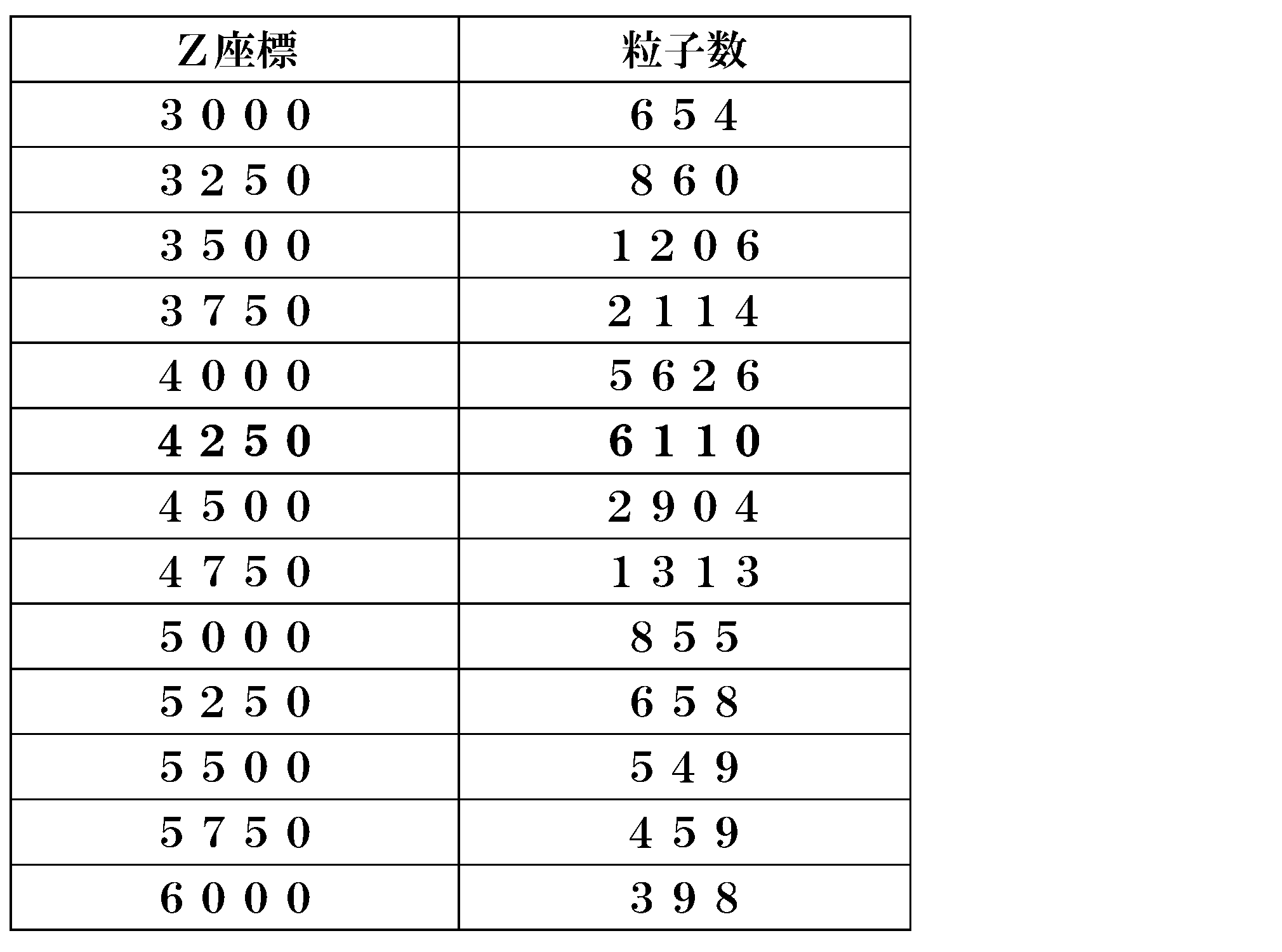
図9(a)及び図9(c)は、焦点が合っていないときの観察像を示し、図9(b)は、焦点が合っているときの観察像を示す。
図9(a)及び図9(c)に示すように、焦点が合っていないときには、個々の粒子の輪郭が不鮮明であるため、個々の粒子の輪郭が隣接する粒子と一体となってしまっている。このため、画像処理の際に、個々の粒子を切り分けて抽出することが困難であり、計測される粒子数が減少する。
また、図7(a)及び図7 (c)に示したように、焦点が全く合っていない場合には、観察像中の粒子そのものの判別が困難となるため、計測される粒子数は大きく低下する。
図9(a)及び図9(c)に示すように、焦点が合っていないときには、個々の粒子の輪郭が不鮮明であるため、個々の粒子の輪郭が隣接する粒子と一体となってしまっている。このため、画像処理の際に、個々の粒子を切り分けて抽出することが困難であり、計測される粒子数が減少する。
また、図7(a)及び図7 (c)に示したように、焦点が全く合っていない場合には、観察像中の粒子そのものの判別が困難となるため、計測される粒子数は大きく低下する。
一方、図9(b)に示すように、焦点が合っているときには、個々の粒子の輪郭が鮮明となるため、画像処理の際に、個々の粒子を隣接する粒子から切り分けて抽出することができ、計測される粒子数が増加する。したがって、最も多くの粒子が計測されるときには、最も多くの粒子に焦点が合っていることになる。
次に、粒子数が最大値である観察像を撮像したときの焦点位置を最適値として設定する(S23)。ここでは、Z座標「4250」がCCDカメラ3の焦点位置の最適値として設定される。
(画像取得工程)
次に、LEDランプ2の最適な電流設定値、及びCCDカメラ3の最適な焦点位置の撮像条件で撮像された画像を取得する(図2のS3)。
なお、この画像の取得にあたっては、最適化した撮像条件で改めて観察像を撮像してもよいし、撮像条件を設定する過程で、最適化された撮像条件と同一条件で撮像された観察像のデータを選択して利用してもよい。
次に、LEDランプ2の最適な電流設定値、及びCCDカメラ3の最適な焦点位置の撮像条件で撮像された画像を取得する(図2のS3)。
なお、この画像の取得にあたっては、最適化した撮像条件で改めて観察像を撮像してもよいし、撮像条件を設定する過程で、最適化された撮像条件と同一条件で撮像された観察像のデータを選択して利用してもよい。
このように、本実施形態では、LEDランプ2の電流設定値を変えて撮像した各観察像について、HLS色空間における観察像の平均輝度(L)及び平均彩度(S)に基づいた指標(I)が最大値となる撮像条件が設定された。さらに、本実施形態では、焦点位置を変えて撮像した観察像中の粒子数が最大となる撮像条件が設定された。これにより、細胞培養を継続中に培養液10の色調が変化したり、透明度が低下したりした場合であっても、また、培養細胞11が培養液10中で同一平面上に位置していない場合であっても、培養細胞11の撮像条件を最適化することができる。
[第2実施形態]
第2実施形態の撮像装置は、第1実施形態のものと同一であるので、その詳細な説明を省略する。
本実施形態の撮像方法では、図10のフローチャートに示すように、培養細胞11の観察像を撮像する際の撮像条件を設定するにあたって、観察像の明るさを調節するための撮像条件としてLEDランプ2の電流値を設定する明るさ設定工程(S1、S3及びS5)と、CCDカメラ3を移動させて焦点位置を設定する焦点位置設定工程(S2及びS4)とが交互に繰り返される。そして、最後に設定されたLEDランプ2の電流値及びCCDカメラ3の焦点位置で最適化された撮像条件で撮像した観察像を取得する(S6)。
第2実施形態の撮像装置は、第1実施形態のものと同一であるので、その詳細な説明を省略する。
本実施形態の撮像方法では、図10のフローチャートに示すように、培養細胞11の観察像を撮像する際の撮像条件を設定するにあたって、観察像の明るさを調節するための撮像条件としてLEDランプ2の電流値を設定する明るさ設定工程(S1、S3及びS5)と、CCDカメラ3を移動させて焦点位置を設定する焦点位置設定工程(S2及びS4)とが交互に繰り返される。そして、最後に設定されたLEDランプ2の電流値及びCCDカメラ3の焦点位置で最適化された撮像条件で撮像した観察像を取得する(S6)。
図11(a)~図11(e)は、図10のS1~S5の各工程において最適化された撮像条件で撮像した観察像を示す。
図11(a)は、1回目の明るさ設定工程(図10のS1)において最適化されたLEDランプ2の電流設定値で撮像した観察像を示す。この段階では未だ焦点が合っていないため、粒子が判別できない。
図11(a)は、1回目の明るさ設定工程(図10のS1)において最適化されたLEDランプ2の電流設定値で撮像した観察像を示す。この段階では未だ焦点が合っていないため、粒子が判別できない。
図12(a)のグラフは、1回目の明るさ設定工程(S1)における、LEDランプ2の電流設定値ごとの指標(I)を示す。1回目の明るさ設定工程(S1)では、LEDランプ2の電流設定値を「2」刻みで「2」から「40」まで変化させて、各電流設定値で培養細胞11を撮像し、合計20枚の観察像を生成した。
図12(a)に示すように、LEDランプ2の電流設定値が「14」のときに、指標(I)が極大となっている。図11(a)に示した観察像は、電流設定値を「14」とした撮像条件で撮像したものである。
次に、図11(b)は、1回目の焦点位置設定工程(図10のS2)において最適化された焦点位置で撮像した観察像を示す。この段階では、焦点が合っているため、観察像中の粒子が判別できる。
図13(a)のグラフは、1回目の焦点位置設定工程(S2)における、CCDカメラ3の焦点位置ごとの粒子数を示す。1回目の焦点位置設定工程(S2)では、焦点位置を表すZ座標を「250」刻みで「0」から「9750」まで変化させて、各Z座標で培養細胞11を撮像し、合計40枚の観察像を生成した。
図13(a)に示すように、Z座標が「3500」のときに、粒子数が最大となっている。図11(b)に示した観察像は、電流設定値を「14」としたまま、Z座標を「3500」とした撮像条件で撮像したものである。
次に、図11(c)に、2回目の明るさ設定工程(図10のS3)において最適化されたLEDランプ2の電流設定値で撮像した観察像を示す。1回目の焦点調節の結果、観察像の明るさが最適でなくなった場合であっても、2回目の明るさ調整によって、観察像の明るさが再び最適化される。
図12(b)のグラフは、2回目の明るさ設定工程(S3)における、LEDランプ2の電流設定値ごとの指標(I)を示す。2回目の明るさ設定工程(S3)も、1回目の明るさ設定工程と同様に、LEDランプ2の電流設定値を「2」刻みで、「2」から「40」まで変化させて、各電流設定値で培養細胞11を撮像し、合計20枚の観察像を生成した。
図12(b)に示すように、LEDランプ2の電流設定値が「14」のときに、指標(I)が極大となっている。図11(c)に示した観察像は、Z座標を「3500」としたまま、電流設定値を「14」とした撮像条件で撮像したものである。
図11(d)に、2回目の焦点位置設定工程(図10のS4)において2回目の焦点調整によって最適化された焦点位置で撮像した観察像を示す。
図13(b)のグラフは、2回目の焦点位置設定工程(S4)における、CCDカメラ3の焦点位置ごとの粒子数を示す。2回目の焦点位置設定工程(S4)では、焦点位置を表すZ座標の刻み幅を、1回目よりも狭い「100」刻みとし、Z座標の変化範囲を、1回目の最適値である「3500」を含み、かつ、1回目よりも狭い範囲である「2800」から「4200」として、各Z座標で培養細胞11を撮像し、合計15枚の観察像を生成した。
図13(b)に示すように、2回目の焦点位置設定工程(S4)では、Z座標が「3600」のときに、粒子数が最大となっている。図11(d)に示した観察像は、電流設定値を「14」としたまま、Z座標を「3600」とした撮像条件で撮像したものである。
このように、2回目の焦点位置設定における刻み幅を、1回目のものよりも狭くしつつ、2回目の焦点位置設定における変化範囲を、1回目のものよりも狭くした結果、撮像枚数の増大を回避しつつ、CCDカメラ3の焦点位置の撮像条件の設定精度の向上させることができる。
このように、2回目の焦点位置設定における刻み幅を、1回目のものよりも狭くしつつ、2回目の焦点位置設定における変化範囲を、1回目のものよりも狭くした結果、撮像枚数の増大を回避しつつ、CCDカメラ3の焦点位置の撮像条件の設定精度の向上させることができる。
次に、図11(e)に、3回目の明るさ設定工程(図10のS5)において最適化されたLEDランプ2の電流設定値で撮像した観察像を示す。
図12(c)のグラフは、3回目の明るさ設定工程(S5)における、LEDランプ2の電流設定値ごとの指標(I)を示す。3回目の明るさ設定工程(S5)では、LEDランプ2の電流設定値の刻み幅を、1回目よりも狭い「1」刻みとし、電流設定値の変化範囲を、2回目の最適値である「14」を含む「9」から「18」として、各電流設定値で培養細胞11を撮像し、合計10枚の観察像を生成した。
図12(c)に示すように、3回目の明るさ設定工程(S5)では、LEDランプ2の電流設定値が「15」のときに、指標(I)が極大となっている。図11(e)に示した観察像は、Z座標を「3600」としたまま、電流設定値を「15」とした撮像条件で撮像したものである。
このように、3回目の明るさ設定工程(S5)におけるLEDランプ2の電流設定値の刻み幅を、2回目のものよりも狭くしつつ、3回目の明るさ設定工程(S5)における電流設定値の変化範囲を、2回目のものよりも狭くした結果、撮像枚数の増大を回避しつつ、LEDランプ2の電流設定値の撮像条件の設定精度の向上させることができる。
このように、3回目の明るさ設定工程(S5)におけるLEDランプ2の電流設定値の刻み幅を、2回目のものよりも狭くしつつ、3回目の明るさ設定工程(S5)における電流設定値の変化範囲を、2回目のものよりも狭くした結果、撮像枚数の増大を回避しつつ、LEDランプ2の電流設定値の撮像条件の設定精度の向上させることができる。
このようにして、図11(e)に示した観察像が最適画像として取得される(図10のS6)。
以上、本発明の実施形態を説明したが、本発明は種々の変更及び変形を行うことができる。例えば、上述した各実施形態では、撮像部であるCCDカメラを、培養容器を挟んで、LEDランプの反対側に配置し、透過光を撮像した観察像を生成した例を説明したが、本発明では、観察像は透過光像に限定されず、例えば、反射光像であってもよい。
また、上述した実施形態では、観察像の明るさを調節する撮像条件として、LEDランプの電流値を設定した例を説明したが、本発明では、観察像の明るさを調節する撮像条件は、これに限定されず、例えば、絞りの開度で明るさを調節してもよいし、CCDカメラのシャッター速度で明るさを調節してもよい。
また、HSV表色系で指標を算出してもよい。HSV表色系は、色相(hue)、彩度(Saturation)及び明度(value)の3成分で表される。色相は、色味を360度の角度で表す。彩度は、純色を最大値とし、灰色を最低値として、色の鮮やかさを表す。明度は、色の明るさを表す。
また、上述した実施形態では、焦点位置を変化させるにあたり、撮像部としてのCCDカメラを移動させた例を説明したが、本発明では、焦点位置の調節方法はこれに限定されず、CCDカメラの光学系を移動させて焦点位置を調節してもよいし、培養容器を移動させて焦点位置を調節してもよい。
また、上述した実施形態では、照明部としてLEDランプを使用した例を説明したが、本発明では、照明部はこれに限定されない。また、上述した実施形態では、撮像部としてCCDカメラを使用した例を説明したが、本発明では、撮像部はこれに限定されない。
この明細書に記載の文献及び本願のパリ優先の基礎となる日本出願明細書の内容を全てここに援用する。
本発明は培養細胞の自動タイムラプス撮像に適用して好適である。
1 培養容器
2 LEDランプ
3 CCDカメラ
4 調光器
5 アクチュエータ
6 制御部
10 培養液
11 培養細胞
2 LEDランプ
3 CCDカメラ
4 調光器
5 アクチュエータ
6 制御部
10 培養液
11 培養細胞
Claims (9)
- 培養容器内の培養細胞の観察像を撮像する撮像方法であって、
観察像の明るさを調節する撮像条件を設定する明るさ設定工程と、
観察像を取得する画像取得工程と
を有し、
前記明るさ設定工程は、
観察像の明るさを調節する撮像条件を所定の変化範囲内で所定の刻み幅で変えて培養細胞を撮像する工程と、
撮像した各観察像について、HLS色空間における観察像の平均輝度及び平均彩度を算出する工程と、
前記各観察像について、前記平均輝度及び前記平均彩度に基づいて指標を算出する工程と、
前記指標が最大値である観察像を撮像したときの明るさを調節する撮像条件を最適値として設定する工程と
を有することを特徴とする、培養細胞の撮像方法。 - 培養容器内の培養細胞の観察像を撮像する撮像方法であって、
観察像の焦点位置を設定する焦点位置設定工程と、
観察像を取得する画像取得工程と
を有し、
前記焦点位置設定工程は、
焦点位置を所定の変化範囲内で所定の刻み幅で変えて培養細胞を撮像する工程と、
撮像した各観察像について、観察像中の粒子数を計測する工程と、
前記粒子数が最大値である観察像を撮像したときの焦点位置を最適値として設定する工程と
を有することを特徴とする、培養細胞の撮像方法。 - 培養容器内の培養細胞の観察像を撮像する撮像方法であって、
観察像の明るさを調節する撮像条件を設定する明るさ設定工程と、
観察像の焦点位置を設定する焦点位置設定工程と、
観察像を取得する画像取得工程と
を有し、
前記明るさ設定工程は、
観察像の明るさを調節する撮像条件を所定の変化範囲内で所定の刻み幅で変えて培養細胞を撮像する工程と、
撮像した各観察像について、HLS色空間における観察像の平均輝度及び平均彩度を算出する工程と、
前記各観察像について、前記平均輝度及び前記平均彩度に基づいて指標を算出する工程と、
前記指標が最大値である観察像を撮像したときの明るさを調節する撮像条件を最適値として設定する工程と
を有し、
前記焦点位置設定工程は、
焦点位置を所定の変化範囲内で所定の刻み幅で変えて培養細胞を撮像する工程と、
撮像した各観察像について、観察像中の粒子数を計測する工程と、
前記粒子数が最大値である観察像を撮像したときの焦点位置を最適値として設定する工程と
を有することを特徴とする、培養細胞の撮像方法。 - 前記明るさ設定工程と、前記焦点位置設定工程とを交互に繰り返し、
2回目以降の前記明るさ設定工程において、
明るさを調節する撮像条件の変化範囲を、前回の前記明るさ設定工程において設定された明るさを調節する撮像条件を含み、かつ、前回の前記明るさ設定工程の明るさを調節する撮像条件の変化範囲よりも狭くし、
明るさを調節する撮像条件の変化の刻み幅を、前回の前記明るさ設定工程の明るさを調節する撮像条件の変化の刻み幅よりも狭くする
ことを特徴とする、請求項3記載の培養細胞の撮像方法。 - 前記明るさ設定工程と、前記焦点位置設定工程とを交互に繰り返し、
2回目以降の前記焦点位置設定工程において、
焦点位置の変化範囲を、前回の前記焦点位置設定工程において設定された焦点位置を含み、かつ、前回の前記焦点位置設定工程の焦点位置の変化範囲よりも狭くし、
焦点位置の変化の刻み幅を、前回の前記焦点位置設定工程の焦点位置の変化の刻み幅よりも狭くする
ことを特徴とする、請求項3又は4記載の培養細胞の撮像方法。 - 前記指標は、輝度階調の最大値である最大輝度と前記観察像の平均輝度との差と、前記平均輝度と前記観察像の平均彩度との和との積を含む
ことを特徴とする、請求項1、3~5のいずれか一項に記載の培養細胞の撮像方法。 - 培養容器内の培養細胞の観察像を撮像する撮像装置であって、
前記培養細胞を照明する照明部と、
前記培養細胞を撮像して観察像を生成する撮像部と、
観察像の明るさを調節する撮像条件を設定する明るさ制御部と
を備え、
前記明るさ制御部は、観察像の明るさを調節する撮像条件を所定の変化範囲内で所定の刻み幅で変えて前記撮像部に前記培養細胞を撮像させ、前記撮像部によって撮像された各観察像について、HLS色空間における観察像の平均輝度及び平均彩度を算出し、前記各観察像について、前記平均輝度及び前記平均彩度に基づいて指標を算出し、前記指標が最大値である観察像を撮像したときの明るさを調節する撮像条件を最適値として設定する
ことを特徴とする、培養細胞の撮像装置。 - 培養容器内の培養細胞の観察像を撮像する撮像装置であって、
前記培養細胞を照明する照明部と、
前記培養細胞を撮像して観察像を生成する撮像部と、
観察像の焦点位置を設定する焦点位置制御部と
を備え、
前記焦点位置制御部は、焦点位置を所定の変化範囲内で所定の刻み幅で変えて前記撮像部に前記培養細胞を撮像させ、前記撮像部によって撮像された各観察像について、観察像中の粒子数を計測し、前記粒子数が最大値である観察像を撮像したときの最適値として焦点位置を設定する
ことを特徴とする、培養細胞の撮像装置。 - 培養容器内の培養細胞の観察像を撮像する撮像装置であって、
前記培養細胞を照明する照明部と、
前記培養細胞を撮像して観察像を生成する撮像部と、
観察像の明るさを調節する撮像条件を設定する明るさ制御部と、
観察像の焦点位置を設定する焦点位置制御部と
を備え、
前記明るさ制御部は、観察像の明るさを調節する撮像条件を所定の変化範囲内で所定の刻み幅で変えて前記撮像部に前記培養細胞を撮像させ、前記撮像部によって撮像された各観察像について、HLS色空間における観察像の平均輝度及び平均彩度を算出し、前記各観察像について、前記平均輝度及び前記平均彩度に基づいて指標を算出し、前記指標が最大値である観察像を撮像したときの明るさを調節する撮像条件を最適値として設定し、
前記焦点位置制御部は、焦点位置を所定の変化範囲内で所定の刻み幅で変えて前記撮像部に前記培養細胞を撮像させ、前記撮像部によって撮像された各観察像について、観察像中の粒子数を計測し、前記粒子数が最大値である観察像を撮像したときの焦点位置を最適値として設定することを特徴とする、培養細胞の撮像装置。
Applications Claiming Priority (2)
| Application Number | Priority Date | Filing Date | Title |
|---|---|---|---|
| JP2016-121483 | 2016-06-20 | ||
| JP2016121483A JP2017225354A (ja) | 2016-06-20 | 2016-06-20 | 培養細胞の撮像方法及び装置 |
Publications (1)
| Publication Number | Publication Date |
|---|---|
| WO2017221616A1 true WO2017221616A1 (ja) | 2017-12-28 |
Family
ID=60784056
Family Applications (1)
| Application Number | Title | Priority Date | Filing Date |
|---|---|---|---|
| PCT/JP2017/019233 Ceased WO2017221616A1 (ja) | 2016-06-20 | 2017-05-23 | 培養細胞の撮像方法及び装置 |
Country Status (3)
| Country | Link |
|---|---|
| JP (1) | JP2017225354A (ja) |
| TW (1) | TW201804211A (ja) |
| WO (1) | WO2017221616A1 (ja) |
Citations (5)
| Publication number | Priority date | Publication date | Assignee | Title |
|---|---|---|---|---|
| JP2011141407A (ja) * | 2010-01-07 | 2011-07-21 | Sanyo Electric Co Ltd | 観察ユニット用の制御装置、制御プログラム及び制御方法、並びに観察システム |
| JP2014514580A (ja) * | 2011-05-06 | 2014-06-19 | ビオメリュー | バイオ画像化方法及びシステム |
| JP2015181374A (ja) * | 2014-03-24 | 2015-10-22 | 東レ株式会社 | 糸状性細菌検出装置および糸状性細菌の検出方法 |
| JP2015194544A (ja) * | 2014-03-31 | 2015-11-05 | 富士フイルム株式会社 | 細胞撮像制御装置および方法並びにプログラム |
| JP2016508034A (ja) * | 2012-12-20 | 2016-03-17 | スリーエム イノベイティブ プロパティズ カンパニー | 画像の微生物コロニーを区別する方法 |
-
2016
- 2016-06-20 JP JP2016121483A patent/JP2017225354A/ja active Pending
-
2017
- 2017-05-23 WO PCT/JP2017/019233 patent/WO2017221616A1/ja not_active Ceased
- 2017-06-01 TW TW106118128A patent/TW201804211A/zh unknown
Patent Citations (5)
| Publication number | Priority date | Publication date | Assignee | Title |
|---|---|---|---|---|
| JP2011141407A (ja) * | 2010-01-07 | 2011-07-21 | Sanyo Electric Co Ltd | 観察ユニット用の制御装置、制御プログラム及び制御方法、並びに観察システム |
| JP2014514580A (ja) * | 2011-05-06 | 2014-06-19 | ビオメリュー | バイオ画像化方法及びシステム |
| JP2016508034A (ja) * | 2012-12-20 | 2016-03-17 | スリーエム イノベイティブ プロパティズ カンパニー | 画像の微生物コロニーを区別する方法 |
| JP2015181374A (ja) * | 2014-03-24 | 2015-10-22 | 東レ株式会社 | 糸状性細菌検出装置および糸状性細菌の検出方法 |
| JP2015194544A (ja) * | 2014-03-31 | 2015-11-05 | 富士フイルム株式会社 | 細胞撮像制御装置および方法並びにプログラム |
Also Published As
| Publication number | Publication date |
|---|---|
| JP2017225354A (ja) | 2017-12-28 |
| TW201804211A (zh) | 2018-02-01 |
Similar Documents
| Publication | Publication Date | Title |
|---|---|---|
| CN102984448B (zh) | 利用颜色数字图像对动作如锐度修改进行控制的方法 | |
| CN112839216B (zh) | 一种图像色彩校正方法及装置 | |
| CN1994000A (zh) | 自动白平衡方法及设备 | |
| CN1795467A (zh) | 对具有至少一个识别特征的材料进行定性判定的方法 | |
| EP2688302A3 (en) | Image processing apparatus and control method thereof, and image capture apparatus | |
| CN103764013A (zh) | 自动曝光控制装置、控制装置、内窥镜装置和自动曝光控制方法 | |
| CN109856164A (zh) | 一种机器视觉采集大范围图像的优化装置及其检测方法 | |
| JP2016126750A (ja) | 画像処理システム、画像処理装置、撮像装置、画像処理方法、プログラムおよび記録媒体 | |
| US8890967B2 (en) | Method apparatus for controlling exposure amount of an imaging device | |
| WO2013114803A1 (ja) | 画像処理装置及びその画像処理方法、並びにコンピュータ・プログラム、および画像処理システム | |
| CN107644437B (zh) | 基于分块的偏色检测系统及其方法 | |
| CN111131723A (zh) | 一种瓷砖图像采集方法及系统 | |
| US20140192223A1 (en) | Imaging device, imaging system, and imaging method | |
| CN109685727A (zh) | 图像处理方法 | |
| JP6738553B2 (ja) | 変化度合い導出装置、変化度合い導出システム、変化度合い導出方法及びプログラム | |
| CN114257736A (zh) | 一种工件自适应拍摄方法 | |
| US20220343472A1 (en) | Selective image signal processing | |
| CN110022426A (zh) | 一种基于图像识别的相机智能补光方法 | |
| US20180260929A1 (en) | Digital camera methods and devices optimized for computer vision applications | |
| WO2017221616A1 (ja) | 培養細胞の撮像方法及び装置 | |
| KR20120122574A (ko) | 디지털 카메라 장치에서 영상 처리 장치 및 방법 | |
| JP5609459B2 (ja) | 二値化処理方法及び画像処理装置 | |
| CN110326288A (zh) | 图像处理设备以及成像设备 | |
| US10917590B2 (en) | Imaging control device, imaging apparatus, and imaging control method | |
| JP2022166523A (ja) | 画像処理装置、撮像センサ、画像処理方法、およびプログラム |
Legal Events
| Date | Code | Title | Description |
|---|---|---|---|
| 121 | Ep: the epo has been informed by wipo that ep was designated in this application |
Ref document number: 17815085 Country of ref document: EP Kind code of ref document: A1 |
|
| NENP | Non-entry into the national phase |
Ref country code: DE |
|
| 122 | Ep: pct application non-entry in european phase |
Ref document number: 17815085 Country of ref document: EP Kind code of ref document: A1 |