WO2017204208A1 - Method for analyzing expression of smn protein nuclear body - Google Patents
Method for analyzing expression of smn protein nuclear body Download PDFInfo
- Publication number
- WO2017204208A1 WO2017204208A1 PCT/JP2017/019165 JP2017019165W WO2017204208A1 WO 2017204208 A1 WO2017204208 A1 WO 2017204208A1 JP 2017019165 W JP2017019165 W JP 2017019165W WO 2017204208 A1 WO2017204208 A1 WO 2017204208A1
- Authority
- WO
- WIPO (PCT)
- Prior art keywords
- smn protein
- blood
- labeling
- expression
- cells
- Prior art date
Links
Images


Classifications
-
- G—PHYSICS
- G01—MEASURING; TESTING
- G01N—INVESTIGATING OR ANALYSING MATERIALS BY DETERMINING THEIR CHEMICAL OR PHYSICAL PROPERTIES
- G01N33/00—Investigating or analysing materials by specific methods not covered by groups G01N1/00 - G01N31/00
- G01N33/48—Biological material, e.g. blood, urine; Haemocytometers
- G01N33/50—Chemical analysis of biological material, e.g. blood, urine; Testing involving biospecific ligand binding methods; Immunological testing
- G01N33/68—Chemical analysis of biological material, e.g. blood, urine; Testing involving biospecific ligand binding methods; Immunological testing involving proteins, peptides or amino acids
- G01N33/6893—Chemical analysis of biological material, e.g. blood, urine; Testing involving biospecific ligand binding methods; Immunological testing involving proteins, peptides or amino acids related to diseases not provided for elsewhere
- G01N33/6896—Neurological disorders, e.g. Alzheimer's disease
-
- G—PHYSICS
- G01—MEASURING; TESTING
- G01N—INVESTIGATING OR ANALYSING MATERIALS BY DETERMINING THEIR CHEMICAL OR PHYSICAL PROPERTIES
- G01N15/00—Investigating characteristics of particles; Investigating permeability, pore-volume, or surface-area of porous materials
- G01N15/10—Investigating individual particles
- G01N15/14—Electro-optical investigation, e.g. flow cytometers
-
- G—PHYSICS
- G01—MEASURING; TESTING
- G01N—INVESTIGATING OR ANALYSING MATERIALS BY DETERMINING THEIR CHEMICAL OR PHYSICAL PROPERTIES
- G01N21/00—Investigating or analysing materials by the use of optical means, i.e. using sub-millimetre waves, infrared, visible or ultraviolet light
- G01N21/62—Systems in which the material investigated is excited whereby it emits light or causes a change in wavelength of the incident light
- G01N21/63—Systems in which the material investigated is excited whereby it emits light or causes a change in wavelength of the incident light optically excited
- G01N21/64—Fluorescence; Phosphorescence
- G01N21/6428—Measuring fluorescence of fluorescent products of reactions or of fluorochrome labelled reactive substances, e.g. measuring quenching effects, using measuring "optrodes"
-
- G—PHYSICS
- G01—MEASURING; TESTING
- G01N—INVESTIGATING OR ANALYSING MATERIALS BY DETERMINING THEIR CHEMICAL OR PHYSICAL PROPERTIES
- G01N33/00—Investigating or analysing materials by specific methods not covered by groups G01N1/00 - G01N31/00
- G01N33/48—Biological material, e.g. blood, urine; Haemocytometers
- G01N33/483—Physical analysis of biological material
- G01N33/487—Physical analysis of biological material of liquid biological material
- G01N33/49—Blood
- G01N33/4915—Blood using flow cells
-
- G—PHYSICS
- G01—MEASURING; TESTING
- G01N—INVESTIGATING OR ANALYSING MATERIALS BY DETERMINING THEIR CHEMICAL OR PHYSICAL PROPERTIES
- G01N33/00—Investigating or analysing materials by specific methods not covered by groups G01N1/00 - G01N31/00
- G01N33/48—Biological material, e.g. blood, urine; Haemocytometers
- G01N33/50—Chemical analysis of biological material, e.g. blood, urine; Testing involving biospecific ligand binding methods; Immunological testing
- G01N33/53—Immunoassay; Biospecific binding assay; Materials therefor
- G01N33/569—Immunoassay; Biospecific binding assay; Materials therefor for microorganisms, e.g. protozoa, bacteria, viruses
- G01N33/56966—Animal cells
- G01N33/56972—White blood cells
-
- G—PHYSICS
- G01—MEASURING; TESTING
- G01N—INVESTIGATING OR ANALYSING MATERIALS BY DETERMINING THEIR CHEMICAL OR PHYSICAL PROPERTIES
- G01N33/00—Investigating or analysing materials by specific methods not covered by groups G01N1/00 - G01N31/00
- G01N33/48—Biological material, e.g. blood, urine; Haemocytometers
- G01N33/50—Chemical analysis of biological material, e.g. blood, urine; Testing involving biospecific ligand binding methods; Immunological testing
- G01N33/58—Chemical analysis of biological material, e.g. blood, urine; Testing involving biospecific ligand binding methods; Immunological testing involving labelled substances
- G01N33/582—Chemical analysis of biological material, e.g. blood, urine; Testing involving biospecific ligand binding methods; Immunological testing involving labelled substances with fluorescent label
-
- G—PHYSICS
- G01—MEASURING; TESTING
- G01N—INVESTIGATING OR ANALYSING MATERIALS BY DETERMINING THEIR CHEMICAL OR PHYSICAL PROPERTIES
- G01N33/00—Investigating or analysing materials by specific methods not covered by groups G01N1/00 - G01N31/00
- G01N33/48—Biological material, e.g. blood, urine; Haemocytometers
- G01N33/50—Chemical analysis of biological material, e.g. blood, urine; Testing involving biospecific ligand binding methods; Immunological testing
- G01N33/68—Chemical analysis of biological material, e.g. blood, urine; Testing involving biospecific ligand binding methods; Immunological testing involving proteins, peptides or amino acids
- G01N33/6875—Nucleoproteins
-
- G—PHYSICS
- G01—MEASURING; TESTING
- G01N—INVESTIGATING OR ANALYSING MATERIALS BY DETERMINING THEIR CHEMICAL OR PHYSICAL PROPERTIES
- G01N21/00—Investigating or analysing materials by the use of optical means, i.e. using sub-millimetre waves, infrared, visible or ultraviolet light
- G01N21/62—Systems in which the material investigated is excited whereby it emits light or causes a change in wavelength of the incident light
- G01N21/63—Systems in which the material investigated is excited whereby it emits light or causes a change in wavelength of the incident light optically excited
- G01N21/64—Fluorescence; Phosphorescence
- G01N21/6428—Measuring fluorescence of fluorescent products of reactions or of fluorochrome labelled reactive substances, e.g. measuring quenching effects, using measuring "optrodes"
- G01N2021/6439—Measuring fluorescence of fluorescent products of reactions or of fluorochrome labelled reactive substances, e.g. measuring quenching effects, using measuring "optrodes" with indicators, stains, dyes, tags, labels, marks
-
- G—PHYSICS
- G01—MEASURING; TESTING
- G01N—INVESTIGATING OR ANALYSING MATERIALS BY DETERMINING THEIR CHEMICAL OR PHYSICAL PROPERTIES
- G01N2800/00—Detection or diagnosis of diseases
- G01N2800/28—Neurological disorders
- G01N2800/2878—Muscular dystrophy
Abstract
The purpose of the present invention is to provide a method capable of analyzing an SMN protein nuclear body that serves as a more highly reliable biomarker. The method according to the present invention is a method for analyzing the expression of an SMN protein nuclear body, and comprises the steps of: labeling at least one surface antigen marker for blood-derived nucleated cells with at least one labeling antibody in a sample containing the nucleated cells; labeling the SMN protein in the nucleated cells; labeling the nuclei of the nucleated cells; selecting one cell mass from among multiple cell masses of the nucleated cells, the multiple cell masses being cell masses in which the nucleus and the SMN protein are labeled and which have been classified on the basis of the surface antigen marker labeled with the labeling antibody or the like; and subjecting the selected cell mass to analysis on the expression of the SMN protein nuclear body on the basis of the labeling of the SMN protein. The method involves carrying out imaging flow cytometry using an objective lens at a specific magnification.
Description
本発明は、サバイバルモーターニューロン(SMN)タンパク質の核内構造体の発現解析方法に関する。
The present invention relates to an expression analysis method for a nuclear structure of a survival motor neuron (SMN) protein.
脊髄性筋萎縮症(SMA)は、脊髄前角細胞の病変によって引き起こされる筋萎縮症であり、体幹や四肢の筋力低下及び筋萎縮を特徴とする下位運動ニューロン徴候を示す。SMAは、発症年齢と重症度によってI型からIV型に分類され、生後6ヶ月までに発症するI型:重症型(ウェルドニッヒ・ホフマン病とも言う)、1歳6ヶ月までに発症するII型:中間型(デュボヴィッツ病とも言う)、及び1歳6ヶ月以降に発症するIII型:軽症型(クーゲルベルグ・ウェランダー病とも言う)が、小児期発症SMAである。一方、20歳以降に発症するIV型は、成人発症SMAである。
I型SMAは、SMAの約30%を占めており、6ヶ月以下で発症する。その症状は極めて深刻で、生涯座位保持不可能であり、人工呼吸を使わずに2歳以上生存できることは稀である。II型SMAは、生涯起立及び歩行は不可能であり、III型SMAは、自立歩行を獲得するものの、次第に転びやすい、歩けない又は立てないという症状が現れる。しかしながら、依然としてSMAの根本治療法は確立しておらず、SMAは国が指定する難病のうちの1つである。 Spinal muscular atrophy (SMA) is a muscular atrophy caused by an anterior horn cell lesion and exhibits lower motor neuron signs characterized by muscle weakness and muscle atrophy of the trunk and extremities. SMA is classified from type I to type IV according to age and severity of onset, and type I develops by 6 months after birth: severe type (also called Weldnig-Hoffmann disease), type II that develops by 1 year and 6 months: Childhood-onset SMA is intermediate type (also called Dubovitz disease) and type III that develops after 1 year and 6 months: Mild type (also called Kugelberg-Wehlander disease). On the other hand, type IV that develops after age 20 is adult-onset SMA.
Type I SMA accounts for about 30% of SMA and develops in less than 6 months. Symptoms are extremely severe, unable to maintain a sitting position throughout life, and rarely survive over 2 years without using artificial respiration. Type II SMA is unable to stand and walk for life, and Type III SMA gains self-sustained gait, but gradually develops symptoms such as falling or being unable to walk or stand. However, the fundamental cure for SMA has not yet been established, and SMA is one of the intractable diseases designated by the government.
多くの小児期発症SMAの原因遺伝子は、5番染色体長腕5q13に存在するSMN1遺伝子である。多くの小児期発症SMAでは、SMN1遺伝子の欠失又は変異が認められ、小児期発症SMAは常染色体劣性遺伝性疾患として認識されている。SMN1遺伝子からは、SMNタンパク質が発現され、SMNタンパク質が運動ニューロンの形成等に関与していると考えられている。
また、SMN1遺伝子が存在する5番染色体の同領域には、SMN1遺伝子とコーディング領域の塩基が1つだけ異なるSMN2遺伝子が存在している。小児期発症SMA患者では、SMN1遺伝子が欠失又は変異し、SMN1遺伝子由来のSMNタンパク質が低下している一方で、SMN2遺伝子は正常に機能している。そのため、小児期発症SMA患者においても、SMN2遺伝子由来のSMNタンパク質は発現しており、このSMN2遺伝子由来のSMNタンパク質の発現量に応じて、症状の重症度が異なると考えられている。
しかしながら、SMN1遺伝子の転写産物は全長SMN1mRNAの1種類であるのに対し、SMN2遺伝子の転写産物は2種類存在し、SMN1遺伝子から転写される全長SMN1mRNAの量を100%とすると、全長SMN2mRNAがおよそ10%であり、エクソン7の欠失が認められる短縮型SMN2mRNAがおよそ90%である。短縮型SMN2mRNAは非機能的なタンパク質に翻訳され、全長SMN2mRNAのみが正常にSMNタンパク質に翻訳されるため、SMN1遺伝子が欠失又は変異している小児期発症SMA患者においては、SMNタンパク質の発現量が正常者に比べて低く、正常者の10%~20%ほどしかない。そのため、SMN1遺伝子が欠失又は変異することにより、筋力低下及び筋萎縮が生じることとなる。
小児期発症SMA患者よりも数は少ないが、成人発症SMA患者においても、上記のようなSMN1遺伝子の欠失又は変異が認められる場合があり、やはりSMNタンパク質の発現量に応じて、症状の重症度が異なると考えられている。 The causative gene for many childhood-onset SMAs is the SMN1 gene present in the long arm 5q13 of chromosome 5. Many childhood-onset SMA has a deletion or mutation of the SMN1 gene, and childhood-onset SMA is recognized as an autosomal recessive inherited disease. From the SMN1 gene, SMN protein is expressed, and it is considered that SMN protein is involved in the formation of motor neurons.
Further, in the same region of chromosome 5 where the SMN1 gene is present, there is an SMN2 gene that differs from the SMN1 gene by one base in the coding region. In childhood-onset SMA patients, the SMN1 gene is deleted or mutated, and the SMN1 gene-derived SMN protein is decreased, while the SMN2 gene functions normally. Therefore, even in childhood-onset SMA patients, the SMN2 gene-derived SMN protein is expressed, and it is considered that the severity of symptoms varies depending on the expression level of the SMN2 gene-derived SMN protein.
However, while the transcript of the SMN1 gene is one type of full-length SMN1 mRNA, there are two types of transcripts of the SMN2 gene, and when the amount of full-length SMN1 mRNA transcribed from the SMN1 gene is 100%, About 90% of the shortened SMN2 mRNA is 10% and exon 7 deletion is observed. Abbreviated SMN2 mRNA is translated into a non-functional protein, and only full-length SMN2 mRNA is normally translated into SMN protein. Therefore, the expression level of SMN protein in childhood-onset SMA patients in which the SMN1 gene is deleted or mutated Is lower than normal, only 10% to 20% of normal. Therefore, when the SMN1 gene is deleted or mutated, muscle weakness and muscle atrophy occur.
The number of SMN1 gene deletions or mutations may be observed in adult-onset SMA patients, although the number is smaller than in childhood-onset SMA patients. The severity of symptoms depends on the expression level of SMN protein. The degree is considered different.
SMN1遺伝子が欠失しているSMA患者において、SMN2遺伝子は、SMN1遺伝子に置き換わって存在することがある。この場合、SMN2遺伝子は1つの染色体上に2コピー存在することとなり、SMN2遺伝子由来のSMNタンパク質の発現量もコピー数に応じて多くなる。SMA患者において、SMN2遺伝子由来のSMNタンパク質の発現量が増えれば増えるほど、症状は軽くなることがわかっている。
以上のように、SMNタンパク質の発現量は、SMAと密接に関連しており、SMNタンパク質は、SMA、特には小児期発症SMAの診断のためや、薬剤の効果を的確に判断するための有用なバイオマーカーとなる可能性があると考えられている。しかしながら、SMA患者においても、SMNタンパク質の発現自体は認められ、SMNタンパク質をSMAの有用なバイオマーカーとして利用するためには、正常者と患者とを比較した場合のSMNタンパク質発現レベルの差異を検出できるほどの鋭敏さが必要となる。
また、SMAは常染色体劣性遺伝性疾患であり、1対の常染色体のうちの一方においてのみSMN1遺伝子が欠失又は変異している場合には、何ら筋力低下及び筋萎縮の症状は現れず、保因者ということになる。保因者においては、患者よりもSMNタンパク質の発現量は高いものの、保因者と患者との間では、正常者と患者との間のSMNタンパク質の発現量の差ほど大きな差があるわけではないと考えられる。従って、SMNタンパク質をSMAの有用なバイオマーカーとして利用するためには、保因者と患者とを比較した場合のSMNタンパク質発現レベルの差異を検出できるほどの更なる鋭敏さが必要となる。 In SMA patients lacking the SMN1 gene, the SMN2 gene may be present in place of the SMN1 gene. In this case, two copies of the SMN2 gene are present on one chromosome, and the expression amount of the SMN protein derived from the SMN2 gene increases in accordance with the copy number. In SMA patients, it has been found that the greater the expression level of SMN protein derived from the SMN2 gene, the milder the symptoms.
As described above, the expression level of SMN protein is closely related to SMA, and SMN protein is useful for diagnosing SMA, especially childhood-onset SMA, and for accurately determining the effects of drugs. It is considered to be a potential biomarker. However, even in SMA patients, the expression of SMN protein itself was recognized, and in order to use SMN protein as a useful biomarker of SMA, the difference in the expression level of SMN protein was detected when comparing normal patients with patients. You need to be as sensitive as you can.
Also, SMA is an autosomal recessive disorder, and when the SMN1 gene is deleted or mutated only in one of a pair of autosomes, no symptoms of muscle weakness and muscle atrophy appear, It will be a carrier. Carriers have higher expression levels of SMN protein than patients, but there is not as much difference between carriers and patients as there are differences in SMN protein expression levels between normal and patients. It is not considered. Therefore, in order to use the SMN protein as a useful biomarker of SMA, it is necessary to have such sensitivity that the difference in the expression level of the SMN protein can be detected when the carrier and the patient are compared.
実際に、末梢血単核球細胞を用いた、ELISA法によるSMNタンパク質の定量解析が試みられている(非特許文献1)。しかしながら、Kobayashiらは、小児期発症SMA患者と保因者との間において、ELISA法ではSMNタンパク質発現レベルに有意差が認められないという結果が得られたことを報告している。更に、Kobayashiらは、SMNタンパク質発現レベルは、SMAの診断において信頼性のある指標とはならないとさえ開示している。
また、ELISA法によるSMNタンパク質の定量解析においては、末梢血単核球細胞(PBMC)を用いることを必要とし、患者らから得られた血液サンプルを遠心分離するなどの手間がかかる上に、細胞の収率も低下する。そのため、PBMCを用いるELISA法によるSMNタンパク質の定量では、必要とされる採血量も増大する。
このような状況において、本発明者らはSMNタンパク質の発現を検出する方法を見出し、SMNタンパク質を小児期発症SMAの有用なバイオマーカーとして利用し得ることを報告した(特許文献1)。
しかしながら、特許文献1には、SMNタンパク質の細胞内での局在化や、SMNタンパク質の核内構造体については明らかにされていない。 Actually, quantitative analysis of SMN protein by ELISA method using peripheral blood mononuclear cells has been attempted (Non-patent Document 1). However, Kobayashi et al. Reported that the ELISA method showed no significant difference in SMN protein expression levels between childhood-onset SMA patients and carriers. Furthermore, Kobayashi et al. Even discloses that SMN protein expression levels are not a reliable indicator in the diagnosis of SMA.
In addition, the quantitative analysis of SMN protein by ELISA requires the use of peripheral blood mononuclear cells (PBMC), and it takes time and effort to centrifuge blood samples obtained from patients. The yield of is also reduced. Therefore, in the quantification of SMN protein by ELISA using PBMC, the amount of blood collected is also increased.
Under such circumstances, the present inventors have found a method for detecting the expression of SMN protein, and have reported that SMN protein can be used as a useful biomarker for childhood-onset SMA (Patent Document 1).
However, Patent Document 1 does not disclose the localization of the SMN protein in cells or the nuclear structure of the SMN protein.
本発明は、血液細胞サンプルを用いて、バイオマーカーとしての信頼性がより高いSMNタンパク質の核内構造体を解析できる方法を提供することを目的とする。
An object of the present invention is to provide a method capable of analyzing a nuclear structure of an SMN protein having higher reliability as a biomarker using a blood cell sample.
本発明者らは、鋭意検討を重ねた結果、SMNタンパク質の発現を検出する細胞について、血液細胞中から特定の細胞集団を選別して、その細胞集団においてSMNタンパク質の発現を検出することで、SMNタンパク質発現レベルの検出感度を高めるとともに、イメージングフローサイトメトリーによりSMNタンパク質の核内構造体の解析が可能であることを新たに見出した。
本発明の一態様において、
SMNタンパク質の核内構造体の発現解析方法であって、
血液由来の有核細胞を含むサンプル中、前記有核細胞の1種以上の表面抗原マーカーを、1種以上の標識抗体を用いて標識する工程、
前記有核細胞内のSMNタンパク質を標識する工程、
前記有核細胞の核を標識する工程、
前記有核細胞のうち、核及びSMNタンパク質が標識され、かつ前記1種以上の標識抗体により標識された前記1種以上の表面抗原マーカー又は前記1種以上の標識抗体により標識された前記1種以上の表面抗原マーカーと側方散乱(SSC)とに基づき分類された複数の細胞集団から1つの細胞集団を選別する工程、及び
前記選別した細胞集団について、SMNタンパク質に対する標識に基づいてSMNタンパク質の核内構造体の発現解析をする工程
を含み、前記細胞集団を選別する工程及び前記SMNタンパク質の核内構造体の発現解析をする工程を、40倍以上の対物レンズを使用するイメージングフローサイトメトリーにより行う、前記方法が提供される。 As a result of intensive studies, the present inventors have selected a specific cell population from blood cells for cells that detect the expression of SMN protein, and detected the expression of SMN protein in the cell population. It was newly found that the detection sensitivity of the SMN protein expression level was enhanced and the nuclear structure of the SMN protein could be analyzed by imaging flow cytometry.
In one embodiment of the present invention,
A method for analyzing the expression of a nuclear structure of SMN protein,
Labeling one or more surface antigen markers of the nucleated cells with one or more labeled antibodies in a sample containing nucleated cells derived from blood,
Labeling the SMN protein in the nucleated cell,
Labeling the nucleus of the nucleated cell,
Among the nucleated cells, the nuclear and SMN proteins are labeled and the one or more surface antigen markers labeled with the one or more labeled antibodies or the one or more labeled with the one or more labeled antibodies. A step of selecting one cell population from a plurality of cell populations classified based on the above surface antigen markers and side scatter (SSC), and for the selected cell population, the SMN protein Imaging flow cytometry using an objective lens of 40 times or more, comprising the step of analyzing the expression of the nuclear structure, the step of selecting the cell population and the step of analyzing the expression of the nuclear structure of the SMN protein Is provided.
本発明の一態様では、SMNタンパク質の核内構造体の発現解析方法において、有核細胞内のSMNタンパク質を標識する工程が、SMNタンパク質を第1の蛍光色素により標識することを含み、かつ、SMNタンパク質の核内構造体の発現解析をする工程が、第1の蛍光色素が発する蛍光強度を測定することを含む。
本発明の一態様では、SMNタンパク質の核内構造体の発現解析方法において、有核細胞内のSMNタンパク質を標識する工程が、SMNタンパク質を第1の蛍光色素により標識することを含み、かつ、有核細胞の核を標識する工程が、核を第2の蛍光色素により標識することを含む。
本発明の一態様では、SMNタンパク質の核内構造体の発現解析方法において、血液由来の有核細胞を含むサンプルを赤血球溶血処理に付す工程を更に含む。 In one aspect of the present invention, in the method for analyzing the expression of a nuclear structure of an SMN protein, the step of labeling the SMN protein in a nucleated cell comprises labeling the SMN protein with a first fluorescent dye, and The step of analyzing the expression of the nuclear structure of the SMN protein includes measuring the fluorescence intensity emitted by the first fluorescent dye.
In one aspect of the present invention, in the method for analyzing the expression of a nuclear structure of an SMN protein, the step of labeling the SMN protein in a nucleated cell comprises labeling the SMN protein with a first fluorescent dye, and The step of labeling the nucleus of the nucleated cell includes labeling the nucleus with a second fluorescent dye.
In one embodiment of the present invention, the method for analyzing the expression of a nuclear structure of an SMN protein further includes a step of subjecting a sample containing nucleated cells derived from blood to erythrocyte hemolysis.
本発明の一態様では、SMNタンパク質の核内構造体の発現解析方法において、血液由来の有核細胞を含むサンプルが被験者から得られたものであり、
SMNタンパク質の核内構造体の発現解析をする工程が、SMNタンパク質の核内構造体を定量することを含み、
前記定量により得られた結果を、以下の(a)~(c)より選択されるコントロールと比較する工程を含む:
(a) 前記被験者がSMA患者である場合には、前記コントロールは、正常者又は保因者から得られる血液を用いること以外は、前記被験者から得られた血液を用いてSMNタンパク質の核内構造体を定量した方法と同様にして得られた結果であり、
(b) 前記被験者が正常者又は保因者である場合には、前記コントロールは、SMA患者から得られる血液を用いること以外は、前記被験者から得られた血液を用いてSMNタンパク質の核内構造体を定量した方法と同様にして得られた結果であり、又は
(c) 前記被験者がSMAの治療薬又は治療候補薬を投与された者である場合には、前記コントロールは、前記SMAの治療薬又は治療候補薬の投与前の前記被験者から得られた血液を用いること以外は、前記投与後の前記被験者から得られた血液を用いてSMNタンパク質の核内構造体を定量した方法と同様にして得られた結果である。 In one embodiment of the present invention, in the expression analysis method for the nuclear structure of the SMN protein, a sample containing nucleated cells derived from blood is obtained from a subject.
The step of analyzing the expression of the nuclear structure of the SMN protein comprises quantifying the nuclear structure of the SMN protein;
Comparing the result obtained by the quantification with a control selected from the following (a) to (c):
(a) When the subject is an SMA patient, the control uses the blood obtained from the subject except for blood obtained from a normal person or a carrier. It is the result obtained in the same way as the method of quantifying the body,
(b) When the subject is a normal person or a carrier, the control uses the blood obtained from the subject except for blood obtained from the SMA patient, and the nuclear structure of the SMN protein. The results obtained in the same manner as the method for quantifying the body, or Except for using blood obtained from the subject before administration of the drug or treatment candidate drug, the method was the same as the method for quantifying the nuclear structure of SMN protein using blood obtained from the subject after administration. This is the result obtained.
本発明の一態様によれば、血液細胞サンプルを用いて、バイオマーカーとしての信頼性がより高いSMNタンパク質の核内構造体を解析することができる。
また、ひいては、本発明の一態様によれば、SMA患者と正常者又は保因者との間において、SMNタンパク質の核内構造体の発現における差異を検出できる方法を提供することができる。
更に、本発明の一態様によれば、血液約500μLでSMNタンパク質の核内構造体の発現解析が可能であり、SMAの治療薬又は治療候補薬の試験においてSMNタンパク質の核内構造体をバイオマーカーとする際、特に、小児の患者から血液を採取する際に有益な方法を提供することができる。 According to one embodiment of the present invention, an intranuclear structure of an SMN protein with higher reliability as a biomarker can be analyzed using a blood cell sample.
In addition, according to one embodiment of the present invention, it is possible to provide a method capable of detecting a difference in the expression of the nuclear structure of the SMN protein between an SMA patient and a normal person or a carrier.
Furthermore, according to one embodiment of the present invention, the expression analysis of the nuclear structure of the SMN protein can be performed with about 500 μL of blood, and the nuclear structure of the SMN protein can be detected in a test of a therapeutic drug or candidate drug for SMA. When used as a marker, it can provide a useful method, particularly when collecting blood from pediatric patients.
以下、本発明について詳細に説明する。
本発明のSMNタンパク質の核内構造体の発現解析方法では、被験者から得た血液に由来するサンプルが用いられる。血液サンプルは、患者への侵襲が少なく、試料として最も適している。被検者は、例えば、小児期発症SMA患者、成人発症SMA患者、保因者、又はSMA患者でも保因者でもない者(正常者、健常者)であり、特に限定されるものではない。小児期発症SMA患者には、I型SMA患者、II型SMA患者及びIII型SMA患者が含まれる。
本発明の方法に用いられる、被験者から得た血液に由来するサンプルは、血液由来の有核細胞を含むものであればよい。本発明の方法に用いられるサンプルは、被験者から得た血液を採血管に採取したものをそのまま使用してもよいし、血液由来の有核細胞を含むように処理したものである限り、被験者から得た血液をどのように処理したものであってもよい。例えば、本発明の方法に用いられる血液由来の有核細胞を含むサンプルとして、被験者から得た血液を遠心分離し、末梢血単核球細胞(PBMC)として分離したものや、血液中の赤血球を溶血させ、次いで遠心分離により沈殿させて得た有核細胞を含むものを使用することができるが、これらに限定されるものではない。 Hereinafter, the present invention will be described in detail.
In the expression analysis method for the nuclear structure of the SMN protein of the present invention, a sample derived from blood obtained from a subject is used. A blood sample is most suitable as a sample because it is less invasive to a patient. The subject is, for example, a childhood-onset SMA patient, an adult-onset SMA patient, a carrier, or a person who is neither an SMA patient nor a carrier (normal person, healthy person), and is not particularly limited. Childhood-onset SMA patients include type I SMA patients, type II SMA patients, and type III SMA patients.
A sample derived from blood obtained from a subject used in the method of the present invention may be any sample containing nucleated cells derived from blood. The sample used in the method of the present invention may be a sample obtained by collecting blood obtained from a subject in a blood collection tube as it is, or from a subject as long as it is treated so as to contain blood-derived nucleated cells. The obtained blood may be processed in any way. For example, as a sample containing nucleated cells derived from blood used in the method of the present invention, blood obtained from a subject is centrifuged and separated as peripheral blood mononuclear cells (PBMC), or red blood cells in blood Those containing nucleated cells obtained by hemolysis and then precipitated by centrifugation can be used, but are not limited thereto.
本発明の方法において、被験者から得た血液を処理して血液由来の有核細胞を含むサンプルを得るために、本発明の技術分野において従来知られている技術を用いることができる。
本発明の方法において、被験者から得た血液を処理して血液由来の有核細胞を含むサンプルを得る方法としては、細胞の収率が良く、できるだけ細胞を傷つけない方法が好ましい。例えば、血液中の赤血球を溶血させ、次いで遠心分離により沈殿させた有核細胞を分離する方法(溶血法)では、被験者からの採血量が少なくてすみ、かつ血液細胞に対して与えるストレスも小さい。そのため、本発明の方法において、溶血法を用いて、被験者から得た血液を処理して血液由来の有核細胞を含むサンプルを得ることが好ましい。特に、SMAの治療薬又は治療候補薬の試験においてSMNタンパク質をバイオマーカーとして利用する際に、溶血法を用いれば、小児の患者等からの採血量が少なくてすむため有利である。
血液由来の有核細胞を含むサンプル中、有核細胞の1種以上の表面抗原マーカーを、1種以上の標識抗体を用いて標識する工程の前に、被験者から得た血液を溶血法を用いて処理してもよいし、当該標識工程の後に、溶血法を用いてもよい。
また、本発明の方法において、血液由来の有核細胞を含むサンプルとして、上記の通り従来知られている技術により血液由来の有核細胞を分離した後、細胞をリンパ芽球化したものを含むサンプルを使用してもよい。リンパ芽球化の処理についても、本発明の技術分野において従来知られている技術を用いることができる。 In the method of the present invention, a technique conventionally known in the technical field of the present invention can be used to process blood obtained from a subject to obtain a sample containing nucleated cells derived from blood.
In the method of the present invention, as a method for obtaining a sample containing nucleated cells derived from blood by processing blood obtained from a subject, a method that has good cell yield and that does not damage cells as much as possible is preferable. For example, in the method of hemolyzing red blood cells in blood and then separating nucleated cells that have been precipitated by centrifugation (hemolysis method), the amount of blood collected from the subject can be reduced, and the stress applied to the blood cells is also small. . Therefore, in the method of the present invention, it is preferable to obtain a sample containing nucleated cells derived from blood by processing blood obtained from a subject using a hemolysis method. In particular, when the SMN protein is used as a biomarker in the test of a therapeutic drug or therapeutic drug for SMA, it is advantageous to use a hemolysis method because the amount of blood collected from a child patient or the like can be reduced.
Prior to the step of labeling one or more surface antigen markers of nucleated cells with one or more labeled antibodies in a sample containing nucleated cells derived from blood, the blood obtained from the subject is subjected to hemolysis. Alternatively, the hemolysis method may be used after the labeling step.
Further, in the method of the present invention, the sample containing blood-derived nucleated cells includes those obtained by separating blood-derived nucleated cells by a conventionally known technique as described above and then lymphoblastizing the cells. Samples may be used. For the treatment of lymphoblastization, a technique conventionally known in the technical field of the present invention can be used.
本発明の方法の一実施態様では、血液を遠心分離して血液由来の有核細胞を含むサンプルを準備する工程を含む。血液の遠心分離は、例えば、約1400~約3200rpm(190~1000 x g)で、約5~約20分間行うことができる。
血液を遠心分離して血液由来の有核細胞を含むサンプルを準備する工程が、溶血法による有核細胞の分離を含む場合、溶血剤による溶血後の遠心分離は約1400~約2300rpmで、約5~約8分間行うことが好ましい。有核細胞の1種以上の表面抗原マーカーを、1種以上の標識抗体を用いて標識する工程の後に溶血法を用いる場合も同様に、溶血剤による溶血後の遠心分離は約1400~約2300rpm(190~510 x g)で、約5~約8分間行うことが好ましい。
血液を遠心分離して血液由来の有核細胞を含むサンプルを準備する工程が、PBMCの分離を含む場合、ヘパリン管に採血してフィコール液と重層した後、若しくは血液をBDバキュテイナ(登録商標)CPTTM単核球分離用採血管に入れた後、遠心分離は約2000~約3200rpm(390~1000 x g)で、約15~約20分間行うことが好ましい。
血液を遠心分離して血液由来の有核細胞を含むサンプルを準備する工程は、本発明の技術分野において従来知られている技術を用いて行うことができる。
本発明の方法において用いられる、被験者由来の血液の量は、SMNタンパク質の核内構造体の発現解析が可能な量であればよく、特に限定されないが、例えば、約0.5mL以上、好ましくは約1mL以上であり、約3mL以下、好ましくは約2mL以下である。なお、従来の、末梢血単核球細胞(PBMC)を用いた、ELISA法によるSMNタンパク質の定量解析では、少なくとも4mLの血液量が必要とされる。従って、本発明の一実施態様では、必要とされる血液量が少ない点で有利である。 One embodiment of the method of the present invention includes the step of centrifuging blood to provide a sample containing nucleated cells derived from blood. Centrifugation of blood can be performed, for example, at about 1400 to about 3200 rpm (190-1000 × g) for about 5 to about 20 minutes.
If the step of centrifuging blood to prepare a sample containing nucleated cells derived from blood includes the separation of nucleated cells by hemolysis, centrifugation after hemolysis with a hemolyzing agent is about 1400 to about 2300 rpm, Preferably it is performed for 5 to about 8 minutes. Similarly, when hemolysis is used after the step of labeling one or more surface antigen markers of nucleated cells with one or more labeled antibodies, centrifugation after hemolysis with a hemolyzing agent is performed at about 1400 to about 2300 rpm. (190-510 xg), preferably for about 5 to about 8 minutes.
When the step of preparing a sample containing nucleated cells derived from blood by centrifuging blood includes separation of PBMC, blood is collected in a heparin tube and overlaid with ficoll solution, or the blood is BD vacutainer (registered trademark) After placing in the CPT ™ mononuclear cell collection tube, centrifugation is preferably performed at about 2000 to about 3200 rpm (390 to 1000 x g) for about 15 to about 20 minutes.
The step of preparing a sample containing nucleated cells derived from blood by centrifuging blood can be performed using a technique conventionally known in the technical field of the present invention.
The amount of blood derived from the subject used in the method of the present invention is not particularly limited as long as the expression analysis of the nuclear structure of the SMN protein is possible, and is not particularly limited, for example, about 0.5 mL or more, preferably about 1 mL or more, about 3 mL or less, preferably about 2 mL or less. In the conventional quantitative analysis of SMN protein by ELISA using peripheral blood mononuclear cells (PBMC), a blood volume of at least 4 mL is required. Therefore, one embodiment of the present invention is advantageous in that it requires less blood volume.
上述したとおり、本発明の方法に用いられるサンプルは、血液由来の有核細胞を含むものであればよい。しかしながら、全血(末梢血)中にはあらゆる有核細胞が含まれるため、全血は、多様性に富む、いわば不均一な細胞集団を含むものと言える。また、ヒトの血液細胞分画は常に変動するものであるため、血液細胞分画の揺らぎによる血中SMNタンパク質発現量への影響を最小限に抑えることが好ましい。
そこで、本発明のSMNタンパク質の核内構造体の発現解析方法は、血液由来の有核細胞を含むサンプル中、有核細胞の1種以上の表面抗原マーカーを、1種以上の標識抗体を用いて標識する工程を含む。このように標識抗体を用いて表面抗原マーカーを標識することにより、血液中の有核細胞を複数のクラスター(細胞集団)、例えば2~4つのクラスター、に分類し、クラスターごとにSMNタンパク質発現量を測定することができる。また、表面抗原マーカーと併せて側方散乱(SSC)に基づいて、細胞集団を分類することもできる。これにより、血液細胞分画の揺らぎによる血中SMNタンパク質発現量への影響を低減することができる。
なお、本発明のSMNタンパク質の核内構造体の発現解析方法は、有核細胞内のSMNタンパク質を標識する工程と、有核細胞の核を標識する工程とを含むが、両工程と、血液由来の有核細胞を含むサンプル中、有核細胞の1種以上の表面抗原マーカーを、1種以上の標識抗体を用いて標識する工程とを合わせた3つの工程は、順不同で行い得る。 As above-mentioned, the sample used for the method of this invention should just contain the nucleated cell derived from blood. However, since all nucleated cells are contained in whole blood (peripheral blood), it can be said that whole blood contains a diverse and so-called heterogeneous cell population. In addition, since the human blood cell fraction constantly changes, it is preferable to minimize the influence of fluctuations in the blood cell fraction on the blood SMN protein expression level.
Therefore, in the method for analyzing the expression of the nuclear structure of the SMN protein of the present invention, one or more surface antigen markers of nucleated cells are used in a sample containing nucleated cells derived from blood, and one or more labeled antibodies are used. And labeling. By labeling surface antigen markers with labeled antibodies in this way, nucleated cells in the blood are classified into a plurality of clusters (cell population), for example, 2 to 4 clusters, and the SMN protein expression level for each cluster Can be measured. It is also possible to classify cell populations based on side scatter (SSC) in conjunction with surface antigen markers. Thereby, the influence on the blood SMN protein expression level by the fluctuation | variation of a blood cell fraction can be reduced.
The method for analyzing the expression of the nuclear structure of the SMN protein of the present invention includes a step of labeling the SMN protein in the nucleated cell and a step of labeling the nucleus of the nucleated cell. The three steps including the step of labeling one or more surface antigen markers of nucleated cells with one or more labeled antibodies in a sample containing nucleated cells derived from can be performed in any order.
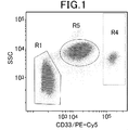
本発明の方法において用いることのできる表面抗原マーカーは、特に限定されるものではないが、例えば、CD45、CD66、CD14、CD3、CD19、CD33、CD123、CD34、CD11c、CD25、HLA-DR等が挙げられる。本発明の方法において、表面抗原マーカーは、単独でも、2種以上を一緒に用いてもよい。このような表面抗原マーカーのうち、CD45は汎血球マーカー、CD66は好中球マーカー、CD3はT細胞マーカー、CD19はB細胞マーカー、CD14は単球マーカーとして一般に知られている。
本発明の方法の一実施態様では、単球の細胞集団又は単球を含む細胞集団を選別することを含み得る。単球を含む細胞集団は、単球を95%以上、90%以上、85%以上、80%以上、75%以上、70%以上、65%以上、60%以上、55%以上、50%以上含み得る。本発明の方法において、単球を含む細胞集団を選別し、当該細胞集団について、SMNタンパク質に対する標識に基づいてSMNタンパク質の核内構造体の発現解析をする場合、正常者とSMA患者の間で、SMNタンパク質の核内構造体(スポット)の蛍光強度についても有意差が認められるため、より好ましい。 The surface antigen marker that can be used in the method of the present invention is not particularly limited, and examples thereof include CD45, CD66, CD14, CD3, CD19, CD33, CD123, CD34, CD11c, CD25, and HLA-DR. Can be mentioned. In the method of the present invention, the surface antigen markers may be used alone or in combination of two or more. Of these surface antigen markers, CD45 is generally known as a panblood cell marker, CD66 is a neutrophil marker, CD3 is a T cell marker, CD19 is a B cell marker, and CD14 is a monocyte marker.
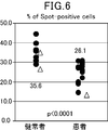
One embodiment of the method of the invention may comprise selecting a cell population of monocytes or a cell population comprising monocytes. Cell populations containing monocytes include monocytes>95%,>90%,>85%,>80%,>75%,>70%,>65%,>60%,>55%,> 50% May be included. In the method of the present invention, when a cell population containing monocytes is selected and the expression analysis of the nuclear structure of the SMN protein is performed on the cell population based on the label for the SMN protein, the normal population and the SMA patient are analyzed. A significant difference is also observed in the fluorescence intensity of the nuclear structure (spot) of the SMN protein, which is more preferable.
本発明の方法の一実施態様では、単球の細胞集団又は単球を含む細胞集団を選別することを含み得る。単球を含む細胞集団は、単球を95%以上、90%以上、85%以上、80%以上、75%以上、70%以上、65%以上、60%以上、55%以上、50%以上含み得る。本発明の方法において、単球を含む細胞集団を選別し、当該細胞集団について、SMNタンパク質に対する標識に基づいてSMNタンパク質の核内構造体の発現解析をする場合、正常者とSMA患者の間で、SMNタンパク質の核内構造体(スポット)の蛍光強度についても有意差が認められるため、より好ましい。 The surface antigen marker that can be used in the method of the present invention is not particularly limited, and examples thereof include CD45, CD66, CD14, CD3, CD19, CD33, CD123, CD34, CD11c, CD25, and HLA-DR. Can be mentioned. In the method of the present invention, the surface antigen markers may be used alone or in combination of two or more. Of these surface antigen markers, CD45 is generally known as a panblood cell marker, CD66 is a neutrophil marker, CD3 is a T cell marker, CD19 is a B cell marker, and CD14 is a monocyte marker.
One embodiment of the method of the invention may comprise selecting a cell population of monocytes or a cell population comprising monocytes. Cell populations containing monocytes include monocytes>95%,>90%,>85%,>80%,>75%,>70%,>65%,>60%,>55%,> 50% May be included. In the method of the present invention, when a cell population containing monocytes is selected and the expression analysis of the nuclear structure of the SMN protein is performed on the cell population based on the label for the SMN protein, the normal population and the SMA patient are analyzed. A significant difference is also observed in the fluorescence intensity of the nuclear structure (spot) of the SMN protein, which is more preferable.
本発明の方法の一実施態様では、B細胞の細胞集団又はB細胞を含む細胞集団を選別することを含み得る。B細胞を含む細胞集団は、B細胞を95%以上、90%以上、85%以上、80%以上、75%以上、70%以上、65%以上、60%以上、55%以上、50%以上含み得る。
本発明の方法の一実施態様では、T細胞の細胞集団又はT細胞を含む細胞集団を選別することを含み得る。T細胞を含む細胞集団は、T細胞を95%以上、90%以上、85%以上、80%以上、75%以上、70%以上、65%以上、60%以上、55%以上、50%以上含み得る。
本発明の方法の一実施態様では、顆粒球の細胞集団又は顆粒球を含む細胞集団を選別することを含み得る。顆粒球を含む細胞集団は、顆粒球を95%以上、90%以上、85%以上、80%以上、75%以上、70%以上、65%以上、60%以上、55%以上、50%以上含み得る。 One embodiment of the method of the invention may comprise sorting a cell population of B cells or a cell population comprising B cells. Cell populations containing B cells include 95% or more, 90% or more, 85% or more, 80% or more, 75% or more, 70% or more, 65% or more, 60% or more, 55% or more, 50% or more May be included.
One embodiment of the method of the invention may comprise sorting a cell population of T cells or a cell population comprising T cells. Cell populations containing T cells are 95% or more, 90% or more, 85% or more, 80% or more, 75% or more, 70% or more, 65% or more, 60% or more, 55% or more, 50% or more May be included.
One embodiment of the method of the invention may comprise selecting a cell population of granulocytes or a cell population comprising granulocytes. Cell populations containing granulocytes are granulocytes of 95% or more, 90% or more, 85% or more, 80% or more, 75% or more, 70% or more, 65% or more, 60% or more, 55% or more, 50% or more May be included.
本発明のSMNタンパク質の核内構造体の発現解析方法は、サンプル中の有核細胞内のSMNタンパク質を標識する工程を含む。
SMNタンパク質は、SMAの原因遺伝子であるSMN遺伝子により発現されるタンパク質であり、運動ニューロンの形成等に関与していると考えられている。SMN遺伝子には、SMN1遺伝子とSMN2遺伝子が存在する。両者ともに5番染色体長腕5q13に存在しており、SMN2遺伝子は、SMN1遺伝子とコーディング領域の塩基が1つだけ異なっている。しかしながら、正常なSMNタンパク質に翻訳される、SMN1遺伝子から転写される全長SMN1mRNAの量を100%とすると、SMN2遺伝子の転写産物は、全長SMN2mRNAがおよそ10%であり、エクソン7の欠失が認められる短縮型SMN2mRNAがおよそ90%である。短縮型SMN2mRNAからは非機能的なタンパク質が生成され、運動ニューロンの形成には役立たない。
本発明の方法において、血液由来の有核細胞を含むサンプル中のSMNタンパク質を標識するために、本発明の技術分野において従来知られている技術を用いることができる。ここで、SMNタンパク質の標識は、全長SMN1mRNA及び全長SMN2mRNAから翻訳される正常なSMNタンパク質を標識できるものであればよいが、短縮型SMN2mRNAから翻訳される非機能的なタンパク質をも標識するものであっても構わない。 The method for analyzing the expression of an SMN protein nuclear structure of the present invention includes a step of labeling an SMN protein in a nucleated cell in a sample.
The SMN protein is a protein expressed by the SMN gene that is a causative gene of SMA, and is considered to be involved in the formation of motor neurons. The SMN gene includes the SMN1 gene and the SMN2 gene. Both are present in the long arm 5q13 of chromosome 5, and the SMN2 gene differs from the SMN1 gene by one base in the coding region. However, assuming that the amount of full-length SMN1 mRNA transcribed from the SMN1 gene, which is translated into normal SMN protein, is 100%, the transcript of the SMN2 gene is approximately 10% of full-length SMN2 mRNA, and exon 7 deletion is observed. About 90% of the truncated SMN2 mRNA produced. Abbreviated SMN2 mRNA produces a non-functional protein and does not help in the formation of motor neurons.
In the method of the present invention, a technique conventionally known in the technical field of the present invention can be used to label the SMN protein in a sample containing blood-derived nucleated cells. Here, the labeling of the SMN protein is not limited as long as it can label the normal SMN protein translated from the full-length SMN1 mRNA and the full-length SMN2 mRNA, but also labels the non-functional protein translated from the truncated SMN2 mRNA. It does not matter.
本発明の方法において用いられるSMNタンパク質を標識する方法としては、例えば、標識試薬と結合した抗SMN抗体を使用することや、標識試薬の結合していない一次抗体としての抗SMN抗体を使用後、標識試薬と結合した二次抗体を使用することが挙げられるが、これらに限定されるものではない。標識試薬には、例えば、酵素、色素、蛍光色素、ビオチン、放射性物質、各種ペプチド、アミノ酸リンカーが含まれるが、これらに限定されるものではない。好ましくは、標識試薬は蛍光色素であり、例えば、フルオレセイン、ローダミン、クマリン、イミダゾール誘導体、インドール誘導体、Cy3、Cy5、Cy5.5、Cy7、APC、PE、DyLight、AlexaFluor等が挙げられる。
本発明の方法において用いられる、標識試薬と結合した抗SMN抗体または一次抗体として用いる抗SMN抗体は、モノクローナル抗体又はポリクローナル抗体であってもよく、市販品を利用することができる。例えば、抗SMN抗体の市販品としては、Millipore社のMilli-MarkTMや、BD社、Abcam社、Sigma-Aldrich社から提供される抗体等が挙げられる。 As a method for labeling the SMN protein used in the method of the present invention, for example, using an anti-SMN antibody bound to a labeling reagent or using an anti-SMN antibody as a primary antibody not bound to a labeling reagent, Although using the secondary antibody couple | bonded with the labeling reagent is mentioned, It is not limited to these. Examples of labeling reagents include, but are not limited to, enzymes, dyes, fluorescent dyes, biotin, radioactive substances, various peptides, and amino acid linkers. Preferably, the labeling reagent is a fluorescent dye, and examples thereof include fluorescein, rhodamine, coumarin, imidazole derivatives, indole derivatives, Cy3, Cy5, Cy5.5, Cy7, APC, PE, DyLight, AlexaFluor and the like.
The anti-SMN antibody used as the primary antibody or the anti-SMN antibody bound to the labeling reagent used in the method of the present invention may be a monoclonal antibody or a polyclonal antibody, and a commercially available product can be used. For example, commercially available anti-SMN antibodies include Milli-Mark ™ manufactured by Millipore, antibodies provided by BD, Abcam, and Sigma-Aldrich.
本発明のSMNタンパク質の核内構造体の発現解析方法は、サンプル中の有核細胞の核を標識する工程を含む。核を標識する方法としては、本発明の技術分野において従来知られている技術を用いることができる。例えば、核を標識する方法として、蛍光色素による標識、細胞核特異的蛋白質を認識する抗体による標識等が挙げられる。
本発明の方法において、血液由来の有核細胞を含むサンプル中の有核細胞の核を標識するために蛍光色素を用いる場合、例えば、試薬として、Hoechst 33342、Hoechst 33258、4, 6-diamino-2-phenylindole(DAPI)、Propidium iodide(PI)、蛍光標識抗ヒストン抗体、蛍光標識抗ラミン抗体等が挙げられる。
本明細書において、SMNタンパク質を標識する蛍光色素を第1の蛍光色素と言い、核を標識する蛍光色素を第2の蛍光色素と言う場合がある。
本発明のSMNタンパク質の核内構造体の発現解析方法において、有核細胞内のSMNタンパク質を標識する工程が、SMNタンパク質を第1の蛍光色素、例えばAlexaFluor 488、により標識することを含み、かつ、有核細胞の核を標識する工程が、核を第2の蛍光色素、例えばHoechst 33342、により標識することを含むことが好ましい。 The method for analyzing the expression of the nuclear structure of the SMN protein of the present invention includes a step of labeling the nucleus of a nucleated cell in a sample. As a method of labeling nuclei, techniques conventionally known in the technical field of the present invention can be used. For example, as a method of labeling the nucleus, labeling with a fluorescent dye, labeling with an antibody that recognizes a cell nucleus-specific protein, and the like can be mentioned.
In the method of the present invention, when a fluorescent dye is used to label the nucleus of a nucleated cell in a sample containing nucleated cells derived from blood, for example, as a reagent, Hoechst 33342, Hoechst 33258, 4, 6-diamino- Examples include 2-phenylindole (DAPI), propidium iodide (PI), fluorescently labeled antihistone antibody, and fluorescently labeled antilamin antibody.
In this specification, the fluorescent dye that labels the SMN protein may be referred to as a first fluorescent dye, and the fluorescent dye that labels the nucleus may be referred to as a second fluorescent dye.
In the method for analyzing the expression of the nuclear structure of the SMN protein of the present invention, the step of labeling the SMN protein in the nucleated cell comprises labeling the SMN protein with a first fluorescent dye, such as AlexaFluor 488, and Preferably, the step of labeling the nucleus of the nucleated cell comprises labeling the nucleus with a second fluorescent dye, such as Hoechst 33342.
本発明のSMNタンパク質の核内構造体の発現解析方法は、有核細胞のうち、核及びSMNタンパク質が標識され、かつ1種以上の標識抗体により標識された1種以上の表面抗原マーカー又は1種以上の標識抗体により標識された1種以上の表面抗原マーカーと側方散乱(SSC)とに基づき分類された複数の細胞集団から1つの細胞集団を選別する工程を含む。
ここで、核及びSMNタンパク質が標識されている細胞集団は、インタクトな細胞(生細胞)を100%、95%以上、90%以上、85%以上、80%以上、75%以上、70%以上、65%以上、60%以上、55%以上又は50%以上含む集団である。
本発明の方法の一実施態様では、有核細胞中の核及びSMNタンパク質が標識されている細胞集団は、本明細書の下記実施例1に記載の通り核及びSMNタンパク質を蛍光色素により標識し、その蛍光強度を下記実施例1に記載の通り検出した場合、核の蛍光強度が約1×10⁵以上であり、かつSMNタンパク質の蛍光強度が約1×10³以上である細胞を含む集団である。このような細胞は、インタクトな細胞であると認められる。また、顕微鏡観察にて細胞の形態を保っており、かつアスペクト比(縦横比)と表面積を保っている細胞は、インタクトな細胞であると認められる。インタクトな細胞の判別又はインタクトな細胞を含む細胞集団の判別は、有核細胞中の核及びSMNタンパク質の標識方法によってその基準が異なるが、使用される標識試薬に基づく有核細胞中の核及びSMNタンパク質の検出結果に基づき適宜行うことができる。 In the method for analyzing the expression of the nuclear structure of the SMN protein of the present invention, the nucleus and the SMN protein of nucleated cells are labeled and one or more surface antigen markers labeled with one or more labeled antibodies or 1 Selecting a cell population from a plurality of cell populations classified based on one or more surface antigen markers labeled with one or more labeled antibodies and side scatter (SSC).
Here, the cell population labeled with nuclear and SMN protein is 100%, 95%, 90%, 85%, 80%, 75%, 70% or more of intact cells (live cells). , 65% or more, 60% or more, 55% or more or 50% or more.
In one embodiment of the method of the present invention, a cell population labeled with nuclei and SMN proteins in nucleated cells is labeled with a fluorescent dye as described in Example 1 herein below. When the fluorescence intensity is detected as described in Example 1 below, a population containing cells whose nuclear fluorescence intensity is about 1 × 10 5 or more and the fluorescence intensity of the SMN protein is about 1 × 10 3 or more It is. Such cells are recognized as intact cells. In addition, cells that maintain the cell morphology and maintain the aspect ratio (aspect ratio) and the surface area by microscopic observation are recognized as intact cells. Discrimination of intact cells or cell populations containing intact cells has different criteria depending on the method of labeling nuclei in nucleated cells and SMN proteins, but the nuclei in nucleated cells and the This can be appropriately performed based on the detection result of the SMN protein.
本発明のSMNタンパク質の核内構造体の発現解析方法は、前述のとおり選別した細胞集団について、SMNタンパク質に対する標識に基づいてSMNタンパク質の核内構造体の発現解析をする工程を含む。
本発明の方法において、前記細胞集団を選別する工程及び前記SMNタンパク質の核内構造体の発現解析をする工程を、40倍以上の対物レンズを使用するイメージングフローサイトメトリーにより行う。このようなイメージングフローサイトメトリーにより、本発明の方法においては、1種以上の標識抗体により標識された1種以上の表面抗原マーカー又は当該表面抗原マーカーと側方散乱(SSC)とに基づき、所望の血球成分を主に含む細胞集団(所望の血液細胞分画)に焦点を当てて、SMNタンパク質の核内構造体の発現解析を行うことができる。そのため、本発明の方法は、SMNタンパク質の機能解析においても広く利用でき、SMAの診断技術及び治療技術に多大な貢献をもたらし得る。
用いる対物レンズの倍率としては、50倍以上が好ましく、60倍以上がより好ましく、その上限は、SMNタンパク質の核内構造体の発現解析が可能な範囲内である限り任意であるが、例えば、200倍以下、150倍以下、100倍以下、80倍以下であり得る。
イメージンングフローサイトメトリーは、市販の装置を使用して、メーカーの取扱説明書に従って適宜行うことができる。
なお、本発明において、SMNタンパク質の核内構造体がスポット状に観測されたことに鑑みて、本明細書において、スポット状に観測されたSMNタンパク質の核内構造体を単に「スポット」と呼ぶことがある。スポットの定義としては、例えば、1)核内にあり、2)大きさが0.34~3.07μm2であり、3)バックグラウンドと比較し5倍以上の蛍光強度を示し、4)縦横比0.5~1.0であるものが挙げられる。また、スポットの別の定義としては、例えば、1)核内にあり、2)大きさが0.34~8.54μm2であり、3)バックグラウンドと比較し5.5倍以上の蛍光強度を示し、4)縦横比0.4~1.0であるものが挙げられる。 The method for analyzing the expression of the nuclear structure of the SMN protein of the present invention includes the step of analyzing the expression of the nuclear structure of the SMN protein based on the label for the SMN protein for the cell population selected as described above.
In the method of the present invention, the step of selecting the cell population and the step of analyzing the expression of the nuclear structure of the SMN protein are performed by imaging flow cytometry using an objective lens of 40 times or more. By such imaging flow cytometry, in the method of the present invention, based on one or more surface antigen markers labeled with one or more labeled antibodies or the surface antigen markers and side scatter (SSC), a desired The expression analysis of the nuclear structure of the SMN protein can be performed by focusing on a cell population mainly containing the blood cell component (desired blood cell fraction). Therefore, the method of the present invention can be widely used in the functional analysis of SMN protein, and can greatly contribute to the diagnostic technique and therapeutic technique of SMA.
The magnification of the objective lens to be used is preferably 50 times or more, more preferably 60 times or more, and the upper limit thereof is arbitrary as long as it is within a range where expression analysis of the nuclear structure of the SMN protein is possible. It can be 200 times or less, 150 times or less, 100 times or less, or 80 times or less.
Imaging flow cytometry can be appropriately performed using a commercially available apparatus according to the manufacturer's instruction manual.
In the present invention, in view of the fact that the nuclear structure of the SMN protein was observed in the form of spots, in this specification, the nuclear structure of the SMN protein observed in the form of spots is simply referred to as a “spot”. Sometimes. The definition of the spot is, for example, 1) in the nucleus, 2) the size is 0.34 to 3.07 μm 2 , 3) the fluorescence intensity is 5 times more than the background, and 4) the aspect ratio is 0.5 to One that is 1.0. As another definition of spot, for example, 1) it is in the nucleus, 2) the size is 0.34 to 8.54 μm 2 , and 3) it shows 5.5 times or more fluorescence intensity compared to the background 4) Those having an aspect ratio of 0.4 to 1.0 can be mentioned.
本発明のSMNタンパク質の核内構造体の発現解析方法において、SMNタンパク質に対する標識に基づいてSMNタンパク質の核内構造体の発現解析をする工程は、SMNタンパク質の核内構造体を定量することを含んでもよい。また、本発明の方法は、例えば、SMA患者、保因者、及び正常者間、具体的にはSMA患者と保因者間又はSMA患者と正常者間で、SMNタンパク質の核内構造体の定量結果を比較する工程を含んでもよい。比較により、SMA患者と保因者、SMA患者と正常者の間でSMNタンパク質の定量結果(発現量又は局在化)に差異が認められることで、SMA患者、保因者、及び正常者をそれぞれ分類することができる。
また、本発明のSMNタンパク質の核内構造体の発現解析方法は、SMAを治療するための薬剤をスクリーニングする方法において用いることもできる。
本発明の方法において、リン酸緩衝液等の試薬で細胞を洗浄する工程等、通常、細胞におけるタンパク質発現を検出する際に行われる操作を適宜行うことができる。 In the method for analyzing the expression of the nuclear structure of the SMN protein of the present invention, the step of analyzing the expression of the nuclear structure of the SMN protein based on the label for the SMN protein comprises quantifying the nuclear structure of the SMN protein. May be included. In addition, the method of the present invention can be applied to the nuclear structure of the SMN protein between, for example, an SMA patient, a carrier, and a normal person, specifically, between an SMA patient and a carrier or between an SMA patient and a normal person. A step of comparing the quantitative results may be included. Comparison shows that there is a difference in the quantification results (expression level or localization) of SMN protein between SMA patients and carriers, and between SMA patients and normal individuals. Each can be classified.
Further, the expression analysis method for the nuclear structure of the SMN protein of the present invention can also be used in a method for screening a drug for treating SMA.
In the method of the present invention, operations usually performed when detecting protein expression in cells, such as a step of washing cells with a reagent such as a phosphate buffer, can be appropriately performed.
また、本発明のSMNタンパク質の核内構造体の発現解析方法においては、血液由来の有核細胞を含むサンプルが被験者から得られたものであり、SMNタンパク質の核内構造体の発現解析をする工程が、SMNタンパク質の核内構造体を定量することを含み、
前記定量により得られた結果を、コントロールと比較する工程を含んでもよい。ここで、コントロールとは、以下の(a)~(c)より選択され得る:
(a) 前記被験者がSMA患者である場合には、前記コントロールは、正常者又は保因者から得られる血液を用いること以外は、前記被験者から得られた血液を用いてSMNタンパク質の核内構造体を定量した方法と同様にして得られた結果であり、
(b) 前記被験者が正常者又は保因者である場合には、前記コントロールは、SMA患者から得られる血液を用いること以外は、前記被験者から得られた血液を用いてSMNタンパク質の核内構造体を定量した方法と同様にして得られた結果であり、又は
(c) 前記被験者がSMAの治療薬又は治療候補薬を投与された者である場合には、前記コントロールは、前記SMAの治療薬又は治療候補薬の投与前の前記被験者から得られた血液を用いること以外は、前記投与後の前記被験者から得られた血液を用いてSMNタンパク質の核内構造体を定量した方法と同様にして得られた結果である。 In the method for analyzing the expression of the nuclear structure of the SMN protein of the present invention, a sample containing blood-derived nucleated cells is obtained from the subject, and the expression analysis of the nuclear structure of the SMN protein is performed. The process comprises quantifying the nuclear structure of the SMN protein;
A step of comparing the result obtained by the quantification with a control may be included. Here, the control can be selected from the following (a) to (c):
(a) When the subject is an SMA patient, the control uses the blood obtained from the subject except for blood obtained from a normal person or a carrier. It is the result obtained in the same way as the method of quantifying the body,
(b) When the subject is a normal person or a carrier, the control uses the blood obtained from the subject except for blood obtained from the SMA patient, and the nuclear structure of the SMN protein. The results obtained in the same manner as the method for quantifying the body, or Except for using blood obtained from the subject before administration of the drug or treatment candidate drug, the method was the same as the method for quantifying the nuclear structure of SMN protein using blood obtained from the subject after administration. This is the result obtained.
ここで、「同様にして測定した」とは、SMNタンパク質の核内構造体の定量結果(発現量又は局在化)を比較する上で、SMNタンパク質の核内構造体の定量に実質的に影響を与えない程度の変更は許されるものと解され、例えば、細胞を洗浄する工程などまで完全に一致することは意味しない。
SMNタンパク質の核内構造体を定量することは、検量線法など、本発明の技術分野において従来知られている技術を用いて適宜行うことができる。
なお、本発明の一実施態様は、上記のような比較の工程を含み、かつ比較の結果に基づいて被験者を診断する工程を含むことで、SMAを診断する方法に関すると言うこともできる。また、本発明の一実施態様は、上記のような比較の工程を含み、かつ比較の結果に基づいてSMAの治療薬又は治療候補薬を選択する工程を含むことで、SMAの治療薬又は治療候補薬のスクリーニング方法に関すると言うこともできる。 Here, “measured in the same way” means that, in comparing the quantification result (expression level or localization) of the nuclear structure of the SMN protein, the quantification of the nuclear structure of the SMN protein is substantially performed. Changes that do not affect are understood to be allowed, and for example, it does not mean that the process is completely consistent up to the step of washing the cells.
Quantification of the nuclear structure of the SMN protein can be appropriately performed using techniques conventionally known in the technical field of the present invention, such as a calibration curve method.
In addition, it can be said that one embodiment of the present invention relates to a method for diagnosing SMA by including a comparison step as described above and a step of diagnosing a subject based on the comparison result. In addition, an embodiment of the present invention includes a comparison step as described above, and a step of selecting a SMA therapeutic agent or a treatment candidate drug based on the comparison result, whereby the SMA therapeutic agent or treatment It can also be said that it relates to a screening method for candidate drugs.
以下の実施例によって本発明をさらに詳細に説明する。なお、本発明はこれらの実施例に限定されるものではない。
The following examples further illustrate the present invention. The present invention is not limited to these examples.
実施例1:SMNタンパク質の核内構造体の発現解析
1.サンプルの調製
以下の染色プロトコールに従って、サンプルを調製した。なお、特に記載のない限り、全ての手順は室温で行った。
(1)被験者から得た末梢血1.5mLを、15mLコニカルチューブに移した。
(2)コニカルチューブ内の末梢血中に、30μLのFc受容体ブロッキング試薬(Clear Back, MTG-001, MBL)を添加し、暗所にて15分間インキュベートした。
(3)インキュベート後のコニカルチューブ内末梢血中に、表面抗原(CD19、CD33、CD3)に対するモノクローナル抗体(それぞれBioLegend社より入手)5μLを、下記表1及び2に従って添加し、暗所にて30分間インキュベートした。 Example 1: Expression analysis of nuclear structure of SMN protein Sample Preparation Samples were prepared according to the following staining protocol. Unless otherwise stated, all procedures were performed at room temperature.
(1) 1.5 mL of peripheral blood obtained from the subject was transferred to a 15 mL conical tube.
(2) 30 μL of Fc receptor blocking reagent (Clear Back, MTG-001, MBL) was added to peripheral blood in a conical tube and incubated for 15 minutes in the dark.
(3) 5 μL of monoclonal antibodies (obtained from BioLegend, respectively) against surface antigens (CD19, CD33, CD3) were added to the peripheral blood in the conical tube after incubation according to Tables 1 and 2 below. Incubated for minutes.


AF:アレクサフルオロ
R-PE:R-フィコエリスリン
PE-Cy5:R-フィコエリスリン-シアニン5
Hoechst:Hoechst 33342
BV:ブリリアントバイオレット
AF: Alexa Fluor
R-PE: R-Phycoerythrin
PE-Cy5: R-phycoerythrin-cyanine 5
Hoechst: Hoechst 33342
BV: Brilliant Violet
(4)37℃の水浴で温めた溶血・固定液(BD PhosflowTM Lyse/Fix Buffer)10mLを各チューブに加えた(8~10回逆さにしてよく混合した)。
(5)37℃の水浴で10分間インキュベートした。
(6)300 x gで5分間遠心分離した。
(7)<50μLとなるまで上清を除去した。
(8)15mLのPBS(-)で細胞を再懸濁し、上記(6)と同様に遠心分離し、上清を除去した。
(9)1mLのサポニン含有細胞膜透過試薬(BD PhosflowTM;557885(BD PhosflowTM Perm/Wash Buffer I とも言う))で細胞を再懸濁し、暗所にて20分間インキュベートした。
(10)上記(6)と同様に遠心分離した。
(11)2mLの細胞膜透過試薬(BD PhosflowTM;557885)で細胞を洗浄した。
(12)およそ100μLの細胞膜透過試薬(BD PhosflowTM;557885)で細胞を再懸濁した。
(13)1.5mLマイクロチューブ中に95μLのPBS(-)を準備し、その中に上記12で調製した細胞懸濁液を5μL添加し、20倍希釈して染色用チューブとした。同様に20倍希釈し、コントロール用チューブとした。各チューブ中、細胞懸濁液の濃度は、1 x 10⁶ 細胞/約50μLであった。
(14-1)染色用チューブに対して、1μLのAF488結合抗ヒトSMNモノクローナル抗体(2B1)、AF647結合抗ヒトGemin3(EPR11283)、AF750結合抗ヒトZNF259(EPR7595)で染色を行った。
(14-2)コントロール用チューブに対して、上記(14-1)と並行して、AF488結合アイソタイプコントロール、AF647結合アイソタイプコントロール、AF750結合アイソタイプコントロールで染色を行った。
(15)暗所にて45分間インキュベートした。
(16)500μLの細胞膜透過試薬(BD PhosflowTM;557885)を添加し、300 x gで5分間遠心分離した。
(17)500μLの細胞膜透過試薬(BD PhosflowTM;557885)で細胞を再懸濁し、上記(16)と同様に遠心分離した。
(18)Hoechst 33342(molecular probe(登録商標))を希釈して調製した50μLの希釈Hoechst 33342(0.25μg/mL PBS)で細胞を再懸濁した。
(19)暗所にて15分間インキュベートした。
(20)500μLのPBS(-)を添加し、上記(16)と同様に遠心分離した。
(21)90μLのPBS(-)で細胞を再懸濁し、各チューブが30μLの細胞懸濁液を含むよう3つのチューブに分けた。 (4) 10 mL of hemolytic / fixing solution (BD Phosflow ™ Lyse / Fix Buffer) warmed in a 37 ° C. water bath was added to each tube (mixed well by inverting 8-10 times).
(5) Incubated in a 37 ° C. water bath for 10 minutes.
(6) Centrifuged at 300 x g for 5 minutes.
(7) The supernatant was removed until <50 μL.
(8) The cells were resuspended with 15 mL of PBS (−), centrifuged as in (6) above, and the supernatant was removed.
(9) The cells were resuspended with 1 mL of a saponin-containing cell membrane permeation reagent (BD Phosflow ™ ; 557885 (also referred to as BD Phosflow ™ Perm / Wash Buffer I)) and incubated for 20 minutes in the dark.
(10) Centrifugation was performed in the same manner as in (6) above.
(11) The cells were washed with 2 mL of cell membrane permeation reagent (BD Phosflow ™ ; 557885).
(12) The cells were resuspended with about 100 μL of cell membrane permeation reagent (BD Phosflow ™ ; 557885).
(13) Prepare 95 μL of PBS (−) in a 1.5 mL microtube, add 5 μL of the cell suspension prepared in 12 above, and dilute it 20 times to obtain a staining tube. Similarly, it was diluted 20 times to obtain a control tube. In each tube, the concentration of the cell suspension was 1 × 10 6 cells / approximately 50 μL.
(14-1) The staining tube was stained with 1 μL of AF488-conjugated anti-human SMN monoclonal antibody (2B1), AF647-conjugated anti-human Gemin3 (EPR11283), and AF750-conjugated anti-human ZNF259 (EPR7595).
(14-2) The control tube was stained with the AF488 binding isotype control, AF647 binding isotype control, and AF750 binding isotype control in parallel with the above (14-1).
(15) Incubated for 45 minutes in the dark.
(16) 500 μL of cell membrane permeation reagent (BD Phosflow ™ ; 557885) was added and centrifuged at 300 × g for 5 minutes.
(17) The cells were resuspended with 500 μL of a cell membrane permeation reagent (BD Phosflow ™ ; 557885) and centrifuged as in (16) above.
(18) The cells were resuspended in 50 μL of diluted Hoechst 33342 (0.25 μg / mL PBS) prepared by diluting Hoechst 33342 (molecular probe®).
(19) Incubated for 15 minutes in the dark.
(20) 500 μL of PBS (−) was added and centrifuged as in (16) above.
(21) The cells were resuspended with 90 μL of PBS (−) and divided into three tubes so that each tube contained 30 μL of the cell suspension.
2.イメージングフローサイトメトリー
上記のとおり得られたサンプルについて、イメージングフローサイトメトリー(ImageStream(登録商標)x Mark II イメージングフローサイトメーター、Amnis)によりSMNタンパク質の核内構造体の発現解析を行った。対物レンズの倍率は60倍を用い、計50000個以上の細胞をイメージングフローサイトメトリーに供した。
なお、上記のとおり得られたサンプルとして、以下の被験者から得たものを用いた。
正常者(健常者):成人女性2名(38~51歳)、男女を含む小児8名(1~7歳)
SMA患者:成人男性1名(21歳)、男女を含む乳児~小児19名(2ヶ月~8歳) 2. Imaging Flow Cytometry For the samples obtained as described above, the expression analysis of the nuclear structure of the SMN protein was performed by imaging flow cytometry (ImageStream (registered trademark) x Mark II imaging flow cytometer, Amnis). The magnification of the objective lens was 60 times, and a total of 50,000 or more cells were subjected to imaging flow cytometry.
In addition, what was obtained from the following test subjects was used as a sample obtained as mentioned above.
Normal (healthy): 2 adult women (38-51 years old), 8 children (1-7 years old) including men and women
SMA patients: 1 adult male (21 years old), 19 infants including males and females (2 months to 8 years old)
(1)細胞集団の分画
(i)全分画を含む画像データより、明視野における焦点のパラメータを利用し、焦点距離が正しい細胞群を選択した。
(ii)前方散乱光(FSC)による細胞の大きさと、細胞径の縦横比で展開し、凝集した状態の細胞を除外した。
(iii)Hoechst33342により標識された核DNAの蛍光強度を指標として、有核細胞を選択した(デブリス、埃、赤血球などの除外)。
(iv)側方散乱光(SSC)による細胞の内部構造と、細胞表面抗原CD33発現で展開し、有核細胞群を3群に分けた。結果を図1に示す。図1において、R1はリンパ球、R4は単球、R5は好中球を主として含む細胞集団を表す。なお、図1は、正常者から得られた血液サンプルを用いて、イメージングフローサイトメトリーにより測定した蛍光強度をプロットしたグラフであるが、患者から得られた血液サンプルを用いて測定した場合にも同様に有核細胞群が3群に分けられた。また、図1は1サンプルから得られた結果を示すものであるが、各サンプルについても同様に、有核細胞群が3群に分けられた。
(v)R1のリンパ球を主として含む細胞集団について、細胞表面抗原CD3およびCD19発現で展開し、CD3陽性T細胞を主として含む細胞集団と、CD19陽性B細胞を主として含む細胞集団の2群に分けた。結果を図2に示す。図2において、R2がCD3陽性T細胞を主として含む細胞集団を表し、R3がCD19陽性B細胞を主として含む細胞集団を表す。
以上の解析により、末梢血中の有核細胞が以下の4群に分画された。なお、患者から得られた血液サンプルを用いて測定した場合にも同様である。また、図2は1サンプルから得られた結果を示すものであるが、各サンプルについても同様に、最終的に末梢血中の有核細胞群が以下の4群に分けられた。
R2:CD3陽性T細胞を主として含む細胞群 (SSClow, CD33-, CD3+)
R3:CD19陽性B細胞を主として含む細胞群 (SSClow, CD33-, CD19+)
R4:単球を主として含む細胞群 (SSCintermediate, CD33++)
R5:好中球を主として含む細胞群 (SSCintermediate, CD33intermediate) (1) Fractionation of cell population (i) From the image data including all fractions, a cell group having a correct focal length was selected using a focal parameter in a bright field.
(Ii) The cells were expanded with the forward scattered light (FSC) size and the aspect ratio of the cell diameter, and aggregated cells were excluded.
(Iii) Nucleated cells were selected using the fluorescence intensity of nuclear DNA labeled with Hoechst33342 as an index (excluding debris, dust, erythrocytes, etc.).
(Iv) The cell nucleated cell group was divided into three groups by developing the cell internal structure by side scattered light (SSC) and the cell surface antigen CD33 expression. The results are shown in FIG. In FIG. 1, R1 represents lymphocytes, R4 represents monocytes, and R5 represents a cell population mainly containing neutrophils. FIG. 1 is a graph plotting fluorescence intensity measured by imaging flow cytometry using a blood sample obtained from a normal person, but also when measured using a blood sample obtained from a patient. Similarly, the nucleated cell group was divided into 3 groups. FIG. 1 shows the results obtained from one sample, and the nucleated cell group was similarly divided into three groups for each sample.
(V) Cell populations mainly containing R1 lymphocytes are expanded with cell surface antigens CD3 and CD19 expression and divided into two groups: a cell population mainly containing CD3 positive T cells and a cell population mainly containing CD19 positive B cells. It was. The results are shown in FIG. In FIG. 2, R2 represents a cell population mainly containing CD3 positive T cells, and R3 represents a cell population mainly containing CD19 positive B cells.
Based on the above analysis, nucleated cells in peripheral blood were fractionated into the following 4 groups. The same applies to measurement using a blood sample obtained from a patient. FIG. 2 shows the results obtained from one sample. For each sample, the nucleated cell group in the peripheral blood was finally divided into the following four groups.
R2: CD3-positive T cells predominantly containing cell population (SSC low, CD33 -, CD3 +)
R3: CD19-positive B cells mainly comprising cell population (SSC low, CD33 -, CD19 +)
R4: Cell group mainly containing monocytes (SSC intermediate , CD33 ++ )
R5: Cell group mainly containing neutrophils (SSC intermediate , CD33 intermediate )
(2)単球を主として含む細胞群におけるSMNタンパク質の核内構造体(スポット)の解析
前述のとおりの方法でイメージングフローサイトメトリーを行い、撮影した細胞写真を図3及び4に示す。図3及び4は、SMA患者及び健常者(コントロール)からそれぞれ得られた血液サンプル中の、単球を主として含む細胞群を対象とした細胞写真であり、SMNタンパク質は緑色の蛍光で示される。この細胞写真から明らかなように、細胞全体における蛍光強度、核における蛍光強度、スポットの数、スポットにおける蛍光強度のいずれにおいても、SMA患者よりも健常者(コントロール)の方が大きいことが観測された。
なお、ここで言う「スポット」とは、スポット状に観測されたSMNタンパク質の核内構造体を意味するものであり、本実施例では「スポット」の定義を、1)核内にあり、2)大きさが0.34~3.07μm2であり、3)バックグラウンドと比較し5倍以上の強度を示し、4)縦横比0.5~1.0であるものとした。 (2) Analysis of nuclear structure (spot) of SMN protein in a cell group mainly containing monocytes Imaging flow cytometry was performed by the method as described above, and photographed cell photographs are shown in FIGS. FIGS. 3 and 4 are cytophotographs of cell groups mainly containing monocytes in blood samples obtained from SMA patients and healthy subjects (controls), respectively, and the SMN protein is shown in green fluorescence. As is clear from this cell photograph, it was observed that healthy subjects (controls) were larger than SMA patients in terms of fluorescence intensity in the whole cell, fluorescence intensity in the nucleus, number of spots, and fluorescence intensity in the spots. It was.
The “spot” here means a nuclear structure of the SMN protein observed in a spot shape. In this example, the definition of “spot” is 1) in the nucleus, 2 ) The size is 0.34 to 3.07 μm 2 , 3) it is 5 times more intense than the background, and 4) the aspect ratio is 0.5 to 1.0.
(i)スポット数の平均値
所定の蛍光強度を示す細胞群中のスポット数の平均値を以下のとおり算出した。
(核内照準調整)
核内の解析をより正確に行うために、以下の操作を行った。
1) 核内を染色したHoechst 33342を検出するチャンネルにおける焦点およびコントラストのパラメータで展開し、両パラメータのピーク部分の細胞群に限定した。
2) 上記の細胞群よりHoechst 33342の蛍光強度を指標とし、同程度に蛍光強度を示す細胞群を1000細胞以上選択した。これを照準調整細胞数とした。
(アイソタイプコントロール由来データの減算)
特異的抗体(抗SMN抗体)で染色した試料の数値より、非特異的抗体(アイソタイプコントロール)で染色した試料の数値を減じたものを解析値として評価に用いた。
(スポット数の平均値の算出)
スポット数/照準調整細胞数によりMeanを求め、以下の式によりスポット数の平均値を算出した。結果を図5に示す。図5のグラフ中、0.426及び0.295の数値は、それぞれ、試験した全ての正常者由来血液サンプル及びSMA患者由来血液サンプルにおいて算出したスポット数の平均値(算術平均)である。

(I) Average value of the number of spots The average value of the number of spots in the cell group exhibiting a predetermined fluorescence intensity was calculated as follows.
(Intranuclear aiming adjustment)
The following operations were performed in order to carry out analysis in the nucleus more accurately.
1) The focus and contrast parameters in the channel detecting Hoechst 33342 stained in the nucleus were developed and limited to the cell group at the peak of both parameters.
2) Using the fluorescence intensity of Hoechst 33342 as an index from the above cell group, more than 1000 cells having the same fluorescence intensity were selected. This was defined as the number of aiming adjusted cells.
(Subtraction of data derived from isotype control)
The value obtained by subtracting the value of the sample stained with the non-specific antibody (isotype control) from the value of the sample stained with the specific antibody (anti-SMN antibody) was used for evaluation as an analysis value.
(Calculation of the average number of spots)
Mean was calculated from the number of spots / the number of aiming-adjusted cells, and the average value of the number of spots was calculated according to the following formula. The results are shown in FIG. In the graph of FIG. 5, the numerical values of 0.426 and 0.295 are the average values (arithmetic average) of the number of spots calculated in all the blood samples derived from normal subjects and blood samples derived from SMA patients, respectively.
所定の蛍光強度を示す細胞群中のスポット数の平均値を以下のとおり算出した。
(核内照準調整)
核内の解析をより正確に行うために、以下の操作を行った。
1) 核内を染色したHoechst 33342を検出するチャンネルにおける焦点およびコントラストのパラメータで展開し、両パラメータのピーク部分の細胞群に限定した。
2) 上記の細胞群よりHoechst 33342の蛍光強度を指標とし、同程度に蛍光強度を示す細胞群を1000細胞以上選択した。これを照準調整細胞数とした。
(アイソタイプコントロール由来データの減算)
特異的抗体(抗SMN抗体)で染色した試料の数値より、非特異的抗体(アイソタイプコントロール)で染色した試料の数値を減じたものを解析値として評価に用いた。
(スポット数の平均値の算出)
スポット数/照準調整細胞数によりMeanを求め、以下の式によりスポット数の平均値を算出した。結果を図5に示す。図5のグラフ中、0.426及び0.295の数値は、それぞれ、試験した全ての正常者由来血液サンプル及びSMA患者由来血液サンプルにおいて算出したスポット数の平均値(算術平均)である。
(Intranuclear aiming adjustment)
The following operations were performed in order to carry out analysis in the nucleus more accurately.
1) The focus and contrast parameters in the channel detecting Hoechst 33342 stained in the nucleus were developed and limited to the cell group at the peak of both parameters.
2) Using the fluorescence intensity of Hoechst 33342 as an index from the above cell group, more than 1000 cells having the same fluorescence intensity were selected. This was defined as the number of aiming adjusted cells.
(Subtraction of data derived from isotype control)
The value obtained by subtracting the value of the sample stained with the non-specific antibody (isotype control) from the value of the sample stained with the specific antibody (anti-SMN antibody) was used for evaluation as an analysis value.
(Calculation of the average number of spots)
Mean was calculated from the number of spots / the number of aiming-adjusted cells, and the average value of the number of spots was calculated according to the following formula. The results are shown in FIG. In the graph of FIG. 5, the numerical values of 0.426 and 0.295 are the average values (arithmetic average) of the number of spots calculated in all the blood samples derived from normal subjects and blood samples derived from SMA patients, respectively.
(ii)スポット陽性細胞の割合
所定の蛍光強度を示す細胞群中のスポット陽性細胞の割合を以下のとおり算出した。
(核内照準調整)
核内の解析をより正確に行うために、以下の操作を行った。
1) 核内を染色したHoechst 33342を検出するチャンネルにおける焦点およびコントラストのパラメータで展開し、両パラメータのピーク部分の細胞群に限定した。
2) 上記の細胞群よりHoechst 33342の蛍光強度を指標とし、同程度に蛍光強度を示す細胞群を1000細胞以上選択した。これを照準調整細胞数とした。
(アイソタイプコントロール由来データの減算)
特異的抗体(抗SMN抗体)で染色した試料の数値より、非特異的抗体(アイソタイプコントロール)で染色した試料の数値を減じたものを解析値として評価に用いた。
(スポット陽性細胞の割合の算出)
スポット数/照準調整細胞数×100により割合(%)を求め、以下の式によりスポット陽性細胞の割合を算出した。結果を図6に示す。図6のグラフ中、35.6及び26.1の数値は、それぞれ、試験した全ての正常者由来血液サンプル及びSMA患者由来血液サンプルにおいて算出したスポット陽性細胞の割合の平均値(算術平均)である。

(Ii) Ratio of spot positive cells The ratio of spot positive cells in a cell group exhibiting a predetermined fluorescence intensity was calculated as follows.
(Intranuclear aiming adjustment)
The following operations were performed in order to carry out analysis in the nucleus more accurately.
1) The focus and contrast parameters in the channel detecting Hoechst 33342 stained in the nucleus were developed and limited to the cell group at the peak of both parameters.
2) Using the fluorescence intensity of Hoechst 33342 as an index from the above cell group, more than 1000 cells having the same fluorescence intensity were selected. This was defined as the number of aiming adjusted cells.
(Subtraction of data derived from isotype control)
The value obtained by subtracting the value of the sample stained with the non-specific antibody (isotype control) from the value of the sample stained with the specific antibody (anti-SMN antibody) was used for evaluation as an analysis value.
(Calculation of the percentage of spot positive cells)
The ratio (%) was calculated by the number of spots / the number of aiming adjusted cells × 100, and the ratio of spot positive cells was calculated by the following formula. The results are shown in FIG. In the graph of FIG. 6, the numerical values of 35.6 and 26.1 are the average values (arithmetic average) of the ratio of spot positive cells calculated in all the blood samples derived from normal subjects and blood samples derived from SMA patients, respectively.
所定の蛍光強度を示す細胞群中のスポット陽性細胞の割合を以下のとおり算出した。
(核内照準調整)
核内の解析をより正確に行うために、以下の操作を行った。
1) 核内を染色したHoechst 33342を検出するチャンネルにおける焦点およびコントラストのパラメータで展開し、両パラメータのピーク部分の細胞群に限定した。
2) 上記の細胞群よりHoechst 33342の蛍光強度を指標とし、同程度に蛍光強度を示す細胞群を1000細胞以上選択した。これを照準調整細胞数とした。
(アイソタイプコントロール由来データの減算)
特異的抗体(抗SMN抗体)で染色した試料の数値より、非特異的抗体(アイソタイプコントロール)で染色した試料の数値を減じたものを解析値として評価に用いた。
(スポット陽性細胞の割合の算出)
スポット数/照準調整細胞数×100により割合(%)を求め、以下の式によりスポット陽性細胞の割合を算出した。結果を図6に示す。図6のグラフ中、35.6及び26.1の数値は、それぞれ、試験した全ての正常者由来血液サンプル及びSMA患者由来血液サンプルにおいて算出したスポット陽性細胞の割合の平均値(算術平均)である。
(Intranuclear aiming adjustment)
The following operations were performed in order to carry out analysis in the nucleus more accurately.
1) The focus and contrast parameters in the channel detecting Hoechst 33342 stained in the nucleus were developed and limited to the cell group at the peak of both parameters.
2) Using the fluorescence intensity of Hoechst 33342 as an index from the above cell group, more than 1000 cells having the same fluorescence intensity were selected. This was defined as the number of aiming adjusted cells.
(Subtraction of data derived from isotype control)
The value obtained by subtracting the value of the sample stained with the non-specific antibody (isotype control) from the value of the sample stained with the specific antibody (anti-SMN antibody) was used for evaluation as an analysis value.
(Calculation of the percentage of spot positive cells)
The ratio (%) was calculated by the number of spots / the number of aiming adjusted cells × 100, and the ratio of spot positive cells was calculated by the following formula. The results are shown in FIG. In the graph of FIG. 6, the numerical values of 35.6 and 26.1 are the average values (arithmetic average) of the ratio of spot positive cells calculated in all the blood samples derived from normal subjects and blood samples derived from SMA patients, respectively.
(iii)スポット陽性細胞の蛍光強度
スポット陽性細胞に限定したSMNタンパク質の発現の解析を行った。
非特異的抗体(アイソタイプコントロール)で染色した試料における非特異的スポット検出が僅少に抑えられたため(<200細胞)、非特異的抗体(アイソタイプコントロール)で染色した試料におけるスポット陽性細胞の蛍光強度の算出は不可能だった。そのため、アイソタイプコントロールによるバックグラウンドの減算は不必要であると考え、特異的抗体(抗SMN抗体)により算出されたデータを評価に用いた。結果を図7に示す。図7のグラフ中、7255及び5364の数値は、それぞれ、試験した全ての正常者由来血液サンプル及びSMA患者由来血液サンプルにおいて算出したスポット陽性細胞の蛍光強度の平均値(算術平均)である。 (Iii) Fluorescence intensity of spot positive cells The expression of SMN protein limited to spot positive cells was analyzed.
Because non-specific spot detection in samples stained with non-specific antibody (isotype control) was slightly suppressed (<200 cells), the fluorescence intensity of spot-positive cells in samples stained with non-specific antibody (isotype control) Calculation was impossible. Therefore, it was considered that background subtraction by isotype control was unnecessary, and data calculated with a specific antibody (anti-SMN antibody) was used for evaluation. The results are shown in FIG. In the graph of FIG. 7, numerical values 7255 and 5364 are average values (arithmetic averages) of fluorescence intensity of spot positive cells calculated in all the blood samples derived from normal subjects and blood samples derived from SMA patients, respectively.
スポット陽性細胞に限定したSMNタンパク質の発現の解析を行った。
非特異的抗体(アイソタイプコントロール)で染色した試料における非特異的スポット検出が僅少に抑えられたため(<200細胞)、非特異的抗体(アイソタイプコントロール)で染色した試料におけるスポット陽性細胞の蛍光強度の算出は不可能だった。そのため、アイソタイプコントロールによるバックグラウンドの減算は不必要であると考え、特異的抗体(抗SMN抗体)により算出されたデータを評価に用いた。結果を図7に示す。図7のグラフ中、7255及び5364の数値は、それぞれ、試験した全ての正常者由来血液サンプル及びSMA患者由来血液サンプルにおいて算出したスポット陽性細胞の蛍光強度の平均値(算術平均)である。 (Iii) Fluorescence intensity of spot positive cells The expression of SMN protein limited to spot positive cells was analyzed.
Because non-specific spot detection in samples stained with non-specific antibody (isotype control) was slightly suppressed (<200 cells), the fluorescence intensity of spot-positive cells in samples stained with non-specific antibody (isotype control) Calculation was impossible. Therefore, it was considered that background subtraction by isotype control was unnecessary, and data calculated with a specific antibody (anti-SMN antibody) was used for evaluation. The results are shown in FIG. In the graph of FIG. 7,
(iv)スポットの蛍光強度
スポットそのものの蛍光強度、即ち、SMNタンパク質の核内構造体の発現の解析を行った。
非特異的抗体(アイソタイプコントロール)で染色した試料における非特異的スポット検出が僅少に抑えられたため(<200細胞)、非特異的抗体(アイソタイプコントロール)で染色した試料におけるスポット陽性細胞中のスポットの蛍光強度の算出は不可能だった。そのため、アイソタイプコントロールによるバックグラウンドの減算は不必要であると考え、特異的抗体(抗SMN抗体)により算出されたデータを評価に用いた。結果を図8に示す。図8のグラフ中、332.7及び162.7の数値は、それぞれ、試験した全ての正常者由来血液サンプル及びSMA患者由来血液サンプルにおいて算出したスポットの蛍光強度の平均値(算術平均)である。 (Iv) Fluorescence intensity of the spot The fluorescence intensity of the spot itself, that is, the expression of the nuclear structure of the SMN protein was analyzed.
Detection of non-specific spots in samples stained with non-specific antibody (isotype control) was slightly suppressed (<200 cells), so spots in spot-positive cells in samples stained with non-specific antibody (isotype control) It was impossible to calculate the fluorescence intensity. Therefore, it was considered that background subtraction by isotype control was unnecessary, and data calculated with a specific antibody (anti-SMN antibody) was used for evaluation. The results are shown in FIG. In the graph of FIG. 8, the numerical values of 332.7 and 162.7 are the average values (arithmetic average) of the fluorescence intensities of the spots calculated in all the blood samples derived from normal subjects and blood samples derived from SMA patients, respectively.
以上の結果から、スポット数の平均値、スポット陽性細胞の割合、スポット陽性細胞の蛍光強度、及びスポットの蛍光強度のいずれにおいても、正常ヒト血液細胞とSMA患者由来血液細胞に関して、有意水準0.01において有意差が認められた。更に、スポット陽性細胞の蛍光強度と、スポットの蛍光強度の結果を比較すると、スポット陽性細胞の蛍光強度の結果によれば、正常ヒト血液細胞/SMA患者由来血液細胞の相対的な比は1.35(7255/5364)であるが、スポットの蛍光強度の結果によれば、正常ヒト血液細胞/SMA患者由来血液細胞の相対的な比は2.04である。従って、スポットの蛍光強度の結果に基づけばより検出感度が高められることが示唆された。即ち、本発明のSMNタンパク質の核内構造体の発現解析方法は、バイオマーカーとしての信頼性がより高いSMNタンパク質の核内構造体の発現を単に検出するばかりでなく、正常者とSMA患者における差異をより高い検出感度で測定することができる。
From the above results, the average value of the number of spots, the ratio of spot positive cells, the fluorescence intensity of the spot positive cells, and the fluorescence intensity of the spots were compared at a significance level of 0.01 for normal human blood cells and blood cells derived from SMA patients. Significant differences were observed. Furthermore, when comparing the fluorescence intensity of the spot positive cells with the fluorescence intensity of the spots, the relative ratio of normal human blood cells / blood cells derived from SMA patients is 1.35 ( 7255/5364), but according to the fluorescence intensity of the spot, the relative ratio of normal human blood cells / blood cells from SMA patients is 2.04. Therefore, it was suggested that the detection sensitivity could be further increased based on the result of the fluorescence intensity of the spot. That is, the method for analyzing the expression of the nuclear structure of the SMN protein of the present invention not only detects the expression of the nuclear structure of the SMN protein with higher reliability as a biomarker, but also in normal subjects and SMA patients. Differences can be measured with higher detection sensitivity.
SMAは、依然として根本治療法が確立しておらず、国が指定する難病のうちの1つである。本発明は、SMAの治療法となり得る方法の評価のための利用に極めて有用である。例えば、本発明の方法を、SMAの治療薬又は治療候補薬の評価に用いることができ、SMAの治療薬又は治療候補薬のスクリーニングに用いることができる。
また、本発明は、SMAの診断における利用にも有益である。更には、本発明は、SMNタンパク質の機能解析においても広く利用でき、SMAの診断技術及び治療技術に多大な貢献をもたらし得る。
従って、本発明により、難病のうちの1つであるSMAの診断技術及び治療技術の格段の進展が見込まれる。
更にまた、本発明は、SMAに限らず、SMNタンパク質の発現量低下により引き起こされる疾患や症状を診断又は改善するための技術への応用も期待できる。 SMA is still one of the intractable diseases designated by the country, with no fundamental cure established. The present invention is extremely useful for use in the evaluation of methods that can be therapeutic methods for SMA. For example, the method of the present invention can be used for the evaluation of therapeutic agents or therapeutic candidates for SMA, and can be used for screening therapeutic agents or therapeutic candidates for SMA.
The present invention is also useful for use in the diagnosis of SMA. Furthermore, the present invention can be widely used in the functional analysis of SMN protein, and can greatly contribute to the diagnostic technique and therapeutic technique of SMA.
Therefore, according to the present invention, significant progress is expected in the diagnostic technique and therapeutic technique for SMA, which is one of intractable diseases.
Furthermore, the present invention is not limited to SMA, and can be expected to be applied to a technique for diagnosing or improving a disease or symptom caused by a decrease in the expression level of SMN protein.
Claims (5)
- SMNタンパク質の核内構造体の発現解析方法であって、
血液由来の有核細胞を含むサンプル中、前記有核細胞の1種以上の表面抗原マーカーを、1種以上の標識抗体を用いて標識する工程、
前記有核細胞内のSMNタンパク質を標識する工程、
前記有核細胞の核を標識する工程、
前記有核細胞のうち、核及びSMNタンパク質が標識され、かつ前記1種以上の標識抗体により標識された前記1種以上の表面抗原マーカー又は前記1種以上の標識抗体により標識された前記1種以上の表面抗原マーカーと側方散乱(SSC)とに基づき分類された複数の細胞集団から1つの細胞集団を選別する工程、及び
前記選別した細胞集団について、SMNタンパク質に対する標識に基づいてSMNタンパク質の核内構造体の発現解析をする工程
を含み、前記細胞集団を選別する工程及び前記SMNタンパク質の核内構造体の発現解析をする工程を、40倍以上の対物レンズを使用するイメージングフローサイトメトリーにより行う、前記方法。 A method for analyzing the expression of a nuclear structure of SMN protein,
Labeling one or more surface antigen markers of the nucleated cells with one or more labeled antibodies in a sample containing nucleated cells derived from blood,
Labeling the SMN protein in the nucleated cell,
Labeling the nucleus of the nucleated cell,
Among the nucleated cells, the nuclear and SMN proteins are labeled and the one or more surface antigen markers labeled with the one or more labeled antibodies or the one or more labeled with the one or more labeled antibodies. A step of selecting one cell population from a plurality of cell populations classified based on the above surface antigen markers and side scatter (SSC), and for the selected cell population, the SMN protein Imaging flow cytometry using an objective lens of 40 times or more, comprising the step of analyzing the expression of the nuclear structure, the step of selecting the cell population and the step of analyzing the expression of the nuclear structure of the SMN protein Said method. - 前記SMNタンパク質を標識する工程が、SMNタンパク質を第1の蛍光色素により標識することを含み、かつ、前記SMNタンパク質の核内構造体の発現解析をする工程が、前記第1の蛍光色素が発する蛍光強度を測定することを含む、請求項1に記載の方法。 The step of labeling the SMN protein includes labeling the SMN protein with a first fluorescent dye, and the step of analyzing the expression of the nuclear structure of the SMN protein emits the first fluorescent dye. The method of claim 1, comprising measuring fluorescence intensity.
- 前記SMNタンパク質を標識する工程が、SMNタンパク質を第1の蛍光色素により標識することを含み、かつ、前記核を標識する工程が、核を第2の蛍光色素により標識することを含む、請求項1又は2に記載の方法。 The step of labeling the SMN protein comprises labeling the SMN protein with a first fluorescent dye, and the step of labeling the nucleus comprises labeling the nucleus with a second fluorescent dye. The method according to 1 or 2.
- 前記血液由来の有核細胞を含むサンプルを赤血球溶血処理に付す工程を更に含む、請求項1~3のいずれか1項に記載の方法。 The method according to any one of claims 1 to 3, further comprising a step of subjecting the sample containing the blood-derived nucleated cells to erythrocyte hemolysis.
- 前記血液由来の有核細胞を含むサンプルが被験者から得られたものであり、
前記SMNタンパク質の核内構造体の発現解析をする工程が、SMNタンパク質の核内構造体を定量することを含み、
前記定量により得られた結果を、以下の(a)~(c)より選択されるコントロールと比較する工程を含む、請求項1~4のいずれか1項に記載の方法:
(a) 前記被験者がSMA患者である場合には、前記コントロールは、正常者又は保因者から得られる血液を用いること以外は、前記被験者から得られた血液を用いてSMNタンパク質の核内構造体を定量した方法と同様にして得られた結果であり、
(b) 前記被験者が正常者又は保因者である場合には、前記コントロールは、SMA患者から得られる血液を用いること以外は、前記被験者から得られた血液を用いてSMNタンパク質の核内構造体を定量した方法と同様にして得られた結果であり、又は
(c) 前記被験者がSMAの治療薬又は治療候補薬を投与された者である場合には、前記コントロールは、前記SMAの治療薬又は治療候補薬の投与前の前記被験者から得られた血液を用いること以外は、前記投与後の前記被験者から得られた血液を用いてSMNタンパク質の核内構造体を定量した方法と同様にして得られた結果である。 A sample containing the blood-derived nucleated cells is obtained from a subject,
The step of analyzing the expression of the nuclear structure of the SMN protein comprises quantifying the nuclear structure of the SMN protein;
The method according to any one of claims 1 to 4, comprising a step of comparing the result obtained by the quantification with a control selected from the following (a) to (c):
(a) When the subject is an SMA patient, the control uses the blood obtained from the subject except for blood obtained from a normal person or a carrier. It is the result obtained in the same way as the method of quantifying the body,
(b) When the subject is a normal person or a carrier, the control uses the blood obtained from the subject except for blood obtained from the SMA patient, and the nuclear structure of the SMN protein. The results obtained in the same manner as the method for quantifying the body, or Except for using blood obtained from the subject before administration of the drug or treatment candidate drug, the method was the same as the method for quantifying the nuclear structure of SMN protein using blood obtained from the subject after administration. This is the result obtained.
Priority Applications (3)
| Application Number | Priority Date | Filing Date | Title |
|---|---|---|---|
| JP2018519557A JP6671664B2 (en) | 2016-05-24 | 2017-05-23 | Expression analysis method of nuclear structure of SMN protein |
| US16/304,219 US20210270844A1 (en) | 2016-05-24 | 2017-05-23 | Method for analyzing expression of smn protein nuclear body |
| EP17802794.2A EP3467497B1 (en) | 2016-05-24 | 2017-05-23 | Method for analyzing expression of smn protein nuclear body |
Applications Claiming Priority (2)
| Application Number | Priority Date | Filing Date | Title |
|---|---|---|---|
| JP2016103482 | 2016-05-24 | ||
| JP2016-103482 | 2016-05-24 |
Publications (1)
| Publication Number | Publication Date |
|---|---|
| WO2017204208A1 true WO2017204208A1 (en) | 2017-11-30 |
Family
ID=60411292
Family Applications (1)
| Application Number | Title | Priority Date | Filing Date |
|---|---|---|---|
| PCT/JP2017/019165 WO2017204208A1 (en) | 2016-05-24 | 2017-05-23 | Method for analyzing expression of smn protein nuclear body |
Country Status (4)
| Country | Link |
|---|---|
| US (1) | US20210270844A1 (en) |
| EP (1) | EP3467497B1 (en) |
| JP (1) | JP6671664B2 (en) |
| WO (1) | WO2017204208A1 (en) |
Citations (5)
| Publication number | Priority date | Publication date | Assignee | Title |
|---|---|---|---|---|
| WO2006050451A2 (en) * | 2004-11-02 | 2006-05-11 | Whitehead Institute For Biomedical Research | Methods and compositions to treat motor neuron disease |
| JP2012526046A (en) * | 2009-05-02 | 2012-10-25 | ジェンザイム コーポレイション | Gene therapy for neurodegenerative disorders |
| US20140349938A1 (en) * | 2011-06-03 | 2014-11-27 | President And Fellows Of Harvard College | Methods of diagnosing and treating amyotrophic lateral sclerosis |
| WO2015152410A1 (en) | 2014-04-03 | 2015-10-08 | 学校法人 東京女子医科大学 | Method for detecting smn protein expression |
| JP2017029116A (en) * | 2015-08-05 | 2017-02-09 | 国立大学法人京都大学 | Inspection method of motoneuron disease, and screening method of therapeutic agent |
-
2017
- 2017-05-23 EP EP17802794.2A patent/EP3467497B1/en active Active
- 2017-05-23 WO PCT/JP2017/019165 patent/WO2017204208A1/en active Search and Examination
- 2017-05-23 US US16/304,219 patent/US20210270844A1/en not_active Abandoned
- 2017-05-23 JP JP2018519557A patent/JP6671664B2/en active Active
Patent Citations (5)
| Publication number | Priority date | Publication date | Assignee | Title |
|---|---|---|---|---|
| WO2006050451A2 (en) * | 2004-11-02 | 2006-05-11 | Whitehead Institute For Biomedical Research | Methods and compositions to treat motor neuron disease |
| JP2012526046A (en) * | 2009-05-02 | 2012-10-25 | ジェンザイム コーポレイション | Gene therapy for neurodegenerative disorders |
| US20140349938A1 (en) * | 2011-06-03 | 2014-11-27 | President And Fellows Of Harvard College | Methods of diagnosing and treating amyotrophic lateral sclerosis |
| WO2015152410A1 (en) | 2014-04-03 | 2015-10-08 | 学校法人 東京女子医科大学 | Method for detecting smn protein expression |
| JP2017029116A (en) * | 2015-08-05 | 2017-02-09 | 国立大学法人京都大学 | Inspection method of motoneuron disease, and screening method of therapeutic agent |
Non-Patent Citations (9)
| Title |
|---|
| "Nanbyo Chiryo Kenkyu de Survival Motor Neuron no Kashika & Teiryoka ni Imaging Flow Cytometer o Shiyo", MERCK LIFE SCIENCE RESEARCH & BIOLOGY, Retrieved from the Internet <URL:http://www.merckmillipore.com/JP/ja/20160715_072009>> [retrieved on 20170807] * |
| ARAKAWA REIKO: "Development of SMN protein analysis in human blood cells as a spinal muscular atrophy biomarker", ANNUAL MEETING OF THE JAPAN SOCIETY OF HUMAN GENETICS PROGRAM SHOROKUSHU, vol. 61, 6 April 2016 (2016-04-06), pages 1, XP009516455, ISSN: 1434-5161, [retrieved on 20170807] * |
| ARAKAWA, MASAYUKI: "A novel evaluation method of survival motor neuron protein as a biomarker of spinal muscular atrophy by imaging flow cytometry", BIOCHEM BIOPHYS RES COMMUN, vol. 453, no. 3, 24 October 2014 (2014-10-24), pages 368 - 374, XP029117054 * |
| CZECH , CHRISTIAN: "Biomarker for Spinal Muscular Atrophy: Expression of SMN in Peripheral Blood of SMA Patients and Healthy Controls", PLOS ONE, vol. 10, no. 10, 2015, pages e0139950, XP055562664 * |
| DIONE T. KOBAYASHI ET AL., PLOS ONE, vol. 7, no. 11, November 2012 (2012-11-01), pages e50763 |
| PENA, E: "Neuronal body size correlates with the number of nucleoli and Cajal bodies, and with the organization of the splicing machinery in rat trigeminal ganglion neurons", J COMP NEUROL, vol. 430, no. 2, 5 February 2001 (2001-02-05), pages 250 - 263, XP055562674 * |
| REIKO ARAKAWA: "Imaging Flow Cytometry-ho o Mochiita Shinki SMN Tanpakushitsu Kaisekiho", ANNUAL MEETING OF THE JAPAN SOCIETY OF HUMAN GENETICS PROGRAM SHOROKUSHU, vol. 60, 2015, pages 295 * |
| RENUSCH, SAMANTHA R: "Spinal Muscular Atrophy Biomarker Measurements from Blood Samples in a Clinical Trial of Valproic Acid in Ambulatory Adults", J NEUROMUSCUL DIS, vol. 2, no. 2, 4 June 2015 (2015-06-04), pages 119 - 130, XP055562662 * |
| See also references of EP3467497A4 |
Also Published As
| Publication number | Publication date |
|---|---|
| EP3467497B1 (en) | 2021-09-29 |
| US20210270844A1 (en) | 2021-09-02 |
| EP3467497A1 (en) | 2019-04-10 |
| JP6671664B2 (en) | 2020-03-25 |
| EP3467497A4 (en) | 2020-02-05 |
| JPWO2017204208A1 (en) | 2019-08-08 |
Similar Documents
| Publication | Publication Date | Title |
|---|---|---|
| Ohkawa et al. | Autoantibodies to epilepsy-related LGI1 in limbic encephalitis neutralize LGI1-ADAM22 interaction and reduce synaptic AMPA receptors | |
| US7901950B2 (en) | Method for assessing disease states by profile analysis of isolated circulating endothelial cells | |
| Brickshawana et al. | Investigation of the KIR4. 1 potassium channel as a putative antigen in patients with multiple sclerosis: a comparative study | |
| JP2014507005A (en) | Method and means for monitoring tissue homeostasis disturbances throughout the body | |
| CN108445232B (en) | Application of the NF-L in the detection of neurolues cerebrospinal fluid | |
| JP6115979B2 (en) | Method for detecting expression of SMN protein | |
| WO2006020936A2 (en) | A method for assessing disease states by profile analysis of isolated circulating endothelial cells | |
| JP5791095B2 (en) | Method for detecting PNH type leukocytes | |
| JP6671664B2 (en) | Expression analysis method of nuclear structure of SMN protein | |
| JP6310861B2 (en) | Method for detecting urinary podocytes | |
| CA2941460C (en) | Erythrocyte-derived extracellular vesicles as a biomarker for clinically assessing parkinson's disease | |
| JP2016070798A (en) | Method for assisting determination of behcet's disease and method for assisting evaluation of activity of behcet's disease | |
| US20220291204A1 (en) | Method for acquiring information on spinal muscular atrophy | |
| JP2008249618A (en) | External secretion disorder diagnostic reagent | |
| JP6956402B2 (en) | analysis method | |
| RU2735738C1 (en) | Method for early diagnosis of rheumatoid arthritis | |
| Charoensri et al. | Autoimmune Antibody in Encephalopathic Patients: A Pilot Study. | |
| Green et al. | Blood Biomarkers Discriminate Cerebral Amyloid Status and Cognitive Diagnosis when Collected with ACD-A Anticoagulant | |
| CN111458515A (en) | Method for detecting number of lung small cell tumor cells in peripheral blood | |
| CN116679067A (en) | Detection method of nAChRs subunit protein and application thereof | |
| CN114280290A (en) | Kit for evaluating blood coagulation function of systemic lupus erythematosus and application of kit | |
| CN108414479A (en) | A kind of purposes of immune composition in preparing IgA nephrosis Non-invasive detection reagents | |
| WO2014080979A1 (en) | Method for detecting neurological disease accompanied by inflammation and/or demyelination | |
| Riera Cervantes et al. | Anti-polymorphonuclear neutrophil antibodies in patients with leukopenia or neutropenia | |
| VERMEULEN et al. | Anti-polymorphonuclear neutrophil antibodies in patients with leukopenia or neutropenia |
Legal Events
| Date | Code | Title | Description |
|---|---|---|---|
| DPE2 | Request for preliminary examination filed before expiration of 19th month from priority date (pct application filed from 20040101) | ||
| ENP | Entry into the national phase |
Ref document number: 2018519557 Country of ref document: JP Kind code of ref document: A |
|
| NENP | Non-entry into the national phase |
Ref country code: DE |
|
| 121 | Ep: the epo has been informed by wipo that ep was designated in this application |
Ref document number: 17802794 Country of ref document: EP Kind code of ref document: A1 |
|
| ENP | Entry into the national phase |
Ref document number: 2017802794 Country of ref document: EP Effective date: 20190102 |