WO2017179767A1 - Method for inducing differentiation of adipose stem cells into neural stem cells, neurons and gamma-aminobutyric acid neurons, and method for inducing differentiation of human stem cells that secrete large amounts of growth factors from human bone marrow derived mesenchymal stem cells - Google Patents
Method for inducing differentiation of adipose stem cells into neural stem cells, neurons and gamma-aminobutyric acid neurons, and method for inducing differentiation of human stem cells that secrete large amounts of growth factors from human bone marrow derived mesenchymal stem cells Download PDFInfo
- Publication number
- WO2017179767A1 WO2017179767A1 PCT/KR2016/007687 KR2016007687W WO2017179767A1 WO 2017179767 A1 WO2017179767 A1 WO 2017179767A1 KR 2016007687 W KR2016007687 W KR 2016007687W WO 2017179767 A1 WO2017179767 A1 WO 2017179767A1
- Authority
- WO
- WIPO (PCT)
- Prior art keywords
- stem cells
- cells
- neurons
- mesenchymal stem
- growth factor
- Prior art date
Links
Images


Classifications
-
- G—PHYSICS
- G01—MEASURING; TESTING
- G01N—INVESTIGATING OR ANALYSING MATERIALS BY DETERMINING THEIR CHEMICAL OR PHYSICAL PROPERTIES
- G01N33/00—Investigating or analysing materials by specific methods not covered by groups G01N1/00 - G01N31/00
- G01N33/48—Biological material, e.g. blood, urine; Haemocytometers
- G01N33/50—Chemical analysis of biological material, e.g. blood, urine; Testing involving biospecific ligand binding methods; Immunological testing
Landscapes
- Life Sciences & Earth Sciences (AREA)
- Health & Medical Sciences (AREA)
- Engineering & Computer Science (AREA)
- Molecular Biology (AREA)
- Biomedical Technology (AREA)
- Chemical & Material Sciences (AREA)
- Hematology (AREA)
- Immunology (AREA)
- Urology & Nephrology (AREA)
- Cell Biology (AREA)
- Microbiology (AREA)
- Biotechnology (AREA)
- Food Science & Technology (AREA)
- Medicinal Chemistry (AREA)
- Physics & Mathematics (AREA)
- Analytical Chemistry (AREA)
- Biochemistry (AREA)
- General Health & Medical Sciences (AREA)
- General Physics & Mathematics (AREA)
- Pathology (AREA)
- Micro-Organisms Or Cultivation Processes Thereof (AREA)
Abstract
The present invention relates to a method of differentiating adipocyte derived mesenchymal stem cells into neural stem cells, neurons and gamma-aminobutyric acid neurons. More specifically, the present invention has established a method which cultures adipocyte derived mesenchymal stem cells in a cell culture medium to which low molecular inhibitors SB431543, Noggin, and LDN193189 are added for 6 to 8 days (step 1: pre-induction step), then cultures the adipocyte derived mesenchymal stem cells in a medium to which B27, N2, and ascorbic acid are added for 5 days (step 2: neural induction step), further cultures the adipocyte derived mesenchymal stem cells in a medium to which bFGF and EGF are added for 5 to 7 days (step 3: proliferation step) to differentiate the same into neural stem cells, further differentiates the neural stem cells into neurons using a medium where purmorphamine and a brain-derived neurotrophic factor (BDNF) are added to the cell culture of step 3, and also differentiates the neurons into gamma-aminobutyric acid neurons using a medium where purmorphamine and BDNF and dbcAMP and BDNF are added to the cell culture of step 2. The present invention may be useful for identifying molecular mechanisms associated with the development of human neurons or GABAergic interneurons. Adult mesenchymal stem cell-derived neurons or induced GABAergic intervening interneurons having the properties of a particular disease in vitro using the differentiation method of the present invention can be effectively useful for identifying a neurological disease and for screening a drug.
Description
본 발명은 인간 지방줄기세포로부터 신경줄기세포, 신경세포 및 가바성(Gamma-aminobutyric acid; GABA) 신경세포로의 분화를 유도하는 방법에 관한 것이다. The present invention relates to a method for inducing differentiation from human adipose stem cells to neural stem cells, neurons and Gamma-aminobutyric acid (GABA) neurons.
아울러, 본 발명은 인간 골수 유래 중간엽 줄기세포로부터 성장인자를 다량 분비하는 인간 줄기세포로의 분화를 유도하는 방법에 관한 것이다. In addition, the present invention relates to a method of inducing differentiation from human bone marrow-derived mesenchymal stem cells to human stem cells secreting a large amount of growth factors.
신경손상과 퇴행성 신경질환은 아직까지 뚜렷한 치료법이 없어, 임상의와 과학자들에게 주요한 문제로 남아있다. 줄기세포는 자가증식능과 여러 계통으로 분화 할 수 있는 능력을 가지고 있으며, 세포치료의 효과적인 공급원으로 여겨지고 있다. 신경손상과 퇴행성 신경질환은 아직까지 뚜렷한 치료법이 없어, 임상의와 과학자들에게 주요한 문제로 남아있다. 줄기세포는 자가증식능과 여러 계통으로 분화할 수 있는 능력을 가지고 있으며, 세포치료의 효과적인 공급원으로 여겨지고 있다. 배아줄기세포(ESCs)와 유도만능줄기세포(iPSCs)를 포함하는 인간만능줄기세포는 재생치료에 적용할 수 있는 강력한 후보들이며, 다양한 대사질환, 유전질환, 퇴행성질환 등 의생명분야와 임상연구 분야에서 줄기세포의 전분화능이 유용하지만, 아직까지 여러 잠재적인 문제와 제약이 존재한다. 배아줄기세포에 관련된 윤리적 문제 외에도, 유도만능줄기세포는 발암 경로를 활성화 시킬 수도 있는 외래 세포 재프로그래밍 인자에 관련된 안전성 문제를 가지고 있으며, 느린 재프로그래밍 과정과 낮은 효율 등 기술적 문제를 가지고 있다. 이러한 문제를 극복하기 위해서 대안으로 화학물질, microRNA 분자, 직접 교차분화 방법이 이용되어왔다(Bao et al., 2013; Lin et al., 2009; Maroof et al., 2013). Nerve damage and degenerative neuropathy have yet to be treated clearly and remain a major problem for clinicians and scientists. Stem cells have the ability to self-proliferate and differentiate into multiple lines, and are considered to be an effective source of cell therapy. Nerve damage and degenerative neuropathy have yet to be treated clearly and remain a major problem for clinicians and scientists. Stem cells have the ability to self-proliferate and differentiate into multiple lines, and are considered to be an effective source of cell therapy. Human pluripotent stem cells, including embryonic stem cells (ESCs) and induced pluripotent stem cells (iPSCs), are strong candidates for regenerative therapy, and are widely used in biomedical and clinical research, including metabolic, genetic and degenerative diseases. While stem cell pluripotency is useful, there are still many potential problems and limitations. In addition to ethical issues related to embryonic stem cells, induced pluripotent stem cells have safety issues related to foreign cell reprogramming factors that may activate the carcinogenic pathways, and technical issues such as slow reprogramming and low efficiency. To overcome this problem, chemical, microRNA molecules, and direct cross-differentiation methods have been used (Bao et al., 2013; Lin et al., 2009; Maroof et al., 2013).
괄목할 만한 발전에도 불구하고 여전히 유도만능줄기세포의 임상 적용은 난관으로 남아있다. 배아줄기세포나 유도만능줄기세포와 비교해 볼 때, 자가 적합성, 높은 획득성, 강한 증식능을 이유로 성체중간엽줄기세포 중 하나인 인간 지방유래 줄기세포가 재생의학에 있어 더 적합한 세포 공급원이다(Anghileri et al., 2008).Despite remarkable developments, the clinical application of induced pluripotent stem cells remains a challenge. Compared with embryonic stem cells or induced pluripotent stem cells, human adipose-derived stem cells, one of the adult mesenchymal stem cells, are more suitable for regenerative medicine because of their self-compatibility, high gain, and strong proliferation ability (Anghileri et. al., 2008).
최근 보고에 따르면, 인간 지방유래줄기세포가 신경줄기세포와 테트로도톡신 감수성 나트륨이온 전류 또는 외향성 칼륨이온 전류를 만들 수 있는 신경세포유사세포로 분화할 수 있을 뿐만 아니라 시험관상에서 신경교세포 유사세포로도 분화할 수 있다는 것이 알려졌다(Feng et al., 2013; Jang et al., 2010). 이러한 결과는 성체중간엽줄기세포의 신경세포 분화능을 나타내지만, 이뿐만 아니라 활동전위와 같은 기능적인 특성도 평가되어야 한다. 성체 중간엽줄기세포를 신경세포 또는 특정한 신경세포로 분화시키기 위한 노력에도 불구하고, 기능적, 모양적으로 확인된 GABA 분비 개재뉴런으로의 분화 프로토콜에 대해서는 현재까지 알려진 바가 없다.According to recent reports, human adipose-derived stem cells can differentiate into neural stem cells and neuronal-like cells capable of producing tetrodotoxin-sensitive sodium ion currents or exogenous potassium ion currents as well as glial cell-like cells in vitro. It is known (Feng et al., 2013; Jang et al., 2010). These results indicate the neuronal differentiation ability of adult mesenchymal stem cells, but also the functional characteristics such as action potential should be evaluated. Despite efforts to differentiate adult mesenchymal stem cells into neurons or specific neurons, no differentiation protocol has been known to functionally and morphologically identified GABA-secreting intervening neurons.
신경줄기세포로 분화 유도된 세포에서는 초기신경발달과정에서 발현되는 Pax6, Sox1, Nestin, Vimentin과 같은 신경줄기세포 표지의 발현을 확인할 수 있었다(Pankratz et al., 2007). SMAD 신호를 상승적으로 억제하는 방법으로 Lefty/Actin/TGFβ 신호 억제제인 SB431542와 골격형성단백질 억제제인 Noggin, 그리고 LDN193189를 이용해 배아줄기세포와 유도만능줄기세포의 신경세포로의 교차분화능을 강화할 수 있다(Chambers et al., 2009). 저분자 억제제는 성체중간엽줄기세포의 신경세포으로의 교차분화도 진척시킨다(Madhu et al., 2015). 유도된 신경줄기세포는 유도만능줄기세포 GABA 분비 개재뉴런으로의 분화와 같이 신경성장인자(BDNF; brain-derived neurotrophic factor), 음파고슴도치단백질(SHH; sonic hedgehog), 퍼모프아민(purmorphamine), 사이클릭AMP(cAMP), 다이뷰티릴 사이클릭AMP(dbcAMP) 처리 시 신경세포, 성상교세포, 초기 희소돌기신경교세포, GABA분비 개재뉴런으로의 분화능을 갖는다(Liu et al., 2013). In the cells induced to differentiate into neural stem cells, expression of neural stem cell markers such as Pax6, Sox1, Nestin, and Vimentin expressed during early neural development was confirmed (Pankratz et al., 2007). Synergistic inhibition of SMAD signaling can enhance cross-differentiation of embryonic stem cells and induced pluripotent stem cells into neurons using Lefty / Actin / TGFβ signaling inhibitor SB431542, skeletal protein inhibitor Noggin, and LDN193189. Chambers et al., 2009). Small molecule inhibitors also promote cross-differentiation of adult mesenchymal stem cells into neurons (Madhu et al., 2015). Induced neural stem cells, such as differentiation into induced pluripotent stem cell GABA secretory neurons, are known as neuronal growth factor (BDNF), sonic hedgehog (SHH), permorphamine, and cyclic AMP (cAMP), dibutyryl cyclic AMP (dbcAMP) treatment has the ability to differentiate into neurons, astroglia, early oligodendrocytes, GABA secretion neurons (Liu et al., 2013).
골수 유래 중간엽 줄기세포(mesenchymal stem cells, MSC)는 실험실내에서 생장을 유지함과 동시에 자기재생을 할 수 있다. 이 다분화성 세포는 다양한 중배엽 세포로 분화할 수 있다(Bianco et al., 2001; Pittenger et al., 1999; Prockop, 1997). 게다가, MSC는 다양한 신경세포 표지와 기능적으로 신경세포의 활성을 가지는 유사 신경세포로 교차분화 할 수 있는 잠재성을 갖는다(Munoz-Elias et al., 2003; Sanchez-Ramos et al., 2000; Trzaska et al., 2007; Woodbury et al., 2000). 높은 가소성(plasticity)과 낮은 면역 거부 반응 때문에, 인간 중간엽 줄기세포(human mesenchymal stem cells, hMSC)의 응용은 손상된 신경계에 세포 치료를 위한 가치 있는 요인으로서 여겨진다(Thuret et al., 2006). 이전에 본 연구실은 이식된 hMSC가 생체외(ex vivo) 척수손상 모델에서 손상된 신경섬유의 성장과 척수조직 세포의 생존을 증가시킨다는 것을 입증하였다(Cho et al., 2009). 그러나, 줄기세포 유래 성장인자 다량 분비 세포를 신경손상 치료에 이용한다는 보고는 없다.Bone marrow-derived mesenchymal stem cells (MSCs) are capable of self-renewal while maintaining growth in the laboratory. These multipotent cells can differentiate into a variety of mesodermal cells (Bianco et al., 2001; Pittenger et al., 1999; Prockop, 1997). In addition, MSCs have the potential to cross-differentiate into neuronal neurons with various neuronal markers and functional neuronal activity (Munoz-Elias et al., 2003; Sanchez-Ramos et al., 2000; Trzaska et al., 2007; Woodbury et al., 2000). Because of the high plasticity and low immune rejection response, the application of human mesenchymal stem cells (hMSCs) is considered to be a valuable factor for cell therapy in the damaged nervous system (Thuret et al., 2006). Previously, the laboratory demonstrated that transplanted hMSCs increase the growth of damaged nerve fibers and the survival of spinal cord tissue cells in an ex vivo spinal cord injury model (Cho et al., 2009). However, there are no reports of using stem cell-derived growth factor-secreting cells for the treatment of neuronal damage.
이에, 본 발명자들은 성장인자 다량 분비 세포의 특성을 가지도록 hMSC를 분화유도시켰으며, 상기 분화유도된 hMSC(gfMSC)가 신경섬유 재생을 위해 영양인자를 공급할 수 있을 것이라는 가설을 평가하기 위해 연구를 진행하던 중, gfMSC가 HGF(hepatocyte growth factor) 및 VEGF(vascular endothelial growth factor)를 비롯한 여러 성장인자들을 다량 분비하는 것을 최초로 밝혔으며, 상기 HGF 및 VEGF의 분비를 통해 신경세포 및 손상된 척수조직 절편의 척수신경에서 직접적으로 신경성장인자로서의 역할을 수행하는 것을 확인함으로써 본 발명을 완성하였다.Therefore, the present inventors induced hMSC differentiation to characterize growth factor macrosecreting cells, and the study was conducted to evaluate the hypothesis that the induced differentiation hMSC (gfMSC) could supply nutrients for neurofibrillary regeneration. In the process, gfMSC released for the first time a large amount of growth factors, including hepatocyte growth factor (HGF) and vascular endothelial growth factor (VEGF), the secretion of nerve cells and damaged spinal cord tissue sections through the secretion of the HGF and VEGF The present invention was completed by confirming that it plays a role as a nerve growth factor directly in the spinal nerve.
아울러, 본 발명자들은 유전자 조작 없이 저분자 억제제와 성장인자만을 이용해 인간 지방 유래 중간엽 줄기세포를 신경줄기세포, 신경세포, 가바성 신경세포로의 최적의 분화 조건을 확립하는 프로토콜을 만들고자 노력하였고, 지방세포 유래 중간엽 줄기세포를 세포 배양 배지에 저분자 억제제인 SB431543, Noggin 및 LDN193189를 첨가하여 6 내지 8일간 배양한 다음(단계 1: 전-처리 단계), B27, N2, 및 ascorbic acid를 첨가한 배지에 5일간 배양하고(단계 2: 신경 유도 단계), bFGF (basic fibroblast growth factor) 및 EGF (epidermal growth factor)를 첨가한 배지에서 5 내지 7일간 더 배양하여(단계 3: 증식 단계), 신경줄기세포로 분화시키고, 상기 단계 3의 세포 배양액에 purmorphamine 및 BDNF를 첨가한 배지를 이용하여 추가적으로 신경세포로 분화시켰으며, 또한, 상기 단계 2의 세포 배양액에 purmorphamine 및 BDNF와 dbcAMP 및 BDNF를 첨가한 배지를 이용하여 상기 신경세포를 가바성 신경세포로 분화시키는 방법을 확립하였다. 상기 본 발명은 인간 신경세포 또는 GABA 분비 개재뉴런의 발달과 연관된 분자 기작을 확인하는데 유용하게 사용될 수 있으며, 이에 본 발명의 분화방법을 이용한 시험관상에서 특정 질병의 성질을 가진 성체중간엽줄기세포로부터 유도된 신경세포 또는 유도된 GABA분비 개재신경세포는 신경학적 질병 규명과 약물 스크리닝에 유용하게 사용될 수 있음을 밝힘으로써, 본 발명을 완성하였다.In addition, the present inventors have tried to establish a protocol for establishing optimal differentiation conditions of human adipose-derived mesenchymal stem cells into neural stem cells, neurons, and Gaba neurons using only low molecule inhibitors and growth factors without genetic modification. Derived mesenchymal stem cells were cultured for 6 to 8 days with the addition of low molecular weight inhibitors SB431543, Noggin and LDN193189 to the cell culture medium (Step 1: pre-treatment step), and then to the medium to which B27, N2, and ascorbic acid were added. Incubate for 5 days (step 2: nerve induction step), incubate for 5 to 7 more days in a medium to which basic fibroblast growth factor (bFGF) and epidermal growth factor (EGF) were added (step 3: proliferation step), and into neural stem cells. Differentiated, and further differentiated into neurons using a medium in which the addition of purmorphamine and BDNF to the cell culture medium of step 3, and further, the cell culture of step 2 To it established the method of differentiation of the neuron to GABA neurons using the medium supplemented with purmorphamine and BDNF with dbcAMP and BDNF. The present invention may be useful for identifying molecular mechanisms associated with the development of human neurons or GABA-secreting mediated neurons, and thus derived from adult mesenchymal stem cells having specific disease properties in vitro using the differentiation method of the present invention. The present invention has been completed by revealing that the induced neurons or induced GABA-secreting intervening neurons can be useful for neurological disease identification and drug screening.
본 발명의 목적은 유전자 조작 없이 저분자 억제제와 성장인자만을 이용해 인간 지방 유래 중간엽 줄기세포를 신경줄기세포, 신경세포, 가바성 신경세포로의 최적의 분화 조건을 확립하는 프로토콜을 제공하는 것이다.It is an object of the present invention to provide a protocol for establishing optimal differentiation conditions of human adipose-derived mesenchymal stem cells into neural stem cells, neurons, and gamma neurons using only small molecule inhibitors and growth factors without genetic modification.
본 발명의 또 다른 목적은 인간 골수 유래 중간엽 줄기세포로부터 분화된 성장인자를 다량 분비하는 인간 줄기세포를 유효성분으로 함유하는 개체내 HGF 또는 VEGF 전달용 조성물 및 신경 손상 세포 치료용 조성물을 제공하는 것이다.It is another object of the present invention to provide a composition for delivery of HGF or VEGF in a subject containing human stem cells secreting large amounts of growth factors differentiated from human bone marrow-derived mesenchymal stem cells and a composition for treating neuronal damage cells. will be.
상기 목적을 달성하기 위하여, 본 발명은In order to achieve the above object, the present invention
1) 지방세포 유래 중간엽 줄기세포(adipose-derived mesenchymal stem cell; ADSC)를 BMP(Bone morphogenic protein) 억제제, TGF(Transforming growth factor) 베타 신호전달 저해제를 포함하는 배지에서 배양하는 단계;1) culturing adipocyte-derived mesenchymal stem cells (ADSC) in a medium comprising a BMP (Bone morphogenic protein) inhibitor and a transforming growth factor (TGF) beta signaling inhibitor;
2) 상기 단계 1)의 세포를 B27, N2, 및 ascorbic acid를 포함하는 배지에서 배양하는 단계; 및2) culturing the cells of step 1) in a medium containing B27, N2, and ascorbic acid; And
3) 상기 단계 2)의 세포를 표피성장인자(EGF) 및 염기성 섬유모세포성장인자 (bFGF)를 첨가한 배지에서 배양하는 단계를 포함하는 지방세포 유래 중간엽 줄기세포를 신경줄기세포(neural stem cell)로 분화시키는 방법을 제공한다.3) Neural stem cells of adipocyte-derived mesenchymal stem cells comprising culturing the cells of step 2) in a medium to which epidermal growth factor (EGF) and basic fibroblast growth factor (bFGF) are added. It provides a way to differentiate into.
또한, 본 발명은In addition, the present invention
1) 지방세포 유래 중간엽 줄기세포(adipose-derived mesenchymal stem cell; ADSC)를 BMP(Bone morphogenic protein) 억제제, TGF(Transforming growth factor) 베타 신호전달 저해제를 포함하는 배지에서 배양하는 단계;1) culturing adipocyte-derived mesenchymal stem cells (ADSC) in a medium comprising a BMP (Bone morphogenic protein) inhibitor and a transforming growth factor (TGF) beta signaling inhibitor;
2) 상기 단계 1)의 세포를 B27, N2, 및 ascorbic acid를 포함하는 배지에서 배양하는 단계;2) culturing the cells of step 1) in a medium containing B27, N2, and ascorbic acid;
3) 상기 단계 2)의 세포를 표피성장인자(epidermal growth factor; EGF) 및 염기성 섬유모세포성장인자(basic fibroblast growth factor; bFGF)를 첨가한 배지에서 배양하여 신경줄기세포로 분화시키는 단계; 및3) culturing the cells of step 2) in a medium to which epidermal growth factor (EGF) and basic fibroblast growth factor (bFGF) are added to differentiate into neural stem cells; And
4) 상기 단계 3)의 신경줄기세포를 purmorphamine 및 BDNF(Brain-Derived Neurotrophic Factor)를 첨가한 배지에서 배양하는 단계를 포함하는 지방세포 유래 중간엽 줄기세포를 신경세포(neural cell)로 분화시키는 방법을 제공한다.4) a method of differentiating adipocyte-derived mesenchymal stem cells into neural cells, comprising culturing the neural stem cells of step 3) in a medium to which purmorphamine and BDNF (Brain-Derived Neurotrophic Factor) are added. to provide.
또한, 본 발명은 In addition, the present invention
1) 지방세포 유래 중간엽 줄기세포(adipose-derived mesenchymal stem cell; ADSC)를 BMP(Bone morphogenic protein) 억제제, TGF(Transforming growth factor) 베타 신호전달 저해제를 포함하는 배지에서 배양하는 단계;1) culturing adipocyte-derived mesenchymal stem cells (ADSC) in a medium comprising a BMP (Bone morphogenic protein) inhibitor and a transforming growth factor (TGF) beta signaling inhibitor;
2) 상기 단계 1)의 세포를 B27, N2, 및 ascorbic acid를 포함하는 배지에서 배양하는 단계;2) culturing the cells of step 1) in a medium containing B27, N2, and ascorbic acid;
3) 상기 단계 2)의 세포를 purmorphamine 및 BDNF(Brain-Derived Neurotrophic Factor)를 첨가한 배지에서 배양하여 신경세포로 분화시키는 단계; 및3) culturing the cells of step 2) in a medium to which purmorphamine and Brain-Derived Neurotrophic Factor (BDNF) are added to differentiate into neurons; And
4) 상기 단계 3)의 신경세포를 dbcAMP(dibutyryl cyclic AMP) 및 BDNF(Brain-derived neurotrophic factor)를 첨가한 배지에서 배양하는 단계를 포함하는 지방세포 유래 중간엽 줄기세포를 가바성 신경세포(GABAergic neural cells)로 분화시키는 방법을 제공한다.4) GABAergic to the adipocyte-derived mesenchymal stem cells comprising the step of culturing the neurons of step 3) in a medium added with dbcAMP (dibutyryl cyclic AMP) and Brain-derived neurotrophic factor (BDNF) neural cells).
또한, 본 발명은In addition, the present invention
1) 사람 골수 유래 중간엽 줄기세포(human mesenchymal stem cell; hMSC)를 우태아혈청(fetal bovine serum; FBS) 및 베타-머캅토에탄올(β-mercaptoethanol)을 포함하는 배지에서 1차 배양하는 단계;1) primary culture of human bone marrow-derived mesenchymal stem cells (hMSC) in a medium containing fetal bovine serum (FBS) and beta-mercaptoethanol;
2) 상기 단계 1)을 통해 배양된 세포를 우태아혈청 및 레티노산(retinoic acid; RA)을 포함하는 배지에서 2차 배양하는 단계; 및 2) secondary culture of the cells cultured through step 1) in a medium containing fetal bovine serum and retinoic acid (RA); And
3) 상기 단계 2)를 통해 배양된 세포를 포스콜린(forskolin), 염기성 섬유모세포생장인자(basic-fibroblast growth factor; bFGF), 혈소판유래 생장인자-AA(platelet derived growth factor-AA; PDGF-AA) 및 헤레굴린-베타1(heregulin-β1; HRG-β1)을 첨가한 배지에서 3차 배양하는 단계를 포함하는, 성장인자를 다량 분비하는 인간 줄기세포로 분화시키는 방법을 제공한다.3) the cells cultured through the step 2), forskolin, basic fibroblast growth factor (bFGF), platelet derived growth factor-AA (platelet derived growth factor-AA); PDGF-AA And a third culture in a medium to which heregulin-β1 (HRG-β1) is added, a method of differentiating growth factors into human stem cells secreting a large amount is provided.
또한, 본 발명은 상기 방법으로 제조된 신경줄기세포, 신경세포, 가바성 신경세포, 성장인자를 다량 분비하는 인간 줄기세포를 제공한다.In addition, the present invention provides a human stem cell that secretes a large amount of neural stem cells, neurons, Gaba neurons, growth factors prepared by the above method.
아울러, 본 발명은 상기 방법으로 제조된 신경줄기세포, 신경세포, 가바성 신경세포 및 성장인자를 다량 분비하는 인간 줄기세포를 포함하는 신경 손상 세포치료용 조성물, 신경 손상 세포치료제 스크리닝용 조성물, 뇌질환 치료제 스크리닝용 조성물, 시신경 손상 관련 질환 예방 및 치료용 조성물 및 인공 망막 제조용 조성물을 제공한다.In addition, the present invention is a neuronal stem cell prepared by the above method, a neuronal damage cell treatment composition comprising a human stem cell secreting a large amount of nerve cells, Gabba nerve cells and growth factors, neuronal damage cell therapy screening composition, brain diseases The present invention provides a composition for screening a therapeutic agent, a composition for preventing and treating diseases related to optic nerve damage, and a composition for preparing an artificial retina.
본 발명의 방법으로 지방세포 유래 중간엽 줄기세포로부터 신경 줄기세포로 분화시킨 결과, 저분자 억제제를 첨가한 실험군이 미처리 실험군보다 신경줄기세포의 분자표지인 Nestin, Sox1, Pax6, Musashi-1, Vimentin, Olig2, Nkx2.1, FoxG1, Tuj1, 및 Ascl1의 mRNA 발현이 증가함을 확인하였고, 형광 면역세포염색법분석을 통해 Nestin과 Sox2를 모두 발현하는 유도 신경줄기세포의 검출을 확인하였으며, 상기 신경줄기세포를 본 발명의 방법으로 추가적으로 신경세포로 분화시킨 결과, 성숙한 신경세포와 관련된 유전자들의 발현이 현저하게 증가함을 확인하였고, 또한, 상기 신경세포를 본 발명의 방법으로 가바성 신경세포로 분화시킨 결과, 내측 신경절 융기 (MGE) 세포 분자표지인 NKX2.1, DLX2, LHX6와 신경세포 분자표지인 TuJ1, MAP2를 발현함을 확인하였으며, 상기 가바성 신경세포는 글루탐산 수용체 차단제가 함께 있을 때의 자발적 억제성 시냅스 후 전류 (IPSC)가 나타났다가 GABAA 수용체 차단제를 처리하면 사라짐을 확인함으로써, 기능을 가진 가바성 신경세포로 최종 분화됨을 확인하였으며, 따라서 상기 분화방법을 통한 신경세포 및 가바성 신경세포는 신경학적 질환의 치료에 유용하게 사용될 수 있다.As a result of differentiation from adipocyte-derived mesenchymal stem cells to neural stem cells by the method of the present invention, the experimental group to which the low-molecular weight inhibitor was added, the molecular label of neural stem cells than the untreated experimental group, Nestin, Sox1, Pax6, Musashi-1, Vimentin, Olig2 , MRNA expression of Nkx2.1, FoxG1, Tuj1, and Ascl1 was increased, and the detection of induced neural stem cells expressing both Nestin and Sox2 was confirmed by fluorescence immunocytostaining analysis. As a result of further differentiation into neurons by the method of, it was confirmed that the expression of genes related to mature neurons is markedly increased. Also, as a result of differentiating the neurons into Gaba neurons by the method of the present invention, medial ganglion It was confirmed that the ridge (MGE) cell molecule labels NKX2.1, DLX2, LHX6 and the neuron cell labels TuJ1, MAP2 were expressed. When glutamate receptor blockers is found that after spontaneous inhibitory synaptic current (IPSC) of when together to handle the GABA A receptor antagonist by checking the disappearance, it was found that the final differentiated into GABA neurons with a function, and therefore the differentiation Neurons and Gaba neurons through the method can be usefully used for the treatment of neurological diseases.
또한, 본 발명의 인간 중간엽 줄기세포로부터 분화된 성장인자를 다량 함유하는 인간 줄기세포는 HGF(hepatocyte growth factor) 및 VEGF(vascular endothelial growth factor)를 과발현하고, 상기 HGF 및 VEGF의 분비를 통해 신경세포 및 손상된 척수조직 절편의 척수신경에서 신경섬유 및 세포의 성장 증가 및, 세포 사멸의 억제 효과를 나타내어 직접적으로 신경성장인자로서의 역할을 수행하는 것을 확인하였으므로, 본 발명의 분화된 성장인자를 다량 함유하는 인간 줄기세포는 신경 손상 질환을 가지는 개체의 치료 또는 개선을 위해 개체 내 HGF 또는 VEGF 전달용 조성물 및 신경계 손상을 치료하기 위한 유전적 조작 없이 자가조직에서 유래한 세포 치료제로서 용이하게 이용할 수 있다.In addition, human stem cells containing a large amount of growth factors differentiated from human mesenchymal stem cells of the present invention overexpress the hepatocyte growth factor (HGF) and vascular endothelial growth factor (VEGF), and through the secretion of the HGF and VEGF nerve Since the growth of nerve fibers and cells in the spinal nerves of cells and damaged spinal cord tissue sections and the suppression of cell death have been shown to directly play a role as nerve growth factors, it contains a large amount of differentiated growth factors of the present invention. Human stem cells can be easily used as a cell therapeutic agent derived from autologous tissues without the genetic manipulation for treating neuronal damage and compositions for delivery of HGF or VEGF in the subject for the treatment or amelioration of a subject having a neurological damage disease.
도 1은 저분자 억제제(small molecule inhibitor; SMI)를 이용한 신경줄기세포 교차분화 프로토콜의 최적화를 나타낸 도이다:1 is a diagram illustrating the optimization of neural stem cell cross differentiation protocol using small molecule inhibitors (SMI):
A: 지방세포 유래 중간엽 줄기세포에서부터 신경줄기세포로의 유도에 대한 과정을 도식화한 것이다.A: Schematic of the induction of adipocyte-derived mesenchymal stem cells to neural stem cells.
B: Scale bar: 100 μm.B: Scale bar: 100 μm.
C: 유도신경줄기세포의 특성을 실시간 유전자 증폭 분석법 (Real time PCR)을 통해 분석한 결과를 나타낸 도이다. 세로축은 상대적인 유전자 발현량을 나타낸다. C is a diagram showing the results of analyzing the characteristics of induced neuronal stem cells through real time PCR (Real time PCR). The vertical axis represents relative gene expression levels.
*P < 0.05, **P < 0.01, ***P < 0.001 유의확률은 지방세포 유래 중간엽 줄기세포와 비교한 값이고 †P < 0.05, ††P < 0.01, †††P < 0.001 유의확률은 저분자 억제제를 처리하지 않은 그룹과 비교한 값이다. * P <0.05, ** P <0.01, *** P <0.001 Significance is compared with adipocyte-derived mesenchymal stem cells and † P <0.05, †† P <0.01, ††† P <0.001 Probability is the value compared to the group not treated with the small molecule inhibitor.
D: 형광 유세포 분석기기 (FACS Caliber)를 이용해 신경세포막 부착 단백질 (neural cell adhesion molecule; NCAM)을 유세포 분석함으로써 유도 신경줄기세포의 정량화를 나타낸 도이다.D: Quantification of induced neural stem cells by flow cytometry of neural cell adhesion molecule (NCAM) using a fluorescence flow cytometer (FACS Caliber).
도 2는 저분자 억제제를 처리하여 교차분화시킨 유도 신경줄기세포의 특성을 나타낸 도이다:2 is a diagram showing the characteristics of induced neural stem cells cross-differentiated by treatment with small molecule inhibitors:
A: 형광 면역세포염색법분석을 통해 Nestin과 Sox2를 모두 발현하는 유도 신경줄기세포의 검출을 나타낸 도이다.A: Detection of induced neural stem cells expressing both Nestin and Sox2 by fluorescence immunocytostaining analysis.
B: 실시간 유전자 증폭 분석을 이용하여, 신경줄기세포 교차분화과정을 따라 신경줄기세포와 초기 신경세포 분자표지 (Sox1, Sox2, Nestin, Musashi-1, FoxG1, Nkx2.1, Pax6, Gli3, Vimentin, Tuj1, Emx1)의 발현 변화를 나타낸 도이다. 세로축은 상대적인 유전자 발현량을 나타낸다. B: Using real-time gene amplification analysis, neural stem cells and early neuronal molecular markers (Sox1, Sox2, Nestin, Musashi-1, FoxG1, Nkx2.1, Pax6, Gli3, Vimentin, Tuj1, Figure shows the change in expression of Emx1). The vertical axis represents relative gene expression levels.
*P < 0.05, **P < 0.01, ***P < 0.001 유의확률은 지방세포 유래 중간엽 줄기세포와 비교한 값이다. * P <0.05, ** P <0.01, *** P <0.001 Significance is compared with adipocyte-derived mesenchymal stem cells.
†P < 0.05, ††P < 0.01, †††P < 0.001 유의확률은 단계1에서의 세포와 비교한 값이며 #P < 0.05 유의확률은 단계 2에서의 세포와 비교한 값이다. † P <0.05, †† P <0.01, ††† P <0.001 Significance is compared with cells in step 1 and # P <0.05 Significance is compared with cells in step 2.
도 3은 유도 신경세포에 대한 실험개요와 그 형태를 나타낸 도이다:Figure 3 is a diagram showing the experimental overview of the induced neurons and its form:
A: 지방세포 유래 중간엽 줄기세포로부터 유도 신경세포로의 분화에 대한 실험의 도식화를 나타낸다.A: Schematic representation of the experiment for differentiation from adipocyte derived mesenchymal stem cells to induced neurons.
B: 유도 신경세포가 성숙된 신경세포와 같은 형태를 보임을 나타낸다. Scale bar: 100 μm.B: Induced neurons show the same shape as mature neurons. Scale bar: 100 μm.
C: iNSC(induced neural stem cells), iN(induced neural cells)C: induced neural stem cells (inNSC), induced neural cells (INN)
*P < 0.05, **P < 0.01, ***P < 0.001 유의확률은 지방세포 유래 중간엽 줄기세포와 비교한 값이며 †P < 0.05, ††P < 0.01, †††P < 0.001 유의확률은 유도 신경줄기세포와 비교한 값이다. * P <0.05, ** P <0.01, *** P <0.001 Significance compared with adipocyte-derived mesenchymal stem cells, † P <0.05, †† P <0.01, ††† P <0.001 Probability is the value compared with induced neural stem cells.
도 4는 지방세포 유래 중간엽 줄기세포로부터 유래한 유도 신경세포의 특징을 나타낸 도이다:Figure 4 shows the characteristics of induced neurons derived from adipocyte-derived mesenchymal stem cells:
A: Scale Bar: 20 μm.A: Scale Bar: 20 μm.
B: 신경세포 전구세포, 신경세포, 신경교세포 분자표지를 발현하는 유도 신경세포의 수를 적어도 서로 다른 세 영역에서 계수하였다. 백분율는 전체 세포 수에 해당하는 DAPI를 발현하는 유도신경세포 중에서 해당 분자표지를 발현하는 유도신경세포의 수가 차지하는 비율을 나타낸다. (평균값 ± 평균의 표준오차)B: The number of induced neurons expressing neuronal progenitor, neuronal and glial molecular markers was counted in at least three different regions. The percentage represents the ratio of the number of induced neurons expressing the molecular label among the induced neurons expressing DAPI corresponding to the total cell number. (Mean value ± standard error of the mean)
C: 전형적인 신경세포의 형태를 가지는 유도 신경세포로부터 측정된 전기생리학적 기록 표본을 나타내었다. 표본 이미지와 전류 주입에 의한 활동전위 유도를 나타내며, 전류 주입 프로토콜은 활동전위 기록 아래에 있다. 하단부 기록은 전압고정 클램프 방식(-60mV 고정)에서 유도 신경세포로부터 얻어진 대표적인 자발적 시냅스 활동이며, 확대한 단일 전류는 연속적으로 나타낸 기록 아래에 나타내었다.C: Electrophysiological record samples measured from induced neurons with typical neuronal morphology are shown. Representing the action potential induction by sample image and current injection, the current injection protocol is under the action potential record. Bottom recordings are representative spontaneous synaptic activity obtained from induced neurons in a voltage clamped clamp approach (-60 mV fixation), and an enlarged single current is shown below the successive records.
도 5는 가바성 신경세포로의 교차분화 프로토콜의 최적화를 나타낸 도이다: 5 shows the optimization of cross-differentiation protocols into Gab neurons:
B: Scale bar: 20 μm.B: Scale bar: 20 μm.
C: 내측 신경절 융기(MGE) 세포와 신경세포 분자표지를 발현하는 유도 가바성 신경세포의 수는 적어도 서로 다른 세 영역에서 계수되었다. 백분율는 전체 세포 수에 해당하는 DAPI를 발현하는 유도신경세포 중에서 해당 분자표지를 발현하는 유도 가바성 신경세포의 수가 차지하는 비율을 나타내는 것이다. (평균값 ± 평균의 표준오차)C: The number of induced GAB neurons expressing medial ganglion ridge (MGE) cells and neuronal molecular markers was counted in at least three different regions. The percentage represents the ratio of the number of induced GA neurons expressing the molecular label among the induced neurons expressing DAPI corresponding to the total cell number. (Mean value ± standard error of the mean)
D: Scale bar: 100 μm.D: Scale bar: 100 μm.
도 6은 유도 가바성 신경세포의 기능적 특성을 나타낸 도이다:6 is a diagram showing the functional characteristics of induced GaB neurons:
A, B: *P < 0.05, **P < 0.01, ***P < 0.001 유의확률은 지방세포 유래 중간엽 줄기세포와 비교한 값이며 †P < 0.05, ††P < 0.01, †††P < 0.001 유의확률은 시험관 배양 25일 차의 유도 가바성 신경세포와 비교한 값이다.A, B: * P <0.05, ** P <0.01, *** P <0.001 Significance is compared with adipocyte-derived mesenchymal stem cells, † P <0.05, †† P <0.01, ††† P <0.001 Significant Probability compared to induced GAB neurons at 25 days of in vitro culture.
C: Scale bar: 20 μm.C: Scale bar: 20 μm.
D: 가바성 신경세포 분자표지를 발현하는 유도 가바성 신경세포의 수를 적어도 서로 다른 세 영역에서 계수하였다. 백분율는 전체 세포 수에 해당하는 DAPI를 발현하는 유도 가바성 신경세포 중에서 해당 분자표지를 발현하는 유도 가바성 신경세포의 수가 차지하는 비율을 나타낸다(평균값±평균의 표준오차).D: The number of induced GAB neurons expressing GAB molecular markers was counted in at least three different regions. The percentage represents the ratio of the number of induced GA neurons expressing the molecular label among the induced GA neurons expressing DAPI corresponding to the total cell number (mean value ± standard error of the mean).
E: 상단 좌측 패널은 전류고정 클램프 방식으로 기록된 유도 가바성 신경세포의 활동전위 발화 기록을 나타낸 것이다. 활동전위 기록 아래에는 전류 주입 프로토콜을 나타내었다. E: The upper left panel shows the action potential ignition records of induced GaB neurons recorded by current clamp method. Below the action potential record, the current injection protocol is shown.
상단 우측 패널에는 유도 가바성 신경세포에서 램프 프로토콜 적용이 빠른 전류유입을 유도하고 고정전압을 탈분극 시킴을 보여주고 있다. 전압은 1초 동안 점진적으로 -100 mV에서 0 mV까지 증가했으며, 점선으로 된 상자는 별표로 표시한 전류를 확대한 것이다. The upper right panel shows that ramp protocol application induces fast current induction and depolarization of fixed voltage in induced GaB neurons. The voltage gradually increased from -100 mV to 0 mV for 1 second, and the dotted box is an enlargement of the starred current.
실험디자인의 모식도를 하단 좌측 패널에 나타냈다.A schematic of the experimental design is shown in the lower left panel.
도 7은 인간 중간엽 줄기세포의 성장인자 다량 분비 인간 줄기세포 유도 후 세포 형태와 슈반세포 표지 유전자의 변화를 현미경 및 정량적 RT-PCR로 확인한 결과를 나타낸 도이다:Figure 7 shows the results of confirming the changes in the cell morphology and Schwann cell marker gene after microscopic and quantitative RT-PCR after induction of growth factor large secretion of human mesenchymal stem cells:
A: 인간 골수 유래 중간엽 줄기세포(Human bone marrow-derived mesenchymal stem cell; hMSC)를 슈반세포(Schwann cell)로 분화를 유도하는 과정에서 세포의 형태 변화를 관찰한 현미경 사진; A: micrograph showing morphological changes of cells during the differentiation of human bone marrow-derived mesenchymal stem cells (hMSCs) into Schwann cells;
B: 분화시킨 hMSC에서 슈반세포 특이 유전자(GFAP, P0, S100, CNPase 및 p75NTR)의 PCR 산물의 gel 사진; B: gel photograph of PCR products of Schwann cell specific genes (GFAP, P0, S100, CNPase and p75NTR) in differentiated hMSCs;
C: 슈반세포 특이 유전자의 PCR 결과에서 각 유전자의 mRNA 수준을 밀도계(densitometry)계로 정량한 그래프(*P<0.05, **P<0.001 versus uhMSC. †P<0.05 versus gfMSC).C: In the PCR results of Schwann cell specific genes, the mRNA level of each gene was quantified by densitometry ( * P <0.05, ** P <0.001 versus uhMSC. † P <0.05 versus gfMSC).
도 8은 gfMSC에서 슈반세포 표지 단백질의 발현을 세포면역화학분석을 통해 확인한 도이다:Figure 8 is a diagram confirming the expression of Schwann cell marker protein in gfMSC through cytochemical analysis:
A: 분화시킨 hMSC에서 슈반세포 표지 단백질(GFAP, P0, S100, CNPase 및 p75NTR)의 세포면역형광염색 현미경 사진;A: cell immunofluorescence staining micrograph of Schwann cell marker proteins (GFAP, P0, S100, CNPase and p75NTR) in differentiated hMSCs;
B: 세포면역형광염색 현미경 사진에서 각 단백질의 형광 세기를 측정한 그래프(*P<0.05, **P<0.001 versus uhMSC. ††P<0.001 versus gfMSC).B: A graph measuring the fluorescence intensity of each protein in cell immunofluorescence staining micrographs ( * P <0.05, ** P <0.001 versus uhMSC. †† P <0.001 versus gfMSC).
도 9는 gfMSC에서 분화가 진행되어 세포 주기의 정지가 일어나는 것을 세포면역화학분석을 통해 확인한 도이다:9 is a diagram confirming through the cell immunochemical analysis that the differentiation progresses in the gfMSC cell cycle stop occurs:
A: uhMSC, gfMSC 및 gfMSC+GM에서 세포면역형광염색법으로 p75NTR(빨간색) 및 브로모데옥시유리딘(Bromodeoxyuridine; BrdU)(녹색)을 염색한 현미경 사진;A: micrograph showing staining of p75NTR (red) and bromodeoxyuridine (BrdU) (green) by cell immunofluorescence staining in uhMSC, gfMSC and gfMSC + GM;
B: 상기 A의 현미경 사진에서 BrdU 양성 세포의 비율을 나타낸 그래프;B: A graph showing the proportion of BrdU positive cells in the micrograph of A;
C: 상기 A의 현미경 사진에서 p75NTR(+)/BrdU(-)세포의 비율을 나타낸 그래프(* P<0.05, **P<0.001 versus uhMSC. ††P<0.001 versus gfMSC).C: Graph showing the ratio of p75NTR (+) / BrdU (−) cells in the micrograph of A ( * P <0.05, ** P <0.001 versus uhMSC. †† P <0.001 versus gfMSC).
도 10은 gfMSC로부터 다양한 성장 인자의 분비를 성장인자 분석 어레이를 통해 분석한 결과를 나타낸 도이다:10 is a diagram showing the results of analyzing the growth factor secretion of various growth factors from the gfMSC through the growth factor analysis array:
A: 배지, uhMSC-CdM 및 gfMSC-CdM에서 성장인자 마이크로어레이 분석 결과;A: growth factor microarray analysis in medium, uhMSC-CdM and gfMSC-CdM;
B: 상기 A의 결과에서 각 성장인자의 상대적 발현수준을 양성대조군에 대한 상대값으로 나타낸 그래프;B: a graph showing the relative expression level of each growth factor in the result of A as a value relative to the positive control group;
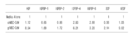
C: 상기 A의 결과에서 각 성장인자의 상대적 발현을 배지에서 탐지된 양과 비교한 표(*P<0.05, **P<0.001 versus Media Alone. †P<0.05, ††P<0.001 versus uhMSC-CdM);C: Table comparing the relative expression of each growth factor in the results of A with the amount detected in the medium ( * P <0.05, ** P <0.001 versus Media Alone. † P <0.05, †† P <0.001 versus uhMSC- CdM);
D: uhMSC, gfMSC 및 gfMSC+GM에서 HGF의 발현을 ELISA로 확인한 결과를 나타낸 그래프;D: Graph showing the results of ELISA confirming the expression of HGF in uhMSC, gfMSC and gfMSC + GM;
E: uhMSC, gfMSC 및 gfMSC+GM에서 VEGF의 발현을 ELISA로 확인한 결과를 나타낸 그래프(*P<0.05, **P<0.001 versus CdM).E: Graph showing the results of ELISA expression of VEGF in uhMSC, gfMSC and gfMSC + GM ( * P <0.05, ** P <0.001 versus CdM).
F: gfMSC가 유도분화 배양액(SCIM) 및 일반 성장 배양액(GM)에서 HGF 생산을 확인한 ELISA 결과;F: ELISA results in which gfMSC confirmed HGF production in induced differentiation culture (SCIM) and normal growth culture (GM);
G: gfMSC가 유도분화 배양액(SCIM) 및 일반 성장 배양액(GM)에서 VEGF 생산을 확인한 ELISA 결과;G: ELISA results in which gfMSC confirmed VEGF production in induced differentiation culture (SCIM) and normal growth culture (GM);
H: uhMSC, gfMSC, gfMSC+SCIM 및 gfMSC+GM에서 HGF 및 VEGF의 발현을 정량적 RT-PCR로 확인한 gel 사진;H: gel photograph showing quantitative RT-PCR expression of HGF and VEGF in uhMSC, gfMSC, gfMSC + SCIM and gfMSC + GM;
I: 상기 H 결과에서 HGF의 발현량을 GAPDH에 대한 상대량으로 보정한 그래프;I: a graph of correcting the expression level of HGF in the H result relative to GAPDH;
J: 상기 H 결과에서 VEGF의 발현량을 GAPDH에 대한 상대량으로 보정한 그래프(*P<0.05, **P<0.001 versus uhMSC).J: A graph of correcting the expression level of VEGF by the relative amount to GAPDH in the H result ( * P <0.05, ** P <0.001 versus uhMSC).
도 11은 gfMSC가 동시배양된 Neuro2A 세포의 신경돌기(neurite)의 성장과 증식(proliferation)을 촉진시키는 것을 신경돌기의 정량분석 및 트립판 블루 염색을 통해 확인한 결과를 나타낸 도이다:FIG. 11 shows the results obtained through quantitative analysis of neurites and trypan blue staining that gfMSC promotes growth and proliferation of neurites of co-cultured Neuro2A cells:
A: Neuro2A 세포 및 인간 중간엽 줄기세포의 동시배양 모식도;A: Schematic diagram of co-culture of Neuro2A cells and human mesenchymal stem cells;
B: gfMSC가 동시배양된 Neuro2A 세포의 신경돌기를 관찰한 현미경 사진;B: micrograph of neurite processes of Neuro2A cells co-cultured with gfMSC;
C: 상기 B의 결과에서 Neuro2A 세포에서 최소 한개 세포체 지름 길이 이상의 신경돌기를 가지는 세포의 비율을 나타낸 그래프;C: A graph showing the proportion of cells having neuronal protrusions of at least one cell body diameter or longer in Neuro2A cells in the results of B;
D: 상기 B의 결과에서 Neuro2A 세포에서 세포당 신경돌기 길이의 총합을 나타낸 그래프(*P<0.05, **P<0.001 versus Media. †P<0.05, ††P<0.001 versus uhMSC. #P<0.05, ##P<0.001 versus gfMSC);D: A graph showing the total number of neurites per cell in Neuro2A cells from the results of B ( * P <0.05, ** P <0.001 versus Media. † P <0.05, †† P <0.001 versus uhMSC. # P < 0.05, ## P <0.001 versus gfMSC);
E: gfMSC가 동시배양된 Neuro2A 세포를 트립판 블루로 염색하여 각 그룹의 Neuro2A 세포의 평균 수를 나타낸 그래프;E: Graph showing the average number of Neuro2A cells of each group by staining Neuro2A cells co-cultured with gfMSC with trypan blue;
F: gfMSC가 동시배양된 Neuro2A 세포를 트립판 블루로 염색하여 각 그룹의 사멸된 Neuro2A 세포의 비율을 나타낸 그래프(*P<0.05, **P<0.001 versus Media. †P<0.05, ††P<0.001 versus uhMSC. #P<0.05, ##P<0.001 versus gfMSC).F: Graph of neuro2A cells co-cultured with gfMSC with trypan blue to show the percentage of killed Neuro2A cells in each group ( * P <0.05, ** P <0.001 versus Media. † P <0.05, †† P <0.001 versus uh MSC. # P <0.05, ## P <0.001 versus gfMSC).
도 12는 gfMSC의 이식이 손상 척수조직 절편 모델에서 척수 신경의 성장과 세포 생존을 증가시키는 것을 조직면역화학염색 및 TUNEL 분석을 통해 확인한 결과를 나타낸 도이다:12 is a diagram showing the results confirmed by tissue immunohistochemical staining and TUNEL analysis that the transplantation of gfMSC increases the growth and cell survival of the spinal cord nerve in the damaged spinal cord tissue section model:
A: gfMSC의 이식이 손상 척수조직 절편 모델에서 척수 신경의 성장을 조직면역화학염색한 현미경 사진;A: Micrographs of grafts of gfMSCs immunohistochemically stain the growth of spinal nerves in an injured spinal cord slice model;
B: 상기 A의 결과에서 통합 광학 밀도를 대조군 척수조직 절편에 의해 표준화시킨 그래프(*P<0.05, **P<0.001 versus Control. ††P<0.001 versus lysolecithin-treated slices(LPC). ##P<0.001 versus LPC+uhMSC. §§P<0.001 versus LPC+gfMSC);B:.. Graph was standardized by the integrated optical density on the result to the control spinal cord tissue sections of the A (* P <0.05, ** P <0.001 versus Control †† P <0.001 versus lysolecithin-treated slices (LPC) ## . P <0.001 versus LPC + uhMSC §§ P <0.001 versus LPC + gfMSC);
C: gfMSC의 이식이 손상 척수조직 절편 모델에서 세포 생존을 증가시키는 것을 TUNEL 분석을 통해 확인한 현미경 사진; 및C: photomicrograph confirmed by TUNEL analysis that transplantation of gfMSC increased cell survival in injured spinal cord tissue section model; And
D: 상기 C의 결과에서 TUNEL-양성 세포를 정량 분석한 그래프(** P<0.001, versus Control. ††P<0.001 versus LPC. #P<0.05, ##P<0.001 versus LPC+uhMSC).D: A graph quantitatively analyzing TUNEL-positive cells in the results of C ( ** P <0.001, versus Control. †† P <0.001 versus LPC. # P <0.05, ## P <0.001 versus LPC + uhMSC).
도 13은 Neuro2A 세포와 손상 척수조직 절편 모델에서 외인성 HGF 및 VEGF의 신경 영양 인자로서의 효과를 신경돌기의 정량분석, 트립판 블루 염색 및 조직면역화학염색을 통해 확인한 결과를 나타낸 도이다:13 is a diagram showing the results of neuronal trophic factors of exogenous HGF and VEGF as a neurotrophic factor in Neuro2A cells and injured spinal cord tissue section model through quantitative analysis of neurite, trypan blue staining and tissue immunochemical staining:
A: 재조합 HGF를 첨가하여 배양한 Neuro2A 세포에서 최소 한개 세포체 지름 길이 이상의 신경돌기를 가지는 세포의 비율을 나타낸 그래프;A: A graph showing the proportion of cells having a neurites of at least one cell body diameter or longer in Neuro2A cells cultured with recombinant HGF;
B: 재조합 HGF를 첨가하여 배양한 Neuro2A 세포에서 세포당 신경돌기 길이의 총합을 나타낸 그래프;B: Graph showing total sum of neurites per cell in Neuro2A cells cultured with recombinant HGF;
C: 재조합 VEGF를 첨가하여 배양한 Neuro2A 세포에서 최소 한개 세포체 지름 길이 이상의 신경돌기를 가지는 세포의 비율을 나타낸 그래프;C: a graph showing the proportion of cells having neuronal processes of at least one cell body diameter or longer in Neuro2A cells cultured with recombinant VEGF;
D: 재조합 VEGF를 첨가하여 배양한 Neuro2A 세포에서 세포당 신경돌기 길이의 총합을 나타낸 그래프(*P<0.05, **P<0.001 versus 미처리 Neuro2A 세포. †P<0.05, ††P<0.001 versus HGF와 VEGF 동시 처리된 Neuro2A 세포);D: Graph showing total sum of neurites per cell in Neuro2A cells cultured with recombinant VEGF ( * P <0.05, ** P <0.001 versus untreated Neuro2A cells. † P <0.05, †† P <0.001 versus HGF And VEGF co-treated Neuro2A cells);
E: 외인성 HGF 및/또는 VEGF 단백질이 손상된 척수조직 절편의 신경돌기 성장을 증가시키는 것을 확인한 현미경 사진;E: micrograph showing that exogenous HGF and / or VEGF protein increased neurite growth in damaged spinal cord tissue sections;
F: 상기 E의 결과에서 HGF 및/또는 VEGF 단백질의 농도에 따라 NF-M 면역형광 염색된 신경섬유의 상대적인 통합 광학 밀도(IOD) 값을 대조군 척수조직 절편으로 표준화시킨 그래프(*P<0.05, **P<0.001 versus control. †P<0.05, ††P<0.001 versus 라이소레시틴 처리된 척수조직(LPC));F: Graph of standardized relative integrated optical density (IOD) values of NF-M immunofluorescent stained nerve fibers with control spinal cord tissue sections according to the concentration of HGF and / or VEGF protein in the results of E ( * P <0.05, ** P <0.001 versus control. † P <0.05, †† P <0.001 versus lysolecithin-treated spinal cord tissue (LPC));
G: 인간 골수 유래 중간엽 줄기세포에서 분화된 성장인자 다량 분비 인간 줄기세포가 손상척추신경에서 HGF 및 VEGF를 분비하여 신경 영양 인자로서 기능하는 메카니즘을 나타낸 도이다.G: High secretion of growth factors differentiated from human bone marrow-derived mesenchymal stem cells Human stem cells secrete HGF and VEGF in the injured spinal nerve to show a mechanism that functions as a neurotrophic factor.
이하, 본 발명을 상세히 설명한다.Hereinafter, the present invention will be described in detail.
상기 목적을 달성하기 위하여, 본 발명은In order to achieve the above object, the present invention
1) 지방세포 유래 중간엽 줄기세포(adipose-derived mesenchymal stem cell; ADMSC)를 BMP(Bone morphogenic protein) 억제제, TGF(Transforming growth factor) 베타 신호전달 저해제를 포함하는 배지에서 배양하는 단계;1) culturing adipocyte-derived mesenchymal stem cells (ADMSC) in a medium comprising a BMP (Bone morphogenic protein) inhibitor and a transforming growth factor (TGF) beta signaling inhibitor;
2) 상기 단계 1)의 세포를 B27, N2, 및 ascorbic acid를 포함하는 배지에서 배양하는 단계; 및2) culturing the cells of step 1) in a medium containing B27, N2, and ascorbic acid; And
3) 상기 단계 2)의 세포를 표피성장인자(EGF) 및 염기성 섬유모세포성장인자 (bFGF)를 첨가한 배지에서 배양하는 단계를 포함하는 지방세포 유래 중간엽 줄기세포를 신경줄기세포(neural stem cell)로 분화시키는 방법을 제공한다.3) Neural stem cells of adipocyte-derived mesenchymal stem cells comprising culturing the cells of step 2) in a medium to which epidermal growth factor (EGF) and basic fibroblast growth factor (bFGF) are added. It provides a way to differentiate into.
또한, 본 발명은In addition, the present invention
1) 지방세포 유래 중간엽 줄기세포(adipose-derived mesenchymal stem cell; ADSC)를 BMP(Bone morphogenic protein) 억제제, TGF(Transforming growth factor) 베타 신호전달 저해제를 포함하는 배지에서 배양하는 단계;1) culturing adipocyte-derived mesenchymal stem cells (ADSC) in a medium comprising a BMP (Bone morphogenic protein) inhibitor and a transforming growth factor (TGF) beta signaling inhibitor;
2) 상기 단계 1)의 세포를 B27, N2, 및 ascorbic acid를 포함하는 배지에서 배양하는 단계;2) culturing the cells of step 1) in a medium containing B27, N2, and ascorbic acid;
3) 상기 단계 2)의 세포를 표피성장인자(epidermal growth factor; EGF) 및 염기성 섬유모세포성장인자(basic fibroblast growth factor; bFGF)를 첨가한 배지에서 배양하여 신경줄기세포로 분화시키는 단계; 및3) culturing the cells of step 2) in a medium to which epidermal growth factor (EGF) and basic fibroblast growth factor (bFGF) are added to differentiate into neural stem cells; And
4) 상기 단계 3)의 신경줄기세포를 purmorphamine 및 BDNF(Brain-Derived Neurotrophic Factor)를 첨가한 배지에서 배양하는 단계를 포함하는 지방세포 유래 중간엽 줄기세포를 신경세포(neural cell)로 분화시키는 방법을 제공한다.4) a method of differentiating adipocyte-derived mesenchymal stem cells into neural cells, comprising culturing the neural stem cells of step 3) in a medium to which purmorphamine and BDNF (Brain-Derived Neurotrophic Factor) are added. to provide.
또한, 본 발명은 In addition, the present invention
1) 지방세포 유래 중간엽 줄기세포(adipose-derived mesenchymal stem cell; ADSC)를 BMP(Bone morphogenic protein) 억제제, TGF(Transforming growth factor) 베타 신호전달 저해제를 포함하는 배지에서 배양하는 단계;1) culturing adipocyte-derived mesenchymal stem cells (ADSC) in a medium comprising a BMP (Bone morphogenic protein) inhibitor and a transforming growth factor (TGF) beta signaling inhibitor;
2) 상기 단계 1)의 세포를 B27, N2, 및 ascorbic acid를 포함하는 배지에서 배양하는 단계;2) culturing the cells of step 1) in a medium containing B27, N2, and ascorbic acid;
3) 상기 단계 2)의 세포를 purmorphamine 및 BDNF(Brain-Derived Neurotrophic Factor)를 첨가한 배지에서 배양하여 신경세포로 분화시키는 단계; 및3) culturing the cells of step 2) in a medium to which purmorphamine and Brain-Derived Neurotrophic Factor (BDNF) are added to differentiate into neurons; And
4) 상기 단계 3)의 신경세포를 dbcAMP(dibutyryl cyclic AMP) 및 BDNF(Brain-derived neurotrophic factor)를 첨가한 배지에서 배양하는 단계를 포함하는 지방세포 유래 중간엽 줄기세포를 가바성 신경세포(GABAergic neural cells)로 분화시키는 방법을 제공한다.4) GABAergic to the adipocyte-derived mesenchymal stem cells comprising the step of culturing the neurons of step 3) in a medium added with dbcAMP (dibutyryl cyclic AMP) and Brain-derived neurotrophic factor (BDNF) neural cells).
상기 단계 1)의 BMP 억제제는 Noggin 및 LDN193189인 것이 바람직하고, TGF 베타 신호전달 저해제는 ALK(activin receptor-like kinase) 수용체 저해제인 SB431542인 것이 바람직하다.The BMP inhibitor of step 1) is preferably Noggin and LDN193189, and the TGF beta signaling inhibitor is preferably SB431542 which is an activin receptor-like kinase (ALK) receptor inhibitor.
상기 단계 1)의 배지는 DMEM F-12 기본배지에 0.5 내지 30% KOSR, 0.5 내지 1.5% Penicillin/ Streptomycin, 0.1 내지 10% Glutamax, 0.1 내지 10% non-essential amino acid 그리고 1 내지 500 ng/ml bFGF(basic fibroblast growth factor), 1 내지 200 μM SB431542, 0.01 내지 1 μg/ml Noggin, 0.1 내지 20 μM LDN193289을 첨가한 배지인 것이 바람직하고, 특히 5 내지 20 μM SB431542, 0.05 내지 0.2 μg/ml Noggin, 0.1 내지 1.0 μM LDN193289을 첨가하는 것이 신경줄기세포로의 분화를 증가시키는데 더욱 바람직하며, 상기 범위를 벗어나는 경우에는 분화 효율이 감소될 수 있다.The medium of step 1) is 0.5-30% KOSR, 0.5-1.5% Penicillin / Streptomycin, 0.1-10% Glutamax, 0.1-10% non-essential amino acid and 1-500 ng / ml A medium to which bFGF (basic fibroblast growth factor), 1 to 200 μM SB431542, 0.01 to 1 μg / ml Noggin, 0.1 to 20 μM LDN193289 is preferable, particularly 5 to 20 μM SB431542, 0.05 to 0.2 μg / ml Noggin It is more desirable to add 0.1 to 1.0 μM LDN193289 to increase the differentiation into neural stem cells, the efficiency of differentiation may be reduced if outside the above range.
상기 단계 2)의 배지는 DMEM F-12:Neurobasal (1:1) 기본배지에 0.1 내지 10% Glutamax, 1 내지 20 mM D-glucose, 0.01 내지 2 mM ascorbic acid, 0.1 내지 10 mM sodium pyruvate, 0.1 내지 10% B27 및 0.1 내지 10% N2를 첨가한 배지인 것이 바람직하며, 단계 3)의 배지는 DMEM F-12:Neurobasal (1:1) 기본배지에 0.1 내지 10% Glutamax, 1 내지 20 mM D-glucose, 0.01 내지 2 mM ascorbic acid, 0.1 내지 10 mM sodium pyruvate, 0.1 내지 10% B27 및 0.1 내지 10% N2, 1 내지 500 ng/ml bFGF 및 1 내지 500 ng/ml EGF(epidermal growth factor)를 첨가한 배지인 것이 바람직하다. The medium of step 2) is 0.1-10% Glutamax, 1-20 mM D-glucose, 0.01-2 mM ascorbic acid, 0.1-10 mM sodium pyruvate, 0.1 in DMEM F-12: Neurobasal (1: 1) basal medium. Preferably, the medium is added with 10% B27 and 0.1-10% N2, and the medium of step 3) is 0.1-10% Glutamax, 1-20 mM D in DMEM F-12: Neurobasal (1: 1) basal medium. -glucose, 0.01-2 mM ascorbic acid, 0.1-10 mM sodium pyruvate, 0.1-10% B27 and 0.1-10% N2, 1-500 ng / ml bFGF and 1-500 ng / ml epidermal growth factor (EGF) It is preferable that it is an added medium.
상기 단계 1)의 배양은 4 내지 12일, 상기 단계 2)의 배양은 3 내지 10일, 상기 단계 3)의 배양은 3 내지 10일 동안 실시하는 것이 바람직하고, 1)의 배양은 6 내지 8일, 2)의 배양은 5일, 3)의 배양은 5 내지 7일 동안 실시하는 것이 더욱 바람직하다.Cultivation of the step 1) is 4 to 12 days, culturing of the step 2) is 3 to 10 days, the culturing of the step 3) is preferably carried out for 3 to 10 days, the culturing of 1) is 6 to 8 It is more preferable that the culture of days 1 and 2) is carried out for 5 days and the culture of 3) is carried out for 5 to 7 days.
또한, 상기 단계 4)의 배지는 DMEM F-12:Neurobasal (1:1) 기본배지에 0.1 내지 10% Glutamax, 1 내지 20 mM D-glucose, 0.01 내지 2 mM ascorbic acid, 0.1 내지 10 mM sodium pyruvate, 0.1 내지 10% B27 및 0.1 내지 10% N2, 0.1 내지 50 μM purmorphamine 및 1 내지 500 ng/ml BDNF를 첨가한 배지인 것이 바람직하고, 특히 0.8 내지 2.0 μM purmorphamine 및 5 내지 30 ng/ml BDNF를 첨가하는 것이 신경세포로의 분화를 증가시키는데 더욱 바람직하며, 상기 범위를 벗어나는 경우에는 분화 효율이 감소될 수 있다.In addition, the medium of step 4) is 0.1-10% Glutamax, 1-20 mM D-glucose, 0.01-2 mM ascorbic acid, 0.1-10 mM sodium pyruvate in DMEM F-12: Neurobasal (1: 1) basal medium. , Medium containing 0.1 to 10% B27 and 0.1 to 10% N2, 0.1 to 50 μM purmorphamine and 1 to 500 ng / ml BDNF is preferred, in particular 0.8 to 2.0 μM purmorphamine and 5 to 30 ng / ml BDNF. Addition is more desirable to increase differentiation into neurons, and if outside this range, differentiation efficiency may be reduced.
상기 배양은 7 내지 16일 동안 실시하는 것이 바람직하고, 12 내지 14일 동안 실시하는 것이 더욱 바람직하다.The culture is preferably carried out for 7 to 16 days, more preferably for 12 to 14 days.
또한, 상기 단계 5)의 배지는 DMEM F-12:Neurobasal (1:1) 기본배지에 0.1 내지 10% Glutamax, 1 내지 20 mM D-glucose, 0.01 내지 2 mM ascorbic acid, 0.1 내지 10 mM sodium pyruvate, 0.1 내지 10% B27 및 0.1 내지 10% N2, 0.01 내지 1 mM dbcAMP 및 1 내지 500 ng/ml BDNF를 첨가한 배지인 것이 바람직하고, 특히 0.04 내지 0.1 mM dbcAMP 및 5 내지 30 ng/ml BDNF를 첨가하는 것이 가바성 신경세포로의 분화를 증가시키는데 더욱 바람직하며, 상기 범위를 벗어나는 경우에는 분화 효율이 감소될 수 있다.In addition, the medium of step 5) is 0.1-10% Glutamax, 1-20 mM D-glucose, 0.01-2 mM ascorbic acid, 0.1-10 mM sodium pyruvate in DMEM F-12: Neurobasal (1: 1) basal medium. , 0.1 to 10% B27 and 0.1 to 10% N2, 0.01 to 1 mM dbcAMP and 1 to 500 ng / ml BDNF are preferred, in particular 0.04 to 0.1 mM dbcAMP and 5 to 30 ng / ml BDNF Addition is more desirable to increase differentiation into GAB neurons, and if outside this range, differentiation efficiency may be reduced.
상기 배양은 10 내지 40일 동안 배양하는 것이 바람직하고, 13 내지 35일인 것이 더욱 바람직하며, 18일 내지 30일 동안 배양하는 것이 가장 바람직하다.The culture is preferably cultured for 10 to 40 days, more preferably 13 to 35 days, most preferably 18 to 30 days.
본 발명의 구체적인 실시 예에서, 지방세포 유래 중간엽 줄기세포를 세포 배양 배지에 저분자 억제제인 SB431543, Noggin, 및 LDN193189를 첨가하여 6 내지 8일간 배양하고(단계 1: 전-처리 단계), 그런 다음, B27 및 N2를 첨가한 배지에 5일간 배양하고(단계 2: 신경 유도 단계), bFGF 및 EGF를 첨가한 배지에서 5 내지 7일간 더 배양하여(단계 3: 증식 단계), 신경 줄기세포로 분화시킨 결과, 저분자억제제를 첨가한 실험군이 미처리 실험군보다 신경줄기세포의 분자표지인 Nestin, Sox1, Pax6, Musashi-1, Vimentin, Olig2, Nkx2.1, FoxG1, Tuj1, 및 Ascl1의 mRNA 발현이 증가함을 확인하였고, 형광 면역세포염색법분석을 통해 Nestin과 Sox2를 모두 발현하는 유도 신경줄기세포의 검출을 확인하였으며(도 1 및 도 2 참조), 다음으로 상기 단계 3의 세포 배양액에 purmorphamine 및 BDNF를 첨가한 배지를 이용하여 상기 신경줄기세포를 추가적으로 신경세포로 분화시킨 결과, 성숙한 신경세포와 관련된 유전자들의 발현이 현저하게 증가함을 확인하였고(도 3 및 도 4 참조), 또한, 상기 단계 2의 세포 배양액에 purmorphamine 및 BDNF와 dbcAMP 및 BDNF를 첨가한 배지를 이용하여 상기 신경세포를 가바성 신경세포로 분화시킨 결과, 내측 신경절 융기 (MGE) 세포 분자표지인 NKX2.1, DLX2, LHX6와 신경세포 분자표지인 TuJ1, MAP2를 발현함을 확인하였으며, 상기 가바성 신경세포는 글루탐산 수용체 차단제가 함께 있을 때의 자발적 억제성 시냅스 후 전류 (IPSC)가 나타났다가 GABAA 수용체 차단제를 처리하면 사라짐을 확인함으로써, 기능을 가진 가바성 신경세포로 최종 분화됨을 확인하였다(도 5 및 도 6 참조). In a specific embodiment of the present invention, the adipocyte-derived mesenchymal stem cells were incubated for 6 to 8 days with the addition of the low molecular weight inhibitors SB431543, Noggin, and LDN193189 to the cell culture medium (step 1: pre-treatment step), and then , Cultured in medium containing B27 and N2 for 5 days (step 2: nerve induction step), and further cultured in bFGF and EGF medium for 5 to 7 days (step 3: proliferation step) to differentiate into neural stem cells. As a result, mRNA expression of Nestin, Sox1, Pax6, Musashi-1, Vimentin, Olig2, Nkx2.1, FoxG1, Tuj1, and Ascl1, which are the molecular markers of neural stem cells, was increased in the experimental group to which the low molecular weight inhibitor was added. Confirmation of the induced neural stem cells expressing both Nestin and Sox2 was confirmed by fluorescence immunocytostaining analysis (see FIGS. 1 and 2). Use By further differentiating the neural stem cells into neurons, it was confirmed that the expression of genes related to mature neurons was significantly increased (see FIGS. 3 and 4), and also purmorphamine and BDNF in the cell culture medium of step 2 above. And the differentiation of the neurons into Gab neurons using a medium containing dbcAMP and BDNF. As a result, the medial ganglion bump (MGE) cell markers NKX2.1, DLX2, LHX6 and the neuronal cell markers TuJ1, MAP2 It is confirmed that the expression of the Gaba neurons, spontaneous inhibitory post-synaptic current (IPSC) when the glutamic acid receptor blocker is present, and confirms that the disappearance when treated with GABAA receptor blockers, functioning Gaba nerves Confirmation of final differentiation into cells (see FIGS. 5 and 6).
따라서 본 발명의 분화방법을 통한 신경세포 및 가바성 신경세포는 신경학적 질환 치료방법의 개발을 위해 유용하게 사용될 수 있다.Therefore, the neurons and Gaba neurons through the differentiation method of the present invention can be usefully used for the development of neurological disease treatment method.
본 발명의 지방세포 유래 중간엽 줄기세포는 인간, 원숭이, 돼지, 말, 소, 양, 개, 고양이, 생쥐 또는 토끼 등의 모든 유래일 수 있으며, 구체적으로 인간 유래일 수 있으나, 이에 한정되지 않는다.Adipocyte-derived mesenchymal stem cells of the present invention may be derived from humans, monkeys, pigs, horses, cows, sheep, dogs, cats, mice, or rabbits, and may be specifically derived from humans, but are not limited thereto. .
본 발명에서 용어, "B27" 및 “N2”는 무혈청 보충제로써 본 발명의 방법에 사용되는 배지의 성분이다.As used herein, the terms "B27" and "N2" are components of the medium used in the methods of the invention as serum-free supplements.
본 발명에서 “bFGF”는 세포증식, 세포분화 등을 비롯해 분열 촉진 인자, 혈관 생성 인자, 뼈 형성 인자 및 신경성장인자로써 기능하는 FGF 패밀리에 속하는 단백질로써 FGF2라고도 불리며 주로 FGFR 1b, FGFR 1c, FGFR 2c, FGFR 3c, FGFR 4c를 포함하는 수용체 단백질을 활성화시키며, 특히 FGFR 1c, FGFR 3c를 강력히 활성화시키는 것으로 알려져 있다. 상기 FGFR을 활성화시키는 FGF 패밀리 단백질을 포함하여 bFGF와 유사한 신호를 전달할 수 있는 물질은 제한 없이 사용할 수 있다.In the present invention, "bFGF" is a protein belonging to the FGF family that functions as a cell proliferation, cell differentiation, division promoting factor, angiogenesis factor, bone formation factor, and nerve growth factor, also called FGF2, mainly FGFR 1b, FGFR 1c, FGFR It is known to activate receptor proteins including 2c, FGFR 3c, FGFR 4c, and in particular to strongly activate FGFR 1c, FGFR 3c. A substance capable of transmitting a signal similar to bFGF, including the FGF family protein that activates the FGFR, can be used without limitation.
본 발명에서 용어, "TGF (Transforming growth factor) 베타 신호전달 저해제"는 TGF 베타 신호전달을 억제하는 물질을 의미한다. 상기 TGF 베타는 생체 내에서 세포의 증식, 분화, 세포사멸, 이동, 세포외기질(ECM)의 생산 혈관형성, 발생 등 다양한 생리적 과정을 조절하는 물질이다. As used herein, the term "TGF (Transforming growth factor) beta signaling inhibitor" refers to a substance that inhibits TGF beta signaling. The TGF beta is a substance that regulates various physiological processes such as cell proliferation, differentiation, apoptosis, migration, extracellular matrix (ECM) production, angiogenesis, and development.
본 발명에서 상기 TGF 베타 신호전달 저해제는 TGF 신호전달을 억제시킬 수 있는 물질이라면 제한 없이 사용할 수 있으며, 그 예로 ALK (activin receptor-like kinase) 수용체 저해제가 있으나, 이에 한정되지 않는다. 본 발명의 일 실시예에서는 ALK 수용체 저해제인 SB431542를 사용하였다.In the present invention, the TGF beta signaling inhibitor may be used without limitation as long as it is a substance capable of inhibiting TGF signaling. Examples thereof include an activin receptor-like kinase (ALK) receptor inhibitor, but are not limited thereto. In one embodiment of the present invention was used AB receptor inhibitor SB431542.
또한, 본 발명은In addition, the present invention
1) 지방세포 유래 중간엽 줄기세포를 BMP(Bone morphogenic protein) 억제제, TGF(Transforming growth factor) 베타 신호전달 저해제를 포함하는 배지에서 배양하는 단계;1) culturing the adipocyte-derived mesenchymal stem cells in a medium containing a BMP (Bone morphogenic protein) inhibitor and a transforming growth factor (TGF) beta signaling inhibitor;
2) 상기 단계 1)의 세포를 B27, N2, 및 ascorbic acid를 포함하는 배지에서 배양하는 단계; 및2) culturing the cells of step 1) in a medium containing B27, N2, and ascorbic acid; And
3) 상기 단계 2)의 세포를 표피성장인자(EGF) 및 염기성 섬유모세포성장인자 (bFGF)를 첨가한 배지에서 배양하는 단계를 포함하는 지방세포 유래 중간엽 줄기세포로부터 신경줄기세포의 제조방법을 제공한다.3) providing a method for producing neural stem cells from adipocyte-derived mesenchymal stem cells comprising culturing the cells of step 2) in a medium to which epidermal growth factor (EGF) and basic fibroblast growth factor (bFGF) are added. do.
또한, 본 발명은In addition, the present invention
1) 지방세포 유래 중간엽 줄기세포를 BMP(Bone morphogenic protein) 억제제, TGF(Transforming growth factor) 베타 신호전달 저해제를 포함하는 배지에서 배양하는 단계;1) culturing the adipocyte-derived mesenchymal stem cells in a medium containing a BMP (Bone morphogenic protein) inhibitor and a transforming growth factor (TGF) beta signaling inhibitor;
2) 상기 단계 1)의 세포를 B27, N2, 및 ascorbic acid를 포함하는 배지에서 배양하는 단계;2) culturing the cells of step 1) in a medium containing B27, N2, and ascorbic acid;
3) 상기 단계 2)의 세포를 표피성장인자(EGF) 및 염기성 섬유모세포성장인자 (bFGF)를 첨가한 배지에서 배양하는 단계; 및3) culturing the cells of step 2) in a medium to which epidermal growth factor (EGF) and basic fibroblast growth factor (bFGF) are added; And
4) 상기 단계 3)의 신경줄기세포를 purmorphamine 및 BDNF를 첨가한 배지에서 배양하는 단계를 포함하는 지방세포 유래 중간엽 줄기세포로부터 신경세포의 제조방법을 제공한다.4) It provides a method for producing neurons from adipocyte-derived mesenchymal stem cells comprising the step of culturing the neural stem cells of step 3) in a medium to which purmorphamine and BDNF are added.
또한, 본 발명은In addition, the present invention
1) 지방세포 유래 중간엽 줄기세포를 BMP(Bone morphogenic protein) 억제제, TGF(Transforming growth factor) 베타 신호전달 저해제를 포함하는 배지에서 배양하는 단계;1) culturing the adipocyte-derived mesenchymal stem cells in a medium containing a BMP (Bone morphogenic protein) inhibitor and a transforming growth factor (TGF) beta signaling inhibitor;
2) 상기 단계 1)의 세포를 B27, N2, 및 ascorbic acid를 포함하는 배지에서 배양하는 단계;2) culturing the cells of step 1) in a medium containing B27, N2, and ascorbic acid;
3) 상기 단계 2)의 세포를 purmorphamine 및 BDNF를 첨가한 배지에서 배양하여 신경세포로 분화시키는 단계; 및3) culturing the cells of step 2) in a medium containing purmorphamine and BDNF to differentiate into neurons; And
4) 상기 신경세포를 dbcAMP 및 BDNF를 첨가한 배지에서 배양하는 단계를 포함하는 지방세포 유래 중간엽 줄기세포로부터 가바성 신경세포의 제조방법을 제공한다.4) It provides a method for producing GABA neurons from adipocyte-derived mesenchymal stem cells comprising the step of culturing the nerve cells in a medium added with dbcAMP and BDNF.
또한, 본 발명은 상기 방법으로 제조된 신경줄기세포, 신경세포, 및 가바성 신경세포를 제공한다.The present invention also provides neural stem cells, neurons, and Gaba neurons prepared by the above method.
아울러, 본 발명은 상기 방법으로 제조된 신경줄기세포, 신경세포 및 가바성 신경세포를 포함하는 신경 손상 세포치료용 조성물, 신경 손상 세포치료제 스크리닝용 조성물, 뇌질환 치료제 스크리닝용 조성물, 시신경 손상 관련 질환 예방 및 치료용 조성물 및 인공 망막 제조용 조성물을 제공한다.In addition, the present invention is a composition for treating neuronal damage cells comprising neural stem cells, neurons and Gaba neurons prepared by the above method, a composition for screening neuronal damage cell therapy, a screening composition for treating brain diseases, prevention of optic nerve damage related diseases And it provides a composition for the treatment and composition for the preparation of artificial retina.
본 발명의 지방세포유래 중간엽 줄기세포로부터 상기 단계별 조성물을 이용하면, 일차 신경세포 및 일차 가바성 신경세포와 기능적, 유전적으로 유사한 특성을 가진 신경세포 및 가바성 신경세포를 제조하는 데에 유용하게 사용될 수 있으며, 최종 순수 분리된 신경세포 및 가바성 신경세포는 신경학적 질환의 치료 약물을 개발하기 위한 약물 스크리닝, 및 약물 독성분석에 유용하게 사용될 수 있다. By using the step-by-step composition from the adipocyte-derived mesenchymal stem cells of the present invention, it is useful for preparing neurons and Gaba neurons having functional and genetic similarities to primary neurons and primary Gaba neurons. The final purely isolated neurons and Gaba neurons can be usefully used for drug screening and drug toxicity analysis to develop therapeutic drugs for neurological diseases.
본 발명은 인간 골수 유래 중간엽 줄기세포로부터 분화된 성장인자 다량 분비 인간 줄기세포(growth factor releasing human mesenchymal stem cell; gfMSC)를 유효성분으로 함유하는 개체내 HGF 또는 VEGF 전달용 조성물 및 신경 손상 세포 치료용 조성물을 제공한다.The present invention provides a composition for delivery of HGF or VEGF in a subject containing a growth factor releasing human mesenchymal stem cell (gfMSC) differentiated from human bone marrow-derived mesenchymal stem cells as an active ingredient and neuronal damage cell treatment It provides a composition for.
상기 분화된 성장인자 다량 분비 인간 줄기세포는 분화 단계(lineage) 중 미성숙(immature) 세포인 것이 바람직하나, 이에 제한되는 것은 아니며 유사 슈반세포(Schwann-like cell)일 수 있다.The differentiated growth factor large secreted human stem cells are preferably immature cells during the differentiation stage (lineage), but is not limited thereto may be Schwann-like cells.
상기 분화된 성장인자 다량 분비 인간 줄기세포는 미분화 인간 중간엽 줄기세포(Untreated hMSC; uhMSC)로부터 하기 a) 내지 c)의 단계를 포함하는 방법으로 분화된 것이 바람직하나 이에 제한되는 것은 아니며, 미분화 포유동물 중간엽 줄기세포 또는 피부-유래 1차 세포(skin-derived primary cell)로부터 성장인자 다량 분비 인간 줄기세포로의 분화에 사용되는 방법이라면 모두 사용가능하다:The differentiated growth factor large secreted human stem cells are preferably differentiated by a method comprising the steps of a) to c) from undifferentiated human mesenchymal stem cells (Untreated hMSC; uhMSC), but is not limited thereto. Any method used for differentiation from animal mesenchymal stem cells or skin-derived primary cells into growth factor high secreting human stem cells can be used:
a) 1 내지 20% FBS 및 0.1 내지 10 mM β-머캅토에탄올(mercaptoethanol)을 함유하는 DMEM 배지로 24시간 동안 1차 배양하는 단계;a) primary culture for 24 hours in DMEM medium containing 1-20% FBS and 0.1-10 mM β-mercaptoethanol;
b) 1 내지 20% FBS 및 0.05 내지 5 μg/ml 레티노산(retinoic acid)을 함유하는 DMEM 배지로 72시간 동안 2차 배양하는 단계; 및, b) secondary culture for 72 hours in DMEM medium containing 1-20% FBS and 0.05-5 μg / ml retinoic acid; And,
c) 1 내지 20% FBS, 1 내지 100 μ/ml 폴스콜린(forskolin), 1 내지 100 ng/ml 인간 bFGF, 1 내지 50 ng/ml PDGF-AA 및 20 내지 2 μg/ml 인간 헤레귤린-β1(heregulin-β1)을 함유하는 DMEM 배지로 6 내지 12일 동안 3차 배양하는 단계.c) 1 to 20% FBS, 1 to 100 μ / ml forskolin, 1 to 100 ng / ml human bFGF, 1 to 50 ng / ml PDGF-AA and 20 to 2 μg / ml human Heregulin-β1 tertiary culturing for 6-12 days with DMEM medium containing (heregulin-β1).
상기 조성물은 신경 손상 질환을 가지는 개체의 치료 또는 개선을 위해 투여될 수 있다. 상기 신경 손상 질환은 뇌졸중 (stroke), 파킨슨씨병, 알츠하이머병, 피크병 (Pick's disease), 헌팅톤병 (Huntington's disease), 근위축성 측면 경화증 (Amyotrophic lateral sclerosis), 외상성 중추 신경계 질환 (traumatic central nervous system diseases) 및 척수 손상 질환 (spinal cord injury disease)으로 이루어진 군으로부터 선택되는 어느 하나의 질병, 바람직하게는 척수 손상 질환이나 이에 제한되는 것은 아니다. The composition may be administered for the treatment or amelioration of an individual with a neurological injury disease. The nerve injury diseases include stroke, Parkinson's disease, Alzheimer's disease, Pick's disease, Huntington's disease, Amyotrophic lateral sclerosis, and traumatic central nervous system diseases. ) And spinal cord injury disease, preferably, but not limited to any one disease selected from the group consisting of spinal cord injury disease.
본 발명의 구체적인 실시예에서, 본 발명자들은 미분화된 인간 골수 유래 중간엽 줄기세포(uhMSC)를 성장인자 다량 분비 인간 줄기세포(growth factor releasing human mesenchymal stem cell; gfMSC)로 분화를 유도하여 gfMSC 세포에서 분비되는 인간 성장인자를 분석한 결과, uhMSC보다 gfMSC에서 HGF(hepatocyte growth factor), VEGF(vascular endothelial growth factor), IGFBP-1(insulin-like growth factor binding protein-1), IGFBP-2, IGFBP-4, IGFBP-6 및 SCF(stem cell factor)의 발현이 증가되어 있는 것을 확인하였으며(도 10a 내지 도 10c 참조), 특히, HGF 및 VEGF의 발현이 10배 이상 증가하였고(도 10d 및 도 10e 참조), gfMSC의 HGF 및 VEGF 분비는 일반 성장 배양액에서 배양하였을 때에도 11일 동안 높게 유지되며(도 10f 및 도 10g 참조), 이는 mRNA의 발현량이 증가하기 때문임을 확인하였다(도 10h 내지 도 10j 참조).In a specific embodiment of the present invention, the inventors induce differentiation of undifferentiated human bone marrow-derived mesenchymal stem cells (uhMSCs) into growth factor releasing human mesenchymal stem cells (gfMSCs) in gfMSC cells. Analysis of secreted human growth factors revealed that hepatocyte growth factor (HGF), vascular endothelial growth factor (VEGF), insulin-like growth factor binding protein-1 (IGFBP-1), IGFBP-2 and IGFBP- 4, it was confirmed that the expression of IGFBP-6 and stem cell factor (SCF) is increased (see Figs. 10a to 10c), in particular, the expression of HGF and VEGF increased more than 10-fold (see Figs. 10d and 10e). ), HGF and VEGF secretion of gfMSC is maintained high for 11 days even when cultured in normal growth culture (see Fig. 10f and 10g), it was confirmed that this is because the expression level of mRNA is increased (see Figs. 10h to 10j). .
또한, 본 발명의 구체적 실시예에서 본 발명자들은 uhMSC로부터 분화된 성장인자를 과분비하는 gfMSC가 신경세포에 미치는 영향을 분석한 결과, gfMSC를 신경세포(Neuro2A)와 동시배양(co-culture)하였을 때, 신경돌기(neurite)의 성장 및 세포 성장 및 생존을 증가시키는 효과가 있음을 확인하였고, 상기 효과는 HGF 및/또는 VEGF에 대한 항체에 의해 감소되는 것을 관찰하여, gfMSC의 신경세포에 대한 신경성장인자로서의 효과는 gfMSC에서 분비되는 HGF 및 VEGF에 의해 유도되는 것임을 알 수 있었다(도 11b 내지 도 11f 참조).In a specific embodiment of the present invention, the present inventors analyzed the effect of gfMSC oversecreting growth factors differentiated from uhMSC on neurons, and when co-culture of gfMSC with neurons (Neuro2A) In addition, it was confirmed that there is an effect of increasing the growth and cell growth and survival of neurite, the effect is reduced by the antibody to HGF and / or VEGF, the nerve growth of gfMSC to nerve cells The effect as a factor was found to be induced by HGF and VEGF secreted from gfMSC (see FIGS. 11B-11F).
또한, 본 발명의 구체적 실시예에서 본 발명자들은 상기에서 확인한 gfMSC의 신경세포에 대한 신경성장인자로서의 효과가 ex vivo에서도 동일하게 작용하는지 국립서울대학교 실험동물운영위원회의 승인을 받아 실험한 결과, 스프라그-돌리(Sprague-Dawley) 래트로부터 채취한, 라이소레시틴을 처리하여 손상시킨 요추(lumbar) 척수조직절편(LPC)에서 gfMSC를 이식했을 경우, uhMSC를 이식한 경우보다 신경돌기 성장 및 세포사멸 억제 효과가 현저하게 높은 것을 확인하였다(도 12a 내지 도 12d 참조). 상기 gfMSC 이식에 의한 손상된 척수조직절편의 신경돌기 성장 증가는 HGF 및/또는 VEGF에 대한 항체를 처리하였을 때 유의하게 감소하였으나, gfMSC의 척수조직 절편의 세포 사멸 억제 효과는 HGF 및/또는 VEGF에 대한 항체 처리 여부에 유의한 차이를 보이지 않은 것으로부터 gfMSC의 척수조직 절편에 대한 신경성장인자로서의 효과는 gfMSC에서 분비되는 HGF 및 VEGF에 의해 유도되는 것이지만, 리소레시틴에 의한 척수조직 절편 세포의 세포사멸 보호 효과는 HGF 및 VEGF의 분비에 의한 효과와 독립적인 것을 알 수 있었다(도 12a 내지 도 12d 참조). In addition, in a specific embodiment of the present invention, the present inventors have been tested with the approval of the Seoul National University Laboratory Animal Steering Committee whether the effect of the GFMSC as a neuronal growth factor on the neurons identified above ex vivo. Transplantation of gfMSCs from lumbar spinal cord sections (LPCs) treated with lysolecithin damaged from Sprague-Dawley rats, compared to uhMSCs, resulted in neurite outgrowth and apoptosis. It was confirmed that the inhibitory effect was remarkably high (see FIGS. 12A to 12D). The increase in neurites growth of damaged spinal cord tissue sections by gfMSC transplantation was significantly decreased when HGF and / or VEGF were treated. However, the inhibitory effect of gfMSC on spinal cord tissue sections was inhibited by HGF and / or VEGF. The effect of GFMSC as a nerve growth factor on spinal cord tissue fragmentation from the absence of significant difference in antibody treatment was induced by HGF and VEGF secreted from gfMSC, but lysocithin protects apoptosis of spinal cord tissue cells. The effect was found to be independent of the effect by the secretion of HGF and VEGF (see FIGS. 12A-12D).
아울러, 본 발명의 구체적 실시예에서 상기에서 확인한 gfMSC의 신경세포의 신경돌기 및 세포 성장 증가 및 손상 척수조직 절편의 신경돌기 성장 및 세포사멸 억제 효과가 gfMSC가 분비하는 성장인자에 의한 것인지 확인하기 위하여 신경세포 또는 척수조직절편에 재조합 HGF 또는 VEGF 단백질을 처리하여 분석한 결과, 외인성 HGF 및/또는 VEGF 단백질에 의해 신경세포 및 척수조직절편의 신경돌기 성장 및, 신경세포 생장이 증가되는 것을 확인하였다(도 13a 내지 도 13f 참조).In addition, in a specific embodiment of the present invention, to confirm whether the neutrophils and cell growth increase and neuronal ganglia growth and apoptosis inhibitory effect of the neuronal gfMSCs confirmed by the growth factor secreted by gfMSCs As a result of treatment with recombinant HGF or VEGF protein in nerve cells or spinal cord tissue sections, it was confirmed that exogenous HGF and / or VEGF proteins increase neurite growth and nerve cell growth of nerve cells and spinal cord tissue sections ( 13A-13F).
상기 결과로부터 인간 골수 유래 중간엽 줄기세포에서 분화된 성장인자 다량 분비 인간 줄기세포가 다양한 성장인자를 과분비하여 신경세포의 신경돌기 및 세포 성장을 증가시키는 효과를 가지는 것을 확인하였며, 상기 효과를 이용하여 in vitro뿐만 아니라 ex vivo에서도 신경 재생을 유도할 수 있다는 것을 증명하였다(도 13g 참조). 또한, 본 발명에서 gfMSC에서 HGF 및 VEGF의 분비를 처음으로 확인하였으므로, 본 발명의 분화된 hMSC를 신경 손상 질환을 가지는 개체내로 HGF 및 VEGF를 운반용 조성물로서 유용하게 사용할 수 있다.From the above results, it was confirmed that human stem cells differentiated from human bone marrow-derived mesenchymal stem cells have an effect of oversecreting various growth factors to increase neurites and cell growth of neurons. It was proved that it can induce nerve regeneration not only in vitro but also ex vivo (see FIG. 13g). In addition, since the secretion of HGF and VEGF in the gfMSC for the first time in the present invention, the differentiated hMSC of the present invention can be usefully used as a transport composition for HGF and VEGF into a subject having a neurological disorder.
본 발명의 세포 치료용 조성물은 바람직하게는 약학적 조성물이며, 당업자에게 공지의 방법으로 제재화하는 것이 가능하다. 예를 들면, 필요에 따라 물 혹은 그외 약학적으로 허용가능한 액과의 무균성 용액, 또는 현탁액제의 주사제의 형태로 비경구적으로 사용할 수 있다. 예를 들면, 약리학상 허용되는 담체 혹은 매체, 구체적으로는 멸균수나 생리식염수, 식물유, 유화제, 현탁제, 계면활성제, 안정제, 부형제, 비히클(vehicle), 방부제, 결합제 등과 적절히 조합하여 일반적으로 인정된 제약 실시에 요구되는 단위 용량 형태로 혼화하는 것에 의해 제재화하는 것으로 여겨진다. 상기 제재에 있어서 유효 성분량은 지시받은 범위의 적당 용량을 얻을 수 있도록 하는 것이다. 또, 주사를 위한 무균 조성물은 주사용 증류수와 같은 비히클을 이용해 통상의 제재 실시에 따라 처방할 수 있다. 이때 주사용 수용액으로는 예를 들면, 생리 식염수, 포도당이나 그외 보조약을 포함한 등장용액, 예를들면, D-소르비톨, D-만노스, 염화나트륨을 들 수 있어 적당한 용해 보조제, 예를 들면 알코올, 구체적으로 에탄올, 폴리알코올, 예를 들면 프로필렌 글리콜, 폴리에틸렌 글리콜, 비이온성 계면활성제, 예를 들면 폴리소르베이트 80(TM), HCO-50으로 병용할 수 있다. 유성액으로서는 참기름, 콩기름을 들 수 있어 용해 보조제로서 안식향산벤질, 벤질 알코올과 병용할 수 있다. 또, 완충제, 예를 들면 인산염 완충액, 초산나트륨 완충액, 무통화제, 예를 들면 염산 프로카인, 안정화제, 예를 들면 벤질 알코올, 페놀, 산화방지제와 배합할 수 있다. 조제된 주사액은 통상 적당한 앰플에 충전시킨다.The cell therapy composition of the present invention is preferably a pharmaceutical composition, and can be formulated by methods known to those skilled in the art. For example, it can be used parenterally in the form of an injection as a sterile solution or suspension with water or other pharmaceutically acceptable liquid, if necessary. For example, pharmaceutically acceptable carriers or media, specifically sterile water or physiological saline, vegetable oils, emulsifiers, suspending agents, surfactants, stabilizers, excipients, vehicles, preservatives, binders and the like, as appropriately combined It is considered to be sanctioned by blending into the unit dosage form required for pharmaceutical implementation. In the above preparation, the active ingredient amount is such that an appropriate dose in the range indicated can be obtained. In addition, a sterile composition for injection may be prescribed in accordance with conventional preparations using a vehicle such as distilled water for injection. At this time, the aqueous solution for injection may include, for example, isotonic solution containing physiological saline, glucose or other supplements, for example, D-sorbitol, D-mannose, sodium chloride, and suitable dissolution aids such as alcohol, Ethanol, polyalcohols such as propylene glycol, polyethylene glycol, nonionic surfactants such as polysorbate 80 (TM) and HCO-50. Examples of the oily liquid include sesame oil and soybean oil, and it can be used in combination with benzyl benzoate and benzyl alcohol as a dissolution aid. It can also be combined with buffers such as phosphate buffers, sodium acetate buffers, analgesics such as procaine hydrochloride, stabilizers such as benzyl alcohol, phenols, antioxidants. The prepared injection solution is usually filled in a suitable ampoule.
환자의 체내에의 투여는 바람직하게는 비경구투여이며, 구체적으로는 손상부위에의 1회 투여가 기본이지만 여러차례 투여도 좋다. 또, 투여시간은 단시간이라도 장시간 지속 투여라도 좋다. 더욱 구체적으로는 주사제형, 경피투여형 등을 들 수 있다.The administration to the body of the patient is preferably parenteral administration. Specifically, once administration to the damaged area is basic, multiple administrations may be used. In addition, the administration time may be a short time or a long time continuous administration. More specifically, there may be mentioned injection, transdermal administration and the like.
본 발명의 약학적 조성물이 적용될 수 있는 개체는 척추동물이고 바람직하게는 포유동물이며, 그보다 바람직하게는 쥐, 토끼, 기니아피그, 햄스터, 개, 고양이와 같은 실험동물이고, 가장 바람직하게는 침팬지, 고릴라와 같은 유인원류 동물이다. The subject to which the pharmaceutical composition of the invention can be applied is a vertebrate and preferably a mammal, more preferably an experimental animal such as a rat, rabbit, guinea pig, hamster, dog, cat, most preferably a chimpanzee, Ape-like animals such as gorillas.
또한, 본 발명은 In addition, the present invention
1) 미분화 인간 중간엽 줄기세포를 성장인자 다량 분비 인간 줄기세포(growth factor releasing human mesenchymal stem cell; gfMSC)로 분화시키는 단계; 및,1) differentiating undifferentiated human mesenchymal stem cells into growth factor releasing human mesenchymal stem cells (gfMSCs); And,
2) 상기 단계 1)에서 분화된 줄기세포를 신경 손상 질환을 가지는 개체내로 투여하는 단계를 포함하는 HGF 및/또는 VEGF의 개체내 운반 방법을 제공한다. 2) It provides a method for intra-individual delivery of HGF and / or VEGF comprising the step of administering the stem cells differentiated in step 1) into a subject having a neurological damage disease.
상기 단계 1)의 분화는 하기 a) 내지 c)의 단계를 포함하는 분화 방법으로 이루어지는 것이 바람직하나 이에 제한되는 것은 아니며, 미분화 포유동물 중간엽 줄기세포 또는 피부-유래 1차 세포로부터 성장인자 다량 분비 인간 줄기세포(gfMSC)로의 분화에 사용되는 방법이라면 모두 사용가능하다:The differentiation of step 1) is preferably made of a differentiation method comprising the steps of a) to c), but is not limited thereto, and secretes large amounts of growth factors from undifferentiated mammalian mesenchymal stem cells or skin-derived primary cells. Any method used for differentiation into human stem cells (gfMSC) can be used:
a) 10% FBS 및 1 mM β-머캅토에탄올을 함유하는 DMEM 배지로 24시간 동안 1차 배양하는 단계;a) first incubation for 24 hours with DMEM medium containing 10% FBS and 1 mM β-mercaptoethanol;
b) 10% FBS 및 280 ng/ml 레티노산을 함유하는 DMEM 배지로 72시간 동안 2차 배양하는 단계; 및, b) secondary incubation for 72 hours in DMEM medium containing 10% FBS and 280 ng / ml retinoic acid; And,
c) 10% FBS, 10 μ/ml 폴스콜린, 10 ng/ml 인간 bFGF, 5 ng/ml PDGF-AA 및 200 ng/ml 인간 헤레귤린-β1(heregulin-β1)을 함유하는 DMEM 배지로 8일 동안 3차 배양하는 단계.c) 8 days in DMEM medium containing 10% FBS, 10 μ / ml foscholine, 10 ng / ml human bFGF, 5 ng / ml PDGF-AA and 200 ng / ml human Heregulin-β1 (heregulin-β1) During the third culture.
상기 단계 2)의 분화된 성장인자 다량 분비 인간 줄기세포는 미성숙 세포인 것이 바람직하나, 이에 제한되는 것은 아니며 유사 슈반세포(Schwann cell-like cell)라면 모두 사용가능하다. Human factor cells secreted by the differentiated growth factor of step 2) are preferably immature cells, but are not limited thereto, and any Schwann cell-like cells may be used.
상기 단계 2)의 신경 손상 질환은 뇌졸중, 파킨슨씨병, 알츠하이머병, 피크병, 헌팅톤병, 근위축성 측면 경화증, 외상성 중추 신경계 질환 및 척수 손상 질환으로 이루어진 군으로부터 선택되는 것이 바람직하고, 척수 손상 질환인 것이 더욱 바람직하나, 이에 제한되는 것은 아니다. The nerve injury disease of step 2) is preferably selected from the group consisting of stroke, Parkinson's disease, Alzheimer's disease, peak disease, Huntington's disease, amyotrophic lateral sclerosis, traumatic central nervous system disease and spinal cord injury disease, More preferably, but is not limited thereto.
상기 단계 2)의 분화된 성장인자 다량 분비 인간 줄기세포는 HGF 및 VEGF를 과분비하는 특성을 가지며, 본 발명의 방법은 신경 손상 질환을 가지는 개체의 치료 및 개선을 위해 본 발명의 분화된 성장인자 다량 분비 인간 줄기세포를 개체 내로 운반하기 위해 사용된다. Human stem cells secreting large amounts of differentiated growth factors of step 2) have the property of oversecreting HGF and VEGF, the method of the present invention is a large amount of differentiated growth factors of the present invention for the treatment and improvement of individuals with neurological disorders It is used to transport secretory human stem cells into an individual.
상기 단계 2)의 투여는 바람직하게는 비경구투여이며, 구체적으로는 손상부위에의 1회 투여가 기본이지만 여러차례 투여도 좋다. 또, 투여시간은 단시간이라도 장시간 지속 투여라도 좋다. 더욱 구체적으로는 주사제형, 경피투여형 등을 들 수 있다.The administration of step 2) is preferably parenteral administration. Specifically, once administration to the damaged area is basic, multiple administration may be performed. In addition, the administration time may be a short time or a long time continuous administration. More specifically, there may be mentioned injection, transdermal administration and the like.
본 발명의 방법이 적용될 수 있는 개체는 척추동물이고 바람직하게는 포유동물이며, 그보다 바람직하게는 쥐, 토끼, 기니아피그, 햄스터, 개, 고양이와 같은 실험동물이고, 가장 바람직하게는 침팬지, 고릴라와 같은 유인원류 동물이다. The subject to which the method of the invention can be applied is a vertebrate and preferably a mammal, more preferably an experimental animal such as a rat, rabbit, guinea pig, hamster, dog, cat, and most preferably chimpanzee, gorilla and It is the same anthropoid animal.
본 발명의 방법은 과학자 또는 의사에 의해서 적절히 수정되어 본 발명의 분화된 성장인자 다량 분비 인간 줄기세포를 개체로 투여하기 위해 사용될 수 있다. The method of the present invention can be suitably modified by a scientist or physician to be used to administer a differentiated growth factor high secreting human stem cell of the present invention to a subject.
아울러, 본 발명은 미분화 인간 중간엽 줄기세포(uhMSC)를 성장인자 다량 분비 인간 줄기세포로 분화시키는 단계를 포함하는 HGF 또는 VEGF의 생산 방법을 제공한다.In addition, the present invention provides a method for producing HGF or VEGF comprising the step of differentiating undifferentiated human mesenchymal stem cells (uhMSC) into growth factor large secreted human stem cells.
상기 분화는 하기 a) 내지 c)의 단계를 포함하는 방법으로 이루어지는 것이 바람직하나 이에 제한되는 것은 아니며, 미분화 포유동물 중간엽 줄기세포 또는 피부-유래 1차 세포로부터 성장인자 다량 분비 인간 줄기세포로의 분화에 사용되는 방법이라면 모두 사용가능하다:Preferably, the differentiation is performed by a method comprising the steps of a) to c), but is not limited thereto, from undifferentiated mammalian mesenchymal stem cells or skin-derived primary cells to growth factor-secreting human stem cells. Any method used for differentiation can be used:
a) 10% FBS 및 1 mM β-머캅토에탄올(mercaptoethanol)을 함유하는 DMEM 배지로 24시간 동안 1차 배양하는 단계;a) primary culture for 24 hours in DMEM medium containing 10% FBS and 1 mM β-mercaptoethanol;
b) 10% FBS 및 280 ng/ml 레티노산(retinoic acid)을 함유하는 DMEM 배지로 72시간 동안 2차 배양하는 단계; 및, b) secondary culture for 72 hours in DMEM medium containing 10% FBS and 280 ng / ml retinoic acid; And,
c) 10% FBS, 10 μ/ml 폴스콜린(forskolin), 10 ng/ml 인간 bFGF, 5 ng/ml PDGF-AA 및 200 ng/ml 인간 헤레귤린-β1(heregulin-β1)을 함유하는 DMEM 배지로 8일 동안 3차 배양하는 단계.c) DMEM medium containing 10% FBS, 10 μ / ml forskolin, 10 ng / ml human bFGF, 5 ng / ml PDGF-AA and 200 ng / ml human Heregulin-β1 (heregulin-β1) Tertiary incubation for 8 days.
이하, 본 발명을 실시예 및 실험예에 의해서 상세히 설명한다.Hereinafter, the present invention will be described in detail by Examples and Experimental Examples.
단, 하기 실시예 및 실험예는 본 발명을 설명하기 위한 것일 뿐 본 발명이 하기 실시예 및 실험예에 의해서 한정되는 것은 아니다.However, the following Examples and Experimental Examples are only for illustrating the present invention and the present invention is not limited by the following Examples and Experimental Examples.
<<
실시예Example
1> 지방세포 유래 1> Adipose Cell Derived
중간엽Mesenchyme
줄기세포 ( Stem Cells (
hADSChADSC
) 배양Culture
지방세포 유래 중간엽 줄기세포를 유지하기 위해 DMEM 기본 배지에 10% 우태아 혈청, 1% penicillin/streptomycin이 포함된 배지에서 배양하며, 37℃ 5% CO2 incubator 에서 유지한다. 그런 다음, 0.25% trypsin-EDTA를 세포를 배양한 디쉬에 처리하여 세포를 떼어낸 후, 젤라틴 코팅이 된 10㎝2 조직배양 디쉬에 8 X 10⁴ 세포수로 넣어 4일 간격으로 계대배양하였다. 배지는 이틀에 한번씩 새로운 배지로 갈아주고, 세포는 계대배양하는 passage를 9 이상 넘지 않도록 하였다.To maintain the adipocyte-derived mesenchymal stem cells, the cells were cultured in a medium containing 10% fetal calf serum and 1% penicillin / streptomycin in DMEM basal medium, and maintained in a 37 ° C 5% CO 2 incubator. Then, 0.25% trypsin-EDTA was treated in a dish in which the cells were cultured to remove the cells, and then subcultured at 4 × intervals in 8 × 10 4 cells in a 10 cm 2 tissue culture dish coated with gelatin. The medium was changed to fresh medium every other day and the cells were allowed to pass no more than 9 passage passages.
< 실험예 1> 지방유래 줄기세포에서 유래된 유사 신경줄기세포로의 교차분화방법 확립의 확인 <Experimental Example 1> Check Cross-check differentiated way to the lip of Origin similar neural stem cells from adipose derived stem cells
<1-1> 3단계 분화조건의 확립<1-1> Establishment of Stage 3 Differentiation Conditions
지방세포 유래 중간엽 줄기세포를 신경줄기세포로 교차분화하는 방법을 위해, 아무런 처리 없이 3% knock out serum replacement (KOSR)를 넣은 조건과, 저분자 억제제 처리와 함께 3% KOSR을 넣은 조건으로 나누어 초기 비교실험을 진행하여 분화조건을 확립하였다.For cross-differentiation of adipocyte-derived mesenchymal stem cells into neural stem cells, an initial comparison was made with 3% knock out serum replacement (KOSR) without treatment and 3% KOSR with small molecule inhibitor treatment. Experiments were conducted to establish differentiation conditions.
분화는 크게 3단계로 나누어지며, 1 단계는 지방세포 유래 중간엽 줄기세포 2 X 10⁵ 세포를 6cm2 의 젤라틴 코팅된 디쉬에 넣어 37℃ 5% CO2 배양기에 하루 정도 배양한다. 다음날 전처리 유도 배지(pre-induction medium)로 갈아주는데, 배지의 조성은 DMEM F-12 기본배지에 3% KOSR, 1% Penicillin/ Streptomycin, 1% Glutamax, 1% non-essential amino acid 그리고 4 ng/ml bFGF(basic fibroblast growth factor)가 포함되어 있으며 8일 동안 37℃ 5% CO2 배양기에서 유지한다. 이 단계에서, 조건을 저분자 억제제인 10 μM SB431542, 0.1 ㎍/ml Noggin, 0.5 μM LDN193289을 처리한 그룹과 처리하지 않은 그룹으로 나누어 신경줄기세포로의 분화를 비교하였다. Differentiation is largely divided into three stages, and the first stage is adipocyte-derived mesenchymal stem cells 2 X 10 5 cells in a 6 cm 2 gelatin coated dish and incubated for about one day at 37 ℃ 5% CO 2 incubator. The next day, the medium was changed to pre-induction medium. The composition of the medium was 3% KOSR, 1% Penicillin / Streptomycin, 1% Glutamax, 1% non-essential amino acid and 4 ng / ml bFGF (basic fibroblast growth factor) is included and maintained in 37 ° C. 5% CO 2 incubator for 8 days. At this stage, the conditions were divided into groups treated with the low molecular inhibitors 10 μM SB431542, 0.1 μg / ml Noggin, 0.5 μM LDN193289 and untreated groups to compare differentiation into neural stem cells.
그 다음, 두번째 단계에서, 배지조성을 신경유도 배지(Neural induction medium)로 바꾸어 주는데, 이는 DMEM F-12:Neurobasal (1:1) 기본배지에 1% Glutamax, 3 mM D-glucose, 0.2 mM ascorbic acid, 1 mM sodium pyruvate, 2% B27 그리고 1% N2이 포함되어 있으며 5일 동안 37℃ 5% CO2 배양기에서 배양하였다.Next, in the second step, the medium composition is changed to neural induction medium, which is 1% Glutamax, 3 mM D-glucose, 0.2 mM ascorbic acid in DMEM F-12: Neurobasal (1: 1) basal medium. , 1 mM sodium pyruvate, 2% B27 and 1% N2 were incubated in 37 ° C. 5% CO 2 incubator for 5 days.
마지막 세번째 단계는 신경줄기 유사세포 수를 불리기 위해 신경유도 배지에 성장인자인 20 ng/ml bFGF와 20 ng/ml EGF(epidermal growth factor)가 포함된 성장 배지로 교체하여 7일간 37℃ 5% CO2 배양기에서 배양한다. 그런 다음, 신경줄기세포로 제대로 분화가 되었는지 확인하기 위해, 3단계가 끝난 세포들을 PBS로 씻은 후 1X TryPLE select를 3~4분간 37℃ 배양기에서 처리하여 단일세포로 잘 부수어 준다. 그리고 미리 1 μg/ml Poly-L- ornithine (PLO)/ 10 μg/ml Fibronectin(FN)로 코팅시켜 준비한 6cm2 디쉬와 12mm2 의 커버 글래스를 넣은 4well 디쉬에 붙여 37℃ 5% CO2 배양기에서 2~3일간 배양시킨 후 세포의 분화를 분석하였다. 6cm2 디쉬는 real-time PCR 분석을 통해 타겟 유전자 발현을 비교하였고, 커버 글래스가 있는 4 well 디쉬는 면역형광염색법 분석을 통해 단백질 발현 및 발현 모양을 관찰하였다. The third and final step was to replace the growth medium containing 20 ng / ml bFGF and 20 ng / ml EGF (epidermal growth factor) growth factors in the neuro-induced medium to call the neural stem-like cells. Incubate in 2 incubators. Then, in order to check whether the neural stem cells are properly differentiated, after washing the cells in step 3 PBS and 1X TryPLE select the 3-4 minutes in a 37 ℃ incubator to crush well into single cells. In a 37 ° C. 5% CO 2 incubator, a 6 cm 2 dish prepared by coating with 1 μg / ml Poly-L-ornithine (PLO) / 10 μg / ml Fibronectin (FN) and a 4 well dish containing 12 mm 2 cover glass were attached. After incubation for 2-3 days, the differentiation of cells was analyzed. 6 cm 2 dishes were compared with target gene expression by real-time PCR analysis, and 4 well dishes with cover glass were examined for protein expression and expression patterns by immunofluorescence staining analysis.
<1-2> Real time PCR을 통한 유도된 신경줄기세포의 분화 관련 유전자 발현정도 확인 <1-2> Identification of Gene Expression Related to Differentiation of Neural Stem Cells Induced by Real Time PCR
상기 분화 유도된 신경줄기세포의 유전자 발현정도를 분석하기 위해, cell scraper를 이용하여 샘플링을 하고 Trizol reagent을 이용하여 매뉴얼 방법대로 Total RNA를 분리하였다. 분리한 Total RNA는 M-MLV 역전사 효소를 통해 42℃에서 1시간 반응으로 cDNA로 합성하였다. 합성된 cDNA를 주형으로 SYBR Green gene expression assays 분석을 통해 유전자 발현을 비교하였고, 비교하고자 하는 타겟 유전자 및 프라이머 서열은 하기 표 1에 나타내었다. 타겟 유전자의 발현량은 endogenous GAPDH에 의해 표준화 시키고, 유전자 레벨의 비교는 측정 유전자의 Ct 값 비교법을 이용하였다. Ct 값은 형광 레벨이 임계값에 도달하는 cycle 주기로 ΔCt 값은 표적 유전자 [ΔCt =Ct(target)-Ct(GAPDH)]의 값으로부터 GAPDH의 값을 제거한, CT 값을 계산함으로써 결정하였고, 표준 발현 유전자를 타겟 유전자의 상대값 GAPDH = 2ΔCt 의 ΔCT으로 나타내었다.In order to analyze the gene expression level of the differentiation-induced neural stem cells, sampling was performed using a cell scraper and total RNA was isolated using a Trizol reagent according to a manual method. Total RNA isolated was synthesized as cDNA by 1 hour reaction at 42 ℃ through M-MLV reverse transcriptase. Gene expression was compared by SYBR Green gene expression assays analysis using the synthesized cDNA template, and the target gene and primer sequences to be compared are shown in Table 1 below. The expression level of the target gene was normalized by endogenous GAPDH, and the gene level comparison was performed by comparing the Ct values of the measured genes. The Ct value was determined by calculating the CT value, with the ΔCt value subtracting the GAPDH value from the value of the target gene [ΔCt = Ct (target) -Ct (GAPDH)] at the cycle period in which the fluorescence level reached the threshold, and the standard expression. The gene is represented by ΔCT of relative value GAPDH = 2ΔCt of the target gene.
| Gene nameGene name | Forward(F) & reverse(R) primer sequencesForward (F) & reverse (R) primer sequences | Gene Bank AccessionGene Bank Accession | 서열번호SEQ ID NO: |
| CALB2CALB2 | F: 5'-CTCCAGGAATACACCCAAA-3'F: 5'-CTCCAGGAATACACCCAAA-3 ' | BC015484.2BC015484.2 | 1One |
| R: 5'-CAGCTCATGCTCGTCAATGT-3'R: 5'-CAGCTCATGCTCGTCAATGT-3 ' | 22 | ||
| Dlx2Dlx2 | F: 5'-GCACATGGGTTCCTACCAGT-3'F: 5'-GCACATGGGTTCCTACCAGT-3 ' | BC032558.1BC032558.1 | 33 |
| R: 5'-TCCTTCTCAGGCTCGTTGTT-3'R: 5'-TCCTTCTCAGGCTCGTTGTT-3 ' | 44 | ||
| Dlx5Dlx5 | F: 5'-CCAACCAGCCAGAGAAAGAA-3'F: 5'-CCAACCAGCCAGAGAAAGAA-3 ' | BC006226BC006226 | 55 |
| R: 5'-GCAAGGCGAGGTACTGAGTC-3'R: 5'-GCAAGGCGAGGTACTGAGTC-3 ' | 66 | ||
| Emx1Emx1 | F: 5'-AAGCGCGGCTTTACCATAGAG-3'F: 5'-AAGCGCGGCTTTACCATAGAG-3 ' | NM_004097.2NM_004097.2 | 77 |
| R: 5'-GCTGGGGTGAGGGTAGTTG-3'R: 5'-GCTGGGGTGAGGGTAGTTG-3 ' | 88 | ||
| FoxG1FoxG1 | F: 5'-AGAAGAACGGCAAGTACGAGA-3'F: 5'-AGAAGAACGGCAAGTACGAGA-3 ' | BC050072.1BC050072.1 | 99 |
| R: 5'-TGTTGAGGGACAGATTGTGGC-3'R: 5'-TGTTGAGGGACAGATTGTGGC-3 ' | 1010 | ||
| GABRA1GABRA1 | F: 5'-GGATTGGGAGAGCGTGTAACC -3' F: 5'-GGATTGGGAGAGCGTGTAACC -3 ' | BC030696BC030696 | 1111 |
| R: 5'-TGAAACGGGTCCGAAACTG-3'R: 5'-TGAAACGGGTCCGAAACTG-3 ' | 1212 | ||
| GABRA2GABRA2 | F: 5'-GTTCAAGCTGAATGCCCAAT-3'F: 5'-GTTCAAGCTGAATGCCCAAT-3 ' | BC022488BC022488 | 1313 |
| R: 5'-ACCTAGAGCCATCAGGAGCA-3'R: 5'-ACCTAGAGCCATCAGGAGCA-3 ' | 1414 | ||
| GABRA5GABRA5 | F: 5'-ATCTTGGATGGGCTCTTGG-3'F: 5'-ATCTTGGATGGGCTCTTGG-3 ' | BC111979BC111979 | 1515 |
| R: 5'-TGTACTCCATTTCCGTGTCG-3'R: 5'-TGTACTCCATTTCCGTGTCG-3 ' | 1616 |
| GAD65GAD65 | F: 5'-GGTGGCTCCAGTGATTAAAG-3' F: 5'-GGTGGCTCCAGTGATTAAAG-3 ' | M81882.1M81882.1 | 1717 |
| R: 5'-TGTCCAAGGCGTTCTATTTC-3'R: 5'-TGTCCAAGGCGTTCTATTTC-3 ' | 1818 | ||
| GAD67GAD67 | F: 5'-AGGCAATCCTCCAAGAACC-3'F: 5'-AGGCAATCCTCCAAGAACC-3 ' | M81883.1M81883.1 | 1919 |
| R: 5'-TGAAAGTCCAGCACCTTGG-3'R: 5'-TGAAAGTCCAGCACCTTGG-3 ' | 2020 | ||
| GFAPGFAP | F: 5'-CAACCTGCAGATTCGAGAAA-3'F: 5'-CAACCTGCAGATTCGAGAAA-3 ' | AF419299.1AF419299.1 | 2121 |
| R: 5'-GTCCTGCCTCACATCACATC-3'R: 5'-GTCCTGCCTCACATCACATC-3 ' | 2222 | ||
| Gli3Gli3 | F: 5'-TGGTTACATGGAGCCCCACTA-3'F: 5'-TGGTTACATGGAGCCCCACTA-3 ' | M57609.1M57609.1 | 2323 |
| R: 5'-GAATCGGAGATGGATCGTAATGG-3'R: 5'-GAATCGGAGATGGATCGTAATGG-3 ' | 2424 | ||
| LHX6LHX6 | F: 5'-GGGCGCGTCATAAAAAGCAC-3'F: 5'-GGGCGCGTCATAAAAAGCAC-3 ' | BC103936BC103936 | 2525 |
| R: 5'-TGAACGGGGTGTAGTGGATG-3'R: 5'-TGAACGGGGTGTAGTGGATG-3 ' | 2626 | ||
| Map2Map2 | F: 5'-CGCTCAGACACCCTTCAGATAAC-3'F: 5'-CGCTCAGACACCCTTCAGATAAC-3 ' | U01828.1U01828.1 | 2727 |
| R: 5'-AAATCATCCTCGATGGTCACAAC-3'R: 5'-AAATCATCCTCGATGGTCACAAC-3 ' | 2828 | ||
| Mash1Mash1 | F: 5'-TGCACTCCAATCATTCACG-3'F: 5'-TGCACTCCAATCATTCACG-3 ' | NM_004316NM_004316 | 2929 |
| R: 5'-GTGCGTGTTAGAGGTGATGG-3'R: 5'-GTGCGTGTTAGAGGTGATGG-3 ' | 3030 | ||
| MusashiMusashi | F: 5'-TTCGGGTTTGTCACGTTTGAG-3'F: 5'-TTCGGGTTTGTCACGTTTGAG-3 ' | AB012851.1AB012851.1 | 3131 |
| R: 5'-GGCCTGTATAACTCCGGCTG-3'R: 5'-GGCCTGTATAACTCCGGCTG-3 ' | 3232 | ||
| NestinNestin | F: 5'-CACCTGTGCCAGCCTTTCTTA -3'F: 5'-CACCTGTGCCAGCCTTTCTTA -3 ' | NM_006617NM_006617 | 3333 |
| R: 5'-TTTCCTCCCACCCTGTGTCT-3'R: 5'-TTTCCTCCCACCCTGTGTCT-3 ' | 3434 |
| NKX2.1NKX2.1 | F: 5'-GTGAGCAAGAACATGGCCC-3'F: 5'-GTGAGCAAGAACATGGCCC-3 ' | BC006221.2BC006221.2 | 3535 |
| R: 5'-AACCAGATCTTGACCTGCGT-3'R: 5'-AACCAGATCTTGACCTGCGT-3 ' | 3636 | ||
| Olig2Olig2 | F: 5'-GCTGCGACGACTATCTTCCC-3'F: 5'-GCTGCGACGACTATCTTCCC-3 ' | NM_005806.3NM_005806.3 | 3737 |
| R: 5'-GCCTCCTAGCTTGTCCCCA-3'R: 5'-GCCTCCTAGCTTGTCCCCA-3 ' | 3838 | ||
| Pax6Pax6 | F: 5'-AGGTATTACGAGACTGGCTCC-3'F: 5'-AGGTATTACGAGACTGGCTCC-3 ' | AY047583AY047583 | 3939 |
| R: 5'-TCCCGCTTATACTGGGCTATTT-3'R: 5'-TCCCGCTTATACTGGGCTATTT-3 ' | 4040 | ||
| SCN5ASCN5A | F: 5'-GGATCGAGACCATGTGGGAC-3'F: 5'-GGATCGAGACCATGTGGGAC-3 ' | BC144621BC144621 | 4141 |
| R: 5'-GCTGTGAGGTTGTCTGCACT-3'R: 5'-GCTGTGAGGTTGTCTGCACT-3 ' | 4242 | ||
| Sox1Sox1 | F: 5'-AGATGCCACACTCGGAGATCA-3'F: 5'-AGATGCCACACTCGGAGATCA-3 ' | NM_005986NM_005986 | 4343 |
| R: 5'-GAGTACTTGTCCTCCTTGAGCAGC-3'R: 5'-GAGTACTTGTCCTCCTTGAGCAGC-3 ' | 4444 | ||
| Sox2Sox2 | F: 5'-AGTCTCCAAGCGACGAAAAA-3' F: 5'-AGTCTCCAAGCGACGAAAAA-3 ' | NM_003106.3NM_003106.3 | 4545 |
| R: 5'-GCAAGAAGCCTCTCCTTGAA-3'R: 5'-GCAAGAAGCCTCTCCTTGAA-3 ' | 4646 | ||
| TuJ1TuJ1 | F: 5'-GGCCTTTGGACATCTCTTCA -3'F: 5'-GGCCTTTGGACATCTCTTCA -3 ' | BC000748.2BC000748.2 | 4747 |
| R: 5'-ATACTCCTCACGCACCTTGC -3'R: 5'-ATACTCCTCACGCACCTTGC -3 ' | 4848 | ||
| VimentinVimentin | F: 5'-AGAACTTTGCCGTTGAAGCTG-3'F: 5'-AGAACTTTGCCGTTGAAGCTG-3 ' | NM_003380.3NM_003380.3 | 4949 |
| R:5'-CCAGAGGGAGTGAATCCAGATTA-3'R: 5'-CCAGAGGGAGTGAATCCAGATTA-3 ' | 5050 | ||
| GAPDHGAPDH | F: 5'-GTCAGTGGTGGACCTGACCT-3'F: 5'-GTCAGTGGTGGACCTGACCT-3 ' | BC083511.1BC083511.1 | 5151 |
| R: 5'-CACCACCCTGTTGCTGTAGC -3'R: 5'-CACCACCCTGTTGCTGTAGC -3 ' | 5252 |
<1-3> 면역형광염색법을 통한 유도된 신경줄기세포의 분화 관련 단백질 발현 확인 <1-3> Identification of Differentiation-related Protein Expression of Neural Stem Cells Induced by Immunofluorescence Staining
4 well 디쉬에 유지하던 유도된 신경줄기세포를 4%의 포름알데하이드를 이용하여 15분간 고정시킨 후 칼슘 이온과 마그네슘 이온이 포함된 PBS(phosphate buffered saline)로 두번 세척하였고, 항체의 투과를 위해 PBS에 계면활성제 종류인 Triton X-100을 0.1%로 희석하여 10분에 한번씩 두번 처리하였다. The induced neural stem cells maintained in a 4 well dish were fixed for 15 minutes using 4% formaldehyde and washed twice with PBS (phosphate buffered saline) containing calcium and magnesium ions. Triton X-100, a surfactant type, was diluted to 0.1% and treated twice every 10 minutes.
그리고 비특이적 항체가 붙어 검출되는 것을 막기 위해 염소 혈청을 0.1% Triton X-100/PBS에 5%로 희석하여 넣어 시료에 1시간 반응시킨다. 1차 항체는 세포에 따라 붙이는 항체 종류가 다르며, 단백질에 따른 타겟 항체를 표 4에 나타내었다.And in order to prevent the detection of non-specific antibodies attached to the goat serum diluted in 0.1% Triton X-100 / PBS 5% 1 hour reaction. Primary antibodies differ in the type of antibody attached to cells, and target antibodies according to proteins are shown in Table 4.
| Antibody name Antibody name | Company (Cat. No.)Company (Cat.No.) | Antibody name Antibody name | Company (Cat. No.)Company (Cat.No.) |
| Anti-DLX2 Anti-DLX2 | Santa Cruz (sc-81960) Santa Cruz (sc-81960) | Anti-NKX2.1 Anti-NKX2.1 | Merck Millipre (MAB5460)Merck Millipre (MAB5460) |
| Anti-GABA Anti-GABA | Sigma-Aldrich (A2052) Sigma-Aldrich (A2052) | Anti-Olig2 Anti-Olig2 | From lab stocks (Gift of Harvard University) From lab stocks (Gift of Harvard University) |
| Anti-GAD Anti-GAD | Merck Millipore (AB1511) Merck Millipore (AB1511) | Anti-PAX6 Anti-PAX6 | Merck Millipore (MAB5554) Merck Millipore (MAB5554) |
| Anti-GFAP Anti-GFAP | Merck Millipore (MAB3402) Merck Millipore (MAB3402) | Anti-PSD96 Anti-PSD96 | Merck Millipore (MABN68) Merck Millipore (MABN68) |
| Anti-MAP2 Anti-MAP2 | Merck Millipore (MAB3418) Merck Millipore (MAB3418) | Anti-SlOO Anti-SlOO | Dako(Z0311)Dako (Z0311) |
| Merck Millipore (AB5622)Merck Millipore (AB5622) | |||
| Anti-NCAM Anti-NCAM | BD bioscience (562794) BD bioscience (562794) | Aanti-SOX2 Aanti-SOX2 | Merck Millipore (AB5603) Merck Millipore (AB5603) |
| Anti-Nestin Anti-nestin | BD bioscience (611658) BD bioscience (611658) | Anti-SYP Anti-SYP | Sigma-Aldrich (SAB4502906) Sigma-Aldrich (SAB4502906) |
| Anti-NeuN Anti-NeuN | Merck Millipore (MAB377) Merck Millipore (MAB377) | Anti-TuJl Anti-TuJl | Bio Legend (PRB-435P) Bio Legend (PRB-435P) |
| Anti-NFM Anti-NFM | Merck Millipore (AB1987) Merck Millipore (AB1987) | ||
1차 항체를 붙인 뒤 4℃ shaker에서 16시간 동안 반응시켰다. 2차 항체는 1차 항체의 숙주(host)와 파장에 따라 선택하여 사용하였으며, goat anti-(mouse IgG)-conjugated Alexa Fluor® 555 (1:200 희석), goat anti-(mouse IgG)-conjugated Alexa Fluor® 488 (1:200 희석), goat anti-(rabbit IgG)-conjugated Alexa Fluor® 555 (1:200 희석) 그리고 goat anti-(rabbit IgG)-conjugated Alexa Fluor® 488 (1:200 희석)을 사용하였다. 또한 DAPI (1:1000 희석)를 이용하여 핵을 염색하였다.After attaching the primary antibody, it was reacted for 16 hours at 4 ℃ shaker. Secondary antibodies were selected according to the host and wavelength of the primary antibody, goat anti- (mouse IgG) -conjugated Alexa Fluor® 555 (1: 200 dilution), goat anti- (mouse IgG) -conjugated Alexa Fluor® 488 (1: 200 dilution), goat anti- (rabbit IgG) -conjugated Alexa Fluor® 555 (1: 200 dilution) and goat anti- (rabbit IgG) -conjugated Alexa Fluor® 488 (1: 200 dilution) Was used. Nuclei were also stained using DAPI (1: 1000 dilution).
염색이 완료된 시료는 공초점 현미경 (confocal laser-scanning microscope)인 Carl Zeiss LSM700을 이용하여 촬영 및 분석하였고, 상기의 과정은 도 1a에 도식화하였다.The stained samples were photographed and analyzed using a Carl Zeiss LSM700, a confocal laser-scanning microscope, and the procedure is illustrated in FIG. 1A.
그 결과, 도 1B 내지 1D에 나타난 바와 같이, 유도신경줄기세포는 신경줄기세포 교차분화 과정에서 저분자 억제제 처리군과 미처리군 모두에서 크기가 작아지고 균일한 형태를 보였다(도 1B). 그러나, 도 1c에 나타난 바와 같이, 신경줄기세포의 분자표지인 Nestin, Sox1, Pax6, Musashi-1, Vimentin, Olig2, Nkx2.1, FoxG1, Tuj1, 및 Ascl1의 mRNA 발현이 저분자 억제제 처리 조건과 미처리 조건에서 모두 증가하였으나, 저분자억제제를 처리한 군이 미처리 군보다 신경줄기세포 분자표지 발현이 현저하게 증가함을 확인하였다(도 1C). 또한, 형광 유세포 분석기기 (FACS Caliber)를 이용해 신경세포막 부착 단백질 (neural cell adhesion molecule; NCAM)을 유세포 분석하여 유도 신경줄기세포를 정량하였다(도 1D).As a result, as shown in Figures 1B to 1D, induced neuronal stem cells showed a small and uniform shape in both the small molecule inhibitor treated group and the untreated group during neural stem cell cross-differentiation process (Fig. 1B). However, as shown in Figure 1c, the mRNA expression of Nestin, Sox1, Pax6, Musashi-1, Vimentin, Olig2, Nkx2.1, FoxG1, Tuj1, and Ascl1, which are the molecular markers of neural stem cells, were treated with small molecule inhibitors and untreated conditions. Although all increased in, the group treated with low molecular weight inhibitors was found to significantly increase the expression of molecular markers of neural stem cells than the untreated group (Fig. 1C). In addition, fluorescence flow cytometry (FACS Caliber) was used to quantify the induced neural stem cells by flow cytometry analysis of neural cell adhesion molecule (NCAM) (FIG. 1D).
또한, 도 2에 나타난 바와 같이, 형광 면역세포염색법분석을 통해 Nestin과 Sox2를 모두 발현하는 유도 신경줄기세포의 검출을 확인하였고(도 2A), 실시간 유전자 증폭 분석을 통해, 신경줄기세포 교차분화과정을 따라 신경줄기세포와 초기 신경세포 분자표지 (Sox1, Sox2, Nestin, Musashi-1, FoxG1, Nkx2.1, Pax6, Gli3, Vimentin, Tuj1, Emx1)의 발현 변화가 나타남을 확인하였다(도 2B).In addition, as shown in Figure 2, the detection of induced neuronal stem cells expressing both Nestin and Sox2 through fluorescence immunocytostaining analysis (Fig. 2A), through real-time gene amplification analysis, along the neural stem cell cross-differentiation process It was confirmed that the expression of neural stem cells and early neuronal molecular markers (Sox1, Sox2, Nestin, Musashi-1, FoxG1, Nkx2.1, Pax6, Gli3, Vimentin, Tuj1, Emx1) appeared (Fig. 2B).
<실험예 2> 지방유래 줄기세포에서 성숙한 신경세포로의 분화방법 확립의 확인Experimental Example 2 Confirmation of Establishment of Differentiation Method from Adipose-Derived Stem Cells to Mature Neurons
상기 <실험예 1>의 지방세포 유래 중간엽 줄기세포를 신경줄기세포로 분화하는 방법을 바탕으로 3단계의 분화방법으로 진행하여, 신경줄기세포와 유사한 특성을 가진 세포들이 불균질하게(heterogeneous) 존재하였고 이를 성숙한 신경세포로 분화시키기 위해 하기 실험을 수행하였다. Based on the method of differentiating the adipocyte-derived mesenchymal stem cells of <Experimental Example 1> into neural stem cells, a three-step differentiation method was performed, and cells having characteristics similar to those of neural stem cells were heterogeneous (heterogeneous). The following experiment was performed to differentiate them into mature neurons.
구체적으로, 지방세포 유래 중간엽 줄기세포 2 X 10⁵ 개를 6 cm2의 젤라틴 코팅된 디쉬에 넣어 37℃ 5% CO2 배양기에 하루 동안 배양하였다. 다음날 DMEM F-12 기본 배지에 3% KOSR(knockout serum replacement), 1% Penicillin/ Streptomycin, 1% Glutamax, 1% non-essential amino acid 그리고 4 ng/ml bFGF를 첨가한 전처리 유도배지(pre-induction medium)에 저분자 억제제인 10 μM SB431542, 0.1 ㎍/ml Noggin, 0.5 μM LDN193289를 첨가한 배지로 갈아주고, 37℃ 5% CO2 배양기에서 6일 동안 배양한다. 두 번째 단계에서, 2% B27과 1% N2가 포함된 신경유도 배지 (neural induction medium)로 바꾸어 주고 5일 동안 37℃ 5% CO2 배지에서 배양한다. 그런 다음, 마지막 세번째 단계로 신경줄기 유사세포 수를 불리기 위해 신경유도 배지에 성장인자인 20 ng/ml bFGF와 20 ng/ml EGF가 포함된 성장 배지로 교체하여 5일간 37℃ 5% CO2 배양기에서 배양한다. 그 후, 성숙한 신경세포로 분화시키기 위해, 상기 3단계의 분화과정이 끝난 세포들을 PBS로 씻은 후 1X TryPLE select를 3~4분간 37℃ 배양기에서 처리하여 단일세포로 잘 부수어 준다. 그리고 미리 PLO/FN 코팅된 35 mm2 디쉬에 1 X 10⁶ 세포수로, 12 mm2 커버 글래스가 들어있는 4 well 디쉬에는 1 X 10⁵ 세포수/well로 넣고 37℃ 5% CO2 배양기에서 배양기에서 하루동안 안정화시킨다. 다음날, 신경유도 배지에 1 μM purmorphamin과 10 ng/ml BDNF(brain-derived neurotrophic factor)를 포함한 배지로 갈아주고, 12~14일 동안 37℃ 5% CO2 배양기에서 3일에 한번씩 새로운 배지로 갈아주면서 배양한다.Specifically, 2 × 10 5 adipocyte-derived mesenchymal stem cells were placed in a 6 cm 2 gelatin coated dish and cultured in 37 ° C. 5% CO 2 incubator for one day. The following day pre-induction medium with 3% knockout serum replacement (KOSR), 1% Penicillin / Streptomycin, 1% Glutamax, 1% non-essential amino acid and 4 ng / ml bFGF in DMEM F-12 basal medium medium) to a medium to which 10 μM SB431542, 0.1 μg / ml Noggin, and 0.5 μM LDN193289, which are small molecule inhibitors, are changed and incubated in a 37 ° C. 5% CO 2 incubator for 6 days. In the second step, change to neural induction medium containing 2% B27 and 1% N2 and incubate in 37 ° C. 5% CO 2 medium for 5 days. Then, the third and final step was to replace the growth medium containing 20 ng / ml bFGF and 20 ng / ml EGF growth factors in the neuroduction medium to call the neural stem-like cell number for 5 days at 37 ° C. 5% CO 2 incubator. Incubate in. Then, in order to differentiate into mature neurons, the cells of the three stages of differentiation process is washed with PBS and then treated with 1X TryPLE select in a 37 ℃ incubator for 3-4 minutes to break down into single cells. In a well-prepared PLO / FN-coated 35 mm 2 dish, 1 X 10 6 cells, and a 4 well dish containing 12 mm 2 cover glass, 1 X 10 5 cells / well, were placed in a 37 ° C 5% CO 2 incubator. Stabilize for one day in the incubator. The next day, change to a medium containing 1 μM purmorphamin and 10 ng / ml brain-derived neurotrophic factor (BDNF) in neuroinduction medium and change to fresh medium every 3 days in a 37 ° C 5% CO 2 incubator for 12-14 days. Incubate while giving.
그 결과, 도 3에 나타난 바와 같이, 유도 신경세포는 작은 세포체에서 나온 양극 또는 다극 신경돌기를 가지는 것과는 달리 성숙된 신경세포와 같은 형태를 나타내었다(도 3B). As a result, as shown in FIG. 3, the induced neurons exhibited the same shape as mature neurons, unlike those having bipolar or multipolar neurites from small cell bodies (FIG. 3B).
또한, 실시간 유전자 증폭 분석법을 통해 정량분석을 수행하여, 신경세포의 분자표지인 Tuj1, MAP2와 발달과정 중 초기와 후기에 발현되는 내측 신경절 융기(medial ganglionic eminence; MGE) 관련 전사인자들 (FoxG1, Nkx2.1, Pax6, Dlx2, Dlx5, Lhx6), 그리고 초기 GABA 분자표지 (GAD67)와 나트륨이온채널 유전자(SCN5A)의 발현 변화를 확인한 결과, 유도 신경세포에서 성숙한 신경세포와 관련된 유전자들의 발현이 현저하게 증가함을 확인하였다(도 3C).In addition, quantitative analysis was performed through real-time gene amplification analysis, and the transcriptional factors (FoxG1, MGE) related to the molecular markers of neurons, Tuj1, MAP2 and medial ganglionic eminence (MGE), which are expressed early and late during development Nkx2.1, Pax6, Dlx2, Dlx5, Lhx6), and early GABA molecular labeling (GAD67) and sodium ion channel gene (SCN5A) expression changes, indicating that the expression of genes related to mature neurons in induced neurons It was confirmed to increase (Fig. 3C).
또한, 도 4에 나타낸 바와 같이, 유도 신경세포에서 신경세포 전구세포 분자표지들(TuJ1, Pax6), 성숙한 신경세포 분자표지들(NeuN/MAP2), 성상세포 분자표지들(GFAP/S100), 초기 희소돌기아교세포 분자표지(Olig2)과 같은 뇌 발달과 관련된 신경세포 분자표지들이 발현됨을 확인하였고(도 4A), 이러한 분자표지를 단독 또는 공통적으로 발현하는 유도 신경세포의 수의 비율을 정량적으로 확인하였다(도 4B). 또한, 도 4c에 전형적인 신경세포의 형태를 가지는 유도 신경세포로부터 측정된 전기생리학적 기록 표본을 나타내었으며, 표본 이미지와 전류 주입에 의한 활동전위 유도를 나타내었다. 전류 주입 프로토콜은 활동전위 기록 아래에 나타나있고, 하단부 기록은 전압고정 클램프 방식(-60mV 고정)에서 유도 신경세포로부터 얻어진 대표적인 자발적 시냅스 활동이다. 확대한 단일 전류는 연속적으로 나타낸 기록 아래에 나타내었다(도 4C).4, neuronal progenitor molecular markers (TuJ1, Pax6), mature neuronal molecular markers (NeuN / MAP2), astrocytic molecular markers (GFAP / S100), early stages in induced neurons It was confirmed that neuronal molecular markers related to brain development, such as oligodendrocyte molecular marker (Olig2), were expressed (FIG. 4A), and the ratio of the number of induced neurons expressing these molecular markers alone or in common was quantitatively confirmed. (FIG. 4B). 4C shows electrophysiological recording samples measured from induced neurons having typical neuronal morphology, showing the action potential induction by the sample image and current injection. The current injection protocol is shown below the action potential record, and the bottom record is a representative spontaneous synaptic activity obtained from induced neurons in the voltage clamp method (fixed at -60 mV). An enlarged single current is shown below the successive recordings (FIG. 4C).
<<
실험예Experimental Example
3> 지방유래 줄기세포에서 3> from adipose derived stem cells
가바성Gava Castle
신경세포로의 분화방법 확립의 확인 Confirmation of establishment of differentiation method into neuron
마찬가지로, 상기 <실험예 1>에서 확립한 3 단계의 분화방법을 바탕으로 지방세포 유래 중간엽 줄기세포를 가바성 신경세포로 분화시키는 방법을 확립하였다. 신경성숙 배지에서 배양하는 일수를 늘이기 위해 신경줄기세포 교차분화 일수를 단축하였고 이어 purmorphamine, BDNF로 구성된 신경세포유도 배지와 dbcAMP와 BDNF로 구성된 신경세포성숙 배지를 이용하였다.Similarly, a method of differentiating adipocyte-derived mesenchymal stem cells into Gab neurons was established based on the three-step differentiation method established in Experimental Example 1. In order to increase the number of days cultured in the neuro-maturation medium, the number of days of neural stem cell cross-differentiation was shortened, followed by neuronal cell-induced medium consisting of purmorphamine, BDNF and neuronal cell maturation medium consisting of dbcAMP and BDNF.
구체적으로, 지방세포 유래 중간엽 줄기세포 2 X 10⁵ 세포를 6 cm2의 젤라틴 코팅된 디쉬에 넣어 37℃ 5% CO2 배양기에 하루 동안 배양하였다. 다음날, DMEM F-12 기본 배지, 3% KOSR, 1% Penicillin/ Streptomycin, 1% Glutamax, 1% nonessential amino acid 그리고 4 ng/ml bFGF의 전처리 유도배지(Pre-induction medium)에 저분자 억제제인 10 μM SB431542, 0.1 ㎍/ml Noggin, 0.5 μM LDN193289를 첨가한 배지로 갈아주고, 37℃ 5% CO2 배양기에서 6일 동안 배양하였다. 상기 단계 1의 배양이 끝난 후, 세포를 PBS로 씻은 후 1X TryPLE select를 3~4분간 37℃ 배양기에서 처리하여 단일세포로 잘 부수어 PLO/FN 코팅된 35 mm2디쉬에 1 X 10⁶ 세포수로, 12 mm2 커버 글래스가 들어있는 4 well 디쉬에는 1 X 10⁵ 세포수/well로 나누어 넣고 37℃ 5% CO2 배양기에서 배양기에서 하루 동안 안정화 시킨다. 두 번째 단계인 2% B27과 1% N2가 포함된 신경유도 배지 (neural induction medium)로 바꾸어 주고 5일 동안 37℃ 5% CO2 배양기에서 배양한다. 그런 다음, 가바성 신경세포로 분화시키기 위해, 신경세포 분화 조건인 신경유도 배지에 1 μM purmorphamine과 10 ng/ml BDNF를 포함한 배지로 갈아주고, 10 내지 14일 동안 37℃ 5% CO2 배양기에서 배양하는데 동일하게 3일에 한번씩 새로운 배지로 갈아준다. 10 내지 14일 동안 신경세포로 분화시킨 후, 가바성 신경세포로 더 분화를 유도시키기 위해 신경유도 배지에 50 μM dbcAMP와 20 ng/ml BDNF를 첨가하여 13~20일 동안 분화를 더 시켰다.Specifically, adipocyte-derived mesenchymal stem cells 2 X 10 5 cells were put in a 6 cm 2 gelatin coated dish and cultured for one day at 37 ℃ 5% CO 2 incubator. The next day, 10 μM, a small molecule inhibitor, in a pre-induction medium of DMEM F-12 basal medium, 3% KOSR, 1% Penicillin / Streptomycin, 1% Glutamax, 1% nonessential amino acid and 4 ng / ml bFGF SB431542, 0.1 μg / ml Noggin, 0.5 μM LDN193289 was added to the medium and incubated for 6 days in a 37 ° C. 5% CO 2 incubator. After the incubation of step 1, the cells were washed with PBS, and then treated with 1X TryPLE select in an incubator at 37 ° C. for 3-4 minutes to break down into single cells. The number of 1 × 10 6 cells in a 35 mm 2 dish coated with PLO / FN In a 4 well dish containing 12 mm 2 cover glass, divide into 1 X 10 5 cells / well and stabilize for one day in the incubator at 37 ° C 5% CO 2 incubator. The second step is replaced with a neural induction medium containing 2% B27 and 1% N2 and incubated in a 37% 5% CO2 incubator for 5 days. Then, in order to differentiate into GaB neurons, change to a medium containing 1 μM purmorphamine and 10 ng / ml BDNF in neuronal differentiation conditions, neuronal differentiation conditions, and then in a 37 ° C. 5% CO 2 incubator for 10-14 days. Incubate with fresh medium once every 3 days. After differentiation into neurons for 10 to 14 days, 50 μM dbcAMP and 20 ng / ml BDNF were added to the neuronal induction medium to induce further differentiation into the neuronal cells, and further differentiated for 13-20 days.
그 결과, 도 5에 나타난 바와 같이, 유도 가바성 신경세포는 신경구 유사 집단에서부터 뻗어 나오는 수 많은 방사형의 신경돌기를 나타내었고, 유도 가바성 신경세포 중 많은 수가 내측 신경절 융기 (MGE) 세포 분자표지인 NKX2.1, DLX2, LHX6와 신경세포 분자표지인 TuJ1, MAP2를 발현함을 확인하였고(도 5B), 이러한 분자표지를 단독 또는 공통적으로 발현하는 유도 가바성 신경세포의 수의 비율을 정량적으로 확인하였다(도 5C). 또한, 유도 가바성 신경세포는 신경구 유사 세포 집단에서부터 뻗어나오는 신경돌기를 나타내었고, 신경미세섬유 분자표지인 neurofilament-M (NF-M)을 발현함을 확인하였다(도 5D).As a result, as shown in Fig. 5, the induced GAB neurons exhibited a large number of radial neurites extending from the neurosphere-like population, and many of the induced GAB neurons were labeled with medial ganglion ridge (MGE) cell molecules. It was confirmed that the NKX2.1, DLX2, LHX6 and the neuronal molecular markers TuJ1, MAP2 were expressed (Fig. 5B), and the ratio of the number of induced GAB neurons expressing these molecular markers alone or in common was quantitatively confirmed. (FIG. 5C). In addition, induced Gaba neurons exhibited neurites extending from the neurosphere-like cell population, and it was confirmed that they express neurofilament-M (NF-M), which is a neurofibrillary molecular label (FIG. 5D).
또한, 도 6에 나타낸 바와 같이, 실시간 유전자 증폭 분석법의 결과, 신경세포 (MAP2), 내측 신경절 융기(MGE) 전사인자(Dlx2, Dlx5), 성상세포 (GFAP), 칼슘결합단백질 (CALB2), GABA 수용체 (GABRA1. GABRA2, GABRA5) 그리고 GABA (GAD65, GAD67)의 유전자 발현의 변화를 확인하였다. 대부분의 시험관 배양 32일 차의 분화 일수가 높은 유도 가바성 신경세포는 시험관 배양 25일 차의 유도 가바성 신경세포 보다 성숙한 가바성 신경세포의 유전자 발현의 극적인 증가를 보인 반면, 상대적으로 초기 가바성 신경세포 유전자 발현의 감소를 보였다(도 6A 및 6B). 또한, 형광 면역세포염색법 분석을 통해 가바성 신경세포와 성숙한 신경세포의 분자표지(GABA/MAP2, GAD/MAP2, PSD95/MAP2, PSD95/SYP)를 모두 발현하고 있음을 확인하였고(도 6C), 이를 정량적으로 확인하였다(도 6D). 또한, 도 6E의 하단 오른쪽에 나타난 바와 같이, 글루탐산 수용체 차단제 (50 M APV and 20 M CNQX)가 함께 있을 때의 자발적 억제성 시냅스 후 전류 (IPSC)가 나타났다가 GABAA 수용체 차단제인 10 M bicuculline을 처리하면 사라짐을 확인하였다(도 6E).In addition, as shown in Figure 6, the results of the real-time gene amplification assay, neurons (MAP2), medial ganglion bumps (MGE) transcription factors (Dlx2, Dlx5), astrocytes (GFAP), calcium binding protein (CALB2), GABA Changes in gene expression of receptors (GABRA1. GABRA2, GABRA5) and GABA (GAD65, GAD67) were identified. Induced GaB neurons with high differentiation days at day 32 of most in vitro cultures showed a dramatic increase in gene expression of mature GaB neurons compared to induced GaB neurons at 25 days of in vitro culture. A decrease in neuronal gene expression was shown (FIGS. 6A and 6B). In addition, it was confirmed by fluorescent immunocytostaining analysis that all of the molecular markers (GABA / MAP2, GAD / MAP2, PSD95 / MAP2, PSD95 / SYP) of GABA and mature neurons were expressed (FIG. 6C). This was confirmed quantitatively (FIG. 6D). In addition, as shown in the bottom right of FIG. 6E, spontaneous inhibitory post-synaptic current (IPSC) when glutamic acid receptor blockers (50 M APV and 20 M CNQX) were present followed by 10 M bicuculline, GABA A receptor blocker. The treatment was confirmed to disappear (Figure 6E).
<<
실험예Experimental Example
4> 사람 골수 유래 4> human bone marrow stem
중간엽Mesenchyme
줄기세포로부터 성장인자를 다량 분비하는 인간 줄기세포로(growth factor releasing human Growth factor releasing human secreting large amounts of growth factors from stem cells
mesenchymalmesenchymal
stem cell; stem cell;
gfMSCgfMSC
)의 분화 유도Induce differentiation
본 발명자들은 Cambrex(미국)에서 구입한 인간 골수 유래 중간엽 줄기세포(Human bone marrow-derived mesenchymal stem cell; hMSC)를 성장인자를 다량 분비하는 인간 줄기세포(growh factor releasing human mesenchymal stem cell; gfMSC)로 분화를 유도하였다. The inventors of the present invention found that human bone marrow-derived mesenchymal stem cells (hMSCs) purchased from Cambrex (USA) grow growth factor releasing human mesenchymal stem cells (gfMSCs). Differentiation was induced.
구체적으로, 미분화된 인간 골수 유래 중간엽 줄기세포(Untreated hMSC; uhMSC)는 DMEM-저포도당 배지, 10% FBS 및 1% 페니실린-스트렙토마이신 혼합물로 구성된 기본 성장배지와 함께 37℃, 5% CO2의 세포 배양기에서 배양하였다. 세포 배양기에서 2일 동안 배양한 후 기본 성장배지를 1차 분화배지[(DMEM-저포도당 배지, 10% FBS, 1% 페니실린-스트렙토마이신 혼합물, 1 mM β-메르캅토에탄올(β-mercaptoethanol)]로 교체하여 24시간 동안 배양하였다. 이후, 상기 중간엽 줄기세포를 PBS(인산완충용액)으로 세척 후 2차 분화배지(DMEM-저포도당 배지, 10% FBS, 1% 페니실린-스트렙토마이신 혼합물, 0.28 μg/ml all-trans-retinoic acid)로 교체하여 배양하였다. 3일 후, 상기 중간엽 줄기세포를 PBS로 세척한 후 3차 분화배지[DMEM-저포도당 배지, 10% FBS, 1% 페니실린-스트렙토마이신 혼합물, 10 mM forskolin, 10 ng/ml 인간 섬유아세포 성장인자(human basic-fibroblast growth factor), 5 ng/ml 인간 혈소판 유도성장인자-AA(human platelet derived growth factor-AA), 200 ng/ml 헤레귤린1-베타1(heregulin-β1)]로 교체하여 8일 동안 배양하였다. 이때, 상기 3차 분화배지는 이틀에 한번 교체하였다. 분화유도를 위해 배양중인 hMSC의 형태를 현미경으로 관찰하였다. 분화유도 과정동안의 세포 생존율은 트립판 블루 염색을 통해 확인하였으며, 트립판 블루 염색이 되지 않은 세포(살아있는 세포)의 퍼센트로 측정되었다.Specifically, undifferentiated human bone marrow-derived mesenchymal stem cells (uhMSC) were treated at 37 ° C., 5% CO 2 with a basic growth medium consisting of DMEM-low glucose medium, 10% FBS, and 1% penicillin-streptomycin mixture. The cells were cultured in a cell incubator. Primary growth medium after incubation for 2 days in cell culture medium ((DMEM-low glucose medium, 10% FBS, 1% penicillin-streptomycin mixture, 1 mM β-mercaptoethanol)] The mesenchymal stem cells were washed with PBS (phosphate buffer solution), followed by secondary differentiation medium (DMEM-low glucose medium, 10% FBS, 1% penicillin-streptomycin mixture, 0.28). 3 days later, the mesenchymal stem cells were washed with PBS, followed by tertiary differentiation medium (DMEM-low glucose medium, 10% FBS, 1% penicillin-). Streptomycin mixture, 10 mM forskolin, 10 ng / ml human basic-fibroblast growth factor, 5 ng / ml human platelet derived growth factor-AA, 200 ng / ml Heregulin-beta1 (heregulin-β1)] was incubated for 8 days, wherein the third The flower medium was replaced once every two days, and the morphology of the cultured hMSC was observed under the microscope for induction of differentiation. Cell viability during the differentiation induction process was confirmed by trypan blue staining, and cells without trypan blue staining ( Live cells).
그 결과, 도 7a에 나타난 바와 같이 uhMSC는 세포 분화 유도 과정을 통해 시간이 경과할수록 형태적 변화와 함께 성장인자를 다량 분비하는 인간 줄기세포의 형태(elongated bipolar shape)로 변화됨을 관찰하였다. 특히, 분화 8일째 54%까지, 분화 12일째 80% 이상 성장인자를 다량 분비하는 인간 줄기세포(gfMSC)로 변화되었고, 분화유도 과정 동안 대부분의 세포가 살아있음을 관찰하였다. 분화 유도 완료 후 3일 동안 일반 성장 배지에서 배양된 줄기세포의 약 50%가 성장인자를 다량 분비하는 인간 줄기세포의 형태를 유지하였다. As a result, as shown in FIG. 7A, it was observed that uhMSC changes into the elongated bipolar shape that secretes large amounts of growth factors with morphological changes over time through the induction of cell differentiation. In particular, up to 54% at 8 days of differentiation, 80% or more growth factors secreted growth factors over 80% at 12 days of differentiation were changed to human stem cells (gfMSC), the majority of cells were observed during the differentiation induction process. Three days after completion of the differentiation induction, about 50% of the stem cells cultured in the normal growth medium maintained the form of human stem cells secreting a large amount of growth factors.
<<
실험예Experimental Example
5> 5>
분화된 성장인자 다량 분비 인간 줄기세포의 성장인자 분비 확인Differentiated growth factor large secretion Confirmation of growth factor secretion of human stem cells
<5-1> 분화된 성장인자 다량 분비 인간 줄기세포의 성장인자 분비 분석<5-1> Growth Factor Secretion Analysis of Differentiated Growth Factor High-secretion Human Stem Cells
본 발명자들은 상기 <실시예 4>에서 분화시킨 gfMSC 세포에서 분비되는 인간 성장인자를 하기와 같이 분석하였다. The present inventors analyzed the human growth factors secreted from the gfMSC cells differentiated in Example 4 as follows.
구체적으로, 조건 배지(CdM)를 준비하기 위해 uhMSC 및 gfMSC를 PBS로 4번 세척 후 혈청이 없는 Neurobasal 배지(Gibco-BRL)에 배양하였다. 배양한지 18시간 후, 각 배지를 수집한 뒤 1500g에서 5분 동안 원심분리하여 세포 잔여물을 제거하였다. 상기 원심분리 후 수득한 각 세포 배양액의 상층액(uhMSC-CdM 및 gfMSC-CdM)은 인간 성장인자 어레이(RayBiotech)을 사용하여 제조사의 지시법에 따라 분석하였다. 이때, 상기 세포 배양에 사용한 배지를 음성 대조군으로서 함께 측정하였다. 간단히 설명하면, 인간 성장인자 어레이 막을 블로킹 버퍼로 블로킹하고 4℃에서 하룻밤동안 1 ml의 조건 배지를 배양한 다음, 상기 막을 워싱버퍼I로 3번, 워싱버퍼II로 2번 각각 5분 동안 실온에서 세척하였다. 이후, 1 ml의 희석된 바이오틴 결합 항체를 첨가한 후 실온에서 2시간 동안 배양한 후 다시 막을 세척하고 마지막으로 희석된 HRP 결합 스트렙타비딘과 함께 실온에서 2시간 배양하였다. 상기 막은 Lumino 이미지 분석기인 LAS-3000(Fujifilm)을 사용하여 검출하였고, 신호의 세기는 밀도계(Bio-Rad)로 정량화하였다. 각 인간 성장인자의 상대적인 발현 수준은 같은 분석결과에 있는 양성 대조군(POS)의 염색 강도에 대한 각각의 슬롯의 염색 강도를 비교함으로서 분석되었다. 상기 분석 결과를 도 10b에 양성 대조군에 대한 uhMSC-CdM 또는 gfMSC-CdM의 성장인자 분비율(%)을, 도 10c에 음성대조군에 대한 uhMSC-CdM 또는 gfMSC-CdM의 성장인자 분비량(배수)를 나타내었으며, 상기 실험은 독립적으로 최소 3번 반복되었으며 평균±표준오차로 표시되었다.Specifically, uhMSC and gfMSC were washed four times with PBS and then cultured in serum-free Neurobasal medium (Gibco-BRL) to prepare condition medium (CdM). After 18 hours of incubation, each medium was collected and centrifuged at 1500 g for 5 minutes to remove cell residue. Supernatants (uhMSC-CdM and gfMSC-CdM) of each cell culture obtained after the centrifugation were analyzed using a human growth factor array (RayBiotech) according to the manufacturer's instructions. At this time, the medium used for the cell culture was measured together as a negative control. Briefly, the human growth factor array membranes were blocked with blocking buffer and incubated 1 ml of conditioned medium overnight at 4 ° C., and then the membranes were washed three times with washing buffer I and two times with washing buffer II at room temperature for 5 minutes each. Washed. Thereafter, 1 ml of diluted biotin-binding antibody was added, followed by 2 hours of incubation at room temperature, followed by washing the membrane and finally 2 hours of incubation with diluted HRP binding streptavidin. The membrane was detected using a Lumino image analyzer, LAS-3000 (Fujifilm), and the signal intensity was quantified by a density meter (Bio-Rad). The relative expression level of each human growth factor was analyzed by comparing the staining intensity of each slot against the staining intensity of the positive control (POS) in the same assay. The results of the analysis are shown in Figure 10b the growth factor secretion rate (%) of uhMSC-CdM or gfMSC-CdM for the positive control, the growth factor secretion (multiple) of uhMSC-CdM or gfMSC-CdM for the negative control in Figure 10c The experiments were repeated at least three times independently and expressed as mean ± standard error.
그 결과, 도 10a 내지 도 10c에 나타난 바와 같이 uhMSC-CdM보다 gfMSC-CdM에서 (HGFhepatocyte growth factor), VEGF(vascular endothelial growth factor), IGFBP-1(insulin-like growth factor binding protein-1), IGFBP-2, IGFBP-4, IGFBP-6 및 SCF(stem cell factor)이 더 높게 발현되는 것을 관찰할 수 있었으며, 이를 직사각형으로 표시하였다.As a result, as shown in FIGS. 10A to 10C, GFMSC-CdM (HGF hepatocyte growth factor), VEGF (vascular endothelial growth factor), IGFBP-1 (insulin-like growth factor binding protein-1), and IGFBP than uhMSC-CdM Higher expression of -2, IGFBP-4, IGFBP-6 and stem cell factor (SCF) could be observed, which was indicated by a rectangle.
<5-2> 분화된 성장인자 다량 분비 인간 줄기세포의 <5-2> Differentiated Growth Factors
HGFHGF
및 And
VEGFVEGF
생산량 분석 Yield analysis
본 발명자들은 상기 실시예 5-1에서 확인한 uhMSC 및 gfMSC의 성장인자 분비량 차이를 효소면역측정법 (enzyme linked immunosorbent assay)를 통해 확인하였다. The present inventors confirmed the growth factor secretion difference between uhMSC and gfMSC identified in Example 5-1 through an enzyme linked immunosorbent assay.
배지, uhMSC-CdM 또는 gfMSC-CdM에 항-HGF 항체[마우스 항-human HGF(MAB294), R&D Systems] 또는 항-VEGF 항체[마우스 항-human VEGF antibody(MAB293), R&D Systems]를 처리하거나 처리하지 않은 그룹에서 HGF 또는 VEGF에 대한 ELISA Kit(RayBiotech)를 사용하여 ELISA를 수행하였다. 상기 실험은 독립적으로 최소 3번 반복되었으며 평균±표준오차로 표시되었다.Treat or treat anti-HGF antibody [mouse anti-human HGF (MAB294), R & D Systems] or anti-VEGF antibody [mouse anti-human VEGF antibody (MAB293), R & D Systems] in medium, uhMSC-CdM or gfMSC-CdM ELISA was performed using the ELISA Kit (RayBiotech) for HGF or VEGF in the non-group. The experiments were repeated at least three times independently and expressed as mean ± standard error.
그 결과, 도 10d 및 도 10e에 나타난 바와 같이 gfMSC에서 생산되는 HGF 및 VEGF의 양은 uhMSC 보다 각각 약 10배 및 약 11배 증가함을 확인하였다.As a result, as shown in Figure 10d and 10e it was confirmed that the amount of HGF and VEGF produced in gfMSC increases about 10 times and about 11 times than uhMSC, respectively.
<5-3> 분화된 성장인자 다량 분비 인간 줄기세포의 배지별 <5-3> Differentiated Growth Factors Secretion of Human Stem Cells by Medium
HGFHGF
및 And
VEGFVEGF
생산량 분석 Yield analysis
본 발명자들은 분화 유도완료 후 HGF 및 VEGF의 생산이 얼마동안 유지되는지 확인하기 위하여, gfMSC을 유도분화 배양액(SCIM) 및 일반 성장 배양액(GM)에서 각각 0, 1, 3 및 5일 동안 배양한 후 상기 실시예 5-2와 동일한 방법으로 ELISA 분석을 실시하였다.In order to confirm how long production of HGF and VEGF is maintained after induction of differentiation, the present inventors incubated gfMSCs in induced differentiation culture (SCIM) and normal growth culture (GM) for 0, 1, 3 and 5 days, respectively. ELISA analysis was performed in the same manner as in Example 5-2.
그 결과, 도 10f 및 도 10g에 나타난 바와 같이 시간이 경과함에 따라 점점 HGF 및 VEGF의 분비량이 감소하지만, 5일째에도 여전히 uhMSC(Control)보다 분비량이 높게 유지되고 있음을 확인하였으며, 일반 성장 배양액보다 유도분화 배양액에서 더 분비량이 많음을 확인하다. As a result, as shown in FIGS. 10F and 10G, the secretion amount of HGF and VEGF gradually decreased as time passed, but it was confirmed that the secretion amount was still higher than that of uhMSC (Control) even on the 5th day. It is confirmed that more secretion in the induced differentiation culture.
<5-4> 분화된 성장인자 다량 분비 인간 줄기세포의 <5-4> Differentiated Growth Factors
HGFHGF
및 And
VEGFVEGF
mRNAmRNA
발현량 분석 Expression analysis
본 발명자들은 상기 실시예 5-2 및 5-3에서 확인한 uhMSC의 분화에 따른 성장인자의 발현이 mRNA 단계부터 조절되는 것인지 정량적 RT-PCR을 통해 확인하였다. 구체적으로, 물, uhMSC, gfMSC 또는, gfMSC를 SCIM 또는 GM에서 1, 3 및 5일 동안 각각 배양한 군에서 서열번호 53으로 기재되는 센스 프라이머(5'-atgctcatggaccctggt-3') 및 서열번호 54로 기재되는 안티센스 프라이머(5'-gcctggcaagcttcatta-3')의 HGF에 대한 프라이머 쌍 및, 서열번호 55(5'-gccttgctgctctacctcca-3')로 기재되는 센스 프라이머 및 서열번호 56(5'-caaggcccacagggatttt-3')으로 기재되는 안티센스 프라이머의 VEGF에 대한 프라이머 쌍을 사용하여 상기 실시예 4-2와 동일한 방법을 사용하여 정량적 RT-PCR을 수행하였다.The present inventors confirmed by quantitative RT-PCR whether the expression of growth factors according to the differentiation of uhMSC identified in Examples 5-2 and 5-3 is controlled from the mRNA stage. Specifically, in the group in which water, uhMSC, gfMSC, or gfMSC were incubated for 1, 3 and 5 days in SCIM or GM, respectively, according to the sense primer (5'-atgctcatggaccctggt-3 ') and SEQ ID NO: 54 Primer pair for HGF of the antisense primer (5'-gcctggcaagcttcatta-3 ') described, and the sense primer set forth in SEQ ID NO: 55 (5'-gccttgctgctctacctcca-3') and SEQ ID NO: 56 (5'-caaggcccacagggatttt-3 ' Quantitative RT-PCR was performed using the same method as Example 4-2 above using primer pairs for VEGF of the antisense primers described above.
그 결과, 도 10h 내지 도 10j에 나타난 바와 같이 uhMSC과 비교하였을 때 배지의 종류에 상관없이 gfMSC의 HGF 및 VEGF의 mRNA 발현량이 5일까지 높게 유지되는 것을 확인하였다. As a result, as shown in Figure 10h to 10j it was confirmed that the mRNA expression of HGF and VEGF mRNA of gfMSC is maintained high up to 5 days, regardless of the type of medium when compared to uhMSC.
<<
실험예Experimental Example
6> 분화된 성장인자 다량 분비 인간 줄기세포가 신경세포에 미치는 영향 6> Effect of Differentiated Growth Factor High-secreting Human Stem Cells on Neurons
본 발명자들은 uhMSC로부터 분화된 성장인자를 과분비하는 gfMSC가 신경세포에 미치는 영향을 알아보기 위하여 하기 실험을 수행하였다. The present inventors performed the following experiment to investigate the effect of gfMSC oversecreting growth factors differentiated from uhMSC on neurons.
<6-1> 분화된 성장인자 다량 분비 인간 줄기세포에 의한 신경돌기 성장 증가 확인<6-1> Confirmation of neurite outgrowth by differentiated growth factor secretory human stem cells
신경 모세포종(neuroblastoma)인 Neuro2A 세포(ATCC # CCL-131)를 2 μg/ml 피브로넥틴(fibronectin)이 코팅된 6-웰 플레이트에 웰당 1.3×10⁵개의 세포를 깔고 성장배지에서 하룻밤 동안 배양하였다. 그리고 Neuro2A 세포는 항-HGF 항체 또는 항-VEGF 항체를 처리하거나 처리하지 않은 Neurobasal 배지(0.5% FBS)로 배양하였다. Neuro2A 세포를 깔기 24시간 전에 uhMSC 및 gfMSC를 1.0-μm 지름 크기 세포 배양 삽입체에 삽입체당 5×10⁴ 개의 세포 밀도로 각각 깔았으며, 각각의 배지에 배양되었다. 48시간 후, 배양 삽입체는 PBS로 3번 세척한 뒤, Neuro2A 세포가 담겨있는 6-웰 플레이트로 옮겼다(도 11a). 세포가 없는 음성 대조군 웰의 경우는 단지 0.5% FBS가 포함된 Neurobasal 배지만이 담겨있는 배양 삽입체만 포함하고 있으며 실험군과 동일한 조건아래 배양되었다. Neuro2A 세포가 담겨있는 6-웰 플레이트로 옮긴 uhMSC 또는 gfMSC를 48시간 동안 배양한 다음, Neuro2A 세포를 위상차 현미경을 사용하여 분석하였다. 또한, 상기 분석 결과 Neuro2A 세포에서 최소 한개 세포체 지름 길이 이상의 신경돌기를 가지는 세포의 비율 및 세포당 신경돌기 길이의 총합을 도 11c 및 도 11d에 각각 나타내었다.Neuroblastoma Neuro2A cells (ATCC # CCL-131) were plated with 1.3 x 10 5 cells per well in 6-well plates coated with 2 μg / ml fibronectin and cultured overnight in growth medium. Neuro2A cells were cultured in Neurobasal medium (0.5% FBS) with or without anti-HGF antibody or anti-VEGF antibody. 24 hours prior to laying Neuro2A cells, uhMSC and gfMSC were plated at 1.0 × μm diameter size cell culture inserts at 5 × 10 4 cell densities per insert, respectively, and cultured in each medium. After 48 hours, the culture inserts were washed three times with PBS and then transferred to 6-well plates containing Neuro2A cells (FIG. 11A). Negative control wells without cells contained culture inserts containing only Neurobasal medium containing only 0.5% FBS and were cultured under the same conditions as the experimental group. UhMSC or gfMSCs transferred to 6-well plates containing Neuro2A cells were incubated for 48 hours, and then Neuro2A cells were analyzed using a phase contrast microscope. In addition, as a result of the analysis, the ratio of cells having neurites at least one cell body diameter in length and the total number of neurites per cell are shown in FIGS. 11C and 11D, respectively.
그 결과, 도 11b 내지 도 11d에 나타난 바와 같이 uhMSC와 비교하였을 때 gfMSC와 동시배양(co-culture)된 Neuro2A 세포에서 신경돌기(neurite)의 길이가 더 길었으며, 상기와 같은 효과는 HGF 및/또는 VEGF에 대한 항체에 의해 감소되는 것을 확인하였다. As a result, as shown in FIGS. 11B to 11D, the length of the neurites was longer in Neuro2A cells co-cultured with gfMSC as compared to uhMSC, and the effect was HGF and //. Or reduced by antibodies to VEGF.
<6-2> 분화된 성장인자 다량 분비 인간 줄기세포에 의한 신경세포의 성장 및 생존 증가 확인<6-2> Confirmation of Growth and Survival of Neuronal Cells by Differentiated Growth Factor High-secreting Human Stem Cells
상기 실시예 6-1과 동일한 조건에서 gfMSC가 신경세포에 성장 및 생존에 미치는 영향을 트립판 블루 염색법을 통해 확인하였다. 각 그룹별로 Neuro2A 세포의 평균 수 및 사멸된 Neuro2A 세포의 비율을 도 11e 및 도 11f에 각각 나타내었다. 세포 사멸은 트립판 블루 염색이 된 세포(사멸 세포)의 퍼센트로 나타내었다. 각 그룹별로 최소 700개 세포를 임의의 영역에서 선택하여 분석하였고, 상기 실험은 독립적으로 최소 3번 반복되었으며 평균±표준오차으로 표시되었다.The effect of gfMSC on the growth and survival of neurons in the same conditions as in Example 6-1 was confirmed by trypan blue staining. The mean number of Neuro2A cells and the percentage of killed Neuro2A cells in each group are shown in FIGS. 11E and 11F, respectively. Apoptosis was expressed as percentage of trypan blue stained cells (killed cells). At least 700 cells in each group were selected and analyzed in an arbitrary region, and the experiment was repeated at least three times independently and expressed as mean ± standard error.
그 결과, 도 11e 및 도 11f에 나타난 바와 같이 uhMSC와 비교하였을 때 gfMSC와 동시배양(co-culture)된 Neuro2A 세포의 성장 증가 및 세포사 감소 효과를 확인하였으며, 상기와 같은 효과는 HGF 및/또는 VEGF에 대한 항체에 의해 감소되는 것을 확인하였다. As a result, as shown in Fig. 11e and 11f confirmed the growth and cell death reduction effect of neuro2A cells co-cultured with gfMSC when compared to uhMSC, such effects are HGF and / or VEGF It was confirmed that the decrease by the antibody to.
<<
실험예Experimental Example
7> 분화된 성장인자 다량 분비 인간 줄기세포가 손상 척수조직 절편에 미치는 영향 7> Effects of Differentiated Growth Factor High-secreting Human Stem Cells on Damaged Spinal Cord Tissue Slices
<7-1> 분화된 성장인자 다량 분비 인간 줄기세포에 의한 손상 척수조직의 신경돌기 성장 증가 확인<7-1> Confirmed Increase of Neurites Growth of Injured Spinal Cord Tissue by Differentiated Growth Factor High-secreting Human Stem Cells
본 발명자들은 상기 실시예 3-1 및 실시예 6-2에서 확인한 gfMSC의 신경세포의 신경돌기 성장 증가 효과가 ex vivo에서도 동일하게 작용하는지 확인하기 위하여 국립서울대학교 실험동물운영위원회의 승인 하에서 하기 실험을 수행하였다. The present inventors have performed the following experiments under the approval of the Seoul National University Experimental Animal Steering Committee to confirm whether the neutrophil growth increase effect of neurons of gfMSCs identified in Examples 3-1 and 6-2 works the same in ex vivo. Was performed.
구체적으로, 척수조직 절편은 16일령 스프라그-돌리(Sprague-Dawley) 래트를 애버틴(avertin)으로 마취하였고, 래트의 요추(lumbar) 척수를 멸균 조건에서 채취하였다. 상기 척수에서 신경근(nerve root) 및 결합조직을 차가운 6.4 mg/ml 포도당이 함유된 Hank's balanced salt solution(Gibco-BRL)를 사용하여 제거한 후, 척수를 McIlwain tissue chopper(Mickle Laboratory Engineering)를 이용하여 400μm 두께의 절편으로 절단한 후 탈수초화된 신경조직 절편(LPC)을 제조하였다. 4개 절편을 한 개 막 삽입체(Millicell-CM; Millipore)에 조심스럽게 놓고, 50% Eagle's minimum essential medium(Gibco-BRL), 25% Hank's balanced salt solution, 25% horse serum(Gibco-BRL), 6.4 mg/ml 포도당 및 20 mM HEPES(Sigma-Aldrich)을 포함하는 배지 1 ml이 담긴 6 웰 플레이트에 막 삽입체를 놓았다. 상기 조직절편은 37℃, 5% CO2 배양기에서 배양되었고 배양배지는 1주일에 2번 교체하였다. 배양 7일에, 척수조직절편을 0.5 mg/ml 라이소레시틴(lysophosphatidyl choline, lysolecithin)으로 17시간 동안 처리하였다. 그리고 항-HGF 항체 또는 항-VEGF 항체를 처리하거나 처리하지 않은 새 배지로 교체하였다. 중간엽 줄기세포를 이식하기 위해서, uhMSC 또는 gfMSC는 트립신 처리 후 2,000 g에서 3분간 원심분리를 시행하였고, 침전된 세포를 PBS에서 현탁한 후 세포(1.5 μl 당 3×10⁴개 세포)를 미세모세관 피펫이 연결된 흡입관 부품(aspirator tube assembly)을 이용하여 각 척수조직절편의 하부(ventral) 부분으로 직접 각각의 세포를 이식하였다. 조직 면역형광염색 분석을 하기 위해, 척수조직절편을 4% 파라포름알데하이드로 4℃에서 하룻밤동안 고정하였고, 0.5% Triton X-100, 1% 소혈정알부민(BSA)이 함유된 PBS를 실온에서 10분 처리하여 투과 가능하게 하였다. 0.1% Triton X-100, 3% 소혈정알부민(BSA)이 포함된 PBS로 1시간 동안 조직절편을 배양하여 블로킹한 후, 토끼 항-신경미세섬유(neurofilament, NF)-M(1:500, 척수조직 절편의 신경섬유(Axon) 염색, Chemicon) 및 마우스 항-인간 핵 항체(1:500, 이식된 인간 중간엽 줄기세포 염색, Chemicon)를 각각 사용하여 4℃에서 하룻밤동안 배양하여 이를 염색하였다. 항-NF-M 항체에 대한 2차 항체는 Alexa 546 항체(빨간색)를, 항-인간 핵 항체에 대한 2차 항체는 Alexa 488 항체(녹색)를 사용하여 상기와 동일하게 시행되었다. z-Stack 이미지는 3-4 μm 간격으로 레이저 주사 공초점 현미경을 사용하여 얻었다. 신경미세섬유-M 면역형광은 z-Stack 레이저 주사 공초점 현미경을 통해 관찰되었고, Image J 소프트웨어(National Institutes of Health)를 사용하여 morphometric image analysis(integrated optical density, IOD)로 정량화하였다. 현미경으로 관찰한 결과를 도 6a의 A에 나타내었고, 척수조직 절편에서 NF-M이 염색된 신경섬유의 상대적인 통합 광학 밀도(integrated optical density, IOD)를 도 6a의 B에 나타내었다. 도 6a의 A에서 척수조직 절편(SC) 및 이식된 중간엽 줄기세포는 경계선으로 표시되었다. 상기 통합 광학 밀도는 대조군 척수조직 절편에 의해 표준화되었으며, 막대 그래프는 최소 7개 조직 절편의 평균±표준오차를 나타낸다.Specifically, spinal cord tissue sections were anesthetized with 16-day-old Sprague-Dawley rats with avertin, and the lumbar spinal cord of rats was taken under sterile conditions. After removing the nerve root and connective tissue from the spinal cord with a cold 6.4 mg / ml glucose containing Hank's balanced salt solution (Gibco-BRL), the spinal cord was removed with 400 μm using McIlwain tissue chopper (Mickle Laboratory Engineering). After cutting into thick sections, demyelinated nerve tissue sections (LPCs) were prepared. Carefully place 4 sections in one membrane insert (Millicell-CM; Millipore), 50% Eagle's minimum essential medium (Gibco-BRL), 25% Hank's balanced salt solution, 25% horse serum (Gibco-BRL), The membrane insert was placed in a 6 well plate containing 1 ml of medium containing 6.4 mg / ml glucose and 20 mM HEPES (Sigma-Aldrich). The tissue sections were incubated in a 37 ° C., 5% CO 2 incubator and the culture medium was replaced twice a week. On the 7th day of culture, spinal cord tissue sections were treated with 0.5 mg / ml lysocithin (lysophosphatidyl choline, lysolecithin) for 17 hours. And replaced with fresh medium with or without anti-HGF antibody or anti-VEGF antibody. To transplant the mesenchymal stem cells, uhMSC or gfMSC was centrifuged at 2,000 g for 3 minutes after trypsin treatment, and the cells (3 × 10 4 cells per 1.5 μl) were finely suspended after the precipitated cells were suspended in PBS. Each cell was directly implanted into the ventral portion of each spinal cord tissue section using an aspirator tube assembly to which a capillary pipette was connected. For tissue immunofluorescence staining analysis, spinal cord tissue sections were fixed overnight at 4 ° C. with 4% paraformaldehyde, and PBS containing 0.5% Triton X-100, 1% bovine serum albumin (BSA) was added at room temperature for 10 minutes. The treatment was carried out for minutes. After blocking by incubating tissue sections for 1 hour with PBS containing 0.1% Triton X-100, 3% bovine serum albumin (BSA), rabbit anti-neurofilament (NF) -M (1: 500, Axon staining, Chemicon and mouse anti-human nuclear antibody (1: 500, transplanted human mesenchymal stem cell staining, Chemicon) of spinal cord tissue sections were incubated overnight at 4 ° C for staining. . Secondary antibodies against anti-NF-M antibodies were run in the same manner as above using Alexa 546 antibody (red) and secondary antibodies against anti-human nuclear antibody (Alexa 488 antibody (green)). z-Stack images were obtained using a laser scanning confocal microscope at 3-4 μm intervals. Neurofibro-M immunofluorescence was observed through a z-Stack laser scanning confocal microscope and quantified by morphometric image analysis (integrated optical density, IOD) using Image J software (National Institutes of Health). The microscopic results are shown in A of FIG. 6A, and the relative integrated optical density (IOD) of NF-M stained nerve fibers in spinal cord tissue sections is shown in B of FIG. 6A. Spinal cord tissue sections (SCs) and transplanted mesenchymal stem cells in A of FIG. 6A are indicated by border lines. The integrated optical density was normalized by control spinal cord tissue sections and the bar graph represents the mean ± standard error of at least 7 tissue sections.
그 결과, 도 12a 및 도 12b에 나타난 바와 같이 LPC만 있는 경우와 비교하여 LPC에 uhMSC를 이식한 경우 3.5배, LPC에 gfMSC를 이식한 경우 5.5배 신경돌기 성장이 각각 증가하였으며, 상기 gfMSC 이식에 의한 LPC의 신경돌기 성장 증가는 HGF 및/또는 VEGF에 대한 항체를 처리하였을 때 유의하게 감소하는 것을 확인하였다. As a result, as shown in FIGS. 12A and 12B, 3.5-fold neurite growth was increased by 3.5 times when uhMSC was implanted in LPC and 5.5-fold when gfMSC was implanted in LPC, compared to the case where only LPC was present. The increase in neurite growth of LPC was found to significantly decrease when treated with antibodies to HGF and / or VEGF.
<7-2> 분화된 성장인자 다량 분비 인간 줄기세포에 의한 손상 척수조직의 세포사멸 억제 확인<7-2> Confirmation of Inhibition of Apoptosis of Injured Spinal Cord Tissue by Differentiated Growth Factor High-secreting Human Stem Cells
본 발명자들은 상기 실시예 6-1 및 실시예 6-2에서 확인한 gfMSC의 세포 사멸 억제 효과가 ex vivo에서도 동일하게 작용하는지 확인하기 위하여 하기 실험을 수행하였다. The present inventors carried out the following experiment to confirm whether the effect of inhibiting cell death of gfMSC in Example 6-1 and Example 6-2 works the same in ex vivo.
구체적으로, 상기 실시예 7-1과 동일한 방법으로 탈수초화된 신경조직 절편(LPC)을 제조하였다. 4개 절편을 한 개 막 삽입체에 조심스럽게 놓고, 50% Eagle's minimum essential medium(Gibco-BRL), 25% Hank's balanced salt solution, 25% horse serum(Gibco-BRL), 6.4 mg/ml 포도당 및 20 mM HEPES(Sigma-Aldrich)을 포함하는 배지 1 ml이 담긴 6 웰 플레이트에 막 삽입체를 놓았다. 상기 조직절편은 37℃, 5% CO2 배양기에서 배양되었고 배양배지는 1주일에 2번 교체하였다. 배양 7일에, 척수조직절편을 0.5 mg/ml 라이소레시틴(lysolecithin)으로 17시간 동안 처리하였다. 그리고 항-HGF 항체 또는 항-VEGF 항체를 처리하거나 처리하지 않은 새 배지로 교체하였다. 중간엽 줄기세포를 이식하기 위해서, uhMSC 또는 gfMSC는 트립신 처리 후 2,000 g에서 3분간 원심분리를 시행하였고, 침전된 세포를 PBS에서 현탁한 후 세포(1.5 μl 당 3×10⁴개 세포)를 미세모세관 피펫이 연결된 흡입관 부품(aspirator tube assembly)을 이용하여 각 척수조직절편의 하부(ventral) 부분으로 직접 각각의 세포를 이식하였다. 각 그룹에서 라이소레시틴(lysolecithin)에 의해 매개된 세포 사멸을 In Situ Cell Death Detection Kit(Roche Diagnostics)를 사용하여 제조사의 지시에 따라 TUNEL(Terminal deoxynucleotidyl transferase dUTP nick-end labeling) 염색(빨간색)을 수행하였다. 이때, DAPI 염색(파란색)으로 핵을 관찰하였다. 상기 결과를 현미경 사진으로 관찰하여 도 6b의 C에 나타내었으며(화살표 머리: TUNEL-양성 세포), 상기 100배 확대의 현미경 사진으로부터 척수조직절편의 하부 부분의 TUNEL-양성 세포의 수를 측정하여 세포사멸 정도를 그래프로 도 6b의 D에 나타내었다. 막대그래프는 최소 5개 조직 절편의 평균±표준오차를 나타낸다.Specifically, a demyelized neural tissue section (LPC) was prepared in the same manner as in Example 7-1. Carefully place 4 sections in one membrane insert, 50% Eagle's minimum essential medium (Gibco-BRL), 25% Hank's balanced salt solution, 25% horse serum (Gibco-BRL), 6.4 mg / ml glucose and 20 The membrane insert was placed in a 6 well plate containing 1 ml of medium containing mM HEPES (Sigma-Aldrich). The tissue sections were incubated in a 37 ° C., 5% CO 2 incubator and the culture medium was replaced twice a week. On day 7 of culture, spinal cord tissue sections were treated with 0.5 mg / ml lysolecithin for 17 hours. And replaced with fresh medium with or without anti-HGF antibody or anti-VEGF antibody. To transplant the mesenchymal stem cells, uhMSC or gfMSC was centrifuged at 2,000 g for 3 minutes after trypsin treatment, and the cells (3 × 10 4 cells per 1.5 μl) were finely suspended after the precipitated cells were suspended in PBS. Each cell was directly implanted into the ventral portion of each spinal cord tissue section using an aspirator tube assembly to which a capillary pipette was connected. Cell death mediated by lysolecithin in each group was subjected to terminal deoxynucleotidyl transferase dUTP nick-end labeling (TUNEL) staining (red) using the In Situ Cell Death Detection Kit (Roche Diagnostics) according to the manufacturer's instructions. Was performed. At this time, nuclei were observed by DAPI staining (blue). The results were observed microscopically as shown in C of FIG. 6B (arrow head: TUNEL-positive cells), and the number of TUNEL-positive cells in the lower part of spinal cord tissue sections was measured from the 100-fold magnification of the cells. The degree of killing is shown graphically in D of FIG. 6B. The bar graph represents the mean ± standard error of at least five tissue sections.
그 결과, 도 12c 및 도 12d에 나타난 바와 같이 LPC에 uhMSC를 이식한 경우와 비교하였을 때 gfMSC를 이식한 경우에서 라이소레시틴 처리에 의해 증가된 척수조직 절편의 세포 사멸이 더욱 감소되는 것을 확인하였다. 그러나 상기 gfMSC의 척수조직 절편의 세포 사멸 억제 효과는 HGF 및/또는 VEGF에 대한 항체 처리 여부에 유의한 차이를 보이지 않았다.As a result, as shown in FIGS. 12C and 12D, it was confirmed that the cell death of the spinal cord tissue increased by lysolecithin treatment was further reduced when gfMSC was transplanted compared to when uhMSC was implanted in LPC. . However, the effect of inhibiting apoptosis of the spinal cord tissue sections of the gfMSC showed no significant difference in the treatment of HGF and / or VEGF antibody.
<<
실험예Experimental Example
8> 8>
HGFHGF
및 And
VEGF가VEGF
신경세포 및 손상 척수조직에 미치는 영향 Effects on Neurons and Damaged Spinal Cord Tissues
본 발명자들은 상기 실시예 6-1 내지 7-2에서 확인한 gfMSC의 신경세포의 신경돌기 및 세포 성장 증가 및 손상 척수조직 절편의 신경돌기 성장 및 세포사멸 억제 효과가 gfMSC가 분비하는 성장인자에 의한 것인지 확인하기 위하여 하기 실험을 수행하였다.The present inventors confirmed that the neutrophils and cell growth increase and neuronal growth and neuronal cell death inhibition of gfMSCs of the gfMSCs identified in Examples 6-1 to 7-2 were caused by growth factors secreted by gfMSCs. The following experiment was performed to confirm.
<8-1> <8-1>
HGFHGF
및 And
VEGF에VEGF
의한 신경세포의 신경돌기 성장 증가 확인 Increased growth of neurites
본 발명자들은 0, 12.5, 25, 50 및 100 ng/ml의 재조합 HGF 또는 VEGF 단백질(R&D Systems)을 각각 배지에 첨가하여 48시간 동안 배양한 Neuro2A 세포에서 위상차 현미경을 사용하여 최소 한개 세포체 지름 길이 이상의 신경돌기를 가지는 세포의 비율 및 세포당 신경돌기 길이의 총합을 도 13a 및 도 13b에 각각 나타내었다.We added at least one cell body diameter length using a phase contrast microscope in Neuro2A cells cultured for 48 hours with 0, 12.5, 25, 50 and 100 ng / ml of recombinant HGF or VEGF protein (R & D Systems) respectively added to the medium. The sum of the proportion of cells with neurites and the length of neurites per cell is shown in FIGS. 13A and 13B, respectively.
그 결과, 도 13a 및 도 13b에 나타난 바와 같이 100 ng/ml HGF 및 50 ng/ml VEGF를 처리한 Neuro2A 세포에서 신경돌기 길이의 성장이 가장 높았다. As a result, as shown in FIGS. 13A and 13B, the growth of neurite length was highest in Neuro2A cells treated with 100 ng / ml HGF and 50 ng / ml VEGF.
<8-2> <8-2>
HGFHGF
및 And
VEGF에VEGF
의한 신경세포의 성장 및 생존 증가 확인 Growth and survival of neurons
본 발명자들은 0, 12.5, 25, 50 및 100 ng/ml의 재조합 HGF 및/또는 VEGF 단백질(R&D Systems)을 각각 배지에 첨가하여 48시간 동안 배양한 Neuro2A 세포를 트립판 블루로 염색하여 상기 단백질이 세포의 성장 및 생존에 미치는 영향을 확인하였다. 전체 세포수 및 생존세포의 비율을 각각 도 13c 및 도 13d에 나타내었다. 세포 생존률은 트립판 블루 염색이 되지 않은 세포(살아있는 세포)의 퍼센트로 측정되었다. 실험은 독립적으로 최소 3번 반복되었으며 평균±표준오차로 표시되었다.We added 0, 12.5, 25, 50 and 100 ng / ml of recombinant HGF and / or VEGF protein (R & D Systems) to the medium, respectively, and stained with Trypan Blue for Neuro2A cells cultured for 48 hours. The effect on cell growth and survival was confirmed. Total cell numbers and percentage of viable cells are shown in FIGS. 13C and 13D, respectively. Cell viability was measured as percentage of cells that were not trypan blue stained (live cells). The experiment was repeated at least three times independently and expressed as mean ± standard error.
그 결과, 도 13c 및 도 13d에 나타난 바와 같이 외인성 HGF 및/또는 VEGF 단백질은 Neuro2A 세포의 신경돌기 성장 및 세포 생장을 증가시키는 것을 확인하였다.As a result, the exogenous HGF and / or VEGF protein as shown in Figure 13c and 13d was confirmed to increase the neurite outgrowth and cell growth of Neuro2A cells.
<8-3> <8-3>
HGFHGF
및 And
VEGF에VEGF
의한 손상 척수 조직의 신경돌기 성장 증가 확인 Increased growth of neurites in damaged spinal cord tissue
상기 실시예 7-1과 동일한 방법으로 척수조직 절편의 신경섬유를 NF-M 항체 및 Alexa 546 항체(빨간색)로 염색하였다. 배양 7일째의 척수조직 절편을 0.5 mg/ml 라이소레시틴(lysolecithin)으로 17시간 동안 처리한 다음, 0, 12.5, 25, 50 및 100 ng/ml의 재조합 HGF 및/또는 VEGF 단백질을 각각 포함하는 배지에서 1주일간 배양하였다. 이후, 척수조직 절편에서 NF-M 면역형광 염색된 신경섬유의 상대적인 상대적인 통합 광학 밀도(IOD) 값을 상기 실시예 7-1과 동일한 방법으로 측정하였고, 상기 통합 광학 밀도는 대조군 척수조직 절편으로 표준화되었다. 그래프 가로축의 농도 단위(ng/ml)는 HGF 단독, VEGF 단독, 또는 HGF+VEGF의 혼합 농도를 지칭한다. 막대그래프는 최소 5개 조직 절편의 평균±표준오차를 나타낸다.The nerve fibers of spinal cord tissue sections were stained with NF-M antibody and Alexa 546 antibody (red) in the same manner as in Example 7-1. Spinal cord tissue sections at day 7 of treatment were treated with 0.5 mg / ml lysolecithin for 17 hours and then each containing 0, 12.5, 25, 50 and 100 ng / ml recombinant HGF and / or VEGF protein, respectively. The medium was incubated for 1 week. Thereafter, relative relative integrated optical density (IOD) values of NF-M immunofluorescent stained nerve fibers in spinal cord tissue sections were measured in the same manner as in Example 7-1, and the integrated optical density was normalized to control spinal cord tissue sections. It became. Concentration units on the graph abscissa (ng / ml) refer to HGF alone, VEGF alone, or a mixed concentration of HGF + VEGF. The bar graph represents the mean ± standard error of at least five tissue sections.
그 결과, 도 13e 및 도 13f에서 나타난 바와 같이 외인성 HGF 및/또는 VEGF 단백질이 손상된 척수조직 절편의 신경돌기 성장을 증가시키는 것을 확인하였다.As a result, it was confirmed that exogenous HGF and / or VEGF protein increases neurite outgrowth of damaged spinal cord tissue sections as shown in FIGS. 13E and 13F.
<110> Seoul National University Industry Foundation<110> Seoul National University Industry Foundation
<120> Differentiation method of adipose-derived mesenchymal stem cells into neural stem cells, neural cells, and GABAergic neural cells<120> Differentiation method of adipose-derived mesenchymal stem cells into neural stem cells, neural cells, and GABAergic neural cells
<130> 2016FPO-06-002_PCT<130> 2016FPO-06-002_PCT
<150> KR 10-2016-0044968<150> KR 10-2016-0044968
<151> 2016-04-12<151> 2016-04-12
<150> KR 10-2016-0088435<150> KR 10-2016-0088435
<151> 2016-07-13<151> 2016-07-13
<160> 56<160> 56
<170> KopatentIn 2.0<170> KopatentIn 2.0
<210> 1<210> 1
<211> 19<211> 19
<212> DNA<212> DNA
<213> Artificial Sequence<213> Artificial Sequence
<220><220>
<223> CALB2 F<223> CALB2 F
<400> 1<400> 1
ctccaggaat acacccaaa 19ctccaggaat acacccaaa 19
<210> 2<210> 2
<211> 20<211> 20
<212> DNA<212> DNA
<213> Artificial Sequence<213> Artificial Sequence
<220><220>
<223> CALB2 R<223> CALB2 R
<400> 2<400> 2
cagctcatgc tcgtcaatgt 20 cagctcatgc tcgtcaatgt 20
<210> 3<210> 3
<211> 20<211> 20
<212> DNA<212> DNA
<213> Artificial Sequence<213> Artificial Sequence
<220><220>
<223> Dlx2 F<223> Dlx2 F
<400> 3<400> 3
gcacatgggt tcctaccagt 20 gcacatgggt tcctaccagt 20
<210> 4<210> 4
<211> 20<211> 20
<212> DNA<212> DNA
<213> Artificial Sequence<213> Artificial Sequence
<220><220>
<223> Dlx2 R<223> Dlx2 R
<400> 4<400> 4
tccttctcag gctcgttgtt 20 tccttctcag gctcgttgtt 20
<210> 5<210> 5
<211> 20<211> 20
<212> DNA<212> DNA
<213> Artificial Sequence<213> Artificial Sequence
<220><220>
<223> Dlx5 F<223> Dlx5 F
<400> 5<400> 5
ccaaccagcc agagaaagaa 20 ccaaccagcc agagaaagaa 20
<210> 6<210> 6
<211> 20<211> 20
<212> DNA<212> DNA
<213> Artificial Sequence<213> Artificial Sequence
<220><220>
<223> Dlx5 R<223> Dlx5 R
<400> 6<400> 6
gcaaggcgag gtactgagtc 20 gcaaggcgag gtactgagtc 20
<210> 7<210> 7
<211> 21<211> 21
<212> DNA<212> DNA
<213> Artificial Sequence<213> Artificial Sequence
<220><220>
<223> Emx1 F<223> Emx1 F
<400> 7<400> 7
aagcgcggct ttaccataga g 21aagcgcggct ttaccataga g 21
<210> 8<210> 8
<211> 19<211> 19
<212> DNA<212> DNA
<213> Artificial Sequence<213> Artificial Sequence
<220><220>
<223> Emx1 R<223> Emx1 R
<400> 8<400> 8
gctggggtga gggtagttg 19gctggggtga gggtagttg 19
<210> 9<210> 9
<211> 21<211> 21
<212> DNA<212> DNA
<213> Artificial Sequence<213> Artificial Sequence
<220><220>
<223> FoxG1 F<223> FoxG1 F
<400> 9<400> 9
agaagaacgg caagtacgag a 21agaagaacgg caagtacgag a 21
<210> 10<210> 10
<211> 21<211> 21
<212> DNA<212> DNA
<213> Artificial Sequence<213> Artificial Sequence
<220><220>
<223> FoxG1 R<223> FoxG1 R
<400> 10<400> 10
tgttgaggga cagattgtgg c 21tgttgaggga cagattgtgg c 21
<210> 11<210> 11
<211> 21<211> 21
<212> DNA<212> DNA
<213> Artificial Sequence<213> Artificial Sequence
<220><220>
<223> GABRA1 F<223> GABRA1 F
<400> 11<400> 11
ggattgggag agcgtgtaac c 21ggattgggag agcgtgtaac c 21
<210> 12<210> 12
<211> 19<211> 19
<212> DNA<212> DNA
<213> Artificial Sequence<213> Artificial Sequence
<220><220>
<223> GABRA1 R<223> GABRA1 R
<400> 12<400> 12
tgaaacgggt ccgaaactg 19tgaaacgggt ccgaaactg 19
<210> 13<210> 13
<211> 20<211> 20
<212> DNA<212> DNA
<213> Artificial Sequence<213> Artificial Sequence
<220><220>
<223> GABRA2 F<223> GABRA2 F
<400> 13<400> 13
gttcaagctg aatgcccaat 20 gttcaagctg aatgcccaat 20
<210> 14<210> 14
<211> 20<211> 20
<212> DNA<212> DNA
<213> Artificial Sequence<213> Artificial Sequence
<220><220>
<223> GABRA2 R<223> GABRA2 R
<400> 14<400> 14
acctagagcc atcaggagca 20 acctagagcc atcaggagca 20
<210> 15<210> 15
<211> 19<211> 19
<212> DNA<212> DNA
<213> Artificial Sequence<213> Artificial Sequence
<220><220>
<223> GABRA5 F<223> GABRA5 F
<400> 15<400> 15
atcttggatg ggctcttgg 19atcttggatg ggctcttgg 19
<210> 16<210> 16
<211> 20<211> 20
<212> DNA<212> DNA
<213> Artificial Sequence<213> Artificial Sequence
<220><220>
<223> GABRA5 R<223> GABRA5 R
<400> 16<400> 16
tgtactccat ttccgtgtcg 20 tgtactccat ttccgtgtcg 20
<210> 17<210> 17
<211> 20<211> 20
<212> DNA<212> DNA
<213> Artificial Sequence<213> Artificial Sequence
<220><220>
<223> GAD65 F<223> GAD65 F
<400> 17<400> 17
ggtggctcca gtgattaaag 20 ggtggctcca gtgattaaag 20
<210> 18<210> 18
<211> 20<211> 20
<212> DNA<212> DNA
<213> Artificial Sequence<213> Artificial Sequence
<220><220>
<223> GAD65 R<223> GAD65 R
<400> 18<400> 18
tgtccaaggc gttctatttc 20tgtccaaggc gttctatttc 20
<210> 19<210> 19
<211> 19<211> 19
<212> DNA<212> DNA
<213> Artificial Sequence<213> Artificial Sequence
<220><220>
<223> GAD67 F<223> GAD67 F
<400> 19<400> 19
aggcaatcct ccaagaacc 19aggcaatcct ccaagaacc 19
<210> 20<210> 20
<211> 19<211> 19
<212> DNA<212> DNA
<213> Artificial Sequence<213> Artificial Sequence
<220><220>
<223> GAD67 R<223> GAD67 R
<400> 20<400> 20
tgaaagtcca gcaccttgg 19tgaaagtcca gcaccttgg 19
<210> 21<210> 21
<211> 20<211> 20
<212> DNA<212> DNA
<213> Artificial Sequence<213> Artificial Sequence
<220><220>
<223> GFAP F<223> GFAP F
<400> 21<400> 21
caacctgcag attcgagaaa 20 caacctgcag attcgagaaa 20
<210> 22<210> 22
<211> 20<211> 20
<212> DNA<212> DNA
<213> Artificial Sequence<213> Artificial Sequence
<220><220>
<223> GFAP R<223> GFAP R
<400> 22<400> 22
gtcctgcctc acatcacatc 20gtcctgcctc acatcacatc 20
<210> 23<210> 23
<211> 21<211> 21
<212> DNA<212> DNA
<213> Artificial Sequence<213> Artificial Sequence
<220><220>
<223> Gli3 F<223> Gli3 F
<400> 23<400> 23
tggttacatg gagccccact a 21tggttacatg gagccccact a 21
<210> 24<210> 24
<211> 23<211> 23
<212> DNA<212> DNA
<213> Artificial Sequence<213> Artificial Sequence
<220><220>
<223> Gli3 R<223> Gli3 R
<400> 24<400> 24
gaatcggaga tggatcgtaa tgg 23gaatcggaga tggatcgtaa tgg 23
<210> 25<210> 25
<211> 20<211> 20
<212> DNA<212> DNA
<213> Artificial Sequence<213> Artificial Sequence
<220><220>
<223> LHX6 F<223> LHX6 F
<400> 25<400> 25
gggcgcgtca taaaaagcac 20 gggcgcgtca taaaaagcac 20
<210> 26<210> 26
<211> 20<211> 20
<212> DNA<212> DNA
<213> Artificial Sequence<213> Artificial Sequence
<220><220>
<223> LHX6 R<223> LHX6 R
<400> 26<400> 26
tgaacggggt gtagtggatg 20 tgaacggggt gtagtggatg 20
<210> 27<210> 27
<211> 23<211> 23
<212> DNA<212> DNA
<213> Artificial Sequence<213> Artificial Sequence
<220><220>
<223> Map2 F<223> Map2 F
<400> 27<400> 27
cgctcagaca cccttcagat aac 23cgctcagaca cccttcagat aac 23
<210> 28<210> 28
<211> 23<211> 23
<212> DNA<212> DNA
<213> Artificial Sequence<213> Artificial Sequence
<220><220>
<223> Map2 R<223> Map2 R
<400> 28<400> 28
aaatcatcct cgatggtcac aac 23aaatcatcct cgatggtcac aac 23
<210> 29<210> 29
<211> 19<211> 19
<212> DNA<212> DNA
<213> Artificial Sequence<213> Artificial Sequence
<220><220>
<223> Mash1 F<223> Mash1 F
<400> 29<400> 29
tgcactccaa tcattcacg 19tgcactccaa tcattcacg 19
<210> 30<210> 30
<211> 20<211> 20
<212> DNA<212> DNA
<213> Artificial Sequence<213> Artificial Sequence
<220><220>
<223> Mash1 R<223> Mash1 R
<400> 30<400> 30
gtgcgtgtta gaggtgatgg 20 gtgcgtgtta gaggtgatgg 20
<210> 31<210> 31
<211> 21<211> 21
<212> DNA<212> DNA
<213> Artificial Sequence<213> Artificial Sequence
<220><220>
<223> Musashi F<223> Musashi F
<400> 31<400> 31
ttcgggtttg tcacgtttga g 21ttcgggtttg tcacgtttga g 21
<210> 32<210> 32
<211> 20<211> 20
<212> DNA<212> DNA
<213> Artificial Sequence<213> Artificial Sequence
<220><220>
<223> Musashi R<223> Musashi R
<400> 32<400> 32
ggcctgtata actccggctg 20 ggcctgtata actccggctg 20
<210> 33<210> 33
<211> 21<211> 21
<212> DNA<212> DNA
<213> Artificial Sequence<213> Artificial Sequence
<220><220>
<223> Nestin F<223> Nestin F
<400> 33<400> 33
cacctgtgcc agcctttctt a 21cacctgtgcc agcctttctt a 21
<210> 34<210> 34
<211> 20<211> 20
<212> DNA<212> DNA
<213> Artificial Sequence<213> Artificial Sequence
<220><220>
<223> Nestin R<223> Nestin R
<400> 34<400> 34
tttcctccca ccctgtgtct 20 tttcctccca ccctgtgtct 20
<210> 35<210> 35
<211> 19<211> 19
<212> DNA<212> DNA
<213> Artificial Sequence<213> Artificial Sequence
<220><220>
<223> NKX2.1 F<223> NKX2.1 F
<400> 35<400> 35
gtgagcaaga acatggccc 19gtgagcaaga acatggccc 19
<210> 36<210> 36
<211> 20<211> 20
<212> DNA<212> DNA
<213> Artificial Sequence<213> Artificial Sequence
<220><220>
<223> NKX2.1 R<223> NKX2.1 R
<400> 36<400> 36
aaccagatct tgacctgcgt 20 aaccagatct tgacctgcgt 20
<210> 37<210> 37
<211> 20<211> 20
<212> DNA<212> DNA
<213> Artificial Sequence<213> Artificial Sequence
<220><220>
<223> Olig2 F<223> Olig2 F
<400> 37<400> 37
gctgcgacga ctatcttccc 20 gctgcgacga ctatcttccc 20
<210> 38<210> 38
<211> 19<211> 19
<212> DNA<212> DNA
<213> Artificial Sequence<213> Artificial Sequence
<220><220>
<223> Olig2 R<223> Olig2 R
<400> 38<400> 38
gcctcctagc ttgtcccca 19gcctcctagc ttgtcccca 19
<210> 39<210> 39
<211> 21<211> 21
<212> DNA<212> DNA
<213> Artificial Sequence<213> Artificial Sequence
<220><220>
<223> Pax6 F<223> Pax6 F
<400> 39<400> 39
aggtattacg agactggctc c 21aggtattacg agactggctc c 21
<210> 40<210> 40
<211> 22<211> 22
<212> DNA<212> DNA
<213> Artificial Sequence<213> Artificial Sequence
<220><220>
<223> Pax6 R<223> Pax6 R
<400> 40<400> 40
tcccgcttat actgggctat tt 22tcccgcttat actgggctat tt 22
<210> 41<210> 41
<211> 20<211> 20
<212> DNA<212> DNA
<213> Artificial Sequence<213> Artificial Sequence
<220><220>
<223> SCN5A F<223> SCN5A F
<400> 41<400> 41
ggatcgagac catgtgggac 20 ggatcgagac catgtgggac 20
<210> 42<210> 42
<211> 20<211> 20
<212> DNA<212> DNA
<213> Artificial Sequence<213> Artificial Sequence
<220><220>
<223> SCN5A R<223> SCN5A R
<400> 42<400> 42
gctgtgaggt tgtctgcact 20 gctgtgaggt tgtctgcact 20
<210> 43<210> 43
<211> 21<211> 21
<212> DNA<212> DNA
<213> Artificial Sequence<213> Artificial Sequence
<220><220>
<223> Sox1 F<223> Sox1 F
<400> 43<400> 43
agatgccaca ctcggagatc a 21agatgccaca ctcggagatc a 21
<210> 44<210> 44
<211> 24<211> 24
<212> DNA<212> DNA
<213> Artificial Sequence<213> Artificial Sequence
<220><220>
<223> Sox1 R<223> Sox1 R
<400> 44<400> 44
gagtacttgt cctccttgag cagc 24gagtacttgt cctccttgag cagc 24
<210> 45<210> 45
<211> 20<211> 20
<212> DNA<212> DNA
<213> Artificial Sequence<213> Artificial Sequence
<220><220>
<223> Sox2 F<223> Sox2 F
<400> 45<400> 45
agtctccaag cgacgaaaaa 20 agtctccaag cgacgaaaaa 20
<210> 46<210> 46
<211> 20<211> 20
<212> DNA<212> DNA
<213> Artificial Sequence<213> Artificial Sequence
<220><220>
<223> Sox2 R<223> Sox2 R
<400> 46<400> 46
gcaagaagcc tctccttgaa 20 gcaagaagcc tctccttgaa 20
<210> 47<210> 47
<211> 20<211> 20
<212> DNA<212> DNA
<213> Artificial Sequence<213> Artificial Sequence
<220><220>
<223> TuJ1 F<223> TuJ1 F
<400> 47<400> 47
ggcctttgga catctcttca 20 ggcctttgga catctcttca 20
<210> 48<210> 48
<211> 20<211> 20
<212> DNA<212> DNA
<213> Artificial Sequence<213> Artificial Sequence
<220><220>
<223> TuJ1 R<223> TuJ1 R
<400> 48<400> 48
atactcctca cgcaccttgc 20 atactcctca cgcaccttgc 20
<210> 49<210> 49
<211> 21<211> 21
<212> DNA<212> DNA
<213> Artificial Sequence<213> Artificial Sequence
<220><220>
<223> Vimentin F<223> Vimentin F
<400> 49<400> 49
agaactttgc cgttgaagct g 21agaactttgc cgttgaagct g 21
<210> 50<210> 50
<211> 23<211> 23
<212> DNA<212> DNA
<213> Artificial Sequence<213> Artificial Sequence
<220><220>
<223> Vimentin R<223> Vimentin R
<400> 50<400> 50
ccagagggag tgaatccaga tta 23ccagagggag tgaatccaga tta 23
<210> 51<210> 51
<211> 20<211> 20
<212> DNA<212> DNA
<213> Artificial Sequence<213> Artificial Sequence
<220><220>
<223> GAPDH F<223> GAPDH F
<400> 51<400> 51
gtcagtggtg gacctgacct 20 gtcagtggtg gacctgacct 20
<210> 52<210> 52
<211> 20<211> 20
<212> DNA<212> DNA
<213> Artificial Sequence<213> Artificial Sequence
<220><220>
<223> GAPDH R<223> GAPDH R
<400> 52<400> 52
caccaccctg ttgctgtagc 20 caccaccctg ttgctgtagc 20
<210> 53<210> 53
<211> 18<211> 18
<212> DNA<212> DNA
<213> Artificial Sequence<213> Artificial Sequence
<220><220>
<223> HGF F<223> HGF F
<400> 53<400> 53
atgctcatgg accctggt 18atgctcatgg accctggt 18
<210> 54<210> 54
<211> 18<211> 18
<212> DNA<212> DNA
<213> Artificial Sequence<213> Artificial Sequence
<220><220>
<223> HGF R<223> HGF R
<400> 54<400> 54
gcctggcaag cttcatta 18gcctggcaag cttcatta 18
<210> 55<210> 55
<211> 20<211> 20
<212> DNA<212> DNA
<213> Artificial Sequence<213> Artificial Sequence
<220><220>
<223> VEGF F<223> VEGF F
<400> 55<400> 55
gccttgctgc tctacctcca 20 gccttgctgc tctacctcca 20
<210> 56<210> 56
<211> 19<211> 19
<212> DNA<212> DNA
<213> Artificial Sequence<213> Artificial Sequence
<220><220>
<223> VEGF R<223> VEGF R
<400> 56<400> 56
caaggcccac agggatttt 19caaggcccac agggatttt 19
Claims (35)
1) 지방세포 유래 중간엽 줄기세포(adipose-derived mesenchymal stem cell; ADSC)를 BMP(Bone morphogenic protein) 억제제, TGF(Transforming growth factor) 베타 신호전달 저해제를 포함하는 배지에서 1차 배양하는 단계;1) culturing the adipocyte-derived mesenchymal stem cells (ADSC) in a medium containing a BMP (Bone morphogenic protein) inhibitor and a transforming growth factor (TGF) beta signaling inhibitor;
2) 상기 단계 1)을 통해 배양된 세포를 B27, N2, 및 ascorbic acid를 포함하는 배지에서 2차 배양하는 단계; 및2) secondary culture of the cells cultured through step 1) in a medium containing B27, N2, and ascorbic acid; And
3) 상기 단계 2)를 통해 배양된 세포를 표피성장인자(epidermal growth factor; EGF) 및 염기성 섬유모세포성장인자(basic fibroblast growth factor;bFGF)를 첨가한 배지에서 3차 배양하는 단계;3) tertiary culturing the cells cultured through step 2) in a medium to which epidermal growth factor (EGF) and basic fibroblast growth factor (bFGF) are added;
를 포함하는, 지방세포 유래 중간엽 줄기세포를 신경줄기세포(neural stem cell)로 분화시키는 방법.A method of differentiating adipocyte-derived mesenchymal stem cells into neural stem cells.
제 1항에 있어서, 상기 단계 1)의 BMP 억제제는 Noggin 및 LDN193189인 것을 특징으로 하는, 지방세포 유래 중간엽 줄기세포를 신경줄기세포로 분화시키는 방법.The method of claim 1, wherein the BMP inhibitor of step 1) is Noggin and LDN193189, characterized in that the adipocyte-derived mesenchymal stem cells to differentiate into neural stem cells.
제 2항에 있어서, 상기 Noggin은 0.01 내지 1 ㎍/ml의 농도로 첨가되고 LDN193189은 0.1 내지 20 μM의 농도로 첨가되는 것을 특징으로 하는, 지방세포 유래 중간엽 줄기세포를 신경줄기세포로 분화시키는 방법.The method of claim 2, wherein the Noggin is added at a concentration of 0.01 to 1 μg / ml and LDN193189 is added at a concentration of 0.1 to 20 μM, the method for differentiating adipocyte-derived mesenchymal stem cells into neural stem cells .
제 1항에 있어서, 상기 단계 1)의 TGF 베타 신호전달 저해제는 ALK(activin receptor-like kinase) 수용체 저해제인 SB431542인 것을 특징으로 하는, 지방세포 유래 중간엽 줄기세포를 신경줄기세포로 분화시키는 방법.The method of claim 1, wherein the TGF beta signaling inhibitor of step 1) is SB431542, which is an activin receptor-like kinase (ALK) receptor inhibitor, wherein the adipocyte-derived mesenchymal stem cells are differentiated into neural stem cells.
제 4항에 있어서, 상기 SB431542는 1 내지 200 μM의 농도로 첨가되는 것을 특징으로 하는, 지방세포 유래 중간엽 줄기세포를 신경줄기세포로 분화시키는 방법.The method of claim 4, wherein the SB431542 is added at a concentration of 1 to 200 μM, wherein the adipocyte-derived mesenchymal stem cells are differentiated into neural stem cells.
제 1항에 있어서, 상기 단계 1)의 1차 배양은 6 내지 10일, 상기 단계 2)의 2차 배양은 3 내지 7일, 및 상기 단계 3)의 3차 배양은 5 내지 9일 동안 실시하는 것을 특징으로 하는, 지방세포 유래 중간엽 줄기세포를 신경줄기세포로 분화시키는 방법.The method of claim 1, wherein the primary culture of step 1) is performed for 6 to 10 days, the secondary culture of step 2) to 3 to 7 days, and the tertiary culture of step 3) is performed for 5 to 9 days. Characterized in that the adipocyte-derived mesenchymal stem cells into neural stem cells.
1) 지방세포 유래 중간엽 줄기세포(adipose-derived mesenchymal stem cell; ADSC)를 BMP(Bone morphogenic protein) 억제제, TGF(Transforming growth factor) 베타 신호전달 저해제를 포함하는 배지에서 1차 배양하는 단계;1) culturing the adipocyte-derived mesenchymal stem cells (ADSC) in a medium containing a BMP (Bone morphogenic protein) inhibitor and a transforming growth factor (TGF) beta signaling inhibitor;
2) 상기 단계 1)을 통해 배양된 세포를 B27, N2, 및 ascorbic acid를 포함하는 배지에서 2차 배양하는 단계;2) secondary culture of the cells cultured through step 1) in a medium containing B27, N2, and ascorbic acid;
3) 상기 단계 2)를 통해 배양된 세포를 표피성장인자(epidermal growth factor; EGF) 및 염기성 섬유모세포성장인자(basic fibroblast growth factor; bFGF)를 첨가한 배지에서 3차 배양하여 신경줄기세포로 분화시키는 단계; 및3) The cells cultured through step 2) are cultured in a medium to which epidermal growth factor (EGF) and basic fibroblast growth factor (bFGF) are added to differentiate into neural stem cells. step; And
4) 상기 단계 3)의 신경줄기세포를 purmorphamine 및 BDNF(Brain-Derived Neurotrophic Factor)를 첨가한 배지에서 4차 배양하는 단계;4) culturing the neural stem cells of step 3) in a medium to which purmorphamine and Brain-Derived Neurotrophic Factor (BDNF) are added;
를 포함하는, 지방세포 유래 중간엽 줄기세포를 신경세포(neural cell)로 분화시키는 방법.A method of differentiating adipocyte-derived mesenchymal stem cells into neural cells.
제 7항에 있어서, 상기 단계 4)의 purmorphamine은 0.1 내지 50 μM의 농도로 첨가되고 BDNF은 1 내지 500 ng/ml의 농도로 첨가되는 것을 특징으로 하는, 지방세포 유래 중간엽 줄기세포를 신경세포로 분화시키는 방법.The method according to claim 7, wherein the purmorphamine of step 4) is added to the concentration of 0.1 to 50 μM and BDNF is added to the concentration of 1 to 500 ng / ml, the adipocyte-derived mesenchymal stem cells neurons How to differentiate into.
제 7항에 있어서, 상기 단계 4)의 4차 배양은 10 내지 16일 동안 실시하는 것을 특징으로 하는, 지방세포 유래 중간엽 줄기세포를 신경세포로 분화시키는 방법.8. The method of claim 7, wherein the fourth culture of step 4) is carried out for 10 to 16 days, wherein the adipocyte-derived mesenchymal stem cells are differentiated into neurons.
1) 지방세포 유래 중간엽 줄기세포(adipose-derived mesenchymal stem cell; ADSC)를 BMP(Bone morphogenic protein) 억제제, TGF(Transforming growth factor) 베타 신호전달 저해제를 포함하는 배지에서 1차 배양하는 단계;1) culturing the adipocyte-derived mesenchymal stem cells (ADSC) in a medium containing a BMP (Bone morphogenic protein) inhibitor and a transforming growth factor (TGF) beta signaling inhibitor;
2) 상기 단계 1)을 통해 배양된 세포를 B27, N2, 및 ascorbic acid를 포함하는 배지에서 2차 배양하는 단계;2) secondary culture of the cells cultured through step 1) in a medium containing B27, N2, and ascorbic acid;
3) 상기 단계 2)를 통해 배양된 세포를 purmorphamine 및 BDNF(Brain-Derived Neurotrophic Factor)를 첨가한 배지에서 3차 배양하여 신경세포로 분화시키는 단계; 및3) differentiating the cells cultured through step 2) into neurons by tertiary culture in a medium to which purmorphamine and Brain-Derived Neurotrophic Factor (BDNF) are added; And
4) 상기 단계 3)을 통해 분화된 신경세포를 dbcAMP(dibutyryl cyclic AMP) 및 BDNF(Brain-derived neurotrophic factor)를 첨가한 배지에서 4차 배양하는 단계;4) culturing the neuronal cells differentiated through step 3) in a medium to which dibutyryl cyclic AMP (dbcAMP) and Brain-derived neurotrophic factor (BDNF) are added;
를 포함하는, 지방세포 유래 중간엽 줄기세포를 가바성 신경세포(GABAergic neural cells)로 분화시키는 방법.A method of differentiating adipocyte derived mesenchymal stem cells into GABAergic neural cells.
제 10항에 있어서, 상기 단계 4)의 dbcAMP는 0.01 내지 1 mM의 농도로 첨가되고 BDNF은 1 내지 500 ng/ml의 농도로 첨가되는 것을 특징으로 하는, 지방세포 유래 중간엽 줄기세포를 가바성 신경세포로 분화시키는 방법.11. The method of claim 10, dbcAMP of step 4) is added to the concentration of 0.01 to 1 mM and BDNF is characterized in that the adipocyte-derived mesenchymal stem cells are added to the concentration of 1 to 500 ng / ml How to differentiate into neurons.
제 10항에 있어서, 상기 단계 4)의 4차 배양은 10 내지 22일 동안 배양하는 것을 특징으로 하는, 지방세포 유래 중간엽 줄기세포를 가바성 신경세포로 분화시키는 방법.The method of claim 10, wherein the fourth culture of step 4) is characterized in that culture for 10 to 22 days, adipocyte-derived mesenchymal stem cells differentiation into Gab neurons.
1) 지방세포 유래 중간엽 줄기세포를 BMP(Bone morphogenic protein) 억제제, TGF(Transforming growth factor) 베타 신호전달 저해제를 포함하는 배지에서 1차 배양하는 단계;1) culturing the adipocyte-derived mesenchymal stem cells in a medium containing a BMP (Bone morphogenic protein) inhibitor and a transforming growth factor (TGF) beta signaling inhibitor;
2) 상기 단계 1)을 통해 배양된 세포를 B27, N2, 및 ascorbic acid를 포함하는 배지에서 2차 배양하는 단계; 및2) secondary culture of the cells cultured through step 1) in a medium containing B27, N2, and ascorbic acid; And
3) 상기 단계 2)를 통해 배양된 세포를 표피성장인자(EGF) 및 염기성 섬유모세포성장인자(bFGF)를 첨가한 배지에서 3차 배양하는 단계;3) tertiary culturing the cells cultured through step 2) in a medium to which epidermal growth factor (EGF) and basic fibroblast growth factor (bFGF) are added;
를 포함하는, 지방세포 유래 중간엽 줄기세포로부터 신경줄기세포의 제조방법.A method of producing neural stem cells from adipocyte-derived mesenchymal stem cells comprising a.
1) 지방세포 유래 중간엽 줄기세포를 BMP(Bone morphogenic protein) 억제제, TGF(Transforming growth factor) 베타 신호전달 저해제를 포함하는 배지에서 1차 배양하는 단계;1) culturing the adipocyte-derived mesenchymal stem cells in a medium containing a BMP (Bone morphogenic protein) inhibitor and a transforming growth factor (TGF) beta signaling inhibitor;
2) 상기 단계 1)을 통해 배양된 세포를 B27, N2, 및 ascorbic acid를 포함하는 배지에서 2차 배양하는 단계;2) secondary culture of the cells cultured through step 1) in a medium containing B27, N2, and ascorbic acid;
3) 상기 단계 2)를 통해 배양된 세포를 표피성장인자(EGF) 및 염기성 섬유모세포성장인자(bFGF)를 첨가한 배지에서 3차 배양하여 신경줄기세포로 분화시키는 단계; 및3) differentiating the cells cultured through step 2) into neural stem cells by tertiary culture in a medium to which epidermal growth factor (EGF) and basic fibroblast growth factor (bFGF) are added; And
4) 상기 단계 3)의 신경줄기세포를 purmorphamine 및 BDNF를 첨가한 배지에서 4차 배양하는 단계;4) culturing the neural stem cells of step 3) in a medium to which purmorphamine and BDNF are added;
를 포함하는, 지방세포 유래 중간엽 줄기세포로부터 신경세포의 제조방법.A method of producing neurons from adipocyte-derived mesenchymal stem cells comprising a.
1) 지방세포 유래 중간엽 줄기세포를 BMP(Bone morphogenic protein) 억제제, TGF(Transforming growth factor) 베타 신호전달 저해제를 포함하는 배지에서 1차 배양하는 단계;1) culturing the adipocyte-derived mesenchymal stem cells in a medium containing a BMP (Bone morphogenic protein) inhibitor and a transforming growth factor (TGF) beta signaling inhibitor;
2) 상기 단계 1)을 통해 배양된 세포를 B27, N2, 및 ascorbic acid를 포함하는 배지에서 2차 배양하는 단계;2) secondary culture of the cells cultured through step 1) in a medium containing B27, N2, and ascorbic acid;
3) 상기 단계 2)를 통해 배양된 세포를 purmorphamine 및 BDNF를 첨가한 배지에서 3차 배양하여 신경세포로 분화시키는 단계; 및3) differentiating the cells cultured through step 2) into neurons by tertiary culture in a medium to which purmorphamine and BDNF are added; And
4) 상기 단계 3)에서 분화된 신경세포를 dbcAMP 및 BDNF를 첨가한 배지에서 4차 배양하는 단계;4) culturing the neuronal cells differentiated in step 3) in a medium to which dbcAMP and BDNF are added;
를 포함하는, 지방세포 유래 중간엽 줄기세포로부터 가바성 신경세포의 제조방법.Comprising a method for producing gab nerve cells from adipocyte-derived mesenchymal stem cells.
1) 사람 골수 유래 중간엽 줄기세포(human mesenchymal stem cell; hMSC)를 우태아혈청(fetal bovine serum; FBS) 및 베타-머캅토에탄올(β-mercaptoethanol)을 포함하는 배지에서 1차 배양하는 단계;1) primary culture of human bone marrow-derived mesenchymal stem cells (hMSC) in a medium containing fetal bovine serum (FBS) and beta-mercaptoethanol;
2) 상기 단계 1)을 통해 배양된 세포를 우태아혈청 및 레티노산(retinoic acid; RA)을 포함하는 배지에서 2차 배양하는 단계; 및 2) secondary culture of the cells cultured through step 1) in a medium containing fetal bovine serum and retinoic acid (RA); And
3) 상기 단계 2)를 통해 배양된 세포를 포스콜린(forskolin), 염기성 섬유모세포생장인자(basic-fibroblast growth factor; bFGF), 혈소판유래 생장인자-AA(platelet derived growth factor-AA; PDGF-AA) 및 헤레굴린-베타1(heregulin-β1; HRG-β1)을 첨가한 배지에서 3차 배양하는 단계;3) the cells cultured through the step 2), forskolin, basic fibroblast growth factor (bFGF), platelet derived growth factor-AA (platelet derived growth factor-AA); PDGF-AA ) And tertiary culturing in a medium to which heregulin-beta1 (heregulin-β1; HRG-β1) is added;
를 포함하는, 성장인자를 다량 분비하는 인간 줄기세포(growh factor releasing human mesenchymal stem cell)로 분화시키는 방법.A method of differentiating growth factor releasing human mesenchymal stem cells, comprising a large amount of growth factors.
제 16항에 있어서, 상기 단계 1)의 우태아혈청은 1 내지 20% 및 베타-머캅토에탄올은 0.1 내지 10 mM의 농도로 첨가되는 것을 특징으로 하는, 성장인자를 다량 분비하는 인간 줄기세포로 분화시키는 방법.The human stem cell of claim 16, wherein the fetal bovine serum of step 1) is added at a concentration of 1-20% and beta-mercaptoethanol at a concentration of 0.1-10 mM. How to differentiate.
제 16항에 있어서, 상기 단계 2)의 우태아혈청은 1 내지 20% 및 레티노산은 0.05 내지 5 μg/ml의 농도로 첨가되는 것을 특징으로 하는, 성장인자를 다량 분비하는 인간 줄기세포로 분화시키는 방법.17. The method of claim 16, characterized in that the fetal bovine serum of step 2) is added at a concentration of 1 to 20% and retinoic acid at 0.05 to 5 μg / ml, to differentiate into a human stem cell secreting a large amount of growth factors Way.
제 16항에 있어서, 상기 단계 3)의 포스콜린은 1 내지 100 μM, 염기성 섬유세포생장인자는 1 내지 100 ng/ml, 혈소판유래 생장인자는 1 내지 50 ng/ml 및 헤레굴린-베타는 20 내지 2 μg/ml의 농도로 첨가되는 것을 특징으로 하는, 성장인자를 다량 분비하는 인간 줄기세포로 분화시키는 방법. 17. The method according to claim 16, wherein the step 3) of phospholine is 1 to 100 μM, basic fibroblast growth factor is 1 to 100 ng / ml, platelet-derived growth factor is 1 to 50 ng / ml and heregulin-beta is 20 To 2 μg / ml concentration, characterized in that the method for differentiating the growth factor of human stem cells secrete a large amount.
제 16항에 있어서, 상기 단계 1)의 1차 배양은 12 내지 48시간, 단계 2)의 2차 배양은 1일 내지 5일, 상기 단계 3)의 3차 배양은 5일 내지 10일 동안 배양하는 것을 특징으로 하는, 성장인자를 다량 분비하는 인간 줄기세포로 분화시키는 방법. The method of claim 16, wherein the primary culture of step 1) is 12 to 48 hours, the secondary culture of step 2) is 1 to 5 days, the tertiary culture of step 3) is cultured for 5 days to 10 days Characterized in that, differentiation of growth factors into human stem cells secrete a large amount.
제 16항에 있어서, 상기 성장인자는 VEGF(vescular endthelial growth factor) 또는 HGF(hepatocyte growth factor)인 것을 특징으로 하는, 성장인자를 다량 분비하는 인간 줄기세포로 분화시키는 방법. The method of claim 16, wherein the growth factor is a vescular endthelial growth factor (VEGF) or a hepatocyte growth factor (HGF).
1) 사람 골수 유래 중간엽 줄기세포(human mesenchymal stem cell; hMSC)를 우태아혈청(fetal bovine serum; FBS) 및 베타-머캅토에탄올(β-mercaptoethanol)을 포함하는 배지에서 1차 배양하는 단계;1) primary culture of human bone marrow-derived mesenchymal stem cells (hMSC) in a medium containing fetal bovine serum (FBS) and beta-mercaptoethanol;
2) 상기 단계 1)을 통해 배양된 세포를 우태아혈청 및 레티노산(retinoic acid; RA)을 포함하는 배지에서 2차 배양하는 단계; 및 2) secondary culture of the cells cultured through step 1) in a medium containing fetal bovine serum and retinoic acid (RA); And
3) 상기 단계 2)를 통해 배양된 세포를 포스콜린(forskolin), 염기성 섬유모세포생장인자(basic-fibroblast growth factor; bFGF), 혈소판유래 생장인자-AA(platelet derived growth factor-AA; PDGF-AA) 및 헤레굴린-베타1(heregulin-β1; HRG-β1)을 첨가한 배지에서 3차 배양하는 단계;3) the cells cultured through the step 2), forskolin, basic fibroblast growth factor (bFGF), platelet derived growth factor-AA (platelet derived growth factor-AA); PDGF-AA ) And tertiary culturing in a medium to which heregulin-beta1 (heregulin-β1; HRG-β1) is added;
를 포함하는, 성장인자를 다량 분비하는 인간 줄기세포의 제조방법.Including, the method of producing a human stem cell secreting a large amount of growth factors.
제 13항의 방법으로부터 제조된 신경줄기세포.Neural stem cells prepared from the method of claim 13.
제 14항의 방법으로부터 제조된 신경세포.Neurons produced from the method of claim 14.
제 15항의 방법으로부터 제조된 가바성 신경세포.Gaba neurons prepared from the method of claim 15.
제 22항의 방법으로부터 제조된 성장인자를 다량 분비하는 인간 줄기세포.Human stem cells secreting a large amount of growth factors prepared from the method of claim 22.
제 23항, 제 24항, 제 25항 또는 제 26항의 세포를 함유하는 것을 특징으로 하는 신경 손상 세포 치료용 조성물.A composition for treating nerve injury cells, comprising the cells of claim 23, 24, 25 or 26.
제 27항에 있어서, 상기 신경 손상은 뇌졸중 (stroke), 파킨슨씨병, 알츠하이머병, 피크병 (Pick's disease), 헌팅톤병 (Huntington's disease), 근위축성 측면 경화증 (Amyotrophic lateral sclerosis), 외상성 중추 신경계 질환 (traumatic central nervous system diseases) 및 척수 손상 질환 (spinal cord injury disease)으로 이루어진 군으로부터 선택되는 것을 특징으로 하는 세포 치료용 조성물.The method of claim 27, wherein the nerve injury is stroke, Parkinson's disease, Alzheimer's disease, Pick's disease, Huntington's disease, Amyotrophic lateral sclerosis, Traumatic central nervous system disease ( Traumatic central nervous system diseases) and spinal cord injury (spinal cord injury disease) composition for cell treatment, characterized in that selected from the group consisting of.
제 23항, 제 24항, 제 25항 또는 제 26항의 세포를 함유하는 것을 특징으로 하는 신경 손상 세포치료제 스크리닝용 조성물. 27. A composition for screening neuronal damage cell therapy comprising the cells of claim 23, 24, 25 or 26.
제 23항, 제 24항, 제 25항 또는 제 26항의 세포를 함유하는 것을 특징으로 하는 시신경 손상 관련 질환 예방 및 치료용 조성물.27. A composition for preventing and treating diseases related to optic nerve injury, comprising the cells of claim 23, 24, 25 or 26.
제 30항에 있어서, 상기 시신경 손상 관련 질환은 망막 황반 변성, 녹내장, 시신경염(optic neuritis), 허혈시신경병증(ischemic optic neuropathy), 외상시신경병증(traumatic optic neuropathy), 영양시신경병증(nutritional optic neuropathy), 중독시신경병증(toxic optic neuropathy), 유전시신경병증(hereditary optic neuropahty), 시신경유두결손(optic disc coloboma), 시신경위축(optic atropy), 시신경병증 및 시신경 세포 질환으로 이루어진 군으로부터 선택되는 것을 특징으로 하는 시신경 손상 관련 질환 예방 및 치료용 조성물.31. The method of claim 30, wherein the optic nerve injury-related disease is macular degeneration, glaucoma, optic neuritis, ischemic optic neuropathy, traumatic optic neuropathy, nutritional optic neuropathy. Toxic optic neuropathy, hereditary optic neuropahty, optic disc coloboma, optic atropy, optic neuropathy and optic nerve cell disease. A composition for preventing and treating diseases related to optic nerve injury.
제 23항, 제 24항, 제 25항 또는 제 26항의 세포를 함유하는 것을 특징으로 하는 인공 망막 제조용 조성물.A composition for producing an artificial retina, comprising the cells of claims 23, 24, 25, or 26.
제 26항의 세포를 함유하는 것을 특징으로 하는 개체내 HGF 또는 VEGF 전달용 조성물.A composition for delivery of HGF or VEGF in a subject, comprising the cell of claim 26.
1) 미분화 인간 중간엽 줄기세포(uhMSC)를 성장인자를 다량 분비하는 인간 줄기세포로 분화시키는 단계; 및,1) differentiating undifferentiated human mesenchymal stem cells (uhMSC) into human stem cells secreting large amounts of growth factors; And,
2) 상기 단계 1)에서 분화된 줄기세포를 신경 손상 질환을 가지는 개체내로 투여하는 단계를 포함하는 HGF 또는 VEGF의 개체내 운반 방법. 2) A method for transporting HGF or VEGF in a subject, comprising administering the stem cells differentiated in step 1) into a subject having a neurological disorder.
미분화 인간 중간엽 줄기세포(uhMSC)를 성장인자를 다량 분비하는 인간 줄기세포로 분화시키는 단계를 포함하는 HGF 또는 VEGF의 생산 방법.A method for producing HGF or VEGF comprising the step of differentiating undifferentiated human mesenchymal stem cells (uhMSC) into human stem cells secreting large amounts of growth factors.
Applications Claiming Priority (4)
| Application Number | Priority Date | Filing Date | Title |
|---|---|---|---|
| KR20160044968 | 2016-04-12 | ||
| KR10-2016-0044968 | 2016-04-12 | ||
| KR1020160088435A KR101870240B1 (en) | 2016-04-12 | 2016-07-13 | Differentiation method of adipose-derived mesenchymal stem cells into neural stem cells, neural cells, and GABAergic neural cells |
| KR10-2016-0088435 | 2016-07-13 |
Publications (1)
| Publication Number | Publication Date |
|---|---|
| WO2017179767A1 true WO2017179767A1 (en) | 2017-10-19 |
Family
ID=60041817
Family Applications (1)
| Application Number | Title | Priority Date | Filing Date |
|---|---|---|---|
| PCT/KR2016/007687 WO2017179767A1 (en) | 2016-04-12 | 2016-07-14 | Method for inducing differentiation of adipose stem cells into neural stem cells, neurons and gamma-aminobutyric acid neurons, and method for inducing differentiation of human stem cells that secrete large amounts of growth factors from human bone marrow derived mesenchymal stem cells |
Country Status (1)
| Country | Link |
|---|---|
| WO (1) | WO2017179767A1 (en) |
Cited By (10)
| Publication number | Priority date | Publication date | Assignee | Title |
|---|---|---|---|---|
| CN109266610A (en) * | 2018-09-19 | 2019-01-25 | 宁波金未生物科技有限公司 | A method of promotion Derived from Mesenchymal Stem Cells is neuron |
| CN109652367A (en) * | 2019-01-04 | 2019-04-19 | 吉林省拓华生物科技有限公司 | A method of preparing clinical grade fat stem cell |
| CN109852581A (en) * | 2019-01-07 | 2019-06-07 | 北京健坤禾润科技有限公司 | A kind of method for promoting stem cell to break up at rouge and its agents useful for same or kit |
| CN113101303A (en) * | 2021-04-15 | 2021-07-13 | 四川大学 | Preparation method and application of spontaneous apoptotic mesenchymal stem cells |
| CN113151181A (en) * | 2021-04-14 | 2021-07-23 | 中山大学附属第七医院(深圳) | Method for reprogramming adipose-derived stem cells into neurons and cell-adaptive hydrogel loaded with neurons for repairing spinal cord injury |
| CN114540279A (en) * | 2022-03-02 | 2022-05-27 | 生物岛实验室 | Stem cell serum-free feeder-layer-free culture medium and preparation method and application thereof |
| CN114958746A (en) * | 2022-06-08 | 2022-08-30 | 中国科学院动物研究所 | Method and kit for inducing pluripotent stem cells to generate 3D brain bodies |
| CN115125209A (en) * | 2022-08-31 | 2022-09-30 | 华科星河(北京)生物科技有限公司 | Method for inducing cervical spinal cord neural stem cells from induced pluripotent stem cells |
| CN115125210A (en) * | 2022-08-31 | 2022-09-30 | 华科星河(北京)生物科技有限公司 | Culture medium and method for lumbosacral segment spinal cord neural stem cells induced from iPSC |
| CN116083362A (en) * | 2023-03-17 | 2023-05-09 | 晟造源生物科技有限公司 | Method for transdifferentiating human adipose-derived stem cells into neural stem cells and application |
Citations (4)
| Publication number | Priority date | Publication date | Assignee | Title |
|---|---|---|---|---|
| US20030036195A1 (en) * | 2001-04-20 | 2003-02-20 | Lorenz Studer | Generation of differentiated tissue from nuclear transfer embryonic stem cells and methods of use |
| KR20110104684A (en) * | 2010-03-17 | 2011-09-23 | 서울대학교산학협력단 | A composition for treating disease caused by neuronal insult comprising schwann cell-like cells that secreting high amount of growth factors as active ingredients |
| KR20120039025A (en) * | 2009-08-12 | 2012-04-24 | 고쿠리츠 다이가쿠 호진 교토 다이가쿠 | Method for inducing differentiation of pluripotent stem cells into neural precursor cells |
| WO2016032152A1 (en) * | 2014-08-25 | 2016-03-03 | 한국생명공학연구원 | Method for producing astrocytes |
-
2016
- 2016-07-14 WO PCT/KR2016/007687 patent/WO2017179767A1/en active Application Filing
Patent Citations (4)
| Publication number | Priority date | Publication date | Assignee | Title |
|---|---|---|---|---|
| US20030036195A1 (en) * | 2001-04-20 | 2003-02-20 | Lorenz Studer | Generation of differentiated tissue from nuclear transfer embryonic stem cells and methods of use |
| KR20120039025A (en) * | 2009-08-12 | 2012-04-24 | 고쿠리츠 다이가쿠 호진 교토 다이가쿠 | Method for inducing differentiation of pluripotent stem cells into neural precursor cells |
| KR20110104684A (en) * | 2010-03-17 | 2011-09-23 | 서울대학교산학협력단 | A composition for treating disease caused by neuronal insult comprising schwann cell-like cells that secreting high amount of growth factors as active ingredients |
| WO2016032152A1 (en) * | 2014-08-25 | 2016-03-03 | 한국생명공학연구원 | Method for producing astrocytes |
Non-Patent Citations (3)
| Title |
|---|
| ANGHILERI, ELENA ET AL.: "Neuronal Differentiation Potential of Human Adipose-derived Mesenchymal Stem Cells", STEM CELLS AND DEVELOPMENT, vol. 17, 2008, pages 909 - 916, XP055429276 * |
| FARAVELLI, IRENE ET AL.: "Motor Neuron Derivation from Human Embryonic and Induced Pluripotent Stem Cells: Experimental Approaches and Clinical Perspectives", STEM CELL RESEARCH & THERAPY, vol. 5, no. 4, 2014, pages 1 - 13, XP021208904 * |
| SCHWARTZ, PHILIP H. ET AL.: "Differentiation of Neural Lineage Cells from Human Pluripotent Stem Cells", METHODS, vol. 45, 2008, pages 142 - 158, XP022796175 * |
Cited By (18)
| Publication number | Priority date | Publication date | Assignee | Title |
|---|---|---|---|---|
| CN109266610B (en) * | 2018-09-19 | 2021-06-25 | 宁波金未生物科技有限公司 | Method for promoting mesenchymal stem cells to differentiate into neurons |
| CN109266610A (en) * | 2018-09-19 | 2019-01-25 | 宁波金未生物科技有限公司 | A method of promotion Derived from Mesenchymal Stem Cells is neuron |
| CN109652367B (en) * | 2019-01-04 | 2022-06-14 | 吉林省拓华生物科技有限公司 | Method for preparing clinical-grade adipose-derived stem cells |
| CN109652367A (en) * | 2019-01-04 | 2019-04-19 | 吉林省拓华生物科技有限公司 | A method of preparing clinical grade fat stem cell |
| CN109852581A (en) * | 2019-01-07 | 2019-06-07 | 北京健坤禾润科技有限公司 | A kind of method for promoting stem cell to break up at rouge and its agents useful for same or kit |
| CN109852581B (en) * | 2019-01-07 | 2020-08-21 | 北京健坤禾润科技有限公司 | Method for promoting stem cell adipogenic differentiation and used reagent or kit thereof |
| CN113151181B (en) * | 2021-04-14 | 2022-11-29 | 中山大学附属第七医院(深圳) | Method for reprogramming adipose-derived stem cells into neurons and cell-adaptive hydrogel loaded with neurons for repairing spinal cord injury |
| CN113151181A (en) * | 2021-04-14 | 2021-07-23 | 中山大学附属第七医院(深圳) | Method for reprogramming adipose-derived stem cells into neurons and cell-adaptive hydrogel loaded with neurons for repairing spinal cord injury |
| CN113101303A (en) * | 2021-04-15 | 2021-07-13 | 四川大学 | Preparation method and application of spontaneous apoptotic mesenchymal stem cells |
| CN113101303B (en) * | 2021-04-15 | 2023-04-07 | 四川大学 | Preparation method and application of spontaneous apoptotic mesenchymal stem cells |
| CN114540279A (en) * | 2022-03-02 | 2022-05-27 | 生物岛实验室 | Stem cell serum-free feeder-layer-free culture medium and preparation method and application thereof |
| CN114540279B (en) * | 2022-03-02 | 2023-05-02 | 生物岛实验室 | Stem cell serum-free feeder layer-free culture medium and preparation method and application thereof |
| CN114958746A (en) * | 2022-06-08 | 2022-08-30 | 中国科学院动物研究所 | Method and kit for inducing pluripotent stem cells to generate 3D brain bodies |
| CN115125209A (en) * | 2022-08-31 | 2022-09-30 | 华科星河(北京)生物科技有限公司 | Method for inducing cervical spinal cord neural stem cells from induced pluripotent stem cells |
| CN115125210A (en) * | 2022-08-31 | 2022-09-30 | 华科星河(北京)生物科技有限公司 | Culture medium and method for lumbosacral segment spinal cord neural stem cells induced from iPSC |
| CN115125210B (en) * | 2022-08-31 | 2022-12-02 | 华科星河(北京)生物科技有限公司 | Culture medium and method for lumbosacral segment spinal cord neural stem cells induced from iPSC |
| CN115125209B (en) * | 2022-08-31 | 2022-12-06 | 华科星河(北京)生物科技有限公司 | Method for inducing cervical spinal cord neural stem cells from induced pluripotent stem cells |
| CN116083362A (en) * | 2023-03-17 | 2023-05-09 | 晟造源生物科技有限公司 | Method for transdifferentiating human adipose-derived stem cells into neural stem cells and application |
Similar Documents
| Publication | Publication Date | Title |
|---|---|---|
| WO2017179767A1 (en) | Method for inducing differentiation of adipose stem cells into neural stem cells, neurons and gamma-aminobutyric acid neurons, and method for inducing differentiation of human stem cells that secrete large amounts of growth factors from human bone marrow derived mesenchymal stem cells | |
| WO2011043591A2 (en) | Method for differentiation into retinal cells from stem cells | |
| WO2011043592A2 (en) | Compositions for inducing differentiation into retinal cells from retinal progenitor cells or inducing proliferation of retinal cells comprising wnt signaling pathway activators | |
| WO2014163425A1 (en) | Method for producing reprogrammed derivative neuronal stem cell from non-neuronal cell by using hmga2 | |
| WO2016032263A1 (en) | Method for differentiation into retinal ganglion cells from stem cells | |
| US20160160177A1 (en) | Method and medium for neural differentiation of pluripotent cells | |
| WO2018190656A1 (en) | Method for manufacturing in vitro-matured human intestinal organoids, and use thereof | |
| WO2010147395A2 (en) | Medium composition comprising neuropeptide y for the generation, maintenance, prologned undifferentiated growth of pluripotent stem cells and method of culturing pluripotent stem cell using the same | |
| WO2011096728A2 (en) | Method for proliferating stem cells by activating c-met/hgf signaling and notch signaling | |
| WO2010107192A2 (en) | Isolating method for umbilical cord blood-derived pluripotent stem cells expressing znf281 | |
| WO2015016420A1 (en) | Method for preparing induced dopaminergic neuronal progenitor by direct reprogramming | |
| WO2019083281A2 (en) | Novel musculoskeletal stem cell | |
| WO2018135907A1 (en) | Schwann cell precursor and method for preparing schwann cell differentiated therefrom | |
| WO2018048220A1 (en) | Pharmaceutical composition comprising inflammation-stimulated mesenchymal stem cell for prevention or treatment of immune disease or inflammatory disease | |
| WO2020251181A1 (en) | Composition for promoting production of stem cell-derived exosomes and increasing stemness | |
| WO2011118954A9 (en) | Method for differentiating cartilage cells, bone cells, nerve cells or fat cells from human inferior turbinate mesenchymal stromal cells | |
| KR100931887B1 (en) | Pluripotent embryonic-like stem cells derived from corneal limbus, methods of isolation and uses thereof | |
| WO2021045374A1 (en) | Medium composition for differentiation of proliferative liver organoid and method for preparing liver organoid using same | |
| WO2022025559A1 (en) | Composition including stem cell-derived exosome, and method for producing same | |
| WO2016032151A1 (en) | Method for producing cholinergic neural cells | |
| WO2020139041A1 (en) | Systemic sclerosis disease model and use thereof | |
| WO2018026212A2 (en) | Method for producing fibrosis disease model, and use of fibrosis disease model | |
| WO2013137567A1 (en) | Method for differentiating human pluripotent stem cells into blood progenitor cells, vascular endothelial progenitor cells, endothelial cells, and smooth muscle cells using selenium | |
| WO2020218845A1 (en) | Method for regulation of selective differentiation of musculoskeletal stem cells | |
| WO2019059713A2 (en) | Method for producing natural killer cell and use thereof |
Legal Events
| Date | Code | Title | Description |
|---|---|---|---|
| NENP | Non-entry into the national phase |
Ref country code: DE |
|
| 121 | Ep: the epo has been informed by wipo that ep was designated in this application |
Ref document number: 16898730 Country of ref document: EP Kind code of ref document: A1 |
|
| 122 | Ep: pct application non-entry in european phase |
Ref document number: 16898730 Country of ref document: EP Kind code of ref document: A1 |