WO2017176050A1 - 혈관 내피세포와 중간엽 기질세포의 상호작용을 통한 줄기세포 재생 증진기술 - Google Patents
혈관 내피세포와 중간엽 기질세포의 상호작용을 통한 줄기세포 재생 증진기술 Download PDFInfo
- Publication number
- WO2017176050A1 WO2017176050A1 PCT/KR2017/003739 KR2017003739W WO2017176050A1 WO 2017176050 A1 WO2017176050 A1 WO 2017176050A1 KR 2017003739 W KR2017003739 W KR 2017003739W WO 2017176050 A1 WO2017176050 A1 WO 2017176050A1
- Authority
- WO
- WIPO (PCT)
- Prior art keywords
- stem cells
- vascular endothelial
- cells
- mesenchymal stem
- endothelial cells
- Prior art date
- Legal status (The legal status is an assumption and is not a legal conclusion. Google has not performed a legal analysis and makes no representation as to the accuracy of the status listed.)
- Ceased
Links
Images











Classifications
-
- C—CHEMISTRY; METALLURGY
- C12—BIOCHEMISTRY; BEER; SPIRITS; WINE; VINEGAR; MICROBIOLOGY; ENZYMOLOGY; MUTATION OR GENETIC ENGINEERING
- C12N—MICROORGANISMS OR ENZYMES; COMPOSITIONS THEREOF; PROPAGATING, PRESERVING, OR MAINTAINING MICROORGANISMS; MUTATION OR GENETIC ENGINEERING; CULTURE MEDIA
- C12N5/00—Undifferentiated human, animal or plant cells, e.g. cell lines; Tissues; Cultivation or maintenance thereof; Culture media therefor
- C12N5/06—Animal cells or tissues; Human cells or tissues
- C12N5/0602—Vertebrate cells
- C12N5/0652—Cells of skeletal and connective tissues; Mesenchyme
-
- C—CHEMISTRY; METALLURGY
- C12—BIOCHEMISTRY; BEER; SPIRITS; WINE; VINEGAR; MICROBIOLOGY; ENZYMOLOGY; MUTATION OR GENETIC ENGINEERING
- C12N—MICROORGANISMS OR ENZYMES; COMPOSITIONS THEREOF; PROPAGATING, PRESERVING, OR MAINTAINING MICROORGANISMS; MUTATION OR GENETIC ENGINEERING; CULTURE MEDIA
- C12N5/00—Undifferentiated human, animal or plant cells, e.g. cell lines; Tissues; Cultivation or maintenance thereof; Culture media therefor
- C12N5/06—Animal cells or tissues; Human cells or tissues
- C12N5/0602—Vertebrate cells
- C12N5/0652—Cells of skeletal and connective tissues; Mesenchyme
- C12N5/0662—Stem cells
-
- A—HUMAN NECESSITIES
- A61—MEDICAL OR VETERINARY SCIENCE; HYGIENE
- A61K—PREPARATIONS FOR MEDICAL, DENTAL OR TOILETRY PURPOSES
- A61K35/00—Medicinal preparations containing materials or reaction products thereof with undetermined constitution
- A61K35/12—Materials from mammals; Compositions comprising non-specified tissues or cells; Compositions comprising non-embryonic stem cells; Genetically modified cells
- A61K35/28—Bone marrow; Haematopoietic stem cells; Mesenchymal stem cells of any origin, e.g. adipose-derived stem cells
-
- A—HUMAN NECESSITIES
- A61—MEDICAL OR VETERINARY SCIENCE; HYGIENE
- A61K—PREPARATIONS FOR MEDICAL, DENTAL OR TOILETRY PURPOSES
- A61K35/00—Medicinal preparations containing materials or reaction products thereof with undetermined constitution
- A61K35/12—Materials from mammals; Compositions comprising non-specified tissues or cells; Compositions comprising non-embryonic stem cells; Genetically modified cells
- A61K35/44—Vessels; Vascular smooth muscle cells; Endothelial cells; Endothelial progenitor cells
-
- A—HUMAN NECESSITIES
- A61—MEDICAL OR VETERINARY SCIENCE; HYGIENE
- A61K—PREPARATIONS FOR MEDICAL, DENTAL OR TOILETRY PURPOSES
- A61K38/00—Medicinal preparations containing peptides
- A61K38/16—Peptides having more than 20 amino acids; Gastrins; Somatostatins; Melanotropins; Derivatives thereof
- A61K38/17—Peptides having more than 20 amino acids; Gastrins; Somatostatins; Melanotropins; Derivatives thereof from animals; from humans
- A61K38/18—Growth factors; Growth regulators
- A61K38/1825—Fibroblast growth factor [FGF]
-
- A—HUMAN NECESSITIES
- A61—MEDICAL OR VETERINARY SCIENCE; HYGIENE
- A61K—PREPARATIONS FOR MEDICAL, DENTAL OR TOILETRY PURPOSES
- A61K38/00—Medicinal preparations containing peptides
- A61K38/16—Peptides having more than 20 amino acids; Gastrins; Somatostatins; Melanotropins; Derivatives thereof
- A61K38/17—Peptides having more than 20 amino acids; Gastrins; Somatostatins; Melanotropins; Derivatives thereof from animals; from humans
- A61K38/18—Growth factors; Growth regulators
- A61K38/1858—Platelet-derived growth factor [PDGF]
- A61K38/1866—Vascular endothelial growth factor [VEGF]
-
- A—HUMAN NECESSITIES
- A61—MEDICAL OR VETERINARY SCIENCE; HYGIENE
- A61K—PREPARATIONS FOR MEDICAL, DENTAL OR TOILETRY PURPOSES
- A61K38/00—Medicinal preparations containing peptides
- A61K38/16—Peptides having more than 20 amino acids; Gastrins; Somatostatins; Melanotropins; Derivatives thereof
- A61K38/17—Peptides having more than 20 amino acids; Gastrins; Somatostatins; Melanotropins; Derivatives thereof from animals; from humans
- A61K38/19—Cytokines; Lymphokines; Interferons
- A61K38/191—Tumor necrosis factors [TNF], e.g. lymphotoxin [LT], i.e. TNF-beta
-
- A—HUMAN NECESSITIES
- A61—MEDICAL OR VETERINARY SCIENCE; HYGIENE
- A61K—PREPARATIONS FOR MEDICAL, DENTAL OR TOILETRY PURPOSES
- A61K38/00—Medicinal preparations containing peptides
- A61K38/16—Peptides having more than 20 amino acids; Gastrins; Somatostatins; Melanotropins; Derivatives thereof
- A61K38/17—Peptides having more than 20 amino acids; Gastrins; Somatostatins; Melanotropins; Derivatives thereof from animals; from humans
- A61K38/19—Cytokines; Lymphokines; Interferons
- A61K38/20—Interleukins [IL]
- A61K38/204—IL-6
-
- A—HUMAN NECESSITIES
- A61—MEDICAL OR VETERINARY SCIENCE; HYGIENE
- A61K—PREPARATIONS FOR MEDICAL, DENTAL OR TOILETRY PURPOSES
- A61K38/00—Medicinal preparations containing peptides
- A61K38/16—Peptides having more than 20 amino acids; Gastrins; Somatostatins; Melanotropins; Derivatives thereof
- A61K38/17—Peptides having more than 20 amino acids; Gastrins; Somatostatins; Melanotropins; Derivatives thereof from animals; from humans
- A61K38/19—Cytokines; Lymphokines; Interferons
- A61K38/20—Interleukins [IL]
- A61K38/2066—IL-10
-
- C—CHEMISTRY; METALLURGY
- C12—BIOCHEMISTRY; BEER; SPIRITS; WINE; VINEGAR; MICROBIOLOGY; ENZYMOLOGY; MUTATION OR GENETIC ENGINEERING
- C12N—MICROORGANISMS OR ENZYMES; COMPOSITIONS THEREOF; PROPAGATING, PRESERVING, OR MAINTAINING MICROORGANISMS; MUTATION OR GENETIC ENGINEERING; CULTURE MEDIA
- C12N5/00—Undifferentiated human, animal or plant cells, e.g. cell lines; Tissues; Cultivation or maintenance thereof; Culture media therefor
- C12N5/06—Animal cells or tissues; Human cells or tissues
- C12N5/0602—Vertebrate cells
- C12N5/0652—Cells of skeletal and connective tissues; Mesenchyme
- C12N5/0662—Stem cells
- C12N5/0663—Bone marrow mesenchymal stem cells (BM-MSC)
-
- C—CHEMISTRY; METALLURGY
- C12—BIOCHEMISTRY; BEER; SPIRITS; WINE; VINEGAR; MICROBIOLOGY; ENZYMOLOGY; MUTATION OR GENETIC ENGINEERING
- C12N—MICROORGANISMS OR ENZYMES; COMPOSITIONS THEREOF; PROPAGATING, PRESERVING, OR MAINTAINING MICROORGANISMS; MUTATION OR GENETIC ENGINEERING; CULTURE MEDIA
- C12N5/00—Undifferentiated human, animal or plant cells, e.g. cell lines; Tissues; Cultivation or maintenance thereof; Culture media therefor
- C12N5/06—Animal cells or tissues; Human cells or tissues
- C12N5/0602—Vertebrate cells
- C12N5/069—Vascular Endothelial cells
-
- C—CHEMISTRY; METALLURGY
- C12—BIOCHEMISTRY; BEER; SPIRITS; WINE; VINEGAR; MICROBIOLOGY; ENZYMOLOGY; MUTATION OR GENETIC ENGINEERING
- C12N—MICROORGANISMS OR ENZYMES; COMPOSITIONS THEREOF; PROPAGATING, PRESERVING, OR MAINTAINING MICROORGANISMS; MUTATION OR GENETIC ENGINEERING; CULTURE MEDIA
- C12N2501/00—Active agents used in cell culture processes, e.g. differentation
- C12N2501/10—Growth factors
- C12N2501/115—Basic fibroblast growth factor (bFGF, FGF-2)
-
- C—CHEMISTRY; METALLURGY
- C12—BIOCHEMISTRY; BEER; SPIRITS; WINE; VINEGAR; MICROBIOLOGY; ENZYMOLOGY; MUTATION OR GENETIC ENGINEERING
- C12N—MICROORGANISMS OR ENZYMES; COMPOSITIONS THEREOF; PROPAGATING, PRESERVING, OR MAINTAINING MICROORGANISMS; MUTATION OR GENETIC ENGINEERING; CULTURE MEDIA
- C12N2501/00—Active agents used in cell culture processes, e.g. differentation
- C12N2501/10—Growth factors
- C12N2501/145—Thrombopoietin [TPO]
-
- C—CHEMISTRY; METALLURGY
- C12—BIOCHEMISTRY; BEER; SPIRITS; WINE; VINEGAR; MICROBIOLOGY; ENZYMOLOGY; MUTATION OR GENETIC ENGINEERING
- C12N—MICROORGANISMS OR ENZYMES; COMPOSITIONS THEREOF; PROPAGATING, PRESERVING, OR MAINTAINING MICROORGANISMS; MUTATION OR GENETIC ENGINEERING; CULTURE MEDIA
- C12N2501/00—Active agents used in cell culture processes, e.g. differentation
- C12N2501/10—Growth factors
- C12N2501/15—Transforming growth factor beta (TGF-β)
-
- C—CHEMISTRY; METALLURGY
- C12—BIOCHEMISTRY; BEER; SPIRITS; WINE; VINEGAR; MICROBIOLOGY; ENZYMOLOGY; MUTATION OR GENETIC ENGINEERING
- C12N—MICROORGANISMS OR ENZYMES; COMPOSITIONS THEREOF; PROPAGATING, PRESERVING, OR MAINTAINING MICROORGANISMS; MUTATION OR GENETIC ENGINEERING; CULTURE MEDIA
- C12N2501/00—Active agents used in cell culture processes, e.g. differentation
- C12N2501/10—Growth factors
- C12N2501/165—Vascular endothelial growth factor [VEGF]
-
- C—CHEMISTRY; METALLURGY
- C12—BIOCHEMISTRY; BEER; SPIRITS; WINE; VINEGAR; MICROBIOLOGY; ENZYMOLOGY; MUTATION OR GENETIC ENGINEERING
- C12N—MICROORGANISMS OR ENZYMES; COMPOSITIONS THEREOF; PROPAGATING, PRESERVING, OR MAINTAINING MICROORGANISMS; MUTATION OR GENETIC ENGINEERING; CULTURE MEDIA
- C12N2501/00—Active agents used in cell culture processes, e.g. differentation
- C12N2501/20—Cytokines; Chemokines
- C12N2501/23—Interleukins [IL]
- C12N2501/2306—Interleukin-6 (IL-6)
-
- C—CHEMISTRY; METALLURGY
- C12—BIOCHEMISTRY; BEER; SPIRITS; WINE; VINEGAR; MICROBIOLOGY; ENZYMOLOGY; MUTATION OR GENETIC ENGINEERING
- C12N—MICROORGANISMS OR ENZYMES; COMPOSITIONS THEREOF; PROPAGATING, PRESERVING, OR MAINTAINING MICROORGANISMS; MUTATION OR GENETIC ENGINEERING; CULTURE MEDIA
- C12N2501/00—Active agents used in cell culture processes, e.g. differentation
- C12N2501/20—Cytokines; Chemokines
- C12N2501/23—Interleukins [IL]
- C12N2501/231—Interleukin-10 (IL-10)
-
- C—CHEMISTRY; METALLURGY
- C12—BIOCHEMISTRY; BEER; SPIRITS; WINE; VINEGAR; MICROBIOLOGY; ENZYMOLOGY; MUTATION OR GENETIC ENGINEERING
- C12N—MICROORGANISMS OR ENZYMES; COMPOSITIONS THEREOF; PROPAGATING, PRESERVING, OR MAINTAINING MICROORGANISMS; MUTATION OR GENETIC ENGINEERING; CULTURE MEDIA
- C12N2501/00—Active agents used in cell culture processes, e.g. differentation
- C12N2501/20—Cytokines; Chemokines
- C12N2501/24—Interferons [IFN]
-
- C—CHEMISTRY; METALLURGY
- C12—BIOCHEMISTRY; BEER; SPIRITS; WINE; VINEGAR; MICROBIOLOGY; ENZYMOLOGY; MUTATION OR GENETIC ENGINEERING
- C12N—MICROORGANISMS OR ENZYMES; COMPOSITIONS THEREOF; PROPAGATING, PRESERVING, OR MAINTAINING MICROORGANISMS; MUTATION OR GENETIC ENGINEERING; CULTURE MEDIA
- C12N2501/00—Active agents used in cell culture processes, e.g. differentation
- C12N2501/20—Cytokines; Chemokines
- C12N2501/25—Tumour necrosing factors [TNF]
-
- C—CHEMISTRY; METALLURGY
- C12—BIOCHEMISTRY; BEER; SPIRITS; WINE; VINEGAR; MICROBIOLOGY; ENZYMOLOGY; MUTATION OR GENETIC ENGINEERING
- C12N—MICROORGANISMS OR ENZYMES; COMPOSITIONS THEREOF; PROPAGATING, PRESERVING, OR MAINTAINING MICROORGANISMS; MUTATION OR GENETIC ENGINEERING; CULTURE MEDIA
- C12N2501/00—Active agents used in cell culture processes, e.g. differentation
- C12N2501/40—Regulators of development
- C12N2501/415—Wnt; Frizzeled
-
- C—CHEMISTRY; METALLURGY
- C12—BIOCHEMISTRY; BEER; SPIRITS; WINE; VINEGAR; MICROBIOLOGY; ENZYMOLOGY; MUTATION OR GENETIC ENGINEERING
- C12N—MICROORGANISMS OR ENZYMES; COMPOSITIONS THEREOF; PROPAGATING, PRESERVING, OR MAINTAINING MICROORGANISMS; MUTATION OR GENETIC ENGINEERING; CULTURE MEDIA
- C12N2502/00—Coculture with; Conditioned medium produced by
- C12N2502/13—Coculture with; Conditioned medium produced by connective tissue cells; generic mesenchyme cells, e.g. so-called "embryonic fibroblasts"
-
- C—CHEMISTRY; METALLURGY
- C12—BIOCHEMISTRY; BEER; SPIRITS; WINE; VINEGAR; MICROBIOLOGY; ENZYMOLOGY; MUTATION OR GENETIC ENGINEERING
- C12N—MICROORGANISMS OR ENZYMES; COMPOSITIONS THEREOF; PROPAGATING, PRESERVING, OR MAINTAINING MICROORGANISMS; MUTATION OR GENETIC ENGINEERING; CULTURE MEDIA
- C12N2502/00—Coculture with; Conditioned medium produced by
- C12N2502/13—Coculture with; Conditioned medium produced by connective tissue cells; generic mesenchyme cells, e.g. so-called "embryonic fibroblasts"
- C12N2502/1352—Mesenchymal stem cells
-
- C—CHEMISTRY; METALLURGY
- C12—BIOCHEMISTRY; BEER; SPIRITS; WINE; VINEGAR; MICROBIOLOGY; ENZYMOLOGY; MUTATION OR GENETIC ENGINEERING
- C12N—MICROORGANISMS OR ENZYMES; COMPOSITIONS THEREOF; PROPAGATING, PRESERVING, OR MAINTAINING MICROORGANISMS; MUTATION OR GENETIC ENGINEERING; CULTURE MEDIA
- C12N2502/00—Coculture with; Conditioned medium produced by
- C12N2502/28—Vascular endothelial cells
-
- C—CHEMISTRY; METALLURGY
- C12—BIOCHEMISTRY; BEER; SPIRITS; WINE; VINEGAR; MICROBIOLOGY; ENZYMOLOGY; MUTATION OR GENETIC ENGINEERING
- C12N—MICROORGANISMS OR ENZYMES; COMPOSITIONS THEREOF; PROPAGATING, PRESERVING, OR MAINTAINING MICROORGANISMS; MUTATION OR GENETIC ENGINEERING; CULTURE MEDIA
- C12N2510/00—Genetically modified cells
Definitions
- the present invention relates to a stem cell regeneration enhancement technology through the interaction of vascular endothelial cells and mesenchymal stromal cells.
- stem cells can fundamentally solve organ damage as well as treat most diseases. Almost all organ regeneration of the human body, as well as the treatment of Parkinson's disease, various cancers, diabetes and spinal cord injuries, such as incurable diseases have been proposed in a variety of applications.
- Stem cells are cells capable of differentiating into the various cells constituting biological tissues, which collectively refer to the undifferentiated cells obtained before the differentiation obtained in each tissue of the embryo, fetus and adult.
- pluripotent stem cells refer to stem cells having versatility capable of differentiating into all three germ layers constituting a living body. In classifying stem cells, they can be classified according to anatomical sites, cell functions, types of antigens expressed on the cell surface, transcription factors, proteins produced by cells, or specific cell types that stem cells can produce. The most commonly used and relatively clear classification of these various classifications is according to the individual from which the stem cells were isolated.
- embryonic stem cells When separated from the embryo (embryo) can be divided into embryonic stem cells (ESC) and adult stem cells (adult stem cells) when separated from adults. Another common classification can be divided into pluripotency, multipotency and unipotent stem cells, depending on how many differentiating cells can be produced from one stem cell, and generally embryonic stem Embryonic stem cells (ES cells) can be divided into pluripotent stem cells and adult stem cells (adult stem cells) can be divided into multipotency and unipotent stem cells.
- the inner cell mass of the blastocyt the earliest development after fertilization, is the part that will form the fetus in the future.
- Embryonic stem cells (ES cells) formed from this inner cell mass are one individual.
- embryonic stem cells are undifferentiated cells capable of unlimited proliferation, can differentiate into all cells, and unlike adult stem cells, can make germ cells and can be inherited for the next generation.
- Pluripotent stem cells were first isolated from adult bone marrow and subsequently identified in other adult tissues.
- bone marrow is the most widely known source of stem cells, but pluripotent stem cells can also be obtained from skin, blood vessels, muscles, and brain.
- stem cells in adult tissues such as bone marrow are very rare, and these cells are difficult to culture without induction of differentiation, making them difficult to culture without specifically screened media. That is, stem cells have a disadvantage in that it is very difficult to separate and preserve in vitro. Therefore, there is a great interest in the identification, proliferation and differentiation of human stem cells.
- stem cells in tissue engineering, gene therapy, and cell therapy products are rapidly progressing, interest in stem cells is explosively increasing in the art, and development of stem cells obtained from various tissues and more stable There is an urgent need for a technique for stem cell proliferation and stable differentiation into various cells or tissues that can exhibit excellent proliferative effects while being in Korean (Korean Patent Publication 10-2012-00111301).
- the present invention has been made to solve the above problems, the present inventors have made diligent efforts to improve the effectiveness of mesenchymal stromal cell-based cell therapy, when co-cultured vascular endothelial cells and mesenchymal stem cells, Niche activity of mesenchymal stem cells and self-renewal of hematopoietic or neural stem cells are improved, and in particular, by controlling stimulation of vascular endothelial cells to mesenchymal stem cells (cytokines, etc.), the niche activity can be easily controlled.
- the present invention was completed based on this finding.
- An object of the present invention to provide a composition for promoting self-renewal of adult stem cells using vascular endothelial cells.
- Another object of the present invention is to provide a method for producing mesenchymal stem cells having enhanced niche activity, the method comprising coculture with vascular endothelial cells and mesenchymal stem cells in physical contact, separated in vitro. have.
- Another object of the present invention to provide a method for controlling niche activity of mesenchymal stem cells using vascular endothelial cells.
- the present invention provides a composition for promoting self-renewal of adult stem cells, including vascular endothelial cells overexpressing one or more ligands of Wnt and EphrinB2.
- the vascular endothelial cells may be increased in STAT3 activity.
- the vascular endothelial cells may be transfected with a vector comprising any one of Wnt ligand, EphrinB2 ligand, activated STAT3 (STAT3-C), and a combination thereof, b-FGF, One of the factors selected from the group consisting of TPO, VEGF, IL-6, IL-10, TNF- ⁇ , and a combination thereof may be treated.
- the composition further comprises in vitro, mesenchymal stem cells, the mesenchymal stem cells may be provided in physical contact with vascular endothelial cells.
- the present invention is vascular endothelial cells; And b-FGF, TPO, VEGF, IL-6, IL-10, TNF- ⁇ , and combinations thereof, and provides a composition for promoting self-renewal of adult stem cells.
- the composition further separated in vitro, mesenchymal stem cells, wherein the mesenchymal stem cells may be provided in physical contact with vascular endothelial cells.
- the present invention provides a method for producing nitric activity-enhanced mesenchymal stem cells comprising coculture with vascular endothelial cells and mesenchymal stem cells in physical contact with each other.
- vascular endothelial cells prior to the co-culture step, further comprising the step of overexpressing any one of Wnt ligand, EphrinB2 ligand, activated STAT3 (STAT3-C), and combinations thereof in vascular endothelial cells Can be.
- the co-culturing step may be performed by adding any one factor selected from the group consisting of b-FGF, TPO, VEGF, IL-6, IL-10, TNF- ⁇ , and a combination thereof. It may be to culture.
- the present invention includes overexpressing or underexpressing a ligand of any one or more of Wnt and EphrinB2 in vascular endothelial cells, which are cocultured in physical contact with mesenchymal stem cells isolated in vitro.
- a method for regulating niche activity of lobe stem cells is provided.
- the step of overexpressing, transfected vascular endothelial cells with a vector comprising any one of Wnt ligand, EphrinB2 ligand, activated STAT3 (STAT3-C), and combinations thereof may be treated with a factor selected from the group consisting of b-FGF, TPO, VEGF, IL-6, IL-10, TNF- ⁇ , and a combination thereof.
- the step of low expression may be to treat a factor selected from the group consisting of Tgfb3, IFN- ⁇ and combinations thereof to vascular endothelial cells.
- the mesenchymal stem cells may be derived from adipose tissue, bone marrow, peripheral blood or umbilical cord blood, the adult stem cells may be hematopoietic stem cells or neural stem cells.
- the present invention when vascular endothelial cells and mesenchymal stem cells are co-cultured, niche activity of mesenchymal stem cells and self-renewal of hematopoietic or neural stem cells are improved, and in particular, vascular endothelial cells to mesenchymal stem cells.
- vascular endothelial cells By controlling the stimulation of (cytokines, etc.), it was confirmed that the niche activity and the self-renewal ability of hematopoietic or neural stem cells can be easily controlled. Therefore, the present invention is not only able to improve the self-renewal ability of the stem cells, it can be easily adjusted as necessary, it is expected that the utility of mesenchymal stem cell-based cell therapy can be extended.
- Figure 1 is a schematic diagram showing an experimental procedure for confirming the characteristic changes of mesenchymal stem cells according to coculture with vascular endothelial cells.
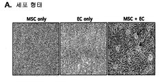
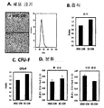
- Figure 2 shows the cell growth changes of mesenchymal stem cells co-cultured with vascular endothelial cells, (A) morphological features of each mesenchymal stem cell; (B) size; And (C) a result of comparing the degree of proliferation.
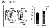
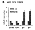
- 3 is a cell growth change of mesenchymal stem cells co-cultured with vascular endothelial cells, (A) cell cycle of each mesenchymal stem cell; And (B) cell cycle regulators.
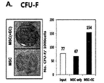
- Figure 4 confirms the improvement of the stem cell capacity of mesenchymal stem cells co-cultured with vascular endothelial cells, (A) CFU-F of each mesenchymal stem cells; (B) Comparison of differentiation into adipose tissue.
- Figure 5 is the size (A) of each of the mesenchymal stem cells between the group cultured in contact with the vascular endothelial cells (EC-CM) and mesenchymal stem cells alone (MSC-CM); (B) cell proliferation; (C) CFU-F; And (D) bone formation or differentiation into adipose tissue.
- EC-CM vascular endothelial cells
- MSC-CM mesenchymal stem cells alone
- B cell proliferation
- C CFU-F
- D bone formation or differentiation into adipose tissue.
- Figure 6 confirms the change in cellular characteristics of mesenchymal stem cells co-cultured with vascular endothelial cells, comparing the expression of genes associated with perivascular characteristics.
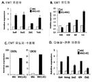
- Figure 7 shows the changes in cellular characteristics of mesenchymal stem cells co-cultured with vascular endothelial cells, each of (A) EMT gene of mesenchymal stem cells; (B) EMC inducing genes; (C) an EMT induction-receptor gene; And pluripotency related genes.
- Figure 8 confirms the effect of improving the niche activity of mesenchymal stem cells co-cultured with vascular endothelial cells
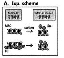
- A schematic schematic diagram of the experimental procedure
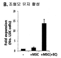
- B hematopoietic stem maintenance activity
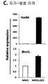
- C a result of comparing the expression of the self-renewal marker.
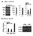
- Figure 9 confirms the effect of improving the niche activity of mesenchymal stem cells co-cultured with vascular endothelial cells, the expression of (A) hematopoietic cytokine of each mesenchymal stem cells; And (B) a result of comparing notch ligand expression.
- Figure 10 confirms the effect of improving the niche activity of mesenchymal stem cells co-cultured with vascular endothelial cells, (A) notch repoter of each of the co-cultured hematopoietic stem cells; And (B) notch target genes.
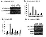
- Figure 11 shows the expression factors of vascular endothelial cells associated with niche activity, (A) b-catenin of co-cultured vascular endothelial cells; (B) b-catenin co-factor; (C) Wnt receptors; And (D) expression of b-catenin regulators.
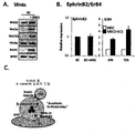
- Figure 12 shows the expression factors of vascular endothelial cells associated with niche activity, (A) Wnt protein expression of co-cultured vascular endothelial cells; (B) results of comparing EphrinB2 and ErB4 expression of cocultured mesenchymal stem cells; And (C) is a schematic diagram showing a mechanism related to hematopoietic stem cell self-renewal according to the present invention.
- Figure 13 is a signal factor that regulates the stimulating effect of vascular endothelial cells, (A) is a result of comparing the expression of Wnt ligand of co-cultured vascular endothelial cells; And (B) notch ligand expression of cocultured mesenchymal stem cells following cytokine treatment.
- Figure 14 is a signal factor that regulates the stimulating effect of vascular endothelial cells
- A is a schematic diagram of retroviral vectors expressing active STAT3 (STAT3-C; SC) and inhibitory STAT3 (dnSTAT3)
- B confirming notch ligand expression of vascular endothelial cells into which activated STAT3 was introduced
- C confirming CFU-F of the vascular endothelial cell co-cultured mesenchymal stem cells
- D confirming the effect of improving the hematopoietic stem cell self-renewal ability of the mesenchymal stem cells.
- FIG. 15 is a schematic diagram showing the mechanism of regulating self-renewal of hematopoietic stem cells according to the present invention.
- Figure 16 shows the effect of promoting neuronal self-renewal by the mixed administration of mesenchymal stem cells and vascular endothelial cells in a spinal cord cutting animal model, the result of analyzing the function of motor neuron Basso Mouse Sacle (BMS) method.
- BMS Basso Mouse Sacle
- Figure 17 shows the effect of promoting neuronal self-renewal by the mixed administration of mesenchymal stem cells and vascular endothelial cells in a spinal cord cutting animal model, the result of analyzing the function of sensory nerves by the somatosensory evoked potential (SEP) method.
- SEP somatosensory evoked potential
- FIG. 18 confirms the neural system self-renewal effect by the mixed administration of mesenchymal stem cells and vascular endothelial cells in a spinal cord cutting animal model, and compares the number of NeuN positive cells per spinal cord injury site.
- Figure 19 shows the neuronal self-renewal effect by the mixed administration of mesenchymal stem cells and vascular endothelial cells in a spinal cord cutting animal model, it is a result of comparing the number of Tuj-1 positive cells for each spinal cord injury site.
- Figure 20 shows the neuronal self-renewal effect by the mixed administration of mesenchymal stem cells and vascular endothelial cells in a spinal cord cutting animal model, a result of comparing the number of nestin-positive cells for each spinal cord injury site.
- 21 is a schematic diagram showing a method for regulating self-renewal ability of hematopoietic or neural stem cells using the technique of the present invention.
- the present invention comprises a composition for promoting self-renewal of adult stem cells, including vascular endothelial cells overexpressing any one or more ligands of Wnt and EphrinB2;
- a method for promoting self-renewal of adult stem cells comprising administering to a subject vascular endothelial cells overexpressing any one or more ligands of Wnt and EphrinB2; And vascular endothelial cells overexpressing any one or more ligands of Wnt and EphrinB2 for promoting self-renewal of adult stem cells.
- self-renewal is also called self-replicating, self-replicating, and self-renewing with the ability to produce cells having the same properties and characteristics, and is one of the important features of stem cells.
- self-renewal in the present invention means the ability to continue proliferation while maintaining an undifferentiated state.
- adult stem cells are multipotency and unipotent stem cells, and can be obtained from skin, blood vessels, muscles, and brain.
- stem cells in adult tissues such as bone marrow are very rare, and these cells are difficult to culture without induction of differentiation, making them difficult to culture without specifically screened media. That is, stem cells have a disadvantage in that it is very difficult to separate and preserve in vitro.
- the adult stem cells may be hematopoietic stem cells or neural stem cells.
- hematopoietic stem cell is an ancestral cell of undifferentiated bone marrow hematopoietic cells that make blood cells, such as red blood cells, white blood cells, platelets, and also called hematopoietic stem cells.
- the term “Neural stem cell (NSC)” refers to undifferentiated stem cells that produce neurons, such as astrocytes, neurons and oligodendrocytes, as primitive cells present in the nervous system.
- NSC Neurological stem cell
- Hematopoietic stem cells or neural stem cells exhibit long-term repopulation with self-renewing capacity during regeneration when transplanted into a host where the bone marrow or nervous system is destroyed, respectively.
- Adult stem cells of the present invention are humans, monkeys, pigs, horses, cows, sheep, dogs, cats, mice ), Including adult stem cells derived from all animals such as rabbits, but are preferably cells derived from humans.
- the niche activity of the mesenchymal stem cells is improved, thereby identifying that the self-renewal ability of adult stem cells can be improved It was.
- the present invention provides a technique for enhancing niche activity of mesenchymal stem cells using vascular endothelial cells overexpressing any one or more ligands of Wnt and EphrinB2, and ultimately, promoting the self-renewal of adult stem cells. have.
- methods known in the art can be used to overexpress Wnt or EphrinB2 ligands in vascular endothelial cells, preferably Wnt ligands, EphrinB2 ligands, activated STAT3 (STAT3-C), and Transfected with a vector comprising any of these combinations or selected from the group consisting of b-FGF, TPO, VEGF, IL-6, IL-10, TNF- ⁇ , and combinations thereof in vascular endothelial cells Either factor can be overexpressed by treatment of vascular endothelial cells.
- the term "vector” refers to an expression vector into which a polynucleotide sequence encoding the above factor is inserted, but is not limited thereto, and preferably refers to a plasmid, virus or other medium known in the art.
- the polynucleotide sequence according to the present invention may be operably linked to an expression control sequence, wherein the operably linked gene sequence and the expression control sequence are one expression vector containing a selection marker and a replication origin together. It may be included within.
- “Operably linked” can be genes and expression control sequences linked in such a way as to enable gene expression when the appropriate molecule is bound to the expression control sequences.
- control sequence is meant a DNA sequence that regulates the expression of a polynucleotide sequence operably linked in a particular host cell.
- control sequence is a promoter for transcription, any for controlling transcription.
- plasmids examples include E. coli-derived plasmids (pBR322, pBR325, pUC118 and pUC119), Bacillus subtilis-derived plasmids (pUB110 and pTP5), and yeast-derived plasmids (YEp13, YEp24 and YCp50).
- Viruses selected from the group consisting of adenoviruses, vaccinia viruses, baculoviruses, herpes simplex viruses, complexes with lentiviruses, and the like can be used.
- Those skilled in the art can use a vector suitable for introducing a gene or polynucleotide of the present invention into a host cell, and preferably a vector designed to facilitate protein expression induction and isolation of the expressed protein.
- vascular endothelial cells overexpressing any one or more ligands of Wnt and EphrinB2 can be provided by targeting mesenchymal stem cells existing in vivo. It may also be provided as a combination of cytokines (b-FGF, TPO, VEGF, IL-6, IL-10, TNF- ⁇ , and combinations thereof) that can enhance the expression of vascular endothelial cells and Wnt or EphrinB2 ligands. Can be.
- mesenchymal stem cells separated in vitro can be added to the two types of compositions, wherein the vascular endothelial cells and mesenchymal stem cells are transplanted while maintaining physical contact It may also be injected into the body after the coculture process, if necessary.
- mesenchymal stem cell is a cell that helps to create cartilage, bone, fat, myeloid epilepsy, muscle, nerves, etc., in adults, but generally stays in the bone marrow, cord blood, It exists in peripheral blood, other tissues, etc., and means the cell which can be obtained from these.
- mesenchymal stem cells are used in the same sense as mesenchymal stromal cells or stromal cells.
- the mesenchymal stem cells are human, monkey, pigs, horses, cows, sheep, dogs, cats, mice It includes all animal-derived cells, such as rabbits, but preferably cells derived from humans.
- Niche refers to a component (cell and / or substance) composed of tissues or organs that support the development and proliferation of tissue cells such as stem cells and other somatic cells, and inter-cell interactions. It is known to secrete factors necessary to induce action and to be omnipotent.
- Niche also called the microenvironment, plays a key role in maintaining the stemness that expresses all the characteristics of stem cells. Stem cells are located in a kind of microenvironment that consists of adhesion molecule growth factors, which are called niches in academia, and these areas of stem cells are located, adhesiveness, and homing. It is responsible for maintaining and coordinating homing, quiescence and activation. In other words, niche is a major microenvironment surrounding stem cells that regulates the differentiation of stem cells and prevents and protects them from moving to other places or suicide.
- the composition for promoting self-renewal of adult stem cells can be used for the treatment of patients in need of transplantation of hematopoietic stem cells.
- acute leukemia chronic leukemia, myelodysplastic syndrome, lymphoma, multiple myeloma, germ cell tumor, breast cancer, ovarian cancer, small cell lung cancer, neuroblastoma, aplastic anemia, erythropathy, Gaucher's disease, Hunter syndrome, ADA enzyme deficiency
- It can be used for transplantation into patients with Wiskot-Aldrich syndrome, rheumatoid arthritis, systemic lupus erythematosus, or multiple sclerosis, or patients whose hematopoietic cells have been damaged due to chemotherapy or irradiation.
- composition for promoting self-renewal of adult stem cells can be used for the treatment of patients in need of transplantation of neural stem cells, specifically stroke, Alzheimer's disease, Parkinson's disease , Huntington's disease, Amyotrophic lateral sclerosis (ALS), Pick's disease, Niiemann pick disease, and spinal cord injury disease It can be used for transplantation into patients suffering from neurological diseases.
- neural stem cells specifically stroke, Alzheimer's disease, Parkinson's disease , Huntington's disease, Amyotrophic lateral sclerosis (ALS), Pick's disease, Niiemann pick disease, and spinal cord injury disease It can be used for transplantation into patients suffering from neurological diseases.
- the effect of the above-described interaction between vascular endothelial cells and mesenchymal stem cells is due to the signaling system of vascular endothelial cells itself, the signal regulation of vascular endothelial cells to amplify the niche activity of mesenchymal stem cells It can be used as a method. Therefore, the present invention has another technical feature in that niche activity of mesenchymal stem cells can be improved through enhancement of vascular endothelial cell factors (Wnt and EphrinB2).
- the present invention comprises the step of co-culture of the vascular endothelial cells and mesenchymal stem cells separated in vitro in physical contact, mesenchymal stem cells having enhanced niche activity It provides a method of manufacturing.
- the method for producing mesenchymal stem cells having enhanced niche activity of the present invention uses the vascular endothelial cells, mesenchymal stem cells, and the like described in the composition for promoting self-renewal of adult stem cells described above, they are common among them. The description is omitted, in order to avoid excessive complexity of the present specification.
- the present invention may further include the step of overexpressing any one of the Wnt ligand, EphrinB2 ligand, activated STAT3 (STAT3-C), and combinations thereof to vascular endothelial cells before the coculture step.
- the step may be to co-culture by adding any one factor selected from the group consisting of b-FGF, TPO, VEGF, IL-6, IL-10, TNF- ⁇ and combinations thereof.
- the overexpressing step may be to introduce a vector comprising any one gene selected from the group consisting of a Wnt ligand, an EphrinB2 ligand, and an activated STAT3 (STAT3-C) into vascular endothelial cells, but is not limited thereto.
- a vector comprising any one gene selected from the group consisting of a Wnt ligand, an EphrinB2 ligand, and an activated STAT3 (STAT3-C) into vascular endothelial cells, but is not limited thereto.
- the vector according to the present invention may be introduced into a cell using a method known in the art, but the method of introducing the recombinant vector according to the present invention into a host cell is not limited thereto, but calcium chloride (CaCl 2 ) Heat shock methods, silicon carbide whiskers, sonication, precipitation by polyethylenlycol (PEG), transient transfection, microinjection, transduction, Cell fusion, calcium phosphate precipitation, liposome-mediated transfection, DEAE Dextran-mediated transfection, polybrene-mediated transfection, Electroporation, gene gun methods, etc. may be used, and may be introduced into cells by other known methods for introducing nucleic acids into cells.
- CaCl 2 calcium chloride
- PEG polyethylenlycol
- the effect of the above-described interaction between the vascular endothelial cells and the mesenchymal stem cells is under the control of the signaling system of the vascular endothelial cell itself, and the signal regulation on the vascular endothelial cells regulates niche activity of the mesenchymal stem cells. It was found that it can be used as a way to. Accordingly, the present invention has another technical feature in that the niche activity of mesenchymal stem cells can be adjusted up or down through the regulation of vascular endothelial cell factors (Wnt and EphrinB2).
- Wnt and EphrinB2 vascular endothelial cell factors
- the present invention includes the step of overexpressing or underexpressing any one or more ligands of Wnt and EphrinB2 in co-cultured vascular endothelial cells in physical contact with mesenchymal stem cells isolated in vitro, Provided is a method for regulating niche activity of mesenchymal stem cells.
- the method for regulating niche activity of mesenchymal stem cells of the present invention uses vascular endothelial cells, mesenchymal stem cells, and the like described in the composition for promoting self-renewal of adult stem cells described above, the common contents among them are In order to avoid excessive complexity of the specification, the description thereof is omitted.
- the step of overexpressing transfected vascular endothelial cells with a vector comprising any one of Wnt ligand, EphrinB2 ligand, activated STAT3 (STAT3-C), and a combination thereof, or b -FGF, TPO, VEGF, IL-6, IL-10, TNF- ⁇ , and combinations thereof may be treated with a factor selected from the group.
- the step of low expression may be to treat a factor selected from the group consisting of Tgfb3, IFN- ⁇ , and combinations thereof to vascular endothelial cells.
- vascular endothelial cells In order to confirm the effects of vascular endothelial cells on the stem cell capacity of mesenchymal stem cells, the two cells were co-cultured in vitro and analyzed for the change accordingly.
- GFP was introduced into only one of the two cells.
- vascular endothelial cells In order to make the conditions similar to the three-dimensional interaction of the two cells in vivo, vascular endothelial cells were laid on the floor, and then mesenchymal stem cells were laid on them. Through this coculture method, direct cell-contact with vascular endothelial cells and the effect of soluble factors (design factor) was designed to affect the mesenchymal stem cells (Fig. 1).
- mesenchymal stem cells by interaction with vascular endothelial cells were examined, and the comparison group was used as a mesenchymal stem cell culture group.
- Mesenchymal stem cells co-cultured with vascular endothelial cells unlike single cultures, grew in a sphere-like form on vascular endothelial cells, the size of cells decreased, and cell proliferation was significantly reduced (FIG. 2A-2C).
- mesenchymal stem cells co-cultured with vascular endothelial cells exhibited higher G0 arrest and inhibited overall cell cycle progression compared to the case of monoculture, thus maintaining a quienscence state (FIG. 3A).
- G0-G1 progression-related factors such as cyclinD1 and E were reduced, and inhibitory factors such as p21 and p27 were increased (FIG. 3B).
- vascular endothelial cells In addition, in order to analyze the interaction mechanism on the effects of vascular endothelial cells, only the conditioned medium (EC-CM) of vascular endothelial cells treated alone (cultured in a non-contact state with vascular endothelial cells) and When mesenchymal stem cells were cultured alone (MSC-CM) was compared. As a result, there was no significant difference in the size, cell growth, colony forming ability, and differentiation capacity of the mesenchymal stem cells between them. The stem cell ability was found to be a phenomenon caused by direct contact between the two cells. (FIGS. 5A-5D).
- the comparison group was used in mesenchymal stem cell culture alone.
- mesenchymal stem cells co-cultured with vascular endothelial cells the expression of nestin, NG2, etc. was increased in perivascular cells or pericyte markers adjacent to vascular endothelial cells in vivo, but ⁇ -SMA decreased, indicating the characteristics of arterial perivascular cells.
- EMT markers Sox9, Snail1, Zeb1, Twist, etc.
- mesenchymal characteristics were enhanced with respect to the reprogramming of high functional stem cell ability (FIG. 7A).
- cytokines capable of inducing EMT is increased in vascular endothelial cells co-cultured with mesenchymal stem cells, and the expression of IL-8 receptors (CXCR1, 2) is rapidly increased in mesenchymal stem cells.
- Undifferentiated hematopoietic stem cells Lin compared to mesenchymal stem cells cultured alone were isolated from co-culture with hematopoietic stem cells (Lin-cells) isolated from bone marrow after separation of mesenchymal stem cells changed by co-culture with vascular endothelial cells. Promoting the proliferation of -Sca + Kit + (LSK cells), the expression of factors (HoxB4, Bmi1) associated with the self-renewal of hematopoietic stem cells was also increased to confirm that the niche activity was improved (Figs. 8A to 8C).
- cytokine IL-6, IL-10, TPO, VEGF
- IL-6, IL-10, TPO, VEGF cytokine
- VEGF vascular endothelial growth factor
- CBF-reporter (repoter that monitors the function of CBF-1 (RBPJ-kappa) in combination with NICD when notch signaling occurs) was analyzed and co-cultured with mesenchymal stem cells changed by vascular endothelial cells. Notch signaling was increased in hematopoietic stem cells, and expression of notch target genes (Hes1, 5, Hey1, Deltex1) was increased in hematopoietic stem cells, suggesting improved niche activity of mesenchymal stem cells. It was confirmed again (FIGS. 10A and 10B).
- the function and related mediators of vascular endothelial cells that stimulate mesenchymal stem cells were identified.
- an up-stream signaling system regulating the related activity of vascular endothelial cells was analyzed.
- the expression of Wnt ligands in vascular endothelial cells, in which vascular endothelial cells were found to be mediated by stimulating mesenchymal stem cells was increased by bFGF, VEGF, TPO, IL-6, and IL-10.
- VEGF, IL-6, IL-10 and TNF-alpha significantly increased the expression of notched ligands jagged-1 and Dll-1 in mesenchymal stem cells (FIGS. 13A and 13B).
- STAT3 acts as a major factor in the activation of vascular endothelial cells by the cytokine.
- the active STAT3 STAT3-C; SC, Bromberg JF, Wrzeszczynska MH, Devgan G, et al. Stat3 as an oncogene. Cell.1999; sequence disclosed in 98: 295-303
- the inhibitory STAT3 dnSTAT3
- Fig. 14A After constructing with a retroviral vector expressing (Fig. 14A), they were introduced into vascular endothelial cells and expressed.
- mesenchymal stem cells (Lin + MSC (+ MEC)) in contact with vascular endothelial cells induced expansion of much higher hematopoietic stem cells (Lin-Sca-1 + c-kit +; LSK), in particular STAT3-C Mesenchymal stem cells (Lin + MSC (+ SEC)) in contact with activated vascular endothelial cells (SEC) showed even higher hematopoietic stem cell expansion capacity (FIG. 14D).
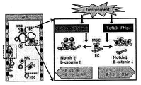
- cytokines such as IL-6 and IL-10 activate vascular endothelial cells (STAT3, Wnt ligand, EhrineB2 ligand), and mesenchymal stem cells in contact with the activated vascular endothelial cells have enhanced niche activity. (b-catenin, notch), it was confirmed that induces self-renewal and expansion of undifferentiated hematopoietic stem cells (Fig. 15). Accordingly, the present invention has been found that by regulating the activity of vascular endothelial cells and expression of related factors, the niche activity of mesenchymal stem cells and the self-renewal ability of hematopoietic stem cells can be increased or decreased.
- each cell (2D-MSC, 3D MSC + EC) was mixed with a matrigel and administered to the injured area, and then the recovery of spinal cord function was performed through a locomoter rating scale. Comparative analysis.
- BMS Basso Mouse Sacle
- the mesenchymal stem cell-treated group (2D-MSC) showed a higher recovery of the motor performance than the single matrigel-only group.
- the mesenchymal stem cells and vascular endothelial cells were mixed together and then administered (3D MSC + EC).
- SEP somatosensory evoked potential
- the spinal cord tissue in the regeneration process was analyzed by immunofluorescence staining, and as a comparison group, the group receiving the simple cultured mesenchymal stem cells (2D-MSC), the group receiving the three-dimensional cultured mesenchymal stem cells ( 3D-MSC) was used.
- 2D-MSC simple cultured mesenchymal stem cells
- 3D-MSC three-dimensional cultured mesenchymal stem cells
Landscapes
- Health & Medical Sciences (AREA)
- Life Sciences & Earth Sciences (AREA)
- Engineering & Computer Science (AREA)
- Biomedical Technology (AREA)
- Zoology (AREA)
- Chemical & Material Sciences (AREA)
- Biotechnology (AREA)
- Bioinformatics & Cheminformatics (AREA)
- Cell Biology (AREA)
- General Health & Medical Sciences (AREA)
- Organic Chemistry (AREA)
- Wood Science & Technology (AREA)
- Genetics & Genomics (AREA)
- Immunology (AREA)
- Developmental Biology & Embryology (AREA)
- Animal Behavior & Ethology (AREA)
- Public Health (AREA)
- Veterinary Medicine (AREA)
- Medicinal Chemistry (AREA)
- Epidemiology (AREA)
- Pharmacology & Pharmacy (AREA)
- General Engineering & Computer Science (AREA)
- Microbiology (AREA)
- Biochemistry (AREA)
- Vascular Medicine (AREA)
- Virology (AREA)
- Rheumatology (AREA)
- Hematology (AREA)
- Gastroenterology & Hepatology (AREA)
- Proteomics, Peptides & Aminoacids (AREA)
- Micro-Organisms Or Cultivation Processes Thereof (AREA)
- Medicines Containing Material From Animals Or Micro-Organisms (AREA)
Abstract
본 발명에서는, 혈관 내피세포와 중간엽 줄기세포를 공배양시킨 경우, 중간엽 줄기세포의 니치 활성, 및 조혈 또는 신경 줄기세포의 자가 재생능이 향상되며, 특히, 중간엽 줄기세포에 대한 혈관 내피세포의 자극을 조절함 (사이토카인 등)으로써, 상기 니치 활성 및 조혈 또는 신경 줄기세포의 자가 재생능을 손쉽게 조절할 수 있음을 확인하였다. 따라서, 본 발명은 줄기세포의 자가재생능을 향상시킬 수 있을 뿐만 아니라, 필요에 따라 이를 손쉽게 조절할 수 있는바, 중간엽 줄기세포-기반 세포 치료의 효용성을 확장시킬 수 있을 것이라 기대된다.
Description
본 발명은 혈관 내피세포와 중간엽 기질세포의 상호작용을 통한 줄기세포 재생 증진기술에 관한 것이다.
21세기의 생명공학은 인간복지를 최종목표로 식량, 환경, 건강 문제에 새로운 해결책의 가능성을 제시하고 있으며, 최근 줄기세포의 기술은 난치병 치료의 새로운 기술로 떠오르고 있다. 이전까지는 인간의 난치병 치료를 위해 장기이식이나 유전자 치료 등이 제시되었으나, 면역거부와 공급 장기 부족, 벡터 개발이나 질환 유전자에 대한 지식 부족으로 효율적인 실용화가 미진하였다.
이에, 증식과 분화를 통해 모든 기관을 형성할 능력을 가진 만능 줄기세포에 대한 관심이 고조되어, 줄기세포가 대부분의 질병 치료는 물론 장기 훼손을 근원적으로 해결할 수 있는 것으로 인식되고 있으며, 많은 과학자가 인체의 거의 모든 장기 재생은 물론 난치병이었던 파킨슨병, 각종 암, 당뇨병과 척수손상 등의 치료에 이르기까지 다양하게 줄기세포의 적용 가능성을 제시해 왔다.
줄기세포(stem cell)는 생물 조직을 구성하는 다양한 세포들로 분화할 수 있는 세포로서 배아, 태아 및 성체의 각 조직에서 얻을 수 있는 분화(differentiation)되기 전 단계의 미분화 세포들을 총칭한다. 여러 가지 줄기세포 분류 중 만능줄기세포(pluripotent stem cell)라 함은 생체를 구성하는 3가지 배엽(germ layer) 모두로 분화할 수 있는 다기능성을 지닌 줄기세포를 지칭한다. 줄기세포를 분류함에 있어, 해부학적인 존재부위, 세포의 기능, 세포표면에 표현된 항원의 종류, 전사인자, 세포가 생성하는 단백질 또는 그 줄기세포가 만들어 낼 수 있는 특정 세포 종류에 따라 분류할 수 있는데, 이러한 여러 가지 분류 중 가장 흔히 이용되고 비교적 명확한 분류는 줄기세포가 분리된 개체에 따른 것이다.
배아(embryo)에서 분리된 경우 배아 줄기세포(embryonic stem cell, ES cell)와 성체에서 분리된 경우 성체 줄기세포(adult stem cell)로 나눌 수 있다. 또 다른 흔한 분류는 하나의 줄기세포로부터 몇 종류의 분화된 세포를 만들어 낼 수 있는가에 따라 만능(pluripotency), 다분화능(multipotency) 및 단분화능(unipotent) 줄기세포로 나눌 수 있으며, 일반적으로 배아줄기세포(embryonic stem cell, ES cell)는 만능줄기세포로, 성체줄기세포(adult stem cell)는 다분화능(multipotency) 및 단분화능(unipotent) 줄기세포로 구분할 수 있다. 수정 후 발생 초기인 배반포기(blastocyt)의 내부세포덩어리(세포내괴, inner cell mass)는 장차 태아를 형성할 부분으로서 이 내부세포덩어리로부터 형성된 배아줄기세포(embryonic stem cell, ES cell)는 한 개체를 구성하는 모든 조직의 세포로 분화될 수 있는 잠재력을 지닌 만능줄기세포(pluripotent stem cell)라 할 수 있다. 즉, 배아줄기세포는 무제한적으로 증식이 가능한 미분화 세포이고, 모든 세포로 분화할 수 있으며 성체줄기세포와 달리 배(germ) 세포도 만들 수 있어 다음 세대로 유전될 수 있다.
다능성 줄기세포는 성체 골수에서 최초로 분리되었고, 그 후 다른 여러 성체 조직에서도 확인되었다. 다시 말해, 골수는 가장 널리 알려진 줄기세포의 원천이지만, 다능성 줄기세포는 피부, 혈관, 근육 및 뇌로부터도 획득할 수 있다. 그러나 골수와 같은 성체 조직 내의 줄기세포는 매우 드물게 존재하고, 이러한 세포들은 분화유도 하지 않고 배양하기 어려워서, 특이적으로 스크리닝되어 있는 배지들이 없으면 그 세포들을 배양하기 어렵다. 즉, 줄기세포들은 분리하여 체외에서 보존하기가 매우 어렵다는 단점이 있다. 따라서 최근 이러한 인간 줄기세포의 동정, 증식 및 분화에 대한 관심이 크다.
또한, 조직 공학, 유전자 치료 및 세포 치료제에서의 줄기세포의 적용이 급속도로 진행되면서 현재 당업계에서는 줄기세포에 대한 관심이 폭발적으로 증대되고 있는바, 다양한 조직으로부터 획득한 줄기세포의 개발 및 보다 안정되면서도 뛰어난 증식효과를 나타낼 수 있는 줄기세포의 증식 방법 및 여러 세포 또는 조직으로의 안정된 분화 방법에 대한 기술이 절실히 요구되고 있는 상황이다 (한국 공개특허 10-2012-00111301).
본 발명은 상기와 같은 문제점을 해결하기 위해 안출된 것으로서, 본 발명자들은 중간엽 기질세포-기반 세포 치료의 유효성를 향상시키기 위하여 예의 노력한 결과, 혈관 내피세포와 중간엽 줄기세포를 공배양시킨 경우, 중간엽 줄기세포의 니치 활성, 및 조혈 또는 신경 줄기세포의 자가 재생능이 향상되며, 특히, 중간엽 줄기세포에 대한 혈관 내피세포의 자극을 조절함 (사이토카인 등)으로써, 상기 니치 활성을 손쉽게 조절할 수 있음을 확인하고, 이에 기초하여 본 발명을 완성하였다.
본 발명의 목적은 혈관 내피세포를 이용한 성체 줄기세포의 자가재생 촉진용 조성물을 제공하는데 있다.
본 발명의 다른 목적은 체외로 분리된, 혈관 내피세포와 중간엽 줄기세포를 물리적으로 접촉되어 있는 상태로 공배양하는 단계를 포함하는, 니치 활성이 증진된 중간엽 줄기세포의 제조방법을 제공하는데 있다.
본 발명의 또 다른 목적은 혈관 내피세포를 이용한 중간엽 줄기세포의 니치 활성 조절방법을 제공하는데 있다.
그러나 본 발명이 이루고자 하는 기술적 과제는 이상에서 언급한 과제에 제한되지 않으며, 언급되지 않은 또 다른 과제들은 아래의 기재로부터 당업자에게 명확하게 이해될 수 있을 것이다.
상기와 같은 본 발명의 목적을 달성하기 위하여, 본 발명은 Wnt 및 EphrinB2 중 어느 하나 이상의 리간드를 과발현하는 혈관 내피세포를 포함하는, 성체 줄기세포의 자가재생 촉진용 조성물을 제공한다.
본 발명의 일 구현예로서, 상기 혈관 내피세포는 STAT3 활성이 증가되어 있는 것일 수 있다.
본 발명의 다른 구현예로서, 상기 혈관 내피세포는 Wnt 리간드, EphrinB2 리간드, 활성화형 STAT3 (STAT3-C), 및 이들의 조합 중 어느 하나를 포함하는 벡터로 형질감염된 것일 수 있고, b-FGF, TPO, VEGF, IL-6, IL-10, TNF-α, 및 이들의 조합으로 이루어진 군으로부터 선택되는 어느 하나 인자가 처리된 것일 수 있다.
본 발명의 또 다른 구현예로서, 상기 조성물은 체외로 분리된, 중간엽 줄기세포를 추가로 포함하며, 상기 중간엽 줄기세포는 혈관 내피세포와 물리적으로 접촉된 상태로 제공되는 것일 수 있다.
또한, 본 발명은 혈관 내피세포; 및 b-FGF, TPO, VEGF, IL-6, IL-10, TNF-α, 및 이들의 조합으로 이루어진 군으로부터 선택되는 어느 하나 인자를 포함하는 성체 줄기세포의 자가재생 촉진용 조성물을 제공한다.
본 발명의 일 구현예로서, 상기 조성물은 체외로 분리된, 중간엽 줄기세포를 추가로 포함하며, 상기 중간엽 줄기세포는 혈관 내피세포와 물리적으로 접촉된 상태로 제공되는 것일 수 있다.
또한, 체외로 분리된, 혈관 내피세포와 중간엽 줄기세포를 물리적으로 접촉되어 있는 상태로 공배양하는 단계를 포함하는, 니치 활성이 증진된 중간엽 줄기세포의 제조방법을 제공한다.
본 발명의 일 구현예로서, 상기 공배양하는 단계 전, 혈관 내피세포에 Wnt 리간드, EphrinB2 리간드, 활성화형 STAT3 (STAT3-C), 및 이들의 조합 중 어느 하나를 과발현시키는 단계를 추가로 포함할 수 있다.
본 발명의 다른 구현예로서, 상기 공배양하는 단계는 b-FGF, TPO, VEGF, IL-6, IL-10, TNF-α 및 이들의 조합으로 이루어진 군으로부터 선택되는 어느 하나 인자를 첨가하여 공배양하는 것일 수 있다.
또한, 본 발명은 체외로 분리된, 중간엽 줄기세포와 물리적으로 접촉되어 있는 상태로 공배양되는, 혈관 내피세포 내 Wnt 및 EphrinB2 중 어느 하나 이상의 리간드를 과발현 또는 저발현시키는 단계를 포함하는, 중간엽 줄기세포의 니치 활성 조절방법을 제공한다.
본 발명의 일 구현예로서, 상기 과발현시키는 단계는, 혈관 내피세포를 Wnt 리간드, EphrinB2 리간드, 활성화형 STAT3 (STAT3-C), 및 이들의 조합 중 어느 하나를 포함하는 벡터로 형질감염시키거나, 혈관 내피세포에 b-FGF, TPO, VEGF, IL-6, IL-10, TNF-α 및 이들의 조합으로 이루어진 군으로부터 선택되는 인자를 처리하는 것일 수 있다.
본 발명의 다른 구현예로서, 상기 저발현시키는 단계는 혈관 내피세포에 Tgfb3, IFN-γ 및 이들의 조합으로 이루어진 군으로부터 선택되는 인자를 처리하는 것일 수 있다.
한편, 본 발명에서, 상기 중간엽 줄기세포는 지방조직, 골수, 말초 혈액 또는 제대혈에서 유래한 것일 수 있고, 상기 성체 줄기세포는 조혈 줄기세포 또는 신경 줄기세포일 수 있다.
본 발명에서는, 혈관 내피세포와 중간엽 줄기세포를 공배양시킨 경우, 중간엽 줄기세포의 니치 활성, 및 조혈 또는 신경 줄기세포의 자가 재생능이 향상되며, 특히, 중간엽 줄기세포에 대한 혈관 내피세포의 자극을 조절함 (사이토카인 등)으로써, 상기 니치 활성 및 조혈 또는 신경 줄기세포의 자가 재생능을 손쉽게 조절할 수 있음을 확인하였다. 따라서, 본 발명은 줄기세포의 자가재생능을 향상시킬 수 있을 뿐만 아니라, 필요에 따라 이를 손쉽게 조절할 수 있는바, 중간엽 줄기세포-기반 세포 치료의 효용성을 확장시킬 수 있을 것이라 기대된다.
도 1은 혈관 내피세포와의 공배양에 따른 중간엽 줄기세포의 특성 변화를 확인하기 위한 실험과정을 개략적으로 나타낸 모식도이다.
도 2는 혈관 내피세포와의 공배양된 중간엽 줄기세포의 세포 성장 변화를 확인한 것으로서, 중간엽 줄기세포 각각의 (A) 형태적 특징; (B) 크기; 및 (C) 증식 정도를 비교한 결과이다.
도 3은 혈관 내피세포와의 공배양된 중간엽 줄기세포의 세포 성장 변화를 확인한 것으로서, 중간엽 줄기세포 각각의 (A) 세포 주기; 및 (B) 세포 주기 조절 인자를 비교한 결과이다.
도 4는 혈관 내피세포와의 공배양된 중간엽 줄기세포의 줄기세포능 향상을 확인한 것으로서, 중간엽 줄기세포 각각의 (A) CFU-F; (B) 지방조직으로의 분화를 비교한 결과이다.
도 5는 혈관 내피세포와 비접촉되어 있는 상태로 배양된 군(EC-CM)과 중간엽 줄기세포를 단독배양한 군(MSC-CM)간 중간엽 줄기세포 각각의 (A) 크기; (B) 세포 증식; (C) CFU-F; 및 (D) 골 형성 또는 지방조직으로의 분화를 비교한 결과이다.
도 6은 혈관 내피세포와의 공배양된 중간엽 줄기세포의 세포 특성 변화를 확인한 것으로서, 혈관주위(perivascular) 특성과 관련된 유전자의 발현을 비교한 결과이다.
도 7은 혈관 내피세포와의 공배양된 중간엽 줄기세포의 세포 특성 변화를 확인한 것으로서, 중간엽 줄기세포 각각의 (A) EMT 유전자; (B) EMC 유도 유전자; (C) EMT 유도-수용체 유전자; 및 다능성 관련 유전자를 비교한 결과이다.
도 8은 혈관 내피세포와의 공배양된 중간엽 줄기세포의 니치 활성 향상 효과를 확인한 것으로서, (A) 실험과정의 개략적인 모식도; (B) 조혈모 유지 활성 (LSK 세포); 및 (C) 자가재생 마커의 발현을 비교한 결과이다.
도 9는 혈관 내피세포와의 공배양된 중간엽 줄기세포의 니치 활성 향상 효과를 확인한 것으로서, 중간엽 줄기세포 각각의 (A) 조혈모 사이토카인의 발현; 및 (B) notch 리간드의 발현을 비교한 결과이다.
도 10은 혈관 내피세포와의 공배양된 중간엽 줄기세포의 니치 활성 향상 효과를 확인한 것으로서, 공배양된 조혈 줄기세포 각각의 (A) notch repoter; 및 (B) notch 타겟 유전자의 발현을 비교한 결과이다.
도 11은 니치 활성과 관련된 혈관 내피세포의 발현인자를 확인한 것으로서, 공배양된 혈관 내피세포의 (A) b-catenin; (B) b-catenin co-factor; (C) Wnt 수용체; 및 (D) b-catenin 조절자의 발현을 비교한 결과이다.
도 12는 니치 활성과 관련된 혈관 내피세포의 발현인자를 확인한 것으로서, (A) 공배양된 혈관 내피세포의 Wnt 단백질 발현; (B) 공배양된 중간엽 줄기세포의 EphrinB2 및 ErB4 발현을 비교한 결과이고; 및 (C) 본 발명에 따른 조혈 줄기세포 자가재생 관련 메커니즘을 개략적으로 나타낸 모식도이다.
도 13은 혈관 내피세포의 자극 효과를 조절하는 신호 인자 규명한 것으로서, (A) 공배양된 혈관 내피세포의 Wnt 리간드 발현을 비교한 결과이고; 및 (B) 사이토카인 처리에 따른 공배양된 중간엽 줄기세포의 notch 리간드 발현을 비교한 결과이다.
도 14는 혈관 내피세포의 자극 효과를 조절하는 신호 인자 규명한 것으로서, (A) 활성화형 STAT3 (STAT3-C; SC)와 억제형 STAT3 (dnSTAT3)을 발현하는 레트로바이러스 벡터의 모식도이고; (B) 활성화형 STAT3가 도입된 혈관 내피세포의 notch 리간드 발현을 확인한 결과이고; (C) 상기 혈관 내피세포 공배양된 중간엽 줄기세포의 CFU-F를 확인한 결과이고; 및 (D) 상기 중간엽 줄기세포의 조혈 줄기세포 자가재생능 향상 효과를 확인한 결과이다.
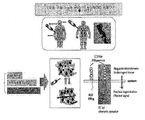
도 15는 본 발명에 따른 조혈 줄기세포의 자가 재생능 조절 기전을 개략적으로 나타낸 모식도이다.
도 16은 척수 절단 동물모델을 대상으로 중간엽 줄기세포 및 혈관 내피세포의 혼합 투여에 의한 신경계 자가재생 촉진 효과를 확인한 것으로서, 운동 신경의 기능을 Basso Mouse Sacle(BMS) 방법으로 분석한 결과이다.
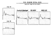
도 17은 척수 절단 동물모델을 대상으로 중간엽 줄기세포 및 혈관 내피세포의 혼합 투여에 의한 신경계 자가재생 촉진 효과를 확인한 것으로서, 감각 신경의 기능을 somatosensory evoked potential (SEP) 방법으로 분석한 결과이다.
도 18은 척수 절단 동물모델을 대상으로 중간엽 줄기세포 및 혈관 내피세포의 혼합 투여에 의한 신경계 자가재생 효과를 확인한 것으로서, 척수절단손상 부위별 NeuN 양성 세포 수를 비교한 결과이다.
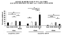
도 19는 척수 절단 동물모델을 대상으로 중간엽 줄기세포 및 혈관 내피세포의 혼합 투여에 의한 신경계 자가재생 효과를 확인한 것으로서, 척수절단손상 부위별 Tuj-1 양성 세포 수를 비교한 결과이다.
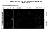
도 20은 척수 절단 동물모델을 대상으로 중간엽 줄기세포 및 혈관 내피세포의 혼합 투여에 의한 신경계 자가재생 효과를 확인한 것으로서, 척수절단손상 부위별 nestin 양성 세포 수를 비교한 결과이다.
도 21은 본 발명의 기술을 이용한 조혈 또는 신경 줄기세포의 자가재생능 조절 방법을 개략적으로 나타낸 모식도이다.
이하, 본 발명을 상세히 설명하기로 한다.
본 발명은, Wnt 및 EphrinB2 중 어느 하나 이상의 리간드를 과발현하는 혈관 내피세포를 포함하는, 성체 줄기세포의 자가재생 촉진용 조성물; Wnt 및 EphrinB2 중 어느 하나 이상의 리간드를 과발현하는 혈관 내피세포를 개체에 투여하는 단계를 포함하는 성체 줄기세포의 자가재생 촉진방법; 및 성체 줄기세포의 자가재생 촉진을 위한 Wnt 및 EphrinB2 중 어느 하나 이상의 리간드를 과발현하는 혈관 내피세포의 용도를 제공한다.
본 발명에서 사용되는 용어, "자가 재생 (self-renewal)"은 동일한 성상 및 특징을 갖는 세포를 생산해내는 능력으로 자가복제, 자기복제, 자가재생이라고도 하며, 줄기세포가 가지는 중요한 특징의 하나이다. 특히, 본원 발명에서 자가 재생이란 분화되지 않은 상태를 유지하면서 증식을 계속하는 능력을 의미한다.
본 발명에서 사용되는 용어, 성체 줄기세포는 다분화능(multipotency) 및 단분화능(unipotent) 줄기세포로서, 피부, 혈관, 근육 및 뇌로부터도 획득할 수 있다. 그러나 골수와 같은 성체 조직 내의 줄기세포는 매우 드물게 존재하고, 이러한 세포들은 분화유도 하지 않고 배양하기 어려워서, 특이적으로 스크리닝되어 있는 배지들이 없으면 그 세포들을 배양하기 어렵다. 즉, 줄기세포들은 분리하여 체외에서 보존하기가 매우 어렵다는 단점이 있다. 한편, 본 발명의 목적상, 상기 성체 줄기세포는 조혈 줄기세포 또는 신경 줄기세포일 수 있다.
본 발명에서 사용되는 용어, "조혈 줄기세포(hematopoietic stem cell: HSC)"는 적혈구, 백혈구, 혈소판 등의 혈액세포를 만드는 미분화된 골수조혈세포의 조상세포로서, 조혈모 세포라고도 한다. 용어, "신경 줄기세포(Neural stem cell; NSC)"는 신경계에 존재하는 원시세포로서 아스트로사이트 (astrocyte), 뉴런 (neuron) 및 올리고덴드로사이트 (oligodendrocyte) 등의 신경세포를 만드는 미분화된 줄기세포를 말한다. 조혈 줄기세포 또는 신경 줄기세포는 각각 골수 또는 신경계가 파괴된 숙주에 이식하였을 때 재생하는 동안 자가재생능 (self-renewing capacity)을 가지고 장기간 재구성능력(repopulation)을 보여준다. 본 발명의 성체 줄기세포는 인간(human), 원숭이(monkey), 돼지(pigs), 말(horses), 소(cows), 양(sheep), 개(dogs), 고양이(cats), 생쥐(mice), 토끼(rats) 등의 모든 동물 유래의 성체줄기세포를 포함하나, 바람직하게는 인간 유래의 세포이다.
본 발명에서, 혈관 내피세포와 중간엽 줄기세포를 물리적으로 접촉된 상태로 공배양시킴으로써, 중간엽 줄기세포의 니치 활성이 향상되고, 이에 따라 성체 줄기세포의 자가 재생능을 향상시킬 수 있음을 규명하였다. 특히, 혈관 내피세포와 중간엽 줄기세포간 상호 작용에 의한 효과는, 혈관 내피세포의 Wnt 리간드, EphrinB2 리간드에 의해 매개됨을 실험적으로 확인하였다. 따라서, 본 발명은 Wnt 및 EphrinB2 중 어느 하나 이상의 리간드를 과발현하는 혈관 내피세포를 이용한 중간엽 줄기세포의 니치 활성 증진 기술, 궁극적으로, 성체 줄기세포의 자가재생 촉진 기술을 제공한다는 점에서 기술적 특징이 있다.
본 발명에서는, 당업계의 공지된 방법을 사용하여, 혈관 내피세포에 Wnt 또는 EphrinB2 리간드를 과발현시킬 수 있으며, 바람직하게 혈관 내피세포에 Wnt 리간드, EphrinB2 리간드, 활성화형 STAT3 (STAT3-C), 및 이들의 조합 중 어느 하나를 포함하는 벡터로 형질감염시키거나, 혈관 내피세포에 b-FGF, TPO, VEGF, IL-6, IL-10, TNF-α, 및 이들의 조합으로 이루어진 군으로부터 선택되는 어느 하나 인자를 혈관 내피세포에 처리하여 과발현시킬 수 있다.
본 발명에서 사용되는 용어, 벡터는 상기 인자를 코딩하는 폴리뉴클레오티드 서열이 삽입된 발현 벡터를 말하며 이에 한정되지 않지만 바람직하게는 당업계에 공지된 플라스미드, 바이러스 또는 기타 매개체를 의미한다. 본 발명에 따른 폴리뉴클레오티드 서열은 발현 조절 서열에 작동 가능하게 연결될 수 있으며, 상기 작동 가능하게 연결된 유전자 서열과 발현 조절 서열은 선택 마커 및 복제 개시점(replication origin)을 같이 포함하고 있는 하나의 발현 벡터 내에 포함될 수 있다. "작동 가능하게 연결(operably linked)"된다는 것은 적절한 분자가 발현 조절 서열에 결합될 때 유전자 발현을 가능하게 하는 방식으로 연결된 유전자 및 발현 조절 서열일 수 있다. 상기 발현 조절 서열(expression control sequence)"이란 특정한 숙주 세포에서 작동 가능하게 연결된 폴리뉴클레오티드 서열의 발현을 조절하는 DNA 서열을 의미한다. 그러한 조절 서열은 전사를 실시하기 위한 프로모터, 전사를 조절하기 위한 임의의 오퍼레이터 서열, 적합한 mRNA 리보좀 결합 부위를 코딩하는 서열 및 전사 및 해독의 종결을 조절하는 서열을 포함한다.
상기 플라스미드의 예로는 대장균 유래 플라스미드(pBR322, pBR325, pUC118 및 pUC119), 바실러스 서브틸러스 유래 플라스미드(pUB110 및 pTP5) 및 효모 유래 플라스미드(YEp13, YEp24 및 YCp50) 등이 있으며, 상기 바이러스는 레트로바이러스, 아데노바이러스, 백시니아 바이러스, 배큘로바이러스, 헤르페스 심플렉스 바이러스 및 렌티바이러스와의 복합체 등으로 구성된 군으로부터 선택된 바이러스가 사용될 수 있다. 당업자라면 본 발명의 유전자 또는 폴리뉴클레오티드를 숙주세포에 도입시키는데 적합한 벡터를 사용할 수 있으며 바람직하게는 단백질 발현 유도 및 발현된 단백질의 분리가 용이하도록 디자인된 벡터를 사용할 수 있다.
한편, 본 발명은 상기와 동일한 기능을 수행할 수 있는 범위 내에서 변형되어 적용될 수 있다. 우선, 전술한 바와 같이, 생체 내 존재하는 중간엽 줄기세포를 타겟으로 하여, 전술한 바와 같이 Wnt 및 EphrinB2 중 어느 하나 이상의 리간드를 과발현하는 혈관 내피세포만을 제공할 수 있다. 또한, 혈관 내피세포와 Wnt 또는 EphrinB2 리간드의 발현을 증진시킬 수 있는 사이토카인 (b-FGF, TPO, VEGF, IL-6, IL-10, TNF-α, 및 이들의 조합)의 조합으로 제공될 수 있다. 이 뿐만 아니라, 상기 2 가지 형태의 조성물에 체외로 분리된 중간엽 줄기세포를 첨가하여 제공할 수 있으며, 이 때, 상기 혈관 내피세포와 중간엽 줄기세포는 물리적으로 접촉된 상태를 유지한 채로 이식될 수 있고, 또한, 필요에 따라 공배양 과정 후 체내 주입될 수도 있다.
본 발명에서 사용되는 용어, 중간엽 줄기세포 (mesenchymal stem cell; MSC)는 연골, 뼈, 지방, 골수간질, 근육, 신경 등을 만드는데 원조가 되는 세포로서, 성인에서는 일반적으로 골수에 머물러 있지만 제대혈, 말초혈액, 기타 조직 등에도 존재하며, 이들로부터 수득할 수 있는 세포를 의미한다. 본 명세서에서는, 중간엽 줄기세포는 중간엽 기질세포 또는 기질세포와 같은 의미로 사용된다. 상기 중간엽 줄기세포는 인간(human), 원숭이(monkey), 돼지(pigs), 말(horses), 소(cows), 양(sheep), 개(dogs), 고양이(cats), 생쥐(mice), 토끼(rats) 등의 모든 동물 유래의 세포를 포함하나, 바람직하게는 인간 유래의 세포일 수 있다.
본 발명에서 사용되는 용어, 니치(Niche)는 줄기세포 및 그 외 체세포와 같은 조직세포의 발달 및 증식을 지원하는 조직 또는 기관으로 구성된 구성요소(세포 및/또는 물질)를 의미하며, 세포간 상호작용을 유발하고 전능성을 갖기 위하여 필요한 인자들을 분비하는 것으로 알려져 있다. 니치는 소위 미세환경(microenvironment)이라고도 말하며, 줄기세포의 모든 특징을 표현하는 줄기세포성(stemness)을 유지하는데 있어 핵심적인 역할을 한다. 줄기세포는 점착성 분자 성장 요소(adhesion molecules growth factors)로 이루어진 일종의 미세환경에 자리 잡고 있는바, 학계에서는 이를 니치라고 명명하며, 줄기세포의 이러한 영역은 입지(location), 점착성(adhesiveness), 귀소성(homing), 휴면성(quiescence), 활동성 (activation)을 유지하고 조정하는 역할을 하고 있다. 즉 니치는 줄기세포의 분화를 조정하며, 다른 곳으로의 이동 혹은 세포 자살을 막고 보호해주는 역할을 하는 줄기세포를 둘러싼 주요한 미세환경이라고 볼 수 있다.
본 발명에 따른 성체 줄기세포의 자가재생 촉진용 조성물은, 조혈 줄기세포의 이식을 필요로 하는 환자의 치료를 위해 이용될 수 있다. 구체적으로 급성 백혈병, 만성 백혈병, 골수이형성 증후군, 림프종, 다발성 골수종, 생식세포 종양, 유방암, 난소암, 소세포폐암, 신경아세포종양, 재생불량성 빈혈, 적혈구병증, 고셔씨병, 헌터 증후군, ADA 효소결핍증, Wiskot-Aldrich 증후군, 류마티스 관절염, 전신성홍반성 낭창, 또는 다발성 경화증을 앓는 환자, 또는 항암제 투여 또는 방사선 조사로 인해 조혈세포가 손상된 환자에게 이식하기 위해 사용할 수 있다.
또한, 본 발명에 따른 성체 줄기세포의 자가재생 촉진용 조성물은 신경 줄기세포의 이식을 필요로 하는 환자의 치료를 위해 이용될 수 있으며, 구체적으로 뇌졸중 (stroke), 알츠하이머(Alzheimer's disease), 파킨슨병, 헌팅톤병 (Huntington's disease), 근위축성 측색 경화증(Amyotrophic lateral sclerosis; ALS), 픽병(Pick's disease), 니이만병(Niemann pick disease) 및 척수 손상 질환 (spinal cord injury disease)으로 구성된 군에서 선택되는 퇴행성 신경계 질환을 앓는 환자에게 이식하기 위해 사용할 수 있다.
본 발명에서, 전술한 혈관 내피세포와 중간엽 줄기세포간 상호 작용의 효과는 혈관 내피세포 자체의 신호체계로부터 기인되는바, 혈관 내피세포에 대한 신호 조절이 중간엽 줄기세포의 니치 활성을 증폭하는 방법으로 활용될 수 있음을 규명하였다. 따라서, 본 발명은 혈관 내피 세포의 인자 (Wnt 및 EphrinB2)의 증진을 통해 중간엽 줄기세포의 니치 활성을 향상시킬 수 있다는 점에서 또 다른 기술적 특징이 있다.
이에, 본 발명의 다른 양태로서, 본 발명은 체외로 분리된, 혈관 내피세포와 중간엽 줄기세포를 물리적으로 접촉되어 있는 상태로 공배양하는 단계를 포함하는, 니치 활성이 증진된 중간엽 줄기세포의 제조방법을 제공한다.
본 발명의 니치 활성이 증진된 중간엽 줄기세포의 제조방법은 전술한 성체 줄기세포의 자가재생 촉진용 조성물에서 기술한 바 있는 혈관 내피세포, 중간엽 줄기세포 등을 이용하기 때문에, 이들 사이에 공통된 내용은 본 명세서의 과도한 복잡성을 피하기 위하여, 그 기재를 생략한다.
본 발명은 상기 공배양하는 단계 전, 혈관 내피세포에 Wnt 리간드, EphrinB2 리간드, 활성화형 STAT3 (STAT3-C), 및 이들의 조합 중 어느 하나를 과발현시키는 단계를 추가로 포할 수 있으며, 상기 공배양하는 단계는 b-FGF, TPO, VEGF, IL-6, IL-10, TNF-α 및 이들의 조합으로 이루어진 군으로부터 선택되는 어느 하나 인자를 첨가하여 공배양하는 것일 수 있다.
상기 과발현시키는 단계는 Wnt 리간드, EphrinB2 리간드, 활성화형 STAT3 (STAT3-C)으로 이루어진 군으로 선택되는 어느 하나의 유전자를 포함하는 벡터를 혈관 내피세포에 도입하는 것일 수 있으나, 이에 제한되는 것은 아니다.
본 발명에 따른 상기 벡터는 당업계에 공지된 방법을 사용하여 세포에 도입할 수 있는데, 상기 본 발명에 따른 재조합 벡터를 숙주 세포에 도입하는 방법으로는 이에 한정되지는 않으나, 염화칼슘(CaCl2) 및 열 쇼크(heat shock) 방법, 실리콘 탄화물 위스커(Silicon carbide whiskers), 초음파 처리(sonication), PEG(Polyethylenglycol)에 의한 침전법, 일시적 형질감염(transient transfection), 미세주사, 형질도입(transduction), 세포융합, 칼슘 포스페이트 침전법, 리포좀 매개된 형질감염(liposome-mediated transfection), DEAE 덱스트란-매개된 형질감염(DEAE Dextran- mediated transfection), 폴리브렌-매개된 형질감염(polybrene-mediated transfection), 전기침공법(electropora tion), 유전자 총(gene gun) 방법 등을 사용할 수 있으며, 세포 내로 핵산을 유입시키기 위한 다른 공지의 방법에 의해 세포 내로 도입할 수 있다
본 발명에서, 전술한 혈관 내피세포와 중간엽 줄기세포간 상호 작용의 효과는 혈관 내피세포 자체의 신호체계의 조절 하에 있는 바, 혈관 내피세포에 대한 신호 조절이 중간엽 줄기세포의 니치 활성을 조절하는 방법으로 활용될 수 있음을 규명하였다. 따라서, 본 발명은 혈관 내피 세포의 인자 (Wnt 및 EphrinB2)의 조절을 통해 중간엽 줄기세포의 니치 활성을 상향 또는 하향 조절할 수 있다는 점에 또 다른 기술적 특징이 있다.
이에, 본 발명은 체외로 분리된, 중간엽 줄기세포와 물리적으로 접촉되어 있는 상태로, 공배양되는 혈관 내피세포 내 Wnt, 및 EphrinB2 중 어느 하나 이상의 리간드를 과발현 또는 저발현시키는 단계를 포함하는, 중간엽 줄기세포의 니치 활성 조절방법을 제공한다.
본 발명의 중간엽 줄기세포의 니치 활성 조절방법은 전술한 성체 줄기세포의 자가재생 촉진용 조성물에서 기술한 바 있는 혈관 내피세포, 중간엽 줄기세포 등을 이용하기 때문에, 이들 사이에 공통된 내용은 본 명세서의 과도한 복잡성을 피하기 위하여, 그 기재를 생략한다.
본 발명에서, 상기 과발현시키는 단계는 혈관 내피세포를 Wnt 리간드, EphrinB2 리간드, 활성화형 STAT3(STAT3-C), 및 이들의 조합 중 어느 하나를 포함하는 벡터로 형질감염시키거나, 혈관 내피세포에 b-FGF, TPO, VEGF, IL-6, IL-10, TNF-α, 및 이들의 조합으로 이루어진 군으로부터 선택되는 인자를 처리하는 것일 수 있다.
본 발명에서, 상기 저발현시키는 단계는 혈관 내피세포에 Tgfb3, IFN-γ, 및 이들의 조합으로 이루어진 군으로부터 선택되는 인자를 처리하는 것일 수 있다.
이하, 본 발명의 이해를 돕기 위하여 바람직한 실시예를 제시한다. 그러나 하기의 실시예는 본 발명을 보다 쉽게 이해하기 위하여 제공되는 것일 뿐, 하기 실시예에 의해 본 발명의 내용이 한정되는 것은 아니다.
실시예 1. 혈관 내피세포와의 공배양에 따른 중간엽 줄기세포의 특성 변화
중간엽 줄기세포의 줄기세포능 등에 대한 혈관 내피세포의 영향을 확인하기 위하여, 두 세포를 in vitro 상에서 공동 배양시킨 후, 이에 따른 변화를 분석하였다. 공배양한 중간엽 줄기세포를 cell sorter로 분리하기 위하여, 두 가지 세포 중 하나의 세포에만 GFP를 도입하였다. 생체 내 두 세포의 입체적 상호작용과 유사한 조건을 만들기 위하여, 혈관 내피세포를 바닥에 깔아준 후, 그 위에 중간엽 줄기세포를 깔아주는 방법을 이용하였다. 이러한 공배양 방법을 통하여, 혈관 내피세포와의 직접적 접촉(direct cell-contact)과 분비인자(soluble factor)에 의한 영향이 중간엽 줄기세포에 미치도록 설계하였다(도 1).
1-1. 중간엽 줄기세포의 세포 성장 변화
본 실시예에서는, 혈관 내피세포와의 상호 작용에 의한 중간엽 줄기세포의 성장 변화를 확인하고자 하였으며, 비교군은 중간엽 줄기세포 단독배양 군을 이용하였다. 혈관 내피세포와 공배양한 중간엽 줄기세포는, 단독배양한 경우와 달리, 혈관 내피세포 위에서 sphere-like 형태로 성장하고, 세포의 크기가 작아지며, 세포증식이 현저히 감소됨을 확인할 수 있었다(도 2A 내지 도 2C). 또한, cell cycle을 분석한 결과, 혈관 내피세포와 공배양한 중간엽 줄기세포는, 단독배양한 경우에 비해, G0 arrest가 높게 나타나 전반적인 cell cycle progression이 저해되어 quienscence 상태를 유지하고 있었다(도 3A). 아울러, cyclinD1, E 등의 G0-G1 진행 관련 인자의 발현이 감소되고, p21, p27등의 억제 인자들은 증가되어 있음을 확인할 수 있었다(도 3B).
1-2. 중간엽 줄기세포의 줄기세포능 향상
본 실시예에서는, 혈관 내피세포와의 상호 작용에 의한 중간엽 줄기세포의 줄기세포능 변화를 확인하고자 하였으며, 비교군은 중간엽 줄기세포 단독배양 군을 이용하였다. 혈관 내피세포와 공배양한 중간엽 줄기세포는 집락군 형성능(CFU-F)이 비교군에 비해 증가되었으며, 특히, 세포의 증식이 현저히 감소함에도 불구하고, 공배양 전에 비해 집락군 형성능을 가진 세포의 비율이 증가된 상기의 결과를 볼 때, 미분화된 상태가 유지될 뿐만 아니라 집락군을 형성하는 세포로의 리프로그래밍이 일어났음을 알 수 있었다(도 4A). 또한, 중간엽 줄기세포의 다분화능의 경우, 혈관 내피세포와의 공배양에 의해 지방 세포로의 분화는 억제되는 경향을 보였다 (도 4B).
이와 더불어, 혈관 내피세포의 영향에 대한 상호작용 메커니즘을 분석하기 위해, 혈관 내피세포의 conditioned medium(EC-CM)만을 단독으로 처리한 경우(혈관 내피세포와 비접촉되어 있는 상태로 배양된 군)와 중간엽 줄기세포를 단독배양한 경우(MSC-CM)를 비교하였다. 그 결과, 이들간 중간엽 줄기세포의 크기, 세포성장, 집락군 형성능, 분화능에는 큰 차이를 보이지 않았는바, 상기 줄기세포능은 두 세포간의 직접적인 접촉(direct-contact)에 의한 현상임을 알 수 있었다 (도 5A 내지 도 5D).
1-3. 중간엽 줄기세포의 세포 특성 변화
본 실시예에서는, 혈관 내피세포와의 상호 작용에 의한 중간엽 줄기세포의 세포 특성 변화를 확인하고자 하였으며, 비교군은 중간엽 줄기세포 단독배양 군을 이용하였다. 혈관 내피세포와 공배양한 중간엽 줄기세포는 생체 내 혈관 내피세포와 인접한 perivascular cells 또는 pericyte의 마커 중 nestin, NG2 등의 발현이 증가되었으나 α-SMA은 감소하여 arterial perivascular cell의 특성을 나타내었다. 이러한 결과는 체외 배양된 중간엽 줄기세포가 혈관 내피세포와의 상호작용에 의해 생체 내에서와 유사한 perivascular character를 획득함을 나타내는 것이다(도 6).
[규칙 제91조에 의한 정정 13.06.2017]
또한, 상기 공배양된 중간엽 줄기세포는 최근 암줄기세포 뿐만 아니라, 보다 미분화된 중간엽 줄기세포의 특성과 관련된 것으로 알려진 EMT marker (Sox9, Snail1, Zeb1, Twist 등)의 발현이 현저히 증가함을 확인할 수 있었으며, 고기능성 줄기세포능의 획득 (reprogramming)과 관련해 중간엽적 특성이 강화됨을 알 수 있었다(도 7A). 실제로, 중간엽 줄기세포와 공배양한 혈관 내피세포에서 EMT를 유도할 수 있는 cytokine들의 발현이 증가되어 있고, 상기 중간엽 줄기세포에서는 IL-8의 수용체 (CXCR1, 2)의 발현이 급격히 증가하여 혈관 내피세포로부터 이러한 인자의 수용 능력(susceptiblity)이 증가함을 확안할 수 있었다. 따라서, 상기 두 세포의 상호작용에 의한 중간엽기질세포의 reprogramming 시, EMT 과정이 수반됨을 확인할 수 있었다(도 7B 내지 7C).
또한, 상기 공배양된 중간엽 줄기세포는 최근 암줄기세포 뿐만 아니라, 보다 미분화된 중간엽 줄기세포의 특성과 관련된 것으로 알려진 EMT marker (Sox9, Snail1, Zeb1, Twist 등)의 발현이 현저히 증가함을 확인할 수 있었으며, 고기능성 줄기세포능의 획득 (reprogramming)과 관련해 중간엽적 특성이 강화됨을 알 수 있었다(도 7A). 실제로, 중간엽 줄기세포와 공배양한 혈관 내피세포에서 EMT를 유도할 수 있는 cytokine들의 발현이 증가되어 있고, 상기 중간엽 줄기세포에서는 IL-8의 수용체 (CXCR1, 2)의 발현이 급격히 증가하여 혈관 내피세포로부터 이러한 인자의 수용 능력(susceptiblity)이 증가함을 확안할 수 있었다. 따라서, 상기 두 세포의 상호작용에 의한 중간엽기질세포의 reprogramming 시, EMT 과정이 수반됨을 확인할 수 있었다(도 7B 내지 7C).
[규칙 제91조에 의한 정정 13.06.2017]
한편, 중간엽 줄기세포의 줄기세포능의 향상과 관련하여, 배아 줄기세포성 또는 체세포의 역분화와 관련된 Oct4, Sox2, Klf4등의 pluripotency 유전자와 epigenetic regulator인 Chd1의 발현이 증가함을 확인함으로써, 중간엽 줄기세포의 세포적 특성이 pluripotent 상태로 reprogramming됨을 알 수 있었다(도 7D).
한편, 중간엽 줄기세포의 줄기세포능의 향상과 관련하여, 배아 줄기세포성 또는 체세포의 역분화와 관련된 Oct4, Sox2, Klf4등의 pluripotency 유전자와 epigenetic regulator인 Chd1의 발현이 증가함을 확인함으로써, 중간엽 줄기세포의 세포적 특성이 pluripotent 상태로 reprogramming됨을 알 수 있었다(도 7D).
실시예 2. 조혈 줄기세포에 대한 자가재생 촉진효과 확인
2-1. 중간엽 줄기세포의 니치 활성 향상
혈관 내피세포와의 공배양에 의해 변화된 중간엽 줄기세포를 분리하여 골수에서 분리한 조혈 줄기세포(Lin-cells)와 공배양한 결과, 단독배양한 중간엽 줄기세포에 비해 미분화된 조혈줄기세포 Lin-Sca+Kit+(LSK cells)의 증식을 촉진시켰으며, 이때, 조혈 줄기세포의 자가재생과 관련된 인자 (HoxB4, Bmi1)들의 발현도 높아져 니치 활성이 향상되었음을 확인할 수 있었다(도 8A 내지 8C). 또한, 이러한 혈관 내피세포와 공배양된 중간엽 줄기세포는 조혈 줄기세포의 성장인자로 알려진 cytokine(IL-6, IL-10, TPO, VEGF)들의 발현이 증가되어 있었으며, 특히 조혈 줄기세포의 미분화유지 및 자가재생에 중요한 인자로 알려진 notch ligand (Jag1,2, DLL1)들의 발현이 증가하였다(도 9A 및 9B). 아울러, CBF-reporter(notch signaling이 일어날 경우 NICD와 결합한 CBF-1(RBPJ-kappa)의 기능을 모니터링하는 repoter)를 통해서 분석한 결과, 혈관 내피세포에 의해 변화된 중간엽 줄기세포와 공배양된, 조혈 줄기세포에서 notch signaling이 증가되어 있고, 이러한 조혈 줄기세포에서 notch target 유전자들(Hes1, 5, Hey1, Deltex1)의 발현이 증가되어 있는 것을 확인할 수 있었는바, 중간엽 줄기세포의 향상된 니치 활성을 재차 확인할 수 있었다 (도 10A 및 10B).
2-2 니치 활성과 관련된 혈관 내피세포의 발현 인자 확인
중간엽 줄기세포의 이러한 기능적 변화, 구체적으로, 주요 신호전달 인자들의 변화를 유도하는 메커니즘을 분석한 결과, 중간엽 줄기세포 (MSC)에서 b-catenin의 활성을 돕는 co-factor인 LEF1, TCF1, TCF3 등이 증가한 것을 확인하였고, wnt ligand 에 반응할 수 있는 수용체들(Fzd4, 7, LRP family)이 증가되어 있었다(도 11A 내지 11D). 또한, 신경 줄기세포를 자극할 때 관여하는 것으로 알려진 EphrinB2 리간드는 혈관 내피세포에서 발현되는데, 이와 관련하여, EphrinB2에 반응할 수 있는 수용체인 ErB4의 발현이 중간엽 줄기세포에서 증가되어 있었다. 따라서, 중간엽 줄기세포의 유지 활성(supporting activity)을 나타내는 과정에서, 혈관 내피세포의 Wnt 리간드와 EhrineB2 리간드가 매개 인자로 관여함을 발견하였다(도 12A 내지 12C).
2-3.혈관 내피세포의 자극 효과를 조절하는 신호 인자 규명
상기 실시예들에서는, 중간엽 줄기세포를 자극하는 혈관 내피세포의 기능 및 관련 매개 인자를 규명하였다. 본 실시예에서, 중간엽 줄기세포를 자극하는 혈관 내피세포의 능력을 조절하기 위한 수단으로서, 혈관 내피세포의 관련 활성을 조절하는 up-stream 신호체계를 분석하였다. 그 결과, 혈관 내피세포가 중간엽 줄기세포를 자극하는 매개 인자로 밝혀진, Wnt 리간드의 혈관 내피세포에서의 발현이 bFGF, VEGF, TPO, IL-6, 및 IL-10에 의해 증가하였고, bFGF, VEGF, IL-6, IL-10 및 TNF-alpha는 중간엽 줄기세포의 notch 리간드인 jagged-1 과 Dll-1의 발현을 유의하게 증가시켰다(도 13A 및 13B).
또한, 상기 싸이토카인에 의해 혈관 내피세포가 활성화됨에 있어서, STAT3의 활성이 주요 인자로 작용함을 알 수 있었다. 구체적으로, 활성화형 STAT3 (STAT3-C; SC, Bromberg JF, Wrzeszczynska MH, Devgan G, et al. Stat3 as an oncogene.Cell.1999;98:295-303에 개시된 서열)와 억제형 STAT3 (dnSTAT3)를 발현하는 레트로바이러스 벡터로 구축한 후(도 14A), 이들을 혈관 내피세포에 도입하여 발현시킨 결과, 활성화된 STAT3를 도입한 혈관 내피세포에서, EphrineB2 리간드와 더불어 Jgged-1, 2, Dll-4 등의 notch ligand 가 뚜렷하게 증가함을 확인할 수 있었다(도 14B). 아울러, 이러한 STAT3의 활성 및 억제형을 발현하는 혈관 내피세포와 중간엽 줄기세포를 공배양한 결과, STAT3-C에 의해 중간엽 줄기세포의 CFU-F 가 증가함을 확인하였다(도 14C). 특히, 이들 각각의 혈관 내피세포와 접촉한 중간엽 줄기세포를 sorting으로 분리한 후, 미분화된 조혈모 세포(Lin-)와 공배양한 결과, 단독배양한 중간엽 줄기세포(Lin+MSC)에 비해, 혈관 내피세포와 접촉했던 중간엽 줄기세포(Lin+MSC(+MEC))가 훨씬 높은 조혈 줄기세포(Lin-Sca-1+c-kit+; LSK) 의 팽창을 유도하였고, 특히 STAT3-C 가 활성화된 혈관 내피세포(SEC)와 접촉했던 중간엽 줄기세포(Lin+MSC(+SEC))는, 그 보다도 더 높은 조혈 줄기세포 팽창 능력을 보여주었다 (도 14D).
즉, IL-6, IL-10 등의 싸이토카인은 혈관 내피세포를 활성화하고(STAT3, Wnt 리간드, EhrineB2 리간드), 상기와 같이 활성화된 혈관 내피세포와 접촉된 중간엽 줄기세포는 니치 활성이 향샹되어(b-catenin, notch), 미분화 조혈 줄기세포의 자가재생 및 팽창을 유도함을 확인할 수 있었다(도 15). 따라서, 본 발명은 혈관 내피세포의 활성 및 관련 인자의 발현을 조절함으로써, 중간엽 줄기세포의 니치 활성 및 조혈 줄기세포의 자가재생능을 상승 또는 하향 조절할 수 있음을 규명하였다.
실시예 3. 신경 줄기세포의 자가재생 촉진 효과 확인
본 실시예에서는 전술한 혈관 내피세포와 중간엽 줄기세포간 상호 작용이 조혈계뿐만 아니라, 신경계의 자가재생에도 영향을 미칠 수 있는지 확인하였다.
척수를 절단한 마우스 동물 모델을 대상으로 각각의 세포(2D-MSC, 3D MSC+EC)를 matrigel과 섞어서 손상부위에 투여한 후, 척수 기능의 회복을 운동 능력의 정도(locomoter rating scale)를 통하여 비교 분석하였다. 운동 신경의 기능을 측정하는 Basso Mouse Sacle(BMS) 방법으로 분석했을 때, 단독 matrigel만 투여한 군에 비해 중간엽 줄기세포를 투여한 군 (2D-MSC)에서 더 높은 운동 능력의 회복이 보였으며, 특히, 중간엽 줄기세포와 혈관 내피세포를 함께 섞은 후 투여한 군(3D MSC+EC)에서 현격하게 높은 비율로 운동 능력이 회복되는 것을 확인할 수 있었다(도 16). 또한, 감각 신경의 기능을 측정하는 somatosensory evoked potential (SEP) 방법으로 분석했을 때에도 유사하게 MSC+EC에서 기능성이 증가하는 것을 관찰하였다(도 17).
또한, 재생 과정에 있는 척수 조직을 면역형광 염색으로 분석하였으며, 비교군으로는 단순배양된 중간엽 줄기세포를 투여한 군(2D-MSC), 3차원 배양된 중간엽 줄기세포를 투여한 군(3D-MSC)을 이용하였다. 중간엽 줄기세포와 혈관 내피세포를 함께 섞은 후 투여한 군(3D MSC+EC)에서, 척수절단손상 부위의 상부 (-1mm) 영역에서 신경세포의 마커인 NeuN 양성 세포, 척수절단손상 부위의 하부 (+1mm) 영역에서 신경세포의 마커인 Tuj-1 양성 세포가 다른 비교군에 유의하게 증가함을 확인할 수 있었다(도 18 및 도 19). 또한, 분화된 신경 세포조직뿐만 아니라, 이들의 재생과 관련된 신경 줄기세포 (neural progenitor)에 해당하는 마커인 nestin 양성 세포가 다른 비교군에 비해 뚜렷하게 증가함을 확인할 수 있었다 (도 20). 상기의 결과는, 혈관 내피세포와 중간엽 줄기세포간 상호 작용이 신경계의 자가재생에도 큰 영향을 미침을 나타내는 것이다.
전술한 본 발명의 설명은 예시를 위한 것이며, 본 발명이 속하는 기술분야의 통상의 지식을 가진 자는 본 발명의 기술적 사상이나 필수적인 특징을 변경하지 않고서 다른 구체적인 형태로 쉽게 변형이 가능하다는 것을 이해할 수 있을 것이다. 그러므로 이상에서 기술한 실시예들은 모든 면에서 예시적인 것이며 한정적이 아닌 것으로 이해해야만 한다.
Claims (17)
- Wnt, 및 EphrinB2 중 어느 하나 이상의 리간드를 과발현하는 혈관 내피세포를 포함하는, 성체 줄기세포의 자가재생 촉진용 조성물.
- 제1항에 있어서, 상기 혈관 내피세포는 STAT3 활성이 증가되어 있는 것인, 성체 줄기세포의 자가재생 촉진용 조성물.
- 제1항에 있어서, 상기 혈관 내피세포는 Wnt 리간드, EphrinB2 리간드, 활성화형 STAT3 (STAT3-C), 및 이들의 조합 중 어느 하나를 포함하는 벡터로 형질감염된 것인, 성체 줄기세포의 자가재생 촉진용 조성물.
- 제1항에 있어서, 상기 혈관 내피세포는 b-FGF (basic Fibroblast Growth Factor), TPO (thrombopoietin), VEGF (vascular endothelial growth factor), IL-6 (interleukin-6), IL-10 (interleukin-10), TNF-α (tumor necrosis factor-α), 및 이들의 조합으로 이루어진 군으로부터 선택되는 어느 하나 인자가 처리된 것인, 성체 줄기세포의 자가재생 촉진용 조성물.
- 제1항에 있어서, 상기 성체 줄기세포는 조혈 줄기세포 또는 신경 줄기세포인 것인, 성체 줄기세포의 자가재생 촉진용 조성물.
- 제1항에 있어서, 상기 조성물은 체외로 분리된, 중간엽 줄기세포를 추가로 포함하며, 상기 중간엽 줄기세포는 혈관 내피세포와 물리적으로 접촉된 상태로 제공되는 것인, 성체 줄기세포의 자가재생 촉진용 조성물.
- 혈관 내피세포; 및b-FGF, TPO, VEGF, IL-6, IL-10, TNF-α, 및 이들의 조합으로 이루어진 군으로부터 선택되는 어느 하나 인자를 포함하는, 성체 줄기세포의 자가재생 촉진용 조성물.
- 제7항에 있어서, 상기 성체 줄기세포는 조혈 줄기세포 또는 신경 줄기세포인 것인, 성체 줄기세포의 자가재생 촉진용 조성물.
- 제7항에 있어서, 상기 조성물은 체외로 분리된, 중간엽 줄기세포를 추가로 포함하며, 상기 중간엽 줄기세포는 혈관 내피세포와 물리적으로 접촉된 상태로 제공되는 것인, 성체 줄기세포의 자가재생 촉진용 조성물.
- 체외로 분리된, 혈관 내피세포와 중간엽 줄기세포를 물리적으로 접촉되어 있는 상태로 공배양하는 단계를 포함하는, 니치 활성이 증진된 중간엽 줄기세포의 제조방법.
- 제10항에 있어서, 상기 공배양하는 단계 전, 혈관 내피세포에 Wnt 리간드, EphrinB2 리간드, 활성화형 STAT3 (STAT3-C), 및 이들의 조합 중 어느 하나를 과발현시키는 단계를 추가로 포함하는, 니치 활성이 증진된 중간엽 줄기세포의 제조방법.
- 제10항에 있어서, 상기 공배양하는 단계는 b-FGF, TPO, VEGF, IL-6, IL-10, TNF-α 및 이들의 조합으로 이루어진 군으로부터 선택되는 어느 하나 인자를 첨가하여 공배양하는 것인, 니치 활성이 증진된 중간엽 줄기세포의 제조방법.
- 제10항에 있어서, 상기 중간엽 줄기세포는 지방조직, 골수, 말초 혈액 또는 제대혈에서 유래한 것인, 니치 활성이 증진된 중간엽 줄기세포의 제조방법.
- 체외로 분리된 중간엽 줄기세포와 물리적으로 접촉되어 있는 상태로 공배양되는, 혈관 내피세포 내 Wnt, 및 EphrinB2 중 어느 하나 이상의 리간드를 과발현 또는 저발현시키는 단계를 포함하는, 중간엽 줄기세포의 니치 활성 조절방법.
- 제14항에 있어서, 상기 과발현시키는 단계는, 혈관 내피세포를 Wnt 리간드, EphrinB2 리간드, 활성화형 STAT3 (STAT3-C), 및 이들의 조합 중 어느 하나를 포함하는 벡터로 형질감염시키는 것인, 중간엽 줄기세포의 니치 활성 조절방법.
- 제14항에 있어서, 상기 과발현시키는 단계는, 혈관 내피세포에 b-FGF, TPO, VEGF, IL-6, IL-10, TNF-α, 및 이들의 조합으로 이루어진 군으로부터 선택되는 인자를 처리하는 것인, 중간엽 줄기세포의 니치 활성 조절방법.
- 제14항에 있어서, 상기 저발현시키는 단계는, 혈관 내피세포에 Tgfb3 (transforming growth factor, beta 3), IFN-γ (interferon gamma) 및 이들의 조합으로 이루어진 군으로부터 선택되는 인자를 처리하는 것인, 중간엽 줄기세포의 니치 활성 조절방법.
Priority Applications (1)
| Application Number | Priority Date | Filing Date | Title |
|---|---|---|---|
| US16/091,146 US20190127700A1 (en) | 2016-04-06 | 2017-04-05 | Technique for Enhancing Stem Cell Regeneration Through Interaction of Vascular Endothelial Cell and Mesenchymal Stem Cell |
Applications Claiming Priority (2)
| Application Number | Priority Date | Filing Date | Title |
|---|---|---|---|
| KR10-2016-0042201 | 2016-04-06 | ||
| KR20160042201 | 2016-04-06 |
Publications (1)
| Publication Number | Publication Date |
|---|---|
| WO2017176050A1 true WO2017176050A1 (ko) | 2017-10-12 |
Family
ID=60000586
Family Applications (1)
| Application Number | Title | Priority Date | Filing Date |
|---|---|---|---|
| PCT/KR2017/003739 Ceased WO2017176050A1 (ko) | 2016-04-06 | 2017-04-05 | 혈관 내피세포와 중간엽 기질세포의 상호작용을 통한 줄기세포 재생 증진기술 |
Country Status (3)
| Country | Link |
|---|---|
| US (1) | US20190127700A1 (ko) |
| KR (1) | KR101935226B1 (ko) |
| WO (1) | WO2017176050A1 (ko) |
Cited By (1)
| Publication number | Priority date | Publication date | Assignee | Title |
|---|---|---|---|---|
| CN108553638A (zh) * | 2018-05-21 | 2018-09-21 | 深圳市第二人民医院 | 重组的猪源il-6在制备药物中的用途 |
Citations (4)
| Publication number | Priority date | Publication date | Assignee | Title |
|---|---|---|---|---|
| US20050277189A1 (en) * | 2004-03-31 | 2005-12-15 | Sally Temple | Method for stimulating self-renewal of neural stem cells and enhancing neurogenesis |
| US20080213892A1 (en) * | 2007-03-02 | 2008-09-04 | The Board Of Trustees Of The Leland Stanford Junior University | Self-renewal of neural stem cells is promoted by wnt proteins |
| KR20080095729A (ko) * | 2007-04-25 | 2008-10-29 | 가톨릭대학교 산학협력단 | 인터루킨(il)-10을 포함하는 조혈모세포의 자가 재생산을촉진하는 조성물 |
| KR101027288B1 (ko) * | 2002-02-15 | 2011-04-06 | 가톨릭대학교 산학협력단 | 스탓3 활성화 줄기세포 |
-
2017
- 2017-04-05 WO PCT/KR2017/003739 patent/WO2017176050A1/ko not_active Ceased
- 2017-04-05 KR KR1020170044247A patent/KR101935226B1/ko active Active
- 2017-04-05 US US16/091,146 patent/US20190127700A1/en not_active Abandoned
Patent Citations (4)
| Publication number | Priority date | Publication date | Assignee | Title |
|---|---|---|---|---|
| KR101027288B1 (ko) * | 2002-02-15 | 2011-04-06 | 가톨릭대학교 산학협력단 | 스탓3 활성화 줄기세포 |
| US20050277189A1 (en) * | 2004-03-31 | 2005-12-15 | Sally Temple | Method for stimulating self-renewal of neural stem cells and enhancing neurogenesis |
| US20080213892A1 (en) * | 2007-03-02 | 2008-09-04 | The Board Of Trustees Of The Leland Stanford Junior University | Self-renewal of neural stem cells is promoted by wnt proteins |
| KR20080095729A (ko) * | 2007-04-25 | 2008-10-29 | 가톨릭대학교 산학협력단 | 인터루킨(il)-10을 포함하는 조혈모세포의 자가 재생산을촉진하는 조성물 |
Non-Patent Citations (2)
| Title |
|---|
| MASCKAUCHAN, T. ET AL.: "Wnt5a Signaling Induces Proliferation and Survival of Endothelial Cells In Vitro and Expression of MMP-1 and Tie-2", MOLECULAR BIOLOGY OF THE CELL, vol. 17, December 2006 (2006-12-01), pages 5163 - 5172, XP055429995 * |
| SALEH, F. A. ET AL.: "Effects of Endothelial Cells on Human Mesenchymal Stem Cell Activity in a Three-dimensional In Vitro Model", EUROPEAN CELLS AND MATERIALS, vol. 22, 2011, pages 242 - 257, XP055429998 * |
Cited By (1)
| Publication number | Priority date | Publication date | Assignee | Title |
|---|---|---|---|---|
| CN108553638A (zh) * | 2018-05-21 | 2018-09-21 | 深圳市第二人民医院 | 重组的猪源il-6在制备药物中的用途 |
Also Published As
| Publication number | Publication date |
|---|---|
| KR101935226B1 (ko) | 2019-01-07 |
| KR20170115442A (ko) | 2017-10-17 |
| US20190127700A1 (en) | 2019-05-02 |
Similar Documents
| Publication | Publication Date | Title |
|---|---|---|
| Ratajczak | A novel view of the adult bone marrow stem cell hierarchy and stem cell trafficking | |
| Rao | Multipotent and restricted precursors in the central nervous system | |
| Zhou et al. | Human umbilical cord-derived mesenchymal stem cells improve learning and memory function in hypoxic-ischemic brain-damaged rats via an IL-8-mediated secretion mechanism rather than differentiation pattern induction | |
| Chen et al. | Transplanted bone marrow stromal cells migrate, differentiate and improve motor function in rats with experimentally induced cerebral stroke | |
| US20150250824A1 (en) | Methods and compositions for expansion of stem cells and other cells | |
| Kurtenbach et al. | Cell-based therapy restores olfactory function in an inducible model of hyposmia | |
| Fukuda et al. | Cell fate determination regulated by a transcriptional signal network in the developing mouse brain | |
| Kalgudde Gopal et al. | Wound infiltrating adipocytes are not myofibroblasts | |
| CN111560344A (zh) | 使用脐带间充质干细胞构建类脑组织的方法 | |
| Lim et al. | Microporation is a valuable transfection method for efficient gene delivery into human umbilical cord blood-derived mesenchymal stem cells | |
| Hsu et al. | Brain‐specific 1B promoter of FGF1 gene facilitates the isolation of neural stem/progenitor cells with self‐renewal and multipotent capacities | |
| WO2007106200A2 (en) | Expansion and differentiation of neural stem cells under low oxygen conditions | |
| Liao et al. | Direct conversion of cord blood CD34+ cells into neural stem cells by OCT4 | |
| Kunisada et al. | The stemness of neural crest cells and their derivatives | |
| Gao et al. | Single-cell RNA-sequencing in astrocyte development, heterogeneity, and disease | |
| Tang et al. | PDGF, NT-3 and IGF-2 in combination induced transdifferentiation of muscle-derived stem cells into Schwann cell-like cells | |
| van Niekerk et al. | Methods for culturing adult CNS neurons reveal a CNS conditioning effect | |
| CN106574243A (zh) | 表达腺病毒e4orf1的神经细胞及其制备方法和应用 | |
| EP2915880B1 (en) | Mait-like cells and method for manufacturing same | |
| Gancheva et al. | Effect of octamer-binding transcription factor 4 overexpression on the neural induction of human dental pulp stem cells | |
| KR101935226B1 (ko) | 혈관 내피세포와 중간엽 기질세포의 상호작용을 통한 줄기세포 재생 증진기술 | |
| Liard et al. | In vitro isolation of neural precursor cells from the adult pig subventricular zone | |
| Zhu et al. | Effects of neuritin on the differentiation of bone marrow-derived mesenchymal stem cells into neuron-like cells | |
| WO2018056616A1 (ko) | 환자 맞춤형 줄기세포 치료제의 스크리닝 방법 | |
| Sukhikh et al. | Neural stem cell: Biology and prospects of neurotransplantation |
Legal Events
| Date | Code | Title | Description |
|---|---|---|---|
| NENP | Non-entry into the national phase |
Ref country code: DE |
|
| 121 | Ep: the epo has been informed by wipo that ep was designated in this application |
Ref document number: 17779350 Country of ref document: EP Kind code of ref document: A1 |
|
| 122 | Ep: pct application non-entry in european phase |
Ref document number: 17779350 Country of ref document: EP Kind code of ref document: A1 |