WO2017170896A1 - Mannose-6-phosphate-group-containing glycoprotein production method, and method for detecting intracellular distribution of mannose-6-phosphate-group-containing glycoprotein capable of binding to fluorescent group - Google Patents
Mannose-6-phosphate-group-containing glycoprotein production method, and method for detecting intracellular distribution of mannose-6-phosphate-group-containing glycoprotein capable of binding to fluorescent group Download PDFInfo
- Publication number
- WO2017170896A1 WO2017170896A1 PCT/JP2017/013322 JP2017013322W WO2017170896A1 WO 2017170896 A1 WO2017170896 A1 WO 2017170896A1 JP 2017013322 W JP2017013322 W JP 2017013322W WO 2017170896 A1 WO2017170896 A1 WO 2017170896A1
- Authority
- WO
- WIPO (PCT)
- Prior art keywords
- group
- sugar chain
- man
- man6p
- glcnac
- Prior art date
Links
Images


Classifications
-
- C—CHEMISTRY; METALLURGY
- C12—BIOCHEMISTRY; BEER; SPIRITS; WINE; VINEGAR; MICROBIOLOGY; ENZYMOLOGY; MUTATION OR GENETIC ENGINEERING
- C12N—MICROORGANISMS OR ENZYMES; COMPOSITIONS THEREOF; PROPAGATING, PRESERVING, OR MAINTAINING MICROORGANISMS; MUTATION OR GENETIC ENGINEERING; CULTURE MEDIA
- C12N9/00—Enzymes; Proenzymes; Compositions thereof; Processes for preparing, activating, inhibiting, separating or purifying enzymes
- C12N9/14—Hydrolases (3)
-
- C—CHEMISTRY; METALLURGY
- C12—BIOCHEMISTRY; BEER; SPIRITS; WINE; VINEGAR; MICROBIOLOGY; ENZYMOLOGY; MUTATION OR GENETIC ENGINEERING
- C12P—FERMENTATION OR ENZYME-USING PROCESSES TO SYNTHESISE A DESIRED CHEMICAL COMPOUND OR COMPOSITION OR TO SEPARATE OPTICAL ISOMERS FROM A RACEMIC MIXTURE
- C12P19/00—Preparation of compounds containing saccharide radicals
- C12P19/26—Preparation of nitrogen-containing carbohydrates
- C12P19/28—N-glycosides
-
- C—CHEMISTRY; METALLURGY
- C12—BIOCHEMISTRY; BEER; SPIRITS; WINE; VINEGAR; MICROBIOLOGY; ENZYMOLOGY; MUTATION OR GENETIC ENGINEERING
- C12P—FERMENTATION OR ENZYME-USING PROCESSES TO SYNTHESISE A DESIRED CHEMICAL COMPOUND OR COMPOSITION OR TO SEPARATE OPTICAL ISOMERS FROM A RACEMIC MIXTURE
- C12P21/00—Preparation of peptides or proteins
-
- G—PHYSICS
- G01—MEASURING; TESTING
- G01N—INVESTIGATING OR ANALYSING MATERIALS BY DETERMINING THEIR CHEMICAL OR PHYSICAL PROPERTIES
- G01N33/00—Investigating or analysing materials by specific methods not covered by groups G01N1/00 - G01N31/00
- G01N33/48—Biological material, e.g. blood, urine; Haemocytometers
-
- C—CHEMISTRY; METALLURGY
- C12—BIOCHEMISTRY; BEER; SPIRITS; WINE; VINEGAR; MICROBIOLOGY; ENZYMOLOGY; MUTATION OR GENETIC ENGINEERING
- C12N—MICROORGANISMS OR ENZYMES; COMPOSITIONS THEREOF; PROPAGATING, PRESERVING, OR MAINTAINING MICROORGANISMS; MUTATION OR GENETIC ENGINEERING; CULTURE MEDIA
- C12N15/00—Mutation or genetic engineering; DNA or RNA concerning genetic engineering, vectors, e.g. plasmids, or their isolation, preparation or purification; Use of hosts therefor
- C12N15/09—Recombinant DNA-technology
Definitions
- the present disclosure relates to a method for producing a mannose-6-phosphate group-containing glycoprotein and a method for detecting the intracellular distribution of a fluorescent group-bound mannose-6-phosphate group-containing glycoprotein.
- lysosomal diseases are intractable diseases that develop due to genetic defects such as hydrolases (lysosomal enzymes) present in lysosomes, which are intracellular organelles.
- Current treatment methods include prepared enzymes (recombinant enzymes).
- Enzyme replacement therapy is known which is administered by intravenous administration. Enzyme replacement therapy is associated with binding of a non-reducing mannose-6-phosphate group (hereinafter referred to as Man6P) of an N-linked sugar chain of a glycoprotein to Man6P receptors present on the cell surface and lysosomes.
- Man6P non-reducing mannose-6-phosphate group
- a target glycoprotein is to be introduced into a target cell or organelle with high efficiency, the glycoprotein in which Man6P is added to the non-reducing end of the N-linked sugar chain is used. It is important that enzyme replacement therapy with a high therapeutic effect is possible.
- the phosphate group is not necessarily added to the glycoprotein, and there is a problem that the content of Man6P cannot be increased depending on the glycoprotein. Another problem is that many glycoproteins that are not bound to Man6P are mixed into the resulting glycoprotein due to the influence of phosphatases from cells serving as hosts.
- end M reports a method in which a large oligosaccharide chain site on the non-reducing end side of an N-linked sugar chain, which is a sugar chain donor, is transferred to a sugar chain acceptor at one time.
- a mutant (glycosynthase) in which the amino acid of endo M is modified can efficiently transfer a sugar chain of a sugar chain donor containing a structure derived from a target N-linked sugar chain to a sugar chain acceptor.
- the glycosynthase can transfer a sugar chain to a sugar chain acceptor using an N-linked sugar chain having Man6P as a sugar chain donor, the desired sugar having a high content of Man6P is consequently obtained. It can be expected that protein will be obtained.
- the sugar chain donor used in the above document uses an oxazolined compound that requires strong acidic conditions for its preparation
- an oxazolineated sugar chain donor having Man6P is used.
- it has a drawback that it is necessary to oxazoline so as not to eliminate a phosphate group that is weak in acidic conditions.
- the oxazoline ring is unstable, it is difficult to introduce a phosphate group after oxazolinization.
- sugar chain transfer reaction of the oxazolineized sugar chain donor there is a concern that the sugar chain transfer also occurs on residues other than GlcNAc of the sugar chain acceptor, for example, amino acid residues.
- the present disclosure has been made in view of the above, and a production method capable of easily producing a Man6P-containing glycoprotein using a glycosyl transfer reaction, and a fluorescent group-bound mannose-6-phosphate group-containing glycoprotein cell It is an object to provide a method for detecting an internal distribution.
- a mannose-6-phosphate group-containing glycoprotein is produced by producing a mannose-6-phosphate group-containing glycoprotein by a sugar chain transfer reaction between a sugar chain receptor represented by the following general formula (1):
- (b) Other than the 175th amino acid residue of the amino acid sequence of (a)
- An endo M mutant having an activity of catalyzing a chain transfer reaction.
- Y 1 represents an acylamino group containing a structure derived from a glycoprotein.
- GlcNAc represents an N-acetylglucosaminyl group.
- Y 1 in the general formula (1) is an acylamino group containing a structure derived from a lysosomal enzyme.
- ⁇ 3> The method for producing a mannose-6-phosphate group-containing glycoprotein according to ⁇ 1> or ⁇ 2>, wherein the sugar chain donor is represented by the following general formula (2).
- X 1 , X 2 , X 3 , X 4 , X 5 and X 6 each independently represent a hydrogen atom or a saccharide-derived group, and X 1 , X 2 , X 3 , X 4, at least one of X 5 and X 6, the non-reducing end is a group derived from a saccharide having a mannose-6-phosphate group .
- Z 1 represents a hydrogen atom or a GlcNAc
- Z 1 is GlcNAc
- the GlcNAc is bound to Man linked to GlcNAc at ⁇ 1-4 by ⁇ 1-4
- Y 2 represents a monovalent substituent
- GlcNAc represents an N-acetylglucosaminyl group.
- ⁇ 1-4 represents a ⁇ glycoside bond between position 1 of GlcNAc and position 4 of GlcNAc, or ⁇ glycoside bond between position 1 of Man and position 4 of GlcNAc
- Man represents a mannosyl group
- ⁇ 1-6 represents Man 1st and Man 6th It represents ⁇ -glycosidic bond
- [alpha] 1-3 represents a ⁇ -glycosidic bond between the 1- and 3-position of Man of Man.
- ⁇ 4> The method for producing a mannose-6-phosphate group-containing glycoprotein according to any one of ⁇ 1> to ⁇ 3>, wherein the sugar chain donor is represented by the following general formula (3).
- X 7 , X 8 and X 9 each independently represent a hydrogen atom, Man, Man ⁇ 1-2Man, Man6P, Man6P ⁇ 1-2Man or Man6P ⁇ 1-6Man.
- Man represents a mannosyl group.
- Man6P represents a mannosyl group phosphoric acid group is bonded to the 6-position.
- X 7 -6 indicates the X 7 bound to 6-position of Man
- X 8 -3 is attached to the 3-position of Man and showed X 8, X 9 -2 at least one .
- X 7, X 8 and X 9 showing the X 9 attached to the 2-position of Man is Man6P
- either Man6P ⁇ 1-2Man and Man6P ⁇ 1-6Man GlcNAc represents an N-acetylglucosaminyl group
- ⁇ 1-6 is an ⁇ -glycoside bond between position 1 of Man and position 6 of Man or position 1 of Man6P and M
- n represents the ⁇ -glycoside bond at the 6-position
- ⁇ 1-3 represents the ⁇ -glycoside bond between the 1-position of Man and the 3-position of Man
- ⁇ 1-2 represents the ⁇ -glycoside between Man's 1-position and Man's 2-position
- ⁇ 1-4 represents a ⁇ -glyco
- Fluorescent group-bound mannose obtained by further introducing a fluorescent group into the mannose-6-phosphate group-containing glycoprotein obtained by the production method according to any one of ⁇ 1> to ⁇ 4> A method for detecting intracellular distribution of a fluorescent group-bound mannose-6-phosphate group-containing glycoprotein by applying a -6-phosphate group-containing glycoprotein to cells.
- a production method capable of easily producing a Man6P-containing glycoprotein using a glycosyl transfer reaction, and a method for detecting intracellular distribution of a fluorescent group-bound mannose-6-phosphate group-containing glycoprotein. can be provided.
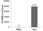
- FIG. 1 is a diagram showing the IDUA activity of an extract obtained from a middle silk gland produced by a transgenic silkworm introduced with a human ⁇ -iduronidase (IDUA) gene.
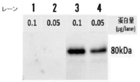
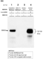
- FIG. 2 is a diagram showing the results of Western blotting using an anti-IDUA antibody on an extract obtained from a middle silk gland produced by a transgenic silkworm into which an IDUA gene has been introduced.
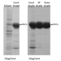
- FIG. 3 is a diagram showing the results of SDS-PAGE of a solution obtained by purifying the extract obtained from the middle silk gland of a transgenic silkworm introduced with the IDUA gene by each chromatography operation.
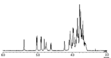
- FIG. 4 is a diagram showing a 1 H-NMR spectrum of Compound 12, which is a sugar chain donor in the present disclosure.
- FIG. 5 is an enlarged view of the 1 H-NMR spectrum of FIG.
- FIG. 6 is a diagram showing a mass spectrum of Compound 12 obtained by MALDI-TOF MS measurement.
- FIG. 7A is a graph showing the results of SDS-PAGE of a sample after sugar chain transfer to a sugar chain acceptor (IDUA after endoenzyme treatment (GlcNAc-IDUA)) of a sugar chain donor (compound 12). It is.
- Lane 1 is a sample containing IDUA obtained from Chinese hamster (CHO) cells into which IDUA gene has been introduced
- Lane 2 is a sample containing IDUA before endo-enzyme treatment
- Lane 3 is a sugar chain receptor (endo).
- FIG. 7B is a view showing the results of lectin blotting with the same sample as FIG. 7A using a lectin (Dom9-His) that specifically binds to Man6P.
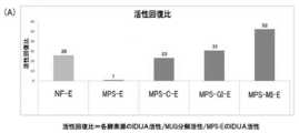
- FIG. 8A is a graph showing an activity recovery ratio in intracellular IDUA activity after adding M6P-containing IDUA to mucopolysaccharidosis type 1 (MPS-1) patient skin fibroblasts and the like and incubating for 24 hours.
- MPS-1 mucopolysaccharidosis type 1
- FIG. 8B shows 4-methylumbelliferyl (intracellular solution (enzyme source)) of 4-methylumbelliferyl obtained after adding M6P-containing IDUA to mucopolysaccharidosis type 1 (MPS-1) patient skin fibroblasts and the like.
- FIG. 4 is a graph showing the measurement of ⁇ -hexosaminidase (Hex) (control enzyme) activity against 4MU) -N-acetyl- ⁇ -D-glucopyranoside (MUG).
- FIG. 9 is a schematic diagram showing a method for modifying a fluorescent group to a Man6P-containing compound (Man6P-Man5-AFO).
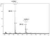
- FIG. 10 is a diagram showing a MALDI-TOF MS spectrum of a fluorescent group-modified Man6P-containing compound (Man6P-Man5-AFO).
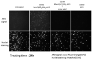
- FIG. 11 shows mucopolysaccharidosis type 1 (MPS-1) patient skin fibroblasts observed with a fluorescence microscope after addition of a fluorescent group-modified Man6P-containing compound (Man6P-Man5-AFO) and incubation for 24 hours. It is a figure which shows a result.
- FIG. 12 shows that mucopolysaccharidosis type 1 (MPS-1) patient skin fibroblasts after addition of fluorescent group-modified Man6P-containing IDUA (Man6P-IDUA-AFO) and incubation for 24 hours were observed with a fluorescence microscope.
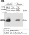
- FIG. 13A is a graph showing the results of SDS-PAGE of a sample after sugar chain transfer to a sugar chain acceptor (CTSA after endoenzyme treatment (GlcNAc-CTSA)) of a sugar chain donor (compound 12). It is.
- Lane 1 is a sample containing CTSA obtained from Chinese hamster (CHO) cells into which a CTSA gene has been introduced
- lane 2 is a sample containing a sugar chain receptor (CTSA after endoenzyme treatment (GlcNAc-CTSA)).
- lane 3 is a sample after the sugar chain transfer of the sugar chain donor (compound 12) to the sugar chain acceptor (GlcNAc-CTSA).
- FIG. 13B is a diagram showing the result of lectin blotting with the same sample as FIG. 13A using a lectin (Dom9-His) that specifically binds to Man6P.
- Endo M is a kind of “Endoenzyme”, and “Endoenzyme” has the same meaning as “Endo- ⁇ -N-acetylglucosaminidase” and extends the end of the arrow in the following general formula (4).
- An “endoenzyme variant” is one in which one or more amino acid residues of an endoenzyme amino acid have been deleted, added or substituted.
- the substance to be subjected to sugar chain transfer refers to a sugar chain donor and a sugar chain acceptor in a sugar chain transfer reaction.
- X represents an oligosaccharide that binds to the 4-position of GlcNAc on the non-reducing end side of the reducing end chitobiosyl residue (GlcNAc ⁇ 1-4GlcNAc) in the N-linked sugar chain
- R represents a polypeptide Indicates.
- the arrow represents the position where the endoenzyme or endoenzyme variant is hydrolyzed.
- Transglycosylation means a part of a sugar chain structure of a sugar chain donor, for example, in the case of the general formula (4), a part on the left side from the position where the tip of the arrow is extended ( ⁇ 1-4 glycoside bond part). , Refers to binding (transfer) to a sugar chain receptor.
- the sugar chain receptor in the present disclosure refers to a sugar-containing substance having GlcNAc represented by the general formula (1) at the non-reducing end.
- Transglycosylation activity refers to the ability of Endo M or Endo M mutant to transfer a sugar chain donor to a sugar chain acceptor to generate a new product (transfer to a sugar chain).
- Transglycosylation yield is a product generated after the transglycosylation reaction, and is transferred to the glycan acceptor from the position where the arrow in the above general formula (4) is extended to the glycan acceptor.
- the product may be referred to as a product after sugar chain transfer.
- endo M mutant refers to an endo enzyme mutant in which the amino acid of the amino acid sequence of endo M (see SEQ ID NO: 1) has been deleted, added, or substituted.
- End M means an endoenzyme derived from the hair mold Mucor Himaris (GenBank Accession No. BAB43869)
- End D means an endoenzyme derived from Streptococcus pneumoniae (GenBank Accession No. BAB62042.1). .
- “Homology” refers to residues in a protein amino acid sequence variant that are identical after aligning the sequence by introducing gaps, if necessary, to achieve maximum homology (percent). Defined as a percentage. Methods and computer programs for alignment are well known in the art and use ClustralW2 (http://www.ebi.ac.uk/Tools/msa/clustalw2/) herein. . In addition, as a method of describing amino acid residues, it is expressed by either one of three letters or one letter.
- the 175th asparagine residue is indicated as N175.
- an amino acid residue substituted that is, an amino acid residue substituted with another amino acid
- a residue obtained by substituting the 175th asparagine residue of endo M with a glutamine residue (or alanine) is N175Q ( Alternatively, End M, which is designated N175A) and contains it, may be referred to as the N175Q variant of Endo M, or simply the N175Q variant.
- endo M mutant of the present disclosure a mutant having an amino acid sequence in which the 175th amino acid residue of the amino acid sequence represented by SEQ ID NO: 1 is glutamine or alanine is referred to as “endo M mutant of the present disclosure”. There is.
- the amino acid sequence of 175 of the amino acid sequence represented by SEQ ID NO: 1 has an amino acid sequence of glutamine or alanine, and 175 of the amino acid sequence represented by SEQ ID NO: 1 Amino acids modified within a range of homology of 80% or more to the amino acid sequence of the endo-M mutant by deletion, addition or substitution of one or more amino acid residues other than the amino acid residue
- the endo-M mutant homolog of the present disclosure it may be particularly referred to as “the endo-M mutant homolog of the present disclosure”. is there.
- the transglycosylation activity by the endo M mutant can be confirmed by measuring the Man6P-containing glycoprotein produced in the solution after the reaction, for example, by SDS-PAGE, lectin blotting and MALDI-TOF MS.
- Examples of the MALDI-TOF MS include the following method. That is, a certain amount of acetone was added to the solution after the reaction, the dissolved portion was dried, and then a certain amount of DHBA solution (20 mg / mL 2,5-dihydroxybenzoic acid dissolved in 50% aqueous methanol solution) was added. Dissolve. Thereafter, a part of the dissolved solution is spotted on a plate for MALDI-TOF MS analysis, dried, and measured with an autoflex speed-tko1 reflector system (manufactured by Bruker Daltonics) under the following conditions: The mass of the product after the reaction can be confirmed.
- Measurement mode positive ion mode or negative ion mode and reflector mode or linear mode
- Measurement voltage 1.5Kv ⁇ 2.5Kv
- Measurement molecular weight range 0 to 10,000 (m / z) ⁇ Total number of times: 500-10000
- a lectin blot for example, a lectin against Man6P can be used, and the lectin described in the literature by Akeboshi et al. [APPLIED AND ENVIRONMENTAL MICROBIOLOGY, Vol. 73, 4805-4812, (2007)] Man6PR (sometimes referred to as Dom9-His).
- the product after sugar chain transfer can be identified by, for example, measurement by SDS-PAGE, lectin blot and MALDI-TOF MS as described above, and the sugar obtained by SDS-PAGE and lectin blot From the comparison of the density of the band of the product after chain transfer, it is possible to qualitatively confirm the magnitude of the yield of the product after sugar chain transfer (yield of sugar chain transfer). Moreover, from this result, it is possible to qualitatively confirm the size of the endo-M mutant for the transglycosylation activity.
- the carbohydrate portion is described with reference to the nomenclature usually used for describing oligosaccharides. These nomenclatures are found, for example, in Hubbard et al. [Ann. Rev. Biochem., Vol. 50, 555, (1981)].
- Mannose is represented by Man
- 2-N-acetylglucosamine is represented by GlcNAc
- galactose is Gal
- fucose is Fuc
- glucose Glc
- Sialic acid is represented by the abbreviated notation NeuAc for 5-N-acetylneuraminic acid and NeuGc for 5-glycolylneuraminic acid.
- the N-acetylglucosaminyl group may be referred to as a GlcNAc residue
- the mannosyl group may be referred to as a Man residue.
- the monosaccharide means only the above-mentioned sugars such as Man and GlcNAc itself.
- the position of the carbon that forms the sugar is represented by the 1st position of the reducing end and the 2nd position of the adjacent carbon atom.
- a combination of these two or more monosaccharides is called an oligosaccharide, and its derivative is called an oligosaccharide derivative. That is, the N-linked sugar chain is an oligosaccharide.
- “Glycoside bond” refers to a bond in which the hydroxyl group at the 1-position of a sugar unit in a sugar chain and the hydroxyl group of another sugar are dehydrated and bonded together via an oxygen atom.
- an ⁇ 1-6 glycoside bond The term “sugar” refers to a glycosidic bond in which the 1-position (carbon) of a sugar and the 6-position (oxygen atom at the 6-position) of another sugar are bonded in an ⁇ -type.
- the hydroxyl group at the 1-position of the sugar has ⁇ type and ⁇ type.
- the glucosaminyl group (GlcNAc) on the non-reducing end side of the chitobiosyl site (-GlcNAc ⁇ 1-4GlcNAc-) does not contain the oxygen atom at the 1-position of the GlcNAc, and It indicates that it contains a 4-position oxygen atom.
- GlcNAc that binds to Y on the reducing end side does not include the oxygen atom at the 1-position and includes an oxygen atom at the 4-position.
- the 2nd, 3rd, 4th and 6th carbons that are not glycosidic bonded indicate that a hydroxyl group is bonded.
- a sugar chain including three monosaccharides may be referred to as three sugars, and a five sugar chain including five sugars.
- the “core sugar chain” refers to the sugar chain part represented by C-1 below in the N-linked sugar chain, and “trimannosyl” refers to the three mannose parts in the core sugar chain. . Further, as shown in the broken line portion on the left side of C-1, among the trimannosyl portion, Man binding at ⁇ 1-6 on the non-reducing end side is Man2, and Man binding at ⁇ 1-3 on the non-reducing end side is Man3. And Man on the reducing end side is referred to as Man1.
- oligosaccharide as shown on the left side of the position where the tip of the arrow in the general formula (4) is extended may be referred to as a trimannosyl GlcNAc-containing sugar chain.
- protein means containing a peptide. That is, the glycoprotein includes a glycopeptide.
- glycopeptide refers to a glycoprotein having 50 or less amino acid residues.
- protein includes both glycoproteins and non-glycoproteins unless otherwise specified.
- glycoprotein indicates that at least one or more N-linked sugar chains are present in the peptide or polypeptide portion. Unless otherwise specified, the type of the plurality of N-linked sugar chains is 1 Species and two or more types are included.
- lysosomal enzyme refers to a glycoprotein that functions as an enzyme in lysosomes in eukaryotic cells.
- a stable natural-type substrate can be used in the preparation of a chemo-enzymatic Man6P-containing glycoprotein, and a Man6P-containing glycoprotein that cannot be achieved by the prior art can be produced.
- an N-linked sugar chain derivative having a chitobiosyl skeleton is used as a sugar chain donor in the sugar chain transfer reaction of endo M
- the reactivity in the sugar chain transfer is due to the sugar chain structure on the non-reducing terminal side of endo M. It was known to change significantly.
- an N-linked sugar chain having a large polar residue such as a phosphate group is expected to be unsuitable as a sugar chain donor for a sugar chain transfer reaction by an endo M mutant.
- the endo M mutant recognizes an N-linked sugar chain having Man6P at the non-reducing end, but recognizes it as a sugar chain donor, and the sugar chain acceptor represented by the general formula (1) is water. It is considered that the transglycosylation reaction proceeds by preferentially binding to the glycan donor rather than the glycan donor, and once the product after the transglycosylation is formed, the endo M mutant is re-linked to the saccharide. Since it is difficult to recognize the product after chain transfer, it is presumed that the yield of the product after sugar chain transfer increases as a result.
- the sugar chain donor having no oxazoline skeleton used in the above-mentioned sugar chain transfer reaction has an advantage that it can be synthesized by a method in which the phosphate group is not exposed to acidic conditions, and is stable even in a buffer solution. Can exist.
- the sugar chain donor having a chitobiosyl skeleton in the present disclosure is extremely unlikely to bind to a group other than GlcNAc of the sugar chain acceptor, like an oxazolineized sugar chain donor.
- the sugar chain transfer reaction in the present disclosure is a reaction that facilitates the production of a Man6P-containing glycoprotein that could not be easily produced by conventional oxazolineated sugar chain donors. Application to large-scale preparation can also be considered.
- the method for producing a Man6P-containing glycoprotein of the present disclosure includes a sugar chain having a structure derived from an N-linked sugar chain in which Man6P is bound to a non-reducing end in the presence of the endo M mutant of (a) or (b) below: This is a method for producing a Man6P-containing glycoprotein by producing a Man6P-containing glycoprotein by a sugar chain transfer reaction between a donor and the sugar chain acceptor represented by the general formula (1).
- Man6P-containing glycoproteins produced by the production method of the present disclosure include those found in nature and those synthesized.
- the second amino acid residue from the asparagine is necessarily a threonine residue or a serine residue on the C-terminal side of the asparagine to which the N-linked sugar chain binds in a naturally occurring glycoprotein.
- the glycoprotein of the Man6P-containing glycoprotein produced by the production method of the present disclosure is not particularly limited.
- glycoproteins secreted inside or outside the cell or on the cell surface and having an N-linked sugar chain are exemplified.
- examples include various enzymes, various hormones, cell adhesion factors, various receptors, and the like.
- Examples include various cytokines.
- various enzymes are preferable.
- examples of various enzymes include enzymes that function in cells, and among them, lysosomal enzymes are preferable.
- ⁇ -hexosaminidase As lysosomal enzymes, ⁇ -hexosaminidase ( ⁇ N-acetylglucosaminidase, ⁇ N-acetylgalactosaminidase), ⁇ N-acetylglucosaminidase, ⁇ galactosidase, ⁇ galactosidase, ⁇ glucosidase, ⁇ iduronidase, ⁇ iduronic acid 2 sulfatase, ⁇ glucuronidase, ⁇ -glucocerebrosidase, ⁇ -galactocerebrosidase, ⁇ N-acetylgalactosaminidase, ⁇ fucosidase, cathepsin A, cathepsin B, cathepsin D, cathepsin H, cathepsin L, aspartyl glucosaminidase, endo ⁇ -galactosidase
- the origin of the glycoprotein is not particularly limited as long as it does not impair the effect of the method of the present disclosure. However, from the viewpoint of introducing and functioning in an animal cell, it is preferably derived from an animal cell, and further to humans. From the viewpoint of enhancing the effect of enzyme replacement therapy, it is more preferably derived from a human.
- the endo M variant in the present disclosure is (a) an endo M variant having an amino acid sequence in which the amino acid residue at position 175 of the amino acid sequence represented by SEQ ID NO: 1 is glutamine or alanine, or (b) Modification within 80% or more homology with the amino acid sequence of (a) above by deletion, addition or substitution of one or more amino acid residues other than the 175th amino acid residue of the amino acid sequence And an endo-M mutant (endo-M mutant homologue) having an amino acid sequence and an activity of catalyzing a sugar chain transfer reaction.
- the sugar chain transfer reaction refers to a sugar chain donor having a structure derived from an N-linked sugar chain in which Man6P is bonded to the non-reducing end, and a sugar chain acceptor represented by the above general formula (1): This refers to the transglycosylation reaction.
- the endo M mutant of the present disclosure is a mutant in which a mutation is introduced into the amino acid of endo M.
- the endoenzyme represented by SEQ ID NO: 1 is an endo ⁇ -N acetylglucosaminidase (GenBank Accession No. BAB43869) derived from Mucor Himalis.
- the endo M mutant is N175Q or N175A, so that hydrolysis of the product after the transglycosylation reaction can be sufficiently suppressed. Further, from the viewpoint of sugar chain transfer yield, N175Q mutant is preferable.
- the endo M mutant of the present disclosure can be prepared by a normal genetic engineering technique, and can be prepared using various types of hosts and corresponding appropriate protein expression vectors.
- the host include Escherichia coli, Brevibacillus, cyanobacteria, lactic acid bacteria, yeast, insect cells and animal cells.
- E. coli Escherichia coli
- Brevibacillus cyanobacteria
- lactic acid bacteria yeast
- insect cells insect cells
- animal cells from the viewpoint of ease of preparation and expression level, it is preferable to prepare by a method using E. coli as a host or a method using yeast as a host.
- the specific production method is described in detail in the literature of Umekawa et al. [J. Biol. Chem., Vol.285 (1), 511-521, ⁇ (2010)] for Escherichia coli. This is described in detail in JP-A-11-332568.
- the endo-M mutant and endo-M mutant homologue of the present disclosure are fused endo-M mutants (or fused forms) fused with other peptides or proteins on the C-terminal side or the N-terminal side in a transglycosylation reaction. Endo M mutant homologue).
- the peptide or protein that can be fused is not particularly limited as long as it does not inhibit the transglycosylation reaction.
- hexahistidine peptide (amino acid sequence is from N-terminal to HHHHHH), flag peptide (amino acid sequence is N-terminal) To DYKDDDDK), influenza HA polypeptide (amino acid sequence is YPYDVPDYA from the N-terminus), glutathione-S-transferase, luciferase, avidin, chitin-binding protein, c-myc, thioredoxin, disulfide isomerase (DsbA), maltose-binding protein ( MBP) and green fluorescent protein (GFP).
- hexahistidine peptides and flag peptides are particularly preferable from the viewpoint of ease of preparation of endo M mutant or endo M mutant homolog.
- fusion-type endo M mutant or fusion-type endo M mutant homolog there is a gap between the polypeptide part of the peptide or protein to be fused and the polypeptide part of the endo-M mutant or endo-M mutant homolog.
- the linker region may include an amino acid sequence portion (protease site) that is hydrolyzed by a protease.
- protease site For example, a factor Xa site, a thrombin site, an enterokinase site, a precision protease site is mentioned.
- saccharide hydrolase found in nature has a family of hundreds of glycosylhydrases from the homology of amino acid sequences. (GH family).
- Endo M is a glycosyl hydrolase belonging to the GH family 85, and other proteins belonging to the GH family 85 are known to be widely distributed from humans to bacteria.
- the endo M mutant homologue of the present disclosure may be any amino acid other than the 175th amino acid residue as long as it has 80% homology with the N175Q mutant or N175A mutant and has transglycosylation activity. Those prepared by substituting, deleting or adding residues to other amino acids may be used.
- an endo M mutation caused by deletion, addition, or substitution of one or more amino acid residues other than the 175th amino acid residue of the amino acid sequence of the endo M mutant of the present disclosure
- the homology of the amino acid sequence of the body homologue to the amino acid sequence of the endo M mutant is 80% or more, the transglycosylation activity can be sufficiently maintained.
- it is 90% or more it is preferable from the sugar-transfer yield easily improving, More preferably, it is 95% or more, Especially preferably, it is 98% or more, Most preferably, it is 99% or more.
- the concentration of the endo M mutant or endo M mutant homolog in the reaction solution is preferably 0.1 U / ml to 1 U / ml.
- the concentration is more preferably 0.1 U / ml to 1 U / ml, and particularly preferably 0.2 U / ml to 0.5 U / ml.
- the purity of the endo M mutant or endo M mutant homolog used in the reaction is 50% from the viewpoint of shortening the reaction time and the yield of sugar chain transfer from the results of SDS-polyacrylamide electrophoresis. % Or more, more preferably 70% or more, particularly preferably 80% or more, and most preferably 90% or more. When the degree of purification is 50% or more, the reaction time can be shortened and the sugar chain transfer yield can be easily increased.
- Man6P has a structure derived from an N-linked sugar chain bonded to the non-reducing end, and the sugar chain can be transferred to the sugar chain acceptor represented by the general formula (1). If it is a thing, it will not specifically limit.
- the structure derived from the N-linked sugar chain means that the N-linked sugar chain itself and a derivative of the N-linked sugar chain are included.
- a sugar chain donor represented by the following general formula (2) is preferable.
- X 1 to X 6 each independently represents a hydrogen atom or a saccharide-derived group, and at least one of X 1 to X 6 is derived from a saccharide having Man6P at the non-reducing end. It is a group.
- Z 1 represents a hydrogen atom or GlcNAc, and when Z 1 is GlcNAc, the GlcNAc is bonded to Man linked to GlcNAc at ⁇ 1-4 at ⁇ 1-4.
- Y 2 represents a monovalent substituent.
- X 1 to X 6 may be a hydrogen atom or a saccharide-derived group, and at least one of X 1 to X 6 may be a saccharide-derived group having Man6P at the non-reducing end.
- the saccharide-derived group is not particularly limited as long as it does not impair the effect of the method of the present disclosure.
- the saccharide-derived group having Man6P includes Man6P itself.
- groups other than the saccharide-derived group having Man6P at the non-reducing end include, for example, GlcNAc ⁇ 1-2, Gal ⁇ 1-4GlcNAc ⁇ 1-2, NeuAc ⁇ 2-6Gal ⁇ 1-4GlcNAc ⁇ 1-2, NeuAc ⁇ 2-3Gal ⁇ 1-4GlcNAc ⁇ 1-2, NeuGc ⁇ 2-6Gal ⁇ 1-4GlcNAc ⁇ 1-2 and NeuGc ⁇ 2-3Gal ⁇ 1-4GlcNAc ⁇ 1-2, heterologous antigens: Galc1-4GlcNAc ⁇ 1-2 4GlcNAc ⁇ 1-3] nGal ⁇ 1-4GlcNAc ⁇ 1-2 (n is an arbitrary number), keratan sulfate [Gal ⁇ 1-4GlcNAc (6SO 3) ⁇ 1-3] nGal ⁇ 1-4GlcNAc ⁇ 1-2 (n is an arbitrary number) [Gal (6SO 3) ⁇ 1-4GlcNAc ( 6SO 3) ⁇ 1-3] nGal
- the saccharide-derived groups having Man6P at the non-reducing end include GlcNAc ⁇ 1-2, Gal ⁇ 1-4GlcNAc ⁇ 1-2, [Gal ⁇ 1-4GlcNAc ⁇ 1-3] nGal ⁇ 1-4GlcNAc ⁇ 1-2 (n is Any number), [Gal ⁇ 1-4GlcNAc (6SO 3 ) ⁇ 1-3] nGal ⁇ 1-4GlcNAc ⁇ 1-2 (where n is an arbitrary number), etc. Things.
- a group other than a saccharide-derived group having Man6P at the non-reducing end may have any of a structure derived from a high mannose-type sugar chain in addition to a hydrogen atom.
- Examples of X 1 to X 6 include Man ⁇ 1-6, Man ⁇ 1-3, Man ⁇ 1-2Man ⁇ 1-3, Man ⁇ 1-2Man ⁇ 1-6, Man ⁇ 1-2, Man ⁇ 1-2Man ⁇ 1-2, and the like.
- saccharide-derived groups having Man6P at the non-reducing end include, for example, Man6P ⁇ 1-6, Man6P ⁇ 1-3, Man6P ⁇ 1-2Man ⁇ 1-3, Man6P ⁇ 1-2Man ⁇ 1-6, Man6P ⁇ 1-2 , Man6P ⁇ 1-2Man ⁇ 1-2 and the like.
- Z 1 may be a hydrogen atom or GlcNAc ⁇ 1-4.
- examples of Y 2 include a substituent containing a structure in which an oxygen atom, a nitrogen atom, a carbon atom, or a sulfur atom is directly bonded to the 1-position carbon of GlcNAc.
- Y 2 is not particularly limited as long as it does not reduce the transglycosylation activity.
- alkoxy group, acylamino group, aryloxy group, alkenyl examples thereof include an oxy group, an acyloxy group, an alkylsulfinyl group, an arylsulfinyl group, an alkylsulfonyl group, an arylsulfonyl group, an alkylsulfonyloxy group, and an arylsulfonyloxy group.
- substituents may further have a substituent.
- Y 2 is a hydroxyl group, an alkoxy group having 1 to 30 carbon atoms which may have a substituent, or an alkyl group having 6 to 30 carbon atoms which may have a substituent.
- An aryloxy group, an optionally substituted alkenyloxy group having 1 to 30 carbon atoms and an optionally substituted acylamino group are preferred.
- Y 2 is an alkoxy group optionally having a substituent having 1 to 10 carbon atoms and a substitution having 2 to 10 carbon atoms from the viewpoint of ease of preparation as a sugar chain donor and a yield of sugar chain transfer.
- An alkenyloxy group which may have a group, an aryloxy group which may have a substituent having 6 to 24 carbon atoms, and an acylamino group which may have a substituent are preferable, and more preferably 1 to An alkoxy group optionally having 8 substituents, an alkenyloxy group optionally having 2 to 6 carbon atoms, and an aryloxy optionally having 6 to 12 carbon atoms
- An acylamino group which may have a group and a substituent is particularly preferred. Specific examples include a methoxy group, an ethoxy group, a phenoxy group, a paramethoxyphenoxy group, and a paranitrophenoxy group.
- the acylamino group which may have a substituent is preferably an acylamino group to which the amino group of the side chain of the asparagine of the peptide or protein is bonded, and the solubility in the reaction solution and the transglycosylation yield. From the viewpoint, an acylamino group to which the amino group of the side chain of the asparagine of the peptide is bonded is more preferable.
- substituents which may be substituted include an alkyl group having 1 to 6 carbon atoms, an alkenyl group having 2 to 12 carbon atoms, an aryl group having 6 to 12 carbon atoms, a halogen atom, a cyano group, a nitro group, When it is at least one selected from an amino group, a carboxyl group and a pyridyl group and there are two or more substituents, each substituent may be the same or different.
- the method for preparing the sugar chain donor represented by the general formula (2) is not particularly limited, but for the ease of the preparation method, for example, an N-linked sugar chain is prepared and Man6P is chemically introduced. A method is mentioned.
- N-linked sugar chains include high mannose sugar chains and complex sugar chains.
- the high mannose sugar chains can be prepared from natural products, even by chemical synthesis. There may be.
- As a method by chemical synthesis for example, as described in the literature of Matsuo et al. [Tetrahedron, Vol. 62, 8262-8277, (2006)], As described in Japanese Patent Application Publication No. 2007-297429, a method combining a chemical synthesis method and mannosidase can be mentioned.
- Examples of the method of preparing from natural products include a method of extracting from egg white albumin as described in Japanese Patent No. 3776952 and JP-A-10-251304.
- a synthetic preparation method is preferred from the viewpoint of introducing Man6P into the non-reducing end.
- the method for preparing the complex type sugar chain may be a method by chemical synthesis or a method of preparing from a natural product.
- a method by chemical synthesis for example, it can be prepared by the method described in Wang et al. [J. Am. Chem. Soc., Vol. 134 (29), 12308, (2012)].
- Examples of the method of preparing from a natural product include a method of extracting from egg yolk (for example, International Publication No. 962255 and Japanese Patent Application Laid-Open No. 2011-231293).
- those obtained by synthesis are preferable from the viewpoint of introducing Man6P into the non-reducing end.
- a chemical introduction method can be mentioned.
- any synthetic route may be used as long as Man6P can be introduced into the non-reducing end of the N-linked sugar chain, and it is appropriately selected depending on the structure of the target sugar chain donor.
- the method for introducing a phosphate group into mannose is not particularly limited.
- the phosphate group can be introduced by reacting a phosphorylating reagent with the 6-position hydroxyl group of the mannose residue at the non-reducing end of an N-linked sugar chain. .
- Examples of the phosphorylating reagent include tetrabenzyl pyrophosphate, cyclic phosphite amide, phosphite, phosphate triester, and amidite.
- a phosphate group from the viewpoint of introducing a phosphate group only to the 6-position hydroxyl group of the mannose residue at the non-reducing end of the N-linked sugar chain, other than the hydroxyl group is protected by a protecting group. It is preferable.
- protection means that the hydroxyl group is converted into a group that is not reactive with the phosphorylating reagent.
- a sugar chain donor represented by the following general formula (3) is preferable.
- X 7 , X 8 and X 9 each independently represent a hydrogen atom, Man, Man ⁇ 1-2Man, Man6P, Man6P ⁇ 1-2Man or Man6P ⁇ 1-6Man.
- Man represents a mannosyl group.
- Man6P represents a mannosyl group having a phosphate group bonded to the 6-position.
- X 7 -6 indicates the X 7 bound to 6-position of Man
- X 8 -3 shows the X 8 attached to the 3-position of Man
- X 9 -2 is attached to the 2-position of Man X 9 is shown.
- At least one of X 7 , X 8 and X 9 represents any of Man6P, Man6P ⁇ 1-2Man and Man6P ⁇ 1-6Man.
- GlcNAc represents an N-acetylglucosaminyl group.
- ⁇ 1-6 represents an ⁇ -glycosidic bond between the 1-position of Man and the 6-position of Man or an ⁇ -glycosidic bond between the 1-position of Man6P and the 6-position of Man
- ⁇ 1-3 represents ⁇ 1-2 represents an ⁇ -glycoside bond between the 1-position of Man and the 2-position of Man, or an ⁇ -glycoside bond between the 1-position of Man6P and the 2-position of Man.
- ⁇ 1-4 represents a ⁇ glycosidic bond between position 1 of GlcNAc and position 4 of GlcNAc or ⁇ glycoside bond between position 1 of Man and position 4 of GlcNAc.
- Y 3 represents a monovalent substituent.
- X 7 , X 8 and X 9 when at least one of X 7 , X 8 and X 9 is any one of Man6P, Man6P ⁇ 1-2Man or Man6P ⁇ 1-6Man, the other is a hydrogen atom, Man, Man ⁇ 1-2Man, Man6P, Man6P ⁇ 1 -2Man or Man6P ⁇ 1-6Man.
- the one having Man6P at the non-reducing end may be any of X 7 to X 9 and is appropriately adjusted depending on the type of the target peptide or glycoprotein to be transferred to the sugar chain. It is preferable.
- X 7 is more preferably any of Man6P, Man6P ⁇ 1-2Man, or Man6P ⁇ 1-6Man.
- the number of Man6Ps among X 7 to X 9 is more preferably 2 or more, and particularly preferably 3.
- sugar chain donors represented by the general formula (3) from the viewpoint of being a high mannose type sugar chain already found in natural glycoproteins, the following general formulas (3-1) to (3) Those represented by the general formula (3-10) are preferable, and among them, the general formula 3-3 is more preferable.
- Y 3 represents a monovalent substituent.
- Man6P represents a mannosyl group having a phosphate group bonded to the 6-position.
- Y 3 has the same meaning as Y 2 in the general formula (2), and the preferred range is also the same.
- the compound represented by the general formula (3) can be prepared by appropriately combining, for example, a known chemical glycosylation method and the above-described method for introducing a phosphate group or the like.
- the sugar chain receptor is a GlcNAc-containing glycoprotein represented by the following general formula (1).
- the sugar chain receptor is not particularly limited as long as it is represented by the general formula (1) as long as it does not inhibit the sugar chain transfer activity of the endo M mutant.
- Y 1 represents an acylamino group containing a structure derived from a glycoprotein.
- GlcNAc represents an N-acetylglucosaminyl group.
- Y 1 as the glycoprotein, for example, various enzymes, various hormones, cell adhesion factors, and various receptors and various cytokines and the like. Among them, various enzymes are preferable from the viewpoint of functioning in cells, and lysosomal enzymes are preferable from the viewpoint of functioning in lysosomes using the Man6P receptor on the lysosomal membrane.
- ⁇ -hexosaminidase As lysosomal enzymes, ⁇ -hexosaminidase ( ⁇ N-acetylglucosaminidase, ⁇ N-acetylgalactosaminidase), ⁇ N-acetylglucosaminidase, ⁇ galactosidase, ⁇ galactosidase, ⁇ glucosidase, ⁇ iduronidase, ⁇ iduronic acid 2 sulfatase, ⁇ glucuronidase, ⁇ -glucocerebrosidase, ⁇ -galactocerebrosidase, ⁇ N-acetylgalactosaminidase, ⁇ fucosidase, cathepsin A, cathepsin B, cathepsin D, cathepsin H, cathepsin L, aspartyl glucosaminidase, endo ⁇ -galactosidase
- the origin of the glycoprotein is not particularly limited as long as it does not impair the effect of the method of the present disclosure. However, from the viewpoint of introducing and functioning in an animal cell, it is preferably derived from an animal cell, and further to humans. From the viewpoint of enhancing the effect of enzyme replacement therapy, it is more preferably derived from a human.
- Y 1 is an acylamino group containing a structure derived from a glycoprotein having 30 or less amino acid residues
- Y 1 is an acylamino group containing a structure derived from a glycoprotein having 30 or less amino acid residues
- a chemical method or a chemistry including a peptide extension step as shown in a patent document (Japanese Patent Laid-Open No. 10-45788).
- a glycopeptide or N-linked sugar chain derivative obtained by a reagent manufacturer can be easily prepared by hydrolysis with the endo M mutant of the present disclosure or a commercially available endo enzyme.
- Y 1 is an acylamino group containing a structure derived from a glycoprotein is a glycoprotein obtained by purchasing a commercially available reagent or food, a glycoprotein prepared by a genetic engineering technique, or a food or natural product. Can be easily prepared by hydrolyzing the glycoprotein obtained by a general extraction / separation method with the endo-M mutant of the present disclosure or a commercially available endo-enzyme.
- End H manufactured by New England Biolabs
- End S manufactured by Sigma-Aldrich
- End D manufactured by Cosmo Bio
- End M manufactured by Tokyo Chemical Industry Co., Ltd.
- End F1 ⁇ And F3 manufactured by Sigma-Aldrich
- Non-commercial enzymes include glycosyl hydrolases belonging to GH family 85, or enzymes that belong to GH family 18 and have been shown to hydrolyze N-linked sugar chains. Can do.
- a gene encoding a target glycoprotein is introduced (transgenic) into a host cell or individual to prepare a desired glycoprotein.
- it does not specifically limit as a host.
- yeast, an insect cell the cell derived from various animals, animals, such as an insect which is an individual, an amphibian, and a mammal, are mentioned.
- insect cells or insects that are individuals are more preferred, silkworm cells or silkworms are particularly preferred, and silkworms that are individuals are most preferred.
- silkworms for example, as shown in the literature of Ito et al.
- transgenic silkworms introduced with foreign genes (transgenic silkworms) It is known that foreign proteins are expressed in large quantities in silk glands.
- the expressed sugar chain of the protein contains a human-type-like partial structure, and has an advantage that an insect-specific sugar chain structure is not added.
- glycoproteins prepared from transgenic silkworms for example, as shown in JP-A-2015-208260, N-linked glycoprotein sugar chains are mainly high mannose sugar chains, many endoenzymes are used. This is advantageous for the preparation of sugar chain receptors. For this reason, it can be said that the silkworm which can be raised in large quantities and cheaply is economically advantageous for preparation of said glycoprotein.
- the reaction means a sugar chain transfer reaction, and the sugar chain transfer reaction is performed in a solution in which an endo M mutant, a sugar chain donor, and a sugar chain acceptor are dissolved.
- the solution used for the reaction is not particularly limited as long as it does not inhibit the transglycosylation activity of the endo M mutant.
- phosphate buffer, citrate buffer, carbonate buffer, Tris-HCl buffer, MES Examples include a buffer solution, a MOPS buffer solution, a HEPES buffer solution, a borate buffer solution, and a tartrate buffer solution. These buffers may be used alone or in combination.
- MES buffer solution MOPS buffer solution
- HEPES buffer solution phosphate buffer solution
- phosphate buffer solution is preferable from the viewpoint of glycosyltransferase activity of endo M mutant or endo M mutant homolog.
- concentration of the phosphate buffer is preferably 10 mM to 250 mM, more preferably 20 mM to 150 mM, and particularly preferably 50 mM to 100 mM, from the viewpoint of glycosyl transfer activity of the endo M mutant or endo M mutant homolog. .
- the buffering capacity is increased, and by setting the concentration to 250 mM or less, the sugar chain transfer activity of the endo M mutant or the endo M mutant homolog can be increased and the sugar chain transfer yield can be increased.
- the pH of the solution in the reaction is preferably 5.5 to 8.5, more preferably 6.0 to 8.0, and particularly preferably 6.5 to 7.5 from the viewpoint of increasing the sugar chain transfer yield.
- the temperature in the reaction is 4 from the viewpoint of transglycosylation activity of the endo M mutant or endo M mutant homolog, from the viewpoint of increasing the transglycosylation yield, and from the viewpoint of the stability of the sugar chain receptor, endo M mutant, etc. It is preferably from 40 ° C to 40 ° C, more preferably from 20 ° C to 40 ° C, and particularly preferably from 25 ° C to 35 ° C. By setting the temperature to 4 ° C. or higher, the transglycosylation activity of the endo M mutant can be increased, and by setting the temperature to 40 ° C. or lower, the stability of the endo M mutant or the endo M mutant homolog can be increased. .
- the concentration of the endo M mutant (or endo M mutant homolog) in the reaction solution is preferably 0.005 ⁇ g / ⁇ L to 0.5 ⁇ g / ⁇ L.
- the transglycosylation activity is improved, and when it is 0.5 ⁇ g / ⁇ L or less, the product of the endo-M mutant (or endo-M mutant homolog) after the transglycosylation Rehydrolysis can be suppressed, and the sugar chain transfer yield can be improved.
- the concentration of the sugar chain receptor in the reaction solution is not particularly limited as long as it does not inhibit the sugar chain transfer reaction of the End M mutant or End M mutant homolog. It is adjusted as appropriate in consideration of the sugar chain transfer yield and the solubility of the sugar chain receptor in the reaction solution.
- the sugar chain receptor represented by the general formula (1) contains a protein
- 1.0 mg / mL to 20 mg / mL is preferable. By being in this range, a higher sugar chain transfer yield can be obtained. Of these, 2.0 mg / mL to 10 mg / mL is more preferable.
- the molar ratio of the sugar chain donor to the sugar chain acceptor is the type of sugar chain donor or sugar chain acceptor used. In addition, it can be appropriately set in consideration of recovery of the reaction after the reaction. However, from the viewpoint of transglycosylation yield, the molar ratio is preferably 10 to 3000, more preferably 100 to 2000, particularly preferably 200 to 1500, and further preferably 300 to 1000. Is most preferred. When the molar ratio is 10 or more, a more sufficient transglycosylation yield can be obtained, and when the molar ratio is 3000 or less, a higher transglycosylation yield can be obtained.
- the reaction time in the transglycosylation reaction is appropriately set depending on the reaction temperature and the concentration of the endo M mutant or endo M mutant homolog in the solution, but is preferably 3 hours to 100 hours. By being 3 hours or longer, the sugar chain transfer yield can be further increased. By being 100 hours or shorter, the influence of hydrolysis of the product after the sugar chain transfer is further suppressed, and the sugar chain transfer yield is increased. be able to. Among the above reaction times, it is more preferably 10 hours to 50 hours.
- the sugar chain donor and the endo M mutant or the endo M mutant homolog are added to the reaction solution again. It is preferable to react for a certain time.
- Man6P containing glycoprotein manufactured with said manufacturing method can be provided as a pharmaceutical composition and the chemical
- the pharmaceutical composition includes a pharmaceutically acceptable stabilizer, buffer, excipient, binder, disintegrant, flavoring agent, A coloring agent, a fragrance
- a protein having a high proportion of many types of Man6P-containing sugar chains can be obtained using many types of sugar chain donors. They are also useful as glycoprotein preparations.
- the method for detecting the intracellular distribution of the fluorescent group-bound Man6P-containing glycoprotein of the present disclosure includes a fluorescent group-bound Man6P-containing product obtained by further introducing a fluorescent group into the Man6P-containing glycoprotein obtained by the above production method. This is a method for detecting the intracellular distribution of a glycoprotein by applying the glycoprotein to a cell.
- the fluorescent group binding type means that the fluorescent group is bound to the Man6P-containing glycoprotein by a covalent bond or an ionic bond.
- the distribution of the Man6P-containing glycoprotein in the cells is distributed. Can be detected.
- fluorescent group-bound Man6P-containing glycoproteins and the like easily enter the cell by the Man6P receptor, and thus the behavior of the fluorescent Man6P glycoprotein and the like after the entry in the cell is traced by the fluorescence emitted by the fluorescent group. can do.
- the detection method for example, by adding the fluorescent Man6P glycoprotein to a target cell or tissue, incubating at a constant atmosphere, temperature, and time, and observing with a fluorescence microscope or the like, The localization and distribution of the fluorescent Man6P glycoprotein in the cell can be known.
- the amount, temperature, and time of addition of the fluorescent Man6P glycoprotein to the cells are appropriately adjusted depending on the type of cells, the function and properties of the protein in the fluorescent Man6P glycoprotein, and the like.
- IDUA human ⁇ -iduronidase
- IDUA refers to an enzyme having activity as an enzyme.
- GlcNAc-IDUA is a protein obtained by hydrolyzing an IDUA N-linked sugar chain with an endoenzyme, and at least one GlcNAc is bound to a polypeptide constituting IDUA. It shows that.
- Man6P-IDUA refers to a Man6P-containing glycoprotein, and unless otherwise specified, is obtained from a transgenic silkworm (also referred to as TG silkworm) described later and then purified by three-stage chromatography. Further, human IDUA usually has 6 N-linked sugar chains, and IDUA prepared by TG silkworm hereinafter is a mixture of 1 to 6 N-linked sugar chains.
- Detection of protein in the gel after SDS-PAGE was performed by shaking in CBB staining solution [10% CBB-R350 (manufactured by GE), 27% methanol, 9% acetic acid, 0.1% CuSO 4 ] for 1 hour or longer. And then decoloring for 6 hours or more using a decolorizing solution (10% acetic acid).
- 1 st IDUA antibodies as probes (Anti-IDUA, sheep IgG polyclonal antibody, (R & D Co., Ltd.)) was used to shake the PVDF membrane in 50% Blocking one / TBS (4 °C, O / N). Thereafter, the PVDF membrane was washed 5 times at room temperature in 0.1% Tween20 / TBS (TBST) 5 times, and further washed in TBS at room temperature for 5 minutes.
- Use as 2 nd probe anti-sheep antibodies (Biotin-conjugated anti-sheep antibody (Vector Co.)), in 50% Blocking one / TBS, was shaken for 1 hour PVDF membranes at room temperature.
- the PVDF membrane in TBST was repeatedly washed 5 times at room temperature for 5 minutes, and further washed in TBS at room temperature for 5 minutes. 3 rd using HRP-conjugated anti-biotin antibody as a probe (Cell Signaling Inc.), the PVDF membrane was shaken for 1 hour at room temperature in 50% Blocking one / TBS. Thereafter, the PVDF membrane in TBST was washed 5 times at room temperature for 5 minutes, and further washed in TBS at room temperature for 5 minutes.
- the PVDF film was subjected to chemiluminescence using Western lighting Chemiluminescence Reagent Ultra / Plus (manufactured by Perkin Elmer), and signal (band) detection was performed using an image analyzer [LAS-4000miniEPUV (manufactured by Fuji Film)].
- IDUA activity is sodium citrate buffer using 4-methylumbelliferyl (4MU) - ⁇ -L-idpyranoside (manufactured by Toronto Research Chemicals) (hereinafter sometimes referred to as 4MU-Ido) as a substrate.
- 4MU-Ido 4-methylumbelliferyl
- the enzyme activity using 4MU-Ido as a substrate may be referred to as IDUA activity.
- the middle silk gland from silkworms was collected by a known general method. Break the outer skin (by hand with surgical gloves) of the TG silkworm from the 5th to the mature stage, and pull out the silk gland that is pushed out from the cracked part. It was. In physiological saline, the front and rear silk glands and the attached fat body tissue were removed from the whole collected silk gland tissue, and only the middle silk gland was fractionated and collected. After washing twice with physiological saline, 50 heads were packed in a conical tube and stored frozen at ⁇ 20 to ⁇ 30 ° C.
- IDUA purification from middle silk gland> IDUA was obtained by purifying by performing the three-stage chromatography operation (affinity chromatography, cation exchange chromatography, hydrophobic interaction chromatography) in combination with the above IDUA activity measurement method.
- the middle silk gland collected from the above TG silkworm was thawed and then shredded and sonicated in 20 mM sodium acetate buffer (pH 4.5) containing 0.5 M NaCl to prepare an extract A. did.
- FIG. 1 shows the results of measuring the enzyme activity (IDUA activity) against 4MU-Ido for the obtained extract A.
- the term “not introduced” means that the IDUA activity was measured for the extract B of the middle silk gland obtained from the silkworm not introduced with the IDUA gene in the same manner as described above. From the results, the extract A obtained from the TG silkworm into which the IDUA gene was introduced showed significantly higher IDUA activity than the extract B. Further, when Western blotting using human IDUA antibody was performed on the obtained extract, as shown in lanes 1 and 2 in FIG. 2, a protein-containing solution extracted from silkworms into which no IDUA gene had been introduced, While no band was obtained, as shown in lanes 3 and 4, a strong band was found around 80 kDa in the protein-containing solution extracted from the TG silkworm into which the IDUA gene was introduced. From the above results, an extract A containing IDUA having IDUA activity was obtained from a TG silkworm into which a human IDUA gene was introduced.
- the extract A obtained above was centrifuged by centrifugation [20,000 ⁇ g, 4 ° C., 20 minutes (Beckman Coulter, Avanti J-E, rotor: JRA 16.250)], and the supernatant was collected.
- CaCl 2 and MnCl 2 were added to the resulting supernatant to a final concentration of 1 mM, and a binding buffer [20 mM sodium acetate buffer, 500 mM NaCl, 1 mM CaCl 2 and 1 mM MnCl 2 (pH 4) was previously added.
- elution was performed with an elution buffer [20 mM sodium acetate buffer, 500 mM NaCl, 1 mM CaCl 2 , 1 mM MnCl 2 , and 0.5 M methyl- ⁇ -D-mannopyranoside (pH 4.5)].
- the obtained elution fraction (hereinafter referred to as ConA elution fraction) was concentrated using an Amicon ultra filter unit (fractional molecular weight 30 kDa) (Millipore), and then a cation exchange column binding buffer [10 mM acetic acid. Buffer exchange was performed using sodium buffer, 150 mM NaCl (pH 6.0)].
- SP elution fraction The elution fraction showing IDUA activity (hereinafter referred to as SP elution fraction) was concentrated using an Amicon ultra filter unit (fractional molecular weight 30 kDa) (Millipore), and then a butyl-Sepharose column binding buffer [50 mM acetic acid.
- Buffer exchange with sodium, 1M ammonium sulfate, 150 mM NaCl (pH 4.5)] was performed. Thereafter, the mixture was filtered through a 0.22 ⁇ m filter, and applied to a hydrophobic interaction chromatography column (HiTrap Butyl FF, manufactured by GE) equipped with AKTA purifier UPC-10, and the ammonium sulfate concentration was changed from 1M to 0M.
- a purification operation was performed under the condition of continuously decreasing, the IDUA activity of each fractionated fraction was measured, and fractions having activity (hereinafter referred to as Butyl-eluting fractions) were collected. Table 1 shows the results of the specific activities of the fractions obtained by the respective purification methods. Further, FIG.
- Extract indicates the extract A
- ConA elute indicates the Con A elution fraction
- SP elute indicates the SP elution fraction
- Butyl elate indicates the Butyl. Represents the eluted fraction.
- SDS-PAGE was performed by applying the same amount (10 ⁇ g) of protein to each lane.
- the specific activity for 4MU-Ido of the Butyl elution fraction obtained by the above purification method is improved to 30 times or more the specific activity of Extract A (Extract) for 4MU-Ido.
- Highly purified IDUA could be obtained from TG silkworm.
- SDS-PAGE in FIG. 3 the staining concentration of bands other than the band around 80 kDa of the SP elution fraction and the Butyl elution fraction is significantly thinner than that of the extract, It was shown that IDUA present in the obtained SP elution fraction and Butyl elution fraction was highly purified.
- IDUA present in the Butyl elution fraction is referred to as TG-IDUA.
- the fraction containing IDUA obtained by the above purification operation was used for the preparation of the sugar chain receptor shown below.
- the sugar chain receptor is obtained by converting the TG-IDUA obtained above into an N-linked sugar chain having a mannose residue number of 5 or less using an endoenzyme (Endo-D, New England Biolabs).
- Endo-D an endoenzyme
- the sugar chain receptor is obtained by converting the TG-IDUA obtained above into an N-linked sugar chain having a mannose residue number of 5 or less using an endoenzyme (Endo-D, New England Biolabs).
- Endo-D Endo-D, New England Biolabs
- MALDI-TOF MS A solution obtained by dissolving the reaction solution as it is or after purification in a solvent (for example, methylene chloride, ethyl acetate, methanol, toluene, acetone, water) and a DHBA solution (20 mg / ml 2,5-dihydroxybenzoic acid in 50% methanol) 0.5 to 2 ⁇ L each of the solution dissolved in an aqueous solution was spotted on a plate for MALDI-TOF MS analysis and dried, and MALDI-TOF MS analysis was performed using an autoflex speed-tko1 reflector system (manufactured by Bruker Daltonics). ) And the following measurement conditions were used.
- a solvent for example, methylene chloride, ethyl acetate, methanol, toluene, acetone, water
- a DHBA solution 20 mg / ml 2,5-dihydroxybenzoic acid in 50% methanol
- Measurement mode positive ion mode or negative ion mode and reflector mode or linear mode
- Measurement voltage 1.5Kv ⁇ 2.5Kv
- Measurement molecular weight range 0 to 10,000 (m / z) ⁇ Total number of times: 500-10000
- a specific method for preparing a sugar chain donor will be described.
- a hexasaccharide derivative (Compound 6) was synthesized by a known glycosylation method. Thereafter, the paramethoxyphenyl group at the reducing end is converted to a fluoro group which is a leaving group, and glycosylation is performed under the glycosylation conditions described below to synthesize compound 9 which is a heptasaccharide derivative, Deprotection of two protecting groups at the position where an acid group is introduced, followed by introduction of a phosphate group, followed by deprotection of the protecting group, a parasaccharide phenyl derivative of 7 sugars having Man6P at the terminal (sugar chain Donor) was obtained.
- reaction solution was cooled to ⁇ 30 ° C., 1.6 g (7.0 mmol) of N-iodosuccinimide and 61 ⁇ L (0.7 mmol) of trifluoromethanesulfonic acid were added, and the mixture was stirred for 3 hours. After confirming the reaction by TLC, 210 ⁇ L (1.5 mmol) of triethylamine was added to the reaction solution to stop the reaction, followed by filtration through Celite. The reaction mixture was diluted with ethyl acetate, washed successively with saturated aqueous sodium thiosulfate solution, saturated aqueous sodium hydrogen carbonate solution and saturated brine, dried over magnesium sulfate, filtered and concentrated under reduced pressure.
- the solution was filtered through celite, diluted with ethyl acetate, washed successively with saturated aqueous sodium hydrogen carbonate solution and saturated brine, dried over magnesium sulfate, filtered, and concentrated under reduced pressure.
- the product was subjected to NMR measurement, and the following results were obtained.
- 0.81 g (0.31 mmol) of the obtained 1-OH compound was dissolved in 8.1 mL of methylene chloride and then cooled to ⁇ 20 ° C., and 0.16 mL (1.2 mmol) of (diethylamino) sulfur trifluoride was added. Stir for 5 hours.
- 0.34 g (0.12 mmol) of the obtained compound was dissolved in a mixed solvent of 1.7 mL of tetrahydrofuran and 1.7 mL of methanol, sodium methoxide (2.5 ⁇ L, 0.012 mmol) was added, and the mixture was stirred for 2.5 hours. . Thereafter, sodium methoxide (5.0 ⁇ L, 0.023 mmol) was further added and stirred for 17 hours.
- a lectin (CI-Man6PR) for detecting Man6P was prepared as follows. That is, a plasmid vector for expression of CI-Man6PR (Dom9-His) prepared by referring to Akeboshi et al. [APPLIED AND ENVIRONMENTAL MICROBIOLOGY, Vol. 73, 4805-4812, (2007)] was used as a methanol-assimilating yeast strain. (Pichia pastoris GS11) was introduced into 5 strains by electroporation.
- the whole culture solution containing yeast after culturing is centrifuged (6000 rpm, 4 ° C., 1 h, KUBOTA 6800 (rotor: RA-1500)), and the culture supernatant is recovered. did.
- the culture supernatant was filtered using a 0.22 ⁇ m filter (Millipore) and concentrated by ultrafiltration [Labscale TFF System, Pellicon XL5K (Millipore)].
- the concentrated culture supernatant was bound to a His tag purification resin (Ni-Sepharose 6 Fast Flow, manufactured by GE), and then washed with a washing buffer [20 mM sodium phosphate according to the protocol attached to the resin. , 0.5 M NaCl (pH 8.0)] and elution with elution buffer [20 mM sodium phosphate, 0.5 M NaCl, 50 mM imidazole (pH 8.0)] A solution containing CI-Man6PR (Dom9-His) was obtained. The obtained solution was appropriately diluted and used for detection of Man6P in the product in the solution after the transglycosylation reaction described later.
- the PVDF membrane was shaken for 1 hour at room temperature in 50% Blocking one / TBS. Thereafter, the PVDF membrane in TBST was washed 5 times at room temperature for 5 minutes, and further washed in TBS at room temperature for 5 minutes.
- the PVDF film was chemiluminescent using Western lightning Chemiluminescence Reagent Ultra / Plus (manufactured by Perkin Elmer), and using an image analyzer [LAS-4000miniEPUV (manufactured by Fujifilm)] with high exposure conditions with an exposure time of 1 second. The signal (band) was detected at.
- FIG. 7A shows the result of SDS-PAGE for the solution after the above reaction
- FIG. 7B shows the result of lectin blotting.
- lane 1 is a sample containing IDUA (hereinafter referred to as CHO-IDUA) obtained by culturing after culturing CHO cells (K1 strain, RIKEN Cell Bank) into which IDUA gene has been introduced.
- Lane 2 is the result of the sample containing TG-IDUA
- Lane 3 is the result of the sample after treating TG-IDUA with End D
- Lane 4 is the result after the transglycosylation reaction. It is a result. From FIG.
- Man6P-IDUA ⁇ Preparation of Man6P-containing glycoprotein (Man6P-IDUA)>
- the solution after the transglycosylation reaction of Example 1 was subjected to ultrafiltration using an Amicon ultra filter unit (fractionated molecular weight of 30 kDa) (manufactured by Millipore) to remove unreacted sugar chain donors. Further, the buffer solution was replaced with a phosphate physiological buffer solution (hereinafter referred to as PBS) to obtain Man6P-IDUA (10.5 ⁇ g).
- PBS phosphate physiological buffer solution
- the obtained Man6P-IDUA was used for examining the supplementary effect on the following mucopolysaccharidosis type 1 patient fibroblasts (MPS1 patient-derived fibroblasts) and the like.
- TG-IDUA, GlcNAc-IDUA, and Man6P-IDUA may be collectively referred to as supplementary enzymes.
- MPS1 patient-derived fibroblasts are present in Ham's F-10 medium (+ 10% Fetal bovine serum) from oral mucosal tissues after approval by Tokushima University Hospital Small Ethics Committee and informed consent to patients It was obtained by inducing under and establishing by adhesion culture and subculture.
- Each enzyme source was prepared as follows. To each 1 mL of culture solution having 5 ⁇ 10 4 cells / well of MPS1 patient-derived cells or healthy human fibroblasts (HS68) cultured in a collagen-coated 24-well plate (Collagen-coated microplate 24well, manufactured by IWAKI) TG-IDUA (endoenzyme-free IDUA) was added at 2000 nmol / h (2.5 ⁇ g), GlcNAc-IDUA was added at 2000 nmol / h (2.85 ⁇ g), and Man6P-IDUA 2000 nmol / h (4.6 ⁇ g) was added. The cells were cultured for 24 hours at 37 ° C. in a 5% CO 2 atmosphere.
- the cells were washed twice with 1 mL of PBS, and further 0.2 mL of 0.5% trypsin EDTA solution was added, and the cells were detached from the wells by incubation for 5 minutes. After adding 1 mL of the culture solution to stop the trypsin reaction, the cells were collected by centrifugation at 4 ° C. and 200 ⁇ g for 5 minutes.
- the enzyme source obtained by the above preparation method is referred to as MPS-E, and the healthy human fibroblasts (HS68)
- the enzyme source obtained by the above preparation method after incubation without adding any of the supplemental enzymes is referred to as NF-E.
- the enzyme source obtained by the above preparation method was referred to as MPS-CE, and GlcNAc-IDUA was added to MPS1 patient-derived fibroblasts.
- the enzyme source obtained by the above preparation method is called MPS-GI-E, and Man6P-IDUA is added to MPS1 patient-derived fibroblasts and incubated, and then the above preparation method
- MPS-MI-E The enzyme source obtained by this is referred to as MPS-MI-E.
- IDUA activity 4-methylumbelliferyl (4MU) - ⁇ -L-idpyranoside (manufactured by Toronto Research Chemicals) (hereinafter sometimes referred to as 4MU-Ido) as a substrate in a sodium citrate buffer (pH 4. After incubating at 37 ° C. for 30 minutes in 5), the fluorescence intensity (Ex: 355 nm, Em: 460 nm) of the released 4MU was measured as an index.
- the enzyme activity using 4MU-Ido as a substrate may be referred to as IDUA activity.
- MUG degradation activity which is ⁇ -hexosaminidase activity
- MUG degradation activity is sodium citrate buffer (pH 4) using 4-methylumbelliferyl (4MU) -N-acetyl- ⁇ -D-glucosaminide (manufactured by SIGMA) as a substrate. .5) was incubated at 37 ° C. for 30 minutes, and the fluorescence intensity (Ex: 355 nm, Em: 460 nm) of the released 4MU was measured as an index.
- FIG. 8A shows the activity recovery ratio in intracellular IDUA activity after adding each of the above-mentioned supplementary enzymes to MPS1 patient-derived fibroblasts and incubating for 24 hours
- FIG. 8B shows the MUG of each of the above enzyme sources. Degradation activity is shown. Further, the numbers on each column in FIG. 8A indicate the value of the activity recovery ratio, and the numbers on each column in FIG. 8B indicate the value of the MUG decomposition activity. From the results, the activity recovery ratio of other enzyme sources other than MPS-E showed a very high value of 23 to 52 times that of MPS-E. In addition, the activity recovery ratio of MPS-MI-E was shown to be the highest in the activity recovery ratio compared to the activity recovery ratios of other enzyme sources.
- a fluorescent group-modified Man6P-containing compound Man6P-Man5-AFO
- a fluorescent group-modified Man6P-IDUA were prepared and used to produce the Man6P-containing compound and Man6P-IDUA in cells. The localization and distribution of was investigated.
- a compound having an n-propoxy group having an amino group at the 3-position as shown in FIG. 9 by using a method similar to the method of synthesizing compound 12 using -2-deoxy-2-phthalimido- ⁇ -D-glucopyranoside AF was synthesized. Further, compound AF1 was prepared by binding with a fluorescent molecule (AcidiFluor ORANGE-NHS, manufactured by Goryuka Kagaku Co., Ltd.) that is activated at acidic pH.
- FIG. 10 shows MALDI-TOF MS spectra of the obtained compound AF1 and compound AF.
- Man6P addition compound Man6P-Man5-AFO modified with a fluorescent group was obtained from the compound AF which is a raw material compound.
- AFO means a fluorescent compound (AcidiFluor ORANGE).
- Man6P-Man5-AFO which is a derivative of 6-saccharide oligosaccharide, was taken into cells through binding to the Man6P receptor on the surface of MPS1 patient-derived fibroblasts.
- Man6P-Man5-AFO was added to a culture solution containing MPS1 patient-derived fibroblasts cultured in a 35 mm collagen-coated dish (35 mm / Collagen-coated dish, manufactured by IWAKI) at a final concentration of 50 nM, and 24 hours later, The fluorescence of Man6P-Man5-AFO was observed with a fluorescence microscope (BZ-9000, manufactured by KEYENCE). The results are shown in FIG.
- Man6P-Man5-AFO was taken up into the cell name, and fluorescence was observed.
- M6P mannose 6-phosphate
- Man6P-Man5-AFO is mediated by M6P receptor on the cell surface. It has been shown to be taken up and transported to late endosomes / lysosomes, intracellular organelles with acidic pH. Thus, Man6P-Man5-AFO has been shown to function as a tag for delivery of molecules bound to Man6P to intracellular organelles (lysosomes).
- DMSO DMSO amount: 6 ⁇ L
- Man6P-IDUA Fluorescent group-modified (fluorescent group binding type) Man6P-IDUA (ultrafluorescent group binding type) by ultrafiltration with an Amicon ultra filter unit (fractional molecular weight 30 kDa) (manufactured by Millipore) and removing unreacted AcidiFluor ORANGE-NHS Man6P-IDUA-AFO) was obtained.
- the obtained Man6P-IDUA-AFO was stored at ⁇ 80 ° C. until just before use.
- Man6P-IDUA-AFO ⁇ Detection of intracellular distribution of Man6P-IDUA-AFO> The Man6P-IDUA-AFO obtained above was added to a culture solution of MPS1 patient-derived fibroblasts cultured on an 8-well chamber slide (manufactured by Nunc) at a final concentration of 70 nM. After a period of time, the cells were washed with PBS, and then the fluorescence of AFO was observed with a fluorescence microscope (BZ-9000, manufactured by KEYENCE). The results are shown in FIG. From the results, it was taken up into cells and fluorescence was observed in acidic pH organelles (late endosome / lysosome).
- Man6P-IDUA-AFO was taken up through the M6P receptor on the surface of fibroblasts and was shown to be transported to the late endosome / lysosome, which is an acidic organelle.
- ConA Sepharose 4B (manufactured by GE Healthcare) was performed on the culture supernatant of the CHO / CTSA strain to purify the CTSA protein. Specifically, the culture supernatant was collected, and CaCl 2 and MnCl 2 were added to the culture supernatant so that the final concentration was 1 mM.
- ConA Sepharose was equilibrated with a binding buffer [20 mM MES buffer, 150 mM NaCl, 1 mM CaCl 2 , 1 mM MnCl 2 (pH 5.5)], and the culture supernatant was applied thereto for adsorption to Con A. It was.
- ConA Sepharose was washed with ConA washing buffer [20 mM MES buffer, 500 mM NaCl, 1 mM CaCl 2 , 1 mM MnCl 2 (pH 5.5)]. Thereafter, elution was performed with an elution buffer [20 mM MES buffer, 150 mM NaCl, 1 mM CaCl 2 , 1 mM MnCl 2, 0.5 M methyl- ⁇ -D-mannopyranoside (pH 5.5)].
- the ConA column elution fraction obtained was concentrated with an Amicon ultrafilter unit 30 kDa cut, and the buffer solution was exchanged with a buffer solution for butyl binding [50 mM MES, 1 M ammonium sulfate (pH 5.5)]. Thereafter, filtration was performed with a 0.22 ⁇ m filter, and purification was performed by hydrophobic interaction chromatography using a HiTrap Butyl HP column (GE Healthcare). For purification, AKTA purifier UPC-10 was used, and elution was performed while the ammonium sulfate concentration was continuously changed from 1 M to 0 M after adsorption of CTSA protein to the column.
- Butyl column elution fraction was buffered in Phos-tag (registered trademark) binding buffer [100 mM Tris-acetate buffer solution (pH 7.5)] using an Amicon ultra filter unit 30 kDa cut (manufactured by Millipore). The liquid was changed.
- Phos-tag registered trademark
- Phos-tag registered trademark
- agarose manufactured by Wako
- Phos-tag® agarose was equilibrated with a binding buffer [100 mM [Tris-acetate buffer (pH 7.5)], and a sample was applied thereto for adsorption to Phos-tag TM agarose. It was. After removing the flow-through fraction, the Phos-tag (registered trademark) agarose was washed with Phos-tag (registered trademark) washing buffer [100 mM Tris-acetate buffer solution (pH 7.5)]. Thereafter, CTSA was eluted with an elution buffer [20 mM sodium phosphate buffer (pH 6.5)].
- reaction solution was replaced with 20 mM Tris-HCl buffer (pH 7.0) and 200 mM NaCl using an Amicon ultra filter unit 10 kDa cut (manufactured by Millipore). Thereafter, filtration with a 0.22 ⁇ m filter (Millipore) was performed, and the reaction solution was added to 1 mL of an MBP trap column equilibrated with 20 mM Tris-HCl buffer (pH 7.0) and 200 mM NaCl. Hf was adsorbed on the MBP trap column.
- the flow-through fraction was collected, and 20 mM Tris-HCl buffer (pH 7.0) and 2OmL of 200 mM NaCl were added to the MBP trap column to wash out the flow-through fraction.
- the flow-through fraction was concentrated using an Amicon ultra filter unit 10 kDa cut (manufactured by Millipore), and the buffer solution was exchanged with 50 mM MES buffer solution (pH 6.0).
- the obtained fraction was designated as GlcNAc-CTSA (sugar chain receptor).
- Glycosyltransferase Glycosynthase (Endo-M-N175Q) 2 mU -Molar ratio (D / A) of sugar chain donor (D) to sugar chain acceptor (A): 1000 -Reaction temperature: 30 ° C ⁇ Reaction time: 24h
- FIG. 13A For the solution after the above reaction, the result of CBB staining of the gel after SDS-PAGE is shown in FIG. 13A, and the lectin (Dom9-His (Akeboshi H. et al., Glycobiology (2009) 19 (9)): FIG. 13B shows the result of lectin blotting using 1002-1009))) as the primary probe.
- lane 1 is the result of the sample containing CTSA
- lane 2 is the result of the sample containing GlcNAc-CTSA
- lane 3 is the result after the transglycosylation reaction.
- FIG. 13A shows that GlcNAc-CTSA is obtained from CTSA by the above Endo-Hf treatment because the band of lane 2 appears on the lower molecular weight side than the band of lane 1. It was.
- the lane 3 band appeared on the higher molecular weight side than the lane 2 band. Therefore, it was shown that a glycosyltransferase reaction occurred in GlcNAc-CTSA, which is a sugar chain receptor, and a post-glycosylation product in which one or two terminal Man6P-containing N-type sugar chains were linked was obtained.
- FIG. 13B since the luminescence signal disappeared in lane 2 of FIG. 13B is newly obtained in lane 3, the sugar chain containing Man6P in the sugar chain donor is added to the sugar chain acceptor. Showed metastasis.
Abstract
A mannose-6-phosphate-group (Man6P)-containing glycoprotein production method for producing a Man6P-containing glycoprotein by a transglycosylation reaction between a sugar chain donor that has a structure derived from an N-binding sugar chain having a Man6P bound to a non-reducing end thereof and a sugar chain acceptor represented by general formula (1) in the presence of an Endo-M mutant (a): an Endo-M mutant having an amino acid sequence represented by SEQ ID NO: 1 in which the 175th amino acid residue is replaced by a glutamine or alanine residue. In general formula (1), Y1 represents an acylamino group containing a glycoprotein-derived structure.
Description
本開示は、マンノース-6-リン酸基含有糖蛋白質の製造方法、及び蛍光基結合型マンノース-6-リン酸基含有糖蛋白質の細胞内分布を検出する方法に関する。
The present disclosure relates to a method for producing a mannose-6-phosphate group-containing glycoprotein and a method for detecting the intracellular distribution of a fluorescent group-bound mannose-6-phosphate group-containing glycoprotein.
細胞内で機能する糖蛋白質遺伝子の欠損症では、細胞内での該糖蛋白質の機能が不全となり、重篤な疾患を引き起こすことが知られている。例えば、リソソーム病は、細胞内小器官であるリソソームに存在する加水分解酵素(リソソーム酵素)等の遺伝的欠損により発症する難病であり、現在の治療法としては、調製した酵素(組み換え酵素)を静脈内投与して治療する酵素補充療法が知られている。酵素補充療法とは、糖蛋白質が有するN結合型糖鎖の非還元末端のマンノース-6-リン酸基(以下、Man6Pと称する)と、細胞表面及びリソソームに存在するMan6Pレセプターとの結合に伴うエンドサイトーシスによる細胞内への取り込み現象を利用した療法であり、リソソーム病患者の標的となる細胞に、Man6Pを有するリソソーム酵素を細胞外から与えることにより、治療効果を発揮させる治療法である。
ここで、標的とする細胞内、あるいは細胞内小器官に、高効率に目的の糖蛋白質を導入したい場合には、N結合型糖鎖の非還元末端にMan6Pが付加された該糖蛋白質を用いることが重要であり、これにより治療効果の高い酵素補充療法が可能となると考えられる。 It is known that in a deficiency of a glycoprotein gene that functions in a cell, the function of the glycoprotein in the cell becomes defective, causing a serious disease. For example, lysosomal diseases are intractable diseases that develop due to genetic defects such as hydrolases (lysosomal enzymes) present in lysosomes, which are intracellular organelles. Current treatment methods include prepared enzymes (recombinant enzymes). Enzyme replacement therapy is known which is administered by intravenous administration. Enzyme replacement therapy is associated with binding of a non-reducing mannose-6-phosphate group (hereinafter referred to as Man6P) of an N-linked sugar chain of a glycoprotein to Man6P receptors present on the cell surface and lysosomes. It is a therapy that utilizes the phenomenon of uptake into cells by endocytosis, and is a treatment method that exerts a therapeutic effect by giving a lysosomal enzyme having Man6P from outside the cell to cells targeted by a lysosomal disease patient.
Here, when a target glycoprotein is to be introduced into a target cell or organelle with high efficiency, the glycoprotein in which Man6P is added to the non-reducing end of the N-linked sugar chain is used. It is important that enzyme replacement therapy with a high therapeutic effect is possible.
Man6Pを有する糖蛋白質の調製方法としては、以前より、個々の糖蛋白質の遺伝子を導入した細胞によって調製する方法が知られている(例えば、特表2007-523648号公報参照)。
As a method for preparing a glycoprotein having Man6P, a method using a cell into which an individual glycoprotein gene has been introduced has been known (for example, see JP-T-2007-523648).
しかし、糖蛋白質の遺伝子を導入した細胞によって調製する方法では、リン酸基の糖蛋白質への付加が必ずしもおこるわけではなく、糖蛋白質によっては、Man6Pの含有量を高められないという問題がある。また宿主となる細胞等の脱リン酸化酵素の影響等により、得られる糖蛋白質には、Man6Pが全く結合していないものが多く混入するという問題もある。
However, in the method of preparing using cells into which a glycoprotein gene has been introduced, the phosphate group is not necessarily added to the glycoprotein, and there is a problem that the content of Man6P cannot be increased depending on the glycoprotein. Another problem is that many glycoproteins that are not bound to Man6P are mixed into the resulting glycoprotein due to the influence of phosphatases from cells serving as hosts.
一方、糖蛋白質のN結合型糖鎖を変換する酵素-化学的な方法としては、特開平7-59587号公報に記載されるように、毛カビ由来のエンド-β-N-アセチルグルコサミニダーゼM(エンドM)により、糖鎖供与体であるN結合型糖鎖の非還元末端側の大きなオリゴ糖鎖部位を、糖鎖受容体に一度に糖鎖転移する方法が報告されている。また、エンドMのアミノ酸を改変した変異体(グライコシンターゼ)は、目的とするN結合型糖鎖由来の構造を含む糖鎖供与体の糖鎖を糖鎖受容体に効率よく糖鎖転移できることが、梅川らの文献[J. Biol. Chem., Vol. 285(1), 511-521, (2010)]によって報告されている。
On the other hand, as an enzyme-chemical method for converting an N-linked sugar chain of glycoprotein, as described in JP-A-7-59587, endo-β-N-acetylglucosaminidase M derived from hair mold ( End M) reports a method in which a large oligosaccharide chain site on the non-reducing end side of an N-linked sugar chain, which is a sugar chain donor, is transferred to a sugar chain acceptor at one time. In addition, a mutant (glycosynthase) in which the amino acid of endo M is modified can efficiently transfer a sugar chain of a sugar chain donor containing a structure derived from a target N-linked sugar chain to a sugar chain acceptor. , Umekawa et al. [J. Biol. Chem., Vol. 285 (1), 511-521, (2010)].
従って、グライコシンターゼが、Man6Pを有するN結合型糖鎖を糖鎖供与体として、糖鎖受容体に糖鎖転移することが可能であれば、結果的に、Man6Pの含有量が高い所望の糖蛋白質が得られることが期待できる。
Therefore, if the glycosynthase can transfer a sugar chain to a sugar chain acceptor using an N-linked sugar chain having Man6P as a sugar chain donor, the desired sugar having a high content of Man6P is consequently obtained. It can be expected that protein will be obtained.
しかし、前記文献に用いられている糖鎖供与体には、その調製のために強い酸性条件を必要とするオキサゾリン化された化合物を用いているため、Man6Pを有するオキサゾリン化された糖鎖供与体を調製する場合には、酸性条件に弱いリン酸基を脱離しないようにオキサゾリン化する必要があるという難点を有する。また、オキサゾリン環は不安定であるため、オキサゾリン化した後にリン酸基を導入することは難しい。
However, since the sugar chain donor used in the above document uses an oxazolined compound that requires strong acidic conditions for its preparation, an oxazolineated sugar chain donor having Man6P is used. In the case of preparing, it has a drawback that it is necessary to oxazoline so as not to eliminate a phosphate group that is weak in acidic conditions. Moreover, since the oxazoline ring is unstable, it is difficult to introduce a phosphate group after oxazolinization.
また、オキサゾリン化した糖鎖供与体の糖鎖転移反応においては、糖鎖転移が、糖鎖受容体のGlcNAc以外の残基、例えばアミノ酸残基にもおこるという懸念がある。
In addition, in the sugar chain transfer reaction of the oxazolineized sugar chain donor, there is a concern that the sugar chain transfer also occurs on residues other than GlcNAc of the sugar chain acceptor, for example, amino acid residues.
このように、Man6Pを容易に調製する方法として、グライコシンターゼを利用し、かつオキサゾリン化していない糖鎖供与体を用いる新たな製造方法が望まれていた。
Thus, as a method for easily preparing Man6P, a new production method using glycosynthase and using a sugar chain donor that is not oxazolated has been desired.
本開示は上記に鑑みてなされたものであり、糖鎖転移反応を用いてMan6P含有糖蛋白質を容易に製造可能な製造方法、及び蛍光基結合型マンノース-6-リン酸基含有糖蛋白質の細胞内分布を検出する方法の提供を課題とする。
The present disclosure has been made in view of the above, and a production method capable of easily producing a Man6P-containing glycoprotein using a glycosyl transfer reaction, and a fluorescent group-bound mannose-6-phosphate group-containing glycoprotein cell It is an object to provide a method for detecting an internal distribution.
課題を解決するための具体的手段には、以下の形態が含まれる。
<1> 下記(a)又は(b)のエンドM変異体の存在下、マンノース-6-リン酸基が非還元末端に結合したN結合型糖鎖に由来する構造を有する糖鎖供与体と、下記一般式(1)で表される糖鎖受容体と、の間の糖鎖転移反応により、マンノース-6-リン酸基含有糖蛋白質を製造するマンノース-6-リン酸基含有糖蛋白質の製造方法:
(a)配列番号1で示されるアミノ酸配列の175番目のアミノ酸残基がグルタミン又はアラニンであるアミノ酸配列を有するエンドM変異体
(b)前記(a)のアミノ酸配列の175番目のアミノ酸残基以外の1個又は複数個のアミノ酸残基の欠失、付加又は置換により、前記(a)のアミノ酸配列に対して80%以上の相同性の範囲内で修飾されたアミノ酸配列を有し且つ前記糖鎖転移反応を触媒する活性を有するエンドM変異体。 Specific means for solving the problems include the following forms.
<1> A sugar chain donor having a structure derived from an N-linked sugar chain in which a mannose-6-phosphate group is bound to a non-reducing end in the presence of the endo M mutant of the following (a) or (b): A mannose-6-phosphate group-containing glycoprotein is produced by producing a mannose-6-phosphate group-containing glycoprotein by a sugar chain transfer reaction between a sugar chain receptor represented by the following general formula (1): Production method:
(A) Endo M mutant having an amino acid sequence in which the 175th amino acid residue of the amino acid sequence represented by SEQ ID NO: 1 is glutamine or alanine (b) Other than the 175th amino acid residue of the amino acid sequence of (a) And having the amino acid sequence modified within 80% or more homology with the amino acid sequence of (a) by deletion, addition or substitution of one or more amino acid residues An endo M mutant having an activity of catalyzing a chain transfer reaction.

(一般式(1)中、Y1は糖蛋白質に由来する構造を含むアシルアミノ基を表す。GlcNAcはN-アセチルグルコサミニル基を表す。)
(In general formula (1), Y 1 represents an acylamino group containing a structure derived from a glycoprotein. GlcNAc represents an N-acetylglucosaminyl group.)
<2>前記一般式(1)中、Y1は、リソソーム酵素に由来する構造を含むアシルアミノ基である<1>に記載のマンノース-6-リン酸基含有糖蛋白質の製造方法。
<2> The method for producing a mannose-6-phosphate group-containing glycoprotein according to <1>, wherein Y 1 in the general formula (1) is an acylamino group containing a structure derived from a lysosomal enzyme.
<3>前記糖鎖供与体が、下記一般式(2)で表される<1>又は<2>に記載のマンノース-6-リン酸基含有糖蛋白質の製造方法。
<3> The method for producing a mannose-6-phosphate group-containing glycoprotein according to <1> or <2>, wherein the sugar chain donor is represented by the following general formula (2).

(一般式(2)中、X1、X2、X3、X4、X5及びX6は、それぞれ独立に水素原子又は糖質由来の基を表し、X1、X2、X3、X4、X5及びX6の少なくとも一つは、非還元末端にマンノース-6-リン酸基を有する糖質由来の基である。Z1は、水素原子又はGlcNAcを表し、Z1がGlcNAcである場合には、前記GlcNAcはβ1-4でGlcNAcに結合したManにβ1-4で結合している。Y2は一価の置換基を表す。GlcNAcはN-アセチルグルコサミニル基を表し、β1-4はGlcNAcの1位とGlcNAcの4位とのβグリコシド結合又はManの1位とGlcNAcの4位とのβグリコシド結合を表す。Manはマンノシル基を表し、α1-6はManの1位とManの6位とのαグリコシド結合を表し、α1-3はManの1位とManの3位とのαグリコシド結合を表す。)
(In General Formula (2), X 1 , X 2 , X 3 , X 4 , X 5 and X 6 each independently represent a hydrogen atom or a saccharide-derived group, and X 1 , X 2 , X 3 , X 4, at least one of X 5 and X 6, the non-reducing end is a group derived from a saccharide having a mannose-6-phosphate group .Z 1 represents a hydrogen atom or a GlcNAc, Z 1 is GlcNAc In this case, the GlcNAc is bound to Man linked to GlcNAc at β1-4 by β1-4, Y 2 represents a monovalent substituent, and GlcNAc represents an N-acetylglucosaminyl group. , Β1-4 represents a β glycoside bond between position 1 of GlcNAc and position 4 of GlcNAc, or β glycoside bond between position 1 of Man and position 4 of GlcNAc, Man represents a mannosyl group, α1-6 represents Man 1st and Man 6th It represents α-glycosidic bond, [alpha] 1-3 represents a α-glycosidic bond between the 1- and 3-position of Man of Man.)
<4>前記糖鎖供与体が、下記一般式(3)で表される<1>~<3>のいずれか1つに記載のマンノース-6-リン酸基含有糖蛋白質の製造方法。
<4> The method for producing a mannose-6-phosphate group-containing glycoprotein according to any one of <1> to <3>, wherein the sugar chain donor is represented by the following general formula (3).

(一般式(3)中、X7、X8及びX9は、それぞれ独立して、水素原子、Man、Manα1-2Man、Man6P、Man6Pα1-2Man又はMan6Pα1-6Manを表す。Manはマンノシル基を表す。Man6Pは、6位にリン酸基が結合したマンノシル基を表す。また、X7-6は、Manの6位に結合したX7を示し、X8-3は、Manの3位に結合したX8を示し、X9-2は、Manの2位に結合したX9を示す。X7、X8及びX9の少なくとも一つは、Man6P、Man6Pα1-2Man及びMan6Pα1-6Manのいずれかであることを示す。GlcNAcはN-アセチルグルコサミニル基を表す。α1-6はManの1位とManの6位とのαグリコシド結合又はMan6Pの1位とManの6位とのαグリコシド結合を表し、α1-3はManの1位とManの3位とのαグリコシド結合を表し、α1-2はManの1位とManの2位とのαグリコシド結合又はMan6Pの1位とManの2位とのαグリコシド結合を表す。β1-4はGlcNAcの1位とGlcNAcの4位とのβグリコシド結合又はManの1位とGlcNAcの4位とのβグリコシド結合を表す。Y3は一価の置換基を表す。)
(In the general formula (3), X 7 , X 8 and X 9 each independently represent a hydrogen atom, Man, Manα1-2Man, Man6P, Man6Pα1-2Man or Man6Pα1-6Man. Man represents a mannosyl group. .Man6P represents a mannosyl group phosphoric acid group is bonded to the 6-position. also, X 7 -6 indicates the X 7 bound to 6-position of Man, X 8 -3 is attached to the 3-position of Man and showed X 8, X 9 -2 at least one .X 7, X 8 and X 9 showing the X 9 attached to the 2-position of Man is Man6P, either Man6Pα1-2Man and Man6Pα1-6Man GlcNAc represents an N-acetylglucosaminyl group, and α1-6 is an α-glycoside bond between position 1 of Man and position 6 of Man or position 1 of Man6P and M n represents the α-glycoside bond at the 6-position, α1-3 represents the α-glycoside bond between the 1-position of Man and the 3-position of Man, and α1-2 represents the α-glycoside between Man's 1-position and Man's 2-position Represents an α-glycoside bond between position 1 of Man6P and position 2 of Man, β1-4 represents a β-glycoside bond between position 1 of GlcNAc and position 4 of GlcNAc, or β position between position 1 of Man and position 4 of GlcNAc Represents a glycosidic bond, Y 3 represents a monovalent substituent.)
<5>前記<1>~<4>のいずれか1つに記載の製造方法によって得られるマンノース-6-リン酸基含有糖蛋白質に、さらに蛍光基を導入して得られる蛍光基結合型マンノース-6-リン酸基含有糖蛋白質を、細胞に付与することによって、前記蛍光基結合型マンノース-6-リン酸基含有糖蛋白質の細胞内分布を検出する方法。
<5> Fluorescent group-bound mannose obtained by further introducing a fluorescent group into the mannose-6-phosphate group-containing glycoprotein obtained by the production method according to any one of <1> to <4> A method for detecting intracellular distribution of a fluorescent group-bound mannose-6-phosphate group-containing glycoprotein by applying a -6-phosphate group-containing glycoprotein to cells.
本開示によれば、糖鎖転移反応を用いてMan6P含有糖蛋白質を容易に製造可能な製造方法、及び蛍光基結合型マンノース-6-リン酸基含有糖蛋白質の細胞内分布を検出する方法を提供できる。
According to the present disclosure, a production method capable of easily producing a Man6P-containing glycoprotein using a glycosyl transfer reaction, and a method for detecting intracellular distribution of a fluorescent group-bound mannose-6-phosphate group-containing glycoprotein. Can be provided.
本明細書及び請求の範囲を通じて示された用語について説明する。
数値範囲を表す「~」はその上限及び下限の数値を含む範囲を表す。 Terms used throughout this specification and claims will be explained.
“˜” representing a numerical range represents a range including upper and lower numerical values.
数値範囲を表す「~」はその上限及び下限の数値を含む範囲を表す。 Terms used throughout this specification and claims will be explained.
“˜” representing a numerical range represents a range including upper and lower numerical values.
「エンドM」とは「エンド酵素」の一種を示し、「エンド酵素」とは、「エンド-β-N―アセチルグルコサミニダーゼ」と同じ意味であり、下記一般式(4)の矢印の先を伸ばした位置、すなわちGlcNAcとGlcNAcの間のグリコシド結合を加水分解する酵素を意味する。「エンド酵素変異体」とは、エンド酵素のアミノ酸の1個又は複数個のアミノ酸残基が欠失、付加又は置換したものである。糖鎖転移する対象となる物質とは、糖鎖転移反応における糖鎖供与体及び糖鎖受容体のことを指す。
“Endo M” is a kind of “Endoenzyme”, and “Endoenzyme” has the same meaning as “Endo-β-N-acetylglucosaminidase” and extends the end of the arrow in the following general formula (4). Means an enzyme that hydrolyzes the glycosidic bond between GlcNAc and GlcNAc. An “endoenzyme variant” is one in which one or more amino acid residues of an endoenzyme amino acid have been deleted, added or substituted. The substance to be subjected to sugar chain transfer refers to a sugar chain donor and a sugar chain acceptor in a sugar chain transfer reaction.

一般式(4)中、XはN結合型糖鎖中の、還元末端のキトビオシル残基(GlcNAcβ1-4GlcNAc)の非還元末端側のGlcNAcの4位に結合するオリゴ糖を示し、Rはポリペプチドを示す。矢印はエンド酵素又はエンド酵素変異体が加水分解する位置を表す。
In the general formula (4), X represents an oligosaccharide that binds to the 4-position of GlcNAc on the non-reducing end side of the reducing end chitobiosyl residue (GlcNAcβ1-4GlcNAc) in the N-linked sugar chain, and R represents a polypeptide Indicates. The arrow represents the position where the endoenzyme or endoenzyme variant is hydrolyzed.
「糖鎖転移」とは、糖鎖供与体の糖鎖構造の部分、例えば前記一般式(4)であれば、矢印の先を伸ばした位置(β1-4グリコシド結合部分)から左側の部分を、糖鎖受容体に結合(転移)させることをいう。本開示における糖鎖受容体とは、前記一般式(1)で表されるようなGlcNAcを非還元末端に有する糖含有物質を指す。
「糖鎖転移活性」とは、エンドM又はエンドM変異体が、糖鎖供与体を糖鎖受容体に転移させ、新たな生成物を生成する(糖鎖転移する)能力をいう。 “Transglycosylation” means a part of a sugar chain structure of a sugar chain donor, for example, in the case of the general formula (4), a part on the left side from the position where the tip of the arrow is extended (β1-4 glycoside bond part). , Refers to binding (transfer) to a sugar chain receptor. The sugar chain receptor in the present disclosure refers to a sugar-containing substance having GlcNAc represented by the general formula (1) at the non-reducing end.
“Transglycosylation activity” refers to the ability of Endo M or Endo M mutant to transfer a sugar chain donor to a sugar chain acceptor to generate a new product (transfer to a sugar chain).
「糖鎖転移収量」とは、糖鎖転移反応後に生ずる生成物であって、糖鎖受容体に、糖鎖供与体において、上記一般式(4)の矢印の先を伸ばした位置から左側の部分が転移されて生成した糖蛋白質の量をいう。当該生成物を、糖鎖転移後生成物と称することがある。
“Transglycosylation yield” is a product generated after the transglycosylation reaction, and is transferred to the glycan acceptor from the position where the arrow in the above general formula (4) is extended to the glycan acceptor. The amount of glycoprotein produced by transferring the part. The product may be referred to as a product after sugar chain transfer.
ここで、「エンドM変異体」とは、エンドMのアミノ酸配列(配列番号1参照)のアミノ酸が欠失、付加又は置換されているエンド酵素変異体を示す。「エンドM」とは、毛カビであるムコールヒエマリス由来のエンド酵素(GenBank Accession No.BAB43869)、「エンドD」とはストレプトコッカス ニューモニエ由来のエンド酵素(GenBank Accession No.BAB62042.1)を意味する。
Here, the “endo M mutant” refers to an endo enzyme mutant in which the amino acid of the amino acid sequence of endo M (see SEQ ID NO: 1) has been deleted, added, or substituted. “End M” means an endoenzyme derived from the hair mold Mucor Himaris (GenBank Accession No. BAB43869), and “End D” means an endoenzyme derived from Streptococcus pneumoniae (GenBank Accession No. BAB62042.1). .
「相同性」とは、蛋白質のアミノ酸配列の変異体において、最大の相同性(パーセント)を達成するために、必要ならば間隙を導入して、配列を整列させた後に同一である残基のパーセンテージとして定義される。アライメントのための方法及びコンピュータープログラムは本技術分野においてよく知られており、本明細書中ではClustralW2 (http://www.ebi.ac.uk/Tools/msa/clustalw2/)を使用している。
またアミノ酸残基の表記方法としては、3文字表記及び1文字表記のどちらかで表記する。 “Homology” refers to residues in a protein amino acid sequence variant that are identical after aligning the sequence by introducing gaps, if necessary, to achieve maximum homology (percent). Defined as a percentage. Methods and computer programs for alignment are well known in the art and use ClustralW2 (http://www.ebi.ac.uk/Tools/msa/clustalw2/) herein. .
In addition, as a method of describing amino acid residues, it is expressed by either one of three letters or one letter.
また、蛋白質におけるアミノ酸配列中の特定のアミノ酸残基を示す表記について、例えばエンドMのアミノ酸配列においては、175番目のアスパラギン残基は、N175と示される。また、アミノ酸残基を置換、すなわち他のアミノ酸に置換されたアミノ酸残基の表記について、例えばエンドMの175番目のアスパラギン残基をグルタミン残基(もしくはアラニン)に置換した残基は、N175Q(もしくはN175A)と示され、これを含むエンドMを、エンドMのN175Q変異体、もしくは単にN175Q変異体のように称することがある。
Also, regarding the notation indicating a specific amino acid residue in the amino acid sequence of the protein, for example, in the amino acid sequence of endo M, the 175th asparagine residue is indicated as N175. In addition, regarding the notation of an amino acid residue substituted, that is, an amino acid residue substituted with another amino acid, for example, a residue obtained by substituting the 175th asparagine residue of endo M with a glutamine residue (or alanine) is N175Q ( Alternatively, End M, which is designated N175A) and contains it, may be referred to as the N175Q variant of Endo M, or simply the N175Q variant.
また、エンドM変異体のうち、配列番号1で示されるアミノ酸配列の175番目のアミノ酸残基がグルタミン又はアラニンであるアミノ酸配列を有する変異体を、「本開示のエンドM変異体」と称することがある。また、本開示のエンドM変異体のうち、配列番号1で示されるアミノ酸配列の175番目のアミノ酸残基がグルタミン又はアラニンであるアミノ酸配列を有し、かつ配列番号1で示されるアミノ酸配列の175番目のアミノ酸残基以外の1個又は複数個のアミノ酸残基の欠失、付加又は置換により、前記エンドM変異体のアミノ酸配列に対して80%以上の相同性の範囲内で修飾されたアミノ酸配列を有し糖鎖転移活性を有しているエンドM変異体を、上記のN175Q変異体もしくはN175A変異体等と区別する観点から、特に「本開示のエンドM変異体ホモログ」と称することがある。
In addition, among the endo M mutants, a mutant having an amino acid sequence in which the 175th amino acid residue of the amino acid sequence represented by SEQ ID NO: 1 is glutamine or alanine is referred to as “endo M mutant of the present disclosure”. There is. Further, among the endo M mutants of the present disclosure, the amino acid sequence of 175 of the amino acid sequence represented by SEQ ID NO: 1 has an amino acid sequence of glutamine or alanine, and 175 of the amino acid sequence represented by SEQ ID NO: 1 Amino acids modified within a range of homology of 80% or more to the amino acid sequence of the endo-M mutant by deletion, addition or substitution of one or more amino acid residues other than the amino acid residue From the viewpoint of distinguishing the endo-M mutant having a sequence and transglycosylation activity from the above-described N175Q mutant or N175A mutant, it may be particularly referred to as “the endo-M mutant homolog of the present disclosure”. is there.
エンドM変異体による糖鎖転移活性は、反応後の溶液中に生成したMan6P含有糖蛋白質を、例えば、SDS-PAGE、レクチンブロット及びMALDI-TOF MSで測定することによって確認することができる。
The transglycosylation activity by the endo M mutant can be confirmed by measuring the Man6P-containing glycoprotein produced in the solution after the reaction, for example, by SDS-PAGE, lectin blotting and MALDI-TOF MS.
MALDI-TOF MSとしては、例えば、次の方法が挙げられる。
すなわち、反応後の溶液に一定量のアセトンを加え、溶解した部分を乾燥後、一定量のDHBA溶液(20mg/mLの2、5-ジヒドロキシ安息香酸を50%メタノール水溶液に溶解させた溶液)に溶解させる。その後、溶解させた溶液の一部をMALDI-TOF MS分析用のplateにスポットし乾燥させ、autoflex speed-tko1リフレクタ システム(ブルーカー・ダルトニクス社製)によって、下記の条件にて測定することで、反応後の生成物の質量を確認できる。
<条件>
・測定モード:positive ion mode又はnegative ion mode及びreflector mode又はLinear mode
・測定電圧:1.5Kv~2.5Kv
・測定分子量の範囲:0~10000(m/z)
・積算回数:500~10000 Examples of the MALDI-TOF MS include the following method.
That is, a certain amount of acetone was added to the solution after the reaction, the dissolved portion was dried, and then a certain amount of DHBA solution (20 mg /mL 2,5-dihydroxybenzoic acid dissolved in 50% aqueous methanol solution) was added. Dissolve. Thereafter, a part of the dissolved solution is spotted on a plate for MALDI-TOF MS analysis, dried, and measured with an autoflex speed-tko1 reflector system (manufactured by Bruker Daltonics) under the following conditions: The mass of the product after the reaction can be confirmed.
<Condition>
Measurement mode: positive ion mode or negative ion mode and reflector mode or linear mode
・ Measurement voltage: 1.5Kv ~ 2.5Kv
Measurement molecular weight range: 0 to 10,000 (m / z)
・ Total number of times: 500-10000
すなわち、反応後の溶液に一定量のアセトンを加え、溶解した部分を乾燥後、一定量のDHBA溶液(20mg/mLの2、5-ジヒドロキシ安息香酸を50%メタノール水溶液に溶解させた溶液)に溶解させる。その後、溶解させた溶液の一部をMALDI-TOF MS分析用のplateにスポットし乾燥させ、autoflex speed-tko1リフレクタ システム(ブルーカー・ダルトニクス社製)によって、下記の条件にて測定することで、反応後の生成物の質量を確認できる。
<条件>
・測定モード:positive ion mode又はnegative ion mode及びreflector mode又はLinear mode
・測定電圧:1.5Kv~2.5Kv
・測定分子量の範囲:0~10000(m/z)
・積算回数:500~10000 Examples of the MALDI-TOF MS include the following method.
That is, a certain amount of acetone was added to the solution after the reaction, the dissolved portion was dried, and then a certain amount of DHBA solution (20 mg /
<Condition>
Measurement mode: positive ion mode or negative ion mode and reflector mode or linear mode
・ Measurement voltage: 1.5Kv ~ 2.5Kv
Measurement molecular weight range: 0 to 10,000 (m / z)
・ Total number of times: 500-10000
また、レクチンブロットとしては、例えば、Man6Pに対するレクチンを用いることができ、該レクチンとしては、Akeboshiらの文献[APPLIED AND ENVIRONMENTAL MICROBIOLOGY, Vol. 73, 4805-4812, (2007)]に記載のCI-Man6PR(Dom9-Hisと称することがある)が挙げられる。
In addition, as a lectin blot, for example, a lectin against Man6P can be used, and the lectin described in the literature by Akeboshi et al. [APPLIED AND ENVIRONMENTAL MICROBIOLOGY, Vol. 73, 4805-4812, (2007)] Man6PR (sometimes referred to as Dom9-His).
また、糖鎖転移後生成物については、例えば、上記のSDS-PAGE、レクチンブロット及びMALDI-TOF MSで測定することによって同定することができ、さらにSDS-PAGE及びレクチンブロット等で得られた糖鎖転移後生成物のバンドの濃さ等の比較から、糖鎖転移後生成物の収量(糖鎖転移収量)の大きさについて定性的に確認することができる。また、この結果から、エンドM変異体の糖鎖転移活性についての大きさについても定性的に確認することができる。
The product after sugar chain transfer can be identified by, for example, measurement by SDS-PAGE, lectin blot and MALDI-TOF MS as described above, and the sugar obtained by SDS-PAGE and lectin blot From the comparison of the density of the band of the product after chain transfer, it is possible to qualitatively confirm the magnitude of the yield of the product after sugar chain transfer (yield of sugar chain transfer). Moreover, from this result, it is possible to qualitatively confirm the size of the endo-M mutant for the transglycosylation activity.
本明細書等では、炭水化物の部分は、オリゴ糖の記述に通常用いられる命名法を参照して記載される。これらの命名法は、例えば、Hubbardらの文献[Ann.Rev.Biochem., Vol.50, 555, (1981)]に見出される。この方法に従って、マンノースはMan、2-N-アセチルグルコサミンはGlcNAc、ガラクトースはGal、フコースはFuc及びグルコースはGlc、という略式表記によって表記される。シアル酸は、5-N-アセチルノイラミン酸に対するNeuAc、及び5-グリコリルノイラミン酸に対するNeuGcという略式表記によって表記される。また、N-アセチルグルコサミニル基はGlcNAc残基、マンノシル基はMan残基と称することもある。
In this specification and the like, the carbohydrate portion is described with reference to the nomenclature usually used for describing oligosaccharides. These nomenclatures are found, for example, in Hubbard et al. [Ann. Rev. Biochem., Vol. 50, 555, (1981)]. According to this method, Mannose is represented by Man, 2-N-acetylglucosamine is represented by GlcNAc, galactose is Gal, fucose is Fuc, and glucose is Glc. Sialic acid is represented by the abbreviated notation NeuAc for 5-N-acetylneuraminic acid and NeuGc for 5-glycolylneuraminic acid. Further, the N-acetylglucosaminyl group may be referred to as a GlcNAc residue, and the mannosyl group may be referred to as a Man residue.
単糖とは、上記の糖、例えばManやGlcNAcそのもののみを意味する。
糖を形成する炭素の位置は、還元末端を1位、その隣の炭素原子を2位のように表し、それらの炭素原子それぞれに結合する水酸基又は酸素原子を1位の水酸基又は1位の酸素原子のように表し、グリコシド結合を表す場合には、単に「GlcNAcの1位とGlcNAcの4位とのグリコシド結合」のように示す。なお、これらの2以上の単糖が結合したものをオリゴ糖といい、その誘導体はオリゴ糖誘導体と称する。すなわち、N結合型糖鎖はオリゴ糖である。 The monosaccharide means only the above-mentioned sugars such as Man and GlcNAc itself.
The position of the carbon that forms the sugar is represented by the 1st position of the reducing end and the 2nd position of the adjacent carbon atom. When expressed as an atom and a glycosidic bond, it is simply expressed as “glycoside bond between the 1-position of GlcNAc and the 4-position of GlcNAc”. A combination of these two or more monosaccharides is called an oligosaccharide, and its derivative is called an oligosaccharide derivative. That is, the N-linked sugar chain is an oligosaccharide.
「グリコシド結合」とは、糖鎖中の糖ユニットの1位の水酸基と他の糖の水酸基が脱水縮合することで、互いが酸素原子を介して結合する結合をいい、例えばα1-6グリコシド結合とは、糖の1位(の炭素)と他の糖の6位(6位の酸素原子)がα型で結合したグリコシド結合であることをいう。なお、糖の1位の水酸基はα型とβ型が存在する。
上記に従い、例えば前記一般式(4)について示すと、そのキトビオシル部位(-GlcNAcβ1-4GlcNAc-)の非還元末端側のグルコサミニル基(GlcNAc)は、そのGlcNAcの1位の酸素原子を含まず、かつ4位の酸素原子を含むことを示す。還元末端側のYに結合するGlcNAcは、1位の酸素原子を含まず、4位の酸素原子を含むことを示す。
なお、ManやGlcNAcにおいて、グリコシド結合していない2位、3位、4位及び6位の炭素は、水酸基が結合していることを示す。
また、糖鎖を説明する表現として、単糖を3つ含む糖鎖は3糖、5つ含む場合は5糖、のように称することもある。 “Glycoside bond” refers to a bond in which the hydroxyl group at the 1-position of a sugar unit in a sugar chain and the hydroxyl group of another sugar are dehydrated and bonded together via an oxygen atom. For example, an α1-6 glycoside bond The term “sugar” refers to a glycosidic bond in which the 1-position (carbon) of a sugar and the 6-position (oxygen atom at the 6-position) of another sugar are bonded in an α-type. In addition, the hydroxyl group at the 1-position of the sugar has α type and β type.
According to the above, for example, with respect to the general formula (4), the glucosaminyl group (GlcNAc) on the non-reducing end side of the chitobiosyl site (-GlcNAcβ1-4GlcNAc-) does not contain the oxygen atom at the 1-position of the GlcNAc, and It indicates that it contains a 4-position oxygen atom. GlcNAc that binds to Y on the reducing end side does not include the oxygen atom at the 1-position and includes an oxygen atom at the 4-position.
In Man and GlcNAc, the 2nd, 3rd, 4th and 6th carbons that are not glycosidic bonded indicate that a hydroxyl group is bonded.
In addition, as an expression for explaining a sugar chain, a sugar chain including three monosaccharides may be referred to as three sugars, and a five sugar chain including five sugars.
「コア糖鎖」とは、N結合型糖鎖の中で、下記C-1で表される糖鎖部分をいい、「トリマンノシル」とは、コア糖鎖の中の3つのマンノース部分をいう。また、C-1の左側の破線部分に示すように、トリマンノシル部分のうち、非還元末端側のα1-6で結合するManをMan2、非還元末端側のα1-3で結合するManをMan3及び還元末端側のManをMan1と称する。
The “core sugar chain” refers to the sugar chain part represented by C-1 below in the N-linked sugar chain, and “trimannosyl” refers to the three mannose parts in the core sugar chain. . Further, as shown in the broken line portion on the left side of C-1, among the trimannosyl portion, Man binding at α1-6 on the non-reducing end side is Man2, and Man binding at α1-3 on the non-reducing end side is Man3. And Man on the reducing end side is referred to as Man1.

また、一般式(4)の矢印の先を伸ばした位置の左側に示すようなオリゴ糖を、トリマンノシルGlcNAc含有糖鎖と称することがある。
In addition, the oligosaccharide as shown on the left side of the position where the tip of the arrow in the general formula (4) is extended may be referred to as a trimannosyl GlcNAc-containing sugar chain.
本明細書中、「蛋白質」は、ペプチドを含むことを意味する。すなわち、糖蛋白質は糖ペプチドを含む。また、糖ペプチドと称する場合には、該糖ペプチドのアミノ酸残基数が50以下の糖蛋白質のことをさす。本明細書中、「蛋白質」には、特に断わらない限り、糖蛋白質も前記糖蛋白質でない蛋白質も含む。
また「糖蛋白質」は、それらのペプチドやポリペプチド部分に、N結合型糖鎖が少なくとも1つ以上存在していることを示し、特に説明されない限り、複数のN結合型糖鎖の種類は1種のものも、2種以上のものも含む。 In the present specification, “protein” means containing a peptide. That is, the glycoprotein includes a glycopeptide. In addition, the term “glycopeptide” refers to a glycoprotein having 50 or less amino acid residues. In the present specification, “protein” includes both glycoproteins and non-glycoproteins unless otherwise specified.
“Glycoprotein” indicates that at least one or more N-linked sugar chains are present in the peptide or polypeptide portion. Unless otherwise specified, the type of the plurality of N-linked sugar chains is 1 Species and two or more types are included.
また、「リソソーム酵素」とは、真核細胞内のリソソーム内で酵素として機能する糖蛋白質のことをいう。
Also, “lysosomal enzyme” refers to a glycoprotein that functions as an enzyme in lysosomes in eukaryotic cells.
本開示においては、化学-酵素的なMan6P含有糖蛋白質の調製において、安定な天然型の基質を用いることができ、かつ従来技術では達成できなかったMan6P含有糖蛋白質を製造することができる。
エンドMの糖鎖転移反応において、糖鎖供与体としてキトビオシル骨格を有するN結合型糖鎖誘導体を用いる場合には、エンドMの非還元末端側の糖鎖構造によって、糖鎖転移における反応性が大きく変わることが知られていた。特に、リン酸基のような大きな極性残基を有するN結合型糖鎖は、エンドM変異体による糖鎖転移反応の糖鎖供与体には適さないと予想されるところ、本開示においては、予想外にも高い糖鎖転移収量を達成できることが示された。
上記の高い糖鎖転移収量を達成できる理由は定かではないが、発明者は以下のように考えている。すなわち、エンドM変異体は、Man6Pを非還元末端に有するN結合型糖鎖を認識しにくいながらも糖鎖供与体として認識し、前記一般式(1)で表される糖鎖受容体が水よりも優先的に該糖鎖供与体と結合することによって、糖鎖転移反応が進行するものと考えられ、糖鎖転移後生成物が一旦形成された場合には、再度エンドM変異体が糖鎖転移後生成物を認識しにくいために、結果として、糖鎖転移後生成物の収量が増加するものと推察される。 In the present disclosure, a stable natural-type substrate can be used in the preparation of a chemo-enzymatic Man6P-containing glycoprotein, and a Man6P-containing glycoprotein that cannot be achieved by the prior art can be produced.
When an N-linked sugar chain derivative having a chitobiosyl skeleton is used as a sugar chain donor in the sugar chain transfer reaction of endo M, the reactivity in the sugar chain transfer is due to the sugar chain structure on the non-reducing terminal side of endo M. It was known to change significantly. In particular, an N-linked sugar chain having a large polar residue such as a phosphate group is expected to be unsuitable as a sugar chain donor for a sugar chain transfer reaction by an endo M mutant. It was shown that unexpectedly high transglycosylation yields can be achieved.
The reason why the above-mentioned high transglycosylation yield can be achieved is not clear, but the inventor thinks as follows. That is, the endo M mutant recognizes an N-linked sugar chain having Man6P at the non-reducing end, but recognizes it as a sugar chain donor, and the sugar chain acceptor represented by the general formula (1) is water. It is considered that the transglycosylation reaction proceeds by preferentially binding to the glycan donor rather than the glycan donor, and once the product after the transglycosylation is formed, the endo M mutant is re-linked to the saccharide. Since it is difficult to recognize the product after chain transfer, it is presumed that the yield of the product after sugar chain transfer increases as a result.
また、上記の糖鎖転移反応に用いるオキサゾリン骨格を有しない糖鎖供与体は、リン酸基を酸性条件に晒さない方法で合成することが可能であるという利点を有する上に、緩衝溶液中でも安定に存在できる。加えて、本開示におけるキトビオシル骨格を有する糖鎖供与体は、オキサゾリン化された糖鎖供与体のように、糖鎖受容体のGlcNAc以外の基と結合する可能性は著しく低い。
このため、本開示における糖鎖転移反応は、従来のオキサゾリン化された糖鎖供与体では容易に製造することができなかったMan6P含有糖蛋白質の製造を容易にする反応であり、さらに、工業的な大量調製への応用も視野に入れることができる。 In addition, the sugar chain donor having no oxazoline skeleton used in the above-mentioned sugar chain transfer reaction has an advantage that it can be synthesized by a method in which the phosphate group is not exposed to acidic conditions, and is stable even in a buffer solution. Can exist. In addition, the sugar chain donor having a chitobiosyl skeleton in the present disclosure is extremely unlikely to bind to a group other than GlcNAc of the sugar chain acceptor, like an oxazolineized sugar chain donor.
For this reason, the sugar chain transfer reaction in the present disclosure is a reaction that facilitates the production of a Man6P-containing glycoprotein that could not be easily produced by conventional oxazolineated sugar chain donors. Application to large-scale preparation can also be considered.
以下、本開示のMan6P含有糖蛋白質の製造方法を説明する。
Hereinafter, a method for producing the Man6P-containing glycoprotein of the present disclosure will be described.
≪Man6P含有糖蛋白質の製造方法≫
本開示のMan6P含有糖蛋白質の製造方法は、下記(a)又は(b)のエンドM変異体の存在下、Man6Pが非還元末端に結合したN結合型糖鎖に由来する構造を有する糖鎖供与体と、上記一般式(1)で表される糖鎖受容体と、の間の糖鎖転移反応により、Man6P含有糖蛋白質を製造するMan6P含有糖蛋白質の製造方法である。
(a)配列番号1で示されるアミノ酸配列の175番目のアミノ酸残基がグルタミン又はアラニンであるアミノ酸配列を有するエンドM変異体
(b)前記(a)のアミノ酸配列の175番目のアミノ酸残基以外の1個又は複数個のアミノ酸残基の欠失、付加又は置換により、前記(a)のアミノ酸配列に対して80%以上の相同性の範囲内で修飾されたアミノ酸配列を有し且つ前記糖鎖転移反応を触媒する活性を有するエンドM変異体。 << Manufacturing method of Man6P-containing glycoprotein >>
The method for producing a Man6P-containing glycoprotein of the present disclosure includes a sugar chain having a structure derived from an N-linked sugar chain in which Man6P is bound to a non-reducing end in the presence of the endo M mutant of (a) or (b) below: This is a method for producing a Man6P-containing glycoprotein by producing a Man6P-containing glycoprotein by a sugar chain transfer reaction between a donor and the sugar chain acceptor represented by the general formula (1).
(A) Endo M mutant having an amino acid sequence in which the 175th amino acid residue of the amino acid sequence represented by SEQ ID NO: 1 is glutamine or alanine (b) Other than the 175th amino acid residue of the amino acid sequence of (a) And having the amino acid sequence modified within 80% or more homology with the amino acid sequence of (a) by deletion, addition or substitution of one or more amino acid residues An endo M mutant having an activity of catalyzing a chain transfer reaction.
(Man6P含有糖蛋白質)
本開示の製造方法において製造されるMan6P含有糖蛋白質としては、天然に見いだされているものも、合成によるものも含む。また、天然に存在する糖蛋白質におけるN-結合型糖鎖が結合するアスパラギンのC末端側には、該アスパラギンから2つ目のアミノ酸残基が必ずスレオニン残基かセリン残基であることが知られているが、本開示においても同様である。 (Man6P-containing glycoprotein)
Man6P-containing glycoproteins produced by the production method of the present disclosure include those found in nature and those synthesized. In addition, it is known that the second amino acid residue from the asparagine is necessarily a threonine residue or a serine residue on the C-terminal side of the asparagine to which the N-linked sugar chain binds in a naturally occurring glycoprotein. However, the same applies to the present disclosure.
本開示の製造方法で製造されるMan6P含有糖蛋白質のうちの糖蛋白質は、特に制限されない。例えば、動物細胞由来であれば、細胞内外又は細胞表面に分泌しかつN-結合型糖鎖を有する糖蛋白質などが挙げられ、例として、各種酵素、各種ホルモン、細胞接着因子、各種受容体及び各種サイトカインなどが挙げられる。
この中でも、動物細胞内で機能するという観点から、各種酵素が好ましく、各種酵素としては、細胞内で機能する酵素が挙げられ、中でもリソソーム酵素が好ましい。リソソーム酵素としては、βヘキソサミニダーゼ(βN-アセチルグルコサミニダーゼ、βN-アセチルガラクトサミニダーゼ)、αN-アセチルグルコサミニダーゼ、βガラクトシダーゼ、αガラクトシダーゼ、αグルコシダーゼ、αイズロニダーゼ、αイズロン酸2スルファターゼ、βグルクロニダーゼ、βグルコセレブロシダーゼ、βガラクトセレブロシダーゼ、αN-アセチルガラクトサミニダーゼ、αフコシダーゼ、カテプシンA、カテプシンB、カテプシンD、カテプシンH、カテプシンL、アスパルチルグルコサミニダーゼ、エンドβガラクトシダーゼ、エンドβN-アセチルグルコサミニダーゼ、βマンノシダーゼ、αマンノシダーゼ、アリルスルファターゼ、ヒアルロニダーゼ、酸性リパーゼ、酸性セラミダーゼ、酸性スフィンゴミエリナーゼ、αノイラミニダーゼ1、αノイラミニダーゼ4、N-アセチルグルコサミン-1-ホスホトランスフェラーゼ、N-アセチルグルコサミン-1-ホスホジエステルαN-アセチルグルコサミニダーゼ、トリペプチジルペプチダーゼ、シアリン、へパランN-スルファターゼ、アセチル-CoAαN-アセチルグルコサミニド アセチルトランスフェラーゼ、N-アセチルグルコサミン-6-スルファターゼ、ガラクトース-6-硫酸スルファターゼ、N-アセチルガラクトサミン-6-スルファターゼ、N-アセチルガラクトサミン-4-スルファターゼ、チオエステラーゼ、カテプシンK、プロサポシン、サポシンA、サポシンB、サポシンC、サポシンD、GM2活性化タンパク質、NPC1タンパク質、及びリポタンパクリパーゼ等が挙げられる。
また、上記の糖蛋白質の由来は、本開示の方法の効果を損なうものでない限り特に限定されないが、動物細胞内に導入させて機能させるという観点から、動物細胞由来が好ましく、さらに、ヒトへの酵素補充療法の効果を高めるという観点から、ヒト由来であることがより好ましい。 The glycoprotein of the Man6P-containing glycoprotein produced by the production method of the present disclosure is not particularly limited. For example, if derived from animal cells, glycoproteins secreted inside or outside the cell or on the cell surface and having an N-linked sugar chain are exemplified. Examples include various enzymes, various hormones, cell adhesion factors, various receptors, and the like. Examples include various cytokines.
Among these, from the viewpoint of functioning in animal cells, various enzymes are preferable. Examples of various enzymes include enzymes that function in cells, and among them, lysosomal enzymes are preferable. As lysosomal enzymes, β-hexosaminidase (βN-acetylglucosaminidase, βN-acetylgalactosaminidase), αN-acetylglucosaminidase, βgalactosidase, αgalactosidase, αglucosidase, αiduronidase,αiduronic acid 2 sulfatase, βglucuronidase, β-glucocerebrosidase, β-galactocerebrosidase, αN-acetylgalactosaminidase, αfucosidase, cathepsin A, cathepsin B, cathepsin D, cathepsin H, cathepsin L, aspartyl glucosaminidase, endo β-galactosidase, endo βN-acetylglucosaminidase, β Mannosidase, α-mannosidase, allylsulfatase, hyaluronidase, acid lipase, acid ceramidase, acid sphingomye Nase, α neuraminidase 1, α neuraminidase 4, N-acetylglucosamine-1-phosphotransferase, N-acetylglucosamine-1-phosphodiester αN-acetylglucosaminidase, tripeptidylpeptidase, sialin, heparan N-sulfatase, acetyl-CoAαN- Acetylglucosaminide acetyltransferase, N-acetylglucosamine-6-sulfatase, galactose-6-sulfate sulfatase, N-acetylgalactosamine-6-sulfatase, N-acetylgalactosamine-4-sulfatase, thioesterase, cathepsin K, prosaposin, saposin A, saposin B, saposin C, saposin D, GM2 activating protein, NPC1 protein, and lipoprotein lipper Etc. The.
The origin of the glycoprotein is not particularly limited as long as it does not impair the effect of the method of the present disclosure. However, from the viewpoint of introducing and functioning in an animal cell, it is preferably derived from an animal cell, and further to humans. From the viewpoint of enhancing the effect of enzyme replacement therapy, it is more preferably derived from a human.
この中でも、動物細胞内で機能するという観点から、各種酵素が好ましく、各種酵素としては、細胞内で機能する酵素が挙げられ、中でもリソソーム酵素が好ましい。リソソーム酵素としては、βヘキソサミニダーゼ(βN-アセチルグルコサミニダーゼ、βN-アセチルガラクトサミニダーゼ)、αN-アセチルグルコサミニダーゼ、βガラクトシダーゼ、αガラクトシダーゼ、αグルコシダーゼ、αイズロニダーゼ、αイズロン酸2スルファターゼ、βグルクロニダーゼ、βグルコセレブロシダーゼ、βガラクトセレブロシダーゼ、αN-アセチルガラクトサミニダーゼ、αフコシダーゼ、カテプシンA、カテプシンB、カテプシンD、カテプシンH、カテプシンL、アスパルチルグルコサミニダーゼ、エンドβガラクトシダーゼ、エンドβN-アセチルグルコサミニダーゼ、βマンノシダーゼ、αマンノシダーゼ、アリルスルファターゼ、ヒアルロニダーゼ、酸性リパーゼ、酸性セラミダーゼ、酸性スフィンゴミエリナーゼ、αノイラミニダーゼ1、αノイラミニダーゼ4、N-アセチルグルコサミン-1-ホスホトランスフェラーゼ、N-アセチルグルコサミン-1-ホスホジエステルαN-アセチルグルコサミニダーゼ、トリペプチジルペプチダーゼ、シアリン、へパランN-スルファターゼ、アセチル-CoAαN-アセチルグルコサミニド アセチルトランスフェラーゼ、N-アセチルグルコサミン-6-スルファターゼ、ガラクトース-6-硫酸スルファターゼ、N-アセチルガラクトサミン-6-スルファターゼ、N-アセチルガラクトサミン-4-スルファターゼ、チオエステラーゼ、カテプシンK、プロサポシン、サポシンA、サポシンB、サポシンC、サポシンD、GM2活性化タンパク質、NPC1タンパク質、及びリポタンパクリパーゼ等が挙げられる。
また、上記の糖蛋白質の由来は、本開示の方法の効果を損なうものでない限り特に限定されないが、動物細胞内に導入させて機能させるという観点から、動物細胞由来が好ましく、さらに、ヒトへの酵素補充療法の効果を高めるという観点から、ヒト由来であることがより好ましい。 The glycoprotein of the Man6P-containing glycoprotein produced by the production method of the present disclosure is not particularly limited. For example, if derived from animal cells, glycoproteins secreted inside or outside the cell or on the cell surface and having an N-linked sugar chain are exemplified. Examples include various enzymes, various hormones, cell adhesion factors, various receptors, and the like. Examples include various cytokines.
Among these, from the viewpoint of functioning in animal cells, various enzymes are preferable. Examples of various enzymes include enzymes that function in cells, and among them, lysosomal enzymes are preferable. As lysosomal enzymes, β-hexosaminidase (βN-acetylglucosaminidase, βN-acetylgalactosaminidase), αN-acetylglucosaminidase, βgalactosidase, αgalactosidase, αglucosidase, αiduronidase,
The origin of the glycoprotein is not particularly limited as long as it does not impair the effect of the method of the present disclosure. However, from the viewpoint of introducing and functioning in an animal cell, it is preferably derived from an animal cell, and further to humans. From the viewpoint of enhancing the effect of enzyme replacement therapy, it is more preferably derived from a human.
<エンドM変異体>
本開示におけるエンドM変異体は、(a)配列番号1で示されるアミノ酸配列の175番目のアミノ酸残基がグルタミン又はアラニンであるアミノ酸配列を有するエンドM変異体又は(b)前記(a)のアミノ酸配列の175番目のアミノ酸残基以外の1個又は複数個のアミノ酸残基の欠失、付加又は置換により、前記(a)のアミノ酸配列に対して80%以上の相同性の範囲内で修飾されたアミノ酸配列を有し且つ糖鎖転移反応を触媒する活性を有するエンドM変異体(エンドM変異体ホモログ)である。ここで、糖鎖転移反応とは、Man6Pが非還元末端に結合したN結合型糖鎖に由来する構造を有する糖鎖供与体と、上記一般式(1)で表される糖鎖受容体と、の間の糖鎖転移反応のことをいう。 <End M mutant>
The endo M variant in the present disclosure is (a) an endo M variant having an amino acid sequence in which the amino acid residue at position 175 of the amino acid sequence represented by SEQ ID NO: 1 is glutamine or alanine, or (b) Modification within 80% or more homology with the amino acid sequence of (a) above by deletion, addition or substitution of one or more amino acid residues other than the 175th amino acid residue of the amino acid sequence And an endo-M mutant (endo-M mutant homologue) having an amino acid sequence and an activity of catalyzing a sugar chain transfer reaction. Here, the sugar chain transfer reaction refers to a sugar chain donor having a structure derived from an N-linked sugar chain in which Man6P is bonded to the non-reducing end, and a sugar chain acceptor represented by the above general formula (1): This refers to the transglycosylation reaction.
本開示のエンドM変異体は、エンドMのアミノ酸に変異を導入した変異体である。配列番号1で示されるエンド酵素は、ムコールヒエマリス由来のエンドβ-Nアセチルグルコサミニダーゼ(GenBank Accession No.BAB43869)である。
本開示では、エンドM変異体としては、N175QもしくはN175Aであることで、糖鎖転移反応後の生成物の加水分解を十分に抑えることができる。また、糖鎖転移収量の観点からは、N175Q変異体であることが好ましい。 The endo M mutant of the present disclosure is a mutant in which a mutation is introduced into the amino acid of endo M. The endoenzyme represented by SEQ ID NO: 1 is an endo β-N acetylglucosaminidase (GenBank Accession No. BAB43869) derived from Mucor Himalis.
In the present disclosure, the endo M mutant is N175Q or N175A, so that hydrolysis of the product after the transglycosylation reaction can be sufficiently suppressed. Further, from the viewpoint of sugar chain transfer yield, N175Q mutant is preferable.
本開示のエンドM変異体は、通常の遺伝子工学的な手法によって調製することができ、様々な宿主の種類や、対応する適切な蛋白質発現ベクターを用いて調製することができる。宿主としては、大腸菌、ブレビバシラス菌、シアノバクテリウム、乳酸菌、酵母、昆虫細胞及び動物細胞などが挙げられる。この中でも調製のしやすさ、及び発現量の観点から、大腸菌を宿主とする方法や、酵母を宿主とする方法によって調製することが好ましい。具体的な製造方法としては、大腸菌であれば梅川らの文献[J. Biol. Chem., Vol.285(1), 511-521, (2010)]に詳しく記載され、酵母であれば特開平11-332568号公報に詳しく記載されている。
The endo M mutant of the present disclosure can be prepared by a normal genetic engineering technique, and can be prepared using various types of hosts and corresponding appropriate protein expression vectors. Examples of the host include Escherichia coli, Brevibacillus, cyanobacteria, lactic acid bacteria, yeast, insect cells and animal cells. Among these, from the viewpoint of ease of preparation and expression level, it is preferable to prepare by a method using E. coli as a host or a method using yeast as a host. The specific production method is described in detail in the literature of Umekawa et al. [J. Biol. Chem., Vol.285 (1), 511-521, 大腸菌 (2010)] for Escherichia coli. This is described in detail in JP-A-11-332568.
本開示のエンドM変異体及びエンドM変異体ホモログは、糖鎖転移反応において、他のペプチドもしくは蛋白質とそれらのC末端側もしくはN末端側で融合させた融合型エンドM変異体(もしくは融合型エンドM変異体ホモログ)としても用いることができる。融合させることができるペプチドもしくは蛋白質としては、糖鎖転移反応を阻害するものでなければ特に限定されないが、例えば、ヘキサヒスチジンペプチド(アミノ酸配列はN末端からHHHHHH)、フラッグペプチド(アミノ酸配列はN末端からDYKDDDDK)、インフルエンザHAポリペプチド(アミノ酸配列はN末端からYPYDVPDYA)、グルタチオン-S-トランスフェラーゼ、ルシフェラーゼ、アビジン、キチン結合蛋白質、c-myc、チオレドキシン、ジスルフィド異性化酵素(DsbA)、マルトース結合蛋白質(MBP)、及び緑色蛍光蛋白質(GFP)などが挙げられる。この中でも、エンドM変異体又はエンドM変異体ホモログの調製のしやすさという観点から、ヘキサヒスチジンペプチド及びフラッグペプチドが特に好ましい。
The endo-M mutant and endo-M mutant homologue of the present disclosure are fused endo-M mutants (or fused forms) fused with other peptides or proteins on the C-terminal side or the N-terminal side in a transglycosylation reaction. Endo M mutant homologue). The peptide or protein that can be fused is not particularly limited as long as it does not inhibit the transglycosylation reaction. For example, hexahistidine peptide (amino acid sequence is from N-terminal to HHHHHH), flag peptide (amino acid sequence is N-terminal) To DYKDDDDK), influenza HA polypeptide (amino acid sequence is YPYDVPDYA from the N-terminus), glutathione-S-transferase, luciferase, avidin, chitin-binding protein, c-myc, thioredoxin, disulfide isomerase (DsbA), maltose-binding protein ( MBP) and green fluorescent protein (GFP). Among these, hexahistidine peptides and flag peptides are particularly preferable from the viewpoint of ease of preparation of endo M mutant or endo M mutant homolog.
なお、上記の融合型エンドM変異体もしくは融合型エンドM変異体ホモログにおいては、融合させるペプチド又は蛋白質のポリペプチド部分と、エンドM変異体もしくはエンドM変異体ホモログのポリペプチド部分との間には数残基から50残基のリンカー領域としてのペプチド部分を含む。なお、リンカー領域を構成するアミノ酸残基の種類としては特に限定されない。リンカー領域には、プロテアーゼによって加水分解されるアミノ酸配列部分(プロテアーゼサイト)を含めてもよい。プロテアーゼサイトとしては特に限定されないが、例えば、ファクターXaサイト、トロンビンサイト、エンテロキナーゼサイト、プレシジョンプロテアーゼサイトが挙げられる。
In the above-mentioned fusion-type endo M mutant or fusion-type endo M mutant homolog, there is a gap between the polypeptide part of the peptide or protein to be fused and the polypeptide part of the endo-M mutant or endo-M mutant homolog. Contains a peptide moiety as a linker region of several to 50 residues. In addition, it does not specifically limit as a kind of amino acid residue which comprises a linker region. The linker region may include an amino acid sequence portion (protease site) that is hydrolyzed by a protease. Although it does not specifically limit as a protease site, For example, a factor Xa site, a thrombin site, an enterokinase site, a precision protease site is mentioned.
天然から見いだされている糖質加水分解酵素は、公知の情報(http://www.cazy.org/)で示されるように、アミノ酸配列の相同性等から、百数十のグリコシルヒドラーゼファミリー(GHファミリー)に分類されている。エンドMは、GHファミリー85に属するグリコシルヒドラーゼであり、他のGHファミリー85に属する蛋白質としては、ヒトから細菌類までに広く分布すること知られている。エンドMのアミノ酸配列(配列番号1)を基に、一般に使用できるホモロジー検索(http://blast.ncbi.nlm.nih.gov/Blast.cgi)を行うと、エンドMと他のGHファミリー85に属する蛋白質とは、アミノ末端側から500番目のアミノ酸付近までの触媒領域を含む領域内で、高い相同性を有することがわかる。
このため、本開示のエンド酵素変異体ホモログは、エンドMの触媒領域を含む500残基程度のアミノ酸配列の長さを有していれば、エンドMとしての糖鎖転移活性を有すると推定できる。
本開示のエンドM変異体ホモログは、N175Q変異体もしくはN175A変異体と80%以上の相同性を有し、かつ糖鎖転移活性を有するものであれば、175番目のアミノ酸残基以外のどのアミノ残基を他のアミノ酸に置換、欠失又は付加により調製されたものでもよい。 As shown in the publicly known information (http://www.cazy.org/), saccharide hydrolase found in nature has a family of hundreds of glycosylhydrases from the homology of amino acid sequences. (GH family). Endo M is a glycosyl hydrolase belonging to the GH family 85, and other proteins belonging to the GH family 85 are known to be widely distributed from humans to bacteria. When a homology search (http://blast.ncbi.nlm.nih.gov/Blast.cgi) that can be generally used is performed based on the amino acid sequence of End M (SEQ ID NO: 1), End M and other GH families 85 It can be seen that the protein belonging to the group has high homology within the region including the catalytic region from the amino terminal side to the vicinity of the 500th amino acid.
For this reason, if the endoenzyme mutant homologue of the present disclosure has an amino acid sequence length of about 500 residues including the catalytic region of endo M, it can be presumed to have transglycosylation activity as endo M. .
The endo M mutant homologue of the present disclosure may be any amino acid other than the 175th amino acid residue as long as it has 80% homology with the N175Q mutant or N175A mutant and has transglycosylation activity. Those prepared by substituting, deleting or adding residues to other amino acids may be used.
以上の観点から、本開示においては、本開示のエンドM変異体のアミノ酸配列の175番目のアミノ酸残基以外の1個又は複数個のアミノ酸残基の欠失、付加又は置換により生じるエンドM変異体ホモログのアミノ酸配列の前記エンドM変異体のアミノ酸配列に対する相同性が80%以上であれば糖鎖転移活性を十分に維持できる。また、90%以上であれば、糖鎖転移収量が向上しやすいことから好ましく、より好ましくは95%以上であり、特に好ましくは98%以上であり、最も好ましくは99%以上である。
From the above viewpoint, in the present disclosure, an endo M mutation caused by deletion, addition, or substitution of one or more amino acid residues other than the 175th amino acid residue of the amino acid sequence of the endo M mutant of the present disclosure If the homology of the amino acid sequence of the body homologue to the amino acid sequence of the endo M mutant is 80% or more, the transglycosylation activity can be sufficiently maintained. Moreover, if it is 90% or more, it is preferable from the sugar-transfer yield easily improving, More preferably, it is 95% or more, Especially preferably, it is 98% or more, Most preferably, it is 99% or more.
本開示において、反応溶液中のエンドM変異体もしくはエンドM変異体ホモログの濃度は、0.1U/ml~1U/mlであることが好ましい。0.1U/ml以上であることで、糖鎖転移収量を高めることができ、1U/ml以下であることで、糖鎖転移反応後の生成物の加水分解反応をより抑制することができる。こここで、1ユニット(U)とは、シアログライコペプチド(以下SGPと称する)1μモルを、1分間で、βパラニトロフェニルGlcNAc(GlcNAcβ1-O-pNP)に転移する酵素量を示す。さらに、濃度としては、0.1U/ml~1U/mlであることがより好ましく、0.2U/ml~0.5U/mlであることが特に好ましい。
In the present disclosure, the concentration of the endo M mutant or endo M mutant homolog in the reaction solution is preferably 0.1 U / ml to 1 U / ml. When the amount is 0.1 U / ml or more, the sugar chain transfer yield can be increased, and when the amount is 1 U / ml or less, the hydrolysis reaction of the product after the sugar chain transfer reaction can be further suppressed. Here, 1 unit (U) indicates the amount of enzyme that transfers 1 μmol of sialoglycopeptide (hereinafter referred to as SGP) to β-paranitrophenyl GlcNAc (GlcNAcβ1-O-pNP) in 1 minute. Further, the concentration is more preferably 0.1 U / ml to 1 U / ml, and particularly preferably 0.2 U / ml to 0.5 U / ml.
また、反応に用いるエンドM変異体もしくはエンドM変異体ホモログの精製度においては、反応時間の短縮化、糖鎖転移収量の観点から、SDS-ポリアクリルアミド電気泳動の結果等から、精製度が50%以上であることが好ましく、さらに70%以上であることが好ましく、80%以上であることが特に好ましく、90%以上であることが最も好ましい。精製度が50%以上であることで、反応時間を短縮化することができ、かつ糖鎖転移収量を高めやすい。
The purity of the endo M mutant or endo M mutant homolog used in the reaction is 50% from the viewpoint of shortening the reaction time and the yield of sugar chain transfer from the results of SDS-polyacrylamide electrophoresis. % Or more, more preferably 70% or more, particularly preferably 80% or more, and most preferably 90% or more. When the degree of purification is 50% or more, the reaction time can be shortened and the sugar chain transfer yield can be easily increased.
(糖鎖供与体)
糖鎖供与体としては、Man6Pが非還元末端に結合したN結合型糖鎖に由来する構造を有していればよく、一般式(1)で表される糖鎖受容体に糖鎖転移できるものであれば特に限定されない。N結合型糖鎖に由来する構造とは、N結合型糖鎖そのもの、及びN結合型糖鎖の誘導体も含むことを意味する。 (Sugar chain donor)
As the sugar chain donor, it is sufficient that Man6P has a structure derived from an N-linked sugar chain bonded to the non-reducing end, and the sugar chain can be transferred to the sugar chain acceptor represented by the general formula (1). If it is a thing, it will not specifically limit. The structure derived from the N-linked sugar chain means that the N-linked sugar chain itself and a derivative of the N-linked sugar chain are included.
また、糖鎖転移収量をより高める観点からは、下記一般式(2)で表される糖鎖供与体であることが好ましい。
Further, from the viewpoint of further increasing the sugar chain transfer yield, a sugar chain donor represented by the following general formula (2) is preferable.

一般式(2)中、X1~X6は、それぞれ独立に水素原子又は糖質由来の基を表し、X1~X6の少なくとも一つは、非還元末端にMan6Pを有する糖質由来の基である。Z1は、水素原子又はGlcNAcを表し、Z1がGlcNAcである場合は、前記GlcNAcはβ1-4でGlcNAcに結合したManにβ1-4で結合している。Y2は一価の置換基を表す。
In the general formula (2), X 1 to X 6 each independently represents a hydrogen atom or a saccharide-derived group, and at least one of X 1 to X 6 is derived from a saccharide having Man6P at the non-reducing end. It is a group. Z 1 represents a hydrogen atom or GlcNAc, and when Z 1 is GlcNAc, the GlcNAc is bonded to Man linked to GlcNAc at β1-4 at β1-4. Y 2 represents a monovalent substituent.
一般式(2)中、X1~X6が水素原子又は糖質由来の基であって、X1~X6の少なくとも一つは、非還元末端にMan6Pを有する糖質由来の基であればよく、糖質由来の基としては、本開示の方法の効果を損なうものでない限り特に限定されない。なお、Man6Pを有する糖質由来の基には、Man6Pそのものを含む。
In general formula (2), X 1 to X 6 may be a hydrogen atom or a saccharide-derived group, and at least one of X 1 to X 6 may be a saccharide-derived group having Man6P at the non-reducing end. The saccharide-derived group is not particularly limited as long as it does not impair the effect of the method of the present disclosure. In addition, the saccharide-derived group having Man6P includes Man6P itself.
上記一般式(2)中、X1~X6のうち、Man6Pを非還元末端に有する糖質由来の基以外の基としては、水素原子以外に例えば、GlcNAcβ1-2、Galβ1-4GlcNAcβ1-2、NeuAcα2-6Galβ1-4GlcNAcβ1-2、NeuAcα2-3Galβ1-4GlcNAcβ1-2、NeuGcα2-6Galβ1-4GlcNAcβ1-2及びNeuGcα2-3Galβ1-4GlcNAcβ1-2、異種抗原:Galα1-3Galβ1-4GlcNAcβ1-2、ポリラクトサミン:[Galβ1-4GlcNAcβ1-3]nGalβ1-4GlcNAcβ1-2(nは任意の数)、ケラタン硫酸[Galβ1-4GlcNAc(6SO3)β1-3]nGalβ1-4GlcNAcβ1-2(nは任意の数)、[Gal(6SO3)β1-4GlcNAc(6SO3)β1-3]nGalβ1-4GlcNAcβ1-2(nは任意の数)、LacDiNAc:GalNAcβ1-4GlcNAcβ1-2、硫酸化LacDiNAc:GalNAcβ1-4GlcNAcβ1-2、末端硫酸修飾(3SO3)Galβ1-4GlcNAcβ1-2、ルイス糖鎖:Galβ1-4(Fucα1-3)GlcNAcβ1-2、Fucα1-2Galβ1-4(Fucα1-3)GlcNAcβ1-2、Galβ1-3(Fucα1-4)GlcNAcβ1-2、Fucα1-2Galβ1-3(Fucα1-4)GlcNAcβ1-2、血液型抗原:Fucα1-2Galβ1-4GlcNAcβ1-2、Galα1-3(Fucα1-2)Galβ1-4GlcNAcβ1-2、GalNAcα1-3(Fucα1-2)Galβ1-4GlcNAcβ1-2、HNK1抗原:(3SO3)GlcAβ1-3Galβ1-4GlcNAcβ1-2、GlcAβ1-2及びGlcNAcAβ1-2が挙げられる。
In the general formula (2), among X 1 to X 6 , groups other than the saccharide-derived group having Man6P at the non-reducing end include, for example, GlcNAcβ1-2, Galβ1-4GlcNAcβ1-2, NeuAcα2-6Galβ1-4GlcNAcβ1-2, NeuAcα2-3Galβ1-4GlcNAcβ1-2, NeuGcα2-6Galβ1-4GlcNAcβ1-2 and NeuGcα2-3Galβ1-4GlcNAcβ1-2, heterologous antigens: Galc1-4GlcNAcβ1-2 4GlcNAcβ1-3] nGalβ1-4GlcNAcβ1-2 (n is an arbitrary number), keratan sulfate [Galβ1-4GlcNAc (6SO 3) β1-3] nGalβ1-4GlcNAcβ1-2 (n is an arbitrary number) [Gal (6SO 3) β1-4GlcNAc ( 6SO 3) β1-3] nGalβ1-4GlcNAcβ1-2 (n is an arbitrary number), LacDiNAc: GalNAcβ1-4GlcNAcβ1-2, sulfated LacDiNAc: GalNAcβ1-4GlcNAcβ1-2, terminal sulfate Modified (3SO 3 ) Galβ1-4GlcNAcβ1-2, Lewis sugar chains: Galβ1-4 (Fucα1-3) GlcNAcβ1-2, Fucα1-2Galβ1-4 (Fucα1-3) GlcNAcβ1-2, Galβ1-3 (Fucα1-4) GlcNAcβ1 -2, Fucα1-2Galβ1-3 (Fucα1-4) GlcNAcβ1-2, blood group antigens: Fucα1-2Galβ1-4GlcNAcβ1-2, Galα1-3 (Fucα1-2) Galβ1-4GlcNAcβ1 -2, GalNAcα1-3 (Fucα1-2) Galβ1-4GlcNAcβ1-2, HNK1 antigen: (3SO 3 ) GlcAβ1-3Galβ1-4GlcNAcβ1-2, GlcAβ1-2 and GlcNAcAβ1-2.
また、X1~X6のうち、Man6Pを非還元末端に有する糖質由来の基としては、GlcNAcβ1-2、Galβ1-4GlcNAcβ1-2、[Galβ1-4GlcNAcβ1-3]nGalβ1-4GlcNAcβ1-2(nは任意の数)、[Galβ1-4GlcNAc(6SO3)β1-3]nGalβ1-4GlcNAcβ1-2(nは任意の数)等のオリゴ糖残基の非還元末端に、Man6Pがα1-2等で結合したものが挙げられる。
Among X 1 to X 6, the saccharide-derived groups having Man6P at the non-reducing end include GlcNAcβ1-2, Galβ1-4GlcNAcβ1-2, [Galβ1-4GlcNAcβ1-3] nGalβ1-4GlcNAcβ1-2 (n is Any number), [Galβ1-4GlcNAc (6SO 3 ) β1-3] nGalβ1-4GlcNAcβ1-2 (where n is an arbitrary number), etc. Things.
また、X1~X6のうち、Man6Pを非還元末端に有する糖質由来の基以外の基は、水素原子以外にハイマンノース型糖鎖由来の構造のいずれかを有していてもよく、X1~X6は、例えば、Manα1-6、Manα1-3、Manα1-2Manα1-3、Manα1-2Manα1-6、Manα1-2、Manα1-2Manα1-2等が挙げられる。
また、X1~X6のうち、Man6Pを非還元末端に有する糖質由来の基としては、例えば、Man6Pα1-6、Man6Pα1-3、Man6Pα1-2Manα1-3、Man6Pα1-2Manα1-6、Man6Pα1-2、Man6Pα1-2Manα1-2等が挙げられる。 In addition, among X 1 to X 6 , a group other than a saccharide-derived group having Man6P at the non-reducing end may have any of a structure derived from a high mannose-type sugar chain in addition to a hydrogen atom. Examples of X 1 to X 6 include Manα1-6, Manα1-3, Manα1-2Manα1-3, Manα1-2Manα1-6, Manα1-2, Manα1-2Manα1-2, and the like.
In addition, among X 1 to X 6 , saccharide-derived groups having Man6P at the non-reducing end include, for example, Man6Pα1-6, Man6Pα1-3, Man6Pα1-2Manα1-3, Man6Pα1-2Manα1-6, Man6Pα1-2 , Man6Pα1-2Manα1-2 and the like.
X1~X6が上記の場合には、Z1は水素原子でもGlcNAcβ1-4であってもよい。
When X 1 to X 6 are as described above, Z 1 may be a hydrogen atom or GlcNAcβ1-4.
前記一般式(2)において、Y2としては酸素原子、窒素原子、炭素原子又は硫黄原子がGlcNAcの1位の炭素に直接結合する構造を含む置換基が挙げられる。
Y2としては、糖鎖転移活性を低下させるものでなければ、特に限定されない。例えば、ヒドロキシル基、アルコキシ基、アリールオキシ基、アルケニルオキシ基、アシルオキシ基、アルキル基、アルケニル基、アルキニル基、アラルキル基、アルキルエステル基、アリールエステル基、アミノ基、アシルアミノ基、イミド基、アジド基、カルボキシル基、アルキルスルフィニル基、アリールスルフィニル基、アルキルスルホニル基、アリールスルホニル基、アルキルスルホニルオキシ基、又はハロゲン基が挙げられ、これらの基の中で、アルコキシ基、アシルアミノ基、アリールオキシ基、アルケニルオキシ基、アシルオキシ基、アルキルスルフィニル基、アリールスルフィニル基、アルキルスルホニル基、アリールスルホニル基、アルキルスルホニルオキシ基及びアリールスルホニルオキシ基が挙げられる。これらの置換基は、さらに置換基を有していてもよい。 In the general formula (2), examples of Y 2 include a substituent containing a structure in which an oxygen atom, a nitrogen atom, a carbon atom, or a sulfur atom is directly bonded to the 1-position carbon of GlcNAc.
Y 2 is not particularly limited as long as it does not reduce the transglycosylation activity. For example, hydroxyl group, alkoxy group, aryloxy group, alkenyloxy group, acyloxy group, alkyl group, alkenyl group, alkynyl group, aralkyl group, alkyl ester group, aryl ester group, amino group, acylamino group, imide group, azide group , Carboxyl group, alkylsulfinyl group, arylsulfinyl group, alkylsulfonyl group, arylsulfonyl group, alkylsulfonyloxy group, or halogen group. Among these groups, alkoxy group, acylamino group, aryloxy group, alkenyl Examples thereof include an oxy group, an acyloxy group, an alkylsulfinyl group, an arylsulfinyl group, an alkylsulfonyl group, an arylsulfonyl group, an alkylsulfonyloxy group, and an arylsulfonyloxy group. These substituents may further have a substituent.
以上の中でも、調製のしやすさから、Y2はヒドロキシル基、置換基を有していてもよい炭素数1~30のアルコキシ基、置換基を有していてもよい炭素数6~30のアリールオキシ基、置換基を有していてもよい炭素数1~30のアルケニルオキシ基及び置換基を有していてもよいアシルアミノ基が好ましい。
Among these, for ease of preparation, Y 2 is a hydroxyl group, an alkoxy group having 1 to 30 carbon atoms which may have a substituent, or an alkyl group having 6 to 30 carbon atoms which may have a substituent. An aryloxy group, an optionally substituted alkenyloxy group having 1 to 30 carbon atoms and an optionally substituted acylamino group are preferred.
さらに、糖鎖供与体としての調製のしやすさ及び糖鎖転移収量の観点から、Y2は炭素数1~10の置換基を有していてもよいアルコキシ基、炭素数2~10の置換基を有していてもよいアルケニルオキシ基、炭素数6~24の置換基を有していてもよいアリールオキシ基及び置換基を有していてもよいアシルアミノ基が好ましく、さらに炭素数1~8の置換基を有していてもよいアルコキシ基、炭素数2~6の置換基を有していてもよいアルケニルオキシ基、炭素数6~12の置換基を有していてもよいアリールオキシ基及び置換基を有していてもよいアシルアミノ基が特に好ましい。
具体的には、メトキシ基、エトキシ基、フェノキシ基、パラメトキシフェノキシ基、パラニトロフェノキシ基などが挙げられる。 Furthermore, Y 2 is an alkoxy group optionally having a substituent having 1 to 10 carbon atoms and a substitution having 2 to 10 carbon atoms from the viewpoint of ease of preparation as a sugar chain donor and a yield of sugar chain transfer. An alkenyloxy group which may have a group, an aryloxy group which may have a substituent having 6 to 24 carbon atoms, and an acylamino group which may have a substituent are preferable, and more preferably 1 to An alkoxy group optionally having 8 substituents, an alkenyloxy group optionally having 2 to 6 carbon atoms, and an aryloxy optionally having 6 to 12 carbon atoms An acylamino group which may have a group and a substituent is particularly preferred.
Specific examples include a methoxy group, an ethoxy group, a phenoxy group, a paramethoxyphenoxy group, and a paranitrophenoxy group.
また、置換基を有していてもよいアシルアミノ基としては、ペプチド又は蛋白質のアスパラギンの側鎖のアミノ基が結合したアシルアミノ基であることが好ましく、反応溶液中への溶解度及び糖鎖転移収量の観点からはペプチドのアスパラギンの側鎖のアミノ基が結合したアシルアミノ基がより好ましい。
The acylamino group which may have a substituent is preferably an acylamino group to which the amino group of the side chain of the asparagine of the peptide or protein is bonded, and the solubility in the reaction solution and the transglycosylation yield. From the viewpoint, an acylamino group to which the amino group of the side chain of the asparagine of the peptide is bonded is more preferable.
また、上記の置換されていてもよい置換基は、炭素数1~6のアルキル基、炭素数2~12のアルケニル基、炭素数6~12のアリール基、ハロゲン原子、シアノ基、ニトロ基、アミノ基、カルボキシル基、ピリジル基から選択される少なくとも1つであり、置換基が2以上ある場合には、それぞれの置換基は同一であっても異なっていてもよい。
In addition, the above-described substituents which may be substituted include an alkyl group having 1 to 6 carbon atoms, an alkenyl group having 2 to 12 carbon atoms, an aryl group having 6 to 12 carbon atoms, a halogen atom, a cyano group, a nitro group, When it is at least one selected from an amino group, a carboxyl group and a pyridyl group and there are two or more substituents, each substituent may be the same or different.
上記一般式(2)で表される糖鎖供与体の調製方法としては特に限定されないが、調製方法の容易さから、例えば、N結合型糖鎖を調製し、さらにMan6Pを化学的に導入する方法が挙げられる。
N結合型糖鎖としては、ハイマンノース型糖鎖と複合型糖鎖が挙げられるが、ハイマンノース型糖鎖の調製方法としては、化学合成による方法であっても、天然物から調製する方法であってもよい。化学合成による方法としては、例えば、松尾らの文献[Tetrahedron, Vol. 62, 8262-8277, (2006)]に記載されるように、マンノシル残基を順次グリコシル化して調製する方法や、特開2007-297429号公報に記載されるように、化学合成的な手法とマンノシダーゼを組み合わせた方法が挙げられる。また、天然物から調製する方法としては、例えば、特許第3776952号及び特開平10-251304号公報に記載されるように、卵白アルブミンから抽出する方法が挙げられる。
上記のハイマンノース型糖鎖の調製方法の中でも、非還元末端にMan6Pを導入する観点から、合成による調製方法が好ましい。 The method for preparing the sugar chain donor represented by the general formula (2) is not particularly limited, but for the ease of the preparation method, for example, an N-linked sugar chain is prepared and Man6P is chemically introduced. A method is mentioned.
Examples of N-linked sugar chains include high mannose sugar chains and complex sugar chains. The high mannose sugar chains can be prepared from natural products, even by chemical synthesis. There may be. As a method by chemical synthesis, for example, as described in the literature of Matsuo et al. [Tetrahedron, Vol. 62, 8262-8277, (2006)], As described in Japanese Patent Application Publication No. 2007-297429, a method combining a chemical synthesis method and mannosidase can be mentioned. Examples of the method of preparing from natural products include a method of extracting from egg white albumin as described in Japanese Patent No. 3776952 and JP-A-10-251304.
Among the methods for preparing the above-described high mannose sugar chain, a synthetic preparation method is preferred from the viewpoint of introducing Man6P into the non-reducing end.
複合型糖鎖の調製方法としては、化学合成による方法であっても、天然物から調製する方法であってもよい。化学合成による方法としては、例えば、Wangらの文献[J.Am.Chem.Soc., Vol.134(29), 12308, (2012)]に記載の方法によって調製することができる。天然物から調製する方法としては、例えば、卵黄から抽出する方法(例えば国際公開第962255号及び特開2011-231293号公報)を挙げることができる。
上記の複合型糖鎖の調製方法の中でも、非還元末端にMan6Pを導入する観点から、合成によって得られるものであることが好ましい。 The method for preparing the complex type sugar chain may be a method by chemical synthesis or a method of preparing from a natural product. As a method by chemical synthesis, for example, it can be prepared by the method described in Wang et al. [J. Am. Chem. Soc., Vol. 134 (29), 12308, (2012)]. Examples of the method of preparing from a natural product include a method of extracting from egg yolk (for example, International Publication No. 962255 and Japanese Patent Application Laid-Open No. 2011-231293).
Among the preparation methods of the above complex type sugar chains, those obtained by synthesis are preferable from the viewpoint of introducing Man6P into the non-reducing end.
また、上記で得られるN結合型糖鎖の非還元末端にMan6Pを導入する方法としては、化学的な導入方法を挙げることができる。化学的な導入方法としては、N結合型糖鎖の非還元末端にMan6Pが導入できれば、どのような合成経路であってもよく、目的とする糖鎖供与体の構造によって適宜選択される。また、リン酸基のマンノースへの導入方法は特に限定されないが、例えば、N結合型糖鎖の非還元末端のマンノース残基の6位水酸基にリン酸化試薬を反応させることによって導入することができる。リン酸化試薬としては、例えば、ピロリン酸テトラベンジル、環状亜リン酸アミド、ホスファイト、リン酸トリエステル、アミダイトが挙げられる。リン酸基を導入する場合には、N結合型糖鎖の非還元末端のマンノース残基の6位水酸基のみにリン酸基を導入する観点から、前記水酸基以外は保護基によって保護化されていることが好ましい。なお、保護化とは、水酸基が、上記のリン酸化試薬とは反応性を有しない基に変換されていることをいう。
Also, as a method for introducing Man6P into the non-reducing end of the N-linked sugar chain obtained above, a chemical introduction method can be mentioned. As a chemical introduction method, any synthetic route may be used as long as Man6P can be introduced into the non-reducing end of the N-linked sugar chain, and it is appropriately selected depending on the structure of the target sugar chain donor. The method for introducing a phosphate group into mannose is not particularly limited. For example, the phosphate group can be introduced by reacting a phosphorylating reagent with the 6-position hydroxyl group of the mannose residue at the non-reducing end of an N-linked sugar chain. . Examples of the phosphorylating reagent include tetrabenzyl pyrophosphate, cyclic phosphite amide, phosphite, phosphate triester, and amidite. When introducing a phosphate group, from the viewpoint of introducing a phosphate group only to the 6-position hydroxyl group of the mannose residue at the non-reducing end of the N-linked sugar chain, other than the hydroxyl group is protected by a protecting group. It is preferable. In addition, protection means that the hydroxyl group is converted into a group that is not reactive with the phosphorylating reagent.
また、糖鎖転移収量及び糖鎖転移後生成物の細胞内への導入効率からは、下記一般式(3)で表される糖鎖供与体であることが好ましい。
Further, from the sugar chain transfer yield and the efficiency of introducing the product after sugar chain transfer into cells, a sugar chain donor represented by the following general formula (3) is preferable.

一般式(3)中、X7、X8及びX9は、それぞれ独立して、水素原子、Man、Manα1-2Man、Man6P、Man6Pα1-2Man又はMan6Pα1-6Manを表す。Manはマンノシル基を表す。Man6Pは、6位にリン酸基が結合したマンノシル基を表す。また、X7-6は、Manの6位に結合したX7を示し、X8-3は、Manの3位に結合したX8を示し、X9-2は、Manの2位に結合したX9を示す。X7、X8及びX9の少なくとも一つは、Man6P、Man6Pα1-2Man及びMan6Pα1-6Manのいずれかであることを示す。GlcNAcはN-アセチルグルコサミニル基を表す。α1-6はManの1位とManの6位とのαグリコシド結合又はMan6Pの1位とManの6位とのαグリコシド結合を表し、α1-3はManの1位とManの3位とのαグリコシド結合を表し、α1-2はManの1位とManの2位とのαグリコシド結合又はMan6Pの1位とManの2位とのαグリコシド結合を表す。β1-4はGlcNAcの1位とGlcNAcの4位とのβグリコシド結合又はManの1位とGlcNAcの4位とのβグリコシド結合を表す。Y3は一価の置換基を表す。
In the general formula (3), X 7 , X 8 and X 9 each independently represent a hydrogen atom, Man, Manα1-2Man, Man6P, Man6Pα1-2Man or Man6Pα1-6Man. Man represents a mannosyl group. Man6P represents a mannosyl group having a phosphate group bonded to the 6-position. Further, X 7 -6 indicates the X 7 bound to 6-position of Man, X 8 -3 shows the X 8 attached to the 3-position of Man, X 9 -2 is attached to the 2-position of Man X 9 is shown. At least one of X 7 , X 8 and X 9 represents any of Man6P, Man6Pα1-2Man and Man6Pα1-6Man. GlcNAc represents an N-acetylglucosaminyl group. α1-6 represents an α-glycosidic bond between the 1-position of Man and the 6-position of Man or an α-glycosidic bond between the 1-position of Man6P and the 6-position of Man, and α1-3 represents Α1-2 represents an α-glycoside bond between the 1-position of Man and the 2-position of Man, or an α-glycoside bond between the 1-position of Man6P and the 2-position of Man. β1-4 represents a β glycosidic bond between position 1 of GlcNAc and position 4 of GlcNAc or β glycoside bond between position 1 of Man and position 4 of GlcNAc. Y 3 represents a monovalent substituent.
一般式(3)中、X7、X8及びX9の少なくとも一つが、Man6P、Man6Pα1-2Man又はMan6Pα1-6Manのいずれかであれば、他は水素原子、Man、Manα1-2Man、Man6P、Man6Pα1-2Man又はMan6Pα1-6Manのいずれかであってよい。また、一般式(3)中、Man6Pを非還元末端に有するものは、X7~X9のうちのいずれであってもよく、糖鎖転移させる目的のペプチド又は糖蛋白質の種類によって適宜調整されることが好ましい。しかし、糖鎖転移後生成物の細胞内への導入効率からは、X7がMan6P、Man6Pα1-2Man又はMan6Pα1-6Manのいずれかであることがより好ましい。また、X7~X9のうちのMan6Pの数は、2以上がより好ましく、3であることが特に好ましい。
In the general formula (3), when at least one of X 7 , X 8 and X 9 is any one of Man6P, Man6Pα1-2Man or Man6Pα1-6Man, the other is a hydrogen atom, Man, Manα1-2Man, Man6P, Man6Pα1 -2Man or Man6Pα1-6Man. In the general formula (3), the one having Man6P at the non-reducing end may be any of X 7 to X 9 and is appropriately adjusted depending on the type of the target peptide or glycoprotein to be transferred to the sugar chain. It is preferable. However, in view of the efficiency of introducing the product after sugar chain transfer into the cell, X 7 is more preferably any of Man6P, Man6Pα1-2Man, or Man6Pα1-6Man. Further, the number of Man6Ps among X 7 to X 9 is more preferably 2 or more, and particularly preferably 3.
一般式(3)で表される糖鎖供与体のうち、すでに天然の糖蛋白質中に見出されているハイマンノース型糖鎖であるという観点からは、以下の一般式(3-1)~一般式(3-10)で表されるものが好ましく、中でも、一般式3-3がより好ましい。
Of the sugar chain donors represented by the general formula (3), from the viewpoint of being a high mannose type sugar chain already found in natural glycoproteins, the following general formulas (3-1) to (3) Those represented by the general formula (3-10) are preferable, and among them, the general formula 3-3 is more preferable.


上記一般式(3-1)~一般式(3-10)中、Y3は一価の置換基を表す。Man6Pは、6位にリン酸基が結合したマンノシル基を表す。
Y3は一般式(2)のY2と同義であり、好ましい範囲も同じである。 In the above general formulas (3-1) to (3-10), Y 3 represents a monovalent substituent. Man6P represents a mannosyl group having a phosphate group bonded to the 6-position.
Y 3 has the same meaning as Y 2 in the general formula (2), and the preferred range is also the same.
上記一般式(3)で表される化合物は、例えば、公知の化学的なグリコシル化法と、上記のリン酸基などの導入法などを適宜組み合わせることにより調製できる。
The compound represented by the general formula (3) can be prepared by appropriately combining, for example, a known chemical glycosylation method and the above-described method for introducing a phosphate group or the like.
(糖鎖受容体)
糖鎖受容体は、以下の一般式(1)で表されるGlcNAc含有糖蛋白質である。糖鎖受容体としては、前記一般式(1)で表されるものであれば、エンドM変異体の糖鎖転移活性を阻害するものでない限り、特に限定されない。 (Sugar chain receptor)
The sugar chain receptor is a GlcNAc-containing glycoprotein represented by the following general formula (1). The sugar chain receptor is not particularly limited as long as it is represented by the general formula (1) as long as it does not inhibit the sugar chain transfer activity of the endo M mutant.
一般式(1)中、Y1は糖蛋白質に由来する構造を含むアシルアミノ基を表す。GlcNAcはN-アセチルグルコサミニル基を表す。
In general formula (1), Y 1 represents an acylamino group containing a structure derived from a glycoprotein. GlcNAc represents an N-acetylglucosaminyl group.
Y1において、糖蛋白質としては、例えば、各種酵素、各種ホルモン、細胞接着因子、各種受容体及び各種サイトカインなどが挙げられる。中でも、細胞内で機能させる観点から各種酵素が好ましく、さらに、リソソーム膜上Man6Pレセプターを利用してリソソーム内で機能させる観点からは、リソソーム酵素が好ましい。リソソーム酵素としては、βヘキソサミニダーゼ(βN-アセチルグルコサミニダーゼ、βN-アセチルガラクトサミニダーゼ)、αN-アセチルグルコサミニダーゼ、βガラクトシダーゼ、αガラクトシダーゼ、αグルコシダーゼ、αイズロニダーゼ、αイズロン酸2スルファターゼ、βグルクロニダーゼ、βグルコセレブロシダーゼ、βガラクトセレブロシダーゼ、αN-アセチルガラクトサミニダーゼ、αフコシダーゼ、カテプシンA、カテプシンB、カテプシンD、カテプシンH、カテプシンL、アスパルチルグルコサミニダーゼ、エンドβガラクトシダーゼ、エンドβN-アセチルグルコサミニダーゼ、βマンノシダーゼ、αマンノシダーゼ、アリルスルファターゼ、ヒアルロニダーゼ、酸性リパーゼ、酸性セラミダーゼ、酸性スフィンゴミエリナーゼ、αノイラミニダーゼ1、αノイラミニダーゼ4、N-アセチルグルコサミン-1-ホスホトランスフェラーゼ、N-アセチルグルコサミン-1-ホスホジエステルαN-アセチルグルコサミニダーゼ、トリペプチジルペプチダーゼ、シアリン、へパランN-スルファターゼ、アセチル-CoAαN-アセチルグルコサミニド アセチルトランスフェラーゼ、N-アセチルグルコサミン-6-スルファターゼ、ガラクトース-6-硫酸スルファターゼ、N-アセチルガラクトサミン-6-スルファターゼ、N-アセチルガラクトサミン-4-スルファターゼ、チオエステラーゼ、カテプシンK、プロサポシン、サポシンA、サポシンB、サポシンC、サポシンD、GM2活性化タンパク質、NPC1タンパク質、及びリポタンパクリパーゼ等が挙げられる。
また、上記の糖蛋白質の由来は、本開示の方法の効果を損なうものでない限り特に限定されないが、動物細胞内に導入させて機能させるという観点から、動物細胞由来が好ましく、さらに、ヒトへの酵素補充療法の効果を高めるという観点から、ヒト由来であることがより好ましい。 In Y 1, as the glycoprotein, for example, various enzymes, various hormones, cell adhesion factors, and various receptors and various cytokines and the like. Among them, various enzymes are preferable from the viewpoint of functioning in cells, and lysosomal enzymes are preferable from the viewpoint of functioning in lysosomes using the Man6P receptor on the lysosomal membrane. As lysosomal enzymes, β-hexosaminidase (βN-acetylglucosaminidase, βN-acetylgalactosaminidase), αN-acetylglucosaminidase, βgalactosidase, αgalactosidase, αglucosidase, αiduronidase,αiduronic acid 2 sulfatase, βglucuronidase, β-glucocerebrosidase, β-galactocerebrosidase, αN-acetylgalactosaminidase, αfucosidase, cathepsin A, cathepsin B, cathepsin D, cathepsin H, cathepsin L, aspartyl glucosaminidase, endo β-galactosidase, endo βN-acetylglucosaminidase, β Mannosidase, α-mannosidase, allylsulfatase, hyaluronidase, acid lipase, acid ceramidase, acid sphingomye Nase, α neuraminidase 1, α neuraminidase 4, N-acetylglucosamine-1-phosphotransferase, N-acetylglucosamine-1-phosphodiester αN-acetylglucosaminidase, tripeptidylpeptidase, sialin, heparan N-sulfatase, acetyl-CoAαN- Acetylglucosaminide acetyltransferase, N-acetylglucosamine-6-sulfatase, galactose-6-sulfate sulfatase, N-acetylgalactosamine-6-sulfatase, N-acetylgalactosamine-4-sulfatase, thioesterase, cathepsin K, prosaposin, saposin A, saposin B, saposin C, saposin D, GM2 activating protein, NPC1 protein, and lipoprotein lipper Etc. The.
The origin of the glycoprotein is not particularly limited as long as it does not impair the effect of the method of the present disclosure. However, from the viewpoint of introducing and functioning in an animal cell, it is preferably derived from an animal cell, and further to humans. From the viewpoint of enhancing the effect of enzyme replacement therapy, it is more preferably derived from a human.
また、上記の糖蛋白質の由来は、本開示の方法の効果を損なうものでない限り特に限定されないが、動物細胞内に導入させて機能させるという観点から、動物細胞由来が好ましく、さらに、ヒトへの酵素補充療法の効果を高めるという観点から、ヒト由来であることがより好ましい。 In Y 1, as the glycoprotein, for example, various enzymes, various hormones, cell adhesion factors, and various receptors and various cytokines and the like. Among them, various enzymes are preferable from the viewpoint of functioning in cells, and lysosomal enzymes are preferable from the viewpoint of functioning in lysosomes using the Man6P receptor on the lysosomal membrane. As lysosomal enzymes, β-hexosaminidase (βN-acetylglucosaminidase, βN-acetylgalactosaminidase), αN-acetylglucosaminidase, βgalactosidase, αgalactosidase, αglucosidase, αiduronidase,
The origin of the glycoprotein is not particularly limited as long as it does not impair the effect of the method of the present disclosure. However, from the viewpoint of introducing and functioning in an animal cell, it is preferably derived from an animal cell, and further to humans. From the viewpoint of enhancing the effect of enzyme replacement therapy, it is more preferably derived from a human.
一般式(1)で表される糖鎖受容体のうち、Y1がアミノ酸残基数で30残基以下の糖蛋白質に由来する構造を含むアシルアミノ基であるものは、例えば、Huangら[Chembiochem., Vol.12(6), 932-941, (2011)]に示されるように、化学的な方法や特許文献(特開平10-45788号公報)に示されるようなペプチド伸長工程を含む化学的な合成法によって調製できる。さらに、試薬メーカーによって得られる糖ペプチド又はN結合型糖鎖の誘導体を、本開示のエンドM変異体もしくは市販のエンド酵素によって加水分解することで容易に調製できる。
また、Y1が糖蛋白質に由来する構造を含むアシルアミノ基であるものは、市販の試薬や食品を購入して得られる糖蛋白質、遺伝子工学的な手法によって調製した糖蛋白質、あるいは食品や天然物を一般的な抽出分離法によって得られた糖蛋白質に対し、本開示のエンドM変異体もしくは市販のエンド酵素で加水分解することで容易に調製できる。 Among the sugar chain receptors represented by the general formula (1), those in which Y 1 is an acylamino group containing a structure derived from a glycoprotein having 30 or less amino acid residues are disclosed in, for example, Huang et al. [Chembiochem , Vol.12 (6), 932-941, (2011)], a chemical method or a chemistry including a peptide extension step as shown in a patent document (Japanese Patent Laid-Open No. 10-45788). Can be prepared by standard synthetic methods. Furthermore, a glycopeptide or N-linked sugar chain derivative obtained by a reagent manufacturer can be easily prepared by hydrolysis with the endo M mutant of the present disclosure or a commercially available endo enzyme.
Y 1 is an acylamino group containing a structure derived from a glycoprotein is a glycoprotein obtained by purchasing a commercially available reagent or food, a glycoprotein prepared by a genetic engineering technique, or a food or natural product. Can be easily prepared by hydrolyzing the glycoprotein obtained by a general extraction / separation method with the endo-M mutant of the present disclosure or a commercially available endo-enzyme.
前記市販のエンド酵素としては、エンドH(ニューイングランドバイオラボ社製)、エンドS(シグマ-アルドリッチ社製)、エンドD(コスモバイオ社製)、エンドM(東京化成工業社製)、エンドF1~F3(シグマ-アルドリッチ社製)などが挙げられる。また、市販されていない酵素としては、GHファミリー85に属するグリコシルヒドラーゼ、又はGHファミリー18に属し、かつN-結合型糖鎖を加水分解する酵素であることが示されているものを挙げることができる。
The commercially available endoenzymes include End H (manufactured by New England Biolabs), End S (manufactured by Sigma-Aldrich), End D (manufactured by Cosmo Bio), End M (manufactured by Tokyo Chemical Industry Co., Ltd.), End F1 ~ And F3 (manufactured by Sigma-Aldrich). Non-commercial enzymes include glycosyl hydrolases belonging to GH family 85, or enzymes that belong to GH family 18 and have been shown to hydrolyze N-linked sugar chains. Can do.
上記の遺伝子工学的な手法による糖蛋白質の調製においては、目的の糖蛋白質をコードする遺伝子を、宿主となる細胞や個体に導入(トランスジェニック)して、所望とする糖蛋白質を調製することができる。宿主としては特に限定されないが、糖蛋白質を容易に調製する観点からは、酵母、昆虫細胞及び各種動物由来の細胞、並びに、個体である昆虫、両生類及び哺乳類等の動物が挙げられる。これらの宿主の中でも、糖鎖受容体の調製における経済性の観点から、昆虫細胞又は個体である昆虫がより好ましく、さらにカイコ細胞又はカイコが特に好ましく、個体であるカイコが最も好ましい。
カイコについては、例えば、伊藤らの文献[日本糖質学会年会要旨集(2011)、30th、54頁]に示されるように、外来遺伝子を導入されたカイコ(トランスジェニックカイコ)は、その中部絹糸腺中に大量に外来蛋白質を発現することが知られている。また、発現された該蛋白質が有する糖鎖は、ヒト型様部分構造を含み、昆虫特異的な糖鎖構造が付加されないという利点がある。
また、トランスジェニックカイコから調製される糖蛋白質は、例えば特開2015-208260号公報に示されるように、N結合型糖蛋白質糖鎖が主にハイマンノース型糖鎖であるため、多くのエンド酵素の基質となりやすく、糖鎖受容体の調製に有利である。
このため、大量にかつ安価に飼育が可能なカイコは、上記の糖蛋白質の調製にとって経済的に有利であるといえる。 In the preparation of glycoprotein by the above-mentioned genetic engineering technique, a gene encoding a target glycoprotein is introduced (transgenic) into a host cell or individual to prepare a desired glycoprotein. it can. Although it does not specifically limit as a host, From a viewpoint of preparing glycoprotein easily, yeast, an insect cell, the cell derived from various animals, animals, such as an insect which is an individual, an amphibian, and a mammal, are mentioned. Among these hosts, from the viewpoint of economy in preparing sugar chain receptors, insect cells or insects that are individuals are more preferred, silkworm cells or silkworms are particularly preferred, and silkworms that are individuals are most preferred.
As for silkworms, for example, as shown in the literature of Ito et al. [Abstracts of Annual Meeting of the Carbohydrate Society of Japan (2011), 30th, p. 54], silkworms introduced with foreign genes (transgenic silkworms) It is known that foreign proteins are expressed in large quantities in silk glands. In addition, the expressed sugar chain of the protein contains a human-type-like partial structure, and has an advantage that an insect-specific sugar chain structure is not added.
In addition, since glycoproteins prepared from transgenic silkworms, for example, as shown in JP-A-2015-208260, N-linked glycoprotein sugar chains are mainly high mannose sugar chains, many endoenzymes are used. This is advantageous for the preparation of sugar chain receptors.
For this reason, it can be said that the silkworm which can be raised in large quantities and cheaply is economically advantageous for preparation of said glycoprotein.
(反応)
反応は、糖鎖転移反応を意味し、糖鎖転移反応は、エンドM変異体、糖鎖供与体及び糖鎖受容体を溶解させた溶液中で行われる。反応に用いる溶液としては、エンドM変異体の糖鎖転移活性を阻害しないものであれば特に限定されないが、例えば、リン酸緩衝液、クエン酸緩衝液、炭酸緩衝液、トリス塩酸緩衝液、MES緩衝液、MOPS緩衝液、HEPES緩衝液、ホウ酸緩衝液及び酒石酸緩衝液などが挙げられる。これらの緩衝液は、単独でも組み合わせて用いられてもよい。
上記の中でも、エンドM変異体又はエンドM変異体ホモログの糖転移活性の点から、MES緩衝液、MOPS緩衝液、HEPES緩衝液及びリン酸緩衝液が好ましい。
また、リン酸緩衝液の濃度としては、エンドM変異体又はエンドM変異体ホモログの糖転移活性の点から、10mM~250mMが好ましく、20mM~150mMがより好ましく、特に好ましくは50mM~100mMである。10mM以上とすることで緩衝能力を高め、250mM以下とすることで、エンドM変異体又はエンドM変異体ホモログの糖鎖転移活性を高め、糖鎖転移収量を高めることができる。 (reaction)
The reaction means a sugar chain transfer reaction, and the sugar chain transfer reaction is performed in a solution in which an endo M mutant, a sugar chain donor, and a sugar chain acceptor are dissolved. The solution used for the reaction is not particularly limited as long as it does not inhibit the transglycosylation activity of the endo M mutant. For example, phosphate buffer, citrate buffer, carbonate buffer, Tris-HCl buffer, MES Examples include a buffer solution, a MOPS buffer solution, a HEPES buffer solution, a borate buffer solution, and a tartrate buffer solution. These buffers may be used alone or in combination.
Among these, MES buffer solution, MOPS buffer solution, HEPES buffer solution and phosphate buffer solution are preferable from the viewpoint of glycosyltransferase activity of endo M mutant or endo M mutant homolog.
The concentration of the phosphate buffer is preferably 10 mM to 250 mM, more preferably 20 mM to 150 mM, and particularly preferably 50 mM to 100 mM, from the viewpoint of glycosyl transfer activity of the endo M mutant or endo M mutant homolog. . By setting the concentration to 10 mM or more, the buffering capacity is increased, and by setting the concentration to 250 mM or less, the sugar chain transfer activity of the endo M mutant or the endo M mutant homolog can be increased and the sugar chain transfer yield can be increased.
また、反応における溶液のpHは、糖鎖転移収量を高める観点から、5.5~8.5が好ましく、6.0~8.0がより好ましく、6.5~7.5が特に好ましい。
Further, the pH of the solution in the reaction is preferably 5.5 to 8.5, more preferably 6.0 to 8.0, and particularly preferably 6.5 to 7.5 from the viewpoint of increasing the sugar chain transfer yield.
反応における温度としては、エンドM変異体又はエンドM変異体ホモログの糖鎖転移活性の観点、糖鎖転移収量を高める観点及び糖鎖受容体やエンドM変異体等の安定性の観点から、4℃~40℃であることが好ましく、20℃~40℃であることがより好ましく、25℃~35℃であることが特に好ましい。4℃以上とすることで、エンドM変異体等の糖鎖転移活性を高めることができ、40℃以下とすることで、エンドM変異体又はエンドM変異体ホモログの安定性を高めることができる。
The temperature in the reaction is 4 from the viewpoint of transglycosylation activity of the endo M mutant or endo M mutant homolog, from the viewpoint of increasing the transglycosylation yield, and from the viewpoint of the stability of the sugar chain receptor, endo M mutant, etc. It is preferably from 40 ° C to 40 ° C, more preferably from 20 ° C to 40 ° C, and particularly preferably from 25 ° C to 35 ° C. By setting the temperature to 4 ° C. or higher, the transglycosylation activity of the endo M mutant can be increased, and by setting the temperature to 40 ° C. or lower, the stability of the endo M mutant or the endo M mutant homolog can be increased. .
糖鎖転移反応においては、反応溶液中のエンドM変異体(もしくはエンドM変異体ホモログ)の濃度は、0.005μg/μL~0.5μg/μLであることが好ましい。0.005μg/μL以上であることで、糖鎖転移活性を向上させ、0.5μg/μL以下であることで、エンドM変異体(もしくはエンドM変異体ホモログ)の糖鎖転移後生成物の再加水分解を抑制し、糖鎖転移収量を向上させることができる。
In the sugar chain transfer reaction, the concentration of the endo M mutant (or endo M mutant homolog) in the reaction solution is preferably 0.005 μg / μL to 0.5 μg / μL. When it is 0.005 μg / μL or more, the transglycosylation activity is improved, and when it is 0.5 μg / μL or less, the product of the endo-M mutant (or endo-M mutant homolog) after the transglycosylation Rehydrolysis can be suppressed, and the sugar chain transfer yield can be improved.
糖鎖転移反応において、糖鎖受容体の反応溶液中の濃度としては、エンドM変異体又はエンドM変異体ホモログの糖鎖転移反応を阻害するものでなければ特に限定されるものでなく、所望とする糖鎖転移収量や糖鎖受容体の反応溶液中への溶解度等を考慮して適宜調整される。しかし、前記一般式(1)で表される糖鎖受容体が蛋白質を含むものでは、1.0mg/mL~20mg/mLが好ましい。この範囲であることで、より高い糖鎖転移収量を得ることができる。中でも、2.0mg/mL~10mg/mLであることがより好ましい。
In the sugar chain transfer reaction, the concentration of the sugar chain receptor in the reaction solution is not particularly limited as long as it does not inhibit the sugar chain transfer reaction of the End M mutant or End M mutant homolog. It is adjusted as appropriate in consideration of the sugar chain transfer yield and the solubility of the sugar chain receptor in the reaction solution. However, when the sugar chain receptor represented by the general formula (1) contains a protein, 1.0 mg / mL to 20 mg / mL is preferable. By being in this range, a higher sugar chain transfer yield can be obtained. Of these, 2.0 mg / mL to 10 mg / mL is more preferable.
糖鎖転移反応においては、糖鎖供与体の糖鎖受容体に対するモル比率(糖鎖供与体のモル数/糖鎖受容体のモル数)は、用いる糖鎖供与体もしくは糖鎖受容体の種類や、反応後における、それらの回収等を考慮して、適宜設定することができる。しかし、糖鎖転移収量の観点からは、モル比率は10~3000で行うことが好ましく、100~2000で行うことがより好ましく、200~1500で行うことが特に好ましく、さらに300~1000で行うことが最も好ましい。モル比率が10以上で、より十分な糖鎖転移収量を得ることができ、3000以下で行うことで、より高い糖鎖転移収量を得ることができる。
In the sugar chain transfer reaction, the molar ratio of the sugar chain donor to the sugar chain acceptor (number of moles of sugar chain donor / number of moles of sugar chain acceptor) is the type of sugar chain donor or sugar chain acceptor used. In addition, it can be appropriately set in consideration of recovery of the reaction after the reaction. However, from the viewpoint of transglycosylation yield, the molar ratio is preferably 10 to 3000, more preferably 100 to 2000, particularly preferably 200 to 1500, and further preferably 300 to 1000. Is most preferred. When the molar ratio is 10 or more, a more sufficient transglycosylation yield can be obtained, and when the molar ratio is 3000 or less, a higher transglycosylation yield can be obtained.
糖鎖転移反応における反応時間は、反応温度及びエンドM変異体又はエンドM変異体ホモログの溶液中の濃度等によって、適宜設定されるが、3時間~100時間であることが好ましい。3時間以上であることで、糖鎖転移収量をより高めることができ、100時間以下であることで、糖鎖転移後の生成物の加水分解の影響をより抑制し、糖鎖転移収量を高めることができる。上記の反応時間の中でも、10時間~50時間であることがより好ましい。
また、反応方法としては、反応の糖鎖転移収量をさらに高める観点から、一定時間の反応後に、再度、反応溶液中に糖鎖供与体とエンドM変異体又はエンドM変異体ホモログを添加して、一定時間反応させることが好ましい。 The reaction time in the transglycosylation reaction is appropriately set depending on the reaction temperature and the concentration of the endo M mutant or endo M mutant homolog in the solution, but is preferably 3 hours to 100 hours. By being 3 hours or longer, the sugar chain transfer yield can be further increased. By being 100 hours or shorter, the influence of hydrolysis of the product after the sugar chain transfer is further suppressed, and the sugar chain transfer yield is increased. be able to. Among the above reaction times, it is more preferably 10 hours to 50 hours.
In addition, as a reaction method, from the viewpoint of further increasing the sugar chain transfer yield of the reaction, after the reaction for a certain time, the sugar chain donor and the endo M mutant or the endo M mutant homolog are added to the reaction solution again. It is preferable to react for a certain time.
本開示のMan6P含有糖蛋白質の製造方法では、個体、動物組織及び細胞等を使用しないので、外部からの夾雑物又はウィルスなどは混入する可能性は極めて低い。このため、上記の製造方法で製造されたMan6P含有糖蛋白質は、安全かつ機能を維持した医薬用組成物や薬品の構成成分として提供できる。また、医薬用組成物は、本開示の製造方法によって製造されたMan6P含有糖蛋白質の他、製薬上許容し得る安定化剤、緩衝剤、賦形剤、結合剤、崩壊剤、嬌味剤、着色剤、香料等を適宜添加して、注射剤、錠剤、カプセル剤、顆粒剤、細粒剤、散剤等の剤形にすることができる。
また、本開示のMan6P含有糖蛋白質の製造方法では、多くの種類の糖鎖供与体を用いて、対応する多くの種類のMan6P含有糖鎖を高い割合で有する蛋白質が得られるため、得られたものは糖蛋白質標品としても有用である。 In the method for producing a Man6P-containing glycoprotein of the present disclosure, since no individual, animal tissue, cell, or the like is used, there is very little possibility of contamination from outside or a virus. For this reason, Man6P containing glycoprotein manufactured with said manufacturing method can be provided as a pharmaceutical composition and the chemical | medical component which maintained the function safely. In addition to the Man6P-containing glycoprotein produced by the production method of the present disclosure, the pharmaceutical composition includes a pharmaceutically acceptable stabilizer, buffer, excipient, binder, disintegrant, flavoring agent, A coloring agent, a fragrance | flavor, etc. can be added suitably and it can be set as dosage forms, such as an injection, a tablet, a capsule, a granule, a fine granule, and a powder.
Further, in the method for producing a Man6P-containing glycoprotein of the present disclosure, a protein having a high proportion of many types of Man6P-containing sugar chains can be obtained using many types of sugar chain donors. They are also useful as glycoprotein preparations.
≪蛍光基結合型Man6P含有糖蛋白質の細胞内分布を検出する方法≫
本開示の蛍光基結合型Man6P含有糖蛋白質の細胞内分布を検出する方法は、上記の製造方法によって得られたMan6P含有糖蛋白質に、さらに蛍光基を導入して得られる蛍光基結合型Man6P含有糖蛋白質を、細胞に付与することによって、該糖蛋白質の細胞内分布を検出する方法である。蛍光基結合型とは、Man6P含有糖蛋白質に共有結合又はイオン結合で蛍光基が結合していることを意味する。
すなわち、上記の製造方法によって得られたMan6P含有糖蛋白質に、さらに蛍光基を導入し、これを目的の細胞に付与することにより、前記Man6P含有糖蛋白質の細胞内での局在性などの分布を検出することができる。前記方法では、蛍光基結合型Man6P含有糖蛋白質等が、Man6Pレセプターによって容易に細胞内に侵入するので、侵入後の蛍光Man6P糖蛋白質等の細胞内での挙動を、蛍光基が発する蛍光によって追跡することができる。 ≪Method for detecting intracellular distribution of fluorescent protein-binding Man6P-containing glycoprotein≫
The method for detecting the intracellular distribution of the fluorescent group-bound Man6P-containing glycoprotein of the present disclosure includes a fluorescent group-bound Man6P-containing product obtained by further introducing a fluorescent group into the Man6P-containing glycoprotein obtained by the above production method. This is a method for detecting the intracellular distribution of a glycoprotein by applying the glycoprotein to a cell. The fluorescent group binding type means that the fluorescent group is bound to the Man6P-containing glycoprotein by a covalent bond or an ionic bond.
That is, by introducing a fluorescent group into the Man6P-containing glycoprotein obtained by the above production method and imparting it to the target cells, the distribution of the Man6P-containing glycoprotein in the cells is distributed. Can be detected. In the above method, fluorescent group-bound Man6P-containing glycoproteins and the like easily enter the cell by the Man6P receptor, and thus the behavior of the fluorescent Man6P glycoprotein and the like after the entry in the cell is traced by the fluorescence emitted by the fluorescent group. can do.
蛍光基の導入方法としては、例えば、Asanumaらの文献[Angew. Chem. Int. Ed. Engl., Vol. 53(24) 6085-6089, (2014)]及び五稜化薬株式会社のホームページに記載の文献(http://goryochemical.com/products/acidifluor/acidifluor-orange.html)を参考にして、適宜所望とする蛍光基を、上記の製造方法によって得られたMan6P含有糖蛋白質に導入することによって調製できる。また、蛍光基を導入する試薬は市販されており、例えば、五稜化薬株式会社製のAcidiFluor ORANGE-NHSを利用することができる。
Methods for introducing fluorescent groups are described, for example, in the literature of Asanuma et al. [Angew. Chem. Int. Ed. Engl., Vol. 53 (24) 6085-6089, (2014)] and the website of Goryika Chemical Co., Ltd. Referring to the literature (http://goryochemical.com/products/acidifluor/acidifluor-orange.html), appropriately introducing a desired fluorescent group into the Man6P-containing glycoprotein obtained by the above production method Can be prepared. A reagent for introducing a fluorescent group is commercially available. For example, AcidiFluor® ORANGE-NHS manufactured by Goryuka Kayaku Co., Ltd. can be used.
前記検出方法においては、例えば、目的とする細胞や組織等に、前記蛍光Man6P糖蛋白質を添加させ、一定の雰囲気、温度、及び時間にてインキュベートした後に、蛍光顕微鏡等で観察することによって、前記蛍光Man6P糖蛋白質の細胞内での局在性や分布を知ることができる。前記蛍光Man6P糖蛋白質の細胞への添加量雰囲気、温度及び時間は、細胞の種類、前記蛍光Man6P糖蛋白質における蛋白質としての機能や性質等によって適宜調整される。
In the detection method, for example, by adding the fluorescent Man6P glycoprotein to a target cell or tissue, incubating at a constant atmosphere, temperature, and time, and observing with a fluorescence microscope or the like, The localization and distribution of the fluorescent Man6P glycoprotein in the cell can be known. The amount, temperature, and time of addition of the fluorescent Man6P glycoprotein to the cells are appropriately adjusted depending on the type of cells, the function and properties of the protein in the fluorescent Man6P glycoprotein, and the like.
以下、本開示の方法を実施例によって詳細に説明するが、本開示の方法の範囲はこれらの実施例に記載された態様に限定されるものではない。
以下、ヒトαイズロニダーゼはIDUAと称する。また特に断らない限り、IDUAは酵素としての活性を有するものであるものをさす。
「GlcNAc-IDUA」とは、IDUAのN結合型糖鎖を、エンド酵素によって加水分解した後に得られる蛋白質であり、IDUAを構成するポリペプチドに1つのGlcNAcが、少なくとも1か所結合していることを示す。
「Man6P-IDUA」とは、Man6P含有糖蛋白質をさし、特に断らない限り、後述するトランスジェニックカイコ(TGカイコとも称する)から得た後に3段階のクロマトグラフィーにより精製されたものをさす。
また、ヒトIDUAは、通常、6個のN結合型糖鎖を有し、以下TGカイコによって調製されるIDUAは、1個~6個のN結合型糖鎖の混合物である。 Hereinafter, the method of the present disclosure will be described in detail by way of examples. However, the scope of the method of the present disclosure is not limited to the embodiments described in these examples.
Hereinafter, human α-iduronidase is referred to as IDUA. Unless otherwise specified, IDUA refers to an enzyme having activity as an enzyme.
“GlcNAc-IDUA” is a protein obtained by hydrolyzing an IDUA N-linked sugar chain with an endoenzyme, and at least one GlcNAc is bound to a polypeptide constituting IDUA. It shows that.
“Man6P-IDUA” refers to a Man6P-containing glycoprotein, and unless otherwise specified, is obtained from a transgenic silkworm (also referred to as TG silkworm) described later and then purified by three-stage chromatography.
Further, human IDUA usually has 6 N-linked sugar chains, and IDUA prepared by TG silkworm hereinafter is a mixture of 1 to 6 N-linked sugar chains.
〔実施例1〕
<糖鎖受容体(GlcNAc-IDUA)の調製> [Example 1]
<Preparation of sugar chain receptor (GlcNAc-IDUA)>
<糖鎖受容体(GlcNAc-IDUA)の調製> [Example 1]
<Preparation of sugar chain receptor (GlcNAc-IDUA)>
(IDUA遺伝子を導入したTGカイコの作製)
Takahashiらの文献[Proc Natl Acad Sci USA.,Vol.104,8941-8946, (2007)]及びKobayashiらの文献[Arch Insect Biochem Physiol.,Vol.76(4),195-210, (2011)]を参考にして、piggyBacトランスポゾンベクターに、カイコ中部絹糸腺(組織)に特異的な転写プロモーター(セリンシン1プロモーター)下流に転写因子GAL4、及びその認識配列UASの下流に、ヒトαイズロニダーゼ(IDUA)遺伝子、さらに遺伝子導入個体の選別用のキヌレニン酸化酵素遺伝子を挿入し、カイコ卵にインジェクションした。本方法により、遺伝子導入カイコ幼虫の中部絹糸腺特異的にIDUAを高発現するトランスジェニックカイコを作製した。 (Preparation of TG silkworm with IDUA gene introduced)
Takahashi et al. [Proc Natl Acad Sci USA., Vol. 104, 8941-8946, (2007)] and Kobayashi et al. [Arch Insect Biochem Physiol., Vol. 76 (4), 195-210, (2011). ] With reference to the piggyBac transposon vector, the transcription factor GAL4 downstream of the silkworm middle silk gland (tissue) specific transcription promoter (serine 1 promoter), and the human α-iduronidase (IDUA) downstream of its recognition sequence UAS A gene and a kynurenine oxidase gene for selection of transgenic individuals were inserted and injected into silkworm eggs. By this method, transgenic silkworms that highly express IDUA specifically in the middle silk gland of transgenic silkworm larvae were produced.
Takahashiらの文献[Proc Natl Acad Sci USA.,Vol.104,8941-8946, (2007)]及びKobayashiらの文献[Arch Insect Biochem Physiol.,Vol.76(4),195-210, (2011)]を参考にして、piggyBacトランスポゾンベクターに、カイコ中部絹糸腺(組織)に特異的な転写プロモーター(セリンシン1プロモーター)下流に転写因子GAL4、及びその認識配列UASの下流に、ヒトαイズロニダーゼ(IDUA)遺伝子、さらに遺伝子導入個体の選別用のキヌレニン酸化酵素遺伝子を挿入し、カイコ卵にインジェクションした。本方法により、遺伝子導入カイコ幼虫の中部絹糸腺特異的にIDUAを高発現するトランスジェニックカイコを作製した。 (Preparation of TG silkworm with IDUA gene introduced)
Takahashi et al. [Proc Natl Acad Sci USA., Vol. 104, 8941-8946, (2007)] and Kobayashi et al. [Arch Insect Biochem Physiol., Vol. 76 (4), 195-210, (2011). ] With reference to the piggyBac transposon vector, the transcription factor GAL4 downstream of the silkworm middle silk gland (tissue) specific transcription promoter (
(カイコによるαイズロニダーゼの発現及び精製)
まず、上記で得られたTGカイコからIDUAを得ることを確認するための各種測定方法について説明し、次に、TGカイコからのIDUAの抽出及び分離精製方法について説明する。
<各種測定方法>
~SDS-PAGE/CBB染色~
SDS-PAGEには、スラブ電気泳動装置(バイオクラフト社製)を使用し、10%ポリアクリルアミド均一ゲルを用いて電気泳動を行った。SDS-PAGE後のゲル内タンパク質の検出は、CBB染色液[10%CBB-R350(GE社製)、27%メタノール、9%酢酸、0.1%CuSO4]中で振盪し、1h以上染色を行った後、脱色液(10%酢酸)を用い6時間以上脱色することにより行った。 (Expression and purification of α-iduronidase by silkworm)
First, various measurement methods for confirming that IDUA is obtained from the TG silkworm obtained above will be described, and next, a method for extracting and separating and purifying IDUA from TG silkworm will be described.
<Various measurement methods>
~ SDS-PAGE / CBB staining ~
For SDS-PAGE, a slab electrophoresis apparatus (manufactured by Biocraft) was used, and electrophoresis was performed using a 10% polyacrylamide uniform gel. Detection of protein in the gel after SDS-PAGE was performed by shaking in CBB staining solution [10% CBB-R350 (manufactured by GE), 27% methanol, 9% acetic acid, 0.1% CuSO 4 ] for 1 hour or longer. And then decoloring for 6 hours or more using a decolorizing solution (10% acetic acid).
~ウェスタンブロッティング~
サンプルを10%SDS-PAGEで電気泳動を行った後、Trans-Blot SD Semi-Dry Electrophoretic Transfer Cell(バイオラッド社製)を使用しPVDF膜へのタンパク質の転写を、15Vの定電圧で1時間行った。転写用の緩衝液は48mMトリス、39mMグリシン、20%メタノールの混合溶液を用いた。転社したPVDF膜を、50%Blocking one(ナカライテスク)/TBS中で、室温で1.5時間振盪してブロッキングした。
1stプローブとしてIDUA抗体(Anti-IDUA、sheep IgG polyclonal antibody、(R&D社製))を使用し、50%Blocking one/TBS中でPVDF膜を振盪した(4℃、O/N)。その後0.1%Tween20/TBS(TBST)中、PVDF膜について、室温で5分間の洗浄を5回繰り返した後、さらにTBS中、室温で5分間洗浄した。2ndプローブとして抗ヒツジ抗体(Biotin-conjugated anti-sheep antibody(Vector社製))を使用し、50%Blocking one/TBS中、PVDF膜を室温で1時間振盪した。その後TBST中、PVDF膜について、室温で5分間の洗浄を5回繰り返した後、さらにTBS中で室温で5分間洗浄した。3rdプローブとしてHRP-conjugated anti-biotin antibody(Cell Signaling社製)を使用し、50%Blocking one/TBS中でPVDF膜を室温で1時間振盪した。その後TBST中、PVDF膜について、室温で5分間の洗浄を5回繰り返した後、さらにTBS中、室温で5分間洗浄した。PVDF膜はWestern lightning Chemiluminescence Reagent Ultra/Plus(Perkin Elmer社製)により化学発光させ、画像解析装置[LAS-4000miniEPUV(富士フイルム社製)]を用いてシグナル(バンド)検出を行った。 ~ Western blotting ~
After electrophoresis of the sample with 10% SDS-PAGE, transfer of the protein to the PVDF membrane using Trans-Blot SD Semi-Dry Electrophoretic Transfer Cell (manufactured by Bio-Rad) was performed at a constant voltage of 15 V for 1 hour. went. As a transfer buffer, a mixed solution of 48 mM Tris, 39 mM glycine, and 20% methanol was used. The transferred PVDF membrane was blocked by shaking in 50% Blocking one (Nacalai Tesque) / TBS at room temperature for 1.5 hours.
1 st IDUA antibodies as probes (Anti-IDUA, sheep IgG polyclonal antibody, (R & D Co., Ltd.)) was used to shake the PVDF membrane in 50% Blocking one / TBS (4 ℃, O / N). Thereafter, the PVDF membrane was washed 5 times at room temperature in 0.1% Tween20 / TBS (TBST) 5 times, and further washed in TBS at room temperature for 5 minutes. Use as 2 nd probe anti-sheep antibodies (Biotin-conjugated anti-sheep antibody (Vector Co.)), in 50% Blocking one / TBS, was shaken for 1 hour PVDF membranes at room temperature. Thereafter, the PVDF membrane in TBST was repeatedly washed 5 times at room temperature for 5 minutes, and further washed in TBS at room temperature for 5 minutes. 3 rd using HRP-conjugated anti-biotin antibody as a probe (Cell Signaling Inc.), the PVDF membrane was shaken for 1 hour at room temperature in 50% Blocking one / TBS. Thereafter, the PVDF membrane in TBST was washed 5 times at room temperature for 5 minutes, and further washed in TBS at room temperature for 5 minutes. The PVDF film was subjected to chemiluminescence using Western lighting Chemiluminescence Reagent Ultra / Plus (manufactured by Perkin Elmer), and signal (band) detection was performed using an image analyzer [LAS-4000miniEPUV (manufactured by Fuji Film)].
~人工蛍光基質を用いる酵素活性測定法~
ヒトαイズロニダーゼ(IDUA)活性は4-メチルウンベリフェリル(4MU)-α-L-イドピラノシド(Tronto Research Chemicals社製)(以下、4MU-Idoと称することがある)を基質としてクエン酸ナトリウム緩衝液(pH4.5)中にて37℃で30分間インキュベートした後、遊離した4MUの蛍光強度(Ex:355nm、Em:460nm)を指標にして測定した。なお、4MU-Idoを基質とする酵素活性は、IDUA活性と称することがある。 -Measurement method of enzyme activity using artificial fluorescent substrate-
Human α-iduronidase (IDUA) activity is sodium citrate buffer using 4-methylumbelliferyl (4MU) -α-L-idpyranoside (manufactured by Toronto Research Chemicals) (hereinafter sometimes referred to as 4MU-Ido) as a substrate. After incubating at 37 ° C. for 30 minutes in (pH 4.5), the fluorescence intensity (Ex: 355 nm, Em: 460 nm) of the released 4MU was measured as an index. The enzyme activity using 4MU-Ido as a substrate may be referred to as IDUA activity.
<中部絹糸腺の採取方法>
カイコからの中部絹糸腺の採取は、公知の一般的な方法によって行った。5令~熟蚕期のTGカイコの腹側(第4節~第5節付近)の外皮を(手術用手袋を装着した人手で)割き、割かれた部分から押し出されてくる絹糸腺を引き出した。生理食塩水中で、採取した絹糸腺組織全体から、前部、後部絹糸腺及び付着した脂肪体組織を除去し、中部絹糸腺のみを分取して回収した。生理食塩水での洗浄を2回行った後に50頭分ずつをコニカルチューブに詰め、-20~-30℃で凍結保存した。 <How to collect the middle silk gland>
The middle silk gland from silkworms was collected by a known general method. Break the outer skin (by hand with surgical gloves) of the TG silkworm from the 5th to the mature stage, and pull out the silk gland that is pushed out from the cracked part. It was. In physiological saline, the front and rear silk glands and the attached fat body tissue were removed from the whole collected silk gland tissue, and only the middle silk gland was fractionated and collected. After washing twice with physiological saline, 50 heads were packed in a conical tube and stored frozen at −20 to −30 ° C.
<中部絹糸腺からのIDUA精製>
IDUAは、3段階のクロマトグラフィー操作(アフィニティークロマトグラフィー、陽イオン交換クロマトグラフィー、疎水性相互作用クロマトグラフィー)と、上記のIDUA活性測定方法を併せて行うことによって精製することで得られた。
まず、上記のTGカイコから採取した中部絹糸腺を解凍後、細断し、0.5MのNaClを含有した20mM酢酸ナトリウム緩衝液 (pH4.5)中で超音波破砕し、抽出液Aを調製した。
得られた抽出液Aについて、4MU-Idoに対する酵素活性(IDUA活性)を測定した結果を図1に示す。なお、図1中、未導入とは、IDUA遺伝子を導入していないカイコから、上記の方法と同様に得られた中部絹糸腺の抽出液BについてIDUA活性を測定したものである。
結果から、IDUA遺伝子を導入したTGカイコから得られた抽出液Aは、抽出液Bに比べて顕著に高いIDUA活性を示した。
また、得られた抽出液に対してヒトIDUA抗体を用いたウェスタンブロッティングを行ったところ、図2中のレーン1及び2に示すように、IDUA遺伝子未導入のカイコから抽出された蛋白質含有溶液では何らのバンドも得られなかったのに対し、レーン3及び4に示すように、IDUA遺伝子を導入したTGカイコから抽出された蛋白質含有溶液では80kDa付近に強いバンドが見出された。
以上の結果から、ヒトIDUA遺伝子を導入したTGカイコから、IDUA活性を有するIDUAを含む抽出液Aが得られた。 <IDUA purification from middle silk gland>
IDUA was obtained by purifying by performing the three-stage chromatography operation (affinity chromatography, cation exchange chromatography, hydrophobic interaction chromatography) in combination with the above IDUA activity measurement method.
First, the middle silk gland collected from the above TG silkworm was thawed and then shredded and sonicated in 20 mM sodium acetate buffer (pH 4.5) containing 0.5 M NaCl to prepare an extract A. did.
FIG. 1 shows the results of measuring the enzyme activity (IDUA activity) against 4MU-Ido for the obtained extract A. In FIG. 1, the term “not introduced” means that the IDUA activity was measured for the extract B of the middle silk gland obtained from the silkworm not introduced with the IDUA gene in the same manner as described above.
From the results, the extract A obtained from the TG silkworm into which the IDUA gene was introduced showed significantly higher IDUA activity than the extract B.
Further, when Western blotting using human IDUA antibody was performed on the obtained extract, as shown in lanes 1 and 2 in FIG. 2, a protein-containing solution extracted from silkworms into which no IDUA gene had been introduced, While no band was obtained, as shown in lanes 3 and 4, a strong band was found around 80 kDa in the protein-containing solution extracted from the TG silkworm into which the IDUA gene was introduced.
From the above results, an extract A containing IDUA having IDUA activity was obtained from a TG silkworm into which a human IDUA gene was introduced.
IDUAは、3段階のクロマトグラフィー操作(アフィニティークロマトグラフィー、陽イオン交換クロマトグラフィー、疎水性相互作用クロマトグラフィー)と、上記のIDUA活性測定方法を併せて行うことによって精製することで得られた。
まず、上記のTGカイコから採取した中部絹糸腺を解凍後、細断し、0.5MのNaClを含有した20mM酢酸ナトリウム緩衝液 (pH4.5)中で超音波破砕し、抽出液Aを調製した。
得られた抽出液Aについて、4MU-Idoに対する酵素活性(IDUA活性)を測定した結果を図1に示す。なお、図1中、未導入とは、IDUA遺伝子を導入していないカイコから、上記の方法と同様に得られた中部絹糸腺の抽出液BについてIDUA活性を測定したものである。
結果から、IDUA遺伝子を導入したTGカイコから得られた抽出液Aは、抽出液Bに比べて顕著に高いIDUA活性を示した。
また、得られた抽出液に対してヒトIDUA抗体を用いたウェスタンブロッティングを行ったところ、図2中のレーン1及び2に示すように、IDUA遺伝子未導入のカイコから抽出された蛋白質含有溶液では何らのバンドも得られなかったのに対し、レーン3及び4に示すように、IDUA遺伝子を導入したTGカイコから抽出された蛋白質含有溶液では80kDa付近に強いバンドが見出された。
以上の結果から、ヒトIDUA遺伝子を導入したTGカイコから、IDUA活性を有するIDUAを含む抽出液Aが得られた。 <IDUA purification from middle silk gland>
IDUA was obtained by purifying by performing the three-stage chromatography operation (affinity chromatography, cation exchange chromatography, hydrophobic interaction chromatography) in combination with the above IDUA activity measurement method.
First, the middle silk gland collected from the above TG silkworm was thawed and then shredded and sonicated in 20 mM sodium acetate buffer (pH 4.5) containing 0.5 M NaCl to prepare an extract A. did.
FIG. 1 shows the results of measuring the enzyme activity (IDUA activity) against 4MU-Ido for the obtained extract A. In FIG. 1, the term “not introduced” means that the IDUA activity was measured for the extract B of the middle silk gland obtained from the silkworm not introduced with the IDUA gene in the same manner as described above.
From the results, the extract A obtained from the TG silkworm into which the IDUA gene was introduced showed significantly higher IDUA activity than the extract B.
Further, when Western blotting using human IDUA antibody was performed on the obtained extract, as shown in
From the above results, an extract A containing IDUA having IDUA activity was obtained from a TG silkworm into which a human IDUA gene was introduced.
上記にて得られた抽出液Aを、遠心分離[20、000xg、4℃、20分(ベックマンコールター社製、AvantiJ-E、ロータ:JRA16.250)]によって遠心後、上清を回収した。得られた上清にまず終濃度が1mMとなるようにCaCl2及びMnCl2を添加し、予め結合用緩衝液[20mM酢酸ナトリウム緩衝液、500mMのNaCl、1mMCaCl2及び1mMのMnCl2含有(pH4.5)]で平衡化したConA樹脂(ConAセファロース4B、GE社製)を含む遠心管内で、蓋で密閉後、ローテータ(Taitec社製のRT-50)で緩やかに撹拌し、抽出液A内の糖タンパク質のConA樹脂への吸着を行った(4℃、O/N)。遠心後の上清を除去し、ConA樹脂の洗浄用緩衝液[20mM酢酸ナトリウム緩衝液、1MのNaCl、1mMのCaCl2及び1mMのMnCl2含有(pH4.5)]を用いてConA樹脂の洗浄を行った。その後、溶出用緩衝液[20mM酢酸ナトリウム緩衝液、500mMのNaCl、1mMのCaCl2、1mMのMnCl2、及び0.5Mのメチル-α-D-マンノピラノシド(pH4.5)]により溶出を行った。得られた溶出画分(以下、ConA溶出画分と称する)を、アミコンウルトラフィルターユニット(分画分子量30kDa)(ミリポア社製)を用いて濃縮後、陽イオン交換カラム結合用緩衝液[10mM酢酸ナトリウム緩衝液、150mMのNaCl含有(pH6.0)]を用いて緩衝液交換を行った。その後、0.22μmのフィルター(ミリポア社製)を用いて、前記溶出画分中の不溶物を除去した後、AKTA purifier UPC-10(GE社製)に装備されている陽イオン交換クロマトグラフィー用のカラム(HiTrap SP HP(GE社製))にアプライし、NaCl濃度を150mMから500mMまで連続的に増大させる条件によって精製操作を行った。IDUA活性を示す溶出画分(以下、SP溶出画分と称する)をアミコンウルトラフィルターユニット(分画分子量30kDa)(ミリポア社製)を用いて濃縮後、ブチル-セファロースカラム結合用緩衝液[50mM酢酸ナトリウム、1M硫酸アンモニウム、150mMのNaCl含有(pH4.5)]との緩衝液交換を行った。その後、0.22μmフィルターで濾過を行い、AKTA purifier UPC-10に装備されている疎水性相互作用クロマトグラフィー用のカラム(HiTrap Butyl FF、GE社製)にアプライし、硫酸アンモニウム濃度を1Mから0Mまで連続的に低下させる条件によって精製操作を行い、分画された各画分のIDUA活性を測定し、活性を有する画分(以下、Butyl溶出画分と称する)を集めた。上記の各精製方法によって得られた画分の比活性の結果を、表1に示す。また、抽出液、ConA溶出画分及びSP溶出画分についてSDS-PAGEを行った結果を図3に示す。表1及び図3中、Extractは、上記抽出液Aを示し、ConA eluateは、上記のConA溶出画分を示し、SP eluateは、上記のSP溶出画分を示し、Butyl eluateは、上記のButyl溶出画分を表す。なお、SDS-PAGEは各レーンに同量(10μg)のタンパク量をアプライして行った。
The extract A obtained above was centrifuged by centrifugation [20,000 × g, 4 ° C., 20 minutes (Beckman Coulter, Avanti J-E, rotor: JRA 16.250)], and the supernatant was collected. First, CaCl 2 and MnCl 2 were added to the resulting supernatant to a final concentration of 1 mM, and a binding buffer [20 mM sodium acetate buffer, 500 mM NaCl, 1 mM CaCl 2 and 1 mM MnCl 2 (pH 4) was previously added. .5)] in the centrifuge tube containing Con A resin (Con A Sepharose 4B, manufactured by GE), sealed with a lid, and gently stirred with a rotator (RT-50 manufactured by Taitec), and then in extract A Was adsorbed onto ConA resin (4 ° C., O / N). The supernatant after centrifugation was removed, and the ConA resin was washed with a ConA resin washing buffer [containing 20 mM sodium acetate buffer, 1 M NaCl, 1 mM CaCl 2 and 1 mM MnCl 2 (pH 4.5)]. Went. Thereafter, elution was performed with an elution buffer [20 mM sodium acetate buffer, 500 mM NaCl, 1 mM CaCl 2 , 1 mM MnCl 2 , and 0.5 M methyl-α-D-mannopyranoside (pH 4.5)]. . The obtained elution fraction (hereinafter referred to as ConA elution fraction) was concentrated using an Amicon ultra filter unit (fractional molecular weight 30 kDa) (Millipore), and then a cation exchange column binding buffer [10 mM acetic acid. Buffer exchange was performed using sodium buffer, 150 mM NaCl (pH 6.0)]. Then, using an 0.22 μm filter (Millipore), the insoluble matter in the elution fraction was removed, and then used for cation exchange chromatography equipped with AKTA purifier UPC-10 (GE). Column (HiTrap SP HP (manufactured by GE)), and purification operation was performed under the condition that the NaCl concentration was continuously increased from 150 mM to 500 mM. The elution fraction showing IDUA activity (hereinafter referred to as SP elution fraction) was concentrated using an Amicon ultra filter unit (fractional molecular weight 30 kDa) (Millipore), and then a butyl-Sepharose column binding buffer [50 mM acetic acid. Buffer exchange with sodium, 1M ammonium sulfate, 150 mM NaCl (pH 4.5)] was performed. Thereafter, the mixture was filtered through a 0.22 μm filter, and applied to a hydrophobic interaction chromatography column (HiTrap Butyl FF, manufactured by GE) equipped with AKTA purifier UPC-10, and the ammonium sulfate concentration was changed from 1M to 0M. A purification operation was performed under the condition of continuously decreasing, the IDUA activity of each fractionated fraction was measured, and fractions having activity (hereinafter referred to as Butyl-eluting fractions) were collected. Table 1 shows the results of the specific activities of the fractions obtained by the respective purification methods. Further, FIG. 3 shows the results of SDS-PAGE of the extract, the ConA elution fraction, and the SP elution fraction. In Table 1 and FIG. 3, Extract indicates the extract A, ConA elute indicates the Con A elution fraction, SP elute indicates the SP elution fraction, and Butyl elate indicates the Butyl. Represents the eluted fraction. SDS-PAGE was performed by applying the same amount (10 μg) of protein to each lane.

結果から、上記の精製方法によって得られたButyl溶出画分の4MU-Idoに対する比活性は、抽出液A(Extract)の4MU-Idoに対する比活性の30倍以上に向上していることから、前記TGカイコから、高度に精製されたIDUAを得ることができた。
また、図3のSDS-PAGEの結果から、SP溶出画分及びButyl溶出画分の80kDa付近のバンド以外のバンドの染色濃度が、抽出液のものに比べて顕著に薄くなっていることから、得られたSP溶出画分及びButyl溶出画分に存在するIDUAが高度に精製されていることが示された。なお、以下、前記Butyl溶出画分に存在するIDUAをTG-IDUAと称する。
上記の精製操作によって得られたIDUAを含む画分は、以下に示す糖鎖受容体の調製に用いた。 From the results, the specific activity for 4MU-Ido of the Butyl elution fraction obtained by the above purification method is improved to 30 times or more the specific activity of Extract A (Extract) for 4MU-Ido. Highly purified IDUA could be obtained from TG silkworm.
Further, from the results of SDS-PAGE in FIG. 3, the staining concentration of bands other than the band around 80 kDa of the SP elution fraction and the Butyl elution fraction is significantly thinner than that of the extract, It was shown that IDUA present in the obtained SP elution fraction and Butyl elution fraction was highly purified. Hereinafter, IDUA present in the Butyl elution fraction is referred to as TG-IDUA.
The fraction containing IDUA obtained by the above purification operation was used for the preparation of the sugar chain receptor shown below.
〈エンド酵素によるGlcNAc-IDUA(糖鎖受容体)の調製〉
糖鎖受容体は、以下のように、上記にて得られたTG-IDUAを、エンド酵素(Endo-D、New England Biolabs社製)によってマンノース残基数が5以下のN結合型糖鎖を加水分解することにより調製した。
4mgのTG-IDUA と1000UのEndo-Dを50mMのリン酸ナトリウム緩衝液(pH7.5)(500mMのNaCl含有)800μLに溶解し,37℃で24時間インキュベートした。インキュベート後はChitin resin(New England Biolab社製)によってEndo-Dを吸着除去した。具体的には50mMのリン酸ナトリウム緩衝液(pH7.5)(500mMのNaCl含有)で平衡化したChitin resin 100μLに対して上記インキュベート後溶液を加えて、4℃で2時間穏やかに撹拌した。その後サンプルをムロマックカラム(ムロマチテクノス社製)に加え,素通り画分を回収した。この画分はアミコンウルトラフィルターユニット(分画分子量30kDa)(ミリポア社製)を用いて濃縮し、さらに50mMの2-(N-モルホリノ)エタンスルホン酸(MES)緩衝液(pH6.0)への緩衝液交換を行った。得られた画分をGlcNAc-IDUA(糖鎖受容体)とした。 <Preparation of GlcNAc-IDUA (sugar chain receptor) by endoenzyme>
As described below, the sugar chain receptor is obtained by converting the TG-IDUA obtained above into an N-linked sugar chain having a mannose residue number of 5 or less using an endoenzyme (Endo-D, New England Biolabs). Prepared by hydrolysis.
4 mg of TG-IDUA and 1000 U of Endo-D were dissolved in 800 μL of 50 mM sodium phosphate buffer (pH 7.5) (containing 500 mM NaCl) and incubated at 37 ° C. for 24 hours. After the incubation, Endo-D was removed by adsorption using Chitin resin (manufactured by New England Biolab). Specifically, the solution after incubation was added to 100 μL of Chitin resin equilibrated with 50 mM sodium phosphate buffer (pH 7.5) (containing 500 mM NaCl), and gently stirred at 4 ° C. for 2 hours. Thereafter, the sample was added to a Muromac column (manufactured by Muromachi Technos), and the flow-through fraction was collected. This fraction was concentrated using an Amicon ultrafilter unit (fractionalmolecular weight 30 kDa) (Millipore), and further diluted with 50 mM 2- (N-morpholino) ethanesulfonic acid (MES) buffer (pH 6.0). Buffer exchange was performed. The obtained fraction was designated as GlcNAc-IDUA (sugar chain receptor).
糖鎖受容体は、以下のように、上記にて得られたTG-IDUAを、エンド酵素(Endo-D、New England Biolabs社製)によってマンノース残基数が5以下のN結合型糖鎖を加水分解することにより調製した。
4mgのTG-IDUA と1000UのEndo-Dを50mMのリン酸ナトリウム緩衝液(pH7.5)(500mMのNaCl含有)800μLに溶解し,37℃で24時間インキュベートした。インキュベート後はChitin resin(New England Biolab社製)によってEndo-Dを吸着除去した。具体的には50mMのリン酸ナトリウム緩衝液(pH7.5)(500mMのNaCl含有)で平衡化したChitin resin 100μLに対して上記インキュベート後溶液を加えて、4℃で2時間穏やかに撹拌した。その後サンプルをムロマックカラム(ムロマチテクノス社製)に加え,素通り画分を回収した。この画分はアミコンウルトラフィルターユニット(分画分子量30kDa)(ミリポア社製)を用いて濃縮し、さらに50mMの2-(N-モルホリノ)エタンスルホン酸(MES)緩衝液(pH6.0)への緩衝液交換を行った。得られた画分をGlcNAc-IDUA(糖鎖受容体)とした。 <Preparation of GlcNAc-IDUA (sugar chain receptor) by endoenzyme>
As described below, the sugar chain receptor is obtained by converting the TG-IDUA obtained above into an N-linked sugar chain having a mannose residue number of 5 or less using an endoenzyme (Endo-D, New England Biolabs). Prepared by hydrolysis.
4 mg of TG-IDUA and 1000 U of Endo-D were dissolved in 800 μL of 50 mM sodium phosphate buffer (pH 7.5) (containing 500 mM NaCl) and incubated at 37 ° C. for 24 hours. After the incubation, Endo-D was removed by adsorption using Chitin resin (manufactured by New England Biolab). Specifically, the solution after incubation was added to 100 μL of Chitin resin equilibrated with 50 mM sodium phosphate buffer (pH 7.5) (containing 500 mM NaCl), and gently stirred at 4 ° C. for 2 hours. Thereafter, the sample was added to a Muromac column (manufactured by Muromachi Technos), and the flow-through fraction was collected. This fraction was concentrated using an Amicon ultrafilter unit (fractional
<糖鎖供与体の調製>
(試薬及び測定方法)
用いた試薬は特に記載がない場合、東京化成工業株式会社のものを使用した。NMRは日本電子株式会社製ECA-400を使用した。MALDI-TOF MS測定はブルカー・ダルトニクス社製autoflex speed-tko1リフレクタ システムを使用した。薄層クロマトグラフィー(以下TLCと称する)はメルク社製60F 254を用い、10%硫酸エタノールにて発色させた。 <Preparation of sugar chain donor>
(Reagent and measurement method)
Unless otherwise stated, the reagents used were those from Tokyo Chemical Industry Co., Ltd. For NMR, ECA-400 manufactured by JEOL Ltd. was used. The MALDI-TOF MS measurement was performed using an autoflex speed-tko1 reflector system manufactured by Bruker Daltonics. Thin layer chromatography (hereinafter referred to as TLC) was developed using Merck 60F 254 and colored with 10% ethanol sulfate.
<各種測定方法>
~MALDI-TOF MS~
反応液をそのままもしくは精製後のサンプルを溶媒(例えば塩化メチレン、酢酸エチル、メタノール、トルエン、アセトン、水)に溶解した溶液及びDHBA溶液(20mg/mlの2,5-ジヒドロキシ安息香酸を50%メタノール水溶液に溶解させた溶液)の0.5~2μLずつを、MALDI-TOF MS分析用のplateにスポットし乾燥させ、MALDI-TOF MS分析を、autoflex speed-tko1リフレクタ システム(ブルーカー・ダルトニクス社製)を用い、下記の測定条件で行った。
・測定モード:positive ion mode又はnegative ion mode及びreflector mode又はLinear mode
・測定電圧:1.5Kv~2.5Kv
・測定分子量の範囲:0~10000(m/z)
・積算回数:500~10000
以下、具体的な糖鎖供与体の調製方法を示す。 <Various measurement methods>
~ MALDI-TOF MS ~
A solution obtained by dissolving the reaction solution as it is or after purification in a solvent (for example, methylene chloride, ethyl acetate, methanol, toluene, acetone, water) and a DHBA solution (20 mg /ml 2,5-dihydroxybenzoic acid in 50% methanol) 0.5 to 2 μL each of the solution dissolved in an aqueous solution was spotted on a plate for MALDI-TOF MS analysis and dried, and MALDI-TOF MS analysis was performed using an autoflex speed-tko1 reflector system (manufactured by Bruker Daltonics). ) And the following measurement conditions were used.
Measurement mode: positive ion mode or negative ion mode and reflector mode or linear mode
・ Measurement voltage: 1.5Kv ~ 2.5Kv
Measurement molecular weight range: 0 to 10,000 (m / z)
・ Total number of times: 500-10000
Hereinafter, a specific method for preparing a sugar chain donor will be described.
~MALDI-TOF MS~
反応液をそのままもしくは精製後のサンプルを溶媒(例えば塩化メチレン、酢酸エチル、メタノール、トルエン、アセトン、水)に溶解した溶液及びDHBA溶液(20mg/mlの2,5-ジヒドロキシ安息香酸を50%メタノール水溶液に溶解させた溶液)の0.5~2μLずつを、MALDI-TOF MS分析用のplateにスポットし乾燥させ、MALDI-TOF MS分析を、autoflex speed-tko1リフレクタ システム(ブルーカー・ダルトニクス社製)を用い、下記の測定条件で行った。
・測定モード:positive ion mode又はnegative ion mode及びreflector mode又はLinear mode
・測定電圧:1.5Kv~2.5Kv
・測定分子量の範囲:0~10000(m/z)
・積算回数:500~10000
以下、具体的な糖鎖供与体の調製方法を示す。 <Various measurement methods>
~ MALDI-TOF MS ~
A solution obtained by dissolving the reaction solution as it is or after purification in a solvent (for example, methylene chloride, ethyl acetate, methanol, toluene, acetone, water) and a DHBA solution (20 mg /
Measurement mode: positive ion mode or negative ion mode and reflector mode or linear mode
・ Measurement voltage: 1.5Kv ~ 2.5Kv
Measurement molecular weight range: 0 to 10,000 (m / z)
・ Total number of times: 500-10000
Hereinafter, a specific method for preparing a sugar chain donor will be described.
(化合物12(糖鎖供与体)の合成)
糖鎖供与体として化合物12を、下記に示す経路で合成した。 (Synthesis of Compound 12 (Sugar Chain Donor))
Compound 12 was synthesized as a sugar chain donor by the route shown below.
糖鎖供与体として化合物12を、下記に示す経路で合成した。 (Synthesis of Compound 12 (Sugar Chain Donor))
Compound 12 was synthesized as a sugar chain donor by the route shown below.

上記経路で示すように、公知のグリコシル化法にて6糖の誘導体(化合物6)を合成した。その後、還元末端のパラメトキシフェニル基を脱離基であるフルオロ基に変換し、後述するようなグリコシル化の条件でグリコシル化を行って、7糖の誘導体である化合物9を合成した後、リン酸基を導入する位置の2つの保護基を脱保護し、続いてリン酸基を導入した後、保護基を脱保護することで、末端にMan6Pを有する7糖のパラメトキシフェニル誘導体(糖鎖供与体)を得た。
As shown in the above route, a hexasaccharide derivative (Compound 6) was synthesized by a known glycosylation method. Thereafter, the paramethoxyphenyl group at the reducing end is converted to a fluoro group which is a leaving group, and glycosylation is performed under the glycosylation conditions described below to synthesize compound 9 which is a heptasaccharide derivative, Deprotection of two protecting groups at the position where an acid group is introduced, followed by introduction of a phosphate group, followed by deprotection of the protecting group, a parasaccharide phenyl derivative of 7 sugars having Man6P at the terminal (sugar chain Donor) was obtained.
~化合物6の合成~
東京化成工業株式会社製のMan[2Bz、3All、46Bzd]β(1-4)GlcNPhth[36Bn]-β-MP(製品コード:M2442)より誘導した化合物1(2.9g、3.1mmol)と文献[Tetrahedron Lett., Vol.51, 4323-4327, (2010)]に従い合成した化合物2(4.7g、4.6mol)をトルエン150mlに溶解しモレキュラーシーブス4Å15gを加え室温で1時間撹拌した。反応液を-30℃に冷却し、N-ヨードスクシンイミド1.6g(7.0mmol)、トリフルオロメタンスルホン酸61μL(0.7mmol)を加え3時間撹拌した。TLCにて反応を確認後、反応液にトリエチルアミン210μL(1.5mmol)を加え反応を停止し、セライトで濾過した。反応液を酢酸エチルで希釈し、飽和チオ硫酸ナトリウム水溶液、飽和炭酸水素ナトリウム水溶液、飽和食塩水で順次洗浄し、硫酸マグネシウムで乾燥後に濾過し、減圧濃縮した。得られた残渣をシリカゲルカラムクロマトグラフィー(トルエン:酢酸エチル=11:1)で精製し、化合物3(4.5g、2.5mmol)を収率76%で得た。
次にナスフラスコにトリフルオロメタンスルホン酸銀0.91g(3.5mmol)、ハフノセンジクロリド0.67g(1.8mmol)、モレキュラーシーブス4Å4.2gを入れ、化合物3のベンジリデンアセタール保護基を脱保護することにより得られた化合物4(1.3g、0.7mmol)のトルエン21mL溶液を加え1.5時間撹拌した。反応液を-30℃に冷却後、文献[Tetrahedron Lett., Vol.40, 8049-8053, (1999)]に従い合成したO-(6-O-アセチル-2、3、4-トリ-O-ベンジル-α-D-マンノピラノシル)-(1→6)-2、3、4-トリ-O-ベンジル-α-D-マンノピラノースより調製した化合物5(0.82g、0.88mmol)のトルエン21mL溶液を、前記反応液に加え1時間撹拌した。この溶液をセライト濾過後に、酢酸エチルで希釈し、飽和炭酸水素ナトリウム水溶液、飽和食塩水で順次洗浄し、硫酸マグネシウムで乾燥後濾過し、減圧濃縮した。得られた残渣をシリカゲルカラムクロマトグラフィー(トルエン:酢酸エチル=8:1→5:1)で精製し、1.7gの生成物を得た。生成物について、NMR測定を行い下記の結果を得た
1H-NMR(CDCl3、400MHz)δ(ppm):5.54(d、1H、J=8.7Hz、H-1a)、4.67(brs、1H、H-1b)、4.33(m、1H、H-2a)、4.18(m、1H、H-3a)、4.06(m、1H、H-4a)、3.42(m、1H、H-5a)、1.93(s、3H、Ac)、1.88(s、3H、Ac).
13C-NMR(CDCl3、100MHz)δ:170.92、170.73、155.34、150.87、137.90-139.10(arom.)、126.86-128.51(arom.)、118.72、114.30、101.99、101.49、99.28、98.43、98.00、97.62、83.15、79.84、79.67、79.40、79.06、78.60、75.36、75.17、75.10、74.97、74.82、74.71、74.57、74.41、74.12、73.47、73.35、72.70、72.54、72.20、72.01、71.36、71.05、70.58、69.97、68.12、66.68、65.61、63.57、63.48、55.57、20.84.
以上の測定結果によって、糖鎖供与体である化合物6(0.61mmol)を収率87%で得られたことが示された。 ~ Synthesis of Compound 6 ~
Man [2Bz, 3All, 46Bzd] β (1-4) GlcNPhth [36Bn] -β-MP (product code: M2442) manufactured by Tokyo Chemical Industry Co., Ltd. and compound 1 (2.9 g, 3.1 mmol) and Compound 2 (4.7 g, 4.6 mol) synthesized according to the literature [Tetrahedron Lett., Vol. 51, 4323-4327, (2010)] was dissolved in 150 ml of toluene, and 4-15 g of molecular sieves was added and stirred at room temperature for 1 hour. The reaction solution was cooled to −30 ° C., 1.6 g (7.0 mmol) of N-iodosuccinimide and 61 μL (0.7 mmol) of trifluoromethanesulfonic acid were added, and the mixture was stirred for 3 hours. After confirming the reaction by TLC, 210 μL (1.5 mmol) of triethylamine was added to the reaction solution to stop the reaction, followed by filtration through Celite. The reaction mixture was diluted with ethyl acetate, washed successively with saturated aqueous sodium thiosulfate solution, saturated aqueous sodium hydrogen carbonate solution and saturated brine, dried over magnesium sulfate, filtered and concentrated under reduced pressure. The obtained residue was purified by silica gel column chromatography (toluene: ethyl acetate = 11: 1) to obtain Compound 3 (4.5 g, 2.5 mmol) in a yield of 76%.
Next, 0.91 g (3.5 mmol) of silver trifluoromethanesulfonate, 0.67 g (1.8 mmol) of hafnocene dichloride, and 4.2 g of molecular sieves are placed in an eggplant flask to remove the benzylidene acetal protecting group ofCompound 3. Then, a solution of compound 4 (1.3 g, 0.7 mmol) obtained in 21 ml of toluene was added and stirred for 1.5 hours. After the reaction solution was cooled to −30 ° C., O- (6-O-acetyl-2,3,4-tri-O— synthesized according to the literature [Tetrahedron Lett., Vol.40, 8049-8053, (1999)]. Benzyl-α-D-mannopyranosyl)-(1 → 6) -2,3,4-tri-O-benzyl-α-D-mannopyranose prepared from compound 5 (0.82 g, 0.88 mmol) in toluene A 21 mL solution was added to the reaction solution and stirred for 1 hour. The solution was filtered through celite, diluted with ethyl acetate, washed successively with saturated aqueous sodium hydrogen carbonate solution and saturated brine, dried over magnesium sulfate, filtered, and concentrated under reduced pressure. The obtained residue was purified by silica gel column chromatography (toluene: ethyl acetate = 8: 1 → 5: 1) to obtain 1.7 g of a product. The product was subjected to NMR measurement, and the following results were obtained. 1 H-NMR (CDCl 3 , 400 MHz) δ (ppm): 5.54 (d, 1H, J = 8.7 Hz, H-1a) 67 (brs, 1H, H-1b), 4.33 (m, 1H, H-2a), 4.18 (m, 1H, H-3a), 4.06 (m, 1H, H-4a), 3.42 (m, 1H, H-5a), 1.93 (s, 3H, Ac), 1.88 (s, 3H, Ac).
13 C-NMR (CDCl 3 , 100 MHz) δ: 170.92, 170.73, 155.34, 150.87, 137.90-139.10 (arom.), 126.86-128.51 (arom. ), 118.72, 114.30, 101.99, 101.49, 99.28, 98.43, 98.00, 97.62, 83.15, 79.84, 79.67, 79.40, 79.06, 78.60, 75.36, 75.17, 75.10, 74.97, 74.82, 74.71, 74.57, 74.41, 74.12, 73.47, 73. 35, 72.70, 72.54, 72.20, 72.01, 71.36, 71.05, 70.58, 69.97, 68.12, 66.68, 65.61, 63.57, 63.48, 55.57, 2 .84.
From the above measurement results, it was shown that Compound 6 (0.61 mmol), which is a sugar chain donor, was obtained in a yield of 87%.
東京化成工業株式会社製のMan[2Bz、3All、46Bzd]β(1-4)GlcNPhth[36Bn]-β-MP(製品コード:M2442)より誘導した化合物1(2.9g、3.1mmol)と文献[Tetrahedron Lett., Vol.51, 4323-4327, (2010)]に従い合成した化合物2(4.7g、4.6mol)をトルエン150mlに溶解しモレキュラーシーブス4Å15gを加え室温で1時間撹拌した。反応液を-30℃に冷却し、N-ヨードスクシンイミド1.6g(7.0mmol)、トリフルオロメタンスルホン酸61μL(0.7mmol)を加え3時間撹拌した。TLCにて反応を確認後、反応液にトリエチルアミン210μL(1.5mmol)を加え反応を停止し、セライトで濾過した。反応液を酢酸エチルで希釈し、飽和チオ硫酸ナトリウム水溶液、飽和炭酸水素ナトリウム水溶液、飽和食塩水で順次洗浄し、硫酸マグネシウムで乾燥後に濾過し、減圧濃縮した。得られた残渣をシリカゲルカラムクロマトグラフィー(トルエン:酢酸エチル=11:1)で精製し、化合物3(4.5g、2.5mmol)を収率76%で得た。
次にナスフラスコにトリフルオロメタンスルホン酸銀0.91g(3.5mmol)、ハフノセンジクロリド0.67g(1.8mmol)、モレキュラーシーブス4Å4.2gを入れ、化合物3のベンジリデンアセタール保護基を脱保護することにより得られた化合物4(1.3g、0.7mmol)のトルエン21mL溶液を加え1.5時間撹拌した。反応液を-30℃に冷却後、文献[Tetrahedron Lett., Vol.40, 8049-8053, (1999)]に従い合成したO-(6-O-アセチル-2、3、4-トリ-O-ベンジル-α-D-マンノピラノシル)-(1→6)-2、3、4-トリ-O-ベンジル-α-D-マンノピラノースより調製した化合物5(0.82g、0.88mmol)のトルエン21mL溶液を、前記反応液に加え1時間撹拌した。この溶液をセライト濾過後に、酢酸エチルで希釈し、飽和炭酸水素ナトリウム水溶液、飽和食塩水で順次洗浄し、硫酸マグネシウムで乾燥後濾過し、減圧濃縮した。得られた残渣をシリカゲルカラムクロマトグラフィー(トルエン:酢酸エチル=8:1→5:1)で精製し、1.7gの生成物を得た。生成物について、NMR測定を行い下記の結果を得た
1H-NMR(CDCl3、400MHz)δ(ppm):5.54(d、1H、J=8.7Hz、H-1a)、4.67(brs、1H、H-1b)、4.33(m、1H、H-2a)、4.18(m、1H、H-3a)、4.06(m、1H、H-4a)、3.42(m、1H、H-5a)、1.93(s、3H、Ac)、1.88(s、3H、Ac).
13C-NMR(CDCl3、100MHz)δ:170.92、170.73、155.34、150.87、137.90-139.10(arom.)、126.86-128.51(arom.)、118.72、114.30、101.99、101.49、99.28、98.43、98.00、97.62、83.15、79.84、79.67、79.40、79.06、78.60、75.36、75.17、75.10、74.97、74.82、74.71、74.57、74.41、74.12、73.47、73.35、72.70、72.54、72.20、72.01、71.36、71.05、70.58、69.97、68.12、66.68、65.61、63.57、63.48、55.57、20.84.
以上の測定結果によって、糖鎖供与体である化合物6(0.61mmol)を収率87%で得られたことが示された。 ~ Synthesis of Compound 6 ~
Man [2Bz, 3All, 46Bzd] β (1-4) GlcNPhth [36Bn] -β-MP (product code: M2442) manufactured by Tokyo Chemical Industry Co., Ltd. and compound 1 (2.9 g, 3.1 mmol) and Compound 2 (4.7 g, 4.6 mol) synthesized according to the literature [Tetrahedron Lett., Vol. 51, 4323-4327, (2010)] was dissolved in 150 ml of toluene, and 4-15 g of molecular sieves was added and stirred at room temperature for 1 hour. The reaction solution was cooled to −30 ° C., 1.6 g (7.0 mmol) of N-iodosuccinimide and 61 μL (0.7 mmol) of trifluoromethanesulfonic acid were added, and the mixture was stirred for 3 hours. After confirming the reaction by TLC, 210 μL (1.5 mmol) of triethylamine was added to the reaction solution to stop the reaction, followed by filtration through Celite. The reaction mixture was diluted with ethyl acetate, washed successively with saturated aqueous sodium thiosulfate solution, saturated aqueous sodium hydrogen carbonate solution and saturated brine, dried over magnesium sulfate, filtered and concentrated under reduced pressure. The obtained residue was purified by silica gel column chromatography (toluene: ethyl acetate = 11: 1) to obtain Compound 3 (4.5 g, 2.5 mmol) in a yield of 76%.
Next, 0.91 g (3.5 mmol) of silver trifluoromethanesulfonate, 0.67 g (1.8 mmol) of hafnocene dichloride, and 4.2 g of molecular sieves are placed in an eggplant flask to remove the benzylidene acetal protecting group of
13 C-NMR (CDCl 3 , 100 MHz) δ: 170.92, 170.73, 155.34, 150.87, 137.90-139.10 (arom.), 126.86-128.51 (arom. ), 118.72, 114.30, 101.99, 101.49, 99.28, 98.43, 98.00, 97.62, 83.15, 79.84, 79.67, 79.40, 79.06, 78.60, 75.36, 75.17, 75.10, 74.97, 74.82, 74.71, 74.57, 74.41, 74.12, 73.47, 73. 35, 72.70, 72.54, 72.20, 72.01, 71.36, 71.05, 70.58, 69.97, 68.12, 66.68, 65.61, 63.57, 63.48, 55.57, 2 .84.
From the above measurement results, it was shown that Compound 6 (0.61 mmol), which is a sugar chain donor, was obtained in a yield of 87%.
~化合物9の合成~
化合物6(1.0g、0.36mmol)をアセトニトリル20mL、トルエン15mL、水10mLに溶解した。0℃に冷却し、硝酸アンモニウムセリウム(IV)2.0g(3.6mmol)を加え2時間撹拌した。反応液を酢酸エチルで希釈し、有機層を水、飽和炭酸水素ナトリウム水溶液、飽和食塩水で順次洗浄、硫酸マグネシウムで乾燥後濾過し、減圧濃縮した。得られた残渣をシリカゲルカラムクロマトグラフィー(トルエン:酢酸エチル=3:1)で精製し、還元末端が脱保護された1-OH体0.81g(0.31mmol)を収率84%で得た。得られた1-OH体0.81g(0.31mmol)を塩化メチレン8.1mLに溶解させた後に-20℃に冷却し、(ジエチルアミノ)サルファートリフルオリド0.16mL(1.2mmol)を加え1.5時間撹拌した。TLCにて反応を確認後、反応液にメタノール(0.1mL、3.1mmol)を加え反応を停止し、塩化メチレンで希釈した。有機層を飽和炭酸水素ナトリウム水溶液、飽和食塩水で順次洗浄し、硫酸マグネシウムで乾燥後濾過し、減圧濃縮した。得られた残渣をシリカゲルカラムクロマトグラフィー(トルエン:酢酸エチル=5:1)で精製し、化合物7(0.72g、0.27mmol)を得た。
次にナスフラスコにトリフルオロメタンスルホン酸銀0.21g(0.82mmol)、ハフノセンジクロリド0.28g(1.6mmol)、モレキュラーシーブス4Å2.2gを加え[参考文献;文献1(Tetrahedron Lett., Vol.30, 4853-4856, (1989))及び文献2(Carbohydr Lett., Vol.295, 25-39, (1996))]、さらに東京化成工業株式会社製の化合物8(製品コード:M1615)(0.24g、0.41mmol)をトルエン11mL溶液に加え、アルゴン雰囲気下2時間撹拌した。反応液を-30℃に冷却後、化合物7(0.72g、0.27mmol)のトルエン(11mL)溶液を加え13時間撹拌した。TLCにて反応を確認後、この溶液をセライト濾過し、酢酸エチルで希釈し、飽和炭酸水素ナトリウム水溶液、飽和食塩水で順次洗浄し、硫酸マグネシウムで乾燥後濾過し、減圧濃縮した。得られた残渣をシリカゲルカラムクロマトグラフィー(ヘキサン:酢酸エチル=2:1)で精製し、化合物9(0.70g、0.22mmol)を化合物6に対し収率72%で得た。 -Synthesis of Compound 9-
Compound 6 (1.0 g, 0.36 mmol) was dissolved in 20 mL of acetonitrile, 15 mL of toluene, and 10 mL of water. After cooling to 0 ° C., 2.0 g (3.6 mmol) of ammonium cerium (IV) nitrate was added and stirred for 2 hours. The reaction mixture was diluted with ethyl acetate, and the organic layer was washed successively with water, saturated aqueous sodium hydrogen carbonate solution and saturated brine, dried over magnesium sulfate, filtered and concentrated under reduced pressure. The obtained residue was purified by silica gel column chromatography (toluene: ethyl acetate = 3: 1) to obtain 0.81 g (0.31 mmol) of a 1-OH compound in which the reducing end was deprotected at a yield of 84%. . 0.81 g (0.31 mmol) of the obtained 1-OH compound was dissolved in 8.1 mL of methylene chloride and then cooled to −20 ° C., and 0.16 mL (1.2 mmol) of (diethylamino) sulfur trifluoride was added. Stir for 5 hours. After confirming the reaction by TLC, methanol (0.1 mL, 3.1 mmol) was added to the reaction solution to stop the reaction and diluted with methylene chloride. The organic layer was washed successively with saturated aqueous sodium hydrogen carbonate solution and saturated brine, dried over magnesium sulfate, filtered, and concentrated under reduced pressure. The obtained residue was purified by silica gel column chromatography (toluene: ethyl acetate = 5: 1) to obtain Compound 7 (0.72 g, 0.27 mmol).
Next, 0.21 g (0.82 mmol) of silver trifluoromethanesulfonate, 0.28 g (1.6 mmol) of hafnocene dichloride, and 2.2 g of molecular sieves were added to the eggplant flask [Reference: Reference 1 (Tetrahedron Lett., Vol. 30, 4853-4856, (1989)) and Reference 2 (Carbohydr Lett., Vol. 295, 25-39, (1996))], and compound 8 (product code: M1615) manufactured by Tokyo Chemical Industry Co., Ltd. (0.24 g, 0.41 mmol) was added to a toluene 11 mL solution and stirred for 2 hours under an argon atmosphere. After cooling the reaction solution to −30 ° C., a solution of compound 7 (0.72 g, 0.27 mmol) in toluene (11 mL) was added and stirred for 13 hours. After confirming the reaction by TLC, the solution was filtered through Celite, diluted with ethyl acetate, washed successively with saturated aqueous sodium hydrogen carbonate solution and saturated brine, dried over magnesium sulfate, filtered, and concentrated under reduced pressure. The obtained residue was purified by silica gel column chromatography (hexane: ethyl acetate = 2: 1) to obtain Compound 9 (0.70 g, 0.22 mmol) with a yield of 72% based on Compound 6.
~化合物10の合成~
化合物9(0.52g、0.16mmol)をn-ブタノール10mLに溶解し、エチレンジアミン0.54mL(8.1mmol)加え110℃で6時間撹拌後、室温で10時間撹拌し、さらに140℃に昇温して10時間撹拌した。TLCにて反応を確認後、反応液を減圧濃縮し、トルエン共沸をおこなった。得られた残渣にピリジン(5mL)を加え溶解させ、無水酢酸(0.24mL、2.58mmol)及び4-ジメチルアミノピリジン5.0mg(0.041mmol)加え、室温で4時間撹拌し、さらに50℃で3時間撹拌した。TLCにて反応を確認後、反応液を減圧濃縮し、トルエン共沸後、酢酸エチルで希釈した。有機層を2M塩酸、飽和炭酸水素ナトリウム水溶液、飽和食塩水で順次洗浄し、硫酸マグネシウムで乾燥後濾過し、減圧濃縮した。得られた残渣をシリカゲルカラムクロマトグラフィー(塩化メチレン:メタノール=100:1→80:1)で精製し、アセチル化された化合物0.35g(0.11mmol)を収率71%で得た。得られた前記化合物0.34g(0.12mmol)をテトラヒドロフラン1.7mLとメタノール1.7mLの混合溶媒に溶解させ、ナトリウムメトキシド(2.5μL、0.012mmol)を加え2.5時間撹拌した。その後さらにナトリウムメトキシド(5.0μL、0.023mmol)加え17時間撹拌した。DOWEX(50WX8(200-400H+))を加え中和し、濾過後に、減圧濃縮した。得られた残渣をシリカゲルカラムクロマトグラフィー(ジクロロメタン:メタノール=70:1)で精製し、化合物10(0.24g、0.085mmol)を収率73%で得た。 ~ Synthesis ofCompound 10 ~
Compound 9 (0.52 g, 0.16 mmol) was dissolved in 10 mL of n-butanol, 0.54 mL (8.1 mmol) of ethylenediamine was added, and the mixture was stirred at 110 ° C. for 6 hours, then stirred at room temperature for 10 hours, and further raised to 140 ° C. Warmed and stirred for 10 hours. After confirming the reaction by TLC, the reaction solution was concentrated under reduced pressure and subjected to toluene azeotropy. To the obtained residue, pyridine (5 mL) was added and dissolved, acetic anhydride (0.24 mL, 2.58 mmol) and 4-dimethylaminopyridine 5.0 mg (0.041 mmol) were added, and the mixture was stirred at room temperature for 4 hours, and further stirred. Stir at 0 ° C. for 3 hours. After confirming the reaction by TLC, the reaction solution was concentrated under reduced pressure, azeotroped with toluene, and diluted with ethyl acetate. The organic layer was washed successively with 2M hydrochloric acid, saturated aqueous sodium hydrogen carbonate solution and saturated brine, dried over magnesium sulfate, filtered and concentrated under reduced pressure. The obtained residue was purified by silica gel column chromatography (methylene chloride: methanol = 100: 1 → 80: 1) to obtain 0.35 g (0.11 mmol) of an acetylated compound in a yield of 71%. 0.34 g (0.12 mmol) of the obtained compound was dissolved in a mixed solvent of 1.7 mL of tetrahydrofuran and 1.7 mL of methanol, sodium methoxide (2.5 μL, 0.012 mmol) was added, and the mixture was stirred for 2.5 hours. . Thereafter, sodium methoxide (5.0 μL, 0.023 mmol) was further added and stirred for 17 hours. DOWEX (50WX8 (200-400H + )) was added for neutralization, and after filtration, the filtrate was concentrated under reduced pressure. The obtained residue was purified by silica gel column chromatography (dichloromethane: methanol = 70: 1) to obtain Compound 10 (0.24 g, 0.085 mmol) in a yield of 73%.
化合物9(0.52g、0.16mmol)をn-ブタノール10mLに溶解し、エチレンジアミン0.54mL(8.1mmol)加え110℃で6時間撹拌後、室温で10時間撹拌し、さらに140℃に昇温して10時間撹拌した。TLCにて反応を確認後、反応液を減圧濃縮し、トルエン共沸をおこなった。得られた残渣にピリジン(5mL)を加え溶解させ、無水酢酸(0.24mL、2.58mmol)及び4-ジメチルアミノピリジン5.0mg(0.041mmol)加え、室温で4時間撹拌し、さらに50℃で3時間撹拌した。TLCにて反応を確認後、反応液を減圧濃縮し、トルエン共沸後、酢酸エチルで希釈した。有機層を2M塩酸、飽和炭酸水素ナトリウム水溶液、飽和食塩水で順次洗浄し、硫酸マグネシウムで乾燥後濾過し、減圧濃縮した。得られた残渣をシリカゲルカラムクロマトグラフィー(塩化メチレン:メタノール=100:1→80:1)で精製し、アセチル化された化合物0.35g(0.11mmol)を収率71%で得た。得られた前記化合物0.34g(0.12mmol)をテトラヒドロフラン1.7mLとメタノール1.7mLの混合溶媒に溶解させ、ナトリウムメトキシド(2.5μL、0.012mmol)を加え2.5時間撹拌した。その後さらにナトリウムメトキシド(5.0μL、0.023mmol)加え17時間撹拌した。DOWEX(50WX8(200-400H+))を加え中和し、濾過後に、減圧濃縮した。得られた残渣をシリカゲルカラムクロマトグラフィー(ジクロロメタン:メタノール=70:1)で精製し、化合物10(0.24g、0.085mmol)を収率73%で得た。 ~ Synthesis of
Compound 9 (0.52 g, 0.16 mmol) was dissolved in 10 mL of n-butanol, 0.54 mL (8.1 mmol) of ethylenediamine was added, and the mixture was stirred at 110 ° C. for 6 hours, then stirred at room temperature for 10 hours, and further raised to 140 ° C. Warmed and stirred for 10 hours. After confirming the reaction by TLC, the reaction solution was concentrated under reduced pressure and subjected to toluene azeotropy. To the obtained residue, pyridine (5 mL) was added and dissolved, acetic anhydride (0.24 mL, 2.58 mmol) and 4-dimethylaminopyridine 5.0 mg (0.041 mmol) were added, and the mixture was stirred at room temperature for 4 hours, and further stirred. Stir at 0 ° C. for 3 hours. After confirming the reaction by TLC, the reaction solution was concentrated under reduced pressure, azeotroped with toluene, and diluted with ethyl acetate. The organic layer was washed successively with 2M hydrochloric acid, saturated aqueous sodium hydrogen carbonate solution and saturated brine, dried over magnesium sulfate, filtered and concentrated under reduced pressure. The obtained residue was purified by silica gel column chromatography (methylene chloride: methanol = 100: 1 → 80: 1) to obtain 0.35 g (0.11 mmol) of an acetylated compound in a yield of 71%. 0.34 g (0.12 mmol) of the obtained compound was dissolved in a mixed solvent of 1.7 mL of tetrahydrofuran and 1.7 mL of methanol, sodium methoxide (2.5 μL, 0.012 mmol) was added, and the mixture was stirred for 2.5 hours. . Thereafter, sodium methoxide (5.0 μL, 0.023 mmol) was further added and stirred for 17 hours. DOWEX (50WX8 (200-400H + )) was added for neutralization, and after filtration, the filtrate was concentrated under reduced pressure. The obtained residue was purified by silica gel column chromatography (dichloromethane: methanol = 70: 1) to obtain Compound 10 (0.24 g, 0.085 mmol) in a yield of 73%.
~化合物11の合成~
文献[Bull.Chem.Soc.Jpn., Vol.81, No.7, 796-819, (2008)]に従いリン酸化を行った。化合物10(0.10g、0.034mmol)をテトラヒドロフラン3mLに溶解しアルゴン雰囲気下ピロリン酸テトラベンジル0.18g(0.34mmol)を加え-60℃に冷却した。その後、リチウムビス(トリメチルシリル)アミド(約26%テトラヒドロフラン溶液、約1.3mol/L)の67μL(0.088mmol)を加え1.5時間撹拌した。さらに、2時間、3.5時間後にそれぞれリチウムビス(トリメチルシリル)アミド(約26%テトラヒドロフラン溶液、約1.3mol/L)の67μL(0.088mmol)を加え0.5時間攪拌した。TLCにて反応を確認後、反応液に酢酸エチル及び飽和炭酸水素ナトリウム水溶液を加え反応を停止した。酢酸エチルで希釈後、有機層を飽和炭酸水素ナトリウム水溶液、飽和食塩水で順次洗浄し、硫酸マグネシウムで乾燥後に濾過し、減圧濃縮した。得られた残渣をゲル濾過カラムクロマトグラフィーで粗精製した。次いで、シリカゲルカラムクロマトグラフィー(トルエン:アセトン=8:1→7:1)で精製し、化合物11(90mg、0.026mmol)を収率76%で得た。 ~ Synthesis of Compound 11 ~
Phosphorylation was performed according to the literature [Bull.Chem.Soc.Jpn., Vol.81, No.7, 796-819, (2008)]. Compound 10 (0.10 g, 0.034 mmol) was dissolved in 3 mL of tetrahydrofuran, 0.18 g (0.34 mmol) of tetrabenzyl pyrophosphate was added under an argon atmosphere, and the mixture was cooled to −60 ° C. Thereafter, 67 μL (0.088 mmol) of lithium bis (trimethylsilyl) amide (about 26% tetrahydrofuran solution, about 1.3 mol / L) was added and stirred for 1.5 hours. Further, after 2 hours and 3.5 hours, 67 μL (0.088 mmol) of lithium bis (trimethylsilyl) amide (about 26% tetrahydrofuran solution, about 1.3 mol / L) was added and stirred for 0.5 hours. After confirming the reaction by TLC, ethyl acetate and saturated aqueous sodium hydrogen carbonate solution were added to the reaction solution to stop the reaction. After dilution with ethyl acetate, the organic layer was washed successively with saturated aqueous sodium hydrogen carbonate solution and saturated brine, dried over magnesium sulfate, filtered and concentrated under reduced pressure. The obtained residue was roughly purified by gel filtration column chromatography. Subsequently, it was purified by silica gel column chromatography (toluene: acetone = 8: 1 → 7: 1) to obtain Compound 11 (90 mg, 0.026 mmol) in a yield of 76%.
~化合物12の合成~
化合物11(0.085g、0.024mmol)をテトラヒドロフラン15mL、エタノール6mL、水6mLの混合溶液に溶解し、パラジウム炭素(0.34g)加え水素気流下で22時間撹拌した。TLCにて反応を確認後、反応液をセライト濾過し、減圧濃縮した。得られた残渣をDOWEX(Na+)にてナトリウム塩に変換後、ゲル濾過カラムクロマトグラフィーで精製し、0.035gの生成物を得た。生成物について、NMR測定を行い下記の結果を得た。NMR測定により得られた測定値を下記に示す。また、得られた1H-NMRスペクトルを図4に示し、その拡大したスペクトルを図5にそれぞれ示す。また、生成物をMALDI-TOF MS測定により得られた測定値を下記に示し、得られたマススペクトルを図6に示す。
1H-NMR(D2O、400MHz)δ(ppm):6.94-7.03(m、4H)、5.39(brs、1H)、5.01-5.03(m、2H)、4.90(m、2H)、4.81(brs、1H)、4.62(d、1H、H-1、J1,2=7.78Hz)、3.62-4.22(m、44H)、2.08(s、3H、Ac)、2.02(s、3H、Ac).
13C-NMR(D2O、100MHz)δ(ppm):175.53、175.44、155.53、151.73、118.94、115.76、103.15、102.08、101.61、101.15、100.51、100.19、80.43、79.75、75.43、75.09、72.85、72.64、71.39、71.35、71.01、70.87、70.63、67.20、66.68、60.64、56.48、55.63、22.92、22.81.
31P-NMR(D2O、162MHz)δ(ppm):3.03(brs、2P).
MALDI-TOF/MS calcd for [M-H]-1499.40;found、1499.96.
以上の測定結果によって、糖鎖供与体である化合物12を収率86%で得られたことが示された。 ~ Synthesis of Compound 12 ~
Compound 11 (0.085 g, 0.024 mmol) was dissolved in a mixed solution of 15 mL of tetrahydrofuran, 6 mL of ethanol, and 6 mL of water, palladium on carbon (0.34 g) was added, and the mixture was stirred under a hydrogen stream for 22 hours. After confirming the reaction by TLC, the reaction solution was filtered through Celite and concentrated under reduced pressure. The resulting residue was converted to a sodium salt with DOWEX (Na + ) and then purified by gel filtration column chromatography to obtain 0.035 g of product. The product was subjected to NMR measurement and the following results were obtained. Measurement values obtained by NMR measurement are shown below. The obtained 1 H-NMR spectrum is shown in FIG. 4, and the enlarged spectrum is shown in FIG. Further, the measurement values obtained by MALDI-TOF MS measurement of the product are shown below, and the obtained mass spectrum is shown in FIG.
1 H-NMR (D 2 O, 400 MHz) δ (ppm): 6.94-7.03 (m, 4H), 5.39 (brs, 1H), 5.01 to 5.03 (m, 2H) 4.90 (m, 2H), 4.81 (brs, 1H), 4.62 (d, 1H, H-1, J 1,2 = 7.78 Hz), 3.62-4.22 (m 44H), 2.08 (s, 3H, Ac), 2.02 (s, 3H, Ac).
13 C-NMR (D 2 O, 100 MHz) δ (ppm): 175.53, 175.44, 155.53, 151.73, 118.94, 115.76, 103.15, 102.08, 101. 61, 101.15, 100.51, 100.19, 80.43, 79.75, 75.43, 75.09, 72.85, 72.64, 71.39, 71.35, 71.01, 70.87, 70.63, 67.20, 66.68, 60.64, 56.48, 55.63, 22.92, 22.81.
31 P-NMR (D 2 O, 162 MHz) δ (ppm): 3.03 (brs, 2P).
MALDI-TOF / MS calcd for [ M-H] - 1499.40; found, 1499.96.
The above measurement results showed that Compound 12 as a sugar chain donor was obtained in a yield of 86%.
<エンドM変異体>
本実施例で用いられるエンドM変異体は、N175Q変異体(東京化成工業社製、グライコシンターゼリコンビナント)を用いた。 <End M mutant>
The N175Q mutant (manufactured by Tokyo Chemical Industry Co., Ltd., glycosynthase recombinant) was used as the endo M mutant used in this example.
本実施例で用いられるエンドM変異体は、N175Q変異体(東京化成工業社製、グライコシンターゼリコンビナント)を用いた。 <End M mutant>
The N175Q mutant (manufactured by Tokyo Chemical Industry Co., Ltd., glycosynthase recombinant) was used as the endo M mutant used in this example.
(エンドM変異体による糖鎖転移)
〈Man6PR(Dom9-His)の調製方法〉
Man6Pを検出するためのレクチン(CI-Man6PR)は、以下のように調製した。
すなわち、Akeboshiらの文献[APPLIED AND ENVIRONMENTAL MICROBIOLOGY, Vol. 73, 4805-4812, (2007)]を参考にして作製したCI-Man6PR(Dom9-His)発現用プラスミドベクターを、メタノール資化性酵母株(Pichia pastorisGS11)5株にエレクトロポレーション法により導入した。2%メタノールを含むBMGY培地で5日培養した後、培養後の酵母を含む培養液全量を遠心分離(6000rpm、4℃、1h、KUBOTA6800(ロータ:RA-1500))し、培養上清を回収した。培養上清は0.22μmフィルター(ミリポア社製)を用いてフィルトレーションし、限外ろ過[Labscale TFF System、PelliconXL5K(ミリポア社製)]により濃縮した。
濃縮された培養上清を、Hisタグ精製用樹脂(Ni-Sepharose 6 Fast Flow、GE社製)に結合させた後、前記樹脂に付属されているプロトコールに従って、洗浄用緩衝液[20mMリン酸ナトリウム、0.5MのNaCl含有(pH8.0)]で洗浄し、溶出用緩衝液[20mMリン酸ナトリウム、0.5MのNaCl含有、50mMイミダゾール(pH8.0)]を用いて溶出させることで、CI-Man6PR(Dom9-His)を含む溶液を得た。得られた溶液を適宜希釈して、後述する糖鎖転移反応後溶液中の生成物中のMan6Pの検出に用いた。 (Glycosyl transfer by endo M mutant)
<Preparation method of Man6PR (Dom9-His)>
A lectin (CI-Man6PR) for detecting Man6P was prepared as follows.
That is, a plasmid vector for expression of CI-Man6PR (Dom9-His) prepared by referring to Akeboshi et al. [APPLIED AND ENVIRONMENTAL MICROBIOLOGY, Vol. 73, 4805-4812, (2007)] was used as a methanol-assimilating yeast strain. (Pichia pastoris GS11) was introduced into 5 strains by electroporation. After culturing in BMGY medium containing 2% methanol for 5 days, the whole culture solution containing yeast after culturing is centrifuged (6000 rpm, 4 ° C., 1 h, KUBOTA 6800 (rotor: RA-1500)), and the culture supernatant is recovered. did. The culture supernatant was filtered using a 0.22 μm filter (Millipore) and concentrated by ultrafiltration [Labscale TFF System, Pellicon XL5K (Millipore)].
The concentrated culture supernatant was bound to a His tag purification resin (Ni-Sepharose 6 Fast Flow, manufactured by GE), and then washed with a washing buffer [20 mM sodium phosphate according to the protocol attached to the resin. , 0.5 M NaCl (pH 8.0)] and elution with elution buffer [20 mM sodium phosphate, 0.5 M NaCl, 50 mM imidazole (pH 8.0)] A solution containing CI-Man6PR (Dom9-His) was obtained. The obtained solution was appropriately diluted and used for detection of Man6P in the product in the solution after the transglycosylation reaction described later.
<エンドM変異体による糖鎖転移反応>
糖鎖受容体として、上記のGlcNAc-IDUA、糖鎖供与体として、上記の化合物12を、50mMの2-(N-モルホリノ)エタンスルホン酸(MES)緩衝液(pH6.0)10μLに溶解し、下記の条件にて糖鎖転移反応を行った。
<条件>
・糖鎖供与体:化合物12・・・200nmol
・糖鎖受容体:GlcNAc-IDUA・・・200pmol
・エンドM変異体:N175Q・・・2mU
・糖鎖受容体(A)に対する糖鎖供与体(D)のモル比(D/A):1000
・反応温度:30℃
・反応時間:24時間 <Glycosylation reaction by endo M mutant>
GlcNAc-IDUA as a sugar chain acceptor and the above compound 12 as a sugar chain donor are dissolved in 10 μL of 50 mM 2- (N-morpholino) ethanesulfonic acid (MES) buffer (pH 6.0). The sugar chain transfer reaction was performed under the following conditions.
<Condition>
Sugar chain donor: Compound 12 ... 200 nmol
Sugar chain receptor: GlcNAc-IDUA ... 200pmol
・ End M mutant: N175Q ... 2mU
-Molar ratio (D / A) of sugar chain donor (D) to sugar chain acceptor (A): 1000
-Reaction temperature: 30 ° C
・ Reaction time: 24 hours
<レクチンブロット>
サンプル(糖鎖転移反応後の溶液等)について、7.5%SDS-PAGEで電気泳動を行った後、Trans-Blot SD Semi-Dry Electrophoretic Transfer Cell(バイオラッド社製)を使用しPVDF膜へのタンパク質の転写を、15Vの定電圧で1時間行った。転写用の緩衝液は48mMトリス、39mMグリシン、20%メタノールの混合溶液を用いた。転社したPVDF膜を、50%Blocking one(ナカライテスク)/TBS中で、室温で1.5時間振盪してブロッキングした。
1stプローブとして上記のDom9-Hisを使用し、50%Blocking one/TBS中でPVDF膜を振盪した(4℃、O/N)。その後0.1%Tween20/TBS(TBST)中、PVDF膜について、室温で5分間の洗浄を5回繰り返した後、さらにTBS中、室温で5分間洗浄した。2ndプローブとして抗His抗体(Anti-His tag-mouse IgG(QIAGEN社製))を使用し、50%Blocking one/TBS中、PVDF膜を室温で1時間振盪した。その後TBST中、PVDF膜について、室温で5分間の洗浄を5回繰り返した後、さらにTBS中で室温で5分間洗浄した。3rdプローブとしてHRP-conjugated anti-mouse IgG(Biosource社製)を使用し、50%Blocking one/TBS中でPVDF膜を室温で1時間振盪した。その後TBST中、PVDF膜について、室温で5分間の洗浄を5回繰り返した後、さらにTBS中、室温で5分間洗浄した。PVDF膜はWestern lightning Chemiluminescence Reagent Ultra/Plus(Perkin Elmer社製)により化学発光させ、画像解析装置[LAS-4000miniEPUV(富士フイルム社製)]を用いて、露光時間1秒でかつ高感度の取込み条件でシグナル(バンド)検出を行った。 <Lectin blot>
Samples (solution after sugar chain transfer reaction, etc.) were subjected to electrophoresis with 7.5% SDS-PAGE, and then transferred to a PVDF membrane using Trans-Blot SD Semi-Dry Electrophoretic Transfer Cell (Bio-Rad). The protein was transferred at a constant voltage of 15 V for 1 hour. As a transfer buffer, a mixed solution of 48 mM Tris, 39 mM glycine, and 20% methanol was used. The transferred PVDF membrane was blocked by shaking in 50% Blocking one (Nacalai Tesque) / TBS at room temperature for 1.5 hours.
1 using the above Dom9-His as st probes were shaken PVDF membranes in 50% Blocking one / TBS (4 ℃, O / N). Thereafter, the PVDF membrane was washed 5 times at room temperature in 0.1% Tween20 / TBS (TBST) 5 times, and further washed in TBS at room temperature for 5 minutes. Use as 2 nd probe anti-His antibody (Anti-His tag-mouse IgG (QIAGEN Inc.)), in 50% Blocking one / TBS, was shaken for 1 hour PVDF membranes at room temperature. Thereafter, the PVDF membrane in TBST was repeatedly washed 5 times at room temperature for 5 minutes, and further washed in TBS at room temperature for 5 minutes. 3 rd using HRP-conjugated anti-mouse IgG as a probe (Biosource, Inc.), the PVDF membrane was shaken for 1 hour at room temperature in 50% Blocking one / TBS. Thereafter, the PVDF membrane in TBST was washed 5 times at room temperature for 5 minutes, and further washed in TBS at room temperature for 5 minutes. The PVDF film was chemiluminescent using Western lightning Chemiluminescence Reagent Ultra / Plus (manufactured by Perkin Elmer), and using an image analyzer [LAS-4000miniEPUV (manufactured by Fujifilm)] with high exposure conditions with an exposure time of 1 second. The signal (band) was detected at.
上記の反応後の溶液について、SDS-PAGEを行った結果を図7Aに示し、さらにレクチンブロットを行った結果を図7Bに示す。なお図7A及び図7B中のレーン1は、IDUA遺伝子を導入したCHO細胞(K1株、理化学研究所セルバンク)の培養後に精製して得られたIDUA(以下、CHO-IDUAと称する)を含む試料の結果であり、レーン2は、TG-IDUAを含む試料の結果であり、レーン3は、TG-IDUAをエンドDで処理した後の試料の結果であり、レーン4は糖鎖転移反応後の結果である。
図7Aから、レーン3に示すように、レーン3のバンドがレーン2のバンドよりも低分子量側にバンドが現れていることから、上記のエンドDの処理によって、TG-IDUAからGlcNAc-IDUAが得られていることが示された。次に、レーン4に示すように、糖鎖転移反応を行った後は、レーン3の80kDa付近のGlcNAc-IDUAのバンドが消滅し、代わりに、高分子量側にバンドが現れた。従って、糖鎖受容体であるGlcNAc-IDUAに糖鎖転移が起こり、1個~6個のN結合型糖鎖に2個~12個のMan6Pが結合した糖鎖転移後生成物が得られたことが示された。
また、図7Bの結果から、図7Bのレーン2~4中、レーン4のバンドに相当するバンドのみに強い発光シグナルが得られることから、糖鎖転移後生成物がMan6Pを有することが示唆されたことから、糖鎖供与体中のMan6Pを含む糖鎖が、糖鎖受容体に糖鎖転移したことが示された。以上の結果から、糖鎖転移後生成物のほぼすべてが、少なくとも1つのM6P含有糖鎖を有していることが示唆された。
このように、上記のMan6Pを有する糖鎖供与体を用いた糖鎖転移反応は、Man6P含有糖蛋白質の調製に有効であることが示された。 FIG. 7A shows the result of SDS-PAGE for the solution after the above reaction, and FIG. 7B shows the result of lectin blotting. In FIG. 7A and FIG. 7B,lane 1 is a sample containing IDUA (hereinafter referred to as CHO-IDUA) obtained by culturing after culturing CHO cells (K1 strain, RIKEN Cell Bank) into which IDUA gene has been introduced. Lane 2 is the result of the sample containing TG-IDUA, Lane 3 is the result of the sample after treating TG-IDUA with End D, and Lane 4 is the result after the transglycosylation reaction. It is a result.
From FIG. 7A, as shown inlane 3, since the band of lane 3 appears on the lower molecular weight side than the band of lane 2, TG-IDUA to GlcNAc-IDUA are processed by the above processing of End D. It was shown that it was obtained. Next, as shown in Lane 4, after the transglycosylation reaction, the GlcNAc-IDUA band in the vicinity of 80 kDa disappeared in Lane 3, and a band appeared on the high molecular weight side instead. Therefore, sugar chain transfer occurred in GlcNAc-IDUA, which is a sugar chain receptor, and a product after sugar chain transfer in which 2 to 12 Man6P were bound to 1 to 6 N-linked sugar chains was obtained. It was shown that.
Further, from the result of FIG. 7B, a strong luminescence signal is obtained only in the band corresponding to the band oflane 4 in lanes 2 to 4 of FIG. 7B, suggesting that the product after sugar chain transfer has Man6P. From these results, it was shown that the sugar chain containing Man6P in the sugar chain donor was transferred to the sugar chain acceptor. From the above results, it was suggested that almost all the products after sugar chain transfer have at least one M6P-containing sugar chain.
Thus, it was shown that the sugar chain transfer reaction using the sugar chain donor having Man6P described above is effective for the preparation of a Man6P-containing glycoprotein.
図7Aから、レーン3に示すように、レーン3のバンドがレーン2のバンドよりも低分子量側にバンドが現れていることから、上記のエンドDの処理によって、TG-IDUAからGlcNAc-IDUAが得られていることが示された。次に、レーン4に示すように、糖鎖転移反応を行った後は、レーン3の80kDa付近のGlcNAc-IDUAのバンドが消滅し、代わりに、高分子量側にバンドが現れた。従って、糖鎖受容体であるGlcNAc-IDUAに糖鎖転移が起こり、1個~6個のN結合型糖鎖に2個~12個のMan6Pが結合した糖鎖転移後生成物が得られたことが示された。
また、図7Bの結果から、図7Bのレーン2~4中、レーン4のバンドに相当するバンドのみに強い発光シグナルが得られることから、糖鎖転移後生成物がMan6Pを有することが示唆されたことから、糖鎖供与体中のMan6Pを含む糖鎖が、糖鎖受容体に糖鎖転移したことが示された。以上の結果から、糖鎖転移後生成物のほぼすべてが、少なくとも1つのM6P含有糖鎖を有していることが示唆された。
このように、上記のMan6Pを有する糖鎖供与体を用いた糖鎖転移反応は、Man6P含有糖蛋白質の調製に有効であることが示された。 FIG. 7A shows the result of SDS-PAGE for the solution after the above reaction, and FIG. 7B shows the result of lectin blotting. In FIG. 7A and FIG. 7B,
From FIG. 7A, as shown in
Further, from the result of FIG. 7B, a strong luminescence signal is obtained only in the band corresponding to the band of
Thus, it was shown that the sugar chain transfer reaction using the sugar chain donor having Man6P described above is effective for the preparation of a Man6P-containing glycoprotein.
<Man6P含有糖蛋白質(Man6P-IDUA)の調製>
実施例1の糖鎖転移反応後の溶液を、アミコンウルトラフィルターユニット(分画分子量30kDa)(ミリポア社製)を用いて限外ろ過を行い,未反応の糖鎖供与体を除去した。またリン酸生理緩衝溶液(以下、PBSと称する)への緩衝液交換を行い、Man6P-IDUA(10.5μg)を得た。得られたMan6P-IDUAを、以下のムコ多糖症1型患者繊維芽細胞(MPS1患者由来繊維芽細胞)等に対する補充効果の検討に用いた。
また、以下、TG-IDUA、GlcNAc-IDUA及びMan6P-IDUAを総称して、補充酵素と称することがある。 <Preparation of Man6P-containing glycoprotein (Man6P-IDUA)>
The solution after the transglycosylation reaction of Example 1 was subjected to ultrafiltration using an Amicon ultra filter unit (fractionated molecular weight of 30 kDa) (manufactured by Millipore) to remove unreacted sugar chain donors. Further, the buffer solution was replaced with a phosphate physiological buffer solution (hereinafter referred to as PBS) to obtain Man6P-IDUA (10.5 μg). The obtained Man6P-IDUA was used for examining the supplementary effect on the following mucopolysaccharidosistype 1 patient fibroblasts (MPS1 patient-derived fibroblasts) and the like.
Hereinafter, TG-IDUA, GlcNAc-IDUA, and Man6P-IDUA may be collectively referred to as supplementary enzymes.
実施例1の糖鎖転移反応後の溶液を、アミコンウルトラフィルターユニット(分画分子量30kDa)(ミリポア社製)を用いて限外ろ過を行い,未反応の糖鎖供与体を除去した。またリン酸生理緩衝溶液(以下、PBSと称する)への緩衝液交換を行い、Man6P-IDUA(10.5μg)を得た。得られたMan6P-IDUAを、以下のムコ多糖症1型患者繊維芽細胞(MPS1患者由来繊維芽細胞)等に対する補充効果の検討に用いた。
また、以下、TG-IDUA、GlcNAc-IDUA及びMan6P-IDUAを総称して、補充酵素と称することがある。 <Preparation of Man6P-containing glycoprotein (Man6P-IDUA)>
The solution after the transglycosylation reaction of Example 1 was subjected to ultrafiltration using an Amicon ultra filter unit (fractionated molecular weight of 30 kDa) (manufactured by Millipore) to remove unreacted sugar chain donors. Further, the buffer solution was replaced with a phosphate physiological buffer solution (hereinafter referred to as PBS) to obtain Man6P-IDUA (10.5 μg). The obtained Man6P-IDUA was used for examining the supplementary effect on the following mucopolysaccharidosis
Hereinafter, TG-IDUA, GlcNAc-IDUA, and Man6P-IDUA may be collectively referred to as supplementary enzymes.
(MPS1患者由来繊維芽細胞への補充効果)
補充効果は、以下のように、各補充酵素を、各種細胞に添加した後に得られる前記細胞内の酵素(酵素源)についての活性を比較することによって検討した。 (Supplementary effect on MPS1 patient-derived fibroblasts)
The replenishment effect was examined by comparing the activity of the intracellular enzyme (enzyme source) obtained after each supplemental enzyme was added to various cells as follows.
<各種細胞>
MPS1患者由来繊維芽細胞は、徳島大学病院小倫理委員会の承認と患者様へのインフォームド・コンセントを経て、口腔粘膜組織から、Ham‘s F-10培地(+10% Fetal bovine serum)存在下で誘導し、接着培養及び継代操作により樹立することで得た。健常人の繊維芽細胞である”normal fibro”(Hs68)は、ATCC製品(製品番号:CRL-1635)を購入して入手し、同様に接着培養及び継代した。 <Various cells>
MPS1 patient-derived fibroblasts are present in Ham's F-10 medium (+ 10% Fetal bovine serum) from oral mucosal tissues after approval by Tokushima University Hospital Small Ethics Committee and informed consent to patients It was obtained by inducing under and establishing by adhesion culture and subculture. “Normal fibro” (Hs68), a healthy human fibroblast, was obtained by purchasing an ATCC product (product number: CRL-1635), and similarly cultured and subcultured.
<各酵素源>
各酵素源は以下のように調製した。
コラーゲンコート24ウェルプレート(Collagen-coated microplate 24well、IWAKI社製)に培養された5x104cells/ウェルのMPS1患者由来細胞又は健常人の繊維芽細胞(HS68)を有する培養液1mLに、それぞれ上記の、TG-IDUA(エンド酵素未処理のIDUA)を2000nmol/h(2.5μg)、GlcNAc-IDUAを2000nmol/h(2.85μg)及びMan6P-IDUA2000nmol/h(4.6μg)のいずれかを添加し、5%CO2雰囲気下、37℃の条件で24時間培養した。培養後、細胞をPBS1mLで2回洗浄後、さらに0.2mLの0.5%トリプシンEDTA溶液を添加し、5分間インキュベートすることによって細胞をウェルから剥がした。培養液1mLを加え、トリプシンの反応を停止させた後、4℃、200xgで5分間遠心分離することで細胞を回収した。得られた細胞に対し50mMの酢酸ナトリウム緩衝液(pH4.5)(500mMのNaCl、1%のNP40、1μMのペプスタチンA、20μMのロイペプチン及び2mMのEDTAを含む)100μLを加え、超音波発生装置(MUS-10、東京理化器械株式会社製)によって細胞を破砕した。その後4℃、17,900xgで15分間遠心分離し、得られた上清を酵素源として次の酵素活性測定に供した。また健常者の繊維芽細胞についても同様の抽出処理を行い、得られた酵素源を次の酵素活性測定に供した。
なお、MPS1患者由来繊維芽細胞に、補充酵素のいずれも加えずにインキュベートした後、上記の調製方法によって得られた酵素源をMPS-Eと称し、健常人の繊維芽細胞(HS68)に、補充酵素のいずれも加えずにインキュベートした後、上記の調製方法によって得られた酵素源をNF-Eと称する。また、MPS1患者由来繊維芽細胞に、TG-IDUAを添加してインキュベートした後、上記の調製方法によって得られた酵素源をMPS-C-Eと称し、MPS1患者由来繊維芽細胞にGlcNAc-IDUAを添加してインキュベートした後、上記の調製方法によって得られた酵素源をMPS-GI-Eと称し、MPS1患者由来繊維芽細胞に、Man6P-IDUAを添加してインキュベートした後、上記の調製方法によって得られた酵素源をMPS-MI-Eと称する。 <Each enzyme source>
Each enzyme source was prepared as follows.
To each 1 mL of culture solution having 5 × 10 4 cells / well of MPS1 patient-derived cells or healthy human fibroblasts (HS68) cultured in a collagen-coated 24-well plate (Collagen-coated microplate 24well, manufactured by IWAKI) TG-IDUA (endoenzyme-free IDUA) was added at 2000 nmol / h (2.5 μg), GlcNAc-IDUA was added at 2000 nmol / h (2.85 μg), and Man6P-IDUA 2000 nmol / h (4.6 μg) was added. The cells were cultured for 24 hours at 37 ° C. in a 5% CO 2 atmosphere. After culturing, the cells were washed twice with 1 mL of PBS, and further 0.2 mL of 0.5% trypsin EDTA solution was added, and the cells were detached from the wells by incubation for 5 minutes. After adding 1 mL of the culture solution to stop the trypsin reaction, the cells were collected by centrifugation at 4 ° C. and 200 × g for 5 minutes. 100 μL of 50 mM sodium acetate buffer (pH 4.5) (containing 500 mM NaCl, 1% NP40, 1 μM pepstatin A, 20 μM leupeptin and 2 mM EDTA) is added to the obtained cells, and an ultrasonic generator Cells were disrupted with (MUS-10, Tokyo Rika Kikai Co., Ltd.). Thereafter, the mixture was centrifuged at 17,900 × g for 15 minutes at 4 ° C., and the obtained supernatant was used for the next enzyme activity measurement as an enzyme source. Moreover, the same extraction process was performed also on the healthy person's fibroblast, and the obtained enzyme source was used for the following enzyme activity measurement.
In addition, after incubating MPS1 patient-derived fibroblasts without adding any of the supplementary enzymes, the enzyme source obtained by the above preparation method is referred to as MPS-E, and the healthy human fibroblasts (HS68) The enzyme source obtained by the above preparation method after incubation without adding any of the supplemental enzymes is referred to as NF-E. In addition, after TG-IDUA was added to MPS1 patient-derived fibroblasts and incubated, the enzyme source obtained by the above preparation method was referred to as MPS-CE, and GlcNAc-IDUA was added to MPS1 patient-derived fibroblasts. The enzyme source obtained by the above preparation method is called MPS-GI-E, and Man6P-IDUA is added to MPS1 patient-derived fibroblasts and incubated, and then the above preparation method The enzyme source obtained by this is referred to as MPS-MI-E.
各酵素源は以下のように調製した。
コラーゲンコート24ウェルプレート(Collagen-coated microplate 24well、IWAKI社製)に培養された5x104cells/ウェルのMPS1患者由来細胞又は健常人の繊維芽細胞(HS68)を有する培養液1mLに、それぞれ上記の、TG-IDUA(エンド酵素未処理のIDUA)を2000nmol/h(2.5μg)、GlcNAc-IDUAを2000nmol/h(2.85μg)及びMan6P-IDUA2000nmol/h(4.6μg)のいずれかを添加し、5%CO2雰囲気下、37℃の条件で24時間培養した。培養後、細胞をPBS1mLで2回洗浄後、さらに0.2mLの0.5%トリプシンEDTA溶液を添加し、5分間インキュベートすることによって細胞をウェルから剥がした。培養液1mLを加え、トリプシンの反応を停止させた後、4℃、200xgで5分間遠心分離することで細胞を回収した。得られた細胞に対し50mMの酢酸ナトリウム緩衝液(pH4.5)(500mMのNaCl、1%のNP40、1μMのペプスタチンA、20μMのロイペプチン及び2mMのEDTAを含む)100μLを加え、超音波発生装置(MUS-10、東京理化器械株式会社製)によって細胞を破砕した。その後4℃、17,900xgで15分間遠心分離し、得られた上清を酵素源として次の酵素活性測定に供した。また健常者の繊維芽細胞についても同様の抽出処理を行い、得られた酵素源を次の酵素活性測定に供した。
なお、MPS1患者由来繊維芽細胞に、補充酵素のいずれも加えずにインキュベートした後、上記の調製方法によって得られた酵素源をMPS-Eと称し、健常人の繊維芽細胞(HS68)に、補充酵素のいずれも加えずにインキュベートした後、上記の調製方法によって得られた酵素源をNF-Eと称する。また、MPS1患者由来繊維芽細胞に、TG-IDUAを添加してインキュベートした後、上記の調製方法によって得られた酵素源をMPS-C-Eと称し、MPS1患者由来繊維芽細胞にGlcNAc-IDUAを添加してインキュベートした後、上記の調製方法によって得られた酵素源をMPS-GI-Eと称し、MPS1患者由来繊維芽細胞に、Man6P-IDUAを添加してインキュベートした後、上記の調製方法によって得られた酵素源をMPS-MI-Eと称する。 <Each enzyme source>
Each enzyme source was prepared as follows.
To each 1 mL of culture solution having 5 × 10 4 cells / well of MPS1 patient-derived cells or healthy human fibroblasts (HS68) cultured in a collagen-coated 24-well plate (Collagen-coated microplate 24well, manufactured by IWAKI) TG-IDUA (endoenzyme-free IDUA) was added at 2000 nmol / h (2.5 μg), GlcNAc-IDUA was added at 2000 nmol / h (2.85 μg), and Man6P-
In addition, after incubating MPS1 patient-derived fibroblasts without adding any of the supplementary enzymes, the enzyme source obtained by the above preparation method is referred to as MPS-E, and the healthy human fibroblasts (HS68) The enzyme source obtained by the above preparation method after incubation without adding any of the supplemental enzymes is referred to as NF-E. In addition, after TG-IDUA was added to MPS1 patient-derived fibroblasts and incubated, the enzyme source obtained by the above preparation method was referred to as MPS-CE, and GlcNAc-IDUA was added to MPS1 patient-derived fibroblasts. The enzyme source obtained by the above preparation method is called MPS-GI-E, and Man6P-IDUA is added to MPS1 patient-derived fibroblasts and incubated, and then the above preparation method The enzyme source obtained by this is referred to as MPS-MI-E.
<酵素活性測定>
IDUA活性については、4-メチルウンベリフェリル(4MU)-α-L-イドピラノシド(Tronto Research Chemicals社製)(以下、4MU-Idoと称することがある)を基質としてクエン酸ナトリウム緩衝液(pH4.5)中にて37℃で30分間インキュベートした後、遊離した4MUの蛍光強度(Ex:355nm、Em:460nm)を指標にして測定した。なお、4MU-Idoを基質とする酵素活性は、IDUA活性と称することがある。
また、β-ヘキソサミニダーゼ活性であるMUG分解活性は、4-メチルウンベリフェリル(4MU)-N-アセチル-β-D-グルコサミニド(SIGMA社製)を基質としてクエン酸ナトリウム緩衝液(pH4.5)中にて37℃、30分間インキュベート後、遊離した4MUの蛍光強度(Ex:355nm、Em:460nm)を指標にして測定した。 <Enzyme activity measurement>
For IDUA activity, 4-methylumbelliferyl (4MU) -α-L-idpyranoside (manufactured by Toronto Research Chemicals) (hereinafter sometimes referred to as 4MU-Ido) as a substrate in a sodium citrate buffer (pH 4. After incubating at 37 ° C. for 30 minutes in 5), the fluorescence intensity (Ex: 355 nm, Em: 460 nm) of the released 4MU was measured as an index. The enzyme activity using 4MU-Ido as a substrate may be referred to as IDUA activity.
In addition, MUG degradation activity, which is β-hexosaminidase activity, is sodium citrate buffer (pH 4) using 4-methylumbelliferyl (4MU) -N-acetyl-β-D-glucosaminide (manufactured by SIGMA) as a substrate. .5) was incubated at 37 ° C. for 30 minutes, and the fluorescence intensity (Ex: 355 nm, Em: 460 nm) of the released 4MU was measured as an index.
IDUA活性については、4-メチルウンベリフェリル(4MU)-α-L-イドピラノシド(Tronto Research Chemicals社製)(以下、4MU-Idoと称することがある)を基質としてクエン酸ナトリウム緩衝液(pH4.5)中にて37℃で30分間インキュベートした後、遊離した4MUの蛍光強度(Ex:355nm、Em:460nm)を指標にして測定した。なお、4MU-Idoを基質とする酵素活性は、IDUA活性と称することがある。
また、β-ヘキソサミニダーゼ活性であるMUG分解活性は、4-メチルウンベリフェリル(4MU)-N-アセチル-β-D-グルコサミニド(SIGMA社製)を基質としてクエン酸ナトリウム緩衝液(pH4.5)中にて37℃、30分間インキュベート後、遊離した4MUの蛍光強度(Ex:355nm、Em:460nm)を指標にして測定した。 <Enzyme activity measurement>
For IDUA activity, 4-methylumbelliferyl (4MU) -α-L-idpyranoside (manufactured by Toronto Research Chemicals) (hereinafter sometimes referred to as 4MU-Ido) as a substrate in a sodium citrate buffer (
In addition, MUG degradation activity, which is β-hexosaminidase activity, is sodium citrate buffer (pH 4) using 4-methylumbelliferyl (4MU) -N-acetyl-β-D-glucosaminide (manufactured by SIGMA) as a substrate. .5) was incubated at 37 ° C. for 30 minutes, and the fluorescence intensity (Ex: 355 nm, Em: 460 nm) of the released 4MU was measured as an index.
<評価>
MPS1患者由来繊維芽細胞への補充効果は、以下のように評価した。すなわち、各酵素源(MPS-E、NF-E、MPS-C-E、MPS-GI-E及びMPS-MI-E)のそれぞれのIDUA活性の活性回復比を、各酵素源のMUG分解活性を対照酵素活性として、以下の式1によって算出することにより求めた。
各活性回復比=(各酵素源のIDUA活性/各酵素源のMUG分解活性)/MPS-EのIDUA活性・・・式1 <Evaluation>
The supplementation effect on MPS1 patient-derived fibroblasts was evaluated as follows. That is, the activity recovery ratio of each IDUA activity of each enzyme source (MPS-E, NF-E, MPS-CE, MPS-GI-E, and MPS-MI-E) is determined by the MUG degradation activity of each enzyme source. Was calculated by the followingformula 1 as the control enzyme activity.
Each activity recovery ratio = (IDUA activity of each enzyme source / MUG decomposition activity of each enzyme source) / IDUA activity of MPS-E Formula 1
MPS1患者由来繊維芽細胞への補充効果は、以下のように評価した。すなわち、各酵素源(MPS-E、NF-E、MPS-C-E、MPS-GI-E及びMPS-MI-E)のそれぞれのIDUA活性の活性回復比を、各酵素源のMUG分解活性を対照酵素活性として、以下の式1によって算出することにより求めた。
各活性回復比=(各酵素源のIDUA活性/各酵素源のMUG分解活性)/MPS-EのIDUA活性・・・式1 <Evaluation>
The supplementation effect on MPS1 patient-derived fibroblasts was evaluated as follows. That is, the activity recovery ratio of each IDUA activity of each enzyme source (MPS-E, NF-E, MPS-CE, MPS-GI-E, and MPS-MI-E) is determined by the MUG degradation activity of each enzyme source. Was calculated by the following
Each activity recovery ratio = (IDUA activity of each enzyme source / MUG decomposition activity of each enzyme source) / IDUA activity of MPS-
図8Aは、上記の各補充酵素をMPS1患者由来繊維芽細胞等に添加して24時間インキュベートした後の細胞内のIDUA活性における活性回復比を示し、図8Bは、上記の各酵素源のMUG分解活性を示す。また、図8Aの各カラム上の数字は、活性回復比の値を示し、図8Bの各カラム上の数字は、MUG分解活性の値を示す。
結果から、MPS-E以外の、他の酵素源の活性回復比は、MPS-Eの23倍~52倍という非常に高い値を示した。また、MPS-MI-Eの活性回復比は、他の酵素源の活性回復比に比較して最も活性回復比が高いことが示された。従って、Man6P-IDUAを、IDUA活性を有しない細胞(MPS1患者由来繊維芽細胞)に添加した場合には、該細胞内のIDUA活性を著しく向上させることが示された。このように、Man6P-IDUAを細胞に添加することで、細胞内にIDUA活性を有する酵素が補充され、該活性をより向上させることが示された。 FIG. 8A shows the activity recovery ratio in intracellular IDUA activity after adding each of the above-mentioned supplementary enzymes to MPS1 patient-derived fibroblasts and incubating for 24 hours, and FIG. 8B shows the MUG of each of the above enzyme sources. Degradation activity is shown. Further, the numbers on each column in FIG. 8A indicate the value of the activity recovery ratio, and the numbers on each column in FIG. 8B indicate the value of the MUG decomposition activity.
From the results, the activity recovery ratio of other enzyme sources other than MPS-E showed a very high value of 23 to 52 times that of MPS-E. In addition, the activity recovery ratio of MPS-MI-E was shown to be the highest in the activity recovery ratio compared to the activity recovery ratios of other enzyme sources. Therefore, it was shown that when Man6P-IDUA was added to cells having no IDUA activity (MPS1 patient-derived fibroblasts), the IDUA activity in the cells was remarkably improved. Thus, it was shown that by adding Man6P-IDUA to cells, an enzyme having IDUA activity was supplemented in the cells, and the activity was further improved.
(蛍光基修飾されたMan6P含有化合物の細胞内分布の検出)
以下のように、蛍光基修飾されたMan6P含有化合物(Man6P-Man5-AFO)、及び蛍光基修飾されたMan6P-IDUAを調製し、それらを用いて、細胞内でのMan6P含有化合物及びMan6P-IDUAの局在性及び分布を調べた。 (Detection of intracellular distribution of a fluorescent group-modified Man6P-containing compound)
As described below, a fluorescent group-modified Man6P-containing compound (Man6P-Man5-AFO) and a fluorescent group-modified Man6P-IDUA were prepared and used to produce the Man6P-containing compound and Man6P-IDUA in cells. The localization and distribution of was investigated.
<蛍光基修飾されたMan6P含有化合物の合成>
非還元末端にMan6Pを含有する化合物は、Asanumaらの文献[Angew.Chem.Int.Ed.Engl., Vol.53(24), 6085-6089, (2014)]及び五稜化薬株式会社のホームページに記載の文献(http://goryochemical.com/products/acidifluor/acidifluor-orange.html)を参考にして合成した。すなわち、上記で得られた化合物7と、文献[Tetrahedron Lett., Vol.61, 4313-4321, (2005)]に従い合成した3-(Carbobenzoxyamino)-1-propyl 3,6-di-O-benzyl-2-deoxy-2-phthalimido-β-D-glucopyranosideとを用い、化合物12の合成法と同様の方法により、図9に示すように、3位にアミノ基を有するn-プロポキシ基を有する化合物AFを合成した。さらに、酸性pHで活性化する蛍光分子(AcidiFluor ORANGE-NHS、五稜化薬株式会社製)と結合させることで、化合物AF1を作製した。得られた化合物AF1と化合物AFのMALDI-TOF MSスペクトルを図10に示す。結果から、原料化合物である化合物AFから、蛍光基修飾されたMan6P付加型化合物(Man6P-Man5-AFO)が得られたことが示された。なお、AFOとは蛍光化合物(AcidiFluor ORANGE)をさす。 <Synthesis of fluorescent group-modified Man6P-containing compound>
Compounds containing Man6P at the non-reducing end can be obtained from Asanuma et al. [Angew. Chem. Int. Ed. Engl., Vol. 53 (24), 6085-6089, (2014)] and the website of Goryuka Kagaku Co., Ltd. (Http://goryochemical.com/products/acidifluor/acidifluor-orange.html). That is, the compound 7 obtained above and 3- (Carbobenzoxyamino) -1-propyl 3,6-di-O-benzoyl synthesized according to the literature [Tetrahedron Lett., Vol. 61, 4313-4321, (2005)]. A compound having an n-propoxy group having an amino group at the 3-position as shown in FIG. 9 by using a method similar to the method of synthesizing compound 12 using -2-deoxy-2-phthalimido-β-D-glucopyranoside AF was synthesized. Further, compound AF1 was prepared by binding with a fluorescent molecule (AcidiFluor ORANGE-NHS, manufactured by Goryuka Kagaku Co., Ltd.) that is activated at acidic pH. FIG. 10 shows MALDI-TOF MS spectra of the obtained compound AF1 and compound AF. From the results, it was shown that the Man6P addition compound (Man6P-Man5-AFO) modified with a fluorescent group was obtained from the compound AF which is a raw material compound. AFO means a fluorescent compound (AcidiFluor ORANGE).
非還元末端にMan6Pを含有する化合物は、Asanumaらの文献[Angew.Chem.Int.Ed.Engl., Vol.53(24), 6085-6089, (2014)]及び五稜化薬株式会社のホームページに記載の文献(http://goryochemical.com/products/acidifluor/acidifluor-orange.html)を参考にして合成した。すなわち、上記で得られた化合物7と、文献[Tetrahedron Lett., Vol.61, 4313-4321, (2005)]に従い合成した3-(Carbobenzoxyamino)-1-propyl 3,6-di-O-benzyl-2-deoxy-2-phthalimido-β-D-glucopyranosideとを用い、化合物12の合成法と同様の方法により、図9に示すように、3位にアミノ基を有するn-プロポキシ基を有する化合物AFを合成した。さらに、酸性pHで活性化する蛍光分子(AcidiFluor ORANGE-NHS、五稜化薬株式会社製)と結合させることで、化合物AF1を作製した。得られた化合物AF1と化合物AFのMALDI-TOF MSスペクトルを図10に示す。結果から、原料化合物である化合物AFから、蛍光基修飾されたMan6P付加型化合物(Man6P-Man5-AFO)が得られたことが示された。なお、AFOとは蛍光化合物(AcidiFluor ORANGE)をさす。 <Synthesis of fluorescent group-modified Man6P-containing compound>
Compounds containing Man6P at the non-reducing end can be obtained from Asanuma et al. [Angew. Chem. Int. Ed. Engl., Vol. 53 (24), 6085-6089, (2014)] and the website of Goryuka Kagaku Co., Ltd. (Http://goryochemical.com/products/acidifluor/acidifluor-orange.html). That is, the compound 7 obtained above and 3- (Carbobenzoxyamino) -1-
次に6糖のオリゴ糖の誘導体であるMan6P-Man5-AFOがMPS1患者由来繊維芽細胞表面のMan6Pレセプターとの結合を介して、細胞内に取り込まれるかどうかを評価した。Man6P-Man5-AFOを最終濃度50nMで、35mmコラーゲンコートディッシュ(35mm/Collagen-coated dish、IWAKI社製)中に培養されたMPS1患者由来繊維芽細胞を含む培養液に添加し、24時間後に、Man6P-Man5-AFOの蛍光を蛍光顕微鏡(BZ-9000、KEYENCE社製)で観察した。結果を図11に示す。
結果から、AFO試薬自体は、細胞内に取り込まれなかったのに対し、Man6P-Man5-AFOは細胞名に取り込まれ、蛍光が観察された。一方、M6Pレセプターの取り込みにおける競合阻害剤である5mMのマンノース6リン酸(M6P)存在下では細胞内蛍光は観察されなかったことから、Man6P-Man5-AFOは、細胞表面のM6Pレセプターを介して取り込まれ、酸性pHを有する細胞内小器官である後期エンドソーム/リソソームまで輸送されていることを示した。したがってMan6P-Man5-AFOは、Man6Pに結合した分子を、細胞内小器官(リソソーム)へと送達するためのタグとして機能することが示された。 Next, it was evaluated whether or not Man6P-Man5-AFO, which is a derivative of 6-saccharide oligosaccharide, was taken into cells through binding to the Man6P receptor on the surface of MPS1 patient-derived fibroblasts. Man6P-Man5-AFO was added to a culture solution containing MPS1 patient-derived fibroblasts cultured in a 35 mm collagen-coated dish (35 mm / Collagen-coated dish, manufactured by IWAKI) at a final concentration of 50 nM, and 24 hours later, The fluorescence of Man6P-Man5-AFO was observed with a fluorescence microscope (BZ-9000, manufactured by KEYENCE). The results are shown in FIG.
From the results, the AFO reagent itself was not taken up into the cells, whereas Man6P-Man5-AFO was taken up into the cell name, and fluorescence was observed. On the other hand, since intracellular fluorescence was not observed in the presence of 5 mM mannose 6-phosphate (M6P), which is a competitive inhibitor in M6P receptor uptake, Man6P-Man5-AFO is mediated by M6P receptor on the cell surface. It has been shown to be taken up and transported to late endosomes / lysosomes, intracellular organelles with acidic pH. Thus, Man6P-Man5-AFO has been shown to function as a tag for delivery of molecules bound to Man6P to intracellular organelles (lysosomes).
<蛍光基修飾されたMan6P-IDUAの合成>
上記によって得られたMan6P-IDUA30μgを、0.1M重炭酸ナトリウム緩衝液(pH8.3)300μLに溶解後、DMSOに溶解したAcidiFluor ORANGE-NHS(五稜化薬株式会社製)60μgを(DMSO量6μL)を加え、チューブ内で混合し、遮光しながら室温で2時間撹拌した。アミコンウルトラフィルターユニット(分画分子量30kDa)(ミリポア社製)にて限外ろ過し、未反応のAcidiFluor ORANGE-NHSを除去することで、蛍光基修飾された(蛍光基結合型)Man6P-IDUA(Man6P-IDUA-AFO)を得た。得られたMan6P-IDUA-AFOは、使用直前まで-80℃で保存した。 <Synthesis of fluorescent group-modified Man6P-IDUA>
30 μg of Man6P-IDUA obtained as described above was dissolved in 300 μL of 0.1 M sodium bicarbonate buffer (pH 8.3), and then 60 μg of AcidiFluor ORANGE-NHS (manufactured by Goryuka Chemical Co., Ltd.) dissolved in DMSO (DMSO amount: 6 μL) ) Were added, mixed in a tube, and stirred at room temperature for 2 hours while being protected from light. Fluorescent group-modified (fluorescent group binding type) Man6P-IDUA (ultrafluorescent group binding type) by ultrafiltration with an Amicon ultra filter unit (fractionalmolecular weight 30 kDa) (manufactured by Millipore) and removing unreacted AcidiFluor ORANGE-NHS Man6P-IDUA-AFO) was obtained. The obtained Man6P-IDUA-AFO was stored at −80 ° C. until just before use.
上記によって得られたMan6P-IDUA30μgを、0.1M重炭酸ナトリウム緩衝液(pH8.3)300μLに溶解後、DMSOに溶解したAcidiFluor ORANGE-NHS(五稜化薬株式会社製)60μgを(DMSO量6μL)を加え、チューブ内で混合し、遮光しながら室温で2時間撹拌した。アミコンウルトラフィルターユニット(分画分子量30kDa)(ミリポア社製)にて限外ろ過し、未反応のAcidiFluor ORANGE-NHSを除去することで、蛍光基修飾された(蛍光基結合型)Man6P-IDUA(Man6P-IDUA-AFO)を得た。得られたMan6P-IDUA-AFOは、使用直前まで-80℃で保存した。 <Synthesis of fluorescent group-modified Man6P-IDUA>
30 μg of Man6P-IDUA obtained as described above was dissolved in 300 μL of 0.1 M sodium bicarbonate buffer (pH 8.3), and then 60 μg of AcidiFluor ORANGE-NHS (manufactured by Goryuka Chemical Co., Ltd.) dissolved in DMSO (DMSO amount: 6 μL) ) Were added, mixed in a tube, and stirred at room temperature for 2 hours while being protected from light. Fluorescent group-modified (fluorescent group binding type) Man6P-IDUA (ultrafluorescent group binding type) by ultrafiltration with an Amicon ultra filter unit (fractional
<Man6P-IDUA-AFOの細胞内分布の検出>
上記で得られたMan6P-IDUA-AFOを、最終濃度70nMで、8ウェルチャンバースライド(8-well chamber slide、Nunc社製)に培養されたMPS1患者由来繊維芽細胞の培養液に添加し、24時間後にPBSで細胞を洗浄し、その後にAFOの蛍光を蛍光顕微鏡(BZ-9000、KEYENCE社製)で観察した。結果を図12に示す。
結果から、細胞内に取り込まれ、酸性pHオルガネラ(後期エンドソーム/リソソーム)での蛍光が観察された。一方、M6Pレセプターの取り込みにおける競合阻害剤である5mMのマンノース6リン酸(M6P)存在下では細胞内蛍光は著しく減少していた。また、この場合に、Man6P-IDUA-AFOは、細胞内に取り込まれず、酸性pHオルガネラでの蛍光は観察されなかった。したがって、Man6P-IDUA-AFOは、繊維芽細胞表面のM6Pレセプターを介して取り込まれ、酸性を有する細胞内小器官である後期エンドソーム/リソソームまで輸送されていることが示された。
このように、蛍光基が導入されたMan6P含有糖蛋白質を用いることによって、目的とするMan6P含有糖蛋白質の細胞内での局在や分布を容易に検出できることから、当該Man6P含有糖蛋白質を用いる検出方法の有効性が示された。 <Detection of intracellular distribution of Man6P-IDUA-AFO>
The Man6P-IDUA-AFO obtained above was added to a culture solution of MPS1 patient-derived fibroblasts cultured on an 8-well chamber slide (manufactured by Nunc) at a final concentration of 70 nM. After a period of time, the cells were washed with PBS, and then the fluorescence of AFO was observed with a fluorescence microscope (BZ-9000, manufactured by KEYENCE). The results are shown in FIG.
From the results, it was taken up into cells and fluorescence was observed in acidic pH organelles (late endosome / lysosome). On the other hand, intracellular fluorescence was significantly reduced in the presence of 5 mM mannose 6-phosphate (M6P), which is a competitive inhibitor in M6P receptor uptake. Further, in this case, Man6P-IDUA-AFO was not taken up into the cells, and fluorescence with an acidic pH organelle was not observed. Therefore, Man6P-IDUA-AFO was taken up through the M6P receptor on the surface of fibroblasts and was shown to be transported to the late endosome / lysosome, which is an acidic organelle.
Thus, since the localization and distribution of the target Man6P-containing glycoprotein in the cell can be easily detected by using the Man6P-containing glycoprotein introduced with a fluorescent group, detection using the Man6P-containing glycoprotein is possible. The effectiveness of the method was demonstrated.
〔実施例2〕
<Man6P含有CTSAの調製> [Example 2]
<Preparation of Man6P-containing CTSA>
<Man6P含有CTSAの調製> [Example 2]
<Preparation of Man6P-containing CTSA>
(CTSA遺伝子恒常発現CHO細胞株の培養上清からのCTSAの精製)
組換えヒトCTSA(組換えヒトカテプシンA;CTSA)遺伝子を恒常的に発現しているCHO細胞株(CHO/CTSA株)からCTSAタンパク質を3段階の精製法(アフィニティークロマトグラフィー→疎水性相互作用クロマトグラフィー→アフィニティークロマトグラフィー)で精製した。 (Purification of CTSA from culture supernatant of CTSA gene constitutive expression CHO cell line)
A three-step purification method (affinity chromatography → hydrophobic interaction chromatography) of a CTSA protein from a CHO cell line (CHO / CTSA strain) constitutively expressing a recombinant human CTSA (recombinant human cathepsin A; CTSA) gene Chromatography → affinity chromatography).
はじめに、CHO/CTSA株の培養上清についてConAセファロース4B(GE Healthcare社製)を用いたアフィニティークロマトグラフィーを行い、CTSAタンパク質を精製した。具体的には、培養上清を集め、終濃度が1mMとなるようにCaCl2及びMnCl2を培養上清に加えた。次にConAセファロースを結合用緩衝液[20mM MES緩衝液、150mM NaCl、1mM CaCl2、1mM MnCl2(pH5.5)]で平衡化し,これに培養上清をアプライしてConAへの吸着を行った。素通り画分を除去した後,ConA洗浄用緩衝液[20mM MES緩衝液、500mM NaCl、1mM CaCl2、1mM MnCl2 (pH5.5)]を用いてConAセファロースの洗浄を行った。その後,溶出用緩衝液[20mM MES緩衝液、150mM NaCl、1mM CaCl2、1mM MnCl2、0.5M メチル-α-D-マンノピラノシド(pH5.5)]により溶出を行った。
First, affinity chromatography using ConA Sepharose 4B (manufactured by GE Healthcare) was performed on the culture supernatant of the CHO / CTSA strain to purify the CTSA protein. Specifically, the culture supernatant was collected, and CaCl 2 and MnCl 2 were added to the culture supernatant so that the final concentration was 1 mM. Next, ConA Sepharose was equilibrated with a binding buffer [20 mM MES buffer, 150 mM NaCl, 1 mM CaCl 2 , 1 mM MnCl 2 (pH 5.5)], and the culture supernatant was applied thereto for adsorption to Con A. It was. After removing the flow-through fraction, ConA Sepharose was washed with ConA washing buffer [20 mM MES buffer, 500 mM NaCl, 1 mM CaCl 2 , 1 mM MnCl 2 (pH 5.5)]. Thereafter, elution was performed with an elution buffer [20 mM MES buffer, 150 mM NaCl, 1 mM CaCl 2 , 1 mM MnCl 2, 0.5 M methyl-α-D-mannopyranoside (pH 5.5)].
次に得られたConAカラム溶出画分をアミコンウルトラフィルターユニット30kDaカットで濃縮し、Butyl結合用緩衝液[50mM MES, 1M 硫酸アンモニウム(pH5.5)]に緩衝液を交換した。その後,0.22μmフィルターでフィルトレーションを行い,HiTrap Butyl HPカラム(GE Healthcare社製)を用いた疎水性相互作用クロマトグラフィーにて精製した。精製にはAKTA purifier UPC-10を使用し,カラムへのCTSAタンパク質吸着後に硫酸アンモニウム濃度を1Mから0Mまで連続的に変化させながら溶出した。得られたButylカラム溶出画分は、アミコンウルトラフィルターユニット30kDaカット(Millipore製)を用いて、Phos-tag(登録商標)結合用緩衝液[100mM トリス-酢酸緩衝液 (pH7.5)]に緩衝液を交換した。
Next, the ConA column elution fraction obtained was concentrated with an Amicon ultrafilter unit 30 kDa cut, and the buffer solution was exchanged with a buffer solution for butyl binding [50 mM MES, 1 M ammonium sulfate (pH 5.5)]. Thereafter, filtration was performed with a 0.22 μm filter, and purification was performed by hydrophobic interaction chromatography using a HiTrap Butyl HP column (GE Healthcare). For purification, AKTA purifier UPC-10 was used, and elution was performed while the ammonium sulfate concentration was continuously changed from 1 M to 0 M after adsorption of CTSA protein to the column. The resulting Butyl column elution fraction was buffered in Phos-tag (registered trademark) binding buffer [100 mM Tris-acetate buffer solution (pH 7.5)] using an Amicon ultra filter unit 30 kDa cut (manufactured by Millipore). The liquid was changed.
その後,Phos-tag(登録商標)アガロース(Wako社製)を用いたアフィニティークロマトグラフィーにて精製した。Phos-tag(登録商標)アガロースを結合用緩衝液[100mM トリス-酢酸緩衝液 (pH7.5)]で平衡化し,これにサンプルをアプライしてPhos-tag(登録商標)アガロースへの吸着を行った。素通り画分を除去した後,Phos-tag(登録商標)洗浄用緩衝液[100mM トリス-酢酸緩衝液 (pH7.5)]を用いてPhos-tag(登録商標)アガロースの洗浄を行った。その後,溶出用緩衝液[20mM リン酸ナトリウム緩衝液(pH6.5)]によりCTSAの溶出を行った。
Thereafter, purification was performed by affinity chromatography using Phos-tag (registered trademark) agarose (manufactured by Wako). Phos-tag® agarose was equilibrated with a binding buffer [100 mM [Tris-acetate buffer (pH 7.5)], and a sample was applied thereto for adsorption to Phos-tag ™ agarose. It was. After removing the flow-through fraction, the Phos-tag (registered trademark) agarose was washed with Phos-tag (registered trademark) washing buffer [100 mM Tris-acetate buffer solution (pH 7.5)]. Thereafter, CTSA was eluted with an elution buffer [20 mM sodium phosphate buffer (pH 6.5)].
(エンド酵素によるGlcNAc-CTSA(糖鎖受容体)の調製)
糖鎖受容体は、上記により得られたCTSAについて、エンド酵素(Endo-Hf、New England Biolabs社製)を用いて、マンノース数が5以上のN結合型糖鎖のキトビオース構造を加水分解することにより調製した。60μgのCTSAと120UのEndo-Hfとを50mM MES緩衝液(pH6.0)の緩衝液200μLに溶解し,4℃で48hインキュベートした。反応後、MBPトラップカラム(GE Healthcare社製)を用いて、Endo-Hfを吸着除去した。具体的には反応溶液について、アミコンウルトラフィルターユニット10kDaカット(Millipore製)を用いて、20mM トリス-HCl緩衝液(pH7.0)、200mM NaClに緩衝液を交換した。その後,0.22μmフィルター(Millipore製)でフィルトレーションを行い,20mM トリス-HCl緩衝液(pH7.0)、200mM NaClで平衡化したMBPトラップカラム1mLに対して反応溶液を加えて、Endo-HfをMBPトラップカラムに吸着させた。素通り画分を回収し、さらに20mM トリス-HCl緩衝液(pH7.0)、200mM NaCl 2mLを、MBPトラップカラムに加えて素通り画分を洗い出した。素通り画分はアミコンウルトラフィルターユニット10kDaカット(Millipore製)を用いて濃縮し、50mM MES緩衝液(pH6.0)に緩衝液を交換した。得られた画分をGlcNAc-CTSA(糖鎖受容体)とした。 (Preparation of GlcNAc-CTSA (sugar chain receptor) by endoenzyme)
The sugar chain receptor hydrolyzes the chitobiose structure of an N-linked sugar chain having a mannose number of 5 or more using the endoenzyme (Endo-Hf, manufactured by New England Biolabs) for the CTSA obtained as described above. It was prepared by. 60 μg CTSA and 120 U Endo-Hf were dissolved in 200 μL of 50 mM MES buffer (pH 6.0) and incubated at 4 ° C. for 48 h. After the reaction, Endo-Hf was removed by adsorption using an MBP trap column (manufactured by GE Healthcare). Specifically, the reaction solution was replaced with 20 mM Tris-HCl buffer (pH 7.0) and 200 mM NaCl using an Amiconultra filter unit 10 kDa cut (manufactured by Millipore). Thereafter, filtration with a 0.22 μm filter (Millipore) was performed, and the reaction solution was added to 1 mL of an MBP trap column equilibrated with 20 mM Tris-HCl buffer (pH 7.0) and 200 mM NaCl. Hf was adsorbed on the MBP trap column. The flow-through fraction was collected, and 20 mM Tris-HCl buffer (pH 7.0) and 2OmL of 200 mM NaCl were added to the MBP trap column to wash out the flow-through fraction. The flow-through fraction was concentrated using an Amicon ultra filter unit 10 kDa cut (manufactured by Millipore), and the buffer solution was exchanged with 50 mM MES buffer solution (pH 6.0). The obtained fraction was designated as GlcNAc-CTSA (sugar chain receptor).
糖鎖受容体は、上記により得られたCTSAについて、エンド酵素(Endo-Hf、New England Biolabs社製)を用いて、マンノース数が5以上のN結合型糖鎖のキトビオース構造を加水分解することにより調製した。60μgのCTSAと120UのEndo-Hfとを50mM MES緩衝液(pH6.0)の緩衝液200μLに溶解し,4℃で48hインキュベートした。反応後、MBPトラップカラム(GE Healthcare社製)を用いて、Endo-Hfを吸着除去した。具体的には反応溶液について、アミコンウルトラフィルターユニット10kDaカット(Millipore製)を用いて、20mM トリス-HCl緩衝液(pH7.0)、200mM NaClに緩衝液を交換した。その後,0.22μmフィルター(Millipore製)でフィルトレーションを行い,20mM トリス-HCl緩衝液(pH7.0)、200mM NaClで平衡化したMBPトラップカラム1mLに対して反応溶液を加えて、Endo-HfをMBPトラップカラムに吸着させた。素通り画分を回収し、さらに20mM トリス-HCl緩衝液(pH7.0)、200mM NaCl 2mLを、MBPトラップカラムに加えて素通り画分を洗い出した。素通り画分はアミコンウルトラフィルターユニット10kDaカット(Millipore製)を用いて濃縮し、50mM MES緩衝液(pH6.0)に緩衝液を交換した。得られた画分をGlcNAc-CTSA(糖鎖受容体)とした。 (Preparation of GlcNAc-CTSA (sugar chain receptor) by endoenzyme)
The sugar chain receptor hydrolyzes the chitobiose structure of an N-linked sugar chain having a mannose number of 5 or more using the endoenzyme (Endo-Hf, manufactured by New England Biolabs) for the CTSA obtained as described above. It was prepared by. 60 μg CTSA and 120 U Endo-Hf were dissolved in 200 μL of 50 mM MES buffer (pH 6.0) and incubated at 4 ° C. for 48 h. After the reaction, Endo-Hf was removed by adsorption using an MBP trap column (manufactured by GE Healthcare). Specifically, the reaction solution was replaced with 20 mM Tris-HCl buffer (pH 7.0) and 200 mM NaCl using an Amicon
(エンドM変異体による糖鎖転移反応)
糖鎖受容体として、前記のGlcNAc-CTSA、糖鎖供与体として、前記の化合物12、糖鎖転移酵素として、エンドM変異体(グライコシンターゼ(Endo-M-N175Q)、東京化成工業社製;製品コードG0365)を、60μLの50mM MES緩衝液(pH6.0)、150mM NaClに溶解して下記の条件にて糖鎖転移反応を行った。
〈条件〉
・糖鎖供与体:化合物12・・・200nmol
・糖鎖受容体:GlcNAc-CTSA・・・200pmol
・糖鎖転移酵素:グライコシンターゼ(Endo-M-N175Q)・・・2mU
・糖鎖受容体(A)に対する糖鎖供与体(D)のモル比(D/A):1000
・反応温度:30℃
・反応時間:24h (Glycosyltransferase reaction by endo M mutant)
GlcNAc-CTSA as the sugar chain acceptor, Compound 12 as the sugar chain donor, Endo M mutant (Glycosynthase (Endo-M-N175Q), manufactured by Tokyo Chemical Industry Co., Ltd.) as the glycosyltransferase; The product code G0365) was dissolved in 60 μL of 50 mM MES buffer (pH 6.0) and 150 mM NaCl, and a sugar chain transfer reaction was performed under the following conditions.
<conditions>
Sugar chain donor: Compound 12 ... 200 nmol
Sugar chain receptor: GlcNAc-CTSA ... 200pmol
・ Glycosyltransferase: Glycosynthase (Endo-M-N175Q) 2 mU
-Molar ratio (D / A) of sugar chain donor (D) to sugar chain acceptor (A): 1000
-Reaction temperature: 30 ° C
・ Reaction time: 24h
上記の反応後の溶液について、SDS-PAGE後のゲルのCBB染色を行った結果を図13Aに示し、さらにレクチン(Dom9-His(Akeboshi H.らの文献、Glycobiology(2009)19(9):1002-1009を参照))を1次プローブとしてレクチンブロットを行った結果を図13Bに示す。なお図13A及びB中のレーン1はCTSAを含む試料の結果であり、レーン2は、GlcNAc-CTSAを含む試料の結果であり、レーン3は、糖鎖転移反応後の結果である。
図13Aの結果から、レーン2のバンドがレーン1のバンドよりも低分子量側に現れていることから、上記のEndo-Hfの処理によって、CTSAからGlcNAc-CTSAが得られていることが示された。次にレーン3に示すように、エンドM変異体(Endo-M(N175Q)を用いて糖転移反応を行った後は、レーン3のバンドがレーン2のバンドよりも高分子量側に現れた。したがって、糖鎖受容体であるGlcNAc-CTSAに糖転移反応が起こり、末端Man6P含有N型糖鎖が1~2本結合した糖鎖転移後生成物が得られたことが示された。
また、図13Bの結果から、図13Bのレーン2において消失した発光シグナルが、レーン3において新たに得られることから、糖鎖供与体中のMan6Pを含む糖鎖が、糖鎖受容体に糖鎖転移したことが示された。 For the solution after the above reaction, the result of CBB staining of the gel after SDS-PAGE is shown in FIG. 13A, and the lectin (Dom9-His (Akeboshi H. et al., Glycobiology (2009) 19 (9)): FIG. 13B shows the result of lectin blotting using 1002-1009))) as the primary probe. In FIGS. 13A and 13B,lane 1 is the result of the sample containing CTSA, lane 2 is the result of the sample containing GlcNAc-CTSA, and lane 3 is the result after the transglycosylation reaction.
The result of FIG. 13A shows that GlcNAc-CTSA is obtained from CTSA by the above Endo-Hf treatment because the band oflane 2 appears on the lower molecular weight side than the band of lane 1. It was. Next, as shown in lane 3, after the transglycosylation reaction using the endo M mutant (Endo-M (N175Q)), the lane 3 band appeared on the higher molecular weight side than the lane 2 band. Therefore, it was shown that a glycosyltransferase reaction occurred in GlcNAc-CTSA, which is a sugar chain receptor, and a post-glycosylation product in which one or two terminal Man6P-containing N-type sugar chains were linked was obtained.
Further, from the result of FIG. 13B, since the luminescence signal disappeared inlane 2 of FIG. 13B is newly obtained in lane 3, the sugar chain containing Man6P in the sugar chain donor is added to the sugar chain acceptor. Showed metastasis.
図13Aの結果から、レーン2のバンドがレーン1のバンドよりも低分子量側に現れていることから、上記のEndo-Hfの処理によって、CTSAからGlcNAc-CTSAが得られていることが示された。次にレーン3に示すように、エンドM変異体(Endo-M(N175Q)を用いて糖転移反応を行った後は、レーン3のバンドがレーン2のバンドよりも高分子量側に現れた。したがって、糖鎖受容体であるGlcNAc-CTSAに糖転移反応が起こり、末端Man6P含有N型糖鎖が1~2本結合した糖鎖転移後生成物が得られたことが示された。
また、図13Bの結果から、図13Bのレーン2において消失した発光シグナルが、レーン3において新たに得られることから、糖鎖供与体中のMan6Pを含む糖鎖が、糖鎖受容体に糖鎖転移したことが示された。 For the solution after the above reaction, the result of CBB staining of the gel after SDS-PAGE is shown in FIG. 13A, and the lectin (Dom9-His (Akeboshi H. et al., Glycobiology (2009) 19 (9)): FIG. 13B shows the result of lectin blotting using 1002-1009))) as the primary probe. In FIGS. 13A and 13B,
The result of FIG. 13A shows that GlcNAc-CTSA is obtained from CTSA by the above Endo-Hf treatment because the band of
Further, from the result of FIG. 13B, since the luminescence signal disappeared in
2016年3月30日に出願された日本国特許出願2016-069437の開示はその全体が参照により本明細書に取り込まれる。
本明細書に記載された全ての文献、特許出願、および技術規格は、個々の文献、特許出願、および技術規格が参照により取り込まれることが具体的かつ個々に記された場合と同程度に、本明細書中に参照により取り込まれる。 The disclosure of Japanese Patent Application No. 2016-069437 filed on Mar. 30, 2016 is incorporated herein by reference in its entirety.
All documents, patent applications, and technical standards mentioned in this specification are to the same extent as if each individual document, patent application, and technical standard were specifically and individually described to be incorporated by reference, Incorporated herein by reference.
Claims (5)
- 下記(a)又は(b)のエンドM変異体の存在下、マンノース-6-リン酸基が非還元末端に結合したN結合型糖鎖に由来する構造を有する糖鎖供与体と、下記一般式(1)で表される糖鎖受容体と、の間の糖鎖転移反応により、マンノース-6-リン酸基含有糖蛋白質を製造するマンノース-6-リン酸基含有糖蛋白質の製造方法:
(a)配列番号1で示されるアミノ酸配列の175番目のアミノ酸残基がグルタミン又はアラニンであるアミノ酸配列を有するエンドM変異体
(b)前記(a)の175番目のアミノ酸残基以外の1個又は複数個のアミノ酸残基の欠失、付加又は置換により、前記(a)のアミノ酸配列に対して80%以上の相同性の範囲内で修飾されたアミノ酸配列を有し且つ前記糖鎖転移反応を触媒する活性を有するエンドM変異体。

(一般式(1)中、Y1は糖蛋白質に由来する構造を含むアシルアミノ基を表す。GlcNAcはN-アセチルグルコサミニル基を表す。) A sugar chain donor having a structure derived from an N-linked sugar chain in which a mannose-6-phosphate group is bonded to a non-reducing end in the presence of an endo M mutant of the following (a) or (b); A method for producing a mannose-6-phosphate group-containing glycoprotein comprising producing a mannose-6-phosphate group-containing glycoprotein by a sugar chain transfer reaction between the sugar chain receptor represented by formula (1):
(A) Endo M mutant having an amino acid sequence in which the 175th amino acid residue of the amino acid sequence represented by SEQ ID NO: 1 is glutamine or alanine (b) One other than the 175th amino acid residue of (a) Alternatively, the transglycosylation reaction has an amino acid sequence modified within the range of homology of 80% or more with respect to the amino acid sequence of (a) by deletion, addition or substitution of a plurality of amino acid residues. An endo-M mutant having an activity of catalyzing.
(In general formula (1), Y 1 represents an acylamino group containing a structure derived from a glycoprotein. GlcNAc represents an N-acetylglucosaminyl group.) - 前記一般式(1)中、Y1は、リソソーム酵素に由来する構造を含むアシルアミノ基である請求項1に記載のマンノース-6-リン酸基含有糖蛋白質の製造方法。 The method for producing a mannose-6-phosphate group-containing glycoprotein according to claim 1, wherein Y 1 in the general formula (1) is an acylamino group containing a structure derived from a lysosomal enzyme.
- 前記糖鎖供与体が、下記一般式(2)で表される請求項1又は請求項2に記載のマンノース-6-リン酸基含有糖蛋白質の製造方法。

(一般式(2)中、X1、X2、X3、X4、X5及びX6は、それぞれ独立に水素原子又は糖質由来の基を表し、X1、X2、X3、X4、X5及びX6の少なくとも一つは、非還元末端にマンノース-6-リン酸基を有する糖質由来の基である。Z1は、水素原子又はGlcNAcを表し、Z1がGlcNAcである場合には、前記GlcNAcはβ1-4でGlcNAcに結合したManにβ1-4で結合している。Y2は一価の置換基を表す。GlcNAcはN-アセチルグルコサミニル基を表し、β1-4はGlcNAcの1位とGlcNAcの4位とのβグリコシド結合又はManの1位とGlcNAcの4位とのβグリコシド結合を表す。Manはマンノシル基を表し、α1-6はManの1位とManの6位とのαグリコシド結合を表し、α1-3はManの1位とManの3位とのαグリコシド結合を表す。) The method for producing a mannose-6-phosphate group-containing glycoprotein according to claim 1 or 2, wherein the sugar chain donor is represented by the following general formula (2).
(In General Formula (2), X 1 , X 2 , X 3 , X 4 , X 5 and X 6 each independently represent a hydrogen atom or a saccharide-derived group, and X 1 , X 2 , X 3 , X 4, at least one of X 5 and X 6, the non-reducing end is a group derived from a saccharide having a mannose-6-phosphate group .Z 1 represents a hydrogen atom or a GlcNAc, Z 1 is GlcNAc In this case, the GlcNAc is bound to Man linked to GlcNAc at β1-4 by β1-4, Y 2 represents a monovalent substituent, and GlcNAc represents an N-acetylglucosaminyl group. , Β1-4 represents a β glycoside bond between position 1 of GlcNAc and position 4 of GlcNAc, or β glycoside bond between position 1 of Man and position 4 of GlcNAc, Man represents a mannosyl group, α1-6 represents Man 1st and Man 6th It represents α-glycosidic bond, [alpha] 1-3 represents a α-glycosidic bond between the 1- and 3-position of Man of Man.) - 前記糖鎖供与体が、下記一般式(3)で表される請求項1~請求項3のいずれか1項に記載のマンノース-6-リン酸基含有糖蛋白質の製造方法。

(一般式(3)中、X7、X8及びX9は、それぞれ独立して、水素原子、Man、Manα1-2Man、Man6P、Man6Pα1-2Man又はMan6Pα1-6Manを表す。Manはマンノシル基を表す。Man6Pは、6位にリン酸基が結合したマンノシル基を表す。また、X7-6は、Manの6位に結合したX7を示し、X8-3は、Manの3位に結合したX8を示し、X9-2は、Manの2位に結合したX9を示す。X7、X8及びX9の少なくとも一つは、Man6P、Man6Pα1-2Man及びMan6Pα1-6Manのいずれかであることを示す。GlcNAcはN-アセチルグルコサミニル基を表す。α1-6はManの1位とManの6位とのαグリコシド結合又はMan6Pの1位とManの6位とのαグリコシド結合を表し、α1-3はManの1位とManの3位とのαグリコシド結合を表し、α1-2はManの1位とManの2位とのαグリコシド結合又はMan6Pの1位とManの2位とのαグリコシド結合を表す。β1-4はGlcNAcの1位とGlcNAcの4位とのβグリコシド結合又はManの1位とGlcNAcの4位とのβグリコシド結合を表す。Y3は一価の置換基を表す。) The method for producing a mannose-6-phosphate group-containing glycoprotein according to any one of claims 1 to 3, wherein the sugar chain donor is represented by the following general formula (3).
(In the general formula (3), X 7 , X 8 and X 9 each independently represent a hydrogen atom, Man, Manα1-2Man, Man6P, Man6Pα1-2Man or Man6Pα1-6Man. Man represents a mannosyl group. .Man6P represents a mannosyl group phosphoric acid group is bonded to the 6-position. also, X 7 -6 indicates the X 7 bound to 6-position of Man, X 8 -3 is attached to the 3-position of Man and showed X 8, X 9 -2 at least one .X 7, X 8 and X 9 showing the X 9 attached to the 2-position of Man is Man6P, either Man6Pα1-2Man and Man6Pα1-6Man GlcNAc represents an N-acetylglucosaminyl group, and α1-6 is an α-glycoside bond between position 1 of Man and position 6 of Man or position 1 of Man6P and M n represents the α-glycoside bond at the 6-position, α1-3 represents the α-glycoside bond between the 1-position of Man and the 3-position of Man, and α1-2 represents the α-glycoside between Man's 1-position and Man's 2-position Represents an α-glycoside bond between position 1 of Man6P and position 2 of Man, β1-4 represents a β-glycoside bond between position 1 of GlcNAc and position 4 of GlcNAc, or β position between position 1 of Man and position 4 of GlcNAc Represents a glycosidic bond, Y 3 represents a monovalent substituent.) - 請求項1~4のいずれか1項に記載の製造方法によって得られるマンノース-6-リン酸基含有糖蛋白質に、さらに蛍光基を導入して得られる蛍光基結合型マンノース-6-リン酸基含有糖蛋白質を、細胞に付与することによって、前記蛍光基結合型マンノース-6-リン酸基含有糖蛋白質の細胞内分布を検出する方法。 A fluorescent group-bound mannose-6-phosphate group obtained by further introducing a fluorescent group into the mannose-6-phosphate group-containing glycoprotein obtained by the production method according to any one of claims 1 to 4. A method for detecting the intracellular distribution of the fluorescent group-bound mannose-6-phosphate group-containing glycoprotein by applying the contained glycoprotein to cells.
Applications Claiming Priority (2)
| Application Number | Priority Date | Filing Date | Title |
|---|---|---|---|
| JP2016069437 | 2016-03-30 | ||
| JP2016-069437 | 2016-03-30 |
Publications (1)
| Publication Number | Publication Date |
|---|---|
| WO2017170896A1 true WO2017170896A1 (en) | 2017-10-05 |
Family
ID=59964713
Family Applications (1)
| Application Number | Title | Priority Date | Filing Date |
|---|---|---|---|
| PCT/JP2017/013322 WO2017170896A1 (en) | 2016-03-30 | 2017-03-30 | Mannose-6-phosphate-group-containing glycoprotein production method, and method for detecting intracellular distribution of mannose-6-phosphate-group-containing glycoprotein capable of binding to fluorescent group |
Country Status (2)
| Country | Link |
|---|---|
| JP (1) | JP6996676B2 (en) |
| WO (1) | WO2017170896A1 (en) |
Citations (4)
| Publication number | Priority date | Publication date | Assignee | Title |
|---|---|---|---|---|
| JP2005538941A (en) * | 2002-04-25 | 2005-12-22 | トランスカリヨティック セラピーズ インコーポレーテッド | Treatment of α-galactosidase A deficiency |
| JP2012512313A (en) * | 2008-12-16 | 2012-05-31 | ジェンザイム・コーポレーション | Oligosaccharide-protein complex |
| WO2012147933A1 (en) * | 2011-04-28 | 2012-11-01 | 国立大学法人大阪大学 | Pharmaceutical composition for treating lysosomal storage disease |
| WO2012157136A1 (en) * | 2011-05-19 | 2012-11-22 | 財団法人東京都医学総合研究所 | Rrecombinant human saposin b protein containing phosphorylated glucose ring and use thereof |
Family Cites Families (1)
| Publication number | Priority date | Publication date | Assignee | Title |
|---|---|---|---|---|
| JP4742191B2 (en) | 2001-06-14 | 2011-08-10 | 独立行政法人産業技術総合研究所 | Glycoprotein and method for producing the same |
-
2017
- 2017-03-30 JP JP2017068970A patent/JP6996676B2/en active Active
- 2017-03-30 WO PCT/JP2017/013322 patent/WO2017170896A1/en active Application Filing
Patent Citations (4)
| Publication number | Priority date | Publication date | Assignee | Title |
|---|---|---|---|---|
| JP2005538941A (en) * | 2002-04-25 | 2005-12-22 | トランスカリヨティック セラピーズ インコーポレーテッド | Treatment of α-galactosidase A deficiency |
| JP2012512313A (en) * | 2008-12-16 | 2012-05-31 | ジェンザイム・コーポレーション | Oligosaccharide-protein complex |
| WO2012147933A1 (en) * | 2011-04-28 | 2012-11-01 | 国立大学法人大阪大学 | Pharmaceutical composition for treating lysosomal storage disease |
| WO2012157136A1 (en) * | 2011-05-19 | 2012-11-22 | 財団法人東京都医学総合研究所 | Rrecombinant human saposin b protein containing phosphorylated glucose ring and use thereof |
Non-Patent Citations (2)
| Title |
|---|
| FUJITA K. ET AL.: "Molecular cloning of Mucor hiemalis endo-beta-N-acetylglucosaminidase and some properties of the recombinant enzyme", ARCH. BIOCHEM. BIOPHYS., vol. 432, no. 1, 2004, pages 41 - 49, XP004616696, ISSN: 0003-9861 * |
| UMEKAWA M. ET AL.: "Efficient Glycosynthase Mutant Derived from Mucor hiemalis Endo-beta-N- acetylglucosaminidase Capable of Transferring Oligosaccharide from Both Sugar Oxazoline and Natural N-Glycan", J. BIOL. CHEM., vol. 285, no. 1, 2010, pages 511 - 521, XP055429703, ISSN: 0021-9258 * |
Also Published As
| Publication number | Publication date |
|---|---|
| JP6996676B2 (en) | 2022-02-04 |
| JP2017184736A (en) | 2017-10-12 |
Similar Documents
| Publication | Publication Date | Title |
|---|---|---|
| Schauer | Sialic acids: fascinating sugars in higher animals and man | |
| Parker et al. | Regulation of intracellular signaling by extracellular glycan remodeling | |
| CA2899232C (en) | Glyco-modified atrial natriuretic peptide | |
| JP4993693B2 (en) | Production of high mannose protein in plant culture | |
| Kobata | Exo-and endoglycosidases revisited | |
| Ghosh | Sialic acids and sialoglycoconjugates in the biology of life, health and disease | |
| Sugiura et al. | Chondroitinase from baculovirus Bombyx mori nucleopolyhedrovirus and chondroitin sulfate from silkworm Bombyx mori | |
| CN107429257A (en) | Glycosylate lysosomal protein, production method and purposes | |
| Gaunitz et al. | Mucin-type proteins produced in the Trichoplusia ni and Spodoptera frugiperda insect cell lines carry novel O-glycans with phosphocholine and sulfate substitutions | |
| JP6996676B2 (en) | A method for producing a mannose-6-phosphate group-containing glycoprotein, and a method for detecting the intracellular distribution of a fluorescent group-bound mannose-6-phosphate group-containing glycoprotein. | |
| US20030215435A1 (en) | Expression system for effeiciently producing clinically effective lysosomal enzymes (glucocerebrosidase) | |
| Tong et al. | Glycosylation changes as markers for the diagnosis and treatment of human disease | |
| Mamedov et al. | Engineering, and production of functionally active human Furin in N. benthamiana plant | |
| JP6888207B2 (en) | Method for producing glycopeptide or glycoprotein | |
| Ledeen et al. | Neurobiology meets glycosciences | |
| Gleeson | Complex carbohydrates of plants and animals—A comparison | |
| JP6830576B2 (en) | Endo M mutant and method for producing N-linked sugar chain-containing compound or N-linked sugar chain-containing protein | |
| KR20130055894A (en) | Sialyltransferase of halocynthia rorentzi and a method for synthesis of sialoglycoconjugates using it | |
| Low | Structure and function of GPI-specific phospholipases | |
| Morrison et al. | Modification of a recombinant GPI‐anchored metalloproteinase for secretion alters the protein glycosylation | |
| Vliegenthart et al. | 15th Joint Meeting | |
| Bigi | Characterization of human sialidase NEU4: role of the proline-rich region in signal transduction | |
| Bakar et al. | Biosynthesis and Function of Glycoconjugates | |
| Brito-Arias et al. | Glycoconjugates | |
| Tomar | Profiling Ganglioside Expression and Change in THP-1 Macrophages Upon LPS Stimulation |
Legal Events
| Date | Code | Title | Description |
|---|---|---|---|
| NENP | Non-entry into the national phase |
Ref country code: DE |
|
| 121 | Ep: the epo has been informed by wipo that ep was designated in this application |
Ref document number: 17775430 Country of ref document: EP Kind code of ref document: A1 |
|
| 122 | Ep: pct application non-entry in european phase |
Ref document number: 17775430 Country of ref document: EP Kind code of ref document: A1 |