WO2017146011A1 - Biomarker for diagnosis of allergic rhinitis - Google Patents
Biomarker for diagnosis of allergic rhinitis Download PDFInfo
- Publication number
- WO2017146011A1 WO2017146011A1 PCT/JP2017/006247 JP2017006247W WO2017146011A1 WO 2017146011 A1 WO2017146011 A1 WO 2017146011A1 JP 2017006247 W JP2017006247 W JP 2017006247W WO 2017146011 A1 WO2017146011 A1 WO 2017146011A1
- Authority
- WO
- WIPO (PCT)
- Prior art keywords
- allergic rhinitis
- gene
- etv7
- human
- crlf2
- Prior art date
Links
- 201000010105 allergic rhinitis Diseases 0.000 title claims abstract description 155
- 206010039085 Rhinitis allergic Diseases 0.000 title claims abstract description 153
- 239000000090 biomarker Substances 0.000 title claims abstract description 16
- 238000003745 diagnosis Methods 0.000 title abstract description 20
- 230000014509 gene expression Effects 0.000 claims abstract description 166
- 239000002299 complementary DNA Substances 0.000 claims abstract description 86
- 108090000623 proteins and genes Proteins 0.000 claims abstract description 83
- 101000956427 Homo sapiens Cytokine receptor-like factor 2 Proteins 0.000 claims abstract description 76
- 108020004999 messenger RNA Proteins 0.000 claims abstract description 72
- 102000004169 proteins and genes Human genes 0.000 claims abstract description 62
- 241000218645 Cedrus Species 0.000 claims abstract description 60
- 102100038497 Cytokine receptor-like factor 2 Human genes 0.000 claims abstract description 51
- 208000035285 Allergic Seasonal Rhinitis Diseases 0.000 claims abstract description 46
- 102000048388 human CRLF2 Human genes 0.000 claims abstract description 46
- 206010048908 Seasonal allergy Diseases 0.000 claims abstract description 37
- 238000000034 method Methods 0.000 claims description 115
- 239000000523 sample Substances 0.000 claims description 72
- 210000003651 basophil Anatomy 0.000 claims description 58
- 238000012360 testing method Methods 0.000 claims description 48
- 101150001649 ETV7 gene Proteins 0.000 claims description 43
- 239000003814 drug Substances 0.000 claims description 33
- 239000000126 substance Substances 0.000 claims description 33
- 239000013566 allergen Substances 0.000 claims description 32
- 101150051753 gene 7 gene Proteins 0.000 claims description 26
- 239000012472 biological sample Substances 0.000 claims description 25
- 229940079593 drug Drugs 0.000 claims description 24
- 230000007423 decrease Effects 0.000 claims description 22
- 238000012216 screening Methods 0.000 claims description 22
- 238000009007 Diagnostic Kit Methods 0.000 claims description 18
- 208000026935 allergic disease Diseases 0.000 claims description 8
- 229940124597 therapeutic agent Drugs 0.000 claims description 8
- 206010020751 Hypersensitivity Diseases 0.000 claims description 7
- 230000007815 allergy Effects 0.000 claims description 7
- 206010039083 rhinitis Diseases 0.000 claims description 5
- 230000000069 prophylactic effect Effects 0.000 claims description 3
- 101001057127 Homo sapiens Transcription factor ETV7 Proteins 0.000 abstract description 72
- 101710194733 Cytokine receptor-like factor 2 Proteins 0.000 abstract description 58
- 102100027263 Transcription factor ETV7 Human genes 0.000 abstract description 30
- 102000047020 human ETV7 Human genes 0.000 abstract description 18
- 208000024891 symptom Diseases 0.000 abstract description 7
- 239000000428 dust Substances 0.000 abstract description 4
- 239000003550 marker Substances 0.000 abstract description 4
- 206010070834 Sensitisation Diseases 0.000 description 55
- 239000002773 nucleotide Substances 0.000 description 47
- 125000003729 nucleotide group Chemical group 0.000 description 47
- 230000008313 sensitization Effects 0.000 description 46
- 102000040430 polynucleotide Human genes 0.000 description 40
- 108091033319 polynucleotide Proteins 0.000 description 40
- 239000002157 polynucleotide Substances 0.000 description 40
- 230000000638 stimulation Effects 0.000 description 38
- 125000003275 alpha amino acid group Chemical group 0.000 description 32
- 108010029485 Protein Isoforms Proteins 0.000 description 20
- 102000001708 Protein Isoforms Human genes 0.000 description 20
- 238000003753 real-time PCR Methods 0.000 description 14
- 239000000427 antigen Substances 0.000 description 13
- 108091007433 antigens Proteins 0.000 description 13
- 102000036639 antigens Human genes 0.000 description 13
- 150000001413 amino acids Chemical class 0.000 description 12
- 238000004458 analytical method Methods 0.000 description 12
- 108091028043 Nucleic acid sequence Proteins 0.000 description 11
- 230000035945 sensitivity Effects 0.000 description 11
- 238000007796 conventional method Methods 0.000 description 10
- 230000002441 reversible effect Effects 0.000 description 10
- 108091032973 (ribonucleotides)n+m Proteins 0.000 description 9
- 210000004369 blood Anatomy 0.000 description 9
- 239000008280 blood Substances 0.000 description 9
- 238000000684 flow cytometry Methods 0.000 description 9
- 210000004027 cell Anatomy 0.000 description 7
- NTYJJOPFIAHURM-UHFFFAOYSA-N Histamine Chemical compound NCCC1=CN=CN1 NTYJJOPFIAHURM-UHFFFAOYSA-N 0.000 description 6
- 238000002372 labelling Methods 0.000 description 6
- 240000005109 Cryptomeria japonica Species 0.000 description 5
- 108020004414 DNA Proteins 0.000 description 5
- 238000003556 assay Methods 0.000 description 5
- 230000002265 prevention Effects 0.000 description 5
- 230000003449 preventive effect Effects 0.000 description 5
- YBJHBAHKTGYVGT-ZKWXMUAHSA-N (+)-Biotin Chemical compound N1C(=O)N[C@@H]2[C@H](CCCCC(=O)O)SC[C@@H]21 YBJHBAHKTGYVGT-ZKWXMUAHSA-N 0.000 description 4
- 108091003079 Bovine Serum Albumin Proteins 0.000 description 4
- 241001465754 Metazoa Species 0.000 description 4
- 230000000172 allergic effect Effects 0.000 description 4
- 208000010668 atopic eczema Diseases 0.000 description 4
- 238000001514 detection method Methods 0.000 description 4
- 238000011161 development Methods 0.000 description 4
- 238000010586 diagram Methods 0.000 description 4
- 239000012091 fetal bovine serum Substances 0.000 description 4
- 241000238876 Acari Species 0.000 description 3
- 241000283690 Bos taurus Species 0.000 description 3
- 241000282472 Canis lupus familiaris Species 0.000 description 3
- 241000282693 Cercopithecidae Species 0.000 description 3
- 108060001084 Luciferase Proteins 0.000 description 3
- 239000005089 Luciferase Substances 0.000 description 3
- 241000699670 Mus sp. Species 0.000 description 3
- 206010028735 Nasal congestion Diseases 0.000 description 3
- 206010039094 Rhinitis perennial Diseases 0.000 description 3
- 230000003321 amplification Effects 0.000 description 3
- 230000000295 complement effect Effects 0.000 description 3
- 238000012258 culturing Methods 0.000 description 3
- 238000005516 engineering process Methods 0.000 description 3
- MHMNJMPURVTYEJ-UHFFFAOYSA-N fluorescein-5-isothiocyanate Chemical compound O1C(=O)C2=CC(N=C=S)=CC=C2C21C1=CC=C(O)C=C1OC1=CC(O)=CC=C21 MHMNJMPURVTYEJ-UHFFFAOYSA-N 0.000 description 3
- 229960001340 histamine Drugs 0.000 description 3
- 230000001900 immune effect Effects 0.000 description 3
- 238000000691 measurement method Methods 0.000 description 3
- 238000003199 nucleic acid amplification method Methods 0.000 description 3
- -1 or label thereof Substances 0.000 description 3
- 210000002966 serum Anatomy 0.000 description 3
- 238000011144 upstream manufacturing Methods 0.000 description 3
- NHBKXEKEPDILRR-UHFFFAOYSA-N 2,3-bis(butanoylsulfanyl)propyl butanoate Chemical compound CCCC(=O)OCC(SC(=O)CCC)CSC(=O)CCC NHBKXEKEPDILRR-UHFFFAOYSA-N 0.000 description 2
- FWMNVWWHGCHHJJ-SKKKGAJSSA-N 4-amino-1-[(2r)-6-amino-2-[[(2r)-2-[[(2r)-2-[[(2r)-2-amino-3-phenylpropanoyl]amino]-3-phenylpropanoyl]amino]-4-methylpentanoyl]amino]hexanoyl]piperidine-4-carboxylic acid Chemical compound C([C@H](C(=O)N[C@H](CC(C)C)C(=O)N[C@H](CCCCN)C(=O)N1CCC(N)(CC1)C(O)=O)NC(=O)[C@H](N)CC=1C=CC=CC=1)C1=CC=CC=C1 FWMNVWWHGCHHJJ-SKKKGAJSSA-N 0.000 description 2
- YXHLJMWYDTXDHS-IRFLANFNSA-N 7-aminoactinomycin D Chemical compound C[C@H]1OC(=O)[C@H](C(C)C)N(C)C(=O)CN(C)C(=O)[C@@H]2CCCN2C(=O)[C@@H](C(C)C)NC(=O)[C@H]1NC(=O)C1=C(N)C(=O)C(C)=C2OC(C(C)=C(N)C=C3C(=O)N[C@@H]4C(=O)N[C@@H](C(N5CCC[C@H]5C(=O)N(C)CC(=O)N(C)[C@@H](C(C)C)C(=O)O[C@@H]4C)=O)C(C)C)=C3N=C21 YXHLJMWYDTXDHS-IRFLANFNSA-N 0.000 description 2
- 108700012813 7-aminoactinomycin D Proteins 0.000 description 2
- 108090001008 Avidin Proteins 0.000 description 2
- 101150112014 Gapdh gene Proteins 0.000 description 2
- 102000009438 IgE Receptors Human genes 0.000 description 2
- 108010073816 IgE Receptors Proteins 0.000 description 2
- 108010004729 Phycoerythrin Proteins 0.000 description 2
- 239000012980 RPMI-1640 medium Substances 0.000 description 2
- 241000700159 Rattus Species 0.000 description 2
- 208000036284 Rhinitis seasonal Diseases 0.000 description 2
- 102100031294 Thymic stromal lymphopoietin Human genes 0.000 description 2
- 230000004913 activation Effects 0.000 description 2
- 238000000137 annealing Methods 0.000 description 2
- 239000000043 antiallergic agent Substances 0.000 description 2
- 108010005774 beta-Galactosidase Proteins 0.000 description 2
- 102000005936 beta-Galactosidase Human genes 0.000 description 2
- 230000015572 biosynthetic process Effects 0.000 description 2
- 229960002685 biotin Drugs 0.000 description 2
- 235000020958 biotin Nutrition 0.000 description 2
- 239000011616 biotin Substances 0.000 description 2
- 210000000170 cell membrane Anatomy 0.000 description 2
- 239000003795 chemical substances by application Substances 0.000 description 2
- 239000001963 growth medium Substances 0.000 description 2
- 210000003630 histaminocyte Anatomy 0.000 description 2
- 238000009396 hybridization Methods 0.000 description 2
- 238000003018 immunoassay Methods 0.000 description 2
- 239000007788 liquid Substances 0.000 description 2
- 210000002850 nasal mucosa Anatomy 0.000 description 2
- 238000007911 parenteral administration Methods 0.000 description 2
- 208000017022 seasonal allergic rhinitis Diseases 0.000 description 2
- 206010041232 sneezing Diseases 0.000 description 2
- 238000007619 statistical method Methods 0.000 description 2
- 210000000130 stem cell Anatomy 0.000 description 2
- 108010029307 thymic stromal lymphopoietin Proteins 0.000 description 2
- 210000001519 tissue Anatomy 0.000 description 2
- OBYNJKLOYWCXEP-UHFFFAOYSA-N 2-[3-(dimethylamino)-6-dimethylazaniumylidenexanthen-9-yl]-4-isothiocyanatobenzoate Chemical compound C=12C=CC(=[N+](C)C)C=C2OC2=CC(N(C)C)=CC=C2C=1C1=CC(N=C=S)=CC=C1C([O-])=O OBYNJKLOYWCXEP-UHFFFAOYSA-N 0.000 description 1
- 102100031126 6-phosphogluconolactonase Human genes 0.000 description 1
- 108010029731 6-phosphogluconolactonase Proteins 0.000 description 1
- 108010022752 Acetylcholinesterase Proteins 0.000 description 1
- 102000012440 Acetylcholinesterase Human genes 0.000 description 1
- 102000007698 Alcohol dehydrogenase Human genes 0.000 description 1
- 108010021809 Alcohol dehydrogenase Proteins 0.000 description 1
- 239000012103 Alexa Fluor 488 Substances 0.000 description 1
- 239000012114 Alexa Fluor 647 Substances 0.000 description 1
- 239000012117 Alexa Fluor 700 Substances 0.000 description 1
- 108020004774 Alkaline Phosphatase Proteins 0.000 description 1
- 102000002260 Alkaline Phosphatase Human genes 0.000 description 1
- 244000036975 Ambrosia artemisiifolia Species 0.000 description 1
- 235000003129 Ambrosia artemisiifolia var elatior Nutrition 0.000 description 1
- 102100037320 Apolipoprotein A-IV Human genes 0.000 description 1
- 241000894006 Bacteria Species 0.000 description 1
- 206010065181 Bacterial rhinitis Diseases 0.000 description 1
- 244000274847 Betula papyrifera Species 0.000 description 1
- 235000009113 Betula papyrifera Nutrition 0.000 description 1
- 235000009109 Betula pendula Nutrition 0.000 description 1
- 235000010928 Betula populifolia Nutrition 0.000 description 1
- 235000002992 Betula pubescens Nutrition 0.000 description 1
- 241000219495 Betulaceae Species 0.000 description 1
- 101710148099 Blue fluorescence protein Proteins 0.000 description 1
- 241000283707 Capra Species 0.000 description 1
- 102100035882 Catalase Human genes 0.000 description 1
- 108010053835 Catalase Proteins 0.000 description 1
- 241000700198 Cavia Species 0.000 description 1
- 102000000844 Cell Surface Receptors Human genes 0.000 description 1
- 108010001857 Cell Surface Receptors Proteins 0.000 description 1
- 241000725101 Clea Species 0.000 description 1
- 241000699800 Cricetinae Species 0.000 description 1
- 241000218691 Cupressaceae Species 0.000 description 1
- 102000053602 DNA Human genes 0.000 description 1
- 101100447432 Danio rerio gapdh-2 gene Proteins 0.000 description 1
- XPDXVDYUQZHFPV-UHFFFAOYSA-N Dansyl Chloride Chemical compound C1=CC=C2C(N(C)C)=CC=CC2=C1S(Cl)(=O)=O XPDXVDYUQZHFPV-UHFFFAOYSA-N 0.000 description 1
- 239000006144 Dulbecco’s modified Eagle's medium Substances 0.000 description 1
- 238000002965 ELISA Methods 0.000 description 1
- 239000006145 Eagle's minimal essential medium Substances 0.000 description 1
- 208000004739 Egg Hypersensitivity Diseases 0.000 description 1
- 102000004190 Enzymes Human genes 0.000 description 1
- 108090000790 Enzymes Proteins 0.000 description 1
- 241000283086 Equidae Species 0.000 description 1
- 241000282326 Felis catus Species 0.000 description 1
- 102100028314 Filaggrin Human genes 0.000 description 1
- 108700041153 Filaggrin Proteins Proteins 0.000 description 1
- 206010016946 Food allergy Diseases 0.000 description 1
- 108010015776 Glucose oxidase Proteins 0.000 description 1
- 239000004366 Glucose oxidase Substances 0.000 description 1
- 108010018962 Glucosephosphate Dehydrogenase Proteins 0.000 description 1
- 241000238631 Hexapoda Species 0.000 description 1
- 108010001336 Horseradish Peroxidase Proteins 0.000 description 1
- 108060003951 Immunoglobulin Proteins 0.000 description 1
- 102000008394 Immunoglobulin Fragments Human genes 0.000 description 1
- 108010021625 Immunoglobulin Fragments Proteins 0.000 description 1
- 206010061218 Inflammation Diseases 0.000 description 1
- 102100034343 Integrase Human genes 0.000 description 1
- 241000039951 Lithocarpus glaber Species 0.000 description 1
- 102000013460 Malate Dehydrogenase Human genes 0.000 description 1
- 108010026217 Malate Dehydrogenase Proteins 0.000 description 1
- 241000699666 Mus <mouse, genus> Species 0.000 description 1
- 102000002673 NFATC Transcription Factors Human genes 0.000 description 1
- 108010018525 NFATC Transcription Factors Proteins 0.000 description 1
- 238000000636 Northern blotting Methods 0.000 description 1
- 241000283973 Oryctolagus cuniculus Species 0.000 description 1
- 240000007594 Oryza sativa Species 0.000 description 1
- 235000007164 Oryza sativa Nutrition 0.000 description 1
- 102000004316 Oxidoreductases Human genes 0.000 description 1
- 108090000854 Oxidoreductases Proteins 0.000 description 1
- 241000282577 Pan troglodytes Species 0.000 description 1
- 241001494479 Pecora Species 0.000 description 1
- 108010087702 Penicillinase Proteins 0.000 description 1
- 102000003992 Peroxidases Human genes 0.000 description 1
- 235000008331 Pinus X rigitaeda Nutrition 0.000 description 1
- 235000011613 Pinus brutia Nutrition 0.000 description 1
- 241000018646 Pinus brutia Species 0.000 description 1
- 108010092799 RNA-directed DNA polymerase Proteins 0.000 description 1
- 108700008625 Reporter Genes Proteins 0.000 description 1
- 208000036071 Rhinorrhea Diseases 0.000 description 1
- 206010039101 Rhinorrhoea Diseases 0.000 description 1
- 239000006146 Roswell Park Memorial Institute medium Substances 0.000 description 1
- 238000012300 Sequence Analysis Methods 0.000 description 1
- 241000282887 Suidae Species 0.000 description 1
- 208000037114 Symptom Flare Up Diseases 0.000 description 1
- 238000010162 Tukey test Methods 0.000 description 1
- 108010046334 Urease Proteins 0.000 description 1
- 206010064948 Viral rhinitis Diseases 0.000 description 1
- 241000700605 Viruses Species 0.000 description 1
- 241000190020 Zelkova serrata Species 0.000 description 1
- 229940022698 acetylcholinesterase Drugs 0.000 description 1
- 201000009961 allergic asthma Diseases 0.000 description 1
- 108010004469 allophycocyanin Proteins 0.000 description 1
- 108010082017 alpha chain interleukin-7 receptor Proteins 0.000 description 1
- 238000000540 analysis of variance Methods 0.000 description 1
- 210000004102 animal cell Anatomy 0.000 description 1
- 235000003484 annual ragweed Nutrition 0.000 description 1
- 230000000890 antigenic effect Effects 0.000 description 1
- 239000000739 antihistaminic agent Substances 0.000 description 1
- 229940125715 antihistaminic agent Drugs 0.000 description 1
- 108010073614 apolipoprotein A-IV Proteins 0.000 description 1
- 208000006673 asthma Diseases 0.000 description 1
- 239000012298 atmosphere Substances 0.000 description 1
- 210000003719 b-lymphocyte Anatomy 0.000 description 1
- 235000006263 bur ragweed Nutrition 0.000 description 1
- 239000000969 carrier Substances 0.000 description 1
- 238000004113 cell culture Methods 0.000 description 1
- 230000008859 change Effects 0.000 description 1
- 235000003488 common ragweed Nutrition 0.000 description 1
- 238000012937 correction Methods 0.000 description 1
- 238000012217 deletion Methods 0.000 description 1
- 230000037430 deletion Effects 0.000 description 1
- 239000000104 diagnostic biomarker Substances 0.000 description 1
- 238000002405 diagnostic procedure Methods 0.000 description 1
- 239000000539 dimer Substances 0.000 description 1
- 201000010099 disease Diseases 0.000 description 1
- 208000037265 diseases, disorders, signs and symptoms Diseases 0.000 description 1
- 238000002651 drug therapy Methods 0.000 description 1
- 230000002500 effect on skin Effects 0.000 description 1
- 201000010860 egg allergy Diseases 0.000 description 1
- 229940088598 enzyme Drugs 0.000 description 1
- 210000003743 erythrocyte Anatomy 0.000 description 1
- 238000002474 experimental method Methods 0.000 description 1
- 238000010195 expression analysis Methods 0.000 description 1
- 238000000605 extraction Methods 0.000 description 1
- 239000000835 fiber Substances 0.000 description 1
- 108091006047 fluorescent proteins Proteins 0.000 description 1
- 102000034287 fluorescent proteins Human genes 0.000 description 1
- 230000037406 food intake Effects 0.000 description 1
- 230000006870 function Effects 0.000 description 1
- 230000002538 fungal effect Effects 0.000 description 1
- 238000003633 gene expression assay Methods 0.000 description 1
- 229940116332 glucose oxidase Drugs 0.000 description 1
- 235000019420 glucose oxidase Nutrition 0.000 description 1
- 239000000833 heterodimer Substances 0.000 description 1
- 239000012052 hydrophilic carrier Substances 0.000 description 1
- 210000000987 immune system Anatomy 0.000 description 1
- 102000018358 immunoglobulin Human genes 0.000 description 1
- 238000011532 immunohistochemical staining Methods 0.000 description 1
- 230000006872 improvement Effects 0.000 description 1
- 238000011534 incubation Methods 0.000 description 1
- 208000015181 infectious disease Diseases 0.000 description 1
- 230000004054 inflammatory process Effects 0.000 description 1
- 238000001361 intraarterial administration Methods 0.000 description 1
- 238000007917 intracranial administration Methods 0.000 description 1
- 238000007918 intramuscular administration Methods 0.000 description 1
- 238000007912 intraperitoneal administration Methods 0.000 description 1
- 238000001990 intravenous administration Methods 0.000 description 1
- 238000007914 intraventricular administration Methods 0.000 description 1
- 150000002617 leukotrienes Chemical class 0.000 description 1
- 238000004949 mass spectrometry Methods 0.000 description 1
- 239000000463 material Substances 0.000 description 1
- 230000007246 mechanism Effects 0.000 description 1
- 238000002493 microarray Methods 0.000 description 1
- 239000007758 minimum essential medium Substances 0.000 description 1
- 238000010172 mouse model Methods 0.000 description 1
- 210000000056 organ Anatomy 0.000 description 1
- 239000006174 pH buffer Substances 0.000 description 1
- 238000007427 paired t-test Methods 0.000 description 1
- 229950009506 penicillinase Drugs 0.000 description 1
- 208000022719 perennial allergic rhinitis Diseases 0.000 description 1
- 230000002093 peripheral effect Effects 0.000 description 1
- 108040007629 peroxidase activity proteins Proteins 0.000 description 1
- 108060006184 phycobiliprotein Proteins 0.000 description 1
- 210000002381 plasma Anatomy 0.000 description 1
- 239000013641 positive control Substances 0.000 description 1
- 108090000765 processed proteins & peptides Proteins 0.000 description 1
- 102000004196 processed proteins & peptides Human genes 0.000 description 1
- 230000001681 protective effect Effects 0.000 description 1
- 235000009736 ragweed Nutrition 0.000 description 1
- 229910052761 rare earth metal Inorganic materials 0.000 description 1
- 150000002910 rare earth metals Chemical class 0.000 description 1
- 102000005962 receptors Human genes 0.000 description 1
- 108020003175 receptors Proteins 0.000 description 1
- 230000000306 recurrent effect Effects 0.000 description 1
- 235000009566 rice Nutrition 0.000 description 1
- 210000003296 saliva Anatomy 0.000 description 1
- 230000001932 seasonal effect Effects 0.000 description 1
- 229910052710 silicon Inorganic materials 0.000 description 1
- 239000010703 silicon Substances 0.000 description 1
- 239000003381 stabilizer Substances 0.000 description 1
- 238000010561 standard procedure Methods 0.000 description 1
- 150000003431 steroids Chemical class 0.000 description 1
- 238000007920 subcutaneous administration Methods 0.000 description 1
- 230000002194 synthesizing effect Effects 0.000 description 1
- 239000004753 textile Substances 0.000 description 1
- 238000002560 therapeutic procedure Methods 0.000 description 1
- 210000002700 urine Anatomy 0.000 description 1
- 238000001262 western blot Methods 0.000 description 1
Images


Classifications
-
- C—CHEMISTRY; METALLURGY
- C12—BIOCHEMISTRY; BEER; SPIRITS; WINE; VINEGAR; MICROBIOLOGY; ENZYMOLOGY; MUTATION OR GENETIC ENGINEERING
- C12Q—MEASURING OR TESTING PROCESSES INVOLVING ENZYMES, NUCLEIC ACIDS OR MICROORGANISMS; COMPOSITIONS OR TEST PAPERS THEREFOR; PROCESSES OF PREPARING SUCH COMPOSITIONS; CONDITION-RESPONSIVE CONTROL IN MICROBIOLOGICAL OR ENZYMOLOGICAL PROCESSES
- C12Q1/00—Measuring or testing processes involving enzymes, nucleic acids or microorganisms; Compositions therefor; Processes of preparing such compositions
- C12Q1/68—Measuring or testing processes involving enzymes, nucleic acids or microorganisms; Compositions therefor; Processes of preparing such compositions involving nucleic acids
- C12Q1/6876—Nucleic acid products used in the analysis of nucleic acids, e.g. primers or probes
- C12Q1/6883—Nucleic acid products used in the analysis of nucleic acids, e.g. primers or probes for diseases caused by alterations of genetic material
-
- G—PHYSICS
- G01—MEASURING; TESTING
- G01N—INVESTIGATING OR ANALYSING MATERIALS BY DETERMINING THEIR CHEMICAL OR PHYSICAL PROPERTIES
- G01N33/00—Investigating or analysing materials by specific methods not covered by groups G01N1/00 - G01N31/00
- G01N33/48—Biological material, e.g. blood, urine; Haemocytometers
- G01N33/50—Chemical analysis of biological material, e.g. blood, urine; Testing involving biospecific ligand binding methods; Immunological testing
- G01N33/68—Chemical analysis of biological material, e.g. blood, urine; Testing involving biospecific ligand binding methods; Immunological testing involving proteins, peptides or amino acids
- G01N33/6893—Chemical analysis of biological material, e.g. blood, urine; Testing involving biospecific ligand binding methods; Immunological testing involving proteins, peptides or amino acids related to diseases not provided for elsewhere
-
- C—CHEMISTRY; METALLURGY
- C12—BIOCHEMISTRY; BEER; SPIRITS; WINE; VINEGAR; MICROBIOLOGY; ENZYMOLOGY; MUTATION OR GENETIC ENGINEERING
- C12Q—MEASURING OR TESTING PROCESSES INVOLVING ENZYMES, NUCLEIC ACIDS OR MICROORGANISMS; COMPOSITIONS OR TEST PAPERS THEREFOR; PROCESSES OF PREPARING SUCH COMPOSITIONS; CONDITION-RESPONSIVE CONTROL IN MICROBIOLOGICAL OR ENZYMOLOGICAL PROCESSES
- C12Q2600/00—Oligonucleotides characterized by their use
- C12Q2600/136—Screening for pharmacological compounds
-
- C—CHEMISTRY; METALLURGY
- C12—BIOCHEMISTRY; BEER; SPIRITS; WINE; VINEGAR; MICROBIOLOGY; ENZYMOLOGY; MUTATION OR GENETIC ENGINEERING
- C12Q—MEASURING OR TESTING PROCESSES INVOLVING ENZYMES, NUCLEIC ACIDS OR MICROORGANISMS; COMPOSITIONS OR TEST PAPERS THEREFOR; PROCESSES OF PREPARING SUCH COMPOSITIONS; CONDITION-RESPONSIVE CONTROL IN MICROBIOLOGICAL OR ENZYMOLOGICAL PROCESSES
- C12Q2600/00—Oligonucleotides characterized by their use
- C12Q2600/158—Expression markers
-
- G—PHYSICS
- G01—MEASURING; TESTING
- G01N—INVESTIGATING OR ANALYSING MATERIALS BY DETERMINING THEIR CHEMICAL OR PHYSICAL PROPERTIES
- G01N2800/00—Detection or diagnosis of diseases
- G01N2800/24—Immunology or allergic disorders
Definitions
- the present invention relates to a method for collecting data for diagnosing allergic rhinitis, a diagnostic kit for allergic rhinitis, a biomarker for diagnosing allergic rhinitis, and a screening agent for preventing or treating allergic rhinitis. Regarding the method.
- Allergic rhinitis is an allergic disease in the nasal mucosa such as sneezing, nasal congestion, and nasal congestion caused by excessive reaction of the immune system to a foreign antigen that should be harmless.
- QOL quality of life
- the number of hay fever patients such as Japanese cedar pollinosis is increasing year by year, as is allergic rhinitis caused by house dust such as mold and mites.
- cedar pollinosis The main cause of cedar pollinosis is considered to be an antigenic substance in cedar pollen, that is, cedar antigen (allergen).
- cedar antigen allergen
- IgE immunoglobulin E antibodies
- B cells This IgE binds to the high affinity IgE receptor Fc ⁇ RI present on the cell membranes of mast cells and basophils. This state is called "sensitization”.
- mediators such as histamine and leukotriene are released (degranulation reaction) and allergic rhinitis develops. .
- Treatment of allergic rhinitis is carried out mainly by drug therapy such as antiallergic agents represented by antihistamines and inhaled steroids.
- drug therapy such as antiallergic agents represented by antihistamines and inhaled steroids.
- the initial therapy pre-seasonal treatment in which an antiallergic drug is administered before pollen disperses improves the findings and symptoms from the start of the dispersal or symptom exacerbation.
- Symptoms of allergic rhinitis are also observed in infection with viruses and bacteria, so it is important to accurately determine whether or not allergic rhinitis has occurred in order to properly treat allergic rhinitis It is.
- the mechanism from unsensitized to sensitized and from sensitized to onset may be hardly elucidated, and there is currently no effective means for diagnosing and predicting the risk of developing allergic rhinitis.
- CRLF2 Cytokine-receptor-like factor-2
- TSLP Thimic-Stromal-Lymphopoietin receptor
- An object of the present invention is to provide a diagnostic marker for allergic rhinitis that can accurately diagnose the onset of allergic rhinitis and the risk of developing allergic rhinitis.
- the present inventors performed changes in gene expression levels in biological samples collected from sensitization-negative non-developed allergic rhinitis patients, sensitization-positive undeveloped persons, and sensitization-positive onset persons. After analysis, we found a diagnostic marker for allergic rhinitis that can accurately classify the presence or absence of allergic rhinitis, sensitization positive onset of allergic rhinitis, and non-sensitivity of allergic rhinitis.
- the present invention has been completed.
- the present invention is as follows.
- [1] Data for diagnosing allergic rhinitis characterized by detecting increased expression of mRNA or cDNA of human CRLF2 gene or ETV7 gene, or increased expression of protein encoded by human CRLF2 gene or ETV7 gene How to collect.
- [2] The method according to [1] above, wherein an increase in expression of mRNA or cDNA of human CRLF2 gene and ETV7 gene or an increase in expression of protein encoded by human CRLF2 gene and ETV7 gene is detected.
- [3] The method according to [1] or [2] above, wherein the allergic rhinitis is cedar pollinosis or tick allergic rhinitis.
- mRNA, cDNA and protein are basophil-derived mRNA, cDNA and protein.
- mRNA, cDNA and protein are basophil-derived mRNA, cDNA and protein.
- detecting an increase in expression of allergen-specific IgE comprising a primer or probe for detecting the expression of mRNA or cDNA of human CRLF2 gene or ETV7 gene, or a label thereof.
- [7] Primer or probe for detecting the expression of human CRLF2 gene mRNA or cDNA, or label thereof, and primer or probe for detecting the expression of human ETV7 gene mRNA or cDNA, or label thereof
- a diagnostic kit for allergic rhinitis comprising an antibody that specifically binds to a protein encoded by a human CRLF2 gene or ETV7 gene, or a label thereof.
- the kit according to [8] above which is characterized.
- a biomarker for diagnosing allergic rhinitis comprising a human CRLF2 gene or ETV7 gene, or a protein encoded by a human CRLF2 gene or ETV7 gene.
- a screening method for a prophylactic or therapeutic agent for allergic rhinitis comprising the following steps (a) and (b): (A) a step of administering a test drug or a test substance to a non-human animal with allergic rhinitis; (B) detecting a decrease in expression of mRNA or cDNA of CRLF2 gene or ETV7 gene or a decrease in expression of protein encoded by CRLF2 gene or ETV7 gene in a biological sample collected from the non-human animal; [15] The method according to [14], wherein in step (b), a decrease in expression of mRNA or cDNA of CRLF2 gene and ETV7 gene or a decrease in expression of protein encoded by CRLF2 gene and ETV7 gene is detected. Screening method. [16] The screening method of [14] or
- screening method of the present invention include a method for determining the effectiveness of a preventive or therapeutic agent (drug) for allergic rhinitis, a prophylactic agent (drug) candidate or a therapeutic agent (drug) candidate for allergic rhinitis.
- the screening method can be mentioned.
- allergenicity characterized by detecting increased expression of mRNA or cDNA of human CRLF2 gene or ETV7 gene, or increased expression of protein encoded by human CRLF2 gene or ETV7 gene.
- examples include rhinitis diagnostic methods, human CRLF2 gene or ETV7 gene mRNA or cDNA for use in diagnosis of allergic rhinitis, or proteins encoded by human CRLF2 gene or ETV7 gene.
- the collection method of the present invention it is possible to accurately diagnose (classify) the presence or absence of allergic rhinitis onset, sensitization positive onset of allergic rhinitis, and non-sensitivity of allergic rhinitis onset. Before, it is possible to prevent the development of allergic rhinitis or to perform appropriate treatment of allergic rhinitis.
- the screening method of the present invention contributes to the development of a preventive (protective) or therapeutic agent (medicine) for allergic rhinitis.
- FIG. 1A shows the expression level of mRNA of CRLF2 gene in basophils (“FPKM value” on the vertical axis) for three groups related to cedar pollinosis (a healthy group, a non-sensitized group, and an onset group). It is a figure which shows the result analyzed by Seq.
- FIG. 1B is a diagram showing the results of analyzing the expression level of the CRLF2 gene cDNA in basophils by quantitative PCR for the above three groups. “RQ” on the vertical axis represents the relative value of the expression level when the expression level of the stimulus “ ⁇ ” in the healthy group is 1. Stimulations “ ⁇ ” and “+” in the figure indicate the absence and presence of cedar antigen stimulation, respectively.
- FIG. 2A is a diagram showing the results of analysis of RNA expression level (“FPKM value” on the vertical axis) of ETV7 gene mRNA in basophils by RNA-Seq for the above three groups.
- FIG. 2B is a diagram showing the results of analyzing the expression level of cDNA of the ETV7 gene in basophils by quantitative PCR for the above three groups.
- “RQ” on the vertical axis represents the relative value of the expression level when the expression level of the stimulus “ ⁇ ” in the healthy group is 1.
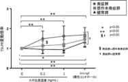
- Stimulations “ ⁇ ” and “+” in the figure indicate the absence and presence of cedar antigen stimulation, respectively. “*” In the figure indicates that there is a statistically significant difference (p ⁇ 0.05). It is a figure which shows the result of having analyzed the expression level of TSLPR in a basophil using the flow cytometer about said 3 groups.
- the “TSLPR fluctuation magnification” on the vertical axis represents the ratio of the expression level of TSLPR in the cedar antigen stimulation to the expression level of TSLPR in the cedar antigen unstimulated.
- FIG. 6A It is a figure which shows the result (FIG. 6B) which determined both groups based on the expression level of TSLPR in a base sphere.
- the solid line in the figure indicates the average value, and the dotted line indicates the threshold value.
- “*” In the figure indicates that there is a statistically significant difference (P ⁇ 0.0001).
- FIG. 8 is a diagram showing a result of combining a method for determining both groups (FIG. 7B).
- the solid line in the figure indicates the average value, and the dotted line indicates the threshold value.
- “*” In the figure indicates that there is a statistically significant difference (P ⁇ 0.05).
- the method for collecting data for diagnosing allergic rhinitis includes mRNA of human CRLF2 (Cytokine receptor-like factor 2) gene in a biological sample collected from a subject (provider) or a reverse transcript thereof. Increased expression of (cDNA), increased expression of human ETV7 (ets variant 7) gene mRNA or cDNA in the biological sample, and increased expression of a protein encoded by the human CRLF2 gene (human CRLF2 protein) in the biological sample. Or a method of collecting diagnostic data for allergic rhinitis that detects an increase in the expression of a protein encoded by the human ETV7 gene (human ETV7 protein) in the biological sample and quantifies it as necessary (hereinafter referred to as “the collection of the present case”).
- the method is not particularly limited.
- the diagnostic kit for allergic rhinitis of the present invention is for detecting the expression of human CRLF2 gene mRNA or cDNA in the biological sample, or the expression of human ETV7 gene mRNA or cDNA in the biological sample.
- a kit comprising a primer or a probe, or a label thereof for use in diagnosis of allergic rhinitis (hereinafter referred to as “the present diagnostic kit 1”), or specifically binds to human CRLF2 protein in the biological sample.
- a kit for diagnosing allergic rhinitis comprising an antibody or a labeled product thereof, an antibody that specifically binds to human ETV7 protein in the biological sample, or a labeled product thereof (hereinafter referred to as “this diagnostic Kit 2 ”) is not particularly limited, and the diagnostic kits 1 and 2 are allergic rhinitis. It is a use invention related to a kit for diagnosis, and these kits are used for diagnosing allergic rhinitis in addition to components generally used in this type of diagnostic kit, for example, carriers, pH buffers, stabilizers, etc. Attached documents such as instructions are usually included.
- a biomarker for diagnosing allergic rhinitis of the present invention a biomarker for diagnosing allergic rhinitis in a subject (hereinafter referred to as “the human CRLF2 gene or ETV7 gene” or human CRLF2 protein or ETV7 protein).
- the human CRLF2 gene or ETV7 gene or human CRLF2 protein or ETV7 protein.
- the present biomarker There is no particular limitation as long as it is referred to as “the present biomarker”.
- the screening method for the preventive or therapeutic agent for allergic rhinitis of the present invention includes the step (a) of administering a test drug or test substance to a non-human animal with allergic rhinitis;
- a method comprising sequentially detecting an increase in the expression of mRNA or cDNA of a non-human CRLF2 gene or ETV7 gene or an increase in the expression of a non-human CRLF2 protein or ETV7 protein in a biological sample (hereinafter referred to as “the present screening method”). If it says, it will not be restrict
- the above "diagnosis of allergic rhinitis” includes the determination of whether or not allergic rhinitis has occurred, more specifically, allergic rhinitis sensitization positive onset, allergic rhinitis sensitization negative onset or sensitization positive not on Classification of onset and determination of the risk of developing allergic rhinitis, more specifically, classification of allergic rhinitis positive onset of sensitization and allergic rhinitis sensitization positive onset.
- human CRLF2 gene mRNA or cDNA expression increase and human ETV7 gene mRNA or cDNA expression increase are performed simultaneously, sequentially, or individually. And a method of detecting an increase in expression of human CRLF2 protein and an increase in expression of human ETV7 protein simultaneously, sequentially or individually.
- the present diagnostic kit 1 from the viewpoint of providing a more accurate diagnostic kit, primers or probes for detecting the expression of mRNA or cDNA of the human CRLF2 gene, or a label thereof, human ETV7
- a kit comprising a primer or probe for detecting the expression of mRNA or cDNA of a gene, or a label thereof is preferable.
- the diagnostic kit 2 specifically binds to an antibody that specifically binds to human CRLF2 protein, or a label thereof, and human ETV7 protein.
- a kit comprising an antibody or a label thereof is preferred.
- the present biomarker is preferably one consisting of a human CRLF2 gene and an ETV7 gene, or one consisting of a human CRLF2 protein and an ETV7 protein, from the viewpoint of providing a more accurate diagnostic biomarker.
- a decrease in the expression of mRNA or cDNA of the non-human CRLF2 gene and a non-human ETV7 gene Of detecting the decrease in the expression of mRNA or cDNA simultaneously, sequentially or individually, or detecting the decrease in the expression of non-human CRLF2 protein and the decrease in expression of the non-human ETV7 protein simultaneously, sequentially or individually The method is preferred.
- allergic rhinitis means that an antibody is produced in a living body by ingestion or contact of a certain exogenous substance, and an antigen-antibody reaction by reuptake or recontact of the same exogenous substance (allergen [antigen]).
- allergen [antigen] refers to type I allergic (allergic to IgE antibodies) inflammation in the nasal mucosa where allergic rhinitis symptoms (nasal congestion, recurrent sneezing, and / or aqueous rhinorrhea) appear.
- allergic rhinitis sensitization negative non-onset means a state in which no increase in the subject allergen-specific IgE antibody is observed, and no symptoms of the allergic rhinitis are observed
- Allergy rhinitis sensitization positive non-occurrence means a state in which an increase in the allergen-specific IgE antibody in the subject is observed and no symptoms of the allergic rhinitis are observed.
- Positive onset means a state in which an increase in the subject's allergen-specific IgE antibody is observed and symptoms of the allergic rhinitis are observed.
- the above allergic rhinitis is classified into year-round allergic rhinitis and seasonal allergic rhinitis from the beginning.
- Such perennial allergic rhinitis can develop throughout the year due to house dust (eg, mold, fungal spores, textile fibers, animal dander, mites, insect carcasses) and the like.
- house dust eg, mold, fungal spores, textile fibers, animal dander, mites, insect carcasses
- the above-mentioned seasonal allergic rhinitis can occur at a specific time of the year due to pollen and the like.
- the allergic rhinitis include allergic rhinitis caused by the house dust, Japanese cedar pollinosis, Japanese cypress pollinosis, ragweed hay fever, rice hay fever, Japanese zelkova hay fever, Japanese white hay fever, white birch pollinosis, Japanese oak Examples include hay fever, alder pollinosis, and pine hay fever.
- cedar pollinosis and allergic rhinitis caused by ticks tick allergic rhinitis are preferable.
- the subject is an allergic rhinitis such as a subject who has not developed sensitization positive for allergic rhinitis or a subject who may have other diseases such as bacterial rhinitis or viral rhinitis Can be mentioned.
- Such subjects who are unclear as to whether or not they have allergic rhinitis include subjects who have suffered from allergic rhinitis in the past and who are unclear as to whether or not they have allergic rhinitis at the time of examination.
- the biological sample examples include non-liquid samples such as tissues, cells and organs, liquid samples such as blood, urine and saliva, and samples containing basophils prepared from blood. Of these, blood or a sample containing basophils prepared from blood is preferable.
- the expression level of human CRLF2 gene mRNA or cDNA or the expression level of human CRLF2 protein is usually a biological sample that is not stimulated by the target allergen (hereinafter referred to as “allergic rhinitis”).
- allergic rhinitis a biological sample that is not stimulated by the target allergen
- it may be referred to as an “unstimulated sample” and a biological sample stimulated with the target allergen (hereinafter sometimes referred to as “stimulated sample” for the sake of convenience).
- the expression level of mRNA or cDNA of the human CRLF2 gene hardly changes between the “unstimulated sample” derived from the subject and the “stimulated sample”, or the “unstimulated sample derived from the subject” If the expression level of human CRLF2 protein is almost the same between the “stimulation sample” and the “stimulation sample”, the data for diagnosing that such a subject is likely to be a sensitization-negative non-developed person with allergic rhinitis Can be collected.
- the expression level of human CRLF2 gene mRNA or cDNA or the expression level of human CRLF2 protein is usually higher than that of “unstimulated sample”.
- the “sample” increases.
- the relative value of the increase level of the “stimulation sample” with respect to the “unstimulated sample” derived from such a sensitization-positive undeveloped person ratio; hereinafter referred to as “CRLF2 ratio in non-sensitized persons” for convenience
- human CRLF2 protein derived from basophils when detected by an immunoassay, it is usually 1.01 to 1.4, for example, 1.01 to 1.35, 1.01 to 1.3, 1.01 to 1.25, 1.01 to 1.2, 1.01 to 1.15 1.01 to 1.1, 1.01 to 1.05, 1.05 to 1.35, 1.05 to 1.3, 1.05 to 1.25, 1.05 to 1.2, 1 .05 to 1.15, 1.05 to 1.1, 1.1 to 1.35, 1.1 to 1.3, 1 1 to 1.25, 1.1 to 1.2, 1.1 to 1.15, 1.15 to 1.35, 1.15 to 1.3, 1.15 to 1.25, 1.15 to 1.20, 1.2-1.35, 1.2-1.3, 1.2-1.25, 1.25-1.35, 1.25-1.3, 1.3-1. 35, 1.05-1.4, 1.1-1.4, 1.15-1.4, 1.2-1.4, 1.25-1.4, 1.3-1.4, Examples include 1.35 to 1.4.
- CRLF2 ratio in non-sensitized persons is usually 1.2 or more, for example, 1.3 or more, 1.6 or more, 1.7 or more, 2.0 or more, 2.3 or more, 2.6 or more. 3.0 or more, 3.3 or more, 3.6 or more, 4.0 or more, 4.3 or more, 4.6 or more, 5.0 or more, 5.3 or more, 5.6 or more, 6.0 or more 6.3 or more, 6.6 or more, 7.0 or more.
- the expression level of human CRLF2 gene mRNA or cDNA in the “stimulated sample” derived from the subject relative to the expression level of human CRLF2 gene mRNA or cDNA in the “unstimulated sample” derived from the subject. If the ratio is greater than or equal to the “CRLF2 ratio in non-sensitized persons”, such subjects can collect data to diagnose that they are likely to be non-sensitized persons with allergic rhinitis. .
- the expression level of human CRLF2 gene mRNA or cDNA and the expression level of human CRLF2 protein are usually higher in “stimulated sample” than in “unstimulated sample”. Will increase.
- the relative value of the increase level of the “stimulation sample” with respect to the “unstimulated sample” derived from the onset person ratio; hereinafter sometimes referred to as “CRLF2 ratio in the onset person” for convenience
- basophil-derived human CRLF2 protein is detected by immunoassay, it is usually 1.05 or higher. For example, 1.
- the ratio of the expression level of the human CRLF2 protein in the “stimulation sample” derived from the subject to the expression level of the human CRLF2 protein in the “unstimulated sample” derived from the subject is “CRLF2 in the onset subject”. If the ratio is greater than or equal to, the data can be collected to diagnose that such subject is likely to be a sensitization positive onset of allergic rhinitis.
- CRLF2 ratio in the onset person is usually 1.2 or more, for example, 1.3 or more, 1.6 or more, 1.9 or more, 2.0 or more, 2.3 or more, 2.6 or more, 3 or more, 0.0 or more, 3.3 or more, 3.6 or more, 4.0 or more, 4.3 or more, 4.6 or more, 5.0 or more, 5.3 or more, 5.6 or more, 6.0 or more, 6 .3 or more, 6.6 or more, 7.0 or more.
- the expression level of human CRLF2 gene mRNA or cDNA in the “stimulated sample” derived from the subject relative to the expression level of human CRLF2 gene mRNA or cDNA in the “unstimulated sample” derived from the subject.
- the ratio is equal to or higher than the “CRLF2 ratio in the onset person”
- the subject can collect data for diagnosing that the person is highly likely to be a sensitization-positive onset person of allergic rhinitis.
- the expression level of human ETV7 gene mRNA or cDNA and the expression level of human ETV7 protein are usually higher than that of “unstimulated sample”. Will increase.
- the relative value of the increase level of the “stimulation sample” with respect to the “unstimulated sample” derived from such a sensitization positive onset subject is a biological sample to be detected, Since it varies depending on the concentration of allergen to be stimulated, the detection method, etc., it cannot be specified in general.
- human ETV7 gene cDNA When the expression of human ETV7 gene cDNA is detected by quantitative PCR, it is usually 1.2 or more, for example, 1.5 or more, 2.0 or more, 2.5 or more, 3.0 or more, 3.5 or more. 4.0 or more, 4.5 or more, 5.0 or more, 5.5 or more, 6.0 or more, 6.5 or more, 7.0 or more, 7.5 or more, 8.0 or more, 8.5 or more 9.0 or more, 9.5 or more, 10 or more, etc. It can be mentioned.
- the expression level of human CRLF2 gene mRNA or cDNA in the “stimulation sample” derived from the subject relative to the expression level of human ETV7 gene mRNA or cDNA in the “unstimulated sample” derived from the subject.
- the expression of human ETV7 protein in the “stimulated sample” derived from the subject relative to the expression level of human ETV7 protein in the “unstimulated sample” derived from the subject when the ratio is equal to or greater than the “ETV7 ratio in the onset” If the amount ratio is greater than or equal to the “ETV7 ratio in onset”, such subjects can collect data to diagnose that they are likely to be sensitization-positive onset of allergic rhinitis.
- the expression level of mRNA or cDNA of the human CRLF2 gene in the “stimulation sample” relative to the “unstimulated sample” derived from the subject increases, and the “stimulation” for the “unstimulated sample” derived from the subject If the expression level of human ETV7 gene mRNA or cDNA in the “sample” does not increase, such a subject may collect data for diagnosing that it is highly likely that the subject is not sensitized positive in allergic rhinitis. it can.
- the expression level ratio of human CRLF2 gene or ETV7 gene mRNA or cDNA, or the expression level ratio of CRLF2 protein or ETV7 protein in the “stimulation sample” relative to the “unstimulated sample” is a threshold (cutoff value). It can also be determined whether the expression of these mRNA, cDNA, or protein is increased by using whether or not it is higher.
- the above threshold value is determined using a standard method, for example, a ROC (Receiver-Operating-Characteristic) curve using statistical analysis software based on "CRLF2 ratio in non-sensitized persons" data or "CRLF2 ratio in onset persons” data. Can be calculated.
- the “stimulation sample” can be prepared by culturing a biological sample in the presence of an allergen.
- the culture period of the biological sample is not particularly limited, and is, for example, 10 minutes to 2 days, preferably 1 to 12 hours.
- the culture medium used for culturing the biological sample is not particularly limited.
- a basic culture medium for animal cell culture DMEM, EMEM, RPMI
- FBS Fetal bovine serum
- the concentration of allergen in the culture solution is not particularly limited and is, for example, in the range of 0.01 to 1 ng / mL, preferably 0.05 to 0.2 ng / mL.
- the culture temperature is usually in the range of 30 to 40 ° C., preferably about 37 ° C.
- the CO 2 concentration during the cultivation is usually within the range of about 1 to 10%, preferably about 5%.
- the humidity during the cultivation is usually in the range of about 70 to 100%, preferably in the range of about 95 to 100%.
- the “unstimulated sample” is preferably prepared under the same conditions as the “stimulated sample” in the absence of allergen.
- human CRLF2 gene include one or more polynucleotides selected from the following [Group A polynucleotide].
- Group A polynucleotide (1) A polynucleotide comprising the nucleotide sequence shown in SEQ ID NO: 1 (cDNA encoding CRLF2 isoform 1 [NCBI Reference Sequence: NM — 022148]), or one or several in the nucleotide sequence shown in SEQ ID NO: 1.
- human CRLF2 protein include one or more proteins selected from the following [Group A proteins].
- Group A protein (1) A protein comprising the amino acid sequence shown in SEQ ID NO: 2 (CRLF2 isoform 1 [NCBI Reference Sequence: NP_071431]), or one or several amino acids deleted in the amino acid sequence shown in SEQ ID NO: 2 A protein consisting of a substituted and / or added amino acid sequence and having increased expression in a subject compared to a control; (2) A protein consisting of the amino acid sequence shown in SEQ ID NO: 4 (CRLF2 isoform 2 [NCBI Reference Sequence: NP_001012288]) or the amino acid sequence shown in SEQ ID NO: 4 has one or several amino acids deleted A protein consisting of a substituted and / or added amino acid sequence and having increased expression in a subject compared to a control;
- the human ETV7 gene include one or more polynucleotides selected from the following [Group B polynucleotides].
- Group B polynucleotide (1) A polynucleotide comprising the nucleotide sequence shown in SEQ ID NO: 5 (cDNA encoding ETV7 isoform 1 [NCBI Reference Sequence: NM — 016135]), or one or several in the nucleotide sequence shown in SEQ ID NO: 5 A polynucleotide whose nucleotide sequence is deleted, substituted and / or added and whose expression in a subject is increased compared to a control; (2) a polynucleotide comprising the nucleotide sequence represented by SEQ ID NO: 7 (cDNA encoding isoform 2 of ETV7 [NCBI Reference Sequence: NM_001207035]), Alternatively, in the nucleotide sequence shown in SEQ ID NO: 7, a polynucleotide consisting of a nucleo
- human ETV7 protein examples include one or more proteins selected from the following [Group B proteins].
- Group B proteins (1) A protein consisting of the amino acid sequence shown in SEQ ID NO: 6 (isoform 1 of ETV7 [NCBI Reference Sequence: NP — 057219]), or one or several amino acids deleted in the amino acid sequence shown in SEQ ID NO: 6.
- a protein consisting of a substituted and / or added amino acid sequence and having increased expression in a subject compared to a control (2) A protein comprising the amino acid sequence shown in SEQ ID NO: 8 (ETV7 isoform 2 [NCBI Reference Sequence: NP_001193964]) or the amino acid sequence shown in SEQ ID NO: 8 with one or several amino acids deleted A protein consisting of a substituted and / or added amino acid sequence and having increased expression in a subject compared to a control; (3) A protein comprising the amino acid sequence shown in SEQ ID NO: 10 (ETV7 isoform 3 [NCBI Reference Sequence: NP_001193965]) or the amino acid sequence shown in SEQ ID NO: 10 with one or several amino acids deleted A protein consisting of a substituted and / or added amino acid sequence and having increased expression in a subject compared to a control; (4) Protein consisting of the amino acid sequence shown in SEQ ID NO: 12 (ETV7 isoform 4 [NCBI Reference Sequence: NP_00
- nucleotide sequence in which one or several nucleotides are deleted, substituted and / or added is usually in the range of 1 to 10, preferably in the range of 1 to 7, more preferably in the range of 1 to 6. Within the range, more preferably within the range of 1 to 5, more preferably within the range of 1 to 4, more preferably within the range of 1 to 3, more preferably within the range of 1 to 2, most preferably Preferably, it means a nucleotide sequence in which one number of nucleotides is deleted, substituted and / or added.
- amino acid sequence in which one or several amino acids are deleted, substituted and / or added is usually in the range of 1 to 10, preferably in the range of 1 to 7, more preferably in the range of 1 to 6. Within the range, more preferably within the range of 1 to 5, more preferably within the range of 1 to 4, more preferably within the range of 1 to 3, more preferably within the range of 1 to 2, most preferably Preferably, it means an amino acid sequence in which one number of amino acids are deleted, substituted and / or added.
- the expression level of CRLF2 or ETV7 gene mRNA or cDNA is detected and quantified by a method that can specifically detect a part or all of the CRLF2 or ETV7 gene mRNA or cDNA.
- any method may be used, and specifically, a method of extracting and purifying total RNA in cells in a biological sample and performing RNA-Seq analysis, or the above-mentioned total RNA is mRNA of CRLF2 or ETV7 gene
- a method of detecting by quantitative PCR methods such as dynamic PCR method, real-time PCR method, etc., and the above-mentioned cDNA is detected with a CRLF2 or ETV7 gene detection probe (CRLF2 or ETV7 gene cDNA labeled with a labeling substance such as biotin and avidin) And a method of detecting with a microarray using a material immobilized on a support that can be used for hybridization, such as silicon and plastic.
- any method can be used for detecting and quantifying the expression level of CRLF2 or ETV7 protein as long as it can specifically detect a part or all of CRLF2 or ETV7 protein.
- Specific examples include mass spectrometry for detecting peptides constituting CRLF2 or ETV7 protein, and immunological measurement methods using an antibody that specifically recognizes CRLF2 or ETV7 protein.
- immunohistochemical staining method As the immunological measurement method, immunohistochemical staining method, ELISA method, EIA method, RIA method, Western blotting method, flow cytometry and the like can be preferably exemplified.
- Flow cytometry is performed using fluorescent substances (allophycocyanin [APC], phycoerythrin [PE], FITC [fluorescein isothiocyanate], Alexa Fluor 488, Alexa Fluor 647, Alexa Fluor 700, PE-Texas Red, PE-Cy7, PE-Cy7, etc. ), And fluorescence activated cell sorter (FACS) using an antibody that specifically binds to CRLF2 or ETV7 protein.
- APC allophycocyanin
- PE phycoerythrin
- FITC fluorescein isothiocyanate
- Alexa Fluor 488 Alexa Fluor 647
- Alexa Fluor 700 Alexa Fluor 700
- PE-Texas Red PE
- CRLF2 protein is a cell surface receptor
- CRLF2 protein in cells such as basophils can be detected in the state of living cells. For this reason, when detecting and quantifying the expression level of CRLF2 protein in cells such as basophils, it is preferable to use flow cytometry in consideration of simplicity.
- a method for detecting an increase or decrease in the expression of allergen-specific IgE simultaneously, sequentially or individually Is preferred is particularly preferably performed before the collection method.
- the method for detecting an increase or decrease in the expression of allergen-specific IgE may be any method that can detect and quantify the expression level of allergen-specific IgE in a biological sample.
- CAP Capsulated Hydrophilic Carrier Test
- RAST Radioallergosorbent Test
- mast cell which detects the binding of allergen-specific IgE and allergen in a biological sample by contacting the biological sample with an allergen (antigen)
- HRT histamine release test
- BAT Basophil activation test
- Basophil activation test BAT
- primers in the present diagnostic kit complementary primer sets that can be annealed with a part of the upstream or downstream sequence of mRNA or cDNA of human CRLF2 or ETV7 gene (for convenience, they are referred to as “forward primer and reverse primer”, respectively).
- the length of the primer sequence, the site for annealing with the cDNA, the length of the cDNA to be amplified, and the like can be appropriately selected in consideration of the amplification efficiency and specificity of the cDNA.
- the length of the primer sequence is usually 10 to 100 bases, preferably 10 to 40 bases, more preferably 10 to 30 bases, and further preferably 15 to 30 bases. .
- the forward primer and the reverse primer are usually selected so that an amplification product derived from a polynucleotide selected from the above [Group A polynucleotide] and [Group B polynucleotide] as template DNA is specifically generated. . That is, in the above [Group A polynucleotide] and [Group B polynucleotide], when the first nucleotide sequence is upstream and the tail is downstream, the nucleotide at the 3 ′ end of the forward primer is Considering avoidance of false positives due to double-stranded DNA formation (primer dimer formation) by the reverse primer, it is usually selected to anneal at least upstream from the nucleotide at the 3 ′ end of the reverse primer.
- the forward primer and the reverse primer are annealed (hybridized) with a part of a template DNA (a polynucleotide selected from the above [Group A polynucleotide] and [Group B polynucleotide]), and Any PCR product may be used as long as it can generate an amplification product.
- “at least a part” of the forward primer or reverse primer generally means 60% or more of the nucleotide sequence of the forward primer or reverse primer, preferably Is 65% or more, more preferably 70% or more, still more preferably 75% or more, even more preferably 80% or more, particularly preferably 85% or more, and most preferably 90% or more. Means.
- the length of the probe As a probe in the present diagnostic kit 1, if a part or all of mRNA or cDNA of human CRLF2 or ETV7 gene is hybridized, the length of the probe, the hybridization site, etc. It can be appropriately selected in consideration of specificity.
- the length of the probe is usually 50 to 2000 bases, preferably 100 to 1500 bases, more preferably 200 to 1000 bases, and further preferably 300 to 800 bases.
- the probe is usually selected so as to anneal (hybridize) to a polynucleotide selected from the above-mentioned [Group A polynucleotide] and [Group B polynucleotide] which is a template DNA.
- a polynucleotide selected from the above-mentioned [Group A polynucleotide] and [Group B polynucleotide] is a template DNA.
- “at least a part” of the probe usually means 60% or more, preferably 65% or more, more preferably 70% or more, more preferably, relative to the nucleotide sequence of the probe. It means 75% or more, still more preferably 80% or more, particularly preferably 85% or more, and most preferably 90% or more.
- the antibody in the present diagnostic kit 2 may be an antibody such as a monoclonal antibody, a polyclonal antibody, a human antibody, a chimeric antibody, or a humanized antibody, and among these, F (ab ′) 2 , Fab, Antibody fragments comprising a part of an antibody such as diabody, Fv, ScFv, Sc (Fv) 2 are also included.
- Examples of the labeling substance in the labeling product of the diagnostic kit 1 or 2 include peroxidase (for example, horseradish peroxidase), alkaline phosphatase, ⁇ -D-galactosidase, glucose oxidase, glucose-6-phosphate dehydrogenase, alcohol dehydrogenase, Enzymes such as malate dehydrogenase, penicillinase, catalase, apoglucose oxidase, urease, luciferase or acetylcholinesterase, fluorescent substances such as fluorescein isothiocyanate, phycobiliprotein, rare earth metal chelates, dansyl chloride or tetramethylrhodamine isothiocyanate , Green Fluorescence Protein (GFP), Cyan Fluorescence Protein (CFP), Blue Fluorescence Protein (B) Lue Fluorescence Protein (BFP), Yellow Fluorescence Protein (YFP), Red Fluorescence Protein (RFP), lucifer
- parenteral administration includes, for example, intravenous administration, intraarterial administration, intramuscular administration, intradermal administration, subcutaneous administration, intraperitoneal administration, intraventricular administration, intracranial administration, intranasal administration, intracolonic administration, trans Mention may be made of dermal administration.
- step (b) of the present screening method when the expression level of mRNA or cDNA of the non-human CRLF2 gene in the “stimulation sample” decreases before and after administration of the test drug or test substance, or in the “stimulation sample” When the expression level of the non-human CRLF2 protein decreases before and after administration of the test drug or test substance, such test drug or test substance is selected as a candidate drug or substance for the prevention or treatment of allergic rhinitis be able to.
- the test drug or test substance can be excluded as a candidate drug or substance for the prevention or treatment of allergic rhinitis.
- step (b) of the present screening method when the expression level of mRNA or cDNA of the non-human ETV7 gene in the “stimulation sample” decreases before and after administration of the test drug or test substance,
- the test drug or test substance is used as a candidate drug or substance for the prevention or treatment of allergic rhinitis. You can choose.
- test drug or test substance when the expression level of mRNA or cDNA of the non-human ETV7 gene does not decrease before and after administration of the test drug or test substance, or the expression level of the non-human ETV7 protein in the “stimulation sample” When it does not decrease before and after administration of the test substance, such test drug or test substance can be excluded as a candidate drug or substance for the prevention or treatment of allergic rhinitis.
- the allergic rhinitis non-human animal in this screening method may be a non-human animal that naturally developed allergic rhinitis, or the literature “The Journal of Complementary and Alternative Medicine, Vol. 9, No. 2, September 2012: 107-113 ”and the method described in the literature“ Haenuki, Y. et al., J. Allergy Clin. Immunol. 2012, 130: 184-194e11 ”.
- the non-human animal include mice, non-human mammals such as rats, hamsters, guinea pigs, monkeys, cows, pigs, horses, rabbits, sheep, goats, cats, and dogs.
- Non-human animal CRLF2 gene or protein is mouse (NCBI Gene ID: 57914), rat (NCBI Gene ID: 171499), dog (NCBI Gene ID: 491709), bovine (NCBI Gene ID: 529792), monkeys (NCBI Gene ID: 106995136) and the like.
- non-human animal ETV7 gene or protein includes chimpanzee (NCBI Gene ID: 747854), dog (NCBI Gene ID: 481764), cow (NCBI Gene ID: 529792), monkey ( NCBI Gene ID: 719151).
- RNA derived from basophils Six sensitization-positive non-sensitivity patients of cedar pollinosis (non-sensitization group), 11 sensitization-positive onset patients of cedar pollinosis (onset group), and cedar pollinosis 90 mL of blood was collected from a total of 22 people (16 males and 6 females 27- to 50-year-old) among 5 sensitization-negative patients (healthy group), and red blood cells were removed using HetaSep (manufactured by STEMCELL Technologies) Basophils were negatively isolated using EasySep Neg Human Basophil Kit (manufactured by STEMCELL Technologies).
- Basophils derived from the above three groups were present in the presence and absence of 0.1 ng / mL cedar pollen extract (manufactured by LSL), respectively. After incubation in RPMI-1640 medium containing 10% FBS for a time (37 ° C.), total RNA was purified using miRNeasy Mini kit (manufactured by Qiagen) according to the attached protocol.
- RNA sequence analysis and statistical analysis The RNA-Seq analysis using the total RNA derived from the basophils was commissioned to Kazusa DNA Laboratory.
- the expression level of RNA genes among the above three groups (non-sensitized group, sensitized group, and healthy group) with or without cedar antigen (cedar pollen) stimulation was determined by a statistical method (multi-group test [ANOVA method] ], Subordinate multiple test [Tukey method], 2 group test [paired t-test]), 12 genes including CRLF2 and ETV7 genes were identified as biomarkers for diagnosis of allergic rhinitis .
- Quantitative PCR method Further screening of the above 12 candidate genes was performed by quantitative PCR method. Quantitative PCR using TaqMan Gene Expression Assays (Applied Biosystems) was performed using the above basophil-derived total RNA as a template according to the protocol attached to the product. As a result, CRLF2 and ETV7 genes were identified as biomarkers for diagnosing allergic rhinitis. The GAPDH gene was used as an internal standard.
- the Assay ID of each gene detection primer / probe solution used for quantitative PCR is as follows. CRLF2 (Assay ID: Hs00845692_m1) ETV7 (Assay ID: Hs00903229_m1) Gapdh (Assay ID: Hs99999905_m1)
- the expression level of the CRLF2 gene mRNA is 2.46 ⁇ 1.00 (0.45) and both in the non-sensitized group and the onset group as compared with the non-stimulated group by stimulation with cedar pollen, respectively. 2.32 ⁇ 1.09 (0.33) -fold increase was shown (FIG. 1A, shown as “mean ⁇ standard deviation (standard error)”).
- the expression level of the cDNA of the CRLF2 gene was also 1.75 ⁇ 0.21 (both in the sensitized group and the onset group, respectively, by stimulation with cedar pollen, compared to the unstimulated group. 0.09) and 1.62 ⁇ 0.50 (0.15) -fold increase (FIG. 1B).
- the expression levels of ETV7 gene mRNA and cDNA were 3.12 ⁇ 3.04 (0.96) and 4.05 ⁇ 4 in the onset group, respectively, by stimulation with cedar pollen, compared to the case of no stimulation. It was shown to increase by .76 (1.80) times (FIGS. 2A and B).
- the expression levels of the mRNA and cDNA of the ETV7 gene were hardly changed even when stimulated with cedar pollen (FIGS. 2A and B). ).
- TSLP receptor constituted by basophil-derived CRLF2 protein
- TSLPR TSLP receptor
- 11 sensitization positive non-affected individuals of cedar pollinosis non-sensitized group
- 13 sensitization positive onset of cedar pollinosis onset group
- sensitization negative of cedar pollinosis 90 mL of blood was collected from a total of 33 unaffected 9 (healthy group), 4 hours (37 ° C.) in the presence and absence of 0.1 and 1 ng / mL cedar pollen extract (manufactured by LSL), respectively.
- basophils derived from 20 cedar pollinosis-free groups (9 healthy groups and 11 sensitized-unaffected groups) whose nasal provocation test is negative, and cedar pollen whose nasal provocation test is positive Basophils derived from 12 patients with onset of illness were stimulated with 0.1 ng / mL cedar pollen extract (manufactured by LSL) according to the method described in the item “4. Flow cytometry analysis (1)” above. Then, the expression level of TSLPR was measured, and the ratio (TSLPR fluctuation ratio) to the expression level of TSLPR in cedar pollen unstimulated was calculated.
- the ImmunoCAP method and the nasal provocation test were performed according to the method described in the document “Nasal Allergy Clinical Practice Guidelines-Perennial Rhinitis and Pollinosis-2016 Edition.
- the threshold value (cutoff value) of the TSLPR fluctuation ratio is set to 1.5
- the ratio (sensitivity) of true positive patients that are 1.5 or more in the onset group is 58.3% (7/12 )
- the ratio (specificity) of true-negative patients that are less than 1.5 in the undeveloped group is 95.0% (19/20)
- the true positive for the entire onset group and the non-onset group was as high as 81.3% (FIG. 4B).
- class 1 sugi-specific IgE antibody titer [UA / mL] is 0.35) that is suspected of being positive in the determination by cedar-specific IgE is set as a threshold
- the ratio (sensitivity) of true positive patients that is 1 or more (0.35 [UA / mL] or more) is 100% (12/12)
- it is less than class 1 (0.35 [0.3 [ (Specificity) is 45% (9/20)
- the ratio of true positive patients and true negative patients to the entire onset group and non-onset group (correction rate). ) was about 66.0% (FIG. 4A).
- class 5 Sugi-specific IgE antibody titer [UA / mL] is 50; upper dotted line in FIG. 5A) is set as a threshold value, and it corresponds to classes 1 to 5 Determination of 17 patients (patients within the range of the two dotted lines in FIG.
- the ratio (sensitivity) of true positive patients to be 57.1% (4/7) and the ratio (specificity) of true negative patients less than 1.5 in the non-onset group is 90% (9/9). 10), and the ratio (correct diagnosis rate) of the true positive patients and true negative patients to the entire onset group and non-onset group was 76.5% (FIG. 5B).
- This result is determined by the primary determination method based on the value of the cedar-specific IgE antibody of the conventional method, that it is determined that a person who is negative is likely to be a sensitization-negative non-developed person of cedar pollinosis.
- a person who is positive can be determined to have a high possibility of being a sensitization positive onset of Japanese cedar pollinosis, and a person who could not determine either positive or negative is a basophil-derived TSLPR
- the secondary determination method (the method of the present invention) is further performed based on the expression level of the expression, and in such a secondary determination method, a person who is positive is likely to be a sensitization positive onset person of cedar pollinosis.
- the method of the present invention is used to determine cedar pollinosis patients who could not be determined as sensitization-positive non-developed persons or onset patients by the conventional method. As a result, it is possible to determine the undeveloped group and the onset group of cedar pollinosis with high accuracy (correct diagnosis rate: 84.4%, 27/32).
- TSLPR fluctuation ratio The ratio (TSLPR fluctuation ratio) to the expression level of TSLPR in the mite antigen unstimulated was calculated.
- the ImmunoCAP method and the nasal provocation test were performed according to the method described in the document “Nasal Allergy Clinical Practice Guidelines-Perennial Rhinitis and Pollinosis-2016 Edition.
- the threshold value of the TSLPR fluctuation ratio is set to 1.5
- the ratio (sensitivity) of true positive patients who are 1.5 or more in the onset group is 81.8% (9/11)
- the ratio (specificity) of true negative patients that are less than 1.5 in the group is 93.8% (15/16)
- the ratio of the above true positive patients and true negative patients to the entire onset group and the non-onset group was as high as 88.9% (FIG. 6B).
- class 1 mite-specific IgE antibody titer [UA / mL] is 0.35
- the ratio (sensitivity) of true positive patients that is 1 or more (0.35 [UA / mL] or more) is 100% (11/11)
- it is less than class 1 (0.35 [UA (Specificity) is 62.5% (10/16)
- the ratio of the above-mentioned true positive patients and true negative patients to the entire onset group and non-onset group (correct diagnosis) The rate was about 77.8% (FIG. 6A).
- the method of the present invention based on the expression level of TSLPR derived from basophils is more effective than the conventional tick-specific IgE antibody value. It is shown that the accuracy (specificity and correct diagnosis rate) is superior to the determination method based on.
- the present invention is based on the method based on the tick-specific IgE antibody value of the conventional method and the expression level of TSLPR derived from basophils in order to determine the non-onset group and the onset group of tick allergic rhinitis.
- class 5 mite-specific IgE antibody titer [UA / mL] is 50; upper dotted line in FIG. 7A) is set as a threshold value, and it corresponds to classes 1 to 5
- the determination of 17 patients patients within the range of the two dotted lines in FIG.
- This result is determined by the primary determination method based on the tick-specific IgE antibody value of the conventional method, and it is determined that a person who is negative is likely to be a sensitization-negative non-developed person of tick allergic rhinitis
- a secondary determination method (the method of the present invention) based on the expression level of basophil-derived TSLPR was further performed, and the secondary determination method was positive. It can be determined that the person is likely to be a sensitization positive onset of tick allergic rhinitis, and the person who is negative is likely to be a non-sensitivity positive person of tick allergic rhinitis. It shows that it can be determined.
- the conventional method can be used to determine a tick allergic rhinitis patient who could not be determined as a sensitization-positive undeveloped person or an onset person by the method of the present invention.
- the method of the present invention it becomes possible to determine an undeveloped group and an onset group of tick allergic rhinitis with high accuracy (correct diagnosis rate: 88.9%, 24/27).
- the present invention contributes to the development of preventive or therapeutic agents for allergic rhinitis in addition to the diagnosis, prevention, and treatment of allergic rhinitis.
Abstract
The present invention addresses the problem of providing a marker and similar for diagnosis of allergic rhinitis, which makes it possible to accurately diagnose the risks of an allergic rhinitis outbreak as well as the presence or absence of the first symptoms of allergic rhinitis. By using as biomarker a human CRLF2 (Cytokine receptor-like factor 2) gene or a human ETV7 (ets variant 7) gene, and a human CRLF2 protein or a human ETV7 protein, the increased expression of the mRNA or cDNA of said gene and the increased expression of said protein is detected, and thus allergic rhinitis, more preferably cedar pollinosis, or the presence or absence of house dust mite allergic rhinitis and the risk of outbreak thereof are accurately diagnosed.
Description
本発明は、アレルギー性鼻炎を診断するためのデータを収集する方法や、アレルギー性鼻炎の診断用キットや、アレルギー性鼻炎を診断するためのバイオマーカーや、アレルギー性鼻炎の予防又は治療剤のスクリーニング方法に関する。
The present invention relates to a method for collecting data for diagnosing allergic rhinitis, a diagnostic kit for allergic rhinitis, a biomarker for diagnosing allergic rhinitis, and a screening agent for preventing or treating allergic rhinitis. Regarding the method.
アレルギー性鼻炎は、本来無害であるはずの外来抗原に対して、免疫系が過剰に反応することにより生じる、くしゃみ、鼻みず、鼻づまり等の鼻粘膜におけるアレルギー性疾患である。ここ数十年来、生活様式や生活環境の変化に伴い、アレルギー性鼻炎を罹患する患者が急増し、患者におけるQOL(Quality Of Life)の低下や医療費負担の増大が問題となっている。特にスギ花粉症等の花粉症患者は、カビ、ダニ等のハウスダストが原因で生じるアレルギー性鼻炎と同様に、年々増加の一途をたどっている。
Allergic rhinitis is an allergic disease in the nasal mucosa such as sneezing, nasal congestion, and nasal congestion caused by excessive reaction of the immune system to a foreign antigen that should be harmless. In recent decades, with changes in lifestyle and living environment, the number of patients suffering from allergic rhinitis has increased rapidly, and there has been a problem of a decrease in quality of life (QOL) and an increase in the burden of medical expenses. In particular, the number of hay fever patients such as Japanese cedar pollinosis is increasing year by year, as is allergic rhinitis caused by house dust such as mold and mites.
スギ花粉症の主因は、スギ花粉中の抗原性物質、すなわち、スギ抗原(アレルゲン)であると考えられている。大気中に飛散したスギ花粉がヒトの体内に侵入すると、B細胞によってスギ抗原に対するイムノグロブリンE抗体(IgE)が産生される。このIgEは、肥満細胞や好塩基球の細胞膜上に存在する高親和性IgE受容体FcεRIに結合する。この状態を「感作」といい、さらに、スギ抗原が再び侵入して、これら細胞膜上のIgEを架橋すると、ヒスタミンやロイコトリエン等のメディエータが放出され(脱顆粒反応)、アレルギー性鼻炎が発症する。
The main cause of cedar pollinosis is considered to be an antigenic substance in cedar pollen, that is, cedar antigen (allergen). When cedar pollen scattered in the atmosphere enters the human body, immunoglobulin E antibodies (IgE) against cedar antigens are produced by B cells. This IgE binds to the high affinity IgE receptor FcεRI present on the cell membranes of mast cells and basophils. This state is called "sensitization". Furthermore, when cedar antigens re-enter and IgE on these cell membranes is cross-linked, mediators such as histamine and leukotriene are released (degranulation reaction) and allergic rhinitis develops. .
アレルギー性鼻炎の治療は、主として抗ヒスタミン剤を代表とする抗アレルギー剤、吸入ステロイド薬等の薬物療法により行われている。また、花粉飛散前に抗アレルギー薬を投与する初期療法(季節前治療)が、飛散開始後又は症状増悪後より治療を開始するより所見、症状がよくなることが示唆されている。
Treatment of allergic rhinitis is carried out mainly by drug therapy such as antiallergic agents represented by antihistamines and inhaled steroids. In addition, it has been suggested that the initial therapy (pre-seasonal treatment) in which an antiallergic drug is administered before pollen disperses improves the findings and symptoms from the start of the dispersal or symptom exacerbation.
アレルギー性鼻炎の症状は、ウィルスや細菌等の感染においてもみられることから、適切なアレルギー性鼻炎の治療を行うためには、アレルギー性鼻炎を発症しているか否かを正確に判定することが重要である。一方、アレルギー性鼻炎の発症を予防するためには、アレルギー性鼻炎の発症の前段階である感作未発症状態を診断することにより、アレルギー性鼻炎の発症リスクを診断・予測する必要がある。しかしながら、未感作から感作、及び感作から発症に至るメカニズムはほとんど解明されていないこともあり、アレルギー性鼻炎の発症リスクを診断・予測する有効な手段がないのが現状である。
Symptoms of allergic rhinitis are also observed in infection with viruses and bacteria, so it is important to accurately determine whether or not allergic rhinitis has occurred in order to properly treat allergic rhinitis It is. On the other hand, in order to prevent the development of allergic rhinitis, it is necessary to diagnose and predict the risk of developing allergic rhinitis by diagnosing the non-sensitized state before the onset of allergic rhinitis. However, the mechanism from unsensitized to sensitized and from sensitized to onset may be hardly elucidated, and there is currently no effective means for diagnosing and predicting the risk of developing allergic rhinitis.
近年、タンパク質の解析技術が著しく発展し、罹患者等の生体組織におけるタンパク質量(の変動)に関する情報を網羅的に得ることができるようになった。この技術を利用して、アレルギー性鼻炎の改善レベルを評価するためのバイオマーカーとして、フィラグリンやアポリポタンパク質A-IVが報告されている(特許文献1、2)。しかし、かかるバイオマーカーは、アレルギー性鼻炎発症の有無や、アレルギー性鼻炎の発症リスクを診断・予測するものではない。
In recent years, protein analysis technology has been remarkably developed, and it has become possible to obtain comprehensive information on the amount (variation) of protein in living tissues of affected individuals and the like. Filaggrin and apolipoprotein A-IV have been reported as biomarkers for evaluating the improvement level of allergic rhinitis using this technique (Patent Documents 1 and 2). However, such biomarkers do not diagnose or predict the onset of allergic rhinitis or the risk of developing allergic rhinitis.
一方、CRLF2(Cytokine receptor-like factor 2)は、IL-7Rα鎖とヘテロ2量体を形成してTSLP(Thymic Stromal Lymphopoietin)受容体(TSLPR)として機能するタンパク質である。近年、ヒト末梢好塩基球において、TSLPR(CRLF2)が発現することが報告されている(非特許文献1)。また、アレルギー喘息患者由来の好塩基球におけるCRLF2の発現は、アレルゲン刺激により増加することが報告されている(非特許文献2)。しかしながら、CRLF2の発現増加と、アレルギー性鼻炎発症の有無や、アレルギー性鼻炎の発症リスクとの関連性については知られていなかった。
On the other hand, CRLF2 (Cytokine-receptor-like factor-2) is a protein that functions as a TSLP (Thymic-Stromal-Lymphopoietin) receptor (TSLPR) by forming a heterodimer with the IL-7Rα chain. In recent years, it has been reported that TSLPR (CRLF2) is expressed in human peripheral basophils (Non-patent Document 1). Moreover, it has been reported that the expression of CRLF2 in basophils derived from patients with allergic asthma is increased by allergen stimulation (Non-patent Document 2). However, the relationship between the increased expression of CRLF2, the presence of allergic rhinitis, and the risk of developing allergic rhinitis has not been known.
本発明の課題は、アレルギー性鼻炎発症の有無や、アレルギー性鼻炎の発症リスクを精度よく診断できるアレルギー性鼻炎の診断用マーカー等を提供することにある。
An object of the present invention is to provide a diagnostic marker for allergic rhinitis that can accurately diagnose the onset of allergic rhinitis and the risk of developing allergic rhinitis.
本発明者らは、上記課題を解決するため、アレルギー性鼻炎の感作陰性未発症者、感作陽性未発症、及び感作陽性発症者から採取された生体試料中の遺伝子発現量の変化を解析したところ、アレルギー性鼻炎発症の有無や、アレルギー性鼻炎の感作陽性発症と、アレルギー性鼻炎の感作陽性未発症とを精度よく分類することができるアレルギー性鼻炎の診断用マーカーを見いだし、本発明を完成するに至った。
In order to solve the above-mentioned problems, the present inventors performed changes in gene expression levels in biological samples collected from sensitization-negative non-developed allergic rhinitis patients, sensitization-positive undeveloped persons, and sensitization-positive onset persons. After analysis, we found a diagnostic marker for allergic rhinitis that can accurately classify the presence or absence of allergic rhinitis, sensitization positive onset of allergic rhinitis, and non-sensitivity of allergic rhinitis. The present invention has been completed.
すなわち、本発明は以下のとおりである。
〔1〕ヒトCRLF2遺伝子又はETV7遺伝子のmRNA若しくはcDNAの発現増加、或いは、ヒトCRLF2遺伝子又はETV7遺伝子がコードするタンパク質の発現増加を検出することを特徴とするアレルギー性鼻炎を診断するためのデータを収集する方法。
〔2〕ヒトCRLF2遺伝子及びETV7遺伝子のmRNA若しくはcDNAの発現増加、或いは、ヒトCRLF2遺伝子及びETV7遺伝子がコードするタンパク質の発現増加を検出することを特徴とする上記〔1〕に記載の方法。
〔3〕アレルギー性鼻炎が、スギ花粉症又はダニアレルギー性鼻炎であることを特徴とする上記〔1〕又は〔2〕に記載の方法。
〔4〕mRNA、cDNA及びタンパク質が、好塩基球由来のmRNA、cDNA及びタンパク質であることを特徴とする上記〔1〕~〔3〕のいずれかに記載の方法。
〔5〕さらに、アレルゲン特異的IgEの発現増加を検出することを特徴とする上記〔1〕~〔4〕のいずれかに記載の方法。
〔6〕ヒトCRLF2遺伝子又はETV7遺伝子のmRNA若しくはcDNAの発現を検出するためのプライマー若しくはプローブ、又はそれらの標識物を備えることを特徴とするアレルギー性鼻炎の診断用キット。
〔7〕ヒトCRLF2遺伝子のmRNA若しくはcDNAの発現を検出するためのプライマー若しくはプローブ、又はそれらの標識物と、ヒトETV7遺伝子のmRNA若しくはcDNAの発現を検出するためのプライマー若しくはプローブ、又はそれらの標識物とを備えることを特徴とする上記〔6〕に記載のキット。
〔8〕ヒトCRLF2遺伝子又はETV7遺伝子がコードするタンパク質に特異的に結合する抗体、又はそれらの標識物を備えることを特徴とするアレルギー性鼻炎の診断用キット。
〔9〕ヒトCRLF2遺伝子がコードするタンパク質に特異的に結合する抗体、又はそれらの標識物と、ヒトETV7遺伝子がコードするタンパク質に特異的に結合する抗体、又はそれらの標識物とを備えることを特徴とする上記〔8〕に記載のキット。
〔10〕アレルギー性鼻炎が、スギ花粉症又はダニアレルギー性鼻炎であることを特徴とする上記〔6〕~〔9〕のいずれかに記載のキット。
〔11〕ヒトCRLF2遺伝子又はETV7遺伝子、或いは、ヒトCRLF2遺伝子又はETV7遺伝子がコードするタンパク質からなることを特徴とするアレルギー性鼻炎を診断するためのバイオマーカー。
〔12〕ヒトCRLF2遺伝子と、ヒトETV7遺伝子とからなる、或いは、ヒトCRLF2遺伝子がコードするタンパク質と、ヒトETV7遺伝子がコードするタンパク質とからなることを特徴とする上記〔11〕に記載のバイオマーカー。
〔13〕アレルギー性鼻炎が、スギ花粉症又はダニアレルギー性鼻炎であることを特徴とする上記〔11〕又は〔12〕に記載のバイオマーカー。
〔14〕以下の工程(a)及び(b)を備えたことを特徴とするアレルギー性鼻炎の予防又は治療剤のスクリーニング方法。
(a)アレルギー性鼻炎非ヒト動物に被検薬剤又は被検物質を投与する工程;
(b)前記非ヒト動物から採取された生体試料中の、CRLF2遺伝子又はETV7遺伝子のmRNA若しくはcDNAの発現減少、或いは、CRLF2遺伝子又はETV7遺伝子がコードするタンパク質の発現減少を検出する工程;
〔15〕工程(b)において、CRLF2遺伝子及びETV7遺伝子のmRNA若しくはcDNAの発現減少、或いは、CRLF2遺伝子及びETV7遺伝子がコードするタンパク質の発現減少を検出することを特徴とする上記〔14〕に記載のスクリーニング方法。
〔16〕アレルギー性鼻炎が、スギ花粉症又はダニアレルギー性鼻炎であることを特徴とする上記〔14〕又は〔15〕に記載のスクリーニング方法。 That is, the present invention is as follows.
[1] Data for diagnosing allergic rhinitis characterized by detecting increased expression of mRNA or cDNA of human CRLF2 gene or ETV7 gene, or increased expression of protein encoded by human CRLF2 gene or ETV7 gene How to collect.
[2] The method according to [1] above, wherein an increase in expression of mRNA or cDNA of human CRLF2 gene and ETV7 gene or an increase in expression of protein encoded by human CRLF2 gene and ETV7 gene is detected.
[3] The method according to [1] or [2] above, wherein the allergic rhinitis is cedar pollinosis or tick allergic rhinitis.
[4] The method according to any one of [1] to [3] above, wherein the mRNA, cDNA and protein are basophil-derived mRNA, cDNA and protein.
[5] The method according to any one of [1] to [4] above, further comprising detecting an increase in expression of allergen-specific IgE.
[6] A diagnostic kit for allergic rhinitis comprising a primer or probe for detecting the expression of mRNA or cDNA of human CRLF2 gene or ETV7 gene, or a label thereof.
[7] Primer or probe for detecting the expression of human CRLF2 gene mRNA or cDNA, or label thereof, and primer or probe for detecting the expression of human ETV7 gene mRNA or cDNA, or label thereof The kit according to [6] above, comprising a product.
[8] A diagnostic kit for allergic rhinitis comprising an antibody that specifically binds to a protein encoded by a human CRLF2 gene or ETV7 gene, or a label thereof.
[9] An antibody that specifically binds to a protein encoded by the human CRLF2 gene, or a label thereof, and an antibody that specifically binds to a protein encoded by the human ETV7 gene, or a label thereof. The kit according to [8] above, which is characterized.
[10] The kit according to any one of [6] to [9] above, wherein the allergic rhinitis is cedar pollinosis or tick allergic rhinitis.
[11] A biomarker for diagnosing allergic rhinitis, comprising a human CRLF2 gene or ETV7 gene, or a protein encoded by a human CRLF2 gene or ETV7 gene.
[12] The biomarker according to [11] above, comprising a human CRLF2 gene and a human ETV7 gene, or comprising a protein encoded by the human CRLF2 gene and a protein encoded by the human ETV7 gene .
[13] The biomarker according to [11] or [12] above, wherein the allergic rhinitis is cedar pollinosis or tick allergic rhinitis.
[14] A screening method for a prophylactic or therapeutic agent for allergic rhinitis, comprising the following steps (a) and (b):
(A) a step of administering a test drug or a test substance to a non-human animal with allergic rhinitis;
(B) detecting a decrease in expression of mRNA or cDNA of CRLF2 gene or ETV7 gene or a decrease in expression of protein encoded by CRLF2 gene or ETV7 gene in a biological sample collected from the non-human animal;
[15] The method according to [14], wherein in step (b), a decrease in expression of mRNA or cDNA of CRLF2 gene and ETV7 gene or a decrease in expression of protein encoded by CRLF2 gene and ETV7 gene is detected. Screening method.
[16] The screening method of [14] or [15] above, wherein the allergic rhinitis is cedar pollinosis or tick allergic rhinitis.
また本発明のスクリーニング方法のその他の態様としては、アレルギー性鼻炎の予防又は治療剤(薬)の有効性を判定する方法や、アレルギー性鼻炎の予防剤(薬)候補又は治療剤(薬)候補のスクリーニング方法を挙げることができる。
Other aspects of the screening method of the present invention include a method for determining the effectiveness of a preventive or therapeutic agent (drug) for allergic rhinitis, a prophylactic agent (drug) candidate or a therapeutic agent (drug) candidate for allergic rhinitis. The screening method can be mentioned.
また本発明の実施の他の形態として、ヒトCRLF2遺伝子又はETV7遺伝子のmRNA若しくはcDNAの発現増加、或いは、ヒトCRLF2遺伝子又はETV7遺伝子がコードするタンパク質の発現増加を検出することを特徴とするアレルギー性鼻炎の診断方法や、アレルギー性鼻炎の診断における使用のためのヒトCRLF2遺伝子又はETV7遺伝子のmRNA若しくはcDNA、或いは、ヒトCRLF2遺伝子又はETV7遺伝子がコードするタンパク質を挙げることができる。
As another embodiment of the present invention, allergenicity characterized by detecting increased expression of mRNA or cDNA of human CRLF2 gene or ETV7 gene, or increased expression of protein encoded by human CRLF2 gene or ETV7 gene. Examples include rhinitis diagnostic methods, human CRLF2 gene or ETV7 gene mRNA or cDNA for use in diagnosis of allergic rhinitis, or proteins encoded by human CRLF2 gene or ETV7 gene.
本発明の収集方法によると、アレルギー性鼻炎発症の有無や、アレルギー性鼻炎の感作陽性発症と、アレルギー性鼻炎の感作陽性未発症とを精度よく診断(分類)できるため、アレルギー性鼻炎発症前に、アレルギー性鼻炎の発症を予防したり、アレルギー性鼻炎の適切な治療を行うことができる。また、本発明のスクリーニング方法は、アレルギー性鼻炎の予防(保護)又は治療剤(薬)の開発に資するものである。
According to the collection method of the present invention, it is possible to accurately diagnose (classify) the presence or absence of allergic rhinitis onset, sensitization positive onset of allergic rhinitis, and non-sensitivity of allergic rhinitis onset. Before, it is possible to prevent the development of allergic rhinitis or to perform appropriate treatment of allergic rhinitis. The screening method of the present invention contributes to the development of a preventive (protective) or therapeutic agent (medicine) for allergic rhinitis.
本発明のアレルギー性鼻炎を診断するためのデータを収集する方法としては、被験者(提供者)から採取された生体試料中のヒトCRLF2(Cytokine receptor-like factor 2)遺伝子のmRNA又はその逆転写産物(cDNA)の発現増加や、前記生体試料中のヒトETV7(ets variant 7)遺伝子のmRNA又はcDNAの発現増加や、前記生体試料中のヒトCRLF2遺伝子がコードするタンパク質(ヒトCRLF2タンパク質)の発現増加や、前記生体試料中のヒトETV7遺伝子がコードするタンパク質(ヒトETV7タンパク質)の発現増加を検出、及び必要に応じて定量する、アレルギー性鼻炎の診断用データを収集する方法(以下、「本件収集方法」という)であれば特に制限されない。
The method for collecting data for diagnosing allergic rhinitis according to the present invention includes mRNA of human CRLF2 (Cytokine receptor-like factor 2) gene in a biological sample collected from a subject (provider) or a reverse transcript thereof. Increased expression of (cDNA), increased expression of human ETV7 (ets variant 7) gene mRNA or cDNA in the biological sample, and increased expression of a protein encoded by the human CRLF2 gene (human CRLF2 protein) in the biological sample Or a method of collecting diagnostic data for allergic rhinitis that detects an increase in the expression of a protein encoded by the human ETV7 gene (human ETV7 protein) in the biological sample and quantifies it as necessary (hereinafter referred to as “the collection of the present case”). The method is not particularly limited.
また、本発明のアレルギー性鼻炎の診断用キットとしては、上記生体試料中のヒトCRLF2遺伝子のmRNA又はcDNAの発現や、上記生体試料中のヒトETV7遺伝子のmRNA又はcDNAの発現を検出するためのプライマー若しくはプローブ、又はそれらの標識物を備える、アレルギー性鼻炎の診断に用いるためのキット(以下、「本件診断用キット1」という)や、上記生体試料中のヒトCRLF2タンパク質に特異的に結合する抗体、又はそれらの標識物や、上記生体試料中のヒトETV7タンパク質に特異的に結合する抗体、又はそれらの標識物を備える、アレルギー性鼻炎の診断に用いるためのキット(以下、「本件診断用キット2」という)であれば特に制限されず、本件診断用キット1及び2は、アレルギー性鼻炎を診断するためのキットに関する用途発明であり、これらキットには、一般にこの種の診断キットに用いられる成分、例えば担体、pH緩衝剤、安定剤の他、取扱説明書、アレルギー性鼻炎を診断するための説明書等の添付文書が通常含まれる。
The diagnostic kit for allergic rhinitis of the present invention is for detecting the expression of human CRLF2 gene mRNA or cDNA in the biological sample, or the expression of human ETV7 gene mRNA or cDNA in the biological sample. A kit comprising a primer or a probe, or a label thereof for use in diagnosis of allergic rhinitis (hereinafter referred to as “the present diagnostic kit 1”), or specifically binds to human CRLF2 protein in the biological sample. A kit for diagnosing allergic rhinitis comprising an antibody or a labeled product thereof, an antibody that specifically binds to human ETV7 protein in the biological sample, or a labeled product thereof (hereinafter referred to as “this diagnostic Kit 2 ”) is not particularly limited, and the diagnostic kits 1 and 2 are allergic rhinitis. It is a use invention related to a kit for diagnosis, and these kits are used for diagnosing allergic rhinitis in addition to components generally used in this type of diagnostic kit, for example, carriers, pH buffers, stabilizers, etc. Attached documents such as instructions are usually included.
また、本発明のアレルギー性鼻炎を診断するためのバイオマーカーとしては、ヒトCRLF2遺伝子やETV7遺伝子、或いは、ヒトCRLF2タンパク質やETV7タンパク質からなる、被験者におけるアレルギー性鼻炎の診断用バイオマーカー(以下、「本件バイオマーカー」という)であれば特に制限されない。
In addition, as a biomarker for diagnosing allergic rhinitis of the present invention, a biomarker for diagnosing allergic rhinitis in a subject (hereinafter referred to as “the human CRLF2 gene or ETV7 gene” or human CRLF2 protein or ETV7 protein). There is no particular limitation as long as it is referred to as “the present biomarker”.
また、本発明のアレルギー性鼻炎の予防又は治療剤のスクリーニング方法としては、アレルギー性鼻炎非ヒト動物に被検薬剤又は被検物質を投与する工程(a);及び前記非ヒト動物から採取された生体試料中の、非ヒトCRLF2遺伝子やETV7遺伝子のmRNA若しくはcDNAの発現増加、或いは、非ヒトCRLF2タンパク質やETV7タンパク質の発現増加を検出する工程;を順次備えた方法(以下、「本件スクリーニング方法」という)であれば特に制限されない。
In addition, the screening method for the preventive or therapeutic agent for allergic rhinitis of the present invention includes the step (a) of administering a test drug or test substance to a non-human animal with allergic rhinitis; A method comprising sequentially detecting an increase in the expression of mRNA or cDNA of a non-human CRLF2 gene or ETV7 gene or an increase in the expression of a non-human CRLF2 protein or ETV7 protein in a biological sample (hereinafter referred to as “the present screening method”). If it says, it will not be restrict | limited in particular.
上記「アレルギー性鼻炎の診断」には、アレルギー性鼻炎発症の有無の判定、より具体的には、アレルギー性鼻炎の感作陽性発症と、アレルギー性鼻炎の感作陰性未発症又は感作陽性未発症との分類や、アレルギー性鼻炎発症リスクの判定、より具体的には、アレルギー性鼻炎の感作陽性発症と、アレルギー性鼻炎の感作陽性未発症との分類が含まれる。
The above "diagnosis of allergic rhinitis" includes the determination of whether or not allergic rhinitis has occurred, more specifically, allergic rhinitis sensitization positive onset, allergic rhinitis sensitization negative onset or sensitization positive not on Classification of onset and determination of the risk of developing allergic rhinitis, more specifically, classification of allergic rhinitis positive onset of sensitization and allergic rhinitis sensitization positive onset.
本件収集方法としては、より精度の高い診断用データを提供する観点から、ヒトCRLF2遺伝子のmRNA又はcDNAの発現増加と、ヒトETV7遺伝子のmRNA又はcDNAの発現増加とを、同時、逐次、又は個別に検出する方法や、ヒトCRLF2タンパク質の発現増加と、ヒトETV7タンパク質の発現増加とを、同時、逐次、又は個別に検出する方法が好ましい。
As the collection method, from the viewpoint of providing more accurate diagnostic data, human CRLF2 gene mRNA or cDNA expression increase and human ETV7 gene mRNA or cDNA expression increase are performed simultaneously, sequentially, or individually. And a method of detecting an increase in expression of human CRLF2 protein and an increase in expression of human ETV7 protein simultaneously, sequentially or individually.
また、本件診断用キット1としては、より精度の高い診断用キットを提供する観点から、ヒトCRLF2遺伝子のmRNA若しくはcDNAの発現を検出するためのプライマー若しくはプローブ、又はそれらの標識物と、ヒトETV7遺伝子のmRNA若しくはcDNAの発現を検出するためのプライマー若しくはプローブ、又はそれらの標識物とを備えるキットが好ましい。また、本件診断用キット2としては、より精度の高い診断用キットを提供する観点から、ヒトCRLF2タンパク質に特異的に結合する抗体、又はそれらの標識物と、ヒトETV7タンパク質に特異的に結合する抗体、又はそれらの標識物とを備えるキットが好ましい。
In addition, as the present diagnostic kit 1, from the viewpoint of providing a more accurate diagnostic kit, primers or probes for detecting the expression of mRNA or cDNA of the human CRLF2 gene, or a label thereof, human ETV7 A kit comprising a primer or probe for detecting the expression of mRNA or cDNA of a gene, or a label thereof is preferable. In addition, from the viewpoint of providing a more accurate diagnostic kit, the diagnostic kit 2 specifically binds to an antibody that specifically binds to human CRLF2 protein, or a label thereof, and human ETV7 protein. A kit comprising an antibody or a label thereof is preferred.
また、本件バイオマーカーとしては、より精度の高い診断用バイオマーカーを提供する観点から、ヒトCRLF2遺伝子とETV7遺伝子とからなるもの、又は、ヒトCRLF2タンパク質とETV7タンパク質とからなるものが好ましい。
In addition, the present biomarker is preferably one consisting of a human CRLF2 gene and an ETV7 gene, or one consisting of a human CRLF2 protein and an ETV7 protein, from the viewpoint of providing a more accurate diagnostic biomarker.
また、本件スクリーニング方法としては、より効果の高いアレルギー性鼻炎の予防又は治療剤をスクリーニングする観点から、上記工程(b)において、非ヒトCRLF2遺伝子のmRNA又はcDNAの発現減少と、非ヒトETV7遺伝子のmRNA又はcDNAの発現減少とを、同時、逐次、又は個別に検出する方法や、非ヒトCRLF2タンパク質の発現減少と、非ヒトETV7タンパク質の発現減少とを、同時、逐次、又は個別に検出する方法が好ましい。
In addition, as the screening method, from the viewpoint of screening for a more effective preventive or therapeutic agent for allergic rhinitis, in the above step (b), a decrease in the expression of mRNA or cDNA of the non-human CRLF2 gene and a non-human ETV7 gene Of detecting the decrease in the expression of mRNA or cDNA simultaneously, sequentially or individually, or detecting the decrease in the expression of non-human CRLF2 protein and the decrease in expression of the non-human ETV7 protein simultaneously, sequentially or individually The method is preferred.
本発明において「アレルギー性鼻炎」とは、ある種の外因性物質の摂取又は接触により生体内に抗体が作られ、同じ外因性物質(アレルゲン[抗原])の再摂取又は再接触により抗原抗体反応が生じて、アレルギー性鼻炎の症状(鼻閉、発作性反復性のくしゃみ、及び/又は水性鼻漏)が現れる、鼻粘膜におけるI型アレルギー性(IgE抗体によるアレルギー性)の炎症を意味する。
In the present invention, “allergic rhinitis” means that an antibody is produced in a living body by ingestion or contact of a certain exogenous substance, and an antigen-antibody reaction by reuptake or recontact of the same exogenous substance (allergen [antigen]). Refers to type I allergic (allergic to IgE antibodies) inflammation in the nasal mucosa where allergic rhinitis symptoms (nasal congestion, recurrent sneezing, and / or aqueous rhinorrhea) appear.
本発明において、「アレルギー性鼻炎の感作陰性未発症」とは、対象のアレルゲン特異的IgE抗体の増加が認められず、かつ、上記アレルギー性鼻炎の症状が認められない状態を意味し、「アレルギー性鼻炎の感作陽性未発症」とは、対象のアレルゲン特異的IgE抗体の増加が認められ、かつ、上記アレルギー性鼻炎の症状が認められない状態を意味し、「アレルギー性鼻炎の感作陽性発症」とは、対象のアレルゲン特異的IgE抗体の増加が認められ、かつ、上記アレルギー性鼻炎の症状が認められる状態を意味する。
In the present invention, “allergic rhinitis sensitization negative non-onset” means a state in which no increase in the subject allergen-specific IgE antibody is observed, and no symptoms of the allergic rhinitis are observed, “Allergy rhinitis sensitization positive non-occurrence” means a state in which an increase in the allergen-specific IgE antibody in the subject is observed and no symptoms of the allergic rhinitis are observed. “Positive onset” means a state in which an increase in the subject's allergen-specific IgE antibody is observed and symptoms of the allergic rhinitis are observed.
上記アレルギー性鼻炎は、始発時期から通年性のアレルギー性鼻炎と、季節性のアレルギー性鼻炎とに分類される。かかる通年性のアレルギー性鼻炎は、ハウスダスト(例えば、カビ、真菌の胞子、織物の繊維、動物の鱗屑、ダニ、昆虫の死骸)等が原因で、年間を通じて発症し得るものである。一方、上記季節性のアレルギー性鼻炎は、花粉等が原因で、一年の特定の時期に発症し得るものである。
The above allergic rhinitis is classified into year-round allergic rhinitis and seasonal allergic rhinitis from the beginning. Such perennial allergic rhinitis can develop throughout the year due to house dust (eg, mold, fungal spores, textile fibers, animal dander, mites, insect carcasses) and the like. On the other hand, the above-mentioned seasonal allergic rhinitis can occur at a specific time of the year due to pollen and the like.
上記アレルギー性鼻炎としては、具体的に、上記ハウスダストにより生じるアレルギー性鼻炎や、スギ花粉症、ヒノキ花粉症、ブタクサ花粉症、イネ花粉症、ケヤキ花粉症、カモガヤ花粉症、シラカバ花粉症、コナラ花粉症、ハンノキ花粉症、マツ属花粉症を挙げることができ、これらの中でもスギ花粉症や、ダニが原因で発症するアレルギー性鼻炎(ダニアレルギー性鼻炎)が好ましい。
Specific examples of the allergic rhinitis include allergic rhinitis caused by the house dust, Japanese cedar pollinosis, Japanese cypress pollinosis, ragweed hay fever, rice hay fever, Japanese zelkova hay fever, Japanese white hay fever, white birch pollinosis, Japanese oak Examples include hay fever, alder pollinosis, and pine hay fever. Among these, cedar pollinosis and allergic rhinitis caused by ticks (tick allergic rhinitis) are preferable.
上記被験者としては、アレルギー性鼻炎の感作陽性未発症の被検者や、細菌性鼻炎やウィルス性鼻炎等の他の疾患に罹患している可能性がある被験者などのアレルギー性鼻炎かどうか不明な被験者を挙げることができる。かかるアレルギー性鼻炎かどうか不明な被験者には、過去にアレルギー性鼻炎に罹患したことがあり、被検時にアレルギー性鼻炎かどうか不明な被験者も含まれる。
Whether the subject is an allergic rhinitis such as a subject who has not developed sensitization positive for allergic rhinitis or a subject who may have other diseases such as bacterial rhinitis or viral rhinitis Can be mentioned. Such subjects who are unclear as to whether or not they have allergic rhinitis include subjects who have suffered from allergic rhinitis in the past and who are unclear as to whether or not they have allergic rhinitis at the time of examination.
上記生体試料としては、組織、細胞、器官等の非液性試料や、血液、尿、唾液等の液性試料や、血液から調製された好塩基球を含む試料を挙げることができ、これらの中でも血液、又は血液から調製された好塩基球を含む試料が好ましい。
Examples of the biological sample include non-liquid samples such as tissues, cells and organs, liquid samples such as blood, urine and saliva, and samples containing basophils prepared from blood. Of these, blood or a sample containing basophils prepared from blood is preferable.
被験者がアレルギー性鼻炎の感作陰性未発症者である場合、ヒトCRLF2遺伝子のmRNA又はcDNAの発現量や、ヒトCRLF2タンパク質の発現量は、通常、対象のアレルゲンで刺激されていない生体試料(以下、便宜上「未刺激試料」ということがある)と、対象のアレルゲンで刺激されている生体試料(以下、便宜上「刺激試料」ということがある)との間でほとんど変わらない。
When the test subject is an allergic rhinitis sensitization-negative non-developed person, the expression level of human CRLF2 gene mRNA or cDNA or the expression level of human CRLF2 protein is usually a biological sample that is not stimulated by the target allergen (hereinafter referred to as “allergic rhinitis”). For the sake of convenience, it may be referred to as an “unstimulated sample”) and a biological sample stimulated with the target allergen (hereinafter sometimes referred to as “stimulated sample” for the sake of convenience).
したがって、本件収集方法において、被験者由来の「未刺激試料」と、「刺激試料」との間でヒトCRLF2遺伝子のmRNA又はcDNAの発現量がほとんど変わらない場合、或いは、被験者由来の「未刺激試料」と、「刺激試料」との間でヒトCRLF2タンパク質の発現量がほとんど変わらない場合、かかる被験者は、アレルギー性鼻炎の感作陰性未発症者である可能性が高いと診断するためのデータを収集することができる。
Therefore, in the present collection method, when the expression level of mRNA or cDNA of the human CRLF2 gene hardly changes between the “unstimulated sample” derived from the subject and the “stimulated sample”, or the “unstimulated sample derived from the subject” If the expression level of human CRLF2 protein is almost the same between the “stimulation sample” and the “stimulation sample”, the data for diagnosing that such a subject is likely to be a sensitization-negative non-developed person with allergic rhinitis Can be collected.
また、被験者がアレルギー性鼻炎の感作陽性未発症者である場合、ヒトCRLF2遺伝子のmRNA又はcDNAの発現量や、ヒトCRLF2タンパク質の発現量は、通常、「未刺激試料」と比べ、「刺激試料」の方が増加する。かかる感作陽性未発症者由来の「未刺激試料」に対する「刺激試料」の増加レベルの相対値(比率;以下、便宜上「感作未発症者におけるCRLF2比率」ということがある)は、検出対象の生体試料、刺激するアレルゲンの濃度、検出方法等により異なるため、一概に特定することはできないが、好塩基球由来のヒトCRLF2タンパク質を免疫学的測定法で検出する場合、通常1.01~1.4の範囲内であり、例えば、1.01~1.35、1.01~1.3、1.01~1.25、1.01~1.2、1.01~1.15、1.01~1.1、1.01~1.05、1.05~1.35、1.05~1.3、1.05~1.25、1.05~1.2、1.05~1.15、1.05~1.1、1.1~1.35、1.1~1.3、1.1~1.25、1.1~1.2、1.1~1.15、1.15~1.35、1.15~1.3、1.15~1.25、1.15~1.20、1.2~1.35、1.2~1.3、1.2~1.25、1.25~1.35、1.25~1.3、1.3~1.35、1.05~1.4、1.1~1.4、1.15~1.4、1.2~1.4、1.25~1.4、1.3~1.4、1.35~1.4等を挙げることができる。
In addition, when the test subject is an allergic rhinitis sensitized positive non-prone person, the expression level of human CRLF2 gene mRNA or cDNA or the expression level of human CRLF2 protein is usually higher than that of “unstimulated sample”. The “sample” increases. The relative value of the increase level of the “stimulation sample” with respect to the “unstimulated sample” derived from such a sensitization-positive undeveloped person (ratio; hereinafter referred to as “CRLF2 ratio in non-sensitized persons” for convenience) Since it differs depending on the biological sample, the concentration of the allergen to be stimulated, the detection method, etc., it cannot be generally specified. However, when human CRLF2 protein derived from basophils is detected by an immunoassay, it is usually 1.01 to 1.4, for example, 1.01 to 1.35, 1.01 to 1.3, 1.01 to 1.25, 1.01 to 1.2, 1.01 to 1.15 1.01 to 1.1, 1.01 to 1.05, 1.05 to 1.35, 1.05 to 1.3, 1.05 to 1.25, 1.05 to 1.2, 1 .05 to 1.15, 1.05 to 1.1, 1.1 to 1.35, 1.1 to 1.3, 1 1 to 1.25, 1.1 to 1.2, 1.1 to 1.15, 1.15 to 1.35, 1.15 to 1.3, 1.15 to 1.25, 1.15 to 1.20, 1.2-1.35, 1.2-1.3, 1.2-1.25, 1.25-1.35, 1.25-1.3, 1.3-1. 35, 1.05-1.4, 1.1-1.4, 1.15-1.4, 1.2-1.4, 1.25-1.4, 1.3-1.4, Examples include 1.35 to 1.4.
したがって、本件収集方法において、「未刺激試料」に対する「刺激試料」の、CRLF2タンパク質の発現量比率が、「感作未発症者におけるCRLF2比率」の範囲内である場合、かかる被験者は、アレルギー性鼻炎の感作陽性未発症者である可能性が高いと診断するためのデータを収集することができる。
Therefore, in this collection method, when the ratio of the expression level of CRLF2 protein in the “stimulated sample” to the “unstimulated sample” is within the range of the “CRLF2 ratio in non-sensitized subjects”, the subject is allergic. Data can be collected for diagnosing that there is a high probability of being a sensitization-positive unaffected person with rhinitis.
また、好塩基球由来のCRLF2遺伝子のmRNAの発現をRNAシーケンス(RNA-Seq)解析で検出する場合や、好塩基球由来のヒトCRLF2遺伝子のcDNAの発現を定量PCR法で検出する場合、「感作未発症者におけるCRLF2比率」は、通常1.2以上であり、例えば、1.3以上、1.6以上、1.7以上、2.0以上、2.3以上、2.6以上、3.0以上、3.3以上、3.6以上、4.0以上、4.3以上、4.6以上、5.0以上、5.3以上、5.6以上、6.0以上、6.3以上、6.6以上、7.0以上等を挙げることができる。
In addition, when detecting mRNA expression of basophil-derived CRLF2 gene by RNA sequence (RNA-Seq) analysis, or detecting expression of basophil-derived human CRLF2 gene cDNA by quantitative PCR, The “CRLF2 ratio in non-sensitized persons” is usually 1.2 or more, for example, 1.3 or more, 1.6 or more, 1.7 or more, 2.0 or more, 2.3 or more, 2.6 or more. 3.0 or more, 3.3 or more, 3.6 or more, 4.0 or more, 4.3 or more, 4.6 or more, 5.0 or more, 5.3 or more, 5.6 or more, 6.0 or more 6.3 or more, 6.6 or more, 7.0 or more.
したがって、本件収集方法において、被験者由来の「未刺激試料」中のヒトCRLF2遺伝子のmRNA又はcDNAの発現量に対する、前記被験者由来の「刺激試料」中のヒトCRLF2遺伝子のmRNA又はcDNAの発現量の比率が、「感作未発症者におけるCRLF2比率」以上である場合、かかる被験者は、アレルギー性鼻炎の感作陽性未発症者である可能性が高いと診断するためのデータを収集することができる。
Therefore, in this collection method, the expression level of human CRLF2 gene mRNA or cDNA in the “stimulated sample” derived from the subject relative to the expression level of human CRLF2 gene mRNA or cDNA in the “unstimulated sample” derived from the subject. If the ratio is greater than or equal to the “CRLF2 ratio in non-sensitized persons”, such subjects can collect data to diagnose that they are likely to be non-sensitized persons with allergic rhinitis. .
また、被験者がアレルギー性鼻炎の発症者である場合、ヒトCRLF2遺伝子のmRNA又はcDNAの発現量や、ヒトCRLF2タンパク質の発現量は、通常、「未刺激試料」と比べ、「刺激試料」の方が増加する。かかる発症者由来の「未刺激試料」に対する「刺激試料」の増加レベルの相対値(比率;以下、便宜上「発症者におけるCRLF2比率」ということがある)は、検出対象の生体試料、刺激するアレルゲンの濃度、検出方法等により異なるため、一概に特定することはできないが、好塩基球由来のヒトCRLF2タンパク質を免疫学的測定法で検出する場合、通常1.05以上であり、例えば、1.1以上、1.15以上、1.2以上、1.25以上、1.3以上、1.35以上、1.4以上、1.45以上、1.5以上、1.55以上、1.6以上、1.65以上、1.7以上、1.75以上、1.8以上、1.85以上、1.9以上、1.95以上、2.0以上、2.05以上、2.1以上、2.15以上、2.2以上、2.25以上、2.3以上、2.35以上、2.4以上、2.45以上、2.5以上等を挙げることができる。
In addition, when the subject is a person who has developed allergic rhinitis, the expression level of human CRLF2 gene mRNA or cDNA and the expression level of human CRLF2 protein are usually higher in “stimulated sample” than in “unstimulated sample”. Will increase. The relative value of the increase level of the “stimulation sample” with respect to the “unstimulated sample” derived from the onset person (ratio; hereinafter sometimes referred to as “CRLF2 ratio in the onset person” for convenience) However, when basophil-derived human CRLF2 protein is detected by immunoassay, it is usually 1.05 or higher. For example, 1. 1 or more, 1.15 or more, 1.2 or more, 1.25 or more, 1.3 or more, 1.35 or more, 1.4 or more, 1.45 or more, 1.5 or more, 1.55 or more 6 or more, 1.65 or more, 1.7 or more, 1.75 or more, 1.8 or more, 1.85 or more, 1.9 or more, 1.95 or more, 2.0 or more, 2.05 or more, 2. 1 or more, 2.15 or more, 2.2 or more, 2.25 or more, .3 above, 2.35 or more, 2.4 or more, can be given 2.45 or more, 2.5 or more, and the like.
したがって、本件収集方法において、被験者由来の「未刺激試料」中のヒトCRLF2タンパク質の発現量に対する、前記被験者由来の「刺激試料」中のヒトCRLF2タンパク質の発現量の比率が、「発症者におけるCRLF2比率」以上である場合、かかる被験者は、アレルギー性鼻炎の感作陽性発症者である可能性が高いと診断するためのデータを収集することができる。
Therefore, in this collection method, the ratio of the expression level of the human CRLF2 protein in the “stimulation sample” derived from the subject to the expression level of the human CRLF2 protein in the “unstimulated sample” derived from the subject is “CRLF2 in the onset subject”. If the ratio is greater than or equal to, the data can be collected to diagnose that such subject is likely to be a sensitization positive onset of allergic rhinitis.
また、好塩基球由来のヒトCRLF2遺伝子のmRNAの発現をRNAシーケンス(RNA-Seq)解析で検出する場合や、好塩基球由来のヒトCRLF2遺伝子のcDNAの発現を定量PCR法で検出する場合、「発症者におけるCRLF2比率」は、通常1.2以上であり、例えば、1.3以上、1.6以上、1.9以上、2.0以上、2.3以上、2.6以上、3.0以上、3.3以上、3.6以上、4.0以上、4.3以上、4.6以上、5.0以上、5.3以上、5.6以上、6.0以上、6.3以上、6.6以上、7.0以上等を挙げることができる。
In addition, when detecting the expression of human basophil-derived human CRLF2 gene mRNA by RNA sequence (RNA-Seq) analysis, or when detecting the expression of basophil-derived human CRLF2 gene cDNA by quantitative PCR, The “CRLF2 ratio in the onset person” is usually 1.2 or more, for example, 1.3 or more, 1.6 or more, 1.9 or more, 2.0 or more, 2.3 or more, 2.6 or more, 3 or more, 0.0 or more, 3.3 or more, 3.6 or more, 4.0 or more, 4.3 or more, 4.6 or more, 5.0 or more, 5.3 or more, 5.6 or more, 6.0 or more, 6 .3 or more, 6.6 or more, 7.0 or more.
したがって、本件収集方法において、被験者由来の「未刺激試料」中のヒトCRLF2遺伝子のmRNA又はcDNAの発現量に対する、前記被験者由来の「刺激試料」中のヒトCRLF2遺伝子のmRNA又はcDNAの発現量の比率が、「発症者におけるCRLF2比率」以上である場合、かかる被験者は、アレルギー性鼻炎の感作陽性発症者である可能性が高いと診断するためのデータを収集することができる。
Therefore, in this collection method, the expression level of human CRLF2 gene mRNA or cDNA in the “stimulated sample” derived from the subject relative to the expression level of human CRLF2 gene mRNA or cDNA in the “unstimulated sample” derived from the subject. When the ratio is equal to or higher than the “CRLF2 ratio in the onset person”, the subject can collect data for diagnosing that the person is highly likely to be a sensitization-positive onset person of allergic rhinitis.
本件収集方法において、被験者由来の「未刺激試料」と、「刺激試料」との間でヒトETV7遺伝子のmRNA又はcDNAの発現量とがほとんど変わらない場合、或いは、被験者由来の「未刺激試料」と、「刺激試料」との間でヒトETV7タンパク質の発現量がほとんど変わらない場合、かかる被験者は、アレルギー性鼻炎の感作陰性未発症者又は感作陽性未発症者である可能性が高いと診断するためのデータを収集することができる。
In this collection method, when the expression level of human ETV7 gene mRNA or cDNA hardly changes between the “unstimulated sample” derived from the subject and the “stimulated sample”, or the “unstimulated sample” derived from the subject. And the expression level of human ETV7 protein between the “stimulation sample” and the “stimulation sample”, the subject is likely to be a non-sensitized person who has not developed sensitization or who has not developed sensitization. Data for diagnosis can be collected.
また、被験者がアレルギー性鼻炎の感作陽性発症者である場合、ヒトETV7遺伝子のmRNA又はcDNAの発現量や、ヒトETV7タンパク質の発現量は、通常、「未刺激試料」と比べ、「刺激試料」の方が増加する。かかる感作陽性発症者由来の「未刺激試料」に対する「刺激試料」の増加レベルの相対値(比率;以下、便宜上「発症者におけるETV7比率」ということがある)は、検出対象の生体試料、刺激するアレルゲンの濃度、検出方法等により異なるため、一概に特定することはできないが、好塩基球由来のヒトETV7遺伝子のmRNAの発現をRNA-Seq解析で検出する場合や、好塩基球由来のヒトETV7遺伝子のcDNAの発現を定量PCR法で検出する場合、通常1.2以上であり、例えば、1.5以上、2.0以上、2.5以上、3.0以上、3.5以上、4.0以上、4.5以上、5.0以上、5.5以上、6.0以上、6.5以上、7.0以上、7.5以上、8.0以上、8.5以上、9.0以上、9.5以上、10以上等を挙げることができる。
In addition, when the subject is a sensitization positive onset of allergic rhinitis, the expression level of human ETV7 gene mRNA or cDNA and the expression level of human ETV7 protein are usually higher than that of “unstimulated sample”. Will increase. The relative value of the increase level of the “stimulation sample” with respect to the “unstimulated sample” derived from such a sensitization positive onset subject (ratio; hereinafter sometimes referred to as “ETV7 ratio in the onset subject” for convenience) is a biological sample to be detected, Since it varies depending on the concentration of allergen to be stimulated, the detection method, etc., it cannot be specified in general. However, when detecting the expression of human ETV7 gene mRNA derived from basophils by RNA-Seq analysis, When the expression of human ETV7 gene cDNA is detected by quantitative PCR, it is usually 1.2 or more, for example, 1.5 or more, 2.0 or more, 2.5 or more, 3.0 or more, 3.5 or more. 4.0 or more, 4.5 or more, 5.0 or more, 5.5 or more, 6.0 or more, 6.5 or more, 7.0 or more, 7.5 or more, 8.0 or more, 8.5 or more 9.0 or more, 9.5 or more, 10 or more, etc. It can be mentioned.
したがって、本件収集方法において、被験者由来の「未刺激試料」中のヒトETV7遺伝子のmRNA又はcDNAの発現量に対する、前記被験者由来の「刺激試料」中のヒトCRLF2遺伝子のmRNA又はcDNAの発現量の比率が、「発症者におけるETV7比率」以上である場合、或いは、被験者由来の「未刺激試料」中のヒトETV7タンパク質の発現量に対する、前記被験者由来の「刺激試料」中のヒトETV7タンパク質の発現量の比率が、「発症者におけるETV7比率」以上である場合、かかる被験者は、アレルギー性鼻炎の感作陽性発症者である可能性が高いと診断するためのデータを収集することができる。
Therefore, in this collection method, the expression level of human CRLF2 gene mRNA or cDNA in the “stimulation sample” derived from the subject relative to the expression level of human ETV7 gene mRNA or cDNA in the “unstimulated sample” derived from the subject. The expression of human ETV7 protein in the “stimulated sample” derived from the subject relative to the expression level of human ETV7 protein in the “unstimulated sample” derived from the subject when the ratio is equal to or greater than the “ETV7 ratio in the onset” If the amount ratio is greater than or equal to the “ETV7 ratio in onset”, such subjects can collect data to diagnose that they are likely to be sensitization-positive onset of allergic rhinitis.
本件収集方法において、被験者由来の「未刺激試料」と、「刺激試料」との間でヒトCRLF2及びETV7遺伝子の両方について、mRNA又はcDNAの発現量がほとんど変わらない場合、かかる被験者は、アレルギー性鼻炎の感作陰性未発症者である可能性が高いと診断するためのデータを収集することができる。
In this collection method, when the expression level of mRNA or cDNA for both human CRLF2 and ETV7 genes is hardly changed between the “unstimulated sample” derived from the subject and the “stimulated sample”, the subject is allergic. Data can be collected for diagnosing a high likelihood of being a sensitization-negative naïve person with rhinitis.
また、本件収集方法において、被験者由来の「未刺激試料」に対する「刺激試料」中のヒトCRLF2遺伝子のmRNA又はcDNAの発現量が増加し、かつ、前記被験者由来の「未刺激試料」に対する「刺激試料」中のヒトETV7遺伝子のmRNA又はcDNAの発現量が増加しない場合、かかる被験者は、アレルギー性鼻炎の感作陽性未発症者である可能性が高いと診断するためのデータを収集することができる。
In this collection method, the expression level of mRNA or cDNA of the human CRLF2 gene in the “stimulation sample” relative to the “unstimulated sample” derived from the subject increases, and the “stimulation” for the “unstimulated sample” derived from the subject If the expression level of human ETV7 gene mRNA or cDNA in the “sample” does not increase, such a subject may collect data for diagnosing that it is highly likely that the subject is not sensitized positive in allergic rhinitis. it can.
また、本件収集方法において、ヒトCRLF2及びETV7遺伝子の両方について、被験者由来の「未刺激試料」に対する「刺激試料」のmRNA又はcDNAの発現量が増加する場合、かかる被験者は、アレルギー性鼻炎の感作陽性発症者である可能性が高いと診断するためのデータを収集することができる。
In addition, in this collection method, if the expression level of mRNA or cDNA of the “stimulated sample” relative to the “unstimulated sample” derived from the subject increases for both human CRLF2 and ETV7 genes, the subject is sensitized to allergic rhinitis. Data for diagnosing the possibility of being a positive patient can be collected.
本件収集方法において、「未刺激試料」に対する「刺激試料」の、ヒトCRLF2遺伝子又はETV7遺伝子のmRNA若しくはcDNAの発現量比率や、CRLF2タンパク質又はETV7タンパク質の発現量比率が、閾値(カットオフ値)より高いか否かを指標として、これらmRNA若しくはcDNA、又はタンパク質の発現が増加しているか否かを判定することもできる。上記閾値は、定法、例えば、「感作未発症者におけるCRLF2比率」のデータや、「発症者におけるCRLF2比率」のデータを基に、統計解析ソフトウェアを用いたROC(Receiver Operating Characteristic)曲線を用いて算出することができる。
In this collection method, the expression level ratio of human CRLF2 gene or ETV7 gene mRNA or cDNA, or the expression level ratio of CRLF2 protein or ETV7 protein in the “stimulation sample” relative to the “unstimulated sample” is a threshold (cutoff value). It can also be determined whether the expression of these mRNA, cDNA, or protein is increased by using whether or not it is higher. The above threshold value is determined using a standard method, for example, a ROC (Receiver-Operating-Characteristic) curve using statistical analysis software based on "CRLF2 ratio in non-sensitized persons" data or "CRLF2 ratio in onset persons" data. Can be calculated.
上記「刺激試料」は、生体試料をアレルゲン存在下で培養することにより調製できる。生体試料の培養期間としては、特に制限されず、例えば10分~2日間、好ましくは1~12時間である。また、生体試料の培養に用いる培養液としては、特に制限されず、例えば、5~20%のウシ胎児血清(Fetal bovine serum;FBS)を含む動物細胞培養用基礎培養液(DMEM、EMEM、RPMI-1640、α-MEM、F-12、F-10、M-199、AIM-V等)を挙げることができる。また、培養液中のアレルゲンの濃度としては、特に制限されず、例えば0.01~1ng/mLの範囲内、好ましくは0.05~0.2ng/mLである。培養温度は、通常30~40℃の範囲内であり、好ましくは約37℃である。培養時のCO2濃度は、通常約1~10%の範囲内であり、好ましくは約5%である。また、培養時の湿度は、通常約70~100%の範囲内であり、好ましくは約95~100%の範囲内である。上記「未刺激試料」は、アレルゲン非存在下で「刺激試料」と同じ条件で調製することが好ましい。
The “stimulation sample” can be prepared by culturing a biological sample in the presence of an allergen. The culture period of the biological sample is not particularly limited, and is, for example, 10 minutes to 2 days, preferably 1 to 12 hours. The culture medium used for culturing the biological sample is not particularly limited. For example, a basic culture medium for animal cell culture (DMEM, EMEM, RPMI) containing 5-20% Fetal bovine serum (FBS). -1640, α-MEM, F-12, F-10, M-199, AIM-V, etc.). Further, the concentration of allergen in the culture solution is not particularly limited and is, for example, in the range of 0.01 to 1 ng / mL, preferably 0.05 to 0.2 ng / mL. The culture temperature is usually in the range of 30 to 40 ° C., preferably about 37 ° C. The CO 2 concentration during the cultivation is usually within the range of about 1 to 10%, preferably about 5%. Further, the humidity during the cultivation is usually in the range of about 70 to 100%, preferably in the range of about 95 to 100%. The “unstimulated sample” is preferably prepared under the same conditions as the “stimulated sample” in the absence of allergen.
上記ヒトCRLF2遺伝子としては、具体的に以下の[A群ポリヌクレオチド]から選択される1又は2種以上のポリヌクレオチドを挙げることができる。
[A群ポリヌクレオチド]
(1)配列番号1に示されるヌクレオチド配列からなるポリヌクレオチド(CRLF2のアイソフォーム1をコードするcDNA[NCBI Reference Sequence:NM_022148])、或いは、配列番号1に示されるヌクレオチド配列において、1若しくは数個のヌクレオチドが欠失、置換及び/又は付加されたヌクレオチド配列からなり、かつ対照者と比較して被験者における発現が増加するポリヌクレオチド;
(2)配列番号3に示されるヌクレオチド配列からなるポリヌクレオチド(CRLF2のアイソフォーム2をコードするcDNA[NCBI Reference Sequence:NM_001012288])
、或いは、配列番号3に示されるヌクレオチド配列において、1若しくは数個のヌクレオチドが欠失、置換及び/又は付加されたヌクレオチド配列からなり、かつ対照者と比較して被験者における発現が増加するポリヌクレオチド; Specific examples of the human CRLF2 gene include one or more polynucleotides selected from the following [Group A polynucleotide].
[Group A polynucleotide]
(1) A polynucleotide comprising the nucleotide sequence shown in SEQ ID NO: 1 (cDNA encoding CRLF2 isoform 1 [NCBI Reference Sequence: NM — 022148]), or one or several in the nucleotide sequence shown in SEQ ID NO: 1. A polynucleotide whose nucleotide sequence is deleted, substituted and / or added and whose expression in a subject is increased compared to a control;
(2) A polynucleotide comprising the nucleotide sequence shown in SEQ ID NO: 3 (cDNA encoding CRLF2 isoform 2 [NCBI Reference Sequence: NM_001012288])
Alternatively, a polynucleotide comprising a nucleotide sequence in which one or several nucleotides are deleted, substituted and / or added in the nucleotide sequence shown in SEQ ID NO: 3, and increased in expression in a subject compared to a control ;
上記ヒトCRLF2タンパク質としては、具体的に以下の[A群タンパク質]から選択される1又は2種以上のタンパク質を挙げることができる。
[A群タンパク質]
(1)配列番号2に示されるアミノ酸配列からなるタンパク質(CRLF2のアイソフォーム1[NCBI Reference Sequence:NP_071431])、或いは、配列番号2に示されるアミノ酸配列において、1若しくは数個のアミノ酸が欠失、置換及び/又は付加されたアミノ酸配列からなり、かつ対照者と比較して被験者における発現が増加するタンパク質;
(2)配列番号4に示されるアミノ酸配列からなるタンパク質(CRLF2のアイソフォーム2[NCBI Reference Sequence:NP_001012288])、或いは、配列番号4に示される
アミノ酸配列において、1若しくは数個のアミノ酸が欠失、置換及び/又は付加されたアミノ酸配列からなり、かつ対照者と比較して被験者における発現が増加するタンパク質; Specific examples of the human CRLF2 protein include one or more proteins selected from the following [Group A proteins].
[Group A protein]
(1) A protein comprising the amino acid sequence shown in SEQ ID NO: 2 (CRLF2 isoform 1 [NCBI Reference Sequence: NP_071431]), or one or several amino acids deleted in the amino acid sequence shown in SEQ ID NO: 2 A protein consisting of a substituted and / or added amino acid sequence and having increased expression in a subject compared to a control;
(2) A protein consisting of the amino acid sequence shown in SEQ ID NO: 4 (CRLF2 isoform 2 [NCBI Reference Sequence: NP_001012288]) or the amino acid sequence shown in SEQ ID NO: 4 has one or several amino acids deleted A protein consisting of a substituted and / or added amino acid sequence and having increased expression in a subject compared to a control;
上記ヒトETV7遺伝子としては、具体的に以下の[B群ポリヌクレオチド]から選択される1又は2種以上のポリヌクレオチドを挙げることができる。
[B群ポリヌクレオチド]
(1)配列番号5に示されるヌクレオチド配列からなるポリヌクレオチド(ETV7のアイソフォーム1をコードするcDNA[NCBI Reference Sequence:NM_016135])、或いは、配列番号5に示されるヌクレオチド配列において、1若しくは数個のヌクレオチドが欠失、置換及び/又は付加されたヌクレオチド配列からなり、かつ対照者と比較して被験者における発現が増加するポリヌクレオチド;
(2)配列番号7に示されるヌクレオチド配列からなるポリヌクレオチド(ETV7のアイソフォーム2をコードするcDNA[NCBI Reference Sequence:NM_001207035])、
或いは、配列番号7に示されるヌクレオチド配列において、1若しくは数個のヌクレオチドが欠失、置換及び/又は付加されたヌクレオチド配列からなり、かつ対照者と比較して被験者における発現が増加するポリヌクレオチド;
(3)配列番号9に示されるヌクレオチド配列からなるポリヌクレオチド(ETV7のアイソフォーム3をコードするcDNA[NCBI Reference Sequence:NM_001207036])、
或いは、配列番号9に示されるヌクレオチド配列において、1若しくは数個のヌクレオチドが欠失、置換及び/又は付加されたヌクレオチド配列からなり、かつ対照者と比較して被験者における発現が増加するポリヌクレオチド;
(4)配列番号11に示されるヌクレオチド配列からなるポリヌクレオチド(ETV7のアイソフォーム4をコードするcDNA[NCBI Reference Sequence:NM_001207037])
、或いは、配列番号11に示されるヌクレオチド配列において、1若しくは数個のヌクレオチドが欠失、置換及び/又は付加されたヌクレオチド配列からなり、かつ対照者と比較して被験者における発現が増加するポリヌクレオチド;
(5)配列番号13に示されるヌクレオチド配列からなるポリヌクレオチド(ETV7のアイソフォーム5をコードするcDNA[NCBI Reference Sequence:NM_001207038])
、或いは、配列番号13に示されるヌクレオチド配列において、1若しくは数個のヌクレオチドが欠失、置換及び/又は付加されたヌクレオチド配列からなり、かつ対照者と比較して被験者における発現が増加するポリヌクレオチド;
(6)配列番号15に示されるヌクレオチド配列からなるポリヌクレオチド(ETV7のアイソフォーム6をコードするcDNA[NCBI Reference Sequence:NM_001207039])
、或いは、配列番号15に示されるヌクレオチド配列において、1若しくは数個のヌクレオチドが欠失、置換及び/又は付加されたヌクレオチド配列からなり、かつ対照者と比較して被験者における発現が増加するポリヌクレオチド;
(7)配列番号17に示されるヌクレオチド配列からなるポリヌクレオチド(ETV7のアイソフォーム7をコードするcDNA[NCBI Reference Sequence:NM_001207040])
、或いは、配列番号17に示されるヌクレオチド配列において、1若しくは数個のヌクレオチドが欠失、置換及び/又は付加されたヌクレオチド配列からなり、かつ対照者と比較して被験者における発現が増加するポリヌクレオチド;
(8)配列番号19に示されるヌクレオチド配列からなるポリヌクレオチド(ETV7のアイソフォーム8をコードするcDNA[NCBI Reference Sequence:NM_001207041])
、或いは、配列番号19に示されるヌクレオチド配列において、1若しくは数個のヌクレオチドが欠失、置換及び/又は付加されたヌクレオチド配列からなり、かつ対照者と比較して被験者における発現が増加するポリヌクレオチド; Specific examples of the human ETV7 gene include one or more polynucleotides selected from the following [Group B polynucleotides].
[Group B polynucleotide]
(1) A polynucleotide comprising the nucleotide sequence shown in SEQ ID NO: 5 (cDNA encoding ETV7 isoform 1 [NCBI Reference Sequence: NM — 016135]), or one or several in the nucleotide sequence shown in SEQ ID NO: 5 A polynucleotide whose nucleotide sequence is deleted, substituted and / or added and whose expression in a subject is increased compared to a control;
(2) a polynucleotide comprising the nucleotide sequence represented by SEQ ID NO: 7 (cDNA encoding isoform 2 of ETV7 [NCBI Reference Sequence: NM_001207035]),
Alternatively, in the nucleotide sequence shown in SEQ ID NO: 7, a polynucleotide consisting of a nucleotide sequence in which one or several nucleotides are deleted, substituted and / or added and whose expression in a subject is increased compared to a control;
(3) a polynucleotide comprising the nucleotide sequence represented by SEQ ID NO: 9 (cDNA encoding isoform 3 of ETV7 [NCBI Reference Sequence: NM_001207036]),
Alternatively, a polynucleotide consisting of a nucleotide sequence in which one or several nucleotides are deleted, substituted and / or added in the nucleotide sequence shown in SEQ ID NO: 9 and having increased expression in a subject compared to a control;
(4) A polynucleotide comprising the nucleotide sequence shown in SEQ ID NO: 11 (cDNA encoding isoform 4 of ETV7 [NCBI Reference Sequence: NM_001207037])
Alternatively, a polynucleotide comprising a nucleotide sequence in which one or several nucleotides are deleted, substituted and / or added in the nucleotide sequence shown in SEQ ID NO: 11, and increased in expression in a subject compared to a control ;
(5) A polynucleotide comprising the nucleotide sequence shown in SEQ ID NO: 13 (cDNA encoding isoform 5 of ETV7 [NCBI Reference Sequence: NM_001207038])
Alternatively, a polynucleotide comprising a nucleotide sequence in which one or several nucleotides are deleted, substituted and / or added in the nucleotide sequence shown in SEQ ID NO: 13, and the expression in the subject is increased compared to the control ;
(6) A polynucleotide comprising the nucleotide sequence shown in SEQ ID NO: 15 (cDNA encoding isoform 6 of ETV7 [NCBI Reference Sequence: NM_001207039])
Alternatively, a polynucleotide consisting of a nucleotide sequence in which one or several nucleotides are deleted, substituted and / or added in the nucleotide sequence shown in SEQ ID NO: 15, and whose expression in a subject is increased compared to a control ;
(7) A polynucleotide comprising the nucleotide sequence shown in SEQ ID NO: 17 (cDNA encoding isoform 7 of ETV7 [NCBI Reference Sequence: NM_001207040])
Alternatively, a polynucleotide comprising a nucleotide sequence in which one or several nucleotides are deleted, substituted and / or added in the nucleotide sequence shown in SEQ ID NO: 17, and the expression in a subject is increased compared to a control ;
(8) A polynucleotide comprising the nucleotide sequence shown in SEQ ID NO: 19 (cDNA encoding isoform 8 of ETV7 [NCBI Reference Sequence: NM_001207041])
Alternatively, a polynucleotide comprising a nucleotide sequence in which one or several nucleotides are deleted, substituted and / or added in the nucleotide sequence shown in SEQ ID NO: 19, and increased in expression in a subject compared to a control ;
[B群ポリヌクレオチド]
(1)配列番号5に示されるヌクレオチド配列からなるポリヌクレオチド(ETV7のアイソフォーム1をコードするcDNA[NCBI Reference Sequence:NM_016135])、或いは、配列番号5に示されるヌクレオチド配列において、1若しくは数個のヌクレオチドが欠失、置換及び/又は付加されたヌクレオチド配列からなり、かつ対照者と比較して被験者における発現が増加するポリヌクレオチド;
(2)配列番号7に示されるヌクレオチド配列からなるポリヌクレオチド(ETV7のアイソフォーム2をコードするcDNA[NCBI Reference Sequence:NM_001207035])、
或いは、配列番号7に示されるヌクレオチド配列において、1若しくは数個のヌクレオチドが欠失、置換及び/又は付加されたヌクレオチド配列からなり、かつ対照者と比較して被験者における発現が増加するポリヌクレオチド;
(3)配列番号9に示されるヌクレオチド配列からなるポリヌクレオチド(ETV7のアイソフォーム3をコードするcDNA[NCBI Reference Sequence:NM_001207036])、
或いは、配列番号9に示されるヌクレオチド配列において、1若しくは数個のヌクレオチドが欠失、置換及び/又は付加されたヌクレオチド配列からなり、かつ対照者と比較して被験者における発現が増加するポリヌクレオチド;
(4)配列番号11に示されるヌクレオチド配列からなるポリヌクレオチド(ETV7のアイソフォーム4をコードするcDNA[NCBI Reference Sequence:NM_001207037])
、或いは、配列番号11に示されるヌクレオチド配列において、1若しくは数個のヌクレオチドが欠失、置換及び/又は付加されたヌクレオチド配列からなり、かつ対照者と比較して被験者における発現が増加するポリヌクレオチド;
(5)配列番号13に示されるヌクレオチド配列からなるポリヌクレオチド(ETV7のアイソフォーム5をコードするcDNA[NCBI Reference Sequence:NM_001207038])
、或いは、配列番号13に示されるヌクレオチド配列において、1若しくは数個のヌクレオチドが欠失、置換及び/又は付加されたヌクレオチド配列からなり、かつ対照者と比較して被験者における発現が増加するポリヌクレオチド;
(6)配列番号15に示されるヌクレオチド配列からなるポリヌクレオチド(ETV7のアイソフォーム6をコードするcDNA[NCBI Reference Sequence:NM_001207039])
、或いは、配列番号15に示されるヌクレオチド配列において、1若しくは数個のヌクレオチドが欠失、置換及び/又は付加されたヌクレオチド配列からなり、かつ対照者と比較して被験者における発現が増加するポリヌクレオチド;
(7)配列番号17に示されるヌクレオチド配列からなるポリヌクレオチド(ETV7のアイソフォーム7をコードするcDNA[NCBI Reference Sequence:NM_001207040])
、或いは、配列番号17に示されるヌクレオチド配列において、1若しくは数個のヌクレオチドが欠失、置換及び/又は付加されたヌクレオチド配列からなり、かつ対照者と比較して被験者における発現が増加するポリヌクレオチド;
(8)配列番号19に示されるヌクレオチド配列からなるポリヌクレオチド(ETV7のアイソフォーム8をコードするcDNA[NCBI Reference Sequence:NM_001207041])
、或いは、配列番号19に示されるヌクレオチド配列において、1若しくは数個のヌクレオチドが欠失、置換及び/又は付加されたヌクレオチド配列からなり、かつ対照者と比較して被験者における発現が増加するポリヌクレオチド; Specific examples of the human ETV7 gene include one or more polynucleotides selected from the following [Group B polynucleotides].
[Group B polynucleotide]
(1) A polynucleotide comprising the nucleotide sequence shown in SEQ ID NO: 5 (cDNA encoding ETV7 isoform 1 [NCBI Reference Sequence: NM — 016135]), or one or several in the nucleotide sequence shown in SEQ ID NO: 5 A polynucleotide whose nucleotide sequence is deleted, substituted and / or added and whose expression in a subject is increased compared to a control;
(2) a polynucleotide comprising the nucleotide sequence represented by SEQ ID NO: 7 (cDNA encoding isoform 2 of ETV7 [NCBI Reference Sequence: NM_001207035]),
Alternatively, in the nucleotide sequence shown in SEQ ID NO: 7, a polynucleotide consisting of a nucleotide sequence in which one or several nucleotides are deleted, substituted and / or added and whose expression in a subject is increased compared to a control;
(3) a polynucleotide comprising the nucleotide sequence represented by SEQ ID NO: 9 (
Alternatively, a polynucleotide consisting of a nucleotide sequence in which one or several nucleotides are deleted, substituted and / or added in the nucleotide sequence shown in SEQ ID NO: 9 and having increased expression in a subject compared to a control;
(4) A polynucleotide comprising the nucleotide sequence shown in SEQ ID NO: 11 (
Alternatively, a polynucleotide comprising a nucleotide sequence in which one or several nucleotides are deleted, substituted and / or added in the nucleotide sequence shown in SEQ ID NO: 11, and increased in expression in a subject compared to a control ;
(5) A polynucleotide comprising the nucleotide sequence shown in SEQ ID NO: 13 (
Alternatively, a polynucleotide comprising a nucleotide sequence in which one or several nucleotides are deleted, substituted and / or added in the nucleotide sequence shown in SEQ ID NO: 13, and the expression in the subject is increased compared to the control ;
(6) A polynucleotide comprising the nucleotide sequence shown in SEQ ID NO: 15 (cDNA encoding isoform 6 of ETV7 [NCBI Reference Sequence: NM_001207039])
Alternatively, a polynucleotide consisting of a nucleotide sequence in which one or several nucleotides are deleted, substituted and / or added in the nucleotide sequence shown in SEQ ID NO: 15, and whose expression in a subject is increased compared to a control ;
(7) A polynucleotide comprising the nucleotide sequence shown in SEQ ID NO: 17 (cDNA encoding isoform 7 of ETV7 [NCBI Reference Sequence: NM_001207040])
Alternatively, a polynucleotide comprising a nucleotide sequence in which one or several nucleotides are deleted, substituted and / or added in the nucleotide sequence shown in SEQ ID NO: 17, and the expression in a subject is increased compared to a control ;
(8) A polynucleotide comprising the nucleotide sequence shown in SEQ ID NO: 19 (cDNA encoding isoform 8 of ETV7 [NCBI Reference Sequence: NM_001207041])
Alternatively, a polynucleotide comprising a nucleotide sequence in which one or several nucleotides are deleted, substituted and / or added in the nucleotide sequence shown in SEQ ID NO: 19, and increased in expression in a subject compared to a control ;
上記ヒトETV7タンパク質としては、具体的に以下の[B群タンパク質]から選択される1又は2種以上のタンパク質を挙げることができる。
[B群タンパク質]
(1)配列番号6に示されるアミノ酸配列からなるタンパク質(ETV7のアイソフォーム1[NCBI Reference Sequence:NP_057219])、或いは、配列番号6に示されるアミノ酸配列において、1若しくは数個のアミノ酸が欠失、置換及び/又は付加されたアミノ酸配列からなり、かつ対照者と比較して被験者における発現が増加するタンパク質;
(2)配列番号8に示されるアミノ酸配列からなるタンパク質(ETV7のアイソフォーム2[NCBI Reference Sequence:NP_001193964])、或いは、配列番号8に示されるアミノ酸配列において、1若しくは数個のアミノ酸が欠失、置換及び/又は付加されたアミノ酸配列からなり、かつ対照者と比較して被験者における発現が増加するタンパク質;
(3)配列番号10に示されるアミノ酸配列からなるタンパク質(ETV7のアイソフォーム3[NCBI Reference Sequence:NP_001193965])、或いは、配列番号10に示されるアミノ酸配列において、1若しくは数個のアミノ酸が欠失、置換及び/又は付加されたアミノ酸配列からなり、かつ対照者と比較して被験者における発現が増加するタンパク質;
(4)配列番号12に示されるアミノ酸配列からなるタンパク質(ETV7のアイソフォーム4[NCBI Reference Sequence:NP_001193966])、或いは、配列番号12に示されるアミノ酸配列において、1若しくは数個のアミノ酸が欠失、置換及び/又は付加されたアミノ酸配列からなり、かつ対照者と比較して被験者における発現が増加するタンパク質;
(5)配列番号14に示されるアミノ酸配列からなるタンパク質(ETV7のアイソフォーム5[NCBI Reference Sequence:NP_001193967])、或いは、配列番号14に示されるアミノ酸配列において、1若しくは数個のアミノ酸が欠失、置換及び/又は付加されたアミノ酸配列からなり、かつ対照者と比較して被験者における発現が増加するタンパク質;
(6)配列番号16に示されるアミノ酸配列からなるタンパク質(ETV7のアイソフォーム6[NCBI Reference Sequence:NP_001193968])、或いは、配列番号16に示されるアミノ酸配列において、1若しくは数個のアミノ酸が欠失、置換及び/又は付加されたアミノ酸配列からなり、かつ対照者と比較して被験者における発現が増加するタンパク質;
(7)配列番号18に示されるアミノ酸配列からなるタンパク質(ETV7のアイソフォーム7[NCBI Reference Sequence:NP_001193969])、或いは、配列番号18に示されるアミノ酸配列において、1若しくは数個のアミノ酸が欠失、置換及び/又は付加されたアミノ酸配列からなり、かつ対照者と比較して被験者における発現が増加するタンパク質;
(8)配列番号20に示されるアミノ酸配列からなるタンパク質(ETV7のアイソフォーム8[NCBI Reference Sequence:NP_001193970])、或いは、配列番号20に示されるアミノ酸配列において、1若しくは数個のアミノ酸が欠失、置換及び/又は付加されたアミノ酸配列からなり、かつ対照者と比較して被験者における発現が増加するタンパク質; Specific examples of the human ETV7 protein include one or more proteins selected from the following [Group B proteins].
[Group B proteins]
(1) A protein consisting of the amino acid sequence shown in SEQ ID NO: 6 (isoform 1 of ETV7 [NCBI Reference Sequence: NP — 057219]), or one or several amino acids deleted in the amino acid sequence shown in SEQ ID NO: 6. A protein consisting of a substituted and / or added amino acid sequence and having increased expression in a subject compared to a control;
(2) A protein comprising the amino acid sequence shown in SEQ ID NO: 8 (ETV7 isoform 2 [NCBI Reference Sequence: NP_001193964]) or the amino acid sequence shown in SEQ ID NO: 8 with one or several amino acids deleted A protein consisting of a substituted and / or added amino acid sequence and having increased expression in a subject compared to a control;
(3) A protein comprising the amino acid sequence shown in SEQ ID NO: 10 (ETV7 isoform 3 [NCBI Reference Sequence: NP_001193965]) or the amino acid sequence shown in SEQ ID NO: 10 with one or several amino acids deleted A protein consisting of a substituted and / or added amino acid sequence and having increased expression in a subject compared to a control;
(4) Protein consisting of the amino acid sequence shown in SEQ ID NO: 12 (ETV7 isoform 4 [NCBI Reference Sequence: NP_001193966]) or deletion of one or several amino acids in the amino acid sequence shown in SEQ ID NO: 12 A protein consisting of a substituted and / or added amino acid sequence and having increased expression in a subject compared to a control;
(5) A protein consisting of the amino acid sequence shown in SEQ ID NO: 14 (isoform 5 of ETV7 [NCBI Reference Sequence: NP_001193967]), or one or several amino acids deleted in the amino acid sequence shown in SEQ ID NO: 14. A protein consisting of a substituted and / or added amino acid sequence and having increased expression in a subject compared to a control;
(6) A protein consisting of the amino acid sequence shown in SEQ ID NO: 16 (ETV7 isoform 6 [NCBI Reference Sequence: NP_001193968]) or one or several amino acids deleted in the amino acid sequence shown in SEQ ID NO: 16 A protein consisting of a substituted and / or added amino acid sequence and having increased expression in a subject compared to a control;
(7) A protein consisting of the amino acid sequence shown in SEQ ID NO: 18 (isoform 7 of ETV7 [NCBI Reference Sequence: NP_001193969]) or the amino acid sequence shown in SEQ ID NO: 18 has one or several amino acids deleted A protein consisting of a substituted and / or added amino acid sequence and having increased expression in a subject compared to a control;
(8) A protein consisting of the amino acid sequence shown in SEQ ID NO: 20 (ETV7 isoform 8 [NCBI Reference Sequence: NP_001193970]), or one or several amino acids deleted in the amino acid sequence shown in SEQ ID NO: 20. A protein consisting of a substituted and / or added amino acid sequence and having increased expression in a subject compared to a control;
[B群タンパク質]
(1)配列番号6に示されるアミノ酸配列からなるタンパク質(ETV7のアイソフォーム1[NCBI Reference Sequence:NP_057219])、或いは、配列番号6に示されるアミノ酸配列において、1若しくは数個のアミノ酸が欠失、置換及び/又は付加されたアミノ酸配列からなり、かつ対照者と比較して被験者における発現が増加するタンパク質;
(2)配列番号8に示されるアミノ酸配列からなるタンパク質(ETV7のアイソフォーム2[NCBI Reference Sequence:NP_001193964])、或いは、配列番号8に示されるアミノ酸配列において、1若しくは数個のアミノ酸が欠失、置換及び/又は付加されたアミノ酸配列からなり、かつ対照者と比較して被験者における発現が増加するタンパク質;
(3)配列番号10に示されるアミノ酸配列からなるタンパク質(ETV7のアイソフォーム3[NCBI Reference Sequence:NP_001193965])、或いは、配列番号10に示されるアミノ酸配列において、1若しくは数個のアミノ酸が欠失、置換及び/又は付加されたアミノ酸配列からなり、かつ対照者と比較して被験者における発現が増加するタンパク質;
(4)配列番号12に示されるアミノ酸配列からなるタンパク質(ETV7のアイソフォーム4[NCBI Reference Sequence:NP_001193966])、或いは、配列番号12に示されるアミノ酸配列において、1若しくは数個のアミノ酸が欠失、置換及び/又は付加されたアミノ酸配列からなり、かつ対照者と比較して被験者における発現が増加するタンパク質;
(5)配列番号14に示されるアミノ酸配列からなるタンパク質(ETV7のアイソフォーム5[NCBI Reference Sequence:NP_001193967])、或いは、配列番号14に示されるアミノ酸配列において、1若しくは数個のアミノ酸が欠失、置換及び/又は付加されたアミノ酸配列からなり、かつ対照者と比較して被験者における発現が増加するタンパク質;
(6)配列番号16に示されるアミノ酸配列からなるタンパク質(ETV7のアイソフォーム6[NCBI Reference Sequence:NP_001193968])、或いは、配列番号16に示されるアミノ酸配列において、1若しくは数個のアミノ酸が欠失、置換及び/又は付加されたアミノ酸配列からなり、かつ対照者と比較して被験者における発現が増加するタンパク質;
(7)配列番号18に示されるアミノ酸配列からなるタンパク質(ETV7のアイソフォーム7[NCBI Reference Sequence:NP_001193969])、或いは、配列番号18に示されるアミノ酸配列において、1若しくは数個のアミノ酸が欠失、置換及び/又は付加されたアミノ酸配列からなり、かつ対照者と比較して被験者における発現が増加するタンパク質;
(8)配列番号20に示されるアミノ酸配列からなるタンパク質(ETV7のアイソフォーム8[NCBI Reference Sequence:NP_001193970])、或いは、配列番号20に示されるアミノ酸配列において、1若しくは数個のアミノ酸が欠失、置換及び/又は付加されたアミノ酸配列からなり、かつ対照者と比較して被験者における発現が増加するタンパク質; Specific examples of the human ETV7 protein include one or more proteins selected from the following [Group B proteins].
[Group B proteins]
(1) A protein consisting of the amino acid sequence shown in SEQ ID NO: 6 (
(2) A protein comprising the amino acid sequence shown in SEQ ID NO: 8 (ETV7 isoform 2 [NCBI Reference Sequence: NP_001193964]) or the amino acid sequence shown in SEQ ID NO: 8 with one or several amino acids deleted A protein consisting of a substituted and / or added amino acid sequence and having increased expression in a subject compared to a control;
(3) A protein comprising the amino acid sequence shown in SEQ ID NO: 10 (ETV7 isoform 3 [NCBI Reference Sequence: NP_001193965]) or the amino acid sequence shown in SEQ ID NO: 10 with one or several amino acids deleted A protein consisting of a substituted and / or added amino acid sequence and having increased expression in a subject compared to a control;
(4) Protein consisting of the amino acid sequence shown in SEQ ID NO: 12 (ETV7 isoform 4 [NCBI Reference Sequence: NP_001193966]) or deletion of one or several amino acids in the amino acid sequence shown in SEQ ID NO: 12 A protein consisting of a substituted and / or added amino acid sequence and having increased expression in a subject compared to a control;
(5) A protein consisting of the amino acid sequence shown in SEQ ID NO: 14 (
(6) A protein consisting of the amino acid sequence shown in SEQ ID NO: 16 (ETV7 isoform 6 [NCBI Reference Sequence: NP_001193968]) or one or several amino acids deleted in the amino acid sequence shown in SEQ ID NO: 16 A protein consisting of a substituted and / or added amino acid sequence and having increased expression in a subject compared to a control;
(7) A protein consisting of the amino acid sequence shown in SEQ ID NO: 18 (isoform 7 of ETV7 [NCBI Reference Sequence: NP_001193969]) or the amino acid sequence shown in SEQ ID NO: 18 has one or several amino acids deleted A protein consisting of a substituted and / or added amino acid sequence and having increased expression in a subject compared to a control;
(8) A protein consisting of the amino acid sequence shown in SEQ ID NO: 20 (ETV7 isoform 8 [NCBI Reference Sequence: NP_001193970]), or one or several amino acids deleted in the amino acid sequence shown in SEQ ID NO: 20. A protein consisting of a substituted and / or added amino acid sequence and having increased expression in a subject compared to a control;
上記「1若しくは数個のヌクレオチドが欠失、置換及び/又は付加されたヌクレオチド配列」とは、通常1~10個の範囲内、好ましくは1~7個の範囲内、より好ましくは1~6個の範囲内、さらに好ましくは1~5個の範囲内、より好ましくは1~4個の範囲内、さらに好ましくは1~3個の範囲内、より好ましくは1~2個の範囲内、最も好ましくは1個の数のヌクレオチドが欠失、置換及び/又は付加されたヌクレオチド配列を意味する。
The above “nucleotide sequence in which one or several nucleotides are deleted, substituted and / or added” is usually in the range of 1 to 10, preferably in the range of 1 to 7, more preferably in the range of 1 to 6. Within the range, more preferably within the range of 1 to 5, more preferably within the range of 1 to 4, more preferably within the range of 1 to 3, more preferably within the range of 1 to 2, most preferably Preferably, it means a nucleotide sequence in which one number of nucleotides is deleted, substituted and / or added.
上記「1若しくは数個のアミノ酸が欠失、置換及び/又は付加されたアミノ酸配列」とは、通常1~10個の範囲内、好ましくは1~7個の範囲内、より好ましくは1~6個の範囲内、さらに好ましくは1~5個の範囲内、より好ましくは1~4個の範囲内、さらに好ましくは1~3個の範囲内、より好ましくは1~2個の範囲内、最も好ましくは1個の数のアミノ酸が欠失、置換及び/又は付加されたアミノ酸配列を意味する。
The above-mentioned “amino acid sequence in which one or several amino acids are deleted, substituted and / or added” is usually in the range of 1 to 10, preferably in the range of 1 to 7, more preferably in the range of 1 to 6. Within the range, more preferably within the range of 1 to 5, more preferably within the range of 1 to 4, more preferably within the range of 1 to 3, more preferably within the range of 1 to 2, most preferably Preferably, it means an amino acid sequence in which one number of amino acids are deleted, substituted and / or added.
本件収集方法や本件スクリーニング方法において、CRLF2又はETV7遺伝子のmRNA若しくはcDNAの発現量を検出・定量する方法としては、CRLF2又はETV7遺伝子のmRNA若しくはcDNAの一部又は全部を特異的に検出できる方法であればどのような方法であってもよく、具体的には、生体試料中の細胞における全RNAを抽出・精製し、RNA-Seq解析する方法や、上記全RNAを、CRLF2又はETV7遺伝子のmRNAに相補的な塩基配列からなるプローブを用いたノーザンブロッティング法で検出する方法や、上記全RNAを、逆転写酵素を用いてCRLF2又はETV7遺伝子のmRNAの逆転写産物(cDNA)を合成した後、かかる逆転写産物を特異的に増幅するプライマー対を用いた、競合的PCR法、リアルタイムPCR法等の定量PCR法で検出する方法や、上記cDNAを、CRLF2又はETV7遺伝子検出用プローブ(ビオチン、アビジン等の標識物質でラベルしたCRLF2又はETV7遺伝子のcDNA)が、ガラス、シリコン、プラスチックなどのハイブリダイゼーションに使用可能な支持体上に固定化したものを用いたマイクロアレイで検出する方法等を挙げることができる。
In the present collection method and the present screening method, the expression level of CRLF2 or ETV7 gene mRNA or cDNA is detected and quantified by a method that can specifically detect a part or all of the CRLF2 or ETV7 gene mRNA or cDNA. Any method may be used, and specifically, a method of extracting and purifying total RNA in cells in a biological sample and performing RNA-Seq analysis, or the above-mentioned total RNA is mRNA of CRLF2 or ETV7 gene A method of detecting by Northern blotting using a probe consisting of a base sequence complementary to, or synthesizing a reverse transcript (cDNA) of CRLF2 or ETV7 gene mRNA using reverse transcriptase, Competition using primer pairs that specifically amplify such reverse transcripts. A method of detecting by quantitative PCR methods such as dynamic PCR method, real-time PCR method, etc., and the above-mentioned cDNA is detected with a CRLF2 or ETV7 gene detection probe (CRLF2 or ETV7 gene cDNA labeled with a labeling substance such as biotin and avidin) And a method of detecting with a microarray using a material immobilized on a support that can be used for hybridization, such as silicon and plastic.
本件収集方法や本件スクリーニング方法において、CRLF2又はETV7タンパク質の発現量を検出・定量する方法としては、CRLF2又はETV7タンパク質の一部又は全部を特異的に検出できる方法であればどのような方法であってもよく、具体的には、CRLF2又はETV7タンパク質を構成するペプチドを検出する質量分析法や、CRLF2又はETV7タンパク質を特異的に認識する抗体を用いた免疫学的測定法を挙げることができる。
In this collection method and this screening method, any method can be used for detecting and quantifying the expression level of CRLF2 or ETV7 protein as long as it can specifically detect a part or all of CRLF2 or ETV7 protein. Specific examples include mass spectrometry for detecting peptides constituting CRLF2 or ETV7 protein, and immunological measurement methods using an antibody that specifically recognizes CRLF2 or ETV7 protein.
上記免疫学的測定法としては、免疫組織化学染色法、ELISA法、EIA法、RIA法、ウェスタンブロッティング法、フローサイトメトリー等を好適に例示することができる。フローサイトメトリーは、蛍光物質(アロフィコシアニン[APC]、フィコエリトリン[PE]、FITC[fluorescein isothiocyanate]、Alexa Fluor 488、Alexa Fluor 647、Alexa Fluor 700、PE-Texas Red、PE-Cy5、PE-Cy7等)で標識した、CRLF2又はETV7タンパク質に特異的に結合する抗体を用いた蛍光活性化セルソーター(FACS)により行うことができる。CRLF2タンパク質は細胞表面受容体であることから、好塩基球等の細胞中のCRLF2タンパク質は、生細胞の状態で検出できる。このため、好塩基球等の細胞中のCRLF2タンパク質の発現量を検出・定量する場合、簡便性を考慮すると、フローサイトメトリーを用いることが好ましい。
As the immunological measurement method, immunohistochemical staining method, ELISA method, EIA method, RIA method, Western blotting method, flow cytometry and the like can be preferably exemplified. Flow cytometry is performed using fluorescent substances (allophycocyanin [APC], phycoerythrin [PE], FITC [fluorescein isothiocyanate], Alexa Fluor 488, Alexa Fluor 647, Alexa Fluor 700, PE-Texas Red, PE-Cy7, PE-Cy7, etc. ), And fluorescence activated cell sorter (FACS) using an antibody that specifically binds to CRLF2 or ETV7 protein. Since CRLF2 protein is a cell surface receptor, CRLF2 protein in cells such as basophils can be detected in the state of living cells. For this reason, when detecting and quantifying the expression level of CRLF2 protein in cells such as basophils, it is preferable to use flow cytometry in consideration of simplicity.
本件収集方法又は本件スクリーニング方法において、アレルギー性鼻炎を診断するためのデータの信頼性をより高めるために、さらに、アレルゲン特異的IgEの発現増加又は減少を、同時、逐次、又は個別に検出する方法が好ましい。かかるアレルゲン特異的IgEの発現増加又は減少を検出する方法は、特に、本件収集方法の前に実施することが好ましい。
In the present collection method or the present screening method, in order to further increase the reliability of the data for diagnosing allergic rhinitis, a method for detecting an increase or decrease in the expression of allergen-specific IgE simultaneously, sequentially or individually Is preferred. Such a method for detecting an increase or decrease in the expression of allergen-specific IgE is particularly preferably performed before the collection method.
本件収集方法又は本件スクリーニング方法において、アレルゲン特異的IgEの発現増加又は減少を検出する方法としては、生体試料中のアレルゲン特異的IgEの発現量を検出・定量できる方法であればよく、具体的には、かかる生体試料と、アレルゲン(抗原)とを接触させ、生体試料中のアレルゲン特異的IgEと、アレルゲンとの結合を検出するCAP(Capsulated Hydrophilic Carrier Polymer)-RAST(Radioallergosorbent Test)法、マスト細胞又は好塩基球におけるIgE受容体と、アレルゲンとの結合により放出されるヒスタミンを検出するヒスタミン遊離試験(HRT)、アレルゲン刺激による好塩基球細胞の表面の活性化マーカー(CD203等)の変化を、フローサイトメーターにより解析する好塩基球活性化試験(BAT)、アレルゲン特異的IgE及び転写因子NF-AT(Nuclear Factor-Activated Tcell)により制御可能なプロモーター下流にレポーター遺伝子(ルシフェラーゼ、βガラクトシダーゼ、GFP等)が挿入された遺伝子を用いたレポーターアッセイにより解析する方法などを挙げることができる。アレルゲン特異的IgEを検出する場合の生体試料は、通常、血液や、血液から調製された血清又は血漿である。
In the present collection method or the present screening method, the method for detecting an increase or decrease in the expression of allergen-specific IgE may be any method that can detect and quantify the expression level of allergen-specific IgE in a biological sample. CAP (Capsulated Hydrophilic Carrier Test), RAST (Radioallergosorbent Test), mast cell, which detects the binding of allergen-specific IgE and allergen in a biological sample by contacting the biological sample with an allergen (antigen) Alternatively, histamine release test (HRT) for detecting histamine released by binding of IgE receptor and allergen in basophils, change of activation marker (CD203 etc.) on the surface of basophil cells by allergen stimulation, Basophil activation test (BAT) analyzed by flow cytometer, Analysis by reporter assay using a gene inserted with a reporter gene (luciferase, β-galactosidase, GFP, etc.) downstream of a promoter that can be controlled by allergen-specific IgE and transcription factor NF-AT (NuclearuFactor-Activated Tcell) Can be mentioned. The biological sample for detecting allergen-specific IgE is usually blood, or serum or plasma prepared from blood.
本件診断用キット1におけるプライマーとしては、ヒトCRLF2又はETV7遺伝子のmRNA若しくはcDNAの上流又は下流の配列の一部とアニーリングしうる相補的なプライマーセット(便宜上、それぞれ「フォワードプライマー及びリバースプライマー」という)であれば、プライマー配列の長さ、かかるcDNAとアニーリングする部位、増幅するcDNAの長さ等は、cDNAの増幅効率や特異性を考慮して適宜選択することができる。例えば、プライマー配列の長さとしては、通常10~100塩基長であり、好ましくは10~40塩基長であり、より好ましくは10~30塩基長であり、さらに好ましくは15~30塩基長である。
As primers in the present diagnostic kit 1, complementary primer sets that can be annealed with a part of the upstream or downstream sequence of mRNA or cDNA of human CRLF2 or ETV7 gene (for convenience, they are referred to as “forward primer and reverse primer”, respectively). If so, the length of the primer sequence, the site for annealing with the cDNA, the length of the cDNA to be amplified, and the like can be appropriately selected in consideration of the amplification efficiency and specificity of the cDNA. For example, the length of the primer sequence is usually 10 to 100 bases, preferably 10 to 40 bases, more preferably 10 to 30 bases, and further preferably 15 to 30 bases. .
上記フォワードプライマー及びリバースプライマーは、通常、鋳型DNAである上記[A群ポリヌクレオチド]及び[B群ポリヌクレオチド]から選択されるポリヌクレオチド由来の増幅産物が特異的に生成されるように選択される。すなわち、上記[A群ポリヌクレオチド]及び[B群ポリヌクレオチド]のポリヌクレオチドにおいて、ヌクレオチド配列の1番目を上流とし、末尾を下流とした場合、フォワードプライマーの3’末端におけるヌクレオチドは、フォワードプライマーとリバースプライマーによる二本鎖DNA形成(プライマーダイマー形成)による擬陽性を回避することも考慮すると、通常、リバースプライマーの3’末端におけるヌクレオチドよりも少なくとも上流にアニールするように選択される。
The forward primer and the reverse primer are usually selected so that an amplification product derived from a polynucleotide selected from the above [Group A polynucleotide] and [Group B polynucleotide] as template DNA is specifically generated. . That is, in the above [Group A polynucleotide] and [Group B polynucleotide], when the first nucleotide sequence is upstream and the tail is downstream, the nucleotide at the 3 ′ end of the forward primer is Considering avoidance of false positives due to double-stranded DNA formation (primer dimer formation) by the reverse primer, it is usually selected to anneal at least upstream from the nucleotide at the 3 ′ end of the reverse primer.
上記フォワードプライマー及びリバースプライマーは、その少なくとも一部が鋳型DNA(上記[A群ポリヌクレオチド]及び[B群ポリヌクレオチド]から選択されるポリヌクレオチド)の一部とアニール(ハイブリダイズ)し、かつ、PCRにより増幅産物を生成できるものであればよく、ここでフォワードプライマー又はリバースプライマーの「少なくとも一部」とは、通常、フォワードプライマー又はリバースプライマーのヌクレオチド配列に対して60%以上を意味し、好ましくは65%以上であり、より好ましくは70%以上であり、さらに好ましくは75%以上であり、さらにより好ましくは80%以上であり、特に好ましくは85%以上であり、最も好ましくは90%以上を意味する。
The forward primer and the reverse primer are annealed (hybridized) with a part of a template DNA (a polynucleotide selected from the above [Group A polynucleotide] and [Group B polynucleotide]), and Any PCR product may be used as long as it can generate an amplification product. Here, “at least a part” of the forward primer or reverse primer generally means 60% or more of the nucleotide sequence of the forward primer or reverse primer, preferably Is 65% or more, more preferably 70% or more, still more preferably 75% or more, even more preferably 80% or more, particularly preferably 85% or more, and most preferably 90% or more. Means.
本件診断用キット1におけるプローブとしては、ヒトCRLF2又はETV7遺伝子のmRNA若しくはcDNAの一部又は全部がハイブリダイゼーションするプローブであれば、プローブの長さ、ハイブリダイズする部位等は、ハイブリダイゼーションの効率や特異性を考慮して適宜選択することができる。例えば、プローブの長さは、通常50~2000塩基長であり、好ましくは100~1500塩基長であり、より好ましくは200~1000塩基長であり、さらに好ましくは300~800塩基長である。
As a probe in the present diagnostic kit 1, if a part or all of mRNA or cDNA of human CRLF2 or ETV7 gene is hybridized, the length of the probe, the hybridization site, etc. It can be appropriately selected in consideration of specificity. For example, the length of the probe is usually 50 to 2000 bases, preferably 100 to 1500 bases, more preferably 200 to 1000 bases, and further preferably 300 to 800 bases.
上記プローブは、通常、鋳型DNAである上記[A群ポリヌクレオチド]及び[B群ポリヌクレオチド]から選択されるポリヌクレオチドにアニール(ハイブリダイズ)するように選択される。この場合、上記プローブは、その少なくとも一部が鋳型DNA(上記[A群ポリヌクレオチド]及び[B群ポリヌクレオチド]から選択されるポリヌクレオチド)の一部とアニール(ハイブリダイズ)できるものであればよく、ここでプローブの「少なくとも一部」とは、通常、プローブのヌクレオチド配列に対して60%以上を意味し、好ましくは65%以上であり、より好ましくは70%以上であり、さらに好ましくは75%以上であり、さらにより好ましくは80%以上であり、特に好ましくは85%以上であり、最も好ましくは90%以上を意味する。
The probe is usually selected so as to anneal (hybridize) to a polynucleotide selected from the above-mentioned [Group A polynucleotide] and [Group B polynucleotide] which is a template DNA. In this case, as long as at least a part of the probe is capable of annealing (hybridizing) with a part of a template DNA (a polynucleotide selected from the above-mentioned [Group A polynucleotide] and [Group B polynucleotide]). Well, here, “at least a part” of the probe usually means 60% or more, preferably 65% or more, more preferably 70% or more, more preferably, relative to the nucleotide sequence of the probe. It means 75% or more, still more preferably 80% or more, particularly preferably 85% or more, and most preferably 90% or more.
本件診断用キット2における抗体としては、モノクローナル抗体、ポリクローナル抗体、ヒト抗体、キメラ抗体、ヒト化抗体などの抗体であってもよく、また、この中には、F(ab’)2、Fab、diabody、Fv、ScFv、Sc(Fv)2などの抗体の一部からなる抗体断片も含まれる。
The antibody in the present diagnostic kit 2 may be an antibody such as a monoclonal antibody, a polyclonal antibody, a human antibody, a chimeric antibody, or a humanized antibody, and among these, F (ab ′) 2 , Fab, Antibody fragments comprising a part of an antibody such as diabody, Fv, ScFv, Sc (Fv) 2 are also included.
本件診断用キット1や2の標識物における標識物質としては、ペルオキシダーゼ(例えば、horseradish peroxidase)、アルカリフォスファターゼ、β-D-ガラクトシダーゼ、グルコースオキシダーゼ、グルコ-ス-6-ホスフェートデヒドロゲナーゼ、アルコール脱水素酵素、リンゴ酸脱水素酵素、ペニシリナーゼ、カタラーゼ、アポグルコースオキシダーゼ、ウレアーゼ、ルシフェラーゼ若しくはアセチルコリンエステラーゼ等の酵素、フルオレスセインイソチオシアネート、フィコビリタンパク、希土類金属キレート、ダンシルクロライド若しくはテトラメチルローダミンイソチオシアネート等の蛍光物質、緑色蛍光タンパク質(Green Fluorescence Protein;GFP)、シアン蛍光タンパク質(Cyan Fluorescence Protein;CFP)、青色蛍光タンパク質(Blue Fluorescence Protein;BFP)、黄色蛍光タンパク質(Yellow Fluorescence Protein;YFP)、赤色蛍光タンパク質(Red Fluorescence Protein;RFP)、ルシフェラーゼ(luciferase)等の蛍光タンパク質、3H 、14C、125I若しくは131I等の放射性同位体、ビオチン、アビジン、又は化学発光物質を挙げることができる。
Examples of the labeling substance in the labeling product of the diagnostic kit 1 or 2 include peroxidase (for example, horseradish peroxidase), alkaline phosphatase, β-D-galactosidase, glucose oxidase, glucose-6-phosphate dehydrogenase, alcohol dehydrogenase, Enzymes such as malate dehydrogenase, penicillinase, catalase, apoglucose oxidase, urease, luciferase or acetylcholinesterase, fluorescent substances such as fluorescein isothiocyanate, phycobiliprotein, rare earth metal chelates, dansyl chloride or tetramethylrhodamine isothiocyanate , Green Fluorescence Protein (GFP), Cyan Fluorescence Protein (CFP), Blue Fluorescence Protein (B) Lue Fluorescence Protein (BFP), Yellow Fluorescence Protein (YFP), Red Fluorescence Protein (RFP), luciferase and other fluorescent proteins, 3 H, 14 C, 125 I or 131 I, etc. Or a bioluminescent, biotin, avidin, or chemiluminescent substance.
本件スクリーニング方法の工程(a)において、アレルギー性鼻炎非ヒト動物に被検薬剤又は被検物質を投与する方法としては、具体的に、非経口投与や経口投与を挙げることができる。かかる非経口投与としては、例えば、静脈内投与、小動脈内投与、筋肉内投与、皮内投与、皮下投与、腹腔内投与、心室内投与、頭蓋内投与、鼻腔内投与、結腸内投与、経皮的投与を挙げることができる。
Specific examples of methods for administering a test drug or a test substance to a non-human animal with allergic rhinitis in step (a) of the present screening method include parenteral administration and oral administration. Such parenteral administration includes, for example, intravenous administration, intraarterial administration, intramuscular administration, intradermal administration, subcutaneous administration, intraperitoneal administration, intraventricular administration, intracranial administration, intranasal administration, intracolonic administration, trans Mention may be made of dermal administration.
本件スクリーニング方法の工程(b)において、「刺激試料」における非ヒトCRLF2遺伝子のmRNA又はcDNAの発現量が、被検薬剤又は被検物質の投与前後で減少する場合、或いは、「刺激試料」における非ヒトCRLF2タンパク質の発現量が、被検薬剤又は被検物質の投与前後で減少する場合、かかる被検薬剤又は被検物質は、アレルギー性鼻炎の予防又は治療剤の候補薬剤又は物質として選択することができる。一方、「刺激試料」における非ヒトCRLF2遺伝子のmRNA又はcDNAの発現量が、被検薬剤又は被検物質の投与前後で減少しない場合、或いは、「刺激試料」における非ヒトCRLF2タンパク質の発現量が、被検薬剤又は被検物質の投与前後で減少しない場合、かかる被検薬剤又は被検物質は、アレルギー性鼻炎の予防又は治療剤の候補薬剤又は物質として除外することができる。
In the step (b) of the present screening method, when the expression level of mRNA or cDNA of the non-human CRLF2 gene in the “stimulation sample” decreases before and after administration of the test drug or test substance, or in the “stimulation sample” When the expression level of the non-human CRLF2 protein decreases before and after administration of the test drug or test substance, such test drug or test substance is selected as a candidate drug or substance for the prevention or treatment of allergic rhinitis be able to. On the other hand, if the expression level of the mRNA or cDNA of the non-human CRLF2 gene in the “stimulation sample” does not decrease before and after administration of the test drug or test substance, or the expression level of the non-human CRLF2 protein in the “stimulation sample” When the test drug or test substance does not decrease before and after administration of the test drug or test substance, the test drug or test substance can be excluded as a candidate drug or substance for the prevention or treatment of allergic rhinitis.
また、本件スクリーニング方法の工程(b)において、「刺激試料」における非ヒトETV7遺伝子のmRNA又はcDNAの発現量が、被検薬剤又は被検物質の投与前後で減少する場合、或いは、「刺激試料」における非ヒトETV7タンパク質の発現量が、被検薬剤又は被検物質の投与前後で減少する場合、かかる被検薬剤又は被検物質は、アレルギー性鼻炎の予防又は治療剤の候補薬剤又は物質として選択することができる。一方、非ヒトETV7遺伝子のmRNA又はcDNAの発現量が、被検薬剤又は被検物質の投与前後で減少しない場合、或いは、「刺激試料」における非ヒトETV7タンパク質の発現量が、被検薬剤又は被検物質の投与前後で減少しない場合、かかる被検薬剤又は被検物質は、アレルギー性鼻炎の予防又は治療剤の候補薬剤又は物質として除外することができる。
Further, in the step (b) of the present screening method, when the expression level of mRNA or cDNA of the non-human ETV7 gene in the “stimulation sample” decreases before and after administration of the test drug or test substance, When the expression level of the non-human ETV7 protein decreases before and after administration of the test drug or test substance, the test drug or test substance is used as a candidate drug or substance for the prevention or treatment of allergic rhinitis. You can choose. On the other hand, when the expression level of mRNA or cDNA of the non-human ETV7 gene does not decrease before and after administration of the test drug or test substance, or the expression level of the non-human ETV7 protein in the “stimulation sample” When it does not decrease before and after administration of the test substance, such test drug or test substance can be excluded as a candidate drug or substance for the prevention or treatment of allergic rhinitis.
本件スクリーニング方法におけるアレルギー性鼻炎非ヒト動物としては、アレルギー性鼻炎を自然に発症した非ヒト動物であってもよいし、文献「日本補完代替医療学会誌 第9巻 第2号 2012年9月:107-113」に記載の方法にしたがって作製したアレルギー性鼻炎モデルラットや、文献「Haenuki, Y. et al., J. Allergy Clin. Immunol. 2012, 130: 184-194e11」に記載の方法にしたがって作製したアレルギー性鼻炎モデルマウスや、特開2013-70653号公報に記載の方法にしたがって作製したアレルギー性鼻炎モデル動物や、市販されているアレルギー性鼻炎モデル動物、例えば、卵アレルギーモデル OVA-IgEマウス(BALB/cA-Tg(IgE-H01-4)Rin Tg(IgE-kL01-4)Rin /Jcl)や、化学物質アレルギーモデルTNP-IgEマウス(BALB/cA-Tg(IgE-Hb4)Rin Tg(IgE-kLb4)Rin /Jcl)(すべて、日本クレア社製)であってもよい。上記非ヒト動物としては、マウスの他、ラット、ハムスター、モルモット、サル、ウシ、ブタ、ウマ、ウサギ、ヒツジ、ヤギ、ネコ、イヌ等の非ヒト哺乳動物を例示することができる。
The allergic rhinitis non-human animal in this screening method may be a non-human animal that naturally developed allergic rhinitis, or the literature “The Journal of Complementary and Alternative Medicine, Vol. 9, No. 2, September 2012: 107-113 ”and the method described in the literature“ Haenuki, Y. et al., J. Allergy Clin. Immunol. 2012, 130: 184-194e11 ”. Allergic rhinitis model mice prepared, allergic rhinitis model animals prepared according to the method described in JP2013-70653A, and allergic rhinitis model animals that are commercially available, such as egg allergy model OVA-IgE mice (BALB / cA-Tg (IgE-H01-4) Rin Tg (IgE-kL01-4) Rin / Jcl) and chemical allergy model TNP-IgE mice (BALB / cA-Tg (IgE-Hb4) Rin Tg (IgE-kLb4) Rin / Jcl) (all manufactured by CLEA Japan) may be used. Examples of the non-human animal include mice, non-human mammals such as rats, hamsters, guinea pigs, monkeys, cows, pigs, horses, rabbits, sheep, goats, cats, and dogs.
非ヒト動物のCRLF2遺伝子又はタンパク質(ヒトCRLF2遺伝子又はタンパク質のオーソログ)は、マウス(NCBI Gene ID: 57914)、ラット(NCBI Gene ID: 171499)、イヌ(NCBI Gene ID: 491709)、ウシ(NCBI Gene ID: 529792)、サル(NCBI Gene ID: 106995136)等において知られている。
また、非ヒト動物のETV7遺伝子又はタンパク質(ヒトETV7遺伝子又はタンパク質のオーソログ)は、チンパンジー(NCBI Gene ID: 747854)、イヌ(NCBI Gene ID: 481764)、ウシ(NCBI Gene ID: 529792)、サル(NCBI Gene ID: 719151)等において知られている。 Non-human animal CRLF2 gene or protein (human CRLF2 gene or protein ortholog) is mouse (NCBI Gene ID: 57914), rat (NCBI Gene ID: 171499), dog (NCBI Gene ID: 491709), bovine (NCBI Gene ID: 529792), monkeys (NCBI Gene ID: 106995136) and the like.
In addition, non-human animal ETV7 gene or protein (human ETV7 gene or protein ortholog) includes chimpanzee (NCBI Gene ID: 747854), dog (NCBI Gene ID: 481764), cow (NCBI Gene ID: 529792), monkey ( NCBI Gene ID: 719151).
以下、実施例により本発明をより具体的に説明するが、本発明の技術的範囲はこれらの例示に限定されるものではない。
Hereinafter, the present invention will be described more specifically by way of examples. However, the technical scope of the present invention is not limited to these examples.
1.好塩基球由来のRNAの抽出
スギ花粉症の感作陽性未発症者6名(感作未発症群)、及びスギ花粉症の感作陽性発症者11名(発症群)、並びにスギ花粉症の感作陰性未発症者5名(健常群)の計22名(27~50歳の男性16名及び女性6名)から90mL採血し、HetaSep(STEMCELL Technologies社製)を用いて赤血球を除去した後、EasySep Neg Human Basophil Kit(STEMCELL Technologies社製)を用いて好塩基球をネガティブ分離した。上記3群(感作未発症群、感作発症群、及び健常群)由来の好塩基球を、それぞれ0.1ng/mLのスギ花粉抽出液(LSL社製)存在下及び非存在下で4時間(37℃)、10%FBSを含有するRPMI-1640培養液中に培養した後、miRNeasy Mini kit(Qiagen社製)を用い、添付のプロトコールにしたがってトータルRNAを精製した。 1. Extraction of RNA derived from basophils Six sensitization-positive non-sensitivity patients of cedar pollinosis (non-sensitization group), 11 sensitization-positive onset patients of cedar pollinosis (onset group), and cedar pollinosis 90 mL of blood was collected from a total of 22 people (16 males and 6 females 27- to 50-year-old) among 5 sensitization-negative patients (healthy group), and red blood cells were removed using HetaSep (manufactured by STEMCELL Technologies) Basophils were negatively isolated using EasySep Neg Human Basophil Kit (manufactured by STEMCELL Technologies). Basophils derived from the above three groups (non-sensitized group, sensitized group, and healthy group) were present in the presence and absence of 0.1 ng / mL cedar pollen extract (manufactured by LSL), respectively. After incubation in RPMI-1640 medium containing 10% FBS for a time (37 ° C.), total RNA was purified using miRNeasy Mini kit (manufactured by Qiagen) according to the attached protocol.
スギ花粉症の感作陽性未発症者6名(感作未発症群)、及びスギ花粉症の感作陽性発症者11名(発症群)、並びにスギ花粉症の感作陰性未発症者5名(健常群)の計22名(27~50歳の男性16名及び女性6名)から90mL採血し、HetaSep(STEMCELL Technologies社製)を用いて赤血球を除去した後、EasySep Neg Human Basophil Kit(STEMCELL Technologies社製)を用いて好塩基球をネガティブ分離した。上記3群(感作未発症群、感作発症群、及び健常群)由来の好塩基球を、それぞれ0.1ng/mLのスギ花粉抽出液(LSL社製)存在下及び非存在下で4時間(37℃)、10%FBSを含有するRPMI-1640培養液中に培養した後、miRNeasy Mini kit(Qiagen社製)を用い、添付のプロトコールにしたがってトータルRNAを精製した。 1. Extraction of RNA derived from basophils Six sensitization-positive non-sensitivity patients of cedar pollinosis (non-sensitization group), 11 sensitization-positive onset patients of cedar pollinosis (onset group), and cedar pollinosis 90 mL of blood was collected from a total of 22 people (16 males and 6 females 27- to 50-year-old) among 5 sensitization-negative patients (healthy group), and red blood cells were removed using HetaSep (manufactured by STEMCELL Technologies) Basophils were negatively isolated using EasySep Neg Human Basophil Kit (manufactured by STEMCELL Technologies). Basophils derived from the above three groups (non-sensitized group, sensitized group, and healthy group) were present in the presence and absence of 0.1 ng / mL cedar pollen extract (manufactured by LSL), respectively. After incubation in RPMI-1640 medium containing 10% FBS for a time (37 ° C.), total RNA was purified using miRNeasy Mini kit (manufactured by Qiagen) according to the attached protocol.
2.RNAシーケンス解析及び統計解析
上記好塩基球由来のトータルRNAを用いたRNA-Seq解析を、かずさDNA研究所に委託した。スギ抗原(スギ花粉)刺激あり又はなしの上記3群(感作未発症群、感作発症群、及び健常群)間におけるRNA遺伝子の発現量を、統計学的手法(多群検定[ANOVA法]、下位多重検定[Tukey法]、2群検定[paired t-test])により解析した結果、CRLF2及びETV7遺伝子を含む12種の遺伝子が、アレルギー性鼻炎診断用バイオマーカーの候補として同定された。 2. RNA sequence analysis and statistical analysis The RNA-Seq analysis using the total RNA derived from the basophils was commissioned to Kazusa DNA Laboratory. The expression level of RNA genes among the above three groups (non-sensitized group, sensitized group, and healthy group) with or without cedar antigen (cedar pollen) stimulation was determined by a statistical method (multi-group test [ANOVA method] ], Subordinate multiple test [Tukey method], 2 group test [paired t-test]), 12 genes including CRLF2 and ETV7 genes were identified as biomarkers for diagnosis of allergic rhinitis .
上記好塩基球由来のトータルRNAを用いたRNA-Seq解析を、かずさDNA研究所に委託した。スギ抗原(スギ花粉)刺激あり又はなしの上記3群(感作未発症群、感作発症群、及び健常群)間におけるRNA遺伝子の発現量を、統計学的手法(多群検定[ANOVA法]、下位多重検定[Tukey法]、2群検定[paired t-test])により解析した結果、CRLF2及びETV7遺伝子を含む12種の遺伝子が、アレルギー性鼻炎診断用バイオマーカーの候補として同定された。 2. RNA sequence analysis and statistical analysis The RNA-Seq analysis using the total RNA derived from the basophils was commissioned to Kazusa DNA Laboratory. The expression level of RNA genes among the above three groups (non-sensitized group, sensitized group, and healthy group) with or without cedar antigen (cedar pollen) stimulation was determined by a statistical method (multi-group test [ANOVA method] ], Subordinate multiple test [Tukey method], 2 group test [paired t-test]), 12 genes including CRLF2 and ETV7 genes were identified as biomarkers for diagnosis of allergic rhinitis .
3.定量PCR法
上記12種の候補遺伝子のさらなるスクリーニングを定量PCR法により行った。上記好塩基球由来のトータルRNAを鋳型として、TaqMan Gene Expression Assays(Applied Biosystems社製)を用いた定量PCRを、製品添付のプロトコールにしたがって行った。
その結果、CRLF2及びETV7遺伝子が、アレルギー性鼻炎診断用バイオマーカーとして同定された。なお、内部標準としてGAPDH遺伝子を用いた。また、定量PCRに用いた各遺伝子検出用プライマー・プローブ溶液のAssay IDは、以下のとおりである。
CRLF2(Assay ID:Hs00845692_m1)
ETV7(Assay ID:Hs00903229_m1)
Gapdh(Assay ID:Hs99999905_m1) 3. Quantitative PCR method Further screening of the above 12 candidate genes was performed by quantitative PCR method. Quantitative PCR using TaqMan Gene Expression Assays (Applied Biosystems) was performed using the above basophil-derived total RNA as a template according to the protocol attached to the product.
As a result, CRLF2 and ETV7 genes were identified as biomarkers for diagnosing allergic rhinitis. The GAPDH gene was used as an internal standard. The Assay ID of each gene detection primer / probe solution used for quantitative PCR is as follows.
CRLF2 (Assay ID: Hs00845692_m1)
ETV7 (Assay ID: Hs00903229_m1)
Gapdh (Assay ID: Hs99999905_m1)
CRLF2遺伝子のmRNAの発現量は、スギ花粉で刺激することにより、未刺激の場合と比べ、感作未発症群及び発症群の両方において、それぞれ2.46±1.00(0.45)及び2.32±1.09(0.33)倍増加することが示された(図1A、「平均値±標準偏差(標準誤差)」として示す)。また、CRLF2遺伝子のcDNAの発現量についても同様に、スギ花粉で刺激することにより、未刺激の場合と比べ、感作未発症群及び発症群の両方において、それぞれ1.75±0.21(0.09)及び1.62±0.50(0.15)倍増加することが示された(図1B)。一方、健常群由来のCRLF2遺伝子のmRNA及びcDNAの発現量は、スギ花粉で刺激しても、未刺激の場合と比べ、ほとんど変わらなかった(図1A及びB)。
この結果は、RNA-Seq解析や定量PCR法等により、好塩基球由来のCRLF2遺伝子のmRNA又はcDNAの発現を解析すると、アレルゲン刺激による発現量増加の有無を指標として、アレルギー性鼻炎の感作陰性未発症者と、感作陽性未発症及び感作陽性発症者とを分類できることを示している。 The expression level of the CRLF2 gene mRNA is 2.46 ± 1.00 (0.45) and both in the non-sensitized group and the onset group as compared with the non-stimulated group by stimulation with cedar pollen, respectively. 2.32 ± 1.09 (0.33) -fold increase was shown (FIG. 1A, shown as “mean ± standard deviation (standard error)”). Similarly, the expression level of the cDNA of the CRLF2 gene was also 1.75 ± 0.21 (both in the sensitized group and the onset group, respectively, by stimulation with cedar pollen, compared to the unstimulated group. 0.09) and 1.62 ± 0.50 (0.15) -fold increase (FIG. 1B). On the other hand, the expression levels of the CRLF2 gene mRNA and cDNA derived from the healthy group were almost the same even when stimulated with cedar pollen as compared to the unstimulated case (FIGS. 1A and B).
This result shows that sensitization of allergic rhinitis can be achieved by analyzing the expression of mRNA or cDNA of CRLF2 gene derived from basophils by RNA-Seq analysis, quantitative PCR method, etc. It shows that it is possible to classify a negative non-developed person and a sensitization positive non-onset person and a sensitization positive onset person.
また、ETV7遺伝子のmRNA及びcDNAの発現量は、スギ花粉で刺激することにより、未刺激の場合と比べ、発症群でそれぞれ3.12±3.04(0.96)及び4.05±4.76(1.80)倍増加することが示された(図2A及びB)。一方、健常群及び感作未発症群の両方においては、ETV7遺伝子のmRNA及びcDNAの発現量は、スギ花粉で刺激しても、未刺激の場合と比べ、ほとんど変わらなかった(図2A及びB)。
この結果は、RNA-Seq解析や定量PCR法等により、好塩基球由来のETV7遺伝子のmRNA又はcDNAの発現を解析すると、アレルゲン刺激による発現量増加の有無を指標として、アレルギー性鼻炎の感作陰性未発症者及び感作陽性未発症と、感作陽性発症者とを分類できることを示している。 Moreover, the expression levels of ETV7 gene mRNA and cDNA were 3.12 ± 3.04 (0.96) and 4.05 ± 4 in the onset group, respectively, by stimulation with cedar pollen, compared to the case of no stimulation. It was shown to increase by .76 (1.80) times (FIGS. 2A and B). On the other hand, in both the healthy group and the non-sensitized group, the expression levels of the mRNA and cDNA of the ETV7 gene were hardly changed even when stimulated with cedar pollen (FIGS. 2A and B). ).
This result shows that when RNA or Seq analysis or quantitative PCR method is used to analyze the expression of mRNA or cDNA of ETV7 gene derived from basophils, sensitization of allergic rhinitis is performed using the presence or absence of allergen-stimulated expression as an index. It shows that it is possible to classify those who have not developed negative and those who have not developed sensitization and those who have developed sensitization.
以上の結果を総合すると、RNA-Seq解析や定量PCR法等により、好塩基球由来のCRLF2及びETV7遺伝子の両方の遺伝子について、mRNA又はcDNAの発現を解析すると、アレルゲン刺激による発現量増加の有無を指標として、アレルギー性鼻炎の感作陰性未発症者、感作陽性未発症、及び感作陽性発症者を分類できることを示している。すなわち、アレルゲン刺激によりCRLF2及びETV7遺伝子の両方の遺伝子について、mRNA又はcDNAの発現増加が認められない場合、アレルギー性鼻炎の感作陰性未発症者と判定することができ、アレルゲン刺激によりCRLF2遺伝子のmRNA又はcDNAの発現増加が認められ、かつ、ETV7遺伝子のmRNA又はcDNAの発現増加が認められない場合、アレルギー性鼻炎の感作陽性未発症者と判定することができ、アレルゲン刺激によりCRLF2及びETV7遺伝子の両方の遺伝子について、mRNA又はcDNAの発現増加が認められる場合、アレルギー性鼻炎の感作陽性発症者と判定することができる。
To summarize the above results, when analyzing the expression of mRNA or cDNA for both the basophil-derived CRLF2 and ETV7 genes by RNA-Seq analysis, quantitative PCR method, etc., there was no increase in the expression level due to allergen stimulation. It is shown that sensitization-negative non-onset person, sensitization-positive non-onset person, and sensitization-positive onset person of allergic rhinitis can be classified by using as an index. That is, when no increase in mRNA or cDNA expression is observed for both CRLF2 and ETV7 genes due to allergen stimulation, it can be determined that the allergic rhinitis sensitization has not occurred, and CRLF2 gene When an increase in the expression of mRNA or cDNA is observed and no increase in the expression of mRNA or cDNA of the ETV7 gene is observed, it can be determined that the allergic rhinitis has not been sensitized positive, and CRLF2 and ETV7 can be determined by allergen stimulation. If increased expression of mRNA or cDNA is observed for both genes, it can be determined that the allergic rhinitis is positively sensitized.
4.フローサイトメトリー解析(1)
好塩基球由来のCRLF2タンパク質が構成するTSLP受容体(TSLPR)の発現をフローサイトメーターにより解析した。具体的には、スギ花粉症の感作陽性未発症者11名(感作未発症群)、及びスギ花粉症の感作陽性発症者13名(発症群)、並びにスギ花粉症の感作陰性未発症者9名(健常群)の計33名から90mL採血し、それぞれ0.1及び1ng/mLのスギ花粉抽出液(LSL社製)存在下及び非存在下で4時間(37℃)、10%FBSを含有するRPMI-1640培養液中に培養した後、標識物質で標識した抗TSLPR抗体(BioLegend社製)の他、死細胞を除去するための7-AAD(7-Amino-Actinomycin D)(BD Biosciences社製)、ネガティブセレクションのための標識物質で標識した抗CD3抗体(BioLegend社製)、及びポジティブセレクションのための標識物質で標識した抗CRTH2抗体を用いて30分間、4℃条件下で抗原抗体反応を行った。その後、好塩基球のゲーティング、及び好塩基球由来のTSLPRの発現解析は、フローサイトメーター(FACS Aria II[BD Biosciences社製])を用いて行った(図3)。なお、陽性コントロールとして、上記3群(感作未発症群、感作発症群、及び健常群)由来の血液を、Anti-IgEで刺激した場合の実験も行った。 4). Flow cytometry analysis (1)
Expression of TSLP receptor (TSLPR) constituted by basophil-derived CRLF2 protein was analyzed by a flow cytometer. Specifically, 11 sensitization positive non-affected individuals of cedar pollinosis (non-sensitized group), 13 sensitization positive onset of cedar pollinosis (onset group), and sensitization negative of cedar pollinosis 90 mL of blood was collected from a total of 33 unaffected 9 (healthy group), 4 hours (37 ° C.) in the presence and absence of 0.1 and 1 ng / mL cedar pollen extract (manufactured by LSL), respectively. After culturing in RPMI-1640 medium containing 10% FBS, in addition to anti-TSLPR antibody (BioLegend) labeled with a labeling substance, 7-AAD (7-Amino-Actinomycin D) for removing dead cells ) (BD Biosciences), anti-CD3 antibody labeled with a labeling substance for negative selection (BioLegend), and anti-CRTH2 antibody labeled with a labeling substance for positive selection for 30 minutes at 4 ° C. Under the antigen-antibody reaction . Subsequently, basophil gating and basophil-derived TSLPR expression analysis were performed using a flow cytometer (FACS Aria II [manufactured by BD Biosciences]) (FIG. 3). In addition, as a positive control, an experiment was conducted in which blood from the above three groups (non-sensitized group, sensitized group, and healthy group) was stimulated with Anti-IgE.
感作未発症群由来の好塩基球を、0.1及び1ng/mLのスギ花粉で刺激すると、未刺激の場合と比べ、TSLPRの発現量はそれぞれ1.18±0.24及び1.29±0.46倍増加することが示された(図3、「平均値±標準偏差」として示す)。一方、発症群由来の好塩基球を、0.1及び1ng/mLのスギ花粉で刺激すると、未刺激の場合と比べ、TSLPRの発現量はそれぞれ1.43±0.33及び1.46±0.31倍増加することが示された(図3)。
すなわち、感作未発症群及び発症群由来の好塩基球におけるTSLPRは、アレルゲン刺激によりともに増加するものの、感作未発症群と比べ、発症群の方が増加レベルは高いことが示された。
一方、健常群由来の好塩基球をスギ花粉で刺激しても、未刺激の場合と比べ、TSLPRの発現量はほとんど変わらなかった(図3)。
以上の結果は、フローサイトメトリー等の免疫学的測定法により、好塩基球由来のTSLPRの発現を解析すると、アレルゲン刺激による発現量増加の有無や、発現量増加レベルを指標として、アレルギー性鼻炎の感作陰性未発症者、感作陽性未発症、及び感作陽性発症者を分類できることを示している。 When basophils from the non-sensitized group were stimulated with cedar pollen at 0.1 and 1 ng / mL, the expression level of TSLPR was 1.18 ± 0.24 and 1.29, respectively, as compared to the case without stimulation. It was shown to increase by ± 0.46 times (shown as “mean value ± standard deviation” in FIG. 3). On the other hand, when the basophils derived from the onset group were stimulated with cedar pollen of 0.1 and 1 ng / mL, the expression level of TSLPR was 1.43 ± 0.33 and 1.46 ±, respectively, compared to the case of no stimulation. An increase of 0.31 times was shown (FIG. 3).
That is, although TSLPR in basophils derived from the non-sensitized group and the basophil derived from the onset group was increased by allergen stimulation, the increased level was higher in the onset group than in the non-sensitized group.
On the other hand, even when basophils derived from the healthy group were stimulated with cedar pollen, the expression level of TSLPR was almost the same as in the case of no stimulation (FIG. 3).
The above results show that when the expression of basophil-derived TSLPR is analyzed by an immunological measurement method such as flow cytometry, allergic rhinitis is determined using the presence or absence of allergen-stimulated expression level and the level of expression level increase as indicators. This shows that sensitization-negative non-developed persons, sensitization-positive non-onset persons, and sensitization-positive onset persons can be classified.
5.フローサイトメトリー解析(2)
次に、スギ花粉症の未発症群(健常群及び感作未発症群)と発症群との判定を、ImmunoCAP法を用いて血清中のスギ特異的IgE抗体値を検出する従来法に代えて、好塩基球由来のTSLPRの発現量を検出する方法を行った場合に、判定精度が向上するか否かを解析した。具体的には、鼻誘発テストが陰性であるスギ花粉症未発症群(健常群9名及び感作未発症群11名)20名由来の好塩基球と、鼻誘発テストが陽性であるスギ花粉症発症群12名由来の好塩基球とを、上記「4.フローサイトメトリー解析(1)」の項目に記載の方法に従って、0.1ng/mLのスギ花粉抽出液(LSL社製)で刺激し、TSLPRの発現量を測定し、スギ花粉未刺激におけるTSLPRの発現量に対する比率(TSLPR変動倍率)を算出した。なお、ImmunoCAP法や鼻誘発テストは、文献「鼻アレルギー診療ガイドライン-通年性鼻炎と花粉症-2016年版.鼻アレルギー診療ガイドライン作製委員会 編、ライフ・サイエンス社」に記載の方法に従って行った。 5. Flow cytometry analysis (2)
Next, the determination of the cedar pollinosis unaffected group (healthy group and sensitized unaffected group) and the onset group was carried out instead of the conventional method of detecting the cedar-specific IgE antibody level in the serum using the ImmunoCAP method. Then, when the method for detecting the expression level of basophil-derived TSLPR was performed, whether or not the determination accuracy was improved was analyzed. Specifically, basophils derived from 20 cedar pollinosis-free groups (9 healthy groups and 11 sensitized-unaffected groups) whose nasal provocation test is negative, and cedar pollen whose nasal provocation test is positive Basophils derived from 12 patients with onset of illness were stimulated with 0.1 ng / mL cedar pollen extract (manufactured by LSL) according to the method described in the item “4. Flow cytometry analysis (1)” above. Then, the expression level of TSLPR was measured, and the ratio (TSLPR fluctuation ratio) to the expression level of TSLPR in cedar pollen unstimulated was calculated. The ImmunoCAP method and the nasal provocation test were performed according to the method described in the document “Nasal Allergy Clinical Practice Guidelines-Perennial Rhinitis and Pollinosis-2016 Edition.
その結果、TSLPR変動倍率の閾値(カットオフ値)を1.5に設定すると、発症群の中で1.5以上となる真陽性患者の割合(感度)は、58.3%(7/12)であり、未発症群の中で1.5未満となる真陰性患者の割合(特異度)は、95.0%(19/20)であり、発症群及び未発症群全体に対する上記真陽性患者及び真陰性患者の割合(正診率)は81.3%と高かった(図4B)。
一方、スギ特異的IgEによる判定において、陽性の疑いがあるとされているクラス1(スギ特異的IgE抗体価[UA/mL]が0.35)を閾値に設定すると、発症群の中でクラス1以上(0.35[UA/mL]以上)となる真陽性患者の割合(感度)は、100%(12/12)であるものの、未発症群の中でクラス1未満(0.35[UA/mL]未満)となる真陰性患者の割合(特異度)は45%(9/20)であり、発症群及び未発症群全体に対する上記真陽性患者及び真陰性患者の割合(正診率)は、66.0%程度であった(図4A)。
この結果は、スギ花粉症の未発症群と発症群とを判定する場合、好塩基球由来のTSLPRの発現量を基にした本発明の方法の方が、従来のスギ特異的IgE抗体値を基にした方法よりも、精度(特異度及び正診率)が優れていることを示している。 As a result, when the threshold value (cutoff value) of the TSLPR fluctuation ratio is set to 1.5, the ratio (sensitivity) of true positive patients that are 1.5 or more in the onset group is 58.3% (7/12 ), And the ratio (specificity) of true-negative patients that are less than 1.5 in the undeveloped group is 95.0% (19/20), and the true positive for the entire onset group and the non-onset group The ratio of patients and true negative patients (correct diagnosis rate) was as high as 81.3% (FIG. 4B).
On the other hand, when class 1 (sugi-specific IgE antibody titer [UA / mL] is 0.35) that is suspected of being positive in the determination by cedar-specific IgE is set as a threshold, Although the ratio (sensitivity) of true positive patients that is 1 or more (0.35 [UA / mL] or more) is 100% (12/12), it is less than class 1 (0.35 [0.3 [ (Specificity) is 45% (9/20), and the ratio of true positive patients and true negative patients to the entire onset group and non-onset group (correction rate). ) Was about 66.0% (FIG. 4A).
This result shows that when determining the cedar pollinosis undeveloped group and the onset group, the method of the present invention based on the expression level of basophil-derived TSLPR shows a conventional cedar-specific IgE antibody value. It shows that accuracy (specificity and correct diagnosis rate) is superior to the method based on it.
次に、スギ花粉症の未発症群と発症群との判定を、従来法のスギ特異的IgE抗体値を基にした方法と、好塩基球由来のTSLPRの発現量を基にした本発明の方法とを組み合わせて行った。具体的には、図4Aの結果において、さらに、クラス5(スギ特異的IgE抗体価[UA/mL]が50;図5A中の上側点線)を閾値に設定し、クラス1~5に該当する患者17名(図5A中の2つの点線の範囲内にある患者)の判定を保留し、それ以外の患者15名について、クラス5以上(50[UA/mL]以上)を陽性とし、クラス1未満(0.35[UA/mL]未満)を陰性として判定した。その結果、発症群の中でクラス5以上となる真陽性患者の割合(感度)は100%(5/5)であり、未発症群の中でクラス1未満となる真陰性患者の割合(特異度)は90%(9/10)であり、発症群及び未発症群全体に対する上記真陽性患者及び真陰性患者の割合(正診率)は93.3%であった(図5A)。判定を保留した患者17名について、好塩基球由来のTSLPRの発現量を基にした判定を、TSLPR変動倍率の閾値を1.5に設定して行うと、発症群の中で1.5以上となる真陽性患者の割合(感度)は57.1%(4/7)であり、未発症群の中で1.5未満となる真陰性患者の割合(特異度)は90%(9/10)であり、発症群及び未発症群全体に対する上記真陽性患者及び真陰性患者の割合(正診率)は76.5%であった(図5B)。
Next, the determination of the cedar pollinosis non-onset group and the onset group based on the conventional method of cedar-specific IgE antibody values and the expression level of basophil-derived TSLPR Combined with the method. Specifically, in the result of FIG. 4A, class 5 (Sugi-specific IgE antibody titer [UA / mL] is 50; upper dotted line in FIG. 5A) is set as a threshold value, and it corresponds to classes 1 to 5 Determination of 17 patients (patients within the range of the two dotted lines in FIG. 5A) is suspended, and for the other 15 patients, class 5 or higher (50 [UA / mL] or higher) is considered positive, class 1 Less than (less than 0.35 [UA / mL]) was determined as negative. As a result, the proportion (sensitivity) of true positive patients who are class 5 or higher in the onset group is 100% (5/5), and the proportion of true negative patients that are less than class 1 in the non-onset group (specific) Degree) was 90% (9/10), and the ratio of true positive patients and true negative patients (correct diagnosis rate) to the entire onset group and non-onset group was 93.3% (FIG. 5A). For the 17 patients whose determination was suspended, when the determination based on the expression level of basophil-derived TSLPR was performed with the TSLPR fluctuation threshold set to 1.5, 1.5 or more in the onset group The ratio (sensitivity) of true positive patients to be 57.1% (4/7) and the ratio (specificity) of true negative patients less than 1.5 in the non-onset group is 90% (9/9). 10), and the ratio (correct diagnosis rate) of the true positive patients and true negative patients to the entire onset group and non-onset group was 76.5% (FIG. 5B).
この結果は、従来法のスギ特異的IgE抗体値を基にした1次判定法により、陰性である者は、スギ花粉症の感作陰性未発症者である可能性が高いと判定することができ、陽性である者は、スギ花粉症の感作陽性発症者である可能性が高いと判定することができ、陽性及び陰性のいずれか判断できなかった者については、好塩基球由来のTSLPRの発現量を基にした2次判定法(本発明の方法)をさらに行い、かかる2次判定法において、陽性である者は、スギ花粉症の感作陽性発症者である可能性が高いと判定することができ、陰性である者は、スギ花粉症の感作陽性未発症者である可能性が高いと判定することができることを示している。
このように、従来法と本発明の方法を組み合わせることにより、従来法では、感作陽性未発症者、又は発症者のいずれか判断できなかったスギ花粉症患者を、本発明の方法により判定することができ、結果として、スギ花粉症の未発症群と発症群とを、高い精度(正診率:84.4%、27/32)で判定することが可能となる。 This result is determined by the primary determination method based on the value of the cedar-specific IgE antibody of the conventional method, that it is determined that a person who is negative is likely to be a sensitization-negative non-developed person of cedar pollinosis. A person who is positive can be determined to have a high possibility of being a sensitization positive onset of Japanese cedar pollinosis, and a person who could not determine either positive or negative is a basophil-derived TSLPR The secondary determination method (the method of the present invention) is further performed based on the expression level of the expression, and in such a secondary determination method, a person who is positive is likely to be a sensitization positive onset person of cedar pollinosis. It can be determined that a person who is negative can be determined to have a high possibility of being a sensitization positive non-developed person of cedar pollinosis.
Thus, by combining the conventional method and the method of the present invention, the method of the present invention is used to determine cedar pollinosis patients who could not be determined as sensitization-positive non-developed persons or onset patients by the conventional method. As a result, it is possible to determine the undeveloped group and the onset group of cedar pollinosis with high accuracy (correct diagnosis rate: 84.4%, 27/32).
6.フローサイトメトリー解析(3)
次に、ダニアレルギー性鼻炎の未発症群(健常群及び感作未発症群)と発症群との判定を、ImmunoCAP法を用いて血清中のダニ特異的IgE抗体値を検出する従来方法に代えて、好塩基球由来のTSLPRの発現量を検出する方法を行った場合に、判定精度が向上するか否かを解析した。
鼻誘発テストが陰性であるダニアレルギー性鼻炎未発症群(健常群9名及び感作未発症群7名)16名由来の好塩基球と、鼻誘発テストが陽性であるダニアレルギー性鼻炎発症群11名由来の好塩基球とを、上記「4.フローサイトメトリー解析(1)」の項目に記載の方法に従って、0.1ng/mLのダニ抗原(INDOOR biotechnologies社製)で刺激し、TSLPRの発現量を測定し、ダニ抗原未刺激におけるTSLPRの発現量に対する比率(TSLPR変動倍率)を算出した。なお、ImmunoCAP法や鼻誘発テストは、文献「鼻アレルギー診療ガイドライン-通年性鼻炎と花粉症-2016年版.鼻アレルギー診療ガイドライン作製委員会 編、ライフ・サイエンス社」に記載の方法に従って行った。 6). Flow cytometry analysis (3)
Next, the determination of the tick allergic rhinitis non-onset group (normal group and sensitization non-onset group) and onset group is replaced with the conventional method of detecting tick-specific IgE antibody levels in serum using the ImmunoCAP method. Thus, it was analyzed whether or not the determination accuracy was improved when a method for detecting the expression level of basophil-derived TSLPR was performed.
Basophils from 16 patients with no mite allergic rhinitis (9 healthy groups and 7 sensitized groups) with negative nasal provocation test and mite allergic rhinitis with positive nasal provocation test 11 basophils were stimulated with 0.1 ng / mL mite antigen (INDOOR biotechnologies) according to the method described in the item “4. Flow cytometry analysis (1)” above, and TSLPR The expression level was measured, and the ratio (TSLPR fluctuation ratio) to the expression level of TSLPR in the mite antigen unstimulated was calculated. The ImmunoCAP method and the nasal provocation test were performed according to the method described in the document “Nasal Allergy Clinical Practice Guidelines-Perennial Rhinitis and Pollinosis-2016 Edition.
その結果、TSLPR変動倍率の閾値を1.5に設定すると、発症群の中で1.5以上となる真陽性患者の割合(感度)は81.8%(9/11)であり、未発症群の中で1.5未満となる真陰性患者の割合(特異度)は93.8%(15/16)であり、発症群及び未発症群全体に対する上記真陽性患者及び真陰性患者の割合(正診率)は88.9%と高かった(図6B)。
一方、ダニ特異的IgEによる判定において、陽性の疑いがあるとされているクラス1(ダニ特異的IgE抗体価[UA/mL]が0.35)を閾値に設定すると、発症群の中でクラス1以上(0.35[UA/mL]以上)となる真陽性患者の割合(感度)は100%(11/11)であるものの、未発症群の中でクラス1未満(0.35[UA/mL]未満)となる真陰性患者の割合(特異度)は62.5%(10/16)であり、発症群及び未発症群全体に対する上記真陽性患者及び真陰性患者の割合(正診率)は77.8%程度であった(図6A)。
この結果は、ダニアレルギー性鼻炎の未発症群と発症群とを判定する場合、好塩基球由来のTSLPRの発現量を基にした本発明の方法の方が、従来のダニ特異的IgE抗体値を基にした判定法よりも、精度(特異度及び正診率)が優れていることを示している。 As a result, when the threshold value of the TSLPR fluctuation ratio is set to 1.5, the ratio (sensitivity) of true positive patients who are 1.5 or more in the onset group is 81.8% (9/11) The ratio (specificity) of true negative patients that are less than 1.5 in the group is 93.8% (15/16), and the ratio of the above true positive patients and true negative patients to the entire onset group and the non-onset group The (correct diagnosis rate) was as high as 88.9% (FIG. 6B).
On the other hand, when class 1 (mite-specific IgE antibody titer [UA / mL] is 0.35), which is considered to be positive in determination by tick-specific IgE, is set as a threshold, Although the ratio (sensitivity) of true positive patients that is 1 or more (0.35 [UA / mL] or more) is 100% (11/11), it is less than class 1 (0.35 [UA (Specificity) is 62.5% (10/16), and the ratio of the above-mentioned true positive patients and true negative patients to the entire onset group and non-onset group (correct diagnosis) The rate was about 77.8% (FIG. 6A).
As a result, when determining the non-onset group and the onset group of tick allergic rhinitis, the method of the present invention based on the expression level of TSLPR derived from basophils is more effective than the conventional tick-specific IgE antibody value. It is shown that the accuracy (specificity and correct diagnosis rate) is superior to the determination method based on.
次に、ダニアレルギー性鼻炎の未発症群と発症群との判定を、従来法のダニ特異的IgE抗体値を基にした方法と、好塩基球由来のTSLPRの発現量を基にした本発明の方法とを組み合わせて行った。具体的には、図6Aの結果において、さらに、クラス5(ダニ特異的IgE抗体価[UA/mL]が50;図7A中の上側点線)を閾値に設定し、クラス1~5に該当する患者17名(図7A中の2つの点線の範囲内にある患者)の判定を保留し、それ以外の患者10名について、クラス5以上(50[UA/mL]以上)を陽性とし、クラス1未満(0.35[UA/mL]未満)を陰性として判定した。その結果、未発症群の中でクラス1未満となる真陰性患者の割合(特異度)は100%(10/10)であり、未発症群全体に対する上記真陰性患者の割合(正診率)は100%であった(図7A)。判定を保留した患者17名について、好塩基球由来のTSLPRの発現量を基にした判定を、TSLPR変動倍率の閾値を1.5に設定して行うと、発症群の中で1.5以上となる真陽性患者の割合(感度)は81.8%(9/11)であり、未発症群の中で1.5未満となる真陰性患者の割合(特異度)は83.3%(5/6)であり、発症群及び未発症群全体に対する上記真陽性患者及び真陰性患者の割合(正診率)は82.4%であった(図7B)。
Next, the present invention is based on the method based on the tick-specific IgE antibody value of the conventional method and the expression level of TSLPR derived from basophils in order to determine the non-onset group and the onset group of tick allergic rhinitis. In combination with the method. Specifically, in the result of FIG. 6A, class 5 (mite-specific IgE antibody titer [UA / mL] is 50; upper dotted line in FIG. 7A) is set as a threshold value, and it corresponds to classes 1 to 5 The determination of 17 patients (patients within the range of the two dotted lines in FIG. 7A) is suspended, and for the other 10 patients, class 5 or higher (50 [UA / mL] or higher) is regarded as positive, class 1 Less than (less than 0.35 [UA / mL]) was determined as negative. As a result, the ratio (specificity) of true negative patients who are less than class 1 in the undeveloped group is 100% (10/10), and the ratio of true negative patients to the entire undeveloped group (correct diagnosis rate). Was 100% (FIG. 7A). For the 17 patients whose determination was suspended, when the determination based on the expression level of basophil-derived TSLPR was performed with the TSLPR fluctuation threshold set to 1.5, 1.5 or more in the onset group The ratio (sensitivity) of true positive patients is 81.8% (9/11), and the ratio (specificity) of true negative patients less than 1.5 in the non-onset group is 83.3% ( 5/6), and the ratio of true positive patients and true negative patients (correct diagnosis rate) to the entire onset group and non-onset group was 82.4% (FIG. 7B).
この結果は、従来法のダニ特異的IgE抗体値を基にした1次判定法により、陰性である者は、ダニアレルギー性鼻炎の感作陰性未発症者である可能性が高いと判定することができ、陰性か判断できなかった者については、好塩基球由来のTSLPRの発現量を基にした2次判定法(本発明の方法)をさらに行い、かかる2次判定法において、陽性である者は、ダニアレルギー性鼻炎の感作陽性発症者である可能性が高いと判定することができ、陰性である者は、ダニアレルギー性鼻炎の感作陽性未発症者である可能性が高いと判定することができることを示している。
このように、従来法と本発明の方法を組み合わせることにより、従来法では、感作陽性未発症者、又は発症者のいずれか判断できなかったダニアレルギー性鼻炎患者を、本発明の方法により判定することができ、結果として、ダニアレルギー性鼻炎の未発症群と発症群とを、高い精度(正診率:88.9%、24/27)で判定することが可能となる。 This result is determined by the primary determination method based on the tick-specific IgE antibody value of the conventional method, and it is determined that a person who is negative is likely to be a sensitization-negative non-developed person of tick allergic rhinitis For those who could not determine whether it was negative or not, a secondary determination method (the method of the present invention) based on the expression level of basophil-derived TSLPR was further performed, and the secondary determination method was positive. It can be determined that the person is likely to be a sensitization positive onset of tick allergic rhinitis, and the person who is negative is likely to be a non-sensitivity positive person of tick allergic rhinitis. It shows that it can be determined.
Thus, by combining the conventional method and the method of the present invention, the conventional method can be used to determine a tick allergic rhinitis patient who could not be determined as a sensitization-positive undeveloped person or an onset person by the method of the present invention. As a result, it becomes possible to determine an undeveloped group and an onset group of tick allergic rhinitis with high accuracy (correct diagnosis rate: 88.9%, 24/27).
本発明は、アレルギー性鼻炎発症の診断、予防、及び治療の他、アレルギー性鼻炎の予防又は治療剤の開発に資するものである。
The present invention contributes to the development of preventive or therapeutic agents for allergic rhinitis in addition to the diagnosis, prevention, and treatment of allergic rhinitis.
Claims (11)
- ヒトCRLF2遺伝子及びETV7遺伝子のいずれか一方又は両方のmRNA若しくはcDNAの発現増加、或いは、ヒトCRLF2遺伝子及びETV7遺伝子のいずれか一方又は両方がコードするタンパク質の発現増加を検出することを特徴とするアレルギー性鼻炎を診断するためのデータを収集する方法。 Allergy characterized by detecting increased expression of mRNA or cDNA of one or both of human CRLF2 gene and ETV7 gene, or increased expression of protein encoded by either or both of human CRLF2 gene and ETV7 gene To collect data to diagnose rhinitis.
- アレルギー性鼻炎が、スギ花粉症又はダニアレルギー性鼻炎であることを特徴とする請求項1に記載の方法。 The method according to claim 1, wherein the allergic rhinitis is cedar pollinosis or tick allergic rhinitis.
- mRNA、cDNA及びタンパク質が、好塩基球由来のmRNA、cDNA及びタンパク質であることを特徴とする請求項1又は2に記載の方法。 3. The method according to claim 1, wherein the mRNA, cDNA and protein are basophil-derived mRNA, cDNA and protein.
- さらに、アレルゲン特異的IgEの発現増加を検出することを特徴とする請求項1~3のいずれかに記載の方法。 The method according to any one of claims 1 to 3, further comprising detecting an increase in expression of allergen-specific IgE.
- ヒトCRLF2遺伝子及びETV7遺伝子のいずれか一方又は両方のmRNA若しくはcDNAの発現を検出するためのプライマー若しくはプローブ、又はそれらの標識物を備えることを特徴とするアレルギー性鼻炎の診断用キット。 A diagnostic kit for allergic rhinitis, comprising a primer or probe for detecting the expression of mRNA or cDNA of one or both of the human CRLF2 gene and ETV7 gene, or a label thereof.
- ヒトCRLF2遺伝子及びETV7遺伝子のいずれか一方又は両方がコードするタンパク質に特異的に結合する抗体、又はそれらの標識物を備えることを特徴とするアレルギー性鼻炎の診断用キット。 A diagnostic kit for allergic rhinitis comprising an antibody that specifically binds to a protein encoded by one or both of the human CRLF2 gene and the ETV7 gene, or a label thereof.
- アレルギー性鼻炎が、スギ花粉症又はダニアレルギー性鼻炎であることを特徴とする請求項5又は6に記載のキット。 The kit according to claim 5 or 6, wherein the allergic rhinitis is cedar pollinosis or tick allergic rhinitis.
- ヒトCRLF2遺伝子及びETV7遺伝子のいずれか一方又は両方、或いは、ヒトCRLF2遺伝子及びETV7遺伝子のいずれか一方又は両方がコードするタンパク質からなることを特徴とするアレルギー性鼻炎を診断するためのバイオマーカー。 A biomarker for diagnosing allergic rhinitis, characterized by comprising one or both of human CRLF2 gene and ETV7 gene, or a protein encoded by either or both of human CRLF2 gene and ETV7 gene.
- アレルギー性鼻炎が、スギ花粉症又はダニアレルギー性鼻炎であることを特徴とする請求項8に記載のバイオマーカー。 The biomarker according to claim 8, wherein the allergic rhinitis is cedar pollinosis or tick allergic rhinitis.
- 以下の工程(a)及び(b)を備えたことを特徴とするアレルギー性鼻炎の予防又は治療剤のスクリーニング方法。
(a)アレルギー性鼻炎非ヒト動物に被検薬剤又は被検物質を投与する工程;
(b)前記非ヒト動物から採取された生体試料中の、CRLF2遺伝子及びETV7遺伝子のいずれか一方又は両方のmRNA若しくはcDNAの発現減少、或いは、CRLF2遺伝子及びETV7遺伝子のいずれか一方又は両方がコードするタンパク質の発現減少を検出する工程; A screening method for a prophylactic or therapeutic agent for allergic rhinitis, comprising the following steps (a) and (b):
(A) a step of administering a test drug or a test substance to a non-human animal with allergic rhinitis;
(B) Decrease in the expression of mRNA or cDNA of either or both of CRLF2 gene and ETV7 gene, or either or both of CRLF2 gene and ETV7 gene in the biological sample collected from the non-human animal Detecting a decrease in expression of the protein to be treated; - アレルギー性鼻炎が、スギ花粉症又はダニアレルギー性鼻炎であることを特徴とする請求項10に記載のスクリーニング方法。 The screening method according to claim 10, wherein the allergic rhinitis is cedar pollinosis or tick allergic rhinitis.
Priority Applications (2)
| Application Number | Priority Date | Filing Date | Title |
|---|---|---|---|
| US16/077,478 US20190093164A1 (en) | 2016-02-22 | 2017-02-21 | Biomarker for diagnosis of allergic rhinitis |
| JP2018501683A JPWO2017146011A1 (en) | 2016-02-22 | 2017-02-21 | Biomarker for diagnosis of allergic rhinitis |
Applications Claiming Priority (2)
| Application Number | Priority Date | Filing Date | Title |
|---|---|---|---|
| JP2016031056 | 2016-02-22 | ||
| JP2016-031056 | 2016-02-22 |
Publications (1)
| Publication Number | Publication Date |
|---|---|
| WO2017146011A1 true WO2017146011A1 (en) | 2017-08-31 |
Family
ID=59685168
Family Applications (1)
| Application Number | Title | Priority Date | Filing Date |
|---|---|---|---|
| PCT/JP2017/006247 WO2017146011A1 (en) | 2016-02-22 | 2017-02-21 | Biomarker for diagnosis of allergic rhinitis |
Country Status (3)
| Country | Link |
|---|---|
| US (1) | US20190093164A1 (en) |
| JP (1) | JPWO2017146011A1 (en) |
| WO (1) | WO2017146011A1 (en) |
Citations (6)
| Publication number | Priority date | Publication date | Assignee | Title |
|---|---|---|---|---|
| JPH06197768A (en) * | 1993-01-07 | 1994-07-19 | Meiji Seika Kaisha Ltd | Cedar pollen antigen and gene coding the same |
| WO2000065046A1 (en) * | 1999-04-27 | 2000-11-02 | Genox Research, Inc. | Pollinosis-associated gene 373 |
| JP2002119281A (en) * | 2000-10-11 | 2002-04-23 | Jenokkusu Soyaku Kenkyusho:Kk | Method for assaying allergic disease |
| JP2009523426A (en) * | 2006-01-13 | 2009-06-25 | アイアールエム・リミテッド・ライアビリティ・カンパニー | Antibodies to thymic stromal lymphopoietin receptors for the treatment of allergic diseases |
| JP2009210420A (en) * | 2008-03-04 | 2009-09-17 | Univ Of Fukui | Allergosis remedy and therapeutic effect marker |
| JP2011511638A (en) * | 2008-02-07 | 2011-04-14 | シェーリング コーポレイション | Design and production of anti-TSLPR antibody |
-
2017
- 2017-02-21 US US16/077,478 patent/US20190093164A1/en not_active Abandoned
- 2017-02-21 WO PCT/JP2017/006247 patent/WO2017146011A1/en active Application Filing
- 2017-02-21 JP JP2018501683A patent/JPWO2017146011A1/en active Pending
Patent Citations (6)
| Publication number | Priority date | Publication date | Assignee | Title |
|---|---|---|---|---|
| JPH06197768A (en) * | 1993-01-07 | 1994-07-19 | Meiji Seika Kaisha Ltd | Cedar pollen antigen and gene coding the same |
| WO2000065046A1 (en) * | 1999-04-27 | 2000-11-02 | Genox Research, Inc. | Pollinosis-associated gene 373 |
| JP2002119281A (en) * | 2000-10-11 | 2002-04-23 | Jenokkusu Soyaku Kenkyusho:Kk | Method for assaying allergic disease |
| JP2009523426A (en) * | 2006-01-13 | 2009-06-25 | アイアールエム・リミテッド・ライアビリティ・カンパニー | Antibodies to thymic stromal lymphopoietin receptors for the treatment of allergic diseases |
| JP2011511638A (en) * | 2008-02-07 | 2011-04-14 | シェーリング コーポレイション | Design and production of anti-TSLPR antibody |
| JP2009210420A (en) * | 2008-03-04 | 2009-09-17 | Univ Of Fukui | Allergosis remedy and therapeutic effect marker |
Non-Patent Citations (2)
| Title |
|---|
| AKASAKI, S. ET AL.: "Murine Allergic rhinitis and nasal Th2 activation are mediated via TSLP- and IL -33-signaling pathways", INT. IMMUNOL., vol. 28, no. 2, 2015, pages 65 - 76, XP055413287, ISSN: 0953-8178 * |
| ATSUHITO NAKAO ET AL.: "TSLP o Kaishita Allergy-sei Ensho no Mechanism", EXPERIMENTAL MEDICINE, vol. 32, no. 17, 2014, pages 119 - 123, ISSN: 0288-5514 * |
Also Published As
| Publication number | Publication date |
|---|---|
| JPWO2017146011A1 (en) | 2019-01-17 |
| US20190093164A1 (en) | 2019-03-28 |
Similar Documents
| Publication | Publication Date | Title |
|---|---|---|
| US20130115232A1 (en) | Methods for detecting graft-versus-host disease | |
| JP2016536002A (en) | System, device, and method for anti-TL1A therapy | |
| US20130316338A1 (en) | CCR6 As A Biomarker of Alzheimer's Disease | |
| EP3545103B1 (en) | Method and biomarkers for in vitro diagnosis of mental disorders | |
| US8609432B2 (en) | Method and kit for detection of autoimmune chronic urticaria | |
| JP2008512099A (en) | Substances for the treatment or prevention of allergic diseases | |
| JPWO2017159771A1 (en) | PD marker for hepatocyte growth factor (HGF) | |
| DK2931920T3 (en) | BIOMARKERS FOR INFLAMMATORY ASTMPHENotypes AND TREATMENT RESPONSE | |
| WO2017146011A1 (en) | Biomarker for diagnosis of allergic rhinitis | |
| JP2017038553A (en) | Micro rna for diagnosis of asthma | |
| JP2019050736A (en) | Biomarker for angiitis diagnosis | |
| US20150376723A1 (en) | Prognostic biomarkers for influenza | |
| JP2019193577A (en) | Inspection method and inspection kit of eosinophilic gastrointestinal disorders or food protein-induced enteropathy syndrome | |
| WO2019208542A1 (en) | Biomarker for differentiating between still's disease and septicemia | |
| JP2022551653A (en) | Methods for detecting food-specific immune responses | |
| KR20170053035A (en) | Marker composition for diagnosing asthama, kit for diagnosing asthama and method of providing data for diagnosising asthama | |
| JP4925063B2 (en) | Diagnostic marker for migraine and its use | |
| EP3438281A1 (en) | Stress biomarker | |
| EP3639026A1 (en) | Methods and kits for evaluating clinical outcomes of autoimmune disease | |
| CN116287207B (en) | Use of biomarkers in diagnosing cardiovascular related diseases | |
| CN114438191B (en) | Application of hypoxia inducible factor 1 alpha as marker in diagnosis of depression recurrence | |
| EP3825695B1 (en) | Biomarkers for responders to ait | |
| JP6083784B2 (en) | Method for detecting an exacerbation index of chronic obstructive pulmonary disease | |
| WO2015129324A1 (en) | Mental illness marker and use therefor | |
| RU2693471C1 (en) | Diagnostic technique for early manifestations of respiratory allergosis in children in conditions of excessive contamination with aluminum |
Legal Events
| Date | Code | Title | Description |
|---|---|---|---|
| WWE | Wipo information: entry into national phase |
Ref document number: 2018501683 Country of ref document: JP |
|
| NENP | Non-entry into the national phase |
Ref country code: DE |
|
| 121 | Ep: the epo has been informed by wipo that ep was designated in this application |
Ref document number: 17756449 Country of ref document: EP Kind code of ref document: A1 |
|
| 122 | Ep: pct application non-entry in european phase |
Ref document number: 17756449 Country of ref document: EP Kind code of ref document: A1 |