WO2016152554A1 - 歯周炎原因細胞の特定方法、歯周炎治療薬のスクリーニング方法、歯周炎の検査方法、検査キット - Google Patents
歯周炎原因細胞の特定方法、歯周炎治療薬のスクリーニング方法、歯周炎の検査方法、検査キット Download PDFInfo
- Publication number
- WO2016152554A1 WO2016152554A1 PCT/JP2016/057552 JP2016057552W WO2016152554A1 WO 2016152554 A1 WO2016152554 A1 WO 2016152554A1 JP 2016057552 W JP2016057552 W JP 2016057552W WO 2016152554 A1 WO2016152554 A1 WO 2016152554A1
- Authority
- WO
- WIPO (PCT)
- Prior art keywords
- fibroblasts
- periodontitis
- expression level
- expression
- seq
- Prior art date
- Legal status (The legal status is an assumption and is not a legal conclusion. Google has not performed a legal analysis and makes no representation as to the accuracy of the status listed.)
- Ceased
Links
Images


Classifications
-
- C—CHEMISTRY; METALLURGY
- C12—BIOCHEMISTRY; BEER; SPIRITS; WINE; VINEGAR; MICROBIOLOGY; ENZYMOLOGY; MUTATION OR GENETIC ENGINEERING
- C12Q—MEASURING OR TESTING PROCESSES INVOLVING ENZYMES, NUCLEIC ACIDS OR MICROORGANISMS; COMPOSITIONS OR TEST PAPERS THEREFOR; PROCESSES OF PREPARING SUCH COMPOSITIONS; CONDITION-RESPONSIVE CONTROL IN MICROBIOLOGICAL OR ENZYMOLOGICAL PROCESSES
- C12Q1/00—Measuring or testing processes involving enzymes, nucleic acids or microorganisms; Compositions therefor; Processes of preparing such compositions
- C12Q1/68—Measuring or testing processes involving enzymes, nucleic acids or microorganisms; Compositions therefor; Processes of preparing such compositions involving nucleic acids
-
- G—PHYSICS
- G01—MEASURING; TESTING
- G01N—INVESTIGATING OR ANALYSING MATERIALS BY DETERMINING THEIR CHEMICAL OR PHYSICAL PROPERTIES
- G01N33/00—Investigating or analysing materials by specific methods not covered by groups G01N1/00 - G01N31/00
- G01N33/48—Biological material, e.g. blood, urine; Haemocytometers
- G01N33/50—Chemical analysis of biological material, e.g. blood, urine; Testing involving biospecific ligand binding methods; Immunological testing
- G01N33/53—Immunoassay; Biospecific binding assay; Materials therefor
-
- C—CHEMISTRY; METALLURGY
- C12—BIOCHEMISTRY; BEER; SPIRITS; WINE; VINEGAR; MICROBIOLOGY; ENZYMOLOGY; MUTATION OR GENETIC ENGINEERING
- C12N—MICROORGANISMS OR ENZYMES; COMPOSITIONS THEREOF; PROPAGATING, PRESERVING, OR MAINTAINING MICROORGANISMS; MUTATION OR GENETIC ENGINEERING; CULTURE MEDIA
- C12N15/00—Mutation or genetic engineering; DNA or RNA concerning genetic engineering, vectors, e.g. plasmids, or their isolation, preparation or purification; Use of hosts therefor
- C12N15/09—Recombinant DNA-technology
Definitions
- the present invention relates to a method for discriminating periodontitis-causing cells using a novel molecular marker, and a method for screening a therapeutic agent for periodontitis.
- Periodontal disease is two major diseases of teeth along with dental caries. Periodontal disease is divided into gingivitis and periodontitis, but it is periodontitis called so-called alveolar pyorrhea that ultimately leads to tooth loss. Periodontal disease is said to be the most common disease in the world, and about 80% of adults in Japan are affected. 42% of tooth loss is reported to be due to periodontitis and is also an important cause of tooth loss.
- Periodontal basic treatments such as plaque control and scaling, and surgery to surgically reduce the depth of the periodontal pocket are performed.
- Periodontal disease bacteria that satisfy the four principles of Koch have not been isolated, and animal models cannot be created, and gingivitis, which is known to be caused by bacterial infection, shifts to periodontitis. It can be explained that the cause of periodontitis is bacteria, such as the fact that there are only about 15% of patients, and the prevalence of severe periodontitis does not change between the group with a habit of brushing teeth and the group without brushing. Often not.
- FIG. 1A schematically shows periodontal tissue, with the left half showing a healthy tooth state and the right half showing a tooth state affected by periodontitis.
- a ligament made of collagen fibers called periodontal ligament exists between the root and the alveolar bone, and firmly connects the root and the alveolar bone.
- Periodontitis absorbs the mineral content of the alveolar bone (see right part of FIG. 1A, periodontitis), but the periodontal ligament is not lost by itself. Nor will you lose your teeth.
- the loss of teeth is caused by the dissolution of the periodontal ligament from the so-called periodontal pocket, which eliminates the adhesion of collagen between the teeth and the bone, and epithelial cells enter between the root and the alveolar bone. The cause is to reach the apex.
- FIG. 1B shows the results of culturing gingival fibroblasts isolated from the gums of severe periodontitis patients, mild periodontitis patients, and healthy individuals on a three-dimensional gel.
- this fibroblast with high collagen resolution As aggressive fibroblast and believes that it is highly likely to be the main cause of periodontitis for the following reasons.
- Aggressive fibroblasts with very high collagen resolution can be isolated from gingivitis with invasive periodontitis, which is a disease in which the amount of plaque adhesion is small and inflammation is mild, but rapid loss of adhesion occurs.
- Periodontitis there is a part where the pathological condition has progressed even in the same tooth. This is called periodontitis site specificity, but the presence of aggressive fibroblasts can explain the site specificity of periodontitis.
- Patent Documents 1 and 2 For the above reasons, aggressive fibroblasts are thought to contribute to periodontitis. And the present inventors have already disclosed a method for screening an active ingredient that suppresses periodontal tissue destruction and improves periodontitis. Furthermore, therapeutic agents containing a collagen degradation inhibitor as an active ingredient have been developed (Patent Documents 1 and 2).
- the present invention searches for a marker of aggressive fibroblasts that is a causative cell of periodontitis, identifies periodontitis-causing cells using the marker expression as an index, and screens for searching for new therapeutic agents It is an object to provide a method.
- the present invention relates to the following methods for identifying aggressive fibroblasts, screening methods for therapeutic agents for periodontitis, testing methods, and testing kits.
- a method for identifying aggressive fibroblasts according to any one of the above (1) to (3), wherein the expression level is defined as a primer set of SEQ ID NOs: 2 and 4, and a primer set of SEQ ID NOs: 7 and 8
- a method for specifying gene expression wherein the expression of the gene is measured by at least one of the primer sets of SEQ ID NOs: 9 and 10.
- a screening method for a therapeutic agent for periodontitis which comprises contacting a test substance with a cell and using as an index the increase in the expression level of RUNX2 from P1 and / or the expression level of DLX5 .
- test method according to any one of (10) to (12) above, wherein the expression level is expressed by the primer set of SEQ ID NOs: 2 and 4, the primer set of SEQ ID NOs: 7 and 8, SEQ ID NOs: 9 and 10
- a test method characterized by quantifying gene expression by one or more of the primer sets.
- a diagnostic kit for periodontitis comprising at least one primer set of SEQ ID NOs: 2 and 4, primer set of SEQ ID NOs: 7 and 8, and primer set of SEQ ID NOs: 9 and 10, and quantification
- a test kit comprising reagents necessary for the preparation.
- FIG. 1A schematically shows healthy periodontal tissue and periodontitis in periodontitis.
- 1B and 1C are photographs showing the degree of contraction of a collagen gel obtained by three-dimensional cell culture of fibroblasts isolated from a periodontitis patient or a healthy person.
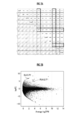
- 2A is a correlation plot of various fibroblasts
- FIG. 2B is an MA plot of aggressive fibroblasts and periodontal ligament fibroblasts.
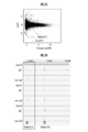
- FIG. 3A is a diagram showing MA plots of aggressive fibroblasts and normal gingival fibroblasts.
- FIG. 3B shows gene expression profiles from aggressive fibroblasts of periodontal disease patients and RUNX2 P1 promoter and P2 promoter of normal gingival fibroblasts.
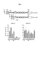
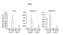
- the figure which shows the gene expression profile from P1 promoter and P2 promoter of RUNNX2 in various primary culture fibroblasts The figure which shows the gene expression profile from aggressive fibroblast of the same periodontal disease patient, DLX5 of normal gingival fibroblast, P1 promoter of RUNX2, and P2 promoter.
- the present inventors have developed a method for identifying aggressive fibroblasts without performing three-dimensional cell culture by searching for markers that are specifically expressed in aggressive fibroblasts. Furthermore, since markers specific to aggressive fibroblasts were found, screening for therapeutic agents for periodontitis can be performed using the expression of these markers as an index.
- a candidate compound is screened by bringing a candidate compound into contact with aggressive fibroblasts identified using the marker of the present invention and measuring changes in each marker gene.
- high risk of periodontitis includes current and future risk of periodontitis.
- expression refers to gene and / or protein expression unless otherwise specified. Any method may be used as long as it can be quantitatively measured whether it is a gene or a protein.
- Gene expression can be measured by quantitative PCR, and protein expression by antibodies.
- quantitative PCR method a known method such as SYBR Green method, TaqMan probe method, RT-PCR method or the like can be used.
- protein can be quantified by well-known methods, such as ELISA method, RIA method, Western blot method. By measuring mRNA and protein, the marker can be measured with high sensitivity.
- Cells collected from patients include not only gingiva removed from patients during periodontal surgery and tissues obtained from extracted teeth, but also cultures obtained by culturing these tissues. Cells are also included.
- Gingival Fibroblasts were isolated based on the method of Ohshima et al. Specifically, after obtaining a tissue from a gingival piece or an extracted tooth that has been excised and removed during periodontal surgery, and then chopping the tissue, the tissue piece is placed on a plate, and exogenous cells are removed from the tissue piece. As passage 1, passage cells up to passage 6 were used in the following experiments.
- the culture was performed by adding ⁇ -minimum essential medium ( ⁇ -MEM, manufactured by Wako) to 10% fetal bovine serum (FBS, manufactured by Hyclone), and 1% Penicillin / Streptmycine / Neomycin (manufactured by Life Technologies).
- the solution was used under wet conditions of 37 ° C. and 5% CO 2 .
- the cells reach confluence, the cells are detached using a 0.05% trypsin / EDTA solution (manufactured by Wako) and seeded on a plate or flask so that the area ratio is 1: 3. I went.
- Gingival epithelial cells were isolated based on the method of Ohshima et al. Specifically, the gingival pieces excised and removed during periodontal surgery were treated with Dispase I (manufactured by Godo Seisei Co., Ltd.), and then the epithelial tissue part was peeled from the connective tissue part. After the epithelial tissue was minced, the tissue piece was allowed to stand on a plate, cells exogenous from the tissue piece were used as the first generation, and passage cells from the 4th to the 8th generation were used in the following experiments.
- Dispase I manufactured by Godo Seisei Co., Ltd.
- a plate or flask manufactured by Sumitomo Bakelite Co., Ltd. coated with type I collagen was used.
- Epi Life medium manufactured by Life Technologies
- additives S7 manufactured by Life Technologies
- 1% Gentaminin / Amphoticin Solution manufactured by Sumitomo Bakelite Co., Ltd.
- a medium supplemented with Technologies was used.
- the above-cured gingival epithelial cells are dispersed with trypsin on the gel hardened as described above, and seeded at 2.0 to 2.5 ⁇ 10 5 cells to form a monolayer, and collagen gel Built.
- FIG. 1C shows the results of three-dimensional cell culture of cells isolated from two cases of periodontitis patients and healthy subjects.
- the gel is peeled off from the plate, and the gel is allowed to float. Gel contraction occurs after 3-4 days, allowing differentiation between aggressive and normal gingival fibroblasts.
- fibroblasts obtained from gingival pieces of periodontitis patients always contain aggressive fibroblasts with high collagen resolution.
- the following analysis was performed using cells with high collagen resolution selected by three-dimensional cell culture as cultured fibroblasts in plates, flasks, and the like.
- the group with many aggressive fibroblasts is selected instead of isolating aggressive fibroblasts.
- aggressive fibroblasts are not mixed in the cell group with low collagen resolving power, and most are considered to be normal gingival fibroblasts.
- the FANTOM (Functional AnnoTation Of Mammalian cDNA) 5 project is an international consortium of RIKEN, and is an international project that works on systematic mapping of transcription start sites using various types of human cells.
- the FANTOM project obtains and analyzes gene expression profiles by the CAGE method (Cap Analysis of Gene Expression) in order to understand the transcriptional regulatory network of the genome.
- the CAGE method is a method of cutting out a tag sequence of about 20 bases from the 5 ′ end of mRNA and determining the base sequence.
- the CAGE method is a technique capable of analyzing gene expression for individual promoters. In combination with next-generation sequencers, this is the only technology that can quantitatively analyze gene expression on a genome-wide basis. Theoretically, RNA that expresses only 1 copy or less in 10 cells can be detected with an accuracy of 99.9%. It is said that it can be caught.
- cDNA was first prepared from RNA. 5 ⁇ g of RNA was mixed with 500 ng of random N15 primer (Clontech), heat-denatured at 65 ° C. for 5 minutes, and then cooled. RT master mix (manufactured by Clontech) was added, and the reaction was performed at 25 ° C. for 30 seconds, 42 ° C. for 30 minutes, 50 ° C. for 10 minutes, 56 ° C. for 10 minutes, and 60 ° C. for 10 minutes, and cooled to 4 ° C. The cDNA / RNA was purified and dissolved by Agencourt RNACleanXP (Beckman Coulter).
- oxidation reaction and biotinylation were performed. 2 ⁇ L of 1 M sodium acetate (pH 4.5) and 2 ⁇ L of 250 mM sodium periodate were added to the cDNA / RNA mixture, and the mixture was allowed to stand on ice in a dark room for 45 minutes. The reaction was stopped by mixing 2 ⁇ L of 40% glycerol and 14 ⁇ L of 1M Tris-HCl (pH 8.5). The cDNA / RNA mixture was purified by Agencourt RNACleanXP.
- MPG streptavidin magnetic bead slurry (manufactured by TakaraBio) was allowed to stand on ice for 30 minutes using tRNA (manufactured by Sigma) to perform pre-blocking. Thereafter, washing was performed with buffer 1 (4.5 M sodium chloride, 5 mM EDTA, pH 8.0), and suspended in buffer 1 containing 50 ⁇ g of tRNA.
- the biotinylated cDNA / RNA mixture was purified with blocked beads. Binding at 50 ° C. for 30 minutes, buffer 1 once, buffer 2 (0.3 M sodium chloride, 1 mM EDTA, pH 8.0) once, buffer 3 (1 mM EDTA, 0.4% sodium dodecyl sulfate, 0.5 M sodium acetate) Washing was performed twice with 20 mM Tris-HCl, pH 8.5) and twice with buffer4 (1 mM EDTA, 0.5 M sodium acetate, 10 mM Tris-HCl, pH 8.5). The captured cDNA was released by heat shock and RNase I treatment. The beads were dissolved in RNase I buffer and left on ice immediately after treatment at 95 ° C. for 5 minutes. RNA was removed by RNase treatment at 37 ° C. for 15 minutes.
- Cap-trapped first-strand cDNA was generated by Agencourt AMPure XP (manufactured by Beckman Coulter). The quality of cDNA was evaluated using OliGreen ssDNA Quantitation kit (manufactured by Invitrogen). Generally, 10-20 ng of CAGE library is prepared from 5 ⁇ g of RNA.
- poly (dA) tailing reaction was performed. 10 ng single-stranded CAGE library was heat denatured at 95 ° C. for 5 minutes, and then rapidly cooled. Terminal Transferase, dATP, and BSA (manufactured by NEB) were added to the denatured mixture and reacted at 37 ° C. for 1 hour. The enzyme was inactivated at 70 ° C. for 10 minutes and cooled to 4 ° C. After the poly (A) tailing reaction, the reaction solution was heat-denatured at 95 ° C. for 5 minutes and rapidly cooled.
- a blocking mixture containing 0.5 ⁇ L of 200 ⁇ M biotin-ddATP was prepared, added to the denatured poly-adenylated mixture, reacted at 37 ° C. for 1 hour, then inactivated at 70 ° C. for 20 minutes and cooled to 4 ° C. did.
- 2 pM of Carrier Oligonucleotide (5′-TCACTATTGTTTGAGAACGTTGGCCTATATAGTGAGTCGTTATACCGCGCGGT [ddC] -3 ′) (SEQ ID NO: 1) was added to the reaction solution inactivated by the measurement, and the molar concentration of the sample was measured by HelicosHopti
- FIG. 2A shows a correlation plot of various fibroblasts and gingival fibroblasts.
- the abbreviations in the figure indicate the following fibroblasts, and the numbers in parentheses indicate the number of fibroblast types used in the analysis.
- GF gingival fibroblast (gingival fibroblast (6))
- AAF aortic adventitial fibroblast (outer aortic fibroblast (3))
- CF cardiac fibroblast (cardiac fibroblast (6)), CPF: choroid plexus fibroblast (Choroidal fibroblast (3))
- ConF conjunctival fibroblast (conjunctival fibroblast (2)), DF: dermal fibroblast (skin fibroblast (6)),
- LF lung fibroblast (lung fibroblast (3) )
- LymF lymphatic fibroblast (lymphatic fibroblast (3))
- MF mammary fibroblast (mammary fibroblast (3))
- PLF periodontal ligament fibroblast (
- the correlation of the CAGE tag count between samples of each fibroblast was calculated using the plotCorrelation function in CAGE, which is a package of Bioconductor.
- the correlation coefficient is Pearson's correlation coefficient, and the higher the correlation coefficient, the more similar the gene expression.
- Gingival fibroblasts are highly correlated with the periodontal ligament fibroblasts (PLF) and the expression pattern of genes expressed among fibroblasts. Therefore, in order to characterize the gene expression of gingival fibroblasts, the gene expression profiles of gingival fibroblasts and all other fibroblasts excluding periodontal fibroblasts were compared. For comparison, edgeR, which is a package of Bioconductor, was used. Each gene expression level was set to FDR (False Discovery Rate) ⁇ 0.05 by MA plot, and comparison was performed for regions with high confidence regions (FIG. 2B).
- FDR Fealse Discovery Rate
- the CAGE peak profiles of individual aggressive fibroblasts and normal gingival fibroblasts were confirmed in the ZENBU database, and there was a difference in the expression of the CAGE peak of the P1 promoter of RUNX2 in cells obtained from any donor. It was. In the aggressive fibroblasts (agF), the expression from the P1 promoter was suppressed, whereas in normal gingival fibroblasts (non-agF), the expression was not suppressed. In addition, there was no donor in which expression difference was observed from the P2 promoter in aggressive fibroblasts and normal gingival fibroblasts. The results are shown in FIG. 3B.
- RUNX2 is a transcription factor having a runt homology domain, and is known to act as a master gene for osteoblast differentiation and bone formation (Non-patent Document 7). RUNX2 mutations cause cliocranial dysplasia (CCD). It is considered that the abnormality of tooth formation seen as a symptom of CCD is related to abnormality of RUNX2 in cells involved in tooth formation.
- RUNX2 P1 promoter ⁇ Expression from RUNX2 P1 promoter and P2 promoter was analyzed by quantitative PCR using various fibroblasts.
- the cells used are as follows. The following four primary cultured cells obtained by the present inventors from periodontitis patients: GFH2: gingival fibroblasts, GFH3-1: gingival fibroblasts, PLH2: periodontal ligament fibroblasts, PL29: periodontal ligament fibroblasts Cells and the following four cell lines purchased: NB1RGB: normal skin fibroblasts, NHLF: normal adult lung fibroblasts, WI-38: fetal lung fibroblast cell line, HFL-1: fetal lung fibroblast cell line RNA was purified from and used for analysis.
- Mx-3000P qPCR System manufactured by Stratagene
- Quantect SYBR Green PCR manufactured by Qiagen were used.
- the gene expression level was normalized by using glyceraldehyde-3-phosphate dehydrogenase (GAPDH).
- GPDH glyceraldehyde-3-phosphate dehydrogenase
- RUNX2 P1 F TGGCATCAAACAGCCCTCTTCAG (SEQ ID NO: 2)
- RUNX2 P2 F GTGATGCGGTATTCCCGTAG (SEQ ID NO: 3)
- RUNX2 common R GGCTCACGTCGCCTATTTTTG (SEQ ID NO: 4)
- GAPDH F GGTGAAGGTCGGAGCAACGGA (SEQ ID NO: 5)
- GAPDH R GAGGGATCTCCGCTCCTGGAAGA (SEQ ID NO: 6)
- the transcript derived from the P1 promoter of RUNX2 was specifically expressed in gingival fibroblasts and periodontal ligament fibroblasts. On the other hand, it was found that the transcript derived from the P2 promoter was uniformly expressed in all fibroblasts. Therefore, it is possible to identify aggressive fibroblasts using the expression from the RUNX2 P1 promoter as an index.
- DLX5 FLT1 ⁇ RUNX2 is a master regulatory factor involved in bone formation, and is known to regulate transcription of DLX5 (Distal-less homeobox 5), which is an upstream gene (Non-patent Document 8).
- DLX5 is known to be involved in the final differentiation of osteoblasts. Therefore, analysis was performed on the gene expression from the P1 promoter and P2 promoter of DLX5 and RUNX2, using cells derived from 2 patients with periodontitis. Quantitative RT-PCR was performed with Sybr premix (Takara) and a real-time instrument (TP-900, Takara) was used. The following sequences were used as primers for DLX5.
- DLX5 F CAACTTTGCCCGAGCTTCA (SEQ ID NO: 7)
- DLX5 R GTTGAGAGCTTTGCCATAGGAA (SEQ ID NO: 8)
- the results are normalized by GAPDH as in Example 4. The results are shown in FIG.
- DLX5 expression was suppressed in aggressive fibroblasts compared to normal gingival fibroblasts.
- the expression from the RUNX2P1 promoter was suppressed in aggressive fibroblasts as in the above analysis results, whereas the expression from the P2 promoter was not significantly different.
- FLT1 Fms-like Tyrosine Kinase 1, VEGFR-1
- FLT1 R TGCTGCTTCCTGGTCTCAAAATA
- RUNX2 P1 promoter and DLX5 was significantly decreased in aggressive fibroblasts (GF) compared to normal gingival fibroblasts (PGF) and periodontal ligament fibroblasts (PL).
- the expression of FLT1 was significantly increased in aggressive fibroblasts (GF) compared to normal gingival fibroblasts (PGF) and periodontal ligament fibroblasts (PL). Therefore, it can be judged whether it is an aggressive fibroblast on the basis of the expression level of these genes in a normal gingival fibroblast and a periodontal fibroblast.
- RUNX2 expression level from P1 promoter, DLX5 expression level is low, or FLT1 expression level is high can be determined to be aggressive fibroblasts. Further, by setting the average expression level of these genes in normal gingival fibroblasts and periodontal ligament fibroblasts as a predetermined value, aggressive fibroblasts can be identified or used as a threshold for examination.
Landscapes
- Life Sciences & Earth Sciences (AREA)
- Health & Medical Sciences (AREA)
- Chemical & Material Sciences (AREA)
- Engineering & Computer Science (AREA)
- Immunology (AREA)
- Organic Chemistry (AREA)
- Molecular Biology (AREA)
- Zoology (AREA)
- General Health & Medical Sciences (AREA)
- Proteomics, Peptides & Aminoacids (AREA)
- Microbiology (AREA)
- Urology & Nephrology (AREA)
- Analytical Chemistry (AREA)
- Wood Science & Technology (AREA)
- Physics & Mathematics (AREA)
- Biochemistry (AREA)
- Hematology (AREA)
- Biomedical Technology (AREA)
- Biotechnology (AREA)
- Genetics & Genomics (AREA)
- General Engineering & Computer Science (AREA)
- Cell Biology (AREA)
- Bioinformatics & Cheminformatics (AREA)
- Biophysics (AREA)
- Food Science & Technology (AREA)
- Medicinal Chemistry (AREA)
- General Physics & Mathematics (AREA)
- Pathology (AREA)
- Measuring Or Testing Involving Enzymes Or Micro-Organisms (AREA)
- Investigating Or Analysing Biological Materials (AREA)
Abstract
歯周炎の原因細胞であるアグレッシブ線維芽細胞のマーカーの解析を行い、新規の歯周炎治療薬のスクリーニング、検査方法、検査キットを確立する。アグレッシブ線維芽細胞の解析を行った結果、RUNX2のP1プロモーターからの発現、DLX5の発現が正常歯肉線維芽細胞と比較して減少していることが明らかとなった。また、FLT1の発現の増加が見られた。これらアグレッシブ線維芽細胞に特徴的な発現をマーカーとして、アグレッシブ線維芽細胞の特定、治療薬のスクリーニング方法、検査方法とすることができる。
Description
本発明は、新規の分子マーカーを用いた歯周炎原因細胞の判別方法、及び歯周炎治療薬のスクリーニング方法に関する。
歯周病はう蝕とともに、歯の二大疾患である。歯周病は歯肉炎と歯周炎とに分けられるが、最終的な歯の喪失に至るのは、いわゆる歯槽膿漏といわれる歯周炎である。歯周病は世界で最も多い疾患であるともいわれ、我が国でも成人の約8割が罹患しているとされる。歯喪失の42%は歯周炎によると報告されており、歯喪失の重要な原因でもある。
保持歯数は全身の健康状態とも密接に関連することが指摘されており、歯周炎の治療と予防は歯科分野だけではなく、医療全般の問題ともなる。歯科保健の分野では、高齢者においても歯の喪失が10歯以下であれば、食生活に大きな支障を生じないとの研究に基づき、「80歳になっても自分の歯を20本以上保とう」という8020(ハチマル・ニイマル)運動が提唱、推進されている。8020運動は1989年より、厚生省(当時)と日本歯科医師会が推進している運動である。しかしながら、2011年の調査では達成率は38.3%であり、前回調査時(2005年)の24.1%に比べ大幅に達成率が上昇しているとはいえ、いまだに低い数値を推移している。
歯周炎の原因は、これまで歯肉炎同様、細菌感染であると考えられていたことから、現在の治療法としては、抗菌、抗炎症などの対症療法が主流である。また、プラークコントロール、スケーリング等の歯周基本治療、外科的に歯周ポケットの深さを減少させる手術が行われている。
歯肉炎は歯磨きの習慣と相関があり、歯肉炎の原因は歯に付いた細菌性プラークであるという研究結果から、細菌が原因であると考えられる(非特許文献1)。しかしながら、コッホの4原則を満たす歯周病菌が単離されておらず、動物モデルを作ることができないこと、細菌感染が原因であることが明らかになっている歯肉炎から歯周炎に移行する患者は15%程度しかいないこと、歯を磨く習慣のある集団と磨かない集団とで、重度歯周炎の罹患率が変わらない等、歯周炎の原因は細菌であると考えると説明のつかないことが多い。
図1Aは歯周組織を模式的に示したものであり、左半分は健康な歯の状態を、右半分は歯周炎に罹患した歯の状態を示したものである。歯根と歯槽骨の間には、歯根膜というコラーゲン線維でできた靭帯が存在し、歯根と歯槽骨を強固につなげている。歯周炎によって歯槽骨のミネラル分は吸収されるが(図1A右、歯周炎の部分参照)、それだけでは歯根膜は失われない。また、歯を失うことにもならない。歯喪失につながるのは、いわゆる歯周ポケットの部分から、歯根膜が溶解することにより、歯と骨との間のコラーゲンによる付着がなくなり、歯根と歯槽骨の間に上皮細胞が入り込み、やがて根尖まで到達することが原因となる。
本発明者らは、歯周炎の研究の過程で、重度歯周炎患者の歯肉由来の線維芽細胞をコラーゲンゲル三次元細胞培養法によって培養を行うと、培地であるゲルのコラーゲンが分解され小さくなるということを見出した。図1Bに重度歯周炎患者、軽度歯周炎患者、健常者の歯肉から単離した歯肉線維芽細胞を三次元ゲルで培養した結果を示している。重度歯周炎患者の歯肉由来の線維芽細胞を三次元細胞培養すると、矢印で示したように、培地のコラーゲンが分解され、小さくゲルが収縮する。本願発明者らは、このコラーゲン分解能の高い線維芽細胞をアグレッシブ線維芽細胞と名付けて、以下の理由から歯周炎の主因である可能性が高いと考えている。
(1)現在までに50症例以上の歯周炎患者より、アグレッシブ線維芽細胞の単離を試みているが、すべての歯周炎患者歯肉からアグレッシブ線維芽細胞は単離できている。
(2)アグレッシブ線維芽細胞はコラーゲン分解能が高いことから、歯と骨との間に存在するコラーゲンを主成分とする歯根膜を分解する原因となっていると考えられる。
(3)プラーク付着量が少なく、炎症の程度は軽いが、付着の急速な消失が起こる疾患である侵襲性歯周炎罹患歯肉からは、コラーゲン分解能の非常に高いアグレッシブ線維芽細胞が分離できる。
(4)歯周炎では、同じ歯でも病態がとくに進行した部分がある。これを歯周炎の部位特異性というが、アグレッシブ線維芽細胞が存在することで歯周炎の部位特異性を説明できる。
(1)現在までに50症例以上の歯周炎患者より、アグレッシブ線維芽細胞の単離を試みているが、すべての歯周炎患者歯肉からアグレッシブ線維芽細胞は単離できている。
(2)アグレッシブ線維芽細胞はコラーゲン分解能が高いことから、歯と骨との間に存在するコラーゲンを主成分とする歯根膜を分解する原因となっていると考えられる。
(3)プラーク付着量が少なく、炎症の程度は軽いが、付着の急速な消失が起こる疾患である侵襲性歯周炎罹患歯肉からは、コラーゲン分解能の非常に高いアグレッシブ線維芽細胞が分離できる。
(4)歯周炎では、同じ歯でも病態がとくに進行した部分がある。これを歯周炎の部位特異性というが、アグレッシブ線維芽細胞が存在することで歯周炎の部位特異性を説明できる。
上記の理由から、アグレッシブ線維芽細胞が歯周炎の一因であると考えられる。そして、本発明者らはすでに歯周組織破壊を抑制し、歯周炎を改善する有効成分をスクリーニングする方法を開示している。さらに、コラーゲン分解阻害剤を有効成分とする治療薬を開発している(特許文献1、2)。
しかしながら、アグレッシブ線維芽細胞を特定するためには長期にわたる培養時間が必要であることから、短時間でアグレッシブ線維芽細胞を特定する方法が望まれていた。また、新しい治療薬、診断方法を提供するためにアグレッシブ線維芽細胞で特異的な発現パターンを示す遺伝子の解析も望まれていた。
Loe et al., 1965, J. Periodontol., Vol.36, pp.177-187
Ohshima et al., 1994, J. Periodontal.Res., Vol.29, pp.421-429
Ohshima et al., 2008, J. Periodontol., Vol.79, pp.912-919
Ohshima et al., 2010, J. Dent. Res., Vol.89(11), pp.1315-1321
Chang, H.Y. et al., 2002, PNAS,Vol.99(20), pp.12877-12882
Lipson,D. et al., 2009,Nat. Biotechnol.Vol.27, pp.652-658
Komori, T. et al., 1997, Cell, Vol.89(5), p.755-764
Camilleri S. & McDonald F., 2006, Eur. J. Oral. Sci., Vol.114,pp.361-373
本発明は、歯周炎の原因細胞であるアグレッシブ線維芽細胞のマーカーを探索し、当該マーカーの発現を指標として、歯周炎原因細胞を特定するとともに、新たな治療薬を探索するためのスクリーニング方法を提供することを課題とする。
本発明は、以下に示すアグレッシブ線維芽細胞の特定方法、歯周炎の治療薬のスクリーニング方法、検査方法、及び検査キットに関する。
(1)歯周炎の原因細胞であるアグレッシブ線維芽細胞の特定方法であって、RUNX2のP1からの発現量及び/又はDLX5の発現量が所定の値より低いことを特徴とする特定方法。
(2)上記(1)記載のアグレッシブ線維芽細胞の特定方法であって、さらに、FLT1発現量が所定の値より高いことを特徴とする特定方法。
(3)上記(1)又は(2)記載のアグレッシブ線維芽細胞の特定方法であって、前記発現量は、遺伝子及び/又はタンパク質の発現量を測定することを特徴とする特定方法。
(4)上記(1)~(3)のいずれか1つ記載のアグレッシブ線維芽細胞の特定方法であって、前記発現量を配列番号2及び4のプライマーセット、配列番号7及び8のプライマーセット、配列番号9及び10のプライマーセットの少なくとも1つ以上により遺伝子の発現を測定することを特徴とする特定方法。
(5)歯周炎治療薬のスクリーニング方法であって、細胞に被験物質を接触させ、RUNX2のP1からの発現量及び/又はDLX5の発現量の増加を指標とすることを特徴とするスクリーニング方法。
(6)上記(5)記載のスクリーニング方法であって、さらに、FLT1の発現量の低下を指標とすることを特徴とするスクリーニング方法。
(7)上記(5)又は(6)記載のスクリーニング方法であって、前記細胞はアグレッシブ線維芽細胞であることを特徴とするスクリーニング方法。
(8)上記(5)~(7)のいずれか1つ記載のスクリーニング方法であって、前記発現量は、遺伝子及び/又はタンパク質の発現量を測定することを特徴とするスクリーニング方法。
(9)上記(5)~(8)のいずれか1つ記載のスクリーニング方法であって、前記発現量を配列番号2及び4のプライマーセット、配列番号7及び8のプライマーセット、配列番号9及び10のプライマーセットの少なくとも1つ以上により遺伝子の発現を測定することを特徴とするスクリーニング方法。
(10)歯周炎の検査方法であって、患者から採取された検体のRUNX2のP1からの発現量及び/又はDLX5の発現量を測定し、所定の値より低い場合に歯周炎のリスクが高いと判定することを特徴とする検査方法。
(11)上記(10)記載の検査方法であって、さらに、FLT1の発現量が所定の値より高い場合に歯周炎のリスクが高いと判定することを特徴とする検査方法。
(12)上記(10)又は(11)記載の検査方法であって、前記発現量は、遺伝子及び/又はタンパク質の発現量であることを特徴とする検査方法。
(13)上記(10)~(12)のいずれか記載の検査方法であって、前記発現量を配列番号2及び4記載のプライマーセット、配列番号7及び8のプライマーセット、配列番号9及び10のプライマーセットの1つ以上により遺伝子の発現を定量することを特徴とする検査方法。
(14)歯周炎の診断キットであって、配列番号2及び4のプライマーセット、配列番号7及び8のプライマーセット、配列番号9及び10のプライマーセットの少なくとも1つ以上のプライマーセットと、定量に必要な試薬を含むことを特徴とする検査キット。
(1)歯周炎の原因細胞であるアグレッシブ線維芽細胞の特定方法であって、RUNX2のP1からの発現量及び/又はDLX5の発現量が所定の値より低いことを特徴とする特定方法。
(2)上記(1)記載のアグレッシブ線維芽細胞の特定方法であって、さらに、FLT1発現量が所定の値より高いことを特徴とする特定方法。
(3)上記(1)又は(2)記載のアグレッシブ線維芽細胞の特定方法であって、前記発現量は、遺伝子及び/又はタンパク質の発現量を測定することを特徴とする特定方法。
(4)上記(1)~(3)のいずれか1つ記載のアグレッシブ線維芽細胞の特定方法であって、前記発現量を配列番号2及び4のプライマーセット、配列番号7及び8のプライマーセット、配列番号9及び10のプライマーセットの少なくとも1つ以上により遺伝子の発現を測定することを特徴とする特定方法。
(5)歯周炎治療薬のスクリーニング方法であって、細胞に被験物質を接触させ、RUNX2のP1からの発現量及び/又はDLX5の発現量の増加を指標とすることを特徴とするスクリーニング方法。
(6)上記(5)記載のスクリーニング方法であって、さらに、FLT1の発現量の低下を指標とすることを特徴とするスクリーニング方法。
(7)上記(5)又は(6)記載のスクリーニング方法であって、前記細胞はアグレッシブ線維芽細胞であることを特徴とするスクリーニング方法。
(8)上記(5)~(7)のいずれか1つ記載のスクリーニング方法であって、前記発現量は、遺伝子及び/又はタンパク質の発現量を測定することを特徴とするスクリーニング方法。
(9)上記(5)~(8)のいずれか1つ記載のスクリーニング方法であって、前記発現量を配列番号2及び4のプライマーセット、配列番号7及び8のプライマーセット、配列番号9及び10のプライマーセットの少なくとも1つ以上により遺伝子の発現を測定することを特徴とするスクリーニング方法。
(10)歯周炎の検査方法であって、患者から採取された検体のRUNX2のP1からの発現量及び/又はDLX5の発現量を測定し、所定の値より低い場合に歯周炎のリスクが高いと判定することを特徴とする検査方法。
(11)上記(10)記載の検査方法であって、さらに、FLT1の発現量が所定の値より高い場合に歯周炎のリスクが高いと判定することを特徴とする検査方法。
(12)上記(10)又は(11)記載の検査方法であって、前記発現量は、遺伝子及び/又はタンパク質の発現量であることを特徴とする検査方法。
(13)上記(10)~(12)のいずれか記載の検査方法であって、前記発現量を配列番号2及び4記載のプライマーセット、配列番号7及び8のプライマーセット、配列番号9及び10のプライマーセットの1つ以上により遺伝子の発現を定量することを特徴とする検査方法。
(14)歯周炎の診断キットであって、配列番号2及び4のプライマーセット、配列番号7及び8のプライマーセット、配列番号9及び10のプライマーセットの少なくとも1つ以上のプライマーセットと、定量に必要な試薬を含むことを特徴とする検査キット。
歯周炎原因細胞であるアグレッシブ線維芽細胞の新しい遺伝子マーカーを見出したことにより、これらの発現を指標としてアグレッシブ線維芽細胞を迅速に選別することが可能となる。また、歯周炎の治療薬をスクリーニングすることが可能となる。さらに、新たな検査方法、検査キットを提供することができる。
本発明者らは、アグレッシブ線維芽細胞に特異的に発現しているマーカーを探索することによって、三次元細胞培養を行わずにアグレッシブ線維芽細胞を特定する方法を開発した。さらに、アグレッシブ線維芽細胞に特異的なマーカーを見出したことから、これらマーカーの発現を指標として、歯周炎治療薬のスクリーニングを行うことができる。
具体的には、本発明のマーカーを用いて特定したアグレッシブ線維芽細胞に、候補化合物を接触させ、各マーカー遺伝子の変化を測定することにより、治療薬の候補となる化合物のスクリーニングを行う。
三次元細胞培養法を経ずにアグレッシブ線維芽細胞を特定することによって、非常に短期間で歯周炎の原因細胞を特定することができるため、歯周炎治療薬のスクリーニングを行うことが容易となる。また、歯周炎のリスクが高い患者の判定を行うことができる。本発明において、歯周炎のリスクが高いとは、現在、及び将来の歯周炎のリスクを含む。
本発明において、発現とは特に断らない限り、遺伝子及び/又はタンパク質の発現を指す。遺伝子であっても、タンパク質であっても、定量的に測定することができれば、どのような方法を用いてもよい。遺伝子発現は定量的PCR法によって、タンパク質発現は、抗体によって測定することができる。定量的PCR法は、SYBR Green法、TaqManプローブ法、RT-PCR法等、公知の方法を用いることができる。また、タンパク質は、ELISA法、RIA法、Western blot法等公知の方法によって定量することができる。mRNA、タンパク質を測定することにより、感度よくマーカーを測定することができる。
患者から採取された細胞とは、患者から歯周外科手術の際に切除されて不要となった歯肉や、抜去歯より得られた組織だけではなく、これら組織を培養して得られたた培養細胞も含まれる。
以下、実施例を交えながら本発明を詳細に説明する。
≪アグレッシブ線維芽細胞の特定≫
1.歯肉線維芽細胞の調製
歯肉線維芽細胞はOhshimaらの方法(非特許文献2)に基づいて単離した。具体的には、歯周外科手術の際に切除され不要となった歯肉片又は抜去歯より組織を得て細切後、組織片をプレートに静置し、組織片より外生した細胞を第1代として、第6代目までの継代細胞を以下の実験に用いた。培養は、α-minimum essential medium(α-MEM、Wako社製)に10%ウシ胎児血清(FBS、Hyclone社製)、及び1%Penicillin/Streptmycine/Neomycin溶液(Life Technolgies社製)を添加した培養液を用いて、37℃、5%CO2の湿潤条件下で行った。継代は細胞がコンフルエントに達した時点で、0.05%trypsin/EDTA溶液(Wako社製)を用いて細胞を剥離し、面積比が1:3になるようにプレート、又はフラスコに播種して行った。
1.歯肉線維芽細胞の調製
歯肉線維芽細胞はOhshimaらの方法(非特許文献2)に基づいて単離した。具体的には、歯周外科手術の際に切除され不要となった歯肉片又は抜去歯より組織を得て細切後、組織片をプレートに静置し、組織片より外生した細胞を第1代として、第6代目までの継代細胞を以下の実験に用いた。培養は、α-minimum essential medium(α-MEM、Wako社製)に10%ウシ胎児血清(FBS、Hyclone社製)、及び1%Penicillin/Streptmycine/Neomycin溶液(Life Technolgies社製)を添加した培養液を用いて、37℃、5%CO2の湿潤条件下で行った。継代は細胞がコンフルエントに達した時点で、0.05%trypsin/EDTA溶液(Wako社製)を用いて細胞を剥離し、面積比が1:3になるようにプレート、又はフラスコに播種して行った。
2.歯肉上皮細胞の調製
歯肉上皮細胞はOhshimaらの方法(非特許文献3)に基づいて単離した。具体的には、歯周外科手術の際に切除され不要となった歯肉片をDispase I(合同酒精株式会社製)処理した後、上皮組織部分を結合組織部分から剥離した。上皮組織を細切後、組織片をプレートに静置し、組織片から外生した細胞を第1代として、第4~8代目までの継代細胞を以下の実験に用いた。
歯肉上皮細胞はOhshimaらの方法(非特許文献3)に基づいて単離した。具体的には、歯周外科手術の際に切除され不要となった歯肉片をDispase I(合同酒精株式会社製)処理した後、上皮組織部分を結合組織部分から剥離した。上皮組織を細切後、組織片をプレートに静置し、組織片から外生した細胞を第1代として、第4~8代目までの継代細胞を以下の実験に用いた。
培養はI型コラーゲンがコートされたプレート、又はフラスコ(住友ベークライト社製)を用い、Epi Life medium(Life Technolgies社製)に添加剤S7(Life Technolgies社製)及び1%Gentamicin/Amphotericin Solution(Life Technolgies社製)を加えた培地を用いた。細胞がコンフルエントに達した時点で、0.05%trypsin/EDTA溶液を用いて細胞を剥離し継代培養を行った。
3.三次元細胞培養によるコラーゲン分解能の高い歯肉線維芽細胞の選択
歯肉線維芽細胞を歯肉上皮細胞とともに培養することによって、そのコラーゲン分解能が増強する。コラーゲンゲルの構築方法、三次元細胞培養方法については論文(非特許文献4)に詳しく記載しているが、以下に方法の概要を記載する。
歯肉線維芽細胞を歯肉上皮細胞とともに培養することによって、そのコラーゲン分解能が増強する。コラーゲンゲルの構築方法、三次元細胞培養方法については論文(非特許文献4)に詳しく記載しているが、以下に方法の概要を記載する。
セルマトリックスtype I-A(新田ゼラチン社製)、5×DMEM(新田ゼラチン社製)、再構成用緩衝液(新田ゼラチン社製)を混合して、コラーゲン混合溶液を作製した。ここに、上記継代培養した歯肉線維芽細胞をトリプシンで分散させ、FBSに浮遊させて懸濁した後、細胞数2.0~2.5×10⁵個をコラーゲン混合溶液に混和し、6穴プレートに播種して37℃インキュベーターで30分間静置しゲル化させた。
次に、上記で硬化させたゲル上に、上記継代培養した歯肉上皮細胞をトリプシンで分散させ、細胞数2.0~2.5×10⁵個で播種し単層を形成させ、コラーゲンゲルを構築した。
図1Cには、2症例の歯周炎患者と健常者から単離した細胞を三次元細胞培養した結果を示している。通常、コラーゲンゲルを構築した翌日にゲルをプレートから剥がし、ゲルを浮遊させる。3~4日後にゲルの収縮が起こり、アグレッシブ線維芽細胞と正常歯肉線維芽細胞との区別が可能となる。図1Cに示すように、歯周炎患者の歯肉片より得られた線維芽細胞では、コラーゲン分解能が高いアグレッシブ線維芽細胞が必ず含まれている。三次元細胞培養によって選択されたコラーゲン分解能の高い細胞をアグレッシブ線維芽細胞としてプレート、フラスコ等で培養したものを用いて以下の解析を行った。なお、これらは初代培養細胞であることから、アグレッシブ線維芽細胞を単離しているのではなく、アグレッシブ線維芽細胞の多い群を選択していることになる。また、コラーゲン分解能が低い細胞群にはアグレッシブ線維芽細胞は混在しておらず、ほとんどが正常歯肉線維芽細胞であると考えられる。
≪理研FANTOM5による解析≫
様々な組織から単離した線維芽細胞は同じような形状をしているものの、トランスクリプトーム解析を行うと、各組織で特徴的な遺伝子発現をしていることが知られている(非特許文献5)。歯肉線維芽細胞についても解析が行われているが、2つの遺伝子のみについて特徴づけられているにすぎず、歯肉線維芽細胞の遺伝子発現パターンの特徴は明らかとはいえない。そこで、FANTOM5において、歯肉線維芽細胞のCAGE法による解析を行った。
様々な組織から単離した線維芽細胞は同じような形状をしているものの、トランスクリプトーム解析を行うと、各組織で特徴的な遺伝子発現をしていることが知られている(非特許文献5)。歯肉線維芽細胞についても解析が行われているが、2つの遺伝子のみについて特徴づけられているにすぎず、歯肉線維芽細胞の遺伝子発現パターンの特徴は明らかとはいえない。そこで、FANTOM5において、歯肉線維芽細胞のCAGE法による解析を行った。
FANTOM(Functional ANnoTation Of Mammalian cDNA)5プロジェクトは、理化学研究所の国際コンソーシアムによるものであり、様々な種類のヒト細胞を使って、転写開始部位の系統的マッピングに取り組む国際的なプロジェクトである。
FANTOMプロジェクトは、ゲノムの転写制御ネットワークを理解するために、遺伝子の発現プロファイルをCAGE法(Cap Analysis of Gene Expression)によって得て解析を行っている。CAGE法は、mRNAの5´末端から約20塩基のタグ配列を切り出し、塩基配列を決定する手法である。CAGE法は、個々のプロモーターについての遺伝子発現を解析できる手法である。また、次世代シーケンサーと組み合せることにより、ゲノムワイドに遺伝子発現を定量解析できる唯一の技術であり、理論的には10細胞に1コピー以下しか発現していないRNAを99.9%の精度で捉えることができると言われている。
1.RNAの採取
RNAはTRIzol(商標、invitrogen社製)を用いて抽出した。抽出したRNAは、Agilent 2100 Bioanalyzerを用いてクォリティを確認した。
RNAはTRIzol(商標、invitrogen社製)を用いて抽出した。抽出したRNAは、Agilent 2100 Bioanalyzerを用いてクォリティを確認した。
2.CAGEライブラリーの作製
最初にFirst-strand cDNAをRNAから作製した。5μgのRNAを500ngのランダムN15プライマー(Clontech社製)と混ぜ、65℃5分、熱変性を行いその後冷却した。RT master mix(Clontech社製)を加え、25℃30秒、42℃30分、50℃10分、56℃10分、60℃10分で反応を行い、4℃に冷却した。cDNA/RNAはAgencourt RNACleanXP(Beckman Coulter社製)により精製し溶解した。
最初にFirst-strand cDNAをRNAから作製した。5μgのRNAを500ngのランダムN15プライマー(Clontech社製)と混ぜ、65℃5分、熱変性を行いその後冷却した。RT master mix(Clontech社製)を加え、25℃30秒、42℃30分、50℃10分、56℃10分、60℃10分で反応を行い、4℃に冷却した。cDNA/RNAはAgencourt RNACleanXP(Beckman Coulter社製)により精製し溶解した。
次に酸化反応とビオチン化を行った。cDNA/RNA混合物に2μLの1M酢酸ナトリウム(pH4.5)と2μLの250mM過ヨウ素酸ナトリウムを加え氷上、暗室にて45分静置した。2μLの40%グリセロールと14μLの1MTris-HCl(pH8.5)を混合し反応を止めた。cDNA/RNA混合物はAgencourt RNACleanXPにより精製した。4μLの1M酢酸ナトリウム(pH6.0)と1μLの150mMビオチン(ロングアーム)ヒドラジド(VECTOR Lab社製)を加え、37℃2時間、暗所に静置した。12μLのイソプロパノールを加え、ビオチン化した産物はAgencourt RNACleanXPにて精製した。相補的なfirst-strand cDNA strandにより保護されない一本鎖RNA領域はRNase1(Promega社製)を加え分解した。
次にCap-trappingとreleaseを行った。MPG streptavidin magnetic bead slurry(TakaraBio社製)をtRNA (Sigma社製)を用いて氷上30分間静置しpre-blockingを行った。その後buffer1(4.5M塩化ナトリウム、5mM EDTA、pH8.0)で洗浄を行い、50μgのtRNAを含むbuffer1に懸濁した。
ビオチン化したcDNA/RNA混合物はブロッキングしたビーズにより精製した。50℃30分で結合させ、buffer1で1回、buffer2(0.3M塩化ナトリウム、1mM EDTA、pH8.0)で1回、buffer3(1mM EDTA、0.4%ドデシル硫酸ナトリウム、0.5M酢酸ナトリウム、20mM Tris-HCl、pH8.5)で2回、buffer4(1mM EDTA、0.5M酢酸ナトリウム、10mM Tris-HCl、pH8.5)で2回洗浄を行った。CaptureされたcDNAは熱ショックとRNaseI処理によりreleaseした。ビーズはRNaseI bufferに溶解し、5分間95°Cで処理後すぐに氷上に静置した。RNAは37℃15分のRNase処理により取り除いた。
Cap-trappedされたfirst-strand cDNAはAgencourt AMPure XP(Beckman Coulter社製)により生成した。cDNAのクォリティはOliGreen ssDNA Quantitation kit (Invitrogen社製)により評価した。一般的には5μgのRNAから10-20ngのCAGE libraryが作製される。
次にpoly(dA) tailing反応を行った。10ngのsingle-stranded CAGE libraryを95℃5分にて熱変性させた後、急速冷却を行った。変性させた混合物にTerminal Transferase、dATP、BSA(NEB社製)を加え37℃1時間で反応させ、70℃10分で酵素を不活化させ4℃に冷却した。poly(A)tailing反応後、反応液を95℃5分で熱変性させ、急速冷却を行った。次に0.5μLの200μM biotin-ddATPを含むblocking mixtureを作製し、変性させたpoly-adenylated mixtureに加え37℃1時間反応させた後、70℃20分で酵素を不活化し4℃に冷却した。2pMのCarrier Oligonucleotide(5′-TCACTATTGTTGAGAACGTTGGCCTATAGTGAGTCGTATTACGCGCGGT[ddC]-3′)(配列番号1)を不活化した反応液に加え、サンプルのモル濃度をHelicos OptiHyb assay(Helicos社製)により測定した。
次にHeliScopeによるシーケンシングを行った。上述のように処理を行ったテンプレートをマニュアルに従ってシーケンシングを行った。次に得られたシーケンスのrawリードのフィルタリングとアライメントを行った。フィルタリングはLipsonらの手法を応用して行った(非特許文献6)。本法では20ntから70ntのリードのみを使用し、アライメントスコアが3.5以上のリードは除外した。Reference sequencesはUCSC Genome Browser databaseのヒトゲノムアセンブリのhg19とヒトリボソームDNAの反復配列を用いた(GenBank accession U13369)。各々のリードは helisphere-0.14.a015 package のindexDPgenomicを用いてアライメントを行った。 遺伝子発現はRefSeqの5′末端の500bp以内のアラインされたリードの数を測定した。各細胞のリードカウントはFANTOM5 Table Extraction Toolから取得した。
図2Aは各種線維芽細胞と歯肉線維芽細胞の相関プロットを示す。図中の略号は以下の線維芽細胞を示し、かっこ内の数字は、解析に用いた線維芽細胞種の数を示す。GF:gingival fibroblast(歯肉線維芽細胞(6))、AAF:aortic adventitial fibroblast(大動脈外膜線維芽細胞(3))、CF:cardiac fibroblast(心臓線維芽細胞(6))、CPF:choroid plexus fibroblast(脈絡膜線維芽細胞(3))、ConF:conjunctival fibroblast(結膜線維芽細胞(2))、DF:dermal fibroblast(皮膚線維芽細胞(6))、LF:lung fibroblast(肺線維芽細胞(3))、LymF:lymphatic fibroblast(リンパ管線維芽細胞(3))、MF:mammary fibroblast(乳腺線維芽細胞(3))、PLF:periodontal
ligament fibroblast(歯根膜線維芽細胞(6))、PAF:pulmonary artery fibroblast(肺動脈線維芽細胞(1))、VMF:villus mesenchymal fibroblast(絨毛間葉線維芽細胞(3))。
ligament fibroblast(歯根膜線維芽細胞(6))、PAF:pulmonary artery fibroblast(肺動脈線維芽細胞(1))、VMF:villus mesenchymal fibroblast(絨毛間葉線維芽細胞(3))。
各線維芽細胞のサンプル間のCAGE tagカウントの相関をBioconductorのpackageであるCAGEの中のplotCorrelation機能を用いて計算した。相関係数はPearsonの相関係数であり、相関係数が高いほど遺伝子発現が似ていることを示す。
歯肉線維芽細胞(GF)は、線維芽細胞の中では、歯根膜線維芽細胞(PLF)と発現している遺伝子の発現パターンの相関が高い。そこで、歯肉線維芽細胞の遺伝子発現を特徴づけるために歯肉線維芽細胞と、歯根膜線維芽細胞を除く他の線維芽細胞すべての遺伝子発現プロファイルを比較した。比較にはBioconductorのパッケージであるedgeRを用いて行った。各々の遺伝子発現量をMAプロットにより、FDR(False Discovery Rate)<0.05に設定し、信頼領域の高い領域について比較を行った(図2B)。
FDR<0.05の条件では、歯肉線維芽細胞での遺伝子発現が、他の線維芽細胞に対して、4148ピーク、1076遺伝子の発現が増加していることが認められた。また、歯肉線維芽細胞での発現が減少しているものは514ピーク、250遺伝子であった。このうち、発現の変化が顕著であったものを大きなドットで示している。
≪アグレッシブ線維芽細胞に特徴的な遺伝子発現≫
3名の同一の患者から得られたアグレッシブ線維芽細胞、及び健常者とコラーゲン分解能に差が見られない歯肉線維芽細胞(以下、正常歯肉線維芽細胞と記載することもある。)を用いて、遺伝子発現プロファイルをRのパッケージであるedgeRにより解析した。各々の遺伝子発現量をMAプロットにより、FDR<0.05に設定し、信頼領域の高い領域について比較を行った(図3A)。FDR<0.05を満たすものが103ピーク、42遺伝子認められた。アグレッシブ線維芽細胞で発現が上昇しているものが71ピーク、29遺伝子、低下しているものが32ピーク、13遺伝子認められた。実施例2で同定された歯肉線維芽細胞に特異的な発現を示す遺伝子のうちRUNX2のP1プロモーターからの発現とDLX5の発現がアグレッシブ線維芽細胞においてCAGEピークの低下を認めた。
3名の同一の患者から得られたアグレッシブ線維芽細胞、及び健常者とコラーゲン分解能に差が見られない歯肉線維芽細胞(以下、正常歯肉線維芽細胞と記載することもある。)を用いて、遺伝子発現プロファイルをRのパッケージであるedgeRにより解析した。各々の遺伝子発現量をMAプロットにより、FDR<0.05に設定し、信頼領域の高い領域について比較を行った(図3A)。FDR<0.05を満たすものが103ピーク、42遺伝子認められた。アグレッシブ線維芽細胞で発現が上昇しているものが71ピーク、29遺伝子、低下しているものが32ピーク、13遺伝子認められた。実施例2で同定された歯肉線維芽細胞に特異的な発現を示す遺伝子のうちRUNX2のP1プロモーターからの発現とDLX5の発現がアグレッシブ線維芽細胞においてCAGEピークの低下を認めた。
個々のアグレッシブ線維芽細胞、および正常歯肉線維芽細胞のCAGEピークのプロファイルをZENBUデータベースにて確認したところいずれのドナーから得られた細胞においても、RUNX2のP1プロモーターのCAGEピークの発現に差が見られた。アグレッシブ線維芽細胞(agF)では、P1プロモーターからの発現が抑制されているのに対し、正常歯肉線維芽細胞(non-agF)ではその発現は抑制されていなかった。また、アグレッシブ線維芽細胞、正常歯肉線維芽細胞でP2プロモーターから発現に差が見られるドナーはいなかった。図3Bに結果を示す。
RUNX2は、runtホモロジードメインを備える転写因子であり、骨芽細胞の分化と骨形成のマスター遺伝子として作用することが知られている(非特許文献7)。RUNX2の突然変異は、鎖骨頭蓋骨異形成症(cleiocranial dysplasia、CCD)を引き起こす。CCDの症状として見られる歯の形成異常は、歯の形成に関与する細胞におけるRUNX2の異常が関与していると考えられている。
したがって、得られた歯肉線維芽細胞のRUNX2のP1プロモーターからの遺伝子発現を解析することにより、アグレッシブ線維芽細胞、正常歯肉線維芽細胞を識別することができる。
《RUNX2 P1プロモーターからの発現》
次に、種々の線維芽細胞を用いて、RUNX2のP1プロモーター、P2プロモーターからの発現を定量的PCRにより解析した。用いた細胞は下記のとおりである。本発明者らが歯周炎患者から得た以下の4つの初代培養細胞、GFH2:歯肉線維芽細胞、GFH3-1:歯肉線維芽細胞、PLH2:歯根膜線維芽細胞、PL29:歯根膜線維芽細胞、及び購入した以下の4つの細胞株、NB1RGB:正常皮膚線維芽細胞、NHLF:正常成人肺線維芽細胞、WI-38:胎児肺線維芽細胞株、HFL-1:胎児肺線維芽細胞株からRNAを精製して解析に用いた。
次に、種々の線維芽細胞を用いて、RUNX2のP1プロモーター、P2プロモーターからの発現を定量的PCRにより解析した。用いた細胞は下記のとおりである。本発明者らが歯周炎患者から得た以下の4つの初代培養細胞、GFH2:歯肉線維芽細胞、GFH3-1:歯肉線維芽細胞、PLH2:歯根膜線維芽細胞、PL29:歯根膜線維芽細胞、及び購入した以下の4つの細胞株、NB1RGB:正常皮膚線維芽細胞、NHLF:正常成人肺線維芽細胞、WI-38:胎児肺線維芽細胞株、HFL-1:胎児肺線維芽細胞株からRNAを精製して解析に用いた。
RNAはRNAeasy Mini Kit(QIAGEN社製)にて回収し、SuperScript III Reverse Transcriptase (Invitrogen社製)を用いてfirst-strand cDNAを合成した。定量RT-PCRはMx-3000P qPCR System(Stratagene社製)とQuantiTect SYBR Green PCR (Qiagen社製)を用いた。遺伝子発現レベルはglyceraldehyde-3-phosphate dehydrogenase(GAPDH)により正規化を行った。
用いたプライマーは下記のとおりである。なお、Fはforward、Rはreverseを示す。結果を図4に示す。
RUNX2 P1 F:TGGCATCAAACAGCCTCTTCAG(配列番号2)
RUNX2 P2 F:GTGATGCGTATTCCCGTAG(配列番号3)
RUNX2 共通 R:GGCTCACGTCGCTCATTTTG(配列番号4)
GAPDH F:GGTGAAGGTCGGAGTCAACGGA(配列番号5)
GAPDH R:GAGGGATCTCGCTCCTGGAAGA(配列番号6)
RUNX2 P1 F:TGGCATCAAACAGCCTCTTCAG(配列番号2)
RUNX2 P2 F:GTGATGCGTATTCCCGTAG(配列番号3)
RUNX2 共通 R:GGCTCACGTCGCTCATTTTG(配列番号4)
GAPDH F:GGTGAAGGTCGGAGTCAACGGA(配列番号5)
GAPDH R:GAGGGATCTCGCTCCTGGAAGA(配列番号6)
CAGEデータから推測されていたように、RUNX2のP1プロモーター由来の転写産物は歯肉線維芽細胞、および歯根膜線維芽細胞において特異的に発現していた。一方でP2プロモーター由来の転写産物はすべての線維芽細胞において一様に発現していることが分かった。したがって、RUNX2 P1プロモーターからの発現を指標にアグレッシブ線維芽細胞を特定することが可能である。
≪アグレッシブ線維芽細胞に特徴的な遺伝子発現、DLX5、FLT1≫
RUNX2は、骨形成に関わるマスター調節因子であり、その上流の遺伝子であるDLX5(Distal-less homeobox 5)の転写調節を行うことが知られている(非特許文献8)。また、DLX5は、骨芽細胞の最終的な分化に関与することが知られている。そこで、2例の歯周炎患者由来の細胞を用いて、DLX5、RUNX2のP1プロモーター、P2プロモーターからの遺伝子発現に関して解析を行った。Sybr premix(Takara社製)により定量RT-PCRを行い、リアルタイム機器(TP-900、Takara社製)を用いた。DLX5のプライマーは下記の配列を用いた。
DLX5 F:CAACTTTGCCCGAGTCTTCA(配列番号7)
DLX5 R:GTTGAGAGCTTTGCCATAGGAA(配列番号8)
結果は、実施例4と同様にGAPDHにより正規化を行っている。結果を図5に示す。
RUNX2は、骨形成に関わるマスター調節因子であり、その上流の遺伝子であるDLX5(Distal-less homeobox 5)の転写調節を行うことが知られている(非特許文献8)。また、DLX5は、骨芽細胞の最終的な分化に関与することが知られている。そこで、2例の歯周炎患者由来の細胞を用いて、DLX5、RUNX2のP1プロモーター、P2プロモーターからの遺伝子発現に関して解析を行った。Sybr premix(Takara社製)により定量RT-PCRを行い、リアルタイム機器(TP-900、Takara社製)を用いた。DLX5のプライマーは下記の配列を用いた。
DLX5 F:CAACTTTGCCCGAGTCTTCA(配列番号7)
DLX5 R:GTTGAGAGCTTTGCCATAGGAA(配列番号8)
結果は、実施例4と同様にGAPDHにより正規化を行っている。結果を図5に示す。
DLX5の発現は、アグレッシブ線維芽細胞では、正常歯肉線維芽細胞に比べて発現が抑制されていた。また、RUNX2P1プロモーターからの発現は、上記解析結果と同様に、アグレッシブ線維芽細胞では抑制されているのに対して、P2プロモーターからの発現は顕著な差が見られなかった。
次に、アグレッシブ線維芽細胞(GF)、正常歯肉線維芽細胞(PGF)、歯根膜線維芽細胞(PL)を用いて、RUNX2のP1プロモーター、P2プロモーター、DLX5、及びFLT1の発現に差が見られるか定量的PCRにより解析を行った。FLT1(Fms-like Tyrosine Kinase 1、VEGFR-1)は、3次元ゲルを用いて細胞を培養した場合に、アグレッシブ線維芽細胞での発現が、正常歯肉線維芽細胞での発現と比較して有意に増強していた遺伝子である。各細胞でのGAPDHの発現を1とし、発現を正規化した。用いたFLT1のプロモーターは下記のとおり。
FLT1 F:CTGTCATGCTAATGGTGTCCC(配列番号9)
FLT1 R:TGCTGCTTCCTGGTCCTAAAATA(配列番号10)
結果を図6に示す。
FLT1 F:CTGTCATGCTAATGGTGTCCC(配列番号9)
FLT1 R:TGCTGCTTCCTGGTCCTAAAATA(配列番号10)
結果を図6に示す。
RUNX2のP1プロモーター、DLX5の発現は、アグレッシブ線維芽細胞(GF)では、正常歯肉線維芽細胞(PGF)、歯根膜線維芽細胞(PL)と比較して、有意に減少していた。一方、FLT1の発現は、アグレッシブ線維芽細胞(GF)では、正常歯肉線維芽細胞(PGF)、歯根膜線維芽細胞(PL)と比較して、有意に増加していた。したがって、正常歯肉線維芽細胞、歯根膜線維芽細胞でのこれら遺伝子の発現量を基準としてアグレッシブ線維芽細胞であるかを判断することができる。例えば、正常歯肉線維芽細胞、歯根膜線維芽細胞での遺伝子発現量の平均値を基準として、RUNX2のP1プロモーターからの発現量、DLX5の発現量が低い細胞、又はFLT1の発現量が高い細胞をアグレッシブ線維芽細胞であると判断することができる。また、正常歯肉線維芽細胞、歯根膜線維芽細胞でのこれら遺伝子発現量の平均値を所定の値とすることにより、アグレッシブ線維芽細胞を特定したり、検査の閾値として用いることができる。
以上の結果から、アグレッシブ線維芽細胞では、正常歯肉線維芽細胞と比較して、RUNX2のP1プロモーターからの発現、DLX5の発現は減少し、FLT1の発現は増加していることが明らかである。したがって、これらの遺伝子発現をマーカーとしてアグレッシブ線維芽細胞を選別可能となる。また、診断薬としての応用も可能となる。さらに、これら遺伝子発現を指標として新たな歯周炎の医薬をスクリーニングすることができる。
Claims (14)
- 歯周炎の原因細胞であるアグレッシブ線維芽細胞の特定方法であって、
RUNX2のP1からの発現量及び/又はDLX5の発現量が所定の値より低いことを特徴とする特定方法。 - 請求項1記載のアグレッシブ線維芽細胞の特定方法であって、
さらに、FLT1発現量が所定の値より高いことを特徴とする特定方法。 - 請求項1又は2記載のアグレッシブ線維芽細胞の特定方法であって、
前記発現量は、遺伝子及び/又はタンパク質の発現量を測定することを特徴とする特定方法。 - 請求項1~3のいずれか1項記載のアグレッシブ線維芽細胞の特定方法であって、
前記発現量を配列番号2及び4のプライマーセット、配列番号7及び8のプライマーセット、配列番号9及び10のプライマーセットの少なくとも1つ以上により遺伝子の発現を測定することを特徴とする特定方法。 - 歯周炎治療薬のスクリーニング方法であって、
細胞に被験物質を接触させ、
RUNX2のP1からの発現量及び/又はDLX5の発現量の増加を指標とすることを特徴とするスクリーニング方法。 - 請求項5記載のスクリーニング方法であって、
さらに、FLT1の発現量低下を指標とすることを特徴とするスクリーニング方法。 - 請求項5又は6記載のスクリーニング方法であって、
前記細胞はアグレッシブ線維芽細胞であることを特徴とするスクリーニング方法。 - 請求項5~7のいずれか1項記載のスクリーニング方法であって、
前記発現量は、遺伝子及び/又はタンパク質の発現量を測定することを特徴とするスクリーニング方法。 - 請求項5~8のいずれか1項記載のスクリーニング方法であって、
前記発現量を配列番号2及び4のプライマーセット、配列番号7及び8のプライマーセット、配列番号9及び10のプライマーセットの少なくとも1つ以上により遺伝子の発現を測定することを特徴とするスクリーニング方法。 - 歯周炎の検査方法であって、
患者から採取された検体のRUNX2のP1からの発現量及び/又はDLX5の発現量を測定し、
所定の値より低い場合に歯周炎のリスクが高いと判定することを特徴とする検査方法。 - 請求項10記載の検査方法であって、
さらに、FLT1発現量が所定の値より高い場合に歯周炎のリスクが高いと判定することを特徴とする検査方法。 - 請求項10又は11記載の検査方法であって、
前記発現量は、遺伝子及び/又はタンパク質の発現量であることを特徴とする検査方法。 - 請求項10~12のいずれか1項記載の検査方法であって、
前記発現量を配列番号2及び4のプライマーセット、配列番号7及び8のプライマーセット、配列番号9及び10のプライマーセットの1つ以上により遺伝子の発現を定量することを特徴とする検査方法。 - 歯周炎の診断キットであって、
配列番号2及び4のプライマーセット、配列番号7及び8のプライマーセット、配列番号9及び10のプライマーセットの少なくとも1つ以上のプライマーセットと、
定量に必要な試薬を含むことを特徴とする検査キット。
Applications Claiming Priority (2)
| Application Number | Priority Date | Filing Date | Title |
|---|---|---|---|
| JP2015-060644 | 2015-03-24 | ||
| JP2015060644A JP6009023B2 (ja) | 2015-03-24 | 2015-03-24 | 歯周炎原因細胞の特定方法、歯周炎治療薬のスクリーニング方法、歯周炎の検査方法、検査キット |
Publications (1)
| Publication Number | Publication Date |
|---|---|
| WO2016152554A1 true WO2016152554A1 (ja) | 2016-09-29 |
Family
ID=56977412
Family Applications (1)
| Application Number | Title | Priority Date | Filing Date |
|---|---|---|---|
| PCT/JP2016/057552 Ceased WO2016152554A1 (ja) | 2015-03-24 | 2016-03-10 | 歯周炎原因細胞の特定方法、歯周炎治療薬のスクリーニング方法、歯周炎の検査方法、検査キット |
Country Status (2)
| Country | Link |
|---|---|
| JP (1) | JP6009023B2 (ja) |
| WO (1) | WO2016152554A1 (ja) |
Citations (1)
| Publication number | Priority date | Publication date | Assignee | Title |
|---|---|---|---|---|
| JP2013216589A (ja) * | 2012-04-05 | 2013-10-24 | Nihon Univ | 歯周炎治療薬 |
-
2015
- 2015-03-24 JP JP2015060644A patent/JP6009023B2/ja active Active
-
2016
- 2016-03-10 WO PCT/JP2016/057552 patent/WO2016152554A1/ja not_active Ceased
Patent Citations (1)
| Publication number | Priority date | Publication date | Assignee | Title |
|---|---|---|---|---|
| JP2013216589A (ja) * | 2012-04-05 | 2013-10-24 | Nihon Univ | 歯周炎治療薬 |
Non-Patent Citations (6)
| Title |
|---|
| DATABASE GenBank 6 December 2004 (2004-12-06), "DEFINITION: Homo sapiens vascular endothelial growth factor receptor (FLT1) mRNA, complete cds", XP055316273, Database accession no. AF063657 * |
| KOMORI, TOSHIHISA ET AL.: "Cbfa1 in bone development", CURR. OPIN. GENET. DEV., vol. 8, 1998, pages 494 - 499, XP004641200 * |
| MITSUHIRO OSHIMA ET AL.: "Atarashii Shishuen nc Mikata to Tainshisei Shikkan Hassho no BINGO Model Kasetsu", RESEARCH ON DRUG INTERACTIONS, vol. 38, 2014, pages 82 - 88 * |
| MITSUHIRO OSHIMA ET AL.: "Paradigm shift in pharmacological treatment of periodontitis", FOLIA PHARMACOLOGICA JAPONICA, vol. 141, 2013, pages 314 - 320 * |
| OHSHIMA, MITSUHIRO ET AL.: "Fibroblast VEGF- receptor 1 expression as molecular target in periodontitis", J. CLIN. PERIODONTOL., vol. 43, 23 February 2016 (2016-02-23), pages 128 - 137, XP055316265 * |
| ZHENG, WEI ET AL.: "Periodontitis promotes the proliferation and suppresses the differentiation potential of human periodontal ligament stem cells", INT. J. MOL. MED., vol. 36, 2015, pages 915 - 922, XP055316270, [retrieved on 20150817] * |
Also Published As
| Publication number | Publication date |
|---|---|
| JP2016178889A (ja) | 2016-10-13 |
| JP6009023B2 (ja) | 2016-10-19 |
Similar Documents
| Publication | Publication Date | Title |
|---|---|---|
| Firulli et al. | HAND1 loss-of-function within the embryonic myocardium reveals survivable congenital cardiac defects and adult heart failure | |
| Cattaneo et al. | DOT1L regulates chamber-specific transcriptional networks during cardiogenesis and mediates postnatal cell cycle withdrawal | |
| Nakayama et al. | Proinflammatory cytokines induce amelotin transcription in human gingival fibroblasts | |
| Tapia del Fierro et al. | SMCHD1 has separable roles in chromatin architecture and gene silencing that could be targeted in disease | |
| Menini et al. | MicroRNAs in Peri-implant Crevicular Fluid Can Predict Peri-implant Bone Resorption: Clinical Trial with a 5-Year Follow-up. | |
| Cheng et al. | Keratin gene expression profiles after digit amputation in C57BL/6 vs. regenerative MRL mice imply an early regenerative keratinocyte activated-like state | |
| CN113906132B (zh) | 特发性肺纤维化的动物模型、其构建方法和用途 | |
| WO2020196725A1 (ja) | 神経軸索分岐異常の改善剤 | |
| JP6009023B2 (ja) | 歯周炎原因細胞の特定方法、歯周炎治療薬のスクリーニング方法、歯周炎の検査方法、検査キット | |
| JP2020513407A (ja) | 心臓細胞の機能を調節する方法、関連ヌクレオチドおよび化合物 | |
| JP2013116088A (ja) | 線維芽細胞の細胞老化状態の評価方法 | |
| Zhang et al. | ZBTB20 coordinates the proliferation-differentiation switch of incisor epithelial progenitor cells | |
| Iacono et al. | The Hay Wells syndrome-derived TAp63αQ540L mutant has impaired transcriptional and cell growth regulatory activity | |
| JP2023515681A (ja) | 線維症を予防するための線維芽細胞の活性化調節 | |
| Hachoud et al. | Inhibition of TGFβ pathway prevents short body size and cardiac defects in Nipbl-deficient mice, a mouse model of Cornelia de Lange syndrome | |
| EP4454663A1 (en) | Therapeutic or prophylactic agent for heart attack, heart fibrosis, or heart failure, where htra3 is therapeutic target | |
| Phillips | The 20 Kilodalton Isoform of Connexin 43: A Multifaceted Contributor to Cerebral Cavernous Malformation Type 3 Pathogenesis | |
| JP6443938B2 (ja) | コフィン−サイリス症候群の検出方法 | |
| CN120550081A (zh) | Yap在制备治疗或预防牙龈退缩产品中的应用 | |
| Drudi et al. | Targeting the architecture of pathological extracellular matrix in keloid scars | |
| KR102279751B1 (ko) | 근원성 인자 또는 nogo-a 측정 제제를 포함하는 근육병 진단용 조성물과 이를 이용한 근육병 진단 방법 | |
| Melfi | Study of physio-pathological mechanisms at the base of schwannoma development | |
| Ashraf et al. | Transcriptome analyses of human corneal endothelial cell lines derived from patients with Fuchs endothelial corneal dystrophy | |
| JP2018164442A (ja) | 皮膚有棘細胞癌の判定、予防又は治療方法 | |
| CN116769894A (zh) | 人衰老标志物及其在改善衰老表型中的应用 |
Legal Events
| Date | Code | Title | Description |
|---|---|---|---|
| 121 | Ep: the epo has been informed by wipo that ep was designated in this application |
Ref document number: 16768457 Country of ref document: EP Kind code of ref document: A1 |
|
| NENP | Non-entry into the national phase |
Ref country code: DE |
|
| 122 | Ep: pct application non-entry in european phase |
Ref document number: 16768457 Country of ref document: EP Kind code of ref document: A1 |