WO2016010153A1 - Procédé pour induire des lymphocytes t pour une immunothérapie - Google Patents
Procédé pour induire des lymphocytes t pour une immunothérapie Download PDFInfo
- Publication number
- WO2016010153A1 WO2016010153A1 PCT/JP2015/070622 JP2015070622W WO2016010153A1 WO 2016010153 A1 WO2016010153 A1 WO 2016010153A1 JP 2015070622 W JP2015070622 W JP 2015070622W WO 2016010153 A1 WO2016010153 A1 WO 2016010153A1
- Authority
- WO
- WIPO (PCT)
- Prior art keywords
- cells
- cell
- human
- medium
- ips
- Prior art date
Links
- 210000001744 T-lymphocyte Anatomy 0.000 title claims abstract description 235
- 238000000034 method Methods 0.000 title claims abstract description 69
- 230000001939 inductive effect Effects 0.000 title claims abstract description 21
- 238000009169 immunotherapy Methods 0.000 title abstract 5
- 210000004027 cell Anatomy 0.000 claims abstract description 346
- 239000000427 antigen Substances 0.000 claims abstract description 72
- 108091007433 antigens Proteins 0.000 claims abstract description 72
- 102000036639 antigens Human genes 0.000 claims abstract description 72
- 241000282414 Homo sapiens Species 0.000 claims abstract description 41
- 108091008874 T cell receptors Proteins 0.000 claims abstract description 27
- 102000016266 T-Cell Antigen Receptors Human genes 0.000 claims abstract description 23
- 210000001778 pluripotent stem cell Anatomy 0.000 claims abstract description 22
- 210000004698 lymphocyte Anatomy 0.000 claims abstract description 15
- 206010028980 Neoplasm Diseases 0.000 claims description 39
- 101150084041 WT1 gene Proteins 0.000 claims description 39
- 201000011510 cancer Diseases 0.000 claims description 32
- 238000002659 cell therapy Methods 0.000 claims description 30
- 210000000130 stem cell Anatomy 0.000 claims description 30
- 210000002865 immune cell Anatomy 0.000 claims description 28
- 241000700605 Viruses Species 0.000 claims description 16
- 201000010099 disease Diseases 0.000 claims description 10
- 208000037265 diseases, disorders, signs and symptoms Diseases 0.000 claims description 10
- 208000035473 Communicable disease Diseases 0.000 claims description 5
- 102000054766 genetic haplotypes Human genes 0.000 claims description 5
- 208000015181 infectious disease Diseases 0.000 claims description 5
- 208000023275 Autoimmune disease Diseases 0.000 claims description 2
- 206010020751 Hypersensitivity Diseases 0.000 claims description 2
- 230000007815 allergy Effects 0.000 claims description 2
- 238000012790 confirmation Methods 0.000 abstract description 4
- 210000001978 pro-t lymphocyte Anatomy 0.000 abstract 2
- 239000002609 medium Substances 0.000 description 88
- 108090000765 processed proteins & peptides Proteins 0.000 description 60
- 210000001151 cytotoxic T lymphocyte Anatomy 0.000 description 45
- 101001136981 Homo sapiens Proteasome subunit beta type-9 Proteins 0.000 description 41
- 102100035764 Proteasome subunit beta type-9 Human genes 0.000 description 41
- 108700020467 WT1 Proteins 0.000 description 36
- 102100022748 Wilms tumor protein Human genes 0.000 description 36
- 230000001472 cytotoxic effect Effects 0.000 description 22
- 108090000623 proteins and genes Proteins 0.000 description 20
- 210000005259 peripheral blood Anatomy 0.000 description 19
- 239000011886 peripheral blood Substances 0.000 description 19
- 238000012413 Fluorescence activated cell sorting analysis Methods 0.000 description 16
- 102000003812 Interleukin-15 Human genes 0.000 description 16
- 230000006698 induction Effects 0.000 description 16
- 238000002054 transplantation Methods 0.000 description 16
- 108010002586 Interleukin-7 Proteins 0.000 description 15
- 230000008672 reprogramming Effects 0.000 description 15
- 230000001172 regenerating effect Effects 0.000 description 14
- UCSJYZPVAKXKNQ-HZYVHMACSA-N streptomycin Chemical compound CN[C@H]1[C@H](O)[C@@H](O)[C@H](CO)O[C@H]1O[C@@H]1[C@](C=O)(O)[C@H](C)O[C@H]1O[C@@H]1[C@@H](NC(N)=N)[C@H](O)[C@@H](NC(N)=N)[C@H](O)[C@H]1O UCSJYZPVAKXKNQ-HZYVHMACSA-N 0.000 description 14
- 230000004069 differentiation Effects 0.000 description 13
- 239000000243 solution Substances 0.000 description 13
- 210000001082 somatic cell Anatomy 0.000 description 13
- 102000004127 Cytokines Human genes 0.000 description 12
- 108090000695 Cytokines Proteins 0.000 description 12
- 229930182555 Penicillin Natural products 0.000 description 12
- JGSARLDLIJGVTE-MBNYWOFBSA-N Penicillin G Chemical compound N([C@H]1[C@H]2SC([C@@H](N2C1=O)C(O)=O)(C)C)C(=O)CC1=CC=CC=C1 JGSARLDLIJGVTE-MBNYWOFBSA-N 0.000 description 12
- 230000031942 natural killer cell mediated cytotoxicity Effects 0.000 description 12
- 229940049954 penicillin Drugs 0.000 description 12
- 239000013598 vector Substances 0.000 description 12
- 239000012737 fresh medium Substances 0.000 description 11
- 208000032839 leukemia Diseases 0.000 description 11
- 239000001963 growth medium Substances 0.000 description 10
- 230000000638 stimulation Effects 0.000 description 10
- 102000017420 CD3 protein, epsilon/gamma/delta subunit Human genes 0.000 description 9
- 102100036462 Delta-like protein 1 Human genes 0.000 description 9
- 101000928537 Homo sapiens Delta-like protein 1 Proteins 0.000 description 9
- 210000004443 dendritic cell Anatomy 0.000 description 9
- 230000000694 effects Effects 0.000 description 9
- 210000001239 CD8-positive, alpha-beta cytotoxic T lymphocyte Anatomy 0.000 description 8
- 239000006143 cell culture medium Substances 0.000 description 8
- 238000003501 co-culture Methods 0.000 description 8
- 239000003112 inhibitor Substances 0.000 description 8
- 238000002360 preparation method Methods 0.000 description 8
- 102000004196 processed proteins & peptides Human genes 0.000 description 8
- XJMOSONTPMZWPB-UHFFFAOYSA-M propidium iodide Chemical compound [I-].[I-].C12=CC(N)=CC=C2C2=CC=C(N)C=C2[N+](CCC[N+](C)(CC)CC)=C1C1=CC=CC=C1 XJMOSONTPMZWPB-UHFFFAOYSA-M 0.000 description 8
- 210000001519 tissue Anatomy 0.000 description 8
- 108010002350 Interleukin-2 Proteins 0.000 description 7
- 210000004369 blood Anatomy 0.000 description 7
- 239000008280 blood Substances 0.000 description 7
- 238000004113 cell culture Methods 0.000 description 7
- 238000012258 culturing Methods 0.000 description 7
- 239000012636 effector Substances 0.000 description 7
- 210000004700 fetal blood Anatomy 0.000 description 7
- 210000003958 hematopoietic stem cell Anatomy 0.000 description 7
- 210000001616 monocyte Anatomy 0.000 description 7
- 239000002243 precursor Substances 0.000 description 7
- 229960005322 streptomycin Drugs 0.000 description 7
- 102100028972 HLA class I histocompatibility antigen, A alpha chain Human genes 0.000 description 6
- 108010075704 HLA-A Antigens Proteins 0.000 description 6
- 101001100327 Homo sapiens RNA-binding protein 45 Proteins 0.000 description 6
- 102100038823 RNA-binding protein 45 Human genes 0.000 description 6
- 108020004459 Small interfering RNA Proteins 0.000 description 6
- 210000000601 blood cell Anatomy 0.000 description 6
- 238000010367 cloning Methods 0.000 description 6
- 238000000684 flow cytometry Methods 0.000 description 6
- 239000000203 mixture Substances 0.000 description 6
- 239000008188 pellet Substances 0.000 description 6
- RXWNCPJZOCPEPQ-NVWDDTSBSA-N puromycin Chemical compound C1=CC(OC)=CC=C1C[C@H](N)C(=O)N[C@H]1[C@@H](O)[C@H](N2C3=NC=NC(=C3N=C2)N(C)C)O[C@@H]1CO RXWNCPJZOCPEPQ-NVWDDTSBSA-N 0.000 description 6
- 108090000672 Annexin A5 Proteins 0.000 description 5
- 102000004121 Annexin A5 Human genes 0.000 description 5
- 101100326920 Caenorhabditis elegans ctl-1 gene Proteins 0.000 description 5
- 239000006144 Dulbecco’s modified Eagle's medium Substances 0.000 description 5
- 241000711408 Murine respirovirus Species 0.000 description 5
- 108091027967 Small hairpin RNA Proteins 0.000 description 5
- QTENRWWVYAAPBI-YZTFXSNBSA-N Streptomycin sulfate Chemical compound OS(O)(=O)=O.OS(O)(=O)=O.OS(O)(=O)=O.CN[C@H]1[C@H](O)[C@@H](O)[C@H](CO)O[C@H]1O[C@@H]1[C@](C=O)(O)[C@H](C)O[C@H]1O[C@H]1[C@H](N=C(N)N)[C@@H](O)[C@H](N=C(N)N)[C@@H](O)[C@@H]1O.CN[C@H]1[C@H](O)[C@@H](O)[C@H](CO)O[C@H]1O[C@@H]1[C@](C=O)(O)[C@H](C)O[C@H]1O[C@H]1[C@H](N=C(N)N)[C@@H](O)[C@H](N=C(N)N)[C@@H](O)[C@@H]1O QTENRWWVYAAPBI-YZTFXSNBSA-N 0.000 description 5
- 239000011324 bead Substances 0.000 description 5
- 238000005119 centrifugation Methods 0.000 description 5
- 238000006243 chemical reaction Methods 0.000 description 5
- 230000008569 process Effects 0.000 description 5
- 230000008929 regeneration Effects 0.000 description 5
- 238000011069 regeneration method Methods 0.000 description 5
- 239000004055 small Interfering RNA Substances 0.000 description 5
- 102000029816 Collagenase Human genes 0.000 description 4
- 108060005980 Collagenase Proteins 0.000 description 4
- KCXVZYZYPLLWCC-UHFFFAOYSA-N EDTA Chemical compound OC(=O)CN(CC(O)=O)CCN(CC(O)=O)CC(O)=O KCXVZYZYPLLWCC-UHFFFAOYSA-N 0.000 description 4
- -1 Fbx15 Proteins 0.000 description 4
- 108010010803 Gelatin Proteins 0.000 description 4
- 239000012981 Hank's balanced salt solution Substances 0.000 description 4
- 102100031573 Hematopoietic progenitor cell antigen CD34 Human genes 0.000 description 4
- 101000777663 Homo sapiens Hematopoietic progenitor cell antigen CD34 Proteins 0.000 description 4
- 101001018097 Homo sapiens L-selectin Proteins 0.000 description 4
- 101000608935 Homo sapiens Leukosialin Proteins 0.000 description 4
- 102100033467 L-selectin Human genes 0.000 description 4
- 102100039564 Leukosialin Human genes 0.000 description 4
- NWIBSHFKIJFRCO-WUDYKRTCSA-N Mytomycin Chemical compound C1N2C(C(C(C)=C(N)C3=O)=O)=C3[C@@H](COC(N)=O)[C@@]2(OC)[C@@H]2[C@H]1N2 NWIBSHFKIJFRCO-WUDYKRTCSA-N 0.000 description 4
- 102000004142 Trypsin Human genes 0.000 description 4
- 108090000631 Trypsin Proteins 0.000 description 4
- NIJJYAXOARWZEE-UHFFFAOYSA-N Valproic acid Chemical compound CCCC(C(O)=O)CCC NIJJYAXOARWZEE-UHFFFAOYSA-N 0.000 description 4
- 239000011575 calcium Substances 0.000 description 4
- 230000032823 cell division Effects 0.000 description 4
- 230000008859 change Effects 0.000 description 4
- 229960002424 collagenase Drugs 0.000 description 4
- 229920000159 gelatin Polymers 0.000 description 4
- 239000008273 gelatin Substances 0.000 description 4
- 235000019322 gelatine Nutrition 0.000 description 4
- 235000011852 gelatine desserts Nutrition 0.000 description 4
- 208000024908 graft versus host disease Diseases 0.000 description 4
- 238000012546 transfer Methods 0.000 description 4
- 239000012588 trypsin Substances 0.000 description 4
- 108091032973 (ribonucleotides)n+m Proteins 0.000 description 3
- 108010017213 Granulocyte-Macrophage Colony-Stimulating Factor Proteins 0.000 description 3
- 102100039620 Granulocyte-macrophage colony-stimulating factor Human genes 0.000 description 3
- 102000003964 Histone deacetylase Human genes 0.000 description 3
- 108090000353 Histone deacetylase Proteins 0.000 description 3
- 108090000978 Interleukin-4 Proteins 0.000 description 3
- 241000288906 Primates Species 0.000 description 3
- 238000011316 allogeneic transplantation Methods 0.000 description 3
- 230000003321 amplification Effects 0.000 description 3
- 210000003719 b-lymphocyte Anatomy 0.000 description 3
- 210000001185 bone marrow Anatomy 0.000 description 3
- 238000010322 bone marrow transplantation Methods 0.000 description 3
- 230000024245 cell differentiation Effects 0.000 description 3
- 239000006285 cell suspension Substances 0.000 description 3
- 230000007423 decrease Effects 0.000 description 3
- 229940079593 drug Drugs 0.000 description 3
- 239000003814 drug Substances 0.000 description 3
- 238000001415 gene therapy Methods 0.000 description 3
- 238000011534 incubation Methods 0.000 description 3
- 238000002955 isolation Methods 0.000 description 3
- 238000001638 lipofection Methods 0.000 description 3
- 238000002826 magnetic-activated cell sorting Methods 0.000 description 3
- 238000004519 manufacturing process Methods 0.000 description 3
- 239000003550 marker Substances 0.000 description 3
- 238000005259 measurement Methods 0.000 description 3
- 238000000520 microinjection Methods 0.000 description 3
- 210000005087 mononuclear cell Anatomy 0.000 description 3
- 238000003199 nucleic acid amplification method Methods 0.000 description 3
- 239000013612 plasmid Substances 0.000 description 3
- 229950010131 puromycin Drugs 0.000 description 3
- 239000000725 suspension Substances 0.000 description 3
- 238000002560 therapeutic procedure Methods 0.000 description 3
- FWMNVWWHGCHHJJ-SKKKGAJSSA-N 4-amino-1-[(2r)-6-amino-2-[[(2r)-2-[[(2r)-2-[[(2r)-2-amino-3-phenylpropanoyl]amino]-3-phenylpropanoyl]amino]-4-methylpentanoyl]amino]hexanoyl]piperidine-4-carboxylic acid Chemical compound C([C@H](C(=O)N[C@H](CC(C)C)C(=O)N[C@H](CCCCN)C(=O)N1CCC(N)(CC1)C(O)=O)NC(=O)[C@H](N)CC=1C=CC=CC=1)C1=CC=CC=C1 FWMNVWWHGCHHJJ-SKKKGAJSSA-N 0.000 description 2
- 102100038511 AT-rich interactive domain-containing protein 3A Human genes 0.000 description 2
- FERIUCNNQQJTOY-UHFFFAOYSA-N Butyric acid Chemical compound CCCC(O)=O FERIUCNNQQJTOY-UHFFFAOYSA-N 0.000 description 2
- OBMZMSLWNNWEJA-XNCRXQDQSA-N C1=CC=2C(C[C@@H]3NC(=O)[C@@H](NC(=O)[C@H](NC(=O)N(CC#CCN(CCCC[C@H](NC(=O)[C@@H](CC4=CC=CC=C4)NC3=O)C(=O)N)CC=C)NC(=O)[C@@H](N)C)CC3=CNC4=C3C=CC=C4)C)=CNC=2C=C1 Chemical compound C1=CC=2C(C[C@@H]3NC(=O)[C@@H](NC(=O)[C@H](NC(=O)N(CC#CCN(CCCC[C@H](NC(=O)[C@@H](CC4=CC=CC=C4)NC3=O)C(=O)N)CC=C)NC(=O)[C@@H](N)C)CC3=CNC4=C3C=CC=C4)C)=CNC=2C=C1 OBMZMSLWNNWEJA-XNCRXQDQSA-N 0.000 description 2
- 102000053187 Glucuronidase Human genes 0.000 description 2
- 108010060309 Glucuronidase Proteins 0.000 description 2
- 208000009329 Graft vs Host Disease Diseases 0.000 description 2
- 108010043121 Green Fluorescent Proteins Proteins 0.000 description 2
- 102000004144 Green Fluorescent Proteins Human genes 0.000 description 2
- 102100039996 Histone deacetylase 1 Human genes 0.000 description 2
- 241000282412 Homo Species 0.000 description 2
- 101000808887 Homo sapiens AT-rich interactive domain-containing protein 3A Proteins 0.000 description 2
- 101001035024 Homo sapiens Histone deacetylase 1 Proteins 0.000 description 2
- 101000914514 Homo sapiens T-cell-specific surface glycoprotein CD28 Proteins 0.000 description 2
- 101001074035 Homo sapiens Zinc finger protein GLI2 Proteins 0.000 description 2
- ZDXPYRJPNDTMRX-VKHMYHEASA-N L-glutamine Chemical compound OC(=O)[C@@H](N)CCC(N)=O ZDXPYRJPNDTMRX-VKHMYHEASA-N 0.000 description 2
- 229930182816 L-glutamine Natural products 0.000 description 2
- 206010025323 Lymphomas Diseases 0.000 description 2
- 101710176384 Peptide 1 Proteins 0.000 description 2
- 101150086694 SLC22A3 gene Proteins 0.000 description 2
- 108700042075 T-Cell Receptor Genes Proteins 0.000 description 2
- 102100027213 T-cell-specific surface glycoprotein CD28 Human genes 0.000 description 2
- 102100035558 Zinc finger protein GLI2 Human genes 0.000 description 2
- 230000004913 activation Effects 0.000 description 2
- 239000012190 activator Substances 0.000 description 2
- 230000001464 adherent effect Effects 0.000 description 2
- 230000000961 alloantigen Effects 0.000 description 2
- 210000004507 artificial chromosome Anatomy 0.000 description 2
- 210000001106 artificial yeast chromosome Anatomy 0.000 description 2
- XEYBRNLFEZDVAW-ARSRFYASSA-N dinoprostone Chemical compound CCCCC[C@H](O)\C=C\[C@H]1[C@H](O)CC(=O)[C@@H]1C\C=C/CCCC(O)=O XEYBRNLFEZDVAW-ARSRFYASSA-N 0.000 description 2
- 229960002986 dinoprostone Drugs 0.000 description 2
- 210000001671 embryonic stem cell Anatomy 0.000 description 2
- 239000003797 essential amino acid Substances 0.000 description 2
- 230000004927 fusion Effects 0.000 description 2
- 239000005090 green fluorescent protein Substances 0.000 description 2
- 210000002443 helper t lymphocyte Anatomy 0.000 description 2
- 210000000688 human artificial chromosome Anatomy 0.000 description 2
- 210000005260 human cell Anatomy 0.000 description 2
- 230000036039 immunity Effects 0.000 description 2
- 229960003444 immunosuppressant agent Drugs 0.000 description 2
- 239000003018 immunosuppressive agent Substances 0.000 description 2
- 238000000338 in vitro Methods 0.000 description 2
- 210000004263 induced pluripotent stem cell Anatomy 0.000 description 2
- 201000006747 infectious mononucleosis Diseases 0.000 description 2
- 238000001802 infusion Methods 0.000 description 2
- 210000003071 memory t lymphocyte Anatomy 0.000 description 2
- 210000001704 mesoblast Anatomy 0.000 description 2
- 239000011325 microbead Substances 0.000 description 2
- 229960004857 mitomycin Drugs 0.000 description 2
- 108020004707 nucleic acids Proteins 0.000 description 2
- 102000039446 nucleic acids Human genes 0.000 description 2
- 150000007523 nucleic acids Chemical class 0.000 description 2
- 230000002093 peripheral effect Effects 0.000 description 2
- 239000000047 product Substances 0.000 description 2
- XEYBRNLFEZDVAW-UHFFFAOYSA-N prostaglandin E2 Natural products CCCCCC(O)C=CC1C(O)CC(=O)C1CC=CCCCC(O)=O XEYBRNLFEZDVAW-UHFFFAOYSA-N 0.000 description 2
- 102000004169 proteins and genes Human genes 0.000 description 2
- 230000028327 secretion Effects 0.000 description 2
- 150000003384 small molecules Chemical class 0.000 description 2
- 239000006228 supernatant Substances 0.000 description 2
- 238000012360 testing method Methods 0.000 description 2
- 229960000604 valproic acid Drugs 0.000 description 2
- 238000005406 washing Methods 0.000 description 2
- DGVVWUTYPXICAM-UHFFFAOYSA-N β‐Mercaptoethanol Chemical compound OCCS DGVVWUTYPXICAM-UHFFFAOYSA-N 0.000 description 2
- MZOFCQQQCNRIBI-VMXHOPILSA-N (3s)-4-[[(2s)-1-[[(2s)-1-[[(1s)-1-carboxy-2-hydroxyethyl]amino]-4-methyl-1-oxopentan-2-yl]amino]-5-(diaminomethylideneamino)-1-oxopentan-2-yl]amino]-3-[[2-[[(2s)-2,6-diaminohexanoyl]amino]acetyl]amino]-4-oxobutanoic acid Chemical compound OC[C@@H](C(O)=O)NC(=O)[C@H](CC(C)C)NC(=O)[C@H](CCCN=C(N)N)NC(=O)[C@H](CC(O)=O)NC(=O)CNC(=O)[C@@H](N)CCCCN MZOFCQQQCNRIBI-VMXHOPILSA-N 0.000 description 1
- JUNHQBJCWZVSAT-BQYQJAHWSA-N (e)-n-hydroxy-3-[1-methyl-4-(4-methylbenzoyl)pyrrol-2-yl]prop-2-enamide Chemical compound C1=CC(C)=CC=C1C(=O)C1=CN(C)C(\C=C\C(=O)NO)=C1 JUNHQBJCWZVSAT-BQYQJAHWSA-N 0.000 description 1
- NMUSYJAQQFHJEW-UHFFFAOYSA-N 5-Azacytidine Natural products O=C1N=C(N)N=CN1C1C(O)C(O)C(CO)O1 NMUSYJAQQFHJEW-UHFFFAOYSA-N 0.000 description 1
- ZAYHVCMSTBRABG-UHFFFAOYSA-N 5-Methylcytidine Natural products O=C1N=C(N)C(C)=CN1C1C(O)C(O)C(CO)O1 ZAYHVCMSTBRABG-UHFFFAOYSA-N 0.000 description 1
- NMUSYJAQQFHJEW-KVTDHHQDSA-N 5-azacytidine Chemical compound O=C1N=C(N)N=CN1[C@H]1[C@H](O)[C@H](O)[C@@H](CO)O1 NMUSYJAQQFHJEW-KVTDHHQDSA-N 0.000 description 1
- ZAYHVCMSTBRABG-JXOAFFINSA-N 5-methylcytidine Chemical compound O=C1N=C(N)C(C)=CN1[C@H]1[C@H](O)[C@H](O)[C@@H](CO)O1 ZAYHVCMSTBRABG-JXOAFFINSA-N 0.000 description 1
- YXHLJMWYDTXDHS-IRFLANFNSA-N 7-aminoactinomycin D Chemical compound C[C@H]1OC(=O)[C@H](C(C)C)N(C)C(=O)CN(C)C(=O)[C@@H]2CCCN2C(=O)[C@@H](C(C)C)NC(=O)[C@H]1NC(=O)C1=C(N)C(=O)C(C)=C2OC(C(C)=C(N)C=C3C(=O)N[C@@H]4C(=O)N[C@@H](C(N5CCC[C@H]5C(=O)N(C)CC(=O)N(C)[C@@H](C(C)C)C(=O)O[C@@H]4C)=O)C(C)C)=C3N=C21 YXHLJMWYDTXDHS-IRFLANFNSA-N 0.000 description 1
- 108700012813 7-aminoactinomycin D Proteins 0.000 description 1
- 102000006306 Antigen Receptors Human genes 0.000 description 1
- 108010083359 Antigen Receptors Proteins 0.000 description 1
- 108060000903 Beta-catenin Proteins 0.000 description 1
- 102000015735 Beta-catenin Human genes 0.000 description 1
- 206010005003 Bladder cancer Diseases 0.000 description 1
- 206010006187 Breast cancer Diseases 0.000 description 1
- 208000026310 Breast neoplasm Diseases 0.000 description 1
- 208000011691 Burkitt lymphomas Diseases 0.000 description 1
- AQGNHMOJWBZFQQ-UHFFFAOYSA-N CT 99021 Chemical compound CC1=CNC(C=2C(=NC(NCCNC=3N=CC(=CC=3)C#N)=NC=2)C=2C(=CC(Cl)=CC=2)Cl)=N1 AQGNHMOJWBZFQQ-UHFFFAOYSA-N 0.000 description 1
- 101100494773 Caenorhabditis elegans ctl-2 gene Proteins 0.000 description 1
- 101100257372 Caenorhabditis elegans sox-3 gene Proteins 0.000 description 1
- 206010009944 Colon cancer Diseases 0.000 description 1
- 108020004414 DNA Proteins 0.000 description 1
- 241000702421 Dependoparvovirus Species 0.000 description 1
- 108010053187 Diphtheria Toxin Proteins 0.000 description 1
- 206010059866 Drug resistance Diseases 0.000 description 1
- 108090000790 Enzymes Proteins 0.000 description 1
- 101150099612 Esrrb gene Proteins 0.000 description 1
- 108010037362 Extracellular Matrix Proteins Proteins 0.000 description 1
- 102000010834 Extracellular Matrix Proteins Human genes 0.000 description 1
- 102100024785 Fibroblast growth factor 2 Human genes 0.000 description 1
- 108090000379 Fibroblast growth factor 2 Proteins 0.000 description 1
- 229920001917 Ficoll Polymers 0.000 description 1
- 108700007698 Genetic Terminator Regions Proteins 0.000 description 1
- 102000001398 Granzyme Human genes 0.000 description 1
- 108060005986 Granzyme Proteins 0.000 description 1
- 102000011787 Histone Methyltransferases Human genes 0.000 description 1
- 108010036115 Histone Methyltransferases Proteins 0.000 description 1
- 101000977692 Homo sapiens Iroquois-class homeodomain protein IRX-6 Proteins 0.000 description 1
- 101000946889 Homo sapiens Monocyte differentiation antigen CD14 Proteins 0.000 description 1
- 101000595669 Homo sapiens Pituitary homeobox 2 Proteins 0.000 description 1
- 101000777245 Homo sapiens Undifferentiated embryonic cell transcription factor 1 Proteins 0.000 description 1
- 206010021143 Hypoxia Diseases 0.000 description 1
- 102100023527 Iroquois-class homeodomain protein IRX-6 Human genes 0.000 description 1
- 101150072501 Klf2 gene Proteins 0.000 description 1
- 108700021430 Kruppel-Like Factor 4 Proteins 0.000 description 1
- 108700042652 LMP-2 Proteins 0.000 description 1
- 241000713666 Lentivirus Species 0.000 description 1
- 206010058467 Lung neoplasm malignant Diseases 0.000 description 1
- 101710151321 Melanostatin Proteins 0.000 description 1
- 102100035877 Monocyte differentiation antigen CD14 Human genes 0.000 description 1
- 208000034578 Multiple myelomas Diseases 0.000 description 1
- 101100310657 Mus musculus Sox1 gene Proteins 0.000 description 1
- 101100310648 Mus musculus Sox17 gene Proteins 0.000 description 1
- 101100257376 Mus musculus Sox3 gene Proteins 0.000 description 1
- 102100038895 Myc proto-oncogene protein Human genes 0.000 description 1
- 101710135898 Myc proto-oncogene protein Proteins 0.000 description 1
- 201000003793 Myelodysplastic syndrome Diseases 0.000 description 1
- 108700026495 N-Myc Proto-Oncogene Proteins 0.000 description 1
- 102100030124 N-myc proto-oncogene protein Human genes 0.000 description 1
- 101150072008 NR5A2 gene Proteins 0.000 description 1
- 208000001894 Nasopharyngeal Neoplasms Diseases 0.000 description 1
- 206010061306 Nasopharyngeal cancer Diseases 0.000 description 1
- 208000034176 Neoplasms, Germ Cell and Embryonal Diseases 0.000 description 1
- 102400000064 Neuropeptide Y Human genes 0.000 description 1
- 206010033128 Ovarian cancer Diseases 0.000 description 1
- 206010061535 Ovarian neoplasm Diseases 0.000 description 1
- KHGNFPUMBJSZSM-UHFFFAOYSA-N Perforine Natural products COC1=C2CCC(O)C(CCC(C)(C)O)(OC)C2=NC2=C1C=CO2 KHGNFPUMBJSZSM-UHFFFAOYSA-N 0.000 description 1
- 102100036090 Pituitary homeobox 2 Human genes 0.000 description 1
- 206010035226 Plasma cell myeloma Diseases 0.000 description 1
- 101100247004 Rattus norvegicus Qsox1 gene Proteins 0.000 description 1
- 208000000453 Skin Neoplasms Diseases 0.000 description 1
- 101150001847 Sox15 gene Proteins 0.000 description 1
- 208000005718 Stomach Neoplasms Diseases 0.000 description 1
- 108700042076 T-Cell Receptor alpha Genes Proteins 0.000 description 1
- 101150111019 Tbx3 gene Proteins 0.000 description 1
- 108020004440 Thymidine kinase Proteins 0.000 description 1
- 101710150448 Transcriptional regulator Myc Proteins 0.000 description 1
- 102000004887 Transforming Growth Factor beta Human genes 0.000 description 1
- 108090001012 Transforming Growth Factor beta Proteins 0.000 description 1
- RTKIYFITIVXBLE-UHFFFAOYSA-N Trichostatin A Natural products ONC(=O)C=CC(C)=CC(C)C(=O)C1=CC=C(N(C)C)C=C1 RTKIYFITIVXBLE-UHFFFAOYSA-N 0.000 description 1
- 108060008682 Tumor Necrosis Factor Proteins 0.000 description 1
- 102000000852 Tumor Necrosis Factor-alpha Human genes 0.000 description 1
- 102100031278 Undifferentiated embryonic cell transcription factor 1 Human genes 0.000 description 1
- 208000007097 Urinary Bladder Neoplasms Diseases 0.000 description 1
- 208000006105 Uterine Cervical Neoplasms Diseases 0.000 description 1
- 208000002495 Uterine Neoplasms Diseases 0.000 description 1
- 208000036142 Viral infection Diseases 0.000 description 1
- 108050003627 Wnt Proteins 0.000 description 1
- 230000009471 action Effects 0.000 description 1
- 230000001154 acute effect Effects 0.000 description 1
- 150000001413 amino acids Chemical group 0.000 description 1
- 229960000723 ampicillin Drugs 0.000 description 1
- AVKUERGKIZMTKX-NJBDSQKTSA-N ampicillin Chemical compound C1([C@@H](N)C(=O)N[C@H]2[C@H]3SC([C@@H](N3C2=O)C(O)=O)(C)C)=CC=CC=C1 AVKUERGKIZMTKX-NJBDSQKTSA-N 0.000 description 1
- 210000004436 artificial bacterial chromosome Anatomy 0.000 description 1
- QVGXLLKOCUKJST-UHFFFAOYSA-N atomic oxygen Chemical compound [O] QVGXLLKOCUKJST-UHFFFAOYSA-N 0.000 description 1
- 229960002756 azacitidine Drugs 0.000 description 1
- 230000008901 benefit Effects 0.000 description 1
- 210000002798 bone marrow cell Anatomy 0.000 description 1
- 239000000801 calcium channel stimulating agent Substances 0.000 description 1
- 230000015556 catabolic process Effects 0.000 description 1
- 238000010370 cell cloning Methods 0.000 description 1
- 210000000170 cell membrane Anatomy 0.000 description 1
- 230000004663 cell proliferation Effects 0.000 description 1
- 239000002458 cell surface marker Substances 0.000 description 1
- 230000010307 cell transformation Effects 0.000 description 1
- 239000003795 chemical substances by application Substances 0.000 description 1
- 230000001684 chronic effect Effects 0.000 description 1
- GKTWGGQPFAXNFI-HNNXBMFYSA-N clopidogrel Chemical compound C1([C@H](N2CC=3C=CSC=3CC2)C(=O)OC)=CC=CC=C1Cl GKTWGGQPFAXNFI-HNNXBMFYSA-N 0.000 description 1
- 208000029742 colonic neoplasm Diseases 0.000 description 1
- 150000001875 compounds Chemical class 0.000 description 1
- 238000011109 contamination Methods 0.000 description 1
- 238000007796 conventional method Methods 0.000 description 1
- 239000012228 culture supernatant Substances 0.000 description 1
- 231100000433 cytotoxic Toxicity 0.000 description 1
- 230000003013 cytotoxicity Effects 0.000 description 1
- 231100000135 cytotoxicity Toxicity 0.000 description 1
- 238000006731 degradation reaction Methods 0.000 description 1
- 239000003968 dna methyltransferase inhibitor Substances 0.000 description 1
- 210000003162 effector t lymphocyte Anatomy 0.000 description 1
- 210000002257 embryonic structure Anatomy 0.000 description 1
- 238000005516 engineering process Methods 0.000 description 1
- 239000003623 enhancer Substances 0.000 description 1
- 235000020776 essential amino acid Nutrition 0.000 description 1
- 230000003203 everyday effect Effects 0.000 description 1
- 230000001747 exhibiting effect Effects 0.000 description 1
- 210000002744 extracellular matrix Anatomy 0.000 description 1
- 239000007850 fluorescent dye Substances 0.000 description 1
- 108091006047 fluorescent proteins Proteins 0.000 description 1
- 238000005194 fractionation Methods 0.000 description 1
- 239000012634 fragment Substances 0.000 description 1
- 230000006870 function Effects 0.000 description 1
- 206010017758 gastric cancer Diseases 0.000 description 1
- 210000004602 germ cell Anatomy 0.000 description 1
- 201000003115 germ cell cancer Diseases 0.000 description 1
- 229960002743 glutamine Drugs 0.000 description 1
- 239000003572 glycogen synthase kinase 3 inhibitor Substances 0.000 description 1
- 230000002489 hematologic effect Effects 0.000 description 1
- 208000019691 hematopoietic and lymphoid cell neoplasm Diseases 0.000 description 1
- 210000000777 hematopoietic system Anatomy 0.000 description 1
- 230000001146 hypoxic effect Effects 0.000 description 1
- 210000002861 immature t-cell Anatomy 0.000 description 1
- 230000008105 immune reaction Effects 0.000 description 1
- 210000000987 immune system Anatomy 0.000 description 1
- 230000003053 immunization Effects 0.000 description 1
- 238000002649 immunization Methods 0.000 description 1
- 230000001861 immunosuppressant effect Effects 0.000 description 1
- 230000005764 inhibitory process Effects 0.000 description 1
- 108010028309 kalinin Proteins 0.000 description 1
- 229930027917 kanamycin Natural products 0.000 description 1
- 229960000318 kanamycin Drugs 0.000 description 1
- SBUJHOSQTJFQJX-NOAMYHISSA-N kanamycin Chemical compound O[C@@H]1[C@@H](O)[C@H](O)[C@@H](CN)O[C@@H]1O[C@H]1[C@H](O)[C@@H](O[C@@H]2[C@@H]([C@@H](N)[C@H](O)[C@@H](CO)O2)O)[C@H](N)C[C@@H]1N SBUJHOSQTJFQJX-NOAMYHISSA-N 0.000 description 1
- 229930182823 kanamycin A Natural products 0.000 description 1
- 108010038862 laminin 10 Proteins 0.000 description 1
- 101150111214 lin-28 gene Proteins 0.000 description 1
- 239000002502 liposome Substances 0.000 description 1
- 201000007270 liver cancer Diseases 0.000 description 1
- 208000014018 liver neoplasm Diseases 0.000 description 1
- 230000007774 longterm Effects 0.000 description 1
- 201000005202 lung cancer Diseases 0.000 description 1
- 208000020816 lung neoplasm Diseases 0.000 description 1
- 210000001165 lymph node Anatomy 0.000 description 1
- 210000004962 mammalian cell Anatomy 0.000 description 1
- 239000000463 material Substances 0.000 description 1
- 108010082117 matrigel Proteins 0.000 description 1
- 239000012528 membrane Substances 0.000 description 1
- 239000003697 methyltransferase inhibitor Substances 0.000 description 1
- 108091072810 miR-294 stem-loop Proteins 0.000 description 1
- 108091076076 miR-295 stem-loop Proteins 0.000 description 1
- 108091030789 miR-302 stem-loop Proteins 0.000 description 1
- 108091070501 miRNA Proteins 0.000 description 1
- 239000002679 microRNA Substances 0.000 description 1
- 239000002829 mitogen activated protein kinase inhibitor Substances 0.000 description 1
- 238000007799 mixed lymphocyte reaction assay Methods 0.000 description 1
- FMURUEPQXKJIPS-UHFFFAOYSA-N n-(1-benzylpiperidin-4-yl)-6,7-dimethoxy-2-(4-methyl-1,4-diazepan-1-yl)quinazolin-4-amine;trihydrochloride Chemical compound Cl.Cl.Cl.C=12C=C(OC)C(OC)=CC2=NC(N2CCN(C)CCC2)=NC=1NC(CC1)CCN1CC1=CC=CC=C1 FMURUEPQXKJIPS-UHFFFAOYSA-N 0.000 description 1
- AEMBWNDIEFEPTH-UHFFFAOYSA-N n-tert-butyl-n-ethylnitrous amide Chemical compound CCN(N=O)C(C)(C)C AEMBWNDIEFEPTH-UHFFFAOYSA-N 0.000 description 1
- 210000000581 natural killer T-cell Anatomy 0.000 description 1
- 210000000822 natural killer cell Anatomy 0.000 description 1
- 108091027963 non-coding RNA Proteins 0.000 description 1
- 102000042567 non-coding RNA Human genes 0.000 description 1
- URPYMXQQVHTUDU-OFGSCBOVSA-N nucleopeptide y Chemical compound C([C@@H](C(=O)N[C@@H]([C@@H](C)CC)C(=O)N[C@@H](CC(N)=O)C(=O)N[C@@H](CC(C)C)C(=O)N[C@@H]([C@@H](C)CC)C(=O)N[C@@H]([C@@H](C)O)C(=O)N[C@@H](CCCNC(N)=N)C(=O)N[C@@H](CCC(N)=O)C(=O)N[C@@H](CCCNC(N)=N)C(=O)N[C@@H](CC=1C=CC(O)=CC=1)C(N)=O)NC(=O)[C@H](CC=1NC=NC=1)NC(=O)[C@H](CCCNC(N)=N)NC(=O)[C@H](CC(C)C)NC(=O)[C@H](C)NC(=O)[C@H](CO)NC(=O)[C@H](CC=1C=CC(O)=CC=1)NC(=O)[C@H](CC=1C=CC(O)=CC=1)NC(=O)[C@H](CCCNC(N)=N)NC(=O)[C@H](C)NC(=O)[C@H](CC(C)C)NC(=O)[C@H](CC(O)=O)NC(=O)[C@H](CCC(O)=O)NC(=O)[C@H](C)NC(=O)[C@H]1N(CCC1)C(=O)[C@H](C)NC(=O)[C@H](CC(O)=O)NC(=O)[C@H](CCC(O)=O)NC(=O)CNC(=O)[C@H]1N(CCC1)C(=O)[C@H](CC(N)=O)NC(=O)[C@H](CC(O)=O)NC(=O)[C@H]1N(CCC1)C(=O)[C@H](CCCCN)NC(=O)[C@H](CO)NC(=O)[C@H]1N(CCC1)C(=O)[C@@H](N)CC=1C=CC(O)=CC=1)C1=CC=C(O)C=C1 URPYMXQQVHTUDU-OFGSCBOVSA-N 0.000 description 1
- 229910052760 oxygen Inorganic materials 0.000 description 1
- 239000001301 oxygen Substances 0.000 description 1
- 229940094443 oxytocics prostaglandins Drugs 0.000 description 1
- 244000052769 pathogen Species 0.000 description 1
- 229930192851 perforin Natural products 0.000 description 1
- 210000003819 peripheral blood mononuclear cell Anatomy 0.000 description 1
- 239000002504 physiological saline solution Substances 0.000 description 1
- 229920000724 poly(L-arginine) polymer Polymers 0.000 description 1
- 230000008488 polyadenylation Effects 0.000 description 1
- 108010011110 polyarginine Proteins 0.000 description 1
- 230000035755 proliferation Effects 0.000 description 1
- UQOQENZZLBSFKO-POPPZSFYSA-N prostaglandin J2 Chemical compound CCCCC[C@H](O)\C=C\[C@@H]1[C@@H](C\C=C/CCCC(O)=O)C=CC1=O UQOQENZZLBSFKO-POPPZSFYSA-N 0.000 description 1
- 150000003180 prostaglandins Chemical class 0.000 description 1
- 210000002307 prostate Anatomy 0.000 description 1
- 230000009257 reactivity Effects 0.000 description 1
- 210000003289 regulatory T cell Anatomy 0.000 description 1
- 230000001105 regulatory effect Effects 0.000 description 1
- 210000002966 serum Anatomy 0.000 description 1
- 239000012679 serum free medium Substances 0.000 description 1
- 230000011664 signaling Effects 0.000 description 1
- 201000000849 skin cancer Diseases 0.000 description 1
- MFBOGIVSZKQAPD-UHFFFAOYSA-M sodium butyrate Chemical compound [Na+].CCCC([O-])=O MFBOGIVSZKQAPD-UHFFFAOYSA-M 0.000 description 1
- 239000007787 solid Substances 0.000 description 1
- 210000000952 spleen Anatomy 0.000 description 1
- 230000004936 stimulating effect Effects 0.000 description 1
- 201000011549 stomach cancer Diseases 0.000 description 1
- 239000000126 substance Substances 0.000 description 1
- 239000000758 substrate Substances 0.000 description 1
- 230000001629 suppression Effects 0.000 description 1
- 208000024891 symptom Diseases 0.000 description 1
- 230000008685 targeting Effects 0.000 description 1
- 210000001541 thymus gland Anatomy 0.000 description 1
- RTKIYFITIVXBLE-QEQCGCAPSA-N trichostatin A Chemical compound ONC(=O)/C=C/C(/C)=C/[C@@H](C)C(=O)C1=CC=C(N(C)C)C=C1 RTKIYFITIVXBLE-QEQCGCAPSA-N 0.000 description 1
- 241000701161 unidentified adenovirus Species 0.000 description 1
- 241001430294 unidentified retrovirus Species 0.000 description 1
- 201000005112 urinary bladder cancer Diseases 0.000 description 1
- 206010046766 uterine cancer Diseases 0.000 description 1
- 230000009385 viral infection Effects 0.000 description 1
Images


Classifications
-
- A—HUMAN NECESSITIES
- A61—MEDICAL OR VETERINARY SCIENCE; HYGIENE
- A61K—PREPARATIONS FOR MEDICAL, DENTAL OR TOILETRY PURPOSES
- A61K39/00—Medicinal preparations containing antigens or antibodies
- A61K39/12—Viral antigens
- A61K39/245—Herpetoviridae, e.g. herpes simplex virus
-
- A—HUMAN NECESSITIES
- A61—MEDICAL OR VETERINARY SCIENCE; HYGIENE
- A61K—PREPARATIONS FOR MEDICAL, DENTAL OR TOILETRY PURPOSES
- A61K35/00—Medicinal preparations containing materials or reaction products thereof with undetermined constitution
- A61K35/12—Materials from mammals; Compositions comprising non-specified tissues or cells; Compositions comprising non-embryonic stem cells; Genetically modified cells
- A61K35/14—Blood; Artificial blood
- A61K35/17—Lymphocytes; B-cells; T-cells; Natural killer cells; Interferon-activated or cytokine-activated lymphocytes
-
- A—HUMAN NECESSITIES
- A61—MEDICAL OR VETERINARY SCIENCE; HYGIENE
- A61K—PREPARATIONS FOR MEDICAL, DENTAL OR TOILETRY PURPOSES
- A61K39/00—Medicinal preparations containing antigens or antibodies
- A61K39/0005—Vertebrate antigens
- A61K39/0011—Cancer antigens
-
- A—HUMAN NECESSITIES
- A61—MEDICAL OR VETERINARY SCIENCE; HYGIENE
- A61K—PREPARATIONS FOR MEDICAL, DENTAL OR TOILETRY PURPOSES
- A61K39/00—Medicinal preparations containing antigens or antibodies
- A61K39/0005—Vertebrate antigens
- A61K39/0011—Cancer antigens
- A61K39/001152—Transcription factors, e.g. SOX or c-MYC
- A61K39/001153—Wilms tumor 1 [WT1]
-
- A—HUMAN NECESSITIES
- A61—MEDICAL OR VETERINARY SCIENCE; HYGIENE
- A61K—PREPARATIONS FOR MEDICAL, DENTAL OR TOILETRY PURPOSES
- A61K39/00—Medicinal preparations containing antigens or antibodies
- A61K39/12—Viral antigens
-
- A—HUMAN NECESSITIES
- A61—MEDICAL OR VETERINARY SCIENCE; HYGIENE
- A61K—PREPARATIONS FOR MEDICAL, DENTAL OR TOILETRY PURPOSES
- A61K39/00—Medicinal preparations containing antigens or antibodies
- A61K39/46—Cellular immunotherapy
- A61K39/461—Cellular immunotherapy characterised by the cell type used
- A61K39/4611—T-cells, e.g. tumor infiltrating lymphocytes [TIL], lymphokine-activated killer cells [LAK] or regulatory T cells [Treg]
-
- A—HUMAN NECESSITIES
- A61—MEDICAL OR VETERINARY SCIENCE; HYGIENE
- A61K—PREPARATIONS FOR MEDICAL, DENTAL OR TOILETRY PURPOSES
- A61K39/00—Medicinal preparations containing antigens or antibodies
- A61K39/46—Cellular immunotherapy
- A61K39/464—Cellular immunotherapy characterised by the antigen targeted or presented
- A61K39/4643—Vertebrate antigens
- A61K39/4644—Cancer antigens
- A61K39/464452—Transcription factors, e.g. SOX or c-MYC
- A61K39/464453—Wilms tumor 1 [WT1]
-
- A—HUMAN NECESSITIES
- A61—MEDICAL OR VETERINARY SCIENCE; HYGIENE
- A61K—PREPARATIONS FOR MEDICAL, DENTAL OR TOILETRY PURPOSES
- A61K39/00—Medicinal preparations containing antigens or antibodies
- A61K39/46—Cellular immunotherapy
- A61K39/464—Cellular immunotherapy characterised by the antigen targeted or presented
- A61K39/464838—Viral antigens
-
- A—HUMAN NECESSITIES
- A61—MEDICAL OR VETERINARY SCIENCE; HYGIENE
- A61P—SPECIFIC THERAPEUTIC ACTIVITY OF CHEMICAL COMPOUNDS OR MEDICINAL PREPARATIONS
- A61P31/00—Antiinfectives, i.e. antibiotics, antiseptics, chemotherapeutics
-
- A—HUMAN NECESSITIES
- A61—MEDICAL OR VETERINARY SCIENCE; HYGIENE
- A61P—SPECIFIC THERAPEUTIC ACTIVITY OF CHEMICAL COMPOUNDS OR MEDICINAL PREPARATIONS
- A61P31/00—Antiinfectives, i.e. antibiotics, antiseptics, chemotherapeutics
- A61P31/12—Antivirals
-
- A—HUMAN NECESSITIES
- A61—MEDICAL OR VETERINARY SCIENCE; HYGIENE
- A61P—SPECIFIC THERAPEUTIC ACTIVITY OF CHEMICAL COMPOUNDS OR MEDICINAL PREPARATIONS
- A61P35/00—Antineoplastic agents
-
- A—HUMAN NECESSITIES
- A61—MEDICAL OR VETERINARY SCIENCE; HYGIENE
- A61P—SPECIFIC THERAPEUTIC ACTIVITY OF CHEMICAL COMPOUNDS OR MEDICINAL PREPARATIONS
- A61P37/00—Drugs for immunological or allergic disorders
- A61P37/02—Immunomodulators
-
- A—HUMAN NECESSITIES
- A61—MEDICAL OR VETERINARY SCIENCE; HYGIENE
- A61P—SPECIFIC THERAPEUTIC ACTIVITY OF CHEMICAL COMPOUNDS OR MEDICINAL PREPARATIONS
- A61P37/00—Drugs for immunological or allergic disorders
- A61P37/08—Antiallergic agents
-
- C—CHEMISTRY; METALLURGY
- C12—BIOCHEMISTRY; BEER; SPIRITS; WINE; VINEGAR; MICROBIOLOGY; ENZYMOLOGY; MUTATION OR GENETIC ENGINEERING
- C12N—MICROORGANISMS OR ENZYMES; COMPOSITIONS THEREOF; PROPAGATING, PRESERVING, OR MAINTAINING MICROORGANISMS; MUTATION OR GENETIC ENGINEERING; CULTURE MEDIA
- C12N7/00—Viruses; Bacteriophages; Compositions thereof; Preparation or purification thereof
-
- A—HUMAN NECESSITIES
- A61—MEDICAL OR VETERINARY SCIENCE; HYGIENE
- A61K—PREPARATIONS FOR MEDICAL, DENTAL OR TOILETRY PURPOSES
- A61K39/00—Medicinal preparations containing antigens or antibodies
- A61K2039/51—Medicinal preparations containing antigens or antibodies comprising whole cells, viruses or DNA/RNA
- A61K2039/515—Animal cells
- A61K2039/5158—Antigen-pulsed cells, e.g. T-cells
-
- A—HUMAN NECESSITIES
- A61—MEDICAL OR VETERINARY SCIENCE; HYGIENE
- A61K—PREPARATIONS FOR MEDICAL, DENTAL OR TOILETRY PURPOSES
- A61K39/00—Medicinal preparations containing antigens or antibodies
- A61K2039/58—Medicinal preparations containing antigens or antibodies raising an immune response against a target which is not the antigen used for immunisation
- A61K2039/585—Medicinal preparations containing antigens or antibodies raising an immune response against a target which is not the antigen used for immunisation wherein the target is cancer
-
- A—HUMAN NECESSITIES
- A61—MEDICAL OR VETERINARY SCIENCE; HYGIENE
- A61K—PREPARATIONS FOR MEDICAL, DENTAL OR TOILETRY PURPOSES
- A61K2239/00—Indexing codes associated with cellular immunotherapy of group A61K39/46
- A61K2239/46—Indexing codes associated with cellular immunotherapy of group A61K39/46 characterised by the cancer treated
- A61K2239/48—Blood cells, e.g. leukemia or lymphoma
-
- C—CHEMISTRY; METALLURGY
- C12—BIOCHEMISTRY; BEER; SPIRITS; WINE; VINEGAR; MICROBIOLOGY; ENZYMOLOGY; MUTATION OR GENETIC ENGINEERING
- C12N—MICROORGANISMS OR ENZYMES; COMPOSITIONS THEREOF; PROPAGATING, PRESERVING, OR MAINTAINING MICROORGANISMS; MUTATION OR GENETIC ENGINEERING; CULTURE MEDIA
- C12N2710/00—MICROORGANISMS OR ENZYMES; COMPOSITIONS THEREOF; PROPAGATING, PRESERVING, OR MAINTAINING MICROORGANISMS; MUTATION OR GENETIC ENGINEERING; CULTURE MEDIA dsDNA viruses
- C12N2710/00011—Details
- C12N2710/16011—Herpesviridae
- C12N2710/16211—Lymphocryptovirus, e.g. human herpesvirus 4, Epstein-Barr Virus
- C12N2710/16234—Use of virus or viral component as vaccine, e.g. live-attenuated or inactivated virus, VLP, viral protein
Definitions
- This application relates to a method for inducing T cells for immune cell therapy.
- the present application relates to a method for inducing T cells for immune cell therapy in immune cell therapy in which a T cell having a desired antigen specificity is transplanted.
- TCRs T cell receptors
- T cells immortalized, expanded and cloned A method for infinite increase by immortalizing T cells has also been proposed.
- One cell is immortalized, expanded and cloned.
- Examples of the immortalization of cells include a method by fusion with cancer cells and a method such as long-term culture by TCR stimulation and cytokine stimulation.
- the T cells immortalized in this way are so-called cancer cells, and autotransplantation to return to the patient himself is dangerous. There is also a problem that the function is lowered in the cloning step.
- a technique for solving the problem of T cell cloning for autologous transplantation has been proposed. This is a method of cloning as a stem cell having the structure of a specific TCR gene by using a reprogramming technique. Specifically, it is a method for producing pluripotent stem cells from T cells by nuclear transfer, iPS cell transformation, etc., and patent applications have been filed (WO2008 / 038579, WO2011 / 096482). Papers on such methods have been published in 2010 and 2013.
- This method is premised on autotransplantation in which ES cells or iPS cells are produced from the patient's own T cells, amplified, and the T cells are regenerated and returned to the patient.
- this method has at least the following three problems. A1) It is necessary to prepare iPS cells for each patient and cannot be prepared in advance. A2) Since iPS cells are individually prepared, every time they are produced in terms of their effectiveness and safety and the quality of iPS cells. A3) T cells derived from T-iPS cells may become cancerous.
- TCR gene-introduced T cell therapy An antigen-specific T cell receptor (TCR) gene is isolated, and the gene is expressed in the patient's normal T cells (a collection of many clones) and returned to the patient's body (autologous transplantation) There are many clinical trials of gene therapy in various regions (Morgan RA et al, Science, 314: 126. 2006,). This method suppresses the expression of TCR originally expressed by the patient's normal T cells using, for example, siRNA (Okamoto S et al, Cancer Res 69: 9003, 2009,), and T cells that express only a specific TCR are expressed. Autotransplant. For example, a WT1 antigen-specific T cell receptor (TCR) gene has been isolated, and gene therapy for cancers that express WT1 has been performed.
- TCR antigen-specific T cell receptor
- T cells used for treatment are prepared from the patient's own T cells.
- the method B has the following three problems. B1) Because of gene therapy, patient T cells may become cancerous. B2) Suppression of endogenous TCR of T cells to be transplanted is not complete, and there is a risk of unexpected reactivity appearing. B3) Since it is a treatment for each patient, advance preparations cannot be made.
- Donor lymphocyte infusion therapy Bone marrow transplantation for tumors of the blood system such as leukemia has an aspect of immune cell therapy. That is, T cells contained in transplanted donor bone marrow cells are expected to attack the leukemia cells of the recipient. In order to enhance the effect, donor lymphocyte infusion is also known, in which only donor T cells are administered later. In recent years, a method has been reported in which T cells amplified as clones against a specific antigen are transfused (Chapuis et al, Sci Transl Med, 5: 174ra27, 2013,).
- the T cells used for treatment are cells derived from another donor
- the recipient's hematopoietic system after receiving a bone marrow transplant is the same as that of the donor. Is essentially regarded as a kind of autograft. This method requires prior bone marrow transplantation and the patient must receive an immunosuppressant for life.
- T cells As described above, various immune cell therapies using T cells have been proposed. Except for D, all of them are autotransplants or transplants of T cells under conditions that are considered autotransplants. Transplantation of T cells is contrary to the common sense of immune cell therapy. For example, in malignant tumors of the blood system (such as leukemia), bone marrow transplantation that transplants hematopoietic stem cells is performed, but is usually transplanted from an HLA-type donor that matches the recipient so that the donor's bone marrow is not rejected by the recipient. The However, in other humans, amino acid sequences are mismatched in many protein molecules other than HLA, and donor T cells can recognize these mismatches as targets for attack.
- graft-versus-host reaction which is a reaction in which part of the transplanted donor T cells attack the cells of the recipient's body, can cause the recipient to die (Ito et al Lancet, 331: 413, 1988,).
- This application aims to provide a more efficient, effective and safe immune cell therapy.
- the present application relates to immunization including inducing T precursor cells or mature cell T cells from pluripotent stem cells having a desired antigen-specific T cell receptor gene, and transplanting the T precursor cells or mature T cells to a patient. It relates to cell therapy.
- iPS cells are induced from T cells having a desired antigen specificity, and the induced iPS cells are further induced into T progenitor cells or mature T cells for use in allogeneic transplantation.
- iPS cells derived from T cells are referred to as T-iPS cells.
- antigen-specific T cells are considered to be collected from patients with infectious diseases or cancer. This is because antigen-specific T cells are amplified in the body of an infectious disease or cancer patient, and it is thought that it is easy to detect / collect specific reactive T cells.
- the present application provides a method for collecting T cells specific for an antigen associated with a disease from a patient having such a disease and using it as a material for T-iPS cells for transplantation. On the other hand, the present application also provides a method for obtaining antigen-specific T cells from a healthy person.
- T-iPS cells obtained from T cells of healthy persons the following effects can be obtained: 1) Since T cells having various antigen specificities can be derived from healthy human cells, T-iPS cells having many types of TCR genes can be prepared in advance. 2) Since it targets healthy people, it is easy to collect donors when creating a T-iPS bank.
- the T cells used in the immune cell therapy of the present application are T cell clones having the same TCR, so the possibility of causing graft-versus-host reaction is remarkably low and can be used not only for autotransplantation but also for allogeneic transplantation. .
- the method of the present application is a method that cannot be predicted at all from the common sense that other cell transplantation of T cells is contraindicated.
- T progenitor cells or mature T cells regenerated from T-iPS cells are administered to patients who have a certain HLA type or more.
- the regenerative T cells used in the immune cell therapy of the present application are provided as clones, the regenerative T cells are unlikely to cause graft-versus-host rejection and attack the patient.
- the possibility of eliciting an alloreactivity for a patient to whom cells have been administered is not zero. Therefore, it is preferable to co-culture regenerative T cells and patient-derived lymphocytes for safety to confirm in advance that the regenerated T cells do not show alloreactivity to the patient's HLA.
- the present application also provides (1) a step of providing a human pluripotent stem cell having a desired antigen-specific T cell receptor, and (2) a T precursor cell or a T mature cell from the pluripotent stem cell of step (1).
- a method of inducing T cells for immune cell therapy comprising the step of inducing is provided.
- the step of confirming that T cells derived from pluripotent stem cells are not alloreactive to a patient by co-culture with lymphocytes derived from an immune cell therapy subject A method for inducing T cells for immunocytotherapy is provided.
- human iPS cells are preferably used as human pluripotent stem cells.
- the problem in the conventional technical recognition can be solved unexpectedly, and the following effects can be obtained: 1) It is not necessary to prepare T cells for transplantation for each patient, and preparation can be made in advance 2) Can be processed after confirming the safety and quality of transplanted cells in advance. 3) Even if the HLA matches, the minor antigen does not match, and it is a nontransgenic transplant. After a certain period of time, it is rejected by the patient's immune reaction, and the transferred cells are not likely to become cancerous.
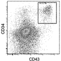
- the FACS analysis result which shows that the LMP2 tetramer positive and CD8 positive T cell were induced
- FIG. The figure which shows that the T cell induced
- derived from the LMP2 peptide specific T cell The FACS analysis result of the cell of the differentiation induction process (Day41) to the T cell of the T-iPS cell induced
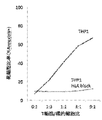
- FIG. 6 shows cytotoxic activity of regenerative CTL derived from clone WT1 # 3-3 against leukemia cell line THP1. Cytotoxic activity was completely blocked with antibodies against HLA class I.
- FIG. 6 shows the cytotoxic activity of regenerative CTL derived from clone WT1 # 3-3 against leukemia cell line HL60. Cytotoxic activity was completely blocked with antibodies against HLA class I.
- Non-growth control in Example 5 Regenerated CTL cultured with IL-7 (5ng / ml) only. Control without target cells in Example 5. Even if there is no target cell, it grows a little, and this proliferated part is used as a control thereafter. Regenerated CTLs are not alloreactive to autologous HLA. Regenerative CTL generally does not show alloreactivity to third party HLA. Regenerated CTL may be alloreactive to third party HLA.
- a “pluripotent stem cell” is a stem cell that has pluripotency that can be differentiated into many cells existing in a living body and also has a self-proliferating ability.
- pluripotent stem cells include embryonic stem (ES) cells, embryonic stem (ntES) cells derived from cloned embryos obtained by nuclear transfer, embryonic germ cells (“EG cells”), induced pluripotent stems (iPS) cells and the like are exemplified.
- the pluripotent stem cells are preferably mammalian pluripotent stem cells, more preferably human pluripotent stem cells.
- iPS cells are preferably used.
- T-iPS cells iPS cells derived from T cells.
- T cell means a cell expressing on its surface an antigen receptor, which is recognized as a T cell receptor (TCR). It has been reported, for example, in WO2011 / 096482 and Vizcardo et al., Cell Stem Cell 12, 31-36 2013 () that iPS cells are induced from T cells.
- the T cells induced into iPS cells are preferably T cells that express CD3 and express at least one molecule selected from the group consisting of CD4 and CD8.
- human T cells include helper / regulatory T cells that are CD4 positive cells, cytotoxic T cells that are CD8 positive cells, naive T cells (CD45RA + CD62L + cells), central memory T cells ( CD45RA ⁇ CD62L + cells), effector memory T cells (CD45RA ⁇ CD62L ⁇ cells), and terminal effector T cells (CD45RA + CD62L ⁇ cells).
- Human T cells can be isolated from human tissues by a known technique.
- the human tissue is not particularly limited as long as it is a tissue containing the T cell, and examples thereof include peripheral blood, lymph nodes, bone marrow, thymus, spleen, umbilical cord blood, and lesioned tissue. Among these, peripheral blood and umbilical cord blood are preferable from the viewpoint of low invasiveness to humans and easy preparation.
- a known technique for isolating human T cells includes, for example, flow cytometry using an antibody against a cell surface marker such as CD4 and a cell sorter as shown in the Examples described later.
- desired T cells can be isolated using cytokine secretion or functional molecule expression as an index.
- T cells have different cytokines secreted depending on Th1 type or Th2 type. Therefore, T cells having a desired Th type can be isolated by selecting such cytokines as indicators. .
- cytotoxic (killer) T cells can be isolated using secretion or production of granzyme or perforin as an index.
- a “T cell having a desired antigen specificity” or “T cell having a desired antigen specificity TCR” can be obtained, for example, by obtaining or inducing a cytotoxic T cell having the TCR from a donor cell.
- a cancer antigen-specific cytotoxic T cell can be obtained by stimulating lymphocytes obtained from a donor by a conventional method with a cancer antigen specific for the cancer to be treated. Cancer antigens have been identified for various cancers, and methods for inducing cytotoxic T cells using cancer antigens or epitope peptides thereof are well known. Alternatively, lymphocytes may be stimulated using cancer cells to be treated.
- cytotoxic T cells specific to a cancer antigen specific to the cancer may be induced from peripheral blood obtained from a donor affected with the cancer to be treated.
- a method of purifying using can be employed.
- a tetramerized MHC (major histocompatibility gene complex) to which a desired antigen is bound is used to obtain a “T having a desired antigen specificity from a human tissue.
- a method of purifying “cells” can also be employed.
- pluripotent stem cell cells Inducing pluripotent stem cells from human T cells having the desired antigen specificity.
- the method described in Vizcardo et al., “Cell Stem Cell 12”, “31-36” 2013 () may be used.
- a T cell having a desired antigen specificity can be obtained from a subject who has acquired immunity against the disease to be treated, and T-iPS cells can be obtained by introducing a Yamanaka factor into this cell (Takahashi and Yamanaka, Cell 126). , 663-673 (2006), Takahashi et al., Cell 131, 861-872 (2007) and Grskovic et al., Nat. Rev. Drug Dscov. 10,915-929 (2011)).
- An induced pluripotent stem (iPS) cell is a somatic cell-derived artificial stem cell that can be produced by allowing a specific reprogramming factor to act on a somatic cell, and has almost the same characteristics as an ES cell ( K. Takahashi and S. Yamanaka (2006) Cell, 126: 663-676; K. Takahashi et al. (2007), Cell, 131: 861-872; J. Yu et al. (2007), Science, 318: 1917-1920; Nakagawa, M. et al., Nat. Biotechnol. 26: 101-106 (2008); International Publication WO 2007/069666).
- the reprogramming factor is a gene specifically expressed in ES cells, its gene product or non-cording RNA, a gene that plays an important role in maintaining undifferentiation of ES cells, its gene product or non-coding RNA, or It may be constituted by a low molecular compound.
- genes included in the reprogramming factor include Oct3 / 4, Sox2, Sox1, Sox3, Sox15, Sox17, Klf4, Klf2, c-Myc, N-Myc, L-Myc, Nanog, Lin28, Fbx15, ERas, ECAT15 -2, Tcl1, beta-catenin, Lin28b, Sall1, Sall4, Esrrb, Nr5a2, Tbx3 or Glis1 etc.
- the reprogramming factor may be brought into contact with or introduced into a somatic cell by a known method according to its form.
- a protein form it may be introduced into a somatic cell by techniques such as lipofection, fusion with a cell membrane permeable peptide (eg, HIV-derived TAT and polyarginine), and microinjection.
- a cell membrane permeable peptide eg, HIV-derived TAT and polyarginine
- Virus vectors include retrovirus vectors, lentivirus vectors (cell, 126, pp.663-676, 2006; Cell, 131, pp.861-872, 2007; Science, 318, pp.1917-1920, 2007 ), Adenovirus vectors (Science, 322, 945-949, 2008), adeno-associated virus vectors, Sendai virus vectors (WO 2010/008054) and the like.
- artificial chromosome vectors examples include human artificial chromosomes (HAC), yeast artificial chromosomes (YAC), and bacterial artificial chromosomes (BAC, PAC).
- HAC human artificial chromosomes
- YAC yeast artificial chromosomes
- BAC bacterial artificial chromosomes
- a plasmid a plasmid for mammalian cells can be used (Science, 322: 949-953, 2008).
- the vector can contain regulatory sequences such as a promoter, enhancer, ribosome binding sequence, terminator, polyadenylation site, etc. so that a nuclear reprogramming substance can be expressed.
- Selective marker sequences such as kanamycin resistance gene, ampicillin resistance gene, puromycin resistance gene, thymidine kinase gene, diphtheria toxin gene, reporter gene sequences such as green fluorescent protein (GFP), ⁇ -glucuronidase (GUS), FLAG, etc. Can be included.
- GFP green fluorescent protein
- GUS ⁇ -glucuronidase
- FLAG FLAG
- RNA incorporating 5-methylcytidine and pseudoouridine® may be used as an initialization factor (Warren® L, ® (2010) ® Cell® Stem® Cell. 7: 618-630).
- the culture solution for iPS cell induction is, for example, DMEM, DMEM / F12 or DME culture solution containing 10 to 15% FBS (in addition to LIF, penicillin / streptomycin, puromycin, L-glutamine, Non-essential amino acids, ⁇ -mercaptoethanol, etc. may be included as appropriate.) Or commercially available culture media [eg, culture medium for mouse ES cell culture (TX-WES culture medium, Thrombo X), primate ES cell culture Culture medium (primate ES / iPS cell culture medium, Reprocell), serum-free medium (mTeSR, Stemcell Technology)] and the like.
- FBS penicillin / streptomycin
- puromycin puromycin
- L-glutamine Non-essential amino acids
- ⁇ -mercaptoethanol etc.
- commercially available culture media eg, culture medium for mouse ES cell culture (TX-WES culture medium, Thrombo X), primate ES cell culture Culture
- a somatic cell is brought into contact with a reprogramming factor in DMEM or DMEM / F12 medium containing 10% FBS in the presence of 5% CO 2 at 37 ° C. for about 4 to 7 days.
- a reprogramming factor in DMEM or DMEM / F12 medium containing 10% FBS in the presence of 5% CO 2 at 37 ° C. for about 4 to 7 days.
- feeder cells for example, mitomycin C-treated STO cells, SNL cells, etc.
- culture medium for bFGF-containing primate ES cell culture about 10 days after contact of somatic cells and reprogramming factor
- the ES-like colonies can be generated about 30 to about 45 days or more after the contact.
- FBS-containing DMEM medium including LIF, penicillin / streptomycin, etc.
- feeder cells eg, mitomycin C-treated STO cells, SNL cells, etc.
- Puromycin, L-glutamine, non-essential amino acids, ⁇ -mercaptoethanol, etc. may be included as appropriate.
- somatic cells to be reprogrammed themselves are used (Takahashi K, et al. (2009), PLoS One.
- iPS cells may be established under hypoxic conditions (oxygen concentration of 0.1% or more and 15% or less) (Yoshida Y, et al. (2009), Cell Stem Cell. 5: 237 -241 or WO2010 / 013845). (The literature described in this paragraph constitutes part of this application by reference)
- histone deacetylase (HDAC) inhibitors for example, small molecule inhibitors such as valproic acid (VPA), trichostatin A, sodium butyrate, MC 1293, M344, HDAC siRNA and shRNA (eg, nucleic acid expression inhibitors such as HDAC1 siRNA Smartpool ⁇ (Millipore), HuSH 29mer shRNA Constructs against HDAC1, etc.), MEK inhibitors (eg, PD184352, PD98059, U0126, SL327 and PD0325901) ), Glycogen synthase kinase-3 inhibitors (eg, Bio and CHIR99021), DNA methyltransferase inhibitors (eg, 5-azacytidine), histone methyltransferase inhibitors (eg, small molecule inhibitors such as BIX-01294, Suv39hl, Nucleic acid expression inhibitors such as siRNA and shRNA for Suv39h2,
- HDAC siRNA and shRNA eg,
- the culture medium is exchanged with a fresh culture medium once a day from the second day onward.
- the number of somatic cells used for nuclear reprogramming is not limited, but ranges from about 5 ⁇ 10 3 to about 5 ⁇ 10 6 cells per 100 cm 2 of culture dish.
- IPS cells can be selected according to the shape of the formed colonies.
- a culture medium selective culture medium
- a drug resistance gene that is expressed in conjunction with a gene that is expressed when somatic cells are initialized for example, Oct3 / 4, Nanog
- iPS cells can be selected by adding a luminescent substrate in the case of a luminescent enzyme gene. Induced iPS cells (T-iPS cells) maintain the T cell receptor gene of the derived T cells.
- iPS cells having the desired T cell receptor (TCR) gene are induced to differentiate into T precursor cells or mature T cells.
- TCR T cell receptor
- Examples of the method of inducing differentiation from iPS cells to T progenitor cells or mature T cells include the methods described in Timmermans et al., Journal of Immunology, 2009, 182: 6879-6888 ().
- T progenitor cell refers to a stage of a cell immediately before receiving positive selection / negative selection from a stage corresponding to a hematopoietic stem cell, which is the most undifferentiated cell among hematopoietic cells. To the equivalent of. T cell differentiation is described in Blood B111: 1318 (2008), NaturemmImmunology 11: 585 (2010).
- T cells are roughly divided into ⁇ T cells and ⁇ T cells, and ⁇ T cells include killer T cells and helper T cells. This application covers all T cells.
- T cells are derived from iPS cells”, both T precursor cells and mature T cells are targeted, preferably CD3 is expressed, In addition, T cells that express at least one molecule selected from the group consisting of CD4 and CD8 are used.
- T progenitor cells and mature T cells derived from iPS cells having the desired antigen-specific TCR gene are obtained as clones maintaining the same antigen specificity as the original T cells. Since all T cells to be administered exhibit a single antigen specificity, it is unlikely that transplantation host rejection will be induced even if transplantation is performed, and immune cell therapy can be performed safely.
- induced T progenitor cells or mature T cells are suspended in an appropriate medium such as physiological saline or PBS and used for treatment of patients.
- an appropriate medium such as physiological saline or PBS
- the graft-versus-host rejection reaction is not induced in the patient before the induced T precursor cell or mature T cell is administered to the patient.
- Not inducing graft-versus-host rejection is a mixed lymphocyte in which the induced T progenitor cells or mature T cells are mixed and cultured with cells of some tissue of the patient undergoing transplantation, preferably patient lymphocyte cells.
- the reaction (Mixed Lymphocyte Reaction, MLR) can be confirmed in advance.
- MLR Mated Lymphocyte Reaction
- regenerative T cells do not recognize patient lymphocyte HLA as an alloantigen in MLR, regenerative T cells do not cause graft-versus-host rejection and can be safely administered for treatment. is there.
- Administration to the patient may be performed intravenously.
- the number of cells to be administered is not particularly limited, and may be appropriately determined according to the patient's age, sex, height, weight, target disease, symptoms, and the like.
- the optimal number of cells to be administered may be appropriately determined by clinical trials.
- T cells can target various antigens, and the method of the present application can be applied to immune cell therapy for various diseases such as cancer, infectious diseases, autoimmune diseases, and allergies.
- WT1 gene is, for example, hematopoietic tumor such as leukemia, myelodysplastic syndrome, multiple myeloma, malignant lymphoma, stomach cancer, colon cancer, lung cancer, breast cancer, germ cell cancer, liver cancer, skin cancer, bladder cancer, prostate T-iPS cells are induced from CTL cells that are highly expressed in natural types in solid cancers such as cancer, uterine cancer, cervical cancer, and ovarian cancer, and have WT1-specific cytotoxic activity.
- CTL cells that are highly expressed in natural types in solid cancers such as cancer, uterine cancer, cervical cancer, and ovarian cancer, and have WT1-specific cytotoxic activity.
- they can be applied to immunocell therapy of these various cancers expressing the WT1 gene.
- Epstein-Barr (EB) virus is a virus that causes various diseases, but it can also cause cancers such as infectious mononucleosis, malignant lymphoma (Burkitt reformer), and nasopharyngeal cancer.
- EB Epstein-Barr
- T-iPS cells are induced from CTL cells having cytotoxicity specific to LMP2 antigen, which is an EB virus-related antigen, and cells in which CTL cells are induced to differentiate from such T-iPS cells are used, It can be applied to immune cell therapy for infectious diseases and cancer.
- T-iPS cells having a desired antigen specificity or those obtained by regenerating T precursor cells or mature T cells from the T-iPS cells are stocked in advance. It ’s fine. Therefore, not only can the time to treatment be shortened, but there is also an advantage that the quality of transplanted cells can be confirmed before transplantation.
- T cell preparations targeting cancer antigens For example, preparation of T cell preparations targeting cancer antigens.
- a cancer antigen-specific T-iPS cell is prepared for a cancer patient, and the T-cell prepared from the T-iPS cell is returned to the original cancer patient and the effect is confirmed.
- iPS cells are banked and stored, and if transplantable HLA-type individuals suffer from cancers that express the same cancer antigen, T-cells made from banked T-iPS cells are administered to the patient be able to. Since it is sufficient that the cells are stored in advance as T cells, they can be prepared in advance and administered to a patient more quickly. Since it is not necessary to create iPS cells for each patient, there is no need for time for preparation thereof, and the administered cells will eventually be rejected, so there is no need to consider the risk of canceration of the administered cells.
- T-iPS cells are induced from T cells having specificity for LMP2 antigen derived from peripheral blood mononuclear cells obtained from EB virus-infected patients (clone LMP2 # 1), and LMP2 antigen-specific CTL ( Regenerated LMP2-CTL # 1) was induced.
- EB virus is a virus that causes infectious mononucleosis in the acute phase and sometimes causes cancer such as Burkitt lymphoma.
- T cells are provided by healthy individuals who have a history of infection with the EB virus.
- the donor is a so-called EB virus carrier because the virus stays in life after infection in lymphocytes. Therefore, this provider can be regarded as a chronic virus infected person although it does not develop.
- LMP2 antigen-specific cytotoxic T lymphocytes
- CTL cytotoxic T lymphocytes
- Monocytes were isolated using CD14 microbeads from the peripheral blood of a healthy volunteer A who has HLA-A2402 and is also infected with EB virus. After washing, dendritic cell culture medium was added to adjust to 5 ⁇ 10 5 / mL. 2.
- Cytokines were added to final concentrations of GM-CSF 800 U / mL (or 50 ng / mL) and IL-4 200 U / mL (or 40 ng / mL). Pour onto a 6-well plate at 5 mL / well. Incubated at 37 ° C. with 5% CO 2 . 3.
- GM-CSF was added to fresh dendritic cell medium at a concentration of 800 U / mL and IL-4 at a concentration of 200 U / mL. 4. 3 mL of new dendritic cell culture medium was added to each well. 5. On Day 6, immature MoDCs were collected from the plates and suspended in a small amount of fresh dendritic cell medium. 6. The cell concentration was adjusted to 5 ⁇ 10 5 / mL. 7.
- GM-CSF (hereinafter, final concentration: 800 U / mL), IL-4 (200 U / mL), TNF- ⁇ (10 ng / mL), PGE2 (1 ⁇ g / mL), and 24-well plate cells were seeded at approximately 5 X 10 5/1 mL / well to. 8. Incubate for 24 hours at 37 ° C and 5% CO2. 9. Peptide was added during the last 2 hours of the culture. The final concentration of peptide was 10 ⁇ m. DCs were collected and washed twice with T cell medium. 10. The number of DC cells was counted and adjusted to 2 ⁇ 10 5 / mL with T cell medium.
- LCL LCL was collected from the culture and irradiated with 35 Gy. 2. Suspended in T cell medium and adjusted to 5 ⁇ 10 5 / mL. 3. Peptide was added at 100 nM and cultured for 2 hours. 4. LCL was collected, washed with T cell medium, and adjusted to 2 ⁇ 10 5 / mL.
- IL-7 final concentration 5 ng / mL
- IL-15 final concentration 1 ng / mL
- the culture was carried out for 2 weeks while changing the medium with a T cell medium supplemented with cytokines every week. (First course of peptide stimulation with LCL) 4. LCL was further cultured for 2 hours in a medium supplemented with 100 nM peptide, and CTL was added thereto. 5.
- IL-7 final concentration 5 ng / mL
- IL-15 final concentration 1 ng / mL
- LMP2-specific killer activity of LMP2-specific CTL 1.
- the OUN-1 leukemia cell line used as a target cell was labeled with CFSE, suspended in T cell medium, and cultured in the presence of LMP2 peptide 1 nM for 2 hours.
- LMP2-specific killer T cells and OUN-1 leukemia cell lines proliferated by peptide stimulation on 96-well U-bottom plates become 0: 1, 1: 9, 1: 3, 1: 1, 3: 1 respectively.
- the dead cell ratio of the target cells in the presence or absence of the peptide was assayed by the ratio of Annexin V and PI (Propidium Iodide) found in the CFSE positive fraction. The results are shown in FIG. 3. It was confirmed that LMP2-specific killer T cells exhibited antigen-specific killer activity against target cells.
- LMP2-T-iPS cells A. Activation of LMP2-specific CTL 1. CD8 positive cells were concentrated with MACS beads. 2. All cells were suspended in T cell medium, and IL-7 (final concentration 5 ng / mL) and IL-15 (final concentration 10 ng / mL) were added. Furthermore, Dynabeads Human T-Activator CD3 / CD28 was added so that T cell: beads became 1: 1, and CD8 positive cells were activated by culturing for 2 days.
- the penicillin / streptomycin solution consisted of penicillin 10000 U / mL and streptomycin 10000 ⁇ g / mL, with final concentrations of 100 U / mL and 100 ⁇ g / mL, respectively.
- the penicillin / streptomycin solution consisted of penicillin 10000 U / mL and streptomycin 10000 ⁇ g / mL, with final concentrations of 100 U / mL and 0 ⁇ g / mL, respectively.
- OP9 cells 6 ml of 0.1% gelatin / PBS solution was placed in a 10 cm culture dish and allowed to stand at 37 ° C. for 30 minutes or more. Confluent OP9 cells were detached with a trypsin / EDTA solution and seeded in a 10 cm culture dish coated with a 1/4 equivalent amount of gelatin. Medium A was added to medium A to 10 ml. 10 ml of medium A was newly added to the OP9 cell culture dish seeded 4 days later so that the total volume became 20 ml.
- the medium of OP9 cells used for blood cell progenitor cell induction co-culture from iPS cells was aspirated and replaced with fresh medium A.
- the medium of the iPS cell culture dish was aspirated and 10 ml of fresh medium A was added.
- the iPS cell mass was cut with an EZ-passage roller. The cut iPS cell mass was suspended by pipetting with a 200 ul pipetman, and approximately 600 iPS cell masses were visually seeded on OP9 cells. Three or more dishes were used per iPS cell clone, and when subcultured, the cells were combined once and then redistributed to the same number to reduce variability between dishes.
- Day 1 (medium change) It was confirmed whether the iPS cell mass started to adhere and differentiate, and the medium was replaced with fresh medium A 20 ml.
- Day 5 (change medium half amount) Half of the medium was replaced with 10 ml of fresh medium A.
- Day 9 (medium exchange) Half of the medium was replaced with 10 ml of fresh medium A.
- Day 13 Transfer induced mesoderm cells from OP9 cells to OP9 / DLL1 cells
- the medium was aspirated and the medium on the cell surface was washed away with HBSS (+ Mg + Ca). Thereafter, 10 ml of a 250 U collagenase IV / HBSS (+ Mg + Ca) solution was added, followed by incubation at 37 ° C. for 45 minutes.
- the collagenase solution was aspirated and washed with 10 ml of PBS (-). Thereafter, 5 ml of 0.05% trypsin / EDTA solution was added, followed by incubation at 37 ° C. for 20 minutes. After culturing, the cells were peeled off in a film form, so they were physically made fine by pipetting (to separate the adherent cells). 20 ml of fresh medium A was added thereto, and further cultured at 37 ° C. for 45 minutes. After culture, the supernatant containing floating cells was collected through a 100 ⁇ m mesh. After centrifuging at 4 ° C. and 1200 rpm for 7 minutes, the pellet was suspended in 10 ml of medium B.
- FACS analysis was performed to confirm the differentiation stage during the culture period. Many dead cells were observed during the culture in all periods. Therefore, at the time of FACS analysis, PI (Propidium Iodide), 7-AAD, etc. were used to analyze after removing dead cells.
- PI Propidium Iodide
- 7-AAD 7-AAD
- IL-15 was added here to induce mature killer T cells (CD8SP cells).
- CD8SP cells mature killer T cells
- LCL used as target cells was labeled with CFSE, suspended in T cell medium, and cultured in the presence of LMP2 peptide 1 nM for 2 hours. 2.
- regenerated CD8 T cells and LCL used as target cells are 0: 1, 1: 9, 1: 3, 1: 1, 3: 1, 10: 1, 30: 1, respectively.
- the dead cell ratio of the target cells in the presence (p +) or absence (p-) of the peptide was assayed by Annexin V and PI (Propidium Iodide) found in the CFSE positive fraction. 3.
- the results are shown in FIG. LMP2-specific killer T cells were confirmed to exhibit antigen-specific killer activity with respect to LCL (HLA-A2402) used as target cells.
- LMP2 peptide-specific CTL is induced according to the procedure of Example 1, T-iPS cells are induced from the CTL (clone LMP # 13), and further from T-iPS cells.
- CD8 single positive T cells were obtained.
- the LMP2 peptide used is the same as in Example 1.
- the peptide-specific CTL activity of the obtained regenerated LMP2-CTL (# 13) was confirmed by the cytotoxic activity using the peptide-loaded LCL cells as target cells. The results are shown in FIG.
- T-iPS cells are induced from WT1 antigen-specific cytotoxic T cells (CTL) induced from the peripheral blood of healthy volunteers (clone WT1 # 9), and from the T-iPS cells, WT1 antigen-specific mature T cells ( Regenerative WT1-CTL (# 9)) was induced.
- CTL cytotoxic T cells
- Regenerative WT1-CTL (# 9) WT1 antigen-specific mature T cells
- the embodiment has the following configuration. 1) Amplification of WT1 antigen-specific CTL 2) Establishment of WT1-T-iPS cells 3) Differentiation induction from WT1-T-iPS cells to CD8 single positive T cells (CTL) 4) Confirmation of antigen-specific cytotoxic activity of regenerated WT1-CTL obtained in 3)
- WT1 antigen-specific CTL Amplification of WT1 antigen-specific CTL
- the culture media used are as follows.
- the WT1 peptide used is as follows.
- WT1 modified: CYTWNQMNL (SEQ ID NO: 2), Cancer Immunol. Immunothera. 51: 614 (2002)) Modified versions of WT1 peptide and WT1 tetramer used below were used.
- LCL Lymphoblastoid cell line
- LCL was LCL having HLA-A2402 collected from healthy volunteers at the Kyoto University Hospital Hematological Oncology Department (Kyoto City, Japan) as follows.
- A. Isolation of T cells from human peripheral blood and stimulation with peptides 1. Mononuclear cells were purified by Ficoll from the peripheral blood of healthy volunteers and suspended in T cell medium. The HLA types of healthy volunteers are HLA-A * 02: 01/24: 02; B * 15: 01/15: 11; C * 03: 03/08: 01; DRB1 * 12: 01/12: 02 . 2. Cells were seeded in a 96-well U-bottom plate at 2.5 ⁇ 10 5 / mL per well, and the peptide was added to 10 ⁇ m.
- IL-2 final concentration 12.5 U / mL
- IL-7 final concentration 5 ng / mL
- IL-15 final concentration 1 ng / mL
- LCL LCL was collected and irradiated with 35 Gy. 2. Suspended in T cell medium and adjusted to 5 ⁇ 10 5 / mL. 3. Peptide was added at 100 nM and cultured for 2 hours. 4. LCL was collected, washed with T cell medium, and adjusted to 2 ⁇ 10 5 / mL.
- IL-2 final concentration 12.5 U / mL
- IL-7 final concentration 5 ng / mL
- IL-15 final concentration 1 ng / mL
- the culture was carried out for 2 weeks while changing the medium with a T cell medium supplemented with cytokines every week. (First course of peptide stimulation with LCL) 4.
- LCL was again cultured for 2 hours in medium supplemented with 100 nM peptide, and CTL was added here. 5.
- IL-2 final concentration 12.5 U / mL
- IL-7 final concentration 5 ng / mL
- IL-15 final concentration 1 ng / mL
- the culture was carried out for 2 weeks while changing the medium with a T cell medium supplemented with cytokines every week. (Second course of peptide stimulation by LCL) 6. LCL was again cultured for 2 hours in medium supplemented with 100 nM peptide, and CTL was added here. 7. On the third day, IL-2 (final concentration 12.5 U / mL), IL-7 (final concentration 5 ng / mL), and IL-15 (final concentration 1 ng / mL) were added. The culture was carried out for 2 weeks while changing the medium with a T cell medium supplemented with cytokines every week. (3rd peptide stimulation with LCL) 8. Flow cytometric analysis was performed. The results are shown in FIG. It was confirmed that the CD8 positive WT1 tetramer positive fraction was detected in the CD8 positive T cells at a rate of 60% or more.
- the penicillin / streptomycin solution consisted of penicillin 10000 U / mL and streptomycin 10000 ⁇ g / mL, with final concentrations of 100 U / mL and 100 ⁇ g / mL, respectively.
- OP9 cells 6 ml of 0.1% gelatin / PBS solution was placed in a 10 cm culture dish and allowed to stand at 37 ° C. for 30 minutes or more. Confluent OP9 cells were detached with trypsin / EDTA solution and seeded on a 10cm culture dish coated with 1/4 of gelatin. Medium A was added to medium A to 10 ml. 10 ml of medium A was newly added to the OP9 cell culture dish seeded 4 days later so that the total volume became 20 ml.
- the medium of OP9 cells used for blood cell progenitor cell induction co-culture from iPS cells was aspirated and replaced with fresh medium A.
- the medium of the iPS cell culture dish was aspirated and 10 ml of new medium A was added.
- IPS cells were cut with an EZ-passage roller. The cut iPS cell mass was suspended by pipetting with a 200 ul pipetman, and approximately 600 iPS cell masses were visually seeded on OP9 cells. Using three or more dishes per clone of iPS cells, and subculture, the cells were combined once and then redistributed to the same number to reduce variability between dishes.
- Day 1 (medium change) The iPS cell mass was confirmed to be attached and differentiated, and the medium was replaced with fresh medium A 20 ml.
- Day 5 (change medium half amount) Half of the medium was replaced with 10 ml of fresh medium A.
- Day 9 (medium exchange) Half of the medium was replaced with 10 ml of fresh medium A.
- Day 13 Transfer induced mesoderm cells from OP9 cells to OP9 / DLL1 cells
- the medium was aspirated and the medium on the cell surface was washed away with HBSS (+ Mg + Ca). Thereafter, 10 ml of 250U collagenase IV / HBSS (+ Mg + Ca) solution was added, and the mixture was incubated at 37 ° C. for 45 minutes.
- the induced T cells become T cells showing the same antigen specificity as the original T cells. Moreover, since the obtained T cell expresses the surface antigen which a mature cell emits, it was confirmed that it is functionally well matured.
- LCL used as target cells was labeled with CFSE, suspended in T cell medium, and cultured for 2 hours in the presence of WT1 peptide (SEQ ID NO: 2). 2. In a 96-well U-bottom plate, mix the regenerated CD8 single positive T cells and the LCL used as target cells in a ratio of 0: 1, 1: 3, 1: 1, 3: 1, 9: 1 respectively. The co-culture was performed in the presence or absence of various concentrations of peptides. After 6 hours, the dead cell ratio of the target cells was assayed by the ratio of Annexin V positive cells found in the CFSE positive fraction.
- WT1 peptide-specific CTL is induced from the same healthy volunteer as in Example 3 according to the procedure of Example 3, T-iPS cells (clone WT1 # 3-3) are induced from the CTL, and T-iPS cells are further induced. From CD8 single positive T cells (regenerated WT1-CTL (# 3-3)). The WT1 peptide used is the same as in Example 3. The peptide-specific CTL activity of the obtained regenerated WT1-CTL (# 3-3) was confirmed by a cytotoxic activity test using LCL cells loaded with the peptide as target cells.
- cytotoxic activity against leukemia cell line THP1 expressing WT1 antigen of regenerated WT1-CTL (# 3-3) and leukemia cell line HL60 also expressing WT1 antigen, and the cytotoxic activity against antibodies against HLA class I I checked if it could be blocked.
- the results are shown in FIG. 17 and FIG.
- Regenerated WT1-CTL (# 3-3) showed cytotoxic activity against both THP-1 and HL60 strains expressing WT1, and such cytotoxic activity was completely blocked with antibodies against HLA class I. From this result, it is considered that regenerated WT1-CTL (# 3-3) kills leukemia cells specifically for the WT1 antigen.
- the effector cells were labeled with CSFE, which is a fluorescent dye.
- target cells peripheral blood monocytes and B cells obtained by concentrating from the peripheral blood of volunteers A and B using anti-CD14 and anti-CD19 Max beads, respectively, were used.
- Effector cell proliferation was measured by examining the degree of cell division from the fluorescence intensity of CFSE. When the effector is activated, cell division proceeds and CFSE decreases.
- Regenerated WT1-CTL (# 9), which is an effector cell, was cultured for 6 days in a medium containing no target cells and only IL-7 (5 ng / ml). No proliferation of regenerative CTL cells was observed and the cells were not activated (non-growth control, FIG. 19).
- Effector cells 8 ⁇ 10 4 cells and 2 ⁇ 10 5 target cells were mixed and cultured for 6 days, and then the degree of cell division was examined by measuring the fluorescence intensity of CFSE.
- FIG. 21 shows the results when monocytes and B cells derived from the peripheral blood of volunteers derived from clone WT # 9 were used as target cells. It can be seen that WT1-CTL (# 9) is not activated at all, which is almost the same as the case without target cells. Regenerated CTLs are not alloreactive to autologous HLA.
- FIG. 22 shows the results when volunteer A peripheral blood-derived cells were used as target cells. It was confirmed that the regenerated CTL did not show alloreactivity against the target cells derived from volunteer A, which had completely different HLA. Regenerated WT1-CTL (# 9) was a cloned cell, and it was found that cloning could prevent contamination of alloreactive T cells.
- FIG. 23 shows the result when volunteer B peripheral blood-derived cells were used as target cells.
- the target cells were volunteer B-derived cells, regenerated WT1-CTL (# 9) was activated. That is, it turns out that the immune cell therapy in the combination of clone WT1 # 9 and volunteer B is dangerous. Even when cloned, the possibility of alloreactivity is not completely eliminated. Thus, in immune cell therapy, it is necessary to screen in advance when the cloned regenerative CTL is administered to a patient.
Abstract
Priority Applications (2)
| Application Number | Priority Date | Filing Date | Title |
|---|---|---|---|
| JP2016534510A JPWO2016010153A1 (ja) | 2014-07-18 | 2015-07-17 | 免疫細胞療法用t細胞の誘導方法 |
| US15/326,940 US20170296649A1 (en) | 2014-07-18 | 2015-07-17 | Method for inducing t cells for cell-based immunotherapy |
Applications Claiming Priority (4)
| Application Number | Priority Date | Filing Date | Title |
|---|---|---|---|
| US201462026328P | 2014-07-18 | 2014-07-18 | |
| US201462026322P | 2014-07-18 | 2014-07-18 | |
| US62/026,322 | 2014-07-18 | ||
| US62/026,328 | 2014-07-18 |
Publications (1)
| Publication Number | Publication Date |
|---|---|
| WO2016010153A1 true WO2016010153A1 (fr) | 2016-01-21 |
Family
ID=55078638
Family Applications (1)
| Application Number | Title | Priority Date | Filing Date |
|---|---|---|---|
| PCT/JP2015/070622 WO2016010153A1 (fr) | 2014-07-18 | 2015-07-17 | Procédé pour induire des lymphocytes t pour une immunothérapie |
Country Status (3)
| Country | Link |
|---|---|
| US (1) | US20170296649A1 (fr) |
| JP (1) | JPWO2016010153A1 (fr) |
| WO (1) | WO2016010153A1 (fr) |
Cited By (5)
| Publication number | Priority date | Publication date | Assignee | Title |
|---|---|---|---|---|
| US10472610B2 (en) | 2014-05-21 | 2019-11-12 | Kyoto University | Method for generating pancreatic bud cells and therapeutic agent for pancreatic disease containing pancreatic bud cells |
| WO2020022512A1 (fr) | 2018-07-26 | 2020-01-30 | 国立大学法人京都大学 | Procédé de production d'une cellule introduite par un gène récepteur d'antigène étranger |
| US11401504B2 (en) | 2016-04-15 | 2022-08-02 | Kyoto University | Method for inducing antigen specific CD8 positive T cells |
| KR20230074505A (ko) | 2020-09-24 | 2023-05-30 | 고쿠리츠 다이가쿠 호진 교토 다이가쿠 | 원하는 특이성을 갖는 이펙터 세포의 제조 방법 |
| KR20230128324A (ko) | 2021-02-05 | 2023-09-04 | 고쿠리츠다이가쿠호진 고베다이가쿠 | 인공 다능성 줄기 세포 유래 γδ T 세포 및 그 제작방법 |
Citations (1)
| Publication number | Priority date | Publication date | Assignee | Title |
|---|---|---|---|---|
| JP2006306822A (ja) * | 2005-05-02 | 2006-11-09 | Japan Science & Technology Agency | 移植片拒絶反応及び移植片対宿主疾患を防ぐ移植免疫反応抑制ポリフェノール溶液及び移植免疫反応抑制方法 |
-
2015
- 2015-07-17 JP JP2016534510A patent/JPWO2016010153A1/ja active Pending
- 2015-07-17 WO PCT/JP2015/070622 patent/WO2016010153A1/fr active Application Filing
- 2015-07-17 US US15/326,940 patent/US20170296649A1/en not_active Abandoned
Patent Citations (1)
| Publication number | Priority date | Publication date | Assignee | Title |
|---|---|---|---|---|
| JP2006306822A (ja) * | 2005-05-02 | 2006-11-09 | Japan Science & Technology Agency | 移植片拒絶反応及び移植片対宿主疾患を防ぐ移植免疫反応抑制ポリフェノール溶液及び移植免疫反応抑制方法 |
Non-Patent Citations (12)
| Title |
|---|
| ATSUTAKA MINAGAWA ET AL.: "3.iPS Saibo kara no Kogen Tokuiteki T-Saibo Yudo to Sono Rinsho Oyo", HEMATOLOGY FRONTIER, vol. 24, no. 2, January 2014 (2014-01-01), pages 39 - 45 * |
| ATSUTAKA MINAGAWA ET AL.: "Genome Edit o Riyo shita, iPS Saibo yori no Kogen Tokuiteki T- Saibo no Saisei", THE 18TH JAPANESE ASSOCIATION OF CANCER IMMUNOLOGY SOKAI PROGRAM/SHOROKUSHU, June 2014 (2014-06-01), pages 161, Q19 - 3 * |
| HIROSHI KAWAMOTO ET AL.: "Shokika ni yoru Kogen Tokuiteki T-Saibo no Cloning to Bank-ka Koso", REGENERATIVE MEDICINE, vol. 13, no. Suppl. 2014, January 2014 (2014-01-01), pages 176, SY-25 - 4 * |
| HIROSHI KAWAMOTO: "Application of the iPSC technology to immune cell therapy against cancer", CLINICAL ONCOLOGY, vol. 12, no. 4, 2013, pages 450 - 459 * |
| JURGENS LISA A. ET AL.: "Transduction of Primary Lymphocytes with Epstein-Barr Virus (EBV) Latent Membrane Protein-Specific T- Cell Receptor Induces Lysis of Virus-Infected Cells: A Novel Strategy for the Treatment of Hodgkin's Disease and Nasopharyngeal Carcinoma", JOURNAL OF CLINICAL IMMUNOLOGY, vol. 26, no. 1, 2006, pages 22 - 32, XP019281116 * |
| KAWAMOTO HIROSHI ET AL.: "Regeneration of antigen specific T cells using the iPSC technology: A novel strategy for cancer immunotherapy", THE 18TH JAPANESE ASSOCIATION OF CANCER IMMUNOLOGY SOKAI PROGRAM/SHOROKUSHU, June 2014 (2014-06-01), pages 50, Sl-3 * |
| LEI FENGYANG ET AL.: "Dual signals of TCR and Notch promote antigen-specific T cell development from pluripotent stem cells (P4370", THE JOURNAL OF IMMUNOLOGY, vol. 190, 2013, pages 177.18 * |
| LEI FENGYANG ET AL.: "In Vivo Programming of Tumor Antigen-Specific T Lymphocytes from Pluripotent Stem Cells to Promote Cancer Immunosurveillance", CANCER RESEARCH, vol. 71, no. 14, 2011, pages 4742 - 4747, XP055143834, DOI: doi:10.1158/0008-5472.CAN-11-0359 * |
| RIOLOBOS LAURA ET AL.: "HLA Engineering of Human Pluripotent Stem Cells", MOLECULAR THERAPY, vol. 21, no. 6, 2013, pages 1232 - 1241, XP055145726, DOI: doi:10.1038/mt.2013.59 * |
| TAKUYA MAEDA ET AL.: "iPS Saibo Gijutsu o Mochiita Gan Kogen Tokuiteki T-Saibo no Saisei", REGENERATIVE MEDICINE, vol. 13, no. Suppl. 2014, January 2014 (2014-01-01), pages 226 * |
| TAMANAKA TAICHI ET AL.: "Recognition of a Natural WT1 Epitope by a Modified WT1 Peptide- specific T- Cell Receptor", ANTICANCER RESEARCH, vol. 32, 2012, pages 5201 - 5210 * |
| THEMELI MARIA ET AL.: "Generation of tumor- targeted human T lymphocytes from induced pluripotent stem cells for cancer therapy", NATURE BIOTECHNOLOGY, vol. 31, no. 10, 2013, pages 928 - 933, XP055143283, DOI: doi:10.1038/nbt.2678 * |
Cited By (5)
| Publication number | Priority date | Publication date | Assignee | Title |
|---|---|---|---|---|
| US10472610B2 (en) | 2014-05-21 | 2019-11-12 | Kyoto University | Method for generating pancreatic bud cells and therapeutic agent for pancreatic disease containing pancreatic bud cells |
| US11401504B2 (en) | 2016-04-15 | 2022-08-02 | Kyoto University | Method for inducing antigen specific CD8 positive T cells |
| WO2020022512A1 (fr) | 2018-07-26 | 2020-01-30 | 国立大学法人京都大学 | Procédé de production d'une cellule introduite par un gène récepteur d'antigène étranger |
| KR20230074505A (ko) | 2020-09-24 | 2023-05-30 | 고쿠리츠 다이가쿠 호진 교토 다이가쿠 | 원하는 특이성을 갖는 이펙터 세포의 제조 방법 |
| KR20230128324A (ko) | 2021-02-05 | 2023-09-04 | 고쿠리츠다이가쿠호진 고베다이가쿠 | 인공 다능성 줄기 세포 유래 γδ T 세포 및 그 제작방법 |
Also Published As
| Publication number | Publication date |
|---|---|
| JPWO2016010153A1 (ja) | 2017-04-27 |
| US20170296649A1 (en) | 2017-10-19 |
Similar Documents
| Publication | Publication Date | Title |
|---|---|---|
| JP7440027B2 (ja) | 多能性幹細胞から免疫細胞療法用t細胞を誘導する方法 | |
| US20220251506A1 (en) | Method for inducing antigen specific cd8 positive t cells | |
| JP2021000108A (ja) | 抗原特異的t細胞の製造方法 | |
| WO2016010155A1 (fr) | Méthode de production de cellules souches pluripotentes possédant un gène codant pour le récepteur des cellules t spécifique d'un antigène | |
| JP5861191B2 (ja) | ミエロイド系血液細胞の製造方法 | |
| WO2016010153A1 (fr) | Procédé pour induire des lymphocytes t pour une immunothérapie | |
| WO2020027094A1 (fr) | PROCÉDÉ DE PRODUCTION D'UNE POPULATION DE LYMPHOCYTES T RÉGÉNÉRÉS PAR L'INTERMÉDIAIRE DE CELLULES iPS | |
| WO2015099134A1 (fr) | Immunothérapie utilisant des cellules t précurseurs dérivées de cellules souches pluripotentes portant un gène du récepteur t réarrangé | |
| WO2017159087A1 (fr) | Procédé d'induction de cellule t spécifique de l'antigène ny-eso1 pour immunothérapie cellulaire | |
| US20220233665A1 (en) | Medicinal composition | |
| JP7407415B2 (ja) | Cd4陽性制御性t細胞の製造方法 |
Legal Events
| Date | Code | Title | Description |
|---|---|---|---|
| 121 | Ep: the epo has been informed by wipo that ep was designated in this application |
Ref document number: 15822087 Country of ref document: EP Kind code of ref document: A1 |
|
| ENP | Entry into the national phase |
Ref document number: 2016534510 Country of ref document: JP Kind code of ref document: A |
|
| NENP | Non-entry into the national phase |
Ref country code: DE |
|
| WWE | Wipo information: entry into national phase |
Ref document number: 15326940 Country of ref document: US |
|
| 122 | Ep: pct application non-entry in european phase |
Ref document number: 15822087 Country of ref document: EP Kind code of ref document: A1 |