WO2016000619A1 - Anti-pd-l1 antibodies and their use as therapeutics and diagnostics - Google Patents
Anti-pd-l1 antibodies and their use as therapeutics and diagnostics Download PDFInfo
- Publication number
- WO2016000619A1 WO2016000619A1 PCT/CN2015/083066 CN2015083066W WO2016000619A1 WO 2016000619 A1 WO2016000619 A1 WO 2016000619A1 CN 2015083066 W CN2015083066 W CN 2015083066W WO 2016000619 A1 WO2016000619 A1 WO 2016000619A1
- Authority
- WO
- WIPO (PCT)
- Prior art keywords
- seq
- binding
- human
- cells
- antibody
- Prior art date
- Legal status (The legal status is an assumption and is not a legal conclusion. Google has not performed a legal analysis and makes no representation as to the accuracy of the status listed.)
- Ceased
Links
Images


Classifications
-
- C—CHEMISTRY; METALLURGY
- C07—ORGANIC CHEMISTRY
- C07K—PEPTIDES
- C07K16/00—Immunoglobulins [IG], e.g. monoclonal or polyclonal antibodies
- C07K16/18—Immunoglobulins [IG], e.g. monoclonal or polyclonal antibodies against material from animals or humans
- C07K16/28—Immunoglobulins [IG], e.g. monoclonal or polyclonal antibodies against material from animals or humans against receptors, cell surface antigens or cell surface determinants
- C07K16/2803—Immunoglobulins [IG], e.g. monoclonal or polyclonal antibodies against material from animals or humans against receptors, cell surface antigens or cell surface determinants against the immunoglobulin superfamily
- C07K16/2827—Immunoglobulins [IG], e.g. monoclonal or polyclonal antibodies against material from animals or humans against receptors, cell surface antigens or cell surface determinants against the immunoglobulin superfamily against B7 molecules, e.g. CD80, CD86
-
- A—HUMAN NECESSITIES
- A61—MEDICAL OR VETERINARY SCIENCE; HYGIENE
- A61P—SPECIFIC THERAPEUTIC ACTIVITY OF CHEMICAL COMPOUNDS OR MEDICINAL PREPARATIONS
- A61P35/00—Antineoplastic agents
-
- C—CHEMISTRY; METALLURGY
- C07—ORGANIC CHEMISTRY
- C07K—PEPTIDES
- C07K16/00—Immunoglobulins [IG], e.g. monoclonal or polyclonal antibodies
- C07K16/18—Immunoglobulins [IG], e.g. monoclonal or polyclonal antibodies against material from animals or humans
- C07K16/28—Immunoglobulins [IG], e.g. monoclonal or polyclonal antibodies against material from animals or humans against receptors, cell surface antigens or cell surface determinants
- C07K16/2803—Immunoglobulins [IG], e.g. monoclonal or polyclonal antibodies against material from animals or humans against receptors, cell surface antigens or cell surface determinants against the immunoglobulin superfamily
- C07K16/2818—Immunoglobulins [IG], e.g. monoclonal or polyclonal antibodies against material from animals or humans against receptors, cell surface antigens or cell surface determinants against the immunoglobulin superfamily against CD28 or CD152
-
- A—HUMAN NECESSITIES
- A61—MEDICAL OR VETERINARY SCIENCE; HYGIENE
- A61K—PREPARATIONS FOR MEDICAL, DENTAL OR TOILETRY PURPOSES
- A61K39/00—Medicinal preparations containing antigens or antibodies
- A61K2039/505—Medicinal preparations containing antigens or antibodies comprising antibodies
-
- C—CHEMISTRY; METALLURGY
- C07—ORGANIC CHEMISTRY
- C07K—PEPTIDES
- C07K2317/00—Immunoglobulins specific features
- C07K2317/20—Immunoglobulins specific features characterized by taxonomic origin
- C07K2317/24—Immunoglobulins specific features characterized by taxonomic origin containing regions, domains or residues from different species, e.g. chimeric, humanized or veneered
-
- C—CHEMISTRY; METALLURGY
- C07—ORGANIC CHEMISTRY
- C07K—PEPTIDES
- C07K2317/00—Immunoglobulins specific features
- C07K2317/30—Immunoglobulins specific features characterized by aspects of specificity or valency
- C07K2317/34—Identification of a linear epitope shorter than 20 amino acid residues or of a conformational epitope defined by amino acid residues
-
- C—CHEMISTRY; METALLURGY
- C07—ORGANIC CHEMISTRY
- C07K—PEPTIDES
- C07K2317/00—Immunoglobulins specific features
- C07K2317/70—Immunoglobulins specific features characterized by effect upon binding to a cell or to an antigen
- C07K2317/76—Antagonist effect on antigen, e.g. neutralization or inhibition of binding
-
- C—CHEMISTRY; METALLURGY
- C07—ORGANIC CHEMISTRY
- C07K—PEPTIDES
- C07K2317/00—Immunoglobulins specific features
- C07K2317/90—Immunoglobulins specific features characterized by (pharmaco)kinetic aspects or by stability of the immunoglobulin
- C07K2317/92—Affinity (KD), association rate (Ka), dissociation rate (Kd) or EC50 value
-
- C—CHEMISTRY; METALLURGY
- C07—ORGANIC CHEMISTRY
- C07K—PEPTIDES
- C07K2319/00—Fusion polypeptide
- C07K2319/30—Non-immunoglobulin-derived peptide or protein having an immunoglobulin constant or Fc region, or a fragment thereof, attached thereto
Definitions
- Kang Li Jing Song, Tong Zhang, Yucheng Li, Zhiying Ren, Yanshen Kang, all of Beijing, China
- PD-L1 was initially cloned as a member (known as B7-H1) of B7 protein family (Dong et al., 1999 Nature Med 5: 1365) . It binds to Programmed Death-1 (PD-1) receptor and activates negative regulatory signaling pathway, inhibiting T-cell proliferation and activities (Freeman et. al. 2000 J Exp Med 192: 1027) . Therefore, it was also termed as PD-1 ligand 1 (PD-L1 or CD274) .
- B7-H1 B7-H1 of B7 protein family
- PD-L1 B7-H1
- PD-L2 B7-DC
- Human PD-L1 gene encodes a full-length protein of 290 amino acid residues (NCBI accession NP_054862.1) with a leader peptide, which is removed after PD-L1 is expressed on cell surface as a mature protein.
- the calculated molecular weight of the full length PD-L1 is 33 kD. However, the observed molecular weight is around 50 kD due to glycosylation, based on Western blot data from ours and others.
- PD-L1 was found constitutively expressed in human heart, lung, thymus and vascular endothelial cells, and expressed at a low level in many other human tissues and cell types including antigen presenting cells, peripheral blood monocytes and other immune cells (Freeman et. al. 2000 J Exp Med 192: 1027; Eppihimer et. al. 2002 Microcirculation 9: 133) .
- IFN- ⁇ , IL-12 and type I interferons many of those cell types were found expressing increased level of PD-L1 (Bald et. al. 2014 Cancer Discov 4: 674-687; Planes et. al. 2014 J Virol 88: 6672-6689) .
- Neoplasia 8 190
- ovary Hamanishi et. al. 2007 Proc Natl Acad Sci 104: 3360
- pancreas Nomi et. al. 2007 Clin Cancer Res 13: 2151
- melanocytes Hino et. al. 2010 Cancer 116: 1757
- esophagus Ohigashi et. al. 2005 Clin Cancer Res 11: 2947.
- the increased expression of PD-L1 in those cancers is associated to poor prognosis in patient survival outcome.
- Blockade of PD-L1 engaging PD-1 receptor by B7-H1Ig or anti-PD-L1 antibody stimulated T-cell proliferation and functional activities (Dong et. al. 1999 Nature Med 5: 1365; Freeman et. al. 2000 J Exp Med 192: 1027; Tamura et. al. 2001 Blood 97: 1809; Iwai et. al. 2002 PNAS 99: 12293) , enhanced immune responses against tumor growth and viral infection (Iwai et. al. 2002 PNAS 99: 12293) .
- Those observations suggested that inhibition of PD-L1/PD-1 signaling may activate immune responses not only against cancer cell growth, but also against viral infection and expansion in human.
- the popular HIV infection frequently evades human immune system by similar mechanism.
- Therapeutic modulation of PD-L1 induced signaling by antagonist molecules may revert immune cells from tolerance, and reactivated to eradicate cancer and chronic viral infection (Blank et. al. 2005 Cancer Immunol Immunother 54: 307; Okazaki et. al. 2007 Int Immunol 19: 813) .
- the invention provides methods and compositions for immune-activation by inhibiting PD-L1-mediated signaling and function.
- the invention provides an antibody antigen binding domain which specifically binds human PD-L1, and comprises a complementarity determining region (CDR) sequence described herein.
- CDRs are amenable to recombination into heavy chain variable region (Vh) and light chain variable regions (Vk) which comprise (CDR-H1, CDR-H2 and CDR-H3) and (CDR-L1, CDR-L2 and CDR-L3) sequences, respectively and retain PD-L1-specific binding and/or functionality.
- the domain comprises CDR1, CDR2 and CDR3, in a combination selected from (a) - (r) as follows, wherein the antibody (Ab) , heavy chain (HC) or light chain (LC) and CDR nomenclature system (Kabat, IMGT or composite) from which the CDR combinations derive are shown in the first column, and residues in bold text are Kabat system, and residues underlined are IMGT system:
- the domain comprises a heavy chain variable region (Vh) comprising a CDR1, CDR2 and CDR3 combination selected from (a) - (o) , and a light chain variable region (Vk) comprising a CDR1, CDR2 and CDR3 combination selected from (p) - (r) .
- Vh heavy chain variable region
- Vk light chain variable region
- the domain comprises CDR1, CDR2 and CDR3, in a combination selected from (c) , (f) , (i) , (l) , (o) and (r) , as follows:
- the domain comprises a heavy chain variable region (Vh) or a light chain variable region (Vk) , comprising a sequence that is:
- the domain comprises a heavy chain variable region (Vh) and a light chain variable region (Vk) comprising a sequence that is:
- the domain comprises comprising a heavy chain variable region (Vh) or a light chain variable region (Vk) comprising:
- the domain comprises comprising a heavy chain variable region (Vh) and a light chain variable region (Vk) comprising:
- the domain specifically binds PD-L1 residues: D26 and R113.
- the invention also provides antibodies, particularly monoclonal antibodies, and F (ab) or F(ab) 2 comprising a subject PD-L1 binding domain.
- the invention also provides novel polynucleotides such as cDNAs and expression vectors, encoding a subject PD-L1 antigen binding domain, and cells comprising such polynucleotides, and non-human animals comprising such cells.
- the polynucleotides may be operably linked to a heterologous transcription regulating sequence for expression, and may be incorporated into such vectors, cells, etc.
- the invention provides methods of using the subject domains by administering the domain to a person determined to have cancer or a viral infection or to otherwise be in need of PD-L1 antagonism.
- compositions of the invention are useful for the treatment of cancer, neurodegenerative and infectious, particularly viral, diseases and other conditions in which inappropriate or detrimental expression of the human PD-1 and/or is a component of the etiology or pathology of the condition.
- the invention provides methods for treating cancer or inhibiting tumor progression in a subject in need thereof with a subject anti-PD-L1 protein, and the humanized anti-PD-1 mAbs are used as therapeutic agents to treat human diseases that are involved in suppression of immune cells by PD-1 mediated intracellular signaling, leading to disease progression, particularly cancers and viral infections.
- the invention further provides the use of subject polynucleotides for the manufacture of a medicament for treating cancer or inhibiting tumor progression in a subject.
- Fig. 1 Schematic presentation of full length PD-L1 (top) , PD-L1/Fc (middle) and PD-L1/His (bottom) .
- ECD extracellular domain.
- L linker.
- Fc ⁇ 4Fc fragment from human IgG4.
- H His tag.
- N N-terminus.
- C C-terminus.
- Fig. 2 Dose-dependent binding to the purified human PD-L1/His in ELISA (A) or to cell surface expressed PD-L1 in FACS (B) by murine mAbs.
- the murine mAbs and mouse IgG as negative control were indicated at top-left corner of each figure.
- (A) ELISA plate was coated by PD-L1/His at 100ng per well.
- the binding signal strength in ELISA was indicated by direct OD 450 readings in y-axis.
- Concentrations of mAbs or mouse IgG were indicated by x-axis.
- HEK293/PD-L1 cells were stained with a serial dilution of murine anti-PD-L1 mAbs or control mouse IgG.
- the binding signal strength was indicated by MFI (mean fluorescence intensity) in FACS analyses. Concentrations of mAbs or mouse IgG were indicated by x-axis.
- Fig. 3 Dose-dependent response curves of murine anti-PD-L1 mAb-induced IL-2 secretion in HuT78/PD-1 cells after co-culture with HEK293/OS8/PD-L1 cells.
- Baseline Average IL-2 release induced by mouse IgG (mIgG) at all tested concentrations. Each represents average of duplicate data points.
- Top line Highest IL-2 release based on regression calculation by Prizm.
- Fig. 4 Dose-dependent response curves of murine anti-PD-L1 mAb-inhibited IL-2 secretion in HuT78/P3Z cells after co-culture with HEK293/PD-L1 cells. Engagement of PD-L1 and P3Z chimeric receptor leads to activation of P3Z chimeric receptor and IL-2 secretion.
- Fig. 5 Dose-dependent competition curves of murine anti-PD-L1 mAbs against biotin-conjugated PD-1/Fc. Fixed amount of biotin-PD-1-ECD/Fc was mixed with increasing concentrations of anti-PD-L1 mAbs indicated in x-axis. Mean fluorescence intensity (MFI) analyzed by FACS was showed in y-axis. Mouse Gig (mug) was used as a negative control.
- MFI mean fluorescence intensity
- Fig. 6 Dose-dependent competition curves of murine anti-PD-L1 mAbs against biotin-conjugated CD80/Fc. Fixed amount of biotin-CD80-ECD/Fc was mixed with increasing amount of anti-PD-L1 mAbs indicated in x-aixs. Mean fluorescence intensity (MFI) analyzed by FACS was showed in y-axis. Mouse IgG (muIgG) was used as a negative control.
- MFI mean fluorescence intensity
- Fig. 7 IFN- ⁇ secretion induced by humanized anti-PD-L1 Fabs in primary human PBMCs from different healthy donors (Donor 3 (A) and Donor 17 (B) ) .
- PBMCs were co-cultured with HEK293/OS8/PD-L1 cells for overnight.
- IFN- ⁇ in conditioned medium was assayed by ELISA.
- BSA was used as a negative control.
- Fig. 8 C1q bindings of humanized anti-PD-L1 mAbs in wild type (IgG1wt) or mutant human IgG1 formates (IgG1mc and IgG1mf) .
- Fifty microliters of a serial dilution (x-axis) of humanized anti-PD-L1 mAb were coated on MaxiSorp ELISA plate.
- Human C1q bindings (y-axis) were assessed by ELISA OD 450 readings using a specific monoclonal antibody to human C1q.
- Fig. 9 Antibody-dependent cell-cytotoxicity (ADCC) induced by humanized anti-PD-L1 mAbs in wild type (IgG1wt) or mutant human IgG1 formats (IgG1mc and IgG1mf) .
- Human NK92MI cells transfected with Fc ⁇ RIIIA were used as effector cells and HEK293/PD-L1 cells were used as target cells. Percentage of cytotoxicity (y-axis) was calculated based on lactate dehydrogenase (LDH) release assay as described in Example 5.
- Fig. 10 Complement-dependent cytotoxicity (CDC) activities of humanized anti-PD-L1 mAbs in wild type (IgG1wt) or mutant human IgG1 formats (IgG1mc and IgG1mf) .
- Daudi/PD-L1 cells were used as target cells and human sera from healthy donors were used as the source of complement components.
- Rituximab (Roche) was used as positive control in classical CDC assay.
- Percentage of CDC (y-axis) was calculated based on cell-titer glow assay as described in Example 5.
- Fig. 11 IFN- ⁇ secretion induced by humanized anti-PD-L1 mAbs in primary human PBMCs from different healthy donors (Donor 3 (A) and Donor 17 (B) ) .
- PBMCs were co-cultured with HEK293/OS8/PD-L1 cells for overnight.
- IFN- ⁇ in conditioned medium was assayed by ELISA.
- Human IgG served as a negative control.
- Fig. 12 Dose-dependent bindings of anti-PD-L1 mAbs to the purified human PD-L1/His (A) , cynomolgus monkey PD-L1/His (B) , and mouse PD-L1/His (C) in ELISA. MaxiSorp ELISA plates were coated with 50 microliters of human, monkey and mouse PD-L1/His, respectively. Concentrations of anti-PD-L1 mAbs were indicated by x-axis. The binding signal strength was indicated by direct OD 450 readings (y-axis) .
- Fig. 13 Mapping the binding epitopes of anti-PD-L1 mAbs by ELISA (upper panel) and Western Blot (lower panel) .
- A Binding activities to mutant PD-L1 by hu333-4B2-IgG1.
- B Binding activities to mutant PD-L1 by Y1-IgG1.
- C Binding activities to D 26 A mutant PD-L1 by anti-PD-L1 mAbs.
- Conditioned media containing wild type or mutant PD-L1/His proteins were used to assess binding activity by ELISA and Western Blot. ** indicates the PD-L1 mutant to which the mAb binding activity reduced to 25-50%of that to wild type PD-L1.
- *** indicates the PD-L1 mutant to which the mAb binding activity reduced below 25%of that to wild type PD-L1.
- Fig. 14 Binding assays of anti-PD-L1 mAbs to a mixture of human serum protein and PD-L1 antigen by ELISA.
- A Dose-dependent reaction curves of murine mAb or murine chimeric mAb binding to a mixture of human serum and PD-L1/His protein. Serial dilutions of PD-L1/His protein in PBS were coated onto 96-well MaxiSorp ELISA plate as indicated, and human serum pool (from three healthy donors) were added at a fixed final concentration of 5%. Three ug/mL indicated mAbs were added to each well and incubated for one hour at room temperature.
- B Histogram showed the average OD 450 readings of three data points in left side (coated mainly with human serum proteins and with very little PD-L1/His) of curves for each mAb and the negative control (without mAb) .
- Fig. 15 Mean tumor growth curves upon treatment with hu333-4B2-IgG1mf or vehicle.
- NOD/SCID mice implanted with human cancer cells A431 and PBMCs from healthy donors were treated with hu333-4B2-IgG1mf at the dose regimen of 10mg/kg, twice/week.
- PD-L1 initiates inhibitory signaling in immune cells when engaged by its ligands, PD-L1 or PD-L2.
- the activation of PD-1 signaling promotes immune tolerance, leading to the cancers or virus-infected cells escaping from immune surveillance and cancer metastasis or viral load increase.
- Inhibition of PD-L1 mediated cellular signaling by therapeutic agents can activate immune cells including T-cells, B-cells and NK cells, and therefore enhance immune cell functions inhibiting cancer cell growth or viral infection, and restore immune surveillance and immune memory function to treat such human diseases.
- the invention provides antibodies whose functions are antagonistic to PD-L1-induced cellular signaling in immune cells.
- Murine anti-PD-L1 antibodies were humanized to a high degree of similarity to human antibodies in the framework regions.
- the full antibodies made in the modified human IgG variant format have a unique set of features in the aspects of effector functions and physicochemical properties.
- the disclosed anti-PD-L1 antibodies are suitable for therapeutic uses in cancer treatment, controlling viral infections and other human diseases that are mechanistically involved in exacerbated immune tolerance.
- antibody is used in the broadest sense and specifically covers antibodies (including full length monoclonal antibodies) and antibody fragments so long as they recognize PD-L1.
- An antibody molecule is usually monospecific, but may also be described as idiospecific, heterospecific, or polyspecific.
- Antibody molecules bind by means of specific binding sites to specific antigenic determinants or epitopes on antigens.
- Antibody fragments comprise a portion of a full length antibody, generally the antigen binding or variable region thereof. Examples of antibody fragments include Fab, Fab', F (ab') . sub. 2, and Fv fragments; diabodies; linear antibodies; single-chain antibody molecules; and multispecific antibodies formed from antibody fragments.
- Monoclonal antibodies may be obtained by methods known to those skilled in the art. See, for example Kohler et al (1975) ; U.S. Pat. No. 4,376,110; Ausubel et al (1987-1999) ; Harlow et al (1988) ; and Colligan et al (1993) .
- the mAbs of the invention may be of any immunoglobulin class including IgG, IgM, IgE, IgA, and any subclass thereof.
- a hybridoma producing a mAb may be cultivated in vitro or in vivo.
- High titers of mAbs can be obtained in in vivo production where cells from the individual hybridomas are injected intraperitoneally into mice, such as pristine-primed Balb/c mice to produce ascites fluid containing high concentrations of the desired mAbs.
- MAbs of isotype IgM or IgG may be purified from such ascites fluids, or from culture supernatants, using column chromatography methods well known to those of skill in the art.
- isolated polynucleotide refers to a polynucleotide segment or fragment which has been separated from sequences which flank it in a naturally occurring state, e.g., a DNA fragment which has been removed from the sequences which are normally adjacent to the fragment, e.g., the sequences adjacent to the fragment in a genome in which it naturally occurs.
- the term therefore includes, for example, a recombinant DNA which is incorporated into a vector, into an autonomously replicating plasmid or virus, or into the genomic DNA of a prokaryote or eukaryote, or which exists as a separate molecule (e.g., as a cDNA or a genomic or cDNA fragment produced by PCR or restriction enzyme digestion) independent of other sequences. It also includes a recombinant DNA, which is part of a hybrid gene encoding additional polypeptide sequence.
- a “construct” means any recombinant polynucleotide molecule such as a plasmid, cosmid, virus, autonomously replicating polynucleotide molecule, phage, or linear or circular single-stranded or double-stranded DNA or RNA polynucleotide molecule, derived from any source, capable of genomic integration or autonomous replication, comprising a polynucleotide molecule where one or more polynucleotide molecule has been linked in a functionally operative manner, i.e. operably linked.
- a recombinant construct will typically comprise the polynucleotides of the invention operably linked to transcriptional initiation regulatory sequences that will direct the transcription of the polynucleotide in the intended host cell.
- transcriptional initiation regulatory sequences that will direct the transcription of the polynucleotide in the intended host cell.
- Both heterologous and non-heterologous (i.e., endogenous) promoters can be employed to direct expression of the nucleic acids of the invention.
- a “vector” refers any recombinant polynucleotide construct that may be used for the purpose of transformation, i.e. the introduction of heterologous DNA into a host cell.
- a "plasmid” refers to a circular double stranded DNA loop into which additional DNA segments can be ligated.
- a viral vector Another type of vector is a viral vector, wherein additional DNA segments can be ligated into the viral genome.
- Certain vectors are capable of autonomous replication in a host cell into which they are introduced (e.g., bacterial vectors having a bacterial origin of replication and episomal mammalian vectors) .
- vectors e.g., non-episomal mammalian vectors
- vectors are integrated into the genome of a host cell upon introduction into the host cell, and thereby are replicated along with the host genome.
- certain vectors are capable of directing the expression of genes to which they are operatively linked. Such vectors are referred to herein as "expression vectors" .
- an "expression vector” as used herein refers to a nucleic acid molecule capable of replication and expressing a gene of interest when transformed, transfected or transduced into a host cell.
- the expression vectors comprise one or more phenotypic selectable markers and an origin of replication to ensure maintenance of the vector and to, if desired, provide amplification within the host.
- the expression vector further comprises a promoter to drive the expression of the polypeptide within the cells.
- Suitable expression vectors may be plasmids derived, for example, from pBR322 or various pUC plasmids, which are commercially available. Other expression vectors may be derived from bacteriophage, phagemid, or cosmid expression vectors.
- Murine anti-human PD-L1 monoclonal antibodies were generated using hybridoma fusion technology (Kohler and Milstein 1975 Nature 256: 495-497; Mechetner 2007 Methods Mol Biol 378: 1-13) with modifications. MAbs with high binding activities in enzyme-linked immunosorbent assay (ELISA) and fluorescence-activated cell sorting (FACS) assay were selected for further characterization in cell-based functional assays.
- ELISA enzyme-linked immunosorbent assay
- FACS fluorescence-activated cell sorting
- the full length human PD-L1 cDNA was synthesized by GeneScript (Nanjing, China) based on published sequence (NCBI reference sequence NM_014143.3) (SEQ. NO. 1 and 2) .
- the extracellular domain consisting of amino acids (AA) 1-239 of human PD-L1 (SEQ. NO.
- PD-L1/Fc was purified using a Protein G Sepharose Fast Flow column (Cat. No. 17061805, GE Life Sciences, Shanghai, China) .
- PD-L1/His was purified through Ni-Sepharose Fast Flow affinity chromatography (Cat. No. 17531801, GE Life Sciences) , followed by size exclusion chromatography using a HiLoad 16/60 Superdex 200 column (Cat. No. 17106901, GE Life Sciences) .
- Both PD-L1/Fc and PD-L1/His proteins were dialyzed against phosphate buffered saline (PBS) and stored in -80°C freezer in small aliquots.
- PBS phosphate buffered saline
- Expression plasmid containing full-length human PD-1 cDNA was obtained from Origene (Cat. No. SC117011, NCBI Accession No: NM_005018.1, Beijing, China) .
- the extracellular domain consisting of amino acid (AA) 1-168 of PD-1 was PCR-amplified, and subcloned in pcDNA3.1-based expression vector (Invitrogen, Carlsbad, CA, USA) with C-terminus fused to the Fc domain of human IgG4 heavy chain, abbreviated as PD-1/Fc.
- the human CD80 (B7-1) cDNA was synthesized by GeneScript according to the published sequence (NCBI access number NM_005191.3) .
- the extracellular domain (AA1-242) of CD80 was fused with human Fc at C-terminus and subcloned in a mammalian expression vector similar to the method described previously (Patent US 8735553) .
- the fusion protein was named as CD80-ECD/Fc or CD80/Fc.
- Stable cell line expressing human PD-L1 was established by transfection of pcDNA3.1 plasmid containing PD-L1 into HEK293 (ATCC, Manassas, VA, USA) , and followed by selection with media containing 600 micrograms of hygromycin (Cat. No. 10687-010, Invitrogen) per milliliter. Single clones were isolated by picking up single colonies from culture-dish surface. All clones were screened by FACS analysis and Western blot using PD-L1 antibody (Cat. No. 17-5983, eBioscience, San Diego, USA) , and the top expression clones were used for FACS binding analyses and functional assays.
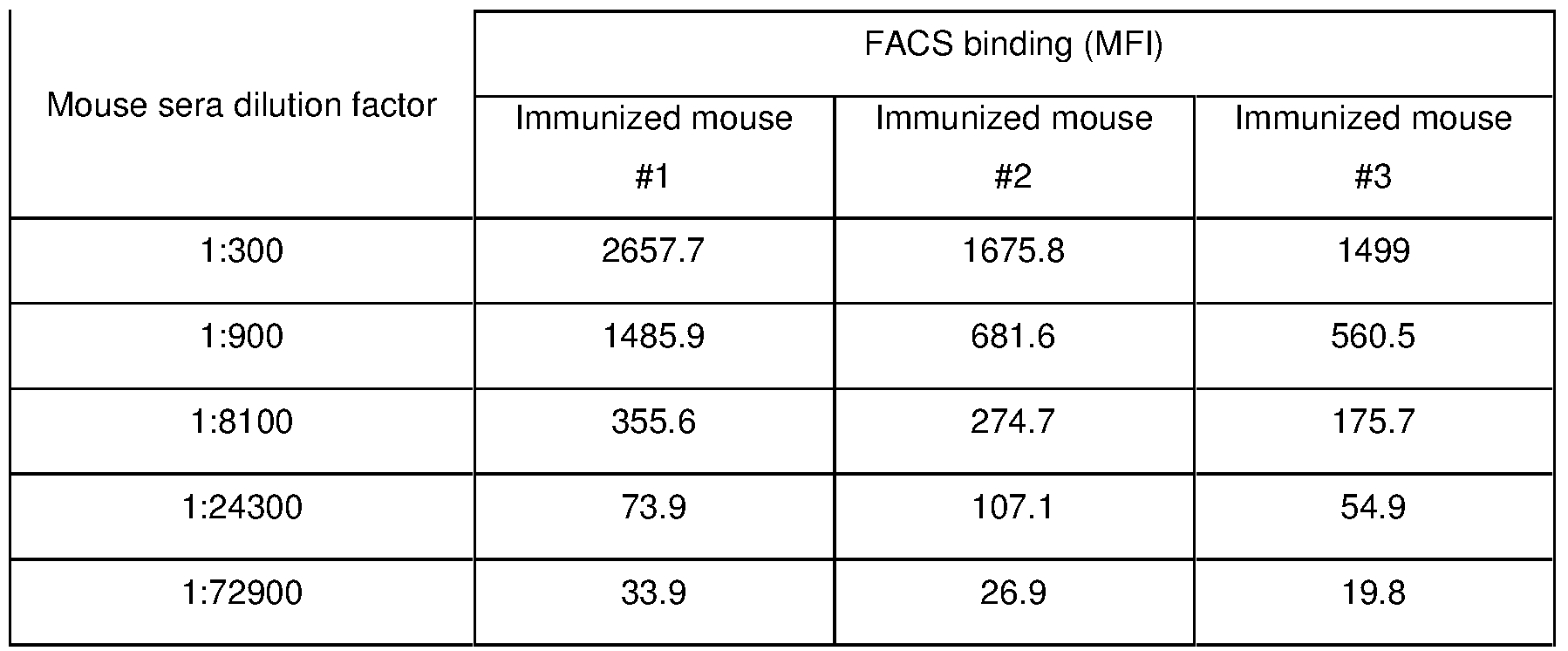
- Murine anti-human PD-L1 monoclonal antibodies were generated using the hybridoma fusion technology. All animal protocols were reviewed by and performed following BeiGene Animal Care and Use Procedure. Ten to twelve week-old Balb/c mice (HFK Bioscience, Beijing, China) were immunized three times (3 weeks apart between injections) subcutaneously and/or intra-peritoneally, each immunization was done with 100uL of adjuvant (Cat. No. KX0210041, KangBiQuan, Beijing, China) containing 5-10 microgram of PD-L1/Fc. Two weeks after the 2 nd immunization, the mice sera were collected and evaluated for PD-L1 binding by ELISA and FACS.
- adjuvant Cat. No. KX0210041, KangBiQuan, Beijing, China
- mice with high anti-PD-L1 binding titers in sera were selected and boosted intraperitoneally with 50 micrograms of PD-L1/Fc in PBS.
- the splenocytes were isolated and fused with the murine myeloma cell line, SP2/0 (ATCC) , using standard techniques (Mechetner et. al. 2007 Methods Mol Biol 378: 1-13) with some modification.
- the supernatants of hybridoma clones were initially screened for PD-L1 binding activities by a modified ELISA assay (Flanagan 2007 Methods Mol Biol 378: 33-52) . Briefly, 50-200 nanograms of PD-L1/His were diluted in 50 microliters of PBS and coated in each well of 96-well ELISA plates (JinCanHua, Shenzhen, China) . After blocking with 3%bovine serum albumin in TBST (20mM Tris, 150mM NaCl, 0.05%Tween20, pH7.5) and incubating with culture supernatants of hybridoma clones, the HRP-conjugated horse anti-mouse IgG antibody (Cat. No.
- tetramethylbenzidine (Cat. No. PA107-01, TianGen, Beijing, China) were used to detect binding signals by a plate reader (PHREAstar, BMG Labtech, Germany) as light absorbance at 450nm.
- the ELISA-positive clones were further verified by fluorescence-activated cell sorting (FACS) .
- PD-L1 expression cells, HEK293/PD-L1 (10 5 cells/well) were incubated with supernatants from hybridoma clones in V-bottom 96-well plates (Cat. No. 3897, Corning) .
- the cell surface bound PD-L1 antibodies were detected with Dylight 649-conjugated goat anti-mouse IgG antibody (Cat. No. 405312, Biolegend, San Diego, CA, USA) and cell fluorescence was monitored in a Guava EasyCyte 8HT flow cytometer (Millipore, USA) .
- hybridoma clones that were positive in both ELISA and FACS assays were then tested in human immune cell-based functional assays to identify antibodies with good functional activities.
- the hybridoma clones with positive functional activities were further subcloned and characterized.
- the positive hybridoma clones from primary screening through ELISA, FACS and functional assays were subcloned by limiting dilution. Three subclones from each original clone were selected and confirmed in FACS and functional assays. The subclones selected through functional assays were defined as monoclonal antibody. The top subclones were adapted to grow in the CDM4MAB medium (Cat. No. SH30801.02, Hyclone) with 0-3%fetal bovine serum for purification and further characterizations.
- Hybridoma cells or 293-F cells transiently transfected with an antibody expression plasmid (Cat. No. R79007, Invitrogen) was cultured either in CDM4MAb medium (Cat. No. SH30801.02, Hyclone) or in Freestyle TM 293 Expression medium (Cat. No. 12338018, Invitrogen) , and incubated in a CO 2 incubator for 5 to 7 days at 37°C. The conditioned medium was collected through centrifugation at 10,000g for 30 minutes to remove all cells and cell debris, and filtrated through a 0.22 ⁇ m membrane before purification. Murine or recombinant antibodies containing supernatants were applied and bound to a Protein A column (Cat. No.
- Murine mAb333 has a dose-dependent binding activities with EC 50 (effective concentration at 50%activity) of 0.036 ⁇ g/mL and 0.099 ⁇ g/mL, respectively, in ELISA and FACS assays.
- control mouse IgG muIgG
- the stable cell lines for human T cell-based functional assays were essentially the same as described in Patent US8735553. Briefly, a fusion protein expression plasmid, OS8, was generated containing a scFv of anti-human CD3 mAb OKT3 and a C-terminal domain of mouse CD8 ⁇ which included transmembrane and cytoplasmic domains. OS8 could function as a membrane anchored T cell engager that directly activates T-cell receptor (TCR) . A stable cell line that co-expresses both OS8 and PD-L1 was generated by co-transfection of two expression constructs in HEK293 cells followed by hygromycin or G418 selection for 10-14 days.
- TCR T-cell receptor
- This cell line was named as HEK293/OS8/PD-L1.
- a human T-cell line, HuT78/PD-1 was generated that expresses human PD-1.
- a reverse signaling human T-cell line, HuT78/P3Z was generated by stable transfection with a chimeric PD-1 expression construct (named as P3Z) made by fusing the extracellular and transmembrane domains of human PD-1 to the cytoplasmic region of human CD3 ⁇ chain.
- P3Z encoded a membrane bound receptor that would activate T cells upon ligation with PD-1 ligand (PD-L1 or PD-L2) .
- Cell lines were cloned by limiting dilution as described previously (Fuller 2001 Curr Protoc Mol Biol, Chap 11, unit 11.8) .
- HEK293/OS8/PD-L1 cells were pre-incubated with anti-PD-L1 mAbs for 15 minutes prior to co-culture with HuT78/PD-1 cells (1-3 x10 4 per well) in a flat bottom plate fed with 200 ⁇ l of RPMI1640 growth medium per well at 37°C. After 16-18 hours of co-culture, supernatants were collected. IL-2 was assayed by ELISA using human IL-2 Ready-Set-Go! ELISA kits (Cat. No. 88-7025, eBiosciences, San Diego, CA) . In this assay, blockade of PD-L1-PD-1 signaling with anti-PD-L1 antibodies resulted in enhanced TCR signaling and IL-2 production.
- a reference anti-PD-L1 mAb (named Y1) was synthesized based on the variable regions of the published data (US 2010/0203056 A1) , and both human and mouse format of Y1 antibodies were generated by fusing Y1 variable regions with mouse or human IgG1 ⁇ constant regions to generate Y1-muIgG1 or Y1-huIgG1, respectively (collectively termed asY1-hIgG1) .
- Y1-muIgG1’s function was also confirmed in this assay.
- the purified murine anti-PD-L1 mAbs were compared in the same assay for quantitative assessments of the blocking activities.
- Fig. 3 showed the dose response curves of the representative murine-anti-PD-L1 mAbs.
- Table 5 summarized the EC 50 and the maximum IL-2 concentration these mAbs could induce.
- Mu333 was a potent antagonist of the PD-L1-PD-1 signaling, and induced significant IL-2 production with a very low EC 50 .
- one of the mAbs, mu277 had much weaker blocking activity than mu333, judged by the dose dependent response curve and the parameters of top line signal read-out and EC 50 .
- muIgG could not block PD-L1/PD-1 signaling and stimulate IL-2 production.
- HuT78/P3Z cells PD-1 mediated TCR signaling is reversed by design as described in the previous sections.
- HEK293/PD-L1 cells were pre-incubated with purified PD-L1 antibodies for 15 minutes prior to co-culture with HuT78/P3Z cells in 96-well flat bottom plates at 37°C. After 16-18 hours of co-culture, supernatants were collected and IL-2 production was assayed by ELISA as described above.
- Inhibitory activity of murine anti-PD-L1 mAbs was detected directly correlated to the decrease of IL-2 release in dose-dependent fashion. Consistent with the results shown above, mu333 had potent activities inhibiting IL-2 secretion by preventing PD-L1 engagement on P3Z chimeric receptor on HuT78 cells. As showed in Table 6 and Figure 6, mu333 was much more potent than mu277 in terms of IC 50 (the concentration of mAb at 50%inhibition of the maximum activity) , consistent with the above results obtained with regular signaling in T-cell. The negative control, muIgG, could not inhibit PD-L1/P3Z induced IL-2 production.
- HEK293/PD-L1 cells (1x10 5 cells per well) were incubated with the mixture of PD-L1 antibodies and biotin-conjugated PD-1/Fc fusion protein in V-bottom 96-well plate. Biotinylation of PD-1/Fc was done using the EZ-Link Sulfo-NHS-LC-Biotin reagent according to manufacturer’s instruction (Cat. No. 21327, Thermo Sci) .
- Mu333 showed better inhibitory efficacy with an IC 50 of 0.463 ⁇ g/mL, compared to an IC 50 of 2.172 ⁇ g/mL for mu277.
- control antibody murine IgG
- Fig. 5 had no such inhibitory effect.
- PD-L1 Besides interaction with PD-1, PD-L1 also binds to another B7 family protein, B7-1 or alternatively named as CD80 (Butte M.J. 2007 Immunity 27: 111-122) .
- CD80 NCBI accession: NP_005182.1
- HEK293/PD-L1 cells were incubated with the mixture of PD-L1 antibodies and biotin-conjugated CD80/Fc fusion protein.
- blockade of biotin-CD80/Fc binding to PD-L1 by anti-PD-L1 antibodies resulted in decreased binding signals (MFI readings) .
- MFI readings binding signals
- mu333 competed off the binding of CD80 to PD-L1 with100%of max inhibition and very low IC 50 (0.052 ⁇ g/mL) , which compared to the IC 50 of 0.162 ⁇ g/mL for mu277.
- murine IgG had no such competition effect (Fig. 6) .
- the 1 st strand cDNA was synthesized using reverse transcriptase (Cat. No. AH301-02, TransGen, Beijing, China) .
- PCR amplification of heavy chain (Vh) and light chain variable region (V ⁇ ) of murine mAb was performed using PCR reagent kit (Cat. No. AP221-12, TransGen, Beijing, China) and a set of primers specific for cloning of murine Vh and V ⁇ as described (Brocks 2001 Mol Med 7: 461-469) .
- the PCR products were subcloned into the pEASY-Blunt cloning vector (Cat. No. CB101-02, TransGen) and subsequently sequenced by Genewiz (Beijing, China) .
- the amino acid sequences of Vh and Vk were deduced from the DNA sequencing results.
- CDRs Complementary determinant regions
- IMGT Lefranc 1999 Nucleic Acids Research 27: 209-212
- Table 9 listed the CDRs of mu333 (SEQ. NO. 5-14) , based on the definitions of Kabat and IMGT systems.
- variable domain of mu333 The three dimensional structures were simulated for variable domain of mu333 in order to identify framework residues that might be important for supporting CDR loop structures. Potentially important framework residues were kept as the original murine residues in the first round antibody humanization.
- the previously established structural modeling method for antibodies (Morea et al. Methods 2000 20: 267-279) was adopted to simulate 3D structure of anti-PD-L1 mAb mu333 based on the known canonical structures of antibodies (Al-Lazikani et al. 1997 Journal of Molecular Biology 273: 927-948) . Briefly, the sequence of each variable domain (Vk and Vh) of mu333 was blasted in the PDB database (Protein Data Bank, http: // blast. ncbi. nlm.
- Selected structure templates for modeling mu333 (listed in Table 10) had the same classes of canonical loop structures in L-CDR1, L-CDR2, L-CDR3, H-CDR1, and H-CDR2 to the mu333 to be modeled.
- V ⁇ and Vh came from different immunoglobulins, they were packed together by a least-squares fit of the main chain atoms to form a hybrid structure of Vk-Vh interface residues, which was used as the templates for structural homology modeling by Swiss-model program (Kiefer et al.
- human germline IgG genes were searched for sequences that share high degree of homology to the cDNA sequences of mu333 variable regions by blasting the human immunoglobulin gene database in IMGT ( http: //www. imgt. org/IMGT_vquest/share/textes/index. html ) and NCBI ( http: //www. ncbi. nlm. nih. gov/igblast/ ) websites.
- the human IGVH and IGV ⁇ genes that are present in human antibody repertoires with high frequency (Glanville 2009 PNAS 106: 20216-20221) and are highly homologous to mu333 were selected as the templates for humanization.
- Humanization was carried out by CDR-grafting (Methods in Molecular Biology, Vol 248: Antibody Engineering, Methods and Protocols, Humana Press) and the humanization antibodies (hu333s) were engineered as the human Fab format using an in-house developed expression vector.
- mutations from murine to human amino acid residues in framework regions were guided by the simulated 3D structure, and the murine framework residues of structural importance for supporting the canonical structures of CDRs were retained in the 1 st version of humanization antibody 333 (hu333-1A, SEQ. NO. 15-16) .
- CDRs of mu333 Vk were grafted into the framework of human germline variable gene IGVK1-5, and no murine framework residues were retained (SEQ NO 16) .
- CDRs of mu333 Vh were grafted into the framework of human germline variable gene IGVH3-7, with 4 murine framework residues retained, V 24 , L 67 , K 71 and V 78 (SEQ NO 15) .
- All grafted CDRs were based on the Kabat’s CDR definition in hu333-1A (Table 9 and SEQ. NO. 15-16) . In the following hu333 variants, only the N-terminal half of Kabat H-CDR2 was grafted, as only the N-terminal half was considered to be important for antigen binding according to the simulated 3D structure (Table 14) .
- Hu333-1A were constructed as human Fab format using in-house developed expression vectors that contain human IgG CH-1 and constant region of kappa chain, respectively, with easy adapting subcloning sites.
- the hu333-1A joined IgG2a-CH1 was tagged at C-terminus with a 8xHis peptide to facilitate purification.
- the C 232 S and C 233 S (Kabat residue numbering, Kabat et al. Sequence of proteins of immunologic interest, 5 th ed Bethesda, MD, NIH 1991) mutations were introduced in the IgG2 heavy chain to prevent disulfide bond exchange and stabilize human IgG2 in the IgG2a conformation (Lightle et al.
- CM5 biosensor chip Cat. No. BR100530, GE Life Sciences
- hu333 Fabs were expressed, purified and tested in binding and functional assays as described previously. Comparing to hu333-1A, hu333-2A, hu333-2C and hu333-2D had significantly reduced binding affinities and functionalities. Only hu333-2B (SEQ. NO. 16 and 17) had similar binding and functional activities to hu333-1A (Table 11) . Taken together, hu333-2B (SEQ. NO. 16 and 17) reached a high level of humanization in the framework regions while maintained potent binding affinity and functional activities.
- Vh and Vk sequence composition for hu333 that could be used as therapeutic antibody in human
- hu333 by introducing mutations in CDRs and framework regions in considerations of the antibody’s molecular properties, such as physiochemical stabilities, amino acid compositions, projected isoelectronic points (pIs) , expression level, antibody-dependent cell-mediated cytotoxicity (ADCC) and complement-dependent cytotoxicity (CDC) while maintaining functional activities.
- molecular properties such as physiochemical stabilities, amino acid compositions, projected isoelectronic points (pIs) , expression level, antibody-dependent cell-mediated cytotoxicity (ADCC) and complement-dependent cytotoxicity (CDC) while maintaining functional activities.
- ADCC antibody-dependent cell-mediated cytotoxicity
- CDC complement-dependent cytotoxicity
- Deamination site NS 76-77 in Vh of hu333-1A was mutated to NT 76-77 to generate hu333-3A2 (SEQ. NO. 18 and 23) .
- V60 of hu333-3A2 -Vh was mutated to V60A, which is consistent to the consensus sequences of major human IGVH3 genes with reduced a surface-exposure of hydrophobicity.
- This mutant construct was given the code name hu333-3C2 (SEQ. NO. 19 and 23) .
- Another deamidation site NS 73-74 was mutated to TS 73-74 on the template of hu333-3C2, which is also consistent to the consensus sequences of major human IGVH3 genes.
- hu333-3H2 SEQ. NO. 20 and 23
- hu333-3A2 hu333-3C2
- hu333-3H2 all retained the potent functional activity, only with slight variations in binding affinity.
- these engineered hu333 variants have better projected physiochemical properties.
- NS 101-102 to TS 101- 102 on the templates of hu333-3A2 and hu333-3H2, respectively.
- the resulting humanization mAbs were constructed in human IgG1 Fab format, named as hu333-4A2 (SEQ. NO. 21 and 23) and hu333-4B2 (SEQ. NO. 22 and 23) .
- PBMCs peripheral blood mononuclear cells
- Histopaque-1077 Cat. No. 10771, Sigma, St. Louis, USA
- PBMCs were then stimulated with 40 ng/mL of anti-CD3 mAb OKT3 (Cat. No. 16-0037, eBioscience, San Diego, CA, USA) for 3 days prior to the assay.
- the activated PBMC population mainly consisted of T-cells (50-70%) , B-cells and NK cells (15-30%) , and monocytes (2-10%) .
- the activated PBMCs were co-cultured with HEK293/OS8/PD-L1 cells in each well of 96-well plates. Functional effect of anti-PD-L1 mAbs were tested by adding the mAb to the culture, co-cultured for 15-18 hours before harvesting culture supernatants to assess IFN- ⁇ level using Ready-Set-Go! ELISA kits (Cat. No. 88-7316, eBiosciences) . As shown in Fig.
- Example 5 Generation of recombinant anti-PD-L1 mAbs without effector functions by fusion to modified human IgG1 constant region
- PD-L1 is expressed on a wide range of normal human cells including hematopoietic cells such as T-cells, B-cells, dendritic cells, macrophages, mesenchymal stem cells and bone-marrow derived mast cells, and nonhematopoietic cells and tissues such as lung, hepatocytes, pancreatic islets, placental synctiotrophoblasts and vascular endothelium (Keir et. al. 2006 J Exp Med 203: 883-895, Keir et. al. 2008 Ann Rev Immunol 26: 677-704, Mu et. al. 2011 Medical Oncology 28: 682-688) .
- hematopoietic cells such as T-cells, B-cells, dendritic cells, macrophages, mesenchymal stem cells and bone-marrow derived mast cells
- nonhematopoietic cells and tissues such as lung, hepatocytes, pancreatic is
- PD-L1 blocking antibodies linked to human wild type IgG- ⁇ Fc moieties will induce ⁇ Fc-mediated effector functions, for examples, antibody-dependent cell-mediated cytotoxicity (ADCC) and complement-dependent cytotoxicity (CDC) , which might lead to unwanted toxicity to vital organs.
- ADCC antibody-dependent cell-mediated cytotoxicity
- CDC complement-dependent cytotoxicity
- hu333-4A2 and hu333-4B2 full antibody by linking the Vh sequences to mutated IgG1 constant regions, and screened for reduced or null Fc ⁇ receptors (Fc ⁇ Rs) binding or C1q binding activities, therefore, attenuating or eliminating ADCC and CDC effector functions.
- Fc ⁇ Rs Fc ⁇ receptors
- IgG2 has some sequence variations from IgG1 in the hinge region, which was attributed to weaker binding or no binding to most of the Fc ⁇ Rs except to Fc ⁇ RIIA H131 .
- IgG1/IgG2 hybrid (IgG1 ⁇ b) with most of IgG1 hinge sequence incorporating some IgG2 sequences was demonstrated having significantly reduced the binding activities to most Fc ⁇ Rs and attenuated ADCC and CDC effector functions (Armour et. al. 1999 Eur J Immunol 29: 2613-2624) .
- V 234 A and G 237 A were designed to reduce the surface hydrophobic side chain at the ⁇ Fc/Fc ⁇ R binding interface to further reduce the binding to Fc ⁇ RIIA and Fc ⁇ RIIB, (Lund et. al. 1992 Mol Immunol 29: 53-59, Lund et. al. 1996 J Immunol 157: 4963-4969, Wines et. al. 2000 J Immunol 164: 5313-5318) .
- the P 239 A mutation was designed to further reduce the C1q binding and CDC (Idusogie et. al. 2000 J of Immunol 164: 4178-4184) .
- the IgG1mf SEQ. NO.
- IgG1mc 29 was similar to IgG1mc except that the amino acid residue G 237 was not mutated.
- the recombinant full length anti-PD-L1 antibodies, hu333-4A2-IgG1mc (SEQ. NO. 30 and 32) , hu333-4B2-IgG1mc (SEQ. NO. 31 and 32) and hu333-4B2-IgG1mf (SEQ. NO. 32 and 33) were expressed in HEK293-F cells and purified as described in previous sections.
- ADCC is initiated when an antibody binds to cell surface target protein followed by ligation to Fc ⁇ RIIIA expressed on effector cells.
- CDC is activated when an antibody cross-links a cell surface target by binding to C1q protein, which leads to a cascade reaction of complement complex formation and activation and target cell lysis.
- biochemical assays for antibody binding to Fc ⁇ Rs and C1q may serve as the fundamental indicator of ADCC and CDC.
- Fc ⁇ Rs The extracellular domains of Fc ⁇ Rs were fused to C-terminal His tags as described in previous sections.
- Recombinant proteins were expressed in 293-F cells by transient transfection and purified using Ni-Sepharose column followed by gel filtration column as described. 2-5 ⁇ g/mL of Fc ⁇ Rs were coated on Nunc MaxiSorp ELISA plates (Cat. No. 442404, Nunc, Thermo Fisher) except Fc ⁇ RIIB and Fc ⁇ RIIIB, Ni-chelate plates were used (Cat. No. 15242, Pierce, Thermo Fisher) . After washing and blocking of the wells, a preformed immune-complex was added to each well and incubated at room temperature for 1-2 hours.
- the preformed immune-complex contained 60ng/mL streptavidin-HRP, 60ng/mL of biotinylated-F (ab’) 2 goat anti-human IgG (Cat. No. 109-066-097, Jackson ImmunoRes, West Grove, PA, USA) , and 1-5 ⁇ g/mL of indicated IgG1 Fc variants fused to the humanized anti-PD-L1 (hu333-4A2 or hu333-4B2) in the blocking buffer. After washing the plate four times, binding signals were detected by chemiluminescence using Immobilon Chemiluminescence Substrate A/B (Cat. No. WBKLS0500, Millipore) .
- Immobilon Chemiluminescence Substrate A/B Cat. No. WBKLS0500, Millipore
- Table 16 summarized the results of hu333-4A2-IgG1mc (SEQ. NO. 30 and 32) , hu333-4B2-IgG1mc (SEQ. NO. 31 and 32) and hu333-4B2-IgG1mf (SEQ. NO. 32 and 33) binding to various Fc ⁇ Rs.
- all three IgG1 mutant hu333 mAbs had very low binding activities to Fc ⁇ Rs, which indicated that all three hu333 mAbs above would have significantly reduced effector functions mediated by Fc ⁇ Rs.
- Bindings of humanized anti-PD-L1 in various IgG1 formats (wt, IgG1mc, IgG1mf, SEQ. NO. 27-29) to Fc ⁇ Rs were also determined by flow cytometry.
- a series of stable HEK293 transfectants expressing human Fc ⁇ Rs were established. These stable cell lines expressed Fc ⁇ RI, Fc ⁇ RIIA H131 , Fc ⁇ RIIA R131 , Fc ⁇ RIIB, Fc ⁇ RIIIA F158 or Fc ⁇ RIIIA V158 , respectively.
- Multi-subunit Fc ⁇ Rs i.e., Fc ⁇ RI and Fc ⁇ RIIIA were co-expressed with FcR ⁇ subunit.
- a secondary antibody (goat anti-human IgG F (ab’) 2 -Alexa Fluor 488, Cat. No. 109-546-097, Jackson ImmunoResearch, West Grove, PA, USA) was used to detect the binding of monomeric anti-PD-L1 mAbs with the IgG1 variants (Table 17) to Fc ⁇ R expressing HEK293 cells.
- hu333-4A2 in IgG1wt format had strong binding signals (MFI) to Fc ⁇ RI, Fc ⁇ RIIA H131 and Fc ⁇ RIIIA V158 , and weak but significant binding signals to Fc ⁇ RIIA R131 , Fc ⁇ RIIB and Fc ⁇ RIIIA F158 (Table 17) .
- the modified IgG1 variants (hu333-4A2-IgG1mc, hu333-4B2-IgG1mc and hu333-4B2-IgG1mf, SEQ. NO. 30-33) had significantly reduced binding signals which were close to backgrounds.
- the ELISA-based C1q binding assay was done by conventional ELISA method with minor modification. Briefly, indicated amounts of the humanized 333 antibodies fused to either wild type or modified IgG1 constant regions were coated onto the Maxisorp ELISA plate. After blocking and washing, the wells were incubated with 2 ⁇ g/mL of human C1q (Cat. No. A400, Quidel, San Diego, USA) at room temperature for 2 hours. After washing, the bound C1q was detected using a murine monoclonal antibody against human C1q (Cat. No. A201, Quidel) and HRP conjugated anti-murine IgG (Cat. No. A0168, Sigma, Shanghai, China) . As shown in Fig.
- ADCC Classical antibody-dependent cellular cytotoxicity
- CD16 Fc ⁇ RIIIA
- NK92MI/CD16V cells which were generated from NK92MI cells (Cat. No. CRL-2408, ATCC) by co-transducing expression plasmids containing CD16 (V158 allele) and FcR ⁇ genes, were used as effector cells, and PD-L1-expressing HEK293 cell line, HEK293/PD-L1, was used as target cells.
- Cytotoxicity of NK92MI/CD16 cells exerted against HEK293/PD-L1 cells was determined by lactate dehydrogenase (LDH) release assay using the CytoTox 96 Non-Radioactive Cytotoxicity Assay kit (Promega, Madison, WI) . Specific lysis was determined by the following equation.
- Human IgG1 antibodies in general, induce significant complement-dependent cytotoxicity (CDC) via classical pathway. Whether the humanized anti-PD-L1 antibodies in selected IgG1 mutant formats (IgG1mc and IgG1mf) trigger CDC was evaluated using a PD-L1-expressing B cell line, Daudi/PD-L1, and fresh human serum from healthy donors, which contains all necessary components for CDC. Cell lysis by CDC was determined by Celltiter glo assay kits (Promega) .
- Daudi/PD-L1 cells (2x10 4 cells/well) were incubated in serum-free RPMI1640 (Invitrogen) with anti-PD-L1 Abs (0.001-10 ⁇ g/mL) at 37°C for 15 minutes before adding normal human serum to the final concentration of 16.6%in 96-well flat-bottom plates in a total volume of 120 ⁇ L. After overnight incubation at 37°C, cells were lysed and assayed for ATP concentration.
- Anti-CD20 mAb Rituximab (Roche) was used as a positive control as Daudi cells constitutively express CD20. The amount of ATP is directly proportional to the number of cells present in culture.
- %CDC activity [ (RFU test -RFU background) / (RFU at total cell lysis -RFU background) ] x 100.
- Rituximab induced robust CDC in CD20 + Daudi/PD-L1 cells.
- both hu333-4B2-IgG1mc (SEQ. NO. 31 and 32) and hu333-4B2-IgG1mf (SEQ. NO. 32 and 33) showed no CDC.
- FACS binding analysis was performed as described in previous sections. Serial dilutions of antibodies were incubated with HEK293/PD-L1 cells and the bindings were detected using the Goat anti-human IgG F (ab’) 2 -Alexa Fluor 488 (Cat. No. 109-546-097, Jackson ImmunoResearch) . Dose-dependent binding activities were observed for the selected mAbs to native PD-L1 protein expressed on surface of HEK293 cells.
- hu333-4A2-IgG1mc, hu333-4B2-IgG1mc and hu333-4B2-IgG1mf showed similar dose-dependent binding activities to the HEK293/PD-L1 cells with EC 50 (effective concentration at 50%activity) around 0.1 ⁇ g/mL.
- the functionalities of the purified anti-PD-L1 mAbs were assessed in the HuT78/PD-1 and HEK293/OS8/PD-L1 co-culture system as described in previous section. As shown in Table 19, the humanized 333 mAbs were potent antagonists of PD-L1/PD-1 signaling in this co-culture system, and induced increased IL-2 secretions.
- hu333-4A2-IgG1mc, hu333-4B2-IgG1mc and hu333-4B2-IgG1mf showed similar potencies in this assay with very close EC 50 (0.075-0.087 ⁇ g/mL) and maximum induction of IL-2 levels (287-300 pg/mL) .
- the functionalities of the purified anti-PD-L1 mAbs were also assessed in the reversed signaling system in which HuT78/P3Z and HEK293/PD-L1 were co-cultured as described. Consistently, the humanized 333 mAbs were potent inhibitor of PD-L1/P3Z signaling in this co-culture system, and inhibited IL-2 secretions induced by PD-L1/P3Z engagement.
- hu333-4A2-IgG1mc, hu333-4B2-IgG1mc and hu333-4B2-IgG1mf showed similar potencies in the assay, as shown by similar IC 50 s (0.037-0.045 ⁇ g/mL) and maximum inhibition levels (99%) (Table 19) .
- PBMCs peripheral blood mononuclear cells
- T-cells 50-70%)
- B-cells and NK cells 15-30%)
- monocytes 2-10%)
- Human PBMCs were isolated from healthy donors by density gradient centrifugation using ficoll lymphocyte separation medium (Histopaque-1077; Cat. No. 10771, Sigma) according to manufacturer’s instruction. The human blood collections were done followed the Internal Procedure of BeiGene. PBMCs were then stimulated with 40 ng/mL of anti-CD3 mAb OKT3 (Cat. No.
- PBMCs (1x10 4 cells) were co-cultured with HEK293/OS8/PD-L1 cells (3x10 4 cells) in each well of 96-well flat-bottom plates. Indicated concentrations of anti-PD-L1 antibodies were added to the culture. After 15-18 hours of co-culture, culture supernatants were assayed for IFN- ⁇ level by ELISA using Ready-Set-Go! ELISA kits (Cat. No.
- the potencies of hu333-4A2-IgG1mc and hu333-4B2-IgG1mf were comparable to the parental murine antibody mu333-IgG.
- the base level of IFN- ⁇ without antibody treatment varied among different donors, the increase of IFN- ⁇ secretion in PBMCs treated by hu333-4A2-IgG1mc, hu333-4B2-IgG1mf and mu333-IgG was statistically significant in the range of 0.01 to 10 ⁇ g/mL of antibody treatment (about 5-8 fold induction at 10 ⁇ g/mL depending on the donor) .
- hu333-4A2-IgG1mc, hu333-4B2-IgG1mc and hu333-4B2-IgG1mf were potent antagonists blocking PD-L1/PD-1 interactions and downstream signaling in all the cell line and primary immune cell-based assays. They were very similar in their functional activities and potencies, as they were very similar in sequences (only minor difference in framework regions) , shared identical binding epitope and had very similar binding affinities and specificity (see below section) .
- the binding specificity was studied for the mAbs hu333 (hu333-4A2-IgG1mc, hu333-4B2-IgG1mc and hu333-4B2-IgG1mf) using human, cynomolgus monkey (Macaca fascicularis) and mouse (Mus musculus) PD-L1 as target proteins.
- the monkey PD-L1/His and murine PD-L1/His were expressed and purified in a similar way to the human PD-L1/His as described earlier.
- Y1 was a reference functional anti-PD-L1 mAb which was synthesized according to a published patent (US 2010/0203056 A1) and fused to human IgG1mc variant.
- the synthesized full length mAb was named as Y1-IgG1mc.
- the ELISA assay was performed essentially in the same way as described in the previous section. Briefly, 200ng of PD-L1 protein was coated on each well of Nunc MaxiSorp ELISA plate (Cat. No. 442404, Nunc, Thermo Fisher) . After washing and blocking, indicated concentrations of anti-PD-L1 mAbs were added and incubated at room temperature for 1 hour. After washing, the bound anti-PD-L1 mAbs were detected using a HRP-conjugated goat anti-human Fc antibody (Cat. No. A0170, Sigma) .
- hu333-4A2-IgG1mc (SEQ. NO. 30 and 32)
- hu333-4B2-IgG1mc (SEQ. NO. 31 and 32)
- hu333-4B2-IgG1mf (SEQ. NO. 32 and 33) bound to human and monkey PD-L1 in a dose-dependent manner, but not to murine PD-L1, which was consistent with the fact that the original mu333 was generated from mouse immunized with human PD-L1/Fc and that human and monkey PD-1 shares high degree of sequence homology (96%) .
- Y1-IgG1mc bound to all PD-L1 proteins from human, monkey and mouse.
- Hu333-4A2 (SEQ. NO. 21 and 23)
- hu333-4B2 (SEQ. NO. 22 and 23)
- the reference antibody Y1 were constructed as human IgG1 Fab format in which the Vh and Vk were fused to the N-terminus of human IgG1-CH1 and constant region of kappa chain, respectively.
- the IgG1-CH1 was fused to a C-terminal His6 tag to facilitate purification. Expression and purification of recombinant Fabs were performed as described in the previous section.
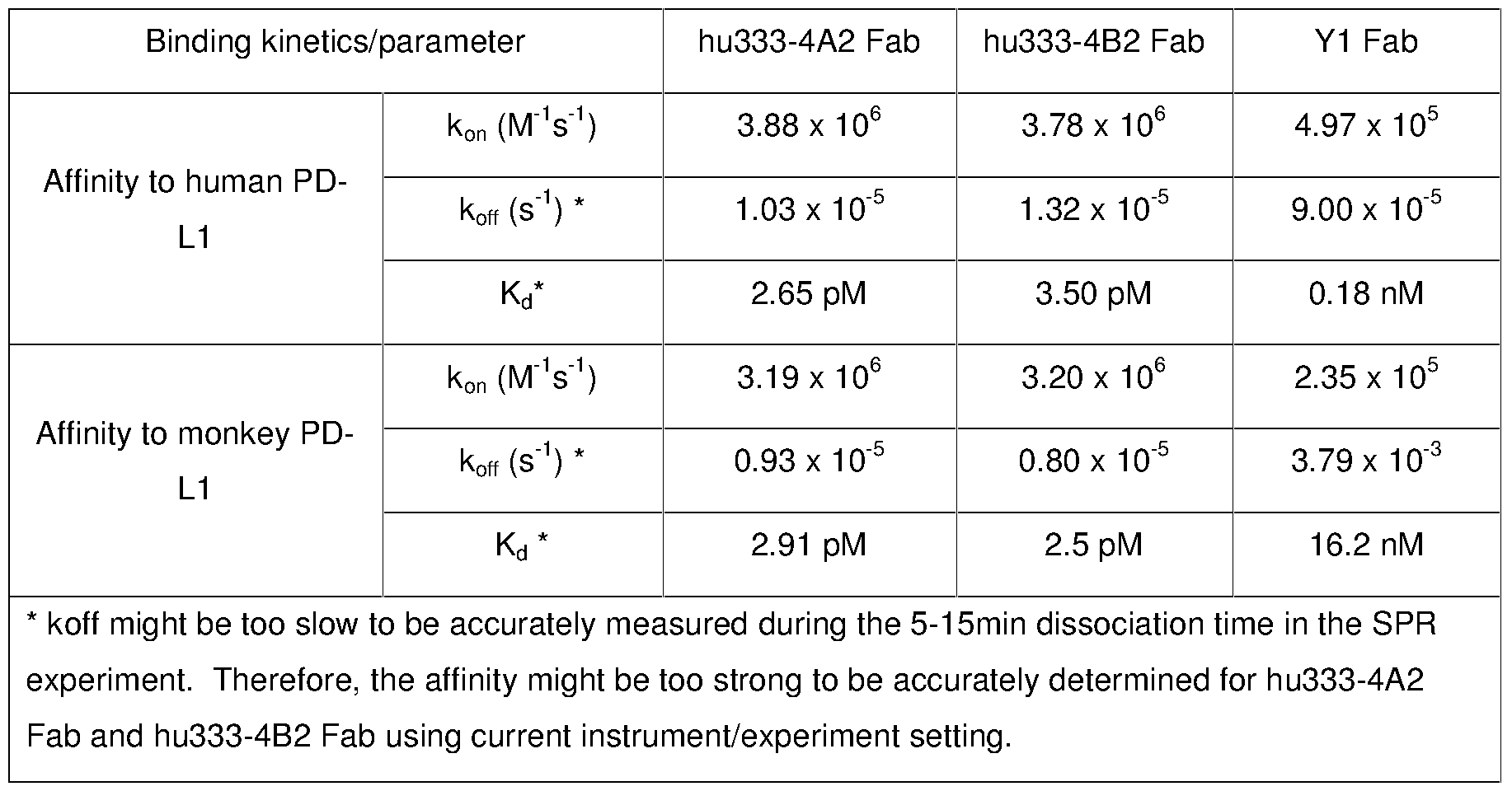
- the SPR-determined binding affinities of anti-PD-L1 Fabs were listed in Table 20.
- Hu333-4A2 and hu333-4B2 Fabs bind to human PD-L1 with higher affinities than Y1 Fab does, which was indicated by the faster k on , slower k off and much smaller K d value.
- the hu333-4A2 and hu333-4B2 Fabs bind to monkey PD-L1 almost equally well as their binding to human PD-L1.
- Y1 Fab binds to the monkey PD-L1with about 100-fold lower affinity than its binding to human PD-L1 (K d of 0.18 nM to human PD-L1 and K d of 16.2 nM to monkey PD-L1) .
- the wild-type PD-L1/His (Fig. 1) was used as template for rolling-circle mutagenesis using Fast Mutagenesis kit (Cat. No. FM111, Transgen Biotech, Beijing, China) . All mutants were sub-cloned in our pcDNA-based expression vectors, and verified by sequencing. The mutant and wild-type PD-L1/His proteins were expressed by transient transfection as described in previous sections.
- the conditioned media (CM) were collected after 4 to 6 days of culture, and analyzed by Western blot to verify the PD-L1/His protein expression in terms of quality and quantity.
- the supernatants (CM) after clearing cell debris, were directly used in ELISA analysis or Western blot for epitope-mapping.
- ELISA assays using the wild-type and mutant PD-L1/His were performed to study the binding epitopes of the anti-PD-L1 mAbs.
- several murine mAbs from us and one reference antibody Y1-IgG1 (adapted from US 2010/0203056 A1 and fused to human IgG1kappa constant regions) were included in the study.
- Equal volume of CM containing wild type or mutant PD-L1/His was coated in 96-well plate for all mAbs in the same ELISA assay.
- the ELISA results were normalized using the mean ELISA readings of wild type PD-L1 binding signals as the standard.
- ELISA binding signals to a specific mutant PD-L1 were further normalized against the highest antibody binding read-out (set as 100%) to the specific mutant PD-L1 to even out expression variations between PD-L1 mutants.
- a mAb’s ELISA binding signal for a specific PD-L1 mutant dropped below 50%relative to wild type PD-L1, it was defined that the binding function is significantly impaired due to the corresponding amino acid mutation.
- a mAb’s ELISA binding signal for a specific mutant dropped below 25%, it was defined to be very significant.
- amino acid residues R 113 in PD-L1 is critical for hu333-4B2-IgG1 binding to PD-L1, whose mutation significantly impaired the hu333-4B2-IgG1 binding to PD-L1.
- the reference antibody Y1-IgG1 had distinctive binding epitopes. F 19 , I 54 , D 122 and Y 123 were all significant epitopes for its binding besides R 113 (Fig. 13B) . Different signatures of binding epitopes were also observed in other anti-PD-L1 mAbs from our study. The data from Western blot for both hu333-4B2-IgG1 and Y1-IgG1 antibodies confirmed these results although the antigen proteins were denatured.
- the mutation D 26 A Apart from the above key binding epitope mutations, we also made the mutation D 26 A. ELISA and Western blot results showed the mutation D 26 A in PD-L1 significantly inhibited the binding activities of all functional anti-PD-L1 mAbs including mAbs mu333, hu333-4B2-IgG1 and Y1-IgG1, but not inhibited the binding of non-functional antibodies, such as mu260 (Fig. 13C) .
- hu333-4B2 binds to two key amino acid residues (epitopes) of human PD-L1, D 26 and R 113 ; in contrast, mAb Y1 binds to at least six amino acid residues.
- anti-PD-L1 mAbs are capable of binding to different epitope signatures through molecular recognition, which might have profound impact on binding affinity, binding specificity and functional activity, e.g. hu333-4A2 and hu333-4B2 can only bind to human PD-L1 (Fig. 12A) , but not to mouse PD-L1 (Fig. 12C) ; in contrast, Y1 binds to both human and mouse PD-L1 (Fig. 12A and 12C) although human and mouse PD-L1 have 26%sequence divergence.
- ELISA study was performed using 96-well Nunc Maxisorp ELISA plates coated with 5%human serum (from healthy donors) and various concentrations of PD-L1/His antigen as indicated in Fig 14A, x-axis. Same amount of murine PD-L1 Abs or a chimeric Y1 (named as Y1-muIgG1) , which was made of Y1 variable domains fused to murine IgG1kappa constant regions) were added in the ELISA reaction, and the bindings were detected with anti-mouse Fc-HRP (Cat. No. A9309, Sigma) . The PBS buffer without any antibody was included as negative control (Fig. 14) .
- the baseline O. D. reading (the reading at very low antigen concentration) for mu333 was almost identical to the negative control, while the baseline O.D. reading for Y1-muIgG1 is 4 folds higher than the negative control (PBS) , indicating a differential property between mu333 and YI-mIgG1 in binding selectivity.
- the humanized mAb hu333-4B2-IgG1mc and hu333-4B2-IgG1mf were also assessed for non-specific binding in similar assay method using fetal bovine serum (FBS) instead of human serum. Reminiscent to its parental mouse hybridoma mAb, hu333-4B2-IgG1mc and hu333-4B2-IgG1mf had no binding to FBS.
- FBS fetal bovine serum
- mice Ten to twelve week-old female Balb/c nude mice (18-25g) were used to study the pharmacokinetics of the humanized mAb hu333-4B2-IgG1mf (SEQ. NO. 32 and 33) .
- Mice were dosed with 10mg/kg of mAb hu333-4B2-IgG1mf either as a single intravenous (i.v. ) or subcutaneous (s.c. ) injection. Intravenous injections were administered via a tail vein, and subcutaneous injections were administered in the flank. In each injection group, mice were separated into different subgroups and in each subgroup blood sera were collected at certain time points. For i.v.
- serum was harvested 2 days predose, and postdose at 15 min, 30 min, 60 min, 90 min, 6 h, 24 h and once on days 2, 3, 4, 5, 7, 10, 14, 21 and 28.
- serum was harvested 2 days predose, and postdose at 1.5 h, 6 h, 24 h and once on days 2, 3, 4, 5, 7, 10, 14, 21, and 28.
- Serum level of hu333-4B2-IgG1mf was determined by ELISA using human PD-L1/His protein. Briefly, Nunc MaxiSorp ELISA plates (Cat. No. 442404, Nunc, Thermo Fisher) were coated overnight at 4 °C with 100 ⁇ L per well of 3 ⁇ g/mL human PD-L1/His protein. Plates were blocked with 3%bovine serum albumin, 0.05%Tween 20 in PBS (blocking buffer) at room temperature for 1 hour. After washing, serially diluted serum samples and purified hu333-4B2-IgG1mf standards were added and incubated at room temperature for 1 hour.
- hu333-4B2-IgG1mf was detected using a HRP-conjugated goat anti-human Fc antibody (Cat. No. A0170, Sigma) and color developed using TMB substrate (Cat. No. T0440, Sigma) .
- a standard curve was fit using nonlinear regression and the serum concentrations of hu333-4B2-IgG1mf were deduced from the standard curve and dilution factors.
- the serum concentrations of hu333-4B2-IgG1mf versus time data were analyzed using the non-compartment model for the i.v. and s.c. doses (WinNonlin, Pharsight) .
- the clearance, volume of distribution, half-lives, mean residence time and bioavailability were deduced from WinNonlin data fitting.
- mice The pharmacokinetics of hu333-4B2-IgG1mf in mice was summarized in Table 22.
- hu333-4B2-IgG1mf SEQ. NO. 32 and 33 concentrations were cleared from the serum in a biphasic manner. The terminal half-life was about 10-14 days. After an i.v. dose of 10 mg/kg, the clearance was 7.9 mL/day/kg in mice.
- peak concentrations of hu333-4B2-IgG1mf in the serum as approximately 30-50%of that noted after i.v. administration of the same dose. Comparison of the AUC after the 10 mg/kg s. c. and i.v.
- the pharmacokinetics of hu333-4B2-IgG1mc was studied in cynomolgus monkeys.
- humanized 333 bound to human and monkey PD-L1 with almost identical affinities the pharmacokinetic profile in cynomolgus monkeys should be very informative and scalable to predict the pharmacokinetic profile in humans.
- the drug administrations and blood serum collections were done at 3D BioOptima Co. Ltd (Suzhou, China) following 3D BioOptima’s Animal Care and Use Procedure.
- ELISA based bioanalyses and pharmacokinetic analyses were performed essentially as described above.
- the averaged serum concentration from 2 monkeys was used for fitting except for the time points of 22 and 30 days post dose, where the data from only one monkey were used, as another monkey showed accelerated clearance and undetectable hu333-4B2-IgG1mc serum levels, presumably due to a monkey anti-drug immune response, at these two time points.
- the serum concentration of hu333-4B2-IgG1mc versus time data were analyzed using the non-compartment model for the i.v. dose.
- hu333-4B2-IgG1mc The pharmacokinetics of hu333-4B2-IgG1mc in cynomolgus monkeys was summarized in Table 23.
- hu333-4B2-IgG1mc concentrations were cleared from the sera in a biphasic manner.
- the terminal half-life was about 9 days.
- i.v. dose of 10 mg/kg the clearance was 6.4 mL/day/kg in cynomolgus monkeys.
- peak concentration of hu333-4B2-IgG1mc was 283 ⁇ g/mL at 5 min post dose.
- PK parameters indicated that hu333-4B2-IgG1mc had normal pharmacokinetic profile in cynomolgus monkeys, which predicted normal pharmacokinetic behavior in humans (Deng et. al. 2011 mAbs 3: 61-66) .
- Example 9 Humanized anti-PD-L1 mAb inhibits tumor growth in a mouse xenograft cancer model in vivo
- mice Female NOD/SCID mice (6-7 weeks) were pre-treated with cyclophosphamide.
- Human peripheral blood mononuclear cells PBMCs were isolated from blood of healthy human volunteer, mixed with A431 epidermoid carcinoma cells (Cat. No. CRL-1555 ATCC) and matrigel, and injected subcutaneously into the right front flank of the animals.
- mice were randomly assigned into 3 groups with 8 mice per group. Mice were treated twice weekly (BIW i.p. ) with vehicle (PBS) or 10 mg/kg hu333-4B2-IgG1mf (SEQ. NO. 32 and 33) for 4 weeks. Individual animal body weight and tumor volume were recorded twice weekly, with mice being monitored daily for clinical signs of toxicity for the duration of the study. Tumor volumes were calculated using the following formula: [D ⁇ (d 2 ) ] /2, in which D represents the large diameter of the tumor, and d represents the small diameter.
- the A431 tumor growth was slower compared to that in the PBS-treated group.
Landscapes
- Health & Medical Sciences (AREA)
- Chemical & Material Sciences (AREA)
- Immunology (AREA)
- Organic Chemistry (AREA)
- Medicinal Chemistry (AREA)
- Life Sciences & Earth Sciences (AREA)
- General Health & Medical Sciences (AREA)
- Proteomics, Peptides & Aminoacids (AREA)
- Genetics & Genomics (AREA)
- Molecular Biology (AREA)
- Biophysics (AREA)
- Biochemistry (AREA)
- Nuclear Medicine, Radiotherapy & Molecular Imaging (AREA)
- General Chemical & Material Sciences (AREA)
- Chemical Kinetics & Catalysis (AREA)
- Pharmacology & Pharmacy (AREA)
- Animal Behavior & Ethology (AREA)
- Public Health (AREA)
- Veterinary Medicine (AREA)
- Peptides Or Proteins (AREA)
- Medicines Containing Antibodies Or Antigens For Use As Internal Diagnostic Agents (AREA)
- Preparation Of Compounds By Using Micro-Organisms (AREA)
- Micro-Organisms Or Cultivation Processes Thereof (AREA)
Abstract
Description


| SEQ ID No. 1 | PD-L1 full length cDNA | SEQ ID No. 18 | hu333-3A2 pro-vh |
| SEQ ID No. 2 | PD-L1 full length PRT | SEQ ID No. 19 | hu333-3C2 pro-vh |
| SEQ ID No. 3 | PD-L1 ECD cDNA | SEQ ID No. 20 | hu333-3H2 pro-vh |
| SEQ ID No. 4 | PD-L1 ECD PRT | SEQ ID No. 21 | hu333-4A2 pro-vh |
| SEQ ID No. 5 | mu333 cDNA-Vh | SEQ ID No. 22 | hu333-4B2 pro-vh |
| SEQ ID No. 6 | mu333 pro-Vh | SEQ ID No. 23 | hu333-4B2 pro-vk |
| SEQ ID No. 7 | mu333 cDNA-Vk | SEQ ID No. 24 | hu333-4A2 H-CDR2 |
| SEQ ID No. 8 | mu333 pro-Vk | SEQ ID No. 25 | hu333-4B2 H-CDR2 |
| SEQ ID No. 9 | mu333 H-CDR1 | SEQ ID No. 26 | hu333-4B2 H-CDR3 |
| SEQ ID No. 10 | mu333 H-CDR2 | SEQ ID No. 27 | huIgG1wt pro |
| SEQ ID No. 11 | mu333 H-CDR3 | SEQ ID No. 28 | huIgG1mc pro |
| SEQ ID No. 12 | mu333 L-CDR1 | SEQ ID No. 29 | huIgG1mf pro |
| SEQ ID No. 13 | mu333 L-CDR2 | SEQ ID No. 30 | hu333-4A2-IgG1mc HC pro |
| SEQ ID No. 14 | mu333 L-CDR3 | SEQ ID No. 31 | hu333-4B2-IgG1mc HC pro |
| SEQ ID No. 15 | hu333-1A pro-vh | SEQ ID No. 32 | hu333-4B2-IgG1mf LC pro |
| SEQ ID No. 16 | hu333-1A pro-vk | SEQ ID No. 33 | hu333-4B2-IgG1mf HC pro |
| SEQ ID No. 17 | hu333-2B pro-vh |






Claims (12)
- An antibody antigen binding domain which specifically binds human PD-L1, and comprises complementarity determining region (CDR) 1, CDR2 and CDR3, in a combination selected from (a) - (r) as follows, wherein the antibody (Ab) , heavy chain (HC) or light chain (LC) and CDR nomenclature system (Kabat, IMGT or composite) from which the CDR combinations derive are shown in the first column, and residues in bold text are Kabat system, and residues underlined are IMGT system:

- An antibody antigen binding domain according to claim 1 comprising a heavy chain variable region (Vh) comprising a CDR1, CDR2 and CDR3 combination selected from (a) - (o) , and a light chain variable region (Vk) comprising a CDR1, CDR2 and CDR3 combination selected from (p) - (r) .
- An antibody antigen binding domain according to claim 1 comprising CDR1, CDR2 and CDR3, in a combination selected from (c) , (f) , (i) , (l) , (o) and (r) , as follows:
Ab, Chain CDR1 CDR2 CDR3 mu333 vh (c) SEQ ID NO: 9 SEQ ID NO: 10 SEQ ID NO: 11 hu333 2B/3A2 vh (f) SEQ ID NO: 9 SEQ ID NO: 24 SEQ ID NO: 11 hu333 3C2/3H2 vh (i) SEQ ID NO: 9 SEQ ID NO: 25 SEQ ID NO: 11 hu333 4A2 vh (l) SEQ ID NO: 9 SEQ ID NO: 24 SEQ ID NO: 26 hu333 4B2 vh (o) SEQ ID NO: 9 SEQ ID NO: 25 SEQ ID NO: 26 mu333 vk (r) SEQ ID NO: 12 SEQ ID NO: 13 SEQ ID NO: 14 - An antibody antigen binding domain according to claim 1 comprising a heavy chain variable region (Vh) or a light chain variable region (Vk) , comprising a sequence that is:

- An antibody antigen binding domain according to claim 1 comprising a heavy chain variable region (Vh) and a light chain variable region (Vk) comprising a sequence that is:


- An antibody antigen binding domain according to claim 1 comprising a heavy chain variable region (Vh) or a light chain variable region (Vk) comprising:hu333-4B2 vh (SEQ ID NO: 22) ; orhu333-4B2 vk (SEQ ID NO: 23) .
- An antibody antigen binding domain according to claim 1 comprising a heavy chain variable region (Vh) and a light chain variable region (Vk) comprising:hu333-4A2 vh and vk (SEQ ID NOS: 21 and 23) ; orhu333-4B2 vh and vk (SEQ ID NOS: 22 and 23) .
- An antibody antigen binding domain according to claim 1 which specifically binds PD-L1 residues: D26 and R113
- A monoclonal IgG antibody comprising an antibody antigen binding domain according to any of claims 1-8.
- A method of using an antibody antigen binding domain according to any of claims 1-8 comprising the step of administering the domain to a person determined to have cancer or a viral infection or to otherwise be in need of PD-L1 antagonism.
- An expression vector encoding an antibody antigen binding domain according to any of claims 1-8.
- A cultured cell expressing an antibody antigen binding domain according to any of claims 1-8.
Priority Applications (9)
| Application Number | Priority Date | Filing Date | Title |
|---|---|---|---|
| EP15815646.3A EP3160505A4 (en) | 2014-07-03 | 2015-07-01 | Anti-pd-l1 antibodies and their use as therapeutics and diagnostics |
| KR1020197021196A KR102130600B1 (en) | 2014-07-03 | 2015-07-01 | Anti-PD-L1 Antibodies and Their Use as Therapeutics and Diagnostics |
| US15/323,153 US10544225B2 (en) | 2014-07-03 | 2015-07-01 | Anti-PD-L1 antibodies and their use as therapeutics and diagnostics |
| JP2017519772A JP6526189B2 (en) | 2014-07-03 | 2015-07-01 | Anti-PD-L1 antibodies and their use for therapy and diagnosis |
| KR1020177001646A KR102003754B1 (en) | 2014-07-03 | 2015-07-01 | Anti-PD-L1 Antibodies and Their Use as Therapeutics and Diagnostics |
| CN201910109866.9A CN110156892B (en) | 2014-07-03 | 2015-07-01 | anti-PD-L1 antibodies and their use as therapeutic and diagnostic agents |
| CN201580034247.8A CN106604742B (en) | 2014-07-03 | 2015-07-01 | Anti-PD-L1 antibody and its use as a therapeutic and diagnostic agent |
| US16/704,738 US11512132B2 (en) | 2014-07-03 | 2019-12-05 | Anti-PD-L1 antibodies and their use as therapeutics and diagnostics |
| US18/050,441 US20230279117A1 (en) | 2014-07-03 | 2022-10-27 | Anti-pd-l1 antibodies and their use as therapeutics and diagnostics |
Applications Claiming Priority (2)
| Application Number | Priority Date | Filing Date | Title |
|---|---|---|---|
| CNPCT/CN2014/081581 | 2014-07-03 | ||
| CN2014081581 | 2014-07-03 |
Related Child Applications (2)
| Application Number | Title | Priority Date | Filing Date |
|---|---|---|---|
| US15/323,153 A-371-Of-International US10544225B2 (en) | 2014-07-03 | 2015-07-01 | Anti-PD-L1 antibodies and their use as therapeutics and diagnostics |
| US16/704,738 Continuation US11512132B2 (en) | 2014-07-03 | 2019-12-05 | Anti-PD-L1 antibodies and their use as therapeutics and diagnostics |
Publications (1)
| Publication Number | Publication Date |
|---|---|
| WO2016000619A1 true WO2016000619A1 (en) | 2016-01-07 |
Family
ID=55018460
Family Applications (1)
| Application Number | Title | Priority Date | Filing Date |
|---|---|---|---|
| PCT/CN2015/083066 Ceased WO2016000619A1 (en) | 2014-07-03 | 2015-07-01 | Anti-pd-l1 antibodies and their use as therapeutics and diagnostics |
Country Status (7)
| Country | Link |
|---|---|
| US (3) | US10544225B2 (en) |
| EP (1) | EP3160505A4 (en) |
| JP (2) | JP6526189B2 (en) |
| KR (2) | KR102003754B1 (en) |
| CN (2) | CN106604742B (en) |
| TW (2) | TWI726608B (en) |
| WO (1) | WO2016000619A1 (en) |
Cited By (213)
| Publication number | Priority date | Publication date | Assignee | Title |
|---|---|---|---|---|
| US9523695B2 (en) | 2011-01-14 | 2016-12-20 | The Regents Of The University Of California | Therapeutic antibodies against ROR-1 protein and methods for use of same |
| EP3195878A1 (en) | 2016-01-22 | 2017-07-26 | Werner Lubitz | Bacterial ghosts for the treatment of cancer |
| WO2017196867A1 (en) * | 2016-05-09 | 2017-11-16 | Igm Biosciences, Inc. | Anti-pd-l1 antibodies |
| WO2017201131A1 (en) * | 2016-05-18 | 2017-11-23 | Albert Einstein College Of Medicine, Inc. | Variant pd-l1 polypeptides, t-cell modulatory multimeric polypeptides, and methods of use thereof |
| WO2017205721A1 (en) | 2016-05-27 | 2017-11-30 | Agenus Inc. | Anti-tim-3 antibodies and methods of use thereof |
| US9834606B2 (en) | 2013-09-13 | 2017-12-05 | Beigene, Ltd | Anti-PD1 antibodies and their use as therapeutics and diagnostics |
| WO2017218533A1 (en) | 2016-06-13 | 2017-12-21 | Torque Therapeutics, Inc. | Methods and compositions for promoting immune cell function |
| WO2017218435A1 (en) * | 2016-06-13 | 2017-12-21 | Askgene Pharma Inc. | PD-L1 Specific Monoclonal Antibodies for Disease Treatment and Diagnosis |
| WO2017220989A1 (en) | 2016-06-20 | 2017-12-28 | Kymab Limited | Anti-pd-l1 and il-2 cytokines |
| WO2018029474A2 (en) | 2016-08-09 | 2018-02-15 | Kymab Limited | Anti-icos antibodies |
| US9920123B2 (en) | 2008-12-09 | 2018-03-20 | Genentech, Inc. | Anti-PD-L1 antibodies, compositions and articles of manufacture |
| WO2018059437A1 (en) * | 2016-09-27 | 2018-04-05 | Beigene, Ltd. | Treatment cancers using combination comprising parp inhibitors |
| WO2018071500A1 (en) | 2016-10-11 | 2018-04-19 | Agenus Inc. | Anti-lag-3 antibodies and methods of use thereof |
| US9957323B2 (en) | 2016-06-20 | 2018-05-01 | Kymab Limited | Anti-ICOS antibodies |
| US9988452B2 (en) | 2014-10-14 | 2018-06-05 | Novartis Ag | Antibody molecules to PD-L1 and uses thereof |
| WO2018106864A1 (en) | 2016-12-07 | 2018-06-14 | Agenus Inc. | Antibodies and methods of use thereof |
| WO2018106862A1 (en) | 2016-12-07 | 2018-06-14 | Agenus Inc. | Anti-ctla-4 antibodies and methods of use thereof |
| WO2018132739A2 (en) | 2017-01-13 | 2018-07-19 | Agenus Inc. | T cell receptors that bind to ny-eso-1 and methods of use thereof |
| EP3309177A4 (en) * | 2016-03-04 | 2018-10-10 | Sichuan Kelun-Biotech Biopharmaceutical Co., Ltd. | Pdl-1 antibody, pharmaceutical composition thereof, and uses thereof |
| WO2018191502A2 (en) | 2017-04-13 | 2018-10-18 | Agenus Inc. | Anti-cd137 antibodies and methods of use thereof |
| WO2018198091A1 (en) | 2017-04-28 | 2018-11-01 | Novartis Ag | Antibody conjugates comprising toll-like receptor agonist and combination therapies |
| WO2018204363A1 (en) | 2017-05-01 | 2018-11-08 | Agenus Inc. | Anti-tigit antibodies and methods of use thereof |
| US10144779B2 (en) | 2015-05-29 | 2018-12-04 | Agenus Inc. | Anti-CTLA-4 antibodies and methods of use thereof |
| WO2018229715A1 (en) | 2017-06-16 | 2018-12-20 | Novartis Ag | Compositions comprising anti-cd32b antibodies and methods of use thereof |
| WO2018237173A1 (en) | 2017-06-22 | 2018-12-27 | Novartis Ag | Antibody molecules to cd73 and uses thereof |
| WO2018237157A1 (en) | 2017-06-22 | 2018-12-27 | Novartis Ag | Antibody molecules to cd73 and uses thereof |
| WO2018234879A1 (en) | 2017-06-22 | 2018-12-27 | Novartis Ag | Il-1beta binding antibodies for use in treating cancer |
| WO2019006007A1 (en) | 2017-06-27 | 2019-01-03 | Novartis Ag | Dosage regimens for anti-tim-3 antibodies and uses thereof |
| WO2019018730A1 (en) | 2017-07-20 | 2019-01-24 | Novartis Ag | Dosage regimens of anti-lag-3 antibodies and uses thereof |
| WO2019032431A1 (en) | 2017-08-07 | 2019-02-14 | Amgen Inc. | Treatment of triple negative breast cancer or colorectal cancer with liver metastases with an anti pd-l1 antibody and an oncolytic virus |
| WO2019046856A1 (en) | 2017-09-04 | 2019-03-07 | Agenus Inc. | T cell receptors that bind to mixed lineage leukemia (mll)-specific phosphopeptides and methods of use thereof |
| WO2019081983A1 (en) | 2017-10-25 | 2019-05-02 | Novartis Ag | Antibodies targeting cd32b and methods of use thereof |
| WO2019090263A1 (en) | 2017-11-06 | 2019-05-09 | Genentech, Inc. | Diagnostic and therapeutic methods for cancer |
| WO2019108900A1 (en) | 2017-11-30 | 2019-06-06 | Novartis Ag | Bcma-targeting chimeric antigen receptor, and uses thereof |
| WO2019122884A1 (en) | 2017-12-19 | 2019-06-27 | Kymab Limited | Antibodies to icos |
| US10336824B2 (en) | 2015-03-13 | 2019-07-02 | Cytomx Therapeutics, Inc. | Anti-PDL1 antibodies, activatable anti-PDL1 antibodies, and methods of thereof |
| WO2019136432A1 (en) | 2018-01-08 | 2019-07-11 | Novartis Ag | Immune-enhancing rnas for combination with chimeric antigen receptor therapy |
| WO2019152660A1 (en) | 2018-01-31 | 2019-08-08 | Novartis Ag | Combination therapy using a chimeric antigen receptor |
| WO2019160956A1 (en) | 2018-02-13 | 2019-08-22 | Novartis Ag | Chimeric antigen receptor therapy in combination with il-15r and il15 |
| JP2019526279A (en) * | 2016-08-05 | 2019-09-19 | ワイ−バイオロジクス・インコーポレイテッド | Antibody against programmed cell death protein ligand-1 (PD-L1) and use thereof |
| US10428146B2 (en) | 2014-07-22 | 2019-10-01 | Cb Therapeutics, Inc. | Anti PD-1 antibodies |
| WO2019185029A1 (en) * | 2018-03-29 | 2019-10-03 | I-Mab | Anti-pd-l1 antibodies and uses thereof |
| US10435470B2 (en) | 2014-08-05 | 2019-10-08 | Cb Therapeutics, Inc. | Anti-PD-L1 antibodies |
| WO2019200229A1 (en) | 2018-04-13 | 2019-10-17 | Novartis Ag | Dosage regimens for anti-pd-l1 antibodies and uses thereof |
| JP2019529373A (en) * | 2016-08-26 | 2019-10-17 | ベイジーン リミテッド | Anti-Tim-3 antibody and use thereof |
| US10457680B2 (en) | 2015-08-25 | 2019-10-29 | Beigene, Ltd. | Process for preparing a PARP inhibitor, crystalline forms, and uses thereof |
| WO2019210055A2 (en) | 2018-04-26 | 2019-10-31 | Agenus Inc. | Heat shock protein-binding peptide compositions and methods of use thereof |
| US10472419B2 (en) | 2014-01-31 | 2019-11-12 | Novartis Ag | Antibody molecules to TIM-3 and uses thereof |
| WO2019224275A1 (en) | 2018-05-23 | 2019-11-28 | Adc Therapeutics Sa | Molecular adjuvant |
| WO2019232244A2 (en) | 2018-05-31 | 2019-12-05 | Novartis Ag | Antibody molecules to cd73 and uses thereof |
| WO2019229701A2 (en) | 2018-06-01 | 2019-12-05 | Novartis Ag | Binding molecules against bcma and uses thereof |
| WO2019229658A1 (en) | 2018-05-30 | 2019-12-05 | Novartis Ag | Entpd2 antibodies, combination therapies, and methods of using the antibodies and combination therapies |
| US10501467B2 (en) | 2011-12-31 | 2019-12-10 | Beigene, Ltd. | Fused tetra or penta-cyclic dihydrodiazepinocarbazolones as PARP inhibitors |
| WO2019246557A1 (en) | 2018-06-23 | 2019-12-26 | Genentech, Inc. | Methods of treating lung cancer with a pd-1 axis binding antagonist, a platinum agent, and a topoisomerase ii inhibitor |
| WO2020012337A1 (en) | 2018-07-10 | 2020-01-16 | Novartis Ag | 3-(5-amino-1-oxoisoindolin-2-yl)piperidine-2,6-dione derivatives and their use in the treatment of i karos family zinc finger 2 (ikzf2)-dependent diseases |
| WO2020012334A1 (en) | 2018-07-10 | 2020-01-16 | Novartis Ag | 3-(5-hydroxy-1-oxoisoindolin-2-yl)piperidine-2,6-dione derivatives and their use in the treatment of ikaros family zinc finger 2 (ikzf2)-dependent diseases |
| WO2020018789A1 (en) | 2018-07-18 | 2020-01-23 | Genentech, Inc. | Methods of treating lung cancer with a pd-1 axis binding antagonist, an antimetabolite, and a platinum agent |
| US10544223B2 (en) | 2017-04-20 | 2020-01-28 | Adc Therapeutics Sa | Combination therapy with an anti-axl antibody-drug conjugate |
| US10544225B2 (en) | 2014-07-03 | 2020-01-28 | Beigene, Ltd. | Anti-PD-L1 antibodies and their use as therapeutics and diagnostics |
| US10570204B2 (en) | 2013-09-26 | 2020-02-25 | The Medical College Of Wisconsin, Inc. | Methods for treating hematologic cancers |
| WO2020051099A1 (en) | 2018-09-03 | 2020-03-12 | Genentech, Inc. | Carboxamide and sulfonamide derivatives useful as tead modulators |
| US10597438B2 (en) | 2016-12-14 | 2020-03-24 | Janssen Biotech, Inc. | PD-L1 binding fibronectin type III domains |
| WO2020089811A1 (en) | 2018-10-31 | 2020-05-07 | Novartis Ag | Dc-sign antibody drug conjugates |
| WO2020117988A1 (en) | 2018-12-04 | 2020-06-11 | Tolero Pharmaceuticals, Inc. | Cdk9 inhibitors and polymorphs thereof for use as agents for treatment of cancer |
| WO2020128636A1 (en) | 2018-12-21 | 2020-06-25 | Novartis Ag | Use of il-1 beta antibodies in the treatment or prevention of myelodysplastic syndrome |
| WO2020128637A1 (en) | 2018-12-21 | 2020-06-25 | Novartis Ag | Use of il-1 binding antibodies in the treatment of a msi-h cancer |
| WO2020127965A1 (en) | 2018-12-21 | 2020-06-25 | Onxeo | New conjugated nucleic acid molecules and their uses |
| WO2020128972A1 (en) | 2018-12-20 | 2020-06-25 | Novartis Ag | Dosing regimen and pharmaceutical combination comprising 3-(1-oxoisoindolin-2-yl)piperidine-2,6-dione derivatives |
| WO2020128613A1 (en) | 2018-12-21 | 2020-06-25 | Novartis Ag | Use of il-1beta binding antibodies |
| WO2020128620A1 (en) | 2018-12-21 | 2020-06-25 | Novartis Ag | Use of il-1beta binding antibodies |
| WO2020150152A1 (en) | 2019-01-14 | 2020-07-23 | Genentech, Inc. | Methods of treating cancer with a pd-1 axis binding antagonist and an rna vaccine |
| WO2020156507A1 (en) * | 2019-02-01 | 2020-08-06 | 信达生物制药(苏州)有限公司 | Novel anti-pd-l1 antibodies and use thereof |
| WO2020165833A1 (en) | 2019-02-15 | 2020-08-20 | Novartis Ag | 3-(1-oxo-5-(piperidin-4-yl)isoindolin-2-yl)piperidine-2,6-dione derivatives and uses thereof |
| WO2020167990A1 (en) | 2019-02-12 | 2020-08-20 | Tolero Pharmaceuticals, Inc. | Formulations comprising heterocyclic protein kinase inhibitors |
| WO2020165834A1 (en) | 2019-02-15 | 2020-08-20 | Novartis Ag | Substituted 3-(1-oxoisoindolin-2-yl)piperidine-2,6-dione derivatives and uses thereof |
| US10752687B2 (en) | 2014-01-24 | 2020-08-25 | Novartis Ag | Antibody molecules to PD-1 and uses thereof |
| KR20200104886A (en) * | 2017-12-28 | 2020-09-04 | 난징 레전드 바이오테크 씨오., 엘티디. | Antibodies and variants against PD-L1 |
| WO2020198077A1 (en) | 2019-03-22 | 2020-10-01 | Sumitomo Dainippon Pharma Oncology, Inc. | Compositions comprising pkm2 modulators and methods of treatment using the same |
| WO2020214995A1 (en) | 2019-04-19 | 2020-10-22 | Genentech, Inc. | Anti-mertk antibodies and their methods of use |
| WO2020226986A2 (en) | 2019-05-03 | 2020-11-12 | Genentech, Inc. | Methods of treating cancer with an anti-pd-l1 antibody |
| WO2020239558A1 (en) | 2019-05-24 | 2020-12-03 | Pfizer Inc. | Combination therapies using cdk inhibitors |
| US10864203B2 (en) | 2016-07-05 | 2020-12-15 | Beigene, Ltd. | Combination of a PD-1 antagonist and a RAF inhibitor for treating cancer |
| US10875864B2 (en) | 2011-07-21 | 2020-12-29 | Sumitomo Dainippon Pharma Oncology, Inc. | Substituted imidazo[1,2-B]pyridazines as protein kinase inhibitors |
| WO2021003417A1 (en) | 2019-07-03 | 2021-01-07 | Sumitomo Dainippon Pharma Oncology, Inc. | Tyrosine kinase non-receptor 1 (tnk1) inhibitors and uses thereof |
| US10899763B2 (en) | 2017-02-28 | 2021-01-26 | Beigene, Ltd. | Crystalline forms of salts of fused penta-cyclic dihydrodiazepinocarbazolones, and uses thereof |
| WO2021024020A1 (en) | 2019-08-06 | 2021-02-11 | Astellas Pharma Inc. | Combination therapy involving antibodies against claudin 18.2 and immune checkpoint inhibitors for treatment of cancer |
| US10927158B2 (en) | 2016-12-22 | 2021-02-23 | Cue Biopharma, Inc. | T-cell modulatory multimeric polypeptides and methods of use thereof |
| US10927161B2 (en) | 2017-03-15 | 2021-02-23 | Cue Biopharma, Inc. | Methods for modulating an immune response |
| CN112424225A (en) * | 2018-03-29 | 2021-02-26 | 桂林三金药业股份有限公司 | anti-PD-L1 antibodies and uses thereof |
| WO2021042019A1 (en) | 2019-08-30 | 2021-03-04 | Agenus Inc. | Anti-cd96 antibodies and methods of use thereof |
| WO2021053559A1 (en) | 2019-09-18 | 2021-03-25 | Novartis Ag | Entpd2 antibodies, combination therapies, and methods of using the antibodies and combination therapies |
| WO2021053560A1 (en) | 2019-09-18 | 2021-03-25 | Novartis Ag | Combination therapy with entpd2 and cd73 antibodies |
| GB202102396D0 (en) | 2021-02-19 | 2021-04-07 | Adc Therapeutics Sa | Molecular adjuvant |
| WO2021079188A1 (en) | 2019-10-21 | 2021-04-29 | Novartis Ag | Combination therapies with venetoclax and tim-3 inhibitors |
| WO2021079195A1 (en) | 2019-10-21 | 2021-04-29 | Novartis Ag | Tim-3 inhibitors and uses thereof |
| WO2021097110A1 (en) | 2019-11-13 | 2021-05-20 | Genentech, Inc. | Therapeutic compounds and methods of use |
| WO2021102343A1 (en) | 2019-11-22 | 2021-05-27 | Sumitomo Dainippon Pharma Oncology, Inc. | Solid dose pharmaceutical composition |
| WO2021104834A1 (en) | 2019-11-27 | 2021-06-03 | Adc Therapeutics Sa | Combination therapy |
| WO2021123996A1 (en) | 2019-12-20 | 2021-06-24 | Novartis Ag | Uses of anti-tgf-beta antibodies and checkpoint inhibitors for the treatment of proliferative diseases |
| US11059893B2 (en) | 2015-04-15 | 2021-07-13 | Bergenbio Asa | Humanized anti-AXL antibodies |
| WO2021144657A1 (en) | 2020-01-17 | 2021-07-22 | Novartis Ag | Combination comprising a tim-3 inhibitor and a hypomethylating agent for use in treating myelodysplastic syndrome or chronic myelomonocytic leukemia |
| WO2021152005A1 (en) | 2020-01-28 | 2021-08-05 | Universite De Strasbourg | Antisense oligonucleotide targeting linc00518 for treating melanoma |
| WO2021155149A1 (en) | 2020-01-31 | 2021-08-05 | Genentech, Inc. | Methods of inducing neoepitope-specific t cells with a pd-1 axis binding antagonist and an rna vaccine |
| US11168144B2 (en) | 2017-06-01 | 2021-11-09 | Cytomx Therapeutics, Inc. | Activatable anti-PDL1 antibodies, and methods of use thereof |
| WO2021255223A1 (en) | 2020-06-19 | 2021-12-23 | Onxeo | New conjugated nucleic acid molecules and their uses |
| WO2021260528A1 (en) | 2020-06-23 | 2021-12-30 | Novartis Ag | Dosing regimen comprising 3-(1-oxoisoindolin-2-yl)piperidine-2,6-dione derivatives |
| WO2022008519A1 (en) | 2020-07-07 | 2022-01-13 | BioNTech SE | Therapeutic rna for hpv-positive cancer |
| US11226339B2 (en) | 2012-12-11 | 2022-01-18 | Albert Einstein College Of Medicine | Methods for high throughput receptor:ligand identification |
| WO2022020716A1 (en) | 2020-07-24 | 2022-01-27 | Genentech, Inc. | Heterocyclic inhibitors of tead for treating cancer |
| WO2022029573A1 (en) | 2020-08-03 | 2022-02-10 | Novartis Ag | Heteroaryl substituted 3-(1-oxoisoindolin-2-yl)piperidine-2,6-dione derivatives and uses thereof |
| US11279694B2 (en) | 2016-11-18 | 2022-03-22 | Sumitomo Dainippon Pharma Oncology, Inc. | Alvocidib prodrugs and their use as protein kinase inhibitors |
| US11299534B2 (en) | 2016-12-14 | 2022-04-12 | Janssen Biotech, Inc. | CD8A-binding fibronectin type III domains |
| US11318211B2 (en) | 2017-06-14 | 2022-05-03 | Adc Therapeutics Sa | Dosage regimes for the administration of an anti-CD19 ADC |
| WO2022093981A1 (en) | 2020-10-28 | 2022-05-05 | Genentech, Inc. | Combination therapy comprising ptpn22 inhibitors and pd-l1 binding antagonists |
| WO2022098628A2 (en) | 2020-11-04 | 2022-05-12 | Genentech, Inc. | Subcutaneous dosing of anti-cd20/anti-cd3 bispecific antibodies |
| WO2022098638A2 (en) | 2020-11-04 | 2022-05-12 | Genentech, Inc. | Dosing for treatment with anti-cd20/anti-cd3 bispecific antibodies |
| WO2022097060A1 (en) | 2020-11-06 | 2022-05-12 | Novartis Ag | Cd19 binding molecules and uses thereof |
| WO2022098648A2 (en) | 2020-11-04 | 2022-05-12 | Genentech, Inc. | Dosing for treatment with anti-cd20/anti-cd3 bispecific antibodies and anti-cd79b antibody drug conjugates |
| WO2022103904A1 (en) | 2020-11-13 | 2022-05-19 | Genentech, Inc. | Methods and compositions comprising a krasg12c inhibitor and a pd-l1 binding antagonist for treating lung cancer |
| US11345739B2 (en) | 2016-12-14 | 2022-05-31 | Janssen Biotech, Inc | CD137 binding fibronectin type III domains |
| US11344620B2 (en) | 2014-09-13 | 2022-05-31 | Novartis Ag | Combination therapies |
| WO2022119830A1 (en) | 2020-12-02 | 2022-06-09 | Genentech, Inc. | Methods and compositions for neoadjuvant and adjuvant urothelial carcinoma therapy |
| WO2022136266A1 (en) | 2020-12-21 | 2022-06-30 | BioNTech SE | Therapeutic rna for treating cancer |
| WO2022135666A1 (en) | 2020-12-21 | 2022-06-30 | BioNTech SE | Treatment schedule for cytokine proteins |
| WO2022135667A1 (en) | 2020-12-21 | 2022-06-30 | BioNTech SE | Therapeutic rna for treating cancer |
| WO2022162569A1 (en) | 2021-01-29 | 2022-08-04 | Novartis Ag | Dosage regimes for anti-cd73 and anti-entpd2 antibodies and uses thereof |
| US11440960B2 (en) | 2017-06-20 | 2022-09-13 | Kymab Limited | TIGIT antibodies, encoding nucleic acids and methods of using said antibodies in vivo |
| WO2022195551A1 (en) | 2021-03-18 | 2022-09-22 | Novartis Ag | Biomarkers for cancer and methods of use thereof |
| WO2022203090A1 (en) | 2021-03-25 | 2022-09-29 | Astellas Pharma Inc. | Combination therapy involving antibodies against claudin 18.2 for treatment of cancer |
| US11459394B2 (en) | 2017-02-24 | 2022-10-04 | Macrogenics, Inc. | Bispecific binding molecules that are capable of binding CD137 and tumor antigens, and uses thereof |
| WO2022215011A1 (en) | 2021-04-07 | 2022-10-13 | Novartis Ag | USES OF ANTI-TGFβ ANTIBODIES AND OTHER THERAPEUTIC AGENTS FOR THE TREATMENT OF PROLIFERATIVE DISEASES |
| WO2022219080A1 (en) | 2021-04-14 | 2022-10-20 | INSERM (Institut National de la Santé et de la Recherche Médicale) | New method to improve nk cells cytotoxicity |
| WO2022221227A1 (en) | 2021-04-13 | 2022-10-20 | Nuvalent, Inc. | Amino-substituted heterocycles for treating cancers with egfr mutations |
| WO2022232503A1 (en) | 2021-04-30 | 2022-11-03 | Genentech, Inc. | Therapeutic and diagnostic methods and compositions for cancer |
| WO2022228705A1 (en) | 2021-04-30 | 2022-11-03 | F. Hoffmann-La Roche Ag | Dosing for combination treatment with anti-cd20/anti-cd3 bispecific antibody and anti-cd79b antibody drug conjugate |
| US11497756B2 (en) | 2017-09-12 | 2022-11-15 | Sumitomo Pharma Oncology, Inc. | Treatment regimen for cancers that are insensitive to BCL-2 inhibitors using the MCL-1 inhibitor alvocidib |
| US11505591B2 (en) | 2016-05-18 | 2022-11-22 | Cue Biopharma, Inc. | T-cell modulatory multimeric polypeptides and methods of use thereof |
| WO2022243378A1 (en) | 2021-05-18 | 2022-11-24 | Kymab Limited | Uses of anti-icos antibodies |
| WO2022243846A1 (en) | 2021-05-18 | 2022-11-24 | Novartis Ag | Combination therapies |
| WO2022254227A1 (en) | 2021-06-04 | 2022-12-08 | Kymab Limited | Treatment of pd-l1 negative or low expressing cancer with anti-icos antibodies |
| WO2023279092A2 (en) | 2021-07-02 | 2023-01-05 | Genentech, Inc. | Methods and compositions for treating cancer |
| US11555038B2 (en) | 2017-01-25 | 2023-01-17 | Beigene, Ltd. | Crystalline forms of (S)-7-(1-(but-2-ynoyl)piperidin-4-yl)-2-(4-phenoxyphenyl)-4,5,6,7-tetrahydropyrazolo[1,5-a]pyrimidine-3-carboxamide, preparation, and uses thereof |
| WO2023285552A1 (en) | 2021-07-13 | 2023-01-19 | BioNTech SE | Multispecific binding agents against cd40 and cd137 in combination therapy for cancer |
| WO2023010095A1 (en) | 2021-07-28 | 2023-02-02 | F. Hoffmann-La Roche Ag | Methods and compositions for treating cancer |
| WO2023010094A2 (en) | 2021-07-28 | 2023-02-02 | Genentech, Inc. | Methods and compositions for treating cancer |
| US11597768B2 (en) | 2017-06-26 | 2023-03-07 | Beigene, Ltd. | Immunotherapy for hepatocellular carcinoma |
| WO2023051926A1 (en) | 2021-09-30 | 2023-04-06 | BioNTech SE | Treatment involving non-immunogenic rna for antigen vaccination and pd-1 axis binding antagonists |
| WO2023056403A1 (en) | 2021-09-30 | 2023-04-06 | Genentech, Inc. | Methods for treatment of hematologic cancers using anti-tigit antibodies, anti-cd38 antibodies, and pd-1 axis binding antagonists |
| WO2023057882A1 (en) | 2021-10-05 | 2023-04-13 | Pfizer Inc. | Combinations of azalactam compounds with a pd-1 axis binding antagonist for the treatment of cancer |
| US11628222B2 (en) | 2019-10-14 | 2023-04-18 | Aro Biotherapeutics Company | CD71 binding fibronectin type III domains |
| US11629189B2 (en) | 2017-12-19 | 2023-04-18 | Kymab Limited | Bispecific antibody for ICOS and PD-L1 |
| WO2023061930A1 (en) | 2021-10-11 | 2023-04-20 | BioNTech SE | Therapeutic rna for lung cancer |
| WO2023080900A1 (en) | 2021-11-05 | 2023-05-11 | Genentech, Inc. | Methods and compositions for classifying and treating kidney cancer |
| WO2023079428A1 (en) | 2021-11-03 | 2023-05-11 | Pfizer Inc. | Combination therapies using tlr7/8 agonist |
| WO2023083439A1 (en) | 2021-11-09 | 2023-05-19 | BioNTech SE | Tlr7 agonist and combinations for cancer treatment |
| WO2023097195A1 (en) | 2021-11-24 | 2023-06-01 | Genentech, Inc. | Therapeutic indazole compounds and methods of use in the treatment of cancer |
| WO2023097194A2 (en) | 2021-11-24 | 2023-06-01 | Genentech, Inc. | Therapeutic compounds and methods of use |
| WO2023111203A1 (en) | 2021-12-16 | 2023-06-22 | Onxeo | New conjugated nucleic acid molecules and their uses |
| US11701357B2 (en) | 2016-08-19 | 2023-07-18 | Beigene Switzerland Gmbh | Treatment of B cell cancers using a combination comprising Btk inhibitors |
| US11702461B2 (en) | 2018-01-09 | 2023-07-18 | Cue Biopharma, Inc. | T-cell modulatory multimeric polypeptides comprising reduced-affinity immunomodulatory polypeptides |
| WO2023137161A1 (en) | 2022-01-14 | 2023-07-20 | Amgen Inc. | Triple blockade of tigit, cd112r, and pd-l1 |
| US11746103B2 (en) | 2020-12-10 | 2023-09-05 | Sumitomo Pharma Oncology, Inc. | ALK-5 inhibitors and uses thereof |
| US11753479B2 (en) | 2014-03-04 | 2023-09-12 | Kymab Limited | Nucleic acids encoding anti-OX40L antibodies |
| WO2023174210A1 (en) | 2022-03-14 | 2023-09-21 | Laekna Limited | Combination treatment for cancer |
| WO2023191816A1 (en) | 2022-04-01 | 2023-10-05 | Genentech, Inc. | Dosing for treatment with anti-fcrh5/anti-cd3 bispecific antibodies |
| US11781138B2 (en) | 2019-10-14 | 2023-10-10 | Aro Biotherapeutics Company | FN3 domain-siRNA conjugates and uses thereof |
| US11779604B2 (en) | 2016-11-03 | 2023-10-10 | Kymab Limited | Antibodies, combinations comprising antibodies, biomarkers, uses and methods |
| US11786529B2 (en) | 2017-11-29 | 2023-10-17 | Beigene Switzerland Gmbh | Treatment of indolent or aggressive B-cell lymphomas using a combination comprising BTK inhibitors |
| US11793802B2 (en) | 2019-03-20 | 2023-10-24 | Sumitomo Pharma Oncology, Inc. | Treatment of acute myeloid leukemia (AML) with venetoclax failure |
| WO2023214325A1 (en) | 2022-05-05 | 2023-11-09 | Novartis Ag | Pyrazolopyrimidine derivatives and uses thereof as tet2 inhibitors |
| WO2023219613A1 (en) | 2022-05-11 | 2023-11-16 | Genentech, Inc. | Dosing for treatment with anti-fcrh5/anti-cd3 bispecific antibodies |
| WO2023220703A1 (en) | 2022-05-12 | 2023-11-16 | Genentech, Inc. | Methods and compositions comprising a shp2 inhibitor and a pd-l1 binding antagonist |
| WO2023222854A1 (en) | 2022-05-18 | 2023-11-23 | Kymab Limited | Uses of anti-icos antibodies |
| WO2023240058A2 (en) | 2022-06-07 | 2023-12-14 | Genentech, Inc. | Prognostic and therapeutic methods for cancer |
| US11851471B2 (en) | 2017-01-09 | 2023-12-26 | Cue Biopharma, Inc. | T-cell modulatory multimeric polypeptides and methods of use thereof |
| US11858996B2 (en) | 2016-08-09 | 2024-01-02 | Kymab Limited | Anti-ICOS antibodies |
| WO2024015897A1 (en) | 2022-07-13 | 2024-01-18 | Genentech, Inc. | Dosing for treatment with anti-fcrh5/anti-cd3 bispecific antibodies |
| US11878062B2 (en) | 2020-05-12 | 2024-01-23 | Cue Biopharma, Inc. | Multimeric T-cell modulatory polypeptides and methods of use thereof |
| WO2024020432A1 (en) | 2022-07-19 | 2024-01-25 | Genentech, Inc. | Dosing for treatment with anti-fcrh5/anti-cd3 bispecific antibodies |
| WO2024049949A1 (en) | 2022-09-01 | 2024-03-07 | Genentech, Inc. | Therapeutic and diagnostic methods for bladder cancer |
| WO2024077095A1 (en) | 2022-10-05 | 2024-04-11 | Genentech, Inc. | Methods and compositions for classifying and treating bladder cancer |
| WO2024077166A1 (en) | 2022-10-05 | 2024-04-11 | Genentech, Inc. | Methods and compositions for classifying and treating lung cancer |
| WO2024091991A1 (en) | 2022-10-25 | 2024-05-02 | Genentech, Inc. | Therapeutic and diagnostic methods for multiple myeloma |
| WO2024115725A1 (en) | 2022-12-01 | 2024-06-06 | BioNTech SE | Multispecific antibody against cd40 and cd137 in combination therapy with anti-pd1 ab and chemotherapy |
| WO2024126457A1 (en) | 2022-12-14 | 2024-06-20 | Astellas Pharma Europe Bv | Combination therapy involving bispecific binding agents binding to cldn18.2 and cd3 and immune checkpoint inhibitors |
| WO2024137589A2 (en) | 2022-12-20 | 2024-06-27 | Genentech, Inc. | Methods of treating pancreatic cancer with a pd-1 axis binding antagonist and an rna vaccine |
| US12029782B2 (en) | 2020-09-09 | 2024-07-09 | Cue Biopharma, Inc. | MHC class II T-cell modulatory multimeric polypeptides for treating type 1 diabetes mellitus (T1D) and methods of use thereof |
| US12037379B2 (en) | 2021-04-14 | 2024-07-16 | Aro Biotherapeutics Company | CD71 binding fibronectin type III domains |
| WO2024160721A1 (en) | 2023-01-30 | 2024-08-08 | Kymab Limited | Antibodies |
| WO2024216028A1 (en) | 2023-04-12 | 2024-10-17 | Agenus Inc. | Methods of treating cancer using an anti-ctla4 antibody and an enpp1 inhibitor |
| WO2024233646A1 (en) | 2023-05-10 | 2024-11-14 | Genentech, Inc. | Methods and compositions for treating cancer |
| WO2024233341A1 (en) | 2023-05-05 | 2024-11-14 | Genentech, Inc. | Dosing for treatment with anti-fcrh5/anti-cd3 bispecific antibodies |
| WO2024263904A1 (en) | 2023-06-23 | 2024-12-26 | Genentech, Inc. | Methods for treatment of liver cancer |
| WO2024263195A1 (en) | 2023-06-23 | 2024-12-26 | Genentech, Inc. | Methods for treatment of liver cancer |
| WO2025024257A1 (en) | 2023-07-21 | 2025-01-30 | Genentech, Inc. | Diagnostic and therapeutic methods for cancer |
| US12239710B2 (en) | 2021-04-14 | 2025-03-04 | Aro Biotherapeutics Company | FN3 domain-siRNA conjugates and uses thereof |
| WO2025049277A1 (en) | 2023-08-25 | 2025-03-06 | Genentech, Inc. | Methods and compositions for treating non-small cell lung cancer comprising an anti-tigit antagonist antibody and a pd-1 axis binding antagonist |
| US12252535B2 (en) | 2014-03-14 | 2025-03-18 | Novartis Ag | Antibody molecules to LAG-3 and uses thereof |
| WO2025056778A1 (en) | 2023-09-15 | 2025-03-20 | BioNTech SE | Methods of treatment using agents binding to epcam and cd137 in combination with pd-1 axis binding antagonists |
| WO2025085404A1 (en) | 2023-10-16 | 2025-04-24 | Genentech, Inc. | Diagnostic and therapeutic methods for treating lung cancer |
| WO2025121445A1 (en) | 2023-12-08 | 2025-06-12 | Astellas Pharma Inc. | Combination therapy involving bispecific binding agents binding to cldn18.2 and cd3 and agents stabilizing or increasing expression of cldn18.2 |
| WO2025121444A1 (en) | 2023-12-08 | 2025-06-12 | Astellas Pharma Inc. | Combination therapy involving bispecific binding agents binding to cldn18.2 and cd3 and anti-vegfr2 antibodies |
| WO2025120866A1 (en) | 2023-12-08 | 2025-06-12 | Astellas Pharma Inc. | Combination therapy involving bispecific binding agents binding to cldn18.2 and cd3 and agents stabilizing or increasing expression of cldn18.2 |
| US12338261B2 (en) | 2015-05-18 | 2025-06-24 | Sumitomo Pharma Oncology, Inc. | Alvocidib prodrugs having increased bioavailability |
| WO2025155607A1 (en) | 2024-01-16 | 2025-07-24 | Genentech, Inc. | Methods of treating urothelial carcinoma with a pd-1 axis binding antagonist and an rna vaccine |
| WO2025174933A1 (en) | 2024-02-14 | 2025-08-21 | Genentech, Inc. | Methods for treatment of pancreatic cancer with anti-pd-l1 ab, anti-tigit ab, gemcitabine and nab-placlitaxel |
| US12491255B2 (en) | 2019-10-14 | 2025-12-09 | Aro Biotherapeutics Company | EPCAM binding fibronectin type III domains |
| WO2026019990A1 (en) | 2024-07-18 | 2026-01-22 | Genentech, Inc. | Methods of treating cancer with anti-ccr8/anti-cd3 bispecific antibodies |
| WO2026033885A1 (en) | 2024-08-08 | 2026-02-12 | Astellas Pharma Inc. | Combination therapy involving bispecific binding agents binding to cldn18.2 and cd3 and agents stabilizing or increasing expression of cldn18.2 |
| WO2026074471A1 (en) | 2024-10-02 | 2026-04-09 | Janssen Pharmaceutica Nv | Combination therapies with hafnium oxide nanoparticles and pd-l1 antibodies for use in the treatment of non small cell lung cancer |
| US12600777B2 (en) | 2015-07-29 | 2026-04-14 | Novartis Ag | Combination therapies comprising antibody molecules to LAG-3 |
| USRE50880E1 (en) | 2015-04-30 | 2026-05-05 | Kymab Limited | Anti-human OX40L antibodies and methods of treatment |
| US12630633B2 (en) | 2016-12-16 | 2026-05-19 | Novartis Ag | Antibody molecules to PD-1 and uses thereof |
Families Citing this family (11)
| Publication number | Priority date | Publication date | Assignee | Title |
|---|---|---|---|---|
| EP3344656A1 (en) | 2015-09-01 | 2018-07-11 | Agenus Inc. | Anti-pd-1 antibodies and methods of use thereof |
| EP4509183A3 (en) | 2016-08-16 | 2025-05-21 | BeiGene Switzerland GmbH | Crystalline form of (s)-7-(1-acryloylpiperidin-4-yl)-2-(4-phenoxyphenyl)-4,5,6,7-tetra-hydropyrazolo[1,5-a]pyrimidine-3-carboxamide, preparation, and uses thereof |
| KR20190137847A (en) | 2017-04-20 | 2019-12-11 | 에이디씨 테라퓨틱스 에스에이 | Combination Therapy with Anti-CD25 Antibody-Drug Conjugates |
| WO2019101196A1 (en) * | 2017-11-27 | 2019-05-31 | 山东博安生物技术有限公司 | Anti-pd-l1 antibody and use thereof |
| KR102311838B1 (en) * | 2017-12-27 | 2021-10-14 | 주식회사 파멥신 | Anti-PD-L1 Antibody and Use Thereof |
| MX2020012567A (en) | 2018-05-23 | 2021-01-29 | Beigene Ltd | ANTI-OX40 ANTIBODIES AND METHODS OF USE. |
| CN113677709B (en) | 2019-04-11 | 2024-08-23 | 斯克里普斯抗体研究所 | Antibodies against programmed cell death protein ligand-1 (PD-L1) and uses thereof |
| MX2022006147A (en) * | 2019-11-21 | 2022-06-17 | Beigene Ltd | Methods of cancer treatment using anti-ox40 antibodies in combination with anti-pd1 or anti-pdl1 antibodies. |
| CN113121686B (en) * | 2019-12-31 | 2025-12-02 | 迈威(上海)生物科技股份有限公司 | Anti-PD-L1 antibodies and their applications |
| TWI789679B (en) * | 2021-01-12 | 2023-01-11 | 大陸商江蘇先聲藥業有限公司 | Anti-human programmed death ligand-1 (pd-l1) antibodies and uses thereof |
| KR20250091367A (en) * | 2023-12-13 | 2025-06-23 | 주식회사 노드큐어 | Canine PDL-1 antibody and uses thereof |
Citations (6)
| Publication number | Priority date | Publication date | Assignee | Title |
|---|---|---|---|---|
| EP0586740A1 (en) | 1992-09-11 | 1994-03-16 | Siemens-Elema AB | Device for preventing passage of air bubbles |
| CN101104640A (en) * | 2006-07-10 | 2008-01-16 | 苏州大学 | Preparation and application of anti-human PD-L1 monoclonal antibody |
| WO2010089411A2 (en) * | 2009-02-09 | 2010-08-12 | Universite De La Mediterranee | Pd-1 antibodies and pd-l1 antibodies and uses thereof |
| US8617546B2 (en) * | 2008-10-02 | 2013-12-31 | Seoul National University Industry Foundation | Anticancer agent comprising anti-PD-1 antibody or anti-PD-L1 antibody |
| WO2014055897A2 (en) * | 2012-10-04 | 2014-04-10 | Dana-Farber Cancer Institute, Inc. | Human monoclonal anti-pd-l1 antibodies and methods of use |
| WO2014100079A1 (en) * | 2012-12-21 | 2014-06-26 | Merck Sharp & Dohme Corp. | Antibodies that bind to human programmed death ligand 1 (pd-l1) |
Family Cites Families (200)
| Publication number | Priority date | Publication date | Assignee | Title |
|---|---|---|---|---|
| BE792533A (en) | 1971-12-09 | 1973-06-08 | Int Chem & Nuclear Corp | NEW PYRAZOLO (1.5A) PYRIMIDINES AND THEIR PREPARATION PROCESS |
| EP0307434B2 (en) | 1987-03-18 | 1998-07-29 | Scotgen Biopharmaceuticals, Inc. | Altered antibodies |
| US5859205A (en) | 1989-12-21 | 1999-01-12 | Celltech Limited | Humanised antibodies |
| US5994514A (en) | 1991-08-14 | 1999-11-30 | Genentech, Inc. | Immunoglobulin variants |
| CA2118508A1 (en) | 1992-04-24 | 1993-11-11 | Elizabeth S. Ward | Recombinant production of immunoglobulin-like domains in prokaryotic cells |
| JPH08511420A (en) | 1993-06-16 | 1996-12-03 | セルテック・セラピューテイクス・リミテッド | Body |
| JPH07291996A (en) | 1994-03-01 | 1995-11-07 | Yuu Honshiyo | Containing a polypeptide associated with programmed cell death in humans, a DNA encoding the same, a vector comprising the DNA, a host cell transformed with the vector, an antibody of the polypeptide, and the polypeptide or the antibody Pharmaceutical composition |
| JP2778921B2 (en) | 1994-11-18 | 1998-07-23 | 三共株式会社 | Imidazopyrazole derivatives |
| US6194551B1 (en) | 1998-04-02 | 2001-02-27 | Genentech, Inc. | Polypeptide variants |
| US6528624B1 (en) | 1998-04-02 | 2003-03-04 | Genentech, Inc. | Polypeptide variants |
| GB9809951D0 (en) | 1998-05-08 | 1998-07-08 | Univ Cambridge Tech | Binding molecules |
| US6737056B1 (en) | 1999-01-15 | 2004-05-18 | Genentech, Inc. | Polypeptide variants with altered effector function |
| KR100940380B1 (en) | 1999-01-15 | 2010-02-02 | 제넨테크, 인크. | Polypeptide Variants with Altered Effector Function |
| PT1210428E (en) | 1999-08-23 | 2015-07-21 | Genetics Inst Llc | Pd-1, a receptor for b7-4, and uses therefor |
| MXPA02001878A (en) | 1999-08-23 | 2003-08-20 | Dana Farber Cancer Inst Inc | Novel b74 molecules and uses therefor. |
| ES2222919T3 (en) | 1999-08-27 | 2005-02-16 | Abbott Laboratories | SULFONYL PHENYL PIRAZOLES USEFUL AS COX-2 INHIBITORS. |
| KR20020088406A (en) | 1999-09-17 | 2002-11-27 | 애보트 게엠베하 운트 콤파니 카게 | Pyrazolopyrimidines as therapeutic agents |
| WO2001071005A2 (en) | 2000-03-24 | 2001-09-27 | Micromet Ag | Multifunctional polypeptides comprising a binding site to an epitope of the nkg2d receptor complex |
| CA2399940A1 (en) | 2000-04-13 | 2001-10-25 | The Rockefeller University | Enhancement of antibody-mediated immune responses |
| US20020094989A1 (en) | 2000-10-11 | 2002-07-18 | Hale Jeffrey J. | Pyrrolidine modulators of CCR5 chemokine receptor activity |
| US7414171B2 (en) | 2000-11-15 | 2008-08-19 | Ono Pharmaceutical Co., Ltd. | PD-1-lacking mouse and use thereof |
| JP4336498B2 (en) | 2000-12-12 | 2009-09-30 | メディミューン,エルエルシー | Molecules with extended half-life and compositions and uses thereof |
| AU3113902A (en) | 2000-12-21 | 2002-07-01 | Bristol Myers Squibb Co | Thiazolyl inhibitors of tec family tyrosine kinases |
| US20030133939A1 (en) | 2001-01-17 | 2003-07-17 | Genecraft, Inc. | Binding domain-immunoglobulin fusion proteins |
| HUP0303428A2 (en) | 2001-03-07 | 2004-01-28 | Merck Patent Gmbh. | Expression technology for proteins containing a hybrid isotype antibody moiety |
| EP1370557B1 (en) | 2001-03-09 | 2005-11-16 | Pfizer Products Inc. | Benzimidazole anti-inflammatory compounds |
| AR036993A1 (en) | 2001-04-02 | 2004-10-20 | Wyeth Corp | USE OF AGENTS THAT MODULATE THE INTERACTION BETWEEN PD-1 AND ITS LINKS IN THE SUBMODULATION OF IMMUNOLOGICAL ANSWERS |
| WO2003004497A1 (en) | 2001-07-05 | 2003-01-16 | Sumitomo Pharmaceuticals Company, Limited | Novel heterocyclic compound |
| US7662925B2 (en) | 2002-03-01 | 2010-02-16 | Xencor, Inc. | Optimized Fc variants and methods for their generation |
| US7317091B2 (en) | 2002-03-01 | 2008-01-08 | Xencor, Inc. | Optimized Fc variants |
| PT1537878E (en) | 2002-07-03 | 2010-11-18 | Ono Pharmaceutical Co | IMMUNOPOTENTIAL COMPOSITIONS |
| KR101050700B1 (en) | 2002-08-26 | 2011-07-20 | 다케다 야쿠힌 고교 가부시키가이샤 | Calcium receptor modulating compounds and uses thereof |
| ZA200500782B (en) | 2002-08-26 | 2007-10-31 | Takeda Pharmaceutical | Calcium receptor modulating compound and use thereof |
| EP1578447A4 (en) | 2002-10-31 | 2009-06-03 | Genentech Inc | METHODS AND COMPOSITIONS THAT CAN INCREASE ANTIBODY PRODUCTION |
| WO2004056875A1 (en) | 2002-12-23 | 2004-07-08 | Wyeth | Antibodies against pd-1 and uses therefor |
| US7960512B2 (en) | 2003-01-09 | 2011-06-14 | Macrogenics, Inc. | Identification and engineering of antibodies with variant Fc regions and methods of using same |
| EP2368578A1 (en) | 2003-01-09 | 2011-09-28 | Macrogenics, Inc. | Identification and engineering of antibodies with variant Fc regions and methods of using same |
| WO2004072286A1 (en) | 2003-01-23 | 2004-08-26 | Ono Pharmaceutical Co., Ltd. | Substance specific to human pd-1 |
| US8084582B2 (en) | 2003-03-03 | 2011-12-27 | Xencor, Inc. | Optimized anti-CD20 monoclonal antibodies having Fc variants |
| US20060183746A1 (en) | 2003-06-04 | 2006-08-17 | Currie Kevin S | Certain imidazo[1,2-a]pyrazin-8-ylamines and method of inhibition of Bruton's tyrosine kinase by such compounds |
| WO2005014599A1 (en) | 2003-06-04 | 2005-02-17 | Cellular Genomics, Inc. | Imidazo[1,2-a]pyrazin-8-ylamines and method of inhibition of bruton’s tyrosine kinase by such compounds |
| WO2005005429A1 (en) | 2003-06-30 | 2005-01-20 | Cellular Genomics, Inc. | Certain heterocyclic substituted imidazo[1,2-a]pyrazin-8-ylamines and methods of inhibition of bruton’s tyrosine kinase by such compounds |
| WO2005011597A2 (en) | 2003-07-29 | 2005-02-10 | Irm Llc | Compounds and compositions as protein kinase inhibitors |
| CN1871259A (en) | 2003-08-22 | 2006-11-29 | 比奥根艾迪克Ma公司 | Improved antibodies having altered effector function and methods for making the same |
| US20060134105A1 (en) | 2004-10-21 | 2006-06-22 | Xencor, Inc. | IgG immunoglobulin variants with optimized effector function |
| WO2005047290A2 (en) | 2003-11-11 | 2005-05-26 | Cellular Genomics Inc. | Imidazo[1,2-a] pyrazin-8-ylamines as kinase inhibitors |
| WO2005077981A2 (en) | 2003-12-22 | 2005-08-25 | Xencor, Inc. | Fc POLYPEPTIDES WITH NOVEL Fc LIGAND BINDING SITES |
| GB0400440D0 (en) | 2004-01-09 | 2004-02-11 | Isis Innovation | Receptor modulators |
| CA2580981C (en) | 2004-09-22 | 2013-10-22 | Kirin Beer Kabushiki Kaisha | Stabilized human igg4 antibodies |
| EP1812442A2 (en) | 2004-11-10 | 2007-08-01 | CGI Pharmaceuticals, Inc. | Imidazo[1,2-a]pyrazin-8-ylamines useful as modulators of kinase activity |
| AU2005316540A1 (en) | 2004-12-16 | 2006-06-22 | Vertex Pharmaceuticals Incorporated | Pyrid-2-ones useful as inhibitors of Tec family protein kinases for the treatment of inflammatory, proliferative and immunologically-mediated diseases |
| TW200639163A (en) | 2005-02-04 | 2006-11-16 | Genentech Inc | RAF inhibitor compounds and methods |
| WO2006099075A2 (en) | 2005-03-10 | 2006-09-21 | Cgi Pharmaceuticals, Inc. | Certain substituted amides, method of making, and method of use thereof |
| PL2161336T5 (en) | 2005-05-09 | 2017-10-31 | Ono Pharmaceutical Co | Human monoclonal antibodies to programmed death 1(PD-1) and methods for treating cancer using anti-PD-1 antibodies alone or in combination with other immunotherapeutics |
| KR101510065B1 (en) | 2005-06-08 | 2015-04-07 | 다나-파버 캔서 인스티튜트 인크. | Methods and compositions for the treatment of persistent infections and cancer by inhibiting the programmed cell death 1 (pd-1) pathway |
| CA2612241C (en) | 2005-07-01 | 2018-11-06 | Medarex, Inc. | Human monoclonal antibodies to programmed death ligand 1 (pd-l1) |
| ATE528302T1 (en) | 2005-08-29 | 2011-10-15 | Vertex Pharma | 3,5-DISUBSTITUTED PYRID-2-ONES USEFUL AS INHIBITORS OF THE TEC FAMILY OF NON-RECEPTOR TYROSINE KINASES |
| ATE548363T1 (en) | 2005-08-29 | 2012-03-15 | Vertex Pharma | 3,5-DISUBSTITUTED PYRID-2-ONES, WHICH ARE SUITABLE AS INHIBITORS OF THE TEC FAMILY OF NON-RECEPTOR TYROSINE KINASES |
| WO2007026720A1 (en) | 2005-08-31 | 2007-03-08 | Taisho Pharmaceutical Co., Ltd. | Ring-fused pyrazole derivative |
| KR20080049758A (en) | 2005-09-01 | 2008-06-04 | 아스텔라스세이야쿠 가부시키가이샤 | Pyridazinone Derivatives Used to Treat Pain |
| AU2006322094A1 (en) | 2005-12-08 | 2007-06-14 | Millennium Pharmaceuticals, Inc. | Bicyclic compounds with kinase inhibitory activity |
| BRPI0621027A2 (en) | 2006-01-13 | 2011-11-29 | Pharmacyclics Inc | compound, pharmaceutical composition |
| BRPI0709050B1 (en) | 2006-03-23 | 2018-12-26 | Bioarctic Neuroscience Ab | antibody or fragment thereof, composition, methods for detecting ab protofibrils in vitro, and use of the antibody |
| ATE552245T1 (en) | 2006-05-15 | 2012-04-15 | Merck Sharp & Dohme | ANTIDIABETIC BICYCLIC COMPOUNDS |
| AU2007254179B2 (en) | 2006-05-18 | 2013-03-21 | Pharmacyclics Llc | Intracellular kinase inhibitors |
| JO3235B1 (en) | 2006-05-26 | 2018-03-08 | Astex Therapeutics Ltd | Pyrroloperimidine compounds and their uses |
| JP2010502751A (en) | 2006-09-11 | 2010-01-28 | シージーアイ ファーマシューティカルズ,インコーポレイティド | Kinase inhibitors and methods of using and identifying kinase inhibitors |
| PE20081370A1 (en) | 2006-09-11 | 2008-11-28 | Cgi Pharmaceuticals Inc | CERTAIN AMIDAS SUBSTITUTED, METHOD OF PREPARATION AND METHOD OF USE OF THE SAME |
| AR063946A1 (en) | 2006-09-11 | 2009-03-04 | Cgi Pharmaceuticals Inc | CERTAIN REPLACED PIRIMIDINS, THE USE OF THE SAME FOR THE TREATMENT OF DISEASES MEDIATED BY THE INHIBITION OF THE ACTIVITY OF BTK AND PHARMACEUTICAL COMPOSITIONS THAT UNDERSTAND THEM. |
| PE20080839A1 (en) | 2006-09-11 | 2008-08-23 | Cgi Pharmaceuticals Inc | CERTAIN AMIDAS SUBSTITUTED, METHOD OF PREPARATION AND METHOD OF USE OF THE SAME |
| PL2526933T3 (en) | 2006-09-22 | 2015-08-31 | Pharmacyclics Llc | Inhibitors of Bruton's tyrosine kinase |
| CN101522026A (en) | 2006-10-06 | 2009-09-02 | Irm责任有限公司 | Protein kinase inhibitors and methods for using thereof |
| ES2403546T3 (en) | 2006-11-03 | 2013-05-20 | Pharmacyclics, Inc. | Bruton tyrosine kinase activity probe and method of use |
| JP2010522241A (en) | 2007-03-21 | 2010-07-01 | ブリストル−マイヤーズ スクイブ カンパニー | Condensed heterocyclic compounds useful as proliferative diseases, allergic diseases, autoimmune diseases or inflammatory diseases |
| WO2008144253A1 (en) | 2007-05-14 | 2008-11-27 | Irm Llc | Protein kinase inhibitors and methods for using thereof |
| JP6071165B2 (en) | 2007-05-31 | 2017-02-01 | ゲンマブ エー/エス | Stable IgG4 antibody |
| KR101586617B1 (en) | 2007-06-18 | 2016-01-20 | 머크 샤프 앤 도메 비.브이. | Antibodies to human programmed death receptor PD-1 |
| WO2009024531A1 (en) | 2007-08-17 | 2009-02-26 | INSERM (Institut National de la Santé et de la Recherche Médicale) | Method for treating and diagnosing hematologic malignancies |
| CL2008002793A1 (en) | 2007-09-20 | 2009-09-04 | Cgi Pharmaceuticals Inc | Compounds derived from substituted amides, inhibitors of btk activity; pharmaceutical composition comprising them; Useful in the treatment of cancer, bone disorders, autoimmune diseases, among others |
| JO3076B1 (en) | 2007-10-17 | 2017-03-15 | Janssen Alzheimer Immunotherap | Immunotherapy regimes dependent on apoe status |
| US7989465B2 (en) | 2007-10-19 | 2011-08-02 | Avila Therapeutics, Inc. | 4,6-disubstituted pyrimidines useful as kinase inhibitors |
| US7982036B2 (en) | 2007-10-19 | 2011-07-19 | Avila Therapeutics, Inc. | 4,6-disubstitued pyrimidines useful as kinase inhibitors |
| JP5587193B2 (en) | 2007-10-23 | 2014-09-10 | エフ.ホフマン−ラ ロシュ アーゲー | Novel kinase inhibitors |
| CN101952283B (en) | 2007-12-14 | 2013-04-17 | 霍夫曼-拉罗奇有限公司 | Imidazo[1,2-a]pyridine and imidazo[1,2-b]pyridazine derivatives |
| DK2242749T3 (en) | 2008-02-05 | 2013-06-17 | Hoffmann La Roche | NEW PYRIDINONES AND PYRIDAZINONES |
| CN101952454B (en) | 2008-02-08 | 2014-06-04 | 米迪缪尼有限公司 | Anti-IFNAR1 antibodies with reduced Fc ligand affinity |
| US8168757B2 (en) | 2008-03-12 | 2012-05-01 | Merck Sharp & Dohme Corp. | PD-1 binding proteins |
| JP5529852B2 (en) | 2008-05-06 | 2014-06-25 | ジリード コネティカット,インコーポレイティド | Substituted amides, their preparation and use as Btk inhibitors |
| US8338439B2 (en) | 2008-06-27 | 2012-12-25 | Celgene Avilomics Research, Inc. | 2,4-disubstituted pyrimidines useful as kinase inhibitors |
| NZ603525A (en) | 2008-06-27 | 2015-02-27 | Celgene Avilomics Res Inc | Pyrimidine based compound and uses thereof |
| CA2728683C (en) | 2008-07-02 | 2017-05-02 | F.Hoffmann-La Roche Ag | Novel phenylpyrazinones as kinase inhibitors |
| CA2726460C (en) | 2008-07-15 | 2017-02-21 | F. Hoffmann-La Roche Ag | Novel phenyl-imidazopyridines and pyridazines |
| CA2730930C (en) | 2008-07-16 | 2015-01-13 | Pharmacyclics, Inc. | Inhibitors of bruton's tyrosine kinase for the treatment of solid tumors |
| EP2307418B1 (en) | 2008-07-18 | 2014-03-12 | F.Hoffmann-La Roche Ag | Novel phenylimidazopyrazines |
| CN102164604A (en) | 2008-07-24 | 2011-08-24 | 百时美施贵宝公司 | Fused heterocyclic compounds useful as kinase modulators |
| JP2012500652A (en) | 2008-08-25 | 2012-01-12 | アンプリミューン、インコーポレーテッド | Targeted costimulatory polypeptides and methods of use for treating cancer |
| KR101341876B1 (en) | 2008-09-05 | 2013-12-20 | 셀진 아빌로믹스 리서치, 인코포레이티드 | Algorithm for designing irreversible inhibitors |
| JP5794917B2 (en) | 2008-09-12 | 2015-10-14 | アイシス・イノベーション・リミテッドIsis Innovationlimited | PD-1-specific antibodies and uses thereof |
| CA2736829C (en) | 2008-09-12 | 2018-02-27 | Isis Innovation Limited | Pd-1 specific antibodies and uses thereof |
| PL2342226T3 (en) | 2008-09-26 | 2017-01-31 | Dana-Farber Cancer Institute, Inc. | Human anti-pd-1, pd-l1, and pd-l2 antibodies and uses thereof |
| CN102271515B (en) | 2008-10-31 | 2014-07-02 | 健泰科生物技术公司 | Pyrazolopyrimidine jak inhibitor compounds and methods |
| US20120028981A1 (en) | 2008-11-05 | 2012-02-02 | Principia Biopharma Inc. | Kinase Knockdown Via Electrophilically Enhanced Inhibitors |
| US8598174B2 (en) | 2008-11-12 | 2013-12-03 | Genetech, Inc. | Pyridazinones, method of making, and method of use thereof |
| EP2370593B1 (en) | 2008-11-28 | 2016-03-30 | Emory University | Methods for determining the efficacy of pd-1 antagonists |
| US8426428B2 (en) | 2008-12-05 | 2013-04-23 | Principia Biopharma, Inc. | EGFR kinase knockdown via electrophilically enhanced inhibitors |
| EP4209510B1 (en) * | 2008-12-09 | 2024-01-31 | F. Hoffmann-La Roche AG | Anti-pd-l1 antibodies and their use to enhance t-cell function |
| WO2010068806A1 (en) | 2008-12-10 | 2010-06-17 | Cgi Pharmaceuticals, Inc. | Amide derivatives as btk inhibitors in the treatment of allergic, autoimmune and inflammatory disorders as well as cancer |
| WO2010068788A1 (en) | 2008-12-10 | 2010-06-17 | Cgi Pharmaceuticals, Inc. | Heterocyclic amides as btk inhibitors |
| WO2010068810A2 (en) | 2008-12-10 | 2010-06-17 | Cgi Pharmaceuticals, Inc. | Certain substituted amides, method of making, and method of use thereof |
| ES2443948T3 (en) | 2008-12-19 | 2014-02-21 | Bristol-Myers Squibb Company | Carbazole carboxamide compounds useful as kinase inhibitors |
| US20100197924A1 (en) | 2008-12-22 | 2010-08-05 | Millennium Pharmaceuticals, Inc. | Preparation of aminotetralin compounds |
| CA2748181C (en) | 2009-01-06 | 2019-07-16 | Nathanael S. Gray | Pyrimido-diazepinone kinase scaffold compounds and methods of treating disorders |
| US8299077B2 (en) | 2009-03-02 | 2012-10-30 | Roche Palo Alto Llc | Inhibitors of Bruton's tyrosine kinase |
| EP2421854B1 (en) | 2009-04-24 | 2014-07-23 | F.Hoffmann-La Roche Ag | Inhibitors of bruton's tyrosine kinase |
| WO2010126960A1 (en) | 2009-04-29 | 2010-11-04 | Locus Pharmaceuticals, Inc. | Pyrrolotriazine compounds |
| JP2012529535A (en) | 2009-06-12 | 2012-11-22 | ブリストル−マイヤーズ スクイブ カンパニー | Nicotinamide compounds useful as kinase modulators |
| AR077468A1 (en) | 2009-07-09 | 2011-08-31 | Array Biopharma Inc | PIRAZOLO COMPOUNDS (1,5-A) PYRIMIDINE SUBSTITUTED AS TRK-QUINASA INHIBITORS |
| EP2789614B1 (en) | 2009-08-11 | 2017-04-26 | Bristol-Myers Squibb Company | Azaindazoles as Btk kinase modulators and use thereof |
| WO2011028952A1 (en) | 2009-09-02 | 2011-03-10 | Xencor, Inc. | Compositions and methods for simultaneous bivalent and monovalent co-engagement of antigens |
| WO2011029043A1 (en) | 2009-09-04 | 2011-03-10 | Biogen Idec Ma Inc. | Heteroaryl btk inhibitors |
| CA2771822C (en) | 2009-09-04 | 2020-08-11 | Daniel A. Erlanson | Bruton's tyrosine kinase inhibitors |
| US7741330B1 (en) | 2009-10-12 | 2010-06-22 | Pharmacyclics, Inc. | Pyrazolo-pyrimidine inhibitors of Bruton's tyrosine kinase |
| WO2011056644A2 (en) | 2009-10-28 | 2011-05-12 | Centocor Ortho Biotech Inc. | Anti-glp-1r antibodies and their uses |
| NO2504364T3 (en) * | 2009-11-24 | 2018-01-06 | ||
| US10053513B2 (en) | 2009-11-30 | 2018-08-21 | Janssen Biotech, Inc. | Antibody Fc mutants with ablated effector functions |
| PL3053932T3 (en) | 2010-02-19 | 2021-03-08 | Xencor, Inc. | New CTLA4-IG immunoadhesins |
| JP2013528357A (en) | 2010-03-29 | 2013-07-11 | ザイムワークス,インコーポレイテッド | Antibody with enhanced or suppressed effector function |
| KR101862327B1 (en) | 2010-05-07 | 2018-05-29 | 질레드 코네티컷 인코포레이티드 | Pyridone and aza-pyridone compounds and methods of use |
| MX2012013622A (en) | 2010-05-31 | 2013-02-01 | Ono Pharmaceutical Co | Purinone derivative. |
| NZ772688A (en) | 2010-06-03 | 2022-09-30 | Pharmacyclics Llc | The use of inhibitors of bruton’s tyrosine kinase (btk) |
| US8685969B2 (en) | 2010-06-16 | 2014-04-01 | Bristol-Myers Squibb Company | Carboline carboxamide compounds useful as kinase inhibitors |
| CA2803056C (en) | 2010-06-23 | 2017-05-16 | Hanmi Science Co., Ltd. | Fused pyrimidine derivatives for inhibition of tyrosine kinase activity |
| US20120053189A1 (en) | 2010-06-28 | 2012-03-01 | Pharmacyclics, Inc. | Btk inhibitors for the treatment of immune mediated conditions |
| WO2012009705A1 (en) | 2010-07-15 | 2012-01-19 | Zyngenia, Inc. | Ang-2 binding complexes and uses thereof |
| CN105566229A (en) | 2010-08-10 | 2016-05-11 | 西建阿维拉米斯研究公司 | Besylate salt of a BTK inhibitor, application and preparation method thereof |
| AR082590A1 (en) | 2010-08-12 | 2012-12-19 | Hoffmann La Roche | INHIBITORS OF THE TIROSINA-QUINASA DE BRUTON |
| WO2012030990A1 (en) | 2010-09-01 | 2012-03-08 | Gilead Connecticut, Inc. | Pyridazinones, method of making, and method of use thereof |
| WO2012031004A1 (en) | 2010-09-01 | 2012-03-08 | Gilead Connecticut, Inc. | Pyridinones/pyrazinones, method of making, and method of use thereof |
| CA2824279A1 (en) | 2010-12-22 | 2012-06-28 | Cephalon Australia Pty Ltd | Modified antibody with improved half-life |
| ES2692268T5 (en) | 2011-03-29 | 2025-02-26 | Roche Glycart Ag | Antibody fc variants |
| MX341076B (en) | 2011-03-31 | 2016-08-04 | Merck Sharp & Dohme | Stable formulations of antibodies to human programmed death receptor pd-1 and related treatments. |
| US8703767B2 (en) | 2011-04-01 | 2014-04-22 | University Of Utah Research Foundation | Substituted N-(3-(pyrimidin-4-yl)phenyl)acrylamide analogs as tyrosine receptor kinase BTK inhibitors |
| CA2760174A1 (en) | 2011-12-01 | 2013-06-01 | Pharmascience Inc. | Protein kinase inhibitors and uses thereof |
| EA201391558A1 (en) | 2011-04-20 | 2014-05-30 | Глэксо Груп Лимитед | TETRAHYDROPYRAZOLO [1,5-A] Pyrimidine as an anti-tuberculosis compound |
| SI2699264T1 (en) | 2011-04-20 | 2018-08-31 | Medimmune Llc | Antibodies and other molecules that bind B7-H1 and PD-1 |
| EA025496B1 (en) | 2011-05-17 | 2016-12-30 | Принсипиа Биофарма Инк. | Tyrosine kinase inhibitors |
| WO2012158795A1 (en) | 2011-05-17 | 2012-11-22 | Principia Biopharma Inc. | Pyrazolopyrimidine derivatives as tyrosine kinase inhibitors |
| JP5859640B2 (en) | 2011-05-17 | 2016-02-10 | エフ.ホフマン−ラ ロシュ アーゲーF. Hoffmann−La Roche Aktiengesellschaft | Breton tyrosine kinase inhibitor |
| WO2012158810A1 (en) | 2011-05-17 | 2012-11-22 | Principia Biopharma Inc. | Tyrosine kinase inhibitors |
| PL2718322T3 (en) | 2011-06-06 | 2019-07-31 | Novo Nordisk A/S | Therapeutic antibodies |
| US9096642B2 (en) * | 2011-06-08 | 2015-08-04 | Aurigene Discovery Technologies Limited | Therapeutic compounds for immunomodulation |
| EA031153B1 (en) | 2011-06-10 | 2018-11-30 | Мерк Патент Гмбх | Use of pyrimidine compounds with btk inhibitory activity |
| US9409988B2 (en) | 2011-06-22 | 2016-08-09 | Inserm (Institut National De La Sante Et De La Recherche Medicale) | Anti-Axl antibodies and uses thereof |
| KR20140058543A (en) | 2011-07-08 | 2014-05-14 | 노파르티스 아게 | Novel pyrrolo pyrimidine derivatives |
| EP2739358B1 (en) | 2011-08-01 | 2018-11-28 | F.Hoffmann-La Roche Ag | Methods of treating cancer using pd-1 axis binding antagonists and mek inhibitors |
| US9624298B2 (en) | 2011-11-28 | 2017-04-18 | Merck Patent Gmbh | Anti-PD-L1 antibodies and uses thereof |
| EP2813568B1 (en) | 2012-02-09 | 2025-04-23 | Chugai Seiyaku Kabushiki Kaisha | Modified fc region of antibody |
| WO2013165690A1 (en) | 2012-04-30 | 2013-11-07 | Medimmune, Llc | Molecules with reduced effector function and extended half-lives, compositions, and uses thereof |
| AU2013263076B2 (en) | 2012-05-15 | 2017-08-31 | Bristol-Myers Squibb Company | Cancer immunotherapy by disrupting PD-1/PD-L1 signaling |
| AU2013267161A1 (en) | 2012-05-31 | 2014-11-20 | Sorrento Therapeutics, Inc. | Antigen binding proteins that bind PD-L1 |
| JP2015526409A (en) | 2012-07-03 | 2015-09-10 | ヤンセン・サイエンシズ・アイルランド・ユーシー | C-terminal and central epitope A-beta antibodies |
| US20140377269A1 (en) | 2012-12-19 | 2014-12-25 | Adimab, Llc | Multivalent antibody analogs, and methods of their preparation and use |
| TWI682941B (en) | 2013-02-01 | 2020-01-21 | 美商再生元醫藥公司 | Antibodies comprising chimeric constant domains |
| MY185881A (en) | 2013-02-20 | 2021-06-14 | Regeneron Pharma | Non-human animals with modified immunoglobulin heavy chain sequences |
| KR20150127591A (en) | 2013-03-15 | 2015-11-17 | 리제너론 파아마슈티컬스, 인크. | Il-33 antagonists and uses thereof |
| SI2989106T1 (en) | 2013-04-25 | 2017-07-31 | Beigene, Ltd. | Fused heterocyclic compounds as protein kinase inhibitors |
| US9676853B2 (en) | 2013-05-31 | 2017-06-13 | Sorrento Therapeutics, Inc. | Antigen binding proteins that bind PD-1 |
| LT3702373T (en) | 2013-09-13 | 2022-11-10 | Beigene Switzerland Gmbh | Anti-pd1 antibodies and their use as therapeutics and diagnostics |
| TWI617309B (en) | 2013-10-25 | 2018-03-11 | 製藥公司 | Treatment using bruton's tyrosine kinase inhibitors and immunotherapy |
| CA2935423C (en) | 2014-01-24 | 2023-11-07 | Dana-Farber Cancer Institute, Inc. | Antibody molecules to pd-1 and uses thereof |
| TWI726608B (en) | 2014-07-03 | 2021-05-01 | 英屬開曼群島商百濟神州有限公司 | Anti-pd-l1 antibodies and their use as therapeutics and diagnostics |
| WO2016008411A1 (en) | 2014-07-18 | 2016-01-21 | Beigene, Ltd. | 5-amino-4-carbamoyl-pyrazole compounds as selective and irreversible t790m over wt-egfr kinase inhibitors and use thereof |
| HRP20220738T1 (en) | 2014-08-11 | 2022-08-19 | Acerta Pharma B.V. | Therapeutic combinations of a btk inhibitor, a pi3k inhibitor, a jak-2 inhibitor, a pd-1 inhibitor and/or a pd-l1 inhibitor |
| EP3180343A1 (en) | 2014-08-14 | 2017-06-21 | Assia Chemical Industries Ltd. | Solid state forms of ibrutinib |
| TW201625304A (en) | 2014-10-24 | 2016-07-16 | 美國禮來大藥廠 | Therapy for urothelial carcinoma |
| WO2016087994A1 (en) | 2014-12-05 | 2016-06-09 | Acerta Pharma B.V. | Btk inhibitors to treat solid tumors through modulation of the tumor microenvironment |
| US10485797B2 (en) | 2014-12-18 | 2019-11-26 | Principia Biopharma Inc. | Treatment of pemphigus |
| FI3236943T3 (en) | 2014-12-24 | 2023-04-27 | Principia Biopharma Inc | Compositions for ileo-jejunal drug delivery |
| US9139653B1 (en) | 2015-04-30 | 2015-09-22 | Kymab Limited | Anti-human OX40L antibodies and methods of treatment |
| HK1248773A1 (en) | 2015-05-29 | 2018-10-19 | 豪夫迈‧罗氏有限公司 | Therapeutic and diagnostic methods for cancer |
| WO2017024465A1 (en) | 2015-08-10 | 2017-02-16 | Innovent Biologics (Suzhou) Co., Ltd. | Pd-1 antibodies |
| US20190022092A1 (en) | 2015-09-15 | 2019-01-24 | Acerta Pharma B.V. | Therapeutic Combinations of a BTK Inhibitor and a GITR Binding Molecule, a 4-1BB Agonist, or an OX40 Agonist |
| EP3355875B1 (en) | 2015-10-01 | 2021-09-29 | Gilead Sciences, Inc. | Combination of a btk inhibitor and a checkpoint inhibitor for treating cancers |
| WO2018007885A1 (en) | 2016-07-05 | 2018-01-11 | Beigene, Ltd. | COMBINATION OF A PD-l ANTAGONIST AND A RAF INHIBITOR FOR TREATING CANCER |
| EP4509183A3 (en) | 2016-08-16 | 2025-05-21 | BeiGene Switzerland GmbH | Crystalline form of (s)-7-(1-acryloylpiperidin-4-yl)-2-(4-phenoxyphenyl)-4,5,6,7-tetra-hydropyrazolo[1,5-a]pyrimidine-3-carboxamide, preparation, and uses thereof |
| AU2017313085B2 (en) | 2016-08-19 | 2024-06-20 | Beone Medicines I Gmbh | Use of a combination comprising a Btk inhibitor for treating cancers |
| IL264970B2 (en) | 2016-08-26 | 2024-12-01 | Beigene Ltd | Anti-tim-3 antibodies and use thereof |
| US10793632B2 (en) | 2016-08-30 | 2020-10-06 | Xencor, Inc. | Bispecific immunomodulatory antibodies that bind costimulatory and checkpoint receptors |
| WO2018059437A1 (en) | 2016-09-27 | 2018-04-05 | Beigene, Ltd. | Treatment cancers using combination comprising parp inhibitors |
| BR112019007281A2 (en) | 2016-10-14 | 2019-07-09 | Xencor Inc | heterodimeric protein, nucleic acid and expression vector compositions, expression vector, host cell, and methods for producing heterodimeric protein and for treating cancer in a patient |
| WO2018137681A1 (en) | 2017-01-25 | 2018-08-02 | Beigene, Ltd. | Crystalline forms of (s) -7- (1- (but-2-ynoyl) piperidin-4-yl) -2- (4-phenoxyphenyl) -4, 5, 6, 7-tetrahy dropyrazolo [1, 5-a] pyrimidine-3-carboxamide, preparation, and uses thereof |
| US20200405879A1 (en) | 2017-04-20 | 2020-12-31 | Adc Therapeutics Sa | Combination therapy |
| KR102757960B1 (en) | 2017-06-26 | 2025-01-22 | 베이진 엘티디 | Immunotherapy for hepatocellular carcinoma (HCC) |
| US11377449B2 (en) | 2017-08-12 | 2022-07-05 | Beigene, Ltd. | BTK inhibitors with improved dual selectivity |
| CN111801334B (en) | 2017-11-29 | 2023-06-09 | 百济神州瑞士有限责任公司 | Treatment of Indolent or Aggressive B-Cell Lymphoma Using Combinations Containing BTK Inhibitors |
| CN120459291A (en) | 2018-02-09 | 2025-08-12 | 百济神州有限公司 | Immunotherapy for urothelial carcinoma |
| GB201803746D0 (en) | 2018-03-08 | 2018-04-25 | Ultrahuman Eight Ltd | PD1 binding agents |
| AU2019238207A1 (en) | 2018-03-21 | 2020-10-01 | Mei Pharma, Inc. | Combination therapy |
| WO2020249001A1 (en) | 2019-06-10 | 2020-12-17 | 百济神州瑞士有限责任公司 | Oral solid tablet comprising bruton's tyrosine kinase inhibitor and preparation method therefor |
| CN113950327A (en) | 2019-06-10 | 2022-01-18 | 百济神州瑞士有限责任公司 | Oral capsule and preparation method thereof |
-
2015
- 2015-07-01 TW TW109104045A patent/TWI726608B/en not_active IP Right Cessation
- 2015-07-01 US US15/323,153 patent/US10544225B2/en active Active
- 2015-07-01 JP JP2017519772A patent/JP6526189B2/en not_active Expired - Fee Related
- 2015-07-01 TW TW104121364A patent/TWI687438B/en not_active IP Right Cessation
- 2015-07-01 CN CN201580034247.8A patent/CN106604742B/en active Active
- 2015-07-01 CN CN201910109866.9A patent/CN110156892B/en active Active
- 2015-07-01 EP EP15815646.3A patent/EP3160505A4/en not_active Withdrawn
- 2015-07-01 KR KR1020177001646A patent/KR102003754B1/en not_active Expired - Fee Related
- 2015-07-01 KR KR1020197021196A patent/KR102130600B1/en not_active Expired - Fee Related
- 2015-07-01 WO PCT/CN2015/083066 patent/WO2016000619A1/en not_active Ceased
-
2019
- 2019-05-07 JP JP2019087323A patent/JP6734436B2/en not_active Expired - Fee Related
- 2019-12-05 US US16/704,738 patent/US11512132B2/en active Active
-
2022
- 2022-10-27 US US18/050,441 patent/US20230279117A1/en not_active Abandoned
Patent Citations (6)
| Publication number | Priority date | Publication date | Assignee | Title |
|---|---|---|---|---|
| EP0586740A1 (en) | 1992-09-11 | 1994-03-16 | Siemens-Elema AB | Device for preventing passage of air bubbles |
| CN101104640A (en) * | 2006-07-10 | 2008-01-16 | 苏州大学 | Preparation and application of anti-human PD-L1 monoclonal antibody |
| US8617546B2 (en) * | 2008-10-02 | 2013-12-31 | Seoul National University Industry Foundation | Anticancer agent comprising anti-PD-1 antibody or anti-PD-L1 antibody |
| WO2010089411A2 (en) * | 2009-02-09 | 2010-08-12 | Universite De La Mediterranee | Pd-1 antibodies and pd-l1 antibodies and uses thereof |
| WO2014055897A2 (en) * | 2012-10-04 | 2014-04-10 | Dana-Farber Cancer Institute, Inc. | Human monoclonal anti-pd-l1 antibodies and methods of use |
| WO2014100079A1 (en) * | 2012-12-21 | 2014-06-26 | Merck Sharp & Dohme Corp. | Antibodies that bind to human programmed death ligand 1 (pd-l1) |
Cited By (337)
| Publication number | Priority date | Publication date | Assignee | Title |
|---|---|---|---|---|
| US9920123B2 (en) | 2008-12-09 | 2018-03-20 | Genentech, Inc. | Anti-PD-L1 antibodies, compositions and articles of manufacture |
| US9523695B2 (en) | 2011-01-14 | 2016-12-20 | The Regents Of The University Of California | Therapeutic antibodies against ROR-1 protein and methods for use of same |
| US10627409B2 (en) | 2011-01-14 | 2020-04-21 | The Regents Of The University Of California | Therapeutic antibodies against ROR-1 protein and methods for use of same |
| US10900973B2 (en) | 2011-01-14 | 2021-01-26 | The Regents Of The University Of California | Therapeutic antibodies against ROR-1 protein and methods for use of same |
| US9933434B2 (en) | 2011-01-14 | 2018-04-03 | The Regents Of The University Of California | Therapeutic antibodies against ROR-1 protein and methods for use of same |
| US11536727B2 (en) | 2011-01-14 | 2022-12-27 | The Regents Of The University Of California | Therapeutic antibodies against ROR-1 protein and methods for use of same |
| US12222355B2 (en) | 2011-01-14 | 2025-02-11 | The Regents Of The University Of California | Therapeutic antibodies against ROR-1 protein and methods for use of same |
| US10875864B2 (en) | 2011-07-21 | 2020-12-29 | Sumitomo Dainippon Pharma Oncology, Inc. | Substituted imidazo[1,2-B]pyridazines as protein kinase inhibitors |
| US10501467B2 (en) | 2011-12-31 | 2019-12-10 | Beigene, Ltd. | Fused tetra or penta-cyclic dihydrodiazepinocarbazolones as PARP inhibitors |
| US11226339B2 (en) | 2012-12-11 | 2022-01-18 | Albert Einstein College Of Medicine | Methods for high throughput receptor:ligand identification |
| US9834606B2 (en) | 2013-09-13 | 2017-12-05 | Beigene, Ltd | Anti-PD1 antibodies and their use as therapeutics and diagnostics |
| US9988450B2 (en) | 2013-09-13 | 2018-06-05 | Beigene Switzerland Gmbh | Anti-PD1 antibodies and their use as therapeutics and diagnostics |
| US11673951B2 (en) | 2013-09-13 | 2023-06-13 | Beigene Switzerland Gmbh | Anti-PD1 antibodies and their use as therapeutics and diagnostics |
| US10519235B2 (en) | 2013-09-13 | 2019-12-31 | Beigene Switzerland Gmbh | Anti-PD1 antibodies and their use as therapeutics and diagnostics |
| US11186637B2 (en) | 2013-09-13 | 2021-11-30 | Beigene Switzerland Gmbh | Anti-PD1 antibodies and their use as therapeutics and diagnostics |
| US11708412B2 (en) | 2013-09-26 | 2023-07-25 | Novartis Ag | Methods for treating hematologic cancers |
| US10570204B2 (en) | 2013-09-26 | 2020-02-25 | The Medical College Of Wisconsin, Inc. | Methods for treating hematologic cancers |
| US11827704B2 (en) | 2014-01-24 | 2023-11-28 | Novartis Ag | Antibody molecules to PD-1 and uses thereof |
| US10752687B2 (en) | 2014-01-24 | 2020-08-25 | Novartis Ag | Antibody molecules to PD-1 and uses thereof |
| US11155620B2 (en) | 2014-01-31 | 2021-10-26 | Novartis Ag | Method of detecting TIM-3 using antibody molecules to TIM-3 |
| US10472419B2 (en) | 2014-01-31 | 2019-11-12 | Novartis Ag | Antibody molecules to TIM-3 and uses thereof |
| US10981990B2 (en) | 2014-01-31 | 2021-04-20 | Novartis Ag | Antibody molecules to TIM-3 and uses thereof |
| US11773175B2 (en) | 2014-03-04 | 2023-10-03 | Kymab Limited | Antibodies, uses and methods |
| US11753479B2 (en) | 2014-03-04 | 2023-09-12 | Kymab Limited | Nucleic acids encoding anti-OX40L antibodies |
| US12252535B2 (en) | 2014-03-14 | 2025-03-18 | Novartis Ag | Antibody molecules to LAG-3 and uses thereof |
| US11512132B2 (en) | 2014-07-03 | 2022-11-29 | Beigene, Ltd. | Anti-PD-L1 antibodies and their use as therapeutics and diagnostics |
| US10544225B2 (en) | 2014-07-03 | 2020-01-28 | Beigene, Ltd. | Anti-PD-L1 antibodies and their use as therapeutics and diagnostics |
| US11560429B2 (en) | 2014-07-22 | 2023-01-24 | Apollomics Inc. | Anti PD-1 antibodies |
| US10981994B2 (en) | 2014-07-22 | 2021-04-20 | Apollomics Inc. | Anti PD-1 antibodies |
| US10428146B2 (en) | 2014-07-22 | 2019-10-01 | Cb Therapeutics, Inc. | Anti PD-1 antibodies |
| US10435470B2 (en) | 2014-08-05 | 2019-10-08 | Cb Therapeutics, Inc. | Anti-PD-L1 antibodies |
| US11111300B2 (en) | 2014-08-05 | 2021-09-07 | Apollomics Inc. | Anti PD-L1 antibodies |
| US11827707B2 (en) | 2014-08-05 | 2023-11-28 | Apollomics Inc. | Anti PD-L1 antibodies |
| US11344620B2 (en) | 2014-09-13 | 2022-05-31 | Novartis Ag | Combination therapies |
| US10851165B2 (en) | 2014-10-14 | 2020-12-01 | Novartis Ag | Antibody molecules to PD-L1 and methods of treating cancer |
| US9988452B2 (en) | 2014-10-14 | 2018-06-05 | Novartis Ag | Antibody molecules to PD-L1 and uses thereof |
| US10669339B2 (en) | 2015-03-13 | 2020-06-02 | Cytomx Therapeutics, Inc. | Anti-PDL1 antibodies, activatable anti-PDL1 antibodies, and methods of use thereof |
| US11174316B2 (en) | 2015-03-13 | 2021-11-16 | Cytomx Therapeutics, Inc. | Anti-PDL1 antibodies, activatable anti-PDL1 antibodies, and methods of use thereof |
| US10336824B2 (en) | 2015-03-13 | 2019-07-02 | Cytomx Therapeutics, Inc. | Anti-PDL1 antibodies, activatable anti-PDL1 antibodies, and methods of thereof |
| US11059893B2 (en) | 2015-04-15 | 2021-07-13 | Bergenbio Asa | Humanized anti-AXL antibodies |
| USRE50880E1 (en) | 2015-04-30 | 2026-05-05 | Kymab Limited | Anti-human OX40L antibodies and methods of treatment |
| US12338261B2 (en) | 2015-05-18 | 2025-06-24 | Sumitomo Pharma Oncology, Inc. | Alvocidib prodrugs having increased bioavailability |
| US11267889B2 (en) | 2015-05-29 | 2022-03-08 | Agenus Inc. | Anti-CTLA-4 antibodies and methods of use thereof |
| US12492257B2 (en) | 2015-05-29 | 2025-12-09 | Agenus Inc. | Anti-CTLA-4 antibodies and methods of use thereof |
| US10144779B2 (en) | 2015-05-29 | 2018-12-04 | Agenus Inc. | Anti-CTLA-4 antibodies and methods of use thereof |
| EP3736290A1 (en) | 2015-05-29 | 2020-11-11 | Agenus Inc. | Anti-ctla-4 antibodies and methods of use thereof |
| US10479833B2 (en) | 2015-05-29 | 2019-11-19 | Agenus Inc. | Anti-CTLA-4 antibodies and methods of use thereof |
| US12600777B2 (en) | 2015-07-29 | 2026-04-14 | Novartis Ag | Combination therapies comprising antibody molecules to LAG-3 |
| US10457680B2 (en) | 2015-08-25 | 2019-10-29 | Beigene, Ltd. | Process for preparing a PARP inhibitor, crystalline forms, and uses thereof |
| EP3195878A1 (en) | 2016-01-22 | 2017-07-26 | Werner Lubitz | Bacterial ghosts for the treatment of cancer |
| US10772944B2 (en) | 2016-01-22 | 2020-09-15 | Bird-C Gmbh | Bacterial ghosts for the treatment of cancer |
| WO2017125564A1 (en) | 2016-01-22 | 2017-07-27 | Bird-C Gmbh | Bacterial ghosts for the treatment of cancer |
| EP3309177A4 (en) * | 2016-03-04 | 2018-10-10 | Sichuan Kelun-Biotech Biopharmaceutical Co., Ltd. | Pdl-1 antibody, pharmaceutical composition thereof, and uses thereof |
| US11136413B2 (en) | 2016-03-04 | 2021-10-05 | Sichuan Kelun-Biotech Biopharmaceutical Co., Ltd. | PDL-1 antibody, pharmaceutical composition thereof, and uses thereof |
| US10465014B2 (en) | 2016-03-04 | 2019-11-05 | Sichuan Kelun-Biotech Biopharmaceutical Co., Ltd. | PDL-1 antibody, pharmaceutical composition thereof, and uses thereof |
| AU2017264683B2 (en) * | 2016-05-09 | 2024-02-29 | Igm Biosciences, Inc. | Anti-PD-L1 antibodies |
| IL262295B1 (en) * | 2016-05-09 | 2023-05-01 | Igm Biosciences Inc | Anti-pd-l1 antibodies |
| IL262295B2 (en) * | 2016-05-09 | 2023-09-01 | Igm Biosciences Inc | Anti-pd-l1 antibodies |
| KR20190003989A (en) * | 2016-05-09 | 2019-01-10 | 아이쥐엠 바이오사이언스 인코포레이티드 | Anti-PD-L1 antibody |
| CN109311985B (en) * | 2016-05-09 | 2022-09-30 | Igm生物科学股份有限公司 | anti-PD-L1 antibody |
| WO2017196867A1 (en) * | 2016-05-09 | 2017-11-16 | Igm Biosciences, Inc. | Anti-pd-l1 antibodies |
| CN109311985A (en) * | 2016-05-09 | 2019-02-05 | Igm生物科学股份有限公司 | Anti-PD-L1 antibody |
| JP2019519492A (en) * | 2016-05-09 | 2019-07-11 | アイジーエム バイオサイエンシズ インコーポレイテッド | Anti-PD-L1 antibody |
| US10954302B2 (en) | 2016-05-09 | 2021-03-23 | Igm Biosciences, Inc. | Anti-PD-L1 antibodies |
| KR102343742B1 (en) | 2016-05-09 | 2021-12-24 | 아이쥐엠 바이오사이언스 인코포레이티드 | anti-PD-L1 antibody |
| US11505591B2 (en) | 2016-05-18 | 2022-11-22 | Cue Biopharma, Inc. | T-cell modulatory multimeric polypeptides and methods of use thereof |
| AU2017266905B2 (en) * | 2016-05-18 | 2022-12-15 | Albert Einstein College Of Medicine, Inc. | Variant PD-L1 polypeptides, T-cell modulatory multimeric polypeptides, and methods of use thereof |
| US11339201B2 (en) | 2016-05-18 | 2022-05-24 | Albert Einstein College Of Medicine | Variant PD-L1 polypeptides, T-cell modulatory multimeric polypeptides, and methods of use thereof |
| CN109689096A (en) * | 2016-05-18 | 2019-04-26 | 阿尔伯特爱因斯坦医学院公司 | Variant PD-L1 polypeptides, T cell regulatory multimer polypeptides, and methods of using the same |
| WO2017201131A1 (en) * | 2016-05-18 | 2017-11-23 | Albert Einstein College Of Medicine, Inc. | Variant pd-l1 polypeptides, t-cell modulatory multimeric polypeptides, and methods of use thereof |
| WO2017205721A1 (en) | 2016-05-27 | 2017-11-30 | Agenus Inc. | Anti-tim-3 antibodies and methods of use thereof |
| WO2017218435A1 (en) * | 2016-06-13 | 2017-12-21 | Askgene Pharma Inc. | PD-L1 Specific Monoclonal Antibodies for Disease Treatment and Diagnosis |
| CN109414500B (en) * | 2016-06-13 | 2022-02-25 | 奥美药业有限公司 | PD-L1-specific monoclonal antibodies for therapy and diagnosis |
| WO2017218533A1 (en) | 2016-06-13 | 2017-12-21 | Torque Therapeutics, Inc. | Methods and compositions for promoting immune cell function |
| US10077308B2 (en) | 2016-06-13 | 2018-09-18 | Askgene Pharma Inc. | PD-L1 specific monoclonal antibodies for disease treatment and diagnosis |
| CN109414500A (en) * | 2016-06-13 | 2019-03-01 | 奥美药业有限公司 | PD-L1-specific monoclonal antibodies for therapy and diagnosis |
| WO2017220989A1 (en) | 2016-06-20 | 2017-12-28 | Kymab Limited | Anti-pd-l1 and il-2 cytokines |
| US10604576B2 (en) | 2016-06-20 | 2020-03-31 | Kymab Limited | Antibodies and immunocytokines |
| US9957323B2 (en) | 2016-06-20 | 2018-05-01 | Kymab Limited | Anti-ICOS antibodies |
| US11965026B2 (en) | 2016-06-20 | 2024-04-23 | Kymab Limited | Anti-PD-L1 and IL-2 cytokines |
| US12209128B2 (en) | 2016-06-20 | 2025-01-28 | Kymab Limited | Anti-PD-L1 antibodies |
| WO2017220988A1 (en) | 2016-06-20 | 2017-12-28 | Kymab Limited | Multispecific antibodies for immuno-oncology |
| WO2017220990A1 (en) | 2016-06-20 | 2017-12-28 | Kymab Limited | Anti-pd-l1 antibodies |
| US11534431B2 (en) | 2016-07-05 | 2022-12-27 | Beigene Switzerland Gmbh | Combination of a PD-1 antagonist and a RAF inhibitor for treating cancer |
| US10864203B2 (en) | 2016-07-05 | 2020-12-15 | Beigene, Ltd. | Combination of a PD-1 antagonist and a RAF inhibitor for treating cancer |
| JP2019526279A (en) * | 2016-08-05 | 2019-09-19 | ワイ−バイオロジクス・インコーポレイテッド | Antibody against programmed cell death protein ligand-1 (PD-L1) and use thereof |
| US10919966B2 (en) | 2016-08-05 | 2021-02-16 | Y-Biologics Inc. | Antibody to programmed death-ligand 1 (PD-L1) and use thereof |
| US11858996B2 (en) | 2016-08-09 | 2024-01-02 | Kymab Limited | Anti-ICOS antibodies |
| WO2018029474A2 (en) | 2016-08-09 | 2018-02-15 | Kymab Limited | Anti-icos antibodies |
| US11701357B2 (en) | 2016-08-19 | 2023-07-18 | Beigene Switzerland Gmbh | Treatment of B cell cancers using a combination comprising Btk inhibitors |
| JP2022027659A (en) * | 2016-08-26 | 2022-02-10 | ベイジーン リミテッド | Anti-Tim-3 antibody and its use |
| JP2019529373A (en) * | 2016-08-26 | 2019-10-17 | ベイジーン リミテッド | Anti-Tim-3 antibody and use thereof |
| JP7158552B2 (en) | 2016-08-26 | 2022-10-21 | ベイジーン リミテッド | Anti-Tim-3 Antibodies and Uses Thereof |
| US11203637B2 (en) | 2016-08-26 | 2021-12-21 | Beigene, Ltd. | Anti-Tim-3 antibodies and use thereof |
| WO2018059437A1 (en) * | 2016-09-27 | 2018-04-05 | Beigene, Ltd. | Treatment cancers using combination comprising parp inhibitors |
| US11202782B2 (en) | 2016-09-27 | 2021-12-21 | Beigene, Ltd. | Treatment cancers using a combination comprising PARP inhibitors |
| WO2018071500A1 (en) | 2016-10-11 | 2018-04-19 | Agenus Inc. | Anti-lag-3 antibodies and methods of use thereof |
| US12187795B2 (en) | 2016-10-11 | 2025-01-07 | Agenus Inc. | Anti-LAG-3 antibodies and methods of use thereof |
| US10844119B2 (en) | 2016-10-11 | 2020-11-24 | Agenus Inc. | Anti-LAG-3 antibodies and methods of use thereof |
| US11993651B2 (en) | 2016-10-11 | 2024-05-28 | Agenus Inc. | Anti-lag-3 antibodies and methods of use thereof |
| US10882908B2 (en) | 2016-10-11 | 2021-01-05 | Agenus Inc. | Anti-LAG-3 antibodies and methods of use thereof |
| US11779604B2 (en) | 2016-11-03 | 2023-10-10 | Kymab Limited | Antibodies, combinations comprising antibodies, biomarkers, uses and methods |
| US11279694B2 (en) | 2016-11-18 | 2022-03-22 | Sumitomo Dainippon Pharma Oncology, Inc. | Alvocidib prodrugs and their use as protein kinase inhibitors |
| US11013802B2 (en) | 2016-12-07 | 2021-05-25 | Agenus Inc. | Anti-CTLA-4 antibodies and methods of use thereof |
| WO2018106862A1 (en) | 2016-12-07 | 2018-06-14 | Agenus Inc. | Anti-ctla-4 antibodies and methods of use thereof |
| WO2018106864A1 (en) | 2016-12-07 | 2018-06-14 | Agenus Inc. | Antibodies and methods of use thereof |
| US11638755B2 (en) | 2016-12-07 | 2023-05-02 | Agenus Inc. | Anti-CTLA-4 antibodies and methods of use thereof |
| EP4289484A2 (en) | 2016-12-07 | 2023-12-13 | Agenus Inc. | Anti-ctla-4 antibodies and methods of use thereof |
| US12246066B2 (en) | 2016-12-07 | 2025-03-11 | Agenus Inc. | Anti-CTLA-4 antibodies and methods of use thereof |
| US10912831B1 (en) | 2016-12-07 | 2021-02-09 | Agenus Inc. | Anti-CTLA-4 antibodies and methods of use thereof |
| US11932680B2 (en) | 2016-12-14 | 2024-03-19 | Janssen Biotech, Inc. | CD8A-binding fibronectin type III domains |
| US11447539B2 (en) | 2016-12-14 | 2022-09-20 | Janssen Biotech, Inc. | PD-L1 binding fibronectin type III domains |
| US11345739B2 (en) | 2016-12-14 | 2022-05-31 | Janssen Biotech, Inc | CD137 binding fibronectin type III domains |
| US10597438B2 (en) | 2016-12-14 | 2020-03-24 | Janssen Biotech, Inc. | PD-L1 binding fibronectin type III domains |
| US12258385B2 (en) | 2016-12-14 | 2025-03-25 | Janssen Biotech, Inc. | CD137 binding fibronectin type III domains |
| US11299534B2 (en) | 2016-12-14 | 2022-04-12 | Janssen Biotech, Inc. | CD8A-binding fibronectin type III domains |
| US12559541B2 (en) | 2016-12-14 | 2026-02-24 | Janssen Biotech, Inc. | CD8A-binding fibronectin type III domains |
| US12630633B2 (en) | 2016-12-16 | 2026-05-19 | Novartis Ag | Antibody molecules to PD-1 and uses thereof |
| US10927158B2 (en) | 2016-12-22 | 2021-02-23 | Cue Biopharma, Inc. | T-cell modulatory multimeric polypeptides and methods of use thereof |
| US11708400B2 (en) | 2016-12-22 | 2023-07-25 | Cue Biopharma, Inc. | T-cell modulatory multimeric polypeptides and methods of use thereof |
| US12152061B2 (en) | 2016-12-22 | 2024-11-26 | Cue Biopharma, Inc. | T-cell modulatory multimeric polypeptides and methods of use thereof |
| US11377478B2 (en) | 2016-12-22 | 2022-07-05 | Cue Biopharma, Inc. | T-cell modulatory multimeric polypeptides and methods of use thereof |
| US11117945B2 (en) | 2016-12-22 | 2021-09-14 | Cue Biopharma, Inc. | T-cell modulatory multimeric polypeptides and methods of use thereof |
| US11530248B2 (en) | 2016-12-22 | 2022-12-20 | Cue Biopharma, Inc. | T-cell modulatory multimeric polypeptides and methods of use thereof |
| US11370821B2 (en) | 2016-12-22 | 2022-06-28 | Cue Biopharma, Inc. | T-cell modulatory multimeric polypeptides and methods of use thereof |
| US11739133B2 (en) | 2016-12-22 | 2023-08-29 | Cue Biopharma, Inc. | T-cell modulatory multimeric polypeptides and methods of use thereof |
| US11401314B2 (en) | 2016-12-22 | 2022-08-02 | Cue Biopharma, Inc. | T-cell modulatory multimeric polypeptides and methods of use thereof |
| US11987610B2 (en) | 2016-12-22 | 2024-05-21 | Cue Biopharma, Inc. | T-cell modulatory multimeric polypeptides and methods of use thereof |
| US12421287B2 (en) | 2016-12-22 | 2025-09-23 | Cue Biopharma, Inc. | T-cell modulatory multimeric polypeptides and methods of use thereof |
| US12145973B2 (en) | 2016-12-22 | 2024-11-19 | Cue Biopharma, Inc. | T-cell modulatory multimeric polypeptides and methods of use thereof |
| US11905320B2 (en) | 2016-12-22 | 2024-02-20 | Cue Biopharma, Inc. | T-cell modulatory multimeric polypeptides and methods of use thereof |
| US11851467B2 (en) | 2016-12-22 | 2023-12-26 | Cue Biopharma, Inc. | T-cell modulatory multimeric polypeptides and methods of use thereof |
| US12180258B2 (en) | 2016-12-22 | 2024-12-31 | Cue Biopharma, Inc. | T-cell modulatory multimeric polypeptides and methods of use thereof |
| US11505588B2 (en) | 2016-12-22 | 2022-11-22 | Cue Biopharma, Inc. | T-cell modulatory multimeric polypeptides and methods of use thereof |
| US11851471B2 (en) | 2017-01-09 | 2023-12-26 | Cue Biopharma, Inc. | T-cell modulatory multimeric polypeptides and methods of use thereof |
| WO2018132739A2 (en) | 2017-01-13 | 2018-07-19 | Agenus Inc. | T cell receptors that bind to ny-eso-1 and methods of use thereof |
| US11555038B2 (en) | 2017-01-25 | 2023-01-17 | Beigene, Ltd. | Crystalline forms of (S)-7-(1-(but-2-ynoyl)piperidin-4-yl)-2-(4-phenoxyphenyl)-4,5,6,7-tetrahydropyrazolo[1,5-a]pyrimidine-3-carboxamide, preparation, and uses thereof |
| US11942149B2 (en) | 2017-02-24 | 2024-03-26 | Macrogenics, Inc. | Bispecific binding molecules that are capable of binding CD137 and tumor antigens, and uses thereof |
| EP4389226A2 (en) | 2017-02-24 | 2024-06-26 | MacroGenics, Inc. | Bispecific binding molecules that are capable of binding cd137 and tumor antigens, and uses thereof |
| US11459394B2 (en) | 2017-02-24 | 2022-10-04 | Macrogenics, Inc. | Bispecific binding molecules that are capable of binding CD137 and tumor antigens, and uses thereof |
| US10899763B2 (en) | 2017-02-28 | 2021-01-26 | Beigene, Ltd. | Crystalline forms of salts of fused penta-cyclic dihydrodiazepinocarbazolones, and uses thereof |
| US10927161B2 (en) | 2017-03-15 | 2021-02-23 | Cue Biopharma, Inc. | Methods for modulating an immune response |
| US11993641B2 (en) | 2017-03-15 | 2024-05-28 | Cue Biopharma, Inc. | Methods for modulating an immune response |
| US11104712B2 (en) | 2017-03-15 | 2021-08-31 | Cue Biopharma, Inc. | Methods for modulating an immune response |
| US11479595B2 (en) | 2017-03-15 | 2022-10-25 | Cue Biopharma, Inc. | Methods for modulating an immune response |
| US11958893B2 (en) | 2017-03-15 | 2024-04-16 | Cue Biopharma, Inc. | Methods for modulating an immune response |
| US11767355B2 (en) | 2017-03-15 | 2023-09-26 | Cue Biopharma, Inc. | Methods for modulating an immune response |
| WO2018191502A2 (en) | 2017-04-13 | 2018-10-18 | Agenus Inc. | Anti-cd137 antibodies and methods of use thereof |
| US10544223B2 (en) | 2017-04-20 | 2020-01-28 | Adc Therapeutics Sa | Combination therapy with an anti-axl antibody-drug conjugate |
| WO2018198091A1 (en) | 2017-04-28 | 2018-11-01 | Novartis Ag | Antibody conjugates comprising toll-like receptor agonist and combination therapies |
| EP4275698A2 (en) | 2017-05-01 | 2023-11-15 | Agenus Inc. | Anti-tigit antibodies and methods of use thereof |
| WO2018204363A1 (en) | 2017-05-01 | 2018-11-08 | Agenus Inc. | Anti-tigit antibodies and methods of use thereof |
| US11168144B2 (en) | 2017-06-01 | 2021-11-09 | Cytomx Therapeutics, Inc. | Activatable anti-PDL1 antibodies, and methods of use thereof |
| US11938192B2 (en) | 2017-06-14 | 2024-03-26 | Medimmune Limited | Dosage regimes for the administration of an anti-CD19 ADC |
| US11318211B2 (en) | 2017-06-14 | 2022-05-03 | Adc Therapeutics Sa | Dosage regimes for the administration of an anti-CD19 ADC |
| WO2018229715A1 (en) | 2017-06-16 | 2018-12-20 | Novartis Ag | Compositions comprising anti-cd32b antibodies and methods of use thereof |
| US11440960B2 (en) | 2017-06-20 | 2022-09-13 | Kymab Limited | TIGIT antibodies, encoding nucleic acids and methods of using said antibodies in vivo |
| WO2018235056A1 (en) | 2017-06-22 | 2018-12-27 | Novartis Ag | Il-1beta binding antibodies for use in treating cancer |
| WO2018237157A1 (en) | 2017-06-22 | 2018-12-27 | Novartis Ag | Antibody molecules to cd73 and uses thereof |
| WO2018234879A1 (en) | 2017-06-22 | 2018-12-27 | Novartis Ag | Il-1beta binding antibodies for use in treating cancer |
| WO2018237173A1 (en) | 2017-06-22 | 2018-12-27 | Novartis Ag | Antibody molecules to cd73 and uses thereof |
| US11597768B2 (en) | 2017-06-26 | 2023-03-07 | Beigene, Ltd. | Immunotherapy for hepatocellular carcinoma |
| WO2019006007A1 (en) | 2017-06-27 | 2019-01-03 | Novartis Ag | Dosage regimens for anti-tim-3 antibodies and uses thereof |
| WO2019018730A1 (en) | 2017-07-20 | 2019-01-24 | Novartis Ag | Dosage regimens of anti-lag-3 antibodies and uses thereof |
| WO2019032431A1 (en) | 2017-08-07 | 2019-02-14 | Amgen Inc. | Treatment of triple negative breast cancer or colorectal cancer with liver metastases with an anti pd-l1 antibody and an oncolytic virus |
| WO2019046856A1 (en) | 2017-09-04 | 2019-03-07 | Agenus Inc. | T cell receptors that bind to mixed lineage leukemia (mll)-specific phosphopeptides and methods of use thereof |
| US11497756B2 (en) | 2017-09-12 | 2022-11-15 | Sumitomo Pharma Oncology, Inc. | Treatment regimen for cancers that are insensitive to BCL-2 inhibitors using the MCL-1 inhibitor alvocidib |
| WO2019081983A1 (en) | 2017-10-25 | 2019-05-02 | Novartis Ag | Antibodies targeting cd32b and methods of use thereof |
| WO2019090263A1 (en) | 2017-11-06 | 2019-05-09 | Genentech, Inc. | Diagnostic and therapeutic methods for cancer |
| US11786529B2 (en) | 2017-11-29 | 2023-10-17 | Beigene Switzerland Gmbh | Treatment of indolent or aggressive B-cell lymphomas using a combination comprising BTK inhibitors |
| WO2019108900A1 (en) | 2017-11-30 | 2019-06-06 | Novartis Ag | Bcma-targeting chimeric antigen receptor, and uses thereof |
| WO2019122884A1 (en) | 2017-12-19 | 2019-06-27 | Kymab Limited | Antibodies to icos |
| US12404330B2 (en) | 2017-12-19 | 2025-09-02 | Kymab Limited | Antibodies to ICOS |
| US11629189B2 (en) | 2017-12-19 | 2023-04-18 | Kymab Limited | Bispecific antibody for ICOS and PD-L1 |
| KR20200104886A (en) * | 2017-12-28 | 2020-09-04 | 난징 레전드 바이오테크 씨오., 엘티디. | Antibodies and variants against PD-L1 |
| US12168688B2 (en) | 2017-12-28 | 2024-12-17 | Nanjing Legend Biotech Co., Ltd. | Antibodies and variants thereof against PD-L1 |
| EP3732203A4 (en) * | 2017-12-28 | 2021-12-15 | Nanjing Legend Biotech Co., Ltd. | ANTIBODIES AND VARIANTS THEREOF AGAINST PD-L1 |
| KR102845020B1 (en) | 2017-12-28 | 2025-08-12 | 난징 레전드 바이오테크 씨오., 엘티디. | Antibodies and variants against PD-L1 |
| WO2019136432A1 (en) | 2018-01-08 | 2019-07-11 | Novartis Ag | Immune-enhancing rnas for combination with chimeric antigen receptor therapy |
| US11702461B2 (en) | 2018-01-09 | 2023-07-18 | Cue Biopharma, Inc. | T-cell modulatory multimeric polypeptides comprising reduced-affinity immunomodulatory polypeptides |
| WO2019152660A1 (en) | 2018-01-31 | 2019-08-08 | Novartis Ag | Combination therapy using a chimeric antigen receptor |
| WO2019160956A1 (en) | 2018-02-13 | 2019-08-22 | Novartis Ag | Chimeric antigen receptor therapy in combination with il-15r and il15 |
| WO2019185029A1 (en) * | 2018-03-29 | 2019-10-03 | I-Mab | Anti-pd-l1 antibodies and uses thereof |
| CN112424225B (en) * | 2018-03-29 | 2023-08-11 | 桂林三金药业股份有限公司 | anti-PD-L1 antibodies and uses thereof |
| US11220546B2 (en) | 2018-03-29 | 2022-01-11 | I-Mab Biopharma Us Limited | Anti-PD-L1 antibodies and uses thereof |
| EP3774920A4 (en) * | 2018-03-29 | 2022-01-05 | Adagene Inc. | ANTI-PD-L1 ANTIBODIES AND USE OF THESE LATEST |
| CN112424225A (en) * | 2018-03-29 | 2021-02-26 | 桂林三金药业股份有限公司 | anti-PD-L1 antibodies and uses thereof |
| WO2019200229A1 (en) | 2018-04-13 | 2019-10-17 | Novartis Ag | Dosage regimens for anti-pd-l1 antibodies and uses thereof |
| WO2019210055A2 (en) | 2018-04-26 | 2019-10-31 | Agenus Inc. | Heat shock protein-binding peptide compositions and methods of use thereof |
| WO2019224275A1 (en) | 2018-05-23 | 2019-11-28 | Adc Therapeutics Sa | Molecular adjuvant |
| WO2019229658A1 (en) | 2018-05-30 | 2019-12-05 | Novartis Ag | Entpd2 antibodies, combination therapies, and methods of using the antibodies and combination therapies |
| WO2019232244A2 (en) | 2018-05-31 | 2019-12-05 | Novartis Ag | Antibody molecules to cd73 and uses thereof |
| WO2019229701A2 (en) | 2018-06-01 | 2019-12-05 | Novartis Ag | Binding molecules against bcma and uses thereof |
| WO2019246557A1 (en) | 2018-06-23 | 2019-12-26 | Genentech, Inc. | Methods of treating lung cancer with a pd-1 axis binding antagonist, a platinum agent, and a topoisomerase ii inhibitor |
| EP4306111A2 (en) | 2018-07-10 | 2024-01-17 | Novartis AG | 3-(5-hydroxy-1-oxoisoindolin-2-yl)piperidine-2,6-dione derivatives and uses thereof |
| WO2020012337A1 (en) | 2018-07-10 | 2020-01-16 | Novartis Ag | 3-(5-amino-1-oxoisoindolin-2-yl)piperidine-2,6-dione derivatives and their use in the treatment of i karos family zinc finger 2 (ikzf2)-dependent diseases |
| WO2020012334A1 (en) | 2018-07-10 | 2020-01-16 | Novartis Ag | 3-(5-hydroxy-1-oxoisoindolin-2-yl)piperidine-2,6-dione derivatives and their use in the treatment of ikaros family zinc finger 2 (ikzf2)-dependent diseases |
| WO2020018789A1 (en) | 2018-07-18 | 2020-01-23 | Genentech, Inc. | Methods of treating lung cancer with a pd-1 axis binding antagonist, an antimetabolite, and a platinum agent |
| WO2020051099A1 (en) | 2018-09-03 | 2020-03-12 | Genentech, Inc. | Carboxamide and sulfonamide derivatives useful as tead modulators |
| WO2020089811A1 (en) | 2018-10-31 | 2020-05-07 | Novartis Ag | Dc-sign antibody drug conjugates |
| US11034710B2 (en) | 2018-12-04 | 2021-06-15 | Sumitomo Dainippon Pharma Oncology, Inc. | CDK9 inhibitors and polymorphs thereof for use as agents for treatment of cancer |
| WO2020117988A1 (en) | 2018-12-04 | 2020-06-11 | Tolero Pharmaceuticals, Inc. | Cdk9 inhibitors and polymorphs thereof for use as agents for treatment of cancer |
| US12077554B2 (en) | 2018-12-04 | 2024-09-03 | Sumitomo Pharma Oncology, Inc. | CDK9 inhibitors and polymorphs thereof for use as agents for treatment of cancer |
| US11530231B2 (en) | 2018-12-04 | 2022-12-20 | Sumitomo Pharma Oncology, Inc. | CDK9 inhibitors and polymorphs thereof for use as agents for treatment of cancer |
| WO2020128972A1 (en) | 2018-12-20 | 2020-06-25 | Novartis Ag | Dosing regimen and pharmaceutical combination comprising 3-(1-oxoisoindolin-2-yl)piperidine-2,6-dione derivatives |
| WO2020128637A1 (en) | 2018-12-21 | 2020-06-25 | Novartis Ag | Use of il-1 binding antibodies in the treatment of a msi-h cancer |
| WO2020128636A1 (en) | 2018-12-21 | 2020-06-25 | Novartis Ag | Use of il-1 beta antibodies in the treatment or prevention of myelodysplastic syndrome |
| WO2020127965A1 (en) | 2018-12-21 | 2020-06-25 | Onxeo | New conjugated nucleic acid molecules and their uses |
| WO2020128613A1 (en) | 2018-12-21 | 2020-06-25 | Novartis Ag | Use of il-1beta binding antibodies |
| WO2020128620A1 (en) | 2018-12-21 | 2020-06-25 | Novartis Ag | Use of il-1beta binding antibodies |
| WO2020150152A1 (en) | 2019-01-14 | 2020-07-23 | Genentech, Inc. | Methods of treating cancer with a pd-1 axis binding antagonist and an rna vaccine |
| WO2020156507A1 (en) * | 2019-02-01 | 2020-08-06 | 信达生物制药(苏州)有限公司 | Novel anti-pd-l1 antibodies and use thereof |
| EP4606433A2 (en) | 2019-02-12 | 2025-08-27 | Sumitomo Pharma America, Inc. | Formulations comprising heterocyclic protein kinase inhibitors |
| US11471456B2 (en) | 2019-02-12 | 2022-10-18 | Sumitomo Pharma Oncology, Inc. | Formulations comprising heterocyclic protein kinase inhibitors |
| WO2020167990A1 (en) | 2019-02-12 | 2020-08-20 | Tolero Pharmaceuticals, Inc. | Formulations comprising heterocyclic protein kinase inhibitors |
| WO2020165834A1 (en) | 2019-02-15 | 2020-08-20 | Novartis Ag | Substituted 3-(1-oxoisoindolin-2-yl)piperidine-2,6-dione derivatives and uses thereof |
| WO2020165833A1 (en) | 2019-02-15 | 2020-08-20 | Novartis Ag | 3-(1-oxo-5-(piperidin-4-yl)isoindolin-2-yl)piperidine-2,6-dione derivatives and uses thereof |
| US11793802B2 (en) | 2019-03-20 | 2023-10-24 | Sumitomo Pharma Oncology, Inc. | Treatment of acute myeloid leukemia (AML) with venetoclax failure |
| US11712433B2 (en) | 2019-03-22 | 2023-08-01 | Sumitomo Pharma Oncology, Inc. | Compositions comprising PKM2 modulators and methods of treatment using the same |
| WO2020198077A1 (en) | 2019-03-22 | 2020-10-01 | Sumitomo Dainippon Pharma Oncology, Inc. | Compositions comprising pkm2 modulators and methods of treatment using the same |
| WO2020214995A1 (en) | 2019-04-19 | 2020-10-22 | Genentech, Inc. | Anti-mertk antibodies and their methods of use |
| WO2020226986A2 (en) | 2019-05-03 | 2020-11-12 | Genentech, Inc. | Methods of treating cancer with an anti-pd-l1 antibody |
| WO2020239558A1 (en) | 2019-05-24 | 2020-12-03 | Pfizer Inc. | Combination therapies using cdk inhibitors |
| WO2021003417A1 (en) | 2019-07-03 | 2021-01-07 | Sumitomo Dainippon Pharma Oncology, Inc. | Tyrosine kinase non-receptor 1 (tnk1) inhibitors and uses thereof |
| US11529350B2 (en) | 2019-07-03 | 2022-12-20 | Sumitomo Pharma Oncology, Inc. | Tyrosine kinase non-receptor 1 (TNK1) inhibitors and uses thereof |
| WO2021024020A1 (en) | 2019-08-06 | 2021-02-11 | Astellas Pharma Inc. | Combination therapy involving antibodies against claudin 18.2 and immune checkpoint inhibitors for treatment of cancer |
| WO2021025177A1 (en) | 2019-08-06 | 2021-02-11 | Astellas Pharma Inc. | Combination therapy involving antibodies against claudin 18.2 and immune checkpoint inhibitors for treatment of cancer |
| WO2021042019A1 (en) | 2019-08-30 | 2021-03-04 | Agenus Inc. | Anti-cd96 antibodies and methods of use thereof |
| WO2021053559A1 (en) | 2019-09-18 | 2021-03-25 | Novartis Ag | Entpd2 antibodies, combination therapies, and methods of using the antibodies and combination therapies |
| WO2021053560A1 (en) | 2019-09-18 | 2021-03-25 | Novartis Ag | Combination therapy with entpd2 and cd73 antibodies |
| US12491255B2 (en) | 2019-10-14 | 2025-12-09 | Aro Biotherapeutics Company | EPCAM binding fibronectin type III domains |
| US11628222B2 (en) | 2019-10-14 | 2023-04-18 | Aro Biotherapeutics Company | CD71 binding fibronectin type III domains |
| US12589159B2 (en) | 2019-10-14 | 2026-03-31 | Aro Biotherapeutics Company | CD71 binding fibronectin type III domains |
| US11781138B2 (en) | 2019-10-14 | 2023-10-10 | Aro Biotherapeutics Company | FN3 domain-siRNA conjugates and uses thereof |
| WO2021079195A1 (en) | 2019-10-21 | 2021-04-29 | Novartis Ag | Tim-3 inhibitors and uses thereof |
| WO2021079188A1 (en) | 2019-10-21 | 2021-04-29 | Novartis Ag | Combination therapies with venetoclax and tim-3 inhibitors |
| WO2021097110A1 (en) | 2019-11-13 | 2021-05-20 | Genentech, Inc. | Therapeutic compounds and methods of use |
| WO2021102343A1 (en) | 2019-11-22 | 2021-05-27 | Sumitomo Dainippon Pharma Oncology, Inc. | Solid dose pharmaceutical composition |
| WO2021104834A1 (en) | 2019-11-27 | 2021-06-03 | Adc Therapeutics Sa | Combination therapy |
| WO2021123996A1 (en) | 2019-12-20 | 2021-06-24 | Novartis Ag | Uses of anti-tgf-beta antibodies and checkpoint inhibitors for the treatment of proliferative diseases |
| WO2021123902A1 (en) | 2019-12-20 | 2021-06-24 | Novartis Ag | Combination of anti tim-3 antibody mbg453 and anti tgf-beta antibody nis793, with or without decitabine or the anti pd-1 antibody spartalizumab, for treating myelofibrosis and myelodysplastic syndrome |
| WO2021144657A1 (en) | 2020-01-17 | 2021-07-22 | Novartis Ag | Combination comprising a tim-3 inhibitor and a hypomethylating agent for use in treating myelodysplastic syndrome or chronic myelomonocytic leukemia |
| WO2021152005A1 (en) | 2020-01-28 | 2021-08-05 | Universite De Strasbourg | Antisense oligonucleotide targeting linc00518 for treating melanoma |
| WO2021155149A1 (en) | 2020-01-31 | 2021-08-05 | Genentech, Inc. | Methods of inducing neoepitope-specific t cells with a pd-1 axis binding antagonist and an rna vaccine |
| US12257311B2 (en) | 2020-05-12 | 2025-03-25 | Cue Biopharma, Inc. | Multimeric T-cell modulatory polypeptides and methods of use thereof |
| US12485184B2 (en) | 2020-05-12 | 2025-12-02 | Cue Biopharma, Inc. | Multimeric T-cell modulatory polypeptides and methods of use thereof |
| US12485183B2 (en) | 2020-05-12 | 2025-12-02 | Cue Biopharma, Inc. | Multimeric T-cell modulatory polypeptides and methods of use thereof |
| US11878062B2 (en) | 2020-05-12 | 2024-01-23 | Cue Biopharma, Inc. | Multimeric T-cell modulatory polypeptides and methods of use thereof |
| WO2021255223A1 (en) | 2020-06-19 | 2021-12-23 | Onxeo | New conjugated nucleic acid molecules and their uses |
| WO2021260528A1 (en) | 2020-06-23 | 2021-12-30 | Novartis Ag | Dosing regimen comprising 3-(1-oxoisoindolin-2-yl)piperidine-2,6-dione derivatives |
| WO2022008519A1 (en) | 2020-07-07 | 2022-01-13 | BioNTech SE | Therapeutic rna for hpv-positive cancer |
| WO2022020716A1 (en) | 2020-07-24 | 2022-01-27 | Genentech, Inc. | Heterocyclic inhibitors of tead for treating cancer |
| WO2022029573A1 (en) | 2020-08-03 | 2022-02-10 | Novartis Ag | Heteroaryl substituted 3-(1-oxoisoindolin-2-yl)piperidine-2,6-dione derivatives and uses thereof |
| US12029782B2 (en) | 2020-09-09 | 2024-07-09 | Cue Biopharma, Inc. | MHC class II T-cell modulatory multimeric polypeptides for treating type 1 diabetes mellitus (T1D) and methods of use thereof |
| WO2022093981A1 (en) | 2020-10-28 | 2022-05-05 | Genentech, Inc. | Combination therapy comprising ptpn22 inhibitors and pd-l1 binding antagonists |
| WO2022098628A2 (en) | 2020-11-04 | 2022-05-12 | Genentech, Inc. | Subcutaneous dosing of anti-cd20/anti-cd3 bispecific antibodies |
| WO2022098638A2 (en) | 2020-11-04 | 2022-05-12 | Genentech, Inc. | Dosing for treatment with anti-cd20/anti-cd3 bispecific antibodies |
| WO2022098648A2 (en) | 2020-11-04 | 2022-05-12 | Genentech, Inc. | Dosing for treatment with anti-cd20/anti-cd3 bispecific antibodies and anti-cd79b antibody drug conjugates |
| WO2022097060A1 (en) | 2020-11-06 | 2022-05-12 | Novartis Ag | Cd19 binding molecules and uses thereof |
| WO2022103904A1 (en) | 2020-11-13 | 2022-05-19 | Genentech, Inc. | Methods and compositions comprising a krasg12c inhibitor and a pd-l1 binding antagonist for treating lung cancer |
| WO2022119830A1 (en) | 2020-12-02 | 2022-06-09 | Genentech, Inc. | Methods and compositions for neoadjuvant and adjuvant urothelial carcinoma therapy |
| US11746103B2 (en) | 2020-12-10 | 2023-09-05 | Sumitomo Pharma Oncology, Inc. | ALK-5 inhibitors and uses thereof |
| WO2022135667A1 (en) | 2020-12-21 | 2022-06-30 | BioNTech SE | Therapeutic rna for treating cancer |
| WO2022136257A1 (en) | 2020-12-21 | 2022-06-30 | BioNTech SE | Therapeutic rna for treating cancer |
| WO2022136266A1 (en) | 2020-12-21 | 2022-06-30 | BioNTech SE | Therapeutic rna for treating cancer |
| WO2022135666A1 (en) | 2020-12-21 | 2022-06-30 | BioNTech SE | Treatment schedule for cytokine proteins |
| WO2022136255A1 (en) | 2020-12-21 | 2022-06-30 | BioNTech SE | Treatment schedule for cytokine proteins |
| WO2022162569A1 (en) | 2021-01-29 | 2022-08-04 | Novartis Ag | Dosage regimes for anti-cd73 and anti-entpd2 antibodies and uses thereof |
| GB202102396D0 (en) | 2021-02-19 | 2021-04-07 | Adc Therapeutics Sa | Molecular adjuvant |
| WO2022195551A1 (en) | 2021-03-18 | 2022-09-22 | Novartis Ag | Biomarkers for cancer and methods of use thereof |
| WO2022203090A1 (en) | 2021-03-25 | 2022-09-29 | Astellas Pharma Inc. | Combination therapy involving antibodies against claudin 18.2 for treatment of cancer |
| WO2022215011A1 (en) | 2021-04-07 | 2022-10-13 | Novartis Ag | USES OF ANTI-TGFβ ANTIBODIES AND OTHER THERAPEUTIC AGENTS FOR THE TREATMENT OF PROLIFERATIVE DISEASES |
| WO2022221227A1 (en) | 2021-04-13 | 2022-10-20 | Nuvalent, Inc. | Amino-substituted heterocycles for treating cancers with egfr mutations |
| US12037346B2 (en) | 2021-04-13 | 2024-07-16 | Nuvalent, Inc. | Amino-substituted heteroaryls for treating cancers with EGFR mutations |
| WO2022219080A1 (en) | 2021-04-14 | 2022-10-20 | INSERM (Institut National de la Santé et de la Recherche Médicale) | New method to improve nk cells cytotoxicity |
| US12239710B2 (en) | 2021-04-14 | 2025-03-04 | Aro Biotherapeutics Company | FN3 domain-siRNA conjugates and uses thereof |
| US12037379B2 (en) | 2021-04-14 | 2024-07-16 | Aro Biotherapeutics Company | CD71 binding fibronectin type III domains |
| WO2022232503A1 (en) | 2021-04-30 | 2022-11-03 | Genentech, Inc. | Therapeutic and diagnostic methods and compositions for cancer |
| WO2022228705A1 (en) | 2021-04-30 | 2022-11-03 | F. Hoffmann-La Roche Ag | Dosing for combination treatment with anti-cd20/anti-cd3 bispecific antibody and anti-cd79b antibody drug conjugate |
| WO2022243378A1 (en) | 2021-05-18 | 2022-11-24 | Kymab Limited | Uses of anti-icos antibodies |
| WO2022243846A1 (en) | 2021-05-18 | 2022-11-24 | Novartis Ag | Combination therapies |
| WO2022254227A1 (en) | 2021-06-04 | 2022-12-08 | Kymab Limited | Treatment of pd-l1 negative or low expressing cancer with anti-icos antibodies |
| WO2023279092A2 (en) | 2021-07-02 | 2023-01-05 | Genentech, Inc. | Methods and compositions for treating cancer |
| WO2023285552A1 (en) | 2021-07-13 | 2023-01-19 | BioNTech SE | Multispecific binding agents against cd40 and cd137 in combination therapy for cancer |
| WO2023010094A2 (en) | 2021-07-28 | 2023-02-02 | Genentech, Inc. | Methods and compositions for treating cancer |
| WO2023010095A1 (en) | 2021-07-28 | 2023-02-02 | F. Hoffmann-La Roche Ag | Methods and compositions for treating cancer |
| WO2023051926A1 (en) | 2021-09-30 | 2023-04-06 | BioNTech SE | Treatment involving non-immunogenic rna for antigen vaccination and pd-1 axis binding antagonists |
| WO2023052531A1 (en) | 2021-09-30 | 2023-04-06 | BioNTech SE | Treatment involving non-immunogenic rna for antigen vaccination and pd-1 axis binding antagonists |
| WO2023056403A1 (en) | 2021-09-30 | 2023-04-06 | Genentech, Inc. | Methods for treatment of hematologic cancers using anti-tigit antibodies, anti-cd38 antibodies, and pd-1 axis binding antagonists |
| WO2023057882A1 (en) | 2021-10-05 | 2023-04-13 | Pfizer Inc. | Combinations of azalactam compounds with a pd-1 axis binding antagonist for the treatment of cancer |
| WO2023061930A1 (en) | 2021-10-11 | 2023-04-20 | BioNTech SE | Therapeutic rna for lung cancer |
| WO2023079428A1 (en) | 2021-11-03 | 2023-05-11 | Pfizer Inc. | Combination therapies using tlr7/8 agonist |
| WO2023080900A1 (en) | 2021-11-05 | 2023-05-11 | Genentech, Inc. | Methods and compositions for classifying and treating kidney cancer |
| WO2023083868A1 (en) | 2021-11-09 | 2023-05-19 | BioNTech SE | Tlr7 agonist and combinations for cancer treatment |
| WO2023083439A1 (en) | 2021-11-09 | 2023-05-19 | BioNTech SE | Tlr7 agonist and combinations for cancer treatment |
| US12110276B2 (en) | 2021-11-24 | 2024-10-08 | Genentech, Inc. | Pyrazolo compounds and methods of use thereof |
| WO2023097195A1 (en) | 2021-11-24 | 2023-06-01 | Genentech, Inc. | Therapeutic indazole compounds and methods of use in the treatment of cancer |
| WO2023097194A2 (en) | 2021-11-24 | 2023-06-01 | Genentech, Inc. | Therapeutic compounds and methods of use |
| US12275745B2 (en) | 2021-11-24 | 2025-04-15 | Genentech, Inc. | Therapeutic compounds and methods of use |
| WO2023111203A1 (en) | 2021-12-16 | 2023-06-22 | Onxeo | New conjugated nucleic acid molecules and their uses |
| WO2023137161A1 (en) | 2022-01-14 | 2023-07-20 | Amgen Inc. | Triple blockade of tigit, cd112r, and pd-l1 |
| WO2023174210A1 (en) | 2022-03-14 | 2023-09-21 | Laekna Limited | Combination treatment for cancer |
| WO2023191816A1 (en) | 2022-04-01 | 2023-10-05 | Genentech, Inc. | Dosing for treatment with anti-fcrh5/anti-cd3 bispecific antibodies |
| WO2023214325A1 (en) | 2022-05-05 | 2023-11-09 | Novartis Ag | Pyrazolopyrimidine derivatives and uses thereof as tet2 inhibitors |
| WO2023219613A1 (en) | 2022-05-11 | 2023-11-16 | Genentech, Inc. | Dosing for treatment with anti-fcrh5/anti-cd3 bispecific antibodies |
| WO2023220703A1 (en) | 2022-05-12 | 2023-11-16 | Genentech, Inc. | Methods and compositions comprising a shp2 inhibitor and a pd-l1 binding antagonist |
| WO2023222854A1 (en) | 2022-05-18 | 2023-11-23 | Kymab Limited | Uses of anti-icos antibodies |
| WO2023240058A2 (en) | 2022-06-07 | 2023-12-14 | Genentech, Inc. | Prognostic and therapeutic methods for cancer |
| WO2024015897A1 (en) | 2022-07-13 | 2024-01-18 | Genentech, Inc. | Dosing for treatment with anti-fcrh5/anti-cd3 bispecific antibodies |
| WO2024020432A1 (en) | 2022-07-19 | 2024-01-25 | Genentech, Inc. | Dosing for treatment with anti-fcrh5/anti-cd3 bispecific antibodies |
| WO2024049949A1 (en) | 2022-09-01 | 2024-03-07 | Genentech, Inc. | Therapeutic and diagnostic methods for bladder cancer |
| WO2024077095A1 (en) | 2022-10-05 | 2024-04-11 | Genentech, Inc. | Methods and compositions for classifying and treating bladder cancer |
| WO2024077166A1 (en) | 2022-10-05 | 2024-04-11 | Genentech, Inc. | Methods and compositions for classifying and treating lung cancer |
| WO2024091991A1 (en) | 2022-10-25 | 2024-05-02 | Genentech, Inc. | Therapeutic and diagnostic methods for multiple myeloma |
| WO2024115725A1 (en) | 2022-12-01 | 2024-06-06 | BioNTech SE | Multispecific antibody against cd40 and cd137 in combination therapy with anti-pd1 ab and chemotherapy |
| WO2024126457A1 (en) | 2022-12-14 | 2024-06-20 | Astellas Pharma Europe Bv | Combination therapy involving bispecific binding agents binding to cldn18.2 and cd3 and immune checkpoint inhibitors |
| WO2024137589A2 (en) | 2022-12-20 | 2024-06-27 | Genentech, Inc. | Methods of treating pancreatic cancer with a pd-1 axis binding antagonist and an rna vaccine |
| WO2024160721A1 (en) | 2023-01-30 | 2024-08-08 | Kymab Limited | Antibodies |
| WO2024216028A1 (en) | 2023-04-12 | 2024-10-17 | Agenus Inc. | Methods of treating cancer using an anti-ctla4 antibody and an enpp1 inhibitor |
| WO2024233341A1 (en) | 2023-05-05 | 2024-11-14 | Genentech, Inc. | Dosing for treatment with anti-fcrh5/anti-cd3 bispecific antibodies |
| WO2024233646A1 (en) | 2023-05-10 | 2024-11-14 | Genentech, Inc. | Methods and compositions for treating cancer |
| WO2024263904A1 (en) | 2023-06-23 | 2024-12-26 | Genentech, Inc. | Methods for treatment of liver cancer |
| WO2024263195A1 (en) | 2023-06-23 | 2024-12-26 | Genentech, Inc. | Methods for treatment of liver cancer |
| WO2025024257A1 (en) | 2023-07-21 | 2025-01-30 | Genentech, Inc. | Diagnostic and therapeutic methods for cancer |
| WO2025049277A1 (en) | 2023-08-25 | 2025-03-06 | Genentech, Inc. | Methods and compositions for treating non-small cell lung cancer comprising an anti-tigit antagonist antibody and a pd-1 axis binding antagonist |
| WO2025056180A1 (en) | 2023-09-15 | 2025-03-20 | BioNTech SE | Methods of treatment using agents binding to epcam and cd137 in combination with pd-1 axis binding antagonists |
| WO2025056778A1 (en) | 2023-09-15 | 2025-03-20 | BioNTech SE | Methods of treatment using agents binding to epcam and cd137 in combination with pd-1 axis binding antagonists |
| WO2025085404A1 (en) | 2023-10-16 | 2025-04-24 | Genentech, Inc. | Diagnostic and therapeutic methods for treating lung cancer |
| WO2025121444A1 (en) | 2023-12-08 | 2025-06-12 | Astellas Pharma Inc. | Combination therapy involving bispecific binding agents binding to cldn18.2 and cd3 and anti-vegfr2 antibodies |
| WO2025121445A1 (en) | 2023-12-08 | 2025-06-12 | Astellas Pharma Inc. | Combination therapy involving bispecific binding agents binding to cldn18.2 and cd3 and agents stabilizing or increasing expression of cldn18.2 |
| WO2025120866A1 (en) | 2023-12-08 | 2025-06-12 | Astellas Pharma Inc. | Combination therapy involving bispecific binding agents binding to cldn18.2 and cd3 and agents stabilizing or increasing expression of cldn18.2 |
| WO2025120867A1 (en) | 2023-12-08 | 2025-06-12 | Astellas Pharma Inc. | Combination therapy involving bispecific binding agents binding to cldn18.2 and cd3 and anti-vegfr2 antibodies |
| WO2025155607A1 (en) | 2024-01-16 | 2025-07-24 | Genentech, Inc. | Methods of treating urothelial carcinoma with a pd-1 axis binding antagonist and an rna vaccine |
| WO2025174933A1 (en) | 2024-02-14 | 2025-08-21 | Genentech, Inc. | Methods for treatment of pancreatic cancer with anti-pd-l1 ab, anti-tigit ab, gemcitabine and nab-placlitaxel |
| WO2026019990A1 (en) | 2024-07-18 | 2026-01-22 | Genentech, Inc. | Methods of treating cancer with anti-ccr8/anti-cd3 bispecific antibodies |
| WO2026033885A1 (en) | 2024-08-08 | 2026-02-12 | Astellas Pharma Inc. | Combination therapy involving bispecific binding agents binding to cldn18.2 and cd3 and agents stabilizing or increasing expression of cldn18.2 |
| WO2026074471A1 (en) | 2024-10-02 | 2026-04-09 | Janssen Pharmaceutica Nv | Combination therapies with hafnium oxide nanoparticles and pd-l1 antibodies for use in the treatment of non small cell lung cancer |
Also Published As
| Publication number | Publication date |
|---|---|
| US20200283527A1 (en) | 2020-09-10 |
| TWI726608B (en) | 2021-05-01 |
| CN106604742B (en) | 2019-01-11 |
| JP2017522905A (en) | 2017-08-17 |
| CN110156892A (en) | 2019-08-23 |
| KR102003754B1 (en) | 2019-07-25 |
| TW202019970A (en) | 2020-06-01 |
| US20230279117A1 (en) | 2023-09-07 |
| KR20190089090A (en) | 2019-07-29 |
| CN106604742A (en) | 2017-04-26 |
| TW201625681A (en) | 2016-07-16 |
| US11512132B2 (en) | 2022-11-29 |
| KR20170023102A (en) | 2017-03-02 |
| TWI687438B (en) | 2020-03-11 |
| JP6526189B2 (en) | 2019-06-05 |
| JP2019172677A (en) | 2019-10-10 |
| EP3160505A1 (en) | 2017-05-03 |
| CN110156892B (en) | 2023-05-16 |
| JP6734436B2 (en) | 2020-08-05 |
| US10544225B2 (en) | 2020-01-28 |
| US20180215825A1 (en) | 2018-08-02 |
| KR102130600B1 (en) | 2020-07-08 |
| EP3160505A4 (en) | 2018-01-24 |
Similar Documents
| Publication | Publication Date | Title |
|---|---|---|
| US20230279117A1 (en) | Anti-pd-l1 antibodies and their use as therapeutics and diagnostics | |
| US11186637B2 (en) | Anti-PD1 antibodies and their use as therapeutics and diagnostics | |
| JP7430137B2 (en) | Antibodies and methods of use | |
| HK40005909B (en) | Anti-pd-l1 antibodies and their use as therapeutics and diagnostics | |
| HK40005909A (en) | Anti-pd-l1 antibodies and their use as therapeutics and diagnostics | |
| HK40085327A (en) | Anti-pd1 antibodies and their use as therapeutics and diagnostics |
Legal Events
| Date | Code | Title | Description |
|---|---|---|---|
| 121 | Ep: the epo has been informed by wipo that ep was designated in this application |
Ref document number: 15815646 Country of ref document: EP Kind code of ref document: A1 |
|
| ENP | Entry into the national phase |
Ref document number: 2017519772 Country of ref document: JP Kind code of ref document: A |
|
| WWE | Wipo information: entry into national phase |
Ref document number: 15323153 Country of ref document: US |
|
| NENP | Non-entry into the national phase |
Ref country code: DE |
|
| ENP | Entry into the national phase |
Ref document number: 20177001646 Country of ref document: KR Kind code of ref document: A |
|
| REEP | Request for entry into the european phase |
Ref document number: 2015815646 Country of ref document: EP |
|
| WWE | Wipo information: entry into national phase |
Ref document number: 2015815646 Country of ref document: EP |
|
| WWW | Wipo information: withdrawn in national office |
Ref document number: 2015815646 Country of ref document: EP |