WO2015184249A2 - Sle and sle-related disease-associated risk markers and uses thereof - Google Patents
Sle and sle-related disease-associated risk markers and uses thereof Download PDFInfo
- Publication number
- WO2015184249A2 WO2015184249A2 PCT/US2015/033161 US2015033161W WO2015184249A2 WO 2015184249 A2 WO2015184249 A2 WO 2015184249A2 US 2015033161 W US2015033161 W US 2015033161W WO 2015184249 A2 WO2015184249 A2 WO 2015184249A2
- Authority
- WO
- WIPO (PCT)
- Prior art keywords
- snp
- subject
- sle
- chromosome
- risk
- Prior art date
- Legal status (The legal status is an assumption and is not a legal conclusion. Google has not performed a legal analysis and makes no representation as to the accuracy of the status listed.)
- Ceased
Links
Classifications
-
- C—CHEMISTRY; METALLURGY
- C12—BIOCHEMISTRY; BEER; SPIRITS; WINE; VINEGAR; MICROBIOLOGY; ENZYMOLOGY; MUTATION OR GENETIC ENGINEERING
- C12Q—MEASURING OR TESTING PROCESSES INVOLVING ENZYMES, NUCLEIC ACIDS OR MICROORGANISMS; COMPOSITIONS OR TEST PAPERS THEREFOR; PROCESSES OF PREPARING SUCH COMPOSITIONS; CONDITION-RESPONSIVE CONTROL IN MICROBIOLOGICAL OR ENZYMOLOGICAL PROCESSES
- C12Q1/00—Measuring or testing processes involving enzymes, nucleic acids or microorganisms; Compositions therefor; Processes of preparing such compositions
- C12Q1/68—Measuring or testing processes involving enzymes, nucleic acids or microorganisms; Compositions therefor; Processes of preparing such compositions involving nucleic acids
- C12Q1/6876—Nucleic acid products used in the analysis of nucleic acids, e.g. primers or probes
- C12Q1/6883—Nucleic acid products used in the analysis of nucleic acids, e.g. primers or probes for diseases caused by alterations of genetic material
-
- C—CHEMISTRY; METALLURGY
- C12—BIOCHEMISTRY; BEER; SPIRITS; WINE; VINEGAR; MICROBIOLOGY; ENZYMOLOGY; MUTATION OR GENETIC ENGINEERING
- C12Q—MEASURING OR TESTING PROCESSES INVOLVING ENZYMES, NUCLEIC ACIDS OR MICROORGANISMS; COMPOSITIONS OR TEST PAPERS THEREFOR; PROCESSES OF PREPARING SUCH COMPOSITIONS; CONDITION-RESPONSIVE CONTROL IN MICROBIOLOGICAL OR ENZYMOLOGICAL PROCESSES
- C12Q2600/00—Oligonucleotides characterized by their use
- C12Q2600/156—Polymorphic or mutational markers
-
- C—CHEMISTRY; METALLURGY
- C12—BIOCHEMISTRY; BEER; SPIRITS; WINE; VINEGAR; MICROBIOLOGY; ENZYMOLOGY; MUTATION OR GENETIC ENGINEERING
- C12Q—MEASURING OR TESTING PROCESSES INVOLVING ENZYMES, NUCLEIC ACIDS OR MICROORGANISMS; COMPOSITIONS OR TEST PAPERS THEREFOR; PROCESSES OF PREPARING SUCH COMPOSITIONS; CONDITION-RESPONSIVE CONTROL IN MICROBIOLOGICAL OR ENZYMOLOGICAL PROCESSES
- C12Q2600/00—Oligonucleotides characterized by their use
- C12Q2600/158—Expression markers
Definitions
- SLE Systemic lupus erythematosus
- SLE tends to be clinically heterogenous, with manifestations ranging from relatively mild symptoms such as skin rash to severe impairment of functions of kidney, heart, lung, central nervous system and other organs. While SLE and SLE-related diseases were first described in human patients, they are also seen in other species including dogs with similar clinical manifestations. The most common clinical signs shown in dogs are polyarthritis, fever, anemia, skin problems, and rarely renal failure.
- the invention is premised in part on the identification of germ-line risk markers (e.g., SNPs) that can be used singly or together (e.g., forming a haplotype) to predict elevated risk of an SLE or an SLE-related disease such as immune-mediated rheumatic disease (IMRD), and steroid-responsive meningitis-arteritis (SRMA) in subjects, e.g., canine subjects.
- IMRD immune-mediated rheumatic disease
- SRMA steroid-responsive meningitis-arteritis
- the invention is also premised in part on the identification of particular genes that when up- or down- regulated that can be used singly or together to predict elevated risk of SLE or an SLE- related disease such as IMRD, and SRMA in subjects, e.g., canine subjects.
- SNPs on chromosomes 3 and 32 were identified as being associated with SRMA.
- 11 genes were identified that had altered expression patterns.
- SNPs identified as associated with ANA-positive EVIRD were found to correlate with decreased expression of PTPN3 and a specific isoform of HOMER2 bearing an amino acid change (A/G, chr3:57546568, Thrl44Ala (T144A) in exon 3) and increased expression of AP3B2, WHAMM, VRK1, WFDC3 and BANK1, indicating that the expression levels of these genes may correlate with the presence of IMRD.
- SNPs associated with SRMA were found to correlate with increased expression of DAPP1, LAMTOR3, DDIT4L, PPP3CA and AP3B2.
- aspects of the invention provide methods for identifying subjects that are at elevated risk of developing SLE or an SLE-related disease such as EVIRD and SRMA or subjects having otherwise undiagnosed SLE or an SLE-related disease such as IMRD and SRMA.
- Subjects are identified based on the presence of one or more germ-line risk markers shown to be associated with the presence of SLE or an SLE-related disease such as IMRD and SRMA and/or expression levels of one or more genes shown to be associated with the presence of SLE or an SLE-related disease such as EVIRD and SRMA, in accordance with the invention.
- Prognostic, diagnostic, and theranostic methods utilizing one or more germ-line risk markers and/or expression levels of one or more genes are also provided by the invention.
- aspects of the disclosure relate to a method, comprising a) analyzing genomic DNA from a canine subject for the presence of a single nucleotide polymorphism (SNP) selected from i) one or more chromosome 3 SNPs, ii) one or more chromosome 8 SNPs, iii) one or more chromosome 11 SNPs, iv) one or more chromosome 24 SNPs and v) one or more chromosome 32 SNPs; and b) identifying a canine subject having the SNP as a subject at elevated risk of developing IMRD or having undiagnosed IMRD.
- the SNP is selected from i) one or more chromosome 3 SNPs, ii) one or more chromosome 8
- the SNP is selected from a SNP at chromosome position chr32:24542001, chr32:24524629, chr32:24534623, chr32:24534923, chr32:24535749, chr32:24536306, chr32:24553050, or chr32:24565468.
- the SNP is a SNP at chromosome position chr32:24542001.
- the SNP is selected from a SNP at chromosome position chr24:36087012, chr24:36066098, or chr24:36075761. In some embodiments, the SNP is a SNP at chromosome position chr24:36087012. In some embodiments, the SNP is selected from a SNP at chromosome position chr3:57432981, chr3:57546568, or chr3:57484486. In some embodiments, the SNP is a SNP at chromosome position chr3:57432981.
- the SNP is selected from a SNP at chromosome position chr8:68708503 or chr8:68712185. In some embodiments, the SNP is the SNP is a SNP at chromosome position chr8: 68708503. In some embodiments, the SNP is a SNP at chromosome position chrl l : 67537177, chrl 1 :67516041 , chrl l : 67538032, or chrl l : 67538806. In some embodiments, the SNP is a SNP at chromosome position chrl l :
- the genomic DNA is obtained from a bodily fluid or tissue sample of the subject. In some embodiments, the genomic DNA is analyzed using a single nucleotide polymorphism (SNP) array. In some embodiments, the genomic DNA is analyzed using a bead array. In some embodiments, the genomic DNA is analyzed using a nucleic acid sequencing assay. In some embodiments, the SNP is two or more SNPs. In some embodiments, the SNP is three or more SNPs.
- SNP single nucleotide polymorphism
- SNP single nucleotide polymorphism
- a method comprising: a) analyzing genomic DNA from a canine subject for the presence of a single nucleotide polymorphism (SNP) selected from i) one or more chromosome 3 SNPs and ii) one or more chromosome 8 SNPs and iii) one or more chromosome 32 SNPs; and b) identifying a canine subject having the SNP as a subject at elevated risk of developing SRMA or having undiagnosed SRMA.
- the SNP is a SNP at chromosome position chr3:57484486.
- the SNP is a SNP at chromosome position chr8: 68726546. In some embodiments, the SNP is a SNP at chromosome position chr32:24827518.
- the genomic DNA is obtained from a bodily fluid or tissue sample of the subject. In some embodiments, the genomic DNA is analyzed using a single nucleotide polymorphism (SNP) array. In some embodiments, the genomic DNA is analyzed using a bead array. In some embodiments, the genomic DNA is analyzed using a nucleic acid sequencing assay. In some embodiments, the SNP is two or more SNPs. In some embodiments, the SNP is three or more SNPs.
- a method comprising (a) analyzing genomic DNA from a canine subject for the presence of a risk haplotype selected from a risk haplotype having chromosome coordinates chrl 1 :67485866-67583604, a risk haplotype having chromosome coordinates chr32:24524629-24565468, a risk haplotype having chromosome coordinates chr3:57432981-57546568, a risk haplotype having chromosome coordinates chr8:68708503-68712185, and a risk haplotype having chromosome coordinates
- the presence of the risk haplotype is detected by analyzing the genomic DNA for the presence of a SNP located within the risk haplotype.
- the SNP is selected from a SNP at chromosome position chrl 1 :67516041, chrl 1 :67538032,
- the SNP is selected from a SNP at chromosome position chr24:36066098, chr24:36075761 , chr24:36087012, chr32:24524629, chr32:24534623, chr32:24534923, chr32:24535749, chr32:24536306, chr32:24542001, chr32:24553050, chr32:24565468, chr3:57546568, chr3:57484486, or chr3:57432981.
- aspects of the disclosure relate to a method, comprising (a) analyzing genomic DNA from a canine subject for the presence of a risk haplotype having chromosome coordinates chr3:57432981-57484486; and (b) identifying a canine subject having the risk haplotype as a subject at elevated risk of developing SRMA or having undiagnosed SRMA.
- the presence of the risk haplotype is detected by analyzing the genomic DNA for the presence of a SNP located within the risk haplotype.
- the SNP is a SNP at chromosome position chr3:57484486.
- the genomic DNA is obtained from a bodily fluid or tissue sample of the subject.
- the genomic DNA is analyzed using a single nucleotide polymorphism (SNP) array. In some embodiments, the genomic DNA is analyzed using a bead array. In some embodiments, the genomic DNA is analyzed using a nucleic acid sequencing assay. In some embodiments, the risk haplotype is two risk haplotypes. In some embodiments, the SNP is two or more SNPs. In some embodiments, the SNP is three or more SNPs.
- SNP single nucleotide polymorphism
- genomic DNA is obtained from a bodily fluid or tissue sample of the subject.
- genomic DNA is analyzed using a single nucleotide polymorphism (SNP) array.
- SNP single nucleotide polymorphism
- the genomic DNA is analyzed using a bead array.
- the genomic DNA is analyzed using a nucleic acid sequencing assay.
- the mutation is two or more mutations.
- the mutation is three or more mutations.
- the gene is two or more genes.
- the gene is three or more genes.
- genomic DNA is obtained from a bodily fluid or tissue sample of the subject.
- genomic DNA is analyzed using a single nucleotide polymorphism (SNP) array.
- SNP single nucleotide polymorphism
- the genomic DNA is analyzed using a bead array.
- the genomic DNA is analyzed using a nucleic acid sequencing assay.
- the mutation is two or more mutations. In some embodiments, the mutation is three or more mutations.
- the gene is two or more genes. In some embodiments, the gene is three or more genes.
- aspects of the disclosure relate to a method, comprising (a) analyzing a sample from a canine subject for a level of AP3B2 and/or WHAMM; and (b) identifying a canine subject having an elevated level of AP3B2 and/or WHAMM compared to a control level as a subject at elevated risk of developing IMRD or having undiagnosed IMRD.
- aspects of the disclosure relate to a method, comprising: (a) analyzing a sample from a canine subject for a level of WFDC3, HOMER2 and/or VRK1 ; and (b) identifying a canine subject having a decreased level of HOMER2 and/or an elevated level of WFDC3 and/or VRK1 compared to a control level as a subject at elevated risk of developing IMRD or having undiagnosed EVIRD.
- the EVIRD is ANA-positive, homogeneous pattern (ANAH) IMRD.
- aspects of the disclosure relate to a method, comprising (a) analyzing a sample from a canine subject for a level of DAPP1, LAMTOR3, DDIT4L, PPP3CA and/or AP3B2; and (b) identifying a canine subject having an elevated level of DAPP1, LAMTOR3, DDIT4L, PPP3CA and/or AP3B2 compared to a control level as a subject at elevated risk of developing SRMA or having undiagnosed SRMA.
- aspects of the disclosure relate to a method, comprising (a) analyzing genomic DNA in a sample from a subject for presence of a mutation in a gene selected from one or more of WFDC3, AP3B2, WHAMM, HOMER2, and VRK1, or an orthologue of any of these genes; and (b) identifying a subject having the mutation as a subject at elevated risk of developing SLE or an SLE-related disease or having undiagnosed SLE or an SLE-related disease.
- the subject is a human subject.
- the subject is a canine subject.
- the genomic DNA is obtained from a bodily fluid or tissue sample of the subject.
- the genomic DNA is analyzed using a single nucleotide polymorphism (SNP) array. In some embodiments, the genomic DNA is analyzed using a bead array. In some embodiments, the genomic DNA is analyzed using a nucleic acid sequencing assay. In some embodiments, the gene is two or more genes. In some embodiments, the gene is three or more genes. In some embodiments, the mutation is two or more mutations. In some embodiments, the mutation is three or more mutations.
- SNP single nucleotide polymorphism
- the some of the methods above are directed to identifying a subject at elevated risk of developing IMRD or SRMA or having undiagnosed IMRD or SRMA. It is to be understood that other diseases are also contemplated, such as SLE or an SLE-related disease or a sub-type of IMRD such as ANA-positive or speckled ANA-positive IMRD or homogeneous ANA-positive IMRD.
- SLE SLE-related disease
- a sub-type of IMRD such as ANA-positive or speckled ANA-positive IMRD or homogeneous ANA-positive IMRD.
- FIG. 1 is two photographs showing indirect immunofluorescent staining of HEp-2 cells treated with serum from patient dogs. ANAs directed against specific nuclear antigens reveal different patterns on stained cells.
- FIGs. 2A-2C show the genetic analysis of chromosome 11 locus and PTPN3 expression.
- 2A The strongest association signals were observed for the ANA-positive IMRD phenotype (52 patient dogs) and overlap with the PTPN3 gene.
- the circled shapes show the SNPs that correlate with gene expression and indicated for all associated phenotypes.
- the gene structure is shown below with exons as vertical bars, the direction of transcription is indicated by an arrowhead.
- the protective haplotype is T/T-C/C-A/A-C/C is shown in the left most box, the associated risk haplotypes T/C-C/A-A G-T/T and C/C-A/A- G/G-T/T - in the two right-most boxes.
- the PTPN3 gene is 7-times downregulated in the heterozygous risk haplotype compared to the protective haplotype. Boxes represent interquartile range 25-75% with median, and 5-95 percentile range with maximum and minimum values. The dog number in each group is shown next to the haplotypes or genotypes.
- the gene expression was normalized to the levels of the housekeeping reference gene TBP and analyzed using a one-way ANOVA. All phenotypes and SNP labels as well as gene structures, expression and normalization presented here are unified with the figures for other loci.
- FIGs. 3A-C show genetic and gene expression analyses for the chromosome 24 locus. 3A) The strongest association was observed for the ANAH dogs with a homozygous
- MHC(DLA) haplotype 14 patient dogs in the region that contains WFDC3 and DNTTIP1 genes.
- Differentially expressed gene WFDC3 is labeled with bold font.
- the circled shapes show the SNPs that correlate with gene expression. The gene structure is shown below with exons as vertical bars, the direction of transcription is indicated by an arrowhead.
- FIGs. 5A-H show the genetic and gene expression analyses for the chromosome 3 locus. 5A) The strongest association signals were observed in all ANA-positive dogs in the AP3B2 and in the intergenic region between AP3B2 and FSD2 genes, followed by a single SNP peak in the HOMER2 gene (3:57546568) associated ANAH dogs with the MHC risk allele DLA-DQA 1*00601. The circled shapes show the SNPs that correlate with gene expression. The gene structure is shown below with exons as vertical bars, the direction of transcription is indicated by an arrowhead.
- the top SNP for ANA-positive dogs (3:57432981) occur in a four SNP haplotype with strong LD (r2>0.9).
- the haplotype made of SNPs 3:57432981-3:57546568 is best associated with expression changes of WHAMM (1.5-fold up-regulation in the risk, P ⁇ 0.0001) (5F), while AP3B2 was not altered in this haplotype.
- 5H The schematic structure of HOMER2 protein with EVH1 domain and coiled coil region shown.
- the partial amino acid sequence and the corresponding positions in the secondary structure elements like ⁇ strands from ⁇ 4 to ⁇ 6 are shown.
- the nonsynonymous variant 3:57546568 changes Thr (protective) to Ala (risk) in the HOMER2 protein.
- the hydrophobic core residue is marked by an asterisk, the two amino acids critical for peptide binding site are marked with diamonds.
- FIGs. 6A-E show the genetic analysis of the chromosome 8 locus.
- 6A Two independent strong association signals were observed for SRMA affected dogs (8:68726546 ) and ANA H dogs with MHC(DLA) risk allele DLA-DQA1*00601(8:68712185 and
- the SNP 8:68708503 for ANAH dogs with MHC(DLA) risk allele DLA-DQA1*00601 is not in LD with any other genotyped variants, but have a strong regulatory potential (lower panel).
- the 1 kb region was aligned with the 29 mammals constraint elements using UCSC Genome browser. The corresponding human fragment is shown with several tracks for gene regulation and conservation displayed.
- the tracks in the panel, from top to bottom, are layered H3K4Mel (ENCODE Enhancer- and Promoter- Associated Histone Mark (H3K4Mel) on 8 Cell Lines), Layered H3K4Me3 (ENCODE Promoter- Associated Histone Mark (H3K4Me3) on 9 Cell Lines), DNase Clusters (ENCODE Digital DNasel Hypersensitivity Clusters), Txn Factor ChIP (ENCODE Transcription Factor ChlP-seq), 13 Heterochrom/lo (ENCODE Broad Chromatin State Segmentation by HMM (in GM 12878 cells) and ENCODE Broad Chromatin State Segmentation by HMM (in Hl-hESC cells)), 7 Weak Enhancer, VSPOU3F2_01 (HMR conserveed Transcription Factor Binding Sites), 7X Reg Potential (ESPERR Regulatory Potential (7 species)), and Mammal Cons (Placental Mammal Basewise Conservation by Phyl
- 6D Expression levels of the VRK1 gene in the blood cells of healthy NSDTRs stratified by SNP 8:68708503. Only one dog homozygous for the risk A allele was available.
- 6E The DNA fragment with the SNP 8:68708503 was cloned in a luciferase reporter vector and following transfection in K562 cells the protein lysate was assayed for enzyme activity. The risk allele A enhanced luciferase expression comparing to the protective C allele in both unstimulated and stimulated with PMA cells.
- FIG. 7 shows the comparative expression levels of genes from chromosome 32 locus. The gene expression was measured in the total RNA purified from canine blood and normalized to the levels of the TBP gene.
- FIG. 8 shows the tissue-specific expression of FSD2 in dogs analyzed by RT-PCR in total RNA purified from different tissues.
- the gene is highly expressed in heart, skeletal muscles, testis, skin, kidney and cartilage, and in the canine MDCK cell line.
- FIGS. 9A and B shows HOMER2 gene expression in dog blood cells.
- the best of the genotyped SNPs associated with HOMER2 expression is 3:57564331 (not associated with SLE) (9A).
- the associated with SLE haplotype includes the non-synonymous substitution (Thr->Ala) and display a trend, not statistically significant though due to sample size, towards gene down- regulation, and is shown on the right of the graph, the protective haplotype - on the left (9B).
- FIG. 9C shows expression of WHAMM stratified by the 2-SNP haplotype 57432981- 57484486.
- FIGs. 10A-D show the common structure of the HOMER family proteins (10A). Two major domains EVHl and dimerization coiled coil region are shown. Partial alignment of the EVHl domain including ⁇ 4 to ⁇ 6 sheets shown for all three proteins: HOMER 1 (10B), HOMER2 (IOC), and HOMER3 (10D). The non-synonymous SNP in HOMER2 changing conserved Thr to Ala and the corresponding amino acid in the EVHl domain of HOMER 1 and
- HOMER3 is enclosed in boxes. The Thr is conserved in HOMER 2 except for in the Tenrec.
- Thr is conserved in HOMER 3 except for in the Bushbaby.
- the protein alignment was performed by Vertebrate Multiz Alignment & Conservation (44 Species). Hydrophobic core residue is marked by an asterisk, the two amino acids critical for the peptide binding site are marked with diamonds.
- the amino acid substitution in HOMER2 protein does not affect Thr- phosphorylation as analyzed by NetPhos 2.0.
- FIG. 11 shows a D'-plot for the associated region on chromosome 8.
- the top three associated SNPs, two for ANA H with MHC haplotype, 8:68708503 and 8:68712185, and one SNP for SRMA 8:68726546 are labeled with ovals.
- the SNPs listed from top to bottom are:
- FIG. 12 is a Venn diagram showing overlap between clinicopathologic features and associated genes. *Muscle pain and fever occurred in both ANA-positive groups, but were s s
- ANA showed an earlier onset of disease than ANA H (2 versus 3 years), while SRMA affected dogs at even a younger age (4-19 months).
- FIG. 13 is a graph showing gene expression measured in canine tissues by RNA seq. The bars for each tissue are, from left to right, AP3B2, WHAMM, HOMER2, VRKl , PTPN3, WFDC3, DAPP1 , LAMTOR3, DDIT4L, PPP3CA, and BANK1.
- SLE is a chronic systemic autoimmune disorder in which the immune system of a subject attacks the cells and tissue of the body, resulting in tissue damage and inflammation.
- SLE can affect any part of the body, but most typically affects the heart, joints, skin, lungs, blood vessels, liver, kidneys, and nervous system. SLE often consists of alternating periods of illness and remission. While SLE and SLE-related diseases were first described in human patients, they are also seen in other species including dogs with similar clinical manifestations [refs. 3, 6, 7]. The most common clinical signs shown in dogs are polyarthritis, fever, anemia, skin problems, and rarely renal failure [refs. 3,8].
- IMRD immune-mediated rheumatic disease
- SRMA steroid-responsive meningitis-arteritis
- aspects of the invention relate to germ-line risk markers (such as single nucleotide polymorphisms (SNPs), risk haplotypes, and mutations in genes) and various methods of use and/or detection thereof.
- the invention is premised, in part, on the results of a genomic analysis conducted using NSDTR dogs having different sub-types of antinuclear antibody (ANA) positive IMRD and SRMA.
- ANA antinuclear antibody
- SNPs on chromosomes 3, 8, 11 , 24, and 32 as being associated with IMRD and/or steroid-responsive meningitis-arteritis (SRMA), another related autoimmune disease.
- chromosomes that were associated with IMRD were found to correlate with decreased expression of PTPN3 and a specific isoform of HOMER2 bearing an Thr->Ala amino acid change, and increased expression of WFDC3, BANKl, WHAMM, and VRKl indicating that the expression level of these genes may correlate with the presence of EVIRD.
- SNPs on these chromosomes that were associated with SRMA were found to correlate with increased expression of DAPP1, LAMTOR3, DDIT4L, PPP3CA, and AP3B2.
- aspects of the invention provide methods that involve detecting one or more of the identified germ-line risk markers in a subject, e.g., a canine subject, in order to (a) identify a subject at elevated risk of developing SLE or an SLE-related disease such as IMRD, or (b) identify a subject having SLE or an SLE-related disease such as IMRD that is as yet undiagnosed.
- a subject e.g., a canine subject
- the methods can be used for prognostic purposes and for diagnostic purposes. Identifying canine subjects having an elevated risk of developing SLE or an SLE-related disease such as IMRD is useful in a number of applications.
- canine subjects identified as at elevated risk may be excluded from a breeding program and/or conversely canine subjects that do not carry the germ-line risk markers may be included in a breeding program (e.g., selected as breeding dogs in order to minimize unfavorable combinations of genetic risk factors).
- canine subjects identified as at elevated risk may be monitored, including monitored more regularly, for the appearance of SLE or an SLE-related disease such as IMRD and SRMA and/or may be treated prophylactically (e.g., prior to the development of the disease) or therapeutically.
- monitoring comprises performing a method described herein or diagnostic assay known in the art for identification of SLE or an SLE-related diseases such as IMRD or SRMA.
- Canine subjects carrying one or more of the germ-line risk markers may also be used to further study the progression of SLE or an SLE-related disease such as IMRD and SRMA and optionally to study the efficacy of various treatments.
- the germ-line risk markers such as risk-associated regions and/or genes, identified in accordance with the invention may also be or may contain risk markers and/or mediators of human SLE. Accordingly, the invention provides diagnostic and prognostic methods for use in canine subjects, animals more generally, and human subjects, as well as animal models of human disease and treatment, as well as others. Elevated risk of developing SLE and SLE-related diseases
- the germ-line risk markers of the invention can be used to identify subjects at elevated risk of developing SLE or an SLE-related disease such as IMRD and another similar autoimmune disease SRMA.
- An elevated risk means a lifetime risk of developing SLE or an SLE-related disease such as IMRD and SRMA that is higher than the risk of developing the same disease in (a) a population that is unselected for the presence or absence of the germ-line risk marker and/or up- or down -regulated expression of genes such as PTPN3, WFDC3, BANK1, DAPP1, LAMTOR3, DDIT4L, PPP3CA, AP3B2, WHAMM, HOMER2 and VRK1 (i.e., the general population) or (b) a population that does not carry the germ-line risk marker or has an expression of genes such as PTPN3, WFDC3, BANK1, DAPP1 , LAMTOR3, DDIT4L, PPP3CA, AP3B2, WHAMM, HOMER2 and VRK1 that is
- aspects of the invention include various methods, such as prognostic and diagnostic methods, related to SLE and SLE-related diseases such as IMRD and SRMA.
- SLE tends to be clinically heterogenous, with manifestations ranging from relatively mild symptoms such as skin rash to severe impairment of functions of kidney, heart, lung, central nervous system and other organs. While SLE and SLE-related diseases were first described in human patients, they are also seen in other species including dogs with similar clinical manifestations [ref. 3, 6, 7]. The most common clinical signs shown in dogs are polyarthritis, fever, anemia, skin problems, and rarely renal failure [ref. 3, 8]. One such SLE- related disease is called immune-mediated rheumatic disease (IMRD). Another such SLE- related disease is steroid-responsive meningitis-arteritis (SRMA). Yet another such SLE- related disease is Kawasaki disease in humans.
- IMRD immune-mediated rheumatic disease
- SRMA steroid-responsive meningitis-arteritis
- Kawasaki disease in humans.
- SLE SLE-antibodies directed to several self- molecules found in the nucleus, cytoplasm or on cell surface.
- Antinuclear antibodies ANA
- ANA Antinuclear antibodies
- IMRD IMRD
- speckled distribution of ANAs may be indicative of the presence of particular types of ANAs.
- Specific ANAs have been linked to sub-types of disease [refs.
- germ-line risk markers and expression levels of particular genes described herein can be used to (a) identify a subject at elevated risk of developing SLE or an SLE- related disease such as IMRD or SRMA, or (b) identify a subject having SLE or an SLE- related disease such as IMRD or SRMA that is as yet undiagnosed.
- the invention provides methods to (a) identify a subject at elevated risk of developing SLE or an SLE-related disease such as IMRD or SRMA, or (b) identify a subject having SLE or an SLE- related disease such as IMRD or SRMA that is as yet undiagnosed.
- the invention provides methods to (a) identify a subject at elevated risk of developing a sub-type of IMRD or (b) identify a subject having sub-type of EVIRD that is as yet undiagnosed.
- the sub-type of IMRD is ANA-positive IMRD.
- the invention provides methods to (a) identify a subject at elevated risk of developing a sub-type of ANA- positive IMRD or (b) identify a subject having sub-type of ANA-positive IMRD that is as yet undiagnosed.
- the sub-type of ANA-positive IMRD is speckled ANA- positive IMRD.
- Speckled ANA-positive EVIRD is a sub-type of IMRD characterized by a speckled pattern of staining, e.g., indirect immunofluorescence staining.
- a speckled pattern of ANA can be identified using methods known in the art and described herein [see, e.g., refs. 3 and 8, which are incorporated herein by reference in their entirety].
- ANAs available methods for diagnosis of SLE and SLE-related diseases include detection of ANAs, e.g., using indirect immunofluorescence (IIF).
- IIF indirect immunofluorescence
- Subtypes of ANAs include anti-Smith and anti-double stranded DNA (dsDNA) antibodies, which have been shown to be associated with SLE.
- Other ANAs that may be used include anti-Ul RNP, anti-Ro, and anti-La antibodies.
- tests routinely performed to aid in diagnosis of SLE include measurement of complement system levels (low levels suggest consumption of C3 and C4 by the immune system), electrolytes and renal function (disturbed if the kidney is involved), liver enzymes, and complete blood count.
- the prognostic or diagnostic methods of the invention may further comprise performing a diagnostic assay known in the art for identification of SLE or an SLE-related diseases such as IMRD or SRMA.
- a method provided herein further comprises providing a report describing identified risk markers, if any, and their relevance to disease.
- a method provided herein further comprises providing one or more
- a method provided herein further comprises providing one or more recommendations to owners or veterinarians regarding breeding risks associated with identified risk markers, if any.
- a germ-line marker is a mutation in the genome of a subject that can be passed to the offspring of the subject.
- Germ-line markers may or may not be risk markers.
- Germ-line markers are generally found in the majority, if not all, of the cells in a subject.
- Germ-line markers are generally inherited from one or both parents of the subject (i.e., were present in the germ cells of one or both parents).
- Germ-line markers, as used herein, also include de novo germ-line mutations, which are spontaneous mutations that occur at the single-cell stage during embryonic development.
- Somatic marker is a mutation in the genome of a subject that occurs after the single-cell stage during development. Somatic mutations are considered to be spontaneous mutations. Somatic mutations generally originate in a single cell or subset of cells in the subject.
- a germ-line risk marker is a germ-line marker that is associated with an elevated risk of developing SLE or an SLE-related disease such as IMRD or SRMA.
- Examples of germ-line risk markers include a SNP, a risk haplotype, or a mutation in a gene. Further discussion of each type of germ-line risk marker is provided herein.
- a mutation is one or more changes in the nucleotide sequence of the genome of the subject.
- the terms mutation, alteration, variation, and polymorphism are used interchangeably herein.
- mutations include, but are not limited to, point mutations, insertions, deletions, rearrangements, inversions and duplications. Mutations also include, but are not limited to, silent mutations, missense mutations, and nonsense mutations.
- Single Nucleotide Polymorphisms SNPs
- a germ-line risk marker is a single nucleotide polymorphism (SNP).
- SNP is a mutation that occurs at a single nucleotide location on a chromosome. The nucleotide located at that position may differ between individuals in a population and/or paired chromosomes in an individual.
- a germ-line risk marker is a SNP selected from Table 1A or Table IB. Table 1 A and IB provides the risk nucleotide identity for each SNP (see "risk allele” column).
- the risk nucleotide is the nucleotide identity that is associated with elevated risk of developing SLE or an SLE-related disease such as IMRD or SRMA or having undiagnosed SLE or an SLE-related disease such as IMRD or SRMA.
- the position (i.e., the chromosome coordinates) for each SNP in Table 1A and IB are based on the CanFam 2.0 genome assembly (see, e.g., Lindblad-Toh K, Wade CM, Mikkelsen TS, Karlsson EK, Jaffe DB, Kamal M, Clamp M, Chang JL, Kulbokas EJ 3rd, Zody MC, et al.: Genome sequence, comparative analysis and haplotype structure of the domestic dog. Nature 2005, 438:803-819, which is incorporated herein by reference in its entirety).
- the first base pair in each chromosome is labeled 0 and the position of the SNP is then the number of base pairs from the first base pair (for example, the SNP on chromosome 11 at position 67536642 is located 67536642 base pairs from the first base pair of chromosome 11).
- Table 1A List of SNPs associated with elevated risk of IMRD
- Table IB List of SNPs associated with elevated risk of SRMA 32 24827518
- the SNP may be one or more of i) one or more chromosome 3 SNPs, ii) one or more chromosome 8 SNPs, iii) one or more chromosome 11 SNPs, iv) one or more chromosome 24 SNPs, or v) one or more chromosome 32 SNPs, all of which are provided in Table 1A and Table IB.
- the SNP may be one or more of i) one or more chromosome 3 SNPs, ii) one or more chromosome 8 SNPs, iii) one or more chromosome 24 SNPs, or iv) one or more chromosome 32 SNPs, all of which are provided in Table 1A and Table IB.
- a SNP may be used in the methods described herein.
- the method comprises:
- identifying the canine subject having one or more of the SNPs as a subject (a) at elevated risk of developing SLE or an SLE-related disease such as IMRD or SRMA or (b) having undiagnosed SLE or an SLE-related disease such as IMRD or SRMA.
- a SNP may be used in the methods described herein.
- the method comprises:
- identifying the canine subject having one or more of the SNPs as a subject (a) at elevated risk of developing SLE or an SLE-related disease such as IMRD or SRMA or (b) having undiagnosed SLE or an SLE-related disease such as IMRD or SRMA.
- the SNP is selected from a SNP at chromosome position chrl 1:67536642, chrl 1:67535953, chrl 1:67537177, chrl 1:67485866, chrl 1:67504858, chrl 1:67518596, chrl 1:67518781, or chrl 1:67537924.
- the SNP is a SNP at chromosome position chrl 1:67516041, chrl l : 67538032, or chr 11: 67538806.
- the SNP is a SNP at chromosome position chrl 1 : 67537177. In some embodiments, the SNP is selected from a SNP at chromosome position chr32:24524629, chr32:24534623, chr32:24534923, chr32:24535749, chr32:24536306, chr32:24542001 , chr32:24553050, or chr32:24565468. In some embodiments, the SNP is a SNP at chromosome position chr32:24542001.
- the SNP is selected from a SNP at chromosome position chr24:36066098, chr24:36075761, or chr24:36087012. In some embodiments, the SNP is a SNP at chromosome position chr24:36087012.
- the SNP is selected from a SNP at chromosome position chr3:57546568, chr3:57484486, or chr3:57432981. In some embodiments, the SNP is a SNP at chromosome position chr3:57432981.
- the SNP is selected from a SNP at chromosome position chr8:68712185 or chr8: 68708503. In some embodiments, the SNP is a SNP at chromosome position chr8: 68708503.
- the SNP is a SNP at chromosome position chr32:24827518. In some embodiments, the SNP is a SNP at chromosome position chr8: 68726546.
- SNPs e.g., 1, 2, 3, 4, 5, 6, 7, 8, 9, 10, 11 , 12, 13, 14, 15, 16, 17, 18, 19, or 20 or more SNPs
- chromosome 3 e.g., 1, 2, 3, 4, 5, 6, 7, 8, 9, 10, 11 , 12, 13, 14, 15, 16, 17, 18, 19, or 20 or more SNPs
- risk haplotypes e.g., 1, 2, 3, 4, 5, 6, 7, 8, 9, 10, 11 , 12, 13, 14, 15, 16, 17, 18, 19, or 20 or more SNPs
- a germ-line risk marker is a risk haplotype.
- a risk haplotype as used herein, is a chromosomal region containing at least one mutation that correlates with the presence of or likelihood of developing SLE or an SLE-related disease such as IMRD in a subject.
- a risk haplotype is detected or identified and/or may be defined by one or more mutations.
- a risk haplotype may be a chromosomal region with boundaries that are defined by two or more SNPs that are in linkage disequilibrium with each other and correlate with the presence or likelihood of developing SLE or an SLE-related disease such as IMRD in a subject.
- Such SNPs may themselves be disease-causative or may, alternatively or additionally, be indicators of other mutations present in the chromosomal region of the risk haplotype that correlate with or cause SLE or an SLE-related disease such as EVIRD in a subject.
- SLE or an SLE-related disease such as EVIRD in a subject.
- other mutations within the risk haplotype may correlate with presence of or likelihood of developing SLE or an SLE-related disease such as IMRD in a subject and are contemplated for use in the methods herein as well.
- methods described herein comprise use and/or detection of a risk haplotype.
- the risk haplotype is selected from a risk haplotype having chromosome coordinates chr 11 :67485866-67583604, a risk haplotype having chromosome coordinates chr32:24524629-24565468, a risk haplotype having chromosome coordinates chr3:57432981-57546568, a risk haplotype having chromosome coordinates chr8:68708503-68712185, or a risk haplotype having chromosome coordinates
- the risk haplotype is selected from a risk haplotype having chromosome coordinates a risk haplotype having chromosome coordinates chr32:24524629-24565468, a risk haplotype having chromosome coordinates chr3:57432981-57546568, a risk haplotype having chromosome coordinates chr8:68708503- 68712185, or a risk haplotype having chromosome coordinates chr24:36066098-36087012 for IMRD.
- the risk haplotype is selected from a risk haplotype having chromosome coordinates chr3:57432981-57484486, a risk haplotype having chromosome coordinates chr8: 68726546, or a risk haplotype having chromosome coordinates
- chr32:24827518 for SRMA.
- the chromosome coordinates described herein are based on the CanFam 2.0 genome assembly (see, e.g., Lindblad-Toh K, Wade CM, Mikkelsen TS, Karlsson EK, Jaffe DB, Kamal M, Clamp M, Chang JL, Kulbokas EJ 3rd, Zody MC, et al.: Genome sequence, comparative analysis and haplotype structure of the domestic dog. Nature 2005, 438:803-819, which is incorporated herein by reference in its entirety).
- the risk haplotype may include additional chromosomal regions flanking the chromosomal regions described above, e.g., an additional 0.1, 0.5, 1, 2, 3, 4 or 5 Mb.
- the risk haplotype may be a shortened chromosomal region relative to the chromosomal regions described above, e.g., 0.1, 0.5, or 1Mb fewer than the chromosomal regions described above.
- any mutation of any size located within or spanning the chromosomal boundaries of a risk haplotype is contemplated herein for detection of the risk haplotype, e.g., a SNP, a deletion, an inversion, a translocation, or a duplication.
- the risk haplotype is detected by analyzing the chromosomal region of the risk haplotype for the presence of a SNP.
- a SNP in a risk haplotype is a SNP described in Table 1 A or IB. It is to be understood that other SNPs not listed in Table 1A or IB but located within the risk haplotype coordinates on chromosome 3, 8, 11, 24 and/or 32 described above are also contemplated herein.
- the risk haplotype comprises one or more of the SNPs at chromosome positions chrl 1:67,516,041, chrl 1:67,538,032, chrl 1:67,538,806 and
- the risk haplotype comprises one or more of the SNPs at chromosome positions chrl l:67,516,041, chrl 1:67,538,032, and chrl 1:67,538,806. In some embodiments, the risk haplotype comprises the SNPs at chromosome positions chrl 1:67,516,041, chrl 1:67,538,032, and chrl 1:67,538,806.
- the risk haplotype comprises the SNPs at chromosome positions chrl l:67,516,041, chrl 1:67,538,032, chrl 1 :67,538,806 and chrl 1 :67,583,604.
- the risk haplotype comprises one or more of the SNPs at chromosome positions chr24:36,066,098, chr24:36,075,761,and chr24:36,087,012.
- the risk haplotype comprises the SNPs at chromosome positions chr24:36,066,098, chr24:36,075,761,and chr24:36,087,012.
- the risk haplotype comprises one or more of the SNPs at chromosome positions chr32:24524629, chr32:24534623, chr32:24534923, chr32:24535749, chr32:24536306, chr32:24542001, chr32:24553050, and chr32:24565468.
- the risk haplotype comprises the SNPs at chromosome positions
- chr32:24524629 chr32:24534623, chr32:24534923, chr32:24535749, chr32:24536306, chr32:24542001 , chr32:24553050, and chr32:24565468.
- the risk haplotype comprises one or more of the SNPs at chromosome positions chr3:57546568, chr3:57484486, and chr3:57432981. In some embodiments, the risk haplotype comprises the SNPs at chromosome positions chr3:57546568, chr3:57484486, and chr3:57432981.
- the risk haplotype comprises one or more of the SNPs at chromosome positions chr3:57546568, chr3:57484486, and chr3:57432981. In some embodiments, the risk haplotype comprises the SNPs at chromosome positions chr3:57546568, chr3:57484486, and chr3:57432981.
- the risk haplotype comprises one or more of the SNPs at chromosome positions chr8:68712185 and chr8: 68708503. In some embodiments, the risk haplotype comprises the SNPs at chromosome positions chr8:68712185 and chr8: 68708503. In some embodiments, a risk haplotype can be used in the methods described herein. In some embodiments, the method comprises:
- a risk haplotype selected from a risk haplotype having chromosome coordinates a risk haplotype having chromosome coordinates chr32:24524629-24565468, a risk haplotype having chromosome coordinates chr3:57432981-57546568, a risk haplotype having chromosome coordinates chr8:68708503-68712185, a risk haplotype having chromosome coordinates chr24:36066098- 36087012, a risk haplotype having chromosome coordinates chr3:57432981-57484486, a risk haplotype having chromosome coordinates chr8: 68726546, and a risk haplotype having chromosome coordinates chr32:24827518; and
- any number of mutations can exist within each risk haplotype. It is also to be understood that not all mutations within the risk haplotype must be detected in order to determine that the risk haplotype is present or to make a diagnosis. For example, one mutation may be used to detect the presence of a risk haplotype. In another example, two or more mutations may be used to detect and/or confirm the presence of a risk haplotype. It is also to be understood that subject identification may involve any number of risk haplotypes (e.g., 1, 2, 3, 4, or 5 risk haplotypes).
- the presence of a risk haplotype is determined by detecting one or more SNPs within the chromosomal coordinates of the risk haplotype.
- the presence of the risk haplotype is detected by analyzing the genomic DNA for the presence of one or more SNPs in Table 1A or IB within the chromosomal coordinates of the risk haplotype.
- any number of SNPs e.g., 1, 2, 3, 4, 5, 6, 7, 8, 9, 10, 11, 12, 13, 14, 15, 16, 17, 18, 19, or 20 or more SNPs
- any number of risk haplotypes e.g., 1 or 2 or more risk haplotypes
- a subset or all SNPs in Table 1A or IB located within a risk haplotype are used to detect the presence of the risk haplotype.
- a germ-line risk marker is a mutation in a gene.
- a gene may include both coding and non-coding nucleotide sequences.
- a gene may include any regulatory sequences (e.g., any promoters, enhancers, or suppressors, either adjacent to or far from the coding sequence) and any coding sequences.
- a coding sequence includes the first DNA nucleotide to the last DNA nucleotide that is transcribed into an mRNA that includes the untranslated regions (UTRs), exons, and introns.
- the coding sequence for each gene can be obtained using the Ensembl database by entering the Ensembl gene IDs provided in Table 2, or by other methods known in the art.
- the gene is contained within, near, or spanning the boundaries of a risk haplotype as described herein.
- a mutation such as a SNP, is contained within or near the gene.
- the mutation is contained within or near the coding sequence of the gene.
- the mutation is within 5000 kb, 2500 kb, 1000 kb, 900 kb, 800 kb, 700 kb, 600 kb, 500 kb, 400 kb, 300 kb, 200 kb, 150 kb, 100 kb, 50 kb, 25 kb, 10 kb, or 5 kb of a gene or of the coding sequence of the gene, as described herein.
- the mutation is present in a gene selected from one or more genes located within a risk haplotype described herein or within 5000 kb, 2500 kb, 1000 kb, 900 kb, 800 kb, 700 kb, 600 kb, 500 kb, 400 kb, 300 kb, 200 kb, 150 kb, 100 kb, 50 kb, 25 kb, 10 kb, or 5 kb of a SNP described herein.
- the mutation is present within the coding sequence of a gene selected from one or more genes located within a risk haplotype described herein or within 5000 kb, 2500 kb, 1000 kb, 900 kb, 800 kb, 700 kb, 600 kb, 500 kb, 400 kb, 300 kb, 200 kb, 150 kb, 100 kb, 50 kb, 25 kb, 10 kb, or 5 kb of a SNP described herein.
- the mapped genes located within or near risk haplotypes or SNPs on chromosomes 3, 8, 11, 24 and 32 are described in Table 2A and 2B.
- the Ensembl gene identifiers are based on the CanFam 2.0 genome assembly (see, e.g., Lindblad-Toh K, Wade CM, Mikkelsen TS, Karlsson EK, Jaffe DB, Kamal M, Clamp M, Chang JL, Kulbokas EJ 3rd, Zody MC, et al.: Genome sequence, comparative analysis and haplotype structure of the domestic dog. Nature 2005, 438:803-819).
- Ensembl Gene IDs marked with a "*" are CanFam3 IDs.
- the Ensembl gene ID provided for each gene can be used to determine the nucleotide sequence of the gene, as well as associated transcript and protein sequences, by inputting the Ensemble ID into the Ensemble database (Ensembl release 75).
- Table 2A Genes present in or near chromosomal regions associated with elevated risk of IMRD
- Table 2B Genes present in or near chromosomal regions associated with elevated risk of SRMA
- a mutation in a gene is used in the methods described herein.
- the method comprises:
- a mutation in a gene is used in the methods described herein.
- the method comprises:
- the mutation is a mutation that causes a Thr to Ala amino acid substitution at position 144 (Thr 144 Ala) in the HOMER2 protein.
- a mutation in a gene is used in the methods described herein.
- the method comprises:
- the method comprises:
- the mutation is a mutation that causes a Thr to Ala amino acid substitution at position 144 (Thr 144 Ala) in the HOMER2 protein.
- a mutation in a gene is used in the methods described herein.
- the method comprises:
- any number of mutations e.g., 1, 2, 3, 4, 5, 6, 7, 8, 9, 10, 11, 12, 13, 14, 15, 16, 17, 18, 19, or 20 or more mutations
- genes e.g., 1, 2, 3, 4, 5, 6, 7, 8, 9 or more genes
- the genes described herein can also be used to identify a subject at elevated risk of or having undiagnosed SLE or an SLE-related disease, where the subject is any of a variety of animal subjects including but not limited to human subjects.
- the method comprises
- genes selected from WFDC3, AP3B2, WHAMM, HOMER2, VRK1, PTPN3, BANK1, DAPP1 , LAMTOR3, DDIT4L, and PPP3CA, or an orthologue of any of these genes; and
- SLE or an SLE-related disease or having an undiagnosed SLE or an SLE-related disease or having an undiagnosed SLE or an SLE-related disease.
- the method comprises
- DAPP1 DAPP1 , LAMTOR3, DDIT4L, and PPP3CA, or an orthologue of any of these genes;
- the mutation is a mutation that causes a Thr to Ala amino acid substitution at position 144 (Thr 144 Ala) in the HOMER2 protein, or a corresponding mutation that causes a Thr to Ala amino acid substitution at a corresponding position in an orthologue of HOMER2.
- the method comprises
- the method comprises
- the mutation is a mutation that causes a Thr to Ala amino acid substitution at position 144 (Thrl44Ala) in the HOMER2 protein, or a corresponding mutation that causes a Thr to Ala amino acid substitution at a corresponding position in an orthologue of HOMER2.
- the subject is a human subject. In some embodiments, the subject is a canine subject.
- An orthologue of a gene may be, e.g., a human gene as identified in Table 2A or Table 2B. In some embodiments, an orthologue of a gene has a sequence that is at least 70%, 75%, 80%, 85%, 90%, 95%, or 99% or more homologous to a sequence of the gene.
- the invention contemplates that elevated risk of developing SLE or an SLE-related disease such as IMRD or SRMA or having undiagnosed SLE or an SLE-related disease such as IMRD or SRMA is associated with an altered expression pattern of a gene located at, within, or near a risk haplotype, such as a gene located in Table 2A or Table 2B.
- the invention therefore contemplates methods that involve measuring the mRNA or protein levels or protein activity levels for these genes and comparing such levels to control levels, including for example predetermined thresholds.
- the method comprises:
- the method comprises:
- identifying a canine subject having a decreased level of HOMER2 e.g., HOMER2 comprising a T144A mutation
- a decreased level of HOMER2 e.g., HOMER2 comprising a T144A mutation
- an elevated level of WFDC3 and/or VRK1 compared to a control level as a subject at elevated risk of developing SLE or an SLE-related disease such as IMRD or having an undiagnosed SLE or an SLE-related disease such as IMRD.
- the method comprises:
- the method comprises:
- the method comprises:
- any of the methods described above may be performed by analyzing a sample from a human subject and identifying the human subject as a subject at elevated risk of developing SLE or an SLE-related disease such as Kawasaki disease or having an undiagnosed SLE or an SLE-related disease such as Kawasaki disease. Accordingly, in some embodiments, the method comprises:
- the method comprises:
- identifying a human subject having a decreased level of HOMER2 e.g., HOMER2 comprising a T144A mutation
- a decreased level of HOMER2 e.g., HOMER2 comprising a T144A mutation
- an elevated level of WFDC3 and/or VRK1 compared to a control level as a subject at elevated risk of developing SLE or an SLE-related disease or having an undiagnosed SLE or an SLE-related disease.
- the method comprises:
- the method comprises:
- the method comprises:
- an elevated level means that the level of expression or activity is above a control level, such as a pre-determined threshold or an expression level or activity level of the same gene in a control sample. Control levels are described in detail herein.
- An elevated level includes a level that is, for example, 1%, 5%, 10%, 20%, 30%, 40%, 50%, 60%, 70%, 80%, 90%, 100%, 150%, 200%, 300%, 400%, 500% or more above a control level.
- An elevated level also includes increasing a phenomenon from a zero state (e.g., no or
- a non-zero state e.g., a detectable level in a sample
- a decreased level means that the level of expression or activity is below a control level, such as a pre-determined threshold or an expression level or activity level of the same gene in a control sample. Control levels are described in detail herein.
- a decreased level includes a level that is, for example, 1%, 5%, 10%, 20%, 30%, 40%, 50%, 60%, 70%, 80%, 90%, 100%, 150%, 200%, 300%, 400%, 500% or more below a control level.
- a decreased level also includes decreasing a phenomenon from a non-zero state (e.g., a detectable level in a sample) to a zero state (e.g., no or undetectable level in a sample).
- aspects of the invention relate to use of major histocompatibility complex (MHC) class II alleles.
- MHC class II genes are called dog leukocyte antigen (DLA) class II genes and consist of three polymorphic genes known as DLA-DRB1, -DQA1 and -DQB1 and one monomorphic gene DLA-DRA.
- DLA dog leukocyte antigen
- the alleles for each DLA have been previously described [refs. 18, 19, and 23, which are incorporated herein by reference in their entirety].
- the alleles for DLA-DRB1 include DLA-DRB 1*01502, DLA-DRB 1*00601, DLA-DRB 1*01501, DLA- DRB 1 *02301, and DLA-DRB 1*00401.
- the alleles for DLA-DQA1 include DLA- DQA1*00601, DLA-DQA1*005011, DLA-DQA1*00301, and DLA-DQA1*00201.
- the alleles for DLA-DQBl include DLA-DQB 1*02301, DLA-DQB 1*02001, DLA-DQB 1*00301, DLA-DQB 1 *00501 , and DLA-DQB 1 *01501.
- speckled ANA-positive IMRD was found to be associated with the DLA haplotype DLA-DRB 1*00601,
- DQA1*005011 and DQB 1*02001, and highly associated with homozygosity of the DLA haplotype DLA-DRB 1*00601, DQA1*005011, and DQB 1*02001.
- aspects of the invention relate to use of DLA haplotypes.
- the identity of DLA haplotype in a genomic DNA sample can be determined, e.g., using a nucleic acid based method such as PCR or sequencing. Nucleic acid based methods are described herein. The following nucleotide sequences are the fragments of each DLA allele (beginning at base 14 of exon 2 of each DLA allele). These sequences can be used to distinguish the different alleles.
- methods of the invention comprise analyzing genomic DNA for the presence of a DLA haplotype.
- the DLA haplotype is DLA- DRB 1 *00601, DQA1*005011, and DQB 1*02001.
- a subject heterozygous or homozygous for the DLA haplotype is identified as a subject at elevated risk of developing SLE or an SLE-related disease such as IMRD or having undiagnosed SLE or an SLE-related disease such as IMRD.
- a subject homozygous for the DLA haplotype is identified as a subject at elevated risk of developing SLE or an SLE-related disease such as IMRD or having undiagnosed SLE or an SLE-related disease such as IMRD.
- the SLE or an SLE-related disease is IMRD.
- the IMRD is ANA-positive IMRD.
- the ANA-positive EVIRD is speckled ANA-positive IMRD.
- methods of the invention can combine analysis of the DLA haplotype with analysis of a germ-line marker of the invention.
- the method comprising:
- SNP single nucleotide polymorphism
- the method comprises:
- the method comprises:
- analyzing genomic DNA comprises carrying out a nucleic acid-based assay, such as a sequencing-based assay or a hybridization based assay.
- the genomic DNA is analyzed using a single nucleotide polymorphism (SNP) array.
- the genomic DNA is analyzed using a bead array.
- Affymetrix The Affymetrix SNP 6.0 array contains over 1.8 million SNP and copy number probes on a single array.
- the method utilizes at a simple restriction enzyme digestion of 250 ng of genomic DNA, followed by linker-ligation of a common adaptor sequence to every fragment, a tactic that allows multiple loci to be amplified using a single primer complementary to this adaptor.
- Standard PCR then amplifies a predictable size range of fragments, which converts the genomic DNA into a sample of reduced complexity as well as increases the concentration of the fragments that reside within this predicted size range.
- the target is fragmented, labeled with biotin, hybridized to microarrays, stained with streptavidin- phycoerythrin and scanned.
- Affymetrix Fluidics Stations and integrated GS-3000 Scanners can be used.
- Illumina Infinium examples include the 660W-Quad (>660,000 probes), the IMDuo (over 1 million probes), and the custom iSelect (up to 200,000 SNPs selected by user). Samples begin the process with a whole genome amplification step, then 200 ng is transferred to a plate to be denatured and neutralized, and finally plates are incubated overnight to amplify. After amplification the samples are enzymatically fragmented using end-point fragmentation. Precipitation and resuspension clean up the DNA before hybridization onto the chips.
- the fragmented, resuspended DNA samples are then dispensed onto the appropriate BeadChips and placed in the hybridization oven to incubate overnight. After hybridization the chips are washed and labeled nucleotides are added to extend the primers by one base. The chips are immediately stained and coated for protection before scanning. Scanning is done with one of the two Illumina iScanTM Readers, which use a laser to excite the fluorophore of the single-base extension product on the beads. The scanner records high-resolution images of the light emitted from the fluorophores. All plates and chips are barcoded and tracked with an internally derived laboratory information management system.
- Illumina BeadArray The Illumina Bead Lab system is a multiplexed array-based format. Illumina's BeadArray Technology is based on 3-micron silica beads that self-assemble in microwells on either of two substrates: fiber optic bundles or planar silica slides. When randomly assembled on one of these two substrates, the beads have a uniform spacing of -5.7 microns. Each bead is covered with hundreds of thousands of copies of a specific
- oligonucleotide that act as the capture sequences in one of Illumina's assays.
- BeadArray technology is utilized in Illumina's iScan System.
- nanodispenser is used for small- volume transfer in pre-PCR, and another in post-PCR.
- Beckman Multimeks equipped with either a 96-tip head or a 384-tip head, are used for more substantial liquid handling of mixes.
- Two Sequenom pin-tool are used to dispense nanoliter volumes of analytes onto target chips for detection by mass spectrometry.
- Sequenom Compact mass spectrometers can be used for genotype detection.
- methods provided herein comprise analyzing genomic DNA using a nucleic acid sequencing assay.
- Methods of genome sequencing are known in the art. Examples of genome sequencing methods and commercially available tools are described below.
- Illumina Sequencing 89 GAIIx Sequencers are used for sequencing of samples.
- SOLiD Sequencing SOLiD v3.0 instruments are used for sequencing of samples. Sequencing set-up is supported by a Stratagene MX3005p qPCR machine and a Beckman SC Quanter for bead counting.
- ABI Prism® 3730 XL Sequencing ABI Prism® 3730 XL machines are used for sequencing samples. Automated Sequencing reaction set-up is supported by 2 Multimek Automated Pipettors and 2 Deerac Fluidics - Equator systems. PCR is performed on 60 Thermo-Hybaid 384-well systems.
- Ion Torrent Ion PGMTM or Ion ProtonTM machines are used for sequencing samples. Ion library kits (Invitrogen) can be used to prepare samples for sequencing.
- Examples of other commercially available platforms include Helicos Heliscope Single-Molecule Sequencer, Polonator G.007, and Raindance RDT 1000 Rainstorm.
- mRNA-based assays include but are not limited to oligonucleotide microarray assays, quantitative RT-PCR, Northern analysis, and multiplex bead-based assays.
- Expression profiles of cells in a biological sample can be carried out using an oligonucleotide microarray analysis.
- this analysis may be carried out using a commercially available oligonucleotide microarray or a custom designed oligonucleotide microarray comprising oligonucleotides for all or a subset of the transcripts described herein.
- the microarray may comprise any number of the transcripts, as the invention contemplates that elevated risk may be determined based on the analysis of single differentially expressed transcripts or a combination of differentially expressed transcripts.
- the transcripts may be those that are up-regulated in samples carrying a germ-line risk marker (compared to a control sample that does not carry the germ- line risk marker), or those that are down-regulated in samples carrying a germ-line risk marker (compared to a control that does not carry the germ- line risk marker), or a combination of these.
- the number of transcripts measured using the microarray therefore may be 1, 2, 3, 4, 5, 6, 7, 8, or more transcripts encoded by a gene in Table 2.
- arrays may however also comprise positive and/or negative control transcripts such as housekeeping genes that can be used to determine if the array has been degraded and/or if the sample has been degraded or contaminated.
- positive and/or negative control transcripts such as housekeeping genes that can be used to determine if the array has been degraded and/or if the sample has been degraded or contaminated.
- the art is familiar with the construction of oligonucleotide arrays.
- GeneChip microarrays as well as all of Illumina standard expression arrays, including two GeneChip 450 Fluidics Stations and a GeneChip 3000 Scanner, Affymetrix High-Throughput Array (HTA) System composed of a GeneStation liquid handling robot and a GeneChip HT Scanner providing automated sample preparation, hybridization, and scanning for 96-well Affymetrix PEGarrays.
- HTA High-Throughput Array
- the invention also contemplates analyzing expression levels from fixed samples (as compared to freshly isolated samples).
- the fixed samples include formalin-fixed and/or paraffin-embedded samples. Such samples may be analyzed using the whole genome Illumina DASL assay.
- High-throughput gene expression profile analysis can also be achieved using bead-based solutions, such as Luminex systems.
- mRNA detection and quantitation methods include multiplex detection assays known in the art, e.g., xMAP® bead capture and detection (Luminex Corp., Austin, TX).
- Another exemplary method is a quantitative RT-PCR assay which may be carried out as follows: mRNA is extracted from cells in a biological sample (e.g., blood) using the RNeasy kit (Qiagen). Total mRNA is used for subsequent reverse transcription using the Superscript III First-Strand Synthesis SuperMix (Invitrogen) or the Superscript VILO cDNA synthesis kit (Invitrogen). 5 ⁇ of the RT reaction is used for quantitative PCR using SYBR Green PCR Master Mix and gene-specific primers, in triplicate, using an ABI 7300 Real Time PCR System.
- mRNA detection binding partners include oligonucleotide or modified oligonucleotide
- Probes may be designed using the sequences or sequence identifiers listed in Table 2. Methods for designing and producing oligonucleotide probes are well known in the art (see, e.g., US Patent No. 8036835; Rimour et al. GoArrays: highly dynamic and efficient microarray probe design. Bioinformatics (2005) 21 (7): 1094-1103; and Wernersson et al. Probe selection for DNA microarrays using OligoWiz. Nat Protoc. 2007;2(11):2677-91).
- Protein levels may be measured using protein-based assays such as but not limited to immunoassays,
- a biological sample is applied to a substrate having bound to its surface protein-specific binding partners (i.e., immobilized protein-specific binding partners).
- the protein-specific binding partner i.e., immobilized protein-specific binding partners.
- capture ligand (which may be referred to as a "capture ligand" because it functions to capture and immobilize the protein on the substrate) may be an antibody or an antigen-binding antibody fragment such as Fab, F(ab)2, Fv, single chain antibody, Fab and sFab fragment, F(ab') 2 , Fd fragments, scFv, and dAb fragments, although it is not so limited.
- Other binding partners are described herein. Protein present in the biological sample bind to the capture ligands, and the substrate is washed to remove unbound material. The substrate is then exposed to soluble protein-specific binding partners (which may be identical to the binding partners used to immobilize the protein).
- the soluble protein-specific binding partners are allowed to bind to their respective proteins immobilized on the substrate, and then unbound material is washed away.
- the substrate is then exposed to a detectable binding partner of the soluble protein-specific binding partner.
- the soluble protein-specific binding partner is an antibody having some or all of its Fc domain. Its detectable binding partner may be an anti-Fc domain antibody.
- the assay may be configured so that the soluble protein-specific binding partners are all antibodies of the same isotype. In this way, a single detectable binding partner, such as an antibody specific for the common isotype, may be used to bind to all of the soluble protein-specific binding partners bound to the substrate.
- the substrate may comprise capture ligands for one or more proteins, including two or more, three or more, four or more, five or more, etc. up to and including all of the proteins encoded by the genes in Table 2 provided by the invention.
- protein detection and quantitation methods include multiplexed immunoassays as described for example in US Patent Nos. 6939720 and 8148171, and published US Patent Application No. 2008/0255766, and protein microarrays as described for example in published US Patent Application No. 2009/0088329.
- Protein detection binding partners include protein-specific binding partners. Protein- specific binding partners can be generated using the sequences or sequence identifiers listed in Table 2. In some embodiments, binding partners may be antibodies.
- the term "antibody” refers to a protein that includes at least one immunoglobulin variable domain or immunoglobulin variable domain sequence.
- an antibody can include a heavy (H) chain variable region (abbreviated herein as VH), and a light (L) chain variable region (abbreviated herein as VL).
- an antibody includes two heavy (H) chain variable regions and two light (L) chain variable regions.
- antibody encompasses antigen-binding fragments of antibodies (e.g., single chain antibodies, Fab and sFab fragments, F(ab') 2 , Fd fragments, Fv fragments, scFv, and dAb fragments) as well as complete antibodies. Methods for making antibodies and antigen-binding fragments are well known in the art (see, e.g.
- Binding partners also include non-antibody proteins or peptides that bind to or interact with a target protein, e.g., through non-covalent bonding.
- a binding partner may be a receptor for that ligand.
- a binding partner may be a ligand for that receptor.
- a binding partner may be a protein or peptide known to interact with a protein. Methods for producing proteins are well known in the art (see, e.g.
- Binding partners also include aptamers and other related affinity agents.
- Aptamers include oligonucleic acid or peptide molecules that bind to a specific target. Methods for producing aptamers to a target are known in the art (see, e.g., published US Patent Application No.
- affinity agents include SOMAmerTM (Slow Off-rate Modified Aptamer, SomaLogic, Boulder, CO) modified nucleic acid-based protein binding reagents.
- Binding partners also include any molecule capable of demonstrating selective binding to any one of the target proteins disclosed herein, e.g., peptoids (see, e.g., Reyna J Simon et al., "Peptoids: a modular approach to drug discovery” Proceedings of the National Academy of Sciences USA, (1992), 89(20), 9367-9371 ; US Patent No. 5811387; and M. Muralidhar Reddy et al., Identification of candidate IgG biomarkers for Alzheimer's disease via combinatorial library screening. Cell 144, 132-142, January 7, 2011).
- peptoids see, e.g., Reyna J Simon et al., "Peptoids: a modular approach to drug discovery” Proceedings of the National Academy of Sciences USA, (1992), 89(20), 9367-9371 ; US Patent No. 5811387; and M. Muralidhar Reddy et al., Identification of candidate IgG biomarkers for Alzheimer's disease
- Detectable binding partners may be directly or indirectly detectable.
- a directly detectable binding partner may be labeled with a detectable label such as a fluorophore.
- An indirectly detectable binding partner may be labeled with a moiety that acts upon (e.g., an enzyme or a catalytic domain) or a moiety that is acted upon (e.g., a substrate) by another moiety in order to generate a detectable signal.
- Exemplary detectable labels include, e.g., enzymes, radioisotopes, haptens, biotin, and fluorescent, luminescent and chromogenic substances. These various methods and moieties for detectable labeling are known in the art.
- Any of the methods provided herein can be performed on a device, e.g., an array.
- a device for detecting any of the germ-line risk markers (e.g., 1, 2, 3, 4, 5, 6, 7, 8, 9, 10, or more germ-line risk markers, or at least 10, at least 20, at least 30, at least 40, at least 50, or more germ-line risk markers, or up to 3, up to 5, up to 10, up to 15, up to 20, up to 25, up to 30, up to 35, up to 40, up to 45, up to 50, up to 75 or up to 100 germ-line risk markers) described herein is also contemplated.
- germ-line risk markers e.g., 1, 2, 3, 4, 5, 6, 7, 8, 9, 10, or more germ-line risk markers, or at least 10, at least 20, at least 30, at least 40, at least 50, or more germ-line risk markers, or up to 3, up to 5, up to 10, up to 15, up to 20, up to 25, up to 30, up to 35, up to 40, up to 45, up to 50, up to 75 or up to 100 germ-line risk markers
- kits for detecting any of the germ-line risk markers e.g., 1, 2, 3, 4, 5, 6, 7, 8, 9, 10, or more germ-line risk markers, or at least 10, at least 20, at least 30, at least 40, at least 50, or more germ-line risk markers, or up to 3, up to 5, up to 10, up to 15, up to 20, up to 25, up to 30, up to 35, up to 40, up to 45, up to 50, up to 75 or up to 100 germ-line risk markers) described herein is also contemplated.
- germ-line risk markers e.g., 1, 2, 3, 4, 5, 6, 7, 8, 9, 10, or more germ-line risk markers, or at least 10, at least 20, at least 30, at least 40, at least 50, or more germ-line risk markers, or up to 3, up to 5, up to 10, up to 15, up to 20, up to 25, up to 30, up to 35, up to 40, up to 45, up to 50, up to 75 or up to 100 germ-line risk markers
- the kit comprises reagents for detecting any of the germ-line risk markers described herein, e.g., reagents for use in a method described herein. Suitable reagents are described herein and art known in the art.
- Some of the methods provided herein involve measuring a level of expression of a gene or determining the identity of a germ-line risk marker in a biological sample and then comparing that level or identity to a control in order to identify a subject having an elevated risk of developing SLE or an SLE-related disease, such as IMRD or SRMA, or having undiagnosed SLE or an SLE-related disease, such as EVIRD or SRMA.
- the control may be a control level or identity that is a level or identity of the same gene or germ-line marker in a control tissue, control subject, or a population of control subjects.
- the control may be (or may be derived from) a normal subject (or normal subjects).
- a normal subject refers to a subject that is healthy, such a subject experiencing none of the symptoms associate with SLE or an SLE-related disease.
- the control population may be a population of normal subjects.
- control may be (or may be derived from) a subject who is negative for a germ-line risk marker described herein.
- control levels or identity are measured every time a subject is tested. Rather, it is contemplated that control levels of expression of genes or control identities or germ-line risk markers are obtained and recorded and that any test level is compared to such a pre-determined level or identity (or threshold).
- a control is a nucleotide other than the risk nucleotide as described in Table 1.
- the methods provided herein detect and optionally measure (and thus analyze) particular germ-line risk markers or levels of expression genes in biological samples.
- Bio samples refer to samples taken or obtained from a subject. These biological samples may be tissue samples or they may be fluid samples (e.g., bodily fluid). Examples of biological fluid samples are whole blood, plasma, serum, urine, sputum, phlegm, saliva, tears, and other bodily fluids. In some embodiments, the biological sample is a whole blood or saliva sample. In some embodiments, the biological sample is skin.
- the biological sample may comprise a polynucleotide (e.g., genomic DNA or mRNA) derived from a tissue sample or fluid sample of the subject.
- the biological sample may comprise a polypeptide (e.g., a protein) derived from a tissue sample or fluid sample of the subject.
- the biological sample may be manipulated to extract a polynucleotide or polypeptide.
- the biological sample may be manipulated to amplify a polynucleotide sample. Methods for extraction and amplification are well known in the art.
- canine subjects include, for example, those with a higher incidence of SLE or an SLE-related disease such as IMRD or SRMA as determined by breed.
- the canine subject may be a Nova Scotia duck tolling retriever dog or a descendant of a Nova Scotia duck tolling retriever dog.
- a "descendant" includes any blood relative in the line of descent, e.g., first generation, second generation, third generation, fourth generation, etc., of a canine subject.
- Such a descendant may be a pure-bred canine subject, e.g., a descendant of two Nova Scotia duck tolling retriever dogs or a mixed-breed canine subject, e.g., a descendant of both a Nova Scotia duck tolling retriever dog and a non- Nova Scotia duck tolling retriever dog. Breed can be determined, e.g., using commercially available genetic tests (see, e.g., Wisdom Panel).
- a subject is homozygous for the DLA haplotype DLA- DRB 1 *00601, DQA1*005011, and DQB 1*02001.
- Methods of the invention may be used in a variety of other subjects including but not limited to human subjects.
- a human subject may have or be at risk for SLE or an SLE-related disease, such as Kawasaki disease.
- methods of computation analysis of genomic and expression data are known in the art. Examples of available computational programs are: Genome Analysis Toolkit (GATK, Broad Institute, Cambridge, MA), Expressionist Refiner module (Genedata AG, Basel, Switzerland), GeneChip - Robust Multichip Averaging (CG-RMA) algorithm, PLINK (Purcell et al, 2007), GCTA (Yang et al, 2011), the EIGENSTRAT method (Price et al 2006), EMMAX (Kang et al, 2010). In some embodiments, methods described herein include a step comprising
- a breeding program is a planned, intentional breeding of a group of animals to reduce detrimental or undesirable traits and/or increase beneficial or desirable traits in offspring of the animals.
- a subject identified using the methods described herein as not having a germ-line risk marker of the invention may be included in a breeding program (e.g., selected as a breeding dog) to reduce the risk of developing SLE or an SLE-related disease such as IMRD or SRMA in the offspring of said subject.
- a subject identified using the methods described herein as having one germ-line risk marker of the invention may be included in a breeding program (e.g., selected as a breeding dog) to reduce the risk of developing SLE or an SLE-related disease such as IMRD or SRMA in the offspring of said subject.
- a subject identified using the methods described herein as having a germ-line risk marker of the invention may be excluded from a breeding program.
- a subject identified using the methods described herein as having at least two (e.g., 2, 3, 4, 5, 6, 7, 8, 9, 10, or more) germ-line risk markers of the invention may be excluded from a breeding program.
- methods of the invention comprise exclusion of a subject identified as being at elevated risk of developing SLE or an SLE-related disease such as IMRD or SRMA or having undiagnosed SLE or an SLE-related disease such as IMRD or SRMA in a breeding program or inclusion of a subject identified as not being at elevated risk of developing SLE or an SLE-related disease such as IMRD or SRMA or having undiagnosed SLE or an SLE-related disease such as EVIRD or SRMA in a breeding program.
- a method provided herein further comprises providing a report describing identified risk markers, if any, and their relevance to disease for inclusion or exclusion in a breeding program.
- a method provided herein further comprises providing one or more recommendations to owners or veterinarians regarding disease risks associated with identified risk markers, if any, as the disease risks relate to inclusion or exclusion in a breeding program. In some embodiments, a method provided herein further comprises providing one or more recommendations to owners or veterinarians regarding breeding risks associated with identified risk markers, if any.
- diagnostic or prognostic methods that comprise a treatment step (also referred to as "theranostic” methods due to the inclusion of the treatment step).
- a treatment step also referred to as "theranostic” methods due to the inclusion of the treatment step.
- Any treatment for SLE or an SLE-related disease such as EVIRD or SRMA is contemplated.
- treatment comprises administration of an effective amount of a corticosteroid, a non-steroidal anti-inflammatory drug, an immunomodulatory drug, or an antimalarial drug.
- treatment comprises administration of an effective amount of a corticosteroid, such as prednisone or prednisolone.
- treatment comprises administration of an effective amount of an immunomodulatory drug, such as azathioprine.
- treatment is palliative treatment.
- palliative treatment comprises administering an effective amount of an analgesic.
- treatment comprises treating one or more symptoms of SLE or an SLE-related disease such as IMRD or SRMA.
- Symptoms of ANA H IMRD include, but are not limited to, lameness with joint pain, stiffness, and skin lesions.
- Symptoms of ANA S EVIRD include, but are not limited to, lameness with joint pain, stiffness, muscle pain, and fever.
- Symptoms of SRMA include, but are not limited to, inflammation of the CNS, meningitis, arteritis, stiffness, cervical pain, and fever. It is to be understood that any treatment described herein may be used alone or may be used in combination with any other treatment described herein.
- a subject identified as being at elevated risk of developing SLE or an SLE-related disease such as IMRD or SRMA or having undiagnosed SLE or an SLE- related disease such as IMRD or SRMA is treated.
- the method comprises selecting a subject for treatment on the basis of the presence of one or more germ- line risk markers or a level of expression of a gene as described herein.
- the method comprises treating a subject with SLE or an SLE-related disease such as IMRD or SRMA, wherein the subject was previously identified as being at elevated risk of developing SLE or an SLE-related disease such as IMRD or SRMA or having undiagnosed SLE or an SLE-related disease such as IMRD or SRMA by a method described herein.
- the method comprises treating a subject with SLE or an SLE-related disease such as IMRD or SRMA characterized by the presence of one or more germ-line risk markers or a level of expression of a gene as defined herein.
- the method comprises treating a subject having one or more symptoms of SLE or an SLE-related disease such as IMRD or SRMA, wherein the subject has or is known to have one or more germ-line risk markers or a level of expression of a gene as defined herein.
- the method comprises treating a subject having one or more symptoms of SLE or an SLE-related disease such as IMRD or SRMA, wherein the subject was identified as being at elevated risk of developing SLE or an SLE-related disease such as IMRD or SRMA or having undiagnosed SLE or an SLE-related disease such as IMRD or SRMA by a method described herein.
- treat or “treatment” includes, but is not limited to, preventing or reducing the development of SLE or an SLE-related disease such as IMRD or SRMA and/or reducing the symptoms of SLE or an SLE-related disease such as IMRD or SRMA.
- An effective amount is a dosage of a therapy sufficient to provide a medically desirable result, such as treatment of SLE or an SLE-related disease such as EVIRD or SRMA.
- the effective amount will vary with the age and physical condition of the subject being treated, the severity of the condition, the duration of the treatment, the nature of any concurrent therapy, the specific route of administration and the like factors within the knowledge and expertise of the health practitioner.
- Administration of a treatment may be accomplished by any method known in the art (see, e.g., Harrison's Principle of Internal Medicine, McGraw Hill Inc.). Administration may be local or systemic. Administration may be parenteral (e.g., intravenous, subcutaneous, or intradermal) or oral. Compositions for different routes of administration are well known in the art (see, e.g., Remington's Pharmaceutical Sciences by E. W. Martin). Dosage will depend on the subject and the route of administration. Dosage can be determined by the skilled artisan. EXAMPLES
- SLE Systemic lupus erythematosus
- ANA antinuclear antibodies
- SRMA steroid-responsive meningitis-arteritis
- LAMTOR3, DDIT4L and PPP3CA were identified located on five chromosomes that contained multiple risk haplotypes correlated with gene expression and disease sub-phenotypes in an intricate manner. Intriguingly, the association of B ANK1 with both human and canine SLE appears to lead to similar changes in gene expression levels in both species. The results herein suggest that molecular definition may help unravel the mechanisms of different clinical features common between and specific to various autoimmune disease phenotypes in dogs and humans.
- SLE is a chronic autoimmune disorder caused by multiple genetic and environmental risk factors.
- the disease tends to be clinically heterogeneous [1], with manifestations ranging from relatively mild symptoms such as skin rash to severe impairment of functions of kidney, heart, lung, central nervous system and other organs [2, 3].
- a hallmark of the disease is the production of autoantibodies directed to self-antigens located in the nucleus, cytoplasm or on the cell surface.
- Antinuclear antibodies (ANA) are found in more than 95% of human SLE cases [4].
- SLE and SLE-related diseases were first described in human patients, they are also seen in other species including dogs with similar clinical manifestations [5-8], which makes dog a good comparative model for genetic studies of human SLE.
- Nova Scotia duck tolling retriever (NSDTR) dogs appear to be predisposed to an SLE-like disease called immune-mediated rheumatic disease (IMRD) [5], and also show strong predisposition to another related immune-mediated disease, steroid-responsive meningitis-arteritis (SRMA), which share some features with human vasculitides including Kawasaki disease [9-12, Burns JC, Felsburg PJ, Wilson H, Rosen FS, Glickman LT.
- Canine pain syndrome is a model for the study of Kawasaki disease.
- immunofluorescent ANA test reveals two major patterns of ANA, homogeneous with a concomitant cytoplasmic and chromosomal reactivity and speckled with only cytoplasmic antigens stained.
- IIF indirect immunofluorescence
- 61 % showed the speckled pattern (ANA S )
- NSDTRs Nova Scotia duck tolling retrievers
- IMRD immune- mediated rheumatic disease
- SRMA steroid-responsive meningitis-arteritis
- the typical, acute form of SRMA is characterized by cervical rigidity, pain, pyrexia and a polymorphonuclear pleocytosis of the cerebrospinal fluid (CSF) [10].
- CSF cerebrospinal fluid
- AN As detectable in serum have been correlated to SLE and certain SLE-related diseases [7, 15, 16].
- the specific pattern of nucleoplasmic immunofluorescence such as a homogeneous staining (with a concomitant chromosomal reactivity) or speckled distribution of ANA S may be indicative of the presence of particular ANA S .
- Specific ANA S have been linked to various sub-types of disease in both dogs and human patients [8, 15, 17].
- IIF-ANA indirect immunofluorescence
- 61 % showed the speckled pattern (ANA S )
- the staining phenotype was consistent during the course of the disease [5].
- MHC class II alleles have been associated with different major histocompatibility complex (MHC) class II alleles [18-23] as well as other susceptibility genes [24].
- the MHC class II of the dog is called dog leukocyte antigen (DLA) class II and consists of three polymorphic genes known as DLA-DRB1, -DQA1 and -DQB 1 and one monomorphic gene DLA-DRA.
- DLA dog leukocyte antigen
- Genotype frequencies in the NSDTR population indicate an increased frequency for ANAS dogs homozygous for haplotype 2 (DLA-BRB1*00601/DQA1*005011/DQB 1*02001) compared to controls and an increase in frequency for ANAH dogs with a homozygous haplotype (No 1.1 and 3.3) compared to controls.
- Distinct risk loci additionally contribute to susceptibility to IMRD and SRMA
- PTPN3 mRNA levels measured in total RNA purified from the whole blood peripherial blood mononuclear cells (PBMCs) of 167 healthy NSDTRs.
- PBMCs whole blood peripherial blood mononuclear cells
- the expression of PTPN3 was substantially down-regulated in heterozygotes (7-fold change, P ANOVA ⁇ 0.0001). AS only one dog was found to be homozygous for the risk haplotype among the 167 healthy dogs, it was not included in the statistical analysis, but it was noted that this dog showed extremely low levels of PTPN3 expression.
- FIG. 3A The strongest association for the risk locus on chromosome 24 was identified for a sub- phenotype of ANA in combination with a homozygous DLA haplotype (FIG. 3A).
- FIG. 3B Six SNPs in high LD (r 2 >0.9) (FIG. 3B) define a risk haplotype that overlapped two genes coding for WAP four-disulfide core domain protein 3 (WFDC3) and terminal deoxynucleotidyltransferase interacting protein 1 (DNTTIP1) and was present at an allele frequency of 69.2% in cases and 38.6% in controls.
- WFDC3 WAP four-disulfide core domain protein 3
- DNTTIP1 terminal deoxynucleotidyltransferase interacting protein 1
- PANOVA 0.0076, r 2 >0.9) (Table 9). Since all three variants are in high LD, and the two synonymous SNPs have stronger or equal association with transcription compared with the top SLE SNP 24:36,087,012, effect of the three-SNP haplotype (24:36,066,098, 24:36,075,761 , 24:36,087,012) on gene expression was determined.
- mRNA expression of all nine genes from the locus was measured in the blood cells (PBMCs) of healthy dogs (FIG. 4D-H, FIG. 7). 24 highly associated SNPs were genotyped across the entire region and correlated with expression of all the genes (Table 10). Interestingly, the ANA S /ANA/ANA H association signal was only significantly correlated with a 1.5 -fold up-regulation of the BANK1 gene
- the top SNP (chr32:24,827,58) for SRMA was also the SNP associated with the most significant expression changes for the following genes: DAPP1, LAMTOR3, DDIT4L and PPP3CA, respectively.
- Table 10 Association of differential expression of genes with genotyped variants across the chromosome 32 locus.
- the top SNP 3:57,432,981 is a common variant tagging two risk sub-haplotypes: (3:57,432,981-3:57,484,486) and (3:57,432,981-3:57,546,568) occurring in 26% and 16% of patients, correspondingly. While the first haplotype was associated more with general ANA positivity, the second one was associated with homogeneous ANA staining and even more specifically with a particular DLA risk allele. This could suggest that depending on the second SNP in the risk haplotype, the combination of affected genes (AP3B2 and to a lesser extent WHAMM (FIG. 9C), or WHAMM and H0MER2) may determine what pathways are under impact and hence, what disease phenotype could be expected.
- WHAMM FOG. 9C
- the associated SNP located within HOMER2 (chr3:57,546,568) (FIG. 5A and 5D), is a non-synonymous variant in exon 3 of the HOMER2 gene causing a Thr to Ala substitution.
- the substitution is located in the ⁇ 5 strand of a highly conserved EVH1 protein domain and is close to the key amino acids participating in the formation of the HOMER2 ligand binding site [26, 27] (FIG. 5H, FIG. 10).
- SIFT Korean P, Henikoff S, Ng PC. Predicting the effects of coding non-synonymous variants on protein function using the SIFT algorithm. Nat Protoc.
- association signals on chromosome 8 falls into a 2 Mb gene-desert, downstream of the VRK1 gene.
- VRK1 The only close gene, VRK1, is located over 100 kb upstream of the signals, and its expression measured in the blood cells was found associated with the twoANA risk variants when analyzed using the two available genotypes only: major protective and heterozygous (FIG. 6D). Due to low minor allele frequencies, only one dog homozygous for the ANA H risk variants was found in the study cohort (167 NSDTRs), thus limiting the power of statistical analysis of the VRK1 expression.
- SNP chr8:68,708,503 lies in the region with high regulatory potential as predicted by ESPERR [28] (FIG. 6C). This region also contains enhancer- associated H3K4Mel histone-modification marks in lymphablastoid cells, ENCODE ChlP-seq and DNAse I hypersensitive sites.
- the SNP is located only 5 bp from the highly conserved Pou5fl/Oct4 binding site [29].
- the risk allele A creates a binding site for signal transducer and activator of transcription (STAT) family transcription factors.
- Oct-1 and STAT5 members of the Pou domain-containing and STAT family transcription factors, correspondingly, were shown to form stable transcription complexes upon cell activation with cytokines and induce cyclin Dl expression [30].
- the 550 bp DNA fragment was cloned into the pGL4.26 vector, and after transfection into K562 cells, a luciferase assay was performed. It was confirmed that the risk allele A indeed enhanced expression in both non-stimulated and stimulated cells (FIG. 6E). It was concluded that the SNP chr8:68,708,503 not only has a proven regulatory potential but might be involved as a part of an enhancer in the up-regulation of the VRK1 gene in the risk for ANA H .
- the down-regulation of PTPN3 and up-regulation of WHAMM were associated with general ANA-positivity, while the homogeneous pattern (ANA H ) with a specific MHC haplotype showed a stronger link to the up-regulation of WFDC3 and VRK1 and down-regulation of a specific isoform of HOMER2 bearing the Ala amino acid (Thr->Ala substitution); whereas the speckled phenotype (ANA ) homozygous for the MHC class II risk haplotype was more strongly connected to the up-regulation of AP3B2 and BANK1, a known susceptibility gene for human SLE.
- SRMA steroid-responsive meningitis- arteritis
- Chr chromosome
- ++ indicates the strongest genetic association with phenotype
- + indicates a genetic association with a phenotype
- ANA S ANA speckled
- ANA H ANA homogeneous
- up upregulation of expression
- down downregulation of expression
- MHC MHC 2.2 haplotype for speckled ANA
- MHC DQA1 *00601 for homogeneous ANA.
- IMRD immune-mediated rheumatic disease
- ANA autoantibodies which display two major patterns when stained with indirect immunofluorescence, homogeneous and speckled ANA.
- Both sub-phenotypes have overlapping clinical and pathological features in NSDTRs such as musculoskeletal signs, including stiffness and joint pain without joint swelling, sometimes muscle pain and lymphopenia; and all dogs showed good response to corticosteroid treatment [5].
- the association of different ANA patterns with various clinical signs for SLE was previously reported for a group of dogs including German Shepherds, NSDTRs, and several other breeds [8].
- the MHC region has the strongest association to many autoimmune diseases in humans due to its utmost importance in the recognition of antigens; and it was also shown to be important for canine SLE [21]. Moreover, certain genotypes such as the HLA-DRB1*03:01 was found recently to be significantly associated with specific sub-phenotypes with anti-Ro/SSA and anti- La/SSB autoantibodies [Morris DL, Fernando MM, Taylor KE, Chung SA, Nititham J, Alarcon-Riquelme ME, et al. MHC associations with clinical and autoantibody manifestations in European SLE. Genes Immun. 2014]. In dogs in the present study, it was observed that different DLA genotypes and alleles were associated with either ANA H or ANA S , which may indicate the reactivity towards certain autoantigens produced more frequently in a particular ANA pattern.
- WHAMM WAS protein homolog associated with actin, Golgi membranes and microtubules
- the observed substantial reduction of the PTPN3 mRNA levels in dogs carrying the risk haplotypes may cause a sustained activation of TCR signaling and lead to development of autoimmune disease.
- the BANKl gene encoding the B-cell scaffold protein with ankyrin repeats was previously found associated with human SLE and other autoimmune diseases in distinct populations and ethnic groups [60- 66].
- the expression of the human BANKl gene similarly to what we found in IMRD dogs, is up-regulated in patients carrying the risk alleles [67]. This may suggest a common disease mechanism in the human and dog diseases.
- the individual genes contribute to a particular ANA- staining pattern, and more generally, to specific clinical manifestations, and what interplay could be between the associated genes and their pathways, may be further studied. Also, while the identity of major autoantibodies present in the serum of human individuals with different ANA reactivity is already known [15], it may need to be studied in more detail in dogs.
- SRMA cerebrospinal fluid
- the disease usually occurs at a young age (4-19 months) and is characterized by inflammation of leptomeninges and vasculitis of the leptomeningeal and mediastinal blood vessels, including arteritis of heart, thymus, and also vessels of the thyroid glands and muscles [68, 69].
- the disease can be treated with immunosuppressive doses of corticosteroids similarly to IMRD [12]. While the etiology is largely unknown, it has been proven that inflammatory processes in the CNS are not caused by viral or bacterial infection [12].
- the cut-off p- value is 0.001. The results only for autoimmune diseases and neuroinflammatory conditions are presented.
- the arteritis of the blood vessels may lead to ischemia in CNS or myocardial infarction through decreased nutrient and oxygen supply by damaged arteries and may further induce an already genetically modulated DDIT4L in neurons or cardiac myocytes, which in turn inhibit mTOR signaling and activate autophagy (genes LAMTOR3 and AP3B2 code for adaptor proteins in the lysosome- endosome system) and lead to apoptosis or necrosis [72]. It may be that the enhanced levels of brain- specific expression of the SRMA-associated genes could be responsible also for hyperesthesia, an extreme pain sensitivity condition mainly exhibited by cervical, neck and spinal pain, and always seen in SRMA but not IMRD dogs [12].
- ANA S IMRD only AP3B2
- the gene also shared with SRMA shows the highest expression levels in the brain.
- the ANA H genes showed a more diverse pattern of expression with the highest levels in the following tissues: blood (BANK1 and VRK1), skin and kidney (WFDC3), muscle and kidney
- HOMER2 heart, kidney and liver
- PTPN3 heart, kidney and liver
- WHAMM muscle/heart, liver and kidney
- each affected gene in all three phenotypes, ANA , ANA and SRMA will depend on a particular tissue and cell context, the precise timing of expression and responsiveness to environmental inducers.
- the highest shift in expression levels associated with SRMA was a 2.5-fold up-regulation observed for the DDIT4L gene (FIG. 4G), DNA- damage-inducible transcript 4-like, known to be induced by a variety of stress factors, including hypoxia [73].
- the function of DDIT4L in autoimmunity may be related to the negative regulation of mTOR [31]. While the inhibition of mTOR promotes generation of CD8+ memory T cells [32], at the same time it induces cell death by necrosis in the culture of U-937 monocytes [34], which may trigger an auto-inflammatory response.
- the genomic architecture of almost all loci showed a considerable complexity with SNPs showing long-range effects on gene regulation either alone or in conjunction with other SNPs.
- the chromosome 3 top SNP (3:57,432,981) was a part of two different haplotypes present in different sub-phenotypes.
- a haplotype associated with ANA S exerted a distant effect on a gene located in the other part of the locus, while SNPs located internally in the region affected four other genes in SRMA. It remains to be shown whether this type of complex regulatory structure will be a common finding in both canine and human disease.
- EVIRD ANA-positive dogs were musculoskeletal signs indicating a systemic rheumatic disorder, including stiffness mainly after rest, and pain from several joints of extremities. These signs had to be apparent for at least 14 days and were the main reason for the dog owner to visit the veterinary clinic. The examining veterinary physician suspected no other diseases in their diagnosis. All EVIRD dogs should also display a positive indirect immunofluorescence (IIF) ANA test. Healthy controls were above seven years of age with no history of autoimmune disease. All study dogs were verified for relatedness and those related were excluded from the analysis.
- IIF indirect immunofluorescence
- ANA tests were analyzed with indirect immunofluorescence at the University Animal Hospital, Swedish University of Agricultural Sciences (SLU), Uppsala, Sweden using monolayers of HEp-2 cells fixed on glass slides (Immuno Concepts). The glass slides were examined by fluorescence microscopy and considered positive at a titer of >1 : 100. The visible nuclear fluorescence patterns could be divided into two groups; homogeneous (ANA H ) or speckled (ANA S ) patterns as previously described [8]. DNA purification, PCR amplification of OLA regions and sequence analysis
- Genomic DNA was purified from 200 ⁇ of blood using Qiagen QIAamp DNA Blood Mini Kit (Qiagen) according to the manufacturer's protocol.
- DLA-DRB1, -DQA1 and DQB 1 exon 2 were amplified by PCR as previously described [23].
- DNA sequencing was performed using capillary electrophoresis on an Applied Biosystems 3730x1. BigDye® Terminator v3.1 (Applied Biosystems) Sequencing of the purified PCR products was made in one direction, reverse for DLA-DRB1 and -DQA1 and forward for DLA-DQB 1. Analysis of the nucleotide sequence was performed using MatchTools and MatchTools Navigator (Applied Biosystems) [23]. Statistical analysis
- SNPs for five loci were chosen from the re- sequencing data. SNPs were chosen based on the following criteria: difference in allele frequency in cases compared to controls, positioned in either protein coding regions, 5' UTR or 3' UTR and located within non-coding conserved elements. conserveed elements were identified using comparative sequence analysis based on the analysis of 29 mammals assessed by using SiPhy [57]. These SNPs were genotyped by GoldenGate® Genotyping Assay. PLINK [58] was used to analyze the markers with a MAF >0.05 and a call rate >0.75. Total genotyping rate was 97%.
- RNA and DNA were purified according to the manufacture's protocol.
- Total RNA from blood was purified using the Tempus Spin RNA Isolation Reagent kit (Applied Biosystems) according to the manufacturer's instructions, and the quantity and the quality of RNA was assessed by NanoDrop ND-1000 spectrophotometer (Thermo Scientific).
- genomic DNA was purified for each sample and genotyped using pyrosequencing or direct Sanger sequencing with the primers shown in Table 14.
- cDNA synthesis was performed at 42°C in 20 ⁇ for 80 min using 2 ⁇ g of RNA, 5 ⁇ oligo-dT primer, MuLV transcriptase, RNase inhibitor in the buffer supplemented with 5 mM MgCl 2 and 1 mM dNTPs. All reagents were from Applied Biosystems. The reaction was terminated by heating for 5 min at 95°C and diluted to 25 ng/ ⁇ .
- Gene expression was measured by quantitative real-time PCR on 7900HT Sequence Detector (Applied Biosystems) with SDS 2.3 software using SYBR Green for signal detection.
- Gene-specific primers and annealing T m are shown in Table 15 in accordance with the guidelines for the minimum information for publication of quantitative real-time PCR experiments (MIQE) [70].
- the regions for primers' design were selected to target all known transcripts for a particular gene and thus allow to measure the total gene expression.
- primers were located to cover either several exons separated by long introns or exon/exon junctions and further verified by BLAST search.
- PCR buffer was supplemented with 1.5 mM MgCl 2 , 200 ⁇ of each dNTPs, primers, SYBRGreen (Molecular Probes), 15 ng of cDNA and 0.5 U of Platinum Taq polymerase (Invitrogen). The reaction was carried out in 20 ⁇ ⁇ on a MicroAmp Optical 384-well reaction plate (Applied Biosytems).
- G (SEQ ID NO: 121) 60 153 bp AACACCGTGTTGGCTCTGCTGTC
- the dog 550 bp genomic fragment containing SNP chr8:68,708,503 was amplified by PCR and cloned in front of the minimal promoter in the pGL4.26 reporter vector (Promega). After sequence validation, the plasmids were purified with EndoFree Plasmid Maxi Kit (Qiagen). The transfection of K562 cells was performed in the 24-well plates as follows: 7*10 5 cells/well were seeded 24 hours before transfection in the RPMI-1640 medium supplemented with L-glutamine and 10% of heat-inactivated bovine serum.
- 750 ng of the reporter plasmid and 50 ng of the pRL-TK (Promega) normalization vector were transfected into each well by Lipofectamine 2000 (Invitrogen) according to the manufacture's protocol. Twenty-four hours after transfection, cells were additionally stimulated with 20 ng/ml of PMA for 10 hours, then harvested and assayed for the Firefly and Renilla luciferase activities with the Dual-Luciferase Reporter Assay System (Promega). The experiment was repeated three times with four technical replicates for each plasmid.
- Gershwin LJ Autoimmune diseases in small animals. The Veterinary clinics of North America Small animal practice 2010, 40:439-457.
- Hay EM Systemic lupus erythematosus. Bailliere's clinical rheumatology 1995, 9:437-470.
- Tan EM Antinuclear antibodies: diagnostic markers for autoimmune diseases and probes for cell biology. Advances in immunology 1989, 44:93-151.
- PTPH1 is a predominant protein-tyrosine phosphatase capable of interacting with and dephosphorylating the T cell receptor zeta subunit. J Biol Chem 2004, 279:7760-7769.
- Balduini W, Carloni S, Buonocore G Autophagy in hypoxia-ischemia induced brain injury.
- the journal of maternal-fetal & neonatal medicine the official journal of the European Association of Perinatal Medicine, the Federation of Asia and Oceania Perinatal Societies, the International Society of Perinatal Obstetricians 2012, 25 Suppl 1 :30-34.
- Corradetti MN, Inoki K, Guan KL The stress-inducted proteins RTP801 and RTP801L are negative regulators of the mammalian target of rapamycin pathway. J Biol Chem 2005, 280:9769-9772.
- Rao RR Li Q, Odunsi K, Vietnamesekant PA: The mTOR kinase determines effector versus memory CD8+ T cell fate by regulating the expression of transcription factors T-bet and Eomesodermin. Immunity 2010, 32:67-78. 51. Cuaz-Perolin C, Furman C, Larigauderie G, Legedz L, Lasselin C, Copin C, Jaye M, Searfoss G, Yu KT, Duverger N, et al: REDD2 gene is upregulated by modified LDL or hypoxia and mediates human macrophage cell death. Arterioscler Thromb Vase Biol 2004, 24: 1830-1835.
- Livak KJ, Schmittgen TD Analysis of relative gene expression data using realtime quantitative PCR and the 2(-Delta Delta C(T)) Method. Methods 2001, 25:402-408.
Landscapes
- Chemical & Material Sciences (AREA)
- Life Sciences & Earth Sciences (AREA)
- Proteomics, Peptides & Aminoacids (AREA)
- Health & Medical Sciences (AREA)
- Organic Chemistry (AREA)
- Wood Science & Technology (AREA)
- Analytical Chemistry (AREA)
- Zoology (AREA)
- Genetics & Genomics (AREA)
- Engineering & Computer Science (AREA)
- Pathology (AREA)
- Immunology (AREA)
- Microbiology (AREA)
- Molecular Biology (AREA)
- Biotechnology (AREA)
- Biophysics (AREA)
- Physics & Mathematics (AREA)
- Biochemistry (AREA)
- Bioinformatics & Cheminformatics (AREA)
- General Engineering & Computer Science (AREA)
- General Health & Medical Sciences (AREA)
- Measuring Or Testing Involving Enzymes Or Micro-Organisms (AREA)
- Investigating Or Analysing Biological Materials (AREA)
Abstract
Provided herein are methods and compositions for identifying subjects, including canine subjects, as having an elevated risk of developing systemic lupus erythematosus (SLE) or an SLE-related immune-mediated rheumatic disorder, such as IMRD or SRMA, or having undiagnosed SLE or an SLE-related immune-mediated rheumatic disorder, such as IMRD or SRMA. These subjects are identified based on the presence of germ-line risk markers.
Description
SLE AND SLE-RELATED DISEASE- ASSOCIATED RISK MARKERS
AND USES THEREOF
CROSS-REFERENCE TO RELATED APPLICATIONS This application claims the benefit of the filing date of U.S. Provisional Application No. 62/005,828, filed May 30, 2014, the entire contents of which are incorporated by reference herein.
FEDERALLY SPONSORED RESEARCH OR DEVELOPMENT
This invention was made with U.S. Government support under U54HG003067 awarded by the National Institutes of Health. The U.S. Government has certain rights in the invention. The research was also generously supported and funded by the Swedish Government and Uppsala University.
BACKGROUND OF INVENTION
Systemic lupus erythematosus (SLE) is a chronic autoimmune disorder. SLE tends to be clinically heterogenous, with manifestations ranging from relatively mild symptoms such as skin rash to severe impairment of functions of kidney, heart, lung, central nervous system and other organs. While SLE and SLE-related diseases were first described in human patients, they are also seen in other species including dogs with similar clinical manifestations. The most common clinical signs shown in dogs are polyarthritis, fever, anemia, skin problems, and rarely renal failure.
SUMMARY OF INVENTION
The invention is premised in part on the identification of germ-line risk markers (e.g., SNPs) that can be used singly or together (e.g., forming a haplotype) to predict elevated risk of an SLE or an SLE-related disease such as immune-mediated rheumatic disease (IMRD), and steroid-responsive meningitis-arteritis (SRMA) in subjects, e.g., canine subjects. The invention is also premised in part on the identification of particular genes that when up- or down- regulated that can be used singly or together to predict elevated risk of SLE or an SLE- related disease such as IMRD, and SRMA in subjects, e.g., canine subjects.
As described herein, a genomic analysis was performed on DNA obtained from canines having different sub-types of EVIRD and SRMA. SNPs on chromosomes 3, 8, 11, 24 and 32
were identified as being associated with EVIRD (e.g., homogeneous and speckled
subphenotypes of IMRD), while SNPs on chromosomes 3 and 32 were identified as being associated with SRMA. 11 genes were identified that had altered expression patterns. In particular, SNPs identified as associated with ANA-positive EVIRD were found to correlate with decreased expression of PTPN3 and a specific isoform of HOMER2 bearing an amino acid change (A/G, chr3:57546568, Thrl44Ala (T144A) in exon 3) and increased expression of AP3B2, WHAMM, VRK1, WFDC3 and BANK1, indicating that the expression levels of these genes may correlate with the presence of IMRD. SNPs associated with SRMA were found to correlate with increased expression of DAPP1, LAMTOR3, DDIT4L, PPP3CA and AP3B2.
Accordingly, aspects of the invention provide methods for identifying subjects that are at elevated risk of developing SLE or an SLE-related disease such as EVIRD and SRMA or subjects having otherwise undiagnosed SLE or an SLE-related disease such as IMRD and SRMA. Subjects are identified based on the presence of one or more germ-line risk markers shown to be associated with the presence of SLE or an SLE-related disease such as IMRD and SRMA and/or expression levels of one or more genes shown to be associated with the presence of SLE or an SLE-related disease such as EVIRD and SRMA, in accordance with the invention. Prognostic, diagnostic, and theranostic methods utilizing one or more germ-line risk markers and/or expression levels of one or more genes are also provided by the invention.
Aspects of the disclosure relate to a method, comprising a) analyzing genomic DNA from a canine subject for the presence of a single nucleotide polymorphism (SNP) selected from i) one or more chromosome 3 SNPs, ii) one or more chromosome 8 SNPs, iii) one or more chromosome 11 SNPs, iv) one or more chromosome 24 SNPs and v) one or more chromosome 32 SNPs; and b) identifying a canine subject having the SNP as a subject at elevated risk of developing IMRD or having undiagnosed IMRD. In some embodiments, the SNP is selected from i) one or more chromosome 3 SNPs, ii) one or more chromosome 8
SNPs, iii) one or more chromosome 24 SNPs and iv) one or more chromosome 32 SNPs. In some embodiments, the SNP is selected from a SNP at chromosome position chr32:24542001, chr32:24524629, chr32:24534623, chr32:24534923, chr32:24535749, chr32:24536306, chr32:24553050, or chr32:24565468. In some embodiments, the SNP is a SNP at chromosome position chr32:24542001. In some embodiments, the SNP is selected from a SNP at chromosome position chr24:36087012, chr24:36066098, or chr24:36075761. In some embodiments, the SNP is a SNP at chromosome position chr24:36087012. In some
embodiments, the SNP is selected from a SNP at chromosome position chr3:57432981, chr3:57546568, or chr3:57484486. In some embodiments, the SNP is a SNP at chromosome position chr3:57432981. In some embodiments, the SNP is selected from a SNP at chromosome position chr8:68708503 or chr8:68712185. In some embodiments, the SNP is the SNP is a SNP at chromosome position chr8: 68708503. In some embodiments, the SNP is a SNP at chromosome position chrl l : 67537177, chrl 1 :67516041 , chrl l : 67538032, or chrl l : 67538806. In some embodiments, the SNP is a SNP at chromosome position chrl l :
67537177. In some embodiments, the genomic DNA is obtained from a bodily fluid or tissue sample of the subject. In some embodiments, the genomic DNA is analyzed using a single nucleotide polymorphism (SNP) array. In some embodiments, the genomic DNA is analyzed using a bead array. In some embodiments, the genomic DNA is analyzed using a nucleic acid sequencing assay. In some embodiments, the SNP is two or more SNPs. In some embodiments, the SNP is three or more SNPs.
Other aspects of the disclosure relate to a method, comprising: a) analyzing genomic DNA from a canine subject for the presence of a single nucleotide polymorphism (SNP) selected from i) one or more chromosome 3 SNPs and ii) one or more chromosome 8 SNPs and iii) one or more chromosome 32 SNPs; and b) identifying a canine subject having the SNP as a subject at elevated risk of developing SRMA or having undiagnosed SRMA. In some embodiments, the SNP is a SNP at chromosome position chr3:57484486. In some embodiments, the SNP is a SNP at chromosome position chr8: 68726546. In some embodiments, the SNP is a SNP at chromosome position chr32:24827518. In some embodiments, the genomic DNA is obtained from a bodily fluid or tissue sample of the subject. In some embodiments, the genomic DNA is analyzed using a single nucleotide polymorphism (SNP) array. In some embodiments, the genomic DNA is analyzed using a bead array. In some embodiments, the genomic DNA is analyzed using a nucleic acid sequencing assay. In some embodiments, the SNP is two or more SNPs. In some embodiments, the SNP is three or more SNPs.
Other aspects of the disclosure relate to a method, comprising (a) analyzing genomic DNA from a canine subject for the presence of a risk haplotype selected from a risk haplotype having chromosome coordinates chrl 1 :67485866-67583604, a risk haplotype having chromosome coordinates chr32:24524629-24565468, a risk haplotype having chromosome coordinates chr3:57432981-57546568, a risk haplotype having chromosome coordinates
chr8:68708503-68712185, and a risk haplotype having chromosome coordinates
chr24:36066098-36087012; and (b) identifying a canine subject having the risk haplotype as a subject at elevated risk of developing IMRD or having undiagnosed IMRD. In some embodiments, the presence of the risk haplotype is detected by analyzing the genomic DNA for the presence of a SNP located within the risk haplotype. In some embodiments, the SNP is selected from a SNP at chromosome position chrl 1 :67516041, chrl 1 :67538032,
chrl 1 :67538806, chr24:36066098, chr24:36075761, chr24:36087012, chr32:24524629, chr32:24534623, chr32:24534923, chr32:24535749, chr32:24536306, chr32:24542001, chr32:24553050, chr32:24565468, chr3:57546568, chr3:57484486, or chr3:57432981. In some embodiments, the SNP is selected from a SNP at chromosome position chr24:36066098, chr24:36075761 , chr24:36087012, chr32:24524629, chr32:24534623, chr32:24534923, chr32:24535749, chr32:24536306, chr32:24542001, chr32:24553050, chr32:24565468, chr3:57546568, chr3:57484486, or chr3:57432981.
Other aspects of the disclosure relate to a method, comprising (a) analyzing genomic DNA from a canine subject for the presence of a risk haplotype having chromosome coordinates chr3:57432981-57484486; and (b) identifying a canine subject having the risk haplotype as a subject at elevated risk of developing SRMA or having undiagnosed SRMA. In some embodiments, the presence of the risk haplotype is detected by analyzing the genomic DNA for the presence of a SNP located within the risk haplotype. In some embodiments, the SNP is a SNP at chromosome position chr3:57484486. In some embodiments, the genomic DNA is obtained from a bodily fluid or tissue sample of the subject. In some embodiments, the genomic DNA is analyzed using a single nucleotide polymorphism (SNP) array. In some embodiments, the genomic DNA is analyzed using a bead array. In some embodiments, the genomic DNA is analyzed using a nucleic acid sequencing assay. In some embodiments, the risk haplotype is two risk haplotypes. In some embodiments, the SNP is two or more SNPs. In some embodiments, the SNP is three or more SNPs.
Other aspects of the disclosure relate to a method, comprising (a) analyzing genomic DNA from a canine subject for the presence of a mutation in a gene selected from WFDC3, AP3B2, WHAMM, HOMER2, and VRK1 ; and (b) identifying a canine subject having the mutation as a subject at elevated risk of developing EVIRD or having undiagnosed IMRD. In some embodiments, the genomic DNA is obtained from a bodily fluid or tissue sample of the subject. In some embodiments, the genomic DNA is analyzed using a single nucleotide
polymorphism (SNP) array. In some embodiments, the genomic DNA is analyzed using a bead array. In some embodiments, the genomic DNA is analyzed using a nucleic acid sequencing assay. In some embodiments, the mutation is two or more mutations. In some embodiments, the mutation is three or more mutations. In some embodiments, the gene is two or more genes. In some embodiments, the gene is three or more genes.
Yet other aspects of the disclosure relate to a method, comprising: (a) analyzing genomic DNA from a canine subject for the presence of a mutation in a gene selected from DAPP1, LAMTOR3, DDIT4L, PPP3CA, and AP3B2; and (b) identifying a canine subject having the mutation as a subject at elevated risk of developing SRMA or having undiagnosed SRMA. In some embodiments, the genomic DNA is obtained from a bodily fluid or tissue sample of the subject. In some embodiments, the genomic DNA is analyzed using a single nucleotide polymorphism (SNP) array. In some embodiments, the genomic DNA is analyzed using a bead array. In some embodiments, the genomic DNA is analyzed using a nucleic acid sequencing assay. In some embodiments, the mutation is two or more mutations. In some embodiments, the mutation is three or more mutations. In some embodiments, the gene is two or more genes. In some embodiments, the gene is three or more genes.
Other aspects of the disclosure relate to a method, comprising (a) analyzing a sample from a canine subject for a level of AP3B2 and/or WHAMM; and (b) identifying a canine subject having an elevated level of AP3B2 and/or WHAMM compared to a control level as a subject at elevated risk of developing IMRD or having undiagnosed IMRD.
Other aspects of the disclosure relate to a method, comprising: (a) analyzing a sample from a canine subject for a level of WFDC3, HOMER2 and/or VRK1 ; and (b) identifying a canine subject having a decreased level of HOMER2 and/or an elevated level of WFDC3 and/or VRK1 compared to a control level as a subject at elevated risk of developing IMRD or having undiagnosed EVIRD. In some embodiments, the EVIRD is ANA-positive, homogeneous pattern (ANAH) IMRD.
Other aspects of the disclosure relate to a method, comprising (a) analyzing a sample from a canine subject for a level of DAPP1, LAMTOR3, DDIT4L, PPP3CA and/or AP3B2; and (b) identifying a canine subject having an elevated level of DAPP1, LAMTOR3, DDIT4L, PPP3CA and/or AP3B2 compared to a control level as a subject at elevated risk of developing SRMA or having undiagnosed SRMA.
Other aspects of the disclosure relate to a method, comprising (a) analyzing genomic DNA in a sample from a subject for presence of a mutation in a gene selected from one or more of WFDC3, AP3B2, WHAMM, HOMER2, and VRK1, or an orthologue of any of these genes; and (b) identifying a subject having the mutation as a subject at elevated risk of developing SLE or an SLE-related disease or having undiagnosed SLE or an SLE-related disease. In some embodiments, the subject is a human subject. In some embodiments, the subject is a canine subject. In some embodiments, the genomic DNA is obtained from a bodily fluid or tissue sample of the subject. In some embodiments, the genomic DNA is analyzed using a single nucleotide polymorphism (SNP) array. In some embodiments, the genomic DNA is analyzed using a bead array. In some embodiments, the genomic DNA is analyzed using a nucleic acid sequencing assay. In some embodiments, the gene is two or more genes. In some embodiments, the gene is three or more genes. In some embodiments, the mutation is two or more mutations. In some embodiments, the mutation is three or more mutations.
For the sake of brevity, the some of the methods above are directed to identifying a subject at elevated risk of developing IMRD or SRMA or having undiagnosed IMRD or SRMA. It is to be understood that other diseases are also contemplated, such as SLE or an SLE-related disease or a sub-type of IMRD such as ANA-positive or speckled ANA-positive IMRD or homogeneous ANA-positive IMRD. BRIEF DESCRIPTION OF DRAWINGS
FIG. 1 is two photographs showing indirect immunofluorescent staining of HEp-2 cells treated with serum from patient dogs. ANAs directed against specific nuclear antigens reveal different patterns on stained cells. A) Speckled ANA (ANAS) pattern. Arrows point at mitotic cells with negatively stained chromosomes surrounded by positive nucleosome staining. B) Homogeneous ANA (ANA ) staining pattern of mitotic chromosomes in dividing cells (arrows).
FIGs. 2A-2C show the genetic analysis of chromosome 11 locus and PTPN3 expression. 2A) The strongest association signals were observed for the ANA-positive IMRD phenotype (52 patient dogs) and overlap with the PTPN3 gene. The circled shapes show the SNPs that correlate with gene expression and indicated for all associated phenotypes. The gene structure is shown below with exons as vertical bars, the direction of transcription is indicated by an arrowhead. The association signals are labeled using the following shapes: X = ANA,
triangle = ANAH, square = ANAS, star = SRMA, circle = ANAS MHC 2.2, + = ANAH MHC homozygous, and diamond = ANAH MHC DQ A 1*00601. 2B) A 15 SNP risk haplotype identified for ANA positive cases, all SNPs in almost complete LD (r2 >0.9 for all pairs) with the top associated variant (11 :67537177). The r2 values are indicated using the following shapes: filled in circle = <0.2, square = 0.2-0.4, circled circle = 0.4-0.6, triangle, 0.9-1.0, X = 1, and circled plus-sign = Top SNP. 2C) The log-transformed mRNA levels of PTPN3 in the blood cells of dogs with different haplotypes comprised of SNPs in the 3'-UTR and intron 18 and two synonymous SNPs in exon 18 and 3. The protective haplotype is T/T-C/C-A/A-C/C is shown in the left most box, the associated risk haplotypes T/C-C/A-A G-T/T and C/C-A/A- G/G-T/T - in the two right-most boxes. The PTPN3 gene is 7-times downregulated in the heterozygous risk haplotype compared to the protective haplotype. Boxes represent interquartile range 25-75% with median, and 5-95 percentile range with maximum and minimum values. The dog number in each group is shown next to the haplotypes or genotypes. The gene expression was normalized to the levels of the housekeeping reference gene TBP and analyzed using a one-way ANOVA. All phenotypes and SNP labels as well as gene structures, expression and normalization presented here are unified with the figures for other loci.
FIGs. 3A-C show genetic and gene expression analyses for the chromosome 24 locus. 3A) The strongest association was observed for the ANAH dogs with a homozygous
MHC(DLA) haplotype (14 patient dogs) in the region that contains WFDC3 and DNTTIP1 genes. Differentially expressed gene WFDC3 is labeled with bold font. The circled shapes show the SNPs that correlate with gene expression. The gene structure is shown below with exons as vertical bars, the direction of transcription is indicated by an arrowhead. The association signals are labeled using the following shapes: X = ANA, triangle = ANAH, square = ANAS, star = SRMA, circle = ANAS MHC 2.2, + = ANAH MHC homozygous, and diamond = ANAH MHC DQA1 *00601. 3B) r2 analysis was performed on the top SNP
(24:36087012) and revealed a seven SNP risk haplotype (r2>0.9) overlapping the WFDC3 and DNTTIP1 genes. The r2 values are indicated using the following shapes: filled in circle = <0.2, circled circle = 0.4-0.6, triangle, 0.8-0.9, X = 0.9-1.0, and circled plus-sign = Top SNP. 3C) The increased transcript level of WFDC3 in the risk haplotype made of two synonymous SNPs (24:36066098, 24:36075761) and the top SLE variant.
FIGs. 4A-H show the genetic and gene expression analyses for the chromosome 32 locus. 4A) Strong association was observed in ANAS dogs that were homozygous for DLA
haplotype 2 (N=18) and independently for SRMA-affected dogs (N=66). The signal for ANAS dogs is located between the MTTP and DAPPl gene. SRMA dogs show multiple strong signals spread over a 1.3 Mb region. The circled shapes show the SNPs that correlate with gene expression. The gene structure is shown below with exons as vertical bars, the direction of transcription is indicated by an arrowhead. The association signals are labeled using the following shapes: X = ANA, triangle = AN AH, square = ANAS, star = SRMA, circle = ANAS MHC 2.2, + = ANAH MHC homozygous, and diamond = ANAH MHC DQA1 *00601. 4B) ANAS dogs homozygous for DLA haplotype 2 show a narrow eight SNP risk haplotype with complete LD (r2=l) with the top SNP (32:24542001). The r2 values are indicated using the following shapes: filled in circle = <0.2, square = 0.2-0.4, circled circle = 0.4-0.6, triangle, 0.6- 0.8, X = 1, and circled plus-sign = Top SNP. 4C) No strong LD was identified for the top SNP (32:24827518) in SRMA dogs, but r2 of 0.6-0.8 occur throughout the region. The r2 values are indicated using the following shapes: filled in circle = <0.2, square = 0.2-0.4, triangle, 0.4-0.6, X = 0.6-0.8, and circled plus-sign = Top SNP. Genes with differential expression in the blood cells associated with two top variants (32:24542001 for ANAs- MHC2.2(DLA2.2)) with BANKl (4D), and (32:24827518 for SRMA) with DAPPl (4E), LAMTOR3 (4F), DDIT4L (4G), PPP3CA (4H).
FIGs. 5A-H show the genetic and gene expression analyses for the chromosome 3 locus. 5A) The strongest association signals were observed in all ANA-positive dogs in the AP3B2 and in the intergenic region between AP3B2 and FSD2 genes, followed by a single SNP peak in the HOMER2 gene (3:57546568) associated ANAH dogs with the MHC risk allele DLA-DQA 1*00601. The circled shapes show the SNPs that correlate with gene expression. The gene structure is shown below with exons as vertical bars, the direction of transcription is indicated by an arrowhead. The association signals are labeled using the following shapes: X = ANA, triangle = ANAH, square = ANAS, star = SRMA, circle = ANAS MHC 2.2, + = ANAH MHC homozygous, and diamond = ANAH MHC DQA1 *00601. 5B) The top SNP for ANA-positive dogs (3:57432981) occur in a four SNP haplotype with strong LD (r2>0.9). The r2 values are indicated using the following shapes: filled in circle = <0.2, square = 0.2-0.4, circled circle = 0.4-0.6, triangle, 0.9-1.0, X = 1, and circled plus-sign = Top SNP. 5C) Another associated region was identified for ANA-positive dogs (top SNP
3:57484486) in an 18 SNP haplotype with strong LD (r2>0.9). The r2 values are indicated using the following shapes: filled in circle = <0.2, square = 0.2-0.4, circled circle = 0.4-0.6,
star = 0.6-0.8, diamond = 0.8-0.9, triangle, 0.9-1.0, X = 1, and circled plus-sign = Top SNP. 5D) ANAH dogs with MHC risk DLA-DQA 1*00601 show one top SNP (3:57546568) independent of other variants. The r2 values are indicated using the following shapes: filled in circle = <0.2, square = 0.2-0.4 and circled plus-sign = Top SNP. 13E) The frequencies of the haplotypes made of the top SNPs that represent three independently associated regions. The haplotype made of SNPs 3:57432981-3:57546568 is best associated with expression changes of WHAMM (1.5-fold up-regulation in the risk, P<0.0001) (5F), while AP3B2 was not altered in this haplotype. The second risk haplotype (3:57432981-3:57484486) is stronger associated with up-regulation of AP3B2 (1.5-fold, P=0.007) (5G) and to a lesser extent with WHAMM (1.2-fold up-regulation, P=0.016, FIG. 9C). 5H) The schematic structure of HOMER2 protein with EVH1 domain and coiled coil region shown. The partial amino acid sequence and the corresponding positions in the secondary structure elements like β strands from β4 to β6 are shown. The nonsynonymous variant 3:57546568 changes Thr (protective) to Ala (risk) in the HOMER2 protein. The hydrophobic core residue is marked by an asterisk, the two amino acids critical for peptide binding site are marked with diamonds.
FIGs. 6A-E show the genetic analysis of the chromosome 8 locus. 6A) Two independent strong association signals were observed for SRMA affected dogs (8:68726546 ) and ANAH dogs with MHC(DLA) risk allele DLA-DQA1*00601(8:68712185 and
8:68708503). The only closest gene, VRK1, is located over 100 kb upstream of the associated region while there are no protein-coding genes for more than 1.9 Mb downstream. The two left-most circled shapes show the SNPs that correlate with gene expression. The association signals are labeled using the following shapes: X = ANA, triangle = ANAH, square = ANAS, star = SRMA, circle = ANAS MHC 2.2, + = ANAH MHC homozygous, and diamond = ANAH MHC DQA1 *00601. 6B) No SNPs were in LD with the top SRMA SNP (8:68726546 ). The r values are indicated using the following shapes: filled in circle = <0.2 and circled plus-sign = Top SNP. 6C) The SNP 8:68708503 for ANAH dogs with MHC(DLA) risk allele DLA-DQA1*00601 is not in LD with any other genotyped variants, but have a strong regulatory potential (lower panel). The r2 values are indicated using the following shapes: filled in circle = <0.2, square = 0.2-0.4, triangle = 0.4-0.6, and circled plus-sign = Top SNP. The 1 kb region was aligned with the 29 mammals constraint elements using UCSC Genome browser. The corresponding human fragment is shown with several tracks for gene regulation and conservation displayed. The tracks in the panel, from top to bottom, are layered H3K4Mel
(ENCODE Enhancer- and Promoter- Associated Histone Mark (H3K4Mel) on 8 Cell Lines), Layered H3K4Me3 (ENCODE Promoter- Associated Histone Mark (H3K4Me3) on 9 Cell Lines), DNase Clusters (ENCODE Digital DNasel Hypersensitivity Clusters), Txn Factor ChIP (ENCODE Transcription Factor ChlP-seq), 13 Heterochrom/lo (ENCODE Broad Chromatin State Segmentation by HMM (in GM 12878 cells) and ENCODE Broad Chromatin State Segmentation by HMM (in Hl-hESC cells)), 7 Weak Enhancer, VSPOU3F2_01 (HMR Conserved Transcription Factor Binding Sites), 7X Reg Potential (ESPERR Regulatory Potential (7 species)), and Mammal Cons (Placental Mammal Basewise Conservation by PhyloP). 6D) Expression levels of the VRK1 gene in the blood cells of healthy NSDTRs stratified by SNP 8:68708503. Only one dog homozygous for the risk A allele was available. 6E) The DNA fragment with the SNP 8:68708503 was cloned in a luciferase reporter vector and following transfection in K562 cells the protein lysate was assayed for enzyme activity. The risk allele A enhanced luciferase expression comparing to the protective C allele in both unstimulated and stimulated with PMA cells.
FIG. 7 shows the comparative expression levels of genes from chromosome 32 locus. The gene expression was measured in the total RNA purified from canine blood and normalized to the levels of the TBP gene.
FIG. 8 shows the tissue-specific expression of FSD2 in dogs analyzed by RT-PCR in total RNA purified from different tissues. The gene is highly expressed in heart, skeletal muscles, testis, skin, kidney and cartilage, and in the canine MDCK cell line.
FIGS. 9A and B shows HOMER2 gene expression in dog blood cells. The best of the genotyped SNPs associated with HOMER2 expression is 3:57564331 (not associated with SLE) (9A). Expression of HOMER2 stratified by the 2-SNP haplotype 57432981-57546568. The associated with SLE haplotype includes the non-synonymous substitution (Thr->Ala) and display a trend, not statistically significant though due to sample size, towards gene down- regulation, and is shown on the right of the graph, the protective haplotype - on the left (9B). FIG. 9C shows expression of WHAMM stratified by the 2-SNP haplotype 57432981- 57484486.
FIGs. 10A-D show the common structure of the HOMER family proteins (10A). Two major domains EVHl and dimerization coiled coil region are shown. Partial alignment of the EVHl domain including β4 to β6 sheets shown for all three proteins: HOMER 1 (10B), HOMER2 (IOC), and HOMER3 (10D). The non-synonymous SNP in HOMER2 changing
conserved Thr to Ala and the corresponding amino acid in the EVHl domain of HOMER 1 and
HOMER3 is enclosed in boxes. The Thr is conserved in HOMER 2 except for in the Tenrec.
The Thr is conserved in HOMER 3 except for in the Bushbaby. The protein alignment was performed by Vertebrate Multiz Alignment & Conservation (44 Species). Hydrophobic core residue is marked by an asterisk, the two amino acids critical for the peptide binding site are marked with diamonds. The amino acid substitution in HOMER2 protein does not affect Thr- phosphorylation as analyzed by NetPhos 2.0.
FIG. 11 shows a D'-plot for the associated region on chromosome 8. The top three associated SNPs, two for ANAH with MHC haplotype, 8:68708503 and 8:68712185, and one SNP for SRMA 8:68726546 are labeled with ovals. The SNPs listed from top to bottom are:

68732728, Chr8: 100, Chr8:200, 8:68734829, 8:68740341 , 8:68742377, 8:68745098, 8:68745772, 8:68747073, 8:68753958, 8:68760031 , 8:68770114, 8:68775560, 8:68775697, 68780048, and 8:68785200.
FIG. 12 is a Venn diagram showing overlap between clinicopathologic features and associated genes. *Muscle pain and fever occurred in both ANA-positive groups, but were s s
slightly more frequent in ANA dogs in the study cohort. ANA showed an earlier onset of disease than ANAH (2 versus 3 years), while SRMA affected dogs at even a younger age (4-19 months).
FIG. 13 is a graph showing gene expression measured in canine tissues by RNA seq. The bars for each tissue are, from left to right, AP3B2, WHAMM, HOMER2, VRKl , PTPN3, WFDC3, DAPP1 , LAMTOR3, DDIT4L, PPP3CA, and BANK1.
DETAILED DESCRIPTION OF INVENTION SLE is a chronic systemic autoimmune disorder in which the immune system of a subject attacks the cells and tissue of the body, resulting in tissue damage and inflammation. SLE can affect any part of the body, but most typically affects the heart, joints, skin, lungs, blood vessels, liver, kidneys, and nervous system. SLE often consists of alternating periods of illness and remission. While SLE and SLE-related diseases were first described in human patients, they are also seen in other species including dogs with similar clinical manifestations
[refs. 3, 6, 7]. The most common clinical signs shown in dogs are polyarthritis, fever, anemia, skin problems, and rarely renal failure [refs. 3,8]. In canine subjects, such as Nova Scotia duck tolling retriever (NSDTR) dogs, an SLE-like disease called immune-mediated rheumatic disease (IMRD) can develop and another inflammatory disease partly overlapping with IMRD clinical signs - steroid-responsive meningitis-arteritis (SRMA). Methods for identifying subjects at risk for developing SLE or an SLE-related disease such as IMRD and also SRMA would have significant medical benefit.
Aspects of the invention relate to germ-line risk markers (such as single nucleotide polymorphisms (SNPs), risk haplotypes, and mutations in genes) and various methods of use and/or detection thereof. The invention is premised, in part, on the results of a genomic analysis conducted using NSDTR dogs having different sub-types of antinuclear antibody (ANA) positive IMRD and SRMA. The study is described herein. Briefly, SNPs on chromosomes 3, 8, 11 , 24, and 32 as being associated with IMRD and/or steroid-responsive meningitis-arteritis (SRMA), another related autoimmune disease. SNPs on these
chromosomes that were associated with IMRD were found to correlate with decreased expression of PTPN3 and a specific isoform of HOMER2 bearing an Thr->Ala amino acid change, and increased expression of WFDC3, BANKl, WHAMM, and VRKl indicating that the expression level of these genes may correlate with the presence of EVIRD. SNPs on these chromosomes that were associated with SRMA were found to correlate with increased expression of DAPP1, LAMTOR3, DDIT4L, PPP3CA, and AP3B2.
Accordingly, aspects of the invention provide methods that involve detecting one or more of the identified germ-line risk markers in a subject, e.g., a canine subject, in order to (a) identify a subject at elevated risk of developing SLE or an SLE-related disease such as IMRD, or (b) identify a subject having SLE or an SLE-related disease such as IMRD that is as yet undiagnosed. The methods can be used for prognostic purposes and for diagnostic purposes. Identifying canine subjects having an elevated risk of developing SLE or an SLE-related disease such as IMRD is useful in a number of applications. For example, canine subjects identified as at elevated risk may be excluded from a breeding program and/or conversely canine subjects that do not carry the germ-line risk markers may be included in a breeding program (e.g., selected as breeding dogs in order to minimize unfavorable combinations of genetic risk factors). As another example, canine subjects identified as at elevated risk may be monitored, including monitored more regularly, for the appearance of SLE or an SLE-related
disease such as IMRD and SRMA and/or may be treated prophylactically (e.g., prior to the development of the disease) or therapeutically. In some embodiments, monitoring comprises performing a method described herein or diagnostic assay known in the art for identification of SLE or an SLE-related diseases such as IMRD or SRMA. Canine subjects carrying one or more of the germ-line risk markers may also be used to further study the progression of SLE or an SLE-related disease such as IMRD and SRMA and optionally to study the efficacy of various treatments.
In addition, in view of the clinical similarity between canine IMRD with human SLE, the germ-line risk markers, such as risk-associated regions and/or genes, identified in accordance with the invention may also be or may contain risk markers and/or mediators of human SLE. Accordingly, the invention provides diagnostic and prognostic methods for use in canine subjects, animals more generally, and human subjects, as well as animal models of human disease and treatment, as well as others. Elevated risk of developing SLE and SLE-related diseases
The germ-line risk markers of the invention can be used to identify subjects at elevated risk of developing SLE or an SLE-related disease such as IMRD and another similar autoimmune disease SRMA. An elevated risk means a lifetime risk of developing SLE or an SLE-related disease such as IMRD and SRMA that is higher than the risk of developing the same disease in (a) a population that is unselected for the presence or absence of the germ-line risk marker and/or up- or down -regulated expression of genes such as PTPN3, WFDC3, BANK1, DAPP1, LAMTOR3, DDIT4L, PPP3CA, AP3B2, WHAMM, HOMER2 and VRK1 (i.e., the general population) or (b) a population that does not carry the germ-line risk marker or has an expression of genes such as PTPN3, WFDC3, BANK1, DAPP1 , LAMTOR3, DDIT4L, PPP3CA, AP3B2, WHAMM, HOMER2 and VRK1 that is similar to a control level.
SLE, SLE-related diseases, and diagnostic/prognostic methods
Aspects of the invention include various methods, such as prognostic and diagnostic methods, related to SLE and SLE-related diseases such as IMRD and SRMA.
SLE tends to be clinically heterogenous, with manifestations ranging from relatively mild symptoms such as skin rash to severe impairment of functions of kidney, heart, lung, central nervous system and other organs. While SLE and SLE-related diseases were first
described in human patients, they are also seen in other species including dogs with similar clinical manifestations [ref. 3, 6, 7]. The most common clinical signs shown in dogs are polyarthritis, fever, anemia, skin problems, and rarely renal failure [ref. 3, 8]. One such SLE- related disease is called immune-mediated rheumatic disease (IMRD). Another such SLE- related disease is steroid-responsive meningitis-arteritis (SRMA). Yet another such SLE- related disease is Kawasaki disease in humans.
A hallmark of SLE is the production of auto-antibodies directed to several self- molecules found in the nucleus, cytoplasm or on cell surface. Antinuclear antibodies (ANA) are found in more than 95% of SLE cases [ref. 5]. In both humans and dogs, a variety of ANAs detectable in serum have been correlated to SLE and certain SLE-related diseases such as IMRD [refs. 1, 7, 9]. The specific pattern of nucleoplasmic immunofluorescence such as a homogeneous staining (with a concomitant chromosomal reactivity) or speckled distribution of ANAs may be indicative of the presence of particular types of ANAs. Specific ANAs have been linked to sub-types of disease [refs. 1 , 3, 8, and 10]. A recent study showed that among canine IMRD cases positive for ANA, 61% showed a speckled pattern (ANAS) whereas 39% displayed homogeneous staining pattern (ANAH) [ref. 3]. The staining phenotype was consistent during the course of the disease [ref. 3]. ANA-positive diseases, such as IMRD, have been shown to be prevalent in Nova Scotia duck tolling retriever (NSDTR) dogs and German shepherd dogs. Prior to the invention, distinction between speckled ANA-positive disease and other types of ANA-positive disease was difficult.
Accordingly, germ-line risk markers and expression levels of particular genes described herein can be used to (a) identify a subject at elevated risk of developing SLE or an SLE- related disease such as IMRD or SRMA, or (b) identify a subject having SLE or an SLE- related disease such as IMRD or SRMA that is as yet undiagnosed. In some aspects, the invention provides methods to (a) identify a subject at elevated risk of developing SLE or an SLE-related disease such as IMRD or SRMA, or (b) identify a subject having SLE or an SLE- related disease such as IMRD or SRMA that is as yet undiagnosed. In some aspects, the invention provides methods to (a) identify a subject at elevated risk of developing a sub-type of IMRD or (b) identify a subject having sub-type of EVIRD that is as yet undiagnosed. In some embodiments, the sub-type of IMRD is ANA-positive IMRD. In some aspects, the invention provides methods to (a) identify a subject at elevated risk of developing a sub-type of ANA- positive IMRD or (b) identify a subject having sub-type of ANA-positive IMRD that is as yet
undiagnosed. In some embodiments, the sub-type of ANA-positive IMRD is speckled ANA- positive IMRD. Speckled ANA-positive EVIRD is a sub-type of IMRD characterized by a speckled pattern of staining, e.g., indirect immunofluorescence staining. A speckled pattern of ANA can be identified using methods known in the art and described herein [see, e.g., refs. 3 and 8, which are incorporated herein by reference in their entirety].
Available methods for diagnosis of SLE and SLE-related diseases include detection of ANAs, e.g., using indirect immunofluorescence (IIF). Subtypes of ANAs include anti-Smith and anti-double stranded DNA (dsDNA) antibodies, which have been shown to be associated with SLE. Other ANAs that may be used include anti-Ul RNP, anti-Ro, and anti-La antibodies. Other tests routinely performed to aid in diagnosis of SLE include measurement of complement system levels (low levels suggest consumption of C3 and C4 by the immune system), electrolytes and renal function (disturbed if the kidney is involved), liver enzymes, and complete blood count.
Thus, in some embodiments, the prognostic or diagnostic methods of the invention may further comprise performing a diagnostic assay known in the art for identification of SLE or an SLE-related diseases such as IMRD or SRMA.
In some embodiments, a method provided herein further comprises providing a report describing identified risk markers, if any, and their relevance to disease. In some
embodiments, a method provided herein further comprises providing one or more
recommendations to owners or veterinarians regarding disease risks associated with identified risk markers, if any. In some embodiments, a method provided herein further comprises providing one or more recommendations to owners or veterinarians regarding breeding risks associated with identified risk markers, if any. Germ-line risk markers
Aspects of the invention relate to germ-line risk markers and use and detection thereof in various methods. In general terms, a germ-line marker is a mutation in the genome of a subject that can be passed to the offspring of the subject. Germ-line markers may or may not be risk markers. Germ-line markers are generally found in the majority, if not all, of the cells in a subject. Germ-line markers are generally inherited from one or both parents of the subject (i.e., were present in the germ cells of one or both parents). Germ-line markers, as used herein, also include de novo germ-line mutations, which are spontaneous mutations that occur at the
single-cell stage during embryonic development. This is distinct from a somatic marker, which is a mutation in the genome of a subject that occurs after the single-cell stage during development. Somatic mutations are considered to be spontaneous mutations. Somatic mutations generally originate in a single cell or subset of cells in the subject.
A germ-line risk marker, as used herein, is a germ-line marker that is associated with an elevated risk of developing SLE or an SLE-related disease such as IMRD or SRMA. Examples of germ-line risk markers include a SNP, a risk haplotype, or a mutation in a gene. Further discussion of each type of germ-line risk marker is provided herein.
As used herein, a mutation is one or more changes in the nucleotide sequence of the genome of the subject. The terms mutation, alteration, variation, and polymorphism are used interchangeably herein. As used herein, mutations include, but are not limited to, point mutations, insertions, deletions, rearrangements, inversions and duplications. Mutations also include, but are not limited to, silent mutations, missense mutations, and nonsense mutations. Single Nucleotide Polymorphisms (SNPs)
In some embodiments, a germ-line risk marker is a single nucleotide polymorphism (SNP). A SNP is a mutation that occurs at a single nucleotide location on a chromosome. The nucleotide located at that position may differ between individuals in a population and/or paired chromosomes in an individual. In some embodiments, a germ-line risk marker is a SNP selected from Table 1A or Table IB. Table 1 A and IB provides the risk nucleotide identity for each SNP (see "risk allele" column). The risk nucleotide is the nucleotide identity that is associated with elevated risk of developing SLE or an SLE-related disease such as IMRD or SRMA or having undiagnosed SLE or an SLE-related disease such as IMRD or SRMA. The position (i.e., the chromosome coordinates) for each SNP in Table 1A and IB are based on the CanFam 2.0 genome assembly (see, e.g., Lindblad-Toh K, Wade CM, Mikkelsen TS, Karlsson EK, Jaffe DB, Kamal M, Clamp M, Chang JL, Kulbokas EJ 3rd, Zody MC, et al.: Genome sequence, comparative analysis and haplotype structure of the domestic dog. Nature 2005, 438:803-819, which is incorporated herein by reference in its entirety). The first base pair in each chromosome is labeled 0 and the position of the SNP is then the number of base pairs from the first base pair (for example, the SNP on chromosome 11 at position 67536642 is located 67536642 base pairs from the first base pair of chromosome 11).
Table 1A: List of SNPs associated with elevated risk of IMRD

Table IB: List of SNPs associated with elevated risk of SRMA
 32 24827518
In some embodiments, the SNP may be one or more of i) one or more chromosome 3 SNPs, ii) one or more chromosome 8 SNPs, iii) one or more chromosome 11 SNPs, iv) one or more chromosome 24 SNPs, or v) one or more chromosome 32 SNPs, all of which are provided in Table 1A and Table IB. In some embodiments, the SNP may be one or more of i) one or more chromosome 3 SNPs, ii) one or more chromosome 8 SNPs, iii) one or more chromosome 24 SNPs, or iv) one or more chromosome 32 SNPs, all of which are provided in Table 1A and Table IB.
In some embodiments, a SNP may be used in the methods described herein. In some embodiments, the method comprises:
a) analyzing genomic DNA from a canine subject for the presence of a SNP selected from i) one or more chromosome 3 SNPs, ii) one or more chromosome 8 SNPs, iii) one or more chromosome 11 SNPs, iv) one or more chromosome 24 SNPs, or v) one or more chromosome 32 SNPs; and
b) identifying the canine subject having one or more of the SNPs as a subject (a) at elevated risk of developing SLE or an SLE-related disease such as IMRD or SRMA or (b) having undiagnosed SLE or an SLE-related disease such as IMRD or SRMA.
In some embodiments, a SNP may be used in the methods described herein. In some embodiments, the method comprises:
a) analyzing genomic DNA from a canine subject for the presence of a SNP selected from i) one or more chromosome 3 SNPs, ii) one or more chromosome 8 SNPs, iii) one or more chromosome 24 SNPs, or iv) one or more chromosome 32 SNPs; and
b) identifying the canine subject having one or more of the SNPs as a subject (a) at elevated risk of developing SLE or an SLE-related disease such as IMRD or SRMA or (b) having undiagnosed SLE or an SLE-related disease such as IMRD or SRMA.
In some embodiments, the SNP is selected from a SNP at chromosome position chrl 1:67536642, chrl 1:67535953, chrl 1:67537177, chrl 1:67485866, chrl 1:67504858, chrl 1:67518596, chrl 1:67518781, or chrl 1:67537924. In some embodiments, the SNP is a SNP at chromosome position chrl 1:67516041, chrl l : 67538032, or chr 11: 67538806. In some embodiments, the SNP is a SNP at chromosome position chrl 1 : 67537177.
In some embodiments, the SNP is selected from a SNP at chromosome position chr32:24524629, chr32:24534623, chr32:24534923, chr32:24535749, chr32:24536306, chr32:24542001 , chr32:24553050, or chr32:24565468. In some embodiments, the SNP is a SNP at chromosome position chr32:24542001.
In some embodiments, the SNP is selected from a SNP at chromosome position chr24:36066098, chr24:36075761, or chr24:36087012. In some embodiments, the SNP is a SNP at chromosome position chr24:36087012.
In some embodiments, the SNP is selected from a SNP at chromosome position chr3:57546568, chr3:57484486, or chr3:57432981. In some embodiments, the SNP is a SNP at chromosome position chr3:57432981.
In some embodiments, the SNP is selected from a SNP at chromosome position chr8:68712185 or chr8: 68708503. In some embodiments, the SNP is a SNP at chromosome position chr8: 68708503.
In some embodiments, the SNP is a SNP at chromosome position chr32:24827518. In some embodiments, the SNP is a SNP at chromosome position chr8: 68726546.
It is to be understood that any number of SNPs (e.g., 1, 2, 3, 4, 5, 6, 7, 8, 9, 10, 11 , 12, 13, 14, 15, 16, 17, 18, 19, or 20 or more SNPs) on either chromosome 3, 8, 11, 24, 32, or any combination thereof may be detected and/or used to identify a subject. Risk haplotypes
In some embodiments, a germ-line risk marker is a risk haplotype. A risk haplotype, as used herein, is a chromosomal region containing at least one mutation that correlates with the presence of or likelihood of developing SLE or an SLE-related disease such as IMRD in a subject. A risk haplotype is detected or identified and/or may be defined by one or more mutations. For example, a risk haplotype may be a chromosomal region with boundaries that are defined by two or more SNPs that are in linkage disequilibrium with each other and correlate with the presence or likelihood of developing SLE or an SLE-related disease such as IMRD in a subject. Such SNPs may themselves be disease-causative or may, alternatively or additionally, be indicators of other mutations present in the chromosomal region of the risk haplotype that correlate with or cause SLE or an SLE-related disease such as EVIRD in a subject. Thus, other mutations within the risk haplotype may correlate with presence of or
likelihood of developing SLE or an SLE-related disease such as IMRD in a subject and are contemplated for use in the methods herein as well.
Accordingly, in some embodiments, methods described herein comprise use and/or detection of a risk haplotype. In some embodiments, the risk haplotype is selected from a risk haplotype having chromosome coordinates chr 11 :67485866-67583604, a risk haplotype having chromosome coordinates chr32:24524629-24565468, a risk haplotype having chromosome coordinates chr3:57432981-57546568, a risk haplotype having chromosome coordinates chr8:68708503-68712185, or a risk haplotype having chromosome coordinates
chr24:36066098-36087012 for IMRD. In some embodiments, the risk haplotype is selected from a risk haplotype having chromosome coordinates a risk haplotype having chromosome coordinates chr32:24524629-24565468, a risk haplotype having chromosome coordinates chr3:57432981-57546568, a risk haplotype having chromosome coordinates chr8:68708503- 68712185, or a risk haplotype having chromosome coordinates chr24:36066098-36087012 for IMRD. In some embodiments, the risk haplotype is selected from a risk haplotype having chromosome coordinates chr3:57432981-57484486, a risk haplotype having chromosome coordinates chr8: 68726546, or a risk haplotype having chromosome coordinates
chr32:24827518 for SRMA. The chromosome coordinates described herein are based on the CanFam 2.0 genome assembly (see, e.g., Lindblad-Toh K, Wade CM, Mikkelsen TS, Karlsson EK, Jaffe DB, Kamal M, Clamp M, Chang JL, Kulbokas EJ 3rd, Zody MC, et al.: Genome sequence, comparative analysis and haplotype structure of the domestic dog. Nature 2005, 438:803-819, which is incorporated herein by reference in its entirety).
Any chromosomal coordinates described herein are meant to be inclusive (i.e., include the boundaries of the chromosomal coordinates). In some embodiments, the risk haplotype may include additional chromosomal regions flanking the chromosomal regions described above, e.g., an additional 0.1, 0.5, 1, 2, 3, 4 or 5 Mb. In some embodiments, the risk haplotype may be a shortened chromosomal region relative to the chromosomal regions described above, e.g., 0.1, 0.5, or 1Mb fewer than the chromosomal regions described above.
Any mutation of any size located within or spanning the chromosomal boundaries of a risk haplotype is contemplated herein for detection of the risk haplotype, e.g., a SNP, a deletion, an inversion, a translocation, or a duplication. In some embodiments, the risk haplotype is detected by analyzing the chromosomal region of the risk haplotype for the presence of a SNP. In some embodiments, a SNP in a risk haplotype is a SNP described in
Table 1 A or IB. It is to be understood that other SNPs not listed in Table 1A or IB but located within the risk haplotype coordinates on chromosome 3, 8, 11, 24 and/or 32 described above are also contemplated herein.
In some embodiments, the risk haplotype comprises one or more of the SNPs at chromosome positions chrl 1:67,516,041, chrl 1:67,538,032, chrl 1:67,538,806 and
chrl 1:67,583,604. In some embodiments, the risk haplotype comprises one or more of the SNPs at chromosome positions chrl l:67,516,041, chrl 1:67,538,032, and chrl 1:67,538,806. In some embodiments, the risk haplotype comprises the SNPs at chromosome positions chrl 1:67,516,041, chrl 1:67,538,032, and chrl 1:67,538,806. In some embodiments, the risk haplotype comprises the SNPs at chromosome positions chrl l:67,516,041, chrl 1:67,538,032, chrl 1 :67,538,806 and chrl 1 :67,583,604. In some embodiments, the risk haplotype comprises one or more of the SNPs at chromosome positions chr24:36,066,098, chr24:36,075,761,and chr24:36,087,012. In some embodiments, the risk haplotype comprises the SNPs at chromosome positions chr24:36,066,098, chr24:36,075,761,and chr24:36,087,012.
In some embodiments, the risk haplotype comprises one or more of the SNPs at chromosome positions chr32:24524629, chr32:24534623, chr32:24534923, chr32:24535749, chr32:24536306, chr32:24542001, chr32:24553050, and chr32:24565468. In some embodiments, the risk haplotype comprises the SNPs at chromosome positions
chr32:24524629, chr32:24534623, chr32:24534923, chr32:24535749, chr32:24536306, chr32:24542001 , chr32:24553050, and chr32:24565468.
In some embodiments, the risk haplotype comprises one or more of the SNPs at chromosome positions chr3:57546568, chr3:57484486, and chr3:57432981. In some embodiments, the risk haplotype comprises the SNPs at chromosome positions chr3:57546568, chr3:57484486, and chr3:57432981.
In some embodiments, the risk haplotype comprises one or more of the SNPs at chromosome positions chr3:57546568, chr3:57484486, and chr3:57432981. In some embodiments, the risk haplotype comprises the SNPs at chromosome positions chr3:57546568, chr3:57484486, and chr3:57432981.
In some embodiments, the risk haplotype comprises one or more of the SNPs at chromosome positions chr8:68712185 and chr8: 68708503. In some embodiments, the risk haplotype comprises the SNPs at chromosome positions chr8:68712185 and chr8: 68708503.
In some embodiments, a risk haplotype can be used in the methods described herein. In some embodiments, the method comprises:
(a) analyzing genomic DNA from a canine subject for the presence of a risk haplotype selected from a risk haplotype having chromosome coordinates a risk haplotype having chromosome coordinates chr32:24524629-24565468, a risk haplotype having chromosome coordinates chr3:57432981-57546568, a risk haplotype having chromosome coordinates chr8:68708503-68712185, a risk haplotype having chromosome coordinates chr24:36066098- 36087012, a risk haplotype having chromosome coordinates chr3:57432981-57484486, a risk haplotype having chromosome coordinates chr8: 68726546, and a risk haplotype having chromosome coordinates chr32:24827518; and
(b) identifying a canine subject having the risk haplotype as a subject at elevated risk of developing SLE or an SLE-related disease such as IMRD or SRMA or having an undiagnosed SLE or an SLE-related disease such as IMRD or SRMA.
It is to be understood that any number of mutations (e.g., 1, 2, 3, 4, 5, 6, 7, 8, 9, 10, 11, 12, 13, 14, 15, 16, 17, 18, 19, or 20 or more mutations) can exist within each risk haplotype. It is also to be understood that not all mutations within the risk haplotype must be detected in order to determine that the risk haplotype is present or to make a diagnosis. For example, one mutation may be used to detect the presence of a risk haplotype. In another example, two or more mutations may be used to detect and/or confirm the presence of a risk haplotype. It is also to be understood that subject identification may involve any number of risk haplotypes (e.g., 1, 2, 3, 4, or 5 risk haplotypes).
In some embodiments, the presence of a risk haplotype is determined by detecting one or more SNPs within the chromosomal coordinates of the risk haplotype. In some
embodiments, the presence of the risk haplotype is detected by analyzing the genomic DNA for the presence of one or more SNPs in Table 1A or IB within the chromosomal coordinates of the risk haplotype.
It is to be understood that any number of SNPs (e.g., 1, 2, 3, 4, 5, 6, 7, 8, 9, 10, 11, 12, 13, 14, 15, 16, 17, 18, 19, or 20 or more SNPs) in any number of risk haplotypes (e.g., 1 or 2 or more risk haplotypes) may be used. In some embodiments, a subset or all SNPs in Table 1A or IB located within a risk haplotype are used to detect the presence of the risk haplotype.
Genes
In some embodiments, a germ-line risk marker is a mutation in a gene. As used herein, a gene may include both coding and non-coding nucleotide sequences. As such, a gene may include any regulatory sequences (e.g., any promoters, enhancers, or suppressors, either adjacent to or far from the coding sequence) and any coding sequences. As used herein, a coding sequence includes the first DNA nucleotide to the last DNA nucleotide that is transcribed into an mRNA that includes the untranslated regions (UTRs), exons, and introns. The coding sequence for each gene can be obtained using the Ensembl database by entering the Ensembl gene IDs provided in Table 2, or by other methods known in the art. In some embodiments, the gene is contained within, near, or spanning the boundaries of a risk haplotype as described herein. In some embodiments, a mutation, such as a SNP, is contained within or near the gene. In some embodiments, the mutation is contained within or near the coding sequence of the gene. In some embodiments, the mutation is within 5000 kb, 2500 kb, 1000 kb, 900 kb, 800 kb, 700 kb, 600 kb, 500 kb, 400 kb, 300 kb, 200 kb, 150 kb, 100 kb, 50 kb, 25 kb, 10 kb, or 5 kb of a gene or of the coding sequence of the gene, as described herein. In some embodiments, the mutation is present in a gene selected from one or more genes located within a risk haplotype described herein or within 5000 kb, 2500 kb, 1000 kb, 900 kb, 800 kb, 700 kb, 600 kb, 500 kb, 400 kb, 300 kb, 200 kb, 150 kb, 100 kb, 50 kb, 25 kb, 10 kb, or 5 kb of a SNP described herein. In some embodiments, the mutation is present within the coding sequence of a gene selected from one or more genes located within a risk haplotype described herein or within 5000 kb, 2500 kb, 1000 kb, 900 kb, 800 kb, 700 kb, 600 kb, 500 kb, 400 kb, 300 kb, 200 kb, 150 kb, 100 kb, 50 kb, 25 kb, 10 kb, or 5 kb of a SNP described herein.
The mapped genes located within or near risk haplotypes or SNPs on chromosomes 3, 8, 11, 24 and 32 are described in Table 2A and 2B. The Ensembl gene identifiers are based on the CanFam 2.0 genome assembly (see, e.g., Lindblad-Toh K, Wade CM, Mikkelsen TS, Karlsson EK, Jaffe DB, Kamal M, Clamp M, Chang JL, Kulbokas EJ 3rd, Zody MC, et al.: Genome sequence, comparative analysis and haplotype structure of the domestic dog. Nature 2005, 438:803-819). However, Ensembl Gene IDs marked with a "*" are CanFam3 IDs. The Ensembl gene ID provided for each gene can be used to determine the nucleotide sequence of the gene, as well as associated transcript and protein sequences, by inputting the Ensemble ID into the Ensemble database (Ensembl release 75).
Table 2A: Genes present in or near chromosomal regions associated with elevated risk of IMRD

Table 2B: Genes present in or near chromosomal regions associated with elevated risk of SRMA

In some embodiments, a mutation in a gene is used in the methods described herein. In some embodiments, the method comprises:
(a) analyzing genomic DNA from a canine subject for the presence of a mutation in a gene selected from WFDC3, AP3B2, WHAMM, HOMER2, VRKl, PTPN3, BANKl, DAPPl,
LAMTOR3, DDIT4L, and PPP3CA; and
(b) identifying a canine subject having the mutation as a subject at elevated risk of developing SLE or an SLE-related disease such as IMRD or SRMA or having undiagnosed SLE or an SLE-related disease such as IMRD or SRMA.
In some embodiments, a mutation in a gene is used in the methods described herein. In some embodiments, the method comprises:
(a) analyzing genomic DNA from a canine subject for the presence of a mutation in a gene selected from WFDC3, AP3B2, WHAMM, HOMER2, VRKl, DAPPl , LAMTOR3, DDIT4L, and PPP3CA; and
(b) identifying a canine subject having the mutation as a subject at elevated risk of developing SLE or an SLE-related disease such as IMRD or SRMA or having undiagnosed SLE or an SLE-related disease such as IMRD or SRMA. In some embodiments, the mutation is a mutation that causes a Thr to Ala amino acid substitution at position 144 (Thr 144 Ala) in the HOMER2 protein.
In some embodiments, a mutation in a gene is used in the methods described herein. In some embodiments, the method comprises:
(a) analyzing genomic DNA from a canine subject for the presence of a mutation in a gene selected from WFDC3, AP3B2, WHAMM, HOMER2, PTPN3, BANKl, and VRKl ; and (b) identifying a canine subject having the mutation as a subject at elevated risk of developing SLE or an SLE-related disease such as IMRD or having undiagnosed SLE or an SLE-related disease such as IMRD.
In some embodiments, the method comprises:
(a) analyzing genomic DNA from a canine subject for the presence of a mutation in a gene selected from WFDC3, AP3B2, WHAMM, HOMER2, and VRKl ; and
(b) identifying a canine subject having the mutation as a subject at elevated risk of developing SLE or an SLE-related disease such as IMRD or having undiagnosed SLE or an SLE-related disease such as IMRD. In some embodiments, the mutation is a mutation that causes a Thr to Ala amino acid substitution at position 144 (Thr 144 Ala) in the HOMER2 protein.
In some embodiments, a mutation in a gene is used in the methods described herein. In some embodiments, the method comprises:
(a) analyzing genomic DNA from a canine subject for the presence of a mutation in a gene selected from DAPP1 , LAMTOR3, DDIT4L, PPP3CA, and AP3B2; and
(b) identifying a canine subject having the mutation as a subject at elevated risk of developing SLE or an SLE-related disease such as SRMA or having undiagnosed SLE or an SLE-related disease such as SRMA.
Any number of mutations (e.g., 1, 2, 3, 4, 5, 6, 7, 8, 9, 10, 11, 12, 13, 14, 15, 16, 17, 18, 19, or 20 or more mutations) in any number of genes (e.g., 1, 2, 3, 4, 5, 6, 7, 8, 9 or more genes) are contemplated.
The genes described herein can also be used to identify a subject at elevated risk of or having undiagnosed SLE or an SLE-related disease, where the subject is any of a variety of animal subjects including but not limited to human subjects.
In some embodiments, the method, comprises
(a) analyzing genomic DNA in a sample from a subject for presence of a mutation in a gene selected from:
one or more genes selected from WFDC3, AP3B2, WHAMM, HOMER2, VRK1, PTPN3, BANK1, DAPP1 , LAMTOR3, DDIT4L, and PPP3CA, or an orthologue of any of these genes; and
(b) identifying a subject having the mutation as a subject at elevated risk of developing
SLE or an SLE-related disease or having an undiagnosed SLE or an SLE-related disease.
In some embodiments, the method, comprises
(a) analyzing genomic DNA in a sample from a subject for presence of a mutation in a gene selected from:
one or more genes selected from WFDC3, AP3B2, WHAMM, HOMER2, VRK1 ,
DAPP1 , LAMTOR3, DDIT4L, and PPP3CA, or an orthologue of any of these genes; and
(b) identifying a subject having the mutation as a subject at elevated risk of developing SLE or an SLE-related disease or having an undiagnosed SLE or an SLE-related disease. In some embodiments, the SLE-related disease is Kawasaki disease, IMRD or SRMA. In some embodiments, the mutation is a mutation that causes a Thr to Ala amino acid substitution at position 144 (Thr 144 Ala) in the HOMER2 protein, or a corresponding mutation that causes a Thr to Ala amino acid substitution at a corresponding position in an orthologue of HOMER2.
In some embodiments, the method, comprises
(a) analyzing genomic DNA in a sample from a subject for presence of a mutation in a gene selected from: one or more genes selected from WFDC3, AP3B2, WHAMM, HOMER2,
PTPN3, BANK1, and VRK1 , or an orthologue of any of these genes; and
(b) identifying a subject having the mutation as a subject at elevated risk of developing SLE or an SLE-related disease or having an undiagnosed SLE or an SLE-related disease.
In some embodiments, the method, comprises
(a) analyzing genomic DNA in a sample from a subject for presence of a mutation in a gene selected from: one or more genes selected from WFDC3, AP3B2, WHAMM, HOMER2, and VRK1 , or an orthologue of any of these genes; and
(b) identifying a subject having the mutation as a subject at elevated risk of developing SLE or an SLE-related disease or having an undiagnosed SLE or an SLE-related disease. In some embodiments, the mutation is a mutation that causes a Thr to Ala amino acid substitution at position 144 (Thrl44Ala) in the HOMER2 protein, or a corresponding mutation that causes a Thr to Ala amino acid substitution at a corresponding position in an orthologue of HOMER2.
In some embodiments, the subject is a human subject. In some embodiments, the subject is a canine subject. An orthologue of a gene may be, e.g., a human gene as identified in Table 2A or Table 2B. In some embodiments, an orthologue of a gene has a sequence that is at least 70%, 75%, 80%, 85%, 90%, 95%, or 99% or more homologous to a sequence of the gene.
Gene Expression levels
The invention contemplates that elevated risk of developing SLE or an SLE-related disease such as IMRD or SRMA or having undiagnosed SLE or an SLE-related disease such as IMRD or SRMA is associated with an altered expression pattern of a gene located at, within, or near a risk haplotype, such as a gene located in Table 2A or Table 2B. The invention therefore contemplates methods that involve measuring the mRNA or protein levels or protein activity levels for these genes and comparing such levels to control levels, including for example predetermined thresholds.
In some embodiments, the method comprises:
(a) analyzing a sample from a canine subject for a level of AP3B2 and/or WHAMM; and
(b) identifying a canine subject having an elevated level of AP3B2 and/or WHAMM compared to a control level as a subject at elevated risk of developing SLE or an SLE-related disease such as IMRD or having an undiagnosed SLE or an SLE-related disease such as IMRD.
In some embodiments, the method comprises:
(a) analyzing a sample from a canine subject for a level of WFDC3, HOMER2 and/or VRK1 ; and
(b) identifying a canine subject having a decreased level of HOMER2 (e.g., HOMER2 comprising a T144A mutation) and/or an elevated level of WFDC3 and/or VRK1 compared to
a control level as a subject at elevated risk of developing SLE or an SLE-related disease such as IMRD or having an undiagnosed SLE or an SLE-related disease such as IMRD.
In some embodiments, the method comprises:
(a) analyzing a sample from a canine subject for a level of WFDC3, BANK1 and/or VRK1 ; and
(b) identifying a canine subject having an elevated level of WFDC3, BANK1 and/or VRK1 compared to a control level as a subject at elevated risk of developing SLE or an SLE- related disease such as IMRD or having an undiagnosed SLE or an SLE-related disease such as IMRD.
In some embodiments, the method comprises:
(a) analyzing a sample from a canine subject for a level of PTPN3, and/or HOMER2; and
(b) identifying a canine subject having a decreased level of PTPN3 and/or HOMER2 compared to a control level as a subject at elevated risk of developing SLE or an SLE-related disease such as IMRD or having an undiagnosed SLE or an SLE-related disease such as IMRD.
In some embodiments, the method comprises:
(a) analyzing a sample from a canine subject for a level of DAPP1, LAMTOR3, DDIT4L, PPP3CA, and/or AP3B2; and
(b) identifying a canine subject having an elevated level of DAPP1, LAMTOR3, DDIT4L, PPP3CA, and/or AP3B2 compared to a control level as a subject at elevated risk of developing SLE or an SLE-related disease such as SRMA or having an undiagnosed SLE or an SLE-related disease such as SRMA.
In some embodiments, any of the methods described above may be performed by analyzing a sample from a human subject and identifying the human subject as a subject at elevated risk of developing SLE or an SLE-related disease such as Kawasaki disease or having an undiagnosed SLE or an SLE-related disease such as Kawasaki disease. Accordingly, In some embodiments, the method comprises:
(a) analyzing a sample from a human subject for a level of AP3B2 and/or WHAMM; and
(b) identifying a human subject having an elevated level of AP3B2 and/or WHAMM compared to a control level as a subject at elevated risk of developing SLE or an SLE-related disease or having an undiagnosed SLE or an SLE-related disease.
In some embodiments, the method comprises:
(a) analyzing a sample from a human subject for a level of WFDC3, HOMER2 and/or
VRK1 ; and
(b) identifying a human subject having a decreased level of HOMER2 (e.g., HOMER2 comprising a T144A mutation) and/or an elevated level of WFDC3 and/or VRK1 compared to a control level as a subject at elevated risk of developing SLE or an SLE-related disease or having an undiagnosed SLE or an SLE-related disease.
In some embodiments, the method comprises:
(a) analyzing a sample from a human subject for a level of WFDC3, BANK1 and/or VRK1 ; and
(b) identifying a human subject having an elevated level of WFDC3, BANK1 and/or VRK1 compared to a control level as a subject at elevated risk of developing SLE or an SLE- related disease or having an undiagnosed SLE or an SLE-related disease.
In some embodiments, the method comprises:
(a) analyzing a sample from a canine subject for a level of PTPN3, and/or HOMER2; and
(b) identifying a canine subject having a decreased level of PTPN3 and/or HOMER2 compared to a control level as a subject at elevated risk of developing SLE or an SLE-related disease such as IMRD or having an undiagnosed SLE or an SLE-related disease such as IMRD.
In some embodiments, the method comprises:
(a) analyzing a sample from a human subject for a level of DAPP1, LAMTOR3,
DDIT4L, PPP3CA, and/or AP3B2; and
(b) identifying a human subject having an elevated level of DAPP1, LAMTOR3, DDIT4L, PPP3CA, and/or AP3B2 compared to a control level as a subject at elevated risk of developing SLE or an SLE-related disease such as Kawasaki disease or having an undiagnosed SLE or an SLE-related disease such as Kawasaki disease.
As used herein, "an elevated level" means that the level of expression or activity is above a control level, such as a pre-determined threshold or an expression level or activity level of the same gene in a control sample. Control levels are described in detail herein. An elevated level includes a level that is, for example, 1%, 5%, 10%, 20%, 30%, 40%, 50%, 60%, 70%, 80%, 90%, 100%, 150%, 200%, 300%, 400%, 500% or more above a control level. An elevated level also includes increasing a phenomenon from a zero state (e.g., no or
undetectable level in a sample) to a non-zero state (e.g., a detectable level in a sample).
As used herein, "a decreased level" means that the level of expression or activity is below a control level, such as a pre-determined threshold or an expression level or activity level of the same gene in a control sample. Control levels are described in detail herein. A decreased level includes a level that is, for example, 1%, 5%, 10%, 20%, 30%, 40%, 50%, 60%, 70%, 80%, 90%, 100%, 150%, 200%, 300%, 400%, 500% or more below a control level. A decreased level also includes decreasing a phenomenon from a non-zero state (e.g., a detectable level in a sample) to a zero state (e.g., no or undetectable level in a sample). MHC Class II alleles
Aspects of the invention relate to use of major histocompatibility complex (MHC) class II alleles. In canines, the MHC class II genes are called dog leukocyte antigen (DLA) class II genes and consist of three polymorphic genes known as DLA-DRB1, -DQA1 and -DQB1 and one monomorphic gene DLA-DRA. The alleles for each DLA have been previously described [refs. 18, 19, and 23, which are incorporated herein by reference in their entirety]. The alleles for DLA-DRB1 include DLA-DRB 1*01502, DLA-DRB 1*00601, DLA-DRB 1*01501, DLA- DRB 1 *02301, and DLA-DRB 1*00401. The alleles for DLA-DQA1 include DLA- DQA1*00601, DLA-DQA1*005011, DLA-DQA1*00301, and DLA-DQA1*00201. The alleles for DLA-DQBl include DLA-DQB 1*02301, DLA-DQB 1*02001, DLA-DQB 1*00301, DLA-DQB 1 *00501 , and DLA-DQB 1 *01501. As described herein, speckled ANA-positive IMRD was found to be associated with the DLA haplotype DLA-DRB 1*00601,
DQA1*005011 , and DQB 1*02001, and highly associated with homozygosity of the DLA haplotype DLA-DRB 1*00601, DQA1*005011, and DQB 1*02001.
Accordingly, aspects of the invention relate to use of DLA haplotypes. The identity of DLA haplotype in a genomic DNA sample can be determined, e.g., using a nucleic acid based method such as PCR or sequencing. Nucleic acid based methods are described herein.
The following nucleotide sequences are the fragments of each DLA allele (beginning at base 14 of exon 2 of each DLA allele). These sequences can be used to distinguish the different alleles.
> DLA-DRB 1 *00401
CACATTTCGTGTACCAGTTTAAGCCCGAGTGCCATTTCACCAACGGGACGGAGCG GGTGCGGTTCGTGGAAAGACACATCCATAACCGGGAGGAGTTCGTGCGCTTCGAC AGCGACGTGGGGGAGTACCGGGCGGTCACGGAGCTCGGGCGGCCCGACGCTGAG TCCTGGAACGGGCAGAAGGAGCTCTTGGAGCAGGAGCGGGCAACGGTGGACACC TACTGCAGACACAACTACGGGGTGATTGAGAGCTTCACGGTGCAGCGGCGAG
(SEQ ID NO: 1)
>DLA-DRB 1*00601
CACATTTCTTGGAGGTGGCAAAGGCCGAGTGCCATTTCACCAACGGGACGGAGCG GGTGCGGTTCGTGGAAAGATACATCTATAACCGGGAGGAGTACGTGCGCTTCGAC AGCGACGTGGGGGAGTACCGGGCGGTCACGGAGCTCGGGCGGCCCGACGCTGAG TACTGGAACCCGCAGAAGGAGCTCTTGGAGCGGGCGCGGGCCGCGGTGGACACCT ACTGCAGACACAACTACGGGGTGGGCGAGAGCTTCACGGTGCAGCGGCGAG (SEQ ID NO: 2)
> DLA-DRB 1 *01501
CACATTTCTTGGAGATGGTAAAGTTCGAGTGCCATTTCACCAACGGGACGGAGCG GGTGCGGCTTCTGGTGAGAGACATCTATAACCGGGAGGAGCACGTGCGCTTCGAC AGCGACGTGGGGGAGTACCGGGCGGTCACGGAGCTCGGGCGGCCCGACGCTGAG TACTGGAACGGGCAGAAGGAGCTCTTGGAGCAGAGGCGGGCCGAGGTGGACACG GTGTGCAGACACAACTACGGGGTGATTGAGAGCTTCACGGTGCAGCGGCGAG
(SEQ ID NO: 3)
> DLA-DRB 1 *01502
CACATTTCTTGGAGATGGTAAAGTTCGAGTGCCATTTCACCAACGGGACGGAGCG GGTGCGGCTTCTGGTGAGAGACATCTATAACCGGGAGGAGCACGTGCGCTTCGAC AGCGACGTGGGGGAGTACCGGGCGGTCACGGAGCTCGGGCGGCCCGACGCTGAG TACTGGAACGGGCAGAAGGAGCTCTTGGAGCAGAGGCGGGCCGAGGTGGACACG GTGTGCAGACACAACTACGGGGTGATTGAGAGCTTCGCGGTGCAGCGGCGAG
(SEQ ID NO: 4)
> DLA-DRB 1 *02301
CACATTTCTTGGAGATGTTAAAGTTCGAGTGCCATTTCACCAACGGGACGGAGCG GGTGCGGTTCGTGGAAAGATACATCCATAACCGGGAGGAGTTCGTGCGCTTCGAC AGCGACGTGGGGGAGTACCGGGCGGTCACGGAGCTCGGGCGGCCCGACGCTGAG TCCTGGAACCGGCAGAAGGAGCTCTTGGAGCAGGAGCGGGCCGCGGTGGACACCT ACTGCAGACACAACTACCGGGTGGGCGAGAGCTTCACGGTGCAGCGGCGAG (SEQ ID NO: 5)
> DLA-DQB 1*00301
GATTTCGTGTACCAGTTTAAGGCCGAGTGCTATTTCACCAACGGGACGGAGCGGG TGCGGCTTCTGACTAAATACATCTATAACCGGGAGGAGTACGTGCGCTTCGACAG CGACGTGGGGGAGTACCGGGCGGTCACGGAGCTCGGGCGGCCCTCGGCTGAGTAC TGGAACCCGCAGAAGGACGAGATGGACCGGGTACGGGCCGAGCTGGACACGGTG TGCAGACACAACTACGGGTTGGAAGAGCTCACCACGTTGCAGCGGCGA (SEQ ID NO: 6)
> DLA-DQB 1*00501
GATTTCGTGTTCCAGTATAAGGCCGAGTGCTATTTCACCAACGGGACGGAGCGGG TGCGGCTTCTGACTAAATACATCTATAACCGGGAGGAGTACGTGCGCTTCGACAG CGACGTGGGGGAGTACCGGGCGGTCACGGAGCTCGGGCGGCCCTGGGCTGAGTAC TGGAACCCGCAGAAGGACGAGATGGACCGGGTACGGGCCGAGCTGGACACGGTG TGCAGACACAACTACGGGTTGGAAGAGCTCACCACGTTGCAGCGGCGA (SEQ ID NO: 7)
> DLA-DQB 1*01501
GATTTCGTGTACCAGTGTAAGGCCGAGTGCTATTTCACCAACGGGACGGAGCGGG TGCGGTTTCTGGCTAAATACATCTATAACCGGGAGGAGTTCGTGCGCTTCGACAGC GACGTGGGGGAGTTCCGGGCGGTCACGGAGCTCGGGCGGCCCTCGGCTGAGTACT GGAACGGGCAGAAGGAGATCTTGGAGCAGGAGCGGGCAACGGTGGACACGGTGT GCAGACACAACTACGGGGTGGAAGAGCTCTACACGTTGCAGCGGCGA (SEQ ID NO: 8)
> DLA-DQB 1*02001
GATTTCGTGTACCAGTTTAAGGCCGAGTGCTATTTCACCAACGGGACGGAGCGGG TGCGGCTTCTGACGAGAAGCATCTATAACCGGGAGGAGTTCGTGCGCTTCGACAG CGACGTGGGGGAGTTCCGGGCGGTCACGGAGCTCGGGCGGCCCGTCGCTGAGTAC
TGGAACGGGCAGAAGGAGATCTTGGAGCGGAAGCGGGCCGCGGTGGACACGGTG TGCAGACACAACTACGGGAGGGAAGAGCTCACCACGTTGCAGCGGCGA (SEQ ID NO: 9)
> DLA-DQB 1*02301
GATTTCGTGTACCAGTTTAAGGGCGAGTGCTATTTCACCAACGGGACGGAGCGGG TGCGGCTTCTGACTAAATACATCTATAACCGGGAGGAGTACGTGCGCTTCGACAG CGACGTGGGGGAGTACCGGGCGGTCACGGAGCTCGGGCGGCCCTCGGCTGAGTAC TGGAACCCGCAGAAGGACGAGATGGACCGGGTACGGGCCGAGCTGGACACGGTG TGCAGACACAACTACGGGTTGGAAGAGCTCACCACGTTGCAGCGGCGA (SEQ ID NO: 10)
> DLA-DQA 1*00201
GACCATGTTGCCTACTACGGCATAAATGTCTACCAGTCTTACGGTCCCTCTGGCCA GTACACCCATGAATTTGATGGCGATGAGGAGTTCTACGTGGACCTGGAGAAGAAG GAAACTGTCTGGCGGCTGCCTGTGTTTAGCACATTTACAAGTTTTGACCCACAGGG TGCACTGAGAAACTTGGCTATAACAAAACAAAACTTGAACATCATGACTAAAAGG TCCAACAAAACTGCTGCTACCAAT (SEQ ID NO: 11)
> DLA-DQA 1*00301
GACCATGTTGCCTACTACGGCATAAATGTCTACCAGTCTTACGGTCCCTCTGGCCA GTACACCCATGAATTTGATGGCGATGAGGAGTTCTACGTGGACCTGGAGAAGAAG GAAACTGTCTGGCGGCTGCCTGTGTTTAGCACATTTACAAGTTTTGACCCACAGGG TGCACTGAGAAACTTGGCCAGAGCAAAACAAAACTTGAACATCCTGACTAAAAGT TCCAACCAAACTGCTGCTACCAAT (SEQ ID NO: 12)
> DLA-DQA 1*005011
GACCATGTTGCCTACTACGGCATAAATGTCTACCAGTCTTACGGTCCCTCTGGCCA GTTCACCCATGAATTTGATGGCGATGAGGAGTTCTACGTGGACCTGGAGAAGAAG GAAACTGTCTGGCGGCTGCCTGTGTTTAGCACATTTACAAGTTTTGACCCACAGGG TGCACTGAGAAACTTGGCTATAACAAAACAAAACTTGAACATCATGACTAAAAGG TCCAACAAAACTGCTGCTACCAAT (SEQ ID NO: 13)
> DLA-DQA 1*00601
GACCATGTTGCCTACTACGGCATAAATGTCTACCAGTCTTACGGTCCCTCTGGCCA GTACACCCATGAATTTGATGGCGATGAGGAGTTCTACGTGGACCTGGAGAAGAAG GAAACTGTCTGGCGGCTGCCTGTGTTTAGCACATTTAGAAGTTTTGACCCACAGGG
TGCACTGAGAAACTTGGCTATAATAAAACAAAACTTGAACATCCTGACTAAAAGG TCCAACCAAACTGCTGCTACCAAT (SEQ ID NO: 14)
In some embodiments, methods of the invention comprise analyzing genomic DNA for the presence of a DLA haplotype. In some embodiments, the DLA haplotype is DLA- DRB 1 *00601, DQA1*005011, and DQB 1*02001. In some embodiments, a subject heterozygous or homozygous for the DLA haplotype is identified as a subject at elevated risk of developing SLE or an SLE-related disease such as IMRD or having undiagnosed SLE or an SLE-related disease such as IMRD. In some embodiments, a subject homozygous for the DLA haplotype is identified as a subject at elevated risk of developing SLE or an SLE-related disease such as IMRD or having undiagnosed SLE or an SLE-related disease such as IMRD. In some embodiments, the SLE or an SLE-related disease is IMRD. In some embodiments, the IMRD is ANA-positive IMRD. In some embodiments, the ANA-positive EVIRD is speckled ANA-positive IMRD.
In some embodiments, methods of the invention can combine analysis of the DLA haplotype with analysis of a germ-line marker of the invention. In some embodiments, the method, comprising:
a) analyzing genomic DNA from a canine subject for the presence of a single nucleotide polymorphism (SNP) selected from i) one or more chromosome 11 SNPs and ii) one or more chromosome 32 SNPs;
b) analyzing the genomic DNA for the presence of a DLA haplotype DLA- BRB 1*00601 , DQA1*005011, and DQB 1*02001 ; and
c) identifying a canine subject having the SNP and homozygous for the DLA haplotype as a subject at elevated risk of developing SLE or an SLE-related disease such as IMRD or having an undiagnosed SLE or an SLE-related disease such as IMRD.
In some embodiments, the method comprises:
a) analyzing genomic DNA from a canine subject for the presence of a risk haplotype selected from a risk haplotype having chromosome coordinates chr 11 :67536642-67583604 and a risk haplotype having chromosome coordinates chr32:24556037-25816401 ;
b) analyzing the genomic DNA for the presence of a DLA haplotype DLA-
BRB 1*00601 , DQA1*005011, and DQB 1*02001 ; and
c) identifying a canine subject having the risk haplotype and homozygous for the DLA haplotype as a subject at elevated risk of developing SLE or an SLE-related disease such as IMRD or having an undiagnosed SLE or an SLE-related disease such as IMRD.
In some embodiments, the method comprises:
(a) analyzing genomic DNA from a canine subject for the presence of a mutation in a gene selected from:
one or more genes located within a risk haplotype having chromosome coordinates chrl 1 :67536642-67583604,
one or more genes located within a risk haplotype having chromosome coordinates chr32:24556037-25816401 ;
b) analyzing the genomic DNA for the presence of a DLA haplotype DLA- BRB 1*00601 , DQA1*005011, and DQB 1*02001 ; and
(c) identifying a canine subject having the mutation and homozygous for the DLA haplotype as a subject at elevated risk of developing SLE or an SLE-related disease such as IMRD or having an undiagnosed SLE or an SLE-related disease such as IMRD.
Genome analysis methods
Some methods provided herein comprise analyzing genomic DNA. In some embodiments, analyzing genomic DNA comprises carrying out a nucleic acid-based assay, such as a sequencing-based assay or a hybridization based assay. In some embodiments, the genomic DNA is analyzed using a single nucleotide polymorphism (SNP) array. In some embodiments, the genomic DNA is analyzed using a bead array. Methods of genetic analysis are known in the art. Examples of genetic analysis methods and commercially available tools are described below.
Affymetrix: The Affymetrix SNP 6.0 array contains over 1.8 million SNP and copy number probes on a single array. The method utilizes at a simple restriction enzyme digestion of 250 ng of genomic DNA, followed by linker-ligation of a common adaptor sequence to every fragment, a tactic that allows multiple loci to be amplified using a single primer complementary to this adaptor. Standard PCR then amplifies a predictable size range of fragments, which converts the genomic DNA into a sample of reduced complexity as well as increases the concentration of the fragments that reside within this predicted size range. The target is fragmented, labeled with biotin, hybridized to microarrays, stained with streptavidin-
phycoerythrin and scanned. To support this method, Affymetrix Fluidics Stations and integrated GS-3000 Scanners can be used.
Illumina Infinium: Examples of commercially available Infinium array options include the 660W-Quad (>660,000 probes), the IMDuo (over 1 million probes), and the custom iSelect (up to 200,000 SNPs selected by user). Samples begin the process with a whole genome amplification step, then 200 ng is transferred to a plate to be denatured and neutralized, and finally plates are incubated overnight to amplify. After amplification the samples are enzymatically fragmented using end-point fragmentation. Precipitation and resuspension clean up the DNA before hybridization onto the chips. The fragmented, resuspended DNA samples are then dispensed onto the appropriate BeadChips and placed in the hybridization oven to incubate overnight. After hybridization the chips are washed and labeled nucleotides are added to extend the primers by one base. The chips are immediately stained and coated for protection before scanning. Scanning is done with one of the two Illumina iScan™ Readers, which use a laser to excite the fluorophore of the single-base extension product on the beads. The scanner records high-resolution images of the light emitted from the fluorophores. All plates and chips are barcoded and tracked with an internally derived laboratory information management system. The data from these images are analyzed to determine SNP genotypes using Illumina's BeadStudio. To support this process, Biomek F/X, three Tecan Freedom Evos, and two Tecan Genesis Workstation 150s can be used to automate all liquid handling steps throughout the sample and chip prep process.
Illumina BeadArray: The Illumina Bead Lab system is a multiplexed array-based format. Illumina's BeadArray Technology is based on 3-micron silica beads that self-assemble in microwells on either of two substrates: fiber optic bundles or planar silica slides. When randomly assembled on one of these two substrates, the beads have a uniform spacing of -5.7 microns. Each bead is covered with hundreds of thousands of copies of a specific
oligonucleotide that act as the capture sequences in one of Illumina's assays. BeadArray technology is utilized in Illumina's iScan System.
Sequenom: During pre-PCR, either of two Packard Multiprobes is used to pool oligonucleotides, and a Tomtec Quadra 384 is used to transfer DNA. A Cartesian
nanodispenser is used for small- volume transfer in pre-PCR, and another in post-PCR.
Beckman Multimeks, equipped with either a 96-tip head or a 384-tip head, are used for more substantial liquid handling of mixes. Two Sequenom pin-tool are used to dispense nanoliter
volumes of analytes onto target chips for detection by mass spectrometry. Sequenom Compact mass spectrometers can be used for genotype detection.
In some embodiments, methods provided herein comprise analyzing genomic DNA using a nucleic acid sequencing assay. Methods of genome sequencing are known in the art. Examples of genome sequencing methods and commercially available tools are described below.
Illumina Sequencing: 89 GAIIx Sequencers are used for sequencing of samples.
Library construction is supported with 6 Agilent Bravo plate-based automation, Stratagene MX3005p qPCR machines, Matrix 2-D barcode scanners on all automation decks and 2 Multimek Automated Pipettors for library normalization.
454 Sequencing: Roche® 454 FLX-Titanium instruments are used for sequencing of samples. Library construction capacity is supported by Agilent Bravo automation deck, Biomek FX and Janus PCR normalization.
SOLiD Sequencing: SOLiD v3.0 instruments are used for sequencing of samples. Sequencing set-up is supported by a Stratagene MX3005p qPCR machine and a Beckman SC Quanter for bead counting.
ABI Prism® 3730 XL Sequencing: ABI Prism® 3730 XL machines are used for sequencing samples. Automated Sequencing reaction set-up is supported by 2 Multimek Automated Pipettors and 2 Deerac Fluidics - Equator systems. PCR is performed on 60 Thermo-Hybaid 384-well systems.
Ion Torrent: Ion PGM™ or Ion Proton™ machines are used for sequencing samples. Ion library kits (Invitrogen) can be used to prepare samples for sequencing.
Other Technologies: Examples of other commercially available platforms include Helicos Heliscope Single-Molecule Sequencer, Polonator G.007, and Raindance RDT 1000 Rainstorm.
mRNA assays
The art is familiar with various methods for analyzing mRNA levels. Examples mRNA-based assays include but are not limited to oligonucleotide microarray assays, quantitative RT-PCR, Northern analysis, and multiplex bead-based assays.
Expression profiles of cells in a biological sample (e.g., blood) can be carried out using an oligonucleotide microarray analysis. As an example, this analysis may be carried out using a commercially available oligonucleotide microarray or a custom designed oligonucleotide microarray comprising oligonucleotides for all or a subset of the transcripts described herein. The microarray may comprise any number of the transcripts, as the invention contemplates that elevated risk may be determined based on the analysis of single differentially expressed transcripts or a combination of differentially expressed transcripts. The transcripts may be those that are up-regulated in samples carrying a germ-line risk marker (compared to a control sample that does not carry the germ- line risk marker), or those that are down-regulated in samples carrying a germ-line risk marker (compared to a control that does not carry the germ- line risk marker), or a combination of these. The number of transcripts measured using the microarray therefore may be 1, 2, 3, 4, 5, 6, 7, 8, or more transcripts encoded by a gene in Table 2. It is to be understood that such arrays may however also comprise positive and/or negative control transcripts such as housekeeping genes that can be used to determine if the array has been degraded and/or if the sample has been degraded or contaminated. The art is familiar with the construction of oligonucleotide arrays.
Commercially available gene expression systems include Affymetrix GeneChip microarrays as well as all of Illumina standard expression arrays, including two GeneChip 450 Fluidics Stations and a GeneChip 3000 Scanner, Affymetrix High-Throughput Array (HTA) System composed of a GeneStation liquid handling robot and a GeneChip HT Scanner providing automated sample preparation, hybridization, and scanning for 96-well Affymetrix PEGarrays. These systems can be used in the cases of small or potentially degraded RNA samples. The invention also contemplates analyzing expression levels from fixed samples (as compared to freshly isolated samples). The fixed samples include formalin-fixed and/or paraffin-embedded samples. Such samples may be analyzed using the whole genome Illumina DASL assay. High-throughput gene expression profile analysis can also be achieved using bead-based solutions, such as Luminex systems.
Other mRNA detection and quantitation methods include multiplex detection assays known in the art, e.g., xMAP® bead capture and detection (Luminex Corp., Austin, TX).
Another exemplary method is a quantitative RT-PCR assay which may be carried out as follows: mRNA is extracted from cells in a biological sample (e.g., blood) using the RNeasy kit (Qiagen). Total mRNA is used for subsequent reverse transcription using the
Superscript III First-Strand Synthesis SuperMix (Invitrogen) or the Superscript VILO cDNA synthesis kit (Invitrogen). 5 μΐ of the RT reaction is used for quantitative PCR using SYBR Green PCR Master Mix and gene-specific primers, in triplicate, using an ABI 7300 Real Time PCR System.
mRNA detection binding partners include oligonucleotide or modified oligonucleotide
(e.g. locked nucleic acid) probes that hybridize to a target mRNA. Probes may be designed using the sequences or sequence identifiers listed in Table 2. Methods for designing and producing oligonucleotide probes are well known in the art (see, e.g., US Patent No. 8036835; Rimour et al. GoArrays: highly dynamic and efficient microarray probe design. Bioinformatics (2005) 21 (7): 1094-1103; and Wernersson et al. Probe selection for DNA microarrays using OligoWiz. Nat Protoc. 2007;2(11):2677-91).
Protein assays
The art is familiar with various methods for measuring protein levels. Protein levels may be measured using protein-based assays such as but not limited to immunoassays,
Western blots, Western immunoblotting, multiplex bead-based assays, and assays involving aptamers (such as SOMAmer™ technology) and related affinity agents.
A brief description of an exemplary immunoassay is provided here. A biological sample is applied to a substrate having bound to its surface protein-specific binding partners (i.e., immobilized protein-specific binding partners). The protein-specific binding partner
(which may be referred to as a "capture ligand" because it functions to capture and immobilize the protein on the substrate) may be an antibody or an antigen-binding antibody fragment such as Fab, F(ab)2, Fv, single chain antibody, Fab and sFab fragment, F(ab')2, Fd fragments, scFv, and dAb fragments, although it is not so limited. Other binding partners are described herein. Protein present in the biological sample bind to the capture ligands, and the substrate is washed to remove unbound material. The substrate is then exposed to soluble protein-specific binding partners (which may be identical to the binding partners used to immobilize the protein). The soluble protein-specific binding partners are allowed to bind to their respective proteins immobilized on the substrate, and then unbound material is washed away. The substrate is then exposed to a detectable binding partner of the soluble protein-specific binding partner. In one embodiment, the soluble protein-specific binding partner is an antibody having some or all of its Fc domain. Its detectable binding partner may be an anti-Fc domain antibody. As will
be appreciated by those in the art, if more than one protein is being detected, the assay may be configured so that the soluble protein-specific binding partners are all antibodies of the same isotype. In this way, a single detectable binding partner, such as an antibody specific for the common isotype, may be used to bind to all of the soluble protein-specific binding partners bound to the substrate.
It is to be understood that the substrate may comprise capture ligands for one or more proteins, including two or more, three or more, four or more, five or more, etc. up to and including all of the proteins encoded by the genes in Table 2 provided by the invention.
Other examples of protein detection and quantitation methods include multiplexed immunoassays as described for example in US Patent Nos. 6939720 and 8148171, and published US Patent Application No. 2008/0255766, and protein microarrays as described for example in published US Patent Application No. 2009/0088329.
Protein detection binding partners include protein- specific binding partners. Protein- specific binding partners can be generated using the sequences or sequence identifiers listed in Table 2. In some embodiments, binding partners may be antibodies. As used herein, the term "antibody" refers to a protein that includes at least one immunoglobulin variable domain or immunoglobulin variable domain sequence. For example, an antibody can include a heavy (H) chain variable region (abbreviated herein as VH), and a light (L) chain variable region (abbreviated herein as VL). In another example, an antibody includes two heavy (H) chain variable regions and two light (L) chain variable regions. The term "antibody" encompasses antigen-binding fragments of antibodies (e.g., single chain antibodies, Fab and sFab fragments, F(ab')2, Fd fragments, Fv fragments, scFv, and dAb fragments) as well as complete antibodies. Methods for making antibodies and antigen-binding fragments are well known in the art (see, e.g. Sambrook et al, "Molecular Cloning: A Laboratory Manual" (2nd Ed.), Cold Spring Harbor Laboratory Press (1989); Lewin, "Genes IV", Oxford University Press, New York, (1990), and Roitt et al., "Immunology" (2nd Ed.), Gower Medical Publishing, London, New York (1989), WO2006/040153, WO2006/122786, and WO2003/002609).
Binding partners also include non-antibody proteins or peptides that bind to or interact with a target protein, e.g., through non-covalent bonding. For example, if the protein is a ligand, a binding partner may be a receptor for that ligand. In another example, if the protein is a receptor, a binding partner may be a ligand for that receptor. In yet another example, a binding partner may be a protein or peptide known to interact with a protein. Methods for
producing proteins are well known in the art (see, e.g. Sambrook et al, "Molecular Cloning: A Laboratory Manual" (2nd Ed.), Cold Spring Harbor Laboratory Press (1989) and Lewin, "Genes IV", Oxford University Press, New York, (1990)) and can be used to produce binding partners such as ligands or receptors.
Binding partners also include aptamers and other related affinity agents. Aptamers include oligonucleic acid or peptide molecules that bind to a specific target. Methods for producing aptamers to a target are known in the art (see, e.g., published US Patent Application No.
2009/0075834, US Patent Nos. 7435542, 7807351, and 7239742). Other examples of affinity agents include SOMAmer™ (Slow Off-rate Modified Aptamer, SomaLogic, Boulder, CO) modified nucleic acid-based protein binding reagents.
Binding partners also include any molecule capable of demonstrating selective binding to any one of the target proteins disclosed herein, e.g., peptoids (see, e.g., Reyna J Simon et al., "Peptoids: a modular approach to drug discovery" Proceedings of the National Academy of Sciences USA, (1992), 89(20), 9367-9371 ; US Patent No. 5811387; and M. Muralidhar Reddy et al., Identification of candidate IgG biomarkers for Alzheimer's disease via combinatorial library screening. Cell 144, 132-142, January 7, 2011).
Detectable labels
Detectable binding partners may be directly or indirectly detectable. A directly detectable binding partner may be labeled with a detectable label such as a fluorophore. An indirectly detectable binding partner may be labeled with a moiety that acts upon (e.g., an enzyme or a catalytic domain) or a moiety that is acted upon (e.g., a substrate) by another moiety in order to generate a detectable signal. Exemplary detectable labels include, e.g., enzymes, radioisotopes, haptens, biotin, and fluorescent, luminescent and chromogenic substances. These various methods and moieties for detectable labeling are known in the art.
Devices and Kits
Any of the methods provided herein can be performed on a device, e.g., an array.
Suitable arrays are described herein and known in the art. Accordingly, a device, e.g., an array, for detecting any of the germ-line risk markers (e.g., 1, 2, 3, 4, 5, 6, 7, 8, 9, 10, or more germ-line risk markers, or at least 10, at least 20, at least 30, at least 40, at least 50, or more germ-line risk markers, or up to 3, up to 5, up to 10, up to 15, up to 20, up to 25, up to 30, up to
35, up to 40, up to 45, up to 50, up to 75 or up to 100 germ-line risk markers) described herein is also contemplated.
Reagents for use in any of the methods provided herein can be in the form of a kit. Accordingly, a kit for detecting any of the germ-line risk markers (e.g., 1, 2, 3, 4, 5, 6, 7, 8, 9, 10, or more germ-line risk markers, or at least 10, at least 20, at least 30, at least 40, at least 50, or more germ-line risk markers, or up to 3, up to 5, up to 10, up to 15, up to 20, up to 25, up to 30, up to 35, up to 40, up to 45, up to 50, up to 75 or up to 100 germ-line risk markers) described herein is also contemplated. In some embodiments, the kit comprises reagents for detecting any of the germ-line risk markers described herein, e.g., reagents for use in a method described herein. Suitable reagents are described herein and art known in the art.
Controls
Some of the methods provided herein involve measuring a level of expression of a gene or determining the identity of a germ-line risk marker in a biological sample and then comparing that level or identity to a control in order to identify a subject having an elevated risk of developing SLE or an SLE-related disease, such as IMRD or SRMA, or having undiagnosed SLE or an SLE-related disease, such as EVIRD or SRMA. The control may be a control level or identity that is a level or identity of the same gene or germ-line marker in a control tissue, control subject, or a population of control subjects.
The control may be (or may be derived from) a normal subject (or normal subjects). A normal subject, as used herein, refers to a subject that is healthy, such a subject experiencing none of the symptoms associate with SLE or an SLE-related disease. The control population may be a population of normal subjects.
In other instances, the control may be (or may be derived from) a subject who is negative for a germ-line risk marker described herein.
It is to be understood that the methods provided herein do not require that a control level or identity be measured every time a subject is tested. Rather, it is contemplated that control levels of expression of genes or control identities or germ-line risk markers are obtained and recorded and that any test level is compared to such a pre-determined level or identity (or threshold).
In some embodiments, a control is a nucleotide other than the risk nucleotide as described in Table 1.
Samples
The methods provided herein detect and optionally measure (and thus analyze) particular germ-line risk markers or levels of expression genes in biological samples.
Biological samples, as used herein, refer to samples taken or obtained from a subject. These biological samples may be tissue samples or they may be fluid samples (e.g., bodily fluid). Examples of biological fluid samples are whole blood, plasma, serum, urine, sputum, phlegm, saliva, tears, and other bodily fluids. In some embodiments, the biological sample is a whole blood or saliva sample. In some embodiments, the biological sample is skin.
In some embodiments, the biological sample may comprise a polynucleotide (e.g., genomic DNA or mRNA) derived from a tissue sample or fluid sample of the subject. In some embodiments, the biological sample may comprise a polypeptide (e.g., a protein) derived from a tissue sample or fluid sample of the subject. In some embodiments, the biological sample may be manipulated to extract a polynucleotide or polypeptide. In some embodiments, the biological sample may be manipulated to amplify a polynucleotide sample. Methods for extraction and amplification are well known in the art.
Subjects
Methods of the invention are generally intended for canine subjects. In some embodiments, canine subjects include, for example, those with a higher incidence of SLE or an SLE-related disease such as IMRD or SRMA as determined by breed. For example, the canine subject may be a Nova Scotia duck tolling retriever dog or a descendant of a Nova Scotia duck tolling retriever dog. However, it should be appreciated that other breeds may also be included as well. As used herein, a "descendant" includes any blood relative in the line of descent, e.g., first generation, second generation, third generation, fourth generation, etc., of a canine subject. Such a descendant may be a pure-bred canine subject, e.g., a descendant of two Nova Scotia duck tolling retriever dogs or a mixed-breed canine subject, e.g., a descendant of both a Nova Scotia duck tolling retriever dog and a non- Nova Scotia duck tolling retriever dog. Breed can be determined, e.g., using commercially available genetic tests (see, e.g., Wisdom Panel).
In some embodiments, a subject is homozygous for the DLA haplotype DLA- DRB 1 *00601, DQA1*005011, and DQB 1*02001.
Methods of the invention may be used in a variety of other subjects including but not limited to human subjects. In some embodiments, a human subject may have or be at risk for SLE or an SLE-related disease, such as Kawasaki disease. Computational analysis
Methods of computation analysis of genomic and expression data are known in the art. Examples of available computational programs are: Genome Analysis Toolkit (GATK, Broad Institute, Cambridge, MA), Expressionist Refiner module (Genedata AG, Basel, Switzerland), GeneChip - Robust Multichip Averaging (CG-RMA) algorithm, PLINK (Purcell et al, 2007), GCTA (Yang et al, 2011), the EIGENSTRAT method (Price et al 2006), EMMAX (Kang et al, 2010). In some embodiments, methods described herein include a step comprising
computational analysis.
Breeding programs
Other aspects of the invention relate to use of the diagnostic methods in connection with a breeding program. A breeding program is a planned, intentional breeding of a group of animals to reduce detrimental or undesirable traits and/or increase beneficial or desirable traits in offspring of the animals. Thus, a subject identified using the methods described herein as not having a germ-line risk marker of the invention may be included in a breeding program (e.g., selected as a breeding dog) to reduce the risk of developing SLE or an SLE-related disease such as IMRD or SRMA in the offspring of said subject. In some embodiments, a subject identified using the methods described herein as having one germ-line risk marker of the invention may be included in a breeding program (e.g., selected as a breeding dog) to reduce the risk of developing SLE or an SLE-related disease such as IMRD or SRMA in the offspring of said subject. Alternatively, a subject identified using the methods described herein as having a germ-line risk marker of the invention may be excluded from a breeding program. In some embodiments, a subject identified using the methods described herein as having at least two (e.g., 2, 3, 4, 5, 6, 7, 8, 9, 10, or more) germ-line risk markers of the invention may be excluded from a breeding program. In some embodiments, methods of the invention comprise exclusion of a subject identified as being at elevated risk of developing SLE or an SLE-related disease such as IMRD or SRMA or having undiagnosed SLE or an SLE-related disease such as IMRD or SRMA in a breeding program or inclusion of a subject identified as not being at
elevated risk of developing SLE or an SLE-related disease such as IMRD or SRMA or having undiagnosed SLE or an SLE-related disease such as EVIRD or SRMA in a breeding program. In some embodiments, a method provided herein further comprises providing a report describing identified risk markers, if any, and their relevance to disease for inclusion or exclusion in a breeding program. In some embodiments, a method provided herein further comprises providing one or more recommendations to owners or veterinarians regarding disease risks associated with identified risk markers, if any, as the disease risks relate to inclusion or exclusion in a breeding program. In some embodiments, a method provided herein further comprises providing one or more recommendations to owners or veterinarians regarding breeding risks associated with identified risk markers, if any.
Treatment
Other aspects of the invention relate to diagnostic or prognostic methods that comprise a treatment step (also referred to as "theranostic" methods due to the inclusion of the treatment step). Any treatment for SLE or an SLE-related disease such as EVIRD or SRMA is contemplated.
In some embodiments, treatment comprises administration of an effective amount of a corticosteroid, a non-steroidal anti-inflammatory drug, an immunomodulatory drug, or an antimalarial drug.
In some embodiments, treatment comprises administration of an effective amount of a corticosteroid, such as prednisone or prednisolone. In some embodiments, treatment comprises administration of an effective amount of an immunomodulatory drug, such as azathioprine.
In some embodiment, treatment is palliative treatment. In some embodiments, palliative treatment comprises administering an effective amount of an analgesic.
In some embodiments, treatment comprises treating one or more symptoms of SLE or an SLE-related disease such as IMRD or SRMA. Symptoms of ANAH IMRD include, but are not limited to, lameness with joint pain, stiffness, and skin lesions. Symptoms of ANAS EVIRD include, but are not limited to, lameness with joint pain, stiffness, muscle pain, and fever. Symptoms of SRMA include, but are not limited to, inflammation of the CNS, meningitis, arteritis, stiffness, cervical pain, and fever.
It is to be understood that any treatment described herein may be used alone or may be used in combination with any other treatment described herein.
In some embodiments, a subject identified as being at elevated risk of developing SLE or an SLE-related disease such as IMRD or SRMA or having undiagnosed SLE or an SLE- related disease such as IMRD or SRMA is treated. In some embodiments, the method comprises selecting a subject for treatment on the basis of the presence of one or more germ- line risk markers or a level of expression of a gene as described herein. In some embodiments, the method comprises treating a subject with SLE or an SLE-related disease such as IMRD or SRMA, wherein the subject was previously identified as being at elevated risk of developing SLE or an SLE-related disease such as IMRD or SRMA or having undiagnosed SLE or an SLE-related disease such as IMRD or SRMA by a method described herein. In some embodiments, the method comprises treating a subject with SLE or an SLE-related disease such as IMRD or SRMA characterized by the presence of one or more germ-line risk markers or a level of expression of a gene as defined herein. In some embodiments, the method comprises treating a subject having one or more symptoms of SLE or an SLE-related disease such as IMRD or SRMA, wherein the subject has or is known to have one or more germ-line risk markers or a level of expression of a gene as defined herein. In some embodiments, the method comprises treating a subject having one or more symptoms of SLE or an SLE-related disease such as IMRD or SRMA, wherein the subject was identified as being at elevated risk of developing SLE or an SLE-related disease such as IMRD or SRMA or having undiagnosed SLE or an SLE-related disease such as IMRD or SRMA by a method described herein.
As used herein, "treat" or "treatment" includes, but is not limited to, preventing or reducing the development of SLE or an SLE-related disease such as IMRD or SRMA and/or reducing the symptoms of SLE or an SLE-related disease such as IMRD or SRMA.
An effective amount is a dosage of a therapy sufficient to provide a medically desirable result, such as treatment of SLE or an SLE-related disease such as EVIRD or SRMA. The effective amount will vary with the age and physical condition of the subject being treated, the severity of the condition, the duration of the treatment, the nature of any concurrent therapy, the specific route of administration and the like factors within the knowledge and expertise of the health practitioner.
Administration of a treatment may be accomplished by any method known in the art (see, e.g., Harrison's Principle of Internal Medicine, McGraw Hill Inc.). Administration may
be local or systemic. Administration may be parenteral (e.g., intravenous, subcutaneous, or intradermal) or oral. Compositions for different routes of administration are well known in the art (see, e.g., Remington's Pharmaceutical Sciences by E. W. Martin). Dosage will depend on the subject and the route of administration. Dosage can be determined by the skilled artisan. EXAMPLES
Example 1
The complexity of clinical manifestations commonly observed in autoimmune disorders poses a major challenge to genetic studies of such diseases. Systemic lupus erythematosus (SLE) affects humans as well as other mammals and is characterized by the presence of antinuclear antibodies (ANA) in patient's sera and multiple disparate clinical features. The study herein presents evidence that particular sub-phenotypes of canine SLE- related disease, based on homogenous (ANAH) and speckled ANA (ANAS) staining pattern, and also steroid-responsive meningitis-arteritis (SRMA) are associated with different but overlapping sets of genes. In addition to association to certain MHC alleles and haplotypes, 11 genes (WFDC3, HOMER2, VRK1 , PTPN3, WHAMM, BANK1, AP3B2, DAPP1,
LAMTOR3, DDIT4L and PPP3CA) were identified located on five chromosomes that contained multiple risk haplotypes correlated with gene expression and disease sub-phenotypes in an intricate manner. Intriguingly, the association of B ANK1 with both human and canine SLE appears to lead to similar changes in gene expression levels in both species. The results herein suggest that molecular definition may help unravel the mechanisms of different clinical features common between and specific to various autoimmune disease phenotypes in dogs and humans.
SLE is a chronic autoimmune disorder caused by multiple genetic and environmental risk factors. The disease tends to be clinically heterogeneous [1], with manifestations ranging from relatively mild symptoms such as skin rash to severe impairment of functions of kidney, heart, lung, central nervous system and other organs [2, 3]. A hallmark of the disease is the production of autoantibodies directed to self-antigens located in the nucleus, cytoplasm or on the cell surface. Antinuclear antibodies (ANA) are found in more than 95% of human SLE cases [4].
While SLE and SLE-related diseases were first described in human patients, they are also seen in other species including dogs with similar clinical manifestations [5-8], which makes dog a good comparative model for genetic studies of human SLE. Nova Scotia duck
tolling retriever (NSDTR) dogs appear to be predisposed to an SLE-like disease called immune-mediated rheumatic disease (IMRD) [5], and also show strong predisposition to another related immune-mediated disease, steroid-responsive meningitis-arteritis (SRMA), which share some features with human vasculitides including Kawasaki disease [9-12, Burns JC, Felsburg PJ, Wilson H, Rosen FS, Glickman LT. Canine pain syndrome is a model for the study of Kawasaki disease. Perspect Biol Med. 1991 ;35(l):68-73. Felsburg PJ, HogenEsch H, Somberg RL, Snyder PW, Glickman LT. Immunologic abnormalities in canine juvenile polyarteritis syndrome: a naturally occurring animal model of Kawasaki disease. Clin Immunol Immunopathol. 1992;65(2): 110-8], Henoch- Schonlein purpura [Saulsbury FT. Clinical update: Henoch-Schonlein purpura. Lancet. 2007;369(9566):976-8] and Behcet's disease [Dalvi SR, Yildirim R, Yazici Y. Behcet's Syndrome. Drugs. 2012;72(17):2223-41].
It was shown in the recent years that circulating autoantibodies could be linked to specific types of both canine and human autoimmune diseases [8, 13, 15]. The
immunofluorescent ANA test reveals two major patterns of ANA, homogeneous with a concomitant cytoplasmic and chromosomal reactivity and speckled with only cytoplasmic antigens stained. A previous study showed that among canine IMRD cases positive for indirect immunofluorescence (IIF)-ANA, 61 % showed the speckled pattern (ANAS), whereas 39% displayed homogeneous phenotype (ANA ) [5]. While the link between autoantibodies and sub-phenotypes of disease may be evident, especially in the case of tissue-specific antigens, the genetic factors behind this connection are not well known.
To date, autoimmune diseases in both humans and dogs have been found associated with both major histocompatibility complex (MHC) class II alleles [16-21] and many other susceptibility genes [22, 23]. Out of 40 loci that have been associated with human SLE the causative variant and susceptibility mechanism has been described only for a few [Rullo OJ, Tsao BP. Recent insights into the genetic basis of systemic lupus erythematosus. Annals of the rheumatic diseases. 2013;72 Suppl 2:ii56-61], leaving a lot of remaining work in
understanding genome function and genotype-phenotype correlations.
Nova Scotia duck tolling retrievers (NSDTRs) were included in the study. Each dog was classified as an ANA-positive IMRD case, a steroid-responsive meningitis-arteritis (SRMA) case, or a healthy control. NSDTR dogs are strongly predisposed to immune- mediated rheumatic disease (IMRD) [5], and also show strong predisposition to another related autoimmune disease, steroid-responsive meningitis-arteritis (SRMA), which is considered a
canine model for Kawasaki disease [9-12]. The typical, acute form of SRMA is characterized by cervical rigidity, pain, pyrexia and a polymorphonuclear pleocytosis of the cerebrospinal fluid (CSF) [10]. SRMA may occasionally occur concurrently with immune-mediated polyarthritis [13, 14].
In both humans and dogs, a variety of AN As detectable in serum have been correlated to SLE and certain SLE-related diseases [7, 15, 16]. The specific pattern of nucleoplasmic immunofluorescence such as a homogeneous staining (with a concomitant chromosomal reactivity) or speckled distribution of ANAS may be indicative of the presence of particular ANAS. Specific ANAS have been linked to various sub-types of disease in both dogs and human patients [8, 15, 17]. A recent study showed that among canine IMRD cases positive for indirect immunofluorescence (IIF-ANA), 61 % showed the speckled pattern (ANAS), whereas 39% displayed homogeneous staining pattern (ANAH)[5]. The staining phenotype was consistent during the course of the disease [5].
Autoimmune diseases in both humans and dogs have been associated with different major histocompatibility complex (MHC) class II alleles [18-23] as well as other susceptibility genes [24]. The MHC class II of the dog is called dog leukocyte antigen (DLA) class II and consists of three polymorphic genes known as DLA-DRB1, -DQA1 and -DQB 1 and one monomorphic gene DLA-DRA. Previously, a significant association was observed between ANA-positive IMRD and DLA-DRB 1*00601/DQA1*005011/DQB 1*02001 in NSDTR dogs. No association between MHC class II and SRMA affected dogs was observed [23]. In the present study, five loci were carefully examined for their association to different sub- phenotypes of disease as well as study the correlation between the most associated SNPs and an altered expression of genes in the respective loci. The present study provdes evidence for the association of 11 genes located on five chromosomes and specific genotypes for the canine leukocyte antigen (DLA, equivalent to MHC) class II to different sub-phenotypes of SLE- related disease and SRMA in dogs.
RESULTS
Association of the speckled ANA pattern with DLA class II risk haplotype and general homozygosity for DLA haplotypes with the homogeneous ANA phenotype
An indirect immunofluorescent ANA test was performed on serum from 59 cases 63 healthy control NSDTRs. Of these, 26 cases were classified as ANAH and 27 cases as
ANAS (six cases could not be classified due to lack of serum) (Fig. 1), while all healthy controls were ANA- negative. For each of the DLA-DRB 1, -DQA1 and -DQB 1 genes the polymorphic exon 2 was sequenced in all dogs (Table 4). A total of five DLA-DRB 1, four DLA-DQA1 and five DLA-DQB 1 alleles, forming five different haplotypes were identified (Tables 5 and 6). Ten different genotypes were observed in the study population (Table 3). Association analysis was performed for alleles, haplotypes and genotypes for the ANAH and ANAS case groups separately as well as the combined case group, and each was compared to controls (Tables 4-6). Table 3. Genotype frequencies in the NSDTR population indicate an increased frequency for ANAS dogs homozygous for haplotype 2 (DLA-BRB1*00601/DQA1*005011/DQB 1*02001) compared to controls and an increase in frequency for ANAH dogs with a homozygous haplotype (No 1.1 and 3.3) compared to controls.

S= Speckled, H=Homogeneous
Bold indicates between what groups the largest allele frequency difference occurred and where statistics were performed (OR and P- values).
Table 4. Diagnostic information and DLA-DRBl, -DQAl and -DQBl alleles, haplotypes and genotypes for all dogs included in the study.
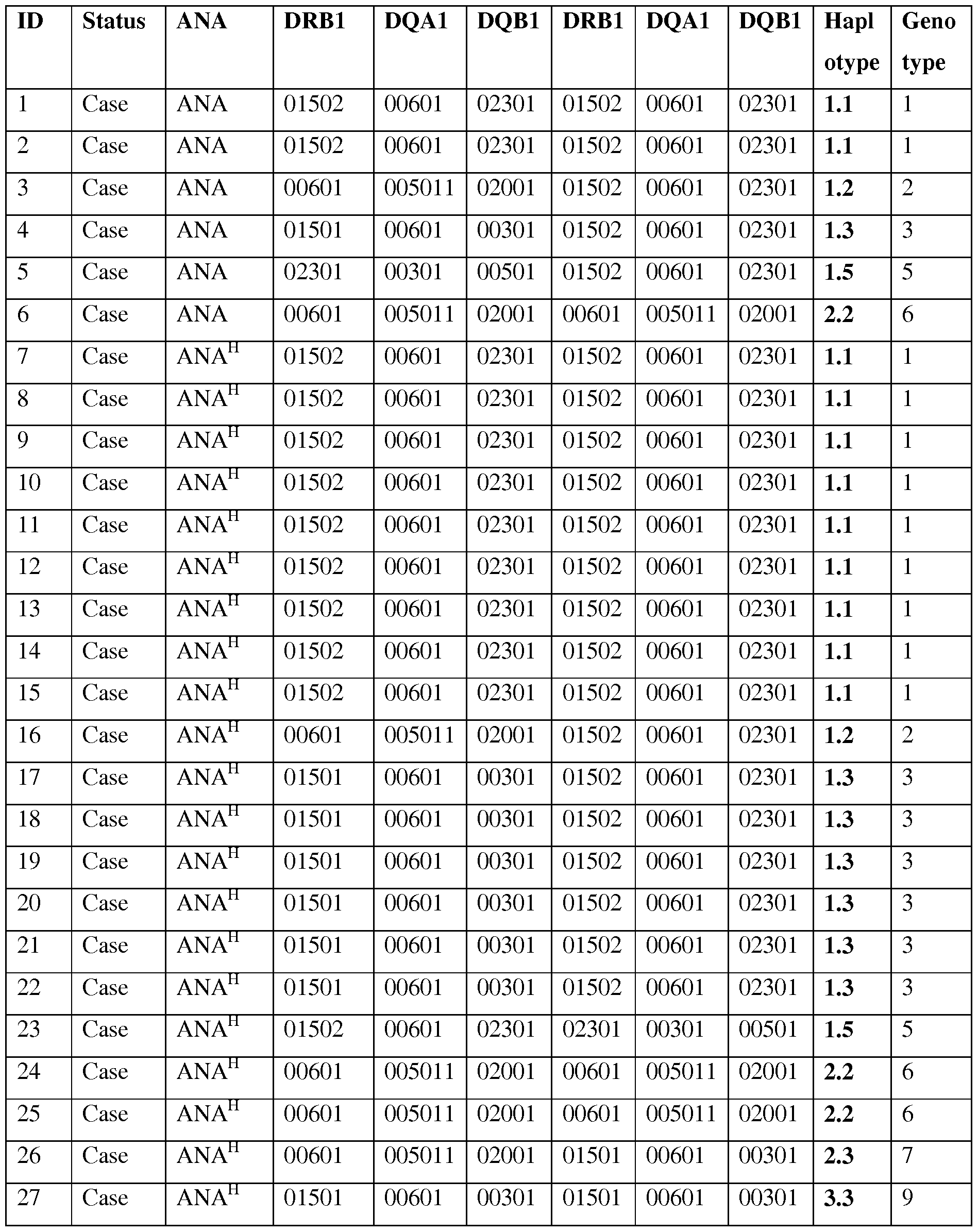
Case ANA" 01501 00601 00301 01501 00601 00301 3.3 9
Case ANAH 01501 00601 00301 01501 00601 00301 3.3 9
Case ANAH 01501 00601 00301 01501 00601 00301 3.3 9
Case ANA" 01502 00601 02301 01502 00601 02301 1.1 1
Case ANAS 00601 005011 02001 01502 00601 02301 1.2 2
Case ANAS 00601 005011 02001 01502 00601 02301 1.2 2
Case ANAS 00601 005011 02001 01502 00601 02301 1.2 2
Case ANAS 02301 00301 00501 01502 00601 02301 1.5 5
Case ANAS 00601 005011 02001 00601 005011 02001 2.2 6
Case ANAS 00601 005011 02001 00601 005011 02001 2.2 6
Case ANAS 00601 005011 02001 00601 005011 02001 2.2 6
Case ANAS 00601 005011 02001 00601 005011 02001 2.2 6
Case ANAS 00601 005011 02001 00601 005011 02001 2.2 6
Case ANAS 00601 005011 02001 00601 005011 02001 2.2 6
Case ANAS 00601 005011 02001 00601 005011 02001 2.2 6
Case ANAS 00601 005011 02001 00601 005011 02001 2.2 6
Case ANAS 00601 005011 02001 00601 005011 02001 2.2 6
Case ANAS 00601 005011 02001 00601 005011 02001 2.2 6
Case ANAS 00601 005011 02001 00601 005011 02001 2.2 6
Case ANAS 00601 005011 02001 00601 005011 02001 2.2 6
Case ANAS 00601 005011 02001 00601 005011 02001 2.2 6
Case ANAS 00601 005011 02001 00601 005011 02001 2.2 6
Case ANAS 00601 005011 02001 00601 005011 02001 2.2 6
Case ANAS 00601 005011 02001 00601 005011 02001 2.2 6
Case ANAS 00601 005011 02001 00601 005011 02001 2.2 6
Case ANAS 00601 005011 02001 00601 005011 02001 2.2 6
Case ANAS 00601 005011 02001 00601 005011 02001 2.2 6
Case ANAS 02301 00301 00501 00601 005011 02001 2.5 8
Case ANAS 01501 00601 00301 01501 00601 00301 3.3 9
Case ANAS 00601 005011 02001 00601 005011 02001 2.2 6
Case ANAS 00601 005011 02001 00601 005011 02001 2.2 6
Control Negative 01502 00601 02301 01502 00601 02301 1.1 1
Control Negative 01502 00601 02301 01502 00601 02301 1.1 1
Control Negative 01502 00601 02301 01502 00601 02301 1.1 1
Control Negative 01502 00601 02301 01502 00601 02301 1.1 1
Control Negative 01502 00601 02301 01502 00601 02301 1.1 1
Control Negative 01502 00601 02301 01502 00601 02301 1.1 1
Control Negative 01502 00601 02301 00601 005011 02001 1.2 2
Control Negative 01502 00601 02301 00601 005011 02001 1.2 2
Control Negative 01502 00601 02301 00601 005011 02001 1.2 2
Control Negative 01502 00601 02301 00601 005011 02001 1.2 2
Control Negative 01502 00601 02301 00601 005011 02001 1.2 2
Control Negative 01502 00601 02301 00601 005011 02001 1.2 2
Control Negative 01502 00601 02301 00601 005011 02001 1.2 2
Control Negative 01502 00601 02301 00601 005011 02001 1.2 2
Control Negative 01502 00601 02301 00601 005011 02001 1.2 2
Control Negative 01502 00601 02301 00601 005011 02001 1.2 2
Control Negative 01502 00601 02301 00601 005011 02001 1.2 2
Control Negative 01502 00601 02301 00601 005011 02001 1.2 2
Control Negative 01502 00601 02301 00601 005011 02001 1.2 2
Control Negative 01502 00601 02301 00601 005011 02001 1.2 2
Control Negative 01502 00601 02301 00601 005011 02001 1.2 2
Control Negative 01502 00601 02301 00601 005011 02001 1.2 2
Control Negative 01502 00601 02301 00601 005011 02001 1.2 2
Control Negative 01502 00601 02301 00601 005011 02001 1.2 2
Control Negative 01502 00601 02301 01501 00601 00301 1.3 3
Control Negative 01502 00601 02301 01501 00601 00301 1.3 3
Control Negative 01502 00601 02301 01501 00601 00301 1.3 3
Control Negative 01502 00601 02301 01501 00601 00301 1.3 3
Control Negative 01502 00601 02301 01501 00601 00301 1.3 3
Control Negative 01502 00601 02301 01501 00601 00301 1.3 3
90 Control Nej native 01502 00601 02301 01501 00601 00301 1.3 3
91 Control Nej native 01502 00601 02301 01501 00601 00301 1.3 3
92 Control Nej native 01502 00601 02301 01501 00601 00301 1.3 3
93 Control Nej native 01502 00601 02301 01501 00601 00301 1.3 3
94 Control Nej native 01502 00601 02301 01501 00601 00301 1.3 3
95 Control Nej native 01502 00601 02301 00401 00201 01501 1.4 4
96 Control Nej native 01502 00601 02301 02301 00301 00501 1.5 5
97 Control Nej native 01502 00601 02301 02301 00301 00501 1.5 5
98 Control Nej native 01502 00601 02301 02301 00301 00501 1.5 5
99 Control Nej native 00601 005011 02001 00601 005011 02001 2.2 6
100 Control Nej native 00601 005011 02001 00601 005011 02001 2.2 6
101 Control Nej native 00601 005011 02001 00601 005011 02001 2.2 6
102 Control Nej native 00601 005011 02001 00601 005011 02001 2.2 6
103 Control Nej native 00601 005011 02001 00601 005011 02001 2.2 6
104 Control Nej native 00601 005011 02001 00601 005011 02001 2.2 6
105 Control Nej native 00601 005011 02001 00601 005011 02001 2.2 6
106 Control Nej native 00601 005011 02001 00601 005011 02001 2.2 6
107 Control Nej native 00601 005011 02001 00601 005011 02001 2.2 6
108 Control Nej native 00601 005011 02001 01501 00601 00301 2.3 7
109 Control Nej native 00601 005011 02001 01501 00601 00301 2.3 7
110 Control Nej native 00601 005011 02001 01501 00601 00301 2.3 7
111 Control Nej native 00601 005011 02001 01501 00601 00301 2.3 7
112 Control Nej native 00601 005011 02001 01501 00601 00301 2.3 7
113 Control Nej native 00601 005011 02001 01501 00601 00301 2.3 7
114 Control Nej native 00601 005011 02001 01501 00601 00301 2.3 7
115 Control Nej native 00601 005011 02001 01501 00601 00301 2.3 7
116 Control Nej native 00601 005011 02001 02301 00301 00501 2.5 8
117 Control Nej native 00601 005011 02001 02301 00301 00501 2.5 8
118 Control Nej native 00601 005011 02001 02301 00301 00501 2.5 8
119 Control Nej native 01501 00601 00301 01501 00601 00301 3.3 9
120 Control Nej native 01501 00601 00301 01501 00601 00301 3.3 9
121 Control Negative 01501 00601 00301 02301 00301 00501 3.5 10
122 Control Negative 01501 00601 00301 02301 00301 00501 3.5 10
Table 5. Allele frequencies and statistical results in the NSDTR population including all ANA-positive cases, ANAS, ANAH and controls. Five different DRBl, four DQAl and five DQB 1 alleles where identified.

DQB 1*003 16.9 32.7 3.7 19.8 18.4 2.0 0.1 01
DQB 1*015 0.0 0.0 0.0 0.8 0.4
01
DQB 1*005 3.4 1.9 3.7 6.3 4.9
01
S= Speckled, H=Homogeneous. Bold indicate between what groups the largest difference in allele frequencies occurred and where statistics were performed (OR and P-values).
Table 6. Haplotype frequencies in the NSDTR population reveal an associated haplotype for ANAS dogs (DLA-DRB 1*00601/DQA1*005011/DQB 1*02001).
ANA
% ANAH ANAS Controls Total O
No Haplotype (2n=ll % % % population % R P-value
DRB1/DQA1/DQB1 8) (2n=52) (2n=54) (2n=126) (2n=244)
1 01502/00601/02301 33.1 53.8 7.4 35.7 34.4 2.1 0.039
2 00601/005011/02001 46.6 11.5 85.2 37.3 41.8 9.7 <0.0001
3 01501/00601/00301 16.9 32.7 3.7 19.8 18.4 2.0 0.10
4 00401/00201/01501 0.0 0.0 0.0 0.8 0.4 - -
5 02301/00301/00501 3.4 1.9 3.7 6.3 4.9 - -
S= Speckled, H=Homogeneous. Bold indicate between what groups the largest difference in allele frequencies occurred and where statistics were performed (OR and P-values). There was a significant association with haplotype 2 in ANAS cases compared to the control group (OR= 9.7 and p=<0.0001) (Table 6), and a higher OR was observed with two copies
(homozygote individuals) of this haplotype (genotype 2; 77.8% in ANAS cases vs. 14.3% in controls; OR=21.0 and p<0.0001) (Table 4). In total, 93% of the twenty-seven ANAS dogs were either homo- or heterozygous for haplotype 2 (DLA- DRBI*00601/DQA1*005011/DQB 1*02001) and twenty-one of them (77.8%) were
homozygous.
No significant association was observed between the haplotypes or genotypes of MHC class II and the cases with ANAH pattern, but a significant association was identified for the
DQA1*00601 allele in 86.5% ANAH cases compared to 55.6% controls; OR=5.1 and
p=0.00017 (Table 5). As homozygosity has been hypothesized to be a risk factor in itself, the
ANAS risk genotype was removed and the remaining data was analyzed for association to homozygosity regardless of haplotype. When removing the ANAS risk genotype and analyzing the remaining data, an increase in homozygosity of any haplotype was seen in ANAH cases 62.5% vs. controls 14.8% (OR=9.6 and p<0.0001), implicating a general homozygous disadvantage at DLA class II for ANAH dogs (Table 7).
Table 7. Homozygosity at MHC class II observed in the NSDTR population including all genotypes with and without the risk genotype 2.
Genotype ANA % ANAH % ANAS % Controls Total population OR P-value
(n=59) (n=26) (n=27) % (n=63) % (n=122)
Homozygous 71.2 65.4 81.5 27.0 48.4 5.1 0.0016
Heterozygous 28.8 34.6 18.5 73.0 51.6 - -
Genotype ANA % ANAH % ANAS % Controls Total population OR P-value
(n=35) (n=24) (n=6) % (n=54) % (n=89)
Homozygous no
2 risk 51.4 62.5 16.7 14.8 29.2 9.6 •CO.0001
Heterozygous
no 2 risk 48.6 37.5 83.3 85.2 70.8 - -
S= Speckled, H=Homogeneous. Bold indicate between what groups the largest difference in allele frequencies occurred and where statistics were performed (OR and P-values).
Distinct risk loci additionally contribute to susceptibility to IMRD and SRMA
To search for candidate variants, the five genetic risk loci on CFA 3, 8, 11, 24 and 32 that were previously identified by GWAS to be associated to EVIRD and SRMA [25] were re-sequenced in four ANA-positive cases, two SRMA cases and three healthy dogs using Nimblegen capture and Illumina sequencing. Using standard methods, a total of 13,084 SNPs and 2,780 indels were detected. No structural changes or CNVs that differed between cases and controls were identified. Among those, 426 SNPs and 88 indels showed a potential functional effect by SeqScoring [Truve K, Eriksson O, Norling M, Wilbe M, Mauceli E, Lindblad-Toh K, et al. SEQscoring: a tool to facilitate the interpretation of data generated with next generation sequencing technologies. EMBnet journal. 2011 ;17(l):38-45]. Next, 308 SNPs fitting the risk haplotype patterns were chosen for genotyping in the entire sample set (Table 8). For each locus, association analysis was performed between the 132 healthy controls and each of the different sub-phenotypes: 1) SRMA-affected dogs (N=66), 2) all ANA-affected dogs (N= 52), 3) ANAS (N=24) and 4) ANAH staining pattern (N=21). Furthermore, two conditional analyses were performed where only ANAS dogs homozygous for DLA risk haplotype 2 (N=18) and
ANA" dogs homozygous for DLA (N=14) and ANA" dogs with the DQA 1*00601 allele (N=16) were included respectively. For the risk locus on chromosome 11 the strongest association was observed with all ANA dogs (FIG. 2A), for the risk locus on chromosome 24 the strongest associated sub-phenotype was the ANAH dogs homozygous for DLA (FIG. 3A), while the risk locus on chromosome 32 showed two independent association signals for SRMA and ANAS with and without DLA association (FIG. 4A); and the risk locus on chromosome 3 also showed two signals (all ANA and for ANAH and ANAS tagged by different haplotypes, as well as to a lesser extent to SRMA, FIG. 5A, C, E). Finally, the risk locus on chromosome 8 also showed two separate signals to SRMA and ANAH dogs associated DLA (FIG. 6A). Of note, in the current report, as the already associated and replicated regions [23] were further investiaged, raw p-values focusing on the highest peaks were used for each subphenotype to investigate its effect on gene expression. These regions have been replicated in a genome-wide association study with P values ranging between 10"5-10"6, with three loci on chromosomes 3, 11 and 24 reaching genome-wide significance following fine-mapping and validation with P values of 10"u-10"13 [23]. These results are discussed in more detail below, together with expression analysis of the genes at each locus.
Table 8. Positions of all SNPs genotyped and p-values for genetic association, minor allele (Al) and major allele (A2) for each phenotype studies.


Co


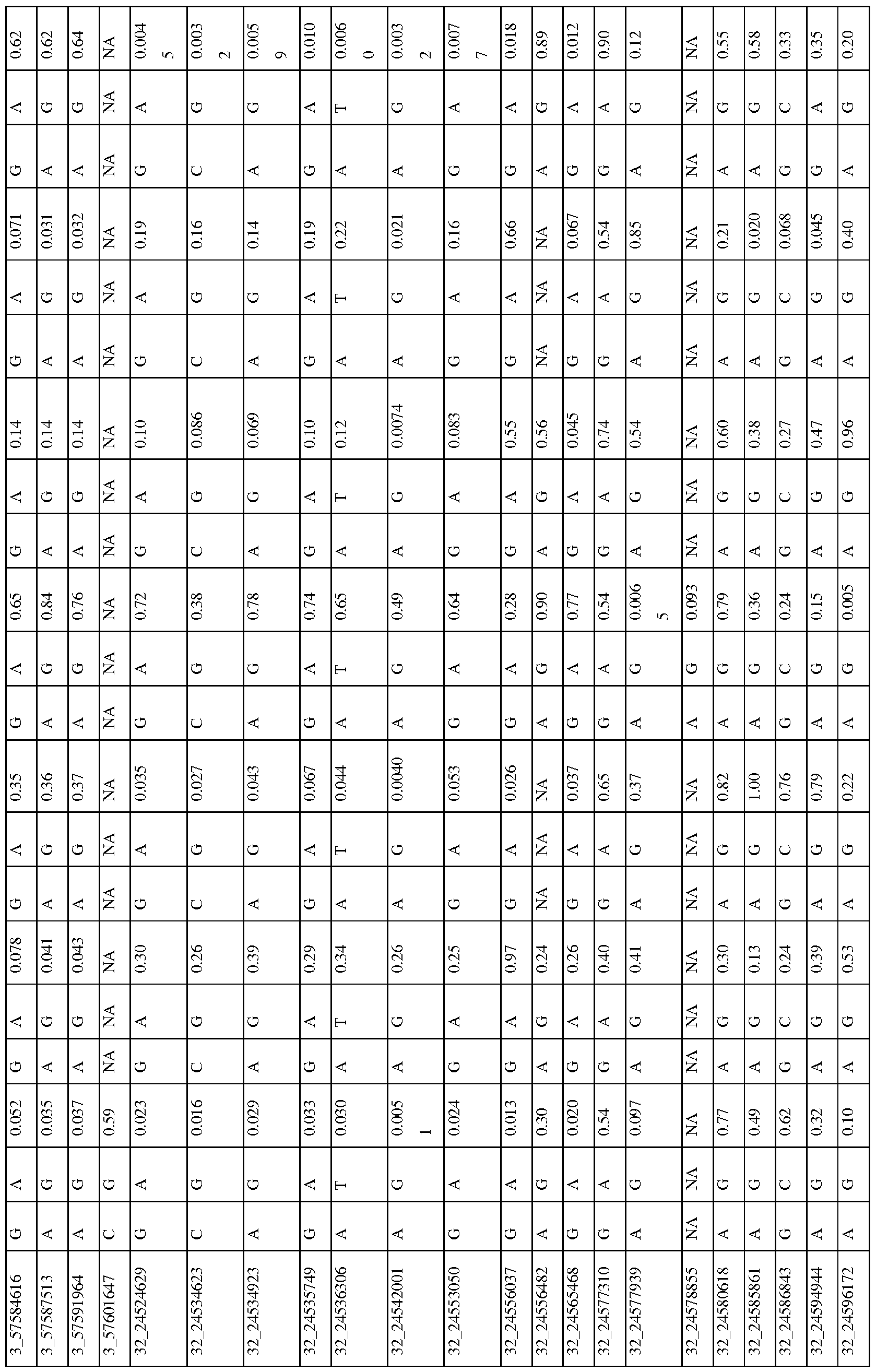


CO


Down-regulation ofPTPN3 is associated with risk for ANA-positive IMRD
On chromosome 11 , the strongest association was seen between all ANA cases and a SNP (CanFam2.0 chrl 1 :67,537,177, p= 2.3*10~4) located within the non-receptor type 3 protein tyrosine phosphatase gene (PTPN3) (FIG. 2A). The top SNP was in almost complete LD with 14 other SNPs (r2 >0.9 for all pairs) (FIG. 2B) forming a 58 kb haplotype overlapping the 3' end of the gene. The risk haplotype had 19.4% allele frequency in cases versus 7.2% in controls. Based on the strong LD, three SNPs were selected from the haplotype (one in the 3'UTR, a SNP in intron 18 and one synonymous SNP in exon 18: 11 :67,516,041,
11 :67,538,032 and 11 :67,538,806, respectively) and then the genotypes were correlated with PTPN3 mRNA levels measured in total RNA purified from the whole blood peripherial blood mononuclear cells (PBMCs) of 167 healthy NSDTRs. The expression of PTPN3 was substantially down-regulated in heterozygotes (7-fold change, PANOVA<0.0001). AS only one dog was found to be homozygous for the risk haplotype among the 167 healthy dogs, it was not included in the statistical analysis, but it was noted that this dog showed extremely low levels of PTPN3 expression. To examine whether all three genotypes could be represented in the expression analysis, a synonymous SNP in exon 3 (11 :67,583,604, p= 0.43) falling just outside the most associated haplotype (r =0.23) was selected for analysis in combination with the other three SNPs from the risk haplotype. This SNP contributed to the down-regulation both independently and in combination with the risk haplotype (FIG. 2C), suggesting a cumulative effect on gene expression levels from the risk haplotype and the exon 3 variant. Among the cases used in the association study, only four dogs were homozygous for the risk haplotype C/C-A/A-G/G-T/T (11 :67,516,041, 11 :67,538,032, 11 :67,538,806 and
11 :67,583,604, respectively) and three of them showed severe IMRD that eventually led to death (one dog died at the age of 19 months old, and two at 6 years of age).
Up-regulation of WFDC3 correlates with risk for ANAH in combination with homozygosity for DLA
The strongest association for the risk locus on chromosome 24 was identified for a sub- phenotype of ANA in combination with a homozygous DLA haplotype (FIG. 3A). Six SNPs in high LD (r2>0.9) (FIG. 3B) define a risk haplotype that overlapped two genes coding for WAP four-disulfide core domain protein 3 (WFDC3) and terminal deoxynucleotidyltransferase interacting protein 1 (DNTTIP1) and was present at an allele frequency of 69.2% in cases and
38.6% in controls. The mRNA expression in PBMCs of these two genes was analyzed in detail. Several other genes located upstream in the locus WFDC13, WFDCIOB, SPINT4 and SPINT5 were found not to be expressed in either PBMCs or 13 diverse dog tissues analyzed (data not shown). The top SNP, located in intron 3 of DNTTIPl (24:36,087,012, p=4.5* 10~4), was associated with expression of WFDC3 (1.4-fold change,
 whereas neither alternative splicing nor expression changes for the DNTTIPl gene were detected. Both genes are separated by less than 3 kb and placed head-to-head, whereby intronic SNPs in DNTTIPl could serve as distal upstream enhancers for WFDC3. The strongest eQTL variant for WFDC3 was a synonymous SNP in exon 3 (24:36,075,761 , 1.4-fold, PANOVA= 0.0039, r2>0.9 with 24:36,087,012), followed by a synonymous SNP in exon 5 (24:36,066,098, 1.4-fold,
PANOVA=0.0076, r2>0.9) (Table 9). Since all three variants are in high LD, and the two synonymous SNPs have stronger or equal association with transcription compared with the top SLE SNP 24:36,087,012, effect of the three-SNP haplotype (24:36,066,098, 24:36,075,761 , 24:36,087,012) on gene expression was determined. The WFDC3 gene was up-regulated 1.4- times in the risk haplotype G/G-A/A-C/C (PANOVA=0.005) compared to protective A/A-C/C- G/G (FIG. 3C).
Table 9. Association of differential expression of WFDC3 with genotyped variants on chromosome 24.

* Correlation was performed by ANOVA, ND - no difference
Two independent association signals on chromosome 32 link to ANA and SRMA respectively
Strong association for the risk locus on chromosome 32 was observed in multiple sub- phenotypes. In ANAS dogs that were homozygous for DLA haplotype 2 and to a lesser extent in "all ANA" and homogeneous ANA with DLA DQA1 *00601 allele and general
homozygosity for DLA (FIG . 4A), the signal was located between the MTTP and DAPP1 genes and included eight SNPs in complete LD (r2=l) on a 41 kb haplotype (32:24,514,629- 24,565,468), best association: p=4.0*10~4) that occurred more than twice as frequently in patients (23.5%) as in healthy controls (8.8%, FIG. 4B). In contrast to ANA phenotypes, the association with SRMA did not form a single risk-haplotype, but instead was spread over a 1.3 Mb region (32:24,827,518- 32:26,115,349) (FIG. 4A). While no strong LD was identified, 11 SNPs out of 131 genotyped for this chromosomal region have an r2 between 0.6-0.8 across the whole region (FIG. 4C).
To examine the functional effect of the risk genotypes, mRNA expression of all nine genes from the locus was measured in the blood cells (PBMCs) of healthy dogs (FIG. 4D-H, FIG. 7). 24 highly associated SNPs were genotyped across the entire region and correlated with expression of all the genes (Table 10). Interestingly, the ANAS/ANA/ANAH association signal was only significantly correlated with a 1.5 -fold up-regulation of the BANK1 gene
(PANOVA=0.0007), located 1.5 Mb downstream from the top SNP 32:24,542,001, which suggests a long-range regulatory effect (FIG. 4D).
The top SNP (chr32:24,827,518) for SRMA, on the other hand, was also the SNP associated with the most significant expression changes for the following genes: DAPP1, LAMTOR3, DDIT4L and PPP3CA, respectively. The risk allele A correlated with enhanced gene expression levels (1.7-fold for DAPP1, PANOVA=0.001 ; 1.9-fold for LAMTOR3, PANOVA=0.008; 2.5-fold for DDIT4L, PANOVA<0.0001 ; 2-fold for PPP3CA, PANOVA=0.01) (FIG. 4E-H). Table 10. Association of differential expression of genes with genotyped variants across the chromosome 32 locus.

24556037 ND ND P=0.0038 ND P=0.0027
24606503 ND ND ND ND ND
24667283 ND ND ND ND ND
24667774 ND ND ND ND ND
24672221 ND ND ND ND ND
1.7X, 2.5X,
24827518 F '=0.0012 2X, P=0.0076 P O.0001 2X, P=0.0133 ND
1.25X, 1.2X,
24890208 1.9X, P=0.049 P=0.0594 2X, P=0.0005 ND P=0.0172
2.5X, 1.2X,
24985562 2X, P=0.0233 ND P=0.0068 ND P=0.0042
1.9X, 2.3X, 1.2X,
24987404 F '=0.0928 ND P =0.0256 ND P=0.0078
1.6X,
25007496 ND ND P =0.0638 ND ND
25305524 ND ND ND ND ND
25363099 ND ND ND ND ND
2.3X, 1.2X,
25485961 ND ND P =0.0001 ND P=0.0045
1.7X, 1.9X, 1.8X,
25512953 2X, P=0.0637 P=0.0231 P =0.0532 P =0.0336 ND
1.2X,
25537276 ND ND ND ND p=0.0321
1.2X,
25537876 ND ND ND ND P=0.0434
25642357 ND ND ND ND ND
25702963 ND ND ND ND ND
1.9X, 1.8X, 1.8X,
25714903 ND P=0.0755 P =0.0233 P =0.0264 ND
25718852 ND ND ND ND ND
25779083 ND ND ND ND ND
1.17X,
25798353 ND ND ND ND p=0.0455
1.5X, 1.7X,
26115349 ND 2X, P=0.0719 P =0.0345 P=0.0247 ND
* Correlation was performed 3y ANOVA, N 3 - no difference
Complex changes o f gene functions in chromosome 3 locus are associated with ANA-positivity and with SRMA
Association of the risk locus on chromosome 3 was identified for 'all ANA' dogs including speckled and homogeneous sub-phenotypes (FIG. 5 A) with the top SNP (chr3:57,432,981, p=4.9* 10"4) linked to a four SNP haplotype in strong LD (r2>0.9) (FIG. 5B) where the haplotype overlaps the AP3B2 gene and extends towards the FSD2 gene. A second haplotype consisting of 18 SNPs in strong LD (r2>0.9) with a SNP at chr3:57,484,486 (p=5.6*10~3, FIG. 5C) was located upstream of the WHAMM gene and also associated to the 'all ANA' and speckled phenotypes (p=2.2*10-2). This haplotype was also strongly associated with SRMA (p=2.6* 10"2) (Figure 5E). Separately the phenotype of ANAH + DLA-DQA1 *00601 was found to be associated with a SNP located in the HOMER2 gene (chr3:57,546,568, p=1.2*10"3, FIG. 5A). No LD was observed between this SNP and the other strongly associated SNPs (FIG. 5D).
Next, the mRNA expression of the four genes located within the borders of the associated region was analyzed. It was found that three genes, AP3B2, WHAMM and HOMER2, were ubiquitously expressed while FSD2 expression was restricted to skeletal muscle, heart, kidney, testis, skin, and very low levels detected in cartilage (FIG. 8). Due to low minor allele frequencies, enough genetically different tissue samples could not be collected from NSDTRs to analyze FSD2 expression, and thus it could not be ruled out entirely that there was a possibility for this gene to be affected by the associated variants in a particular tissue.
The most associated SNP for 'all ANA' (located between AP3B and FSD2) was most associated with expression of WHAMM. The minor risk allele T correlates with up-regulation of WHAMM mRNA expression (1.4-fold, PANOVA<0.0001), while showing almost no biologically significant effect on AP3B2 (1.07-fold median change, P=0.036) (Table 11). On the contrary, the associated SNP 3:57,484,486 located upstream of WHAMM, exerted a more profound effect on AP3B2 transcription (1.5-fold, PANOVA =0.004) (Table 11), thus indicating the existence of negative cross-regulation of the two genes.
The expression of HOMER2 was not associated with any of the SLE-related variants, although differential regulation of the gene marginally associated with some genetic variants was found (FIG. 9A), suggesting that there may be an independent eQTL variant(s) not directly related to the sub-phenotypes studied here. When a 2 SNP haplotype (3:57,432,981-3:57,546,568) was considered by combining the associated SNPs at the two ends of this locus (near AP3B2 and
within HOMER2), the effect on WHAMM increased compared to when either of the two SNPs were examined separately (1.5-fold, PANOVA< .0001 , FIG. 5F, FIG. 9B).
Interestingly, the top SNP 3:57,432,981 is a common variant tagging two risk sub-haplotypes: (3:57,432,981-3:57,484,486) and (3:57,432,981-3:57,546,568) occurring in 26% and 16% of patients, correspondingly. While the first haplotype was associated more with general ANA positivity, the second one was associated with homogeneous ANA staining and even more specifically with a particular DLA risk allele. This could suggest that depending on the second SNP in the risk haplotype, the combination of affected genes (AP3B2 and to a lesser extent WHAMM (FIG. 9C), or WHAMM and H0MER2) may determine what pathways are under impact and hence, what disease phenotype could be expected.
In addition to all the regulatory variants described here, the associated SNP located within HOMER2 (chr3:57,546,568) (FIG. 5A and 5D), is a non-synonymous variant in exon 3 of the HOMER2 gene causing a Thr to Ala substitution. The substitution is located in the β5 strand of a highly conserved EVH1 protein domain and is close to the key amino acids participating in the formation of the HOMER2 ligand binding site [26, 27] (FIG. 5H, FIG. 10). Analysis with SIFT [Kumar P, Henikoff S, Ng PC. Predicting the effects of coding non-synonymous variants on protein function using the SIFT algorithm. Nat Protoc. 2009 ;4(7): 1073-81] and PROVEAN [Choi Y, Sims GE, Murphy S, Miller JR, Chan AP. Predicting the functional effect of amino acid substitutions and indels. PLoS One. 2012;7(10):e46688] suggests that the variation is rather deleterious for protein function.
Table 11. Association of differential expression of genes with genotyped variants on chromosome 3.

57484486 1.5X, P=0.0036 ND ND
57484658 ND ND ND
57500572 ND ND ND
57517383 ND ND ND
57526312 ND 1.25X; P=0.0004 ND
57527697 1.6X, P=0.0186 1.2X, P=0.012 ND
57546568 ND 1.4X, P=0.0046 ND, Thr->Ala
57564331 ND 1.26X, PKO.0001 1.4X, P=0.0924
57575243 ND ND ND
57610712 ND 1.3X, PKO.0001 ND
* Correlation was per] "armed by ANOVA, N 3 - no difference
Chromosome 8 SNPs in gene desert associated with ANA and SRMA phenotypes
The association signals on chromosome 8 falls into a 2 Mb gene-desert, downstream of the VRK1 gene. The strongest association was observed in SRMA dogs (chr8:68,726,546, p=2.2*10"3), but also the phenotype: ANAH + risk allele DLA-DQA1 *00601 showed significant association to two SNPs (chr8:68,712,185, p=6.7* 10"3 and chr8:68,708,503, p=8.7*10~ ) (FIG. 6A). No LD occurred between the three variants for the two sub-phenotypes (FIG. 6B-C, FIG. 11). The only close gene, VRK1, is located over 100 kb upstream of the signals, and its expression measured in the blood cells was found associated with the twoANA risk variants when analyzed using the two available genotypes only: major protective and heterozygous (FIG. 6D). Due to low minor allele frequencies, only one dog homozygous for the ANAH risk variants was found in the study cohort (167 NSDTRs), thus limiting the power of statistical analysis of the VRK1 expression.
Among the three top variants only SNP chr8:68,708,503 lies in the region with high regulatory potential as predicted by ESPERR [28] (FIG. 6C). This region also contains enhancer- associated H3K4Mel histone-modification marks in lymphablastoid cells, ENCODE ChlP-seq and DNAse I hypersensitive sites. The SNP is located only 5 bp from the highly conserved Pou5fl/Oct4 binding site [29]. Moreover, the risk allele A creates a binding site for signal transducer and activator of transcription (STAT) family transcription factors. Interestingly, Oct-1 and STAT5, members of the Pou domain-containing and STAT family transcription
factors, correspondingly, were shown to form stable transcription complexes upon cell activation with cytokines and induce cyclin Dl expression [30]. In order to verify the regulatory effect of this variant, the 550 bp DNA fragment was cloned into the pGL4.26 vector, and after transfection into K562 cells, a luciferase assay was performed. It was confirmed that the risk allele A indeed enhanced expression in both non-stimulated and stimulated cells (FIG. 6E). It was concluded that the SNP chr8:68,708,503 not only has a proven regulatory potential but might be involved as a part of an enhancer in the up-regulation of the VRK1 gene in the risk for ANAH.
In summary, five risk loci were identified for the canine SLE- related disease complex. The association signals within each locus were characterized and 11 genes with altered gene expression based on the risk haplotypes were identified. It was found that particular sub- phenotypes of dog SLE based on the immunofluorescent ANA staining involved different sets of genes and that some regions contained multiple SNPs or haplotypes associated to disease subtypes and altered gene expression. The down-regulation of PTPN3 and up-regulation of WHAMM were associated with general ANA-positivity, while the homogeneous pattern (ANAH) with a specific MHC haplotype showed a stronger link to the up-regulation of WFDC3 and VRK1 and down-regulation of a specific isoform of HOMER2 bearing the Ala amino acid (Thr->Ala substitution); whereas the speckled phenotype (ANA ) homozygous for the MHC class II risk haplotype was more strongly connected to the up-regulation of AP3B2 and BANK1, a known susceptibility gene for human SLE. The steroid-responsive meningitis- arteritis (SRMA) phenotype was associated with up-regulation of a distinct set of genes, DAPP1, LAMTOR3, DDIT4L, and PPP3CA, on chromosome 32 and AP3B2 on chromosome 3. These summarized results are depicted in Table 12. These results suggest a complex genomic architecture within the five disease loci where multiple variants affect the expression of different genes in different sub-phenotypes of the disease.
Table 12. Summary of genes associated with IMRD and SRMA phenotypes.

32 32:24827518 PPP3CA up - - - - - ++
3 3:57484486 AP3B2 up ++ + - - - +
3 3:57432981 WHAMM up ++ + - + + -
3 3:57546568 HOMER2 nsSNP + + ++
(Thr- >Ala) or
down1
8 8:68708503 VRK12 up + - - + ++ -
Chr = chromosome, ++ indicates the strongest genetic association with phenotype, + indicates a genetic association with a phenotype, ANAS = ANA speckled, ANAH= ANA homogeneous, up = upregulation of expression, down = downregulation of expression, MHC = MHC 2.2 haplotype for speckled ANA, and MHC DQA1 *00601 for homogeneous ANA. l- A trend towards down-regulation of HOMER2 in the risk haplotype was also observed, although it did not reach statistical significance due to small sample size. 2-the strong genetic association signal with SRMA on chromosome 8 was not associated with VRK1 expression levels. Discussion
SLE and other autoimmune diseases occurring in humans have been intensely studied over the past years due to the high genetic heritability of such diseases and the availability of modern genetic tools. The most recent review article reports over 40 loci associated with human SLE [31]. The heterogeneity of SLE reflected by the 11 diagnostic criteria established by the American College of Rheumatology (ACR) [2, 3] supports the current understanding that the genetic factors underlying such disparate clinical manifestations could be different as well. Recently, differential genetic associations with SLE based on the anti-dsDNA autoantibody status, either anti-dsDNA positive or anti-dsDN A- negative SLE, were reported [32]. The study of the relationships between the SLE risk alleles and clinical sub-phenotypes led to the discovery that certain lupus manifestations are more dependent on the presence of multiple risk alleles, while others are more strongly associated with a single variant, for example, renal disease more significantly associated with HLA-DRB 1 , and arthritis with the protective allele of ITGAM [33]. Interestingly, a third group of sub-phenotypes (malar rash, discoid rash, photosensitivity, serositis, and neurological disorders) was found not to be associated with the currently known known SLE susceptibility genes, suggesting either the presence of not yet identified factors or potentially non-genetic factors, such as environmental conditions or epigenetic effects.
Dogs, like other mammalian species, also develop autoimmune disorders. The existence of canine breeds such as NSDTRs, which are predisposed to the development of autoimmune diseases, make them potentially useful for mapping disease genes and finding novel disease pathways [34]. In the present study, detailed genetic association and gene expression analyses were performed on the two sub-phenotypes of IMRD, an SLE-like disease in dogs, and another immune-mediated disease SRMA.
A hallmark of systemic SLE-related immune-mediated rheumatic disease (IMRD) in dogs is the presence of ANA autoantibodies, which display two major patterns when stained with indirect immunofluorescence, homogeneous and speckled ANA. Both sub-phenotypes have overlapping clinical and pathological features in NSDTRs such as musculoskeletal signs, including stiffness and joint pain without joint swelling, sometimes muscle pain and lymphopenia; and all dogs showed good response to corticosteroid treatment [5]. The association of different ANA patterns with various clinical signs for SLE was previously reported for a group of dogs including German Shepherds, NSDTRs, and several other breeds [8]. Of note, in the NSDTRs sample set described herein, the skin lesions were present in the ANAH group, while muscle pain and fever were slightly more frequent in the speckled ANA group (Table 13). The onset of disease in the study cohort was at a median age of 3 years for
H S
ANA dogs and less than 2 years for ANA dogs. Table 13. Clinical manifestations in ANA positive NSDTRs

Complete clinical data were available for 25 NSDTRs. The remaining 27 dogs collected were diagnosed as having IMRD,
The MHC region has the strongest association to many autoimmune diseases in humans due to its utmost importance in the recognition of antigens; and it was also shown to be important for canine SLE [21]. Moreover, certain genotypes such as the HLA-DRB1*03:01 was found recently to be significantly associated with specific sub-phenotypes with anti-Ro/SSA and anti-
La/SSB autoantibodies [Morris DL, Fernando MM, Taylor KE, Chung SA, Nititham J, Alarcon-Riquelme ME, et al. MHC associations with clinical and autoantibody manifestations in European SLE. Genes Immun. 2014]. In dogs in the present study, it was observed that different DLA genotypes and alleles were associated with either ANAH or ANAS, which may indicate the reactivity towards certain autoantigens produced more frequently in a particular ANA pattern.
Among the seven genes associated with "all ANA", WFDC3, HOMER2 and VRK1 were clearly not associated with ANAS reactivity (Table 12). Moreover, for these genes the cumulative association with DLA, either general homozygosity for any DLA haplotype or the DQA1*00601 allele, plays a somewhat stronger role. The AP3B2 gene was associated with "all ANA"; and also has equally strong signals for two phenotypes, the speckled ANA group and SRMA, but no association with ANAH. Interestingly, the observed genetic segregation may favor a previously suggested hypothesis based on the differences in clinical manifestations that ANAS pattern could represent another SLE-related disorder, while the ANAH is more similar to human SLE [5]. The PTPN3, WHAMM and BANKl genes were associated with both ANA sub-phenotypes and thus could be considered as common genes.
A function of WHAMM (WAS protein homolog associated with actin, Golgi membranes and microtubules), a gene previously reported to participate in Golgi transport and membrane remodeling, and cytoskeleton formation by binding to microtubules and promoting actin polymerization [Campellone KG, Webb NJ, Znameroski EA, Welch MD. WHAMM is an
Arp2/3 complex activator that binds microtubules and functions in ER to Golgi transport. Cell. 2008;134(1): 148-61], has not been described before in immunity, while the other two common genes PTPN3 and BANKl implicate major perturbations in both T and B cells. Human protein tyrosine phosphatase PTPH1 encoded by the PTPN3 gene inhibits T cell- activation by dephosphorylating the immune tyrosine-based activation motifs (ITAM) in the TCRζ chain that results in a downstream inhibition of NF-AT [29, 30]. The observed substantial reduction of the PTPN3 mRNA levels in dogs carrying the risk haplotypes may cause a sustained activation of TCR signaling and lead to development of autoimmune disease. The BANKl gene encoding the B-cell scaffold protein with ankyrin repeats was previously found associated with human SLE and other autoimmune diseases in distinct populations and ethnic groups [60- 66]. The expression of the human BANKl gene, similarly to what we found in IMRD dogs, is up-regulated in patients carrying the risk alleles [67]. This may suggest a common disease mechanism in the human and dog diseases.
Whether or not and to what extent, the individual genes contribute to a particular ANA- staining pattern, and more generally, to specific clinical manifestations, and what interplay could be between the associated genes and their pathways, may be further studied. Also, while the identity of major autoantibodies present in the serum of human individuals with different ANA reactivity is already known [15], it may need to be studied in more detail in dogs.
SRMA, on the other hand, differs from IMRD by displaying predominantly neurological signs including pain, cervical rigidity, pyrexia and a polymorphonuclear pleocytosis of the cerebrospinal fluid (CSF) [10]. The disease usually occurs at a young age (4-19 months) and is characterized by inflammation of leptomeninges and vasculitis of the leptomeningeal and mediastinal blood vessels, including arteritis of heart, thymus, and also vessels of the thyroid glands and muscles [68, 69]. The disease can be treated with immunosuppressive doses of corticosteroids similarly to IMRD [12]. While the etiology is largely unknown, it has been proven that inflammatory processes in the CNS are not caused by viral or bacterial infection [12].
The strongest genetic association with SRMA detected in NSDTRs was located on chromosome 32, followed by signals on chromosome 8 and 3. Although there was no strong LD (r2<0.8) between the genetically associated variants on chromosome 32, the presence of multiple highly associated SNPs across the 1.3 Mb region and their association with gene expression levels suggests that the entire region including several genes is important for SRMA susceptibility. While many typed variants show correlation with gene expression, there was one tag SNP (chr32:24,827,518) whose risk allele A was most strongly associated with increased expression of DAPP1, LAMTOR3, DDIT4L and PPP3CA. The distant cis-effect on expression of genes placed far away from each other may indicate a complex topology of the chromosomal locus with a possible locus control region(s) and common enhancers driving regulation of genes located on the opposite DNA strands.
The previously reported signal on chromosome 8 [23] was corroborated in the present study, but no correlation was found between the SRMA SNPs and expression levels of the only nearby gene VRK1. Thereby, the identity of the gene affected in this locus in SRMA remains to be identified or the SNP could have its effect in a different tissue than PBMCs. Also, the progress in mapping non-coding genes including microRNA and lincRNA genes on the dog genome [70] may help to resolve this question in the future. The SRMA associated haplotype on chromosome 3 was correlated with increased expression of the AP3B2 gene.
Interestingly, there was little overlap between the genes associated with SRMA and IMRD, suggesting the involvement of largely distinct pathways in these phenotypes (FIG. 12). While most of the 11 genes are widely expressed, there is a certain emphasis on the predominant brain, muscle and immune cells expression of SRMA genes AP3B2, PPP3CA, DDIT4L, DAPP1 and LAMTOR3 (FIG. 13), consistent with the involvement of the central nervous system and muscles seen in this disease. Moreover, two genes, DDIT4L and PPP3CA, showed relatively strong association signals in human neuromuscular inflammatory conditions (Table 16). Overall, for nine dog genes out of eleven, there was either evidence or at least a trend of association with various human autoimmune disorders, as can be viewed using the GRASP Search tool [71].
Table 16. The association signals for the dog genes obtained in various human genome-wide association studies and retrieved by the
GRASP search tool.

CO

CO
CO

CO

ο


CO


C/T;0,
1.7 12759 6E rsl l944 Europ T;0,03 1;0,21 +14 421 1868 EA cases,2938 controls ean 4806 4806 66 7981 YES
1.7
6E rsl2651 Europ G;0,2 A/G;? +14 056
 1868 EA cases,2938 controls ean 4806 4806 17 YES

o

o


The cut-off p- value is 0.001. The results only for autoimmune diseases and neuroinflammatory conditions are presented.
Without wishing to be bound by a theory, it is hypothesized that primary inflammation triggered by as yet unknown environmental factors in SRMA-susceptible dogs is then maintained by the over-activated B cells producing high titers of IgA, often detected in the serum and cerebrospinal fluid (CSF) of SRMA patients [12, 69]. The enhanced function of DAPP1 , PPP3CA and LAMTOR3 acting downstream of the B cell receptor via MAPK signaling pathway may be responsible for the altered B cell reactions. Moreover, the arteritis of the blood vessels may lead to ischemia in CNS or myocardial infarction through decreased nutrient and oxygen supply by damaged arteries and may further induce an already genetically modulated DDIT4L in neurons or cardiac myocytes, which in turn inhibit mTOR signaling and activate autophagy (genes LAMTOR3 and AP3B2 code for adaptor proteins in the lysosome- endosome system) and lead to apoptosis or necrosis [72]. It may be that the enhanced levels of brain- specific expression of the SRMA-associated genes could be responsible also for hyperesthesia, an extreme pain sensitivity condition mainly exhibited by cervical, neck and spinal pain, and always seen in SRMA but not IMRD dogs [12].
Of note, out of four genes associated with ANAS IMRD only AP3B2, the gene also shared with SRMA, shows the highest expression levels in the brain. In contrast, the ANAH genes showed a more diverse pattern of expression with the highest levels in the following tissues: blood (BANK1 and VRK1), skin and kidney (WFDC3), muscle and kidney
(HOMER2), heart, kidney and liver (PTPN3), muscle/heart, liver and kidney (WHAMM).
Many cellular pathways are interconnected and therefore it may be difficult to predict the exact biological effect, especially when several genes are altered, so the signaling output of
H S
each affected gene in all three phenotypes, ANA , ANA and SRMA, will depend on a particular tissue and cell context, the precise timing of expression and responsiveness to environmental inducers. Thus, for instance, the highest shift in expression levels associated with SRMA was a 2.5-fold up-regulation observed for the DDIT4L gene (FIG. 4G), DNA- damage-inducible transcript 4-like, known to be induced by a variety of stress factors, including hypoxia [73]. Although the precise role of the encoded protein remains largely unknown, the function of DDIT4L in autoimmunity may be related to the negative regulation of mTOR [31]. While the inhibition of mTOR promotes generation of CD8+ memory T cells [32], at the same time it induces cell death by necrosis in the culture of U-937 monocytes [34], which may trigger an auto-inflammatory response.
In addition to the complexity of phenotypes relating to the different genes and genetic variants, the genomic architecture of almost all loci showed a considerable complexity with
SNPs showing long-range effects on gene regulation either alone or in conjunction with other SNPs. For example, the chromosome 3 top SNP (3:57,432,981) was a part of two different haplotypes present in different sub-phenotypes. On chromosome 32, a haplotype associated with ANAS exerted a distant effect on a gene located in the other part of the locus, while SNPs located internally in the region affected four other genes in SRMA. It remains to be shown whether this type of complex regulatory structure will be a common finding in both canine and human disease.
In conclusion, functional changes were identified in eleven genes associated with different sub-phenotypes of canine EVIRD resembling human SLE, and with SRMA which share clinical signs with a group of human vasulitides including Kawasaki disease, Henoch- Schonlein purpura and Behcet's disease. Based on the gene functions and pattern of expression, it is hypothesized how different genetic factors, sometimes located in the same genomic region, may lead to diverse clinical manifestations. The common genes and pathways may account for overlapping manifestations, whereas alterations in the functions of certain tissue-specific or even ubiquitous genes may be linked to specific clinical signs. The utility of dog breeds in identification of disease genes underlying human complex diseases is demonstrated by the identification of a well-known human SLE gene, BANK1, and several novel genes. The novel genes may warrant further study both in canine and human
autoimmune disease.
Methods
Study population and diagnostic procedures
250 Nova Scotia duck tolling retrievers (NSDTRs) were included in this study, 52 of them classified as ANA-positive EVIRD cases, 66 as SRMA cases and 132 as healthy controls. All dogs included were privately owned. The dog pedigrees were verified to exclude closely related animals (littermates) from the analysis. Individual dog owners had consulted different veterinary clinics in Sweden and Finland. The inclusion criteria for EVIRD ANA-positive dogs were musculoskeletal signs indicating a systemic rheumatic disorder, including stiffness mainly after rest, and pain from several joints of extremities. These signs had to be apparent for at least 14 days and were the main reason for the dog owner to visit the veterinary clinic. The examining veterinary physician suspected no other diseases in their diagnosis. All EVIRD dogs should also display a positive indirect immunofluorescence (IIF) ANA test. Healthy controls
were above seven years of age with no history of autoimmune disease. All study dogs were verified for relatedness and those related were excluded from the analysis.
ANA tests were analyzed with indirect immunofluorescence at the University Animal Hospital, Swedish University of Agricultural Sciences (SLU), Uppsala, Sweden using monolayers of HEp-2 cells fixed on glass slides (Immuno Concepts). The glass slides were examined by fluorescence microscopy and considered positive at a titer of >1 : 100. The visible nuclear fluorescence patterns could be divided into two groups; homogeneous (ANAH) or speckled (ANAS) patterns as previously described [8]. DNA purification, PCR amplification of OLA regions and sequence analysis
Genomic DNA was purified from 200 μΐ of blood using Qiagen QIAamp DNA Blood Mini Kit (Qiagen) according to the manufacturer's protocol. DLA-DRB1, -DQA1 and DQB 1 exon 2 were amplified by PCR as previously described [23]. DNA sequencing was performed using capillary electrophoresis on an Applied Biosystems 3730x1. BigDye® Terminator v3.1 (Applied Biosystems) Sequencing of the purified PCR products was made in one direction, reverse for DLA-DRB1 and -DQA1 and forward for DLA-DQB 1. Analysis of the nucleotide sequence was performed using MatchTools and MatchTools Navigator (Applied Biosystems) [23]. Statistical analysis
Statistical analyses were performed using VassarStats (vassarstats.net/odds2x2.html). Odds ratios and p- values for each allele, haplotype and genotype were calculated using a 2x2 contingency table. The total number of cases and controls carrying a specific allele or genotype was compared with the cases and controls not carrying it. The same comparison was made for alleles as well as genotypes for the ANA-positive cases with homogeneous or speckled pattern and the controls. The total numbers of homozygous dogs were also compared in cases and controls.
Next generation re-sequencing
To identify candidate variants, the five regions previously found associated with IMRD
[25], spanning approximately 5Mb, were re-sequenced in nine individuals (four ANA cases, two SRMA cases and three controls) using 385K custom designed capture arrays from Roche NimbleGen and 400-600 X coverage Illumina sequencing. The sequencing data was aligned
with BWA (bio-bwa.sourceforge.net) [53] and analyzed using SAMtools (samtools.sourceforge.net) [54], BEDTools (code.google.com/p/bedtools) [55], SEQscoring [56] (seqscoring.net) and other in-house tools to discover variants (SNPs and indels and structural changes) in the genomic sequence between EVIRD, SRMA and healthy control dogs. A total of 13,084 potential SNPs were detected and of these, 426 SNPs were located within or close (±5 bp) to a conserved element [39, 68]. 2780 possible InDels were detected, among those, 88 occurred within or close to a conserved element (±5 bp). To identify structural variations, such as larger insertions, CNV or deletions, SEQscoring was used to calculate coverage differences between cases and controls. No structural variants were identified that differed between cases and controls.
SNP selection for genotyping and association analysis
308 SNPs for five loci (chromosome 3, 8, 11, 24 and 32) were chosen from the re- sequencing data. SNPs were chosen based on the following criteria: difference in allele frequency in cases compared to controls, positioned in either protein coding regions, 5' UTR or 3' UTR and located within non-coding conserved elements. Conserved elements were identified using comparative sequence analysis based on the analysis of 29 mammals assessed by using SiPhy [57]. These SNPs were genotyped by GoldenGate® Genotyping Assay. PLINK [58] was used to analyze the markers with a MAF >0.05 and a call rate >0.75. Total genotyping rate was 97%.
RNA extraction and cDNA synthesis
Peripheral blood was drawn from 165 healthy NSDTR dogs directly in Tempus Blood RNA tubes (Applied Biosystems) and kept on ice during transportation. For isolation of total RNA from different dog tissues, fresh tissue samples from euthanized dogs were immediately immersed in the TRIZOL solution (Invitrogen) and RNA and DNA were purified according to the manufacture's protocol. Total RNA from blood was purified using the Tempus Spin RNA Isolation Reagent kit (Applied Biosystems) according to the manufacturer's instructions, and the quantity and the quality of RNA was assessed by NanoDrop ND-1000 spectrophotometer (Thermo Scientific). In parallel, genomic DNA was purified for each sample and genotyped using pyrosequencing or direct Sanger sequencing with the primers shown in Table 14. cDNA synthesis was performed at 42°C in 20 μΕ for 80 min using 2 μg of RNA, 5 μΜ oligo-dT primer, MuLV transcriptase, RNase inhibitor in the buffer supplemented with 5 mM MgCl2
and 1 mM dNTPs. All reagents were from Applied Biosystems. The reaction was terminated by heating for 5 min at 95°C and diluted to 25 ng/μΐ.
Table 14. Primer pairs used for genotyping in expression samples.


Gene expression was measured by quantitative real-time PCR on 7900HT Sequence Detector (Applied Biosystems) with SDS 2.3 software using SYBR Green for signal detection. Gene-specific primers and annealing Tm are shown in Table 15 in accordance with the guidelines for the minimum information for publication of quantitative real-time PCR experiments (MIQE) [70]. The regions for primers' design were selected to target all known transcripts for a particular gene and thus allow to measure the total gene expression. In order to avoid amplification from genomic DNA, primers were located to cover either several exons separated by long introns or exon/exon junctions and further verified by BLAST search. The PCR conditions were optimized for each primer set prior to qPCR, and the specificity of amplification was verified by the post-PCR analysis of melting curves and agarose gel analysis. Initial denaturation at 95°C for 5 min was followed by 45 cycles (95°C for 15s, annealing at primer-specific Tm for 15s and 72°C for 25s). PCR buffer was supplemented with 1.5 mM MgCl2, 200 μΜ of each dNTPs, primers, SYBRGreen (Molecular Probes), 15 ng of cDNA and 0.5 U of Platinum Taq polymerase (Invitrogen). The reaction was carried out in 20 μΐ^ on a MicroAmp Optical 384-well reaction plate (Applied Biosytems). Expression levels were normalized to the reference gene TBP using the comparative 2"ACt- method [59]. All experiments were run in triplicate. Correlation of gene expression with genotypes and haplotypes was performed using one-way ANOVA tests in PRISM 6 (GraphPad Software).
Table 15. Primers for qRT-PCR.

1 16
HOMER2
CTACTTCTACGATGTCACCAGGAATA
Ex2-Homer2-forw
G (SEQ ID NO: 121) 60 153 bp AACACCGTGTTGGCTCTGCTGTC
Rev-Homer2-ex3 (SEQ ID NO: 122)
VRK1
AATATTGCTGGTTTGATGGACAAAT
174 bp
VRKl-exlO-for (SEQ ID NO: 123) 63
CTTGCCATCATCCTTGCTTCCTAT
Rev-VRKl-exl l (SEQ ID NO: 124)
PTPN3
AGTCGACTCTCTGAGATGGCTG (SEQ
PTPN3-ex2-3-for ID NO: 125) 60 142 bp
CAAATGCCTGGTTTGTTCTTGCT
Rev-PTPN3-ex4-5 (SEQ ID NO: 126)
WFDC3
GGCATTCCAGAGGGGAGGAAA (SEQ
WFDC3-ex3-4-for ID NO: 127) 66 302 bp
CACCCCGCCCTCCCTTAAT (SEQ ID
Rev-ex5-6-WFDC3 NO: 128)
DNTTIP1
CCGCACCTCTTTAAGTATGCAG (SEQ
DNTTIP-exl 0-11 -for ID NO: 129) 60 158 bp
AAGTCCAGGCATCCTCTATAGTCATC
Rev-DNTTIP-exl2-13 (SEQ ID NO: 130)
MTTP
CAGATCATTCTCAGGAGCTTCAGT
MTTP-for-exl5-16 (SEQ ID NO: 131) 63 176 bp
GTCCACAGTCATGCTGCCAATTA
Rev-exl7-MTTP (SEQ ID NO: 132)
DAPP1
AGGAATTGGGGTGGTATCACG (SEQ
DAPPl-Exl-2-f ID NO: 133) 60 165 bp
CAACATGAAAGTGTTTGACAGAGTC
Rev-ex3-DAPPl (SEQ ID NO: 134)
LAMTOR3
AACACCTACCAGGTGGTTCAATTCA
LAMTOR-ex5-6-for (SEQ ID NO: 135) 60 157 bp
TGCCAGATTAAGAAACTTCCACCACT
Rev-ex7-LAMTOR3 (SEQ ID NO: 136)
DNAJB14
AGATCTGGATCAGGGCAAAC (SEQ ID
DNAJB14-ex6-7-for NO: 137) 60 207 bp
TACTTTGGCTGCATACTGCATATC
Rev-NEW-ex8-DNAJB 14 (SEQ ID NO: 138)
H2AFZ
ACCGCAGAGGTACTTGAATTG (SEQ
H2AFZ-ex3-4-for ID NO: 139) 60 151 bp
GTGGAATAACACCACCACCAG (SEQ
Rev-ex4-5-H2AFZ ID NO: 140)
DDIT4L
CATGGTGGCAACTGGCAGTTTGA
DDIT4L-exl-for (SEQ ID NO: 141) 60 105 bp
CAGTAGTCAAAATCATTTAGCAGGC
Rev-ex 1-2-DDIT4L (SEQ ID NO: 142)
EMCN
CAGACCCAGGCACACCAGAA (SEQ
EMCN-For-qPCR ID NO: 143) 60 110 bp
TGCAGAGTGCTCACCAGACTCAT
Rev-EMCN-qPCR (SEQ ID NO: 144)
PPP3CA
TAATAACAGAAGGGGCTTCAATTC
PPP3CA-ex2-for
(SEQ ID NO: 145) 60 175 bp
CTGTCAACATAGTCCCCTAAGAAG
Rev-ex3-PPP3CA (SEQ ID NO: 146)
BANK1
GTATTCAGAGGTTCTGAGGACTA
K9-BANKl-for2 (SEQ ID NO: 147) 63 176 bp
TCACCAGGATTCTCACATGGAAT BANKl-rev-K9 (SEQ ID NO: 148)
TBP house-keeping gene
TCAGTTCTGGGAAGATGGTGTGTA
TBP-ex5-forw (SEQ ID NO: 149) 63 218 bp
CTCTGGCTCGTAACTGCTAAACT
Rev-ex6-7-TBP (SEQ ID NO: 150)
Luciferase reporter assay
The dog 550 bp genomic fragment containing SNP chr8:68,708,503 was amplified by PCR and cloned in front of the minimal promoter in the pGL4.26 reporter vector (Promega). After sequence validation, the plasmids were purified with EndoFree Plasmid Maxi Kit (Qiagen). The transfection of K562 cells was performed in the 24-well plates as follows: 7*105 cells/well were seeded 24 hours before transfection in the RPMI-1640 medium supplemented with L-glutamine and 10% of heat-inactivated bovine serum. 750 ng of the reporter plasmid and 50 ng of the pRL-TK (Promega) normalization vector were transfected into each well by Lipofectamine 2000 (Invitrogen) according to the manufacture's protocol. Twenty-four hours
after transfection, cells were additionally stimulated with 20 ng/ml of PMA for 10 hours, then harvested and assayed for the Firefly and Renilla luciferase activities with the Dual-Luciferase Reporter Assay System (Promega). The experiment was repeated three times with four technical replicates for each plasmid.
References
1. Rahman A, Isenberg DA: Systemic lupus erythematosus. The New England journal of medicine 2008, 358:929-939.
2. Hochberg MC: Updating the American College of Rheumatology revised criteria for the classification of systemic lupus erythematosus. Arthritis and rheumatism 1997, 40: 1725.
3. Tan EM, Cohen AS, Fries JF, Masi AT, McShane DJ, Rothfield NF, Schaller JG, Talal N, Winchester RJ: The 1982 revised criteria for the classification of systemic lupus erythematosus. Arthritis and rheumatism 1982, 25:1271-1277.
4. D'Cruz D: Testing for autoimmunity in humans. Toxicology letters 2002,
127:93-100.
5. Hansson-Hamlin H, Lilliehook I: A possible systemic rheumatic disorder in the Nova Scotia duck tolling retriever. Acta veterinaria Scandinavica 2009, 51 : 16.
6. Gershwin LJ: Autoimmune diseases in small animals. The Veterinary clinics of North America Small animal practice 2010, 40:439-457.
7. Hay EM: Systemic lupus erythematosus. Bailliere's clinical rheumatology 1995, 9:437-470.
8. Hansson-Hamlin H, Lilliehook I, Trowald-Wigh G: Subgroups of canine antinuclear antibodies in relation to laboratory and clinical findings in immune-mediated disease. Veterinary clinical pathology / American Society for Veterinary Clinical Pathology 2006, 35:397-404.
9. Anfinsen KP, Berendt M, Liste FJ, Haagensen TR, Indrebo A, Lingaas F, Stigen O, Alban L: A retrospective epidemiological study of clinical signs and familial predisposition associated with aseptic meningitis in the Norwegian population of Nova Scotia duck tolling retrievers born 1994-2003. Canadian journal of veterinary research = Revue canadienne de recherche veterinaire 2008, 72:350-355.
10. Hansson-Hamlin H, Lilliehook I: Steroid-responsive meningitis-arteritis in Nova Scotia duck tolling retrievers. The Veterinary record 2013, 173:527.
11. Burns JC, Felsburg PJ, Wilson H, Rosen FS, Glickman LT: Canine pain syndrome is a model for the study of Kawasaki disease. Perspectives in biology and medicine 1991, 35:68-73.
12. Felsburg PJ, HogenEsch H, Somberg RL, Snyder PW, Glickman LT:
Immunologic abnormalities in canine juvenile polyarteritis syndrome: a naturally occurring animal model of Kawasaki disease. Clinical immunology and immunopathology 1992, 65: 110- 118.
13. Webb AA, Taylor SM, Muir GD: Steroid-responsive meningitis-arteritis in dogs with noninfectious, nonerosive, idiopathic, immune-mediated polyarthritis. Journal of veterinary internal medicine / American College of Veterinary Internal Medicine 2002, 16:269- 273.
14. Tipold A, Schatzberg SJ: An update on steroid responsive meningitis-arteritis. The Journal of small animal practice 2010, 51 : 150-154.
15. Tan EM: Antinuclear antibodies: diagnostic markers for autoimmune diseases and probes for cell biology. Advances in immunology 1989, 44:93-151.
16. Arbuckle MR, McClain MT, Rubertone MV, Scofield RH, Dennis GJ, James JA, Harley JB: Development of autoantibodies before the clinical onset of systemic lupus erythematosus. The New England journal of medicine 2003, 349: 1526-1533.
17. Lyons R, Narain S, Nichols C, Satoh M, Reeves WH: Effective use of autoantibody tests in the diagnosis of systemic autoimmune disease. Ann N Y Acad Sci 2005, 1050:217-228.
18. Wilbe M, Ziener ML, Aronsson A, Harlos C, Sundberg K, Norberg E,
Andersson L, Lindblad-Toh K, Hedhammar A, Andersson G, Lingaas F: DLA class II alleles are associated with risk for canine symmetrical lupoid onychodystrophy [corrected](SLO). PLoS One 2010, 5:el2332.
19. Kennedy LJ, Davison LJ, Barnes A, Short AD, Fretwell N, Jones CA, Lee AC, Oilier WE, Catchpole B: Identification of susceptibility and protective major
histocompatibility complex haplotypes in canine diabetes mellitus. Tissue Antigens 2006, 68:467-476.
20. Kennedy LJ, Quarmby S, Happ GM, Barnes A, Ramsey IK, Dixon RM, Catchpole B, Rusbridge C, Graham PA, Hillbertz NS, et al: Association of canine
hypothyroidism with a common major histocompatibility complex DLA class II allele. Tissue Antigens 2006, 68:82-86.
21. Oilier WE, Kennedy LJ, Thomson W, Barnes AN, Bell SC, Bennett D, Angles JM, Innes JF, Carter SD: Dog MHC alleles containing the human RA shared epitope confer susceptibility to canine rheumatoid arthritis. Immunogenetics 2001, 53:669-673.
22. Wilbe M, Sundberg K, Hansen IR, Strandberg E, Nachreiner RF, Hedhammar A, Kennedy LJ, Andersson G, Bjornerfeldt S: Increased genetic risk or protection for canine autoimmune lymphocytic thyroiditis in Giant Schnauzers depends on DLA class II genotype. Tissue Antigens 2010, 75:712-719.
23. Wilbe M, Jokinen P, Hermanrud C, Kennedy LJ, Strandberg E, Hansson- Hamlin H, Lohi H, Andersson G: MHC class II polymorphism is associated with a canine SLE-related disease complex. Immunogenetics 2009, 61 :557-564.
24. Guerra SG, Vyse TJ, Cunninghame Graham DS: The genetics of lupus: a functional perspective. Arthritis Res Ther 2012, 14:211.
25. Wilbe M, Jokinen P, Truve K, Seppala EH, Karlsson EK, Biagi T, Hughes A, Bannasch D, Andersson G, Hansson-Hamlin H, et al: Genome-wide association mapping identifies multiple loci for a canine SLE-related disease complex. Nature genetics 2010, 42:250-254.
26. Prehoda KE, Lee DJ, Lim WA: Structure of the enabled/VASP homology 1 domain-peptide complex: a key component in the spatial control of actin assembly. Cell 1999, 97:471-480.
27. Beneken J, Tu JC, Xiao B, Nuriya M, Yuan JP, Worley PF, Leahy DJ: Structure of the Homer EVHl domain-peptide complex reveals a new twist in polyproline recognition. Neuron 2000, 26: 143-154.
28. Taylor J, Tyekucheva S, King DC, Hardison RC, Miller W, Chiaromonte F: ESPERR: learning strong and weak signals in genomic sequence alignments to identify functional elements. Genome research 2006, 16: 1596-1604.
29. Lindblad-Toh K, Garber M, Zuk O, Lin MF, Parker BJ, Washietl S, Kheradpour P, Ernst J, Jordan G, Mauceli E, et al: A high-resolution map of human evolutionary constraint using 29 mammals. Nature 2011, 478:476-482.
30. Magne S, Caron S, Charon M, Rouyez MC, Dusanter-Fourt I: STAT5 and Oct- 1 form a stable complex that modulates cyclin Dl expression. Molecular and cellular biology 2003, 23:8934-8945.
31. Rullo OJ, Tsao BP: Recent insights into the genetic basis of systemic lupus erythematosus. Annals of the rheumatic diseases 2013, 72 Suppl 2:ii56-61.
32. Chung SA, Taylor KE, Graham RR, Nititham J, Lee AT, Ortmann WA, Jacob CO, Alarcon-Riquelme ME, Tsao BP, Harley JB, et al: Differential genetic associations for systemic lupus erythematosus based on anti-dsDNA autoantibody production. PLoS genetics 2011, 7:el001323.
33. Taylor KE, Chung SA, Graham RR, Ortmann WA, Lee AT, Langefeld CD,
Jacob CO, Kamboh MI, Alarcon-Riquelme ME, Tsao BP, et al: Risk alleles for systemic lupus erythematosus in a large case-control collection and associations with clinical subphenotypes. PLoS genetics 2011, 7:el001311.
34. Karlsson EK, Lindblad-Toh K: Leader of the pack: gene mapping in dogs and other model organisms. Nature reviews Genetics 2008, 9:713-725.
35. Morris DL, Fernando MM, Taylor KE, Chung SA, Nititham J, Alarcon- Riquelme ME, Barcellos LF, Behrens TW, Cotsapas C, Gaffney PM, et al: MHC associations with clinical and autoantibody manifestations in European SLE. Genes and immunity 2014.
36. Han S, Williams S, Mustelin T: Cytoskeletal protein tyrosine phosphatase PTPH1 reduces T cell antigen receptor signaling. Eur J Immunol 2000, 30: 1318-1325.
37. Sozio MS, Mathis MA, Young JA, Walchli S, Pitcher LA, Wrage PC, Bartok B, Campbell A, Watts JD, Aebersold R, et al: PTPH1 is a predominant protein-tyrosine phosphatase capable of interacting with and dephosphorylating the T cell receptor zeta subunit. J Biol Chem 2004, 279:7760-7769.
38. Kozyrev SV, Abelson AK, Wojcik J, Zaghlool A, Linga Reddy MV, Sanchez E,
Gunnarsson I, Svenungsson E, Sturfelt G, Jonsen A, et al: Functional variants in the B-cell gene BANK1 are associated with systemic lupus erythematosus. Nature genetics 2008, 40:211- 216.
39. Chang YK, Yang W, Zhao M, Mok CC, Chan TM, Wong RW, Lee KW, Mok MY, Wong SN, Ng 10, et al: Association of BANK1 and TNFSF4 with systemic lupus erythematosus in Hong Kong Chinese. Genes and immunity 2009, 10:414-420.
40. Yang W, Shen N, Ye DQ, Liu Q, Zhang Y, Qian XX, Hirankarn N, Ying D, Pan HF, Mok CC, et al: Genome-wide association study in Asian populations identifies variants in ETS1 and WDFY4 associated with systemic lupus erythematosus. PLoS genetics 2010, 6:el000841.
41. Sanchez E, Comeau ME, Freedman BI, Kelly JA, Kaufman KM, Langefeld CD, Brown EE, Alarcon GS, Kimberly RP, Edberg JC, et al: Identification of novel genetic
susceptibility loci in African American lupus patients in a candidate gene association study. Arthritis and rheumatism 2011, 63:3493-3501.
42. Zhang X, Fei Z, Wan J, Xu J, Yu B, Guan M: Association analysis of BANKl gene with psoriasis in Southern Han Chinese. International journal of immunogenetics 2011, 38:507-512.
43. Muhali FS, Song RH, Wang X, Shi XH, Jiang WJ, Xiao L, Li DF, He ST, Xu J, Zhang JA: Genetic variants of BANKl gene in autoimmune thyroid diseases: a case-control association study. Experimental and clinical endocrinology & diabetes : official journal, German Society of Endocrinology [and] German Diabetes Association 2013, 121 :556-560.
44. Rueda B, Gourh P, Broen J, Agarwal SK, Simeon C, Ortego-Centeno N, Vonk
MC, Coenen M, Riemekasten G, Hunzelmann N, et al: BANKl functional variants are associated with susceptibility to diffuse systemic sclerosis in Caucasians. Annals of the rheumatic diseases 2010, 69:700-705.
45. Kozyrev SV, Bernal-Quiros M, Alarcon-Riquelme ME, Castillejo-Lopez C: The dual effect of the lupus-associated polymorphism rsl0516487 on BANKl gene expression and protein localization. Genes Immun 2012, 13: 129-138.
46. Lowrie M, Penderis J, McLaughlin M, Eckersall PD, Anderson TJ: Steroid responsive meningitis-arteritis: a prospective study of potential disease markers, prednisolone treatment, and long-term outcome in 20 dogs (2006-2008). Journal of veterinary internal medicine / American College of Veterinary Internal Medicine 2009, 23:862-870.
47. Balduini W, Carloni S, Buonocore G: Autophagy in hypoxia-ischemia induced brain injury. The journal of maternal-fetal & neonatal medicine : the official journal of the European Association of Perinatal Medicine, the Federation of Asia and Oceania Perinatal Societies, the International Society of Perinatal Obstetricians 2012, 25 Suppl 1 :30-34.
48. Shoshani T, Faerman A, Mett I, Zelin E, Tenne T, Gorodin S, Moshel Y, Elbaz
S, Budanov A, Chajut A, et al: Identification of a novel hypoxia-inducible factor 1-responsive gene, RTP801, involved in apoptosis. Molecular and cellular biology 2002, 22:2283-2293.
49. Corradetti MN, Inoki K, Guan KL: The stress-inducted proteins RTP801 and RTP801L are negative regulators of the mammalian target of rapamycin pathway. J Biol Chem 2005, 280:9769-9772.
50. Rao RR, Li Q, Odunsi K, Shrikant PA: The mTOR kinase determines effector versus memory CD8+ T cell fate by regulating the expression of transcription factors T-bet and Eomesodermin. Immunity 2010, 32:67-78.
51. Cuaz-Perolin C, Furman C, Larigauderie G, Legedz L, Lasselin C, Copin C, Jaye M, Searfoss G, Yu KT, Duverger N, et al: REDD2 gene is upregulated by modified LDL or hypoxia and mediates human macrophage cell death. Arterioscler Thromb Vase Biol 2004, 24: 1830-1835.
52. Strang A, Macmillan G: The Nova Scotia duck tolling retriever. Loveland, Colo.: Alpine Publications; 1996.
53. Li H, Durbin R: Fast and accurate long-read alignment with Burrows -Wheeler transform. Bioinformatics 2010, 26:589-595.
54. Li H, Handsaker B, Wysoker A, Fennell T, Ruan J, Homer N, Marth G, Abecasis G, Durbin R: The Sequence Alignment/Map format and SAMtools. Bioinformatics 2009, 25:2078-2079.
55. Quinlan AR, Hall IM: BEDTools: a flexible suite of utilities for comparing genomic features. Bioinformatics 2010, 26:841-842.
56. Truve K, Eriksson O, Norling M, Wilbe M, Mauceli E, Lindblad-Toh K, Bongcam-Rudloff E: SEQscoring: a tool to facilitate the interpretation of data generated with next generation sequencing technologies. EMBnet journal 2011, 17:38-45.
57. Garber M, Guttman M, Clamp M, Zody MC, Friedman N, Xie X: Identifying novel constrained elements by exploiting biased substitution patterns. Bioinformatics 2009, 25:i54-62.
58. Purcell S, Neale B, Todd-Brown K, Thomas L, Ferreira MA, Bender D, Mailer J, Sklar P, de Bakker PI, Daly MJ, Sham PC: PLINK: a tool set for whole-genome association and population-based linkage analyses. American journal of human genetics 2007, 81 :559-575.
59. Livak KJ, Schmittgen TD: Analysis of relative gene expression data using realtime quantitative PCR and the 2(-Delta Delta C(T)) Method. Methods 2001, 25:402-408.
60. Kozyrev SV, Abelson AK, Wojcik J, Zaghlool A, Linga Reddy MV, Sanchez E, et al. Functional variants in the B-cell gene BANK1 are associated with systemic lupus erythematosus. Nat Genet. 2008;40(2):211-6. Epub 2008/01/22. doi: 10.1038/ng.79. PubMed PMID: 18204447.
61. Chang YK, Yang W, Zhao M, Mok CC, Chan TM, Wong RW, et al.
Association of BANK1 and TNFSF4 with systemic lupus erythematosus in Hong Kong Chinese. Genes Immun. 2009;10(5):414-20. Epub 2009/04/10. doi: 10.1038/gene.2009.16. PubMed PMID: 19357697; PubMed Central PMCID: PMC2834352.
62. Yang W, Shen N, Ye DQ, Liu Q, Zhang Y, Qian XX, et al. Genome-wide association study in Asian populations identifies variants in ETS 1 and WDFY4 associated with systemic lupus erythematosus. PLoS Genet. 2010;6(2):el000841. Epub 2010/02/20. doi:
10.1371/journal.pgen.l000841. PubMed PMID: 20169177; PubMed Central PMCID:
PMC2820522.
63. Sanchez E, Comeau ME, Freedman BI, Kelly JA, Kaufman KM, Langefeld CD, et al. Identification of novel genetic susceptibility loci in African American lupus patients in a candidate gene association study. Arthritis and rheumatism. 2011 ;63(11):3493-501. Epub 2011/07/28. doi: 10.1002/art.30563. PubMed PMID: 21792837; PubMed Central PMCID: PMC3205224.
64. Zhang X, Fei Z, Wan J, Xu J, Yu B, Guan M. Association analysis of BANKl gene with psoriasis in Southern Han Chinese. Int J Immunogenet. 2011;38(6):507-12. Epub 2011/10/13. doi: 10.1111/j.l744-313X.2011.01045.x. PubMed PMID: 21989138.
65. Muhali FS, Song RH, Wang X, Shi XH, Jiang WJ, Xiao L, et al. Genetic variants of BANKl gene in autoimmune thyroid diseases: a case-control association study. Exp Clin Endocrinol Diabetes. 2013;121(9):556-60. Epub 2013/10/16. doi: 10.1055/s-0033- 1348220. PubMed PMID: 24127308.
66. Rueda B, Gourh P, Broen J, Agarwal SK, Simeon C, Ortego-Centeno N, et al. BANKl functional variants are associated with susceptibility to diffuse systemic sclerosis in Caucasians. Annals of the rheumatic diseases. 2010;69(4):700-5. Epub 2009/10/10. doi:
10.1136/ard.2009.118174. PubMed PMID: 19815934; PubMed Central PMCID:
PMC2975737.
67. Kozyrev SV, Bernal-Quiros M, Alarcon-Riquelme ME, Castillejo-Lopez C. The dual effect of the lupus-associated polymorphism rsl0516487 on BANKl gene expression and protein localization. Genes Immun. 2012;13(2):129-38. Epub 2011/09/09. doi:
10.1038/gene.2011.62. PubMed PMID: 21900951; PubMed Central PMCID: PMC3291805.
68. Hayes TJ, Roberts GK, Halliwell WH. An idiopathic febrile necrotizing arteritis syndrome in the dog: beagle pain syndrome. Toxicol Pathol. 1989; 17(1 Pt 2):129-37. Epub 1989/01/01. PubMed PMID: 2568680.
69. Lowrie M, Penderis J, McLaughlin M, Eckersall PD, Anderson TJ. Steroid responsive meningitis-arteritis: a prospective study of potential disease markers, prednisolone treatment, and long-term outcome in 20 dogs (2006-2008). J Vet Intern Med. 2009;23(4):862- 70. Epub 2009/07/02. doi: 10.1111/j.l939-1676.2009.0337.x. PubMed PMID: 19566851.
70. Hoeppner MP, Lundquist A, Pirun M, Meadows JR, Zamani N, Johnson J, et al. An improved canine genome and a comprehensive catalogue of coding genes and non-coding transcripts. PLoS One. 2014;9(3):e91172. Epub 2014/03/15. doi:
10.1371/journal.pone.0091172. PubMed PMID: 24625832; PubMed Central PMCID:
PMC3953330.
71. Leslie R, O'Donnell CJ, Johnson AD. GRASP: analysis of genotype -phenotype results from 1390 genome-wide association studies and corresponding open access database. Bioinformatics. 2014;30(12):il85-94. Epub 2014/06/17. doi: 10.1093/bioinformatics/btu273. PubMed PMID: 24931982; PubMed Central PMCID: PMC4072913.
72. Balduini W, Carloni S, Buonocore G. Autophagy in hypoxia-ischemia induced brain injury. J Matern Fetal Neona. 2012;25 Suppl 1 :30-4. Epub 2012/03/06. doi:
10.3109/14767058.2012.663176. PubMed PMID: 22385271.
73. Shoshani T, Faerman A, Mett I, Zelin E, Tenne T, Gorodin S, et al.
Identification of a novel hypoxia-inducible factor 1 -responsive gene, RTP801, involved in apoptosis. Mol Cell Biol. 2002;22(7):2283-93. Epub 2002/03/09. PubMed PMID: 11884613;
PubMed Central PMCID: PMC133671.
Without further elaboration, it is believed that one skilled in the art can, based on the above description, utilize the present invention to its fullest extent. The specific embodiments are, therefore, to be construed as merely illustrative, and not limitative of the remainder of the disclosure in any way whatsoever. All publications cited herein are incorporated by reference for the purposes or subject matter referenced herein.
The indefinite articles "a" and "an," as used herein in the specification and in the claims, unless clearly indicated to the contrary, should be understood to mean "at least one."
From the above description, one skilled in the art can easily ascertain the essential characteristics of the present invention, and without departing from the spirit and scope thereof, can make various changes and modifications of the invention to adapt it to various usages and conditions. Thus, other embodiments are also within the claims.
Claims
1. A method, comprising:
a) analyzing genomic DNA from a canine subject for the presence of a single nucleotide polymorphism (SNP) selected from i) one or more chromosome 3 SNPs, ii) one or more chromosome 8 SNPs, iii) one or more chromosome 11 SNPs, iv) one or more chromosome 24 SNPs and v) one or more chromosome 32 SNPs; and
b) identifying a canine subject having the SNP as a subject at elevated risk of developing IMRD or having undiagnosed IMRD.
2. The method of claim 1 , wherein the SNP is selected from i) one or more chromosome 3 SNPs, ii) one or more chromosome 8 SNPs, iii) one or more chromosome 24 SNPs and iv) one or more chromosome 32 SNPs.
3. The method of claim 1 or 2, wherein the SNP is selected from a SNP at chromosome position chr32:24542001, chr32:24524629, chr32:24534623, chr32:24534923,
chr32:24535749, chr32:24536306, chr32:24553050, or chr32:24565468.
4. The method of claim 3, wherein the SNP is a SNP at chromosome position chr32:24542001.
5. The method of claim 1 or 2, wherein the SNP is selected from a SNP at chromosome position chr24:36087012, chr24:36066098, or chr24:36075761.
6. The method of claim 5, wherein the SNP is a SNP at chromosome position chr24:36087012.
7. The method of claim 1 or 2, wherein the SNP is selected from a SNP at chromosome position chr3:57432981, chr3:57546568, or chr3:57484486.
8. The method of claim 7, wherein the SNP is a SNP at chromosome position chr3:57432981.
9. The method of claim 1 or 2, wherein the SNP is selected from a SNP at chromosome position chr8: 68708503 or chr8:68712185.
10. The method of claim 9, wherein the SNP is the SNP is a SNP at chromosome position chr8:68708503.
11. The method of claim 1 , wherein the SNP is a SNP at chromosome position chr 11 : 67537177, chrl 1:67516041, chrl l: 67538032, or chrl l: 67538806.
12. The method of claim 1, wherein the SNP is a SNP at chromosome position chrl 1: 67537177.
13. A method, comprising:
a) analyzing genomic DNA from a canine subject for the presence of a single nucleotide polymorphism (SNP) selected from i) one or more chromosome 3 SNPs and ii) one or more chromosome 8 SNPs and iii) one or more chromosome 32 SNPs; and
b) identifying a canine subject having the SNP as a subject at elevated risk of developing SRMA or having undiagnosed SRMA.
14. The method of claim 13, wherein the SNP is a SNP at chromosome position chr3:57484486.
15. The method of claim 13, wherein the SNP is a SNP at chromosome position chr8:68726546.
16. The method of claim 13, wherein the SNP is a SNP at chromosome position chr32:24827518.
17. The method of any one of claims 1 to 16, wherein the genomic DNA is obtained from a bodily fluid or tissue sample of the subject.
18. The method of any one of claims 1 to 17, wherein the genomic DNA is analyzed using a single nucleotide polymorphism (SNP) array.
19. The method of any one of claims 1 to 17 wherein the genomic DNA is analyzed using a bead array.
20. The method of any one of claims 1 to 17, wherein the genomic DNA is analyzed using a nucleic acid sequencing assay.
21. The method of claim 1, 2 or 13, wherein the SNP is two or more SNPs.
22. The method of claim 1, 2 or 13, wherein the SNP is three or more SNPs.
23. A method, comprising:
(a) analyzing genomic DNA from a canine subject for the presence of a risk haplotype selected from a risk haplotype having chromosome coordinates chr 11 :67485866-67583604, a risk haplotype having chromosome coordinates chr32:24524629-24565468, a risk haplotype having chromosome coordinates chr3:57432981-57546568, a risk haplotype having chromosome coordinates chr8:68708503-68712185, and a risk haplotype having chromosome coordinates chr24:36066098-36087012; and
(b) identifying a canine subject having the risk haplotype as a subject at elevated risk of developing IMRD or having undiagnosed IMRD.
24. The method of claim 23, wherein the presence of the risk haplotype is detected by analyzing the genomic DNA for the presence of a SNP located within the risk haplotype.
25. The method of claim 24, wherein the SNP is selected from a SNP at chromosome position chrl 1 :67516041, chrl 1 :67538032, chrl 1:67538806, chr24:36066098,
chr24:36075761, chr24:36087012, chr32:24524629, chr32:24534623, chr32:24534923, chr32:24535749, chr32:24536306, chr32:24542001, chr32:24553050, chr32:24565468, chr3:57546568, chr3:57484486, or chr3:57432981.
26. The method of claim 24, wherein the SNP is selected from a SNP at chromosome position chr24:36066098, chr24:36075761, chr24:36087012, chr32:24524629,
chr32:24534623, chr32:24534923, chr32:24535749, chr32:24536306, chr32:24542001, chr32:24553050, chr32:24565468, chr3:57546568, chr3:57484486, or chr3:57432981.
27. A method, comprising:
(a) analyzing genomic DNA from a canine subject for the presence of a risk haplotype having chromosome coordinates chr3:57432981-57484486; and
(b) identifying a canine subject having the risk haplotype as a subject at elevated risk of developing SRMA or having undiagnosed SRMA.
28. The method of claim 27, wherein the presence of the risk haplotype is detected by analyzing the genomic DNA for the presence of a SNP located within the risk haplotype.
29. The method of claim 28, wherein the SNP is a SNP at chromosome position chr3:57484486.
30. The method of any one of claims 23 to 29, wherein the genomic DNA is obtained from a bodily fluid or tissue sample of the subject.
31. The method of any one of claims 23 to 30, wherein the genomic DNA is analyzed using a single nucleotide polymorphism (SNP) array.
32. The method of any one of claims 23 to 30, wherein the genomic DNA is analyzed using a bead array.
33. The method of any one of claims 23 to 30, wherein the genomic DNA is analyzed using a nucleic acid sequencing assay.
34. The method of any one of claims 23 to 30, wherein the risk haplotype is two risk haplotypes.
35. The method of claim 24, wherein the SNP is two or more SNPs.
36. The method of claim24 or 25, wherein the SNP is three or more SNPs.
37. A method, comprising:
(a) analyzing genomic DNA from a canine subject for the presence of a mutation in a gene selected from WFDC3, AP3B2, WHAMM, HOMER2, and VRK1 ; and
(b) identifying a canine subject having the mutation as a subject at elevated risk of developing IMRD or having undiagnosed IMRD.
38. A method, comprising:
(a) analyzing genomic DNA from a canine subject for the presence of a mutation in a gene selected from DAPP1, LAMTOR3, DDIT4L, PPP3CA, and AP3B2; and
(b) identifying a canine subject having the mutation as a subject at elevated risk of developing SRMA or having undiagnosed SRMA.
39. The method of claim 37 or 38, wherein the genomic DNA is obtained from a bodily fluid or tissue sample of the subject.
40. The method of any one of claims 37 to 39, wherein the genomic DNA is analyzed using a single nucleotide polymorphism (SNP) array.
41. The method of any one of claims 37 to 39, wherein the genomic DNA is analyzed using a bead array.
42. The method of any one of claims 37 to 39, wherein the genomic DNA is analyzed using a nucleic acid sequencing assay.
43. The method of claim 37 or 38, wherein the mutation is two or more mutations.
44. The method of claim 37 or 38, wherein the mutation is three or more mutations.
45. The method of claim 37 or 38, wherein the gene is two or more genes.
46. The method of claim 37 or 38, wherein the gene is three or more genes.
47. A method, comprising:
(a) analyzing a sample from a canine subject for a level of AP3B2 and/or WHAMM; and
(b) identifying a canine subject having an elevated level of AP3B2 and/or WHAMM compared to a control level as a subject at elevated risk of developing IMRD or having undiagnosed IMRD.
48. A method, comprising:
(a) analyzing a sample from a canine subject for a level of WFDC3, HOMER2 and/or VRKl ; and
(b) identifying a canine subject having a decreased level of HOMER2 and/or an elevated level of WFDC3 and/or VRKl compared to a control level as a subject at elevated risk of developing IMRD or having undiagnosed IMRD.
49. The method of claim 48, wherein the EVIRD is ANA-positive, homogeneous pattern (ANAH) IMRD.
50. A method, comprising:
(a) analyzing a sample from a canine subject for a level of DAPP1, LAMTOR3, DDIT4L, PPP3CA and/or AP3B2; and
(b) identifying a canine subject having an elevated level of DAPP1, LAMTOR3, DDIT4L, PPP3CA and/or AP3B2 compared to a control level as a subject at elevated risk of developing SRMA or having undiagnosed SRMA.
51. A method, comprising:
(a) analyzing genomic DNA in a sample from a subject for presence of a mutation in a gene selected from one or more of WFDC3, AP3B2, WHAMM, HOMER2, and VRKl, or an orthologue of any of these genes; and
(b) identifying a subject having the mutation as a subject at elevated risk of developing SLE or an SLE-related disease or having undiagnosed SLE or an SLE-related disease.
52. The method of claim 51 , wherein the subject is a human subject.
53. The method of claim 51, wherein the subject is a canine subject.
54. The method of any one of claims 51 to 53, wherein the genomic DNA is obtained from a bodily fluid or tissue sample of the subject.
55. The method of any one of claims 51 to 54, wherein the genomic DNA is analyzed using a single nucleotide polymorphism (SNP) array.
56. The method of any one of claims 51 to 54, wherein the genomic DNA is analyzed using a bead array.
57. The method of any one of claims 51 to 54, wherein the genomic DNA is analyzed using a nucleic acid sequencing assay.
58. The method of claim any one of claims 51 to 57, wherein the gene is two or more genes.
59. The method of claim any one of claims 51 to 57, wherein the gene is three or more genes.
60. The method of claim any one of claims 51 to 59, wherein the mutation is two or more mutations.
61. The method of claim any one of claims 51 to 59, wherein the mutation is three or more mutations.
Applications Claiming Priority (2)
| Application Number | Priority Date | Filing Date | Title |
|---|---|---|---|
| US201462005828P | 2014-05-30 | 2014-05-30 | |
| US62/005,828 | 2014-05-30 |
Publications (2)
| Publication Number | Publication Date |
|---|---|
| WO2015184249A2 true WO2015184249A2 (en) | 2015-12-03 |
| WO2015184249A3 WO2015184249A3 (en) | 2016-01-28 |
Family
ID=54700068
Family Applications (1)
| Application Number | Title | Priority Date | Filing Date |
|---|---|---|---|
| PCT/US2015/033161 Ceased WO2015184249A2 (en) | 2014-05-30 | 2015-05-29 | Sle and sle-related disease-associated risk markers and uses thereof |
Country Status (1)
| Country | Link |
|---|---|
| WO (1) | WO2015184249A2 (en) |
Cited By (6)
| Publication number | Priority date | Publication date | Assignee | Title |
|---|---|---|---|---|
| CN111518885A (en) * | 2020-04-22 | 2020-08-11 | 深圳市福田区风湿病专科医院 | Method for detecting mutation sites of systemic lupus erythematosus genes |
| US11482302B2 (en) | 2020-04-30 | 2022-10-25 | Optum Services (Ireland) Limited | Cross-variant polygenic predictive data analysis |
| US11574738B2 (en) | 2020-04-30 | 2023-02-07 | Optum Services (Ireland) Limited | Cross-variant polygenic predictive data analysis |
| US11610645B2 (en) | 2020-04-30 | 2023-03-21 | Optum Services (Ireland) Limited | Cross-variant polygenic predictive data analysis |
| US11967430B2 (en) | 2020-04-30 | 2024-04-23 | Optum Services (Ireland) Limited | Cross-variant polygenic predictive data analysis |
| US11978532B2 (en) | 2020-04-30 | 2024-05-07 | Optum Services (Ireland) Limited | Cross-variant polygenic predictive data analysis |
-
2015
- 2015-05-29 WO PCT/US2015/033161 patent/WO2015184249A2/en not_active Ceased
Cited By (7)
| Publication number | Priority date | Publication date | Assignee | Title |
|---|---|---|---|---|
| CN111518885A (en) * | 2020-04-22 | 2020-08-11 | 深圳市福田区风湿病专科医院 | Method for detecting mutation sites of systemic lupus erythematosus genes |
| US11482302B2 (en) | 2020-04-30 | 2022-10-25 | Optum Services (Ireland) Limited | Cross-variant polygenic predictive data analysis |
| US11574738B2 (en) | 2020-04-30 | 2023-02-07 | Optum Services (Ireland) Limited | Cross-variant polygenic predictive data analysis |
| US11610645B2 (en) | 2020-04-30 | 2023-03-21 | Optum Services (Ireland) Limited | Cross-variant polygenic predictive data analysis |
| US11869631B2 (en) | 2020-04-30 | 2024-01-09 | Optum Services (Ireland) Limited | Cross-variant polygenic predictive data analysis |
| US11967430B2 (en) | 2020-04-30 | 2024-04-23 | Optum Services (Ireland) Limited | Cross-variant polygenic predictive data analysis |
| US11978532B2 (en) | 2020-04-30 | 2024-05-07 | Optum Services (Ireland) Limited | Cross-variant polygenic predictive data analysis |
Also Published As
| Publication number | Publication date |
|---|---|
| WO2015184249A3 (en) | 2016-01-28 |
Similar Documents
| Publication | Publication Date | Title |
|---|---|---|
| CN101796197B (en) | Biomarkers Predictive of Anti-TNF Responsiveness or Non-Responsiveness | |
| US20220056529A1 (en) | Identification of pediatric onset inflammatory bowel disease loci and methods for use thereof for the diagnosis and treatment of the same | |
| US11674179B2 (en) | Therapeutic regimen for hypertension | |
| JP2010529464A5 (en) | ||
| WO2015184249A2 (en) | Sle and sle-related disease-associated risk markers and uses thereof | |
| Wilbe et al. | Multiple changes of gene expression and function reveal genomic and phenotypic complexity in SLE-like disease | |
| RU2596391C2 (en) | Method of diagnosing lupus in human | |
| US20260022420A1 (en) | Genomic alterations associated with schizophrenia and methods of use thereof for the diagnosis and treatment of the same | |
| US20130157873A1 (en) | Methods of assessing a risk of developing necrotizing meningoencephalitis | |
| JP2010515467A (en) | A platform for diagnostic markers and drug design in myocardial infarction and heart failure | |
| WO2004015100A1 (en) | Method of judging inflammatory disease | |
| EP2707497B1 (en) | Detecting the brachyspina mutation | |
| US10770183B2 (en) | Methods of assessing a risk of developing necrotizing meningoencephalitis | |
| US20160060699A1 (en) | Sle and sle-related disease-associated risk markers and uses thereof | |
| Kubistova et al. | Polymorphisms of the immune response genes: selected biological, methodical and medical aspects. | |
| Meyers‐Wallen | Application of genomic and molecular methods to fundamental questions in canine and feline reproductive health | |
| US20120289593A1 (en) | Methods for risk assessment, treating, and diagnosing myocardial infarction | |
| CN102803509B (en) | Methods for treating, diagnosing and monitoring lupus | |
| US20130323733A1 (en) | Methods of assessing a risk of developing necrotizing meningoencephalitis | |
| WO2019140518A1 (en) | Method of use of fat3 in scoliosis | |
| JP2008502341A (en) | Human obesity susceptibility gene encoding voltage-gated potassium channel and use thereof | |
| Wan | Genetic epidemiology of atopy and asthma | |
| US20160032397A1 (en) | Mast cell cancer-associated germ-line risk markers and uses thereof | |
| HK1237003A1 (en) | Methods for treating, diagnosing, and monitoring lupus | |
| HK1176384B (en) | Methods for treating, diagnosing, and monitoring lupus |
Legal Events
| Date | Code | Title | Description |
|---|---|---|---|
| 121 | Ep: the epo has been informed by wipo that ep was designated in this application |
Ref document number: 15799423 Country of ref document: EP Kind code of ref document: A2 |
|
| NENP | Non-entry into the national phase in: |
Ref country code: DE |
|
| 122 | Ep: pct application non-entry in european phase |
Ref document number: 15799423 Country of ref document: EP Kind code of ref document: A2 |