WO2015133591A1 - 表皮セリンラセマーゼ及び/又はd-セリン量を指標とした、皮膚のバリア機能亢進薬剤のスクリーニング方法並びに皮膚バリア機能評価方法 - Google Patents
表皮セリンラセマーゼ及び/又はd-セリン量を指標とした、皮膚のバリア機能亢進薬剤のスクリーニング方法並びに皮膚バリア機能評価方法 Download PDFInfo
- Publication number
- WO2015133591A1 WO2015133591A1 PCT/JP2015/056570 JP2015056570W WO2015133591A1 WO 2015133591 A1 WO2015133591 A1 WO 2015133591A1 JP 2015056570 W JP2015056570 W JP 2015056570W WO 2015133591 A1 WO2015133591 A1 WO 2015133591A1
- Authority
- WO
- WIPO (PCT)
- Prior art keywords
- serine
- barrier function
- racemase
- skin barrier
- skin
- Prior art date
- Legal status (The legal status is an assumption and is not a legal conclusion. Google has not performed a legal analysis and makes no representation as to the accuracy of the status listed.)
- Ceased
Links
Images



Classifications
-
- C—CHEMISTRY; METALLURGY
- C12—BIOCHEMISTRY; BEER; SPIRITS; WINE; VINEGAR; MICROBIOLOGY; ENZYMOLOGY; MUTATION OR GENETIC ENGINEERING
- C12Q—MEASURING OR TESTING PROCESSES INVOLVING ENZYMES, NUCLEIC ACIDS OR MICROORGANISMS; COMPOSITIONS OR TEST PAPERS THEREFOR; PROCESSES OF PREPARING SUCH COMPOSITIONS; CONDITION-RESPONSIVE CONTROL IN MICROBIOLOGICAL OR ENZYMOLOGICAL PROCESSES
- C12Q1/00—Measuring or testing processes involving enzymes, nucleic acids or microorganisms; Compositions therefor; Processes of preparing such compositions
- C12Q1/68—Measuring or testing processes involving enzymes, nucleic acids or microorganisms; Compositions therefor; Processes of preparing such compositions involving nucleic acids
- C12Q1/6876—Nucleic acid products used in the analysis of nucleic acids, e.g. primers or probes
- C12Q1/6883—Nucleic acid products used in the analysis of nucleic acids, e.g. primers or probes for diseases caused by alterations of genetic material
-
- G—PHYSICS
- G01—MEASURING; TESTING
- G01N—INVESTIGATING OR ANALYSING MATERIALS BY DETERMINING THEIR CHEMICAL OR PHYSICAL PROPERTIES
- G01N33/00—Investigating or analysing materials by specific methods not covered by groups G01N1/00 - G01N31/00
- G01N33/48—Biological material, e.g. blood, urine; Haemocytometers
- G01N33/50—Chemical analysis of biological material, e.g. blood, urine; Testing involving biospecific ligand binding methods; Immunological testing
- G01N33/5005—Chemical analysis of biological material, e.g. blood, urine; Testing involving biospecific ligand binding methods; Immunological testing involving human or animal cells
- G01N33/5008—Chemical analysis of biological material, e.g. blood, urine; Testing involving biospecific ligand binding methods; Immunological testing involving human or animal cells for testing or evaluating the effect of chemical or biological compounds, e.g. drugs, cosmetics
- G01N33/5044—Chemical analysis of biological material, e.g. blood, urine; Testing involving biospecific ligand binding methods; Immunological testing involving human or animal cells for testing or evaluating the effect of chemical or biological compounds, e.g. drugs, cosmetics involving specific cell types
-
- G—PHYSICS
- G01—MEASURING; TESTING
- G01N—INVESTIGATING OR ANALYSING MATERIALS BY DETERMINING THEIR CHEMICAL OR PHYSICAL PROPERTIES
- G01N33/00—Investigating or analysing materials by specific methods not covered by groups G01N1/00 - G01N31/00
- G01N33/48—Biological material, e.g. blood, urine; Haemocytometers
- G01N33/50—Chemical analysis of biological material, e.g. blood, urine; Testing involving biospecific ligand binding methods; Immunological testing
- G01N33/68—Chemical analysis of biological material, e.g. blood, urine; Testing involving biospecific ligand binding methods; Immunological testing involving proteins, peptides or amino acids
- G01N33/6803—General methods of protein analysis not limited to specific proteins or families of proteins
- G01N33/6806—Determination of free amino acids
-
- C—CHEMISTRY; METALLURGY
- C12—BIOCHEMISTRY; BEER; SPIRITS; WINE; VINEGAR; MICROBIOLOGY; ENZYMOLOGY; MUTATION OR GENETIC ENGINEERING
- C12Q—MEASURING OR TESTING PROCESSES INVOLVING ENZYMES, NUCLEIC ACIDS OR MICROORGANISMS; COMPOSITIONS OR TEST PAPERS THEREFOR; PROCESSES OF PREPARING SUCH COMPOSITIONS; CONDITION-RESPONSIVE CONTROL IN MICROBIOLOGICAL OR ENZYMOLOGICAL PROCESSES
- C12Q2600/00—Oligonucleotides characterized by their use
- C12Q2600/136—Screening for pharmacological compounds
-
- C—CHEMISTRY; METALLURGY
- C12—BIOCHEMISTRY; BEER; SPIRITS; WINE; VINEGAR; MICROBIOLOGY; ENZYMOLOGY; MUTATION OR GENETIC ENGINEERING
- C12Q—MEASURING OR TESTING PROCESSES INVOLVING ENZYMES, NUCLEIC ACIDS OR MICROORGANISMS; COMPOSITIONS OR TEST PAPERS THEREFOR; PROCESSES OF PREPARING SUCH COMPOSITIONS; CONDITION-RESPONSIVE CONTROL IN MICROBIOLOGICAL OR ENZYMOLOGICAL PROCESSES
- C12Q2600/00—Oligonucleotides characterized by their use
- C12Q2600/158—Expression markers
-
- G—PHYSICS
- G01—MEASURING; TESTING
- G01N—INVESTIGATING OR ANALYSING MATERIALS BY DETERMINING THEIR CHEMICAL OR PHYSICAL PROPERTIES
- G01N2333/00—Assays involving biological materials from specific organisms or of a specific nature
- G01N2333/90—Enzymes; Proenzymes
- G01N2333/99—Isomerases (5.)
Definitions
- the present invention relates to a screening method for a skin barrier function-enhancing drug and a method for evaluating the skin barrier function in the skin.
- Examples of the index of the barrier function of the skin include transepidermal water evaporation (TEWL) and permeability to the skin using an index substance such as a pigment.
- the TEWL of the skin can be measured using a device such as Vapometer, but the measurement of the amount of water greatly affects the measurement environment such as the amount of moisture in the atmosphere, the air flow, the body temperature of the person, the psychological state, and sweat. Since this is an index that tends to be unstable, a method that can more stably evaluate the barrier function of the skin is desired.
- an indicator substance such as a pigment has to be applied to the skin, and the burden on the skin is large.
- TEWL can be used as an index of the skin barrier function, and experiments can be performed in vivo, but this is greatly affected by the measurement environment. It has been difficult to screen a large number of candidate drugs for drugs that are unstable and physiologically or structurally have an effect of enhancing skin barrier function. On the other hand, as such a screening method, it is also possible to measure the intrinsic factors involved in the recovery and enhancement of the barrier function using isolated skin, and evaluate the recovery and enhancement of the barrier function. There is no appropriate factor that can be easily evaluated with these samples, and identification of such a factor has been desired.
- amino acids are known to have an L-form and a D-form as optical isomers.
- Amino acids constituting a living body are mainly L-form, and D-form amino acids are extremely useful for bacterial peptidoglycan and the like. It was thought to be a limited biological component.
- various D-amino acids exist not only in microorganisms but also in plants and mammals as free forms or as amino acid residues constituting proteins, and exhibit various physiological functions. It has been made clear that D-serine and D-aspartic acid were first found as free D-amino acids in mammals.
- Non-patent Document 1 D-serine is localized in the cerebrum and hippocampus, acts as a NMDA receptor co-agonist, and plays a role in the control of excitatory neurotransmission.
- D-aspartic acid is found to be localized in the testis and pineal gland, and has been found to be involved in the regulation of hormone secretion.
- recent D-amino acid research results have revealed that D-serine, D-aspartic acid, D-alanine, D-glutamic acid, and D-proline are present in the dermis and epidermis. It was shown to decrease with aging (Non-patent Document 2).
- D-serine is the D-amino acid with the highest content in the epidermis and stratum corneum, but the role of D-serine in the skin has been found to reduce UV damage in fibroblasts in the dermis. Although it has been released (Patent Document 2), it has not yet been clarified in the epidermis having a high content.
- An amino acid racemase is known as an enzyme involved in the production of D-amino acid, and is classified into an enzyme that depends on pyridoxal phosphate (PLP) and a racemase that does not require a cofactor. It has been reported that PLP-dependent serine racemase exists in mammals following the report that D-amino acids are present in eukaryotic cells including mammals (Non-patent Document 3). Aspartate racemase It has not yet been confirmed in mammals whether it is present with racemase activity. Since D-serine is localized in the cerebrum and hippocampus, serine racemase has been confirmed to be expressed in the brain in mammals including humans, but there have been few reports on expression in other tissues ( Non-patent document 3).
- the present inventors have conducted extensive research on D-serine whose function in the skin is not yet known, and found that D-serine contributes to the skin barrier function.
- D-serine contributes to the skin barrier function.
- a drug having an effect of enhancing the skin barrier function by using as an index the amount of D-serine and the expression level or activity of serine racemase that produces D-serine from L-serine in keratinocytes This screening method can be provided, and a simple and stable evaluation of the enhancement of the skin barrier function can be performed with less burden on the subject, and the present invention has been achieved. By using this screening method, a drug that enhances the skin barrier function could be screened.
- [1] A method for screening a drug for enhancing skin barrier function, using as an index the activity or expression level of serine racemase and / or the amount of D-serine in keratinocytes.
- [2] The following: Adding a candidate drug to keratinocytes; Measuring the activity or expression level of serine racemase and / or the amount of D-serine in keratinocytes, 2.
- the screening method according to item 1 comprising a step of determining the skin barrier function enhancing action of the candidate drug from the serine racemase expression level or activity and / or D-serine level.
- the screening method according to item 2 further comprising a step of inducing differentiation of keratinocytes.
- the screening method according to item 1 wherein the screening method is performed on differentiation-induced keratinocytes.
- the serine racemase expression level is measured by measuring the amount of serine racemase mRNA or protein.
- the serine racemase activity is measured by measuring the conversion efficiency of a substrate or the amount of a D-amino acid product.
- [7] One or more selected from the group consisting of hydrolyzed oat protein, Lumpuyan extract, Shirobanaikouzorina extract, pyridoxal phosphate, and vitamin B 6 obtained by the screening method according to items 1 to 6
- Serine racemase activator comprising a plurality of substances as active ingredients.
- the method for evaluating the skin barrier function according to Item 8 comprising a step of determining the barrier function of the subject's skin from the serine racemase activity or expression level and / or the D-serine level.
- the serine racemase activity is measured by measuring the conversion efficiency of the substrate or the amount of the D-amino acid product by serine racemase.
- the use of the screening method of the present invention makes it possible to select a drug that can improve or enhance the skin barrier function.
- the skin barrier function can be determined at the cellular level, tissue level, or biochemically by the skin barrier function evaluation method of the present invention.
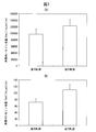
- FIG. 1A shows a comparison of the amount of free L-serine in the skin of the high TEWL group and the low TEWL group.
- FIG. 1B shows a comparison of the amount of free D-serine in the skin of the high TEWL group and the low TEWL group.
- FIG. 2 is a chromatogram obtained by detecting intracellular D-serine before and after applying differentiation-inducing stimulation in cultured keratinocytes.
- FIG. 3A is a graph showing that hydrolyzed oat protein solution, which is one of candidate drugs, increased the amount of free D-serine.
- FIG. 3B is a diagram showing that hydrolyzed oat protein solution, which is one of candidate drugs, increased the expression of serine racemase.
- FIG. 1A shows a comparison of the amount of free L-serine in the skin of the high TEWL group and the low TEWL group.
- FIG. 1B shows a comparison of the amount of free D-serine in
- FIG. 4 is a graph showing that the amount of free D-serine was increased by adding pyridoxal phosphate (PLP), which is one of candidate drugs, to cultured keratinocytes.
- PBP pyridoxal phosphate
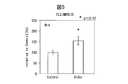
- FIG. 5 shows that D-serine increased filaggrin gene expression in a three-dimensional skin model.
- FIG. 6 is a diagram showing that D-serine increased the expression of the Cornulin gene in a three-dimensional skin model.
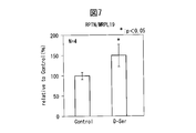
- FIG. 7 is a diagram showing that D-serine increased the expression of the repetin gene in a three-dimensional skin model.
- the present invention relates to a method for screening a drug for enhancing skin barrier function, which is a combination of one or more of the indicators of serine racemase expression level, activity, and D-serine level in keratinocytes.
- the skin barrier function is a function of retaining moisture of the skin or preventing foreign matter from entering from the outside, and the stratum corneum is responsible for such a function, particularly in the epidermis.
- the skin barrier function can be measured by transepidermal water evaporation (TEWL), and it is considered that the higher the water evaporation, the lower the skin barrier function.
- TEWL transepidermal water evaporation
- the enhancement of the skin barrier function can be achieved by, for example, increasing the number of stratum corneum layers, maturation of the keratinized outer membrane (cornified envelope) in the stratum corneum, and increasing intercellular lipids such as cholesterol and ceramide.
- the agent selected by the screening method of the present invention enhances the expression or activity of serine racemase by applying to the skin, or D -It is considered that the amount of serine can be increased, and as a result, effects such as an increase in the number of layers in the stratum corneum can be exhibited and the skin barrier function can be enhanced.
- the candidate drug used in the screening method of the present invention may be any compound, for example, a compound obtained from a recombinatorial library, or a mixture or extract.
- candidate drugs for example, food materials, cosmetic materials or pharmaceutical material compounds, mixtures or extract libraries can be used.
- a drug that has an action of enhancing skin barrier function can be applied to foods such as functional foods and nutritional foods, cosmetics, pharmaceuticals, or quasi drugs.
- Cosmetics can be applied to, for example, lotions, creams, emulsions, gels, cosmetics, ointments, packs, bathing agents, body soaps, shampoos, rinses, foundations, etc., but are intended to be limited to these. is not.
- cosmetics for sensitive skin such as lotions, creams, milky lotions, and cosmetics. If it is a pharmaceutical or a quasi-drug, it can be applied to various forms such as various ointments and creams for transdermal administration and forms for oral administration.
- the expression level or activity of serine racemase and / or the amount of D-serine is used as an index in keratinocytes.
- the expression level or activity of serine racemase and / or the amount of D-serine is taken into consideration. Therefore, when a candidate drug is added, the screening method of the present invention can be selected by selecting a drug having a skin barrier function-enhancing effect on the basis of determination of serine racemase expression level or activity and / or D-serine level. It corresponds to.
- Keratinocytes are cells that constitute about 95% of the epidermis.
- keratinocyte stem cells exist in the basal layer of the epidermis and move to the outer layer as they mature, forming a spiny layer, a granular layer, and a stratum corneum.
- the properties of keratinocytes change with maturity, cells become flat in the spinous layer and granule layer, lipids such as ceramide are released into the interstitial space, and enucleation occurs in the outermost stratum corneum.
- This series of maturation processes can be referred to as keratinocyte differentiation.
- Cells that have undergone enucleation lose their cell morphology, become stratified, form a stratum corneum, and finally peel off as dirt from the outermost layer.
- the cultured keratinocytes used in the present invention may be cells obtained from any of the basal layer, spiny layer, granule layer, and horny layer of the epidermis, and stem cells such as skin epithelial stem cells, pluripotent stem cells, For example, cells induced to differentiate from ES cells, iPS cells, EG cells and the like may be used.
- the cells obtained from the epidermis and cultured may be primary cultured cells, subcultured cells, or cancerized or immortalized cells.
- the cultured keratinocytes used in the present invention may be, for example, established keratinocytes derived from humans or animals. These keratinocyte cell lines can be obtained from public institutions and vendors.
- the culture may refer only to desired cells, may contain other cells, and may further be composed of cells and a medium. Therefore, the keratinocyte culture may refer only to keratinocytes, may contain keratinocytes and other cells, or may be composed of keratinocytes alone or keratinocytes and other cells and a medium. Examples of such cultures include monolayer cultures, mixed cultures, and three-dimensional cultures.
- the medium for cultivating keratinocytes may be any medium as long as it allows culturing of keratinocytes. For example, various media may be added to a medium of HuMedia-KB2 (Kurabo) or Keratinocyte-SFM (Invitrogen). However, it is not intended to be limited thereto.
- the culture conditions are usually cultured at 37 ° C. in the presence of 5% CO 2, but can be changed within a range where keratinocyte proliferation occurs.
- the medium is preferably enriched in L-serine.
- D-amino acid is not intentionally added to the culture medium, and the abundance of L-serine relative to D-serine in the medium is at least 1.1 times, more preferably 10 times, even more preferably. Is 50 times or more.
- KGM medium in which a HuMedia-KG growth additive set is added to HuMedia-KB2 (Kurabo)
- the abundance of L-serine relative to D-serine is 100 times or more.
- the keratinocyte culture is cultured after seeding, and about 50% confluent to confluent can be used for the experiment.
- the concentration is preferably 70% or more, more preferably 80% or more, from the viewpoint of inducing differentiation.
- the step of adding the candidate drug to the keratinocyte culture may be performed by replacing the medium of the keratinocyte culture with a medium containing the candidate drug.
- the drug may be added as it is or as a diluent.
- Serine racemase is a pyridoxal 5 ′ phosphate (PLP) -dependent racemase that is a vitamin B 6 coenzyme type. While not intending to be limited by theory, serine racemase can convert between L-serine and D-serine via the formation of an anionic intermediate by abstracting the ⁇ -hydrogen of serine. An enzyme that can be performed. In the conversion reaction between L-serine and D-serine, the enthalpy does not change before and after the reaction, and the reaction is driven by entropy. Therefore, in an equilibrium state, L-serine and D-serine are equivalent.
- PRP pyridoxal 5 ′ phosphate
- the racemase may be the same as the racemase whose expression was recognized in the brain according to Non-Patent Document 3, or a homolog or ortholog thereof. Therefore, it means that it can be amplified by the primer pair used in the same document and can be detected by the anti-racemase antibody described in the same document.
- the serine racemase can be appropriately specified by those skilled in the art from the website of a public organization such as NCBI according to the animal species.
- skin barrier function can be evaluated or a drug for enhancing skin barrier function can be screened using serine racemase activity in keratinocytes as an index.
- the activity of serine racemase may be based on the amount of D-serine produced by the conversion of L-serine contained in a normal medium in the absence of addition of a substance that is a substrate for the enzyme. Based on the total amount of D-amino acids. Further, by adding a substance other than serine which can be a substrate for serine racemase, the amount of product based on the conversion of the additive or the conversion efficiency may be measured.
- the enzyme activity can also be evaluated by measuring the increase in L-serine produced by the conversion.
- the enzyme activity can be evaluated by examining the conversion efficiency to the corresponding optical isomer. is there.
- skin barrier function can be evaluated or a drug for enhancing skin barrier function can be screened using the amount of D-serine in keratinocytes as an index.
- the amount of D-serine may be measured in a skin sample isolated from a subject, or in a keratinocyte culture, ie, in culture medium, cells, or both.
- the method for measuring the amount of D-serine can be performed using a known method, for example, HPLC method using an optical isomer separation column, gas chromatography method, electrophoresis method, enzymatic method.
- L-serine may be added to the keratinocyte culture in order to increase the amount of D-serine.
- serine racemase performs a conversion reaction between L-serine and D-serine, the enthalpy does not change before and after the reaction, and the reaction is driven by entropy.
- the amount of D-serine can be increased, making it easier to measure the amount of D-serine.
- skin barrier function can be evaluated or a skin barrier enhancement drug can be screened using the expression level of serine racemase as an index.
- the expression level of serine racemase can be determined by measuring the protein of serine racemase by an immunological technique using an anti-serine racemase antibody.
- an immunological technique for example, Western blotting, fluorescent immunostaining, or ELISA may be used.
- anti-serine racemase antibody a commercially available monoclonal antibody or polyclonal antibody may be used, or an antibody collected by immunizing a test animal such as a rabbit or mouse with a serine racemase protein or antigen peptide may be used.
- the expression level of serine racemase may be determined by determining the amount of serine racemase mRNA in the cell.
- the measurement of mRNA may be performed by any method, and may be measured based on, for example, a quantitative PCR technique.
- the quantitative PCR method is usually performed as follows, but is not intended to be limited thereto. MRNA is extracted from the collected cells, and cDNA is prepared using poly-T primer and reverse transcriptase. By using the Syber Green method or the fluorescent probe method on the prepared cDNA, the amount of mRNA contained in the cell can be quantified.
- the screening method of the present invention includes the following: Adding a candidate drug to keratinocytes; The step of measuring serine racemase expression level or activity and / or D-serine level in keratinocytes and the serine racemase expression level or activity and / or D-serine level determine the action of the candidate drug to enhance the skin barrier function A process may be included.
- the screening method of the present invention may further include a step of inducing differentiation of keratinocytes.
- the differentiation induction step is performed by applying differentiation induction stimulation to keratinocytes.
- differentiation-inducing stimulation By applying differentiation-inducing stimulation to keratinocytes, cell flattening, increase of intracellular fibers or granules, obscuration of cell gaps, stratification (especially in the case of monolayer cultures), and finally desorption It causes morphological changes such as the nucleus and changes in cells such as protein and protein changes such as activation of keratin 1, keratin 10, involucrin, and transglutamine sase (Nichishikai 90 (12), 1089- 1101, http://drmtl.org/data/090121089.pdf, see Drug News Perspect 2004, 17 (2) p117).
- the differentiation-inducing stimulus may be any stimulus as long as it can cause the differentiation of keratinocytes as described above.
- calcium ion stimulation for increasing the concentration of calcium ions phorbol-12-myristate-13- Stimulation with acetate (Hawley-Nelson P, Stanley Jr et al, Exp Cell Res, 37137: 155-167, 1982), physical stimulation such as voltage or gas phase exposure, stimulation by biological fluid may be used.
- a calcium salt or a diluted solution thereof may be added so that the final concentration of calcium ions is 0.1 mM to 10 mM, or prepared so as to include a final concentration of calcium ions in advance. It may be replaced with a modified medium.
- Examples of calcium salts used include calcium chloride, calcium bicarbonate, calcium carbonate, calcium nitrate, and calcium acetate.
- the upper limit of the final concentration of calcium ions is preferably 5 mM or less, more preferably 2 mM or less, from the viewpoint of not adversely affecting cell culture. From the viewpoint of promoting differentiation induction, the lower limit value of the concentration is preferably 0.5 mM or more, more preferably 1.0 mM or more.
- the differentiation induction process and the candidate drug addition process may be performed simultaneously, or the candidate drug addition process may be performed after the differentiation induction process, or vice versa, that is, the differentiation induction after the candidate drug addition process.
- a process may be performed.
- the timing of differentiation induction and addition of a candidate drug can be varied depending on the desired mechanism of action of the candidate drug.
- the adding step is preferably performed simultaneously with or before the stimulation step.
- the addition step is preferably performed at the same time as or after the stimulation step.
- the stimulation step is preferably performed after the addition step.
- a cell culture step of several minutes to several hours may be included between the addition step and the stimulation step.
- a cell culture step may be further included after the differentiation induction stimulus and the addition of the candidate drug and before the step of measuring the expression level or activity of serine racemase and / or the amount of D-serine.
- the period of this cell culture step can be arbitrarily selected according to the type and concentration of the differentiation-inducing agent used and the type of keratinocytes.
- the culture can be performed for 6 hours to 1 week.
- the lower limit of the culture period is preferably 12 hours or more, and more preferably 1 day or more.
- the upper limit of the culture period is not particularly specified because it differs depending on the culture method such as monolayer culture, multi-layer culture, three-dimensional culture, etc., but from the viewpoint of maintaining healthy culture conditions of cells In this case, it is preferably within one week, more preferably within three days.
- the screening method of the present invention may further include a damage imparting step.
- the damage imparting step the cultured keratinocytes may be damaged by physical or chemical stimulation, such as ultraviolet irradiation, addition of a radical source such as an oxidizing agent, addition of a surfactant, Substitution with an oligotrophic medium and the like can be mentioned. It is considered that the expression of serine racemase is reduced in the keratinocytes damaged by the damage imparting step. Therefore, in the screening method in which the damage imparting step is performed after the candidate agent addition step, if the decrease in the amount of racemase expression or racemase activity and / or the amount of D-serine is suppressed, the candidate agent may cause damage to the barrier function of the skin.
- the candidate drug when the decreased racemase expression level or racemase activity and / or D-serine level is increased in the screening method in which the candidate drug addition step is performed after the damage imparting step, the candidate drug is used to restore the skin barrier function. It can be an ameliorating or therapeutic agent.
- a candidate drug that increases the amount of racemase expression or racemase activity and / or the amount of D-serine can be said to be a skin barrier function enhancer.
- the drug screened by the screening method of the present invention is not limited to the extract derived from Lumpuyan or Shirobanaikouzorina, but the extract of the related species similarly has the effect of enhancing the skin barrier function. Therefore, as the serine racemase expression promoter, serine racemase activator or skin barrier function enhancer of the present invention, Zingiberaceae family Zingiber genus plant and Chrysanthemum (Compositae) family Elephamtopus genus plant. An extract is mentioned. Therefore, a cosmetic that exhibits an effect of enhancing the skin barrier function can be produced by blending these substances into the cosmetic.
- pyridoxal phosphate (PLP: active vitamin B 6 ) could be selected in keratinocytes.
- PBP active vitamin B 6
- pyridoxal phosphate can be referred to as a serine racemase activator or skin barrier function enhancer.
- the hydrolyzed oat protein used in the present invention is a protein hydrolyzate obtained from oat sativa.
- the protein of oats is extracted from, for example, seeds, leaves, roots, nuka, etc. Seeds are particularly preferred.
- the oats are preferably dried and crushed immediately after collection. Drying may be performed in the sun or using a commonly used dryer. Protein is hydrolyzed by acid or enzyme treatment during or after extraction.
- Owl is a plant cultivated for food in West Asia, Europe and North America.
- the extraction solvent used in the present invention is not particularly limited as long as it is usually used for extraction, and in particular, organic solvents such as alcohols such as methanol and ethanol, hydrous alcohols, acetone and ethyl acetate are used alone or in combination. Or extraction with water.
- organic solvents such as alcohols such as methanol and ethanol, hydrous alcohols, acetone and ethyl acetate are used alone or in combination. Or extraction with water.
- the blending amount of the hydrolyzed oat protein in the present invention can be arbitrarily selected by those skilled in the art within a range where the effect is exhibited.
- the blending amount is 0.00005 to 20.0 mass% as a dry product in the total amount of the external preparation.
- the upper limit of the amount is preferably 10% by mass or less, more preferably 5% by mass or less from the viewpoints of formulation and effects.
- the lower limit of the amount is preferably 0.0001% by mass or more, more preferably 0.0005% by mass or more, from the viewpoint of exerting a sufficient skin barrier function enhancing effect.
- Lumpuyan is a plant that grows especially on dry grasslands and pastures in Indonesia.
- the extract used in the present invention is obtained by immersing whole plant plants such as the above-mentioned plant leaves, stems including underground stems, fruits and the like together with an extraction solvent, and optionally heating and refluxing, followed by filtration and concentration.
- the extraction solvent used in the present invention is not particularly limited as long as it is usually used for extraction, and in particular, organic solvents such as alcohols such as methanol and ethanol, hydrous alcohols, acetone and ethyl acetate are used alone or in combination. Or extraction with water.
- organic solvents such as alcohols such as methanol and ethanol, hydrous alcohols, acetone and ethyl acetate are used alone or in combination. Or extraction with water.
- the blending amount of the extract of the genus Zingiberaceae belonging to the genus Zingiberaceae in the present invention can be arbitrarily selected by those skilled in the art within a range where the effect is exhibited.
- the blending amount is 0.00005 to 20.0 mass% as a dry product in the total amount of the external preparation.
- the upper limit of the amount is preferably 10% by mass or less, more preferably 5% by mass or less from the viewpoints of formulation and effects.
- the lower limit of the amount is preferably 0.0001% by mass or more, more preferably 0.0005% by mass or more, from the viewpoint of exerting a sufficient skin barrier function enhancing effect.
- Shirobanaikouzorina (scientific name: Elephantopus mollis) is preferable.
- Shirobanaikouzorina is native to Venezuela in South America and is a naturalized plant in tropical Asia.
- the extract of the genus Elephamtopus belonging to the family Compositae used in the present invention was obtained by immersing or heating and refluxing the above-mentioned plant leaves, stems, flowers, bark, seeds or fruits, whole plants, etc. with an extraction solvent. Thereafter, it is filtered and concentrated.
- the extraction solvent used in the present invention is not particularly limited as long as it is usually used for extraction, and in particular, organic solvents such as alcohols such as methanol and ethanol, hydrous alcohols, acetone and ethyl acetate are used alone or in combination. Can do.
- the amount of the extract of the plant belonging to the genus Elephamtopus belonging to the family Compositae in the present invention can be arbitrarily selected by those skilled in the art within a range where the effect is exhibited.
- the blending amount is 0.00005 to 20.0 mass% as a dry product in the total amount of the external preparation.
- the upper limit of the amount is preferably 10% by mass or less, more preferably 5% by mass or less from the viewpoints of formulation and effects.
- the lower limit of the amount is preferably 0.0001% by mass or more, more preferably 0.0005% by mass or more, from the viewpoint of exerting a sufficient skin barrier function enhancing effect.
- the pyridoxal phosphate used in the present invention is a substance used as a coenzyme for serine racemase.
- Pyridoxal phosphate is an active form of pyridoxal known as vitamin B 6 compound. While not intending to be limited by theory, pyridoxal phosphate can increase the activity of serine racemase as a coenzyme, and the resulting D-serine acts on keratinocytes to cause skin barrier It is considered that the expression of gene groups related to functions such as filaggrin, cornulin and repetin is increased, thereby enhancing the skin barrier function.
- Vitamin B 6 contains pyridoxal and pyridoxamine in addition to pyridoxal, and is known to be a component incorporated in cosmetics, but is a water-soluble vitamin and absorbs and stabilizes when transdermally administered. There may be a problem.
- vitamin B 6 derivatives with increased fat solubility for the purpose of improving percutaneous absorption are known.
- vitamin B 6 derivatives such as pyridoxine trishexyldecanoate, pyridoxine tripalmitate, pyridoxine dipalmitate, pyridoxine dicaprylate are also known. It included in the vitamin B 6 class.
- pyridoxine cyclic phosphate and pyridoxine hydrochloride are known for the purpose of improving stability.
- pyridoxal phosphate but also vitamin B 6 is used as a serine racemase activator or a skin function enhancer.
- the blending amount of these is, for example, 0.000001 to 10% by mass.
- the upper limit of the amount is preferably 10% by mass or less, more preferably 1% by mass or less from the viewpoint of formulation and effect.
- the lower limit of the amount is preferably 0.000001% by mass or more, more preferably 0.00001% by mass or more, from the viewpoint of exerting a sufficient skin barrier function enhancing effect.
- the measured serine racemase expression level or activity and / or D-serine level is set in advance. It can be determined using a table showing the relationship between the expression level or activity and / or the amount of D-serine and the barrier function enhancing action. Such a table shows, as a control, racemase expression level or activity and / or D-serine level, serine racemase expression level or activity, and / or D, measured by conducting the same experiment without containing a candidate drug.
- a threshold value is set in advance from comparison with the control, and the skin barrier function enhancing action of the candidate drug can be determined based on whether or not the threshold value is higher than the threshold value with a statistically significant difference.
- a person skilled in the art can appropriately set such a threshold according to the desired skin barrier function enhancing action.
- a threshold of magnification can be arbitrarily set for the expression level or activity of serine racemase and / or the amount of D-serine in the non-drug-added control.
- Values can also be set with statistically significant differences and trend differences.
- a statistically significant difference is when p ⁇ 0.05, and a tendency difference is when p ⁇ 0.1.
- the expression level or activity of racemase and / or the amount of D-serine was higher than the amount of racemase expression or activity measured by conducting an experiment in the same manner without containing a candidate drug.
- it may be determined to have a skin barrier function enhancing action, in which case it can be determined that the higher the expression level or activity, and / or the D-serine level, the skin barrier function enhancing action. .
- the present invention may relate to a method for evaluating, determining, or distinguishing skin barrier function using serine racemase expression level or activity and / or D-serine level as an index.
- evaluation, determination, or differentiation may be performed by measuring D-serine content, serine racemase activity or serine racemase expression level in a skin sample, for example, a stratum corneum sample obtained by tape stripping, from a subject.
- the evaluation, determination, or identification method of skin barrier function is usually performed by measuring TEWL using Vapomaeter, etc., but TEWL depends on the state of the skin, and the numerical value changes due to washing, friction, etc. In patients, there is not necessarily a correlation between numbers and skin health.
- the evaluation, determination, or differentiation method of the present invention can measure the skin barrier function at the cell level inherent in the skin by using serine racemase activity or expression level and / or D-serine level as an index. It is. Furthermore, since the evaluation method of the present invention determines the health related to the skin barrier function at the cellular level, it enables prediction of the future skin barrier function rather than the current skin barrier function. Evaluation, determination, or differentiation of the skin barrier function is determined using a chart or threshold value indicating the relationship between the pre-set serine racemase expression level or activity and / or the D-serine level and the barrier function enhancing action. be able to.
- Such a chart or threshold indicates the expression level or activity obtained in a skin sample collected and isolated from a group of subjects with healthy skin, a group of subjects with rough skin, or a group of subjects according to age. And / or based on the amount of D-serine.
- D-serine which plays an important role in barrier function formation
- cornified and lepetin which contribute to the formation of cornified cell (envelope) (CE)
- SErine racemase activation or expression promoters screened according to the present invention can increase the D-serine content in keratinocytes, where D-serine is present in keratinocytes.
- the skin barrier function is enhanced by enhancing the expression of proteins involved in the barrier function. Therefore, D-serine itself can also be used as a skin barrier function enhancer.
- D-serine can also be called an expression promoter for filaggrin gene, cornulin gene and repetin gene, and can also be called a keratinization promoter.
- Filaggrin is a basic protein rich in histidine, arginine, serine, glycine, glutamic acid, etc., and has a function of depositing and aggregating in keratin fibers. As a result, the keratin fibers inside the keratinocytes aggregate to flatten the entire cell, and change from the granule layer to the horny layer to keratinize. Filaggrin is expressed as a profilagrin precursor in granule cells of the epidermis, and is degraded into filaggrin by the action of proteolytic enzymes when the granule cells keratinize. The decrease in filaggrin is considered to be a major cause of ichthyosis and atopic dermatitis, and these symptoms show a marked decrease in skin barrier function.
- Cornulin and lepetin are proteins that constitute the peripheral zone (cornified cell envelope) that lines the cell membrane of keratinocytes.
- Cornulin and lepetin are genes that are present at the chromosomal location of 1q21, and are expressed in the outermost layer of the epidermis in the final stage of keratinization, as are other components of the peripheral zone, such as involucrin and loricrin. .
- the protein constituting the peripheral zone is involved in the barrier function of the skin together with keratin aggregated by the action of filaggrin.
- the skin barrier function-enhancing agent or serine racemase activator according to the screening method of the present invention may be blended in cosmetics or pharmaceuticals.
- Such cosmetics are used in subjects with reduced skin barrier function, for example, subjects with symptoms such as atopic dermatitis, ichthyosis, psoriasis, and rough skin.
- a pharmaceutical can be used as a pharmaceutical composition for treatment of atopic dermatitis, ichthyosis, psoriasis, etc.
- Example 1 Verification of the relationship between the D-serine content in the stratum corneum and the barrier function The subjects were 57 volunteers whose test sites were healthy. Waiting for 30 minutes under constant humidity conditions (room temperature 22 ° C., humidity 45%). Thereafter, the transdermal moisture transpiration (TEWL) was measured by Vapometer (Delfin). The measurement was performed three times, and the average value was used for the subsequent analysis. Next, of the stratum corneum at the position where the TEWL was measured, the amino acid was extracted in a 95% methanol aqueous solution from the second piece of which was peeled twice using an adhesive tape.
- TEWL transdermal moisture transpiration
- the optical isomers of the extracted amino acids were analyzed according to a previously reported method (Reference: Yurika Miyoshi et al., Journal of Chromatography B, 877 (2009) 2506-2512). The outline is shown below.
- the extracted amino acid was fluorescently derivatized with 4-fluoro-7-nitro-2,1,3-benzooxadiazole (NBD-F) in borate buffer (pH 8.0), and trifluoroacetic acid was used. After acid conditions, it was subjected to two-dimensional micro HPLC (Shiseido).
- a micromonolith ODS column (inner diameter 0.53 mm, total length 500, 750, 1000 mm, Shiseido), and in the second dimension, an optical resolution column with an inner diameter of 1.5 mm suitable for each amino acid A mobile phase was used. NBD-amino acid was detected by fluorescence emission at 530 nm (excitation wavelength: 470 nm). The results are shown in Table 1 and FIG.
- each subject was arranged in the order of the TEWL value.
- the top 17 (barrier function subgroup) and the bottom 17 (barrier function) The D-serine content in the stratum corneum of the upper functional group) was compared. Significant differences between the two groups were verified by non-paired Student's t-test. As a result, humans with high skin barriers showed significantly higher D-serine content.
- the comparison results for L-serine the L-serine content tended to be low in the barrier function subgroup and the L-serine content was high in the barrier function subgroup. There wasn't. Therefore, the high D-serine content in the upper barrier function group is not due to the amount of the precursor L-serine, but can be said to be the effect of racemase expression and activity.
- Example 2 Change in D-serine content accompanying induction of keratinocyte differentiation
- KGM medium test group
- KGM medium control group
- the extracted amino acid was fluorescently derivatized with 4-fluoro-7-nitro-2,1,3-benzoxadiazole (NBD-F) in borate buffer (pH 8.0) and trifluoroacetic acid. After acid conditions, it was subjected to two-dimensional micro HPLC (Shiseido). The first dimension uses a micromonolith ODS column (inner diameter 0.53 mm, total length 750 mm, Shiseido), and the second dimension uses an optical resolution column and mobile phase with an inner diameter of 1.5 mm suitable for each amino acid. It was. NBD-amino acids were detected by fluorescence emission at 530 nm (excitation wavelength: 470 nm). The results of the test group and the control group are shown in FIG.
- Example 3 Screening ⁇ br/> culture and drug-treated human normal keratinocytes cells of the candidate drug was seeded as cell density of 2.5 ⁇ 10 4 cells / well in 24-well multiplate, KGM medium (Kurabo Industries ) Was cultured under conditions of 37 ° C. and 5% CO 2 . Three days later, the medium was replaced with KGM medium containing 1.8 mM calcium chloride, 10 mM L-serine, and a diluted solution of a candidate drug, and further cultured for 2 days. Seven types of drugs were used as candidate drugs, and 0.5% hydrolyzed oat protein solution was used as one of them.
- the extracted amino acid was fluorescently derivatized with 4-fluoro-7-nitro-2,1,3-benzooxadiazole (NBD-F) in borate buffer (pH 8.0), and trifluoroacetic acid was used. After acid conditions, it was subjected to two-dimensional micro HPLC (Shiseido). The first dimension uses a micromonolith ODS column (inner diameter 0.53 mm, total length 750 mm, Shiseido), and the second dimension uses an optical resolution column and mobile phase with an inner diameter of 1.5 mm suitable for each amino acid. It was. NBD-amino acid was detected by fluorescence emission at 530 nm (excitation wavelength: 470 nm). The results are shown in Table 2 below and FIG. 3A.
- RT-PCR Quantitative PCR
- MagNA Pure LC MagNA Pure LC
- cDNA was synthesized using a kit of reverse transcriptase SuperScriptVILO (Invitrogen).
- real-time PCR using the fluorescent dye CyberGreen was performed with LightCycler (Roche Diagnostics).
- the PCR reaction was performed at 95 ° C. for 10 minutes, and then the amplification reaction at 95 ° C. for 15 seconds, 60 ° C. for 10 seconds, and 72 ° C. for 10 seconds was repeated 30 times.
- the gene expression level was calculated as a relative value from the number of cycles until a certain amount of PCR product was obtained.
- RT-PCR was also performed on the G3PDH (glyceraldehyde-3 -phosphate) gene as an internal standard in addition to the serine racemase gene, and the drug was evaluated by correcting the relative expression level of serine racemase with the relative expression level of G3PDH.
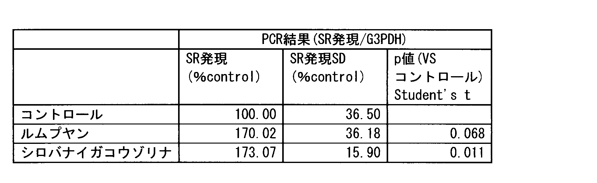
- the primers used in RT-PCR were selected from those described in Non-Patent Document 3 and Non-Patent Document 4. The results are shown in Table 2 below and FIG. 3B.
- hydrolyzed oat protein solution enhanced the expression of serine racemase in differentiation-induced keratinocytes. Furthermore, it was also shown that hydrolyzed oat protein solution increases the D-serine content in the induced keratinocytes. Based on these results, the hydrolyzed oat protein solution can be selected as an effective drug for enhancing the skin barrier function.
- Example 4 Screening ⁇ br/> culture and drug-treated human normal keratinocytes cells of the candidate drug was seeded as cell density of 2.5 ⁇ 10 4 cells / well in 24-well multiplate, KGM medium (Kurabo Industries ) Was cultured under conditions of 37 ° C. and 5% CO 2 . Three days later, the medium was replaced with KGM medium containing 1.8 mM calcium chloride, 10 mM L-serine, and a diluted solution of a candidate drug, and further cultured for 2 days. Twelve kinds of drugs were used as candidate drugs, among which 0.5% Lumpuyan extract and 0.5% Shirobanaikouzorina extract were included.
- RT-PCR Quantitative PCR
- MagNA Pure LC MagNA Pure LC
- cDNA was synthesized using a kit of reverse transcriptase SuperScriptVILO (Invitrogen).
- real-time PCR using the fluorescent dye CyberGreen was performed with LightCycler (Roche Diagnostics).
- the PCR reaction was performed at 95 ° C. for 10 minutes, and then the amplification reaction at 95 ° C. for 15 seconds, 60 ° C. for 10 seconds, and 72 ° C. for 10 seconds was repeated 30 times.
- the gene expression level was calculated as a relative value from the number of cycles until a certain amount of PCR product was obtained.
- RT-PCR was also performed on the G3PDH (glyceraldehyde-3 -phosphate) gene as an internal standard in addition to the serine racemase gene, and the drug was evaluated by correcting the relative expression level of serine racemase with the relative expression level of G3PDH.
- the primers used in RT-PCR were selected from those described in Non-Patent Document 3 and Non-Patent Document 4. The results are shown in Table 3 below.
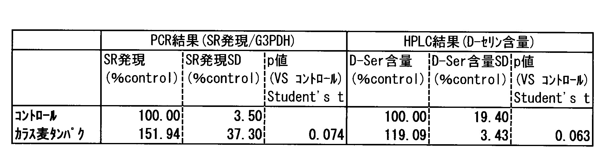
- Lumpuyan extract and Shirobanaikouzorina extract enhanced the expression of serine racemase in differentiation-induced keratinocytes. Based on this result, the Lumpuyan extract and Shirobanaikouzorina extract can be selected as effective drugs for enhancing the skin barrier function.
- Example 5 Screening for candidate drugs Cell culture and drug treatment Human normal keratinocytes are seeded in a 24-well multiplate so that the cell density is 2.5 ⁇ 10 4 cells / well, and the condition is 37 ° C. and 5% CO 2 in KGM medium (Kurabo) Cultured under. After 3 days, the medium was replaced with KGM medium containing 1.8 mM calcium chloride, cultured for 2 days, and then replaced with KGM medium containing 1.8 mM calcium chloride and a diluted solution of pyridoxal phosphate (PLP) as a candidate drug for another 2 days. Cultured.
- KGM medium KGM medium containing 1.8 mM calcium chloride
- Sample preparation for HPLC and analysis of free D-serine by HPLC Wash cells cultured in a medium supplemented with pyridoxal phosphate with PBS, add 500 ⁇ L of 5% trichloroacetic acid aqueous solution, and suspend and collect the cells for 30 minutes on ice After sonication, the supernatant was obtained through centrifugation. Amino acid was extracted by adding 20 times the amount of methanol to the supernatant. The optical isomers of the extracted amino acids were analyzed according to a previously reported method (Reference: Yurika Miyoshi et al. Journal of Chromatography B, 877 (2009) 2506-2512). The outline is shown below.
- the extracted amino acid was fluorescently derivatized with 4-fluoro-7-nitro-2,1,3-benzooxadiazole (NBD-F) in borate buffer (pH 8.0), and trifluoroacetic acid was used. After acid conditions, it was subjected to two-dimensional micro HPLC (Shiseido). The first dimension uses a micromonolith ODS column (inner diameter 0.53 mm, total length 750 mm, Shiseido), and the second dimension uses an optical resolution column and mobile phase with an inner diameter of 1.5 mm suitable for each amino acid. It was. NBD-amino acid was detected by fluorescence emission at 530 nm (excitation wavelength: 470 nm). The concentration results for D-serine are shown in FIG. 4 below.
- PLP which is a coenzyme for serine racemase, is a drug that can activate serine racemase, and can enhance the skin barrier function through the activation of serine racemase.
- Example 6 Effect of D-serine on the skin Three-dimensional culture of epidermal keratinocytes Human normal keratinocytes are seeded on a polycarbonate insert (Millipore) with a diameter of 12 mm and a pore size of 0.4 ⁇ m, and CnT-Prime and Epithelial Culture Medium (CELLnTEC Advanced Cell Systems AG) are placed inside and outside the insert. In addition, after culturing at 37 ° C. in a 5% CO 2 atmosphere for 3 days, it was replaced with Cnt-prime 3D Barrier medium (CELLnTEC Advanced Cell Systems AG) and cultured for 24 hours.
- Cnt-prime 3D Barrier medium CELLnTEC Advanced Cell Systems AG
- the medium in the insert was removed, and a medium containing D-Ser at a concentration of 100 ⁇ M was added to the outside of the insert, or in the control, Cnt-prime 3D Barrier medium without D-Ser was added to expose the cell surface to air. Cultured in a state. After exchanging the medium once every two days and culturing for 7 days, the membrane of the insert was cut out with a scalpel and immersed in Trizol reagent (Invitrogen) to extract total RNA.
- Trizol reagent Invitrogen
- RNA extracted using Trizol reagent was measured for RNA concentration using NanoDrop 1000 Spectrophotometer (Thermo Fisher Scientific), and then SuperScriptVILO (Invitrogen) was used to prepare cDNA.
- SuperScriptVILO Invitrogen
- cDNA was prepared as a template, Platinum SYBER Green qPCR SuperMix-UDG (Invitrogen) in StepOne Real-Time PCR System (Applied Bioscience), filaggrin (FLG), cornulin (CRNN), repetin (RPTN) gene,
- MRPL19 mitochondrial ribosomal protein L19
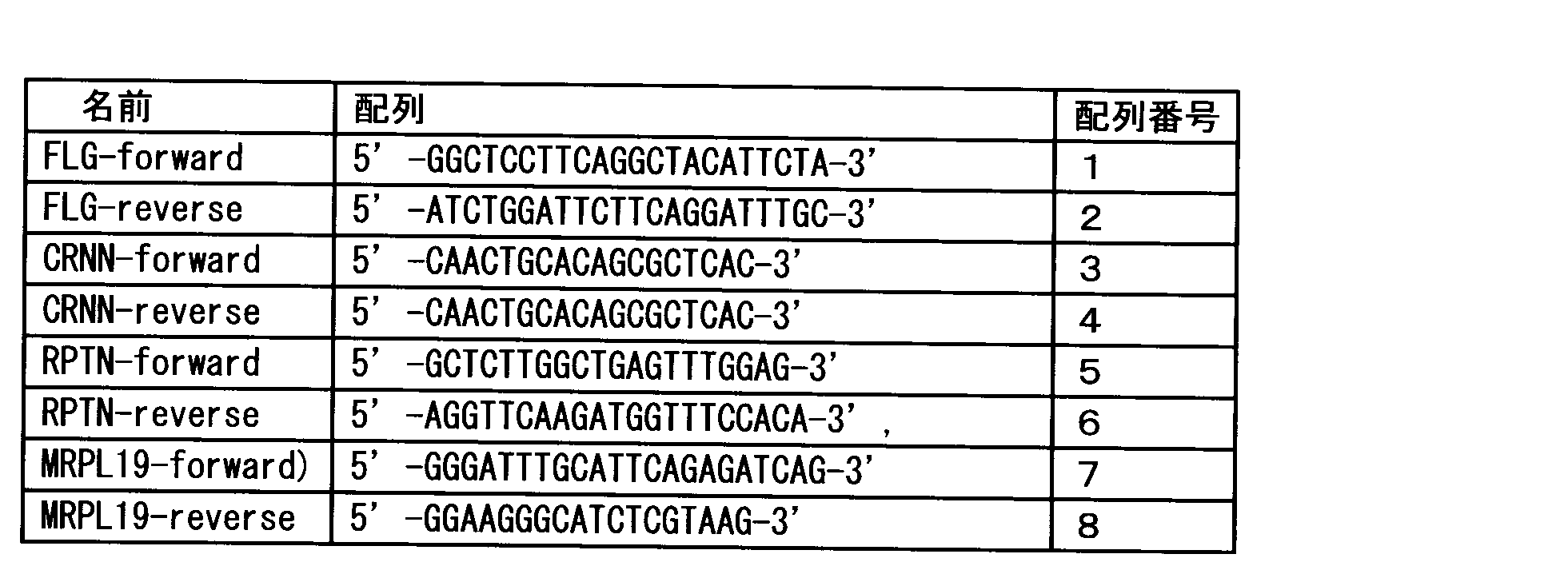
- the primers used are as follows: The relative expression level of each group of target genes was determined by correcting the expression level of the mitochondrial ribosomal protein L19 gene (MRPL19), which is an internal standard (FIGS. 5 to 7). In these genes, gene expression increased significantly with the addition of D-Ser.
- MRPL19 mitochondrial ribosomal protein L19 gene
Landscapes
- Life Sciences & Earth Sciences (AREA)
- Health & Medical Sciences (AREA)
- Engineering & Computer Science (AREA)
- Chemical & Material Sciences (AREA)
- Molecular Biology (AREA)
- Immunology (AREA)
- Biomedical Technology (AREA)
- Proteomics, Peptides & Aminoacids (AREA)
- Hematology (AREA)
- Physics & Mathematics (AREA)
- Analytical Chemistry (AREA)
- Urology & Nephrology (AREA)
- Biochemistry (AREA)
- General Health & Medical Sciences (AREA)
- Microbiology (AREA)
- Cell Biology (AREA)
- Bioinformatics & Cheminformatics (AREA)
- Organic Chemistry (AREA)
- Pathology (AREA)
- Biotechnology (AREA)
- Medicinal Chemistry (AREA)
- Biophysics (AREA)
- General Physics & Mathematics (AREA)
- Genetics & Genomics (AREA)
- Food Science & Technology (AREA)
- Wood Science & Technology (AREA)
- Zoology (AREA)
- Bioinformatics & Computational Biology (AREA)
- General Engineering & Computer Science (AREA)
- Toxicology (AREA)
- Tropical Medicine & Parasitology (AREA)
- Cosmetics (AREA)
- Measuring Or Testing Involving Enzymes Or Micro-Organisms (AREA)
Abstract
Description
[1] ケラチノサイトにおいて、セリンラセマーゼの活性若しくは発現量、及び/又はD-セリン量を指標とする、皮膚のバリア機能亢進薬剤のスクリーニング方法。
[2] 以下の:
ケラチノサイトに、候補薬剤を添加する工程、
ケラチノサイトにおいてセリンラセマーゼの活性若しくは発現量、及び/又はD-セリン量を測定する工程、
セリンラセマーゼ発現量若しくは活性、及び/又はD-セリン量から、候補薬剤の皮膚のバリア機能亢進作用を決定する工程
を含む、項目1に記載のスクリーニング方法。
[3] ケラチノサイトを分化誘導する工程をさらに含む、項目2に記載のスクリーニング方法。
[4] 前記スクリーニング方法が、分化誘導されたケラチノサイトにおいて行われる、項目1に記載のスクリーニング方法。
[5] セリンラセマーゼ発現量の測定が、セリンラセマーゼのmRNA又はタンパク質の量を測定することにより行われる、項目1~4のいずれか一項に記載のスクリーニング方法。
[6] セリンラセマーゼ活性の測定が、基質の変換効率の測定もしくはD-アミノ酸生成物の量を測定することによる、項目1~4のいずれか一項に記載のスクリーニング方法。
[7] 項目1~6に記載のスクリーニング方法によって得られた、加水分解カラス麦タンパク、ルムプヤン抽出物、シロバナイガコウゾリナ抽出物、ピリドキサールリン酸、及びビタミンB6類からなる群から選ばれる1又は複数の物質を有効成分とするセリンラセマーゼ活性化剤。
[8] ヒト皮膚から単離した皮膚サンプルからセリンラセマーゼ活性若しくは発現量及び/又はD-セリン量を指標とする、皮膚バリア機能を評価する方法。
[9] 以下の:
対象から単離された皮膚サンプルにおいて、セリンラセマーゼ活性若しくは発現量及び/又はD-セリン量を測定する工程、
セリンラセマーゼ活性若しくは発現量及び/又はD-セリン量から、対象の皮膚のバリア機能を決定する工程
を含む、項目8に記載の皮膚バリア機能を評価する方法。
[10] セリンラセマーゼ発現量の測定が、セリンラセマーゼのmRNA又はタンパク質の量を測定することにより行われる、項目8又は9に記載の皮膚バリア機能を評価する方法。
[11] セリンラセマーゼ活性の測定が、セリンラセマーゼによる、基質の変換効率の測定もしくはD-アミノ酸生成物の量を測定することによる、項目8又は9に記載の方法。
[12] 項目1~6に記載のスクリーニング方法によって得られた、加水分解カラス麦タンパク、ルムプヤン抽出物、シロバナイガコウゾリナ抽出物、ピリドキサールリン酸、及びビタミンB6類からなる群から選ばれる1又は複数の物質を含む皮膚のバリア機能亢進剤。
[13] 皮膚バリア機能が低下した対象に対して、加水分解カラス麦タンパク、ルムプヤン抽出物、シロバナイガコウゾリナ抽出物、ピリドキサールリン酸、及びビタミンB6類からなる群から選ばれる1又は複数の物質を投与することを含む、皮膚バリア機能亢進方法。
[14] セリンラセマーゼ活性が低下した対象に対して、加水分解カラス麦タンパク、ルムプヤン抽出物、シロバナイガコウゾリナ抽出物、ピリドキサールリン酸、及びビタミンB6類からなる群から選ばれる1又は複数の物質を投与することを含む、セリンラセマーゼ活性化方法。
[15] セリンラセマーゼ活性化剤の製造のための、加水分解カラス麦タンパク、ルムプヤン抽出物、シロバナイガコウゾリナ抽出物、ピリドキサールリン酸、及びビタミンB6類の使用。
[16] 皮膚バリア機能亢進剤の製造のための、加水分解カラス麦タンパク、ルムプヤン抽出物、シロバナイガコウゾリナ抽出物、ピリドキサールリン酸、及びビタミンB6類の使用。
ケラチノサイトに、候補薬剤を添加する工程、
ケラチノサイトにおいてセリンラセマーゼ発現量若しくは活性、及び/又はD-セリン量を測定する工程、及び
セリンラセマーゼ発現量若しくは活性、及び/又はD-セリン量から、候補薬剤の皮膚のバリア機能亢進作用を決定する工程
を含んでもよい。本発明のスクリーニング方法は、さらに、ケラチノサイトを分化誘導させる工程をさらに含んでもよい。
被験者は被験箇所が健常なボランティア57名で、被験部位を定法で洗浄した後に洗浄で濡れた被験部位を拭き、恒温・恒湿条件(室温22℃, 湿度45%)で30分間待機した。その後、経皮水分蒸散量(TEWL)をVapometer(Delfin社)により測定した。測定は3回行い、その平均値をその後の解析に用いた。次に、TEWLを測定した位置の角層を、粘着テープを用いて2回剥離したうち2枚目を、95% メタノール水溶液中でアミノ酸を抽出した。抽出したアミノ酸の光学異性体は既報の方法に則り分析を行なった(参考文献:Yurika MiyoshiらJournal of Chromatography B, 877 (2009) 2506-2512)。概要を以下に示す。抽出したアミノ酸をホウ酸塩緩衝液(pH 8.0)中で4-フルオロ-7-ニトロ-2,1,3-ベンゾオキサジアゾール (NBD-F)により蛍光誘導体化し、トリフルオロ酢酸にて酸性条件とした後、二次元ミクロHPLC(資生堂)に供した。一次元目にはミクロモノリスODSカラム(内径0.53 mm、全長500、750、1000 mm、資生堂)、二次元目の光学分割には各アミノ酸に適した内径1.5 mmの光学分割カラムと移動相を用いた。NBD-アミノ酸は530 nmの蛍光発光(励起波長470 nm)により検出した。結果を表1及び図1に示す。

細胞の培養と薬剤処理
ヒト正常ケラチノサイトを24ウェルマルチプレートに細胞密度が2.5×10⁴細胞/ウェルになるように播種し、KGM培地(クラボウ)中、37℃、5%CO2の条件下で培養した。3日後に、1.8mM 塩化カルシウム及び10mM L-セリンを添加したKGM培地(試験群)または10mM L-セリンのみを添加したKGM培地(対照群)に交換し、さらに2日間培養した。
細胞をPBSで洗浄後、5%トリクロロ酢酸水溶液を500μL加えて細胞をよく懸濁して回収し、氷上で30分間超音波処理したのちに遠心分離を経て上清を得た。この上清に対して20倍量のメタノールを加えてアミノ酸を抽出した。抽出したアミノ酸の光学異性体は既報の方法に則り分析を行なった(参考文献:Yurika MiyoshiらJournal of Chromatography B, 877 (2009) 2506-2512)。概要を以下に示す。抽出したアミノ酸をホウ酸塩緩衝液(pH 8.0)中で4-フルオロ-7-ニトロ-2,1,3-ベンゾオキサジアゾール(NBD-F)により蛍光誘導体化し、トリフルオロ酢酸にて酸性条件とした後、二次元ミクロHPLC(資生堂)に供した。一次元目にはミクロモノリスODSカラム(内径0.53 mm、全長750 mm、資生堂)、二次元目の光学分割には各アミノ酸に適した内径1.5 mmの光学分割カラムと移動相を用いた。NBD-アミノ酸は530 nmの蛍光発光(励起波長470 nm)により検出した。試験群と対照群の結果を図2に示す。
細胞の培養と薬剤処理
ヒト正常ケラチノサイトを24ウェルマルチプレートに細胞密度が2.5×10⁴細胞/ウェルになるように播種し、KGM培地(クラボウ)中、37℃、5%CO2の条件下で培養した。3日後に1.8mM 塩化カルシウム、10mM L-セリンおよび候補薬剤の希釈液を含むKGM培地に交換し、さらに2日間培養した。候補薬剤として、7種類の薬剤を用い、そのうちの1つとして0.5%加水分解カラス麦タンパク液を用いた。
細胞をPBSで洗浄後、5%トリクロロ酢酸水溶液を500μL加えて細胞をよく懸濁して回収し、氷上で30分間超音波処理したのちに遠心分離を経て上清を得た。この上清に対して20倍量のメタノールを加えてアミノ酸を抽出した。抽出したアミノ酸の光学異性体は既報の方法に則り分析を行なった(参考文献:Yurika MiyoshiらJournal of Chromatography B, 877 (2009) 2506-2512)。概要を以下に示す。抽出したアミノ酸をホウ酸塩緩衝液(pH 8.0)中で4-フルオロ-7-ニトロ-2,1,3-ベンゾオキサジアゾール(NBD-F)により蛍光誘導体化し、トリフルオロ酢酸にて酸性条件とした後、二次元ミクロHPLC(資生堂)に供した。一次元目にはミクロモノリスODSカラム(内径0.53 mm、全長750 mm、資生堂)、二次元目の光学分割には各アミノ酸に適した内径1.5 mmの光学分割カラムと移動相を用いた。NBD-アミノ酸は530 nmの蛍光発光(励起波長470 nm)により検出した。この結果を、下記の表2及び図3Aに示す。
細胞をPBSで洗浄後、MagNA Pure LC(ロシュ・ダイアグノスティックス)を用いて細胞からmRNAを抽出した。次に逆転写酵素SuperScriptVILO(インビトロジェン)のキットを用いてcDNAを合成した。このcDNAを鋳型にLightCycler(ロシュ・ダイアグノスティックス)で蛍光色素CyberGreenを用いたリアルタイムPCRを行った。PCR反応は95℃、10分間の酵素活性化ののち、95℃15秒・60℃10秒・72℃10秒の増幅反応を30回繰り返した。遺伝子発現量は、一定量のPCR産物が得られるまでのサイクル数から相対値を算出した。RT-PCRはセリンラセマーゼ遺伝子の他に内部標準としてG3PDH(glyceraldehyde-3-phosphate)遺伝子についても行い、セリンラセマーゼの相対発現量をG3PDHの相対発現量で補正することによって薬剤の評価を行った。RT-PCRにおいて用いたプライマーは、非特許文献3および非特許文献4に記載のものを選択して用いた。この結果を、下記の表2及び図3Bに示す。
細胞の培養と薬剤処理
ヒト正常ケラチノサイトを24ウェルマルチプレートに細胞密度が2.5×10⁴細胞/ウェルになるように播種し、KGM培地(クラボウ)中、37℃、5%CO2の条件下で培養した。3日後に1.8mM 塩化カルシウム、10mM L-セリンおよび候補薬剤の希釈液を含むKGM培地に交換し、さらに2日間培養した。候補薬剤として、12種類の薬剤を用い、その中には、0.5%ルムプヤン抽出物及び0.5%シロバナイガコウゾリナ抽出物が含まれていた。
細胞をPBSで洗浄後、MagNA Pure LC(ロシュ・ダイアグノスティックス)を用いて細胞からmRNAを抽出した。次に逆転写酵素SuperScriptVILO(インビトロジェン)のキットを用いてcDNAを合成した。このcDNAを鋳型にLightCycler(ロシュ・ダイアグノスティックス)で蛍光色素CyberGreenを用いたリアルタイムPCRを行った。PCR反応は95℃、10分間の酵素活性化ののち、95℃15秒・60℃10秒・72℃10秒の増幅反応を30回繰り返した。遺伝子発現量は、一定量のPCR産物が得られるまでのサイクル数から相対値を算出した。RT-PCRはセリンラセマーゼ遺伝子の他に内部標準としてG3PDH(glyceraldehyde-3-phosphate)遺伝子についても行い、セリンラセマーゼの相対発現量をG3PDHの相対発現量で補正することによって薬剤の評価を行った。RT-PCRにおいて用いたプライマーは、非特許文献3および非特許文献4に記載のものを選択して用いた。この結果を、下記の表3に示す。
細胞の培養と薬剤処理
ヒト正常ケラチノサイトを24ウェルマルチプレートに細胞密度が2.5×10⁴細胞/ウェルになるように播種し、KGM培地(クラボウ)中、37℃、5%CO2の条件下で培養した。3日後に1.8mM塩化カルシウムを含むKGM培地に交換して2日間培養後、1.8mM 塩化カルシウムおよび候補薬剤としてピリドキサールリン酸(PLP)の希釈液を含むKGM培地に交換してさらに2日間培養した。
ピリドキサールリン酸添加培地で培養した細胞をPBSで洗浄後、5%トリクロロ酢酸水溶液を500μL加えて細胞をよく懸濁して回収し、氷上で30分間超音波処理したのちに遠心分離を経て上清を得た。この上清に対して20倍量のメタノールを加えてアミノ酸を抽出した。抽出したアミノ酸の光学異性体は既報の方法に則り分析を行なった(参考文献:Yurika MiyoshiらJournal of Chromatography B, 877 (2009) 2506-2512)。概要を以下に示す。抽出したアミノ酸をホウ酸塩緩衝液(pH 8.0)中で4-フルオロ-7-ニトロ-2,1,3-ベンゾオキサジアゾール(NBD-F)により蛍光誘導体化し、トリフルオロ酢酸にて酸性条件とした後、二次元ミクロHPLC(資生堂)に供した。一次元目にはミクロモノリスODSカラム(内径0.53 mm、全長750 mm、資生堂)、二次元目の光学分割には各アミノ酸に適した内径1.5 mmの光学分割カラムと移動相を用いた。NBD-アミノ酸は530nmの蛍光発光(励起波長470nm)により検出した。D-セリンについての濃度の結果を、下記の図4に示す。セリンラセマーゼの補酵素であるPLPは、セリンラセマーゼを活性化することができる薬剤であり、セリンラセマーゼの活性化を介して、皮膚バリア機能の亢進することができる。
表皮角化細胞の三次元培養
ヒト正常ケラチノサイトを径12mm、孔径0.4μmのポリカーボネートインサート(Millipore社)に播種し、インサートの内外にCnT-Prime, Epithelial Culture Medium(CELLnTEC Advanced Cell Systems AG社)を加えて、37℃、5%CO2雰囲気下で3日間培養後、Cnt-prime 3D Barrier medium(CELLnTEC Advanced Cell Systems AG)に置換して24時間培養した。その後、インサート内の培地を除き、インサート外にはD-Serを100μMの濃度で含む培地、又は対照ではD-Ser無添加のCnt-prime 3D Barrier mediumを加えて、細胞表面を空気に暴露した状態で培養した。2日に1回培地交換を行って7日間培養した後、インサートのメンブレンをメスで切り出してTrizol reagent(Invitrogen社)に浸漬して総RNAを抽出した。
Trizol reagent (Invitrogen社)を用いて抽出された総RNAをについて、NanoDrop 1000 Spectrophotometer (Thermo Fisher Scientific社)を用いてRNA濃度を測定した後、SuperScriptVILO(Invitrogen社)を用いてcDNAを作製した。このcDNAを鋳型としてStepOne Real-Time PCR System(Applied Bioscience社)においてPlatinum SYBER Green qPCR SuperMix-UDG(Invitrogen社)を用いて、フィラグリン(FLG)、コーニュリン(CRNN)、レペチン(repetin:RPTN)遺伝子、並びに内部標準遺伝子としてミトコンドリアリボソームタンパク質L19(MRPL19)の遺伝子の相対発現量を測定した。使用したプライマーは、以下の通りである:
Claims (12)
- ケラチノサイトにおいて、セリンラセマーゼの活性若しくは発現量、及び/又はD-セリン量を指標とする、皮膚のバリア機能亢進薬剤のスクリーニング方法。
- 以下の:
ケラチノサイトに、候補薬剤を添加する工程、
ケラチノサイトにおいてセリンラセマーゼの活性若しくは発現量、及び/又はD-セリン量を測定する工程、
セリンラセマーゼ発現量若しくは活性、及び/又はD-セリン量から、候補薬剤の皮膚のバリア機能亢進作用を決定する工程
を含む、請求項1に記載のスクリーニング方法。 - ケラチノサイトを分化誘導する工程をさらに含む、請求項2に記載のスクリーニング方法。
- 前記スクリーニング方法が、分化誘導されたケラチノサイトにおいて行われる、請求項1に記載のスクリーニング方法。
- セリンラセマーゼ発現量の測定が、セリンラセマーゼのmRNA又はタンパク質の量を測定することにより行われる、請求項1~4のいずれか一項に記載のスクリーニング方法。
- セリンラセマーゼ活性の測定が、セリンラセマーゼによる、基質の変換効率の測定もしくはD-アミノ酸生成物の量を測定することによる、請求項1~4のいずれか一項に記載のスクリーニング方法。
- 請求項1~6に記載のスクリーニング方法によって得られた、加水分解カラス麦タンパク、ルムプヤン抽出物、シロバナイガコウゾリナ抽出物、ピリドキサールリン酸、及びビタミンB6類からなる群から選ばれる1又は複数の物質を有効成分とするセリンラセマーゼ活性化剤。
- ヒト皮膚から単離した皮膚サンプルからセリンラセマーゼ活性若しくは発現量及び/又はD-セリン量を指標とする、皮膚バリア機能を評価する方法。
- 以下の:
対象から単離された皮膚サンプルにおいて、セリンラセマーゼ活性若しくは発現量及び/又はD-セリン量を測定する工程、
セリンラセマーゼ活性若しくは発現量及び/又はD-セリン量から、対象の皮膚のバリア機能を決定する工程
を含む、請求項8に記載の皮膚バリア機能を評価する方法。 - セリンラセマーゼ発現量の測定が、セリンラセマーゼのmRNA又はタンパク質の量を測定することにより行われる、請求項8又は9に記載の皮膚バリア機能を評価する方法。
- セリンラセマーゼ活性の測定が、セリンラセマーゼによる、基質の変換効率の測定もしくはD-アミノ酸生成物の量を測定することによる、請求項8又は9に記載の皮膚バリア機能を評価する方法。
- 請求項1~6に記載のスクリーニング方法によって得られた、加水分解カラス麦タンパク、ルムプヤン抽出物、シロバナイガコウゾリナ抽出物、ピリドキサールリン酸、及びビタミンB6類からなる群から選ばれる1又は複数の物質を含む皮膚のバリア機能亢進剤。
Priority Applications (4)
| Application Number | Priority Date | Filing Date | Title |
|---|---|---|---|
| US15/123,274 US10337064B2 (en) | 2014-03-05 | 2015-03-05 | Method for screening agents promoting skin barrier function and method for evaluating skin barrier function taking epidermal serine racemase and/or D-serine level as indicator |
| KR1020167024424A KR20160127751A (ko) | 2014-03-05 | 2015-03-05 | 표피 세린 라세마아제 및/또는 d-세린 양을 지표로 하는, 피부 배리어 기능 항진 약제의 스크리닝 방법 및 피부 배리어 기능 평가 방법 |
| US16/412,993 US10907208B2 (en) | 2014-03-05 | 2019-05-15 | Method for screening agents promoting skin barrier function and method for evaluating skin barrier function taking epidermal serine racemase and/or D-serine level as indicator |
| US17/130,886 US20210189487A1 (en) | 2014-03-05 | 2020-12-22 | Method for screening agents promoting skin barrier function and method for evaluating skin barrier function taking epidermal serine racemase and/or d-serine level as indicator |
Applications Claiming Priority (6)
| Application Number | Priority Date | Filing Date | Title |
|---|---|---|---|
| PCT/JP2014/055680 WO2015132917A1 (ja) | 2014-03-05 | 2014-03-05 | 表皮セリンラセマーゼ及び/又はd-セリン量を指標とした、皮膚のバリア機能亢進薬剤のスクリーニング方法並びに皮膚バリア機能評価方法 |
| JP2014043073 | 2014-03-05 | ||
| JP2014-043073 | 2014-03-05 | ||
| JPPCT/JP2014/055680 | 2014-03-05 | ||
| US14/477,213 US20160069867A1 (en) | 2014-09-04 | 2014-09-04 | Method for screening skin barrier function enhancing agent based on epidermal serine racemase and/or d-serine level, and skin barrier function evaluation method |
| US14/477,213 | 2014-09-04 |
Related Parent Applications (1)
| Application Number | Title | Priority Date | Filing Date |
|---|---|---|---|
| PCT/JP2014/055680 Continuation WO2015132917A1 (ja) | 2014-03-05 | 2014-03-05 | 表皮セリンラセマーゼ及び/又はd-セリン量を指標とした、皮膚のバリア機能亢進薬剤のスクリーニング方法並びに皮膚バリア機能評価方法 |
Related Child Applications (3)
| Application Number | Title | Priority Date | Filing Date |
|---|---|---|---|
| US15/123,274 A-371-Of-International US10337064B2 (en) | 2014-03-05 | 2015-03-05 | Method for screening agents promoting skin barrier function and method for evaluating skin barrier function taking epidermal serine racemase and/or D-serine level as indicator |
| US16/412,993 Division US10907208B2 (en) | 2014-03-05 | 2019-05-15 | Method for screening agents promoting skin barrier function and method for evaluating skin barrier function taking epidermal serine racemase and/or D-serine level as indicator |
| US16/412,993 Continuation US10907208B2 (en) | 2014-03-05 | 2019-05-15 | Method for screening agents promoting skin barrier function and method for evaluating skin barrier function taking epidermal serine racemase and/or D-serine level as indicator |
Publications (1)
| Publication Number | Publication Date |
|---|---|
| WO2015133591A1 true WO2015133591A1 (ja) | 2015-09-11 |
Family
ID=54055391
Family Applications (1)
| Application Number | Title | Priority Date | Filing Date |
|---|---|---|---|
| PCT/JP2015/056570 Ceased WO2015133591A1 (ja) | 2014-03-05 | 2015-03-05 | 表皮セリンラセマーゼ及び/又はd-セリン量を指標とした、皮膚のバリア機能亢進薬剤のスクリーニング方法並びに皮膚バリア機能評価方法 |
Country Status (2)
| Country | Link |
|---|---|
| KR (1) | KR20160127751A (ja) |
| WO (1) | WO2015133591A1 (ja) |
Citations (1)
| Publication number | Priority date | Publication date | Assignee | Title |
|---|---|---|---|---|
| WO2011030903A1 (ja) * | 2009-09-14 | 2011-03-17 | 株式会社資生堂 | 紫外線障害軽減組成物 |
Family Cites Families (1)
| Publication number | Priority date | Publication date | Assignee | Title |
|---|---|---|---|---|
| JP2012092032A (ja) | 2010-10-26 | 2012-05-17 | Unitika Ltd | セラミド産生促進剤 |
-
2015
- 2015-03-05 WO PCT/JP2015/056570 patent/WO2015133591A1/ja not_active Ceased
- 2015-03-05 KR KR1020167024424A patent/KR20160127751A/ko not_active Ceased
Patent Citations (1)
| Publication number | Priority date | Publication date | Assignee | Title |
|---|---|---|---|---|
| WO2011030903A1 (ja) * | 2009-09-14 | 2011-03-17 | 株式会社資生堂 | 紫外線障害軽減組成物 |
Non-Patent Citations (3)
| Title |
|---|
| INOUE R . ET AL.: "Localization of Serine Racemase and Its Role in the Skin", JOURNAL OF INVESTIGATIVE DERMATOLOGY, vol. 134, 20 February 2014 (2014-02-20), pages 1618 - 1626 * |
| YOSUKE TOJO: "Free D-amino acids in human and foods as physiologically active molecules for skin", AMINO SAN KENKYU, vol. 7, no. 2, February 2014 (2014-02-01), pages 91 - 96 * |
| YOSUKE TOJO: "Special interview at the Research Front : Overlooked health functions of D-amino acids : Challenge its application in the skin science", FOOD STYLE, vol. 17, no. 8, pages 8 - 11 * |
Also Published As
| Publication number | Publication date |
|---|---|
| KR20160127751A (ko) | 2016-11-04 |
Similar Documents
| Publication | Publication Date | Title |
|---|---|---|
| JP6934925B2 (ja) | セリンラセマーゼ活性化剤、及び皮膚のバリア機能亢進剤 | |
| EP3391046B1 (fr) | Procedes d'evaluation des effets de la deshydratation sur la peau d'enfant | |
| JP5235439B2 (ja) | HMG−CoA還元酵素産生促進剤 | |
| Park et al. | Compound K improves skin barrier function by increasing SPINK5 expression | |
| Konstantinou et al. | Mechanisms of senescence and anti-senescence strategies in the skin | |
| EP3475697B1 (fr) | Modèles de la dermatite atopique juvénile | |
| WO2014170495A2 (fr) | Utilisation de biomarqueurs de la barrière pour l'évaluation de l'efficacité d'actifs | |
| WO2023166269A1 (fr) | Modele de peau reconstituee | |
| EP3642326A1 (fr) | Modeles de peau sensible reconstituee | |
| US10907208B2 (en) | Method for screening agents promoting skin barrier function and method for evaluating skin barrier function taking epidermal serine racemase and/or D-serine level as indicator | |
| EP3049809B1 (fr) | Procédés d'évaluation des effets délétères des uv sur la peau d'enfant | |
| JP7116589B2 (ja) | Discoidin Domain Receptor2(DDR2)産生促進用組成物 | |
| WO2012144080A1 (ja) | 皮膚バリア機能改善剤 | |
| Hyun et al. | Upcycling fish byproducts for skin health: An enzymatic approach to sustainable nutricosmetics | |
| WO2015133591A1 (ja) | 表皮セリンラセマーゼ及び/又はd-セリン量を指標とした、皮膚のバリア機能亢進薬剤のスクリーニング方法並びに皮膚バリア機能評価方法 | |
| WO2015132917A1 (ja) | 表皮セリンラセマーゼ及び/又はd-セリン量を指標とした、皮膚のバリア機能亢進薬剤のスクリーニング方法並びに皮膚バリア機能評価方法 | |
| Bachelor et al. | Transcriptional profiling of epidermal barrier formation in vitro | |
| TWI666444B (zh) | 以表皮絲胺酸消旋酶及/或d-絲胺酸量為指標之皮膚之障壁機能促進藥劑之篩選方法及皮膚障壁機能評價方法 | |
| Baumann et al. | Nutritional supplementation of an apple callus extract to target epidermal aging | |
| KR20160028736A (ko) | 표피 세린 라세마아제 및/또는 d-세린 양을 지표로 하는, 피부 배리어 기능 항진 약제의 스크리닝 방법 및 피부 배리어 기능 평가 방법 | |
| Gattlen et al. | Use of a novel microbiome modulator improves anticancer immunity in a murine model of malignant pleural mesothelioma | |
| JP2014028787A (ja) | スクリ−ニング方法 | |
| Kahsay | Formulation and characterization of colloidal formulations loaded with free amino acids for dermal delivery | |
| JP2015178469A (ja) | Fatp4産生促進剤 |
Legal Events
| Date | Code | Title | Description |
|---|---|---|---|
| 121 | Ep: the epo has been informed by wipo that ep was designated in this application |
Ref document number: 15758788 Country of ref document: EP Kind code of ref document: A1 |
|
| ENP | Entry into the national phase |
Ref document number: 20167024424 Country of ref document: KR Kind code of ref document: A |
|
| WWE | Wipo information: entry into national phase |
Ref document number: 15123274 Country of ref document: US |
|
| NENP | Non-entry into the national phase |
Ref country code: DE |
|
| 122 | Ep: pct application non-entry in european phase |
Ref document number: 15758788 Country of ref document: EP Kind code of ref document: A1 |