WO2015098904A1 - 放射線治療用スペーサー - Google Patents
放射線治療用スペーサー Download PDFInfo
- Publication number
- WO2015098904A1 WO2015098904A1 PCT/JP2014/084029 JP2014084029W WO2015098904A1 WO 2015098904 A1 WO2015098904 A1 WO 2015098904A1 JP 2014084029 W JP2014084029 W JP 2014084029W WO 2015098904 A1 WO2015098904 A1 WO 2015098904A1
- Authority
- WO
- WIPO (PCT)
- Prior art keywords
- spacer
- radiotherapy
- test
- fiber assembly
- flexibility
- Prior art date
- Legal status (The legal status is an assumption and is not a legal conclusion. Google has not performed a legal analysis and makes no representation as to the accuracy of the status listed.)
- Ceased
Links
Classifications
-
- A—HUMAN NECESSITIES
- A61—MEDICAL OR VETERINARY SCIENCE; HYGIENE
- A61N—ELECTROTHERAPY; MAGNETOTHERAPY; RADIATION THERAPY; ULTRASOUND THERAPY
- A61N5/00—Radiation therapy
- A61N5/10—X-ray therapy; Gamma-ray therapy; Particle-irradiation therapy
-
- A—HUMAN NECESSITIES
- A61—MEDICAL OR VETERINARY SCIENCE; HYGIENE
- A61N—ELECTROTHERAPY; MAGNETOTHERAPY; RADIATION THERAPY; ULTRASOUND THERAPY
- A61N5/00—Radiation therapy
- A61N5/10—X-ray therapy; Gamma-ray therapy; Particle-irradiation therapy
- A61N5/1048—Monitoring, verifying, controlling systems and methods
- A61N5/1049—Monitoring, verifying, controlling systems and methods for verifying the position of the patient with respect to the radiation beam
-
- A—HUMAN NECESSITIES
- A61—MEDICAL OR VETERINARY SCIENCE; HYGIENE
- A61B—DIAGNOSIS; SURGERY; IDENTIFICATION
- A61B90/00—Instruments, implements or accessories specially adapted for surgery or diagnosis and not covered by any of the groups A61B1/00 - A61B50/00, e.g. for luxation treatment or for protecting wound edges
- A61B90/04—Protection of tissue around surgical sites against effects of non-mechanical surgery, e.g. laser surgery
-
- A—HUMAN NECESSITIES
- A61—MEDICAL OR VETERINARY SCIENCE; HYGIENE
- A61L—METHODS OR APPARATUS FOR STERILISING MATERIALS OR OBJECTS IN GENERAL; DISINFECTION, STERILISATION OR DEODORISATION OF AIR; CHEMICAL ASPECTS OF BANDAGES, DRESSINGS, ABSORBENT PADS OR SURGICAL ARTICLES; MATERIALS FOR BANDAGES, DRESSINGS, ABSORBENT PADS OR SURGICAL ARTICLES
- A61L31/00—Materials for other surgical articles, e.g. stents, stent-grafts, shunts, surgical drapes, guide wires, materials for adhesion prevention, occluding devices, surgical gloves, tissue fixation devices
- A61L31/04—Macromolecular materials
- A61L31/06—Macromolecular materials obtained otherwise than by reactions only involving carbon-to-carbon unsaturated bonds
-
- A—HUMAN NECESSITIES
- A61—MEDICAL OR VETERINARY SCIENCE; HYGIENE
- A61L—METHODS OR APPARATUS FOR STERILISING MATERIALS OR OBJECTS IN GENERAL; DISINFECTION, STERILISATION OR DEODORISATION OF AIR; CHEMICAL ASPECTS OF BANDAGES, DRESSINGS, ABSORBENT PADS OR SURGICAL ARTICLES; MATERIALS FOR BANDAGES, DRESSINGS, ABSORBENT PADS OR SURGICAL ARTICLES
- A61L31/00—Materials for other surgical articles, e.g. stents, stent-grafts, shunts, surgical drapes, guide wires, materials for adhesion prevention, occluding devices, surgical gloves, tissue fixation devices
- A61L31/14—Materials characterised by their function or physical properties, e.g. injectable or lubricating compositions, shape-memory materials, surface modified materials
-
- A—HUMAN NECESSITIES
- A61—MEDICAL OR VETERINARY SCIENCE; HYGIENE
- A61L—METHODS OR APPARATUS FOR STERILISING MATERIALS OR OBJECTS IN GENERAL; DISINFECTION, STERILISATION OR DEODORISATION OF AIR; CHEMICAL ASPECTS OF BANDAGES, DRESSINGS, ABSORBENT PADS OR SURGICAL ARTICLES; MATERIALS FOR BANDAGES, DRESSINGS, ABSORBENT PADS OR SURGICAL ARTICLES
- A61L31/00—Materials for other surgical articles, e.g. stents, stent-grafts, shunts, surgical drapes, guide wires, materials for adhesion prevention, occluding devices, surgical gloves, tissue fixation devices
- A61L31/14—Materials characterised by their function or physical properties, e.g. injectable or lubricating compositions, shape-memory materials, surface modified materials
- A61L31/146—Porous materials, e.g. foams or sponges
-
- A—HUMAN NECESSITIES
- A61—MEDICAL OR VETERINARY SCIENCE; HYGIENE
- A61L—METHODS OR APPARATUS FOR STERILISING MATERIALS OR OBJECTS IN GENERAL; DISINFECTION, STERILISATION OR DEODORISATION OF AIR; CHEMICAL ASPECTS OF BANDAGES, DRESSINGS, ABSORBENT PADS OR SURGICAL ARTICLES; MATERIALS FOR BANDAGES, DRESSINGS, ABSORBENT PADS OR SURGICAL ARTICLES
- A61L31/00—Materials for other surgical articles, e.g. stents, stent-grafts, shunts, surgical drapes, guide wires, materials for adhesion prevention, occluding devices, surgical gloves, tissue fixation devices
- A61L31/14—Materials characterised by their function or physical properties, e.g. injectable or lubricating compositions, shape-memory materials, surface modified materials
- A61L31/148—Materials at least partially resorbable by the body
-
- A—HUMAN NECESSITIES
- A61—MEDICAL OR VETERINARY SCIENCE; HYGIENE
- A61L—METHODS OR APPARATUS FOR STERILISING MATERIALS OR OBJECTS IN GENERAL; DISINFECTION, STERILISATION OR DEODORISATION OF AIR; CHEMICAL ASPECTS OF BANDAGES, DRESSINGS, ABSORBENT PADS OR SURGICAL ARTICLES; MATERIALS FOR BANDAGES, DRESSINGS, ABSORBENT PADS OR SURGICAL ARTICLES
- A61L31/00—Materials for other surgical articles, e.g. stents, stent-grafts, shunts, surgical drapes, guide wires, materials for adhesion prevention, occluding devices, surgical gloves, tissue fixation devices
- A61L31/14—Materials characterised by their function or physical properties, e.g. injectable or lubricating compositions, shape-memory materials, surface modified materials
- A61L31/18—Materials at least partially X-ray or laser opaque
-
- G—PHYSICS
- G21—NUCLEAR PHYSICS; NUCLEAR ENGINEERING
- G21F—PROTECTION AGAINST X-RADIATION, GAMMA RADIATION, CORPUSCULAR RADIATION OR PARTICLE BOMBARDMENT; TREATING RADIOACTIVELY CONTAMINATED MATERIAL; DECONTAMINATION ARRANGEMENTS THEREFOR
- G21F3/00—Shielding characterised by its physical form, e.g. granules, or shape of the material
-
- A—HUMAN NECESSITIES
- A61—MEDICAL OR VETERINARY SCIENCE; HYGIENE
- A61B—DIAGNOSIS; SURGERY; IDENTIFICATION
- A61B90/00—Instruments, implements or accessories specially adapted for surgery or diagnosis and not covered by any of the groups A61B1/00 - A61B50/00, e.g. for luxation treatment or for protecting wound edges
- A61B90/04—Protection of tissue around surgical sites against effects of non-mechanical surgery, e.g. laser surgery
- A61B2090/0409—Specification of type of protection measures
- A61B2090/0436—Shielding
-
- A—HUMAN NECESSITIES
- A61—MEDICAL OR VETERINARY SCIENCE; HYGIENE
- A61B—DIAGNOSIS; SURGERY; IDENTIFICATION
- A61B90/00—Instruments, implements or accessories specially adapted for surgery or diagnosis and not covered by any of the groups A61B1/00 - A61B50/00, e.g. for luxation treatment or for protecting wound edges
- A61B90/08—Accessories or related features not otherwise provided for
- A61B2090/0815—Implantable devices for insertion in between organs or other soft tissues
-
- A—HUMAN NECESSITIES
- A61—MEDICAL OR VETERINARY SCIENCE; HYGIENE
- A61N—ELECTROTHERAPY; MAGNETOTHERAPY; RADIATION THERAPY; ULTRASOUND THERAPY
- A61N5/00—Radiation therapy
- A61N5/10—X-ray therapy; Gamma-ray therapy; Particle-irradiation therapy
- A61N2005/1092—Details
- A61N2005/1094—Shielding, protecting against radiation
-
- A—HUMAN NECESSITIES
- A61—MEDICAL OR VETERINARY SCIENCE; HYGIENE
- A61N—ELECTROTHERAPY; MAGNETOTHERAPY; RADIATION THERAPY; ULTRASOUND THERAPY
- A61N5/00—Radiation therapy
- A61N5/10—X-ray therapy; Gamma-ray therapy; Particle-irradiation therapy
- A61N2005/1092—Details
- A61N2005/1096—Elements inserted into the radiation path placed on the patient, e.g. bags, bolus, compensators
Definitions
- the present invention relates to a spacer for radiation therapy.
- Cancer is the top cause of death among Japanese people, and more than half of the deaths due to cancer are elderly people over 75 years old. For elderly people, it is often difficult to perform radical surgery for pancreatic cancer, liver cancer, bile duct cancer, etc., which are highly invasive to the body, so it is desirable to develop a radical treatment that is small in size and comparable to surgery.
- radiotherapy including particle beam therapy and intra-tissue irradiation
- a radical dose of radiation is given to the tumor. Is difficult to irradiate. Therefore, surgical treatment and radiotherapy are fused, a surgical space is secured between the tumor and the surrounding normal tissue, and a radical dose of radiation is irradiated to the tumor, and such a treatment space is used to surround the tumor.
- Treatment methods that protect normal tissues from exposure are becoming widespread.
- a medical member for securing a treatment space between a tumor and a surrounding normal tissue is called a “spacer”.
- a radiotherapy spacer including a fiber assembly in which fibers made of a bioabsorbable synthetic polymer material are three-dimensionally entangled has been proposed (Patent Document 1).
- This radiotherapy spacer effectively shields radiation with moisture retained in the fiber assembly to protect surrounding normal tissue, and uses a bioabsorbable material, so re-operation is required for removal. There is an excellent effect that there is no need to do.
- the spacer it is essential for the spacer to be placed in the body to maintain the fitted shape while gently fitting along the organ. That is, the spacer needs to have flexibility to gently cover various shapes of organs and pressure resistance to maintain a treatment space and maintain a shape without being crushed even when inter-organ pressure is applied.
- the fiber if flexibility is required in the spacer described in Patent Document 1, the fiber must be thinned and pressure resistance is reduced. On the other hand, if pressure resistance is required, the fiber must be thickened and flexibility is reduced. Patent Document 1 does not describe the compatibility between the flexibility and pressure resistance of such a spacer.
- a radiation therapy spacer that has a good balance between flexibility to gently cover various organs and pressure resistance to maintain the shape and maintain the treatment space without collapsing even when inter-organ pressure is applied. Not yet provided. Moreover, in the spacer of patent document 1, since a moisture content will fall when a space
- the present inventors have found that adjusting the density of the fiber assembly achieves a good balance between the flexibility and pressure resistance of the spacer and can solve the above problems.
- a radiotherapy spacer comprising a fiber assembly entangled with biocompatible fibers, A radiotherapeutic spacer, wherein the density of the fiber aggregate is 0.05g / cm 3 ⁇ 0.2g / cm 3.
- the radiotherapeutic spacer density of the fiber aggregate by the 0.05g / cm 3 ⁇ 0.2g / cm 3, voids and fibers capable of satisfying both flexibility and pressure resistance to the fiber aggregate Can be constructed in an optimal balance.
- the radiotherapy spacer exhibits flexibility without thinning the fibers constituting the fiber assembly, and also exhibits pressure resistance without thickening the fibers.
- the radiotherapy spacer is flexible enough to cover variously shaped organs and has a pressure resistance to maintain the treatment space and maintain its shape without collapsing even when inter-organ pressure is applied. Can be provided in a well-balanced manner.
- the fiber assembly is preferably a non-woven fabric.
- the radiotherapy spacer allows the fibers constituting the fiber assembly to be entangled randomly and three-dimensionally to form voids of various sizes within the fiber assembly.
- the thickness can be adjusted easily, and both flexibility and pressure resistance can be achieved more efficiently and effectively.
- the biocompatible fiber may be bioabsorbable.
- the radiotherapy spacer is gradually absorbed into the body after being placed in the body of the cancer patient and disappears after the radiation treatment is completed. Therefore, re-operation for removing the indwelling spacer is unnecessary, and the quality of life (QOL) of cancer patients can be improved.
- the thickness of the fiber assembly is preferably 5 mm to 15 mm.
- the radiotherapy spacer can achieve both the above-described flexibility and pressure resistance, and secures a treatment space between the tumor and the surrounding normal tissue.
- the original function of the spacer which protects normal tissue around the tumor from exposure, can be reliably achieved without inhibiting the irradiation of a radical dose of radiation.
- Spacer therapy is a treatment that secures a treatment space between the tumor and the surrounding normal tissue, and protects the normal tissue around the tumor from exposure by the treatment space while irradiating the tumor with a radical dose of radiation. Means the law.
- Spacer means a medical member for securing a space between a tumor and a surrounding normal tissue in spacer treatment.
- Biocompatibility means a property that does not affect the living body and is well adapted without being affected by the living body.
- radiotherapeutic spacer of the present invention the density of the fiber aggregate by the 0.05g / cm 3 ⁇ 0.2g / cm 3, treatment between the normal tissues of tumor and surrounding While maintaining the space and achieving the functions required of spacers to effectively shield radiation and protect the surrounding normal tissue by moisture retained in the fiber assembly, it also gently covers along various shaped organs Flexibility in balance with pressure resistance to maintain the shape and maintain the treatment space without collapsing even when inter-organ pressure is applied, all radiation including particle beam therapy and tissue irradiation Applicable to treatment.
- Radiotherapy spacer The radiotherapy spacer includes a fiber assembly in which biocompatible fibers are entangled.
- the entire fiber assembly is formed by three-dimensionally intermingling biocompatible fibers so that there are innumerable voids between the fibers, and radiation is retained by retaining moisture in the voids.
- the entire radiotherapy spacer is light and flexible.
- Density of the fiber aggregate constituting the radiotherapy spacer is 0.05g / cm 3 ⁇ 0.2g / cm 3.
- the spacer for radiotherapy has a fiber assembly density within the above range, so that it can be gently covered with various shapes of organs and maintains its shape without collapsing even when inter-organ pressure is applied. It is possible to achieve both the pressure resistance for ensuring the above without compromising any of the effects. More specifically, by setting the density of the fiber assembly constituting the radiotherapy spacer within the above range, innumerable communicating voids existing in the fiber assembly are optimally provided with both the flexibility and the pressure resistance. As a result, it is possible to exhibit pressure resistance without increasing the thickness of the fiber, while exhibiting flexibility without thinning the fibers constituting the fiber assembly.
- the density of the fiber assembly constituting the radiotherapy spacer is within the above range, a space is secured between the tumor and the surrounding normal tissue, and the radiation is effectively absorbed by the moisture retained in the fiber assembly.
- the original function of the spacer to shield and protect the surrounding normal tissue can be sufficiently achieved.
- the radiotherapy spacer When the density is less than 0.05 g / cm 3 , the radiotherapy spacer is easily crushed by the inter-organ pressure when placed in the body, and it is difficult to maintain the shape and keep the treatment space. At the same time, there is a possibility that the water necessary for shielding radiation cannot be sufficiently retained. In addition, when the density exceeds 0.2 g / cm 3 , the radiotherapy spacer is less flexible and cannot be gently fitted to organs of various shapes when placed in the body. It may not be possible to implement it effectively.
- the fiber assembly constituting the spacer for radiotherapy, it is necessary that there are innumerable communicating voids between the fibers so that the fiber assembly can retain moisture and shield radiation.
- Examples include three-dimensional woven fabrics, three-dimensional knitted fabrics, and non-woven fabrics.
- the fiber assembly is preferably a non-woven fabric.
- the structure of the fiber assembly is a non-woven fabric, so that the radiotherapy spacer is entangled in three dimensions in a random and three-dimensional manner in the fiber assembly. Are formed in communication, and the density and thickness can be adjusted specifically and easily.
- the radiotherapy spacer can achieve both a balance between flexibility and pressure resistance in a more efficient and effective manner.
- a bulky fiber structure can be formed from a small amount of fiber if it is a nonwoven fabric, the said radiation treatment spacer can be manufactured at low cost.
- the fiber assembly constituting the radiotherapy spacer is entangled with biocompatible fibers.
- biocompatible fibers may be used as materials that can be used as medical devices and are not toxic to living tissue or cells and do not cause infectious diseases.
- the biocompatible fiber may be bioabsorbable.
- the radiation treatment spacer is gradually decomposed after being placed in the body of the cancer patient during the spacer treatment, and absorbed into the body. Treatment begins and disappears completely from the body after radiation therapy is complete. That is, the radiotherapy spacer uses a bioabsorbable biocompatible fiber, which eliminates the need for re-operation for removing the spacer placed in the body, and can greatly reduce the risk of infection. At the same time, it can contribute to the improvement of QOL of cancer patients.
- the kind of the bioabsorbable biocompatible fiber is not particularly limited, but it is difficult to cause an inflammatory reaction, an immune reaction, or a thrombus formation reaction to a living tissue or cell.
- Molecular materials are preferred.
- Such bioabsorbable synthetic polymer materials include, for example, poly (ester ether), poly (ester carbonate), poly (acid anhydride), polycarbonate, poly (amide-ester), polyacrylate, and inorganic high polymers. Molecule and the like.
- poly (glycolic acid), poly (L-lactic acid), poly (DL-lactic acid), polyglactin (D / L 9/1), polydioxanone, glycolide / trimethylene carbonate (9/1) , Polycaprolactone, lactide (D, L, DL form), glycolide-lactide (D, L, DL form) copolymer, glycolide- ⁇ -caprolactone copolymer, lactide (D, L, DL form) - ⁇ - Examples thereof include at least one selected from a caprolactone copolymer, poly (p-dioxanone), glycolide-lactide (D, L, DL form) - ⁇ -caprolactone lactide (D, L, DL form).
- the biocompatible fiber may be a non-bioabsorbable synthetic polymer material.
- non-bioabsorbable synthetic polymer materials include at least one selected from polyester, polyethylene, polypropylene, polybutester, polytetrafluoroethylene, polyamide, polyvinylidene fluoride, polyurethane, vinylidene fluoride / hexafluoropropylene, and the like. Can be mentioned.
- the thickness of the fiber assembly constituting the spacer for radiotherapy is preferably 5 mm to 15 mm.
- the radiotherapy spacer has the flexibility to gently cover the various organs described above and the shape that does not collapse even when inter-organ pressure is applied.
- To maintain pressure and maintain a treatment space in a well-balanced manner to maintain sufficient moisture to shield the radiation necessary for spacer treatment, and to protect surrounding normal organs from radiation Secure treatment space can be realized.
- the thickness of the fiber assembly is less than 5 mm, it is difficult for the radiotherapy spacer to sufficiently retain moisture, and the radiation shielding effect may be reduced.
- the thickness exceeds 15 mm, when the radiotherapy spacer is placed in the body of a cancer patient, the treatment space becomes excessively large, compressing the surrounding normal organs and placing an excessive burden on the living body. there is a possibility.
- a plurality of spacers can be stacked to adjust the thickness of the fiber assembly to 5 mm to 15 mm.
- the radiotherapy spacer Regardless of whether the structure of the fiber assembly constituting the spacer for radiotherapy is a three-dimensional woven fabric, a three-dimensional knitted fabric, or a non-woven fabric, it can be produced by a known means. In particular, when the structure of the fiber assembly is a nonwoven fabric, the radiotherapy spacer can be manufactured by a needle punch method, a chemical bond method, a thermal bond method, a spunlace method, or the like.
- the manufacturing process of the spacer for radiotherapy may include a sterilization process for the spacer in consideration of use for cancer patients.
- the sterilization process include manufacturing a spacer in a sterile room, or sterilizing after manufacturing the spacer.
- the sterilization method include autoclave sterilization, EOG sterilization, ⁇ -ray sterilization, electron beam sterilization, and plasma sterilization.
- the sterilized spacer for radiotherapy is placed around the tumor, which is a site to be treated by a cancer patient, by surgical operation, and is placed so as to secure a therapeutic space between the tumor and the surrounding organ.
- the density, thickness, size, and shape of the radiotherapy spacer can be arbitrarily adjusted according to the type of cancer and the indwelling site.
- a plurality of the radiotherapy spacers can be used in a stacked manner.
- the moisture content of the radiotherapy spacer is not particularly limited as long as moisture can be retained in the fiber assembly to the extent that radiation can be sufficiently shielded.
- the radiotherapy spacer After placing the radiotherapy spacer in the body, start radiotherapy.
- the radiotherapy spacer provides flexibility to gently cover the surrounding organs in the body of the cancer patient, and maintains the shape by maintaining the shape without collapsing even when inter-organ pressure is applied.
- the pressure resistance to continue is exhibited in a well-balanced manner, and the normal tissue around the tumor can be protected from irradiation by the treatment space without inhibiting the irradiation of a radical dose of radiation to the target tumor.
- the radiotherapy spacer containing the bioabsorbable fiber assembly that can be absorbed by the body is absorbed into the body and disappears. Risk can be reduced.
- cancers to be treated include head and neck tumors, skull base tumors, non-small cell lung cancer, mediastinal tumors, hepatocellular carcinoma, pancreatic cancer, gastric cancer, prostate cancer, rectal cancer, and vaginal cancer. , Metastatic tumors (single), bone and soft tissue tumors.
- the spacer for radiotherapy of the present invention is not limited to the above embodiment.
- the spacer for radiotherapy of the present invention can include a member for preventing adhesion with a living body on the surface layer of the fiber assembly.
- the radiotherapy spacer of the present invention prevents adhesion between the fiber assembly and the surrounding organs after the operation of spacer placement, and prevents cancer patients from The risk of complications can be reduced and spacer treatment can be performed more effectively for cancer patients.
- an adhesion preventing member include those applied to the surface layer of a fiber assembly, or those in the form of a film as a film.
- test spacer The spacer used in this test (hereinafter sometimes referred to as “test spacer”) was manufactured by using a polyglycolic acid fiber, a bioabsorbable synthetic polymer, as a raw material and processing it into a nonwoven fabric by a known method. . Specifically, the raw material polyglycolic acid fiber was cut into short fibers, processed into a fiber sheet by web formation, and then processed into a nonwoven fabric by needle punching. The shape of the test spacer thus manufactured is a substantially rectangular parallelepiped plate. The density of the test spacer was adjusted in the nonwoven fabric processing step.
- the test spacers according to Examples 1 to 3 and Comparative Example 1 described later have the same fiber, shape, and size, but have different densities.
- Example 1 This is a test spacer having a density adjusted to 0.05 g / cm 3 .
- Example 2 This is a test spacer having a density adjusted to 0.1 g / cm 3 .
- Example 3 This is a test spacer having a density adjusted to 0.2 g / cm 3 .
- the flexibility of the test spacer was specified by the bending characteristics when pressure was applied to a certain area of the plate-like surface of the spacer. Specifically, the bending characteristics were measured by carrying out a three-point bending test in accordance with JIS K 7171 (Plastics-Testing method for bending characteristics). Five samples of each of the test spacers of Examples 1 to 3 and Comparative Example 1 cut to a length of 10 mm, a width of 100 mm, and a thickness of 5 mm were prepared and installed in a testing machine.
- the position at the start of the indenter was 6 mm above the test spacer, the sink was 30 mm below the test spacer, the test speed was 100 mm / sec, the temperature was 20 ° C., and the humidity was 65% RH.
- the maximum bending stress was calculated. In the measurement, the first measurement was not counted, and the maximum value and the minimum value of each value from the second time to the sixth time (5 times in total) were recorded, and the average value of 5 times was calculated. The area where pressure was applied was 1.57 cm 2 . In such a three-point bending test, when the maximum bending stress was 350 kPa or less, it was visually observed that the test spacer exhibited optimum flexibility to the extent that it fits gently into the organ. Therefore, when the maximum bending stress was 350 kPa or less, it was determined that the test spacer exhibited optimum flexibility to fit gently to the organ.
- the pressure resistance of the test spacer was specified by the thickness remaining ratio when pressure was applied to the substantially entire plate-like surface of the spacer from the thickness direction.
- Three test spacers of Examples 1 to 3 and Comparative Example 1 cut to a width of 63.5 mm, a depth of 40 mm, and a thickness of 5 mm were prepared and installed in a testing machine. Note that the area of the substantially entire surface to which pressure was applied was about 20.5 cm 2 .
- the thickness remaining ratio (%) is based on JIS L 1912: 1997 (medical nonwoven fabric test method), and the thickness when the pressure is 0.5 kPa is regarded as the initial load, and the thickness when the load is increased is measured. Then, the average value of three samples was calculated, and the ratio was calculated according to the following formula 1.
- the load on the plate-like surface of the spacer is mainly loaded with organs such as the liver and intestine.
- the pressure P applied to an area of 100 cm 2 (10 cm ⁇ 10 cm) of the spacer plate-like surface which is the average weight of the heaviest liver among organs in the human body, is calculated as follows: 37 kPa.
- Table 1 shows the measurement results of flexibility and pressure resistance in this test.
- the radiation therapy spacer of the present invention has sufficient flexibility and pressure resistance by performing additional tests in which conditions closer to the in vivo environment are set. , Further confirmed.
- Additional test spacers were processed by the same means as in Examples 1 to 3 and Comparative Example 1 above.
- Example 4 It is a spacer for additional testing with the density adjusted to 0.05 g / cm 3 .
- Example 5 It is an additional test spacer with a density adjusted to 0.1 g / cm 3 .
- Example 6 It is an additional test spacer with a density adjusted to 0.2 g / cm 3 .
- AA Additional flexibility test (with water)
- the flexibility of the spacer for the additional test was specified by the bending characteristics when pressure was applied to a certain area of the spacer plate surface with the spacer sufficiently impregnated with water. Specifically, the bending characteristics were measured by carrying out a three-point bending test in accordance with JIS K 7171 (Plastics-Testing method for bending characteristics). Prepare 2 samples each of additional test spacers of Examples 4 to 6 and Comparative Examples 2 to 3 cut to 10 mm in length, 100 mm in width, and 5 mm in thickness. installed.
- the initial position of the indenter is 6 mm above the additional test spacer, the sink is 30 mm below the additional test spacer, the test speed is 100 mm / second, the temperature is 20 ° C., and the humidity is 65% RH.
- the maximum bending stress in the spacer was calculated. In the measurement, the first measurement was not counted, and the maximum value and the minimum value of each value from the second time to the sixth time (5 times in total) were recorded, and the average value of 5 times was calculated.
- the area where pressure was applied was 1.57 cm 2 .
- BB Additional pressure resistance test (by implantation in rats)
- the pressure resistance of the spacer for the additional test was specified by the residual thickness rate in the rat abdominal cavity implantation test as follows. That is, three samples of additional test spacers of Examples 4 to 6 and Comparative Examples 2 to 3 cut to a length of about 30 mm, a width of about 40 mm, and a thickness of about 4 to 6 mm were prepared and sterilized with ethylene oxide gas. Prepare 8-week-old male rats of CD (SD) strain, and sterilized and dried test spacers of Examples 4 to 6 and Comparative Examples 2 to 3 were buried in the center of the abdomen of the rats under general anesthesia. Planted.
- SD CD
- the rats were laparotomized, and additional test spacers of Examples 4 to 6 and Comparative Examples 2 to 3 were removed and their thicknesses were measured.
- the thickness remaining rate (%) was calculated by dividing the thickness of the additional test spacer after implantation by the thickness before implantation. Based on the above (B) pressure resistance test, when the residual thickness ratio is 85% or more, the test spacer is optimally maintained in the living body without being crushed by the inter-organ pressure and maintaining the treatment space. It was judged that the pressure resistance was exhibited, and the pressure resistance was sufficient from the clinical point of view if the residual thickness rate was 60% or more in vivo.
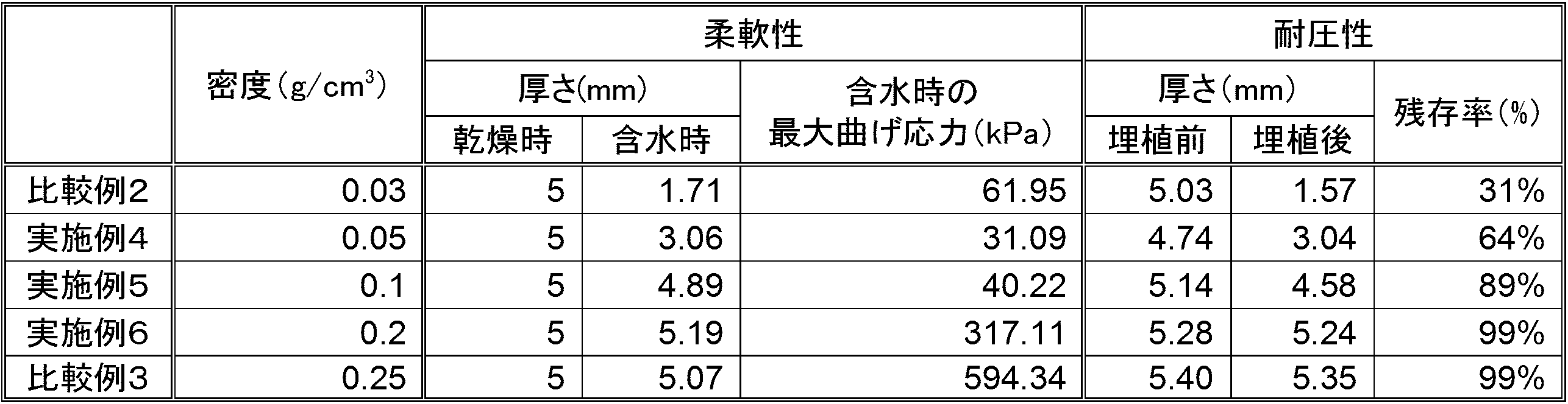
- Table 2 shows the measurement results of flexibility and pressure resistance in this additional test.
- Comparative Example 2 which has a particularly low density compared to Examples 4 to 6 and Comparative Example 3, the thickness is significantly reduced to 1.71 mm due to water content, and is about 1/3 of that before water content.
- the function as a spacer for providing a distance cannot be sufficiently exhibited.
- Examples 4 to 6 can sufficiently function as spacers. That is, these results show that Examples 4 to 6 show the optimum flexibility to fit the organ gently while also exhibiting the function of a spacer for providing a distance between the organs as compared with Comparative Examples 2 to 3. It is shown that it is also demonstrated at the same time.
- Example 4 Pressure resistance (by implantation in rats) From Table 2, in Examples 5 to 6 and Comparative Example 3, the residual thickness rate after implantation in the rat body exceeded 85%, and the shape was maintained without being crushed by the inter-organ pressure. The optimum pressure resistance was maintained to keep space. In addition, the thickness survival rate after implantation in Example 4 is less than 85% (64%), but in the living body of the rat, the low density spacer is relatively easily crushed due to the influence of the body fluid and the pressure between organs. Conceivable. However, from a clinical point of view, a treatment space can be secured if the thickness remaining ratio of the spacer is 60% or more in an actual living body, and Example 4 maintains its shape without being crushed even when inter-organ pressure is applied.
- Comparative Example 2 the thickness remaining ratio after implantation is an extremely low value of 31%, which cannot sufficiently function as a spacer for providing a distance between organs in Comparative Example 2. It is shown that. In Comparative Example 2, it is considered that the spacer having the lowest density was crushed by the body fluid and the inter-organ pressure after being implanted from the dry state before implantation into the rat body.
- the present invention can be used for radiotherapy such as X-ray therapy, gamma ray therapy, proton beam therapy, and heavy particle beam therapy.
Landscapes
- Health & Medical Sciences (AREA)
- Life Sciences & Earth Sciences (AREA)
- Veterinary Medicine (AREA)
- Public Health (AREA)
- General Health & Medical Sciences (AREA)
- Animal Behavior & Ethology (AREA)
- Surgery (AREA)
- Heart & Thoracic Surgery (AREA)
- Vascular Medicine (AREA)
- Epidemiology (AREA)
- Engineering & Computer Science (AREA)
- Biomedical Technology (AREA)
- Chemical & Material Sciences (AREA)
- Nuclear Medicine, Radiotherapy & Molecular Imaging (AREA)
- Pathology (AREA)
- Radiology & Medical Imaging (AREA)
- Chemical Kinetics & Catalysis (AREA)
- Dispersion Chemistry (AREA)
- Physics & Mathematics (AREA)
- General Engineering & Computer Science (AREA)
- High Energy & Nuclear Physics (AREA)
- Optics & Photonics (AREA)
- Oral & Maxillofacial Surgery (AREA)
- Medical Informatics (AREA)
- Molecular Biology (AREA)
- Materials For Medical Uses (AREA)
- Radiation-Therapy Devices (AREA)
Abstract
Description
生体適合性の繊維を交絡した繊維集合体を含む放射線治療用スペーサーであって,
上記繊維集合体の密度が0.05g/cm3~0.2g/cm3であることを特徴とする放射線治療用スペーサーである。
「スペーサー治療法」とは,腫瘍と周辺の正常組織との間に治療スペースを確保し,腫瘍に対し根治線量の放射線を照射しつつ治療スペースにより腫瘍周辺の正常組織を被曝から保護するという治療法を意味する。「スペーサー」とは,スペーサー治療法において腫瘍と周辺の正常組織との間のスペースを確保するための医療用部材を意味する。「生体適合性」とは,生体に影響を与えず,また生体からも影響を受けずに良くなじむ性質を意味する。
当該放射線治療用スペーサーは,生体適合性の繊維を交絡した繊維集合体を含むものである。この繊維集合体全体には,生体適合性の繊維同士が三次元的に交絡して形成されることで繊維間に無数の連通した空隙が存在し,当該空隙に水分を保持することで放射線を遮蔽できるようにすると共に,当該放射線治療用スペーサー全体に軽さと柔軟性を付与している。
次いで,当該放射線治療用スペーサーの製造方法を説明する。当該放射線治療用スペーサーを構成する繊維集合体の構造が三次元の織物,三次元の編物,不織布のいずれの場合であっても,公知の手段で製造することができる。特に上記繊維集合体の構造が不織布の場合,当該放射線治療用スペーサーは,ニードルパンチ法,ケミカルボンド法,サーマルボンド法,スパンレース法等により製造することができる。
次いで,当該放射線治療用スペーサーの使用方法を説明する。滅菌した当該放射線治療用スペーサーを,外科的手術によりがん患者の治療対象部位である腫瘍周辺に配設し,腫瘍と周辺臓器との間に治療用のスペースを確保するよう留置する。この留置手術において,当該放射線治療用スペーサーは,癌の種類や留置部位に応じて密度,厚さ,大きさ,形状を任意に調整することができる。また,当該放射線治療用スペーサーを複数重ねて使うこともできる。
本試験で使用するスペーサー(以下「試験用スペーサー」と称する場合がある。)は,体内吸収性合成高分子であるポリグリコール酸製繊維を原料とし,公知の方法により不織布に加工して製造した。具体的には,原料となるポリグリコール酸製繊維を短繊維にカットし,ウェブ化により繊維シート状に加工した後,ニードルパンチにより不織布に加工した。このようにして製造された試験用スペーサーの形状は略直方体の板状である。なお,試験用スペーサーの密度は,不織布の加工工程において調整した。後述する実施例1~3及び比較例1に係る試験用スペーサーについては,使用した繊維,形状及びサイズは同じであるが,密度がそれぞれ異なる。
密度を0.05g/cm3に調整した試験用スペーサーである。
密度を0.1g/cm3に調整した試験用スペーサーである。
密度を0.2g/cm3に調整した試験用スペーサーである。
密度を0.3g/cm3に調整した試験用スペーサーである。
試験用スペーサーをヒトの生体内に留置した場合,試験用スペーサーの板状表面に臓器の荷重がかかることとなる。このように試験用スペーサーをヒト生体内に留置した場合における柔軟性と耐圧性とを,以下の通り特定した。
試験用スペーサーの柔軟性は,スペーサーの板状表面の一定の面積に圧力をかけた場合の曲げ特性で特定した。具体的には,当該曲げ特性を,JIS K 7171(プラスチック-曲げ特性の試験方法)に準拠し,3点曲げ試験を実施することにより測定した。縦10mm,横100mm,厚さ5mmに裁断した実施例1~3及び比較例1の試験用スペーサーを各5サンプル用意し,試験機に設置した。圧子の開始時の位置は試験用スペーサーより6mm上方,沈みは試験用スペーサーより下方に30mm,試験速度100mm/秒,温度20℃,湿度65%RHにて測定を実施し,各試験用スペーサーにおける最大曲げ応力を算出した。測定において1回目の測定はカウントせず,2回目から6回目の各値(計5回)の最大値及び最小値を記録し,5回の平均値を算出した。なお,圧力をかけた面積は1.57cm2とした。このような3点曲げ試験において,最大曲げ応力が350kPa以下であると,当該試験用スペーサーは臓器に優しくフィットする程度の最適な柔軟性を発揮することが目視できた。従って,最大曲げ応力が350kPa以下の場合,当該試験用スペーサーは臓器に優しくフィットする最適な柔軟性を発揮すると判断した。
試験用スペーサーの耐圧性は,スペーサーの板状表面の略全面に厚さ方向から圧力をかけた場合の厚さ残存率で特定した。幅63.5mm,奥行き40mm,厚さ5mmに裁断した実施例1~3及び比較例1の試験用スペーサーを各3サンプル用意し,試験機に設置した。なお,圧力をかけた略全面の面積は約20.5cm2であった。厚さ残存率(%)は,JIS L 1912:1997(医療用不織布試験方法)に準拠し,圧力0.5kPaの場合の厚さを初荷重と考え,荷重を増やした際の厚さを測定し,3サンプルの平均値を算出し,次の式1の通り比率で算出した。


本試験における柔軟性及び耐圧性の測定結果を表1に示す。
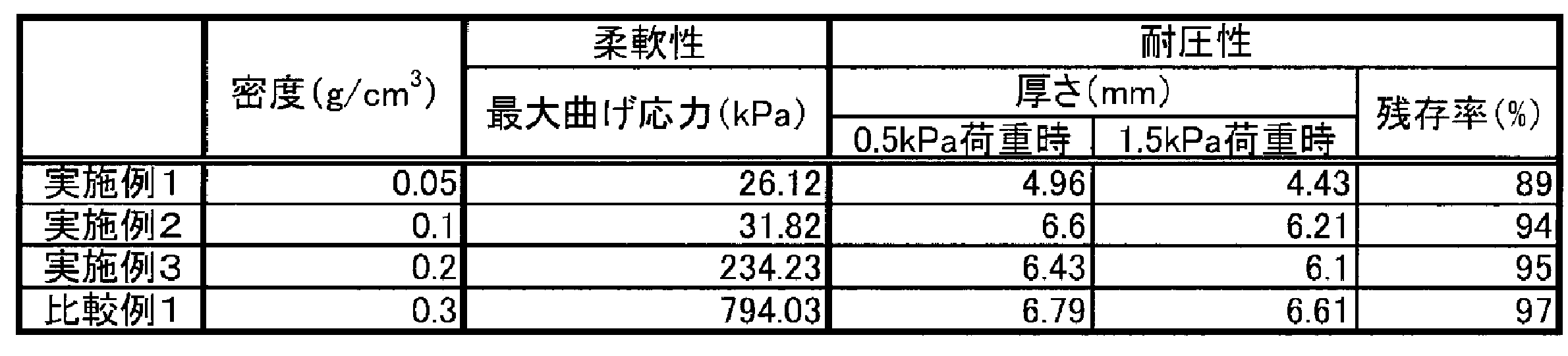
表1より,実施例1~3及び比較例1の厚さ残存率はいずれも85%を超えており,臓器間圧力が負荷されても潰れず形状を維持し治療スペースを確保し続ける最適な耐圧性を発揮していることが示唆された。一方,実施例1~3の最大曲げ応力はいずれも350kPa以下であり,臓器に沿って優しくフィットする最適な柔軟性を発揮することが示唆されたが,比較例1の最大曲げ応力は350kPaを大きく超える値となった。即ち,実施例1~3は,比較例1と比較して,臓器に沿って優しくフィットする最適な柔軟性と,臓器間圧力によっても潰れず形状を維持し治療スペースを確保し続ける最適な耐圧性とを両立して発揮することが確認された。
追加試験で使用するスペーサー(以下「追加試験用スペーサー」と称する場合がある。)を,上記実施例1~3及び比較例1と同様の手段により加工した。
密度を0.05g/cm3に調整した追加試験用スペーサーである。
密度を0.1g/cm3に調整した追加試験用スペーサーである。
密度を0.2g/cm3に調整した追加試験用スペーサーである。
密度を0.03g/cm3に調整した追加試験用スペーサーである。
密度を0.25g/cm3に調整した追加試験用スペーサーである。
追加試験用スペーサーの柔軟性は,スペーサーに水を十分含浸させた状態で,スペーサーの板状表面の一定の面積に圧力をかけた場合の曲げ特性で特定した。具体的には,当該曲げ特性を,JIS K 7171(プラスチック-曲げ特性の試験方法)に準拠し,3点曲げ試験を実施することにより測定した。縦10mm,横100mm,厚さ5mmに裁断した実施例4~6及び比較例2~3の追加試験用スペーサーを各2サンプル用意し,全てのサンプルに水を十分含浸させた状態で試験機に設置した。圧子の開始時の位置は追加試験用スペーサーより6mm上方,沈みは追加試験用スペーサーより下方に30mm,試験速度100mm/秒,温度20℃,湿度65%RHにて測定を実施し,各追加試験用スペーサーにおける最大曲げ応力を算出した。測定において1回目の測定はカウントせず,2回目から6回目の各値(計5回)の最大値及び最小値を記録し,5回の平均値を算出した。なお,圧力をかけた面積は1.57cm2とした。このような3点曲げ試験において,上述の(A)柔軟性の試験と同様に最大曲げ応力が350kPa以下であると,当該追加試験用スペーサーは臓器に沿って優しくフィットする程度の最適な柔軟性を発揮することが目視できた。従って,最大曲げ応力が350kPa以下の場合,当該追加試験用スペーサーは臓器に沿って優しくフィットする最適な柔軟性を発揮すると判断した。
追加試験用スペーサーの耐圧性は,次のように,ラットの腹腔内への埋植試験による厚さ残存率で特定した。即ち,縦約30mm,横約40mm,厚さ約4~6mmに裁断した実施例4~6及び比較例2~3の追加試験用スペーサーを各3サンプル用意し,エチレンオキサイドガスにより滅菌した。CD(SD)系統の8週齢の雄性ラットを用意し,滅菌済みの乾燥状態の実施例4~6及び比較例2~3の追加試験用スペーサーを,全身麻酔下でラットの腹部中央に埋植した。埋植時点から計測して24時間後にラットを開腹し,実施例4~6及び比較例2~3の追加試験用スペーサーを摘出し,厚さを測定した。厚さ残存率(%)は,埋植後の追加試験用スペーサーの厚さを埋植前の厚さで除して算出した。上記(B)耐圧性の試験に準拠し,厚さ残存率が85%以上の場合,当該試験用スペーサーは生体内において臓器間圧力によっても潰れず形状を維持し治療スペースを確保し続ける最適の耐圧性を発揮すると判断すると共に,生体内において厚さ残存率が60%以上あれば,耐圧性は臨床上の観点から十分であるとした。
本追加試験における柔軟性及び耐圧性の測定結果を表2に示す。
1.柔軟性(含水時)
表2より,比較例2及び実施例4~6では,含水時の最大曲げ応力がいずれも350kPa未満であり,臓器に優しくフィットする最適な柔軟性を発揮することが示された。これに対し比較例3では,含水時の最大曲げ応力が350kPaを遥かに超えており(約594kPa),柔軟性に欠け使用に適さないことが判明した。
ここで追加試験用スペーサーのうち比較例2,実施例4~5は,含水により厚さが減少したが,これは比較的密度の小さいスペーサーを含水させると,繊維間に水が浸入することで水の張力により繊維と繊維が引き寄せられ,その結果スペーサーの厚みが縮むためと考えられる。実施例4~6及び比較例3と比較して密度の特に低い比較例2では,含水により厚さが1.71mmまで著しく減少して含水前の約1/3となっており,臓器間に距離を設けるためのスペーサーとしての機能を十分には発揮できない。これに対し,実施例4~6は,スペーサーとして十分に機能できる。即ち,これらの結果は,実施例4~6は,比較例2~3と比べて,臓器間に距離を設けるためのスペーサーの機能も発揮しつつ,しかも臓器に優しくフィットする最適な柔軟性をも同時に発揮するものであることを示している。
表2より,実施例5~6及び比較例3では,ラット生体内への埋植後における厚さ残存率がいずれも85%を超えており,臓器間圧力によっても潰れず形状を維持し治療スペースを確保し続ける最適な耐圧性を発揮していた。また,実施例4の埋植後の厚さ残存率は85%を下回る(64%)が,ラットの生体内では体液や臓器間圧力の影響により密度の低いスペーサーが相対的に潰れやすいためと考えられる。しかしながら臨床の観点からすると,実際の生体内でスペーサーの厚さ残存率が60%以上であれば治療スペースを確保でき,実施例4も,臓器間圧力が負荷されても潰れず形状を維持し治療スペースを確保し続けるに十分な耐圧性を有している。一方,比較例2では,埋植後の厚さ残存率は31%と極めて低い値であり,これは,比較例2が臓器間に距離を設けるためのスペーサーとしての機能を十分果し得ないことを示している。比較例2では,最も低密度である当該スペーサーが,埋植前乾燥状態からラットの体内に埋植された後,体液と臓器間圧力によって潰れてしまったためと考えられる。
以上の追加試験の結果から,実施例4~6は,比較例2及び3と比較して,臓器に沿って優しくフィットする最適の柔軟性と,臓器間圧力が負荷されても潰れず形状を維持し治療スペースを確保し続ける最適の耐圧性とを,同時に発揮することが確認された。
Claims (4)
- 生体適合性の繊維を交絡した繊維集合体を含む放射線治療用スペーサーであって,
上記繊維集合体の密度が0.05g/cm3~0.2g/cm3であることを特徴とする放射線治療用スペーサー。 - 上記繊維集合体が不織布である請求項1に記載の放射線治療用スペーサー。
- 上記生体適合性の繊維が生体吸収性である請求項1又は請求項2に記載の放射線治療用スペーサー。
- 上記繊維集合体の厚さが5mm~15mmである請求項1~3のいずれか1項に記載の放射線治療用スペーサー。
Priority Applications (5)
| Application Number | Priority Date | Filing Date | Title |
|---|---|---|---|
| US15/107,842 US20160361565A1 (en) | 2013-12-25 | 2014-12-24 | Radiotherapy spacer |
| JP2015554910A JPWO2015098904A1 (ja) | 2013-12-25 | 2014-12-24 | 放射線治療用スペーサー |
| EP14874083.0A EP3095483A4 (en) | 2013-12-25 | 2014-12-24 | Radiotherapy spacer |
| KR1020167018164A KR20160101955A (ko) | 2013-12-25 | 2014-12-24 | 방사선 치료용 스페이서 |
| CN201480071060.0A CN105899260A (zh) | 2013-12-25 | 2014-12-24 | 放射线治疗用间隔物 |
Applications Claiming Priority (2)
| Application Number | Priority Date | Filing Date | Title |
|---|---|---|---|
| JP2013266214 | 2013-12-25 | ||
| JP2013-266214 | 2013-12-25 |
Publications (1)
| Publication Number | Publication Date |
|---|---|
| WO2015098904A1 true WO2015098904A1 (ja) | 2015-07-02 |
Family
ID=53478757
Family Applications (1)
| Application Number | Title | Priority Date | Filing Date |
|---|---|---|---|
| PCT/JP2014/084029 Ceased WO2015098904A1 (ja) | 2013-12-25 | 2014-12-24 | 放射線治療用スペーサー |
Country Status (6)
| Country | Link |
|---|---|
| US (1) | US20160361565A1 (ja) |
| EP (1) | EP3095483A4 (ja) |
| JP (1) | JPWO2015098904A1 (ja) |
| KR (1) | KR20160101955A (ja) |
| CN (1) | CN105899260A (ja) |
| WO (1) | WO2015098904A1 (ja) |
Cited By (1)
| Publication number | Priority date | Publication date | Assignee | Title |
|---|---|---|---|---|
| KR20190075062A (ko) | 2016-09-15 | 2019-06-28 | 고쿠리츠다이가쿠호진 고베다이가쿠 | 방사선 치료용 스페이서 |
Families Citing this family (2)
| Publication number | Priority date | Publication date | Assignee | Title |
|---|---|---|---|---|
| CN111569277B (zh) * | 2020-05-13 | 2022-02-18 | 戴建荣 | 放疗靶区隔离体 |
| US12303712B1 (en) | 2023-02-11 | 2025-05-20 | Ip Liberty Vision Corporation | Conformal system for therapeutic beta or low-energy gamma radiation shielding |
Citations (3)
| Publication number | Priority date | Publication date | Assignee | Title |
|---|---|---|---|---|
| WO2006107388A1 (en) * | 2005-04-05 | 2006-10-12 | Boston Scientific Scimed, Inc. | Radiation shield for insertion into a living body |
| WO2011055670A1 (ja) * | 2009-11-05 | 2011-05-12 | 国立大学法人神戸大学 | 電離放射線治療用スペーサー |
| WO2013125720A1 (ja) * | 2012-02-23 | 2013-08-29 | 凸版印刷株式会社 | 放射線遮断シート |
Family Cites Families (9)
| Publication number | Priority date | Publication date | Assignee | Title |
|---|---|---|---|---|
| US2423707A (en) * | 1945-03-02 | 1947-07-08 | Eastman Kodak Co | Fabric or gauze of uniformly oxidized cellulose |
| US6674087B2 (en) * | 2001-01-31 | 2004-01-06 | Worldwide Innovations & Technologies, Inc. | Radiation attenuation system |
| US7754937B2 (en) * | 2004-03-18 | 2010-07-13 | Boehringer Technologies, L.P. | Wound packing material for use with suction |
| US7726318B2 (en) * | 2005-03-21 | 2010-06-01 | Xoft, Inc. | Radiation blocking patch for radio-therapy |
| US20080123810A1 (en) * | 2006-11-03 | 2008-05-29 | Kirkpatrick John P | Bolus materials for radiation therapy and methods of making and using the same |
| EP2050848B1 (en) * | 2007-09-17 | 2017-06-07 | Orfit Industries | Hybrid fabric, articles comprising said fabric and methods of making same |
| WO2010087923A1 (en) * | 2009-02-02 | 2010-08-05 | C.R. Bard, Inc. | Implant for pelvic organ prolapse repair |
| US9060837B2 (en) * | 2009-11-23 | 2015-06-23 | Ams Research Corporation | Patterned sling implant and method |
| WO2013126718A1 (en) * | 2012-02-24 | 2013-08-29 | Bvw Holding Ag | Anti-erosion soft tissue repair device |
-
2014
- 2014-12-24 JP JP2015554910A patent/JPWO2015098904A1/ja active Pending
- 2014-12-24 US US15/107,842 patent/US20160361565A1/en not_active Abandoned
- 2014-12-24 WO PCT/JP2014/084029 patent/WO2015098904A1/ja not_active Ceased
- 2014-12-24 EP EP14874083.0A patent/EP3095483A4/en not_active Withdrawn
- 2014-12-24 CN CN201480071060.0A patent/CN105899260A/zh active Pending
- 2014-12-24 KR KR1020167018164A patent/KR20160101955A/ko not_active Withdrawn
Patent Citations (3)
| Publication number | Priority date | Publication date | Assignee | Title |
|---|---|---|---|---|
| WO2006107388A1 (en) * | 2005-04-05 | 2006-10-12 | Boston Scientific Scimed, Inc. | Radiation shield for insertion into a living body |
| WO2011055670A1 (ja) * | 2009-11-05 | 2011-05-12 | 国立大学法人神戸大学 | 電離放射線治療用スペーサー |
| WO2013125720A1 (ja) * | 2012-02-23 | 2013-08-29 | 凸版印刷株式会社 | 放射線遮断シート |
Non-Patent Citations (1)
| Title |
|---|
| See also references of EP3095483A4 * |
Cited By (1)
| Publication number | Priority date | Publication date | Assignee | Title |
|---|---|---|---|---|
| KR20190075062A (ko) | 2016-09-15 | 2019-06-28 | 고쿠리츠다이가쿠호진 고베다이가쿠 | 방사선 치료용 스페이서 |
Also Published As
| Publication number | Publication date |
|---|---|
| KR20160101955A (ko) | 2016-08-26 |
| EP3095483A1 (en) | 2016-11-23 |
| US20160361565A1 (en) | 2016-12-15 |
| CN105899260A (zh) | 2016-08-24 |
| EP3095483A4 (en) | 2017-09-27 |
| JPWO2015098904A1 (ja) | 2017-03-23 |
Similar Documents
| Publication | Publication Date | Title |
|---|---|---|
| CN102596317B (zh) | 电离放射线治疗用隔离体 | |
| US11759979B2 (en) | Device system and method for tissue displacement or separation | |
| CN102119046B (zh) | 利用植入物瞄准外部波束放射的系统 | |
| US9011485B2 (en) | Implantable biodegradable wound closure device and method | |
| JP2017521196A (ja) | オンコプラスティック手術のための埋込型デバイスおよび技術 | |
| WO2015098904A1 (ja) | 放射線治療用スペーサー | |
| Kigure et al. | Laparoscopic microwave thermotherapy on small renal tumors: experimental studies using implanted VX-2 tumors in rabbits | |
| US20060224034A1 (en) | Radiation shield | |
| US11064988B2 (en) | Implantable scaffold and method | |
| EP2381974B1 (en) | A method of forming and the resulting membrane composition for surgical site preservation | |
| US12295557B2 (en) | Implantable tissue scaffold | |
| CN111449830A (zh) | 可植入金属作为肿瘤精准高效磁热治疗试剂的应用 | |
| Sasaki et al. | Medical Application of Nonwoven Fabrics-Intra-abdominal Spacers for Particle Therapy | |
| Goel et al. | Case report of isolated impaling lung injury with successful outcome | |
| EP3513837A1 (en) | Spacer for radiation therapy | |
| Cushing et al. | B041 Surgical efficiency in bilateral simultaneous cochlear implantation: patient safety and cost containment | |
| HK1113300A (en) | Device system and method for tissue displacement or separation | |
| JP2011189090A (ja) | 生体吸収性ヘルニア治療用材料 |
Legal Events
| Date | Code | Title | Description |
|---|---|---|---|
| 121 | Ep: the epo has been informed by wipo that ep was designated in this application |
Ref document number: 14874083 Country of ref document: EP Kind code of ref document: A1 |
|
| ENP | Entry into the national phase |
Ref document number: 2015554910 Country of ref document: JP Kind code of ref document: A |
|
| NENP | Non-entry into the national phase |
Ref country code: DE |
|
| ENP | Entry into the national phase |
Ref document number: 20167018164 Country of ref document: KR Kind code of ref document: A |
|
| REEP | Request for entry into the european phase |
Ref document number: 2014874083 Country of ref document: EP |
|
| WWE | Wipo information: entry into national phase |
Ref document number: 2014874083 Country of ref document: EP |
|
| WWE | Wipo information: entry into national phase |
Ref document number: 15107842 Country of ref document: US |