WO2014174899A1 - 病的状態の細胞モデルとしての圧縮細胞又は圧縮組織とその製法 - Google Patents
病的状態の細胞モデルとしての圧縮細胞又は圧縮組織とその製法 Download PDFInfo
- Publication number
- WO2014174899A1 WO2014174899A1 PCT/JP2014/055199 JP2014055199W WO2014174899A1 WO 2014174899 A1 WO2014174899 A1 WO 2014174899A1 JP 2014055199 W JP2014055199 W JP 2014055199W WO 2014174899 A1 WO2014174899 A1 WO 2014174899A1
- Authority
- WO
- WIPO (PCT)
- Prior art keywords
- cell
- tissue
- compressed
- compression
- adipocyte
- Prior art date
Links
Images


Classifications
-
- G—PHYSICS
- G01—MEASURING; TESTING
- G01N—INVESTIGATING OR ANALYSING MATERIALS BY DETERMINING THEIR CHEMICAL OR PHYSICAL PROPERTIES
- G01N33/00—Investigating or analysing materials by specific methods not covered by groups G01N1/00 - G01N31/00
- G01N33/48—Biological material, e.g. blood, urine; Haemocytometers
- G01N33/50—Chemical analysis of biological material, e.g. blood, urine; Testing involving biospecific ligand binding methods; Immunological testing
- G01N33/5005—Chemical analysis of biological material, e.g. blood, urine; Testing involving biospecific ligand binding methods; Immunological testing involving human or animal cells
- G01N33/5091—Chemical analysis of biological material, e.g. blood, urine; Testing involving biospecific ligand binding methods; Immunological testing involving human or animal cells for testing the pathological state of an organism
-
- C—CHEMISTRY; METALLURGY
- C12—BIOCHEMISTRY; BEER; SPIRITS; WINE; VINEGAR; MICROBIOLOGY; ENZYMOLOGY; MUTATION OR GENETIC ENGINEERING
- C12N—MICROORGANISMS OR ENZYMES; COMPOSITIONS THEREOF; PROPAGATING, PRESERVING, OR MAINTAINING MICROORGANISMS; MUTATION OR GENETIC ENGINEERING; CULTURE MEDIA
- C12N5/00—Undifferentiated human, animal or plant cells, e.g. cell lines; Tissues; Cultivation or maintenance thereof; Culture media therefor
- C12N5/06—Animal cells or tissues; Human cells or tissues
- C12N5/0602—Vertebrate cells
- C12N5/0652—Cells of skeletal and connective tissues; Mesenchyme
- C12N5/0653—Adipocytes; Adipose tissue
-
- G—PHYSICS
- G01—MEASURING; TESTING
- G01N—INVESTIGATING OR ANALYSING MATERIALS BY DETERMINING THEIR CHEMICAL OR PHYSICAL PROPERTIES
- G01N33/00—Investigating or analysing materials by specific methods not covered by groups G01N1/00 - G01N31/00
- G01N33/48—Biological material, e.g. blood, urine; Haemocytometers
- G01N33/50—Chemical analysis of biological material, e.g. blood, urine; Testing involving biospecific ligand binding methods; Immunological testing
- G01N33/5005—Chemical analysis of biological material, e.g. blood, urine; Testing involving biospecific ligand binding methods; Immunological testing involving human or animal cells
- G01N33/5008—Chemical analysis of biological material, e.g. blood, urine; Testing involving biospecific ligand binding methods; Immunological testing involving human or animal cells for testing or evaluating the effect of chemical or biological compounds, e.g. drugs, cosmetics
- G01N33/5044—Chemical analysis of biological material, e.g. blood, urine; Testing involving biospecific ligand binding methods; Immunological testing involving human or animal cells for testing or evaluating the effect of chemical or biological compounds, e.g. drugs, cosmetics involving specific cell types
-
- C—CHEMISTRY; METALLURGY
- C12—BIOCHEMISTRY; BEER; SPIRITS; WINE; VINEGAR; MICROBIOLOGY; ENZYMOLOGY; MUTATION OR GENETIC ENGINEERING
- C12N—MICROORGANISMS OR ENZYMES; COMPOSITIONS THEREOF; PROPAGATING, PRESERVING, OR MAINTAINING MICROORGANISMS; MUTATION OR GENETIC ENGINEERING; CULTURE MEDIA
- C12N2527/00—Culture process characterised by the use of mechanical forces, e.g. strain, vibration
Definitions
- the present invention relates to a new compressed cell or compressed tissue obtained under artificial compression culture conditions and a method for producing the same, and further evaluates the action or side effect of a drug using the compressed cell or compressed tissue.
- the present invention relates to a screening method.
- Non-Patent Documents 1 and 2 It is known that cells are affected by various mechanical stimuli, and their functions, morphology, and motility change (Non-Patent Documents 1 and 2). For example, it has been clarified that the elastic modulus of the cell culture substrate affects the differentiation of mesenchymal stem cells (Non-patent Document 1). In addition, recently, it has been announced that differentiation of cells is initiated by pressurization of cells, and as a stimulus-induced pluripotency acquisition cell (STAP cell, Stimulus-Triggered Acquisition of Pluripotency cells) (Non-Patent Document 4). In this way, the focus is on the field of cell engineering to study the effect of changes in the cell environment on cells.
- STAP cell Stimulus-Triggered Acquisition of Pluripotency cells
- the two-dimensional culture method is also used when evaluating the mechanical influence of various culture media. It was common to be evaluated and examined (Patent Documents 1 and 2). Therefore, many cells and tissues obtained by culturing have different properties from cells and tissues existing in a living body, and are different from cells and tissues in an actual living body. Therefore, even when ordinary cultured cells are used in a drug screening method, it is difficult to obtain a biological response and it is difficult to obtain an accurate evaluation of the drug.
- An object of the present invention is to provide an artificially prepared compressed cell or tissue that can be used for drug screening as various disease models and a method for producing the same.
- it is an object to provide a compressed cell or a compressed tissue for screening therapeutic agents for obesity, diabetes, metabolic syndrome, dyslipidemia, hypertension, arteriosclerosis and the like based on chronic inflammation of adipose tissue.
- the present inventor has already reported that cultured cells differentiated into various cells can be prepared by culturing in a two-dimensional gel medium having various elastic moduli using mesenchymal stem cells (Patent Literature). 1). Furthermore, when 3T3-L1 adipocytes are cultured on a two-dimensional gel medium having the same hardness as normal adipose tissue (250 Pa) in the presence of a saturated fatty acid such as palmitic acid, fat cells are enlarged. Regardless, it has been reported that the hardness of the two-dimensional gel medium affects the cultured cells, resulting in cells in which insulin resistance is not induced (insulin sensitivity is maintained) (Patent Document 2).
- the present inventors have searched for factors other than the hardness of the two-dimensional gel medium in order to elucidate the mechanical factors that influence the properties of cultured cells.
- the function of cultured cells can be controlled by applying compression to the cells, and the present invention has been completed. That is, it has been found that compressed cultured cells having various cell functions can be produced by culturing cells under pressure.
- normal adipocytes (tissues) collected from a living body can be artificially produced by pressing and compressing and culturing cells having the function of obese adipocytes.
- mast adipocyte tissue is a cell that shows the same shape as normal adipocytes (it does not accompany the enlargement seen in mast adipocytes), It became clear that it showed the same function as obese adipocytes.
- the properties of the cells change.
- the resulting adipocytes are not accompanied by the hypertrophy seen in mast adipocytes, and the function of adipocytes is It turns out that it shows the same function.
- adipocytes exhibiting the same function as fat cells in obese states by compressing normal adipocytes ⁇ Adipose tissue was produced for the first time. That is, by the artificial three-dimensional compression of normal fat cells and normal fat tissues, new compressed fat cells / compressed fat tissues having the same function as fat cells in the obese state were produced. Therefore, since the adipocytes or adipose tissue of this compression culture show the same function as the adipocytes in the obese state, they can be used for evaluation screening of the action and side effects of drugs as an alternative to obesity adipocytes and obesity adipose tissue. I understood.
- the present inventor completed the present invention based on the above findings. That is, the gist of the present invention is as follows. (1) A compressed cell or tissue that is produced by compressing and culturing a cell or tissue as a cell model of a pathological state, a) Pressurization in the above-mentioned compression culture is 0.07 to 1.6 g / cm 2 , or the compression rate of cells and tissues is 2 to 90%. b) The compressed cell or the compressed tissue has the same capacity as that of the cell before compression, and exhibits the function of a cell whose pathological state is enhanced from that before compression. Compressed tissue. (2) The compressed cell or tissue according to (1) above, wherein the cell or tissue is a normal fat cell or normal fat tissue.
- the compressed cell according to (2) above wherein the function of the cell in which the pathological state is enhanced exhibits the same function as an adipocyte in an obese state of 100 ⁇ m to 150 ⁇ m, Compressed tissue.
- the normal adipocyte or normal adipose tissue is 3T3-L1 adipocyte, mouse adipocyte or tissue, rat adipocyte or tissue, human adipocyte or human adipose tissue Compressed cells or tissues as described.
- the compression according to (7) above, wherein the function of the cells in which the pathological state is enhanced is a function of obese adipocytes or obese adipose tissue whose pathological condition has further deteriorated Cell or compressed tissue.
- the compressed cell or compressed tissue according to (7) above, wherein the obese adipocyte or obese adipose tissue is an ob / ob mouse adipose tissue.
- (21) A method for producing a pathological state of a cell or tissue (cell or tissue of a disease state model), characterized by compressing and culturing the cell or tissue, a) whether the compressibility of the cells or tissues is 2 to 90%, b) A method for producing a pathological cell or tissue, wherein the compression pressure is 0.07 to 1.6 g / cm 2 .
- (22) The production method according to (21), wherein the cell or tissue is a normal fat cell or a normal fat tissue.
- (23) The method according to (21) or (22) above, wherein the compression ratio is 2 to 20%.
- the solution medium is a DMEM medium.
- the production method according to (24), wherein the two-dimensional gel medium has a hardness of 250 Pa.
- adipocyte or adipose tissue is 3T3-L1 adipocyte, mouse adipocyte or tissue, rat adipocyte or tissue, human adipocyte or human adipose tissue.
- the mouse adipose tissue is mouse normal adipose tissue.
- the pathological state of the cell or tissue is obesity adipocyte or obesity adipose tissue.
- the cell or tissue is a pathological cell or pathological tissue, and the pathological cell or tissue is further deteriorated in pathological condition.
- the obese adipocyte or obesity adipose tissue is an ob / ob mouse obesity adipocyte or obesity adipose tissue.
- the cell compression culture method of the present invention is a method for producing a desired disease state model cell by applying a mechanical load to normal cells or normal tissues and compressing them. Therefore, for example, if adipocytes are used, cells (tissues) having the same functions as obesity adipocytes or obesity adipose tissue can be easily prepared from normal adipocytes or adipose tissue by the method of the present invention. Can do. That is, by culturing normal adipocytes or normal adipose tissue under pressure, a cell having the same function as obesity adipocytes or obesity adipose tissue is produced, although there is no enlargement of adipocytes as the cell volume. it can.
- the cell compression culture method of the present invention makes it possible to produce various pathological model cells and pathological model tissues and to screen for appropriate drugs.
- examination of the mechanism of transmission of mechanical stimulation of cell compression has made it possible to screen for drugs that are suitable for eliminating the pathological conditions that occur with compression.
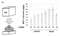
- FIG. 1 shows the amount of MCP-1 secreted when 3T3-L1 adipocytes are cultured in a cell culture medium (two-dimensional gel) having a hardness of 250 Pa. It is the figure which showed the secretion amount of MCP-1 at the time of culture
- FIG. 1 shows the amount of MCP-1 secreted when 3T3-L1 adipocytes are cultured in a cell culture medium (two-dimensional gel) having a hardness of 250 Pa. It is the figure which showed the secretion amount of MCP-1 at the time of culture
- FIG. 3 is a diagram showing the dynamic stiffness of mouse gonad adipose tissue. It was found that the normal adipose tissue of the lean wild type mouse showed a value of about 110 Pa, and the obese adipose tissue of the ob / ob mouse showed a value of about 260 Pa. In FIG. 4, obese adipose tissue of ob / ob mice was treated with collagenase to remove collagen from the obese adipose tissue.
- FIG. 4 is a diagram showing that the hardness of obese adipose tissue decreases as the collagen content of obese adipose tissue decreases. It has been shown that if the collagen content of obese adipose tissue decreases, the hardness of obese adipose tissue decreases.
- the first aspect of the present invention relates to a compressed cell and a compressed tissue.
- the “cell or tissue” of the present invention is not particularly limited, but refers to a healthy or pathological cell or tissue collected from a living body, or an established cell line of animal cells. Say that. For example, it refers to differentiated cells such as fat cells, muscle cells, and liver cells.
- the “compressive culture” of the present invention refers to culturing while compressing under pressure (by applying a weight).
- the “pathological cell” of the present invention refers to a cell that does not show normal metabolic function or shape unlike normal cells or normal tissues.
- pathological cells include mild and severe stages, cells having a shape (capacity) and properties deviating from the normal value range of cells and tissues are referred to as pathological cells.
- the pathological cell in the case of an adipocyte, the pathological cell refers to an obese adipocyte. Therefore, the pathological state cell produced by the cell compression of the present invention can be easily produced as a disease state model cell and can be widely used in screening methods.
- the phrase “pathological state is enhanced” in the present invention means that the degree of the pathological state of the cell has progressed and the degree of deviation from the normal range of metabolic function and shape has increased.
- the “compression rate” in the present invention refers to a change in size in the compression direction before and after compression, and can be in the range of 2 to 90%.
- normal adipocytes refer to normal adipocytes of mammals such as 3T3-L1 adipocytes derived from mouse fibroblasts, mouse adipocytes, rat adipocytes, and normal adipocytes of human adipocytes. Can be mentioned.
- adipose tissue refers to a tissue portion where the adipocytes are accumulated. For example, in the case of mice, mention may be made of subcutaneous adipose tissue or gonad adipose tissue.
- the “mast adipocyte” of the present invention refers to a mature adipocyte that is enlarged by storing neutral fat.
- the cell diameter of the enlarged fat cells is 100 ⁇ m or more and at most 130 to 140 ⁇ m (Sugihara Satoshi, Proceedings of the 124th Symposium of the Japan Medical Association, Science of Obesity, 71-81 (2004)).
- the cell diameter of normal fat cells normal body weight of BMI 20-22 was 70-90 ⁇ m.
- the cell diameter is significantly different between normal fat cells and enlarged fat cells.
- fat cells that are enlarged have a polyhedral shape, but in normal fat cells, the fat cells are spherical and have gaps between them, such as a grape shape.
- the “obese adipose tissue” of the present invention refers to, for example, gonad adipose tissue of ob / ob mice, and refers to a tissue in which many obese adipocytes exist.
- Examples of the “collagen” of the present invention include collagens of types I to IX. Preferable examples include collagens I and VI.
- Pressure in the present invention means applying a load (weighting), and can be in the range of 0.07 to 1.6 g / cm 2 . Preferably, 0.07 to 1.1 g / cm 2 can be mentioned. Depending on the cells used, the pressure limit varies.
- a 5.76 cm 2 cover glass (0.2 g, 2.4 ⁇ 2.4 cm) is added to a fat cell ( It is preferable that the weight is set to 0.2 to 9 g.
- a solid medium such as a cover glass, agar medium or two-dimensional gel medium, and making it an upper and lower sandwich, the load (pressure) due to the weight of the medium on the cultured cells sandwiched between them Can be given.
- cells can be cultured in a three-dimensional medium, and a weight (pressure) can be applied to the medium.
- a medium having a desired shape and its own weight can be selected according to the purpose.
- the “compressed adipocyte or compressed adipose tissue” of the present invention refers to an artificial adipocyte / adipose tissue newly produced by compression culture.
- the function of the cell is not the fat cell or adipose tissue after obesity without showing the fat cell hypertrophy seen when the adipocyte or adipocyte of the adipose tissue becomes obese. Similar functions are shown.
- adipokines such as monocyte chemotactic active factor (MCP-1), IL-12 ⁇ , TNF- ⁇ , PAI-1, and resistin that increase insulin resistance is increased in the same manner as obesity adipocytes or adipose tissue. is doing. That is, this artificial adipocyte / adipose tissue does not accompany cell size enlargement, and no accumulation of neutral fat is observed.
- MCP-1 monocyte chemotactic active factor
- IL-12 ⁇ IL-12 ⁇
- TNF- ⁇ IL-12 ⁇
- PAI-1 PAI-1
- “Cultivation” in the present invention refers to culturing used for normal cell culture, indwelling under pressure and compression on adipose tissue in an in vivo environment, and further, an agar medium, a two-dimensional gel medium, etc.
- Two-dimensional culture medium of 2 layers in which cells are seeded and cultured, or cells are cultured in a three-dimensional culture medium is not.
- a general medium such as DMEM medium may be used, and an additive such as a fatty acid may be added depending on the purpose.
- the “compression resistance” of the present invention refers to a property that hardly affects the function of cells during cell compression.
- the information transmission mechanism regarding NF-kB, Rho, ROCK, and MLCK is inhibited.
- the second aspect of the present invention relates to a cell compression culture method for artificially preparing a cell pathological model by pressurizing and compressing cells and tissues.
- the “pathological state of a cell or tissue” of the present invention means that it does not show a normal metabolic function or shape unlike a normal cell or normal tissue.
- the pathological state is referred to as a pathological state that exhibits a shape (capacity) or property that deviates from the range of normal values of cells and tissues.
- the pathological state represents the state of obese fat cells.
- the “pathological cell or tissue” of the present invention refers to a cell or tissue having a pathological condition produced by the cell compression of the present invention. Therefore, the “pathological cell or tissue” of the present invention can be easily prepared as a “pathological model cell or tissue” and can be widely used in screening methods.
- adipocytes examples include mouse fibroblast-derived 3T3-L1 adipocytes, mouse adipocytes, rat adipocytes, and human adipocytes.
- the “adipose tissue” of the present invention refers to a tissue portion where the adipocytes are accumulated.
- mouse subcutaneous adipose tissue or mouse gonad adipose tissue can be mentioned.
- the “solution medium” of the present invention is a general solution medium used for cell culture, and is not particularly limited. An example is DMEM medium.
- the “two-dimensional gel medium” of the present invention is, for example, a polyacrylamide gel prepared by the method described in Patent Document 1, or a commercially available thiolated hyaluronic acid gel (www.glycosan.com) resin such as polyamide nano It refers to fiber, porous polyvinyl formal (PVF) resin, and the like.
- the “cell compression culture method” of the present invention is, for example, a method in which two sheets of the above two-dimensional gel medium are stacked (pseudo three-dimensional gelation), cells are seeded between them, and compression culture is performed with the own weight of the two-dimensional gel medium.
- Examples include a method in which a collected tissue (for example, normal adipose tissue) is pressurized and compressed, and cultured in a solution medium. Furthermore, the method of applying and compressing a fat tissue under in-vivo environment can be mentioned.
- the artificial mast adipocyte (tissue) obtained from the normal adipocyte (tissue) by the compression culture method of the present invention has the same capacity as the normal adipocyte, does not have an enlarged cell size, and is neutral. There is no fat accumulation.
- the extracellular matrix component is also the same as that of normal fat cells.
- this artificial adipocyte / adipose tissue shows the synthesis / secretion pattern of adipocytokines similar to that of general obesity adipocytes / obesity adipose tissue, and activation of NF ⁇ B occurs.
- activation of low molecular weight GTP-binding proteins Rho, ROCK, and myosin light chain kinase has occurred, which is the same state as general obesity adipocytes / obesity adipose tissue.
- adipokines such as monocyte chemotactic active factor (MCP-1), IL-12 ⁇ , TNF- ⁇ , PAI-1, resistin and the like which increase insulin resistance is increased in the same manner as obese adipocytes.
- MCP-1 monocyte chemotactic active factor
- IL-12 ⁇ IL-12 ⁇
- TNF- ⁇ IL-12 ⁇
- PAI-1 resistin and the like which increase insulin resistance is increased in the same manner as obese adipocytes.
- adipocyte function refers to a function related to metabolism and secretion of adipocytes.
- adipocytokine refers to a physiologically active protein secreted from adipocytes.
- TNF- ⁇ , PAI-1, HB-EGF acting in the direction of promoting arteriosclerosis and prevention of arteriosclerosis are prevented.
- TNF- ⁇ tumor necrosis factor ⁇
- PAI-1 tumorogen activator inhibitor-1
- HB-EGF heparin binding-epidermal growth factor-like growth factor
- leptin is secreted from white adipocytes and works in the satiety center of the hypothalamus to suppress appetite.
- Adiponectin is produced from normal adipocytes and is a good factor such as promoting insulin sensitivity.
- the “compound evaluation method” of the present invention is a normal or obese adipocyte-derived artificial adipocyte (obesity type) obtained by compression culture under the influence of a medicinal component added to the culture system. Alternatively, it is a method for measuring and evaluating the state of changes in the type and amount of adipocytokines secreted by fat cells (functioning similar to the state in which obesity is further aggravated). The variation in expression of this adipocytokine can be performed using a general-purpose cytokine quantification method.
- saturated fatty acid in the present invention refers to a saturated higher fatty acid, and examples thereof include saturated fatty acids having 12 or more carbon atoms such as palmitic acid, stearic acid, lauric acid and the like. Preferable examples include palmitic acid. Terms common to the first aspect of the present invention have the same meaning.
- Example 1 Pseudo three-dimensional cell culture method and 3T3-LI adipocyte culture (1)
- APS Ammonium persulfate
- TEMED N, N, N ′, N′ ⁇
- the cells are cultured for 2 days in a DMEM medium supplemented with 10% CS to be confluent.
- the cultured cells are removed from the soft gel (2.4 ⁇ 2.4 cm) described above and cultured for 48 hours.
- the culture medium contains 10% FBS, 0.5 m MIBMX (3-isobutyl-1-methylxanthine), 1 ⁇ M dexamethasone, and 1.7 ⁇ M insulin in DMEM medium.
- the cells are cultured in DMEM medium supplemented with 10% FBS for 7-9 days and differentiated into adipocytes. Meanwhile, DMEM medium containing 10% FBS is changed every other day.
- MCP-1 which is a cell inflammation marker
- MCP-1 which is a cell inflammation marker
- the secretion amount of MCP-1 is about twice as high in the case of adipocytes cultured in a pseudo three-dimensional model medium. That is, in culturing adipocytes in a pseudo three-dimensional model medium, not only the hardness of the cell culture gel medium (soft gel) affects the cultured cells but also the gel medium (soft gel) placed on the cells. It has been clarified that pressurization and compression by gel) have a great influence. That is, 3T3-L1 adipocytes cultured on a culture base (two-dimensional soft gel medium) having an elasticity of 250 Pa in the extracellular matrix can be used as a model for healthy white adipocytes.
- the new 3T3-L1 adipocytes (compressed adipocytes) obtained with the pseudo 3D soft gel medium of the present invention overlaid with the 2D soft gel medium can be used as a model of mast adipocytes having insulin resistance.
- the enlargement of the cell was not seen in this 3T3-L1 fat cell which has insulin resistance.
- Example 2 Evaluation of properties of compressed and deformed adipose tissue As shown in Example 1 above, by compressing normal adipocytes under pressure, artificial obesity adipocytes having insulin resistance are obtained. It was.
- adipocyte function is affected during pressure compression.
- adipose tissue was collected from normal mice or obese diabetic model mice and compressed, and the pressure on the adipose tissue and the secretion amount of MCP-1 were evaluated.
- adipose tissue control group collected from normal mice and 200-250 mg of adipose tissue (ob / ob groups) collected from obese diabetes model mice (ob / ob mice), Culture in DMEM medium containing 10% CS. A 2.4 cm ⁇ 2.4 cm (0.2 g) cover glass is placed on these tissues, and weights of 0 g, 3 g, 6 g, and 9 g are placed thereon. Each of these words 0.04g / cm 2, 0.56g / cm 2, 1.1g / cm 2, a pressure of 1.6 g / cm 2. After culturing for 6 hours or longer, the medium is collected, and the amount of MCP-1 contained therein is measured.
- adipocyte tissue corresponding to the severity of obesity can be prepared by using normal adipocyte tissue or obesity adipocyte tissue and controlling the degree of pressurization (weighting). It was done. Therefore, it was found that the effects and side effects of the drug can be evaluated by using a fat cell tissue corresponding to the degree of progression of the obesity pathology.
- the cell culture method under pressure compression of the present invention can be converted into cells having the same properties as fat cells in the obese state, for example, using normal fat cells.
- the compression-cultured cells of the present invention can be used for evaluation screening of drugs as an artificially obtained pathological cell model.
- a compressed adipocyte obtained using normal adipocytes exhibits the same function as that of an obese adipocyte in a living body, so that it is possible to obtain an evaluation result of a valid drug screening.
- the pressurization and compression conditions for example, normal fat cells, obesity fat cells, or obesity adipose tissue can be used to easily produce obesity fat cells having a desired disease state. Therefore, using the compressed adipocytes of the present invention, it is possible to elucidate the pathophysiology of obesity, metabolic syndrome, diabetes, dyslipidemia, hypertension or arteriosclerosis, and evaluate compounds related to new diagnosis and treatment etc. It became.
Landscapes
- Health & Medical Sciences (AREA)
- Engineering & Computer Science (AREA)
- Life Sciences & Earth Sciences (AREA)
- Biomedical Technology (AREA)
- Immunology (AREA)
- Chemical & Material Sciences (AREA)
- Biotechnology (AREA)
- Hematology (AREA)
- Molecular Biology (AREA)
- Urology & Nephrology (AREA)
- Cell Biology (AREA)
- General Health & Medical Sciences (AREA)
- Biochemistry (AREA)
- Bioinformatics & Cheminformatics (AREA)
- Microbiology (AREA)
- Medicinal Chemistry (AREA)
- Wood Science & Technology (AREA)
- Physics & Mathematics (AREA)
- Analytical Chemistry (AREA)
- Tropical Medicine & Parasitology (AREA)
- Organic Chemistry (AREA)
- General Physics & Mathematics (AREA)
- Pathology (AREA)
- Zoology (AREA)
- Food Science & Technology (AREA)
- Genetics & Genomics (AREA)
- Rheumatology (AREA)
- Physiology (AREA)
- General Engineering & Computer Science (AREA)
- Toxicology (AREA)
- Micro-Organisms Or Cultivation Processes Thereof (AREA)
- Investigating Or Analysing Biological Materials (AREA)
Abstract
本発明は、肥満症、メタボリック・シンドローム、糖尿病、脂質異常症、高血圧症又は動脈硬化の病態解明と診断薬・治療薬のスクリーニングのために必要な培養細胞の提供を目的とする。 正常脂肪細胞又は正常脂肪組織、あるいは肥満脂肪細胞又は肥満脂肪組織を圧縮して培養するという細胞圧縮培養方法により、所望の病態の肥満脂肪細胞と同様の細胞(組織)を作製できることを見出した。その結果、この圧縮脂肪細胞(組織)を用いることにより、メタボリック・シンドローム等を治療できる化合物を、より生体に近い系でスクリーニングできる新しい方法が可能になった。
Description
本発明は、人工的な圧縮培養条件下で得られた新たな圧縮細胞又は圧縮組織とその製法に関するものであり、更には、この圧縮細胞又は圧縮組織を用いた、薬剤の作用若しくは副作用を評価するスクリーニング方法に関するものである。
細胞は、様々な力学的刺激による影響を受け、機能や形態、運動能が変化することが知られている(非特許文献1、2)。例えば、細胞培養基板の弾性率が間葉系幹細胞の分化に影響を与えることが明らかになっている(非特許文献1)。
また、最近、細胞への加圧によって、分化した細胞の初期化が起るとの発表が行われ、刺激惹起性多能性獲得細胞(STAP細胞、Stimulus−Triggered Acquisition of Pluripotency cells)として、注目を浴びている(非特許文献4)。このように、細胞環境の変化が細胞に及ぼす影響を研究する細胞工学の分野に脚光が集まっている。
そして、このような細胞工学の応用分野として、ES細胞やiPS細胞に代表される再生医療の基礎研究が1990年代から急速に進歩し、それらを応用した細胞移植医療(再生医療)の実用化が期待されている。例えば、iPS細胞を利用した網膜再生医療が進められようとしている。そして、これらの研究の進展に伴って、細胞の増殖だけでなく、分化や3次元化(立体的な組織化)と言った細胞加工(セルプロセッシング)が必要になって来ている(非特許文献3)。
そこで、3次元化した培養方法が種々検討されており、培養培地として市販されているものもある(住友ベークライト社製「セルフィーユ」)。しかしながら、一般的に使用される通常の細胞培養方法は、ゲル上での平面(2次元)培養方法であるため、種々の培養培地の力学的影響を評価する場合にも、2次元培養方法の中で評価検討されることが一般的であった(特許文献1,2)。
そのため、培養して得られる細胞や組織は、生体に存在する細胞や組織とは異なった性状のものになるものが多く、実際の生体の細胞や組織とは異なったものとなっている。それ故、通常の培養細胞を薬剤のスクリーニング方法に使用しても、生体の反応を反映したものになりにくく、薬剤の的確な評価を得ることが難しいという状況であった。
Engler et al.,Cell 126,677−689,2006."Matrixelasticity directs stem cell lineage specification."
水野大介ら、生物物理、51(1),014−017(2011)「細胞の力学知覚の物理メカニズム」
高木睦、生物工学会誌、第90巻、第1号,27−27(2012)「新分野セルプロセッシング工学の展開」
小保方晴子,NATURE 505;641−647(2014)
本発明は、種々の疾患モデルとして薬剤のスクリーニングに使用可能な、人工的に作成された圧縮細胞又は圧縮組織とその製造方法を提供することを目的とする。特に脂肪組織の慢性炎症に基づく肥満症、糖尿病、メタボリック症候群、脂質異常症、高血圧、動脈硬化症などの治療剤のスクリーニング用の圧縮細胞又は圧縮組織を提供することを目的とする。更には、上記圧縮細胞又は圧縮組織を用いた新たな治療方法として、慢性炎症部位の肥満脂肪組織の細胞外基質タンパク密度を低下させることによる、細胞内圧の緩和・解消方法を提供することを目的とする。
本発明者は、既に、間葉系幹細胞を用いて、各種の弾性率を持つ2次元ゲル培地で培養することにより、色々な細胞に分化した培養細胞を作製できることを報告している(特許文献1)。更に、正常脂肪組織と同じ硬さ(250Pa)を持つ2次元ゲル培地の上で、パルミチン酸のような飽和脂肪酸の存在下に、3T3−L1脂肪細胞を培養すると、脂肪細胞が肥大化するに係らず、2次元ゲル培地の硬さが培養細胞に影響して、インスリン抵抗性が惹起されない(インスリン感受性が維持される)細胞になることを報告してきた(特許文献2)。
上記のように、本発明者は、培養細胞の性状に対して影響する力学的な因子を解明するため、2次元ゲル培地の硬さ以外の因子を探索した。その結果、細胞に圧縮を与えることにより、培養細胞の機能をコントロールできることを見出し、本発明を完成させた。即ち、細胞を加圧圧縮して培養することにより、種々の細胞機能を有する圧縮培養細胞を作製できることを見出した。例えば、生体から採取した正常脂肪細胞(組織)を加圧圧縮し培養することにより、人工的に肥満脂肪細胞の機能を持つ細胞を作製することができた。そして、この人工的肥満脂肪細胞(組織)は、細胞の形状としては正常脂肪細胞と同様の形状を示すもの(肥満脂肪細胞に見られる肥大化を伴うことがない)であるが、機能としては肥満脂肪細胞と同じ機能を示すことが明らかになった。
以上のように、培養時において、細胞に加圧圧縮を行うと、細胞の性状が変化する。例えば、正常脂肪細胞又は正常脂肪組織を使用して加圧圧縮して培養すると、得られた脂肪細胞は、肥満脂肪細胞に見られる肥大化を伴うことなく、脂肪細胞の機能は肥満脂肪細胞と同様の機能を示すことが分かった。
このように、正常脂肪細胞を高カロリー状態で膨張させて肥満脂肪細胞に誘導するのではなく、驚いたことに、正常脂肪細胞を圧縮することによって肥満状態の脂肪細胞と同じ機能を示す脂肪細胞・脂肪組織が、初めて作製できた。即ち、正常脂肪細胞や正常脂肪組織の人工的な三次元圧縮によって、肥満状態の脂肪細胞と同じ機能を示す新たな圧縮脂肪細胞・圧縮脂肪組織が生み出された。
それ故、この圧縮培養の脂肪細胞又は脂肪組織は、肥満状態の脂肪細胞と同じ機能を示すことから、肥満脂肪細胞や肥満脂肪組織の代替として、薬剤の作用や副作用の評価スクリーニングに使用できることが分かった。本発明者は、以上の知見に基き本発明を完成した。
即ち、本発明の要旨は以下の通りである。
(1)細胞又は組織を圧縮培養して作製される、病的状態の細胞モデルとしての圧縮細胞又は圧縮組織であって、
a)上記圧縮培養における加圧が、0.07~1.6g/cm2であるか、細胞や組織の圧縮率が2~90%である、
b)上記圧縮細胞又は圧縮組織は、圧縮前の細胞と同じ容量であり、かつ、圧縮前より病的状態が亢進している細胞の機能を示すものである
ことを特徴とする、圧縮細胞又は圧縮組織。
(2)上記細胞又は組織が正常脂肪細胞又は正常脂肪組織であることを特徴とする、上記(1)に記載の圧縮細胞又は圧縮組織。
(3)上記病的状態が亢進している細胞の機能が、100μm~150μmの肥満状態の脂肪細胞と同じ機能を示すものであることを特徴とする、上記(2)に記載の圧縮細胞又は圧縮組織。
(4)上記正常脂肪細胞又は正常脂肪組織が、3T3−L1脂肪細胞、マウス脂肪細胞又は組織、ラット脂肪細胞又は組織、ヒト脂肪細胞又はヒト脂肪組織である、上記(2)又は(3)に記載の圧縮細胞又は圧縮組織。
(5)マウス脂肪組織が、マウス正常脂肪組織である、上記(4)に記載の圧縮細胞又は圧縮組織。
(6)上記マウス正常脂肪組織が、マウスの生殖腺脂肪組織である、上記(5)に記載の圧縮細胞又は圧縮組織。
(7)上記細胞又は組織が、肥満脂肪細胞又は肥満脂肪組織であることを特徴とする、上記(1)に記載の圧縮細胞又は圧縮組織。
(8)上記病的状態が亢進している細胞の機能が、更に病態の悪化した肥満脂肪細胞又は肥満脂肪組織の機能を示すものであることを特徴とする、上記(7)に記載の圧縮細胞又は圧縮組織。
(9)上記肥満脂肪細胞又は肥満脂肪組織が、ob/obマウスの脂肪組織である、上記(7)に記載の圧縮細胞又は圧縮組織。
(10)上記加圧が、1.6g/cm2である、上記(7)~(9)のいずれかに記載の圧縮細胞又は圧縮組織。
(11)上記(2)~(10)に記載の圧縮細胞又は圧縮組織を使用し、
更に評価薬剤との共存下で培養することにより、上記細胞又は組織における細胞機能を評価することによる、肥満症、メタボリック・シンドローム、糖尿病、脂質異常症、高血圧症または動脈硬化の病態、診断、治療に係る化合物の評価方法。
(12)上記脂肪細胞機能を評価するための指標としてのアディポサイトカインが、アディポネクチン、MCP−1またはIL−12αである、上記(11)に記載の化合物の評価方法。
(13)薬剤と共に脂肪酸が添加されている、上記(11)または(12)に記載の化合物の評価方法。
(14)上記脂肪酸が、飽和脂肪酸である、上記(13)に記載の化合物の評価方法。
(15)上記飽和脂肪酸がパルミチン酸である、上記(14)に記載の化合物の評価方法。
(16)細胞と細胞外基質との間に間隙を有することを特徴とする、細胞圧縮において細胞に加圧が掛からない圧縮抵抗性の細胞または組織であって、
細胞外基質タンパクの密度が30−50%減少することにより、
加圧により、細胞の生化学的な変化が生じない
ことを特徴とする、圧縮抵抗性の細胞または組織。
(17)細胞外基質タンパクがコラーゲンである、上記(16)に記載の圧縮抵抗性の細胞または組織。
(18)上記コラーゲンが、コラーゲンVIである、上記(16)に記載の圧縮抵抗性の細胞または組織。
(19)圧縮認識機構が阻害されていることを特徴とする、圧縮抵抗性の圧縮細胞または組織であって、
加圧により、細胞の生化学的な変化が生じない
ことを特徴とする、圧縮抵抗性の圧縮細胞または組織。
(20)上記圧縮認機構がNF−kB、Rho、ROCK、MLCKに関するものである、上記(18)に記載の圧縮細胞または組織。
(21)細胞又は組織を圧縮培養することを特徴とする、細胞又は組織の病的状態(病態モデルの細胞又は組織)を作製する方法であって、
a)上記細胞や組織の圧縮率が2~90%であるか、
b)圧縮の加圧が、0.07~1.6g/cm2である
ことを特徴とする、病的細胞又は組織の作製方法。
(22)上記細胞又は組織が、正常脂肪細胞又は正常脂肪組織であることを特徴とする、上記(21)記載の作製方法。
(23)上記圧縮率が2~20%であることを特徴とする、上記(21)又は(22)に記載の作製方法。
(24)上記圧縮培養の培地が、溶液培地または150~350Paの硬さの2次元ゲル培地であることを特徴とする、上記(21)~(23)のいずれかに記載の作製方法。
(25)上記溶液培地が、DMEM培地である、上記(24)に記載の作製方法。
(26)上記2次元ゲル培地の硬さが250Paである、上記(24)に記載の作製方法。
(27)上記2次元ゲル培地がコラーゲン及び/又はフィブロネクチンでゲル上を被覆していることを特徴とする、上記(24)又は(26)に記載の作製方法。
(28)上記脂肪細胞又は脂肪組織が、3T3−L1脂肪細胞、マウス脂肪細胞又は組織、ラット脂肪細胞又は組織、ヒト脂肪細胞又はヒト脂肪組織である、上記(22)に記載の作製方法。
(29)マウス脂肪組織が、マウス正常脂肪組織である、上記(28)に記載の作製方法。
(30)上記細胞又は組織の病的状態が、肥満脂肪細胞又は肥満脂肪組織であることを特徴とする、上記(22)に記載の作製方法。
(31)上記細胞又は組織が、病的細胞または病的組織であり、病態が更に悪化した病的細胞又は組織の作製方法であることを特徴とする、上記(21)に記載の作製方法。
(32)上記病的細胞又は組織が、肥満脂肪細胞又は肥満脂肪組織であることを特徴とする、上記(31)に記載の作製方法。
(33)上記肥満脂肪細胞又は肥満脂肪組織が、ob/obマウスの肥満脂肪細胞又は肥満脂肪組織であることを特徴とする、上記(32)に記載の作製方法。
(34)圧縮により細胞機能に障害が生じないように、組織の細胞外基質タンパクの密度を低下させ、細胞と細胞外基質の間に間隙を有する細胞又は組織を作製する方法。
(35)細胞外基質タンパクの密度を低下させることが、コラゲナーゼ処理を行うことである、上記(34)に記載の方法。
(36)細胞圧縮又は細胞加圧に起因して、病的状態にある細胞又は組織において、圧縮又は加圧環境を解消することにより、細胞又は組織の病的状態を軽減あるいは解消する方法。
(37)上記圧縮又は加圧環境を解消することが、細胞外基質による力学的刺激の伝達を解消することである、上記(36)に記載の方法。
(38)上記細胞外基質による力学的刺激の伝達を解消することが、コラーゲン、アクチンフィラメント、インテグリンの間の信号伝達阻害またはNF−kB、Rho、ROCK、MLCK阻害によるものである、上記(37)に記載の方法。
(39)上記細胞外基質による力学的刺激の伝達を解消することが、コラーゲン6の産生を阻害することを特徴とする、上記(37)に記載の方法。
本発明の細胞圧縮培養方法は、正常細胞又は正常組織に、力学的な負荷を与えて圧縮し、所望の病態モデル細胞を作製する方法である。そのため、例えば、脂肪細胞を用いた場合であれば、正常脂肪細胞又は正常脂肪組織から、本発明方法により簡便に肥満脂肪細胞又は肥満脂肪組織と同様の機能を有する細胞(組織)を作製することができる。即ち、正常脂肪細胞又は正常脂肪組織を加圧圧縮して培養することにより、細胞の容量としては脂肪細胞の肥大化がないものの、肥満脂肪細胞又は肥満脂肪組織と同様の機能を示すものが作製できる。
また、加圧圧縮の程度に応じて、所望の病態の肥満脂肪細胞又は肥満脂肪組織と同様の機能の細胞を簡易に作製できる。そのため、それを用いて、病態に応じた薬剤のスクリーニングが容易に実施できるようになった。
更には、肥満脂肪細胞を使用して、本発明の細胞圧縮培養方法を用いることにより、より病態の悪化した肥満脂肪細胞を作製することが可能である。
このように、本発明の細胞圧縮培養方法により、色々な病態モデル細胞や病態モデル組織を作製し、適切な薬剤をスクリーニングすることが可能になった。
また細胞圧縮の力学的刺激の伝達メカニズムの検討から、圧縮に伴って生じている病的状態を解消するに適した薬剤をスクリーニングすることが可能になった。
図1は3T3−L1脂肪細胞を、250Paの硬さを持つ細胞培養培地(2次元ゲル)で培養した場合のMCP−1の分泌量を示すとともに、上記2次元ゲルを重ねて、2つのゲルに細胞を挟んで(疑似3次元ゲルで)培養した場合のMCP−1の分泌量を示した図である。MCP−1の分泌量は、疑似3次元ゲルの場合に、2次元ゲルの約2倍の分泌量を示すことが明らかになった。このことは、疑似3次元ゲルの場合、培養細胞に負荷されるゲルの重みにより、力学的なストレスが細胞に発生し、MCP−1の分泌量がより増大したことが明らかにされた。
図2はマウスの生殖腺脂肪組織に5.76cm2のカバーガラス(0.2g)を乗せ、それぞれ0、3、6、9gの重石を置いて、0.2、3.2、6.2、9.2g/5.76cm2の4段階の負荷を掛けて培養した。その4段階の場合に生じるMCP−1の分泌量の変化を示した図である。コントロールとして、痩せ型の野性型マウスの正常脂肪組織を使用し、ob/obマウスの肥満脂肪組織との対比を行った。正常脂肪組織に9.2g/5.76cm2の負荷を掛けると、肥満脂肪組織と同様のMCP−1の分泌量を示すことが明らかとなった。
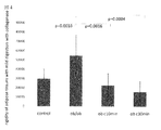
図3はマウスの生殖腺脂肪組織の動的剛性率を示した図である。痩せ型の野性型マウスの正常脂肪組織は約110Paの値を示し、ob/obマウスの肥満脂肪組織は約260Paの値を示すことが明らかとなった。
図4はob/obマウスの肥満脂肪組織をコラゲナーゼで処理し、肥満脂肪組織のコラーゲンを除去した。コラゲナーゼの処理時間により、肥満脂肪組織のコラーゲンの含量が低下する。図4は、肥満脂肪組織のコラーゲンの含量が低下するに従い、肥満脂肪組織の硬さが低下することを示した図である。肥満脂肪組織のコラーゲン含量が低下すれば、肥満脂肪組織の硬さが低下することが示された。
図2はマウスの生殖腺脂肪組織に5.76cm2のカバーガラス(0.2g)を乗せ、それぞれ0、3、6、9gの重石を置いて、0.2、3.2、6.2、9.2g/5.76cm2の4段階の負荷を掛けて培養した。その4段階の場合に生じるMCP−1の分泌量の変化を示した図である。コントロールとして、痩せ型の野性型マウスの正常脂肪組織を使用し、ob/obマウスの肥満脂肪組織との対比を行った。正常脂肪組織に9.2g/5.76cm2の負荷を掛けると、肥満脂肪組織と同様のMCP−1の分泌量を示すことが明らかとなった。
図3はマウスの生殖腺脂肪組織の動的剛性率を示した図である。痩せ型の野性型マウスの正常脂肪組織は約110Paの値を示し、ob/obマウスの肥満脂肪組織は約260Paの値を示すことが明らかとなった。
図4はob/obマウスの肥満脂肪組織をコラゲナーゼで処理し、肥満脂肪組織のコラーゲンを除去した。コラゲナーゼの処理時間により、肥満脂肪組織のコラーゲンの含量が低下する。図4は、肥満脂肪組織のコラーゲンの含量が低下するに従い、肥満脂肪組織の硬さが低下することを示した図である。肥満脂肪組織のコラーゲン含量が低下すれば、肥満脂肪組織の硬さが低下することが示された。
−本発明の第1態様—
本発明の第一の態様は、圧縮細胞及び圧縮組織に関するものである。
本発明の「細胞又は組織」とは、特に限定されるものではないが、生体から採取される健常または病的な状態の細胞又は組織のことであり、あるいは動物細胞の確立された細胞株のことを言う。例えば、脂肪細胞、筋肉細胞、肝臓細胞等の分化された細胞のことを言う。
本発明の「圧縮培養」々は、加圧して(加重を掛けて)圧縮しながら培養することを言う。
本発明の「病的状態の細胞」とは、正常な細胞又は正常な組織と異なり、正常な代謝機能や形状を示さない細胞のことを言う。なお、病的状態にも軽度、重度と幾つかの段階があるが、細胞や組織の正常値の範囲を逸脱した形状(容量)や性質を示す細胞を病的状態の細胞という。例えば、脂肪細胞の場合、病的状態の細胞とは、肥満脂肪細胞のことをいう。従って、本発明の細胞圧縮により作製された病的状態の細胞は、病態モデルの細胞として、簡易に作製でき、スクリーニング方法に汎用することができる。
本発明の「病的状態が亢進している」とは、細胞の病的状態の程度が進行し、代謝機能や形状の正常域からの逸脱の程度が大きくなったことをいう。
本発明の「圧縮率」とは、圧縮前後の圧縮方向におけるサイズ変化のことをいい、2~90%の範囲を挙げることができる。使用する細胞によって、圧縮率の限界は異なるが、例えば正常脂肪細胞又は正常脂肪組織の場合には、2~20%が好ましい。
本発明の「正常脂肪細胞」とは、哺乳動物の正常脂肪細胞のことをいい、例えばマウス線維芽細胞由来の3T3−L1脂肪細胞、マウス脂肪細胞、ラット脂肪細胞、ヒト脂肪細胞の正常脂肪細胞を挙げることができる。
本発明の「脂肪組織」とは、上記脂肪細胞の集積した組織部分のことを言う。例えば、マウスの場合、皮下脂肪組織または生殖腺脂肪組織のことを挙げることができる。
本発明の「肥満脂肪細胞」とは、中性脂肪の貯蔵を行なって肥大化した成熟脂肪細胞のことを言う。肥大した脂肪細胞の細胞直径は、100μm以上であり、最大でも130~140μmである(杉原甫、第124回日本医学会シンポジウム記録集・肥満の科学、71−81頁(2004年))。一方、通常の脂肪細胞(BMI20~22の普通体重者)の細胞直径は、70~90μmであった。このように、通常の脂肪細胞と肥大化した脂肪細胞では、細胞直径が顕著に異なっている。また、形態的にも、肥大化した脂肪細胞では、細胞が多面体の形状を取るが、通常の脂肪細胞では、脂肪細胞は球形で細胞相互間に隙間があり、例えばぶどう型の形状を示している。
本発明の「肥満脂肪組織」とは、例えばob/obマウスの生殖腺脂肪組織などのことを言い、肥満脂肪細胞が多数存在する組織のことを言う。
本発明の「コラーゲン」とは、例えばI~IX型のコラーゲンを挙げることができる。好ましいものとしては、コラーゲンIおよびVIを挙げることができる。
本発明の「加圧」とは、負荷(加重)を掛けることをいい、0.07~1.6g/cm2の範囲を挙げることができる。好ましくは、0.07~1.1g/cm2を挙げることができる。使用する細胞によって、加圧の限界は異なるが、例えば正常脂肪細胞又は正常脂肪組織の場合には、5.76cm2のカバーグラス(0.2g、2.4×2.4cm)を脂肪細胞(組織)に設置し、その上に0.2~9gの加重を掛けることが好ましい。
更には、カバーグラス、寒天培地や2次元ゲル培地などの固形状の培地を使用し、それを上下のサンドイッチ状にすることにより、その間に挟まれた培養細胞に培地の自重による荷重(圧力)を与えることができる。または三次元培地内で細胞を培養し、培地に加重(圧力)を与えることができる。使用する固形状の培地は、それぞれ目的に応じて、所望の形状と自重を持つものを選択することができる。
本発明の「圧縮脂肪細胞又は圧縮脂肪組織」とは、圧縮培養によって新たに作製された人工的な脂肪細胞・脂肪組織のことをいう。本発明の圧縮脂肪細胞・圧縮脂肪組織では、脂肪細胞あるいは脂肪組織の脂肪細胞が肥満する際に見られる脂肪細胞の肥大化を示すことなく、細胞の機能が肥満後の脂肪細胞又は脂肪組織と同様の機能を示している。即ち、インスリン抵抗性を増大させる単球走化活性因子(MCP−1)、IL−12α、TNF−α、PAI−1、レジスチンなどのアディポカインの分泌が、肥満脂肪細胞又は脂肪組織と同様に増大している。
即ち、この人工的な脂肪細胞・脂肪組織は、細胞サイズの肥大化を伴うことがなく、中性脂肪の蓄積も見られない。また、細胞外基質成分も正常脂肪細胞と同じであった。しかし、この人工的な脂肪細胞・脂肪組織の機能に関しては、肥満脂肪細胞・肥満脂肪組織と同様のアディポサイトカインの合成・分泌パターンを示しており、NFκBの活性化が起きていた。また、低分子量GTP結合タンパクRho、ROCK、ミオシン軽鎖キナーゼの活性化も起きており、肥満脂肪細胞・肥満脂肪組織と同様の状態であった。
本発明の「培養」とは、通常の細胞培養に用いる培養のことを表わすと共に、生体内環境下で脂肪組織に加圧圧縮を掛けて留置すること、更には寒天培地や2次元ゲル培地などの二次元培養用培地を2枚重ねて(疑似3次元ゲル化し)、その間に細胞を播種して培養すること、あるいは三次元培養培地で細胞を培養することを意味し、特に限定されるものではない。例えば通常の細胞培養の場合では、DMEM培地のような一般的なものであってもよく、また、目的に応じて、脂肪酸などの添加物を加えることができる。
本発明の「圧縮抵抗性」とは、細胞圧縮時において、細胞の機能に影響が表れ難い性質のことを言う。即ち、圧縮抵抗性には、細胞圧縮の力学的刺激の伝達メカニズムが物理的に及ばない場合と、圧縮認識に関わる信号伝達物質間の情報伝達が生化学的に行われない場合の2つの原因がある。例えば、物理的に及ばない状況としては、細胞と細胞周囲の細胞外基質との間に間隙があり、間隙あるいは間隙に存在する液体などの物質が細胞外基質に加えられた圧縮による力学的負荷を吸収する事態で発生する。この場合、細胞外基質に圧縮が加えられても細胞の圧縮認識機構(インテグリン、アクチン、Rho、ROCK、MLCK、NF−kB)の圧縮による活性化などの生化学的な変化は発生しない。 本発明の「圧縮認識機構が阻害されている」とは、細胞圧縮の力学的刺激が圧縮認識に関わる信号伝達物質間の情報伝達が阻害されることをいう。例えば、NF−kB、Rho、ROCK、MLCKに関する情報伝達メカニズムが阻害されることをいう。その結果、細胞が圧縮されても、圧縮前の細胞と生化学的な変化が発生していない圧縮細胞又は組織となっている。
−本発明の第2態様—
本発明の第2の態様は、細胞及び組織を加圧圧縮して、人工的に細胞の病態モデルを作製する細胞圧縮培養方法に関するものである。
本発明の「細胞又は組織の病的状態」とは、正常な細胞又は正常な組織と異なり、正常な代謝機能や形状を示さないことを言う。なお、病的状態にも軽度、重度と幾つかの段階があるが、細胞や組織の正常値の範囲を逸脱した形状(容量)や性質を示すものを病的状態という。例えば、脂肪細胞の場合、病的状態とは、肥満脂肪細胞の状態を表わすものである。
本発明の「病的細胞又は組織」とは、本発明の細胞圧縮により作製された病的状態を持つ細胞や組織のことを言う。それ故、本発明の「病的細胞又は組織」は、「病態モデルの細胞又は組織」として、簡易に作製でき、スクリーニング方法に汎用することができる。
本発明の「脂肪細胞」とは、例えばマウス線維芽細胞由来の3T3−L1脂肪細胞、マウス脂肪細胞、ラット脂肪細胞、ヒト脂肪細胞のことを挙げることができる。
本発明の「脂肪組織」とは、上記脂肪細胞の集積した組織部分のことを言う。例えば、マウスの皮下脂肪組織またはマウスの生殖腺脂肪組織のことを挙げることができる。
本発明の「溶液培地」とは、細胞培養に使用される一般的な溶液培地のことであり、特に限定されるものではない。例えばDMEM培地を挙げることができる。
本発明の「2次元ゲル培地」とは、例えば特許文献1に記載の方法で作製されたポリアクリルアミドゲル、あるいは市販のチオール化ヒアルロン酸ゲル(www.glycosan.com)の樹脂、例えばポリアミド・ナノファイバー、多孔質のポリビニルフォーマル(PVF)樹脂等のことをいう。
本発明の「細胞圧縮培養方法」とは、例えば上記2次元ゲル培地を2枚重ねて(疑似3次元ゲル化し)、その間に細胞を播種して2次元ゲル培地の自重で圧縮培養する方法、採取した組織(例えば正常脂肪組織)を加圧して圧縮し、溶液培地にて培養する方法などが挙げられる。更には、生体内環境下で脂肪組織に加圧圧縮を掛けて留置する方法を挙げることができる。なお、正常脂肪細胞(組織)から、本発明の圧縮培養方法で得られた人工的な肥満脂肪細胞(組織)は、正常脂肪細胞と同様の容量で、細胞サイズの肥大化がなく、中性脂肪の蓄積も見られない。また、細胞外基質成分も正常脂肪細胞と同じである。一方、この人工的な脂肪細胞・脂肪組織の機能に関しては、一般的な肥満脂肪細胞・肥満脂肪組織と同様のアディポサイトカインの合成・分泌パターンを示しており、NFκBの活性化が起きている。また、低分子量GTP結合タンパクRho、ROCK、ミオシン軽鎖キナーゼの活性化も起きており、一般的な肥満脂肪細胞・肥満脂肪組織と同様の状態である。そして、インスリン抵抗性を増大させる単球走化活性因子(MCP−1)、IL−12α、TNF−α、PAI−1、レジスチンなどのアディポカインの分泌が、肥満脂肪細胞と同様に増大している。
なお、本発明の第1態様と共通する用語は、同じ意味を表わす。
−本発明の第3態様—
本発明の第3の態様は本発明の圧縮細胞を用いる、糖尿病治療剤等のスクリーニング方法に関するものである。
本発明の「脂肪細胞機能」とは、脂肪細胞の代謝、分泌に関する機能のことを言う。この機能は、脂肪細胞内で発現するmRNAの発現量の変動や脂肪細胞から分泌される生理活性タンパク質の分泌量の変動を指標にして、脂肪細胞の機能を評価することができる。
本発明の「アディポサイトカイン」とは、脂肪細胞から分泌される生理活性タンパク質のことを言い、例えば動脈硬化を促進させる方向に働くTNF−α、PAI−1、HB−EGF、と動脈硬化に予防的に働くレプチン、アディポネクチン等が含まれる。TNF−α(腫瘍壊死因子α)は、マクロファージより分泌される炎症性サイトカインでもあり、肥満脂肪組織から分泌され、インスリン抵抗性をひき起こす要因と見なされている。PAI−1(Plasminogen activator inhibitor−1)は肥満脂肪細胞から分泌され、血栓を形成しやすくする。HB−EGF(heparin binding−epidermal growth factor−like growth factor)は肥満脂肪細胞から分泌され、血管平滑筋の遊走・増殖をひき起こす要因となっている。一方、レプチンは白色脂肪細胞から分泌され、視床下部の満腹中枢に働き食欲を抑制する。アディポネクチンは正常脂肪細胞から産生され、インスリン感受性を促進する等の良い要因になっている。
本発明の「化合物の評価方法」とは、培養系中に添加されている薬効成分の影響を受けて、圧縮培養して得られた正常あるいは肥満脂肪細胞由来の人工的な脂肪細胞(肥満型あるいは肥満がさらに悪化した状態に類似した機能を示す脂肪細胞)が分泌するアディポサイトカインの種類と分泌量が変化する様子を測定・評価する方法のことである。このアディポサイトカインの発現変動は、汎用のサイトカインの定量方法を用いて行なうことができる。
本発明の「飽和脂肪酸」とは、飽和高級脂肪酸のことを言い、例えば、パルミチン酸、ステアリン酸、ラウリン酸等の炭素数12以上の飽和脂肪酸を挙げることができる。好ましいものとしては、パルミチン酸を挙げることができる。
なお、本発明の第1態様と共通する用語は、同じ意味を表わす。
次に実施例を挙げて本発明を更に説明するが、本発明はこれらに限定されるものではない。
(実施例1)擬似3次元の細胞培養方法と3T3−LI脂肪細胞の培養
(1)方法
a)250Paポリアクリルアミドゲル(ソフトゲル)の作製
Tissue Eng PartA 15(1):147−154(2009)又はJ.Med.Invest.,56,142−149(2009)に記載の方法に順じて作成される。例えば、250Paソフトゲル用としては、ガラス製カバースリップの表面で、3.0%アクリルアミドと0.2%ビスアクリルアミドの溶液にAPS(Ammonium persulfate)およびTEMED(N,N,N’,N’−tetramethylethane−1,2−diamine)を加えてポリアクリルアミドゲルを作成する。ゲル表面にN−コハク酸イミドあるいはSulfo−SANPAH(N−Sulfosuccinimidyl−6−(4’−azido−2’−nitrophenylamino)hexanoate)を用いて架橋する。このガラス製カバースリップ上に形成されたゲルをコラーゲン、フィブリノーゲンまたはその混合物の溶液に浸漬して、ゲル表面を細胞外マトリックス・リガンドで被覆した。
b)3T3−L1脂肪細胞の擬似3次元モデルでの細胞培養
本発明の疑似3次元培養モデルは、Mol.Cell Biol.,24,7567−7577(2004)の方法に準じて行なわれる。例えば、3T3−L1線維芽細胞の場合、10%CSが添加されたDMEM培地で2日間培養し、コンフルエントにする。この培養細胞を前項のソフトゲル(2.4X2.4cm)に撤き、48時間培養する。培養培地は、DMEM培地に10%FBS、0.5mMIBMX(3−isobutyl−1−methylxanthine)、1μMデキサメタゾン、1.7μMインスリンを含んでいる。その後、10%FBSが添加されたDMEM培地で7−9日間培養し、脂肪細胞に分化させる。その間、10%FBSを含有するDMEM培地は隔日で交換する。
3T3−L1細胞が上記ソフトゲル上で脂肪細胞に分化した後、更に上記a)のソフトゲル(0.07g/cm2)を上記培養細胞に重ねてサンドイッチ状態にした。このサンドイッチ状態の培地のことを、擬似3次元ソフトゲル培地という。更に、この擬似3次元ソフトゲル培地を、10%FBSを含有するDMEM培地に浸漬して細胞培養をする。
(2)結果
ソフトゲル上(2次元モデル)で培養した3T3−L1由来の脂肪細胞と、ソフトゲルを重ねてサンドイッチ状(擬似3次元モデル)で培養した3T3−L1由来の脂肪細胞を比較すると、図1に示すように、細胞の炎症マーカーであるMCP−1の分泌量が、擬似3次元モデル培地で培養された脂肪細胞の場合、約2倍高くなることが明らかとなった。即ち、擬似3次元モデル培地での脂肪細胞の培養においては、細胞培養用のゲル培地(ソフトゲル)の硬さが培養細胞に影響するだけでなく、細胞の上にかぶせられたゲル培地(ソフトゲル)による加圧・圧縮も大きな影響を与えることが明らかとなった。
即ち、細胞外マトリックスが250Paの弾性を持つ培養基盤(2次元ソフトゲル培地)で培養された3T3−L1脂肪細胞は、健常な白色脂肪細胞のモデルとして使用できる。更に、2次元ソフトゲル培地を重ねた本発明の疑似3次元ソフトゲル培地で得られた新たな3T3−L1脂肪細胞(圧縮脂肪細胞)は、インスリン抵抗性を有する肥満脂肪細胞のモデルとして使用できることが見出された。なお、このインスリン抵抗性を有する3T3−L1脂肪細胞において細胞の肥大化は見られなかった。
(実施例2)圧縮と変形させた脂肪組織の性状の評価
上記実施例1に示されるように、正常脂肪細胞を加圧圧縮することにより、インスリン抵抗性を有する人工的な肥満脂肪細胞が得られた。このように、培養条件によって得られる細胞の機能が異なってくるため、一般的に培養細胞を用いて薬剤の評価を行なう場合には、生体環境と同じような力学的な環境でないと、正当な薬剤の評価が難しいことを示している。
そこで、加圧圧縮時に脂肪細胞機能がどのように影響を受けるかを評価した。そのために、脂肪細胞から分泌されるアディポサイトカインのmRNAの発現変動とアディポサイトカインの分泌変動を測定、評価することを行なった。まず、正常マウスまたは肥満糖尿病モデルマウスから脂肪組織を採取して圧縮し、脂肪組織に対する加圧とMCP−1の分泌量について評価した。
a)方法
正常なマウスから採取された200−250mgの脂肪組織(コントロール群)と、肥満糖尿病モデルマウス(ob/obマウス)から採取された200−250mgの脂肪組織(ob/ob群)を、10%CSを含有するDMEM培地で培養する。
それらの組織に対して、2.4cmx2.4cm(0.2g)のカバーグラスを載せ、更にその上に0g、3g、6g、9gの錘を載せる。これらはそれぞれ即ち0.04g/cm2、0.56g/cm2、1.1g/cm2、1.6g/cm2の加圧となる。6時間以上培養後に培地を回収し、その中に含まれるMCP−1の量を測定する。
b)結果
錘を載せて6時間培養した場合、図2で示されるように、正常マウスに由来するコントロール群の脂肪組織では、加重が負荷されるに従って、MCP−1の分泌量が増大することが示された。また、ob/ob群の脂肪組織では、1.1g/cm2の加重を付加させるまでは、MCP−1の分泌量が増大したが、それを超えて、1.6g/cm2の加重を与えると、逆にMCP−1の分泌量が減少していた。
このことは、正常細胞の組織に加重(負荷)を掛けると、MCP−1の分泌量が増大し、組織の性状としては、肥満脂肪細胞の組織に近づいて行くことを示している。更に、肥満脂肪細胞の組織に加重(負荷)を掛けると、MCP−1の分泌量が増大するが、1.1g/cm2の加重を超えると、組織障害が強くなり、肥満細胞の機能が破綻することが明らかになった。従って、このことから、正常な脂肪組織に1.6g/cm2の加重を掛ければ、肥満脂肪細胞の組織と同様の性状を示し、肥満脂肪細胞の代替として使用できることが分かった。更に、肥満脂肪細胞の組織に1.1g/cm2以下の加重を掛けることにより、肥満脂肪細胞から更に病態の悪化した脂肪細胞を作製できることが分かった。
更に、圧縮培養の脂肪細胞・脂肪組織について細胞の外観を評価すると、正常脂肪細胞と同様に、細胞サイズの肥大化がなく、中性脂肪の蓄積がないことが分かった。また、細胞外基質成分が正常脂肪細胞と同じであることも分かった。
一方、圧縮培養の脂肪細胞・脂肪組織の機能は、肥満脂肪細胞・組織と同様のアディポサイトカインの合成・分泌パターンを示し、NFκBの活性化が起こり、低分子量GTP結合タンパクRho、ROCK、ミオシン軽鎖キナーゼ等の活性化が起こっていることが示された。
以上のことから、正常な脂肪細胞の組織や肥満脂肪細胞の組織を使用し、加圧(加重)の程度をコントロールすることにより、肥満の重症度に対応した脂肪細胞の組織を作製できることが示された。従って、これらの肥満の病態の進行度に応じた脂肪細胞の組織を使用して、薬剤の効果と副作用が評価できるようになることが分かった。
[0023]
(実施例3)コラゲナーゼ処理された脂肪組織の性状評価
(1)脂肪組織の動的剛性率(Shear modulus)の測定
a)方法
特許文献1に記載の方法で測定を行った。即ち、20週令のob/obマウスと痩せた野生型のマウスの生殖腺脂肪を直径8mm切り取った。その脂肪組織をRheometrics fluids spectrometer(Rheometrics、Piscataway、NJ)にセットし、3~4%の振動ズレ歪(約2.5rad/sec、37℃)を与えて、脂肪組織の動的剛性率を測定した。
b)結果
図3に示されるように、痩せた野生型マウスの脂肪組織の動的剛性率は、約110Paであったが、Ob/obマウスの肥満脂肪組織の動的剛性率は、約260Paであった。即ち、痩せた野生型マウスから採取された正常脂肪組織は柔らかく、肥満脂肪組織は正常脂肪組織の約2.4倍硬いことが示された。
なお、上記実施例2によれば、正常脂肪組織に約1.6g/cm2の負荷(加重)を掛けて培養すると、MCP−1の分泌量から、肥満脂肪組織と同じような性状の組織に変化することが示されている。このことは、Ob/obマウスを使用しなくても、他の野性型のマウスを使用して、その正常脂肪組織を採取し、例えば約1.6g/cm2の加重を掛ければ、肥満脂肪細胞モデルが作製でき、これを薬効評価に使用できることを示している。
(2)コラゲナーゼ処理をされた肥満脂肪細胞(肥満脂肪組織)の性状測定
図3に示されるように肥満脂肪細胞(肥満脂肪組織)が硬くなる(動的剛性率が高くなる)理由は、細胞外基質の中にコラーゲンが増加することであると考えられた。そこで、コラーゲンを分解すれば、肥満脂肪細胞(肥満脂肪組織)であっても、柔らかくなる(動的剛性率が低くなる)と考えられた。
a)方法
20週令のob/obマウスの生殖腺脂肪(肥満脂肪組織)を直径8mm切り取り、0.04%コラゲナーゼで処理を行った。
37℃にて0分、10分、30分の間、コラゲナーゼで処理した。処理後の脂肪組織をクリープメーター(山電製、RE2−3305B)にセットし、レオロジー測定を行い、脂肪組織のヤング率を測定した。
b)結果
図4に示されるように、コラゲナーゼの処理時間が長くなれば、肥満脂肪細胞の組織であっても、組織の硬さ(ヤング率)が低下することが示された。
また、コラゲナーセ処理により、細胞外基質がおよそ30−50%減少することが、上記ヤング率から推定された。
本発明の加圧圧縮下での細胞培養方法により、例えば正常脂肪細胞を使用して、肥満状態の脂肪細胞と同様の性状を持った細胞に変換できることを見出した。これにより、正常脂肪細胞を用いて、簡単に所望の病態を示す人工的な肥満脂肪細胞を作製できるようになった。即ち、本発明の圧縮培養された細胞は、人工的に得られた病的状態の細胞モデルとして、薬剤の評価スクリーニングに使用することができる。例えば、正常脂肪細胞を用いて得られる圧縮脂肪細胞は、生体の肥満脂肪細胞と同じ機能を示すため、薬剤の正当なスクリーニングの評価結果を得ることが出来る。
また、加圧、圧縮条件を変化させることにより、例えば、正常脂肪細胞あるいは肥満脂肪細胞又は肥満脂肪組織を用いて、簡単に所望の病態の肥満脂肪細胞を作製することができる。そのため、本発明の圧縮脂肪細胞を用いて、肥満症、メタボリック・シンドローム、糖尿病、脂質異常症、高血圧症または動脈硬化の病態生理の解明とともに、新たな診断・治療などに関わる化合物の評価が可能となった。
また、加圧、圧縮条件を変化させることにより、例えば、正常脂肪細胞あるいは肥満脂肪細胞又は肥満脂肪組織を用いて、簡単に所望の病態の肥満脂肪細胞を作製することができる。そのため、本発明の圧縮脂肪細胞を用いて、肥満症、メタボリック・シンドローム、糖尿病、脂質異常症、高血圧症または動脈硬化の病態生理の解明とともに、新たな診断・治療などに関わる化合物の評価が可能となった。
Claims (39)
- 細胞又は組織を圧縮培養して作製される、病的状態の細胞モデルとしての圧縮細胞又は圧縮組織であって、
a)上記圧縮培養における加圧が、0.07~1.6g/cm2であるか、細胞や組織の圧縮率が2~90%である、
b)上記圧縮細胞又は圧縮組織は、圧縮前の細胞サイズの肥大化を伴うことなく、かつ、
圧縮前より病的状態が亢進している細胞の機能を示すものである
ことを特徴とする、圧縮細胞又は圧縮組織。 - 上記細胞又は組織が正常脂肪細胞又は正常脂肪組織であることを特徴とする、請求項1に記載の圧縮細胞又は圧縮組織。
- 上記病的状態が亢進している細胞の機能が、100μm~150μmの肥満状態の脂肪細胞と同じ機能を示すものであることを特徴とする、請求項2に記載の圧縮細胞又は圧縮組織。
- 上記正常脂肪細胞又は正常脂肪組織が、3T3−L1脂肪細胞、マウス脂肪細胞又は組織、ラット脂肪細胞又は組織、ヒト脂肪細胞又はヒト脂肪組織である、請求項2又は3に記載の圧縮細胞又は圧縮組織。
- マウス脂肪組織が、マウス正常脂肪組織である、請求項4に記載の圧縮細胞又は圧縮組織。
- 上記マウス正常脂肪組織が、マウスの生殖腺脂肪組織である、請求項5に記載の圧縮細胞又は圧縮組織。
- 上記脂肪細胞又は脂肪組織が、肥満脂肪細胞又は肥満脂肪組織であることを特徴とする、請求項1に記載の圧縮細胞又は圧縮組織。
- 上記病的状態が亢進している細胞の機能が、更に病態の悪化した肥満脂肪細胞又は肥満脂肪組織の機能を示すものであることを特徴とする、請求項7に記載の圧縮細胞又は圧縮組織。
- 上記肥満脂肪細胞又は肥満脂肪組織が、ob/obマウスの脂肪組織である、請求項7に記載の圧縮細胞又は圧縮組織。
- 上記加圧が、1.6g/cm2である、請求項7~9のいずれかに記載の圧縮細胞又は圧縮組織。
- 上記請求項2~10に記載の圧縮脂肪細胞又は圧縮脂肪組織を使用し、
更に評価薬剤との共存下で培養することにより、上記脂肪細胞又は脂肪組織における脂肪細胞機能を評価することによる、肥満症、メタボリック・シンドローム、糖尿病、脂質異常症、高血圧症または動脈硬化の病態、診断、治療に係る化合物の評価方法。 - 上記脂肪細胞機能を評価するための指標としてのアディポサイトカインが、アディポネクチン、MCP−1またはIL−12αである、請求項11に記載の化合物の評価方法。
- 薬剤と共に脂肪酸が添加されている、請求項11または12に記載の化合物の評価方法。
- 上記脂肪酸が、飽和脂肪酸である、請求項13に記載の化合物の評価方法。
- 上記飽和脂肪酸がパルミチン酸である、請求項14に記載の化合物の評価方法。
- 細胞と細胞外基質との間に間隙を有することを特徴とする、細胞圧縮において細胞に加圧が掛からない圧縮抵抗性の細胞または組織であって、
細胞外基質タンパクが30−50%減少することにより、
加圧により、細胞の生化学的な変化が生じない
ことを特徴とする、圧縮抵抗性の細胞または組織。 - 上記細胞外基質タンパクがコラーゲンである請求項16に記載の圧縮抵抗性の細胞または組織。
- 上記コラーゲンが、コラーゲンVIである、請求項16に記載の圧縮抵抗性の細胞または組織。
- 圧縮認識機構が阻害されていることを特徴とする、圧縮抵抗性の圧縮細胞または組織であって、
加圧により、細胞の生化学的な変化が生じない
ことを特徴とする、圧縮抵抗性の圧縮細胞または組織。 - 上記圧縮認機構がNF−kB、Rho、ROCK、MLCKに関するものである、請求項18に記載の圧縮細胞または組織。
- 細胞又は組織を圧縮培養することを特徴とする、細胞又は組織の病的状態(病態モデルの細胞又は組織)を作製する方法であって、
a)上記細胞や組織の圧縮率が2~90%であるか、
b)圧縮の加圧が、0.07~1.6g/cm2である
ことを特徴とする、病的細胞又は組織の作製方法。 - 上記細胞又は組織が、正常脂肪細胞又は正常脂肪組織であることを特徴とする、請求項21に記載の作製方法。
- 上記圧縮率が2~20%であることを特徴とする、請求項21又は22に記載の作製方法。
- 上記圧縮培養の培地が、溶液培地または150~350Paの硬さの2次元ゲル培地であることを特徴とする、請求項21~23のいずれかに記載の作製方法。
- 上記溶液培地が、DMEM培地である、請求項24に記載の作製方法。
- 上記2次元ゲル培地の硬さが250Paである、請求項24に記載の作製方法。
- 上記2次元ゲル培地がコラーゲン及び/又はフィブロネクチンでゲル上を被覆していることを特徴とする、請求項24又は26に記載の作製方法。
- 上記脂肪細胞又は脂肪組織が、3T3−L1脂肪細胞、マウス脂肪細胞又は組織、ラット脂肪細胞又は組織、ヒト脂肪細胞又はヒト脂肪組織である、請求項22に記載の作製方法。
- マウス脂肪組織が、マウス正常脂肪組織である、請求項28に記載の作製方法。
- 上記細胞又は組織の病的状態が、肥満脂肪細胞又は肥満脂肪組織であることを特徴とする、請求項22に記載の作製方法。
- 上記細胞又は組織が、病的細胞または病的組織であり、病態が更に悪化した病的細胞又は組織の作製方法であることを特徴とする、請求項21に記載の作製方法。
- 上記病的細胞又は組織が、肥満脂肪細胞又は肥満脂肪組織であることを特徴とする、請求項31に記載の作製方法。
- 上記肥満脂肪細胞又は肥満脂肪組織が、ob/obマウスの肥満脂肪細胞又は肥満脂肪組織であることを特徴とする、請求項32に記載の作製方法。
- 圧縮により細胞機能に障害が生じないように、組織の細胞外基質タンパクの密度を低下させ、細胞と細胞外基質の間に間隙を有する細胞又は組織を作製する方法。
- 上記細胞外基質タンパクの密度を低下させることが、コラゲナーゼ処理を行うことである、請求項34に記載の方法。
- 細胞圧縮又は細胞加圧に起因して、病的状態にある細胞又は組織において、圧縮又は加圧環境を解消することにより、細胞又は組織の病的状態を軽減あるいは解消する方法。
- 上記圧縮又は加圧環境を解消することが、細胞外基質による力学的刺激の伝達を解消することである、請求項36に記載の方法。
- 上記細胞外基質による力学的刺激の伝達を解消することが、コラーゲン、アクチンフィラメント、インテグリンの間の信号伝達阻害またはNF−kB、Rho、ROCK、MLCK阻害によるものである、請求項37に記載の方法。
- 上記細胞外基質による力学的刺激の伝達を解消することが、コラーゲン6の産生を阻害することを特徴とする、請求項37に記載の方法。
Applications Claiming Priority (4)
| Application Number | Priority Date | Filing Date | Title |
|---|---|---|---|
| JP2013-090000 | 2013-04-23 | ||
| JP2013089994A JP2016032431A (ja) | 2013-04-23 | 2013-04-23 | 病的状態の細胞モデルとしての圧縮細胞又は圧縮組織 |
| JP2013090000A JP2016032432A (ja) | 2013-04-23 | 2013-04-23 | 細胞圧縮培養による病態モデルの作製方法 |
| JP2013-089994 | 2013-04-23 |
Publications (1)
| Publication Number | Publication Date |
|---|---|
| WO2014174899A1 true WO2014174899A1 (ja) | 2014-10-30 |
Family
ID=51791484
Family Applications (1)
| Application Number | Title | Priority Date | Filing Date |
|---|---|---|---|
| PCT/JP2014/055199 WO2014174899A1 (ja) | 2013-04-23 | 2014-02-24 | 病的状態の細胞モデルとしての圧縮細胞又は圧縮組織とその製法 |
Country Status (1)
| Country | Link |
|---|---|
| WO (1) | WO2014174899A1 (ja) |
Cited By (1)
| Publication number | Priority date | Publication date | Assignee | Title |
|---|---|---|---|---|
| WO2017146124A1 (ja) * | 2016-02-22 | 2017-08-31 | 国立大学法人大阪大学 | 立体的細胞組織の製造方法 |
Citations (4)
| Publication number | Priority date | Publication date | Assignee | Title |
|---|---|---|---|---|
| JP2002531118A (ja) * | 1998-12-11 | 2002-09-24 | アドバンスド ティシュー サイエンシズ,インコーポレーテッド | 埋込み可能な構造物を製造するための、平滑筋細胞に対する剪断流応力の適用 |
| CN101492655A (zh) * | 2009-03-09 | 2009-07-29 | 清华大学 | 一种基于分区的血管化脂肪组织及其构建方法 |
| US20110014597A1 (en) * | 2008-03-25 | 2011-01-20 | Novatissue Gmbh | Perfusable Bioreactor for the Production and/or Cultivation of a Human or Animal Blood Vessel and/or a Human or Animal Tissue |
| JP2012010600A (ja) * | 2010-06-29 | 2012-01-19 | Univ Of Tokushima | 培養脂肪細胞 |
-
2014
- 2014-02-24 WO PCT/JP2014/055199 patent/WO2014174899A1/ja active Application Filing
Patent Citations (4)
| Publication number | Priority date | Publication date | Assignee | Title |
|---|---|---|---|---|
| JP2002531118A (ja) * | 1998-12-11 | 2002-09-24 | アドバンスド ティシュー サイエンシズ,インコーポレーテッド | 埋込み可能な構造物を製造するための、平滑筋細胞に対する剪断流応力の適用 |
| US20110014597A1 (en) * | 2008-03-25 | 2011-01-20 | Novatissue Gmbh | Perfusable Bioreactor for the Production and/or Cultivation of a Human or Animal Blood Vessel and/or a Human or Animal Tissue |
| CN101492655A (zh) * | 2009-03-09 | 2009-07-29 | 清华大学 | 一种基于分区的血管化脂肪组织及其构建方法 |
| JP2012010600A (ja) * | 2010-06-29 | 2012-01-19 | Univ Of Tokushima | 培養脂肪細胞 |
Non-Patent Citations (3)
| Title |
|---|
| AYAKA ITO ET AL.: "Role of MAPK phosphatase-1 (MKP-1) in the induction of MCP-1 during the course of adipocyte hypertrophy", JOURNAL OF JAPAN SOCIETY FOR THE STUDY OF OBESITY, vol. 14, no. 2, 2008, pages 183 - 186 * |
| FRANGOS, J.A. ET AL.: "Shear Stress Induced Stimulation of Mammalian Cell Metabolism", BIOTECHNOLOGY AND BIOENGINEERING, vol. 32, 1988, pages 1053 - 1060 * |
| PELLEGRINELLI, V. ET AL.: "Human adipocyte function is impacted by mechanical cues", THE JOURNAL OF PATHOLOGY, Retrieved from the Internet <URL:http://dx.doi.org/10.1002/pat4347> [retrieved on 20140424] * |
Cited By (3)
| Publication number | Priority date | Publication date | Assignee | Title |
|---|---|---|---|---|
| WO2017146124A1 (ja) * | 2016-02-22 | 2017-08-31 | 国立大学法人大阪大学 | 立体的細胞組織の製造方法 |
| JPWO2017146124A1 (ja) * | 2016-02-22 | 2018-07-26 | 国立大学法人大阪大学 | 立体的細胞組織の製造方法 |
| JP2018113959A (ja) * | 2016-02-22 | 2018-07-26 | 国立大学法人大阪大学 | 立体的細胞組織の製造方法 |
Similar Documents
| Publication | Publication Date | Title |
|---|---|---|
| Huang et al. | Viscoelasticity in natural tissues and engineered scaffolds for tissue reconstruction | |
| Rubiano et al. | Viscoelastic properties of human pancreatic tumors and in vitro constructs to mimic mechanical properties | |
| Sridharan et al. | Long-term changes in the material properties of brain tissue at the implant–tissue interface | |
| Pogoda et al. | Compression stiffening of brain and its effect on mechanosensing by glioma cells | |
| Weng et al. | Mechanically strong double network photocrosslinked hydrogels from N, N-dimethylacrylamide and glycidyl methacrylated hyaluronan | |
| Chang et al. | Preparation and characterization of gelatin/hyaluronic acid cryogels for adipose tissue engineering: In vitro and in vivo studies | |
| Manetti et al. | The IL1-like cytokine IL33 and its receptor ST2 are abnormally expressed in the affected skin and visceral organs of patients with systemic sclerosis | |
| Piechocka et al. | Rheology of heterotypic collagen networks | |
| Wells et al. | Collagen fibril structure and strength in acellular dermal matrix materials of bovine, porcine, and human origin | |
| Klink et al. | Do drainage liquid characteristics serve as predictors for seroma formation after incisional hernia repair? | |
| Leung et al. | Breast pathology in complications associated with polyacrylamide hydrogel (PAAG) mammoplasty | |
| Jia et al. | A pH-responsive hyaluronic acid hydrogel for regulating the inflammation and remodeling of the ECM in diabetic wounds | |
| Yu et al. | Coherent timescales and mechanical structure of multicellular aggregates | |
| Guo et al. | Extracellular matrix stiffness in lung health and disease | |
| Li et al. | How cross-linking mechanisms of methacrylated gellan gum hydrogels alter macrophage phenotype | |
| Zhu et al. | Alginate particles with ovalbumin (OVA) peptide can serve as a carrier and adjuvant for immune therapy in B16-OVA cancer model | |
| WO2014174899A1 (ja) | 病的状態の細胞モデルとしての圧縮細胞又は圧縮組織とその製法 | |
| Khayyeri et al. | Diminishing effects of mechanical loading over time during rat Achilles tendon healing | |
| Skal’s’ kyi et al. | Alternation of the types of fracture for dental polymers in different stages of crack propagation | |
| JP5360663B2 (ja) | 培養脂肪細胞 | |
| Chen et al. | Highly active porous scaffolds of collagen and hyaluronic acid prepared by suppression of polyion complex formation | |
| Gong et al. | Establishment of an experimental intracerebral haemorrhage model for mass effect research using a thermo-sensitive hydrogel | |
| KR20200057765A (ko) | 염증이 생긴 사람 피부의 생체외 모델 및 소염 화합물을 스크리닝하기 위한 이의 용도 | |
| Billiar | The Mechanical Environment of Cells in Collagen Gel Models: Global and Local Effects in Three-dimensional Biological Hydrogels | |
| JP2016032432A (ja) | 細胞圧縮培養による病態モデルの作製方法 |
Legal Events
| Date | Code | Title | Description |
|---|---|---|---|
| 121 | Ep: the epo has been informed by wipo that ep was designated in this application |
Ref document number: 14788840 Country of ref document: EP Kind code of ref document: A1 |
|
| NENP | Non-entry into the national phase |
Ref country code: DE |
|
| 122 | Ep: pct application non-entry in european phase |
Ref document number: 14788840 Country of ref document: EP Kind code of ref document: A1 |
|
| NENP | Non-entry into the national phase |
Ref country code: JP |