WO2014010782A1 - 인간 혈액응고인자 ⅴⅰⅰⅰ 도메인 특이적 항체 및 이의 제조방법 - Google Patents
인간 혈액응고인자 ⅴⅰⅰⅰ 도메인 특이적 항체 및 이의 제조방법 Download PDFInfo
- Publication number
- WO2014010782A1 WO2014010782A1 PCT/KR2012/008075 KR2012008075W WO2014010782A1 WO 2014010782 A1 WO2014010782 A1 WO 2014010782A1 KR 2012008075 W KR2012008075 W KR 2012008075W WO 2014010782 A1 WO2014010782 A1 WO 2014010782A1
- Authority
- WO
- WIPO (PCT)
- Prior art keywords
- coagulation factor
- factor viii
- blood coagulation
- antibody
- human blood
- Prior art date
- Legal status (The legal status is an assumption and is not a legal conclusion. Google has not performed a legal analysis and makes no representation as to the accuracy of the status listed.)
- Ceased
Links
Images





Classifications
-
- G—PHYSICS
- G01—MEASURING; TESTING
- G01N—INVESTIGATING OR ANALYSING MATERIALS BY DETERMINING THEIR CHEMICAL OR PHYSICAL PROPERTIES
- G01N33/00—Investigating or analysing materials by specific methods not covered by groups G01N1/00 - G01N31/00
- G01N33/48—Biological material, e.g. blood, urine; Haemocytometers
- G01N33/50—Chemical analysis of biological material, e.g. blood, urine; Testing involving biospecific ligand binding methods; Immunological testing
- G01N33/86—Chemical analysis of biological material, e.g. blood, urine; Testing involving biospecific ligand binding methods; Immunological testing involving blood coagulating time or factors, or their receptors
-
- C—CHEMISTRY; METALLURGY
- C07—ORGANIC CHEMISTRY
- C07K—PEPTIDES
- C07K16/00—Immunoglobulins [IG], e.g. monoclonal or polyclonal antibodies
- C07K16/18—Immunoglobulins [IG], e.g. monoclonal or polyclonal antibodies against material from animals or humans
- C07K16/36—Immunoglobulins [IG], e.g. monoclonal or polyclonal antibodies against material from animals or humans against blood coagulation factors
-
- C—CHEMISTRY; METALLURGY
- C12—BIOCHEMISTRY; BEER; SPIRITS; WINE; VINEGAR; MICROBIOLOGY; ENZYMOLOGY; MUTATION OR GENETIC ENGINEERING
- C12N—MICROORGANISMS OR ENZYMES; COMPOSITIONS THEREOF; PROPAGATING, PRESERVING, OR MAINTAINING MICROORGANISMS; MUTATION OR GENETIC ENGINEERING; CULTURE MEDIA
- C12N15/00—Mutation or genetic engineering; DNA or RNA concerning genetic engineering, vectors, e.g. plasmids, or their isolation, preparation or purification; Use of hosts therefor
- C12N15/09—Recombinant DNA-technology
- C12N15/11—DNA or RNA fragments; Modified forms thereof; Non-coding nucleic acids having a biological activity
-
- G—PHYSICS
- G01—MEASURING; TESTING
- G01N—INVESTIGATING OR ANALYSING MATERIALS BY DETERMINING THEIR CHEMICAL OR PHYSICAL PROPERTIES
- G01N33/00—Investigating or analysing materials by specific methods not covered by groups G01N1/00 - G01N31/00
- G01N33/48—Biological material, e.g. blood, urine; Haemocytometers
- G01N33/50—Chemical analysis of biological material, e.g. blood, urine; Testing involving biospecific ligand binding methods; Immunological testing
- G01N33/53—Immunoassay; Biospecific binding assay; Materials therefor
-
- G—PHYSICS
- G01—MEASURING; TESTING
- G01N—INVESTIGATING OR ANALYSING MATERIALS BY DETERMINING THEIR CHEMICAL OR PHYSICAL PROPERTIES
- G01N2333/00—Assays involving biological materials from specific organisms or of a specific nature
- G01N2333/435—Assays involving biological materials from specific organisms or of a specific nature from animals; from humans
- G01N2333/745—Assays involving non-enzymic blood coagulation factors
- G01N2333/755—Factors VIII, e.g. factor VIII C [AHF], factor VIII Ag [VWF]
-
- G—PHYSICS
- G01—MEASURING; TESTING
- G01N—INVESTIGATING OR ANALYSING MATERIALS BY DETERMINING THEIR CHEMICAL OR PHYSICAL PROPERTIES
- G01N2800/00—Detection or diagnosis of diseases
- G01N2800/22—Haematology
- G01N2800/224—Haemostasis or coagulation
Definitions
- the present invention relates to human hemagglutinin VIII domain specific antibodies and methods for their preparation.
- Hemophilia is a hereditary bleeding disorder caused by abnormal coagulation factors. Hemophilia A (Hexemia A) and Factor V (FV) -deficient forms (hemophilia B) are associated with the X chromosome. It is widely known. Hemophilia C) form lacking Factor XI (FXI) is a relatively low incidence and autosomal hereditary disease (Bolton-Maggs, PH and KJ Pasi, Haemophilias A and B. Lancet, 2003.361 (9371): p. 1801 -9).
- Hemophilia A occurs in 1 frequency per 5,000 to 10,000 boys and accounts for 85% of Korean hemophilia patients (Kim, KY, et al., Comprehensive clinical and statistical analysis of hemophilia in Korea.JKorean Med Sci, 1988). 3 (3): p. 107-15).
- Exogenous pathways begin with tissue factors activating FVIII exiting the injured site.
- Activated F ⁇ combines with tissue factors to activate FIX and FX (Mackman, N., RE Tilley, and NS Key, Role of the extrinsic pathway of blood coagulation in hemostasis and thrombosis.Arterioscler Thromb Vasc Biol, 2007. 27 (8): p. 1687-93).
- the endogenous pathway is initiated by FXII in contact with the damaged site with a negative charge.
- Activated FXII activates FXI
- activated FXI activates FIX again.
- FVIII and FIX activated by thrombin activate FX.
- This activated FX activates prothrombin as thrombin and activated thrombin activates fibrinogen as fibrin, allowing blood to coagulate (Bowen, DJ, Haemophilia A and haemophilia B: molecular insights. MolPathol, 2002. 55 ( 2): p. 127-44).
- FVIII associated with hemophilia A is produced in hepatocytes and secreted into the blood.
- the gene of FVIII is present on the X chromosome armband and is 180 kb in size.
- the gene of FVIII consists of 26 exons and encodes 2,351 amino acids, including 19 signaling sites for secretion (Lenting, PJ, JA van Mourik, and K. Mertens, The life cycle of coagulation factor VVIII in view ofits structure and function.Blood, 1998. 92 (11): p. 3983-96).
- the precursor of FVIII is a 300 kDa glycoprotein consisting of 2,332 amino acids and has a (NH 2) -A 1 -A 2 -B 3 -A 3 -C- (COOH) structure.
- FVIII is then secreted in the form of a heterodimer in which the heavy chain (A1-A2-B domain) and light chain (A3-C1-C2 domain) are bound to divalent metal ions.
- the von Willebrand factor (vWF) is then circulated in the blood by binding to vascular endothelial cells.
- thrombin forms FVIII in the form of a heterodimer in which the heavy chain (A1, A2 domain) and light chain (A3-C1-C2 domain) are bound to a divalent metal ion. Activate it. Activated FVIII binds to the phospholipid site to which the activated FIX binds away from vWF, acting as a cofactor for FIX to activate FX (Fang, H., L. Wang, and H. Wang, The protein structure and effect of factor VIII. Thromb Res, 2007.119 (1): p. 1-13).
- vWF Proteins known to bind to FVIII include vWF and FIX.
- vWF is a huge glycoprotein synthesized in vascular endothelial cells and megakaryocytes.
- the vWF gene is located on chromosome 12 and consists of 52 exons.
- the mature vWF protein is 250 kDa and consists of 11 domains (D'-D3-A1-A2-A3-D4-B1-B2-B3-C1-C2).
- vWF exists as a multimer of 250 kDa subunits, which are distributed in plasma in various sizes from 500 kDa to thousands of kDa.
- FVIII binds to the 272th amino acid of the D′-D3 domain of vWF and vWF binds between the 1,649-1,689 amino acids of the a3 domain of FVIII and between 2,181-2,243 and 2,248-2,312 amino acids of the C2 domain.
- Non-covalent binding between vWF and FVIII serves to protect FVIII from being captured by vascular endothelial cells and also from being degraded by various serine proteases.
- vWF carries FVIII to where bleeding has occurred, allowing FVIII to be cut and activated by thrombin.
- FIX circulates in the blood in a single chain form of 415 amino acids and has a molecular weight of 57 kDa (Vysotchin, A., LV Medved, and KC Ingham, Domain structure and domaindomain interactions in human coagulation factor IX. J Biol Chem, 1993. 268 ( 12): p. 8436-46).
- FIX cleaves two peptide bonds by FXIa / Ca2 + or TFVIIa / Ca2 + and activates FIXa, a form of serine protease.
- FIXa exists in the form of disulfide bond between the light chain consisting of 1-145 amino acid residues and the heavy chain consisting of 181-415 amino acid residues.
- the light chain consists of a carboxyglutamicacid (Gla) -rich domain (1-40), a short hydrophobic region (41-46) and two epidermal growth factor (EGF) -like domains (47-84, 85-127). It consists of a serine proteolytic enzyme domain that acts as an enzyme.
- Ga carboxyglutamicacid
- EGF epidermal growth factor
- FVIIIa binds to the 330-338 site of FIXa and FIXa binds to 558-565 amino acid residues present in the A2 domain of FVIII to form a FIXa / VIIIa complex (Bajaj, SP, et al., Factor IXa: factor VIIIa interaction. helix 330-338 of factorixainteracts with residues 558-565 and spatially adjacent regions of the a 2 subunit of factor VIIIa.J Biol Chem, 2001. 276 (19): p. 16302-9).
- Methods of diagnosing hemophilia include conventional blood clotting factor activity test and genetic test.
- Commonly used coagulation factor activity measurement methods include single-stage coagulation assay method and chromogen method.
- the single-stage coagulation assay has the advantage of low cost, but has the disadvantage that the standard activity changes by 0.2 ⁇ 1%.
- the chromogen method only the activity of FVIII can be specifically measured, but its use is limited due to the expensive reagents.
- coagulation factor activity measurement is generally applicable to patients, but not applicable to carriers, and thus is not applicable to carrier testing and prenatal diagnosis.
- Genetic testing is very effective in detecting mutations in the causal gene more directly because its activity is variable.
- the size of FVIII the cause of the disease, corresponds to 186kb and consists of 26 exons. Therefore, genetic testing is generally carried out for 26 exons, but the size of the total exon exceeds 7kb and requires high test cost.
- the present inventors have been studying a method for effectively diagnosing human hemophilia at the protein level, and the human blood coagulation factor obtained by injecting a human blood coagulation factor VIII domain specific primer set or synthetic peptide of the present invention into mammals other than humans It was confirmed that the VIII domain specific antibody can diagnose hemophilia by detecting the human blood coagulation factor VIII domain more effectively than the existing antibodies, and completed the present invention.
- An object of the present invention is to provide a hemophilia at a protein level, human blood coagulation factor VIII domain-specific antibody and a method for preparing the same, hemophilia diagnostic kit comprising the antibody, hemophilia diagnostic composition.
- the present invention provides a human blood coagulation factor VIII domain specific antibody and its preparation method.
- the present invention also provides a kit for diagnosing hemophilia, a composition for diagnosing hemophilia, comprising the human blood coagulation factor VIII domain specific antibody.
- the present invention provides information on the diagnosis of hemophilia, characterized in that the identification of the specific antigen-antibody reaction between the human blood coagulation factor VIII domain specific antibody and the human blood coagulation factor VIII domain in a biological sample. Provide a way to.
- the human blood coagulation factor VIII domain specific antibody shows an effective antigen-antibody reaction with the human blood coagulation factor VIII domain
- the antibody titer is higher than that of the human blood coagulation factor VIII domain specific antibody. It is very high, which makes it possible to diagnose hemophilia at the protein level rather than the DNA level, so the hemophilia diagnosis effect is very good.
- Figure 1 shows the A1, A2, A3, C domain sites of human blood coagulation factor VIII for use as epitopes.
- Figure 2 is a diagram showing the result of amplifying the human blood coagulation factor VIII with a domain specific primer, and then confirmed the PCR product.
- FIG. 3 shows a pET28a (+) vector.
- Figure 4 is a diagram showing the results confirmed by the electrophoresis of the transformation of human blood coagulation factor VIII domain specific sequence.
- FIG. 5 is a diagram showing the results of analyzing protein expression by cloning DNA for each human blood coagulation factor VIII domain on pET28a (+) vector, followed by coomassie blue staining (a) and immunochromatography (b). to be.
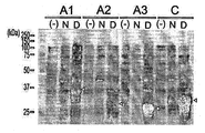
- Figure 6 is a diagram showing the results confirmed by SDS-PAGE and Coomassie blue staining effect for the overexpression of human blood coagulation factor VIII domain protein (N: urea addition control group, D: 8M urea addition group, (-): Negative control incubated without IPTG).
- Figure 7 shows the results of confirming the change in the expression amount of recombinant human blood coagulation factor VIII domain protein according to the change of IPTG and expression induction time through SDS-PAGE and Coomassie blue staining.
- FIG. 8 is a diagram showing the results obtained by extracting each of the recombinant human blood coagulation factor VIII domain proteins and separating them by SDS-PAGE, followed by Coomassie blue staining (A: A1 domain protein, B: A2 domain protein, C: A3 domain protein, D: C domain protein)
- Figure 9 shows the result of confirming the antigen-antibody binding reaction of the antibody and 6x His FVIII fusion protein obtained after injecting a recombinant human blood coagulation factor VIII domain protein in rabbits by SDS-PAGE and immunochromatography
- A Anti-FVIII-A1 and 6x Anti-FVIII-A1 obtained by injection of the synthesized peptides into rabbits
- B Anti-FVIII-A2 and 6x by Anti-FVIII-A1 obtained by injection of the synthesized peptides into rabbits
- C antigen-antibody reaction of anti-FVIII-A3 and 6x His-FVIII-A3 obtained by injecting rabbit purified pure 6x His-FVIII-A3 recombinant protein
- D Anti-FVIII-A3 and 6x His-FVIII-A3 antigen-antibody reactions obtained by injecting rabbit purified pure 6x His-FVIII-A3 re
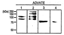
- FIG. 10 is a diagram showing the results of confirming the efficacy of the antibody prepared by confirming the binding reaction of the FVIII protein of the synthesized FVIII domain specific antibody and recombinant human blood coagulation factor FVIII (Advate®) (lane 1: anti-FVIII- A1, lane 2: anti-FVIII-A2, lane 3: anti-FVIII-A3, lane 4: anti-FVIII-C)
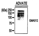
- FIG. 11 is a diagram showing the results of immune development between recombinant human hemagglutinin factor FVIII (Advate®) and FVIII antibody (GMA012).
- FIG. 11 is a diagram showing the results of immune development between recombinant human hemagglutinin factor FVIII (Advate®) and FVIII antibody (GMA012).
- FIG. 12 is a diagram showing the results of comparing the reaction between the synthesized FVIII domain specific antibody and commercial antibodies (GMA012, GMA8011) (A: A2 domain specific antibody, B: C domain specific antibody).
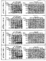
- FIG. 13 shows the results of comparing the reactivity of the synthesized FVIII domain specific antibody with commercial antibodies (GMA012, GMA8011) at different antibody dilution rates (A: A2 domain specific antibody comparison, B: C domain specific) Antibody comparison).
- the present invention is a.
- step 2) fusing the cDNA obtained in step 1) with histidine and cloning the vector to overexpress the histidine fusion recombinant protein;
- step 3 purifying the histidine fusion recombinant protein of step 2) and injecting it into a mammal other than a human; it provides a method for producing a human blood coagulation factor VIII domain specific antibody comprising a.
- the present invention also provides a human coagulation factor VIII domain specific antibody having high antibody titer as compared to the existing human coagulation factor VIII domain specific antibody prepared by the above-described method.
- Step 1) is a step of obtaining human blood coagulation factor VIII domain specific cDNA using a set of human blood coagulation factor VIII specific primers.
- A1, A2, A3 and C can be used as epitopes, and domain specific primers can be used to obtain domain specific recombinant proteins from each domain specific primer.
- the recombinant protein prepared using the primer sets of SEQ ID NOs: 1 and 2 may be human blood coagulation factor VIII A1 domain specific protein
- the recombinant protein prepared using the primer sets of SEQ ID NOs: 3 and 4 may be a human Blood coagulation factor VIII A2 domain specific recombinant protein.
- the recombinant protein prepared using the primer sets of SEQ ID NOs: 5 and 6 may be human blood coagulation factor VIII A3 domain specific protein
- the recombinant protein prepared using the primer sets of SEQ ID NOs: 7 and 8 may represent human blood.
- Step 2) is an overexpression of the histidine fusion recombinant protein, and 6x histidine is labeled on the cloned human blood coagulation factor VIII domain specific cDNA gene obtained in step 1), which is then cloned into a vector and transformed. Histidine fusion recombinant protein can be obtained.
- Transformation includes any method of introducing a nucleic acid to an organism, cell, tissue or organ, and may be carried out by selecting a suitable standard technique according to the host cell as known in the art. These methods include electroshock electroporation, protoplast fusion, calcium phosphate (CaPO 4 ), precipitation, calcium chloride (CaCl 2 ) precipitation, agitation with silicon carbide fibers, agrobacterial mediated transformation, PEG, dextran cells Pate, lipofectamine, and the like. Since the expression amount and formula of the protein appear differently depending on the host cell, it is possible to select and use the most suitable host cell for the purpose.
- Host cells include Escherichia coli, Bacillus subtilis, Streptomyces, Pseudomonas, Proteus mirabilis or Staphylococcus.
- prokaryotic host cells such as, but not limited to.
- fungi eg Aspergillus
- yeast eg Pichia pastoris, Saccharomyces cerevisiae, Schizosaccharomyces
- cells derived from higher eukaryotes including lower eukaryotes such as neurosporacrassa, insect cells, plant cells, mammals, and the like can be used as host cells.
- IPTG isopropylthiogalactoside
- protein expression can be induced for 1 to 20 hours.
- the expression may be induced for 12 hours to induce overexpression of the recombinant protein.
- the human blood coagulation factor VIII domain-specific recombinant protein in which overexpression is induced in the vector in which each domain is recombined may be ultrasonically ground, and then urea may be added to the lysis buffer.
- the amount of urea for preventing the desired recombinant protein from being included in the precipitate of the lysis buffer is preferably 1 to 10M, more preferably 8M.
- step 3 the histidine fusion recombinant protein is purified and injected into mammals except humans.
- Purification of the histidine fusion recombinant protein may be performed by inducing the expression of the histidine fusion recombinant protein for each domain, and then, in a lysis buffer and subjected to ultrasonic grinding, followed by pure purification. To this end, the supernatant containing the recombinant protein may be reacted with the cobalt resin equilibrated with pH 7.0 buffer, and then separated using a column, and the non-specific protein may be removed by using a washing buffer step by step.
- domain A1, A3, and C specific recombinant proteins are preferably obtained by extraction with an elution buffer, and domain A2 specific recombinant proteins may be obtained from a fraction washed with a wash buffer containing imidazole. Can be.
- Mammals other than humans are preferably rabbits, mice, guinea pigs, hamsters, dogs, cats, monkeys, chimpanzees, gorillas, but are not limited thereto.
- the human blood coagulation factor VIII domain is recognized as an antigen, and an immune response is induced.
- the humoral immune response causes an antigen-antibody specific binding reaction to occur in vivo.
- Human clotting factor VIII domain specific antibodies can be generated.
- the specific antigen-antibody response to the human coagulation factor VIII domain can be obtained by specifically overexpressing the human coagulation factor VIII domain, effectively purifying and obtaining it, and injecting it into a mammal other than human. Induction allows the preparation of human hemagglutinin VIII domain specific antibodies with very high antibody titers.
- the present invention comprises the steps of injecting a synthetic peptide set consisting of SEQ ID NO: 9, 10, 11 or SEQ ID NO: 12, 13, 14 to mammals other than humans; preparation of human blood coagulation factor VIII domain specific antibody Provide a method.
- the antibody obtained by injecting the synthetic peptide set of SEQ ID NO: 9, 10, 11 may be a human blood coagulation factor VIII domain A1 specific antibody, and the antibody obtained by injecting the synthetic peptide set of SEQ ID NO: 12, 13, 14 may be human. Blood coagulation factor VIII domain A2-specific antibody.
- the synthetic peptide set of SEQ ID NO: 9, 10, 11 and SEQ ID NO: 12, 13, 14 can be prepared by selecting a high antigenic portion from the human coagulation factor domain A1 or A2 and synthesizing by a known method.
- Human blood coagulation factor VIII domain specific antibody prepared by the above method may be an antibody specific for one or more domains selected from the group consisting of A1, A2, A3, C, and used to diagnose hemophilia at the protein level. Can be.
- the present invention provides a kit for diagnosing hemophilia, a composition for diagnosing hemophilia, comprising a human blood coagulation factor VIII domain specific antibody prepared by the above method.
- compositions and kits comprising antibodies specifically binding to the human coagulation factor VIII domain can be used to provide protein level diagnostics for hemophilia.
- the human blood coagulation factor VIII domain-specific antibody is preferably bound to a chromophore, a chromophore or a fluorescent molecule, but is not limited thereto.
- the human blood coagulation factor VIII domain-specific antibody is essentially the same binding action as biotin to biotin or avidin or streptavidin.
- the one bound to the ligand which is a biotin derivative having the above and the visualization conjugate bound to the ligand-specific binding molecule, the chromophore, the chromophore or the fluorescent molecule bound to the ligand is preferably used.
- the visualization conjugate bound to the ligand-specific binding molecule, the chromophore, the chromophore or the fluorescent molecule bound to the ligand is preferably used.
- kit for diagnosing hemophilia may further comprise one or more other component compositions, solutions or devices suitable for analytical methods.
- the present invention provides a method for providing information for diagnosing hemophilia, characterized by confirming whether a specific antigen-antibody reaction between the antibody prepared by the above production method and a human blood coagulation factor VIII domain in a biological sample. .
- Information for diagnosing hemophilia can be provided by reacting a human blood coagulation factor VIII domain specific antibody of the invention with a biological sample and detecting antigen-antibody complex formation.
- the "biological sample” includes tissue, cells, whole blood, serum, plasma, tissue autopsy samples (brain, skin, lymph nodes, spinal cord, etc.), cell culture supernatants, ruptured eukaryotic cells, and bacterial expression systems, but are not limited thereto. It doesn't happen. These biological samples can be manipulated with or without manipulation of the biological sample to provide information about the diagnosis of hemophilia.
- the "antigen-antibody complex” means a combination of the human blood coagulation factor VIII domain protein antigen in the sample and the antibody according to the present invention which recognizes the same, and the formation of such antigen-antibody complex is performed by conventional enzyme-linked immunoassay (ELISA). , Radioimmunoassay (RIA), sandwich assay, Western blot, immunoprecipitation, immunohistochemical staining, fluorescence immunoassay, enzyme substrate coloration, antigen-antibody aggregation Can be confirmed. However, it is not necessarily limited to these and various applications and applications are possible.
- various labels can be used for detecting the antigen-antibody complex.
- Specific examples may be selected from the group consisting of enzymes, fluorescent materials, ligands, luminescent materials, microparticles, and radioisotopes, but are not necessarily limited thereto.
- domain specific antibodies of human coagulation factor VIII FVIII
- DNAs of each domain were cloned into a pET28a (+) vector (FIG. 3) containing a 6x His tag.
- the vector was then transformed into BL21 (DE3) soluble cells capable of expressing the vector.
- the plasmid DNA of the transformed colonies was digested with restriction enzymes BamHI, XhoI and HindIII, and then electrophoresed on 0.8% agarose gel to confirm whether the target DNA had entered.
- each domain protein of 6V histidine-labeled FVIII it was grown until the OD600 value was 0.6 in 2X YT medium, followed by incubation at 37 ° C, and IPTG treatment to induce protein expression.
- Expression-induced bacteria were incubated at 25 ° C. for 4 hours, harvested, sonicated and lysed to total cell lysate, loaded with 30 ⁇ g, isolated using 10% SDS-PAGE gel, and coomassie blue. Protein expression was analyzed by staining and immunochromatography. Mock bacteria transformed with only the pET28a (+) vector were used as controls.
- the recombinant pET28a (+)-F8-A1, pET28a (+)-F8-A2, pET28a (+)-F8-A3, and pET28a (+)-F8-C express proteins of each domain by IPTG treatment. I could confirm that.
- the protein was overexpressed in a recombinant pET28a (+) vector with the A1, A2, A3, and C domains of FVIII added in lysis buffer (50 mM sodium phosphate, 300 mM NaCl, pH 7.0) and sonicated for total lysis. was obtained, and the supernatant and the precipitate were separated by centrifugation at 4 ° C. and 16,609 ⁇ g for 10 minutes. Each was dissolved in sampling buffer and separated using 10% SDS-PAGE. After the electrophoresis gel was stained with Coomassie blue solution. As a result, it was confirmed that the overexpressed protein was contained in a large amount in the precipitate.
- lysis buffer 50 mM sodium phosphate, 300 mM NaCl, pH 7.0
- the solubility of the protein was increased by adding 8M urea to the lysis buffer in order to resolve the solubility of the overexpression-induced protein in the recombinant pET28a (+) vector and to sink most of the precipitate in the total lysate. More specifically, the bacteria expressing FVIII of each domain were incubated at 37 ° C. until the OD600 value was 0.6, and then 1 mM of IPTG was added thereto, followed by 4 hours of culture at 25 ° C.
- the conditions such as IPTG concentration and expression induction time were adjusted.
- the transformed bacteria were grown at 37 ° C. in 2 ⁇ YT medium until the OD 600 value was 0.6, followed by IPTG concentration (0 mM, 0.1 mM, 0.5 mM, 1.0 mM), expression induction time (0 h, 1 h, 2 h, 3 h, 4 h, 5 h, 12 h and 15 h). After induction of expression, the culture temperature was maintained at 25 °C.
- lysis buffer was added to the harvested bacterial precipitates and ground using an ultrasonic mill. Bacterial proteins made from total cell lysates were dissolved in sampling buffer and protein isolated using 10% SDS-PAGE. After the electrophoresis gel was stained by Coomassie blue staining method. Control Mock was incubated at 25 °C without IPTG treatment.
- 6x His-FVIII fusion protein was isolated using TALON® Metal Affinity Resin. Bacteria transformed for each domain were cultured under the conditions of Example 3 to induce 6x His fusion protein expression. After dissolving the solubilization buffer containing 8M urea and ultrasonically pulverized, the supernatant containing the protein was reacted with the cobalt resin already equilibrated with pH 7.0 buffer and placed in a poly prep column for purification. Proteins that did not bind to the resin were removed with 10 volumes of washing buffer (50 mM sodium phosphate, 300 mM NaCl, 8 M urea, pH7.0), followed by stepwise washing of 5-30 mM imidazole.
- washing buffer 50 mM sodium phosphate, 300 mM NaCl, 8 M urea, pH7.0
- elution buffer 45mM sodium phosphate, 270mM NaCl, 150mM imidazole, 8M urea, pH7.0
- elution buffer 45mM sodium phosphate, 270mM NaCl, 150mM imidazole, 8M urea, pH7.0
- the fusion proteins 6x His-FVIII-A3 and 6x His-FVIII-C purified purely for the production of FVIII domain specific antibodies were injected into rabbits to induce an immune response to induce antibody development.
- 6x His-FVIII-A1 and 6x His-FVIII-A2 which had relatively little expression in protein expression by IPTG, synthesized peptides for the antigenic part of the FVIII sequence and injected into rabbits to induce immune responses.
- Antibody production was induced. More specifically, by analyzing the target coagulation factor VIII sequence through the peptide prediction program of the Abclone antibody manufacturer, the antigenicity and hydrophilicity together with the structure for Factor VIIIa heavy chain, 92 kDa isoform chain. As a result, 3 peptides in the A1 and A2 domains each showed a high possibility of producing an antibody.
- FVIII-A1 and FVIII-A2 peptide synthesis sequences synthesized through this prediction are shown in Table 3.
- each domain of FVIII did not detect only one specific protein, but since FVIII does not exist in the plasma 300 kDa, but in a degraded form, each antibody is present.
- the proteins detected were expected to be digested FVIII containing each domain.
- FVIII-A2 domain antibodies (GMA012, Santacruz) were used to compare the size of the detected proteins.
- the antibody of the present invention detected a protein similar in size to GMA012, an antibody that specifically detects only FVIII-A2.
- the antibody titer of GMA012 is 1: 100
- the anti-FVIII-A2 prepared in the present invention has an antibody titer of 1: 5000, and thus the utility as an antibody was much higher.
- Test Example 3 Detection efficacy comparison with conventional commercial antibodies-antibody titer comparison
- the anti-FVIII-A2 and anti-FVIII-C of the present invention compared to the commercially available FVIII-A2 domain antibody GMA012 (A) and FVIII-C domain antibody GMA8011 (B) at all concentrations.
- the absorbance of the antibody was shown to be higher, indicating that the domain-specific antibody of the FVIII protein of the present invention has a higher reactivity, and thus has much higher utility as an antibody.
- Test Example 4 Comparison of detection efficacy with conventional antibodies-response according to antibody dilution
- Each antibody was diluted in steps of 1: 100, 1: 500, 1: 1000, 1: 5000, 1: 10000 and coated overnight in a 96 well plate at 4 ° C., followed by recombinant human clotting factor FVIII (Advate®) 0.1.
- ELISA was performed by adding the same ⁇ g.
- the anti-FVIII-A2 (A) and the anti-FVIII-A2 domain antibodies (GMA012, Santacruz) and the FVIII-C domain antibodies (GMA8011, Santacruz) were commercially available for all dilution concentrations. It can be seen that the reactivity of the anti-FVIII-C (B) antibody is high.
Landscapes
- Health & Medical Sciences (AREA)
- Life Sciences & Earth Sciences (AREA)
- Chemical & Material Sciences (AREA)
- Engineering & Computer Science (AREA)
- Molecular Biology (AREA)
- Hematology (AREA)
- Biomedical Technology (AREA)
- Immunology (AREA)
- Genetics & Genomics (AREA)
- Organic Chemistry (AREA)
- Biochemistry (AREA)
- General Health & Medical Sciences (AREA)
- Urology & Nephrology (AREA)
- Biotechnology (AREA)
- Medicinal Chemistry (AREA)
- Microbiology (AREA)
- Physics & Mathematics (AREA)
- Biophysics (AREA)
- Pathology (AREA)
- General Physics & Mathematics (AREA)
- Analytical Chemistry (AREA)
- Food Science & Technology (AREA)
- Proteomics, Peptides & Aminoacids (AREA)
- Cell Biology (AREA)
- General Engineering & Computer Science (AREA)
- Bioinformatics & Cheminformatics (AREA)
- Wood Science & Technology (AREA)
- Zoology (AREA)
- Plant Pathology (AREA)
- Peptides Or Proteins (AREA)
- Preparation Of Compounds By Using Micro-Organisms (AREA)
Abstract
본 발명은 인간 혈액응고인자 VIII 도메인 특이적 항체 및 이의 제조방법에 관한 것이다. 본 발명에 따른 인간 혈액응고인자 VIII 도메인 특이적 항체는, 효과적인 인간 혈액 응고인자 VIII 도메인과 특이적인 항원-항체 반응을 통해 기존에 판매되고 있는 인간 혈액응고인자 VIII 도메인 특이적 항체에 비하여 항체 역가가 매우 높으며, 이를 통해 DNA 수준이 아닌 단백질 수준에서 혈우병을 진단할 수 있어, 혈우병 진단 효과가 매우 우수하다.
Description
본 발명은 인간 혈액응고인자 VIII 도메인 특이적 항체 및 이의 제조방법에 관한 것이다.
혈우병은 특정 응고인자의 이상으로 발생하는 유전적인 출혈장애이며, X 염색체에 연관되어 나타나는 Factor VIII(FVIII)이 결핍된 형태(혈우병 A)와 Factor Ⅸ(FⅨ)이 결핍된 형태(혈우병 B)가 널리 알려져 있다. Factor XI(FXI)이 결핍된 혈우병 C) 형태는 상대적으로 발병률이 낮으며, 상염색체 유전질환이다(Bolton-Maggs, P.H. and K.J. Pasi, Haemophilias A and B. Lancet, 2003.361(9371): p. 1801-9). 이중 혈우병 A는 남아 5,000명 내지 10,000명당 1명의 빈도로 발생하며, 한국인 혈우병 환자의 85%를 차지하고있다(Kim, K.Y., et al.,Comprehensive clinical and statistical analysis of hemophilia in Korea. JKorean Med Sci, 1988. 3(3): p. 107-15).
혈관이 손상되면 가장 먼저 일어나는 일은, 손상된 혈관이 수축하면서 피의 순환을 막는 것이다. 출혈이 발생하면 혈액이 응고되어 상처를 막게 되는데, 혈액 응고는 외인성 경로와 내인성 경로, 두 가지의 경로를 통해 시작된다.
외인성 경로는 손상된 부위로부터 빠져나온 조직 Factor가 FⅦ을 활성화시키면서 시작된다. 이때 활성화된 FⅦ은 조직 Factor와 결합하여 FIX과 FX을 활성화시키게된다(Mackman, N., R.E. Tilley, and N.S. Key, Role ofthe extrinsic pathway of blood coagulation in hemostasis and thrombosis.Arterioscler Thromb Vasc Biol, 2007. 27(8): p. 1687-93). 내인성 경로는 손상을 받은 부위가 음전하를 띄면서 그 부위의, 접촉한 FXII에 의해 시작된다. 활성화된 FXII가 FXI을 활성화시키고 활성화된 FXI은 다시 FIX을 활성화시킨다. 트롬빈(Thrombin)에 의해 활성화된 FVIII과 활성화된 FIX은 FX을 활성화시킨다. 이렇게 활성화된 FX은 프로트롬빈(prothrombin)을 트롬빈으로 활성화시키고 활성화된 트롬빈은 피브리노겐을 피브린으로 활성화시켜 혈액이 응고될 수 있게한다(Bowen, D.J., Haemophilia A andhaemophilia B: molecular insights. MolPathol, 2002. 55(2): p. 127-44).
위의 응고인자들 중 혈우병 A와 관련된 FVIII은 간세포에서 생산되어 혈액으로 분비된다. FVIII의 유전자는 X 염색체 장완에 존재하며 크기는 180kb이다. FVIII의 유전자는 26 개의 엑손으로 이루어져 있으며, 분비를 위한 신호부위 19개를 포함하여 2,351개의 아미노산을 코딩하고있다(Lenting, P.J., J.A. vanMourik, and K. Mertens, The life cycle of coagulation factor VVIII in view ofits structure and function. Blood, 1998. 92(11): p. 3983-96). FVIII의 전구체는 2,332개의 아미노산으로 구성된 300kDa의 당단백질로 (NH2)-A1-A2-B-A3-C-(COOH) 구조를 가진다. 이후 FVIII는 중쇄 (A1-A2-B 도메인)와 경쇄 (A3-C1-C2 도메인)가 2가 금속이온에 결합한 헤테로다이머 형태로 분비된다. 이후 혈관내피세포에서 결합한 von Willebrand factor(vWF)와 결합하여 혈액 내를 순환하게 된다.
혈액 내의 FVIII-vWF 복합체가 혈관 손상 부위에서 트롬빈과 결합하게 되면 트롬빈은 FVIII을 중쇄(A1, A2 도메인)와 경쇄(A3-C1-C2 도메인)가 2 가 금속이온에 결합된 헤테로다이머 형태로 만들어 활성화 시킨다. 활성화된 FVIII은 vWF와 떨어져 활성화된 FIX가 결합되어 있는 인지질 부위에 결합하여, FIX이 FX을 활성화시킬 수 있도록 보조인자로서 작용한다(Fang, H., L. Wang, and H.Wang, The protein structure and effect of factor VIII.Thromb Res, 2007.119(1): p. 1-13).
이러한 FVIII과 결합한다고 알려져 있는 단백질에는 vWF, FIX 등이 있다. vWF는 혈관내피 세포 및 거핵 세포에서 합성되는 거대한 당단백질이다. vWF 유전자는 12번 염색체에 위치하며 52개의 엑손으로 이루어져 있다. 성숙한 vWF 단백질은 250 kDa이며 11개의 도메인(D’-D3-A1-A2-A3-D4-B1-B2-B3-C1-C2)으로 구성되어있다. vWF는 250 kDa의 소단위들로 구성된 다량체로 존재하며, 이 다량체들은 500kDa부터 수천 kDa까지 다양한 크기들로 혈장 내에 분포되어있다. FVIII은 vWF의 D’-D3 도메인의 272 번째 아미노산에 결합하며 vWF는 FVIII의 a3 도메인의 1,649-1,689 아미노산 사이와 C2 도메인의 2,181-2,243와 2,248-2,312 아미노산 사이에 결합한다. vWF와 FVIII의 사이의 비공유 결합은 FVIII이 혈관내피 세포에 포획되지 않게 하고 또한 여러 세린 단백질 분해효소에의해 분해되지않도록 보호하는 역할을 한다. vWF는 출혈이 발생한 곳으로 FVIII을 운반하여 FVIII이 트롬빈에 의해 잘려 활성화될 수 있도록 한다. vWF 결핍환자에게 vWF를 투여할 경우 혈장 내 순환하는 FVIII이 정상범위로 증가한다(Astermark, J., S. Lacroix-Desmazes, and M.T. Reding, Inhibitor development.Haemophilia, 2008. 14 Suppl3: p. 36-42).
FIX은 415 개의 아미노산으로 구성된 단일사슬 형태로 혈액 내에서 순환하며 분자량은 57kDa이다(Vysotchin, A., L.V. Medved, and K.C. Ingham,Domain structure and domaindomain interactions in human coagulation factorIX. J BiolChem, 1993. 268(12): p. 8436-46). FIX은 FXIa/Ca²+ 또는 TFVIIa/Ca²+에 의해서 두 개의 펩티드 결합이 잘리고, 활성화되어 세린 단백질 분해효소의 형태인 FIXa가 된다. FIXa은 1-145 아미노산 잔기로 구성된 경쇄와 181-415 아미노산 잔기로 구성된 중쇄가 이황화 결합을 이룬 형태로 존재한다. 경쇄는 carboxyglutamicacid(Gla)-rich 도메인 (1-40), 짧은 소수성 부위(41-46)와 두 개의 epidermal growth factor(EGF)-like 도메인(47-84, 85-127)으로 구성되어 있고 중쇄는 효소작용을 하는 세린 단백질 분해 효소 도메인으로 구성되어있다.
FVIIIa는 FIXa의 330-338 부위에 결합하고 FIXa는 FVIII의 A2 도메인에 존재하는558-565 아미노산 잔기에 결합하여 FIXa/VIIIa 복합체를 형성한다(Bajaj, S.P.,et al., Factor IXa:factorVIIIa interaction. helix 330-338 of factorixainteracts with residues 558-565 and spatially adjacent regions of the a 2 subunit of factor VIIIa. J BiolChem, 2001. 276(19): p. 16302-9).
혈우병을 진단하는 방법으로 기존의 혈액응고인자 활성도 검사와 유전자 검사법이 있다. 일반적으로 적용되는 혈액응고인자 활성도의 측정법은 single-stage coagulation assay 방법과 크로모겐(chromogen)을 이용한 방법이 있다. Single-stage coagulation assay는 저비용이라는 장점이 있지만, 기준활성도가 0.2~1% 가량 변한다는 단점이 있다. 크로모겐 방법의 경우, FVIII의 활성도만 특이적으로 측정할 수 있지만, 고가의 시약으로 인해 사용이 제한적으로 이용되고 있다. 또한 응고인자 활성도 측정법은 일반적으로 환자에게 적용가능하지만 보인자의 경우에는 적용할 수 없어 보인자 검사와 산전진단에는 적용되지 못하고 있다.
유전자 검사법의경우, 활성도가 가변적이기 때문에 보다 직접적으로 원인 유전자의 돌연변이를 검사하는데 매우 효과적이다. 하지만, 질병의 원인 유전자인 FVIII의 크기가 186kb에 해당하며 26개의 엑손으로 구성되어 있어 전체 유전자를 다 조사하는 것에는 매우 큰 한계가 있다. 그렇기 때문에 일반적으로 유전자 검사는 26개의 엑손에 대해서 진행하고 있지만, 전체 엑손의 크기 역시 7kb가 넘어 높은 검사 비용을 필요로 한다. 또한 세계 혈우병 돌연변이 데이터베이스에 등록된 염기서열 돌연변이가 1,000여개로 매우 다양하다.
상기와 같은 다양한 문제와 한계가 존재하기 때문에 보다 편리하고 접근이 용이한 혈우병 진단 방법을 개발할 필요가 있다. 국내의 의료환경과 연구 환경의 비약적 발전으로 인해, 혈액응고인자의 활성도 측정법과 유전자 검사를 통한 돌연변이 검사법 등은 구제 기준에 비하여 큰 차이가 존재하지 않는다. 하지만 후발주자인 국내의 연구 그룹과 제약회사 등이 새로운 시장을 개척하기 위해서는 국내 연구환경을 보다 발전시킬 수 있는 새로운 진단 기법의 개발이 필요하다.
따라서 본 발명자들은 현재까지의 진단 방법에 대한 생각을 전환하여 DNA 수준이 아닌 단백질 수준의 진단법 개발을 수행하였으며, 아직 전세계적으로 보고된 바가 없는 새로운 형태의 진단법을 국내에서 최초로 개발함으로써 혈우병의 새로운 진단방법을 개발하고자 하였다.
본 발명자들은 단백질 수준에서 인간 혈우병을 효과적으로 진단할 수 있는 방법에 대하여 연구하던 중, 본 발명의 인간혈액 응고인자 VIII 도메인 특이적 프라이머 셋트 또는 합성 펩티드를 인간을 제외한 포유류에 주사하여 얻어지는 인간 혈액응고인자 VIII 도메인 특이적 항체가 기존 항체에 비하여 효과적으로 인간 혈액응고인자 VIII 도메인을 검출하여 혈우병을 진단할 수 있음을 확인하고, 본 발명을 완성하였다.
본 발명의 목적은 단백질 수준에서 혈우병을 진단할 수 있는, 인간혈액응고인자 VIII 도메인 특이적 항체 및 이의 제조방법, 상기 항체를 포함하는 혈우병 진단용 키트, 혈우병 진단용 조성물을 제공하는데 있다.
상기와 같은 목적을 달성하기 위해서, 본 발명은 인간 혈액응고인자 VIII 도메인 특이적 항체 및 이의 제조방법을 제공한다.
또한 본 발명은, 상기의 인간 혈액응고인자 VIII 도메인 특이적 항체를 포함하는 혈우병 진단용 키트, 혈우병 진단용 조성물을 제공한다.
또한 본 발명은, 생물학적 시료에서 상기의 인간 혈액응고인자 VIII 도메인 특이적 항체와 인간 혈액 응고인자 VIII 도메인과의 특이적인 항원-항체 반응 여부를 확인하는 것을 특징으로 하는, 혈우병 진단에 대한 정보를 제공하는 방법을 제공한다.
본 발명에 따른 인간 혈액응고인자 VIII 도메인 특이적 항체는, 인간 혈액 응고인자 VIII 도메인과 효과적인 항원-항체 반응을 나타내므로, 기존에 판매되고 있는 인간 혈액응고인자 VIII 도메인 특이적 항체에 비하여 항체 역가가 매우 높으며, 이를 통해 DNA 수준이 아닌 단백질 수준에서 혈우병을 진단할 수 있어, 혈우병 진단 효과가 매우 우수하다.
도 1은 에피토프로 사용하기 위한 인간 혈액 응고인자 VIII의 A1, A2, A3, C 도메인 부위를 나타낸 도이다.
도 2는 인간 혈액 응고인자 VIII을 도메인 특이적 프라이머로 증폭한 후, PCR 산물을 확인한 결과를 나타낸 도이다.
도 3은 pET28a(+) 벡터를 나타낸 도이다.
도 4는 인간 혈액 응고인자 VIII 도메인 특이적 서열의 형질전환 여부를 전기영동을 통해 확인한 결과를 나타낸 도이다.
도 5는 pET28a(+) 벡터에 인간 혈액 응고인자 VIII 도메인별 DNA를 클로닝한 후, 쿠마시블루(coomassie blue) 염색법(a)과 면역발색법(b)으로 단백질 발현을 분석한 결과를 나타낸 도이다.
도 6은 인간 혈액 응고인자 VIII 도메인 단백질의 과발현 유도를 위한 요소 추가 효과를 SDS-PAGE 및 쿠마시 블루 염색을 통해 확인한 결과를 나타낸 도이다(N: 요소 미첨가 대조군, D: 8M 요소 첨가군, (-): IPTG 없이 배양한 음성 대조군).
도 7은 IPTG 및 발현 유도 시간의 변화에 따른, 재조합 인간 혈액응고인자 VIII 도메인 단백질의 발현양의 변화를 SDS-PAGE 및 쿠마시 블루 염색을 통해 확인한 결과를 나타낸 도이다.
도 8은 재조합 인간 혈액 응고인자 VIII 도메인 단백질을 각각 추출하여 SDS-PAGE 로 분리한 후, 쿠마시 블루 염색을 통해 확인한 결과를 나타낸 도이다(A: A1 도메인 단백질, B: A2 도메인 단백질, C: A3 도메인 단백질, D: C 도메인 단백질)
도 9는 재조합 인간 혈액 응고인자 VIII 도메인 단백질을 토끼에 주사하여 면역반응을 유도한 후 얻은 항체와 6x His FVIII 융합 단백질의 항원-항체 결합 반응을 SDS-PAGE 및 면역발색법을 통해 확인한 결과를 나타낸 도이다(A: 합성한 펩티드를 토끼에 주사하여 얻은 anti-FVIII-A1과 6x His-FVIII-A1의 항원-항체반응, B: 합성한 펩티드를 토끼에 주사하여 얻은 anti-FVIII-A2 과 6x His-FVIII-A2의 항원-항체반응, C: 순수 정제한 6x His-FVIII-A3 재조합 단백질을 토끼에 주사하여 얻은 anti-FVIII-A3 와 6x His-FVIII-A3의 항원-항체반응, D: 순수 정제한 6x His-FVIII-A3 재조합 단백질을 토끼에 주사하여 얻은 anti-FVIII-A3 와 6x His-FVIII-A3의 항원-항체반응).
도 10은 합성한 FVIII 도메인 특이적 항체의 FVIII 단백질과 재조합 인간 혈액응고인자 FVIII (Advate®)의 결합반응을 확인하여 제조된 항체의 효용성을 확인한 결과를 나타낸 도이다(lane 1: anti-FVIII-A1, lane 2: anti-FVIII-A2, lane 3: anti-FVIII-A3, lane 4: anti-FVIII-C)
도 11은 재조합 인간 혈액응고인자 FVIII(Advate®)와 FVIII 항체(GMA012) 간의 면역 발색 결과를 나타낸 도이다.
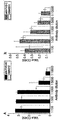
도 12는 합성한 FVIII 도메인 특이적 항체와 시판 항체(GMA012, GMA8011)의 반응도를 비교한 결과를 나타낸 도이다(A: A2 도메인 특이적 항체 비교, B: C 도메인 특이적 항체 비교).
도 13은 합성한 FVIII 도메인 특이적 항체와 시판 항체(GMA012, GMA8011)의 반응도를 항체 희석률을 달리하여 비교한 결과를 나타낸 도이다(A: A2 도메인 특이적 항체 비교, B: C 도메인 특이적 항체 비교).
본 발명은
1) 서열번호 1 및 2, 서열번호 3 및 4, 서열번호 5 및 6, 서열번호 7 및 8 의 프라이머 셋트로 이루어진 군에서 선택된 1종의 프라이머 셋트를 이용하여 인간혈액응고인자 VIII 도메인 특이적 cDNA를 얻는 단계;
2) 상기 1) 단계에서 얻은 cDNA를 히스티딘과 융합하고 벡터에 클로닝하여 히스티딘 융합 재조합 단백질을 과발현시키는 단계; 및
3) 상기 2) 단계의 히스티딘 융합 재조합 단백질을 정제하여 인간을 제외한 포유류에 주입하는 단계;를 포함하는 인간혈액응고인자 VIII 도메인 특이적 항체의 제조방법을 제공한다.
또한, 본 발명은 상기의 제조방법에 의하여 제조된, 기존의 인간 혈액응고인자 VIII 도메인 특이적 항체에 비하여 항체 역가가 높은 인간 혈액응고인자 VIII 도메인 특이적인 항체를 제공한다.
이하, 본 발명의 인간혈액응고인자 VIII 도메인 특이적 항체의 제조 방법에 대하여 단계별로 상세히 설명한다.
상기 1) 단계는 인간혈액응고인자 VIII 특이적 프라이머 셋트를 이용하여 인간혈액응고인자 VIII 도메인 특이적 cDNA를 얻는 단계이다.
인간 혈액응고인자 VIII 도메인 특이적 cDNA를 얻기 위해서, A1, A2, A3, C를 에피토프로 사용할 수 있으며, 각 도메인 특이적 프라이머를 이용하여 도메인의 cDNA를 얻고 이로부터 도메인 특이적 재조합 단백질을 얻을 수 있다. 바람직하게는 서열번호 1 및 2의 프라이머 셋트를 이용하여 제조된 재조합 단백질은 인간 혈액 응고인자 VIII A1 도메인 특이적 단백질일 수 있으며, 서열번호 3 및 4 의 프라이머 셋트를 이용하여 제조된 재조합 단백질은 인간 혈액 응고인자 VIII A2 도메인 특이적 재조합 단백질일 수 있다. 또한, 서열번호 5 및 6의 프라이머 셋트를 이용하여 제조된 재조합 단백질은 인간 혈액 응고인자 VIII A3 도메인 특이적 단백질일 수 있으며, 서열번호 7 및 8 의 프라이머 셋트를 이용하여 제조된 재조합 단백질은 인간 혈액 응고인자 VIII C 도메인 특이적 재조합 단백질일 수 있다.
상기 2) 단계는 히스티딘 융합 재조합 단백질을 과발현 시키는 단계로, 상기 1) 단계에서 얻은 클로닝된 인간 혈액응고인자 VIII 도메인 특이적 cDNA 유전자에 6x 히스티딘을 표지한 후, 이를 벡터에 클로닝하고 형질전환을 수행하여 히스티딘 융합 재조합 단백질을 얻을 수 있다.
형질전환은 핵산을 유기체, 세포, 조직 또는 기관에도 입하는 어떤 방법도 포함되며, 당 분야에서 공지된 바와 같이 숙주 세포에 따라 적합한 표준기술을 선택하여 수행할 수 있다. 이런 방법에는 전기충격 유전자전달법(electroporation), 원형질융합, 인산칼슘(CaPO4), 침전, 염화칼슘(CaCl2) 침전, 실리콘 카바이드 섬유를 이용한 교반, 아그로박테리아 매개된 형질전환, PEG, 덱스트란 셀페이트, 리포펙타민 등이 포함되나 이로 제한되지 않는다. 숙주세포에 따라서 단백질의 발현량과 수식 등이 다르게 나타나므로, 목적에 가장 적합한 숙주세포를 선택하여 사용할 수 있다. 숙주세포로는 에스케리치아 콜라이(Escherichia coli), 바실러스 서브틸리스(Bacillus subtilis), 스트렙토마이세스(Streptomyces), 슈도모나스(Pseudomonas), 프로테우스 미라빌리스(Proteus mirabilis) 또는 스타필로코쿠스(Staphylococcus)와 같은 원핵 숙주세포가 있으나 이로 제한되는 것은 아니다. 또한, 진균(예를 들어, 아스페르길러스(Aspergillus)), 효모(예를 들어, 피치아파스토리스(Pichiapastoris), 사카로마이세스 세르비시애(Saccharomyces cerevisiae), 쉬조사카로마세스(Schizosaccharomyces), 뉴로스포라크라사(Neurosporacrassa))등과 같은 하등 진핵세포, 곤충세포, 식물세포, 포유동물 등을 포함하는 고등 진핵생물 유래의 세포를 숙주세포로 사용할 수 있다.
재조합 단백질의 과발현을 위해서, IPTG(isopropylthiogalactoside)를 0.1 내지 1mM 처리하고 단백질 발현을 1시간 내지 20시간 동안 유도할 수 있다. 바람직하게는 재조합 단백질의 과발현 유도를 위하여 1mM의 IPTG를 처리한 후, 12시간 동안 발현을 유도하여 재조합 단백질의 과발현을 유도할 수 있다.
재조합 단백질을 효과적으로 수득하기 위해서는 각 도메인이 재조합된 벡터에서 과발현이 유도된 인간 혈액응고인자 VIII 도메인 특이적 재조합 단백질을 초음파 분쇄한 후, 용해 완충액에 요소를 첨가할 수 있다. 목적하는 재조합 단백질이 용해 완충액의 침전물에 포함되는 것을 막기 위한 요소의 양은 바람직하게는 1 내지 10M이며, 더욱 바람직하게는 8M로 첨가될 수 있다.
상기 3) 단계는 히스티딘 융합 재조합 단백질을 정제하여 인간을 제외한 포유류에 주입하는 단계이다.
히스티딘 융합 재조합 단백질의 정제는 각 도메인 별로 히스티딘 융합 재조합 단백질의 발현을 유도한 후, 용해 완충액에 넣고 초음파 분쇄를 거친 후, 순수 정제함으로써 이루어질 수 있다. 이를 위하여 pH 7.0 완충액으로 평형시킨 코발트 수지에 재조합 단백질을 포함하는 상층액을 반응시켜 컬럼을 이용하여 분리해 낼 수 있으며, 세척 완충액을 단계적으로 사용하여 비 특이적 단백질을 제거할 수 있다. 순수 정제된 재조합 단백질 중 도메인 A1, A3, C 특이적 재조합 단백질은 용출 완충액으로 추출하여 얻는 것이 바람직하며, 도메인 A2 특이적 재조합 단백질은 이미다졸이 포함된 세척 완충액을 이용하여 세척한 분획으로부터 수득할 수 있다.
상기 인간을 제외한 포유류는 바람직하게는 토끼, 쥐, 기니피그, 햄스터, 개, 고양이, 원숭이, 침팬지, 고릴라일 수 있으나 이에 제한되지 않는다.
상기 히스티딘 융합 재조합 단백질의 주입에 의하여, 인간을 제외한 포유류에서는 인간 혈액응고인자 VIII 도메인을 항원으로 인식하여 면역 반응이 유도되며, 체액성 면역반응에 의하여 항원-항체 특이적 결합반응이 일어나, 생체 내에서 인간 혈액응고인자 VIII 도메인 특이적인 항체를 생성할 수 있다.
상기의 제조방법을 이용하면, 인간 혈액응고인자 VIII 도메인을 특이적으로 과발현시키고 이를 효과적으로 정제하고 수득하여, 인간을 제외한 포유류에 주입함으로써, 인간 혈액응고인자 VIII 도메인에 대한 특이적인 항원-항체 반응을 유도하여, 항체 역가가 매우 높은 인간 혈액응고인자 VIII 도메인 특이적인 항체를 제조할 수 있도록 한다.
또한, 본 발명은 서열번호 9, 10, 11 또는 서열번호 12, 13, 14로 이루어진 합성 펩티드 셋트를 인간을 제외한 포유류에 주입하는 단계;를 포함하는,인간 혈액 응고인자 VIII 도메인 특이적 항체의 제조방법을 제공한다.
상기 서열번호 9, 10, 11의 합성 펩티드 셋트를 주입하여 얻어지는 항체는 인간혈액응고인자 VIII 도메인 A1 특이적 항체일 수 있으며, 서열번호 12, 13, 14의 합성 펩티드 셋트를 주입하여 얻어지는 항체는 인간 혈액응고인자 VIII 도메인 A2 특이적 항체일 수 있다.
상기 서열번호 9, 10, 11 및 서열번호 12, 13, 14의 합성 펩티드 셋트는 인간 혈액응고인자 도메인 A1 또는 A2에서 항원성이 높은 부분을 선별하여, 공지의 방법으로 합성함으로써 제조할 수 있다.
상기의 제조방법으로 제조된 인간혈액응고인자 VIII 도메인 특이적 항체는 A1, A2, A3, C로 구성된 도메인 군으로부터 선택된 1종 이상의 도메인에 특이적인 항체일 수 있으며, 단백질 수준에서 혈우병을 진단하는데 사용될 수 있다.
또한 본 발명은 상기의 제조방법으로 제조된 인간혈액응고인자 VIII 도메인 특이적 항체를 포함하는 혈우병 진단용 키트, 혈우병 진단용 조성물을 제공한다.
인간 혈액응고인자 VIII 도메인에 특이적으로 결합하는 항체를 포함하는 진단용 조성물 및 키트를 사용하여, 혈우병 여부에 대하여 단백질 수준의 진단법을 제공할 수 있다.
본 발명의 인간 혈액응고인자 VIII 도메인 특이적인 항체를 포함하는 혈우병 진단용 키트에 있어서, 인간 혈액응고인자 VIII 도메인 특이적인 항체는 발색효소, 발색물질 또는 형광분자에 결합된 것이 바람직하나, 이에 한정되지 않는다. 또한, 본 발명의 인간 혈액응고인자 VIII 도메인 특이적인 항체를 포함하는 혈우병 진단용 키트에 있어서, 인간 혈액응고인자 VIII 도메인 특이적인 항체는 바이오틴, 또는 아비딘 또는 스트렙트 아비딘에대해 바이오틴과 본질적으로 동일한 결합작용을 갖는 바이오틴 유도체인 리간드에 결합된 것을 사용하는 것이 바람직하고, 상기 리간드 특이적 결합분자에 결합된 발색효소, 발색물질 또는 형광분자가 결합된 시각화 컨쥬게이트가 상기 리간드에 결합된 것을 사용하는 것이 바람직하나 이에 한정되지 않는다
바람직하게 상기 혈우병 진단을 위한 키트는 분석 방법에 적합한 한 종류 또는 그 이상의 다른 구성 성분 조성물, 용액 또는 장치를 더 포함하여 구성될 수 있다.
또한 본 발명은 생물학적 시료에서 상기의 제조방법으로 제조된 항체와 인간 혈액 응고인자 VIII 도메인과의 특이적인 항원-항체 반응 여부를 확인하는 것을 특징으로 하는, 혈우병에 진단에 대한 정보제공방법을 제공한다.
본 발명의 인간 혈액 응고인자 VIII 도메인 특이적 항체를 생물학적 시료와 반응시키고 항원-항체 복합체 형성을 검출함으로써 혈우병 여부를 진단하기 위한 정보를 제공할 수 있다.
상기 “생물학적 시료”란 조직, 세포, 전혈, 혈청, 혈장, 조직부검시료(뇌, 피부, 림프절, 척수 등), 세포배양 상등액, 파열된 진핵세포 및 세균발현계 등을 들 수 있으나, 이에 제한되는 것은 아니다. 이들 생물학적 시료를 조작하거나 조작하지 않은 상태로 본 발명의 항체와 반응시켜 혈우병 진단에 대한 정보를 제공할 수 있다.
상기 "항원-항체복합체"란 시료 중의 인간 혈액응고인자 VIII 도메인 단백질 항원과 이를 인지하는 본 발명에 따른 항체의 결합물을 의미하며, 이러한 항원-항체 복합체의 형성은 통상의 효소면역분석법(ELISA), 방사능면역분석법(radioimmnoassy, RIA), 샌드위치 측정법(sandwich assay), 웨스턴 블랏, 면역침강법, 면역조직화학염색법(immunohistochemical staining), 형광면역법, 효소기질발색법, 항원-항체 응집법 등의 방법을 이용하여 확인될 수 있다. 그러나 반드시 이들로만 제한되지 않고 다양한 응용과 적용이 가능하다.
본 발명에서는 항원-항체 복합체를 검출하기 위한 것으로 여러가지 표지체를 사용할 수 있다. 구체적인 예로는 효소, 형광물, 리간드, 발광물, 미소입자, 방사성 동위 원소로 이루어진 그룹 중에서 선택될 수 있으며, 반드시 이들로만 한정되는 것은 아니다.
이하, 본 발명을 제조예 및 실시예에 의해 상세히 설명한다. 단, 하기 제조예 및 실시예는 본 발명을 예시하는 것일 뿐, 본 발명의 내용이 하기 제조예 및 실시예에 의해 한정되는 것은 아니다.
실시예 1.
FVIII
유전자 클로닝 및 서브클로닝
인간 혈액 응고인자 VIII(human coagulation factor VIII; 이하 FVIII)의 도메인 특이적 항체를 제작하기 위하여, FVIII의 생리 활성화 과정에서 제거되는 B-도메인을 제외한 4개의 도메인인 A1, A2, A3, C를 에피토프로 사용하였다. 에피토프로 사용하기 위한 도메인 부위를 도 1에 나타내었으며, 각각의 도메인 특이적인 프라이머를 제작하여 PCR 산물을 얻었다. 각 도메인에 대한 특이적 프라이머 서열은 다음 표 1과 같다.
표 1 
표 1에 나타낸 프라이머를 사용하여 각 도메인에 해당하는 cDNA 서열을 클로닝하였으며, PCR을 통해 cDNA에 대한 클로닝 결과를 확인하였다. 양성대조군으로 GAPDH 유전자를 사용하였다.
결과를 도 2에 나타내었다.
도 2에 나타낸 바와 같이, FVIII 4개의 도메인인 A1, A2, A3, C의 PCR 산물을 확인할 수 있었다.
실시예 2. 도메인 유전자의 과발현 및 정제
상기 실시예 1에서 클로닝된A1, A2, A3, C 유전자를 박테리아에서 과발현시키고 그 산물을 정제하기 위해 6x His tag이 포함되어 있는 pET28a(+) 벡터(도 3)에 각 도메인별 DNA를 클로닝한 후 벡터를 발현할 수 있는 BL21(DE3) 수용성 세포(competent cell)에 형질전환시켰다. 형질전환이 끝난 콜로니의 플라스미드 DNA를 제한효소 BamHI, XhoI과 HindIII로 절단한 후, 0.8% 아가로오즈 젤에 전기영동하여 목적 DNA가 들어갔는지 여부를 확인하였다.
결과를 도 4에 나타내었다.
도 4에 나타낸 바와 같이, 각각의 삽입 서열을 확인할 수 있었으며, 각 도메인 별 삽입 크기는 아래 표 2와 같다.
표 2
| 벡터 | 삽입크기 |
| pET28a(+)-F8-A1 | 934 bp |
| pET28a(+)-F8-A2 | 622 bp |
| pET28a(+)-F8-A3 | 646 bp |
| pET28a(+)-F8-C | 763 bp |
합성된 벡터가 6x 히스티딘이 표지된 FVIII의 각 도메인 단백질을 발현하는지 확인하기 위해서 2X YT 배지에서 OD600 값이 0.6이 될 때까지 키운 후 37℃ 에서 배양하고 IPTG를 1mM 처리하여 단백질 발현을 유도하였다. 발현이 유도된 박테리아를 25℃ 에서 4 시간 동안 배양하고 수확하여 초음파 분쇄 과정을 거쳐 총 세포용해물로 만든 후 30μg을 로딩하고 10% SDS-PAGE 젤을 사용하여 분리하였고, 쿠마시블루(coomassie blue) 염색법과 면역발색법으로 단백질 발현을 분석하였다.이 때 대조군으로 pET28a(+) 벡터만을 형질전환시킨 Mock 박테리아를 사용하였다.
결과를 도 5에 나타내었다.
도 5에 나타낸 바와 같이, 쿠마시블루를 이용하여 젤을 염색한 결과, pET28a(+) 벡터만을 형질전환 시킨 Mock 박테리아에서는 확인되지 않는 단백질 밴드가 각 도메인을 클로닝하여 단백질 발현을 유도한 박테리아에서는 확인되었다(a). pET28a(+) 벡터에 클로닝된 박테리아를 배양하여 총 세포 용해물을 얻고 이를 SDS-PAGE를 통해 분리하였으며 히스-프로브항체를 사용하여 면역발색법을 진행한 결과, 발현유도시킨 시료에서만 나타나는 밴드가 면역발색법 결과에 나타난 밴드와 일치하는 것을 확인하였다(b). 따라서 재조합시킨 pET28a(+)-F8-A1, pET28a(+)-F8-A2, pET28a(+)-F8-A3, pET28a(+)-F8-C가 IPTG 처리에 의해 각 도메인별 단백질을 발현하는 것을 확인할 수 있었다.
실시예 3. 재조합 FVIII 의 과발현 최적화
6x His과 융합된 FVIII 각 도메인에 대해 단백질의 발현량을 조사한 결과 초음파 분쇄 후 분석되는 단백질의 양이 적은 문제점을 확인하고 이를 해결하기 위하여 과발현 단백질의 용해성을 조사하고 6x His과 융합된 FVIII 각 도메인의 과발현을 위한 최적화 조건을 조사하였다.
FVIII의 A1, A2, A3, C 도메인이 결합되어 있는 재조합 pET28a(+) 벡터에서 과발현 유도한 단백질을 용해 완충액(50 mM 소듐포스페이트, 300 mM NaCl, pH 7.0)을 넣고 초음파 분쇄한 후 총 용해물을 얻고, 4℃, 16,609xg에서 10 분간 원심 분리하여 상층액과 침전물을 구분하여 얻었다. 각각을 샘플링 완충액에 용해시킨 후 10 % SDS-PAGE를 사용하여 분리하였다. 전기영동이 끝난 젤은 쿠마시블루 용액으로 염색하였다. 그 결과 과발현 단백질이 침전물에 다량 포함되어 있음을 확인하였다. 각 도메인이 재조합된 pET28a(+) 벡터에서 과발현 유도한 단백질을 초음파 분쇄한 후 총 용해물에서 대부분 침전물로 가라앉는 것을 해결하기 위해서 용해 완충액에 8M 요소를 첨가하여 단백질의 용해도를 높였다. 보다 구체적으로 각 도메인의 FVIII을 발현하는 박테리아를 37℃ 에서 OD600 값이 0.6이 될 때까지 배양한 후 IPTG 1 mM를 첨가하고 25℃ 에서 4시간 배양하였다. IPTG를 처리하여 단백질의 과발현이 유도된 박테리아를 원심 분리하여 침전시킨 후 8M 요소가 포함된 용해 완충액을 첨가한 후 초음파 분쇄하여 총 용해물을 얻었으며 4℃, 16,609xg에서 10분간 원심분리하고 상층액을 취해서 10% SDS-PAGE를 진행하였다. 전기영동이 끝난 후 쿠마시 블루 염색법으로 젤을 염색하여 단백질을 분석하였다.
결과를 도 6에 나타내었다.
도 6에 나타낸 바와 같이, 요소 첨가 없이 용해 완충액을 처리한 후 초음파 분쇄를 한 대조군 샘플인 N과 8M의 요소가 포함된 용해 완충액을 처리한 후 초음파 분쇄를 한 D 샘플을 비교하면, 요소 첨가 시 단백질의 과발현이 더 잘 유도되어 더 많은 양의 단백질이 상층액에 용해되어 존재하는 것을 확인할 수 있었다.
FVIII의 A1, A2, A3, C 도메인이 결합되어 있는 재조합 pET28a(+) 벡터에서 단백질 발현이 가장 효과적인 조건을 찾기 위해서 IPTG의 농도와 발현 유도 시간 등의 조건을 조정하였다. 형질전환된 박테리아를 37℃, 2X YT 배지에서 OD600 값이 0.6이 될 때까지 키운 후 IPTG 농도(0 mM, 0.1 mM, 0.5 mM, 1.0 mM), 발현유도 시간(0 h, 1 h, 2 h, 3 h, 4 h, 5 h, 12h, 15h)별로 각각 발현을 유도하였다. 발현 유도 후 배양 온도는 25 ℃로 유지하였다. 발현유도가 끝난 후 수확한 박테리아 침전물에 용해 완충액을 첨가하여 초음파 분쇄기를 사용하여 분쇄하였다. 총 세포 용해물로 만든 박테리아 단백질들은 샘플링 완충액에 용해시킨 후 10% SDS-PAGE를 사용하여 단백질을 분리하였다. 전기영동이 끝난 젤은 쿠마시블루 염색법을 통하여 염색하였다. 대조군인 Mock은 25℃에서 IPTG를 처리하지 않고 배양하였다.
결과를 도 7에 나타내었다.
도 7에 나타낸 바와 같이, 각각의 IPTG 및 발현유도 시간을 변화시킨 결과 IPTG 1mM 을 처리하여 12시간 동안 배양하였을 때 단백질의 과발현이 효과적으로 일어나는 것을 확인하였다. 그러나 6x His-FVIII-A1과 6x His-FVIII-A2 는 단백질의 발현량이 6x His-FVIII-A3와 6x His-FVIII-C 의 양과 비교하였을 때 매우 적었다.
실시예 4. 재조합 FVIII의 정제
6x His 융합단백질의 순수 분리를 위해 TALON® Metal Affinity Resin을 이용하여 6x His-FVIII 융합 단백질을 분리하였다. 각 도메인 별로 형질전환된 박테리아를 실시예 3의 조건대로 배양하여 6x His 융합단백질 발현을 유도하였다. 8M 요소가 포함된 용해 완충액을 넣고 초음파 분쇄한 후 이미 pH 7.0 완충액으로 평형시킨 코발트 수지에 단백질을 포함하는 상층액을 반응시킨 후 poly prep column에 넣고 순수 정제를 진행하였다. 수지에 결합하지 않은 단백질들을 세척 완충액(washing buffer)(50 mM 소듐포스페이트, 300 mM NaCl, 8 M 요소, pH7.0) 10 volume으로 제거한 다음 5~30 mM 이미다졸(imidazole)을 단계적으로 세척 완충액에 포함시켜 비 특이적 단백질을 제거하였다. 마지막으로 용출 완충액(elution buffer)(45mM 소듐포스페이트, 270mM NaCl, 150mM 이미다졸, 8M 요소, pH7.0) 5 volume을 poly prep column에 넣어 4℃에서 10 분간 반응시킨 후 천천히 흘려 보내주면서 용출 분획을 시험관에 모아 추출하였다. 추출한 단백질은 10% SDS-PAGE를 사용하여 분리한 후, 쿠마시 블루 염색법을 사용하여 분석하였다.
결과를 도 8에 나타내었다.
도 8에 나타낸 바와 같이, 용출 분획에서 6x His가 융합된 FVIII의 각 도메인 별 재조합 단백질을 얻을 수 있음을 확인하였다. 순수 정제된 재조합 단백질들은 저농도의 이미다졸이 포함된 세척 완충액을 사용하여 세척하는 과정에서부터 소량 추출되는 것을 확인할 수 있었다. 특히 단백질의 발현량이 가장 적었던 A2 도메인의 단백질은 세척 과정에서 대부분의 단백질이 추출되었다. 이러한 결과를 바탕으로, 6x His-FVIII-A1과 6x His-FVIII-A3, 6x His-FVIII-C는 용출 완충액으로 추출한 추출액을 이후 실험에 사용하였고 6x His-FVIII-A2는 20 mM 이미다졸 세척 분획의 결과물을 사용하였다. 각 도메인을 모두 추출한 이후에도 많은 양이 수지에 결합되어 있었다.
실시예 5. FVIII 각 도메인 특이적 항체 제작
FVIII 도메인 특이적 항체 제작을 위해 순수 정제한 융합단백질 6x His-FVIII-A3와 6x His-FVIII-C는 토끼에 주사하여 면역반응을 유도하여 항체 발생을 유도하였다. IPTG에 의한 단백질 발현 유도에서 발현량이 상대적으로 적었던 6x His-FVIII-A1과 6x His-FVIII-A2는 FVIII 서열에서 항원성을 가지는 부분에 대해 펩티드를 합성하여 토끼에 주사하여 면역반응 유도를 통해 항체 생성을 유도하였다. 보다 구체적으로, 앱클론(Abclone) 항체 제조사의 펩티드 예측 프로그램을 통해, 타겟 응고인자 VIII 서열을 분석하여 Factor VIIIa heavy chain, 92 kDa isoform chain에 대한 구조와 함께 항원성(antigenecity)과 친수성(hydrophilicity)을 비교하였으며, 그 결과 A1 및 A2 도메인에서 각각 펩티드 3개씩 항체 제작 가능성이 높은 것으로 나타났다. 이러한 예측을 통해 합성한 FVIII-A1과 FVIII-A2 펩티드 합성 서열은 표 3과 같다.
표 3
| Target | FVIII-A1 | FVIII-A2 | ||
| 서열번호 | 서열번호 | |||
| 펩티드 1 | 9 | DLGELPVDARFPPRVPK-C | 12 | PDDRSYKSQY-C |
| 펩티드 2 | 10 | C-KASEGAEYDDQTSQRE | 13 | C -YSRRLPKGVKHLKD |
| 펩티드 3 | 11 | KSWHWETKN-C | 14 | C - ESVDQRGNQIMSDKR |
토끼의 면역반응 유도로 인해 얻어낸 각 도메인 별 항체의 결합력을 확인하기 위해 순수 정제하여 얻은 6x His FVIII 융합단백질을 10 % SDS-PAGE 전기영동한 후 도메인 별 항체와 결합시키고 면역발색법을 통해서 항원-항체 반응을 확인하였다.
결과를 도 9에 나타내었다.
도 9에 나타낸 바와 같이, 6x His-FVIII-A3와 6x His-FVIII-C에 대한 항체를 재조합 단백질과 결합시켜 인식하는지를 확인한 결과 소수의 불특정 단백질도 함께 검출하였으나 항원-항체 반응은 이상적으로 이루어지는 것을 확인할 수 있었다. 또한 각 도메인 FVIII 서열에서 항원성을 예측하여 제작한 펩티드(표 3)를 통해 얻은 항체를 면역 발색법을 통해 확인해 본 결과 각 도메인 특이 단백질을 효과적으로 검출하는 것을 확인할 수 있었다.
시험예 1. 합성 FVIII 도메인 특이적 항체의 검출 효용성 확인
합성한 FVIII 도메인 특이적 항체가 실제로 FVIII 단백질을 검출하는 것인지 확인하기 위해서 현재 시중에 혈우 환자 치료제로 사용되고 있는 재조합 인간 혈액응고인자 FVIII(Advate®)와의 상호 작용을 확인함으로서 항체로서의 효용성을 확인하였다. 재조합 인간 혈액응고인자 FVIII(Advate®)를 10% SDS-PAGE에서 전기영동하여 분리하였다. 그리고 FVIII 각 도메인 별 특이적 항체를 이용하여 면역발색을 확인하여 각 항체가 Advate®내의 FVIII을 검출하는 것을 확인하였다.
결과를 도 10에 나타내었다.
도 10에 나타낸 바와 같이, FVIII 각 도메인 별 항체가 단 하나의 특이적인 단백질만을 검출하는 것은 아니었으나 혈장 내에서 FVIII은 300kDa의 완벽한 구조로 존재하는 것이 아니고 분해된 형태로 존재하기도 하기 때문에 각 항체가 검출한 단백질들은 각 도메인을 포함하는 분해된 FVIII인 것으로 예상되었다.
시험예 2. 기존 판매 항체와의 검출 효용성 비교-검출단백질 크기 비교
FVIII 각 도메인 특이적 항체의 효용성을 확인하기 위해서 시중에 판매하는 FVIII-A2 도메인 항체(GMA012, Santacruz)를 사용하여 검출된 단백질의 크기를 비교하였다.
결과를 도 11에 나타내었다.
도 11에 나타낸 바와 같이, 본 발명의 항체는 FVIII-A2 만을 특이적으로 검출하는 항체인 GMA012와 비슷한 크기의 단백질을 검출한다는 것을 확인하였다. GMA012는 항체역가가 1:100 인데 비해 본 발명에서 제조한 anti-FVIII-A2는 항체 역가가 1:5000이므로 항체로써의 효용성이 훨씬 높다는 것을 알 수 있었다.
시험예 3.
기존 판매 항체와의 검출 효용성 비교-항체 역가 비교
FVIII 단백질의 도메인 특이적 항체의 효용성을 확인하기 위하여 현재 시중에 판매하는 FVIII-A2 도메인 항체(GMA012, Santacruz)와 FVIII-C 도메인 항체(GMA8011, Santacruz)와의 반응 효율을 ELISA로 확인하였다. 재조합 인간 혈액응고인자 FVIII (Advate®)를 농도별로 희석하여 96 웰 플레이트에 4 ℃에서 하룻밤 동안 코팅 하였다. 각 항체를 1:1000으로 희석하여 4℃에서 하룻밤 동안 반응시킨 후, Stablizedchromogen solution(BiosourceTM)으로 발색시켰다. 이 후, 정지 용액(stop solution)을 첨가하고 450nm 에서 흡광도를 측정하였다.
결과를 도 12에 나타내었다.
도 12에 나타낸 바와 같이, 모든 농도에서 현재 시중에 판매되는 FVIII-A2 도메인 항체 GMA012(A)와 FVIII-C 도메인 항체 GMA8011(B)에 비해 본 발명의 anti-FVIII-A2 및 anti-FVIII-C 항체의 흡광도가 더 높은 것으로 나타나, 본 발명의 FVIII 단백질의 도메인 특이적 항체의 반응도가 더 높아 항체로써의 효용성이 훨씬 높음을 확인하였다.
시험예 4.
기존 판매 항체와의 검출 효용성 비교-항체 희석률에 따른 반응 비교
FVIII 각 도메인 특이적 항체의 효용성 확인을 위해 이번에는 항체의 희석률을 달리하여 재조합 인간 혈액응고인자 FVIII(Advate®)와의 항원-항체 반응을 비교하였다.
각 항체들을 1:100, 1:500, 1:1000, 1:5000, 1:10000로 단계별 희석하여 96 웰 플레이트에 4℃에서 하룻밤 동안 코팅한 뒤, 재조합 인간 혈액응고인자 FVIII (Advate®) 0.1 μg을 동일하게 첨가하여 ELISA 를 진행하였다.
결과를 도 13에 나타내었다.
도 13에 나타낸 바와 같이, 모든 희석 농도에 대해 시중에 판매하는 FVIII-A2 도메인 항체(GMA012, Santacruz)와 FVIII-C 도메인 항체(GMA8011, Santacruz)보다 본 발명의 anti-FVIII-A2(A)와 anti-FVIII-C(B) 항체의 반응도가 높다는 것을 알 수 있다.
Claims (19)
1) 서열번호 1 및 2, 서열번호 3 및 4, 서열번호 5 및 6, 서열번호 7 및 8 의 프라이머 셋트로 이루어진 군에서 선택된 1종의 프라이머 셋트를 이용하여 인간혈액응고인자 VIII 도메인 특이적 cDNA를 얻는 단계;
2) 상기 1) 단계에서 얻은 cDNA를 히스티딘과 융합하고 벡터에 클로닝하여 히스티딘 융합 재조합 단백질을 과발현시키는 단계; 및
3) 상기 2) 단계의 히스티딘 융합 재조합 단백질을 정제하여 인간을 제외한 포유류에 주입하는 단계;를 포함하는 인간혈액응고인자 VIII 도메인 특이적 항체의 제조방법.
제1항에 있어서, 상기 1) 단계의 서열번호 1 및 2의 프라이머 셋트를 이용하여 제조된 재조합 단백질은 인간 혈액 응고인자 VIII A1 도메인 특이적 재조합 단백질임을 특징으로 하는, 인간혈액응고인자 VIII 도메인 특이적 항체의 제조방법.
제1항에 있어서, 상기 1) 단계의 서열번호 3 및 4의 프라이머 셋트를 이용하여 제조된 재조합 단백질은 인간 혈액 응고인자 VIII A2 도메인 특이적 재조합 단백질임을 특징으로 하는, 인간혈액응고인자 VIII 도메인 특이적 항체의 제조방법.
제1항에 있어서, 상기 1) 단계의 서열번호 5 및 6의 프라이머셋트를 이용하여 제조된 재조합 단백질은 인간 혈액 응고인자 VIII A3 도메인 특이적 재조합 단백질임을 특징으로 하는, 인간혈액응고인자 VIII 도메인 특이적 항체의 제조방법.
제1항에 있어서, 상기 1) 단계의 서열번호 7 및 8의 프라이머셋트를 이용하여 제조된 재조합 단백질은 인간 혈액 응고인자 VIII C 도메인 특이적 재조합 단백질임을 특징으로 하는, 인간혈액응고인자 VIII 도메인 특이적 항체의 제조방법.
제1항의 제조방법으로 제조된, 인간혈액응고인자 VIII 도메인 특이적 항체.
제6항에 있어서, 상기 인간혈액응고인자 VIII 도메인은 A1, A2, A3, C로 구성된 도메인 군으로부터 선택된 1종 이상인 것을 특징으로 하는, 인간혈액응고인자 VIII 도메인 특이적 항체.
제 6항 또는 제7항의 인간혈액응고인자 VIII 도메인 특이적 항체를 포함하는 혈우병 진단용 키트.
제6항 또는 제7항의 인간혈액응고인자 VIII 도메인 특이적 항체를 포함하는 혈우병 진단용 조성물.
생물학적 시료에서 제6항의 항체와 인간혈액응고인자 VIII 도메인과의 특이적인 항원-항체 반응 여부를 확인하는 것을 특징으로 하는, 혈우병 진단에 대한 정보제공방법.
제 10항에 있어서, 상기 생물학적 시료는 조직, 세포, 전혈, 혈청, 혈장, 조직부검시료, 세포배양 상등액, 파열된 진핵세포 및 세균발현계로 이루어진 군으로부터 선택된 1종 이상인 것을 특징으로 하는, 혈우병 진단에 대한 정보제공방법.
제10항에 있어서, 상기 항원-항체 반응 여부는 효소면역분석법(ELISA), 방사능면역분석법(radioimmnoassy, RIA), 샌드위치 측정법(sandwich assay), 웨스턴 블랏, 면역침강법, 면역조직화학염색법(immunohistochemical staining), 형광면역법, 효소기질발색법 및 항원-항체 응집법으로 이루어진 군으로부터 선택된 1종 이상의 방법을 이용하여 확인하는 것을 특징으로 하는, 혈우병 진단에 대한 정보제공방법.
서열번호 9, 10, 11 또는 서열번호 12, 13, 14로 이루어진 합성 펩티드 셋트를 인간을 제외한 포유류에 주입하는 단계를 포함하는, 인간 혈액응고인자 VIII 도메인 특이적 항체의 제조방법.
제13항에 있어서, 상기 서열번호 9, 10, 11을 주입하여 얻어지는 항체는 인간혈액응고인자 VIII 도메인 A1 특이적 항체임을 특징으로 하는, 인간혈액응고인자 VIII 도메인 특이적 항체의 제조방법.
제13항에 있어서, 상기 서열번호 12, 13, 14를 주입하여 얻어지는 항체는 인간혈액응고인자 VIII 도메인 A2 특이적 항체임을 특징으로 하는, 인간혈액응고인자 VIII 도메인 특이적 항체의 제조방법.
제13항의 제조방법에 의해 제조되는, 인간혈액응고인자 VIII 도메인 특이적 항체.
제 16항의 인간혈액응고인자 VIII 도메인 특이적 항체를 포함하는 혈우병 진단용 키트.
제16항의 인간혈액응고인자 VIII 도메인 특이적 항체를 포함하는 혈우병 진단용 조성물.
생물학적 시료에서 제16항의 항체와 인간 혈액응고인자 VIII 도메인과의 특이적인 항원-항체 반응 여부를 확인하는 것을 특징으로 하는, 혈우병에 진단에 대한 정보를 제공하는 방법.
Applications Claiming Priority (2)
| Application Number | Priority Date | Filing Date | Title |
|---|---|---|---|
| KR10-2012-0075221 | 2012-07-10 | ||
| KR1020120075221A KR101395736B1 (ko) | 2012-07-10 | 2012-07-10 | 인간 혈액응고인자 ⅷ 도메인 특이적 항체 및 이의 제조방법 |
Publications (1)
| Publication Number | Publication Date |
|---|---|
| WO2014010782A1 true WO2014010782A1 (ko) | 2014-01-16 |
Family
ID=49916209
Family Applications (1)
| Application Number | Title | Priority Date | Filing Date |
|---|---|---|---|
| PCT/KR2012/008075 Ceased WO2014010782A1 (ko) | 2012-07-10 | 2012-10-05 | 인간 혈액응고인자 ⅴⅰⅰⅰ 도메인 특이적 항체 및 이의 제조방법 |
Country Status (2)
| Country | Link |
|---|---|
| KR (1) | KR101395736B1 (ko) |
| WO (1) | WO2014010782A1 (ko) |
Citations (2)
| Publication number | Priority date | Publication date | Assignee | Title |
|---|---|---|---|---|
| EP1194528B1 (en) * | 1999-07-14 | 2007-03-07 | D. Collen Research Foundation vzw | Monoclonal antibody for factor VIII, which even when present in molar excess inactivates factor VIII only in part and a method for producing such an antibody |
| US20090297503A1 (en) * | 2006-08-31 | 2009-12-03 | Masahiro Takeyama | Blood Coagulation Factor VIII Activation-Enhancing Antibodies |
Family Cites Families (3)
| Publication number | Priority date | Publication date | Assignee | Title |
|---|---|---|---|---|
| US6180371B1 (en) * | 1996-06-26 | 2001-01-30 | Emory University | Modified factor VIII |
| US7041635B2 (en) * | 2003-01-28 | 2006-05-09 | In2Gen Co., Ltd. | Factor VIII polypeptide |
| EP1988101A1 (en) * | 2007-05-04 | 2008-11-05 | Novo Nordisk A/S | Improvement of factor VIII polypeptide titers in cell cultures |
-
2012
- 2012-07-10 KR KR1020120075221A patent/KR101395736B1/ko not_active Expired - Fee Related
- 2012-10-05 WO PCT/KR2012/008075 patent/WO2014010782A1/ko not_active Ceased
Patent Citations (2)
| Publication number | Priority date | Publication date | Assignee | Title |
|---|---|---|---|---|
| EP1194528B1 (en) * | 1999-07-14 | 2007-03-07 | D. Collen Research Foundation vzw | Monoclonal antibody for factor VIII, which even when present in molar excess inactivates factor VIII only in part and a method for producing such an antibody |
| US20090297503A1 (en) * | 2006-08-31 | 2009-12-03 | Masahiro Takeyama | Blood Coagulation Factor VIII Activation-Enhancing Antibodies |
Non-Patent Citations (4)
| Title |
|---|
| DATABASE GENBANK 27 May 2012 (2012-05-27), "Homo sapiens coagulation factor VIII, procoagulant component (F8), transcript variant 1, mRNA", accession no. M 000132.3 * |
| DATABASE GENBANK 29 October 2010 (2010-10-29), "Chain A, crystal structure analysis of coagulation Factor VIII", accession no. DB: 2R7E A * |
| JALILZADEH, SHAPOUR ET AL.: "Construction of cDNAs for A1-A2 and A3-Cl-C2 Domains of Factor VIII (al-a2 and a3-cl-c2) by RT-PCR", ARCH. IRN. MED., vol. 5, no. 2, 1 January 2002 (2002-01-01), pages 37 - 43 * |
| LI, C. -M. ET AL.: "Development of monoclonal antibodies against bovine mucin core 2 beta 6 N-acetylglucosaminyltransferase", GLYCOCONJUGATE JOURNAL, vol. 16, no. 9, September 1999 (1999-09-01), pages 555 - 562 * |
Also Published As
| Publication number | Publication date |
|---|---|
| KR101395736B1 (ko) | 2014-05-16 |
| KR20140015694A (ko) | 2014-02-07 |
Similar Documents
| Publication | Publication Date | Title |
|---|---|---|
| Lu et al. | Comparison of activated protein C/protein S-mediated inactivation of human factor VIII and factor V | |
| WO2010041892A9 (ko) | Aimp1 폴리펩티드의 신규한 용도 | |
| Welsch et al. | Amino-terminal alanine functions in a calcium-specific process essential for membrane binding by prothrombin fragment 1 | |
| Mikkat et al. | Mass spectrometric protein structure characterization reveals cause of migration differences of haptoglobin α chains in two‐dimensional gel electrophoresis | |
| Ay et al. | P-selectin gene haplotypes modulate soluble P-selectin concentrations and contribute to the risk of venous thromboembolism | |
| US7169572B1 (en) | Assays for determining anticoagulant cofactor activity | |
| Lubahn et al. | Identification of a F. VIII epitope recognized by a human hemophilic inhibitor | |
| Hoyer | Molecular pathology and immunology of factor VIII (hemophilia A and factor VIII inhibitors) | |
| CN116041520B (zh) | 针对Human CD63的兔单克隆抗体及其制备方法和应用 | |
| DE69840895D1 (de) | Monospezifische antikörper gegen fibrinogen und fibrinopeptide b | |
| WO2020180147A2 (ko) | 나이관련 황반변성 진단용 복합 마커 및 이의 용도 | |
| Smith et al. | Carrier testing in hemophilia B with an immunoassay that distinguishes a prevalent factor IX dimorphism | |
| WO2014010782A1 (ko) | 인간 혈액응고인자 ⅴⅰⅰⅰ 도메인 특이적 항체 및 이의 제조방법 | |
| EP1962091B1 (en) | Methods and kits for detecting and measuring ADAMTS13/FX1 complexes | |
| WO2020189821A1 (ko) | 실명 유발 주요 안질환 진단용 혈액 마커 및 이를 이용한 진단 방법 | |
| ARNESON et al. | Structural arrangement of lens fiber cell plasma membrane protein MP20 | |
| KR101340806B1 (ko) | 인간 혈액응고인자 viii 도메인 특이적인 재조합 단백질 및 이의 이용 | |
| Tykhomyrov et al. | Production and characterization of polyclonal antibodies to human recombinant domain B-free antihemophilic factor VIII | |
| US20030143759A1 (en) | Assays for determining anticoagulant cofactor activity | |
| WO2022030944A1 (ko) | 자가면역 뇌염의 진단 방법 | |
| Mann et al. | A Nonenzymatic cofactors: factor V | |
| JPH02503033A (ja) | インビトロ診断法およびそのために有用な手段 | |
| JP2000069963A (ja) | アポリポプロテインe4特異モノクローナル抗体 | |
| KR100381727B1 (ko) | 마이크로웰을 이용한 dna 돌연변이의 확인방법 및 키트 | |
| JPWO2004018513A1 (ja) | 糖タンパク質若しくは細胞の識別、血清若しくは細胞診断、或いは糖タンパク質若しくは細胞分画のためのレクチンライブラリの使用 |
Legal Events
| Date | Code | Title | Description |
|---|---|---|---|
| 121 | Ep: the epo has been informed by wipo that ep was designated in this application |
Ref document number: 12881037 Country of ref document: EP Kind code of ref document: A1 |
|
| NENP | Non-entry into the national phase |
Ref country code: DE |
|
| 122 | Ep: pct application non-entry in european phase |
Ref document number: 12881037 Country of ref document: EP Kind code of ref document: A1 |