WO2013162050A1 - Lactoferrin fusion protein and method for producing same - Google Patents
Lactoferrin fusion protein and method for producing same Download PDFInfo
- Publication number
- WO2013162050A1 WO2013162050A1 PCT/JP2013/062685 JP2013062685W WO2013162050A1 WO 2013162050 A1 WO2013162050 A1 WO 2013162050A1 JP 2013062685 W JP2013062685 W JP 2013062685W WO 2013162050 A1 WO2013162050 A1 WO 2013162050A1
- Authority
- WO
- WIPO (PCT)
- Prior art keywords
- fusion protein
- hlf
- lactoferrin
- variant
- hinge
- Prior art date
Links
- OCBUCLRUNIEGNG-VQXHTEKXSA-N CC(C)C(C)[C@H]1C(C)CCC1 Chemical compound CC(C)C(C)[C@H]1C(C)CCC1 OCBUCLRUNIEGNG-VQXHTEKXSA-N 0.000 description 1
Images


Classifications
-
- C—CHEMISTRY; METALLURGY
- C07—ORGANIC CHEMISTRY
- C07K—PEPTIDES
- C07K14/00—Peptides having more than 20 amino acids; Gastrins; Somatostatins; Melanotropins; Derivatives thereof
- C07K14/435—Peptides having more than 20 amino acids; Gastrins; Somatostatins; Melanotropins; Derivatives thereof from animals; from humans
- C07K14/79—Transferrins, e.g. lactoferrins, ovotransferrins
-
- A—HUMAN NECESSITIES
- A01—AGRICULTURE; FORESTRY; ANIMAL HUSBANDRY; HUNTING; TRAPPING; FISHING
- A01K—ANIMAL HUSBANDRY; CARE OF BIRDS, FISHES, INSECTS; FISHING; REARING OR BREEDING ANIMALS, NOT OTHERWISE PROVIDED FOR; NEW BREEDS OF ANIMALS
- A01K67/00—Rearing or breeding animals, not otherwise provided for; New breeds of animals
- A01K67/027—New breeds of vertebrates
- A01K67/0275—Genetically modified vertebrates, e.g. transgenic
-
- A—HUMAN NECESSITIES
- A61—MEDICAL OR VETERINARY SCIENCE; HYGIENE
- A61K—PREPARATIONS FOR MEDICAL, DENTAL OR TOILETRY PURPOSES
- A61K38/00—Medicinal preparations containing peptides
- A61K38/16—Peptides having more than 20 amino acids; Gastrins; Somatostatins; Melanotropins; Derivatives thereof
- A61K38/17—Peptides having more than 20 amino acids; Gastrins; Somatostatins; Melanotropins; Derivatives thereof from animals; from humans
- A61K38/40—Transferrins, e.g. lactoferrins, ovotransferrins
-
- A—HUMAN NECESSITIES
- A61—MEDICAL OR VETERINARY SCIENCE; HYGIENE
- A61P—SPECIFIC THERAPEUTIC ACTIVITY OF CHEMICAL COMPOUNDS OR MEDICINAL PREPARATIONS
- A61P1/00—Drugs for disorders of the alimentary tract or the digestive system
-
- A—HUMAN NECESSITIES
- A61—MEDICAL OR VETERINARY SCIENCE; HYGIENE
- A61P—SPECIFIC THERAPEUTIC ACTIVITY OF CHEMICAL COMPOUNDS OR MEDICINAL PREPARATIONS
- A61P1/00—Drugs for disorders of the alimentary tract or the digestive system
- A61P1/02—Stomatological preparations, e.g. drugs for caries, aphtae, periodontitis
-
- A—HUMAN NECESSITIES
- A61—MEDICAL OR VETERINARY SCIENCE; HYGIENE
- A61P—SPECIFIC THERAPEUTIC ACTIVITY OF CHEMICAL COMPOUNDS OR MEDICINAL PREPARATIONS
- A61P1/00—Drugs for disorders of the alimentary tract or the digestive system
- A61P1/10—Laxatives
-
- A—HUMAN NECESSITIES
- A61—MEDICAL OR VETERINARY SCIENCE; HYGIENE
- A61P—SPECIFIC THERAPEUTIC ACTIVITY OF CHEMICAL COMPOUNDS OR MEDICINAL PREPARATIONS
- A61P1/00—Drugs for disorders of the alimentary tract or the digestive system
- A61P1/16—Drugs for disorders of the alimentary tract or the digestive system for liver or gallbladder disorders, e.g. hepatoprotective agents, cholagogues, litholytics
-
- A—HUMAN NECESSITIES
- A61—MEDICAL OR VETERINARY SCIENCE; HYGIENE
- A61P—SPECIFIC THERAPEUTIC ACTIVITY OF CHEMICAL COMPOUNDS OR MEDICINAL PREPARATIONS
- A61P13/00—Drugs for disorders of the urinary system
- A61P13/02—Drugs for disorders of the urinary system of urine or of the urinary tract, e.g. urine acidifiers
-
- A—HUMAN NECESSITIES
- A61—MEDICAL OR VETERINARY SCIENCE; HYGIENE
- A61P—SPECIFIC THERAPEUTIC ACTIVITY OF CHEMICAL COMPOUNDS OR MEDICINAL PREPARATIONS
- A61P13/00—Drugs for disorders of the urinary system
- A61P13/08—Drugs for disorders of the urinary system of the prostate
-
- A—HUMAN NECESSITIES
- A61—MEDICAL OR VETERINARY SCIENCE; HYGIENE
- A61P—SPECIFIC THERAPEUTIC ACTIVITY OF CHEMICAL COMPOUNDS OR MEDICINAL PREPARATIONS
- A61P15/00—Drugs for genital or sexual disorders; Contraceptives
- A61P15/12—Drugs for genital or sexual disorders; Contraceptives for climacteric disorders
-
- A—HUMAN NECESSITIES
- A61—MEDICAL OR VETERINARY SCIENCE; HYGIENE
- A61P—SPECIFIC THERAPEUTIC ACTIVITY OF CHEMICAL COMPOUNDS OR MEDICINAL PREPARATIONS
- A61P19/00—Drugs for skeletal disorders
- A61P19/02—Drugs for skeletal disorders for joint disorders, e.g. arthritis, arthrosis
-
- A—HUMAN NECESSITIES
- A61—MEDICAL OR VETERINARY SCIENCE; HYGIENE
- A61P—SPECIFIC THERAPEUTIC ACTIVITY OF CHEMICAL COMPOUNDS OR MEDICINAL PREPARATIONS
- A61P25/00—Drugs for disorders of the nervous system
-
- A—HUMAN NECESSITIES
- A61—MEDICAL OR VETERINARY SCIENCE; HYGIENE
- A61P—SPECIFIC THERAPEUTIC ACTIVITY OF CHEMICAL COMPOUNDS OR MEDICINAL PREPARATIONS
- A61P25/00—Drugs for disorders of the nervous system
- A61P25/04—Centrally acting analgesics, e.g. opioids
-
- A—HUMAN NECESSITIES
- A61—MEDICAL OR VETERINARY SCIENCE; HYGIENE
- A61P—SPECIFIC THERAPEUTIC ACTIVITY OF CHEMICAL COMPOUNDS OR MEDICINAL PREPARATIONS
- A61P25/00—Drugs for disorders of the nervous system
- A61P25/08—Antiepileptics; Anticonvulsants
-
- A—HUMAN NECESSITIES
- A61—MEDICAL OR VETERINARY SCIENCE; HYGIENE
- A61P—SPECIFIC THERAPEUTIC ACTIVITY OF CHEMICAL COMPOUNDS OR MEDICINAL PREPARATIONS
- A61P25/00—Drugs for disorders of the nervous system
- A61P25/14—Drugs for disorders of the nervous system for treating abnormal movements, e.g. chorea, dyskinesia
- A61P25/16—Anti-Parkinson drugs
-
- A—HUMAN NECESSITIES
- A61—MEDICAL OR VETERINARY SCIENCE; HYGIENE
- A61P—SPECIFIC THERAPEUTIC ACTIVITY OF CHEMICAL COMPOUNDS OR MEDICINAL PREPARATIONS
- A61P25/00—Drugs for disorders of the nervous system
- A61P25/20—Hypnotics; Sedatives
-
- A—HUMAN NECESSITIES
- A61—MEDICAL OR VETERINARY SCIENCE; HYGIENE
- A61P—SPECIFIC THERAPEUTIC ACTIVITY OF CHEMICAL COMPOUNDS OR MEDICINAL PREPARATIONS
- A61P25/00—Drugs for disorders of the nervous system
- A61P25/22—Anxiolytics
-
- A—HUMAN NECESSITIES
- A61—MEDICAL OR VETERINARY SCIENCE; HYGIENE
- A61P—SPECIFIC THERAPEUTIC ACTIVITY OF CHEMICAL COMPOUNDS OR MEDICINAL PREPARATIONS
- A61P25/00—Drugs for disorders of the nervous system
- A61P25/24—Antidepressants
-
- A—HUMAN NECESSITIES
- A61—MEDICAL OR VETERINARY SCIENCE; HYGIENE
- A61P—SPECIFIC THERAPEUTIC ACTIVITY OF CHEMICAL COMPOUNDS OR MEDICINAL PREPARATIONS
- A61P25/00—Drugs for disorders of the nervous system
- A61P25/28—Drugs for disorders of the nervous system for treating neurodegenerative disorders of the central nervous system, e.g. nootropic agents, cognition enhancers, drugs for treating Alzheimer's disease or other forms of dementia
-
- A—HUMAN NECESSITIES
- A61—MEDICAL OR VETERINARY SCIENCE; HYGIENE
- A61P—SPECIFIC THERAPEUTIC ACTIVITY OF CHEMICAL COMPOUNDS OR MEDICINAL PREPARATIONS
- A61P29/00—Non-central analgesic, antipyretic or antiinflammatory agents, e.g. antirheumatic agents; Non-steroidal antiinflammatory drugs [NSAID]
-
- A—HUMAN NECESSITIES
- A61—MEDICAL OR VETERINARY SCIENCE; HYGIENE
- A61P—SPECIFIC THERAPEUTIC ACTIVITY OF CHEMICAL COMPOUNDS OR MEDICINAL PREPARATIONS
- A61P3/00—Drugs for disorders of the metabolism
-
- A—HUMAN NECESSITIES
- A61—MEDICAL OR VETERINARY SCIENCE; HYGIENE
- A61P—SPECIFIC THERAPEUTIC ACTIVITY OF CHEMICAL COMPOUNDS OR MEDICINAL PREPARATIONS
- A61P3/00—Drugs for disorders of the metabolism
- A61P3/06—Antihyperlipidemics
-
- A—HUMAN NECESSITIES
- A61—MEDICAL OR VETERINARY SCIENCE; HYGIENE
- A61P—SPECIFIC THERAPEUTIC ACTIVITY OF CHEMICAL COMPOUNDS OR MEDICINAL PREPARATIONS
- A61P3/00—Drugs for disorders of the metabolism
- A61P3/08—Drugs for disorders of the metabolism for glucose homeostasis
- A61P3/10—Drugs for disorders of the metabolism for glucose homeostasis for hyperglycaemia, e.g. antidiabetics
-
- A—HUMAN NECESSITIES
- A61—MEDICAL OR VETERINARY SCIENCE; HYGIENE
- A61P—SPECIFIC THERAPEUTIC ACTIVITY OF CHEMICAL COMPOUNDS OR MEDICINAL PREPARATIONS
- A61P31/00—Antiinfectives, i.e. antibiotics, antiseptics, chemotherapeutics
- A61P31/04—Antibacterial agents
-
- A—HUMAN NECESSITIES
- A61—MEDICAL OR VETERINARY SCIENCE; HYGIENE
- A61P—SPECIFIC THERAPEUTIC ACTIVITY OF CHEMICAL COMPOUNDS OR MEDICINAL PREPARATIONS
- A61P31/00—Antiinfectives, i.e. antibiotics, antiseptics, chemotherapeutics
- A61P31/12—Antivirals
-
- A—HUMAN NECESSITIES
- A61—MEDICAL OR VETERINARY SCIENCE; HYGIENE
- A61P—SPECIFIC THERAPEUTIC ACTIVITY OF CHEMICAL COMPOUNDS OR MEDICINAL PREPARATIONS
- A61P35/00—Antineoplastic agents
-
- A—HUMAN NECESSITIES
- A61—MEDICAL OR VETERINARY SCIENCE; HYGIENE
- A61P—SPECIFIC THERAPEUTIC ACTIVITY OF CHEMICAL COMPOUNDS OR MEDICINAL PREPARATIONS
- A61P35/00—Antineoplastic agents
- A61P35/04—Antineoplastic agents specific for metastasis
-
- A—HUMAN NECESSITIES
- A61—MEDICAL OR VETERINARY SCIENCE; HYGIENE
- A61P—SPECIFIC THERAPEUTIC ACTIVITY OF CHEMICAL COMPOUNDS OR MEDICINAL PREPARATIONS
- A61P37/00—Drugs for immunological or allergic disorders
- A61P37/02—Immunomodulators
-
- A—HUMAN NECESSITIES
- A61—MEDICAL OR VETERINARY SCIENCE; HYGIENE
- A61P—SPECIFIC THERAPEUTIC ACTIVITY OF CHEMICAL COMPOUNDS OR MEDICINAL PREPARATIONS
- A61P43/00—Drugs for specific purposes, not provided for in groups A61P1/00-A61P41/00
-
- A—HUMAN NECESSITIES
- A61—MEDICAL OR VETERINARY SCIENCE; HYGIENE
- A61P—SPECIFIC THERAPEUTIC ACTIVITY OF CHEMICAL COMPOUNDS OR MEDICINAL PREPARATIONS
- A61P7/00—Drugs for disorders of the blood or the extracellular fluid
- A61P7/06—Antianaemics
-
- A—HUMAN NECESSITIES
- A61—MEDICAL OR VETERINARY SCIENCE; HYGIENE
- A61P—SPECIFIC THERAPEUTIC ACTIVITY OF CHEMICAL COMPOUNDS OR MEDICINAL PREPARATIONS
- A61P9/00—Drugs for disorders of the cardiovascular system
- A61P9/12—Antihypertensives
-
- C—CHEMISTRY; METALLURGY
- C12—BIOCHEMISTRY; BEER; SPIRITS; WINE; VINEGAR; MICROBIOLOGY; ENZYMOLOGY; MUTATION OR GENETIC ENGINEERING
- C12N—MICROORGANISMS OR ENZYMES; COMPOSITIONS THEREOF; PROPAGATING, PRESERVING, OR MAINTAINING MICROORGANISMS; MUTATION OR GENETIC ENGINEERING; CULTURE MEDIA
- C12N15/00—Mutation or genetic engineering; DNA or RNA concerning genetic engineering, vectors, e.g. plasmids, or their isolation, preparation or purification; Use of hosts therefor
- C12N15/09—Recombinant DNA-technology
- C12N15/11—DNA or RNA fragments; Modified forms thereof; Non-coding nucleic acids having a biological activity
- C12N15/62—DNA sequences coding for fusion proteins
-
- C—CHEMISTRY; METALLURGY
- C12—BIOCHEMISTRY; BEER; SPIRITS; WINE; VINEGAR; MICROBIOLOGY; ENZYMOLOGY; MUTATION OR GENETIC ENGINEERING
- C12N—MICROORGANISMS OR ENZYMES; COMPOSITIONS THEREOF; PROPAGATING, PRESERVING, OR MAINTAINING MICROORGANISMS; MUTATION OR GENETIC ENGINEERING; CULTURE MEDIA
- C12N15/00—Mutation or genetic engineering; DNA or RNA concerning genetic engineering, vectors, e.g. plasmids, or their isolation, preparation or purification; Use of hosts therefor
- C12N15/09—Recombinant DNA-technology
- C12N15/63—Introduction of foreign genetic material using vectors; Vectors; Use of hosts therefor; Regulation of expression
- C12N15/79—Vectors or expression systems specially adapted for eukaryotic hosts
- C12N15/82—Vectors or expression systems specially adapted for eukaryotic hosts for plant cells, e.g. plant artificial chromosomes (PACs)
- C12N15/8241—Phenotypically and genetically modified plants via recombinant DNA technology
- C12N15/8242—Phenotypically and genetically modified plants via recombinant DNA technology with non-agronomic quality (output) traits, e.g. for industrial processing; Value added, non-agronomic traits
- C12N15/8257—Phenotypically and genetically modified plants via recombinant DNA technology with non-agronomic quality (output) traits, e.g. for industrial processing; Value added, non-agronomic traits for the production of primary gene products, e.g. pharmaceutical products, interferon
-
- A—HUMAN NECESSITIES
- A01—AGRICULTURE; FORESTRY; ANIMAL HUSBANDRY; HUNTING; TRAPPING; FISHING
- A01K—ANIMAL HUSBANDRY; CARE OF BIRDS, FISHES, INSECTS; FISHING; REARING OR BREEDING ANIMALS, NOT OTHERWISE PROVIDED FOR; NEW BREEDS OF ANIMALS
- A01K2217/00—Genetically modified animals
- A01K2217/05—Animals comprising random inserted nucleic acids (transgenic)
- A01K2217/052—Animals comprising random inserted nucleic acids (transgenic) inducing gain of function
-
- A—HUMAN NECESSITIES
- A01—AGRICULTURE; FORESTRY; ANIMAL HUSBANDRY; HUNTING; TRAPPING; FISHING
- A01K—ANIMAL HUSBANDRY; CARE OF BIRDS, FISHES, INSECTS; FISHING; REARING OR BREEDING ANIMALS, NOT OTHERWISE PROVIDED FOR; NEW BREEDS OF ANIMALS
- A01K2267/00—Animals characterised by purpose
- A01K2267/01—Animal expressing industrially exogenous proteins
-
- A—HUMAN NECESSITIES
- A61—MEDICAL OR VETERINARY SCIENCE; HYGIENE
- A61K—PREPARATIONS FOR MEDICAL, DENTAL OR TOILETRY PURPOSES
- A61K38/00—Medicinal preparations containing peptides
-
- C—CHEMISTRY; METALLURGY
- C07—ORGANIC CHEMISTRY
- C07K—PEPTIDES
- C07K2319/00—Fusion polypeptide
- C07K2319/30—Non-immunoglobulin-derived peptide or protein having an immunoglobulin constant or Fc region, or a fragment thereof, attached thereto
Definitions
- the present invention relates to a lactoferrin fusion protein having improved properties, its use, a production method thereof, and the like.
- Lactoferrin is a glycoprotein having a molecular weight of about 80,000, which is mainly present in mammalian milk and is also found in neutrophils, tears, saliva, nasal discharge, bile, semen and the like. Lactoferrin belongs to the transferrin family because it binds iron.
- the physiological activities of lactoferrin include antibacterial action, iron metabolism regulation action, cell proliferation activation action, hematopoiesis action, anti-inflammatory action, antioxidant action, phagocytosis action, antiviral action, bifidobacteria growth promotion action, anticancer
- the action, the cancer metastasis inhibitory action, the translocation inhibitory action, etc. are known.
- lactoferrin has a lipid metabolism improving action, an analgesic / anti-stress action, and an anti-aging action.
- lactoferrin is a multifunctional physiologically active protein that exhibits a variety of functions, and is expected to be used for uses such as pharmaceuticals and foods to restore or enhance health. It is already on the market.
- Lactoferrin when taken orally, is hydrolyzed by pepsin, an acidic protease present in gastric juice, and is decomposed into peptides, so that lactoferrin molecules can hardly reach the intestinal tract.
- the lactoferrin receptor exists in the small intestinal mucosa in the digestive tract, and it has recently been clarified that lactoferrin is taken into the body from the intestine and expresses biological activity. Therefore, in order to exert the biological activity of lactoferrin, it is important to allow lactoferrin to reach the intestine without being hydrolyzed by pepsin in the gastric juice.
- IgG antibodies are known to have a long blood half-life because their degradation is suppressed by a recycling mechanism via a neonatal Fc receptor (hereinafter referred to as “FcRn”) in vivo.
- FcRn neonatal Fc receptor
- antibody drugs are considered to have fewer side effects than conventional synthetic compounds because their targets are limited to specific proteins, peptides, etc., and their mechanism of action is limited.
- fusion proteins have a reduced biological activity compared to unfused proteins due to the active site being affected by the fusion.
- TPOR-binding pseudopeptides are not inferior in the amount of TPOR binding compared to endogenous TPO, but tended to have slightly lower biological activity in in vitro tests.
- the blood half-life of IFN- ⁇ is greatly increased, but the physiological activity of IFN- ⁇ is decreased. There is a drawback that it will. Therefore, the conditions for producing a fusion protein with desirable characteristics must be fully studied for each protein.
- JP 2007-105044 A Japanese Patent No. 4234438 Special table 2011-523351 gazette Special table 2004-521655 gazette US Pat. No. 5,723,125
- the present invention provides a lactoferrin fusion protein with high clinical usefulness, a method for producing the same, and the like that reduces antigenicity, imparts protease resistance, enables oral, tissue and organ administration, and prolongs the life span of the body. With the goal. Furthermore, the present invention provides a lactoferrin fusion protein that retains the biological activity of natural lactoferrin, has a significantly extended body life, and has a clinical utility superior to that of natural lactoferrin and recombinant lactoferrin, and a method for producing the same. With the goal.
- the inventors of the present invention have made strenuous efforts to create a lactoferrin having a higher-order structure comparable to that of a natural product and maintaining a long half-life without sacrificing physiological activity.
- the lactoferrin protein fused with the Fc region has a half-life of 5. It was found that the increase was significantly 4 times.
- no change in the three-dimensional structure of lactoferrin itself due to fusion with the Fc region was observed in the fusion protein, and the three-dimensional structure was also at least 30 ° C. or higher in terms of thermal stability.
- the result is quite unexpected that it is maintained at 65 ° C. or higher and the biological activity is not impaired at all. Further, the produced lactoferrin fusion protein has the resistance to protease and is most important. The present invention was completed by obtaining the result that the biologically active iron chelating ability was also preserved.
- LF lactoferrin or a biologically active fragment or peptide of lactoferrin
- Y is a protein or peptide containing an FcRn-binding region
- s is a sequence of 0 to 10 arbitrary amino acids
- n is an integer of 1 to 10
- [7] The fusion protein according to any one of [1] to [6], or a variant thereof, wherein the fusion protein has improved chymotrypsin resistance compared to natural or recombinant lactoferrin.
- [8] A nucleic acid molecule encoding the fusion protein according to any one of [1] to [7] or a variant thereof;
- An expression vector comprising the nucleic acid molecule according to [8]; [10] A host cell comprising the expression vector according to [9]; [11] A non-human transgenic animal comprising the nucleic acid molecule according to [8]; [12] A genetically modified plant comprising the nucleic acid molecule according to [8]; [13] A therapeutic agent for a disease ameliorated by lactoferrin, comprising the fusion protein according to any one of [1] to [7] or a variant thereof; [14] A pharmaceutical composition comprising the fusion protein according to any one of [1] to [7] or a variant thereof and a
- the fusion protein of the present invention or a variant thereof retains the iron binding ability of lactoferrin, and therefore, at least the importance of lactoferrin based on the iron binding ability Biological activity is retained.
- fusion protein since it has a long life in the body and has resistance to proteases, it can exhibit biological activity for a long time in the body.
- the fusion protein makes it difficult to undergo digestive degradation by pepsin in the stomach, it can reach the intestine sufficiently without further pharmaceutical treatment for further enterolysis.
- the fusion protein of the present invention is advantageous in terms of production control and quality control by being produced by gene recombination, and is particularly suitable for use as a pharmaceutical ingredient. That is, lactoferrin can be made more useful as a pharmaceutical ingredient by a fusion protein or the like and a method for producing the fusion protein or the like according to the present invention. Since lactoferrin is very safe and has various biological activities, the present invention can be applied more advantageously as a therapeutic or prophylactic agent for diseases or symptoms for which there is no effective therapeutic agent.
- lifestyle diseases arteriosclerosis, hypercholesterolemia, hyperlipidemia, hypertension, diabetes, fatty liver, etc.
- cancer carcinogenesis prevention, secondary prevention of cancer, suppression of metastasis, enhanced action of anticancer drugs, etc.
- Autoimmune diseases dry eyes and dry mice caused by Sjogren's syndrome, rheumatoid arthritis, malignant rheumatoid arthritis, collagen disease, multiple sclerosis, systemic lupus erythematosus, systemic lupus erythematosus, etc.
- neuropsychiatric disorders disementia, Alzheimer) Disease, Parkinson's disease, Tencan, depression, Hikikomori, schizophrenia, various stress diseases, climacteric disorders, etc.
- pain relief morphine and other opioid enhancing effects, cancer pain, neuropathic pain, postherpetic pain, Fibromyalgia, postoperative pain, glossodynia, menstrual pain, toothache, joint pain, menopause, etc.
- the fusion protein of the present invention or the pharmaceutical composition containing the same can be used for various infectious diseases and inflammation based thereon, for example, gastric mucosa of Helicobacter pylori Infection, periodontal disease, alveolar pus effusion, bad breath, oral candidiasis, stomatitis, stomatitis, rhinitis, esophagitis, cholecystitis, urinary tract infection, vaginal infection, athlete's foot, acne, herpes virus infection, elderly It can be applied to pneumonia, postoperative infections, etc., and has the effect of enhancing the action of antibiotics.
- lactoferrin also has an effect of bringing about immune tolerance
- the fusion protein of the present invention or the pharmaceutical composition containing the same is also applied to allergic diseases such as hay fever, atopic dermatitis, seborrhea and urticaria. Is possible.
- lactoferrin has a strong antioxidative stress action based on iron chelating action
- the fusion protein of the present invention or a pharmaceutical composition containing the same has anti-inflammatory properties such as Wilson's disease and fulminant hepatitis. It can also be applied to aging / rejuvenation, age-related macular degeneration, diabetic retinopathy, mucosal epithelial cell keratinization suppression, and rejuvenation.
- the fusion protein of the present invention is taken into a cell via at least one receptor selected from the group consisting of a lactoferrin receptor, an IgG receptor and an albumin receptor, it can be expected that side effects are low.
- FIG. 1 is a diagram showing an outline of a method for preparing pBSIILfAL / Bam by inserting a synthetic oligonucleotide containing a BamHI site into a vector pBSIILfAL containing a full-length human lactoferrin (hLF) cDNA.
- FIG. 2 is a diagram showing the structure of a vector pTeuIgG having the human IgG1 Fc region hinge, CH2, and CH3 genomic sequences and the insertion of hLF cDNA into this vector.
- FIG. 3 shows the structure of vector pmIgG having the CH2 and CH3 cDNA sequences of the human IgG1 Fc region and the insertion of hLF cDNA into this vector.
- FIG. 1 is a diagram showing an outline of a method for preparing pBSIILfAL / Bam by inserting a synthetic oligonucleotide containing a BamHI site into a vector pBSIILf
- FIG. 4 shows an outline of a method for preparing the vector pOpti-VEC-MCS by inserting the region from the T7 primer binding site to the T3 primer binding site of the vector pBluescript II (Stratagene) into the expression vector pOpti-VEC for DG44 cells.
- FIG. FIG. 5 is a diagram showing an outline of a method for producing a hinge-added fusion protein d (hLF / hIgGFc) expression vector and a hinge-deficient fusion protein hLF / mhIgGFc) expression vector.
- FIG. 6 is a view showing the expression level when the DG44 cells are induced to express the hinge-added fusion protein d (hLF / hIgGFc).
- FIG. 7 is a view showing the expression level when the hinge-deficient fusion protein hLF / mhIgGFc is induced to induce expression in DG44 cells.
- FIG. 8 shows the purification of hinged fusion protein d (hLF / hIgGFc).
- FIG. 9 shows the purification of the hinge-deficient fusion protein hLF / mhIgGFc.
- FIG. 10 is a diagram showing the concentration of hinge-added fusion protein d (hLF / hIgGFc) by ammonium sulfate precipitation.
- FIG. 11 is a diagram showing the concentration of hinge-deficient fusion protein hLF / mhIgGFc by ammonium sulfate precipitation.
- FIG. 12 shows the homology of the secondary structures of hLF, hinge-deficient and hinged hLF / hIgGFc fusion proteins.
- Panel A represents the CD spectrum of hLF.
- FIG. 12 shows the homology of the secondary structures of hLF, hinge-deficient and hinged hLF / hIgGFc fusion proteins.
- Panel B represents the CD spectrum of the hinge-deficient fusion protein hLF / mhIgGFc.
- FIG. 12 shows the homology of the secondary structures of hLF, hinge-deficient and hinged hLF / hIgGFc fusion proteins.
- Panel C represents the CD spectrum of the hinged fusion protein d (hLF / hIgGFc).
- FIG. 13 is a diagram showing the thermal stability of hLF, hinge-deficient and hinged hLF / hIgGFc fusion proteins.
- Panel A represents the CD spectrum of hLF.
- FIG. 13 is a diagram showing the thermal stability of hLF, hinge-deficient and hinged hLF / hIgGFc fusion proteins.
- Panel B represents the CD spectrum of the hinge-deficient fusion protein hLF / mhIgGFc.
- FIG. 13 is a diagram showing the thermal stability of hLF, hinge-deficient and hinge-added hLF / hIgGFc fusion proteins.
- Panel C represents the CD spectrum of the hinged fusion protein d (hLF / hIgGFc).
- FIG. 14 is a diagram showing degradation of a hinged hLF / hIgGFc fusion protein.
- FIG. 15-A is a diagram showing a degradation product of a hinged hLF / hIgGFc fusion protein after 3 weeks at 37 ° C.
- FIG. 15-B is a schematic diagram of a hinge-added hLF / hIgGFc fusion protein and a diagram illustrating its degradation site.
- FIG. 16 is a diagram showing the blood stability of the hinge-deficient fusion protein hLF / mhIgGFc.
- FIG. 17-A shows the amino acid sequence of the human lactoferrin (hLF) / human IgG Fc fusion protein encoded by the expression vector pOptiVEC / hLF-dFc (SEQ ID NO: 5 in the sequence listing).
- Double lines indicate spacer amino acids, italics indicate amino acids in the hinge region, underlined bold characters indicate amino acids of the CH2 domain, and bold characters indicate amino acid sequences of the CH3 domain.
- FIG. 17-B shows the amino acid sequence of the hLF / hIgGFc fusion protein encoded by the expression vector pOptiVEC / hLF-mFc (SEQ ID NO: 6 in the sequence listing). Double lines represent spacer amino acids, underlined bold letters represent CH2 domain amino acids, and bold letters represent CH3 domain amino acid sequences.
- FIG. 18 shows the stability of the hinge-deficient fusion protein hLF / mhIgGFc in serum solution.
- FIG. 19 shows uptake of the hinge-deficient fusion protein hLF / mhIgGFc into small intestinal epithelial cells.
- FIG. 20 is an electrophoresis photograph showing the resistance to chymotrypsin of the hinge-deficient fusion protein hLF / mhIgGFc.
- FIG. 21 is a diagram showing chymotrypsin resistance of the hinge-deficient fusion protein hLF / mhIgGFc.
- FIG. 22 shows that the hinge-deficient fusion protein hLF / mhIgGFc forms a homodimer in solution.
- the fusion protein of the present invention is a biologically active fusion protein of a protein or peptide containing an FcRn binding region and lactoferrin or a biologically active fragment or peptide of lactoferrin.
- the protein or peptide to be bound to lactoferrin or a lactoferrin biologically active fragment or peptide in the fusion protein of the present invention is generally a protein or peptide containing a sequence known to bind to FcRn and is suitable for living organisms. It may be possible or pharmacologically inactive.
- blood component proteins IgG or albumin are known to bind to FcRn.
- IgG immunoglobulin G
- IgG heavy chain Fc region in IgG heavy chain
- Fc region in IgG heavy chain examples include regions including the Fc region CH2 domain and CH3 domain, CH2 domain, albumin, and FcRn binding region of albumin.
- the amino acid sequences of these proteins and peptides are known, and when used in the present invention, they may be the same as natural sequences or may have mutations.
- the protein or peptide containing the FcRn binding region used in the present invention is less likely to be degraded from a state fused with lactoferrin.
- the hinge region forms a disulfide bond to form a dimeric fusion protein, but the hinge region is highly sensitive to proteases. Or those in which the cysteine in the hinge region is substituted with another amino acid or the cysteine position is changed.
- protease resistance may be increased by a sugar chain by replacing with a hinge region derived from another isotype including a glycosylation site (Patent Document 2), and these are also included in a protein or peptide containing an FcRn binding region. .
- Patent Document 2 a method of producing a hinge-deficient fusion protein by deleting the hinge region is preferable.
- this fusion protein does not have a hinge region, binding to the Fc ⁇ receptor involved in the effector function and complement may be reduced, cell damage via the effector function leading to side effects, and elimination of the fusion protein from the blood
- the activation of the immune response via the effector function leading to is reduced, which is advantageous compared to those having a hinge region (such as a hinge-added fusion protein).
- Such a protein or peptide containing an FcRn binding region may contain other sequences in addition to the FcRn binding region.
- a sequence containing a J chain forming a multimer such as IgA or IgM is added. By doing so, the polymer can be further multimerized.
- lactoferrin or a biologically active fragment or peptide of lactoferrin used in the fusion protein or the like of the present invention is a recombinant type (including a modified type in which some amino acids are substituted) lactoferrin, It does not matter whether the species from which the sequence is derived, the presence or absence of modification, and the like.
- lactoferrin may have the same amino acid sequence as natural lactoferrin obtained from humans and various animals (bovine, horse, pig, sheep, goat, camel, etc.) As long as it has activity, some amino acids may be deleted, added or substituted.
- Biological (activity) regarding the fusion protein of the present invention means the physiological pharmacological activity of lactoferrin unless otherwise specified.
- the fusion protein of the present invention has an iron chelate (binding) ability equivalent to that of natural lactoferrin (or recombinant lactoferrin having a sequence equivalent to that of nature).
- the fusion protein of the present invention is , At least 50% or more (for example, about 50% to about 150% or about 50% to about 120%) of iron binding ability, and in a preferred embodiment, the fusion protein of the present invention contains natural lactoferrin (or About 70% to about 100% or more (for example, about 70% to about 150% or about 70% to about 120%), or more than about 90% of recombinant lactoferrin having a sequence equivalent to nature It has iron binding ability.
- the iron binding capacity may have an error of about ⁇ 20% when measured by the method described in the examples or a method equivalent thereto.
- the fusion protein of the present invention may further contain an additional amino acid sequence and / or a sugar chain. Between the protein or peptide containing the FcRn-binding region and lactoferrin or a biologically active fragment or peptide of lactoferrin, any amino acid sequence having an appropriate length as a spacer sequence can be contained.
- the spacer sequence (s) can be, for example, an arbitrary amino acid sequence of 0 to 10, or 0 to 5.
- Other additional sequences may provide steric advantages such as spacer sequences, and may have some function as fusion proteins, such as signal peptides and tag sequences used for purification. Those having these may be referred to as variants.
- the fusion protein of the present invention can be produced by gene recombination.
- a lactoferrin gene having a desired amino acid sequence and a protein or peptide gene containing an FcRn binding region are linked by a conventional method to construct an expression vector containing other elements necessary for expression in a desired host cell, This vector can be introduced into a host cell to express the fusion protein, and the expressed fusion protein can be recovered from the cell or medium.
- the nucleic acid molecule encoding the fusion protein or the like of the present invention can be designed and produced by a known sequence and standard gene manipulation techniques. Genes encoding proteins containing lactoferrin and FcRn binding regions can be cloned from a variety of commonly available genomic or cDNA libraries using known nucleic acid or amino acid sequence-based probes, or polymerase chain reaction (PCR) It can be obtained by synthesis by the method. Desired modifications or mutations may be introduced into these genes.
- a vector for expressing the fusion protein of the present invention comprises a sequence encoding lactoferrin or a biologically active fragment or peptide of lactoferrin and a protein or peptide including an FcRn binding region (or a protein or peptide including an FcRn binding region)
- an operably linked state such as a transcriptional promoter, a secretion signal peptide sequence, a transcription terminator, a poly A signal, etc.
- vectors can be used to transform host cells according to various known techniques.
- the fusion protein of the present invention can be produced in these animals and plants by producing genetically modified plants and genetically modified animals.
- the fusion protein of the present invention can be secreted into milk by incorporating a nucleic acid molecule encoding the fusion protein of the present invention into the genome of a non-human animal such as sheep or goat.
- the useful plant which produces the fusion protein of this invention etc. by incorporating in a plant can also be manufactured (for example, Japanese translations of PCT publication No. 2004-528022).
- the fusion protein or the like of the present invention can be appropriately used from a host cell medium transformed with the expression vector of the present invention using various chromatographic techniques such as ammonium sulfate precipitation, gel filtration, ion exchange chromatography, and affinity chromatography. It can be isolated and purified. Particularly preferred purification methods include ion exchange chromatography.
- Lactoferrin is antibacterial, iron metabolism regulating, cell proliferation activating, hematopoietic, anti-inflammatory, antioxidant, phagocytic, antiviral, bifidobacteria growth promoting, anticancer, cancer It has a wide range of physiological activities including metastasis-inhibiting action, translocation-inhibiting action, lipid metabolism improving action, analgesic action, anti-stress action, etc., and by these actions, lifestyle-related diseases (for example, hypercholesterolemia, Including lipemia), pain management (cancer pain, neuropathic pain, etc.), collagen disease (dry eye and dry mouse due to Sjogren's syndrome, rheumatoid arthritis, etc.), periodontal disease, hepatitis C, etc. Many diseases or conditions can be treated (including ameliorated) and prevented.
- lifestyle-related diseases for example, hypercholesterolemia, Including lipemia
- pain management cancer pain, neuropathic pain, etc.
- collagen disease dry eye and dry mouse
- the fusion protein of the present invention sufficiently retains the biological activity of lactoferrin, it can be administered alone or in combination with other drugs as a preventive or therapeutic agent for diseases in which lactoferrin is effective.
- the fusion protein of the present invention is made into a pharmaceutical composition of a desired dosage form by blending various carriers known in the field of pharmaceutics, therapeutically inert bases and / or additives. Can do.
- the term “medicament or pharmaceutical composition” in the context of the present invention includes not only humans but also animals (ie, veterinary medicine, etc.).
- Various components and dosage forms that can be contained in such a pharmaceutical composition are well known to those skilled in the art.
- the effective dose of the therapeutic agent or pharmaceutical composition containing the fusion protein or the like of the present invention varies depending on the type or degree of the disease or symptom to be treated or prevented, the condition of the administration subject, the dosage form, the administration route, etc. It can be appropriately selected based on the amount of lactoferrin. In general, the dose can be significantly lower than the known effective lactoferrin amount (for example, 1/2 to 1/20 amount in terms of lactoferrin amount), and if used at an equivalent dose, the number of administrations can be reduced. Is possible.
- Example 1 Preparation of the fusion protein of human lactoferrin (hLF) and human IgGFc region including the hinge region and bioactivity evaluation 1.
- Cloning of Human Lactoferrin (hLF) Gene Human lactoferrin (hLF) cDNA was obtained from a human cDNA library (trade name “Human Leukocyte Marathon-Ready cDNA”, Clontech) by the PCR method.
- S_LFex_XhoI_ATG (SEQ ID NO : 1; 5′- CTCGAG ATGAAACTTGTCCTTCCTCGTC (introduced underlined XhoI site upstream of the start codon ATG) as a forward primer
- AS_LFex_TAA_XbaI (SEQ ID NO: 2; SEQ ID NO: 2; 5 ′ ) HLF cDNA was amplified using TTACTTCCTGAGGATATTCAC (introducing the underlined XbaI site downstream of the stop codon TAA) and using “KOD-plus” (trade name, Toyobo Co., Ltd.) as the DNA synthase.
- A was added to the obtained DNA fragment and cloned into a “TOPO TA cloning vector” (trade name, Invitrogen). Thereafter, the vector was digested with XhoI and XbaI to excise the DNA fragment of hLF cDNA and cloned into the vector “pBluescript II” (trade name, Stratagene) previously digested with XhoI and XbaI. This vector was named “pBSIILfAL”. The nucleotide sequence of hLF cDNA was confirmed by dideoxy sequencing.
- pBSIILfAL In pBSIILfAL, the full length of the hLF gene has been cloned in such a way that it is sandwiched between the XhoI and XbaI restriction enzyme sites of the vector.
- the structure of pBSIILfAL is shown in FIG. 1 (left figure).
- a vector pBSIILfAL / Bam in which a BamHI site was introduced between EcoRI and XbaI present at the 3 ′ terminal side of the hLF gene was prepared.
- This sample solution was heated to 70 ° C. and then slowly cooled to room temperature to be annealed to form double-stranded DNA (annealing oligo).
- PBSIILfAL (10 ng / ⁇ l) completely digested with EcoRI and XbaI was prepared, and the reagents shown in Table 2 below were mixed and ligated at 16 ° C. for 30 minutes.
- This ligation solution (5 ⁇ l) and competent cell TOP10 (50 ⁇ l) were mixed and incubated on ice for 30 minutes. Thereafter, the competent cell was subjected to heat shock at 42 ° C. for 40 seconds, allowed to stand on ice for 2 minutes, then added with 100 ⁇ l of SOC medium, and incubated at 37 ° C. for 1 hour. The sample incubated for 1 hour was seeded on an LB agar medium containing ampicillin and cultured at 37 ° C. overnight. The next day, the obtained colonies were cultured overnight in 1.5 ml of LB liquid medium (containing 100 ⁇ g / ml ampicillin) at 37 ° C. and 200 rpm with shaking.
- LB liquid medium containing 100 ⁇ g / ml ampicillin
- Plasmid DNA extraction was performed using “QIAprep Spin Miniprep Kit” (trade name, QIAGEN). The ligation between the annealing oligo and pBSIILfAL was confirmed by decoding the nucleotide sequence near the annealing oligo. The produced vector was named pBSIILfAL / Bam (FIG. 1, right).
- hLF / hIgGFc fusion protein expression vector containing hinge region 2-1 Construction of pTeuIgG / hLF Two fusion proteins (hLF / hIgGFc) of hLF and human IgGFc (hIgGFc) are disulfide (s-s) bonded in the hinge region.
- An expression vector for expressing a fusion protein with a quantified structure (sometimes referred to as hinged fusion protein d (hLF / hIgGFc)) in animal cells was constructed.
- pTeuIgG pTeuIgG / hLF.
- the expression vector was prepared according to the method described above. PTeuIgG was constructed by introducing genomic DNA sequences corresponding to the hinge, CH2, and CH3 regions of human IgG1 downstream of SR ⁇ , which is a strong expression promoter (Sato, A. et al., Biochem. J. 371). 603-608 (2003)).
- DHFR-deficient Chinese hamster ovary-derived cells which is a kind of CHO cells, were used for constructing a cell line that stably expresses hLF / hIgGFc fusion protein.
- DHFR is dihydrofolate reductase and is essential for biosynthesis of nucleic acids.
- methotrexate methotrexate
- pOptiVEC (trade name) manufactured by Invitrogen Corporation was used as an expression vector for DG44 cells.
- hLF / hIgGFc hinged fusion protein d (hLF / hIgGFc) expression vector
- hLF hLF / hIgGFc
- CH3 was cut out as XhoI-NotI fragment from vector pTeuIgG / hLF. It was cloned into the XhoI / NotI site of pOptiVEC-MCS.
- This vector was named pOptiVEC / hLF-dFc (FIG. 5).
- the expression vector was prepared according to the method described above.
- the amino acid sequence of human lactoferrin (hLF) / human IgG Fc fusion protein encoded by pOptiVEC / hLF-dFc, which is a hinge-added fusion protein d (hLF / hIgGFc) expression vector, is shown in SEQ ID NO: 5 in the sequence listing.
- amino acid numbers 1 to 711 are hLF
- 712 to 714 are spacers
- 715 to 729 are hinge regions
- 730 to 839 are CH2 domains
- 840 to 946 are CH3 domain amino acid sequences (FIG. 17A: Genbank accession number AAB603324.1. And AAA02914.1 sequence).
- hinged hLF / hIgGFc fusion protein 3-1 Construction of a hinge-added hLF / hIgGFc fusion protein stable expression cell line using DG44 cells as a host The prepared pOptiVEC / hLF-dFc is introduced into DG44 cells, and a hinge-added fusion protein d (hLF / hIgGFc) stable expression cell line is introduced.
- hLF / hIgGFc hinge-added fusion protein d
- the expression vector treated with PvuI was named pOptiVEC / hLF-dFc / PvuI.
- transfection into DG44 cells was performed. 20 ⁇ g of pOptiVEC / hLF-dFc / PvuI is mixed with 15 ⁇ l of “Free Style MAX Reagent” (trade name, Invitrogen) and 1200 ⁇ l of “Opti Pro SFM” (trade name, Invitrogen) and allowed to stand at room temperature for 10 minutes. The transfection solution was used.
- DG44 cells (15 ⁇ 10 6 cells) are suspended in 30 ml of a medium (“complete CD DG44 medium”, Invitrogen), a transfection solution prepared in advance is mixed therein, and a CO 2 incubator (37 ° C., 5 ° C.) is mixed. % CO 2 ) for 48 hours. Thereafter, the medium was changed from “complete CD DG44 medium” to “complete CD opti CHO medium” (Invitrogen).
- This medium is a medium that does not contain hypoxanthine or thymidine, and DHFR-deficient cells cannot grow.
- the cell can grow if the expression vector is integrated into the chromosome of DG44 cells.
- the cells were collected by centrifugation at 400 ⁇ g for 5 minutes and suspended in 30 ml of “complete CD optimal CHO medium” (3 ⁇ 10 6 cells / ml). Thereafter, the cells were cultured in a CO 2 incubator (37 ° C., 5% CO 2 ) while changing the medium every two days. The cells were cultured until the cell density reached 90% or more at the start of the culture.
- MTX methotrexate
- the number of DG44 cells cultured in “Complete CD optimal CHO medium” was counted, and 1.2 ⁇ 10 7 cells were centrifuged at 400 ⁇ g for 5 minutes.
- the cells were suspended in a mixed medium of 30 ml of “complete CD optimal CHO medium” and 1.5 ⁇ l of 1 mM MTX (final concentration: 0.05 ⁇ M).
- the cells were cultured in a CO 2 incubator (37 ° C., 5% CO 2 ) until the cell density reached 90% or more at the start of the culture.
- the MTX concentration was increased stepwise to 0.5 ⁇ M, 1 ⁇ M, 2 ⁇ M, 3 ⁇ M, and 4 ⁇ M, and the same culture was performed.
- the molecular weight of the target d is about 210 kDa. From the MTX concentration of 1 ⁇ M, a band was observed in the vicinity of about 210 kDa indicated by the arrow. As the MTX concentration increased, the band became darker, and it was confirmed that the protein expression level increased.
- a cell line established in the presence of an MTX concentration of 4 ⁇ M was named DG44-d (hLF / hIgGFc).
- hinge-added hLF / hIgGFc fusion protein was performed by static culture using a 175 cm 2 T flask (Greiner, 50 ml of medium) or a Nunc triple flask (200 ml of medium). .
- the strain with stable expression of the hinged fusion protein was subcultured in a serum-free medium “Complete CD Opti CHO medium”.
- Hybridoma Serum Free Medium (trade name, Invitrogen) +4 ⁇ M methotrexate (MTX) +60 ⁇ g / ml proline (hereinafter referred to as “Hybridoma SFM”)
- the cell concentration is 1 ⁇ 10 6 cells /
- the cells were seeded in ml and cultured at 37 ° C. and 5% CO 2 for 7 days. After 7 days, the supernatant and cells were separated by centrifuging the cell culture at 400 ⁇ g for 5 minutes. The cells were again suspended in 50 ml of Hybridoma SFM, and the protein was expressed by culturing at 37 ° C., 5% CO 2 for another week. The supernatant was re-centrifuged at 8000 rpm for 5 minutes, and a final concentration of 0.02% sodium azide was added to the resulting supernatant and stored at 4 ° C.
- the medium supernatant (containing a final concentration of 0.02% sodium azide) obtained by mass expression was used as it was.
- 400 ⁇ l of “MacroCaP SP” (trade name, GE Healthcare), which is a cation exchange carrier, is packed in “Poly-Prep Chromatography Columns” (trade name, Bio-Rad) and 10 mM for 5 column volumes (CV). Equilibrated with sodium phosphate buffer (pH 7.6).
- the 10 mM sodium phosphate buffer (pH 7.6) used for equilibration was discarded, 4 ml of the medium supernatant obtained by mass expression was added, and the mixture was reacted on a seesaw shaker for 30 minutes. After the reaction, the solution was collected as a flow-through fraction. Connect "Poly-Prep Chromatography Columns” to "UV DETECTOR” (Tokyo Rika Kikai Co., Ltd., measuring absorbance at 280 nm), a micro tube pump (Tokyo Rika Kikai Co., Ltd.), and set the flow rate of the pump to 1 ml / min. The carrier was washed by flowing 10 mM sodium phosphate buffer (pH 7.6).
- Hinge-added d (hLF / hIgGFc), which is the target protein, was eluted from the 0.4 M NaCl fraction. From this result, it was found that the hinge-added fusion protein d (hLF / hIgGFc) bound to “MacroCaP SP” can be efficiently recovered by washing with 0.3 M NaCl and eluting with 1.0 M NaCl.
- hinged d (hLF / hIgGFc) protein solution suspended in PBS purified with a cation exchange carrier was added and mixed by inversion until ammonium sulfate was dissolved, and then allowed to stand at 4 ° C. overnight. . Thereafter, centrifugation was performed at 15000 rpm for 30 minutes to precipitate the protein. The supernatant was collected and the precipitate was dissolved in 100 ⁇ l PBS. Each precipitate dissolved in PBS and the collected supernatant were examined by 7.5% SDS-PAGE and CBB staining.
- the molecular weight of hLF was calculated as 80 kDa ( ⁇ 2), and the molecular weight of the Fc portion was calculated as 50 kDa.
- the molecular weight of hLF was calculated as 80 kDa, and the molecular weight of the Fc portion was calculated as 25 kDa.
- Each ammonium sulfate concentration and its recovery rate (%) are shown in Table 3 below.
- the recovery rate was the highest with 70% ammonium sulfate. After ammonium sulfate precipitation, ammonium sulfate was removed by suspending the precipitate in PBS and dialyzing against PBS.
- Lactoferrin is a non-heme iron-binding glycoprotein with a molecular weight of 80,000 and consists of two regions called N-lobe and C-lobe and contains carbonate ions (CO 3 2 - ) Capable of reversibly chelating two iron ions (Fe 3+ ) per molecule of protein in the presence of () (Anderson, et al., Nature, 344, 784-78 (1990)). The iron binding ability of lactoferrin was measured as follows.
- Iron ions (Fe 3+ ) are released from holo-type lactoferrin to prepare apo-type lactoferrin.
- iron recombined lactoferrin to which iron ions (Fe 3+ ) were added in the presence of carbonate ions (CO 3 2 ⁇ ) was prepared.
- the iron content and protein concentration of apo type and iron recombined lactoferrin were measured, and the amount of iron binding was measured.
- the apo-type lactoferrin is composed of Aspergillus-derived recombinant human LF, hinged fusion protein d (hLF / hIgGFc) in 0.1 M citrate buffer (pH 2.1) for 24 hours, and distilled water.
- Iron recombined lactoferrin is prepared by dialysis of apo-type lactoferrin for 24 hours in a phosphate buffer solution (pH 7.5) containing 0.001% ammonium iron citrate, 50 mM sodium carbonate and 50 mM sodium chloride. In order to remove ions, dialysis was performed for 24 hours against distilled water and then phosphate buffer (pH 7.5) containing 50 mM sodium chloride. In order to measure iron ions bound to proteins by a colorimetric method, a serum iron measurement kit “Fe C-Test Wako” (trade name, Wako Pure Chemical Industries, Ltd.) was used.
- the binding ability of iron was calculated as the binding amount of iron bound per mg of hLF protein quantified by the Bradford method (in the case of human IgG Fc fusion protein, 1 mg in terms of hLF calculated using the molecular weight). The results of two experiments are shown in Table 4 below.
- Aspergillus-derived recombinant hLF and fusion protein d (hLF / hIgGFc) suspended in PBS ( ⁇ ) at 0.1 mg / ml are prepared, and a CD having a wavelength of 200 nm to 250 nm at room temperature (about 20 ° C.).
- the spectrum was measured (J-720 used by JASCO).
- the thermal stability of each protein was examined.
- the wavelength to be measured is around 225 nm.
- Aspergillus-derived recombinant hLF and hinged fusion protein d (hLF / hIgGFc) suspended in PBS (-) at 0.1 mg / ml are prepared, and the wavelength is increased by 1 ° C from 30 to 90 ° C.
- a CD spectrum at 225 nm was measured (using J-720 from JASCO).
- hinge-added fusion protein d (hLF / hIgGFc) has improved heat stability compared to the recombinant hLF derived from Aspergillus.
- the structurally stable hinged fusion protein d (hLF / hIgGFc) is considered to be expected to improve blood stability in vivo.
- hinged hLF / hIgGFc fusion protein 3-4 The hinged hLF / hIgGFc fusion protein purified with the cation exchange carrier “MacroCap SP” shown in FIG. 2 was dialyzed into PBS and further diluted with PBS to prepare a sample having a concentration of 336 ⁇ g / mL. 100 ⁇ L of this sample was added to a 1.5 mL tube and allowed to react at 37 ° C. for 3 weeks. 3 ⁇ L of the sample after reaction 0, 1, 2, 3 weeks was taken, 1 ⁇ l of non-reducing 4 ⁇ sample buffer was mixed, and heat treatment was performed at 95 ° C. for 5 minutes.
- FIG. 14 M is a marker
- lane 1 is hLF (1 ⁇ g / lane)
- lanes 2 to 5 are samples after 0, 1, 2, and 3 weeks, respectively.
- M is a marker
- lane 1 is hLF (1 ⁇ g / lane)
- lanes 2 to 5 are samples after 0, 1, 2, and 3 weeks, respectively.
- the band of the hinge-added hLF / hIgGFc fusion protein indicated by * became thinner and the bands of ** and *** that seemed to be degraded became darker with time. . This is considered to be decomposed by the protease mixed in during the purification process. Such degradation was not observed in the hinge-deficient fusion protein (described later).
- FIG. 15-A shows a PVDF membrane stained with CBB.
- M is a marker
- lane 1 is hLF (4 ⁇ g)
- lanes 2 to 6 are all decomposed samples (each 2 ⁇ g / lane).
- a band (arrow) of about 50 kDa was cut out from this PVDF membrane and decolorized.
- the N-terminal amino acid sequence of the band was analyzed with procedure 491HT (trade name, ABI).
- the obtained amino acid sequence is T-H-T-X-P (the fourth amino acid cannot be decoded).
- amino acid numbers 722 to 726 (within the hinge region) of SEQ ID NO: 5 in the sequence listing were examined. ) (FIG. 15-B).
- Example 2 Preparation of the fusion protein of human IgGFc region not including the human lactoferrin with a hinge region ( "hinge defective"), stability biological activity and blood evaluation 1. Construction of hinge-deficient hLF / hIgGFc fusion protein expression vector Using the vector pBSIILfAL / Bam (FIG. 1) prepared above, a fusion protein (hinge-deficient hLF / mhIgGFc) of hLF and hIgGFc not containing the hinge region was used. An expression vector for expression in animal cells was constructed.
- an expression vector for hinge-deficient fusion protein hLF / mhIgGFc a region containing pmIgG / hLF hLF, human IgG Fc region CH2 and CH3 cDNA sequences were excised as XhoI-NotI fragments and pOptiVEC- produced above. Cloning was performed at the XhoI / NotI site of MCS (FIG. 4). This vector was named pOptiVEC / hLF-mFc (FIG. 5). The expression vector was prepared according to the method described above.
- amino acid sequence of the hLF / hIgGFc fusion protein encoded by the hinge-deficient fusion protein hLF / mhIgGFc expression vector pOptiVEC / hLF-mFc is shown in SEQ ID NO: 6 of the Sequence Listing.
- amino acid numbers 1 to 711 are hLF
- 712 to 713 are spacers
- 714 to 823 are CH2 domains
- 824 to 930 are CH3 domain amino acid sequences (FIG. 17B: Genbank accession numbers AAB603324.1 and Based on the sequence of AAA02914.1).
- hinge-deficient hLF / hIgGFc fusion protein 2-1. Construction of a cell line that stably expresses a hinge-deficient hLF / hIgGFc fusion protein using DG44 cells as a host In the same manner as in the case of the hinge-added type, the prepared pOptiVEC / hLF-mFc is introduced into DG44 cells, and hLF / hIgGFc fusion protein A stable expression cell line was established.
- the expression vector treated with PvuI was named pOptiVEC / hLF-mFc / PvuI.
- transfection was performed on DG44 cells using the prepared pOptiVEC / hLF-mFc / PvuI, and the MTX concentration was increased stepwise to carry out the same culture.
- the target molecular weight of hLF / mhIgGFc is about 105 kDa.
- a band was observed in the vicinity of about 105 kDa indicated by an arrow from an MTX concentration of 0.5 ⁇ M, and as the MTX concentration increased, the band became darker and it was confirmed that the protein expression level increased.
- a cell line established in the presence of an MTX concentration of 4 ⁇ M was named DG44-hLF / mhIgGFc.
- the target protein hinge-deficient hLF / mhIgGFc was eluted with 0.5 M to 0.7 M NaCl. From this result, it was found that the hinge-deficient hLF / mhIgGFc bound to “MacroCaP SP” can be efficiently recovered by washing with 0.4 M NaCl and eluting with 1.0 M NaCl.
- the recovery rate was the highest with 60% ammonium sulfate. After ammonium sulfate precipitation, ammonium sulfate was removed by suspending the precipitate in PBS and dialyzing against PBS.
- the hinge-deficient hLF / hIgGFc fusion protein has improved heat stability compared to hLF, and it can be expected to improve blood stability in vivo.
- ELISA AssayMax Human Lactoferrin ELISA kit
- EDTA which is an anticoagulant used at the time of blood collection, and plasma do not affect this ELISA measurement.
- the ELISA kit used was composed of a biotinylated anti-human lactoferrin antibody (primary antibody) and peroxidase-labeled streptazibine. Therefore, there is no need to worry about the cross-reaction between the secondary antibody and the Fc region of the hinge-deficient type (hLF / mhIgGFc), which is a problem in general ELISA using a secondary antibody.
- a calibration curve was prepared using recombinant hLF derived from Aspergillus and a hinge-deficient fusion protein (hLF / mhIgGFc) whose concentrations were known by the Bradford method.
- Recombinant hLF derived from Aspergillus was 0.47 to 7.5 ng / ml
- hinge-deficient fusion protein (hLF / mhIgGFc) was 0.235 to 30.0 ng / ml.
- Each plasma sample was diluted so that the measured value was within this range, and the protein concentration was measured.
- LF was not detected in the blood in either the Aspergillus-derived recombinant hLF or the hinge-deficient fusion protein (hLF / mhIgGFc) administration group.
- the Aspergillus-derived recombinant hLF administration group was almost cleared from the blood 60 minutes after administration, but the hinge-deficient fusion protein (hLF / mhIgGFc) administration group was detected even 240 minutes later.
- Hinge-deficient fusion protein (hLF / mhIgGFc) was reacted in 75% bovine serum / PBS at a concentration of 0.2 ⁇ g / ⁇ l at 37 ° C. for a long period of time I let you. Samples were taken after 0, 14, 21, 38, and 55 days, and the size and amount of remaining LF were detected by immunoblotting using an anti-hLF antibody. As a result, during the storage period of up to 55 days, the initial molecular weight was maintained with little degradation compared to 0 days, and the detected amount was not decreased (FIG. 18). Therefore, the fusion protein of the present invention is sufficiently resistant to degradation by proteases in the blood.
- Example 3 Incorporation Evaluation of human intestinal epithelial-like cells of human lactoferrin and human IgGFc region of a fusion protein ( "hinge-defective") and chymotrypsin resistance evaluation 1. Evaluation of uptake into human small intestinal epithelial cell Caco2 LF is known to be taken up from the intestine and transferred to the thoracic lymph. Therefore, whether or not the hinge-deficient fusion protein (hLF / mhIgGFc) produced in this study was taken up through this pathway was confirmed using human small intestinal epithelial cells Caco2 cells. hLF and the hinge-deficient fusion protein (hLF / mhIgGFc) were labeled with Alexa Fluor 488, a fluorescent probe.
- Alexa Fluor 488 Alexa Fluor 488
- DMSO 100 ⁇ l was added to 1 mg of Alexa Fluor 488, and then each protein added with 1M NaHCO 3 and Alexa Fluor 488 were mixed at a molar ratio of 1:10 and reacted at room temperature for 1 h. After the reaction, the reaction solution was dialyzed against 1 ⁇ PBS ( ⁇ ) for 24 hours to remove unlabeled Alexa Fluor 488.
- Caco-2 cells were seeded in a 12-well plate to a cell concentration of 5 ⁇ 10 4 cells / ml, and cultured at 37 ° C. and 5% CO 2 for 1 week (medium exchange every 2 days). PBS (-) was added to Caco-2 cells at 1 ml / well and washed three times to completely remove the medium components.
- each Alexa-labeled protein was added and reacted for 1 hr. After 1 hr, each Alexa-labeled protein was removed, and 1 ml / well of cold PBS (-) was added, followed by washing once. After washing, 250 ⁇ l / well of 0.25% Trypsin / EDTA was added and reacted at 37 ° C. for 5 min. After 5 minutes, the entire amount of the cells was collected in a tube, and centrifuged at 200 ⁇ g, 2 minutes, 4 ° C. using a centrifuge.
- hLF and hinge-deficient fusion protein were not taken into Caco2 cells under 4 ° C conditions (Fig. 19A), but were taken into Caco2 cells under 37 ° C conditions. (FIG. 19B).
- uptake at 37 ° C. was inhibited by NaN 3 (FIG. 19C) and an excess amount of unlabeled bovine lactoferrin (bLF) (FIG. 19D), so this intracellular uptake is receptor-mediated, and the hinge-deficient fusion protein (HLF / mhIgGFc) was confirmed to be taken up into cells by the LF uptake route. That is, the hinge-deficient fusion protein (hLF / mhIgGFc) is considered to be taken in via any one or more of the lactoferrin receptor, IgG receptor, and albumin receptor.
- FIG. 20 shows the results of electrophoresis of a sample that was allowed to stand at 37 ° C. without adding, and a sample that was allowed to stand at 37 ° C. after adding chymotrypsin of a hinge-deficient fusion protein (hLF / mhIgGFc).
- the density of each band was analyzed by ImageJ.
- the graph was graphed assuming that the average value of the intensity of the band to which chymotrypsin was not added was 100% (FIG. 21).
- the hinge-deficient fusion protein (hLF / mhIgGFc) was hardly degraded, but the control hLF was degraded in 30 seconds, indicating that the hinge-deficient fusion protein (hLF / mhIgGFc) was resistant to chymotrypsin.
- FIG. 22 panel B is a graph showing the correlation between elution time and protein molecular weight.
- the blue curve (“hLF / dhIgGFc”) is the result of a hinged hLF / hIgGFc fusion protein containing a region (hinge region) to form a dimer.
- the green curve (“hLF / mhIgGFc”) is the result of the hinge-deficient fusion protein (hLF / mhIgGFc).
- hLF / mhIgGFc the hinge-deficient fusion protein
- hLF / mhIgGFc the hinge-deficient fusion protein
- the present invention provides a lactoferrin fusion protein having improved properties, uses thereof and methods for producing the same.
Abstract
Description
本発明は、改良された特性を有するラクトフェリン融合タンパク質、その用途及びその製造方法等に関する。
The present invention relates to a lactoferrin fusion protein having improved properties, its use, a production method thereof, and the like.
〔1〕 FcRn結合領域を含むタンパク質又はペプチドとラクトフェリンとの融合タンパク質であって、
(LF−s−Y)n 又は (Y−s−LF)n
〔式中、LFはラクトフェリン又はラクトフェリンの生物活性断片もしくはペプチド、YはFcRn結合領域を含むタンパク質又はペプチド、sは0~10個の任意のアミノ酸の配列、nは1~10の整数を表す〕
で表される融合タンパク質、又はその改変体;
〔2〕 FcRn結合領域を含むタンパク質又はペプチドが、IgG、IgGの重鎖、IgGの重鎖Fc領域、Fc領域のCH2ドメイン及びCH3ドメイン、Fc領域のCH2ドメイン、アルブミン又はアルブミンのFcRn結合領域のいずれかを含む、〔1〕記載の融合タンパク質、又はその改変体;
〔3〕 融合タンパク質が、単量体又は二量体(n=1又は2)である、〔1〕又は〔2〕記載の融合タンパク質、又はその改変体;
〔4〕 融合タンパク質が、単量体である、〔1〕又は〔2〕記載の融合タンパク質、又はその改変体、又はその改変体;
〔5〕 融合タンパク質が、天然ラクトフェリンの50%以上の鉄キレート能を保持している、〔1〕~〔4〕のいずれか1項記載の融合タンパク質、又はその改変体;
〔6〕 融合タンパク質は、ラクトフェリン受容体、または/かつ IgG/アルブミン受容体を介して取り込まれるものである、〔1〕~〔5〕のいずれか1項記載の融合タンパク質、又はその改変体。
〔7〕 融合タンパク質は、天然又は遺伝子組換え型ラクトフェリンと比べてキモトリプシン耐性が向上したものである、〔1〕~〔6〕のいずれか1項記載の融合タンパク質、又はその改変体。
〔8〕 〔1〕~〔7〕のいずれか1項記載の融合タンパク質又はその改変体をコードする核酸分子;
〔9〕 〔8〕記載の核酸分子を含む発現ベクター;
〔10〕 〔9〕記載の発現ベクターを含む宿主細胞;
〔11〕 〔8〕記載の核酸分子を含む非ヒト遺伝子組換え動物;
〔12〕 〔8〕記載の核酸分子を含む遺伝子組換え植物;
〔13〕 〔1〕~〔7〕のいずれか1項記載の融合タンパク質又はその改変体を含む、ラクトフェリンによって改善される疾患の治療剤;
〔14〕 〔1〕~〔7〕のいずれか1項記載の融合タンパク質又はその改変体及び担体を含む、医薬品組成物;
〔15〕 〔1〕~〔7〕のいずれか1項記載の融合タンパク質又はその改変体の製造方法であって、この融合タンパク質又はその改変体をコードする遺伝子を含む宿主細胞を培養して融合タンパク質又はその改変体を発現させ、この宿主細胞又はその培地から融合タンパク質又はその改変体を回収することを含む方法
を提供する。 That is, the present invention
[1] A fusion protein of a protein or peptide containing an FcRn binding region and lactoferrin,
(LF-s-Y) n or (Ys-LF) n
[Wherein LF is lactoferrin or a biologically active fragment or peptide of lactoferrin, Y is a protein or peptide containing an FcRn-binding region, s is a sequence of 0 to 10 arbitrary amino acids, and n is an integer of 1 to 10]
A fusion protein represented by the formula:
[2] Protein or peptide containing FcRn binding region is IgG, IgG heavy chain, IgG heavy chain Fc region, Fc region CH2 domain and CH3 domain, Fc region CH2 domain, albumin or albumin FcRn binding region A fusion protein according to [1], or a variant thereof, comprising any of them;
[3] The fusion protein according to [1] or [2], or a variant thereof, wherein the fusion protein is a monomer or a dimer (n = 1 or 2);
[4] The fusion protein according to [1] or [2], or a variant thereof, or a variant thereof, wherein the fusion protein is a monomer;
[5] The fusion protein according to any one of [1] to [4], or a variant thereof, wherein the fusion protein retains an iron chelating ability of 50% or more of natural lactoferrin;
[6] The fusion protein according to any one of [1] to [5] or a variant thereof, wherein the fusion protein is incorporated via a lactoferrin receptor and / or an IgG / albumin receptor.
[7] The fusion protein according to any one of [1] to [6], or a variant thereof, wherein the fusion protein has improved chymotrypsin resistance compared to natural or recombinant lactoferrin.
[8] A nucleic acid molecule encoding the fusion protein according to any one of [1] to [7] or a variant thereof;
[9] An expression vector comprising the nucleic acid molecule according to [8];
[10] A host cell comprising the expression vector according to [9];
[11] A non-human transgenic animal comprising the nucleic acid molecule according to [8];
[12] A genetically modified plant comprising the nucleic acid molecule according to [8];
[13] A therapeutic agent for a disease ameliorated by lactoferrin, comprising the fusion protein according to any one of [1] to [7] or a variant thereof;
[14] A pharmaceutical composition comprising the fusion protein according to any one of [1] to [7] or a variant thereof and a carrier;
[15] A method for producing a fusion protein or a variant thereof according to any one of [1] to [7], wherein a host cell containing a gene encoding the fusion protein or variant thereof is cultured and fused. A method is provided that comprises expressing the protein or variant thereof and recovering the fusion protein or variant thereof from the host cell or culture medium thereof.
また、本発明の融合タンパク質は、ラクトフェリン受容体、IgG受容体及びアルブミン受容体からなる群より選択される少なくとも1つの受容体を介して細胞に取り込まれるため、副作用が低いことが期待できる。 Further, the fusion protein of the present invention is advantageous in terms of production control and quality control by being produced by gene recombination, and is particularly suitable for use as a pharmaceutical ingredient. That is, lactoferrin can be made more useful as a pharmaceutical ingredient by a fusion protein or the like and a method for producing the fusion protein or the like according to the present invention. Since lactoferrin is very safe and has various biological activities, the present invention can be applied more advantageously as a therapeutic or prophylactic agent for diseases or symptoms for which there is no effective therapeutic agent. For example, lifestyle diseases (arteriosclerosis, hypercholesterolemia, hyperlipidemia, hypertension, diabetes, fatty liver, etc.), cancer (carcinogenesis prevention, secondary prevention of cancer, suppression of metastasis, enhanced action of anticancer drugs, etc.) , Autoimmune diseases (dry eyes and dry mice caused by Sjogren's syndrome, rheumatoid arthritis, malignant rheumatoid arthritis, collagen disease, multiple sclerosis, systemic lupus erythematosus, systemic lupus erythematosus, etc.), neuropsychiatric disorders (dementia, Alzheimer) Disease, Parkinson's disease, Tencan, depression, Hikikomori, schizophrenia, various stress diseases, climacteric disorders, etc., pain relief (morphine and other opioid enhancing effects, cancer pain, neuropathic pain, postherpetic pain, Fibromyalgia, postoperative pain, glossodynia, menstrual pain, toothache, joint pain, menopause, etc.), hepatitis (various viral hepatitis, non-alcoholic hepatitis, Hardness such odd), inflammatory bowel disease (bowel ulcer colitis, Crohn's disease, etc.), irritable bowel syndrome, prostatic hypertrophy, can be expanded urinary frequency, insomnia, adaptive constipation the like. Furthermore, since lactoferrin has an antibacterial / antiviral action and an immunopotentiating action, the fusion protein of the present invention or the pharmaceutical composition containing the same can be used for various infectious diseases and inflammation based thereon, for example, gastric mucosa of Helicobacter pylori Infection, periodontal disease, alveolar pus effusion, bad breath, oral candidiasis, stomatitis, stomatitis, rhinitis, esophagitis, cholecystitis, urinary tract infection, vaginal infection, athlete's foot, acne, herpes virus infection, elderly It can be applied to pneumonia, postoperative infections, etc., and has the effect of enhancing the action of antibiotics. On the other hand, lactoferrin also has an effect of bringing about immune tolerance, and the fusion protein of the present invention or the pharmaceutical composition containing the same is also applied to allergic diseases such as hay fever, atopic dermatitis, seborrhea and urticaria. Is possible. It should be noted that lactoferrin has a strong antioxidative stress action based on iron chelating action, and the fusion protein of the present invention or a pharmaceutical composition containing the same has anti-inflammatory properties such as Wilson's disease and fulminant hepatitis. It can also be applied to aging / rejuvenation, age-related macular degeneration, diabetic retinopathy, mucosal epithelial cell keratinization suppression, and rejuvenation.
Moreover, since the fusion protein of the present invention is taken into a cell via at least one receptor selected from the group consisting of a lactoferrin receptor, an IgG receptor and an albumin receptor, it can be expected that side effects are low.
なお、本明細書において引用した全ての文献、および公開公報、特許公報その他の特許文献は、参照として本明細書に組み込むものとする。また、本明細書は、2012年4月23日に出願された本願優先権主張の基礎となる日本国特許出願(特願2012−098085号)の明細書及び図面に記載の内容を包含する。 Hereinafter, the present invention will be described in detail. The following embodiment is an example for explaining the present invention, and is not intended to limit the present invention to this embodiment alone. The present invention can be implemented in various forms without departing from the gist thereof.
It should be noted that all documents cited in the present specification, as well as published publications, patent gazettes, and other patent documents are incorporated herein by reference. Further, this specification includes the contents described in the specification and drawings of the Japanese patent application (Japanese Patent Application No. 2012-098085), which is the basis of the priority claim of the present application filed on April 23, 2012.
ヒンジ領域のプロテアーゼによる分解を防ぐためには、ヒンジ領域を削除することにより、ヒンジ欠損型融合タンパク質を作製する方法が好ましい。この融合タンパク質はヒンジ領域を持たないことから、エフェクター機能に関わるFcγレセプターや、補体への結合低下が考えられ、副作用につながるエフェクター機能を介した細胞障害や、血中からの融合タンパク質の排除につながるエフェクター機能を介した免疫反応の活性化が軽減し、ヒンジ領域を有するもの(ヒンジ付加型融合タンパク質等)と比較して有利である。 Preferably, the protein or peptide containing the FcRn binding region used in the present invention is less likely to be degraded from a state fused with lactoferrin. For example, when an Fc region containing an IgG hinge region is used, the hinge region forms a disulfide bond to form a dimeric fusion protein, but the hinge region is highly sensitive to proteases. Or those in which the cysteine in the hinge region is substituted with another amino acid or the cysteine position is changed. Alternatively, protease resistance may be increased by a sugar chain by replacing with a hinge region derived from another isotype including a glycosylation site (Patent Document 2), and these are also included in a protein or peptide containing an FcRn binding region. .
In order to prevent degradation of the hinge region by protease, a method of producing a hinge-deficient fusion protein by deleting the hinge region is preferable. Since this fusion protein does not have a hinge region, binding to the Fcγ receptor involved in the effector function and complement may be reduced, cell damage via the effector function leading to side effects, and elimination of the fusion protein from the blood The activation of the immune response via the effector function leading to is reduced, which is advantageous compared to those having a hinge region (such as a hinge-added fusion protein).
ラクトフェリン又はラクトフェリンの生物活性断片もしくはペプチドとFcRn結合領域を含むタンパク質又はペプチドとは、融合タンパク質においてどちらをN末端側又はC末端側に位置させてもよいが、ラクトフェリン又はラクトフェリンの生物活性断片もしくはペプチドをN末端側、FcRn結合領域を含むタンパク質又はペプチドをC末端側とすることが好ましい。したがって、本発明の融合タンパク質としては、(LF−Y)nで表されるもの、及び/又は前記〔1〕の式においてn=1又は2のものが好ましく、(LF−Y)nで表されるものであってn=1のものがさらに好ましい。なお、後述するように、LFとYとの間に付加的な配列が存在してもよい。 Such a protein or peptide containing an FcRn binding region may contain other sequences in addition to the FcRn binding region. For example, a sequence containing a J chain forming a multimer such as IgA or IgM is added. By doing so, the polymer can be further multimerized.
Either lactoferrin or a biologically active fragment or peptide of lactoferrin and a protein or peptide containing an FcRn-binding region may be located on the N-terminal side or the C-terminal side in the fusion protein, but the biologically active fragment or peptide of lactoferrin or lactoferrin Is preferably the N-terminal side, and the protein or peptide containing the FcRn binding region is preferably the C-terminal side. Therefore, the fusion protein of the present invention is preferably a protein represented by (LF-Y) n and / or a protein having n = 1 or 2 in the formula [1], and represented by (LF-Y) n. More preferably, n = 1. As will be described later, an additional sequence may exist between LF and Y.
本発明の融合タンパク質を発現させるためのベクターは、ラクトフェリン又はラクトフェリンの生物活性断片もしくはペプチドをコードする配列及びFcRn結合領域を含むタンパク質又はペプチドをコードする配列(又は、FcRn結合領域を含むタンパク質又はペプチドをコードする配列及びラクトフェリン又はラクトフェリンの生物活性断片もしくはペプチドをコードする配列)に加えて、一般に、作動可能に連結された状態で、転写プロモーター、分泌シグナルペプチド配列、転写ターミネーター、ポリAシグナルなどの要素を含み、さらに通常、薬剤耐性遺伝子などの選択マーカーを含む。 Host cells and vector systems used to replicate nucleic acid molecules, and host and vector systems used to express fusion proteins are known in many eukaryotic cells (mammalian cells, plant cells, yeast, insect cells). Etc.) and prokaryotic cells (bacteria etc.).
A vector for expressing the fusion protein of the present invention comprises a sequence encoding lactoferrin or a biologically active fragment or peptide of lactoferrin and a protein or peptide including an FcRn binding region (or a protein or peptide including an FcRn binding region) In general, in an operably linked state, such as a transcriptional promoter, a secretion signal peptide sequence, a transcription terminator, a poly A signal, etc. Elements, and usually also includes selectable markers such as drug resistance genes.
1.ヒトラクトフェリン(hLF)遺伝子のクローニング
ヒトラクトフェリン(hLF)cDNAは、ヒトcDNAライブラリー(商品名「Human Leukocyte Marathon−Ready cDNA」、クロンテック社)からPCR法により取得した。フォーワード側プライマーとして、S_LFex_XhoI_ATG(配列番号1;5’−CTCGAGATGAAACTTGTCTTCCTCGTC(開始コドンであるATGの上流に下線部XhoIサイトを導入)、リバース側プライマーとして、AS_LFex_TAA _XbaI(配列番号2;5’−TCTAGATTACTTCCTGAGGAATTCAC(終止コドンTAAの下流に下線部XbaIサイトを導入)を用い、DNA合成酵素は「KOD−plus」(商品名、東洋紡社)を使用して、hLF cDNAを増幅した。 Example 1: Preparation of the fusion protein of human lactoferrin (hLF) and human IgGFc region including the hinge region and


2−1 pTeuIgG/hLFの構築
hLFとヒトIgGFc(hIgGFc)との融合タンパク質(hLF/hIgGFc)がヒンジ領域でジスルフィド(s−s)結合により二量体化した構造の融合タンパク質(ヒンジ付加型融合タンパク質d(hLF/hIgGFc)と表記することがある)を動物細胞にて発現させるための発現ベクターを構築した。 2. Construction of hLF / hIgGFc fusion protein expression vector containing hinge region 2-1 Construction of pTeuIgG / hLF Two fusion proteins (hLF / hIgGFc) of hLF and human IgGFc (hIgGFc) are disulfide (s-s) bonded in the hinge region. An expression vector for expressing a fusion protein with a quantified structure (sometimes referred to as hinged fusion protein d (hLF / hIgGFc)) in animal cells was constructed.
なお、pTeuIgGは、強力な発現プロモーターであるSRαの下流にヒトIgG1のヒンジ、CH2、CH3領域に対応するゲノムDNA配列を導入して構築した(Sato,A.et al.,Biochem.J.371,603−608(2003))。 The XhoI-BamHI fragment of pBSIILfAL / Bam was cloned into the XhoI / BamHI site of pTeuIgG (FIG. 2), an expression vector having a human IgGFc region hinge, CH2, and CH3 genomic sequences. This hinged fusion protein d (hLF / hIgGFc) expression vector was named pTeuIgG / hLF. The expression vector was prepared according to the method described above.
PTeuIgG was constructed by introducing genomic DNA sequences corresponding to the hinge, CH2, and CH3 regions of human IgG1 downstream of SRα, which is a strong expression promoter (Sato, A. et al., Biochem. J. 371). 603-608 (2003)).
Sを作製した(図4)。 As an expression vector for DG44 cells, pOptiVEC (trade name) manufactured by Invitrogen Corporation was used. The region from the T7 primer binding site to the T3 primer binding site of the vector pBluescript II (trade name, Stratagene) was amplified by the PCR method and ligated to pOptiVEC by the TA cloning method to thereby obtain the vector pOptiVEC-MC.
S was produced (FIG. 4).
3−1.DG44細胞を宿主とするヒンジ付加型hLF/hIgGFc融合タンパク質安定発現細胞株の構築
作製したpOptiVEC/hLF−dFcをDG44細胞へ導入して、ヒンジ付加型融合タンパク質d(hLF/hIgGFc)安定発現細胞株を樹立した。発現ベクターを効率的に細胞へ導入するために、ベクター20μg分をPvuIで制限酵素処理して直鎖状にした。このサンプルにフェノール/クロロホルム1容量分を加え、ミキサー(Vortex)で攪拌してタンパク質を除去した。15000rpmで5分間遠心後、上清を新しいチューブに移した。上清に1/10容量の3M酢酸ナトリウム及び2.5容量の100%エタノールを加え、15000rpmで20分間遠心してベクターを沈殿させた。上清を除去したチューブに、70%エタノールを100μl加えてリンスした。その後、15000rpmで5分間遠心して上清を捨て、10分間風乾した。風乾したチューブに滅菌水15μlを加え、発現ベクターを懸濁した。PvuI処理した発現ベクターを、pOptiVEC/hLF−dFc/PvuIと命名した。 3. 3. Expression and purification of hinged hLF / hIgGFc fusion protein 3-1. Construction of a hinge-added hLF / hIgGFc fusion protein stable expression cell line using DG44 cells as a host The prepared pOptiVEC / hLF-dFc is introduced into DG44 cells, and a hinge-added fusion protein d (hLF / hIgGFc) stable expression cell line is introduced. Was established. In order to efficiently introduce the expression vector into cells, 20 μg of the vector was subjected to restriction enzyme treatment with PvuI to be linearized. One volume of phenol / chloroform was added to this sample, and the protein was removed by stirring with a mixer (Vortex). After centrifugation at 15000 rpm for 5 minutes, the supernatant was transferred to a new tube. 1/10 volume of 3M sodium acetate and 2.5 volumes of 100% ethanol were added to the supernatant, and the mixture was centrifuged at 15000 rpm for 20 minutes to precipitate the vector. The tube from which the supernatant was removed was rinsed with 100 μl of 70% ethanol. Thereafter, the supernatant was discarded by centrifugation at 15000 rpm for 5 minutes and air-dried for 10 minutes. 15 μl of sterilized water was added to an air-dried tube to suspend the expression vector. The expression vector treated with PvuI was named pOptiVEC / hLF-dFc / PvuI.
1.2×10⁷細胞分の細胞を、各濃度(0、0.05μM、0.5μM、1μM、2μM、3μM、4μM)のMTXを含む「complete CD opti CHO medium」に懸濁してCO2インキュベーター(37℃、5%CO2)で細胞密度が培養開始時の90%以上になるまで培養した。その時点で400×gで5分間遠心して細胞を沈殿させ、その上清を回収した。上清15μlに非還元の4×サンプル緩衝液(0.5M Tris−HCl(pH6.8)を2ml、ラウリル硫酸ナトリウム(ナカライテスク社)を0.8g、グリセリン(Wako社)を4ml混和し、純水で10mlにメスアップして調製)を5μl混合し、95℃で5分間熱処理を行い、7.5% SDS−PAGEで解析した。バンドの染色にはCBBを用いた。 3-2. Confirmation of expression of hinged hLF / hIgGFc fusion protein 1.2 × 10 7 cells of “complement” containing MTX at each concentration (0, 0.05 μM, 0.5 μM, 1 μM, 2 μM, 3 μM, 4 μM) The suspension was suspended in “CD optimal CHO medium” and cultured in a CO 2 incubator (37 ° C., 5% CO 2 ) until the cell density reached 90% or more at the start of the culture. At that time, the cells were precipitated by centrifugation at 400 × g for 5 minutes, and the supernatant was collected. 15 ml of the supernatant was mixed with 2 ml of non-reducing 4 × sample buffer (0.5 M Tris-HCl (pH 6.8), 0.8 g of sodium lauryl sulfate (Nacalai Tesque), and 4 ml of glycerin (Wako). 5 μl of a solution prepared by making up to 10 ml with pure water) was mixed, heat-treated at 95 ° C. for 5 minutes, and analyzed by 7.5% SDS-PAGE. CBB was used for band staining.
MTX濃度4μM存在下で樹立した細胞株を、DG44−d(hLF/hIgGFc)と命名した。 The results are shown in FIG. The molecular weight of the target d (hLF / hIgGFc) is about 210 kDa. From the MTX concentration of 1 μM, a band was observed in the vicinity of about 210 kDa indicated by the arrow. As the MTX concentration increased, the band became darker, and it was confirmed that the protein expression level increased.
A cell line established in the presence of an MTX concentration of 4 μM was named DG44-d (hLF / hIgGFc).
大量発現は、175cm2のTフラスコ(グライナー社製、培地50ml使用)、あるいはNunc社製トリプルフラスコ(培地200ml使用)を用いた静置培養で行った。ヒンジ付加型融合タンパク質安定発現株は、無血清培地「Complete CD Opti CHO medium」で継代培養した。タンパク質発現の際は、培地を「Hybridoma Serum Free Medium」(商品名、Invitrogen社)+4μM メトトレ キセート(MTX)+60μg/ml プロリン(以下「Hybridoma SFM」という)に換え、細胞濃度1×10⁶細胞/mlで播き、37℃、5%CO2で7日間培養した。7日後、細胞培養液を400×gで5分間遠心することにより、上清と細胞とを分けた。細胞は、再びHybridoma SFM 50mlに懸濁して、37℃、5%CO2でさらに1週間培養することによりタンパク質を発現させた。上清は、8000rpmで5分間再遠心し、得られた上清に最終濃度0.02%アジ化ナトリウムを加え、4℃にて保存した。 3-3. Large-scale expression and large- scale expression of hinge-added hLF / hIgGFc fusion protein was performed by static culture using a 175 cm 2 T flask (Greiner, 50 ml of medium) or a Nunc triple flask (200 ml of medium). . The strain with stable expression of the hinged fusion protein was subcultured in a serum-free medium “Complete CD Opti CHO medium”. For protein expression, the medium is changed to “Hybridoma Serum Free Medium” (trade name, Invitrogen) +4 μM methotrexate (MTX) +60 μg / ml proline (hereinafter referred to as “Hybridoma SFM”), and the cell concentration is 1 × 10 6 cells / The cells were seeded in ml and cultured at 37 ° C. and 5% CO 2 for 7 days. After 7 days, the supernatant and cells were separated by centrifuging the cell culture at 400 × g for 5 minutes. The cells were again suspended in 50 ml of Hybridoma SFM, and the protein was expressed by culturing at 37 ° C., 5% CO 2 for another week. The supernatant was re-centrifuged at 8000 rpm for 5 minutes, and a final concentration of 0.02% sodium azide was added to the resulting supernatant and stored at 4 ° C.
精製には、大量発現で得られた培地上清(最終濃度0.02%アジ化ナトリウムを含む)をそのまま使用した。陽イオン交換担体である「MacroCaP SP」(商品名、GEヘルスケア社)400μlを「Poly−Prep Chromatography Columns」(商品名、バイオラッド社)に充填して、5カラム容量(CV)分の10mMリン酸ナトリウム緩衝液(pH7.6)で平衡化した。平衡化に使用した10mMリン酸ナトリウム緩衝液(pH7.6)を捨てて、大量発現で得られた培地上清4mlを加え、シーソーシェイカーで30分間反応させた。反応後、溶液は素通り画分として回収した。「Poly−Prep Chromatography Columns」を、「UV DETECTOR」(東京理化器械社、280nmの吸光度を測定)、マイクロチューブポンプ(東京理化器械社)と接続し、ポンプの流速を1ml/minに設定し、10mMリン酸ナトリウム緩衝液(pH7.6)を流して担体を洗浄した。「UV DETECTOR」の280nmの吸光度が上昇し始めた時点から、排出液を回収した(Wash画分)。この回収は280nmの吸光度が0になるまで続けた。その後、溶液を0.2M NaCl+10mMリン酸ナトリウム緩衝液(pH7.6)に換えて同様の操作を行った(0.2M NaCl溶出画分)。この一連の操作を、NaCl濃度を0.1Mずつ上げながら1.0M NaClまで続けた。回収した各溶出液は、4℃にて保存した。 3-4. For purification and purification of the hinged hLF / hIgGFc fusion protein, the medium supernatant (containing a final concentration of 0.02% sodium azide) obtained by mass expression was used as it was. 400 μl of “MacroCaP SP” (trade name, GE Healthcare), which is a cation exchange carrier, is packed in “Poly-Prep Chromatography Columns” (trade name, Bio-Rad) and 10 mM for 5 column volumes (CV). Equilibrated with sodium phosphate buffer (pH 7.6). The 10 mM sodium phosphate buffer (pH 7.6) used for equilibration was discarded, 4 ml of the medium supernatant obtained by mass expression was added, and the mixture was reacted on a seesaw shaker for 30 minutes. After the reaction, the solution was collected as a flow-through fraction. Connect "Poly-Prep Chromatography Columns" to "UV DETECTOR" (Tokyo Rika Kikai Co., Ltd., measuring absorbance at 280 nm), a micro tube pump (Tokyo Rika Kikai Co., Ltd.), and set the flow rate of the pump to 1 ml / min. The carrier was washed by flowing 10 mM sodium phosphate buffer (pH 7.6). From the time when the absorbance of “UV DETECTOR” at 280 nm began to rise, the effluent was collected (Wash fraction). This recovery was continued until the absorbance at 280 nm was zero. Thereafter, the solution was replaced with 0.2 M NaCl + 10 mM sodium phosphate buffer (pH 7.6), and the same operation was performed (0.2 M NaCl elution fraction). This series of operations was continued to 1.0 M NaCl while increasing the NaCl concentration by 0.1 M. Each recovered eluate was stored at 4 ° C.
液量1mlに対し、硫酸アンモニウムが20、30、40、50、60、70、80、90%飽和になるように、それぞれ硫酸アンモニウム0.113g、0.176g、0.242g、0.314g、0.390g、0.472g、0.561g、0.657gを微量電子天秤で量りとり、2.0mlチューブに入れた。それぞれのチューブに、陽イオン交換担体で精製したPBSに懸濁したヒンジ付加型d(hLF/hIgGFc)タンパク質溶液を1mlずつ加え、硫酸アンモニウムが溶けるまで転倒混和した後、4℃で一晩静置した。その後、15000rpm、30分間の条件で遠心を行い、タンパク質を沈殿させた。上清を回収し、沈殿は100μlのPBSに溶解した。PBSで溶かした各沈殿と回収した上清を7.5%SDS−PAGE及びCBB染色で調べた。 3-5. 0.113 g of ammonium sulfate and 0% of ammonium sulfate were saturated so that ammonium sulfate was 20, 30, 40, 50, 60, 70, 80, and 90% saturated with respect to 1 ml of concentrated solution obtained by ammonium sulfate precipitation of hinged hLF / hIgGFc fusion protein. .176 g, 0.242 g, 0.314 g, 0.390 g, 0.472 g, 0.561 g, and 0.657 g were weighed with a microelectronic balance and placed in a 2.0 ml tube. To each tube, 1 ml of hinged d (hLF / hIgGFc) protein solution suspended in PBS purified with a cation exchange carrier was added and mixed by inversion until ammonium sulfate was dissolved, and then allowed to stand at 4 ° C. overnight. . Thereafter, centrifugation was performed at 15000 rpm for 30 minutes to precipitate the protein. The supernatant was collected and the precipitate was dissolved in 100 μl PBS. Each precipitate dissolved in PBS and the collected supernatant were examined by 7.5% SDS-PAGE and CBB staining.

4−1.ヒンジ付加型hLF/hIgGFc融合タンパク質の鉄結合能の測定
ラクトフェリンは分子量8万の非ヘム性の鉄結合性糖タンパク質で、Nローブ、Cローブと呼ばれる二つの領域からなり、炭酸イオン(CO3 2−)の存在下でタンパク質1分子当たり2個の鉄イオン(Fe3+)を可逆的にキレート結合する能力を有する(Anderson,et al.,Nature,344,784−78(1990))。ラクトフェリンの鉄結合能の測定は、以下のように行った。ホロ(holo)型ラクトフェリンから鉄イオン(Fe3+)を遊離させ、アポ(apo)型ラクトフェリンを調製する。次いで炭酸イオン(CO3 2−)存在下で鉄イオン(Fe3+)を付加させた鉄再結合ラクトフェリンを調製した。apo型及び鉄再結合ラクトフェリンの鉄含量及びタンパク質濃度を測定し、鉄結合量の測定を行った。具体的には、apo型ラクトフェリンは、Aspergillus由来の組換え型ヒトLF、ヒンジ付加型融合タンパク質d(hLF/hIgGFc)を0.1Mクエン酸緩衝液(pH2.1)で24時間、さらに蒸留水で24時間透析を行い、調製した。鉄再結合型ラクトフェリンは、apo型ラクトフェリンを0.001%クエン酸鉄アンモニウム、50mM炭酸ナトリウム及び50mM塩化ナトリウムを含むリン酸緩衝液(pH7.5)で24時間透析を行った後、過剰の鉄イオンを除去するため、蒸留水、次いで50mM塩化ナトリウムを含むリン酸緩衝液(pH7.5)に対し24時間透析を行い、調製した。タンパク質に結合している鉄イオンを比色法で測定するため、血清鉄測定キット「Fe C−テストワコー」(商品名、和光純薬)を用いた。鉄の結合能は、ブラッドフォード法で定量したhLFタンパク質1mg(ヒトIgGFc融合タンパク質の場合は、分子量を用いて算出したhLF換算で1mg)あたりに結合している鉄の結合量として算出した。2回実験を行った結果を以下の表4に示す。 4). Measurement of biological activity 4-1. Measurement of Iron Binding Ability of Hinge-added hLF / hIgGFc Fusion Protein Lactoferrin is a non-heme iron-binding glycoprotein with a molecular weight of 80,000 and consists of two regions called N-lobe and C-lobe and contains carbonate ions (CO 3 2 - ) Capable of reversibly chelating two iron ions (Fe 3+ ) per molecule of protein in the presence of () (Anderson, et al., Nature, 344, 784-78 (1990)). The iron binding ability of lactoferrin was measured as follows. Iron ions (Fe 3+ ) are released from holo-type lactoferrin to prepare apo-type lactoferrin. Next, iron recombined lactoferrin to which iron ions (Fe 3+ ) were added in the presence of carbonate ions (CO 3 2− ) was prepared. The iron content and protein concentration of apo type and iron recombined lactoferrin were measured, and the amount of iron binding was measured. Specifically, the apo-type lactoferrin is composed of Aspergillus-derived recombinant human LF, hinged fusion protein d (hLF / hIgGFc) in 0.1 M citrate buffer (pH 2.1) for 24 hours, and distilled water. Was prepared by dialysis for 24 hours. Iron recombined lactoferrin is prepared by dialysis of apo-type lactoferrin for 24 hours in a phosphate buffer solution (pH 7.5) containing 0.001% ammonium iron citrate, 50 mM sodium carbonate and 50 mM sodium chloride. In order to remove ions, dialysis was performed for 24 hours against distilled water and then phosphate buffer (pH 7.5) containing 50 mM sodium chloride. In order to measure iron ions bound to proteins by a colorimetric method, a serum iron measurement kit “Fe C-Test Wako” (trade name, Wako Pure Chemical Industries, Ltd.) was used. The binding ability of iron was calculated as the binding amount of iron bound per mg of hLF protein quantified by the Bradford method (in the case of human IgG Fc fusion protein, 1 mg in terms of hLF calculated using the molecular weight). The results of two experiments are shown in Table 4 below.
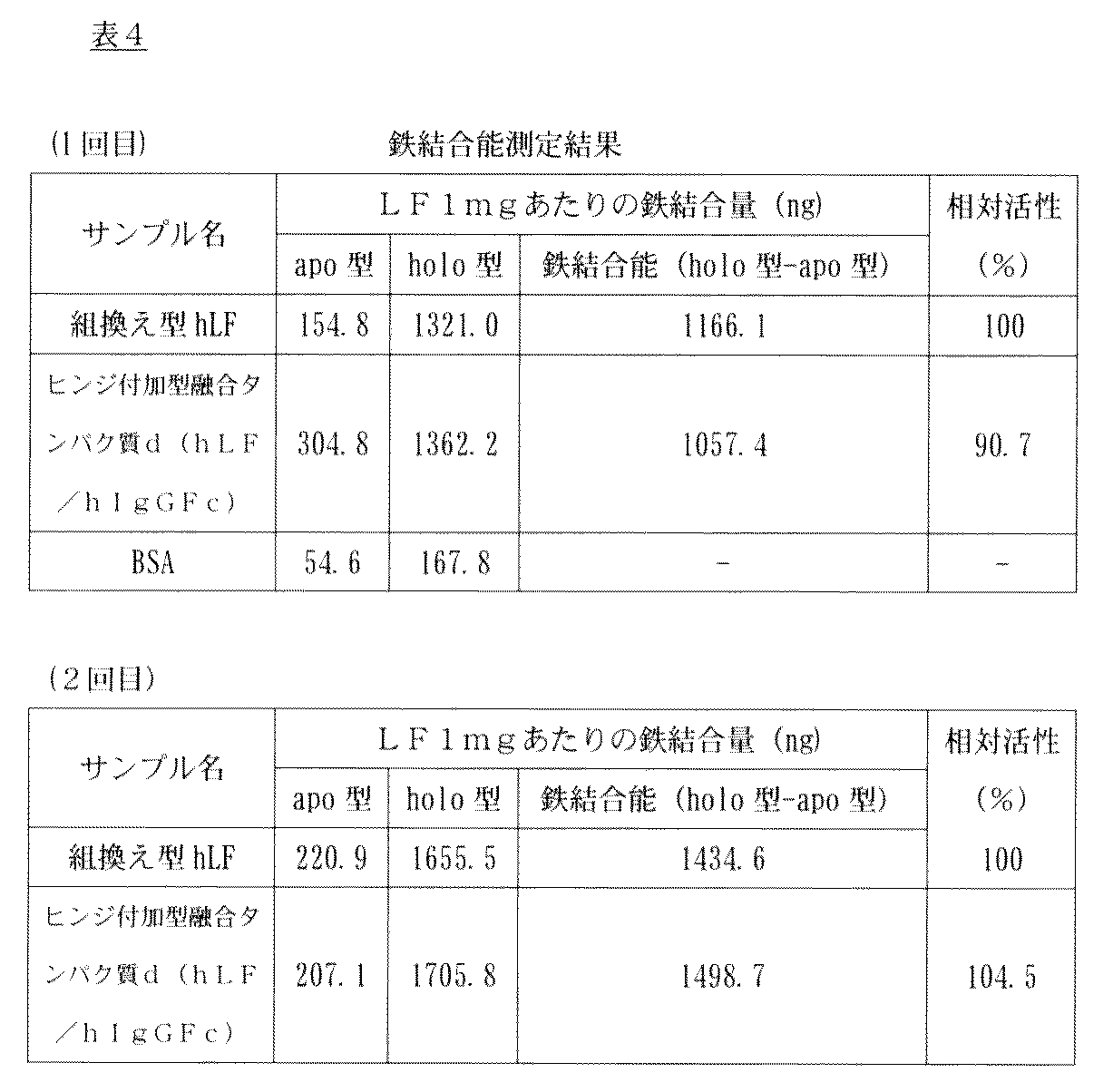
Aspergillus由来の組換え型hLFとヒンジ付加型融合タンパク質d(hLF/hIgGFc)の熱安定性を、円偏光二色性(CD)スペクトルで解析した。円偏光二色性(CD)スペクトルとは、物質にある波長をあてた場合の右円偏光と左円偏光の吸収の差を測定する方法で、タンパク質の二次構造の有無や種類、含量を推定することが可能である。 4-2. Examination of thermal stability of hinged hLF / hIgGFc fusion protein using CD spectrum The thermal stability of recombinant hLF derived from Aspergillus and hinged fusion protein d (hLF / hIgGFc) was determined by circular dichroism ( CD) spectrum. Circular dichroism (CD) spectrum is a method for measuring the difference in absorption between right and left circularly polarized light when a wavelength is applied to a substance. It is possible to estimate.
融合タンパク質d(hLF/hIgGFc)を用意し、30~90℃まで1℃ずつ上昇させながら、波長225nmのCDスペクトルを測定した(日本分光のJ−720使用)。 Next, the thermal stability of each protein was examined. When the CD spectrum is measured while changing the temperature of the protein solution from a low temperature to a high temperature, [θ] rises at a certain temperature and takes a constant value. This phenomenon is caused by the denaturation of proteins caused by heat and the change of secondary structure. When observing thermal stability, generally the wavelength to be measured is around 225 nm. Aspergillus-derived recombinant hLF and hinged fusion protein d (hLF / hIgGFc) suspended in PBS (-) at 0.1 mg / ml are prepared, and the wavelength is increased by 1 ° C from 30 to 90 ° C. A CD spectrum at 225 nm was measured (using J-720 from JASCO).
3−4.で示した陽イオン交換担体「MacroCap SP」で精製後のヒンジ付加型hLF/hIgGFc融合タンパク質を、PBSに透析し、さらにPBSで希釈することで336μg/mLの濃度のサンプルを調製した。このサンプルを1.5mLチューブに100μL加えて、37℃で3週間静置反応させた。反応0、1、2、3週間後のサンプルを3μLとり、非還元の4×サンプル緩衝液を1μl混合し、95℃、5分熱処理を行った。その後、7.5%SDS−PAGEで解析した。
CBB染色の結果を図14に示す。図14において、Mはマーカー、レーン1はhLF(1μg/レーン)、レーン2~5はそれぞれ0、1、2、3週間後のサンプルである。37℃でも数週間は安定であるものの、時間経過と共に、*で示したヒンジ付加型hLF/hIgGFc融合タンパク質のバンドが薄くなり、分解物と思われる**、***のバンドが濃くなった。これは精製過程で混入したプロテアーゼにより分解したものと考えられる。なお、ヒンジ欠損型融合タンパク質(後述)では、このような分解は観察されなかった。 4-3. Examination of stability of hinged hLF / hIgGFc fusion protein 3-4. The hinged hLF / hIgGFc fusion protein purified with the cation exchange carrier “MacroCap SP” shown in FIG. 2 was dialyzed into PBS and further diluted with PBS to prepare a sample having a concentration of 336 μg / mL. 100 μL of this sample was added to a 1.5 mL tube and allowed to react at 37 ° C. for 3 weeks. 3 μL of the sample after
The results of CBB staining are shown in FIG. In FIG. 14, M is a marker,
ヒンジ付加型hLF/hIgGFc融合タンパク質の分解箇所を特定するために、分解したバンドのN末端アミノ酸配列を解析した。上述した方法で、ヒンジ付加型hLF/hIgGFc融合タンパク質(12μg分)を37℃で3週間静置反応させ、その後、分解したサンプルを10%SDS−PAGEで泳動した。泳動後、定法に従いタンパク質をPVDF膜(ミリポア社)に転写した。PVDF膜をCBB染色した図を図15−Aに示す。図15−Aにおいて、Mはマーカー、レーン1はhLF(4μg)、レーン2~6はいずれも分解後のサンプル(各2μg/レーン)である。このPVDF膜から約50kDaのバンド(矢印)を切り出し、脱色した。そのバンドのN末端アミノ酸配列を、procise 491HT(商品名、ABI社)で解析した。得られたアミノ酸配列は、T−H−T−X−P(4アミノ酸目は解読不能)であり、その配列を調べたところ、配列表の配列番号5のアミノ酸番号722~726(ヒンジ領域内)に対応していた(図15−B)。 4-4. Analysis of the degradation site of the hinged hLF / hIgGFc fusion protein In order to identify the degradation site of the hinged hLF / hIgGFc fusion protein, the N-terminal amino acid sequence of the degraded band was analyzed. The hinged hLF / hIgGFc fusion protein (for 12 μg) was allowed to stand at 37 ° C. for 3 weeks by the method described above, and then the degraded sample was electrophoresed on 10% SDS-PAGE. After electrophoresis, the protein was transferred to a PVDF membrane (Millipore) according to a conventional method. FIG. 15-A shows a PVDF membrane stained with CBB. In FIG. 15-A, M is a marker,
1.ヒンジ欠損型hLF/hIgGFc融合タンパク質発現ベクターの構築
上記で作製したベクターpBSIILfAL/Bam(図1)を使用して、hLFとヒンジ領域を含まないhIgGFcとの融合タンパク質(ヒンジ欠損型hLF/mhIgGFc)を動物細胞にて発現させるための発現ベクターを構築した。 Example 2: Preparation of the fusion protein of human IgGFc region not including the human lactoferrin with a hinge region ( "hinge defective"), stability biological activity and
なお、pmIgGは、以下のようにして構築した。前述したヒンジ付加型融合タンパク質発現ベクターであるpTeuIgGのBamHI−XbaIに存在するヒトIgG1のヒンジ、CH2、CH3領域に対応するゲノムDNA配列を、ヒトIgG1のCH2とCH3領域に対応するcDNA配列に入れ替えて作製した。 In order to prepare an expression vector for the hinge-deficient fusion protein hLF / mhIgGFc, the XhoI-BamHI fragment of pBSIILfAL / Bam was added to the XhoI / BamHI site of the expression vector pmIgG (FIG. 3) having the CH2 and CH3 cDNA sequences of the human IgGFc region. Was cloned. This vector was named pmIgG / hLF. Each expression vector was produced according to the method described above.
In addition, pmIgG was constructed as follows. The genomic DNA sequence corresponding to the hinge, CH2, and CH3 regions of human IgG1 present in BamHI-XbaI of pTeuIgG, which is the hinge-added fusion protein expression vector, is replaced with the cDNA sequence corresponding to the CH2 and CH3 regions of human IgG1. Made.
2−1.DG44細胞を宿主とするヒンジ欠損型hLF/hIgGFc融合タンパク質安定発現細胞株の構築
ヒンジ付加型の場合と同様にして、作製したpOptiVEC/hLF−mFcをDG44細胞へ導入して、hLF/hIgGFc融合タンパク質安定発現細胞株を樹立した。PvuI処理した発現ベクターを、pOptiVEC/hLF−mFc/PvuIと命名した。 2. 2. Expression and purification of hinge-deficient hLF / hIgGFc fusion protein 2-1. Construction of a cell line that stably expresses a hinge-deficient hLF / hIgGFc fusion protein using DG44 cells as a host In the same manner as in the case of the hinge-added type, the prepared pOptiVEC / hLF-mFc is introduced into DG44 cells, and hLF / hIgGFc fusion protein A stable expression cell line was established. The expression vector treated with PvuI was named pOptiVEC / hLF-mFc / PvuI.
ヒンジ付加型の場合と同様にして、7.5%SDS−PAGE及びCBB染色で解析した。 2-2. Confirmation of expression of hinge-deficient hLF / hIgGFc fusion protein The analysis was performed by 7.5% SDS-PAGE and CBB staining in the same manner as in the case of the hinge-added type.
MTX濃度4μM存在下で樹立した細胞株を、DG44−hLF/mhIgGFcと命名した。 The results are shown in FIG. The target molecular weight of hLF / mhIgGFc is about 105 kDa. A band was observed in the vicinity of about 105 kDa indicated by an arrow from an MTX concentration of 0.5 μM, and as the MTX concentration increased, the band became darker and it was confirmed that the protein expression level increased.
A cell line established in the presence of an MTX concentration of 4 μM was named DG44-hLF / mhIgGFc.
ヒンジ付加型の場合と同様にして、細胞を継代培養し、融合タンパク質を大量発現させた。得られた上清を4℃にて保存した。 2-3. Mass expression of hinge-deficient hLF / hIgGFc fusion protein As in the case of the hinge-added type, cells were subcultured to express the fusion protein in large quantities. The resulting supernatant was stored at 4 ° C.
ヒンジ付加型の場合と同様にして、大量発現で得られた培地上清から融合タンパク質を精製した。 2-4. Purification of hinge-deficient hLF / hIgGFc fusion protein The fusion protein was purified from the culture supernatant obtained by mass expression in the same manner as in the case of the hinge-added type.
ヒンジ付加型の場合と同様にして、融合タンパク質を硫酸アンモニウム沈殿によって濃縮し、7.5%SDS−PAGE及びCBB染色で調べた。 2-5. Concentration of hinge-deficient hLF / hIgGFc fusion protein by ammonium sulfate precipitation The fusion protein was concentrated by ammonium sulfate precipitation and examined by 7.5% SDS-PAGE and CBB staining.

3−1.ヒンジ欠損型hLF/hIgGFc融合タンパク質の鉄結合能の測定
ラクトフェリンの鉄結合能の測定は、ヒンジ付加型の場合と同様にして行った。2回実験を行った結果を以下の表6に示す。 3. Measurement of biological activity 3-1. Measurement of iron binding ability of hinge-deficient hLF / hIgGFc fusion protein The iron binding ability of lactoferrin was measured in the same manner as the hinge-added type. The results of two experiments are shown in Table 6 below.

Aspergillus由来の組換え型hLFとヒンジ付加型、及びヒンジ欠損型hLF/hIgGFc融合タンパク質の熱安定性を、ヒンジ付加型の場合と同様にしてCDスペクトルで解析した。その結果を、図12に示す。Aspergillus由来の組換え型hLF(パネルA)と、ヒンジ欠損型hLF/hIgGFc融合タンパク質(パネルB)の二次構造には、大きな違いは認められなかった。 3-2. Examination of the thermal stability of hinge-deficient hLF / hIgGFc fusion protein using CD spectrum The thermal stability of recombinant hLF derived from Aspergillus and hinge-added, and hinge-deficient hLF / hIgGFc fusion protein The CD spectrum was analyzed in the same manner as in the case. The result is shown in FIG. There was no significant difference in the secondary structure between the Aspergillus-derived recombinant hLF (panel A) and the hinge-deficient hLF / hIgGFc fusion protein (panel B).
8週齢のWistar系ラット(雄)6頭に、ペントバルビタールナトリウムによる麻酔下にて、外頸静脈に採血用カニューレを留置した。大腿静脈内注射によりAspergillus由来の組換え型hLF(1mg/kg体重で投与)及びヒンジ欠損型融合タンパク質(hLF/mhIgGFc)(hLF換算で1mg/kg体重で投与)を投与した。投与前及び投与後5、10、15、30、60、120、180、240分後に外頸静脈に留置したカニューレから採血し、血漿中のhLF濃度はELISA(「AssayMax Human Lactoferrin ELISA kit」、Assaypro社製)により測定した。なお、予備検討により、採血時に使用した抗凝固剤であるEDTA、及び血漿は、このELISA測定に影響を及ぼさないことを確認している。また、使用したELISAキットは、ビオチン化抗ヒトラクトフェリン抗体(1次抗体)とペルオキシダーゼ標識ストレプトアジビンで構成されている。したがって、2次抗体を使用する一般的なELISAで問題となる2次抗体とヒンジ欠損型(hLF/mhIgGFc)のFc領域との交差反応を懸念する必要がない。 3-3. Examination of blood stability using hinge-deficient hLF / hIgGFc fusion protein rats Six canine Wistar rats (male), 8 weeks old, were anesthetized with sodium pentobarbital, and a blood cannula was placed in the external jugular vein. Detained. Recombinant hLF derived from Aspergillus (administered at 1 mg / kg body weight) and hinge-deficient fusion protein (hLF / mhIgGFc) (administered at 1 mg / kg body weight in terms of hLF) were administered by femoral vein injection. Blood was collected from a cannula placed in the external jugular vein before administration and 5, 10, 15, 30, 60, 120, 180, 240 minutes after administration, and the hLF concentration in plasma was determined by ELISA (“AssayMax Human Lactoferrin ELISA kit”, Assaypro) ). Preliminary studies have confirmed that EDTA, which is an anticoagulant used at the time of blood collection, and plasma do not affect this ELISA measurement. The ELISA kit used was composed of a biotinylated anti-human lactoferrin antibody (primary antibody) and peroxidase-labeled streptazibine. Therefore, there is no need to worry about the cross-reaction between the secondary antibody and the Fc region of the hinge-deficient type (hLF / mhIgGFc), which is a problem in general ELISA using a secondary antibody.
ヒンジ欠損型融合タンパク質(hLF/mhIgGFc)を、75%ウシ血清/PBS中で、0.2μg/μlの濃度で長期間37℃で反応させた。0、14、21、38、55日後にサンプルを採取して、抗hLF抗体を用いたイムノブロッティングにより残存するLFのサイズ及び量を検出した。
その結果、55日間までの保存期間中、0日と比較してほとんど分解を受けずに当初の分子量を維持し、検出量も減少していなかった(図18)。したがって、本発明の融合タンパク質は、血中でのプロテアーゼによる分解に対し、十分な耐性を有する。 3-4. In vitro protease resistance test of hinge-deficient hLF / hIgGFc fusion protein Hinge-deficient fusion protein (hLF / mhIgGFc) was reacted in 75% bovine serum / PBS at a concentration of 0.2 μg / μl at 37 ° C. for a long period of time I let you. Samples were taken after 0, 14, 21, 38, and 55 days, and the size and amount of remaining LF were detected by immunoblotting using an anti-hLF antibody.
As a result, during the storage period of up to 55 days, the initial molecular weight was maintained with little degradation compared to 0 days, and the detected amount was not decreased (FIG. 18). Therefore, the fusion protein of the present invention is sufficiently resistant to degradation by proteases in the blood.
1.ヒト小腸上皮様細胞Caco2への取り込み評価
LFは腸管から取り込まれて胸管リンパに移行することが知られている。そこで、本研究で作製したヒンジ欠損型融合タンパク質(hLF/mhIgGFc)がこの経路より取り込まれるかを、ヒト小腸上皮様細胞Caco2細胞を用いて確認した。
hLFとヒンジ欠損型融合タンパク質(hLF/mhIgGFc)は、蛍光プローブであるAlexa Fluor 488で標識した。1mgのAlexa Fluor 488にDMSO 100μlを加え、その後、1M NaHCO3を加えた各タンパク質とAlexa Fluor 488をモル比1:10で混合して、室温で、1h反応させた。反応後、反応液を1×PBS(−)に対して24時間透析を行い、未標識Alexa Fluor 488を除去した。
Caco−2細胞を12well plateに細胞濃度 5×10⁴cells/mlになるように撒き込み、37℃,5% CO2で1週間培養(2日おきに培地交換)した。Caco−2細胞にPBS(−)を1ml/well加えて、3回洗浄を行い、培地成分を完全に取り除いた。次に、Alexa標識済みの各タンパク質を15μg/well加えて、1hr反応させた。1hr後、Alexa標識済みの各タンパク質を取り除き、cold PBS(−)を1ml/well加えて、1回洗浄を行った。洗浄後、0.25%Trypsin/EDTAを250μl/well加えて、37℃条件下で5min反応させた。5min後、細胞をtubeに全量回収して、遠心機を用いて、200×g,2min,4℃で遠心を行った。遠心後、上清液を取り除き、cold PBS(−)を1ml/tube加えて、軽く懸濁をして、200×g,2min,4℃で遠心を行った。遠心後、上清液を取り除き、4%PFA/PBS(−)を200μl/tube加えて、軽く懸濁をして、室温で15min反応させた。15min後、200×g,2min,4℃で遠心を行った。遠心後、上清液を取り除き、1μg/mlビスベンズイミド/PBS(−)を200μl/tube加えて、軽く懸濁をして、室温で15min反応させた(核染色)。15min後、200×g,2min,4℃で遠心を行った。遠心後、上清液を取り除き、cold PBS(−)を200μl/tube加えて、軽く懸濁をした。懸濁後、200×g,2min,4℃で遠心を行った。遠心後、上清液を取り除き、cold PBS(−)を200μl/tube加えて、軽く懸濁をして、全量を8well chamber plateに移した。その後、共焦点レーザースキャン顕微鏡LSM510で取り込みの蛍光を観察した。 Example 3: Incorporation Evaluation of human intestinal epithelial-like cells of human lactoferrin and human IgGFc region of a fusion protein ( "hinge-defective") and
hLF and the hinge-deficient fusion protein (hLF / mhIgGFc) were labeled with Alexa Fluor 488, a fluorescent probe.
Caco-2 cells were seeded in a 12-well plate to a cell concentration of 5 × 10 4 cells / ml, and cultured at 37 ° C. and 5% CO 2 for 1 week (medium exchange every 2 days). PBS (-) was added to Caco-2 cells at 1 ml / well and washed three times to completely remove the medium components. Next, 15 μg / well of each Alexa-labeled protein was added and reacted for 1 hr. After 1 hr, each Alexa-labeled protein was removed, and 1 ml / well of cold PBS (-) was added, followed by washing once. After washing, 250 μl / well of 0.25% Trypsin / EDTA was added and reacted at 37 ° C. for 5 min. After 5 minutes, the entire amount of the cells was collected in a tube, and centrifuged at 200 × g, 2 minutes, 4 ° C. using a centrifuge. After centrifugation, the supernatant was removed, cold PBS (−) was added at 1 ml / tube, and the suspension was lightly suspended, and centrifuged at 200 × g, 2 min, 4 ° C. After centrifugation, the supernatant was removed, 200 μl / tube of 4% PFA / PBS (−) was added, and the suspension was lightly suspended and reacted at room temperature for 15 min. After 15 min, centrifugation was performed at 200 × g, 2 min, 4 ° C. After centrifugation, the supernatant was removed, 1 μg / ml bisbenzimide / PBS (−) was added at 200 μl / tube, and the suspension was lightly suspended and reacted at room temperature for 15 min (nuclear staining). After 15 min, centrifugation was performed at 200 × g, 2 min, 4 ° C. After centrifugation, the supernatant was removed, and 200 μl / tube of cold PBS (−) was added to suspend lightly. After suspension, centrifugation was performed at 200 × g, 2 min, 4 ° C. After centrifugation, the supernatant was removed, cold PBS (−) was added at 200 μl / tube, and the suspension was lightly suspended, and the entire amount was transferred to an 8-well chamber plate. Thereafter, the captured fluorescence was observed with a confocal laser scanning microscope LSM510.
本実験で用いたBufferは、表7の通り調製し、その後pHを7.4に調製した。
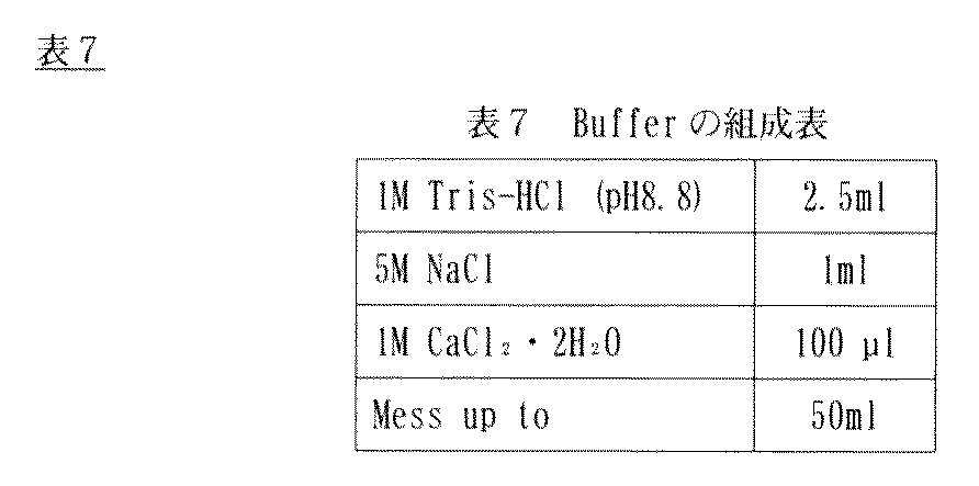
150μlの1mg/ml hLF、あるいはヒンジ欠損型融合タンパク質(hLF/mhIgGFc)をBuffer 120μlと混合して、37℃で5分間静置した。その後、16.7μg/mlのキモトリプシン溶液30μlを加え、37℃で静置反応した。経時的に反応サンプル36μlを回収し、あらかじめ用意しておいたSample Buffer(0.1M Tris−HCl(pH6.8)、4% SDS、20% Glycerol、BPB少量)12μlに加え、消化反応を停止させた。その後、反応液は10% SDS−PAGEで泳動後(15μl/レーン使用)、CBB染色した。
左から順に分子量マーカー、hLFのキモトリプシンを加えずに37℃で静置反応させたサンプル、hLFのキモトリプシンを加えて37℃静置反応させたサンプル、ヒンジ欠損型融合タンパク質(hLF/mhIgGFc)のキモトリプシンを加えずに37℃静置反応させたサンプル、ヒンジ欠損型融合タンパク質(hLF/mhIgGFc)のキモトリプシンを加えて37℃静置反応させたサンプルの泳動結果を図20に示す。 [Digestive enzyme resistance test]
150 μl of 1 mg / ml hLF or hinge-deficient fusion protein (hLF / mhIgGFc) was mixed with 120 μl of Buffer and allowed to stand at 37 ° C. for 5 minutes. Thereafter, 30 μl of a 16.7 μg / ml chymotrypsin solution was added, and the reaction was allowed to stand at 37 ° C. Collect 36 μl of reaction sample over time and add to 12 μl of Sample Buffer (0.1 M Tris-HCl (pH 6.8), 4% SDS, 20% Glycerol, BPB) prepared in advance to stop the digestion reaction I let you. Thereafter, the reaction solution was electrophoresed with 10% SDS-PAGE (using 15 μl / lane) and then stained with CBB.
From left to right, a molecular weight marker, a sample that was allowed to stand at 37 ° C. without adding hLF chymotrypsin, a sample that was allowed to stand at 37 ° C. after adding hLF chymotrypsin, and a chymotrypsin of a hinge-deficient fusion protein (hLF / mhIgGFc) FIG. 20 shows the results of electrophoresis of a sample that was allowed to stand at 37 ° C. without adding, and a sample that was allowed to stand at 37 ° C. after adding chymotrypsin of a hinge-deficient fusion protein (hLF / mhIgGFc).
また、表7に示すBufferに溶解したhLF及びヒンジ欠損型融合タンパク質(hLF/mhIgGFc)をゲルカラムクロマトグラフィー分析したところヒンジ欠損型融合タンパク質(hLF/mhIgGFc)はホモダイマーとして存在することが分かった(図22)。
図22パネルBは溶出時間とタンパク分子量の相関性を示すグラフである。クロマトグラフ(図22パネルA)で、青曲線(「hLF/dhIgGFc」)は2量体を形成するための領域(ヒンジ領域)を含むヒンジ付加型hLF/hIgGFc融合タンパク質の結果である。他方、緑曲線(「hLF/mhIgGFc」)はヒンジ欠損型融合タンパク質(hLF/mhIgGFc)の結果である。hLF/mhIgGFc及びhLF/dhIgGFcをゲルろ過すると、両者の溶出時間にほとんど差がないことが判明した。このことから、ヒンジ欠損型融合タンパク質(hLF/mhIgGFc)は、溶液中ではダイマーを形成していることが示された。 The density of each band was analyzed by ImageJ. The graph was graphed assuming that the average value of the intensity of the band to which chymotrypsin was not added was 100% (FIG. 21). The hinge-deficient fusion protein (hLF / mhIgGFc) was hardly degraded, but the control hLF was degraded in 30 seconds, indicating that the hinge-deficient fusion protein (hLF / mhIgGFc) was resistant to chymotrypsin.
Moreover, hLF and hinge-deficient fusion protein (hLF / mhIgGFc) dissolved in Buffer shown in Table 7 were analyzed by gel column chromatography, and it was found that the hinge-deficient fusion protein (hLF / mhIgGFc) exists as a homodimer ( FIG. 22).
FIG. 22 panel B is a graph showing the correlation between elution time and protein molecular weight. In the chromatograph (FIG. 22 panel A), the blue curve (“hLF / dhIgGFc”) is the result of a hinged hLF / hIgGFc fusion protein containing a region (hinge region) to form a dimer. On the other hand, the green curve (“hLF / mhIgGFc”) is the result of the hinge-deficient fusion protein (hLF / mhIgGFc). When hLF / mhIgGFc and hLF / dhIgGFc were subjected to gel filtration, it was found that there was almost no difference in the elution time between the two. This indicates that the hinge-deficient fusion protein (hLF / mhIgGFc) forms a dimer in the solution.
The present invention provides a lactoferrin fusion protein having improved properties, uses thereof and methods for producing the same.
Claims (15)
- FcRn結合領域を含むタンパク質又はペプチドとラクトフェリン又はラクトフェリンの生物活性断片もしくはペプチドとの融合タンパク質であって、
(LF−s−Y)n 又は (Y−s−LF)n
〔式中、LFはラクトフェリン又はラクトフェリンの生物活性断片もしくはペプチド、YはFcRn結合領域を含むタンパク質又はペプチド、sは0~10個の任意のアミノ酸の配列、nは1~10の整数を表す〕
で表される融合タンパク質、又はその改変体。 A fusion protein of a protein or peptide comprising an FcRn binding region and lactoferrin or a biologically active fragment or peptide of lactoferrin,
(LF-s-Y) n or (Ys-LF) n
[Wherein LF is lactoferrin or a biologically active fragment or peptide of lactoferrin, Y is a protein or peptide containing an FcRn-binding region, s is a sequence of 0 to 10 arbitrary amino acids, and n is an integer of 1 to 10]
Or a variant thereof. - FcRn結合領域を含むタンパク質又はペプチドが、IgG、IgGの重鎖、IgGの重鎖Fc領域、Fc領域のCH2ドメイン及びCH3ドメイン、Fc領域のCH2ドメイン、アルブミン又はアルブミンのFcRn結合領域のいずれかを含む、請求項1記載の融合タンパク質、又はその改変体。 A protein or peptide comprising an FcRn binding region is any of IgG, IgG heavy chain, IgG heavy chain Fc region, Fc region CH2 and CH3 domains, Fc region CH2 domain, albumin or albumin FcRn binding region The fusion protein according to claim 1, or a variant thereof.
- 融合タンパク質が、単量体又は二量体(n=1又は2)である、請求項1又は2記載の融合タンパク質、又はその改変体。 The fusion protein according to claim 1 or 2, or a variant thereof, wherein the fusion protein is a monomer or a dimer (n = 1 or 2).
- 融合タンパク質が、単量体である、請求項1又は2記載の融合タンパク質、又はその改変体。 The fusion protein according to claim 1 or 2, or a variant thereof, wherein the fusion protein is a monomer.
- 融合タンパク質が、天然又は遺伝子組換え型ラクトフェリンの50%以上の鉄キレート能を保持している、請求項1~4のいずれか1項記載の融合タンパク質、又はその改変体。 The fusion protein according to any one of claims 1 to 4, or a variant thereof, wherein the fusion protein retains an iron chelating ability of 50% or more of natural or genetically modified lactoferrin.
- 融合タンパク質は、ラクトフェリン受容体、IgG受容体及びアルブミン受容体からなる群より選択される少なくとも1つの受容体を介して取り込まれるものである、請求項1~5のいずれか1項記載の融合タンパク質、又はその改変体。 The fusion protein according to any one of claims 1 to 5, wherein the fusion protein is incorporated through at least one receptor selected from the group consisting of a lactoferrin receptor, an IgG receptor and an albumin receptor. Or a variant thereof.
- 融合タンパク質は、天然又は遺伝子組換え型ラクトフェリンと比べてキモトリプシン耐性が向上したものである、請求項1~6のいずれか1項記載の融合タンパク質、又はその改変体。 The fusion protein according to any one of claims 1 to 6, or a variant thereof, wherein the fusion protein has improved chymotrypsin resistance compared to natural or genetically modified lactoferrin.
- 請求項1~7のいずれか1項記載の融合タンパク質又はその改変体をコードする核酸分子。 A nucleic acid molecule encoding the fusion protein according to any one of claims 1 to 7 or a variant thereof.
- 請求項8記載の核酸分子を含む発現ベクター。 An expression vector comprising the nucleic acid molecule according to claim 8.
- 請求項9記載の発現ベクターを含む宿主細胞。 A host cell comprising the expression vector according to claim 9.
- 請求項8記載の核酸分子を含む非ヒト遺伝子組換え動物。 A non-human transgenic animal comprising the nucleic acid molecule according to claim 8.
- 請求項8記載の核酸分子を含む遺伝子組換え植物。 A genetically modified plant comprising the nucleic acid molecule according to claim 8.
- 請求項1~7のいずれか1項記載の融合タンパク質又はその改変体を含む、ラクトフェリンによって改善される疾患の治療剤。 A therapeutic agent for a disease ameliorated by lactoferrin, comprising the fusion protein according to any one of claims 1 to 7 or a variant thereof.
- 請求項1~7のいずれか1項記載の融合タンパク質又はその改変体及び担体を含む、医薬品組成物。 A pharmaceutical composition comprising the fusion protein according to any one of claims 1 to 7 or a variant thereof and a carrier.
- 請求項1~7のいずれか1項記載の融合タンパク質又はその改変体の製造方法であって、この融合タンパク質又はその改変体をコードする遺伝子を含む宿主細胞を培養して融合タンパク質を発現させ、この宿主細胞又はその培地から融合タンパク質又はその改変体を回収することを含む方法。 A method for producing the fusion protein or variant thereof according to any one of claims 1 to 7, wherein a host cell containing a gene encoding the fusion protein or variant thereof is cultured to express the fusion protein, Recovering the fusion protein or variant thereof from the host cell or medium thereof.
Priority Applications (10)
| Application Number | Priority Date | Filing Date | Title |
|---|---|---|---|
| EP13780579.2A EP2842969B1 (en) | 2012-04-23 | 2013-04-23 | Lactoferrin fusion protein and method for preparation thereof |
| KR1020147031493A KR102177252B1 (en) | 2012-04-23 | 2013-04-23 | Lactoferrin fusion protein and method for producing same |
| US14/395,826 US9809641B2 (en) | 2012-04-23 | 2013-04-23 | Lactoferrin fusion protein and method for preparation thereof |
| ES13780579.2T ES2671631T3 (en) | 2012-04-23 | 2013-04-23 | Lactoferrin fusion protein and method for preparing it |
| CN201380021309.2A CN104245739B (en) | 2012-04-23 | 2013-04-23 | Lactoferrin fusion protein and preparation method thereof |
| CA2871468A CA2871468C (en) | 2012-04-23 | 2013-04-23 | Lactoferrin fusion protein and method for preparation thereof |
| JP2014512737A JP5855239B2 (en) | 2012-04-23 | 2013-04-23 | Lactoferrin fusion protein and method for producing the same |
| BR112014026162A BR112014026162A2 (en) | 2012-04-23 | 2013-04-23 | FUSION PROTEIN OR VARIANT THEREOF, NUCLEIC ACID MOLECULE, EXPRESSION VECTOR, HOST CELL, GENETICLY MODIFIED NON-HUMAN ANIMAL, GENETICALLY MODIFIED PLANT, THERAPEUTIC AGENT FOR DISEASES ENHANCED BY LACTOFERRIN, PHARMACEUTICAL COMPOSITION, AND METHOD FOR PREPARING PROTEIN FUSION OR A VARIANT THEREOF |
| IN9815DEN2014 IN2014DN09815A (en) | 2012-04-23 | 2014-11-19 | |
| US15/720,023 US10562959B2 (en) | 2012-04-23 | 2017-09-29 | Lactoferrin fusion protein and method for preparation thereof |
Applications Claiming Priority (2)
| Application Number | Priority Date | Filing Date | Title |
|---|---|---|---|
| JP2012098085 | 2012-04-23 | ||
| JP2012-098085 | 2012-04-23 |
Related Child Applications (2)
| Application Number | Title | Priority Date | Filing Date |
|---|---|---|---|
| US14/395,826 A-371-Of-International US9809641B2 (en) | 2012-04-23 | 2013-04-23 | Lactoferrin fusion protein and method for preparation thereof |
| US15/720,023 Continuation US10562959B2 (en) | 2012-04-23 | 2017-09-29 | Lactoferrin fusion protein and method for preparation thereof |
Publications (1)
| Publication Number | Publication Date |
|---|---|
| WO2013162050A1 true WO2013162050A1 (en) | 2013-10-31 |
Family
ID=49483340
Family Applications (1)
| Application Number | Title | Priority Date | Filing Date |
|---|---|---|---|
| PCT/JP2013/062685 WO2013162050A1 (en) | 2012-04-23 | 2013-04-23 | Lactoferrin fusion protein and method for producing same |
Country Status (9)
| Country | Link |
|---|---|
| US (2) | US9809641B2 (en) |
| EP (1) | EP2842969B1 (en) |
| JP (2) | JP5855239B2 (en) |
| KR (1) | KR102177252B1 (en) |
| BR (1) | BR112014026162A2 (en) |
| CA (1) | CA2871468C (en) |
| ES (1) | ES2671631T3 (en) |
| IN (1) | IN2014DN09815A (en) |
| WO (1) | WO2013162050A1 (en) |
Cited By (3)
| Publication number | Priority date | Publication date | Assignee | Title |
|---|---|---|---|---|
| JP2017512058A (en) * | 2014-02-24 | 2017-05-18 | タケダ ゲー・エム・ベー・ハーTakeda GmbH | UTI fusion protein |
| WO2018079701A1 (en) * | 2016-10-28 | 2018-05-03 | 株式会社Nrlファーマ | Anticancer therapeutic adjuvant containing protein having lactoferrin activity |
| JP2018535932A (en) * | 2015-09-30 | 2018-12-06 | アイジーエム バイオサイエンシズ インコーポレイテッド | Binding molecule having a modified J chain |
Families Citing this family (10)
| Publication number | Priority date | Publication date | Assignee | Title |
|---|---|---|---|---|
| CA2871468C (en) | 2012-04-23 | 2021-09-21 | Nrl Pharma, Inc. | Lactoferrin fusion protein and method for preparation thereof |
| WO2016178089A1 (en) * | 2015-05-04 | 2016-11-10 | Laboratoire Francais Du Fractionnement Et Des Biotechnologies | Transgenic production of fc fusion proteins |
| EP3313875A4 (en) | 2015-06-24 | 2018-12-26 | Board of Regents of the University of Texas System | Methods and compositions for treatment of symptoms associated with intracranial hemorrhage |
| WO2018079702A1 (en) * | 2016-10-28 | 2018-05-03 | 株式会社Nrlファーマ | Lactoferrin/albumin fusion protein and production method therefor |
| IL307373A (en) | 2017-02-17 | 2023-11-01 | Denali Therapeutics Inc | Engineered Transferrin Receptor Binding Polypeptides |
| EP3904389A1 (en) | 2017-10-02 | 2021-11-03 | Denali Therapeutics Inc. | Fusion proteins comprising enzyme replacement therapy enzymes |
| WO2020004378A1 (en) * | 2018-06-26 | 2020-01-02 | 株式会社Nrlファーマ | Glycosaminoglycan inhibitor and promoter |
| US20200024329A1 (en) * | 2018-07-17 | 2020-01-23 | Bradley G. Thompson | Methods for regulating endogenous production of lactoferrin and sub-peptides thereof |
| IL293994A (en) | 2019-12-23 | 2022-08-01 | Denali Therapeutics Inc | Progranulin variants, compositions comprising same and uses thereof |
| WO2022115536A2 (en) * | 2020-11-25 | 2022-06-02 | Avirus, Inc. | Methods and compositions for treating viral infection |
Citations (9)
| Publication number | Priority date | Publication date | Assignee | Title |
|---|---|---|---|---|
| US5723125A (en) | 1995-12-28 | 1998-03-03 | Tanox Biosystems, Inc. | Hybrid with interferon-alpha and an immunoglobulin Fc linked through a non-immunogenic peptide |
| JP2000511424A (en) * | 1996-05-31 | 2000-09-05 | ベイラー カレッジ オブ メディスィン | Lactoferrin variants and uses thereof |
| JP2002520045A (en) * | 1998-07-15 | 2002-07-09 | サムヤン ジェネックス コーポレイション | Method for mass-producing lactoferrin polypeptide from yeast and microbial strain useful therefor |
| JP2003512011A (en) * | 1998-10-23 | 2003-04-02 | アムジエン・インコーポレーテツド | Modified peptides as therapeutics |
| JP2004521655A (en) | 2001-07-26 | 2004-07-22 | メデックスジェン カンパニー リミテッド | Immunoconjugates with tandem linkages |
| JP2004528022A (en) | 2001-02-14 | 2004-09-16 | ベントリア バイオサイエンス | Expression of human milk protein in transgenic plants |
| JP2008509153A (en) * | 2004-08-03 | 2008-03-27 | バイオレクシス ファーマシューティカル コーポレイション | Combination therapy using transferrin fusion protein containing GLP-1 |
| JP4234438B2 (en) | 2001-03-07 | 2009-03-04 | メルク パテント ゲゼルシャフト ミット ベシュレンクテル ハフトング | Expression technology of protein containing hybrid isotype antibody part |
| JP2011523351A (en) | 2008-04-25 | 2011-08-11 | ダイアックス、コープ | Antibodies against FcRn and uses thereof |
Family Cites Families (9)
| Publication number | Priority date | Publication date | Assignee | Title |
|---|---|---|---|---|
| US7737260B2 (en) * | 2003-11-13 | 2010-06-15 | Hanmi Pharm. Co., Ltd | Protein complex using an immunoglobulin fragment and method for the preparation thereof |
| JP4261531B2 (en) | 2005-09-06 | 2009-04-30 | 株式会社Nrlファーマ | Lactoferrin complex and method for producing the same |
| UY30820A1 (en) * | 2006-12-21 | 2008-07-03 | Centocor Inc | USE OF LONG-TERM ACTION GLP-1 RECEPTOR AGONISTS TO IMPROVE INSULIN SENSITIVITY AND LIPID PROFILES |
| KR101380729B1 (en) * | 2007-05-30 | 2014-04-10 | 포항공과대학교 산학협력단 | Immunoglobulin fusion proteins |
| JP2010138160A (en) | 2008-11-14 | 2010-06-24 | Nrl Pharma Inc | Liver function protecting pharmaceutical formulation |
| JP2010215522A (en) | 2009-03-13 | 2010-09-30 | Nrl Pharma Inc | Skin disease treating or skin disease preventing pharmaceutical composition and skin disease preventing cosmetic |
| CA2871468C (en) | 2012-04-23 | 2021-09-21 | Nrl Pharma, Inc. | Lactoferrin fusion protein and method for preparation thereof |
| JP2015027981A (en) | 2013-07-05 | 2015-02-12 | 富士フイルム株式会社 | Enteric granule and pharmaceutical composition |
| WO2018079702A1 (en) | 2016-10-28 | 2018-05-03 | 株式会社Nrlファーマ | Lactoferrin/albumin fusion protein and production method therefor |
-
2013
- 2013-04-23 CA CA2871468A patent/CA2871468C/en active Active
- 2013-04-23 JP JP2014512737A patent/JP5855239B2/en active Active
- 2013-04-23 KR KR1020147031493A patent/KR102177252B1/en active IP Right Grant
- 2013-04-23 US US14/395,826 patent/US9809641B2/en active Active
- 2013-04-23 EP EP13780579.2A patent/EP2842969B1/en active Active
- 2013-04-23 BR BR112014026162A patent/BR112014026162A2/en not_active Application Discontinuation
- 2013-04-23 ES ES13780579.2T patent/ES2671631T3/en active Active
- 2013-04-23 WO PCT/JP2013/062685 patent/WO2013162050A1/en active Application Filing
-
2014
- 2014-11-19 IN IN9815DEN2014 patent/IN2014DN09815A/en unknown
-
2015
- 2015-12-08 JP JP2015239725A patent/JP6356115B2/en active Active
-
2017
- 2017-09-29 US US15/720,023 patent/US10562959B2/en active Active
Patent Citations (10)
| Publication number | Priority date | Publication date | Assignee | Title |
|---|---|---|---|---|
| US5723125A (en) | 1995-12-28 | 1998-03-03 | Tanox Biosystems, Inc. | Hybrid with interferon-alpha and an immunoglobulin Fc linked through a non-immunogenic peptide |
| JP2000511424A (en) * | 1996-05-31 | 2000-09-05 | ベイラー カレッジ オブ メディスィン | Lactoferrin variants and uses thereof |
| JP2002520045A (en) * | 1998-07-15 | 2002-07-09 | サムヤン ジェネックス コーポレイション | Method for mass-producing lactoferrin polypeptide from yeast and microbial strain useful therefor |
| JP2003512011A (en) * | 1998-10-23 | 2003-04-02 | アムジエン・インコーポレーテツド | Modified peptides as therapeutics |
| JP2007105044A (en) | 1998-10-23 | 2007-04-26 | Amgen | Modified peptide as therapeutic agent |
| JP2004528022A (en) | 2001-02-14 | 2004-09-16 | ベントリア バイオサイエンス | Expression of human milk protein in transgenic plants |
| JP4234438B2 (en) | 2001-03-07 | 2009-03-04 | メルク パテント ゲゼルシャフト ミット ベシュレンクテル ハフトング | Expression technology of protein containing hybrid isotype antibody part |
| JP2004521655A (en) | 2001-07-26 | 2004-07-22 | メデックスジェン カンパニー リミテッド | Immunoconjugates with tandem linkages |
| JP2008509153A (en) * | 2004-08-03 | 2008-03-27 | バイオレクシス ファーマシューティカル コーポレイション | Combination therapy using transferrin fusion protein containing GLP-1 |
| JP2011523351A (en) | 2008-04-25 | 2011-08-11 | ダイアックス、コープ | Antibodies against FcRn and uses thereof |
Non-Patent Citations (13)
| Title |
|---|
| "Programs and Abstracts of the 2nd Clinical Lactoferrin Symposium 2009", PEPTIDES, vol. 32, no. 9, 30 July 2011 (2011-07-30), pages 1953 - 63 |
| ANDERSON ET AL., NATURE, vol. 344, 1990, pages 784 - 78 |
| BATRA, J. K. ET AL., MOL IMMUNOL., vol. 30, 1993, pages 379 - 386 |
| BROUWER CP; RAHMAN M; WELLING MM, BIOMETALS, vol. 23, no. 3, 18 March 2010 (2010-03-18), pages 493 - 505 |
| DIMITROV, D. S., MABS, vol. 1, 2009, pages 26 - 28 |
| GONG, R. ET AL., J. BIOL. CHEM., vol. 286, 2011, pages 27288 - 27293 |
| JAZAYERI, J. A. ET AL., BIODRUGS, vol. 22, 2008, pages 11 - 26 |
| RUIZ-GIMENEZ P; IBANEZ A; SALOM JB; MARCOS JF; LOPEZ-DIEZ JJ; VALLES S; TORREGROSA G; ALBORCH E; MANZANARES P, ANTIHYPERTENSIVE PROPERTIES OF LACTOFERRICIN B-DERIVED PEPTIDES |
| SATO, A. ET AL., BIOCHEM. J., vol. 371, 2003, pages 603 - 608 |
| See also references of EP2842969A4 |
| SUZUKI, T. ET AL., J. IMMUNOL., vol. 184, 2010, pages 1968 - 1976 |
| VAN DER DOES AM; BOGAARDS SJ; JONK L; WULFERINK M; VELDERS MP; NIBBERING PH, J AGRIC FOOD CHEM., vol. 58, no. 11, 9 June 2010 (2010-06-09), pages 6721 - 7 |
| YEUNG, Y. A. ET AL., J. IMMUNOL., vol. 182, 2009, pages 7663 - 7671 |
Cited By (7)
| Publication number | Priority date | Publication date | Assignee | Title |
|---|---|---|---|---|
| JP2017512058A (en) * | 2014-02-24 | 2017-05-18 | タケダ ゲー・エム・ベー・ハーTakeda GmbH | UTI fusion protein |
| JP2019187427A (en) * | 2014-02-24 | 2019-10-31 | タケダ ゲー・エム・ベー・ハーTakeda GmbH | Uti fusion proteins |
| JP2022002513A (en) * | 2014-02-24 | 2022-01-11 | タケダ ゲー・エム・ベー・ハーTakeda GmbH | Uti fusion proteins |
| JP2018535932A (en) * | 2015-09-30 | 2018-12-06 | アイジーエム バイオサイエンシズ インコーポレイテッド | Binding molecule having a modified J chain |
| JP7065766B2 (en) | 2015-09-30 | 2022-05-12 | アイジーエム バイオサイエンシズ インコーポレイテッド | Bonded molecule with modified J chain |
| JP7314356B2 (en) | 2015-09-30 | 2023-07-25 | アイジーエム バイオサイエンシズ インコーポレイテッド | Binding molecules with modified J chains |
| WO2018079701A1 (en) * | 2016-10-28 | 2018-05-03 | 株式会社Nrlファーマ | Anticancer therapeutic adjuvant containing protein having lactoferrin activity |
Also Published As
| Publication number | Publication date |
|---|---|
| CN104245739A (en) | 2014-12-24 |
| EP2842969A1 (en) | 2015-03-04 |
| US20150093382A1 (en) | 2015-04-02 |
| IN2014DN09815A (en) | 2015-07-31 |
| JP6356115B2 (en) | 2018-07-11 |
| CA2871468A1 (en) | 2013-10-31 |
| BR112014026162A2 (en) | 2017-10-03 |
| CA2871468C (en) | 2021-09-21 |
| US10562959B2 (en) | 2020-02-18 |
| ES2671631T3 (en) | 2018-06-07 |
| JP5855239B2 (en) | 2016-02-09 |
| EP2842969B1 (en) | 2018-03-14 |
| KR20150008870A (en) | 2015-01-23 |
| JPWO2013162050A1 (en) | 2015-12-24 |
| US9809641B2 (en) | 2017-11-07 |
| EP2842969A4 (en) | 2016-01-13 |
| KR102177252B1 (en) | 2020-11-10 |
| JP2016117717A (en) | 2016-06-30 |
| US20180072794A1 (en) | 2018-03-15 |
Similar Documents
| Publication | Publication Date | Title |
|---|---|---|
| JP6356115B2 (en) | Lactoferrin fusion protein and method for producing the same | |
| JP7142915B2 (en) | Lactoferrin/albumin fusion protein and method for producing same | |
| US11358995B2 (en) | Compositions and methods of use for treating metabolic disorders | |
| JP6722175B2 (en) | Compositions for treating metabolic disorders and methods of use thereof | |
| KR102077721B1 (en) | Compositions, Uses and Methods for Treatment of Metabolic Disorders and Diseases | |
| CN114846025A (en) | Ultra-long acting insulin-Fc fusion proteins and methods of use thereof | |
| CN102690354B (en) | Recombined dimerization antithrombin III-Fc fusion protein and mammalian cell efficient expression system thereof | |
| Shiga et al. | Recombinant human lactoferrin-Fc fusion with an improved plasma half-life | |
| CN103044554B (en) | Human urinary trypsin inhibitor (hUTI) of reorganization-dimerization and preparation method and application thereof | |
| CN104245739B (en) | Lactoferrin fusion protein and preparation method thereof | |
| NZ728101B2 (en) | Compositions and methods of use for treating metabolic disorders |
Legal Events
| Date | Code | Title | Description |
|---|---|---|---|
| 121 | Ep: the epo has been informed by wipo that ep was designated in this application |
Ref document number: 13780579 Country of ref document: EP Kind code of ref document: A1 |
|
| ENP | Entry into the national phase |
Ref document number: 2014512737 Country of ref document: JP Kind code of ref document: A |
|
| ENP | Entry into the national phase |
Ref document number: 2871468 Country of ref document: CA |
|
| WWE | Wipo information: entry into national phase |
Ref document number: 14395826 Country of ref document: US |
|
| NENP | Non-entry into the national phase |
Ref country code: DE |
|
| ENP | Entry into the national phase |
Ref document number: 20147031493 Country of ref document: KR Kind code of ref document: A |
|
| WWE | Wipo information: entry into national phase |
Ref document number: 2013780579 Country of ref document: EP |
|
| REG | Reference to national code |
Ref country code: BR Ref legal event code: B01A Ref document number: 112014026162 Country of ref document: BR |
|
| ENP | Entry into the national phase |
Ref document number: 112014026162 Country of ref document: BR Kind code of ref document: A2 Effective date: 20141020 |