WO2013162012A1 - Skin and hair color-controlling factor - Google Patents
Skin and hair color-controlling factor Download PDFInfo
- Publication number
- WO2013162012A1 WO2013162012A1 PCT/JP2013/062439 JP2013062439W WO2013162012A1 WO 2013162012 A1 WO2013162012 A1 WO 2013162012A1 JP 2013062439 W JP2013062439 W JP 2013062439W WO 2013162012 A1 WO2013162012 A1 WO 2013162012A1
- Authority
- WO
- WIPO (PCT)
- Prior art keywords
- activity
- autophagy
- expression
- skin
- melanin
- Prior art date
Links
Images


Classifications
-
- G—PHYSICS
- G01—MEASURING; TESTING
- G01N—INVESTIGATING OR ANALYSING MATERIALS BY DETERMINING THEIR CHEMICAL OR PHYSICAL PROPERTIES
- G01N33/00—Investigating or analysing materials by specific methods not covered by groups G01N1/00 - G01N31/00
- G01N33/48—Biological material, e.g. blood, urine; Haemocytometers
- G01N33/50—Chemical analysis of biological material, e.g. blood, urine; Testing involving biospecific ligand binding methods; Immunological testing
- G01N33/5005—Chemical analysis of biological material, e.g. blood, urine; Testing involving biospecific ligand binding methods; Immunological testing involving human or animal cells
- G01N33/5008—Chemical analysis of biological material, e.g. blood, urine; Testing involving biospecific ligand binding methods; Immunological testing involving human or animal cells for testing or evaluating the effect of chemical or biological compounds, e.g. drugs, cosmetics
- G01N33/5044—Chemical analysis of biological material, e.g. blood, urine; Testing involving biospecific ligand binding methods; Immunological testing involving human or animal cells for testing or evaluating the effect of chemical or biological compounds, e.g. drugs, cosmetics involving specific cell types
-
- G—PHYSICS
- G01—MEASURING; TESTING
- G01N—INVESTIGATING OR ANALYSING MATERIALS BY DETERMINING THEIR CHEMICAL OR PHYSICAL PROPERTIES
- G01N33/00—Investigating or analysing materials by specific methods not covered by groups G01N1/00 - G01N31/00
- G01N33/48—Biological material, e.g. blood, urine; Haemocytometers
- G01N33/50—Chemical analysis of biological material, e.g. blood, urine; Testing involving biospecific ligand binding methods; Immunological testing
- G01N33/68—Chemical analysis of biological material, e.g. blood, urine; Testing involving biospecific ligand binding methods; Immunological testing involving proteins, peptides or amino acids
Definitions
- Melanocytes which are cells involved in melanin biosynthesis, contain melanosomes, which are unique lysosome-related organelles derived from endosomes. In this melanosome, melanin is synthesized through a catalytic pathway using tyrosine as a precursor. Melanosomes receive various gene products selected primarily from the trans-Golgi network, and as a result, have common properties (eg, low internal pH) with lysosomes.
- Non-Patent Document 2 a receptor molecule, is involved in melanin uptake (phagocytosis) in keratinocytes and the possibility of skin color control by regulating its activity.
- Dynein and Dynactin which are protein molecules, are involved in the localization of melanin within the keratinocytes after being transferred to keratinocytes (Non-patent Documents 9 and 10), and further referred to as filopodia. It has also been suggested that MyoX, a protein molecule involved in the formation of cell structures, is involved in both melanin transfer from melanocytes to keratinocytes and from keratinocytes to keratinocytes (Non-patent Document 11). However, the mechanism of uptake, transport and metabolism of melanin (melanosome) produced in melanocytes by keratinocytes has not been clarified yet, and its role for skin color has not been verified.
- Non-patent Document 12 After melanin labeled with a fluorescent substance is taken into epidermal cells, melanin is easily decomposed in white-derived epidermal cells using an evaluation system that analyzes the disappearance of fluorescence in the epidermal cells. Reporting. However, no mention is made of mechanisms or specific factors that contribute to degradation.
- Autophagy generally refers to a process in which intracellular substances such as organelles are transferred to lysosomes and decomposed (Non-Patent Documents 13 to 15).
- Non-Patent Documents 13 to 15 Several types of autophagy are known. The main route that has been studied the most is macro autophagy. Usually, autophagy refers to macroautophagy.
- Autophagy consists of various physiological stresses such as starvation, hypoxia, energy depletion, endoplasmic reticulum stress, high temperature, hormone stimulation, drugs (rapamycin, fluspirylene, trifluoperazine, pimozide, nicardipine, nigurdipine, loperamide, amiodarone, verapamil, minoxidil , Clonidine, etc.), immune signals, infection by bacteria, viruses and parasites, and diseases such as acute pancreatitis, heart disease. Since phenomena related to autophagy have been observed in a wide range of species such as yeast and mammals, it is thought to play a very important role for organisms. In fact, to date, it has been reported that autophagy is involved in cancer, neurodegenerative diseases, inflammation, immunity, aging, and the like (Non-patent Document 16).
- the autophagy pathway involves multiple steps such as phagophore development and elongation, autophagosome formation, autophagosome-lysosome fusion, and autolysosome formation and degradation of internal substances.
- Factors are involved.
- the main factor is a protein involved in the formation of phagophores and autophagosomes, collectively called ATG (Autophagy-related proteins). More than 30 types of ATG have been reported so far (Non-patent Document 11).
- the present invention provides a method for regulating the amount of melanin in keratinocytes comprising the step of regulating the activity of autophagy in keratinocytes where melanin level regulation is desired.
- the present invention provides a method of controlling skin or hair color in a subject comprising the step of modulating the activity of autophagy in a keratinocyte of a subject desiring skin or hair color control.
- B: Quantitative analysis based on Western blotting results; data are mean ⁇ SD (each N 3), *, p ⁇ 0.05 (ANOVA, Holm test). Differences in the expression of autophagy-related factors among races with different skin colors. Difference in autophagy activity among races with different skin colors.
- Lightness of skin cultured with autophagy regulator (L * value); data are mean ⁇ SD (each N 3) **, p ⁇ 0.01; *, p ⁇ 0.05 (ANOVA, Holm test). Skin color control by the melanin amount regulator selected by the selection method of the present invention.
- B Cell respiratory activity.
- the present invention relates to factors relating to melanin regulation in keratinocytes and skin or hair color control.
- the present invention also provides a method for adjusting the amount of melanin in keratinocytes using the factor, a method for controlling skin or hair color, and an agent for regulating the amount of melanin in keratinocytes or a skin or hair color control agent using the factor. It relates to a method of evaluation or selection.
- the present inventors searched for a factor involved in melanin regulation in keratinocytes, and found that autophagy activity is related to melanin content in keratinocytes and that there is a high correlation between autophagy activity and skin color. I confirmed that there was. From these results, the present inventors have found that the amount of melanin in keratinocytes and skin or hair color can be controlled by utilizing the activity of autophagy.
- Examples of the gene expression analysis method include dot blot method, Northern blot method, RNase protection assay method, reporter assay using luciferase, RT-PCR method, DNA microarray and the like.
- Examples of methods for analyzing or quantifying the expression or activity of the protein encoded by the gene include Western blotting, immunostaining, fluorescent staining, ELISA, binding assay and the like.
- autophagy-related gene refers to each step of the autophagy pathway, for example, generation of phagophore, elongation or growth of phagophore, formation of autophagosome, fusion of autophagosome and lysosome, formation of autolysosome Or a gene encoding a protein involved in degradation of a substance in the autolysosome.
- molecules encoded by autophagy-related genes may include proteins and mRNA encoded by genes involved in each step of the autophagy pathway.
- ATG1 (or ULK1 or ULK2)
- ATG13 ATG17, ATG29, ATG31, ATG101, and FIP200 are considered to be involved in the early formation of phagophore as ULK / Atg1 complex
- VPS34, VPS15, ATG6, ATG14, and Ambra1 produce PI (3) P (phosphatidylinositol 3-phosphate) mainly on the endoplasmic reticulum as Class III PI3-kinase complex (Non-patent Document 16).
- LC-3 is a mammalian homologue of yeast Atg8 and exists in LC3-I and LC3-II forms.
- LC3-II is a cytoplasmic localized LC3-I covalently bound to phosphatidylethanolamine, and thereby localized on the autophagosome membrane. Since the amount of LC3-II is proportional to the number of autophagosomes, it is known as an index of autophagy (Non-patent Document 17).
- the autophagy activity can be quantitatively determined. It is possible to evaluate.
- autophagy activity can be evaluated by detecting the amount of LC3-II protein for each sample and combining it with other known autophagy markers such as p62 (Non-patent Document 17).
- p62 is a protein that is localized at the autophagosome formation site and interacts with LC3, and has a function of recruiting a target protein of autophagy to the autophagosome.
- P62 is known as an autophagy-selective substrate together with Nbr1 (Non-patent Document 18).
- UVRAG is known as a molecule that promotes the fusion process of autophagosome and lysosome.
- autophagy-related gene or molecule encoded by the gene is preferably encoded by a gene selected from the group consisting of ATG7, RAB11A, CLIP-170, Rubicon and RAB7B and the gene. Does not contain molecules.
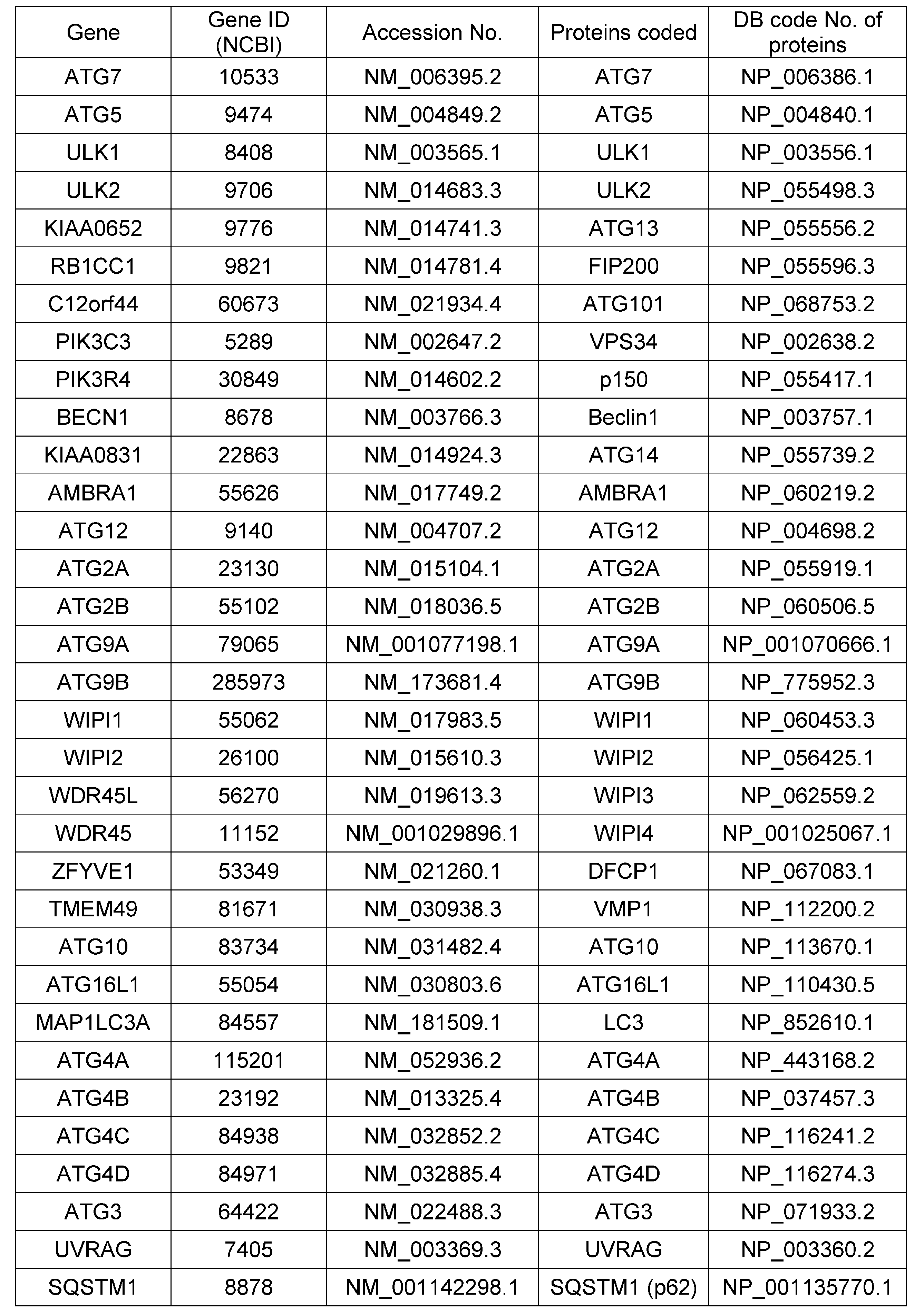
- autophagy-related genes or molecules encoded by the genes are shown in Table 1 below. These are genes and proteins registered in known databases. However, the involvement of these genes and molecules in melanin dynamics in keratinocytes has not been known so far.
- each step of the autophagy pathway such as phagophore generation, phagophore extension or growth, autophagosome formation
- a protein that is encoded by a homologous gene of the autophagy-related gene and that is involved in an autophagy pathway step involving a protein encoded by the autophagy-related gene is a protein encoded by the autophagy-related gene. It is a homolog.
- identity with respect to an amino acid sequence or base sequence refers to the number of positions where the same amino acid residue or base is present in both sequences when the two amino acid sequences or base sequences are aligned (aligned). Is the ratio (%) to the total number of amino acid residues or bases. Specifically, it is calculated by the Lippman-Pearson method (Science, 227, 1435, 1985), and homology analysis of genetic information processing software Genetyx-Win (Ver. 5.1.1; software development) ( A value calculated by performing an analysis with Unit size to compare (ktup) of 2 using a search (homology) program.
- the gene or molecule may be used alone or in any combination of two or more.
- the autophagy-related gene used in the present invention or the gene is an autophagy-related gene selected from the group listed in Table 1 or the mRNA or protein encoded by the gene or a molecule encoded by the gene. Any one of them may be used. More specifically, in the present invention, the genes listed in Table 1 or mRNAs and proteins encoded thereby may be used alone, or a combination of any two or more of the genes may be used. Well, a combination of mRNA encoded by any two or more of the genes may be used, a combination of proteins encoded by any two or more of the genes may be used, or their Any combination of two or more from genes, mRNAs and proteins may be used.
- the kinetics of melanin in keratinocytes are mainly the skin epidermis layer composed of keratinocytes and hair follicle hair matrix cells Alternatively, it is deeply involved in the amount and distribution of melanin in the fur cells that make up the shaft, which in turn affects the color of the skin and hair.
- the amount of melanin in keratinocytes can be regulated, thereby the amount and distribution of melanin in the skin epidermis, hair follicle hair matrix cells and even the fur cells forming the shaft And consequently the skin and hair color can be controlled.
- means for changing the autophagy activity of a cell there is a method of changing the expression or activity of the above-mentioned autophagy-related gene or a molecule encoded by the gene in the cell.
- the amount of melanin in keratinocytes can be adjusted, and the color of skin or hair can be controlled.
- the autophagy-related gene or a molecule encoded by the gene can be used for regulating the amount of melanin in keratinocytes or for controlling skin or hair color.
- the autophagy-related gene or the molecule encoded by the gene can be used for the production of an agent for regulating the amount of melanin in keratinocytes or for controlling skin or hair color.
- the expression or activity of the autophagy-related gene or the molecule encoded by the gene may be changed by any means commonly used in the art.
- means for changing the expression of the gene or the expression or activity of the molecule for example, as means for changing the gene expression, gene knockdown by an antisense oligonucleotide or siRNA or the like, transcription activity of a target gene by a specific promoter , Introduction of a gene from the outside using a vector, addition of an arbitrary substance having an action of changing gene expression, and the like. Among these, addition of any substance having an action of changing the expression of the gene is preferable.
- vectors for introducing genes include various gene expression vectors incorporating CMV promoter or EF1 ⁇ promoter as vectors for cultured cells, adenovirus vectors, retrovirus vectors, as vectors for cultured cells and tissues, Examples include lentiviral vectors. Gene transfer procedures using these vectors are well known to those skilled in the art. For example, the gene transfer procedure into the skin using a lentiviral vector is described in Gene Ther. 2007 Apr, 14 (8): 648-56. Moreover, gene introduction kits using these vectors are commercially available. By introducing the above-mentioned vector into which an autophagy-related gene is incorporated into a cell, the autophagy-related gene is overexpressed in the cell and the autophagy activity is changed.
- siRNA Gene knockdown by siRNA is a technique for specifically inhibiting the expression of a target gene by guiding mRNA degradation in a sequence-specific manner based on the mechanism of RNA interference (RNAi).
- RNAi RNA interference
- siRNA has also been used for in vivo gene knockdown of various organisms including mammals (eg, Song et al., Nature Medicine, 2003, 9: 347-351; McCaffery et al., Nature, 2002, 418: 38-39; Lewis et al., Nature Genetics, 2002, 32: 107-108; see Xia et al., Nature Biotech., 2002, 20: 1006-1010, etc.).
- Target-specifically synthesized siRNA can be purchased, and siRNA transfection kits are sold by many vendors such as QIAGEN and Takara Bio Inc.
- Examples of the molecule encoded by the autophagy-related gene include mRNA and polypeptide encoded by the gene.
- “change in molecular expression or activity” means any state that changes the expression or activity in the whole molecule, for example, change in molecular expression level, change in molecular degradation rate, molecular activation rate. Change, the change in the inactivation rate of the molecule, etc., preferably the change in the expression level of the molecule.
- the means for changing the expression or activity of a molecule the means for changing the above gene expression, the means for changing protein expression, the means for changing the enzyme activity of the molecule, etc., and the interaction between the molecule and its target factor are changed. And means for changing the signal pathway on which the molecule acts. Of these, means for changing gene expression, means for changing protein expression, and the like are preferable.
- an autophagy inducer or an autophagy inhibitor can be mentioned.
- Known autophagy inducers include mTOR signal inhibitors typified by rapamycin, prospirosomes such as fluspirylene, trifluoperazine, pimozide, nicardipine, nigurdipine, loperamide, amiodarone, verapamil, minoxidil, clonidine, PP242, or MG-132. Inhibitors and the like are mentioned.
- Known autophagy inhibitors include hydroxychloroquine, chloroquine, bafilomycin A1, PI3 kinase inhibitor 3-methyladenine, waltomannin, and the like.
- Still another example of means for changing the activity of autophagy includes activating or suppressing the target factor of the autophagy induction or inhibitor described above, and the autophagy-related gene described in Table 1 above or a code for the gene Activation or suppression of a factor that affects the expression of the molecule to be expressed.
- the autophagy inducer rapamycin described above suppresses the activity of the cellular protein mTOR.
- mTOR mammalian target of rapamycin
- mTOR is a protein discovered as a target of rapamycin, and is a kind of protein kinase (serine-threonine kinase) involved in intracellular signal transduction.
- mTOR is also known to have an action of suppressing autophagy induction (Nat Genet. 2004 Jun; 36 (6): 585-95). As shown in Example 8 to be described later, by suppressing mTOR, the autophagy activity of the cells increased and the amount of melanin decreased.
- a subject that alters the activity of autophagy is a natural or genetically engineered keratinocyte that has the ability to express at least one of the above genes and is desired to regulate the amount of melanin.
- cultures, tissues, organs and animals containing the same As said keratinocyte, the keratinocyte which exists in skin and a hair follicle is preferable, and epidermal keratinocyte, a hair matrix cell, and a fur cell are more preferable.
- the culture, tissue and organ containing the keratinocytes, cultured keratinocytes, epidermal tissue, hair follicle tissue, skin and cultures thereof are preferable.
- the animal is preferably a human or non-human mammal, and examples of the non-human mammal include dogs, cats, rabbits, mice, rats, hamsters, guinea pigs, pigs and horses.
- the melanin amount adjustment or skin or hair color control according to the present invention can be performed using the keratinocyte or a culture, tissue or organ containing the keratinocyte as a subject.
- the subject is a cultured keratinocyte, cultured skin tissue, cultured epidermis, or cultured hair follicle.
- an animal desiring to adjust keratinocyte melanin amount or skin or hair color control may be a subject.
- the amount of melanin adjustment or skin or hair color control is for cosmetic or aesthetic purposes, for example, skin whitening or tanning, hair coloring (lightening or darkening) or bleaching, It can be performed non-therapeutically by non-medical workers such as estheticians, hairdressers, barbers, trimmers, etc., for purposes such as color restoration after bleaching, gray hair dyeing and the like.
- non-therapeutic is a concept that does not include a medical act, that is, a treatment act on the human body by therapy.
- An exemplary embodiment of the present invention is a method for increasing the amount of melanin in a keratinocyte, comprising the step of increasing the amount of melanin in the keratinocyte by suppressing the activity of autophagy in the keratinocyte where an increase in the amount of melanin is desired.
- Another exemplary embodiment of the present invention is a method for reducing the amount of melanin in a keratinocyte comprising the step of reducing the amount of melanin in the keratinocyte by enhancing the activity of autophagy in the keratinocyte where a reduction in the amount of melanin is desired. is there.
- One embodiment of the aspect increases the amount of melanin in the keratinocyte by suppressing the expression or activity of a gene involved in the activation of autophagy in the keratinocyte in which an increase in the amount of melanin is desired or a molecule encoded by the gene. It is a method for increasing the amount of melanin in keratinocytes including a step. Another embodiment comprises the step of reducing the amount of melanin in the keratinocyte by enhancing the expression or activity of a gene involved in autophagy activation in the keratinocyte or its encoded molecule, where melanin reduction is desired. A method for reducing the amount of melanin in keratinocytes.
- the step of increasing the amount of melanin in the keratinocyte by enhancing the expression or activity of a gene involved in suppression of autophagy activity or a molecule encoded by the gene in the keratinocyte where increase in the amount of melanin is desired.
- the step of reducing the amount of melanin in the keratinocyte by suppressing the expression or activity of a gene involved in suppression of autophagy activity in the keratinocyte or a molecule encoded by the gene is desired.
- Another exemplary embodiment of the present invention comprises increasing the amount of melanin in a keratinocyte by inhibiting the activity of autophagy in the skin keratinocyte of a subject desiring skin color browning or darkening. It is a method of browning or darkening the skin color of the body. According to this method, for example, skin tanning is possible.
- Another exemplary embodiment of the present invention comprises reducing the amount of melanin in a keratinocyte by enhancing the activity of autophagy in the skin keratinocyte of a subject desiring skin lightening. This is a method for lightening the skin color. According to this method, for example, skin whitening is possible.
- One embodiment of this aspect is to suppress the expression or activity of a gene involved in autophagy activation in a skin keratinocyte of a subject who desires skin browning or darkening, or a molecule encoded thereby,
- a method of browning or darkening the skin color of a subject comprising a step of increasing the amount of melanin in keratinocytes.
- skin tanning is possible.
- Another embodiment is to enhance the expression or activity of a gene involved in autophagy activation in a skin keratinocyte or a molecule encoded by the subject in a subject desiring skin lightening, thereby increasing the amount of melanin in the keratinocyte.
- a method for lightening the skin color of a subject comprising the step of reducing
- Another embodiment is to enhance the expression or activity of a gene involved in suppressing autophagy activity in a skin keratinocyte of a subject who desires skin browning or darkening, or a molecule encoded by the gene, or the keratinocyte.
- the method of browning or darkening the skin color of a subject comprising the step of increasing the amount of melanin in the subject. According to this method, for example, skin tanning is possible.
- melanin in the keratinocyte is suppressed by suppressing the expression or activity of a gene involved in suppressing autophagy activity in a skin keratinocyte or a molecule encoded by the subject in a subject who desires skin lightening.
- a method of lightening the skin color of a subject comprising the step of reducing the amount. According to this method, for example, skin whitening is possible.
- Yet another exemplary embodiment of the invention includes increasing the amount of melanin in the keratinocytes by inhibiting the activity of autophagy in the hair follicle keratinocytes of a subject desiring to brown or darken the hair.
- a method for browning or darkening the hair color of a subject According to this method, for example, it is possible to restore the color of hair after bleaching or to dye white hair.
- Another exemplary embodiment of the present invention comprises reducing the amount of melanin in the keratinocytes by enhancing the activity of autophagy in the hair follicle keratinocytes of a subject desiring hair coloration. This is a method for lightening the hair color. According to this method, for example, it is possible to lighten or bleach hair.
- One embodiment of this aspect is to suppress the expression or activity of a gene involved in autophagy activation in a follicular keratinocyte or a molecule encoded by the subject who desires browning or darkening of the hair, It is a method of browning or darkening the hair color of a subject, comprising a step of increasing the amount of melanin in the keratinocyte. According to this method, for example, it is possible to restore the color of hair after bleaching or to dye white hair. Another embodiment is to enhance the expression or activity of a gene involved in autophagy activation in a follicular keratinocyte or a molecule encoded by the subject in a subject desiring hair coloration, thereby melanin in the keratinocyte.
- a method for lightening a subject's hair color comprising reducing the amount. According to this method, for example, it is possible to lighten or bleach hair. Yet another embodiment enhances the expression or activity of a gene involved in suppression of autophagy activity in a follicular keratinocyte or a molecule encoded by the subject in a subject desiring hair browning or darkening.
- a method for browning or darkening a subject's hair color comprising a step of increasing the amount of melanin in keratinocytes. According to this method, for example, it is possible to restore the color of hair after bleaching or to dye white hair.
- Yet another embodiment is to suppress the expression or activity of a gene involved in suppression of autophagy activity in a hair follicle keratinocyte or a molecule encoded by the subject in a subject desiring hair coloration.
- a method for lightening a subject's hair color comprising a step of reducing the amount of melanin. According to this method, for example, it is possible to lighten or bleach hair.
- a substance that changes the activity of autophagy is a substance that can control the color of skin or hair by adjusting the amount of melanin in keratinocytes. Therefore, the present invention also provides a method for evaluating a keratinocyte melanin amount regulating action or skin or hair color controlling action of a substance using autophagy activity, and a keratinocyte melanin regulating agent or skin or hair color regulating agent. Provide an evaluation or selection method.
- a method for evaluating or selecting a melanin amount regulator in keratinocytes includes a step of administering a test substance to a cell; a step of measuring a change in autophagy activity in the cell; and a result of the measurement, Evaluating the melanin regulating effect of the test substance.
- the cell to which the test substance is administered may be a natural cell or a genetically modified cell as long as it has autophagy activity, and is preferably a cultured cell or a cell derived from an animal tissue or organ culture. Human cells are more preferred.
- the cell is a skin cell, more preferably an epidermal cell or a hair follicle cell, and still more preferably a keratinocyte.
- preferable cells are human-derived cultured epidermal cells (for example, human normal epidermis cells (NHEK) or human tissue cultured hair follicle cells).
- test substance may be natural or synthetic, and may be a single substance or a composition or a mixture.
- form of administration can be any form depending on the test substance.
- the activity of autophagy can be measured, for example, by examining the expression or activity of an autophagy-related gene or a molecule encoded by the gene, or the amount of the molecule in the cell.
- the expression or activity of the gene or the molecule encoded by the gene, or the amount of the molecule in the cell can be measured by any analysis method commonly used in the art.
- the gene expression analysis method include dot blot method, Northern blot method, RNase protection assay method, reporter assay using luciferase, RT-PCR method, DNA microarray and the like.
- Examples of a method for analyzing the expression or activity of a protein encoded by a gene or a method for quantification include Western blotting, immunostaining, fluorescent staining, ELISA, binding assay and the like.
- changes in autophagy activity due to administration of the test substance can be evaluated.
- the autophagy activity can be measured before and after administration of the test substance, and the measured value can be quantified as necessary, and then the results before and after administration can be compared.
- the result is given to the administration group and the non-administration group, or the administration group.
- a control substance administration group can be compared.
- the test substance at different concentrations can be administered to measure autophagy activity, and the difference in the measurement results depending on the concentration of the test substance can be examined.
- the effect of regulating the amount of melanin in the keratinocyte of the test substance can be evaluated.
- the test substance evaluated to affect the activity of autophagy is judged to be a substance having a regulating action of melanin in keratinocytes, or can be used for regulating the amount of melanin in keratinocytes. Can be selected. Therefore, the method for evaluating or selecting a melanin regulator in the present invention may further include a step of selecting the test substance as a melanin regulator in keratinocytes based on the evaluation result of the test substance.
- a substance that suppresses autophagy activity is selected as an agent for increasing the amount of melanin in keratinocytes.
- examples of such substances include substances that suppress the expression or activity of genes involved in autophagy activation or molecules encoded therein, and the expression or molecules encoded in genes involved in suppression of autophagy activity or molecules encoded therein.
- examples include substances that enhance the activity.
- the agent increases the amount of melanin in keratinocytes to brown or darken the skin or hair color, such as a skin or hair color browning or darkening agent (eg, skin tanning agent, hair darkening agent, bleached agent) Hair color rejuvenating agent, white hair dyeing agent, etc.).
- a substance that enhances the activity of autophagy is selected as a melanin amount reducing agent in keratinocytes.
- Such substances include substances that enhance the expression or activity of genes involved in autophagy activation or molecules encoded therein, and genes or molecules encoded therein involved in suppression of autophagy activity
- the substance which suppresses activity is mentioned.
- the agent is used as a skin or hair color lightening agent (for example, skin lightening agent, hair lightening or bleaching agent) to lighten the skin or hair color by reducing the amount of melanin in keratinocytes. be able to.
- the present invention also provides a method for selecting a skin or hair color control agent.
- the method comprises the steps of administering a test substance to a cell; measuring a change in autophagy activity in the cell; and evaluating the skin or hair color control action of the test substance based on the result of the measurement Process.
- the method for measuring the expression or activity of cells, test substances, genes or molecules, and the method for evaluating the measurement results used in the method are the same as described above.
- the skin or hair color control action of the test substance can be evaluated based on the measurement result of the autophagy activity.
- the test substance evaluated to affect the activity of autophagy is judged to be a substance having skin or hair color control action or selected as a skin or hair color control agent that can be used for skin or hair color control. obtain. Therefore, the method for evaluating or selecting the skin or hair color control agent in the present invention may further include a step of selecting the test substance as the skin or hair color control agent based on the evaluation result of the test substance.
- substances that suppress the activity of autophagy increase the amount of melanin in keratinocytes to brown or darken the skin or hair color, such as a skin or hair color browning or darkening agent (e.g., skin tanning agent, Hair darkening agent, bleaching hair color rejuvenating agent, gray hair dyeing agent, etc.).
- a skin or hair color browning or darkening agent e.g., skin tanning agent, Hair darkening agent, bleaching hair color rejuvenating agent, gray hair dyeing agent, etc.
- substances that suppress the expression or activity of genes involved in autophagy activation or molecules encoded therein include substances that enhance the activity.
- substances that enhance the activity of autophagy reduce the amount of melanin in keratinocytes and lighten the skin or hair color.
- skin lightening agents such as skin lightening agents, hair lightening agents. Or a bleaching agent or the like).
- Such substances include substances that enhance the expression or activity of genes involved in autophagy activation or molecules encoded therein, and genes or molecules encoded therein involved in suppression of autophagy activity The substance which suppresses activity is mentioned.
- compositions, methods, or uses are further disclosed herein.
- present invention is not limited to these embodiments.
- Method for evaluating or selecting a melanin regulator in keratinocytes including the following steps: Administering a test substance to the cells; A step of measuring a change in autophagy activity in the cell; and a step of evaluating a melanin amount regulating action of the test substance based on a result of the measurement.
- a method for evaluating a melanin regulating effect in keratinocytes of a test substance including the following steps: Administering a test substance to the cells; A step of measuring a change in autophagy activity in the cell; and a step of evaluating a melanin regulating effect of the test substance in keratinocytes based on the result of the measurement.
- ⁇ 3> The method according to ⁇ 1> or ⁇ 2>, further comprising a step of selecting the test substance as a melanin amount regulator in keratinocytes based on the result of the evaluation.
- ⁇ 4> The method according to any one of ⁇ 1> to ⁇ 3>, wherein the test substance is evaluated to have an action of increasing the amount of melanin in keratinocytes in the following cases: (i) When the expression or activity of a gene involved in autophagy activation or a molecule encoded therein is suppressed; or (ii) The expression of a gene involved in suppression of autophagy activity or a molecule encoded thereby When activity is enhanced.
- test substance is evaluated to have an action of reducing the amount of melanin in keratinocytes when: (i) when the expression or activity of a gene involved in autophagy activation or a molecule encoded therein is enhanced; or (ii) the expression of a gene involved in suppression of autophagy activity or a molecule encoded therein or When activity is suppressed.
- Evaluation method or selection method of skin or hair color control agent including the following steps: Administering a test substance to the cells; A step of measuring a change in autophagy activity in the cell; and a step of evaluating the skin or hair color control action of the test substance based on the result of the measurement.
- a method for evaluating the skin or hair color control action of a test substance comprising the following steps: Administering a test substance to the cells; A step of measuring a change in autophagy activity in the cell; and a step of evaluating the skin or hair color control action of the test substance based on the result of the measurement.
- ⁇ 9> The method according to any one of ⁇ 6> to ⁇ 8>, wherein the test substance is evaluated to have an action of darkening skin or hair color in the following cases: (i) When the expression or activity of a gene involved in autophagy activation or a molecule encoded therein is suppressed; or (ii) The expression of a gene involved in suppression of autophagy activity or a molecule encoded thereby When activity is enhanced.
- test substance is evaluated to have an action of lightening skin or hair color in the following cases: (i) when the expression or activity of a gene involved in autophagy activation or a molecule encoded therein is enhanced; or (ii) the expression of a gene involved in suppression of autophagy activity or a molecule encoded therein or When activity is suppressed.
- a method for regulating the amount of melanin in keratinocytes comprising a step of regulating the activity of autophagy in keratinocytes where melanin amount regulation is desired.
- the step of regulating the activity of autophagy is a step of increasing the amount of melanin in keratinocytes by the following steps: (i) a step of suppressing the expression or activity of a gene involved in autophagy activation or a molecule encoded therein; or (ii) the expression or activity of a gene involved in suppression of autophagy activity or a molecule encoded therein. Enhancing the process.
- the step of regulating autophagy activity is a step of reducing the amount of melanin in keratinocytes by the following steps: (i) enhancing the expression or activity of a gene involved in autophagy activation or a molecule encoded therein; or (ii) expressing or activity of a gene involved in suppression of autophagy activity or a molecule encoded therein. The process of suppressing.
- a method for controlling skin or hair color in a subject comprising a step of adjusting the activity of autophagy in keratinocytes of a subject who desires skin or hair color control.
- step of adjusting the activity of autophagy is a step of darkening the skin color by the following steps: (i) a step of suppressing the expression or activity of a gene involved in autophagy activation or a molecule encoded therein; or (ii) the expression or activity of a gene involved in suppression of autophagy activity or a molecule encoded therein. Enhancing the process.
- step of adjusting the autophagy activity is a step of lightening the skin color by the following steps: (i) enhancing the expression or activity of a gene involved in autophagy activation or a molecule encoded therein; or (ii) expressing or activity of a gene involved in suppression of autophagy activity or a molecule encoded therein. The process of suppressing.
- ⁇ 17> The method according to ⁇ 15> or ⁇ 16>, wherein the keratinocyte is a skin keratinocyte.
- the step of adjusting the activity of autophagy is a step of darkening hair color by the following steps: (i) a step of suppressing the expression or activity of a gene involved in autophagy activation or a molecule encoded therein; or (ii) the expression or activity of a gene involved in suppression of autophagy activity or a molecule encoded therein. Enhancing the process.
- step of adjusting autophagy activity is a step of lightening hair color by the following steps: (i) enhancing the expression or activity of a gene involved in autophagy activation or a molecule encoded therein; or (ii) expressing or activity of a gene involved in suppression of autophagy activity or a molecule encoded therein. The process of suppressing.
- keratinocyte is a hair follicle keratinocyte.
- ⁇ 21> The method according to any one of ⁇ 11> to ⁇ 20>, which is a non-therapeutic method.
- ⁇ 22> Use of an autophagy-related gene or a molecule encoded by the gene to regulate the amount of melanin in keratinocytes.
- ⁇ 23> Use according to ⁇ 22> for increasing or decreasing the amount of melanin.
- ⁇ 25> Use according to ⁇ 24> for darkening or lightening skin or hair color.
- ⁇ 26> The use according to any one of ⁇ 22> to ⁇ 25>, which is a non-therapeutic use.
- ⁇ 28> The gene or molecule according to ⁇ 27> for use in increasing or decreasing the amount of melanin.
- ⁇ 29> An autophagy-related gene or a molecule encoded by the gene for use in controlling skin or hair color.
- the autophagy activity is selected from the group consisting of phagophore generation, phagophore growth, autophagosome formation, autophagosome-lysosome fusion, autolysosome formation, and degradation of substances inside the autolysosome ⁇ 1> to ⁇ 21>.
- the activity of autophagy is selected from the group consisting of generation of phagophore, growth of phagophore, formation of autophagosome, fusion of autophagosome and lysosome, formation of autolysosome, and degradation of substances inside autolysosome ⁇ 22> to ⁇ 26>.
- the autophagy-related gene is a group consisting of generation of phagophore in autophagy, growth of phagophore, formation of autophagosome, fusion of autophagosome and lysosome, formation of autolysosome, and degradation of substances inside autolysosome
- the gene or molecule according to any one of ⁇ 27> to ⁇ 30>, which is a gene involved in the selected step.
- NHEKs normal human neonatal foreskin-derived keratinocytes
- NHEKs Normal human neonatal foreskin-derived keratinocytes
- MNT-1 cells were cultured in RPMI-1640 medium [with 10% (v / v) FBS and 10% (v / v) AIM-V medium], and melanosomes were then isolated from the cells.
- NHEKs Newborn foreskin-derived keratinocytes
- Newborn foreskins of Caucasian American and African American donors were obtained from National Disease Research Interchange (Philadelphia) and NHEKs were prepared according to the method described in Yoshida et al (FASEB J 21: 2829-2839, 2007). .
- the epidermis and the dermis are separated by dispase treatment of the foreskin, and the cells are isolated by treating the epidermis sheet in a 0.25% trypsin / EDTA solution at 37 ° C. for 10 minutes, using a dedicated medium; NHEKs Primary culture was performed.
- Three-dimensional (3D) cultured human skin models (3D-human skin subsidiaries; 3D-HSSs) are available from MatTek Co. (MEL-300A or B) and maintained in EPI-100NMM-113 medium (MatTek Co.) at 37 ° C., 5% CO 2 according to product manual.
- Reference Example 1 Involvement of lysosomal mechanism in melanin degradation Cultured NHEKs, together with isolated melanosomes derived from MNT-1 cells, in the presence or absence of lysosome inhibitors E-64-D and pepstatin A (each 20 ⁇ g / ml) Incubated with After 24 hours, the cells were washed with PBS and further cultured for 24 hours in the presence or absence of E-64-D and pepstatin A (each 20 ⁇ g / ml).
- the cells were washed with PBS, lysed with RIPA buffer (Sigma-Aldrich) containing protease inhibitor cocktail (Roche), homogenized with ultrasound, and the supernatant was recovered and separated by SDS-polyacrylamide electrophoresis.
- the separated sample was transferred to a Sequi-Blot (registered trademark) PVDF membrane (Bio-Rad) and subjected to Western blotting analysis with a specific antibody (clone HMB-45, DAKO Inc.) against the melanosomal protein Pmel-17.
- a specific antibody clone HMB-45, DAKO Inc.
- the PVDF membrane was incubated with a Pmel-17 specific antibody (2000-fold diluted), washed and incubated with a secondary antibody.
- peroxidase-conjugated anti-mouse IgG or peroxidase-conjugated anti-rabbit IgG (5000-fold dilution, GE Healthcare UK Ltd.) was used. Bands were visualized with ECL Western blotting detection reagents (GE Healthcare UK Ltd.). The membrane was then reblotted with an antibody specific for ⁇ -actin loaded as an internal standard (Sigma-Aldrich) to normalize the amount of protein loaded. For some data, the relative intensity of each Pmel-17 band obtained by Western blotting analysis with respect to the ⁇ -actin band was determined, and the amount of accumulated melanin was quantified. The results are shown in FIG. Melanosomes accumulated in NHEKs by the addition of lysosome inhibitors. This result suggests that lysosomal activity is involved in melanosome degradation in NHEKs.
- Example 1 Difference in expression of autophagy-related factors among races with different skin colors
- NHEKs were cultured for 2 days and then subjected to Western blotting analysis using an ATG7-specific antibody (Epitomics Inc.) or ATG13-specific antibody (MBL International) in the same procedure as in Reference Example 1.
- ATG7-specific antibody Epitopomics Inc.
- ATG13-specific antibody MBL International
- Example 2 Difference in autophagy activity among races with different skin colors The activity of autophagy in newborn foreskin keratinocytes (NHEKs) of Caucasian Americans and African Americans was examined. NHEKs were cultured for 48 hours in the presence or absence of an autophagy inhibitor, hydroxychloroquine (HCQ, 10 ⁇ M), and p62-specific antibody (2000), which is a substrate for autophagy, in the same procedure as in Reference Example 1. Western blot analysis with double dilution, MBL International), or LC3-specific antibody (2000-fold dilution, MBL International, or Cosmo Bio Co. Ltd.) was performed.
- HCQ hydroxychloroquine
- LC3 protein As the LC3 protein, phosphatidylethanolamine was added to LC3-I and LC3-I, and LC3-II localized in the autophagosome was detected. The relative intensity of each band of Western blotting with respect to the ⁇ -actin band was determined, and the autophagy activity was quantified. The results are shown in FIG. In the presence of hydroxychloroquine, an autophagy inhibitor, NHKs derived from Caucasian Americans have a higher amount of p62 and LC3-II accumulation and higher autophagy activity than NHEKs derived from African Americans It has been shown.
- Example 3 Difference in autophagy activity after melanosome uptake between races with different skin colors.
- Caucasian American and African American neonatal foreskin-derived keratinocytes were cultured with MNT-1 cell-derived isolated melanosomes for 0, 4, 8, 24, or 48 hours. The cells were washed with PBS, and Western blotting analysis with an LC3-specific antibody or a COX-IV-specific antibody (Abcam Inc.) was performed in the same procedure as in Reference Example 1. Each band of Western blotting was normalized to ⁇ -actin and the relative intensity was determined. The results are shown in FIG.
- NHEKs derived from Caucasian Americans have high autophagy activity because the amount of LC3-II is increased by melanosomes and the amount of COX-IV, which is known as one of the markers that are degraded by autophagy activity, is decreased. It is suggested that melanin is actively decomposed. On the other hand, NHEKs derived from African Americans have a reduced amount of LC3-II due to melanome, and autophagy activity is suppressed, suggesting that melanin is actively accumulated. The From the above, it is considered that the activity of autophagy in keratinocytes is related to the amount of skin melanin and skin color.
- Example 4 Increase in melanosome accumulation in keratinocytes by suppression of autophagy-related factor expression Using HiPerfect Transfection Reagent (QUIAGEN) for NHEKs, 10 nM ATG7-specific siRNA, ATG13-specific siRNA, UVRAG-specific siRNA , Or non-specific siRNA (control) were transfected. After 48 hours, isolated melanosomes derived from MNT-1 cells were added and cultured for 24 hours. The culture was washed with PBS to wash away melanosomes that had not been taken up by the cells. After further incubation for 24 hours, ATG7-specific antibody (Epitomics Inc.), LC3-specific antibody ( Western blotting analysis was performed with Cosmo Bio Co. Ltd.
- NHEKs were transfected with 10 nM ATG7-specific siRNA or non-specific siRNA (control) and cultured with NHEMs for 7 days.
- the cells were washed with PBS, dried, lysed with Solvable TM (PerkinElmer), and the amount of melanin was measured with an absorptiometer (Microplate Reader Model 550; Bio-Rad Laboratories). The result is shown in FIG. 5C. Inhibition of ATG7 expression increased the amount of melanin in co-culture of keratinocytes and melanocytes. These results suggest that autophagy contributes to melanosome degradation in keratinocytes.
- Example 5 Increase in melanosome accumulation in keratinocytes by suppressing expression of autophagy-related factors Using HiPerfect Transfection Reagent (QUIAGEN) for NHEKs, 100 pM, 1 nM, or 10 nM RAB11A specific siRNA or ATG7 specific siRNA Alternatively, 10 nM non-specific siRNA (control) was transfected. After 48 hours, isolated melanosomes derived from MNT-1 cells were added and cultured for 24 hours. The culture was washed with PBS to wash away melanosomes that had not been taken up by the cells.
- HiPerfect Transfection Reagent QUIAGEN
- Example 6 Increase in melanosome accumulation in keratinocytes by suppressing the expression of autophagy-related factors 10 nM ATG5-specific siRNA, UVRAG-specific siRNA, or non-specific, using HiPerfect Transfection Reagent (QUIAGEN) for cultured NHEKs according to the product manual SiRNA (control) was transfected. After 48 hours, isolated melanosomes derived from MNT-1 cells were added and cultured for 24 hours. The culture was washed with PBS to wash away melanosomes that had not been taken up by the cells. After further incubation for 24 hours, the Pmel17-specific antibody (clone HMB-45, DAKO Inc.) was used in the same procedure as in Reference Example 1. Western blotting analysis was performed. The results are shown in FIG. Specific inhibition of ATG5 or UVRAG significantly increased the amount of Pmel17 (clone; HMB45). This result suggests that autophagy-related factors contribute to melanosome degradation in NHEKs.
- Example 7 Increase in melanosome accumulation in keratinocytes by autophagy activation
- mTOR an autophagy inhibitor
- Cultured NHEKs were transfected with 10 nM mTOR-specific or non-specific siRNA (control) using HiPerfect Transfection Reagent (QUIAGEN) according to the product manual. After 24 hours, isolated melanosomes derived from MNT-1 cells were added and cultured for 24 hours. The culture was washed with PBS to wash away melanosomes that had not been taken up by the cells, and further cultured for 24 hours. Then, in the same manner as in Reference Example 1, LC3-specific antibody (Cosmo Bio Co. Ltd.
- Example 8 Effect of Autophagy Modulation on Melanin Content of Three-dimensional Cultured Human Skin Models (3D-HSSs) 3D-HSSs (reconstructed epidermis model containing NHEK and NHEM) are autophagy inducers Were cultured for 14 days with verapamil (10 ⁇ M) or rapamycin (1 ⁇ M). The medium was changed every other day. After the culture, Western blotting analysis using a p62-specific antibody (MBL International) and an LC3-specific antibody (Cosmo Bio Co. Ltd. or MBL International) was performed in the same procedure as in Reference Example 1.
- MBL International p62-specific antibody
- LC3-specific antibody Cosmo Bio Co. Ltd. or MBL International
- 3D-HSSs were cultured for 14 days with hydroxychloroquine (HCQ, 10 ⁇ M) as an autophagy inhibitor in the presence of endothelin-1 and SCF, and Western blot analysis was performed.
- the results are shown in FIG.
- the autophagy inducer suppressed darkening of 3D-HSSs (FIG. 9A, after 14 days of culture).
- the amount of LC3-II which is an index factor of autophagy activity, increased, and the amount of substrate protein p62 that was degraded by autophagy decreased (FIG. 9B, after 6 days of culture).
- HCQ an autophagy inhibitor, darkened 3D-HSSs and increased the amount of p62 (FIGS. 9C and D).
- Example 9 Effect of Autophagy Modulation on Human Skin Color Skin tissue was collected from African Americans and treated with the autophagy inducer verapamil (10 ⁇ M) or rapamycin (1 ⁇ M), or the autophagy inhibitor hydroxychloroquine (HCQ, 10 ⁇ M). Culture was performed in the presence or absence. After culturing for 8 days, the brightness (L * value) of the cultured skin was measured with a color difference meter (Colorimeter; cyberDERM). The results are shown in FIG. The L * value of the skin cultured with the autophagy inducer increased (ie, the skin color became lighter), while the L * value of the skin cultured with the autophagy inhibitor decreased (ie, the skin color). Became darker).
- Example 10 Evaluation or Selection of Melanin Amount Regulator A test substance was added to cultured NHEKs and cultured for 72 hours. After culturing, the cells were washed with PBS, lysed with RIPA buffer (Sigma-Aldrich) containing a protease inhibitor cocktail (Roche), and homogenized with ultrasound to collect the supernatant. Western blotting analysis was performed on the amount of RAB11A or ATG7 in the collected supernatant. The amount of RAB11A or ATG7 was measured for cells cultured in the same procedure without addition of the test substance (control). A test substance that suppressed the amount of RAB11A or ATG7 by 30% or more compared with the control was selected as a candidate substance for the melanin enhancer.
- test substances 15 types were selected as candidate substances.
- the selected candidate substances were examined for melanin regulating activity.
- the candidate substance selected above was added only to the upper part (horny layer side) of 3D-HSSs containing NHEK and NHEM and cultured for 14 days. After the culture, the amount of melanin in the tissue was measured by solubilizing the tissue with an aqueous solution of sodium hydroxide (2N).
- the cytotoxicity of the candidate substance was examined by measuring the cellular respiratory activity of 3D-HSSs cultured with the candidate substance added by the Alamar Blue method.
- the results for the two types of candidate substance samples A and B are shown in FIG. It was confirmed that the candidate substance has melanin amount enhancing activity. Moreover, it was confirmed from the result of cell respiration activity that these candidate substances are not cytotoxic.
Abstract
Description
また最近では、異なる民族に由来するケラチノサイトを用いた検討から、取り込まれたメラニンの代謝能に民族差があることも示唆されている(非特許文献12)。非特許文献12では、蛍光物質で標識したメラニンを表皮細胞に取り込ませた後に、表皮細胞内の蛍光の消退を解析する評価系を用いて、メラニンが白人由来の表皮細胞で分解されやすいことを報告している。しかしながら、分解に寄与するメカニズムや特定の因子については何ら言及されてはいない。 On the other hand, it has also been reported that differences in the melanin presence pattern and maturation state in keratinocytes are observed depending on the skin color (Non-Patent Document 2). In other words, the melanin dynamics in keratinocytes may have some role in determining skin color. Previous reports have suggested that PAR-2, a receptor molecule, is involved in melanin uptake (phagocytosis) in keratinocytes and the possibility of skin color control by regulating its activity (Non-Patent
Recently, studies using keratinocytes derived from different ethnic groups have suggested that there are ethnic differences in the metabolic capacity of the incorporated melanin (Non-patent Document 12). In
下記工程を含むケラチノサイトにおけるメラニン量調節剤の評価又は選択方法:
細胞に被験物質を投与する工程;
該細胞におけるオートファジーの活性を測定する工程;及び
該測定の結果に基づいて、該被験物質のメラニン量調節作用を評価する工程、
を提供する。
別の態様において、本発明は、以下、
下記工程を含む皮膚又は毛髪色制御剤の評価又は選択方法:
細胞に被験物質を投与する工程;
該細胞におけるオートファジーの活性を測定する工程;及び
該測定の結果に基づいて、該被験物質の皮膚又は毛髪色制御作用を評価する工程、
を提供する。 In one aspect, the present invention provides the following:
Method for evaluating or selecting a melanin regulator in keratinocytes including the following steps:
Administering a test substance to the cells;
Measuring the autophagy activity in the cells; and evaluating the melanin regulating effect of the test substance based on the measurement results;
I will provide a.
In another aspect, the present invention provides the following:
Method for evaluating or selecting skin or hair color control agent comprising the following steps:
Administering a test substance to the cells;
Measuring the autophagy activity in the cells; and evaluating the skin or hair color control action of the test substance based on the measurement results;
I will provide a.
さらに別の態様において、本発明は、皮膚又は毛髪色制御を所望する被験体のケラチノサイトにおけるオートファジーの活性を調節する工程を含む、被験体における皮膚又は毛髪色制御方法を提供する。 In yet another aspect, the present invention provides a method for regulating the amount of melanin in keratinocytes comprising the step of regulating the activity of autophagy in keratinocytes where melanin level regulation is desired.
In yet another aspect, the present invention provides a method of controlling skin or hair color in a subject comprising the step of modulating the activity of autophagy in a keratinocyte of a subject desiring skin or hair color control.
オートファジー活性は、例えば、オートファジー関連遺伝子又は当該遺伝子にコードされる分子の発現若しくは活性に反映されており、これらの発現若しくは活性を検出又は決定することによって測定することができる。オートファジー関連遺伝子又はそれにコードされる分子の発現若しくは活性は、当該分野で通常使用される任意の解析方法によって測定することができる。遺伝子発現解析方法としては、例えば、ドットブロット法、ノーザンブロット法、RNaseプロテクションアッセイ法、ルシフェラーゼ等によるリポーターアッセイ、RT-PCR法、DNAマイクロアレイ等が挙げられる。遺伝子にコードされるタンパク質の発現若しくは活性の解析方法又は定量の方法としては、ウエスタンブロッティング法、免疫染色法、蛍光染色法、ELISA、バインディングアッセイ等が挙げられる。 In the present specification, “activity of autophagy” means each step of the autophagy pathway, for example, generation of phagophore, elongation or growth of phagophore, formation of autophagosome, fusion of autophagosome and lysosome, formation of autolysosome This refers to activities such as degradation of substances inside autolysosomes.
The autophagy activity is reflected, for example, on the expression or activity of an autophagy-related gene or a molecule encoded by the gene, and can be measured by detecting or determining the expression or activity. The expression or activity of an autophagy-related gene or a molecule encoded thereby can be measured by any analysis method commonly used in the art. Examples of the gene expression analysis method include dot blot method, Northern blot method, RNase protection assay method, reporter assay using luciferase, RT-PCR method, DNA microarray and the like. Examples of methods for analyzing or quantifying the expression or activity of the protein encoded by the gene include Western blotting, immunostaining, fluorescent staining, ELISA, binding assay and the like.
ケラチノサイトにおけるオートファジーの活性を変化させることによって、ケラチノサイトにおけるメラニン量を調節することができ、それによって皮膚表皮層や毛包の毛母細胞やさらにはシャフトを構成する毛皮質細胞におけるメラニン量及び分布を調節し、結果として皮膚及び毛髪の色を制御することができる。 The kinetics of melanin in keratinocytes, such as uptake, transport, localization, accumulation, excretion, and degradation of melanin transferred from melanocytes, are mainly the skin epidermis layer composed of keratinocytes and hair follicle hair matrix cells Alternatively, it is deeply involved in the amount and distribution of melanin in the fur cells that make up the shaft, which in turn affects the color of the skin and hair.
By altering the activity of autophagy in keratinocytes, the amount of melanin in keratinocytes can be regulated, thereby the amount and distribution of melanin in the skin epidermis, hair follicle hair matrix cells and even the fur cells forming the shaft And consequently the skin and hair color can be controlled.
したがって、ケラチノサイトにおいてオートファジーの活性化に関与する遺伝子又は分子の発現若しくは活性を増加させることにより、皮膚及び毛髪の色は明るくなる。逆に、当該遺伝子又は分子の発現若しくは活性を減少させることにより、皮膚及び毛髪の色は暗くなる。 For example, among the autophagy-related genes and the molecules encoded by them in keratinocytes, decreased expression of genes and molecules involved in autophagy activation reduces autophagy activity, thus increasing the amount of melanin in keratinocytes. And darken the cell color. On the contrary, since the activity of autophagy is enhanced by increasing the expression of these genes and molecules, the amount of melanin in keratinocytes is decreased and the color of the cells is lightened.
Therefore, by increasing the expression or activity of genes or molecules involved in autophagy activation in keratinocytes, the skin and hair colors become brighter. Conversely, decreasing the expression or activity of the gene or molecule darkens the color of the skin and hair.
したがって、ケラチノサイトにおいてオートファジーの活性抑制に関与する遺伝子又は分子の発現若しくは活性を低下させることにより、皮膚及び毛髪の色は明るくなる。逆に、当該遺伝子又は分子の発現若しくは活性を増加させることにより、皮膚及び毛髪の色は暗くなる。 In addition, for example, among the autophagy-related genes in keratinocytes and the molecules encoded by them, increased expression of genes and molecules involved in autophagy activity suppression reduces autophagy activity, so the amount of melanin in keratinocytes is reduced. Increase and darken the cell color. Conversely, the autophagy activity is enhanced by a decrease in the expression of these genes and molecules, so that the amount of melanin in keratinocytes decreases and the color of the cells becomes light.
Therefore, skin and hair colors become brighter by reducing the expression or activity of genes or molecules involved in autophagy activity suppression in keratinocytes. Conversely, increasing the expression or activity of the gene or molecule darkens the color of the skin and hair.
例えば、作用機序未知のオートファジー誘導剤又はオートファジー阻害剤を投与して、オートファジー活性を亢進又は低下させたときに発現が変化する遺伝子又は分子を決定し、それらの遺伝子又は分子を過剰発現又は発現抑制したときのオートファジーの活性の変化を調べることによって、オートファジーの活性化又は活性抑制に関与する遺伝子又は分子を同定することができる。あるいは、オートファジーとの関連が既知の遺伝子又は分子の場合、それらの遺伝子又は分子を過剰発現又は発現抑制したときのオートファジーの活性の変化を調べるだけでよい。 For genes involved in autophagy activation, genes involved in autophagy activity suppression, and molecules encoded by these genes, the effect of gene or molecule expression on autophagy activity is examined by conventional means. Can be identified.
For example, an autophagy inducer or autophagy inhibitor whose mechanism of action is unknown is administered to determine genes or molecules whose expression changes when autophagy activity is increased or decreased, and those genes or molecules are excessive. By examining the change in autophagy activity when expression or expression is suppressed, a gene or molecule involved in autophagy activation or activity suppression can be identified. Alternatively, in the case of genes or molecules that are known to be associated with autophagy, it is only necessary to examine changes in autophagy activity when those genes or molecules are overexpressed or suppressed.
本発明の別の例示的実施態様は、メラニン量低減が所望されるケラチノサイトにおけるオートファジーの活性を増強することによって、当該ケラチノサイトにおけるメラニン量を減少させる工程を含む、ケラチノサイトにおけるメラニン量の低減方法である。 An exemplary embodiment of the present invention is a method for increasing the amount of melanin in a keratinocyte, comprising the step of increasing the amount of melanin in the keratinocyte by suppressing the activity of autophagy in the keratinocyte where an increase in the amount of melanin is desired.
Another exemplary embodiment of the present invention is a method for reducing the amount of melanin in a keratinocyte comprising the step of reducing the amount of melanin in the keratinocyte by enhancing the activity of autophagy in the keratinocyte where a reduction in the amount of melanin is desired. is there.
別の実施形態は、メラニン量低減が所望されるケラチノサイトにおけるオートファジーの活性化に関与する遺伝子又はそれにコードされる分子の発現若しくは活性を増強することによって、当該ケラチノサイトにおけるメラニン量を減少させる工程を含む、ケラチノサイトにおけるメラニン量の低減方法である。
また別の実施形態は、メラニン量増加が所望されるケラチノサイトにおけるオートファジーの活性抑制に関与する遺伝子又はそれにコードされる分子の発現若しくは活性を増強することによって、当該ケラチノサイトにおけるメラニン量を増加させる工程を含む、ケラチノサイトにおけるメラニン量の増加方法である。
また別の実施形態は、メラニン量低減が所望されるケラチノサイトにおけるオートファジーの活性抑制に関与する遺伝子又はそれにコードされる分子の発現若しくは活性を抑制することによって、当該ケラチノサイトにおけるメラニン量を減少させる工程を含む、ケラチノサイトにおけるメラニン量の低減方法である。 One embodiment of the aspect increases the amount of melanin in the keratinocyte by suppressing the expression or activity of a gene involved in the activation of autophagy in the keratinocyte in which an increase in the amount of melanin is desired or a molecule encoded by the gene. It is a method for increasing the amount of melanin in keratinocytes including a step.
Another embodiment comprises the step of reducing the amount of melanin in the keratinocyte by enhancing the expression or activity of a gene involved in autophagy activation in the keratinocyte or its encoded molecule, where melanin reduction is desired. A method for reducing the amount of melanin in keratinocytes.
In another embodiment, the step of increasing the amount of melanin in the keratinocyte by enhancing the expression or activity of a gene involved in suppression of autophagy activity or a molecule encoded by the gene in the keratinocyte where increase in the amount of melanin is desired. Is a method for increasing the amount of melanin in keratinocytes.
In another embodiment, the step of reducing the amount of melanin in the keratinocyte by suppressing the expression or activity of a gene involved in suppression of autophagy activity in the keratinocyte or a molecule encoded by the gene is desired. Is a method for reducing the amount of melanin in keratinocytes.
本発明の別の例示的実施態様は、皮膚色明色化を所望する被験体の皮膚ケラチノサイトにおけるオートファジーの活性を増強することによって、当該ケラチノサイトにおけるメラニン量を減少させる工程を含む、被験体の皮膚色を明色化する方法である。この方法によれば、例えば、皮膚美白が可能になる。 Another exemplary embodiment of the present invention comprises increasing the amount of melanin in a keratinocyte by inhibiting the activity of autophagy in the skin keratinocyte of a subject desiring skin color browning or darkening. It is a method of browning or darkening the skin color of the body. According to this method, for example, skin tanning is possible.
Another exemplary embodiment of the present invention comprises reducing the amount of melanin in a keratinocyte by enhancing the activity of autophagy in the skin keratinocyte of a subject desiring skin lightening. This is a method for lightening the skin color. According to this method, for example, skin whitening is possible.
別の実施形態は、皮膚色明色化を所望する被験体の皮膚ケラチノサイトにおけるオートファジーの活性化に関与する遺伝子又はそれにコードされる分子の発現若しくは活性を増強することによって、当該ケラチノサイトにおけるメラニン量を減少させる工程を含む、被験体の皮膚色を明色化する方法である。この方法によれば、例えば、皮膚美白が可能になる。
また別の実施態様は、皮膚色褐色化又は暗色化を所望する被験体の皮膚ケラチノサイトにおけるオートファジーの活性抑制に関与する遺伝子又はそれにコードされる分子の発現若しくは活性を増強することによって、当該ケラチノサイトにおけるメラニン量を増加させる工程を含む、被験体の皮膚色を褐色化又は暗色化する方法である。この方法によれば、例えば、皮膚タンニングが可能になる。
また別の実施形態は、皮膚色明色化を所望する被験体の皮膚ケラチノサイトにおけるオートファジーの活性抑制に関与する遺伝子又はそれにコードされる分子の発現若しくは活性を抑制することによって、当該ケラチノサイトにおけるメラニン量を減少させる工程を含む、被験体の皮膚色を明色化する方法である。この方法によれば、例えば、皮膚美白が可能になる。 One embodiment of this aspect is to suppress the expression or activity of a gene involved in autophagy activation in a skin keratinocyte of a subject who desires skin browning or darkening, or a molecule encoded thereby, A method of browning or darkening the skin color of a subject, comprising a step of increasing the amount of melanin in keratinocytes. According to this method, for example, skin tanning is possible.
Another embodiment is to enhance the expression or activity of a gene involved in autophagy activation in a skin keratinocyte or a molecule encoded by the subject in a subject desiring skin lightening, thereby increasing the amount of melanin in the keratinocyte. A method for lightening the skin color of a subject, comprising the step of reducing According to this method, for example, skin whitening is possible.
Another embodiment is to enhance the expression or activity of a gene involved in suppressing autophagy activity in a skin keratinocyte of a subject who desires skin browning or darkening, or a molecule encoded by the gene, or the keratinocyte. The method of browning or darkening the skin color of a subject, comprising the step of increasing the amount of melanin in the subject. According to this method, for example, skin tanning is possible.
In another embodiment, melanin in the keratinocyte is suppressed by suppressing the expression or activity of a gene involved in suppressing autophagy activity in a skin keratinocyte or a molecule encoded by the subject in a subject who desires skin lightening. A method of lightening the skin color of a subject comprising the step of reducing the amount. According to this method, for example, skin whitening is possible.
本発明の別の例示的実施態様は、毛髪色明色化を所望する被験体の毛包ケラチノサイトにおけるオートファジーの活性を増強することによって、当該ケラチノサイトにおけるメラニン量を減少させる工程を含む、被験体の毛髪色を明色化する方法である。この方法によれば、例えば、髪のライトニング又はブリーチングが可能になる。 Yet another exemplary embodiment of the invention includes increasing the amount of melanin in the keratinocytes by inhibiting the activity of autophagy in the hair follicle keratinocytes of a subject desiring to brown or darken the hair. A method for browning or darkening the hair color of a subject. According to this method, for example, it is possible to restore the color of hair after bleaching or to dye white hair.
Another exemplary embodiment of the present invention comprises reducing the amount of melanin in the keratinocytes by enhancing the activity of autophagy in the hair follicle keratinocytes of a subject desiring hair coloration. This is a method for lightening the hair color. According to this method, for example, it is possible to lighten or bleach hair.
別の実施形態は、毛髪色明色化を所望する被験体の毛包ケラチノサイトにおけるオートファジーの活性化に関与する遺伝子又はそれにコードされる分子の発現若しくは活性を増強することによって、当該ケラチノサイトにおけるメラニン量を減少させる工程を含む、被験体の毛髪色を明色化する方法である。この方法によれば、例えば、髪のライトニング又はブリーチングが可能になる。
また別の実施形態は、毛髪色褐色化又は暗色化を所望する被験体の毛包ケラチノサイトにおけるオートファジーの活性抑制に関与する遺伝子又はそれにコードされる分子の発現若しくは活性を増強することによって、当該ケラチノサイトにおけるメラニン量を増加させる工程を含む、被験体の毛髪色を褐色化又は暗色化する方法である。この方法によれば、例えば、ブリーチ後の髪の色戻し又は白髪染めが可能になる。
また別の実施形態は、毛髪色明色化を所望する被験体の毛包ケラチノサイトにおけるオートファジーの活性抑制に関与する遺伝子又はそれにコードされる分子の発現若しくは活性を抑制することによって、当該ケラチノサイトにおけるメラニン量を減少させる工程を含む、被験体の毛髪色を明色化する方法である。この方法によれば、例えば、髪のライトニング又はブリーチングが可能になる。 One embodiment of this aspect is to suppress the expression or activity of a gene involved in autophagy activation in a follicular keratinocyte or a molecule encoded by the subject who desires browning or darkening of the hair, It is a method of browning or darkening the hair color of a subject, comprising a step of increasing the amount of melanin in the keratinocyte. According to this method, for example, it is possible to restore the color of hair after bleaching or to dye white hair.
Another embodiment is to enhance the expression or activity of a gene involved in autophagy activation in a follicular keratinocyte or a molecule encoded by the subject in a subject desiring hair coloration, thereby melanin in the keratinocyte. A method for lightening a subject's hair color, comprising reducing the amount. According to this method, for example, it is possible to lighten or bleach hair.
Yet another embodiment enhances the expression or activity of a gene involved in suppression of autophagy activity in a follicular keratinocyte or a molecule encoded by the subject in a subject desiring hair browning or darkening. A method for browning or darkening a subject's hair color, comprising a step of increasing the amount of melanin in keratinocytes. According to this method, for example, it is possible to restore the color of hair after bleaching or to dye white hair.
Yet another embodiment is to suppress the expression or activity of a gene involved in suppression of autophagy activity in a hair follicle keratinocyte or a molecule encoded by the subject in a subject desiring hair coloration. A method for lightening a subject's hair color, comprising a step of reducing the amount of melanin. According to this method, for example, it is possible to lighten or bleach hair.
一方、オートファジーの活性を増強する物質は、ケラチノサイトにおけるメラニン量低下剤として選択される。このような物質としては、オートファジーの活性化に関与する遺伝子又はそれにコードされる分子の発現若しくは活性を増強する物質、及びオートファジーの活性抑制に関与する遺伝子又はそれにコードされる分子の発現若しくは活性を抑制する物質が挙げられる。当該剤は、ケラチノサイトにおけるメラニン量を減少させて皮膚又は毛髪色を明色化する、皮膚又は毛髪色の明色化剤(例えば、皮膚美白剤、髪のライトニング又はブリーチング剤等)として使用することができる。 For example, a substance that suppresses autophagy activity is selected as an agent for increasing the amount of melanin in keratinocytes. Examples of such substances include substances that suppress the expression or activity of genes involved in autophagy activation or molecules encoded therein, and the expression or molecules encoded in genes involved in suppression of autophagy activity or molecules encoded therein. Examples include substances that enhance the activity. The agent increases the amount of melanin in keratinocytes to brown or darken the skin or hair color, such as a skin or hair color browning or darkening agent (eg, skin tanning agent, hair darkening agent, bleached agent) Hair color rejuvenating agent, white hair dyeing agent, etc.).
On the other hand, a substance that enhances the activity of autophagy is selected as a melanin amount reducing agent in keratinocytes. Such substances include substances that enhance the expression or activity of genes involved in autophagy activation or molecules encoded therein, and genes or molecules encoded therein involved in suppression of autophagy activity The substance which suppresses activity is mentioned. The agent is used as a skin or hair color lightening agent (for example, skin lightening agent, hair lightening or bleaching agent) to lighten the skin or hair color by reducing the amount of melanin in keratinocytes. be able to.
一方、オートファジーの活性を増強する物質は、ケラチノサイトにおけるメラニン量を減少させて、皮膚又は毛髪色を明色化する、皮膚又は毛髪色の明色化剤(例えば、皮膚美白剤、髪のライトニング又はブリーチング剤等)として選択される。このような物質としては、オートファジーの活性化に関与する遺伝子又はそれにコードされる分子の発現若しくは活性を増強する物質、及びオートファジーの活性抑制に関与する遺伝子又はそれにコードされる分子の発現若しくは活性を抑制する物質が挙げられる。 For example, substances that suppress the activity of autophagy increase the amount of melanin in keratinocytes to brown or darken the skin or hair color, such as a skin or hair color browning or darkening agent (e.g., skin tanning agent, Hair darkening agent, bleaching hair color rejuvenating agent, gray hair dyeing agent, etc.). Examples of such substances include substances that suppress the expression or activity of genes involved in autophagy activation or molecules encoded therein, and the expression or molecules encoded in genes involved in suppression of autophagy activity or molecules encoded therein. Examples include substances that enhance the activity.
On the other hand, substances that enhance the activity of autophagy reduce the amount of melanin in keratinocytes and lighten the skin or hair color. For example, skin lightening agents such as skin lightening agents, hair lightening agents. Or a bleaching agent or the like). Such substances include substances that enhance the expression or activity of genes involved in autophagy activation or molecules encoded therein, and genes or molecules encoded therein involved in suppression of autophagy activity The substance which suppresses activity is mentioned.
細胞に被験物質を投与する工程;
該細胞におけるオートファジーの活性の変化を測定する工程;及び
該測定の結果に基づいて、該被験物質のメラニン量調節作用を評価する工程。 <1> Method for evaluating or selecting a melanin regulator in keratinocytes including the following steps:
Administering a test substance to the cells;
A step of measuring a change in autophagy activity in the cell; and a step of evaluating a melanin amount regulating action of the test substance based on a result of the measurement.
細胞に被験物質を投与する工程;
該細胞におけるオートファジーの活性の変化を測定する工程;及び
該測定の結果に基づいて、該被験物質のケラチノサイトにおけるメラニン量調節作用を評価する工程。 <2> A method for evaluating a melanin regulating effect in keratinocytes of a test substance including the following steps:
Administering a test substance to the cells;
A step of measuring a change in autophagy activity in the cell; and a step of evaluating a melanin regulating effect of the test substance in keratinocytes based on the result of the measurement.
(i) オートファジーの活性化に関与する遺伝子又はそれにコードされる分子の発現若しくは活性が抑制された場合;又は
(ii) オートファジーの活性抑制に関与する遺伝子又はそれにコードされる分子の発現若しくは活性が増強された場合。 <4> The method according to any one of <1> to <3>, wherein the test substance is evaluated to have an action of increasing the amount of melanin in keratinocytes in the following cases:
(i) When the expression or activity of a gene involved in autophagy activation or a molecule encoded therein is suppressed; or (ii) The expression of a gene involved in suppression of autophagy activity or a molecule encoded thereby When activity is enhanced.
(i) オートファジーの活性化に関与する遺伝子又はそれにコードされる分子の発現若しくは活性が増強された場合;又は
(ii) オートファジーの活性抑制に関与する遺伝子又はそれにコードされる分子の発現若しくは活性が抑制された場合。 <5> The method according to any one of <1> to <3>, wherein the test substance is evaluated to have an action of reducing the amount of melanin in keratinocytes when:
(i) when the expression or activity of a gene involved in autophagy activation or a molecule encoded therein is enhanced; or (ii) the expression of a gene involved in suppression of autophagy activity or a molecule encoded therein or When activity is suppressed.
細胞に被験物質を投与する工程;
該細胞におけるオートファジーの活性の変化を測定する工程;及び
該測定の結果に基づいて、該被験物質の皮膚又は毛髪色制御作用を評価する工程。 <6> Evaluation method or selection method of skin or hair color control agent including the following steps:
Administering a test substance to the cells;
A step of measuring a change in autophagy activity in the cell; and a step of evaluating the skin or hair color control action of the test substance based on the result of the measurement.
細胞に被験物質を投与する工程;
該細胞におけるオートファジーの活性の変化を測定する工程;及び
該測定の結果に基づいて、該被験物質の皮膚又は毛髪色制御作用を評価する工程。 <7> A method for evaluating the skin or hair color control action of a test substance comprising the following steps:
Administering a test substance to the cells;
A step of measuring a change in autophagy activity in the cell; and a step of evaluating the skin or hair color control action of the test substance based on the result of the measurement.
(i) オートファジーの活性化に関与する遺伝子又はそれにコードされる分子の発現若しくは活性が抑制された場合;又は
(ii) オートファジーの活性抑制に関与する遺伝子又はそれにコードされる分子の発現若しくは活性が増強された場合。 <9> The method according to any one of <6> to <8>, wherein the test substance is evaluated to have an action of darkening skin or hair color in the following cases:
(i) When the expression or activity of a gene involved in autophagy activation or a molecule encoded therein is suppressed; or (ii) The expression of a gene involved in suppression of autophagy activity or a molecule encoded thereby When activity is enhanced.
(i) オートファジーの活性化に関与する遺伝子又はそれにコードされる分子の発現若しくは活性が増強された場合;又は
(ii) オートファジーの活性抑制に関与する遺伝子又はそれにコードされる分子の発現若しくは活性が抑制された場合。 <10> The method according to any one of <6> to <8>, wherein the test substance is evaluated to have an action of lightening skin or hair color in the following cases:
(i) when the expression or activity of a gene involved in autophagy activation or a molecule encoded therein is enhanced; or (ii) the expression of a gene involved in suppression of autophagy activity or a molecule encoded therein or When activity is suppressed.
(i) オートファジーの活性化に関与する遺伝子又はそれにコードされる分子の発現若しくは活性を抑制する工程;又は
(ii) オートファジーの活性抑制に関与する遺伝子又はそれにコードされる分子の発現若しくは活性を増強する工程。 <12> The method according to <11>, wherein the step of regulating the activity of autophagy is a step of increasing the amount of melanin in keratinocytes by the following steps:
(i) a step of suppressing the expression or activity of a gene involved in autophagy activation or a molecule encoded therein; or (ii) the expression or activity of a gene involved in suppression of autophagy activity or a molecule encoded therein. Enhancing the process.
(i) オートファジーの活性化に関与する遺伝子又はそれにコードされる分子の発現若しくは活性を増強する工程;又は
(ii) オートファジーの活性抑制に関与する遺伝子又はそれにコードされる分子の発現若しくは活性を抑制する工程。 <13> The method according to <11>, wherein the step of regulating autophagy activity is a step of reducing the amount of melanin in keratinocytes by the following steps:
(i) enhancing the expression or activity of a gene involved in autophagy activation or a molecule encoded therein; or (ii) expressing or activity of a gene involved in suppression of autophagy activity or a molecule encoded therein. The process of suppressing.
(i) オートファジーの活性化に関与する遺伝子又はそれにコードされる分子の発現若しくは活性を抑制する工程;又は
(ii) オートファジーの活性抑制に関与する遺伝子又はそれにコードされる分子の発現若しくは活性を増強する工程。 <15> The method according to <14>, wherein the step of adjusting the activity of autophagy is a step of darkening the skin color by the following steps:
(i) a step of suppressing the expression or activity of a gene involved in autophagy activation or a molecule encoded therein; or (ii) the expression or activity of a gene involved in suppression of autophagy activity or a molecule encoded therein. Enhancing the process.
(i) オートファジーの活性化に関与する遺伝子又はそれにコードされる分子の発現若しくは活性を増強する工程;又は
(ii) オートファジーの活性抑制に関与する遺伝子又はそれにコードされる分子の発現若しくは活性を抑制する工程。 <16> The method according to <14>, wherein the step of adjusting the autophagy activity is a step of lightening the skin color by the following steps:
(i) enhancing the expression or activity of a gene involved in autophagy activation or a molecule encoded therein; or (ii) expressing or activity of a gene involved in suppression of autophagy activity or a molecule encoded therein. The process of suppressing.
(i) オートファジーの活性化に関与する遺伝子又はそれにコードされる分子の発現若しくは活性を抑制する工程;又は
(ii) オートファジーの活性抑制に関与する遺伝子又はそれにコードされる分子の発現若しくは活性を増強する工程。 <18> The method according to <14>, wherein the step of adjusting the activity of autophagy is a step of darkening hair color by the following steps:
(i) a step of suppressing the expression or activity of a gene involved in autophagy activation or a molecule encoded therein; or (ii) the expression or activity of a gene involved in suppression of autophagy activity or a molecule encoded therein. Enhancing the process.
(i) オートファジーの活性化に関与する遺伝子又はそれにコードされる分子の発現若しくは活性を増強する工程;又は
(ii) オートファジーの活性抑制に関与する遺伝子又はそれにコードされる分子の発現若しくは活性を抑制する工程。 <19> The method according to <14>, wherein the step of adjusting autophagy activity is a step of lightening hair color by the following steps:
(i) enhancing the expression or activity of a gene involved in autophagy activation or a molecule encoded therein; or (ii) expressing or activity of a gene involved in suppression of autophagy activity or a molecule encoded therein. The process of suppressing.
(正常ヒト新生児包皮由来ケラチノサイト;NHEKs)
正常ヒト新生児包皮由来ケラチノサイト(NHEKs)はKURABOより購入した。細胞を、6ウェルプレートに1×10⁵細胞/ウェルの密度で播種し、37℃、5%(v/v)CO2下で24時間培養した。培地は、EpiLife培地(KURABO)[10μg/mlインスリン、0.1μg/ml hEGF、0.5μg/mlハイドロコルチゾン、50μg/mlゲンタマイシン、50ng/mlアンフォテリシンB、及び0.4%(v/v)ウシ脳下垂体抽出物(bovine pituitary extract;BPE)添加]を用いた。培養後、BPEとhEGFを除いた培地でさらに24時間培養して、以下の実験に用いた。
(メラノソーム)
メラノーマMNT-1細胞は、Dr. PiedGiorgio Natali (Regina Elena Institute, Rome, Italy)の好意により提供された。MNT-1細胞をRPMI-1640培地[10%(v/v)FBS及び10%(v/v)AIM-V medium添加]で培養し、その後該細胞からメラノソームを単離した。
(新生児包皮由来ケラチノサイト;NHEKs)
コーカシアン系アメリカ人及びアフリカ系アメリカ人ドナーの新生児包皮は、National Disease Research Interchange(フィラデルフィア)から入手し、Yoshida et al (FASEB J 21:2829-2839, 2007)に記載の方法に従ってNHEKsを調製した。すなわち、包皮をディスパーゼ処理することで表皮と真皮を分離し、表皮シートを0.25%トリプシン/EDTA溶液中37℃で10分間処理することで細胞を単離し、専用培地を用いて;NHEKsを初代培養した。
(三次元培養ヒト皮膚モデル)
三次元(3D)培養ヒト皮膚モデル(3D-human skin substitutes;3D-HSSs)は、MatTek Co.から購入し(MEL-300A又はB)、製品マニュアルに従って、EPI-100NMM-113 medium(MatTek Co.)中で37℃、5%CO2下で維持した。 Sample (normal human neonatal foreskin-derived keratinocytes; NHEKs)
Normal human neonatal foreskin-derived keratinocytes (NHEKs) were purchased from KURABO. Cells were seeded in 6-well plates at a density of 1 × 10 5 cells / well and cultured at 37 ° C., 5% (v / v) CO 2 for 24 hours. Medium was EpiLife medium (KURABO) [10 μg / ml insulin, 0.1 μg / ml hEGF, 0.5 μg / ml hydrocortisone, 50 μg / ml gentamicin, 50 ng / ml amphotericin B, and 0.4% (v / v) Bovine pituitary extract (BPE) added] was used. After culturing, the cells were further cultured for 24 hours in a medium excluding BPE and hEGF and used for the following experiments.
(Melanosome)
Melanoma MNT-1 cells are Courtesy of PiedGiorgio Natali (Regina Elena Institute, Rome, Italy). MNT-1 cells were cultured in RPMI-1640 medium [with 10% (v / v) FBS and 10% (v / v) AIM-V medium], and melanosomes were then isolated from the cells.
(Newborn foreskin-derived keratinocytes; NHEKs)
Newborn foreskins of Caucasian American and African American donors were obtained from National Disease Research Interchange (Philadelphia) and NHEKs were prepared according to the method described in Yoshida et al (FASEB J 21: 2829-2839, 2007). . That is, the epidermis and the dermis are separated by dispase treatment of the foreskin, and the cells are isolated by treating the epidermis sheet in a 0.25% trypsin / EDTA solution at 37 ° C. for 10 minutes, using a dedicated medium; NHEKs Primary culture was performed.
(Three-dimensional cultured human skin model)
Three-dimensional (3D) cultured human skin models (3D-human skin subsidiaries; 3D-HSSs) are available from MatTek Co. (MEL-300A or B) and maintained in EPI-100NMM-113 medium (MatTek Co.) at 37 ° C., 5% CO 2 according to product manual.
培養NHEKsは、MNT-1細胞由来単離メラノソームとともに、リソソーム阻害剤であるE-64-D及びペプスタチンA(各20μg/ml)の存在下又は非存在下で培養した。24時間後、細胞をPBSで洗浄し、さらにE-64-D及びペプスタチンA(各20μg/ml)の存在下又は非存在下で24時間培養した。細胞をPBSで洗浄し、プロテアーゼ阻害剤カクテル(Roche)を含むRIPAバッファー(Sigma-Aldrich)で溶解し、超音波でホモジェナイズして、上清を回収し、SDS-ポリアクリルアミド電気泳動で分離した。分離したサンプルをSequi-Blot(登録商標)PVDF膜(Bio-Rad)に転写し、メラノソームタンパク質Pmel-17に特異的抗体(clone HMB-45、DAKO Inc.)によるウエスタンブロッティング解析を行った。
ウエスタンブロッティングでは、上記PVDF膜をPmel-17特異的抗体(2000倍希釈)とともにインキュベートし、洗浄して、2次抗体とインキュベートした。2次抗体は、ペルオキシダーゼ結合抗マウスIgG又はペルオキシダーゼ結合抗ウサギIgG(5000倍希釈、GE Healthcare UK Ltd.)を用いた。バンドは、ECL western blotting detection reagents(GE Healthcare UK Ltd.)で可視化した。次いで、内部標準としてロードしたβ-アクチンに特異的な抗体(Sigma-Aldrich)で膜を再ブロットし、ロードしたタンパク質量を標準化した。
一部のデータについては、ウエスタンブロッティング解析で得られたPmel-17の各バンドのβ-アクチンのバンドに対する相対強度を求め、メラニン蓄積量を定量化した。
結果を図1に示す。リソソーム阻害剤の添加により、NHEKsにメラノソームが蓄積した。この結果より、NHEKsにおけるメラノソーム分解にリソソームの活性が関与していることが示唆された。 Reference Example 1 Involvement of lysosomal mechanism in melanin degradation Cultured NHEKs, together with isolated melanosomes derived from MNT-1 cells, in the presence or absence of lysosome inhibitors E-64-D and pepstatin A (each 20 μg / ml) Incubated with After 24 hours, the cells were washed with PBS and further cultured for 24 hours in the presence or absence of E-64-D and pepstatin A (each 20 μg / ml). The cells were washed with PBS, lysed with RIPA buffer (Sigma-Aldrich) containing protease inhibitor cocktail (Roche), homogenized with ultrasound, and the supernatant was recovered and separated by SDS-polyacrylamide electrophoresis. The separated sample was transferred to a Sequi-Blot (registered trademark) PVDF membrane (Bio-Rad) and subjected to Western blotting analysis with a specific antibody (clone HMB-45, DAKO Inc.) against the melanosomal protein Pmel-17.
For Western blotting, the PVDF membrane was incubated with a Pmel-17 specific antibody (2000-fold diluted), washed and incubated with a secondary antibody. As the secondary antibody, peroxidase-conjugated anti-mouse IgG or peroxidase-conjugated anti-rabbit IgG (5000-fold dilution, GE Healthcare UK Ltd.) was used. Bands were visualized with ECL Western blotting detection reagents (GE Healthcare UK Ltd.). The membrane was then reblotted with an antibody specific for β-actin loaded as an internal standard (Sigma-Aldrich) to normalize the amount of protein loaded.
For some data, the relative intensity of each Pmel-17 band obtained by Western blotting analysis with respect to the β-actin band was determined, and the amount of accumulated melanin was quantified.
The results are shown in FIG. Melanosomes accumulated in NHEKs by the addition of lysosome inhibitors. This result suggests that lysosomal activity is involved in melanosome degradation in NHEKs.
コーカシアン系アメリカ人及びアフリカ系アメリカ人の新生児包皮由来ケラチノサイト(NHEKs)における、オートファジー関連因子ATG7又はATG13の発現量を調べた。
NHEKsは、2日間培養した後、参考例1と同様の手順で、ATG7特異的抗体(Epitomics Inc.)又はATG13特異的抗体(MBL International)によるウエスタンブロッティング解析を行った。
結果を図2に示す。明色の皮膚を有するコーカシアン系アメリカ人由来のNHEKsは、暗色の皮膚を有するアフリカ系アメリカ人由来のNHEKsと比べて、オートファジー関連因子の発現が高かった。 Example 1 Difference in expression of autophagy-related factors among races with different skin colors Expression levels of autophagy-related factors ATG7 or ATG13 in neonatal foreskin-derived keratinocytes (NHEKs) of Caucasian Americans and African Americans Examined.
NHEKs were cultured for 2 days and then subjected to Western blotting analysis using an ATG7-specific antibody (Epitomics Inc.) or ATG13-specific antibody (MBL International) in the same procedure as in Reference Example 1.
The results are shown in FIG. NHKs derived from Caucasian Americans having light skin had higher expression of autophagy-related factors than NHEKs derived from African Americans having dark skin.
コーカシアン系アメリカ人及びアフリカ系アメリカ人の新生児包皮由来ケラチノサイト(NHEKs)における、オートファジーの活性を調べた。
NHEKsは、オートファジー阻害剤であるヒドロキシクロロキン(HCQ、10μM)の存在下又は非存在下で48時間培養し、参考例1と同様の手順で、オートファジーの基質であるp62特異的抗体(2000倍希釈、MBL International)、又はLC3特異的抗体(2000倍希釈、MBL International,又はCosmo Bio Co. Ltd.)によるウエスタンブロッティング解析を行った。なお、LC3タンパク質としては、LC3-I及びLC3-Iにホスファチジルエタノールアミンが付加されオートファゴソームに局在するLC3-IIを検出した。ウエスタンブロッティングの各バンドのβ-アクチンのバンドに対する相対強度を求め、オートファジー活性を定量化した。
結果を図3に示す。オートファジー阻害剤であるヒドロキシクロロキン存在下において、コーカシアン系アメリカ人由来のNHEKsは、アフリカ系アメリカ人由来のNHEKsと比べて、p62及びLC3-IIの蓄積量が多く、高いオートファジー活性を有することが示された。 Example 2 Difference in autophagy activity among races with different skin colors The activity of autophagy in newborn foreskin keratinocytes (NHEKs) of Caucasian Americans and African Americans was examined.
NHEKs were cultured for 48 hours in the presence or absence of an autophagy inhibitor, hydroxychloroquine (HCQ, 10 μM), and p62-specific antibody (2000), which is a substrate for autophagy, in the same procedure as in Reference Example 1. Western blot analysis with double dilution, MBL International), or LC3-specific antibody (2000-fold dilution, MBL International, or Cosmo Bio Co. Ltd.) was performed. As the LC3 protein, phosphatidylethanolamine was added to LC3-I and LC3-I, and LC3-II localized in the autophagosome was detected. The relative intensity of each band of Western blotting with respect to the β-actin band was determined, and the autophagy activity was quantified.
The results are shown in FIG. In the presence of hydroxychloroquine, an autophagy inhibitor, NHKs derived from Caucasian Americans have a higher amount of p62 and LC3-II accumulation and higher autophagy activity than NHEKs derived from African Americans It has been shown.
コーカシアン系アメリカ人及びアフリカ系アメリカ人の新生児包皮由来ケラチノサイト(NHEKs)を、MNT-1細胞由来単離メラノソームとともに0、4、8、24又は48時間培養した。細胞をPBSで洗浄し、参考例1と同様の手順で、LC3特異的抗体、又はCOX-IV特異的抗体(Abcam Inc.)によるウエスタンブロッティング解析を行った。ウエスタンブロッティングの各バンドをβ-アクチンに対して標準化し、相対強度を求めた。
結果を図4に示す。コーカシアン系アメリカ人由来のNHEKsは、メラノソームによりLC3-II量が増加し、また、オートファジー活性により分解されるマーカーの一つとして知られるCOX-IV量は減少したことから、オートファジー活性が高く、メラニンを積極的に分解するようになっていることが示唆される。一方、アフリカ系アメリカ人由来のNHEKsは、メラノームによりLC3-II量が減少しており、オートファジー活性が抑制されていることから、メラニンを積極的に蓄積するようになっていることが示唆される。以上のことから、ケラチノサイトにおけるオートファジーの活性が皮膚のメラニン量、および皮膚色と関係していることが考えられる。 Example 3 Difference in autophagy activity after melanosome uptake between races with different skin colors.
Caucasian American and African American neonatal foreskin-derived keratinocytes (NHEKs) were cultured with MNT-1 cell-derived isolated melanosomes for 0, 4, 8, 24, or 48 hours. The cells were washed with PBS, and Western blotting analysis with an LC3-specific antibody or a COX-IV-specific antibody (Abcam Inc.) was performed in the same procedure as in Reference Example 1. Each band of Western blotting was normalized to β-actin and the relative intensity was determined.
The results are shown in FIG. NHEKs derived from Caucasian Americans have high autophagy activity because the amount of LC3-II is increased by melanosomes and the amount of COX-IV, which is known as one of the markers that are degraded by autophagy activity, is decreased. It is suggested that melanin is actively decomposed. On the other hand, NHEKs derived from African Americans have a reduced amount of LC3-II due to melanome, and autophagy activity is suppressed, suggesting that melanin is actively accumulated. The From the above, it is considered that the activity of autophagy in keratinocytes is related to the amount of skin melanin and skin color.
NHEKsに、HiPerfect Transfection Reagent(QUIAGEN)を用いて、製品マニュアルに従い、10nMのATG7特異的siRNA、ATG13特異的siRNA、UVRAG特異的siRNA、又は非特異的siRNA(対照)をトランスフェクトした。48時間後、MNT-1細胞由来単離メラノソームを添加し、24時間培養した。培養物をPBSで洗浄して細胞に取り込まれなかったメラノソームを洗浄し、さらに24時間培養した後、参考例1と同様の手順で、ATG7特異的抗体(Epitomics Inc.)、LC3特異的抗体(Cosmo Bio Co.Ltd.又はMBL International)、p62特異的抗体(MBL International)又はメラノソームタンパク質であるPmel17に特異的な抗体(clone HMB-45、DAKO Inc.)によるウエスタンブロッティング解析を行った。結果を図5A及びBに示す。ATG7、ATG13及びUVRAGの特異的な発現阻害により、ケラチノサイトにおいてオートファジーの基質であるp62が蓄積し、Pmel17の量が顕著に増加した。
NHEKsに10nMのATG7特異的siRNA又は非特異的siRNA(対照)をトランスフェクトし、NHEMsと共に7日間培養した。細胞をPBSで洗浄後に乾燥し、SolvableTM(PerkinElmer)で溶解させ、吸光度計(Microplate Reader Model 550; Bio-Rad Laboratories)によりメラニン量を測定した。結果を図5Cに示す。ATG7発現阻害によりケラチノサイトとメラノサイトとの共培養におけるメラニン量が増加した。
これらの結果は、オートファジーがケラチノサイトにおけるメラノソーム分解に寄与していることを示唆する。 Example 4 Increase in melanosome accumulation in keratinocytes by suppression of autophagy-related factor expression Using HiPerfect Transfection Reagent (QUIAGEN) for NHEKs, 10 nM ATG7-specific siRNA, ATG13-specific siRNA, UVRAG-specific siRNA , Or non-specific siRNA (control) were transfected. After 48 hours, isolated melanosomes derived from MNT-1 cells were added and cultured for 24 hours. The culture was washed with PBS to wash away melanosomes that had not been taken up by the cells. After further incubation for 24 hours, ATG7-specific antibody (Epitomics Inc.), LC3-specific antibody ( Western blotting analysis was performed with Cosmo Bio Co. Ltd. or MBL International), a p62-specific antibody (MBL International), or an antibody specific for Pmel17, a melanosomal protein (clone HMB-45, DAKO Inc.). The results are shown in FIGS. The specific expression inhibition of ATG7, ATG13 and UVRAG accumulated p62 which is a substrate for autophagy in keratinocytes and markedly increased the amount of Pmel17.
NHEKs were transfected with 10 nM ATG7-specific siRNA or non-specific siRNA (control) and cultured with NHEMs for 7 days. The cells were washed with PBS, dried, lysed with Solvable ™ (PerkinElmer), and the amount of melanin was measured with an absorptiometer (Microplate Reader Model 550; Bio-Rad Laboratories). The result is shown in FIG. 5C. Inhibition of ATG7 expression increased the amount of melanin in co-culture of keratinocytes and melanocytes.
These results suggest that autophagy contributes to melanosome degradation in keratinocytes.
NHEKsに、HiPerfect Transfection Reagent(QUIAGEN)を用いて、製品マニュアルに従い、100pM、1nM、又は10nMのRAB11A特異的siRNA又はATG7特異的siRNA、あるいは10nMの非特異的siRNA(対照)をトランスフェクトした。48時間後、MNT-1細胞由来単離メラノソームを添加し、24時間培養した。培養物をPBSで洗浄して細胞に取り込まれなかったメラノソームを洗浄し、さらに24時間培養した後、参考例1と同様の手順で、RAB11A特異的抗体(Life Technologies, Corp.)、ATG7特異的抗体(Epitomics Inc.)、又はPmel17特異的な抗体(clone HMB-45、DAKO Inc.)によるウエスタンブロッティング解析を行った。
結果を図6に示す。Pmel17量は、RAB11A及びATG7のsiRNA濃度に依存して増加した。この結果は、ケラチノサイトのオートファジー活性レベルとメラニン蓄積との間に負の相関があることを示唆する。 Example 5 Increase in melanosome accumulation in keratinocytes by suppressing expression of autophagy-related factors Using HiPerfect Transfection Reagent (QUIAGEN) for NHEKs, 100 pM, 1 nM, or 10 nM RAB11A specific siRNA or ATG7 specific siRNA Alternatively, 10 nM non-specific siRNA (control) was transfected. After 48 hours, isolated melanosomes derived from MNT-1 cells were added and cultured for 24 hours. The culture was washed with PBS to wash away melanosomes that had not been taken up by the cells. After further incubation for 24 hours, the same procedure as in Reference Example 1 was followed, and RAB11A specific antibody (Life Technologies, Corp.), ATG7 specific Western blotting analysis with an antibody (Epitomics Inc.) or a Pmel17 specific antibody (clone HMB-45, DAKO Inc.) was performed.
The results are shown in FIG. The amount of Pmel17 increased depending on the siRNA concentrations of RAB11A and ATG7. This result suggests that there is a negative correlation between keratinocyte autophagy activity level and melanin accumulation.
培養NHEKsに、HiPerfect Transfection Reagent(QUIAGEN)を用いて、製品マニュアルに従い、10nMのATG5特異的siRNA、UVRAG特異的siRNA、又は非特異的siRNA(対照)をトランスフェクトした。48時間後、MNT-1細胞由来単離メラノソームを添加し、24時間培養した。培養物をPBSで洗浄して細胞に取り込まれなかったメラノソームを洗浄し、さらに24時間培養した後、参考例1と同様の手順で、Pmel17特異的抗体(clone HMB-45、DAKO Inc.)によるウエスタンブロッティング解析を行った。
結果を図7に示す。ATG5又はUVRAGの特異的阻害により、Pmel17(clone;HMB45)の量が顕著に増加した。この結果は、NHEKsにおけるメラノソーム分解にオートファジー関連因子が寄与していることを示唆する。 Example 6 Increase in melanosome accumulation in keratinocytes by suppressing the expression of autophagy-related
The results are shown in FIG. Specific inhibition of ATG5 or UVRAG significantly increased the amount of Pmel17 (clone; HMB45). This result suggests that autophagy-related factors contribute to melanosome degradation in NHEKs.
オートファジー抑制因子であるmTORの抑制がメラニン蓄積に与える影響を調べた。培養NHEKsに、HiPerfect Transfection Reagent(QUIAGEN)を用いて、製品マニュアルに従い、10nMのmTOR特異的siRNA又は非特異的siRNA(対照)をトランスフェクトした。24時間後、MNT-1細胞由来単離メラノソームを添加し、24時間培養した。培養物をPBSで洗浄して細胞に取り込まれなかったメラノソームを洗浄し、さらに24時間培養した後、参考例1と同様の手順で、LC3特異的抗体(Cosmo Bio Co.Ltd.又はMBL International)及びPmel17特異的な抗体(clone HMB-45、DAKO Inc.)によるウエスタンブロッティング解析を行った。ウエスタンブロッティングの各バンドをβ-アクチンに対して標準化し、相対強度を求めた。
結果を図8に示す。オートファジー抑制因子であるmTORの特異的阻害でオートファジー活性が亢進したため、LC3-II量が増加し、Pmel17は顕著に低下した。この結果から、mTORは、オートファジー機構を介してメラニン蓄積量に影響していることが示唆された。 Example 7 Increase in melanosome accumulation in keratinocytes by autophagy activation The influence of inhibition of mTOR, an autophagy inhibitor, on melanin accumulation was examined. Cultured NHEKs were transfected with 10 nM mTOR-specific or non-specific siRNA (control) using HiPerfect Transfection Reagent (QUIAGEN) according to the product manual. After 24 hours, isolated melanosomes derived from MNT-1 cells were added and cultured for 24 hours. The culture was washed with PBS to wash away melanosomes that had not been taken up by the cells, and further cultured for 24 hours. Then, in the same manner as in Reference Example 1, LC3-specific antibody (Cosmo Bio Co. Ltd. or MBL International) And Western blotting analysis using an antibody specific to Pmel17 (clone HMB-45, DAKO Inc.). Each band of Western blotting was normalized to β-actin and the relative intensity was determined.
The results are shown in FIG. Since autophagy activity was enhanced by specific inhibition of mTOR, an autophagy-inhibiting factor, the amount of LC3-II was increased and Pmel17 was significantly reduced. From this result, it was suggested that mTOR influences the amount of melanin accumulation through an autophagy mechanism.
3D-HSSs(three-dimensional human skin substitutes、NHEK及びNHEMを含む再構成表皮モデル)は、オートファジー誘導剤であるベラパミル(10μM)又はラパマイシン(1μM)とともに14日間培養した。培地は1日おきに交換した。培養後、参考例1と同様の手順で、p62特異的抗体(MBL International)及びLC3特異的抗体(Cosmo Bio Co.Ltd.又はMBL International)によるウエスタンブロッティング解析を行った。
また、3D-HSSsをエンドセリン-1及びSCF存在下でオートファジー阻害剤であるヒドロキシクロロキン(HCQ、10μM)とともに14日間培養し、ウエスタンブロッティング解析を行った。
結果を図9に示す。オートファジー誘導剤により、3D-HSSsの暗色化が抑えられた(図9A、培養14日後)。またオートファジー活性の指標因子であるLC3-IIの量が増加し、オートファジーにより分解を受ける基質タンパク質p62の量が減少した(図9B、培養6日後)。一方、オートファジー阻害剤であるHCQにより、3D-HSSsは暗色化し、またp62の量が増加した(図9C、D)。 Example 8 Effect of Autophagy Modulation on Melanin Content of Three-dimensional Cultured Human Skin Models (3D-HSSs) 3D-HSSs (reconstructed epidermis model containing NHEK and NHEM) are autophagy inducers Were cultured for 14 days with verapamil (10 μM) or rapamycin (1 μM). The medium was changed every other day. After the culture, Western blotting analysis using a p62-specific antibody (MBL International) and an LC3-specific antibody (Cosmo Bio Co. Ltd. or MBL International) was performed in the same procedure as in Reference Example 1.
In addition, 3D-HSSs were cultured for 14 days with hydroxychloroquine (HCQ, 10 μM) as an autophagy inhibitor in the presence of endothelin-1 and SCF, and Western blot analysis was performed.
The results are shown in FIG. The autophagy inducer suppressed darkening of 3D-HSSs (FIG. 9A, after 14 days of culture). In addition, the amount of LC3-II, which is an index factor of autophagy activity, increased, and the amount of substrate protein p62 that was degraded by autophagy decreased (FIG. 9B, after 6 days of culture). On the other hand, HCQ, an autophagy inhibitor, darkened 3D-HSSs and increased the amount of p62 (FIGS. 9C and D).
アフリカ系アメリカ人から皮膚組織を採取し、オートファジー誘導剤ベラパミル(10μM)若しくはラパマイシン(1μM)、又はオートファジー阻害剤ヒドロキシクロロキン(HCQ、10μM)の存在下又は非存在下で培養した。8日培養後に、培養皮膚の明度(L*値)を色差計(Colorimeter;cyberDERM社)により測定した。結果を図10に示す。オートファジー誘導剤とともに培養された皮膚のL*値は上昇した(すなわち、皮膚色が明るくなった)一方で、オートファジー阻害剤とともに培養された皮膚のL*値は低下した(すなわち、皮膚色が暗くなった)。 Example 9 Effect of Autophagy Modulation on Human Skin Color Skin tissue was collected from African Americans and treated with the autophagy inducer verapamil (10 μM) or rapamycin (1 μM), or the autophagy inhibitor hydroxychloroquine (HCQ, 10 μM). Culture was performed in the presence or absence. After culturing for 8 days, the brightness (L * value) of the cultured skin was measured with a color difference meter (Colorimeter; cyberDERM). The results are shown in FIG. The L * value of the skin cultured with the autophagy inducer increased (ie, the skin color became lighter), while the L * value of the skin cultured with the autophagy inhibitor decreased (ie, the skin color). Became darker).
選択した候補物質のメラニン量調節活性を調べた。NHEK及びNHEMを含む3D-HSSsの上部(角層側)のみに上記で選択した候補物質を添加して14日間培養した。培養後、組織を水酸化ナトリウム(2N)水溶液で可溶化することにより、組織中のメラニン量を測定した。同様に候補物質を添加して培養した3D-HSSsの細胞呼吸活性を、アラマーブルー法によって測定することにより、候補物質の細胞毒性を調べた。2種類の候補物質サンプルA及びBについての結果を図11に示す。候補物質がメラニン量増強活性を有することが確認された。また細胞呼吸活性の結果より、これらの候補物質に細胞毒性がないことが確認された。 Example 10 Evaluation or Selection of Melanin Amount Regulator A test substance was added to cultured NHEKs and cultured for 72 hours. After culturing, the cells were washed with PBS, lysed with RIPA buffer (Sigma-Aldrich) containing a protease inhibitor cocktail (Roche), and homogenized with ultrasound to collect the supernatant. Western blotting analysis was performed on the amount of RAB11A or ATG7 in the collected supernatant. The amount of RAB11A or ATG7 was measured for cells cultured in the same procedure without addition of the test substance (control). A test substance that suppressed the amount of RAB11A or ATG7 by 30% or more compared with the control was selected as a candidate substance for the melanin enhancer. As a result, 15 types of test substances were selected as candidate substances.
The selected candidate substances were examined for melanin regulating activity. The candidate substance selected above was added only to the upper part (horny layer side) of 3D-HSSs containing NHEK and NHEM and cultured for 14 days. After the culture, the amount of melanin in the tissue was measured by solubilizing the tissue with an aqueous solution of sodium hydroxide (2N). Similarly, the cytotoxicity of the candidate substance was examined by measuring the cellular respiratory activity of 3D-HSSs cultured with the candidate substance added by the Alamar Blue method. The results for the two types of candidate substance samples A and B are shown in FIG. It was confirmed that the candidate substance has melanin amount enhancing activity. Moreover, it was confirmed from the result of cell respiration activity that these candidate substances are not cytotoxic.
Claims (15)
- 下記工程を含むケラチノサイトにおけるメラニン量調節剤の評価又は選択方法:
細胞に被験物質を投与する工程;
該細胞におけるオートファジーの活性の変化を測定する工程;及び
該測定の結果に基づいて、該被験物質のメラニン量調節作用を評価する工程。 Method for evaluating or selecting a melanin regulator in keratinocytes including the following steps:
Administering a test substance to the cells;
A step of measuring a change in autophagy activity in the cell; and a step of evaluating a melanin amount regulating action of the test substance based on a result of the measurement. - 以下の場合に、前記被験物質がケラチノサイトにおけるメラニン量を増加させる作用があると評価される、請求項1記載の方法:
(i) オートファジーの活性化に関与する遺伝子又はそれにコードされる分子の発現若しくは活性が抑制された場合;又は
(ii) オートファジーの活性抑制に関与する遺伝子又はそれにコードされる分子の発現若しくは活性が増強された場合。 The method according to claim 1, wherein the test substance is evaluated to have an action of increasing the amount of melanin in keratinocytes in the following cases:
(i) When the expression or activity of a gene involved in autophagy activation or a molecule encoded therein is suppressed; or (ii) The expression of a gene involved in suppression of autophagy activity or a molecule encoded thereby When activity is enhanced. - 以下の場合に、前記被験物質がケラチノサイトにおけるメラニン量を低下させる作用があると評価される、請求項1記載の方法:
(i) オートファジーの活性化に関与する遺伝子又はそれにコードされる分子の発現若しくは活性が増強された場合;又は
(ii) オートファジーの活性抑制に関与する遺伝子又はそれにコードされる分子の発現若しくは活性が抑制された場合。 The method according to claim 1, wherein the test substance is evaluated as having an effect of reducing the amount of melanin in keratinocytes in the following cases:
(i) when the expression or activity of a gene involved in autophagy activation or a molecule encoded therein is enhanced; or (ii) the expression of a gene involved in suppression of autophagy activity or a molecule encoded therein or When activity is suppressed. - 下記工程を含む皮膚又は毛髪色制御剤の評価又は選択方法:
細胞に被験物質を投与する工程;
該細胞におけるオートファジーの活性を測定する工程;及び
該測定の結果に基づいて、該被験物質の皮膚又は毛髪色制御作用を評価する工程。 Method for evaluating or selecting skin or hair color control agent comprising the following steps:
Administering a test substance to the cells;
A step of measuring autophagy activity in the cell; and a step of evaluating the skin or hair color control action of the test substance based on the result of the measurement. - 以下の場合に、前記被験物質が皮膚又は毛髪色を暗色化する作用があると評価される、請求項4記載の方法:
(i) オートファジーの活性化に関与する遺伝子又はそれにコードされる分子の発現若しくは活性が抑制された場合;又は
(ii) オートファジーの活性抑制に関与する遺伝子又はそれにコードされる分子の発現若しくは活性が増強された場合。 The method according to claim 4, wherein the test substance is evaluated as having an effect of darkening the skin or hair color in the following cases:
(i) When the expression or activity of a gene involved in autophagy activation or a molecule encoded therein is suppressed; or (ii) The expression of a gene involved in suppression of autophagy activity or a molecule encoded thereby When activity is enhanced. - 以下の場合に、前記被験物質が皮膚又は毛髪色を明色化する作用があると評価される、請求項4記載の方法:
(i) オートファジーの活性化に関与する遺伝子又はそれにコードされる分子の発現若しくは活性が増強された場合;又は
(ii) オートファジーの活性抑制に関与する遺伝子又はそれにコードされる分子の発現若しくは活性が抑制された場合。 The method according to claim 4, wherein the test substance is evaluated to have an action of lightening skin or hair color in the following cases:
(i) when the expression or activity of a gene involved in autophagy activation or a molecule encoded therein is enhanced; or (ii) the expression of a gene involved in suppression of autophagy activity or a molecule encoded therein or When activity is suppressed. - メラニン量調節が所望されるケラチノサイトにおけるオートファジーの活性を調節する工程を含む、ケラチノサイトにおけるメラニン量の調節方法。 A method for regulating the amount of melanin in keratinocytes, comprising the step of regulating the activity of autophagy in keratinocytes where melanin amount regulation is desired.
- 前記オートファジーの活性を調節する工程が、以下の工程によりケラチノサイトにおけるメラニン量を増加させる工程である、請求項7記載の方法:
(i) オートファジーの活性化に関与する遺伝子又はそれにコードされる分子の発現若しくは活性を抑制する工程;又は
(ii) オートファジーの活性抑制に関与する遺伝子又はそれにコードされる分子の発現若しくは活性を増強する工程。 The method according to claim 7, wherein the step of regulating autophagy activity is a step of increasing the amount of melanin in keratinocytes by the following steps:
(i) a step of suppressing the expression or activity of a gene involved in autophagy activation or a molecule encoded therein; or (ii) the expression or activity of a gene involved in suppression of autophagy activity or a molecule encoded therein. Enhancing the process. - 前記オートファジーの活性を調節する工程が、以下の工程によりケラチノサイトにおけるメラニン量を減少させる工程である、請求項7記載の方法:
(i) オートファジーの活性化に関与する遺伝子又はそれにコードされる分子の発現若しくは活性を増強する工程;又は
(ii) オートファジーの活性抑制に関与する遺伝子又はそれにコードされる分子の発現若しくは活性を抑制する工程。 The method according to claim 7, wherein the step of regulating autophagy activity is a step of reducing the amount of melanin in keratinocytes by the following steps:
(i) enhancing the expression or activity of a gene involved in autophagy activation or a molecule encoded therein; or (ii) expressing or activity of a gene involved in suppression of autophagy activity or a molecule encoded therein. The process of suppressing. - 皮膚又は毛髪色制御を所望する被験体のケラチノサイトにおけるオートファジーの活性を調節する工程を含む、被験体における皮膚又は毛髪色制御方法。 A method for controlling skin or hair color in a subject, comprising a step of adjusting autophagy activity in keratinocytes of a subject who desires skin or hair color control.
- 前記オートファジーの活性を調節する工程が、以下の工程により皮膚色を暗色化させる工程である、請求項10記載の方法:
(i) オートファジーの活性化に関与する遺伝子又はそれにコードされる分子の発現若しくは活性を抑制する工程;又は
(ii) オートファジーの活性抑制に関与する遺伝子又はそれにコードされる分子の発現若しくは活性を増強する工程。 The method according to claim 10, wherein the step of adjusting the activity of autophagy is a step of darkening the skin color by the following steps:
(i) a step of suppressing the expression or activity of a gene involved in autophagy activation or a molecule encoded therein; or (ii) the expression or activity of a gene involved in suppression of autophagy activity or a molecule encoded therein. Enhancing the process. - 前記オートファジーの活性を調節する工程が、以下の工程により皮膚色を明色化させる工程である、請求項10記載の方法:
(i) オートファジーの活性化に関与する遺伝子又はそれにコードされる分子の発現若しくは活性を増強する工程;又は
(ii) オートファジーの活性抑制に関与する遺伝子又はそれにコードされる分子の発現若しくは活性を抑制する工程。 The method according to claim 10, wherein the step of adjusting the activity of the autophagy is a step of lightening the skin color by the following steps:
(i) enhancing the expression or activity of a gene involved in autophagy activation or a molecule encoded therein; or (ii) expressing or activity of a gene involved in suppression of autophagy activity or a molecule encoded therein. The process of suppressing. - 前記オートファジーの活性を調節する工程が、以下の工程により毛髪色を暗色化させる工程である、請求項10記載の方法:
(i) オートファジーの活性化に関与する遺伝子又はそれにコードされる分子の発現若しくは活性を抑制する工程;又は
(ii) オートファジーの活性抑制に関与する遺伝子又はそれにコードされる分子の発現若しくは活性を増強する工程。 The method according to claim 10, wherein the step of adjusting the autophagy activity is a step of darkening hair color by the following steps:
(i) a step of suppressing the expression or activity of a gene involved in autophagy activation or a molecule encoded therein; or (ii) the expression or activity of a gene involved in suppression of autophagy activity or a molecule encoded therein. Enhancing the process. - 前記オートファジーの活性を調節する工程が、以下の工程により毛髪色を明色化させる工程である、請求項10記載の方法:
(i) オートファジーの活性化に関与する遺伝子又はそれにコードされる分子の発現若しくは活性を増強する工程;又は
(ii) オートファジーの活性抑制に関与する遺伝子又はそれにコードされる分子の発現若しくは活性を抑制する工程。 The method according to claim 10, wherein the step of adjusting autophagy activity is a step of lightening hair color by the following steps:
(i) enhancing the expression or activity of a gene involved in autophagy activation or a molecule encoded therein; or (ii) expressing or activity of a gene involved in suppression of autophagy activity or a molecule encoded therein. The process of suppressing. - 非治療的方法である、請求項7~14のいずれか1項記載の方法。 The method according to any one of claims 7 to 14, which is a non-therapeutic method.
Priority Applications (4)
| Application Number | Priority Date | Filing Date | Title |
|---|---|---|---|
| IN8921DEN2014 IN2014DN08921A (en) | 2012-04-27 | 2013-04-26 | |
| EP13782429.8A EP2843055B1 (en) | 2012-04-27 | 2013-04-26 | Skin and hair color-controlling factor |
| JP2014512718A JP6075891B2 (en) | 2012-04-27 | 2013-04-26 | Skin and hair color control factor |
| CN201380022280.XA CN104254615B (en) | 2012-04-27 | 2013-04-26 | Skin and hair colors controlling elements |
Applications Claiming Priority (4)
| Application Number | Priority Date | Filing Date | Title |
|---|---|---|---|
| US201261639489P | 2012-04-27 | 2012-04-27 | |
| US61/639,489 | 2012-04-27 | ||
| US13/858,374 US8927517B2 (en) | 2012-04-27 | 2013-04-08 | Factors controlling skin and hair color |
| US13/858,374 | 2013-04-08 |
Publications (1)
| Publication Number | Publication Date |
|---|---|
| WO2013162012A1 true WO2013162012A1 (en) | 2013-10-31 |
Family
ID=49483308
Family Applications (1)
| Application Number | Title | Priority Date | Filing Date |
|---|---|---|---|
| PCT/JP2013/062439 WO2013162012A1 (en) | 2012-04-27 | 2013-04-26 | Skin and hair color-controlling factor |
Country Status (6)
| Country | Link |
|---|---|
| US (1) | US8927517B2 (en) |
| EP (1) | EP2843055B1 (en) |
| JP (1) | JP6075891B2 (en) |
| CN (1) | CN104254615B (en) |
| IN (1) | IN2014DN08921A (en) |
| WO (1) | WO2013162012A1 (en) |
Cited By (5)
| Publication number | Priority date | Publication date | Assignee | Title |
|---|---|---|---|---|
| WO2017138652A1 (en) * | 2016-02-12 | 2017-08-17 | 花王株式会社 | Melanin decomposition inhibitor |
| WO2019188491A1 (en) | 2018-03-29 | 2019-10-03 | 森永乳業株式会社 | Anti-aging composition |
| JP2019180324A (en) * | 2018-04-12 | 2019-10-24 | 小林製薬株式会社 | Methods for screening substances that increase or inhibit autophagous activity of keratinocytes in the presence of melanocytes |
| JP2019218343A (en) * | 2018-06-18 | 2019-12-26 | インコスファーム コーポレーション | Novel compounds, and cosmetic composition and pharmaceutical composition comprising the same |
| JP2020070251A (en) * | 2018-10-30 | 2020-05-07 | 花王株式会社 | Autophagy inhibitor |
Families Citing this family (5)
| Publication number | Priority date | Publication date | Assignee | Title |
|---|---|---|---|---|
| US9561167B2 (en) | 2010-11-01 | 2017-02-07 | Kao Corporation | Skin and hair color-controlling factor |
| US9222836B2 (en) * | 2012-11-01 | 2015-12-29 | Aaron James Conti | Hair colorant system and method |
| TW201422246A (en) | 2012-12-11 | 2014-06-16 | Avon Prod Inc | Method of improving aging appearance of skin by modulation of WIPI-1 |
| CN107102154B (en) * | 2017-05-26 | 2019-06-11 | 广东南芯医疗科技有限公司 | Micro-array chip, preparation method, kit and the detection method of protein expression in a kind of detection cellular stress |
| US20190015467A1 (en) | 2017-07-12 | 2019-01-17 | Kao Corporation | Agent for preventing or ameliorating uneven skin tone |
Citations (1)
| Publication number | Priority date | Publication date | Assignee | Title |
|---|---|---|---|---|
| WO2012060323A1 (en) * | 2010-11-01 | 2012-05-10 | 花王株式会社 | Skin and hair color-controlling factor |
Family Cites Families (4)
| Publication number | Priority date | Publication date | Assignee | Title |
|---|---|---|---|---|
| WO2004055184A1 (en) * | 2002-12-13 | 2004-07-01 | Japan Science And Technology Agency | GLUCOSE AND/OR FRUCTOSE TRANSPORTER NaGLT1 AND ITS GENE |
| JP5872158B2 (en) * | 2008-03-11 | 2016-03-01 | 株式会社 資生堂 | Skin whitening method and screening method for skin spot formation inhibition and / or removal factor |
| ES2727627T3 (en) * | 2008-09-10 | 2019-10-17 | Univ Bradford | Compositions and methods to modulate skin pigmentation |
| JP5220791B2 (en) * | 2010-03-29 | 2013-06-26 | 株式会社マンダム | Melanin production inducer and test substance evaluation method |
-
2013
- 2013-04-08 US US13/858,374 patent/US8927517B2/en active Active
- 2013-04-26 IN IN8921DEN2014 patent/IN2014DN08921A/en unknown
- 2013-04-26 EP EP13782429.8A patent/EP2843055B1/en active Active
- 2013-04-26 WO PCT/JP2013/062439 patent/WO2013162012A1/en active Application Filing
- 2013-04-26 CN CN201380022280.XA patent/CN104254615B/en active Active
- 2013-04-26 JP JP2014512718A patent/JP6075891B2/en active Active
Patent Citations (1)
| Publication number | Priority date | Publication date | Assignee | Title |
|---|---|---|---|---|
| WO2012060323A1 (en) * | 2010-11-01 | 2012-05-10 | 花王株式会社 | Skin and hair color-controlling factor |
Non-Patent Citations (30)
| Title |
|---|
| "Bihakuzai no Yakuri to Rinsho", BIHAKU SENRYAKU, pages: 95 - 116 |
| BABIARZ-MAGEE ET AL., PIGMENT CELL RES, vol. 17, 2004, pages 241 - 251 |
| BYERS ET AL., J INVEST DERMATOL, vol. 121, 2003, pages 813 - 820 |
| BYERS ET AL., J INVEST DERMATOL, vol. 127, 2007, pages 1736 - 1744 |
| EBANKS ET AL., J INVEST DERMATOL, vol. 131, 2011, pages 1226 - 1233 |
| EISUKE ITAKURA ET AL.: "Shikkan ni Taiko suru Auto Fuzzy Honyurui Auto Fuzzy no Seigyo to Seiri Kino", EXPERIMENTAL MEDICINE, vol. 27, no. 18, 2009, pages 2937 - 2942 * |
| GENE THER., vol. 14, no. 8, April 2007 (2007-04-01), pages 648 - 56 |
| KIRKIN ET AL., MOL CELL, 2009, pages 505 - 516 |
| KOICHI MATSUNAGA ET AL.: "Shikkan ni Taiko suru Auto Fuzzy Auto Fuzzy no Kudo.Seigyo Kiko no Shinka to Shikkan tono Kakawari", EXPERIMENTAL MEDICINE, vol. 27, no. 18, 2009, pages 2930 - 2936 * |
| LEVINE; KROEMER, CELL, vol. 132, 2008, pages 27 - 42 |
| LEWIS ET AL., NATURE GENETICS, vol. 32, 2002, pages 107 - 108 |
| LIN ET AL., PIGMENT CELL MELANOMA RES, vol. 21, 2008, pages 172 - 183 |
| MCCAFFERY ET AL., NATURE, vol. 418, 2002, pages 38 - 39 |
| MIZUSHIMA ET AL., AUTOPHAGY, vol. 3, 2007, pages 542 - 545 |
| MIZUSHIMA ET AL., CELL, vol. 140, 2010, pages 313 - 326 |
| MIZUSHIMA ET AL., NATURE, vol. 451, 2008, pages 1069 - 1075 |
| NAT GENET., vol. 36, no. 6, June 2004 (2004-06-01), pages 585 - 95 |
| PAINE ET AL., J INVEST DERMATOL, vol. 116, 2001, pages 587 - 595 |
| RUBINSZTEIN, NATURE, vol. 443, 2006, pages 780 - 786 |
| SCHIAFFINO, M.V.: "Signaling pathways in melanosome biogenesis and pathology.", INT. J. BIOCHEM. CELL. BIOL., vol. 42, no. 7, 2010, pages 1094 - 1104, XP027065841 * |
| SCIENCE, vol. 227, 1985, pages 1435 |
| SEIBERG ET AL., EXP CELL RES, vol. 25, 2000, pages 25 - 32 |
| SEIBERG ET AL., J INVEST DERMATOL, vol. 115, 2000, pages 162 - 167 |
| SEIBERG, PIGMENT CELL RES, vol. 14, 2001, pages 236 - 242 |
| SETI KABUSHIKI KAISHA: "Auto Fuzzy ni yoru Saibo no Detox", CELLDETOX, FREGR. J., vol. 38, no. 8, 2010, pages 121 - 122 * |
| SINGH ET AL., FASEB J, vol. 24, 2010, pages 3756 - 3769 |
| SONG ET AL., NATURE MEDICINE, vol. 9, 2003, pages 347 - 351 |
| THONG ET AL., BR J DERMATOL, vol. 149, 2003, pages 498 - 505 |
| XIA ET AL., NATURE BIOTECH., vol. 20, 2002, pages 1006 - 1010 |
| YOSHIDA ET AL., FASEB J, vol. 21, 2007, pages 2829 - 2839 |
Cited By (8)
| Publication number | Priority date | Publication date | Assignee | Title |
|---|---|---|---|---|
| WO2017138652A1 (en) * | 2016-02-12 | 2017-08-17 | 花王株式会社 | Melanin decomposition inhibitor |
| US10966911B2 (en) | 2016-02-12 | 2021-04-06 | Kao Corporation | Melanin decomposition inhibitor |
| WO2019188491A1 (en) | 2018-03-29 | 2019-10-03 | 森永乳業株式会社 | Anti-aging composition |
| JP2019180324A (en) * | 2018-04-12 | 2019-10-24 | 小林製薬株式会社 | Methods for screening substances that increase or inhibit autophagous activity of keratinocytes in the presence of melanocytes |
| JP7139136B2 (en) | 2018-04-12 | 2022-09-20 | 小林製薬株式会社 | Screening method for a substance that increases or suppresses autophagy activity of keratinocytes in the presence of melanocytes |
| JP2019218343A (en) * | 2018-06-18 | 2019-12-26 | インコスファーム コーポレーション | Novel compounds, and cosmetic composition and pharmaceutical composition comprising the same |
| JP2020070251A (en) * | 2018-10-30 | 2020-05-07 | 花王株式会社 | Autophagy inhibitor |
| JP7156907B2 (en) | 2018-10-30 | 2022-10-19 | 花王株式会社 | autophagy inhibitor |
Also Published As
| Publication number | Publication date |
|---|---|
| EP2843055B1 (en) | 2018-06-20 |
| US20130324587A1 (en) | 2013-12-05 |
| US8927517B2 (en) | 2015-01-06 |
| CN104254615A (en) | 2014-12-31 |
| CN104254615B (en) | 2016-05-04 |
| JPWO2013162012A1 (en) | 2015-12-24 |
| IN2014DN08921A (en) | 2015-05-22 |
| JP6075891B2 (en) | 2017-02-08 |
| EP2843055A1 (en) | 2015-03-04 |
| EP2843055A4 (en) | 2016-01-06 |
Similar Documents
| Publication | Publication Date | Title |
|---|---|---|
| JP6075891B2 (en) | Skin and hair color control factor | |
| Yamaguchi et al. | Dickkopf 1 (DKK1) regulates skin pigmentation and thickness by affecting Wnt/β‐catenin signaling in keratinocytes | |
| Kim et al. | Reduced aquaporin3 expression and survival of keratinocytes in the depigmented epidermis of vitiligo | |
| Allouche et al. | NNT mediates redox-dependent pigmentation via a UVB-and MITF-independent mechanism | |
| Hu et al. | Induction of retinal‐dependent calcium influx in human melanocytes by UVA or UVB radiation contributes to the stimulation of melanosome transfer | |
| Huang et al. | High correlation between skin color based on CIELAB color space, epidermal melanocyte ratio, and melanocyte melanin content | |
| Fuerst et al. | DNp73-induced degradation of tyrosinase links depigmentation with EMT-driven melanoma progression | |
| US10793854B2 (en) | Skin and hair color-controlling factor | |
| Xue et al. | TRP‑2 mediates coat color pigmentation in sheep skin | |
| US9168297B2 (en) | Regulation of skin pigmentation by neuregulin-1 (NRG-1) | |
| JP5773872B2 (en) | Compositions and methods | |
| Ito et al. | Activation of the OVOL1-OVOL2 axis in the hair bulb and in pilomatricoma | |
| Yoo et al. | CRTC3, a sensor and key regulator for melanogenesis, as a tunable therapeutic target for pigmentary disorders | |
| Liu et al. | Roles of Na+/H+ exchanger type 1 and intracellular pH in angiotensin II-induced reactive oxygen species generation and podocyte apoptosis | |
| JP5420669B2 (en) | Methods useful for studying or regulating skin or hair pigmentation, plant extracts for use in compositions, and methods of cosmetic care | |
| KR20060128019A (en) | Method of preventing ultraviolet ray-induced apoptosis | |
| Othman et al. | Immunohistochemical expression of MAP1LC3A and MAP1LC3B protein in breast carcinoma tissues | |
| Xu et al. | CD93 ameliorates Diabetic Wounds by promoting angiogenesis via the p38MAPK/MK2/HSP27 Axis | |
| Jeong et al. | Rab25 deficiency perturbs epidermal differentiation and skin barrier function in mice | |
| Chettouh-Hammas et al. | Improvement of antioxidant defences in keratinocytes grown in physioxia: Comparison of 2D and 3D models | |
| Yusupova et al. | Distinct CAMP Signaling Microdomains Differentially Regulate Melanosomal PH and Pigmentation | |
| Noh et al. | The ion channel activator CyPPA inhibits melanogenesis via the GSK3β/β-catenin pathway | |
| KR102109976B1 (en) | Composition for suppressing Cutaneous Pigmentation Containing Arginase 2 Inhibitors | |
| Giles | Investigating mechanisms downstream of (Yes-Associated Protein) YAP involved in skin regeneration using artificial human skin models | |
| Le | Novel Insights Into The Cellular Basis Of Pigmentation Using Mouse Models Of Albinism |
Legal Events
| Date | Code | Title | Description |
|---|---|---|---|
| WWE | Wipo information: entry into national phase |
Ref document number: 201380022280.X Country of ref document: CN |
|
| 121 | Ep: the epo has been informed by wipo that ep was designated in this application |
Ref document number: 13782429 Country of ref document: EP Kind code of ref document: A1 |
|
| ENP | Entry into the national phase |
Ref document number: 2014512718 Country of ref document: JP Kind code of ref document: A |
|
| NENP | Non-entry into the national phase |
Ref country code: DE |
|
| WWE | Wipo information: entry into national phase |
Ref document number: 2013782429 Country of ref document: EP |