WO2013147232A1 - 樹状細胞表面タンパク質に対する抗体を有するバイオナノカプセル - Google Patents
樹状細胞表面タンパク質に対する抗体を有するバイオナノカプセル Download PDFInfo
- Publication number
- WO2013147232A1 WO2013147232A1 PCT/JP2013/059691 JP2013059691W WO2013147232A1 WO 2013147232 A1 WO2013147232 A1 WO 2013147232A1 JP 2013059691 W JP2013059691 W JP 2013059691W WO 2013147232 A1 WO2013147232 A1 WO 2013147232A1
- Authority
- WO
- WIPO (PCT)
- Prior art keywords
- bnc
- bio
- nanocapsule
- antibody
- liposome
- Prior art date
- Legal status (The legal status is an assumption and is not a legal conclusion. Google has not performed a legal analysis and makes no representation as to the accuracy of the status listed.)
- Ceased
Links
Images





Classifications
-
- A—HUMAN NECESSITIES
- A61—MEDICAL OR VETERINARY SCIENCE; HYGIENE
- A61K—PREPARATIONS FOR MEDICAL, DENTAL OR TOILETRY PURPOSES
- A61K39/00—Medicinal preparations containing antigens or antibodies
- A61K39/39—Medicinal preparations containing antigens or antibodies characterised by the immunostimulating additives, e.g. chemical adjuvants
-
- A—HUMAN NECESSITIES
- A61—MEDICAL OR VETERINARY SCIENCE; HYGIENE
- A61K—PREPARATIONS FOR MEDICAL, DENTAL OR TOILETRY PURPOSES
- A61K47/00—Medicinal preparations characterised by the non-active ingredients used, e.g. carriers or inert additives; Targeting or modifying agents chemically bound to the active ingredient
- A61K47/50—Medicinal preparations characterised by the non-active ingredients used, e.g. carriers or inert additives; Targeting or modifying agents chemically bound to the active ingredient the non-active ingredient being chemically bound to the active ingredient, e.g. polymer-drug conjugates
- A61K47/69—Medicinal preparations characterised by the non-active ingredients used, e.g. carriers or inert additives; Targeting or modifying agents chemically bound to the active ingredient the non-active ingredient being chemically bound to the active ingredient, e.g. polymer-drug conjugates the conjugate being characterised by physical or galenical forms, e.g. emulsion, particle, inclusion complex, stent or kit
- A61K47/6901—Conjugates being cells, cell fragments, viruses, ghosts, red blood cells or viral vectors
-
- A—HUMAN NECESSITIES
- A61—MEDICAL OR VETERINARY SCIENCE; HYGIENE
- A61K—PREPARATIONS FOR MEDICAL, DENTAL OR TOILETRY PURPOSES
- A61K9/00—Medicinal preparations characterised by special physical form
- A61K9/10—Dispersions; Emulsions
- A61K9/127—Synthetic bilayered vehicles, e.g. liposomes or liposomes with cholesterol as the only non-phosphatidyl surfactant
-
- A—HUMAN NECESSITIES
- A61—MEDICAL OR VETERINARY SCIENCE; HYGIENE
- A61P—SPECIFIC THERAPEUTIC ACTIVITY OF CHEMICAL COMPOUNDS OR MEDICINAL PREPARATIONS
- A61P31/00—Antiinfectives, i.e. antibiotics, antiseptics, chemotherapeutics
- A61P31/12—Antivirals
- A61P31/14—Antivirals for RNA viruses
-
- A—HUMAN NECESSITIES
- A61—MEDICAL OR VETERINARY SCIENCE; HYGIENE
- A61P—SPECIFIC THERAPEUTIC ACTIVITY OF CHEMICAL COMPOUNDS OR MEDICINAL PREPARATIONS
- A61P31/00—Antiinfectives, i.e. antibiotics, antiseptics, chemotherapeutics
- A61P31/12—Antivirals
- A61P31/14—Antivirals for RNA viruses
- A61P31/16—Antivirals for RNA viruses for influenza or rhinoviruses
-
- A—HUMAN NECESSITIES
- A61—MEDICAL OR VETERINARY SCIENCE; HYGIENE
- A61P—SPECIFIC THERAPEUTIC ACTIVITY OF CHEMICAL COMPOUNDS OR MEDICINAL PREPARATIONS
- A61P31/00—Antiinfectives, i.e. antibiotics, antiseptics, chemotherapeutics
- A61P31/12—Antivirals
- A61P31/14—Antivirals for RNA viruses
- A61P31/18—Antivirals for RNA viruses for HIV
-
- A—HUMAN NECESSITIES
- A61—MEDICAL OR VETERINARY SCIENCE; HYGIENE
- A61P—SPECIFIC THERAPEUTIC ACTIVITY OF CHEMICAL COMPOUNDS OR MEDICINAL PREPARATIONS
- A61P31/00—Antiinfectives, i.e. antibiotics, antiseptics, chemotherapeutics
- A61P31/12—Antivirals
- A61P31/20—Antivirals for DNA viruses
-
- A—HUMAN NECESSITIES
- A61—MEDICAL OR VETERINARY SCIENCE; HYGIENE
- A61P—SPECIFIC THERAPEUTIC ACTIVITY OF CHEMICAL COMPOUNDS OR MEDICINAL PREPARATIONS
- A61P31/00—Antiinfectives, i.e. antibiotics, antiseptics, chemotherapeutics
- A61P31/12—Antivirals
- A61P31/20—Antivirals for DNA viruses
- A61P31/22—Antivirals for DNA viruses for herpes viruses
-
- C—CHEMISTRY; METALLURGY
- C07—ORGANIC CHEMISTRY
- C07K—PEPTIDES
- C07K16/00—Immunoglobulins [IG], e.g. monoclonal or polyclonal antibodies
- C07K16/18—Immunoglobulins [IG], e.g. monoclonal or polyclonal antibodies against material from animals or humans
- C07K16/28—Immunoglobulins [IG], e.g. monoclonal or polyclonal antibodies against material from animals or humans against receptors, cell surface antigens or cell surface determinants
- C07K16/2839—Immunoglobulins [IG], e.g. monoclonal or polyclonal antibodies against material from animals or humans against receptors, cell surface antigens or cell surface determinants against the integrin superfamily
- C07K16/2845—Immunoglobulins [IG], e.g. monoclonal or polyclonal antibodies against material from animals or humans against receptors, cell surface antigens or cell surface determinants against the integrin superfamily against integrin beta2-subunit-containing molecules, e.g. CD11, CD18
-
- A—HUMAN NECESSITIES
- A61—MEDICAL OR VETERINARY SCIENCE; HYGIENE
- A61K—PREPARATIONS FOR MEDICAL, DENTAL OR TOILETRY PURPOSES
- A61K39/00—Medicinal preparations containing antigens or antibodies
- A61K2039/51—Medicinal preparations containing antigens or antibodies comprising whole cells, viruses or DNA/RNA
- A61K2039/525—Virus
- A61K2039/5258—Virus-like particles
-
- A—HUMAN NECESSITIES
- A61—MEDICAL OR VETERINARY SCIENCE; HYGIENE
- A61K—PREPARATIONS FOR MEDICAL, DENTAL OR TOILETRY PURPOSES
- A61K39/00—Medicinal preparations containing antigens or antibodies
- A61K2039/555—Medicinal preparations containing antigens or antibodies characterised by a specific combination antigen/adjuvant
- A61K2039/55511—Organic adjuvants
- A61K2039/55516—Proteins; Peptides
-
- C—CHEMISTRY; METALLURGY
- C07—ORGANIC CHEMISTRY
- C07K—PEPTIDES
- C07K14/00—Peptides having more than 20 amino acids; Gastrins; Somatostatins; Melanotropins; Derivatives thereof
- C07K14/005—Peptides having more than 20 amino acids; Gastrins; Somatostatins; Melanotropins; Derivatives thereof from viruses
-
- C—CHEMISTRY; METALLURGY
- C07—ORGANIC CHEMISTRY
- C07K—PEPTIDES
- C07K2319/00—Fusion polypeptide
- C07K2319/70—Fusion polypeptide containing domain for protein-protein interaction
- C07K2319/705—Fusion polypeptide containing domain for protein-protein interaction containing a protein-A fusion
-
- C—CHEMISTRY; METALLURGY
- C12—BIOCHEMISTRY; BEER; SPIRITS; WINE; VINEGAR; MICROBIOLOGY; ENZYMOLOGY; MUTATION OR GENETIC ENGINEERING
- C12N—MICROORGANISMS OR ENZYMES; COMPOSITIONS THEREOF; PROPAGATING, PRESERVING, OR MAINTAINING MICROORGANISMS; MUTATION OR GENETIC ENGINEERING; CULTURE MEDIA
- C12N2730/00—Reverse transcribing DNA viruses
- C12N2730/00011—Details
- C12N2730/10011—Hepadnaviridae
- C12N2730/10111—Orthohepadnavirus, e.g. hepatitis B virus
- C12N2730/10123—Virus like particles [VLP]
-
- C—CHEMISTRY; METALLURGY
- C12—BIOCHEMISTRY; BEER; SPIRITS; WINE; VINEGAR; MICROBIOLOGY; ENZYMOLOGY; MUTATION OR GENETIC ENGINEERING
- C12N—MICROORGANISMS OR ENZYMES; COMPOSITIONS THEREOF; PROPAGATING, PRESERVING, OR MAINTAINING MICROORGANISMS; MUTATION OR GENETIC ENGINEERING; CULTURE MEDIA
- C12N2810/00—Vectors comprising a targeting moiety
- C12N2810/50—Vectors comprising as targeting moiety peptide derived from defined protein
- C12N2810/80—Vectors comprising as targeting moiety peptide derived from defined protein from vertebrates
- C12N2810/85—Vectors comprising as targeting moiety peptide derived from defined protein from vertebrates mammalian
- C12N2810/859—Vectors comprising as targeting moiety peptide derived from defined protein from vertebrates mammalian from immunoglobulins
Definitions
- the present invention relates to a bio-nanocapsule having an antibody against dendritic cell surface protein.
- DDS nanocarrier for delivery of drugs, genes, etc. to specific tissues by virus-like particles obtained by expressing hepatitis B virus surface antigen protein or a variant thereof in eukaryotes such as yeast cells It has already been reported as useful.
- virus-like particles themselves are particularly useful as DDS nanocarriers for cancer tissues and liver tissues, but molecules that specifically bind to biomolecules such as proteins and sugar chains expressed in tissues where delivery is desired. It is also known that a virus-like particle carrying a drug exerts an effect as a DDS nanocarrier for specifically delivering a drug, gene or the like to such tissue (Patent Documents 1 to 13).
- Non-Patent Documents 1 and 2 DDS technology that efficiently delivers antigenic substances, nucleic acids, etc. to antigen-recognizing cells that play an important role in the immune system, especially dendritic cells, and dendritic cells actively deliver antigenic substances Technologies that can be presented have been developed (Non-Patent Documents 1 and 2).
- DDS technology that delivers antigenic substances, nucleic acids, etc. to dendritic cells to some extent efficiently, and techniques that allow dendritic cells to actively present antigenic substances have not been developed at the research level. However, it has not reached a level that can be used as a high-quality vaccine, and further improvements are needed.
- a vaccine prepared on the basis of the above-described technique requires simultaneous administration of an immunostimulant (so-called adjuvant) that requires a large amount of administration or may adversely affect the living body. It may be necessary. In such a vaccine, it cannot be said that the market needs are sufficiently met, and the development of a higher quality vaccine is required.
- the subject of this invention is providing the vaccine which can fully respond to the above market needs.
- the present inventor administered a bio-nanocapsule having an antibody against the surface protein of dendritic cells, or if necessary, a bio-nanocapsule further containing liposomes to the spleen.
- the present inventors have found that the bio-nanocapsule is taken into dendritic cells present in the tissue.
- Item 1 A bio-nanocapsule having virus-like particles containing an antibody against a dendritic cell surface protein and a virus surface antigen protein.
- Item 2 The bio-nanocapsule according to Item 1, wherein the virus-like particle comprises an antibody binding domain.
- Item 3 The bio-nanocapsule according to Item 2, wherein the antibody binding domain is contained in a virus surface antigen protein.
- Item 4 The bio-nanocapsule according to any one of Items 1 to 3, wherein the virus surface antigen protein is a hepatitis B virus surface antigen (HBsAg) L protein.
- HBsAg hepatitis B virus surface antigen
- Item 5 The bio-nanocapsule according to any one of Items 1 to 4, wherein the antibody is an antibody that recognizes a CD11c protein or an ortholog thereof.
- Item 6 The bio-nanocapsule according to any one of Items 1 to 5, further comprising a liposome.
- Item 7 The bio-nanocapsule according to any one of Items 1 to 6, wherein the liposome is cationic.
- Item 8 The bio-nanocapsule according to any one of Items 1 to 6, wherein the liposome is anionic.
- Item 9 The bio-nanocapsule according to any one of Items 1 to 8, further comprising an antigenic substance and / or a nucleic acid.
- Item 10 An agent for introducing an antigenic substance and / or nucleic acid into dendritic cells, comprising the bio-nanocapsule according to any one of Items 1 to 8.
- a vaccine comprising the bio-nanocapsule according to any one of Items 1 to 8, and an antigenic substance and / or nucleic acid.
- Item 12 A method for introducing an antigenic substance and / or nucleic acid into a dendritic cell of an animal, comprising the step of administering the bio-nanocapsule according to Item 9 to the animal.
- Item 13 The method according to Item 12, wherein the administration is intravenous administration, transdermal administration, or intramuscular administration.
- the bio-nanocapsule of the present invention can efficiently reach dendritic cells when administered into an animal individual. Furthermore, the bio-nanocapsule of the present invention also exerts an action of entering the dendritic cell after reaching the dendritic cell.
- a dendritic cell is a cell that plays a very important role as a cell presenting an antigen in the immune system in a living body. Therefore, when the bio-nanocapsule of the present invention contains an antigenic substance, the dendritic cell is used. The antigenic substance is presented very efficiently, and an immune system related to cellular immunity and / or humoral immunity is induced against the antigenic substance.
- a nucleic acid having a specific sequence is delivered to a dendritic cell or the like, the immune system in mammals can be easily sensitized, and a peptide or the like serving as an antigen substance is encoded in the dendritic cell.
- the nucleic acid to be introduced is introduced, the peptide serving as the antigen substance is expressed in such dendritic cells, thereby making it easier to present the peptide serving as the antigen substance outside such dendritic cells.
- the bio-nanocapsule of the present invention containing an antigenic substance and / or nucleic acid is administered in vivo, a significant amount of antibody against such antigenic substance can be produced in vivo. And since it is natural that such an antibody includes an antibody having a function of neutralizing the antigen substance, the bio-nanocapsule of the present invention containing the antigen substance and / or nucleic acid is a vaccine. As an excellent function.
- bio-nanocapsule containing the antigenic substance and / or nucleic acid of the present invention not only elicits a humoral immune system that produces an antibody against the antigenic substance as described above after entering a dendritic cell, but also cellular immunity.
- the system also has the effect of inducing. Therefore, the bio-nanocapsule of the present invention containing an antigenic substance and / or a nucleic acid has an excellent function as a vaccine.
- bio-nanocapsule of the present invention containing an antigenic substance and / or a nucleic acid is administered to an animal to exert a preventive effect against any disease. Also from this, the bio-nanocapsule of the present invention containing an antigenic substance and / or a nucleic acid exhibits an excellent function as a vaccine.
- a is a bio-nanocapsule
- b is a lipid membrane
- c is a virus surface antigen protein
- d is an antibody recognizing a dendritic cell surface protein
- e is a liposome.
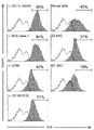
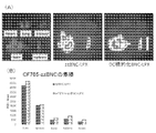
- ZZ-BNC indicates the affinity between mere ZZ-BNC to which no antibody is bound and CD11c-positive dendritic cells.
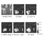
- Bright Field indicates a bright field image
- Ht indicates a heart
- Lg indicates a lung
- Kd indicates a kidney
- Lv indicates a liver
- and Sp indicates a spleen.
- Intensity indicates the fluorescence intensity at 780 nm emitted from the CF-750 dye used for labeling BNC.
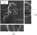
- solid In the figure, DIC indicates a differential interference image, and Fluorescence indicates a fluorescent image.
- XZ and YZ are images after performing the three-dimensional reconstruction image processing, and 15 slice images of 0.25 ⁇ m were subjected to the reconstruction processing.
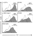
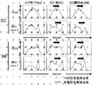
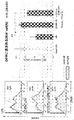
- FIG. 1 shows the results of confirming the transition of various BNCs to dendritic cells in the lymph nodes, and (B) analyzes the transition of various BNCs into dendritic cells in the lymph nodes over time. It is a result.
- DC-BNC represents ⁇ -CD11c-ZZ-BNC (CF750 Labeled), and ZZ-BNC is a BNC having no anti-CD11c antibody.
- ⁇ -DC-BNC in the figure represents ⁇ -CD11c-ZZ-BNC.
- FIG. DC-targeted BNC in the figure represents ⁇ -CD11c-ZZ-BNC.
- mouth in the examination 1 of the antigen loading method to the dendritic cell target BNC of Experimental example 8 is shown.
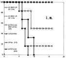
- the vertical axis indicates the survival rate, and the horizontal axis indicates the number of days. Further, regarding the notation (X / Y), X indicates the number of individuals that survived to the 16th day, and Y indicates the number of individuals in each administration group.
- mouth in the examination 2 of the antigen mounting method to the dendritic cell target BNC of Experimental example 10 is shown.
- the vertical axis indicates the titer (Log 10 ) of the anti-JEV IgG antibody.
- A is intravenously administered (IV) with ZZ-BNC-D3
- B is intravenously administered with ⁇ -CD11c-ZZ-BNC-D3
- C is subcutaneously administered with ⁇ -CD11c-ZZ-BNC-D3.
- Administration (SC) shows intramuscular administration (IM) of ⁇ -CD11c-ZZ-BNC-D3.
- IM intramuscular administration
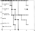
- the infection protection experiment result using dendritic cell targeting BNC which has the antigen of Experimental example 11 is shown.
- the vertical axis indicates the survival rate
- the horizontal axis indicates the number of days.
- X indicates the number of individuals that survived to the 16th day
- Y indicates the number of individuals in each administration group.
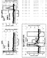
- the experimental result which confirmed manufacture of the dendritic cell target BNC which has the nucleic acid of Experimental example 12 as an antigen is shown.
- IgG-zzBNC represents ⁇ -CD11c-ZZ-BNC
- IgG-zzBNC-LPX represents ⁇ -CD11c-ZZ-BNC—cationic liposome-DNA.
- the experimental result using the in vivo imaging apparatus in the dendritic cell target BNC which has the nucleic acid of Experimental example 12 as an antigen is shown.
- (A) in the figure shows a photographic image
- (B) is a graph showing the fluorescence intensity of each organ.
- DC-targeted BNC-LPX and ⁇ -CD11c-ZZ-BNC-LPX in the figure indicate ⁇ -CD11c-ZZ-BNC-cationic liposome-DNA.
- the vertical axis of the graph in (B) indicates the fluorescence intensity per region.
- DIC indicates a differential interference image
- Fluorescence indicates a fluorescent image.
- XZ and YZ are images after the three-dimensional reconstruction image processing, and 15 slice images of 0.25 ⁇ m are subjected to the reconstruction processing.
- the bio-nanocapsule of the present invention has virus-like particles containing a virus surface antigen protein.
- a virus surface antigen protein is a membrane protein having a self-organizing ability and constituting a capsid, an envelope and the like.
- bio-nanocapsule according to the present invention shown in the schematic diagram of FIG. 1A will be described as appropriate.
- the bio-nanocapsule of the present invention is not limited to those derived from the drawings alone.
- the virus-like particle (a) carries an antibody (c) that recognizes the surface protein of dendritic cells, and the virus surface antigen protein (c) is a lipid constituting the virus-like particle. It exists in a state of piercing the film (b).
- virus-like particles examples include a spherical shape, an elliptical spherical shape, a rugby ball shape, a substantially spherical shape, and a filament shape.
- the inside may be filled with a liquid.
- the virus surface antigen proteins include hepatitis B virus, coronavirus, adenovirus, hepatitis C virus (Hepatitis C virus), type I herpes simplex virus II type II simple Herpesvirus (Herpes simplex virus-2) human immunodeficiency virus (Human immunodefectiveness virus-1: HIV), norovirus (Norovirus), papilloma virus (Sendai virus, Sendai virus, Sendai virus, Sendai virus) Spherical choroid meningitis virus choriomeningitis virus), it includes virus surface antigen proteins, such as Sindbis virus (Sindbis virus).
- Sindbis virus Sindbis virus
- surface antigen proteins such as hepatitis B virus, type I herpes simplex virus, type II herpes simplex virus, human immunodeficiency virus and the like are preferable because they have a membrane-permeable domain responsible for their own entry into cells.
- the hepatitis B virus surface antigen protein includes L protein, M protein, S protein and the like, and is not particularly limited, but is preferably L protein.
- hepatitis B surface antigen protein for example, the N-terminal PreS region of the L protein and the subsequent 77 amino acid residues may be deleted, and the C-terminal side of about 50 amino acids may be deleted ( Patent Document 14).
- Such hepatitis B virus surface antigen protein is preferable in that it can be easily produced using Escherichia coli.
- hepatitis B virus surface antigen protein As a hepatitis B virus surface antigen protein, the 292nd glutamine residue and the 308th glycine residue exposed on the surface of the virus-like particle containing such a protein as a main component are arginine. It is good also as a variant substituted by. Such hepatitis B virus surface antigen protein exerts an effect that it itself is difficult to be recognized as an antigen when administered to a living body.
- the ratio of the virus surface antigen protein contained in the bio-nanocapsule of the present invention is usually about 70 to 95% by weight per bio-nanocapsule. Preferably, it is about 80% to 90% by weight. Therefore, the bio-nanocapsule contains a virus surface antigen protein as a main component. Bionanocapsules contain lipids in addition to viral surface antigen proteins. The ratio is usually about 5 to 10% by weight per bio-nanocapsule. In addition, the bio-nanocapsule includes components such as sugar chains.
- the dendritic cells are not particularly limited, but are not limited to, for example, dendritic cells present in spleen tissue, and may be skin Langerhans cells. Specifically, myeloid dendritic cells and plasmacytoid dendritic cells distributed throughout the body; Langerhans cells distributed in the epidermis; finger-inserted cells distributed in lymph nodes, spleen, thymus, etc .; distributed in lymphatic vessels Vale cells; intradermal dendritic cells distributed in the dermis; germinal center dendritic cells and follicular dendritic cells distributed in lymphoid follicles (embryonic centers) in lymphoid tissues; antigen-carrying cells distributed in lymphatic vessels or sinuses Etc.
- Examples of surface proteins of dendritic cells include CD11c, MHC class II, CD80, CD86, Fc receptor, CD205, CD209, etc.
- dendritic cells they are prominent on the surface of dendritic cells with excellent antigen-presenting ability. From the viewpoint that the amount is expressed, CD11c, MHC class II, CD205 and the like can be mentioned. Moreover, these orthologs may be sufficient as the surface protein of this invention.
- the antibody structure only needs to have a variable region including a site that binds to the surface protein of dendritic cells as described above, and is limited to an immunoglobulin composed of two heavy chains and two light chains, respectively. It is not a thing.
- Specific antibody structures include F (ab ′) 2 that does not contain an Fc region, CH1 region and CL region obtained by digesting immunoglobulin with papain, a heavy chain variable region, and a light chain variable region. Examples include molecular structures such as Fab and the like; Fv and the like that do not contain an immunoglobulin constant region; and scFv that is a single-chain antibody of Fv.
- a multivalent structure combining these molecular structures may be used.
- an scFv structure such as an scFv-Fc structure combining an Fc region and two of the above scFv structures, or a structure called minibody combining a CH3 domain of the constant region and the above two scFv structures, etc.
- Multiplying is achieved by employing a stacking approach. Multivalentization means arranging a plurality of binding sites with dendritic cell surface proteins.
- the above-mentioned antibody includes Affibody (registered trademark).
- An Affibody is a protein molecule having a structure in which three helix domains are bound using a specific domain of Protein A as a scaffold, and has a function of binding to an antigen. That is, when the antibody is an affibody, the affibody is a molecule that recognizes the surface protein of dendritic cells.
- the subtype is not particularly limited, and may be any subtype such as IgG, IgA, IgY, IgM, and IgE. Also, the subclass is not particularly limited.
- binding means that the surface protein of the dendritic cell and the antibody are “bound”. More preferred is specific or selective binding. Such “binding” can be determined by the affinity between the surface protein of the dendritic cell and the antibody, or the affinity between the dendritic cell expressing the surface protein and the antibody.
- the specific determination method is not particularly limited, but can be determined by a known method such as measurement of binding constant, fluorescent immunostaining method such as flow cytometry analysis, Western blotting or the like.
- the determination criteria may be limited to the presence or absence of affinity, and may be determined by relatively comparing the affinity.
- the antibody (d) only needs to have its antigen recognition site exposed to the outside of the bio-nanocapsule. Specifically, for example, as shown in FIG. 1, it may be carried on the virus surface antigen protein (c) contained in the bio-nanocapsule, or may be carried on the lipid membrane (b) contained in the bio-nanocapsule. Good.
- the antibody (d) When the antibody (d) is supported on the virus surface antigen protein (c), the antibody (d) may be supported via a chemical bond such as a covalent bond or by binding via an antibody binding domain. It may be. Among these, it is preferable that the antibody is bound to the antibody binding domain. In particular, it is preferable that the virus surface antigen protein (c) contained in the bio-nanocapsule of the present invention has an antibody binding domain, and the antibody (d) is bound through such a domain.
- the state in which the virus surface antigen protein and the antibody are supported may be further subjected to a crosslinking treatment using a crosslinking agent or the like.
- a crosslinking agent or the like.
- Specific cross-linking agents are not particularly limited. For example, bis [sulfosuccinimidyl] suberate (BS 3 ) dimethyl melimidate dihydrochloride (DMP), dimethyl subericidate dimethyl dihydrochloride (DMS) disuberate And succinimidyl (DSS).
- the antibody-binding domain is not particularly limited.
- the Z domain containing E, D, A, B, and C domains possessed by protein A derived from Staphylococcus aureus and the like, C1 possessed by protein G derived from streptococci, and the like examples thereof include an antibody binding domain containing D1, C2, D2, and C3 domains, an antibody binding domain containing B1, B2, B3, B4, and B5 domains of protein L derived from Peptostreptococcus magnus and the like.
- the domain containing at least the C2 domain is preferably used as the above-mentioned antibody binding domain, and among the antibody binding domains possessed by protein L, the domain containing at least the B1 domain is used as the above-described antibody.
- a binding domain is preferred.
- amino acid sequences of these seed antibody binding domains include the amino acid sequence shown in SEQ ID NO: 1 in the case of the Z domain of protein A. If it is an antibody binding domain which protein G has, the amino acid sequence shown to sequence number 2 will be mentioned. If it is an antibody binding domain which protein L has, the amino acid sequence shown to sequence number 3 will be mentioned.
- amino acid sequences may be amino acid sequences mutated as long as antibody binding ability is not attenuated.
- the specific number of mutations is not particularly limited, but usually the amino acid sequence before mutation is 85% or more, preferably 90% or more, more preferably 95% or more, and most preferably 99% or more. What is necessary is just to set it as the number of mutation introduction
- mutagenesis here refers to substitution, deletion, insertion, and the like.
- a known method can be employed, and is not particularly limited.
- a conservative substitution technique may be employed for substitution.
- the term “conservative substitution technique” means that an amino acid residue is replaced with an amino acid residue having a similar side chain.
- amino acid residues having basic side chains such as lysine, arginine, and histidine
- amino acid residues having acidic side chains such as aspartic acid and glutamic acid
- amino acid residues having uncharged polar side chains such as glycine, asparagine, glutamine, serine, threonine, tyrosine and cysteine
- Amino acid residues with non-polar side chains such as proline, phenylalanine, methionine, and tryptophan
- amino acid residues with ⁇ -branched side chains such as threonine, valine, and isoleucine
- aromatic side chains such as tyrosine, phenylalanine, tryptophan, and histidine
- substitutions between amino acid residues are conservative substitutions.
- identity refers to the degree of amino acid sequences of two or more comparable amino acid sequences with respect to each other. Therefore, the higher the identity of two amino acid sequences, the higher the similarity between those sequences.
- degree of amino acid sequence identity or similarity is determined, for example, using FASTA, a sequence analysis tool, and using default parameters. Specific methods of these analysis methods are publicly known, and the website of the National Center of Biotechnology Information (NCBI) (http://www.ncbi.nlm.nih.gov/) may be referred to.
- the virus surface antigen protein (c) When the above-mentioned antibody binding domain is contained in the virus surface antigen protein (c), it is preferably contained in the surface of the bio-nanocapsule of the present invention or a site exposed to the outside thereof.
- it when it is a virus surface antigen protein, it is preferably contained in the PreS region present at its N-terminus.
- the virus surface antigen protein contained in the bio-nanocapsule is a hepatitis B virus surface antigen protein and its N-terminal and C-terminal are deleted, the C of the protein It is preferable that the above-mentioned antibody binding domain is contained on the terminal side (Patent Document 14).
- a plurality of the antibody binding domains described above may be included in the bio-nanocapsule.
- the above-described various antibody binding domains may be of one type or two or more types.
- the antibody (d) when the antibody (d) is supported on the lipid membrane (b), it may be supported via a chemical bond as in the case of being supported on the lipid. It may be supported by binding of the antibody via That is, the virus-like particle (a) constituting the bio-nanocapsule of the present invention does not contain the antibody binding domain in the virus surface antigen protein (c), and is in a state of being bound or pierced to the lipid membrane (b). Good.
- the average particle size of the bio-nanocapsule of the present invention is usually about 20 nm to 150 nm as the Z-average particle size from the viewpoint of efficient delivery to dendritic cells, efficient entry into dendritic cells, and the like. It is. Preferably, it is usually about 45 nm to 55 nm.
- the charge of the bio-nanocapsule of the present invention is usually about ⁇ 30 mV to ⁇ 10 mV as the ⁇ potential. From the viewpoints of efficient delivery to spleen tissue, efficient delivery to dendritic cells, efficient entry into dendritic cells, delivery of proteins and nucleic acids, etc., toxicity in vivo From the viewpoint and the like, about ⁇ 10 mV to ⁇ 5 mV is preferable.
- the ⁇ potential may be measured by the method described in detail in the following examples.
- the bio-nanocapsule of the present invention tends to easily induce a humoral immune system by stopping for a long period of time after being taken up by acting on dendritic cells. On the other hand, when it moves to the cytosol and stays for a relatively long time, it tends to easily induce the cellular immune system.
- bio-nanocapsules can be appropriately selected by adjusting the charge. Therefore, when the bio-nanocapsule of the present invention further contains a liposome, it is possible to appropriately select the immune system to be raised by adjusting the overall charge including the liposome.
- bio-nanocapsules induces lipids that are easy to form their charge and lipid type (reverse hexagonal phase (also referred to as hexagonal II phase) that destabilizes the endosomal membrane) and the proton sponge effect. It is possible to select appropriately by adjusting the lipid that destroys the endosome. Therefore, by adjusting the charge and lipid type of the bio-nanocapsule of the present invention, it is possible to appropriately select the immune system to be raised.
- the bio-nanocapsule of the present invention tends to have a negative charge in general, the bio-nanocapsule of the present invention as described later has a mode in which the liposome is bound, and the charge of the liposome is adjusted.
- the charge of the entire bio-nanocapsule of the present invention can be easily set as appropriate.
- the charge of the liposome can generally be easily set by adjusting the blending ratio of the lipid composition.
- the setting of the surface charge of the bio-nanocapsule of the present invention is not limited to setting the charge of the liposome.
- the host cell may be a prokaryotic cell or a eukaryotic cell, but a yeast cell is preferable from the viewpoint of introduction of nucleic acid, culture, gene expression level, and the like.
- a yeast cell is preferable from the viewpoint of introduction of nucleic acid, culture, gene expression level, and the like.
- E. coli is used as a host cell. It is preferable to use as.
- the recovered virus-like particles may be purified by subjecting them to a column chromatography method using an ultracentrifugation method, an affinity gel, a size exclusion gel or the like.
- the bio-like capsule of the present invention may be prepared by mixing the thus obtained virus-like particle and the above-mentioned antibody.
- the mixing conditions and the like are not particularly limited, and a known method may be appropriately selected.
- bio-nanocapsule and the antigenic substance and / or nucleic acid are combined in advance as a production method when the bio-nanocapsule of the present invention further contains an antigenic substance and / or nucleic acid.
- a known method such as an electroporation method may be adopted for the bio-nanocapsule produced as described above.
- a liposome may be further bound to the above-mentioned bio-nanocapsule.
- the structure of the bio-nanocapsule of such an embodiment (hereinafter sometimes referred to as a liposome-bound bionanocapsule) will be described as appropriate based on one embodiment of the liposome-bound bionanocapsule shown in the schematic diagram of FIG. .
- liposome-binding bio-nanocapsules are not limited to those derived only from the drawings.
- the above-mentioned liposome-binding bio-nanocapsule has a structure in which the bio-nanocapsule (a) and the liposome (e) are combined and inseparable. More specifically, for example, as shown in Patent Document 8, the bio-nanocapsule has a lipid membrane (b), and the liposome (e) and the bio-nanocapsule (a) are passed through the respective lipid membranes. And membrane fusion.
- the liposome (e) and the bio-nanocapsule (a) do not need to be encapsulated in either one, and may simply be in a state where one of them is stuck into the other.
- the liposome is usually about 2 to 12 parts by mass, more preferably about 6 to 10 parts by mass with respect to 1 part by mass of the bio-nanocapsule.
- the average particle size of the above-mentioned liposome-binding bio-nanocapsule is usually 100 nm to 600 nm as the Z-average particle size from the viewpoint of efficient delivery to dendritic cells, efficient entry into dendritic cells, and the like. Degree.
- the charge of the above-mentioned liposome-bound bio-nanocapsule is usually about ⁇ 30 mV to 20 mV as the ⁇ potential. From the viewpoints of efficient delivery to spleen tissue, efficient delivery to dendritic cells, efficient entry into dendritic cells, delivery of proteins and nucleic acids, etc., toxicity in vivo From the viewpoint and the like, about ⁇ 20 mV to 0 mV is preferable.
- the ⁇ potential may be measured by the method described in detail in the following examples.
- the above-mentioned liposome-binding bio-nanocapsule tends to easily induce a humoral immune system by stopping for a long period of time after being allowed to act on dendritic cells and then taken up by endosomes. On the other hand, when it moves to the cytosol and stays for a relatively long time, it tends to easily induce the cellular immune system.
- the localization of these liposome-binding bio-nanocapsules can also be appropriately selected by adjusting the charge, as in the case of the bio-nanocapsules described above. Therefore, if the charge of the liposome-binding bio-nanocapsule of the present invention is adjusted, it is possible to appropriately select the immune system to be raised.
- the liposome in the above-described liposome-binding bio-nanocapsule may have a single lamellar structure (single film) or a multilamella structure (multiple film). It may be an anionic liposome or a cationic liposome.
- the specific charge of the liposome is usually about ⁇ 60 mV to 60 mV as the ⁇ potential.
- the average particle size of such liposomes is usually about 100 nm to 400 nm as the Z-average particle size. From the viewpoint of complexing with a bio-nanocapsule and then administering to a living body, the thickness is preferably about 100 nm to 300 nm, more preferably about 100 nm to 200 nm.
- the measurement of the ⁇ potential and the Z-average particle diameter of the liposome may be performed by the methods shown in the following examples.
- the method for producing the liposome is not particularly limited, and may be prepared by a known method after mixing the lipids exemplified as the composition as described above at an appropriate composition ratio. Examples include the bangham method, ultrasonic method, reverse phase evaporation method, polyhydric alcohol method, heating method, spray drying method, mechanochemical method, freeze-thaw method, film loading method, supercritical carbon dioxide method and the like. .
- the bio-nanocapsule of the present invention further contains a liposome and an antigenic substance and / or nucleic acid
- a liposome and an antigenic substance and / or nucleic acid are bound in advance.
- an antigenic substance and / or a nucleic acid may be mixed at the same time when the lipid constituting the liposome is mixed.
- the mixing amount of the antigen substance and / or nucleic acid is not particularly limited, and a desired amount may be appropriately set and mixed.
- the prepared liposome can be adjusted to an average particle size using, for example, an extruder together with a filter having a pore size of an appropriate size.
- virus-like particles created in the above-mentioned method for producing bio-nanocapsules are bound to the above-mentioned liposomes, and the liposome-binding bio-nanocapsules obtained thereby are bound to the antibodies that recognize the surface proteins of the above-mentioned dendritic cells. Also good.
- the binding conditions between the bio-nanocapsule and the liposome may be appropriately selected from the below-described binding conditions between the bio-nanocapsule containing the antigenic substance and / or nucleic acid and the liposome.
- the binding conditions for the antibody recognizing the antibody may be appropriately selected from the binding conditions for the virus-like particle described in the method for producing the bio-nanocapsule and the antibody recognizing the surface protein of the dendritic cell as appropriate. .
- the mixing condition of the above-mentioned bio-nanocapsule and the above-mentioned liposome is not particularly limited, and it may be usually incubated for 30 to 120 minutes in an environment of about 20 to 60 ° C.
- the liposome-binding bio-nanocapsule prepared according to the case may be purified.
- a specific purification method may be a known method and is not particularly limited. Examples thereof include ultracentrifugation, column chromatography using affinity gel, size exclusion gel, and the like.
- the method for binding the above-mentioned liposome-binding bio-nanocapsule and the above-mentioned antibody that recognizes the surface protein of the dendritic cell is not particularly limited, and the antibody that recognizes the surface protein of the dendritic cell and the above-mentioned bio-nanocapsule or After mixing with the liposome-binding bio-nanocapsule of the present invention, it is usually incubated for 30 minutes to 120 minutes in an environment of about 20 ° C. to room temperature.
- a bio-nanocapsule carrying an antibody that recognizes a dendritic cell prepared according to circumstances may be purified.
- a specific purification method a known method may be adopted as in the above-described purification of the bio-nanocapsule of the present invention.
- the antibody binding method to the bio-nanocapsule to which the liposome is not bound may be the same as this.
- Bio-nanocapsule containing the antigenic substance and / or nucleic acid of the present invention The bio-nanocapsule containing the antigenic substance and / or nucleic acid of the present invention further comprises an antigenic substance and / or nucleic acid in the above-described bio-nanocapsule of the present invention. Specifically, what is necessary is just to be contained in the virus-like particle
- bio-nanocapsule When the bio-nanocapsule is a liposome-bound bionanocapsule as described above, it may be contained in the virus-like particle (a) or may be contained in the liposome (e).
- virus-like particle (a) may include an antigenic substance and / or a nucleic acid, or may be in a state of being pierced into the lipid membrane (b).
- liposome (e) may include an antigenic substance and / or a nucleic acid, or may be stuck into the liposome.
- the antigenic substance is not particularly limited, the bio-nanocapsule of the present invention is positively delivered to dendritic cells and taken into the dendritic cells when administered in vivo, and thus has an excellent function as a vaccine. Demonstrate. Therefore, the antigenic substance may be a surface protein such as a bacterium or virus that causes an infection or the like, a sugar chain, a lipid, or the like, or a part thereof.
- the nucleic acid is not particularly limited and includes DNA, RNA, peptide nucleic acid and the like.
- a nucleic acid base sequence is preferably a base sequence encoding the antigenic substance described above. Further, it may be a base sequence having a CpG motif, a PATRE sequence, a C3d sequence, or the like. Since the CpG motif or the like exhibits a function as an adjuvant that elicits the immune system, it is effective to use it together with the above-mentioned antigenic substance and nucleic acid having a base sequence encoding the antigenic substance.
- the bio-nanocapsule of the present invention containing an antigenic substance and / or nucleic acid can efficiently reach dendritic cells when administered into an individual. Furthermore, after reaching the dendritic cell, it also exerts an action of entering the dendritic cell.
- a dendritic cell is a cell that plays a very important role as a cell presenting an antigen in the immune system in a living body. Therefore, when the bio-nanocapsule of the present invention contains an antigenic substance, the dendritic cell is used. The antigenic substance is presented very efficiently, and an immune system related to cellular immunity and / or humoral immunity is induced against the antigenic substance.
- nucleic acids that encode peptides that serve as antigenic substances can be introduced into dendritic cells.
- the expression of a peptide serving as an antigen substance in such dendritic cells facilitates the presentation of the peptide as an antigen substance outside such dendritic cells.
- the bio-nanocapsule of the present invention containing an antigenic substance and / or nucleic acid is administered into a living body, a significant amount of an antibody against the antigenic substance can be produced in the living body. And since it is natural that such an antibody includes an antibody having a function of neutralizing the antigen substance, the bio-nanocapsule containing the antigen substance and / or nucleic acid of the present invention is a vaccine. As an excellent function.
- bio-nanocapsule containing the antigenic substance and / or nucleic acid of the present invention not only elicits a humoral immune system that produces an antibody against the antigenic substance as described above after entering a dendritic cell, but also cellular immunity.
- the system also has the effect of inducing. Therefore, it has a function as an excellent vaccine.
- the administration target of the bio-nanocapsule containing the antigenic substance and / or nucleic acid of the present invention is not particularly limited, and is not particularly limited as long as it is an animal that desires a function as a vaccine. , Chicken, horse, sheep, human, goat, chimpanzee, gorilla and the like.
- the specific dose is not particularly limited, for example, when a bio-nanocapsule containing an antigenic substance is administered, it may be usually administered in an amount of about 2 mg to 50 mg / kg in terms of the amount of the antigenic substance.
- When administering a bio-nanocapsule containing a nucleic acid it is usually sufficient to administer about 1 mg to 20 mg / kg in terms of the amount of nucleic acid.
- When administering a bio-nanocapsule containing an antigenic substance and a nucleic acid it is usually sufficient to administer about 3 mg to 70 mg / kg in terms of the total amount of the antigenic substance and nucleic acid.
- bio-nanocapsule containing antigenic substance and / or nucleic acid of the present invention The production method of bio-nanocapsule containing the antigenic substance and / or nucleic acid of the present invention is not particularly limited, and the above-mentioned antigenic substance and / or nucleic acid is described above. And the above-described bio-nanocapsule or liposome-bound bio-nanocapsule, and then incubated for 30 minutes to 120 minutes, usually in an environment of about 20 ° C. to room temperature.
- bio-nanocapsules When antigenic substances and / or nucleic acids are directly contained in the above-mentioned bio-nanocapsules, these may be chemically bound using, for example, a known crosslinking agent. Moreover, you may introduce
- the antigenic substance and / or the nucleic acid may couple
- the antigenic substance and / or nucleic acid and the bio-nanocapsule or the liposome may be mixed and bonded before the bio-nanocapsule and the liposome are bonded in advance.
- the bio-nanocapsule and the liposome may be combined and further mixed and combined. The specific method is as described in detail in [Manufacturing method] of the above (Bio-nanocapsule) and (Liposome).
- Bio nanocapsules to the present invention the method described above antigenic substance and / or nucleic acid transfer of the present invention, when further comprising an antigenic substance and / or nucleic acids, such antigenic substances and / or nucleic acids by administering them to animals It can be introduced into animal dendritic cells.
- the method for introducing an antigenic substance and / or nucleic acid of the present invention into an animal dendritic cell includes a step of administering to the animal a bio-nanocapsule that is a virus particle containing a viral antigen protein, and the bio-nanocapsule is dendritic.
- An introduction method characterized by carrying an antibody that recognizes a cell surface protein and containing an antigenic substance and / or a nucleic acid.
- the animal to be administered, the dose, the administration method, etc. may be as described in detail for the bio-nanocapsule containing the antigenic substance and / or nucleic acid of the present invention .
- the bio-nanocapsule of the present invention exerts an action of invading into dendritic cells together with the antigenic substance and / or nucleic acid. It is useful as a nucleic acid introduction agent.
- the antigenic substance and / or nucleic acid introduction agent of the present invention includes the above-described bio-nanocapsule of the present invention.
- the blending amount of the bio-nanocapsule of the present invention in the antigen substance and / or nucleic acid introduction agent is not particularly limited, but it may be usually about 0.001% to 99.9% per such introduction agent.
- the above-described bio-nanocapsule of the present invention itself may be used as the antigen substance and / or nucleic acid introduction agent of the present invention.
- a bio-nanocapsule (hereinafter sometimes referred to as BNC) used in this example was produced by the following method. Specifically, yeast cells using a method for expressing a hepatitis B virus surface antigen L protein having a ZZ domain (hereinafter sometimes referred to as HBsAg L protein) using the method described in Non-Patent Document 5. The yeast cells were cultured, and bio-nanocapsules (hereinafter referred to as ZZ-BNC) mainly composed of HBsAg L protein having a ZZ domain were isolated and purified from within the yeast cells. .
- BNC bio-nanocapsule
- a bio-nanocapsule (hereinafter referred to as ST-BNC) that does not have a ZZ domain and is mainly composed of HBsAg L protein was isolated and purified by the same method.
- the concentrations of the obtained ZZ-BNC and ST-BNC were measured in terms of the amount of protein as the main component of various BNCs.
- the specific measurement method was measured using a commercially available protein amount measurement kit, BCA protein assay kit (Pierce (IL, USA)) (the same applies to the following experiments).
- the obtained ZZ-BNC and ST-BNC are optionally supplied with Fluorolink Cy5 dye (GE Healthcare CA, USA) or CF750 dye (Biotium CA, USA) for its own detection.
- Fluorolink Cy5 dye GE Healthcare CA, USA
- CF750 dye Biotium CA, USA
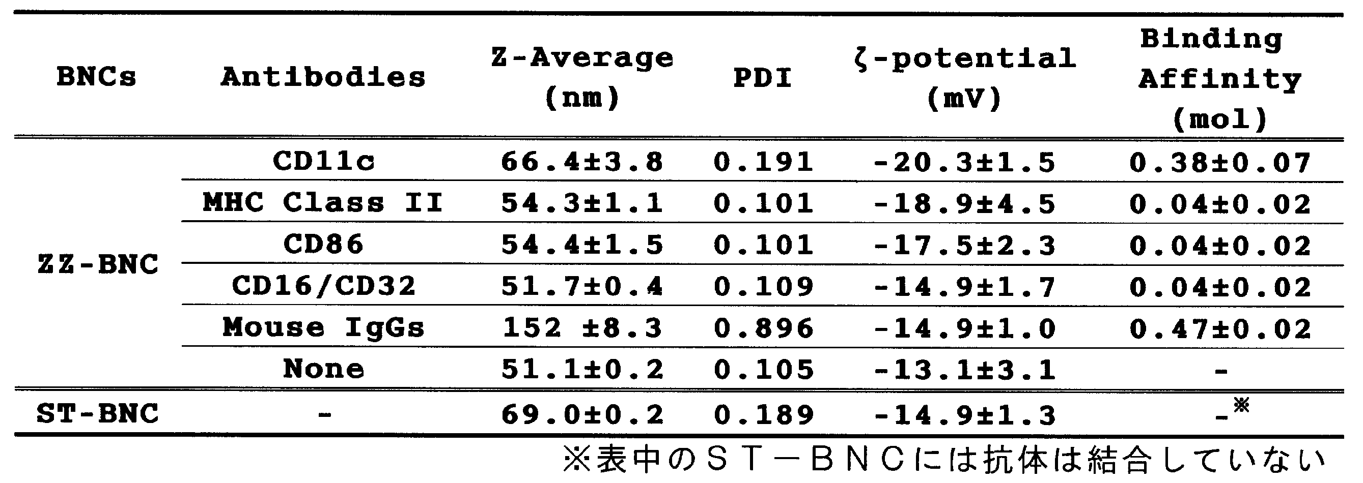
- Z-Average indicates the Z-average particle diameter
- ⁇ -Potentials indicates the ⁇ potential.
- the Z-average particle size and ⁇ potential were both measured using a dynamic light scattering photometer Zetasizer (Malvern Instruments, Worcestershire, UK) in an aqueous solvent at 25 ° C. according to a conventional method (the following). The same applies to the experiment.
- PDI indicates the polydispersity index of the Z-average particle size.
- Binding Affinity indicates the amount of each antibody bound to ZZ-BNC, and was calculated as the amount per mole of HBsAg L protein containing the ZZ domain, which is the main component of ZZ-BNC. Specifically, the amount of each antibody bound to ZZ-BNC was measured using a Twin-Q apparatus (Osaka, Japan) based on the quartz crystal microbalance method at 25 ° C. It was carried out according to a conventional method.
- ZZ-BNC to which various antibodies bind has no ZZ domain and has the same Z-average particle diameter (100 nm or less) and ⁇ potential (negative charge) as conventional ST-BNC. It became clear. Also, for ZZ-BNC, an anti-CD11c antibody having an isotype of an Armenian hamster-derived IgG is known to have a high affinity for the ZZ domain, which is as high as that of mouse-derived IgGs. It became clear that it combined with efficiency.
- Dendritic cells were isolated from spleens of Balb / c mice (female: 6-8 weeks old; Japan SLC: JAPAN) according to a conventional method. Among dendritic cells, CD11c positive dendritic cells were further purified based on MACS (registered trademark) technology (Miltenyi Biotech Co., Ltd.).
- CD11c-positive dendritic cells and ZZ-BNC (ZZ-BNC is labeled with a cy5 dye) bound to the above antibody were incubated at 4 ° C. for 30 minutes, and then Cy5 was released.
- the amount of ZZ-BNC bound to various antibodies having affinity for CD11c-positive dendritic cells was measured based on 670 nm, which is the emission wavelength of Cy5 dye. The result is shown in FIG.
- ZZ-BNC bound with anti-CD11c antibody and anti-MHC class II showed particularly high affinity for CD11c-positive dendritic cells derived from mouse spleen.
- the anti-CD11c antibody described above may be used for ZZ-BNC that does not have an antibody that recognizes dendritic cell surface proteins, such as ZZ-BNC to which no antibody is bound or ZZ-BNC to which Mouse IgGs is bound.
- ZZ-BNC to which no antibody is bound
- ZZ-BNC to which Mouse IgGs is bound
- affinity for spleen-derived CD11c-positive dendritic cells was observed.
- HBsAg L protein having the ZZ domain which is the main component of ZZ-BNC used in this example, has a high mannose sugar chain, and the dendritic cells have a mannose receptor. This is thought to be because these two are involved in binding to dendritic cells. Therefore, ZZ-BNC bound with an antibody that recognizes BNC, ZZ-BNC containing the same and dendritic cell surface protein in this example is particularly effectively used for binding to dendritic cells.
- Example 3 Administration of BNC to living body> Among ZZ-BNCs bound with antibodies recognizing surface proteins of dendritic cells prepared by the above-described method, ZZ-BNCs bound with anti-CD11c antibody and anti-MHC class II antibody are mouse individuals (female: 6-8). An experiment was conducted using a living body observation system (OV-100; Olympus: Japan), followed by observation of the localization of ZZ-BNC in a mouse individual. As a comparative experiment, ZZ-BNC bound to Mouse IgGs, mere ZZ-BNC bound to no antibody, and ST-BNC not having a ZZ domain were also administered. Each BNC was previously labeled with a CF-750 dye.
- Various BNCs were administered to each mouse by tail vein injection at 10 ⁇ g in terms of the amount of the protein as the main component, and after euthanasia to the mouse individuals 40 minutes after the administration, The heart, lung, kidney, liver and spleen were removed.
- Each of the extracted organs is detected by the above-described biological observation system based on the emission wavelength of CF-750 dye at 780 nm, and the various BNCs after administration to the individual mouse are localized in the individual mouse. The result of observation is shown in FIG.
- ZZ-BNC bound with anti-CD11c antibody and ZZ-BNC bound with anti-MHC class II antibody show high affinity for CD11c-positive dendritic cells in spleen tissue Became clear.
- ZZ-BNC bound with anti-CD11c antibody showed higher affinity such that it accumulates in 62% of CD11-positive dendritic cells.
- Non-Patent Document 2 when an antigen to which F (ab ′) 2 of anti-CD11c is bound is administered subcutaneously, the antigen reaches only 1.5% of dendritic cells after 4 hours. Reaching 81.4% dendritic cells after 12 hours.
- ZZ-BNC carrying an antibody recognizing the surface protein of dendritic cells in this Example accumulates as much as 62% of such ZZ-BNC in 4 hours after administration. .
- an antibody that recognizes a surface protein of a dendritic cell that binds to ZZ-BNC is supported on the surface of ZZ-BNC so that an antigen-antibody reaction is more likely to occur. This is considered to be because the effect of recognizing the surface protein increases.
- CD11 positive dendritic cells were purified from the incubated cells using an anti-CD11c antibody, and the purified cell population was observed with a confocal laser microscope (FV-1000; Olympus: Janan). The results are shown in FIG.
- CD11c is displayed in green
- BNC to which the anti-CD11c antibody is bound is displayed in red.
- ZZ-BNC bound with anti-CD11c antibody is present inside CD11c-positive dendritic cells. Therefore, when ZZ-BNC bound with an antibody recognizing the surface protein of dendritic cells in this example was administered to a mouse individual, it reached CD11c-positive dendritic cells in spleen tissue, and such dendritic cells It was revealed that ZZ-BNC was taken into the inside.
- the ZZ-BNC bound to the antibody recognizing the surface protein of the dendritic cell in this example has a function of invading into the dendritic cell because it has not only the membrane fusion domain but also a membrane permeation domain. It is thought that it is because it has.
- the above-mentioned ZZ-BNC binds to CD11c present on the surface of dendritic cells, and then the ZZ-BNC is taken into the dendritic cells based on the phagocytosis by such CD11c.
- ZZ-BNC bound with an antibody recognizing the surface protein of dendritic cells in this example can be suitably used for introducing a substance into CD11c-positive dendritic cells.
- dendritic cells are important cells that exert an antigen-presenting function in the immune system of various mammals including humans, they recognize the surface proteins of the dendritic cells in this example.
- Liposomes were produced according to a conventional method. Specifically, cholesterol (Chol), diethylphosphatidylethanolamine (DOPE), O, O′-ditetradecanol-N- ( ⁇ -trimethylammonioacetyl) diethanol hydrochloride (DC-6-14) at a molar ratio of 3 It was prepared by mixing with a composition of 3: 4. The obtained liposome was confirmed to be a cationic liposome. The resulting liposome had a ⁇ potential of 56.5 mV and a Z-average particle size of 121 nm.
- Example 4 Preparation of BNC bound to liposome> Bio-nanocapsule in which ZZ-BNC containing 100 ⁇ g of HBsAg L protein having ZZ domain and 600 ⁇ g of the above-mentioned cationic liposome are mixed in pH 4 Briton-Robinson buffer and incubated at 37 ° C. for 30 minutes to bind the liposome Was made.
- the liposome-bound BNC obtained here was purified by subjecting it to 0-40% cesium chloride density gradient centrifugation.
- the purified bio-nanocapsule to which the liposome was bound (hereinafter referred to as ZZ-BNC-LP) was dialyzed against PBS.
- the obtained ZZ-BNC-LP contains 19.5 ⁇ g of HBsAg L protein having a ZZ domain, which is the main component of ZZ-BNC, and 65.6 ⁇ g of cationic liposomes, and has a Z-average particle size of 455 nm (PDI; 0 331), the ⁇ potential was ⁇ 5.76 mV.
- the liposome content was measured using a commercially available lipid measurement kit, Cholesterol E-Test (Wako Pure Chemical: Japan).
- D3 antigen of Japanese encephalitis virus was bound to ZZ-BNC-LP as an antigen substance.
- the D3 antigen is one of the envelope protein domains of Japanese encephalitis virus, and was prepared using an expression system using Escherichia coli as described in Non-Patent Document 1.
- ZZ-BNC-LP-D3 The above-mentioned ZZ-BNC-LP containing 120 ⁇ g of ZZ-HBsAg L protein and 404 ⁇ g of cationic liposomes and 60 ⁇ g of the D3 antigen were mixed and incubated at room temperature for 1 hour (the one obtained in this step) Hereafter referred to as ZZ-BNC-LP-D3). Thereafter, 24 ⁇ g of the above anti-CD11c antibody was added and allowed to bind to ZZ-BNC in ZZ-BNC-LP. The final product is hereinafter referred to as ⁇ -CD11c-ZZ-BNC-LP-D3.
- Example 5 Administration of BNC bound to liposome containing antigen to mouse individual> 20 ⁇ g of ⁇ -CD11c-ZZ-BNC-LP-D3 and ZZ-BNC-LP-D3 in terms of the amount of D3 antigen were administered to individual mice (female: 6-8 weeks old; Japan SLC: JAPAN), tail It was administered by the intravenous method.
- a cationic liposome containing the same amount of D3 antigen and a complex of D3 antigen hereinafter referred to as LP-D3
- Table 3 shows various physical properties of the administered sample.
- Antibody titer was measured using a generally known indirect ELISA method. Specifically, the D3 antigen obtained by the above method was immobilized on a microplate, and an HRP-labeled anti-mouse IgG antibody was used for detection. The results are shown in FIG.
- the mouse individual is immunized using only the D3 antigen, the mouse individual is immunized using LP-D3, or 4 weeks after the initial immunization in the mouse individual After 6 weeks, almost no anti-D3 antibody was produced.
- an antibody against D3 is 6 weeks after the first immunization, Although produced in mouse solids, the production amount is similar to that produced four weeks after immunization with ⁇ -CD11c-ZZ-BNC-LP-D3 having an antibody against anti-CD11c, and D3 The amount of antibody produced against the antigen was much inferior to that of ⁇ -CD11c-ZZ-BNC-LP-D3.
- ⁇ Experimental Example 6 Administration route study of dendritic cell targeted BNC> Fluorescent dye CF750-labeled anti-CD11c antibody-presenting ZZ-BNC (hereinafter referred to as ⁇ -CD11c-ZZ-BNC (CF750 Labeled)) is prepared by the method described in Non-Patent Document 3, and the amount of the protein corresponding to 10 ⁇ g is prepared. It is administered to the ridge part (subcutaneous injection: SC) or thigh (intramuscular injection: IM) of mouse Balb / c, and it is sacrificed 12 hours later so that the axillary, inguinal, and popliteal lymph nodes can be directly viewed. After laparotomy, fluorescence observation was performed with an In Vivo imaging device manufactured by Olympus.
- SC subcutaneous injection
- IM intramuscular injection
- Results are shown in FIG. Fluorescence was observed in the inguinal lymph nodes for the SC administration, and in the inguinal and popliteal lymph nodes for the IM administration. This indicates the possibility that ⁇ -CD11c-ZZ-BNC (CF750 Labeled) can infect regional lymph nodes not by conventional IV but by the usual route of administration as vaccines of SC and IM.
- inguinal lymph nodes were removed from mice administered SC with ⁇ -CD11c-ZZ-BNC (CF750 Labeled), and popliteal lymph nodes were removed from mice administered IM with ⁇ -CD11c-ZZ-BNC (CF750 Labeled).
- CD11c positive cells were isolated by the method described in Patent Document 3, and the degree of ZZ-BNC migration to dendritic cells in lymph nodes was examined using FACS.
- Example 7 Experiment on Adjuvant Effect of Dendritic Cell Targeted BNC> ⁇ -CD11c-ZZ-BNC, ZZ-BNC, and 10 ⁇ g of LPS as a positive control were administered to mice with SC or IM, and the inguinal lymph nodes or popliteal lymph nodes were removed 2 days later. Dendritic cells were isolated, and the expression levels of CD86 and MHC class II, which are activation markers on the surface of dendritic cells (cell surface molecules that are expressed when dendritic cells present antigens to T cells), were measured using FACS. Quantified.
- the liposome is aLP-D3.
- the Z average particle size measured according to the above-mentioned method was about 110 nm, and the ⁇ potential was about ⁇ 53.1 mV.
- ZZ-BNC was polymerized at pH 4, 37 ° C. for 1 hour, the anti-CD11c antibody was fixed with BS 3 crosslinker, and the excess crosslinker was masked with glycine.
- This is designated anti-CD11c-ZZ-BNC-aLP-D3.
- the Z average particle diameter of such BNC is about 100 to 600 nm, and the ⁇ potential is about ⁇ 20 to 0 mV.
- ALP-D3, ST-BNC-aLP-D3 (ST-BNC is a BNC having no ZZ domain as described above and presents a yeast-derived high mannose sugar chain), ZZ-BNC-aLP-D3 , And ⁇ -CD11c-ZZ-BNC-aLP-D3 were administered to mice (6 mice per group) at 20 ⁇ g as the amount of D3 at IM every 3 weeks (0, 2, and 4 weeks). . At that time, blood was collected at 0, 4, and 6 weeks, and at 6 weeks, 50% of the lethal dose of half of the mice was inoculated. In addition, as a positive subject experiment, a JEV vaccination group (multivalent and 20 ⁇ g or more of administered antigen) of Kaketsuken was similarly administered.
- FIG. A positive subject the KEV Institute JEV vaccination group (antigen administered is multivalent and greater than 20 micrograms) completely protected against infection by JEV
- ⁇ CD11c-ZZ-BNC-aLP-D3 was similarly Completely protected against infection by JEV. Since ZZ-BNC-aLP-D3 was protected against infection by half of the mice and only one ST-BNC-aLP-D3 was protected, dendritic cells containing only yeast-derived high mannose-derived sugar chains Targeting was considered insufficient to prevent infection.
- the ZZ domain itself recognizes the IgG domain that is presented to B cells and the like, and therefore, it was speculated that ZZ-BNC had higher ability to target dendritic cells than ST-BNC.
- the anti-CD11c antibody directly bound with the D3 antigen could not completely prevent the infection by JEV.
- "1) delivery of D3 antigen to dendritic cells via anionic liposomes is effective for antigen processing in dendritic cells, and D3 directly fed into dendritic cells is It suggests that it is not properly processed or simply degraded in dendritic cells.
- Or“ 2) The anti-CD11c antibody displayed on the surface of ZZ-BNC is aligned on the surface of BNC. This indicates that the avidity of such an antibody has significantly increased compared to that of one antibody molecule, and thus targeting to dendritic cells has been performed efficiently.
- Example 9 Examination of antigen loading method onto dendritic cell targeted BNC 2>
- the anti-CD11c antibody is aligned and presented on the surface of the ZZ-BNC and simultaneously presents the D3 antigen without using a liposome.
- a product was prepared and examined. Specifically, pyridyldithiolated ZZ-BNC was prepared using Sulfo-LC-SPDP (Pierce) for the Free amino group of the ZZ-BNC surface layer.
- mice 40 ⁇ g of ⁇ -CD11c-ZZ-BNC-D3 and ZZ-BNC-D3 in terms of D3 antigen amount were administered to mice (5 mice per group), IV (intravenous), IM (intramuscular), respectively. ) And SC (subcutaneous) routes were inoculated at 0 and 2 weeks, and anti-D3-IgG antibody titers in serum were evaluated by ELISA at 4 weeks.
- Example 11 Infection protection experiment using dendritic cell-targeted BNC with antigen> ⁇ -CD11c-ZZ-BNC-D3, ZZ-BNC-D3, and ST-BNC-D3 described above were converted to D3 antigen amount in mice (6 mice per group) at a dose of 40 ⁇ g twice every 2 weeks. Administered IM (week 0, 2). At that time, blood was collected at the 0th and 4th weeks, and 50% of the lethal dose of half of the mice were inoculated at the 4th week. This is an extreme challenge experiment with a shorter administration interval than the experiment shown in FIG.
- the anti-CD11c antibody directly bound with the D3 antigen could not protect against JEV infection at all. This is because the anti-CD11c antibody presented on the surface layer of ZZ-BNC is aligned on the BNC surface layer, and the avidity of the antibody is markedly higher than that of one antibody molecule. This suggests that targeting was performed efficiently. Note that the possibility of 1) suggested in Experimental Example 8 was considered to be low from this experiment.
- ⁇ Experimental Example 12 Experiment of Dendritic Cell Targeting BNC Having Antigen as Nucleic Acid> ⁇ -CD11c-ZZ-BNC (anti-CD11c antibody 1 ⁇ g and ZZ-BNC 5 ⁇ g) was prepared according to the method described in Non-patent Document 3, and cationic liposomes (composition was described in Non-Patent Document 3) , 30 ⁇ g) and 3 ⁇ g of lipoplex (LPX) complexed with fluorescent dye Cy5-labeled plasmid DNA were mixed and incubated at 37 ° C.
- LPX lipoplex
- ⁇ -CD11c antibody-presenting CF750 fluorescence-labeled ZZ-BNC-cationic liposome-DNA and “CF750 fluorescence-labeled ZZ-BNC-cationic liposome-DNA” were prepared, and ZZ— An amount corresponding to 10 ⁇ g in terms of BNC was administered, and 1 hour later, the concentration of ZZ-BNC in each major organ was quantified by fluorescence intensity using an in vivo imaging apparatus.
- Z average particle diameter and zeta potential were 410 nm / 18.9 mV, 356 nm / 18.7 mV, 268 nm / 40.0 mV, and> 1000 nm / ⁇ 25.1 mV, respectively, in the order described above.
Landscapes
- Health & Medical Sciences (AREA)
- Life Sciences & Earth Sciences (AREA)
- Chemical & Material Sciences (AREA)
- Virology (AREA)
- General Health & Medical Sciences (AREA)
- Medicinal Chemistry (AREA)
- Veterinary Medicine (AREA)
- Public Health (AREA)
- Animal Behavior & Ethology (AREA)
- Pharmacology & Pharmacy (AREA)
- Organic Chemistry (AREA)
- General Chemical & Material Sciences (AREA)
- Chemical Kinetics & Catalysis (AREA)
- Molecular Biology (AREA)
- Communicable Diseases (AREA)
- Oncology (AREA)
- Nuclear Medicine, Radiotherapy & Molecular Imaging (AREA)
- Immunology (AREA)
- Engineering & Computer Science (AREA)
- Epidemiology (AREA)
- Biotechnology (AREA)
- Dispersion Chemistry (AREA)
- Tropical Medicine & Parasitology (AREA)
- Biophysics (AREA)
- Proteomics, Peptides & Aminoacids (AREA)
- Genetics & Genomics (AREA)
- Cell Biology (AREA)
- Hematology (AREA)
- Bioinformatics & Cheminformatics (AREA)
- AIDS & HIV (AREA)
- Biochemistry (AREA)
- Pulmonology (AREA)
- Microbiology (AREA)
- Mycology (AREA)
- Medicinal Preparation (AREA)
- Medicines Containing Antibodies Or Antigens For Use As Internal Diagnostic Agents (AREA)
- Micro-Organisms Or Cultivation Processes Thereof (AREA)
- Medicines That Contain Protein Lipid Enzymes And Other Medicines (AREA)
- Peptides Or Proteins (AREA)
Description
本発明のバイオナノカプセルは、ウイルス表面抗原タンパク質を含むウイルス様粒子を有する。この様なタンパク質は、自己組織化能を有し、カプシド、エンベロープ等を構成する膜タンパク質である。
本発明のバイオナノカプセルの製造方法は、特許文献9に記載されるような、公知の方法を採用すればよい。具体的には、上記ウイルス様粒子を構成するウイルス表面抗原タンパク質をコードする核酸を宿主細胞に導入し、斯かる細胞を培養してウイルス表面抗原タンパク質を発現させ、培養後の細胞からウイルス様粒子を回収する方法が挙げられる。
上述のリポソーム結合バイオナノカプセルにおけるリポソームは、シングルラメラ構造(一枚膜)であってもマルチラメラ構造(多重膜)であってもよい。アニオン性リポソームであっても、カチオン性リポソームであってもよい。具体的なリポソームの電荷は、ζ電位として、通常-60mV~60mV程度である。
上記リポソームの製造方法は、特に限定されることは無く、上述の様に組成として例示した脂質を、適当な組成比で混合した後、公知の方法によって作製すればよい。例えば、例えば、バンガム法、超音波法、逆相蒸発法、多価アルコール法、加温法、噴霧乾燥法、メカノケミカル法、凍結融解法、フィルムローディング法、超臨界二酸化炭素法等が挙げられる。
上述のリポソーム結合バイオナノカプセルは、上述のバイオナノカプセルと上述のリポソームを混合することによって作製すればよい。
本発明の抗原物質及び/又は核酸を含むバイオナノカプセルは、上述の本発明のバイオナノカプセルに、更に、抗原物質及び/又は核酸を含むものである。具体的には、上述の本発明のバイオナノカプセルにおけるウイルス様粒子(a)に含まれていればよい。
本発明の抗原物質及び/又は核酸を含むバイオナノカプセルの製造方法は、特に限定されることは無く、上述の抗原物質及び/または核酸と、上述のバイオナノカプセル、又はリポソーム結合バイオナノカプセルとを混合した後に、通常は20℃~室温程度の環境下で、30分間~120分間インキュベートすればよい。
上述のように本発明のバイオナノカプセルが、更に抗原物質及び/又は核酸を含む場合、これを動物に投与することによって斯かる抗原物質及び/又は核酸を動物の樹状細胞に導入することができる。
上述の様に、本発明のバイオナノカプセルは、抗原物質及び/又は核酸と共に樹状細胞内に侵入する作用を発揮することから、樹状細胞への抗原及び/又は核酸導入剤として有用である。
本実施例にて使用するバイオナノカプセル(以後BNCと称することがある。)を以下に示す方法にて作製した。具体的には、非特許文献5に記載の方法を用い、ZZドメインを有するB型肝炎ウイルス表面抗原Lタンパク質(以下、HBsAg Lタンパク質と称することがある。)を発現させるベクターを用いて酵母細胞を形質転換した後に、斯かる酵母細胞を培養し、ZZドメインを有するHBsAg Lタンパク質を主成分とするバイオナノカプセル(以下、ZZ―BNCとする。)を酵母細胞内から単離及び精製を行った。なお、比較実験用に、ZZドメインを有さない、HBsAg Lタンパク質を主成分とするバイオナノカプセル(以下、ST-BNCとする。)を、同様の方法にて単離及び精製を行った。得られたZZ-BNC及びST-BNCの濃度は、各種BNCの主成分となるタンパク質量に換算して測定した。

上述の各種抗体が結合したZZ-BNCの樹状細胞への親和性を、フローサイトメトリーシステム(BD FACSCan Canto II;BD Bioscience社:CA,USA)を用いて解析した。
従って、本実施例におけるBNC及びそれを含むZZ-BNC並びに樹状細胞の表面タンパク質を認識する抗体が結合したZZ-BNCは、樹状細胞への結合に特に有効に用いられる。
上述の方法にて作製した樹状細胞の表面タンパク質を認識する抗体が結合したZZ-BNCのうち、抗CD11c抗体及び抗MHC class II抗体が結合したZZ-BNCをマウス個体(雌:6~8週齢;日本SLC社:JAPAN)に投与し、その後のZZ-BNCのマウス個体内での局在を観察する実験を、生体観察システム(OV-100;オリンパス:日本)を用いて行った。比較実験として、Mouse IgGsが結合したZZ-BNCと、抗体が結合していない単なるZZ-BNC、及びZZドメインを有さないST-BNCも投与した。なお、それぞれのBNCは予めCF-750色素にて標識化されているものを使用した。
リポソームの作製は、常法に従って行った。具体的には、コレステロール(Chol)、ジエチルホスファチジルエタノールアミン(DOPE)、O,O’-ジテトラデカノル-N-(α-トリメチルアンモニオアセチル)ジエタノール塩酸(DC-6-14)をそれぞれモル比で3:3:4となる組成で混合して作製した。得られたリポソームは、カチオン性リポソームであることが確認された。得られたリポソームのζ電位は56.5mVであり、Z-平均粒子径は121nmであった。
ZZドメインを有するHBsAg Lタンパク質量を100μg含むZZ-BNCと、600μgの上述のカチオン性リポソームをpH4のブリトン-ロビンソン緩衝液中で混合し、37℃で30分間インキュベートしてリポソームが結合したバイオナノカプセルを作製した。ここで得られたリポソーム結合BNCを、0~40%塩化セシウム密度勾配遠心法に供して精製を行った。精製後のリポソームが結合したバイオナノカプセル(以後、ZZ-BNC-LPとする。)はPBSにて透析を行った。
D3抗原の量に換算して20μgのα-CD11c-ZZ-BNC-LP-D3及びZZ-BNC-LP-D3を、マウス個体(雌:6~8週齢;日本SLC社:JAPAN)、尾静脈投与法によって投与した。また、同量のD3抗原を含むカチオン性リポソームとD3抗原の複合体(以下、LP-D3とする。)、及び同量のD3抗原のみも比較実験として投与した。投与したサンプルの各種物性について、表3に示す。

非特許文献3に記載の方法で、蛍光色素CF750標識抗CD11c抗体提示ZZ-BNC(以後、αーCD11c-ZZ-BNC(CF750Labeled)とする。)を用意し、これのタンパク質量10μg相当を、マウスBalb/cの尾根部(皮下注射:SC)または大腿部(筋肉内注射:IM)に投与し、12時間後に屠殺し、腋窩リンパ節、鼠径リンパ節、膝窩リンパ節を直視できるように開腹した後、オリンパス社製In Vivoイメージング装置で蛍光観察を行った。
上述のαーCD11cーZZ-BNC、ZZ-BNC、及び陽性対照としてLPSの10μgを、SC又はIMでマウスに投与し、2日後に鼠径リンパ節又は膝窩リンパ節を摘出し、常法に従い樹状細胞を単離し、樹状細胞表面の活性化マーカー(樹状細胞がT細胞に抗原提示する際に発現する細胞表面分子)であるCD86及びMHC class IIの発現量を、FACSを用いて定量した。
非特許文献3に記載の方法では、カチオン性リポソームにJEV由来のD3抗原を包含して、その後、抗CD11c抗体提示ZZ-BNCを重合させたものが、有意に抗D3抗原に対する抗体を誘導できることを示したが、今回は、アニオン性リポソーム(DPPC:DPPE:DPPG-Na:コレステロール=3:3:6:8(モル比)の脂質組成にて調製したものである。)にD3抗原タンパク質を包含させ、口径200nm及び100nmのエクストリューダーで処理、及び限外濾過したところ、得られたリポソームにおける対脂質D3抗原の含量が23.7%と高効率であることを確認した(以後、このリポソームをaLP-D3とする。)。また、上述の方法に従って測定したZ平均粒子径は110nm程度であり、ζ電位はー53.1mV程度であった。
上述の実験例8において示唆された『2)』の可能性を検討するために、ZZ-BNC表層に抗CD11c抗体を整列化して提示するとともに、リポソームを介さずにD3抗原を同時に提示する組成物を作製しこれについての検討を行った。具体的には、ZZ-BNC表層のFreeアミノ基にSulfo-LC-SPDP(ピアス社)を用いてピリジルジチオール化ZZ-BNCを作製した。、一方で、D3のFreeアミノ基にSPDP(ピアス社)を用いてピリジルジチオール化し、その後DTTで還元してチオール基に変換し、チオール化D3抗原とした。その後、ピリジルジチオール化ZZ-BNCとチオール化D3抗原をそれぞれ1:0.5~1:4の重量比で混合し、4℃、18時間反応させ、還元状態及び非還元状態でSDS-PAGEを行い、銀染色を行ったところ、ZZ-BNC:D3抗原=1:4でも十分にD3抗原がZZ-BNCに結合していることが判明した。得られたBNCをZZ-BNC-D3とする。また、下記表4は各条件で得られたZZ-BNCーD3のZ-average(PDI)及びゼータ電位であり、上述の方法と同様にして測定したものである。
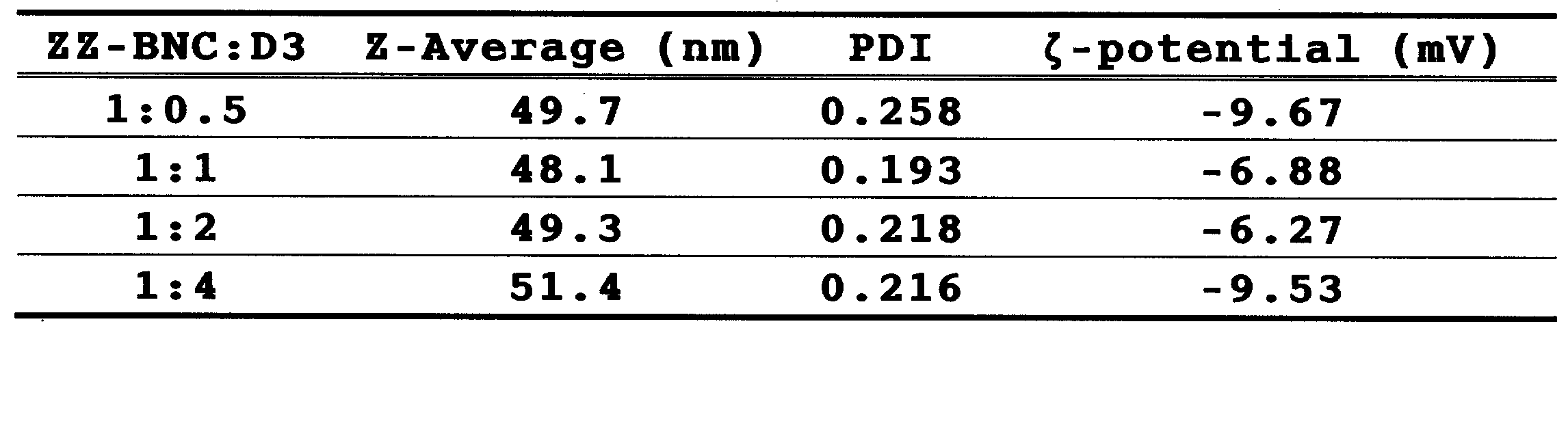
ZZ-BNC:D3=1:4で得られたZZ-BNC-D3に対し、ZZ-BNCーD3に対して5分の1の重量となる抗CD11c抗体を加え、室温で15分間静置し、クロスリンカーBS3で室温1時間処理し、過剰クロスリンカーをグリシンで室温15分処理してマスキングした。得られたものをα―CD11c-ZZーBNCーD3とする。D3抗原量に換算して40μgののαーCD11c-ZZ-BNC-D3、及び及びZZ-BNCーD3を、マウス(1群5匹)に対しそれぞれ、IV(静脈内)、IM(筋肉内)、及びSC(皮下)の経路で0、2週に接種し、4週目で血清中の抗D3-IgG抗体価をELISAで評価した。
上述のαーCD11cーZZ-BNCーD3、ZZ-BNCーD3、ST-BNCーD3、を、マウス(1群6匹)にD3抗原量に換算して40μgずつ、2週おきに2回IMで投与した(0,2週目)。その際、0,4週目に採血を行い、4週目に半数マウス致死量の50倍のJEVを接種した。これは、上述の図11に示される実験よりも投与間隔が短く過激なチャレンジ実験である。
非特許文献3に記載の方法に従ってα-CD11cーZZ-BNC(抗CD11c抗体が1μgでZZ-BNCが5μg)を作製し、カチオン性リポソーム(組成は非特許文献3に記載されたものであり、30μg)に3μgの蛍光色素Cy5標識プラスミドDNAを複合体化したリポプレックス(LPX)を混合し、37℃、1時間(pH3)でインキュベートしてα-CD11cーZZ-BNC-カチオン性リポソーム-DNAを作製し、塩化セシウム密度勾配超遠心法(SW41ローター、23600回転、25℃、16時間)で分離した。各フラクションを採取し、そのタンパク質量、及びCy5蛍光強度を測定した。
Claims (13)
- 樹状細胞の表面タンパク質に対する抗体及びウイルス表面抗原タンパク質を含むウイルス様粒子を有するバイオナノカプセル。
- ウイルス様粒子が、抗体結合ドメインを含む、請求項1に記載のバイオナノカプセル。
- 抗体結合ドメインが、ウイルス表面抗原タンパク質に含まれる、請求項2に記載のバイオナノカプセル。
- ウイルス表面抗原タンパク質が、B型肝炎ウイルス表面抗原(HBsAg)Lタンパク質である、請求項1~3の何れか1項に記載のバイオナノカプセル。
- 抗体が、CD11cタンパク質又はそのオルソログを認識する抗体である、請求項1~4の何れか1項に記載のバイオナノカプセル。
- 更にリポソームを含む、請求項1~5の何れか1項に記載のバイオナノカプセル。
- リポソームがカチオン性である、請求項1~6の何れか1項に記載のバイオナノカプセル。
- リポソームがアニオン性である、請求項1~6の何れか1項に記載のバイオナノカプセル。
- 更に、抗原物質及び/又は核酸を含む請求項1~8の何れか1項に記載のバイオナノカプセル。
- 請求項1~8の何れか1項に記載のバイオナノカプセルを含む、樹状細胞への抗原物質及び/又は核酸の導入剤。
- 請求項1~8の何れか1項に記載のバイオナノカプセルと、抗原物質及び/又は核酸を含むワクチン。
- 動物の樹状細胞への抗原物質及び/又は核酸導入方法であって、請求項9に記載のバイオナノカプセルを動物に投与する工程を含む方法。
- 投与が経静脈投与、経皮下投与、又は経筋肉投与である請求項12に記載の方法。
Priority Applications (1)
| Application Number | Priority Date | Filing Date | Title |
|---|---|---|---|
| JP2013539080A JP5458286B1 (ja) | 2012-03-30 | 2013-03-29 | 樹状細胞表面タンパク質に対する抗体を有するバイオナノカプセル |
Applications Claiming Priority (2)
| Application Number | Priority Date | Filing Date | Title |
|---|---|---|---|
| JP2012080791 | 2012-03-30 | ||
| JP2012-080791 | 2012-03-30 |
Publications (1)
| Publication Number | Publication Date |
|---|---|
| WO2013147232A1 true WO2013147232A1 (ja) | 2013-10-03 |
Family
ID=49260471
Family Applications (1)
| Application Number | Title | Priority Date | Filing Date |
|---|---|---|---|
| PCT/JP2013/059691 Ceased WO2013147232A1 (ja) | 2012-03-30 | 2013-03-29 | 樹状細胞表面タンパク質に対する抗体を有するバイオナノカプセル |
Country Status (2)
| Country | Link |
|---|---|
| JP (1) | JP5458286B1 (ja) |
| WO (1) | WO2013147232A1 (ja) |
Cited By (4)
| Publication number | Priority date | Publication date | Assignee | Title |
|---|---|---|---|---|
| WO2014205491A1 (en) * | 2013-06-25 | 2014-12-31 | Monash University | Biological molecules and methods of use |
| WO2018174005A1 (ja) * | 2017-03-21 | 2018-09-27 | 国立大学法人大阪大学 | 医薬 |
| WO2019217473A1 (en) * | 2018-05-07 | 2019-11-14 | The Administrators Of The Tulane Educational Fund | Mutant e. coli enterotoxins as anti-inflammatory agents |
| CN113655101A (zh) * | 2021-07-23 | 2021-11-16 | 山东师范大学 | 一种电化学生物传感器及其制备方法和应用 |
Citations (1)
| Publication number | Priority date | Publication date | Assignee | Title |
|---|---|---|---|---|
| WO2003082330A1 (en) * | 2002-03-29 | 2003-10-09 | Japan Science And Technology Agency | Therapeutic drug using antibody-presenting hollow protein nanoparticles and hollow protein nanoparticles |
-
2013
- 2013-03-29 WO PCT/JP2013/059691 patent/WO2013147232A1/ja not_active Ceased
- 2013-03-29 JP JP2013539080A patent/JP5458286B1/ja not_active Expired - Fee Related
Patent Citations (1)
| Publication number | Priority date | Publication date | Assignee | Title |
|---|---|---|---|---|
| WO2003082330A1 (en) * | 2002-03-29 | 2003-10-09 | Japan Science And Technology Agency | Therapeutic drug using antibody-presenting hollow protein nanoparticles and hollow protein nanoparticles |
Non-Patent Citations (2)
| Title |
|---|
| MATSUO,H. ET AL.: "Engineered hepatitis B virus surface antigen L protein particles for in vivo active targeting of splenic dendritic cells", INT J NANOMEDICINE, vol. 7, 2012, pages 3341 - 50 * |
| WEI,H. ET AL.: "Targeted delivery of tumor antigens to activated dendritic cells via CD11c molecules induces potent antitumor immunity in mice", CLIN CANCER RES, vol. 15, no. 14, 2009, pages 4612 - 21, XP055089726, DOI: doi:10.1158/1078-0432.CCR-08-3321 * |
Cited By (5)
| Publication number | Priority date | Publication date | Assignee | Title |
|---|---|---|---|---|
| WO2014205491A1 (en) * | 2013-06-25 | 2014-12-31 | Monash University | Biological molecules and methods of use |
| WO2018174005A1 (ja) * | 2017-03-21 | 2018-09-27 | 国立大学法人大阪大学 | 医薬 |
| WO2019217473A1 (en) * | 2018-05-07 | 2019-11-14 | The Administrators Of The Tulane Educational Fund | Mutant e. coli enterotoxins as anti-inflammatory agents |
| US12337025B2 (en) | 2018-05-07 | 2025-06-24 | The Administrators Of The Tulane Educational Fund | Mutated e. coli enterotoxins as anti-inflammatory agents |
| CN113655101A (zh) * | 2021-07-23 | 2021-11-16 | 山东师范大学 | 一种电化学生物传感器及其制备方法和应用 |
Also Published As
| Publication number | Publication date |
|---|---|
| JP5458286B1 (ja) | 2014-04-02 |
| JPWO2013147232A1 (ja) | 2015-12-14 |
Similar Documents
| Publication | Publication Date | Title |
|---|---|---|
| Tornesello et al. | Nanoparticles to improve the efficacy of peptide-based cancer vaccines | |
| JP6321261B2 (ja) | ワクチン組成物及びその使用方法 | |
| Cheng et al. | Biogenic nanoparticles as immunomodulator for tumor treatment | |
| JP4033497B2 (ja) | ワクチン製剤 | |
| Matsuo et al. | CD11c-specific bio-nanocapsule enhances vaccine immunogenicity by targeting immune cells | |
| JP5458286B1 (ja) | 樹状細胞表面タンパク質に対する抗体を有するバイオナノカプセル | |
| Ito et al. | Antigen/adjuvant-displaying enveloped viral replica as a self-adjuvanting anti-breast-cancer vaccine candidate | |
| JPWO1998009650A1 (ja) | ワクチン製剤 | |
| CN111295200B (zh) | 作为疫苗中的佐剂的包含合成的神经节苷脂gm3变体的纳米颗粒 | |
| CN120586028A (zh) | 一种肿瘤细胞疫苗及其制备方法、应用 | |
| CN107050463B (zh) | 一种药物及其制备方法和应用 | |
| JP7045024B2 (ja) | マラリアワクチン | |
| AU2016201691B2 (en) | Vaccine compositions and methods of use thereof | |
| Luo et al. | Emerging strategies for advancing peptide vaccines | |
| TW202506174A (zh) | 一種針對MICA/B靶點的mRNA腫瘤疫苗 | |
| HK40109239A (zh) | 用於活化和靶向免疫效应细胞的物质和方法 | |
| Crooke | Chemical and genetic modification of virus-like particles for applications in vaccine design and drug delivery | |
| CN120789242A (zh) | 一种egfr l858r突变特异性长肽疫苗及其应用 | |
| 松尾 et al. | Development of highly immunogenic vaccine by using dendritic cell-targeting bio-nanocapsule | |
| HK40020716A (en) | Nano-particles that contain synthetic variants of gm3 ganglioside as adjuvants in vaccines | |
| EA042396B1 (ru) | Наночастицы, содержащие синтетические варианты ганглиозида gm3, в качестве адъювантов в вакцинах | |
| AU2013245442A1 (en) | Vaccine compositions and methods of use thereof |
Legal Events
| Date | Code | Title | Description |
|---|---|---|---|
| ENP | Entry into the national phase |
Ref document number: 2013539080 Country of ref document: JP Kind code of ref document: A |
|
| 121 | Ep: the epo has been informed by wipo that ep was designated in this application |
Ref document number: 13768326 Country of ref document: EP Kind code of ref document: A1 |
|
| NENP | Non-entry into the national phase |
Ref country code: DE |
|
| 122 | Ep: pct application non-entry in european phase |
Ref document number: 13768326 Country of ref document: EP Kind code of ref document: A1 |
|
| NENP | Non-entry into the national phase |
Ref country code: DE |