WO2013115248A1 - ホモセリン脱水素酵素を用いたホモシステインの定量法 - Google Patents
ホモセリン脱水素酵素を用いたホモシステインの定量法 Download PDFInfo
- Publication number
- WO2013115248A1 WO2013115248A1 PCT/JP2013/052039 JP2013052039W WO2013115248A1 WO 2013115248 A1 WO2013115248 A1 WO 2013115248A1 JP 2013052039 W JP2013052039 W JP 2013052039W WO 2013115248 A1 WO2013115248 A1 WO 2013115248A1
- Authority
- WO
- WIPO (PCT)
- Prior art keywords
- homocysteine
- homoserine dehydrogenase
- homoserine
- derived
- concentration
- Prior art date
- Legal status (The legal status is an assumption and is not a legal conclusion. Google has not performed a legal analysis and makes no representation as to the accuracy of the status listed.)
- Ceased
Links
Images

Classifications
-
- C—CHEMISTRY; METALLURGY
- C12—BIOCHEMISTRY; BEER; SPIRITS; WINE; VINEGAR; MICROBIOLOGY; ENZYMOLOGY; MUTATION OR GENETIC ENGINEERING
- C12Q—MEASURING OR TESTING PROCESSES INVOLVING ENZYMES, NUCLEIC ACIDS OR MICROORGANISMS; COMPOSITIONS OR TEST PAPERS THEREFOR; PROCESSES OF PREPARING SUCH COMPOSITIONS; CONDITION-RESPONSIVE CONTROL IN MICROBIOLOGICAL OR ENZYMOLOGICAL PROCESSES
- C12Q1/00—Measuring or testing processes involving enzymes, nucleic acids or microorganisms; Compositions therefor; Processes of preparing such compositions
- C12Q1/26—Measuring or testing processes involving enzymes, nucleic acids or microorganisms; Compositions therefor; Processes of preparing such compositions involving oxidoreductase
- C12Q1/32—Measuring or testing processes involving enzymes, nucleic acids or microorganisms; Compositions therefor; Processes of preparing such compositions involving oxidoreductase involving dehydrogenase
Definitions
- the present invention relates to a method for measuring homocysteine in a sample such as blood.
- Homocysteine is a metabolic intermediate of methionine, and its concentration in healthy plasma is low, 3-15 ⁇ M.
- a high blood concentration of homocysteine becomes a risk factor for cardiovascular disease.
- patients with coronary artery disease or cerebrovascular disease are known to have high blood homocysteine levels.
- Diabetes, hypertension, hyperlipidemia, renal failure, complications during pregnancy, Alzheimer's disease, etc. There is also a report that the concentration of medium homocysteine increases.
- an object of the present invention is to provide a method capable of accurately measuring homocysteine with a simple operation.
- the present inventor has conducted various studies on a new method for quantifying homocysteine.
- the hyperthermophilic archaea-derived homoserine dehydrogenase known to have no activity against homocysteine is obtained by coexisting homoserine and homocysteine.
- homocysteine produced the allosteric effect of the enzyme, which increased the dehydrogenation activity of the enzyme against homoserine in a homocysteine concentration-dependent manner.
- the present inventors have found that the homocysteine concentration in a sample can be accurately measured, thereby completing the present invention.
- the present invention relates to the following [1] to [3].
- a method for measuring the homocysteine concentration in a sample which comprises measuring homoserine dehydrogenase activity by allowing archaea-derived homoserine dehydrogenase to act on homoserine in a sample containing homocysteine.
- the homocysteine concentration in a sample can be quantified simply and accurately by reacting only one kind of enzyme.
- the reaction system since the reaction system is simple, it can be quickly quantified by an automatic analyzer equipped with a spectrophotometer.
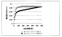
- FIG. 3 shows activation of homoserine dehydrogenase by 10 ⁇ M DL-homocysteine.
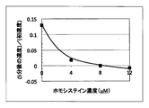
- FIG. 3 is a graph showing the influence of DL-homocysteine on the reaction rate (the influence of homocysteine on the ratio of the reaction rate to the initial rate 5 minutes after the start of the reaction).
- the method for measuring homocysteine concentration in a sample of the present invention is to measure homoserine dehydrogenase activity by allowing archaine-derived homoserine dehydrogenase to act on homoserine in a homocysteine-containing sample.
- the homocysteine-containing sample to be used may be a biological sample containing free homocysteine.
- homocysteine In blood, homocysteine is abundant as free homocysteine, and most of it exists in the form of homocystin, homocysteine-cysteine, and homocysteine-protein, so it was first reduced to release homocysteine.
- the sample is preferably used as a specimen. In this case, the total homocysteine content is measured.
- examples of the reduction treatment means include a method of reacting thiol compounds such as hydrogen sulfide, methanethiol, 2-mercaptoethanol, dithiothreitol, thioglycerol, cysteamine and the like.
- This reduction treatment method can employ known means (1) Refsum, H. et al., Homocysteine and cardiovascular disease. Annu. Rev. ed. (1998) Vol. 49, pp. 31-62, 2) Mansoor, MAet al., Redox status and protein binding of plasma homocysteine and other aminothiols in patients with homocystinuria.Metabolism (1993) Vol.42, pp.1481-5, 3) Mansoor, MAet al., Redox status and protein binding of homocysteine and other aminothiols in patients with early-onset peripheral vascular disease. Arteroiscier Thromb. -bound homocysteine and vascular endothelial function in healthy human subjects. Circ.
- a preferred homocysteine-containing sample is reduced blood, plasma or serum.
- the homoserine dehydrogenase used in the present invention is an archaea-derived homoserine dehydrogenase, not a eukaryote such as a mammal or a bacterium.
- homoserine dehydrogenase derived from bacteria is slightly inhibited by homocysteine (RJ Rowbury, J. Gen. Microbiol. (1968), vol. 54, pp. 337-342) and cannot be used.
- a homoserine dehydrogenase derived from archaea may be used, but a homoserine dehydrogenase derived from sulfolobus is preferred, and a homoserine dehydrogenase derived from Sulfolobus tokodaii is more preferred. Aerobic, acidophilic, and thermophilic archaea belonging to the genus Sulfolobus can be collected from land volcanoes and hot springs, etc. In addition to Sulfolobus tokodai, S. acidocaldarius, S. solfataricus, S. metallicus, S. shibatae, S . Yangmingensis etc. can also be used.
- archaea-derived homoserine dehydrogenase is capable of dehydrogenase activity against homoserine (oxidation reaction from homoserine to aspartate-4-semialdehyde is NAD).
- NAD oxidation reaction from homoserine to aspartate-4-semialdehyde
- (P) depending on reversible catalysis, but not on homocysteine. Therefore, it was completely unpredictable that this archaea-derived homoserine dehydrogenase was activated by homocysteine and the homoserine dehydrogenase activity was enhanced in the presence of homocysteine.
- archaea In order to collect homoserine dehydrogenase from archaea, for example, archaea can be collected from a culture medium obtained by cultivating by known means. However, as described in Non-patent Document 1, E. coli transformed by gene recombination technology is cultured. Can also be collected.
- an archaea-derived homoserine dehydrogenase is added to the homocysteine-containing sample and incubated, and then a predetermined amount of homoserine is added.
- the amount of archaea-derived homoserine dehydrogenase added is not particularly limited, but is preferably about 0.01 to 100 U / ml, more preferably about 0.5 to 2 U / ml.
- the addition amount of homoserine is a predetermined amount, for example, 0.01 to 10000 mM, more preferably 1 to 100 mM.
- the enzyme reaction may be incubated in a buffer solution having a pH of 3 to 11 at 10 to 100 ° C. for a total of 0.1 to 60 minutes.
- the homoserine dehydrogenase activity is an oxidation reaction from homoserine to aspartic acid-4-semialdehyde and proceeds depending on NAD or NADP. Therefore, NAD or NADP is present in the reaction system, and NAD is converted to NADH.
- the homoserine dehydrogenase activity that is, the homocysteine concentration can be quantified by measuring the absorbance of the production of NADPH from NADP with a spectrophotometer.
- the homocysteine concentration in the sample can be determined from a calibration curve based on the homocysteine concentration and enzyme activity (for example, absorbance) prepared in advance.
- Example 1 [Production of archaeal homoserine dehydrogenase] Primer that amplifies the HSDH putative gene (915 bp) on the genome of aerobic / acidophilic hyperthermophilic archaeon Sulfolobus tokodai found from databases such as DDBJ (http://www.ddbj.nig.ac.jp/) And the gene was amplified by PCR. The amplified gene was inserted into the expression vector pET101 to construct an expression plasmid pST1519.
- the supernatant after centrifugation was collected, used as a crude enzyme solution, and the protein concentration of the enzyme solution was measured.
- the crude enzyme was heated for 20 minutes in a thermostat heated to 70 ° C., and the supernatant was collected by centrifugation to obtain an enzyme solution.
- Example 2 [Activity measurement] To the enzyme solution (1 U), 100 mM phosphate buffer (pH 7.0), 1 mM MgCl 2 , 1.3 mM NAD + , and each concentration of DL-homocysteine were added to the cell and incubated at 50 ° C. for 3 minutes. DL-homoserine was added so that the final concentration was 10 mM, the reaction was started, and the activity was measured by observing absorbance at 340 nm over time at 50 ° C. One unit of enzyme was defined as the amount of enzyme required to produce 1 ⁇ mol NADH per minute at 50 ° C. FIG.
- FIG. 1 shows the activity of homoserine dehydrogenase by homocysteine (relationship between homocysteine concentration and specific activity).
- FIG. 1 shows that the homocysteine concentration in the sample can be quantified by the action of homoserine and homoserine dehydrogenase in the homocysteine-containing sample by the method of the present invention.
- Example 3 [Measurement of low-concentration homocysteine activity] To the enzyme solution (1 U), 100 mM phosphate buffer (pH 7.0), 1 mM MgCl 2 , 1.3 mM NAD + , and each concentration of DL-homocysteine were added to the cell and incubated at 50 ° C. for 3 minutes. DL-homoserine was added so that the final concentration was 8 mM, the reaction was started, and the activity was measured by observing absorbance at 340 nm over time at 50 ° C.
- FIG. 2 shows the activation of homoserine dehydrogenase by 10 ⁇ M DL-homocysteine.
Landscapes
- Chemical & Material Sciences (AREA)
- Organic Chemistry (AREA)
- Life Sciences & Earth Sciences (AREA)
- Zoology (AREA)
- Wood Science & Technology (AREA)
- Proteomics, Peptides & Aminoacids (AREA)
- Health & Medical Sciences (AREA)
- Engineering & Computer Science (AREA)
- Microbiology (AREA)
- Biochemistry (AREA)
- Physics & Mathematics (AREA)
- Molecular Biology (AREA)
- Biotechnology (AREA)
- Biophysics (AREA)
- Analytical Chemistry (AREA)
- Immunology (AREA)
- Bioinformatics & Cheminformatics (AREA)
- General Engineering & Computer Science (AREA)
- General Health & Medical Sciences (AREA)
- Genetics & Genomics (AREA)
- Measuring Or Testing Involving Enzymes Or Micro-Organisms (AREA)
- Enzymes And Modification Thereof (AREA)
Abstract
簡便な操作で正確なホモシステインを測定できる方法の提供。 ホモシステイン含有試料中において、ホモセリンにアーキア由来ホモセリン脱水素酵素を作用させてホモセリン脱水酵素活性を測定することを特徴とする、当該試料中のホモシステイン濃度の測定方法。
Description
本発明は、血液等の試料中のホモシステインの測定方法に関する。
ホモシステインは、メチオニンの代謝中間物であり、健常の血漿中の濃度は低く、3~15μMである。しかし、ホモシステインの血中濃度が高くなると、心血管疾患の危険因子になることが知られている。例えば冠動脈疾患患者や脳血管疾患患者では血液中ホモシステイン濃度が高値になることが知られており、さらに糖尿病、高血圧、高脂血症、腎不全、妊娠時の合併症、アルツハイマー病等でも血中ホモシステイン濃度が高くなるという報告もある。その作用は、血中ホモシステイン濃度の上昇によってホモシステインが酸化した際に生じる過酸化水素やスーパーオキシドラジカル等の酸化ストレスによって内皮細胞障害を起こし、動脈硬化等が促進されるといわれている。
従来のホモシステインの定量方法には検体中のホモシステインをSH基と反応する蛍光標識試薬で標識し、高速液体クロマトグラフィーにより分離して定量するプレラベルHPLC法、検体を高速液体クロマトグラフィーで分離し、後にSH基と反応する蛍光標識試薬で標識して定量するポストラベルHPLC法、ホモシステインと特異的に作用する酵素を用い、酵素反応生成物を抗体法により定量する酵素免疫法、ホモシステインに特異的に作用する酵素を用い、酵素反応生成物を酵素法により定量する方法など、幾つかの定量方法が存在する(特許文献1~4)。
日本大学生産工学部第44回学術講演概要(2011-12-3)5-58
しかしながら、従来のホモシステインの測定法のうち、HPLCを用いる方法は操作が煩雑であるという問題があり、酵素法については2種以上の酵素や酵素と抗体の組み合わせを使用するため操作が煩雑になる、試薬が高価になる等の問題があった。
従って、本発明は、簡便な操作で正確にホモシステインを測定できる方法を提供することにある。
従って、本発明は、簡便な操作で正確にホモシステインを測定できる方法を提供することにある。
そこで本発明者は、新たなホモシステインの定量法について種々検討してきたところ、ホモシステインに対する活性を有しないことが知られている超好熱アーキア由来ホモセリン脱水素酵素を、ホモセリンとホモシステインの共存系に作用させたところ、全く意外なことに、ホモシステインにより当該酵素のアロステリック効果が生じ、当該酵素のホモセリンに対する脱水素活性がホモシステイン濃度依存的に向上し、その脱水素活性を直接測定すれば、試料中のホモシステイン濃度が正確に測定できることを見出し、本発明を完成した。
すなわち、本発明は、以下の[1]~[3]に係るものである。
[1]ホモシステイン含有試料中において、ホモセリンにアーキア由来ホモセリン脱水素酵素を作用させてホモセリン脱水素酵素活性を測定することを特徴とする、当該試料中のホモシステイン濃度の測定方法。
[2]アーキア由来ホモセリン脱水素酵素が、スルフォロバス由来のホモセリン脱水素酵素である[1]記載の測定方法。
[3]アーキア由来ホモセリン脱水素酵素が、スルフォロバス・トコダイ由来のホモセリン脱水素酵素である[1]又は[2]記載の測定方法。
[1]ホモシステイン含有試料中において、ホモセリンにアーキア由来ホモセリン脱水素酵素を作用させてホモセリン脱水素酵素活性を測定することを特徴とする、当該試料中のホモシステイン濃度の測定方法。
[2]アーキア由来ホモセリン脱水素酵素が、スルフォロバス由来のホモセリン脱水素酵素である[1]記載の測定方法。
[3]アーキア由来ホモセリン脱水素酵素が、スルフォロバス・トコダイ由来のホモセリン脱水素酵素である[1]又は[2]記載の測定方法。
本発明によれば、1種類の酵素を反応させるだけで試料中のホモシステイン濃度が簡便かつ正確に定量できる。また、反応系が単純なので、分光光度計を備えた自動分析装置により迅速に定量できる。
本発明の試料中のホモシステイン濃度測定方法は、ホモシステイン含有試料中において、ホモセリンにアーキア由来ホモセリン脱水素酵素を作用させ、ホモセリン脱水素酵素活性を測定するものである。
用いられるホモシステイン含有試料としては、遊離のホモシステインを含有する生体試料であればよい。血液中でホモシステインは、遊離のホモシステインとしての存在量は少なく、そのほとんどが、ホモシスチン、ホモシステイン-システイン、ホモシステイン-タンパク質の形態で存在するので、まず還元してホモシステインを遊離させた試料を検体とするのが好ましい。この場合、測定されるのは総ホモシステイン量になる。ここで、還元処理手段としては、硫化水素、メタンチオール、2-メルカプトエタノール、ジチオスレイトール、チオグリセロール、システアミン等のチオール化合物を反応させる方法等が挙げられる。この還元処理方法は公知の手段を採用できる(1)Refsum,H.et al.,Homocysteine and cardiovascular disease.Annu.Rev. ed.(1998)Vol.49,pp.31-62、2)Mansoor,M.A.et al.,Redox status and protein binding of plasma homocysteine and other aminothiols in patients with homocystinuria.Metabolism(1993)Vol.42,pp.1481-5、3)Mansoor,M.A.et al.,Redox status and protein binding of homocysteine and other aminothiols in patients with early-onset peripheral vascular disease.Arteroiscier Thromb.Vasc.Biol.(1995)Vol.15,pp.232-40、4)Chambers,J.C.et al.,Investigation of relationship between reduced,oxidized,and protein-bound homocysteine and vascular endothelial function in healthy human subjects.Circ.Res.(2001)Vol.89,pp.187-192、5)橋本隆男ら、「ホモシステイン代謝」、薬学雑誌(2007)127巻10号1579-92.)
また生体試料としては、血液、血漿、血清、髄液、リンパ液等が挙げられる。
また生体試料としては、血液、血漿、血清、髄液、リンパ液等が挙げられる。
好ましいホモシステイン含有試料は、還元処理された血液、血漿又は血清である。
本発明に用いられるホモセリン脱水素酵素は、哺乳類等の真核生物や細菌由来ではなくアーキア由来のホモセリン脱水素酵素である。細菌由来のホモセリン脱水素酵素はホモシステインで少し阻害されるという報告(RJ Rowbury,J.Gen.Microbiol.(1968),vol.54,pp.337-342)があり、使用できない。アーキア(古細菌)由来のホモセリン脱水素酵素であればよいが、スルフォロバス由来のホモセリン脱水素酵素が好ましく、特にスルフォロバス・トコダイ(Sulfolobus tokodaii)由来のホモセリン脱水素酵素がより好ましい。スルフォロバス属の好気、好酸、好熱性の古細菌は、陸上の火山や温泉などから採取でき、スルフォロバス・トコダイ以外にも、S. acidocaldarius、S. solfataricus、S. metallicus、S. shibatae、S. yangmingensis等も利用できる。
本発明者の研究によれば、非特許文献1に記載のように、アーキア由来のホモセリン脱水素酵素は、ホモセリンに対する脱水素酵素活性(ホモセリンからアスパラギン酸-4-セミアルデヒドへの酸化反応をNAD(P)に依存して可逆的に触媒する活性)は有するが、ホモシステインに対す活性はない。従って、このアーキア由来のホモセリン脱水素酵素がホモシステインによって活性化され、ホモシステインの存在下でホモセリン脱水素酵素活性が増強されることは全く予測できなかった。
アーキアからホモセリン脱水素酵素を採取するには、例えばアーキアを公知の手段により培養した培養液から採取することができるが、非特許文献1記載のように遺伝子組換え技術により形質転換した大腸菌を培養することにより採取することもできる。
ホモシステイン含有試料中で、ホモセリンにアーキア由来ホモセリン脱水素酵素を作用させるには、例えばホモシステイン含有試料にアーキア由来ホモセリン脱水素酵素を添加してインキュベートし、次いで所定量のホモセリンを添加すればよい。アーキア由来のホモセリン脱水素酵素の添加量は、特に限定されないが、0.01~100U/ml程度、さらに0.5~2U/ml程度が好ましい。また、ホモセリンの添加量は、所定量であり、例えば0.01~10000mM、さらに1~100mMが好ましい。なお、酵素反応はpH3~11の緩衝液中、10~100℃で合計0.1分~60分インキュベートすればよい。
ホモセリン脱水素酵素活性は、ホモセリンからアスパラギン酸-4-セミアルデヒドへの酸化反応であり、NAD又はNADPに依存して進行するから、反応系中にNAD又はNADPを存在させておき、NADからNADHの生成又はNADPからNADPHの生成を分光光度計により吸光度を測定すれば、ホモセリン脱水素酵素活性、すなわちホモシステイン濃度が定量できる。
試料中のホモシステイン濃度は、予め作成しておいたホモシステイン濃度と酵素活性(例えば吸光度)に基づく検量線から決定することができる。
次に実施例を上げて本発明を詳細に説明する。
実施例1
[古細菌由来ホモセリン脱水素酵素の生産]
DDBJ(http://www.ddbj.nig.ac.jp/)等のデータベースから見出した好気・好酸性超好熱古細菌スルフォロバス・トコダイのゲノム上のHSDH推定遺伝子(915bp)を増幅するプライマーを設計し、PCRにより遺伝子を増幅した。増幅した遺伝子を発現ベクターpET101に挿入し発現プラスミドpST1519を構築した。pST1519でBL21-CodonPlus(DE3)-RIL Competent Cellsを形質転換した後、50μl/mlアンピシリン含有LB培地でOD660が0.6~0.8になるまで振盪培養し、0.2mM IPTGを加え、3時間37℃で培養した。培養で得られた菌体を遠心分離によって回収、1M KPB(pH7.0)、10mM MgCl2(pH7.0)で懸濁し、超音波破砕(出力35%,on time 2 sec, off time 2 sec, total time 60 min)を氷上で行った。更に、遠心分離後の上清を回収し、それを粗酵素液とし、酵素液をタンパク質濃度測定した。70℃に加熱した恒温槽で粗酵素を20分間加熱し、遠心分離によって上清を回収し、酵素液とした。
[古細菌由来ホモセリン脱水素酵素の生産]
DDBJ(http://www.ddbj.nig.ac.jp/)等のデータベースから見出した好気・好酸性超好熱古細菌スルフォロバス・トコダイのゲノム上のHSDH推定遺伝子(915bp)を増幅するプライマーを設計し、PCRにより遺伝子を増幅した。増幅した遺伝子を発現ベクターpET101に挿入し発現プラスミドpST1519を構築した。pST1519でBL21-CodonPlus(DE3)-RIL Competent Cellsを形質転換した後、50μl/mlアンピシリン含有LB培地でOD660が0.6~0.8になるまで振盪培養し、0.2mM IPTGを加え、3時間37℃で培養した。培養で得られた菌体を遠心分離によって回収、1M KPB(pH7.0)、10mM MgCl2(pH7.0)で懸濁し、超音波破砕(出力35%,on time 2 sec, off time 2 sec, total time 60 min)を氷上で行った。更に、遠心分離後の上清を回収し、それを粗酵素液とし、酵素液をタンパク質濃度測定した。70℃に加熱した恒温槽で粗酵素を20分間加熱し、遠心分離によって上清を回収し、酵素液とした。
実施例2
[活性測定]
酵素液(1U)に100mM リン酸緩衝液(pH7.0)、1mM MgCl2、1.3mM NAD+、各濃度のDL-ホモシステインをセルに加え、50℃で3分間インキュベートした。終濃度が10mMとなる様にDL-ホモセリンを添加し反応を開始させ、50℃において340nmの吸光度を経時的に観測することで活性測定を行った。酵素の1Unitは、50℃で1分間に1μmolのNADHを生じるのに必要な酵素量として定義した。図1にホモセリン脱水素酵素のホモシステインによる活性(ホモシステイン濃度と比活性の関係)化を示す。
図1から、本発明方法により、試料中のホモシステイン濃度が、ホモシステイン含有試料中で、ホモセリンとホモセリン脱水素酵素を作用させることにより、該試料中のホモシステイン濃度が定量できることがわかる。
[活性測定]
酵素液(1U)に100mM リン酸緩衝液(pH7.0)、1mM MgCl2、1.3mM NAD+、各濃度のDL-ホモシステインをセルに加え、50℃で3分間インキュベートした。終濃度が10mMとなる様にDL-ホモセリンを添加し反応を開始させ、50℃において340nmの吸光度を経時的に観測することで活性測定を行った。酵素の1Unitは、50℃で1分間に1μmolのNADHを生じるのに必要な酵素量として定義した。図1にホモセリン脱水素酵素のホモシステインによる活性(ホモシステイン濃度と比活性の関係)化を示す。
図1から、本発明方法により、試料中のホモシステイン濃度が、ホモシステイン含有試料中で、ホモセリンとホモセリン脱水素酵素を作用させることにより、該試料中のホモシステイン濃度が定量できることがわかる。
実施例3
[低濃度ホモシステインの活性測定]
酵素液(1U)に100mM リン酸緩衝液(pH7.0)、1mM MgCl2、1.3mM NAD+、各濃度のDL-ホモシステインをセルに加え、50℃で3分間インキュベートした。終濃度が8mMとなる様にDL-ホモセリンを添加し反応を開始させ、50℃において340nmの吸光度を経時的に観測することで活性測定を行った。
図2にホモセリン脱水素酵素の10μM DL-ホモシステインによる活性化を示す。血中濃度とほぼ同等の10μM ホモシステインによって経時的な吸光度変化が大きく影響を受けていることが示された。特に反応開始直後の初速度と反応5分後の速度がDL-ホモシステインによって影響を受ける。
この初速度と5分後の反応速度の比を計算したところ、図3の様にDL-ホモシステイン濃度との相関が得られた。この検量線によって数μMのホモシステインの定量が可能である。
[低濃度ホモシステインの活性測定]
酵素液(1U)に100mM リン酸緩衝液(pH7.0)、1mM MgCl2、1.3mM NAD+、各濃度のDL-ホモシステインをセルに加え、50℃で3分間インキュベートした。終濃度が8mMとなる様にDL-ホモセリンを添加し反応を開始させ、50℃において340nmの吸光度を経時的に観測することで活性測定を行った。
図2にホモセリン脱水素酵素の10μM DL-ホモシステインによる活性化を示す。血中濃度とほぼ同等の10μM ホモシステインによって経時的な吸光度変化が大きく影響を受けていることが示された。特に反応開始直後の初速度と反応5分後の速度がDL-ホモシステインによって影響を受ける。
この初速度と5分後の反応速度の比を計算したところ、図3の様にDL-ホモシステイン濃度との相関が得られた。この検量線によって数μMのホモシステインの定量が可能である。
Claims (3)
- ホモシステイン含有試料中において、ホモセリンにアーキア由来ホモセリン脱水素酵素を作用させてホモセリン脱水素酵素活性を測定することを特徴とする、当該試料中のホモシステイン濃度の測定方法。
- アーキア由来ホモセリン脱水素酵素が、スルフォロバス由来のホモセリン脱水素酵素である請求項1記載の測定方法。
- アーキア由来ホモセリン脱水素酵素が、スルフォロバス・トコダイ由来のホモセリン脱水素酵素である請求項1又は2記載の測定方法。
Priority Applications (1)
| Application Number | Priority Date | Filing Date | Title |
|---|---|---|---|
| JP2013556465A JP6143191B2 (ja) | 2012-02-01 | 2013-01-30 | ホモセリン脱水素酵素を用いたホモシステインの定量法 |
Applications Claiming Priority (2)
| Application Number | Priority Date | Filing Date | Title |
|---|---|---|---|
| JP2012-019441 | 2012-02-01 | ||
| JP2012019441 | 2012-02-01 |
Publications (1)
| Publication Number | Publication Date |
|---|---|
| WO2013115248A1 true WO2013115248A1 (ja) | 2013-08-08 |
Family
ID=48905280
Family Applications (1)
| Application Number | Title | Priority Date | Filing Date |
|---|---|---|---|
| PCT/JP2013/052039 Ceased WO2013115248A1 (ja) | 2012-02-01 | 2013-01-30 | ホモセリン脱水素酵素を用いたホモシステインの定量法 |
Country Status (2)
| Country | Link |
|---|---|
| JP (1) | JP6143191B2 (ja) |
| WO (1) | WO2013115248A1 (ja) |
Cited By (1)
| Publication number | Priority date | Publication date | Assignee | Title |
|---|---|---|---|---|
| CN111855648A (zh) * | 2020-03-05 | 2020-10-30 | 美康生物科技股份有限公司 | 一种干式同型半胱氨酸测试卡及其应用 |
-
2013
- 2013-01-30 WO PCT/JP2013/052039 patent/WO2013115248A1/ja not_active Ceased
- 2013-01-30 JP JP2013556465A patent/JP6143191B2/ja not_active Expired - Fee Related
Non-Patent Citations (2)
| Title |
|---|
| MARON B.A. ET AL.: "Homocysteine", CLIN. LAB. MED., vol. 26, no. 3, September 2006 (2006-09-01), pages 591 - 609 * |
| PERACCHI A. ET AL.: "Exploring and exploiting allostery: Models, evolution, and drug targeting", BIOCHIM. BIOPHYS. ACTA, vol. 1814, no. 8, August 2011 (2011-08-01), pages 922 - 933 * |
Cited By (1)
| Publication number | Priority date | Publication date | Assignee | Title |
|---|---|---|---|---|
| CN111855648A (zh) * | 2020-03-05 | 2020-10-30 | 美康生物科技股份有限公司 | 一种干式同型半胱氨酸测试卡及其应用 |
Also Published As
| Publication number | Publication date |
|---|---|
| JPWO2013115248A1 (ja) | 2015-05-11 |
| JP6143191B2 (ja) | 2017-06-07 |
Similar Documents
| Publication | Publication Date | Title |
|---|---|---|
| Steffens et al. | High CO2 levels drive the TCA cycle backwards towards autotrophy | |
| Müller et al. | Engineering Escherichia coli for methanol conversion | |
| Meyer et al. | Methanol-essential growth of Escherichia coli | |
| Ambrogelly et al. | Natural expansion of the genetic code | |
| Burrichter et al. | Anaerobic degradation of the plant sugar sulfoquinovose concomitant with H2S production: Escherichia coli K-12 and Desulfovibrio sp. strain DF1 as co-culture model | |
| Montano et al. | Activity assays of mammalian thioredoxin and thioredoxin reductase: fluorescent disulfide substrates, mechanisms, and use with tissue samples | |
| Durigon et al. | Cytosolic thioredoxin system facilitates the import of mitochondrial small Tim proteins | |
| Chen et al. | Use of a tyrosine analogue to modulate the two activities of a nonheme iron enzyme OvoA in ovothiol biosynthesis, cysteine oxidation versus oxidative C–S bond formation | |
| Katsyv et al. | Electron carriers involved in autotrophic and heterotrophic acetogenesis in the thermophilic bacterium Thermoanaerobacter kivui | |
| Ezraty et al. | Methionine sulfoxide reduction and assimilation in Escherichia coli: new role for the biotin sulfoxide reductase BisC | |
| Moon et al. | Proteome analyses of hydrogen-producing hyperthermophilic archaeon Thermococcus onnurineus NA1 in different one-carbon substrate culture conditions | |
| Hamann et al. | A cysteine-rich CCG domain contains a novel [4Fe-4S] cluster binding motif as deduced from studies with subunit B of heterodisulfide reductase from Methanothermobacter marburgensis | |
| Szu et al. | Characterization and mechanistic studies of DesII: a radical S-adenosyl-L-methionine enzyme involved in the biosynthesis of TDP-D-desosamine | |
| Kucera et al. | Ferrous iron oxidation by sulfur-oxidizing Acidithiobacillus ferrooxidans and analysis of the process at the levels of transcription and protein synthesis | |
| Welte et al. | Re‐evaluation of the function of the F420 dehydrogenase in electron transport of Methanosarcina mazei | |
| JP6143191B2 (ja) | ホモセリン脱水素酵素を用いたホモシステインの定量法 | |
| Sherbet et al. | Cofactors, redox state, and directional preferences of hydroxysteroid dehydrogenases | |
| Nomura et al. | Isolation of an H 2‐dependent electron‐bifurcating CO 2‐reducing megacomplex with M vh B polyferredoxin from Methanothermobacter marburgensis | |
| CN112592958B (zh) | 利用fad依赖型d-2-羟基戊二酸脱氢酶和刃天青检测d-2-羟基戊二酸的方法 | |
| Pettinato et al. | Propionate metabolism in Desulfurella acetivorans | |
| WO2017169565A1 (ja) | タンパク質及び核酸の超高感度測定方法 | |
| Serrano et al. | Kinetic and isotopic characterization of L-proline dehydrogenase from Mycobacterium tuberculosis | |
| JP2001190299A (ja) | 糖尿病予備群の検査方法 | |
| Sugiyama et al. | Development of a new chromogenic method for putrescine quantification using coupling reactions involving putrescine oxidase | |
| Burns et al. | Studies of the redox properties of CDP-6-deoxy-L-threo-D-glycero-4-hexulose-3-dehydrase (E1) and CDP-6-deoxy-L-threo-D-glycero-4-hexulose-3-dehydrase reductase (E3): two important enzymes involved in the biosynthesis of ascarylose |
Legal Events
| Date | Code | Title | Description |
|---|---|---|---|
| 121 | Ep: the epo has been informed by wipo that ep was designated in this application |
Ref document number: 13743445 Country of ref document: EP Kind code of ref document: A1 |
|
| ENP | Entry into the national phase |
Ref document number: 2013556465 Country of ref document: JP Kind code of ref document: A |
|
| NENP | Non-entry into the national phase |
Ref country code: DE |
|
| 122 | Ep: pct application non-entry in european phase |
Ref document number: 13743445 Country of ref document: EP Kind code of ref document: A1 |