WO2012173244A1 - Marker for use in detection of periodontopathic bacterium communicable between men and animals - Google Patents
Marker for use in detection of periodontopathic bacterium communicable between men and animals Download PDFInfo
- Publication number
- WO2012173244A1 WO2012173244A1 PCT/JP2012/065408 JP2012065408W WO2012173244A1 WO 2012173244 A1 WO2012173244 A1 WO 2012173244A1 JP 2012065408 W JP2012065408 W JP 2012065408W WO 2012173244 A1 WO2012173244 A1 WO 2012173244A1
- Authority
- WO
- WIPO (PCT)
- Prior art keywords
- antibody
- strain
- anone
- marker
- antigen
- Prior art date
Links
Images


Classifications
-
- A—HUMAN NECESSITIES
- A61—MEDICAL OR VETERINARY SCIENCE; HYGIENE
- A61K—PREPARATIONS FOR MEDICAL, DENTAL OR TOILETRY PURPOSES
- A61K39/00—Medicinal preparations containing antigens or antibodies
- A61K39/02—Bacterial antigens
- A61K39/0216—Bacteriodetes, e.g. Bacteroides, Ornithobacter, Porphyromonas
-
- A—HUMAN NECESSITIES
- A61—MEDICAL OR VETERINARY SCIENCE; HYGIENE
- A61P—SPECIFIC THERAPEUTIC ACTIVITY OF CHEMICAL COMPOUNDS OR MEDICINAL PREPARATIONS
- A61P1/00—Drugs for disorders of the alimentary tract or the digestive system
- A61P1/02—Stomatological preparations, e.g. drugs for caries, aphtae, periodontitis
-
- G—PHYSICS
- G01—MEASURING; TESTING
- G01N—INVESTIGATING OR ANALYSING MATERIALS BY DETERMINING THEIR CHEMICAL OR PHYSICAL PROPERTIES
- G01N33/00—Investigating or analysing materials by specific methods not covered by groups G01N1/00 - G01N31/00
- G01N33/48—Biological material, e.g. blood, urine; Haemocytometers
- G01N33/50—Chemical analysis of biological material, e.g. blood, urine; Testing involving biospecific ligand binding methods; Immunological testing
- G01N33/53—Immunoassay; Biospecific binding assay; Materials therefor
- G01N33/569—Immunoassay; Biospecific binding assay; Materials therefor for microorganisms, e.g. protozoa, bacteria, viruses
- G01N33/56911—Bacteria
- G01N33/56955—Bacteria involved in periodontal diseases
-
- A—HUMAN NECESSITIES
- A61—MEDICAL OR VETERINARY SCIENCE; HYGIENE
- A61K—PREPARATIONS FOR MEDICAL, DENTAL OR TOILETRY PURPOSES
- A61K39/00—Medicinal preparations containing antigens or antibodies
- A61K2039/55—Medicinal preparations containing antigens or antibodies characterised by the host/recipient, e.g. newborn with maternal antibodies
- A61K2039/552—Veterinary vaccine
-
- G—PHYSICS
- G01—MEASURING; TESTING
- G01N—INVESTIGATING OR ANALYSING MATERIALS BY DETERMINING THEIR CHEMICAL OR PHYSICAL PROPERTIES
- G01N2469/00—Immunoassays for the detection of microorganisms
- G01N2469/20—Detection of antibodies in sample from host which are directed against antigens from microorganisms
Definitions
- the present invention relates to a marker for detecting periodontal pathogens common to humans, a method for determining infection by the bacteria, and a method for detecting the bacteria.
- Periodontal disease is inflammation that occurs in periodontal tissues such as gingiva, periodontal ligament, cementum and alveolar bone, and is a very common disease affecting about 30% of the world population.
- Periodontal diseases include gingivitis, periodontitis and occlusal trauma, but the most common periodontal disease is periodontitis, especially chronic periodontitis caused by periodontal disease bacteria ( Non-patent document 1).
- Chronic periodontitis gradually progresses with age from mild gingivitis. The percentage of gingival findings in people over the age of 5 was 64.3% in 1987 (Dental disease survey), 68.1% in 1993 (same survey), and 72.9% in 1999 (same survey). In recent years, it has been on an increasing trend.
- Chronic periodontitis is known to increase the risk of developing various systemic diseases such as cerebrovascular disease, heart disease, premature birth, as well as inflammation in the oral cavity. Therefore, it is important to detect the periodontal disease bacteria that cause the chronic periodontitis at the stage where chronic periodontitis has not become apparent in order to prevent and early treat chronic periodontitis.
- chronic periodontitis is accompanied by symptoms such as inflammation and bleeding of the gingiva, so that it is relatively easy to make a visual diagnosis after onset. Therefore, in the field of dentistry, periodontal disease bacteria have been detected from subjects at an early stage with a simple and high accuracy rate, predicting the possibility of chronic periodontitis in the subject, and based on the results. The development of methods for preventing chronic periodontitis has not been fully developed.
- Porphyromonas ginigivalis As a method for detecting periodontal disease, Porphyromonas ginigivalis (hereinafter referred to as "P. gingivalis”) is one of the resident bacteria in the oral cavity and is known as the most common periodontal disease bacterium. Is known using an antigen-antibody reaction.
- Patent Document 1 discloses P.P. having a specific amino acid sequence. Disclosed are a method for detecting the bacterium by immunoassay using an antibody against an antigen derived from Gingivalis, and a method for treating periodontitis by applying a mouthwash or a dentifrice containing the antibody to a patient. .
- Patent Document 2 P. Disclosed is a method for treating periodontitis by applying an oral cleanser, aerosol, paste or ointment comprising an antibody that binds to Gingivalis lipopolysaccharide to a patient.
- Patent Document 3 includes P.I. A method for diagnosing whether or not the bacterium is present in the gingival crevice using a monoclonal antibody against gingivalis is disclosed.
- the present invention is to provide a system for determining the infection of periodontal disease bacteria having a property that has not been conventionally known and has a common infectivity to humans and other mammals, particularly pets.
- Periodontal disease is not limited to humans, and other mammals can also develop.
- dogs and cats raised as pet animals have a high morbidity rate, and about 80% of dogs over 5 years old are reported to have periodontal disease (Hamp SE et al., 1975. Clinical and roentgengraphical observations, 53 rd . Gen. Meet. IARD, London, J. Dent. Res. 54: special issue A, L-5).
- Periodcheck Sunstar, Osaka
- all dogs under 1 year old were negative. All dogs older than 1 year were positive, and Porphyromonas was isolated from more than 80% of the positive dogs (Isogai H. et al., 1999, J Vet Med B46: 467-473,1999) .
- the Porphyromonas genus is considered to be a major periodontal disease bacterium even in mammals other than humans.
- Mammals have been treated as separate subjects that are totally epidemiologically unrelated.
- a vaccine for animals (Porphyromonas Denticanis-Gulae-Salivosa Bacterin TM (Porphyromonas Vaccine) Pfizer) has been put to practical use to prevent periodontal disease, but this vaccine is intended only for dogs.
- the periodontal disease bacterium can be transmitted from pets such as dogs and cats to humans. Therefore, by detecting such periodontal disease bacteria from other mammals such as companion animals and knowing the risk of infection in advance, prevention of human infection and early treatment can be expected.
- the present inventors have developed a dog food for preventing the disease based on the research results of the epidemiology, diagnostic method, and analysis of oral bacterial flora of dog periodontal disease, which has been a problem in the veterinary field.
- P. cerevisiae isolated from three different individuals.
- Gingivalis is a catalase-positive P. aeruginosa. It was found that it was not a gingivalis strain (P. grae) but a catalase negative strain (Isogai E, et al. 1995, Microbial Ecol Health Dis 8: 57-61).
- a peptide comprising the amino acid sequence represented by any one of SEQ ID NOs: 1 to 7, from a body fluid obtained by collecting an antibody that recognizes an epitope present in the peptide or an antigen-binding fragment thereof from a subject
- a method for determining infection of a zoonotic periodontal disease bacteria P. gingivalis anone comprising a step of detecting an antibody to be detected and a step of determining the presence or absence of infection of the subject against an anon strain based on the results of the antibody detection step.
- One or more polypeptides comprising the amino acid sequence represented by any one of SEQ ID NOs: 1 to 7, an antibody that recognizes an epitope present in a peptide comprising the amino acid sequence and / or an antigen-binding fragment thereof
- Zoo-common infectiousness containing, as an active ingredient, one or more antibodies that recognize an epitope present in the peptide consisting of the amino acid sequence represented by any one of SEQ ID NOs: 1 to 7 and / or antigen-binding fragments thereof Infection inhibitor or inflammation inhibitor of periodontal disease bacteria P. gingivalis anone.
- a kit for detecting a zoonotic periodontitis P. gingivalis anone comprising one or more markers according to (4) and one or more antibodies and / or antigen-binding fragments according to (5).
- P.P. Gingivalis anone can be detected.
- P.P. Gingivalis anon can be detected.
- the average antibody titer in P. gingivalis isolation negative Labrador from 2 to 8 months after birth is shown.
- the average antibody titer in P. gingivalis separation positive Labrador is shown.
- the detection results of anti-anone strain antibodies against glycine (control) in the serum of anone strain-separation negative dogs at the age of 8 months (8M) and 2 years of age (2Y) are shown.
- the antibody detection result by ELISA with respect to the marker HEM331 for the detection of a zoonotic periodontal disease bacteria in the anone strain isolation negative dog serum at the age of 8 months (8M) and 2 years old (2Y) is shown.
- the antibody detection result by ELISA with respect to the marker HEM190 for the detection of the zoonotic periodontal disease bacteria in the anone strain isolation negative dog serum at the age of 8 months (8M) and 2 years old (2Y) is shown.
- the antibody detection result by ELISA with respect to the marker 55 for detecting the zoonotic periodontitis of zoonotic animals in the serum of anone strain-negative dogs at the age of 8 months (8M) and 2 years old (2Y) is shown.
- the antibody detection result by ELISA with respect to the marker Fim141 for detecting the zoonotic periodontal disease of the zoonosis in the serum of anone strain isolation negative dog at the age of 8 months (8M) and 2 years old (2Y) is shown.
- the antibody detection result by ELISA with respect to the marker Fim353 for detecting the zoonotic periodontal disease of the zoonosis in the serum of the anone strain isolation negative dog at the age of 8 months (8M) and 2 years old (2Y) is shown.
- the antibody detection result in ELISA with respect to the marker Me85 for a zoonotic periodontal disease bacteria detection is detected in the serum of the anone strain isolation negative dog at the age of 8 months (8M) and 2 years old (2Y).
- the antibody detection result by ELISA with respect to the marker 48 for the detection of a zoonotic periodontal disease bacteria in anone strain isolation negative dog serum at the age of 8 months (8M) and 2 years old (2Y) is shown.
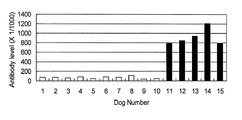
- the antibody titer of 5 individuals (6 to 10) beagle dogs with onset of periodontal disease positive for anone strains aged 5 to 6 years is shown.
- the antibody response result with respect to Me85 in the human of various age groups is shown.
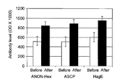
- the antibody titer against the peptide ANON-hexosaminidase as a beast specific infection marker is shown.
- Dog No. 1 to 10 indicate dogs with no periodontal disease
- Dog No. 11 to 15 indicate dogs with periodontal disease origin (bacteria isolation positive dogs).
- the antibody titer against peptide ANON-ASCP as a beast specific infection marker is shown.
- Dog No. 1 to 10 indicate dogs with no periodontal disease, and Dog No.
- 11 to 15 indicate dogs with periodontal disease origin (bacteria isolation positive dogs).
- the antibody titer against the peptide ANON-HagE as a beast specific infection marker is shown.
- Dog No. 1 to 10 indicate dogs with no periodontal disease
- Dog No. 11 to 15 indicate dogs with periodontal disease origin (bacteria isolation positive dogs).
- Anon-Ab shows changes in antibody titer using a peptide specific to ANON strain.
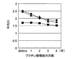
- the change of GI after vaccination is shown.
- diamonds, triangles, and squares indicate the average GI of each beagle dog.
- the change of GI of the front tooth and the canine region is shown.
- diamonds, triangles, and squares indicate the average GI of each beagle dog.
- the first embodiment of the present invention is a marker for detecting zoonotic periodontal disease bacteria.
- the marker for detecting a zoonotic periodontal disease bacterium refers to a marker for specifically detecting a zoonotic periodontal disease bacterium.
- the term “Zoo-infectious periodontal disease bacteria” is a pathogenic infectious bacterium that is commonly present in the oral cavity of humans and other mammals and can cause periodontal disease in both of them. . Specifically, P.I. This applies to P. ginigivalis ANON (hereinafter referred to as “Anon stock”).
- other mammals means all mammals other than humans, and are not particularly limited, but considering the infection rate to humans, the frequency of direct contact with humans High mammals such as dogs, cats, rabbits, hamsters, guinea pigs, pets such as mice, rats, ferrets, squirrels, wolfberry, monkeys and hedgehogs, horses, cows, sheep, goats, pigs, reindeers, camels And livestock such as llamas or laboratory animals.
- pet animals which are frequently contacted by humans, generally dogs and cats, are suitable as other mammals herein.
- This marker is composed of a peptide containing the amino acid sequence represented by any one of SEQ ID NOs: 1 to 7. These amino acid sequences are all described in P.P. It corresponds to a partial sequence of a protein involved in the pathogenicity and infectivity of Gingivalis. Specifically, it is as follows.
- the amino acid sequence represented by SEQ ID NO: 1 It is an amino acid sequence of an anon strain corresponding to hemagglutinin (HagB) positions 331 to 350 (starting methionine residue is number 1; the same shall apply hereinafter) encoded by the gene symbol PG1972 of Gindivaris strain W83.
- the peptide consisting of the amino acid sequence represented by SEQ ID NO: 1 is referred to as “Hem331” for convenience.
- the amino acid sequence shown in SEQ ID NO: 2 is P.I. It is an amino acid sequence of an Anon strain corresponding to positions 190 to 210 of the aforementioned HagB of Gingivalis strain W83.
- the peptide consisting of the amino acid sequence represented by SEQ ID NO: 2 is referred to as “Hem190” for convenience.
- the amino acid sequence shown in SEQ ID NO: 3 is P.I. It is an amino acid sequence of an Anon strain corresponding to positions 55 to 74 of ferrous transport protein B (FeoB-1) encoded by gene symbol PG1043 of Gindivaris W83 strain.
- Fe55 the peptide consisting of the amino acid sequence represented by SEQ ID NO: 3 is referred to as “Fe55” for convenience.
- the amino acid sequence shown in SEQ ID NO: 4 is P.I. It is an amino acid sequence of an anon strain corresponding to positions 141 to 160 of fimbrilin (fimA) encoded by the gene symbol PG2132 of Gingivalis strain W83.
- the peptide consisting of the amino acid sequence represented by SEQ ID NO: 4 is referred to as “Fim141” for convenience.
- the amino acid sequence shown in SEQ ID NO: 5 is P.I. It is an amino acid sequence of an Anon strain corresponding to positions 353 to 370 of the fimA of Gindivaris strain W83.
- the peptide consisting of the amino acid sequence represented by SEQ ID NO: 5 is referred to as “Fim353” for convenience.
- the amino acid sequence shown in SEQ ID NO: 6 is P.I. It is an amino acid sequence of an Anon strain corresponding to positions 85 to 106 of the metalloprotease encoded by the gene symbol PG0383 of Gingivalis strain W83.
- the peptide consisting of the amino acid sequence represented by SEQ ID NO: 6 is referred to as “Me85” for convenience.
- the amino acid sequence shown in SEQ ID NO: 7 is P.I.
- the peptide consisting of the amino acid sequence represented by SEQ ID NO: 7 is referred to as “Me48” for convenience.
- amino acid sequences represented by SEQ ID NOs: 1 to 7 are sequences that are specifically recognized only in Anon strains. Therefore, peptides containing these amino acid sequences can be useful markers for detecting Anon strains, ie, zoonotic periodontal disease bacteria.
- the marker of the present embodiment is not particularly limited as long as the number of amino acids of the peptide is a peptide including the amino acid sequence represented by SEQ ID NOs: 1 to 7.
- it is a peptide consisting of only the amino acid sequence represented by SEQ ID NOs: 1 to 7, ie, Hem331, Hem190, Fe55, Fim141, Me85 or Me48, preferably 40 amino acids or less, more preferably 30 amino acids or less.
- the marker of the present embodiment can be fused with, for example, a tag sequence that facilitates detection.
- tag acid sequences include histidine “His” tags (GentzGenet al., 1989, Proc. Natl. Acad. Sci. USA, 86: 821-824), hemagglutinin “HA” tags (Wilson et al., 1984). , Cell, 37: 767), and “Flag” tags (Knappik et al., Biotechniques, 1994, 17 (4): 754-761).
- the marker of this embodiment may be labeled with a label for detection.
- labeling agents include fluorescent dyes (fluorescein, FITC, rhodamine, umbelliferone, Texas red, Cy3, Cy5), fluorescent proteins (eg, PE, APC, GFP), enzymes (eg, horseradish peroxidase, alkaline phosphatase) , Glucose oxidase, ⁇ -galactosidase), biotin or (strept) avidin, or a radioisotope (eg, 32 P, 33 P, 35 S, 90 Y, 111 In, 112 In) and the like can be used.
- the marker labeling method of the present embodiment may be performed by a technique known in the art.
- the marker of this embodiment may be manufactured according to a known technique by biosynthesis or chemical synthesis.
- a polynucleotide fragment encoding the marker of the present embodiment is amplified by chemical synthesis, or from a genomic DNA or cDNA library of an Anon strain by a nucleic acid amplification method such as PCR.
- the fragment can be obtained by inserting it into an appropriate expression vector, introducing it into a host cell such as E. coli, and then expressing it in the cell.
- a host cell such as E. coli
- tag sequence is fused to the marker of the present embodiment, it is convenient for separating and purifying the target marker from the host cell.
- Such a recombinant protein technique is known in the art, and may be performed according to a known technique when biosynthesizes the marker of the present embodiment. For example, it is possible to refer to the recombinant protein technology described in a biotechnology experiment book (Japan Biotechnology Society, edited by Baifukan, 1996).
- peptide synthesis When chemical synthesis is performed, it may be synthesized using a known peptide synthesis technique.
- peptide synthesis for example, manufacturers such as Kobe Natural Product Chemical Co., Ltd., Takara Bio Inc., and EZBiolab® (US) Inc. conduct contract synthesis, and these can also be used.
- Marker antibody or antigen-binding fragment thereof for detecting anti-animal common infectious periodontal disease bacteria or antigen-binding fragment thereof The second embodiment of the present invention is a marker antibody or anti-human animal infectious periodontal disease detection marker antibody-binding fragment thereof. .
- Marker antibody for detection of anti-human common infectious periodontal disease bacteria refers to the animal of the first embodiment
- An antibody having a marker for detecting a common infectious periodontal disease bacteria as an antigen means an antibody that specifically recognizes an epitope present in the peptide consisting of the amino acid sequence represented by any one of SEQ ID NOs: 1 to 7, that is, Hem331, Hem190, Fe55, Fim141, Me85, or Me48.
- An “epitope” is a polypeptide fragment having antigenicity or immunogenicity, and consists of about 7 to 12 amino acids, preferably 8 to 11 amino acids.
- the anti-marker antibody of this embodiment may recognize any epitope. “Specifically recognizing an epitope” means that only a target epitope is specifically recognized and bound, and other epitopes are not recognized and do not substantially bind, that is, have no cross-reactivity. means.
- the anti-marker antibody of the present embodiment may be any kind of antibody as long as it can retain antigen binding.
- a polyclonal antibody, a monoclonal antibody, and a recombinant antibody can be mentioned.
- the anti-marker antibody of the present embodiment is a polyclonal antibody (anti-marker polyclonal antibody) or a monoclonal antibody (anti-marker monoclonal antibody), any class of immunoglobulin molecules such as IgG, IgE, IgM, IgA, IgD and IgY, Or any subclass, for example, IgG1, IgG2, IgG3, IgG4, IgA1, IgA2.
- Monoclonal antibodies include human monoclonal antibodies, non-human animal monoclonal antibodies (eg, mouse monoclonal antibodies), and chimeric monoclonal antibodies.
- the anti-marker polyclonal antibody and anti-marker monoclonal antibody of the present invention can be prepared by a known method.
- an anti-marker polyclonal antibody can be obtained by immunizing a suitable animal using Hem331, Hem190, Fe55, Fim141, Me85 or Me48 as an antigen.
- anti-marker monoclonal antibodies include spleen cells and myeloma cells from non-human mammals immunized with Hem331, Hem190, Fe55, Fim141, Me85 or Me48 as antigens (eg, mice, human antibody-producing mice, chickens, rabbits, etc.) It can produce by culturing the hybridoma obtained by fusion with.
- the “recombinant antibody” refers to, for example, a chimeric antibody, a humanized antibody, and a multispecific antibody.
- a “chimeric antibody” is an antibody prepared by combining amino acid sequences of antibodies from different animals, and is an antibody in which the constant region (C region) of one antibody is replaced with the C region of another antibody.
- C region constant region
- an antibody obtained by replacing the C region of a mouse monoclonal antibody that recognizes Hem331 with the C region of a human antibody is applicable. This can reduce the immune response to the antibody in the human body.
- a chimeric antibody can be prepared using a known method. For example, DNA encoding a variable region (V region) and DNA encoding the C region of a human antibody may be ligated, incorporated into an expression vector, and introduced into a host for production.
- the “humanized antibody” is a mosaic antibody in which the complementarity determining region (CDR) in the V region of a non-human mammal, for example, a mouse antibody, and the CDR of a human antibody are substituted.
- CDR complementarity determining region
- the antigen binding specificity of an antibody is mainly borne by CDR groups in the V region. Therefore, when producing a recombinant antibody having the same binding characteristics as a specific antibody, it is not necessary to obtain the entire amino acid sequence of the antibody.
- Recombinant antibody that mimics the properties of a specific antibody by preparing and expressing a mosaic antibody in which the DNA sequence encoding the CDR region is replaced with the corresponding DNA sequence encoding the CDR derived from a human antibody, respectively. Can be obtained.
- a humanized antibody can be prepared using, for example, the above-described anti-marker monoclonal antibody and various techniques known in the art based on the base sequence encoding the CDR group of the antibody. For example, the CDR grafting method (Jones et al., Nature (1986) Vol.321: 522-525), the veneering method (veneering) or the resurfacing method (Padlan, 1991, Molecular Immunology 28 (4/5 ): 489 498; Studnicka et al., 1994, Protein Engineering 7 (6): 805 814; and Roguska et al, 1994, Proc Natl Acad Sci USA 91: 969 973), chain shuffling (US patent) No. 5,565,332) and, for example, TanTet al, 2002, J. Immunol. 169: 1119 25, Caldas et al, 2000, Protein Eng. 13: 353 60, etc. That's fine.
- Multispecific antibody refers to a multivalent antibody, that is, an antibody having a plurality of antigen-binding sites in one molecule, each antigen-binding site binding to a different epitope.
- bispecific antibodies that have two antigen-binding sites, such as IgG, that bind to different epitopes on each antigen-binding site can be mentioned.
- this multispecific antibody is preferably capable of binding to different epitopes in which the respective antigen binding sites are present in Hem331, Hem190, Fe55, Fim141, Me85 or Me48.
- These antibodies can be obtained by artificially modifying IgG or the like by a known method using recombinant DNA technology.
- the framework regions (FR) and C regions of the variable region for example, Other amino acids may be substituted with 4 or less, 3 or less, or 2 or less amino acids, preferably 1 to 5 amino acids, more preferably 1 or 2 amino acids.
- “functionally equivalent” means the same biological or biochemical activity as that of an antibody before introduction of an amino acid substitution mutation, specifically the function of specifically recognizing the marker of the present invention as an antigen. It means having.
- the amino acid to be substituted is preferably a substitution between amino acids having similar properties such as charge, side chain, polarity and aromaticity (conservative amino acid substitution).
- basic amino acid group arginine, lysine, histidine
- acidic amino acid group aspartic acid, glutamic acid
- uncharged polar amino acid group glycine, asparagine, glutamine, serine, threonine, cysteine, tyrosine
- nonpolar amino acid group Amino acids within each group of leucine, isoleucine, alanine, valine, proline, phenylalanine, tryptophan, methionine), branched chain amino acid group (threonine, valine, isoleucine), aromatic amino acid group (phenylalanine, tyrosine, tryptophan, histidine) Replacement is applicable.
- the anti-marker antibody of this embodiment and its antigen-binding fragment described later may be modified. Modifications include glycosylation, acetylation, formylation, amidation, phosphorylation, or PEGylation in addition to modification with a label.
- the same indicator as that described in the first embodiment can be used as the indicator.
- the modification with the labeling is useful for detecting the anti-marker antibody of the present embodiment and the antigen-binding fragment thereof described later.
- the glycosylation modification may be a native glycosylation possessed by the endogenous antibody molecule, for example, a modified glycosylation in which the native glycosylation site has been altered by recombinant DNA techniques or by chemical treatment. It may be a site. Modified glycosylation sites are useful for altering the function of an antibody to a desired state, such as to adjust the affinity of the antibody for a target antigen.
- the glycosylation site can be modified by any method known to those skilled in the art.
- a method by genetic manipulation as described above a method using a glycosylation mutant, a method by co-expression with one or more enzymes such as DI N-acetylglucosamine transferase III (GnTIII), various organisms or various organisms
- GnTIII DI N-acetylglucosamine transferase III
- examples include a method in which the antigen-binding fragment of the present invention is expressed in a cell line derived from the cell line and purified, and then the sugar chain is modified.
- the modification by PEGylation is obtained by binding a water-soluble polymer molecule such as polyethylene glycol (PEG) to the anti-marker antibody of the present embodiment and an antigen-binding fragment thereof described later.
- PEGylation can be achieved by chemically coupling PEG to an N-terminal amino group such as an antibody, a C-terminal carboxyl group, or the ⁇ -amino group of a lysine (Lys) residue.
- Modification by PEGylation can increase the in vivo vivo half-life of the modified polypeptide.
- Antigen-binding fragment of marker antibody for detection of anti-animal infectious periodontal disease bacteria Is a partial fragment of the anti-marker antibody described in 2-1, above, and refers to a polypeptide chain or a complex thereof having an activity substantially equivalent to the antigen-specific binding activity of the antibody.
- an antibody part including at least one antigen-binding site described above that is, a polypeptide chain having at least one pair of light chain variable region (VL) and heavy chain variable region (VH) or a complex thereof is applicable.
- Specific examples include many well-characterized antibody fragments produced by cleaving immunoglobulins with various peptidases. More specifically, for example, Fab, F (ab ′) 2 , Fab ′ and the like can be mentioned.
- Fab is a fragment generated by cleaving IgG molecules at the N-terminal side of the hinge disulfide bond by papain, and is composed of three domains (CH1, CH2, CH3) is composed of a CH1 polypeptide adjacent to VH and a light chain.
- F (ab ′) 2 is a dimer of Fab ′ generated by cleaving IgG molecules at the C-terminal side of the disulfide bond at the hinge part by pepsin.
- Fab ′ has a structure substantially equivalent to that of Fab, although its H chain is slightly longer than that of the Fab, including the hinge part.
- Fab, F (ab ′) 2 and Fab ′ can be prepared by methods known in the art.
- Fab can be obtained by papain treatment, and F (ab ′) 2 can be obtained by pepsin treatment.
- Fab ′ can be obtained by reducing F (ab ′) 2 obtained by the pepsin treatment under mild conditions to cleave the disulfide linkage in the hinge region.
- Each of these antigen-binding fragments includes the antigen-binding site of the anti-marker antibody, and has the ability to specifically recognize and bind to the marker of the first embodiment, similarly to the anti-marker antibody. ing.
- the antigen-binding fragment of this embodiment may further be a synthetic antibody fragment synthesized chemically or by using a recombinant DNA method.
- an antibody fragment newly synthesized using a recombinant DNA method can be mentioned.
- scFv single chain fragment
- variable region a constituent antibody fragment
- scFv single chain fragment
- IL variable region
- Single-stranded Fv can be obtained by integrating and expressing a recombinant DNA encoding the same into a phage genome using known techniques.
- a diabody is a molecule having a structure based on a dimeric structure of a single chain Fv (Holliger et al., 1993, Proc. Natl. Acad. Sci. USA 90: 6444-6448). For example, if the linker is shorter than about 12 amino acid residues, a single-chain Fv cannot self-assemble its two variable sites, but forms a diabody and interacts with the two single-chain Fv.
- the VL of one Fv chain can be assembled with the VH of the other Fv chain to form two functional antigen-binding sites (Marvin et al., 2005, Acta Pharmacol. Sin. 26 : 649-658). Furthermore, by adding a cysteine residue to the C-terminus of the single chain Fv, it becomes possible to form a disulfide bond between the two Fv chains and to form a stable diabody (Olafsen et al., 2004). , Prot. Engr. Des. Sel. 17: 21-27).
- a diabody is a bivalent antibody fragment, but each antigen binding site does not need to bind to the same epitope, and each has a bispecificity that recognizes and specifically binds to a different epitope.
- Triabodies and tetrabodies are trivalent and tetravalent antibody fragments, respectively, having trimeric and tetrameric structures based on a single-chain Fv structure in the same way as diabodies. Alternatively, multispecific antibodies may be used.
- the anti-marker antibody and the antigen-binding fragment thereof may be a so-called neutralizing antibody having an activity of suppressing infection of an anon strain to a host and inflammation caused thereby.
- the anti-marker antibody and antigen-binding fragment thereof of this embodiment specifically recognize and bind to the epitope present in the marker of the first embodiment.
- all of the peptides constituting the marker of the first embodiment are P.I. It constitutes part of the protein involved in the pathogenicity and infectivity of Gingivalis. Therefore, the anti-marker antibody and / or antigen-binding fragment thereof of the present embodiment can have an activity of inhibiting or suppressing the original function of the protein by binding to the protein having the target epitope in the Anon strain. .
- the Anon strain loses its infectivity in the oral cavity of the host.
- “host” refers to humans and other mammals described above that can be infected by anone strains.
- an anon strain can be specifically recognized. Therefore, it is useful for detecting anone strains from plaques obtained from a subject in the fifth embodiment to be described later.
- composition of zoonotic periodontal disease infection or inflammation suppressor is a zoonotic periodontal disease infection or inflammation inhibitor.
- the zoonotic periodontal disease infection or inflammation inhibitor of the present embodiment (hereinafter simply referred to as “infection or inflammation inhibitor”) is one or more antibodies and / or one described in the second embodiment. It contains the above antigen-binding fragment as an active ingredient, and suppresses host infection by specifically inhibiting adhesion of the Anon strain to periodontal tissues and teeth (roots), etc. It is a pharmaceutical composition capable of suppressing inflammation via cytokine induced by the protein or the like by inhibiting.
- the antibody and antigen-binding fragment which are active ingredients of the infection or inflammation inhibitor of this embodiment, both have the activity of suppressing the infection of the aforementioned anone strain to the host.
- the antibody is preferably derived from the same species as the subject to which the infection or inflammation inhibitor is administered, or is a recombinant antibody.
- the antibody (monoclonal antibody or polyclonal antibody) which is an active ingredient is preferably a human antibody, a chimeric antibody whose C region is derived from a human antibody, or a humanized antibody.
- the infection or inflammation inhibitor of this embodiment can contain a pharmaceutically acceptable carrier in addition to the active ingredient.
- “Pharmaceutically acceptable carrier” refers to an additive that can be usually used in the field of pharmaceutical technology.
- Additives include collagen, polyvinyl alcohol, polyvinylpyrrolidone, carboxyvinyl polymer, sodium carboxymethylcellulose, sodium polyacrylate, sodium alginate, water-soluble dextran, sodium carboxymethyl starch, pectin, methylcellulose, ethylcellulose, xanthan gum, gum arabic, casein , Agar, polyethylene glycol, diglycerin, glycerin, propylene glycol, petrolatum, paraffin, stearyl alcohol, stearic acid, human serum albumin (HSA), mannitol, sorbitol, lactose, surfactants acceptable as pharmaceutical additives, etc. It is done.
- excipients binders, disintegrants, fillers, emulsifiers, flow additive regulators, lubricants, flavoring agents, solubilizers (solubilizers), suspending agents, dilutions as necessary Agents, surfactants, stabilizers, absorption enhancers, bulking agents, moisturizers, humectants (eg, glycerin and starch), adsorbents, disintegration inhibitors, coating agents, colorants, preservatives, antioxidants, fragrances , Flavoring agents, sweetening agents, buffering agents, and the like.
- the above carriers can be used alone or in appropriate combination depending on the dosage form of the infection or inflammation inhibitor of the present embodiment.
- a purified anti-marker antibody is dissolved in a pharmaceutically acceptable solvent, and an adsorption inhibitor (eg, Tween 80, Tween 20, gelatin and human What added serum albumin) can be used.
- pharmaceutically acceptable solvent refers to, for example, water, physiological saline, buffer solution, glucose solution, pharmaceutically acceptable organic solvent (eg, ethanol, propylene glycol, ethoxylated isostearyl alcohol). , Polyoxylated isostearyl alcohol, polyoxyethylene sorbitan fatty acid esters). These are preferably sterilized and preferably adjusted to be isotonic with blood as necessary.
- the infection or inflammation inhibitor of this embodiment may be lyophilized in order to obtain a dosage form that is reconstituted before use.
- excipients for example, sugar alcohols and saccharides such as mannitol and glucose
- mannitol and glucose can be used for lyophilization.
- composition of the present invention can be formulated according to a conventional method. See, for example, Remington's Pharmaceutical Science, latest edition, Mark Publishing Company, Easton, U.S.A. for formulation.
- the infection or inflammation inhibitor of this embodiment is administered orally, or into tissues (for example, oral mucosal administration, intramuscular administration and intravenous administration), or local administration (for example, transmucosal). Administration). Therefore, the dosage form of the infection or inflammation inhibitor is preferably in a form suitable for the administration method. For example, for intra-tissue administration, injection through the bloodstream is preferred and therefore the dosage form is a liquid.
- the injection site is not particularly limited.
- the infection site of the Anone strain is a periodontal tissue in the oral cavity, for example, in a blood vessel such as a vein or an artery, or in the oral mucosa (periodontal Preferably within the organization).
- the infection or inflammation inhibitor of the present invention can be immediately distributed throughout the body through the bloodstream, and is less invasive and less burdening the subject.
- the active ingredient can be directly applied to the Anon strain.
- an effective amount capable of exerting the infection or inflammation suppression activity is contained in one dosage unit.
- the term “effective amount” as used herein refers to an amount necessary for the anti-marker antibody or antigen-binding fragment thereof, which is an active ingredient, to exert its function, that is, the infection or inflammation inhibitor is transferred to the periodontal tissue of the Anon strain. Is an amount necessary to suppress the occurrence of infection and inflammation caused by the infection, and causes little or no harmful side effects on humans or other mammals as subjects to be administered. This effective amount may vary depending on various conditions such as subject information, dosage form and route of administration.
- Subject information '' refers to species, progression or severity of periodontal disease, general health, age (months, weeks), body weight, sex, diet, drug sensitivity, presence or absence of concomitant medications, and Including resistance to treatment.
- the final dose and effective amount of the above-mentioned infection or inflammation inhibitor are determined by the judgment of a doctor or veterinarian according to the information of individual subjects.
- a large-scale administration of the above-mentioned infection or anti-inflammatory agent is necessary to obtain the infection or inflammation-suppressing effect, it can be divided and administered in several times to reduce the burden on the subject.
- the effective amount of the infection or inflammation inhibitor is The dose is selected in the range of 0.01 mg to 100 mg per kg body weight, but is not limited to this dose.
- the agent for suppressing infection or inflammation of the present invention suppresses inflammation caused by the bacterium by specifically suppressing the infectivity of the Anone strain, prevents or treats periodontal disease caused by the Anone strain, And it is possible to prevent infection between other mammals.
- Kit for detecting zoonotic periodontal disease bacteria The fourth embodiment of the present invention is a kit for detecting zoonotic periodontal bacteria.
- the kit of this embodiment includes one or more markers described in the first embodiment and / or one or more anti-marker antibodies and / or one or more antigen-binding fragments of the second embodiment.
- this kit should contain a labeled secondary antibody, a substrate necessary for detection of the label, a positive control or negative control, a buffer used for dilution or washing of the sample, and / or instructions for use. You can also.
- the anti-marker antibody of the second embodiment or the antigen-binding fragment thereof is used to immunology the marker of the first embodiment derived from the anone strain contained in an appropriate sample such as plaque. It can be easily and simply measured by a manual measurement method. In addition, based on the result, it is possible to quickly determine whether or not the anone strain of the subject who provided the sample is carried.
- the anti-anone antibody contained in an appropriate sample such as a body fluid can be easily and simply measured by an immunological measurement method using the marker of the first embodiment. Further, based on the result, it is possible to quickly determine the presence or absence of current infection from the history of infection of the Anon strain of the subject who provided the sample or the value of the antibody titer.
- the fifth embodiment of the present invention is a method for determining infection of periodontopathic bacteria common to zoonosis.
- the method of the present embodiment can determine the history of anon strain in the subject and / or the current infection status.
- the determination method of this embodiment includes an antibody detection step and a determination step. Hereinafter, each step will be specifically described.
- Antibody detection step refers to an antibody that recognizes an epitope present in a peptide consisting of the amino acid sequence represented by any one of SEQ ID NOs: 1 to 7 contained in a body fluid collected from a subject. This is a step of detecting using one marker.
- Subject refers to an individual that is subjected to the test of the determination method of the present embodiment and can be infected with an Anon strain. For example, it refers to humans and other mammals.
- other mammals as used herein means all mammals other than the above-mentioned mammals, and companion animals, particularly dogs and cats are applicable.
- Body fluid refers to a liquid substance derived from a living body.
- blood including serum, plasma, interstitial fluid
- lymph fluid spinal fluid
- saliva urine
- sweat tears
- semen vaginal fluid
- nasal discharge or cell or tissue extract.
- the body fluid is not particularly limited as long as it can contain an antibody.
- Preferred are blood, cerebrospinal fluid, and lymph.
- serum is preferred.
- Antibody in the present embodiment refers to an antibody produced by the subject's own immune production system and contained in the body fluid.
- any class of immunoglobulin molecules from the subject such as IgG, IgE, IgM, IgA, IgD and IgY, or any subclass, such as IgG1, IgG2, IgG3, IgG4, IgA1, IgA2,
- the artificially modified and synthesized antibodies and fragments thereof such as the recombinant antibodies and antigen-binding fragments described in the second embodiment do not correspond to the antibodies referred to herein.
- the “antibody” of this embodiment is a peptide consisting of the amino acid sequence represented by any one of SEQ ID NOs: 1 to 7, Hem331, Hem190, Fe55, Fim141, Me85 or Me48 as an antigen, and as an epitope present in the antigen Recognize and join. That is, the antibody of the present embodiment is an anti-anone strain antibody that specifically recognizes and binds to an anon strain (hereinafter simply referred to as “anti-anone strain antibody”). Therefore, a subject having the antibody in a body fluid has a history of infection with the anone strain, and if the antibody titer contained in the body fluid is high, it is highly likely that the subject is still carrying the anone strain. Can be estimated.
- the collection of the body fluid from the subject may be performed according to a technique known in the field.
- the collection method may follow a known blood collection method.
- peripheral blood it may be collected by injection into a peripheral vein or the like, and in the case of bone marrow fluid, it may be collected by bone marrow puncture (Marc).
- a needle may be inserted into the umbilical cord before delivery of the postpartum placenta.
- Body fluid collected from the subject may be immediately subjected to the determination method of the present embodiment, or may be necessary after having been stored at a low temperature after appropriate treatment such as adding a blood coagulation inhibitor such as heparin. Occasionally, frozen or refrigerated blood may be thawed and heated by a known method and used for a determination test.
- the body fluid used in the determination method of the present embodiment varies depending on the type of subject, the type of body fluid, or the antibody detection method. For example, in the case of blood, a range of 20 ⁇ L to 200 ⁇ L is usually sufficient, and serum is sufficient. If it exists, the range of 10 ⁇ L to 100 ⁇ L is sufficient.
- any immunological detection method using the marker of the first embodiment can be used.
- it can be carried out by ELISA, fluorescence immunoassay, radioimmunoassay or luminescence immunoassay using the marker of the first embodiment.
- the ELISA method, the fluorescence immunoassay method, the radioimmunoassay method, and the luminescence immunoassay method are all common in that an antigen-antibody complex of a marker that is an antigen and a target antibody is detected by a labeled antibody.
- the labels for detecting the complex are different from each other.
- An enzyme label is used in the ELISA method, a fluorescent substance is used in the fluorescent immunoassay, a radioactive substance is used in the radioimmunoassay, and a luminescent substance is used in the luminescent immunoassay.
- examples of the enzyme include peroxidase (POD), alkaline phosphatase, ⁇ -galactosidase, urease, catalase, glucose oxidase, lactate dehydrogenase, amylase or biotin-avidin complex, etc.
- POD peroxidase
- alkaline phosphatase alkaline phosphatase
- ⁇ -galactosidase urease
- catalase glucose oxidase
- lactate dehydrogenase lactate dehydrogenase
- amylase or biotin-avidin complex
- Fluorescent dyes fluorescein, FITC, rhodamine, umbelliferone, Texas red, Cy3, Cy5
- fluorescent proteins eg, PE, APC, GFP
- radioactive substances such as 3 H, 125 I or 131
- NADH-FMNH 2 -luciferase system As the luminescent substance, NADH-FMNH 2 -luciferase system, luminol-hydrogen peroxide-POD system, acridinium ester system or dioxetane compound system can be used.
- the ELISA method will be described as an example.
- an anti-anone strain antibody which is a target molecule in a body fluid, is used as an antigen by using an antigen immobilized on a solid support and an enzyme-labeled antibody, etc.
- the antibody reaction is detected as a color density or fluorescence intensity, and the anti-anone antibody is quantified.
- the solid phase carrier includes beads made of materials such as polystyrene, polycarbonate, polyvinyltoluene, polypropylene, polyethylene, polyvinyl chloride, nylon, polymethacrylate, latex, gelatin, agarose, cellulose, sepharose, glass, metal, ceramics or magnetic material.
- Insoluble carriers in the form of microplates, test tubes, sticks or test pieces can be used. Immobilization of an antigen on a solid phase carrier can be achieved by binding according to a known method such as a physical adsorption method, a chemical binding method, or a combination of these methods.
- the enzyme-labeled antibody is obtained by labeling an antibody that recognizes an epitope present in the constant region with an appropriate detection enzyme, using the immunoglobulin molecule of the subject as an antigen.
- the class of the immunoglobulin molecule is not particularly limited, but is preferably IgG.
- the binding method between the labeling substance and the antibody may be a known method such as the glutaraldehyde method, the maleimide method, the pyridyl disulfide method or the periodic acid method. It should be noted that the enzyme-labeled antibody uses an anti-human IgG antibody or the like when a human is the subject, and an anti-dog IgG antibody or the like when the dog is the subject.
- Enzyme Immunoassay 3rd edition, Medical School, 1987, edited by Kitagawa Tsuneki et al., "Protein Nucleic Acid Enzyme Separate Volume No.31 Enzyme Immunoassay", Kyoritsu Shuppan, 1987, Kei Irie “Radioimmunoassay” Kodansha Scientific, 1974, Kei Irie, “Continuing Radioimmunoassay”, Kodansha Scientific, 1979.
- one of the markers described in the first embodiment for example, Fe55 is immobilized on a carrier as an antigen.
- the marker to be immobilized may be not only one type of Fe55 but also multiple types.
- human serum that can contain an anti-anone strain IgG antibody is allowed to act on the immobilized Fe55 to form a complex of Fe55 and the anti-anone strain antibody on the surface of the carrier.
- the unbound antibody other than the anti-anone strain antibody present in the sample is removed by sufficiently washing with a washing solution.
- an anti-human IgG antibody that specifically recognizes labeled human IgG is prepared, and this labeled antibody is allowed to act on the carrier on which the complex is formed. After sufficiently washing with the washing solution, the anti-anone strain antibody present in the sample can be detected by detecting the complex using the label.
- a body fluid that may contain an anti-anone strain antibody and the labeled antibody are first mixed to form an antibody complex, and then allowed to act on the immobilized marker.
- the marker to be immobilized is labeled with, for example, biotin, and a biotinylated marker, an anti-anone strain antibody, and a labeled antibody other than biotin are mixed in advance to form an antigen-antibody complex, and then avidin
- the antigen-antibody complex can also be detected by acting on a solid-phased carrier.
- a test strip for immunochromatography can be used as an immunological detection method.
- Immunochromatographic test strip means, for example, a sample receiving portion made of a material that easily absorbs a sample, a reagent portion containing the diagnostic agent of the present invention, a developing portion in which a reaction product of the sample and the diagnostic agent moves, and development It comprises a labeling unit for coloring the reaction product, a presentation unit for developing the colored reaction product, and the like.
- commercially available pregnancy diagnostic agents have the same form.
- the sample receiving portion absorbs the body fluid and reaches the reagent portion.
- the immunochromatographic test strip has low invasiveness and does not pose any pain or danger to the user due to the use of reagents, so it can be used for monitoring at home. It is possible to scrutinize and treat (surgical excision, etc.) and to prevent metastasis and recurrence. Such a test strip is also convenient in that it can be mass-produced at low cost by, for example, a manufacturing method described in JP-A-10-54830.
- immunological detection methods include immunoturbidimetry, latex agglutination, and the formation of immune complex aggregates such as erythrocyte agglutination or particle agglutination such as latex turbidimetry. Is measured by an optical method or visually measured. In that case, a phosphate buffer, glycine buffer, Tris buffer, Good buffer, or the like can be used as a solvent, and a reaction accelerator such as PEG or a nonspecific reaction inhibitor may be included. . Since these are all known methods, they may be detected based on known techniques.
- a surface plasmon resonance method can also be used.
- the surface plasmon resonance phenomenon is a phenomenon in which the intensity of reflected light is significantly attenuated when a metal thin film is irradiated with laser light at a specific incident angle (resonance angle).
- the SPR sensor using the principle of the SPR phenomenon can measure the adsorbate on the surface of the metal thin film with high sensitivity. Therefore, by preliminarily immobilizing antibodies and / or target antigens on the surface of the metal thin film and passing the sample over the surface of the metal thin film, adsorption on the metal surface before and after passing through the sample caused as a result of the antigen-antibody reaction. Differences in objects can be detected.
- a substitution method, an indirect competition method, and the like are known, and any of them may be used. This technique is well known in the art. For example, see Kazuhiro Nagata and Hiroshi Handa, real-time analysis method of biological material interaction, Springer Fairlark Tokyo, Tokyo, 2000.
- the measurement method of the present invention can use a quartz crystal microbalance measurement method (QCM method).
- QCM method utilizes a phenomenon in which when a substance is adsorbed on the surface of an electrode attached to a crystal oscillator, the resonance frequency of the crystal oscillator decreases according to its mass.
- a QCM sensor using this method is a mass measurement sensor that quantitatively captures a very small amount of adsorbate by the amount of change in the water resonance frequency.
- This technique is well known in the art. For example, J. et al. See Christopher Love, et.al., 2005, Chemical Review, 105: 1103-1169; Torihiro Moriizumi, Takamichi Nakamoto, 1997, Sensor Engineering, Shodo.
- the “determination step” is a step of determining the presence or absence of infection of the subject with respect to the Anon strain based on the result of the antibody detection step.
- Presence / absence of infection here includes both whether the subject has been infected with an anon strain in the past (infection history) and whether the subject is currently infected with an anon strain (infection status). .
- the subject As for the history of infection, when an anti-anone strain antibody is detected from the body fluid of the subject as a result of the antibody detection step, the subject has been or has been infected with at least the past and / or present Anone strain. It is determined that On the other hand, if no anti-anone strain antibody is detected, it is determined that the subject has a high possibility of having no history of infection with the anone strain.
- the antibody titer is the cutoff value (the antibody titer in the control + 3SD (standard deviation)) At this time, it is determined that the individual has a high possibility of being infected with an Anon strain. On the other hand, when the antibody titer falls below the cut-off value, it is determined that the individual has a history of infection with an anon strain in the past but is not likely to be infected at present.
- the cut-off value may be determined on the basis of the antibody titer of the anti-anone antibody in the body fluid, preferably serum, of a population of the same species as the subject and uninfected with the anone strain.
- the subject is a dog
- the cutoff value of the antibody titer for Fe55 may be “average value + 3SD” of the antibody titer of the uninfected individual.
- the antibody titer may be calculated based on the measurement value obtained by the immunological detection method described in the antibody detection step. For example, ELISA is applicable.
- the sixth embodiment of the present invention is a method for detecting periodontal disease bacteria that are zoonotic.
- the method of the present embodiment can detect whether or not a subject carries an anon strain.
- the detection method of this embodiment includes a polypeptide detection step.
- the “polypeptide detection step” uses one or more polypeptides containing the marker described in the first embodiment contained in plaque collected from a subject using the antigen-binding fragment described in the second embodiment. This is a detecting step.
- Pig is also called plaque, and is composed of normal oral bacteria including periodontal pathogens attached to periodontal tissues and teeth (roots) in the oral cavity and their metabolites.
- “One or more polypeptides comprising the marker described in the first embodiment” are specific to an anon strain comprising the amino acid sequence represented by SEQ ID NOs: 1 to 7. It refers to HagB, FeoB-1, fimA or metalloprotease or a polypeptide fragment thereof. That is, when one or more of the proteins or polypeptide fragments thereof are detected from the plaque, it means that Anon bacteria are present in the oral cavity of the subject from which the plaque was collected.
- Collecting plaque from a subject may be performed according to a technique known in the art. For example, it is collected using a cotton swab or a scraper. Thereafter, it may be suspended in an appropriate buffer as required.
- the marker protein contained in the plaque is basically the same except that the anti-anone strain antibody of the second embodiment and / or its antigen-binding fragment (hereinafter referred to as “anti-anone antibody” or the like) is used. It can detect by the immunological detection method used at the detection process of 5th Embodiment. That is, using the anti-anone antibody of the second embodiment, ELISA, fluorescence immunoassay, radioimmunoassay or luminescence immunoassay, surface plasmon resonance (SPR), or quartz crystal microbalance measurement The marker protein can be detected by the method (QCM method).
- the ELISA method in order to detect a marker protein including a marker that is an antigen from plaque using an anti-anone antibody or the like, the ELISA method also uses the sandwich method in addition to the direct method and the indirect method. Can do.
- this sandwich method is preferably applied.
- the “sandwich method” is a method in which a first antibody (solid-phased antibody) immobilized on a solid phase carrier is bound to an antigen, and then a second antibody (labeled antibody or primary antibody) that recognizes an epitope different from the first antibody. Is added to the antigen, and when the second antibody is a labeled antibody, the label is detected, and when the second antibody is a primary antibody, it is detected by a third antibody (secondary antibody).
- the detection method of the present embodiment will be described by a sandwich ELISA method with specific examples.
- the anti-anone antibody or the like of Embodiment 2 is immobilized on a carrier.
- a sample such as a buffer in which a plaque capable of containing a marker protein is suspended is allowed to act on a solid-phased anti-anone strain antibody (hereinafter referred to as “solid-phased antibody”).
- An antigen-antibody complex (solid-phase complex) consisting of is formed on the surface of the solid phase carrier.
- the substrate is sufficiently washed with a washing solution to remove the immobilized antibody and the unbound substance.
- an antibody that is present in the marker in the marker protein and specifically recognizes an epitope different from the immobilized antibody is labeled as a labeled antibody and allowed to act on the carrier to which the immobilized complex is bound. That is, as the immobilized antibody and the labeled antibody used here, the anti-anone strain antibody of the second embodiment that recognizes the same marker described in the first embodiment can be used. As a result, a triple complex composed of the immobilized antibody / marker protein / labeled antibody is formed on the solid support. Thereafter, the unbound labeled antibody is sufficiently washed with a washing solution, and then the marker protein present in the sample can be detected and quantified by detecting using the label of the labeled antibody in the triple complex.
- One kind or several kinds of labeled antibodies may be used, but two or more kinds are preferably used, and three kinds are more preferably used. If the animal species from which the immobilized antibody and the labeled antibody are derived are different, the primary antibody is recognized by acting on the immobilized antibody / marker protein as the primary antibody without labeling the labeled antibody. It can also be detected using a labeled secondary antibody. It should be noted that the antibody used for immobilization and the antibody used for labeling can be used in reverse.
- a sample containing a labeled antibody and a marker protein can be mixed in advance to form an antigen-antibody complex, and then allowed to act on the immobilized antibody.
- the immobilized antibody is labeled with biotin
- a biotinylated immobilized antibody, a sample containing a marker protein, and a labeled antibody other than biotin are mixed to form an antigen-antibody complex, and then avidin is immobilized.
- the antigen-antibody complex can be detected using a label other than biotinylation.
- the seventh embodiment of the present invention is a vaccine for treating periodontal disease bacteria.
- the vaccine of this embodiment can be obtained by inactivating or attenuating an anon strain.
- Anon strains are cultured on P. a.
- a known culture method may be used for Gingivalis.
- BHI medium (27.5 g of brain extract, heart extract and peptone mixture per liter of purified water, D-glucose 2.0 g, sodium chloride 5.0 g, disodium hydrogen phosphate 2.5 g; Merk) or GAM medium (purification Per liter of water 10.0 g peptone, 3.0 g soybean peptone, 10.0 g protease peptone, 13.5 g serum powder, 5.0 g yeast extract, 1.2 g meat extract, 3.0 g glucose, 2.5 g potassium dihydrogen phosphate, 3.0 g sodium chloride, soluble And a method of culturing under anaerobic conditions at 37 ° C.
- the anaerobic condition may be performed in an anaerobic glove box (N 2 70%, CO 2 15%, H 2 15%) or an anaerobic jar.
- Inactivation or attenuation treatment of anone strains for use as a vaccine may be performed by a known method. For example, it can be achieved by adding an inactivating agent such as formalin, ⁇ -propiolactone, glutardialdehyde, etc. to the culture solution containing the infectious anone strain and mixing well (Appaiahgari et al., 2004, Vaccine , 22: 3669-3675). Moreover, infectivity can also be rapidly lost by irradiating an infectious anone strain with ultraviolet rays. The inactivation method by ultraviolet irradiation is preferable in that it has little influence on proteins and the like constituting the Anon strain. As the ultraviolet ray source, a commercially available germicidal lamp, particularly a 15 W germicidal lamp, can be used, but it is not limited thereto.
- an inactivating agent such as formalin, ⁇ -propiolactone, glutardialdehyde, etc.
- the vaccine for the treatment of the zoonotic periodontal disease bacteria which inactivated or attenuated the Anone strain of the present invention can be used as a mixed vaccine together with two or more other vaccines.
- the kind of vaccine to mix is not specifically limited, Preferably it is a vaccine which inactivated or attenuated periodontal disease bacteria other than an Anon strain.
- a vaccine in which a bacterium belonging to the genus Porphyromonas is inactivated or attenuated by a known method similar to the above can be mentioned.
- Anonone-specific detection markers are isolated from P. gingivalisanone strains that have been identified as infectious periodontal disease bacteria common to humans and animals, which were not anticipated in the past.
- Method Using genomic analysis by array CGD, a region common to humans and other mammals and a region specific to an animal strain were determined from gene information decoded by Mutation Mapping. Specifically, Mutation Mapping and resequencing analysis were performed, and a peptide having a total of 12 amino acid sequences consisting of a common region of human-derived W83 strain and animal (dog) -derived anone strain, that is, 4 antigens ⁇ 3 regions (Hem331 , Hem301, Hem190, Fe297, Fe259, Fe55, Fim331, Fim141, Fim353, Me85, Me48, Me191) were synthesized as marker candidates.
- Mutation Mapping and resequencing analysis were performed, and a peptide having a total of 12 amino acid sequences consisting of a common region of human-derived W83 strain and animal (dog) -derived anone strain, that is, 4 antigens ⁇ 3 regions (Hem331 , Hem301, Hem190, Fe
- Hem301 indicates a peptide having an amino acid sequence corresponding to positions 301 to 320 of HagB in P. gingivalis strain W83.
- Fe297 and Fe259 indicate a peptide consisting of an amino acid sequence corresponding to positions 297 to 315 and a peptide consisting of an amino acid sequence corresponding to positions 259 to 278 of FeoB-1 in P. gingivalis strain W83, respectively.
- Fim331 and Fim141 indicate a peptide consisting of an amino acid sequence corresponding to positions 331 to 350 of fimA and a peptide consisting of an amino acid sequence corresponding to positions 141 to 160 in P. gingivalis strain W83, respectively.
- Me191 represents a peptide consisting of an amino acid sequence corresponding to positions 191 to 210 of the metalloprotease in P. gingivalis strain W83. Other peptides are as described above. Peptide synthesis was commissioned to KNC Bioresearch Center, Kobe Natural Products Chemicals.
- peptide coating kit (Takara Bio Inc.) was used to immobilize the peptide on the 96-well microplate. Specifically, 100 ⁇ g of peptide was dissolved in 25 mL of reaction buffer (reagent supplied with the kit) and dispensed into a 96-well microplate at 50 ⁇ L / well.
- a coupling reagent (dimethylaminopropylcarbodiimide hydrochloride ethyl acetate: reagent supplied with the kit) was dissolved in distilled water, and 10 ⁇ L / well was dispensed into a 96-well microplate. After thorough mixing, the reaction was allowed to proceed at room temperature for 2 hours to couple to free carboxyl groups. Subsequently, it was cross-linked with an amino group exposed on the bottom surface of the microplate to be solid-phased. Each well was washed three times, and the antibody in the sample was detected after blocking. Serum diluted 1: 100 was used for antibody detection. Serum was obtained from 5 to 8 months old P.
- separation negative means P. gingivalis culture negative, no inflammation in the oral cavity, and even if P. gingivalis is present in the oral cavity, the number of bacteria is very small. This means that there is no invasion of the bacteria into the body and therefore no antibody is produced.
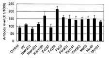
- FIGS. 1 shows the mean antibody titer of sera from dogs negative for P. gingivalis separation.
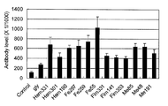
- FIG. 2 shows the mean antibody values of the sera of dogs showing the same properties as P. gingivalis-separation positive dogs and Anon strains.
- antibody-negative dog serum is between the background (Control) antibody titer using fetal bovine serum and the control (gly) antibody titer using a 96-well microplate coated with glycine.
- control fetal bovine serum
- control gly antibody titer using a 96-well microplate coated with glycine.
- the seven types of peptides represented by SEQ ID NOs: 1 to 7 are often referred to as markers for detection of an anus strain of zoonotic periodontal disease bacteria (hereinafter referred to as “detection markers” in the examples of the present specification). ) Separated.
- Example 2 ⁇ Correlation between antibody titer and clinical findings in dogs (1)> It was verified using 10 beagle dogs whether or not the seven kinds of zoonotic periodontal disease bacteria Anon strain detection markers obtained in Example 1 can actually be used for determination of Anon infection.
- peripheral blood was similarly obtained from each of 5 females 5 to 6 years old (No. 6 to 10) who developed periodontal disease and anon strains were detected and were positive for isolation. It collected using.
- Peripheral blood collected from each individual was coagulated at room temperature, centrifuged at 3000 rpm / 10 minutes, and serum was separated.
- Example 1 10 ⁇ L of the obtained serum was used to detect the anti-anone strain antibody in the serum by ELISA using the detection marker separated in Example 1.
- a peptide coating kit (Takara Bio Inc.) was used to immobilize the detection marker on the 96-well microplate. Specifically, 100 ⁇ g of the detection marker was dissolved in 25 mL of a reaction buffer (described above), and dispensed to a 96-well microplate at 50 ⁇ L / well. The coupling reagent was dissolved with distilled water, and 10 ⁇ L / well was dispensed. After thorough mixing, coupling was performed at room temperature for 2 hours to solidify. Each well was washed 3 times and blocked.
- Each serum used was diluted 1: 100 with phosphate buffered saline (pH 7.4, blocking solution—containing 1% Block Ace). After reacting with serum for 1 hour at room temperature, wash 10 times with washing solution (phosphate buffered saline (pH 7.4); 0.05% Tween 20 and 0.1% blocking solution containing Block Ace) and peroxidase labeling at room temperature for 1 hour Reaction with protein A (Abcam, Co.). After washing 10 times with the washing solution, color was developed with the chromogenic substrate TMB (K-blue, Neogen. Co. Lexington KY), the reaction was stopped with 1N sulfuric acid, and the OD value (492 nm) was measured with an ELISA reader (ELNX96, TFB). It was measured.
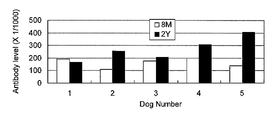
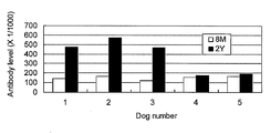
- the results are shown in FIGS.
- the antibody titer was expressed as an OD value indicating the antibody level in ELISA.
- FIG. 3A shows the antibody titers at 8 months of age (8M) and 2 years of age (2Y) against glycine for control. As shown in this figure, the antibody titers at 8 months (8M) and 2 years old (2Y) were low in each of NO.
- the cut-off value for determining “positive” was set to 0.153+ (3 ⁇ 0.030) .
- 3B, 3C, 3D, 3E, 3F, 3G, and 3H are 8 postnatal markers for the detection of an anon strain of 7 common zoonotic periodontal fungi, Hem331, Hem190, Fe55, Fim141, Fim353, Me85, and Me48, respectively.
- the antibody titer at month (8M) and at age 2 (2Y) is shown. As shown in these figures, at 8 months after birth, the antibody titer was as low as 0.2 or less when any of the detection markers was used.
- FIG. 4 shows antibody titers against glycine and seven detection markers in 5 to 6-year-old individuals (No. 6 to 10) who are positive for Anon strain isolation.
- Anon strain isolation positive individual high antibody titers were obtained for all seven kinds of detection markers, and the antibody positive rate was 100%. It was proved that the history of anone strain of the individual can be estimated by using the detection marker of the present invention.
- Example 4 ⁇ Correlation between antibody titer and clinical findings in humans>
- anone strain detection marker obtained in Example 1 can be used for humans, anti-anone strain antibodies in human serum were detected.
- Example 5 ⁇ Synthesis of dog-specific peptides>
- the infecting bacterium is an animal-derived strain type with a dog as a natural host, or a human-derived strain with a human as a natural host Cannot determine whether it is the type. Therefore, we verified the antibody titer against each antigenic peptide in dogs with no periodontal disease and dogs with periodontal disease, and created peptide antigens (beast specific infection markers) that react specifically with animal-derived strain types. Tried.
- FIG. 6A shows the result of ANON-hexosaminidase
- FIG. 6B shows the result of ANON-ASCP
- FIG. 6C shows the result of ANON-HagE.
- 10 dogs with no periodontal disease No. 1 to 10
- dogs with periodontal disease (5 bacteria positive, No. 11).
- ⁇ 15 all were antibody positive.
- Antibody titers were as low as 0.032 to 0.116 in unaffected dogs.
- onset dogs showed high values of 0.586 to 1.214. Therefore, it was shown that ANON-hexosaminidase, ANON-ASCP, and ANON-HagE can all be beast-specific infection markers.
- GI was used as an index of inflammation. GI followed the method of Isogai et al. (Isogai H., et al., 1989, Jpn. J. Vet. Sci. 51, 1151-1162).
- the examination continues in order from the buccal side of the maxillary right, molars (M2 and M1), premolars (P4, P3, P2, P1), canines (C), anterior teeth (I3, I2, I1), then Move to maxillary left cheek side, left maxillary anterior teeth (I1, I2, I3), canine teeth (C), premolars (P1, P2, P3, P4), molars (M1, M2), and then to the lower jaw After confirming the left molar (M3, M2, M1), premolar (P4, P3, P2, P1), canine (C), anterior teeth (I3, I2, I1), move to the left side of the lower jaw and anterior teeth (I1, I2, I3), canine teeth (C), premolars (P1, P2, P3, P4) and molars (M1, M2, M3) were confirmed.
- the degree of inflammation of the gingival part of each tooth was scored as 0: no inflammation, 1 mild inflammation, 2 moderate inflammation, 2 severe inflammation.
- the inflammation at the intermediate stage of the score was set to 0.5, 1.5, and 2.5.
- the average value of the degree of inflammation of the gingival part of each tooth was expressed as the average GI of each dog.
- rgp1 and rgp2 have well-conserved regions (Mikolajczyk-Pawlinska J et al., 1998, Biol Chem 379, 205-211).
- the sequence of RgpB of P. gingivalis ATCC33277 (PubMed), W83 (PubMed), which is considered to be identical to P. gingivalis 381 strain, and the anon strain obtained from array analysis are almost identical (about 99%) ( unpublished). Even if an antibody is produced after infection with Anon strain or P.
- gingivalis which is also infectious to animals, the antibody is degraded by IgG protease (Vincents B et al, FAseB J, 2011, 25: 3741-3750). In this case, the pathogen cannot be eliminated and inflammation will persist. In addition, because P. gingivalis is present in the gingival pocket, Antigen stimulation is likely to be insufficient. Therefore, if the immune response is strengthened by subcutaneous vaccine administration, inflammation may be reduced. In this example, the effect is demonstrated. Permanent dentition of dog is maxillary anterior tooth I1-3, canine C, premolar P1-4, molar M1-2, mandible anterior tooth I1-3, canine C, premolar P1-4, molar M1-3 It consists of teeth.
- the gingival inflammation level of the part corresponding to each tooth was examined in 4 stages.
- the total GI value was divided by 42 to give the average GI.
- the average GI after vaccination decreased in 3 beagle dogs (FIG. 8).
- the average GI up to I1-3 and C with little calculus adhesion was remarkable (FIG. 9).
- Anon strains contain many gene regions of unknown function that are unique in resequencing, and some of these can be more promising candidates as protective antigens.
Abstract
The purpose of the present invention is to provide a system for determining the infection with a periodontopathic bacterium that is communicable between men and other mammals, particularly pet animals, which is unknown heretofore.
P. ginigivalis ANON, which is a periodontopathic bacterium communicable between men and animals, is newly identified. The history of infection with P. ginigivalis ANON or the current state of infection with P. ginigivalis ANON in a subject can be determined by detecting an antibody specific to a marker in a body fluid collected from the subject using an antigen specific to the bacterium as the marker.
Description
本発明は、人獣に共通する歯周病原菌を検出するためのマーカー、当該菌による感染を判定する方法及び当該菌を検出する方法に関する。
The present invention relates to a marker for detecting periodontal pathogens common to humans, a method for determining infection by the bacteria, and a method for detecting the bacteria.
歯周病は、歯肉、歯根膜、セメント質及び歯槽骨等の歯周組織に発生する炎症であり、全世界の人口の約30%が罹患している極めて一般的な疾患である。歯周病には、歯肉炎、歯周炎及び咬合性外傷等が知られるが、このうち最も多く見られる歯周病は、歯周炎、特に、歯周病菌による慢性歯周炎である(非特許文献1)。慢性歯周炎は、軽度の歯肉炎から年齢と共に徐々に進行していく。5歳以上の人で歯肉に所見が認められる割合は、昭和62年では64.3%(歯科疾患実態調査)、平成5年では68.1%(同調査)、平成11年では72.9%(同調査)と、近年増加傾向にある。慢性歯周炎は、口腔内の炎症のみならず、脳血管疾患、心疾患、早産等の種々の全身疾患の発症リスクを高めることが知られている。したがって、慢性歯周炎が顕在化していない段階で、その原因となる歯周病菌を検出することは、慢性歯周炎の予防及び早期治療をする上で重要である。
Periodontal disease is inflammation that occurs in periodontal tissues such as gingiva, periodontal ligament, cementum and alveolar bone, and is a very common disease affecting about 30% of the world population. Periodontal diseases include gingivitis, periodontitis and occlusal trauma, but the most common periodontal disease is periodontitis, especially chronic periodontitis caused by periodontal disease bacteria ( Non-patent document 1). Chronic periodontitis gradually progresses with age from mild gingivitis. The percentage of gingival findings in people over the age of 5 was 64.3% in 1987 (Dental disease survey), 68.1% in 1993 (same survey), and 72.9% in 1999 (same survey). In recent years, it has been on an increasing trend. Chronic periodontitis is known to increase the risk of developing various systemic diseases such as cerebrovascular disease, heart disease, premature birth, as well as inflammation in the oral cavity. Therefore, it is important to detect the periodontal disease bacteria that cause the chronic periodontitis at the stage where chronic periodontitis has not become apparent in order to prevent and early treat chronic periodontitis.
しかし、慢性歯周炎は、歯肉の炎症や出血等の症状を伴うため、発症後の視認による診断が比較的容易である。それ故、歯科分野では、これまで被検体から歯周病菌を早期段階で簡便、かつ高い正診率で検出し、その被験者の慢性歯周炎の罹患可能性を予測し、その結果に基づいて慢性歯周炎を予防する方法の開発は、十分に行われていなかった。
However, chronic periodontitis is accompanied by symptoms such as inflammation and bleeding of the gingiva, so that it is relatively easy to make a visual diagnosis after onset. Therefore, in the field of dentistry, periodontal disease bacteria have been detected from subjects at an early stage with a simple and high accuracy rate, predicting the possibility of chronic periodontitis in the subject, and based on the results. The development of methods for preventing chronic periodontitis has not been fully developed.
歯周病菌検出方法としては、口腔内常在性菌の一つであり、最も一般的な歯周病菌として知られるポルフィロモナス ジンジバリス(Porphyromonas ginigivalis)(以下、「P.ジンジバリス」と略称する)を、抗原抗体反応を用いて検出する方法が知られている。
As a method for detecting periodontal disease, Porphyromonas ginigivalis (hereinafter referred to as "P. gingivalis") is one of the resident bacteria in the oral cavity and is known as the most common periodontal disease bacterium. Is known using an antigen-antibody reaction.
例えば、特許文献1には、特定のアミノ酸配列を有するP.ジンジバリス由来の抗原に対する抗体を用いて、イムノアッセイにより当該菌を検出する方法、及びその抗体を含む洗口剤又は歯磨き剤を患者に適用することによって、歯周炎を治療する方法が開示されている。
For example, Patent Document 1 discloses P.P. having a specific amino acid sequence. Disclosed are a method for detecting the bacterium by immunoassay using an antibody against an antigen derived from Gingivalis, and a method for treating periodontitis by applying a mouthwash or a dentifrice containing the antibody to a patient. .
また、特許文献2には、P.ジンジバリスのリポ多糖に結合する抗体を含む口腔洗浄剤、エアロゾル、ペースト又は軟膏を患者に適用することにより歯周炎を治療する方法が開示されている。
Also, in Patent Document 2, P. Disclosed is a method for treating periodontitis by applying an oral cleanser, aerosol, paste or ointment comprising an antibody that binds to Gingivalis lipopolysaccharide to a patient.
さらに、特許文献3には、P.ジンジバリスに対するモノクローナル抗体を用いて、歯肉溝に当該菌が存在するか否かをELISA法によって診断する方法が開示されている。
Furthermore, Patent Document 3 includes P.I. A method for diagnosing whether or not the bacterium is present in the gingival crevice using a monoclonal antibody against gingivalis is disclosed.
本発明は、ヒト及び他の哺乳動物、特に愛玩動物に対して共通感染性を有する、従来知られていなかった性質を有する歯周病菌の感染判定システムを提供することである。
The present invention is to provide a system for determining the infection of periodontal disease bacteria having a property that has not been conventionally known and has a common infectivity to humans and other mammals, particularly pets.
歯周病は、ヒトに限られず、他の哺乳動物も発症し得る。特に、愛玩動物として飼育されるイヌやネコでは罹患率が高く、5歳以上のイヌの約80%は、歯周病に罹患していると報告されている(Hamp SE et al., 1975.Clinical and roentgengraphical observations, 53rd. Gen. Meet. IARD, London, J. Dent. Res. 54: special issue A, L-5)。本発明者らが日本においてイヌの歯周ポケット内の歯周組織破壊酵素活性を、Periocheck(サンスター、大阪)を用いて調べたところ、1歳未満のイヌは全て陰性だったのに対して、1歳以上のイヌは全て陽性を示し、またその陽性犬の80%以上からポルフィロモナス属が分離された(Isogai H. et al., 1999, J Vet Med B46: 467-473,1999)。このように、ポルフィロモナス属は、ヒト以外の哺乳動物でも主な歯周病菌とされている。
Periodontal disease is not limited to humans, and other mammals can also develop. In particular, dogs and cats raised as pet animals have a high morbidity rate, and about 80% of dogs over 5 years old are reported to have periodontal disease (Hamp SE et al., 1975. Clinical and roentgengraphical observations, 53 rd . Gen. Meet. IARD, London, J. Dent. Res. 54: special issue A, L-5). When the present inventors investigated periodontal tissue destruction enzyme activity in the periodontal pocket of a dog in Japan using Periodcheck (Sunstar, Osaka), all dogs under 1 year old were negative. All dogs older than 1 year were positive, and Porphyromonas was isolated from more than 80% of the positive dogs (Isogai H. et al., 1999, J Vet Med B46: 467-473,1999) . Thus, the Porphyromonas genus is considered to be a major periodontal disease bacterium even in mammals other than humans.
ところで、前述のようにP.ジンジバリスはイヌからも検出されるが、イヌ由来のP.ジンジバリスは、ヒト由来のP.ジンジバリスとはタイプが異なるとされており、現在では、P.ジンジバリスとは別種のP.グラエ(P. gulae)として扱われている(Fournier D., et al., 2001, Int J Syst Evolution Microbiol, 51: 1179-1189)。したがって、歯科分野では、ヒト由来と他の哺乳動物由来の歯周病菌、特にポルフィロモナス属には、共通性がないというのが通説であり、歯周病の診断及び予防においてもヒトと他の哺乳動物は、疫学的に全く無関係のそれぞれ別個の対象として扱われてきた。実際、米国では、歯周病予防として動物用ワクチン(Porphyromonas Denticanis-Gulae-Salivosa BacterinTM(Porphyromonas Vaccine)ファイザー社)が実用化されているが、このワクチンはイヌのみを対象としている。
By the way, as described above, P.I. Although gingivalis is also detected in dogs, P. aeruginosa derived from dogs. Gingivalis is a human-derived P. aeruginosa. It is said that the type is different from Gingivalis. It is treated as P. gulae, which is a different kind of gingivalis (Fournier D., et al., 2001, Int J Syst Evolution Microbiol, 51: 1179-1189). Therefore, in the dental field, it is generally accepted that periodontal pathogens derived from humans and other mammals, especially Porphyromonas, have no commonality, and humans and others are also in the diagnosis and prevention of periodontal diseases. Mammals have been treated as separate subjects that are totally epidemiologically unrelated. In fact, in the United States, a vaccine for animals (Porphyromonas Denticanis-Gulae-Salivosa Bacterin ™ (Porphyromonas Vaccine) Pfizer) has been put to practical use to prevent periodontal disease, but this vaccine is intended only for dogs.
しかし、仮に未知の人獣共通感染性歯周病菌が存在した場合には、当該歯周病菌がイヌやネコ等の愛玩動物からヒトへと感染し得る。それ故、そのような歯周病菌を愛玩動物のような他の哺乳動物から検出し、その感染リスクを予め知ることによって、ヒトへの感染予防及び早期治療が期待できる。
However, if an unknown zoonotic periodontal disease bacterium is present, the periodontal disease bacterium can be transmitted from pets such as dogs and cats to humans. Therefore, by detecting such periodontal disease bacteria from other mammals such as companion animals and knowing the risk of infection in advance, prevention of human infection and early treatment can be expected.
本発明者らは、獣医分野で問題となっていた犬の歯周病の疫学、診断法、口腔細菌叢の解析の研究成果を踏まえ、当該疾患を予防するためのドッグフード開発を行ってきた。その過程で、異なる3匹の個体から分離したP.ジンジバリスが、カタラーゼ陽性のP.ジンジバリス株(P.グラエ)ではなく、カタラーゼ陰性の株であることを見出した(Isogai E, et al. 1995, Microbial Ecol Health Dis 8: 57-61)。当初、これらの分離株は、P.グラエのカタラーゼ欠損型と解釈していたが、分離株を増やし、様々な解析を行った結果、従来の定説を覆す人獣共通の感染性を有するP.ジンジバリス株であることが判明し、P.ジンジバリス アノン(P. ginigivalis ANON)株と命名した。さらに、本発明者らは、P.ジンジバリス アノンの病原性に関与する遺伝子において、他のP.ジンジバリス株では見られない当該株に特有の領域を分離することに成功した。本発明は、上記知見に基づくものであり、以下を提供する。
The present inventors have developed a dog food for preventing the disease based on the research results of the epidemiology, diagnostic method, and analysis of oral bacterial flora of dog periodontal disease, which has been a problem in the veterinary field. In the process, P. cerevisiae isolated from three different individuals. Gingivalis is a catalase-positive P. aeruginosa. It was found that it was not a gingivalis strain (P. grae) but a catalase negative strain (Isogai E, et al. 1995, Microbial Ecol Health Dis 8: 57-61). Initially, these isolates were Although it was interpreted as a catalase-deficient form of Grae, as a result of increasing the number of isolates and conducting various analyses, P. p. It was found that the gingivalis strain. It was named the P. ginigivalis ANON strain. Further, the inventors have described P.I. Among the genes involved in the virulence of Gingivalis anone, We have succeeded in isolating a unique region of the strain that is not found in the Gingivalis strain. The present invention is based on the above findings and provides the following.
(1)人獣共通感染性歯周病菌P.ジンジバリス アノンを不活化又は弱毒化したワクチン。
(1) Zoonotic periodontal disease bacteria P. A vaccine that inactivates or attenuates gingivalis anan.
(2)配列番号1~7のいずれか一で示されるアミノ酸配列からなるペプチドを用いて、前記ペプチド中に存在するエピトープを認識する抗体/又はその抗原結合性断片を被検体より採取した体液から検出する抗体検出工程、及び抗体検出工程の結果に基づいて、被検体のアノン株に対する感染の有無を判定する判定工程を含む、人獣共通感染性歯周病菌P. ジンジバリス アノンの感染判定方法。
(2) Using a peptide comprising the amino acid sequence represented by any one of SEQ ID NOs: 1 to 7, from a body fluid obtained by collecting an antibody that recognizes an epitope present in the peptide or an antigen-binding fragment thereof from a subject A method for determining infection of a zoonotic periodontal disease bacteria P. gingivalis anone, comprising a step of detecting an antibody to be detected and a step of determining the presence or absence of infection of the subject against an anon strain based on the results of the antibody detection step.
(3)配列番号1~7のいずれか一で示されるアミノ酸配列を含む一以上のポリペプチドを、前記アミノ酸配列からなるペプチド中に存在するエピトープを認識する抗体及び/又はその抗原結合性断片を用いて被検体より採取したプラークから検出するポリペプチド検出工程を含む、人獣共通感染性歯周病菌P. ジンジバリス アノンの検出方法。
(3) One or more polypeptides comprising the amino acid sequence represented by any one of SEQ ID NOs: 1 to 7, an antibody that recognizes an epitope present in a peptide comprising the amino acid sequence and / or an antigen-binding fragment thereof A method for detecting P. gingivalis 菌 anone, a zoonotic infectious periodontal disease bacterium, comprising a polypeptide detection step of detecting from a plaque collected from a subject.
(4)配列番号1~7のいずれか一で示されるアミノ酸配列を含むペプチドからなる、人獣共通感染性歯周病菌P. ジンジバリス アノン検出用マーカー。
(4) A marker for detection of P. gingivalis anone, a zoonotic infectious periodontal disease bacterium, comprising a peptide comprising the amino acid sequence represented by any one of SEQ ID NOs: 1 to 7.
(5)配列番号1~7のいずれか一で示されるアミノ酸配列からなるペプチド中に存在するエピトープを認識する人獣共通感染性歯周病菌P. ジンジバリス アノン検出用抗体又はその抗原結合性断片。
(5) An antibody for detecting an animal-infectious periodontal disease bacteria P. gingivalis anone or an antigen-binding fragment thereof that recognizes an epitope present in the peptide comprising the amino acid sequence represented by any one of SEQ ID NOs: 1 to 7.
(6)配列番号1~7のいずれか一で示されるアミノ酸配列からなるペプチドに存在するエピトープを認識する一以上の抗体及び/又はその抗原結合性断片を有効成分として含む、人獣共通感染性歯周病菌P. ジンジバリス アノンの感染抑制又は炎症抑制剤。
(6) Zoo-common infectiousness containing, as an active ingredient, one or more antibodies that recognize an epitope present in the peptide consisting of the amino acid sequence represented by any one of SEQ ID NOs: 1 to 7 and / or antigen-binding fragments thereof Infection inhibitor or inflammation inhibitor of periodontal disease bacteria P. gingivalis anone.
(7)(4)に記載の一以上のマーカー、(5)に記載の一以上の抗体及び/又は抗原結合性断片を含む、人獣共通感染性歯周病菌P.ジンジバリス アノン検出用キット。
(7) A kit for detecting a zoonotic periodontitis P. gingivalis anone, comprising one or more markers according to (4) and one or more antibodies and / or antigen-binding fragments according to (5).
本明細書は本願の優先権の基礎である日本国特許出願2011-135669号の明細書及び/又は図面に記載される内容を包含する。
This specification includes the contents described in the specification and / or drawings of Japanese Patent Application No. 2011-135669, which is the basis of the priority of the present application.
本発明によれば、人獣共通感染性を有するP.ジンジバリス アノンを検出することができる。それによって、従来想定されていなかったヒト以外の他の哺乳動物からヒトへの、またその逆の感染リスクを知り、予防又は早期治療を行うことができる。
According to the present invention, P.P. Gingivalis anone can be detected. As a result, it is possible to know the risk of infection from mammals other than humans to humans and vice versa, which has not been envisaged, and prevent or treat early.
本発明によれば、簡便に、かつ短時間でP.ジンジバリス アノンの検出が可能になる。
According to the present invention, P.P. Gingivalis anon can be detected.
1.人獣共通感染性歯周病菌検出用マーカー
本発明の第1の実施形態は、人獣共通感染性歯周病菌検出用マーカーである。 1. Marker for detecting zoonotic periodontal disease bacteria The first embodiment of the present invention is a marker for detecting zoonotic periodontal disease bacteria.
本発明の第1の実施形態は、人獣共通感染性歯周病菌検出用マーカーである。 1. Marker for detecting zoonotic periodontal disease bacteria The first embodiment of the present invention is a marker for detecting zoonotic periodontal disease bacteria.
「人獣共通感染性歯周病菌検出用マーカー」(以下、単に「マーカー」とも表記する)とは、人獣共通感染性歯周病菌を特異的に検出するためのマーカーをいう。本明細書において「人獣共通感染性歯周病菌」とは、ヒト及び他の哺乳動物の口腔内に共通して存在し、両者のいずれにも歯周病を引き起こし得る病原性感染細菌である。具体的には、P.ジンジバリス アノン(P. ginigivalis ANON)(以下、「アノン株」とする)が該当する。本明細書において「他の哺乳動物」とは、ヒト以外の全ての哺乳動物を意味し、特に制限するものではないが、ヒトへの感染率を考慮すれば、ヒトとの直接的な接触頻度の高い哺乳動物、例えば、イヌ、ネコ、ウサギ、ハムスター、テンジクネズミ、マウス、ラット、フェレット、リス、フクロモモンガ、サル及びハリネズミのような愛玩動物、ウマ、ウシ、ヒツジ、ヤギ、ブタ、トナカイ、ラクダ及びラマのような家畜、又は実験動物が該当する。特に、ヒトが日常的に接触することの多い愛玩動物、一般的にはイヌ及びネコは、本明細書における他の哺乳動物として好適である。
“The marker for detecting a zoonotic periodontal disease bacterium” (hereinafter, also simply referred to as “marker”) refers to a marker for specifically detecting a zoonotic periodontal disease bacterium. In the present specification, the term “Zoo-infectious periodontal disease bacteria” is a pathogenic infectious bacterium that is commonly present in the oral cavity of humans and other mammals and can cause periodontal disease in both of them. . Specifically, P.I. This applies to P. ginigivalis ANON (hereinafter referred to as “Anon stock”). In the present specification, “other mammals” means all mammals other than humans, and are not particularly limited, but considering the infection rate to humans, the frequency of direct contact with humans High mammals such as dogs, cats, rabbits, hamsters, guinea pigs, pets such as mice, rats, ferrets, squirrels, wolfberry, monkeys and hedgehogs, horses, cows, sheep, goats, pigs, reindeers, camels And livestock such as llamas or laboratory animals. In particular, pet animals, which are frequently contacted by humans, generally dogs and cats, are suitable as other mammals herein.
本マーカーは、配列番号1~7のいずれか一で示されるアミノ酸配列を含むペプチドで構成される。これらのアミノ酸配列は、いずれもP.ジンジバリスの病原性及び感染性に関与するタンパク質の部分配列に相当する。具体的には、以下の通りである。
This marker is composed of a peptide containing the amino acid sequence represented by any one of SEQ ID NOs: 1 to 7. These amino acid sequences are all described in P.P. It corresponds to a partial sequence of a protein involved in the pathogenicity and infectivity of Gingivalis. Specifically, it is as follows.
配列番号1で示されるアミノ酸配列は、P.ジンジバリスW83株の遺伝子記号PG1972にコードされるヘマグルチニン(HagB)の331位~350位(開始メチオニン残基を1位とする。以下、同様とする。)に相当するアノン株のアミノ酸配列である。本明細書では、配列番号1で示されるアミノ酸配列からなるペプチドを、便宜的に「Hem331」とする。
The amino acid sequence represented by SEQ ID NO: 1 It is an amino acid sequence of an anon strain corresponding to hemagglutinin (HagB) positions 331 to 350 (starting methionine residue is number 1; the same shall apply hereinafter) encoded by the gene symbol PG1972 of Gindivaris strain W83. In this specification, the peptide consisting of the amino acid sequence represented by SEQ ID NO: 1 is referred to as “Hem331” for convenience.
配列番号2で示されるアミノ酸配列は、P.ジンジバリスW83株の前記HagBの190位~210位に相当するアノン株のアミノ酸配列である。本明細書では、配列番号2で示されるアミノ酸配列からなるペプチドを、便宜的に「Hem190」とする。
The amino acid sequence shown in SEQ ID NO: 2 is P.I. It is an amino acid sequence of an Anon strain corresponding to positions 190 to 210 of the aforementioned HagB of Gingivalis strain W83. In this specification, the peptide consisting of the amino acid sequence represented by SEQ ID NO: 2 is referred to as “Hem190” for convenience.
配列番号3で示されるアミノ酸配列は、P.ジンジバリスW83株の遺伝子記号PG1043にコードされる第一鉄輸送タンパク質B(FeoB-1)の55位~74位に相当するアノン株のアミノ酸配列である。本明細書では、配列番号3で示されるアミノ酸配列からなるペプチドを、便宜的に「Fe55」とする。
The amino acid sequence shown in SEQ ID NO: 3 is P.I. It is an amino acid sequence of an Anon strain corresponding to positions 55 to 74 of ferrous transport protein B (FeoB-1) encoded by gene symbol PG1043 of Gindivaris W83 strain. In this specification, the peptide consisting of the amino acid sequence represented by SEQ ID NO: 3 is referred to as “Fe55” for convenience.
配列番号4で示されるアミノ酸配列は、P.ジンジバリスW83株の遺伝子記号PG2132にコードされるフィンブリリン(fimbrilin)(fimA)の141位~160位に相当するアノン株のアミノ酸配列である。本明細書では、配列番号4で示されるアミノ酸配列からなるペプチドを、便宜的に「Fim141」とする。
The amino acid sequence shown in SEQ ID NO: 4 is P.I. It is an amino acid sequence of an anon strain corresponding to positions 141 to 160 of fimbrilin (fimA) encoded by the gene symbol PG2132 of Gingivalis strain W83. In this specification, the peptide consisting of the amino acid sequence represented by SEQ ID NO: 4 is referred to as “Fim141” for convenience.
配列番号5で示されるアミノ酸配列は、P.ジンジバリスW83株の前記fimAの353位~370位に相当するアノン株のアミノ酸配列である。本明細書では、配列番号5で示されるアミノ酸配列からなるペプチドを、便宜的に「Fim353」とする。
The amino acid sequence shown in SEQ ID NO: 5 is P.I. It is an amino acid sequence of an Anon strain corresponding to positions 353 to 370 of the fimA of Gindivaris strain W83. In this specification, the peptide consisting of the amino acid sequence represented by SEQ ID NO: 5 is referred to as “Fim353” for convenience.
配列番号6で示されるアミノ酸配列は、P.ジンジバリスW83株の遺伝子記号PG0383にコードされるメタロプロテアーゼの85位~106位に相当するアノン株のアミノ酸配列である。本明細書では、配列番号6で示されるアミノ酸配列からなるペプチドを、便宜的に「Me85」とする。
The amino acid sequence shown in SEQ ID NO: 6 is P.I. It is an amino acid sequence of an Anon strain corresponding to positions 85 to 106 of the metalloprotease encoded by the gene symbol PG0383 of Gingivalis strain W83. In this specification, the peptide consisting of the amino acid sequence represented by SEQ ID NO: 6 is referred to as “Me85” for convenience.
配列番号7で示されるアミノ酸配列は、P.ジンジバリスW83株の前記メタロプロテアーゼの48位~65位に相当するアノン株のアミノ酸配列である。本明細書では、配列番号7で示されるアミノ酸配列からなるペプチドを、便宜的に「Me48」とする。
The amino acid sequence shown in SEQ ID NO: 7 is P.I. An amino acid sequence of an Anon strain corresponding to positions 48 to 65 of the metalloprotease of Gindivaris strain W83. In this specification, the peptide consisting of the amino acid sequence represented by SEQ ID NO: 7 is referred to as “Me48” for convenience.
これらのタンパク質において、配列番号1~7で示されるアミノ酸配列は、アノン株でのみ特異的に認められる配列である。したがって、これらのアミノ酸配列を含むペプチドは、アノン株、すなわち、人獣共通感染性歯周病菌を検出する上で有用なマーカーとなり得る。
In these proteins, the amino acid sequences represented by SEQ ID NOs: 1 to 7 are sequences that are specifically recognized only in Anon strains. Therefore, peptides containing these amino acid sequences can be useful markers for detecting Anon strains, ie, zoonotic periodontal disease bacteria.
本実施形態のマーカーは、配列番号1~7で示すアミノ酸配列を含むペプチドであれば、そのペプチドのアミノ酸数は特に限定はしない。好ましくは40アミノ酸以下、より好ましくは30アミノ酸以下、もっとも好ましくは配列番号1~7で示されるアミノ酸配列のみからなるペプチド、すなわち、Hem331、Hem190、Fe55、Fim141、Me85又はMe48である。
The marker of the present embodiment is not particularly limited as long as the number of amino acids of the peptide is a peptide including the amino acid sequence represented by SEQ ID NOs: 1 to 7. Preferably, it is a peptide consisting of only the amino acid sequence represented by SEQ ID NOs: 1 to 7, ie, Hem331, Hem190, Fe55, Fim141, Me85 or Me48, preferably 40 amino acids or less, more preferably 30 amino acids or less.
本実施形態のマーカーは、例えば、検出を容易にするタグ配列と融合させることもできる。タグ酸配列としては、例えば、ヒスチジン「His」タグ(Gentz et al., 1989, Proc. Natl. Acad. Sci. USA, 86:821-824)、ヘマグルチニン「HA」タグ(Wilson et al., 1984, Cell, 37:767)、及び「Flag」タグ(Knappik et al., Biotechniques, 1994, 17(4):754-761)が挙げられる。
The marker of the present embodiment can be fused with, for example, a tag sequence that facilitates detection. Examples of tag acid sequences include histidine “His” tags (GentzGenet al., 1989, Proc. Natl. Acad. Sci. USA, 86: 821-824), hemagglutinin “HA” tags (Wilson et al., 1984). , Cell, 37: 767), and “Flag” tags (Knappik et al., Biotechniques, 1994, 17 (4): 754-761).
本実施形態のマーカーは、その検出用に標識子で標識されていてもよい。標識子には、例えば、蛍光色素(フルオレセイン、FITC、ローダミン、ウンベリフェロン、テキサスレッド、Cy3、Cy5)、蛍光タンパク質(例えば、PE、APC、GFP)、酵素(例えば、西洋ワサビペルオキシダーゼ、アルカリフォスファターゼ、グルコースオキシダーゼ、β-ガラクトシダーゼ)、ビオチン若しくは(ストレプト)アビジン、又は放射線同位元素(例えば、32P、33P、35S、90Y、111In、112In)等を利用することができる。本実施形態のマーカーの標識方法は、当該分野で公知の技術によって行えばよい。
The marker of this embodiment may be labeled with a label for detection. Examples of labeling agents include fluorescent dyes (fluorescein, FITC, rhodamine, umbelliferone, Texas red, Cy3, Cy5), fluorescent proteins (eg, PE, APC, GFP), enzymes (eg, horseradish peroxidase, alkaline phosphatase) , Glucose oxidase, β-galactosidase), biotin or (strept) avidin, or a radioisotope (eg, 32 P, 33 P, 35 S, 90 Y, 111 In, 112 In) and the like can be used. The marker labeling method of the present embodiment may be performed by a technique known in the art.
本実施形態のマーカーの製造は、生合成又は化学合成によって、公知の技術にしたがって製造すればよい。
The marker of this embodiment may be manufactured according to a known technique by biosynthesis or chemical synthesis.
生合成する場合には、例えば、本実施形態のマーカーをコードするポリヌクレオチド断片を化学合成によって、又はアノン株のゲノムDNA若しくはcDNAライブラリーからPCR等の核酸増幅法で目的とするDNA領域を増幅させることによって入手した後、その断片を適当な発現ベクターに挿入し、大腸菌等の宿主細胞に導入後、その細胞内で発現させることによって得ることができる。このとき、本実施形態のマーカーに上述したタグ配列を融合させておけば、宿主細胞から目的のマーカーを分離、精製する上で便利である。このような組換タンパク質技術は、当該分野で公知であり、本実施形態のマーカーを生合成する際にも、公知の技術に従って行えばよい。例えば、生物工学実験書(社団法人 日本生物工学会編、培風館、1996)に記載の組換タンパク質技術を参考にすることができる。
In the case of biosynthesis, for example, a polynucleotide fragment encoding the marker of the present embodiment is amplified by chemical synthesis, or from a genomic DNA or cDNA library of an Anon strain by a nucleic acid amplification method such as PCR. The fragment can be obtained by inserting it into an appropriate expression vector, introducing it into a host cell such as E. coli, and then expressing it in the cell. At this time, if the above-described tag sequence is fused to the marker of the present embodiment, it is convenient for separating and purifying the target marker from the host cell. Such a recombinant protein technique is known in the art, and may be performed according to a known technique when biosynthesizes the marker of the present embodiment. For example, it is possible to refer to the recombinant protein technology described in a biotechnology experiment book (Japan Biotechnology Society, edited by Baifukan, 1996).
化学合成を行う場合には、公知のペプチド合成技術を用いて合成すればよい。ペプチド合成は、例えば、神戸天然物化学株式会社、タカラバイオ社及びEZBiolab (US)社等の各メーカーが受託合成を行っており、それらを利用することもできる。
When chemical synthesis is performed, it may be synthesized using a known peptide synthesis technique. For peptide synthesis, for example, manufacturers such as Kobe Natural Product Chemical Co., Ltd., Takara Bio Inc., and EZBiolab® (US) Inc. conduct contract synthesis, and these can also be used.
アノン株に感染歴のある個体、又は現在アノン株を口腔内に保菌する個体は、原則、血清中にP.ジンジバリス由来の各タンパク質に対する抗体を有している。したがって、アノン株特異的なペプチド抗原である本実施形態のマーカーを用いれば、被検体の血清中のアノン株に対して特異的な抗体を検出することが可能となり、また、その結果によって被検体にアノン株感染歴があるか、又は現在アノン株に感染していることを判定できる。
In principle, individuals with a history of infection with an Anon strain, or individuals who currently carry the Anone strain in the oral cavity, should have P. It has an antibody against each protein derived from Gingivalis. Therefore, by using the marker of the present embodiment, which is an anone strain-specific peptide antigen, it becomes possible to detect an antibody specific to the anone strain in the serum of the subject, and the result indicates the subject. It is possible to determine whether or not there is a history of anone strain infection or that anone strain is currently infected.
2.抗人獣共通感染性歯周病菌検出用マーカー抗体又はその抗原結合性断片
本発明の第2の実施形態は、抗人獣共通感染性歯周病菌検出用マーカー抗体又はその抗原結合性断片である。 2. Marker antibody or antigen-binding fragment thereof for detecting anti-animal common infectious periodontal disease bacteria or antigen-binding fragment thereof The second embodiment of the present invention is a marker antibody or anti-human animal infectious periodontal disease detection marker antibody-binding fragment thereof. .
本発明の第2の実施形態は、抗人獣共通感染性歯周病菌検出用マーカー抗体又はその抗原結合性断片である。 2. Marker antibody or antigen-binding fragment thereof for detecting anti-animal common infectious periodontal disease bacteria or antigen-binding fragment thereof The second embodiment of the present invention is a marker antibody or anti-human animal infectious periodontal disease detection marker antibody-binding fragment thereof. .
2-1.抗人獣共通感染性歯周病菌検出用マーカー抗体
「抗人獣共通感染性歯周病菌検出用マーカー抗体」(以下、「抗マーカー抗体」とする)とは、前記第1実施形態の人獣共通感染性歯周病菌検出用マーカーを抗原とする抗体である。具体的には、配列番号1~7のいずれか一で示されるアミノ酸配列からなるペプチド、すなわち、Hem331、Hem190、Fe55、Fim141、Me85又はMe48中に存在するエピトープを特異的に認識する抗体をいう。「エピトープ」は、抗原性又は免疫原性を有するポリペプチド断片であって、約7~12アミノ酸、好ましくは8~11アミノ酸からなる。 2-1. Marker antibody for detection of anti-human common infectious periodontal disease bacteria “Marker antibody for detection of anti-human common infectious periodontal disease bacteria” (hereinafter referred to as “anti-marker antibody”) refers to the animal of the first embodiment An antibody having a marker for detecting a common infectious periodontal disease bacteria as an antigen. Specifically, it means an antibody that specifically recognizes an epitope present in the peptide consisting of the amino acid sequence represented by any one of SEQ ID NOs: 1 to 7, that is, Hem331, Hem190, Fe55, Fim141, Me85, or Me48. . An “epitope” is a polypeptide fragment having antigenicity or immunogenicity, and consists of about 7 to 12 amino acids, preferably 8 to 11 amino acids.
「抗人獣共通感染性歯周病菌検出用マーカー抗体」(以下、「抗マーカー抗体」とする)とは、前記第1実施形態の人獣共通感染性歯周病菌検出用マーカーを抗原とする抗体である。具体的には、配列番号1~7のいずれか一で示されるアミノ酸配列からなるペプチド、すなわち、Hem331、Hem190、Fe55、Fim141、Me85又はMe48中に存在するエピトープを特異的に認識する抗体をいう。「エピトープ」は、抗原性又は免疫原性を有するポリペプチド断片であって、約7~12アミノ酸、好ましくは8~11アミノ酸からなる。 2-1. Marker antibody for detection of anti-human common infectious periodontal disease bacteria “Marker antibody for detection of anti-human common infectious periodontal disease bacteria” (hereinafter referred to as “anti-marker antibody”) refers to the animal of the first embodiment An antibody having a marker for detecting a common infectious periodontal disease bacteria as an antigen. Specifically, it means an antibody that specifically recognizes an epitope present in the peptide consisting of the amino acid sequence represented by any one of SEQ ID NOs: 1 to 7, that is, Hem331, Hem190, Fe55, Fim141, Me85, or Me48. . An “epitope” is a polypeptide fragment having antigenicity or immunogenicity, and consists of about 7 to 12 amino acids, preferably 8 to 11 amino acids.
上記ペプチド中に複数のエピトープが存在する場合、本実施形態の抗マーカー抗体は、いずれのエピトープを認識してもよい。「エピトープを特異的に認識」とは、標的エピトープのみを特異的に認識して結合し、それ以外のエピトープを認識せず、また実質的に結合しない、すなわち交差反応性を有さないことを意味する。
When a plurality of epitopes are present in the peptide, the anti-marker antibody of this embodiment may recognize any epitope. “Specifically recognizing an epitope” means that only a target epitope is specifically recognized and bound, and other epitopes are not recognized and do not substantially bind, that is, have no cross-reactivity. means.
本実施形態の抗マーカー抗体は、抗原結合性を保持し得る限り、いかなる種類の抗体であってもよい。例えば、ポリクローナル抗体、モノクローナル抗体、組換え抗体が挙げられる。
The anti-marker antibody of the present embodiment may be any kind of antibody as long as it can retain antigen binding. For example, a polyclonal antibody, a monoclonal antibody, and a recombinant antibody can be mentioned.
本実施形態の抗マーカー抗体がポリクローナル抗体(抗マーカーポリクローナル抗体)又はモノクローナル抗体(抗マーカーモノクローナル抗体)の場合、免疫グロブリン分子の任意のクラス、例えば、IgG、IgE、IgM、IgA、IgD及びIgY、又は任意のサブクラス、例えば、IgG1、IgG2、IgG3、IgG4、IgA1、IgA2が該当する。また、モノクローナル抗体には、ヒトモノクローナル抗体、非ヒト動物モノクローナル抗体(例えばマウスモノクローナル抗体)、キメラ型モノクローナル抗体が含まれる。
When the anti-marker antibody of the present embodiment is a polyclonal antibody (anti-marker polyclonal antibody) or a monoclonal antibody (anti-marker monoclonal antibody), any class of immunoglobulin molecules such as IgG, IgE, IgM, IgA, IgD and IgY, Or any subclass, for example, IgG1, IgG2, IgG3, IgG4, IgA1, IgA2. Monoclonal antibodies include human monoclonal antibodies, non-human animal monoclonal antibodies (eg, mouse monoclonal antibodies), and chimeric monoclonal antibodies.
本発明の抗マーカーポリクローナル抗体及び抗マーカーモノクローナル抗体は、公知の方法によって作製することができる。例えば、抗マーカーポリクローナル抗体は、Hem331、Hem190、Fe55、Fim141、Me85又はMe48を抗原として、適当な動物に免疫して得ることができる。また、抗マーカーモノクローナル抗体は、Hem331、Hem190、Fe55、Fim141、Me85又はMe48を抗原として免疫した非ヒト哺乳動物(例えば、マウス、ヒト抗体産生マウス、ニワトリ、ウサギ等)からの脾細胞とミエローマ細胞との融合によって得られたハイブリドーマを培養することによって作製することができる。これらの抗体作製技術は、当該分野で公知の技術であり、例えば、Harlow et al., Antibodies: A Laboratory Manual, (Cold Spring Harbor Laboratory Press, 2nd ed. 1988)、The Immunoassay Handbook (D. Wild編, Stockton Press NY, 1994); Bioconjugate Techniques (Greg T. Hermanson編, Academic Press, 1996)、Hammerling et al., in: Monoclonal Antibodies and T-Cell Hybridomas, pp. 563-681 (Elsevier, N.Y., 1981)、又はMethods of Immunological Analysis (R. Masseyeff, W. H. Albert, and N. A. Staines編, Weinheim: VCH Verlags gesellschaft mbH, 1993)に記載の方法を参照して作製すればよい。
The anti-marker polyclonal antibody and anti-marker monoclonal antibody of the present invention can be prepared by a known method. For example, an anti-marker polyclonal antibody can be obtained by immunizing a suitable animal using Hem331, Hem190, Fe55, Fim141, Me85 or Me48 as an antigen. In addition, anti-marker monoclonal antibodies include spleen cells and myeloma cells from non-human mammals immunized with Hem331, Hem190, Fe55, Fim141, Me85 or Me48 as antigens (eg, mice, human antibody-producing mice, chickens, rabbits, etc.) It can produce by culturing the hybridoma obtained by fusion with. These antibody production techniques are known in the art.For example, Harlow et al., Antibodies: A Laboratory Manual, (Cold Spring Harbor Laboratory Press, 2nd ed. 1988), The Immunoassay Handbook (Edited by D. Wild). , Stockton Press NY, 1994); Bioconjugate Techniques (Greg T. Hermanson, manAcademic Press, 1996), Hammerling et al., In: Monoclonal Antibodies and T-Cell Hybridomas, pp. 563-681 (Elsevier) Or by reference to the method described in Methods of Immunological Analysis (R. Masseyeff, W. H. Albert, and N. A. Staines, Weinheim: VCH Verlags gesellschaft mbH, 1993).
本明細書において上記「組換え抗体」とは、例えば、キメラ抗体、ヒト化抗体及び多重特異性抗体をいう。
In the present specification, the “recombinant antibody” refers to, for example, a chimeric antibody, a humanized antibody, and a multispecific antibody.
「キメラ抗体」とは、異なる動物由来の抗体のアミノ酸配列を組み合わせて作製される抗体で、ある抗体の定常領域(C領域)を他の抗体のC領域で置換した抗体である。例えば、Hem331を認識するマウスモノクローナル抗体のC領域をヒト抗体のC領域と置き換えた抗体が該当する。これによりヒト体内における当該抗体に対する免疫反応を軽減し得る。キメラ抗体の作製は、公知の方法を用いて行うことができる。例えば、可変領域(V領域)をコードするDNAとヒト抗体のC領域をコードするDNAとを連結し、これを発現ベクターに組み込んで宿主に導入し産生させればよい。
A “chimeric antibody” is an antibody prepared by combining amino acid sequences of antibodies from different animals, and is an antibody in which the constant region (C region) of one antibody is replaced with the C region of another antibody. For example, an antibody obtained by replacing the C region of a mouse monoclonal antibody that recognizes Hem331 with the C region of a human antibody is applicable. This can reduce the immune response to the antibody in the human body. A chimeric antibody can be prepared using a known method. For example, DNA encoding a variable region (V region) and DNA encoding the C region of a human antibody may be ligated, incorporated into an expression vector, and introduced into a host for production.
「ヒト化抗体」とは、ヒト以外の哺乳動物、例えば、マウス抗体のV領域における相補性決定領域(CDR)とヒト抗体のCDRとを置換したモザイク抗体である。抗体の抗原結合特異性は、主としてV領域中のCDR群が担っている。したがって、ある特定の抗体と同様の結合特性を有する組換え抗体を作製する際には、その抗体の全アミノ酸配列を得る必要はなく、既存の組換えDNA技術を用いて、その抗体由来の各CDR領域をコードするDNA配列を、それぞれヒト抗体由来の対応するCDRをコードするDNA配列と置き換えたモザイク抗体を調製し、それを発現させることにより、その特定の抗体の性質を模倣した組換え抗体を得ることができる。ヒト化抗体は、例えば、上記抗マーカーモノクローナル抗体を用いて、その抗体のCDR群をコードする塩基配列に基づき、当該分野で知られる様々な技術を用いて作製することができる。例えば、CDRグラフト法(Jones et al., Nature(1986) Vol.321:522-525)、ベニアリング法(veneering)又はリサーフェイシング法(resurfacing)(Padlan, 1991, Molecular Immunology 28(4/5):489 498;Studnicka et al., 1994, Protein Engineering 7(6):805 814;及び Roguska et al, 1994, Proc Natl Acad Sci USA 91:969 973)、チェーンシャッフリング法(chain shuffling)(米国特許第5,565,332号)、並びに、例えば、Tan et al,2002, J. Immunol. 169:1119 25、Caldas et al,2000, Protein Eng. 13:353 60、で開示された技術等を利用して作製すればよい。
The “humanized antibody” is a mosaic antibody in which the complementarity determining region (CDR) in the V region of a non-human mammal, for example, a mouse antibody, and the CDR of a human antibody are substituted. The antigen binding specificity of an antibody is mainly borne by CDR groups in the V region. Therefore, when producing a recombinant antibody having the same binding characteristics as a specific antibody, it is not necessary to obtain the entire amino acid sequence of the antibody. Recombinant antibody that mimics the properties of a specific antibody by preparing and expressing a mosaic antibody in which the DNA sequence encoding the CDR region is replaced with the corresponding DNA sequence encoding the CDR derived from a human antibody, respectively. Can be obtained. A humanized antibody can be prepared using, for example, the above-described anti-marker monoclonal antibody and various techniques known in the art based on the base sequence encoding the CDR group of the antibody. For example, the CDR grafting method (Jones et al., Nature (1986) Vol.321: 522-525), the veneering method (veneering) or the resurfacing method (Padlan, 1991, Molecular Immunology 28 (4/5 ): 489 498; Studnicka et al., 1994, Protein Engineering 7 (6): 805 814; and Roguska et al, 1994, Proc Natl Acad Sci USA 91: 969 973), chain shuffling (US patent) No. 5,565,332) and, for example, TanTet al, 2002, J. Immunol. 169: 1119 25, Caldas et al, 2000, Protein Eng. 13: 353 60, etc. That's fine.
「多重特異性抗体」とは、多価抗体、すなわち抗原結合部位を一分子内に複数有する抗体において、それぞれの抗原結合部位が異なるエピトープと結合する抗体をいう。例えば、IgGのように2つの抗原結合部位を有する抗体で、それぞれの抗原結合部位が異なるエピトープと結合する二重特異性抗体(Bispecific抗体)が挙げられる。本発明においては、この多重特異性抗体は、それぞれの抗原結合部位がHem331、Hem190、Fe55、Fim141、Me85又はMe48中に存在する異なるエピトープと結合できることが好ましい。これらの抗体は、組換えDNA技術を用いて、公知方法によりIgG等を人工的に改変することによって得ることができる。
“Multispecific antibody” refers to a multivalent antibody, that is, an antibody having a plurality of antigen-binding sites in one molecule, each antigen-binding site binding to a different epitope. For example, bispecific antibodies (Bispecific antibodies) that have two antigen-binding sites, such as IgG, that bind to different epitopes on each antigen-binding site can be mentioned. In the present invention, this multispecific antibody is preferably capable of binding to different epitopes in which the respective antigen binding sites are present in Hem331, Hem190, Fe55, Fim141, Me85 or Me48. These antibodies can be obtained by artificially modifying IgG or the like by a known method using recombinant DNA technology.
キメラ抗体、ヒト化抗体及び多重特異性抗体等の組換え抗体を作製する場合、得られる抗体が機能的に同等である限りにおいて、可変領域のフレームワーク領域(FR)やC領域中の、例えば4以下、3以下、又は2以下のアミノ酸、好ましくは1~5アミノ酸、より好ましくは1又は2アミノ酸を他のアミノ酸で置換等してもよい。ここでいう「機能的に同等」とは、アミノ酸置換変異導入前の抗体と同様の生物学的あるいは生化学的活性、具体的には抗原である本発明のマーカーを特異的に認識する機能を有することをいう。この場合、置換するアミノ酸は、電荷、側鎖、極性、芳香族性等の性質の類似するアミノ酸間の置換(保存的アミノ酸置換)であることが望ましい。例えば、塩基性アミノ酸群(アルギニン、リジン、ヒスチジン)、酸性アミノ酸群(アスパラギン酸、グルタミン酸)、無電荷極性アミノ酸群(グリシン、アスパラギン、グルタミン、セリン、トレオニン、システイン、チロシン)、無極性アミノ酸群(ロイシン、イソロイシン、アラニン、バリン、プロリン、フェニルアラニン、トリプトファン、メチオニン)、分枝鎖アミノ酸群(トレオニン、バリン、イソロイシン)、芳香族アミノ酸群(フェニルアラニン、チロシン、トリプトファン、ヒスチジン)の各群内でのアミノ酸置換が該当する。
When producing recombinant antibodies such as chimeric antibodies, humanized antibodies and multispecific antibodies, as long as the antibodies obtained are functionally equivalent, the framework regions (FR) and C regions of the variable region, for example, Other amino acids may be substituted with 4 or less, 3 or less, or 2 or less amino acids, preferably 1 to 5 amino acids, more preferably 1 or 2 amino acids. As used herein, “functionally equivalent” means the same biological or biochemical activity as that of an antibody before introduction of an amino acid substitution mutation, specifically the function of specifically recognizing the marker of the present invention as an antigen. It means having. In this case, the amino acid to be substituted is preferably a substitution between amino acids having similar properties such as charge, side chain, polarity and aromaticity (conservative amino acid substitution). For example, basic amino acid group (arginine, lysine, histidine), acidic amino acid group (aspartic acid, glutamic acid), uncharged polar amino acid group (glycine, asparagine, glutamine, serine, threonine, cysteine, tyrosine), nonpolar amino acid group ( Amino acids within each group of leucine, isoleucine, alanine, valine, proline, phenylalanine, tryptophan, methionine), branched chain amino acid group (threonine, valine, isoleucine), aromatic amino acid group (phenylalanine, tyrosine, tryptophan, histidine) Replacement is applicable.
本実施形態の抗マーカー抗体及び後述するその抗原結合性断片は、修飾されていてもよい。修飾には、標識子による修飾の他、グリコシル化、アセチル化、ホルミル化、アミド化、リン酸化、又はPEG化が含まれる。
The anti-marker antibody of this embodiment and its antigen-binding fragment described later may be modified. Modifications include glycosylation, acetylation, formylation, amidation, phosphorylation, or PEGylation in addition to modification with a label.
標識子には、前記第1実施形態に記載した標識子と同じ標識子を利用することができる。標識子による修飾は、本実施形態の抗マーカー抗体及び後述するその抗原結合性断片を検出する上で有用である。
The same indicator as that described in the first embodiment can be used as the indicator. The modification with the labeling is useful for detecting the anti-marker antibody of the present embodiment and the antigen-binding fragment thereof described later.
グリコシル化に関する修飾は、内在性の抗体分子が有する天然型のグリコシル化であってもよいし、例えば、組換えDNA技術により又は化学的処理によって、天然型グリコシル化部位を改変した改変型グリコシル化部位であってもよい。改変型グリコシル化部位は、標的抗原に対する抗体の親和性を調整するため等、抗体の機能を所望の状態に改変する上で有用である。グリコシル化部位の改変は、当業者に公知のあらゆる方法で行うことができる。例えば、上述のような遺伝子操作による方法、グリコシル化変異型の変異株を用いる方法、1以上の酵素、例えばDI N-アセチルグルコサミントランスフェラーゼIII (GnTIII)との共発現による方法、各種生物若しくは各種生物由来の細胞株で本発明の抗原結合断片を発現させて精製した後に糖鎖を修飾することによる方法が挙げられる。遺伝子操作による改変型グリコシル化部位を作製する方法については、例えば、Umana et al., 1999, Nat. Biotechnol 17:176-180;Davies et al., 2001, Biotechnol Bioeng 74:288-294;Shields et al., 2002, J Biol Chem 277:26733-26740;Shinkawa et al., 2003, J Biol Chem 278:3466-3473を参照することができる。糖鎖を改変する方法については、例えば、米国特許第6,218,149号;欧州特許第0,359,096B1号;米国特許公開第2002/0028486号;国際公開WO 03/035835号;米国特許公開第2003/0115614号;米国特許第6,218,149号;米国特許第6,472,511号を参照することができる。
The glycosylation modification may be a native glycosylation possessed by the endogenous antibody molecule, for example, a modified glycosylation in which the native glycosylation site has been altered by recombinant DNA techniques or by chemical treatment. It may be a site. Modified glycosylation sites are useful for altering the function of an antibody to a desired state, such as to adjust the affinity of the antibody for a target antigen. The glycosylation site can be modified by any method known to those skilled in the art. For example, a method by genetic manipulation as described above, a method using a glycosylation mutant, a method by co-expression with one or more enzymes such as DI N-acetylglucosamine transferase III (GnTIII), various organisms or various organisms Examples include a method in which the antigen-binding fragment of the present invention is expressed in a cell line derived from the cell line and purified, and then the sugar chain is modified. For example, Umana et al., 1999, Nat. Biotechnol 17: 176-180; Davies et al., 2001, Biotechnol Bioeng 74: 288-294; Shields et al., 2002, J Biol Chem 277: 26733-26740; Shinkawa et al., 2003, J Biol Chem 278: 3466-3473. For the method of modifying the sugar chain, for example, US Patent No. 6,218,149; European Patent No. 0,359,096B1; US Patent Publication No. 2002/0028486; International Publication No. WO 03/035835; US Patent Publication No. 2003/0115614; Reference may be made to US Pat. No. 6,218,149; US Pat. No. 6,472,511.
PEG化による修飾は、ポリエチレングリコール(PEG)等の水溶性ポリマー分子を本実施形態の抗マーカー抗体及び後述するその抗原結合性断片に結合させたものである。PEG化は、抗体等のN末端アミノ基、C末端カルボキシル基、又はリシン(Lys)残基のε-アミノ基などにPEGを化学的に結合することによって達成することができる。PEG化による修飾は、修飾したポリペプチドのin vivo半減期を高めることができる。
The modification by PEGylation is obtained by binding a water-soluble polymer molecule such as polyethylene glycol (PEG) to the anti-marker antibody of the present embodiment and an antigen-binding fragment thereof described later. PEGylation can be achieved by chemically coupling PEG to an N-terminal amino group such as an antibody, a C-terminal carboxyl group, or the ε-amino group of a lysine (Lys) residue. Modification by PEGylation can increase the in vivo vivo half-life of the modified polypeptide.
2-2.抗人獣共通感染性歯周病菌検出用マーカー抗体の抗原結合性断片
「抗人獣共通感染性歯周病菌検出用マーカー抗体の抗原結合性断片」(以下、単に「抗原結合性断片」とする)とは、上記2-1に記載の抗マーカー抗体の部分断片であって、該抗体が有する抗原特異的結合活性と実質的に同等の活性を有するポリペプチド鎖又はその複合体をいう。例えば、前述の抗原結合部位を少なくとも1つ包含する抗体部分、すなわち、少なくとも1組の軽鎖可変領域(VL)と重鎖可変領域(VH)を有するポリペプチド鎖又はその複合体が該当する。具体的には、例えば、免疫グロブリンを様々なペプチダーゼで切断することによって生じる多数の十分に特徴付けられた抗体断片等が挙げられる。より具体的には、例えば、Fab、F(ab’)2、Fab’等が挙げられる。 2-2. Antigen-binding fragment of marker antibody for detection of anti-animal infectious periodontal disease bacteria “Antigen-binding fragment of marker antibody for detection of anti-human-infectious periodontal disease bacteria” (hereinafter simply referred to as “antigen-binding fragment”) ) Is a partial fragment of the anti-marker antibody described in 2-1, above, and refers to a polypeptide chain or a complex thereof having an activity substantially equivalent to the antigen-specific binding activity of the antibody. For example, an antibody part including at least one antigen-binding site described above, that is, a polypeptide chain having at least one pair of light chain variable region (VL) and heavy chain variable region (VH) or a complex thereof is applicable. Specific examples include many well-characterized antibody fragments produced by cleaving immunoglobulins with various peptidases. More specifically, for example, Fab, F (ab ′) 2 , Fab ′ and the like can be mentioned.
「抗人獣共通感染性歯周病菌検出用マーカー抗体の抗原結合性断片」(以下、単に「抗原結合性断片」とする)とは、上記2-1に記載の抗マーカー抗体の部分断片であって、該抗体が有する抗原特異的結合活性と実質的に同等の活性を有するポリペプチド鎖又はその複合体をいう。例えば、前述の抗原結合部位を少なくとも1つ包含する抗体部分、すなわち、少なくとも1組の軽鎖可変領域(VL)と重鎖可変領域(VH)を有するポリペプチド鎖又はその複合体が該当する。具体的には、例えば、免疫グロブリンを様々なペプチダーゼで切断することによって生じる多数の十分に特徴付けられた抗体断片等が挙げられる。より具体的には、例えば、Fab、F(ab’)2、Fab’等が挙げられる。 2-2. Antigen-binding fragment of marker antibody for detection of anti-animal infectious periodontal disease bacteria “Antigen-binding fragment of marker antibody for detection of anti-human-infectious periodontal disease bacteria” (hereinafter simply referred to as “antigen-binding fragment”) ) Is a partial fragment of the anti-marker antibody described in 2-1, above, and refers to a polypeptide chain or a complex thereof having an activity substantially equivalent to the antigen-specific binding activity of the antibody. For example, an antibody part including at least one antigen-binding site described above, that is, a polypeptide chain having at least one pair of light chain variable region (VL) and heavy chain variable region (VH) or a complex thereof is applicable. Specific examples include many well-characterized antibody fragments produced by cleaving immunoglobulins with various peptidases. More specifically, for example, Fab, F (ab ′) 2 , Fab ′ and the like can be mentioned.
Fabは、パパインによりIgG分子がヒンジ部のジスルフィド結合よりもN末端側で切断されることによって生じる断片であって、VH及び重鎖定常領域(CH)を構成する3つのドメイン(CH1、CH2、CH3)のうちVHに隣接するCH1からなるポリペプチドと、軽鎖から構成される。F(ab’)2は、ペプシンによりIgG分子がヒンジ部のジスルフィド結合よりもC末端側で切断されることによって生じるFab’の二量体である。Fab’は、Fabよりもヒンジ部を含む分だけH鎖が若干長いものの、実質的にはFabと同等の構造を有する。
Fab is a fragment generated by cleaving IgG molecules at the N-terminal side of the hinge disulfide bond by papain, and is composed of three domains (CH1, CH2, CH3) is composed of a CH1 polypeptide adjacent to VH and a light chain. F (ab ′) 2 is a dimer of Fab ′ generated by cleaving IgG molecules at the C-terminal side of the disulfide bond at the hinge part by pepsin. Fab ′ has a structure substantially equivalent to that of Fab, although its H chain is slightly longer than that of the Fab, including the hinge part.
Fab、F(ab’)2及びFab'は、当該分野で公知の方法で作製することができる。例えば、Fabであれば、パパイン処理によって、F(ab’)2であれば、ペプシン処理によって、それぞれ得ることができる。また、Fab'であれば、前記ペプシン処理で得られたF(ab’)2をマイルドな条件下で還元処理して、ヒンジ領域のジスルフィド連結を切断することによって得ることができる。
Fab, F (ab ′) 2 and Fab ′ can be prepared by methods known in the art. For example, Fab can be obtained by papain treatment, and F (ab ′) 2 can be obtained by pepsin treatment. In addition, Fab ′ can be obtained by reducing F (ab ′) 2 obtained by the pepsin treatment under mild conditions to cleave the disulfide linkage in the hinge region.
これらの抗原結合性断片は、いずれも抗マーカー抗体の抗原結合部位を包含しており、抗マーカー抗体と同様に、前記第1実施形態のマーカーを特異的に認識し、結合する能力を有している。
Each of these antigen-binding fragments includes the antigen-binding site of the anti-marker antibody, and has the ability to specifically recognize and bind to the marker of the first embodiment, similarly to the anti-marker antibody. ing.
本実施形態の抗原結合性断片は、さらに、化学的に、又は組換えDNA法を用いることによって合成した合成抗体断片であってもよい。例えば、組換えDNA法を用いて新たに合成された抗体断片が挙げられる。具体的には、例えば、本発明の抗マーカーモノクローナル抗体のVL及びVHを、適当な長さと配列を有するリンカーペプチド等で人工的に連結させた一量体ポリペプチド、又はその多量体ポリペプチドが該当する。このような構成抗体断片の例としては、例えば、一本鎖Fv(scFv:single chain Fragment of variable region)(Pierce catalog and Handbook、1994-1995、Pierce Chemical co.、Rockford、ILを参照されたい)、ダイアボディ(diabody)、トリアボディ(triabody)又はテトラボディ(tetrabody)等が挙げられる。
The antigen-binding fragment of this embodiment may further be a synthetic antibody fragment synthesized chemically or by using a recombinant DNA method. For example, an antibody fragment newly synthesized using a recombinant DNA method can be mentioned. Specifically, for example, a monomeric polypeptide obtained by artificially linking VL and VH of the anti-marker monoclonal antibody of the present invention with a linker peptide having an appropriate length and sequence, or a multimeric polypeptide thereof. Applicable. As an example of such a constituent antibody fragment, for example, single chain Fv (scFv: single chain fragment) (variable region) (see Pierce catalog and Handbook, 1994-1995, Pierce Chemical, Co., Rockford, IL) , A diabody, a triabody, a tetrabody, and the like.
一本鎖Fvは、それをコードする組換えDNAを、公知技術を用いてファージゲノムに組み込み、発現させることで得ることができる。ダイアボディは、一本鎖Fvの二量体構造を基礎とした構造を有する分子である(Holliger et al., 1993, Proc. Natl. Acad. Sci. USA 90:6444-6448)。例えば、上記リンカーの長さが約12アミノ酸残基よりも短い場合、一本鎖Fvでは自身の2つの可変部位は自己集合できないが、ダイアボディを形成させ、2つの一本鎖Fvを相互作用させることにより、一方のFv鎖のVLが他方のFv鎖のVHと集合可能となり、2つの機能的な抗原結合部位を形成することができる(Marvin et al., 2005, Acta Pharmacol. Sin. 26:649-658)。さらに、一本鎖FvのC末端にシステイン残基を付加させることにより、2本のFv鎖同士のジスルフィド結合が可能となり、安定的なダイアボディを形成させることもできる(Olafsen et al., 2004, Prot. Engr. Des. Sel. 17:21-27)。このようにダイアボディは、二価の抗体断片であるが、それぞれの抗原結合部位は、同一エピトープと結合する必要はなく、それぞれが異なるエピトープを認識し、特異的に結合する二重特異性を有していてもよい。トリアボディ及びテトラボディは、ダイアボディと同様に一本鎖Fv構造を基本としたその三量体、及び四量体構造を有する、それぞれ三価及び四価の抗体断片であり、ダイアボディと同様に多重特異性抗体であってもよい。
Single-stranded Fv can be obtained by integrating and expressing a recombinant DNA encoding the same into a phage genome using known techniques. A diabody is a molecule having a structure based on a dimeric structure of a single chain Fv (Holliger et al., 1993, Proc. Natl. Acad. Sci. USA 90: 6444-6448). For example, if the linker is shorter than about 12 amino acid residues, a single-chain Fv cannot self-assemble its two variable sites, but forms a diabody and interacts with the two single-chain Fv. By doing so, the VL of one Fv chain can be assembled with the VH of the other Fv chain to form two functional antigen-binding sites (Marvin et al., 2005, Acta Pharmacol. Sin. 26 : 649-658). Furthermore, by adding a cysteine residue to the C-terminus of the single chain Fv, it becomes possible to form a disulfide bond between the two Fv chains and to form a stable diabody (Olafsen et al., 2004). , Prot. Engr. Des. Sel. 17: 21-27). Thus, a diabody is a bivalent antibody fragment, but each antigen binding site does not need to bind to the same epitope, and each has a bispecificity that recognizes and specifically binds to a different epitope. You may have. Triabodies and tetrabodies are trivalent and tetravalent antibody fragments, respectively, having trimeric and tetrameric structures based on a single-chain Fv structure in the same way as diabodies. Alternatively, multispecific antibodies may be used.
なお、前記抗マーカー抗体及びその抗原結合性断片は、アノン株の宿主への感染やそれによる炎症を抑制する活性を有する、いわゆる中和抗体であってもよい。本実施形態の抗マーカー抗体及びその抗原結合性断片は、第1実施形態のマーカー中に存在するエピトープを特異的に認識し、結合する。前述のように、第1実施形態のマーカーを構成するペプチドは、いずれもP.ジンジバリスの病原性及び感染性に関与するタンパク質の一部を構成する。したがって、本実施形態の抗マーカー抗体及び/又はその抗原結合性断片は、アノン株において標的エピトープを有するタンパク質に結合することにより、そのタンパク質の本来の機能を阻害、又は抑制する活性を有し得る。その結果、アノン株は、宿主口腔内でその感染力を失うこととなる。ここでいう「宿主」とは、アノン株が感染し得るヒト及び前述の他の哺乳動物が該当する。
The anti-marker antibody and the antigen-binding fragment thereof may be a so-called neutralizing antibody having an activity of suppressing infection of an anon strain to a host and inflammation caused thereby. The anti-marker antibody and antigen-binding fragment thereof of this embodiment specifically recognize and bind to the epitope present in the marker of the first embodiment. As described above, all of the peptides constituting the marker of the first embodiment are P.I. It constitutes part of the protein involved in the pathogenicity and infectivity of Gingivalis. Therefore, the anti-marker antibody and / or antigen-binding fragment thereof of the present embodiment can have an activity of inhibiting or suppressing the original function of the protein by binding to the protein having the target epitope in the Anon strain. . As a result, the Anon strain loses its infectivity in the oral cavity of the host. As used herein, “host” refers to humans and other mammals described above that can be infected by anone strains.
2-3.効果
本実施形態の抗マーカー抗体又はその抗原結合性断片によれば、アノン株を特異的に認識することができる。したがって、後述する第5実施形態における被検体から得られるプラークよりアノン株を検出する上で有用である。 2-3. Effect According to the anti-marker antibody or antigen-binding fragment thereof of the present embodiment, an anon strain can be specifically recognized. Therefore, it is useful for detecting anone strains from plaques obtained from a subject in the fifth embodiment to be described later.
本実施形態の抗マーカー抗体又はその抗原結合性断片によれば、アノン株を特異的に認識することができる。したがって、後述する第5実施形態における被検体から得られるプラークよりアノン株を検出する上で有用である。 2-3. Effect According to the anti-marker antibody or antigen-binding fragment thereof of the present embodiment, an anon strain can be specifically recognized. Therefore, it is useful for detecting anone strains from plaques obtained from a subject in the fifth embodiment to be described later.
3.人獣共通感染性歯周病菌感染又は炎症抑制剤
3-1.人獣共通感染性歯周病菌感染又は炎症抑制剤の構成
本発明の第3の実施形態は、人獣共通感染性歯周病菌感染又は炎症抑制剤である。 3. 3. Infectious or periodontal disease infection or anti-inflammatory agent of human and animal 3-1. Composition of zoonotic periodontal disease infection or inflammation suppressor The third embodiment of the present invention is a zoonotic periodontal disease infection or inflammation inhibitor.
本実施形態の「人獣共通感染性歯周病菌感染又は炎症抑制剤」(以下、単に「感染又は炎症抑制剤」とする)は、第2実施形態に記載の一以上の抗体及び/又は一以上の抗原結合性断片を有効成分として含み、アノン株の歯周組織や歯(歯根部)への付着等を特異的に抑制することによりその宿主感染を抑制し、またアノン株由来のタンパク質等を抑制することによって、該タンパク質等により誘導されるサイトカインを介した炎症を抑制することのできる医薬組成物である。
“The zoonotic periodontal disease infection or inflammation inhibitor” of the present embodiment (hereinafter simply referred to as “infection or inflammation inhibitor”) is one or more antibodies and / or one described in the second embodiment. It contains the above antigen-binding fragment as an active ingredient, and suppresses host infection by specifically inhibiting adhesion of the Anon strain to periodontal tissues and teeth (roots), etc. It is a pharmaceutical composition capable of suppressing inflammation via cytokine induced by the protein or the like by inhibiting.
本実施形態の感染又は炎症抑制剤の有効成分である前記抗体及び抗原結合性断片は、いずれも前述したアノン株の宿主への感染を抑制する活性を有する。また、その抗体は、感染又は炎症抑制剤を投与する被検体と同じ生物種由来であるか、組換え抗体であることが好ましい。例えば、投与する被検体がヒトである場合、有効成分である抗体(モノクローナル抗体又はポリクローナル抗体)は、ヒト抗体、C領域がヒト抗体由来であるキメラ抗体、又はヒト化抗体であることが好ましい。
The antibody and antigen-binding fragment, which are active ingredients of the infection or inflammation inhibitor of this embodiment, both have the activity of suppressing the infection of the aforementioned anone strain to the host. The antibody is preferably derived from the same species as the subject to which the infection or inflammation inhibitor is administered, or is a recombinant antibody. For example, when the subject to be administered is a human, the antibody (monoclonal antibody or polyclonal antibody) which is an active ingredient is preferably a human antibody, a chimeric antibody whose C region is derived from a human antibody, or a humanized antibody.
本実施形態の感染又は炎症抑制剤は、前記有効成分に加えて、医薬的に許容可能な担体を含有することができる。
The infection or inflammation inhibitor of this embodiment can contain a pharmaceutically acceptable carrier in addition to the active ingredient.
「医薬的に許容可能な担体」とは、製剤技術分野において通常使用し得る添加剤をいう。添加剤には、コラーゲン、ポリビニルアルコール、ポリビニルピロリドン、カルボキシビニルポリマー、カルボキシメチルセルロースナトリウム、ポリアクリル酸ナトリウム、アルギン酸ナトリウム、水溶性デキストラン、カルボキシメチルスターチナトリウム、ペクチン、メチルセルロース、エチルセルロース、キサンタンガム、アラビアゴム、カゼイン、寒天、ポリエチレングリコール、ジグリセリン、グリセリン、プロピレングリコール、ワセリン、パラフィン、ステアリルアルコール、ステアリン酸、ヒト血清アルブミン(HSA)、マンニトール、ソルビトール、ラクトース、医薬添加物として許容される界面活性剤等が挙げられる。
“Pharmaceutically acceptable carrier” refers to an additive that can be usually used in the field of pharmaceutical technology. Additives include collagen, polyvinyl alcohol, polyvinylpyrrolidone, carboxyvinyl polymer, sodium carboxymethylcellulose, sodium polyacrylate, sodium alginate, water-soluble dextran, sodium carboxymethyl starch, pectin, methylcellulose, ethylcellulose, xanthan gum, gum arabic, casein , Agar, polyethylene glycol, diglycerin, glycerin, propylene glycol, petrolatum, paraffin, stearyl alcohol, stearic acid, human serum albumin (HSA), mannitol, sorbitol, lactose, surfactants acceptable as pharmaceutical additives, etc. It is done.
この他、必要に応じて、賦形剤、結合剤、崩壊剤、充填剤、乳化剤、流動添加調節剤、滑沢剤、矯味矯臭剤、溶解補助剤(可溶化剤)、懸濁剤、希釈剤、界面活性剤、安定剤、吸収促進、増量剤、付湿剤、保湿剤(例えば、グリセリン及び澱粉)、吸着剤、崩壊抑制剤、コーティング剤、着色剤、保存剤、抗酸化剤、香料、風味剤、甘味剤、緩衝剤等を含むこともできる。
In addition, excipients, binders, disintegrants, fillers, emulsifiers, flow additive regulators, lubricants, flavoring agents, solubilizers (solubilizers), suspending agents, dilutions as necessary Agents, surfactants, stabilizers, absorption enhancers, bulking agents, moisturizers, humectants (eg, glycerin and starch), adsorbents, disintegration inhibitors, coating agents, colorants, preservatives, antioxidants, fragrances , Flavoring agents, sweetening agents, buffering agents, and the like.
上記担体は、本実施形態の感染又は炎症抑制剤の剤形に応じて単独で又は適宜組み合わせて使用することができる。例えば、感染又は炎症抑制剤を注射用製剤として使用する場合、精製された抗マーカー抗体を、医薬的に許容可能な溶媒に溶解し、これに吸着防止剤(例えば、Tween80、Tween20、ゼラチン及びヒト血清アルブミン)を添加したものを使用することができる。ここでいう「医薬的に許容可能な溶媒」とは、例えば、水、生理食塩水、緩衝液、ブドウ糖溶液、医薬的に許容される有機溶剤(例えば、エタノール、プロピレングリコール、エトキシ化イソステアリルアルコール、ポリオキシ化イソステアリルアルコール、ポリオキシエチレンソルビタン脂肪酸エステル類)が挙げられる。これらは、殺菌されていることが望ましく、必要に応じて血液と等張に調整されていることが好ましい。
The above carriers can be used alone or in appropriate combination depending on the dosage form of the infection or inflammation inhibitor of the present embodiment. For example, when an infection or inflammation inhibitor is used as an injectable preparation, a purified anti-marker antibody is dissolved in a pharmaceutically acceptable solvent, and an adsorption inhibitor (eg, Tween 80, Tween 20, gelatin and human What added serum albumin) can be used. As used herein, “pharmaceutically acceptable solvent” refers to, for example, water, physiological saline, buffer solution, glucose solution, pharmaceutically acceptable organic solvent (eg, ethanol, propylene glycol, ethoxylated isostearyl alcohol). , Polyoxylated isostearyl alcohol, polyoxyethylene sorbitan fatty acid esters). These are preferably sterilized and preferably adjusted to be isotonic with blood as necessary.
あるいは、本実施形態の感染又は炎症抑制剤は、使用前に溶解再構成する剤形とするために、凍結乾燥したものであってもよい。例えば、凍結乾燥のために賦形剤(例えば、マンニトール、ブドウ糖等の糖アルコールや糖類)を使用することができる。
Alternatively, the infection or inflammation inhibitor of this embodiment may be lyophilized in order to obtain a dosage form that is reconstituted before use. For example, excipients (for example, sugar alcohols and saccharides such as mannitol and glucose) can be used for lyophilization.
本発明の医薬組成物は、常法に従って製剤化することができる。製剤化については、例えば、Remington’s Pharmaceutical Science,latest edition,Mark Publishing Company,Easton,U.S.A.を参照されたい。
The pharmaceutical composition of the present invention can be formulated according to a conventional method. See, for example, Remington's Pharmaceutical Science, latest edition, Mark Publishing Company, Easton, U.S.A. for formulation.
3-2.感染又は炎症抑制剤の投与方法
本実施形態の感染又は炎症抑制剤は、経口投与、又は組織内投与(例えば、口腔粘膜内投与、筋肉内投与及び静脈内投与)、局所投与(例えば、経粘膜投与)で投与することができる。それ故、感染又は炎症抑制剤の剤形は、投与方法に適する形態であることが好ましい。例えば、組織内投与の場合、血流を介した注射が好ましく、それ故、剤形は液体である。 3-2. Administration Method of Infection or Inflammation Inhibitor The infection or inflammation inhibitor of this embodiment is administered orally, or into tissues (for example, oral mucosal administration, intramuscular administration and intravenous administration), or local administration (for example, transmucosal). Administration). Therefore, the dosage form of the infection or inflammation inhibitor is preferably in a form suitable for the administration method. For example, for intra-tissue administration, injection through the bloodstream is preferred and therefore the dosage form is a liquid.
本実施形態の感染又は炎症抑制剤は、経口投与、又は組織内投与(例えば、口腔粘膜内投与、筋肉内投与及び静脈内投与)、局所投与(例えば、経粘膜投与)で投与することができる。それ故、感染又は炎症抑制剤の剤形は、投与方法に適する形態であることが好ましい。例えば、組織内投与の場合、血流を介した注射が好ましく、それ故、剤形は液体である。 3-2. Administration Method of Infection or Inflammation Inhibitor The infection or inflammation inhibitor of this embodiment is administered orally, or into tissues (for example, oral mucosal administration, intramuscular administration and intravenous administration), or local administration (for example, transmucosal). Administration). Therefore, the dosage form of the infection or inflammation inhibitor is preferably in a form suitable for the administration method. For example, for intra-tissue administration, injection through the bloodstream is preferred and therefore the dosage form is a liquid.
注射の場合、注入部位は特に限定しないが、アノン株の感染部位が口腔内の歯周組織であることを鑑みれば、例えば、静脈内若しくは動脈内等の血管内、又は口腔粘膜内(歯周組織内)であることが好ましい。血管内注射の場合、血流を介して本発明の感染又は炎症抑制剤を直ちに全身に行き渡らせることが可能であり、また侵襲性が比較的低く、被験者に与える負担が小さいからである。また、口腔粘膜内、特に歯周組織内注射の場合、アノン株に直接的に有効成分を作用させることができるからである。
In the case of injection, the injection site is not particularly limited. However, in view of the fact that the infection site of the Anone strain is a periodontal tissue in the oral cavity, for example, in a blood vessel such as a vein or an artery, or in the oral mucosa (periodontal Preferably within the organization). In the case of intravascular injection, the infection or inflammation inhibitor of the present invention can be immediately distributed throughout the body through the bloodstream, and is less invasive and less burdening the subject. In addition, in the case of intraoral mucosa injection, particularly in periodontal tissue, the active ingredient can be directly applied to the Anon strain.
上記の感染又は炎症抑制剤を投与する場合、一投与単位中には、その感染又は炎症抑制活性が発揮され得る有効量が含有されていることが好ましい。ここでいう「有効量」とは、有効成分である抗マーカー抗体やその抗原結合性断片がその機能を発揮する上で必要な量、すなわち、感染又は炎症抑制剤がアノン株の歯周組織への感染及びそれによる炎症の発生を抑制する上で必要な量であって、かつ投与する被験体であるヒト又は他の哺乳動物に対して有害な副作用をほとんど又は全く付与しない量をいう。この有効量は、被験体の情報、剤型及び投与経路等の様々な条件によって変化し得る。「被験体の情報」とは、生物種、歯周病の進行度若しくは重症度、全身の健康状態、年齢(月齢、週齢)、体重、性別、食生活、薬剤感受性、併用医薬の有無及び治療に対する耐性等を含む。上記の感染又は炎症抑制剤の最終的な投与量及び有効量は、個々の被験体の情報等に応じて、医師又は獣医師の判断によって決定される。感染又は炎症抑制効果を得る上で、上記の感染又は炎症抑制剤の大量投与が必要な場合、被験体に対する負担軽減のために数回に分割して投与することもできる。
When administering the above-mentioned infection or inflammation inhibitor, it is preferable that an effective amount capable of exerting the infection or inflammation suppression activity is contained in one dosage unit. The term “effective amount” as used herein refers to an amount necessary for the anti-marker antibody or antigen-binding fragment thereof, which is an active ingredient, to exert its function, that is, the infection or inflammation inhibitor is transferred to the periodontal tissue of the Anon strain. Is an amount necessary to suppress the occurrence of infection and inflammation caused by the infection, and causes little or no harmful side effects on humans or other mammals as subjects to be administered. This effective amount may vary depending on various conditions such as subject information, dosage form and route of administration. `` Subject information '' refers to species, progression or severity of periodontal disease, general health, age (months, weeks), body weight, sex, diet, drug sensitivity, presence or absence of concomitant medications, and Including resistance to treatment. The final dose and effective amount of the above-mentioned infection or inflammation inhibitor are determined by the judgment of a doctor or veterinarian according to the information of individual subjects. When a large-scale administration of the above-mentioned infection or anti-inflammatory agent is necessary to obtain the infection or inflammation-suppressing effect, it can be divided and administered in several times to reduce the burden on the subject.
具体的な投与量の一例として、例えば、アノン株による歯周病感染初期であって、他の薬剤の併用を必要としないヒト成人に投与する場合、感染又は炎症抑制剤の有効量は、一回につき体重1kgあたり0.01mg~100mgの範囲で選択されるが、この投与量に制限されるものではない。
As an example of a specific dose, for example, when administered to a human adult who is in the initial periodontal disease infection by anone strain and does not require the use of other drugs, the effective amount of the infection or inflammation inhibitor is The dose is selected in the range of 0.01 mg to 100 mg per kg body weight, but is not limited to this dose.
本発明の感染又は炎症抑制剤は、アノン株に対して特異的にその感染性を抑制することによって、該菌によって引き起こされる炎症を抑え、アノン株による歯周病を予防又は治療すると共に、ヒト及び他の哺乳動物間の感染を阻止することが可能となる。
The agent for suppressing infection or inflammation of the present invention suppresses inflammation caused by the bacterium by specifically suppressing the infectivity of the Anone strain, prevents or treats periodontal disease caused by the Anone strain, And it is possible to prevent infection between other mammals.
4.人獣共通感染性歯周病菌検出用キット
本発明の第4の実施形態は、人獣共通感染性歯周病菌検出用キットである。本実施形態のキットは、前記第1実施形態に記載の一以上のマーカー、及び/又は第2実施形態の一以上の抗マーカー抗体及び/又は一以上の抗原結合性断片を含む。この他、本キットは、必要に応じて、標識二次抗体、標識の検出に必要な基質、陽性対照や陰性対照、試料の希釈や洗浄に用いる緩衝液及び/又は使用説明書等を加えることもできる。 4). Kit for detecting zoonotic periodontal disease bacteria The fourth embodiment of the present invention is a kit for detecting zoonotic periodontal bacteria. The kit of this embodiment includes one or more markers described in the first embodiment and / or one or more anti-marker antibodies and / or one or more antigen-binding fragments of the second embodiment. In addition to this, this kit should contain a labeled secondary antibody, a substrate necessary for detection of the label, a positive control or negative control, a buffer used for dilution or washing of the sample, and / or instructions for use. You can also.
本発明の第4の実施形態は、人獣共通感染性歯周病菌検出用キットである。本実施形態のキットは、前記第1実施形態に記載の一以上のマーカー、及び/又は第2実施形態の一以上の抗マーカー抗体及び/又は一以上の抗原結合性断片を含む。この他、本キットは、必要に応じて、標識二次抗体、標識の検出に必要な基質、陽性対照や陰性対照、試料の希釈や洗浄に用いる緩衝液及び/又は使用説明書等を加えることもできる。 4). Kit for detecting zoonotic periodontal disease bacteria The fourth embodiment of the present invention is a kit for detecting zoonotic periodontal bacteria. The kit of this embodiment includes one or more markers described in the first embodiment and / or one or more anti-marker antibodies and / or one or more antigen-binding fragments of the second embodiment. In addition to this, this kit should contain a labeled secondary antibody, a substrate necessary for detection of the label, a positive control or negative control, a buffer used for dilution or washing of the sample, and / or instructions for use. You can also.
本実施形態のキットによれば、第2実施形態の抗マーカー抗体又はその抗原結合性断片を用いて、プラーク等の適当な試料中に含まれるアノン株由来の第1実施形態のマーカーを免疫学的測定方法により容易かつ簡便に測定することができる。また、その結果に基づき、試料を提供した被検体のアノン株の保菌の有無を迅速に判定することも可能となる。
According to the kit of the present embodiment, the anti-marker antibody of the second embodiment or the antigen-binding fragment thereof is used to immunology the marker of the first embodiment derived from the anone strain contained in an appropriate sample such as plaque. It can be easily and simply measured by a manual measurement method. In addition, based on the result, it is possible to quickly determine whether or not the anone strain of the subject who provided the sample is carried.
さらに、本実施形態のキットによれば、第1実施形態のマーカー用いて、体液等の適当な試料中に含まれる抗アノン株抗体を免疫学的測定方法により容易かつ簡便に測定することができる。また、その結果に基づき、試料を提供した被検体のアノン株の感染歴又はその抗体価の値から現在の感染の有無を迅速に判定することも可能となる。
Furthermore, according to the kit of this embodiment, the anti-anone antibody contained in an appropriate sample such as a body fluid can be easily and simply measured by an immunological measurement method using the marker of the first embodiment. . Further, based on the result, it is possible to quickly determine the presence or absence of current infection from the history of infection of the Anon strain of the subject who provided the sample or the value of the antibody titer.
5.人獣共通感染性歯周病菌感染判定方法
本発明の第5の実施形態は、人獣共通感染性歯周病菌の感染を判定方法である。本実施形態の方法は、被検体におけるアノン株の既往歴及び/又は現在の感染状況を判定することができる。 5. Method for Determining Infectious Periodontal Disease Bacterial Infections of Beasts The fifth embodiment of the present invention is a method for determining infection of periodontopathic bacteria common to zoonosis. The method of the present embodiment can determine the history of anon strain in the subject and / or the current infection status.
本発明の第5の実施形態は、人獣共通感染性歯周病菌の感染を判定方法である。本実施形態の方法は、被検体におけるアノン株の既往歴及び/又は現在の感染状況を判定することができる。 5. Method for Determining Infectious Periodontal Disease Bacterial Infections of Beasts The fifth embodiment of the present invention is a method for determining infection of periodontopathic bacteria common to zoonosis. The method of the present embodiment can determine the history of anon strain in the subject and / or the current infection status.
本実施形態の判定方法は、抗体検出工程及び判定工程を含む。以下、各工程について具体的に説明をする。
The determination method of this embodiment includes an antibody detection step and a determination step. Hereinafter, each step will be specifically described.
5-1.抗体検出工程
「抗体検出工程」とは、被検体より採取した体液に含まれる配列番号1~7のいずれか一で示されるアミノ酸配列からなるペプチド中に存在するエピトープを認識する抗体を、実施形態1のマーカーを用いて検出する工程である。 5-1. Antibody Detection Step “Antibody detection step” refers to an antibody that recognizes an epitope present in a peptide consisting of the amino acid sequence represented by any one of SEQ ID NOs: 1 to 7 contained in a body fluid collected from a subject. This is a step of detecting using one marker.
「抗体検出工程」とは、被検体より採取した体液に含まれる配列番号1~7のいずれか一で示されるアミノ酸配列からなるペプチド中に存在するエピトープを認識する抗体を、実施形態1のマーカーを用いて検出する工程である。 5-1. Antibody Detection Step “Antibody detection step” refers to an antibody that recognizes an epitope present in a peptide consisting of the amino acid sequence represented by any one of SEQ ID NOs: 1 to 7 contained in a body fluid collected from a subject. This is a step of detecting using one marker.
「被検体」とは、本実施形態の判定方法の検査に供される個体であって、アノン株に感染され得る哺乳動物をいう。例えば、ヒトや他の哺乳動物をいう。ここでいう「他の哺乳動物」とは、前述したヒト以外の全ての哺乳動物を意味するが、愛玩動物、特にイヌ及びネコが該当する。
“Subject” refers to an individual that is subjected to the test of the determination method of the present embodiment and can be infected with an Anon strain. For example, it refers to humans and other mammals. The term “other mammals” as used herein means all mammals other than the above-mentioned mammals, and companion animals, particularly dogs and cats are applicable.
「体液」とは、生体由来の液体物質をいう。例えば、血液(血清、血漿、間質液を含む)、リンパ液、髄液、唾液、尿、汗、涙、精液、膣液、鼻汁、又は細胞若しくは組織抽出液が該当する。本実施形態において、体液は、抗体を含み得るものであれば、特に制限はしない。好ましくは、血液、髄液、リンパ液である。特に、血清は好ましい。
“Body fluid” refers to a liquid substance derived from a living body. For example, blood (including serum, plasma, interstitial fluid), lymph fluid, spinal fluid, saliva, urine, sweat, tears, semen, vaginal fluid, nasal discharge, or cell or tissue extract. In the present embodiment, the body fluid is not particularly limited as long as it can contain an antibody. Preferred are blood, cerebrospinal fluid, and lymph. In particular, serum is preferred.
本実施形態でいう「抗体」は、被検体自身の免疫生産システムによって産生され、上記体液中に含まれる抗体をいう。したがって、被検体からの免疫グロブリン分子の任意のクラス、例えば、IgG、IgE、IgM、IgA、IgD及びIgY、又は任意のサブクラス、例えば、IgG1、IgG2、IgG3、IgG4、IgA1、IgA2が該当し、第2実施形態に記載の組換え抗体や抗原結合性断片等の人工的に改変、合成した抗体やその断片は、ここでいう抗体には、該当しない。
“Antibody” in the present embodiment refers to an antibody produced by the subject's own immune production system and contained in the body fluid. Thus, any class of immunoglobulin molecules from the subject, such as IgG, IgE, IgM, IgA, IgD and IgY, or any subclass, such as IgG1, IgG2, IgG3, IgG4, IgA1, IgA2, The artificially modified and synthesized antibodies and fragments thereof such as the recombinant antibodies and antigen-binding fragments described in the second embodiment do not correspond to the antibodies referred to herein.
本実施形態の「抗体」は、配列番号1~7のいずれか一で示されるアミノ酸配列からなるペプチド、Hem331、Hem190、Fe55、Fim141、Me85又はMe48を抗原として、その抗原中に存在するエピトープとして認識し、結合する。すなわち、本実施形態の抗体は、アノン株を特異的に認識して結合する抗アノン株抗体(以下、単に「抗アノン株抗体」という)である。それ故、体液中に当該抗体を有する被検体は、アノン株に対する感染歴を有し、体液中に含まれるその抗体価が高い場合には、現在もアノン株を保菌している可能性が高いと推定することができる。
The “antibody” of this embodiment is a peptide consisting of the amino acid sequence represented by any one of SEQ ID NOs: 1 to 7, Hem331, Hem190, Fe55, Fim141, Me85 or Me48 as an antigen, and as an epitope present in the antigen Recognize and join. That is, the antibody of the present embodiment is an anti-anone strain antibody that specifically recognizes and binds to an anon strain (hereinafter simply referred to as “anti-anone strain antibody”). Therefore, a subject having the antibody in a body fluid has a history of infection with the anone strain, and if the antibody titer contained in the body fluid is high, it is highly likely that the subject is still carrying the anone strain. Can be estimated.
被検体からの体液の採取は、当該分野で公知の技術に準じて行えばよい。例えば、血液であれば、その採取方法は、公知の採血方法に従えばよい。具体的には、末梢血の場合には、末梢部の静脈等に注射をして採取すればよく、骨髄液の場合には、骨髄穿刺(マルク)によって採取すればよく、また、臍帯血の場合には、分娩後胎盤の娩出前の臍帯に針を刺して採取すればよい。被検体から採取した体液は、直ちに本実施形態の判定方法に供してもよいし、ヘパリン等の血液凝固阻害剤を添加する等の適当な処理を行った後に低温下で一旦保存した後に、必要時に、冷凍又は冷蔵血液を公知の方法によって解凍及び加温処理して判定検査に使用すればよい。
The collection of the body fluid from the subject may be performed according to a technique known in the field. For example, in the case of blood, the collection method may follow a known blood collection method. Specifically, in the case of peripheral blood, it may be collected by injection into a peripheral vein or the like, and in the case of bone marrow fluid, it may be collected by bone marrow puncture (Marc). In some cases, a needle may be inserted into the umbilical cord before delivery of the postpartum placenta. Body fluid collected from the subject may be immediately subjected to the determination method of the present embodiment, or may be necessary after having been stored at a low temperature after appropriate treatment such as adding a blood coagulation inhibitor such as heparin. Occasionally, frozen or refrigerated blood may be thawed and heated by a known method and used for a determination test.
本実施形態の判定方法に供する体液は、被検体の種類、その体液の種類又は抗体検出方法によって変動するが、例えば、血液であれば、通常、20μL~200μLの範囲であれば足り、血清であれば、10μL~100μLの範囲であれば足りる。
The body fluid used in the determination method of the present embodiment varies depending on the type of subject, the type of body fluid, or the antibody detection method. For example, in the case of blood, a range of 20 μL to 200 μL is usually sufficient, and serum is sufficient. If it exists, the range of 10 μL to 100 μL is sufficient.
体液中に含まれる前記抗体を検出する方法は、第1実施形態のマーカーを用いたあらゆる免疫学的検出方法を使用することができる。例えば、第1実施形態のマーカーを用いたELISA法、蛍光免疫測定法、放射免疫測定法若しくは発光免疫測定法により実施することができる。
As the method for detecting the antibody contained in the body fluid, any immunological detection method using the marker of the first embodiment can be used. For example, it can be carried out by ELISA, fluorescence immunoassay, radioimmunoassay or luminescence immunoassay using the marker of the first embodiment.
ELISA法、蛍光免疫測定法、放射免疫測定法及び発光免疫測定法は、いずれも標識した抗体によって抗原であるマーカーと標的抗体との抗原抗体複合体を検出する点において共通する。しかし、その複合体検出のための標識子がそれぞれ異なり、ELISA法では酵素標識、蛍光免疫測定法では蛍光物質、放射免疫測定法では放射性物質、そして発光免疫測定法では発光物質が用いられる。より具体的には、酵素としては、例えば、ペルオキシダーゼ(POD)、アルカリフォスファターゼ、β-ガラクトシダーゼ、ウレアーゼ、カタラーゼ、グルコースオキシダーゼ、乳酸脱水素酵素、アミラーゼ又はビオチン-アビジン複合体等を、蛍光物質としては、蛍光色素(フルオレセイン、FITC、ローダミン、ウンベリフェロン、テキサスレッド、Cy3、Cy5)又は蛍光タンパク質(例えば、PE、APC、GFP)等を、放射性物質としては、例えば、3H、125I若しくは131I等を、そして発光物質としては、NADH-FMNH2-ルシフェラーゼ系、ルミノール-過酸化水素-POD系、アクリジニウムエステル系又はジオキセタン化合物系等を、用いることができる。以下、ELISA法を例に挙げて説明する。
The ELISA method, the fluorescence immunoassay method, the radioimmunoassay method, and the luminescence immunoassay method are all common in that an antigen-antibody complex of a marker that is an antigen and a target antibody is detected by a labeled antibody. However, the labels for detecting the complex are different from each other. An enzyme label is used in the ELISA method, a fluorescent substance is used in the fluorescent immunoassay, a radioactive substance is used in the radioimmunoassay, and a luminescent substance is used in the luminescent immunoassay. More specifically, examples of the enzyme include peroxidase (POD), alkaline phosphatase, β-galactosidase, urease, catalase, glucose oxidase, lactate dehydrogenase, amylase or biotin-avidin complex, etc. , Fluorescent dyes (fluorescein, FITC, rhodamine, umbelliferone, Texas red, Cy3, Cy5) or fluorescent proteins (eg, PE, APC, GFP), etc., and radioactive substances such as 3 H, 125 I or 131 As the luminescent substance, NADH-FMNH 2 -luciferase system, luminol-hydrogen peroxide-POD system, acridinium ester system or dioxetane compound system can be used. Hereinafter, the ELISA method will be described as an example.
ELISA(Enzyme-Linked Immuno Sorbent Assay)法では、体液中の標的分子である抗アノン株抗体を、固相担体に固定した抗原及び酵素標識抗体等を用いて、当該酵素の作用を利用して抗原抗体反応を発色濃度や蛍光強度として検出し、抗アノン株抗体を定量する方法である。
In the ELISA (Enzyme-Linked Immuno Sorbent Assay) method, an anti-anone strain antibody, which is a target molecule in a body fluid, is used as an antigen by using an antigen immobilized on a solid support and an enzyme-labeled antibody, etc. In this method, the antibody reaction is detected as a color density or fluorescence intensity, and the anti-anone antibody is quantified.
固相担体に固定する抗原は、第1実施形態に記載のマーカーのいずれか一以上を使用することができる。固相担体には、ポリスチレン、ポリカーボネート、ポリビニルトルエン、ポリプロピレン、ポリエチレン、ポリ塩化ビニル、ナイロン、ポリメタクリレート、ラテックス、ゼラチン、アガロース、セルロース、セファロース、ガラス、金属、セラミックス又は磁性体等の材質よりなるビーズ、マイクロプレート、試験管、スティック又は試験片等の形状の不溶性担体を用いることができる。抗原の固相担体への固定は、物理的吸着法、化学的結合法又はこれらの併用する方法等、公知の方法に従って結合させることにより達成できる。
Any one or more of the markers described in the first embodiment can be used as the antigen immobilized on the solid phase carrier. The solid phase carrier includes beads made of materials such as polystyrene, polycarbonate, polyvinyltoluene, polypropylene, polyethylene, polyvinyl chloride, nylon, polymethacrylate, latex, gelatin, agarose, cellulose, sepharose, glass, metal, ceramics or magnetic material. Insoluble carriers in the form of microplates, test tubes, sticks or test pieces can be used. Immobilization of an antigen on a solid phase carrier can be achieved by binding according to a known method such as a physical adsorption method, a chemical binding method, or a combination of these methods.
酵素標識抗体は、被検体の免疫グロブリン分子を抗原とし、特に、その定常領域に存在するエピトープを認識する抗体に適当な検出用酵素で標識したものである。免疫グロブリン分子のクラスは、特に限定はしないが、好ましくはIgGである。標識物質と抗体との結合法は、グルタルアルデヒド法、マレイミド法、ピリジルジスルフィド法又は過ヨウ素酸法等の公知の方法を用いればよい。酵素標識抗体は、ヒトを被検体とする場合には抗ヒトIgG抗体等を、またイヌを被検体とする場合には抗イヌIgG抗体等を使用する点に留意する。
The enzyme-labeled antibody is obtained by labeling an antibody that recognizes an epitope present in the constant region with an appropriate detection enzyme, using the immunoglobulin molecule of the subject as an antigen. The class of the immunoglobulin molecule is not particularly limited, but is preferably IgG. The binding method between the labeling substance and the antibody may be a known method such as the glutaraldehyde method, the maleimide method, the pyridyl disulfide method or the periodic acid method. It should be noted that the enzyme-labeled antibody uses an anti-human IgG antibody or the like when a human is the subject, and an anti-dog IgG antibody or the like when the dog is the subject.
ELISA法の測定方法については、公知である。したがって、具体的な検出方法については、例えば、日本臨床病理学会編「臨床病理臨時増刊特集第53号臨床検査のためのイムノアッセイ-技術と応用-」、臨床病理刊行会、1983年、石川榮治ら編「酵素免疫測定法」、第3版、医学書院、1987年、北川常廣ら編「タンパク質核酸酵素別冊No.31酵素免疫測定法」、共立出版、1987年、入江實編「ラジオイムノアッセイ」、講談社サイエンティフィク、1974年、入江實編「続ラジオイムノアッセイ」、講談社サイエンティフィク、1979年を参照すればよい。
The measurement method of the ELISA method is known. Therefore, specific detection methods are described in, for example, “Clinical Pathology Extraordinary Special Issue 53 Immunoassay for Clinical Examination—Technology and Application” edited by the Japanese Society of Clinical Pathology, Clinical Pathology Publications, 1983, Yuji Ishikawa et al. "Enzyme Immunoassay", 3rd edition, Medical School, 1987, edited by Kitagawa Tsuneki et al., "Protein Nucleic Acid Enzyme Separate Volume No.31 Enzyme Immunoassay", Kyoritsu Shuppan, 1987, Kei Irie "Radioimmunoassay" Kodansha Scientific, 1974, Kei Irie, “Continuing Radioimmunoassay”, Kodansha Scientific, 1979.
以下でELISA法を用いた本実施形態の抗体検出について、一例を挙げて説明をする。まず、第1実施形態に記載のマーカーの一つ、例えば、Fe55を抗原として担体に固相化する。なお、固相化するマーカーは、Fe55の1種類のみならず、複種類であってもよい。次に、固相化したFe55に、抗アノン株IgG抗体を含み得るヒト血清を作用させ、Fe55と抗アノン株抗体の複合体を担体の表面上に形成させる。その後、洗浄液を用いて十分に洗浄することで、試料中に存在していた抗アノン株抗体以外の未結合の抗体が除去される。さらに、標識化したヒトIgGを特異的に認識する抗ヒトIgG抗体を作製し、この標識抗体を前記複合体が形成された担体に作用させる。洗浄液を用いて十分に洗浄した後、標識を利用して複合体を検出することで試料中に存在する抗アノン株抗体を検出することができる。抗アノン株抗体を含み得る体液と前記標識抗体とを先に混合し、抗体複合体を形成させた後に、前記固相化マーカーに作用させてもよい。あるいは、固相化するマーカーを、例えば、ビオチンで標識しておき、ビオチン化マーカー、抗アノン株抗体、ビオチン以外の標識抗体を、予め全て混合して抗原抗体複合体を形成させた後、アビジンを固相化した担体に作用させることで、前記抗原抗体複合体を検出することもできる。
Hereinafter, the antibody detection of this embodiment using the ELISA method will be described with an example. First, one of the markers described in the first embodiment, for example, Fe55 is immobilized on a carrier as an antigen. The marker to be immobilized may be not only one type of Fe55 but also multiple types. Next, human serum that can contain an anti-anone strain IgG antibody is allowed to act on the immobilized Fe55 to form a complex of Fe55 and the anti-anone strain antibody on the surface of the carrier. Thereafter, the unbound antibody other than the anti-anone strain antibody present in the sample is removed by sufficiently washing with a washing solution. Furthermore, an anti-human IgG antibody that specifically recognizes labeled human IgG is prepared, and this labeled antibody is allowed to act on the carrier on which the complex is formed. After sufficiently washing with the washing solution, the anti-anone strain antibody present in the sample can be detected by detecting the complex using the label. A body fluid that may contain an anti-anone strain antibody and the labeled antibody are first mixed to form an antibody complex, and then allowed to act on the immobilized marker. Alternatively, the marker to be immobilized is labeled with, for example, biotin, and a biotinylated marker, an anti-anone strain antibody, and a labeled antibody other than biotin are mixed in advance to form an antigen-antibody complex, and then avidin The antigen-antibody complex can also be detected by acting on a solid-phased carrier.
また、免疫学的検出方法として、免疫クロマト用テストストリップを用いることもできる。「免疫クロマト用テストストリップ」とは、例えば、試料を吸収しやすい材料からなる試料受容部、本発明の診断薬を含有する試薬部、試料と診断薬との反応物が移動する展開部、展開してきた反応物を呈色する標識部、呈色された反応物が展開してくる提示部等から構成される。例えば、市販の妊娠診断薬等がこれと同様の形態を有する。本実施形態の場合、まず、試料受容部に体液を供することで、試料受容部は、体液を吸収して試薬部にまで到達させる。続いて、試薬部において体液中の抗アノン株抗体とマーカーが抗原抗体反応し、一次複合体が形成される。続いて、その一次複合体が展開部を移動して標識部に到達する。標識部では、一次複合体と標識抗体との反応により二次複合体が生じ、その二次複合体が提示部にまで展開すると、呈色が認められることになる。上記免疫クロマト用テストストリップは、侵襲性が低く、使用者に対し苦痛や試薬使用による危険性を一切与えないものであるため、家庭におけるモニターに使用することができ、その結果を各医療機関レベルで精査・治療(外科的切除等)し、転移・再発予防に結びつけることが可能となる。また、このようなテストストリップは、例えば、特開平10-54830号に記載の製造方法等により安価に大量生産できる点でも便利である。
Also, as an immunological detection method, a test strip for immunochromatography can be used. “Immunochromatographic test strip” means, for example, a sample receiving portion made of a material that easily absorbs a sample, a reagent portion containing the diagnostic agent of the present invention, a developing portion in which a reaction product of the sample and the diagnostic agent moves, and development It comprises a labeling unit for coloring the reaction product, a presentation unit for developing the colored reaction product, and the like. For example, commercially available pregnancy diagnostic agents have the same form. In the case of the present embodiment, first, by supplying a body fluid to the sample receiving portion, the sample receiving portion absorbs the body fluid and reaches the reagent portion. Subsequently, the anti-anone strain antibody and the marker in the body fluid undergo an antigen-antibody reaction in the reagent part to form a primary complex. Subsequently, the primary complex moves through the development part and reaches the labeling part. In the labeling part, a secondary complex is generated by the reaction between the primary complex and the labeled antibody, and when the secondary complex develops to the presentation part, coloration is recognized. The immunochromatographic test strip has low invasiveness and does not pose any pain or danger to the user due to the use of reagents, so it can be used for monitoring at home. It is possible to scrutinize and treat (surgical excision, etc.) and to prevent metastasis and recurrence. Such a test strip is also convenient in that it can be mass-produced at low cost by, for example, a manufacturing method described in JP-A-10-54830.
この他、免疫学的検出方法として、免疫比濁法、ラテックス凝集反応、ラテックス比濁法のような赤血球凝集反応又は粒子凝集反応等の免疫複合体凝集物の生成を、その透過光や散乱光を光学的方法によって測定するか、目視的に測る方法が挙げられる。その場合には、溶媒としてリン酸緩衝液、グリシン緩衝液、トリス緩衝液又はグッド緩衝液等を用いることができ、さらにPEG等の反応促進剤や非特異的反応抑制剤を含ませてもよい。これらは、いずれも公知の方法であるので、公知の技術に基づいて検出すればよい。
In addition, immunological detection methods include immunoturbidimetry, latex agglutination, and the formation of immune complex aggregates such as erythrocyte agglutination or particle agglutination such as latex turbidimetry. Is measured by an optical method or visually measured. In that case, a phosphate buffer, glycine buffer, Tris buffer, Good buffer, or the like can be used as a solvent, and a reaction accelerator such as PEG or a nonspecific reaction inhibitor may be included. . Since these are all known methods, they may be detected based on known techniques.
また、免疫学的検出方法として、表面プラズモン共鳴法(SPR法)を用いることもできる。表面プラズモン共鳴現象とは、金属薄膜に特定の入射角度(共鳴角)でレーザー光を照射すると反射光強度が著しく減衰する現象をいう。SPR現象の原理を利用したSPRセンサは、金属薄膜表面上の吸着物を高感度に測定することができる。したがって、該金属薄膜表面上に予め抗体及び/又は標的抗原を固定化しておき、その金属薄膜表面上に試料を通過させることにより、抗原抗体反応の結果生じた試料通過前後の金属表面上の吸着物の差を検出することができる。置換法、間接競合法等が知られるが、いずれを用いてもよい。本技術は、当該分野において周知である。例えば、永田和弘、及び半田宏、生体物質相互作用のリアルタイム解析実験法、シュプリンガー・フェアラーク東京、東京、2000を参照されたい。
Further, as an immunological detection method, a surface plasmon resonance method (SPR method) can also be used. The surface plasmon resonance phenomenon is a phenomenon in which the intensity of reflected light is significantly attenuated when a metal thin film is irradiated with laser light at a specific incident angle (resonance angle). The SPR sensor using the principle of the SPR phenomenon can measure the adsorbate on the surface of the metal thin film with high sensitivity. Therefore, by preliminarily immobilizing antibodies and / or target antigens on the surface of the metal thin film and passing the sample over the surface of the metal thin film, adsorption on the metal surface before and after passing through the sample caused as a result of the antigen-antibody reaction. Differences in objects can be detected. A substitution method, an indirect competition method, and the like are known, and any of them may be used. This technique is well known in the art. For example, see Kazuhiro Nagata and Hiroshi Handa, real-time analysis method of biological material interaction, Springer Fairlark Tokyo, Tokyo, 2000.
さらに、本発明の測定方法は、水晶振動子マイクロバランス測定法(QCM法)を用いることもできる。この方法は、晶振動子に取り付けた電極表面に物質が吸着するとその質量に応じて水晶振動子の共振周波数が減少する現象を利用するものである。該方法を用いたQCMセンサは、水共振周波数の変化量によって極微量な吸着物を定量的に捕らえる質量測定センサである。本技術は、当該分野において周知である。例えば、J.Christopher Love, et al., 2005, Chemical Review, 105:1103-1169;森泉豊榮, 中本高道, 1997, センサ工学, 昭晃堂を参照されたい。
Furthermore, the measurement method of the present invention can use a quartz crystal microbalance measurement method (QCM method). This method utilizes a phenomenon in which when a substance is adsorbed on the surface of an electrode attached to a crystal oscillator, the resonance frequency of the crystal oscillator decreases according to its mass. A QCM sensor using this method is a mass measurement sensor that quantitatively captures a very small amount of adsorbate by the amount of change in the water resonance frequency. This technique is well known in the art. For example, J. et al. See Christopher Love, et.al., 2005, Chemical Review, 105: 1103-1169; Torihiro Moriizumi, Takamichi Nakamoto, 1997, Sensor Engineering, Shodo.
5-2.判定工程
「判定工程」は、抗体検出工程の結果に基づいて、被検体のアノン株に対する感染の有無を判定する工程である。 5-2. Determination Step The “determination step” is a step of determining the presence or absence of infection of the subject with respect to the Anon strain based on the result of the antibody detection step.
「判定工程」は、抗体検出工程の結果に基づいて、被検体のアノン株に対する感染の有無を判定する工程である。 5-2. Determination Step The “determination step” is a step of determining the presence or absence of infection of the subject with respect to the Anon strain based on the result of the antibody detection step.
ここでいう「感染の有無」は、過去に被検体がアノン株に感染したことがあるか否か(感染歴)、及び現在アノン株に感染しているか否か(感染状況)のいずれも含む。
“Presence / absence of infection” here includes both whether the subject has been infected with an anon strain in the past (infection history) and whether the subject is currently infected with an anon strain (infection status). .
感染歴については、前記抗体検出工程の結果、被検体の体液から抗アノン株抗体が検出された場合には、その被検体が少なくとも過去及び/又は現在アノン株に感染されていた又は感染されていると判定する。一方、抗アノン株抗体が検出されなかった場合には、その被検体は、これまでアノン株の感染歴はない可能性が高いと判定する。
As for the history of infection, when an anti-anone strain antibody is detected from the body fluid of the subject as a result of the antibody detection step, the subject has been or has been infected with at least the past and / or present Anone strain. It is determined that On the other hand, if no anti-anone strain antibody is detected, it is determined that the subject has a high possibility of having no history of infection with the anone strain.
感染状況については、前記抗体検出工程の結果、被検体の体液から抗アノン株抗体が検出された場合であって、その抗体価がカットオフ値(対照での抗体価+3SD(標準偏差))以上のとき、その個体は、現在アノン株に感染している可能性が高いと判定する。一方、抗体価がカットオフ値を下回るときは、その個体は、過去にアノン株への感染歴があるが、現在は感染していない可能性が高いと判定する。なお、カットオフ値は、被検体と同種の生物で、アノン株未感染の個体群の体液、好ましくは血清中の抗アノン株抗体の抗体価に基づいて、定めればよい。例えば、被検体がイヌで、Fe55に対する抗体価のカットオフ値は、未感染個体の抗体価の「平均値+3SD」とすればよい。
As for the infection status, when the anti-anone antibody is detected from the body fluid of the subject as a result of the antibody detection step, the antibody titer is the cutoff value (the antibody titer in the control + 3SD (standard deviation)) At this time, it is determined that the individual has a high possibility of being infected with an Anon strain. On the other hand, when the antibody titer falls below the cut-off value, it is determined that the individual has a history of infection with an anon strain in the past but is not likely to be infected at present. The cut-off value may be determined on the basis of the antibody titer of the anti-anone antibody in the body fluid, preferably serum, of a population of the same species as the subject and uninfected with the anone strain. For example, the subject is a dog, and the cutoff value of the antibody titer for Fe55 may be “average value + 3SD” of the antibody titer of the uninfected individual.
抗体価は、前記抗体検出工程に記載の免疫学的検出方法による測定値に基づいて算出すればよい。例えば、ELISA等が該当する。
The antibody titer may be calculated based on the measurement value obtained by the immunological detection method described in the antibody detection step. For example, ELISA is applicable.
6.人獣共通感染性歯周病菌検出方法
本発明の第6の実施形態は、人獣共通感染性歯周病菌の検出方法である。本実施形態の方法は、被検体がアノン株を保菌しているか否かを検出することができる。 6). Method for detecting zoonotic periodontal disease bacteria The sixth embodiment of the present invention is a method for detecting periodontal disease bacteria that are zoonotic. The method of the present embodiment can detect whether or not a subject carries an anon strain.
本発明の第6の実施形態は、人獣共通感染性歯周病菌の検出方法である。本実施形態の方法は、被検体がアノン株を保菌しているか否かを検出することができる。 6). Method for detecting zoonotic periodontal disease bacteria The sixth embodiment of the present invention is a method for detecting periodontal disease bacteria that are zoonotic. The method of the present embodiment can detect whether or not a subject carries an anon strain.
本実施形態の検出方法は、ポリペプチド検出工程を含む。
The detection method of this embodiment includes a polypeptide detection step.
「ポリペプチド検出工程」とは、被検体より採取したプラークに含まれる前記第1実施形態に記載のマーカーを含む一以上のポリペプチドを、前記第2実施形態に記載の抗原結合性断片を用いて検出する工程である。
The “polypeptide detection step” uses one or more polypeptides containing the marker described in the first embodiment contained in plaque collected from a subject using the antigen-binding fragment described in the second embodiment. This is a detecting step.
「プラーク」とは、歯垢とも呼ばれ、口腔内の歯周組織や歯(歯根部)に付着した歯周病菌を含む口腔常在菌とその代謝物から構成される。
“Plaque” is also called plaque, and is composed of normal oral bacteria including periodontal pathogens attached to periodontal tissues and teeth (roots) in the oral cavity and their metabolites.
「第1実施形態に記載のマーカーを含む一以上のポリペプチド」(以下、便宜的に「マーカータンパク質」とする)とは、配列番号1~7で示されるアミノ酸配列を含むアノン株特異的なHagB、FeoB-1、fimA若しくはメタロプロテアーゼ又はそのポリペプチド断片をいう。すなわち、プラークから前記タンパク質又はそのポリペプチド断片のいずれかが一以上検出された場合には、そのプラークを採取した被検体の口腔内にはアノン菌が存在することを意味する。
“One or more polypeptides comprising the marker described in the first embodiment” (hereinafter referred to as “marker protein” for the sake of convenience) are specific to an anon strain comprising the amino acid sequence represented by SEQ ID NOs: 1 to 7. It refers to HagB, FeoB-1, fimA or metalloprotease or a polypeptide fragment thereof. That is, when one or more of the proteins or polypeptide fragments thereof are detected from the plaque, it means that Anon bacteria are present in the oral cavity of the subject from which the plaque was collected.
被検体からのプラークの採取は、当該分野で公知の技術に準じて行えばよい。例えば、綿棒やスクレーパー等を用いて採取する。その後、必要に応じて適当なバッファに懸濁してもよい。
Collecting plaque from a subject may be performed according to a technique known in the art. For example, it is collected using a cotton swab or a scraper. Thereafter, it may be suspended in an appropriate buffer as required.
プラーク中に含まれる前記マーカータンパク質は、第2実施形態の抗アノン株抗体及び/又はその抗原結合性断片(以下、「抗アノン抗体」等とする)を用いることを除けば、基本的には第5実施形態の検出工程で用いた免疫学的検出方法によって検出することができる。すなわち、第2実施形態の抗アノン株抗体等を用いて、ELISA法、蛍光免疫測定法、放射免疫測定法若しくは発光免疫測定法、表面プラズモン共鳴法(SPR法)、又は水晶振動子マイクロバランス測定法(QCM法)によってマーカータンパク質を検出することができる。
The marker protein contained in the plaque is basically the same except that the anti-anone strain antibody of the second embodiment and / or its antigen-binding fragment (hereinafter referred to as “anti-anone antibody” or the like) is used. It can detect by the immunological detection method used at the detection process of 5th Embodiment. That is, using the anti-anone antibody of the second embodiment, ELISA, fluorescence immunoassay, radioimmunoassay or luminescence immunoassay, surface plasmon resonance (SPR), or quartz crystal microbalance measurement The marker protein can be detected by the method (QCM method).
本実施形態では、抗アノン抗体等を用いて、抗原であるマーカーを包含するマーカータンパク質をプラーク中から検出するため、前記ELISA法も、直接法及び間接法に加えて、サンドイッチ法を使用することができる。特に、本実施形態においては、このサンドイッチ法が好ましく適用される。「サンドイッチ法」は、固相担体に固定した第1抗体(固相化抗体)を、抗原と結合させた後、第1抗体とは異なるエピトープを認識する第2抗体(標識抗体又は一次抗体)を加えて抗原と結合させ、第2抗体が標識抗体の場合にはその標識を、また第2抗体が一次抗体の場合には第3抗体(二次抗体)によって、検出する方法である。以下で、具体例を挙げてサンドイッチELISA法により本実施形態の検出方法を説明する。
In this embodiment, in order to detect a marker protein including a marker that is an antigen from plaque using an anti-anone antibody or the like, the ELISA method also uses the sandwich method in addition to the direct method and the indirect method. Can do. In particular, in this embodiment, this sandwich method is preferably applied. The “sandwich method” is a method in which a first antibody (solid-phased antibody) immobilized on a solid phase carrier is bound to an antigen, and then a second antibody (labeled antibody or primary antibody) that recognizes an epitope different from the first antibody. Is added to the antigen, and when the second antibody is a labeled antibody, the label is detected, and when the second antibody is a primary antibody, it is detected by a third antibody (secondary antibody). Hereinafter, the detection method of the present embodiment will be described by a sandwich ELISA method with specific examples.
まず、例えば、実施形態2の抗アノン株抗体等を、担体に固相化する。固相化する抗アノン株抗体等は、1種類であっても、又は数種類であってもよい。次に、固相化した抗アノン株抗体(以下、「固相化抗体」とする)に、マーカータンパク質を含み得るプラークを懸濁したバッファ等の試料を作用させ、固相化抗体とマーカータンパク質からなる抗原抗体複合体(固相化複合体)を固相担体表面に形成させる。その後、洗浄液を用いて十分に洗浄して、固相化抗体と未結合の物質を除去する。続いて、マーカータンパク質中のマーカーに存在し、前記固相化抗体とは異なるエピトープを特異的に認識する抗体を標識抗体として標識し、固相化複合体が結合した担体に作用させる。すなわち、ここで使用する固相化抗体と標識抗体は、いずれも第1実施形態に記載した同一のマーカーを認識する第2実施形態の抗アノン株抗体等を使用することができる。これによって、固相担体上に固相化抗体/マーカータンパク質/標識抗体からなる三重複合体が形成される。その後、洗浄液を用いて未結合の標識抗体を十分に洗浄した後、前記三重複合体における標識抗体の標識を利用して検出することで試料中に存在したマーカータンパク質を検出及び定量することができる。標識抗体は、1種類であっても数種類であってもよいが、2種類以上使用することが好ましく、3種類使用することがより好ましい。また、固相化抗体と標識抗体の由来する動物種が異なる場合には、標識抗体を標識しなくても、一次抗体として固相化抗体/マーカータンパク質に作用させて、その一次抗体を認識する標識二次抗体を使用して、検出することもできる。なお、固相化に用いた抗体と標識に用いた抗体は、それぞれ逆に用いることも可能である。
First, for example, the anti-anone antibody or the like of Embodiment 2 is immobilized on a carrier. There may be one kind or several kinds of anti-anone strain antibodies to be immobilized. Next, a sample such as a buffer in which a plaque capable of containing a marker protein is suspended is allowed to act on a solid-phased anti-anone strain antibody (hereinafter referred to as “solid-phased antibody”). An antigen-antibody complex (solid-phase complex) consisting of is formed on the surface of the solid phase carrier. Thereafter, the substrate is sufficiently washed with a washing solution to remove the immobilized antibody and the unbound substance. Subsequently, an antibody that is present in the marker in the marker protein and specifically recognizes an epitope different from the immobilized antibody is labeled as a labeled antibody and allowed to act on the carrier to which the immobilized complex is bound. That is, as the immobilized antibody and the labeled antibody used here, the anti-anone strain antibody of the second embodiment that recognizes the same marker described in the first embodiment can be used. As a result, a triple complex composed of the immobilized antibody / marker protein / labeled antibody is formed on the solid support. Thereafter, the unbound labeled antibody is sufficiently washed with a washing solution, and then the marker protein present in the sample can be detected and quantified by detecting using the label of the labeled antibody in the triple complex. . One kind or several kinds of labeled antibodies may be used, but two or more kinds are preferably used, and three kinds are more preferably used. If the animal species from which the immobilized antibody and the labeled antibody are derived are different, the primary antibody is recognized by acting on the immobilized antibody / marker protein as the primary antibody without labeling the labeled antibody. It can also be detected using a labeled secondary antibody. It should be noted that the antibody used for immobilization and the antibody used for labeling can be used in reverse.
また、標識抗体とマーカータンパク質を含む試料を予め混合して抗原抗体複合体を形成させた後、固相化抗体に作用させることもできる。固相化抗体をビオチン標識しておけば、ビオチン化固相化抗体、マーカータンパク質を含む試料、ビオチン以外の標識抗体を混合して抗原抗体複合体を形成させた後、アビジンを固相化した担体に作用させることで、ビオチン化以外の標識を利用して抗原抗体複合体を検出することができる。
Alternatively, a sample containing a labeled antibody and a marker protein can be mixed in advance to form an antigen-antibody complex, and then allowed to act on the immobilized antibody. If the immobilized antibody is labeled with biotin, a biotinylated immobilized antibody, a sample containing a marker protein, and a labeled antibody other than biotin are mixed to form an antigen-antibody complex, and then avidin is immobilized. By acting on the carrier, the antigen-antibody complex can be detected using a label other than biotinylation.
7.人獣共通感染性歯周病菌治療用ワクチン
本発明の第7の実施形態は、人獣共通感染性歯周病菌治療用ワクチンである。本実施形態のワクチンは、アノン株を不活化又は弱毒化処理することによって得ることができる。 7. Vaccine for treating zoonotic periodontal disease bacteria The seventh embodiment of the present invention is a vaccine for treating periodontal disease bacteria. The vaccine of this embodiment can be obtained by inactivating or attenuating an anon strain.
本発明の第7の実施形態は、人獣共通感染性歯周病菌治療用ワクチンである。本実施形態のワクチンは、アノン株を不活化又は弱毒化処理することによって得ることができる。 7. Vaccine for treating zoonotic periodontal disease bacteria The seventh embodiment of the present invention is a vaccine for treating periodontal disease bacteria. The vaccine of this embodiment can be obtained by inactivating or attenuating an anon strain.
アノン株の培養は、P.ジンジバリスに公知の培養方法を用いればよい。例えば、BHI培地(精製水1Lあたり脳抽出物、心臓抽出物及びペプトンの混合物27.5g、D-グルコース2.0g、塩化ナトリウム5.0g、リン酸水素2ナトリウム2.5g;Merk社)又はGAM培地(精製水1Lあたりペプトン10.0g、大豆ペプトン3.0g、プロテアーゼペプトン10.0g、血清末13.5g、酵母エキス5.0g、肉エキス1.2g、ブドウ糖3.0g、リン酸2水素カリウム2.5g、塩化ナトリウム3.0g、可溶性でんぷん5.0g、L-システイン塩酸塩0.3g、チオグリコール酸ナトリウム0.3g;ニッスイ社)にて37℃で、48時間嫌気条件下で培養する方法が挙げられる。嫌気条件は、嫌気性グローブボックス(N2 70%、CO215%、H215%)、又は嫌気ジャー内で行えばよい。
Anon strains are cultured on P. a. A known culture method may be used for Gingivalis. For example, BHI medium (27.5 g of brain extract, heart extract and peptone mixture per liter of purified water, D-glucose 2.0 g, sodium chloride 5.0 g, disodium hydrogen phosphate 2.5 g; Merk) or GAM medium (purification Per liter of water 10.0 g peptone, 3.0 g soybean peptone, 10.0 g protease peptone, 13.5 g serum powder, 5.0 g yeast extract, 1.2 g meat extract, 3.0 g glucose, 2.5 g potassium dihydrogen phosphate, 3.0 g sodium chloride, soluble And a method of culturing under anaerobic conditions at 37 ° C. for 48 hours in starch 5.0 g, L-cysteine hydrochloride 0.3 g, sodium thioglycolate 0.3 g; Nissui). The anaerobic condition may be performed in an anaerobic glove box (N 2 70%, CO 2 15%, H 2 15%) or an anaerobic jar.
ワクチンとして使用するためのアノン株の不活化又は弱毒化処理は、公知の方法で行えばよい。例えば、ホルマリン、β-プロピオラクトン、グルタルジアルデヒド等の不活化剤を、感染性アノン株を含む培養液に添加して、十分に混合することにより達成できる(Appaiahgari et al., 2004, Vaccine, 22:3669-3675)。また、感染性アノン株に紫外線を照射することにより感染性を迅速に失わせることもできる。紫外線照射による不活性化方法は、アノン株を構成するタンパク質等への影響が少ない点で好ましい。紫外線の線源には、一般に市販されている殺菌灯、特に15W殺菌灯を用いることができるが、それに限るものではない。
Inactivation or attenuation treatment of anone strains for use as a vaccine may be performed by a known method. For example, it can be achieved by adding an inactivating agent such as formalin, β-propiolactone, glutardialdehyde, etc. to the culture solution containing the infectious anone strain and mixing well (Appaiahgari et al., 2004, Vaccine , 22: 3669-3675). Moreover, infectivity can also be rapidly lost by irradiating an infectious anone strain with ultraviolet rays. The inactivation method by ultraviolet irradiation is preferable in that it has little influence on proteins and the like constituting the Anon strain. As the ultraviolet ray source, a commercially available germicidal lamp, particularly a 15 W germicidal lamp, can be used, but it is not limited thereto.
なお、本発明のアノン株を不活化又は弱毒化した人獣共通感染性歯周病菌治療用ワクチンは、二種以上の他のワクチンと共に混合ワクチンとして使用することができる。混合するワクチンの種類は特に限定しないが、好ましくはアノン株以外の歯周病菌を不活化又は弱毒化したワクチンである。例えば、ポルフィロモナス属に属する菌を上記と同様の公知の方法により不活化又は弱毒化したワクチンが挙げられる。具体的には、例えば、アノン株以外のP.ジンジバリス菌又はP.グラエを不活化又は弱毒化したワクチンである。
In addition, the vaccine for the treatment of the zoonotic periodontal disease bacteria which inactivated or attenuated the Anone strain of the present invention can be used as a mixed vaccine together with two or more other vaccines. Although the kind of vaccine to mix is not specifically limited, Preferably it is a vaccine which inactivated or attenuated periodontal disease bacteria other than an Anon strain. For example, a vaccine in which a bacterium belonging to the genus Porphyromonas is inactivated or attenuated by a known method similar to the above can be mentioned. Specifically, for example, a vaccine obtained by inactivating or attenuating P. gingivalis or P. grae other than the Anon strain.
[実施例1]
<人獣共通感染性歯周病菌検出用マーカーの分離>
従来予想されていなかった人獣に共通の感染性歯周病菌として同定されたP. ジンジバリス アノン株からアノン株特異的な検出用マーカーを分離する。 [Example 1]
<Separation of markers for detecting zoonotic periodontal bacteria>
Anonone-specific detection markers are isolated from P. gingivalisanone strains that have been identified as infectious periodontal disease bacteria common to humans and animals, which were not anticipated in the past.
(方法)
アレイCGDによるゲノム解析を用いて、Mutation Mappingで解読の完了した遺伝子情報からヒト及び他の哺乳動物間で共通の領域及び動物株特異な領域を決定した。具体的には、Mutation Mapping及びリシークエンス解析を行い、ヒト由来W83株と動物(イヌ)由来アノン株の共通領域、すなわち、4抗原×3領域からなる計12種のアミノ酸配列を有するペプチド(Hem331、Hem301、Hem190、Fe297、Fe259、Fe55、Fim331、Fim141、Fim353、Me85、Me48、Me191)をマーカー候補として合成した。ここで、Hem301は、P.ジンジバリスW83株におけるHagBの301位~320位に相当するアミノ酸配列からなるペプチドであることを示す。Fe297及びFe259は、それぞれP.ジンジバリスW83株におけるFeoB-1の297位~315位に相当するアミノ酸配列からなるペプチド及び259位~278位に相当するアミノ酸配列からなるペプチドであることを示す。Fim331及びFim141は、それぞれP.ジンジバリスW83株におけるfimAの331位~350位に相当するアミノ酸配列からなるペプチド及び141位~160位に相当するアミノ酸配列からなるペプチドであることを示す。また、Me191は、P.ジンジバリスW83株におけるメタロプロテアーゼの191位~210位に相当するアミノ酸配列からなるペプチドであることを示す。他のペプチドについては、上述した通りである。ペプチドの合成は、神戸天然物化学株式会社KNCバイオリサーチセンターに委託した。 (Method)
Using genomic analysis by array CGD, a region common to humans and other mammals and a region specific to an animal strain were determined from gene information decoded by Mutation Mapping. Specifically, Mutation Mapping and resequencing analysis were performed, and a peptide having a total of 12 amino acid sequences consisting of a common region of human-derived W83 strain and animal (dog) -derived anone strain, that is, 4 antigens × 3 regions (Hem331 , Hem301, Hem190, Fe297, Fe259, Fe55, Fim331, Fim141, Fim353, Me85, Me48, Me191) were synthesized as marker candidates. Here, Hem301 indicates a peptide having an amino acid sequence corresponding to positions 301 to 320 of HagB in P. gingivalis strain W83. Fe297 and Fe259 indicate a peptide consisting of an amino acid sequence corresponding to positions 297 to 315 and a peptide consisting of an amino acid sequence corresponding to positions 259 to 278 of FeoB-1 in P. gingivalis strain W83, respectively. Fim331 and Fim141 indicate a peptide consisting of an amino acid sequence corresponding to positions 331 to 350 of fimA and a peptide consisting of an amino acid sequence corresponding topositions 141 to 160 in P. gingivalis strain W83, respectively. Me191 represents a peptide consisting of an amino acid sequence corresponding to positions 191 to 210 of the metalloprotease in P. gingivalis strain W83. Other peptides are as described above. Peptide synthesis was commissioned to KNC Bioresearch Center, Kobe Natural Products Chemicals.
アレイCGDによるゲノム解析を用いて、Mutation Mappingで解読の完了した遺伝子情報からヒト及び他の哺乳動物間で共通の領域及び動物株特異な領域を決定した。具体的には、Mutation Mapping及びリシークエンス解析を行い、ヒト由来W83株と動物(イヌ)由来アノン株の共通領域、すなわち、4抗原×3領域からなる計12種のアミノ酸配列を有するペプチド(Hem331、Hem301、Hem190、Fe297、Fe259、Fe55、Fim331、Fim141、Fim353、Me85、Me48、Me191)をマーカー候補として合成した。ここで、Hem301は、P.ジンジバリスW83株におけるHagBの301位~320位に相当するアミノ酸配列からなるペプチドであることを示す。Fe297及びFe259は、それぞれP.ジンジバリスW83株におけるFeoB-1の297位~315位に相当するアミノ酸配列からなるペプチド及び259位~278位に相当するアミノ酸配列からなるペプチドであることを示す。Fim331及びFim141は、それぞれP.ジンジバリスW83株におけるfimAの331位~350位に相当するアミノ酸配列からなるペプチド及び141位~160位に相当するアミノ酸配列からなるペプチドであることを示す。また、Me191は、P.ジンジバリスW83株におけるメタロプロテアーゼの191位~210位に相当するアミノ酸配列からなるペプチドであることを示す。他のペプチドについては、上述した通りである。ペプチドの合成は、神戸天然物化学株式会社KNCバイオリサーチセンターに委託した。 (Method)
Using genomic analysis by array CGD, a region common to humans and other mammals and a region specific to an animal strain were determined from gene information decoded by Mutation Mapping. Specifically, Mutation Mapping and resequencing analysis were performed, and a peptide having a total of 12 amino acid sequences consisting of a common region of human-derived W83 strain and animal (dog) -derived anone strain, that is, 4 antigens × 3 regions (Hem331 , Hem301, Hem190, Fe297, Fe259, Fe55, Fim331, Fim141, Fim353, Me85, Me48, Me191) were synthesized as marker candidates. Here, Hem301 indicates a peptide having an amino acid sequence corresponding to positions 301 to 320 of HagB in P. gingivalis strain W83. Fe297 and Fe259 indicate a peptide consisting of an amino acid sequence corresponding to positions 297 to 315 and a peptide consisting of an amino acid sequence corresponding to positions 259 to 278 of FeoB-1 in P. gingivalis strain W83, respectively. Fim331 and Fim141 indicate a peptide consisting of an amino acid sequence corresponding to positions 331 to 350 of fimA and a peptide consisting of an amino acid sequence corresponding to
続いて、合成した12種のペプチドを用いて、ELISA法によりアノン株の免疫学的検出方法に効率的なペプチドをスクリーニングした。スクリーニングの概略を以下に記載する。96ウェルマイクロプレート上へのペプチドの固相化は、ペプチドコーティングキット(タカラバイオ社)を用いた。具体的には、ペプチド100μgを25mLのリアクションバッファー(キット添付試薬)に溶解し、96ウェルマイクロプレートに50μL/ウェルとなるように分注した。次に、カップリング試薬(エチル酸ジメチルアミノプロピルカルボジイミドハイドロクロライド:キット添付試薬)を蒸留水で溶解し、96ウェルマイクロプレートに10μL/ウェルを分注した。十分に混合した後、2時間、室温で反応させて、フリーのカルボキシル基にカップリングを行った。続いて、マイクロプレートの底面に表出しているアミノ基との間で架橋させて固相化した。各ウェルを3回洗浄し、ブロッキング後サンプル中の抗体の検出を行った。抗体の検出には1:100に希釈した血清を用いた。血清は、生後2~8ヶ月のP.ジンジバリス分離陰性ラブラドールレトリーバ5匹、並びにアノン株が分離されたP.ジンジバリス分離陽性ラブラドールレトリーバ1匹及びアノン株と同様の性状を示す6匹のイヌ(すなわち、ジャーマンシェパード雄1匹(5歳)、雌3匹(4歳及び9歳)、ゴールデンレトリーバ雌1匹(8歳)、ラブラドールレトリーバ雌2匹(1歳及び3歳))の計7匹から採取した。室温で1時間希釈した血清と反応後、10回洗浄、室温1時間ペルオキシダーゼ標識プロテインA(Abcam,Co.)と反応させ、10回洗浄した後、発色基質TMB(K-blue,Neogen.Co. Lexington KY)で発色、1N硫酸で反応停止し、ELISAリーダー(ELNX96, TFB社)でOD値を求めた。
Subsequently, the 12 peptides synthesized were screened for peptides that were efficient for immunological detection of anone strains by ELISA. The outline of screening is described below. A peptide coating kit (Takara Bio Inc.) was used to immobilize the peptide on the 96-well microplate. Specifically, 100 μg of peptide was dissolved in 25 mL of reaction buffer (reagent supplied with the kit) and dispensed into a 96-well microplate at 50 μL / well. Next, a coupling reagent (dimethylaminopropylcarbodiimide hydrochloride ethyl acetate: reagent supplied with the kit) was dissolved in distilled water, and 10 μL / well was dispensed into a 96-well microplate. After thorough mixing, the reaction was allowed to proceed at room temperature for 2 hours to couple to free carboxyl groups. Subsequently, it was cross-linked with an amino group exposed on the bottom surface of the microplate to be solid-phased. Each well was washed three times, and the antibody in the sample was detected after blocking. Serum diluted 1: 100 was used for antibody detection. Serum was obtained from 5 to 8 months old P. gingivalis isolation negative Labrador retriever, as well as 1 P. gingivalis isolation positive Labrador retriever from which the anone strain was isolated and 6 dogs with similar characteristics to the Anone strain (ie , 1 German Shepherd male (5 years old), 3 females (4 years old and 9 years old), 1 Golden Retriever female (8 years old), 2 Labrador Retriever females (1 year old and 3 years old) Collected. After reacting with serum diluted for 1 hour at room temperature, washed 10 times, reacted with peroxidase-labeled protein A (Abcam, Co.) for 1 hour at room temperature, washed 10 times, and then developed chromogenic substrate TMB (K-blue, Neogen. Co. The color was developed with Lexington (KY), the reaction was stopped with 1N sulfuric acid, and the OD value was determined with an ELISA reader (ELNX96, TTF).
なお、本明細書で「分離陰性」とは、P.ジンジバリス培養陰性で、口腔内に炎症がなく、仮に口腔内にP.ジンジバリスが存在したとしても非常に少ない菌数であり、また炎症がないことから体内への当該菌の侵入はなく、したがって抗体も産生されない状態をいう。
In this specification, “separation negative” means P. gingivalis culture negative, no inflammation in the oral cavity, and even if P. gingivalis is present in the oral cavity, the number of bacteria is very small. This means that there is no invasion of the bacteria into the body and therefore no antibody is produced.
(結果)
結果を図1及び2に示す。図1は、P.ジンジバリス分離陰性のイヌの血清の平均抗体価を示す。また、図2は、P.ジンジバリス分離陽性のイヌ及びアノン株と同様の性状を示すイヌの血清の平均抗体値を示す。 (result)
The results are shown in FIGS. FIG. 1 shows the mean antibody titer of sera from dogs negative for P. gingivalis separation. FIG. 2 shows the mean antibody values of the sera of dogs showing the same properties as P. gingivalis-separation positive dogs and Anon strains.
図1で示すように、抗体陰性のイヌ血清は、牛胎児血清を用いたバックグラウンド(Control)の抗体価及びグリシンをコートした96ウェルマイクロプレートを用いた対照(gly)の抗体価との間で、Hem331、Hem301、Fe297については、有意差は見られなかった。また、それ以外のペプチドについては、平均値において有意な差が認められたものの、たとえ有意差があっても偽陽性となる可能性が高いほどにOD値が低かった。
As shown in FIG. 1, antibody-negative dog serum is between the background (Control) antibody titer using fetal bovine serum and the control (gly) antibody titer using a 96-well microplate coated with glycine. In Hem331, Hem301, and Fe297, no significant difference was observed. Moreover, although the significant difference was recognized in the average value about the other peptide, even if there was a significant difference, OD value was so low that the possibility of becoming a false positive was high.
一方、図2で示すように、P.ジンジバリス分離陽性のイヌ及びアノン株と同様の性状を示すイヌの血清は、いずれのペプチドも図1のP.ジンジバリス分離陰性のイヌの血清と比較して平均抗体値が4~5倍に上昇した。図1と図2における平均抗体値の差の検定を行った結果、用いたペプチド抗原12種のうち7種類(Hem331、Hem190、Fe55、Fim141、Fim353、Me85、Me48)が有意に高かった。この結果から、配列番号1~7で示す前記7種類のペプチドを人獣共通感染性歯周病菌アノン株検出用マーカー(以降、本明細書の実施例において、しばしば「検出用マーカー」と表記する)として分離した。
On the other hand, as shown in FIG. 2, the sera of dogs showing the same properties as P. gingivalis isolation positive dogs and Anon strains were compared with the sera of P. gingivalis isolation negative dogs in FIG. Average antibody values increased 4-5 fold. As a result of the test of the difference between the mean antibody values in FIG. 1 and FIG. 2, 7 types (Hem331, Hem190, Fe55, Fim141, Fim353, Me85, Me48) among the 12 types of peptide antigens used were significantly higher. From these results, the seven types of peptides represented by SEQ ID NOs: 1 to 7 are often referred to as markers for detection of an anus strain of zoonotic periodontal disease bacteria (hereinafter referred to as “detection markers” in the examples of the present specification). ) Separated.
[実施例2]
<イヌにおける抗体価と臨床所見との相関(1)>
実施例1で得られた7種の人獣共通感染性歯周病菌アノン株検出用マーカーが、実際にアノン菌感染判定に利用できるか否かを、ビーグル犬10匹を用いて検証した。 [Example 2]
<Correlation between antibody titer and clinical findings in dogs (1)>
It was verified using 10 beagle dogs whether or not the seven kinds of zoonotic periodontal disease bacteria Anon strain detection markers obtained in Example 1 can actually be used for determination of Anon infection.
(方法)
まず、対照用として、分離陰性で、かつ曝露期間の短い生後8ヶ月の個体5匹(No.1~5)から末梢血2mLを、シリンジを用いて採取した。これらの個体については、引き続き飼育し、2歳時の歯周病未発症段階に、再び同量の末梢血を採取した。 (Method)
First, as a control, 2 mL of peripheral blood was collected using a syringe from 5 individuals (No. 1 to 5) 8 months old who had a negative separation and a short exposure period. These individuals were subsequently raised and the same amount of peripheral blood was collected again at the age of 2 years when periodontal disease had not developed.
次に、歯周病を発症し、アノン株が検出された歯周病菌分離陽性である5~6歳の雌個体5匹(No.6~10)のそれぞれから同様に末梢血2mLを、シリンジを用いて採取した。
Next, 2 mL of peripheral blood was similarly obtained from each of 5 females 5 to 6 years old (No. 6 to 10) who developed periodontal disease and anon strains were detected and were positive for isolation. It collected using.
各個体から採取した末梢血を室温で凝固させ、3000rpm/10分間遠心した後、血清を分離した。
Peripheral blood collected from each individual was coagulated at room temperature, centrifuged at 3000 rpm / 10 minutes, and serum was separated.
続いて、得られた血清のうち10μLを用いて、血清中の抗アノン株抗体を実施例1で分離した検出用マーカーによるELISA法で検出した。検出用マーカーの96ウェルマイクロプレートへの固相化は、ペプチドコーティングキット(タカラバイオ社)を用いた。具体的には、検出用マーカー100μgを25mLのリアクションバッファー(上述)に溶解し、96ウェルマイクロプレートに50μL/ウェルとなるように分注した。カップリング試薬を蒸留水で溶解し、10μL/ウェルを分注した。十分に混合した後、2時間室温でカップリングを行い、固相化した。各ウェルを3回洗浄し、ブロッキングを行った。また、各血清は、リン酸緩衝食塩水(pH7.4、ブロッキング液-ブロックエース1%含有)で1:100に希釈したものを用いた。室温で1時間血清と反応させた後、洗浄液(リン酸緩衝食塩水(pH7.4);0.05% Tween 20及び0.1% ブロッキング液-ブロックエース含有)で10回洗浄し、室温で1時間ペルオキシダーゼ標識プロテインA(Abcam,Co.)と反応させた。前記洗浄液で10回洗浄した後、発色基質TMB(K-blue,Neogen.Co. Lexington KY)で発色させ、1N硫酸で反応停止し、ELISAリーダー(ELNX96, TFB社)でOD値(492nm)を測定した。
Subsequently, 10 μL of the obtained serum was used to detect the anti-anone strain antibody in the serum by ELISA using the detection marker separated in Example 1. A peptide coating kit (Takara Bio Inc.) was used to immobilize the detection marker on the 96-well microplate. Specifically, 100 μg of the detection marker was dissolved in 25 mL of a reaction buffer (described above), and dispensed to a 96-well microplate at 50 μL / well. The coupling reagent was dissolved with distilled water, and 10 μL / well was dispensed. After thorough mixing, coupling was performed at room temperature for 2 hours to solidify. Each well was washed 3 times and blocked. Each serum used was diluted 1: 100 with phosphate buffered saline (pH 7.4, blocking solution—containing 1% Block Ace). After reacting with serum for 1 hour at room temperature, wash 10 times with washing solution (phosphate buffered saline (pH 7.4); 0.05% Tween 20 and 0.1% blocking solution containing Block Ace) and peroxidase labeling at room temperature for 1 hour Reaction with protein A (Abcam, Co.). After washing 10 times with the washing solution, color was developed with the chromogenic substrate TMB (K-blue, Neogen. Co. Lexington KY), the reaction was stopped with 1N sulfuric acid, and the OD value (492 nm) was measured with an ELISA reader (ELNX96, TFB). It was measured.
(結果)
結果を図3及び4に示す。抗体価は、ELISAにおける抗体レベルを示すOD値で示した。 (result)
The results are shown in FIGS. The antibody titer was expressed as an OD value indicating the antibody level in ELISA.
結果を図3及び4に示す。抗体価は、ELISAにおける抗体レベルを示すOD値で示した。 (result)
The results are shown in FIGS. The antibody titer was expressed as an OD value indicating the antibody level in ELISA.
図3Aは、対照用グリシンに対する生後8ヶ月時(8M)と2歳時(2Y)の抗体価を示す。この図に示すように、NO.1~5の各個体において、生後8ヶ月時(8M)と2歳時(2Y)の抗体価はいずれも低かった。
FIG. 3A shows the antibody titers at 8 months of age (8M) and 2 years of age (2Y) against glycine for control. As shown in this figure, the antibody titers at 8 months (8M) and 2 years old (2Y) were low in each of NO.
そこで、このグリシンにおける平均抗体価(n=5×2=10)と式(対照の平均抗体価+3SD)に基づき、「陽性」と判断するカットオフ値を0.153+(3×0.030)と定めた。
Therefore, based on the average antibody titer (n = 5 × 2 = 10) and formula (average antibody titer of control + 3SD) in this glycine, the cut-off value for determining “positive” was set to 0.153+ (3 × 0.030) .
図3B、3C、3D、3E、3F、3G及び3Hは、それぞれHem331、Hem190、Fe55、Fim141、Fim353、Me85及びMe48の7種の人獣共通感染性歯周病菌アノン株検出用マーカーに対する生後8ヶ月時(8M)と2歳時(2Y)の抗体価を示す。これらの図で示すように、生後8ヶ月時では、いずれの検出用マーカーを用いた場合も抗体価は0.2以下と低かった。一方、2歳時における歯周病未発症時は、個体により異なってはいたが、いくつかの検出用マーカーで抗体価の上昇が見られ、抗体陽性率は、Fim141、Fim353、Me85及びMe48で60%であった。つまり、アノン株分離陰性であっても、本発明の検出用マーカーを用いて体液中の抗アノン株抗体等を検出することによって、アノン株の既往歴、すなわち過去感染していたか又は現在保菌していると推定できることが立証された。
3B, 3C, 3D, 3E, 3F, 3G, and 3H are 8 postnatal markers for the detection of an anon strain of 7 common zoonotic periodontal fungi, Hem331, Hem190, Fe55, Fim141, Fim353, Me85, and Me48, respectively. The antibody titer at month (8M) and at age 2 (2Y) is shown. As shown in these figures, at 8 months after birth, the antibody titer was as low as 0.2 or less when any of the detection markers was used. On the other hand, when periodontal disease did not develop at the age of 2 years, although it varied depending on the individual, an increase in the antibody titer was observed with several detection markers, and the antibody positive rate was Fim141, Fim353, Me85 and Me48 60%. That is, even if anon strain isolation is negative, by using the detection marker of the present invention to detect anti-anone strain antibodies, etc. in body fluids, the past history of the anone strain, that is, the past infection or currently carrying the bacteria. It was proved that it can be estimated.
図4は、アノン株分離陽性である5~6歳の5個体(No.6~10)における、グリシン及び7種の検出用マーカーに対する抗体価を示す。この図で示すように、アノン株分離陽性個体では、7種の検出用マーカー全てにおいて高い抗体価が得られ、抗体陽性率は100%を示した。本発明の検出用マーカーを用いることで、その個体のアノン株の既往歴を推定できることが立証された。
FIG. 4 shows antibody titers against glycine and seven detection markers in 5 to 6-year-old individuals (No. 6 to 10) who are positive for Anon strain isolation. As shown in this figure, in the Anon strain isolation positive individual, high antibody titers were obtained for all seven kinds of detection markers, and the antibody positive rate was 100%. It was proved that the history of anone strain of the individual can be estimated by using the detection marker of the present invention.
[実施例3]
<イヌにおける抗体価と臨床所見との相関(2)>
抗体価と臨床所見としての歯肉炎指数GIとの関連性をビーグル犬で検証した。 [Example 3]
<Correlation between antibody titer and clinical findings in dogs (2)>
We investigated the relationship between antibody titer and gingivitis index GI as clinical findings in beagle dogs.
<イヌにおける抗体価と臨床所見との相関(2)>
抗体価と臨床所見としての歯肉炎指数GIとの関連性をビーグル犬で検証した。 [Example 3]
<Correlation between antibody titer and clinical findings in dogs (2)>
We investigated the relationship between antibody titer and gingivitis index GI as clinical findings in beagle dogs.
(方法)
ビーグル犬は、実施例2と同様に、分離陰性の5匹(8ヶ月時及び2歳時の2回)及び分離陽性の5匹(5~6歳)を使用した。用いた歯肉炎指数GI(Jpn. J. Vet. Sci., 51:1151-1162, 1989)は、Loe &Silnessの方法をイヌ用に改変したものであり、診査は、頬側からのみで行った。全歯牙に対応する歯肉の炎症を4段階で評価し、各個体の平均値として表わした。なお、抗体価測定法は、実施例1、2と同様ELISA法により測定した。 (Method)
In the same manner as in Example 2, 5 beagle-negative animals (2 times at 8 months and 2 years) and 5 beagle-positive animals (5 to 6 years) were used. The gingivitis index GI used (Jpn. J. Vet. Sci., 51: 1151-1162, 1989) is a modified version of the Loe & Silness method for dogs, and was examined only from the buccal side. . Inflammation of the gingiva corresponding to all teeth was evaluated in 4 stages and expressed as an average value of each individual. The antibody titer was measured by ELISA as in Examples 1 and 2.
(結果)
8ヶ月時及び2歳時の5匹では、いずれもG1=0であり、歯肉の炎症は見られなかった。一方、分離陽性の5匹では、GIは、2、2.1、1.3、2.8、及び2.1であった。スピルマン順位相関で抗体価とGIとの相関係数及び有意差があるかどうかを検討した。結果を表1に示す。それぞれ7種のペプチドに対する抗体価は、GIと相関し、またいずれも有意差が見られることが明らかとなった。

(result)
In 5 animals at 8 months and 2 years old, all had G1 = 0, and gingival inflammation was not observed. On the other hand, the GI was 2, 2.1, 1.3, 2.8, and 2.1 in 5 positive mice. The correlation coefficient between TI and GI and whether there was a significant difference was examined in the Spillman rank correlation. The results are shown in Table 1. It was clarified that the antibody titer for each of the seven peptides correlated with GI, and all showed significant differences.
8ヶ月時及び2歳時の5匹では、いずれもG1=0であり、歯肉の炎症は見られなかった。一方、分離陽性の5匹では、GIは、2、2.1、1.3、2.8、及び2.1であった。スピルマン順位相関で抗体価とGIとの相関係数及び有意差があるかどうかを検討した。結果を表1に示す。それぞれ7種のペプチドに対する抗体価は、GIと相関し、またいずれも有意差が見られることが明らかとなった。
In 5 animals at 8 months and 2 years old, all had G1 = 0, and gingival inflammation was not observed. On the other hand, the GI was 2, 2.1, 1.3, 2.8, and 2.1 in 5 positive mice. The correlation coefficient between TI and GI and whether there was a significant difference was examined in the Spillman rank correlation. The results are shown in Table 1. It was clarified that the antibody titer for each of the seven peptides correlated with GI, and all showed significant differences.
[実施例4]
<ヒトにおける抗体価と臨床所見の相関>
実施例1で得られた人獣共通感染性歯周病菌アノン株検出用マーカーが、ヒトに対しても使用可能であることを示すため、ヒト血清中の抗アノン株抗体の検出を行った。 [Example 4]
<Correlation between antibody titer and clinical findings in humans>
In order to show that the zoonotic periodontal disease fungus anone strain detection marker obtained in Example 1 can be used for humans, anti-anone strain antibodies in human serum were detected.
(方法)
抗体価の基本的な測定方法は、実施例2の方法に準じた。なお、ヒトのIgG1、2及び4は、イヌIgGと同様にプロテインAと強い結合性を示すことが知られている。 (Method)
The basic method for measuring the antibody titer was the same as in Example 2. It is known that human IgG1, 2 and 4 show strong binding properties with protein A, similar to dog IgG.
抗体価の基本的な測定方法は、実施例2の方法に準じた。なお、ヒトのIgG1、2及び4は、イヌIgGと同様にプロテインAと強い結合性を示すことが知られている。 (Method)
The basic method for measuring the antibody titer was the same as in Example 2. It is known that human IgG1, 2 and 4 show strong binding properties with protein A, similar to dog IgG.
(結果)
図5に結果を示す。ヒトの歯周疾患は、年齢依存性に患者数が増加し、症状が進行することが知られている。選択したペプチド領域について、ヒトでも年齢依存性に抗アノン株抗体の保有率及び抗体価の上昇が見られた。この結果から、本発明の検出用マーカーは、動物だけでなく、ヒトのアノン株既往歴の診断用にも使えることが立証された。 (result)
The results are shown in FIG. It is known that human periodontal diseases increase in number of patients in an age-dependent manner and progress in symptoms. Regarding the selected peptide region, an increase in the retention rate and antibody titer of anti-anone strain antibodies was also observed in humans in an age-dependent manner. From this result, it was proved that the detection marker of the present invention can be used not only for animals but also for diagnosis of a history of human Anon strains.
[実施例5]
<イヌ特異的ペプチドの合成>
上記検出用マーカーのように、ヒト由来株W83と共通する領域を抗原ペプチドとして用いた場合、感染菌がイヌを自然宿主とする動物由来株タイプなのか、又はヒトを自然宿主としているヒト由来株タイプなのかの判断ができない。そこで、歯周病未発症犬と歯周病発症犬でのそれぞれの抗原ペプチドに対する抗体価を検証し、動物由来株タイプに対して特異的に反応するペプチド抗原(獣特異的感染マーカー)の作成を試みた。 [Example 5]
<Synthesis of dog-specific peptides>
When the region common to human-derived strain W83 is used as an antigenic peptide, such as the above detection marker, the infecting bacterium is an animal-derived strain type with a dog as a natural host, or a human-derived strain with a human as a natural host Cannot determine whether it is the type. Therefore, we verified the antibody titer against each antigenic peptide in dogs with no periodontal disease and dogs with periodontal disease, and created peptide antigens (beast specific infection markers) that react specifically with animal-derived strain types. Tried.
歯周病発症犬は、いずれもアノン株タイプを保有している。このアノン株タイプが単なる一過性の保菌であるならば、抗体応答を起こさないことになる。逆に、発症に伴う高い抗体価が認められれば、感染関連の抗原領域を示すペプチドといえる。
All periodontal disease-onset dogs possess the Anon strain type. If this anone strain type is only transient, it will not cause an antibody response. Conversely, if a high antibody titer associated with the onset is observed, it can be said to be a peptide showing an infection-related antigenic region.
(方法)
ANON-hexosaminidase(配列番号8)、ANON-ASCP(配列番号9)、ANON-HagE (配列番号10)のペプチド合成を行った。これらの合成ペプチドは、ヒトタイプのP.ジンジバリス菌とは異なるアノン株特有のアミノ酸配列を有している。抗原は、ペプチドコーティングキットを用いて固相化した。抗体価についてELISAを用いた。イヌは、ラブラドールレトリーバ、ゴールデンレトリーバ、ジャーマンシェパードの3犬種を使用した。それぞれの犬種を歯周病未発症犬と歯周病発症犬とにグループ分けして、後者は、アノン株タイプを保有していることを確認した。 (Method)
Peptide synthesis of ANON-hexosaminidase (SEQ ID NO: 8), ANON-ASCP (SEQ ID NO: 9), and ANON-HagE (SEQ ID NO: 10) was performed. These synthetic peptides are P.P. It has an amino acid sequence peculiar to an Anon strain that is different from Gingivalis. The antigen was immobilized using a peptide coating kit. ELISA was used for antibody titer. Three dogs were used: Labrador Retriever, Golden Retriever, and German Shepherd. Each dog type was grouped into dogs with no periodontal disease and dogs with onset of periodontal disease, and the latter confirmed that they possessed the Anon strain type.
(結果)
図6に結果を示す。図6AはANON-hexosaminidaseの、図6BはANON-ASCPの、そして図6CはANON-HagEの結果を示している。いずれの合成ペプチドを用いた場合にも、歯周疾患未発症犬10匹(No.1~10)では、全て抗体陰性であり、また歯周疾患発症犬(菌分離陽性 5匹、No.11~15)では全て抗体陽性であった。抗体価は、未発症犬で0.032~0.116と低かった。一方、発症犬では0.586~1.214と高い値を示した。したがって、ANON-hexosaminidase、ANON-ASCP、及びANON-HagEは、いずれも獣特異的感染マーカーとなり得ることが示された。 (result)
The results are shown in FIG. FIG. 6A shows the result of ANON-hexosaminidase, FIG. 6B shows the result of ANON-ASCP, and FIG. 6C shows the result of ANON-HagE. With any synthetic peptide, 10 dogs with no periodontal disease (No. 1 to 10) were all antibody-negative, and dogs with periodontal disease (5 bacteria positive, No. 11). In ~ 15), all were antibody positive. Antibody titers were as low as 0.032 to 0.116 in unaffected dogs. On the other hand, onset dogs showed high values of 0.586 to 1.214. Therefore, it was shown that ANON-hexosaminidase, ANON-ASCP, and ANON-HagE can all be beast-specific infection markers.
[実施例6]
<アノン株を用いた歯周病予防ワクチン>
不活化したアノン株ワクチンと、ワクチン接種後の歯周病の炎症改善について検証した。 [Example 6]
<Periodical disease preventive vaccine using Anon strain>
We examined the inactivated Anon strain vaccine and the improvement of periodontal inflammation after vaccination.
<アノン株を用いた歯周病予防ワクチン>
不活化したアノン株ワクチンと、ワクチン接種後の歯周病の炎症改善について検証した。 [Example 6]
<Periodical disease preventive vaccine using Anon strain>
We examined the inactivated Anon strain vaccine and the improvement of periodontal inflammation after vaccination.
(方法)
ラブラドールレトリーバ由来のアノン株を遠心し、PBSで洗浄した後、再びPBSに懸濁した。5×10⁸/MLに調整したアノン菌液を最終濃度0.4%のホルマリンで4℃にて一晩固定し、菌を不活化した。PBSで洗浄し、一部について生残菌の有無を確認したがあるかどうかを調べた。すなわち、不活化5×108/MLの細菌浮遊液0.1MLを7%馬血液加BHI寒天培地に接種し、嫌気性培養装置(混合ガス:N2 70%, CO2 15%, H2 15% 環境下)内で37℃72時間培養し、増殖する細菌のないことを確認した。不活化の確認後、軽度~中等度の歯周疾患を発症しているビーグル犬にそのワクチンの接種を行った。ワクチン接種による歯周病の炎症の改善は、抗体価の上昇について検証した。炎症の指標にはGIを用いた。GIは、Isogaiらの方法(Isogai H., et al., 1989, Jpn. J. Vet. Sci. 51, 1151-1162)に従った。すなわち、診査は上顎右の頬側から順番に、大臼歯(M2及びM1)、小臼歯(P4,P3,P2,P1)、犬歯(C)、前歯(I3,I2,I1)と続き、次いで上顎左頬側に移動し、左上顎前歯(I1,I2,I3)、犬歯(C)、小臼歯(P1,P2,P3,P4)、大臼歯(M1,M2)と続き、その後、下顎に移り、左側の大臼歯(M3,M2,M1)、小臼歯(P4,P3,P2,P1)、犬歯(C)、前歯(I3,I2,I1)を確認後、下顎左側に移動して前歯(I1,I2,I3)、犬歯(C)、小臼歯(P1,P2,P3,P4)、大臼歯(M1,M2,M3)を確認した。それぞれの歯の歯肉部分の炎症の程度を、炎症なし:0、軽度炎症:1、中等度炎症:2、重度炎症:3とスコアリングした。前記スコアの中間段階の炎症については、0.5、1.5、2.5とした。それぞれの歯の歯肉部分の炎症の程度の平均値を各イヌの平均GIとして表した。 (Method)
An Anon strain derived from a Labrador retriever was centrifuged, washed with PBS, and then resuspended in PBS. Anon bacteria solution adjusted to 5 × 10 8 / ML was fixed overnight at 4 ° C. with formalin having a final concentration of 0.4% to inactivate the bacteria. After washing with PBS, it was examined whether or not some of the bacteria were confirmed to be viable. That is, inactivated 5 × 10 8 / ML of bacterial suspension 0.1ML was inoculated into 7% equine blood BHI agar medium and anaerobic culture device (mixed gas: N 2 70%,CO 2 15%, H 2 15 %) And cultured at 37 ° C. for 72 hours to confirm that there are no bacteria to grow. After confirmation of inactivation, the beagle dogs with mild to moderate periodontal disease were vaccinated. Improving periodontal inflammation by vaccination was tested for increased antibody titer. GI was used as an index of inflammation. GI followed the method of Isogai et al. (Isogai H., et al., 1989, Jpn. J. Vet. Sci. 51, 1151-1162). That is, the examination continues in order from the buccal side of the maxillary right, molars (M2 and M1), premolars (P4, P3, P2, P1), canines (C), anterior teeth (I3, I2, I1), then Move to maxillary left cheek side, left maxillary anterior teeth (I1, I2, I3), canine teeth (C), premolars (P1, P2, P3, P4), molars (M1, M2), and then to the lower jaw After confirming the left molar (M3, M2, M1), premolar (P4, P3, P2, P1), canine (C), anterior teeth (I3, I2, I1), move to the left side of the lower jaw and anterior teeth (I1, I2, I3), canine teeth (C), premolars (P1, P2, P3, P4) and molars (M1, M2, M3) were confirmed. The degree of inflammation of the gingival part of each tooth was scored as 0: no inflammation, 1 mild inflammation, 2 moderate inflammation, 2 severe inflammation. The inflammation at the intermediate stage of the score was set to 0.5, 1.5, and 2.5. The average value of the degree of inflammation of the gingival part of each tooth was expressed as the average GI of each dog.
ラブラドールレトリーバ由来のアノン株を遠心し、PBSで洗浄した後、再びPBSに懸濁した。5×10⁸/MLに調整したアノン菌液を最終濃度0.4%のホルマリンで4℃にて一晩固定し、菌を不活化した。PBSで洗浄し、一部について生残菌の有無を確認したがあるかどうかを調べた。すなわち、不活化5×108/MLの細菌浮遊液0.1MLを7%馬血液加BHI寒天培地に接種し、嫌気性培養装置(混合ガス:N2 70%, CO2 15%, H2 15% 環境下)内で37℃72時間培養し、増殖する細菌のないことを確認した。不活化の確認後、軽度~中等度の歯周疾患を発症しているビーグル犬にそのワクチンの接種を行った。ワクチン接種による歯周病の炎症の改善は、抗体価の上昇について検証した。炎症の指標にはGIを用いた。GIは、Isogaiらの方法(Isogai H., et al., 1989, Jpn. J. Vet. Sci. 51, 1151-1162)に従った。すなわち、診査は上顎右の頬側から順番に、大臼歯(M2及びM1)、小臼歯(P4,P3,P2,P1)、犬歯(C)、前歯(I3,I2,I1)と続き、次いで上顎左頬側に移動し、左上顎前歯(I1,I2,I3)、犬歯(C)、小臼歯(P1,P2,P3,P4)、大臼歯(M1,M2)と続き、その後、下顎に移り、左側の大臼歯(M3,M2,M1)、小臼歯(P4,P3,P2,P1)、犬歯(C)、前歯(I3,I2,I1)を確認後、下顎左側に移動して前歯(I1,I2,I3)、犬歯(C)、小臼歯(P1,P2,P3,P4)、大臼歯(M1,M2,M3)を確認した。それぞれの歯の歯肉部分の炎症の程度を、炎症なし:0、軽度炎症:1、中等度炎症:2、重度炎症:3とスコアリングした。前記スコアの中間段階の炎症については、0.5、1.5、2.5とした。それぞれの歯の歯肉部分の炎症の程度の平均値を各イヌの平均GIとして表した。 (Method)
An Anon strain derived from a Labrador retriever was centrifuged, washed with PBS, and then resuspended in PBS. Anon bacteria solution adjusted to 5 × 10 8 / ML was fixed overnight at 4 ° C. with formalin having a final concentration of 0.4% to inactivate the bacteria. After washing with PBS, it was examined whether or not some of the bacteria were confirmed to be viable. That is, inactivated 5 × 10 8 / ML of bacterial suspension 0.1ML was inoculated into 7% equine blood BHI agar medium and anaerobic culture device (mixed gas: N 2 70%,
(結果)
図7~9に結果を示す。図7からも明らかなように、1ヶ月後にはアノン株に特異的なANON- hexosaminidase、ANON-ASCP、及びANON-HagEに対する抗体上昇が認められた。また、アノン株に特異的なペプチドに対する抗体価だけでなく、ヒト由来W83株とほぼ同じアミノ酸配列を有する線毛の一部を反映しているFim331,Fim141,Fim353に対する抗体価としても上昇した。歯肉の炎症軽減は、ワクチン接種後から4か月後においても認められた(歯肉炎指数GIの変化)。HagEやFimペプチドを含む抗原は、菌体表層に存在する付着関連タンパク質として重要な病原因子となっている。実際、P. ジンジバリス 381株の線毛タンパク質に対して特異的なモノクローナル抗体が、該株の歯周組織や歯根部への付着を抑制することが知られている(Isogai, H., et al., 1988, Archs. oral Biol. 33、 479-485)。P. ジンジバリス 381株とアノン株は、いずれも強いプロテアーゼ活性を有するが、Porphyromonas菌属間においてジンジパインのうちrgpAはシークエンスバリエーションがほとんどないがrgpBには5つの異なったバリエーションが存在することが報告されている(Beikler T. et al., 2005, J.Peridontal Res. 40, 193-198)。また、rgp1やrgp2にはよく保存された領域が存在している(Mikolajczyk-Pawlinska J et al., 1998, Biol Chem 379, 205-211)。P. ジンジバリス381株と同一と考えられているP. ジンジバリス ATCC33277(PubMed)、W83(PubMed)及びアレイ解析から得られたアノン株のRgpBのシークエンスは、ほぼ一致(約99%)している(未発表)。アノン菌株や同様に人獣感染性を有するP.gingivalisに感染した後、抗体が産生されたとしてもIgGプロテアーゼ(Vincents B et al, FAseB J, 2011, 25: 3741-3750)によって抗体が分解される場合には病原体を駆逐することができずに炎症が持続することになる。さらに、P.ジンジバリスは、歯肉ポケットに存在するため、
抗原刺激が不十分となる可能性が高い。したがって、皮下ワクチン投与によって免疫応答を強化すれば炎症の軽減が起こると考えられる。本実施例では、その効果を実証している。イヌの永久歯列は上顎前歯I1-3、犬歯C、小臼歯P1-4、大臼歯M1-2、下顎では前歯I1-3、犬歯C、小臼歯P1-4、大臼歯M1-3の合計42歯からなる。それぞれの歯に該当する部分の歯肉の炎症レベルを4段階で診査した。全合計GI値を42で割って平均GIとした。ワクチン接種後平均GIは3匹のビーグル犬で低下した(図8)。特に歯石付着の少ないI1-3及びCまでの平均GIでは顕著であった(図9)。歯肉溝には血清中の抗体とほぼ同レベルの抗体が検出されることがすでに報告されている。効果的に抗体を誘導することで、炎症の軽減をもたらすことができたと考えた。歯石付着部分で炎症の軽減が少ないことは歯石そのものの炎症誘導能や歯垢が付着しやすくなるなどの影響によるものと考えられる。また、アノン株はリシークエンスで固有の機能不明遺伝子領域を多数含んでおり、これらのうちいくつかは感染防御抗原としてより有力な候補となり得る。 (result)
The results are shown in FIGS. As is clear from FIG. 7, an increase in antibodies to ANON-hexosaminidase, ANON-ASCP, and ANON-HagE specific to the Anon strain was observed after one month. Further, not only antibody titers against peptides specific to anone strains but also antibody titers against Fim331, Fim141, and Fim353 reflecting part of pili having almost the same amino acid sequence as human-derived W83 strains. Reduction of gingival inflammation was also observed 4 months after vaccination (change in gingivitis index GI). Antigens containing HagE and Fim peptides are important pathogenic factors as adhesion-related proteins present on the surface of bacterial cells. In fact, it is known that a monoclonal antibody specific to the pili protein of P. gingivalis 381 strain suppresses the adhesion of the strain to periodontal tissues and roots (Isogai, H., et al , 1988, Archs. Oral Biol. 33, 479-485). P. gingivalis 381 and Anon both have strong protease activity, but among the genus Porphyromonas, rgpA has almost no sequence variation, but rgpB has 5 different variations. (Beikler T. et al., 2005, J. Peridontal Res. 40, 193-198). Moreover, rgp1 and rgp2 have well-conserved regions (Mikolajczyk-Pawlinska J et al., 1998, Biol Chem 379, 205-211). The sequence of RgpB of P. gingivalis ATCC33277 (PubMed), W83 (PubMed), which is considered to be identical to P. gingivalis 381 strain, and the anon strain obtained from array analysis are almost identical (about 99%) ( unpublished). Even if an antibody is produced after infection with Anon strain or P. gingivalis which is also infectious to animals, the antibody is degraded by IgG protease (Vincents B et al, FAseB J, 2011, 25: 3741-3750). In this case, the pathogen cannot be eliminated and inflammation will persist. In addition, because P. gingivalis is present in the gingival pocket,
Antigen stimulation is likely to be insufficient. Therefore, if the immune response is strengthened by subcutaneous vaccine administration, inflammation may be reduced. In this example, the effect is demonstrated. Permanent dentition of dog is maxillary anterior tooth I1-3, canine C, premolar P1-4, molar M1-2, mandible anterior tooth I1-3, canine C, premolar P1-4, molar M1-3 It consists of teeth. The gingival inflammation level of the part corresponding to each tooth was examined in 4 stages. The total GI value was divided by 42 to give the average GI. The average GI after vaccination decreased in 3 beagle dogs (FIG. 8). In particular, the average GI up to I1-3 and C with little calculus adhesion was remarkable (FIG. 9). It has already been reported that antibodies at the same level as antibodies in serum are detected in the gingival sulcus. It was thought that effective induction of antibodies could reduce inflammation. It is thought that the reduction of inflammation at the calculus adhering part is due to the effects of the calculus itself, such as the ability of inducing inflammation and easy adhesion of plaque. Anon strains contain many gene regions of unknown function that are unique in resequencing, and some of these can be more promising candidates as protective antigens.
本明細書で引用した全ての刊行物、特許及び特許出願をそのまま参考として本明細書にとり入れるものとする。
All publications, patents and patent applications cited in this specification shall be incorporated into this specification as they are.
Claims (7)
- 人獣共通感染性歯周病菌ポルフィロモナス ジンジバリス アノン(Porphyromonas gingivalis ANON)を不活化又は弱毒化した人獣共通感染性歯周病治療用ワクチン。 Vaccine for the treatment of periodontal disease of the zoonotic period of infectious animals that inactivates or attenuates Porphyromonas gingivalis ANON.
- 配列番号1~7のいずれか一で示されるアミノ酸配列からなるペプチドを用いて、被検体より採取した体液から前記ペプチド中に存在するエピトープを認識する抗体及び/又はその抗原結合性断片を検出する抗体検出工程、及び
抗体検出工程の結果に基づいて、被検体のアノン株に対する感染の有無を判定する判定工程を含む、人獣共通感染性歯周病菌P. ジンジバリス アノンの感染判定方法。 Using a peptide having the amino acid sequence shown by any one of SEQ ID NOs: 1 to 7, an antibody and / or antigen-binding fragment thereof that recognizes an epitope present in the peptide is detected from a body fluid collected from a subject. A method for determining the infection of a zoonotic periodontal bacterium P. gingivalisanone, comprising an antibody detection step, and a determination step for determining the presence or absence of infection of the subject with the anone strain based on the results of the antibody detection step. - 配列番号1~7のいずれか一で示されるアミノ酸配列を含む一以上のポリペプチドを、前記アミノ酸配列からなるペプチド中に存在するエピトープを認識する抗体及び/又はその抗原結合性断片を用いて被検体より採取したプラークから検出するポリペプチド検出工程を含む、人獣共通感染性歯周病菌P. ジンジバリス アノンの検出方法。 One or more polypeptides comprising the amino acid sequence represented by any one of SEQ ID NOs: 1 to 7 are coated with an antibody that recognizes an epitope present in the peptide comprising the amino acid sequence and / or an antigen-binding fragment thereof. A method for detecting P. gingivalis anone, a zoonotic infectious periodontal disease bacterium, comprising a polypeptide detection step of detecting from a plaque collected from a specimen.
- 配列番号1~7のいずれか一で示されるアミノ酸配列を含むペプチドからなる、P. ジンジバリス アノン検出用マーカー。 A marker for detecting P. gingivalis バ リ anone, comprising a peptide comprising the amino acid sequence represented by any one of SEQ ID NOs: 1 to 7.
- 配列番号1~7のいずれか一で示されるアミノ酸配列からなるペプチド中に存在するエピトープを認識する人獣共通感染性歯周病菌P. ジンジバリス アノン検出用抗体又はその抗原結合性断片。 An antibody for detecting P. gingivalis anone or an antigen-binding fragment thereof that recognizes an epitope present in a peptide consisting of the amino acid sequence represented by any one of SEQ ID NOs: 1 to 7.
- 配列番号1~7のいずれか一で示されるアミノ酸配列からなるペプチド中に存在するエピトープを認識する一以上の抗体及び/又はその抗原結合性断片を有効成分として含む、人獣共通感染性歯周病菌P. ジンジバリス アノンの感染又は炎症抑制剤。 A zoonotic infectious periodontal animal comprising one or more antibodies recognizing an epitope present in the peptide consisting of the amino acid sequence represented by any one of SEQ ID NOs: 1 to 7 and / or an antigen-binding fragment thereof as an active ingredient Infectious or anti-inflammatory agent of the disease fungus P. gingivalis anone.
- 請求項4に記載の一以上のマーカー、請求項5に記載の一以上の抗体及び/又は抗原結合性断片を含む、人獣共通感染性歯周病菌P. ジンジバリス アノン検出用キット。 A kit for detecting P. gingivalis anone, a zoonotic infectious periodontal disease bacterium, comprising one or more markers according to claim 4 and one or more antibodies and / or antigen-binding fragments according to claim 5.
Applications Claiming Priority (2)
| Application Number | Priority Date | Filing Date | Title |
|---|---|---|---|
| JP2011135669 | 2011-06-17 | ||
| JP2011-135669 | 2011-06-17 |
Publications (1)
| Publication Number | Publication Date |
|---|---|
| WO2012173244A1 true WO2012173244A1 (en) | 2012-12-20 |
Family
ID=47357221
Family Applications (1)
| Application Number | Title | Priority Date | Filing Date |
|---|---|---|---|
| PCT/JP2012/065408 WO2012173244A1 (en) | 2011-06-17 | 2012-06-15 | Marker for use in detection of periodontopathic bacterium communicable between men and animals |
Country Status (1)
| Country | Link |
|---|---|
| WO (1) | WO2012173244A1 (en) |
Cited By (3)
| Publication number | Priority date | Publication date | Assignee | Title |
|---|---|---|---|---|
| JP2018154574A (en) * | 2017-03-16 | 2018-10-04 | 合同会社チューモス | Eye drop vaccine and immune induction method |
| JP2021521421A (en) * | 2018-04-12 | 2021-08-26 | コーニンクレッカ フィリップス エヌ ヴェKoninklijke Philips N.V. | Diagnosis method, use, kit of periodontitis |
| JP2021521423A (en) * | 2018-04-12 | 2021-08-26 | コーニンクレッカ フィリップス エヌ ヴェKoninklijke Philips N.V. | Diagnosis method, use, kit of periodontitis |
Citations (4)
| Publication number | Priority date | Publication date | Assignee | Title |
|---|---|---|---|---|
| WO1995010612A1 (en) * | 1993-10-08 | 1995-04-20 | Lion Corporation | FIMBRILLIN PROTEIN OF $i(PORPHYROMONAS GINGIVALIS) |
| JP2000511041A (en) * | 1994-12-09 | 2000-08-29 | ユニバーシティ オブ フロリダ | Cloned Porphyromonas gingivalis gene and probe for detection of periodontal disease |
| JP2007529195A (en) * | 2003-08-15 | 2007-10-25 | ユニバーシティ オブ フロリダ リサーチ ファウンデーション,インク. | Identification of Porphyromonas gingivalis toxic polynucleotides for diagnosis, treatment, and monitoring of periodontal disease |
| JP2009544279A (en) * | 2006-06-27 | 2009-12-17 | オーラル ヘルス オーストラリア ピーティーワイ リミテッド | Porphyromonas gingivalis polypeptide useful for prevention of periodontal disease |
-
2012
- 2012-06-15 WO PCT/JP2012/065408 patent/WO2012173244A1/en active Application Filing
Patent Citations (4)
| Publication number | Priority date | Publication date | Assignee | Title |
|---|---|---|---|---|
| WO1995010612A1 (en) * | 1993-10-08 | 1995-04-20 | Lion Corporation | FIMBRILLIN PROTEIN OF $i(PORPHYROMONAS GINGIVALIS) |
| JP2000511041A (en) * | 1994-12-09 | 2000-08-29 | ユニバーシティ オブ フロリダ | Cloned Porphyromonas gingivalis gene and probe for detection of periodontal disease |
| JP2007529195A (en) * | 2003-08-15 | 2007-10-25 | ユニバーシティ オブ フロリダ リサーチ ファウンデーション,インク. | Identification of Porphyromonas gingivalis toxic polynucleotides for diagnosis, treatment, and monitoring of periodontal disease |
| JP2009544279A (en) * | 2006-06-27 | 2009-12-17 | オーラル ヘルス オーストラリア ピーティーワイ リミテッド | Porphyromonas gingivalis polypeptide useful for prevention of periodontal disease |
Non-Patent Citations (7)
| Title |
|---|
| DASHPER,S.G. ET AL.: "A novel Porphyromonas gingivalis FeoB plays a role in manganese accumulation", J BIOL CHEM, vol. 280, no. 30, 2005, pages 28095 - 102 * |
| DESHPANDE,R.G. ET AL.: "Purification and characterization of hemolysin from Porphyromonas gingivalis A7436", FEMS MICROBIOL LETT, vol. 176, no. 2, 1999, pages 387 - 94 * |
| FUJIWARA,T. ET AL.: "Molecular cloning and sequencing of the fimbrilin gene of Porphyromonas gingivalis strains and characterization of recombinant proteins", BIOCHEM BIOPHYS RES COMMUN, vol. 197, no. 1, 1993, pages 241 - 7 * |
| ISOGAI, E ET AL.: "Effect of Japanese Green Tea Extract on Canine Periodontal Diseases", MICROBIAL ECOLOGY IN HEALTH AND DISEASE, vol. 8, no. 2, 1995, pages 57 - 61 * |
| NELSON,K.E. ET AL.: "Complete genome sequence of the oral pathogenic Bacterium porphyromonas gingivalis strain W83", J BACTERIOL, vol. 185, no. 18, 2003, pages 5591 - 5601 * |
| NOBUSHIRO HAMADA ET AL.: "Kokushoku Shikiso Sansei Gram Insei Kankin Porphyromonas gulae no Bunpu to Watage no Seijo", J ORAL BIOSCI, vol. 46, no. 5, 2004, pages 483 * |
| YASUATSU TAKEDA: "Cloning and Sequencing of Antigenically Different Fimbrial Protein Gene from Porphyromonas ginigivalis", KANAGAWA SHIGAKU, vol. 29, no. 4, 1995, pages 355 - 370 * |
Cited By (5)
| Publication number | Priority date | Publication date | Assignee | Title |
|---|---|---|---|---|
| JP2018154574A (en) * | 2017-03-16 | 2018-10-04 | 合同会社チューモス | Eye drop vaccine and immune induction method |
| JP2021521421A (en) * | 2018-04-12 | 2021-08-26 | コーニンクレッカ フィリップス エヌ ヴェKoninklijke Philips N.V. | Diagnosis method, use, kit of periodontitis |
| JP2021521423A (en) * | 2018-04-12 | 2021-08-26 | コーニンクレッカ フィリップス エヌ ヴェKoninklijke Philips N.V. | Diagnosis method, use, kit of periodontitis |
| JP7379368B2 (en) | 2018-04-12 | 2023-11-14 | コーニンクレッカ フィリップス エヌ ヴェ | How to diagnose gingivitis, uses and kits |
| JP7431747B2 (en) | 2018-04-12 | 2024-02-15 | コーニンクレッカ フィリップス エヌ ヴェ | How to diagnose gingivitis, uses and kits |
Similar Documents
| Publication | Publication Date | Title |
|---|---|---|
| Porter et al. | Q Fever: current state of knowledge and perspectives of research of a neglected zoonosis | |
| JP5597128B2 (en) | H5 subtype-specific binding protein useful for diagnosis and monitoring of H5 avian influenza | |
| BR112014006376B1 (en) | ISOLATED ANTIBODY THAT LINKS TO ONE OR MORE TAU EPITOPES, NUCLEIC ACID, VECTOR, PHARMACEUTICAL COMPOSITION, ARTICLE OF MANUFACTURING, MEDICAL DEVICE, IN VITRO METHOD FOR DIAGNOSING OR TRIATING AN INDIVIDUAL FOR THE PRESENCE OF A TAUTION OR USE OF ALZ OF SUCH ANTIBODY | |
| EP3534160A1 (en) | Method for the detection and classification of prrsv-infections in swine herds and diagnostic antigen compositions for such methods | |
| JP2019142921A (en) | Generation of highly potent antibodies neutralizing lukgh (lukab) toxin of staphylococcus aureus | |
| WO2012173244A1 (en) | Marker for use in detection of periodontopathic bacterium communicable between men and animals | |
| JP2023099009A (en) | Novel severe fever with thrombocytopenia syndrome virus | |
| JP7431450B2 (en) | Diagnosis of Helicobacter suis infection | |
| WO2022036337A1 (en) | Compositions and methods for recombinant polypeptide mimicking sars-cov-2 nucleocapsid protein (np) | |
| KR101647159B1 (en) | Compositions, methods and kits | |
| US11299535B2 (en) | Human IgE antibodies binding to aspergillus allergens | |
| WO2021218947A1 (en) | Anti-novel coronavirus monoclonal antibody and application thereof | |
| US20230122364A1 (en) | HUMAN MONOCLONAL ANTIBODIES TO SEVERE ACUTE RESPIRATORY SYNDROME CORONAVIRUS 2 (SARS-CoV-2) | |
| EP1791967B1 (en) | Methods and compositions relating to mannuronic acid specific binding peptides | |
| KR102097989B1 (en) | Noble severe fever with thrombocytopenia syndrome viruses | |
| WO2021228135A1 (en) | Method for preparing antigen-binding unit | |
| US20220280618A1 (en) | Antibodies to candida and uses thereof | |
| US20220221455A1 (en) | Antigen binding proteins and assays | |
| Lee et al. | Flagellin-adjuvanted trivalent mucosal vaccine targeting key periodontopathic bacteria | |
| Bowe et al. | Immunogenicity and efficacy of a subcutaneously administered, adjuvanted vaccine containing modified S1 spike protein of SARS-CoV-2 variant C. 1.2 | |
| Nara et al. | An octavalent dendrimer of multiple antigenic peptide with a property of pan-coronavirus IgM induction improved clinical signs of feline infectious peritonitis in cats | |
| WO2021252722A1 (en) | Sars-cov-2 polypeptides, ant-sars-cov-2 antibodies and uses thereof | |
| WO2023288204A1 (en) | Diagnostics for porphyromonas gingivalis | |
| WO2023079442A2 (en) | Antigen-binding molecules that bind to porphyromonas gingivalis | |
| KR20230139754A (en) | Composition for prevention and treatment of COVID-19 virus infection |
Legal Events
| Date | Code | Title | Description |
|---|---|---|---|
| 121 | Ep: the epo has been informed by wipo that ep was designated in this application |
Ref document number: 12800921 Country of ref document: EP Kind code of ref document: A1 |
|
| DPE1 | Request for preliminary examination filed after expiration of 19th month from priority date (pct application filed from 20040101) | ||
| NENP | Non-entry into the national phase |
Ref country code: DE |
|
| 122 | Ep: pct application non-entry in european phase |
Ref document number: 12800921 Country of ref document: EP Kind code of ref document: A1 |
|
| NENP | Non-entry into the national phase |
Ref country code: JP |