WO2012160934A1 - ホルマリン固定パラフィン包埋組織切片中のタンパク質の賦活化処理システム及び賦活化処理方法 - Google Patents
ホルマリン固定パラフィン包埋組織切片中のタンパク質の賦活化処理システム及び賦活化処理方法 Download PDFInfo
- Publication number
- WO2012160934A1 WO2012160934A1 PCT/JP2012/061090 JP2012061090W WO2012160934A1 WO 2012160934 A1 WO2012160934 A1 WO 2012160934A1 JP 2012061090 W JP2012061090 W JP 2012061090W WO 2012160934 A1 WO2012160934 A1 WO 2012160934A1
- Authority
- WO
- WIPO (PCT)
- Prior art keywords
- dispensing
- activation treatment
- tissue section
- protein
- activation
- Prior art date
- Legal status (The legal status is an assumption and is not a legal conclusion. Google has not performed a legal analysis and makes no representation as to the accuracy of the status listed.)
- Ceased
Links
Images




Classifications
-
- G—PHYSICS
- G01—MEASURING; TESTING
- G01N—INVESTIGATING OR ANALYSING MATERIALS BY DETERMINING THEIR CHEMICAL OR PHYSICAL PROPERTIES
- G01N1/00—Sampling; Preparing specimens for investigation
- G01N1/28—Preparing specimens for investigation including physical details of (bio-)chemical methods covered elsewhere, e.g. G01N33/50, C12Q
- G01N1/30—Staining; Impregnating ; Fixation; Dehydration; Multistep processes for preparing samples of tissue, cell or nucleic acid material and the like for analysis
-
- G—PHYSICS
- G01—MEASURING; TESTING
- G01N—INVESTIGATING OR ANALYSING MATERIALS BY DETERMINING THEIR CHEMICAL OR PHYSICAL PROPERTIES
- G01N33/00—Investigating or analysing materials by specific methods not covered by groups G01N1/00 - G01N31/00
- G01N33/48—Biological material, e.g. blood, urine; Haemocytometers
- G01N33/50—Chemical analysis of biological material, e.g. blood, urine; Testing involving biospecific ligand binding methods; Immunological testing
- G01N33/68—Chemical analysis of biological material, e.g. blood, urine; Testing involving biospecific ligand binding methods; Immunological testing involving proteins, peptides or amino acids
-
- G—PHYSICS
- G01—MEASURING; TESTING
- G01N—INVESTIGATING OR ANALYSING MATERIALS BY DETERMINING THEIR CHEMICAL OR PHYSICAL PROPERTIES
- G01N33/00—Investigating or analysing materials by specific methods not covered by groups G01N1/00 - G01N31/00
- G01N33/48—Biological material, e.g. blood, urine; Haemocytometers
- G01N33/50—Chemical analysis of biological material, e.g. blood, urine; Testing involving biospecific ligand binding methods; Immunological testing
- G01N33/68—Chemical analysis of biological material, e.g. blood, urine; Testing involving biospecific ligand binding methods; Immunological testing involving proteins, peptides or amino acids
- G01N33/6803—General methods of protein analysis not limited to specific proteins or families of proteins
- G01N33/6848—Methods of protein analysis involving mass spectrometry
Definitions
- the present invention provides for pretreatment of a tissue section in mass spectrometry of a formalin-fixed paraffin-embedded tissue section (hereinafter referred to as “paraffin-embedded tissue section”) that has been subjected to a paraffin-embedded process after a formalin-fixed process. It is related with the activation processing system and the activation processing method of the protein used in this tissue slice used.
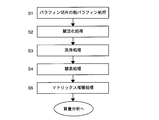
- Non-Patent Documents 1, 2, 3, and 4 In order to use a paraffin-embedded tissue section as a section sample for mass spectrometry, a pretreatment method as described in Non-Patent Documents 1, 2, 3, and 4 has been conventionally performed. A conventional pretreatment method will be described with reference to the chart of FIG. First, after removing paraffin from the paraffin-embedded tissue section, hydration is performed to obtain a deparaffinized tissue section (S1).
- the crosslinks are dissociated by heat treatment in the activation treatment liquid (S2: hereinafter referred to as “activation treatment”). Further, the activation treatment liquid adhering to the deparaffinized tissue section during the activation treatment is washed (S3). Thereafter, the deparaffinized tissue section after washing is treated with a digestive enzyme such as trypsin to hydrolyze the protein contained in the tissue section into a peptide (S4). Finally, the matrix solution is dropped onto the enzyme-treated deparaffinized tissue section to deposit the matrix (S5). The section sample thus prepared is subjected to MALDI mass spectrometry.
- the activation treatment not only restores the two-dimensional structure and three-dimensional structure of the protein molecule damaged by the cross-linking reaction associated with the formalin immobilization treatment, but also in the subsequent enzyme treatment. This process is indispensable for making digestive enzymes easy to approach protein molecules.
- heat treatment a hot tub, a microwave oven, an autoclave
- an activation treatment solution containing a surfactant a surfactant
- An object of the present invention is to provide an activation processing system and an activation processing method for a protein in a deparaffinized tissue section that can detect an intensity peak that does not come and improve the amount of information in image mapping.
- the protein activation treatment system in the deparaffinized tissue section according to the present invention made to solve the above problems, In order to mass-analyze the measurement area of the deparaffinized tissue section obtained by removing paraffin from the formalin-fixed paraffin-embedded tissue section that has been subjected to paraffin-embedding after formalin-fixation, formalin fixation in the protein molecules contained in the measurement area is performed.
- a protein activation treatment system for dissociating the cross-linking generated in step 1 and activating the protein molecule A dispensing unit that dispenses the activation treatment liquid into the entire dispensing region containing the measurement region of the deparaffinized tissue section; And a wet heat treatment part that heats the deparaffinized tissue section in which the activation treatment liquid has been dispensed in the dispensing region with saturated steam.
- the protein activation processing system in the deparaffinized tissue section immerses the deparaffinized tissue section in the activation processing liquid, which has been performed in the conventional activation processing, or is washed after the immersion. This processing is excluded.
- the activation treatment liquid contains a surfactant. If a surfactant is included in the activation treatment liquid, the affinity between the activation treatment liquid and the deparaffinized tissue section is increased. Therefore, the cross-linking caused by formalin fixation is sufficiently dissociated when wet heat treatment is performed. Because it can be done.
- the dispensing unit may dispense the activation treatment liquid at predetermined intervals so that the dispensing unit forms a plurality of droplets independent from each other in the dispensing region. .
- the volume of each droplet and the dispensing interval are set so that the dispensing region is covered with the plurality of droplets.
- the dispensing unit may dispense the activation treatment liquid a plurality of times to the same spot in the dispensing region.
- the activation treatment method of the protein in the deparaffinized tissue section according to the present invention made to solve the above problems, In order to mass-analyze the measurement area of the deparaffinized tissue section obtained by removing paraffin from the formalin-fixed paraffin-embedded tissue section that has been subjected to paraffin-embedding after formalin-fixation, formalin fixation in the protein molecules contained in the measurement area is performed.
- the activation treatment liquid in the dispensing step, may be dispensed at predetermined intervals so as to become a plurality of droplets independent from each other in the dispensing region.
- the volume of each droplet and the dispensing interval are set so that the dispensing region is covered with the plurality of droplets.
- the activation treatment liquid may be dispensed a plurality of times to the same spot in the dispensing region.
- the pH of the activation treatment liquid may be 9 or more.
- the activation processing liquid is deparaffinized tissue. Since the wet heat treatment is performed after dispensing into the sections, the biological components of the deparaffinized tissue sections are not lost by the immersion. In addition, since the washing process after the activation process, which was necessary in the past, is omitted, the biological components of the deparaffinized tissue section are not lost by the washing. Therefore, since the peak intensity reflecting the actual amount of the biological component is obtained, the peak intensity obtained in MALDI mass spectrometry is higher than the conventional one. This increases the number of peaks whose chemical structure is identified by qualitative analysis, and improves the detection limit in quantitative analysis. As a result, more accurate image mapping with an improved amount of information becomes possible.
- the figure which expanded the main detection peak obtained by the present Example and the comparative example A dark line shows a present Example and a thin line shows a comparative example.
- FIG. 3 is a main part configuration diagram of the activation processing system 1 according to an embodiment.
- the activation processing system 1 heats the dispensing part 10 for dispensing the activation treatment liquid to the deparaffinized tissue section 30 and the deparaffinized tissue section 30 into which the activation treatment liquid 31 has been dispensed under saturated steam. It is comprised by the wet heat processing part 20 for doing.
- the dispensing unit 10 includes a placement table 11 for placing the sample plate 34 on which the deparaffinized tissue section 30 is placed, a storage container (not shown) for storing the activation treatment liquid, and the activation treatment liquid.
- a dispenser 12 for dispensing is provided.
- the wet heat treatment unit 20 stores the sample plate 34 on which the deparaffinized tissue section 30 into which the activation treatment liquid 31 has been dispensed is placed in a pressure-resistant container, and the dehumidification process is performed with saturated steam at a predetermined temperature and time.
- the paraffin tissue section 30 is heated.
- a dispensing device capable of dispensing a small amount of solution to a designated region can be used, and in particular, a chemical capable of dispensing a picoliter (pL) unit solution.
- a printer CHIP-1000 manufactured by Shimadzu Corporation
- an autoclave apparatus can be used suitably as the wet heat treatment part 20.
- Dispensing unit 10 and wet heat treatment unit 20 may be separate from each other or may be integrated.
- the dispensing unit 10 can be used not only for dispensing the activation treatment liquid but also for applying a predetermined treatment liquid to the deparaffinized tissue section 30 in the subsequent enzyme treatment and matrix deposition treatment. .
- activation process method executed by the activation system 1
- the activation treatment was performed under a plurality of dispensing conditions in order to examine the optimum dispensing amount of the activation treatment liquid.
- a chemical printer CHIP-1000 was applied as the dispensing unit 10 of the activation processing system 1
- an autoclave KS-243 manufactured by Tommy Seiko Co., Ltd. was applied as the wet heat treatment unit 20.
- a formalin-fixed paraffin-embedded kidney tissue section (thickness 3 ⁇ m) obtained by slicing mouse kidney tissue after formalin-fixing and paraffin-embedding was attached to a conductive support and dried.
- the obtained formalin-fixed paraffin-embedded kidney tissue section was exposed to xylene at room temperature for 30 minutes to obtain a deparaffinized kidney tissue section 30 from which paraffin was removed.
- the deparaffinized kidney tissue section 30 was exposed for 10 seconds in order of 100% ethanol (twice), 90% ethanol, 80% ethanol, and 70% ethanol.
- the deparaffinized kidney tissue section 30 after hydration treatment is placed on a slide glass sample plate and set on a chemical printer CHIP-1000, and 10 mM Tris-HCl (pH 10.0) /0.1% n as an activation treatment solution.
- a storage container was filled with -octyl- ⁇ -D-glucoside solution.
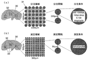
- the activation treatment liquid 31 was dispensed at intervals of 200 ⁇ m into a plurality of 1000 ⁇ m square dispensing regions 32 set in the deparaffinized kidney tissue section 30.
- the total dispensing volume was set to 11 conditions of 500pL, 1000pL, 2000pL, 2500pL, 3000pL, 3500pL, 4000pL, 4500pL, 5000pL, 7500pL and 10000pL per spot.
- the deparaffinized kidney tissue section 30 was placed in a stainless steel box together with wet paper material, and wet-heat treated at 110 ° C. for 10 minutes by an autoclave.
- the deparaffinized kidney tissue section 30 after the heat treatment was set again on the chemical printer CHIP-1000, and a storage container was filled with 100 ⁇ g / mL trypsin / 10 mM ammonium bicarbonate / 5% isopropanol solution as an enzyme treatment solution.
- Dispensing the deparaffinized kidney tissue section 30 by dispensing 5000 pL of the enzyme-treated solution to all spots where the activation treatment solution 31 has been dispensed, and allowing to stand at 37 ° C.
- the activation treatment liquid used in the activation treatment described above was prepared at pH 9.6. In this way, by inclining the pH of the activation treatment solution toward the basic side, in the subsequent enzyme treatment, the reaction rate of protein hydrolysis by trypsin having an optimum pH of weak basicity (pH 8-9) is increased. Can be improved.
- a mouse formalin-fixed paraffin-embedded kidney section was processed by the following method based on the conventional method. Autoclaving at 110 ° C. for 10 minutes in a state where a deparaffinized kidney tissue section 30 of a mouse subjected to deparaffinization and hydration in the same manner as in the above example was immersed in an activation treatment liquid having the same composition as in the above example Wet heat treatment. The deparaffinized kidney tissue section 30 after the wet heat treatment was taken out from the activation treatment liquid, and in order to remove the activation treatment liquid, the deparaffinization kidney tissue section 30 was immersed in 70% ethanol for 5 minutes and sufficiently washed. The enzyme treatment and matrix deposition treatment of the deparaffinized kidney tissue section 30 after washing were performed under the same conditions as in the above examples. The obtained section sample was subjected to MALDI mass spectrometry under the same measurement conditions as in the examples.
- MALDI mass spectrometry was performed under the following measurement conditions.
- a total of 121 points are irradiated per measurement region at intervals of 50 ⁇ m, and two shots are irradiated at each point.
- MALDI mass spectrometry was performed. After obtaining the peak intensity of each mass number in the measurement mass range, subtracting from the peak intensity, the minimum intensity of 100 peaks before and after each detection peak is regarded as the background of each detection peak, Correction was made by dividing by the sum of the intensities.
- the detection peak of this example was compared with the detection peak of the same mass number in the comparative example, and a detection peak having a detection intensity more than twice that of the comparative example and having statistical significance (p ⁇ 0.05) was selected.
- FIG. 5 is a graph showing the number of detected peaks for each dispensing amount of the activation treatment liquid in this example.
- the number of detected peaks shows a tendency to increase as the dispensed volume of the activation treatment liquid increases up to 1000pL of the activation treatment liquid.
- the number of detected peaks at the dispense volume of 1000pL is About twice as much.
- the number of detected peaks reached a peak at a dispensing amount of 1000 pL to 2500 pL.
- the activation treatment liquid having a dispensing amount of 500 pL cannot be said to have a sufficient volume for activating the protein in the dispensing region 32, but if the dispensing amount is 1000 pL or more, the dispensing region 32 It was shown that the inner protein can be sufficiently activated.
- the number of detected peaks tended to decrease rapidly when the dispensing amount of the activation treatment liquid exceeded 3000 pL. This is because, when the dispensing amount of the activation treatment liquid becomes a certain amount or more, the droplets of the activation treatment liquid formed on the deparaffinized kidney tissue section 30 are united, and the activation treatment liquid is dispensed in the dispensing region 32. It is considered that the biological components that have migrated into the activation treatment liquid are lost from within the dispensing region 32 by flowing out of the liquid.
- the dispensing volume of the activation treatment liquid per spot is 2500 pL and 5000 pL
- the deparaffinized kidney tissue section 30 after the dispensing is observed.
- the dispensing volume is 2500 pL
- the droplets of each spot it was confirmed that when the dispensing amount was 5000 pL, the droplets of adjacent spots merged and the activation treatment liquid flowed out of the dispensing region 32. According to these results, the activation treatment liquid can be dispensed as independent droplets up to a dispensing amount of 2500 pL per spot.
- FIG. 6 shows the mass when the activation treatment liquid is dispensed 2500 pL at a time in this embodiment (that is, when the maximum volume of the activation treatment liquid droplets is well maintained).
- FIG. 5 is a diagram showing a peak profile and a mass peak profile obtained in a comparative example in a total of three measurement ranges of m / z 750 to 1150, m / z 1150 to 1650, and m / z 1650 to 2250. It can be seen that the number of clear peaks in the example is larger than that in the comparative example as a whole, and the tendency is particularly remarkable in the mass range of m / z 1650 to 2250.
- FIG. 5 is a diagram showing a peak profile and a mass peak profile obtained in a comparative example in a total of three measurement ranges of m / z 750 to 1150, m / z 1150 to 1650, and m / z 1650 to 2250. It can be seen that the
- FIG. 7 is an enlarged view of typical detection peaks (m / z 1274, m / z 1443, m / z 1509, m / z 1529, m / z 1745, and m / z 1961) in the mass peak profile of FIG.
- the dark line shows the detection peak obtained in this example in which the activation treatment solution was dispensed in 2500 pL, and the thin line shows the detection peak obtained in the comparative example.
- the detection peak of this example showed a peak intensity more than twice that of the comparative example.
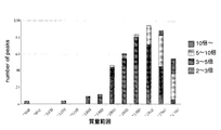
- FIG. 8 shows twice the detection peak of the same mass number in the comparative example among the detection peaks obtained when MALDI mass spectrometry was performed on the section sample obtained by dispensing 2500 pL of the activation treatment liquid in this example.
- the specific peak intensity ratios of a total of 461 detection peaks showing the above peak intensity ratios are shown for each mass range. It was recognized that the peak intensity ratio tended to increase as the detection peak having a larger mass number.
- 182 (39.5%) were detected with a peak intensity 2 to 3 times higher than that of the comparative example, and 149 (32.3%) were detected 3 to 5 times higher.
- the number of detection peaks improved 5 to 10 times was 104 (22.6%), and the number of detection peaks improved 10 times or more was 26 (5.6%).
- the number of detection peaks at the time of MALDI mass spectrometry is increased and the detection sensitivity is higher than when the activation processing is performed by the conventional method. Will also improve. This is thought to be due to the fact that in the present invention, the conventional treatment of immersion in the activation treatment liquid and subsequent washing is omitted, thereby avoiding the loss of biological components from the deparaffinized tissue section. In addition, during the wet heat treatment, it is expected that the biological components and the molecules of the activation treatment liquid frequently collide with each other in the small inside of the droplets of the activation treatment liquid. It is considered that the detection sensitivity is improved by being promoted.
Landscapes
- Life Sciences & Earth Sciences (AREA)
- Health & Medical Sciences (AREA)
- Engineering & Computer Science (AREA)
- Molecular Biology (AREA)
- Physics & Mathematics (AREA)
- Biomedical Technology (AREA)
- Chemical & Material Sciences (AREA)
- Immunology (AREA)
- Hematology (AREA)
- Urology & Nephrology (AREA)
- General Physics & Mathematics (AREA)
- General Health & Medical Sciences (AREA)
- Pathology (AREA)
- Biochemistry (AREA)
- Analytical Chemistry (AREA)
- Bioinformatics & Cheminformatics (AREA)
- Bioinformatics & Computational Biology (AREA)
- Medicinal Chemistry (AREA)
- Food Science & Technology (AREA)
- Microbiology (AREA)
- Cell Biology (AREA)
- Biotechnology (AREA)
- Proteomics, Peptides & Aminoacids (AREA)
- Spectroscopy & Molecular Physics (AREA)
- Biophysics (AREA)
- Other Investigation Or Analysis Of Materials By Electrical Means (AREA)
- Sampling And Sample Adjustment (AREA)
- Investigating Or Analysing Biological Materials (AREA)
Abstract
Description
まず、パラフィン包埋組織切片からパラフィンを除去した後、水和処理を施して脱パラフィン組織切片を得る(S1)。このようにして得られた脱パラフィン組織切片に含まれるタンパク質の分子内では、ホルマリン固定化処理の際に単一アミノ酸残基の結合、ヒドロメチル基によるメチレン架橋の形成、特に一級アミノ基やフェニル基を介して強固なメチレン架橋が生じているので、賦活化処理液中で加熱処理をすることで架橋を解離する(S2:以下、これを「賦活化処理」という)。さらに、賦活化処理の際に脱パラフィン組織切片に付着した賦活化処理液を洗浄する(S3)。その後、洗浄後の脱パラフィン組織切片をトリプシン等の消化酵素で処理し、該組織切片に含まれるタンパク質をペプチドに加水分解する(S4)。最後に、酵素処理された脱パラフィン組織切片にマトリックス溶液を滴下してマトリックスを堆積させる(S5)。こうして作製された切片試料がMALDI質量分析に供される。
ホルマリン固定処理後にパラフィン包埋処理が施されたホルマリン固定パラフィン包埋組織切片からパラフィンを取り除いた脱パラフィン組織切片の測定領域を質量分析するために、当該測定領域に含まれるタンパク質分子内のホルマリン固定で生じた架橋を解離して該タンパク質分子を賦活化させるタンパク質賦活化処理システムであって、
賦活化処理液を前記脱パラフィン組織切片の前記測定領域を内包する分注領域内の全体に分注する分注部と、
前記分注領域に前記賦活化処理液が分注された前記脱パラフィン組織切片を飽和水蒸気により加熱する湿熱処理部と
を備えることを特徴とする。
ホルマリン固定処理後にパラフィン包埋処理が施されたホルマリン固定パラフィン包埋組織切片からパラフィンを取り除いた脱パラフィン組織切片の測定領域を質量分析するために、当該測定領域に含まれるタンパク質分子内のホルマリン固定で生じた架橋を解離して該タンパク質分子を賦活化させるタンパク質賦活化処理方法であって、
賦活化処理液を前記脱パラフィン組織切片の前記測定領域を内包する分注領域内の全体に分注する分注工程と、
前記分注領域に前記賦活化処理液が分注された前記脱パラフィン組織切片を飽和水蒸気により加熱する湿熱処理工程と
を有することを特徴とする。
分注部10は、脱パラフィン組織切片30を載せたサンプルプレート34を載置するための載置台11と、賦活化処理液を格納する貯液容器(図示せず)と、賦活化処理液を分注するための分注器12を備えている。
湿熱処理部20は、賦活化処理液31が分注された脱パラフィン組織切片30を載せたサンプルプレート34を、耐圧性を有する容器内に格納し、所定の温度及び時間で飽和水蒸気により前記脱パラフィン組織切片30を加熱するものである。
分注部10と湿熱処理部20とは互いに別体であっても良いし、一体であっても良い。 また、分注部10は、賦活化処理液の分注だけでなく、その後の酵素処理及びマトリックス堆積処理において、所定の処理液を脱パラフィン組織切片30に付与する際にも使用することができる。
本実施例では、賦活化処理システム1の分注部10としてケミカルプリンタCHIP-1000を適用し、また湿熱処理部20としてオートクレーブKS-243(株式会社トミー精工製)を適用した。
水和処理後の脱パラフィン腎臓組織切片30をスライドグラス用サンプルプレートに載置し、ケミカルプリンタCHIP-1000にセットするとともに、賦活化処理液として10mM Tris-HCl(pH10.0)/0.1%n-オクチル-β-D-グルコシド溶液を貯液容器に充填した。さらに、図4(a)に示すように、前記脱パラフィン腎臓組織切片30に複数個設定した1000μm四方の分注領域32に、200μmの間隔で賦活化処理液31を分注した。即ち、1つの分注領域32の一辺に200μmの間隔で計6スポットを設定し、6スポット×6スポットの計36スポットに賦活化処理液31を分注した。
至適分注量を検討するために、分注量は1スポットあたり合計500pL、1000pL、2000pL、2500pL、3000pL、3500pL、4000pL、4500pL、5000pL、7500pL及び10000pLの計11条件とした。スポット上に1滴100pLを当該量分、滴下することで(即ち、1スポットに合計2500pLを分注したいときは、100pLを25滴、組織上に滴下することで)、目的の体積の賦活化処理液を分注した。
次に、湿熱処理後の脱パラフィン腎臓組織切片30をケミカルプリンタCHIP-1000に再びセットし、酵素処理液として100μg/mLトリプシン/10mM重炭酸アンモニウム/5%イソプロパノール溶液を貯液容器に充填した。賦活化処理液31を分注した全てのスポットに、前記酵素処理液を合計5000pLずつ分注し、恒温庫にて37℃で180分間静置することにより、脱パラフィン腎臓組織切片30の分注領域32に含まれるタンパク質を加水分解した。さらに、マトリックス溶液として50mg/mL 2,5-ジヒドロキシ安息香酸/50%アセトニトリル/0.1%トリフルオロ酢酸溶液を貯液容器に充填し、150μmの間隔で合計7500pLずつ分注して、2,5-ジヒドロキシ安息香酸を脱パラフィン腎臓組織切片30に堆積させた。こうして得られた切片試料をMALDI質量分析へ供した。MALDI質量分析の測定条件については後述する。
上記実施例と同じ方法で脱パラフィン処理及び水和処理を施したマウスの脱パラフィン腎臓組織切片30を、上記実施例と同じ組成の賦活化処理液に浸漬した状態で、110℃で10分間オートクレーブにて湿熱処理した。湿熱処理後の脱パラフィン腎臓組織切片30を賦活化処理液から取り出し、賦活化処理液を取り除くために、前記脱パラフィン腎臓組織切片30を70%エタノールで5分間浸漬し、十分に洗浄した。洗浄後の脱パラフィン腎臓組織切片30の酵素処理及びマトリックス堆積処理については上記実施例と同じ条件にて実施した。得られた切片試料について、実施例と同じ測定条件にてMALDI質量分析を行った。
装置:マトリックス支援レーザー脱離イオン化一飛行時間型質量分析計 AXIMA-QIT(株式会社島津製作所製)
レーザー:5Hz、2 shots/point
照射間隔:50μm
イオン極性:positive
測定質量領域モード:MID MASS
測定質量範囲:m/z 700-2500
測定領域:500μm×500μm
測定ポイント:11×11=121 points
上記測定質量範囲において各質量数のピーク強度を獲得し、各検出ピークの前後100ピークずつの最小強度を各検出ピークのバックグラウンドとみなして前記ピーク強度から差し引いた後に、各ピーク強度を全ピーク強度の総和で除する補正を行った。本実施例の検出ピークを比較例における同じ質量数の検出ピークと比較し、比較例に対し2倍以上の検出強度があり、かつ統計的意義(p<0.05)のある検出ピークを選出した。
まず、図5は、本実施例において賦活化処理液の分注量毎の検出ピーク数を示したグラフである。検出ピーク数は、賦活化処理液の分注量1000pLまでは、賦活化処理液の分注量が大きくなるにつれて増加する傾向を示し、分注量1000pLにおける検出ピーク数は分注量500pLの時の約2倍であった。また、検出ピーク数は分注量1000pLから2500pLにおいて頭打ちとなった。このことから、分注量500pLの賦活化処理液は、分注領域32内のタンパク質を賦活化する上で十分な体積とはいえないが、分注量1000pL以上であれば、分注領域32内のタンパク質を十分に賦活化できることが示された。
その一方、検出ピーク数は、賦活化処理液の分注量が3000pLを超えると急激に減少する傾向を示した。これは、賦活化処理液の分注量がある一定以上になると、脱パラフィン腎臓組織切片30に形成された賦活化処理液の液滴が合体してしまい、賦活化処理液が分注領域32の外へ流出することにより、賦活化処理液中に移行した生体成分が分注領域32内から失われるためと考えられる。
これらの結果によれば、賦活化処理液を独立した液滴として分注できるのは、1スポットあたりの分注量2500pLまでということができる。
また、図7は、図6のマスピークプロファイルのうち代表的な検出ピーク(m/z1274、m/z1443、m/z1509、m/z1529、m/z1745及びm/z1961)を拡大したものであり、濃い線は賦活化処理液を2500pLずつ分注した本実施例において、また薄い線は比較例において得られた検出ピークを示す。いずれの質量数においても、本実施例の検出ピークは比較例の検出ピークの2倍以上のピーク強度を示した。
10…分注部
11…載置台
12…分注器
20…湿熱処理部
30…脱パラフィン組織切片
31…賦活化処理液
32…分注領域
33…測定領域
34…サンプルプレート
Claims (9)
- ホルマリン固定処理後にパラフィン包埋処理が施されたホルマリン固定パラフィン包埋組織切片からパラフィンを取り除いた脱パラフィン組織切片の測定領域を質量分析するために、当該測定領域に含まれるタンパク質分子内のホルマリン固定で生じた架橋を解離して該タンパク質分子を賦活化させるタンパク質賦活化処理システムであって、
賦活化処理液を前記脱パラフィン組織切片の前記測定領域を内包する分注領域内の全体に分注する分注部と、
前記分注領域に前記賦活化処理液が分注された前記脱パラフィン組織切片を飽和水蒸気により加熱する湿熱処理部と
を備えることを特徴とするタンパク質賦活化処理システム。 - 前記分注部が、前記分注領域内において互いに独立した複数の液滴となるように前記賦活化処理液を所定の間隔で分注する
ことを特徴とする請求項1に記載のタンパク質賦活化処理システム。 - 前記分注領域が前記複数の液滴で覆われるように、各液滴の体積及び分注間隔が設定されていることを特徴とする請求項2に記載のタンパク質賦活化処理システム。
- 前記分注部が、前記賦活化処理液を前記分注領域内の同一のスポットに複数回分注する
ことを特徴とする請求項1から請求項3のいずれかに記載のタンパク質賦活化処理システム。 - ホルマリン固定処理後にパラフィン包埋処理が施されたホルマリン固定パラフィン包埋組織切片からパラフィンを取り除いた脱パラフィン組織切片の測定領域を質量分析するために、当該測定領域に含まれるタンパク質分子内のホルマリン固定で生じた架橋を解離して該タンパク質分子を賦活化させるタンパク質賦活化処理方法であって、
賦活化処理液を前記脱パラフィン組織切片の前記測定領域を内包する分注領域内の全体に分注する分注工程と、
前記分注領域に前記賦活化処理液が分注された前記脱パラフィン組織切片を飽和水蒸気により加熱する湿熱処理工程と
を有することを特徴とするタンパク質賦活化処理方法。 - 前記分注工程において、前記賦活化処理液が前記分注領域内において互いに独立した複数の液滴となるように所定の間隔で分注されることを特徴とする、請求項5に記載のタンパク質賦活化処理方法。
- 前記分注領域が前記複数の液滴で覆われるように、各液滴の体積及び分注間隔が設定されていることを特徴とする、請求項6に記載のタンパク質賦活化処理方法。
- 前記分注工程において、前記賦活化処理液が前記分注領域内の同一のスポットに複数回分注されることを特徴とする、請求項5から請求項7のいずれかに記載のタンパク質賦活化処理方法。
- 前記賦活化処理液のpHが9以上であることを特徴とする、請求項5から請求項8のいずれかに記載のタンパク質賦活化処理方法。
Priority Applications (3)
| Application Number | Priority Date | Filing Date | Title |
|---|---|---|---|
| US14/119,639 US20140220624A1 (en) | 2011-05-25 | 2012-04-25 | System and method for retrieval treatment of proteins in formalin-fixed paraffin-embedded tissue section |
| JP2013516264A JP5775156B2 (ja) | 2011-05-25 | 2012-04-25 | ホルマリン固定パラフィン包埋組織切片中のタンパク質の賦活化処理システム及び賦活化処理方法 |
| US14/978,115 US10018539B2 (en) | 2011-05-25 | 2015-12-22 | System and method for retrieval treatment of proteins in formalin-fixed paraffin-embedded tissue section |
Applications Claiming Priority (2)
| Application Number | Priority Date | Filing Date | Title |
|---|---|---|---|
| JP2011-117334 | 2011-05-25 | ||
| JP2011117334 | 2011-05-25 |
Related Child Applications (2)
| Application Number | Title | Priority Date | Filing Date |
|---|---|---|---|
| US14/119,639 A-371-Of-International US20140220624A1 (en) | 2011-05-25 | 2012-04-25 | System and method for retrieval treatment of proteins in formalin-fixed paraffin-embedded tissue section |
| US14/978,115 Division US10018539B2 (en) | 2011-05-25 | 2015-12-22 | System and method for retrieval treatment of proteins in formalin-fixed paraffin-embedded tissue section |
Publications (1)
| Publication Number | Publication Date |
|---|---|
| WO2012160934A1 true WO2012160934A1 (ja) | 2012-11-29 |
Family
ID=47217017
Family Applications (1)
| Application Number | Title | Priority Date | Filing Date |
|---|---|---|---|
| PCT/JP2012/061090 Ceased WO2012160934A1 (ja) | 2011-05-25 | 2012-04-25 | ホルマリン固定パラフィン包埋組織切片中のタンパク質の賦活化処理システム及び賦活化処理方法 |
Country Status (3)
| Country | Link |
|---|---|
| US (2) | US20140220624A1 (ja) |
| JP (1) | JP5775156B2 (ja) |
| WO (1) | WO2012160934A1 (ja) |
Cited By (1)
| Publication number | Priority date | Publication date | Assignee | Title |
|---|---|---|---|---|
| JP2017502272A (ja) * | 2013-12-12 | 2017-01-19 | ユニバーシティー オブ ヒューストン システム | 普遍的な抗原の賦活化化合物とその使用の方法 |
Families Citing this family (2)
| Publication number | Priority date | Publication date | Assignee | Title |
|---|---|---|---|---|
| CN110864939A (zh) * | 2018-08-27 | 2020-03-06 | 深圳大学 | 一种蛋白质提取方法和应用 |
| JP7215467B2 (ja) * | 2019-08-19 | 2023-01-31 | 株式会社島津製作所 | ホルマリン固定パラフィン包埋組織試料からの抗体の分離、検出及び/又は分析方法 |
Citations (4)
| Publication number | Priority date | Publication date | Assignee | Title |
|---|---|---|---|---|
| JP2004347594A (ja) * | 2003-04-30 | 2004-12-09 | Shimadzu Corp | 生体標本への試薬の分注方法及び生体標本の解析方法 |
| JP2007514951A (ja) * | 2003-12-16 | 2007-06-07 | スリーエム イノベイティブ プロパティズ カンパニー | 化学的に架橋された細胞サンプルの分析 |
| WO2008038813A1 (en) * | 2006-09-28 | 2008-04-03 | Shimadzu Corporation | Method for deparaffinization of paraffin-embedded specimen and method for analysis of paraffin-embedded specimen |
| JP2008542724A (ja) * | 2005-05-25 | 2008-11-27 | エクスプレッション、パソロジー、インコーポレイテッド | 組織病理学的に処理した生物学的試料からのプロテオームの適用範囲を増加させるための、液体組織標品を用いたマルチプレックス方法 |
Family Cites Families (2)
| Publication number | Priority date | Publication date | Assignee | Title |
|---|---|---|---|---|
| US20040219588A1 (en) | 2003-04-30 | 2004-11-04 | Masaru Furuta | Method for dispensing reagents onto biological samples and method for analyzing biological samples |
| WO2006066039A2 (en) * | 2004-12-17 | 2006-06-22 | Kram Brian H | High temperature tissue conditioning with low volatility solutions and applications |
-
2012
- 2012-04-25 WO PCT/JP2012/061090 patent/WO2012160934A1/ja not_active Ceased
- 2012-04-25 US US14/119,639 patent/US20140220624A1/en not_active Abandoned
- 2012-04-25 JP JP2013516264A patent/JP5775156B2/ja active Active
-
2015
- 2015-12-22 US US14/978,115 patent/US10018539B2/en active Active
Patent Citations (4)
| Publication number | Priority date | Publication date | Assignee | Title |
|---|---|---|---|---|
| JP2004347594A (ja) * | 2003-04-30 | 2004-12-09 | Shimadzu Corp | 生体標本への試薬の分注方法及び生体標本の解析方法 |
| JP2007514951A (ja) * | 2003-12-16 | 2007-06-07 | スリーエム イノベイティブ プロパティズ カンパニー | 化学的に架橋された細胞サンプルの分析 |
| JP2008542724A (ja) * | 2005-05-25 | 2008-11-27 | エクスプレッション、パソロジー、インコーポレイテッド | 組織病理学的に処理した生物学的試料からのプロテオームの適用範囲を増加させるための、液体組織標品を用いたマルチプレックス方法 |
| WO2008038813A1 (en) * | 2006-09-28 | 2008-04-03 | Shimadzu Corporation | Method for deparaffinization of paraffin-embedded specimen and method for analysis of paraffin-embedded specimen |
Cited By (1)
| Publication number | Priority date | Publication date | Assignee | Title |
|---|---|---|---|---|
| JP2017502272A (ja) * | 2013-12-12 | 2017-01-19 | ユニバーシティー オブ ヒューストン システム | 普遍的な抗原の賦活化化合物とその使用の方法 |
Also Published As
| Publication number | Publication date |
|---|---|
| US20160153872A1 (en) | 2016-06-02 |
| US10018539B2 (en) | 2018-07-10 |
| US20140220624A1 (en) | 2014-08-07 |
| JP5775156B2 (ja) | 2015-09-09 |
| JPWO2012160934A1 (ja) | 2014-07-31 |
Similar Documents
| Publication | Publication Date | Title |
|---|---|---|
| Schwartz et al. | Direct tissue analysis using matrix‐assisted laser desorption/ionization mass spectrometry: practical aspects of sample preparation | |
| Aerni et al. | Automated acoustic matrix deposition for MALDI sample preparation | |
| Kaletaş et al. | Sample preparation issues for tissue imaging by imaging MS | |
| Norris et al. | Analysis of tissue specimens by matrix-assisted laser desorption/ionization imaging mass spectrometry in biological and clinical research | |
| EP2063265B1 (en) | Method of preparing sample for matrix-assisted laser desorption ionization mass spectrometry and matrix-assisted laser desorption ionization mass spectrometry | |
| Angel et al. | In situ imaging of tryptic peptides by MALDI imaging mass spectrometry using fresh‐frozen or formalin‐fixed, paraffin‐embedded tissue | |
| Paine et al. | Digestion-free analysis of peptides from 30-year-old formalin-fixed, paraffin-embedded tissue by mass spectrometry imaging | |
| AU2007300996B2 (en) | Method for deparaffinization of paraffin-embedded specimen and method for analysis of paraffin-embedded specimen | |
| Wisztorski et al. | MALDI direct analysis and imaging of frozen versus FFPE tissues: what strategy for which sample? | |
| JP2007170870A (ja) | 質量分析を用いたinsitu検出方法 | |
| US20140357526A1 (en) | Hydrogel-mediated tissue analysis | |
| O'Rourke et al. | A versatile cost‐effective method for the analysis of fresh frozen tissue sections via matrix‐assisted laser desorption/ionisation imaging mass spectrometry | |
| Piwowar et al. | Effects of cryogenic sample analysis on molecular depth profiles with TOF-secondary ion mass spectrometry | |
| Lu et al. | Soft picosecond infrared laser extraction of highly charged proteins and peptides from bulk liquid water for mass spectrometry | |
| JP5775156B2 (ja) | ホルマリン固定パラフィン包埋組織切片中のタンパク質の賦活化処理システム及び賦活化処理方法 | |
| Dave et al. | Preparation and analysis of proteins and peptides using MALDI TOF/TOF mass spectrometry | |
| Wang et al. | MALDI MS sample preparation by using paraffin wax film: systematic study and application for peptide analysis | |
| Zhou et al. | Plasmonic thermal decomposition/digestion of proteins: a rapid on-surface protein digestion technique for mass spectrometry imaging | |
| US11614450B2 (en) | Mass spectrometry histochemistry of peptides from formaldehyde-fixed, paraffin-embedded tissue | |
| US20050112706A1 (en) | Diagnostic and prognostic methods for prostate cancers | |
| Huang et al. | Molecular imaging of drug‐eluting coronary stents: method development, optimization and selected applications | |
| US10996227B2 (en) | Pre-coated surfaces for imaging biomolecules | |
| Shrestha | Introduction to spatial mapping of biomolecules by imaging mass spectrometry | |
| Lawal | Deep-Ultraviolet Laser Ablation for Biomolecular Analysis | |
| Chicano-Galvez et al. | MALDI mass spectrometry imaging and spatially-resolved proteomics |
Legal Events
| Date | Code | Title | Description |
|---|---|---|---|
| 121 | Ep: the epo has been informed by wipo that ep was designated in this application |
Ref document number: 12789053 Country of ref document: EP Kind code of ref document: A1 |
|
| ENP | Entry into the national phase |
Ref document number: 2013516264 Country of ref document: JP Kind code of ref document: A |
|
| NENP | Non-entry into the national phase |
Ref country code: DE |
|
| WWE | Wipo information: entry into national phase |
Ref document number: 14119639 Country of ref document: US |
|
| 122 | Ep: pct application non-entry in european phase |
Ref document number: 12789053 Country of ref document: EP Kind code of ref document: A1 |