WO2012153815A1 - 三次元皮膚モデルの製造方法 - Google Patents
三次元皮膚モデルの製造方法 Download PDFInfo
- Publication number
- WO2012153815A1 WO2012153815A1 PCT/JP2012/062053 JP2012062053W WO2012153815A1 WO 2012153815 A1 WO2012153815 A1 WO 2012153815A1 JP 2012062053 W JP2012062053 W JP 2012062053W WO 2012153815 A1 WO2012153815 A1 WO 2012153815A1
- Authority
- WO
- WIPO (PCT)
- Prior art keywords
- model
- epidermis
- dermis
- dimensional skin
- skin model
- Prior art date
- Legal status (The legal status is an assumption and is not a legal conclusion. Google has not performed a legal analysis and makes no representation as to the accuracy of the status listed.)
- Ceased
Links
Images




Classifications
-
- C—CHEMISTRY; METALLURGY
- C12—BIOCHEMISTRY; BEER; SPIRITS; WINE; VINEGAR; MICROBIOLOGY; ENZYMOLOGY; MUTATION OR GENETIC ENGINEERING
- C12N—MICROORGANISMS OR ENZYMES; COMPOSITIONS THEREOF; PROPAGATING, PRESERVING, OR MAINTAINING MICROORGANISMS; MUTATION OR GENETIC ENGINEERING; CULTURE MEDIA
- C12N5/00—Undifferentiated human, animal or plant cells, e.g. cell lines; Tissues; Cultivation or maintenance thereof; Culture media therefor
- C12N5/06—Animal cells or tissues; Human cells or tissues
- C12N5/0697—Artificial constructs associating cells of different lineages, e.g. tissue equivalents
- C12N5/0698—Skin equivalents
-
- A—HUMAN NECESSITIES
- A61—MEDICAL OR VETERINARY SCIENCE; HYGIENE
- A61L—METHODS OR APPARATUS FOR STERILISING MATERIALS OR OBJECTS IN GENERAL; DISINFECTION, STERILISATION OR DEODORISATION OF AIR; CHEMICAL ASPECTS OF BANDAGES, DRESSINGS, ABSORBENT PADS OR SURGICAL ARTICLES; MATERIALS FOR BANDAGES, DRESSINGS, ABSORBENT PADS OR SURGICAL ARTICLES
- A61L27/00—Materials for grafts or prostheses or for coating grafts or prostheses
- A61L27/50—Materials characterised by their function or physical properties, e.g. injectable or lubricating compositions, shape-memory materials, surface modified materials
- A61L27/60—Materials for use in artificial skin
-
- C—CHEMISTRY; METALLURGY
- C12—BIOCHEMISTRY; BEER; SPIRITS; WINE; VINEGAR; MICROBIOLOGY; ENZYMOLOGY; MUTATION OR GENETIC ENGINEERING
- C12N—MICROORGANISMS OR ENZYMES; COMPOSITIONS THEREOF; PROPAGATING, PRESERVING, OR MAINTAINING MICROORGANISMS; MUTATION OR GENETIC ENGINEERING; CULTURE MEDIA
- C12N2502/00—Coculture with; Conditioned medium produced by
- C12N2502/09—Coculture with; Conditioned medium produced by epidermal cells, skin cells, oral mucosa cells
- C12N2502/094—Coculture with; Conditioned medium produced by epidermal cells, skin cells, oral mucosa cells keratinocytes
-
- C—CHEMISTRY; METALLURGY
- C12—BIOCHEMISTRY; BEER; SPIRITS; WINE; VINEGAR; MICROBIOLOGY; ENZYMOLOGY; MUTATION OR GENETIC ENGINEERING
- C12N—MICROORGANISMS OR ENZYMES; COMPOSITIONS THEREOF; PROPAGATING, PRESERVING, OR MAINTAINING MICROORGANISMS; MUTATION OR GENETIC ENGINEERING; CULTURE MEDIA
- C12N2502/00—Coculture with; Conditioned medium produced by
- C12N2502/13—Coculture with; Conditioned medium produced by connective tissue cells; generic mesenchyme cells, e.g. so-called "embryonic fibroblasts"
- C12N2502/1305—Adipocytes
-
- C—CHEMISTRY; METALLURGY
- C12—BIOCHEMISTRY; BEER; SPIRITS; WINE; VINEGAR; MICROBIOLOGY; ENZYMOLOGY; MUTATION OR GENETIC ENGINEERING
- C12N—MICROORGANISMS OR ENZYMES; COMPOSITIONS THEREOF; PROPAGATING, PRESERVING, OR MAINTAINING MICROORGANISMS; MUTATION OR GENETIC ENGINEERING; CULTURE MEDIA
- C12N2502/00—Coculture with; Conditioned medium produced by
- C12N2502/13—Coculture with; Conditioned medium produced by connective tissue cells; generic mesenchyme cells, e.g. so-called "embryonic fibroblasts"
- C12N2502/1323—Adult fibroblasts
Definitions
- the present invention relates to a method for producing a three-dimensional skin model into which fat cells are introduced, and a three-dimensional skin model obtained by the production method.
- Keratinocytes and fibroblasts are the main constituent cells of the epidermis and dermis, respectively.
- the above three-dimensional skin model lacks a corresponding structure for subcutaneous tissue mainly composed of fat cells. Become.
- adiponectin released from fat cells inhibits IL-6 production from the epidermis (Ajuwon KM, AJP May, 2005, 288 (5) pp.1220-5) and also adipocytes
- the factor leptin is known to promote collagen production (EzurezT. Et al.,. Biofactors. 2007; 31 (3-4): pp.229-36)).
- IL-1 released from fibroblasts and keratinocytes inhibits PPAR ⁇ (peroxisome proliferator-activated receptor ⁇ ) and suppresses the progression of adipocyte differentiation (Suzawa M et al., Nat Cell Biol.
- adiponectin Chandrasekar B et al., February 15, 2008, J. Biol. Chem. 283) (7): pp.4200-9.
- adipocytes and fibroblasts and keratinocytes are correlated with each other. Therefore, it is considered necessary to introduce fat cells into a conventional skin model in order to obtain a three-dimensional skin model having a structure closer to the skin.
- the present invention provides a method for producing a three-dimensional skin model having a structure close to that of human skin and allowing each cell to interact in order to evaluate the effects of candidate drugs such as cosmetics in vitro.
- the task is to do.
- this application includes the following inventions: [1] A method of manufacturing a three-dimensional skin model, 1) A step of preparing an epidermis / dermis model in which fibroblasts and keratinocytes are laminated in order from the bottom on a support, 2) culturing preadipocytes to differentiate and / or proliferate to a desired extent; 3) placing the epidermis / dermis model on the fat cells; Comprising a method, [2] The method for producing a three-dimensional skin model according to [1], wherein the support is a gel containing collagen and chitosan.
- the epidermis / dermis model is formed by seeding fibroblasts on a support and culturing for a predetermined period, then seeding keratinocytes on the fibroblasts, culturing for a predetermined period, and exposing to air.
- a three-dimensional skin model including an epidermis / dermis model and an adipocyte layer 1) A step of preparing an epidermis / dermis model in which fibroblasts and keratinocytes are laminated in order from the bottom on a support, 2) culturing preadipocytes to differentiate and / or proliferate to a desired extent; 3) placing the epidermis / dermis model on the fat cells;
- the epidermis / dermis model is formed by seeding fibroblasts on a support and culturing for a predetermined period, then seeding keratinocytes on the fibroblasts, culturing for a predetermined period, and exposing to air.
- the introduction of adipocytes reduces the production level of interleukin-6 (IL-6), promotes the differentiation of keratinocytes, and expresses the collagen gene. Stabilization of the epidermis / dermis model was confirmed, including increased levels. On the other hand, the presence of an epidermis / dermis model also affects adipocytes. For example, the expression level of adipocyte differentiation markers was settled, suggesting the possibility that the differentiation state was stabilized. Furthermore, it has been clarified that the three-dimensional skin model behaves like actual skin when irradiated with ultraviolet rays.
- IL-6 interleukin-6
- a three-dimensional skin model closer to the actual human skin configuration in which the constituent cells interact with each other can be obtained. It is done.
- the three-dimensional skin model it becomes possible to evaluate the efficacy or toxicity of a drug in vitro without relying on animal experiments.
- the timing of combining the adipocytes with the epidermis / dermis model can be appropriately adjusted according to the purpose of use, It is also possible to apply the obtained three-dimensional skin model to the study of the differentiation / proliferation stage of adipocytes.
- FIG. 1 is a diagram showing an example of a procedure for creating a three-dimensional model skin of the present invention.
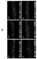
- FIG. 2 is a photograph of immunostaining of a three-dimensional skin model with anti-laminin 5 antibody.
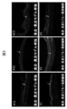
- FIG. 3 is a photograph of immunostaining of a three-dimensional skin model with an anti-transglutaminase antibody.
- FIG. 4 is an immunostaining photograph of a three-dimensional skin model with an anti-filaggrin antibody.
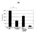
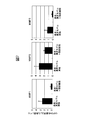
- FIG. 5 is a graph showing changes in IL-6 production level due to introduction of adipocytes into the epidermis / dermis model.
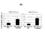
- FIG. 6 is a graph showing changes in the expression of collagen genes due to introduction of adipocytes into the epidermis / dermis model.
- FIG. 7 is a graph showing changes in the expression of the MMP gene due to the introduction of adipocytes into the epidermis / dermis model.
- FIG. 8 is a graph showing changes in mRNA levels of adiponectin by combining adipocytes and epidermis / dermis models, measured using quantitative RT-PCR.
- FIG. 9 is a graph showing changes in the production level of adiponectin by combining adipocytes and the epidermis / dermis model, measured using ELISA.
- Epidermis / dermis model The three-dimensional skin model produced by the present invention includes an epidermis / dermis model imitating the epidermis / dermis and an adipocyte layer.
- epidermis / dermis model means a structure having a structure in which fibroblasts are laminated on a support such as a collagen gel and keratinocytes are laminated on the fibroblasts. To do.
- a culture medium may be interposed between the epidermis / dermis model and the fat cell layer.
- Such epidermis / dermis models are known to those skilled in the art (for example, AmanomanS. Et al., Exp. Cell Res., Vol.271, pp.249-262, 2001 (above) and Tsungega M. et al. , Matrix. Biol., Vol.17, pp.603-613, 1998 (see above)).
- a mixture of fibroblasts mixed with a support on an insert mesh is seeded. After that, keratinocytes may be seeded thereon, cultured, and exposed to air.
- the support used may be a collagen-only gel or a gel containing collagen and chitosan.
- a gel containing collagen and chitosan from the viewpoint of basement membrane structure and dermal fiber formation.
- an epidermis / dermis model having a uniform shape in which the gel is not shrunk it is possible to prevent variation among lots when a plurality of similar three-dimensional skin models are created.
- the epidermis / dermis model contains, for example, fibroblasts in an amount of 1 ⁇ 10 4 to 10 8 cells / cm 2 , preferably 0.1 to 10 ⁇ 10 5 cells / cm 2 , and keratinocytes 1 ⁇ 10 2 to It may be contained in an amount of 10 6 pieces / cm 2 , preferably 1.0 to 10 ⁇ 10 4 pieces / cm 2 , more preferably about 4 to 8 ⁇ 10 4 pieces / cm 2 .
- the epidermis / dermis model is commercially available and is not particularly limited.
- a cell culture insert such as TESTSKIN (registered trademark) (TOYOBO) can be used.
- the culture of the epidermis / dermis model is carried out using, for example, a culture solution used for normal keratinocyte culture as a culture solution, for example, KG medium, Epilife KG2 (Kurabo), Humedia-KG2 (Kurabo), assay medium (TOYOBO), etc. Can be carried out over 0-14 days.
- a DMEM medium GEBCO
- a medium in which 2-0-aD-glucopyranosyl-L-ascorbic acid-containing KGM and DMEM are mixed 1: 1 can be used.
- the medium may be removed when combining the epidermis / dermis model and the fat cell layer.
- the medium for culturing the epidermis / dermis model may be interposed between the epidermis / dermis model and the fat cell layer.
- Fibroblasts and keratinocytes may be homologous or heterologous and may be derived from any mammal.
- the cells constituting the epidermis / dermis model may be those subjected to ultraviolet rays, drugs, or genetic modification.
- the keratinocytes are preferably derived from humans, although not limited thereto, from the viewpoint of bringing the properties of the three-dimensional skin model closer to that of human skin. Further, the ratio of fibroblasts to keratinocytes used is not particularly limited.
- keratinocytes 4 to 8 ⁇ 10 4 to 6 cells but fibroblasts 1 ⁇ 10 5 to 7 cells, preferably keratinocytes 4 Fibroblasts can be 1 ⁇ 10 6 cells to ⁇ 8 ⁇ 10 5 cells.
- adipocytes Many of the subcutaneous tissues existing under the adipocyte dermis are composed of adipocytes. When the number of fat cells increases or the oil droplets in fat cells accumulate and the cells enlarge, obesity and sagging of the skin occur. Therefore, it is possible to study these symptoms by including fat cells in the three-dimensional skin model.
- mature adipocytes were used as adipocytes.
- preadipocytes are used, and after the cells are cultured and differentiated or proliferated to a desired degree, they are combined with an epidermis / dermis model.
- precursor adipocyte means a cell having the ability to differentiate into a mature adipocyte, and for convenience, means a cell that has matured and has not completely differentiated into an adipocyte.
- mature adipocytes means a state in which the proliferation is remarkably lower than that of preadipocytes, and oil droplets are considerably accumulated in the cells.
- the preadipocytes may be the same or different from the cells used in the epidermis / dermis model, and can be derived from any mammal, but the three-dimensional skin approximates the properties of human skin. From the viewpoint of obtaining a model, it is preferably derived from a human.
- the preadipocytes may be those that have been subjected to ultraviolet rays, drugs, or genetic modifications.
- Precursor cells are in a medium containing insulin, indomethacin, dexamethasone, etc., which are factors that induce differentiation into adipocytes (hereinafter referred to as “culture medium for differentiation”) after making cells 100% confluent with high cell density.
- Culture medium for differentiation Differentiate into mature adipocytes by culturing in Differentiation media are commercially available, and human preadipocyte basic media-2 (PBM-2) (CMW) and the like can be used.
- PBM-2 human preadipocyte basic media-2
- the degree of differentiation into adipocytes can be confirmed using morphological characteristics of adipocytes, accumulation of oil droplets, and gene expression of differentiation markers as indices. For example, since preadipocytes accumulate oil droplets in the cells as they differentiate, oil droplet formation confirmed by oil red staining may be used as an indicator of differentiation.
- Adiponectin and PPAR ⁇ can be used as differentiation markers.
- the differentiation medium can be replaced with a proliferation medium or a three-dimensional skin model medium that does not contain a differentiation-inducing factor.
- a proliferation medium or a three-dimensional skin model medium that does not contain a differentiation-inducing factor.
- the differentiation rate was slowed but differentiation did not stop (results not shown).
- what mixed the culture medium for differentiation and the culture medium for proliferation can also be used for the further differentiation process.
- a 1: 1 mixture of differentiation medium: proliferation medium may be utilized to grow and maintain some cells while inducing differentiation of most cells.
- “three-dimensional skin model medium” is a 1: 1 serum-free medium for keratinocytes and Dulbecco's Modified Eagles minimal essential medium supplemented with 10% fetal bovine serum. It is a mixture.
- the preadipocytes differentiated to a desired degree may be cultured and proliferated under predetermined culture conditions. Those that have reached 100% confluence can also be combined with the epidermis / dermis model. Note that the number of days of culture leading to confluence varies depending on the preadipocytes used and the growth conditions.
- the extent to which the preadipocytes are differentiated and proliferated before being combined with the epidermis / dermis model is determined by the use of the three-dimensional skin model. For example, when investigating the effects of externally applied drugs on the skin on the differentiation state of subcutaneous fat cells, combine fat cells with the epidermis and dermis in an undifferentiated state, and factors derived from fat cells affect the skin. When examining the effects, combine them in a differentiated state.
- a three-dimensional skin model can be created by arranging the epidermis / dermis model on adipocytes appropriately differentiated and proliferated.
- An example of the production method of the present invention is shown in FIG.
- the adipocytes and the epidermis / dermis model may be combined indirectly through a medium, or may be physically combined directly without using a medium. From the viewpoint of making it closer to the actual skin configuration, it is preferable that no medium is interposed between the epidermis / dermis model and the fat cells.
- the three-dimensional skin model obtained by the present invention shows a behavior close to that of actual skin when fat cells are introduced.
- the production level of the inflammatory cytokine IL-6 derived from the epidermis / dermis model decreases, and the expression level of type I and III collagen genes also increases, so the presence of adipocytes makes the epidermis / dermis model It is thought to have stabilized.
- an ultraviolet stimulus response similar to that in the living body can be observed.
- Adipocyte differentiation / proliferation Normal human preadipocytes were cultured in a growth medium (L-glutamine, GA-1000, FBS-added PBM-2, CMW) and seeded at 1x10 5 cells in a 12-well plate. . After confirming with a microscope that it was 100% confluent after about 24 hours, the medium was replaced with a differentiation medium (h-INSULIN, INDOMETHACIN, DEXAMETHASONE, IBMX-added PBM-2: CMW), and then cultured for 6 days. Meanwhile, the medium was changed once every two days. Seven days after the replacement with the differentiation medium, the medium was replaced with a medium (mixed medium) in which the differentiation medium and the growth medium were mixed 1: 1, and then cultured for 5 days.
- a differentiation medium h-INSULIN, INDOMETHACIN, DEXAMETHASONE, IBMX-added PBM-2: CMW
- the medium was replaced with a medium for a three-dimensional skin model (KG-2 excluding EGF and DMEM + 10% fetal calf serum mixed 1: 1). After 24 hours, confirm that a large amount of oil droplets accumulated in the fat cells, and without removing the medium for the three-dimensional skin model, place the epidermis / dermis model created in the insert on the fat cells to place the three-dimensional skin. Completed the model.
- Laminin 5 which is a basement membrane component and transglutaminase and filaggrin which are markers of epidermal cell differentiation were analyzed by immunostaining.
- the skin model was fixed with acetone, and paraffin sections were prepared. The sections stained by the usual fluorescent immunostaining method were observed under a fluorescence microscope and photographed (FIGS. 2, 3, and 4).
- Fig. 2 shows a stained image of laminin 5.
- the line indicated by the arrow is a basement membrane containing laminin 5, and when there is an adipocyte, the line is clear and continuous, and the basement membrane is formed better than when there is no adipocyte Recognize.
- Transglutaminase (FIG. 3) and filaggrin (FIG. 4) are both epidermal differentiation markers. In the presence of adipocytes, the stained images of both are clearer than in the absence, and it can be seen that the epidermal cells are well differentiated to form a stratum corneum close to normal.
- Adipocytes release intrinsic factors different from those of the epidermis / dermis model, such as triglycerides, leptin, PPAR ⁇ , and adiponectin. The effect of the epidermis / dermis model on adipocytes was examined.
Landscapes
- Health & Medical Sciences (AREA)
- Engineering & Computer Science (AREA)
- Biomedical Technology (AREA)
- Life Sciences & Earth Sciences (AREA)
- Chemical & Material Sciences (AREA)
- Biotechnology (AREA)
- Organic Chemistry (AREA)
- Bioinformatics & Cheminformatics (AREA)
- Genetics & Genomics (AREA)
- Wood Science & Technology (AREA)
- Zoology (AREA)
- General Health & Medical Sciences (AREA)
- Biochemistry (AREA)
- General Engineering & Computer Science (AREA)
- Microbiology (AREA)
- Cell Biology (AREA)
- Transplantation (AREA)
- Medicinal Chemistry (AREA)
- Oral & Maxillofacial Surgery (AREA)
- Dermatology (AREA)
- Epidemiology (AREA)
- Animal Behavior & Ethology (AREA)
- Public Health (AREA)
- Veterinary Medicine (AREA)
- Micro-Organisms Or Cultivation Processes Thereof (AREA)
- Materials For Medical Uses (AREA)
Abstract
本発明は、三次元皮膚モデルの製造方法であって、1)支持体上に、下から順に線維芽細胞、ケラチノサイトが積層された表皮・真皮モデルを準備する工程、2)前駆脂肪細胞を培養し所望の程度に分化及び/又は増殖させる工程、3)前記表皮・真皮モデルを前記脂肪細胞上に据える工程、を含んで成る方法、を提供する。
Description
本発明は、脂肪細胞を導入した三次元皮膚モデルの製造方法及び当該製造方法により得られる三次元皮膚モデルに関する。
化粧品の開発においては、ヒトの皮膚の代わりに、マウスなどの動物モデルの皮膚を人為的に対象の皮膚症状に近づけた後、直接候補薬剤を適用して評価する方法が一般的である。しかしながら、動物モデルで得られた結果は必ずしもヒトの皮膚に適用できるものでもない。また、近年、欧州では動物愛護の観点から化粧品の開発に関して動物実験を規制する動きが広がっており、動物実験の代替となるin vitro薬物評価系の確立が必要とされている。
従来より、ヒトの皮膚を模した三次元皮膚モデルの開発が様々なグループにより進められており、中には実際に市販されているものもある。これらの公知の三次元皮膚モデルの多くは、コラーゲンゲル等の支持体上に線維芽細胞、当該線維芽細胞上にケラチノサイトが積層された構造を有している(Amano S. et al., Exp. Cell Res., Vol.271, pp.249-262, 2001やTsunenga M. et al., Matrix. Biol., Vol.17, pp.603-613, 1998)。
ケラチノサイト及び線維芽細胞は、それぞれ表皮、真皮の主要構成細胞である。しかしながら、皮膚は、大きく分けて表皮、真皮、そして皮下組織の三層から成るため、上記三次元皮膚モデルは、主に脂肪細胞から構成されている皮下組織について対応する構造を欠いていることになる。
実際の皮膚において、脂肪細胞から放出されるアディポネクチンは、表皮からのIL-6産生を阻害し(Ajuwon KM, AJP May, 2005, 288(5) pp.1220-5)、また、同様に脂肪細胞因子であるレプチンはコラーゲン産生を促進することが知られている(Ezure T. et al., Biofactors. 2007; 31 (3-4):pp.229-36))。また、線維芽細胞やケラチノサイトから放出されるIL-1はPPARγ(ペルオキシソーム増殖因子活性化受容体γ)を阻害して脂肪細胞分化の進行を抑制し(Suzawa M et al., Nat Cell Biol. Mar, 2003,5(3):pp.224-30)、表皮から放出されるIL-18は、アディポネクチンの発現を抑制する(Chandrasekar B et al., February 15, 2008, J. Biol. Chem. 283(7): pp.4200-9 )。このように、脂肪細胞と線維芽細胞やケラチノサイトは互いに相関している。そのため、より皮膚に近い構成を有する三次元皮膚モデルを得るには、従来の皮膚モデルに脂肪細胞を導入することが必要と考えられる。
Sugihara H. et al., British Journal of Dermatology, Vol. 144, pp. 244-253では、ケラチノサイト及び線維芽細胞に加え、脂肪細胞を含む三次元皮膚モデルについて開示している。しかしながら、得られた三次元皮膚モデルでは、ケラチノサイト及び線維芽細胞と脂肪細胞とが相互作用を示すことが確認されなかった。このため、同文献では脂肪細胞を皮膚モデルに導入したことの効果について明らかとされていない。
Amano S. et al., Exp. Cell Res., Vol.271, pp.249-262, 2001
Tsunenga M. et al., Matrix. Biol., Vol.17, pp.603-613, 1998
Suzawa M et al., Nat Cell Biol. Mar, 2003,5(3):pp.224-30
Chandrasekar B et al., February 15, 2008, J. Biol. Chem. 283(7): pp.4200-9
Sugihara H. et al., British Journal of Dermatology, Vol. 144, pp. 244-253
本発明は、化粧品等の候補薬剤の効果をin vitroで評価するために、ヒトの皮膚に近い構成を有し、且つ各細胞が相互作用することのできる三次元皮膚モデルを作製する方法を提供することを課題とする。
Sugihara H.ら(上掲)は、コラーゲンゲル溶液と脂肪細胞を混合してゲルを形成させた後、同様にコラーゲンゲル溶液と線維芽細胞とを含む混合溶液を当該ゲル上に注ぎ、形成した新たな細胞層上にケラチノサイトを播種して三次元皮膚モデルを作成する方法を採用している。本発明者が鋭意検討した結果、表皮・真皮に相当するモデルと、脂肪細胞とを別々に調製してから組み合わせることで、構成細胞同士が互いに相互作用し得る三次元皮膚モデルの作成が可能であることを見出し、本発明を完成させるに至った。
即ち、本願は下記の発明を包含する:
[1] 三次元皮膚モデルの製造方法であって、
1)支持体上に、下から順に線維芽細胞、ケラチノサイトが積層された表皮・真皮モデルを準備する工程、
2)前駆脂肪細胞を培養し所望の程度に分化及び/又は増殖させる工程、
3)前記表皮・真皮モデルを前記脂肪細胞上に据える工程、
を含んで成る方法、
[2] 前記支持体がコラーゲンとキトサンを含むゲルである、[1]に記載の三次元皮膚モデルの製造方法、
[3] 前記表皮・真皮モデルが、支持体上に線維芽細胞を播種して所定の期間培養した後、当該線維芽細胞上にケラチノサイトを播種し所定の期間培養し、空気曝露することで形成される、[1]又は[2]に記載の三次元皮膚モデルの製造方法、
[4] 前記表皮・真皮モデルと脂肪細胞との間に培地が介在している、[1]~[3]のいずれかに記載の三次元皮膚モデルの製造方法、
[5] 表皮・真皮モデルと脂肪細胞層とを含む三次元皮膚モデルであって、
1)支持体上に、下から順に線維芽細胞、ケラチノサイトが積層された表皮・真皮モデルを準備する工程、
2)前駆脂肪細胞を培養し所望の程度に分化及び/又は増殖させる工程、
3)前記表皮・真皮モデルを前記脂肪細胞上に据える工程、
を含んで成る方法により得られる三次元皮膚モデル、
[6] 前記支持体がコラーゲンとキトサンを含むゲルである、[5]に記載の三次元皮膚モデル、
[7] 前記表皮・真皮モデルが、支持体上に線維芽細胞を播種して所定の期間培養した後、当該線維芽細胞上にケラチノサイトを播種し所定の期間培養し、空気曝露することで形成される、請求項5又は6に記載の三次元皮膚モデル。
[8] 前記表皮・真皮モデルと脂肪細胞との間に培地が介在している、[5]~[7]のいずれかに記載の三次元皮膚モデルの製造方法、
[9] 表皮・真皮モデルと脂肪細胞層とを含む三次元皮膚モデルであって、持体上に、下から順に線維芽細胞、ケラチノサイトが積層された表皮・真皮モデルが、前駆脂肪細胞を培養し所望の程度に分化及び/又は増殖された脂肪細胞上に据えられており、当該表皮・真皮モデルと脂肪細胞との間に培地が介在している、三次元皮膚モデル。
即ち、本願は下記の発明を包含する:
[1] 三次元皮膚モデルの製造方法であって、
1)支持体上に、下から順に線維芽細胞、ケラチノサイトが積層された表皮・真皮モデルを準備する工程、
2)前駆脂肪細胞を培養し所望の程度に分化及び/又は増殖させる工程、
3)前記表皮・真皮モデルを前記脂肪細胞上に据える工程、
を含んで成る方法、
[2] 前記支持体がコラーゲンとキトサンを含むゲルである、[1]に記載の三次元皮膚モデルの製造方法、
[3] 前記表皮・真皮モデルが、支持体上に線維芽細胞を播種して所定の期間培養した後、当該線維芽細胞上にケラチノサイトを播種し所定の期間培養し、空気曝露することで形成される、[1]又は[2]に記載の三次元皮膚モデルの製造方法、
[4] 前記表皮・真皮モデルと脂肪細胞との間に培地が介在している、[1]~[3]のいずれかに記載の三次元皮膚モデルの製造方法、
[5] 表皮・真皮モデルと脂肪細胞層とを含む三次元皮膚モデルであって、
1)支持体上に、下から順に線維芽細胞、ケラチノサイトが積層された表皮・真皮モデルを準備する工程、
2)前駆脂肪細胞を培養し所望の程度に分化及び/又は増殖させる工程、
3)前記表皮・真皮モデルを前記脂肪細胞上に据える工程、
を含んで成る方法により得られる三次元皮膚モデル、
[6] 前記支持体がコラーゲンとキトサンを含むゲルである、[5]に記載の三次元皮膚モデル、
[7] 前記表皮・真皮モデルが、支持体上に線維芽細胞を播種して所定の期間培養した後、当該線維芽細胞上にケラチノサイトを播種し所定の期間培養し、空気曝露することで形成される、請求項5又は6に記載の三次元皮膚モデル。
[8] 前記表皮・真皮モデルと脂肪細胞との間に培地が介在している、[5]~[7]のいずれかに記載の三次元皮膚モデルの製造方法、
[9] 表皮・真皮モデルと脂肪細胞層とを含む三次元皮膚モデルであって、持体上に、下から順に線維芽細胞、ケラチノサイトが積層された表皮・真皮モデルが、前駆脂肪細胞を培養し所望の程度に分化及び/又は増殖された脂肪細胞上に据えられており、当該表皮・真皮モデルと脂肪細胞との間に培地が介在している、三次元皮膚モデル。
本発明の方法により得られた三次元皮膚モデルにおいては、脂肪細胞を導入したことで、インターロイキン-6(IL-6)の産生レベルが減少し、ケラチノサイトの分化が促進され、コラーゲン遺伝子の発現レベルが増大する等、表皮・真皮モデルの安定化が確認された。一方で、表皮・真皮モデルの存在も脂肪細胞に影響を及ぼしており、例えば脂肪細胞の分化マーカーの発現レベルが落ち着き、分化状態が安定化した可能性が示唆された。更に、当該三次元皮膚モデルは、紫外線照射された場合に、実際の皮膚に近い挙動を示すことも明らかになった。
従って、本発明によれば、表皮・真皮モデルと脂肪細胞層とを別々に調製して組み合わせることで、構成細胞が互いに相互作用する、より実際のヒト皮膚の構成に近い三次元皮膚モデルが得られる。また、当該三次元皮膚モデルを使用することで、動物実験に依拠せずに薬剤の有効性又は毒性をin vitroで評価することが可能となる。
更に、本発明の方法では、表皮・真皮モデルの調製と別に脂肪細胞を分化・誘導させるため、使用目的に応じて脂肪細胞と表皮・真皮モデルとを組み合わせる時期を適宜調節することができるため、得られた三次元皮膚モデルを脂肪細胞の分化・増殖段階の研究に応用することも可能になる。
表皮・真皮モデル
本発明により製造される三次元皮膚モデルは、表皮・真皮を模した表皮・真皮モデルと、脂肪細胞層とを含む。本明細書で使用する場合、用語「表皮・真皮モデル」とは、コラーゲンゲル等の支持体上に、線維芽細胞が、そして当該線維芽細胞上にケラチノサイトが積層された構造を有するものを意味する。表皮・真皮モデルと脂肪細胞層との間に培地が介在していてもよい。
本発明により製造される三次元皮膚モデルは、表皮・真皮を模した表皮・真皮モデルと、脂肪細胞層とを含む。本明細書で使用する場合、用語「表皮・真皮モデル」とは、コラーゲンゲル等の支持体上に、線維芽細胞が、そして当該線維芽細胞上にケラチノサイトが積層された構造を有するものを意味する。表皮・真皮モデルと脂肪細胞層との間に培地が介在していてもよい。
かかる表皮・真皮モデルは当業者にとって周知の方法(例えば、Amano S. et al., Exp. Cell Res., Vol.271, pp.249-262, 2001(上掲)やTsunenga M. et al., Matrix. Biol., Vol.17, pp.603-613, 1998(上掲)参照のこと)により調製することができ、例えばインサートメッシュ上において繊維芽細胞を支持体に混ぜ込んだものを播いた後、その上にケラチノサイトを播き、培養し、空気曝露することで調製したものであってよい。
使用する支持体は、コラーゲン単独のゲル、あるいは、コラーゲンとキトサンを含むゲルであってもよい。既報(Black AF et al., Tissue Engineering, Vol. 11, 723-733, 2005)にあるように、基底膜構造、真皮繊維形成の観点から、コラーゲンとキトサンを含むゲルを用いることが好ましい。また、表皮・真皮モデルはゲルが縮んでいない均一な形状のものを選択することで、同程度の三次元皮膚モデルを複数作成する場合のロット間のばらつきを防ぐことができる。
表皮・真皮モデルは、例えば繊維芽細胞を1×10⁴~108個/cm2、好ましくは0.1~10×105個/cm2の量で含み、またケラチノサイトを1×102~106個/cm2、好ましくは1.0~10×104個/cm2、より好ましくは約4~8×104個/cm2の量で含むものであってよい。
表皮・真皮モデルは市販もされており、特に限定されるわけではないが、例えばTESTSKIN(登録商標)(TOYOBO)などのセルカルチャーインサートが使用できる。
表皮・真皮モデルの培養は、例えば培養液として通常のケラチノサイト培養に用いられる培養液、例えばKG培地、EpilifeKG2(クラボウ)、Humedia-KG2(クラボウ)、アッセイ培地(TOYOBO)などを用い、約37℃で0~14日間かけて行うことができる。培地としては、その他にDMEM培地(GIBCO)又は2-0-a-D-グルコピラノシル-L-アスコルビン酸含有KGMとDMEMを1:1混合した培地などが使用できる。培地は、表皮・真皮モデルと脂肪細胞層とを組み合わせる際に除去してもよい。別の態様において、表皮・真皮モデルと脂肪細胞層との間に表皮・真皮モデルを培養するための上記培地が介在していてもよい。
繊維芽細胞及びケラチノサイトは同種系でも異種系であってもよく、あらゆる哺乳動物に由来してよい。更に、表皮・真皮モデルを構成する細胞は、紫外線や薬剤、あるいは遺伝子改変を受けたものであってもかまわない。しかしながら、限定するわけではないが、三次元皮膚モデルの性状をヒト皮膚のものに近づける観点から、ケラチノサイトはヒト由来であることが好ましい。また、使用する繊維芽細胞とケラチノサイトの割合も特に限定されるわけではないが、例えばケラチノサイト4~8×10⁴~6細胞に対して繊維芽細胞1×10⁵~7細胞、好ましくはケラチノサイト4~8×10⁵細胞に対し繊維芽細胞1×10⁶細胞とすることができる。
脂肪細胞
真皮の下に存在する皮下組織は、その多くが脂肪細胞により構成されている。脂肪細胞の数が増大したり、あるいは脂肪細胞内の油滴が蓄積されて細胞が肥大すると、肥満や肌のたるみが生じる。従って、三次元皮膚モデルに脂肪細胞を含めることでこれらの症状についての研究が可能となる。Sugihara H.ら(上掲)が作成した三次元皮膚モデルでは、脂肪細胞は成熟した脂肪細胞が使用された。本発明の方法では、前駆脂肪細胞を使用し、当該細胞を培養して所望の程度に分化又は増殖した後、表皮・真皮モデルと組み合わされる。
真皮の下に存在する皮下組織は、その多くが脂肪細胞により構成されている。脂肪細胞の数が増大したり、あるいは脂肪細胞内の油滴が蓄積されて細胞が肥大すると、肥満や肌のたるみが生じる。従って、三次元皮膚モデルに脂肪細胞を含めることでこれらの症状についての研究が可能となる。Sugihara H.ら(上掲)が作成した三次元皮膚モデルでは、脂肪細胞は成熟した脂肪細胞が使用された。本発明の方法では、前駆脂肪細胞を使用し、当該細胞を培養して所望の程度に分化又は増殖した後、表皮・真皮モデルと組み合わされる。
本明細書で使用する場合、「前駆脂肪細胞」とは、成熟脂肪細胞への分化能を有する細胞であって、便宜上、成熟して完全に脂肪細胞に分化していない細胞を意味する。また、「成熟脂肪細胞」とは、前駆脂肪細胞に比べて著しく増殖性が低く、かつ細胞内に油滴がかなり蓄積された状態を意味する。前駆脂肪細胞は、表皮・真皮モデルで使用する細胞と同種系でも異種系であってもよく、あらゆる哺乳動物に由来のものを使用することができるが、ヒト皮膚の性状に近似した三次元皮膚モデルを得る観点からはヒト由来であることが好ましい。また、前駆脂肪細胞は、紫外線や薬剤、あるいは遺伝子改変を受けたものであってもかまわない。
前駆脂肪細胞は、細胞密度の高い100%コンフルエントな状態にした後に、脂肪細胞への分化を誘導する因子であるインスリン、インドメタシン、デキサメタゾン等を含む培地(以下、「分化用培地」と称する)中で培養することで成熟脂肪細胞へと分化する。分化用培地は市販されており、ヒト前駆脂肪細胞基本培地-2(PBM-2)(CMW社)等が使用されうる。脂肪細胞への分化の程度は、脂肪細胞の形態的特徴、油滴の蓄積、分化マーカーの遺伝子発現を指標として確認することができる。例えば、前駆脂肪細胞は分化するにつれ細胞内に油滴を蓄積していくため、オイルレッド染色により確認された油滴形成を分化の指標としてもよい。分化マーカーとしてはアディポネクチンやPPARγを使用することができる。
脂肪細胞への分化がある程度進んだ後は、分化用培地を、分化誘導因子を含まない増殖用培地や三次元皮膚モデル用培地に置換することができる。事実、分化誘導7日目の脂肪細胞を分化誘導因子の非存在下で培養すると、分化速度は遅くなるものの分化が停止することはないことが確認された(結果は示さない)。あるいは、分化用培地と増殖用培地を混合したものを更なる分化過程に使用することもできる。例えば、大半の細胞の分化を誘導しつつ、一部の細胞を増殖させて維持するために、限定しないが、分化用培地:増殖用培地の1:1混合物を利用してもよい。本明細書で使用する場合、「三次元皮膚モデル用培地」とはEGFを除く、ケラチノサイト用無血清培地とDulbecco’s Modified Eagles minimal essential mediumに10%牛胎児血清を添加したものとを1:1で混合したものである。
所望の程度に分化した前駆脂肪細胞は、所定の培養条件下で培養して増殖させてもよい。100%コンフルエントに達したものを表皮・真皮モデルと組み合わせることもできる。尚、コンフルエントに至る培養日数は使用する前駆脂肪細胞や増殖条件によって変動する。
表皮・真皮モデルと組み合わせる前に前駆脂肪細胞をどの程度分化・増殖させるかは、三次元皮膚モデルの用途によって決定される。例えば、皮膚への外用塗布薬剤が、皮下の脂肪細胞の分化状態に与える影響を検討する場合には、脂肪細胞を未分化な状態で表皮・真皮と組み合わせ、脂肪細胞から出る因子が皮膚に及ぼす影響を検討する場合には、分化させた状態で組み合わせる。
三次元皮膚モデル
続いて、適宜分化・増殖した脂肪細胞上に前記表皮・真皮モデルを配置することで三次元皮膚モデルを作成することができる。本発明の製法の一例を図1に示す。脂肪細胞と表皮・真皮モデルは培地を介して間接的に組み合わせてもよいし、培地を介さずに物理的に直接組み合わせてもよい。実際の皮膚の構成により近づける観点からは、表皮・真皮モデルと脂肪細胞との間に培地が介在していないほうが好ましい。また、同程度の三次元皮膚モデルを複数作成する場合、ロット間のばらつきを防ぐために、表皮・真皮モデルはゲルが縮んでいない均一な形状のものを選択するのが好ましい。
続いて、適宜分化・増殖した脂肪細胞上に前記表皮・真皮モデルを配置することで三次元皮膚モデルを作成することができる。本発明の製法の一例を図1に示す。脂肪細胞と表皮・真皮モデルは培地を介して間接的に組み合わせてもよいし、培地を介さずに物理的に直接組み合わせてもよい。実際の皮膚の構成により近づける観点からは、表皮・真皮モデルと脂肪細胞との間に培地が介在していないほうが好ましい。また、同程度の三次元皮膚モデルを複数作成する場合、ロット間のばらつきを防ぐために、表皮・真皮モデルはゲルが縮んでいない均一な形状のものを選択するのが好ましい。
本発明により得られる三次元皮膚モデルは、脂肪細胞が導入されたことで実際の皮膚に近い挙動を示すようになる。例えば、表皮・真皮モデルに由来する炎症性サイトカインIL-6の産生レベルは低下し、また、I型、III型コラーゲン遺伝子の発現レベルも増加することから、脂肪細胞の存在により表皮・真皮モデルが安定化していると考えられる。更に、当該三次元皮膚モデルでは、生体内と同様の紫外線刺激応答が観察され得る。
次に、本願発明を以下の実施例により更に具体的に説明する。
1)表皮・真皮モデルの調製
0.3%コラーゲン溶液18mLに5mLの0.7%キトサン溶液(0.084M酢酸)を添加し、さらに、2mLの2.5%のコンドロイチン硫酸溶液を添加した。混合液400μLを12穴のカルチャーインサートに注ぎ、凍結乾燥した。続いて、70%エタノール・乾燥によって殺菌し、1mLの燐酸緩衝液(PBS)で膨潤させた後、PBSを取り除いて、初代培養真皮繊維芽細胞6x10⁵個を200μLのDMEM+10%牛胎児血清(培地)に浮遊させたものをインサートに注いだ。ウエル中には、1mLの培地を注いだ。週3回培地交換しながら、2~3週間37℃、5%CO2環境で培養した後、培地を取り除き、インサート内に5x10⁶個の初代培養表皮ケラチノサイトを播種した。2~3日間ケラチノサイト用無血清培地(KG-2;クラボウ)で培養した後、上層の培地を取り除き、インサート外には、EGFを除いたKG-2とDMEM+10%牛胎児血清を1対1で混合したものに250μMのアスコルビン酸を添加した皮膚モデル用培地(三次元皮膚モデル用培地)を加えた。これを週3回培地交換しながら2週間培養(空気曝露)したものを表皮・真皮モデルとした。
0.3%コラーゲン溶液18mLに5mLの0.7%キトサン溶液(0.084M酢酸)を添加し、さらに、2mLの2.5%のコンドロイチン硫酸溶液を添加した。混合液400μLを12穴のカルチャーインサートに注ぎ、凍結乾燥した。続いて、70%エタノール・乾燥によって殺菌し、1mLの燐酸緩衝液(PBS)で膨潤させた後、PBSを取り除いて、初代培養真皮繊維芽細胞6x10⁵個を200μLのDMEM+10%牛胎児血清(培地)に浮遊させたものをインサートに注いだ。ウエル中には、1mLの培地を注いだ。週3回培地交換しながら、2~3週間37℃、5%CO2環境で培養した後、培地を取り除き、インサート内に5x10⁶個の初代培養表皮ケラチノサイトを播種した。2~3日間ケラチノサイト用無血清培地(KG-2;クラボウ)で培養した後、上層の培地を取り除き、インサート外には、EGFを除いたKG-2とDMEM+10%牛胎児血清を1対1で混合したものに250μMのアスコルビン酸を添加した皮膚モデル用培地(三次元皮膚モデル用培地)を加えた。これを週3回培地交換しながら2週間培養(空気曝露)したものを表皮・真皮モデルとした。
2)脂肪細胞の分化・増殖
正常ヒト前駆脂肪細胞を増殖用培地(L-glutamine、GA-1000、FBS 添加PBM-2、CMW社)で培養し、12穴プレートにそれぞれ1x10⁵ cellsずつ播種した。約24時間後に100%コンフルエント状態であることを顕微鏡で確認後、分化用培地(h-INSULIN、INDOMETHACIN、DEXAMETHASONE、IBMX添加PBM-2:CMW社)に置換し、その後6日間培養を行った。その間、2日に1回の頻度で培地交換を行った。分化用培地に交換後7日目に分化用培地と増殖用培地を1:1に混合した培地(混合培地)に置換し、その後5日間培養した。混合培地に置換後6日目に三次元皮膚モデル用培地(EGFを除いたKG-2とDMEM+10%牛胎児血清を1対1で混合したもの)に置換した。24時間後、脂肪細胞内に多量の油滴が蓄積したことを確認し、三次元皮膚モデル用培地を除去せず、インサートに作成された表皮・真皮モデルを脂肪細胞上に乗せて三次元皮膚モデルを完成させた。
正常ヒト前駆脂肪細胞を増殖用培地(L-glutamine、GA-1000、FBS 添加PBM-2、CMW社)で培養し、12穴プレートにそれぞれ1x10⁵ cellsずつ播種した。約24時間後に100%コンフルエント状態であることを顕微鏡で確認後、分化用培地(h-INSULIN、INDOMETHACIN、DEXAMETHASONE、IBMX添加PBM-2:CMW社)に置換し、その後6日間培養を行った。その間、2日に1回の頻度で培地交換を行った。分化用培地に交換後7日目に分化用培地と増殖用培地を1:1に混合した培地(混合培地)に置換し、その後5日間培養した。混合培地に置換後6日目に三次元皮膚モデル用培地(EGFを除いたKG-2とDMEM+10%牛胎児血清を1対1で混合したもの)に置換した。24時間後、脂肪細胞内に多量の油滴が蓄積したことを確認し、三次元皮膚モデル用培地を除去せず、インサートに作成された表皮・真皮モデルを脂肪細胞上に乗せて三次元皮膚モデルを完成させた。
3)脂肪細胞が表皮・真皮モデルに及ぼす影響
表皮・真皮モデルからは、ラミニン5、トランスグルタミナーゼ、フィラグリン、IL-6、コラーゲン、MMP等、種々の因子が放出される。脂肪細胞が表皮・真皮モデルに及ぼす影響を検討するために、上記三次元皮膚モデルを組織学的解析にかけた。
表皮・真皮モデルからは、ラミニン5、トランスグルタミナーゼ、フィラグリン、IL-6、コラーゲン、MMP等、種々の因子が放出される。脂肪細胞が表皮・真皮モデルに及ぼす影響を検討するために、上記三次元皮膚モデルを組織学的解析にかけた。
イ)組織学的解析
基底膜成分であるラミニン5および、表皮細胞分化のマーカーであるトランスグルタミナーゼ、フィラグリンを免疫染色によって解析した。皮膚モデルをアセトンで固定し、パラフィン切片を作製した。通常の蛍光免疫染色法によって染色した切片を、蛍光顕微鏡下で観察し、写真撮影を行った(図2、図3、図4)。
基底膜成分であるラミニン5および、表皮細胞分化のマーカーであるトランスグルタミナーゼ、フィラグリンを免疫染色によって解析した。皮膚モデルをアセトンで固定し、パラフィン切片を作製した。通常の蛍光免疫染色法によって染色した切片を、蛍光顕微鏡下で観察し、写真撮影を行った(図2、図3、図4)。
ラミニン5の染色像を図2に示す。矢印で示される線がラミニン5を含む基底膜であり、脂肪細胞がある場合には、ない場合に比べ、その線が明瞭で連続しており、基底膜がより良好に形成されていることがわかる。
トランスグルタミナーゼ(図3)および、フィラグリン(図4)はいずれも表皮の分化マーカーである。脂肪細胞がある場合には、ない場合よりも両者の染色像が明瞭であり、表皮細胞が良好に分化して、正常に近い角層を形成していることがわかる。
ロ)IL-6産生レベルの評価
形成された皮膚モデルの培養上清中のIL-6を市販のELISAキットを用いて定量した(図5)。皮膚モデル単独の場合に比べ、脂肪細胞が組み込まれた場合には、炎症性サイトカインであるIL-6の量が低下し、皮膚モデルが安定していることがわかる。
形成された皮膚モデルの培養上清中のIL-6を市販のELISAキットを用いて定量した(図5)。皮膚モデル単独の場合に比べ、脂肪細胞が組み込まれた場合には、炎症性サイトカインであるIL-6の量が低下し、皮膚モデルが安定していることがわかる。
ハ)表皮・真皮モデルにおけるコラーゲン遺伝子の発現解析
形成された皮膚モデルからRNAを抽出し、RT-PCR法によって各種コラーゲン遺伝子の発現を定量した。皮膚モデルに脂肪細胞を組み込むと、I型およびIII型コラーゲン遺伝子の発現が亢進する結果が得られ、脂肪細胞が真皮線維芽細胞を活性化していることがわかる(図6)。
形成された皮膚モデルからRNAを抽出し、RT-PCR法によって各種コラーゲン遺伝子の発現を定量した。皮膚モデルに脂肪細胞を組み込むと、I型およびIII型コラーゲン遺伝子の発現が亢進する結果が得られ、脂肪細胞が真皮線維芽細胞を活性化していることがわかる(図6)。
ニ)表皮・真皮モデルにおけるMMP遺伝子の発現解析
形成された皮膚モデルからRNAを抽出し、RT-PCR法によって各種MMP遺伝子の発現を定量した。皮膚モデルに脂肪細胞を組み込むと、真皮の細胞外マトリックスを分解するMMP1遺伝子の発現が低下する結果が得られ、脂肪細胞が真皮層のマトリックスを増加させていることがわかる(図7)。
形成された皮膚モデルからRNAを抽出し、RT-PCR法によって各種MMP遺伝子の発現を定量した。皮膚モデルに脂肪細胞を組み込むと、真皮の細胞外マトリックスを分解するMMP1遺伝子の発現が低下する結果が得られ、脂肪細胞が真皮層のマトリックスを増加させていることがわかる(図7)。
4)表皮・真皮モデルが脂肪細胞に及ぼす影響
脂肪細胞は、表皮・真皮モデルとは異なる固有の因子、例えばトリグリセリド、レプチン、PPARγ、アディポネクチンを放出する。表皮・真皮モデルが脂肪細胞に及ぼす影響について検討した。
脂肪細胞は、表皮・真皮モデルとは異なる固有の因子、例えばトリグリセリド、レプチン、PPARγ、アディポネクチンを放出する。表皮・真皮モデルが脂肪細胞に及ぼす影響について検討した。
ロ)脂肪細胞中のアディポネクチンの測定
皮膚モデルと共培養した、あるいは単独で培養した脂肪細胞からRNAを抽出し、RT-PCR法によって、脂肪細胞分化因子であるアディポネクチン遺伝子の発現を定量した(図8)。皮膚モデルと組み合わせることにより、アディポネクチン遺伝子の発現が低下していた。さらに、培養上清中のアディポネクチン蛋白を市販のELISAキットを用いて測定した(図9)。皮膚モデルと合わせて培養した脂肪細胞からのアディポネクチンの発現が低かった。
皮膚モデルと共培養した、あるいは単独で培養した脂肪細胞からRNAを抽出し、RT-PCR法によって、脂肪細胞分化因子であるアディポネクチン遺伝子の発現を定量した(図8)。皮膚モデルと組み合わせることにより、アディポネクチン遺伝子の発現が低下していた。さらに、培養上清中のアディポネクチン蛋白を市販のELISAキットを用いて測定した(図9)。皮膚モデルと合わせて培養した脂肪細胞からのアディポネクチンの発現が低かった。
これらの結果は、皮膚モデルと培養することにより、脂肪細胞の分化状態が安定することを示している。
Claims (9)
- 三次元皮膚モデルの製造方法であって、
1)支持体上に、下から順に線維芽細胞、ケラチノサイトが積層された表皮・真皮モデルを準備する工程、
2)前駆脂肪細胞を培養し所望の程度に分化及び/又は増殖させる工程、
3)前記表皮・真皮モデルを前記脂肪細胞上に据える工程、
を含んで成る方法。 - 前記支持体がコラーゲンとキトサンを含むゲルである、請求項1に記載の三次元皮膚モデルの製造方法。
- 前記表皮・真皮モデルが、支持体上に線維芽細胞を播種して所定の期間培養した後、当該線維芽細胞上にケラチノサイトを播種し所定の期間培養し、空気曝露することで形成される、請求項1又は2に記載の三次元皮膚モデルの製造方法。
- 前記表皮・真皮モデルと脂肪細胞との間に培地が介在している、請求項1~3のいずれか1項に記載の三次元皮膚モデルの製造方法。
- 表皮・真皮モデルと脂肪細胞層とを含む三次元皮膚モデルであって、
1)支持体上に、下から順に線維芽細胞、ケラチノサイトが積層された表皮・真皮モデルを準備する工程、
2)前駆脂肪細胞を培養し所望の程度に分化及び/又は増殖させる工程、
3)前記表皮・真皮モデルを前記脂肪細胞上に据える工程、
を含んで成る方法により得られる三次元皮膚モデル。 - 前記支持体がコラーゲンとキトサンを含むゲルである、請求項5に記載の三次元皮膚モデル。
- 前記表皮・真皮モデルが、支持体上に線維芽細胞を播種して所定の期間培養した後、当該線維芽細胞上にケラチノサイトを播種し所定の期間培養し、空気曝露することで形成される、請求項5又は6に記載の三次元皮膚モデル。
- 前記表皮・真皮モデルと脂肪細胞との間に培地が介在している、請求項5~7のいずれか1項に記載の三次元皮膚モデルの製造方法。
- 表皮・真皮モデルと脂肪細胞層とを含む三次元皮膚モデルであって、持体上に、下から順に線維芽細胞、ケラチノサイトが積層された表皮・真皮モデルが、前駆脂肪細胞を培養し所望の程度に分化及び/又は増殖された脂肪細胞上に据えられており、当該表皮・真皮モデルと脂肪細胞との間に培地が介在している、三次元皮膚モデル。
Applications Claiming Priority (2)
| Application Number | Priority Date | Filing Date | Title |
|---|---|---|---|
| JP2011107328A JP2012235921A (ja) | 2011-05-12 | 2011-05-12 | 三次元皮膚モデルの製造方法 |
| JP2011-107328 | 2011-05-12 |
Publications (1)
| Publication Number | Publication Date |
|---|---|
| WO2012153815A1 true WO2012153815A1 (ja) | 2012-11-15 |
Family
ID=47139290
Family Applications (1)
| Application Number | Title | Priority Date | Filing Date |
|---|---|---|---|
| PCT/JP2012/062053 Ceased WO2012153815A1 (ja) | 2011-05-12 | 2012-05-10 | 三次元皮膚モデルの製造方法 |
Country Status (2)
| Country | Link |
|---|---|
| JP (1) | JP2012235921A (ja) |
| WO (1) | WO2012153815A1 (ja) |
Cited By (3)
| Publication number | Priority date | Publication date | Assignee | Title |
|---|---|---|---|---|
| EP3244831A4 (en) * | 2015-01-12 | 2018-08-29 | Wake Forest University Health Sciences | Multi-layer skin substitute products and methods of making and using the same |
| JP2019500904A (ja) * | 2015-12-30 | 2019-01-17 | ラボ スキン クリエーションズLabskin Creations | 付加堆積によって身体代替体を製造する方法 |
| WO2020111265A1 (ja) * | 2018-11-30 | 2020-06-04 | 株式会社 資生堂 | 色素沈着皮膚モデルおよびその製造方法、ならびに皮膚の色素沈着を治療または予防するための因子の評価方法 |
Families Citing this family (6)
| Publication number | Priority date | Publication date | Assignee | Title |
|---|---|---|---|---|
| KR101621288B1 (ko) | 2014-06-09 | 2016-05-17 | 인하대학교 산학협력단 | 인공 결손피부 모사 장치 및 이를 이용한 인공 결손피부 모사 방법 |
| KR101613618B1 (ko) * | 2014-07-09 | 2016-04-20 | 전세화 | 진피층 및 표피층을 포함하는 삼차원 배양 피부모델을 제조하는 방법 및 이를 통해 제조된 삼차원 배양 피부모델 |
| KR102348046B1 (ko) * | 2015-03-31 | 2022-01-10 | (주)아모레퍼시픽 | 인공피부 배양용기 및 이를 이용한 인공피부의 제조방법 |
| TW201803982A (zh) | 2016-06-24 | 2018-02-01 | 資生堂股份有限公司 | 三維培養皮膚片、用於製造其之細胞培養容器及其製造方法 |
| JP7493945B2 (ja) * | 2020-01-28 | 2024-06-03 | ポーラ化成工業株式会社 | 培養組織の観察方法、培養方法、評価方法及び培養器具 |
| LU502391B1 (en) | 2022-06-28 | 2024-01-09 | Univerza V Mariboru | A complex in vitro model of human skin, a process for preparation and use thereof |
Citations (1)
| Publication number | Priority date | Publication date | Assignee | Title |
|---|---|---|---|---|
| JP2010178701A (ja) * | 2009-02-06 | 2010-08-19 | Vitamin C60 Bioresearch Kk | ヒト皮膚メタボリックシンドローム組織モデルの製造方法 |
-
2011
- 2011-05-12 JP JP2011107328A patent/JP2012235921A/ja active Pending
-
2012
- 2012-05-10 WO PCT/JP2012/062053 patent/WO2012153815A1/ja not_active Ceased
Patent Citations (1)
| Publication number | Priority date | Publication date | Assignee | Title |
|---|---|---|---|---|
| JP2010178701A (ja) * | 2009-02-06 | 2010-08-19 | Vitamin C60 Bioresearch Kk | ヒト皮膚メタボリックシンドローム組織モデルの製造方法 |
Non-Patent Citations (4)
| Title |
|---|
| GIBOT, L. ET AL.: "Development of a human endothelialized reconstructed skin by tissue engineering to study melanoma biology", J. INVEST.DERMATOL., vol. 129, no. SUPP.1, 2009, pages S138 * |
| KECK, M. ET AL.: "Construction of multi-layer skin substitute:Simultaneous cultivation of keratinocytes and preadipocytes on dermal template", BURNS, vol. 37, 2011, pages 626 - 630 * |
| TROTTIER, V. ET AL.: "IFATS Collection: Using human adipose-derived stem/stromal cells for the production of new skin substitutes", STEM CELLS, vol. 26, 2008, pages 2713 - 2723 * |
| XIAO, L. ET AL.: "Kodo Suisanka Fullerene ni yoru Sanka Stress to Shibo Saibo Bunka eno Yokusei Koka, Oyobi, Tabunkano Saibo o Fukumu Hifu Soshiki Model no Katsuyo", JOURNAL OF JAPANESE COSMETIC SCIENCE SOCIETY, vol. 32, no. 3, 2008, pages 248 * |
Cited By (10)
| Publication number | Priority date | Publication date | Assignee | Title |
|---|---|---|---|---|
| EP3244831A4 (en) * | 2015-01-12 | 2018-08-29 | Wake Forest University Health Sciences | Multi-layer skin substitute products and methods of making and using the same |
| AU2016206994B2 (en) * | 2015-01-12 | 2020-06-25 | Wake Forest University Health Sciences | Multi-layer skin substitute products and methods of making and using the same |
| US10751448B2 (en) | 2015-01-12 | 2020-08-25 | Wake Forest University Health Sciences | Multi-layer skin substitute products and methods of making and using the same |
| US11806445B2 (en) | 2015-01-12 | 2023-11-07 | Wake Forest University Health Sciences | Multi-layer skin substitute products and methods of making and using the same |
| JP2019500904A (ja) * | 2015-12-30 | 2019-01-17 | ラボ スキン クリエーションズLabskin Creations | 付加堆積によって身体代替体を製造する方法 |
| JP7175195B2 (ja) | 2015-12-30 | 2022-11-18 | ラボ スキン クリエーションズ | 付加堆積によって身体代替体を製造する方法 |
| US11566234B2 (en) | 2015-12-30 | 2023-01-31 | Lab Skin Creations | Method for manufacturing body substitutes by additive deposition |
| WO2020111265A1 (ja) * | 2018-11-30 | 2020-06-04 | 株式会社 資生堂 | 色素沈着皮膚モデルおよびその製造方法、ならびに皮膚の色素沈着を治療または予防するための因子の評価方法 |
| JPWO2020111265A1 (ja) * | 2018-11-30 | 2021-10-14 | 株式会社 資生堂 | 色素沈着皮膚モデルおよびその製造方法、ならびに皮膚の色素沈着を治療または予防するための因子の評価方法 |
| JP7522042B2 (ja) | 2018-11-30 | 2024-07-24 | 株式会社 資生堂 | 色素沈着皮膚モデルおよびその製造方法、ならびに皮膚の色素沈着を治療または予防するための因子の評価方法 |
Also Published As
| Publication number | Publication date |
|---|---|
| JP2012235921A (ja) | 2012-12-06 |
Similar Documents
| Publication | Publication Date | Title |
|---|---|---|
| WO2012153815A1 (ja) | 三次元皮膚モデルの製造方法 | |
| Wong et al. | In vitro expansion of keratinocytes on human dermal fibroblast-derived matrix retains their stem-like characteristics | |
| Yang et al. | Vitamin C plus hydrogel facilitates bone marrow stromal cell-mediated endometrium regeneration in rats | |
| Alam et al. | Novel function of keratins 5 and 14 in proliferation and differentiation of stratified epithelial cells | |
| Zhu et al. | SPARC promotes self-renewal of limbal epithelial stem cells and ocular surface restoration through JNK and p38-MAPK signaling pathways | |
| Liu et al. | Cell-derived extracellular matrix-coated silk fibroin scaffold for cardiogenesis of brown adipose stem cells through modulation of TGF-β pathway | |
| GB2470644A (en) | Synthetic ocular graft utilising plastically compacted collagen gel substrate | |
| JP2023101792A (ja) | 人工脂肪組織及びその製造方法、人工皮膚の製造方法並びに脂肪細胞の培養剤 | |
| JP2021119202A (ja) | 発毛を調節するための方法および組成物 | |
| Laco et al. | The dose effect of human bone marrow-derived mesenchymal stem cells on epidermal development in organotypic co-culture | |
| Yoo et al. | Application of mesenchymal stem cells derived from bone marrow and umbilical cord in human hair multiplication | |
| EP2737055B1 (en) | Micro organ comprising mesenchymal and epithelial cells | |
| Lynch et al. | The effect of growth factor supplementation on corneal stromal cell phenotype in vitro using a serum-free media | |
| Yang et al. | CD49fhigh defines a distinct skin mesenchymal stem cell population capable of hair follicle epithelial cell maintenance | |
| Hsieh et al. | Elastin in oral connective tissue modulates the keratinization of overlying epithelium | |
| Wang et al. | Exosomes from human adipose–derived mesenchymal stem cells attenuate localized scleroderma fibrosis by the let-7a-5p/TGF-βR1/Smad axis | |
| Michalak-Micka et al. | The influence of CD26+ and CD26− fibroblasts on the regeneration of human dermo-epidermal skin substitutes | |
| Kang et al. | Different ratios of bone marrow mesenchymal stem cells and chondrocytes used in tissue-engineered cartilage and its application for human ear-shaped substitutes in vitro | |
| Yonetani et al. | In vitro expansion of immature melanoblasts and their ability to repopulate melanocyte stem cells in the hair follicle | |
| Medina et al. | Transdifferentiation of peripheral blood mononuclear cells into epithelial-like cells | |
| Baek et al. | Development of iPSC-derived full-thickness human skin via droplet-based 3D bioprinting | |
| Fu et al. | Substrate Stiffness Regulates the Osteogenesis of PDLSCs Via ERK-Mediated YAP Nuclear Translocation | |
| JP6850025B2 (ja) | 内耳細胞の製造法 | |
| Tang et al. | Assessment of cellular materials generated by co-cultured ‘inflamed’and healthy periodontal ligament stem cells from patient-matched groups | |
| Macheiner et al. | A novel method for isolation of epithelial cells from ovine esophagus for tissue engineering |
Legal Events
| Date | Code | Title | Description |
|---|---|---|---|
| 121 | Ep: the epo has been informed by wipo that ep was designated in this application |
Ref document number: 12781859 Country of ref document: EP Kind code of ref document: A1 |
|
| NENP | Non-entry into the national phase |
Ref country code: DE |
|
| 122 | Ep: pct application non-entry in european phase |
Ref document number: 12781859 Country of ref document: EP Kind code of ref document: A1 |