WO2012138100A2 - Activin receptor type ii b inhibitors comprising dlk1 extracellular water-soluble domain - Google Patents
Activin receptor type ii b inhibitors comprising dlk1 extracellular water-soluble domain Download PDFInfo
- Publication number
- WO2012138100A2 WO2012138100A2 PCT/KR2012/002492 KR2012002492W WO2012138100A2 WO 2012138100 A2 WO2012138100 A2 WO 2012138100A2 KR 2012002492 W KR2012002492 W KR 2012002492W WO 2012138100 A2 WO2012138100 A2 WO 2012138100A2
- Authority
- WO
- WIPO (PCT)
- Prior art keywords
- dlk1
- cancer
- seq
- acvr2b
- activin
- Prior art date
Links
Images


Classifications
-
- C—CHEMISTRY; METALLURGY
- C07—ORGANIC CHEMISTRY
- C07K—PEPTIDES
- C07K14/00—Peptides having more than 20 amino acids; Gastrins; Somatostatins; Melanotropins; Derivatives thereof
- C07K14/435—Peptides having more than 20 amino acids; Gastrins; Somatostatins; Melanotropins; Derivatives thereof from animals; from humans
- C07K14/705—Receptors; Cell surface antigens; Cell surface determinants
- C07K14/70575—NGF/TNF-superfamily, e.g. CD70, CD95L, CD153, CD154
-
- A—HUMAN NECESSITIES
- A61—MEDICAL OR VETERINARY SCIENCE; HYGIENE
- A61P—SPECIFIC THERAPEUTIC ACTIVITY OF CHEMICAL COMPOUNDS OR MEDICINAL PREPARATIONS
- A61P35/00—Antineoplastic agents
-
- C—CHEMISTRY; METALLURGY
- C07—ORGANIC CHEMISTRY
- C07K—PEPTIDES
- C07K14/00—Peptides having more than 20 amino acids; Gastrins; Somatostatins; Melanotropins; Derivatives thereof
- C07K14/435—Peptides having more than 20 amino acids; Gastrins; Somatostatins; Melanotropins; Derivatives thereof from animals; from humans
- C07K14/475—Growth factors; Growth regulators
- C07K14/485—Epidermal growth factor [EGF] (urogastrone)
-
- C—CHEMISTRY; METALLURGY
- C07—ORGANIC CHEMISTRY
- C07K—PEPTIDES
- C07K2319/00—Fusion polypeptide
- C07K2319/30—Non-immunoglobulin-derived peptide or protein having an immunoglobulin constant or Fc region, or a fragment thereof, attached thereto
Abstract
The present invention relates to an activin receptor type II B (ACVR2B) inhibitor which comprises the delta-like 1 homolog (DLK1) extracellular water-soluble domain. More specifically, the present invention relates to an extracellular soluble domain of DLK1; fragments of the extracellular soluble domain of DLK1; mutants of the extracellular soluble domain of DLK1; a composition for suppressing ligand linkage with the ACVR2B receptor, which includes a fragment of the mutants as an active ingredient; and a pharmaceutical composition for prevention and treatment of diseases which comprises the same. The composition of the present invention competitively binds to the ACVR2B receptor and inhibits the binding of an ACVR2B ligand to the ACVR2B receptor, which inhibits protein signalling associated with such ligands, and will be useful for prevention and treatment of diseases associated therewith.
Description
본 발명은 DLK1(delta-like 1 homolog)의 세포 외 영역 수용성 도메인을 포함하는 액티빈 수용체 타입 2B 억제제에 관한 것이다. 보다 구체적으로 본 발명은, DLK1의 세포 외 수용성 도메인, DLK1의 세포 외 수용성 도메인의 단편, DLK1의 세포 외 수용성 도메인의 변이체(mutant) 또는 상기 변이체의 단편을 유효성분으로 포함하는, 액티빈 수용체 타입 2B(activin receptor type II B, ACVR2B)와 ACVR2B를 수용체로 가지는 리간드의 결합 억제용 조성물 및 이를 포함하는 질환의 예방 또는 치료용 약학적 조성물 등에 관한 것이다.The present invention relates to an activin receptor type 2B inhibitor comprising an extracellular domain water soluble domain of a delta-like 1 homolog (DLK1). More specifically, the present invention comprises an activin receptor type comprising an extracellular water-soluble domain of DLK1, a fragment of the extracellular water-soluble domain of DLK1, a variant of the extracellular water-soluble domain of DLK1, or a fragment of the variant as an active ingredient. It relates to a composition for inhibiting binding of a ligand having 2B (activin receptor type II B, ACVR2B) and ACVR2B as a receptor, and a pharmaceutical composition for preventing or treating a disease comprising the same.
암이란 "제어되지 않는 세포성장"으로 특징지어지며, 이러한 비정상적인 세포성장에 의해 종양(tumor)이라고 불리는 세포 덩어리가 형성되어 주위의 조직으로 침투하고, 심한 경우에는 신체의 다른 기관으로 전이되기도 한다. 이러한 암은 수술, 방사선 및 화학요법으로 치료를 하더라도 많은 경우에 근본적인 치유가 되지 못하고 환자에게 고통을 주며 궁극적으로는 죽음에 이르게 하는 난치성 만성질환이다. Cancer is characterized by "uncontrolled cell growth," which results in the formation of cell masses called tumors that penetrate into surrounding tissues and, in severe cases, metastasize to other organs in the body. These cancers are intractable chronic diseases that, even if treated with surgery, radiation, and chemotherapy, in many cases fail to cure the underlying condition and cause pain to the patient and ultimately lead to death.
전 세계적으로 암으로 고통받는 환자는 2000만 명이 넘으며, 매년 600만 명 이상이 암으로 사망하고 있고, 2020년경에는 1,100만 명이 암으로 사망할 것으로 예측되므로 암은 시급히 그 치료법을 찾아내어야 할 중요 질환이다. 암은 나라마다 차이는 있지만, 선진국이나 우리나라의 경우 전체 사망원인의 20% 이상을 차지하고 있다. 암은 혈액암과 고형암으로 크게 분류되며, 폐암, 위암, 유방암, 구강암, 간암, 자궁암, 식도암, 피부암 등 신체의 거의 모든 부위에서 발생한다. 이들 악성종양을 치료하기 위해 사용하는 방법들 중 수술이나 방사선 요법을 제외한 화학요법제를 총칭하여 항암제라고 하며, 주로 핵산의 합성을 저해하여 항암활성을 나타내는 물질이 대부분이다.More than 20 million people worldwide suffer from cancer, and more than 6 million people die of cancer each year, and 11 million people are expected to die by 2020, so cancer is an urgent need to find a cure. Disease. Cancer varies from country to country, but developed countries and Korea account for more than 20% of all deaths. Cancer is classified into blood cancer and solid cancer, and it occurs in almost every part of the body such as lung cancer, stomach cancer, breast cancer, oral cancer, liver cancer, uterine cancer, esophageal cancer, and skin cancer. Among the methods used to treat these malignancies, chemotherapy agents, except surgery or radiation therapy, are collectively called anticancer agents, and most of them show anticancer activity by inhibiting the synthesis of nucleic acids.
한편, 노취/델타/서레이트계(notch/delta/serrate family)에 속하는 DLK1(delta-like 1 homolog)은 염색체 14q32에 위치하는 dlk1 유전자에 암호화되어 있는 막통과(transmembrane) 당단백질로 383개의 아미노산으로 구성되어 있다. 상기 단백질은 280개의 세포 외 지역과 24개의 막 통과 지역, 56개의 세포 내 지역으로 나뉘며, 이중 세포막 바깥 부분에 6개의 상피세포 성장 인자 유사 반복(epidermal growth factor like repeat) 도메인을 가지고 있으며, 3개의 N-당화(glycosylation)와 7개의 O-당화 위치를 가지고 있다. DLK1은 막단백질이기도 하지만, 종양괴사인자 알파 전환 효소(Tumor necrosis factor alpha converting enzyme; TACE)에 의해 세포막 바깥 부분이 세포막에서 떨어져 나와(shedding), 따로 기능을 가지기도 하는 단백질로 잘 알려져 있다.Meanwhile, the delta-like 1 homolog (DLK1) belonging to the notch / delta / serrate family is a transmembrane glycoprotein encoded by the dlk1 gene located on chromosome 14q32. It consists of. The protein is divided into 280 extracellular regions, 24 membrane transmembrane regions, and 56 intracellular regions, and has six epidermal growth factor like repeat domains outside the double membrane, and three It has N-glycosylation and seven O-glycosylation sites. Although DLK1 is a membrane protein, it is well known as a protein that sheds the outer part of the cell membrane by a tumor necrosis factor alpha converting enzyme (TACE) and functions separately.
DLK1과 암과의 관련성에 대한 연구로는 뇌암세포(glioma)에서 DLK1의 발현이 과발현되어 있으며, DLK1의 cDNA를 뇌암세포에 과발현시키면 뇌암세포의 증식(proliferation)이 증가하여 전이(migration)가 증가한다는 보고가 있었으며(Yin D et al., Oncogene, 25: 1852-1861, 2006), 간암에서 DLK1의 발현이 정상 간세포에 비해 올라가 있으며, siRNA 실험을 통하여 DLK1의 발현을 감소시켰을 때 종양의 크기가 감소한다는 보고가 있었다(Huang J et al., Carcinogenesis, 28(5): 1094-1103, 2007). Studies on the relationship between DLK1 and cancer include overexpression of DLK1 expression in brain cancer cells (glioma), and overexpression of cDNA of DLK1 in brain cancer cells increases the proliferation of brain cancer cells, leading to increased migration. (Yin D et al., Oncogene, 25: 1852-1861, 2006), the expression of DLK1 in liver cancer is higher than that of normal hepatocytes, and the tumor size is reduced when the expression of DLK1 is reduced by siRNA experiment. It has been reported to decrease (Huang J et al., Carcinogenesis, 28 (5): 1094-1103, 2007).
또한, 본 발명자들은 대한민국등록특허 제10-0982170호에서 DLK1의 세포 외 영역 수용성 도메인 및 상기 도메인과 IgG 항체의 Fc 도메인이 접합된 DLK1-Fc 융합 단백질이 항암효과를 나타낼 수 있음을 최초로 규명한바 있다. 그러나, 상기 DLK1의 세포 외 영역 수용성 도메인의 항암작용이 어떤 작용기전에 의한 것인지는 전혀 알려진 바가 없었다.In addition, the inventors of the present invention first identified that the DLK1-Fc fusion protein conjugated with the extracellular domain water-soluble domain of DLK1 and the Fc domain of the IgG antibody in the Republic of Korea Patent No. 10-0982170 can exhibit an anticancer effect. . However, there is no known mechanism by which the anticancer action of the extracellular domain soluble domain of DLK1 is caused.
이에, 본 발명자들은 DLK1의 세포 외 영역 수용성 도메인의 항암작용 기전을 밝히기 위하여 예의 노력한 결과, DLK1의 세포 외 영역 수용성 도메인이 액티빈과 경쟁적으로 액티빈 수용체 타입 2B(Activin receptor type II B)에 결합하여 액티빈 신호전달을 차단함으로써 항암효과를 나타냄을 발견하고, 본 발명을 완성하였다. Accordingly, the present inventors have made diligent efforts to elucidate the anticancer mechanism of the extracellular domain soluble domain of DLK1. As a result, the extracellular domain soluble domain of DLK1 binds to activin type II B competitively with activin. The present invention was found to exhibit anticancer effects by blocking activin signaling, thereby completing the present invention.
또한, 상기 DLK1의 세포 외 영역 수용성 도메인에 특이적으로 결합하는 인간 항체가 암 세포의 전이를 억제할 수 있을 뿐만 아니라, DLK1의 세포 외 영역 수용성 도메인의 액티빈 수용체 타입 2B에 대한 결합력을 현저히 상승시킬 수 있음을 확인하고, 본 발명을 완성하였다. In addition, human antibodies that specifically bind to the extracellular domain soluble domain of DLK1 not only inhibit cancer cell metastasis, but also significantly increase the binding ability of DLK1 to the activin receptor type 2B of the extracellular domain soluble domain. It was confirmed that the present invention was completed, and the present invention was completed.
본 발명의 목적은 DLK1(delta-like 1 homolog)의 세포 외 수용성 도메인을 유효성분으로 포함하는, 액티빈 수용체 타입 2B(activin receptor type II B, ACVR2B)와 ACVR2B를 수용체로 가지는 리간드의 결합 억제용 조성물을 제공하는 것이다.An object of the present invention is to inhibit the binding of ligands having activin receptor type II B (ACVR2B) and ACVR2B as receptors, including the extracellular soluble domain of DLK1 (delta-like 1 homolog) as an active ingredient. It is to provide a composition.
본 발명의 다른 목적은 상기 조성물을 포함하는 ACVR2B의 신호전달과 관련된 질환의 예방 또는 치료용 약학적 조성물을 제공하는 것이다.Another object of the present invention to provide a pharmaceutical composition for preventing or treating a disease associated with signaling of ACVR2B comprising the composition.
본 발명의 또 다른 목적은 상기 조성물을 포함하는 ACVR2B의 신호전달과 관련된 질환의 진단용 키트를 제공하는 것이다.Still another object of the present invention is to provide a kit for diagnosing a disease associated with signaling of ACVR2B comprising the composition.
상기 목적을 달성하기 위하여, DLK1(delta-like 1 homolog)의 세포 외 수용성 도메인, DLK1의 세포 외 수용성 도메인의 단편, DLK1의 세포 외 수용성 도메인의 변이체(mutant) 또는 상기 변이체의 단편을 유효성분으로 포함하는, 액티빈 수용체 타입 2B(activin receptor type II B, ACVR2B)와 ACVR2B를 수용체로 가지는 리간드의 결합 억제용 조성물을 제공한다.In order to achieve the above object, the extracellular water soluble domain of DLK1 (delta-like 1 homolog), the fragment of the extracellular water soluble domain of DLK1, the mutant of the extracellular water soluble domain of DLK1 or fragment of the variant as an active ingredient It provides a composition for inhibiting binding of a ligand having a activin receptor type 2B (activin receptor type II B, ACVR2B) and ACVR2B as a receptor.
본 발명에서 용어, "DLK1(delta-like 1 homolog)"은 염색체 14q32에 위치하는 dlk1 유전자에 암호화되어 있는 383개의 아미노산으로 구성되어 있는 막 통과 당단백질을 의미한다. As used herein, the term "delta-like 1 homolog" (DLK1) refers to a transmembrane glycoprotein consisting of 383 amino acids encoded in the dlk1 gene located on chromosome 14q32.
본 발명에서 용어, "DLK1의 세포 외 영역 수용성 도메인"이란 세포 외 영역, 막 통과 영역 및 세포 내 영역으로 나뉘는 DLK1 단백질 중 세포 외 영역의 수용성 도메인을 의미하며, DLK1의 세포 외 영역 수용성 도메인의 항암효과는 본 발명자들에 의해서 최초로 규명된 바 있다. As used herein, the term "extracellular domain water-soluble domain of DLK1" refers to the water-soluble domain of the extracellular domain of DLK1 protein divided into extracellular, transmembrane and intracellular regions, and anticancer of the extracellular domain of the DLK1 extracellular domain. The effect was first identified by the inventors.
본 명세서에서 DLK1의 세포 외 영역 수용성 도메인은 수용성 DLK1과 혼용하여 표현할 수 있다.In the present specification, the extracellular domain water-soluble domain of DLK1 may be expressed in combination with water-soluble DLK1.
바람직하게, 본 발명의 수용성 DLK1은 수용성 DLK1 활성을 갖는 200 내지 300개의 아미노산으로 이루어질 수 있으며, 보다 바람직하게는 서열번호 1로 기재된 아미노산 서열로 이루어질 수 있으나, 수용성 DLK1 활성을 나타내는 아미노산 서열은 제한 없이 포함될 수 있다.Preferably, the water-soluble DLK1 of the present invention may be composed of 200 to 300 amino acids having water-soluble DLK1 activity, more preferably may be composed of the amino acid sequence described in SEQ ID NO: 1, the amino acid sequence showing water-soluble DLK1 activity is not limited May be included.
본 발명에서 용어, "액티빈"은 분자량이 약 25,000인 펩티드성 호르몬의 일종으로서, 인히빈 A의 β사슬의 호모2합체(βAβA)인 액티빈 A, 인히빈 B의 β사슬의 호모2합체(βBβB)인 액티빈 B, 이들의 헤테로2합체(βAβB)인 액티빈 AB 등 3종류가 있다. 바람직하게, 상기 액티빈은 액티빈 A일 수 있다.In the present invention, the term "activin" is a kind of peptide hormone having a molecular weight of about 25,000, and is a homodimer of activin A which is a homo-dimer (βAβA) of inhibin A and a beta chain of inhibin B. There are three types, such as activin B which is ((beta) B (beta) B), and activin AB which is these heterodimers ((beta) A (beta) B). Preferably, the activin may be activin A.
본 발명에서 용어, "액티빈 수용체 타입 2B"는 ACVR2B 유전자에 의해 암호화되어 있는 단백질로서 액티빈 신호전달에 관계된다. 액티빈에 의한 신호전달은 암세포의 증식을 막고, 세포사멸을 통하여 종양 억제 인자(tumor suppressor)로서 작용 하지만, 그 반대로 암 유발 인자(pro-oncogenic factor)로 작용하는 경우도 많이 보고되고 있다. As used herein, the term "activin receptor type 2B" refers to activin signaling as a protein encoded by the ACVR2B gene. Activin signaling prevents the proliferation of cancer cells and acts as a tumor suppressor through apoptosis, but on the contrary, it has been reported to act as a cancer pro-oncogenic factor.
본 발명의 ACVR2B를 수용체로 가지는 리간드들로는 액티빈 (activin A, activin B and inhibin A) (Derynck, Zhang et al. 1998), GDF-8/myostatin (Lee and McPherron 2001), Nodal (Oh and Li 2002), BMP6 (Ebisawa, Tada et al. 1999), BMP7 (Yamashita, ten Dijke et al. 1995), GDF5 (Nishitoh, Ichijo et al. 1996), GDF11 (Oh, Yeo et al. 2002) 등이 알려져 있다. 이들 리간드가 ACVR2B에 결합하게 되면 type I 수용체인 ACVR1A 혹은 ACVR1B 등과 결합하여 복합체(complex)를 형성하게 되고, ACVR2B는 인산화되어 type I 수용체를 활성화시키게 된다. 활성화된 type I 수용체는 전사 조절자인 Smad를 활성화 시켜 세포신호 전달을 수행한다 (Derynck, Zhang et al. 1998). Ligands having ACVR2B as a receptor of the present invention include activin (activin A, activin B and inhibin A) (Derynck, Zhang et al. 1998), GDF-8 / myostatin (Lee and McPherron 2001), Nodal (Oh and Li 2002 ), BMP6 (Ebisawa, Tada et al. 1999), BMP7 (Yamashita, ten Dijke et al. 1995), GDF5 (Nishitoh, Ichijo et al. 1996), GDF11 (Oh, Yeo et al. 2002), and the like. . When these ligands bind to ACVR2B, they bind to ACVR1A or ACVR1B, which are type I receptors, to form a complex. ACVR2B is phosphorylated to activate type I receptors. Activated type I receptors activate Smad, a transcriptional regulator, to carry out cell signal transduction (Derynck, Zhang et al. 1998).
이들 리간드의 ACVR2B를 통한 신호 전달은 매우 다양한 기작을 수행하고 있으며 신호 전달의 조절이 잘못되면 특히 리간드의 과발현으로 신호전달의 과활성화가 일어나며, 이는 다양한 이상증을 나타내는 것이 보고되어 있다. 그 중 대표적인 것들은 다음과 같다.Signal transduction of these ligands through ACVR2B performs a wide variety of mechanisms. Incorrect regulation of signal transduction leads to over-activation of signal transduction, in particular due to overexpression of ligands, which have been reported to indicate various abnormalities. The representative ones are as follows.
일반적으로 액티빈(activin)은 암세포의 증식을 세포사멸(apoptosis)을 통하여 막는 현상이 잘 알려져 있지만, 액티빈 신호 전달이 과활성화되면 암세포를 더욱 악성화 시켜 전이가 더욱 촉진되게 된다. 이는 TGF-beta에서 잘 알려져 있는 현상이다 (Tang et al., 2003). 그 대표적 예로서, 대장암 4기에서 액티빈 A의 과발현이 보고되어 있으며(Wildi et al., Gut 49:409-417, 2001), 액티빈 A의 과다 발현은 사람 자궁 내막 선암(endometrial adenocarcinoma)에서 암을 유발하는 것으로 알려져 있다(Tanaka et al., Oncol Rep 11:875-879, 2004). 또한, 액티빈 A는 암세포에서 N-캐드히린(N-cadherin)의 과발현을 유도하고, 이는 암을 악성(aggressive)으로 바꾸어 암의 예후를 나쁘게 하는 것으로 알려져 있다(Yoshinaga et al., Clin Cancer Res, 10:5702-5707, 2004). 또한, ERBB2에 의해 유도된 암에서는 액티빈 신호전달은 선도 신호(leading edge)로 생각되고 있으며(Landis MD et al., Oncogene, 24: 5173-5190, 2005), 난소암 세포주에서의 액티빈 A에 의한 침윤증가 현상도 보고되어 있다(Steller MD et al., Mol Cancer Res, 3: 50-61, 2005). In general, activin (activin) is well known to prevent the proliferation of cancer cells through apoptosis (apoptosis), but if the activation of activin signal transmission is more malignant cancer cells to further promote metastasis. This is a well known phenomenon in TGF-beta (Tang et al., 2003). As a representative example, overexpression of activin A has been reported in stage 4 colorectal cancer (Wildi et al., Gut 49: 409-417, 2001), and overexpression of activin A has been reported in human endometrial adenocarcinoma. Is known to cause cancer in Tanaka et al., Oncol Rep 11: 875-879, 2004. In addition, activin A is known to induce overexpression of N-cadherin in cancer cells, which turns cancer into aggressive and worsens the prognosis of cancer (Yoshinaga et al., Clin Cancer Res , 10: 5702-5707, 2004). In addition, activin signaling is thought to be the leading edge in cancer induced by ERBB2 (Landis MD et al., Oncogene, 24: 5173-5190, 2005), and activin A in ovarian cancer cell lines. Increased infiltration by ethanol has also been reported (Steller MD et al., Mol Cancer Res, 3: 50-61, 2005).
또한, 액티빈 A는 포도당 대사에 중요한 역할을 담당하고 있으며, 그 주요 기관인 간, 근육세포, 지방세포 등에 각각 다르게 역할을 수행하고 있다. 먼저 간세포에서 액티빈 A는 간세포 증식을 억제하고 (Yasuda et al., 1993, De Bleser et al 1997), 글리코겐의 분해를 촉진하고 글루코스신생합성(gluconeogenesis)을 촉진하여 혈중 글루코스 농도를 올리는 역할을 하는 것이 보고되어 있다 (Mine et al., 1989). 근육세포에서 액티빈 A의 과발현은 래트(rat)에서 근위축 (muscle atrophy)을 가져오며 (Gilson et al, 2009), 액티빈 A를 처리해 주면 근섬유(myotube)의 형성을 막는다고 알려져 있다 (Link and Nishi, 1997). 또한, 액티빈 A의 지방세포에서의 지방세포 분화 억제 기능은 매우 잘 알려져 있는 현상이다. In addition, activin A plays an important role in glucose metabolism, and plays a different role in the major organs such as liver, muscle cells, and fat cells. First, in hepatocytes, activin A inhibits hepatocyte proliferation (Yasuda et al., 1993, De Bleser et al 1997), promotes the breakdown of glycogen and promotes glucose synthesis, thereby raising blood glucose levels. Have been reported (Mine et al., 1989). Overexpression of activin A in muscle cells leads to muscle atrophy in rats (Gilson et al, 2009), and treatment with activin A prevents the formation of myotubes (Link and Nishi, 1997). In addition, the function of inhibiting adipocyte differentiation in adipocytes of activin A is a well known phenomenon.
또한, 액티빈 A는 Th2 세포 (Ogawa et al., 2006), B 세포 (Ogawa et al., 2008), 폐포 대식세포(alveolar macrophage) (Matsuse et al., 1995), 복막 대식세포(peritoneal macrophages) (Ogawa et al., 2000), 단핵구-유래 수지상 세포(monocyte-derived dendritic cells) (Robson et al., 2008), 말포 혈액 골수성 수지상 세포 (peripheral blood myeloid dendritic cells) (Robson et al., 2008), 비만 세포 (mast cells) (Funaba et al., 2003)에서 발현되며, 면역세포를 활성화 시키는 것으로 알려져 있다. 따라서 액티빈 A를 막음으로써 자가 면역반응 및 염증 반응, 류마티즘 등의 면역 관련 질병 및 염증 유발에 의해 발생하는 섬유종 (fibrosis) 등의 예방 및 치료가 가능하다. In addition, activin A is known as Th2 cells (Ogawa et al., 2006), B cells (Ogawa et al., 2008), alveolar macrophage (Matsuse et al., 1995), peritoneal macrophages (Ogawa et al., 2000), monocyte-derived dendritic cells (Robson et al., 2008), peripheral blood myeloid dendritic cells (Robson et al., 2008) ), Which is expressed in mast cells (Funaba et al., 2003) and is known to activate immune cells. Therefore, by blocking activin A, it is possible to prevent and treat autoimmune and inflammatory reactions, immune-related diseases such as rheumatism and fibrosis caused by inflammation.
또한, 액티빈 A의 과발현은 생식 세포 암 발생 (gonadal tumor development), 악액질 (cachexia) 등을 나타내며, 이때 수반되는 증상으로 빈혈 (anemia), 체중감소 (weight loss), 소상 괴사 (focal necrosis), 간의 염증 유발 (inflammation of the liver), 및 위 위축 현상 (atrophy of the stomach) 등을 나타낸다 (Matzuk et al., 1994).In addition, the overexpression of activin A indicates gonadal tumor development, cachexia, etc., and the accompanying symptoms include anemia, weight loss, focal necrosis, Inflammation of the liver, atrophy of the stomach, and the like (Matzuk et al., 1994).
그밖에 액티빈 A는 류머티즘 관절염 (rheumatoid arthritis), 뼈 관련 질병 (bone disorders), 폐혈증 (sepsis), 염증성 장 질환 (inflammatory bowel disease), 아테롬성 동맥 경화증 (atherosclerosis), 및 만성 심부전 (chronic heart failure) (Chen et al., 2002; Smith et al., 2004; Werner and Alzheimer, 2006; Yndestad et al., 2004) 등에 영향을 미치는 것으로 알려져 있다.Elsewhere, activin A can be used for rheumatoid arthritis, bone disorders, spepsis, inflammatory bowel disease, atherosclerosis, and chronic heart failure ( Chen et al., 2002; Smith et al., 2004; Werner and Alzheimer, 2006; Yndestad et al., 2004).
액티빈 A의 혈중농도는 만성 간염 (chronic viral hepatitis)와 알코올성 간경변 (alcohol-induced cirrhosis) 환자 (Patella et al., 2001; Yuen et al., 2002) 및 급성 간부전 (acute liver failure) 환자 (Hughes and Evans, 2003), 간암 환자 (Deli et al., 2008) 에서 그 수치가 높이 나타나는 것이 보고되어 있다. The blood levels of activin A were correlated with chronic viral hepatitis and alcoholic-induced cirrhosis (Patella et al., 2001; Yuen et al., 2002) and patients with acute liver failure (Hughes). and Evans, 2003), and elevated levels have been reported in liver cancer patients (Deli et al., 2008).
지방간 환자 비알콜성 지방간 질환(nonalcoholic fatty liver disease, NAFLD), 비알콜성 지방간염 (nonalcoholic steatohepatitis, NASH)에서도 액티빈 A 혈중 농도가 증가 되어 있으며, 액티빈 A에 의한 지방간 발생이 보고되어 있다 (Yndestad et al., 2009). 또한 액티빈 A는 간섬유종 (hepatic fibrosis)을 일으키는 것도 보고되어 있다 (Yndestad et al., 2009).In patients with fatty liver, nonalcoholic fatty liver disease (NAFLD) and nonalcoholic steatohepatitis (NASH) have also been shown to increase blood levels of activin A, and activin A has been reported. Yndestad et al., 2009). Activin A has also been reported to cause hepatic fibrosis (Yndestad et al., 2009).
액티빈 수용체 타입 2B의 또 다른 리간드로 알려져 있는 BMP6와 관련하여, 알츠하이머 환자의 히포캠퍼스와 아밀로이드-베타(amyloid-β, Aβ) 전구체 단백질 (precursor protein) 과발현 쥐(APP transgenic mouse)에서 BMP6 발현이 증가되어 있으며 (Leslie Crews et al., 2010), BMP6 과발현 쥐에서 BMP6의 과발현은 상처의 피부재생(reepitheliazation) 및 흉터 형성(scar formation)을 늦추는 것으로 알려져 있다 (Sibylle Kaiser et al., 1998). 또한, 전립선암(prostate cancer)에서 BMP-6의 과발현이 보고되어 있고, 암을 악성화 시켜 뼈로 전이(bone metastasis)를 일으키고 그 예후가 좋지 않음이 보고되었다 (Ye L et al., 2007, S Darby et al., 2008). 따라서 BMP6의 신호전달 억제 역시 많은 적응증을 보여 줄 수 있음을 알 수 있다.In relation to BMP6, another ligand of activin receptor type 2B, BMP6 expression in hippocampus and amyloid-β and Aβ precursor protein overexpressed mice in Alzheimer's patients Increased (Leslie Crews et al., 2010), and BMP6 overexpression in rats is known to slow wound regeneration and scar formation in wounds (Sibylle Kaiser et al., 1998). In addition, overexpression of BMP-6 has been reported in prostate cancer, and cancer has been reported to cause bone metastasis and malignancy and poor prognosis (Ye L et al., 2007, S Darby). et al., 2008). Therefore, it can be seen that the suppression of BMP6 signaling may also show many indications.
따라서, 본 발명의 결합 억제용 조성물은 다양한 암과 관련되는 액티빈과 액티빈 수용체 타입 2B간의 결합을 효과적으로 억제할 수 있으므로, 암의 예방 및 치료를 위하여 유용하게 사용할 수 있다. 바람직하게, 본 발명의 결합 억제용 조성물은 이에 제한되는 것은 아니나, 피부암, 유방암, 자궁암, 대장암, 신장암, 간암, 폐암, 난소암, 췌장암 또는 위암의 예방 및 치료를 위하여 유용하게 사용할 수 있다.Therefore, the composition for inhibiting binding of the present invention can effectively inhibit the binding between activin and activin receptor type 2B associated with various cancers, and thus can be usefully used for the prevention and treatment of cancer. Preferably, the composition for inhibiting binding of the present invention is not limited thereto, but may be usefully used for the prevention and treatment of skin cancer, breast cancer, uterine cancer, colon cancer, kidney cancer, liver cancer, lung cancer, ovarian cancer, pancreatic cancer or stomach cancer. .
또한, 본 발명의 결합 억제용 조성물은 대사질환, 면역계 질환 및 간 질환의 예방 및 치료를 위하여 유용하게 사용할 수 있다. 바람직하게, 본 발명의 결합 억제용 조성물은 이에 제한되는 것은 아니나, 당뇨병, 비만, 자가면역 질환, 류머티즘 관절염, 만성 간염, 알코올성 간경변, 급성 간부전, 간암 또는 지방간의 예방 및 치료를 위하여 유용하게 사용할 수 있다.In addition, the composition for inhibiting binding of the present invention can be usefully used for the prevention and treatment of metabolic diseases, immune system diseases and liver diseases. Preferably, the composition for inhibiting binding of the present invention is not limited thereto, but may be usefully used for the prevention and treatment of diabetes, obesity, autoimmune diseases, rheumatoid arthritis, chronic hepatitis, alcoholic cirrhosis, acute liver failure, liver cancer or fatty liver. have.
본 발명에 따른 결합 억제용 조성물의 액티빈 수용체 타입 2B와 리간드 간의 결합 억제 효과는 수용성 DLK1이 액티빈 수용체 타입 2B의 리간드들과 경쟁적으로 액티빈 수용체 타입 2B에 결합함으로써 달성할 수 있다. The binding inhibitory effect between the activin receptor type 2B and the ligand of the composition for inhibiting binding according to the present invention can be achieved by binding water soluble DLK1 to activin receptor type 2B competitively with ligands of the activin receptor type 2B.
바람직하게는, 본 발명의 결합 억제용 조성물은 200 내지 300개의 아미노산 서열로 이루어진 수용성 DLK1을 포함할 수 있으며, 보다 바람직하게는 서열번호 1로 기재된 아미노산 서열로 이루어진 수용성 DLK1을 포함할 수 있다.Preferably, the composition for inhibiting binding of the present invention may include a water-soluble DLK1 consisting of 200 to 300 amino acid sequences, more preferably may include a water-soluble DLK1 consisting of the amino acid sequence of SEQ ID NO: 1.
또한, 본 발명의 결합 억제용 조성물은 항체 Fc 영역과 접합된 형태의 수용성 DLK1-Fc 융합 단백질을 포함할 수 있다.In addition, the composition for inhibiting binding of the present invention may include a water-soluble DLK1-Fc fusion protein conjugated with the antibody Fc region.
또한, 본 발명의 결합 억제용 조성물은 DLK1 세포 외 수용성 도메인에 특이적으로 결합하는 인간 항체 또는 이의 항원결합부위를 포함하는 단편을 추가로 포함할 수 있다. 본 발명의 인간 항체 또는 이의 항원결합부위를 포함하는 단편은 수용성 DLK1의 액티빈 수용체 타입 2B에 대한 결합력을 현저히 상승시킬 수 있다.In addition, the composition for inhibiting binding of the present invention may further comprise a fragment comprising a human antibody or antigen-binding site thereof that specifically binds to DLK1 extracellular soluble domain. The fragment comprising the human antibody or antigen-binding site thereof of the present invention can significantly increase the binding ability of the water-soluble DLK1 to activin receptor type 2B.
또한, 본 발명의 결합 억제용 조성물은 액티빈 수용체 타입 2B에 대한 공지의 항체, 예컨대 R&D AF339 항체를 추가로 포함할 수 있다.In addition, the composition for inhibiting binding of the present invention may further include a known antibody against activin receptor type 2B, such as an R & D AF339 antibody.
상기 목적을 달성하기 위하여 하나의 양태로서, 본 발명은 DLK1(delta-like 1 homolog)의 세포 외 영역 수용성 도메인에 특이적으로 결합하는 인간 항체 또는 이의 항원결합부위를 포함하는 단편을 제공한다.In one aspect, the present invention provides a fragment comprising a human antibody or antigen-binding site thereof that specifically binds to the extracellular domain water-soluble domain of DLK1 (delta-like 1 homolog).
본 발명에서 용어, "인간 항체"는 인간 면역글로불린으로부터 유래하는 분자로서 상보성 결정영역, 구조 영역을 포함한 항체를 구성하는 모든 아미노산 서열 전체가 인간의 면역글로불린으로 구성되어 있는 것을 의미한다.As used herein, the term "human antibody" refers to a molecule derived from human immunoglobulin, in which all of the amino acid sequences constituting the antibody including complementarity determining regions and structural regions are composed of human immunoglobulins.
일반적으로 하나의 항체 분자에는 두 개의 중쇄(heavy chain)와 두 개의 경쇄(light chain)가 있으며, 각각의 중쇄와 경쇄는 그의 N-말단에 가변영역을 포함한다. 각 가변영역은 3개의 상보성결정부위(Complementarity determining region, CDR)와 4개의 구조형성부위(framework regions, FRs)로 구성되는데, 상보성결정부위들은 항체의 항원 결합 특이성을 결정하고, 가변영역의 구조형성 부위들로 유지되는 비교적 짧은 펩티드 서열로 존재한다. In general, one antibody molecule has two heavy chains and two light chains, each of which has a variable region at its N-terminus. Each variable region consists of three complementarity determining regions (CDRs) and four framework regions (FRs), which determine the antigen binding specificity of the antibody and determine the structure of the variable region. There is a relatively short peptide sequence that is maintained at the sites.
본 발명의 인간 항체 또는 이의 항원결합부위를 포함하는 단편은 수용성 DLK1에 특이적으로 결합하는 활성을 가지고 있다. 또한, 본 발명의 인간 항체 또는 이의 항원결합부위를 포함하는 단편은 수용성 DLK1에 특이적으로 결합함으로써, 궁극적으로 수용성 DLK1의 액티빈 수용체 타입 2B에 대한 결합력을 상승시키는 활성을 가질 수 있다. 따라서, 본 발명의 인간 항체 또는 이의 항원결합 부위를 포함하는 단편은 DLK1의 세포 외 영역 수용성 도메인의 액티빈 수용체 타입 2B에 대한 결합력을 상승시킴으로써, 액티빈 수용체 타입 2B에 DLK1과 경쟁적으로 결합할 수 있는 다른 리간드의 결합을 차단하여, 신호전달을 억제할 수 있다.The fragment comprising the human antibody or antigen-binding site thereof of the present invention has the activity of specifically binding to water-soluble DLK1. In addition, the fragment comprising the human antibody or antigen-binding site thereof of the present invention may have an activity of specifically binding water-soluble DLK1, thereby ultimately increasing the binding capacity of the water-soluble DLK1 to activin receptor type 2B. Thus, fragments comprising the human antibody or antigen-binding site thereof of the present invention can competitively bind DLK1 to activin receptor type 2B by elevating the binding ability of the extracellular domain soluble domain of DLK1 to activin receptor type 2B. Signaling can be inhibited by blocking the binding of other ligands.
또한, 본 발명의 인간 항체 또는 이의 항원결합부위를 포함하는 단편은 저산소 상태에서 세포 내 신호전달을 억제할 수 있다. In addition, the fragment comprising the human antibody or antigen-binding site thereof of the present invention can inhibit intracellular signaling in a hypoxic state.
본 발명의 일실시예에서는, 본 발명의 인간 항체 중 대표적으로 B09 항체가 수용성 DLK1과 결합할 수 있음을 확인함으로써, 본 발명의 인간 항체가 수용성 DLK1에 특이적으로 결합할 수 있음을 확인하였고(도 21, 22 및 27), 본 발명의 인간 항체를 수용성 DLK1과 함께 처리한 경우 액티빈 수용체 타입 2B에 대한 수용성 DLK1의 결합력이 현저히 상승하였음을 확인하였다(도 25, 27 및 29).In one embodiment of the present invention, it was confirmed that the human antibody of the present invention specifically binds to the water-soluble DLK1 by confirming that the B09 antibody is typically capable of binding to the water-soluble DLK1 of the human antibodies of the present invention ( 21, 22 and 27), when the human antibody of the present invention was treated with water-soluble DLK1 it was confirmed that the binding capacity of water-soluble DLK1 to activin receptor type 2B significantly increased (Figs. 25, 27 and 29).
또한, 본 발명의 일실시예에서는, 본 발명의 인간 항체 중 대표적으로 B09 항체가 저산소 상태에서 세포 내 신호전달을 저해할 수 있음을 확인함으로써, 본 발명의 인간 항체가 저산소 상태에서 세포의 반응을 억제할 수 있음을 확인하였다(도 28). In addition, in one embodiment of the present invention, by confirming that the B09 antibody is representative of the human antibody of the present invention can inhibit intracellular signaling in the hypoxic state, the human antibody of the present invention can react the cells in the hypoxic state It was confirmed that it can be suppressed (FIG. 28).
바람직하게, 본 발명의 인간 항체 또는 이의 항원결합부위를 포함하는 단편은 중쇄 가변영역의 일부로서, 서열번호 2(SYAMN), 서열번호 8(DYAIH), 서열번호 14(EHAMH), 서열번호 20(DYAMH), 서열번호 26(LYGMS) 및 서열번호 32(DYYMS)로 이루어진 군에서 선택된 어느 하나의 중쇄 CDR1 서열; 서열번호 3(TITATSGKTYYADSVKG), 서열번호 9(WINPGSGNTKYSHNFEG), 서열번호 15(GINWNSGKTGYADSVKG), 서열번호 21(GISWNSGSIGYADSVKG), 서열번호 27(SIPGSGTRTHYADSVKG) 및 서열번호 33(YISGSGITTYYADSVKG)으로 이루어진 군에서 선택된 어느 하나의 중쇄 CDR2 서열; 또는 서열번호 4(GESCSGGACSDFDY), 서열번호 10(SVSAYGSNYFDP), 서열번호 16(SGGYGGNTNWYFDL), 서열번호 22(GPGLATGKGYADY), 서열번호 28(STAYLFDY) 및 서열번호 34(LQGHCSGGACSNWFDA)로 이루어진 군에서 선택된 어느 하나의 중쇄 CDR3 서열을 포함하는 인간 항체 또는 이의 항원결합부위를 포함하는 단편일 수 있으며, 또한 경쇄 가변영역의 일부로서, 서열번호 5(TGTSSDIGRYNRVS), 서열번호 11(QASQDISNYLN), 서열번호 17(IGTSSNIGVGYDVH), 서열번호 23(RASQRISSWLA), 서열번호 29(RASQSIRHYLA) 및 서열번호 35(RASQSILTYLN)로 이루어진 군에서 선택된 어느 하나의 경쇄 CDR1 서열; 서열번호 6(DVTTRPS), 서열번호 12(STSNLQS), 서열번호 18(GNNNRPS), 서열번호 24(SASTLHN), 서열번호 30(GASSRAT) 및 서열번호 36(AASSLQR)으로 이루어진 군에서 선택된 어느 하나의 경쇄 CDR2 서열; 또는 서열번호 7(GSYAGSYTY), 서열번호 13(QQLNSYPL), 서열번호 19(QSYDSRLGV), 서열번호 25(QQGHSFPY), 서열번호 31(QHYGSPLH) 및 서열번호 37(QQGYGTPY)로 이루어진 군에서 선택된 어느 하나의 경쇄 CDR3 서열을 포함하는 인간 항체 또는 이의 항원결합부위를 포함하는 단편일 수 있다.Preferably, the fragment comprising the human antibody or antigen-binding portion thereof of the present invention is a part of the heavy chain variable region, SEQ ID NO: 2 (SYAMN), SEQ ID NO: 8 (DYAIH), SEQ ID NO: 14 (EHAMH), SEQ ID NO: 20 ( DYAMH), SEQ ID NO: 26 (LYGMS) and SEQ ID NO: 32 (DYYMS), any one heavy chain CDR1 sequence selected from the group consisting of; SEQ ID NO: 3 (TITATSGKTYYADSVKG), SEQ ID NO: 9 (WINPGSGNTKYSHNFEG), SEQ ID NO: 15 (GINWNSGKTGYADSVKG), SEQ ID NO: 21 (GISWNSGSIGYADSVKG), SEQ ID NO: 27 (SIPGSGTRTHYADSVKG), and SEQ ID NO: 33 (one of the groups selected from YISGSGITTYYADSVKG) CDR2 sequence; Or any one selected from the group consisting of SEQ ID NO: 4 (GESCSGGACSDFDY), SEQ ID NO: 10 (SVSAYGSNYFDP), SEQ ID NO: 16 (SGGYGGNTNWYFDL), SEQ ID NO: 22 (GPGLATGKGYADY), SEQ ID NO: 28 (STAYLFDY), and SEQ ID NO: 34 (LQGHCSGGACSNWFDA). Human antibody comprising a heavy chain CDR3 sequence or a fragment comprising an antigen binding site thereof, and also as part of a light chain variable region, SEQ ID NO: 5 (TGTSSDIGRYNRVS), SEQ ID NO: 11 (QASQDISNYLN), SEQ ID NO: 17 (IGTSSNIGVGYDVH), Any one light chain CDR1 sequence selected from the group consisting of SEQ ID NO: 23 (RASQRISSWLA), SEQ ID NO: 29 (RASQSIRHYLA) and SEQ ID NO: 35 (RASQSILTYLN); Any one light chain selected from the group consisting of SEQ ID NO: 6 (DVTTRPS), SEQ ID NO: 12 (STSNLQS), SEQ ID NO: 18 (GNNNRPS), SEQ ID NO: 24 (SASTLHN), SEQ ID NO: 30 (GASSRAT), and SEQ ID NO: 36 (AASSLQR) CDR2 sequence; Or any one selected from the group consisting of SEQ ID NO: 7 (GSYAGSYTY), SEQ ID NO: 13 (QQLNSYPL), SEQ ID NO: 19 (QSYDSRLGV), SEQ ID NO: 25 (QQGHSFPY), SEQ ID NO: 31 (QHYGSPLH), and SEQ ID NO: 37 (QQGYGTPY). It may be a human antibody comprising a light chain CDR3 sequence or a fragment comprising an antigen binding site thereof.
보다 바람직하게, 본 발명의 인간 항체 또는 이의 항원결합부위를 포함하는 단편은 중쇄 가변 영역으로서, 서열번호 2로 기재된 중쇄 CDR1, 서열번호 3으로 기재된 중쇄 CDR2 및 서열번호 4로 기재된 중쇄 CDR3를 포함하는 중쇄 가변영역; 서열번호 8로 기재된 중쇄 CDR1, 서열번호 9로 기재된 중쇄 CDR2 및 서열번호 10으로 기재된 중쇄 CDR3를 포함하는 중쇄 가변영역; 서열번호 14로 기재된 중쇄 CDR1, 서열번호 15로 기재된 중쇄 CDR2 및 서열번호 16으로 기재된 중쇄 CDR3를 포함하는 중쇄 가변영역; 서열번호 20으로 기재된 중쇄 CDR1, 서열번호 21로 기재된 중쇄 CDR2 및 서열번호 22로 기재된 중쇄 CDR3를 포함하는 중쇄 가변영역; 서열번호 26으로 기재된 중쇄 CDR1, 서열번호 27로 기재된 중쇄 CDR2 및 서열번호 28로 기재된 중쇄 CDR3를 포함하는 중쇄 가변영역; 또는 서열번호 32로 기재된 중쇄 CDR1, 서열번호 33으로 기재된 중쇄 CDR2 및 서열번호 34로 기재된 중쇄 CDR3를 포함하는 중쇄 가변영역을 포함하는 인간 항체 또는 이의 항원결합부위를 포함하는 단편일 수 있다. More preferably, the fragment comprising the human antibody or antigen-binding portion thereof of the present invention comprises a heavy chain variable region, the heavy chain CDR1 of SEQ ID NO: 2, the heavy chain CDR2 of SEQ ID NO: 3, and the heavy chain CDR3 of SEQ ID NO: 4 Heavy chain variable region; A heavy chain variable region comprising a heavy chain CDR1 as set out in SEQ ID NO: 8, a heavy chain CDR2 as set out in SEQ ID NO: 9, and a heavy chain CDR3 as set out in SEQ ID NO: 10; A heavy chain variable region comprising a heavy chain CDR1 as set out in SEQ ID NO: 14, a heavy chain CDR2 as set out in SEQ ID NO: 15, and a heavy chain CDR3 as set out in SEQ ID NO: 16; A heavy chain variable region comprising a heavy chain CDR1 as set out in SEQ ID NO: 20, a heavy chain CDR2 as set out in SEQ ID NO: 21, and a heavy chain CDR3 as set out in SEQ ID NO: 22; A heavy chain variable region comprising a heavy chain CDR1 as set out in SEQ ID NO: 26, a heavy chain CDR2 as set out in SEQ ID NO: 27, and a heavy chain CDR3 as set out in SEQ ID NO: 28; Or a human antibody comprising a heavy chain variable region comprising a heavy chain CDR1 as set out in SEQ ID NO: 32, a heavy chain CDR2 as set out in SEQ ID NO: 33, and a heavy chain CDR3 as set out in SEQ ID NO: 34, or a fragment comprising an antigen-binding site thereof.
또한, 경쇄 가변 영역으로서, 서열번호 5로 기재된 경쇄 CDR1, 서열번호 6으로 기재된 경쇄 CDR2 및 서열번호 7로 기재된 경쇄 CDR3를 포함하는 경쇄 가변영역; 서열번호 11로 기재된 경쇄 CDR1, 서열번호 12로 기재된 경쇄 CDR2 및 서열번호 13으로 기재된 경쇄 CDR3를 포함하는 경쇄 가변영역; 서열번호 17로 기재된 경쇄 CDR1, 서열번호 18로 기재된 경쇄 CDR2 및 서열번호 19로 기재된 경쇄 CDR3를 포함하는 경쇄 가변영역; 서열번호 23으로 기재된 경쇄 CDR1, 서열번호 24로 기재된 경쇄 CDR2 및 서열번호 25로 기재된 경쇄 CDR3를 포함하는 경쇄 가변영역; 서열번호 29로 기재된 경쇄 CDR1, 서열번호 30으로 기재된 경쇄 CDR2 및 서열번호 31로 기재된 경쇄 CDR3를 포함하는 경쇄 가변영역; 또는 서열번호 35로 기재된 경쇄 CDR1, 서열번호 36으로 기재된 경쇄 CDR2 및 서열번호 37로 기재된 경쇄 CDR3를 포함하는 경쇄 가변영역을 포함하는 인간 항체 또는 이의 항원결합부위를 포함하는 단편일 수 있다.Further, as a light chain variable region, a light chain variable region comprising a light chain CDR1 as set out in SEQ ID NO: 5, a light chain CDR2 as set out in SEQ ID NO: 6 and a light chain CDR3 as set out in SEQ ID NO: 7; A light chain variable region comprising a light chain CDR1 as set out in SEQ ID NO: 11, a light chain CDR2 as set out in SEQ ID NO: 12, and a light chain CDR3 as set out in SEQ ID NO: 13; A light chain variable region comprising a light chain CDR1 as set out in SEQ ID NO: 17, a light chain CDR2 as set out in SEQ ID NO: 18, and a light chain CDR3 as set out in SEQ ID NO: 19; A light chain variable region comprising a light chain CDR1 as set out in SEQ ID NO: 23, a light chain CDR2 as set out in SEQ ID NO: 24, and a light chain CDR3 as set out in SEQ ID NO: 25; A light chain variable region comprising a light chain CDR1 as set out in SEQ ID NO: 29, a light chain CDR2 as set out in SEQ ID NO: 30, and a light chain CDR3 as set out in SEQ ID NO: 31; Or a human antibody comprising a light chain variable region comprising a light chain CDR1 as set out in SEQ ID NO: 35, a light chain CDR2 as set out in SEQ ID NO: 36, and a light chain CDR3 as set out in SEQ ID NO: 37, or a fragment comprising an antigen-binding site thereof.
보다 더 바람직하게, 본 발명의 인간 항체 또는 이의 항원결합부위를 포함하는 단편은 중쇄 가변영역 서열로서, 서열번호 38, 서열번호 40, 서열번호 42, 서열번호 44, 서열번호 46 또는 서열번호 48로 기재된 아미노산 서열을 포함하는 인간 항체 또는 이의 항원결합부위를 포함하는 단편일 수 있고, 경쇄 가변영역 서열로서, 서열번호 39, 서열번호 41, 서열번호 43, 서열번호 45, 서열번호 47 또는 서열번호 49로 기재된 아미노산 서열을 포함하는 인간 항체 또는 이의 항원결합부위를 포함하는 단편일 수 있다. 또한, 상기 중쇄 및 경쇄는 목적에 따라 각각 또는 함께 사용될 수 있으며, 당업자가 목적하는 바에 의해 통상적인 유전공학적 방법에 따라 다수의 CDR 서열 및 경쇄와 중쇄의 자유로운 조합이 가능하다.Even more preferably, the fragment comprising the human antibody or antigen-binding portion thereof of the present invention is a heavy chain variable region sequence, SEQ ID NO: 38, SEQ ID NO: 40, SEQ ID NO: 42, SEQ ID NO: 44, SEQ ID NO: 46 or SEQ ID NO: 48 It may be a human antibody comprising the amino acid sequence described or a fragment comprising an antigen-binding portion thereof, and as the light chain variable region sequence, SEQ ID NO: 39, SEQ ID NO: 41, SEQ ID NO: 43, SEQ ID NO: 45, SEQ ID NO: 47 or SEQ ID NO: 49 It may be a human antibody or a fragment comprising an antigen-binding site thereof comprising the amino acid sequence described. In addition, the heavy and light chains may be used individually or together depending on the purpose, and a combination of a plurality of CDR sequences and light and heavy chains is possible according to conventional genetic engineering methods as desired by those skilled in the art.
본 발명에서 용어, "인간 항체의 항원결합부위를 포함하는 단편"이란 항원(수용성 DLK1)-항체 결합을 달성할 수 있는 본 발명의 인간 항체 분자의 면역학적 활성을 가진 단편으로서 항원 결합 기능을 보유하고 있는 단편을 뜻한다. 상기 단편의 예는 (i) 경쇄의 가변역역(VL) 및 중쇄의 가변영역(VH)과 경쇄의 불변역역(CL) 및 중쇄의 첫 번째 불변 영역(CH1)으로 이루어진 Fab 단편; (ii) VH 및 CH1 도메인으로 이루어진 Fd 단편; (iii) 단일클론항체의 VL 및 VH 도메인으로 이루어진 Fv 단편; (iv) VH 도메인으로 이루어진 dAb 단편(Ward ES et al., Nature, 341: 544-546,1989); (v) 분리된 CDR 영역; (vi) 2개의 연결된 Fab 단편을 포함하는 2가 단편인 F(ab')2 단편; (vii) VH 도메인 및 VL 도메인이 항원 결합 부위를 형성하도록 결합시키는 펩타이드 링커에 의해 결합된 단일쇄 Fv 분자(scFv); (viii) 이특이적인 단일쇄 Fv 이량체(PCT/US92/09965) 및 (ix) 유전자 융합에 의해 제작된 다가 또는 다특이적인 단편인 디아바디(diabody; WO94/13804) 등을 포함하며, 이에 제한되는 것은 아니다.As used herein, the term “fragment comprising the antigen-binding site of a human antibody” refers to a fragment having immunological activity of the human antibody molecule of the present invention that can achieve antigen (water-soluble DLK1) -antibody binding and possesses antigen-binding function. It means the piece being done. Examples of such fragments include (i) a Fab fragment consisting of the variable region (VL) of the light chain and the variable region (VH) of the heavy chain, the constant region of the light chain (CL) and the first constant region of the heavy chain (CH1); (ii) a Fd fragment consisting of the VH and CH1 domains; (iii) a Fv fragment consisting of the VL and VH domains of a monoclonal antibody; (iv) dAb fragment consisting of the VH domain (Ward ES et al., Nature, 341: 544-546,1989); (v) isolated CDR regions; (vi) a F (ab ') 2 fragment that is a bivalent fragment comprising two linked Fab fragments; (vii) a single chain Fv molecule (scFv) bound by a peptide linker that binds the VH domain and the VL domain to form an antigen binding site; (viii) bispecific single-chain Fv dimers (PCT / US92 / 09965) and (ix) diabody, WO94 / 13804, a multivalent or multispecific fragment produced by gene fusion, and the like. It is not limited.
본 발명의 인간 항체는 공지의 단일클론항체 제조기술로 용이하게 제조될 수 있다. 예를 들어, 단일클론항체를 제조하는 방법은 면역된 동물로부터 얻은 B 림프구를 사용하여 하이브리도마를 제조함으로써 수행될 수 있거나, 파지 디스플레이(phage display) 기술을 이용함으로써 수행될 수 있으나, 이에 제한되는 것은 아니다.Human antibodies of the present invention can be easily prepared by known monoclonal antibody production techniques. For example, methods for preparing monoclonal antibodies can be performed by preparing hybridomas using B lymphocytes obtained from immunized animals, or by using phage display technology, but are not limited thereto. It doesn't happen.
바람직하게, 본 발명의 인간 항체는 파지 디스플레이로부터 생산된 인간 항체일 수 있다. 파지 디스플레이 기술을 이용한 항체 라이브러리는 하이브리도마를 제작하지 않고 바로 B 림프구로부터 항체 유전자를 얻어 파지(phage) 표면에 항체를 발현시키는 방법이다. 파지 디스플레이 기술을 이용하면 B-세포 불멸화(immortalization)에 의해 단일클론항체를 생성하는데 관련된 기존의 많은 어려움이 극복될 수 있다. 일반적인 항체의 파지 디스플레이 기술은 1) 파지의 외피 단백질(coat protein) p (또는 p) N-말단에 해당하는 유전자 부위에 무작위 서열의 항체가변영역을 포함하는 서열을 삽입하는 단계; 2) 천연형의 외피 단백질 일부와 상기 무작위 서열에 의해 코딩되는 항체 가변영역의 융합 단백질을 발현시키는 단계; 3) 상기 항체 융합 단백질 라이브러리와 결합할 수 있는 항원을 처리하는 단계; 4) 항원에 결합된 항체-파지 입자들을 낮은 pH나 결합 경쟁력 있는 분자를 이용하여 용출시키는 단계; 5) 용출된 파지를 숙주 세포 내에서 증폭시키는 단계; 6) 원하는 양을 얻기 위해 상기 방법을 반복하는 단계; 및 7) 패닝에 의해 선별된 파지 클론들의 DNA 서열로부터 활성이 있는 항체 가변 영역의 서열을 결정하는 단계로 구성될 수 있다.Preferably, the human antibody of the present invention may be a human antibody produced from phage display. The antibody library using phage display technology is a method of expressing an antibody on a phage surface by obtaining an antibody gene from B lymphocytes without producing hybridomas. Phage display technology can overcome many of the existing difficulties associated with generating monoclonal antibodies by B-cell immortalization. Typical phage display techniques of antibodies include the steps of: 1) inserting a sequence comprising an antibody variable region of a random sequence into a gene region corresponding to the coat protein p (or p) N-terminus of the phage; 2) expressing a portion of the native coat protein and a fusion protein of the antibody variable region encoded by the random sequence; 3) treating the antigen capable of binding with the antibody fusion protein library; 4) eluting antibody-phage particles bound to the antigen using low pH or binding competitive molecules; 5) amplifying the eluted phage in host cells; 6) repeating the method to obtain the desired amount; And 7) determining the sequence of the active antibody variable region from the DNA sequences of the phage clones selected by panning.
상기 단계에서 항체 라이브러리를 구축하기 위해 사용될 수 있는 파지는, 예를 들면 필라멘트성 파지(filamentous phage)로 fd, M13, f1, If1, Ike, Zj/Z, Ff, Xf, Pf1 또는 Pf3 파지가 있으나, 상기 예들에 의하여 본 발명에서 사용 가능한 파지의 종류가 제한되는 것은 아니다. 또한, 상기 필라멘트성 파지의 표면상에 이종 유전자의 발현을 위해 사용될 수 있는 벡터의 예로는, fUSE5, fAFF1, fd-CAT1 또는 fdtetDOG 등의 파지 벡터 또는 pHEN1, pComb3, pComb8, pKRIBB-Fab 또는 pSEX 등의 파지미드 벡터가 있으나, 이에 제한되는 것은 아니다.Phage that can be used to build antibody libraries in this step include fd, M13, f1, If1, Ike, Zj / Z, Ff, Xf, Pf1 or Pf3 phage, for example as filamentous phage. By the above examples, the kind of phage that can be used in the present invention is not limited. In addition, examples of vectors that can be used for expression of heterologous genes on the surface of the filamentous phage include phage vectors such as fUSE5, fAFF1, fd-CAT1 or fdtetDOG or pHEN1, pComb3, pComb8, pKRIBB-Fab or pSEX. Phagemid vectors of, but are not limited thereto.
또한, 증폭을 위한 재조합 파지의 성공적인 재감염을 위해 요구되는 야생형 외피 단백질을 제공하기 위해 사용될 수 있는 헬퍼 파지에는, 예를 들어 M13K07 또는 VSCM13 등이 있으나, 이에 제한되는 것은 아니다.In addition, helper phage that may be used to provide the wild type envelope protein required for successful reinfection of recombinant phage for amplification include, for example, M13K07 or VSCM13, but is not limited thereto.
다른 하나의 양태로서, 본 발명은 본 발명에 따른 인간 항체 또는 이의 항원결합부위를 포함하는 단편(이하, 이의 단편)을 코딩하는 폴리뉴클레오티드 및 상기 폴리뉴클레어티드를 포함하는 재조합 벡터를 제공한다.In another aspect, the present invention provides a polynucleotide encoding a fragment (hereinafter, fragment thereof) comprising a human antibody or antigen-binding portion thereof according to the present invention and a recombinant vector comprising the polynucleotide.
본 발명의 폴리뉴클레오티드는 뉴클레오티드 단위체(monomer)가 공유결합에 의해 길게 사슬모양으로 이어진 뉴클레오티드의 중합체(polymer)로 일정한 길이 이상의 DNA(deoxyribonucleic acid) 또는 RNA(ribonucleic acid) 가닥으로서, 본 발명에 따른 인간 항체 또는 이의 단편을 코딩하는 폴리뉴클레오티드이다. 바람직하게, 본 발명의 폴리뉴클레오티드는 서열번호 50으로 기재되는 중쇄 가변영역 및 서열번호 51로 기재되는 경쇄 가변영역을 포함하는 폴리뉴클레오티드일 수 있다.The polynucleotide of the present invention is a polymer of nucleotides in which nucleotide monomers are long chained by covalent bonds, and are strands of DNA (deoxyribonucleic acid) or RNA (ribonucleic acid) having a predetermined length or more, and according to the present invention, Polynucleotides encoding an antibody or fragment thereof. Preferably, the polynucleotide of the present invention may be a polynucleotide comprising a heavy chain variable region of SEQ ID NO: 50 and a light chain variable region of SEQ ID NO: 51.
본 발명의 인간 항체 또는 이의 단편을 코딩하는 폴리뉴클레오티드는 코돈의 축퇴성(degeneracy)으로 인하여 또는 상기 인간 항체 또는 이의 단편을 발현시키고자 하는 생물에서 선호되는 코돈을 고려하여, 코딩영역으로부터 발현되는 인간 항체 또는 이의 단편의 아미노산 서열을 변화시키지 않는 범위 내에서 코딩영역에 다양한 변형이 이루어질 수 있고, 코딩영역을 제외한 부분에서도 유전자의 발현에 영향을 미치지 않는 범위 내에서 다양한 변형 또는 수식이 이루어질 수 있으며, 그러한 변형 유전자 역시 본 발명의 범위에 포함됨을 당업자는 잘 이해할 수 있을 것이다. 즉, 본 발명의 폴리뉴클레오티드는 이와 동등한 활성을 갖는 단백질을 코딩하는 한, 하나 이상의 핵산 염기가 치환, 결실, 삽입 또는 이들의 조합에 의해 변이될 수 있으며, 이들 또한 본 발명의 범위에 포함된다.The polynucleotide encoding the human antibody or fragment thereof of the present invention is a human expressed from a coding region due to the degeneracy of the codon or in view of the codons preferred in the organism in which the human antibody or fragment thereof is to be expressed. Various modifications may be made to the coding region within the range of not changing the amino acid sequence of the antibody or fragment thereof, and various modifications or modifications may be made to the region except for the coding region without affecting the expression of the gene. Those skilled in the art will appreciate that such modified genes are also within the scope of the present invention. That is, as long as the polynucleotide of the present invention encodes a protein having equivalent activity, one or more nucleic acid bases may be mutated by substitution, deletion, insertion, or a combination thereof, which are also included in the scope of the present invention.
본 발명의 재조합 벡터는 숙주세포에 DNA를 도입하여 본 발명의 인간 항체 또는 이의 단편을 미생물에서 발현시키기 위한 수단으로서, 상기 재조합 벡터의 제작시에는 상기 인간 항체 또는 이의 단편을 생산하고자 하는 숙주세포의 종류에 따라 프로모터(promoter), 종결자(terminator), 인핸서(inhancer) 등과 같은 발현 조절 서열, 막 표적화 또는 분비를 위한 서열 등을 적절히 선택하고 목적에 따라 다양하게 조합할 수 있다.The recombinant vector of the present invention is a means for introducing a DNA into a host cell to express a human antibody or fragment thereof of the present invention in a microorganism. According to the type, expression control sequences such as promoters, terminators, enhancers, etc., sequences for membrane targeting or secretion, etc. may be appropriately selected and variously combined according to the purpose.
본 발명의 재조합 벡터는 플라스미드 벡터, 코즈미드 벡터, 박테리오 파아지 벡터 및 바이러스 벡터 등을 포함하나, 이에 제한되는 것은 아니다. 적합한 재조합 벡터는 프로모터, 오퍼레이터, 개시코돈, 종결코돈, 폴리아데닐화 시그널 및 인핸서 같은 발현 조절 엘리먼트 외에도 막 표적화 또는 분비를 위한 시그널 서열 또는 리더 서열을 포함하며 목적에 따라 다양하게 제조될 수 있다. 재조합 벡터의 프로모터는 구성적 또는 유도성일 수 있다. 상기 시그널 서열에는 숙주가 효모(yeast)인 경우에는 MFα 시그널 서열, SUC2 시그널 서열 등이, 숙주가 동물세포인 경우에는 인슐린 시그널 서열, α-인터페론 시그널 서열, 항체 분자 시그널 서열 등을 이용할 수 있으나, 이에 제한되는 것은 아니다. 또한, 제조합 벡터는 벡터를 함유하는 숙주 세포를 선택하기 위한 선택 마커를 포함할 수 있고, 복제 가능한 재조합 벡터인 경우 복제 기원을 포함할 수 있다.Recombinant vectors of the invention include, but are not limited to, plasmid vectors, cosmid vectors, bacteriophage vectors, viral vectors, and the like. Suitable recombinant vectors include signal sequences or leader sequences for membrane targeting or secretion in addition to expression control elements such as promoters, operators, initiation codons, termination codons, polyadenylation signals and enhancers and can be prepared in various ways depending on the purpose. The promoter of the recombinant vector may be constitutive or inducible. As the signal sequence, MFα signal sequence, SUC2 signal sequence, etc. may be used when the host is a yeast, and insulin signal sequence, α-interferon signal sequence, antibody molecular signal sequence, etc. may be used when the host is an animal cell. It is not limited to this. In addition, the recombinant vector may include a selection marker for selecting a host cell containing the vector, and may include the origin of replication in the case of a replicable recombinant vector.
또 다른 하나의 양태로서, 본 발명은 본 발명의 재조합 벡터로 형질전환된 형질전환체 및 (a) 본 발명의 인간 항체 또는 이의 단편을 코딩하는 폴리뉴클레오티드를 포함하는 재조합 벡터를 제조하는 단계; (b) 상기 재조합 벡터를 숙주세포에 도입하여 형질전환체로 형질전환하는 단계; 및 (c) 상기 형질전환체를 배양하는 단계를 포함하는 본 발명에 따른 인간 항체 또는 이의 단편을 제조하는 방법을 제공한다. In another aspect, the present invention provides a method for producing a recombinant vector comprising a transformant transformed with a recombinant vector of the present invention and (a) a polynucleotide encoding a human antibody or fragment thereof of the present invention; (b) introducing the recombinant vector into a host cell and transforming the transformant with a transformant; And (c) provides a method for producing a human antibody or fragment thereof according to the present invention comprising the step of culturing the transformant.
본 발명의 재조합 벡터를 적절한 숙주세포, 예를 들어 효모 세포, 동물 세포 등에 형질전환시킨 후 형질전환된 숙주세포를 배양함으로써 본 발명의 인간 항체 또는 이의 단편을 대량 생산할 수 있다. The recombinant vector of the present invention may be transformed into an appropriate host cell, for example, a yeast cell, an animal cell, or the like, followed by culturing the transformed host cell, thereby producing a large amount of the human antibody or fragment thereof of the present invention.
본 발명에서 용어, "형질전환"이란 유전자를 숙주세포 내에 도입하여 숙주세포 내에서 발현시킬 수 있도록 하는 것을 의미하며, 형질전환된 유전자는 숙주세포 내에서 발현될 수 있으면 숙주세포의 염색체 내 삽입 또는 염색체 외에 위치하고 있는 것이든 제한하지 않고 포함된다. 또한, 상기 유전자는 폴리펩티드를 코딩할 수 있는 폴리뉴클레오티드로 DNA 및 RNA를 포함한다. 상기 유전자는 숙주세포 내로 도입되어 발현될 수 있는 것이면 어떠한 형태로 도입되는 것이든 상관없다. 예를 들면, 상기 유전자는 자체적으로 발현되는데 필요한 모든 요소를 포함하는 폴리뉴클레오티드 구조체인 발현 카세트(expression cassette)의 형태로 숙주세포에 도입될 수 있다. 상기 발현 카세트는 통상 상기 유전자에 작동 가능하게 연결되어 있는 프로모터(promoter), 전사 종결 신호, 리보좀 결합부위 및 번역 종결신호를 포함한다. As used herein, the term "transformation" refers to introducing a gene into a host cell so that the gene can be expressed in the host cell, and if the transformed gene can be expressed in the host cell, the insertion into the chromosome of the host cell or Anything located outside the chromosome is included without limitation. The gene also includes DNA and RNA as polynucleotides capable of encoding a polypeptide. The gene may be introduced in any form as long as it can be expressed by being introduced into a host cell. For example, the gene may be introduced into a host cell in the form of an expression cassette, which is a polynucleotide construct containing all the elements necessary for its expression. The expression cassette typically includes a promoter, transcription termination signal, ribosomal binding site, and translation termination signal operably linked to the gene.
본 발명의 재조합 벡터를 숙주세포에 도입하여 형질전환하는 방법은 본 발명의 DNA를 포함하는 재조합 벡터를 당업계에 공지된 방법, 예를 들어 이에 한정되지는 않으나, 일시적인 형질감염(transient transfection), 미세 주사, 형질 도입(transduction), 세포 융합, 칼슘 포스페이트 침전법, 리포좀 매개된 형질감염(liposem-mediated transfection), DEAE 덱스트란-매개된 형질 감염(DEAE Dextran-mediated transfection), 폴리브렌-매개된 형질 감염(polybrene-mediated transfection), 전기 침공법(electroporation) 등의 공지 방법으로 숙주세포에 도입하여 형질전환시킬 수 있다.The method of transforming by introducing a recombinant vector of the present invention into a host cell is a method known in the art, such as, but not limited to, a transgenic vector comprising the DNA of the present invention, transient transfection, Microinjection, transduction, cell fusion, calcium phosphate precipitation, liposome-mediated transfection, DEAE Dextran-mediated transfection, polybrene-mediated The cell can be transformed by introducing into a host cell by a known method such as polybrene-mediated transfection or electroporation.
상기 숙주세포는 사카로마이세스 세르비지애(Saccharomyces cerevisiae)와 같은 효모, 곤충 세포, 식물 세포, 동물 세포로부터 유래한 진핵 세포일 수 있으며, 바람직하게 상기 동물 세포는 자가 또는 동종 이계 동물 세포일 수 있다. 자가 또는 동종 이계 동물 세포에 도입하여 제조된 형질전환체는 개체에 투여되어 암을 치료하는 세포치료 등에 이용될 수도 있다. The host cell may be a eukaryotic cell derived from yeast, insect cells, plant cells, animal cells, such as Saccharomyces cerevisiae, preferably the animal cell may be an autologous or allogeneic animal cell. have. Transformants prepared by introducing into autologous or allogeneic animal cells may be used in cell therapies for administration to a subject to treat cancer.
본 발명의 형질전환체를 배양하는 방법은 본 발명의 인간 항체 또는 이의 단편을 생산하기 위하여 당업계에 공지된 임의의 방법을 적절하게 선택하여 이용할 수 있다.The method of culturing the transformant of the present invention may be appropriately selected and used in any method known in the art for producing the human antibody or fragment thereof of the present invention.
또한, 본 발명의 결합 억제용 조성물은 상기에서 설명한 바와 같은 암, 대사질환, 면역계 질환 또는 간 질환 예발 또는 치료용 약학적 조성물일 수 있다. In addition, the composition for inhibiting binding of the present invention may be a pharmaceutical composition for predicting or treating cancer, metabolic disease, immune system disease or liver disease as described above.
또한, 또 다른 하나의 양태로서, 본 발명은 본 발명에 따른 결합 억제용 조성물을 상기 질환이 발병한 또는 발병 가능성이 있는 개체에게 투여하는 단계를 포함하는 질병의 치료방법을 제공한다.In still another aspect, the present invention provides a method for treating a disease, comprising administering the composition for inhibiting binding according to the present invention to an individual who develops or is likely to develop the disease.
본 발명에서 용어, "예방"이란 본 발명의 약학적 조성물의 투여에 의해 암을 억제시키거나 발병을 지연시키는 모든 행위를 의미하고, 용어 "치료"란 본 발명의 약학적 조성물의 투여에 의해 암에 의한 증세를 호전시키거나 이롭게 변경하는 모든 행위를 의미한다. As used herein, the term "prevention" means any action that inhibits or delays the onset of cancer by the administration of the pharmaceutical composition of the present invention, and the term "treatment" means cancer by the administration of the pharmaceutical composition of the present invention. Means any act that improves or beneficially changes symptoms.
본 발명에서 용어, "개체"란 암이 발병하였거나 발명할 수 있는 인간을 포함한 모든 동물을 의미한다. As used herein, the term "individual" means any animal, including humans, who may have developed or may develop cancer.
바람직하게, 본 발명의 약학적 조성물은 암 전이 또는 침윤 억제 활성을 통하여 암에 의한 증세를 호전시키거나 이롭게 변경할 수 있다.Preferably, the pharmaceutical composition of the present invention may improve or advantageously alter the symptoms caused by cancer through cancer metastasis or invasion inhibiting activity.
본 발명의 약학적 조성물의 투여 경로는 목적 조직 또는 세포에 도달할 수 있는 한 어떠한 일반적인 경로를 통하여 투여될 수 있다. 본 발명의 약학적 조성물은 목적하는 바에 따라 복강 내 투여, 정맥 내 투여, 근육 내 투여, 피하 투여, 피 내 투여, 경구 투여, 폐 내 투여, 직장 내 투여, 세포 내 직간접 투여될 수 있으다. 이를 위하여 본 발명의 약학적 조성물은 활성 물질이 표적 세포로 이동할 수 있는 임의의 장치에 의해 투여될 수 있다. The route of administration of the pharmaceutical composition of the present invention may be administered via any general route as long as it can reach the desired tissue or cell. The pharmaceutical composition of the present invention may be administered intraperitoneally, intravenously, intramuscularly, subcutaneously, intradermally, orally, pulmonary, rectally, intracellularly or indirectly, as desired. To this end, the pharmaceutical compositions of the present invention may be administered by any device in which the active substance may migrate to the target cell.
본 발명의 약학적 조성물은 허용 가능한 담체를 포함할 수 있다. 약학적으로 허용 가능한 담체를 포함하는 상기 약학적 조성물은 경구 또는 비경구의 여러 가지 제형일 수 있다. 제제화할 경우에는 보통 사용하는 충진제, 증량제, 결합제, 습윤제, 붕해제, 계면활성제 등의 희석제 또는 부형제를 사용하여 조제된다. 경구투여를 위한 고형제제에는 정제환제, 산제, 과립제, 캡슐제 등이 포함되며, 이러한 고형제제는 하나 이상의 화합물에 적어도 하나 이상의 부형제 예를 들면, 전분, 탄산칼슘, 수크로오스(sucrose) 또는 락토오스(lactose), 젤라틴 등을 섞어 조제된다. 경구투여를 위한 액상제제로는 현탁제, 내용액제, 유제, 시럽제 등이 해당되는데 흔히 사용되는 단순희석제인 물, 리퀴드 파라핀 이외에 여러 가지 부형제, 예를 들면 습윤제, 감미제, 방향제, 보존제 등이 포함될 수 있다. 비경구투여를 위한 제제에는 멸균된 수용액, 비수성용제, 현탁제, 유제, 동결건조제제, 좌제가 포함된다. 비수성용제, 현탁용제로는 프로필렌글리콜(propylene glycol), 폴리에틸렌 글리콜, 올리브 오일과 같은 식물성 기름, 에틸올레이트와 같은 주사 가능한 에스테로 등이 사용될 수 있다. 좌제의 기제로는 위텝솔(witepsol), 마크로골, 트윈(tween) 61, 카카오지, 라우린지, 글리세로젤라틴 등이 사용될 수 있다.The pharmaceutical composition of the present invention may comprise an acceptable carrier. The pharmaceutical composition comprising a pharmaceutically acceptable carrier may be in various oral or parenteral formulations. When formulated, diluents or excipients such as fillers, extenders, binders, wetting agents, disintegrating agents, and surfactants are usually used. Solid form preparations for oral administration include tablet pills, powders, granules, capsules, and the like, which form at least one excipient such as starch, calcium carbonate, sucrose or lactose in one or more compounds. ) And gelatin. Liquid preparations for oral administration include suspensions, solution solutions, emulsions, and syrups, and various excipients, such as wetting agents, sweeteners, fragrances, and preservatives, in addition to commonly used simple diluents such as water and liquid paraffin, may be included. have. Formulations for parenteral administration include sterile aqueous solutions, non-aqueous solvents, suspensions, emulsions, lyophilized preparations, suppositories. As the non-aqueous solvent and the suspension solvent, propylene glycol, polyethylene glycol, vegetable oil such as olive oil, injectable ester such as ethyl oleate, and the like can be used. As the base of the suppository, witepsol, macrogol, tween 61, cacao butter, laurin butter, glycerogelatin and the like can be used.
본 발명의 약학적 조성물은 정제, 환제, 산제, 과립제, 캡슐제, 현탁제, 내용액제, 유제, 시럽제, 멸균된 수용액, 비수성용제, 현탁제, 유제, 동결건조제제 및 좌제로 이루어진 군으로부터 선택되는 어느 하나의 제형을 가질 수 있다.The pharmaceutical composition of the present invention is selected from the group consisting of tablets, pills, powders, granules, capsules, suspensions, liquid solutions, emulsions, syrups, sterile aqueous solutions, non-aqueous solutions, suspensions, emulsions, lyophilized preparations and suppositories It can have any one formulation.
본 발명의 약학적 조성물은 약학적으로 유효한 양으로 투여한다. 용어 "약학적으로 유효한 양"은 의학적 치료에 적용 가능한 합리적인 수혜/위험 비율로 질환을 치료하기에 충분한 양을 의미하며, 유효 용량 수준은 개체 종류 및 중증도, 연령, 성별, 약물의 활성, 약물에 대한 민감도, 투여 시간, 투여 경로 및 배출 비율, 치료기간, 동시 사용되는 약물을 포함한 요소 및 기타 의학 분야에 잘 알려진 요소에 따라 결정될 수 있다. The pharmaceutical composition of the present invention is administered in a pharmaceutically effective amount. The term “pharmaceutically effective amount” means an amount sufficient to treat a disease at a reasonable benefit / risk ratio applicable to medical treatment, and an effective dose level is determined by the type and severity of the subject, age, sex, activity of the drug, drug Sensitivity, time of administration, route of administration and rate of release, duration of treatment, factors including concurrent use of drugs, and other factors well known in the medical arts.
본 발명의 약학적 조성물은 암의 치료를 위하여 단독으로, 수술, 호로몬 치료, 약물 치료 및 생물학적 반응 조절제를 사용하는 방법들과 병용하여 사용할 수 있다.The pharmaceutical composition of the present invention can be used alone or in combination with methods using surgery, hormonal therapy, drug therapy and biological response modifiers for the treatment of cancer.
또 다른 하나의 양태로서, 본 발명은 상기 DLK1(delta-like 1 homolog)의 세포 외 수용성 도메인, DLK1의 세포 외 수용성 도메인의 단편, DLK1의 세포 외 수용성 도메인의 변이체(mutant) 또는 상기 변이체의 단편를 포함하는 단편을 포함하는, 암, 대사질환, 면역계 질환 또는 간 질환 진단 키트를 제공한다.In another embodiment, the present invention provides a fragment of the extracellular water soluble domain of DLK1, a fragment of the extracellular water soluble domain of DLK1, a variant of the extracellular water soluble domain of DLK1, or a fragment of the variant. Provided are cancer, metabolic diseases, immune system diseases, or liver disease diagnostic kits, including fragments comprising the fragments.
본 발명에서 상기 키트는 효소면역분석(ELISA) 키트 또는 샌드위치 ELISA 키트를 포함할 수 있으나, 이에 한정되지 않는다.In the present invention, the kit may include an enzyme immunoassay (ELISA) kit or a sandwich ELISA kit, but is not limited thereto.
본 발명의 DLK1(delta-like 1 homolog)의 세포 외 수용성 도메인, DLK1의 세포 외 수용성 도메인의 단편, DLK1의 세포 외 수용성 도메인의 변이체(mutant) 또는 상기 변이체의 단편은 액티빈 수용체 타입 2B에 결합하는 리간드들과 액티빈 수용체 타입 2B에 경쟁적으로 결합함으로써, 리간드가 상기 수용체에 결합하는 것을 억제하며, 리간드들과 관련된 단백질 신호전달을 억제한다. The extracellular water soluble domain of DLK1 (delta-like 1 homolog) of the present invention, the fragment of the extracellular water soluble domain of DLK1, the variant of the extracellular water soluble domain of DLK1, or the fragment of the variant binds to activin receptor type 2B. By competitively binding ligands to activin receptor type 2B, the ligand inhibits binding to the receptor and inhibits protein signaling associated with the ligands.
도 1은 수용성 DLK1에 의한 췌장암 세포주(MIA-PaCa-2)에서의 전이 저해 효과를 보인 사진 및 그래프이다.1 is a photograph and a graph showing a metastasis inhibitory effect in pancreatic cancer cell line (MIA-PaCa-2) by soluble DLK1.
도 2는 수용성 DLK1에 의한 자궁암 세포주(HeLa)에서의 전이 저해 효과를 보인 사진 및 그래프이다.Figure 2 is a photograph and graph showing the effect of inhibiting metastasis in uterine cancer cell line (HeLa) by water-soluble DLK1.
도 3은 수용성 DLK1에 의한 췌장암 세포주(MIA-PaCa-2)에서의 부착 독립성 성장 저해 효과를 보인 사진 및 그래프이다. Figure 3 is a photograph and graph showing the effect of inhibiting growth independent adhesion in pancreatic cancer cell line (MIA-PaCa-2) by water-soluble DLK1.
도 4는 수용성 DLK1에 의한 자궁암 세포주(HeLa)에서의 부착 독립성 성장 저해 효과를 보인 사진 및 그래프이다.Figure 4 is a photograph and graph showing the effect of inhibiting growth independent adhesion in uterine cancer cell line (HeLa) by water-soluble DLK1.
도 5는 수용성 DLK1에 의한 췌장암 세포주(MIA-PaCa-2) 및 신장암 세포주(786-O)에서의 상처치유 저해 효과를 보인 사진이다.Figure 5 is a photograph showing the wound healing inhibitory effect in pancreatic cancer cell line (MIA-PaCa-2) and kidney cancer cell line (786-O) by water-soluble DLK1.
도 6은 수용성 DLK1이 실제 세포의 신호전달에 미치는 영향을 알아보기 위한웨스턴 블랏 결과를 보인 사진이다.Figure 6 is a photograph showing the Western blot results to determine the effect of water-soluble DLK1 on the signaling of real cells.
도 7은 수용성 DLK1을 과발현하는 세포주에서 수용성 DLK1이 과발현되었음을 보이는 웨스턴 블랏 사진 및 RT-PCR 사진이다.7 is a Western blot and RT-PCR photograph showing that soluble DLK1 was overexpressed in a cell line overexpressing soluble DLK1.
도 8은 수용성 DLK1을 과발현하는 세포주를 이용하여 수용성 DLK1에 의한 전이, 침윤, 부착 독립성 성장 및 상처 치유 저해 효과를 보인 사진이다.8 is a photograph showing the effects of inhibiting metastasis, invasion, adhesion independence growth, and wound healing by water-soluble DLK1 using cell lines overexpressing water-soluble DLK1.
도 9는 수용성 DLK1의 수용체가 액티빈 수용체 타입 2B임을 보이는 공동 면역 침강법에 의한 결과를 나타낸다9 shows the results by the co-immunoprecipitation method showing that the receptor of water-soluble DLK1 is activin receptor type 2B.
도 10은 수용성 DLK1이 액티빈 수용체 타입 2B와 특이적으로 결합함을 보이는 효소 면역 측정법에 의한 결과를 나타낸다.Figure 10 shows the results by enzyme immunoassay showing that water-soluble DLK1 specifically binds to activin receptor type 2B.
도 11는 수용성 DLK1이 액티빈 A와는 결합하지 않음을 보이는 효소 면역 측정법에 의한 결과를 나타낸다.11 shows the results by enzyme immunoassay showing that water soluble DLK1 does not bind to activin A. FIG.
도 12는 수용성 DLK1이 액티빈과 경쟁적으로 액티빈 수용체 타입 2B와 결합함을 보이는 경쟁 효소 면역 측정법에 의한 결과를 나타낸다.Figure 12 shows the results by a competitive enzyme immunoassay showing that water soluble DLK1 binds to activin receptor type 2B competitively with activin.
도 13은 수용성 DLK1이 다양한 액티빈 수용체 패밀리 중 액티빈 수용체 타입 2B와 특이적으로 결합함을 보이는 표면 플라즈몬 공명법에 의한 결과를 나타낸다. FIG. 13 shows the results by surface plasmon resonance showing that water-soluble DLK1 specifically binds to activin receptor type 2B in various activin receptor families.
도 14는 유세포 분석기 이동 분석에서 사용한 세포들의 액티빈 수용체 타입 2B 발현양상을 보인 웨스턴 블랏 사진이다.FIG. 14 is a Western blot photograph showing the activin receptor type 2B expression of cells used in flow cytometry migration assay.
도 15는 수용성 DLK1과 액티빈 수용체 타입 2B간의 결합이 실제 세포상에서 일어나고 있음을 보이는 유세포 분석기 이동 분석 결과를 나타낸다.FIG. 15 shows flow cytometry migration assay results showing that binding between water soluble DLK1 and activin receptor type 2B is occurring on real cells.
도 16은 수용성 DLK1에 대한 다클론 파지 항체의 효소 면역 측정법 결과를 나타낸다.16 shows the results of enzymatic immunoassay of polyclonal phage antibodies against water soluble DLK1.
도 17은 수용성 DLK1에 대한 단일파지클론 항체의 다양성을 핑거프린팅을 통해 확인한 결과를 나타낸다.Figure 17 shows the results confirmed by fingerprinting the diversity of monophageclonal antibodies to water-soluble DLK1.
도 18은 수용성 DLK1에 대한 단일파지클론 항체의 CDR에서 폴리펩티드를 분석한 결과를 나타낸다.Figure 18 shows the results of analyzing the polypeptides in the CDRs of monophageclonal antibodies against water soluble DLK1.
도 19는 pNATAB H 벡터의 개열지도를 나타낸다.19 shows a cleavage map of the pNATAB H vector.
도 20은 pNATAB L 벡터의 개열지도를 나타낸다.20 shows a cleavage map of the pNATAB L vector.
도 21은 B09 항체가 DLK1의 수용성 부분에 특이적으로 결합하고 있음을 보이는 유세포 이동 분석 결과(상단) 및 형광면역염색 결과(하단)를 나타낸다.Figure 21 shows the flow cytometry analysis results (top) and fluorescence immunostaining results (bottom) showing that the B09 antibody specifically binds to the water-soluble portion of DLK1.
도 22는 표면 플라즈몬 공명 방법에 의한 수용성 DLK1과 B09 항체간의 결합력 측정 결과를 나타낸다.Figure 22 shows the results of measuring the binding force between the water-soluble DLK1 and B09 antibody by the surface plasmon resonance method.
도 23은 표면 플라즈몬 공명 방법에 의한 액티빈 A와 액티빈 수용체 타입 2B간의 결합력 측정 결과를 나타낸다.Figure 23 shows the results of the binding force measurement between activin A and activin receptor type 2B by surface plasmon resonance method.
도 24는 표면 플라즈몬 공명 방법에 의한 수용성 DLK1과 액티빈 수용체 타입 2B간의 결합력 측정 결과를 나타낸다.Fig. 24 shows the result of binding force measurement between water-soluble DLK1 and activin receptor type 2B by surface plasmon resonance method.
도 25는 표면 플라즈몬 공명 방법에 의한 B09 항체와 수용성 DLK1을 함께 처리한 경우의 수용성 DLK1과 액티빈 수용체 타입 2B간의 결합력 측정 결과를 나타낸다.Fig. 25 shows the results of measurement of the binding force between the water-soluble DLK1 and the activin receptor type 2B when the B09 antibody and the water-soluble DLK1 were treated together by the surface plasmon resonance method.
도 26은 수용성 DLK1과 액티빈 수용체 타입 2B간의 결합력에 미치는 B09 항체의 영향을 나타낸 모식도이다.Fig. 26 is a schematic diagram showing the effect of the B09 antibody on the binding strength between water-soluble DLK1 and activin receptor type 2B.
도 27은 수용성 DLK1과 액티빈 수용체 타입 2B간의 결합에 미치는 항체의 영향을 보이는 경쟁 효소 면역 측정법에 의한 결과를 나타낸다.FIG. 27 shows the results by competition enzyme immunoassay showing the effect of antibodies on binding between water soluble DLK1 and activin receptor type 2B.
도 28은 액티빈 A와 액티빈 수용체 타입 2B간의 결합에 미치는 항체의 영향을 보이는 경쟁 효소 면역 측정법에 의한 결과를 나타낸다.FIG. 28 shows the results by competition enzyme immunoassay showing the effect of antibodies on the binding between activin A and activin receptor type 2B.
도 29는 액티빈 A와 액티빈 수용체 타입 2B간의 결합에 미치는 수용성 DLK1, DLK1 항체 및 액티빈 수용체 타입 2B 항체의 영향을 보이는 경쟁 효소 면역 측정법에 의한 결과를 나타낸다.FIG. 29 shows the results by competitive enzyme immunoassay showing the effect of water soluble DLK1, DLK1 antibodies and activin receptor type 2B antibodies on binding between activin A and activin receptor type 2B.
도 30은 액티빈 신호전달에 미치는 수용성 DLK1의 영향을 보이는 웨스턴 블랏 사진이다.30 is a Western blot photograph showing the effect of water soluble DLK1 on activin signaling.
도 31은 Smad 신호전달에 미치는 수용성 DLK1의 영향을 보이는 SEAP 리포터 분석 결과를 나타낸다.Figure 31 shows the results of SEAP reporter analysis showing the effect of water-soluble DLK1 on Smad signaling.
도 32는 Smad 신호전달에 미치는 B09 항체의 영향을 보이는 SEAP 리포터 분석 결과를 나타낸다.Figure 32 shows the results of SEAP reporter analysis showing the effect of the B09 antibody on Smad signaling.
도 33은 DLK1 항체의 저산소증 신호전달 저해 효과를 보이는 HRE(hypoxia response element) 리포터 에세이 결과를 나타낸다.33 shows the results of a hypoxia response element (HRE) reporter assay showing hypoxia signaling inhibitory effect of DLK1 antibody.
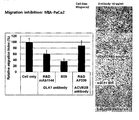
도 34는 액티빈 수용체 타입 2B 항체와 DLK1 항체의 전이 저해 효과를 나타낸다.Figure 34 shows the metastasis inhibitory effect of activin receptor type 2B antibody and DLK1 antibody.
도 35은 수용성 DLK1의 전이 저해 효과에 미치는 액티빈 수용체 타입 2B 항체와 DLK1 항체의 영향을 나타낸다.35 shows the effects of activin receptor type 2B antibody and DLK1 antibody on the metastasis inhibitory effect of water-soluble DLK1.
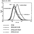
도 36은 췌장암 동소 이식 전이 동물모델 실험에서 DLK1-Fc 처리군의 종양 발생률을 나타낸다.36 shows tumor incidence of DLK1-Fc treated groups in pancreatic cancer in situ transplantation metastasis animal model experiment.
도 37은 췌장암 동소 이식 전이 동물모델 실험에서 DLK1-Fc 처리군의 탈장 형성률을 나타낸다.37 shows the hernia formation rate of DLK1-Fc treated group in pancreatic cancer orthotopic transplantation animal model experiment.
도 38은 췌장암 동소 이식 전이 동물모델 실험에서 DLK1-Fc 처리군에서 암 세포의 증식이 감소하였음을 나타낸다.38 shows that the proliferation of cancer cells was decreased in the DLK1-Fc treatment group in pancreatic cancer in situ transplantation metastasis animal model experiment.
도 39는 췌장암 동소 이식 전이 동물모델 실험에서 DLK1-Fc 처리군의 췌장, 위의 무게 및 크기를 나타낸다.39 shows pancreas, stomach weight and size of DLK1-Fc treated group in pancreatic cancer in situ transplantation metastasis animal model experiment.
도 40은 췌장암 동소 이식 전이 동물모델 실험에서 DLK1-Fc 처리군의 비장비대 현상이 감소하였음을 나타낸다.Figure 40 shows that the non-large band of the DLK1-Fc treatment group in pancreatic cancer in situ transplantation metastasis animal model experiments.
도 41은 췌장암 동소 이식 전이 동물모델의 생체 내 이미지를 나타낸다. FIG. 41 shows in vivo images of pancreatic cancer in situ transplantation metastasis animal model.
이하, 실시예를 통하여 본 발명을 상세히 설명한다. 하기 실시예는 본 발명을 예시하는 것으로, 본 발명의 내용이 실시예에 의해 한정되는 것은 아니다.Hereinafter, the present invention will be described in detail through examples. The following examples illustrate the invention and are not intended to limit the scope of the invention.
참고예 1: 수용성 DLK1 및 DLK1-Fc 융합 단백질의 제조Reference Example 1 Preparation of Water-Soluble DLK1 and DLK1-Fc Fusion Proteins
본 명세서의 실시예에서 사용한 DLK1의 세포 외 영역 수용성 도메인(이하, 수용성 DLK1) 및 수용성 DLK1과 인간항체 Fc 영역이 접합된 DLK1-Fc 융합 단백질(이하, DLK1-Fc)은 대한민국등록특허 제10-0982170호에 개시된 방법에 따라 제조하였다.The extracellular region water soluble domain (hereinafter referred to as water soluble DLK1) of DLK1 and DLK1-Fc fusion protein (hereinafter referred to as DLK1-Fc) in which the water-soluble DLK1 and the human antibody Fc region are conjugated as used in the Examples of the present specification are Prepared according to the method disclosed in 0982170.
실시예 1 : 수용성 DLK1의 암 전이 저해 효과 확인Example 1 Confirmation of Cancer Metastasis Inhibitory Effect of Water Soluble DLK1
수용성 DLK1에 의한 암 세포주에 미치는 영향을 알아보기 위하여 참고문헌(Chen HC, Methods in molecular biology. 294:15-22, 2005)의 방법을 사용하여 암 세포주의 이동 분석(migration assay)을 실시하였다. 먼저, 세포(MIA-PaCa-2, HeLa 세포주)가 50% 정도 수준일 때 무혈청 배지로 갈아 주고, 24시간 후에 세포를 트립신 처리를 통하여 떼어낸 후에 세포 수를 측정하였다. 세포, 무혈청 배지 및 처리하고자 하는 각각의 단백질을 합하여 100 ㎕를 만든 후 37℃에서 1시간 동안 배양하였다. 24-웰 플레이트에 1 ㎖의 화학 유인 물질(chemo-attractant)로서 5~10% FBS를 넣어주고 그 위에 8.0 μm 크기의 포어를 가진 트랜스웰(Corning #3422)을 올린 후 그 안에 1시간 동안 전배양한 세포 및 세포, 단백질 혼합액 100 ㎕를 넣은 후 37℃ 이산화탄소 배양기에서 24시간 내지 48시간 동안 배양하였다. 배양 후, 트렌스웰의 배지를 제거하고, Diff Quick 용액(Sysmex, Japan)을 이용하여 세포를 염색한 후 트랜스웰을 통과하지 못한 세포들을 면봉을 이용하여 완전히 제거해 주었다. 이후, 트랜스웰을 완전히 말린 후 트랜스웰을 통과한 세포들을 400배의 배율로 관찰한 다음 사진을 촬영하여, 도 1 및 2에 나타내었다. In order to determine the effect of the soluble DLK1 on cancer cell lines, a migration assay of cancer cell lines was performed using the method of Reference (Chen HC, Methods in molecular biology. 294: 15-22, 2005). First, when cells (MIA-PaCa-2, HeLa cell line) is about 50% level, the cells were changed to serum-free medium, and after 24 hours, the cells were detached through trypsin treatment, and the number of cells was measured. 100 μl of cells, serum-free medium and proteins to be treated were combined and incubated at 37 ° C. for 1 hour. Add 5-10% FBS as a 1 ml chemo-attractant in a 24-well plate and place a transwell (Corning # 3422) with 8.0 μm pore on it for 1 hour. 100 μl of the cultured cells, cells, and protein mixtures were added, and then cultured in a 37 ° C. carbon dioxide incubator for 24 to 48 hours. After incubation, the transwell medium was removed, the cells were stained with Diff Quick solution (Sysmex, Japan), and the cells that did not pass through the transwell were completely removed using a cotton swab. Subsequently, after completely drying the transwell, the cells passing through the transwell were observed at a magnification of 400 times, and photographs were taken, and are shown in FIGS. 1 and 2.
그 결과, 도 1에 나타난 바와 같이 췌장암 세포주인 MIA-PaCa-2 세포주에 DLK1-Fc를 처리한 경우 양-의존적으로 세포의 이동(전이)이 저해됨을 알 수 있었다(도 1). 또한, 도 2에 나타난 바와 같이 자궁암 세포주인 HeLa 세포주에 DLK1-Fc를 처리한 경우 양-의존적으로 세포의 이동(전이)이 저해됨을 알 수 있었다(도 2). As a result, as shown in FIG. 1, when DLK1-Fc was treated in the MIA-PaCa-2 cell line, which is a pancreatic cancer cell line, it was found that cell migration (metastasis) was inhibited in a sheep-dependent manner (FIG. 1). In addition, as shown in FIG. 2, DLK1-Fc treatment of HeLa cell line, which is a uterine cancer cell line, was found to inhibit cell migration (transition) in a sheep-dependent manner (FIG. 2).
실시예 2 : 암 세포주의 부착 독립성 성장에 미치는 수용성 DLK1의 영향 확인Example 2 Confirmation of Influence of Water Soluble DLK1 on Growth Independence of Cancer Cell Lines
부착 독립성 성장(anchorage independence growth)에 미치는 수용성 DLK1의 영향을 알아보기 위해 소프트 아가 에세이(Soft agar assay)를 수행하였다. 먼저, 10% FBS가 들어 있는 RPMI 배지와 1% 아가(agar)를 1:1로 섞어 0.5% 아가 조성이 되게 60 mm 배양접시에 깔아 준 후, 6 x 10³ 개의 세포(MIA-PaCa-2, HeLa 세포주)를 5 ㎍/㎖의 DLK1-Fc 또는 Fc와 함께 섞은 후, 10% FBS가 들어 있는 RPMI 배지와 1% 아가를 섞어 준 후 최후 아가의 농도를 0.3%가 되도록 한 후 0.5% 아가 위에 깔아 주었다. 3주간 37℃ CO2 배양기에서 배양하였으며, 아가가 마르는 것을 방지하기 위해 3일마다 5 ㎍/㎖의 DLK1-Fc 또는 Fc가 포함된 배지를 첨가해 주었다. 콜로니가 생성된 후에 사진을 찍고, 0.05% 크리스탈 바이올렛 염색약으로 생성된 콜로니를 염색해 준 후 콜로니를 세어 정량화하여, 도 3 및 4에 나타내었다.Soft agar assay was performed to determine the effect of water-soluble DLK1 on anchorage independence growth. First, RPMI medium containing 10% FBS and 1% agar were mixed 1: 1 and placed on a 60 mm culture dish to form 0.5% agar, followed by 6 x 10 3 cells (MIA-PaCa-2 , HeLa cell line) with 5 μg / ml of DLK1-Fc or Fc, and then mixed with RPMI medium containing 10% FBS and 1% agar, the concentration of the final agar was 0.3%, followed by 0.5% agar. Laid on top. It was incubated in a 37 ° C. CO 2 incubator for 3 weeks, and medium containing 5 μg / ml of DLK1-Fc or Fc was added every 3 days to prevent agar from drying out. Photographs were taken after the colonies were produced, the colonies were stained with 0.05% crystal violet dye, and the colonies were counted and quantified, as shown in FIGS. 3 and 4.
그 결과, 췌장암 세포주인 MIA-PaCa-2 세포주에 DLK1-Fc를 처리한 경우 대조군인 Fc만 처리한 세포에 비해 부착 독립성 성장이 저해됨을 알 수 있었다(도 3). 또한, 자궁암 세포주인 HeLa 세포주에 DLK1-Fc를 처리한 경우 대조군인 Fc만 처리한 세포에 비해 부착 독립성 성장이 저해됨을 알 수 있었다(도 4).As a result, the treatment of DLK1-Fc in the pancreatic cancer cell line MIA-PaCa-2 cell line was found to inhibit adhesion-independent growth compared to cells treated with only Fc (FIG. 3). In addition, when treated with DLK1-Fc in the HeLa cell line, uterine cancer cell line, it was found that adhesion-independent growth was inhibited compared to the cells treated with only Fc (FIG. 4).
실시예 3 : 수용성 DLK1이 암 세포주의 상처 치료에 미치는 영향 확인Example 3 Identification of the Effect of Water Soluble DLK1 on the Wound Treatment of Cancer Cell Lines
췌장암 세포주인 MIA-PaCa-2나 신장암 세포주인 786-O를 6-웰 플레이트에서 키운 후 90% 정도 세포가 성장하였을 때 16 시간 동안 무혈청 배지로 갈아 주었다. 이후, 세포에 5~25 ㎍/㎖ 농도의 DLK1-Fc 또는 Fc를 1시간 동안 처리해 준 후 세포를 긁어 상처(scratch)를 낸 후 5% FBS를 첨가하고 786-O 세포주는 16시간 동안, MIA-PaCa-2 세포주는 48시간 동안 배양한 후 상처가 매워지는 양상을 관찰하여, 도 5에 나타내었다.Pancreatic cancer cell line MIA-PaCa-2 or kidney cancer cell line 786-O was grown in 6-well plates and then changed to serum-free medium for 16 hours when 90% of the cells grew. Thereafter, the cells were treated with DLK1-Fc or Fc at a concentration of 5 to 25 µg / ml for 1 hour, after which the cells were scratched and 5% FBS was added, and the 786-O cell line was 16 hours for MIA. The PaCa-2 cell line was cultured for 48 hours, and the wound was observed.
그 결과, 췌장암 세포주(MIA-PaCa-2) 및 신장암 세포주(786-O)에 DLK1-Fc를 처리한 경우 대조군인 Fc만 처리한 세포에 비해 상처 치료가 저해됨을 알 수 있었다(도 5). As a result, when DLK1-Fc was treated in the pancreatic cancer cell line (MIA-PaCa-2) and renal cancer cell line (786-O), wound treatment was inhibited as compared to cells treated with only Fc (FIG. 5). .
실시예 4 : 수용성 DLK1에 의한 신호전달 검증Example 4 Signaling Verification by Water Soluble DLK1
수용성 DLK1이 실제 세포의 신호전달에 미치는 영향을 알아보기 위하여 웨스턴 블랏을 수행하였다. 이를 위하여, 배양용기에서 췌장암 세포주인 MIA-PaCa-2가 70% 정도 자랐을 때, 무혈청 배지로 교환해 주고 16시간 동안 배양하였다. DLK1-Fc 처리군에는 10 ㎍/㎖의 DLK1-Fc를 처리해 준 후 10% FBS를 첨가하고 10분 후 수확하였다. 수확한 세포는 RIPA 버퍼(50 mM TrisHCl pH 7.4, 150 mM NaCl, 2 mM EDTA, 1% NP-40)에 프로테아제 억제제(protease inhibitor; Roche), 포스포타제 억제제 칵테일 I, II(phosphotase inhibitor cocktail I, II; Sigma)를 첨가한 버퍼를 이용하여 세포를 용해시킨 후, BCA 정량 키트(Thermo)를 이용하여 용해된 세포 용해액의 농도를 결정하였다. SDS-PAGE에 30 ㎍의 용해액을 5X 샘플 버퍼를 이용하여 변성시키고 전기영동을 실시한 후 NC 막(Watman)에 트랜스퍼 하였다. 만들어진 블랏으로 p-FAK, FAK, p-AKT, AKT, p-eNOS, eNOS, p-ERK1/2 및 ERK1/2 항체(Cell signaling)를 이용하여 웨스턴 블랏을 실시하였다. 이때, beta-actin 항체(Sigma)를 로딩 컨트롤로 사용하였다. 4℃에서 하룻밤 동안 반응을 시킨 후에 TBST(Tween 20 0.05%)로 워싱을 3번 실시한 후 rabbit-HRP 항체 및 mouse-HRP 항체(Santa Cruz)를 이용하여 1시간 동안 상온에서 반응시킨 후 다시 TBST(Tween 20 0.05%)로 워싱을 3번 실시하였다. 이후, 블랏을 ECL 용액(Intron)을 이용하여 형광 발색하여 필름을 현상하여, 도 6에 나타내었다.Western blot was performed to determine the effect of water-soluble DLK1 on the signaling of real cells. To this end, when MIA-PaCa-2, a pancreatic cancer cell line, grew about 70% in a culture vessel, it was exchanged with a serum-free medium and cultured for 16 hours. The DLK1-Fc treatment group was treated with 10 μg / ml DLK1-Fc, followed by the addition of 10% FBS and harvested 10 minutes later. Harvested cells were prepared in RIPA buffer (50 mM TrisHCl pH 7.4, 150 mM NaCl, 2 mM EDTA, 1% NP-40), protease inhibitor (Roche), phosphotase inhibitor cocktail I, II (phosphotase inhibitor cocktail I Cells were lysed using a buffer added with II, Sigma), and the concentration of lysed cell lysate was determined using a BCA quantitative kit (Thermo). 30 μg of the lysate on SDS-PAGE was denatured using 5 × sample buffer, electrophoresed, and transferred to NC membrane (Watman). Western blot was performed using p-FAK, FAK, p-AKT, AKT, p-eNOS, eNOS, p-ERK1 / 2 and ERK1 / 2 antibody (Cell signaling). At this time, beta-actin antibody (Sigma) was used as a loading control. After reacting at 4 ° C. overnight, washing was performed three times with TBST (Tween 20 0.05%), followed by reaction at room temperature for 1 hour using rabbit-HRP antibody and mouse-HRP antibody (Santa Cruz), and then again TBST ( Washing was performed three times with Tween 20 0.05%). Subsequently, the blot was fluorescently developed using an ECL solution (Intron) to develop a film, as shown in FIG. 6.
그 결과, p-FAK, p-AKT, p-eNOS가 현저히 감소하였음을 알 수 있었으며, p-ERK1/2의 레벨도 약간 감소하는 것을 확인할 수 있었다(도 6). 이러한 결과 중, p-FAK, p-AKT 및 p-eNOS의 감소는 도 1 내지 5의 효과를 뒷받침하는 것이며, p-ERK1/2의 감소는 도 3 및 4의 부착 독립성 성장 저해 효과를 뒷받침하는 것이다.As a result, it was found that p-FAK, p-AKT, and p-eNOS were significantly reduced, and the level of p-ERK1 / 2 was also slightly decreased (FIG. 6). Among these results, the reduction of p-FAK, p-AKT and p-eNOS supports the effects of FIGS. 1 to 5, and the reduction of p-ERK1 / 2 supports the adhesion independent growth inhibition effect of FIGS. will be.
실시예 5 : 수용성 DLK1을 안정적으로 발현하는 세포주 구축Example 5 Cell Line Construction stably Expressing Water-soluble DLK1
외부에서 처리해 준 수용성 DLK1의 효과를 재확인하기 위하여, 췌장암 세포주인 MIA-PaCa-2 세포주를 이용하여 수용성 DLK1을 과발현하는 세포주를 구축하였다. 상기 세포주는 DLK1의 세포 외 도메인(extracellular domain) 지역을 코딩하는 DNA 단편을 pcDNA 3.1 벡터에 넣어 줌으로써 구축하였다. 세포주는 500 ㎍/㎖ G418(Geneticin, GIBCO/BRL)에서 스크리닝하고, 세포주가 잘 구축되었는지 확인하기 위하여 RT-PCR을 수행하였으며, 세포주를 키운 배양액을 Centricon 15(Millpore)를 이용하여 10배 농축한 후 웨스턴 블랏을 통하여 수용성 DLK1이 잘 발현되었는지 확인하였다. 웨스턴 블랏에는 DLK1 단클론 항체(R&D) 제품을 사용하였다. 비교군으로는 인서트가 없는 벡터만을 트랜스펙션하여 사용하였다.In order to reconfirm the effect of soluble DLK1 treated externally, a cell line overexpressing soluble DLK1 was constructed using the pancreatic cancer cell line MIA-PaCa-2 cell line. The cell line was constructed by incorporating a DNA fragment encoding the extracellular domain region of DLK1 into a pcDNA 3.1 vector. The cell line was screened at 500 μg / ml G418 (Geneticin, GIBCO / BRL), RT-PCR was performed to confirm that the cell line was well established, and the cultured cell was grown 10-fold using Centricon 15 (Millpore). After Western blot, it was confirmed whether the water-soluble DLK1 was well expressed. Western blot used the DLK1 monoclonal antibody (R & D) product. In the comparison group, only the vector without the insert was used by transfection.
그 결과, 도 7에 나타난 바와 같이, 상기 세포주에서 수용성 DLK1이 안정적으로 발현되고 있음을 확인할 수 있었다(도 7).As a result, as shown in Figure 7, it was confirmed that the water-soluble DLK1 is stably expressed in the cell line (Fig. 7).
실시예 6 : 수용성 DLK1을 안정적으로 발현하는 세포주에서의 수용성 DLK1에 의한 암 전이, 침윤, 부착 독립성 성장 및 상처 치유 저해 효과 검증Example 6 Inhibition of Cancer Metastasis, Invasion, Adhesion Independent Growth and Wound Healing Inhibition by Water-soluble DLK1 in Cell Lines stably Expressing Water-soluble DLK1
상기 실시예 5에서 구축된 세포주를 이용하여 전이, 침윤 기작에 대한 분석을 시행하였다. 먼저, 세포가 50% 정도 수준일 때 무혈청 배지로 갈아 주고, 24시간 후에 세포를 트립신 처리를 통하여 떼어낸 후 세포 수를 측정하였다. 24-웰 플레이트에 1 ㎖의 화학 유인 물질(chemo-attractant)로 5~10% FBS를 넣어주고 그 위에 8.0 μm 크기의 포어를 가진 트랜스웰(Corning #3422)을 올린 후 그 안에 5×10⁴개의 세포를 넣어준 후 37℃ 이산화탄소 배양기에서 24시간 내지 48시간 동안 배양하였다. 배양 후, 트렌스웰의 배지를 제거하고, Diff Quick 용액(Sysmex, Japan)을 이용하여 세포를 염색한 후 트랜스웰을 통과하지 못한 세포들을 면봉을 이용하여 완전히 제거해 주었다. 이후, 트랜스웰을 완전히 말린 후 트랜스웰을 통과한 세포들을 400배의 배율로 관찰하고, 사진을 촬영하였다. The cell line constructed in Example 5 was used to analyze metastasis and infiltration mechanism. First, the cells were changed to serum-free medium when the level was about 50%, and after 24 hours, the cells were detached through trypsin treatment, and the number of cells was measured. Place 5-10% FBS with 1 ml of chemo-attractant in a 24-well plate, place a transwell (Corning # 3422) with 8.0 μm pore on it, and then place 5 × 10 4 Cells were added and then cultured in a 37 ° C. carbon dioxide incubator for 24 to 48 hours. After incubation, the transwell medium was removed, the cells were stained with Diff Quick solution (Sysmex, Japan), and the cells that did not pass through the transwell were completely removed using a cotton swab. Then, after completely drying the transwell, the cells passing through the transwell were observed at a magnification of 400 times and photographed.
침윤 현상을 관찰하기 위해서는 1 mg/㎖ 농도의 Matrigel(BD Science) 100 ㎕를 트랜스웰 아래쪽 면에 코팅한 후에 실험을 실시하였다.In order to observe the infiltration phenomenon, the experiment was performed after coating 100 μl of Matrigel (BD Science) at a concentration of 1 mg / ml on the lower side of the transwell.
부착 독립성 성장을 관찰하기 위해서는 10% FBS가 들어 있는 RPMI 배지와 1% 아가를 1:1로 섞어 0.5% 아가 조성이 되게 60 mm 배양접시에 깔아 준 후, 6×10³ 개의 세포를 10% FBS가 들어 있는 RPMI 배지와 1% 아가를 섞어 준 후 최후 아가의 농도를 0.3%가 되도록 한 후 0.5% 아가 위에 깔아 주었다. 3주간 37℃ CO2 배양기에서 배양하였으며, 아가가 마르는 것을 방지하기 위해 3일마다 배지를 첨가해 주었다. 콜로니가 생성된 후에 사진을 찍어 결과를 관찰하였다.In order to observe adhesion-independent growth, RPMI medium containing 10% FBS and 1% agar were mixed 1: 1 and placed in a 60 mm culture dish to obtain 0.5% agar composition, followed by 6 × 10 3 cells in 10% FBS. After mixing 1% agar with RPMI medium containing the final concentration of agar to 0.3% and then laid on 0.5% agar. Incubated for 3 weeks in a 37 ℃ CO 2 incubator, medium was added every 3 days to prevent agar from drying. After colonies were produced, photographs were taken to observe the results.
상처 치유 분석은 세포주를 6-웰 플레이트에서 키운 후 90% 정도 세포가 성장하였을 때 무혈청 배지로 갈아주었다. 16시간 동안 무혈청 배지에서 배양한 후 세포를 긁어 상처를 내고 5% FBS를 첨가하여 48시간 동안 배양한 후 상처가 매워지는 양상을 관찰하였다.Wound healing assays were grown in 6-well plates and then changed to serum-free medium when 90% of the cells were grown. After culturing in serum-free medium for 16 hours, the cells were scraped and wounded, and the wound was filled after incubation for 48 hours with 5% FBS.
그 결과, 도 8에 나타난 바와 같이, 세포에 외부 유래의 DLK1-Fc를 처리한 경우와 마찬가지로, 상기 세포주에서 발현된 수용성 DLK1에 의하여 전이, 침윤, 부착 독립성 성장 및 상처 치유가 억제됨을 재확인할 수 있었다(도 8).As a result, as shown in FIG. 8, as in the case where the cells were treated with DLK1-Fc derived from the outside, metastasis, infiltration, adhesion-independent growth and wound healing were inhibited by the water-soluble DLK1 expressed in the cell line. (FIG. 8).
실시예 7 : 수용성 DLK1의 수용체 확인Example 7 Receptor Identification of Water Soluble DLK1
수용성 DLK1의 수용체를 확인하기 위하여 공동 면역 침강법을 수행하였다. 이를 위하여, 먼저 수용성 DLK1과 ACVR2B를 293E 세포에 PEI(Polyscience) 방법을 이용하여 트랜스펙션하여 과발현시켰다. 세포는 차가운 DPBS로 3번 씻어 준 후 변형된 RIPA 버퍼(50 mM TrisHCl pH7.4, 150 mM NaCl, 2 mM EDTA, 1% NP-40)에서 용해시켰다. 이 용해물은 사람 IgG(Jackson Lab)와 단백질 G 비드(GE)를 이용하여 pre-clearing 하였다. 실험실에서 직접 제작한 사람 항체와 단백질 G 비드를 이용하여 각각 면역 침강을 수행하였으며, SDS-PAGE에 샘플을 분리시킨 후 웨스턴 블랏을 수행하여 도 9에 나타내었다. 웨스턴 블랏에서는 DLK1 단클론 항체(R&D)와 ACVR2B affinity purified 다클론 항체(R&D)를 사용하였으며, 비교군으로 Lamin B 항체(Santa Cruz)와α-tubulin 항체(Santa Cruz)를 이용하였다.Co-immunoprecipitation was performed to identify receptors of water-soluble DLK1. To this end, first, soluble DLK1 and ACVR2B were overexpressed in 293E cells by transfection using PEI (Polyscience) method. Cells were washed three times with cold DPBS and lysed in modified RIPA buffer (50 mM TrisHCl pH7.4, 150 mM NaCl, 2 mM EDTA, 1% NP-40). This lysate was pre-cleared using human IgG (Jackson Lab) and protein G beads (GE). Immunoprecipitation was performed using human antibodies and protein G beads produced in the laboratory, and samples were separated on SDS-PAGE and Western blots were shown in FIG. 9. In Western blot, DLK1 monoclonal antibody (R & D) and ACVR2B affinity purified polyclonal antibody (R & D) were used, and Lamin B antibody (Santa Cruz) and α-tubulin antibody (Santa Cruz) were used as a comparative group.
그 결과, 도 9에 나타난 바와 같이, 수용성 DLK1이 액티빈 신호 전달에 관여하는 액티빈 수용체 타입 2B(ACVR2B)에 결합하는 것을 확인할 수 있었다(도 9).As a result, as shown in Figure 9, it was confirmed that the water-soluble DLK1 binds to activin receptor type 2B (ACVR2B) involved in activin signal transmission (Fig. 9).
실시예 8 : 수용성 DLK1과 ACVR2B간의 결합 확인Example 8 Confirmation of Binding Between Water-Soluble DLK1 and ACVR2B
효소 면역 측정법(ELISA)을 통하여 ACVR2B와 수용성 DLK1간의 결합을 확인하였다. 이를 위하여, 1 ㎍/㎖의 FLAG이 태깅된 수용성 DLK1을 면역플레이트(Nunc)에 4℃에서 16시간 동안 코팅하였다. 이때, 비특이적 결합을 측정하기 위한 대조군으로 BSA를 사용하였다. 코팅된 플레이트는 PBST(0.05% Tween 20)를 이용하여 3번 씻어 준 후 5% 탈지유 PBST를 이용하여 블락킹 하였다. ACVR2B의 세포 밖 부분에 Fc를 붙인 ACVR2B-Fc를 계열 희석(serial dilution)을 수행한 후 1시간 동안 상온에서 반응시켜 주었다. 반응 후 PBST(0.05% Tween 20)를 이용하여 3번 씻고 결합도를 측정하기 위하여 사람 IgG-HRP(1:5000)를 1시간 동안 배양해 주었다. 반응 후 PBST(0.05% Tween 20)를 이용하여 3번 씻고, TMB(Sigma)를 이용하여 발색반응을 수행하고, 2.5 M H2SO4를 첨가하여 반응을 종료해 준 후 450 nm에서 흡광도를 측정하였다. 그 결과, 도 10에 나타난 바와 같이, 수용성 DLK1이 ACVR2B와 결합하나, BSA에는 결합하지 않음을 확인할 수 있었다(도 10).Enzyme immunoassay (ELISA) confirmed the binding between ACVR2B and soluble DLK1. To this end, water-soluble DLK1 tagged with 1 μg / ml FLAG was coated on immunoplate (Nunc) at 4 ° C. for 16 hours. At this time, BSA was used as a control for measuring nonspecific binding. The coated plate was washed three times with PBST (0.05% Tween 20) and then blocked with 5% skim milk PBST. ACVR2B-Fc with Fc attached to the extracellular part of ACVR2B was subjected to serial dilution and allowed to react at room temperature for 1 hour. After the reaction, washed three times with PBST (0.05% Tween 20) and the human IgG-HRP (1: 5000) was incubated for 1 hour to measure the binding. After the reaction, washed three times with PBST (0.05% Tween 20), the color reaction was performed using TMB (Sigma), and 2.5 MH 2 SO 4 was added to terminate the reaction, and the absorbance was measured at 450 nm. . As a result, as shown in FIG. 10, it was confirmed that the water-soluble DLK1 binds to ACVR2B but not to BSA (FIG. 10).
또한, 수용성 DLK1과 ACVR2B의 리간드인 액티빈 A와의 관계를 살펴보기 위하여 액티빈 A를 코팅한 후 DLK1-Fc와의 결합도를 측정하였다. 그 결과, 수용성 DLK1은 ACVR2B의 리간드인 액티빈 A와는 결합하지 않음을 알 수 있었다(도 11). In addition, to examine the relationship between activin A, a ligand of water-soluble DLK1 and ACVR2B, the degree of binding to DLK1-Fc was measured after activin A was coated. As a result, it was found that the water-soluble DLK1 did not bind to activin A which is a ligand of ACVR2B (FIG. 11).
실시예 9 : ACVR2B에 대한 수용성 DLK1의 액티빈 A와 경쟁적 결합 확인Example 9 Confirmation of Competitive Binding with Activin A of Water Soluble DLK1 to ACVR2B
수용성 DLK1이 액티빈 A와 경쟁적으로 ACVR2B 수용체에 결합하는지 알아보기 위하여 경쟁 효소 면역 측정법을 수행하였다. 50 ng/㎖의 액티빈 A를 코팅한 후 ACVR2B-Fc와 FLAG-DLK1을 같이 반응시켜 주어 측정하였다. 이때, ACVR2B는 2 nM 농도로 고정하고 FLAG-DLK1을 1에서 16 nM 까지의 농도로 처리하여 준 후 경쟁 결합 정도를 측정하였다. 대조군으로는 액티빈 A와 결합하지 않는 Fc와 ACVR1A-Fc를 사용하였다. Competitive enzyme immunoassay was performed to determine whether water-soluble DLK1 binds to ACVR2B receptor competitively with activin A. 50 ng / mL activin A was coated and reacted with ACVR2B-Fc and FLAG-DLK1. At this time, ACVR2B was fixed at a concentration of 2 nM and treated with FLAG-DLK1 at a concentration of 1 to 16 nM, and then the degree of competitive binding was measured. As a control group, Fc and ACVR1A-Fc, which do not bind to activin A, were used.
그 결과, 도 12에 나타난 바와 같이, 수용성 DLK1의 첨가 농도가 높아질수록 액티빈 A와 ACVR2B의 결합이 점차 감소함을 알 수 있었다(도 12). 이러한 결과는 수용성 DLK1이 액티빈 A와 경쟁적으로 ACVR2B에 결합하여 액티빈 신호전달을 차단할 수 있음을 나타낸다.As a result, as shown in Figure 12, it was found that the binding of activin A and ACVR2B gradually decreases as the concentration of water-soluble DLK1 is increased (Fig. 12). These results indicate that water-soluble DLK1 can competitively bind activin A to ACVR2B to block activin signaling.
실시예 10 : ACVR2B에 대한 수용성 DLK1의 특이적 결합 확인Example 10 Confirmation of Specific Binding of Water Soluble DLK1 to ACVR2B
ACVR2B에 대한 수용성 DLK1의 특이적 결합을 확인하기 위하여 Bio-Rad사의 ProteOn XPR36를 사용하여 표면 플라즈몬 공명을 수행하였으며, 그 방법을 요약하면 다음과 같다. 0.1 M EDC와 0.025 M sulfo-NHS를 60초간 흘려주어 센서 칩을 활성화시키고, 코팅할 단백질을 10 mM 아세트산나트륨(pH 5.0)에 넣어 240초간 30 ㎕/분의 속도로 흘려 주어 칩에 부착반응을 실시하였다. 채널을 1 M 에탄올아민-HCl(pH 8.5)으로 200초간 블락시킨 후, DPBST(PBS with 0.005% tween 20)로 워싱하였다. 결합력을 알아보기 위하여 2배씩 계열 희석한 후 30 ㎕/분의 속도로 120초간 흘려주어 결합력을 측정하였다. 분리력을 알아보기 위해서는 240초간 분리 단계(dissociation phase)를 가져갔다.Surface plasmon resonance was performed using Bio-Rad's ProteOn XPR36 to confirm specific binding of water-soluble DLK1 to ACVR2B. The method is summarized as follows. The sensor chip was activated by flowing 0.1 M EDC and 0.025 M sulfo-NHS for 60 seconds, and the protein to be coated was added to 10 mM sodium acetate (pH 5.0) at a rate of 30 μl / min for 240 seconds to attach to the chip. Was carried out. The channel was blocked with 1 M ethanolamine-HCl (pH 8.5) for 200 seconds and then washed with DPBST (PBS with 0.005% tween 20). In order to determine the binding force, the serial dilution was performed twice, and the binding force was measured by flowing for 120 seconds at a rate of 30 μl / min. In order to determine the separation force, a dissociation phase was taken for 240 seconds.
그 결과, 도 13에 나타난 바와 같이, 수용성 DLK1이 다양한 액티빈 수용체 패밀리 중에서 ACVR2B에 특이적으로 결합하는 것을 확인할 수 있었다(도 13).As a result, as shown in FIG. 13, it was confirmed that the water-soluble DLK1 specifically binds to ACVR2B among various activin receptor families (FIG. 13).
실시예 11 : 실제 세포상에서 수용성 DLK1과 ACVR2B간의 결합 확인Example 11 Confirmation of Binding Between Water-Soluble DLK1 and ACVR2B on Real Cells
실제 세포상에서 수용성 DLK1과 ACVR2B간의 결합이 일어나는지 확인하기 위하여 유세포 분석기를 이용하여 실험하였다. BD사의 CantoII 제품을 이용하여 수행하였으며 실험 방법을 요약하면 다음과 같다. 293E 세포에 ACVR2B 전체 유전자가 들어가 있는 pcDNA 벡터를 이용하여 PEI(Polyscience) 방법을 이용하여 트랜스팩션 시키고 ACVR2B를 과발현시킨 세포를 준비하였다. 트랜스팩션 이틀 후 트랜스펙션 시키지 않은 293E 세포와 함께 수확하였다. 또한, ACVR2B가 수용성 DLK1의 결합에 의미가 있는지 재확인하기 위하여 ACVR2B의 발현을 억제하는 siRNA(Invitrogen)을 처리하여 ACVR2B의 발현을 억제시킨 후 세포를 수확하였으며, siRNA에 대한 대조군으로 control siRNA (Invitrogen)을 처리한 293E도 준비하였다. 수확한 세포는 차가운 DPBS를 이용하여 2번 워싱을 해 주고 DLK1-Fc-FITC를 1 ㎍ 첨가하여 4℃에서 2시간 동안 반응시켜 주었다. 반응에 사용한 DLK1-Fc-FITC는 Pierce 사의 단백질 FITC 라벨링 키트를 이용하여 제작하였다. 반응 후 1% PBA(1% BSA in DPBS) 버퍼를 이용하여 워싱을 수행하고 유세포 분석기를 통하여 결합 정도를 결정하여, 도 15에 나타내었다. 또한, 위에 사용된 세포들은 웨스턴 블랏을 통하여 ACVR2B의 발현 양상을 확인하였다(도 14).In order to confirm whether the binding between water-soluble DLK1 and ACVR2B occurs on the actual cells, experiments were performed using a flow cytometer. It was performed using BD's CantoII product, and summarized the experimental method as follows. Using a pcDNA vector containing a full ACVR2B gene in 293E cells was transfected using PEI (Polyscience) method to prepare cells overexpressing ACVR2B. Two days after transfection, harvest was performed with 293E cells that were not transfected. In addition, cells were harvested by inhibiting the expression of ACVR2B by treating with siRNA (Invitrogen) that inhibits the expression of ACVR2B in order to reconfirm whether ACVR2B is meaningful for the binding of water-soluble DLK1, and control siRNA (Invitrogen) as a control for siRNA. 293E was also prepared. The harvested cells were washed twice with cold DPBS and 1 μg of DLK1-Fc-FITC was added and reacted at 4 ° C. for 2 hours. DLK1-Fc-FITC used for the reaction was prepared using the Pierce protein FITC labeling kit. After the reaction, washing was performed using 1% PBA (1% BSA in DPBS) buffer, and the degree of binding was determined by flow cytometry. In addition, the cells used above confirmed the expression pattern of ACVR2B via Western blot (FIG. 14).
그 결과, 도 15에 나타난 바와 같이, ACVR2B를 과발현시킨 세포주에서 수용성 DLK1에 의한 결합 이동이 현저하게 증가하였으나, siRNA를 처리하여 ACVR2B의 발현을 억제시킨 세포주에서는 이동이 감소하였음을 알 수 있었다(도 15).As a result, as shown in FIG. 15, the binding movement by the soluble DLK1 was significantly increased in the cell line overexpressing ACVR2B, but the cell line in which the expression of ACVR2B was suppressed by siRNA was decreased. 15).
실시예 12 : DLK1의 수용성 부분에 특이적으로 결합하는 인간 항체의 제작 및 검증Example 12 Construction and Validation of Human Antibodies Specific for Binding to the Water-Soluble Portion of DLK1
12-1. DLK1의 수용성 부분에 특이적으로 결합하는 인간 항체의 제작12-1. Construction of human antibodies that specifically bind to the water soluble portion of DLK1
<12-1-1> 라이브러리 파지의 제조<12-1-1> Preparation of Library Phage
다양성을 가진 인간 유래 scFv 라이브러리 세포 2.7×10¹⁰를 2×YTCM [Tryptone (CONDA, 1612.00) 17 g, Yeast extract (CONDA, 1702.00) 10 g, NaCl (sigma, S7653-5 ㎏) 5 g, chloramphenicol (sigma, C0857) 34 ㎍/㎖)], 2% 글루코즈 (sigma, G5400) 및 5 mM MgCl2 (sigma, M2393)를 포함하는 배지 (3 ℓ)에서 37℃의 온도로 2 내지 3시간 동안 배양한 후 (OD600=0.5~0.7), 헬퍼 파지 (helper phage)를 감염시켜 2×YTCMK [2×YT CM, Kanamycin (sigma, K1876) 70 ㎍/㎖, 1 mM IPTG (ELPISBIO, IPTG025)] 배지에서 30℃의 온도로 16시간 동안 배양하였다. 배양한 세포를 원심분리 (4500 rpm, 15분, 4℃)한 후, 상등액에 4% PEG (Fluka, 81253) 6000과 3% NaCl (sigma, S7653)을 첨가하여 잘 녹인 다음 얼음에서 1시간 동안 반응하였다. 다시 원심분리 (8000 rpm, 20분, 4℃)한 후, 펠렛에 PBS를 첨가하여 녹인 다음 원심분리 (12000 rpm, 10분, 4℃)하여 라이브러리 파지를 포함하는 상등액을 새 튜브에 넣어 4℃에서 보관하였다.2.7 × 10 10 of human-derived scFv library cells with variability were 2 × YTCM [17 g of Tryptone (CONDA, 1612.00), 10 g of Yeast extract (CONDA, 1702.00), 5 g of NaCl (sigma, S7653-5 kg), chloramphenicol ( sigma, C0857) 34 μg / ml)], 2% glucose (sigma, G5400) and 5 mM MgCl 2 (sigma, M2393) incubated for 2 to 3 hours at a temperature of 37 ℃ (OD600 = 0.5-0.7), then infected with helper phage to 30 in 2 × YTCMK [2 × YT CM, Kanamycin (sigma, K1876) 70 μg / ml, 1 mM IPTG (ELPISBIO, IPTG025)] medium. Incubated for 16 hours at a temperature of ℃. The cultured cells were centrifuged (4500 rpm, 15 min, 4 ° C.), and then dissolved in the supernatant by adding 4% PEG (Fluka, 81253) 6000 and 3% NaCl (sigma, S7653) to the supernatant for 1 hour on ice. Reacted. After centrifugation (8000 rpm, 20 minutes, 4 ° C) again, PBS was added to the pellets to dissolve, followed by centrifugation (12000 rpm, 10 minutes, 4 ° C), and the supernatant containing the library phage was added to a new tube at 4 ° C. Stored at.
<12-1-2> 단일클론항체의 제조<12-1-2> Preparation of Monoclonal Antibody
(1) 패닝(Panning) 과정(1) Panning Process
상기 실시예 1에서 수득한 정제된 DLK1-Fc 30 ㎍을 Immunosorb 튜브 (Nunc 470319)에 4 ㎖의 코팅 완충용액 [coating buffer; Na2CO3 (sigma, S7795) 1.59 g, NaHCO3 (sigma, S8875) 2.93 g, NaN3(sigma, S2002), 0.2 g]으로 4℃에서 16시간 정도 회전기(rotator)로 코팅한 후, 실온에서 2시간 동안 PBS에 녹여 탈지유 [(BD,232100)-4% in 1XPBS]를 사용하여 면역튜브(immunotube)에서 차단하였다. 제조한 라이브러리 파지 2 ㎖을 면역튜브에 첨가하여 실온에서 2시간 동안 반응시키고, PBST (0.05%)로 5회, PBS로 2회 세척하였다. 세척 후 특이적으로 결합한 scFv-파지들만 100 mM TEA (Sigma T-0886)로 용리하여, 용출된 파지들을 대장균 (XL1-Blue, stratagene, 200249)에 감염시켜 증폭하였다. 첫 번째 패닝에서 증폭된 파지를 PBST 세척 횟수만 늘려서 (2차: 13번, 3차: 23번) 동일한 방법으로 2차, 3차 패닝을 수행하였다.30 μg of the purified DLK1-Fc obtained in Example 1 was added to an Immunosorb tube (Nunc 470319) in 4 ml of a coating buffer; 1.59 g of Na 2 CO 3 (sigma, S7795), 2.93 g of NaHCO 3 (sigma, S8875), NaN 3 (sigma, S2002), 0.2 g], coated with a rotator at 4 ° C. for about 16 hours and then at room temperature. It was dissolved in PBS for 2 hours and blocked in immunotubes using skim milk [(BD, 232100) -4% in 1XPBS]. 2 ml of the prepared library phage was added to the immunotubes, reacted at room temperature for 2 hours, washed 5 times with PBST (0.05%) and twice with PBS. After washing, only specifically bound scFv-phages were eluted with 100 mM TEA (Sigma T-0886), and the eluted phages were amplified by infection with E. coli (XL1-Blue, stratagene, 200249). Phage amplified in the first panning was increased only by the number of PBST washings (second: 13, third: 23), and second and third panning was performed in the same manner.
그 결과, 패닝을 통한 항체의 역가 상승은 표 1에 나타난 바와 같았다.As a result, the titer of the antibody through panning was as shown in Table 1.
표 1
Table 1
| 표적항원 | 패닝횟수 | 초기 파아지수 | 결합한 파아지수 |
| DLK1-Fc | 1st | 4.6×10¹³ | 6×10⁷ |
| 2nd | 2×10¹² | 1.4×10⁶ | |
| 3rd | 1.49×10¹⁴ | 6.93×10⁹ |
| Target antigen | Panning | Initial phage index | Combined Phage Index |
| DLK1-Fc | 1st | 4.6 × 10 13 | 6 × 10 7 |
| 2nd | 2 × 10 12 | 1.4 × 10 6 | |
| 3rd | 1.49 × 10 14 | 6.93 × 10 9 |
(2) 파지 효소 면역 측정법(ELISA)에 의한 파지 항체 탐색(2) Search for phage antibody by phage enzyme immunoassay (ELISA)
① 패닝 결과 확인① Check the panning result
1차부터 3차까지 패닝하여 얼려두었던 세포 저장물(stock)을 5 ㎖의 2×YTCM, 2% 글루코즈, 5 mM MgCl2 배지에 OD600=0.1이 되게 넣어준 다음, 37℃에서 2 내지 3시간 동안 (OD600=0.5~0.7) 배양하였다. 이후, M1 헬퍼 파지를 감염시키고 2×YTCMK, 5 mM MgCl2, 1 mM IPTG 배지에서 30℃의 온도로 16시간 동안 배양하였다. 배양한 세포를 원심분리한 후 (4500 rpm, 15 분, 4℃) 상등액 (패닝된 poly scFv-파지)을 새 튜브로 옮겼다. 96-웰 면역 플레이트 (NUNC 439454)에 항원을 웰 당 100 ng씩 4℃에서 16시간 정도 코팅 완충용액으로 처리하여 코팅한 후, PBS에 녹인 탈지유 (4 %)를 사용하여 각 웰을 차단하였다. 각 웰 마다 PBS-tween20 (0.05%) 0.2 ㎖으로 씻어준 다음 패닝된 poly scFV-파지를 원액 및 5, 25, 125, 625, 3125배 희석한 용액을 각 웰에 100 ㎕씩 넣고 상온에서 2시간 동안 반응시켰다. 다시 각 웰마다 PBS-tween20 (0.05%) 0.2 ㎖을 사용하여 4번 세척한 후 이차 항체인 항-M13-HRP (Amersham 27-9421-01)를 1:2000으로 희석하여 실온에서 1시간 동안 반응하였다. PBS-tween20 (0.05%) 0.2 ㎖으로 세척한 후에 OPD 정제 (Sigmap 8787-TAB)를 PC 완충용액 [C6H8O7·H2O (sigma, C0706) 5.1 g, Na2HPO4 (sigma, S7907) 7.3 g]에 녹인 기질 용액을 만들어 웰당 100 ㎕씩 넣어 10분 동안 발색시킨 후 490 ㎚에서 흡광도를 분광광도계 (MolecularDevice, 미국)로 측정하였다.The frozen cell stocks, which were panned from 1st to 3rd, were placed in 5 ml of 2 × YTCM, 2% glucose, and 5 mM MgCl 2 medium at OD600 = 0.1, followed by 2-3 hours at 37 ° C. (OD600 = 0.5-0.7) incubated. Thereafter, M1 helper phage was infected and incubated for 16 hours at a temperature of 30 ° C. in 2 × YTCMK, 5 mM MgCl 2 , 1 mM IPTG medium. The cultured cells were centrifuged (4500 rpm, 15 min, 4 ° C.) and the supernatant (panned poly scFv-phage) was transferred to a new tube. 96-well immune plates (NUNC 439454) were coated with antigen at 100 ng per well with coating buffer for 16 hours at 4 ° C., and then each well was blocked using skim milk (4%) dissolved in PBS. Wash each well with 0.2 ml of PBS-tween20 (0.05%), and add 100 µl of the diluted solution of panned poly scFV-phage and 5, 25, 125, 625, and 3125 times to each well. Reacted for a while. After washing 4 times with 0.2 ml of PBS-tween20 (0.05%) in each well, the secondary antibody anti-M13-HRP (Amersham 27-9421-01) was diluted 1: 2000 and reacted at room temperature for 1 hour. It was. PBS-tween20 (0.05%) purified OPD After washing with 0.2 ㎖ (Sigmap 8787-TAB) a PC buffer [C 6 H 8 O 7 · H 2 O (sigma, C0706) 5.1 g, Na 2 HPO 4 (sigma , S7907) 7.3 g] was prepared and the substrate solution dissolved in 100 μl per well was developed for 10 minutes and the absorbance at 490 nm was measured with a spectrophotometer (MolecularDevice, USA).
그 결과, 도 16에 나타난 바와 같이, 항원에 대한 결합능이 각각 2차 다클론 scFv-파지 풀(pools)부터 증강하기 시작하여 3차에 결합능이 포화상태에 이르렀음을 확인할 수 있었다(도 16).As a result, as shown in FIG. 16, the binding ability to the antigen began to increase from the secondary polyclonal scFv-phage pools, respectively, and it was confirmed that the binding capacity reached the saturation in the third stage (FIG. 16). .
② 단일클론항체 선별② Monoclonal antibody screening
상기 결합능이 큰 다클론 파지 항체군에서 얻은 콜로니를 2×YTCM, 2% 글루코즈, 5 mM MgCl2 배지 1 ㎖이 포함된 96-deep 웰 플레이트 (바이오니아 90030)에서 37℃의 온도로 16시간 동안 배양하였다. 배양한 세포의 OD600 값이 0.1이 되도록 100~200 ㎕를 취해 1 ㎖의 2×YTCM, 2% 글루코즈, 5 mM MgCl2 배지에 희석한 다음, 96-deep 웰 플레이트에서 37℃의 온도로 OD600 값이 0.5~0.7 되도록 2 내지 3시간 동안 배양하였다. M1 헬퍼 파지를 MOI값이 1:20이 되도록 감염시킨 후, 2×YTCMK, 5 mM MgCl2, 1 mM IPTG 배지에서 30℃의 온도로 16시간 동안 배양하였다. 배양한 세포를 원심분리 (4500 rpm, 15 분, 4℃)한 후, 상등액을 취해 4% PEG 6000과 3% NaCl을 첨가하여 잘 녹인 후 얼음에서 1시간 동안 반응시켰다. 다시 원심분리 (8000 rpm, 20분, 4℃)한 후 펠렛에 PBS를 첨가하여 녹인 다음 원심분리 (12000 rpm, 10분, 4℃)하여 상등액을 취해 새 튜브에 옮겨 4℃에서 보관하였다. 이후, 96웰 면역 플레이트에 항원을 웰당 100 ng씩을 넣어 4℃에서 16시간 동안 코팅한 후 PBS에 녹인 탈지유 (4%)를 사용하여 각 웰을 차단하였다. 각 웰마다 PBS-tween20 (0.05%) 0.2 ㎖을 사용하여 세척한 다음 상기 방법으로 수득한 단일클론 scFv-파지 (each 100 scFv-phage)를 각 웰에 100 ㎕씩 넣고 상온에서 2시간 동안 반응시켰다. 다시 각 웰마다 PBS-tween20 (0.05%) 0.2 ㎖을 사용하여 4번 씻어준 후 2차 항체인 anti-M13-HRP를 1/2000로 희석하여 실온에서 1시간 동안 반응하였다. PBS-tween20 (0.05%) 0.2 ㎖로 세척한 후 발색하여 흡광도 490 ㎚에서 측정하였다. 그 결과, 하기 표 2에 나타난 바와 같이 항원에 대한 결합능이 2 이상인 27개의 단일파지클론들을 선별하였다.Colonies obtained from the polyclonal phage antibody group having high binding capacity were incubated for 16 hours at 37 ° C. in a 96-deep well plate (Bionia 90030) containing 2 ml of YTCM, 2% glucose, and 1 ml of 5 mM MgCl 2 medium. It was. Take 100-200 μl of the cultured cells so that the OD600 value is 0.1 and dilute in 1 ml 2 × YTCM, 2% glucose, 5 mM MgCl 2 medium, and then measure the OD600 value at 37 ° C. in a 96-deep well plate. Incubated for 2-3 hours so as to be 0.5-0.7. M1 helper phages were infected with a MOI of 1:20, and then incubated for 16 hours at a temperature of 30 ° C. in 2 × YTCMK, 5 mM MgCl 2 , and 1 mM IPTG medium. After culturing the cells were centrifuged (4500 rpm, 15 minutes, 4 ℃), the supernatant was taken, dissolved by adding 4 % PEG 6000 and 3% NaCl, and reacted for 1 hour on ice. After centrifugation (8000 rpm, 20 minutes, 4 ° C.), PBS was added to the pellets to dissolve the pellets, followed by centrifugation (12000 rpm, 10 minutes, 4 ° C.) to obtain a supernatant, which was transferred to a new tube and stored at 4 ° C. Thereafter, 100 ng of antigen was added per well to a 96-well immune plate and coated at 4 ° C. for 16 hours, and then each well was blocked using skim milk (4%) dissolved in PBS. Each well was washed with 0.2 ml of PBS-tween20 (0.05%), and 100 μl of monoclonal scFv-phage (each 100 scFv-phage) obtained by the above method was added to each well and allowed to react at room temperature for 2 hours. . Again, each well was washed four times with 0.2 ml of PBS-tween20 (0.05%), and the second antibody, anti-M13-HRP, was diluted to 1/2000 and reacted at room temperature for 1 hour. After washing with 0.2 ml of PBS-tween20 (0.05%), color development was measured at 490 nm. As a result, as shown in Table 2, 27 single phage clones having a binding capacity of 2 or more antigens were selected.
표 2
TABLE 2
| DLK1 | 1 | 2 | 3 | 4 | 5 | 6 | 7 | 8 | 9 | 10 | 11 | 12 |
| A | 0.1129 | 0.0716 | 0.0482 | 3.1152 | 2.9859 | 0.4549 | 0.3612 | 2.92 | 0.0469 | 2.8295 | 0.2175 | 1.0026 |
| B | 0.8858 | 0.8553 | 2.0914 | 0.788 | 2.762 | 2.6351 | 2.8837 | 0.1342 | 2.3259 | 0.1396 | 2.5018 | 0.501 |
| C | 0.4976 | 0.2852 | 2.466 | 0.2239 | 0.1128 | 1.2413 | 2.9255 | 2.1548 | 0.2169 | 0.0608 | 0.2132 | 0.1591 |
| D | 0.2025 | 0.1882 | 0.1109 | 0.0586 | 0.8865 | 0.0749 | 0.0849 | 0.1145 | 0.8514 | 0.0572 | 0.1653 | 2.4751 |
| E | 0.0907 | 0.1001 | 0.0418 | 0.047 | 2.2329 | 2.3476 | 2.3778 | 0.7165 | 0.0919 | 0.7527 | 0.1737 | 0.2233 |
| F | 0.1659 | 0.2324 | 0.4055 | 2.9152 | 0.2405 | 0.933 | 0.3682 | 0.1608 | 0.2258 | 0.1668 | 2.8944 | 2.9681 |
| G | 0.0433 | 0.3815 | 0.2245 | 2.8355 | 2.5814 | 3.0216 | 0.752 | 0.3455 | 0.0609 | 0.4363 | 0.1964 | 0.0504 |
| H | 0.1044 | 2.8427 | 2.7085 | 0.296 | 0.2403 | 2.1306 | 2.8803 | 1.6389 | 3.033 | 0.5009 | 2.7793 | 3.1994 |
| MYC | 1 | 2 | 3 | 4 | 5 | 6 | 7 | 8 | 9 | 10 | 11 | 12 |
| A | 0.2003 | 0.0474 | 0.0487 | 0.5068 | 0.7838 | 1.4922 | 0.4492 | 0.5538 | 0.0557 | 1.2823 | 0.1523 | 0.2432 |
| B | 1.357 | 0.7667 | 1.4038 | 1.1973 | 0.5932 | 0.8478 | 0.5129 | 0.1191 | 0.0918 | 0.7688 | 0.316 | 0.0526 |
| C | 0.2966 | 0.0741 | 0.2871 | 0.1538 | 0.0683 | 0.6353 | 0.5938 | 0.3595 | 0.6692 | 0.1009 | 0.1206 | 0.2206 |
| D | 0.1631 | 0.4308 | 0.078 | 0.045 | 0.5783 | 0.0632 | 0.0538 | 0.052 | 2.1443 | 0.0511 | 0.106 | 0.0889 |
| E | 0.0856 | 0.6073 | 0.0465 | 0.0435 | 0.1722 | 0.1883 | 0.4694 | 0.0867 | 0.0639 | 0.272 | 0.3112 | 0.1566 |
| F | 0.1285 | 0.4057 | 0.1421 | 1.0637 | 0.1115 | 1.1193 | 0.0898 | 1.0797 | 0.1751 | 0.1401 | 0.4419 | 0.524 |
| G | 0.0662 | 0.3396 | 0.0844 | 0.9974 | 2.9974 | 0.6732 | 0.6083 | 0.2278 | 0.0496 | 0.5198 | 0.0561 | 0.0551 |
| H | 0.0479 | 0.5654 | 1.1204 | 0.4634 | 0.066 | 0.8632 | 1.0213 | 0.6574 | 0.8562 | 0.1146 | 0.9677 | 0.6741 |
| FC | 1 | 2 | 3 | 4 | 5 | 6 | 7 | 8 | 9 | 10 | 11 | 12 |
| A | 0.0535 | 0.0726 | 0.0731 | 0.0791 | 0.0704 | 0.1111 | 0.0748 | 0.0709 | 0.0535 | 0.0828 | 0.0591 | 0.2558 |
| B | 0.1375 | 0.4065 | 0.0851 | 0.0702 | 0.0575 | 0.0472 | 2.8291 | 0.0717 | 0.0786 | 0.0743 | 0.0548 | 0.0451 |
| C | 0.0524 | 0.0555 | 0.0521 | 0.0745 | 0.0455 | 0.0825 | 2.8824 | 0.0559 | 0.0772 | 0.0485 | 0.0663 | 0.061 |
| D | 0.0519 | 0.0686 | 0.0447 | 0.0722 | 0.0431 | 0.0455 | 0.0482 | 0.0528 | 0.0498 | 0.1141 | 0.0651 | 0.0831 |
| E | 0.0496 | 0.0543 | 0.0419 | 0.0587 | 0.0472 | 0.0481 | 0.0558 | 0.2673 | 0.0492 | 0.1508 | 0.0601 | 0.0577 |
| F | 0.0614 | 0.0584 | 0.0528 | 0.0879 | 0.0553 | 0.1223 | 0.0792 | 0.0756 | 0.0661 | 0.0922 | 0.0658 | 0.0757 |
| G | 0.0456 | 0.0687 | 0.0521 | 0.1171 | 2.079 | 0.0689 | 0.547 | 0.0991 | 0.0874 | 0.2255 | 0.1894 | 0.0457 |
| H | 0.0594 | 0.0642 | 0.049 | 0.0705 | 0.0608 | 0.0766 | 0.1107 | 0.0819 | 0.0701 | 0.1204 | 0.0592 | 0.0539 |
| DLK1 | One | 2 | 3 | 4 | 5 | 6 | 7 | 8 | 9 | 10 | 11 | 12 |
| A | 0.1129 | 0.0716 | 0.0482 | 3.1152 | 2.9859 | 0.4549 | 0.3612 | 2.92 | 0.0469 | 2.8295 | 0.2175 | 1.0026 |
| B | 0.8858 | 0.8553 | 2.0914 | 0.788 | 2.762 | 2.6351 | 2.8837 | 0.1342 | 2.3259 | 0.1396 | 2.5018 | 0.501 |
| C | 0.4976 | 0.2852 | 2.466 | 0.2239 | 0.1128 | 1.2413 | 2.9255 | 2.1548 | 0.2169 | 0.0608 | 0.2132 | 0.1591 |
| D | 0.2025 | 0.1882 | 0.1109 | 0.0586 | 0.8865 | 0.0749 | 0.0849 | 0.1145 | 0.8514 | 0.0572 | 0.1653 | 2.4751 |
| E | 0.0907 | 0.1001 | 0.0418 | 0.047 | 2.2329 | 2.3476 | 2.3778 | 0.7165 | 0.0919 | 0.7527 | 0.1737 | 0.2233 |
| F | 0.1659 | 0.2324 | 0.4055 | 2.9152 | 0.2405 | 0.933 | 0.3682 | 0.1608 | 0.2258 | 0.1668 | 2.8944 | 2.9681 |
| G | 0.0433 | 0.3815 | 0.2245 | 2.8355 | 2.5814 | 3.0216 | 0.752 | 0.3455 | 0.0609 | 0.4363 | 0.1964 | 0.0504 |
| H | 0.1044 | 2.8427 | 2.7085 | 0.296 | 0.2403 | 2.1306 | 2.8803 | 1.6389 | 3.033 | 0.5009 | 2.7793 | 3.1994 |
| MYC | One | 2 | 3 | 4 | 5 | 6 | 7 | 8 | 9 | 10 | 11 | 12 |
| A | 0.2003 | 0.0474 | 0.0487 | 0.5068 | 0.7838 | 1.4922 | 0.4492 | 0.5538 | 0.0557 | 1.2823 | 0.1523 | 0.2432 |
| B | 1.357 | 0.7667 | 1.4038 | 1.1973 | 0.5932 | 0.8478 | 0.5129 | 0.1191 | 0.0918 | 0.7688 | 0.316 | 0.0526 |
| C | 0.2966 | 0.0741 | 0.2871 | 0.1538 | 0.0683 | 0.6353 | 0.5938 | 0.3595 | 0.6692 | 0.1009 | 0.1206 | 0.2206 |
| D | 0.1631 | 0.4308 | 0.078 | 0.045 | 0.5783 | 0.0632 | 0.0538 | 0.052 | 2.1443 | 0.0511 | 0.106 | 0.0889 |
| E | 0.0856 | 0.6073 | 0.0465 | 0.0435 | 0.1722 | 0.1883 | 0.4694 | 0.0867 | 0.0639 | 0.272 | 0.3112 | 0.1566 |
| F | 0.1285 | 0.4057 | 0.1421 | 1.0637 | 0.1115 | 1.1193 | 0.0898 | 1.0797 | 0.1751 | 0.1401 | 0.4419 | 0.524 |
| G | 0.0662 | 0.3396 | 0.0844 | 0.9974 | 2.9974 | 0.6732 | 0.6083 | 0.2278 | 0.0496 | 0.5198 | 0.0561 | 0.0551 |
| H | 0.0479 | 0.5654 | 1.1204 | 0.4634 | 0.066 | 0.8632 | 1.0213 | 0.6574 | 0.8562 | 0.1146 | 0.9677 | 0.6741 |
| FC | One | 2 | 3 | 4 | 5 | 6 | 7 | 8 | 9 | 10 | 11 | 12 |
| A | 0.0535 | 0.0726 | 0.0731 | 0.0791 | 0.0704 | 0.1111 | 0.0748 | 0.0709 | 0.0535 | 0.0828 | 0.0591 | 0.2558 |
| B | 0.1375 | 0.4065 | 0.0851 | 0.0702 | 0.0575 | 0.0472 | 2.8291 | 0.0717 | 0.0786 | 0.0743 | 0.0548 | 0.0451 |
| C | 0.0524 | 0.0555 | 0.0521 | 0.0745 | 0.0455 | 0.0825 | 2.8824 | 0.0559 | 0.0772 | 0.0485 | 0.0663 | 0.061 |
| D | 0.0519 | 0.0686 | 0.0447 | 0.0722 | 0.0431 | 0.0455 | 0.0482 | 0.0528 | 0.0498 | 0.1141 | 0.0651 | 0.0831 |
| E | 0.0496 | 0.0543 | 0.0419 | 0.0587 | 0.0472 | 0.0481 | 0.0558 | 0.2673 | 0.0492 | 0.1508 | 0.0601 | 0.0577 |
| F | 0.0614 | 0.0584 | 0.0528 | 0.0879 | 0.0553 | 0.1223 | 0.0792 | 0.0756 | 0.0661 | 0.0922 | 0.0658 | 0.0757 |
| G | 0.0456 | 0.0687 | 0.0521 | 0.1171 | 2.079 | 0.0689 | 0.547 | 0.0991 | 0.0874 | 0.2255 | 0.1894 | 0.0457 |
| H | 0.0594 | 0.0642 | 0.049 | 0.0705 | 0.0608 | 0.0766 | 0.1107 | 0.0819 | 0.0701 | 0.1204 | 0.0592 | 0.0539 |
3) 단일클론파지 분류 및 검사3) Monoclonal Phage Classification and Inspection
① 핑거 프린팅에 의한 검증① Verification by Finger Printing
1차 선별된 DLK1-Fc에 대한 27개 단일클론세포 1 ㎕와 Taq.DNA 폴리머라제 (젠닥스 5 U/㎕) 0.2 ㎕, 50 p/㎕의 정방향 프라이머 (서열번호 52 : '-CTAGATAACGAGGGCAAATCATG-3') 및 역방향 프라이머(서열번호 53 : '-CGTCACCAATGAAACCATC-3') 0.2 ㎕, 10X완충용액 3 ㎕, 10 mM dNTP mix 0.6 ㎕ 및 증류수 24.8 ㎕를 혼합하여 콜로니 PCR (iCycler iQ, BIO-RAD)을 수행하였다. PCR 프로그램의 조건은 하기 표 3에 나타내었다.1 μl of 27 monoclonal cells and 0.2 μl of Taq.DNA polymerase (Gendax 5 U / μl) for primary screened DLK1-Fc, 50 p / μl forward primer (SEQ ID NO: 52: '-CTAGATAACGAGGGCAAATCATG-3) Colony PCR (iCycler iQ, BIO-RAD) by mixing 0.2 μl of reverse primer (SEQ ID NO: 53: '-CGTCACCAATGAAACCATC-3'), 3 μl of 10 × buffer solution, 0.6 μl of 10 mM dNTP mix and 24.8 μl of distilled water. Was performed. The conditions of the PCR program are shown in Table 3 below.
표 3
TABLE 3
| 온도 | 시간 | 사이클(cycle) |
| 95℃ | 5 min | |
| 95℃ | 30 sec | 30 사이클 |
| 56℃ | 30 sec | |
| 72℃ | 1 min | |
| 72℃ | 10 min | |
| 4℃ |
| Temperature | time | Cycle |
| 95 | 5 min | |
| 95 ℃ | 30 | 30 cycles |
| 56 ℃ | 30 sec | |
| 72 ℃ | 1 min | |
| 72 ℃ | 10 | |
| 4 ℃ |
상기 콜로니 PCR 산물은 1% 아가로스 겔 (Seakem LE, CAMERES 50004)에서 확인하였고, BstNI (Roche11288075001, 10 U/㎕) 0.2 ㎕를 가하여 37℃에서 2 내지 3시간 동안 반응하였다. 반응 조건은 하기 표 4에 나타내었다. 상기 절단된 산물을 8% DNA 폴리아크릴 아미드 겔에서 확인하였다.The colony PCR product was identified on 1% agarose gel (Seakem LE, CAMERES 50004), and 0.2 μl of BstNI (Roche11288075001, 10 U / μl) was added and reacted at 37 ° C. for 2-3 hours. The reaction conditions are shown in Table 4 below. The cleaved product was identified on an 8% DNA polyacrylamide gel.
표 4
Table 4
| 10X Buffer | 3 ㎕ |
| 콜로니 PCR 산물 | 10 ㎕ |
| BstNI (10 U/㎕) | 0.2 ㎕ |
| 증류수 | 16.8 ㎕ |
| | 3 μl |
| | 10 μl |
| BstNI (10 U / μl) | 0.2 μl |
| Distilled water | 16.8 μl |
그 결과, 도 17에 나타난 바와 같이, BstNI에 의해 잘려진 단일클론파지 항체들의 단편들에 대해 다양성이 확인되었고, 6종의 서로 다른 항체가 선별된 것을 유추하였다.As a result, as shown in FIG. 17, diversity was confirmed for fragments of monoclonal phage antibodies cut by BstNI, and it was inferred that six different antibodies were selected.
② 염기서열 분석에 의한 검증② Verification by sequence analysis
수용성 DLK1에 대한 6 종류의 단일클론파지를 2×YTCM, 2% 글루코즈, 5 mM MgCl2 배지 (5 ㎖)에서 37℃의 온도로 16시간 동안 배양하였다. 배양한 단일클론으로부터 DNA 정제 키트 (Nuclogen 5112)를 이용하여 DNA를 수득한 후, 서열번호 52의 프라이머를 이용한 서열 분석을 의뢰하였다 (솔젠트, 한국). 그 결과, 표 5와 도 18에 나타난 바와 같이 선별된 항체의 VH와 VL의 CDR 영역을 확인하였다.Six monoclonal phages for water-soluble DLK1 were incubated for 16 hours at a temperature of 37 ° C. in 2 × YTCM, 2% glucose, 5 mM MgCl 2 medium (5 mL). DNA was obtained from the cultured monoclonal DNA using a DNA purification kit (Nuclogen 5112), followed by sequencing using the primer of SEQ ID NO: 52 (Solgent, Korea). As a result, the CDR regions of VH and VL of the selected antibodies as shown in Table 5 and FIG. 18 were confirmed.
표 5
Table 5
| Clone Name | VH | Identities | CDR3-a.a. seq | VL | Identities | CDR3-a.a seq | Group | DLK1 | a-myc | Fc | Ratio |
| DLK1A04 | VH1-3 | 263/294(89.5%) | SVSAYG----SNYFDP | L8 | 268/286(93.7%) | QQLNS-YPL | 1 | 3.1152 | 0.5068 | 0.0791 | 6.146803473 |
| DLK1A05 | VH3-9 | 277/290(95.5%) | SGGYGGN--TNWYFDL | V1-13 | 276/295(93.6%) | QSYDSRLGV | 2 | 2.9859 | 0.7838 | 0.0704 | 3.809517734 |
| DLK1A10 | VH3-9 | 287/292(98.3%) | GPGLATG---KGYADY | L5 | 268/284(94.4%) | QQGHS-FPY | 3 | 2.8295 | 1.2823 | 0.0828 | 2.206581923 |
| DLK1B09 | VH3-23 | 266/294(90.5%) | GESCSGG--ACSDFDY | V1-4 | 263/285(92.3%) | GSYAGSYTY | 4 | 2.3259 | 0.0918 | 0.0786 | 25.33660131 |
| DLK1H06 | VH3-23 | 267/293(91.1%) | S--------TAYLFDY | A27 | 257/272(94.5%) | QHYGS-PLH | 5 | 2.1306 | 0.8632 | 0.0766 | 2.468257646 |
| DLK1H12 | VH3-11 | 279/294(94.9%) | LQGHCSGGACSNWFDA | O12 | 272/284(95.8%) | QQGYG-TPY | 6 | 3.1994 | 0.6741 | 0.0539 | 4.746180092 |
| Clone name | VH | Identities | CDR3-aa seq | VL | Identities | CDR3-aa seq | Group | DLK1 | a-myc | Fc | Ratio |
| DLK1A04 | VH1-3 | 263/294 (89.5%) | SVSAYG ---- SNYFDP | L8 | 268/286 (93.7%) | QQLNS-YPL | One | 3.1152 | 0.5068 | 0.0791 | 6.146803473 |
| DLK1A05 | VH3-9 | 277/290 (95.5%) | SGGYGGN--TNWYFDL | V1-13 | 276/295 (93.6%) | | 2 | 2.9859 | 0.7838 | 0.0704 | 3.809517734 |
| DLK1A10 | VH3-9 | 287/292 (98.3%) | GPGLATG --- KGYADY | L5 | 268/284 (94.4%) | QQGHS- | 3 | 2.8295 | 1.2823 | 0.0828 | 2.206581923 |
| DLK1B09 | VH3-23 | 266/294 (90.5%) | GESCSGG--ACSDFDY | V1-4 | 263/285 (92.3%) | | 4 | 2.3259 | 0.0918 | 0.0786 | 25.33660131 |
| DLK1H06 | VH3-23 | 267/293 (91.1%) | S -------- TAYLFDY | A27 | 257/272 (94.5%) | QHYGS- | 5 | 2.1306 | 0.8632 | 0.0766 | 2.468257646 |
| DLK1H12 | VH3-11 | 279/294 (94.9%) | LQGHCSGGACSNWFDA | O12 | 272/284 (95.8%) | QQGYG- | 6 | 3.1994 | 0.6741 | 0.0539 | 4.746180092 |
이들 항체와 생식세포 계열(germ line) 항체군의 유사성을 NCBI의 Ig BLAST 프로그램 (www.ncbi.nlm.nih.gov/igblast/)을 이용하여 조사한 결과 수용성 DLK1에 특이적인 파지 항체 6종을 얻었다. 이때, 중쇄의 경우 VH3-9와 VH3-23이 2종, VH3-11, VH1-3이 각각 1종씩 포함되어 있었다. 항체의 중쇄 및 경쇄의 CDR3에서 사용되고 있는 아미노산 서열을 분석하였으며, 서로 다른 서열을 갖는 것을 확인하였다(도 18).The similarity between these antibodies and germ line antibody groups was examined using NCBI's Ig BLAST program (www.ncbi.nlm.nih.gov/igblast/) to obtain 6 phage antibodies specific for water-soluble DLK1. . At this time, in the heavy chain, two kinds of VH3-9 and VH3-23, one each of VH3-11 and VH1-3 were included. The amino acid sequences used in CDR3 of the heavy and light chains of the antibody were analyzed and confirmed to have different sequences (FIG. 18).
(4) 수용성 DLK1에 대한 인간항체의 특성 분석(4) Characterization of human antibodies against water soluble DLK1
① 전체 IgG 변환 분석① Total IgG Conversion Analysis
수용성 DLK1에 대한 단일클론파지 항체들을 파지에서 IgG 전체 벡터로 전환하기 위해 중쇄는 단일 클론 DNA 1 ㎕, 표 6의 중쇄 정방향 및 역방향 프라이머 10 pmole/㎕, 10X버퍼 5 ㎕, 10 mM dNTP mix 1 ㎕, pfu DNA 중합효소 (솔젠트, 2.5 U/㎕) 0.5 ㎕ 및 증류수를 혼합하여 콜로니 PCR (iCycler iQ, BIO-RAD)을 수행하였다. 경쇄도 표 6의 경쇄 정방향 및 역방향 프라이머를 사용하여 동일한 방법으로 콜로니 PCR을 수행하였다.To convert monoclonal phage antibodies against water-soluble DLK1 from phage to IgG full vector, the heavy chain was 1 μl of monoclonal DNA, 10 pmole / μl of heavy chain forward and reverse primers in Table 6, 5 μl of 10 × buffer, 1 μl of 10 mM dNTP mix. Colony PCR (iCycler iQ, BIO-RAD) was performed by mixing 0.5 μl of pfu DNA polymerase (solgent, 2.5 U / μl) and distilled water. Light Chain Colony PCR was performed in the same manner using the light chain forward and reverse primers in Table 6.
표 6
Table 6
| 클론명 | HC | |||
| 정방향 프라이머 | 역방향 프라이머 | |||
| A04 HC | NATVH3-2 | 서열번호 5 | NATJH-ALL | 서열번호 8 |
| A05 HC | NATVH7-1 | 서열번호 6 | ||
| A10 HC | NATVH3-2 | 서열번호 5 | ||
| B09 HC | NATVH7-1 | 서열번호 6 | ||
| H06 HC | NATVH1-1 | 서열번호 7 | ||
| H12 HC | ||||
| 클론명 | LC | |||
| 정방향 프라이머 | 역방향 프라이머 | |||
| A04 HC | NATVK6 | 서열번호9 | NATJK-R7 | 서열번호12 |
| A05 HC | NATVL13 | 서열번호10 | NATJL2-R | 서열번호13 |
| A10 HC | NATVK6 | 서열번호9 | NATJK-R5 | 서열번호14 |
| B09 HC | NATVL10 | 서열번호11 | NATJL1-R | 서열번호15 |
| H06 HC | NATVK6 | 서열번호9 | NATJK-R4 | 서열번호16 |
| H12 HC | NATJK-R7 | 서열번호12 | ||
| Clone name | HC | |||
| Forward primer | Reverse primer | |||
| A04 HC | NATVH3-2 | SEQ ID NO: 5 | NATJH-ALL | SEQ ID NO: 8 |
| A05 HC | NATVH7-1 | SEQ ID NO: 6 | ||
| A10 HC | NATVH3-2 | SEQ ID NO: 5 | ||
| B09 HC | NATVH7-1 | SEQ ID NO: 6 | ||
| H06 HC | NATVH1-1 | SEQ ID NO: 7 | ||
| H12 HC | ||||
| Clone name | LC | |||
| Forward primer | Reverse primer | |||
| A04 HC | NATVK6 | SEQ ID NO: 9 | NATJK-R7 | SEQ ID NO: 12 |
| A05 HC | NATVL13 | SEQ ID NO: 10 | NATJL2-R | SEQ ID NO: 13 |
| A10 HC | NATVK6 | SEQ ID NO: 9 | NATJK-R5 | SEQ ID NO: 14 |
| B09 HC | NATVL10 | SEQ ID NO: 11 | NATJL1-R | SEQ ID NO: 15 |
| H06 HC | NATVK6 | SEQ ID NO: 9 | NATJK-R4 | SEQ ID NO: 16 |
| H12 HC | NATJK-R7 | SEQ ID NO: 12 | ||
PCR에 의하여 수득한 중쇄 유전자를 DNA-겔 추출 키트 (Qiagen)로 정제한 후, pNATAB H 벡터(도 19) 1 ㎕ (10 ng), 중쇄 (100~200 ng) 15 ㎕, 10X버퍼 2 ㎕, 리가아제 (1 U/㎕) 1 ㎕ 및 증류수를 혼합하여 실온에서 1 내지 2시간 동안 방치하여 상기 벡터와 연결하였다. 상기 벡터를 형질전환용 세포 (XL1-blue)와 함께 얼음에 30분간 방치한 후, 42℃에서 90초간 열충격을 주어 형질도입하였다. 다시 얼음에 5분간 방치한 후, LB 배지 1 ㎖을 주입하고 1시간 동안 37℃에서 배양하였다. 이후, LB Amp 고체배지에 도말한 후, 37℃에서 16시간 동안 배양하였다. 단일 콜로니를 LB Amp 액체배지 5 ㎖에 접종하고 37℃에서 16시간 동안 배양하였다. 상기 배양액으로부터 DNA-prep. 키트 (Nuclogen)를 이용하여 DNA를 추출하였다. 또한, 경쇄는 pNATAB L 벡터(도 20)를 사용하여 상기와 같은 방법으로 DNA를 추출하였다. The heavy chain gene obtained by PCR was purified by DNA-gel extraction kit (Qiagen), and then 1 μl (10 ng) of pNATAB H vector (FIG. 19), 15 μl of heavy chain (100-200 ng), 2 μl of 10 × buffer, 1 μl of ligase (1 U / μl) and distilled water were mixed and left at room temperature for 1-2 hours to connect with the vector. The vector was allowed to stand on ice with transformed cells (XL1-blue) for 30 minutes, and then transduced with heat shock at 42 ° C. for 90 seconds. After standing again on ice for 5 minutes, 1 ml of LB medium was injected and incubated at 37 ° C. for 1 hour. Thereafter, the plate was plated on LB Amp solid medium, and then cultured at 37 ° C. for 16 hours. Single colonies were inoculated in 5 ml of LB Amp liquid medium and incubated at 37 ° C. for 16 hours. DNA-prep. DNA was extracted using the kit (Nuclogen). In addition, the light chain was extracted DNA in the same manner as above using the pNATAB L vector (Fig. 20).
상기 수득한 DNA에 대하여 CMV-proF 프라이머(서열번호 54: AAA TGG GCG GTA GGC GTG)를 이용한 염기서열 분석을 의뢰하였다 (솔젠트). 그 결과, 전체 IgG로 전환한 DLK1-Fc에 대한 6개의 클론파지의 중쇄와 경쇄의 서열이 파지 항체의 서열과 일치됨을 확인하였다.The obtained DNA was requested for sequencing using CMV-proF primer (SEQ ID NO: 54: AAA TGG GCG GTA GGC GTG) (solgent). As a result, it was confirmed that the sequences of the heavy and light chains of the six clone phages for DLK1-Fc converted to total IgG match the sequences of the phage antibody.
12-2. DLK1의 수용성 부분에 특이적으로 결합하는 인간 항체의 검증12-2. Validation of human antibodies that specifically bind to the water soluble portion of DLK1
상기 실시예 12-1에서 제작한 DLK1의 수용성 부분에 특이적으로 결합하는 인간 항체의 결합력을 확인하기 위하여, 6종의 인간 항체 중 대표적으로 B09 항체를 사용하여 DLK1을 발현하고 있는 293E 세포를 이용하여 유세포 분석기 이동 분석을 수행하여, 도 21에 나타내었다. 이때, R&D에서 구매한 DLK1 단클론 항체와 ACVR2B 항체의 결합력을 측정하였다.In order to confirm the binding ability of the human antibody that specifically binds to the water-soluble portion of DLK1 prepared in Example 12-1, 293E cells expressing DLK1 using B09 antibodies are used. Flow cytometry migration analysis was performed, as shown in FIG. 21. At this time, the binding force between DLK1 monoclonal antibody and ACVR2B antibody purchased from R & D was measured.
그 결과, 도 21에 나타난 바와 같이, B09 항체가 DLK1의 수용성 부분에 특이적으로 결합하고 있음을 확인할 수 있었다. 또한, R&D에서 구매한 DLK1 단클론 항체와 ACVR2B 항체 역시 각각 DLK1 및 ACVR2B에 결합하고 있음을 확인할 수 있었다(도 21). As a result, as shown in Figure 21, it was confirmed that the B09 antibody specifically binds to the water-soluble portion of DLK1. In addition, it was confirmed that the DLK1 monoclonal antibody and ACVR2B antibody purchased from R & D also bind to DLK1 and ACVR2B, respectively (FIG. 21).
실시예 13 : 표면 플라즈몬 공명 방법에 의한 결합력 측정Example 13 Adhesion Measurement by Surface Plasmon Resonance Method
결합력을 측정하기 위하여 Bio-Rad사의 ProteOn XPR36를 사용하여 표면 플라즈몬 공명 방법을 수행하였으며, 그 방법을 요약하면 다음과 같다. 0.1 M EDC와 0.025 M sulfo-NHS를 60초간 흘려주어 센서 칩을 활성화시키고, 코팅할 단백질을 10 mM 아세트산나트륨(pH 5.0)에 넣어 240초간 30 ㎕/분의 속도로 흘려 주어 칩에 부착반응을 실시하였다. 채널을 1 M 에탄올아민-HCl(pH 8.5)으로 200초간 블락시킨 후, DPBST(PBS with 0.005% tween 20)로 워싱하였다. 결합력을 알아보기 위하여 2배씩 계열 희석한 후 30 ㎕/분의 속도로 120초간 흘려주어 결합력을 측정하였다. 분리력을 알아보기 위해서는 240초간 분리 단계(dissociation phase)를 가져갔다.In order to measure the binding force, the surface plasmon resonance method was performed using ProteOn XPR36 of Bio-Rad, and the method is summarized as follows. The sensor chip was activated by flowing 0.1 M EDC and 0.025 M sulfo-NHS for 60 seconds, and the protein to be coated was added to 10 mM sodium acetate (pH 5.0) at a rate of 30 μl / min for 240 seconds to attach to the chip. Was carried out. The channel was blocked with 1 M ethanolamine-HCl (pH 8.5) for 200 seconds and then washed with DPBST (PBS with 0.005% tween 20). In order to determine the binding force, the serial dilution was performed twice, and the binding force was measured by flowing for 120 seconds at a rate of 30 μl / min. In order to determine the separation force, a dissociation phase was taken for 240 seconds.
도 22에 나타난 바와 같이, 수용성 DLK1에 대한 B09 항체의 결합력이 Kd값 1.90E-11로 측정되었다(도 22). 또한, ACVR2B에 대한 액티빈 A의 결합력은 Kd값 1.96E-09로 측정되었으며(도 23), ACVR2B에 대한 수용성 DLK1의 결합력은 Kd값 1.31E-09로 측정되었다(도 24). As shown in FIG. 22, the binding force of the B09 antibody to the water-soluble DLK1 was measured with a Kd value of 1.90E-11 (FIG. 22). In addition, the binding force of activin A to ACVR2B was measured with a Kd value of 1.96E-09 (FIG. 23), and the binding force of water-soluble DLK1 to ACVR2B was measured with a Kd value of 1.31E-09 (FIG. 24).
또한, 수용성 DLK1이 액티빈 A와 ACVR2B간의 결합에 미치는 영향을 정량적으로 분석하기 위하여 각각에 대한 결합력을 측정하였다. 그 결과, 수용성 DLK1과 ACVR2B간의 결합시 B09 항체를 처리한 경우 결합력이 증가하여 4.07E-10으로 측정되었다(도 25). In addition, in order to quantitatively analyze the effect of water-soluble DLK1 on the binding between activin A and ACVR2B, the binding force for each was measured. As a result, when the B09 antibody was treated at the time of binding between the water-soluble DLK1 and ACVR2B, the binding force was increased to 4.07E-10 (FIG. 25).
상기 결과를 종합하여 도 26에 나타내었으며, 이러한 결과는 DLK1의 수용성 부분에 특이적으로 결합하는 B09 항체가 상승작용을 하는 아고니스트 항체(agonistic antibody)이며, 결합상수와 분리상수를 비교해 보았을 시 수용성 DLK1과 ACVR2B가 결합할 때 더욱 안정한 구조를 형성하게 하여 분리 속도를 떨어뜨려 주는 것임을 시사한다.The results are summarized in FIG. 26. These results are agonist antibodies synergistic with the B09 antibody that specifically binds to the water-soluble moiety of DLK1, and is water-soluble when the binding constant and separation constant are compared. This suggests that when DLK1 and ACVR2B combine to form a more stable structure, the separation rate is reduced.
실시예 14 : 경쟁 효소 면역 측정법(ELISA)Example 14 Competition Enzyme Immunoassay (ELISA)
수용성 DLK1과 ACVR2B간의 결합 또는 액티빈 A와 ACVR2B간의 결합에 미치는 항체의 영향을 알아보기 위하여 경쟁 효소 면역 측정법을 수행하였다. 50 ng/㎖의 ACVR2B-Fc 또는 200 ng/㎖의 액티빈 A를 코팅한 후 FLAG-DLK1, ACVR2B-Fc 또는 DLK1-Fc를 반응시켰으며 이에 각각의 항체를 첨가해 주어 경쟁 결합 반응을 관찰 하였다. Competitive enzyme immunoassay was performed to determine the effect of antibodies on the binding between water-soluble DLK1 and ACVR2B or the binding between activin A and ACVR2B. 50 ng / ml of ACVR2B-Fc or 200 ng / ml of activin A was coated and then reacted with FLAG-DLK1, ACVR2B-Fc or DLK1-Fc. .
먼저, ACVR2B를 코팅하고 이에 수용성 DLK1을 결합시키면서 각각의 항체를 처리함으로써 수용성 DLK1과 ACVR2B간의 결합에 미치는 항체의 영향을 살펴 본 결과, B09 항체는 ACVR2B에 대한 수용성 DLK1의 결합력을 기하급수적으로 상승시켰으며, 액티빈 A와 ACVR2B간의 결합에 대한 중화 효과(neutralizing effect)를 가지고 있는 것으로 보고된 R&D AF339 항체의 처리에 따라 수용성 DLK1과 ACVR2B간의 결합이 약간 감소하는 것을 확인할 수 있었다(도 27). 이러한 결과는 수용성 DLK1과 ACVR2B간의 결합을 B09 항체가 현저히 상승시킬 수 있음을 나타내며, 또한 AF339 항체에 의해서 결합이 감소하는 것을 보아 액티빈 A와 ACVR2B간의 결합에 관여하는 부분과 수용성 DLK1과 ACVR2B간의 결합에 관여하는 부분이 일부 중복되고 있음을 짐작할 수 있게 해 준다.First, the effect of the antibody on the binding between the water-soluble DLK1 and the ACVR2B by coating the ACVR2B and treating the respective antibodies while binding the water-soluble DLK1 to the BVR antibody exponentially increased the binding capacity of the water-soluble DLK1 to the ACVR2B. In addition, it was confirmed that the binding between the water-soluble DLK1 and the ACVR2B slightly decreased according to the treatment of the R & D AF339 antibody reported to have a neutralizing effect on the binding between the activin A and the ACVR2B (FIG. 27). These results indicate that the B09 antibody can significantly increase the binding between water-soluble DLK1 and ACVR2B, and that the binding is decreased by AF339 antibody, indicating that the part involved in the binding between activin A and ACVR2B and the binding between water-soluble DLK1 and ACVR2B. It allows you to guess that there is some overlap in the areas involved.
다음으로, 액티빈 A를 코팅하고 이에 ACVR2B-Fc를 결합시키면서 각각의 항체를 처리함으로써 액티빈 A와 ACVR2B간의 결합에 미치는 영향을 살펴 본 결과, R&D AF339 항체의 처리에 따라 액티빈 A와 ACVR2B간의 결합이 감소하였으며, DLK1 항체들은 액티빈 A와 ACVR2B간의 결합에 직접적으로 관련이 없는 것을 확인할 수 있었다(도 28).Next, the effect of binding to activin A and ACVR2B by coating activin A and treating each antibody while binding ACVR2B-Fc thereto was examined. Binding was reduced, DLK1 antibodies were confirmed that not directly related to the binding between activin A and ACVR2B (Fig. 28).
또한, 액티빈 A를 코팅하고 이에 ACVR2B-Fc를 결합시키면서 수용성 DLK1 및 각각의 항체를 처리함으로써 액티빈 A와 ACVR2B간의 결합에 수용성 DLK1, DLK1 항체 및 ACVR2B 항체가 미치는 영향을 살펴보았다. 그 결과, 수용성 DLK1은 농도가 증가할수록 액티빈 A와 ACVR2B간의 결합력을 감소시켰으며, B09 항체와 함께 처리한 경우에는 액티빈 A와 ACVR2B간의 결합력을 현저하게 감소시킴을 확인할 수 있었다. 또한, R&D AF339 항체의 처리에 따라 수용성 DLK1에 의한 액티빈 A와 ACVR2B간의 결합저해 효과가 더 상승하였음을 알 수 있었다(도 29). 이러한 결과는 액티빈 신호전달을 저해하기 위하여 수용성 DLK1과 상승작용을 하는 B09 항체 또는 ACVR2B와 액티빈 A간의 결합을 방해하는 항체를 함께 처리하여 주었을 때 가장 좋은 효과를 얻을 수 있음을 나타낸다.In addition, the effects of water-soluble DLK1, DLK1 antibody and ACVR2B antibody on binding between activin A and ACVR2B were examined by coating activin A and treating ACVR2B-Fc with water-soluble DLK1 and respective antibodies. As a result, the water-soluble DLK1 decreased the binding strength between activin A and ACVR2B as the concentration was increased, and when treated with the B09 antibody significantly reduced the binding strength between activin A and ACVR2B. In addition, it was found that the effect of inhibiting binding between activin A and ACVR2B by water-soluble DLK1 was further increased by treatment with the R & D AF339 antibody (FIG. 29). These results indicate that the best effect can be obtained by treating with soluble DLK1 synergistic B09 antibodies or antibodies that interfere with ACVR2B and activin A binding to inhibit activin signaling.
실시예 15 : 액티빈 신호전달에 대한 수용성 DLK1의 영향 검증을 위한 웨스턴 블랏Example 15 Western Blot for Verification of Influence of Water Soluble DLK1 on Activin Signaling
배양용기에서 췌장암 세포주인 MIA-PaCa-2가 70% 정도 자랐을 때, 무혈청 배지로 교환해 주고 16시간 동안 배양하였다. DLK1-Fc 처리군에 10 ㎍/㎖ 의 DLK1-Fc를 처리해 주고, 0.5 nM 액티빈 A를 첨가하고 30분 후 수확하였다. 수확한 세포는 RIPA 버퍼(50 mM TrisHCl pH 7.4, 150 mM NaCl, 2 mM EDTA, 1% NP-40)에 프로테아제 억제제(Roche) 포스포타제 억제제 칵테일 I, II(Sigma)를 첨가한 버퍼를 이용하여 세포를 용해시킨 후, BCA 정량 키트(Thermo)를 이용하여 용해된 세포 용해액의 농도를 결정하였다. SDS-PAGE에 30 ㎍의 용해액을 5X 샘플 버퍼를 이용하여 변성시키고 전기영동을 실시한 후 PVDF 막(GE)에 트랜스퍼 하였다. 만들어진 블랏으로 psamd2/3, Smad2/3, Smad4(Cell signaling), Smad7(Milipore)를 이용하여 웨스턴 블랏을 실시하였다. 이때, beta-actin 항체(Sigma)를 로딩 컨트롤로 사용하였다. 4℃에서 하룻밤 동안 반응을 시킨 후에 TBST(Tween 20 0.05%)로 워싱을 3번 실시한 후 rabbit-HRP 항체와 mouse-HRP 항체(Santa Cruz)를 이용하여 1시간 동안 상온에서 반응시키고, 다시 TBST(Tween 20 0.05%)로 워싱을 3번 실시하였다. 이후, 블랏을 ECL 용액(Intron)을 이용하여 형광 발색하여 필름을 현상하였다.Pancreatic cancer cell line, MIA-PaCa-2, was grown to about 70% in culture vessels, exchanged with serum-free medium and incubated for 16 hours. The DLK1-Fc treatment group was treated with 10 μg / ml DLK1-Fc, and 0.5 nM activin A was added and harvested 30 minutes later. Harvested cells were prepared using RIPA buffer (50 mM TrisHCl pH 7.4, 150 mM NaCl, 2 mM EDTA, 1% NP-40) with protease inhibitor (Roche) phosphatase inhibitor cocktail I, II (Sigma). After lysing the cells, the concentration of the lysed cell lysate was determined using a BCA quantitative kit (Thermo). 30 μg of the lysate on SDS-PAGE was denatured using 5 × sample buffer and subjected to electrophoresis and then transferred to PVDF membrane (GE). Western blot was performed using psamd2 / 3, Smad2 / 3, Smad4 (Cell signaling), and Smad7 (Milipore). At this time, beta-actin antibody (Sigma) was used as a loading control. After reacting at 4 ° C. overnight, washing was performed three times with TBST (Tween 20 0.05%), followed by reaction at room temperature for 1 hour using rabbit-HRP antibody and mouse-HRP antibody (Santa Cruz), followed by TBST ( Washing was performed three times with Tween 20 0.05%). Thereafter, the blot was fluorescently developed using an ECL solution (Intron) to develop the film.
그 결과, 도 30에 나타난 바와 같이, pSmad2/3의 감소에 의하여 액티빈 신호전달이 일어나지 않았으며, 또한 액티빈 신호전달을 감소시키는 Smad7이 증가하였음을 알 수 있었다(도 30).As a result, as shown in FIG. 30, activin signaling did not occur due to the decrease of pSmad2 / 3, and it was also found that Smad7 increased activin signaling (FIG. 30).
실시예 16 : Smad 신호전달에 미치는 수용성 DLK1의 영향Example 16: Effect of Water Soluble DLK1 on Smad Signaling
수용성 DLK1이 Smad 신경전달에 미치는 영향을 알아보기 위하여 SEAP 리포터 분석을 수행하였다. SEAP 리포터 분석에 사용한 벡터는 SEAP 유전자 앞에 액티빈 신호전달에 의해서 증가하는 Smad 결합 부위(Smad binding element, SBE)를 가지고 있어서 세포 내에서 Smad에 의한 전사 조절이 이루어질 때 리포터 유전자가 발현되는 시스템을 가지고 있다. MIA-PaCa-2 세포주에 전기천공법으로 벡터를 도입하였다. 다음날 트랜스펙션된 세포주를 96-웰 플레이트로 옮겨주고 24시간 배양한 후 무혈청 배지로 16시간 동안 추가 배양하였다. 세포에 Fc 또는 DLK1-Fc를 1시간 동안 전처리해 주고, 0.5 nM의 액티빈 A을 처리하고 48시간 배양한 후 세포 배양액을 수거하여 리포터 분석을 수행하였다. 분석은 ABI사의 Phospha-Light 시스템을 이용하였다. 또한, B09 항체를 이용하여 동일한 실험을 수행하였다.SEAP reporter analysis was performed to determine the effect of water-soluble DLK1 on Smad neurotransmission. The vector used for SEAP reporter analysis has a Smad binding element (SBE) that is increased by activin signaling in front of the SEAP gene, and has a system in which reporter genes are expressed when Smad transcriptional regulation is performed in cells. have. The vector was introduced by electroporation into the MIA-PaCa-2 cell line. The next day the transfected cell lines were transferred to 96-well plates, incubated for 24 hours and further incubated for 16 hours with serum free medium. Cells were pretreated with Fc or DLK1-Fc for 1 hour, treated with 0.5 nM of activin A, incubated for 48 hours, and cell culture was collected for reporter analysis. The analysis was performed using ABI's Phospha-Light system. In addition, the same experiment was performed using the B09 antibody.
그 결과, DLK1-Fc의 처리에 따라 리포터 시스템의 시그널이 감소하였으며(도 31), B09 항체의 투여 양에 따라 액티빈 신호전달이 감소하였음을 알 수 있었다(도 32).As a result, the signal of the reporter system was reduced according to the treatment of DLK1-Fc (Fig. 31), and it was found that activin signaling was decreased according to the dose of the B09 antibody (Fig. 32).
실시예 17 : 저산소증 신호전달에 미치는 B09 항체의 영향Example 17 Effect of B09 Antibodies on Hypoxia Signaling
DLK1의 수용성 부분에 특이적으로 결합하는 B09 항체가 DLK1이 과발현되는 저산소증 조건에서의 신호 전달에 미치는 영향을 알아보기 위하여 리포터 분석을 수행하였다. 리포터 분석에 사용한 벡터는 루시퍼라제 유전자 앞에 저산소증 반응 부위(hypoxia response element, HRE)를 가지고 있어 세포 내에서 저산소증에 의한 전사 조절이 이루어질 때 리포터 유전자가 발현되는 시스템을 가지고 있다. MIA-PaCa-2 세포주에 전기천공법(Invitrogen)으로 벡터를 도입하였다. 다음날 트랜스펙션된 세포주를 96-웰 플레이트로 옮기고 24시간 배양한 후 무혈청 배지로 16시간 동안 추가 배양하였다. 무혈청 배지로 배양하는 동안 100 nM의 CoCl2(Sigma)를 처리해 주어 인위적인 저산소증을 유발시켰다. 세포에 DLK1 항체를 1시간 동안 전처리해 주고 리포터 분석을 수행하였다. 분석은 ABI사의 Phospha-Light 시스템을 이용하여 수행하였다. A reporter assay was performed to determine the effect of B09 antibodies that specifically bind to the water-soluble portion of DLK1 on signal transduction in hypoxia conditions in which DLK1 is overexpressed. The vector used for reporter analysis has a hypoxia response element (HRE) in front of the luciferase gene, and thus has a system in which the reporter gene is expressed when transcription control by hypoxia is performed in the cell. The vector was introduced into the MIA-PaCa-2 cell line by electroporation (Invitrogen). The next day the transfected cell lines were transferred to 96-well plates, incubated for 24 hours and further incubated for 16 hours with serum free medium. During incubation in serum free medium, 100 nM of CoCl 2 (Sigma) was treated to cause artificial hypoxia. Cells were pretreated with DLK1 antibody for 1 hour and subjected to reporter analysis. Analysis was performed using ABI's Phospha-Light system.
그 결과, 도 33에 나타난 바와 같이, B09 항체가 저산소증 신호전달을 저해할 수 있음을 알 수 있었다(도 33).As a result, as shown in FIG. 33, it was found that the B09 antibody could inhibit hypoxia signaling (FIG. 33).
실시예 18 : DLK1 항체의 암 전이 저해 효과 및 수용성 DLK1의 암 전이 저해 효과에 미치는 ACVR2B 항체와 DLK1 항체의 영향 확인Example 18 Confirmation of the Effect of ACVR2B and DLK1 Antibodies on the Cancer Metastasis Inhibitory Effect of DLK1 Antibody and the Cancer Metastasis Inhibitory Effect of Water Soluble DLK1
DLK1 항체의 암 전이 저해 효과 및 ACVR2B 항체와 DLK1 항체가 수용성 DLK1의 암 전이 저해 효과에 미치는 영향을 알아보기 위하여 MIA-PaCa-2 세포주를 이용하여 전이 실험을 진행하였다. 먼저, 세포가 70% 정도 수준일 때 무혈청 배지로 갈아 주고 24시간 후에 세포를 트립신 처리를 통하여 떼어낸 후 세포 수를 측정하였다. 세포, 무혈청 배지 및 처리하고자 하는 각각의 단백질을 합하여 100 ㎕를 만들고 37℃에서 1시간 동안 배양하였다. 24-웰 플레이트에 1 ㎖의 화학 유인 물질(chemo-attractant)로 5~10% FBS를 넣어주고 그 위에 8.0 μm 크기의 포어를 가진 트랜스웰 (Corning #3422)을 올리고 그 안에 1시간 동안 DLK1과 항체를 전배양한 세포 및 세포, 단백질 혼합액 100 ㎕와 함께 넣은 후 37℃ 이산화탄소 배양기에서 24시간 내지 48시간 동안 배양하였다. 배양한 후, 트렌스웰의 배지를 제거하고, Diff Quick 용액(Sysmex, Japan)을 이용하여 세포를 염색한 후 트랜스웰을 통과하지 못한 세포들을 면봉을 이용하여 완전히 제거해 주었다. 이후, 트랜스웰을 완전히 말린 후 트랜스웰을 통과한 세포들을 400배의 배율로 관찰하고, 사진을 촬영하였다. In order to investigate the cancer metastasis inhibition effect of DLK1 antibody and the effect of ACVR2B antibody and DLK1 antibody on cancer metastasis inhibitory effect of DLK1, metastasis experiments were performed using MIA-PaCa-2 cell line. First, the cells were changed to serum-free medium when the cells were at about 70% level, and after 24 hours, the cells were detached through trypsin treatment, and the number of cells was measured. Cells, serum-free medium and each protein to be treated were combined to make 100 μl and incubated at 37 ° C. for 1 hour. Add 5-10% FBS with 1 ml of chemo-attractant in a 24-well plate, place a transwell (Corning # 3422) with 8.0 μm pore on it and place DLK1 and Antibodies were pre-cultured with 100 μl of cells, cells and protein mixture and then incubated in a 37 ° C. carbon dioxide incubator for 24 to 48 hours. After incubation, the transwell medium was removed, the cells were stained using Diff Quick solution (Sysmex, Japan), and cells that did not pass through the transwell were completely removed using a cotton swab. Then, after completely drying the transwell, the cells passing through the transwell were observed at a magnification of 400 times and photographed.
그 결과, ACVR2B 항체의 전이 억제능 보다 DLK1 항체의 전이 억제 능력이 보다 우수함을 확인할 수 있었다(도 34). 또한, 수용성 DLK1과 함께 처리할 때 전이에 미치는 영향을 알아본 결과 mab1144 항체는 수용성 DLK1과 함께 처리한 경우에 전이 저해에 미치는 영향이 크지 않았으나, B09 항체는 단독으로 처리했을 때는 물론 수용성 DLK1과 함께 처리한 경우에도 매우 좋은 전이 저해 능력을 보였다. AF339 항체 처리시에는 오히려 수용성 DLK1에 의한 전이 억제 효과가 감소하였음을 확인할 수 있었다(도 35).As a result, it was confirmed that DLK1 antibody's metastasis suppression ability was superior to that of ACVR2B antibody (FIG. 34). In addition, the effects of the mab1144 antibody on metastasis when treated with water-soluble DLK1 were not significantly affected by the inhibition of metastasis when treated with water-soluble DLK1. However, the B09 antibody was treated with water-soluble DLK1 as well as when treated with water-soluble DLK1. The treatment also showed a very good ability to inhibit metastasis. When the AF339 antibody treatment, it was confirmed that the effect of inhibiting metastasis by water-soluble DLK1 was reduced (FIG. 35).
실시예 19 : MIA-Paca-2 췌장암의 동소 이식 동물 모델(orthotopic animal model)Example 19: orthotopic animal model of MIA-Paca-2 pancreatic cancer
DLK1-Fc의 암의 전이 및 암세포의 증식 억제능을 확인하기 위하여 MIA-Paca-2 췌장암의 동소 이식 동물 모델 실험을 수행하였다. 실험에 사용된 누드마우스는 Can Cg-Foxnl-nu/CrljBgi으로 6주령 암컷을 (주)오리엔트에서 구매하였다. 1주 정도 마우스를 적응시킨 후 췌장암 세포주인 MIA-Paca-2 세포주 5x10⁶개를 췌장으로 동소 이식하였다. 두 그룹으로 나누어 한 그룹은 DLK1-Fc를 5 mpk로 주입하고, 다른 한 그룹은 15 mpk로 주입하였으며, PBS를 대조군으로 이용하였다. 단백질은 복강 주사를 통하여 1주일에 2회 주사하여 주입하였다. 동소 이식 후 5주가 지난 후 해부를 통하여 결과를 관찰하였다. In situ transplantation animal model experiment of MIA-Paca-2 pancreatic cancer was performed to confirm the ability of DLK1-Fc to metastasize cancer and inhibit the proliferation of cancer cells. Nude mice used in the experiment were Can Cg-Foxnl-nu / CrljBgi, and 6-week-old females were purchased from Orient Co., Ltd. After one week of adaptation, 5 × 10 6 MIA-Paca-2 cell lines, which are pancreatic cancer cell lines, were orally transplanted into the pancreas. Divided into two groups, one group injected DLK1-Fc at 5 mpk, the other group received 15 mpk, and PBS was used as a control. Protein was injected by injection twice a week via intraperitoneal injection. Five weeks after orthodontic transplantation, the results were observed through dissection.
해부 전에 종양 발생율을 검사한 결과 DLK1-Fc 처리군에서 종양 발생율이 현저히 감소하였음을 알 수 있었으며(도 36), 해부 소견으로의 탈장 형성율을 조사한 결과 DLK1-Fc 처리군에서 탈장 형성율이 현저히 감소하였음을 확인할 수 있었다(도 37).As a result of examining the tumor incidence before dissection, it was found that the tumor incidence was significantly decreased in the DLK1-Fc treated group (FIG. 36). The hernia formation rate in the DLK1-Fc treated group was remarkably reduced. It was confirmed that the decrease (Fig. 37).
또한, DLK1-Fc 처리군에서 간, 폐 및 횡경막 등으로의 전이가 현저히 감소하여 암세포의 증식이 감소하였음을 알 수 있었으며(도 38), 해부 후 췌장 및 위의 크기 및 무게를 결정하여 분석한 결과 DLK1-Fc 처리군에서 현저하게 암의 형성이 억제되었음을 확인할 수 있었다(도 39). 또한, 종양에 의한 비장 비대 현상을 측정한 결과 마찬가지로 대조군에 비해 DLK1-Fc 처리군에서 비장 비대 현상이 현저히 감소하였음을 확인할 수 있었다(도 40).In addition, in DLK1-Fc treated group, metastasis to liver, lung, and diaphragm was significantly reduced, indicating that the proliferation of cancer cells was reduced (FIG. 38), and the size and weight of the pancreas and stomach after dissection were analyzed and analyzed. As a result, it was confirmed that cancer formation was significantly suppressed in the DLK1-Fc treatment group (FIG. 39). In addition, as a result of measuring the splenomegaly caused by the tumor, it was confirmed that the splenomegaly was significantly reduced in the DLK1-Fc treatment group as compared with the control group (FIG. 40).
실시예 20 : 생체 내 이미징(Small Animal In Vivo Imaging; SAIVI)Example 20 Small Animal In Vivo Imaging (SAIVI)
주입한 DLK1-Fc의 실제 마우스에서의 분포를 알아보기 위하여 DLK1-Fc에 형광을 띠는 양자점(Quantum Dots)을 붙인 후 생체 내 이미징(Small Animal In Vivo Imaging; SAIVI) 실험을 수행하였다. 실험에 사용된 누드마우스는 Can Cg-Foxnl-nu/CrljBgi으로 6주령 암컷을 (주)오리엔트에서 구매하였다. 표지한 양자점은 QDs 800(Invitrogen)을, 대조군으로는 QDs 800만을 사용하였다. 먼저, 마우스에 췌장암세포주인 MIA-Paca-2 세포주 5×10⁶개를 췌장으로 동소 이식하였다. 암조직이 형성된 후 양자점을 정맥주사를 통하여 주사 한 후 형광을 관찰함으로써 생체 내 이미징을 수행하였다. 이미징에는 마에스트로 인 비보 이미징 시스템(Maestro in vivo imaging system; CRi, Inc.)을 사용하였다.In order to examine the distribution of the injected DLK1-Fc in the actual mouse, fluorescent animal quantum dots (Quantum Dots) were attached to the small animal In Vivo Imaging (SAIVI) experiment was performed. Nude mice used in the experiment were Can Cg-Foxnl-nu / CrljBgi, and 6-week-old females were purchased from Orient Co., Ltd. QDs 800 (Invitrogen) was used as a labeled quantum dot, and only QDs 800 was used as a control. First, 5 × 10 6 MIA-Paca-2 cell lines, which are pancreatic cancer cell lines, were orally transplanted into the pancreas. After cancer tissue was formed, in vivo imaging was performed by quantum dots injected intravenously and followed by fluorescence. Maestro in vivo imaging system (CRi, Inc.) was used for imaging.
그 결과, 처리한 DLK1-Fc가 암세포가 위치한 곳으로 모이는 것을 확인할 수 있었다(도 41). 이러한 결과는 일반 조직의 세포보다 암세포에서 ACVR2B가 과발현되어 있기 때문으로 사료된다.As a result, it was confirmed that the treated DLK1-Fc gathered where the cancer cells were located (FIG. 41). These results may be due to the overexpression of ACVR2B in cancer cells rather than cells in normal tissues.
리간드들과 관련된 단백질 신호전달을 억제함에 따라 관련 질환의 예방 또는 치료에 유용하게 사용될 수 있다.By inhibiting protein signaling associated with ligands it can be usefully used for the prevention or treatment of related diseases.
SEQ. ID. NO. 1(extracellular water-soluble domain of DLK1): ECFPACNPQN GFCEDDNVCR CQPGWQGPLC DQCVTSPGCL HGLCGEPGQC ICTDGWDGEL CDRDVRACSS APCANNGTCV SLDDGLYECS CAPGYSGKDC QKKDGPCVIN GSPCQHGGTC VDDEGRASHA SCLCPPGFSG NFCEIVANSC TPNPCENDGV CTDIGGDFRC RCPAGFIDKT CSRPVTNCAS SPCQNGGTCL QHTQVSYECL CKPEFTGLTC VKKRALSPQQ VTRLPSGYGL AYRLTPGVHE LPVQQPEHRI LKVSMKELNK KTPLLTEGSEQ. ID. NO. 1 (extracellular water-soluble domain of DLK1): ECFPACNPQN GFCEDDNVCR CQPGWQGPLC DQCVTSPGCL HGLCGEPGQC ICTDGWDGEL CDRDVRACSS APCANNGTCV SLDDGLYECS CAPGYSGKDC QKKDGPCVIN GSPCQHGGTC VDDEGRASHA SCLCPPGFSG NFCEIVANSC TPNPCENDGV CTDIGGDFRC RCPAGFIDKT CSRPVTNCAS SPCQNGGTCL QHTQVSYECL CKPEFTGLTC VKKRALSPQQ VTRLPSGYGL AYRLTPGVHE LPVQQPEHRI LKVSMKELNK KTPLLTEG
SEQ. ID. NO. 2(CDR1 of heavy chain): SYAMNSEQ. ID. NO. 2 (CDR1 of heavy chain): SYAMN
SEQ. ID. NO. 3(CDR2 of heavy chain): TITATSGKTY YADSVKGSEQ. ID. NO. 3 (CDR2 of heavy chain): TITATSGKTY YADSVKG
SEQ. ID. NO. 4(CDR3 of heavy chain): GESCSGGACS DFDYSEQ. ID. NO. 4 (CDR3 of heavy chain): GESCSGGACS DFDY
SEQ. ID. NO. 5(CDR1 of light chain): TGTSSDIGRY NRVSSEQ. ID. NO. 5 (CDR1 of light chain): TGTSSDIGRY NRVS
SEQ. ID. NO. 6(CDR2 of light chain): DVTTRPSSEQ. ID. NO. 6 (CDR2 of light chain): DVTTRPS
SEQ. ID. NO. 7(CDR3 of light chain): GSYAGSYTYSEQ. ID. NO. 7 (CDR3 of light chain): GSYAGSYTY
SEQ. ID. NO. 8(CDR1 of heavy chain): DYAIHSEQ. ID. NO. 8 (CDR1 of heavy chain): DYAIH
SEQ. ID. NO. 9(CDR2 of heavy chain): WINPGSGNTK YSHNFEGSEQ. ID. NO. 9 (CDR2 of heavy chain): WINPGSGNTK YSHNFEG
SEQ. ID. NO. 10(CDR3 of heavy chain): SVSAYGSNYF DPSEQ. ID. NO. 10 (CDR3 of heavy chain): SVSAYGSNYF DP
SEQ. ID. NO. 11(CDR1 of light chain): QASQDISNYL NSEQ. ID. NO. 11 (CDR1 of light chain): QASQDISNYL N
SEQ. ID. NO. 12(CDR2 of light chain): STSNLQSSEQ. ID. NO. 12 (CDR2 of light chain): STSNLQS
SEQ. ID. NO. 13(CDR3 of light chain): QQLNSYPLSEQ. ID. NO. 13 (CDR3 of light chain): QQLNSYPL
SEQ. ID. NO. 14(CDR1 of heavy chain): EHAMHSEQ. ID. NO. 14 (CDR1 of heavy chain): EHAMH
SEQ. ID. NO. 15(CDR2 of heavy chain): GINWNSGKTG YADSVKGSEQ. ID. NO. 15 (CDR2 of heavy chain): GINWNSGKTG YADSVKG
SEQ. ID. NO. 16(CDR3 of heavy chain): SGGYGGNTNW YFDLSEQ. ID. NO. 16 (CDR3 of heavy chain): SGGYGGNTNW YFDL
SEQ. ID. NO. 17(CDR1 of light chain): IGTSSNIGVG YDVHSEQ. ID. NO. 17 (CDR1 of light chain): IGTSSNIGVG YDVH
SEQ. ID. NO. 18(CDR2 of light chain): GNNNRPSSEQ. ID. NO. 18 (CDR2 of light chain): GNNNRPS
SEQ. ID. NO. 19(CDR3 of light chain): QSYDSRLGVSEQ. ID. NO. 19 (CDR3 of light chain): QSYDSRLGV
SEQ. ID. NO. 20(CDR1 of heavy chain): DYAMHSEQ. ID. NO. 20 (CDR1 of heavy chain): DYAMH
SEQ. ID. NO. 21(CDR2 of heavy chian): GISWNSGSIG YADSVKGSEQ. ID. NO. 21 (CDR2 of heavy chian): GISWNSGSIG YADSVKG
SEQ. ID. NO. 22(CDR3 of heavy chain): GPGLATGKGY ADYSEQ. ID. NO. 22 (CDR3 of heavy chain): GPGLATGKGY ADY
SEQ. ID. NO. 23(CDR1 of light chain): RASQRISSWL ASEQ. ID. NO. 23 (CDR1 of light chain): RASQRISSWL A
SEQ. ID. NO. 24(CDR2 of light chain): SASTLHNSEQ. ID. NO. 24 (CDR2 of light chain): SASTLHN
SEQ. ID. NO. 25(CDR3 of light chain): QQGHSFPYSEQ. ID. NO. 25 (CDR3 of light chain): QQGHSFPY
SEQ. ID. NO. 26(CDR1 of heavy chain): LYGMSSEQ. ID. NO. 26 (CDR1 of heavy chain): LYGMS
SEQ. ID. NO. 27(CDR2 of heavy chain): SIPGSGTRTH YADSVKGSEQ. ID. NO. 27 (CDR2 of heavy chain): SIPGSGTRTH YADSVKG
SEQ. ID. NO. 28(CDR3 of heavy chain): STAYLFDYSEQ. ID. NO. 28 (CDR3 of heavy chain): STAYLFDY
SEQ. ID. NO. 29(CDR1 of light chain): RASQSIRHYL ASEQ. ID. NO. 29 (CDR1 of light chain): RASQSIRHYL A
SEQ. ID. NO. 30(CDR2 of light chain): GASSRATSEQ. ID. NO. 30 (CDR2 of light chain): GASSRAT
SEQ. ID. NO. 31(CDR3 of light chain): QHYGSPLHSEQ. ID. NO. 31 (CDR3 of light chain): QHYGSPLH
SEQ. ID. NO. 32(CDR1 of heavy chain): DYYMSSEQ. ID. NO. 32 (CDR1 of heavy chain): DYYMS
SEQ. ID. NO. 33(CDR2 of heavy chain): YISGSGITTY YADSVKGSEQ. ID. NO. 33 (CDR2 of heavy chain): YISGSGITTY YADSVKG
SEQ. ID. NO. 34(CDR3 of heavy chain): LQGHCSGGAC SNWFDASEQ. ID. NO. 34 (CDR3 of heavy chain): LQGHCSGGAC SNWFDA
SEQ. ID. NO. 35(CDR1 of light chain): RASQSILTYL NSEQ. ID. NO. 35 (CDR1 of light chain): RASQSILTYL N
SEQ. ID. NO. 36(CDR2 of light chain): AASSLQRSEQ. ID. NO. 36 (CDR2 of light chain): AASSLQR
SEQ. ID. NO. 37(CDR3 of light chain): QQGYGTPYSEQ. ID. NO. 37 (CDR3 of light chain): QQGYGTPY
SEQ. ID. NO. 38(heavy chain VR of B09): QMQLVESGGR LVRPGGSLRL SCAASGFPFT SYAMNWVRQT PGKGLEWVST ITATSGKTYY ADSVKGRFTI SRDNSRNTLF LQMNSLRAED TAVYYCVRGE SCSGGACSDF DYWGQGALVT VSSSEQ. ID. NO. 38 (heavy chain VR of B09): QMQLVESGGR LVRPGGSLRL SCAASGFPFT SYAMNWVRQT PGKGLEWVST ITATSGKTYY ADSVKGRFTI SRDNSRNTLF LQMNSLRAED TAVYYCVRGE SCSGGACSDF DYWGQGALVT VSS
SEQ. ID. NO. 39(light chain VR of B09): QLVLTQPASV SGSPGQSVTI SCTGTSSDIG RYNRVSWYQH HPGKAPKLIL NDVTTRPSGF SNRFSGSKSG NTASLTISGL QAEDEADYSC GSYAGSYTYV FGTGTKVTVL GGGSEQ. ID. NO. 39 (light chain VR of B09): QLVLTQPASV SGSPGQSVTI SCTGTSSDIG RYNRVSWYQH HPGKAPKLIL NDVTTRPSGF SNRFSGSKSG NTASLTISGL QAEDEADYSC GSYAGSYTYV FGTGTKVTVL GGG
SEQ. ID. NO. 40(heavy chain VR of A04): QVQLVESGAE VKKPGASVKV SCKASGYTFK DYAIHWVRQA PGQRLEWMGW INPGSGNTKY SHNFEGRVIL TRDASANTAY LELPSLRSED TAVYYCARSV SAYGSNYFDP WGQGTLITVS SSEQ. ID. NO. 40 (heavy chain VR of A04): QVQLVESGAE VKKPGASVKV SCKASGYTFK DYAIHWVRQA PGQRLEWMGW INPGSGNTKY SHNFEGRVIL TRDASANTAY LELPSLRSED TAVYYCARSV SAYGSNYFDP WGQGTLITVS S
SEQ. ID. NO. 41(light chain VR of A04): DIQMTQSPSS LSASVGDRVT ITCQASQDIS NYLNWYQQKP GKAPKLLIYS TSNLQSGVPS RFSGSGSGTD FTLTISSLQP EDFATYYCQQ LNSYPLTFGG GTKVDIKRGGSEQ. ID. NO. 41 (light chain VR of A04): DIQMTQSPSS LSASVGDRVT ITCQASQDIS NYLNWYQQKP GKAPKLLIYS TSNLQSGVPS RFSGSGSGTD FTLTISSLQP EDFATYYCQQ LNSYPLTFGG GTKVDIKRGG
SEQ. ID. NO. 42(heavy chain VR of A05): QMQLVESGGG LVQPGRSLRL SCAASGFTFD EHAMHWVRQA PGKGLEWVSG INWNSGKTGY ADSVKGRFTI SRDNGKNSLY LQMNSLRAED TAVYYCAKSG GYGGNTNWYF DLWGRGTLVT VSSSEQ. ID. NO. 42 (heavy chain VR of A05): QMQLVESGGG LVQPGRSLRL SCAASGFTFD EHAMHWVRQA PGKGLEWVSG INWNSGKTGY ADSVKGRFTI SRDNGKNSLY LQMNSLRAED TAVYYCAKSG GYGGNTNWYF DLWGRGTLVT VSS
SEQ. ID. NO. 43(light chain VR of A05): QFVLTQPPSV SGAPGQNVTI SCIGTSSNIG VGYDVHWYQQ VPGTAPKLLV YGNNNRPSGV PDRFSGSKSG TSASLAITGL QAEDEADYYC QSYDSRLGVV FGGGTKLTVL GGGSEQ. ID. NO. 43 (light chain VR of A05): QFVLTQPPSV SGAPGQNVTI SCIGTSSNIG VGYDVHWYQQ VPGTAPKLLV YGNNNRPSGV PDRFSGSKSG TSASLAITGL QAEDEADYYC QSYDSRLGVV FGGGTKLTVL GGG
SEQ. ID. NO. 44(heavy chain VR of A10): QVQLVESGGG LVQPGRSLRL SCAASGFTFD DYAMHWVRQA PGKGLEWVSG ISWNSGSIGY ADSVKGRFTI SRDNAKNSLY LQMNSLRAED TAVYYCARGP GLATGKGYAD YWGQGTLVTV SSSEQ. ID. NO. 44 (heavy chain VR of A10): QVQLVESGGG LVQPGRSLRL SCAASGFTFD DYAMHWVRQA PGKGLEWVSG ISWNSGSIGY ADSVKGRFTI SRDNAKNSLY LQMNSLRAED TAVYYCARGP GLATGKGYAD YWGQGTLVTV SS
SEQ. ID. NO. 45(light chain VR of A10): DIQMTQSPSS VSASVGDRVT ITCRASQRIS SWLAWYQQKP GRAPKLLIHS ASTLHNGVPS RFSGSASGTD FTLTISSLQP EDYAIYYCQQ GHSFPYTFGQ GTKLEIKRGGSEQ. ID. NO. 45 (light chain VR of A10): DIQMTQSPSS VSASVGDRVT ITCRASQRIS SWLAWYQQKP GRAPKLLIHS ASTLHNGVPS RFSGSASGTD FTLTISSLQP EDYAIYYCQQ GHSFPYTFGQ GTKLEIKRGG
SEQ. ID. NO. 46(heavy chain VR of H06): QVQLVQSGGG LIQPGGSLRL SCAASGFTFS LYGMSWVRQA PGKGLEWVSS IPGSGTRTHY ADSVKGRFTI SRDNSKNTLF LQMNSLRAED TAVYYCAKST AYLFDYWGPG TLVTVSSSEQ. ID. NO. 46 (heavy chain VR of H06): QVQLVQSGGG LIQPGGSLRL SCAASGFTFS LYGMSWVRQA PGKGLEWVSS IPGSGTRTHY ADSVKGRFTI SRDNSKNTLF LQMNSLRAED TAVYYCAKST AYLFDYWGPG TLVTVSS
SEQ. ID. NO. 47(light chain VR of H06): DIQMTQSPAT LSLSPGERAT LSCRASQSIR HYLAWYQQKP GQAPRLLIHG ASSRATGIPD RFSGSGSGTD FTLTISRLEP EDFAVYYCQH YGSPLHSFGP GTKVEIKRGGSEQ. ID. NO. 47 (light chain VR of H06): DIQMTQSPAT LSLSPGERAT LSCRASQSIR HYLAWYQQKP GQAPRLLIHG ASSRATGIPD RFSGSGSGTD FTLTISRLEP EDFAVYYCQH YGSPLHSFGP GTKVEIKRGG
SEQ. ID. NO. 48(heavy chain VR of H12): QVQLVQSGGG LVKPGGSLRL SCAASEFTFS DYYMSWVRQA PGKGLEWLSY ISGSGITTYY ADSVKGRFTI SRDNGKKSLY LEMNSLRAED TAVYYCARLQ GHCSGGACSN WFDAWGQGTL ITVSSSEQ. ID. NO. 48 (heavy chain VR of H12): QVQLVQSGGG LVKPGGSLRL SCAASEFTFS DYYMSWVRQA PGKGLEWLSY ISGSGITTYY ADSVKGRFTI SRDNGKKSLY LEMNSLRAED TAVYYCARLQ GHCSGGACSN WFDAWGQGTL ITVSS
SEQ. ID. NO. 49(light chain VR of H12): DIQMTQSPSS LSASVGDRLT ITCRASQSIL TYLNWYQQKP GKAPKLLIYA ASSLQRGVPS RFSGSGSGTD FTLTISGLQP EDFATYYCQQ GYGTPYTFGQ GTKVDIKRGGSEQ. ID. NO. 49 (light chain VR of H12): DIQMTQSPSS LSASVGDRLT ITCRASQSIL TYLNWYQQKP GKAPKLLIYA ASSLQRGVPS RFSGSGSGTD FTLTISGLQP EDFATYYCQQ GYGTPYTFGQ GTKVDIKRGG
SEQ. ID. NO. 50(polynucleotide sequence coding heavy chain of B09 antibody): atgggatgga gctatatcat cctctttttg gtggccacag cggccgatgt ccactcgcag atgcagctgg tggagtctgg gggacgctta gtgcggcctg gggggtccct gagactctcc tgtgcagcct ctggattccc ctttaccagt tatgccatga actgggtccg ccagactcca gggaaggggc tggaatgggt ctctactatc actgctacta gtggtaagac atactacgca gactccgtga agggccggtt caccatctcc agagacaact ccaggaacac gctgtttctg caaatgaaca gtctgagagc cgaggacacg gccgtgtatt actgtgtgag aggggagtct tgtagtggtg gtgcctgctc agattttgac tactggggcc agggagccct ggtcaccgtc tcctcagcta gcaccaaggg cccatcggtc ttccccctgg caccctcctc caagagcacc tctgggggca cagcggccct gggctgcctg gtcaaggact acttccccga accggtgacg gtgtcgtgga actcaggcgc cctgaccagc ggcgtgcaca ccttcccggc tgtcctacag tcctcaggac tctactccct cagcagcgtg gtgaccgtgc cctccagcag cttgggcacc cagacctaca tctgcaacgt gaatcacaag cccagcaaca ccaaggtgga caagagagtt gagcccaaat cttgtgacaa aactcacaca tgcccaccgt gcccagcacc tgaactcctg gggggaccgt cagtcttcct ctttccccca aaacccaagg acaccctcat gatctcccgg acccctgagg tcacatgcgt ggtggtggac gtgagccacg aagaccctga ggtcaagttc aactggtacg tggacggcgt ggaggtgcat aatgccaaga caaagccgcg ggaggagcag tacaacagca cgtaccgtgt ggtcagcgtc ctcaccgtcc tgcaccagga ctggctgaat ggcaaggagt acaagtgcaa ggtctccaac aaagccctcc cagcccccat cgagaaaacc atctccaaag ccaaagggca gccccgagaa ccacaggtgt acaccctgcc cccatcccgg gatgagctga ccaagaacca ggtcagcctg acctgcctgg tcaaaggctt ctatcccagc gacatcgccg tggagtggga gagcaatggg cagccggaga acaactacaa gaccacgcct cccgtgctgg actccgacgg ctccttcttc ctctacagca agctcaccgt ggacaagagc aggtggcagc aggggaacgt cttctcatgc tccgtgatgc atgaggctct gcacaaccac tacacgcaga agagcctctc cctgtctccg ggtaaaSEQ. ID. NO. 50 (polynucleotide sequence coding heavy chain of B09 antibody): atgggatgga gctatatcat cctctttttg gtggccacag cggccgatgt ccactcgcag atgcagctgg tggagtctgg gggacgctta gtgcggcctg gggggtccct gagactctcc tgtgcagcct ctggattccc ctttaccagt tatgccatga actgggtccg ccagactcca gggaaggggc tggaatgggt ctctactatc actgctacta gtggtaagac atactacgca gactccgtga agggccggtt caccatctcc agagacaact ccaggaacac gctgtttctg caaatgaaca gtctgagagc cgaggacacg gccgtgtatt actgtgtgag aggggagtct tgtagtggtg gtgcctgctc agattttgac tactggggcc agggagccct ggtcaccgtc tcctcagcta gcaccaaggg cccatcggtc ttccccctgg caccctcctc caagagcacc tctgggggca cagcggccct gggctgcctg gtcaaggact acttccccga accggtgacg gtgtcgtgga actcaggcgc cctgaccagc ggcgtgcaca ccttcccggc tgtcctacag tcctcaggac tctactccct cagcagcgtg gtgaccgtgc cctccagcag cttgggcacc cagacctaca tctgcaacgt gaatcacaag cccagcaaca ccaaggtgga caagagagtt gagcccaaat cttgtgacaa aactcacaca tgcccaccgt gcccagcacc tgaactcctg gggggaccgt cagtcttcct ctttccccca aaacccaagg acaccctcat gatctcccgg acccctgagg t cacatgcgt ggtggtggac gtgagccacg aagaccctga ggtcaagttc aactggtacg tggacggcgt ggaggtgcat aatgccaaga caaagccgcg ggaggagcag tacaacagca cgtaccgtgt ggtcagcgtc ctcaccgtcc tgcaccagga ctggctgaat ggcaaggagt acaagtgcaa ggtctccaac aaagccctcc cagcccccat cgagaaaacc atctccaaag ccaaagggca gccccgagaa ccacaggtgt acaccctgcc cccatcccgg gatgagctga ccaagaacca ggtcagcctg acctgcctgg tcaaaggctt ctatcccagc gacatcgccg tggagtggga gagcaatggg cagccggaga acaactacaa gaccacgcct cccgtgctgg actccgacgg ctccttcttc ctctacagca agctcaccgt ggacaagagc aggtggcagc aggggaacgt cttctcatgc tccgtgatgc atgaggctct gcacaaccac tacacgcaga agagcctctc cctgtctccg ggtaaa
SEQ. ID. NO. 51(polynucleotide sequence coding light chain of B09 antibody): atgggatgga gctatatcat cctctttttg gtggccacag cggccgatgt ccactcgcag ctcgtgctga ctcagcctgc ctccgtgtct gggtctcctg gacagtcggt caccatctcc tgcactggaa ccagcagtga cattggtcgt tataaccgtg tctcctggta ccaacaccac cccggcaagg cccccaaact cattcttaat gatgtcacta ctcggccctc agggttttct aatcgcttct ctggctccaa gtctggcaac acggcctccc tgaccatctc tgggctccag gctgaggatg aggctgatta ttcctgcggc tcatatgcag gcagctacac ttatgtcttc ggaactggga ccaaggtcac cgtcctaggt ggaggaagat ctgtggctgc accatctgtc ttcatcttcc cgccatctga tgagcagttg aaatctggaa ctgcctctgt tgtgtgcctg ctgaataact tctatcccag agaggccaaa gtacagtgga aggtggataa cgccctccaa tcgggtaact cccaggagag tgtcacagag caggacagca aggacagcac ctacagcctc agcagcaccc tgacgctgag caaagcagac tacgagaaac acaaagtcta cgcctgcgaa gtcacccatc agggcctgag ctcgcccgtc acaaagagct tcaacagggg agagtgtSEQ. ID. NO. 51 (polynucleotide sequence coding light chain of B09 antibody): atgggatgga gctatatcat cctctttttg gtggccacag cggccgatgt ccactcgcag ctcgtgctga ctcagcctgc ctccgtgtct gggtctcctg gacagtcggt caccatctcc tgcactggaa ccagcagtga cattggtcgt tataaccgtg tctcctggta ccaacaccac cccggcaagg cccccaaact cattcttaat gatgtcacta ctcggccctc agggttttct aatcgcttct ctggctccaa gtctggcaac acggcctccc tgaccatctc tgggctccag gctgaggatg aggctgatta ttcctgcggc tcatatgcag gcagctacac ttatgtcttc ggaactggga ccaaggtcac cgtcctaggt ggaggaagat ctgtggctgc accatctgtc ttcatcttcc cgccatctga tgagcagttg aaatctggaa ctgcctctgt tgtgtgcctg ctgaataact tctatcccag agaggccaaa gtacagtgga aggtggataa cgccctccaa tcgggtaact cccaggagag tgtcacagag caggacagca aggacagcac ctacagcctc agcagcaccc tgacgctgag caaagcagac tacgagaaac acaaagtcta cgcctgcgaa gtcacccatc agggcctgag ctcgcccgtc acaaagagct tcaacagggg agagtgt
SEQ. ID. NO. 52(forward primer): ctagataacg agggcaaatc atgSEQ. ID. NO. 52 (forward primer): ctagataacg agggcaaatc atg
SEQ. ID. NO. 53(reverse primer): cgtcaccaat gaaaccatcSEQ. ID. NO. 53 (reverse primer): cgtcaccaat gaaaccatc
SEQ. ID. NO. 54(CMV-proF primer): aaatgggcgg taggcgtgSEQ. ID. NO. 54 (CMV-proF primer): aaatgggcgg taggcgtg
SEQ. ID. NO. 55(NATVH3-2 primer): ttggtggcca cagcggccga tgtccactcg caggtgcagc tggtggagtcSEQ. ID. NO. 55 (NATVH3-2 primer): ttggtggcca cagcggccga tgtccactcg caggtgcagc tggtggagtc
SEQ. ID. NO. 56(NATVH7-1 primer): ttggtggcca cagcggccga tgtccactcg cagatgcagc tggtggagtcSEQ. ID. NO. 56 (NATVH7-1 primer): ttggtggcca cagcggccga tgtccactcg cagatgcagc tggtggagtc
SEQ. ID. NO. 57(NATVH7-1 primer): ttggtggcca cagcggccga tgtccactcg caggtgcagc tggtgcagtcSEQ. ID. NO. 57 (NATVH7-1 primer): ttggtggcca cagcggccga tgtccactcg caggtgcagc tggtgcagtc
SEQ. ID. NO. 58(NATJH-ALL primer): gaggaggcta gctgaggaga cggtgaSEQ. ID. NO. 58 (NATJH-ALL primer): gaggaggcta gctgaggaga cggtga
SEQ. ID. NO. 59(NATVK6 primer): ttggtggcca cagcggccga tgtccactcg gacatccaga tgacccagtc tccSEQ. ID. NO. 59 (NATVK6 primer): ttggtggcca cagcggccga tgtccactcg gacatccaga tgacccagtc tcc
SEQ. ID. NO. 60(NATVL13 primer): ttggtggcca cagcggccga tgtccactcg cagttcgtgc tgactcagccSEQ. ID. NO. 60 (NATVL13 primer): ttggtggcca cagcggccga tgtccactcg cagttcgtgc tgactcagcc
SEQ. ID. NO. 61(NATVL10 primer): ttggtggcca cagcggccga tgtccactcg cagctcgtgc tgactcagccSEQ. ID. NO. 61 (NATVL10 primer): ttggtggcca cagcggccga tgtccactcg cagctcgtgc tgactcagcc
SEQ. ID. NO. 62(NATJK-R7 primer): gaggagagat cttttgatat ccaccttggtSEQ. ID. NO. 62 (NATJK-R7 primer): gaggagagat cttttgatat ccaccttggt
SEQ. ID. NO. 63(NATJL2-R primer): gaggagagat cttaggacgg tcagcttggt cccSEQ. ID. NO. 63 (NATJL2-R primer): gaggagagat cttaggacgg tcagcttggt ccc
SEQ. ID. NO. 64(NATJK-R5 primer): gaggagagat cttttgattt ccagcttggtSEQ. ID. NO. 64 (NATJK-R5 primer): gaggagagat cttttgattt ccagcttggt
SEQ. ID. NO. 65(NATJL1-R primer): gaggagagat cttaggacgg tgaccttggt cccSEQ. ID. NO. 65 (NATJL1-R primer): gaggagagat cttaggacgg tgaccttggt ccc
SEQ. ID. NO. 66(NATJK-R4 primer): gaggagagat cttttgattt ccaccttggtSEQ. ID. NO. 66 (NATJK-R4 primer): gaggagagat cttttgattt ccaccttggt
Claims (16)
- DLK1(delta-like 1 homolog)의 세포 외 수용성 도메인, DLK1의 세포 외 수용성 도메인의 단편, DLK1의 세포 외 수용성 도메인의 변이체(mutant) 또는 상기 변이체의 단편을 유효성분으로 포함하는, 액티빈 수용체 타입 2B(activin receptor type II B, ACVR2B)와 ACVR2B를 수용체로 가지는 리간드의 결합 억제용 조성물.Activin receptor type comprising an extracellular water soluble domain of DLK1 (delta-like 1 homolog), a fragment of the extracellular water soluble domain of DLK1, a variant of the extracellular water soluble domain of DLK1 or a fragment of the variant as an active ingredient A composition for inhibiting binding of a ligand having 2B (activin receptor type II B, ACVR2B) and ACVR2B as a receptor.
- 제 1항에 있어서, The method of claim 1,상기 DLK1의 세포외 영역 수용성 도메인은 서열번호 1로 기재된 아미노산 서열로 이루어진 것인, 조성물.The extracellular region water-soluble domain of the DLK1 is composed of the amino acid sequence set forth in SEQ ID NO: 1.
- 제 1항에 있어서,The method of claim 1,상기 ACVR2B를 수용체로 가지는 리간드는 액티빈(activin), 노달(Nodal), BMP6(Bone morphogenetic protein 6), BMP7(Bone morphogenetic protein 7), GDF5(Growth/differentiation factor 5) 및 GDF11(Growth/differentiation factor 11)로 구성되는 군으로부터 선택되는 것인, 조성물.Ligands having ACVR2B as a receptor include activin, nodal, bone morphogenetic protein 6 (BMP6), bone morphogenetic protein 7 (BMP7), growth / differentiation factor 5 (GDF5), and growth / differentiation factor (GDF11). 11), which is selected from the group consisting of.
- 제 1항에 있어서, The method of claim 1,상기 DLK1(delta-like 1 homolog)의 세포 외 수용성 도메인, DLK1의 세포 외 수용성 도메인의 단편, DLK1의 세포 외 수용성 도메인의 변이체(mutant) 또는 상기 변이체의 단편은 ACVR2B를 수용체로 가지는 리간드와 경쟁적으로 ACVR2B에 결합하는 것인, 조성물.The extracellular water soluble domain of DLK1 (delta-like 1 homolog), the fragment of the extracellular water soluble domain of DLK1, the mutant of the extracellular water soluble domain of DLK1, or the fragment of the variant are competitive with the ligand having ACVR2B as a receptor. Binding to ACVR2B.
- 제 1항에 있어서, The method of claim 1,상기 조성물은 DLK1 세포 외 수용성 도메인에 특이적으로 결합하는 인간 항체 또는 이의 항원 결합부위를 포함하는 단편을 추가로 포함하는 것인, 조성물.Wherein the composition further comprises a fragment comprising a human antibody or antigen binding site thereof that specifically binds to a DLK1 extracellular soluble domain.
- DLK1(delta-like 1 homolog)의 세포 외 수용성 도메인 또는 이의 단편과 인간 항체 Fc 영역이 접합된 DLK1-Fc 융합 단백질을 유효성분으로 포함하는, 액티빈 수용체 타입 2B(activin receptor type II B, ACVR2B)와 ACVR2B를 수용체로 가지는 리간드의 결합 억제용 조성물.Activin receptor type II B (ACVR2B) comprising an extracellular soluble domain of DLK1 (delta-like 1 homolog) or a fragment thereof and a DLK1-Fc fusion protein conjugated with a human antibody Fc region. And a composition for inhibiting binding of a ligand having ACVR2B as a receptor.
- 제 6항에 있어서,The method of claim 6,상기 ACVR2B를 수용체로 가지는 리간드는 액티빈(activin), 노달(Nodal), BMP6(Bone morphogenetic protein 6), BMP7(Bone morphogenetic protein 7), GDF5(Growth/differentiation factor 5) 및 GDF11(Growth/differentiation factor 11)로 구성되는 군으로부터 선택되는 것인, 조성물.Ligands having ACVR2B as a receptor include activin, nodal, bone morphogenetic protein 6 (BMP6), bone morphogenetic protein 7 (BMP7), growth / differentiation factor 5 (GDF5), and growth / differentiation factor (GDF11). 11), which is selected from the group consisting of.
- 제 1항 내지 제 7항 중 어느 한 항의 조성물을 포함하는 암, 대사질환, 면역계 질환 및 간 질환 예방 또는 치료용 약학적 조성물.A pharmaceutical composition for preventing or treating cancer, metabolic diseases, immune system diseases and liver diseases comprising the composition of any one of claims 1 to 7.
- 제 8항에 있어서, The method of claim 8,상기 DLK1의 세포 외 영역 수용성 도메인은 액티빈(activin)과 경쟁적으로 액티빈 수용체 타입 2B(activin receptor type II B)에 결합하는 것인, 약학적 조성물.The extracellular region water-soluble domain of DLK1 binds to activin receptor type II B competitively with activin.
- 제 8항에 있어서, The method of claim 8,상기 조성물은 암 전이 또는 침윤 억제 활성을 갖는 것인, 약학적 조성물.The composition is one having a cancer metastasis or infiltration inhibitory activity.
- 제 8항에 있어서, The method of claim 8,상기 암은 피부암, 유방암, 자궁암, 대장암, 신장암, 간암, 폐암, 난소암, 췌장암 및 위암으로 구성된 군으로부터 선택되는 어느 하나 이상의 암인 것인, 약학적 조성물.The cancer is any one or more cancers selected from the group consisting of skin cancer, breast cancer, uterine cancer, colon cancer, kidney cancer, liver cancer, lung cancer, ovarian cancer, pancreatic cancer and stomach cancer.
- 제 8항에 있어서, The method of claim 8,상기 대사질환은 당뇨병 또는 비만인 것을 특징으로 하는, 약학적 조성물.The metabolic disease is characterized in that diabetes or obesity, pharmaceutical composition.
- 제 8항에 있어서, The method of claim 8,상기 면역계 질환은 자가면역 질환 또는 류머티즘 관절염인 것을 특징으로 하는, 약학적 조성물.The immune system disease, characterized in that the autoimmune disease or rheumatoid arthritis, pharmaceutical composition.
- 제 8항에 있어서, The method of claim 8,상기 간 질환은 만성 간염, 알코올성 간경변, 급성 간부전, 간암 및 지방간으로 이루어진 군에서 선택되는 질환인 것을 특징으로 하는, 약학적 조성물.The liver disease is characterized in that the disease is selected from the group consisting of chronic hepatitis, alcoholic cirrhosis, acute liver failure, liver cancer and fatty liver.
- 제 1항 내지 제 7항 중 어느 한 항의 조성물을 포함하는, 암, 대사질환, 면역계 질환 또는 간 질환 진단 키트.A cancer, metabolic disease, immune system disease or liver disease diagnostic kit comprising the composition of any one of claims 1 to 7.
- 제 15항에 있어서,The method of claim 15,상기 키트는 효소면역분석(ELISA) 키트 또는 샌드위치 ELISA 키트인 것을 특징으로 하는, 진단 키트.The kit is characterized in that the enzyme immunoassay (ELISA) kit or sandwich ELISA kit, diagnostic kit.
Priority Applications (1)
| Application Number | Priority Date | Filing Date | Title |
|---|---|---|---|
| US14/009,037 US20140072558A1 (en) | 2011-04-04 | 2012-04-03 | Activin Receptor Type II B Inhibitors Comprising DLK1 Extracellular Water-Soluble Domain |
Applications Claiming Priority (4)
| Application Number | Priority Date | Filing Date | Title |
|---|---|---|---|
| KR20110030871 | 2011-04-04 | ||
| KR10-2011-0030871 | 2011-04-04 | ||
| KR1020120001452A KR101461706B1 (en) | 2011-04-04 | 2012-01-05 | Activin Receptor Type II B inhibitors including extracellular domain of delta-like 1 homolog |
| KR10-2012-0001452 | 2012-01-05 |
Publications (2)
| Publication Number | Publication Date |
|---|---|
| WO2012138100A2 true WO2012138100A2 (en) | 2012-10-11 |
| WO2012138100A3 WO2012138100A3 (en) | 2013-01-10 |
Family
ID=46969662
Family Applications (1)
| Application Number | Title | Priority Date | Filing Date |
|---|---|---|---|
| PCT/KR2012/002492 WO2012138100A2 (en) | 2011-04-04 | 2012-04-03 | Activin receptor type ii b inhibitors comprising dlk1 extracellular water-soluble domain |
Country Status (1)
| Country | Link |
|---|---|
| WO (1) | WO2012138100A2 (en) |
Citations (3)
| Publication number | Priority date | Publication date | Assignee | Title |
|---|---|---|---|---|
| WO2005052156A1 (en) * | 2003-11-28 | 2005-06-09 | Kanagawa Academy Of Science And Technology | Method of detecting liver cancer, diagnostic for liver cancer and remedy for cancer |
| KR20090088893A (en) * | 2006-11-10 | 2009-08-20 | 가부시키가이샤 리부텍쿠 | Anti-human dlk-1 antibody showing anti-tumor activity in vivo |
| KR100982170B1 (en) * | 2010-03-16 | 2010-09-15 | 한국생명공학연구원 | COMPOSITION FOR THE ANTI-CANCER METASTASIS CONTAINING DLK1-Fc FUSION PROTEIN AS AN EFFECTIVE INGREDIENT |
-
2012
- 2012-04-03 WO PCT/KR2012/002492 patent/WO2012138100A2/en active Application Filing
Patent Citations (3)
| Publication number | Priority date | Publication date | Assignee | Title |
|---|---|---|---|---|
| WO2005052156A1 (en) * | 2003-11-28 | 2005-06-09 | Kanagawa Academy Of Science And Technology | Method of detecting liver cancer, diagnostic for liver cancer and remedy for cancer |
| KR20090088893A (en) * | 2006-11-10 | 2009-08-20 | 가부시키가이샤 리부텍쿠 | Anti-human dlk-1 antibody showing anti-tumor activity in vivo |
| KR100982170B1 (en) * | 2010-03-16 | 2010-09-15 | 한국생명공학연구원 | COMPOSITION FOR THE ANTI-CANCER METASTASIS CONTAINING DLK1-Fc FUSION PROTEIN AS AN EFFECTIVE INGREDIENT |
Non-Patent Citations (1)
| Title |
|---|
| KICHOON LEE ET AL.: 'Inhibition of adipogenesis and development of glucose intolerance by soluble preadipocyte factor-1 (Pref-1)' THE JOURNAL OF CLINICAL INVESTIGATION vol. 111, no. 4, 2003, ISSN 0021-9738 pages 453 - 461 * |
Also Published As
| Publication number | Publication date |
|---|---|
| WO2012138100A3 (en) | 2013-01-10 |
Similar Documents
| Publication | Publication Date | Title |
|---|---|---|
| KR101461706B1 (en) | Activin Receptor Type II B inhibitors including extracellular domain of delta-like 1 homolog | |
| WO2019225777A1 (en) | Anti-ror1 antibody and use thereof | |
| WO2019225787A1 (en) | Anti-b7-h3 antibody and use thereof | |
| WO2016137108A1 (en) | Novel antibody binding to tfpi and composition comprising the same | |
| WO2019098682A1 (en) | Anti-her2 antibody or antigen-binding fragment thereof, and chimeric antigen receptor comprising same | |
| WO2020111913A1 (en) | Anti-4-1bb antibody and use thereof | |
| WO2014077648A1 (en) | Antibody binding specifically to human and mouse l1cam protein, and use therefor | |
| WO2012138102A2 (en) | Dlk1-specific human antibodies and pharmaceutical composition containing same | |
| WO2016114567A1 (en) | Method for preparing novel antibody library and library prepared thereby | |
| WO2014090053A1 (en) | Monoclonal antibody for antagonizing and inhibiting binding of vascular endothelial cell growth factor and its receptor, and coding sequence and use thereof | |
| WO2015058573A1 (en) | Monoclonal antibody for antagonizing and inhibiting binding of programmed death (pd-1) to ligand thereof and coding sequence and use thereof | |
| WO2014030780A1 (en) | Screening and engineering method of super-stable immunoglobulin variable domains and their uses | |
| WO2022039490A1 (en) | Anti-b7-h4/anti-4-1bb bispecific antibodies and use thereof | |
| WO2016013870A1 (en) | Method for positioning, in cytoplasm, antibody having complete immunoglobulin form by penetrating antibody through cell membrane, and use for same | |
| WO2015133882A1 (en) | Scfv antibody library, method for preparing same, and scfv antibody screening method using same | |
| WO2019050362A2 (en) | Antibody against human dlk1 and use thereof | |
| WO2020080908A1 (en) | Anti-l1cam antibody or antigen-binding fragment thereof and chimeric antigen receptor comprising same | |
| WO2018026248A1 (en) | Novel antibody against programmed cell death protein (pd-1), and use thereof | |
| WO2022177394A1 (en) | Bispecific single domain antibody to pd-l1 and cd47 and use thereof | |
| EP4004054A1 (en) | Anti-her2/anti-4-1bb bispecific antibody and use thereof | |
| WO2016013871A1 (en) | Method for suppressing ras activated in cell by using antibody having cytoplasm penetration capacity and complete immunoglobulin form, and use for same | |
| WO2021101346A1 (en) | Anti-ror1/anti-4-1bb bispecific antibodies and uses thereof | |
| WO2020251316A1 (en) | BISPECIFIC ANTIBODY AGAINST α-SYN/IGF1R AND USE THEREOF | |
| WO2021210939A1 (en) | Anti-her2 affibody, and switchable chimeric antigen receptor using same as switch molecule | |
| WO2021020845A1 (en) | Anti-egfr/anti-4-1bb bispecific antibody and use thereof |
Legal Events
| Date | Code | Title | Description |
|---|---|---|---|
| 121 | Ep: the epo has been informed by wipo that ep was designated in this application |
Ref document number: 12768571 Country of ref document: EP Kind code of ref document: A2 |
|
| NENP | Non-entry into the national phase in: |
Ref country code: DE |
|
| WWE | Wipo information: entry into national phase |
Ref document number: 14009037 Country of ref document: US |
|
| 122 | Ep: pct application non-entry in european phase |
Ref document number: 12768571 Country of ref document: EP Kind code of ref document: A2 |