WO2012040602A2 - Targeting mtor substrates in treating proliferative diseases - Google Patents
Targeting mtor substrates in treating proliferative diseases Download PDFInfo
- Publication number
- WO2012040602A2 WO2012040602A2 PCT/US2011/053035 US2011053035W WO2012040602A2 WO 2012040602 A2 WO2012040602 A2 WO 2012040602A2 US 2011053035 W US2011053035 W US 2011053035W WO 2012040602 A2 WO2012040602 A2 WO 2012040602A2
- Authority
- WO
- WIPO (PCT)
- Prior art keywords
- cell
- level
- phosphorylation
- inhibitor
- mtor
- Prior art date
- Legal status (The legal status is an assumption and is not a legal conclusion. Google has not performed a legal analysis and makes no representation as to the accuracy of the status listed.)
- Ceased
Links
Classifications
-
- C—CHEMISTRY; METALLURGY
- C12—BIOCHEMISTRY; BEER; SPIRITS; WINE; VINEGAR; MICROBIOLOGY; ENZYMOLOGY; MUTATION OR GENETIC ENGINEERING
- C12Q—MEASURING OR TESTING PROCESSES INVOLVING ENZYMES, NUCLEIC ACIDS OR MICROORGANISMS; COMPOSITIONS OR TEST PAPERS THEREFOR; PROCESSES OF PREPARING SUCH COMPOSITIONS; CONDITION-RESPONSIVE CONTROL IN MICROBIOLOGICAL OR ENZYMOLOGICAL PROCESSES
- C12Q1/00—Measuring or testing processes involving enzymes, nucleic acids or microorganisms; Compositions therefor; Processes of preparing such compositions
- C12Q1/48—Measuring or testing processes involving enzymes, nucleic acids or microorganisms; Compositions therefor; Processes of preparing such compositions involving transferase
- C12Q1/485—Measuring or testing processes involving enzymes, nucleic acids or microorganisms; Compositions therefor; Processes of preparing such compositions involving transferase involving kinase
-
- G—PHYSICS
- G01—MEASURING; TESTING
- G01N—INVESTIGATING OR ANALYSING MATERIALS BY DETERMINING THEIR CHEMICAL OR PHYSICAL PROPERTIES
- G01N33/00—Investigating or analysing materials by specific methods not covered by groups G01N1/00 - G01N31/00
- G01N33/48—Biological material, e.g. blood, urine; Haemocytometers
- G01N33/50—Chemical analysis of biological material, e.g. blood, urine; Testing involving biospecific ligand binding methods; Immunological testing
- G01N33/5005—Chemical analysis of biological material, e.g. blood, urine; Testing involving biospecific ligand binding methods; Immunological testing involving human or animal cells
- G01N33/5008—Chemical analysis of biological material, e.g. blood, urine; Testing involving biospecific ligand binding methods; Immunological testing involving human or animal cells for testing or evaluating the effect of chemical or biological compounds, e.g. drugs, cosmetics
- G01N33/5011—Chemical analysis of biological material, e.g. blood, urine; Testing involving biospecific ligand binding methods; Immunological testing involving human or animal cells for testing or evaluating the effect of chemical or biological compounds, e.g. drugs, cosmetics for testing antineoplastic activity
-
- G—PHYSICS
- G01—MEASURING; TESTING
- G01N—INVESTIGATING OR ANALYSING MATERIALS BY DETERMINING THEIR CHEMICAL OR PHYSICAL PROPERTIES
- G01N33/00—Investigating or analysing materials by specific methods not covered by groups G01N1/00 - G01N31/00
- G01N33/48—Biological material, e.g. blood, urine; Haemocytometers
- G01N33/50—Chemical analysis of biological material, e.g. blood, urine; Testing involving biospecific ligand binding methods; Immunological testing
- G01N33/68—Chemical analysis of biological material, e.g. blood, urine; Testing involving biospecific ligand binding methods; Immunological testing involving proteins, peptides or amino acids
-
- G—PHYSICS
- G01—MEASURING; TESTING
- G01N—INVESTIGATING OR ANALYSING MATERIALS BY DETERMINING THEIR CHEMICAL OR PHYSICAL PROPERTIES
- G01N33/00—Investigating or analysing materials by specific methods not covered by groups G01N1/00 - G01N31/00
- G01N33/48—Biological material, e.g. blood, urine; Haemocytometers
- G01N33/50—Chemical analysis of biological material, e.g. blood, urine; Testing involving biospecific ligand binding methods; Immunological testing
- G01N33/74—Chemical analysis of biological material, e.g. blood, urine; Testing involving biospecific ligand binding methods; Immunological testing involving hormones or other non-cytokine intercellular protein regulatory factors such as growth factors, including receptors to hormones and growth factors
-
- G—PHYSICS
- G01—MEASURING; TESTING
- G01N—INVESTIGATING OR ANALYSING MATERIALS BY DETERMINING THEIR CHEMICAL OR PHYSICAL PROPERTIES
- G01N2333/00—Assays involving biological materials from specific organisms or of a specific nature
- G01N2333/435—Assays involving biological materials from specific organisms or of a specific nature from animals; from humans
- G01N2333/46—Assays involving biological materials from specific organisms or of a specific nature from animals; from humans from vertebrates
- G01N2333/47—Assays involving proteins of known structure or function as defined in the subgroups
- G01N2333/4701—Details
- G01N2333/4703—Regulators; Modulating activity
Definitions
- mTOR is an evolutionarily conserved ser/thr protein kinase that controls many critical cellular processes including growth, protein translation, metabolic flux, and cell survival.
- mTOR functions as the core catalytic component of two structurally and functionally distinct signaling complexes.
- mTOR complex 1 mTORCl
- mTORC2 mTOR complex 2
- mTORC2 mTOR complex 2
- 1-3 actin cytoskeleton
- mTORCl and mTORC2 Functional characterization of the mTOR signaling pathways has been hampered by the paucity of substrates that have been identified to date. Identifying novel substrates of mTORCl and mTORC2 is important for making progress toward our general understanding how mTOR signals to downstream effectors and to specifically define components of the feedback loops involved in rapamycin resistance.
- the best-characterized mTORCl substrates include p70S6K and 4EBP, whereas mTORC2 phosphorylates several members of the AGC kinase family, including Akt, SGK, and PKC.
- Novel mTORCl substrates identified herein include, but are not limited to GrblO, FOXK1, ZEB2, NDRG3, LARP1, SRPK2, CDK12, MIB1, and IBTK.
- GrblO growth factor receptor- bound protein 10
- the invention provides methods for determining mTOR kinase activity in a cell, the method comprising obtaining a cancer cell from a subject diagnosed to have a cancer, determining the level of GrblO phosphorylation in the cell, and comparing the level of GrblO phosphorylation to a reference level, wherein if the level of GrblO
- the cell is a normal or healthy cell.
- the cell is a cell obtained from a subject not diagnosed with a neoplastic disease.
- the cell is a cell obtained from a subject not diagnosed with a cancer.
- the cell is a neoplastic cell.
- the cell is a cancer cell.
- the cell is a cell obtained from a subject diagnosed with a neoplastic disease, for example, with a cancer.
- the invention provides methods for selecting a treatment of a neoplastic disease, for example, of a cancer, in a subject, the method comprising obtaining a cell from a subject diagnosed to have a neoplastic disease, for example, a cancer, exhibiting an elevated level of mTOR activity; determining the level of GrblO expression in the cell; and comparing the level of Grbl O expression to a reference level, wherein if the level of GrblO expression in the cell is higher than the reference level, then the cell is determined to exhibit a high likelihood of expressing an elevated level ofPBK, Akt, and/or MAPK activity upon being contacted with an mTORCl inhibitor.
- aspects of this invention provide methods for determining mTOR kinase activity in a cell by determining the level of phosphorylation of a phosphorylation site disclosed in Table 1, 2, 3, 7, 8, or 1 1 in the cell, and comparing the level of phosphorylation to a reference level, wherein if the level of phosphorylation is higher than the reference level, then the cell is determined to exhibit an elevated level of mTOR kinase activity, or if the level of phosphorylation is equal or lower than the reference level, then the cell is determined to not exhibit an elevated level of mTOR kinase activity.
- a phosphorylation site determined to exhibit a higher level of phosphorylation in the cell as compared to the reference level is a class I phosphorylation site
- the cell is determined to exhibit an elevated level of rapamycin-sensitive mTORCl activity.
- a phosphorylation site determined to exhibit a higher level of phosphorylation in the cell as compared to the reference level is a class II phosphorylation site
- the cell is determined to exhibit an elevated level of rapamycin-insensitive mTORC 1 and/or mTORC2 activity.
- a phosphorylation site determined to exhibit a higher level of phosphorylation in the cell as compared to the reference level is a class III phosphorylation site, then the cell is determined to exhibit an elevated level of rapamycin-sensitive mTOR activity, or an elevated level of rapamycin-insensitive mTOR activity, or both.
- the methods provided herein further comprise selecting a method of treatment of the subject based on the level of phosphorylation of the
- a method of treatment is selected that includes administering an effective amount of an mTOR kinase inhibitor to the subject.
- a method of treatment is selected that does not include administering an mTOR kinase inhibitor.
- a method of treatment is selected that comprises administering an effective amount of an inhibitor of rapamycin-sensitive mTORCl kinase activity (e.g.
- rapamycin or a rapamycin analog to the subject.
- a method of treatment comprises administering an effective amount of an inhibitor of rapamycin-insensitive mTORCl kinase activity to the subject.
- a method of treatment comprises administering an effective amount of an inhibitor of rapamycin-sensitive and rapamycin- insensitive mTOR kinase activity or a combination of an inhibitor of rapamycin-sensitive mTOR kinase activity and an inhibitor of rapamycin-insensitive mTOR kinase activity to the subject.
- the method of treatment further comprises administering to the subject an effective amount of a compound that stabilizes GrblO or that inhibits the degradation of GrblO. In some embodiments, the method of treatment further comprises administering an effective amount of an inhibitor of PI3K, Akt, or MAPK to the subject.
- a phosphoproteomics array that includes a plurality of phosphosensitive antibodies or antibody fragments each of which specifically binds to phosphorylation site disclosed in Table 1, 2, 3, 7, 8, or 11.
- the invention also provides a method of using a phosphoproteomics array to determine mTOR activity in a cell by contacting a proteinaceous sample derived from a cell, for example, from a cancer cell, with a phosphoproteomics array as described herein under conditions suitable for a protein expressed in the cell to bind to an antibody or antibody fragment of the array, determining the level of phosphorylated protein bound to an antibody or antibody fragment of the array, and comparing the level of phosphorylated protein bound to an antibody or antibody fragment of the array to a reference level. If the level phosphorylated protein in the sample derived from the cell is higher than the reference level, then the cell is determined to exhibit an elevated level of mTOR kinase activity.
- the invention provides methods of identifying an mTOR kinase inhibitor by contacting an mTOR kinase with a polypeptide with a phosphorylation site disclosed in Table 1, 2, 3, 7, 8, or 11 under conditions suitable for the mTOR kinase to phosphorylate the phosphorylation site in the presence of a candidate agent, determining the level of phosphorylation at the phosphorylation site, and comparing the level of
- the candidate agent is identified as an mTOR kinase inhibitor.
- the phosphorylation site is not a phosphorylation site previously known to be an mTOR target.
- the subject matter of this application may involve, in some cases, interrelated products, alternative solutions to a particular problem, and/or a plurality of different uses of a single method or agent.
- FIG. 1 Sample preparation and data analysis for quantitative phosphorylation profiling of mTOR signaling.
- A Schematics of the two quantitative mass spectrometry experiments are shown with a plot highlighting the distribution of phosphopeptides identified in each screen. See data summary in Table 5. Note that most of the phosphopeptides have a ratio of 1 : 1 between the light and heavy populations and hence have a value close to 0 on a Log 2 axis. Proteins with downregulated phosphorylation in each screen are highlighted in the red box.
- B Typical quantitative MS and MS/MS spectra in which
- LS*SLRAS*TSKSESSQK (SEQ ID NO: 1) from ribosomal protein S6 (S235 and S240) are identified as a rapamycin-sensitive phosphopeptide. Note the light and heavy peptides differ by 26 Da, corresponding to 2 labeled Lys and 1 labeled Arg in this particular peptide.
- FIG. 1 Phosphorylation of GrblO at S501/S503 is sensitive to rapamycin inhibition.
- A Identification of a doubly-phosphorylated rapamycin-sensitive Grbl O peptide (MNILSS*QS*PLHPSTLNAVIHR, SEQ ID NO: 2, * indicates phosphorylation sites) corresponding to S501/S503.
- B Evolutionary conservation of GrblO S501/S503 among vertebrate species.
- C Phosphorylation of Grbl O at S501/S503 shows rapamycin sensitivity in vivo. TSC2-I- cells were starved for serum and treated with 20 nM rapamycin for the indicated times.
- Wild type (WT) mouse embryonic fibroblasts (MEFs) were starved for serum overnight and then were stimulated with insulin (100 nM) or serum (10%) for 15 min. The cells were preincubated with the indicated compounds for two hours.
- AktVIII (1 ⁇ ) is a specific inhibitor of Akt whereas AZD (AZD6244, 5 ⁇ ) specifically inhibits ME .
- Rapamycin (rapa) was used at 20 nM.
- G) GrblO phosphorylation at S501/S503 is sensitive to various mTOR kinase inhibitors.
- TSC2-I- cells were serum-starved and treated with the indicated compounds for two hours. The concentrations of the compounds were rapamycin 20 nM, LY (LY294002) 20 ⁇ , BEZ235 (NVP-BEZ235) 500 nM, torin 100 nM, and pp242 1 ⁇ .
- GrblO interacts with raptor, but not rictor.
- HA-tagged GrblO was co-transfected with Myc -raptor or Myc-rictor in HEK293T cells. Cells were lysed in lysis buffer A, and the lysates were subjected to immunoprecipitation using anti-HA antibody conjugated beads. Raptor and rictor were probed with an antibody against the Myc-tag.
- Grbl 0 is phosphorylated by mTOR in vitro. Recombinant GrblO was prepared from bacteria (the
- GST-fused Grbl O shows a molecular weight of 80 kDa) and was incubated with recombinant mTOR in vitro. Phosphorylation of GrblO at S501/S503 was detected by using the phospho- specific antibody against these two sites.
- C Long-term rapamycin treatment leads to GrblO degradation in TSC2 -I- cells. Note that GrblO protein expression levels inversely correlated with Akt activity. Grbl 0 is highly overexpressed in TSC2 -I- cells. mRNA level was determined using quantitative RT-PCR based on three biological replicate experiments.
- D Knockdown of raptor in TSC2 -I- cells decreased GrblO protein level.
- GrblO is involved in the feedback inhibition loop from mTORCl to PI3K and MAPK and GRB10 mRNA expression is significantly down-regulated in many cancers and is negatively correlated with PTEN expression.
- A Knockdown of GrblO in TSC2-I- cells resulted in PI3 and MAPK hyperactivation after insulin or IGF stimulation.
- B Knockdown of GrblO in TSC2 -I- cells protected cells against apoptosis. GrblO knockdown and control cells were starved overnight and then treated with 100 nM staurosporine for 5 hrs to induce apoptosis.
- C Box plots indicating that GRB10 expression is significantly lower in many tumor types compared to their corresponding normal tissues.
- FIG. 6 Examples of protein phosphorylation changes identified in the rapamycin and Ku-0063794 screens. Note that the light cells were treated with rapamycin and the heavy cells were treated with DMSO in the rapamycin screen (2 nd biological replicate). For the Ku- 0063794 screen, light cells were treated with rapamycin whereas the heavy cells were treated with a combination of rapamycin and Ku-0063794.
- T70 phosphorylation is mTOR-independent
- B NRDG1 phosphorylation at S330/S333 was sensitive to Ku-0063794 but not rapamycin inhibition.
- C GSK3 phosphorylation at S9 decreased in both rapamycin and Ku-0063794 screen.
- FIGDGL VKPE ALNKK 2496 SEQ ID NO: 3, * indicates phosphorylation sites
- A Intensities of an mTOR peptide 2471 AGTTVPES * HI S * FIGDGL VKPE ALNKK 2496 (SEQ ID NO: 3, * indicates phosphorylation sites) from the rapamycin-treated (Light) and control (Heavy) TSC2 -I- cells
- B Domain structure of mTOR and conservation of S2478 and S2481 across different species. Sequences, from top to bottom: SEQ ID NO: 1728 to SEQ ID NO: 1735, respectively.
- C Immunoblot experiments showing phospho-mTOR at S2481 is inhibited by acute rapamycin treatment.
- TSC2 -I- cells were starved and then treated with 20 nM rapamycin for the indicated times.
- FIG. 8 Identification of rapamycin-sensitive phosphorylation sites on GrblO.
- A MS/MS experiments identified that phosphorylation of residues S501/S503 of Grbl 0 is strongly inhibited by rapamycin.
- B Phosphorylation at S455/S458 is rapamycin- insensitive. Sequences, from top to bottom: SEQ ID NO: 1736 and SEQ ID NO: 1737, respectively.
- FIG. 9 Phosphorylation of Grbl O at S501/S503 regulates its stability.
- A An antibody showed specificity towards Grbl 0 phosphorylation at S501/S503.Preincubation of the antibody with the blocking antibody completely eliminated the immunoreactivity.
- B Further validation of the phospho-specific antibody raised against the Grbl O S501/S503 sites.
- Grbl O WT, S501A/S503A (AA) and S501D/S503D (DD) were transfected into HEK239T cells and were probed with the antibody. Note that this antibody detected wild type (WT) Grbl 0, but neither of the mutant proteins.
- GrblO is involved in the feedback loop from mTORCl to PI3K.
- A Overexpression of GrblO in HEK293 cells inhibited PI3K activation. W, wild type (WT) GrblO, A, AA mutant and D, DD mutant. Knockdown of Grbl O in TSC2-I- cells led to IRS hyperphosphorylation after insulin stimulation.
- B Overexpression of GrblO in HEK239 cells suppressed IRS tyrosine phosphorylation and PI3K recruitment after insulin stimulation. HA-tagged GrblO was transfected and IRS2 immunoprecipitates were analyzed.
- IPI International Protein Index
- IPI database entry provided by accession number is incorporated herein by reference for disclosure of the respective proteins amino acid sequence and accompanying protein information.
- antibody refers to an immunoglobulin, whether natural or wholly or partially synthetically produced. All derivatives thereof which maintain specific binding ability are also included in the term.
- the term also covers any protein having a binding domain which is homologous or largely homologous to an immunoglobulin binding domain. These proteins may be derived from natural sources, or partly or wholly
- An antibody may be monoclonal or polyclonal.
- the antibody may be a member of any immunoglobulin class, including any of the human classes :IgG, IgM, IgA, IgD, and IgE. Derivatives of the IgG class, however, are preferred in the present invention.
- antibody fragment refers to any derivative of an antibody which is less than full-length. Preferably, the antibody fragment retains at least a significant portion of the full-length antibody's specific binding ability. Examples of antibody fragments include, but are not limited to, Fab, Fab', F(ab') 2 , scFv, Fv, dsFv, diabody, and Fd fragments.
- the antibody fragment may be produced by any means. For instance, the antibody fragment may be enzymatically or chemically produced by fragmentation of an intact antibody or it may be recombinantly produced from a gene encoding the partial antibody sequence. Alternatively, the antibody fragment may be wholly or partially synthetically produced.
- the antibody fragment may optionally be a single chain antibody fragment. Alternatively, the fragment may comprise multiple chains which are linked together, for instance, by disulfide linkages. The fragment may also optionally be a multimolecular complex.
- a functional antibody fragment will typically comprise at least about 50 amino acids and more typically will comprise at least about 200 amino acids.
- Single-chain Fvs are recombinant antibody fragments consisting of only the variable light chain (VL) and variable heavy chain (VH) covalently connected to one another by a polypeptide linker.
- VL variable light chain
- VH variable heavy chain
- the polypeptide linker may be of variable length and composition so long as the two variable domains are bridged without serious steric interference.
- the linkers are comprised primarily of stretches of glycine and serine residues with some glutamic acid or lysine residues interspersed for solubility.
- Diabodies are dimeric scFvs.
- the components of diabodies typically have shorter peptide linkers than most scFvs, and they show a preference for associating as dimers.
- An Fv fragment is an antibody fragment which consists of one V H and one V L domain held together by noncovalent interactions.
- the term dsFv is used herein to refer to an Fv with an engineered intermolecular disulfide bond to stabilize the VH-VL pair.
- a F(ab') 2 fragment is an antibody fragment essentially equivalent to that obtained from immunoglobulins (typically IgG) by digestion with an enzyme pepsin at pH 4.0-4.5. The fragment may be recombinantly produced.
- a Fab fragment is an antibody fragment essentially equivalent to that obtained by reduction of the disulfide bridge or bridges joining the two heavy chain pieces in the F(ab') 2 fragment.
- the Fab' fragment may be recombinantly produced.
- a Fab fragment is an antibody fragment essentially equivalent to that obtained by digestion of immunoglobulins (typically IgG) with the enzyme papain.
- the Fab fragment may be recombinantly produced.
- the heavy chain segment of the Fab fragment is the Fd piece.
- binding agent refers to an agent binding a target molecule, for example, a polypeptide comprising a phosphorylation site provided herein, with high specificity.
- binding agents are antibodies, antibody fragments, aptamers, and adncctins.
- phosphosensitive refers to a binding agent that specifically binds to a phosphorylation site, for example, a phosphorylation site provided herein, in either the phosphorylated or non-phosphorylated state.
- a phosphosensitive binding agent provided herein binds to the phosphorylation site in its phosphorylated state, but does not significantly bind the
- Phosphosensitive binding agents for example, phosphosensitive antibodies or antibody fragments, accordingly, allow for the detection of phosphorylation at a specific phosphorylation site.
- cancer refers to a malignant neoplastic disease. Most cancers are characterized by hyperproliferation of a cell population. In some embodiments, a cancer manifests as a solid tumor. In some embodiments, a cancer manifests as a liquid tumor. Non-limiting examples of cancers include carcinomas (derived from epithelial cells, e.g., some forms of breast, prostate, lung and colon cancer), sarcomas (derived from connective tissue or mesenchymal cells), lymphoma and leukemia (derived from
- hematopoietic cells hematopoietic cells
- seminomas derived from germ cells
- cancer cell refers to a malignant neoplastic cell.
- a cancer cell is part of a neoplastic cell population.
- a cancer cell is a cell of a solid tumor.
- a cancer cell is a cell of a liquid tumor.
- a cancer cell carries a mutation that affects regulation of cell cycle control.
- a cancer cell is a cell obtained from a tumor in a subject.
- candidate agent refers to a molecule to be tested for a specific property, for example, for its ability to inhibit mTOR kinase activity.
- a candidate agent is a small molecule.
- a candidate agent is a polypeptide or protein.
- a candidate agent is a binding agent.
- a candidate agent is a nucleic acid.
- determining a level of expression refers to performing an assay to determine the level of a gene product expressed in a cell or tissue, for example, in a cancer cell or tumor tissue.
- the assay includes obtaining a cell from a subject, for example, by biopsy.
- the gene product is a transcript, for example, an mRNA.
- the gene product is a protein, for example, an mTOR substrate disclosed herein, or a protein comprising a phosphorylation site disclosed herein.
- the gene product is a protein that is phosphorylated at a specific phosphorylation site.
- the gene product is a protein that is not phosphorylated at a specific phosphorylation site.
- Methods, assays, and reagents to determine the level of a gene product in a cell or tissue are described herein and are well known to those of skill in the art. See, for example, Sambrook et al., Molecular Cloning, second edition, Cold Spring Harbor Laboratory, Plainview, N.Y.; (1 89), or Ausubel et ah, Current Protocols in Molecular Biology, Current Protocols (1989), and DNA Cloning: A Practical Approach, Volumes I and II (ed. D. N. Glover) IREL Press, Oxford, (1985); each of which is incorporated herein by reference.
- Methods to determine transcript levels include, for example, RT-PCR, northern blot, in situ hybridization, microarray assays, and massive parallel sequencing assays.
- Methods to determine protein levels include, for example, western blot, immunohistochemistry, ELISA, protein array assays, and mass spectrometry.
- high risk of expressing an elevated level ofPBK, Akt, and/or MAPK activity refers to a condition in a cell that is likely to result in upregulation of PI3K, Akt, and/or MAPK kinase activity, when the cell is contacted with a therapeutic agent, for example, with an mTOR inhibitor.
- a therapeutic agent for example, with an mTOR inhibitor.
- risk is used interchangeably with the term “likelihood” in this context. Such risk can be conferred through feedback-relief triggered by the therapeutic target.
- a cell for example, a cell derived from a tumor in a subject, is determined to have a high likelihood of expressing an elevated level of PI3K, Akt, and/or MAPK, if it is established that the cell highly expresses a feedback-mediator, for example, phosphorylated GrblO, that limits PI3K, Akt, and/or MAPK expression.
- a feedback-mediator for example, phosphorylated GrblO
- the feedback-mediator is a known substrate of an oncogenic kinase, for example, if the feedback-mediator is the mTOR kinase substrate GrblO, then the cell is likely to exhibit feedback relief upon administration of an inhibitor of the kinase, for example, an mTOR kinase inhibitor, which, in turn, may increase the cell's proliferation and/or survival capacity.
- an inhibitor of the kinase for example, an mTOR kinase inhibitor
- inhibitor refers to a molecule that inhibits the activity of a kinase. In some embodiments, an inhibitor diminishes the catalytic activity of a kinase. In some embodiments, an inhibitor abolishes the catalytic activity of a kinase. In some embodiments, the inhibitor is a small molecule. In some embodiments, the inhibitor is a nucleic acid or a polypeptide. In some embodiments, the inhibitor is a binding agent. In some embodiments, a kinase inhibitor effects inhibition by downregulating expression of the kinase.
- a kinase inhibitor effects inhibition by binding the kinase and interfering with the kinase reaction.
- a kinase inhibitor is an allosteric kinase inhibitor.
- allosteric kinase inhibitor refers to a kinase inhibitor, for example, a small molecule, that binds its target kinase, wherein the binding of the inhibitor results in an allosteric change in the kinase molecule, leading to diminished kinase activity.
- Allosteric changes leading to diminished kinase activity can be changes resulting in a reduction of the kinase's ability to bind a substrate molecule, or changes resulting in a reduction of the kinases ability to transfer a phosphate group to a substrate molecule.
- the kinase inhibitor is a catalytic kinase inhibitor.
- a catalytic inhibitor which itself cannot partake in a kinase reaction, competes with a kinase substrate for binding.
- a catalytic kinase inhibitor is an ATP-competitive inhibitor.
- an effective amount refers to an amount of kinase inhibitor sufficient to achieve a measurable inhibition of kinase activity, or an amount of kinase inhibitor sufficient to achieve a clinically desirable outcome.
- an effective amount of the mTOR inhibitor rapamycin is, in some embodiments, an amount of rapamycin that, when administered to a cell or tissue, results in a measurable decrease in mTOR kinase activity in the cell or tissue.
- an effective amount of a kinase inhibitor is an amount that is effective to reduce the activity of the target kinase in a cell or tissue to less than 75%, less than 50%, less than 40%, less than 30%, less than 25%, less than 20%, less than 10%, less than 5%, less than 2.5%, less than 2%, or less than 1% of the activity in the cell or tissue not treated with the inhibitor.
- Assays for measuring kinase activity are well known in the art and described in more detail elsewhere herein.
- an effective amount of a kinase inhibitor for example, of rapamycin
- a clinically desirable outcome is reversal of a disease, for example, a decrease in neoplastic or malignant cell number, tumor size, or cell proliferation rate, for example, in a subject having a proliferative disease, e.g., a neoplastic disease or a cancer, or a delay in the progression of a disease, for example a progression from one tumor stage to the next, or from a benign to a malignant neoplastic disease.
- a proliferative disease e.g., a neoplastic disease or a cancer
- a delay in the progression of a disease for example a progression from one tumor stage to the next, or from a benign to a malignant neoplastic disease.
- mTOR kinase inhibitor refers to a molecule inhibiting a kinase activity of the mTOR kinase.
- mTOR kinase inhibitors are mTORCl kinase inhibitors and mTORCl/2 inhibitors.
- mTORCl kinase inhibitors are rapamycin and rapamycin analogs (e.g., Ridaforolimus, Sirolimus or Everolimus).
- mTORCl/2 inhibitors are PP242, PP30, AZD8055, OSI-027, WYE354, TNK-128,
- mTOR inhibitors are inhibitors that target mTOR and an additional kinase or additional kinases, for example, dual PDKVmTOR kinase inhibitors (e.g. , NVP-BEZ235, BGT-226, XL-765, GSK2126458, or SF1 126).
- dual PDKVmTOR kinase inhibitors e.g. , NVP-BEZ235, BGT-226, XL-765, GSK2126458, or SF1 126.
- PI3K kinase inhibitor refers to a molecule that inhibits PI3K kinase activity. Examples of PI3K inhibitors are ZSTK474, TGX221 , GDC0941, LY294002, XL147, PX147, BKM120, GSK 615, CAL101, and PX-866.
- mutation refers to a change in a gene sequence, for example, a deletion, insertion, inversion, transposition, or substitution. In some embodiments, the mutation results in a change of the expression level of the gene product encoded by the respective gene. In some embodiments, a mutation is a mutation in a gene involved in an mTOR signaling pathway, for example, a gene encoding a protein that regulates, directly or indirectly, mTOR kinase activity.
- genes involved in an mTOR signaling pathway are Ras, Raf, MAPK, RSK, receptor tyrosine kinases, PI3K (Phosphoinositide 3- kinase), PTEN (phosphatase and tensin homo log), Akt (Protein Kinase B), TSCl 2 (Tuberous sclerosis protein 1, Tuberous sclerosis protein 2, respectively), MEK (Dual specificity mitogen-activated protein kinase kinase 1, MAPK21), LKB (, and NF2 (Neurofibromatosis 2). Methods to determine whether a cell carries a mutation in a gene are well known to those of skill in the related arts.
- phosphoproteomic profile refers to a dataset comprising information regarding the level of phosphorylation of a plurality of phosphorylation sites in a biological sample, for example, a proteinaceous sample derived from a cell or tissue sample. Phosphoproteomic profiles of multiple samples can be compared and similarities and dissimilarities in such profiles can be detected and quantified by methods well known to those of skill in the art, including, but not limited to, supervised and non-supervised learning, hierarchical clustering, nearest neighbor analysis. In some embodiments, a
- phosphoproteomics profile of a clinical sample for example, of a sample derived from a malignant cell or tissue sample of a subject is compared to a reference sample from healthy cells or tissue, for example, to determine aberrations in protein phosphorylation in the malignant cell or tissue sample.
- a phosphoproteomics profile of a clinical sample at issue is compared to phosphoproteomics profiles of clinical samples of known character, for example, to classify the clinical sample at issue.
- Phosphoproteomic profiles can be classified by methods well known in the art, including the building and application of predictors for the classification.
- phosphorylation level refers to the proportion of phosphorylated polypeptides carrying a certain phosphorylation site in a sample relation to all polypeptides carrying the phosphorylation site in the sample. For example, if the total number of polypeptides carrying a specific phosphorylation site in a sample is 10, and 3 of these proteins are phosphorylated at that site, while 7 are not, then the phosphorylation level of this phosphorylation site in the sample would be 30%. Phosphorylation levels can be detected and quantified by methods known to those in the art, for example, by protein detection using phosphosensitive binding agents, e.g., phosphosensitive antibodies or antibody fragments.
- phosphosensitive binding agents e.g., phosphosensitive antibodies or antibody fragments.
- phosphorylation site refers to an amino acid residue within an amino acid sequence, or motif, that can be phosphorylated, for example, by a kinase targeting the respective site.
- a phosphorylation site is a substrate of a kinase if it can be phosphorylated by that kinase.
- S421 and S432 are phosphorylation sites in GrblO isoform 3 within the motif MSNILSS*QS*PLHPSTLNAVIHR (SEQ ID NO: 4), and are both mTOR substrates, as described elsewhere herein.
- mTOR phosphorylation sites can further be classified into three classes, as used herein.
- class I phosphorylation site refers to a phosphorylation site that is targeted by rapamycin-sensitive mTORCl activity.
- class II phosphorylation site refers to a phosphorylation site that is targeted by rapamycin- insensitive mTORCl and/or mTORC2 activity.
- class III phosphorylation site refers to a phosphorylation site that is targeted by rapamycin-sensitive mTOR activity, rapamycin-insensitive mTOR activity, or both.
- proliferative disease refers to any disease in which cell or tissue homeostasis is disturbed in that a cell or cell population exhibits an abnormally elevated proliferation rate.
- Proliferative diseases include hyperproliferative diseases, such as pre-neoplastic hyperplastic conditions and neoplastic diseases.
- Neoplastic diseases are characterized by an abnormal proliferation of cells and include both benign and malignant neoplasias. Malignant neoplasia is also referred to a s cancer.
- reference refers to a value, sample, or parameter that serves as a baseline for comparing a value, sample, or parameter of interest to.
- the use of a reference can be of value in many methods that allow for the detection of absolute levels, for example, of expression, phosphorylation, or protein binding, and is essential in methods that yield semi-quantitative or relative results.
- a cell or tissue in question for example, a cell or W tissue obtained from a tumor in a subject
- a reference result allows a determination whether the result is abnormal.
- abnormal results can support the inference of specific molecular or cellular aberrations and, in some embodiments, a selection of a course of treatment over another.
- a reference value is obtained from cells of the same cell type or the same tissue of origin as the cell in question.
- a diseased and a healthy cell is obtained from a subject, and the cells are assayed by methods provided herein in parallel.
- the value observed in the healthy cell for example, a level of phosphorylation of a protein then typically serves as the reference level to which the level observed in the cell in question is compared.
- the reference level is an average level observed or expected in normal cells.
- the reference level is a range typically observed in healthy cells.
- a suitable reference depends, of course, on the type of assay and sample involved.
- a suitable reference for a given assay or sample will readily be apparent to those of skill in the art. The following list of exemplary references is for illustration only, since the invention is not limited in this respect.
- a suitable reference level for example, a suitable reference phosphorylation or expression level, is a level observed or expected in a healthy cell or tissue of the same type as the cell or tissue in question.
- a suitable reference level is a level observed or expected in healthy cells or tissue of the same tissue the tumor originated from, or in cells or tissue adjacent to the tumor.
- a reference cell is a healthy cell that is of the same cell type or tissue of origin as the cell in question.
- a reference cell is a cell exhibiting normal mTOR kinase activity.
- a reference cell is a malignant cell of known phenotype, for example, a malignant cell known to exhibit elevated mTOR kinase activity, a cell known to exhibit elevated mTORCl kinase activity, or a cell known to exhibit elevated mTORCl/2 kinase activity.
- a reference cell is a malignant cell of known phenotype, for example, a rapamycin-sensitive cancer cell or a rapamycin-sensitive cancer cell.
- a suitable reference level is an average level calculated or approximated from historic data.
- a reference level is a level obtained from a reference cell or tissue assayed in parallel to the cell or tissue in question.
- a level obtained from a cell or tissue sample is determined to be different (higher or lower) than the reference level, if it is statistically significantly different (higher or lower) than the reference level.
- a level obtained from a cell or tissue sample is determined to be higher than the reference level, if the level is at least about 1.25- fold, at least about 1.5-fold, at least about 2-fold, at least about 3-fold, at least about 5-fold, or at least about 10-fold the reference level.
- a level obtained from a cell or tissue sample is determined to be lower than the reference level, if it is less than about 0.75-fold, less than about 0.70-fold, less than about 0.60-fold, less than about 0.50-fold, less than about 0.40-fold, less than about 0.30-fold, less than about 0.25-fold, less than about 0.10-fold, less than about 0.05-fold, or less than about 0.01 -fold the reference level.
- a level obtained from a cell or tissue is determined to be substantially similar to the reference level, if it is not statistically significantly different from the reference level.
- a level obtained from a cell or tissue is determined to be substantially similar to a reference level, if it is within the range of 0.75-fold to 1.25-fold of the reference level.
- a profile comprising a plurality of levels obtained from a cell or tissue sample is determined to be substantially similar to a reference profile, if the profiles cluster together in a clustering analysis.
- a multi-value profile obtained from a cell or tissue is determined to be substantially similar to a reference profile, if the correlation coefficient between the profiles is at least 0.75, at least 0.8, at least 0.85, at least 0.9, at least 0.91 , at least 0.92, at least 0.93, at least 0.94, at least 0.95, at least 0.96, at least 0.97, at least 0.98, or at least 0.99.
- sample refers to a biological sample.
- a biological sample typically comprises a cell or tissue, or biological material derived from a cell or tissue.
- biological samples are cell samples and tissue samples obtained from a subject, for example, from a subject having a cancer, cell or tissue cultures, and extracts or preparations obtained from any such samples, for example, protein extracts, isolated proteins or isolated nucleic acids obtained from such samples.
- a “proteinaceous” sample is a sample comprising proteins or fragments of proteins.
- a proteinaceous sample is a sample derived from a cell that includes proteins or fragments of proteins expressed in the cell.
- the sample includes a lysed cell.
- the sample includes proteins isolated from a cell.
- the sample includes an isolated, enriched, or purified protein fraction from a cell, for example, a protein fraction obtained by methods described herein or known to those of skill in the art to separate nucleic acids, carbohydrates, and/or lipids from proteins or fractions of proteins.
- the sample is prepared under conditions suitable for obtaining native proteins from a cell.
- the sample is prepared under conditions that do not significantly affect native protein phosphorylation patterns. In some embodiments, the sample is prepared under conditions that do not significantly affect the antigenic structure of a phosphorylation site identified herein, for example, a phosphorylation site identified herein to be a target of mTOR kinase activity. In some embodiments, the sample is prepared under conditions suitable for obtaining denatured or fragmented proteins.
- the term "subject,” as used herein, refers to an individual mammal. In some embodiments, the subject is a human. In some embodiments, the subject is a non-human mammal. In some embodiments, the subject is a non-human primate. In some embodiments, the subject is a rodent. In some embodiments, the subject is a sheep, a goat, a cattle, a cat, or a dog. In some embodiments, the subject is a subject diagnosed to have a cancer. In some embodiments, the subject is a subject diagnosed to have a cancer that exhibits an elevated level of mTOR kinase activity. In some embodiments, the subject is a subject not diagnosed with a cancer.
- target refers to a substrate of a kinase.
- the term can, accordingly, refer to a protein (e. g. , Grb 10 is a target of mTOR kinase), to an amino acid sequence comprising a phosphorylation site (e.g. ,
- MNILSS*QS*PLHPSTLNAVIHR (SEQ ID NO: 5) or MNILGS*QS*PLHPSTLSTVIHR (SEQ ID NO: 6) are target sequences of mTOR), or to an amino acid residue (e.g. S421 and S423 of mouse GrblO, isoform 3, are targets of mTOR).
- tumor refers to a neoplasm or a solid lesion formed by neoplastic cells.
- a tumor can be benign, pre-malignant, or malignant. In some embodiments, the tumor is a malignant tumor.
- treatment refers to a clinical intervention aimed to prevent or to ameliorate a disease or condition in a subject.
- a treatment is aimed to ameliorate an existing condition, for example, a cancer in a subject.
- a treatment is aimed to prevent a condition from occurring or from recurring.
- an mTOR kinase inhibitor is administered to a subject having a cancer exhibiting an elevated level of mTOR activity in order to inhibit cell proliferation in the malignant cells.
- an mTOR kinase inhibitor is administered to a subject having a cancer exhibiting an elevated level of mTOR activity after a malignant tumor has been removed from the subject, in order to prevent or delay recurrence of the disease.
- a subject having a cancer exhibiting an elevated level of mTOR activity after a malignant tumor has been removed from the subject, in order to prevent or delay recurrence of the disease.
- Some embodiments include a choice of treatment, referring to a selection of a clinical intervention from a number of alternatives, or to a design of a clinical intervention to meet the specific needs of an individual subject.
- a choice of treatment involves the design of a personalized therapeutic approach for a subject having a cancer exhibiting elevated mTOR activity based on the results from diagnostic methods provided herein.
- a choice of treatment includes administering to a subject having cancer a specific mTOR inhibitor in combination with an additional kinase inhibitor targeting an mTOR feedback-inhibited pathway based on a determination that cancer cells in the subject exhibit high mTOR activity and elevated levels of a feedback-inhibitor targeted by mTOR in order to avoid feedback relief.
- a choice of treatment includes the determination of an appropriate treatment. Some embodiments further include carrying out the selected treatment.
- This invention relates to the identification of proteins and sites on those proteins that are phosphorylated by mTOR kinase. Some aspects of this invention relate to the surprising discovery that some proteins that are substrates of mTOR kinase regulate mTOR upstream signaling pathways, thus providing feedback regulatory circuits. Some aspects of this invention relate to methods of classifying cells, for example, neoplastic cells or cancer cells, based on the phosphorylation status of mTOR targets. Some aspects of this invention relate to methods of personalized treatment of subjects having a proliferative disease, for example, a neoplastic disease or a cancer, exhibiting elevated mTOR signaling based on the phosphorylation status of mTOR targets in the diseased cells. Other aspects of this invention relate to methods and materials for the generation of phosphoproteomics profiles that include phosphorylation data for a plurality of mTOR targets. Some aspects of this invention relate to classification of cells or tissues, for example, tumor cells or tissues, based on
- Some aspects of this invention relate to methods of identifying mTOR kinase inhibitors by identifying compounds able to interfere with mTOR kinase activity towards one or more of the mTOR targets provided herein. Some aspects of this invention relate to the identification of GrblO as a target of mTOR kinase activity, and to methods for determining the level of mTOR activity in a cell based on GrblO phosphorylation status. Some aspects of this invention relate to the surprising discovery that GrblO is not only a target of mTOR kinase activity, but also an inhibitor of PI3K kinase activity.
- GrblO has been identified as a mediator of mTOR feedback inhibition herein.
- Other aspects of this invention relate to the surprising discovery that GrblO and PTEN mutations are mutually exclusive in certain types of tumor cells.
- some aspects of this invention relate to the surprising discovery that GrblO is a tumor suppressor and that Grbl 0 phosphorylation is a useful biomarker for determining mTOR kinase activity in cells, and particularly in tumor cells.
- Other aspects of this invention relate to methods and reagents useful for determining the phosphorylation status of a protein or phosphorylation site identified to be an mTOR target herein.
- some aspects of this invention provide phosphosensitive antibodies or antibody fragments specifically binding an mTOR kinase target site identified herein in either the phosphorylated or the non- phosphorylated state, thus allowing for a determination of the phosphorylation state of such a site in a cell.
- Targets of mTOR kinase activity are provided in Tables 1 and 2. Classification of mTOR targets
- Some aspects of this invention relate to the classification of mTOR target
- class I targets of rapamycin-sensitive mTORCl kinase signaling
- class II targets of rapamycin-insensitive mTORCl or mTORC2 signaling
- class III targets of both rapamycin-sensitive and rapamycin-insensitive mTOR signaling
- This classification is useful to determine the type(s) of mTOR signaling exhibited by a cell or tissue and can support a classification of cells that are targeted for therapy, for example, of cancer cells.
- the phosphorylation status of an mTOR target is determined in a cancer cell obtained from a subject with a cancer exhibiting elevated mTOR kinase activity.
- Classification of the type of mTOR signaling can, in some embodiments, be a basis for the selection of an appropriate therapeutic approach. For example, if it is determined that a class I target of mTOR signaling is phosphorylated, then the elevated mTOR kinase activity is rapamycin-sensitive mTORC 1 kinase activity.
- a method of treatment is then typically selected that includes administration of an inhibitor of rapamycin-sensitive mTORCl signaling, for example, rapamycin or a rapamycin analog.
- a method of treatment is then typically selected that includes administration of an mTORCl/2 inhibitor as provided herein.
- a method of treatment is typically selected that does not include
- a method of treatment is selected that includes administration of an mTORCl inhibitor and an mTORCl/2 inhibitor as provided herein.
- a method of treatment that includes administering an mTORCl inhibitor or an mTORCl/2 inhibitor alone may be inappropriate.
- some aspects of this invention provide methods to analyze gene ontology distributions in sets of mTOR targets, for example, in order to determine whether a specific signaling pathway is targeted by mTOR in a given cell, or a given therapeutic or experimental scenario.
- the phosphoproteomic screen described herein identified numerous proteins as mTOR targets, for example, as mTORCl targets.
- the expression, expression level, phosphorylation, or phosphorylation level of one or more of such mTOR target proteins is employed, in some embodiments, as a biomarker for monitoring or diagnosing disease.
- the mTOR target proteins disclosed herein, for example, the mTORCl target proteins described in any of tables 1-3 are used in pharmaceutical screens as drug targets for the development of drugs modulating mTOR pathway downstream effects.
- Grbl one of the mTORCl protein targets identified in the phosphoproteomics screen, is described in more detail elsewhere herein. Briefly, GrblO, also known as growth factor receptor-bound protein 10, is stabilized by mTORCl -mediated phosphorylation, which, in turn, results in feedback inhibition of the PI3K and MAPK pathways. This is consistent with GrblO expression being frequently downregulated in a variety of cancers.
- GrblO is a tumor suppressor with relevance across a broad spectrum of cancer subtypes, and the identification of Grbl 0 as an mTORCl substrate links mTORCl activity to cancers with aberrantly low GrblO expression or stability. Accordingly, some embodiments provide GrblO phosphorylation as a biomarker for mTORCl activity in a cell, tissue, or sample of interest. Methods of using GrblO expression or phosphorylation assays to monitor mTORCl activity in a cell, tissue, or sample of interest are also provided.
- some embodiments provide a method of monitoring the efficacy of a drug on mTORCl activity in a cell, tissue, or sample, by measuring GrblO expression or phosphorylation levels.
- such assessments are used to diagnose or monitor a disease associated with aberrant levels of mTORCl activity, for example, cancer, or to adjust a drug dosage, for example, to the minimal dosage necessary to achieve a target mTORCl activity level, or to choose a suitable drug modulating mTORCl activity from a plurality of drugs, for example, the drug that shows the highest efficacy at a given dose.
- mTORCl protein targets identified herein are potential regulators of gene expression.
- FOXKl Formhead box protein Kl
- M F myocyte nuclear factor
- MNF myocyte nuclear factor-beta
- MNF-beta is a winged-helix protein expressed selectively and transiently in myogenic precursor cells of the heart and skeletal muscles, and collaborates with proteins of the mammalian Sin3 (mSin3) family to repress transcription.
- mSin3 mammalian Sin3
- Mutated forms of MNF-beta that fail to bind mSin3 are defective in transcriptional repression and in negative growth regulation, an overexpression phenotype revealed in oncogenic transformation assays.
- ZEB2 Zinc finger E-box-binding homeobox 2
- ZEB2 Zinc finger E-box-binding homeobox 2
- ZEB proteins induce epithelial to mesenchymal transition (EMT), a process in which epithelial cells become migratory mesenchymal cells.
- EMT epithelial to mesenchymal transition
- E-cadherin is a major target gene of ZEB transcriptional repressors, and e-cadherin downregulation is considered a hallmark of EMT.
- EMT epithelial to mesenchymal transition
- ZEB proteins play an important role in mediating Ras-induced EMT in breast epithelial cells. Mutations in ZEB encoding genes cause severe syndromic malformations, and are implicated in malignant tumor progression. Without wishing to be bound by theory, ZEB2 is believed to be a critical target in lymphangioleiomyomatosis (LAM) that represents the point of convergence of the mTORC 1 and ERK-MAP kinase pathways that are critical to this disease.
- LAM lymphangioleiomyomatosis
- NDRG3 is the downstream target of N-Myc. Phosphorylation of NDRG3 links mTOR signaling pathway to Myc activity as an oncogenic transcription factor.
- some embodiments provide FOXK1, ZEB2, and/or NDRG3 phosphorylation as a biomarker for mTORCl activity in a cell, tissue, or sample of interest.
- Methods of using FOXK1, ZEB2, and/or NDRG3 expression or phosphorylation assays and levels useful for monitoring mTORCl activity in a cell, tissue, or sample of interest are also provided.
- some embodiments provide a method of monitoring the efficacy of a drug on mTORCl activity in a cell, tissue, or sample, by measuring FOXK1, ZEB2, and/or NDRG3 expression or phosphorylation levels.
- such assessments are used to monitor or diagnose a disease associated with aberrant mTORCl activity, as manifest by aberrant FOXK1, ZEB2, and/or NDRG3 phosphorylation level(s), and/or to adjust a drug dosage, for example, to the minimal dosage necessary to achieve a target mTORCl activity level, or to choose a suitable drug modulating mTORC 1 activity from a plurality of drugs, for example, the drug that exhibits the highest efficacy at a given dose.
- mTORCl protein targets identified herein are regulators of mRNA synthesis, mRNA processing, and protein synthesis. This is consistent with the role mTORCl activity plays in the control of cell growth, which, if improperly regulated, can contribute to tumor genesis and/or growth.
- LARP1 binds mRNA in vitro via both the La motif and the LARP1 domain.
- LARP-1 also down-regulates the Ras-MAPK pathway. Accordingly, without wishing to be bound by any particular theory, LARP1 phosphorylation represents an mTORCl -dependent regulation of this feedback loop.
- LARP-1 protein colocalizes with P bodies, which function in RNA degradation, and it is believed that the cluster of LARP-1 homologs functions to control the expression of key developmental regulators. Without wishing to be bound by any particular theory, some aspects of this invention provide that LARP-1 functions in P-bodies to attenuate the abundance of Ras-MAPK pathway-regulated mRNAs.
- EDC3 is associated with an mRNA-decapping complex required for removal of the 5 ' - cap from mRNA prior to its degradation from the 5'- end.
- SRPK2 is involved in SR protein phosphorylation, which influences other aspects of mRNA metabolism, such as splice site selection (alternative splicing), mRNA export, nonsense-mediated decay (NMD), and translation efficiency.
- SRPK2 links mTOR/S6K signaling to SR protein activity.
- CDK12 is involved in the regulation of alternative mRNA splicing. Without wishing to be bound by any particular theory, it is believed that, similar to SRPK2, CDK12 is also involved in regulating mRNA splicing by mTOR signaling.
- a CDK12-binding partner, cyclin-Ll (CCNLl) is also identified as an mTORCl target protein herein.
- CCNLl may contribute to mRNA biogenesis and cell proliferation.
- some embodiments provide LARP1, SRPK2, and/or CDK12 phosphorylation as a biomarker for mTORCl activity in a cell, tissue, or sample of interest.
- Methods of using LARP1, SRPK2, and/or CD 12 expression or phosphorylation levels to monitor mTORCl activity in a cell, tissue, or sample of interest are also provided.
- some embodiments provide a method of monitoring the efficacy of a drug on mTORCl activity in a cell, tissue, or sample, by measuring LARPl, SRPK2, and/or CDK12 expression or phosphorylation levels.
- such assessments are used to monitor or diagnose a disease associated with aberrant mTORC 1 activity, as manifest by aberrant LARPl, SRPK2, and/or CDK12 phosphorylation level(s), and/or to adjust a drug dosage, for example, to the minimal dosage necessary to achieve a target mTORCl activity level, or to choose a suitable drug modulating mTORCl activity from a plurality of drugs, for example, the drug that exhibits the highest efficacy at a given dose.
- MIB1 an E3 ubiquitin-protein ligase that has been reported to ubiquinate Notch, which leads to the degradation of Notch. Accordingly, some aspects of this disclosure provide that mTORCl activity is linked to Notch signaling via phosphorylation of MIBl.
- IBTK Isoform 2 of Inhibitor of Bruton tyrosine kinase
- IBTK Isoform 2 of Inhibitor of Bruton tyrosine kinase
- X-linked agammaglobulinemia Bruton agammaglobulinemia
- IBTK is activated upon binding to PIP3 generated as a result of PI3K activation.
- phosphorylation of IBTK affects IBTK's ability to inhibit PI3K kinase activity. Accordingly, some embodiments of this invention are based on the recognition that there is a link between the mTORCl signaling system and the regulation of tyrosine phosphorylation.
- some embodiments provide MIB1 and/or IBTK phosphorylation as a biomarker for mTORCl activity in a cell, tissue, or sample of interest.
- Methods of using MIB1 and/or IBTK expression or phosphorylation assays to monitor mTORCl activity in a cell, tissue, or sample of interest are also provided.
- some embodiments provide a method of monitoring the efficacy of a drug on mTORCl activity in a cell, tissue, or sample, by measuring MIB1 and/or IBTK expression or phosphorylation levels.
- such assessments are used to monitor or diagnose a disease associated with aberrant mTORCl activity, as manifested by aberrant MIB1 and/or IBTK phosphorylation level(s), and/or to adjust a drug dosage, for example, to the minimal dosage necessary to achieve a target mTORCl activity level, or to choose a suitable drug modulating mTORCl activity from a plurality of drugs, for example, the drug that exhibits the highest efficacy at a given dose.
- NP_872634.1 SRPK2, SEQ ID NO: 16 and SEQ ID NO: 17, respectively
- NP_055898.1 and P_057591.2 CDK12, SEQ ID NO: 18 and SEQ ID NO: 19, respectively
- NP_065825.1 MIB1, SEQ ID NO: 20
- NP_056340.2 IBTK, SEQ ID NO: 21
- Some aspects of this invention relate to the surprising discovery that some mTOR target proteins are involved in feedback loops of mTOR signaling, for example, by inhibiting upstream modulators of mTOR signaling, such as PI3K, Akt, and MAPK, after being phosphorylated as a result of mTOR kinase activity.
- Some aspects of this invention relate to the surprising discovery that relief of feedback inhibition of cellular pro-survival, pro- proliferation signaling by mTOR downstream targets, for example, by GrblO, can contribute to rapamycin resistance or even clinically detrimental outcome in the treatment of neoplastic disease (e.g., cancer) with mTOR inhibitors.
- Some aspects of this invention relate to the identification, for the first time, of GrblO as a target of mTOR kinase activity.
- GrblO growth factor receptor-bound protein 10
- GrblO Representative protein sequences of GrblO can be found under the respective database entries provided in the tables and the database provided herein, and, for example, in the NCBI database under accession numbers NP_001001550.1 (growth factor receptor-bound protein 10 isoform c), NP_005302.3 (isoform a) and NP_001001549.1 (isoform b). These database entries are incorporated by reference for disclosure of representative GrblO sequences.
- Some aspects of this invention relate to the surprising discovery that GrblO is not only a target of mTOR kinase activity, but also a mediator of mTOR feedback inhibition and, thus, a tumor suppressor gene.
- mTOR-mediated phosphorylation of GrblO results in stabilization and/or prevention of degradation of
- GrblO.GrblO inhibits PI3K kinase activity.
- PI3 kinase activity targets mTOR and activates mTOR signaling. Accordingly, as provided by some aspects of this invention, treatment of a cancer exhibiting an elevated level of GrblO phosphorylation can lead to relief of the GrblO-mediated feedback inhibition of PI3K.
- PI3K signaling promotes proliferation and survival, and aberrant PI3K activity is known to contribute to
- mTOR targets identified herein for example, GrblO
- a feedback inhibition of pro-survival and pro-proliferation signaling e.g.PDK, Akt, and MAPK signaling
- an mTORCl inhibitor e.g., rapamycin
- the mTORCl inhibitor may cause relief of the feedback inhibition which may, in turn result in sustained survival and proliferation, and even an increase or acceleration in disease progression or recurrence, as observed in some clinical trials of cancer treatment with mTOR inhibitors.
- the method comprises administering to the subject an mTOR inhibitor and, additionally, an agent that stabilizes GrblO or that inhibits the degradation of GrblO.
- the method comprises effecting an inhibition of Grbl 0 degradation by administering a compound that inhibits
- the compound that inhibits GrblO degradation is a ubiquitin ligase inhibitor, for example, a ubiquitin E3 ligase inhibitor.
- the method comprises administering to the subject an mTOR inhibitor and, additionally, a PI3K inhibitor.
- PI3K inhibitors are known to those of skill in the art and described in more detail elsewhere herein.
- a method includes obtaining a neoplastic cell from a subject diagnosed to have a cancer, determining the level of GrblO phosphorylation in the cell, and comparing the level of GrblO phosphorylation to a reference level. In some embodiments, if the level of GrblO phosphorylation in the cell is higher than the reference level, then the cell is determined to exhibit an elevated level of mTOR kinase activity. In some embodiments, the cell is determined to exhibit an elevated level of mTORCl kinase activity based on an elevated level of GrblO phosphorylation.
- a method includes selecting a method of treatment of a neoplastic disease, for example, a cancer based on a cell from a subject having the disease exhibiting an elevated level of GrblO phosphorylation.
- a method of treatment of a neoplastic disease for example, a cancer based on a cell from a subject having the disease exhibiting an elevated level of GrblO phosphorylation.
- an elevated level of GrblO phosphorylation is indicative of an elevated level of rapamycin- sensitive mTORCl kinase activity in the cell.
- a method of treatment is chosen based on the cell exhibiting an elevated level of GrblO phosphorylation that includes administration of an mTORCl inhibitor, for example, a rapamycin or a rapamycin analog.
- an mTORCl inhibitor for example, a rapamycin or a rapamycin analog.
- Some aspects of this invention relate to the discovery that the mTOR target GrblO is an inhibitor of the mTOR upstream regulators, PI3K, Akt, and MAPK, and, thus, can be characterized as a tumor suppressor gene and an mTOR feedback inhibitor.
- Some aspects of this invention provide a method for selecting a treatment of a cancer known to exhibit an elevated mTOR kinase activity based on the expression level of the mTORC 1 target Grb 10.
- the method comprises obtaining a cancer cell from a subject diagnosed to have a cancer exhibiting an elevated level of mTOR activity, determining the level of GrblO expression in the cell, and comparing the level of GrblO expression to a reference level.
- the cell is determined to exhibit a high likelihood of expressing an elevated level of PI3K, Akt, and/or MAPK activity upon being contacted with an mTORCl inhibitor.
- elevated expression of GrblO supports the conclusion that upon treatment of the cell or a population of cells of the same type, mTORCl -dependent phosphorylation of GrblO will be decreased or abolished, resulting in rapid ubiquitination and degradation of GrblO protein, and, subsequently, in a relief of the GrblO-mediated feedback inhibition of PI3K, as described herein.
- elevated PI3K in a cancer cell is an undesirable response to treatment of a subject because it can lead to increased cell proliferation and/or survival, thus offsetting or even outweighing the beneficial effect of the administered mTORC 1 inhibitor.
- Measuring the expression level of an mTOR target protein can be achieved by using methods well known to those of skill in the art, including, but not limited to, protein expression assays, for example, immunostaining methods ⁇ e.g., western blot, protein microarray, immunohistochemistry, phosphoproteomic assays using phosphosensitive binding agents), ELISA, transcript expression assays, for example, RT-PCR, massive parallel sequencing assays, microarray assays, northern blot, or in situ hybridization. Other suitable methods will be apparent to those of skill in the art and the invention is not limited in this respect.
- protein expression assays for example, immunostaining methods ⁇ e.g., western blot, protein microarray, immunohistochemistry, phosphoproteomic assays using phosphosensitive binding agents), ELISA, transcript expression assays, for example, RT-PCR, massive parallel sequencing assays, microarray assays, northern blot, or in situ hybridization.
- a method of treatment of the subject from which the cell was obtained is selected based on the cancer cell exhibiting a high likelihood of expressing an elevated level ofPBK, Akt, and/or MAPK activity upon being contacted with an mTORCl inhibitor.
- the method of treatment comprises administering an effective amount of an mTOR kinase inhibitor and, additionally, an effective amount of an IGF1R, EGFR, PI3K, Akt, MEK, or RSK inhibitor, or a combination thereof.
- the method of treatment comprises
- the selected method of treatment is communicated to the subject, to a physician or other health care professional treating the subject. In some embodiments, the selected method of treatment is carried out.
- Some aspects of this invention provide methods for determining the level of mTOR activity in a cell based on analysis of the phosphorylation state of one or more mTOR targeted phosphorylation sites as provided herein. Some aspects of this invention provide methods to determine a class of mTOR signaling, for example, rapamycin-sensitive mTORCl signaling, rapamycin-insensitive mTORCl or mTORC2 signaling, or rapamycin- sensitive and/or rapamycin-insensitive mTOR signaling, in a cell based on an analysis of the phosphorylation state of a phosphorylation site as provided herein.
- rapamycin-sensitive mTORCl signaling for example, rapamycin-sensitive mTORCl signaling, rapamycin-insensitive mTORCl or mTORC2 signaling, or rapamycin- sensitive and/or rapamycin-insensitive mTOR signaling
- such methods comprise obtaining a proteinaceous extract from the cell under conditions that allow for protein phosphorylation to be preserved with high fidelity. In some embodiments, such methods comprise contacting the cell extract with a phosphosensitive binding agent or with a plurality of phosphosensitive binding agents, for example, phosphosensitive antibodies or antibody fragments.
- the invention provides phosphosensitive antibodies and antibody fragments to the phosphorylation sites described in the tables and the database. For example,
- phosphosensitive mTORCl downstream effector antibodies including, but not limited to, anti-phospho-NDRG3 (Ser331), anti-phospho-S501/503-GrblO, Anti-CDC2 -related Kinase, Arg/Ser-Rich (Ser437), In some embodiments, antibodies provided herein are developed in rabbits. Other phosphosensitive antibodies are described elsewhere herein.
- Phosphosensitive antibodies are useful in some embodiments to determine the phosphorylation level of one or more phosphorylation sites disclosed herein.
- One non- limiting example of such an embodiment is a phosphosensitive protein microarray assay.
- the cell extract is contacted with a single phosphosensitive binding agent.
- the cell extract is contacted with a plurality of binding agents in parallel.
- the cell is contacted with a microarray comprising a plurality of phosphosensitive binding antibodies or antibody fragments immobilized on a solid surface, for example, a glass surface.
- Phosphoproteomic assays, arrays, binding agents, and methods for sample preparation and analysis are well known in the art, and exemplary methods are described, for example, in Marjo de Graauw (Editor), Phospho-Proteomics: Methods and Protocols (Methods in Molecular Biology), Humana Press; 1 edition (2009), ISBN-10: 1603278338.
- the foregoing reference is incorporated herein by reference in its entirety for disclosure of methods and materials useful for the determination of the phosphorylation state of a phosphorylation site on a protein in a cell, tissue, or biological sample.
- the method comprises determining the level of
- phosphorylation of a phosphorylation site of a protein disclosed in Tables 1 or 2 and comparing the level of phosphorylation to a reference level. If the level of phosphorylation in the cell is higher than the reference level, then the cell is determined to exhibit an elevated level of mTOR kinase activity. If the level of phosphorylation in the cell is equal or lower than the reference level, then the cell is determined to not exhibit an elevated level of mTOR kinase activity.
- Methods of determining a level of phosphorylation are provided herein, and additional suitable methods will be apparent to those of skill in the art. The invention is not limited in this respect. Suitable reference levels and methods of determining a reference level will be apparent to those of skill in the art.
- a suitable reference level may be obtained, in some embodiments, from a cell obtained from healthy or non-malignant tissue adjacent to the solid tumor, or a healthy cell of the same tissue of origin as the tumor cells from the same subject or from a different subject. If the cell is obtained from a healthy subject, a suitable reference level may be obtained from a cell of the same cell type obtained from another healthy subject. In some embodiments, a suitable reference level may be an average level or a range of levels observed or expected in cells obtained from healthy subjects that are of the same cell type of the same tissue of origin as the cell in question.
- a reference level is a historical level, based on experience or prior experiments, or a level published or otherwise known in the art. Other suitable reference levels are described elsewhere herein and additional reference levels and methods to obtain such levels will be apparent to those of skill in the art.
- a plurality of phosphorylation sites are assayed including a
- GrblO phosphorylation site a pNDRG3 phosphorylation site, a CDK12 phosphorylation site, a FOXK1 phosphorylation site, a ZEB2 phosphorylation site, a LAR l phosphorylation site, an MIB1 phosphorylation site, an IBTK phosphorylation site, and/or a SRPK2
- phosphorylation site a group of phosphorylation sites of proteins involved in a specific biological pathway are assayed, for example, of proteins known to those of skill in the art to be involved in a biological pathway disclosed in Table 10.
- the plurality of phosphorylation sites includes mTOR target sites that were previously known.
- the plurality of phosphorylation sites comprises at least 2, at least 3, at least 4, at least 5, at least 6, at least 7, at least 8, at least 9, at least 10, at least 11, at least 12, at least 13, at least 14, at least 15, at least 16, at least 17, at least 18, at least 19, at least 20, at least 25, at least 30, at least 40, at least 50, at least 60, at least 70, at least 80, at least 90, or at least 100 phosphorylation sites disclosed in Table 1.
- the plurality of phosphorylation sites comprises at least 2, at least 3, at least 4, at least 5, at least 6, at least 7, at least 8, at least 9, at least 10, at least 1 1, at least 12, at least 13, at least 14, at least 15, at least 16, at least 17, at least 18, at least 19, at least 20, at least 25, at least 30, at least 40, at least 50, at least 60, at least 70, at least 80, at least 90, or at least 100 phosphorylation sites disclosed in Table 2.
- phosphorylation sites comprises at least 2, at least 3, at least 4, at least 5, at least 6, at least 7, at least 8, at least 9, at least 10, at least 11, at least 12, at least 13, at least 14, at least 15, at least 16, at least 17, at least 18, at least 19, at least 20, at least 25, at least 30, at least 40, at least 50, at least 60, at least 70, at least 80, at least 90, or at least 100 phosphorylation sites disclosed in Table 3 or in replicate described therein.
- the plurality of phosphorylation sites comprises at least 2, at least 3, at least 4, at least 5, at least 6, at least 7, at least 8, at least 9, at least 10, at least 1 1, at least 12, at least 13, at least 14, at least 15, at least 16, at least 17, at least 18, at least 19, at least 20, at least 25, at least 30, at least 40, at least 50, at least 60, at least 70, at least 80, at least 90, or at least 100 phosphorylation sites disclosed in Table 7 or in replicate described therein.
- the plurality of phosphorylation sites comprises at least 2, at least 3, at least 4, at least 5, at least 6, at least 7, at least 8, at least 9, at least 10, at least 11, at least 12, at least 13, at least 14, at least 15, at least 16, at least 17, at least 18, at least 19, at least 20, at least 25, at least 30, at least 40, at least 50, at least 60, at least 70, at least 80, at least 90, or at least 100 phosphorylation sites disclosed in Table 8.
- the plurality of phosphorylation sites comprises at least 2, at least 3, at least 4, at least 5, at least 6, at least 7, at least 8, at least 9, at least 10, at least 11, at least 12, at least 13, at least 14, at least 15, at least 16, at least 17, at least 18, at least 19, at least 20, at least 25, at least 30, at least 40, at least 50, at least 60, at least 70, at least 80, at least 90, or at least 100 phosphorylation sites disclosed in Table 11, or in any Rapa or Ku replicate therein
- a phosphorylation site determined to exhibit a higher level of phosphorylation in a cell in question for example, a cancer cell obtained from a subject, as compared to a reference level is a class I phosphorylation site
- the cell is determined to exhibit an elevated level of rapamycin-sensitive mTORCl activity.
- a phosphorylation site determined to exhibit a higher level of phosphorylation in the cell as compared to the reference level is a class II phosphorylation site, then the cell is determined to exhibit an elevated level of rapamycin-insensitive mTORCl and/or mTORC2 activity.
- a phosphorylation site determined to exhibit a higher level of phosphorylation in the cell as compared to the reference level is a class III phosphorylation site
- the cell is determined to exhibit an elevated level of rapamycin-sensitive mTOR activity, or an elevated level of rapamycin-insensitive mTOR activity, or both.
- a cell is determined to exhibit a higher level of phosphorylation at phosphorylation sites of more than one class, then the cell is determined to exhibit a combination of elevated levels of mTOR signaling of the respective type.
- a method comprising selecting a method of treatment based on the level of phosphorylation of the phosphorylation site(s) assayed. In some embodiments, if the cell is determined to exhibit an elevated level of mTOR kinase activity, then a method of treatment is selected that comprises administering an effective amount of an mTOR kinase inhibitor to the subject. In some embodiments, if the cell is determined to not exhibit an elevated level of mTOR kinase activity, then a method of treatment is selected that does not include administering an mTOR kinase inhibitor.
- a method comprises selecting a method of treatment based on the classification of mTOR target sites that are determined to exhibit an elevated level of phosphorylation in the cell. For example, in some embodiments, if the cell is determined to exhibit an elevated level of rapamycin-sensitive mTORC 1 activity, then a method of treatment is selected that comprises administering an effective amount of an inhibitor of rapamycin-sensitive mTORC 1 kinase activity to the subject.
- a method of treatment comprises administering an effective amount of an inhibitor of rapamycin-insensitive mTORC 1 kinase activity to the subject.
- a method of treatment comprises administering an effective amount of an inhibitor of rapamycin-sensitive and rapamycin- insensitive mTOR kinase activity or a combination of an inhibitor of rapamycin-sensitive mTOR kinase activity and an inhibitor of rapamyc in-insensitive mTOR kinase activity to the subject.
- a method that allows for the classification of a cancer based on GrblO or PTEN expression levels.
- the method comprises determining the level of expression of GrblO in a cancer cell.
- the method comprises determining the level of expression of PTEN in a cancer cell.
- the method comprises determining the level of expression of GrblO and PTEN in a cancer cell.
- Expression levels of GrblO and PTEN can be determined by various methods known to those of skill in the art including, for example methods for determining a level of protein, methods for determining a level of mRNA. Since phosphorylation affects protein stability, for example, stability of Grbl O, methods for determining a level of protein phosphorylation may also be useful in assessing protein expression levels. Some such methods for expression analysis are provided herein, and additional methods will be apparent to those of skill in the art. The invention is not limited in this respect.
- the level of expression determined for GrblO or PTEN is compared to a reference level, for example, to a level observed or expected in a healthy cell of the same cell type or of the same tissue of origin. In some embodiments, if the level of expression of GrblO or of PTEN in the cell is lower than the reference level, then the cell is likely to exhibit an elevated level of PI3K activity. In some embodiments, if the cell is a neoplastic cell in a subject, a method of treatment with a combination of an mTOR inhibitor and a PI3K inhibitor or with a dual mTOR/PI3K inhibitor is indicated.
- Phosphosensitive binding agents that specifically bind mTOR targets are also provided by the present invention.
- some aspects of this invention provide antibodies and antibody fragments to each of the mTOR-targeted phosphorylation site disclosed in any of the Tables provided herein, for example, in any of Tables 1 , 2, 3, 7, 8, or 11, and each such antibody is within the scope of the present invention.
- some aspects of this invention provide aptamers and/or adnectins that specifically bind mTOR- targeted phosphorylation sites disclosed in Table 1, 2, 3, 7, 8, or 1 1 , and each such aptamer or adnectin is within the scope of the present invention.
- a kit is provided that comprises such phosphosensitive binding agents.
- a kit may also include a buffer, a container, control samples, or instructions.
- a phosphoproteomics array that includes a plurality of phosphosensitive binding agents, for example, antibodies or antibody fragments, aptamers or adnectins, each of which specifically bind to a phosphorylation site disclosed in Table 1 , 2, 3, 7, 8, or 1 1.
- the phosphosensitive binding agents are immobilized on a solid substrate, for example, on the surface of a glass slide, a bead, or a microtiter plate.
- the plurality of phosphosensitive binding agents comprises binding agents that specifically bind to at least 2, at least 3, at least 4, at least 5, at least 6, at least 7, at least 8, at least 9, at least 10, at least 15, at least 20, at least 25, at least 30, at least 40, at least 50, at least 60, at least 70, at least 80, at least 90, or at least 100 phosphorylation sites disclosed in Table 1.
- the plurality of phosphosensitive binding agents comprises binding agents that specifically bind to at least 2, at least 3, at least 4, at least 5, at least 6, at least 7, at least 8, at least 9, at least 10, at least 15, at least 20, at least 25, at least 30, at least 40, at least 50, at least 60, at least 70, at least 80, at least 90, or at least 100
- phosphosensitive binding agents comprises binding agents that specifically bind to at least 2, at least 3, at least 4, at least 5, at least 6, at least 7, at least 8, at least 9, at least 10, at least 15, at least 20, at least 25, at least 30, at least 40, at least 50, at least 60, at least 70, at least 80, at least 90, or at least 100 phosphorylation sites disclosed in Table 3.
- the plurality of phosphosensitive binding agents comprises binding agents that specifically bind to at least 2, at least 3, at least 4, at least 5, at least 6, at least 7, at least 8, at least 9, at least 10, at least 15, at least 20, at least 25, at least 30, at least 40, at least 50, at least 60, at least 70, at least 80, at least 90, or at least 100 phosphorylation sites disclosed in Table 7.
- the plurality of phosphosensitive binding agents comprises binding agents that specifically bind to at least 2, at least 3, at least 4, at least 5, at least 6, at least 7, at least 8, at least 9, at least 10, at least 15, at least 20, at least 25, at least 30, at least 40, at least 50, at least 60, at least 70, at least 80, at least 90, or at least 100 phosphorylation sites disclosed in Table 8.
- the plurality of phosphosensitive binding agents comprises binding agents that specifically bind to at least 2, at least 3, at least 4, at least 5, at least 6, at least 7, at least 8, at least 9, at least 10, at least 15, at least 20, at least 25, at least 30, at least 40, at least 50, at least 60, at least 70, at least 80, at least 90, or at least 100 phosphorylation sites disclosed in Table 11.
- phosphosensitive antibodies or antibody fragments are provided for mTOR upstream regulators, including, but not limited to anti-phospho-Akt (Ser473), anti-phospho-ERKl/2, anti-phospho-IRS (Y612), anti-p85, and anti-pl 10 of PI3K.
- phosphosensitive antibodies or antibody fragments are provided for mTOR downstream effectors including, but not limited to anti-phospho-mTOR (S2481), anti-phospho-Akt (Ser473), anti-phospho-S6K (T389), anti- 4EBP (Ser37/46), and anti-phospho-ribosomal protein S6 (Ser235/236).
- antibodies against mTORCl downstream effectors identified for the first time herein are provided, including, anti-phospho- DRG3 (Ser331), anti- phospho-S501/503-Grbl0, and anti-CDC2-related kinase, Arg/Ser-Rich (Ser437).
- Phosphosensitive binding agents, their generation and purification, and their use in assays, arrays, and methods for phosphoproteomics analyses of biological samples are well known in the art, and exemplary methods are described, for example, in Marjo de Graauw (Editor), Phospho-Proteomics: Methods and Protocols (Methods in Molecular Biology), Humana Press; 1 edition (2009), ISBN-10: 1603278338, incorporated herein by reference in its entirety.
- the foregoing reference is incorporated herein by reference in its entirety for disclosure of phosphosensitive binding agents, their generation and purification, and their use in assays, arrays, and methods for phosphoproteomics analyses of biological samples.
- Methods of using a microarray comprising a plurality of phosphosensitive binding agents that specifically bind mTOR targets are also provided.
- methods of using a phosphoproteomics array to determine mTOR activity in a cell are provided.
- the method comprises contacting a proteinaceous sample derived from the cell with the phosphoproteomics array under conditions suitable for a protein expressed in the cell to bind to an antibody or antibody fragment of the array.
- the method further comprises determining a level of phosphorylated protein bound to a binding agent, for example, an antibody or antibody fragment of the array. In some embodiments, this step includes quantification, absolute or relative to a reference level, of the amount of protein bound to a specific binding agent.
- the method includes comparing the level of phosphorylated protein bound to an antibody or antibody fragment of the array to a reference level, wherein if the level phosphorylated protein in the sample derived from the cell is higher than the reference level, then the cell is determined to exhibit an elevated level of mTOR kinase activity.
- methods include generating a
- the method further includes comparing the phosphoproteomic profile of the cell with a phosphoproteomic profile of a control cell. In some embodiments, if the phosphoproteomic profile of the cell is similar to that of the control cell, then the cell is determined to exhibit a level of mTOR kinase activity similar to that of the control cell.
- Methods for comparing phosphoprotein profiles are well known in the art and include, for example, hierarchical clustering methods, supervised and unsupervised learning methods, classification methods, for example class predictor building methods based on
- control cell is a healthy cell. In some embodiments, the control cell is a cell exhibiting normal mTOR kinase activity. In some embodiments, the control cell is a cancer cell. In some embodiments, the control cell is a rapamycin-sensitive cancer cell. In some embodiments, the control cell is a rapamycin insensitive cancer cell.
- the invention provides methods of identifying an mTOR kinase inhibitors by analyzing the phosphorylation state of an mTOR target as provided herein after contacting a cell or test sample with a candidate agent.
- mTOR phosphorylation efficiency directed towards a target disclosed in Table 1, 2, 3, 7, 8, or 1 1 is measured in the presence of a candidate agent.
- the method includes contacting an mTOR kinase molecule with a polypeptide comprising a phosphorylation site disclosed in Table 1 , 2, 3, 7, 8, or 11 under conditions suitable for the mTOR kinase to phosphorylate the phosphorylation site in the presence of a candidate agent.
- the level of phosphorylation of the phosphorylation site is then determined and compared to a reference level. In some embodiments, if the level obtained in the presence of the candidate agent is lower than the reference level, then the candidate agent is identified as an mTOR kinase inhibitor.
- the phosphorylation site is a GrblO phosphorylation site, a pNDRG3 phosphorylation site, a CDK12 phosphorylation site, a FOXK1 phosphorylation site, a ZEB2 phosphorylation site, a LARP1 phosphorylation site, an MIB 1 phosphorylation site, an IBTK phosphorylation site, and/or a SRPK2 phosphorylation site.
- the candidate agent is a polypeptide, an aptamer, an adnectin, or a small molecule.
- the reference level is the level of phosphorylation of the phosphorylation site determined in the absence of the candidate agent.
- the level determined in the presence of the candidate agent is lower than the reference level, if the level determined in the presence of the candidate agent is less than 75%, less than 50%, less than 40%, less than 30%, less than 25%, less than 20%, less than 10%, less than 5%, less than 2.5%, or less than 1% of the reference level.
- the contacting is performed in vitro. In some embodiments, the contacting is performed in vivo. In some embodiments, the reference level is a level observed or expected in the absence of the candidate agent. In some embodiments, the reference level is a level observed or expected in the absence of any candidate agent. In some embodiments, the reference level is a level observed or expected in the presence of a known agent. In some embodiments, the reference level is a level observed or expected in the presence of a control agent. Subjects and cells
- the subject is an animal. In some embodiments, the subject is a domesticated animal. In some embodiments, the subject is a mammal. In some
- the subject is a non-human mammal. In some embodiments, the subject is a non-human primate. In some embodiments, the subject is a mouse, rat, or rabbit. In some embodiments, the subject is a sheep, goat, cattle, pig, horse, dog, or cat. In some
- the subject is a human.
- the subject is a healthy subject. In some embodiments, the subject is a subject having a hyperproliferative disease. In some embodiments, the subject is a subject having a neoplastic disease. In some embodiments, the subject is a subject having a cancer. In some embodiments, the subject is a subject having a cancer characterized and/or diagnosed to exhibit an elevated level of mTOR activity. In some embodiments, the subject is a subject who had a tumor removed.
- the cell is a healthy cell. In some embodiments, the cell is any cell. In some embodiments, the cell is a bacterial cell. In some embodiments, the cell is an animal cell. In some embodiments, the cell is a mammalian cell. In some embodiments, the cell is a human cell. In some embodiments, the cell is a cell of a cell line. In some embodiments, the cell is a transformed or immortalized cell. In some embodiments, the cell is a neoplastic cell. In some embodiments, the cell is a tumor cell. In some embodiments, the cell is a cancer cell. In some embodiments, the cell is obtained from a tumor in a subject, for example, by tumor biopsy. In some embodiments, the cell is a cell obtained from a tumor that has been removed from a subject.
- the cell is a cell known to exhibit an elevated level of mTOR activity. In some embodiments, the cell is a cell carrying a mutation in a gene involved in an mTOR signaling pathway. In some embodiments, the gene involved in an mTOR signaling pathway is a gene involved in IGF signaling, EGFR signaling, GF signaling, PI3K signaling, AKT signaling, MAPK signaling, Ras signaling, Raf signaling, or Rb signaling. In some embodiments, the gene is TSCl/2, a receptor tyrosine kinases (RTK), PI3K, PTEN, Akt, Ras, Raf, MEK, LKB, or NF2.
- RTK receptor tyrosine kinases
- kinase inhibitors useful for some aspects of this invention include, but are not limited to, mTOR inhibitors, PI3K inhibitors, Akt inhibitors, and MAPK inhibitors. Further, inhibitors useful for some methods provided herein include, for example, inhibitors of mTOR target degradation, for example, inhibitors of GrblO degradation.
- a kinase inhibitor for example, an mTOR inhibitor, as provided herein, is an allosteric mTOR kinase inhibitor or a catalytic mTOR kinase inhibitor. In some embodiments, the allosteric mTOR kinase inhibitor is rapamycin or a rapamycin analog.
- the catalytic mTOR kinase inhibitor is an ATP-competitive mTOR kinase inhibitor.
- Other allosteric and catalytic mTOR kinase inhibitors are well known to those of skill in the art, and the invention is not limited in this respect.
- an mTOR kinase inhibitor as provided herein is an mTORCl inhibitor. In some embodiments, an mTOR kinase inhibitor as provided herein is an mTORCl/2 inhibitor. In some embodiments, an mTOR kinase inhibitor as provided herein is rapamycin or a rapamycin analog. In some embodiments, the rapamycin analog is
- an mTOR kinase inhibitor as provided herein is PP242, PP30, AZD8055, OSI-027, WYE354, INK-128, XL388, torinl, rapamycin (sirolimus),FK506 (tacrolimus), CCI779 (temsirolimus), RADOOl (everolimus), AP23573 (deforolimus, ridaforolimus), S-trans,trans-farnesyl thiosalicylic acid (FTS), FKBP38, PX-866, Theophylline, Caffeine, LY303511, PI- 103, 2-(morpholin-l- yl)pyrimido[2,l -aplpha]isoquinolin-4-one, or BEZ235 (NVP-BEZ235)
- an mTOR kinase inhibitor as provided herein is a dual PI3K/mTOR kinase inhibitor.
- the dual PI3K/mTOR kinase inhibitor is NVP-BEZ235, BGT-226, XL-765, GSK2126458 or SF1126.
- Some of the mTOR kinase inhibitors listed immediately above are in various stages of clinical trials. Additional mTOR inhibitors will be apparent to the skilled artisan as they are well known in the art, and it should be appreciated that the invention is not limited in this respect.
- a PI3K inhibitor is provided that is useful in a therapeutic method provided by aspects of this invention.
- a PI3K inhibitor as provided herein is ZSTK474, TGX221 , GDC0941 , or LY294002, XL 147, PX147, BKM120, GSK 615, CAL101, PX-866, Quercetin, Tetrodotoxin citrate, Thioperamide maleate, IC871 14, PI-103, BEZ235 (NVP-BEZ235), TGX-115, (-)-Deguelin, NU 7026, Myricetin, Tandutinib, SF1 126, XL765, D-87503, D-106669, or GSK615.
- a PI3K inhibitor provided herein is a dual PI3K/mTOR kinase inhibitor, for example, NVP-BEZ235, BGT-226, XL-765, GSK2126458 or SF1126.1n
- an Akt inhibitor is provided that is useful in a therapeutic method provided by aspects of this invention.
- the Akt inhibitor is perifosine, GSK690693, A443654 or MK2206.
- a compound that inhibits the degradation of an mTOR target, for example, of GrblO, NDRG3, CDK12, FOXK1, ZEB2, LARP1, MIB1, IBTK, and/or SRPK2.
- the compound that inhibits the degradation of an mTOR target is a ubiquitin ligase inhibitor.
- the ubiquitin ligase inhibitor is an E3 ubiquitin ligase inhibitor.
- compositions that comprise an mTOR kinase inhibitor, a PI3K, Akt, or MAPK inhibitor, and/or an agent that inhibits the degradation of an mTOR target, for example, GrblO, NDRG3, CDK12, FOXK1, ZEB2, LARP1, MIB1, IBTK, and/or SRPK2.
- Pharmaceutical compositions provided herein preferably are sterile and contain an effective amount of one or more therapeutic agents as described herein for producing the desired response in a unit of weight or volume suitable for administration to a patient.
- the response can, for example, be measured by determining the proliferation of neoplastic or cancer cells in a subject after treatment by, for example, measuring tumor volume, evaluating regression, relapse, or disease symptoms, or by obtaining a cell sample and perform cell counting, flow cytometry, FACS, and other methods well known in the art to be suitable to determine cell proliferation.
- the pharmaceutical compositions as described herein may contain suitable buffering agents, for example, acetic acid in a salt form, citric acid in a salt form, boric acid in a salt form, and/or phosphoric acid in a salt form.
- suitable buffering agents for example, acetic acid in a salt form, citric acid in a salt form, boric acid in a salt form, and/or phosphoric acid in a salt form.
- the pharmaceutical compositions also may contain, optionally, suitable preservatives, such as ascorbic acid, benzalkonium chloride, benzyl alcohol, m-cresol, chlorobutanol, parabens, EDTA, EGTA, and/or thimerosal.
- the pharmaceutical compositions may conveniently be presented in unit dosage form and may be prepared by any of the methods well-known in the art of pharmacy.
- a therapeutic method or a method of formulating a kinase inhibitor into a medicament for therapeutic use may include the step of bringing the active agent, for example, a kinase inhibitor as described herein, into association with a carrier which constitutes one or more accessory ingredients.
- a carrier which constitutes one or more accessory ingredients.
- compositions are prepared by uniformly and intimately bringing the active compound(s) into association with a liquid carrier, a finely divided solid carrier, or both, and then, if necessary, shaping the product.
- compositions as described herein that are suitable for oral administration may be presented as discrete units, such as capsules, tablets, lozenges, each containing a predetermined amount of the active compound.
- Other examples of compositions include suspensions in aqueous liquids or non-aqueous liquids, such as a syrup, elixir, or an emulsion.
- compositions for parenteral administration include, without being limited to, sterile aqueous or non-aqueous solutions, suspensions, and emulsions.
- non-aqueous solvents are propylene glycol, polyethylene glycol, vegetable oils such as olive oil, and injectable organic esters such as ethyl oleate.
- examples of aqueous carriers are water,
- alcoholic/aqueous solutions, emulsions or suspensions for example, saline and buffered media.
- parenteral vehicles are sodium chloride solution, Ringer's dextrose, dextrose and sodium chloride, and lactated Ringer's or fixed oils.
- intravenous vehicles are fluid and nutrient replenishers, electrolyte replenishers (such as those based on Ringer's dextrose), and the like.
- Preservatives and other additives may also be present such as, for example, antimicrobials, anti-oxidants, chelating agents, and inert gases, and the like.
- a composition comprising a compound or a combination of compounds, useful in this invention may further comprise an antioxidant to retard oxidation of one or more component.
- an antioxidant to retard oxidation of one or more component.
- the prevention of the action of microorganisms can be brought about by a preservative such as an antibacterial and antifungal agent, including but not limited to parabens (e.g. , methylparabens, propylparabens), chlorobutanol, phenol, sorbic acid, thimerosal or combinations thereof.
- the compounds useful in the invention may be derivatized in various ways.
- derivatives of the compounds (e.g. , small molecule JAK2 and other kinase inhibitors) include salts (e.g.
- any complexes e.g., inclusion complexes or clathrates with compounds such as cyclodextrins, or coordination complexes with metal ions such as Mn 2 + and Zn 2 +
- esters such as in vivo hydrolysable esters, free acids or bases, polymorphic forms of the compounds, solvates (e.g., hydrates), prodrugs or lipids, coupling partners and protecting groups.
- prodrugs is meant for example any compound that is converted in vivo into a biologically active compound.
- pharmaceutically acceptable salt refers to the relatively non-toxic, inorganic or organic acid addition salts of agents of the present invention. These salts can be prepared in situ in the administration vehicle or the dosage form manufacturing process, or by separately reacting a purified agent of the invention with a suitable organic or inorganic acid, and isolating the salt thus formed during subsequent purification.
- Representative salts include the bromide, chloride, sulfate, bisulfate, phosphate, phosphonate, nitrate, acetate, valerate, oleate, palmitate, stearate, laurate, benzoate, lactate, tosylate, citrate, maleate, fumarate, succinate, tartrate, napthylate, mesylate, glucoheptonate, lactobionate, laurylsulphonate salts, and the like. See, for example, Berge et al. (1977) J. Pharm. Sci. 66: 1 -19.
- the pharmaceutically acceptable salts of the subject agents include the conventional nontoxic salts or quaternary ammonium salts of the compounds, e.g., from non-toxic organic or inorganic acids.
- such conventional nontoxic salts include those derived from inorganic acids such as hydrochloride, hydrobromic, sulfuric, sulfamic, phosphoric, nitric, and the like; and the salts prepared from organic acids such as acetic, propionic, succinic, glycolic, stearic, lactic, malic, tartaric, citric, ascorbic, palmitic, maleic, hydroxymaleic, phenylacetic, glutamic, benzoic, salicyclic, sulfanilic, 2-acetoxybenzoic, fumaric,
- certain compounds may contain a basic functional group, such as amino or alkylamino, and are, thus, capable of forming pharmaceutically acceptable salts with pharmaceutically acceptable acids.
- pharmaceutically acceptable salts refers to the relatively non-toxic, inorganic or organic acid addition salts of compounds of the present invention. These salts can be prepared in situ in the administration vehicle or the dosage form manufacturing process, or by separately reacting a purified compound of the invention in its free base form with a suitable organic or inorganic acid, and isolating the salt thus formed during subsequent purification.
- Representative salts include the hydrobromide, hydrochloride, sulfate, bisulfate, phosphate, phosphonate, nitrate, acetate, valerate, oleate, palmitate, stearate, laurate, benzoate, lactate, tosylate, citrate, maleate, fumarate, succinate, tartrate, napthylate, mesylate, glucoheptonate, lactobionate, and laurylsulphonate salts and the like. See, for example, Berge et al. (1977) J. Pharm. Sci. 66:1- 19.
- the pharmaceutically acceptable salts of the compounds useful in the present invention include the conventional nontoxic salts or quaternary ammonium salts of the compounds, e.g. , from non-toxic organic or inorganic acids.
- such conventional nontoxic salts include those derived from inorganic acids such as hydrochloride, hydrobromic, sulfuric, sulfamic, phosphoric, nitric, and the like; and the salts prepared from organic acids such as acetic, propionic, succinic, glycolic, stearic, lactic, malic, tartaric, citric, ascorbic, palmitic, maleic, hydroxymaleic, phenylacetic, glutamic, benzoic, salicyclic, sulfanilic, 2- acetoxybenzoic, fumaric, toluenesulfonic, methanesulfonic, ethane disulfonic, oxalic, isothionic, and the like.
- the compounds may contain one or more acidic functional groups and, thus, are capable of forming pharmaceutically acceptable salts with pharmaceutically acceptable bases.
- pharmaceutically acceptable salts refers to the relatively non-toxic, inorganic and organic base addition salts of compounds of the present invention. These salts can likewise be prepared in situ in the administration vehicle or the dosage form manufacturing process, or by separately reacting the purified compound in its free acid form with a suitable base, such as the hydroxide, carbonate or bicarbonate of a pharmaceutically acceptable metal cation, with ammonia, or with a pharmaceutically acceptable organic primary, secondary or tertiary amine.
- Representative alkali or alkaline earth salts include the lithium, sodium, potassium, calcium, magnesium, and aluminum salts and the like.
- Representative organic amines useful for the formation of base addition salts include ethylamine, diethylamine, ethylenediamine, ethanolamine, diethanolamine, piperazine, and the like. See, for example, Berge et al. (1977) J Pharm. Set 66: 1-19.
- a carrier can be a solvent or dispersion medium comprising but not limited to, water, ethanol, polyol (e.g., glycerol, propylene glycol, liquid polyethylene glycol, etc. ), lipids (e.g., triglycerides, vegetable oils, liposomes), and combinations thereof.
- the proper fluidity can be maintained, for example, by the use of a coating, such as lecithin; by the maintenance of the required particle size by dispersion in carriers such as, for example liquid polyol or lipids; by the use of surfactants such as, for example, hydroxypropylcellulose; or combinations thereof such methods.
- isotonic agents such as, for example, sugars, sodium chloride, or combinations thereof.
- a method of treatment is personalized to a specific subject by taking into account the phosphorylation level of at least one an mTOR target site in the subject.
- therapeutic methods disclosed herein include administration of an mTOR inhibitor, for example, of an mTORCl or an mTORCl/2 inhibitor.
- therapeutic methods described herein include administration of an mTOR inhibitor and an additional kinase inhibitor, for example, a PI3K inhibitor, a MAPK inhibitor, MEK/ERK inhibitor, or an AKT/PKB inhibitor.
- therapeutic methods provided herein include administration of a dual mTOR/PI3K inhibitor, or of a combination of an mTOR inhibitor and a PI3K inhibitor.
- a composition disclosed herein for example, a composition comprising an mTOR inhibitor or a composition comprising an mTOR inhibitor and a PI3K inhibitor is administered to a subject having a cancer in an effective amount.
- An effective amount in some embodiments, is an amount sufficient to elicit a desired clinical response in the subject.
- the desired response is a slowing or inhibiting of the progression of a disorder, for example, of a malignant neoplastic disorder. In some embodiments, this involves slowing the progression of the disease temporarily, although, in more preferable embodiments, it involves halting the progression of the disease permanently.
- the desired response is a permanent reduction of cancer cell proliferation, for example, to a level comparable to a level found in healthy individuals.
- a desired response is the induction of cell death in a cancer cell, in a population of cancer cells, or in all cancer cells in a subject.
- the desired response is delaying or preventing the manifestation of clinical symptoms characteristic of the disease or condition.
- an mTOR inhibitor either alone or in combination with an additional compound, for example, a PI3K inhibitor, as provided herein, can be monitored by routine methods well known to those of skill in the related medical arts, for example, by methods involving assessment of cancer cell proliferation.
- What constitutes an effective amount will depend on the particular condition being treated, the severity of the condition, the individual patient parameters including age, physical condition, size, and weight, the duration of the treatment, the nature of concurrent therapy (if any), the specific route of administration and like factors within the knowledge and expertise of the health care professional treating the subject. These factors are well known to those of ordinary skill in the art and can be addressed with no more than routine experimentation. It is generally preferred that a maximum dose of the individual components or combinations thereof be used, that is, the highest safe dose according to sound medical judgment. It will be understood by those of ordinary skill in the art, however, that a lower dose or tolerable dose may be used for medical reasons.
- an effective amount of a therapeutic agent for example, an mTOR inhibitor or a combination of an mTOR inhibitor with a PI3K, Akt, or MAPK inhibitor, as provided herein, for the treatment of a cancer exhibiting an elevated level of mTOR kinase activity is a dose that achieves an alleviation of the specific neoplastic disease or disorder being treated, for example, by prevention, inhibition, amelioration, delay, or elimination of a symptom of such a disease or disorder.
- Some embodiments provide a method of inducing cell death and/or inhibiting proliferation in a neoplastic cell exhibiting elevated mTOR kinase activity by contacting the cell with an mTOR kinase inhibitor or a combination of an mTOR kinase inhibitor and another kinase inhibitor, for example, a PI3K, Akt, or MAPK inhibitor, chosen based on the determination of the type of mTOR kinase signaling active in the cell.
- the neoplastic cell is contacted in vivo by administering a composition disclosed herein to a subject carrying the cell.
- the neoplastic cell is contacted ex vivo.
- the cell is contacted in vitro.
- a neoplastic cell is contacted in vivo, ex vivo, or in vitro, with an effective amount of an mTOR inhibitor or a combination of an mTOR inhibitor and an additional kinase inhibitor, for example, a PI3 , Akt, or MAPK inhibitor, as provided herein.
- An effective amount in some embodiments, is an amount sufficient to elicit a desired response in the contacted cell.
- the desired response is a slowing or inhibiting of the proliferation of the cell. In some embodiments, this decreases the proliferation rate and/or cell viability and/or life span, although, in more preferable embodiments, it involves the induction of cell death in the contacted cell or cells.
- the therapeutic methods provided herein further involve the administration of an additional antiproliferative agent to a cancer cell or to a subject carrying a cancer cell, for example, as part of a malignant tumor.
- Additional antiproliferative agents useful in the methods described herein are well known in the art and include, but are not limited to chemotherapeutic agents (e.g., cytostatic, and cytotoxic agents). Cytotoxic and cytostatic drugs are drugs that kill malignant cells, or inhibit their proliferation, respectively.
- cytotoxic and cytostatic drugs include, for example, alkylating agents, antimetabolites, antitumor antibiotics, vinca alkaloids, taxanes, topoisomerase-I compounds, anthrapyrazoles, and epidophylotoxins.
- angiogenesis inhibiting drugs including, for example, compounds that block growth promoting receptors (e.g. , PDGF-R and VEGF-R) such as sunitinib (Sutent ® ) may be used as additional antiproliferative agents.
- Non-limiting examples of additional antiproliferative agents include Cytoxan ® (Cyclophosphamide), Methotrexate, 5-Fluorouracil (5-FU), Adriamycin ® (Doxorubicin), Prednisone, Nolvadex ® (Tamoxifen), Taxol ® (Paclitaxel), Leucovorin, Oncovin ® (Vincristine), Thioplex ® (Thiotepa), Arimidex ® (Anastrozole), Taxotere ® (Docetaxel), Navelbine ® , (Vinorelbine), Gemzar ®
- Administration schedules, formulations, dosages, and administration routes of antiproliferative agents and compositions are well known to those in of skill in the art.
- Such administration schedules may comprise the administration of a single antiproliferative drug or the administration of a combination of such drugs, for example, one of the following, commonly administered combinations: CMF (cyclophosphamide, methotrexate, and 5-fluorouracil); classic CMF (oral cyclophosphamide plus methotrexate and 5-fluorouracil); CAF or FAC (cyclophosphamide, Adriamycin ® (doxorubicin), and 5-fluorouracil); AC (Adriamycin ® and cyclophosphamide); ACT (Adriamycin ® plus cyclophosphamide and tamoxifen); AC taxol (Adriamycin ® plus cyclophosphamide and paclitaxel (Taxol ®
- CMFVATN CMF plus vincristine, adriamycin ® , thiotepa, and tamoxifen
- MF metalhotrexate plus 5-fluorouracil and leucovorin
- the therapeutic inhibitors and compositions can be administered in a single dose comprising an effective amount of the individual agents. Multiple doses of the compounds of the invention are also contemplated. When a plurality of inhibitors are used together, they may be administered individually or sequentially, either in a single medicament or in separate units to provide therapeutic doses of the individual compounds. Many mTOR inhibitors, PI3K inhibitors, Akt inhibitors and MAPK inhibitors described herein are in clinical studies or even in clinical use. Therapeutic doses of such compounds are, accordingly, well known in the field of medicine. Dosages of compounds in clinical use are described in references such as Remington 's Pharmaceutical Sciences, 18th ed., 1990; as well as many other medical references relied upon by the medical profession as guidance for the treatment of
- a variety of administration routes are available for the kinase inhibitors and other therapeutic agents described herein.
- the particular mode selected will depend, of course, upon the particular compound selected, the particular condition being treated and the dosage required for therapeutic efficacy.
- the methods of this invention may be practiced using any mode of administration that is medically acceptable, meaning any mode that produces effective levels of compounds without causing clinically unacceptable adverse effects.
- parenteral routes examples include subcutaneous, intravenous, intramuscular, intraperitoneal, and intrasternal injection, or infusion techniques.
- Other routes include, but are not limited to, oral, nasal, dermal, sublingual, and local.
- compositions of the invention are administered in pharmaceutically acceptable solutions, which may routinely contain pharmaceutically acceptable concentrations of salts, buffering agents, preservatives, compatible carriers, adjuvants, and optionally other therapeutic ingredients.
- the compounds described herein may be administered in a pharmaceutical composition.
- a pharmaceutical composition may be administered in a pharmaceutical composition.
- a pharmaceutical composition comprises a compound provided by aspects of the invention and a pharmaceutically acceptable carrier.
- a pharmaceutically acceptable carrier refers to a non-toxic material that does not interfere with the effectiveness of the biological activity of the active ingredients.
- Pharmaceutically acceptable carriers include diluents, fillers, salts, buffers, stabilizers, solubilizers, and other materials which are well known in the art. Such preparations may routinely contain salt, buffering agents, preservatives, compatible carriers, and optionally other therapeutic agents. When used in medicine, the salts should be pharmaceutically acceptable, but non-pharmaceutically acceptable salts may conveniently be used to prepare pharmaceutically acceptable salts thereof and are not excluded from the scope of the invention.
- Such pharmacologically and pharmaceutically acceptable salts include, but are not limited to, those prepared from the following acids: hydrochloric, hydrobromic, sulfuric, nitric, phosphoric, maleic, acetic, salicylic, citric, formic, malonic, succinic, and the like.
- pharmaceutically acceptable salts can be prepared as alkaline metal or alkaline earth salts, such as sodium, potassium or calcium salts.
- the compounds used in the invention may be formulated into preparations in solid, semi-solid, liquid or gaseous forms such as tablets, capsules, powders, granules, ointments, solutions, depositories, inhalants and injections, and usual ways for oral, parenteral or surgical administration.
- Some aspects of the invention also embrace pharmaceutical compositions which are formulated for local administration, such as by implants.
- compositions suitable for oral administration may be presented as discrete units, such as capsules, tablets, lozenges, each containing a predetermined amount of the active compound.
- Other compositions include suspensions in aqueous liquids or non-aqueous liquids such as a syrup, elixir or an emulsion.
- a desirable route of administration may be by pulmonary aerosol.
- a compound provided by some aspects of the invention may be administered directly to a tissue.
- Direct tissue administration may be achieved by direct injection.
- a compound may be administered once or alternatively may be administered in a plurality of administrations. If administered multiple times, a compound may be
- the first (or the first few) administrations may be made directly into the affected tissue while later administrations may be systemic.
- the compounds can be formulated readily by combining the active compounds with pharmaceutically acceptable carriers well known in the art.
- Such carriers enable the compounds of the invention to be formulated as tablets, pills, dragees, capsules, liquids, gels, syrups, slurries, suspensions and the like, for oral ingestion by a subject to be treated.
- Pharmaceutical preparations for oral use can be obtained as solid excipient, optionally grinding a resulting mixture, and processing the mixture of granules, after adding suitable auxiliaries, if desired, to obtain tablets or dragee cores.
- Suitable excipients are, in particular, fillers such as sugars, including lactose, sucrose, mannitol, or sorbitol; cellulose preparations such as, for example, maize starch, wheat starch, rice starch, potato starch, gelatin, gum tragacanth, methyl cellulose, hydroxypropylmethyl-cellulose, sodium carboxymethylcellulose, and/or polyvinylpyrrolidone (PVP).
- fillers such as sugars, including lactose, sucrose, mannitol, or sorbitol
- cellulose preparations such as, for example, maize starch, wheat starch, rice starch, potato starch, gelatin, gum tragacanth, methyl cellulose, hydroxypropylmethyl-cellulose, sodium carboxymethylcellulose, and/or polyvinylpyrrolidone (PVP).
- PVP polyvinylpyrrolidone
- disintegrating agents may be added, such as the cross-linked polyvinyl pyrrolidone, agar, or alginic acid or a salt thereof such as sodium alginate.
- the oral formulations may also be formulated in saline or buffers for neutralizing internal acid conditions or may be administered without any carriers.
- Dragee cores are provided with suitable coatings.
- suitable coatings For this purpose, concentrated sugar solutions may be used, which may optionally contain gum arabic, talc, polyvinyl pyrrolidone, carbopol gel, polyethylene glycol, and/or titanium dioxide, lacquer solutions, and suitable organic solvents or solvent mixtures.
- Dyestuffs or pigments may be added to the tablets or dragee coatings for identification or to characterize different combinations of active compound doses.
- compositions which can be used orally include push-fit capsules made of gelatin, as well as soft, sealed capsules made of gelatin and a plasticizer, such as glycerol or sorbitol.
- the push-fit capsules can contain the active ingredients in admixture with filler such as lactose, binders such as starches, and/or lubricants such as talc or magnesium stearate and, optionally, stabilizers.
- the active compounds may be dissolved or suspended in suitable liquids, such as fatty oils, liquid paraffin, or liquid polyethylene glycols.
- stabilizers may be added.
- Microspheres formulated for oral administration may also be used. Such microspheres have been well defined in the art. All formulations for oral administration should be in dosages suitable for such administration.
- compositions may take the form of tablets or lozenges formulated in conventional manner.
- the compounds for use according to the present invention may be conveniently delivered in the form of an aerosol spray presentation from pressurized packs or a nebulizer, with the use of a suitable propellant, e.g.,
- the dosage unit may be determined by providing a valve to deliver a metered amount.
- Capsules and cartridges of, e.g., gelatin for use in an inhaler or insufflator may be formulated containing a powder mix of the compound and a suitable powder base such as lactose or starch.
- the compounds when it is desirable to deliver them systemically, may be formulated for parenteral administration by injection, e.g., by bolus injection or continuous infusion.
- Formulations for injection may be presented in unit dosage form, e.g. , in ampoules or in multi-dose containers, with an added preservative.
- the compositions may take such forms as suspensions, solutions, or emulsions in oily or aqueous vehicles, and may contain formulatory agents such as suspending, stabilizing, and/or dispersing agents.
- Preparations for parenteral administration include sterile aqueous or non-aqueous solutions, suspensions, and emulsions.
- non-aqueous solvents are propylene glycol, polyethylene glycol, vegetable oils such as olive oil, and injectable organic esters such as ethyl oleate.
- Aqueous carriers include water, alcoholic/aqueous solutions, emulsions or suspensions, including saline and buffered media.
- Parenteral vehicles include sodium chloride solution, Ringer's dextrose, dextrose and sodium chloride, lactated Ringer's, or fixed oils.
- Intravenous vehicles include fluid and nutrient replenishers, electrolyte replenishers (such as those based on Ringer's dextrose), and the like. Preservatives and other additives may also be present such as, for example, antimicrobials, anti-oxidants, chelating agents, and inert gases and the like. Lower doses will result from other forms of administration, such as intravenous administration. In the event that a response in a subject is insufficient at the initial doses applied, higher doses (or effectively higher doses by a different, more localized delivery route) may be employed to the extent that patient tolerance permits. Multiple doses per day are contemplated to achieve appropriate systemic levels of compounds.
- Other delivery systems can include time-release, delayed release or sustained release delivery systems. Such systems can avoid repeated administrations of the compound, increasing convenience to the subject and the physician.
- release delivery systems are available and known to those of ordinary skill in the art. They include polymer base systems such as poly(lactide-glycolide), copolyoxalates, polycaprolactones,
- polyesteramides polyorthoesters, polyhydroxybutyric acid, and polyanhydrides.
- Microcapsules of the foregoing polymers containing drugs are described in, for example, U.S. Patent 5,075,109.Delivery systems also include non-polymer systems that are: lipids including sterols such as cholesterol, cholesterol esters and fatty acids or neutral fats such as mono- di- and tri-glycerides; hydrogel release systems; silastic systems; peptide based systems; wax coatings; compressed tablets using conventional binders and excipients;
- Therapeutic formulations useful in the invention may be prepared for storage by mixing a kinase inhibitor having the desired degree of purity with optional pharmaceutically acceptable carriers, excipients or stabilizers (Remington 's Pharmaceutical Sciences 16th edition, Osol, A. Ed. (1980)), in the form of lyophilized formulations or aqueous solutions.
- Acceptable carriers, excipients, or stabilizers are nontoxic to recipients at the dosages and concentrations employed, and include buffers such as phosphate, citrate, and other organic acids; antioxidants including ascorbic acid and methionine; preservatives (such as
- octadecyldimethylbenzyl ammonium chloride hexamethonium chloride; benzalkonium chloride, benzethonium chloride; phenol, butyl or benzyl alcohol; alkyl parabens such as methyl or propyl paraben; catechol; resorcinol; cyclohexanol; 3-pentanol; and m-cresol); low molecular weight (less than about 10 residues) polypeptides; proteins, such as serum albumin, gelatin, or immunoglobulins; hydrophilic polymers such as polyvinylpyrrolidone; amino acids such as glycine, glutamine, asparagine, histidine, arginine, or lysine; monosaccharides, disaccharides, and other carbohydrates including glucose, mannose, or dextrins; chelating agents such as EDTA; sugars such as sucrose, mannitol, trehalose or
- FIG. 1A Two sets of large-scale, quantitative phospho-proteomics experiments were performed to fully define how mTOR-containing complexes signal to downstream effectors (Figure 1A).
- the first SILAC experiment rapamycin screen
- TSC2 -I- mouse embryonic fibroblasts MEFs
- Deletion of the TSC2 tumor suppressor gene decouples mTORCl from many upstream inputs, leading to constitutive hyper-activation of mTORCl signaling (7).
- mTORCl is still potently and specifically inhibited by rapamycin in these cells, which provides a sensitized genetic background for the study of mTORCl signaling in the absence of other mitogen-regulated phosphorylation cascades.
- Ku-0063794 is a recently identified compound that competitively inhibits mTOR kinase activity by occupying the ATP-binding pocket ( Figure 5C) (4, 1 1).
- Table 9 describes the classification of mTOR targets identified in the rapamycin and Ku-0063794 screens.
- Class I includes downstream effectors of rapamycin-sensitive mTORCl .
- Class II includes downstream effectors of rapamycin-insensitive mTORCl or mTORC2.
- Class II includes the proteins downstream of both mTORCl and mTORC2.
- Class I sites represent phosphorylation events that are mediated by rapamycin- sensitive mTORCl /S6K signaling, (e.g.phospho-rpS6 S235/S236 phosphorylation).
- Class II sites represent phosphorylation events mediated by rapamycin-insensitive mTORCl or mTORC2 function.
- 4EBP1 T36/T45 phosphorylation previously characterized as a rapamycin insensitive mTORCl substrate (72)
- phosphorylation at these sites decreased dramatically (7.2-fold) in the Ku-0063794 screen
- NDRG1 was also identified to contain Class II phosphorylation sites at S330/S333 ( Figures 1C and 6B) and was recently shown to be a substrate of SGK (8), whose activation is under the control of mTORC2.
- Class III represents phosphorylation sites that are both the rapamycin-sensitive and Ku-sensitive mTOR substrates.
- GSK3p S9 phosphorylation is downregulated by approximately 3.3-fold and 2.2-fold in the rapamycin and Ku-0063794 screens, respectively ( Figures 1 C and 6C), consistent with the previous observation that it can be a substrate of both Akt and S6K (9).
- the abundance of mTOR autophosphorylation at S2478/S2481 decreased 3.6-fold in the rapamycin screen ( Figure 7A). Surprisingly, it was previously shown that S2481 is a conserved, rapamycin-insensitive, autophosphorylation site of mTOR ( Figure 7B) (20).
- ULKl has also been shown to interact with mTORCl through binding to raptor (23).
- raptor 23
- mTORCl is known to antagonize macroautophagy, it is believed to speculate that mTORCl negative regulates ULKl either directly or indirectly via inhibitory phosphorylation events.
- knockdown of the related kinase ULK2 had no effect on the autophagic response (22).
- S2234 could be categorized as a Class II phosphorylation site (rapamycin-insensitive and Ku0063794-sensitive, Figure 2 and Table 1), suggesting that it is not an S6K substrate but rather the substrate of Akt or SGK.
- FLNC crosslinks actin filaments into a three- dimensional network and is involved in assembling signaling complexes near the cell membrane (25).
- filamin-A (FLNA) has been shown to be phosphorylated at a similar site (S2152, RRRAPS*V, SEQ ID NO: 23) by RSK and Pak protein kinases which regulates FLNA function in cell migration (26).
- Rapamycin is an allosteric inhibitor that only partially inhibits mTORCl signaling and has no effect on the activity of mTORC2 under short-term treatment conditions (3).
- newly discovered ATP-competetive mTOR inhibitors block the activity of both mTORCl and mTORC2 (2).
- To identify rapamycin-insensitive mTORCl, and mTORC2 substrates we used the mTOR kinase inhibitor Ku-0063794 and performed a second SILAC experiment (Ku-0063794 screen) ( Figure 1 A).
- the light cells were treated with 20 nM rapamycin for 2 hrs, while the heavy cells were treated in parallel with a combination of 20 nM rapamycin and 2 ⁇ of Ku-0063794 for 2 hrs. Both the light and heavy cells were subsequently stimulated with insulin for 15 minutes and samples were pooled and analyzed by quantitative mass spectrometry.
- the combination of insulin stimulation and treatment with the mTOR kinase inhibitor leads to the identification of proteins specficially phosphorylated by rapamycin-insensitive mTOR signaling and kinases activated downstream of mTORC2 such as Akt and SGK.
- Class I sites include rapamycin-sensitive events that are not further decreased by Ku-0063794 treatment, and represent canonical mTORCl downstream effectors, such as rpS6 S235/236 phosphorylation ( Figure 1 C).
- Class II sites include Ku-0063794-sensitive events that are not affected by rapamycin treatment and represent either rapamycin-resistant mTORCl downstream effectors, such as 4EBP1 T36/T45 ( Figures 1C, 5C and 6A), or sites downstream of the mTORC2 signaling, such as the recently described SGK-mediated NDRG1 S330/S333 phosphorylation events ( Figures 1 C and 6B) (8).
- Class III sites are sensitive to both compounds.
- GSK3P S9 phosphorylation is downregulated in both the rapamycin and Ku-0063794 screens ( Figures 1 C and 6C), consistent with the previous observation that GSK3 can be a substrate of both Akt and S6K (9).
- mTOR autophosphorylation at S2481 as a Class III site that is regulated in both a rapamycin sensitive- and insensitive-manner ( Figure 7) (these target classes are discussed in more detail elsewhere herein).
- RTK transmembrane receptor protein tyrosine kinase
- GrblO belongs to the growth factor receptor-bound (Grb) protein family, which contains Grb7, GrblO and Grbl4. Members of this protein family serve as cellular adaptor proteins that bind to activated receptor tyrosine kinases (77).
- GrblO has an N-terminal Ras-associating (RA) domain, a PH domain, a C-terminal SH2 domain and a BPS (between PH and SH2) domain, in which the two rapamycin-sensitive phosphorylation sites reside (Figure 2B).
- RA Ras-associating
- PH domain PH domain
- SH2 domain a C-terminal SH2 domain
- BPS between PH and SH2 domain
- GrblO has been suggested to function as a negative regulator of the insulin signaling pathway.
- overgrowth of both the embryo and placenta was observed, and these mice are approximately 30% larger than normal at birth (75).
- enhanced activity of the insulin-stimulated PI3K/Akt pathway was observed in insulin target tissues, including skeletal muscle and adipose tissue ⁇ 16).
- rapamycin analogues are currently approved, or being assessed in various clinical trials, as targeted therapeutics against several cancer subtypes.
- the current results have been disappointing, as the clinical outcome of rapamycin treatment is unpredictable and rapamycin is largely ineffective as a monotherapy (3).
- post-surgical, maintenance rapamycin treatment led to PDK/Akt activation in glioblastoma patients, and this activation was associated with shorter time-to-progression( ⁇ ).
- HEK Human embryonic kidney
- MEF immortalized wild-type mouse embryonic fibroblast
- TSC2-I- MEFs were maintained in Dulbecco's modified Eagle's medium (DMEM) supplemented with 10% fetal bovine serum.
- DMEM Dulbecco's modified Eagle's medium
- PMA Phorbol Myristate Acetate
- EGF Epidermal Growth Factor
- Anti-GrblO (mouse), anti-phospho-IRS (Y612), anti- p85 and anti-pl 10 of PI3 antibodies were purchased from Santa Cruz, Invitrogen, Millipore and BD, respectively.
- ERK1/2 antibody and anti-HA antibody were prepared in the lab.
- LY294002 and AktVIII inhibitor were purchased from Calbiochem. Lipofectamine 2000 was purchased from Invitrogen. Torin was kindly provided by Nathanael Gray (Dana Farber Cancer Institute). SILAC cell culture
- TSC2-I- MEFs were used in the rapamycin screen due to constitutive hyperactivation of mTORCl signaling in this cell line.
- Cells were grown in light ([ 12 C 6 14 N 2 ]Lys,
- the Ku-0063794 screen was performed using wild- type (WT) MEFs.
- Cells were grown in the aforementioned SILAC media. Both the light and heavy cells were starved of serum for 17 firs. The light cells were treated with 20 nM rapamycin for 2 hrs, while the heavy cells were treated with a combination of 20 nM rapamycin and 2 ⁇ Ku-0063794 for 2 hrs. Both the light and heavy cells were then stimulated with 100 nM insulin for 15 min.
- the heavy and light cells were lysed in urea buffer (8 M urea, 20 mM HEPES pH 7.0, 75 mM ⁇ -glycerolphosphate, 1 mM sodium vanadate, 1 mM DTT and 1.5 mM EGTA) and the lysates were combined at a 1 :1 ratio. Lysates were reduced by adding DTT to a final concentration of 3 mM, followed by incubation at room temperature for 20 min. Cysteines were alkylated by adding iodoacetamide to a final concentration of 50 mM, followed by incubation in the dark for 20 min.
- the lysates were diluted to a final concentration of 2 M urea by addition of 100 mM NH 4 OAC and were digested overnight with sequencing-grade trypsin (Promega) at a 1 : 100 (enzyme: substrate) ratio. Digestion was quenched by addition of trifluoroacetic acid to a final concentration of 0.1% and precipitates were removed by centrifugation at 4,000 rpm for 30 min. Peptides were desalted on SepPak CI 8 columns (Waters) according to manufacturer's instructions.
- Phosphopeptides were enriched by SCX-IMAC (27). Briefly, lyophilized peptides were resuspended in 500 ⁇ SCX buffer A (5 mM K3 ⁇ 4P0 4 , pH 2.65, 30% acetonitrile) and injected onto a SCX column (Polysulfoethyl aspartamide, 9.4 mmx200mm, 5 ⁇ particle size, 200 A pore size, PolyLC). Gradient was developed over 35 min ranging from 0% to 21% buffer B (5 mM KH 2 P0 4 , pH 2.65, 30% acetonitrile, 350 mM KC1) at a flow rate of 2 ml/min. Twelve fractions were collected and lyophilized. Peptides were then desalted using SepPak CI 8 columns and were subjected to IMAC (Sigma) for phosphopeptide enrichment. The eluate was further desalted using STAGE tips (28) and lyophilized.
- SCX buffer A 5 m
- rapamycin screen samples were analyzed by LC-MS/MS on an LTQ-Orbitrap mass spectrometer (Thermo, San Jose, CA) using the top ten method.
- the Ku-0063794 screen samples were analyzed on an LTQ-Velos mass spectrometer (Thermo Fischer Scientific, San Jose, CA) using the top twenty method.
- MS/MS spectra were searched against a composite database of the mouse IPI protein database (Version 3.60) and its reversed complement using the Sequest algorithm. Search parameters allowed for a static modification of 57.02146 Da for Cys and a dynamic modification of phosphorylation (79.96633 Da) on Ser, Thr and Tyr, oxidation (15.99491 Da) on Met, stable isotope
- the cDNA for human Grbl 0 (NCBI gene symbol GRB 10; Gene ID: 2887) was obtained from Invitrogen and amplified by PCR. The product was subcloned into (1) the BamH I and EcoR I sites of pKH3, (2) the BamH I and EcoR I sites of pGEX-4T-3 or (3) the Hind III and EcoR I sites of pLPCX.
- the GrblO point mutant constructs were generated using the QuickChange site-directed mutagenesis kit (Stratagene). pRK5-Myc -raptor andpRK5-Myc-Rictor were kindly provided by David Sabatini (MIT). Lentiviral plasmids ( ⁇ 8.9 and VSVG) were kind gifts from Andrew Kung (Dana Farber Cancer Institute) and David Baltimore (California Institute of Technology). Immunoprecipitation
- lysis buffer A 40 mM HEPES, pH 7.5, 120 mM NaCl, 1 mM EDTA, 10 mM ⁇ -glycerophosphate, 50 mM NaF, 2 mM phenylmethylsulfonyl fluoride, 2 mg/ml aprotinin, 2 mg/ml leupeptin, and 1 mg/ml pepstatin, ImM DTT
- lysis buffer A 40 mM HEPES, pH 7.5, 120 mM NaCl, 1 mM EDTA, 10 mM ⁇ -glycerophosphate, 50 mM NaF, 2 mM phenylmethylsulfonyl fluoride, 2 mg/ml aprotinin, 2 mg/ml leupeptin, and 1 mg/ml pepstatin, ImM DTT
- shRNA expression vectors were a kind gift from William Hahn (Dana Farber Cancer Institute).
- shRNA plasmids were co-transfected into HEK293TD cells along with packaging ( ⁇ 8.9) and envelope (VSVG) expression plasmids using lipofectamine 2000 (Invitrogen). Two days after transfection, viral supernatants were harvested and filtered. Recipient cells were infected in the presence of a serum-containing medium supplemented with 8 ⁇ g/ml polybrene.
- the cells were extracted in lysis buffer (20 mM HEPES (pH 7.5), 1 % Triton X-l 00, 150 mM NaCl, 10 mM EDTA, 1 mM EGTA, 1 mM sodium orthovanadate, 1 mM NaF, 2 mM phenylmethylsulfonyl fluoride, 2 mg/ml aprotinin, 2 mg/ml leupeptin, and 1 mg/ml pepstatin), and extracts were mixed with the 5X reducing buffer (60 mM Tris-HCl, pH 6.8, 25% glycerol, 2% SDS, 14.4 mM 2-mercaptoethanol, 0.1%
- Escherichia coli strain BL21 (DE3), and purified to homogeneity from crude lysates using glutathione-sepharose beads (GE Healthcare) according to the manufacture's protocol. Briefly, protein production was initiated by adding isopropyl-D-thiogalactopyranoside (Sigma) to the cultures. Bacteria were collected by centrifugation, resuspended in PBS and lysed by sonication. After centrifugation at 13,000 rpm for 15 min, the supernatant was incubated with glutathione-sepharose beads for 1 h.
- qRT-PCR quantitative real-time PCR
- a QuantiTect Primer Assay for mouse GrblO was used to amplify the target gene, while thep-acting primers ( ⁇ -actin forward, ACCCAGATCATGTTTGAGACCT (SEQ ID NO: 25); and ⁇ - actin reverse, GCAGTAATCTCCTTCTGCATCC (SEQ ID NO: 26)) were used as a normalization control. All reactions were run on an ABI 7900HT Fast Real-Time PCR instrument with a 15 min hot start at 95°C followed by 40 cycles of a 3-step thermocycling program: denaturation: 15 s at 94°C, annealing: 30 s at 55°C and extension: 30 s at 70°C.
- Microarray expression data from six independent data sets corresponding to patient samples from bladder (GSE3167), glioblastoma (GSE4536), breast (GSE5764), myeloma (GSE5900), pancreatic (GSE1542) carcinoma and matching normal tissues were downloaded from Gene Expression Omnibus (www.ncbi.nlm.nih.gov/geo/) and for prostate carcinoma from the Broad Institute cancer program datasets (www.broadinstitute.org/cgi- bin/cancer/datasets.cgi).
- the correlation in the gene expression between GRBIO and PTEN was performed using Pearson's correlation coefficient analysis and the samples were clustered using the Euclidean distance metric and Ward's linkage algorithm.
- IPI International Protein Index
- compositions of the invention can be used for vocal cord repair or other soft tissue repair or augmentation.
- any of the compositions made according to the methods for preparing compositions disclosed herein can be used for vocal cord repair or other soft tissue repair or augmentation.
- the invention encompasses compositions made according to any of the methods for preparing compositions disclosed herein.
- values that are expressed as ranges can assume any specific value within the stated ranges in different embodiments of the invention, to the tenth of the unit of the lower limit of the range, unless the context clearly dictates otherwise. It is also to be understood that unless otherwise indicated or otherwise evident from the context and/or the understanding of one of ordinary skill in the art, values expressed as ranges can assume any subrange within the given range, wherein the endpoints of the subrange are expressed to the same degree of accuracy as the tenth of the unit of the lower limit of the range.
- Edc4 Isoform 1 of Enhancer of mRNA-decapping protein 4
- Eif3a Eukaryotic translation initiation factor 3 subunit A
- Eif4b Eukaryotic translation initiation factor 4B
- Eif4ebp1 Eukaryotic translation initiation factor 4E-binding protein 1
- Eif4ebp2 Eukaryotic translation initiation factor 4E-binding protein 2
- Eif4g1 Isoform 1 of Eukaryotic translation initiation factor 4 gamma 1
- Eif5;LOC100047658 Eukaryotic translation initiation factor 5
- Fxr1 Isoform E of Fragile X mental retardation syndrome-related protein 1
- Gbf1 Golgi-specific brefeldin A-resistance factor 1
- Gsk3b Glycogen synthase kinase-3 beta
- CHCI Herd (CHCI)-like domain (RLD) 1
- Kdm6a Isoform 1 of Lysine-specific demethylase 6A
- Luc7l2 Isoform 1 of Putative RNA-binding protein Luc7-like 2
- Macfl Isoform 3 of Microtubule-actin cross-linking factor 1 Gene Symbol Annotation
- Megf11 Isoform 4 of Multiple epidermal growth factor-like domains 11
- MII2 similar to myeloid/lymphoid or mixed-lineage leukemia 2
- Pds5b Isoform 1 of Sister chromatid cohesion protein PDS5 homolog B
- Phldb2 Isoform 1 of Pleckstrin homology-like domain family B member 2
- Rps6kb1 Isoform Alpha I of Ribosomal protein S6 kinase beta-1
- Tmpo Isoform Beta of Lamina-associated polypeptide 2 isoforms beta/delta/epsilon/gamma
- Zfp106 Isoform 1 of Zinc finger protein 106
- Table 1 Names and annotations of proteins identified to harbor phosphopeptides whose intensities decrease after rapamycin treatment (rapamycin screen). Note that the rapamycin screen included two biological replicates. For the first replicate, the light cells were controls whereas the heavy cells were treated with rapamycin. For the second replicate experiment, the light cells were treated with rapamycin, and the heavy cells were controls. Table 1 includes all unique proteins identified in both replicates. Protein sequences and
- phosphorylation sites can be identified from the identifiers provided in the tables and database provided herein.
- Map3k2 Mitogen-activated protein kinase kinase kinase 2
- Nck1 non-catalytic region of tyrosine kinase adaptor protein 1
- Rps6kb1 Isoform Alpha I of Ribosomal protein S6 kinase beta-1
- Sh3pxd2a Isoform 1 of SH3 and PX domain-containing protein 2A
- Nek9 Serine/threonine-protein kinase Nek9
- Hnrpll Isoform 1 of Heterogeneous nuclear ribonucleoprotein L-like
- Gsk3b Glycogen synthase kinase-3 beta
- Eif4g1 Isoform 1 of Eukaryotic translation initiation factor 4 gamma 1
- Eif4b Eukaryotic translation initiation factor 4B
- PatH Protein PAT1 homolog 1 Gene Symbol Annotation
- Rbl1 Isoform Long of Retinoblastoma-like protein 1
- Phactr4 Isoform 1 of Phosphatase and actin regulator 4
- Class 1 includes downstream effectors of rapamycin-sensitive mTORC 1.
- Class 2 includes downstream effectors of rapamycin-insensitive mTORCl or mTORC2.
- Class 3 includes the proteins downstream of both mTORCl and mTORC2. Name of exemplary genes and sequences of respective phosphopeptides are shown. * represents the site of phosphorylation (Ser, Thr and Tyr); A and @ represent heavy Arg and Lys, respectively.
- a e . ene names an sequences o t e p osp opept es whose intensities ecrease Ku-0063794 treatment. Note that the light cells were treated with rapamycin and serve as controls whereas the heavy cells were treated with a combination of rapamycin and Ku-
- Class 1 represents downstream effectors of rapamycin-sensitive mTORCl .
- Class 2 represents downstream effectors of rapamycin-insensitive mTORCl or mTORC2.
- Class 3 represents the proteins downstream of both mTORCl and mTORC2. Name of the genes and the sequences of the phosphopeptides are shown.
- Table 9 is provided in two parts because the colums span more than a single page.
- Table 9(a) contains colums 1-6 of Table 9 and Table 9(b) contains columns 7-10, wherein corresponding entries are identified in an ID column in each section.
- Information provided in (a) for ID 1 relates to the information provided in (b) for ID 1 and vice versa
- information provided in (a) for ID 2 relates to the information provided in (b) for ID 2 and vice versa, and so on.
- Rapa BP GO pathways (a): Term Count % PValue
Landscapes
- Health & Medical Sciences (AREA)
- Life Sciences & Earth Sciences (AREA)
- Engineering & Computer Science (AREA)
- Chemical & Material Sciences (AREA)
- Immunology (AREA)
- Molecular Biology (AREA)
- Biomedical Technology (AREA)
- Urology & Nephrology (AREA)
- Hematology (AREA)
- Biotechnology (AREA)
- General Health & Medical Sciences (AREA)
- Physics & Mathematics (AREA)
- Analytical Chemistry (AREA)
- Biochemistry (AREA)
- Microbiology (AREA)
- Organic Chemistry (AREA)
- Proteomics, Peptides & Aminoacids (AREA)
- General Physics & Mathematics (AREA)
- Pathology (AREA)
- Medicinal Chemistry (AREA)
- Food Science & Technology (AREA)
- Cell Biology (AREA)
- Zoology (AREA)
- Wood Science & Technology (AREA)
- Bioinformatics & Cheminformatics (AREA)
- Biophysics (AREA)
- Genetics & Genomics (AREA)
- General Engineering & Computer Science (AREA)
- Tropical Medicine & Parasitology (AREA)
- Toxicology (AREA)
- Endocrinology (AREA)
- Medicines That Contain Protein Lipid Enzymes And Other Medicines (AREA)
- Pharmaceuticals Containing Other Organic And Inorganic Compounds (AREA)
- Investigating Or Analysing Biological Materials (AREA)
- Measuring Or Testing Involving Enzymes Or Micro-Organisms (AREA)
Abstract
Some aspects of this invention relate to the identification of over 300 mTOR kinase targets by a comprehensive phosphoproteomics assay. Some aspects of this invention relate to targeting mTOR kinase targets, for example, Grb10, FOXK1, ZEB2, NDRG3, LARP1, SRPK2, CDK12, MIB1, or IBTK, in treating hyperproliferative disease. Some aspects of this invention relate to methods to determine the level of mTOR activity by measuring the level of phosphorylation of an mTOR targeted phosphorylation site, for example, a Grb10, FOXK1, ZEB2, NDRG3, LARP1, SRPK2, CDK12, MIB1, and/or IBTK phosphorylation site. Some aspects of this invention relate to methods for distinguishing different classes of mTOR activity in a cell based on phosphoproteomic analysis of mTOR-targeted proteins. Some aspects of this invention relate to the classification of a hyperproliferative disease based on phosphoproteomic analysis of mTOR-targeted proteins. Some aspects of this invention relate to the personalization of therapeutic methods for the treatment of hyperproliferative disease based on phosphoproteomics. Some aspects relate to therapeutic methods including administering to a subject an mTOR inhibitor, an mTOR inhibitor and an additional kinase inhibitor, or a dual inhibitor of mTOR and an additional kinase based on the phosphorylation levels of mTOR targets determined in the subject. Some aspects of this invention relate to the discovery that Grb10 is an mTOR-targeted tumor suppressor gene.
Description
TARGETING mTOR SUBSTRATES IN TREATING PROLIFERATIVE DISEASES
RELATED APPLICATIONS
This application claims priority under 35 U.S.C. § 119(e) to U.S. provisional patent application, U.S. S.N. 61/403,932, filed September 23, 2010, which is incorporated herein by reference.
GOVERNMENT SUPPORT
This invention was made with U. S. Government support under grant numbers GM051405 and HG3456 awarded by the National Institutes of Health. The U. S. Government has certain rights in the invention.
BACKGROUND
mTOR is an evolutionarily conserved ser/thr protein kinase that controls many critical cellular processes including growth, protein translation, metabolic flux, and cell survival. mTOR functions as the core catalytic component of two structurally and functionally distinct signaling complexes. mTOR complex 1 (mTORCl) regulates cell growth and is responsible for the well-characterized role of mTOR in controlling protein translation whereas mTOR complex 2 (mTORC2) regulates cell survival and the actin cytoskeleton (1-3). The mechanisms responsible for modulating mTORCl and mTORC2 activity in response to upstream inputs such as growth factors, energetic status, and amino acid levels have been well studied (/). In contrast, relatively few direct substrates of mTOR have been identified and in many cases the mechanisms underlying mTOR's ability to regulate important aspects of cell biology are not known.
Misregulated mTOR activity is a common feature of most cancers (2). Despite great interest, clinical trials for evaluating the selective mTORCl inhibitor rapamycin as an anticancer agent have met with limited success (3). Rapamycin resistance has emerged as a major challenge to its clinical use (4) and is caused in part by feedback loops that activate the PI3K and MAPK signaling pathways in rapamycin-treated cells through poorly understood mechanisms (5, 6).
SUMMARY OF THE INVENTION
Functional characterization of the mTOR signaling pathways has been hampered by the paucity of substrates that have been identified to date. Identifying novel substrates of mTORCl and mTORC2 is important for making progress toward our general understanding how mTOR signals to downstream effectors and to specifically define components of the feedback loops involved in rapamycin resistance. The best-characterized mTORCl substrates include p70S6K and 4EBP, whereas mTORC2 phosphorylates several members of the AGC kinase family, including Akt, SGK, and PKC.
The present invention is based in part on large-scale quantitative phospho-proteomics experiments that were performed to define the entire signaling networks downstream of both mTORCl and mTORC2. Novel mTORCl substrates identified herein include, but are not limited to GrblO, FOXK1, ZEB2, NDRG3, LARP1, SRPK2, CDK12, MIB1, and IBTK.
Extensive characterization of a novel mTORC 1 substrate, the growth factor receptor- bound protein 10 (GrblO), shows that mTORCl -mediated phosphorylation stabilizes GrblO, leading to feedback inhibition of the PI3K and MAPK pathways. In addition, GrblO expression was shown to be frequently downregulated in a variety of cancers. Interestingly, loss of GrblO and the well-established tumor suppressor PTEN are mutually exclusive events. GrblO has been found to be both a novel mTORCl substrate and a tumor suppressor with relevance across a broad spectrum of cancer subtypes.
In some aspects, the invention provides methods for determining mTOR kinase activity in a cell, the method comprising obtaining a cancer cell from a subject diagnosed to have a cancer, determining the level of GrblO phosphorylation in the cell, and comparing the level of GrblO phosphorylation to a reference level, wherein if the level of GrblO
phosphorylation in the cell is higher than the reference level, then the cell is determined to exhibit an elevated level of mTOR kinase activity. In some embodiments, the cell is a normal or healthy cell. In some embodiments, the cell is a cell obtained from a subject not diagnosed with a neoplastic disease. In some embodiments, the cell is a cell obtained from a subject not diagnosed with a cancer. In some embodiments, the cell is a neoplastic cell. In some embodiments, the cell is a cancer cell. In some embodiments, the cell is a cell obtained from a subject diagnosed with a neoplastic disease, for example, with a cancer.
In some aspects, the invention provides methods for selecting a treatment of a neoplastic disease, for example, of a cancer, in a subject, the method comprising obtaining a cell from a subject diagnosed to have a neoplastic disease, for example, a cancer, exhibiting an elevated level of mTOR activity; determining the level of GrblO expression in the cell;
and comparing the level of Grbl O expression to a reference level, wherein if the level of GrblO expression in the cell is higher than the reference level, then the cell is determined to exhibit a high likelihood of expressing an elevated level ofPBK, Akt, and/or MAPK activity upon being contacted with an mTORCl inhibitor.
Other aspects of this invention provide methods for determining mTOR kinase activity in a cell by determining the level of phosphorylation of a phosphorylation site disclosed in Table 1, 2, 3, 7, 8, or 1 1 in the cell, and comparing the level of phosphorylation to a reference level, wherein if the level of phosphorylation is higher than the reference level, then the cell is determined to exhibit an elevated level of mTOR kinase activity, or if the level of phosphorylation is equal or lower than the reference level, then the cell is determined to not exhibit an elevated level of mTOR kinase activity. In certain embodiments, if a phosphorylation site determined to exhibit a higher level of phosphorylation in the cell as compared to the reference level is a class I phosphorylation site, then the cell is determined to exhibit an elevated level of rapamycin-sensitive mTORCl activity. In certain embodiments, if a phosphorylation site determined to exhibit a higher level of phosphorylation in the cell as compared to the reference level is a class II phosphorylation site, then the cell is determined to exhibit an elevated level of rapamycin-insensitive mTORC 1 and/or mTORC2 activity. In some embodiments, if a phosphorylation site determined to exhibit a higher level of phosphorylation in the cell as compared to the reference level is a class III phosphorylation site, then the cell is determined to exhibit an elevated level of rapamycin-sensitive mTOR activity, or an elevated level of rapamycin-insensitive mTOR activity, or both.
In some embodiments, the methods provided herein further comprise selecting a method of treatment of the subject based on the level of phosphorylation of the
phosphorylation site. In some embodiments, if the cell is determined to exhibit an elevated level of mTOR kinase activity, then a method of treatment is selected that includes administering an effective amount of an mTOR kinase inhibitor to the subject. In some embodiments, if the cell is determined to not exhibit an elevated level of mTOR kinase activity, then a method of treatment is selected that does not include administering an mTOR kinase inhibitor. In some embodiments, if the cell is determined to exhibit an elevated level of rapamycin-sensitive mTORCl activity, then a method of treatment is selected that comprises administering an effective amount of an inhibitor of rapamycin-sensitive mTORCl kinase activity (e.g. , rapamycin or a rapamycin analog) to the subject. In some embodiments, if the cell is determined to exhibit an elevated level of rapamycin-insensitive mTORCl and/or mTORC2 activity, then a method of treatment is selected that comprises administering
an effective amount of an inhibitor of rapamycin-insensitive mTORCl kinase activity to the subject. In some embodiments, if the cell is determined to exhibit an elevated level of rapamycin-sensitive mTOR kinase activity, or an elevated level of rapamycin-insensitive mTOR kinase activity, or both, then a method of treatment is selected that comprises administering an effective amount of an inhibitor of rapamycin-sensitive and rapamycin- insensitive mTOR kinase activity or a combination of an inhibitor of rapamycin-sensitive mTOR kinase activity and an inhibitor of rapamycin-insensitive mTOR kinase activity to the subject.
In some embodiments, the method of treatment further comprises administering to the subject an effective amount of a compound that stabilizes GrblO or that inhibits the degradation of GrblO. In some embodiments, the method of treatment further comprises administering an effective amount of an inhibitor of PI3K, Akt, or MAPK to the subject.
Some aspects of this invention provide a phosphoproteomics array that includes a plurality of phosphosensitive antibodies or antibody fragments each of which specifically binds to phosphorylation site disclosed in Table 1, 2, 3, 7, 8, or 11. In some aspects, the invention also provides a method of using a phosphoproteomics array to determine mTOR activity in a cell by contacting a proteinaceous sample derived from a cell, for example, from a cancer cell, with a phosphoproteomics array as described herein under conditions suitable for a protein expressed in the cell to bind to an antibody or antibody fragment of the array, determining the level of phosphorylated protein bound to an antibody or antibody fragment of the array, and comparing the level of phosphorylated protein bound to an antibody or antibody fragment of the array to a reference level. If the level phosphorylated protein in the sample derived from the cell is higher than the reference level, then the cell is determined to exhibit an elevated level of mTOR kinase activity.
In other aspects, the invention provides methods of identifying an mTOR kinase inhibitor by contacting an mTOR kinase with a polypeptide with a phosphorylation site disclosed in Table 1, 2, 3, 7, 8, or 11 under conditions suitable for the mTOR kinase to phosphorylate the phosphorylation site in the presence of a candidate agent, determining the level of phosphorylation at the phosphorylation site, and comparing the level of
phosphorylation to a reference level. If the level of phosphorylation is lower than the reference level, then the candidate agent is identified as an mTOR kinase inhibitor.
In some embodiments involving a phosphorylation site disclosed in Table 1, 2, 3, 7, 8, or 1 1 , the phosphorylation site is not a phosphorylation site previously known to be an mTOR target.
The subject matter of this application may involve, in some cases, interrelated products, alternative solutions to a particular problem, and/or a plurality of different uses of a single method or agent. Other advantages, features, and uses of the invention will become apparent from the following detailed description of non-limiting embodiments of the invention when considered in conjunction with the accompanying drawings.
BRIEF DESCRIPTION OF THE DRAWINGS
Figure 1. Sample preparation and data analysis for quantitative phosphorylation profiling of mTOR signaling. (A) Schematics of the two quantitative mass spectrometry experiments are shown with a plot highlighting the distribution of phosphopeptides identified in each screen. See data summary in Table 5. Note that most of the phosphopeptides have a ratio of 1 : 1 between the light and heavy populations and hence have a value close to 0 on a Log2 axis. Proteins with downregulated phosphorylation in each screen are highlighted in the red box. (B) Typical quantitative MS and MS/MS spectra in which
LS*SLRAS*TSKSESSQK (SEQ ID NO: 1) from ribosomal protein S6 (S235 and S240) are identified as a rapamycin-sensitive phosphopeptide. Note the light and heavy peptides differ by 26 Da, corresponding to 2 labeled Lys and 1 labeled Arg in this particular peptide.
Sequence in lower panel: SEQ ID NO: 1722. (C) Quantitative differences between the rapamycin-sensitive and -insensitive mTOR downstream phosphorylation events.
Phosphopeptides identified in both screens were extracted and their corresponding Log? ratios (fold-changes) were plotted. (D) The top ten pathways enriched in the downregulated phospho-proteins identified in the rapamycin (Rapa) screen.
Figure 2. Phosphorylation of GrblO at S501/S503 is sensitive to rapamycin inhibition. (A) Identification of a doubly-phosphorylated rapamycin-sensitive Grbl O peptide (MNILSS*QS*PLHPSTLNAVIHR, SEQ ID NO: 2, * indicates phosphorylation sites) corresponding to S501/S503.(B) Evolutionary conservation of GrblO S501/S503 among vertebrate species. (C) Phosphorylation of Grbl O at S501/S503 shows rapamycin sensitivity in vivo. TSC2-I- cells were starved for serum and treated with 20 nM rapamycin for the indicated times. Sequences, from top to bottom: SEQ ID NO: 1723 to SEQ ID NO: 1727, respectively. (D) Phosphorylation of GrblO at S501/501 is sensitive to amino acids withdrawal. TSC2-I- cells were starved in DMEM overnight and then transferred to D-PBS for the indicated times. (E) Phosphorylation of Grbl O at S501/S503 is not affected by the pan-kinase inhibitor, staurosporine. TSC2-I- cells were starved for serum and treated with either 100 nM staurosporine or Ku-0063794 at the indicated concentrations for two hours.
(F) GrblO phosphorylation is increased upon growth factor stimulation. Wild type (WT) mouse embryonic fibroblasts (MEFs) were starved for serum overnight and then were stimulated with insulin (100 nM) or serum (10%) for 15 min. The cells were preincubated with the indicated compounds for two hours. AktVIII (1 μΜ) is a specific inhibitor of Akt whereas AZD (AZD6244, 5 μΜ) specifically inhibits ME . Rapamycin (rapa) was used at 20 nM. (G) GrblO phosphorylation at S501/S503 is sensitive to various mTOR kinase inhibitors. TSC2-I- cells were serum-starved and treated with the indicated compounds for two hours. The concentrations of the compounds were rapamycin 20 nM, LY (LY294002) 20 μΜ, BEZ235 (NVP-BEZ235) 500 nM, torin 100 nM, and pp242 1 μΜ.
Figure 3. mTOR-mediated GrblO phosphorylation is important for GrblO stability.
(A) GrblO interacts with raptor, but not rictor. HA-tagged GrblO was co-transfected with Myc -raptor or Myc-rictor in HEK293T cells. Cells were lysed in lysis buffer A, and the lysates were subjected to immunoprecipitation using anti-HA antibody conjugated beads. Raptor and rictor were probed with an antibody against the Myc-tag. (B) Grbl 0 is phosphorylated by mTOR in vitro. Recombinant GrblO was prepared from bacteria (the
GST-fused Grbl O shows a molecular weight of 80 kDa) and was incubated with recombinant mTOR in vitro. Phosphorylation of GrblO at S501/S503 was detected by using the phospho- specific antibody against these two sites. (C) Long-term rapamycin treatment leads to GrblO degradation in TSC2 -I- cells. Note that GrblO protein expression levels inversely correlated with Akt activity. Grbl 0 is highly overexpressed in TSC2 -I- cells. mRNA level was determined using quantitative RT-PCR based on three biological replicate experiments. (D) Knockdown of raptor in TSC2 -I- cells decreased GrblO protein level. Cells were starved overnight and the lysates were probed with the antibodies indicated. (E) GrblO is highly overexpressed in TSC2 -I- cells. (F) S501A/S503A mutant is unstable compared with the wild-type or the S501D/S503D mutant. The same amount of DNA was transfected into
HEK293T cells. (G) Rapamycin failed to induce degradation of the S501D/S503D mutant. S501D/S503D mutant (DD) was stably expressed in TSC2 -I- cells and cells were treated with 20 nM rapamycin for the indicated times. Endogenous GrblO was detected using an antibody that preferentially recognizes mouse GrblO whereas the Grbl O DD mutant (of human origin) was detected using an anti-HA antibody.
Figure 4. GrblO is involved in the feedback inhibition loop from mTORCl to PI3K and MAPK and GRB10 mRNA expression is significantly down-regulated in many cancers and is negatively correlated with PTEN expression. (A) Knockdown of GrblO in TSC2-I-
cells resulted in PI3 and MAPK hyperactivation after insulin or IGF stimulation. (B) Knockdown of GrblO in TSC2 -I- cells protected cells against apoptosis. GrblO knockdown and control cells were starved overnight and then treated with 100 nM staurosporine for 5 hrs to induce apoptosis. (C) Box plots indicating that GRB10 expression is significantly lower in many tumor types compared to their corresponding normal tissues. (Only the tumor types that showed significantly lower GRB10 expression in cancer vs. normal in at least three independent microarray datasets are included). (D) Heat maps indicating a strong negative correlation between GRB10 and PTEN expression in breast carcinomas and myelomas. Low levels of GRB10 expression rarely occurred in tumors that also showed low levels of PTEN expression. (E) Scatter plots comparing the expression levels of GRB10 and PTEN levels in the normal and tumor samples, collected overall from 6 different tissue types, where GRB10 was shown to be significantly down-regulated in cancer vs. normal. The negative correlation between GRB10 and PTEN expression is evident in the tumor (PO.001) but not in the corresponding normal samples.
Figure 5. Data analysis for the rapamycin screen and Ku-0063794 screen. (A)
Phosphopeptides enrichment in the second biological replicate experiment of the rapamycin screen. Number of phospho- and nonphospho- peptides in each SCX fraction was plotted. (B) Number of downregulated proteins in the two biological replicates of the rapamycin screen. (C) Ku-0063794 inhibits insulin- induced Akt phosphorylation. (D) Pathways enriched in the downregulated phospho-proteins identified in the Ku-0063794 screen. The top ten overrepresented pathways were shown. Analysis was performed using DAVID. See Huang DW, Sherman BT, Lempicki RA. Systematic and integrative analysis of large gene lists using DAVID Bioinformatics Resources. Nature Protoc. 2009;4(l):44-57; and Dennis G Jr, Sherman BT, Hosack DA, Yang J, Gao W, Lane HC, Lempicki RA. DA VID: Database for Annotation, Visualization, and Integrated Discovery. Genome Biol. 2003;4(5):P3; both incorporated herein by reference for disclosure of integrative analysis methods for large datasets.
Figure 6. Examples of protein phosphorylation changes identified in the rapamycin and Ku-0063794 screens. Note that the light cells were treated with rapamycin and the heavy cells were treated with DMSO in the rapamycin screen (2nd biological replicate). For the Ku- 0063794 screen, light cells were treated with rapamycin whereas the heavy cells were treated with a combination of rapamycin and Ku-0063794. (A) Phosphorylation changes of different sites on 4EBP in the rapamycin screen and Ku-0063794 screen. Phosphorylation of T36/T45 was partially responsive to rapamycin and was completely abolished as a result of Ku-
0063794 treatment. In contrast, T70 phosphorylation is mTOR-independent (B) NRDG1 phosphorylation at S330/S333 was sensitive to Ku-0063794 but not rapamycin inhibition. (C) GSK3 phosphorylation at S9 decreased in both rapamycin and Ku-0063794 screen.
Figure 7. Identification of the rapamycin-sensitive phosphorylation sites on mTOR (A) Intensities of an mTOR peptide 2471 AGTTVPES * HI S * FIGDGL VKPE ALNKK2496 (SEQ ID NO: 3, * indicates phosphorylation sites) from the rapamycin-treated (Light) and control (Heavy) TSC2 -I- cells (B) Domain structure of mTOR and conservation of S2478 and S2481 across different species. Sequences, from top to bottom: SEQ ID NO: 1728 to SEQ ID NO: 1735, respectively. (C) Immunoblot experiments showing phospho-mTOR at S2481 is inhibited by acute rapamycin treatment. TSC2 -I- cells were starved and then treated with 20 nM rapamycin for the indicated times. (D) mTOR kinase inhibitor pp242 completely ablated mTOR phosphorylation at S2481.TSC2 -I- cells were starved and were then treated with 1 μΜ pp242 for two hours.
Figure 8. Identification of rapamycin-sensitive phosphorylation sites on GrblO. (A) MS/MS experiments identified that phosphorylation of residues S501/S503 of Grbl 0 is strongly inhibited by rapamycin. (B) Phosphorylation at S455/S458 is rapamycin- insensitive. Sequences, from top to bottom: SEQ ID NO: 1736 and SEQ ID NO: 1737, respectively.
Figure 9. Phosphorylation of Grbl O at S501/S503 regulates its stability. (A) An antibody showed specificity towards Grbl 0 phosphorylation at S501/S503.Preincubation of the antibody with the blocking antibody completely eliminated the immunoreactivity. (B) Further validation of the phospho-specific antibody raised against the Grbl O S501/S503 sites. Grbl O WT, S501A/S503A (AA) and S501D/S503D (DD) were transfected into HEK239T cells and were probed with the antibody. Note that this antibody detected wild type (WT) Grbl 0, but neither of the mutant proteins. (C) Treatment of TSC2 -I- cells with various mTOR kinase inhibitors led to GrblO degradation. (D) Both rapamycin and NVP-BEZ235 treatment led to a decreased level of GrblO in TSC1 -I- cells. (E) GrblO is highly
overexpressed in TSC1-/- cells compared with their wild-type counterparts. (F) rapamycin treatment of TSC2-I- cells stably expressing wild type (WT) GrblO led to lower levels of both endogenous and exogenous GrblO.
Figure 10. GrblO is involved in the feedback loop from mTORCl to PI3K. (A) Overexpression of GrblO in HEK293 cells inhibited PI3K activation. W, wild type (WT) GrblO, A, AA mutant and D, DD mutant. Knockdown of Grbl O in TSC2-I- cells led to IRS
hyperphosphorylation after insulin stimulation. (B) Overexpression of GrblO in HEK239 cells suppressed IRS tyrosine phosphorylation and PI3K recruitment after insulin stimulation. HA-tagged GrblO was transfected and IRS2 immunoprecipitates were analyzed.
Phosphorylation at Y612 of IRS was detected. (C) HA-tagged GrblO was transfected and p85 immunoprecipitates were analyzed. (D) Knockdown of GrblO in TSC2-/- cells led to increased IRS Y612 phosphorylation upon insulin stimulation. (E) Knockdown of GrblO in TSC2-I- cells protected these cells against etoposide-induced apoptosis. Cells were starved overnight and were treated with 100 μΜ etoposide for the indicated times. DEFINITIONS
In the tables and the database, *, @, Λ and # denote phosphorylation (Ser, Thr and Tyr), heavy Lys, heavy Arg and oxidation (Met), respectively. International Protein Index (IPI) reference numbers are given in some of the tables and the database provided herein. The IPI database can be accessed at the European Bioinformatics Institute homepage
(www.ebi.ac.uk/), for example, at (www.ebi.ac.uk/IPI/IPIhelp.html). Each IPI database entry provided by accession number is incorporated herein by reference for disclosure of the respective proteins amino acid sequence and accompanying protein information.
The term "antibody," as used herein, refers to an immunoglobulin, whether natural or wholly or partially synthetically produced. All derivatives thereof which maintain specific binding ability are also included in the term. The term also covers any protein having a binding domain which is homologous or largely homologous to an immunoglobulin binding domain. These proteins may be derived from natural sources, or partly or wholly
synthetically produced. An antibody may be monoclonal or polyclonal. The antibody may be a member of any immunoglobulin class, including any of the human classes :IgG, IgM, IgA, IgD, and IgE. Derivatives of the IgG class, however, are preferred in the present invention.
The term "antibody fragment," as used herein, refers to any derivative of an antibody which is less than full-length. Preferably, the antibody fragment retains at least a significant portion of the full-length antibody's specific binding ability. Examples of antibody fragments include, but are not limited to, Fab, Fab', F(ab')2, scFv, Fv, dsFv, diabody, and Fd fragments. The antibody fragment may be produced by any means. For instance, the antibody fragment may be enzymatically or chemically produced by fragmentation of an intact antibody or it may be recombinantly produced from a gene encoding the partial
antibody sequence. Alternatively, the antibody fragment may be wholly or partially synthetically produced. The antibody fragment may optionally be a single chain antibody fragment. Alternatively, the fragment may comprise multiple chains which are linked together, for instance, by disulfide linkages. The fragment may also optionally be a multimolecular complex. A functional antibody fragment will typically comprise at least about 50 amino acids and more typically will comprise at least about 200 amino acids.
Single-chain Fvs (scFvs) are recombinant antibody fragments consisting of only the variable light chain (VL) and variable heavy chain (VH) covalently connected to one another by a polypeptide linker. Either VL or VH may be the NH2-terminal domain. The polypeptide linker may be of variable length and composition so long as the two variable domains are bridged without serious steric interference. Typically, the linkers are comprised primarily of stretches of glycine and serine residues with some glutamic acid or lysine residues interspersed for solubility.
Diabodies are dimeric scFvs. The components of diabodies typically have shorter peptide linkers than most scFvs, and they show a preference for associating as dimers.
An Fv fragment is an antibody fragment which consists of one VH and one VL domain held together by noncovalent interactions. The term dsFv is used herein to refer to an Fv with an engineered intermolecular disulfide bond to stabilize the VH-VL pair.
A F(ab')2 fragment is an antibody fragment essentially equivalent to that obtained from immunoglobulins (typically IgG) by digestion with an enzyme pepsin at pH 4.0-4.5.The fragment may be recombinantly produced.
A Fab fragment is an antibody fragment essentially equivalent to that obtained by reduction of the disulfide bridge or bridges joining the two heavy chain pieces in the F(ab')2 fragment. The Fab' fragment may be recombinantly produced.
A Fab fragment is an antibody fragment essentially equivalent to that obtained by digestion of immunoglobulins (typically IgG) with the enzyme papain. The Fab fragment may be recombinantly produced. The heavy chain segment of the Fab fragment is the Fd piece.
The term "binding agent", as used herein, refers to an agent binding a target molecule, for example, a polypeptide comprising a phosphorylation site provided herein, with high specificity. Examples of binding agents are antibodies, antibody fragments, aptamers, and adncctins.
The term "phosphosensitive", as used herein in the context of a binding agent, refers to a binding agent that specifically binds to a phosphorylation site, for example, a
phosphorylation site provided herein, in either the phosphorylated or non-phosphorylated state. In some embodiments, a phosphosensitive binding agent provided herein binds to the phosphorylation site in its phosphorylated state, but does not significantly bind the
phosphorylation site in a non-phosphorylated state. Phosphosensitive binding agents, for example, phosphosensitive antibodies or antibody fragments, accordingly, allow for the detection of phosphorylation at a specific phosphorylation site.
The term "cancer", as used herein, refers to a malignant neoplastic disease. Most cancers are characterized by hyperproliferation of a cell population. In some embodiments, a cancer manifests as a solid tumor. In some embodiments, a cancer manifests as a liquid tumor. Non-limiting examples of cancers include carcinomas (derived from epithelial cells, e.g., some forms of breast, prostate, lung and colon cancer), sarcomas (derived from connective tissue or mesenchymal cells), lymphoma and leukemia (derived from
hematopoietic cells) and seminomas (derived from germ cells).
The term "cancer cell", as used herein, refers to a malignant neoplastic cell. In some embodiments, a cancer cell is part of a neoplastic cell population. In some embodiments, a cancer cell is a cell of a solid tumor. In some embodiments, a cancer cell is a cell of a liquid tumor. In some embodiments, a cancer cell carries a mutation that affects regulation of cell cycle control. In some embodiments, a cancer cell is a cell obtained from a tumor in a subject.
The term "candidate agent", as used herein refers to a molecule to be tested for a specific property, for example, for its ability to inhibit mTOR kinase activity. In some embodiments, a candidate agent is a small molecule. In some embodiments, a candidate agent is a polypeptide or protein. In some embodiments, a candidate agent is a binding agent. In some embodiments, a candidate agent is a nucleic acid.
The term "determining a level of expression", as used herein, refers to performing an assay to determine the level of a gene product expressed in a cell or tissue, for example, in a cancer cell or tumor tissue. In some embodiments, the assay includes obtaining a cell from a subject, for example, by biopsy. In some embodiments, the gene product is a transcript, for example, an mRNA. In other embodiments, the gene product is a protein, for example, an mTOR substrate disclosed herein, or a protein comprising a phosphorylation site disclosed herein. In some embodiments, the gene product is a protein that is phosphorylated at a specific phosphorylation site. In some embodiments, the gene product is a protein that is not phosphorylated at a specific phosphorylation site. Methods, assays, and reagents to determine the level of a gene product in a cell or tissue are described herein and are well known to those of skill in the art. See, for example, Sambrook et al., Molecular Cloning,
second edition, Cold Spring Harbor Laboratory, Plainview, N.Y.; (1 89), or Ausubel et ah, Current Protocols in Molecular Biology, Current Protocols (1989), and DNA Cloning: A Practical Approach, Volumes I and II (ed. D. N. Glover) IREL Press, Oxford, (1985); each of which is incorporated herein by reference.
Methods to determine transcript levels include, for example, RT-PCR, northern blot, in situ hybridization, microarray assays, and massive parallel sequencing assays. Methods to determine protein levels include, for example, western blot, immunohistochemistry, ELISA, protein array assays, and mass spectrometry.
The term "high risk of expressing an elevated level ofPBK, Akt, and/or MAPK activity", as used herein in the context of cells, refers to a condition in a cell that is likely to result in upregulation of PI3K, Akt, and/or MAPK kinase activity, when the cell is contacted with a therapeutic agent, for example, with an mTOR inhibitor. The term "risk" is used interchangeably with the term "likelihood" in this context. Such risk can be conferred through feedback-relief triggered by the therapeutic target. For example, in some embodiments, a cell, for example, a cell derived from a tumor in a subject, is determined to have a high likelihood of expressing an elevated level of PI3K, Akt, and/or MAPK, if it is established that the cell highly expresses a feedback-mediator, for example, phosphorylated GrblO, that limits PI3K, Akt, and/or MAPK expression. If the feedback-mediator is a known substrate of an oncogenic kinase, for example, if the feedback-mediator is the mTOR kinase substrate GrblO, then the cell is likely to exhibit feedback relief upon administration of an inhibitor of the kinase, for example, an mTOR kinase inhibitor, which, in turn, may increase the cell's proliferation and/or survival capacity.
The term "inhibitor" as used herein in the context of kinases, refers to a molecule that inhibits the activity of a kinase. In some embodiments, an inhibitor diminishes the catalytic activity of a kinase. In some embodiments, an inhibitor abolishes the catalytic activity of a kinase. In some embodiments, the inhibitor is a small molecule. In some embodiments, the inhibitor is a nucleic acid or a polypeptide. In some embodiments, the inhibitor is a binding agent. In some embodiments, a kinase inhibitor effects inhibition by downregulating expression of the kinase. In some embodiments, a kinase inhibitor effects inhibition by binding the kinase and interfering with the kinase reaction. In some embodiments, a kinase inhibitor is an allosteric kinase inhibitor. The term "allosteric kinase inhibitor" refers to a kinase inhibitor, for example, a small molecule, that binds its target kinase, wherein the binding of the inhibitor results in an allosteric change in the kinase molecule, leading to diminished kinase activity. Allosteric changes leading to diminished kinase activity can be
changes resulting in a reduction of the kinase's ability to bind a substrate molecule, or changes resulting in a reduction of the kinases ability to transfer a phosphate group to a substrate molecule. In some embodiments, the kinase inhibitor is a catalytic kinase inhibitor. In some embodiments, a catalytic inhibitor, which itself cannot partake in a kinase reaction, competes with a kinase substrate for binding. In some embodiments, a catalytic kinase inhibitor is an ATP-competitive inhibitor.
The term "effective amount", as used herein in the context of kinase inhibitors, refers to an amount of kinase inhibitor sufficient to achieve a measurable inhibition of kinase activity, or an amount of kinase inhibitor sufficient to achieve a clinically desirable outcome. For example, an effective amount of the mTOR inhibitor rapamycin is, in some embodiments, an amount of rapamycin that, when administered to a cell or tissue, results in a measurable decrease in mTOR kinase activity in the cell or tissue. In some embodiments, an effective amount of a kinase inhibitor is an amount that is effective to reduce the activity of the target kinase in a cell or tissue to less than 75%, less than 50%, less than 40%, less than 30%, less than 25%, less than 20%, less than 10%, less than 5%, less than 2.5%, less than 2%, or less than 1% of the activity in the cell or tissue not treated with the inhibitor. Assays for measuring kinase activity are well known in the art and described in more detail elsewhere herein. In some embodiments, an effective amount of a kinase inhibitor, for example, of rapamycin , is an amount that, when administered to a subject, for example, a subject having a cancer with elevated mTOR activity, results in a clinically desirable outcome. In some embodiments, a clinically desirable outcome is reversal of a disease, for example, a decrease in neoplastic or malignant cell number, tumor size, or cell proliferation rate, for example, in a subject having a proliferative disease, e.g., a neoplastic disease or a cancer, or a delay in the progression of a disease, for example a progression from one tumor stage to the next, or from a benign to a malignant neoplastic disease.
The term "mTOR kinase inhibitor" as used herein, refers to a molecule inhibiting a kinase activity of the mTOR kinase. Examples of mTOR kinase inhibitors are mTORCl kinase inhibitors and mTORCl/2 inhibitors. Examples of mTORCl kinase inhibitors are rapamycin and rapamycin analogs (e.g., Ridaforolimus, Sirolimus or Everolimus). Examples of mTORCl/2 inhibitors are PP242, PP30, AZD8055, OSI-027, WYE354, TNK-128,
XL388,and torinl . Further examples of mTOR inhibitors are inhibitors that target mTOR and an additional kinase or additional kinases, for example, dual PDKVmTOR kinase inhibitors (e.g. , NVP-BEZ235, BGT-226, XL-765, GSK2126458, or SF1 126).
The term "PI3K kinase inhibitor", as used herein, refers to a molecule that inhibits PI3K kinase activity. Examples of PI3K inhibitors are ZSTK474, TGX221 , GDC0941, LY294002, XL147, PX147, BKM120, GSK 615, CAL101, and PX-866.
The term "mutation", as used herein, refers to a change in a gene sequence, for example, a deletion, insertion, inversion, transposition, or substitution. In some embodiments, the mutation results in a change of the expression level of the gene product encoded by the respective gene. In some embodiments, a mutation is a mutation in a gene involved in an mTOR signaling pathway, for example, a gene encoding a protein that regulates, directly or indirectly, mTOR kinase activity. Examples of genes involved in an mTOR signaling pathway are Ras, Raf, MAPK, RSK, receptor tyrosine kinases, PI3K (Phosphoinositide 3- kinase), PTEN (phosphatase and tensin homo log), Akt (Protein Kinase B), TSCl 2 (Tuberous sclerosis protein 1, Tuberous sclerosis protein 2, respectively), MEK (Dual specificity mitogen-activated protein kinase kinase 1, MAPK21), LKB (, and NF2 (Neurofibromatosis 2). Methods to determine whether a cell carries a mutation in a gene are well known to those of skill in the related arts.
The term "phosphoproteomic profile", as used herein, refers to a dataset comprising information regarding the level of phosphorylation of a plurality of phosphorylation sites in a biological sample, for example, a proteinaceous sample derived from a cell or tissue sample. Phosphoproteomic profiles of multiple samples can be compared and similarities and dissimilarities in such profiles can be detected and quantified by methods well known to those of skill in the art, including, but not limited to, supervised and non-supervised learning, hierarchical clustering, nearest neighbor analysis. In some embodiments, a
phosphoproteomics profile of a clinical sample, for example, of a sample derived from a malignant cell or tissue sample of a subject is compared to a reference sample from healthy cells or tissue, for example, to determine aberrations in protein phosphorylation in the malignant cell or tissue sample. In some embodiments, a phosphoproteomics profile of a clinical sample at issue is compared to phosphoproteomics profiles of clinical samples of known character, for example, to classify the clinical sample at issue. Phosphoproteomic profiles can be classified by methods well known in the art, including the building and application of predictors for the classification.
The term "phosphorylation level", as used herein, refers to the proportion of phosphorylated polypeptides carrying a certain phosphorylation site in a sample relation to all polypeptides carrying the phosphorylation site in the sample. For example, if the total number of polypeptides carrying a specific phosphorylation site in a sample is 10, and 3 of
these proteins are phosphorylated at that site, while 7 are not, then the phosphorylation level of this phosphorylation site in the sample would be 30%. Phosphorylation levels can be detected and quantified by methods known to those in the art, for example, by protein detection using phosphosensitive binding agents, e.g., phosphosensitive antibodies or antibody fragments. Most protein quantitation methods known to those of skill in the art do not allow for precise molecule counting, so phosphorylation levels are often approximated based on estimations of the number or fractions of phosphorylated proteins in a sample, for example, after western blot or protein microarray analysis using phosphosensitive antibodies or antibody fragments.
The term "phosphorylation site", as used herein, refers to an amino acid residue within an amino acid sequence, or motif, that can be phosphorylated, for example, by a kinase targeting the respective site. A phosphorylation site is a substrate of a kinase if it can be phosphorylated by that kinase. For example, S421 and S432 are phosphorylation sites in GrblO isoform 3 within the motif MSNILSS*QS*PLHPSTLNAVIHR (SEQ ID NO: 4), and are both mTOR substrates, as described elsewhere herein. mTOR phosphorylation sites can further be classified into three classes, as used herein. The term "class I phosphorylation site", as used herein, refers to a phosphorylation site that is targeted by rapamycin-sensitive mTORCl activity. The term "class II phosphorylation site," as used herein, refers to a phosphorylation site that is targeted by rapamycin- insensitive mTORCl and/or mTORC2 activity. The term "class III phosphorylation site," as used herein, refers to a phosphorylation site that is targeted by rapamycin-sensitive mTOR activity, rapamycin-insensitive mTOR activity, or both.
The term "proliferative disease," as used herein, refers to any disease in which cell or tissue homeostasis is disturbed in that a cell or cell population exhibits an abnormally elevated proliferation rate. Proliferative diseases include hyperproliferative diseases, such as pre-neoplastic hyperplastic conditions and neoplastic diseases. Neoplastic diseases are characterized by an abnormal proliferation of cells and include both benign and malignant neoplasias. Malignant neoplasia is also referred to a s cancer.
The term "reference", interchangeably used with the term "control" herein, refers to a value, sample, or parameter that serves as a baseline for comparing a value, sample, or parameter of interest to. The use of a reference can be of value in many methods that allow for the detection of absolute levels, for example, of expression, phosphorylation, or protein binding, and is essential in methods that yield semi-quantitative or relative results.
Comparing an assay result obtained for a cell or tissue in question, for example, a cell or
W tissue obtained from a tumor in a subject, to a reference result allows a determination whether the result is abnormal. Depending on their nature, abnormal results can support the inference of specific molecular or cellular aberrations and, in some embodiments, a selection of a course of treatment over another. In some embodiments, a reference value is obtained from cells of the same cell type or the same tissue of origin as the cell in question. For example, in some embodiments, a diseased and a healthy cell is obtained from a subject, and the cells are assayed by methods provided herein in parallel. The value observed in the healthy cell, for example, a level of phosphorylation of a protein then typically serves as the reference level to which the level observed in the cell in question is compared. In other embodiments, the reference level is an average level observed or expected in normal cells. In some
embodiments, the reference level is a range typically observed in healthy cells. A suitable reference depends, of course, on the type of assay and sample involved. A suitable reference for a given assay or sample will readily be apparent to those of skill in the art. The following list of exemplary references is for illustration only, since the invention is not limited in this respect.
In some embodiments, a suitable reference level, for example, a suitable reference phosphorylation or expression level, is a level observed or expected in a healthy cell or tissue of the same type as the cell or tissue in question. In some embodiments, for example, in some embodiments where a tumor biopsy is assayed, a suitable reference level is a level observed or expected in healthy cells or tissue of the same tissue the tumor originated from, or in cells or tissue adjacent to the tumor. In some embodiments, for example, in some embodiments where a cell is assayed, a reference cell is a healthy cell that is of the same cell type or tissue of origin as the cell in question. In some embodiments, a reference cell is a cell exhibiting normal mTOR kinase activity. In some embodiments, a reference cell is a malignant cell of known phenotype, for example, a malignant cell known to exhibit elevated mTOR kinase activity, a cell known to exhibit elevated mTORCl kinase activity, or a cell known to exhibit elevated mTORCl/2 kinase activity. In some embodiments, a reference cell is a malignant cell of known phenotype, for example, a rapamycin-sensitive cancer cell or a rapamycin-sensitive cancer cell.
In some embodiments, a suitable reference level is an average level calculated or approximated from historic data. In some embodiments, a reference level is a level obtained from a reference cell or tissue assayed in parallel to the cell or tissue in question. In some embodiments, a level obtained from a cell or tissue sample is determined to be different (higher or lower) than the reference level, if it is statistically significantly different (higher or
lower) than the reference level. In some embodiments, a level obtained from a cell or tissue sample is determined to be higher than the reference level, if the level is at least about 1.25- fold, at least about 1.5-fold, at least about 2-fold, at least about 3-fold, at least about 5-fold, or at least about 10-fold the reference level. In some embodiments, a level obtained from a cell or tissue sample is determined to be lower than the reference level, if it is less than about 0.75-fold, less than about 0.70-fold, less than about 0.60-fold, less than about 0.50-fold, less than about 0.40-fold, less than about 0.30-fold, less than about 0.25-fold, less than about 0.10-fold, less than about 0.05-fold, or less than about 0.01 -fold the reference level. In some embodiments, a level obtained from a cell or tissue is determined to be substantially similar to the reference level, if it is not statistically significantly different from the reference level. In some embodiments, a level obtained from a cell or tissue is determined to be substantially similar to a reference level, if it is within the range of 0.75-fold to 1.25-fold of the reference level. In some embodiments, a profile comprising a plurality of levels obtained from a cell or tissue sample is determined to be substantially similar to a reference profile, if the profiles cluster together in a clustering analysis. In some embodiments, a multi-value profile obtained from a cell or tissue is determined to be substantially similar to a reference profile, if the correlation coefficient between the profiles is at least 0.75, at least 0.8, at least 0.85, at least 0.9, at least 0.91 , at least 0.92, at least 0.93, at least 0.94, at least 0.95, at least 0.96, at least 0.97, at least 0.98, or at least 0.99.
The term "sample", as used herein, refers to a biological sample. A biological sample typically comprises a cell or tissue, or biological material derived from a cell or tissue.
Examples of biological samples are cell samples and tissue samples obtained from a subject, for example, from a subject having a cancer, cell or tissue cultures, and extracts or preparations obtained from any such samples, for example, protein extracts, isolated proteins or isolated nucleic acids obtained from such samples.
A "proteinaceous" sample is a sample comprising proteins or fragments of proteins. In some embodiments, a proteinaceous sample is a sample derived from a cell that includes proteins or fragments of proteins expressed in the cell. In some embodiments, the sample includes a lysed cell. In some embodiments, the sample includes proteins isolated from a cell. In some embodiments, the sample includes an isolated, enriched, or purified protein fraction from a cell, for example, a protein fraction obtained by methods described herein or known to those of skill in the art to separate nucleic acids, carbohydrates, and/or lipids from proteins or fractions of proteins. In some embodiments, the sample is prepared under conditions suitable for obtaining native proteins from a cell. In some embodiments, the sample is prepared under
conditions that do not significantly affect native protein phosphorylation patterns. In some embodiments, the sample is prepared under conditions that do not significantly affect the antigenic structure of a phosphorylation site identified herein, for example, a phosphorylation site identified herein to be a target of mTOR kinase activity. In some embodiments, the sample is prepared under conditions suitable for obtaining denatured or fragmented proteins.
The term "subject," as used herein, refers to an individual mammal. In some embodiments, the subject is a human. In some embodiments, the subject is a non-human mammal. In some embodiments, the subject is a non-human primate. In some embodiments, the subject is a rodent. In some embodiments, the subject is a sheep, a goat, a cattle, a cat, or a dog. In some embodiments, the subject is a subject diagnosed to have a cancer. In some embodiments, the subject is a subject diagnosed to have a cancer that exhibits an elevated level of mTOR kinase activity. In some embodiments, the subject is a subject not diagnosed with a cancer.
The term "target," as used herein in the context of kinases, refers to a substrate of a kinase. The term can, accordingly, refer to a protein (e. g. , Grb 10 is a target of mTOR kinase), to an amino acid sequence comprising a phosphorylation site (e.g. ,
MNILSS*QS*PLHPSTLNAVIHR (SEQ ID NO: 5) or MNILGS*QS*PLHPSTLSTVIHR (SEQ ID NO: 6) are target sequences of mTOR), or to an amino acid residue (e.g. S421 and S423 of mouse GrblO, isoform 3, are targets of mTOR).
The term "tumor," as used herein, refers to a neoplasm or a solid lesion formed by neoplastic cells. A tumor can be benign, pre-malignant, or malignant. In some embodiments, the tumor is a malignant tumor.
The term "treatment", interchangeably used herein with the term "therapy," as used herein, refers to a clinical intervention aimed to prevent or to ameliorate a disease or condition in a subject. In some embodiments, a treatment is aimed to ameliorate an existing condition, for example, a cancer in a subject. In some embodiments, a treatment is aimed to prevent a condition from occurring or from recurring. For example, in some embodiments, an mTOR kinase inhibitor is administered to a subject having a cancer exhibiting an elevated level of mTOR activity in order to inhibit cell proliferation in the malignant cells. For another example, in some embodiments, an mTOR kinase inhibitor is administered to a subject having a cancer exhibiting an elevated level of mTOR activity after a malignant tumor has been removed from the subject, in order to prevent or delay recurrence of the disease. The foregoing examples are non-limiting, and those of skill in the art will readily envision further scenarios of treatment as the invention is not limited in this respect. Some
embodiments include a choice of treatment, referring to a selection of a clinical intervention from a number of alternatives, or to a design of a clinical intervention to meet the specific needs of an individual subject. In some embodiments, a choice of treatment involves the design of a personalized therapeutic approach for a subject having a cancer exhibiting elevated mTOR activity based on the results from diagnostic methods provided herein. For example, in some embodiments, a choice of treatment includes administering to a subject having cancer a specific mTOR inhibitor in combination with an additional kinase inhibitor targeting an mTOR feedback-inhibited pathway based on a determination that cancer cells in the subject exhibit high mTOR activity and elevated levels of a feedback-inhibitor targeted by mTOR in order to avoid feedback relief. In some embodiments, a choice of treatment includes the determination of an appropriate treatment. Some embodiments further include carrying out the selected treatment.
DETAILED DESCRIPTION OF CERTAIN EMBODIMENTS
This invention relates to the identification of proteins and sites on those proteins that are phosphorylated by mTOR kinase. Some aspects of this invention relate to the surprising discovery that some proteins that are substrates of mTOR kinase regulate mTOR upstream signaling pathways, thus providing feedback regulatory circuits. Some aspects of this invention relate to methods of classifying cells, for example, neoplastic cells or cancer cells, based on the phosphorylation status of mTOR targets. Some aspects of this invention relate to methods of personalized treatment of subjects having a proliferative disease, for example, a neoplastic disease or a cancer, exhibiting elevated mTOR signaling based on the phosphorylation status of mTOR targets in the diseased cells. Other aspects of this invention relate to methods and materials for the generation of phosphoproteomics profiles that include phosphorylation data for a plurality of mTOR targets. Some aspects of this invention relate to classification of cells or tissues, for example, tumor cells or tissues, based on
phosphoproteomic data, for example, based on phosphoproteomic profiles of the cells or tissues. Some aspects of this invention relate to methods of identifying mTOR kinase inhibitors by identifying compounds able to interfere with mTOR kinase activity towards one or more of the mTOR targets provided herein. Some aspects of this invention relate to the identification of GrblO as a target of mTOR kinase activity, and to methods for determining the level of mTOR activity in a cell based on GrblO phosphorylation status. Some aspects of this invention relate to the surprising discovery that GrblO is not only a target of mTOR kinase activity, but also an inhibitor of PI3K kinase activity. Since PI3K is a positive
regulator of mTOR kinase activity, GrblO has been identified as a mediator of mTOR feedback inhibition herein. Other aspects of this invention relate to the surprising discovery that GrblO and PTEN mutations are mutually exclusive in certain types of tumor cells.
Accordingly, some aspects of this invention relate to the surprising discovery that GrblO is a tumor suppressor and that Grbl 0 phosphorylation is a useful biomarker for determining mTOR kinase activity in cells, and particularly in tumor cells. Other aspects of this invention relate to methods and reagents useful for determining the phosphorylation status of a protein or phosphorylation site identified to be an mTOR target herein. For example, some aspects of this invention provide phosphosensitive antibodies or antibody fragments specifically binding an mTOR kinase target site identified herein in either the phosphorylated or the non- phosphorylated state, thus allowing for a determination of the phosphorylation state of such a site in a cell. mTOR target proteins and sites
Identification of mTOR target sites
Some aspects of this invention relate to the identification of proteins and
phosphorylation sites thereon as downstream targets of mTOR kinase activity. Targets of mTOR kinase activity are provided in Tables 1 and 2. Classification of mTOR targets
Some aspects of this invention relate to the classification of mTOR target
phosphorylation sites provided herein as class I (targets of rapamycin-sensitive mTORCl kinase signaling), class II (targets of rapamycin-insensitive mTORCl or mTORC2 signaling), and class III (targets of both rapamycin-sensitive and rapamycin-insensitive mTOR signaling) phosphorylation sites.
This classification is useful to determine the type(s) of mTOR signaling exhibited by a cell or tissue and can support a classification of cells that are targeted for therapy, for example, of cancer cells. For example, in some embodiments, the phosphorylation status of an mTOR target is determined in a cancer cell obtained from a subject with a cancer exhibiting elevated mTOR kinase activity. Classification of the type of mTOR signaling can, in some embodiments, be a basis for the selection of an appropriate therapeutic approach. For example, if it is determined that a class I target of mTOR signaling is phosphorylated, then the elevated mTOR kinase activity is rapamycin-sensitive mTORC 1 kinase activity. In some such embodiments, a method of treatment is then typically selected that includes
administration of an inhibitor of rapamycin-sensitive mTORCl signaling, for example, rapamycin or a rapamycin analog.
To give but another example: if it is determined that a class II target of mTOR signaling is phosphorylated, then the elevated mTOR kinase activity is rapamycin-insensitive mTORCl or mTORC2 kinase activity. In some such embodiments, a method of treatment is then typically selected that includes administration of an mTORCl/2 inhibitor as provided herein. In some embodiments where class I targets of mTOR signaling are found to not be phosphorylated, a method of treatment is typically selected that does not include
administration of an mTORCl inhibitor.
To give a third example: if it is determined that a class III target of mTOR signaling is phosphorylated, then a method of treatment is selected that includes administration of an mTORCl inhibitor and an mTORCl/2 inhibitor as provided herein. In some embodiments, a method of treatment that includes administering an mTORCl inhibitor or an mTORCl/2 inhibitor alone may be inappropriate.
Further, some aspects of this invention provide methods to analyze gene ontology distributions in sets of mTOR targets, for example, in order to determine whether a specific signaling pathway is targeted by mTOR in a given cell, or a given therapeutic or experimental scenario. Exemplary proteins identified in the rapamycin phosphoproteomic screen as targets of mTORCl signaling.
The phosphoproteomic screen described herein identified numerous proteins as mTOR targets, for example, as mTORCl targets. As described in more detail elsewhere herein, the expression, expression level, phosphorylation, or phosphorylation level of one or more of such mTOR target proteins, for example, the proteins described in any of tables 1-3, or the level of phosphorylation of their respective phosphorylation sites, is employed, in some embodiments, as a biomarker for monitoring or diagnosing disease. In some embodiments, the mTOR target proteins disclosed herein, for example, the mTORCl target proteins described in any of tables 1-3, are used in pharmaceutical screens as drug targets for the development of drugs modulating mTOR pathway downstream effects. Some exemplary mTORCl target proteins identified herein are described in more detail below. These descriptions of exemplary mTOR target proteins are for illustration of some aspects of this disclosure and are not meant to limit the scope of the invention. Grbl 0, one of the mTORCl protein targets identified in the phosphoproteomics screen, is described in more detail
elsewhere herein. Briefly, GrblO, also known as growth factor receptor-bound protein 10, is stabilized by mTORCl -mediated phosphorylation, which, in turn, results in feedback inhibition of the PI3K and MAPK pathways. This is consistent with GrblO expression being frequently downregulated in a variety of cancers. GrblO is a tumor suppressor with relevance across a broad spectrum of cancer subtypes, and the identification of Grbl 0 as an mTORCl substrate links mTORCl activity to cancers with aberrantly low GrblO expression or stability. Accordingly, some embodiments provide GrblO phosphorylation as a biomarker for mTORCl activity in a cell, tissue, or sample of interest. Methods of using GrblO expression or phosphorylation assays to monitor mTORCl activity in a cell, tissue, or sample of interest are also provided. For example, some embodiments provide a method of monitoring the efficacy of a drug on mTORCl activity in a cell, tissue, or sample, by measuring GrblO expression or phosphorylation levels. In some embodiments, such assessments are used to diagnose or monitor a disease associated with aberrant levels of mTORCl activity, for example, cancer, or to adjust a drug dosage, for example, to the minimal dosage necessary to achieve a target mTORCl activity level, or to choose a suitable drug modulating mTORCl activity from a plurality of drugs, for example, the drug that shows the highest efficacy at a given dose.
Some of the mTORCl protein targets identified herein are potential regulators of gene expression. For example, FOXKl (Forkhead box protein Kl), also known to those of skill in the art as M F, or MNF (myocyte nuclear factor)-beta, is a winged-helix protein expressed selectively and transiently in myogenic precursor cells of the heart and skeletal muscles, and collaborates with proteins of the mammalian Sin3 (mSin3) family to repress transcription. Mutated forms of MNF-beta that fail to bind mSin3 are defective in transcriptional repression and in negative growth regulation, an overexpression phenotype revealed in oncogenic transformation assays. These phenotypic traits associated with MNF-beta dysfunction are consistent with the mTORCl phenotype.
For another example, ZEB2 (Zinc finger E-box-binding homeobox 2) belongs to the ZEB family of zinc finger transcription factors, which are essential regulators of gene expression during normal embryonic development. ZEB proteins induce epithelial to mesenchymal transition (EMT), a process in which epithelial cells become migratory mesenchymal cells. E-cadherin is a major target gene of ZEB transcriptional repressors, and e-cadherin downregulation is considered a hallmark of EMT. In recent years, the
involvement of the ZEB proteins in pathological contexts has been documented as well. For example, ZEB proteins play an important role in mediating Ras-induced EMT in breast
epithelial cells. Mutations in ZEB encoding genes cause severe syndromic malformations, and are implicated in malignant tumor progression. Without wishing to be bound by theory, ZEB2 is believed to be a critical target in lymphangioleiomyomatosis (LAM) that represents the point of convergence of the mTORC 1 and ERK-MAP kinase pathways that are critical to this disease.
For another example, NDRG3 is the downstream target of N-Myc. Phosphorylation of NDRG3 links mTOR signaling pathway to Myc activity as an oncogenic transcription factor.
Accordingly, some embodiments provide FOXK1, ZEB2, and/or NDRG3 phosphorylation as a biomarker for mTORCl activity in a cell, tissue, or sample of interest. Methods of using FOXK1, ZEB2, and/or NDRG3 expression or phosphorylation assays and levels useful for monitoring mTORCl activity in a cell, tissue, or sample of interest are also provided. For example, some embodiments provide a method of monitoring the efficacy of a drug on mTORCl activity in a cell, tissue, or sample, by measuring FOXK1, ZEB2, and/or NDRG3 expression or phosphorylation levels. In some embodiments, such assessments are used to monitor or diagnose a disease associated with aberrant mTORCl activity, as manifest by aberrant FOXK1, ZEB2, and/or NDRG3 phosphorylation level(s), and/or to adjust a drug dosage, for example, to the minimal dosage necessary to achieve a target mTORCl activity level, or to choose a suitable drug modulating mTORC 1 activity from a plurality of drugs, for example, the drug that exhibits the highest efficacy at a given dose.
Some of the mTORCl protein targets identified herein are regulators of mRNA synthesis, mRNA processing, and protein synthesis. This is consistent with the role mTORCl activity plays in the control of cell growth, which, if improperly regulated, can contribute to tumor genesis and/or growth. For example, LARP1 binds mRNA in vitro via both the La motif and the LARP1 domain. LARP-1 also down-regulates the Ras-MAPK pathway. Accordingly, without wishing to be bound by any particular theory, LARP1 phosphorylation represents an mTORCl -dependent regulation of this feedback loop. The LARP-1 protein colocalizes with P bodies, which function in RNA degradation, and it is believed that the cluster of LARP-1 homologs functions to control the expression of key developmental regulators. Without wishing to be bound by any particular theory, some aspects of this invention provide that LARP-1 functions in P-bodies to attenuate the abundance of Ras-MAPK pathway-regulated mRNAs.
For another example, EDC3 is associated with an mRNA-decapping complex required for removal of the 5'- cap from mRNA prior to its degradation from the 5'- end.
For another example, SRPK2 is involved in SR protein phosphorylation, which influences other aspects of mRNA metabolism, such as splice site selection (alternative splicing), mRNA export, nonsense-mediated decay (NMD), and translation efficiency.
Accordingly, some aspects of this disclosure provide that SRPK2 links mTOR/S6K signaling to SR protein activity.
Another example of an MTORCl target identified herein that regulates mRNA synthesis and processing is CDK12 - (CDC2-related kinase, arginine/serine-rich). CDK12 is involved in the regulation of alternative mRNA splicing. Without wishing to be bound by any particular theory, it is believed that, similar to SRPK2, CDK12 is also involved in regulating mRNA splicing by mTOR signaling. A CDK12-binding partner, cyclin-Ll (CCNLl), is also identified as an mTORCl target protein herein. Without wishing to be bound by any particular theory, similar to SRPK and CDK12, CCNLl may contribute to mRNA biogenesis and cell proliferation.
Accordingly, some embodiments provide LARP1, SRPK2, and/or CDK12 phosphorylation as a biomarker for mTORCl activity in a cell, tissue, or sample of interest. Methods of using LARP1, SRPK2, and/or CD 12 expression or phosphorylation levels to monitor mTORCl activity in a cell, tissue, or sample of interest are also provided. For example, some embodiments provide a method of monitoring the efficacy of a drug on mTORCl activity in a cell, tissue, or sample, by measuring LARPl, SRPK2, and/or CDK12 expression or phosphorylation levels. In some embodiments, such assessments are used to monitor or diagnose a disease associated with aberrant mTORC 1 activity, as manifest by aberrant LARPl, SRPK2, and/or CDK12 phosphorylation level(s), and/or to adjust a drug dosage, for example, to the minimal dosage necessary to achieve a target mTORCl activity level, or to choose a suitable drug modulating mTORCl activity from a plurality of drugs, for example, the drug that exhibits the highest efficacy at a given dose.
Another example of an mTORCl target protein identified herein is MIB1, an E3 ubiquitin-protein ligase that has been reported to ubiquinate Notch, which leads to the degradation of Notch. Accordingly, some aspects of this disclosure provide that mTORCl activity is linked to Notch signaling via phosphorylation of MIBl.Yet another example of an mTORCl target protein identified herein is IBTK (Isoform 2 of Inhibitor of Bruton tyrosine kinase), which is a protein tyrosine kinase implicated in the primary immunodeficiency disease X-linked agammaglobulinemia (Bruton agammaglobulinemia). IBTK is activated upon binding to PIP3 generated as a result of PI3K activation. Without wishing to be bound by any particular theory, phosphorylation of IBTK affects IBTK's ability to inhibit PI3K
kinase activity. Accordingly, some embodiments of this invention are based on the recognition that there is a link between the mTORCl signaling system and the regulation of tyrosine phosphorylation.
Accordingly, some embodiments provide MIB1 and/or IBTK phosphorylation as a biomarker for mTORCl activity in a cell, tissue, or sample of interest. Methods of using MIB1 and/or IBTK expression or phosphorylation assays to monitor mTORCl activity in a cell, tissue, or sample of interest are also provided. For example, some embodiments provide a method of monitoring the efficacy of a drug on mTORCl activity in a cell, tissue, or sample, by measuring MIB1 and/or IBTK expression or phosphorylation levels. In some
embodiments, such assessments are used to monitor or diagnose a disease associated with aberrant mTORCl activity, as manifested by aberrant MIB1 and/or IBTK phosphorylation level(s), and/or to adjust a drug dosage, for example, to the minimal dosage necessary to achieve a target mTORCl activity level, or to choose a suitable drug modulating mTORCl activity from a plurality of drugs, for example, the drug that exhibits the highest efficacy at a given dose.
Representative protein sequences of GrblO, FOXKl, ZEB2, NDRG3, LARP1, SRPK2, CDK12, MIB1, IBTK, and other proteins identified herein to be mTORCl substrates , can be found under the respective database entries provided in the tables and the database provided herein, and, for example, in the NCBI database under accession numbers NP_001171 100.1 (growth factor receptor-bound protein 10 (Grb 10) isoform 2, Mus musculus, SEQ ID NO: 1720), NP_034475.2 (growth factor receptor-bound protein 10 (GrblO) isoform 1, Mus musculus, SEQ ID NO: 1721), NP_001001550.1 (growth factor receptor-bound protein 10 (GrblO) isoform c, SEQ ID NO: 7), NP_005302.3 (GrblO isoform a, SEQ ID NO: 8) and NP_001001549.1 (GrblO isoform b, SEQ ID NO: 9); NP_001032242.1 (FOXKl, SEQ ID NO: 10); NP_001165124.1 and NP_055610.1 (ZEB2, SEQ ID NO: 11 and SEQ ID NO: 12, respectively); NP_071922.2 and NPJ 14402.1 (NDRG3, SEQ ID NO: 13 and SEQ ID NO: 14, respectively); NP_056130.2 (LARP1, SEQ ID NO: 15); NP_872633.1 and
NP_872634.1 (SRPK2, SEQ ID NO: 16 and SEQ ID NO: 17, respectively); NP_055898.1 and P_057591.2 (CDK12, SEQ ID NO: 18 and SEQ ID NO: 19, respectively);
NP_065825.1 (MIB1, SEQ ID NO: 20); and NP_056340.2 (IBTK, SEQ ID NO: 21). These database entries are incorporated herein by reference for disclosure of representative sequences of the respective proteins.
Feedback circuitry involving mTOR targets
Some aspects of this invention relate to the surprising discovery that some mTOR target proteins are involved in feedback loops of mTOR signaling, for example, by inhibiting upstream modulators of mTOR signaling, such as PI3K, Akt, and MAPK, after being phosphorylated as a result of mTOR kinase activity. Some aspects of this invention relate to the surprising discovery that relief of feedback inhibition of cellular pro-survival, pro- proliferation signaling by mTOR downstream targets, for example, by GrblO, can contribute to rapamycin resistance or even clinically detrimental outcome in the treatment of neoplastic disease (e.g., cancer) with mTOR inhibitors. Some aspects of this invention relate to the identification, for the first time, of GrblO as a target of mTOR kinase activity. GrblO, growth factor receptor-bound protein 10, is a protein well known to those of skill in the art.
Representative protein sequences of GrblO can be found under the respective database entries provided in the tables and the database provided herein, and, for example, in the NCBI database under accession numbers NP_001001550.1 (growth factor receptor-bound protein 10 isoform c), NP_005302.3 (isoform a) and NP_001001549.1 (isoform b). These database entries are incorporated by reference for disclosure of representative GrblO sequences.
Feedback inhibition of PI3K by GrblO
Some aspects of this invention relate to the surprising discovery that GrblO is not only a target of mTOR kinase activity, but also a mediator of mTOR feedback inhibition and, thus, a tumor suppressor gene. As described in more detail elsewhere herein, mTOR-mediated phosphorylation of GrblO results in stabilization and/or prevention of degradation of
GrblO.GrblO, in turn, inhibits PI3K kinase activity. PI3 kinase activity targets mTOR and activates mTOR signaling. Accordingly, as provided by some aspects of this invention, treatment of a cancer exhibiting an elevated level of GrblO phosphorylation can lead to relief of the GrblO-mediated feedback inhibition of PI3K. PI3K signaling, however, promotes proliferation and survival, and aberrant PI3K activity is known to contribute to
carcinogenesis. The discovery that some mTOR targets identified herein, for example, GrblO, mediate a feedback inhibition of pro-survival and pro-proliferation signaling (e.g.PDK, Akt, and MAPK signaling) in cancer cells, could explain why some types of cancer exhibiting elevated levels of mTOR activity do not respond favorably to treatment with an mTORCl inhibitor (e.g., rapamycin) alone. The mTORCl inhibitor may cause relief of the feedback inhibition which may, in turn result in sustained survival and proliferation, and even an increase or acceleration in disease progression or recurrence, as observed in some clinical trials of cancer treatment with mTOR inhibitors.
Also provided herein are methods of treating a cancer determined to exhibit an elevated level of GrblO phosphorylation. In some embodiments, the method comprises administering to the subject an mTOR inhibitor and, additionally, an agent that stabilizes GrblO or that inhibits the degradation of GrblO.In some embodiments, the method comprises effecting an inhibition of Grbl 0 degradation by administering a compound that inhibits
GrblO degradation. In some embodiments, the compound that inhibits GrblO degradation is a ubiquitin ligase inhibitor, for example, a ubiquitin E3 ligase inhibitor. In some
embodiments, the method comprises administering to the subject an mTOR inhibitor and, additionally, a PI3K inhibitor. PI3K inhibitors are known to those of skill in the art and described in more detail elsewhere herein.
Diagnostic methods
Classification of cancer cells based on GrblO phosphorylation
Some aspects of this invention provide methods for the classification of a neoplastic disease, for example, a cancer, based on the phosphorylation status of GrblO.In some embodiments, a method is provided that includes obtaining a neoplastic cell from a subject diagnosed to have a cancer, determining the level of GrblO phosphorylation in the cell, and comparing the level of GrblO phosphorylation to a reference level. In some embodiments, if the level of GrblO phosphorylation in the cell is higher than the reference level, then the cell is determined to exhibit an elevated level of mTOR kinase activity. In some embodiments, the cell is determined to exhibit an elevated level of mTORCl kinase activity based on an elevated level of GrblO phosphorylation.
Methods of determining a level of protein phosphorylation, for example, of GrblO phosphorylation, are well known to those of skill in the art. Exemplary methods as well as materials useful for such methods are described, for example, in Marjo de Graauw (Editor), Phospho-Proteomics: Methods and Protocols (Methods in Molecular Biology), Humana Press; 1 edition (2009), ISBN-10: 1603278338.Phosphosensitive binding agents, for example, phosphosensitive antibodies, are provided herein, are commercially available, or can be obtained by those of skill in the art with no more than routine experimentation.
Such methods include, for example, detection of phosphorylated protein with a phosphosensitive antibody and comparison of the amount of phosphorylated protein detected to the total amount of the protein in a sample. Other methods for quantitative or semiquantitative detection of phosphorylated protein will be apparent to those of skill in the art and the invention is not limited in this respect.
In some embodiments, a method is provided that includes selecting a method of treatment of a neoplastic disease, for example, a cancer based on a cell from a subject having the disease exhibiting an elevated level of GrblO phosphorylation. In some embodiments, an elevated level of GrblO phosphorylation is indicative of an elevated level of rapamycin- sensitive mTORCl kinase activity in the cell. In some embodiments, a method of treatment is chosen based on the cell exhibiting an elevated level of GrblO phosphorylation that includes administration of an mTORCl inhibitor, for example, a rapamycin or a rapamycin analog. Classification of cancer cells known to exhibit elevated levels of mTOR activity
Some aspects of this invention relate to the discovery that the mTOR target GrblO is an inhibitor of the mTOR upstream regulators, PI3K, Akt, and MAPK, and, thus, can be characterized as a tumor suppressor gene and an mTOR feedback inhibitor. Some aspects of this invention provide a method for selecting a treatment of a cancer known to exhibit an elevated mTOR kinase activity based on the expression level of the mTORC 1 target Grb 10.In some embodiments, the method comprises obtaining a cancer cell from a subject diagnosed to have a cancer exhibiting an elevated level of mTOR activity, determining the level of GrblO expression in the cell, and comparing the level of GrblO expression to a reference level. In some embodiments, if the level of GrblO expression in the cancer cell is higher than the reference level, then the cell is determined to exhibit a high likelihood of expressing an elevated level of PI3K, Akt, and/or MAPK activity upon being contacted with an mTORCl inhibitor. In some embodiments, elevated expression of GrblO supports the conclusion that upon treatment of the cell or a population of cells of the same type, mTORCl -dependent phosphorylation of GrblO will be decreased or abolished, resulting in rapid ubiquitination and degradation of GrblO protein, and, subsequently, in a relief of the GrblO-mediated feedback inhibition of PI3K, as described herein. This relief of feedback inhibition may, in some embodiments, result in elevated PI3K signaling. In some embodiments, elevated PI3K in a cancer cell is an undesirable response to treatment of a subject because it can lead to increased cell proliferation and/or survival, thus offsetting or even outweighing the beneficial effect of the administered mTORC 1 inhibitor.
Measuring the expression level of an mTOR target protein can be achieved by using methods well known to those of skill in the art, including, but not limited to, protein expression assays, for example, immunostaining methods {e.g., western blot, protein microarray, immunohistochemistry, phosphoproteomic assays using phosphosensitive
binding agents), ELISA, transcript expression assays, for example, RT-PCR, massive parallel sequencing assays, microarray assays, northern blot, or in situ hybridization. Other suitable methods will be apparent to those of skill in the art and the invention is not limited in this respect. See, for example, Sambrook et al., Molecular Cloning, second edition, Cold Spring Harbor Laboratory, Plainview, N.Y.; (1989); Ausubel et al, Current Protocols in Molecular Biology, Current Protocols (1989), and DNA Cloning: A Practical Approach, Volumes I and II (ed. D.N. Glover) IREL Press, Oxford, (1985); and Current Protocols in Molecular Biology, Wiley Publishing, accessible at www.currentprotocols.com; each of which is incorporated herein by reference.
In some embodiments, after a cancer cell is classified, a method of treatment of the subject from which the cell was obtained is selected based on the cancer cell exhibiting a high likelihood of expressing an elevated level ofPBK, Akt, and/or MAPK activity upon being contacted with an mTORCl inhibitor. In some embodiments, the method of treatment comprises administering an effective amount of an mTOR kinase inhibitor and, additionally, an effective amount of an IGF1R, EGFR, PI3K, Akt, MEK, or RSK inhibitor, or a combination thereof. In some embodiments, the method of treatment comprises
administering an effective amount of an mTOR kinase inhibitor and, additionally, of a compound stabilizing GrblO to the subject. In some embodiments, the mTOR kinase inhibitor is an mTORCl inhibitor, for example, rapamycin or a rapamycin analog. In some embodiments, the selected method of treatment is communicated to the subject, to a physician or other health care professional treating the subject. In some embodiments, the selected method of treatment is carried out.
Determining and classifying mTOR activity in a cell based on phosphoproteomics
Some aspects of this invention provide methods for determining the level of mTOR activity in a cell based on analysis of the phosphorylation state of one or more mTOR targeted phosphorylation sites as provided herein. Some aspects of this invention provide methods to determine a class of mTOR signaling, for example, rapamycin-sensitive mTORCl signaling, rapamycin-insensitive mTORCl or mTORC2 signaling, or rapamycin- sensitive and/or rapamycin-insensitive mTOR signaling, in a cell based on an analysis of the phosphorylation state of a phosphorylation site as provided herein.
Methods useful for the determination of the phosphorylation level of a
phosphorylation site in a cell are known to those of skill in the art. In some embodiments, such methods comprise obtaining a proteinaceous extract from the cell under conditions that
allow for protein phosphorylation to be preserved with high fidelity. In some embodiments, such methods comprise contacting the cell extract with a phosphosensitive binding agent or with a plurality of phosphosensitive binding agents, for example, phosphosensitive antibodies or antibody fragments.
The invention provides phosphosensitive antibodies and antibody fragments to the phosphorylation sites described in the tables and the database. For example,
phosphosensitive mTORCl downstream effector antibodies, including, but not limited to, anti-phospho-NDRG3 (Ser331), anti-phospho-S501/503-GrblO, Anti-CDC2 -related Kinase, Arg/Ser-Rich (Ser437), In some embodiments, antibodies provided herein are developed in rabbits. Other phosphosensitive antibodies are described elsewhere herein.
Phosphosensitive antibodies are useful in some embodiments to determine the phosphorylation level of one or more phosphorylation sites disclosed herein. One non- limiting example of such an embodiment is a phosphosensitive protein microarray assay. In some embodiments, the cell extract is contacted with a single phosphosensitive binding agent. In some embodiments, the cell extract is contacted with a plurality of binding agents in parallel. In some such embodiments, the cell is contacted with a microarray comprising a plurality of phosphosensitive binding antibodies or antibody fragments immobilized on a solid surface, for example, a glass surface. Phosphoproteomic assays, arrays, binding agents, and methods for sample preparation and analysis are well known in the art, and exemplary methods are described, for example, in Marjo de Graauw (Editor), Phospho-Proteomics: Methods and Protocols (Methods in Molecular Biology), Humana Press; 1 edition (2009), ISBN-10: 1603278338.The foregoing reference is incorporated herein by reference in its entirety for disclosure of methods and materials useful for the determination of the phosphorylation state of a phosphorylation site on a protein in a cell, tissue, or biological sample.
In some embodiments, the method comprises determining the level of
phosphorylation of a phosphorylation site of a protein disclosed in Tables 1 or 2, and comparing the level of phosphorylation to a reference level. If the level of phosphorylation in the cell is higher than the reference level, then the cell is determined to exhibit an elevated level of mTOR kinase activity. If the level of phosphorylation in the cell is equal or lower than the reference level, then the cell is determined to not exhibit an elevated level of mTOR kinase activity. Methods of determining a level of phosphorylation are provided herein, and additional suitable methods will be apparent to those of skill in the art. The invention is not limited in this respect.
Suitable reference levels and methods of determining a reference level will be apparent to those of skill in the art. For example, if the cell is a neoplastic cell or cancer cell obtained from a subject, for example form a biopsy of a solid tumor in the subject, a suitable reference level may be obtained, in some embodiments, from a cell obtained from healthy or non-malignant tissue adjacent to the solid tumor, or a healthy cell of the same tissue of origin as the tumor cells from the same subject or from a different subject. If the cell is obtained from a healthy subject, a suitable reference level may be obtained from a cell of the same cell type obtained from another healthy subject. In some embodiments, a suitable reference level may be an average level or a range of levels observed or expected in cells obtained from healthy subjects that are of the same cell type of the same tissue of origin as the cell in question. In some embodiments, a reference level is a historical level, based on experience or prior experiments, or a level published or otherwise known in the art. Other suitable reference levels are described elsewhere herein and additional reference levels and methods to obtain such levels will be apparent to those of skill in the art.
In some embodiments, a plurality of phosphorylation sites are assayed including a
GrblO phosphorylation site, a pNDRG3 phosphorylation site, a CDK12 phosphorylation site, a FOXK1 phosphorylation site, a ZEB2 phosphorylation site, a LAR l phosphorylation site, an MIB1 phosphorylation site, an IBTK phosphorylation site, and/or a SRPK2
phosphorylation site. In some embodiments, a group of phosphorylation sites of proteins involved in a specific biological pathway are assayed, for example, of proteins known to those of skill in the art to be involved in a biological pathway disclosed in Table 10. In some embodiments, the plurality of phosphorylation sites includes mTOR target sites that were previously known. In some embodiments, the plurality of phosphorylation sites comprises at least 2, at least 3, at least 4, at least 5, at least 6, at least 7, at least 8, at least 9, at least 10, at least 11, at least 12, at least 13, at least 14, at least 15, at least 16, at least 17, at least 18, at least 19, at least 20, at least 25, at least 30, at least 40, at least 50, at least 60, at least 70, at least 80, at least 90, or at least 100 phosphorylation sites disclosed in Table 1. In some embodiments, the plurality of phosphorylation sites comprises at least 2, at least 3, at least 4, at least 5, at least 6, at least 7, at least 8, at least 9, at least 10, at least 1 1, at least 12, at least 13, at least 14, at least 15, at least 16, at least 17, at least 18, at least 19, at least 20, at least 25, at least 30, at least 40, at least 50, at least 60, at least 70, at least 80, at least 90, or at least 100 phosphorylation sites disclosed in Table 2. In some embodiments, the plurality of
phosphorylation sites comprises at least 2, at least 3, at least 4, at least 5, at least 6, at least 7, at least 8, at least 9, at least 10, at least 11, at least 12, at least 13, at least 14, at least 15, at
least 16, at least 17, at least 18, at least 19, at least 20, at least 25, at least 30, at least 40, at least 50, at least 60, at least 70, at least 80, at least 90, or at least 100 phosphorylation sites disclosed in Table 3 or in replicate described therein. In some embodiments, the plurality of phosphorylation sites comprises at least 2, at least 3, at least 4, at least 5, at least 6, at least 7, at least 8, at least 9, at least 10, at least 1 1, at least 12, at least 13, at least 14, at least 15, at least 16, at least 17, at least 18, at least 19, at least 20, at least 25, at least 30, at least 40, at least 50, at least 60, at least 70, at least 80, at least 90, or at least 100 phosphorylation sites disclosed in Table 7 or in replicate described therein. In some embodiments, the plurality of phosphorylation sites comprises at least 2, at least 3, at least 4, at least 5, at least 6, at least 7, at least 8, at least 9, at least 10, at least 11, at least 12, at least 13, at least 14, at least 15, at least 16, at least 17, at least 18, at least 19, at least 20, at least 25, at least 30, at least 40, at least 50, at least 60, at least 70, at least 80, at least 90, or at least 100 phosphorylation sites disclosed in Table 8. In some embodiments, the plurality of phosphorylation sites comprises at least 2, at least 3, at least 4, at least 5, at least 6, at least 7, at least 8, at least 9, at least 10, at least 11, at least 12, at least 13, at least 14, at least 15, at least 16, at least 17, at least 18, at least 19, at least 20, at least 25, at least 30, at least 40, at least 50, at least 60, at least 70, at least 80, at least 90, or at least 100 phosphorylation sites disclosed in Table 11, or in any Rapa or Ku replicate therein
In some embodiments, if a phosphorylation site determined to exhibit a higher level of phosphorylation in a cell in question, for example, a cancer cell obtained from a subject, as compared to a reference level is a class I phosphorylation site, then the cell is determined to exhibit an elevated level of rapamycin-sensitive mTORCl activity. In some embodiments, if a phosphorylation site determined to exhibit a higher level of phosphorylation in the cell as compared to the reference level is a class II phosphorylation site, then the cell is determined to exhibit an elevated level of rapamycin-insensitive mTORCl and/or mTORC2 activity. In some embodiments, if a phosphorylation site determined to exhibit a higher level of phosphorylation in the cell as compared to the reference level is a class III phosphorylation site, then the cell is determined to exhibit an elevated level of rapamycin-sensitive mTOR activity, or an elevated level of rapamycin-insensitive mTOR activity, or both. In some embodiments, if a cell is determined to exhibit a higher level of phosphorylation at phosphorylation sites of more than one class, then the cell is determined to exhibit a combination of elevated levels of mTOR signaling of the respective type.
In some embodiments, a method is provided comprising selecting a method of treatment based on the level of phosphorylation of the phosphorylation site(s) assayed. In
some embodiments, if the cell is determined to exhibit an elevated level of mTOR kinase activity, then a method of treatment is selected that comprises administering an effective amount of an mTOR kinase inhibitor to the subject. In some embodiments, if the cell is determined to not exhibit an elevated level of mTOR kinase activity, then a method of treatment is selected that does not include administering an mTOR kinase inhibitor. In some embodiments, a method is provided that comprises selecting a method of treatment based on the classification of mTOR target sites that are determined to exhibit an elevated level of phosphorylation in the cell. For example, in some embodiments, if the cell is determined to exhibit an elevated level of rapamycin-sensitive mTORC 1 activity, then a method of treatment is selected that comprises administering an effective amount of an inhibitor of rapamycin-sensitive mTORC 1 kinase activity to the subject. In some embodiments, if the cell is determined to exhibit an elevated level of rapamycin-insensitive mTORC 1 and/or mTORC2 activity, then a method of treatment is selected that comprises administering an effective amount of an inhibitor of rapamycin-insensitive mTORC 1 kinase activity to the subject. In some embodiments, if the cell is determined to exhibit an elevated level of rapamycin-sensitive mTOR kinase activity, or an elevated level of rapamycin-insensitive mTOR kinase activity, or both, then a method of treatment is selected that comprises administering an effective amount of an inhibitor of rapamycin-sensitive and rapamycin- insensitive mTOR kinase activity or a combination of an inhibitor of rapamycin-sensitive mTOR kinase activity and an inhibitor of rapamyc in-insensitive mTOR kinase activity to the subject.
Classification of a cancer based on GrblO and/or PTEN expression levels
Some aspects of this invention relate to the surprising discovery that mutations leading to loss of function of the tumor suppressor Grbl 0 and mutations leading to loss of function of the tumor suppressor PTEN, are mutually exclusive. Loss of function of either tumor suppressor leads to elevated PI3K activity, which is thought to significantly contribute to carcinogenesis in certain types of cancer. In some embodiments, a method is provided that allows for the classification of a cancer based on GrblO or PTEN expression levels. In some embodiments, the method comprises determining the level of expression of GrblO in a cancer cell. In some embodiments, the method comprises determining the level of expression of PTEN in a cancer cell. In some embodiments, the method comprises determining the level of expression of GrblO and PTEN in a cancer cell.
Expression levels of GrblO and PTEN can be determined by various methods known to those of skill in the art including, for example methods for determining a level of protein, methods for determining a level of mRNA. Since phosphorylation affects protein stability, for example, stability of Grbl O, methods for determining a level of protein phosphorylation may also be useful in assessing protein expression levels. Some such methods for expression analysis are provided herein, and additional methods will be apparent to those of skill in the art. The invention is not limited in this respect.
In some embodiments, the level of expression determined for GrblO or PTEN is compared to a reference level, for example, to a level observed or expected in a healthy cell of the same cell type or of the same tissue of origin. In some embodiments, if the level of expression of GrblO or of PTEN in the cell is lower than the reference level, then the cell is likely to exhibit an elevated level of PI3K activity. In some embodiments, if the cell is a neoplastic cell in a subject, a method of treatment with a combination of an mTOR inhibitor and a PI3K inhibitor or with a dual mTOR/PI3K inhibitor is indicated.
Phosphoproteomics binding agents and arrays
Phosphosensitive binding agents that specifically bind mTOR targets are also provided by the present invention. For example, some aspects of this invention provide antibodies and antibody fragments to each of the mTOR-targeted phosphorylation site disclosed in any of the Tables provided herein, for example, in any of Tables 1 , 2, 3, 7, 8, or 11, and each such antibody is within the scope of the present invention. For example, some aspects of this invention provide aptamers and/or adnectins that specifically bind mTOR- targeted phosphorylation sites disclosed in Table 1, 2, 3, 7, 8, or 1 1 , and each such aptamer or adnectin is within the scope of the present invention. In some embodiments, a kit is provided that comprises such phosphosensitive binding agents. A kit may also include a buffer, a container, control samples, or instructions.
Some embodiments provide a phosphoproteomics array that includes a plurality of phosphosensitive binding agents, for example, antibodies or antibody fragments, aptamers or adnectins, each of which specifically bind to a phosphorylation site disclosed in Table 1 , 2, 3, 7, 8, or 1 1. In some embodiments the phosphosensitive binding agents are immobilized on a solid substrate, for example, on the surface of a glass slide, a bead, or a microtiter plate. In some embodiments, the plurality of phosphosensitive binding agents comprises binding agents that specifically bind to at least 2, at least 3, at least 4, at least 5, at least 6, at least 7, at least 8, at least 9, at least 10, at least 15, at least 20, at least 25, at least 30, at least 40, at least
50, at least 60, at least 70, at least 80, at least 90, or at least 100 phosphorylation sites disclosed in Table 1. In some embodiments, the plurality of phosphosensitive binding agents comprises binding agents that specifically bind to at least 2, at least 3, at least 4, at least 5, at least 6, at least 7, at least 8, at least 9, at least 10, at least 15, at least 20, at least 25, at least 30, at least 40, at least 50, at least 60, at least 70, at least 80, at least 90, or at least 100
phosphorylation sites disclosed in Table 2. In some embodiments, the plurality of
phosphosensitive binding agents comprises binding agents that specifically bind to at least 2, at least 3, at least 4, at least 5, at least 6, at least 7, at least 8, at least 9, at least 10, at least 15, at least 20, at least 25, at least 30, at least 40, at least 50, at least 60, at least 70, at least 80, at least 90, or at least 100 phosphorylation sites disclosed in Table 3. In some embodiments, the plurality of phosphosensitive binding agents comprises binding agents that specifically bind to at least 2, at least 3, at least 4, at least 5, at least 6, at least 7, at least 8, at least 9, at least 10, at least 15, at least 20, at least 25, at least 30, at least 40, at least 50, at least 60, at least 70, at least 80, at least 90, or at least 100 phosphorylation sites disclosed in Table 7. In some embodiments, the plurality of phosphosensitive binding agents comprises binding agents that specifically bind to at least 2, at least 3, at least 4, at least 5, at least 6, at least 7, at least 8, at least 9, at least 10, at least 15, at least 20, at least 25, at least 30, at least 40, at least 50, at least 60, at least 70, at least 80, at least 90, or at least 100 phosphorylation sites disclosed in Table 8. In some embodiments, the plurality of phosphosensitive binding agents comprises binding agents that specifically bind to at least 2, at least 3, at least 4, at least 5, at least 6, at least 7, at least 8, at least 9, at least 10, at least 15, at least 20, at least 25, at least 30, at least 40, at least 50, at least 60, at least 70, at least 80, at least 90, or at least 100 phosphorylation sites disclosed in Table 11. In some embodiments, an anti-phospho-S501/503-Grbl0, anti-mTOR, anti-phospho- mTOR (S2481), anti-GrblO (human), anti-phospho-Akt (Ser473), anti-Akt, anti-S6K, anti- phospho-S6K (T389), anti-IRS2, anti-PARP, anti-caspase 3, anti-4EBP, anti-4EBP
(Ser37/46), anti-phospho-ribosomal protein S6 (Ser235/236), anti-ribosomal protein S6, anti- phospho-ERKl/2, anti-phospho-IRS (Y612), anti-p85 and anti-pl 10 of PI3K, and/or an anti ERK1/2 antibody or antibody fragment is provided. In some embodiments, phosphosensitive antibodies or antibody fragments are provided for mTOR upstream regulators, including, but not limited to anti-phospho-Akt (Ser473), anti-phospho-ERKl/2, anti-phospho-IRS (Y612), anti-p85, and anti-pl 10 of PI3K. In some embodiments, phosphosensitive antibodies or antibody fragments are provided for mTOR downstream effectors including, but not limited
to anti-phospho-mTOR (S2481), anti-phospho-Akt (Ser473), anti-phospho-S6K (T389), anti- 4EBP (Ser37/46), and anti-phospho-ribosomal protein S6 (Ser235/236).
In some embodiments, antibodies against mTORCl downstream effectors identified for the first time herein are provided, including, anti-phospho- DRG3 (Ser331), anti- phospho-S501/503-Grbl0, and anti-CDC2-related kinase, Arg/Ser-Rich (Ser437).
Phosphosensitive binding agents, their generation and purification, and their use in assays, arrays, and methods for phosphoproteomics analyses of biological samples are well known in the art, and exemplary methods are described, for example, in Marjo de Graauw (Editor), Phospho-Proteomics: Methods and Protocols (Methods in Molecular Biology), Humana Press; 1 edition (2009), ISBN-10: 1603278338, incorporated herein by reference in its entirety. The foregoing reference is incorporated herein by reference in its entirety for disclosure of phosphosensitive binding agents, their generation and purification, and their use in assays, arrays, and methods for phosphoproteomics analyses of biological samples.
Methods of using a microarray comprising a plurality of phosphosensitive binding agents that specifically bind mTOR targets are also provided. In some embodiments, methods of using a phosphoproteomics array to determine mTOR activity in a cell, for example, in a cancer cell, are provided. In some embodiments, the method comprises contacting a proteinaceous sample derived from the cell with the phosphoproteomics array under conditions suitable for a protein expressed in the cell to bind to an antibody or antibody fragment of the array. Such conditions are well known to those of skill in the art and exemplary protocols for phosphoprotein microarrays are described in Marjo de Graauw (Editor), Phospho-Proteomics: Methods and Protocols (Methods in Molecular Biology), Humana Press; 1 edition (2009), ISBN-10: 1603278338, incorporated herein by reference for the disclosure of such methods and conditions. In some embodiments, the method further comprises determining a level of phosphorylated protein bound to a binding agent, for example, an antibody or antibody fragment of the array. In some embodiments, this step includes quantification, absolute or relative to a reference level, of the amount of protein bound to a specific binding agent. In some embodiments, the method includes comparing the level of phosphorylated protein bound to an antibody or antibody fragment of the array to a reference level, wherein if the level phosphorylated protein in the sample derived from the cell is higher than the reference level, then the cell is determined to exhibit an elevated level of mTOR kinase activity.
In some embodiments, methods are provided that include generating a
phosphoproteomic profile of the cell. In some embodiments, the profile includes
phosphorylation levels of a plurality of proteins, for example, mTOR targets expressed in the cell. In some embodiments, the method further includes comparing the phosphoproteomic profile of the cell with a phosphoproteomic profile of a control cell. In some embodiments, if the phosphoproteomic profile of the cell is similar to that of the control cell, then the cell is determined to exhibit a level of mTOR kinase activity similar to that of the control cell. Methods for comparing phosphoprotein profiles are well known in the art and include, for example, hierarchical clustering methods, supervised and unsupervised learning methods, classification methods, for example class predictor building methods based on
phosphoproteomics profiles from cells of known character, and calculation of correlation parameters, such as distance analysis or correlation coefficient calculations. Useful similarity ranges are also known to those of skill in the art and the invention is not limited in this respect. For non-limiting examples of disclosures of such methods and algorithms, see Lim, Y. (2005) Mining the tumor phosphoproteome for cancer markers. Clin Cancer Res 11(9): 3163-3169; Kalume, D. et al. (2003) Tackling the phosphoproteome: tools and strategies Current Opinion in Chemical Biology 7: 64-69; Schmelzle, K. & White, F. (2006)
Phosphoproteomic approaches to elucidate cellular signaling networks. Current Opinion in Chemical Biology 17: 406-414; Olsen JV et al. Global, in vivo, and site-specific
phosphorylation dynamics in signaling networks. Cell. 2006 Nov 3;127(3):635-48; Mumby, M. & Brekken, D. (2005) Phosphoproteomics: new insights into cellular signaling. Genome Biology 6: 230.1-230.7; Zhang et al. J. Proteome Res. vol. 5 pp. 581-8 2006; Hoffert JD, et al. Quantitative phosphoproteomics of vasopressin-sensitive renal cells: regulation of aquaporin-2 phosphorylation at two sites. Proc Natl Acad Sci U SA. 2006 May
2; 103(18):7159-64; Johnson, S & Hunter, T. (2004) Phosphoproteomics finds its timing. Nature Biotech 22(9): 1093-1094; and Marjo de Graauw (Editor), Phospho-Proteomics: Methods and Protocols (Methods in Molecular Biology), Humana Press; 1 edition (2009), ISBN-10: 1603278338; all incorporated herein by reference in their entirety
In some embodiments, the control cell is a healthy cell. In some embodiments, the control cell is a cell exhibiting normal mTOR kinase activity. In some embodiments, the control cell is a cancer cell. In some embodiments, the control cell is a rapamycin-sensitive cancer cell. In some embodiments, the control cell is a rapamycin insensitive cancer cell.
Identification of mTOR kinase inhibitors based on mTOR target analysis
In some aspects, the invention provides methods of identifying an mTOR kinase inhibitors by analyzing the phosphorylation state of an mTOR target as provided herein after
contacting a cell or test sample with a candidate agent. In some embodiments, mTOR phosphorylation efficiency directed towards a target disclosed in Table 1, 2, 3, 7, 8, or 1 1 is measured in the presence of a candidate agent. In some embodiments, the method includes contacting an mTOR kinase molecule with a polypeptide comprising a phosphorylation site disclosed in Table 1 , 2, 3, 7, 8, or 11 under conditions suitable for the mTOR kinase to phosphorylate the phosphorylation site in the presence of a candidate agent. In some embodiments, the level of phosphorylation of the phosphorylation site is then determined and compared to a reference level. In some embodiments, if the level obtained in the presence of the candidate agent is lower than the reference level, then the candidate agent is identified as an mTOR kinase inhibitor.
In some embodiments, the phosphorylation site is a GrblO phosphorylation site, a pNDRG3 phosphorylation site, a CDK12 phosphorylation site, a FOXK1 phosphorylation site, a ZEB2 phosphorylation site, a LARP1 phosphorylation site, an MIB 1 phosphorylation site, an IBTK phosphorylation site, and/or a SRPK2 phosphorylation site. In some embodiments, the candidate agent is a polypeptide, an aptamer, an adnectin, or a small molecule. In some embodiments, the reference level is the level of phosphorylation of the phosphorylation site determined in the absence of the candidate agent. In some embodiments, the level determined in the presence of the candidate agent is lower than the reference level, if the level determined in the presence of the candidate agent is less than 75%, less than 50%, less than 40%, less than 30%, less than 25%, less than 20%, less than 10%, less than 5%, less than 2.5%, or less than 1% of the reference level.
In some embodiments, the contacting is performed in vitro. In some embodiments, the contacting is performed in vivo. In some embodiments, the reference level is a level observed or expected in the absence of the candidate agent. In some embodiments, the reference level is a level observed or expected in the absence of any candidate agent. In some embodiments, the reference level is a level observed or expected in the presence of a known agent. In some embodiments, the reference level is a level observed or expected in the presence of a control agent. Subjects and cells
In some embodiments, the subject is an animal. In some embodiments, the subject is a domesticated animal. In some embodiments, the subject is a mammal. In some
embodiments, the subject is a non-human mammal. In some embodiments, the subject is a non-human primate. In some embodiments, the subject is a mouse, rat, or rabbit. In some
embodiments, the subject is a sheep, goat, cattle, pig, horse, dog, or cat. In some
embodiments, the subject is a human.
In some embodiments, the subject is a healthy subject. In some embodiments, the subject is a subject having a hyperproliferative disease. In some embodiments, the subject is a subject having a neoplastic disease. In some embodiments, the subject is a subject having a cancer. In some embodiments, the subject is a subject having a cancer characterized and/or diagnosed to exhibit an elevated level of mTOR activity. In some embodiments, the subject is a subject who had a tumor removed.
In some embodiments, the cell is a healthy cell. In some embodiments, the cell is any cell. In some embodiments, the cell is a bacterial cell. In some embodiments, the cell is an animal cell. In some embodiments, the cell is a mammalian cell. In some embodiments, the cell is a human cell. In some embodiments, the cell is a cell of a cell line. In some embodiments, the cell is a transformed or immortalized cell. In some embodiments, the cell is a neoplastic cell. In some embodiments, the cell is a tumor cell. In some embodiments, the cell is a cancer cell. In some embodiments, the cell is obtained from a tumor in a subject, for example, by tumor biopsy. In some embodiments, the cell is a cell obtained from a tumor that has been removed from a subject.
In some embodiments, the cell is a cell known to exhibit an elevated level of mTOR activity. In some embodiments, the cell is a cell carrying a mutation in a gene involved in an mTOR signaling pathway. In some embodiments, the gene involved in an mTOR signaling pathway is a gene involved in IGF signaling, EGFR signaling, GF signaling, PI3K signaling, AKT signaling, MAPK signaling, Ras signaling, Raf signaling, or Rb signaling. In some embodiments, the gene is TSCl/2, a receptor tyrosine kinases (RTK), PI3K, PTEN, Akt, Ras, Raf, MEK, LKB, or NF2.
Kinase Inhibitors
Kinase inhibitors useful for some aspects of this invention include, but are not limited to, mTOR inhibitors, PI3K inhibitors, Akt inhibitors, and MAPK inhibitors. Further, inhibitors useful for some methods provided herein include, for example, inhibitors of mTOR target degradation, for example, inhibitors of GrblO degradation. In some embodiments, a kinase inhibitor, for example, an mTOR inhibitor, as provided herein, is an allosteric mTOR kinase inhibitor or a catalytic mTOR kinase inhibitor. In some embodiments, the allosteric mTOR kinase inhibitor is rapamycin or a rapamycin analog. In some embodiments, the catalytic mTOR kinase inhibitor is an ATP-competitive mTOR kinase inhibitor. Other
allosteric and catalytic mTOR kinase inhibitors are well known to those of skill in the art, and the invention is not limited in this respect.
In some embodiments, an mTOR kinase inhibitor as provided herein is an mTORCl inhibitor. In some embodiments, an mTOR kinase inhibitor as provided herein is an mTORCl/2 inhibitor. In some embodiments, an mTOR kinase inhibitor as provided herein is rapamycin or a rapamycin analog. In some embodiments, the rapamycin analog is
Ridaforolimus, Sirolimus or Everolimus. In some embodiments, an mTOR kinase inhibitor as provided herein is PP242, PP30, AZD8055, OSI-027, WYE354, INK-128, XL388, torinl, rapamycin (sirolimus),FK506 (tacrolimus), CCI779 (temsirolimus), RADOOl (everolimus), AP23573 (deforolimus, ridaforolimus), S-trans,trans-farnesyl thiosalicylic acid (FTS), FKBP38, PX-866, Theophylline, Caffeine, LY303511, PI- 103, 2-(morpholin-l- yl)pyrimido[2,l -aplpha]isoquinolin-4-one, or BEZ235 (NVP-BEZ235)
In some embodiments, an mTOR kinase inhibitor as provided herein is a dual PI3K/mTOR kinase inhibitor. In some embodiments, the dual PI3K/mTOR kinase inhibitor is NVP-BEZ235, BGT-226, XL-765, GSK2126458 or SF1126.Some of the mTOR kinase inhibitors listed immediately above are in various stages of clinical trials. Additional mTOR inhibitors will be apparent to the skilled artisan as they are well known in the art, and it should be appreciated that the invention is not limited in this respect.
In some embodiments, a PI3K inhibitor is provided that is useful in a therapeutic method provided by aspects of this invention. In some embodiments, a PI3K inhibitor as provided herein is ZSTK474, TGX221 , GDC0941 , or LY294002, XL 147, PX147, BKM120, GSK 615, CAL101, PX-866, Quercetin, Tetrodotoxin citrate, Thioperamide maleate, IC871 14, PI-103, BEZ235 (NVP-BEZ235), TGX-115, (-)-Deguelin, NU 7026, Myricetin, Tandutinib, SF1 126, XL765, D-87503, D-106669, or GSK615.
In some embodiments, a PI3K inhibitor provided herein is a dual PI3K/mTOR kinase inhibitor, for example, NVP-BEZ235, BGT-226, XL-765, GSK2126458 or SF1126.1n some embodiments, an Akt inhibitor is provided that is useful in a therapeutic method provided by aspects of this invention. In some embodiments, the Akt inhibitor is perifosine, GSK690693, A443654 or MK2206.
In some embodiments, a compound is provided that inhibits the degradation of an mTOR target, for example, of GrblO, NDRG3, CDK12, FOXK1, ZEB2, LARP1, MIB1, IBTK, and/or SRPK2.In some embodiments, the compound that inhibits the degradation of an mTOR target is a ubiquitin ligase inhibitor. In some embodiments, the ubiquitin ligase inhibitor is an E3 ubiquitin ligase inhibitor.
Compositions
In some embodiments, pharmaceutical compositions are provided that comprise an mTOR kinase inhibitor, a PI3K, Akt, or MAPK inhibitor, and/or an agent that inhibits the degradation of an mTOR target, for example, GrblO, NDRG3, CDK12, FOXK1, ZEB2, LARP1, MIB1, IBTK, and/or SRPK2. Pharmaceutical compositions provided herein preferably are sterile and contain an effective amount of one or more therapeutic agents as described herein for producing the desired response in a unit of weight or volume suitable for administration to a patient. If the desired response is amelioration of a hyperproliferative disease, neoplastic disease, or cancer, then the response can, for example, be measured by determining the proliferation of neoplastic or cancer cells in a subject after treatment by, for example, measuring tumor volume, evaluating regression, relapse, or disease symptoms, or by obtaining a cell sample and perform cell counting, flow cytometry, FACS, and other methods well known in the art to be suitable to determine cell proliferation.
In some embodiments, the pharmaceutical compositions as described herein may contain suitable buffering agents, for example, acetic acid in a salt form, citric acid in a salt form, boric acid in a salt form, and/or phosphoric acid in a salt form. The pharmaceutical compositions also may contain, optionally, suitable preservatives, such as ascorbic acid, benzalkonium chloride, benzyl alcohol, m-cresol, chlorobutanol, parabens, EDTA, EGTA, and/or thimerosal. The pharmaceutical compositions may conveniently be presented in unit dosage form and may be prepared by any of the methods well-known in the art of pharmacy.
In some embodiments, a therapeutic method or a method of formulating a kinase inhibitor into a medicament for therapeutic use may include the step of bringing the active agent, for example, a kinase inhibitor as described herein, into association with a carrier which constitutes one or more accessory ingredients. In general, compositions are prepared by uniformly and intimately bringing the active compound(s) into association with a liquid carrier, a finely divided solid carrier, or both, and then, if necessary, shaping the product.
In some therapeutic embodiments, a composition or pharmaceutic preparation provided herein is administered orally to a subject having a cancer. In some embodiments, compositions as described herein that are suitable for oral administration may be presented as discrete units, such as capsules, tablets, lozenges, each containing a predetermined amount of the active compound. Other examples of compositions include suspensions in aqueous liquids or non-aqueous liquids, such as a syrup, elixir, or an emulsion. Examples of compositions for parenteral administration include, without being limited to, sterile aqueous
or non-aqueous solutions, suspensions, and emulsions. Examples of non-aqueous solvents are propylene glycol, polyethylene glycol, vegetable oils such as olive oil, and injectable organic esters such as ethyl oleate. Examples of aqueous carriers are water,
alcoholic/aqueous solutions, emulsions or suspensions, for example, saline and buffered media. Examples of parenteral vehicles are sodium chloride solution, Ringer's dextrose, dextrose and sodium chloride, and lactated Ringer's or fixed oils. Examples for intravenous vehicles are fluid and nutrient replenishers, electrolyte replenishers (such as those based on Ringer's dextrose), and the like. Preservatives and other additives may also be present such as, for example, antimicrobials, anti-oxidants, chelating agents, and inert gases, and the like.
In some embodiments, a composition comprising a compound or a combination of compounds, useful in this invention, may further comprise an antioxidant to retard oxidation of one or more component. Additionally, the prevention of the action of microorganisms can be brought about by a preservative such as an antibacterial and antifungal agent, including but not limited to parabens (e.g. , methylparabens, propylparabens), chlorobutanol, phenol, sorbic acid, thimerosal or combinations thereof.
The compounds useful in the invention, for example the mTOR, PI3K, Akt or MAPK inhibitors provided herein, may be derivatized in various ways. As used herein "derivatives" of the compounds (e.g. , small molecule JAK2 and other kinase inhibitors) include salts (e.g. , pharmaceutically acceptable salts), any complexes (e.g., inclusion complexes or clathrates with compounds such as cyclodextrins, or coordination complexes with metal ions such as Mn2+ and Zn2+), esters such as in vivo hydrolysable esters, free acids or bases, polymorphic forms of the compounds, solvates (e.g., hydrates), prodrugs or lipids, coupling partners and protecting groups. By "prodrugs" is meant for example any compound that is converted in vivo into a biologically active compound.
The term "pharmaceutically acceptable salt" in this respect refers to the relatively non-toxic, inorganic or organic acid addition salts of agents of the present invention. These salts can be prepared in situ in the administration vehicle or the dosage form manufacturing process, or by separately reacting a purified agent of the invention with a suitable organic or inorganic acid, and isolating the salt thus formed during subsequent purification.
Representative salts include the bromide, chloride, sulfate, bisulfate, phosphate, phosphonate, nitrate, acetate, valerate, oleate, palmitate, stearate, laurate, benzoate, lactate, tosylate, citrate, maleate, fumarate, succinate, tartrate, napthylate, mesylate, glucoheptonate, lactobionate, laurylsulphonate salts, and the like. See, for example, Berge et al. (1977) J. Pharm. Sci. 66: 1 -19.
The pharmaceutically acceptable salts of the subject agents include the conventional nontoxic salts or quaternary ammonium salts of the compounds, e.g., from non-toxic organic or inorganic acids. For example, such conventional nontoxic salts include those derived from inorganic acids such as hydrochloride, hydrobromic, sulfuric, sulfamic, phosphoric, nitric, and the like; and the salts prepared from organic acids such as acetic, propionic, succinic, glycolic, stearic, lactic, malic, tartaric, citric, ascorbic, palmitic, maleic, hydroxymaleic, phenylacetic, glutamic, benzoic, salicyclic, sulfanilic, 2-acetoxybenzoic, fumaric,
toluenesulfonic, methanesulfonic, ethane disulfonic, oxalic, isothionic, and the like.
As set out herein, certain compounds may contain a basic functional group, such as amino or alkylamino, and are, thus, capable of forming pharmaceutically acceptable salts with pharmaceutically acceptable acids. The term "pharmaceutically acceptable salts" in this respect refers to the relatively non-toxic, inorganic or organic acid addition salts of compounds of the present invention. These salts can be prepared in situ in the administration vehicle or the dosage form manufacturing process, or by separately reacting a purified compound of the invention in its free base form with a suitable organic or inorganic acid, and isolating the salt thus formed during subsequent purification. Representative salts include the hydrobromide, hydrochloride, sulfate, bisulfate, phosphate, phosphonate, nitrate, acetate, valerate, oleate, palmitate, stearate, laurate, benzoate, lactate, tosylate, citrate, maleate, fumarate, succinate, tartrate, napthylate, mesylate, glucoheptonate, lactobionate, and laurylsulphonate salts and the like. See, for example, Berge et al. (1977) J. Pharm. Sci. 66:1- 19.
The pharmaceutically acceptable salts of the compounds useful in the present invention include the conventional nontoxic salts or quaternary ammonium salts of the compounds, e.g. , from non-toxic organic or inorganic acids. For example, such conventional nontoxic salts include those derived from inorganic acids such as hydrochloride, hydrobromic, sulfuric, sulfamic, phosphoric, nitric, and the like; and the salts prepared from organic acids such as acetic, propionic, succinic, glycolic, stearic, lactic, malic, tartaric, citric, ascorbic, palmitic, maleic, hydroxymaleic, phenylacetic, glutamic, benzoic, salicyclic, sulfanilic, 2- acetoxybenzoic, fumaric, toluenesulfonic, methanesulfonic, ethane disulfonic, oxalic, isothionic, and the like.
In other cases, the compounds may contain one or more acidic functional groups and, thus, are capable of forming pharmaceutically acceptable salts with pharmaceutically acceptable bases. The term "pharmaceutically acceptable salts" in these instances refers to the relatively non-toxic, inorganic and organic base addition salts of compounds of the
present invention. These salts can likewise be prepared in situ in the administration vehicle or the dosage form manufacturing process, or by separately reacting the purified compound in its free acid form with a suitable base, such as the hydroxide, carbonate or bicarbonate of a pharmaceutically acceptable metal cation, with ammonia, or with a pharmaceutically acceptable organic primary, secondary or tertiary amine. Representative alkali or alkaline earth salts include the lithium, sodium, potassium, calcium, magnesium, and aluminum salts and the like. Representative organic amines useful for the formation of base addition salts include ethylamine, diethylamine, ethylenediamine, ethanolamine, diethanolamine, piperazine, and the like. See, for example, Berge et al. (1977) J Pharm. Set 66: 1-19.
In embodiments where the composition is in a liquid form, a carrier can be a solvent or dispersion medium comprising but not limited to, water, ethanol, polyol (e.g., glycerol, propylene glycol, liquid polyethylene glycol, etc. ), lipids (e.g., triglycerides, vegetable oils, liposomes), and combinations thereof. The proper fluidity can be maintained, for example, by the use of a coating, such as lecithin; by the maintenance of the required particle size by dispersion in carriers such as, for example liquid polyol or lipids; by the use of surfactants such as, for example, hydroxypropylcellulose; or combinations thereof such methods. In many cases, it will be advisable to include isotonic agents, such as, for example, sugars, sodium chloride, or combinations thereof. Therapeutic methods
Some aspects of this invention provide therapeutic methods, for example, methods for the treatment of subjects having a hyperproliferative disease, a neoplastic disease, or a cancer. In some embodiments, a method of treatment is personalized to a specific subject by taking into account the phosphorylation level of at least one an mTOR target site in the subject. In some embodiments, therapeutic methods disclosed herein include administration of an mTOR inhibitor, for example, of an mTORCl or an mTORCl/2 inhibitor. In some embodiments, therapeutic methods described herein include administration of an mTOR inhibitor and an additional kinase inhibitor, for example, a PI3K inhibitor, a MAPK inhibitor, MEK/ERK inhibitor, or an AKT/PKB inhibitor. In some embodiments, therapeutic methods provided herein include administration of a dual mTOR/PI3K inhibitor, or of a combination of an mTOR inhibitor and a PI3K inhibitor.
In some therapeutic embodiments, a composition disclosed herein, for example, a composition comprising an mTOR inhibitor or a composition comprising an mTOR inhibitor and a PI3K inhibitor is administered to a subject having a cancer in an effective amount. An
effective amount, in some embodiments, is an amount sufficient to elicit a desired clinical response in the subject. In some embodiments, the desired response is a slowing or inhibiting of the progression of a disorder, for example, of a malignant neoplastic disorder. In some embodiments, this involves slowing the progression of the disease temporarily, although, in more preferable embodiments, it involves halting the progression of the disease permanently. In some embodiments, the desired response is a permanent reduction of cancer cell proliferation, for example, to a level comparable to a level found in healthy individuals. In some embodiments, a desired response is the induction of cell death in a cancer cell, in a population of cancer cells, or in all cancer cells in a subject. In some embodiments, the desired response is delaying or preventing the manifestation of clinical symptoms characteristic of the disease or condition.
The effect of administering an mTOR inhibitor, either alone or in combination with an additional compound, for example, a PI3K inhibitor, as provided herein, can be monitored by routine methods well known to those of skill in the related medical arts, for example, by methods involving assessment of cancer cell proliferation.
What constitutes an effective amount will depend on the particular condition being treated, the severity of the condition, the individual patient parameters including age, physical condition, size, and weight, the duration of the treatment, the nature of concurrent therapy (if any), the specific route of administration and like factors within the knowledge and expertise of the health care professional treating the subject. These factors are well known to those of ordinary skill in the art and can be addressed with no more than routine experimentation. It is generally preferred that a maximum dose of the individual components or combinations thereof be used, that is, the highest safe dose according to sound medical judgment. It will be understood by those of ordinary skill in the art, however, that a lower dose or tolerable dose may be used for medical reasons.
In general, an effective amount of a therapeutic agent, for example, an mTOR inhibitor or a combination of an mTOR inhibitor with a PI3K, Akt, or MAPK inhibitor, as provided herein, for the treatment of a cancer exhibiting an elevated level of mTOR kinase activity is a dose that achieves an alleviation of the specific neoplastic disease or disorder being treated, for example, by prevention, inhibition, amelioration, delay, or elimination of a symptom of such a disease or disorder.
Some embodiments provide a method of inducing cell death and/or inhibiting proliferation in a neoplastic cell exhibiting elevated mTOR kinase activity by contacting the cell with an mTOR kinase inhibitor or a combination of an mTOR kinase inhibitor and
another kinase inhibitor, for example, a PI3K, Akt, or MAPK inhibitor, chosen based on the determination of the type of mTOR kinase signaling active in the cell. In some embodiments, the neoplastic cell is contacted in vivo by administering a composition disclosed herein to a subject carrying the cell. In some embodiments, the neoplastic cell is contacted ex vivo. In some embodiments, the cell is contacted in vitro.
In some embodiments, a neoplastic cell is contacted in vivo, ex vivo, or in vitro, with an effective amount of an mTOR inhibitor or a combination of an mTOR inhibitor and an additional kinase inhibitor, for example, a PI3 , Akt, or MAPK inhibitor, as provided herein. An effective amount, in some embodiments, is an amount sufficient to elicit a desired response in the contacted cell. In some embodiments involving contacting a neoplastic cell exhibiting elevated mTOR activity, for example, because of a mutation in an mTOR upstream pathway, the desired response is a slowing or inhibiting of the proliferation of the cell. In some embodiments, this decreases the proliferation rate and/or cell viability and/or life span, although, in more preferable embodiments, it involves the induction of cell death in the contacted cell or cells.
In some embodiments, the therapeutic methods provided herein further involve the administration of an additional antiproliferative agent to a cancer cell or to a subject carrying a cancer cell, for example, as part of a malignant tumor. Additional antiproliferative agents useful in the methods described herein are well known in the art and include, but are not limited to chemotherapeutic agents (e.g., cytostatic, and cytotoxic agents). Cytotoxic and cytostatic drugs are drugs that kill malignant cells, or inhibit their proliferation, respectively. Examples of cytotoxic and cytostatic drugs include, for example, alkylating agents, antimetabolites, antitumor antibiotics, vinca alkaloids, taxanes, topoisomerase-I compounds, anthrapyrazoles, and epidophylotoxins. In addition, angiogenesis inhibiting drugs, including, for example, compounds that block growth promoting receptors (e.g. , PDGF-R and VEGF-R) such as sunitinib (Sutent®) may be used as additional antiproliferative agents. Non-limiting examples of additional antiproliferative agents include Cytoxan® (Cyclophosphamide), Methotrexate, 5-Fluorouracil (5-FU), Adriamycin® (Doxorubicin), Prednisone, Nolvadex® (Tamoxifen), Taxol® (Paclitaxel), Leucovorin, Oncovin® (Vincristine), Thioplex® (Thiotepa), Arimidex® (Anastrozole), Taxotere® (Docetaxel), Navelbine®, (Vinorelbine), Gemzar®
(Gemcitabine), Ifex® (Ifosfamide), Pemetrexed, Topotecan, Melphalan (L-Pam®), Cisplatin (Cisplatinum®, Platinol®), Carboplatin (Paraplatin®), Carmustine (BCNU; BiCNU®), Methotrexate, Edatrexate, Mitomycin C (Mutamycin®), Mitoxantrone (Novantrone®), Vincristine (Oncovin®), Vinblastine (Velban®), Vinorelbine (Navelbine®), Fenretinide,
Topotecan, Irinotecan, 9-amino-camptothecin (9-AC); Biantrazole, Losoxantrone, Etoposide, and Teniposide.
Administration schedules, formulations, dosages, and administration routes of antiproliferative agents and compositions are well known to those in of skill in the art.
Exemplary administration routes, schedules, and dosages of commonly used
chemotherapeutic drugs are described in Perry, The Chemotherapy Source Book, 4th Edition, Lippinkott Williams & Wilkins, 2008, incorporated herein by reference. Such administration schedules may comprise the administration of a single antiproliferative drug or the administration of a combination of such drugs, for example, one of the following, commonly administered combinations: CMF (cyclophosphamide, methotrexate, and 5-fluorouracil); classic CMF (oral cyclophosphamide plus methotrexate and 5-fluorouracil); CAF or FAC (cyclophosphamide, Adriamycin® (doxorubicin), and 5-fluorouracil); AC (Adriamycin® and cyclophosphamide); ACT (Adriamycin® plus cyclophosphamide and tamoxifen); AC taxol (Adriamycin® plus cyclophosphamide and paclitaxel (Taxol®)); FACT (5-fluorouracil plus adriamycin®, cyclophosphamide, and tamoxifen); A-CMF or Adria/CMF (4 cycles of adriamycin® followed by 8 cycles of CMF); CMFP (CMF plus prednisone); CMFVP (CMF plus vincristine and prednisone); CAFMV (CAF plus methotrexate and vincristine);
CMFVATN (CMF plus vincristine, adriamycin®, thiotepa, and tamoxifen); MF (methotrexate plus 5-fluorouracil and leucovorin). The administration of such combinations of
antiproliferative drugs and agents in addition to the administration of an mTOR kinase inhibitor with or without an additional kinase inhibitor, for example, a PI3 , Akt, or MAP inhibitor, is also envisioned to be embraced by some embodiments.
The therapeutic inhibitors and compositions can be administered in a single dose comprising an effective amount of the individual agents. Multiple doses of the compounds of the invention are also contemplated. When a plurality of inhibitors are used together, they may be administered individually or sequentially, either in a single medicament or in separate units to provide therapeutic doses of the individual compounds. Many mTOR inhibitors, PI3K inhibitors, Akt inhibitors and MAPK inhibitors described herein are in clinical studies or even in clinical use. Therapeutic doses of such compounds are, accordingly, well known in the field of medicine. Dosages of compounds in clinical use are described in references such as Remington 's Pharmaceutical Sciences, 18th ed., 1990; as well as many other medical references relied upon by the medical profession as guidance for the treatment of
proliferation disorders.
Administration routes and methods
A variety of administration routes are available for the kinase inhibitors and other therapeutic agents described herein. The particular mode selected will depend, of course, upon the particular compound selected, the particular condition being treated and the dosage required for therapeutic efficacy. The methods of this invention may be practiced using any mode of administration that is medically acceptable, meaning any mode that produces effective levels of compounds without causing clinically unacceptable adverse effects.
Examples of modes of administration are parenteral routes. The term "parenteral" includes subcutaneous, intravenous, intramuscular, intraperitoneal, and intrasternal injection, or infusion techniques. Other routes include, but are not limited to, oral, nasal, dermal, sublingual, and local.
The formulations of the invention are administered in pharmaceutically acceptable solutions, which may routinely contain pharmaceutically acceptable concentrations of salts, buffering agents, preservatives, compatible carriers, adjuvants, and optionally other therapeutic ingredients.
According to the methods provided by aspects of the invention, the compounds described herein may be administered in a pharmaceutical composition. In some
embodiments, a pharmaceutical composition comprises a compound provided by aspects of the invention and a pharmaceutically acceptable carrier. As used herein, a pharmaceutically acceptable carrier refers to a non-toxic material that does not interfere with the effectiveness of the biological activity of the active ingredients.
Pharmaceutically acceptable carriers include diluents, fillers, salts, buffers, stabilizers, solubilizers, and other materials which are well known in the art. Such preparations may routinely contain salt, buffering agents, preservatives, compatible carriers, and optionally other therapeutic agents. When used in medicine, the salts should be pharmaceutically acceptable, but non-pharmaceutically acceptable salts may conveniently be used to prepare pharmaceutically acceptable salts thereof and are not excluded from the scope of the invention. Such pharmacologically and pharmaceutically acceptable salts include, but are not limited to, those prepared from the following acids: hydrochloric, hydrobromic, sulfuric, nitric, phosphoric, maleic, acetic, salicylic, citric, formic, malonic, succinic, and the like. Also, pharmaceutically acceptable salts can be prepared as alkaline metal or alkaline earth salts, such as sodium, potassium or calcium salts.
The compounds used in the invention may be formulated into preparations in solid, semi-solid, liquid or gaseous forms such as tablets, capsules, powders, granules, ointments,
solutions, depositories, inhalants and injections, and usual ways for oral, parenteral or surgical administration. Some aspects of the invention also embrace pharmaceutical compositions which are formulated for local administration, such as by implants.
Compositions suitable for oral administration may be presented as discrete units, such as capsules, tablets, lozenges, each containing a predetermined amount of the active compound. Other compositions include suspensions in aqueous liquids or non-aqueous liquids such as a syrup, elixir or an emulsion. When the compounds described herein are used therapeutically, in certain embodiments a desirable route of administration may be by pulmonary aerosol.
In some embodiments, a compound provided by some aspects of the invention may be administered directly to a tissue. Direct tissue administration may be achieved by direct injection. A compound may be administered once or alternatively may be administered in a plurality of administrations. If administered multiple times, a compound may be
administered via different routes. For example, the first (or the first few) administrations may be made directly into the affected tissue while later administrations may be systemic.
For oral administration, the compounds can be formulated readily by combining the active compounds with pharmaceutically acceptable carriers well known in the art. Such carriers enable the compounds of the invention to be formulated as tablets, pills, dragees, capsules, liquids, gels, syrups, slurries, suspensions and the like, for oral ingestion by a subject to be treated. Pharmaceutical preparations for oral use can be obtained as solid excipient, optionally grinding a resulting mixture, and processing the mixture of granules, after adding suitable auxiliaries, if desired, to obtain tablets or dragee cores. Suitable excipients are, in particular, fillers such as sugars, including lactose, sucrose, mannitol, or sorbitol; cellulose preparations such as, for example, maize starch, wheat starch, rice starch, potato starch, gelatin, gum tragacanth, methyl cellulose, hydroxypropylmethyl-cellulose, sodium carboxymethylcellulose, and/or polyvinylpyrrolidone (PVP). If desired,
disintegrating agents may be added, such as the cross-linked polyvinyl pyrrolidone, agar, or alginic acid or a salt thereof such as sodium alginate. Optionally the oral formulations may also be formulated in saline or buffers for neutralizing internal acid conditions or may be administered without any carriers.
Dragee cores are provided with suitable coatings. For this purpose, concentrated sugar solutions may be used, which may optionally contain gum arabic, talc, polyvinyl pyrrolidone, carbopol gel, polyethylene glycol, and/or titanium dioxide, lacquer solutions, and suitable organic solvents or solvent mixtures. Dyestuffs or pigments may be added to the
tablets or dragee coatings for identification or to characterize different combinations of active compound doses.
Pharmaceutical preparations which can be used orally include push-fit capsules made of gelatin, as well as soft, sealed capsules made of gelatin and a plasticizer, such as glycerol or sorbitol. The push-fit capsules can contain the active ingredients in admixture with filler such as lactose, binders such as starches, and/or lubricants such as talc or magnesium stearate and, optionally, stabilizers. In soft capsules, the active compounds may be dissolved or suspended in suitable liquids, such as fatty oils, liquid paraffin, or liquid polyethylene glycols. In addition, stabilizers may be added. Microspheres formulated for oral administration may also be used. Such microspheres have been well defined in the art. All formulations for oral administration should be in dosages suitable for such administration.
For buccal administration, the compositions may take the form of tablets or lozenges formulated in conventional manner.
For administration by inhalation, the compounds for use according to the present invention may be conveniently delivered in the form of an aerosol spray presentation from pressurized packs or a nebulizer, with the use of a suitable propellant, e.g.,
dichlorodifluoromethane, trichlorofluoromethane, dichlorotetrafluoroethane, carbon dioxide or other suitable gas. In the case of a pressurized aerosol the dosage unit may be determined by providing a valve to deliver a metered amount. Capsules and cartridges of, e.g., gelatin for use in an inhaler or insufflator may be formulated containing a powder mix of the compound and a suitable powder base such as lactose or starch.
The compounds, when it is desirable to deliver them systemically, may be formulated for parenteral administration by injection, e.g., by bolus injection or continuous infusion. Formulations for injection may be presented in unit dosage form, e.g. , in ampoules or in multi-dose containers, with an added preservative. The compositions may take such forms as suspensions, solutions, or emulsions in oily or aqueous vehicles, and may contain formulatory agents such as suspending, stabilizing, and/or dispersing agents.
Preparations for parenteral administration include sterile aqueous or non-aqueous solutions, suspensions, and emulsions. Examples of non-aqueous solvents are propylene glycol, polyethylene glycol, vegetable oils such as olive oil, and injectable organic esters such as ethyl oleate. Aqueous carriers include water, alcoholic/aqueous solutions, emulsions or suspensions, including saline and buffered media. Parenteral vehicles include sodium chloride solution, Ringer's dextrose, dextrose and sodium chloride, lactated Ringer's, or fixed oils. Intravenous vehicles include fluid and nutrient replenishers, electrolyte replenishers
(such as those based on Ringer's dextrose), and the like. Preservatives and other additives may also be present such as, for example, antimicrobials, anti-oxidants, chelating agents, and inert gases and the like. Lower doses will result from other forms of administration, such as intravenous administration. In the event that a response in a subject is insufficient at the initial doses applied, higher doses (or effectively higher doses by a different, more localized delivery route) may be employed to the extent that patient tolerance permits. Multiple doses per day are contemplated to achieve appropriate systemic levels of compounds.
Other delivery systems can include time-release, delayed release or sustained release delivery systems. Such systems can avoid repeated administrations of the compound, increasing convenience to the subject and the physician. Many types of release delivery systems are available and known to those of ordinary skill in the art. They include polymer base systems such as poly(lactide-glycolide), copolyoxalates, polycaprolactones,
polyesteramides, polyorthoesters, polyhydroxybutyric acid, and polyanhydrides.
Microcapsules of the foregoing polymers containing drugs are described in, for example, U.S. Patent 5,075,109.Delivery systems also include non-polymer systems that are: lipids including sterols such as cholesterol, cholesterol esters and fatty acids or neutral fats such as mono- di- and tri-glycerides; hydrogel release systems; silastic systems; peptide based systems; wax coatings; compressed tablets using conventional binders and excipients;
partially fused implants; and the like. Specific examples include, but are not limited to: (a) erosional systems in which the platelet reducing agent is contained in a form within a matrix such as those described in U.S. Patents4,452,775, 4,675,189, and 5,736,152 and (b)
» diffusional systems in which an active component permeates at a controlled rate from a polymer such as described in U.S. Patents 3,854,480, 5,133,974 and 5,407,686.In addition, pump-based hardware delivery systems can be used, some of which are adapted for implantation.
Therapeutic formulations useful in the invention may be prepared for storage by mixing a kinase inhibitor having the desired degree of purity with optional pharmaceutically acceptable carriers, excipients or stabilizers (Remington 's Pharmaceutical Sciences 16th edition, Osol, A. Ed. (1980)), in the form of lyophilized formulations or aqueous solutions. Acceptable carriers, excipients, or stabilizers are nontoxic to recipients at the dosages and concentrations employed, and include buffers such as phosphate, citrate, and other organic acids; antioxidants including ascorbic acid and methionine; preservatives (such as
octadecyldimethylbenzyl ammonium chloride; hexamethonium chloride; benzalkonium chloride, benzethonium chloride; phenol, butyl or benzyl alcohol; alkyl parabens such as
methyl or propyl paraben; catechol; resorcinol; cyclohexanol; 3-pentanol; and m-cresol); low molecular weight (less than about 10 residues) polypeptides; proteins, such as serum albumin, gelatin, or immunoglobulins; hydrophilic polymers such as polyvinylpyrrolidone; amino acids such as glycine, glutamine, asparagine, histidine, arginine, or lysine; monosaccharides, disaccharides, and other carbohydrates including glucose, mannose, or dextrins; chelating agents such as EDTA; sugars such as sucrose, mannitol, trehalose or sorbitol; salt-forming counter-ions such as sodium; metal complexes (e.g., Zn-protein complexes); and/or non-ionic surfactants such as TWEEN™, PLURONICS™ or polyethylene glycol (PEG). EXAMPLES
Two sets of large-scale, quantitative phospho-proteomics experiments were performed to fully define how mTOR-containing complexes signal to downstream effectors (Figure 1A). The first SILAC experiment (rapamycin screen) was performed using growth factor-deprived TSC2 -I- mouse embryonic fibroblasts (MEFs). Deletion of the TSC2 tumor suppressor gene decouples mTORCl from many upstream inputs, leading to constitutive hyper-activation of mTORCl signaling (7). mTORCl is still potently and specifically inhibited by rapamycin in these cells, which provides a sensitized genetic background for the study of mTORCl signaling in the absence of other mitogen-regulated phosphorylation cascades.
Global quantitative phosphoproteomics defines the signaling networks downstream of both mTORCl and mTORC2
Two populations of TSC2-/- cells were grown in parallel, one in conventional media ("light") and the other in media containing [13C6 15N2]lysine and [13C615N4]arginine
("heavy") (Figure 1A and Table 5). As shown in Figure 5A, phosphopeptides were successfully enriched using a two-step SCX-IMAC procedure with the phosphopeptides representing 67.8% of the peptides identified. We performed two biological replicates of this experimental design with cross-labeling (swapping the labeled state of the rapamycin-treated cells, Table 5), from which a total of 14,635 (FDR=0.25%) and 32,500 (FDR=0.28%) phosphopeptides were identified, respectively. These corresponded to identification of at least 4,484 and 6,832 unique phosphorylation sites on 1,615 and 1,866 proteins, respectively. The quantitation was highly accurate (Table 5) with 93.7% of the phosphoproteome changing less than 50% in abundance between the rapamycin-treated (light) and control (heavy) cells (second biological replicate experiment, Median Log2(H/L) = -0.02 and standard deviation
Log2(H/L) = 0.53, Figure 1 A). Gene names and sequences of the phosphopeptides whose intensities decrease after rapamycin treatment (Rapamycin screen) are identified in Table 7. Note that the Rapamycin screen includes two biological replicates. For the first replicate (Table 7 Rapa Replicate 1), the light cells were controls whereas the heavy cells were treated with rapamycin. For the second replicate experiment (Table 7 Rapa Replicate 2), the light cells were treated with rapamycin and heavy cells were controls.
Based on the tight distribution of the quantified phosphopeptides, we considered phosphorylated peptides whose relative abundance decreased >2-fold as regulated in a rapamycin-sensitive fashion. Using this criteria, several hundred peptides (corresponding to 148 and 85 proteins respectively, in the two biological replicates, Table 11) were determined to contain rapamycin-sensitive phosphorylation sites. Table 1 1 identifies proteins with downregulated phosphorylation identified in the rapamycin and Ku-0063794 screen. Note that there are two biological replicate experiments for the rapamycin screen ("Table 1 1 Rapa Replicate 1," and "Table 1 1 Rapa Replicate 2," respectively) and one Ku-0063749 replicate ("Table 11 Ku"). Treatment for the light and heavy cells in each experiment is described in Figure 1C. We observed a similar distribution for the phosphopeptides in the first biological replicate experiment and found 85 proteins (Table 1 1) that carried rapamycin-sensitive phosphorylation sites. In addition, there was a substantial overlap in the downregulated proteins between replicates and 40 of these proteins were found in both experiments (Figure 5B).
For the Ku-screen, wild-type MEFs were grown in light and heavy SILAC media and starved overnight for serum. The light cells were treated with 20 nM rapamycin for 2 hrs, while the heavy cells were treated in parallel with a combination of 20 nM rapamycin and 2 μΜ of Ku-0063794 for 2 hrs. Both the light and heavy cell were subsequently stimulated with insulin for 15 minutes and samples were pooled and analyzed by quantitative mass spectrometry. Ku-0063794 is a recently identified compound that competitively inhibits mTOR kinase activity by occupying the ATP-binding pocket (Figure 5C) (4, 1 1). For the Ku-screen, a total of 34,642 phosphopeptides were identified with a false discovery rate of 0.28%, which corresponded to identification of 6,220 unique phosphorylation sites on 1,867 proteins (Table 5). Gene names and sequences of the phosphopeptides whose intensities decrease after Ku-0063794 treatment are identified in Table 8. Note that the light cells were treated with rapamycin and serve as controls whereas the heavy cells were treated with a combination of rapamycin and Ku-0063794. A very tight distribution of phosphopeptides
was observed (Median Log2(H/L) - -0.03 and standard devation Log2(H/L) = 0.34) andphosphorylated peptides whose relative abundance decreased >2-fold were considered regulated by Ku-sensitive, but rapamycin-insensitive mTOR signaling (Figure 1 A). Using these critera, 100 proteinswere determined to contain downregulated phosphorylation after Ku-0063794 treatment (Table 1 1). The identified phopho-proteins included many known downstream targets of mTORC2 (Table 6), including GSK3p, Braf, Aktl sl (Akt substrate) and NDRG1 (SGK substrate). Combining the data from both the Rapa-screen and the Ku- screen, we identified a total of 81 ,777 phosphopeptides, and more than 1 1 ,271 unique phosphorylations sites on 2,778 proteins, with 9,694 of the sites confidently localized (Ascore > 13) (Table 5).
Phosphorylation specificity of the downstream effectors in the mTORCl and mTORC2 signaling networks
Table 9 describes the classification of mTOR targets identified in the rapamycin and Ku-0063794 screens. Class I includes downstream effectors of rapamycin-sensitive mTORCl . Class II includes downstream effectors of rapamycin-insensitive mTORCl or mTORC2. Class II includes the proteins downstream of both mTORCl and mTORC2.
Names of the genes and the sequences of the phosphopeptides are shown.
Class I sites represent phosphorylation events that are mediated by rapamycin- sensitive mTORCl /S6K signaling, (e.g.phospho-rpS6 S235/S236 phosphorylation).
Class II sites represent phosphorylation events mediated by rapamycin-insensitive mTORCl or mTORC2 function. For example, 4EBP1 T36/T45 phosphorylation, previously characterized as a rapamycin insensitive mTORCl substrate (72), only decreased slightly (20%) after rapamycin treatment, while phosphorylation at these sites decreased dramatically (7.2-fold) in the Ku-0063794 screen (Figures 1 C, 5C and 6A). As another example, NDRG1 was also identified to contain Class II phosphorylation sites at S330/S333 (Figures 1C and 6B) and was recently shown to be a substrate of SGK (8), whose activation is under the control of mTORC2.
Class III represents phosphorylation sites that are both the rapamycin-sensitive and Ku-sensitive mTOR substrates. For example, GSK3p S9 phosphorylation is downregulated by approximately 3.3-fold and 2.2-fold in the rapamycin and Ku-0063794 screens, respectively (Figures 1 C and 6C), consistent with the previous observation that it can be a substrate of both Akt and S6K (9). As another example for the Class III phosphorylation, the
abundance of mTOR autophosphorylation at S2478/S2481 decreased 3.6-fold in the rapamycin screen (Figure 7A). Surprisingly, it was previously shown that S2481 is a conserved, rapamycin-insensitive, autophosphorylation site of mTOR (Figure 7B) (20).
Immunoblot analysis confirmed that site was indeed rapamycin-sensitive (Figures 5C and 7C). Intriguingly, acute rapamycin treatment only led to partial dephosphorylation of this site. In contrast, mTOR S2481 phosphorylation was completely abolished as a result of mTOR kinase inhibitor treatment (Figures 5C and 7D). Taken together, the data demonstrate that mTOR S2481 phosphorylation is regulated in both a rapamycin sensitive- and insensitive- manner.
There were several surprising examples of phosphopeptides that are insensitive to both rapamycin and Ku-0063794 as identified by the two phosphoproteomics screens. The intensity of a T70 singly phosphorylated 4EBP1 peptide did not change in either the rapamycin or the Ku-0063794 screen (Figures 1C and 6 A), indicating the 4EBP T70 is not phosphorylated by mTOR in this context. Indeed, several reports suggested that this site might be targeted by ERK2, another proline-directed kinase (21). In addition, we found that the 4EBP-1 S85/S 100 phosphorylation was also mTOR-independent (Figure 1 C).
Linking phosphorylation events to the biological processes regulated by mTORCl and mTORC2
To assess the predicated effects of mTOR inhibition in cells as a result of either rapamycin or Ku-0063794 treatment, we mapped the downregulated proteins in the two screens to Gene Ontology (GO) terms with respect to Biological Process (BP) and also performed gene functional classification analysis of these proteins. Table 10 describes the results of GO analysis of the hits identified in the rapamycin and Ku-0063794 screens.
Pathways that the rapamycin-sensitive and Ku-0063794-sensitive hits overrepresented are shown in "Table 10 Rapa pathways" and "Table 10 Ku pathways", respectively. Also shown is the biological process that the rapamycin-sensitive and Ku-0063794-sensitive hits overrepresented (Table 10 Rapa BP GO, and Table 10 Ku BP GO, respectively).
Interestingly, for the rapamycin screen, the top enriched BPs included negative regulation of macromolecule metabolic process (P = 4.2 10"7), negative regulation of cellular biosynthetic process (P = 4.2x 10"6), response to insulin stimulus (P = 2.6x 10"4), negative regulation of transcription (P = 2.7x l 0"4) and vesicle-mediated transport (P = 6.0x l0"4). By grouping the hits based on functional similarities, we also found that the downregulated proteins in the rapamycin screen were enriched for kinases (P = 2.1 x l O"20), WD40-repeat containing
proteins (P = 8.9x 10"12), proteins involved in transcription regulation (P = 3.1 x lO"9) and proteins involved in RNA processing (P = 3.3 x lO"5). The identification of many kinases in the downregulated proteins provides interesting points for potential signal integration and crosstalk. One example is the Ser/Thr kinase, unc-51-like kinase 1 (ULKl). Previously published research has demonstrated that ULKl is positively involved in autophagy response (22). ULKl has also been shown to interact with mTORCl through binding to raptor (23). We identified three phosphorylation sites on ULKl (S747/S757/T763) that showed a 4-fold downregulation after rapamycin treatment (Table 1), suggesting that ULKl is likely a bona fide mTORCl substrate. As mTORCl is known to antagonize macroautophagy, it is tempting to speculate that mTORCl negative regulates ULKl either directly or indirectly via inhibitory phosphorylation events. Interestingly, in mammalian cells, knockdown of the related kinase ULK2 had no effect on the autophagic response (22). Interestingly, all three of the rapamycin-sensitive ULKl phosphorylation sites are proline-directed (SP or TP), whereas two of the three comparable sites in ULK2 possess a C-terminal alanine, making it unlikely that these residues are similarly phosphorylated by mTORCl (Figure 7E).
For the downregulated proteins identified in the Ku-0063794 screen, the top enriched BPs included lamellipodium assembly (P = 0.003), protein amino acid phosphorylation (P = 0.007), the insulin receptor signaling pathway (P = 0.01), actin filament-based process (P = 0.015) and nucleocytoplasmic transport (P = 0.015). Gene functional classification analysis revealed that the downregulated proteins were enriched for ATP-binding proteins (P = 2.4x 10"11) and zinc-finger domain containing proteins (P = 2.1 10"4). Intriguingly, it has been suggested that mTORC2 promotes organization of the actin cytoskeleton upon growth factor stimulation through a mechanism that is poorly defined. The identification of a total of 10 proteins (CCDC88A, ARHGEF17, NCK1 , myo9b, Mtaplb, Epb4.113, vcl, Spnb2, FLNC, Npml) in the Ku-0063794 screen that are related to cytoskeleton regulation provides an opportunity for the discovery of novel points of regulation. For example, we identified a peptide from filamin-C (FLNC) in the Ku-0063794 screen that was singly phosphorylated at S2234 (Figure 2). S2234 is localized in an AGC kinase substrate motif (RERLGS*F, SEQ ID NO: 22) and has previously been shown to be phosphorylated by Akt in vitro (24). We found S2234 could be categorized as a Class II phosphorylation site (rapamycin-insensitive and Ku0063794-sensitive, Figure 2 and Table 1), suggesting that it is not an S6K substrate but rather the substrate of Akt or SGK. FLNC crosslinks actin filaments into a three- dimensional network and is involved in assembling signaling complexes near the cell membrane (25). Interestingly, filamin-A (FLNA) has been shown to be phosphorylated at a
similar site (S2152, RRRAPS*V, SEQ ID NO: 23) by RSK and Pak protein kinases which regulates FLNA function in cell migration (26). It will clearly be of great interest to investigate whether mTORC2 regulates actin assembly and cell migration through Akt- or SGK-mediated phosphorylation of FLNC and whether this phosphorylation coordinates regulation of these processes with Ras-, Cdc42- and Rac-mediated phosphorylation of FLNA.
The analysis identified 4,484 and 6,832 unique phosphorylation sites on 1,615 and 1 ,866 proteins from two biological replicate experiments, respectively, and achieved a high level of accuracy in quantitation (Table 5). Several hundred peptides corresponding to 148 and 85 proteins in the two replicates (Table 1 1) were determined to contain rapamycin- sensitive phosphorylation sites (defined as phosphorylated peptides whose relative abundance decreased > 2-fold in response to rapamycin treatment). There was a substantial overlap in the downregulated proteins between the replicates, as 40 proteins were found in both experiments (Figure 5B). Supporting the validity of the approach, many known effectors of the mTORCl signaling pathway were identified in the downregulated population (Table 6), including p70S6K, 4EBP1/2, Aktlsl (PRAS40), rpS6, eIF4B, eIF4Gl and GSK3p. A representitvie identification of the known rapamycin-sensitive phosphorylation sites on ribosomal protein S6 is shown in Figure IB.
Rapamycin is an allosteric inhibitor that only partially inhibits mTORCl signaling and has no effect on the activity of mTORC2 under short-term treatment conditions (3). In contrast, newly discovered ATP-competetive mTOR inhibitors block the activity of both mTORCl and mTORC2 (2). To identify rapamycin-insensitive mTORCl, and mTORC2 substrates, we used the mTOR kinase inhibitor Ku-0063794 and performed a second SILAC experiment (Ku-0063794 screen) (Figure 1 A). The light cells were treated with 20 nM rapamycin for 2 hrs, while the heavy cells were treated in parallel with a combination of 20 nM rapamycin and 2 μΜ of Ku-0063794 for 2 hrs. Both the light and heavy cells were subsequently stimulated with insulin for 15 minutes and samples were pooled and analyzed by quantitative mass spectrometry. By creating a rapamycin-inhibited background in both the light and heavy cells, the combination of insulin stimulation and treatment with the mTOR kinase inhibitor leads to the identification of proteins specficially phosphorylated by rapamycin-insensitive mTOR signaling and kinases activated downstream of mTORC2 such as Akt and SGK.
In this experiment, one hundred proteins were determined to contain downregulated phosphorylation after Ku-0063794 treatment (Table 11). The identified phospho-proteins included many known downstream targets of mTORC2 (Table 6), including GSK3p, Braf, Aktl sl (Akt substrates) and NDRGl (SGK substrate).
To identify the specific downstream effectors of the two mTOR complexes, we compared the fold-change in phospho-peptides identified in both the rapamycin and Ku- 0063794 screens (Figure 1 C). As expected, the majority of phospho-peptides in the cell are not affected by either rapamycin or Ku-0063794 treatment and show a heavy:light ratio close to 1 : 1 in both screens. However, there are clearly phosphopeptides that are mediated by mTORCl and/or mTORC2 signaling, which can further be divided into three categories.
Class I sites include rapamycin-sensitive events that are not further decreased by Ku-0063794 treatment, and represent canonical mTORCl downstream effectors, such as rpS6 S235/236 phosphorylation (Figure 1 C). Class II sites include Ku-0063794-sensitive events that are not affected by rapamycin treatment and represent either rapamycin-resistant mTORCl downstream effectors, such as 4EBP1 T36/T45 (Figures 1C, 5C and 6A), or sites downstream of the mTORC2 signaling, such as the recently described SGK-mediated NDRG1 S330/S333 phosphorylation events (Figures 1 C and 6B) (8). Finally, Class III sites are sensitive to both compounds. For example, GSK3P S9 phosphorylation is downregulated in both the rapamycin and Ku-0063794 screens (Figures 1 C and 6C), consistent with the previous observation that GSK3 can be a substrate of both Akt and S6K (9). In addition, we identified mTOR autophosphorylation at S2481 as a Class III site that is regulated in both a rapamycin sensitive- and insensitive-manner (Figure 7) (these target classes are discussed in more detail elsewhere herein).
We performed pathway analysis using DAVID (JO), to determine the signaling networks that are statistically overrepresented by the hits in the rapamycin and Ku-0063794 screens (Figures ID and 5D). Not surprisingly, proteins with downregulated phosphorylation in both screens were highly enriched for the canonical mTOR pathway (P = 1.6>< 10"6, ranked first, for the rapamycin screen and P = 0.0014, ranked second, for the Ku-0063794 screen).
Interestingly, one of the enriched GO classes in the rapamycin screen is the transmembrane receptor protein tyrosine kinase (RTK) signaling pathway (P = 0.01), suggesting that mTORCl might inhibit proteins in the upstream PI3K and MAPK pathways through modulating the activities of these RTKs. In particular, we observed that
phosphorylation of two sites on the growth factor receptor-bound protein 10 (Grbl O,
S501/S503, 4ybMNILSS*QS*PLHPSTLNAVIHR b (SEQ ID NO: 24), mass error = 2.32 ppm, Xcorr = 4.39) was strongly inhibited by rapamycin (Figures 2A and 8A). The level of phosphorylation decreased by more than 27-fold after a 2h rapamycin treatment (Table 6). The intensity of a triply phosphorylated GrblO peptide (T76/S96/S104, insufficient MS/MS fragment ions to localize T76) also decreased by about five-fold after rapamycin treatment (Table 6). In contrast, phosphorylation of two other GrblO sites, S455 and S458, did not change after a 2-hr rapamycin treatment (Figure 8B), suggesting that decreased S501/S503 phosphorylation was not the result of a change in GrblO protein abundance.
GrblO belongs to the growth factor receptor-bound (Grb) protein family, which contains Grb7, GrblO and Grbl4. Members of this protein family serve as cellular adaptor proteins that bind to activated receptor tyrosine kinases (77). GrblO has an N-terminal Ras-associating (RA) domain, a PH domain, a C-terminal SH2 domain and a BPS (between PH and SH2) domain, in which the two rapamycin-sensitive phosphorylation sites reside (Figure 2B). The S501/S503 sites, and their flanking sequences, are highly conserved in vertebrates (Figure 2B), suggesting that they could be functionally relevant and phosphorylated by a common evolutionarily conserved kinase, such as mTOR.
We developed a phosphospecific antibody (Figures 9A and 9B) to further characterize these two GrblO phosphorylation sites. Treatment of TSC2-I- MEFs with 20 nM rapamycin induced rapid dephosphorylation of GrblO, within 15 min, and remained completely inhibited for the remainder of the time course examined (Figure 2C). The dephosphorylation kinetics correlated well with that of S6K. We also found that GrblO phosphorylation at S501/S503 is sensitive to amino acid availability in TSC2 -I- MEFs (Figure 2D).
To determine whether the GrblO S501/S503 sites can be phosphorylated by other kinases, we treated TSC2-I- cells with staurosporine, a broad-spectrum kinase inhibitor that does not suppress mTOR activity (72). No change in the phosphorylation level of GrblO S501/S503 phosphorylation was observed in response to staurosporine treatment (Figure 2E). In contrast, treatment of u-0063794 led to a dose-dependent dephosphorylation of GrblO, correlating with the dephosphorylation of mTOR, 4EBP and rpS6.Interestingly, S6K activity was inhibited by staurosporine treatment, as shown by a complete loss of rpS6 phosphorylation, suggesting that GrblO S501/S503 was directly phosphorylated by mTORCl rather than by S6 .
We further investigated the effects of growth factor stimulation and rapamycin-mediated inhibition on GrblO phosphorylation in other cell types. Wild-type MEFs were serum- starved and then stimulated with either insulin or 10% serum, both of which led to a robust
increase in GrblO phosphorylation (Figure 2F). The insulin- or serum-induced increase in GrblO phosphorylation was completely blocked by rapamycin pre-treatment. It was previously reported that GrblO S503 (S476 in human GrblO isoform 3) was phosphorylated by ERK1/2 in vitro (13). We found that inhibiting MEK by using AZD6244 completely abolished the activities of ERK but had no effect on GrblO S501/S503 phosphorylation (Figure 2F), indicating that phosphorylation at these two sites on GrblO is not mediated by ERK in vivo. We also tested other mTOR catalytic site inhibitors, including LY294002, NVP-BEZ235, torin and pp242 (14), all of which completely abolished GrblO S501/S503 phosphorylation (Figure 2G).
We next examined the in vivo interaction between GrblO and the components of the mTOR complexes. We co-overexpressed HA-tagged GrblO with Myc-tagged raptor or rictor in HEK293T cells. GrblO was found to interact with raptor, but not rictor, suggesting GrblO is a binding partner of mTORCl, but not mTORC2 (Figure 3 A). To further evaluate the possibility that GrblO is a substrate of mTORCl, we prepared recombinant GrblO from bacteria and subjected it to an in vitro kinase assay using recombinant mTOR. As shown in Figure 3B, GrblO was robustly phosphorylated by recombinant mTOR at S501/S503.
We found long-term inhibition of mTORCl by rapamycin led to a significant decrease in GrblO protein levels (Figure 3C) while GrblO mRNA levels showed a modest (approximately 2-fold) decrease, suggesting mTORCl activity positively regulates GrblO expression at both the transcriptional and post-translational levels. Similarly, knockdown of the mTORCl component raptor led to a decreased level of GrblO protein, correlating with diminished mTORCl activity, as shown by phospho-4EBP levels (Figure 3D). In addition, long-term treatment with mTOR kinase inhibitors, including LY-294002, NVP-BEZ235, torin and pp242 all led to reduced GrblO protein expression (Figure 9C). Similar results were obtained in TSC1-I- MEFs (Figure 9D). In contrast, we found Grbl 0 was highly
overexpressed in TSC2-I- and TSC1-I- MEFs compared with their wild-type counterparts (Figures 3E and 9E), correlating with elevated mTORCl activity in these cells.
To explore whether mTORCl -mediated GrblO stabilization depends on
phosphorylation on S501/S503, we transfected WT-GrblO, Grbl0-S501A/S503A (AA) mutant and GrblO-S501D/S503D (DD) mutant into HEK293T cells. Exogenous wild type (WT) and DD mutant GrblO proteins were expressed at equal levels, while expression of the AA mutant was markedly reduced, suggesting lower protein stability for the unphosphoryated form of GrblO (Figure 3F). To confirm this result, we generated TSC2-I- MEFs stably expressing the HA-tagged GrblO-DD mutant and treated these cells with rapamycin . Long-
term rapamycin treatment decreased endogenous, wild-type GrblO levels but had no effect on the DD mutant protein levels (Figure 3G). To rule out the possibility that this result was an artifact based on protein overexpression, we repeated the experiment on TSC2-I- cells expressing HA-tagged wild-type GrblO, in which rapamycin treatment decreased both the endogenous and ectopically expressed GrblO (Figure 9F). These data support a critical role for mTORCl in stabilizing Grbl O through phosphorylation of the S501/S503 residues.
GrblO has been suggested to function as a negative regulator of the insulin signaling pathway. In mice with genetically disrupted GrblO function, overgrowth of both the embryo and placenta was observed, and these mice are approximately 30% larger than normal at birth (75). In addition, enhanced activity of the insulin-stimulated PI3K/Akt pathway was observed in insulin target tissues, including skeletal muscle and adipose tissue {16). We examined the role of mTORCl -mediated GrblO protein stabilization and accumulation in mTORCl mediated negative feedback on the PI3K/Akt pathway.
As expected from previous reports (5, 6), both the PI3K/Akt and MAP pathways were highly suppressed in TSC2-I- cells due to constitutively elevated mTORCl activity. In contrast, Akt was strongly activated in GrblO knockdown cells under both serum-starved and insulin/IGF stimulation conditions (Figure 4 A). In addition, GrblO knockdown also led to ER hyperactivation upon insulin/IGF stimulation. Conversely, overexpression of GrblO in HEK293 cells substantially interfered with activation of PI3K, as judged by decreased Akt T308 phosphorylation (Figure 10A). The observed inhibition was not affected by the phosphorylation status of S501/S503.Previous work demonstrated that GrblO interferes with insulin-mediated PI3K activation by binding to, and inhibiting, the insulin receptor (11). We confirmed this observation and demonstrated that GrblO overexpression interferes with insulin receptor-dependent IRS phosphorylation and subsequent PI3K recruitment (Figures 10B, IOC and 10D).
The current model of the negative feedback loop from mTORCl to PI3K involves S6K-mediated phosphorylation and subsequent degradation of IRS 1/2 (<5). However, it has also been shown that overexpression of a dominant-negative, kinase-deficient S6K1 failed to completely recapitulate the hyperactivation of Akt induced by overexpression of kinase-dead mTOR, suggesting mTORCl is directly involved in modulating this feedback inhibition loop (17). Our data clearly show that hyperactivation of mTORCl /S6K promotes feedback loop inhibition of PI3K through a two-prong mechanism: (1) mTORCl /S6K-mediated
phosphorylation and degradation of a positive regulator of PI3K signaling, IRS, and (2)
mTORCl -mediated phosphorylation and accumulation of a negative regulator of PI3K signaling, GrblO.
Numerous genetic alterations in cancers result in hyperactivation of the mTOR complexes. Based on these observations, rapamycin analogues are currently approved, or being assessed in various clinical trials, as targeted therapeutics against several cancer subtypes. However, with a few exceptions, the current results have been disappointing, as the clinical outcome of rapamycin treatment is unpredictable and rapamycin is largely ineffective as a monotherapy (3). In particular, it has been shown that post-surgical, maintenance rapamycin treatment led to PDK/Akt activation in glioblastoma patients, and this activation was associated with shorter time-to-progression(^). We asked whether PI3K hyperactivation induced by GrblO knockdown (which phenotypically mimics rapamycin-induced GrblO depletion) would contribute to the survival of cells undergoing stress-induced apoptosis. In response to either staurosporine or etoposide, reduced caspase 3 cleavage was observed in GrblO knockdown cells compared to control cells. These results indicate that GrblO depletion is sufficient to protect cells from apoptosis (Figures 4B and 10E). Combined with the recent finding that rapamycin can protect cells from energy stress-induced death (18), these results provide a plausible mechanism as to why the effect of rapamycin therapy is cytostatic rather than cytotoxic in some types of cancers and suggest a complete
understanding of the feedback inhibition control will be critical in designing combination therapy involving rapamycin and DNA-damaging reagents or reagents targeting metabolic pathways.
To explore the role of GrblO in cancer progression, we performed a comprehensive meta-analysis of GRB10 expression based on published microarray data. GRB10 expression is significantly down-regulated in many tumor types compared to their normal tissue counterparts (Figure 4C). The most profound down-regulation was observed in infiltrating bladder urothelial carcinoma, glioblastoma, breast, myeloma, prostate and pancreatic cancers. Given that loss of GrblO results in a dramatic activation of the PDK/Akt pathway (Figure 4 A), we performed correlation analysis between variations in the expression of GRB10 and PTEN, a known negative regulator of the PDK/Akt pathway and tumor suppressor gene. While both GRB10 and PTEN are ubiquitously down-regulated, in all cases there was a significantly (p < 0.05) negative correlation between GRB10 and PTEN expression. The most striking differences were observed in breast carcinoma (Pearson correlation coefficient = -0.7, p =0.02) and myeloma (Pearson correlation coefficient = -0.82, p =0.001) (Figure 4D). The data suggest that GRB10 loss provides an alternative mechanism of PDK/Akt activation
when PTEN expression is retained. It is particularly compelling that this correlation is only observed in tumor samples but not the normal tissue controls (Figure 4E). It has been previously reported that PIK3CA mutations and PTEN oss are mutually exclusive in breast cancer (19), suggesting that an increased level of PIP3 resulting from of genetic alteration of either PIK3CA or PTEN relieves selective pressure targeting the other gene. Similarly, GrblO loss, which results in PI3K hyperactivation, might provide the cells with growth and survival advantages that are redundant with respect to PTEN loss-of-function, suggesting that GrblO might be a novel tumor suppressor that is regulated by mTORCl . These data point to the exciting therapeutic prospects of targeting GrblO stability in cancer therapy.
Materials and Methods
Cells and Reagents
Human embryonic kidney (HEK) 293E cells, immortalized wild-type mouse embryonic fibroblast (MEF) cells and TSC2-I- MEFs (a kind gift from David Kwiatkowski, Brigham and Women's Hospital) were maintained in Dulbecco's modified Eagle's medium (DMEM) supplemented with 10% fetal bovine serum. In collaboration with Millipore Inc., we generated anti-phospho-S501/S503-Grbl0 antibodies. Anti-mTOR, anti-phospho-mTOR (S2481), anti-GrblO (human), anti-phospho-Akt (S473), anti-phospho-Akt (T308), anti-Akt, anti-S6K, anti-phospho-S6K (T389), anti-IRS2, anti-PARP, anti-caspase 3, anti-4EBP, anti- 4EBP (T37/T46), anti-phospho-ribosomal protein S6 (S235/S236), and anti-ribosomal protein S6 antibodies were obtained from Cell Signaling Technology. Anti-phospho-ERKl/2 antibody, insulin, Phorbol Myristate Acetate (PMA), Epidermal Growth Factor (EGF) and polybrene were purchased from Sigma. Anti-GrblO (mouse), anti-phospho-IRS (Y612), anti- p85 and anti-pl 10 of PI3 antibodies were purchased from Santa Cruz, Invitrogen, Millipore and BD, respectively. ERK1/2 antibody and anti-HA antibody were prepared in the lab.
LY294002 and AktVIII inhibitor were purchased from Calbiochem. Lipofectamine 2000 was purchased from Invitrogen. Torin was kindly provided by Nathanael Gray (Dana Farber Cancer Institute). SILAC cell culture
TSC2-I- MEFs were used in the rapamycin screen due to constitutive hyperactivation of mTORCl signaling in this cell line. Cells were grown in light ([12C6 14N2]Lys,
[12C6 I4N4]Arg) and heavy ([13C6 I5N2]Lys, [13C6 15N4]Arg) DMEM (Cambridge Isotope Labs), respectively. Both light and heavy DMEM were supplemented with 10% dialyzed FBS
(Invitrogen). Cells were serum-deprived for 17 hours and cells were cultured inheavy media were treated with 20 nM rapamycin for two hours. We performed two biological replicates of this experimental design with cross-labeling (swapping the labeled state of the rapamycin- treated cells). For the purposes of illustration, the data for biological replicate #2 is presented in Figure 1 A and ID.
The Ku-0063794 screen was performed using wild- type (WT) MEFs. Cells were grown in the aforementioned SILAC media. Both the light and heavy cells were starved of serum for 17 firs. The light cells were treated with 20 nM rapamycin for 2 hrs, while the heavy cells were treated with a combination of 20 nM rapamycin and 2 μΜ Ku-0063794 for 2 hrs. Both the light and heavy cells were then stimulated with 100 nM insulin for 15 min.
Sample preparation for mass spectrometric analysis
The heavy and light cells were lysed in urea buffer (8 M urea, 20 mM HEPES pH 7.0, 75 mM β-glycerolphosphate, 1 mM sodium vanadate, 1 mM DTT and 1.5 mM EGTA) and the lysates were combined at a 1 :1 ratio. Lysates were reduced by adding DTT to a final concentration of 3 mM, followed by incubation at room temperature for 20 min. Cysteines were alkylated by adding iodoacetamide to a final concentration of 50 mM, followed by incubation in the dark for 20 min. The lysates were diluted to a final concentration of 2 M urea by addition of 100 mM NH4OAC and were digested overnight with sequencing-grade trypsin (Promega) at a 1 : 100 (enzyme: substrate) ratio. Digestion was quenched by addition of trifluoroacetic acid to a final concentration of 0.1% and precipitates were removed by centrifugation at 4,000 rpm for 30 min. Peptides were desalted on SepPak CI 8 columns (Waters) according to manufacturer's instructions.
Phosphopeptides were enriched by SCX-IMAC (27). Briefly, lyophilized peptides were resuspended in 500 μΐ SCX buffer A (5 mM K¾P04, pH 2.65, 30% acetonitrile) and injected onto a SCX column (Polysulfoethyl aspartamide, 9.4 mmx200mm, 5 μΜ particle size, 200 A pore size, PolyLC). Gradient was developed over 35 min ranging from 0% to 21% buffer B (5 mM KH2P04, pH 2.65, 30% acetonitrile, 350 mM KC1) at a flow rate of 2 ml/min. Twelve fractions were collected and lyophilized. Peptides were then desalted using SepPak CI 8 columns and were subjected to IMAC (Sigma) for phosphopeptide enrichment. The eluate was further desalted using STAGE tips (28) and lyophilized.
Mass spectrometry analysis and data processing
The rapamycin screen samples were analyzed by LC-MS/MS on an LTQ-Orbitrap mass spectrometer (Thermo, San Jose, CA) using the top ten method. The Ku-0063794 screen samples were analyzed on an LTQ-Velos mass spectrometer (Thermo Fischer Scientific, San Jose, CA) using the top twenty method. MS/MS spectra were searched against a composite database of the mouse IPI protein database (Version 3.60) and its reversed complement using the Sequest algorithm. Search parameters allowed for a static modification of 57.02146 Da for Cys and a dynamic modification of phosphorylation (79.96633 Da) on Ser, Thr and Tyr, oxidation (15.99491 Da) on Met, stable isotope
(10.00827 Da) and (8.01420 Da) on Arg and Lys, respectively. Search results were filtered to include <1% matches to the reverse data base by the linear discriminator function (Huttlin et al. , manuscript in preparation) using parameters including Xcorr, dCN, missed cleavage, charge state (exclude 1+ peptides), mass accuracy, peptide length and fraction of ions matched to MS/MS spectra. Phosphorylation site localization was assessed by the Ascore algorithm (29) based on the observation of phosphorylation-specific fragment ions and peptide quantification was performed by using the Vista algorithm (30, 31).
We further filter the peptides according to the following criteria for quantitation of the peptide abundance changes: (1) Vista confidence score must be at least 85, (2) signal to noise ratio (S/N) > 3 for both the heavy and light peptides, (3) in the cases where one of the isotopic species has an S/N of < 3, S/N of the other was required to be > 5, and (4) in the cases where only the heavy or light version of a peptide was found, we reported the peak S/N ratio, or its inverse, as a proxy for relative abundance measurement. For such peptides, we also required S/N of at > 5 for the observed species.
Plasmids
The cDNA for human Grbl 0 (NCBI gene symbol GRB 10; Gene ID: 2887) was obtained from Invitrogen and amplified by PCR. The product was subcloned into (1) the BamH I and EcoR I sites of pKH3, (2) the BamH I and EcoR I sites of pGEX-4T-3 or (3) the Hind III and EcoR I sites of pLPCX. The GrblO point mutant constructs were generated using the QuickChange site-directed mutagenesis kit (Stratagene). pRK5-Myc -raptor andpRK5-Myc-Rictor were kindly provided by David Sabatini (MIT). Lentiviral plasmids (Δ8.9 and VSVG) were kind gifts from Andrew Kung (Dana Farber Cancer Institute) and David Baltimore (California Institute of Technology).
Immunoprecipitation
Cells were extracted with lysis buffer A (40 mM HEPES, pH 7.5, 120 mM NaCl, 1 mM EDTA, 10 mM β-glycerophosphate, 50 mM NaF, 2 mM phenylmethylsulfonyl fluoride, 2 mg/ml aprotinin, 2 mg/ml leupeptin, and 1 mg/ml pepstatin, ImM DTT) containing 1% Triton X-l 00, 1 % NP-40, or 0.2% CHAPS. After centrifugation, supernatants were collected and pre-cleared for 1 h with protein A- and G-Sepharose beads (GE Healthcare Biosciences). After centrifugation at 3,000 rpm for 5 min, the supernatants were incubated with the antibody at 4°C for 2 h, and then incubated with protein A- and G-Sepharose for an additional hour. Beads were washed four times with the lysis buffer and eluted in 2x reducing sample buffer.
Mammalian lentiviral shRNAs
Lentiviral short hairpin RNA (shRNA) expression vectors were a kind gift from William Hahn (Dana Farber Cancer Institute). To generate the lentiviruses, shRNA plasmids were co-transfected into HEK293TD cells along with packaging (Δ8.9) and envelope (VSVG) expression plasmids using lipofectamine 2000 (Invitrogen). Two days after transfection, viral supernatants were harvested and filtered. Recipient cells were infected in the presence of a serum-containing medium supplemented with 8 μg/ml polybrene.
Following infection for 36 h, cells were treated with 2.0 μg/ml puromycin (Sigma) and cell lines that stably expressed the shRNAs were selected. Knockdown efficiencies were examined by immunoblot assay using antibodies against the target protein.
Immunoblot analysis
For immunoblot analysis, the cells were extracted in lysis buffer (20 mM HEPES (pH 7.5), 1 % Triton X-l 00, 150 mM NaCl, 10 mM EDTA, 1 mM EGTA, 1 mM sodium orthovanadate, 1 mM NaF, 2 mM phenylmethylsulfonyl fluoride, 2 mg/ml aprotinin, 2 mg/ml leupeptin, and 1 mg/ml pepstatin), and extracts were mixed with the 5X reducing buffer (60 mM Tris-HCl, pH 6.8, 25% glycerol, 2% SDS, 14.4 mM 2-mercaptoethanol, 0.1%
bromophenol blue). Samples were boiled for 5 min and subject to electrophoresis using the standard SDS-PAGE method. Proteins were then transferred to a nitrocellulose membrane (Whatman). The membranes were blocked with a TBST buffer (25 mM Tris-HCl, pH 7.5, 150 mM NaCl, 0.05% Tween 20) containing 3% nonfat dried milk, and probed overnight with primary antibodies at 4 °C and for lh at RT with peroxidase-conjugated secondary
antibodies. Blots were developed using enhanced chemiluminescence, exposed on autoradiograph film and developed using standard methods.
Recombinant protein purification
For the purification of GST-tagged proteins, plasmids were transformed into
Escherichia coli strain BL21 (DE3), and purified to homogeneity from crude lysates using glutathione-sepharose beads (GE Healthcare) according to the manufacture's protocol. Briefly, protein production was initiated by adding isopropyl-D-thiogalactopyranoside (Sigma) to the cultures. Bacteria were collected by centrifugation, resuspended in PBS and lysed by sonication. After centrifugation at 13,000 rpm for 15 min, the supernatant was incubated with glutathione-sepharose beads for 1 h. The beads were washed with PBS three times and the recombinant protein was eluted with PBS containing 20 mM reduced glutathione. Proteins were dialyzed against PBS and stored at -80 °C until use. Quantitative RT-PCR analysis
Total cellular RNA was purified from cultured cells using the RNeasy mini kit (Qiagen) following the manufacturer's protocol. For quantitative real-time PCR (qRT-PCR), RNA was reverse-transcribed using the High-Capacity cDNA Reverse Transcription Kit (Applied Biosystems) according to the manufacturer's instructions. The resulting cDNA was analyzed by qRT-PCR using the QuantiTect SYBR Green qPCR System (Qiagen). A QuantiTect Primer Assay for mouse GrblO was used to amplify the target gene, while thep-acting primers (β-actin forward, ACCCAGATCATGTTTGAGACCT (SEQ ID NO: 25); and β- actin reverse, GCAGTAATCTCCTTCTGCATCC (SEQ ID NO: 26)) were used as a normalization control. All reactions were run on an ABI 7900HT Fast Real-Time PCR instrument with a 15 min hot start at 95°C followed by 40 cycles of a 3-step thermocycling program: denaturation: 15 s at 94°C, annealing: 30 s at 55°C and extension: 30 s at 70°C. Melting curve analysis was performed at the end of every run to ensure that a single PCR product of the expected melting temperature was produced in a given well. A total of 3 biological replicates x 4 technical replicates/biological replicate were performed for each treatment group. Data analysis utilized the comparative Ct method (ΔΔΟ( method).
Analysis of GRB10 and PTEN expression in human samples.
Microarray expression data from six independent data sets corresponding to patient samples from bladder (GSE3167), glioblastoma (GSE4536), breast (GSE5764), myeloma (GSE5900), pancreatic (GSE1542) carcinoma and matching normal tissues were downloaded from Gene Expression Omnibus (www.ncbi.nlm.nih.gov/geo/) and for prostate carcinoma from the Broad Institute cancer program datasets (www.broadinstitute.org/cgi- bin/cancer/datasets.cgi). The correlation in the gene expression between GRBIO and PTEN was performed using Pearson's correlation coefficient analysis and the samples were clustered using the Euclidean distance metric and Ward's linkage algorithm.
REFERENCES
1. X. M. Ma, J. Blenis, Nat Rev Mol Cell Biol 10, 307 (2009).
2. D. R. Alessi, L. R. Pearce, J. M. Garcia-Martinez, Sci Signal 2, pe27 (2009).
3. A. Y. Choo, J. Blenis, Cell Cycle 8, 567 (2009).
4. T. F. Cloughesy et al. , PLoS Med 5, e8 (2008).
5. A. Carracedo et al. , J Clin Invest 118, 3065 (2008).
6. O. J. Shah, Z. Wang, T. Hunter, Curr Biol 14, 1650 (2004).
7. D. J. Kwiatkowski, B. D. Manning, Hum Mol Genet 14 Spec No. 2, R251 (2005).
8. J. T. Murray et al. , Biochem J 384, 477 (2004).
9. H. H. Zhang, A. I. Lipovsky, C. C. Dibble, M. Sahin, B. D. Manning, Mol Cell 24, 185 (2006).
10. W. Huang da, B. T. Sherman, R. A. Lempicki, Nat Protoc 4, 44 (2009).
11. M. A. Lim, H. Riedel, F. Liu, Front Biosci 9, 387 (2004).
12. A. Y. Choo, S. O. Yoon, S. G. Kim, P. P. Roux, J. Blenis, Proc Natl Acad Sci USA 105, 17414 (2008).
13. P. Langlais et al , Biochemistry 44, 8890 (2005).
14. D. A. Guertin, D. M. Sabatini, Sci Signal 2, pe24 (2009).
15. M. Charalambous et al. , Proc Natl Acad Sci USA 100, 8292 (2003).
16. L. Wang et al. , Mol Cell Biol 27, 6497 (2007).
17. A. Tzatsos, K. V. Kandror, Mol Cell Biol 26, 63 (2006).
18. A. Y. Choo et al. , Mol Cell 38, 487 (2010).
19. L. H. Saal et al. , Cancer Res 65, 2554 (2005).
0. R.T. Peterson, P. A. Beal, M. J. Comb, S. L. Schreiber, J Biol Chem 275, 7416 (2000). 1. T. P. Herbert, A. R. Tee, C. G. Proud, J Biol Chem 277, 1 1591 (2002).
22. E. Y. Chan, S. Kir, S. A. Tooze, J Biol Chem 282, 25464 (2007).
23. N. Hosokawa et al. , Mol Biol Cell 20, 1981 (2009).
24. J. T. Murray, D. G. Campbell, M. Peggie, A. Mora, P. Cohen, Biochem J 384, 489 (2004).
25. J. M. Dyson et al. , J Cell Biol 155, 1065 (2001).
26. M. S. Woo, Y. Ohta, I. Rabinovitz, T. P. Stossel, J. Blenis, Mol Cell Biol 24, 3025 (2004).
27. J. Villen, S. A. Beausoleil, S. A. Gerber, S. P. Gygi, Proc Natl Acad Sci USA 104, 1488 (2007).
28. J. Rappsilber, Y. Ishihama, M. Mann, Anal Chem 75, 663 (2003).
29. S. A. Beausoleil, J. Villen, S. A. Gerber, J. Rush, S. P. Gygi, Nat Biotechnol 24, 1285 (2006).
30. N. Dephoure et al. , Proc Natl Acad Sci USA 105, 10762 (2008).
31. C. E. Bakalarski et al , J Proteome Res 7, 4756 (2008).
INCORPORATION BY REFERENCE
All publications, patents and sequence database entries mentioned herein, including those items listed below and in the databases and tables provided or referred to herein, are hereby incorporated by reference in their entirety for disclosure of the relevant subject matter indicated, as if each individual database entry, publication, or patent was specifically and individually indicated to be incorporated by reference. The specification provides a number of International Protein Index (IPI) accession numbers, starting with IPI followed by a number. International Protein Index database entries identified by IPI accession number in the specification are incorporated by reference for disclosure of the respective protein sequence and accompanying protein information. The IPI database can be accessed at the European Bioinformatics Institute homepage (www.ebi.ac.uk/), for example, at
(www.ebi.ac.uk/IPI/IPIhelp.html). In case of conflict, the present application, including any definitions herein, will control.
EQUIVALENTS AND SCOPE
Those skilled in the art will recognize, or be able to ascertain using no more than routine experimentation, many equivalents to the specific embodiments of the invention
described herein. The scope of the present invention is not intended to be limited to the above description, but rather is as set forth in the appended claims.
In the claims articles such as "a," "an," and "the" may mean one or more than one unless indicated to the contrary or otherwise evident from the context. Claims or descriptions that include "or" between one or more members of a group are considered satisfied if one, more than one, or all of the group members are present in, employed in, or otherwise relevant to a given product or process unless indicated to the contrary or otherwise evident from the context. The invention includes embodiments in which exactly one member of the group is present in, employed in, or otherwise relevant to a given product or process. The invention also includes embodiments in which more than one, or all of the group members are present in, employed in, or otherwise relevant to a given product or process. Furthermore, it is to be understood that the invention encompasses all variations, combinations, and permutations in which one or more limitations, elements, clauses, descriptive terms, etc. , from one or more of the claims or from relevant portions of the description is introduced into another claim. For example, any claim that is dependent on another claim can be modified to include one or more limitations found in any other claim that is dependent on the same base claim.
Furthermore, where the claims recite a composition, it is to be understood that methods of using the composition for any of the purposes disclosed herein are included, and methods of making the composition according to any of the methods of making disclosed herein or other methods known in the art are included, unless otherwise indicated or unless it would be evident to one of ordinary skill in the art that a contradiction or inconsistency would arise. For example, it is to be understood that any of the compositions of the invention can be used for vocal cord repair or other soft tissue repair or augmentation. It is also to be understood that any of the compositions made according to the methods for preparing compositions disclosed herein can be used for vocal cord repair or other soft tissue repair or augmentation. In addition, the invention encompasses compositions made according to any of the methods for preparing compositions disclosed herein.
Where elements are presented as lists, e.g. , in Markush group format, it is to be understood that each subgroup of the elements is also disclosed, and any element(s) can be removed from the group. It is also noted that the term "comprising" is intended to be open and permits the inclusion of additional elements or steps. It should be understood that, in general, where the invention, or aspects of the invention, is/are referred to as comprising particular elements, features, steps, etc. , certain embodiments of the invention or aspects of the invention consist, or consist essentially of, such elements, features, steps, etc. For
purposes of simplicity those embodiments have not been specifically set forth in haec verba herein . Thus for each embodiment of the invention that comprises one or more elements, features, steps, etc., the invention also provides embodiments that consist or consist essentially of those elements, features, steps, etc.
Where ranges are given, endpoints are included. Furthermore, it is to be understood that unless otherwise indicated or otherwise evident from the context and/or the
understanding of one of ordinary skill in the art, values that are expressed as ranges can assume any specific value within the stated ranges in different embodiments of the invention, to the tenth of the unit of the lower limit of the range, unless the context clearly dictates otherwise. It is also to be understood that unless otherwise indicated or otherwise evident from the context and/or the understanding of one of ordinary skill in the art, values expressed as ranges can assume any subrange within the given range, wherein the endpoints of the subrange are expressed to the same degree of accuracy as the tenth of the unit of the lower limit of the range.
In addition, it is to be understood that any particular embodiment of the present invention may be explicitly excluded from any one or more of the claims. Any embodiment, element, feature, application, or aspect of the compositions and/or methods of the invention, can be excluded from any one or more claims. For purposes of brevity, all of the
embodiments in which one or more element, feature, purpose, or aspect is excluded are not set forth explicitly herein.
TABLES
The Tables provided below are referred to in the specification and the claims. Because some of the tables are lengthy, they are provided in this subsection.
Table 1 :

Alkbh6 24 kDa protein
Arhgap17 Isoform 1 of Rho GTPase-activating protein 17
Atg2a Autophagy-related protein 2 homolog A
B230208H17Rik Putative GTP-binding protein Parf
Bat2! Isoform 1 of Protein BAT2-like
Bcas3 Isoform 1 of Breast carcinoma-amplified sequence 3 homolog
Bclafl Isoform 2 of Bcl-2-associated transcription factor 1
Bcr Breakpoint cluster region protein
Bmp2k Isoform 1 of BMP-2-inducible protein kinase
Bodll biorientation of chromosomes in cell division 1 -like
C130092011Rik Isoform 1 of Uncharacterized protein KIAA1680
Cabin 1 calcineurin binding protein 1
Carhspl Calcium-regulated heat stable protein 1
Ccdc6 coiled-coil domain containing 6
Ccnl1 Isoform 1 of Cyclin-U
Cd2ap CD2-associated protein
Cdgap Cdc42 GTPase-activating protein
Chd1 Chromodomain-helicase-DNA-binding protein 1
Crkrs Isoform 2 of Cell division cycle 2-related protein kinase 7
Cttn Src substrate cortactin
Cux1 cut-like homeobox 1 isoform a
D6Wsu116e Isoform 1 of Protein FA 21
D830015G02Rik Putative uncharacterized protein
Dab2 Isoform p93 of Disabled homolog 2
Ddx21 Nucleolar RNA helicase 2
Dennd4a hypothetical protein LOC102442
Dhx15 Putative pre-mRNA-splicing factor ATP-dependent RNA helicase DHX15
Dnajc2 DnaJ homolog subfamily C member 2
Dock11 Dedicator of cytokinesis protein 11
Edc3 Enhancer of mRNA-decapping protein 3
Edc4 Isoform 1 of Enhancer of mRNA-decapping protein 4
Eef2k Elongation factor 2 kinase
Ehbpl Isoform 2 of EH domain-binding protein 1
Ehmt2 Isoform 1 of Histone-lysine N-methyltransferase H3 lysine-9 specific 3
Eif3a Eukaryotic translation initiation factor 3 subunit A
Eif4b Eukaryotic translation initiation factor 4B
Eif4ebp1 Eukaryotic translation initiation factor 4E-binding protein 1
Eif4ebp2 Eukaryotic translation initiation factor 4E-binding protein 2
Eif4g1 Isoform 1 of Eukaryotic translation initiation factor 4 gamma 1
Eif5;LOC100047658 Eukaryotic translation initiation factor 5
Emg1 Probable ribosome biogenesis protein NEP1
Eps8l2 Isoform 1 of Epidermal growth factor receptor kinase substrate 8-like protein 2
Epyc Epiphycan
Gene Symbol Annotation
Erc1 ;LOC100048600 Isoform 1 of ELKS/RAB6-interacting/CAST family member 1
Ext1 Exostosin-1
Fbxw9 F-box and WD-40 domain protein 9
Fkbp15 Isoform B of FK506-binding protein 15
FLNA Filamin-A
Fmnl3 Isoform 1 of Formin-like protein 3
Foxkl Forkhead box protein K1
Foxk2 Isoform 1 of Forkhead box protein K2
Fxr1 Isoform E of Fragile X mental retardation syndrome-related protein 1
Gbf1 Golgi-specific brefeldin A-resistance factor 1
Gm13099 Novel protein similar to preferentially expressed antigen in melanoma-like family
Gm13697 Novel protein containing IF4G and MA3 domains
Novel protein similar to solute carrier family 28 (Sodium-coupled nucleoside
Gm14085 transporter) member 2
Gm6988 similar to hCG1640785
Gm9757 Putative uncharacterized protein
Gon4l RIKEN cDNA 5830417110 gene
Grb10 Isoform 3 of Growth factor receptor-bound protein 10
Gsk3b Glycogen synthase kinase-3 beta
Gtf2f1 General transcription factor IIF subunit 1
Hdgfrp2 Isoform 3 of Hepatoma-derived growth factor-related protein 2
hect (homologous to the E6-AP (UBE3A) carboxyl terminus) domain and RCC1
Herd (CHCI)-like domain (RLD) 1
Hisppdl 140 kDa protein
Hn1l Hematological and neurological expressed 1-like protein
Ibtk Isoform 2 of Inhibitor of Bruton tyrosine kinase
Inf2 Isoform 1 of Inverted formin-2
Iqsed IQ motif and Sec7 domain 1 isoform b
Irs2 Insulin receptor substrate 2
Iws1 Isoform 1 of Protein IWS1 homolog
Junb Transcription factor jun-B
Kdm3b Isoform 2 of Lysine-specific demethylase 3B
Kdm6a Isoform 1 of Lysine-specific demethylase 6A
Ktn1 Isoform 1 of Kinectin
Larpl Isoform 1 of La-related protein 1
Larp4 Putative uncharacterized protein
Larp7 Isoform 1 of La-related protein 7
Ldhd 22 kDa protein
Lin9 Isoform 2 of Lin-9 homolog
Llgll lethal giant larvae homolog 1 isoform 1
LOC100048123;Akt2 RAC-beta serine/threonine-protein kinase
LOC100048559;Sfrs1 Isoform 1 of Splicing factor arginine/serine-rich 1
Luc7l2 Isoform 1 of Putative RNA-binding protein Luc7-like 2
Macfl Isoform 3 of Microtubule-actin cross-linking factor 1
Gene Symbol Annotation
Med1 Isoform 4 of Mediator of RNA polymerase II transcription subunit 1
Megf11 Isoform 4 of Multiple epidermal growth factor-like domains 11
MettlOd Isoform 1 of Putative methyltransferase METHOD
Mib1 E3 ubiquitin-protein ligase MIB1
icalU Isoform 1 of MICAL-like protein 1
MII2 similar to myeloid/lymphoid or mixed-lineage leukemia 2
Mllt4 Isoform 3 of Afadin
Mogatl 2-acylglycerol O-acyltransferase 1
Mtaplb Microtubule-associated protein 1B
Mtor Isoform 1 of FKBP12-rapamycin complex-associated protein
Myef2 Isoform 2 of Myelin expression factor 2
Myo5a Myosin-Va
Nacd Nucleus accumbens-associated protein 1
Ndrg3 Protein NDRG3
Nfic Isoform 1 of Nuclear factor 1 C-type
Npm1 Nucleophosmin
Numal Nuclear mitotic apparatus protein 1
Palm Isoform 1 of Paralemmin
PatH Protein PAT1 homolog 1
Pbx2 Pre-B-cell leukemia transcription factor 2
Pcbpl Poly(rC)-binding protein 1
Pcbp2 Isoform 1 of Poly(rC)-binding protein 2
Pcm1 Isoform 1 of Pericentriolar material 1 protein
Pdcd11 Protein RRP5 homolog
Pds5b Isoform 1 of Sister chromatid cohesion protein PDS5 homolog B
Peg3 Isoform 1 of Paternally-expressed gene 3 protein
Pgrmc2 Membrane-associated progesterone receptor component 2
Phf3 PHD finger protein 3
Phldbl Isoform 2 of Pleckstrin homology-like domain family B member 1
Phldb2 Isoform 1 of Pleckstrin homology-like domain family B member 2
Pi4k2a Phosphatidylinositol 4-kinase type 2-alpha
Pkn2 Isoform 1 of Serine/threonine-protein kinase N2
Pla2g4a Cytosolic phospholipase A2
Plekhml Pleckstrin homology domain-containing family M member 1
Pom121 Nuclear envelope pore membrane protein POM 121
Ppfibp2 Isoform 4 of Liprin-beta-2
Ppp1r12a MCG122391 isoform CRA_e
Prkd2 Serine/threonine-protein kinase D2
Ptk2 Isoform 1 of Focal adhesion kinase 1
Qsox2 Isoform 3 of Sulfhydryl oxidase 2
Rab1 Ras-related protein Rab-1A
RanbpIO Ran-binding protein 10
Ranbp9 RAN binding protein 9
Gene Symbol Annotation
Rb1 Retinoblastoma-associated protein
Rfd Rfd protein
Rictor Isoform 1 of rapamycin-insensitive companion of mTOR
Rnf19b IBR domain containing 3
Rps6 29 kDa protein
Rps6kb1 Isoform Alpha I of Ribosomal protein S6 kinase beta-1
Rsl1d1 Putative uncharacterized protein
rsp6 29 kDa protein
Sap30 Histone deacetylase complex subunit SAP30
Sbnol Isoform 2 of Protein strawberry notch homolog 1
Scrib Isoform 1 of Protein LAP4
Serbpl Isoform 1 of Plasminogen activator inhibitor 1 RNA-binding protein
Setdla SET domain containing 1 A
Sfrs18 splicing factor arginine/serine-rich 18
Sfrs8 splicing factor arginine/serine-rich 8
Sgta Isoform 1 of Small glutamine-rich tetratricopeptide repeat-containing protein alpha
Slc4a1ap solute carrier family 4 (anion exchanger) member 1 adaptor protein
Smarca4 Putative uncharacterized protein
Isoform 1 of SWI/SNF-related matrix-associated actin-dependent regulator of
Smarcadl chromatin subfamily A containing DEAD/H box 1
Smarcc2 Isoform 2 of SWI/SNF complex subunit SMARCC2
Snx17 Sorting nexin-17
Snx30 Sorting nexin-30
Sorbs3 Vinexin
Spna2 Isoform 2 of Spectrin alpha chain brain
Sqstml Isoform 1 of Sequestosome-1
Srpk2 serine/arginine-rich protein-specific kinase 2
Srrml Isoform 2 of Serine/arginine repetitive matrix protein 1
Srrm2 Isoform 3 of Serine/arginine repetitive matrix protein 2
St5 Isoform 1 of Suppression of tumorigenicity 5
Strn3 Striatin-3
Syapl Synapse-associated protein 1
Tbc1d4 140 kDa protein
Tcfeb Transcription factor EB
Tinf2 Putative uncharacterized protein
Tjp2 Tight junction protein ZO-2
Tmem106b Transmembrane protein 106B isoform CRA_b
Tmpo Isoform Beta of Lamina-associated polypeptide 2 isoforms beta/delta/epsilon/gamma
Tnc Isoform 1 of Tenascin
Tns1 tensin 1
Tox4 TOX high mobility group box family member 4
Trim28 Isoform 1 of Transcription intermediary factor 1-beta
TripIO Isoform 3 of Cdc42-interacting protein 4
Ubxn7 UBX domain-containing protein 7
Gene Symbol Annotation
Ulk1 Putative uncharacterized protein
Usp24 Isoform 1 of Ubiquitin carboxyl-terminal hydrolase 24
Usp36 Ubiquitin specific peptidase 36
Uvrag UV radiation resistance associated
Vwa5b1 von Willebrand factor A domain-containing protein 5B1
Wdr91 WD repeat-containing protein 91
Wnk1 Serine/threonine-protein kinase WNK1
Zc3h4 Isoform 2 of Zinc finger CCCH domain-containing protein 4
Zc3hc1 Isoform 1 of Nuclear-interacting partner of ALK
Zeb2 Zinc finger E-box-binding homeobox 2
Zfp106 Isoform 1 of Zinc finger protein 106
Zfp516 Zinc finger protein 516
Znrf2 E3 ubiquitin-protein ligase ZNRF2
Table 1. Names and annotations of proteins identified to harbor phosphopeptides whose intensities decrease after rapamycin treatment (rapamycin screen). Note that the rapamycin screen included two biological replicates. For the first replicate, the light cells were controls whereas the heavy cells were treated with rapamycin. For the second replicate experiment, the light cells were treated with rapamycin, and the heavy cells were controls. Table 1 includes all unique proteins identified in both replicates. Protein sequences and
phosphorylation sites can be identified from the identifiers provided in the tables and database provided herein.
Table 2:

Nfkb2 NF-kB2 splice variant 4
Pcm1 Isoform 1 of Pericentriolar material 1 protein
Mybbpla Myb-binding protein 1A
Sf3b1 Splicing factor 3B subunit 1
Atrx Transcriptional regulator ATRX
Ccdc88a Isoform 2 of Girdin
Bazl b Isoform 1 of Tyrosine-protein kinase BAZ1 B
Nedd4l Isoform 3 of E3 ubiquitin-protein ligase NEDD4-like
Orc6l Origin recognition complex subunit 6
Trp53bp1 Transformation related protein 53 binding protein 1
Map3k2 Mitogen-activated protein kinase kinase kinase 2
Hectd2 Hectd2 protein
Usp10 Ubiquitin carboxyl-terminal hydrolase 10
D830031 N03Rik similar to mKIAA0754 protein
Nck1 non-catalytic region of tyrosine kinase adaptor protein 1
Exosc9 Exosome complex exonuclease RRP45
Dap Death-associated protein 1
Rps6kb1 Isoform Alpha I of Ribosomal protein S6 kinase beta-1
Lmna Isoform C2 of Lamin-A/C
Sltm isoform 1 of SAFB-like transcription modulator
Sh3pxd2a Isoform 1 of SH3 and PX domain-containing protein 2A
Fine Isoform 1 of Filamin-C
Oxr1 Isoform 2 of Oxidation resistance protein 1
Rin2 Isoform 1 of Ras and Rab interactor 2
Nek9 Serine/threonine-protein kinase Nek9
Pebpl Phosphatidylethanolamine-binding protein 1
Pop1 Processing of 1 ribonuclease P/MRP family
Serhl Serine hydrolase-like protein
Epb4.1 l3 Isoform 1 of Band 4.1-like protein 3
Hnrpll Isoform 1 of Heterogeneous nuclear ribonucleoprotein L-like
Samhdl SAM domain and HD domain-containing protein 1
Zfp828 Zinc finger protein 828
Larp7 Isoform 1 of La-related protein 7
Myc myc proto-oncogene protein
Myo9a Isoform 2 of Myosin-IXa
Gsk3b Glycogen synthase kinase-3 beta
Zfp395 zinc finger protein 395
Bend3 BEN domain-containing protein 3
Akap12 Isoform 1 of A-kinase anchor protein 12
Eif4g1 Isoform 1 of Eukaryotic translation initiation factor 4 gamma 1
Eif4b Eukaryotic translation initiation factor 4B
Dock7 Isoform 2 of Dedicator of cytokinesis protein 7
PatH Protein PAT1 homolog 1
Gene Symbol Annotation
Sic7a11 Cystine/glutamate transporter
Myo9b Isoform 1 of Myosin-IXb
Setd2 SET domain containing 2
Gphn Gephyrin
Erf ETS domain-containing transcription factor ERF
Spnb2 Isoform 2 of Spectrin beta chain brain 1
Phip PH-interacting protein
Sdpr Serum deprivation-response protein
Tcofl Treacle protein
Pwp1 Periodic tryptophan protein 1 homolog
Rbl1 Isoform Long of Retinoblastoma-like protein 1
Eef1b2 Elongation factor 1 -beta
Phactr4 Isoform 1 of Phosphatase and actin regulator 4
C230081A13Rik Tyrosine-protein kinase-protein kinase SgK269
Ahnak2 Putative uncharacterized protein
Table 2. Names and annotations of proteins identified to harbor phosphopeptides whose intensities decrease after Ku-0063794 treatment (Ku-0063794 screen). Note that the light cells were treated with rapamycin and serve as controls whereas the heavy cells were treated with a combination of rapamycin and Ku-0063794. Protein sequences and phosphorylation sites can be identified from the identifiers provided in the tables and the database provided herein.
Table 3:


screens. Class 1 includes downstream effectors of rapamycin-sensitive mTORC 1. Class 2 includes downstream effectors of rapamycin-insensitive mTORCl or mTORC2. Class 3 includes the proteins downstream of both mTORCl and mTORC2. Name of exemplary genes and sequences of respective phosphopeptides are shown. * represents the site of phosphorylation (Ser, Thr and Tyr); A and @ represent heavy Arg and Lys, respectively.
Table 4:

Table 4. Gene ontology analysis of mTOR targets identified in the rapamycin and Ku-
0063794 screens. Pathways that were overrepresented among the rapamycin-sensitive and Ku-sensitive mTOR targets are shown. Also shown is the biological process that the rapamycin-sensitive and Ku-sensitive targets overrepresented.
Table 5:

Table 5. Summary of the data. Two biological replicates were obtained for the rapamycin screen (each contained data from two technical replicates) and one SILAC experiment was performed for the Ku-0063794 screen. (L), light cells. (H), heavy cells.
Table 6:

Table 6. Representative hits identified in the rapamycin and Ku-0063794 screens. Only protein targets with downregulated phosphorylation are shown. Median fold-changes of the
identified phosphopeptides are reported. For the rapamycin screen, hits from the second biological replicate are shown. The control group is DMSO vehicle treated sample whereas the experiment group is the one treated with rapamycin . For the Ku-0063794 screen, the control group was treated with rapamycin whereas the experiment group was treated with a combination of rapamycin and Ku-0063794. An asterisk indicates the site of phosphorylation. Phosphorylation of GrblO in MNILSS*QS*PLHPSTLNAVIHR (SEQ ID NO: 27)
corresponds to S421/S423 in isoform 3 and S501/S503 in isoform 1, respectively. The site designation in the text corresponds to the sites in isoform 1 (mouse sequence).
Table 7:


IPI:IPI00136107.1 THS*TSSS*IGSGESPFSR 226 Ndrg3 Protein NDRG3 -2.202


LQEVSEPLTAARA receptor type 21


FRR member 4


Table 7. Phosphopeptides whose intensities decrease after rapamycin treatment (Rapamycin screen).
Table 8:


a e . ene names an sequences o t e p osp opept es whose intensities ecrease Ku-0063794 treatment. Note that the light cells were treated with rapamycin and serve as controls whereas the heavy cells were treated with a combination of rapamycin and Ku-
0063794.
Table 9(a):


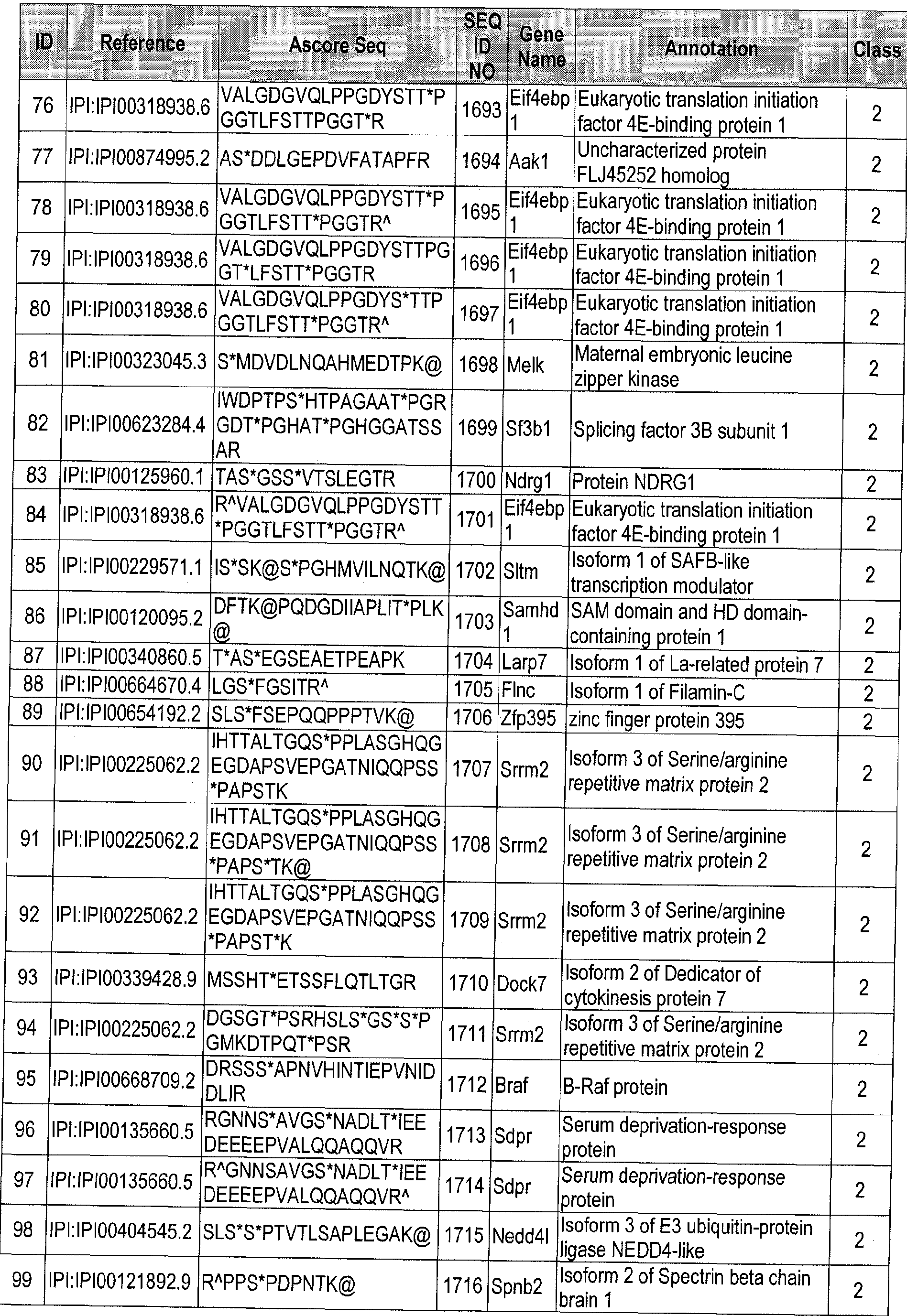

Table 9(b)
rapa median area
ID rapa median SN ratio KU median area ratio KU median SN ratio rstio
1 -1.15E+00 -1.14E+00 -0.8183 -0.9318
2 -2.1437 -2.4238 -0.78695 -0.9492
3 -4.758 -4.9643 -0.6283 -0.5475
4 -4.62805 -4.47925 -0.6283 -0.5475
5 -3.6217 -3.885 -0.6283 -0.5475
6 -1.0246 -1.0724 -0.5666 0.1321
7 -1.881 -1.90875 -0.5354 -0.5361
8 -5.2234 -5.13485 -0.4254 -0.6826
9 -1.8021 -2.01515 -0.4151 -0.4336
10 -1.5621 -1.0464 -0.3725 -0.7405
1 1 -1.4347 -1.3061 -0.3725 -0.7405
12 -1.6324 -1.5779 -0.3224 -0.0637
13 -1.5511 -1.71335 -0.2831 -0.0584
14 -1.78365 -2.00075 -0.2767 0.0092
15 -1.7555 -2.0749 -0.2767 0.0092
16 -1.7499 -1.7737 -0.2767 0.0092
17 -1.5242 -1.76425 -0.2767 0.0092
18 -2.0759 -2.2586 -0.2661 -0.1505
19 -1.9707 -1.7471 -0.2661 -0.1505
20 -1.8874 -1.85315 -0.2661 -0.1505
21 -1.8106 -1.691 -0.2661 -0.1505
22 -1.6838 -1.5207 -0.2661 -0.1505
23 -2.8898 -2.7784 -0.2201 -0.1513
24 -1.0015 -0.8287 -0.2135 -0.3206
25 -3.15205 -2.9693 -0.213 0.1493
26 -1.3868 -0.6688 -0.1992 -0.1577
27 -1.1818 -1.15455 -0.1474 -0.0308
28 -1.7403 -1.8968 -0.1417 0.6197
29 -1.6267 -1.0033 -0.139 -0.2714
30 -1.0844 -1.1514 -0.106 -0.2609
31 -2.78805 -1.56635 -0.1017 -0.8833
32 -3.0877 -2.2749 -0.099 0.6127
33 -2.20425 -2.1077 -0.085 0.023
34 -1.2842 -0.6546 -0.0822 0.1059
35 -1.7868 -2.1396 -0.0816 0.1618
36 -1.2551 -1.22675 -0.0656 -0.106
37 -2.9525 -3.6913 -0.0637 0.7614
rapa median area
rapa median SN ratio KU median area ratio KU median SN ratio ratio
-1.1888 -1.2446 -0.014 -0.0308
-1.2382 -1.25285 -0.0136 0.0107
-2.1557 -2.1257 -0.01 -0.0601
-4.4981 -3.9942 0.0259 -0.0762
-1.7508 -0.9897 0.0386 0.3631
-1.896 -2.1685 0.0563 0.2174
-1.89265 -2.1434 0.0563 0.1073
-1.40465 -1.02655 0.0563 0.2174
-1.896 -2.1685 0.0814 0.17005
-3.6492 -3.6492 0.0966 0.0223
-1.8973 -2.1434 0.1063 0.2306
-1.273 -0.7556 0.1063 0.2306
-2.8428 -2.9973 0.1159 0.0362
-2.8019 -2.9973 0.1159 0.0362
-1.2352 -0.0592 0.1368 0.1632
-3.106 -3.2482 0.1555 0.3337
-2.9525 -3.6913 0.1555 0.3337
-1.298 -1.4299 0.2219 0.413
-1.7403 -1.8968 0.22985 0.1085
-1.3906 -1.2466 0.2704 1.5615
-1.1401 -1.2853 0.2835 0.1358
-2,4187 -0.9488 0.307 0.366
-1.3056 -1.2452 0.3118 0.4053
-1.2719 -1.0234 0.3118 0.4053
-1.4486 -1.5455 0.3219 0.2377
-3.8536 -3.8536 0.3485 0.0243
-1.36155 -1.26665 0.3516 0.4392
-1.889 -1.727 0.3696 1.0368
-2.0572 -2.2717 0.3908 0.1481
-1.626 -2.0445 0.51825 0.767
-1.29335 -1.2722 0.51825 0.767
-1.9492 -2.05475 0.5526 0.865
-1.1567 -1.0106 0.5969 0.9976
-1.1567 -1.0106 0.5969 0.9976
-2.818 -2.9973 1.1365 1.0801
-3.4866 -3.4138 1.2714 1.4529
0.0401 -0.1138 -3.4595 -5.0415
0.011 -0.1502 -3.3715 -3.3715
-0.22145 -0.31255 -2.9948 -2.3042
0.2389 0.2797 -2.8989 -2.0993
-0.2639 -0.32955 -2.8403 -2.8893
-0.2514 -0.2836 -2.7755 -2.167
-0.2469 -0.3415 -2.5654 -2.1344
-0.58015 -0.1451 -2.5295 -2.6811
-0.1752 0.3008 -2.3289 -2.6763
0.2073 0.2067 -2.0546 -2.0769
-0.3079 -0.4553 -1.759 -1.6004
0.13985 0.04745 -1.5237 -1.538
rapa median area
ID KU median area ratio KU median SN ratio ratio
86 -0.1736 -0.41725 -1.3706 -0.9742
87 -0.1646 -0.2464 -1.3468 -1.5444
88 0.3745 0.3233 -1.2893 -1.2512
89 -0.5295 -0.8185 -1.1648 -1.0131
90 -0.4199 -0.3301 -1.1227 -0.4758
91 -0.2531 -0.1626 -1.1227 -0.4758
92 -0.1997 -0.1626 -1.1227 -0.4758
93 0.1923 0.3451 -1.1197 -1.2356
94 -0.0084 0.0364 -1.0814 -1.582
95 -0.3065 -0.4384 -1.0477 -1.431
96 -0.18955 -0.4136 -1.0458 -1.0184
97 -0.15495 -0.3246 -1.0458 -1.0184
98 -0.573 -0.2978 -1.0265 -0.7599
99 0.0109 -0.0378 -1.0209 -0.674
100 0.10215 0.0699 -1.011 -2.1889
101 -2.1207 -2.1939 -6.1589 -5.4183
102 -1.71915 -1.62995 -1.07715 -2.06845
Tab e 9. Classification of mTOR targets identified in the Rapamycin and ] <u-0063794 screens. Class 1 represents downstream effectors of rapamycin-sensitive mTORCl . Class 2 represents downstream effectors of rapamycin-insensitive mTORCl or mTORC2. Class 3 represents the proteins downstream of both mTORCl and mTORC2. Name of the genes and the sequences of the phosphopeptides are shown. Table 9 is provided in two parts because the colums span more than a single page. Table 9(a) contains colums 1-6 of Table 9 and Table 9(b) contains columns 7-10, wherein corresponding entries are identified in an ID column in each section. Information provided in (a) for ID 1 relates to the information provided in (b) for ID 1 and vice versa, information provided in (a) for ID 2 relates to the information provided in (b) for ID 2 and vice versa, and so on.
Table 10:
Rapa BP GO pathways (a):
 Term Count % PValue
GO:0006796~phosphate metabolic process 17 8.854166667 0.009756342
GO:0006897~endocytosis 7 3.645833333 0.010081394
GO:0010324~membrane invagination 7 3.645833333 0.010081394
GO:0006396~RNA processing 11 5.729166667 0.01020022
GO:0007169~transmembrane receptor protein tyrosine
7 3.645833333 0.01110676 kinase signaling pathway
GO:0010608~posttranscriptional regulation of gene
6 3.125 0.01469374 expression
GO:0045792~negative regulation of cell size 4 2.083333333 0.014742447
GO:0001701~in utero embryonic development 8 4.166666667 0.015417559
GO:0051640~organelle localization 4 2.083333333 0.015500935
GO:0051493~regulation of cytoskeleton organization 5 2.604166667 0.015757695
GO:0007049~cell cycle 13 6.770833333 0.015795779
GO:0006417~regulation of translation 5 2.604166667 0.016293879
GO:0017148~negative regulation of translation 3 1.5625 0.019054263
GO:0034622~cellular macromolecular complex assembly 7 3.645833333 0.019238583
GO:0008361~regulation of cell size 5 2.604166667 0.020996944
GO:0009725~response to hormone stimulus 6 3.125 0.022401918
GO:0051056~regulation of small GTPase mediated signal
7 3.645833333 0.023850905 transduction
GO:0030029~actin filament-based process 6 3.125 0.028573614
GO:0048729~tissue morphogenesis 7 3.645833333 0.028642761
GO:0001932~regulation of protein amino acid
5 2.604166667 0.030261806 phosphorylation
GO:0008104~protein localization 14 7.291666667 0.031319732
GO:0000278~mitotic cell cycle 7 3.645833333 0.031803074
GO:0034621 -cellular macromolecular complex subunit
7 3.645833333 0.032351058 organization
GO:0009719~response to endogenous stimulus 6 3.125 0.03367924
GO:0045449~regulation of transcription 31 16.14583333 0.03551581
GO:0007163~establishment or maintenance of cell
3 1.5625 0.036247509 polarity
GO:0080135~regulation of cellular response to stress 4 2.083333333 0.036466875
GO:0006357~regulation of transcription from RNA
12 6.25 0,037453702 polymerase II promoter
GO:0032314~regulation of Rac GTPase activity 2 1.041666667 0.038299417
GO:0040007~growth 6 3.125 0.040063168
GO:0022403~cell cycle phase 8 4.166666667 0.040932988
GO:0031328~positive regulation of cellular biosynthetic
11 5.729166667 0.042602185 process
GO:0032318~regulation of Ras GTPase activity 4 2.083333333 0.042864282
GO:0010604-positive regulation of macromolecule
12 6.25 0.044146336 metabolic process
GO:0022613~ribonucleoprotein complex biogenesis 5 2.604166667 0.044519805
GO:0009891 -positive regulation of biosynthetic process 11 5.729166667 0.044819532
GO:0046822~regulation of nucleocytoplasmic transport 3 1.5625 0.045236118
GO:0006346~methylation-dependent chromatin silencing 2 1.041666667 0.047644564
GO:0016044~membrane organization 7 3.645833333 0.049524346
GO:0007167~enzyme linked receptor protein signaling
7 3.645833333 0.05025049 pathway
ID Term Count % PValue
GO:0035023~regulation of Rho protein signal
76 4 2.083333333 0.051230918 transduction
77 GO:0043009~chordate embryonic development 9 4.6875 0.052874272
78 GO:0006916~anti-apoptosis 4 2.083333333 0.054183284
GO:0009792~embryonic development ending in birth or
79 9 4.6875 0.055294577 egg hatching
80 GO:0044087~regulation of cellular component biogenesis 4 2.083333333 0.055689578
81 GO:0006350~transcription 25 13.02083333 0.055970599
82 GO:0043062~extracellular structure organization 5 2.604166667 0.057303217
GO:0043122~regulation of l-kappaB kinase/N F-kappaB
83 3 1.5625 0.057483236 cascade
84 GO:0051726~regulation of cell cycle 6 3.125 0.057658627
85 GO:0046907~intracellular transport 9 4.6875 0.059056688
86 GO:0006260~DNA replication 5 2.604166667 0.060776557
87 GO:0048598~embryonic morphogenesis 8 4.166666667 0.060833555
88 GO:0043087~regulation of GTPase activity 4 2.083333333 0.061912428
GO.0051129~negative regulation of cellular component
89 4 2.083333333 0.061912428 organization
90 GO:0032386~regulation of intracellular transport 3 1.5625 0.062669535
91 GO:0033043~regulation of organelle organization 5 2.604166667 0.063153096
92 GO:0042325~regulation of phosphorylation 7 3.645833333 0.063602017
93 GO:0045786~negative regulation of cell cycle 3 1.5625 0.065320236
94 GO:0001570~vasculogenesis 3 1.5625 0.065320236
95 GO:0045947~negative regulation of translational initiation 2 1.041666667 0.066065335
96 GO:0006461 -protein complex assembly 6 3.125 0.070461851
97 GO:0070271 -protein complex biogenesis 6 3.125 0.070461851
98 GO:0030833~regulation of actin filament polymerization 3 1.5625 0.070731878
GO:0010557~positive regulation of macromolecule
99 10 5.208333333 0.073220823 biosynthetic process
100 GO:0019220~regulation of phosphate metabolic process 7 3.645833333 0.073259901
101 GO:0051174~regulation of phosphorus metabolic process 7 3.645833333 0.073259901
102 GO:0048589~developmental growth 4 2.083333333 0.073541659
GO:0030838-positive regulation of actin filament
103 2 1.041666667 0.075142677 ' polymerization
104 GO:0030308~negative regulation of cell growth 3 1.5625 0.076284196
105 GO:0030036~actin cytoskeleton organization 5 2.604166667 0.077082848
106 GO:0031399~regulation of protein modification process 5 2.604166667 0.077082848
107 GO:0000902~cell morphogenesis 7 3.645833333 0.080785789
108 GO:0016477~cell migration 6 3.125 0.08471051
GO:0008064~regulation of actin polymerization or
109 3 1.5625 0.084860387 depolymerization
110 GO:0030832~regulation of actin filament length 3 1.5625 0.08778162
111 GO:0002009~morphogenesis of an epithelium 5 2.604166667 0.088103636
112 GO:0006333~chromatin assembly or disassembly 4 2.083333333 0.089801855
113 GO:0010638~positive regulation of organelle organization 3 1.5625 0.090732662
114 GO:0045941 -positive regulation of transcription 9 4.6875 0.091555467
115 GO:0006887~exocytosis 4 2.083333333 0.091694717
116 GO:0008283~cell proliferation 6 3.125 0.092969193
GO:0035020~regulation of Rac protein signal
117 2 1.041666667 0,093035503 transduction
ID Term Count % PValue
118 GO:0000226~microtubule cytoskeleton organization 4 2.083333333 0.093604179
1 19 GO:0060562~epitheliai tube morphogenesis 4 2.083333333 0.093604179
Rapa BP GO pathways (b):

IBTK,EIF4G1,EIF4EBP1 ,EIF4EBP2,ZEB2,MTOR,RICTOR>FAM129A,FXR1
SAP30,SORBS3,TRIM28,RB1 ,CUX1 ,EHMT2,NFIC,SMARCA4
MTOR,RICTOR,SCRIB
BCR.GBF1 ,TBC1 D4,MTOR,RICTOR,SCRIB, IQSEC1
SRPK2,LOC100046628,NACC1,GM9118,AHCTF1 ,GM5611 ,FLNA,GM6477,EIF3A,PTK2,GTF2F1 ,NPM 1 ,GM7289,LOC633387,HELLS,SMARCA4
SRPK2,GM4521 ,BCR,TRIM281LOC544757,PKN2,WNK1,RPS6KB1,PRKD2,PTK2,LOC100048123>ULK 1,AAK1 ,GSK3B,CDK12,EEF2K,BMP2K,PPP1 R12A>MTOR,AKT2
SRPK2>GM4521 ,BCR,TRI 28,LOC544757,PKN2,WNK1)RPS6KB1,PR D2,PTK2,LOC100048123,ULK 1 ,AAK1 ,GSK3B,CDK12,EEF2K,BMP2K,PPP1 R12A,MTOR,AKT2
DAB2, N,GM8786,ULK1 ,SNX17,FKBP15,TRIP10,SCRIB
DAB2,CTTN,GM8786,ULK1 ,SNX17,FKBP15,TRIP10,SCRIB
LOC100046766,SRPK2,PDCD11 ,LOC100046744,EMG1 ,SFRS1 ,LOC100047322,RSL1 D1,SFRS8,PCB P1 ,SRRM2,LARP7,LOC100046735,DHX15,SRRM1,LOC100048559
ZFP106,GRB10,PTK2,IRS2,EIF4EBP1)EIF4EBP2,FLNA
EIF4G1 ,EIF4EBP1,EIF4EBP2,FAM129A,FLNA,FXR1
FOXK1 ,ULK1 ,MTOR,DNAJC2
MIB1 ,DAB2,SFRS1,TCFEB,JUNB,LOC100048559,MLL2,MED1 ,SMARCA4
MYO5A,LOC100046628,PTK2,G 9118,NPM1 ,GM5611 ,GM7289,SCRIB,LOC633387,GM6477
LOC100046628,GM9118,MTAP1B,NPM1 ,GM5611,SPNA2,GM7289,MTOR,RICTOR,LOC633387,GM6 477
LOC100046628,ZC3HC1 ,PDS5B,GM9118,LIN9,GM5611 ,AHCTF1 ,RB1,EHMT2,CD2AP,GM6477,NUM A1,MACF1,GSK3B,NPM1 ,GM7289,DNAJC2,HELLS,LOC633387
EIF4G1 ,EIF4EBP1,EIF4EBP2,FAM129A,FXR1
EIF4EBP1 ,EIF4EBP2,FXR1
SRPK2,PTK2,EIF3A,AHCTF1 , HELLS, FLNA.S ARCA4
LOC100046628,FOXK1 ,GM9118,ULK1,NPM1 ,GM5611,GM7289,MTOR,DNAJC2,LOC6333871GM6477
ZFP106,IRS2,EIF4EBP1,EIF4EBP2,LOC100048123,LOC544757,MTOR,AKT2
BCR,GBF1 ,TBC1 D4,MTOR,RICTOR,SCRIB,IQSEC1
MY05A,INF2,FMNL3,ARHGAP17,RICTOR,INF2Q,FLNA
MIB1,MACF1,TNC,ZEB2,SCRIB,MED1 ,SMARCA4
IBTK,ZEB2, TOR,RICTOR,FAM129A
PALM,LOC100046628,LOC100048600>GM9118,SNX17,GM5611 )AHCTF1 ,FLNA,SCRIB,GM6477,POM 121 ,ERBB2IP,MACF1 ,ULK1 ,RAB1 ,GSK3B,NPM1 ,GM7289,SNX30,ERC1.LOC633387
ZC3HC1,PDS5B,AHCTF1,RB1 ,CD2AP,DNAJC2,HELLS
SRPK2,PTK2,EIF3A,AHCTF1,HELLS,FLNA,SMARCA4
ZFP106,IRS2,EIF4EBP1 ,EIF4EBP2,LOC100048123,LOC544757, TOR,AKT2
BCLAF1.CARHSP1 ,FOXK1 ,FOXK2,AHCTF1 ,ZEB2, 1600027N09RIK,SAP30,SORBS3,GM9791 ,KD 3B ,TCFEB,DNAJC2,HELLS,NACC1,PDCD11 ,TRIM28,CCNL1,RB1 ,EHMT2,JUNB,FLNA,RFC1 ,GTF2F1 ,S MARCC2,TMPO,ZFP516,CUX1 ,NFIC,PBX2,SMARCA4,MED1
MACF1 ,SCRIB,LLGL1
LOC100046628,LOC100048123,GM9118,NP 1 ,LOC544757,GM5611 ,ZEB2,GM7289,MTOR,LOC6333 87,AKT2,GM6477
SAP30,SORBS3,FOXK1 ,TRIM28,RB1 ,CUX1 ,TCFEB,EHMT2,NFIC,PBX2,MED1 ,SMARCA4
MTOR.RICTOR
MYO5A,LOC100046628,GM9118.ULK1 ,NPM1 ,GM5611 ,GM7289,EHMT2,LOC633387, ED1 ,GM6477, SMARCA4
ZC3HC1,PDS5B,AHCTF1 ,RB1 ,EHMT2,CD2AP,DNAJC21HELLS
LOC100046628.GM9118.FOXK1 ,TRIM28,AHCTF1 ,GM5611 ,RB1 ,GM6477,GTF2F1 ,NPM1 ,GM7289,TC FEB,FAM129A,NFIC,PBX2,LOC633387,MED1
Genes .. ■
TBC1D4,MTOR,RICTOR,SCRIB
FOXK1.GTF2F1 JRIM28.AHCTF1 ,MTOR,RB1 ,RICTOR,TCFEB,FAM129A,NFIC,PBX2, ED1
SRPK2,LOC100046628,PDCD11,EIF3A,GM9118,EMG1,NPM1,GM5611,GM7289,LOC633387,GM6477
LOC100046628,GM9118,FOXK1,TRIM28,AHCTF1,GM5611,RB1,GM6477,GTF2F1,NP 1,GM7289,TC FEB,FAM129A,NFIC,PBX2,LOC633387,MED1
GSK3B,PPP1R12A,FLNA
HELLS.SMARCA4
DAB2,CTTN,GM8786,ULK1,SNX17,FKBP15,TRIP10,SCRIB
ZFP106,GRB10,PTK2,IRS2,EIF4EBP1,EIF4EBP2,FLNA
BCR,MTOR,RICTOR,SCRIB
IB1,DAB2,ZEB2,SFRS1,TCFEB,JUNB,LOC100048559,MLL2,MED1,SMARCA4
AKT1S1,ZC3HC1,GSK3B,HELLS
MIB1,DAB2,ZEB2,SFRS1,TCFEB,JUNB,LOC100048559,MLL2,MED1,SMARCA4
PTK2,SPNA2, TOR,RICTOR
BCL-AF1,FOXK1,FOXK2,MYEF2,AHCTF1,ZEB2,1600027N09RIK,SAP30,KDM3B,TCFEB,HELLS,NAC C1,TRIM28,CCNL11RB11JUNB,PHF3,RFC1,GTF2F1,SMARCC2,ZFP5161NFIC,CUX1,PBX2,MED1
MYO5A)PTK2,TNC,LOC100048740,ADAMTS2,SMARCA4
PDCD11,SQSTM1,FLNA
LOC100046628,BCR,FOXK1)GM9118,NPM1,GM5611,G 7289,RB1,SCRIB,JUNB,LOC633387,GM647 7
MYO5A,LOC100046628,LOC100048600,GM9118,GM5611,FLNA,GM6477,MACF1,ERBB2IP,GSK3B,R AB1.NPM1 ,GM7289,CUX1 ,ERC1.LOC633387
RFC1,LIN9,NFIC,DNAJC2,MED1
MIB1 ,MACF1 ,ZEB2,EXT1 ,PBX2,SCRIB,MED1 ,SMARCA4
TBC1 D4,MTOR,RICTOR,SCRIB
PTK2,ULK1,MTAP1B,SPNA2
GSK3B,PPP1R12A,FLNA
LOC100046628.G 9 18.MTAP1 B.NPM1 ,GM5611 ,SPNA2,GM7289, TOR,RICTOR,LOC633387,GM6 477
IBTK, LOC 100046628, GM9118.GM5611 ,ZEB2,RICTOR,RB1 ,GM6477,NP 1 ,G 7289,MTOR,FA 129A .LOC633387
FOXK1,RB1,SCRIB
PTK2,JUNB,SMARCA4
EIF4EBP1 ,EIF4EBP2
LOC100046628,PTK2,NACC11GM9118,GTF2F1,NP 1,AHCTF1,GM5611,GM7289,LOC633387,FLNA, GM6477
LOC100046628,PTK2,NACC1,GM9118,GTF2F1,NPM1,AHCTF1,GM5611,GM7289,LOC633387,FLNA, GM6477
SPNA2,MTOR,RICTOR
FOXK1,GTF2F1,TRIM28,AHCTF1,RB1,TCFEB,FAM129A,NFIC,PBX2(MED1
IBTK,LOC100046628,GM9118,GM5611,ZEB2,RICTOR,RB1,GM6477,NPM1,G 7289,MTOR,FAM129A .LOC633387
IBTK,LOC100046628,GM9118,GM5611,ZEB2,RICTOR,RB1,GM6477,NPM1,GM7289,MTOR,FAM129A .LOC633387
MY05A,ULK1,MED1,SMARCA4
MTOR.RICTOR
FOXK1,ULK1,DNAJC2
INF2,F NL3,ARHGAP17,RICTOR,INF2Q,FLNA
IBTK,ZEB2,MTOR,RICTOR,FAM129A
DAB2IPTK2,MACF1,ULK1,TRIM28,SCRIB,LLGL1
ID Genes
108 PTK2,TNS1,ULK1,ZEB2,CD2AP,SCRIB
109 SPNA2,MTOR,RiCTOR
110 SPNA2,MTOR,RICTOR
111 IB1,TNC,ZEB2,SCRIB,MED1
112 S ARCC2.CHD1 ,HELLS,S ARCA4
113 LOC100046628,GM9118,NP 1,GM5611,GM7289,MTOR,RICTOR,LOC633387,GM6477
114 FOXK1,GTF2F1,TRI 28,AHCTF1,RB1,TCFEB,NFIC,PBX2,MED1
115 MY05A,ARHGAP17,SCRIB,LLGL1
116 IRS2,GSK3B,ZEB2,SCRIB,HELLS,MED1
117 MTOR.RICTOR
118 LOC100046628,PTK21GM9118,MTAP1B,NPM1,GM5611,GM7289,LOC633387,RANBP10,GM6477
119 MIB1,ZEB2,SCRIB,MED1
Rapa BP GO pathways (c):


Rapa KEGG pathways (c):


Ku BP GO (a):

Ku BP GO (b):


Ku BP GO (c):
 74 88 13588 6.25982801 1 0.901699824 71.78744367
74 430 13588 2.562162162 1 0.892310062 71.81242243
74 431 13588 2.55621747 1 0.884669916 72.09697589
74 434 13588 2.538547764 1 0.881167309 72.94148547
74 437 13588 2.521120663 1 0.877904651 73.77204657
74 93 13588 5.923278117 1 0.879820716 75.2555379
74 315 13588 2.914628915 1 0.871223772 75.28140884
74 319 13588 2.878081844 1 0.872300131 76.54568607
74 95 13588 5.798577525 1 0.863873178 76.55268842
74 96 13588 5.738175676 1 0.860545996 77.18216045
74 19 13588 19.32859175 1 0.862220501 78.38406819
74 326 13588 2.81628254 1 0.856532331 78.66045069
Ku KEGG pathways (a):

Ku KEGG pathways (b):


Table 11:

Numal Nuclear mitotic apparatus protein 1
Path Protein PAT1 homolog 1
Pcbp2 Isoform 1 of Poly(rC)-binding protein 2
Pds5b Isoform 1 of Sister chromatid cohesion protein PDS5 homolog B
Peg3 Isoform 1 of Paternally-expressed gene 3 protein
Phf3 PHD finger protein 3
Phldbl Isoform 2 of Pleckstrin homology-like domain family B member 1
Phldb2 Isoform 1 of Pleckstrin homology-like domain family B member 2
Pi4k2a Phosphatidylinositol 4-kinase type 2-alpha
Pkn2 Isoform 1 of Serine/threonine-protein kinase N2
Pla2g4a Cytosolic phospholipase A2
Plekhml Pleckstrin homology domain-containing family M member 1
Ppp1r12a MCG122391 isoform CRA e
Rab1 Ras-related protein Rab-1A
Rps6 29 kDa protein
Rps6kb1 Isoform Alpha I of Ribosomal protein S6 kinase beta-1
Rsl1d1 Putative uncharacterized protein
Sap30 Histone deacetylase complex subunit SAP30
Serbpl Isoform 1 of Plasminogen activator inhibitor 1 RNA-binding protein
Sfrs8 splicing factor arginine/serine-rich 8
Sgta Isoform 1 of Small glutamine-rich tetratricopeptide repeat-containing protein alpha
Smarca4 Putative uncharacterized protein
Snx17 Sorting nexin-17
Srpk2 serine/arginine-rich protein-specific kinase 2
Srrm2 Isoform 3 of Serine/arginine repetitive matrix protein 2
Syapl Synapse-associated protein 1
Tbc1d4 140 kDa protein
Tcfeb Transcription factor EB
TjP2 Tight junction protein ZO-2
Tmem106b Transmembrane protein 106B isoform CRA_b
Tmpo Isoform Beta of Lamina-associated polypeptide 2 isoforms beta/delta/epsilon/gamma
Trim28 Isoform 1 of Transcription intermediary factor 1 -beta
Ubxn7 UBX domain-containing protein 7
Usp24 Isoform 1 of Ubiquitin carboxyl-terminal hydrolase 24
Uvrag UV radiation resistance associated
Wnk1 Serine/threonine-protein kinase WNK1
Zc3hc1 Isoform 1 of Nuclear-interacting partner of ALK
Zfp516 Zinc finger protein 516
Rapa Replicate 2
Mib1 E3 ubiquitin-protein ligase MIB1
rsp6 29 kDa protein
Srrm2 Isoform 3 of Serine/arginine repetitive matrix protein 2
Gm13099 Novel protein similar to preferentially expressed antigen in melanoma-like family
Rnf19b IBR domain containing 3
Rps6 29 kDa protein
Gene Symbol Annotation
Carhspl Calcium-regulated heat stable protein 1
Pgrmc2 Membrane-associated progesterone receptor component 2
FLNA Filamin-A
Ext1 Exostosin-1
Novel protein similar to solute carrier family 28 (Sodium-coupled nucleoside
Gm14085
transporter) member 2
Akt1s1 Proline-rich AKT1 substrate 1
Gm9757 Putative uncharacterized protein
Eif4ebp2 Eukaryotic translation initiation factor 4E-binding protein 2
Gon4l RIKE cDNA 5830417110 gene
Mogatl 2-acylgiycerol O-acyltransferase 1
Eef2k Elongation factor 2 kinase
Dhx15 Putative pre-mRNA-splicing factor ATP-dependent RNA heiicase DHX15
Srrml Isoform 2 of Serine/arginine repetitive matrix protein 1
Setdla SET domain containing 1A
D830015G02Rik Putative uncharacterized protein
Vwa5b1 von Willebrand factor A domain-containing protein 5B1
Ibtk Isoform 2 of Inhibitor of Bruton tyrosine kinase
Erc1;LOC100048600 Isoform 1 of ELKS/RAB6-interacting/CAST family member 1
Adamts2 A disintegrin and metalloproteinase with thrombospondin motifs 2
Grb10 Isoform 3 of Growth factor receptor-bound protein 10
Foxkl Forkhead box protein K1
Gm6988 similar to hCG1640785
Tnc Isoform 1 of Tenascin
Larpl Isoform 1 of La-related protein 1
Alkbh6 24 kDa protein
Larp7 Isoform 1 of La-related protein 7
Ahnak AHNAK nucleoprotein isoform 1
Qsox2 Isoform 3 of Sulfhydryl oxidase 2
Wnk1 Serine/threonine-protein kinase WNK1
Megf11 Isoform 4 of Multiple epidermal growth factor-like domains 11
Ahctfl AT-hook-containing transcription factor 1
Fxr1 Isoform E of Fragile X mental retardation syndrome-related protein 1
Ddx21 Nucleolar RNA heiicase 2
Lin9 Isoform 2 of Lin-9 homolog
D6Wsu116e Isoform 1 of Protein FAM21
Sfrs8 splicing factor arginine/serine-rich 8
Ppfibp2 Isoform 4 of Liprin-beta-2
Hdgfrp2 Isoform 3 of Hepatoma-derived growth factor-related protein 2
Irs2 Insulin receptor substrate 2
Tinf2 Putative uncharacterized protein
Hisppdl 140 kDa protein
Edc3 Enhancer of mRNA-decapping protein 3
Aimll Absent in melanoma 1 -like
hect (homologous to the E6-AP (UBE3A) carboxyl terminus) domain and RCC1
Herd
(CHCI)-like domain (RLD) 1
Fbxw9 F-box and WD-40 domain protein 9
Gene Symbol Annotation
Sfrs18 splicing factor arginine/serine-rich 18
Dnajc2 DnaJ homolog subfamily C member 2
Cux1 cut-like homeobox 1 isoform a
RanbpIO Ran-binding protein 10
Foxk2 Isoform 1 of Forkhead box protein K2
Bodll biorientation of chromosomes in cell division 1 -like
Zeb2 Zinc finger E-box-binding homeobox 2
Bcr Breakpoint cluster region protein
Pdcd11 Protein RRP5 homolog
Hn1l Hematological and neurological expressed 1-like protein
Palm Isoform 1 of Paralemmin
Phf3 PHD finger protein 3
Ptk2 Isoform 1 of Focal adhesion kinase 1
11 0013L07Rik Putative uncharacterized protein
Tcfeb Transcription factor EB
Ehmt2 Isoform 1 of Histone-lysine N-methyltransferase H3 lysine-9 specific 3
Zc3h4 Isoform 2 of Zinc finger CCCH domain-containing protein 4
Ktn1 Isoform 1 of Kinectin
Pbx2 Pre-B-cell leukemia transcription factor 2
Phldbl Isoform 2 of Pleckstrin homology-like domain family B member 1
Cdgap Cdc42 GTPase-activating protein
Eif3a Eukaryotic translation initiation factor 3 subunit A
Tbc1d4 140 kDa protein
Dennd4a hypothetical protein LOC 102442
Wdr91 WD repeat-containing protein 91
Junb Transcription factor jun-B
Srpk2 serine/arginine-rich protein-specific kinase 2
Eif4b Eukaryotic translation initiation factor 4B
Gbf1 Golgi-specific brefeldin A-resistance factor 1
Cd2ap CD2-associated protein
LOC100048559;Sfrs1 Isoform 1 of Splicing factor arginine/serine-rich 1
Ccdc6 coiled-coil domain containing 6
Ndrg3 Protein NDRG3
Ulk1 Putative uncharacterized protein
Arhgap17 Isoform 1 of Rho GTPase-activating protein 17
Spna2 Isoform 2 of Spectrin alpha chain brain
Path Protein PAT1 homolog 1
Bcas3 Isoform 1 of Breast carcinoma-amplified sequence 3 homolog
Atg2a Autophagy-related protein 2 homolog A
Ccnll Isoform 1 of Cyclin-L1
Znrf2 E3 ubiquitin-protein ligase ZNRF2
Larp4 Putative uncharacterized protein
Mtor Isoform 1 of FKBP12-rapamycin complex-associated protein
Gsk3b Glycogen synthase kinase-3 beta
Ranbp9 RAN binding protein 9
dm6a Isoform 1 of Lysine-specific demethylase 6A
C130092011Rik Isoform 1 of Uncharacterized protein KIAA1680
Mtaplb icrotubule-associated protein 1B
Gene Symbol Annotation
Prkd2 Serine/threonine-protein kinase D2
Usp36 Ubiquitin specific peptidase 36
Slc4a1ap solute carrier family 4 (anion exchanger) member 1 adaptor protein
Ugl1 lethal giant larvae homolog 1 isoform 1
Cabinl calcineurin binding protein 1
Pcbpl Poly(rC)-binding protein 1
Eif4ebp1 Eukaryotic translation initiation factor 4E-binding protein 1
Rfd Rfd protein
Pds5b Isoform 1 of Sister chromatid cohesion protein PDS5 homolog B
St5 Isoform 1 of Suppression of tumorigenicity 5
Eif4g1 Isoform 1 of Eukaryotic translation initiation factor 4 gamma 1
Pom121 Nuclear envelope pore membrane protein POM 121
Smarcc2 Isoform 2 of SWI/SNF complex subunit SMARCC2
Eps8l2 Isoform 1 of Epidermal growth factor receptor kinase substrate 8-like protein 2
Plekhml Pleckstrin homology domain-containing family member 1
B230208H17Rik Putative GTP-binding protein Part
MII2 similar to myeloid/lymphoid or mixed-lineage leukemia 2
Snx30 Sorting nexin-30
Rps6kb1 Isoform Alpha I of Ribosomal protein S6 kinase beta-1
Usp24 Isoform 1 of Ubiquitin carboxyl-terminal hydrolase 24
MicalU Isoform 1 of MICAL-like protein 1
Med1 Isoform 4 of Mediator of RNA polymerase II transcription subunit 1
Sorbs3 Vinexin
Phldb2 Isoform 1 of Pleckstrin homology-like domain family B member 2
Isoform 1 of SWI/SNF-related matrix-associated actin-dependent regulator of
Smarcadl
chromatin subfamily A containing DEAD/H box 1
Crkrs Isoform 2 of Cell division cycle 2-related protein kinase 7
Gtf2f1 General transcription factor I IF subunit 1
Scrib Isoform 1 of Protein LAP4
2610110G12Rik Isoform 1 of UPF0635 protein C6orf134 homolog
Mllt4 Isoform 3 of Afadin
Nacd Nucleus accumbens-associated protein 1
Zfp106 Isoform 1 of Zinc finger protein 106
Rictor Isoform 1 of Rapamycin-insensitive companion of mTOR
Tns1 tensin 1
Cttn Src substrate cortactin
Sbnol Isoform 2 of Protein strawberry notch homolog 1
Fmnl3 Isoform 1 of Formin-like protein 3
Syapl Synapse-associated protein 1
Stm3 Striatin-3
Rb1 Retinoblastoma-associated protein
Ehbpl Isoform 2 of EH domain-binding protein 1
Sqstml Isoform 1 of Sequestosome-1
Zc3hc1 Isoform 1 of Nuclear-interacting partner of ALK
Tox4 TOX high mobility group box family member 4
Emg1 Probable ribosome biogenesis protein NEP1
Pcm1 Isoform 1 of Pericentriolar material 1 protein
Gene Symbol Annotation ;
Trip 10 Isoform 3 of Cdc42-interacting protein 4
LOC100048123;Akt2 RAC-beta serine/threonine-protein kinase
Aak1 Isoform 2 of AP2-associated protein kinase 1
Srrm2 Isoform 3 of Serine/arginine repetitive matrix protein 2
Phldb2 Isoform 1 of Pleckstrin homology-like domain family B member 2
Phldbl Isoform 2 of Pleckstrin homology-like domain family B member 1
Ahctfl AT-hook-containing transcription factor 1
Ahctfl AT-hook-containing transcription factor 1
Ahctfl AT-hook-containing transcription factor 1
Arhgap17 Isoform 1 of Rho GTPase-activating protein 17
Rb1 Retinoblastoma-associated protein
Rb1 Retinoblastoma-associated protein
Tox4 TOX high mobility group box family member 4
Tox4 TOX high mobility group box family member 4
Hells Isoform 1 of Lymphocyte-specific helicase
Hells Isoform 1 of Lymphocyte-specific helicase
Hells Isoform 1 of Lymphocyte-specific helicase
Erbb2ip Isoform 1 of Protein LAP2
Eif4ebp2 Eukaryotic translation initiation factor 4E-binding protein 2
Eif4ebp2 Eukaryotic translation initiation factor 4E-binding protein 2
Pds5b Isoform 1 of Sister chromatid cohesion protein PDS5 homolog B
Pds5b Isoform 1 of Sister chromatid cohesion protein PDS5 homolog B
Pds5b Isoform 1 of Sister chromatid cohesion protein PDS5 homolog B
Fam129a Protein Niban
Eif4ebp1 Eukaryotic translation initiation factor 4E-binding protein 1
Ahnak AHNAK nucleoprotein isoform 1
Ahnak AHNAK nucleoprotein isoform 1
Ahnak AHNAK nucleoprotein isoform 1
Eif4ebp1 Eukaryotic translation initiation factor 4E-binding protein 1
Arid 1a AT rich interactive domain 1A
Fam129a Protein Niban
Fam129a Protein Niban
Atxn2 ataxin 2
Atxn2 ataxin 2
RanbpIO Ran-binding protein 10
RanbpIO Ran-binding protein 10
RanbpIO Ran-binding protein 10
RanbpIO Ran-binding protein 10
RanbpIO Ran-binding protein 10
Tox4 TOX high mobility group box family member 4
Tox4 TOX high mobility group box family member 4 yo18a Isoform 4 of Myosin-XVIIIa
Brd2 Isoform 2 of Bromodomain-containing protein 2
6330577E15Rik Uncharacterized protein C10orf78 homolog
Best3 Bestrophin-3
Larpl Isoform 1 of La-related protein 1
Fam62c Isoform 1 of Extended synaptotagmin-3
Gene Symbol Annotation
dn1 Midasin homolog
Rtn4 Isoform 2 of Reticulon-4
Pcdh24 Pcdh24 protein
Synjl similar to mKIAA0910 protein
Zfp318 zinc finger protein 318 isoform 1
Akt1s1 Proline-rich AKT1 substrate 1
Alpk3 myocyte induction differentiation originator
Zc3h14 Isoform 2 of Zinc finger CCCH domain-containing protein 14
Edc4 Isoform 1 of Enhancer of mRNA-decapping protein 4
Srrm2 Isoform 3 of Serine/arginine repetitive matrix protein 2
Kif20b Isoform 1 of M-phase phosphoprotein 1
1110007A13Rik UPF0557 protein C10orf119 homolog
Pi4k2a Phosphatidylinositol 4-kinase type 2-alpha
Top2b DNA topoisomerase 2-beta
BC021381 Isoform 2 of Uncharacterized protein KIAA1931
Bbx Isoform 1 of HMG box transcription factor BBX
Vcl Vinculin
Eif4ebp1 Eukaryotic translation initiation factor 4E-binding protein 1
Rrp15 RRP15-like protein
D10Wsu102e Uncharacterized protein C12orf45 homolog
Braf Isoform 1 of B-Raf proto-oncogene serine/threonine-protein kinase
Atg2b Isoform 1 of Autophagy-related protein 2 homolog B
Npm1 Nucleophosmin
Pcsk5 proprotein convertase subtilisin/kexin type 5
Mtaplb Microtubule-associated protein 1B
Ercc6l DNA excision repair protein ERCC-6-like
Serind Serine incorporator 1
Klf3;L0C100046855 Krueppel-like factor 3
Smarca4 Putative uncharacterized protein
Aak1 Uncharacterized protein FLJ45252 homolog
Eif4ebp2 Eukaryotic translation initiation factor 4E-binding protein 2
Ndrgl Protein NDRG1
Melk Maternal embryonic leucine zipper kinase
Arhgef17 Isoform 1 of Rho guanine nucleotide exchange factor 17
Grit Isoform 2 of Rho/Cdc42/Rac GTPase-activating protein RICS
Mdd mediator of DNA damage checkpoint 1
Nfkb2 NF-kB2 splice variant 4
Pcm1 Isoform 1 of Pericentriolar material 1 protein
Mybbpla Myb-binding protein 1A
Sf3b1 Splicing factor 3B subunit 1
Atrx Transcriptional regulator ATRX
Ccdc88a Isoform 2 of Girdin
Bazl b Isoform 1 of Tyrosine-protein kinase BAZ1B
Nedd4l Isoform 3 of E3 ubiquitin-protein ligase NEDD4-like
Orc6l Origin recognition complex subunit 6
Trp53bp1 Transformation related protein 53 binding protein 1
Map3k2 Mitogen-activated protein kinase kinase kinase 2
Hectd2 Hectd2 protein
Usp10 Ubiquitin carboxyl-terminal hydrolase 10
Gene Symbol Annotation
D830031 N03Rik similar to mKIAA0754 protein
Nck1 non-catalytic region of tyrosine kinase adaptor protein 1
Exosc9 Exosome complex exonuclease RRP45
Dap Death-associated protein 1
Rps6kb1 Isoform Alpha I of Ribosomal protein S6 kinase beta-1
Lmna Isoform C2 of Lamin-A/C
Sltm Isoform 1 of SAFB-like transcription modulator
Sh3pxd2a Isoform 1 of SH3 and PX domain-containing protein 2A
Fine Isoform 1 of Filamin-C
Oxr1 Isoform 2 of Oxidation resistance protein 1
Rin2 Isoform 1 of Ras and Rab interactor 2
Nek9 Serine/threonine-protein kinase Nek9
Pebpl Phosphatidylethanolamine-binding protein 1
Pop1 Processing of 1 ribonuclease P/MRP family
Serhl Serine hydrolase-like protein
Epb4.1l3 Isoform 1 of Band 4.1 -like protein 3
Hnrpll Isoform 1 of Heterogeneous nuclear ribonucleoprotein L-like
Samhdl SAM domain and HD domain-containing protein 1
Zfp828 Zinc finger protein 828
Larp7 Isoform 1 of La-related protein 7
Myc myc proto-oncogene protein
Myo9a Isoform 2 of Myosin-IXa
Gsk3b Glycogen synthase kinase-3 beta
Zfp395 zinc finger protein 395
Bend3 BEN domain-containing protein 3
Akap12 Isoform 1 of A-kinase anchor protein 12
Eif4g1 Isoform 1 of Eukaryotic translation initiation factor 4 gamma 1
Eif4b Eukaryotic translation initiation factor 4B
Dock7 Isoform 2 of Dedicator of cytokinesis protein 7
PatH Protein PAT1 homolog 1
Slc7a11 Cystine/glutamate transporter
Myo9b Isoform 1 of Myosin-IXb
Setd2 SET domain containing 2
Gphn Gephyrin
Erf ETS domain-containing transcription factor ERF
Spnb2 Isoform 2 of Spectrin beta chain brain 1
Phip PH-interacting protein
Sdpr Serum deprivation-response protein
Tcofl Treacle protein
Pwp1 Periodic tryptophan protein 1 homolog
Rbl1 Isoform Long of Retinoblastoma-like protein 1
Eef1b2 Elongation factor 1 -beta
Phactr4 Isoform 1 of Phosphatase and actin regulator 4
C230081A13Rik Tyrosine-protein kinase-protein kinase SgK269
Ahnak2 Putative uncharacterized protein
Table 11. Proteins with downregulated phosphorylation identified in the rapamycin and Ku- 0063794 screen.
Claims
1. A method for determining mTOR kinase activity in a cancer cell, the method comprising
(a) obtaining a cancer cell from a subject diagnosed to have a cancer,
(b) determining the level of GrblO phosphorylation in the cell, and
(c) comparing the level of GrblO phosphorylation to a reference level, wherein if the level of GrblO phosphorylation in the cell is higher than the reference level, then the cell is determined to exhibit an elevated level of mTOR kinase activity.
2. The method of claim 1, further comprising selecting a method of treatment based on the cell exhibiting an elevated level of mTOR kinase activity.
3. The method of claim 2, wherein if the cell is determined to exhibit an elevated level of mTOR kinase activity, then the selected method of treatment comprises
administering an effective amount of an mTOR kinase inhibitor to the subject.
4. The method of claim 3, wherein the method of treatment further comprises administering an effective amount of a compound that stabilizes GrblO or that inhibits the degradation of GrblO and/or an effective amount of a PI3K inhibitor to the subject.
5. The method of claim 3 or 4, wherein the mTOR inhibitor is an allosteric mTOR kinase inhibitor or a catalytic mTOR kinase inhibitor.
6. The method of claim 5, wherein the allosteric mTOR kinase inhibitor is rapamycin or a rapamycin analog, or wherein the catalytic mTOR kinase inhibitor is an ATP- competitive mTOR kinase inhibitor.
7. The method of any of claims 3-6, wherein the mTOR inhibitor is an mTORCl inhibitor.
8. The method of any of claims 3-6, wherein the mTOR inhibitor is an mTORCl/2 inhibitor.
9. The method of claim 6, wherein the rapamycin analog is Ridaforolimus, Sirolimus or Everolimus.
10. The method of any of claims 3-4, wherein the mTOR kinase inhibitor is PP242, PP30, AZD8055, OSI-027, WYE354, INK-128, XL388,or torinl .
11. The method of any of claims 3-4, wherein the mTOR inhibitor is a dual
PI3 /mTOR kinase inhibitor.
12. The method of claim 11, wherein the dual PI3K/mTOR kinase inhibitor is NVP-
BEZ235, BGT-226, XL-765, GSK2126458 or SF1126.
13. The method of any of claims 4-12, wherein the PI3K inhibitor is ZSTK474, TGX221, GDC0941, or LY294002, XL 147, PX147, BKM120, GSK 615, CALlOl, or PX- 866.
14. The method of any of claims 4-12, wherein the PI3K inhibitor is a dual
PI3K/mTOR kinase inhibitor.
15. The method of claim 14, wherein the dual PI3K/mTOR kinase inhibitor is NVP-
BEZ235, BGT-226, XL-765, GSK2126458 or SF1 126.
16. The method of any of claims 3-5, wherein the method of treatment further comprises administering an effective amount of an Akt inhibitor to the subject.
17. The method of claim 16, wherein the Akt inhibitor is perifosine, GSK690693, A443654 or MK2206.
18. The method of any of claims 4- 17, wherein the compound that inhibits the degradation of GrblO is a ubiquitin ligase inhibitor.
19. The method of claim 18, wherein the ubiquitin ligase inhibitor is an E3 ubiquitin ligase inhibitor.
20. The method of any of claims 2-19, further comprising carrying out the selected method of treatment.
21. A method for selecting a treatment of a cancer, the method comprising,
(a) obtaining a cancer cell from a subject diagnosed to have a cancer exhibiting an elevated level of mTOR activity,
(b) determining the level of GrblO expression in the cell, and
(c) comparing the level of GrblO expression to a reference level, wherein if the level of GrblO expression in the cell is higher than the reference level, then the cell is determined to exhibit a high risk of expressing an elevated level ofPDK, Akt, and/or MAPK activity upon being contacted with an mTORCl inhibitor.
22. The method of claim 21 , the method further comprising selecting a method of treatment based on the cell exhibiting a high risk of expressing an elevated level ofPDK, Akt, and/or MAPK activity upon being contacted with an mTORC 1 inhibitor.
23. The method of claim 22, wherein the method of treatment comprises
administering (i) an effective amount of an mTOR kinase inhibitor and/or (ii) an effective amount of an IGFIR, EGFR, PI3K, Akt, MEK, and/or RSK inhibitors and/or of a compound stabilizing GrblO to the subject, or
administering an effective amount of a dual or multi-target inhibitor that inhibits mTOR and inhibits IGFR, EGFR, PI3K, Akt, MEK, and/or RSK and/or of a compound stabilizing GrblO to the subject.
24. The method of claim 23, wherein the mTOR inhibitor is an allosteric mTOR kinase inhibitor or a catalytic mTOR kinase inhibitor.
25. The method of claim 24, wherein the allosteric mTOR kinase inhibitor is rapamycin or a rapamycin analog, or wherein the catalytic mTOR kinase inhibitor is an ATP- competitive mTOR kinase inhibitor.
26. The method of any of claims 23-25, wherein the mTOR inhibitor is an mTORCl inhibitor.
27. The method of any of claims 23-25, wherein the mTOR inhibitor is an mTORC 1/2 inhibitor.
28. The method of any of claims 23-24, wherein the mTOR inhibitor is rapamycin , a rapamycin analog, Ku0063794, or AZD8055.
29. The method of claim 28, wherein the rapamycin analog is Ridaforolimus, Sirolimus or Everolimus.
30. The method of any of claims 23-24, wherein the mTOR kinase inhibitor is PP242,
PP30, AZD8055, OSI-027, WYE354, INK-128, XL388,or torinl.
31. The method of any of claims 23-24, wherein the mTOR inhibitor is a dual PI3K/mTOR kinase inhibitor.
32. The method of claim 31, wherein the dual PI3 /mTOR kinase inhibitor is NVP- BEZ235, BGT-226, XL-765, GSK2126458 or SF1 126.
33. The method of any of claims 24-32, wherein the PI3K inhibitor is ZSTK474, TGX221, GDC0941, or LY294002, XL147, PX147, BKM120, GSK 615, CALlOl , or PX-
866.
34. The method of any of claims 24-32, wherein the PI3K inhibitor is a dual
PI3K7mTOR kinase inhibitor.
35. The method of claim 34, wherein the dual PI3K/mTOR kinase inhibitor is NVP- BEZ235, BGT-226, XL-765, GSK2126458 or SF1126.
36. The method of any of claims 23-35, wherein the method of treatment further comprises administering an effective amount of an Akt inhibitor to the subject.
37. The method of claim 36, wherein the Akt inhibitor is perifosine, GSK690693, A443654 or MK2206.
38. The method of any of claims 23-37, wherein the compound stabilizing GrblO is an ubiquitin ligase inhibitor.
39. The method of claim 38, wherein the ubiquitin ligase inhibitor is an E3 ubiquitin ligase inhibitor.
40. The method of any of claims 22-39, further comprising carrying out the selected method of treatment.
41. A method for determining mTOR kinase activity in a cell, the method comprising
(a) determining the level of phosphorylation of a plurality of phosphorylation sites disclosed in Table 1, 2, 3, 7, or 8, or 11 in the cell; and
(b) comparing the level of phosphorylation determined in step (a) to a reference level, wherein
(i) if the level of phosphorylation determined in step (b) is higher than the reference level, then the cell is determined to exhibit an elevated level of mTOR kinase activity, or
(ii) if the level of phosphorylation determined in step (b) is equal or lower than the reference level, then the cell is determined to not exhibit an elevated level of mTOR kinase activity.
42. The method of claim 41, wherein the method further comprises determining whether the cell carries a mutation in a gene involved in an mTOR signaling pathway.
43. The method of claim 42, wherein the gene involved in an mTOR signaling pathway is a gene involved in IGF signaling, PI3K signaling, AKT signaling, Ras signaling, or Rb signaling.
44. The method of claim 43, wherein the gene involved in an mTOR signaling pathway is TSCl/2, a receptor tyrosine kinases (RTK), PI3K, PTEN, Akt, Ras, Raf, MEK, LKB, or NF2.
45. The method of any of claims 41-44, wherein the plurality of phosphorylation sites is selected from the list consisting of a GrblO phosphorylation site, a pNDRG3
phosphorylation site, a CDK12 phosphorylation site, and a SRPK2 phosphorylation site.
46. The method of claim 45, wherein the GrblO phosphorylation site is comprised in the amino acid sequence MNILSS*QS*PLHPSTLNAVIHR (SEQ ID NO: 28), the amino acid sequence MNILGS*QS*PLHPSTLSTVIHR (SEQ ID NO: 29), or a homologous GrblO sequence, wherein * denotes the preceding amino acid residue as a phosphorylation site.
47. The method of claim 45, wherein the GRb 10 phosphorylation site comprises an S421 phosphorylation site and/or an S423 phosphorylation site of mouse GrblO isoform 3, an S501 phosphorylation site and/or an S503 phosphorylation site of mouse GrblO isoform 1, or anS474 phosphorylation site and/or an S476 in human isoform 3, or a homologous GrblO phosphorylation site.
48. The method of any of claims 41-47, wherein the method comprises determining the level of phosphorylation of at least 2, at least 3, at least 4, at least 5, at least 6, at least 7, at least 8, at least 9, at least 10, at least 15, at least 20, at least 25, at least 30, at least 40, at least 50, at least 60, at least 70, at least 80, at least 90, or at least 100 phosphorylation sites disclosed in Table 1, 2, 3, 7, 8, or 11.
49. The method of any of claims 41-48, wherein
(iii) if a phosphorylation site determined to exhibit a higher level of phosphorylation in the cell as compared to the reference level is a class I phosphorylation site, then the cell is determined to exhibit an elevated level of rapamycin-sensitive mTORCl activity,
(iv) if a phosphorylation site determined to exhibit a higher level of phosphorylation in the cell as compared to the reference level is a class II phosphorylation site, then the cell is determined to exhibit an elevated level of rapamycin-insensitive mTORCl and/or mTORC2 activity, or
(vi) if a phosphorylation site determined to exhibit a higher level of phosphorylation in the cell as compared to the reference level is a class III phosphorylation site, then the cell is determined to exhibit an elevated level of rapamycin-sensitive mTOR activity, or an elevated level of rapamycin-insensitive mTOR activity, or both.
50. The method of any of claims 41-49, wherein the method further comprises selecting a method of treatment of the subject based on the level of phosphorylation of the plurality of phosphorylation sites.
51. The method of claim 50, wherein
if the cell is determined to exhibit an elevated level of mTOR kinase activity, then a method of treatment is selected that comprises administering an effective amount of an mTOR kinase inhibitor to the subject, or
if the cell is determined to not exhibit an elevated level of mTOR kinase activity, then a method of treatment is selected that does not comprise administering an mTOR kinase inhibitor.
52. The method of claim 50, wherein
if the cell is determined to exhibit an elevated level of rapamycin-sensitive mTORCl activity, then a method of treatment is selected that comprises administering an effective amount of an inhibitor of rapamycin-sensitive mTORCl kinase activity to the subject,
if the cell is determined to exhibit an elevated level of rapamycin-insensitive mTORCl and/or mTORC2 activity, then a method of treatment is selected that comprises administering an effective amount of an inhibitor of rapamycin-insensitive mTORCl kinase activity to the subject, or
if the cell is determined to exhibit an elevated level of rapamycin-sensitive mTOR kinase activity, or an elevated level of rapamycin-insensitive mTOR kinase activity, or both, then a method of treatment is selected that comprises administering an effective amount of an inhibitor of rapamycin-sensitive and rapamycin-insensitive mTOR kinase activity or a combination of an inhibitor of rapamycin-sensitive mTOR kinase activity and an inhibitor of rapamycin-insensitive mTOR kinase activity to the subject.
53. The method of any of claims 41-52, wherein the method further comprises determining the level of expression of GrblO in the cell.
54. The method of any of claims 41-52, wherein the method further comprises determining the level of expression of PTEN in the cell.
55. The method of any of claims 41-52, wherein the method further comprises determining the level of expression of GrblO and of PTEN in the cell.
56. The method of any of claims 53-55, wherein determining the level of expression comprises determining a level of protein, a level of mRNA, or a level of protein
phosphorylation in the cell.
57. The method of any of claims 53-56, wherein the method further comprises comparing the level of expression determined in the cell to a reference level, and
if the level of expression in the cell is lower than the reference level, then the cell is determined to exhibit an elevated level of P13K activity.
58. The method of any of claims 50-57, wherein the method of treatment further comprises administering an effective amount of a compound that stabilizes GrblO or that inhibits degradation of GrblO to the subject.
59. The method of claim 58, wherein the compound that inhibits degradation of GrblO is a ubiquitin ligase inhibitor.
60. The method of claim 59, wherein the ubiquitin ligase inhibitor is an E3 ubiquitin ligase inhibitor.
61. The method of any of claims 50-60, wherein the method of treatment further comprises administering an effective amount of a PI3K inhibitor to the subject.
62. The method of any of claims 51-61, wherein the mTOR inhibitor is an allosteric mTOR kinase inhibitor or a catalytic mTOR kinase inhibitor.
63. The method of claim 62, wherein the allosteric mTOR kinase inhibitor is rapamycin or a rapamycin analog, or wherein the catalytic mTOR kinase inhibitor is an ATP- competitive mTOR kinase inhibitor.
64. The method of any of claims 51-63, wherein the mTOR inhibitor is an mTORCl inhibitor.
65. The method of any of claims 51 -63, wherein the mTOR inhibitor is an mTORCl/2 inhibitor.
66. The method of claim 63, wherein the rapamycin analog is Ridaforolimus, Sirolimus or Everolimus.
67. The method of any of claims 51-62, wherein the mTOR kinase inhibitor is PP242,
PP30, AZD8055, OSI-027, WYE354, INK-128, XL388,or torinl.
68. The method of any of claims 51-62, wherein the mTOR inhibitor is a dual PI3K/mTOR kinase inhibitor.
69. The method of claim 68, wherein the dual PI3K/mTOR kinase inhibitor is NVP- BEZ235, BGT-226, XL-765, GSK2126458 or SF1126.
70. The method of any of claims 51 -69, wherein the PI3K inhibitor is ZSTK474, TGX221, GDC0941, or LY294002, XL147, PX147, BKM120, GSK 615, CALlOl, or PX-
866.
71. The method of any of claims 51 -70, wherein the PI3K inhibitor is a dual
PI3K/mTOR kinase inhibitor.
72. The method of claim 71, wherein the dual PI3K/mTOR kinase inhibitor is NVP- BEZ235, BGT-226, XL-765, GSK2126458 or SF1126.
73. The method of any of claims 51-72, wherein the method of treatment further comprises administering an effective amount of an Akt inhibitor to the subject.
74. The method of claim 73, wherein the Akt inhibitor is perifosine, GSK690693, A443654 or MK2206.
75. The method of any of claims 51 -74, wherein the method further comprises carrying out the selected method of treatment.
76. The method of any of claims 41-75, wherein the cell is a cancer cell.
77. The method of any of claims 41-76, wherein the cell is a cell obtained from a tumor in a subject.
78. The method of any of claims 41-76, wherein the method further comprises obtaining the cell from a subject.
79. A method for determining mTOR kinase activity in a cell, the method comprising (a) determining the level of phosphorylation of a phosphorylation site disclosed in
Table 1, 2, 3, 7, 8, or 11 in the cell; and
(b) comparing the level of phosphorylation determined in step (a) to a reference level, and
(i) if the level of phosphorylation determined in step (b) is higher than the reference level, then the cell is determined to exhibit an elevated level of mTOR kinase activity, or
(ii) if the level of phosphorylation determined in step (b) is equal or lower than the reference level, then the cell is determined to not exhibit an elevated level of mTOR kinase activity.
80. The method of claim 79, wherein the method further comprises determining whether the cell carries a mutation in a gene involved in an mTOR signaling pathway.
81. The method of claim 80, wherein the gene involved in an mTOR signaling pathway is a gene involved in IGF signaling, PI3K signaling, AKT signaling, EGF receptor signaling, GF receptor signaling, Ras signaling, Raf signaling, or Rb signaling.
82. The method of claim 81 , wherein the gene involved in an mTOR signaling pathway is TSCl/2.
83. The method of any of claims 79-82, wherein
the phosphorylation site is a phosphorylation site not previously known to be a phosphorylation site targeted by mTOR; or
wherein the phosphorylation site is selected from the list consisting of a GrblO phosphorylation site, a pNDRG3 phosphorylation site, a CDK12 phosphorylation site, and a SRPK2 phosphorylation site.
84. The method of claim 83, wherein the GrblO phosphorylation site is an S421 phosphorylation site.
85. The method of claim 83, wherein the GrblO phosphorylation site is an S423 phosphorylation site.
86. The method of any of claims 79-85, wherein
(iii) if a phosphorylation site determined to exhibit a higher level of phosphorylation in the cell as compared to the reference level is a class I phosphorylation site, then the cell is determined to exhibit an elevated level of rapamycin-sensitive mTORCl activity,
(iv) if a phosphorylation site determined to exhibit a higher level of phosphorylation in the cell as compared to the reference level is a class II phosphorylation site, then the cell is determined to exhibit an elevated level of rapamycin-insensitive mTORCl and/or mTORC2 activity, or
(vi) if a phosphorylation site determined to exhibit a higher level of phosphorylation in the cell as compared to the reference level is a class III phosphorylation site, then the cell is determined to exhibit an elevated level of rapamycin-sensitive mTOR activity, or an elevated level of rapamycin-insensitive mTOR activity, or both.
87. The method of any of claims 79-86, wherein the method further comprises selecting a method of treatment of the subject based on the level of phosphorylation of the phosphorylation site.
88. The method of claim 87, wherein
if the cell is determined to exhibit an elevated level of mTOR kinase activity, then a method of treatment is selected that comprises administering an effective amount of an mTOR kinase inhibitor to the subject, or
if the cell is determined to not exhibit an elevated level of mTOR kinase activity, then a method of treatment is selected that does not comprise administering an mTOR kinase inhibitor.
89. The method of claim 87, wherein if the cell is determined to exhibit an elevated level of rapamycin-sensitive mTORCl activity, then a method of treatment is selected that comprises administering an effective amount of an inhibitor of rapamycin-sensitive mTORCl kinase activity to the subject,
if the cell is determined to exhibit an elevated level of rapamycin-insensitive mTORCl and/or mTORC2 activity, then a method of treatment is selected that comprises administering an effective amount of an inhibitor of rapamycin-insensitive mTORC 1 kinase activity to the subject, or
if the cell is determined to exhibit an elevated level of rapamycin-sensitive mTOR kinase activity, or an elevated level of rapamycin-insensitive mTOR kinase activity, or both, then a method of treatment is selected that comprises administering an effective amount of an inhibitor of rapamycin-sensitive and rapamycin-insensitive mTOR kinase activity or a combination of an inhibitor of rapamycin-sensitive mTOR kinase activity and an inhibitor of rapamycin-insensitive mTOR kinase activity to the subject.
90. The method of any of claims 79-89, wherein the method further comprises determining the level of expression of GrblO in the cell.
91. The method of any of claims 79-89, wherein the method further comprises determining the level of expression of PTEN in the cell.
92. The method of any of claims 79-89, wherein the method further comprises determining the level of expression of GrblO and of PTEN in the cell.
93. The method of any of claims 90-92, wherein determining the level of comprises determining a level of protein, a level of mRNA, or a level of protein
phosphorylation in the cell.
94. The method of any of claims 90-93, wherein the method further comprises comparing the level of expression determined in the cell to a reference level, and
if the level of expression in the cell is lower than the reference level, then the cell is determined to exhibit an elevated level of PI3K activity.
95. The method of any of claims 87-94, wherein the method of treatment further comprises administering an effective amount of a compound that stabilizes GrblO or that inhibits degradation of GrblO to the subject.
96. The method of claim 95, wherein the compound that inhibits degradation of
GrblO is a ubiquitin ligase inhibitor.
97. The method of claim 96, wherein the ubiquitin ligase inhibitor is an E3 ubiquitin ligase inhibitor.
98. The method of any of claims 87-97, wherein the method of treatment further comprises administering an effective amount of a PI3K inhibitor to the subject.
99. The method of any of claims 88-98, wherein the mTOR inhibitor is an allosteric mTOR kinase inhibitor or a catalytic mTOR kinase inhibitor.
100. The method of claim 99, wherein the allosteric mTOR kinase inhibitor is rapamycin or a rapamycin analog, or wherein the catalytic mTOR kinase inhibitor is an ATP- competitive mTOR kinase inhibitor.
101. The method of any of claims 88-98, wherein the mTOR inhibitor is an mTORCl inhibitor.
102. The method of any of claims 88-98, wherein the mTOR inhibitor is an mTORCl/2 inhibitor.
103. The method of claim 100, wherein the rapamycin analog is Ridaforolimus, Sirolimus or Everolimus.
104. The method of any of claims 88-98, wherein the mTOR kinase inhibitor is
PP242, PP30, AZD8055, OSI-027, WYE354, IN -128, XL388, or torinl .
105 The method of any of claims 88-98, wherein the mTOR inhibitor is a dual PI3K/mTOR kinase inhibitor.
106. The method of claim 105, wherein the dual PI3 /mTOR kinase inhibitor is NVP-BEZ235, BGT-226, XL-765, GSK2126458 or SF1126.
107. The method of any of claims 98- 106, wherein the PI3K inhibitor is ZSTK474,
TGX221, GDC0941, or LY294002, XL147, PX147, BKM120, GSK 615, CALlOl, or PX- 866.
108. The method of any of claims 98-106, wherein the PI3K inhibitor is a dual PDK/mTOR kinase inhibitor.
109. The method of claim 108, wherein the dual PI3K/mTOR kinase inhibitor is NVP-BEZ235, BGT-226, XL-765, GSK2126458 or SF1126.
110. The method of any of claims 88-109, wherein the method of treatment further comprises administering an effective amount of an Akt inhibitor to the subject.
1 11. The method of claim 110, wherein the Akt inhibitor is perifosine, GSK690693, A443654 or MK2206.
1 12. The method of any of claims 88-111, wherein the method further comprises carrying out the selected method of treatment.
113. The method of any of claims 79-112, wherein the cell is a cancer cell.
114. The method of any of claims 79-1 13, wherein the cell is a cell obtained from a tumor in a subject.
115. A phosphoproteomics array, comprising a plurality of phosphosensitive antibodies or antibody fragments specifically binding to a plurality of phosphorylation sites disclosed in Table 1, 2, 3, 7, 8, or 11.
1 16. The phosphoproteomics array of claim 115, wherein the antibodies or antibody fragments are immobilized on a solid substrate.
117. The phosphoproteomics array of claim 115 or 116, wherein the plurality of phosphosensitive antibodies or antibody fragments comprises antibodies or antibody fragments specifically binding to at least 2, at least 3, at least 4, at least 5, at least 6, at least 7, at least 8, at least 9, at least 10, at least 15, at least 20, at least 25, at least 30, at least 40, at least 50, at least 60, at least 70, at least 80, at least 90, or at least 100 phosphorylation sites disclosed in Table 1, 2, 3, 7, 8, or 11.
118. The phosphoproteomics array of claim 115 or 116, wherein the plurality of phosphosensitive antibodies or antibody fragments comprises antibodies or antibody fragments selected from the group comprising anti-phospho-S501/503-GrblO, anti-mTOR, anti-phospho-mTOR (S2481), anti-GrblO (human), anti-phospho-Akt (Ser473), anti-Akt, anti-S6K, anti-phospho-S6K (T389), anti-IRS2, anti-PARP, anti-caspase 3, anti-4EBP, anti- 4EBP (Ser37/46), anti-phospho-ribosomal protein S6 (Ser235/236), anti-ribosomal protein S6, anti-phospho-ERKl/2, anti-phospho-IRS (Y612), anti-p85 and anti-pl 10 of PI3K, anti ERKl/2, and anti-HA antibodies and antibody fragments.
119. A method of using a phosphoproteomics array to determine mTOR activity in a cancer cell, the method comprising
(a) contacting a proteinaceous sample derived from a cancer cell with the
phosphoproteomics array of any of claims E1-E4 under conditions suitable for a protein expressed in the cell to bind to an antibody or antibody fragment of the array,
(b) determining the level of phosphorylated protein bound to an antibody or antibody fragment of the array,
(c) comparing the level of phosphorylated protein bound to an antibody or antibody fragment of the array to a reference level, wherein if the level phosphorylated protein in the sample derived from the cell is higher than the reference level, then the cell is determined to exhibit an elevated level of mTOR kinase activity.
120. The method of claim 119, the method further comprising generating a phosphoproteomic profile of the cell, wherein the profile comprises phosphorylation values of a plurality of proteins expressed in the cell.
121. The method of claim 120, wherein the method further comprises comparing the phosphoproteomic profile of the cell with a phosphoproteomic profile of a control cell, wherein, if the phosphoproteomic profile of the cell is substantially similar to that of the control cell, then the cell is determined to exhibit a level of mTOR kinase activity similar to that of the control cell.
122. The method of claim 121, wherein the control cell is a cancer cell.
123. The method of claim 122, wherein the control cell is a rapamycin-sensitive cancer cell.
124. The method of claim 122, wherein the control cell is a rapamycin insensitive cancer cell.
125. The method of claim 124, wherein the control cell is a cell exhibiting normal mTOR kinase activity.
126. The method of claim 119-125, wherein the method further comprises classifying a cell that is determined to exhibit an elevated level of mTOR kinase activity, wherein
if a protein determined to exhibit a higher level of phosphorylation in the cell as compared to the reference level is a class I protein, then the cell is determined to exhibit an elevated level of rapamycin-sensitive mTORCl activity,
if a protein determined to exhibit a higher level of phosphorylation in the cell as compared to the reference level is a class II protein, then the cell is determined to exhibit an elevated level of rapamycin-insensitive mTORCl and/or mTORC2 activity, or
if a protein determined to exhibit a higher level of phosphorylation in the cell as compared to the reference level is a class III protein, then the cell is determined to exhibit an elevated level of rapamycin-sensitive mTOR activity, or an elevated level of rapamycin- insensitive mTOR activity, or both.
127. A method of identifying an mTOR kinase inhibitor, the method comprising
(a) contacting an mTOR kinase with a polypeptide comprising a phosphorylation site disclosed in Table 1, 2, 3, 7, 8, or 1 1 under conditions suitable for the mTOR kinase to phosphorylate the phosphorylation site in the presence of a candidate agent,
(b) determining the level of phosphorylation at the phosphorylation site,
(c) comparing the level of phosphorylation determined in step (b) to a reference level, wherein if the level determined in step (b) is lower than the reference level, then the candidate agent is identified as an mTOR kinase inhibitor.
128. The method of claim 127, wherein the phosphorylation site is a GrblO phosphorylation site.
129. The method of claim 127 or 128, wherein the candidate agent is a polypeptide, an aptamer, an adnectin, or a small molecule compound.
130. The method of any of claims 127-129, wherein the reference level is a level of phosphorylation at the phosphorylation site determined in the absence of the candidate agent.
131. The method of any of claims 127-130, wherein the level determined in step (b) is lower than the reference level, if the level determined in step (b) is less than 75%, less than 50%, less than 40%, less than 30%, less than 25%, less than 20%, less than 10%, less than 5%, less than 2.5%, or less than 1% of the reference level.
132. The method of any of claims 127-131 , wherein the contacting is performed in vitro.
133. The method of any of claims 127-131, wherein the contacting is performed in vivo.
134. An antibody or antibody fragment, wherein the antibody or antibody fragment specifically binds to a phosphorylation site described in Table 1, 2, 3, 7, 8, or 11.
135. The antibody or antibody fragment of claim 134, wherein the antibody or antibody fragment is a phospho-sensitive antibody or antibody fragment.
136. The antibody or antibody fragment of claim 134 or 135, wherein the antibody or antibody fragment specifically binds to a phosphorylation site of a protein described in Table 1, 2, or 3.
137. The antibody or antibody fragment of any of claims 134-136, wherein the antibody or antibody fragment is an anti-phospho-NDRG3 (Ser331) antibody or antibody fragment.
138. The antibody or antibody fragment of any of claims 134-136, wherein the antibody or antibody fragment is an anti-phospho-S501/503-GrblO antibody or antibody fragment.
139. The antibody or antibody fragment of any of claims 134-136, wherein the antibody or antibody fragment is an Anti-CDC2-related Kinase, Arg/Ser-Rich (Ser437) antibody or antibody fragment.
140. A method for determining mTOR kinase activity in a cell, the method comprising
(a) obtaining a cell from a subject,
(b) determining the level of Grb 10 phosphorylation in the cell, and
(c) comparing the level of Grb 10 phosphorylation to a reference level, wherein if the level of Grb 10 phosphorylation in the cell is higher than the reference level, then the cell is determined to exhibit an elevated level of mTOR kinase activity.
141. A method for determining mTOR kinase activity in a cell, the method comprising
(a) obtaining a cell from a subject,
(b) determining the level of FOXKl phosphorylation in the cell, and
(c) comparing the level of FOXKl phosphorylation to a reference level, wherein if the level of FOXKl phosphorylation in the cell is higher than the reference level, then the cell is determined to exhibit an elevated level of mTOR kinase activity.
142. A method for determining mTOR kinase activity in a cell, the method comprising (a) obtaining a cell from a subject,
(b) determining the level of ZEB2 phosphorylation in the cell, and
(c) comparing the level of ZEB2 phosphorylation to a reference level, wherein if the level of ZEB2 phosphorylation in the cell is higher than the reference level, then the cell is determined to exhibit an elevated level of mTOR kinase activity.
143. A method for determining mTOR kinase activity in a cell, the method comprising
(a) obtaining a cell from a subject,
(b) determining the level of NDRG3 phosphorylation in the cell, and
(c) comparing the level of NDRG3 phosphorylation to a reference level, wherein if the level of NDRG3 phosphorylation in the cell is higher than the reference level, then the cell is determined to exhibit an elevated level of mTOR kinase activity.
144. A method for determining mTOR kinase activity in a cell, the method comprising
(a) obtaining a cell from a subject,
(b) determining the level of LARPl phosphorylation in the cell, and
(c) comparing the level of LARPl phosphorylation to a reference level, wherein if the level of LARPl phosphorylation in the cell is higher than the reference level, then the cell is determined to exhibit an elevated level of mTOR kinase activity.
145. A method for determining mTOR kinase activity in a cell, the method comprising
(a) obtaining a cell from a subject,
(b) determining the level of SRPK2 phosphorylation in the cell, and
(c) comparing the level of SRP 2 phosphorylation to a reference level, wherein if the level of SRPK2 phosphorylation in the cell is higher than the reference level, then the cell is determined to exhibit an elevated level of mTOR kinase activity.
146. A method for determining mTOR kinase activity in a cell, the method comprising
(a) obtaining a cell from a subject,
(b) determining the level of CDK12 phosphorylation in the cell, and (c) comparing the level of CD 12 phosphorylation to a reference level, wherein if the level of CDK12 phosphorylation in the cell is higher than the reference level, then the cell is determined to exhibit an elevated level of mTOR kinase activity.
147. A method for determining mTOR kinase activity in a cell, the method comprising
(a) obtaining a cell from a subject,
(b) determining the level of MIBl phosphorylation in the cell, and
(c) comparing the level of MIBl phosphorylation to a reference level, wherein if the level of MIBl phosphorylation in the cell is higher than the reference level, then the cell is determined to exhibit an elevated level of mTOR kinase activity.
148. A method for determining mTOR kinase activity in a cell, the method comprising
(a) obtaining a cell from a subject,
(b) determining the level of IBTK phosphorylation in the cell, and
(c) comparing the level of IBTK phosphorylation to a reference level, wherein if the level of IBTK phosphorylation in the cell is higher than the reference level, then the cell is determined to exhibit an elevated level of mTOR kinase activity.
Priority Applications (1)
| Application Number | Priority Date | Filing Date | Title |
|---|---|---|---|
| US13/825,723 US20130252950A1 (en) | 2010-09-23 | 2011-09-23 | Targeting mtor substrates in treating proliferative diseases |
Applications Claiming Priority (2)
| Application Number | Priority Date | Filing Date | Title |
|---|---|---|---|
| US40393210P | 2010-09-23 | 2010-09-23 | |
| US61/403,932 | 2010-09-23 |
Publications (2)
| Publication Number | Publication Date |
|---|---|
| WO2012040602A2 true WO2012040602A2 (en) | 2012-03-29 |
| WO2012040602A3 WO2012040602A3 (en) | 2012-12-06 |
Family
ID=45874412
Family Applications (1)
| Application Number | Title | Priority Date | Filing Date |
|---|---|---|---|
| PCT/US2011/053035 Ceased WO2012040602A2 (en) | 2010-09-23 | 2011-09-23 | Targeting mtor substrates in treating proliferative diseases |
Country Status (2)
| Country | Link |
|---|---|
| US (1) | US20130252950A1 (en) |
| WO (1) | WO2012040602A2 (en) |
Cited By (3)
| Publication number | Priority date | Publication date | Assignee | Title |
|---|---|---|---|---|
| IT202000016807A1 (en) * | 2020-07-10 | 2022-01-10 | Humanitas Mirasole Spa | PEPTIDES ASSOCIATED WITH CANCER AND THEIR USE |
| WO2022132623A1 (en) * | 2020-12-14 | 2022-06-23 | University Of Pittsburgh-Of The Commonwealth System Of Higher Education | Small molecules as larp1 ligands |
| WO2025189661A1 (en) * | 2024-03-14 | 2025-09-18 | 安徽省立医院(中国科学技术大学附属第一医院) | Protein markers mib1 and/or ybx1 for distinguishing benign and malignant pulmonary nodules and use thereof |
Families Citing this family (8)
| Publication number | Priority date | Publication date | Assignee | Title |
|---|---|---|---|---|
| ES2914099T3 (en) * | 2014-08-25 | 2022-06-07 | Salk Inst For Biological Studi | Novel ULK1 inhibitors and methods of using the same |
| WO2016049280A1 (en) * | 2014-09-24 | 2016-03-31 | Memorial Sloan Kettering Cancer Center | mTORC1 MODULATION |
| WO2016130485A1 (en) * | 2015-02-09 | 2016-08-18 | The Board Of Regents Of The University Of Texas System | Use of mtor inhibitors to prevent and regress adhesions and fibrosis |
| US20180179524A1 (en) * | 2015-03-04 | 2018-06-28 | President And Fellows Of Harvard College | Compositions and methods for inhibiting cell proliferation |
| CA2992945A1 (en) | 2015-07-17 | 2017-01-26 | Memorial Sloan-Kettering Cancer Center | Combination therapy using pdk1 and pi3k inhibitors |
| EP3430405B1 (en) * | 2016-03-14 | 2023-08-09 | Pierce Biotechnology Inc. | Detection and quantification of akt-mtor pathway proteins |
| CN115515588A (en) | 2020-02-14 | 2022-12-23 | 萨克生物研究学院 | Macrocyclic ULK1/2 Inhibitors |
| CN114621338B (en) * | 2022-03-11 | 2022-11-11 | 中山大学 | mTOR inhibitor and application thereof |
Family Cites Families (2)
| Publication number | Priority date | Publication date | Assignee | Title |
|---|---|---|---|---|
| WO2008009004A2 (en) * | 2006-07-13 | 2008-01-17 | Cell Signaling Technology, Inc. | Reagents for the detection of protein phosphorylation in signaling pathways |
| CA2680591A1 (en) * | 2007-03-15 | 2008-09-25 | Genomic Health, Inc. | Gene expression markers for prediction of patient response to chemotherapy |
-
2011
- 2011-09-23 US US13/825,723 patent/US20130252950A1/en not_active Abandoned
- 2011-09-23 WO PCT/US2011/053035 patent/WO2012040602A2/en not_active Ceased
Cited By (4)
| Publication number | Priority date | Publication date | Assignee | Title |
|---|---|---|---|---|
| IT202000016807A1 (en) * | 2020-07-10 | 2022-01-10 | Humanitas Mirasole Spa | PEPTIDES ASSOCIATED WITH CANCER AND THEIR USE |
| WO2022008634A1 (en) * | 2020-07-10 | 2022-01-13 | Humanitas Mirasole S.P.A. | Tumor-associated peptides and uses thereof |
| WO2022132623A1 (en) * | 2020-12-14 | 2022-06-23 | University Of Pittsburgh-Of The Commonwealth System Of Higher Education | Small molecules as larp1 ligands |
| WO2025189661A1 (en) * | 2024-03-14 | 2025-09-18 | 安徽省立医院(中国科学技术大学附属第一医院) | Protein markers mib1 and/or ybx1 for distinguishing benign and malignant pulmonary nodules and use thereof |
Also Published As
| Publication number | Publication date |
|---|---|
| US20130252950A1 (en) | 2013-09-26 |
| WO2012040602A3 (en) | 2012-12-06 |
Similar Documents
| Publication | Publication Date | Title |
|---|---|---|
| US20130252950A1 (en) | Targeting mtor substrates in treating proliferative diseases | |
| Jeong et al. | The ULK1-FBXW5-SEC23B nexus controls autophagy | |
| US9999620B2 (en) | CaMKK-β as a target for treating cancer | |
| Cepeda et al. | CDK‐mediated activation of the SCFFBXO28 ubiquitin ligase promotes MYC‐driven transcription and tumourigenesis and predicts poor survival in breast cancer | |
| Smit et al. | ROCK1 is a potential combinatorial drug target for BRAF mutant melanoma | |
| US12290521B2 (en) | Methods and materials for identifying and treating BET inhibitor-resistant cancers | |
| Parker et al. | Phosphoproteomics of MAPK inhibition in BRAF-mutated cells and a role for the lethal synergism of dual BRAF and CK2 inhibition | |
| Pinzaglia et al. | EIF6 over-expression increases the motility and invasiveness of cancer cells by modulating the expression of a critical subset of membrane-bound proteins | |
| US11927594B2 (en) | Methods of detecting and treating pulmonary hypertension | |
| US11696924B2 (en) | Combination therapy using PDK1 and PI3K inhibitors | |
| AU2007320026B2 (en) | eIF4E regulon-based diagnostics | |
| Shen et al. | Chemogenetic profiling identifies RAD17 as synthetically lethal with checkpoint kinase inhibition | |
| US10041933B2 (en) | Modulators of the interaction of astrin and raptor, and uses thereof in cancer therapy | |
| US11712439B2 (en) | Methods and materials for identifying and treating bet inhibitor-resistant cancers | |
| Holfelder et al. | The MTORC1-AHR pathway sustains translation and autophagy in tumours under tryptophan stress | |
| Bica | Phosphoproteomics reveals a crucial role of FLT3 in BCR:: ABL1 independent resistant chronic myeloid leukemia | |
| Creixell et al. | Dissecting signaling regulators driving AXL-mediated bypass resistance and associated phenotypes by phosphosite perturbations | |
| Sheng et al. | KBTBD11 loss promotes AKT hyperactivation and therapeutic vulnerability in prostate cancer | |
| Ishmaru | Fuhrman Nuclear Grade‐Defined Multiomic Characterisation of Clear Cell Renal Cell Carcinoma | |
| Peng et al. | Amino-Acid-mTORC1-Driven DDA1 Phosphorylation Promotes DNA Repair and Glioblastoma Progression | |
| Dylgjeri | DNA-PKcs: Novel Roles and Therapeutic Targeting in Prostate Cancer | |
| KR20250000863A (en) | Use of HUWE1 for predicting and treating anticancer drug-resistant colorectal cancer | |
| Parthasarathy | E3 Ubiquitin Ligase HECTD3 Acts as a Novel Tumor Suppressor in Colon Cancer by Regulating Essential Oncogenic Pathways | |
| Wei | The Oncogenic Role of the Protein Tyrosine Phosphatase 4A3 (PTP4A3 Or PRL-3) in T-cell Acute Lymphoblastic Leukemia | |
| Wiechmann et al. | Competitive inhibitors of Ras effector binding |
Legal Events
| Date | Code | Title | Description |
|---|---|---|---|
| 121 | Ep: the epo has been informed by wipo that ep was designated in this application |
Ref document number: 11827632 Country of ref document: EP Kind code of ref document: A2 |
|
| NENP | Non-entry into the national phase |
Ref country code: DE |
|
| WWE | Wipo information: entry into national phase |
Ref document number: 13825723 Country of ref document: US |
|
| 122 | Ep: pct application non-entry in european phase |
Ref document number: 11827632 Country of ref document: EP Kind code of ref document: A2 |