WO2011125872A1 - Reagent for assaying anti-treponema pallidum antibody - Google Patents
Reagent for assaying anti-treponema pallidum antibody Download PDFInfo
- Publication number
- WO2011125872A1 WO2011125872A1 PCT/JP2011/058282 JP2011058282W WO2011125872A1 WO 2011125872 A1 WO2011125872 A1 WO 2011125872A1 JP 2011058282 W JP2011058282 W JP 2011058282W WO 2011125872 A1 WO2011125872 A1 WO 2011125872A1
- Authority
- WO
- WIPO (PCT)
- Prior art keywords
- antigen
- syphilis treponema
- reagent
- antibody
- syphilis
- Prior art date
Links
Images


Classifications
-
- C—CHEMISTRY; METALLURGY
- C07—ORGANIC CHEMISTRY
- C07K—PEPTIDES
- C07K14/00—Peptides having more than 20 amino acids; Gastrins; Somatostatins; Melanotropins; Derivatives thereof
- C07K14/195—Peptides having more than 20 amino acids; Gastrins; Somatostatins; Melanotropins; Derivatives thereof from bacteria
- C07K14/20—Peptides having more than 20 amino acids; Gastrins; Somatostatins; Melanotropins; Derivatives thereof from bacteria from Spirochaetales (O), e.g. Treponema, Leptospira
-
- G—PHYSICS
- G01—MEASURING; TESTING
- G01N—INVESTIGATING OR ANALYSING MATERIALS BY DETERMINING THEIR CHEMICAL OR PHYSICAL PROPERTIES
- G01N33/00—Investigating or analysing materials by specific methods not covered by groups G01N1/00 - G01N31/00
- G01N33/48—Biological material, e.g. blood, urine; Haemocytometers
- G01N33/50—Chemical analysis of biological material, e.g. blood, urine; Testing involving biospecific ligand binding methods; Immunological testing
- G01N33/53—Immunoassay; Biospecific binding assay; Materials therefor
- G01N33/543—Immunoassay; Biospecific binding assay; Materials therefor with an insoluble carrier for immobilising immunochemicals
- G01N33/544—Immunoassay; Biospecific binding assay; Materials therefor with an insoluble carrier for immobilising immunochemicals the carrier being organic
- G01N33/545—Synthetic resin
-
- G—PHYSICS
- G01—MEASURING; TESTING
- G01N—INVESTIGATING OR ANALYSING MATERIALS BY DETERMINING THEIR CHEMICAL OR PHYSICAL PROPERTIES
- G01N33/00—Investigating or analysing materials by specific methods not covered by groups G01N1/00 - G01N31/00
- G01N33/48—Biological material, e.g. blood, urine; Haemocytometers
- G01N33/50—Chemical analysis of biological material, e.g. blood, urine; Testing involving biospecific ligand binding methods; Immunological testing
- G01N33/53—Immunoassay; Biospecific binding assay; Materials therefor
- G01N33/569—Immunoassay; Biospecific binding assay; Materials therefor for microorganisms, e.g. protozoa, bacteria, viruses
- G01N33/571—Immunoassay; Biospecific binding assay; Materials therefor for microorganisms, e.g. protozoa, bacteria, viruses for venereal disease, e.g. syphilis, gonorrhoea
-
- G—PHYSICS
- G01—MEASURING; TESTING
- G01N—INVESTIGATING OR ANALYSING MATERIALS BY DETERMINING THEIR CHEMICAL OR PHYSICAL PROPERTIES
- G01N33/00—Investigating or analysing materials by specific methods not covered by groups G01N1/00 - G01N31/00
- G01N33/48—Biological material, e.g. blood, urine; Haemocytometers
- G01N33/50—Chemical analysis of biological material, e.g. blood, urine; Testing involving biospecific ligand binding methods; Immunological testing
- G01N33/68—Chemical analysis of biological material, e.g. blood, urine; Testing involving biospecific ligand binding methods; Immunological testing involving proteins, peptides or amino acids
- G01N33/6854—Immunoglobulins
-
- G—PHYSICS
- G01—MEASURING; TESTING
- G01N—INVESTIGATING OR ANALYSING MATERIALS BY DETERMINING THEIR CHEMICAL OR PHYSICAL PROPERTIES
- G01N2333/00—Assays involving biological materials from specific organisms or of a specific nature
- G01N2333/195—Assays involving biological materials from specific organisms or of a specific nature from bacteria
- G01N2333/20—Assays involving biological materials from specific organisms or of a specific nature from bacteria from Spirochaetales (O), e.g. Treponema, Leptospira
-
- G—PHYSICS
- G01—MEASURING; TESTING
- G01N—INVESTIGATING OR ANALYSING MATERIALS BY DETERMINING THEIR CHEMICAL OR PHYSICAL PROPERTIES
- G01N2469/00—Immunoassays for the detection of microorganisms
- G01N2469/20—Detection of antibodies in sample from host which are directed against antigens from microorganisms
Definitions
- the present invention relates to a highly sensitive and specific anti-syphilis treponema antibody measuring reagent and a measuring method using the same.
- Syphilis is a disease caused by infection with Treponema pallidum bacteria (Treponema pallidum, Treponema pallidum). Due to the development of effective therapeutic agents such as penicillin, the prevalence of syphilis has decreased since the 1940s, but has recently shown an increasing trend again. As a feature of patients with newly affected syphilis in recent years, there are many cases where HIV infection is complicated. As a cause of the high merger rate, it is considered that both syphilis and HIV are sexually transmitted diseases, and at the same time, syphilis increases the risk of HIV infection. Under such circumstances, in order to prevent the spread of syphilis and HIV infection, it is required to detect and treat syphilis patients at an early stage.
- Whether or not the patient is suffering from syphilis is diagnosed by examining the presence or absence of anti-syphilis treponema antibody in the blood immunologically.
- surface antigens present on the surface of syphilis treponema cells those having molecular weights of 47 kDa, 42 kDa, 37 kDa, 17 kDa and 15 kDa are known.
- the surface antigens of syphilis treponema currently used for syphilis diagnosis are cultivated in Rabbit testis of syphilis treponema, solubilized / extracted with surfactants, etc., and insoluble materials using various methods. Is used after removing and purifying necessary components. Since the antigen derived from the syphilis treponema prepared in this way has high specificity with the anti-syphilis treponema antibody, early detection of syphilis patients becomes possible.
- the yield is limited because rabbits are used as a host, and the growth state of syphilis treponema varies depending on the individual rabbits. It is difficult to ensure a stable yield. At this time, direct artificial culture of syphilis treponema has not been successful.
- domain A is counted from 1st to 34th (A1 domain) and 157th to 207th (A2 domain) from the N-terminal of the amino acid sequence
- domain B is 35th to 156th
- domain C is 208th to
- the 335th domain D consists of the 336th to 415th domains (see Non-Patent Document 3, FIG. 1).
- an amino acid sequence showing antigen activity has also been reported for the antibody recognition site of the 47 kDa antigen (for example, Non-Patent Document 4).
- Patent Document 1 a method for measuring an anti-syphilis treponema antibody using the above-mentioned antigen
- Patent Document 2 a method for immunologically measuring an anti-syphilis treponema antibody using a syphilis 47 kDa antigen produced by genetic recombination technology
- Patent Document 2 a method using a protein in which glutathione-S-transferase is fused to the N-terminus of 15 kDa and 17 kDa antigens is also disclosed (see Patent Document 2).
- the recombinant antigen of syphilis treponema produced by gene recombination technology has a difference in three-dimensional structure, lipid modification, and the like as compared with a natural antigen derived from syphilis treponema.
- proteins undergo various modifications after translation. The modification of sugar or lipid is a typical modification among them, but the three-dimensional structure of a protein is changed by such modification.
- post-translational modification of proteins is not performed in an expression system using Escherichia coli mainly used in gene recombination techniques.
- an object of the present invention is to provide an anti-syphilis treponema antibody measuring reagent using a polypeptide antigen that measures anti-syphilis treponema antibody with high sensitivity and high specificity, and a measuring method using the same. It is.
- the present inventor paid attention to a molecular weight 47 kDa antigen which is a surface antigen of syphilis treponema cells, prepared the 47 kDa antigen and a part of the polypeptide by gene recombination technology, and used it as an anti-syphilis treponema antibody reagent system
- the sensitivity and specificity of the recombinant polypeptide consisting of domain C and the recombinant polypeptide consisting of domain D hardly react with each other, but the recombinant polypeptide consisting of domains C and D, domain In the recombinant polypeptide consisting of A2, C and D and the recombinant polypeptide consisting of domain B, A2, C and D, the sensitivity is remarkably improved compared to the recombinant 47 kDa antigen, and the sensitivity is comparable to the natural antigen.
- the present invention has been completed.
- the present invention is as follows. (1) A reagent used for measurement of an anti-syphilis treponema antibody using an antigen-antibody reaction, wherein a recombinant polypeptide containing at least domains C and D and not containing domain A1 is used as an antigen among 47 kDa antigens of syphilis treponema Anti-syphilis treponema antibody measurement reagent.
- An anti-syphilis treponema antibody measuring reagent wherein the insoluble carrier according to (3) is a latex comprising a polymer.
- An anti-syphilis treponema antibody measurement reagent wherein the insoluble carrier according to (4) is a latex composed of a polymer.
- An anti-syphilis treponema antibody measurement method comprising using the anti-syphilis treponema antibody measurement reagent according to (1).
- An anti-syphilis treponema antibody measurement method comprising using the anti-syphilis treponema antibody measurement reagent according to (2).
- An anti-syphilis treponema antibody measurement method comprising using the anti-syphilis treponema antibody measurement reagent according to (3).
- An anti-syphilis treponema antibody measurement method comprising using the anti-syphilis treponema antibody measurement reagent according to (4).
- An anti-syphilis treponema antibody measurement method comprising using the anti-syphilis treponema antibody measurement reagent according to (5).
- a method for measuring an anti-syphilis treponema antibody comprising using the anti-syphilis treponema antibody measurement reagent according to (6).
- a highly accurate and specific anti-syphilis treponema antibody can be measured with high sensitivity and specificity using a recombinant syphilis treponema antigen polypeptide that can stably obtain a large quantity of antigens of uniform quality, and more accurate diagnosis of syphilis. Is possible.
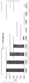
- An iterative PCR image diagram is shown. The image figure of expression vector construction is shown. When using each antigen, 38T. U. It is a figure which shows the light-absorbency change amount in this. When using each antigen, 119T. U. It is a figure which shows the light-absorbency change amount in this. 240T. With each antigen. U. It is a figure which shows the light-absorbency change amount in this.
- the reagent for measuring anti-syphilis treponema antibody using the antigen-antibody reaction of the present invention is characterized by using a recombinant polypeptide containing at least domains C and D but not domain A1 among antigens of syphilis treponema 47 kDa as an antigen.
- Specific examples of the recombinant polypeptide antigen used in the present invention include domains C and D, domains A2 and C and D, or domains B and A2, which are part of the 47 kDa antigen that is the surface antigen of Treponema paridum.
- Recombinant polypeptide consisting of C and D preferably having a small molecular weight and good correlation with a reagent (Mediace (registered trademark) TPLA, manufactured by Sekisui Medical Co., Ltd.) prepared using an antigen derived from syphilis treponema
- a reagent Mediace (registered trademark) TPLA, manufactured by Sekisui Medical Co., Ltd.
- a recombinant polypeptide consisting of domains C and D a recombinant polypeptide consisting of domains A2, C and D, more preferably a recombinant polypeptide consisting of domains C and D.
- Each of the above polypeptides includes those having an amino acid sequence having a homology of 90% or more, preferably having a homology of 95% or more, and more preferably having a homology of 98% or more.
- the entire 47 kDa antigen gene is obtained by cloning a gene from syphilis or by gene synthesis, and is not required with an appropriate restriction enzyme or the like.
- the expression vector is not particularly limited. For example, plasmids, cosmids, phages, viruses and the like can be mentioned.
- Hosts for expressing each polypeptide antigen incorporated in the expression vector are not particularly limited, such as cultured cells, microorganisms such as E. coli, silkworms, etc., but Escherichia coli and cultured cells are representative hosts.
- they may be expressed as fusion proteins with other proteins (hereinafter referred to as tag proteins).
- tag proteins include ⁇ -galactosidase, glutathione S transferase, 6 ⁇ histidine, Cry protein which is an insecticidal protein derived from Bacillus thuringiensis (described in WO2010 / 013789), and the like, but is not particularly limited.
- tag proteins include ⁇ -galactosidase, glutathione S transferase, 6 ⁇ histidine, Cry protein which is an insecticidal protein derived from Bacillus thuringiensis (described in WO2010 / 013789), and the like, but is not particularly limited.
- the immunological measurement reagent of the present invention is not particularly limited, and can be used for, for example, immunoagglutination, enzyme immunoassay (EIA), fluorescent immunoassay (FIA), immunochromatography, etc., and the above antigen is supported on an insoluble carrier.
- EIA enzyme immunoassay
- FIA fluorescent immunoassay
- immunochromatography etc.
- the method used for immunoagglutination is preferred.
- the insoluble carrier is not particularly limited, and examples thereof include organic polymer powders, microorganisms, blood cells, and cell membrane pieces. Of these, organic polymer powder is preferred.
- examples of the organic polymer powder include natural polymer powder and synthetic polymer powder.
- examples of the natural polymer powder include insoluble agarose, cellulose, insoluble dextran, and the like.
- Examples of the synthetic polymer powder include polystyrene, styrene-sulfonic acid (salt) copolymer, styrene-methacrylic acid copolymer, acrylonitrile-butadiene-styrene copolymer, vinyl chloride-acrylic acid ester copolymer, And vinyl acetate-acrylic acid ester copolymer.
- the insoluble carrier those having a sulfonic acid group, a carboxyl group, an amino group or the like introduced on the surface can also be used.
- latex particles in which a synthetic polymer powder is uniformly suspended are particularly suitable.
- plastic microtiter plates; biological particles such as animal red blood cells and bacterial cells; non-biological particles such as bentonite, collodion, cholesterol crystals, silica, kaolin, and carbon powder can also be used.
- the particle diameter of the latex particles is not particularly limited, the preferable lower limit is 0.05 ⁇ m and the preferable upper limit is 1.5 ⁇ m as measured with an electron microscope. When the particle diameter of the latex particles is less than 0.05 ⁇ m, the optical change amount due to aggregation is small, and high sensitivity necessary for measurement may not be obtained.
- the particle diameter of the latex particles exceeds 1.5 ⁇ m, the amount of optical change due to the aggregation of latex particles exceeds the measurable range, and the measurement range may be reduced.
- the preferable lower limit of the particle diameter of the latex particles is 0.1 ⁇ m, and the more preferable upper limit is 0.8 ⁇ m.
- the method of immobilizing the syphilis treponema recombinant antigen on the insoluble carrier is not particularly limited, and a conventionally known method of supporting by a physical or chemical bond can be used. As a method of physically adsorbing, for example, it can be supported by mixing a recombinant antigen and an insoluble carrier under a certain condition. Further, after the immobilization step, the insoluble carrier may be coated with an immunologically inert substance such as albumin, casein, surfactant, etc., and it is preferable to use albumin.
- an immunologically inert substance such as albumin, casein, surfactant, etc.
- the albumin is not particularly limited, and albumin present in animal blood is used. Examples of animal species include cattle, horses, rabbits, goats, and humans.
- the concentration of albumin is not particularly limited, but a preferred lower limit is 0.01% by weight and a preferred upper limit is 10% by weight. If the albumin concentration is less than 0.01% by weight, the surface of the insoluble carrier is not sufficiently coated, and nonspecific aggregation occurs. If the concentration exceeds 10% by weight, the sensitivity of the calibration curve decreases. 5% by weight is more preferred.
- the recombinant antigen can be supported on an insoluble carrier and then mixed with an inert substance under a certain condition.
- the pH during the reaction is not particularly limited, but the preferred lower limit is 2 and the preferred upper limit is 12.
- problems such as the denaturation of recombinant antigens occur, so pH 4 to 10 is more preferable.
- the temperature at the time of reaction is not specifically limited, A preferable minimum is 2 degreeC and a preferable upper limit is 50 degreeC.
- the temperature is lower than 2 ° C, the reaction does not sufficiently occur, or the reaction system freezes, and it becomes difficult to obtain a product having the required sensitivity.
- the temperature is higher than 50 ° C, problems such as degeneration of the recombinant antigen occur. 2 to 10 ° C. is more preferable.
- the carrier carrying the recombinant antigen thus obtained is not particularly limited, but is suspended in a buffer containing an immunologically inactive substance.
- the immunologically inactive substance is not particularly limited, and albumin, casein, surfactant (synthetic polymer compound), synthetic phospholipid, polyvinylpyrrolidone, polyethylene glycol, etc. are used, but albumin, synthetic phospholipid Is preferred.
- the solvent to be used is not particularly limited.
- phosphate buffer, Tris-HCl Examples include a buffer solution, a glycine buffer solution, and a Good buffer solution.
- the specimen is added to the latex particle suspension carrying the antigen thus obtained and allowed to react for a certain period of time. After the reaction, the degree of aggregation caused by the antigen-antibody reaction between the recombinant antigen carried on the latex particles and the anti-syphilis treponema antibody in the sample is optically measured or visually confirmed to visually confirm the anti-antigen in the sample. The amount of syphilis treponema antibody can be measured.
- the method for optically measuring the degree of aggregation is not particularly limited, and a known method is used.
- a turbidimetric method in which the formation of aggregates is regarded as an increase in turbidity, and the formation of aggregates is determined by particle size distribution or average particle size.
- examples thereof include a method of capturing as a change in diameter, an integrating sphere phototurbidity method in which a change in forward scattered light due to the formation of aggregates is measured using an integrating sphere, and a ratio with transmitted light intensity is compared. Also, these methods can be used in combination.
- At least two measurement values are obtained at different time points, and a rate test (rate assay) for determining the degree of aggregation based on the increase rate of the measurement values between these time points, or at a certain time point (usually the end point of the reaction)
- a rate test for determining the degree of aggregation based on the increase rate of the measurement values between these time points, or at a certain time point (usually the end point of the reaction)
- An end point test endpoint assay
- the wavelength of light for performing the above measurement is preferably 250 to 1000 nm, and more preferably 540 to 800 nm.
- Examples of the apparatus used in the above optical measurement method include optical apparatuses capable of detecting scattered light intensity, transmitted light intensity, absorbance, etc., and any biochemical automatic analyzer that is generally used can be used.
- the method of visually observing the degree of aggregation is usually a method in which a solution containing a specimen and a latex particle suspension is mixed on a determination plate, the mixture is shaken, and then the presence or absence of aggregation is determined. Can be used.
- the specimen is not particularly limited as long as an anti-treponema antibody may be present, and examples thereof include human or animal blood, plasma, and serum.
- the reaction solution for the antigen-antibody reaction is not particularly limited as long as it is an aqueous solution that satisfies physiological conditions that can cause an antigen-antibody reaction.
- the pH of the reaction solution is preferably 4 to 10, more preferably 6 to 8.
- the reaction solution may further contain a stabilizer such as bovine serum albumin or sucrose, a preservative such as sodium azide, a salt concentration adjusting agent such as sodium chloride, and the like.
- the reaction temperature is not particularly limited as long as the above immune reaction can occur, and is preferably performed at a constant temperature in the range of 10 to 50 ° C., more preferably 30 to 40 ° C. The reaction time is appropriately determined.
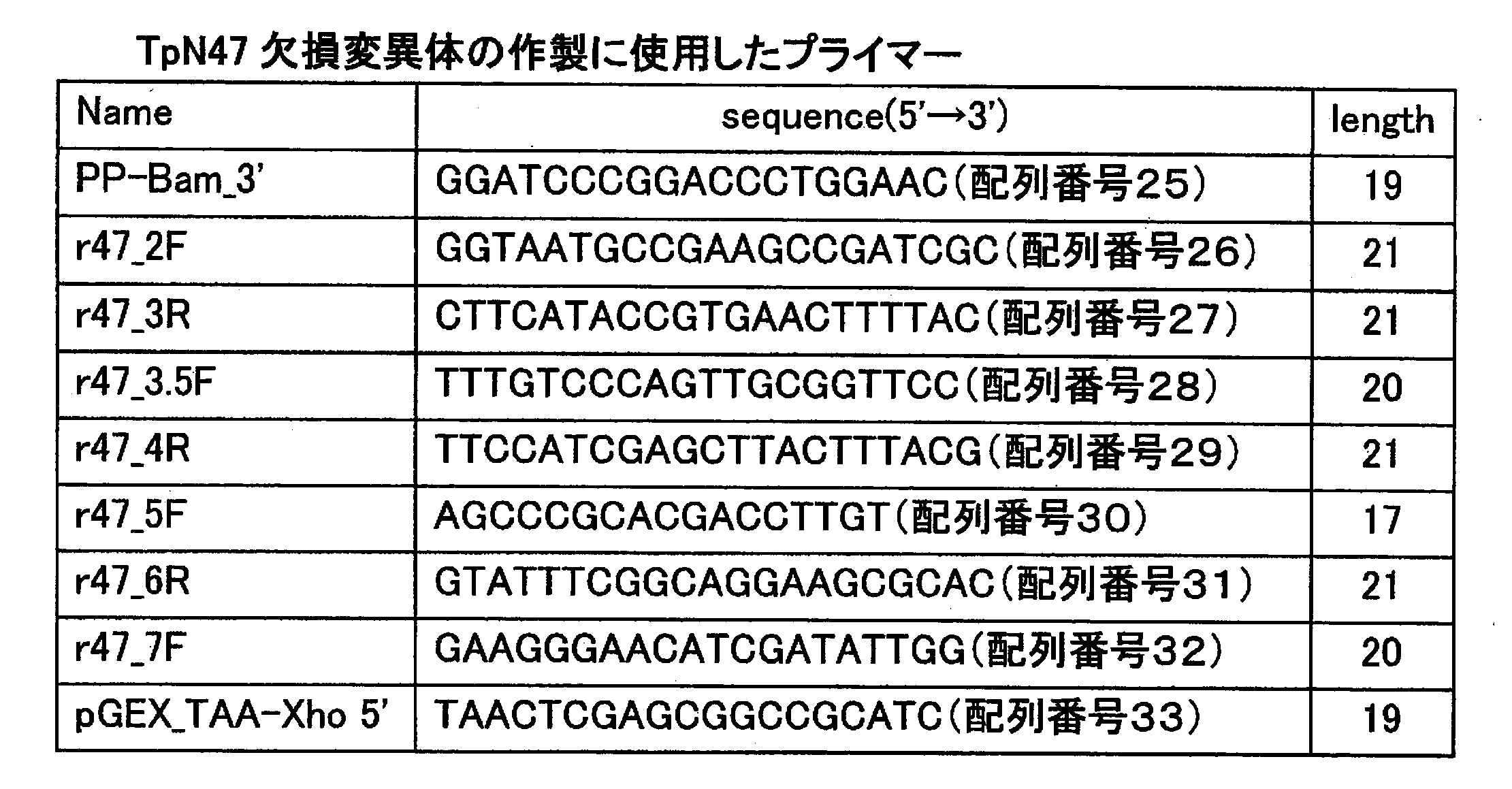
- Table 1 shows primers (synthesized by a DNA synthesizer) used for preparing the TpN47 gene.
- the underlined portion indicates a restriction enzyme site necessary for subcloning into a vector.
- TpN47 gene was synthesized by recursive PCR with reference to the base sequence on the database (GenBnak AE000520). That is, a DNA fragment prepared by PCR using a primer pair TpN47_1f and TpN47_2r having complementary sequences at one end and a series of primer pairs TpN47_3f, TpN47_4r, TpN47_5f, TpN47_6r, TpN47_7f, TpN47_8r having complementary sequence ends.
- TpN47_9f TpN47_10r, TpN47_11f, TpN47_12r, TpN47_13f, TpN47_14r, TpN47_15f, TpN47_16r, TpN47_17f, TpN47_18r, TpN47_19f, TpN47_19f, TpN47_20r
- An image of recursive PCR is shown in FIG.
- TpN47 antigen expression vector Construction of TpN47 antigen expression vector
- the prepared TpN47 gene was ligated to the SmaI site of pBluescript II SK (+) (Stratagene) and transformed into Escherichia coli DH5 ⁇ (Takara Bio) by the calcium method.
- the transformation method by the calcium method is as follows. 0.1 mL of the overnight culture solution of E. coli DH5 ⁇ was inoculated into 5 mL of LB medium (described in Table 2), and cultured with shaking at 37 ° C until the turbidity reached 0.5. 1 mL was collected by centrifugation, suspended in 0.5 mL of ice-cold 50 mM CaCl 2 and allowed to stand on ice for 30 minutes.
- IPTG isopropyl- ⁇ -galactopyranoside
- X-gal 5-chloro-4-bromo-3-D-galactose
- a single colony was selected by blue-white selection, inoculated into 2 mL of TB medium (described in Table 2) containing ampicillin having a final concentration of 100 ⁇ g / mL, and cultured overnight.
- Plasmid DNA was extracted from cultured Escherichia coli using FavorPrep Plasmid DNA Extraction Mini Kit (manufactured by FAVORGEN BIOTECH CORP). The extracted plasmid was confirmed by restriction enzyme treatment and sequencing. After confirming the correct nucleotide sequence, the BpHI of p ⁇ GST in which the TSTN47 gene was deleted from the commercially available vector pGEX-4T-3 (manufactured by GE Healthcare Bioscience) using the one day mutationage method was used.
- P ⁇ GST-Tag in which 4AaCter (696-851) (SEQ ID NO: 42, hereinafter referred to as Tag), which is part of the amino acid sequence of Cry protein derived from Bacillus thuringiensis described in WO2010 / 013789, was introduced into the BamHI site of p ⁇ GST-TpN47.
- -TpN47 was constructed. Specifically, first, a gene encoding Tag was prepared by recursive PCR.
- the gene encoding Tag was prepared such that a BamHI site was added to the 5 ′ end, and a base sequence encoding a linker sequence and a BamHI site were added to the 3 ′ end.
- the gene encoding Tag was inserted into the SmaI site of pBluescript II SK (+), cloned, and confirmed to have the correct base sequence by sequence analysis.
- the gene encoding Tag was excised from pBluescript II SK (+) with BamHI and inserted into the BamHI site of p ⁇ GST-TpN47 to construct p ⁇ GST-Tag-TpN47.
- An image diagram of the expression vector construction is shown in FIG.
- the constructed p ⁇ GST-Tag-TpN47 was introduced into E. coli BL21 (Takara Bio) and transformed. The method of transformation is as described above.
- E. coli BL21 introduced with the p ⁇ GST-Tag-TpN47 expression vector was pre-cultured overnight in 5 mL of TB medium containing ampicillin at a final concentration of 100 ⁇ g / mL.
- 0.5 mL of this overnight culture was added to LB medium containing 50 mL final concentration of 100 ⁇ g / mL ampicillin.
- the culture was performed using a shaking incubator (manufactured by SANYO, MIR-S100) at 240 rpm and 37 ° C. until the OD600 was 0.6 to 0.8.
- IPTG was added to the culture solution to a final concentration of 0.5 mM.
- the cells were further cultured for 3 hours (240 rpm, 37 ° C.) to induce expression.
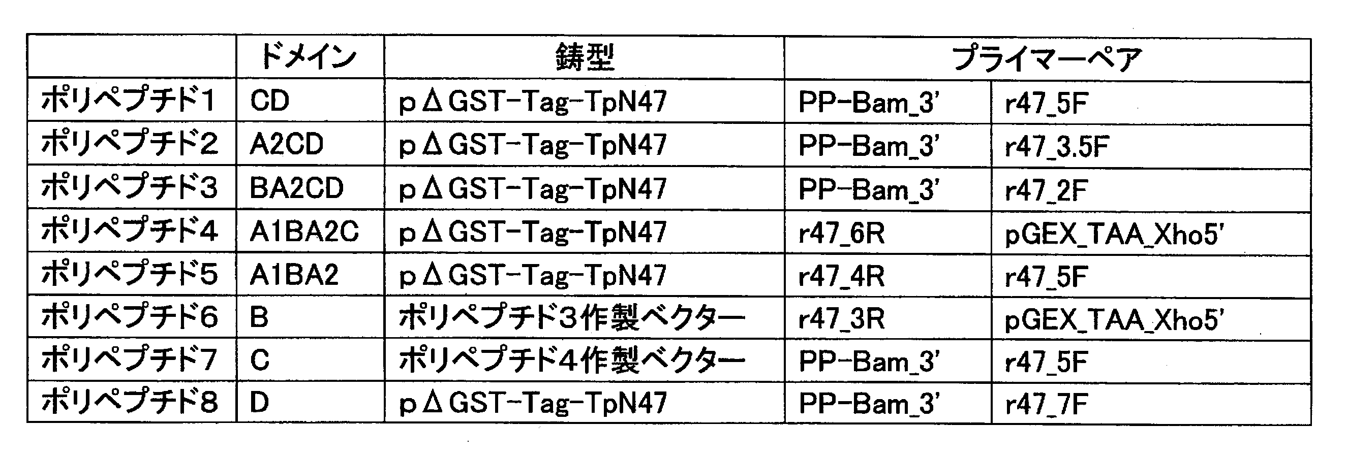
- Table 3 shows primers (synthesized with a DNA synthesizer) for preparing a gene corresponding to polypeptides 1-8 described in SEQ ID NOs: 34 to 41.
- the gene for polypeptide 1-8 used the one day mutationage method using p ⁇ GST-Tag-TpN47 as a template. That is, primers were designed so as to sandwich a portion to be deleted from p ⁇ GST-Tag-TpN47, and DNAs were amplified by PCR reaction outward of the vectors to prepare DNAs encoding the respective polypeptides. Primer pairs for producing each polypeptide were as shown in Table 4. The nucleotide sequences of polypeptides 1 to 8 are shown in SEQ ID NOs: 34 to 41, respectively.
- T4 DNA Ligase (manufactured by Takara Bio Inc.) and 10 X Ligation buffer (manufactured by Takara Bio Inc.) were added to the PCR product described above and self-ligated.
- Restriction enzyme DpnI (manufactured by Toyobo Co., Ltd.) was added to this ligation solution and incubated at 37 ° C. for 1 hour. After insertion into E. coli, the procedure was the same as that for TpN47 vector construction.
- TpN47 expression was followed except that each polypeptide expression vector was used.
- the latex particles obtained were photographed with a transmission electron microscope apparatus (manufactured by JEOL Ltd., “JEM-1010”) at a magnification of 10,000 times, and image analysis was performed on at least 100 particles. The average particle size was measured. The average particle size obtained was 0.4 ⁇ m.
- Example 1 Polypeptide 1 (4 mM) dissolved in 20 mM Tris buffer (hereinafter referred to as Tris-HCl, pH 8.0) in 100 ⁇ L of a latex solution having an average particle size of 0.4 ⁇ m (solid content: 10% (w / v)) was added at 100 ⁇ L (0.4 ⁇ mol) and stirred at 4 ° C. for 1 hour.
- Tris-HCl buffered saline (20 mM Tris-HCl, salt concentration 0.9% by weight) containing 1% (W / V) bovine serum albumin (hereinafter referred to as BSA, fraction V, Reagent Grade, manufactured by Miles Corp.), 1 mL of pH 8.0) was added and stirred for 1 hour.
- BSA bovine serum albumin
- the obtained liquid was centrifuged at 15,000 rpm for 15 minutes at 10 ° C., and 20 mL of 100 mM phosphate buffer (pH 7.5) solution containing 1 (w / v)% BSA was added to the resulting precipitate. Was added, and the latex was suspended to prepare an anti-syphilis treponema antibody measurement reagent.
- Example 2 An anti-syphilis treponema antibody measurement reagent was prepared in the same manner as in Example 1 except that the polypeptide 2 solution was used as the antigen solution.
- Example 3 An anti-syphilis treponema antibody measurement reagent was prepared in the same manner as in Example 1 except that the polypeptide 3 solution was used as the antigen solution.
- Example 2 An anti-syphilis treponema antibody measurement reagent was prepared in the same manner as in Example 1 except that the polypeptide 4 solution was used as the antigen solution.
- an anti-syphilis reponema antibody measuring reagent capable of measuring an anti-syphilis treponema antibody having high sensitivity and specificity.
Abstract
Description
梅毒に罹患しているか否かは、血液中の抗梅毒トレポネーマ抗体の存在の有無を免疫学的に検査することにより診断されている。梅毒トレポネーマ菌体の菌体表面には多数の表面抗原が存在しており、この表面抗原と検体中の抗梅毒トレポネーマ抗体との抗原抗体反応を利用する方法により免疫測定が行われている。梅毒トレポネーマ菌体の菌体表面に存在する表面抗原のうち主なものとしては、分子量47kDa、42kDa、37kDa、17kDa及び15kDaのものが知られている。 Syphilis is a disease caused by infection with Treponema pallidum bacteria (Treponema pallidum, Treponema pallidum). Due to the development of effective therapeutic agents such as penicillin, the prevalence of syphilis has decreased since the 1940s, but has recently shown an increasing trend again. As a feature of patients with newly affected syphilis in recent years, there are many cases where HIV infection is complicated. As a cause of the high merger rate, it is considered that both syphilis and HIV are sexually transmitted diseases, and at the same time, syphilis increases the risk of HIV infection. Under such circumstances, in order to prevent the spread of syphilis and HIV infection, it is required to detect and treat syphilis patients at an early stage.
Whether or not the patient is suffering from syphilis is diagnosed by examining the presence or absence of anti-syphilis treponema antibody in the blood immunologically. Numerous surface antigens exist on the surface of syphilis treponema cells, and immunoassay is performed by a method using an antigen-antibody reaction between this surface antigen and an anti-syphilis treponema antibody in a specimen. Among surface antigens present on the surface of syphilis treponema cells, those having molecular weights of 47 kDa, 42 kDa, 37 kDa, 17 kDa and 15 kDa are known.
非特許文献3によれば、アミノ酸配列のN末端から数えてドメインAは1~34番目(A1ドメイン)と157~207番目(A2ドメイン)、ドメインBは35~156番目、ドメインCは208~335番目、ドメインDは336~415番目までから成っている(非特許文献3、図1参照)。
さらに上記47kDa抗原の抗体認識部位に関しても、抗原活性を示すアミノ酸配列が報告されている(例えば、非特許文献4)。 In recent years, methods for producing cell surface antigens of syphilis treponema using genetic recombination techniques have been proposed. For the syphilis 47 kDa antigen, the encoding gene has been cloned and an amino acid sequence consisting of 415 amino acids has been determined (for example, Non-Patent
According to
Furthermore, an amino acid sequence showing antigen activity has also been reported for the antibody recognition site of the 47 kDa antigen (for example, Non-Patent Document 4).
一方、遺伝子組換え技術による抗原作製以外に、47kDa抗原に関して、抗原活性を有するペプチドを合成し、これを抗原として用いることで抗梅毒トレポネーマ抗体を測定する方法も開示されている(特許文献3参照)。 As a method for measuring an anti-syphilis treponema antibody using the above-mentioned antigen, a method for immunologically measuring an anti-syphilis treponema antibody using a syphilis 47 kDa antigen produced by genetic recombination technology is disclosed (Patent Document 1). reference). In addition, a method using a protein in which glutathione-S-transferase is fused to the N-terminus of 15 kDa and 17 kDa antigens is also disclosed (see Patent Document 2).
On the other hand, in addition to antigen production by gene recombination technology, a method for measuring anti-syphilis treponema antibody by synthesizing a peptide having antigenic activity for 47 kDa antigen and using it as an antigen is also disclosed (see Patent Document 3). ).
これらの問題が発生する要因として、詳細は明確ではないが以下のことが考えられる。すなわち、遺伝子組換え技術によって作製された梅毒トレポネーマの組換え抗原は、梅毒トレポネーマ菌由来の天然抗原と比較して、その立体構造並びに脂質の修飾等に相違があるものと考えられる。一般にタンパク質は翻訳後さまざまな修飾を受ける。糖又は脂質の修飾等はそれらのうちの代表的な修飾であるが、タンパク質はこのような修飾を受けることでその立体構造が変化する。一方、遺伝子組換え技術で主に用いられる大腸菌による発現系では、タンパク質の翻訳後の修飾は行われない。タンパク質の立体構造は、その抗原性を示す上で重要な要因であるが、遺伝子組換え技術によって作製された組換えタンパク質では、翻訳後の修飾を受けないため、天然抗原に比べて立体構造に違いが生じ、これが感度や特異性に影響を与えている可能性が考えられる。
本発明は、上記に鑑み、高感度かつ高い特異性で抗梅毒トレポネーマ抗体を測定するポリペプチド抗原を用いた抗梅毒トレポネーマ抗体測定試薬及びこれを用いた測定方法を提供することを目的とするものである。 Although it is possible to measure anti-syphilis treponema antibodies using recombinant syphilis treponema antigens and synthetic peptide antigens prepared in this way, a set of syphilis treponema produced by genetic recombination technology. The replacement antigen has a problem in low sensitivity and specificity. In addition to the same problems, synthetic peptide antigens have the problem that several peptides must be combined in order to accurately measure anti-syphilis treponema antibodies, which is complicated in operation.
Although the details are not clear as factors that cause these problems, the following may be considered. That is, it is considered that the recombinant antigen of syphilis treponema produced by gene recombination technology has a difference in three-dimensional structure, lipid modification, and the like as compared with a natural antigen derived from syphilis treponema. In general, proteins undergo various modifications after translation. The modification of sugar or lipid is a typical modification among them, but the three-dimensional structure of a protein is changed by such modification. On the other hand, post-translational modification of proteins is not performed in an expression system using Escherichia coli mainly used in gene recombination techniques. The three-dimensional structure of a protein is an important factor in showing its antigenicity, but a recombinant protein produced by gene recombination technology does not undergo post-translational modification, so it has a three-dimensional structure compared to the natural antigen. Differences may have occurred and this may affect sensitivity and specificity.
In view of the above, an object of the present invention is to provide an anti-syphilis treponema antibody measuring reagent using a polypeptide antigen that measures anti-syphilis treponema antibody with high sensitivity and high specificity, and a measuring method using the same. It is.
(1)抗原抗体反応を利用した抗梅毒トレポネーマ抗体の測定に用いる試薬において、梅毒トレポネーマ47kDa抗原のうち、少なくともドメインCとDを含み、ドメインA1を含まない組み換えポリペプチドを抗原として用いることを特徴とする抗梅毒トレポネーマ抗体測定試薬。
(2)抗原が、梅毒トレポネーマ47kDa抗原のうち、ドメインCとD、ドメインA2とCとD、又はドメインBとA2とCとDからなる組み換えポリペプチドである(1)記載の抗梅毒トレポネーマ抗体測定試薬。
(3)抗原が不溶性担体に固定化されている(1)記載の抗梅毒トレポネーマ抗体測定試薬。
(4)抗原が不溶性担体に固定化されている(2)記載の抗梅毒トレポネーマ抗体測定試薬。
(5)(3)記載の不溶性担体が高分子重合体から成るラテックスである抗梅毒トレポネーマ抗体測定試薬。
(6)(4)記載の不溶性担体が高分子重合体から成るラテックスである抗梅毒トレポネーマ抗体測定試薬。
(7)(1)記載の抗梅毒トレポネーマ抗体測定試薬を用いることを特徴とする抗梅毒トレポネーマ抗体測定方法。
(8)(2)記載の抗梅毒トレポネーマ抗体測定試薬を用いることを特徴とする抗梅毒トレポネーマ抗体測定方法。
(9)(3)記載の抗梅毒トレポネーマ抗体測定試薬を用いることを特徴とする抗梅毒トレポネーマ抗体測定方法。
(10)(4)記載の抗梅毒トレポネーマ抗体測定試薬を用いることを特徴とする抗梅毒トレポネーマ抗体測定方法。
(11)(5)記載の抗梅毒トレポネーマ抗体測定試薬を用いることを特徴とする抗梅毒トレポネーマ抗体測定方法。
(12)(6)記載の抗梅毒トレポネーマ抗体測定試薬を用いることを特徴とする抗梅毒トレポネーマ抗体測定方法。 That is, the present invention is as follows.
(1) A reagent used for measurement of an anti-syphilis treponema antibody using an antigen-antibody reaction, wherein a recombinant polypeptide containing at least domains C and D and not containing domain A1 is used as an antigen among 47 kDa antigens of syphilis treponema Anti-syphilis treponema antibody measurement reagent.
(2) The anti-syphilis treponema antibody according to (1), wherein the antigen is a recombinant polypeptide consisting of domains C and D, domains A2 and C and D, or domains B and A2, C and D among 47 kDa antigens of syphilis treponema Measuring reagent.
(3) The anti-syphilis treponema antibody measurement reagent according to (1), wherein the antigen is immobilized on an insoluble carrier.
(4) The anti-syphilis treponema antibody measurement reagent according to (2), wherein the antigen is immobilized on an insoluble carrier.
(5) An anti-syphilis treponema antibody measuring reagent, wherein the insoluble carrier according to (3) is a latex comprising a polymer.
(6) An anti-syphilis treponema antibody measurement reagent, wherein the insoluble carrier according to (4) is a latex composed of a polymer.
(7) An anti-syphilis treponema antibody measurement method comprising using the anti-syphilis treponema antibody measurement reagent according to (1).
(8) An anti-syphilis treponema antibody measurement method comprising using the anti-syphilis treponema antibody measurement reagent according to (2).
(9) An anti-syphilis treponema antibody measurement method comprising using the anti-syphilis treponema antibody measurement reagent according to (3).
(10) An anti-syphilis treponema antibody measurement method comprising using the anti-syphilis treponema antibody measurement reagent according to (4).
(11) An anti-syphilis treponema antibody measurement method comprising using the anti-syphilis treponema antibody measurement reagent according to (5).
(12) A method for measuring an anti-syphilis treponema antibody, comprising using the anti-syphilis treponema antibody measurement reagent according to (6).
本発明に用いる組み換えポリペプチド抗原の具体例としては、トレポネーマ・パリダム菌体の表面抗原である47kDa抗原の一部である、ドメインCとD、ドメインA2とCとD、又はドメインBとA2とCとDからなる組み換えポリペプチドであり、好ましくは、分子量が小さく、梅毒トレポネーマ菌由来の抗原を用いて作製された試薬(メディエース(登録商標)TPLA,積水メディカル社製)と相関が良好なドメインCとDからなる組み換えポリペプチド、ドメインA2とCとDからなる組み換えポリペプチド、さらに好ましくは、ドメインCとDからなる組み換えポリペプチドである。
上記各ポリペプチドとしては、アミノ酸配列で相同性が90%以上のものが含まれるが、相同性が95%以上のものがより好ましく、相同性が98%以上のものがさらに好ましい。 The reagent for measuring anti-syphilis treponema antibody using the antigen-antibody reaction of the present invention is characterized by using a recombinant polypeptide containing at least domains C and D but not domain A1 among antigens of syphilis treponema 47 kDa as an antigen. .
Specific examples of the recombinant polypeptide antigen used in the present invention include domains C and D, domains A2 and C and D, or domains B and A2, which are part of the 47 kDa antigen that is the surface antigen of Treponema paridum. Recombinant polypeptide consisting of C and D, preferably having a small molecular weight and good correlation with a reagent (Mediace (registered trademark) TPLA, manufactured by Sekisui Medical Co., Ltd.) prepared using an antigen derived from syphilis treponema A recombinant polypeptide consisting of domains C and D, a recombinant polypeptide consisting of domains A2, C and D, more preferably a recombinant polypeptide consisting of domains C and D.
Each of the above polypeptides includes those having an amino acid sequence having a homology of 90% or more, preferably having a homology of 95% or more, and more preferably having a homology of 98% or more.
上記発現ベクターに関しても、特に限定されるものではない。例えば、プラスミド、コスミド、ファージ、ウィルス等が挙げられる。 As a method for obtaining a DNA fragment that expresses a polypeptide that is a part of the 47 kDa antigen, i) the entire 47 kDa antigen gene is obtained by cloning a gene from syphilis or by gene synthesis, and is not required with an appropriate restriction enzyme or the like. A method in which a DNA fragment encoding a polypeptide portion is deleted and then inserted into an expression vector; ii) a cDNA bank of syphilis cells is prepared, and a DNA fragment encoding each polypeptide by PCR or the like using appropriate DNA primers Iii) There is a method of directly synthesizing a DNA fragment encoding each polypeptide, or a method of putting it into an expression vector after synthesizing by a PCR method or the like, but it is not particularly limited.
The expression vector is not particularly limited. For example, plasmids, cosmids, phages, viruses and the like can be mentioned.
上記各ポリペプチドを効率的に発現させる、あるいは発現後の精製を容易にするために、他のタンパク質(以下、タグタンパク質という)との融合タンパク質として発現させてもよい。タグタンパク質としては、β-ガラクトシターゼ、グルタチオンSトランスフェラーゼ、6×ヒスチジン、Bacillus thuringiensis菌由来の殺虫タンパク質であるCryタンパク質(WO2010/013789に記載)等があるが、特に限定されるものではない。また、融合タンパク質として発現させた後、精製過程においてはタグタンパク質を必ずしも除去する必要はなく、そのまま用いてもよい。 Hosts for expressing each polypeptide antigen incorporated in the expression vector are not particularly limited, such as cultured cells, microorganisms such as E. coli, silkworms, etc., but Escherichia coli and cultured cells are representative hosts.
In order to efficiently express each of the above polypeptides or to facilitate purification after expression, they may be expressed as fusion proteins with other proteins (hereinafter referred to as tag proteins). Examples of the tag protein include β-galactosidase, glutathione S transferase, 6 × histidine, Cry protein which is an insecticidal protein derived from Bacillus thuringiensis (described in WO2010 / 013789), and the like, but is not particularly limited. In addition, after expression as a fusion protein, it is not always necessary to remove the tag protein in the purification process, and it may be used as it is.
また、上記不溶性担体は、表面にスルホン酸基、カルボキシル基やアミノ基等を導入したものも用いることができる。 The insoluble carrier is not particularly limited, and examples thereof include organic polymer powders, microorganisms, blood cells, and cell membrane pieces. Of these, organic polymer powder is preferred. Examples of the organic polymer powder include natural polymer powder and synthetic polymer powder. Examples of the natural polymer powder include insoluble agarose, cellulose, insoluble dextran, and the like. Examples of the synthetic polymer powder include polystyrene, styrene-sulfonic acid (salt) copolymer, styrene-methacrylic acid copolymer, acrylonitrile-butadiene-styrene copolymer, vinyl chloride-acrylic acid ester copolymer, And vinyl acetate-acrylic acid ester copolymer.
As the insoluble carrier, those having a sulfonic acid group, a carboxyl group, an amino group or the like introduced on the surface can also be used.
上記ラテックス粒子の粒子径は特に限定されないが、電子顕微鏡での測定で、好ましい下限は0.05μm、好ましい上限は1.5μmである。上記ラテックス粒子の粒子径が0.05μm未満であると、凝集による光学的変化量が小さく、測定に必要な高い感度が得られないことがある。上記ラテックス粒子の粒子径が1.5μmを超えると、ラテックス粒子の凝集による光学的変化量が測定可能域を超えてしまい、測定範囲が小さくなることがある。上記ラテックス粒子の粒子径の好ましい下限は0.1μm、より好ましい上限は0.8μmである。 As the insoluble carrier, latex particles in which a synthetic polymer powder is uniformly suspended are particularly suitable. In addition, plastic microtiter plates; biological particles such as animal red blood cells and bacterial cells; non-biological particles such as bentonite, collodion, cholesterol crystals, silica, kaolin, and carbon powder can also be used.
Although the particle diameter of the latex particles is not particularly limited, the preferable lower limit is 0.05 μm and the preferable upper limit is 1.5 μm as measured with an electron microscope. When the particle diameter of the latex particles is less than 0.05 μm, the optical change amount due to aggregation is small, and high sensitivity necessary for measurement may not be obtained. When the particle diameter of the latex particles exceeds 1.5 μm, the amount of optical change due to the aggregation of latex particles exceeds the measurable range, and the measurement range may be reduced. The preferable lower limit of the particle diameter of the latex particles is 0.1 μm, and the more preferable upper limit is 0.8 μm.
上記測定を行う際の光の波長は、250~1000nmが好ましく、540~800nmが更に好ましい。 In the above measurement method, at least two measurement values are obtained at different time points, and a rate test (rate assay) for determining the degree of aggregation based on the increase rate of the measurement values between these time points, or at a certain time point (usually the end point of the reaction) An end point test (endpoint assay) can be used in which one measurement value is obtained at the time point considered) and the degree of aggregation is determined based on this measurement value. From the viewpoint of simplicity of measurement and rapidity, it is preferable to perform a turbidimetric rate test.
The wavelength of light for performing the above measurement is preferably 250 to 1000 nm, and more preferably 540 to 800 nm.
上記凝集の度合いを目視にて観察する方法は、通常、検体とラテックス粒子懸濁液とを含む溶液を判定板上で混合し、混合液を揺り動かした後、凝集の有無を判定する方法等を用いることができる。なお、凝集の度合いの観察には、目視による方法以外に、凝集状態をビデオカメラ等で撮影し、画像処理を施す方法を用いることも可能である。 Examples of the apparatus used in the above optical measurement method include optical apparatuses capable of detecting scattered light intensity, transmitted light intensity, absorbance, etc., and any biochemical automatic analyzer that is generally used can be used. Can be used.
The method of visually observing the degree of aggregation is usually a method in which a solution containing a specimen and a latex particle suspension is mixed on a determination plate, the mixture is shaken, and then the presence or absence of aggregation is determined. Can be used. For observation of the degree of aggregation, it is also possible to use a method of photographing the aggregation state with a video camera or the like and performing image processing in addition to the visual method.
上記反応液には、さらに必要に応じて、牛血清アルブミン、ショ糖等の安定化剤、アジ化ナトリウム等の防腐剤、塩化ナトリウム等の塩濃度調整剤などを添加してもよい。
反応温度は、上記免疫反応が起こりうるものであれば特に限定されず、恒温で10~50℃の範囲内で行うのが好ましく、30~40℃がより好ましい。反応時間は適宜決められる。 The reaction solution for the antigen-antibody reaction is not particularly limited as long as it is an aqueous solution that satisfies physiological conditions that can cause an antigen-antibody reaction. For example, phosphate buffer, citrate buffer, glycine buffer Liquid, Tris buffer, Good buffer, and the like. The pH of the reaction solution is preferably 4 to 10, more preferably 6 to 8.
If necessary, the reaction solution may further contain a stabilizer such as bovine serum albumin or sucrose, a preservative such as sodium azide, a salt concentration adjusting agent such as sodium chloride, and the like.
The reaction temperature is not particularly limited as long as the above immune reaction can occur, and is preferably performed at a constant temperature in the range of 10 to 50 ° C., more preferably 30 to 40 ° C. The reaction time is appropriately determined.
TpN47遺伝子を調製するために用いたプライマー(DNA合成装置で合成)を表1に示す。なお表1に示す塩基配列において下線部はベクターへのサブクローニングに必要な制限酵素サイトを示す。 (Primer for TpN47 gene preparation)
Table 1 shows primers (synthesized by a DNA synthesizer) used for preparing the TpN47 gene. In the base sequence shown in Table 1, the underlined portion indicates a restriction enzyme site necessary for subcloning into a vector.

TpN47遺伝子はデータベース上の塩基配列を参考(GenBnak AE000520)に反復(recursive) PCRによって合成した。すなわち、相互に相補的な配列を一端に有するプライマー対TpN47_1f及びTpN47_2rを用いてPCRにより作製したDNA断片と、相補的な配列末端に有する一連のプライマー対TpN47_3f、TpN47_4r、TpN47_5f,TpN47_6r、TpN47_7f,TpN47_8r、TpN47_9f,TpN47_10r、TpN47_11f、TpN47_12r、TpN47_13f、TpN47_14r、TpN47_15f、TpN47_16r、TpN47_17f、TpN47_18r、TpN47_19f、TpN47_20r、TpN47_21f、TpN47_22r、TpN47_23f及びTpN47_24rを用いて、TpN47遺伝子の全体をコードするDNAを作製した。反復(recursive)PCRのイメージ図を図1に示す。 (Artificial preparation of TpN47 gene)
The TpN47 gene was synthesized by recursive PCR with reference to the base sequence on the database (GenBnak AE000520). That is, a DNA fragment prepared by PCR using a primer pair TpN47_1f and TpN47_2r having complementary sequences at one end and a series of primer pairs TpN47_3f, TpN47_4r, TpN47_5f, TpN47_6r, TpN47_7f, TpN47_8r having complementary sequence ends. TpN47_9f, TpN47_10r, TpN47_11f, TpN47_12r, TpN47_13f, TpN47_14r, TpN47_15f, TpN47_16r, TpN47_17f, TpN47_18r, TpN47_19f, TpN47_19f, TpN47_20r An image of recursive PCR is shown in FIG.
調製したTpN47遺伝子をpBluscript II SK(+)(Stratagene社製)のSmaIサイトにライゲーションし、それを大腸菌DH5α(タカラバイオ社製)へカルシウム法により形質転換した。カルシウム法による形質転換の方法は次の通りである。大腸菌DH5αの一晩培養液0.1mLを5mLのLB培地(表2記載)に植菌し,濁度が0.5になるまで37℃で振盪培養した。1mLを遠心分離により集菌し、0.5mLの氷冷50mM CaCl2に懸濁させ、氷上に30分静置した。懸濁液を0.2mL分取し、ライゲーション後のプラスミドDNAを添加し、氷上に30分放置後,42℃で30秒間ヒートショックを与え、SOB培地(表2記載)0.8mLを添加(全量1mL)した。37℃で1時間培養後、イソプロピル-β-ガラクトピラノシド(以下、IPTGという)(ナカライテスク社製)、5-クロロ-4-ブロモ-3-D-ガラクトース(以下、X-galという)(ナカライテスク社製)及びアンピシリン(和光純薬社製)100μg/mLを含むLB寒天培地(表2記載)に展開し37℃で一夜培養した。単一のコロニーをブルーホワイトセレクションにより選択し、終濃度100μg/mLのアンピシリンを含むTB培地(表2記載)2mLに植菌し一晩培養した。培養した大腸菌はFavorPrep Plasmid DNA Extraction Mini Kit(FAVORGEN BIOTECH CORP社製)によりプラスミドDNAを抽出した。抽出したプラスミドは、制限酵素処理及び、シーケンシングにより塩基配列の確認を行った。
正しい塩基配列であることを確認した後に、TpN47遺伝子を市販のベクターであるpGEX-4T-3(GEヘルスケア バイオサイエンス社製)からOne day mutagenesis法を利用してGST遺伝子を削除したpΔGSTのBamHI及びXhoIサイトに導入し、pΔGST-TpN47を構築した。このpΔGST-TpN47のBamHIサイトに、WO2010/013789記載のBacillus thuringiensis由来のCryタンパク質のアミノ酸配列の一部である4AaCter(696-851)(配列番号42:以下、Tagという)を導入したpΔGST-Tag-TpN47を構築した。具体的には、はじめにTagをコードする遺伝子を反復(recursive)PCRによって作製した。Tagをコードする遺伝子は5’末端にBamHIサイト、ならびに3’末端にリンカー配列をコードする塩基配列およびBamHIサイトが付加するように作製した。作製したTagをコードする遺伝子はpBluscript II SK(+)のSmaIサイトに挿入、クローニングし、シークエンス解析により正しい塩基配列であることを確認した。次にTagをコードする遺伝子をpBluscript II SK(+)よりBamHIにより切り出し、pΔGST-TpN47のBamHIサイトへ挿入し、pΔGST-Tag-TpN47を構築した。発現ベクター構築のイメージ図を図2に示す。構築したpΔGST-Tag-TpN47は大腸菌BL21(タカラバイオ社製)へ導入し形質転換した。形質転換の方法は前述の通りである。 (Construction of TpN47 antigen expression vector)
The prepared TpN47 gene was ligated to the SmaI site of pBluescript II SK (+) (Stratagene) and transformed into Escherichia coli DH5α (Takara Bio) by the calcium method. The transformation method by the calcium method is as follows. 0.1 mL of the overnight culture solution of E. coli DH5α was inoculated into 5 mL of LB medium (described in Table 2), and cultured with shaking at 37 ° C until the turbidity reached 0.5. 1 mL was collected by centrifugation, suspended in 0.5 mL of ice-cold 50 mM CaCl 2 and allowed to stand on ice for 30 minutes. Take 0.2 mL of the suspension, add the ligated plasmid DNA, leave it on ice for 30 minutes, apply heat shock at 42 ° C. for 30 seconds, and add 0.8 mL of SOB medium (described in Table 2) ( 1 mL). After culturing at 37 ° C. for 1 hour, isopropyl-β-galactopyranoside (hereinafter referred to as IPTG) (manufactured by Nacalai Tesque), 5-chloro-4-bromo-3-D-galactose (hereinafter referred to as X-gal) It was developed on an LB agar medium (described in Table 2) containing 100 μg / mL (produced by Nacalai Tesque) and ampicillin (produced by Wako Pure Chemical Industries, Ltd.) and cultured at 37 ° C. overnight. A single colony was selected by blue-white selection, inoculated into 2 mL of TB medium (described in Table 2) containing ampicillin having a final concentration of 100 μg / mL, and cultured overnight. Plasmid DNA was extracted from cultured Escherichia coli using FavorPrep Plasmid DNA Extraction Mini Kit (manufactured by FAVORGEN BIOTECH CORP). The extracted plasmid was confirmed by restriction enzyme treatment and sequencing.
After confirming the correct nucleotide sequence, the BpHI of pΔGST in which the TSTN47 gene was deleted from the commercially available vector pGEX-4T-3 (manufactured by GE Healthcare Bioscience) using the one day mutationage method was used. And introduced into the XhoI site to construct pΔGST-TpN47. PΔGST-Tag in which 4AaCter (696-851) (SEQ ID NO: 42, hereinafter referred to as Tag), which is part of the amino acid sequence of Cry protein derived from Bacillus thuringiensis described in WO2010 / 013789, was introduced into the BamHI site of pΔGST-TpN47. -TpN47 was constructed. Specifically, first, a gene encoding Tag was prepared by recursive PCR. The gene encoding Tag was prepared such that a BamHI site was added to the 5 ′ end, and a base sequence encoding a linker sequence and a BamHI site were added to the 3 ′ end. The gene encoding Tag was inserted into the SmaI site of pBluescript II SK (+), cloned, and confirmed to have the correct base sequence by sequence analysis. Next, the gene encoding Tag was excised from pBluescript II SK (+) with BamHI and inserted into the BamHI site of pΔGST-TpN47 to construct pΔGST-Tag-TpN47. An image diagram of the expression vector construction is shown in FIG. The constructed pΔGST-Tag-TpN47 was introduced into E. coli BL21 (Takara Bio) and transformed. The method of transformation is as described above.

pΔGST-Tag-TpN47発現ベクターを導入した大腸菌BL21を終濃度100μg/mLのアンピシリンが入ったTB培地5mLで一晩、前培養した。この一晩培養液の0.5mLを50mLの終濃度100μg/mLのアンピシリンが入ったLB培地に加えた。培養は、振とう培養機(SANYO社製、MIR-S100)を用いて、240rpm、37℃でOD600が0.6~0.8となるまで培養した。培養液に最終濃度0.5mMとなるようにIPTGを加えた。さらに3時間培養(240rpm,37℃)し,発現を誘導した。 (Expression of TpN47)
E. coli BL21 introduced with the pΔGST-Tag-TpN47 expression vector was pre-cultured overnight in 5 mL of TB medium containing ampicillin at a final concentration of 100 μg / mL. 0.5 mL of this overnight culture was added to LB medium containing 50 mL final concentration of 100 μg / mL ampicillin. The culture was performed using a shaking incubator (manufactured by SANYO, MIR-S100) at 240 rpm and 37 ° C. until the OD600 was 0.6 to 0.8. IPTG was added to the culture solution to a final concentration of 0.5 mM. The cells were further cultured for 3 hours (240 rpm, 37 ° C.) to induce expression.
発現誘導した菌体を回収後、10mLの10mMリン酸緩衝食塩水(10mMリン酸緩衝液(pH7.4)、NaCl0.9重量%)に懸濁させ、超音波破砕し、9000rpmにて10分間遠心して不溶性画分を得た。目的のタンパク質を含む不溶性画分を適当量の超純水で遠心洗浄し、沈殿を尿素溶液(8M Urea,20mM Tris-HCl,pH8.0)によって可溶化し、これを目的のタンパク質溶液(濃度:4mM)とした。 (Purification of TpN47)
After recovering the expression-induced cells, the cells were suspended in 10 mL of 10 mM phosphate buffered saline (10 mM phosphate buffer (pH 7.4), NaCl 0.9% by weight), sonicated, and 9000 rpm for 10 minutes. Centrifugation gave an insoluble fraction. The insoluble fraction containing the target protein is centrifuged and washed with an appropriate amount of ultrapure water, and the precipitate is solubilized with a urea solution (8 M Urea, 20 mM Tris-HCl, pH 8.0). : 4 mM).
配列番号34~41記載のポリペプチド1-8に対応する遺伝子を調製するためのプライマー(DNA合成装置で合成)を表3に示す。 (Primer for preparation of polypeptide 1-8 gene)
Table 3 shows primers (synthesized with a DNA synthesizer) for preparing a gene corresponding to polypeptides 1-8 described in SEQ ID NOs: 34 to 41.
ポリペプチド1-8の遺伝子はpΔGST-Tag-TpN47を鋳型とした、One day mutagenesis法を利用した。すなわちpΔGST-Tag-TpN47から削除したい部分を挟む形でプライマーを設計し、それぞれベクターの外向きにPCR反応によりDNAを増幅させて各々のポリペプチドをコードするDNAを作成した。各ポリペプチドを作製する際のプライマーペアは表4に従った。ポリペプチド1~8の塩基配列は配列番号34~41にそれぞれ示した。 (Artificial preparation of polypeptide 1-8 gene)
The gene for polypeptide 1-8 used the one day mutationage method using pΔGST-Tag-TpN47 as a template. That is, primers were designed so as to sandwich a portion to be deleted from pΔGST-Tag-TpN47, and DNAs were amplified by PCR reaction outward of the vectors to prepare DNAs encoding the respective polypeptides. Primer pairs for producing each polypeptide were as shown in Table 4. The nucleotide sequences of
前述のPCR産物にT4 DNA Ligase(タカラバイオ社製)及び10 X Ligation buffer(タカラバイオ社製)を加えセルフライゲーションした。このライゲーション溶液に制限酵素DpnI(東洋紡社製)を加え37℃にて1時間インキュベートした。大腸菌への挿入以降は、TpN47のベクター構築と同様にした。 (Construction of polypeptide 1-8 expression vector)
T4 DNA Ligase (manufactured by Takara Bio Inc.) and 10 X Ligation buffer (manufactured by Takara Bio Inc.) were added to the PCR product described above and self-ligated. Restriction enzyme DpnI (manufactured by Toyobo Co., Ltd.) was added to this ligation solution and incubated at 37 ° C. for 1 hour. After insertion into E. coli, the procedure was the same as that for TpN47 vector construction.
各ポリペプチドの発現ベクターを用いた以外は、TpN47の発現に従った。 (Expression of polypeptide 1-8)
TpN47 expression was followed except that each polypeptide expression vector was used.
ポリペプチド1-8を発現誘導させた菌体を用いた以外は、TpN47の精製に従った。 (Purification of polypeptide 1-8)
Purification of TpN47 was followed except that cells in which expression of polypeptide 1-8 was induced were used.
攪拌機、還流用冷却器、温度検出器、窒素導入管及びジャケットを備えたガラス製反応容器(容量2L)に、蒸留水1100g、スチレン200g、スチレンスルホン酸ナトリウム0.2g、及び蒸留水50gに過硫酸カリウム1.5gを溶解した水溶液を仕込み、容器内を窒素ガスで置換した後、70℃で攪拌しながら48時間重合した。
重合終了後、上記溶液をろ紙にてろ過処理し、ラテックス粒子を取り出した。得られたラテックス粒子の粒子径を透過型電子顕微鏡装置(日本電子社製、「JEM-1010型」)を用いて10000倍の倍率でラテックス粒子を撮影し、最低100個以上の粒子について画像解析することにより平均粒子径を測定した。得られた平均粒子径は0.4μmであった。 (Production of latex particles)
In a glass reaction vessel (capacity 2 L) equipped with a stirrer, a reflux condenser, a temperature detector, a nitrogen introduction tube and a jacket, 1100 g of distilled water, 200 g of styrene, 0.2 g of sodium styrenesulfonate, and 50 g of distilled water An aqueous solution in which 1.5 g of potassium sulfate was dissolved was charged, the inside of the container was replaced with nitrogen gas, and polymerization was performed for 48 hours while stirring at 70 ° C.
After completion of the polymerization, the solution was filtered with a filter paper, and latex particles were taken out. The latex particles obtained were photographed with a transmission electron microscope apparatus (manufactured by JEOL Ltd., “JEM-1010”) at a magnification of 10,000 times, and image analysis was performed on at least 100 particles. The average particle size was measured. The average particle size obtained was 0.4 μm.
平均粒子径0.4μmのラテックス溶液(固形分10%(w/v))100μLに、20mMのトリス緩衝液(以下、Tris-HClと言う、pH8.0)に溶解したポリペプチド1(4mM)を100μL(0.4μmol)添加し、4℃にて1時間攪拌した。次いで、ウシ血清アルブミン(以下BSAという、フラクションV、ReagentGrade、Miles Corp.社製)を1%(W/V)含有するトリス塩酸緩衝食塩水(20mM Tris-HCl、食塩濃度0.9重量%、pH8.0)1mLを添加し1時間撹拌した。得られた液体を10℃にて15分間、15,000rpmで遠心分離し、得られた沈殿物に、1(w/v)%BSAを含有する100mM リン酸緩衝液(pH7.5)溶液20mLを添加し、ラテックスを懸濁させることにより抗梅毒トレポネーマ抗体測定試薬を調製した。 Example 1
Polypeptide 1 (4 mM) dissolved in 20 mM Tris buffer (hereinafter referred to as Tris-HCl, pH 8.0) in 100 μL of a latex solution having an average particle size of 0.4 μm (solid content: 10% (w / v)) Was added at 100 μL (0.4 μmol) and stirred at 4 ° C. for 1 hour. Next, Tris-HCl buffered saline (20 mM Tris-HCl, salt concentration 0.9% by weight) containing 1% (W / V) bovine serum albumin (hereinafter referred to as BSA, fraction V, Reagent Grade, manufactured by Miles Corp.), 1 mL of pH 8.0) was added and stirred for 1 hour. The obtained liquid was centrifuged at 15,000 rpm for 15 minutes at 10 ° C., and 20 mL of 100 mM phosphate buffer (pH 7.5) solution containing 1 (w / v)% BSA was added to the resulting precipitate. Was added, and the latex was suspended to prepare an anti-syphilis treponema antibody measurement reagent.
ポリペプチド2溶液を抗原液として用いたこと以外は、実施例1と同様にして抗梅毒トレポネーマ抗体測定試薬を調製した。 (Example 2)
An anti-syphilis treponema antibody measurement reagent was prepared in the same manner as in Example 1 except that the polypeptide 2 solution was used as the antigen solution.
ポリペプチド3溶液を抗原液として用いたこと以外は、実施例1と同様にして抗梅毒トレポネーマ抗体測定試薬を調製した。 (Example 3)
An anti-syphilis treponema antibody measurement reagent was prepared in the same manner as in Example 1 except that the
精製したTpN47溶液を抗原液として用いたこと以外は、実施例1と同様にして抗梅毒トレポネーマ抗体測定試薬を調製した。 (Comparative Example 1)
An anti-syphilis treponema antibody measurement reagent was prepared in the same manner as in Example 1 except that the purified TpN47 solution was used as the antigen solution.
ポリペプチド4溶液を抗原液として用いたこと以外は、実施例1と同様にして抗梅毒トレポネーマ抗体測定試薬を調製した。 (Comparative Example 2)
An anti-syphilis treponema antibody measurement reagent was prepared in the same manner as in Example 1 except that the polypeptide 4 solution was used as the antigen solution.
ポリペプチド5溶液を抗原液として用いたこと以外は、実施例1と同様にして抗梅毒トレポネーマ抗体測定試薬を調製した。 (Comparative Example 3)
An anti-syphilis treponema antibody measurement reagent was prepared in the same manner as in Example 1 except that the polypeptide 5 solution was used as the antigen solution.
ポリペプチド6溶液を抗原液として用いたこと以外は、実施例1と同様にして抗梅毒トレポネーマ抗体測定試薬を調製した。 (Comparative Example 4)
An anti-syphilis treponema antibody measurement reagent was prepared in the same manner as in Example 1 except that the polypeptide 6 solution was used as the antigen solution.
ポリペプチド7溶液を抗原液として用いたこと以外は、実施例1と同様にして抗梅毒トレポネーマ抗体測定試薬を調製した。 (Comparative Example 5)
An anti-syphilis treponema antibody measurement reagent was prepared in the same manner as in Example 1 except that the polypeptide 7 solution was used as the antigen solution.
ポリペプチド8溶液を抗原液として用いたこと以外は、実施例1と同様にして抗梅毒トレポネーマ抗体測定試薬を調製した。 (Comparative Example 6)
An anti-syphilis treponema antibody measurement reagent was prepared in the same manner as in Example 1 except that the polypeptide 8 solution was used as the antigen solution.
実施例1~3、比較例1~6にて調製した抗梅毒トレポネーマ抗体測定試薬を用いて、下記の方法により測定を行った。
(1)抗トレポネーマ・パリダム抗体標準液の測定
抗トレポネーマ・パリダム抗体標準液として、梅毒陽性標準血清(積水メディカル社製、5濃度,単位:T.U.*)15μLを採取し、これに検体希釈液(BSAを1%含有する100mMリン緩衝液(pH7.4)にLipidure(日油社製:Lipidure-BL)を0.8(w/v)%添加したもの)100μLを混和し、37℃で適時保持した。これに、抗梅毒トレポネーマ抗体測定試薬100μLを添加、撹拌した後、約80秒から300秒までの間の波長700nmでの吸光度の変化量を測定し、吸光度変化量(ΔAbs)とした。なお、測定は自動分析装置7170形を使用した。
結果を表5及び図3~図5に示した。表5、図3~図5に示されるように、実施例で調製された試薬の反応性が高いことが分かった。
*T.U.は、メディエース(登録商標)TPLA(積水メディカル社製)により測定される抗トレポネーマ抗体価の単位であるTITER UNITSの略称であり、国際標準品を測定した場合、1T.U.は、2mIUである。 (Measurement)
Using the anti-syphilis treponema antibody measurement reagent prepared in Examples 1 to 3 and Comparative Examples 1 to 6, measurement was performed by the following method.
(1) Measurement of anti-treponema / paridum antibody standard solution As anti-treponema / paridum antibody standard solution, 15 μL of syphilis-positive standard serum (Sekisui Medical Co., Ltd., 5 concentration, unit: TU *) was collected and sampled. 100 μL of a diluent (100 mM phosphate buffer (pH 7.4) containing 1% BSA with Lipidure (manufactured by NOF Corporation: Lipidure-BL) added at 0.8 (w / v)%) was mixed with 37 μL. The time was held at 0 ° C. To this, 100 μL of anti-syphilis treponema antibody measurement reagent was added and stirred, and the amount of change in absorbance at a wavelength of 700 nm from about 80 seconds to 300 seconds was measured to obtain the amount of change in absorbance (ΔAbs). For the measurement, an automatic analyzer 7170 type was used.
The results are shown in Table 5 and FIGS. As shown in Table 5 and FIGS. 3 to 5, it was found that the reagents prepared in Examples had high reactivity.
* T. U. Is an abbreviation for TITER UNITS, which is a unit of anti-treponema antibody titer measured by Mediace (registered trademark) TPLA (manufactured by Sekisui Medical). U. Is 2 mIU.

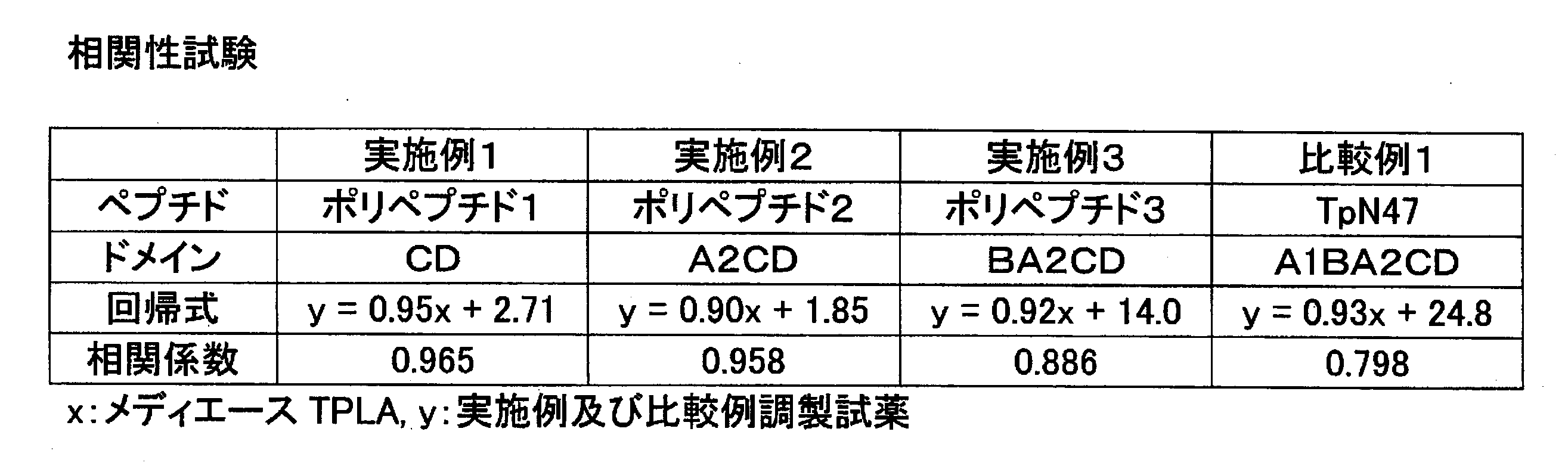
梅毒陽性と判定された血清検体(10T.U,以上)を検体とした以外は(1)と同様にして吸光度変化量(ΔAbs)を求め、(1)の標準品測定結果に基づいて作成した検量線から抗体価を算出した。実施例及び比較例にて調製した抗梅毒トレポネーマ抗体測定試薬での抗体価と、市販品試薬(積水メディカル社製、メディエース(登録商標)TPLA)との抗体価の相関性を求めた。結果を表6に示した(x:市販品試薬,y:実施例1~3及び比較例1にて調製した抗梅毒トレポネーマ抗体測定試薬)。実施例で調製された試薬は、既存の試薬との相関性が良好であることが分かった。 (2) Measurement of positive sample The amount of change in absorbance (ΔAbs) was determined in the same manner as (1) except that a serum sample (10 TU or more) determined to be syphilis positive was used as a sample, and the standard product measurement of (1) The antibody titer was calculated from a calibration curve created based on the results. The correlation between the antibody titer of the anti-syphilis treponema antibody measuring reagent prepared in Examples and Comparative Examples and the antibody titer of a commercially available reagent (Sekisui Medical, Mediace (registered trademark) TPLA) was determined. The results are shown in Table 6 (x: commercially available reagent, y: anti-syphilis treponema antibody measurement reagent prepared in Examples 1 to 3 and Comparative Example 1). The reagents prepared in the examples were found to have good correlation with existing reagents.
梅毒陰性と判定された血清検体(0T.U.)を検体とした以外は(1)と同様にして吸光度変化量(ΔAbs)を求め、(1)の標準品測定結果に基づいて作成した検量線から抗体価を算出した。
結果を表7に示した。この測定方法では、10T.U.以上を陽性と判定する。比較例1では、非特異反応が多く偽陽性が認められたのに対し、実施例1~3では、非特異反応が殆んどなく、偽陽性は認められなかった。 (3) Negative sample measurement The amount of change in absorbance (ΔAbs) was determined in the same manner as in (1) except that a serum sample (0T.U.) determined to be syphilis negative was used as a sample. The antibody titer was calculated from a calibration curve prepared based on the above.
The results are shown in Table 7. In this measurement method, 10T. U. The above is determined as positive. In Comparative Example 1, many non-specific reactions were observed and false positives were observed, whereas in Examples 1 to 3, there were almost no non-specific reactions and no false positives were observed.

Claims (12)
- 抗原抗体反応を利用した抗梅毒トレポネーマ抗体の測定に用いる試薬において、梅毒トレポネーマ47kDa抗原のうち、少なくともドメインCとDを含み、ドメインA1を含まない組み換えポリペプチドを抗原として用いることを特徴とする抗梅毒トレポネーマ抗体測定試薬。 A reagent used for measuring an anti-syphilis treponema antibody using an antigen-antibody reaction, wherein a recombinant polypeptide containing at least domains C and D and not containing domain A1 is used as an antigen among 47 kDa antigens of syphilis treponema. Syphilis treponema antibody measurement reagent.
- 抗原が、梅毒トレポネーマ47kDa抗原のうち、ドメインCとD、ドメインA2とCとD、又はドメインBとA2とCとDからなる組み換えポリペプチドである請求項1記載の抗梅毒トレポネーマ抗体測定試薬。 The anti-syphilis treponema antibody measurement reagent according to claim 1, wherein the antigen is a recombinant polypeptide consisting of domains C and D, domains A2 and C and D, or domains B and A2, C and D among the syphilis treponema 47 kDa antigen.
- 抗原が不溶性担体に固定化されている請求項1記載の抗梅毒トレポネーマ抗体測定試薬。 The anti-syphilis treponema antibody measurement reagent according to claim 1, wherein the antigen is immobilized on an insoluble carrier.
- 抗原が不溶性担体に固定化されている請求項2記載の抗梅毒トレポネーマ抗体測定試薬。 The anti-syphilis treponema antibody measurement reagent according to claim 2, wherein the antigen is immobilized on an insoluble carrier.
- 請求項3記載の不溶性担体が高分子重合体から成るラテックスである抗梅毒トレポネーマ抗体測定試薬。 A reagent for measuring an anti-syphilis treponema antibody, wherein the insoluble carrier according to claim 3 is a latex composed of a polymer.
- 請求項4記載の不溶性担体が高分子重合体から成るラテックスである抗梅毒トレポネーマ抗体測定試薬。 A reagent for measuring an anti-syphilis treponema antibody, wherein the insoluble carrier according to claim 4 is a latex comprising a polymer.
- 請求項1記載の抗梅毒トレポネーマ抗体測定試薬を用いることを特徴とする抗梅毒トレポネーマ抗体測定方法。 An anti-syphilis treponema antibody measurement method comprising using the anti-syphilis treponema antibody measurement reagent according to claim 1.
- 請求項2記載の抗梅毒トレポネーマ抗体測定試薬を用いることを特徴とする抗梅毒トレポネーマ抗体測定方法。 A method for measuring an anti-syphilis treponema antibody, wherein the reagent for measuring an anti-syphilis treponema antibody according to claim 2 is used.
- 請求項3記載の抗梅毒トレポネーマ抗体測定試薬を用いることを特徴とする抗梅毒トレポネーマ抗体測定方法。 An anti-syphilis treponema antibody measurement method comprising using the anti-syphilis treponema antibody measurement reagent according to claim 3.
- 請求項4記載の抗梅毒トレポネーマ抗体測定試薬を用いることを特徴とする抗梅毒トレポネーマ抗体測定方法。 A method for measuring an anti-syphilis treponema antibody, comprising using the reagent for measuring an anti-syphilis treponema antibody according to claim 4.
- 請求項5記載の抗梅毒トレポネーマ抗体測定試薬を用いることを特徴とする抗梅毒トレポネーマ抗体測定方法。 A method for measuring an anti-syphilis treponema antibody, comprising using the reagent for measuring an anti-syphilis treponema antibody according to claim 5.
- 請求項6記載の抗梅毒トレポネーマ抗体測定試薬を用いることを特徴とする抗梅毒トレポネーマ抗体測定方法。 A method for measuring an anti-syphilis treponema antibody, comprising using the reagent for measuring an anti-syphilis treponema antibody according to claim 6.
Priority Applications (6)
| Application Number | Priority Date | Filing Date | Title |
|---|---|---|---|
| EP11765772.6A EP2565647B1 (en) | 2010-03-31 | 2011-03-31 | Reagent for assaying anti-treponema pallidum antibody |
| KR1020127025557A KR102041119B1 (en) | 2010-03-31 | 2011-03-31 | Reagent for assaying anti-treponema pallidum antibody |
| JP2011539575A JP4866977B2 (en) | 2010-03-31 | 2011-03-31 | Anti-syphilis treponema antibody measurement reagent |
| CN201180016822.3A CN102869991B (en) | 2010-03-31 | 2011-03-31 | Anti-syphilis helicoid antibody measures reagent |
| US13/638,622 US8969520B2 (en) | 2010-03-31 | 2011-03-31 | Reagent for assaying anti-treponema pallidum antibody |
| CA2794710A CA2794710C (en) | 2010-03-31 | 2011-03-31 | Reagent for assaying anti-treponema pallidum antibody |
Applications Claiming Priority (2)
| Application Number | Priority Date | Filing Date | Title |
|---|---|---|---|
| JP2010-083822 | 2010-03-31 | ||
| JP2010083822 | 2010-03-31 |
Publications (1)
| Publication Number | Publication Date |
|---|---|
| WO2011125872A1 true WO2011125872A1 (en) | 2011-10-13 |
Family
ID=44762810
Family Applications (1)
| Application Number | Title | Priority Date | Filing Date |
|---|---|---|---|
| PCT/JP2011/058282 WO2011125872A1 (en) | 2010-03-31 | 2011-03-31 | Reagent for assaying anti-treponema pallidum antibody |
Country Status (7)
| Country | Link |
|---|---|
| US (1) | US8969520B2 (en) |
| EP (1) | EP2565647B1 (en) |
| JP (1) | JP4866977B2 (en) |
| KR (1) | KR102041119B1 (en) |
| CN (1) | CN102869991B (en) |
| CA (1) | CA2794710C (en) |
| WO (1) | WO2011125872A1 (en) |
Cited By (2)
| Publication number | Priority date | Publication date | Assignee | Title |
|---|---|---|---|---|
| JP2015506364A (en) * | 2012-01-19 | 2015-03-02 | エフ.ホフマン−ラ ロシュ アーゲーF. Hoffmann−La Roche Aktiengesellschaft | Soluble immunoreactive syphilis treponema TpN47 antigen |
| CN110981947A (en) * | 2019-12-16 | 2020-04-10 | 四川迈克生物新材料技术有限公司 | Preparation and application of treponema pallidum TP47 recombinant antigen |
Families Citing this family (3)
| Publication number | Priority date | Publication date | Assignee | Title |
|---|---|---|---|---|
| CN104360067B (en) * | 2014-11-10 | 2016-11-16 | 厦门大学附属中山医院 | Treponema pallidum specific antibody immue quantitative detection reagent box and preparation method thereof |
| CN104330562B (en) * | 2014-11-10 | 2016-10-26 | 厦门大学附属中山医院 | Treponema pallidum specific antibody high throughput testing test kit and preparation method thereof |
| CN104698185B (en) * | 2015-02-10 | 2016-08-31 | 深圳市新产业生物医学工程股份有限公司 | The detection test kit of syphilis helicoid antibody and detection method thereof and application |
Citations (5)
| Publication number | Priority date | Publication date | Assignee | Title |
|---|---|---|---|---|
| JPH09257799A (en) * | 1996-03-18 | 1997-10-03 | Sekisui Chem Co Ltd | Immunologigal syphilis diagnosing reagent |
| JPH10213585A (en) * | 1997-01-29 | 1998-08-11 | Sekisui Chem Co Ltd | Manufacture of antitreponema antibody measuring reagent and antitreponema antibody measuring reagent |
| JPH11287804A (en) * | 1998-04-01 | 1999-10-19 | Sekisui Chem Co Ltd | Formation for immunoassay reagent, immunoassay reagent and immunoassay method |
| JP2001264334A (en) * | 1999-10-07 | 2001-09-26 | Sekisui Chem Co Ltd | Syphilis treponema antibody measuring reagent and its manufacturing method |
| WO2010013789A1 (en) | 2008-08-01 | 2010-02-04 | 国立大学法人岡山大学 | Protein production method, fusion protein, and antiserum |
Family Cites Families (9)
| Publication number | Priority date | Publication date | Assignee | Title |
|---|---|---|---|---|
| US5681934A (en) | 1986-09-30 | 1997-10-28 | Board Of Regents, The University Of Texas System | 47-kilodalton antigen of Treponema pallidum |
| US5350842A (en) | 1986-09-30 | 1994-09-27 | Board Of Regents, The University Of Texas System | DNAs encoding Treponema pallidum antigens |
| US4868118A (en) * | 1986-09-30 | 1989-09-19 | Board Of Regents, The University Of Texas System | Cloning and expression of the 47-kilodalton antigen of treponema pallidum |
| US5578456A (en) | 1994-02-28 | 1996-11-26 | Fujirebio Inc. | Anti-treponema pallidum antibody immunoassay |
| JP3216473B2 (en) | 1994-02-28 | 2001-10-09 | 富士レビオ株式会社 | Measurement method of anti-treponema antibody |
| DE19536166C1 (en) * | 1995-09-29 | 1997-03-06 | Siegfried Dr Krell | Method for the determination of antibodies against Treponema pallidum (syphilis) |
| AU5147199A (en) * | 1999-02-08 | 2000-08-29 | Hongjun Jin | Modified treponema pallidum outer membrane protein, its immunoassay use and immunoassay kit |
| CN1151171C (en) * | 2001-02-26 | 2004-05-26 | 中国人民解放军军事医学科学院基础医学研究所 | Recombinant syphilis spirochete epitope antigen and multiple-epitope chimeric antigen |
| JP3799410B2 (en) * | 2002-05-20 | 2006-07-19 | 株式会社シノテスト | Nonspecific reaction inhibitory peptide, and nonspecific reaction inhibitory method and antibody measuring method using the same |
-
2011
- 2011-03-31 EP EP11765772.6A patent/EP2565647B1/en active Active
- 2011-03-31 CA CA2794710A patent/CA2794710C/en active Active
- 2011-03-31 US US13/638,622 patent/US8969520B2/en active Active
- 2011-03-31 CN CN201180016822.3A patent/CN102869991B/en active Active
- 2011-03-31 KR KR1020127025557A patent/KR102041119B1/en active IP Right Grant
- 2011-03-31 WO PCT/JP2011/058282 patent/WO2011125872A1/en active Application Filing
- 2011-03-31 JP JP2011539575A patent/JP4866977B2/en active Active
Patent Citations (5)
| Publication number | Priority date | Publication date | Assignee | Title |
|---|---|---|---|---|
| JPH09257799A (en) * | 1996-03-18 | 1997-10-03 | Sekisui Chem Co Ltd | Immunologigal syphilis diagnosing reagent |
| JPH10213585A (en) * | 1997-01-29 | 1998-08-11 | Sekisui Chem Co Ltd | Manufacture of antitreponema antibody measuring reagent and antitreponema antibody measuring reagent |
| JPH11287804A (en) * | 1998-04-01 | 1999-10-19 | Sekisui Chem Co Ltd | Formation for immunoassay reagent, immunoassay reagent and immunoassay method |
| JP2001264334A (en) * | 1999-10-07 | 2001-09-26 | Sekisui Chem Co Ltd | Syphilis treponema antibody measuring reagent and its manufacturing method |
| WO2010013789A1 (en) | 2008-08-01 | 2010-02-04 | 国立大学法人岡山大学 | Protein production method, fusion protein, and antiserum |
Non-Patent Citations (7)
| Title |
|---|
| BAUGHN R. E. ET AL.: "molecular mimicry between an immunodominant amino acid motif on the 47-kDa lipoprotein of Treponema pallidum(Tpp47) and multiple repeats of analogous sequences in fibronectin", THE JOURNAL OF IMMUNOLOGY, vol. 157, no. 2, 15 July 1996 (1996-07-15), pages 720 - 731, XP002674948 * |
| DEKA R. K. ET AL.: "Crystal structure of the 47-kDa lipoprotein of Treponema pallidum reveals a novel penicillin-binding protein", THE JOURNAL OF BIOLOGICAL CHEMISTRY, vol. 277, no. 44, 2002, pages 41857 - 41864, XP002674947 * |
| INFECTION AND IMMUNITY, vol. 60, no. 4, 1992, pages 1568 - 1576 |
| JOURNAL OF IMMUNOLOGY, vol. 157, 1996, pages 720 - 731 |
| See also references of EP2565647A4 * |
| THE JOURNAL OF BIOLOGICAL CHEMISTRY, vol. 277, no. 44, 2002, pages 41857 - 41864 |
| WEIGEL L. M. ET AL.: "Analysis of the N-terminal region of the 47-kilodalton integral membrane lipoprotein of Treponema pallidum", INFECTION AND IMMUNITY, vol. 60, no. 4, April 1992 (1992-04-01), pages 1568 - 1576, XP002912302 * |
Cited By (3)
| Publication number | Priority date | Publication date | Assignee | Title |
|---|---|---|---|---|
| JP2015506364A (en) * | 2012-01-19 | 2015-03-02 | エフ.ホフマン−ラ ロシュ アーゲーF. Hoffmann−La Roche Aktiengesellschaft | Soluble immunoreactive syphilis treponema TpN47 antigen |
| CN110981947A (en) * | 2019-12-16 | 2020-04-10 | 四川迈克生物新材料技术有限公司 | Preparation and application of treponema pallidum TP47 recombinant antigen |
| CN110981947B (en) * | 2019-12-16 | 2021-10-08 | 四川安可瑞新材料技术有限公司 | Preparation and application of treponema pallidum TP47 recombinant antigen |
Also Published As
| Publication number | Publication date |
|---|---|
| CA2794710C (en) | 2018-05-08 |
| EP2565647A1 (en) | 2013-03-06 |
| EP2565647B1 (en) | 2015-11-04 |
| CN102869991B (en) | 2016-04-27 |
| CA2794710A1 (en) | 2011-10-13 |
| US8969520B2 (en) | 2015-03-03 |
| JPWO2011125872A1 (en) | 2013-07-11 |
| EP2565647A4 (en) | 2013-07-03 |
| KR102041119B1 (en) | 2019-11-06 |
| JP4866977B2 (en) | 2012-02-01 |
| KR20130041774A (en) | 2013-04-25 |
| CN102869991A (en) | 2013-01-09 |
| US20130143229A1 (en) | 2013-06-06 |
Similar Documents
| Publication | Publication Date | Title |
|---|---|---|
| US10125177B2 (en) | Treponema pallidum triplet antigen | |
| CN101983238B (en) | Method for concentration of virus | |
| JP4866977B2 (en) | Anti-syphilis treponema antibody measurement reagent | |
| EP2267151A1 (en) | Virus titration method | |
| JP2010518046A (en) | Pathogen binding | |
| KR20190067779A (en) | An antibody measuring method using an antigen-immobilized carrier particle immobilized with an antigen in a different manner, a reagent for measuring an antibody | |
| JP6048932B2 (en) | Polypeptide for distinguishing between silicon oxide and silicon nitride and use thereof | |
| JPH1114627A (en) | Immunological diagnosis reagent | |
| JP2010540921A (en) | Immunological analysis method for detection of antibody against human GSTT1 (anti-HGSTT1) | |
| JP3864194B2 (en) | Method and reagent for measuring antibody specific for common antigenic determinant of antigen with multiple subtypes | |
| WO2023045470A1 (en) | Recombinant thyroid stimulating hormone receptor protein, preparation method therefor and application thereof | |
| JP3715736B2 (en) | Method for producing anti-treponema antibody measuring reagent and anti-treponema antibody measuring reagent | |
| JP4104219B2 (en) | Method for producing anti-treponema antibody measuring reagent, measuring reagent and measuring method | |
| JPH09304391A (en) | Immunological treponema pallidum diagnostic reagent and its manufacture | |
| CN116178558A (en) | Nanometer antibody of targeted tissue factor and preparation method and application thereof | |
| JPH1090264A (en) | Immunological diagnostic reagent | |
| WO2001090756A1 (en) | Method of assaying anti-ena antibody and assay kit | |
| JPH09257799A (en) | Immunologigal syphilis diagnosing reagent | |
| JPH09288110A (en) | Reagent for immunologically diagnosing syphilis | |
| JPH1090265A (en) | Manufacture of immunological syphilis diagnosis reagent and syphilis diagnosis reagent | |
| JP2016191581A (en) | Detection method of trypanosome infection |
Legal Events
| Date | Code | Title | Description |
|---|---|---|---|
| WWE | Wipo information: entry into national phase |
Ref document number: 201180016822.3 Country of ref document: CN |
|
| WWE | Wipo information: entry into national phase |
Ref document number: 2011539575 Country of ref document: JP |
|
| 121 | Ep: the epo has been informed by wipo that ep was designated in this application |
Ref document number: 11765772 Country of ref document: EP Kind code of ref document: A1 |
|
| ENP | Entry into the national phase |
Ref document number: 2794710 Country of ref document: CA |
|
| WWE | Wipo information: entry into national phase |
Ref document number: 2011765772 Country of ref document: EP |
|
| ENP | Entry into the national phase |
Ref document number: 20127025557 Country of ref document: KR Kind code of ref document: A |
|
| NENP | Non-entry into the national phase |
Ref country code: DE |
|
| WWE | Wipo information: entry into national phase |
Ref document number: 13638622 Country of ref document: US |