WO2011031330A1 - Autophagy and phospholipidosis pathway assays - Google Patents
Autophagy and phospholipidosis pathway assays Download PDFInfo
- Publication number
- WO2011031330A1 WO2011031330A1 PCT/US2010/002494 US2010002494W WO2011031330A1 WO 2011031330 A1 WO2011031330 A1 WO 2011031330A1 US 2010002494 W US2010002494 W US 2010002494W WO 2011031330 A1 WO2011031330 A1 WO 2011031330A1
- Authority
- WO
- WIPO (PCT)
- Prior art keywords
- disease
- cells
- cationic amphiphilic
- tracer compound
- compound
- Prior art date
Links
- 230000037361 pathway Effects 0.000 title claims description 12
- 238000003556 assay Methods 0.000 title abstract description 64
- 230000004900 autophagic degradation Effects 0.000 title abstract description 49
- 238000000034 method Methods 0.000 claims abstract description 170
- 239000003814 drug Substances 0.000 claims abstract description 111
- 229940079593 drug Drugs 0.000 claims abstract description 110
- 208000015439 Lysosomal storage disease Diseases 0.000 claims abstract description 80
- 239000003795 chemical substances by application Substances 0.000 claims abstract description 57
- 238000012544 monitoring process Methods 0.000 claims abstract description 27
- 229940000406 drug candidate Drugs 0.000 claims abstract description 22
- 231100000167 toxic agent Toxicity 0.000 claims abstract description 22
- 239000003440 toxic substance Substances 0.000 claims abstract description 22
- 239000012190 activator Substances 0.000 claims abstract description 21
- 230000004642 autophagic pathway Effects 0.000 claims abstract description 20
- 230000001988 toxicity Effects 0.000 claims abstract description 19
- 231100000419 toxicity Toxicity 0.000 claims abstract description 19
- 210000000056 organ Anatomy 0.000 claims abstract description 16
- 210000004027 cell Anatomy 0.000 claims description 336
- 150000001875 compounds Chemical class 0.000 claims description 247
- 125000002091 cationic group Chemical group 0.000 claims description 178
- 239000000700 radioactive tracer Substances 0.000 claims description 159
- 239000000523 sample Substances 0.000 claims description 109
- 210000003934 vacuole Anatomy 0.000 claims description 88
- 238000009825 accumulation Methods 0.000 claims description 62
- 230000035508 accumulation Effects 0.000 claims description 61
- -1 carbonate ester Chemical class 0.000 claims description 54
- 210000003712 lysosome Anatomy 0.000 claims description 53
- 230000001868 lysosomic effect Effects 0.000 claims description 53
- 239000000203 mixture Substances 0.000 claims description 45
- 229920006395 saturated elastomer Polymers 0.000 claims description 40
- 238000012360 testing method Methods 0.000 claims description 39
- 238000012216 screening Methods 0.000 claims description 36
- UWAUSMGZOHPBJJ-UHFFFAOYSA-N 4-nitro-1,2,3-benzoxadiazole Chemical compound [O-][N+](=O)C1=CC=CC2=C1N=NO2 UWAUSMGZOHPBJJ-UHFFFAOYSA-N 0.000 claims description 33
- 125000000217 alkyl group Chemical group 0.000 claims description 33
- IAZDPXIOMUYVGZ-UHFFFAOYSA-N Dimethylsulphoxide Chemical compound CS(C)=O IAZDPXIOMUYVGZ-UHFFFAOYSA-N 0.000 claims description 32
- 229910019142 PO4 Inorganic materials 0.000 claims description 31
- IJGRMHOSHXDMSA-UHFFFAOYSA-N Atomic nitrogen Chemical compound N#N IJGRMHOSHXDMSA-UHFFFAOYSA-N 0.000 claims description 30
- 208000037265 diseases, disorders, signs and symptoms Diseases 0.000 claims description 29
- 150000003904 phospholipids Chemical class 0.000 claims description 28
- 208000008955 Mucolipidoses Diseases 0.000 claims description 27
- 210000004957 autophagosome Anatomy 0.000 claims description 27
- 230000007812 deficiency Effects 0.000 claims description 27
- 210000004962 mammalian cell Anatomy 0.000 claims description 27
- 210000001519 tissue Anatomy 0.000 claims description 27
- 239000010452 phosphate Substances 0.000 claims description 26
- 239000003981 vehicle Substances 0.000 claims description 26
- 229910052760 oxygen Inorganic materials 0.000 claims description 25
- AQHHHDLHHXJYJD-UHFFFAOYSA-N propranolol hydrochloride Natural products C1=CC=C2C(OCC(O)CNC(C)C)=CC=CC2=C1 AQHHHDLHHXJYJD-UHFFFAOYSA-N 0.000 claims description 25
- 229910052717 sulfur Inorganic materials 0.000 claims description 25
- 201000010099 disease Diseases 0.000 claims description 21
- 229960003712 propranolol Drugs 0.000 claims description 21
- 102100022548 Beta-hexosaminidase subunit alpha Human genes 0.000 claims description 20
- 208000001905 GM2 Gangliosidoses Diseases 0.000 claims description 20
- 201000008905 GM2 gangliosidosis Diseases 0.000 claims description 20
- 101001045440 Homo sapiens Beta-hexosaminidase subunit alpha Proteins 0.000 claims description 20
- RYYWUUFWQRZTIU-UHFFFAOYSA-K thiophosphate Chemical compound [O-]P([O-])([O-])=S RYYWUUFWQRZTIU-UHFFFAOYSA-K 0.000 claims description 20
- 230000001965 increasing effect Effects 0.000 claims description 18
- UEZVMMHDMIWARA-UHFFFAOYSA-M phosphonate Chemical compound [O-]P(=O)=O UEZVMMHDMIWARA-UHFFFAOYSA-M 0.000 claims description 18
- NBIIXXVUZAFLBC-UHFFFAOYSA-K phosphate Chemical compound [O-]P([O-])([O-])=O NBIIXXVUZAFLBC-UHFFFAOYSA-K 0.000 claims description 17
- XLYOFNOQVPJJNP-UHFFFAOYSA-N water Substances O XLYOFNOQVPJJNP-UHFFFAOYSA-N 0.000 claims description 17
- 239000003153 chemical reaction reagent Substances 0.000 claims description 16
- 208000005340 mucopolysaccharidosis III Diseases 0.000 claims description 16
- OKTJSMMVPCPJKN-UHFFFAOYSA-N Carbon Chemical compound [C] OKTJSMMVPCPJKN-UHFFFAOYSA-N 0.000 claims description 15
- 206010072927 Mucolipidosis type I Diseases 0.000 claims description 15
- 150000001450 anions Chemical class 0.000 claims description 15
- 229910052799 carbon Inorganic materials 0.000 claims description 15
- 229910052757 nitrogen Inorganic materials 0.000 claims description 15
- 210000001616 monocyte Anatomy 0.000 claims description 14
- NINIDFKCEFEMDL-UHFFFAOYSA-N Sulfur Chemical compound [S] NINIDFKCEFEMDL-UHFFFAOYSA-N 0.000 claims description 13
- QVGXLLKOCUKJST-UHFFFAOYSA-N atomic oxygen Chemical compound [O] QVGXLLKOCUKJST-UHFFFAOYSA-N 0.000 claims description 13
- 239000001301 oxygen Substances 0.000 claims description 13
- 229940124530 sulfonamide Drugs 0.000 claims description 13
- 150000003456 sulfonamides Chemical class 0.000 claims description 13
- 239000011593 sulfur Substances 0.000 claims description 13
- 241000124008 Mammalia Species 0.000 claims description 12
- 206010056886 Mucopolysaccharidosis I Diseases 0.000 claims description 12
- 208000021811 Sandhoff disease Diseases 0.000 claims description 12
- 210000003714 granulocyte Anatomy 0.000 claims description 12
- 210000004698 lymphocyte Anatomy 0.000 claims description 12
- 108010045758 lysosomal proteins Proteins 0.000 claims description 12
- 210000002540 macrophage Anatomy 0.000 claims description 12
- 208000024720 Fabry Disease Diseases 0.000 claims description 11
- 206010053185 Glycogen storage disease type II Diseases 0.000 claims description 11
- 208000002678 Mucopolysaccharidoses Diseases 0.000 claims description 10
- DMSZORWOGDLWGN-UHFFFAOYSA-N ctk1a3526 Chemical compound NP(N)(N)=O DMSZORWOGDLWGN-UHFFFAOYSA-N 0.000 claims description 10
- 150000005690 diesters Chemical class 0.000 claims description 10
- 201000007769 mucolipidosis Diseases 0.000 claims description 10
- 206010028093 mucopolysaccharidosis Diseases 0.000 claims description 10
- 125000005541 phosphonamide group Chemical group 0.000 claims description 10
- 150000008300 phosphoramidites Chemical class 0.000 claims description 10
- 229950001675 spiperone Drugs 0.000 claims description 10
- 150000003457 sulfones Chemical class 0.000 claims description 10
- 150000003462 sulfoxides Chemical class 0.000 claims description 10
- 230000006670 vacuole accumulation Effects 0.000 claims description 10
- 238000007865 diluting Methods 0.000 claims description 9
- 230000000366 juvenile effect Effects 0.000 claims description 9
- BDHFUVZGWQCTTF-UHFFFAOYSA-M sulfonate Chemical compound [O-]S(=O)=O BDHFUVZGWQCTTF-UHFFFAOYSA-M 0.000 claims description 9
- 108010053317 Hexosaminidase A Proteins 0.000 claims description 8
- 102000016871 Hexosaminidase A Human genes 0.000 claims description 8
- 208000035051 Malignant migrating focal seizures of infancy Diseases 0.000 claims description 8
- 206010028095 Mucopolysaccharidosis IV Diseases 0.000 claims description 8
- 208000028781 Mucopolysaccharidosis type 1 Diseases 0.000 claims description 8
- 208000002537 Neuronal Ceroid-Lipofuscinoses Diseases 0.000 claims description 8
- 208000014060 Niemann-Pick disease Diseases 0.000 claims description 8
- 208000013608 Salla disease Diseases 0.000 claims description 8
- 208000000828 Sialic Acid Storage Disease Diseases 0.000 claims description 8
- 108010039203 Tripeptidyl-Peptidase 1 Proteins 0.000 claims description 8
- 102100034197 Tripeptidyl-peptidase 1 Human genes 0.000 claims description 8
- 208000036710 mucopolysaccharidosis type 3A Diseases 0.000 claims description 8
- 208000036709 mucopolysaccharidosis type 3B Diseases 0.000 claims description 8
- 208000036707 mucopolysaccharidosis type 3C Diseases 0.000 claims description 8
- 208000036725 mucopolysaccharidosis type 3D Diseases 0.000 claims description 8
- 239000003960 organic solvent Substances 0.000 claims description 8
- 208000032007 Glycogen storage disease due to acid maltase deficiency Diseases 0.000 claims description 7
- 239000006143 cell culture medium Substances 0.000 claims description 7
- 230000007547 defect Effects 0.000 claims description 7
- 201000004502 glycogen storage disease II Diseases 0.000 claims description 7
- 230000002503 metabolic effect Effects 0.000 claims description 7
- 238000003860 storage Methods 0.000 claims description 7
- 208000000501 Lipidoses Diseases 0.000 claims description 6
- 206010024585 Lipidosis Diseases 0.000 claims description 6
- 108010064171 Lysosome-Associated Membrane Glycoproteins Proteins 0.000 claims description 6
- 102000014944 Lysosome-Associated Membrane Glycoproteins Human genes 0.000 claims description 6
- 125000003178 carboxy group Chemical group [H]OC(*)=O 0.000 claims description 6
- 210000002472 endoplasmic reticulum Anatomy 0.000 claims description 6
- 230000002068 genetic effect Effects 0.000 claims description 6
- 208000007345 glycogen storage disease Diseases 0.000 claims description 6
- 210000002288 golgi apparatus Anatomy 0.000 claims description 6
- 210000005260 human cell Anatomy 0.000 claims description 6
- 238000005192 partition Methods 0.000 claims description 6
- 231100000331 toxic Toxicity 0.000 claims description 6
- 230000002588 toxic effect Effects 0.000 claims description 6
- 239000000872 buffer Substances 0.000 claims description 5
- 239000013074 reference sample Substances 0.000 claims description 5
- 208000029602 Alpha-N-acetylgalactosaminidase deficiency Diseases 0.000 claims description 4
- 206010068220 Aspartylglucosaminuria Diseases 0.000 claims description 4
- 208000033436 CLN6 disease Diseases 0.000 claims description 4
- 206010011777 Cystinosis Diseases 0.000 claims description 4
- 208000011518 Danon disease Diseases 0.000 claims description 4
- 102100031675 DnaJ homolog subfamily C member 5 Human genes 0.000 claims description 4
- 208000001948 Farber Lipogranulomatosis Diseases 0.000 claims description 4
- 208000033149 Farber disease Diseases 0.000 claims description 4
- 201000008892 GM1 Gangliosidosis Diseases 0.000 claims description 4
- 208000017462 Galactosialidosis Diseases 0.000 claims description 4
- 208000015872 Gaucher disease Diseases 0.000 claims description 4
- 208000010055 Globoid Cell Leukodystrophy Diseases 0.000 claims description 4
- 208000001500 Glycogen Storage Disease Type IIb Diseases 0.000 claims description 4
- 208000035148 Glycogen storage disease due to LAMP-2 deficiency Diseases 0.000 claims description 4
- 101000845893 Homo sapiens DnaJ homolog subfamily C member 5 Proteins 0.000 claims description 4
- 208000015178 Hurler syndrome Diseases 0.000 claims description 4
- 208000015204 Hurler-Scheie syndrome Diseases 0.000 claims description 4
- 108700037017 Hyaluronidase Deficiency Proteins 0.000 claims description 4
- 208000005503 Hyaluronidase deficiency Diseases 0.000 claims description 4
- 208000028226 Krabbe disease Diseases 0.000 claims description 4
- 201000011442 Metachromatic leukodystrophy Diseases 0.000 claims description 4
- 206010072928 Mucolipidosis type II Diseases 0.000 claims description 4
- 206010072930 Mucolipidosis type IV Diseases 0.000 claims description 4
- 102100026502 Mucolipin-1 Human genes 0.000 claims description 4
- 206010056893 Mucopolysaccharidosis VII Diseases 0.000 claims description 4
- 208000000149 Multiple Sulfatase Deficiency Disease Diseases 0.000 claims description 4
- 208000035032 Multiple sulfatase deficiency Diseases 0.000 claims description 4
- 208000037006 Progressive epilepsy-intellectual disability syndrome, Finnish type Diseases 0.000 claims description 4
- 208000025816 Sanfilippo syndrome type A Diseases 0.000 claims description 4
- 208000025820 Sanfilippo syndrome type B Diseases 0.000 claims description 4
- 208000025802 Sanfilippo syndrome type C Diseases 0.000 claims description 4
- 208000025804 Sanfilippo syndrome type D Diseases 0.000 claims description 4
- 201000002883 Scheie syndrome Diseases 0.000 claims description 4
- 201000001828 Sly syndrome Diseases 0.000 claims description 4
- 108700001567 Type I Schindler Disease Proteins 0.000 claims description 4
- 208000026589 Wolman disease Diseases 0.000 claims description 4
- 150000001336 alkenes Chemical group 0.000 claims description 4
- 201000008333 alpha-mannosidosis Diseases 0.000 claims description 4
- 201000006486 beta-mannosidosis Diseases 0.000 claims description 4
- 208000031406 ceroid lipofuscinosis, neuronal, 4 (Kufs type) Diseases 0.000 claims description 4
- 208000024042 cholesterol ester storage disease Diseases 0.000 claims description 4
- 208000013760 cholesteryl ester storage disease Diseases 0.000 claims description 4
- 230000001684 chronic effect Effects 0.000 claims description 4
- 208000035475 disorder Diseases 0.000 claims description 4
- 201000008049 fucosidosis Diseases 0.000 claims description 4
- 201000008977 glycoproteinosis Diseases 0.000 claims description 4
- 208000017476 juvenile neuronal ceroid lipofuscinosis Diseases 0.000 claims description 4
- 208000025014 late infantile neuronal ceroid lipofuscinosis Diseases 0.000 claims description 4
- 208000020460 mucolipidosis II alpha/beta Diseases 0.000 claims description 4
- 201000002273 mucopolysaccharidosis II Diseases 0.000 claims description 4
- 208000012253 mucopolysaccharidosis IVA Diseases 0.000 claims description 4
- 208000022018 mucopolysaccharidosis type 2 Diseases 0.000 claims description 4
- 208000025919 mucopolysaccharidosis type 7 Diseases 0.000 claims description 4
- 208000012226 mucopolysaccharidosis type IIIA Diseases 0.000 claims description 4
- 208000012227 mucopolysaccharidosis type IIIB Diseases 0.000 claims description 4
- 208000012224 mucopolysaccharidosis type IIIC Diseases 0.000 claims description 4
- 208000027333 mucopolysaccharidosis type IIID Diseases 0.000 claims description 4
- 208000012091 mucopolysaccharidosis type IVB Diseases 0.000 claims description 4
- 201000008051 neuronal ceroid lipofuscinosis Diseases 0.000 claims description 4
- 201000007607 neuronal ceroid lipofuscinosis 3 Diseases 0.000 claims description 4
- 201000007638 neuronal ceroid lipofuscinosis 8 Diseases 0.000 claims description 4
- 201000007635 neuronal ceroid lipofuscinosis 8 northern epilepsy variant Diseases 0.000 claims description 4
- 230000000269 nucleophilic effect Effects 0.000 claims description 4
- 125000001997 phenyl group Chemical group [H]C1=C([H])C([H])=C(*)C([H])=C1[H] 0.000 claims description 4
- 125000002924 primary amino group Chemical group [H]N([H])* 0.000 claims description 4
- 201000010108 pycnodysostosis Diseases 0.000 claims description 4
- 208000011985 sialidosis Diseases 0.000 claims description 4
- 239000002168 alkylating agent Substances 0.000 claims description 3
- 229940100198 alkylating agent Drugs 0.000 claims description 3
- 125000000400 lauroyl group Chemical group O=C([*])C([H])([H])C([H])([H])C([H])([H])C([H])([H])C([H])([H])C([H])([H])C([H])([H])C([H])([H])C([H])([H])C([H])([H])C([H])([H])[H] 0.000 claims description 3
- WTJKGGKOPKCXLL-RRHRGVEJSA-N phosphatidylcholine Chemical compound CCCCCCCCCCCCCCCC(=O)OC[C@H](COP([O-])(=O)OCC[N+](C)(C)C)OC(=O)CCCCCCCC=CCCCCCCCC WTJKGGKOPKCXLL-RRHRGVEJSA-N 0.000 claims description 3
- 239000000356 contaminant Substances 0.000 claims description 2
- 230000007613 environmental effect Effects 0.000 claims description 2
- 102100033448 Lysosomal alpha-glucosidase Human genes 0.000 claims 3
- 206010072929 Mucolipidosis type III Diseases 0.000 claims 3
- 208000020468 mucolipidosis III alpha/beta Diseases 0.000 claims 3
- 150000001345 alkine derivatives Chemical group 0.000 claims 1
- 238000011282 treatment Methods 0.000 abstract description 13
- 239000000975 dye Substances 0.000 description 216
- OKKJLVBELUTLKV-UHFFFAOYSA-N Methanol Chemical compound OC OKKJLVBELUTLKV-UHFFFAOYSA-N 0.000 description 63
- WHTVZRBIWZFKQO-AWEZNQCLSA-N (S)-chloroquine Chemical compound ClC1=CC=C2C(N[C@@H](C)CCCN(CC)CC)=CC=NC2=C1 WHTVZRBIWZFKQO-AWEZNQCLSA-N 0.000 description 28
- 229960003677 chloroquine Drugs 0.000 description 28
- WHTVZRBIWZFKQO-UHFFFAOYSA-N chloroquine Natural products ClC1=CC=C2C(NC(C)CCCN(CC)CC)=CC=NC2=C1 WHTVZRBIWZFKQO-UHFFFAOYSA-N 0.000 description 28
- 230000002132 lysosomal effect Effects 0.000 description 28
- 239000003112 inhibitor Substances 0.000 description 24
- 235000021317 phosphate Nutrition 0.000 description 24
- YMWUJEATGCHHMB-UHFFFAOYSA-N Dichloromethane Chemical compound ClCCl YMWUJEATGCHHMB-UHFFFAOYSA-N 0.000 description 23
- 125000003545 alkoxy group Chemical group 0.000 description 19
- 210000003463 organelle Anatomy 0.000 description 19
- 239000000243 solution Substances 0.000 description 18
- 239000011521 glass Substances 0.000 description 16
- 235000003642 hunger Nutrition 0.000 description 16
- 238000010186 staining Methods 0.000 description 16
- 230000037351 starvation Effects 0.000 description 16
- WEVYAHXRMPXWCK-UHFFFAOYSA-N Acetonitrile Chemical compound CC#N WEVYAHXRMPXWCK-UHFFFAOYSA-N 0.000 description 15
- HEMHJVSKTPXQMS-UHFFFAOYSA-M Sodium hydroxide Chemical compound [OH-].[Na+] HEMHJVSKTPXQMS-UHFFFAOYSA-M 0.000 description 15
- 238000002360 preparation method Methods 0.000 description 15
- JABNPSKWVNCGMX-UHFFFAOYSA-N 2-(4-ethoxyphenyl)-6-[6-(4-methylpiperazin-1-yl)-1h-benzimidazol-2-yl]-1h-benzimidazole;trihydrochloride Chemical compound Cl.Cl.Cl.C1=CC(OCC)=CC=C1C1=NC2=CC=C(C=3NC4=CC(=CC=C4N=3)N3CCN(C)CC3)C=C2N1 JABNPSKWVNCGMX-UHFFFAOYSA-N 0.000 description 14
- XEKOWRVHYACXOJ-UHFFFAOYSA-N Ethyl acetate Chemical compound CCOC(C)=O XEKOWRVHYACXOJ-UHFFFAOYSA-N 0.000 description 14
- NKANXQFJJICGDU-QPLCGJKRSA-N Tamoxifen Chemical compound C=1C=CC=CC=1C(/CC)=C(C=1C=CC(OCCN(C)C)=CC=1)/C1=CC=CC=C1 NKANXQFJJICGDU-QPLCGJKRSA-N 0.000 description 14
- ZMANZCXQSJIPKH-UHFFFAOYSA-N Triethylamine Chemical compound CCN(CC)CC ZMANZCXQSJIPKH-UHFFFAOYSA-N 0.000 description 13
- 238000001514 detection method Methods 0.000 description 13
- 238000000684 flow cytometry Methods 0.000 description 13
- 239000002609 medium Substances 0.000 description 13
- 239000007787 solid Substances 0.000 description 13
- XDHNQDDQEHDUTM-XJKSCTEHSA-N (3z,5e,7r,8s,9r,11e,13e,15s,16r)-16-[(2s,3r,4s)-4-[(2r,4r,5s,6r)-2,4-dihydroxy-5-methyl-6-propan-2-yloxan-2-yl]-3-hydroxypentan-2-yl]-8-hydroxy-3,15-dimethoxy-5,7,9,11-tetramethyl-1-oxacyclohexadeca-3,5,11,13-tetraen-2-one Chemical compound CO[C@H]1\C=C\C=C(C)\C[C@@H](C)[C@H](O)[C@H](C)\C=C(/C)\C=C(OC)\C(=O)O[C@@H]1[C@@H](C)[C@@H](O)[C@H](C)[C@]1(O)O[C@H](C(C)C)[C@@H](C)[C@H](O)C1 XDHNQDDQEHDUTM-XJKSCTEHSA-N 0.000 description 12
- XDHNQDDQEHDUTM-UHFFFAOYSA-N bafliomycin A1 Natural products COC1C=CC=C(C)CC(C)C(O)C(C)C=C(C)C=C(OC)C(=O)OC1C(C)C(O)C(C)C1(O)OC(C(C)C)C(C)C(O)C1 XDHNQDDQEHDUTM-UHFFFAOYSA-N 0.000 description 12
- 230000015572 biosynthetic process Effects 0.000 description 12
- 229910052739 hydrogen Inorganic materials 0.000 description 12
- 239000001257 hydrogen Substances 0.000 description 12
- 238000011534 incubation Methods 0.000 description 12
- UCSJYZPVAKXKNQ-HZYVHMACSA-N streptomycin Chemical compound CN[C@H]1[C@H](O)[C@@H](O)[C@H](CO)O[C@H]1O[C@@H]1[C@](C=O)(O)[C@H](C)O[C@H]1O[C@@H]1[C@@H](NC(N)=N)[C@H](O)[C@@H](NC(N)=N)[C@H](O)[C@H]1O UCSJYZPVAKXKNQ-HZYVHMACSA-N 0.000 description 12
- ZMXDDKWLCZADIW-UHFFFAOYSA-N N,N-Dimethylformamide Chemical compound CN(C)C=O ZMXDDKWLCZADIW-UHFFFAOYSA-N 0.000 description 11
- 238000004458 analytical method Methods 0.000 description 11
- 239000012091 fetal bovine serum Substances 0.000 description 11
- 238000000799 fluorescence microscopy Methods 0.000 description 11
- 150000002431 hydrogen Chemical class 0.000 description 11
- RTZKZFJDLAIYFH-UHFFFAOYSA-N Diethyl ether Chemical compound CCOCC RTZKZFJDLAIYFH-UHFFFAOYSA-N 0.000 description 10
- 239000004698 Polyethylene Substances 0.000 description 10
- 238000003384 imaging method Methods 0.000 description 10
- 239000002904 solvent Substances 0.000 description 10
- ANRHNWWPFJCPAZ-UHFFFAOYSA-M thionine Chemical compound [Cl-].C1=CC(N)=CC2=[S+]C3=CC(N)=CC=C3N=C21 ANRHNWWPFJCPAZ-UHFFFAOYSA-M 0.000 description 10
- LFQSCWFLJHTTHZ-UHFFFAOYSA-N Ethanol Chemical compound CCO LFQSCWFLJHTTHZ-UHFFFAOYSA-N 0.000 description 9
- SIKJAQJRHWYJAI-UHFFFAOYSA-N Indole Chemical group C1=CC=C2NC=CC2=C1 SIKJAQJRHWYJAI-UHFFFAOYSA-N 0.000 description 9
- 230000002378 acidificating effect Effects 0.000 description 9
- 229940125904 compound 1 Drugs 0.000 description 9
- 230000001419 dependent effect Effects 0.000 description 9
- 239000002953 phosphate buffered saline Substances 0.000 description 9
- 239000000047 product Substances 0.000 description 9
- 239000011541 reaction mixture Substances 0.000 description 9
- 238000003786 synthesis reaction Methods 0.000 description 9
- 239000002676 xenobiotic agent Substances 0.000 description 9
- 230000002886 autophagic effect Effects 0.000 description 8
- XDHNQDDQEHDUTM-ZGOPVUMHSA-N bafilomycin A1 Natural products CO[C@H]1C=CC=C(C)C[C@H](C)[C@H](O)[C@H](C)C=C(C)C=C(OC)C(=O)O[C@@H]1[C@@H](C)[C@@H](O)[C@H](C)[C@]1(O)O[C@H](C(C)C)[C@@H](C)[C@H](O)C1 XDHNQDDQEHDUTM-ZGOPVUMHSA-N 0.000 description 8
- 230000006652 catabolic pathway Effects 0.000 description 8
- ZPEIMTDSQAKGNT-UHFFFAOYSA-N chlorpromazine Chemical compound C1=C(Cl)C=C2N(CCCN(C)C)C3=CC=CC=C3SC2=C1 ZPEIMTDSQAKGNT-UHFFFAOYSA-N 0.000 description 7
- 229960001076 chlorpromazine Drugs 0.000 description 7
- 230000004064 dysfunction Effects 0.000 description 7
- 238000002474 experimental method Methods 0.000 description 7
- 125000005842 heteroatom Chemical group 0.000 description 7
- 230000005764 inhibitory process Effects 0.000 description 7
- 238000005304 joining Methods 0.000 description 7
- 108090000623 proteins and genes Proteins 0.000 description 7
- 229960001603 tamoxifen Drugs 0.000 description 7
- RTHCYVBBDHJXIQ-MRXNPFEDSA-N (R)-fluoxetine Chemical compound O([C@H](CCNC)C=1C=CC=CC=1)C1=CC=C(C(F)(F)F)C=C1 RTHCYVBBDHJXIQ-MRXNPFEDSA-N 0.000 description 6
- RFFLAFLAYFXFSW-UHFFFAOYSA-N 1,2-dichlorobenzene Chemical compound ClC1=CC=CC=C1Cl RFFLAFLAYFXFSW-UHFFFAOYSA-N 0.000 description 6
- QMGUOJYZJKLOLH-UHFFFAOYSA-N 3-[1-[3-(dimethylamino)propyl]indol-3-yl]-4-(1h-indol-3-yl)pyrrole-2,5-dione Chemical compound C12=CC=CC=C2N(CCCN(C)C)C=C1C1=C(C=2C3=CC=CC=C3NC=2)C(=O)NC1=O QMGUOJYZJKLOLH-UHFFFAOYSA-N 0.000 description 6
- QTBSBXVTEAMEQO-UHFFFAOYSA-N Acetic acid Chemical compound CC(O)=O QTBSBXVTEAMEQO-UHFFFAOYSA-N 0.000 description 6
- 108010043121 Green Fluorescent Proteins Proteins 0.000 description 6
- CSNNHWWHGAXBCP-UHFFFAOYSA-L Magnesium sulfate Chemical compound [Mg+2].[O-][S+2]([O-])([O-])[O-] CSNNHWWHGAXBCP-UHFFFAOYSA-L 0.000 description 6
- 229930182555 Penicillin Natural products 0.000 description 6
- JGSARLDLIJGVTE-MBNYWOFBSA-N Penicillin G Chemical compound N([C@H]1[C@H]2SC([C@@H](N2C1=O)C(O)=O)(C)C)C(=O)CC1=CC=CC=C1 JGSARLDLIJGVTE-MBNYWOFBSA-N 0.000 description 6
- 102000003923 Protein Kinase C Human genes 0.000 description 6
- 108090000315 Protein Kinase C Proteins 0.000 description 6
- DPKHZNPWBDQZCN-UHFFFAOYSA-N acridine orange free base Chemical compound C1=CC(N(C)C)=CC2=NC3=CC(N(C)C)=CC=C3C=C21 DPKHZNPWBDQZCN-UHFFFAOYSA-N 0.000 description 6
- 150000001412 amines Chemical class 0.000 description 6
- 239000012267 brine Substances 0.000 description 6
- 238000009826 distribution Methods 0.000 description 6
- 230000000694 effects Effects 0.000 description 6
- 230000005284 excitation Effects 0.000 description 6
- 239000007850 fluorescent dye Substances 0.000 description 6
- 229960002464 fluoxetine Drugs 0.000 description 6
- 125000000524 functional group Chemical group 0.000 description 6
- 229910052736 halogen Inorganic materials 0.000 description 6
- 230000003834 intracellular effect Effects 0.000 description 6
- 238000001000 micrograph Methods 0.000 description 6
- 239000012044 organic layer Substances 0.000 description 6
- 229940049954 penicillin Drugs 0.000 description 6
- 230000003389 potentiating effect Effects 0.000 description 6
- 102000004169 proteins and genes Human genes 0.000 description 6
- 238000000746 purification Methods 0.000 description 6
- ZAHRKKWIAAJSAO-UHFFFAOYSA-N rapamycin Natural products COCC(O)C(=C/C(C)C(=O)CC(OC(=O)C1CCCCN1C(=O)C(=O)C2(O)OC(CC(OC)C(=CC=CC=CC(C)CC(C)C(=O)C)C)CCC2C)C(C)CC3CCC(O)C(C3)OC)C ZAHRKKWIAAJSAO-UHFFFAOYSA-N 0.000 description 6
- 229960002930 sirolimus Drugs 0.000 description 6
- QFJCIRLUMZQUOT-HPLJOQBZSA-N sirolimus Chemical compound C1C[C@@H](O)[C@H](OC)C[C@@H]1C[C@@H](C)[C@H]1OC(=O)[C@@H]2CCCCN2C(=O)C(=O)[C@](O)(O2)[C@H](C)CC[C@H]2C[C@H](OC)/C(C)=C/C=C/C=C/[C@@H](C)C[C@@H](C)C(=O)[C@H](OC)[C@H](O)/C(C)=C/[C@@H](C)C(=O)C1 QFJCIRLUMZQUOT-HPLJOQBZSA-N 0.000 description 6
- 150000003384 small molecules Chemical class 0.000 description 6
- FVAUCKIRQBBSSJ-UHFFFAOYSA-M sodium iodide Chemical compound [Na+].[I-] FVAUCKIRQBBSSJ-UHFFFAOYSA-M 0.000 description 6
- HPALAKNZSZLMCH-UHFFFAOYSA-M sodium;chloride;hydrate Chemical compound O.[Na+].[Cl-] HPALAKNZSZLMCH-UHFFFAOYSA-M 0.000 description 6
- 229960005322 streptomycin Drugs 0.000 description 6
- SGTNSNPWRIOYBX-UHFFFAOYSA-N 2-(3,4-dimethoxyphenyl)-5-{[2-(3,4-dimethoxyphenyl)ethyl](methyl)amino}-2-(propan-2-yl)pentanenitrile Chemical compound C1=C(OC)C(OC)=CC=C1CCN(C)CCCC(C#N)(C(C)C)C1=CC=C(OC)C(OC)=C1 SGTNSNPWRIOYBX-UHFFFAOYSA-N 0.000 description 5
- LHHQTXPEHJNOCX-UHFFFAOYSA-N Rottlerin Natural products CC(=O)c1c(O)c(C)c(O)c(Oc2c(O)c3C=CC(C)(C)Cc3c(C(=O)C=Cc4ccccc4)c2O)c1O LHHQTXPEHJNOCX-UHFFFAOYSA-N 0.000 description 5
- FAPWRFPIFSIZLT-UHFFFAOYSA-M Sodium chloride Chemical compound [Na+].[Cl-] FAPWRFPIFSIZLT-UHFFFAOYSA-M 0.000 description 5
- 102000004183 Synaptosomal-Associated Protein 25 Human genes 0.000 description 5
- 108010057722 Synaptosomal-Associated Protein 25 Proteins 0.000 description 5
- 125000003277 amino group Chemical group 0.000 description 5
- 238000013459 approach Methods 0.000 description 5
- 230000004908 autophagic flux Effects 0.000 description 5
- 230000008901 benefit Effects 0.000 description 5
- DZBUGLKDJFMEHC-UHFFFAOYSA-N benzoquinolinylidene Natural products C1=CC=CC2=CC3=CC=CC=C3N=C21 DZBUGLKDJFMEHC-UHFFFAOYSA-N 0.000 description 5
- 150000001768 cations Chemical class 0.000 description 5
- 230000001413 cellular effect Effects 0.000 description 5
- 238000005119 centrifugation Methods 0.000 description 5
- 229940126214 compound 3 Drugs 0.000 description 5
- 230000021615 conjugation Effects 0.000 description 5
- 235000019439 ethyl acetate Nutrition 0.000 description 5
- 238000000338 in vitro Methods 0.000 description 5
- 230000001939 inductive effect Effects 0.000 description 5
- 229940043355 kinase inhibitor Drugs 0.000 description 5
- 150000002632 lipids Chemical class 0.000 description 5
- 229910052943 magnesium sulfate Inorganic materials 0.000 description 5
- 239000002245 particle Substances 0.000 description 5
- 239000003757 phosphotransferase inhibitor Substances 0.000 description 5
- DEZFNHCVIZBHBI-ZHACJKMWSA-N rottlerin Chemical compound CC(=O)C1=C(O)C(C)=C(O)C(CC=2C(=C(C(=O)\C=C\C=3C=CC=CC=3)C=3OC(C)(C)C=CC=3C=2O)O)=C1O DEZFNHCVIZBHBI-ZHACJKMWSA-N 0.000 description 5
- 150000003839 salts Chemical class 0.000 description 5
- 230000003595 spectral effect Effects 0.000 description 5
- 125000005504 styryl group Chemical group 0.000 description 5
- 230000002477 vacuolizing effect Effects 0.000 description 5
- 229960001722 verapamil Drugs 0.000 description 5
- BHSFGROFSZRPCD-UHFFFAOYSA-N 3-(3,4-dihydro-2h-quinolin-1-yl)propanoic acid Chemical compound C1=CC=C2N(CCC(=O)O)CCCC2=C1 BHSFGROFSZRPCD-UHFFFAOYSA-N 0.000 description 4
- DFOCUWZXJBAUSQ-UHFFFAOYSA-N Berbamine Natural products O1C(C(=CC=2)O)=CC=2CC(C=23)N(C)CCC3=CC(OC)=C(OC)C=2OC(=CC=23)C(OC)=CC=2CCN(C)C3CC2=CC=C1C=C2 DFOCUWZXJBAUSQ-UHFFFAOYSA-N 0.000 description 4
- 102100033697 DNA cross-link repair 1A protein Human genes 0.000 description 4
- 102000004190 Enzymes Human genes 0.000 description 4
- 108090000790 Enzymes Proteins 0.000 description 4
- 102000004144 Green Fluorescent Proteins Human genes 0.000 description 4
- 101000871548 Homo sapiens DNA cross-link repair 1A protein Proteins 0.000 description 4
- VEXZGXHMUGYJMC-UHFFFAOYSA-N Hydrochloric acid Chemical compound Cl VEXZGXHMUGYJMC-UHFFFAOYSA-N 0.000 description 4
- NQRYJNQNLNOLGT-UHFFFAOYSA-N Piperidine Chemical compound C1CCNCC1 NQRYJNQNLNOLGT-UHFFFAOYSA-N 0.000 description 4
- CDBYLPFSWZWCQE-UHFFFAOYSA-L Sodium Carbonate Chemical compound [Na+].[Na+].[O-]C([O-])=O CDBYLPFSWZWCQE-UHFFFAOYSA-L 0.000 description 4
- PPBRXRYQALVLMV-UHFFFAOYSA-N Styrene Chemical compound C=CC1=CC=CC=C1 PPBRXRYQALVLMV-UHFFFAOYSA-N 0.000 description 4
- 102000013530 TOR Serine-Threonine Kinases Human genes 0.000 description 4
- 108010065917 TOR Serine-Threonine Kinases Proteins 0.000 description 4
- 108090000848 Ubiquitin Proteins 0.000 description 4
- 102000044159 Ubiquitin Human genes 0.000 description 4
- 150000001413 amino acids Chemical class 0.000 description 4
- PYKYMHQGRFAEBM-UHFFFAOYSA-N anthraquinone Natural products CCC(=O)c1c(O)c2C(=O)C3C(C=CC=C3O)C(=O)c2cc1CC(=O)OC PYKYMHQGRFAEBM-UHFFFAOYSA-N 0.000 description 4
- 150000004056 anthraquinones Chemical class 0.000 description 4
- 239000007864 aqueous solution Substances 0.000 description 4
- 238000006243 chemical reaction Methods 0.000 description 4
- 239000002131 composite material Substances 0.000 description 4
- 229940125782 compound 2 Drugs 0.000 description 4
- 229940125898 compound 5 Drugs 0.000 description 4
- 238000004163 cytometry Methods 0.000 description 4
- 231100000433 cytotoxic Toxicity 0.000 description 4
- 230000001472 cytotoxic effect Effects 0.000 description 4
- BFMYDTVEBKDAKJ-UHFFFAOYSA-L disodium;(2',7'-dibromo-3',6'-dioxido-3-oxospiro[2-benzofuran-1,9'-xanthene]-4'-yl)mercury;hydrate Chemical compound O.[Na+].[Na+].O1C(=O)C2=CC=CC=C2C21C1=CC(Br)=C([O-])C([Hg])=C1OC1=C2C=C(Br)C([O-])=C1 BFMYDTVEBKDAKJ-UHFFFAOYSA-L 0.000 description 4
- 231100000673 dose–response relationship Toxicity 0.000 description 4
- 229940088598 enzyme Drugs 0.000 description 4
- 239000005090 green fluorescent protein Substances 0.000 description 4
- 150000002367 halogens Chemical class 0.000 description 4
- 238000005286 illumination Methods 0.000 description 4
- 238000002952 image-based readout Methods 0.000 description 4
- 230000006698 induction Effects 0.000 description 4
- 230000002401 inhibitory effect Effects 0.000 description 4
- 230000003993 interaction Effects 0.000 description 4
- 239000007788 liquid Substances 0.000 description 4
- 239000003550 marker Substances 0.000 description 4
- 230000007246 mechanism Effects 0.000 description 4
- 230000007170 pathology Effects 0.000 description 4
- BASFCYQUMIYNBI-UHFFFAOYSA-N platinum Chemical compound [Pt] BASFCYQUMIYNBI-UHFFFAOYSA-N 0.000 description 4
- 230000008569 process Effects 0.000 description 4
- 108020003175 receptors Proteins 0.000 description 4
- 102000005962 receptors Human genes 0.000 description 4
- 230000002829 reductive effect Effects 0.000 description 4
- 238000011160 research Methods 0.000 description 4
- 230000004044 response Effects 0.000 description 4
- 208000024891 symptom Diseases 0.000 description 4
- 239000003039 volatile agent Substances 0.000 description 4
- XMAYWYJOQHXEEK-OZXSUGGESA-N (2R,4S)-ketoconazole Chemical compound C1CN(C(=O)C)CCN1C(C=C1)=CC=C1OC[C@@H]1O[C@@](CN2C=NC=C2)(C=2C(=CC(Cl)=CC=2)Cl)OC1 XMAYWYJOQHXEEK-OZXSUGGESA-N 0.000 description 3
- CAPCBAYULRXQAN-UHFFFAOYSA-N 1-n,1-n-diethylpentane-1,4-diamine Chemical compound CCN(CC)CCCC(C)N CAPCBAYULRXQAN-UHFFFAOYSA-N 0.000 description 3
- YOSZEPWSVKKQOV-UHFFFAOYSA-N 12h-benzo[a]phenoxazine Chemical compound C1=CC=CC2=C3NC4=CC=CC=C4OC3=CC=C21 YOSZEPWSVKKQOV-UHFFFAOYSA-N 0.000 description 3
- BSKHPKMHTQYZBB-UHFFFAOYSA-N 2-methylpyridine Chemical compound CC1=CC=CC=N1 BSKHPKMHTQYZBB-UHFFFAOYSA-N 0.000 description 3
- ZPBYVFQJHWLTFB-UHFFFAOYSA-N 3-methyl-7H-purin-6-imine Chemical compound CN1C=NC(=N)C2=C1NC=N2 ZPBYVFQJHWLTFB-UHFFFAOYSA-N 0.000 description 3
- FSASIHFSFGAIJM-UHFFFAOYSA-N 3MeA Natural products CN1C=NC(N)=C2N=CN=C12 FSASIHFSFGAIJM-UHFFFAOYSA-N 0.000 description 3
- BTQAFTBKHVLPEV-UHFFFAOYSA-N 3h-naphtho[2,3-e]indazole Chemical compound C1=CC=CC2=CC3=C4C=NNC4=CC=C3C=C21 BTQAFTBKHVLPEV-UHFFFAOYSA-N 0.000 description 3
- IGHBXJSNZCFXNK-UHFFFAOYSA-N 4-chloro-7-nitrobenzofurazan Chemical compound [O-][N+](=O)C1=CC=C(Cl)C2=NON=C12 IGHBXJSNZCFXNK-UHFFFAOYSA-N 0.000 description 3
- ITPDYQOUSLNIHG-UHFFFAOYSA-N Amiodarone hydrochloride Chemical compound [Cl-].CCCCC=1OC2=CC=CC=C2C=1C(=O)C1=CC(I)=C(OCC[NH+](CC)CC)C(I)=C1 ITPDYQOUSLNIHG-UHFFFAOYSA-N 0.000 description 3
- WVDDGKGOMKODPV-UHFFFAOYSA-N Benzyl alcohol Chemical compound OCC1=CC=CC=C1 WVDDGKGOMKODPV-UHFFFAOYSA-N 0.000 description 3
- KCXVZYZYPLLWCC-UHFFFAOYSA-N EDTA Chemical compound OC(=O)CN(CC(O)=O)CCN(CC(O)=O)CC(O)=O KCXVZYZYPLLWCC-UHFFFAOYSA-N 0.000 description 3
- 102000004856 Lectins Human genes 0.000 description 3
- 108090001090 Lectins Proteins 0.000 description 3
- WHXSMMKQMYFTQS-UHFFFAOYSA-N Lithium Chemical compound [Li] WHXSMMKQMYFTQS-UHFFFAOYSA-N 0.000 description 3
- JGFZNNIVVJXRND-UHFFFAOYSA-N N,N-diisopropylethylamine Substances CCN(C(C)C)C(C)C JGFZNNIVVJXRND-UHFFFAOYSA-N 0.000 description 3
- TZYWCYJVHRLUCT-VABKMULXSA-N N-benzyloxycarbonyl-L-leucyl-L-leucyl-L-leucinal Chemical compound CC(C)C[C@@H](C=O)NC(=O)[C@H](CC(C)C)NC(=O)[C@H](CC(C)C)NC(=O)OCC1=CC=CC=C1 TZYWCYJVHRLUCT-VABKMULXSA-N 0.000 description 3
- DNIAPMSPPWPWGF-UHFFFAOYSA-N Propylene glycol Chemical compound CC(O)CO DNIAPMSPPWPWGF-UHFFFAOYSA-N 0.000 description 3
- 102000001253 Protein Kinase Human genes 0.000 description 3
- 229930006000 Sucrose Natural products 0.000 description 3
- CZMRCDWAGMRECN-UGDNZRGBSA-N Sucrose Chemical compound O[C@H]1[C@H](O)[C@@H](CO)O[C@@]1(CO)O[C@@H]1[C@H](O)[C@@H](O)[C@H](O)[C@@H](CO)O1 CZMRCDWAGMRECN-UGDNZRGBSA-N 0.000 description 3
- 229960000583 acetic acid Drugs 0.000 description 3
- 125000002355 alkine group Chemical group 0.000 description 3
- 229960005260 amiodarone Drugs 0.000 description 3
- 238000000423 cell based assay Methods 0.000 description 3
- 210000000170 cell membrane Anatomy 0.000 description 3
- KDLRVYVGXIQJDK-AWPVFWJPSA-N clindamycin Chemical compound CN1C[C@H](CCC)C[C@H]1C(=O)N[C@H]([C@H](C)Cl)[C@@H]1[C@H](O)[C@H](O)[C@@H](O)[C@@H](SC)O1 KDLRVYVGXIQJDK-AWPVFWJPSA-N 0.000 description 3
- 229960002227 clindamycin Drugs 0.000 description 3
- 230000008045 co-localization Effects 0.000 description 3
- 238000004624 confocal microscopy Methods 0.000 description 3
- 230000000875 corresponding effect Effects 0.000 description 3
- 238000012303 cytoplasmic staining Methods 0.000 description 3
- 230000001086 cytosolic effect Effects 0.000 description 3
- 238000006731 degradation reaction Methods 0.000 description 3
- 125000002637 deoxyribonucleotide group Chemical group 0.000 description 3
- 230000018109 developmental process Effects 0.000 description 3
- 239000010432 diamond Substances 0.000 description 3
- 238000007877 drug screening Methods 0.000 description 3
- 238000001493 electron microscopy Methods 0.000 description 3
- 239000003797 essential amino acid Substances 0.000 description 3
- 235000020776 essential amino acid Nutrition 0.000 description 3
- MHMNJMPURVTYEJ-UHFFFAOYSA-N fluorescein-5-isothiocyanate Chemical compound O1C(=O)C2=CC(N=C=S)=CC=C2C21C1=CC=C(O)C=C1OC1=CC(O)=CC=C21 MHMNJMPURVTYEJ-UHFFFAOYSA-N 0.000 description 3
- 230000006870 function Effects 0.000 description 3
- 239000012362 glacial acetic acid Substances 0.000 description 3
- 238000013537 high throughput screening Methods 0.000 description 3
- XXSMGPRMXLTPCZ-UHFFFAOYSA-N hydroxychloroquine Chemical compound ClC1=CC=C2C(NC(C)CCCN(CCO)CC)=CC=NC2=C1 XXSMGPRMXLTPCZ-UHFFFAOYSA-N 0.000 description 3
- 229960004171 hydroxychloroquine Drugs 0.000 description 3
- 238000012606 in vitro cell culture Methods 0.000 description 3
- 238000010348 incorporation Methods 0.000 description 3
- 239000000411 inducer Substances 0.000 description 3
- 230000007154 intracellular accumulation Effects 0.000 description 3
- 229960004125 ketoconazole Drugs 0.000 description 3
- 239000002523 lectin Substances 0.000 description 3
- 229910052744 lithium Inorganic materials 0.000 description 3
- 239000012528 membrane Substances 0.000 description 3
- 238000002493 microarray Methods 0.000 description 3
- 238000000386 microscopy Methods 0.000 description 3
- 239000000178 monomer Substances 0.000 description 3
- 108020004707 nucleic acids Proteins 0.000 description 3
- 102000039446 nucleic acids Human genes 0.000 description 3
- 150000007523 nucleic acids Chemical class 0.000 description 3
- 230000035515 penetration Effects 0.000 description 3
- 229910052700 potassium Inorganic materials 0.000 description 3
- 239000011591 potassium Substances 0.000 description 3
- 108060006633 protein kinase Proteins 0.000 description 3
- 230000001105 regulatory effect Effects 0.000 description 3
- 238000009877 rendering Methods 0.000 description 3
- 125000002652 ribonucleotide group Chemical group 0.000 description 3
- 239000000126 substance Substances 0.000 description 3
- 239000005720 sucrose Substances 0.000 description 3
- 230000008685 targeting Effects 0.000 description 3
- 150000003568 thioethers Chemical class 0.000 description 3
- 239000003053 toxin Substances 0.000 description 3
- 231100000765 toxin Toxicity 0.000 description 3
- 238000012546 transfer Methods 0.000 description 3
- SYHDSBBKRLVLFF-UHFFFAOYSA-N triparanol Chemical compound C1=CC(OCCN(CC)CC)=CC=C1C(O)(C=1C=CC(C)=CC=1)CC1=CC=C(Cl)C=C1 SYHDSBBKRLVLFF-UHFFFAOYSA-N 0.000 description 3
- 229950005498 triparanol Drugs 0.000 description 3
- 230000002034 xenobiotic effect Effects 0.000 description 3
- HDTRYLNUVZCQOY-UHFFFAOYSA-N α-D-glucopyranosyl-α-D-glucopyranoside Natural products OC1C(O)C(O)C(CO)OC1OC1C(O)C(O)C(O)C(CO)O1 HDTRYLNUVZCQOY-UHFFFAOYSA-N 0.000 description 2
- JYEXUQKROPHSEF-SFDDJJRUSA-N (e,2r,3s)-2-(methylamino)-5-(4-pentylphenyl)pent-4-ene-1,3-diol Chemical compound CCCCCC1=CC=C(\C=C\[C@H](O)[C@@H](CO)NC)C=C1 JYEXUQKROPHSEF-SFDDJJRUSA-N 0.000 description 2
- PKJLMPNJHMHOIE-UHFFFAOYSA-N 1,2,3,4-tetrafluoro-5,8-dihydroxyanthracene-9,10-dione Chemical compound O=C1C2=C(F)C(F)=C(F)C(F)=C2C(=O)C2=C1C(O)=CC=C2O PKJLMPNJHMHOIE-UHFFFAOYSA-N 0.000 description 2
- LBUJPTNKIBCYBY-UHFFFAOYSA-N 1,2,3,4-tetrahydroquinoline Chemical compound C1=CC=C2CCCNC2=C1 LBUJPTNKIBCYBY-UHFFFAOYSA-N 0.000 description 2
- XSUODMIDWGTQFS-UHFFFAOYSA-N 1,4-bis[5-(diethylamino)pentan-2-ylamino]-2,3-difluoro-5,8-dihydroxyanthracene-9,10-dione Chemical compound O=C1C2=C(O)C=CC(O)=C2C(=O)C2=C1C(NC(C)CCCN(CC)CC)=C(F)C(F)=C2NC(C)CCCN(CC)CC XSUODMIDWGTQFS-UHFFFAOYSA-N 0.000 description 2
- WFNAKBGANONZEQ-UHFFFAOYSA-N 1-[(4-chlorophenyl)-phenylmethyl]-4-methylpiperazine Chemical compound C1CN(C)CCN1C(C=1C=CC(Cl)=CC=1)C1=CC=CC=C1 WFNAKBGANONZEQ-UHFFFAOYSA-N 0.000 description 2
- KJCVRFUGPWSIIH-UHFFFAOYSA-N 1-naphthol Chemical compound C1=CC=C2C(O)=CC=CC2=C1 KJCVRFUGPWSIIH-UHFFFAOYSA-N 0.000 description 2
- VPIXQGUBUKFLRF-UHFFFAOYSA-N 3-(2-chloro-5,6-dihydrobenzo[b][1]benzazepin-11-yl)-N-methyl-1-propanamine Chemical compound C1CC2=CC=C(Cl)C=C2N(CCCNC)C2=CC=CC=C21 VPIXQGUBUKFLRF-UHFFFAOYSA-N 0.000 description 2
- HVCOBJNICQPDBP-UHFFFAOYSA-N 3-[3-[3,5-dihydroxy-6-methyl-4-(3,4,5-trihydroxy-6-methyloxan-2-yl)oxyoxan-2-yl]oxydecanoyloxy]decanoic acid;hydrate Chemical compound O.OC1C(OC(CC(=O)OC(CCCCCCC)CC(O)=O)CCCCCCC)OC(C)C(O)C1OC1C(O)C(O)C(O)C(C)O1 HVCOBJNICQPDBP-UHFFFAOYSA-N 0.000 description 2
- MUDSDYNRBDKLGK-UHFFFAOYSA-N 4-methylquinoline Chemical compound C1=CC=C2C(C)=CC=NC2=C1 MUDSDYNRBDKLGK-UHFFFAOYSA-N 0.000 description 2
- FTOAOBMCPZCFFF-UHFFFAOYSA-N 5,5-diethylbarbituric acid Chemical compound CCC1(CC)C(=O)NC(=O)NC1=O FTOAOBMCPZCFFF-UHFFFAOYSA-N 0.000 description 2
- RZVHIXYEVGDQDX-UHFFFAOYSA-N 9,10-anthraquinone Chemical class C1=CC=C2C(=O)C3=CC=CC=C3C(=O)C2=C1 RZVHIXYEVGDQDX-UHFFFAOYSA-N 0.000 description 2
- 108091006112 ATPases Proteins 0.000 description 2
- 102100034134 Activin receptor type-1B Human genes 0.000 description 2
- 102100034135 Activin receptor type-1C Human genes 0.000 description 2
- 102000057290 Adenosine Triphosphatases Human genes 0.000 description 2
- XKRFYHLGVUSROY-UHFFFAOYSA-N Argon Chemical compound [Ar] XKRFYHLGVUSROY-UHFFFAOYSA-N 0.000 description 2
- 102100021251 Beclin-1 Human genes 0.000 description 2
- DFOCUWZXJBAUSQ-URLMMPGGSA-N Berbamine Chemical compound C([C@@H]1N(C)CCC=2C=C(C(OC=3C(OC)=C(OC)C=C4CCN(C)[C@@H](C=34)CC=3C=C(C(=CC=3)O)O3)=CC=21)OC)C1=CC=C3C=C1 DFOCUWZXJBAUSQ-URLMMPGGSA-N 0.000 description 2
- DQYBRTASHMYDJG-UHFFFAOYSA-N Bisindolylmaleimide Chemical compound C1=CC=C2C(C=3C(=O)NC(C=3C=3C4=CC=CC=C4NC=3)=O)=CNC2=C1 DQYBRTASHMYDJG-UHFFFAOYSA-N 0.000 description 2
- 206010008342 Cervix carcinoma Diseases 0.000 description 2
- VEXZGXHMUGYJMC-UHFFFAOYSA-M Chloride anion Chemical compound [Cl-] VEXZGXHMUGYJMC-UHFFFAOYSA-M 0.000 description 2
- HEDRZPFGACZZDS-UHFFFAOYSA-N Chloroform Chemical compound ClC(Cl)Cl HEDRZPFGACZZDS-UHFFFAOYSA-N 0.000 description 2
- GJSURZIOUXUGAL-UHFFFAOYSA-N Clonidine Chemical compound ClC1=CC=CC(Cl)=C1NC1=NCCN1 GJSURZIOUXUGAL-UHFFFAOYSA-N 0.000 description 2
- 239000006144 Dulbecco’s modified Eagle's medium Substances 0.000 description 2
- 201000011240 Frontotemporal dementia Diseases 0.000 description 2
- PEDCQBHIVMGVHV-UHFFFAOYSA-N Glycerine Chemical compound OCC(O)CO PEDCQBHIVMGVHV-UHFFFAOYSA-N 0.000 description 2
- 229930186217 Glycolipid Natural products 0.000 description 2
- 101000799189 Homo sapiens Activin receptor type-1B Proteins 0.000 description 2
- 101000799193 Homo sapiens Activin receptor type-1C Proteins 0.000 description 2
- OAKJQQAXSVQMHS-UHFFFAOYSA-N Hydrazine Chemical compound NN OAKJQQAXSVQMHS-UHFFFAOYSA-N 0.000 description 2
- 241001562081 Ikeda Species 0.000 description 2
- DGAQECJNVWCQMB-PUAWFVPOSA-M Ilexoside XXIX Chemical compound C[C@@H]1CC[C@@]2(CC[C@@]3(C(=CC[C@H]4[C@]3(CC[C@@H]5[C@@]4(CC[C@@H](C5(C)C)OS(=O)(=O)[O-])C)C)[C@@H]2[C@]1(C)O)C)C(=O)O[C@H]6[C@@H]([C@H]([C@@H]([C@H](O6)CO)O)O)O.[Na+] DGAQECJNVWCQMB-PUAWFVPOSA-M 0.000 description 2
- PUAVTDKYTXNVBU-OIDHKYIRSA-N LSM-3111 Chemical compound C([C@@H]1N(C)CCC=2C=C(C(OC=3C(OC)=C(OC)C=C4CCN(C)[C@@H](C=34)C3)=CC=21)OC)C(C=C1)=CC=C1OC1=CC3=CC=C1OC(=O)C1=CC=C([N+]([O-])=O)C=C1 PUAVTDKYTXNVBU-OIDHKYIRSA-N 0.000 description 2
- WMFOQBRAJBCJND-UHFFFAOYSA-M Lithium hydroxide Chemical compound [Li+].[OH-] WMFOQBRAJBCJND-UHFFFAOYSA-M 0.000 description 2
- BAPJBEWLBFYGME-UHFFFAOYSA-N Methyl acrylate Chemical compound COC(=O)C=C BAPJBEWLBFYGME-UHFFFAOYSA-N 0.000 description 2
- 206010028980 Neoplasm Diseases 0.000 description 2
- 206010060860 Neurological symptom Diseases 0.000 description 2
- 108091034117 Oligonucleotide Proteins 0.000 description 2
- ISWSIDIOOBJBQZ-UHFFFAOYSA-N Phenol Chemical compound OC1=CC=CC=C1 ISWSIDIOOBJBQZ-UHFFFAOYSA-N 0.000 description 2
- 102000015439 Phospholipases Human genes 0.000 description 2
- 108010064785 Phospholipases Proteins 0.000 description 2
- 208000000609 Pick Disease of the Brain Diseases 0.000 description 2
- 208000024571 Pick disease Diseases 0.000 description 2
- ZLMJMSJWJFRBEC-UHFFFAOYSA-N Potassium Chemical compound [K] ZLMJMSJWJFRBEC-UHFFFAOYSA-N 0.000 description 2
- WCUXLLCKKVVCTQ-UHFFFAOYSA-M Potassium chloride Chemical compound [Cl-].[K+] WCUXLLCKKVVCTQ-UHFFFAOYSA-M 0.000 description 2
- 238000004617 QSAR study Methods 0.000 description 2
- 108091028664 Ribonucleotide Chemical group 0.000 description 2
- DSXXEELGXBCYNQ-UHFFFAOYSA-N Ro 31-8220 Chemical compound C12=CC=CC=C2N(C)C=C1C1=C(C=2C3=CC=CC=C3N(CCCSC(N)=N)C=2)C(=O)NC1=O DSXXEELGXBCYNQ-UHFFFAOYSA-N 0.000 description 2
- 108010061312 Sphingomyelin Phosphodiesterase Proteins 0.000 description 2
- HDTRYLNUVZCQOY-WSWWMNSNSA-N Trehalose Natural products O[C@@H]1[C@@H](O)[C@@H](O)[C@@H](CO)O[C@@H]1O[C@@H]1[C@H](O)[C@@H](O)[C@@H](O)[C@@H](CO)O1 HDTRYLNUVZCQOY-WSWWMNSNSA-N 0.000 description 2
- GSEJCLTVZPLZKY-UHFFFAOYSA-N Triethanolamine Chemical compound OCCN(CCO)CCO GSEJCLTVZPLZKY-UHFFFAOYSA-N 0.000 description 2
- 208000006105 Uterine Cervical Neoplasms Diseases 0.000 description 2
- 230000001594 aberrant effect Effects 0.000 description 2
- 238000010521 absorption reaction Methods 0.000 description 2
- 239000002253 acid Substances 0.000 description 2
- 230000009471 action Effects 0.000 description 2
- HDTRYLNUVZCQOY-LIZSDCNHSA-N alpha,alpha-trehalose Chemical compound O[C@@H]1[C@@H](O)[C@H](O)[C@@H](CO)O[C@@H]1O[C@@H]1[C@H](O)[C@@H](O)[C@H](O)[C@@H](CO)O1 HDTRYLNUVZCQOY-LIZSDCNHSA-N 0.000 description 2
- 150000001408 amides Chemical class 0.000 description 2
- MWPLVEDNUUSJAV-UHFFFAOYSA-N anthracene Natural products C1=CC=CC2=CC3=CC=CC=C3C=C21 MWPLVEDNUUSJAV-UHFFFAOYSA-N 0.000 description 2
- 239000003242 anti bacterial agent Substances 0.000 description 2
- 239000000427 antigen Substances 0.000 description 2
- 108091007433 antigens Proteins 0.000 description 2
- 102000036639 antigens Human genes 0.000 description 2
- 239000003430 antimalarial agent Substances 0.000 description 2
- 238000002820 assay format Methods 0.000 description 2
- 239000012298 atmosphere Substances 0.000 description 2
- 230000001580 bacterial effect Effects 0.000 description 2
- HUMNYLRZRPPJDN-UHFFFAOYSA-N benzaldehyde Chemical compound O=CC1=CC=CC=C1 HUMNYLRZRPPJDN-UHFFFAOYSA-N 0.000 description 2
- 230000000975 bioactive effect Effects 0.000 description 2
- 150000003924 bisindolylmaleimides Chemical class 0.000 description 2
- 230000000903 blocking effect Effects 0.000 description 2
- 239000011575 calcium Substances 0.000 description 2
- 201000011510 cancer Diseases 0.000 description 2
- 150000001720 carbohydrates Chemical class 0.000 description 2
- SKOLWUPSYHWYAM-UHFFFAOYSA-N carbonodithioic O,S-acid Chemical compound SC(S)=O SKOLWUPSYHWYAM-UHFFFAOYSA-N 0.000 description 2
- 230000015556 catabolic process Effects 0.000 description 2
- 230000007541 cellular toxicity Effects 0.000 description 2
- 201000010881 cervical cancer Diseases 0.000 description 2
- 230000008859 change Effects 0.000 description 2
- 229960004831 chlorcyclizine Drugs 0.000 description 2
- 229960002896 clonidine Drugs 0.000 description 2
- 230000000295 complement effect Effects 0.000 description 2
- 238000001816 cooling Methods 0.000 description 2
- 210000004748 cultured cell Anatomy 0.000 description 2
- 210000000805 cytoplasm Anatomy 0.000 description 2
- 239000005549 deoxyribonucleoside Substances 0.000 description 2
- 238000011161 development Methods 0.000 description 2
- 238000009509 drug development Methods 0.000 description 2
- 238000007876 drug discovery Methods 0.000 description 2
- 230000036267 drug metabolism Effects 0.000 description 2
- 229940011871 estrogen Drugs 0.000 description 2
- 239000000262 estrogen Substances 0.000 description 2
- AEOCXXJPGCBFJA-UHFFFAOYSA-N ethionamide Chemical compound CCC1=CC(C(N)=S)=CC=N1 AEOCXXJPGCBFJA-UHFFFAOYSA-N 0.000 description 2
- 210000003527 eukaryotic cell Anatomy 0.000 description 2
- 238000001704 evaporation Methods 0.000 description 2
- 230000008020 evaporation Effects 0.000 description 2
- 238000009472 formulation Methods 0.000 description 2
- LEQAOMBKQFMDFZ-UHFFFAOYSA-N glyoxal Chemical compound O=CC=O LEQAOMBKQFMDFZ-UHFFFAOYSA-N 0.000 description 2
- 150000004820 halides Chemical class 0.000 description 2
- 210000003494 hepatocyte Anatomy 0.000 description 2
- 229940088597 hormone Drugs 0.000 description 2
- 239000005556 hormone Substances 0.000 description 2
- 108091008039 hormone receptors Proteins 0.000 description 2
- 125000002887 hydroxy group Chemical group [H]O* 0.000 description 2
- 150000002576 ketones Chemical class 0.000 description 2
- 208000017169 kidney disease Diseases 0.000 description 2
- 238000002372 labelling Methods 0.000 description 2
- 208000032839 leukemia Diseases 0.000 description 2
- 239000003446 ligand Substances 0.000 description 2
- 230000004807 localization Effects 0.000 description 2
- 230000006674 lysosomal degradation Effects 0.000 description 2
- 230000006655 lysosomal degradation pathway Effects 0.000 description 2
- 239000000463 material Substances 0.000 description 2
- 238000005259 measurement Methods 0.000 description 2
- 230000008172 membrane trafficking Effects 0.000 description 2
- 230000004060 metabolic process Effects 0.000 description 2
- 210000003470 mitochondria Anatomy 0.000 description 2
- 210000001700 mitochondrial membrane Anatomy 0.000 description 2
- 238000012986 modification Methods 0.000 description 2
- 230000004048 modification Effects 0.000 description 2
- 239000013642 negative control Substances 0.000 description 2
- 239000002777 nucleoside Substances 0.000 description 2
- 150000003833 nucleoside derivatives Chemical class 0.000 description 2
- 239000002773 nucleotide Substances 0.000 description 2
- 125000003729 nucleotide group Chemical group 0.000 description 2
- 201000008968 osteosarcoma Diseases 0.000 description 2
- 210000005259 peripheral blood Anatomy 0.000 description 2
- 239000011886 peripheral blood Substances 0.000 description 2
- 230000003094 perturbing effect Effects 0.000 description 2
- YVUQSNJEYSNKRX-UHFFFAOYSA-N pimozide Chemical compound C1=CC(F)=CC=C1C(C=1C=CC(F)=CC=1)CCCN1CCC(N2C(NC3=CC=CC=C32)=O)CC1 YVUQSNJEYSNKRX-UHFFFAOYSA-N 0.000 description 2
- 229960003634 pimozide Drugs 0.000 description 2
- 239000004033 plastic Substances 0.000 description 2
- 229920003023 plastic Polymers 0.000 description 2
- 229910052697 platinum Inorganic materials 0.000 description 2
- 108091033319 polynucleotide Proteins 0.000 description 2
- 239000002157 polynucleotide Substances 0.000 description 2
- 102000040430 polynucleotide Human genes 0.000 description 2
- BWHMMNNQKKPAPP-UHFFFAOYSA-L potassium carbonate Chemical compound [K+].[K+].[O-]C([O-])=O BWHMMNNQKKPAPP-UHFFFAOYSA-L 0.000 description 2
- 239000000843 powder Substances 0.000 description 2
- QLNJFJADRCOGBJ-UHFFFAOYSA-N propionamide Chemical compound CCC(N)=O QLNJFJADRCOGBJ-UHFFFAOYSA-N 0.000 description 2
- 230000005588 protonation Effects 0.000 description 2
- 125000000168 pyrrolyl group Chemical group 0.000 description 2
- SMUQFGGVLNAIOZ-UHFFFAOYSA-N quinaldine Chemical group C1=CC=CC2=NC(C)=CC=C21 SMUQFGGVLNAIOZ-UHFFFAOYSA-N 0.000 description 2
- 125000002943 quinolinyl group Chemical group N1=C(C=CC2=CC=CC=C12)* 0.000 description 2
- 238000011084 recovery Methods 0.000 description 2
- 239000002336 ribonucleotide Chemical group 0.000 description 2
- 238000002390 rotary evaporation Methods 0.000 description 2
- 229910052711 selenium Inorganic materials 0.000 description 2
- 230000009919 sequestration Effects 0.000 description 2
- 238000010898 silica gel chromatography Methods 0.000 description 2
- 229910052708 sodium Inorganic materials 0.000 description 2
- 239000011734 sodium Substances 0.000 description 2
- 229910000029 sodium carbonate Inorganic materials 0.000 description 2
- 239000011780 sodium chloride Substances 0.000 description 2
- 235000009518 sodium iodide Nutrition 0.000 description 2
- 150000003408 sphingolipids Chemical class 0.000 description 2
- 108010035597 sphingosine kinase Proteins 0.000 description 2
- 238000003756 stirring Methods 0.000 description 2
- 239000000758 substrate Substances 0.000 description 2
- 239000000725 suspension Substances 0.000 description 2
- 150000003512 tertiary amines Chemical class 0.000 description 2
- 230000001225 therapeutic effect Effects 0.000 description 2
- 150000003573 thiols Chemical class 0.000 description 2
- 231100000816 toxic dose Toxicity 0.000 description 2
- 231100000027 toxicology Toxicity 0.000 description 2
- 238000001890 transfection Methods 0.000 description 2
- LENZDBCJOHFCAS-UHFFFAOYSA-N tris Chemical compound OCC(N)(CO)CO LENZDBCJOHFCAS-UHFFFAOYSA-N 0.000 description 2
- 238000001262 western blot Methods 0.000 description 2
- AQHHHDLHHXJYJD-AWEZNQCLSA-N (2s)-1-naphthalen-1-yloxy-3-(propan-2-ylamino)propan-2-ol Chemical compound C1=CC=C2C(OC[C@@H](O)CNC(C)C)=CC=CC2=C1 AQHHHDLHHXJYJD-AWEZNQCLSA-N 0.000 description 1
- RYHBNJHYFVUHQT-UHFFFAOYSA-N 1,4-Dioxane Chemical compound C1COCCO1 RYHBNJHYFVUHQT-UHFFFAOYSA-N 0.000 description 1
- JSGFYJOFVUZRML-UHFFFAOYSA-N 1,4-bis[2-(dimethylamino)ethylamino]-2,3-difluoro-5,8-dihydroxyanthracene-9,10-dione Chemical compound O=C1C2=C(O)C=CC(O)=C2C(=O)C2=C1C(NCCN(C)C)=C(F)C(F)=C2NCCN(C)C JSGFYJOFVUZRML-UHFFFAOYSA-N 0.000 description 1
- AKOBMYKXUJOZJC-UHFFFAOYSA-M 1-[3-(4-methylquinolin-1-ium-1-yl)propylamino]-3-naphthalen-1-yloxypropan-2-ol;bromide Chemical compound [Br-].C12=CC=CC=C2C(C)=CC=[N+]1CCCNCC(O)COC1=CC=CC2=CC=CC=C12 AKOBMYKXUJOZJC-UHFFFAOYSA-M 0.000 description 1
- QYYCPWLLBSSFBW-UHFFFAOYSA-N 2-(naphthalen-1-yloxymethyl)oxirane Chemical compound C=1C=CC2=CC=CC=C2C=1OCC1CO1 QYYCPWLLBSSFBW-UHFFFAOYSA-N 0.000 description 1
- AMJUJGPJDOQCLN-UHFFFAOYSA-M 3-(4-methylquinolin-1-ium-1-yl)propylazanium;dibromide Chemical compound [Br-].[Br-].C1=CC=C2C(C)=CC=[N+](CCC[NH3+])C2=C1 AMJUJGPJDOQCLN-UHFFFAOYSA-M 0.000 description 1
- UPGQPDVTCACHAI-UHFFFAOYSA-N 3-(pyridin-2-yldisulfanyl)propanamide Chemical compound NC(=O)CCSSC1=CC=CC=N1 UPGQPDVTCACHAI-UHFFFAOYSA-N 0.000 description 1
- XDHOKIWSFWVWSL-UHFFFAOYSA-N 3-azido-2-nitrophenol Chemical compound OC1=CC=CC(N=[N+]=[N-])=C1[N+]([O-])=O XDHOKIWSFWVWSL-UHFFFAOYSA-N 0.000 description 1
- PQIYSSSTRHVOBW-UHFFFAOYSA-N 3-bromopropan-1-amine;hydron;bromide Chemical compound Br.NCCCBr PQIYSSSTRHVOBW-UHFFFAOYSA-N 0.000 description 1
- FWBHETKCLVMNFS-UHFFFAOYSA-N 4',6-Diamino-2-phenylindol Chemical compound C1=CC(C(=N)N)=CC=C1C1=CC2=CC=C(C(N)=N)C=C2N1 FWBHETKCLVMNFS-UHFFFAOYSA-N 0.000 description 1
- IHDBZCJYSHDCKF-UHFFFAOYSA-N 4,6-dichlorotriazine Chemical compound ClC1=CC(Cl)=NN=N1 IHDBZCJYSHDCKF-UHFFFAOYSA-N 0.000 description 1
- ORLGPUVJERIKLW-UHFFFAOYSA-N 5-chlorotriazine Chemical compound ClC1=CN=NN=C1 ORLGPUVJERIKLW-UHFFFAOYSA-N 0.000 description 1
- BPXINCHFOLVVSG-UHFFFAOYSA-N 9-chloroacridine Chemical compound C1=CC=C2C(Cl)=C(C=CC=C3)C3=NC2=C1 BPXINCHFOLVVSG-UHFFFAOYSA-N 0.000 description 1
- BJEZRJPMENLPDS-UHFFFAOYSA-N 9h-xanthene-1,2-diamine Chemical group C1=CC=C2CC3=C(N)C(N)=CC=C3OC2=C1 BJEZRJPMENLPDS-UHFFFAOYSA-N 0.000 description 1
- 230000035495 ADMET Effects 0.000 description 1
- 229940121819 ATPase inhibitor Drugs 0.000 description 1
- 108010052946 Activin Receptors Proteins 0.000 description 1
- 102000018918 Activin Receptors Human genes 0.000 description 1
- 206010001197 Adenocarcinoma of the cervix Diseases 0.000 description 1
- 208000034246 Adenocarcinoma of the cervix uteri Diseases 0.000 description 1
- 108060003345 Adrenergic Receptor Proteins 0.000 description 1
- 102000017910 Adrenergic receptor Human genes 0.000 description 1
- QGZKDVFQNNGYKY-UHFFFAOYSA-O Ammonium Chemical compound [NH4+] QGZKDVFQNNGYKY-UHFFFAOYSA-O 0.000 description 1
- 108090000672 Annexin A5 Proteins 0.000 description 1
- 102000004121 Annexin A5 Human genes 0.000 description 1
- CIWBSHSKHKDKBQ-JLAZNSOCSA-N Ascorbic acid Natural products OC[C@H](O)[C@H]1OC(=O)C(O)=C1O CIWBSHSKHKDKBQ-JLAZNSOCSA-N 0.000 description 1
- 108090001008 Avidin Proteins 0.000 description 1
- NOWKCMXCCJGMRR-UHFFFAOYSA-N Aziridine Chemical compound C1CN1 NOWKCMXCCJGMRR-UHFFFAOYSA-N 0.000 description 1
- 241000193738 Bacillus anthracis Species 0.000 description 1
- 102100021663 Baculoviral IAP repeat-containing protein 5 Human genes 0.000 description 1
- 108090000524 Beclin-1 Proteins 0.000 description 1
- BVKZGUZCCUSVTD-UHFFFAOYSA-M Bicarbonate Chemical compound OC([O-])=O BVKZGUZCCUSVTD-UHFFFAOYSA-M 0.000 description 1
- BTBUEUYNUDRHOZ-UHFFFAOYSA-N Borate Chemical compound [O-]B([O-])[O-] BTBUEUYNUDRHOZ-UHFFFAOYSA-N 0.000 description 1
- 108091003079 Bovine Serum Albumin Proteins 0.000 description 1
- 206010006187 Breast cancer Diseases 0.000 description 1
- 208000026310 Breast neoplasm Diseases 0.000 description 1
- CPELXLSAUQHCOX-UHFFFAOYSA-M Bromide Chemical compound [Br-] CPELXLSAUQHCOX-UHFFFAOYSA-M 0.000 description 1
- YDNKGFDKKRUKPY-JHOUSYSJSA-N C16 ceramide Natural products CCCCCCCCCCCCCCCC(=O)N[C@@H](CO)[C@H](O)C=CCCCCCCCCCCCCC YDNKGFDKKRUKPY-JHOUSYSJSA-N 0.000 description 1
- ZMZXDJNWZLAOTH-UHFFFAOYSA-N CC(C(C)N)C1C(C)CC1 Chemical compound CC(C(C)N)C1C(C)CC1 ZMZXDJNWZLAOTH-UHFFFAOYSA-N 0.000 description 1
- VESIIMRDXDPTRJ-UHFFFAOYSA-N CCN(CC)CCCC(C)Nc(c1n[O](-c(cc(c2n[O](-c(cc(c3n[o]nc33)N4CCC(CN)CC4)c3[N+]([O-])=O)nc22)[N+]([O-])=O)c2N2CCN(CCN)CC2)nc11)ccc1[N+]([O-])=O Chemical compound CCN(CC)CCCC(C)Nc(c1n[O](-c(cc(c2n[O](-c(cc(c3n[o]nc33)N4CCC(CN)CC4)c3[N+]([O-])=O)nc22)[N+]([O-])=O)c2N2CCN(CCN)CC2)nc11)ccc1[N+]([O-])=O VESIIMRDXDPTRJ-UHFFFAOYSA-N 0.000 description 1
- 101100325855 Caenorhabditis elegans bec-1 gene Proteins 0.000 description 1
- OYPRJOBELJOOCE-UHFFFAOYSA-N Calcium Chemical compound [Ca] OYPRJOBELJOOCE-UHFFFAOYSA-N 0.000 description 1
- 102000000584 Calmodulin Human genes 0.000 description 1
- 108010041952 Calmodulin Proteins 0.000 description 1
- 108010078791 Carrier Proteins Proteins 0.000 description 1
- 208000001573 Cataplexy Diseases 0.000 description 1
- 206010008025 Cerebellar ataxia Diseases 0.000 description 1
- ZCKAMNXUHHNZLN-UHFFFAOYSA-N Chlorphentermine Chemical compound CC(C)(N)CC1=CC=C(Cl)C=C1 ZCKAMNXUHHNZLN-UHFFFAOYSA-N 0.000 description 1
- 206010009696 Clumsiness Diseases 0.000 description 1
- 206010009944 Colon cancer Diseases 0.000 description 1
- 206010010356 Congenital anomaly Diseases 0.000 description 1
- 206010010904 Convulsion Diseases 0.000 description 1
- 206010011026 Corneal lesion Diseases 0.000 description 1
- 229920000858 Cyclodextrin Polymers 0.000 description 1
- 108090000695 Cytokines Proteins 0.000 description 1
- 102000004127 Cytokines Human genes 0.000 description 1
- 208000019505 Deglutition disease Diseases 0.000 description 1
- 206010012289 Dementia Diseases 0.000 description 1
- 101100296720 Dictyostelium discoideum Pde4 gene Proteins 0.000 description 1
- 101100181139 Drosophila melanogaster Pkcdelta gene Proteins 0.000 description 1
- 208000014094 Dystonic disease Diseases 0.000 description 1
- BRLQWZUYTZBJKN-UHFFFAOYSA-N Epichlorohydrin Chemical compound ClCC1CO1 BRLQWZUYTZBJKN-UHFFFAOYSA-N 0.000 description 1
- PIICEJLVQHRZGT-UHFFFAOYSA-N Ethylenediamine Chemical compound NCCN PIICEJLVQHRZGT-UHFFFAOYSA-N 0.000 description 1
- 240000004770 Eucalyptus longicornis Species 0.000 description 1
- 241000206602 Eukaryota Species 0.000 description 1
- 108060002716 Exonuclease Proteins 0.000 description 1
- KRHYYFGTRYWZRS-UHFFFAOYSA-M Fluoride anion Chemical compound [F-] KRHYYFGTRYWZRS-UHFFFAOYSA-M 0.000 description 1
- 108091006052 GFP-tagged proteins Proteins 0.000 description 1
- 206010056696 Gaze palsy Diseases 0.000 description 1
- CEAZRRDELHUEMR-URQXQFDESA-N Gentamicin Chemical compound O1[C@H](C(C)NC)CC[C@@H](N)[C@H]1O[C@H]1[C@H](O)[C@@H](O[C@@H]2[C@@H]([C@@H](NC)[C@@](C)(O)CO2)O)[C@H](N)C[C@@H]1N CEAZRRDELHUEMR-URQXQFDESA-N 0.000 description 1
- 229930182566 Gentamicin Natural products 0.000 description 1
- WQZGKKKJIJFFOK-GASJEMHNSA-N Glucose Natural products OC[C@H]1OC(O)[C@H](O)[C@@H](O)[C@@H]1O WQZGKKKJIJFFOK-GASJEMHNSA-N 0.000 description 1
- NYHBQMYGNKIUIF-UUOKFMHZSA-N Guanosine Chemical group C1=NC=2C(=O)NC(N)=NC=2N1[C@@H]1O[C@H](CO)[C@@H](O)[C@H]1O NYHBQMYGNKIUIF-UUOKFMHZSA-N 0.000 description 1
- SQUHHTBVTRBESD-UHFFFAOYSA-N Hexa-Ac-myo-Inositol Natural products CC(=O)OC1C(OC(C)=O)C(OC(C)=O)C(OC(C)=O)C(OC(C)=O)C1OC(C)=O SQUHHTBVTRBESD-UHFFFAOYSA-N 0.000 description 1
- 238000010867 Hoechst staining Methods 0.000 description 1
- 101000894649 Homo sapiens Beclin-1 Proteins 0.000 description 1
- UFHFLCQGNIYNRP-UHFFFAOYSA-N Hydrogen Chemical compound [H][H] UFHFLCQGNIYNRP-UHFFFAOYSA-N 0.000 description 1
- 102000004157 Hydrolases Human genes 0.000 description 1
- 108090000604 Hydrolases Proteins 0.000 description 1
- 206010021118 Hypotonia Diseases 0.000 description 1
- 206010072272 Inborn error of lipid metabolism Diseases 0.000 description 1
- 108010044467 Isoenzymes Proteins 0.000 description 1
- OUYCCCASQSFEME-QMMMGPOBSA-N L-tyrosine Chemical compound OC(=O)[C@@H](N)CC1=CC=C(O)C=C1 OUYCCCASQSFEME-QMMMGPOBSA-N 0.000 description 1
- YDQJXVYGARVLRT-UHFFFAOYSA-N Lepidine Natural products C=1C=CC(CC=2NC=CN=2)=CC=1OC=1C(OC)=CC=CC=1CC1=NC=CN1 YDQJXVYGARVLRT-UHFFFAOYSA-N 0.000 description 1
- PEEHTFAAVSWFBL-UHFFFAOYSA-N Maleimide Chemical class O=C1NC(=O)C=C1 PEEHTFAAVSWFBL-UHFFFAOYSA-N 0.000 description 1
- MZOPWQKISXCCTP-UHFFFAOYSA-N Malonoben Chemical compound CC(C)(C)C1=CC(C=C(C#N)C#N)=CC(C(C)(C)C)=C1O MZOPWQKISXCCTP-UHFFFAOYSA-N 0.000 description 1
- AFVFQIVMOAPDHO-UHFFFAOYSA-N Methanesulfonic acid Chemical compound CS(O)(=O)=O AFVFQIVMOAPDHO-UHFFFAOYSA-N 0.000 description 1
- 206010048723 Multiple-drug resistance Diseases 0.000 description 1
- 108010085220 Multiprotein Complexes Proteins 0.000 description 1
- 102000007474 Multiprotein Complexes Human genes 0.000 description 1
- 208000007379 Muscle Hypotonia Diseases 0.000 description 1
- CRJGESKKUOMBCT-VQTJNVASSA-N N-acetylsphinganine Chemical compound CCCCCCCCCCCCCCC[C@@H](O)[C@H](CO)NC(C)=O CRJGESKKUOMBCT-VQTJNVASSA-N 0.000 description 1
- YZGZNERGTLRVPX-UHFFFAOYSA-N NCCN(CC1)CCN1c(c1n[O](-c(cc(c2n[o]nc22)N3CCC(CN)CC3)c2[N+]([O-])=O)nc11)ccc1[N+]([O-])=O Chemical compound NCCN(CC1)CCN1c(c1n[O](-c(cc(c2n[o]nc22)N3CCC(CN)CC3)c2[N+]([O-])=O)nc11)ccc1[N+]([O-])=O YZGZNERGTLRVPX-UHFFFAOYSA-N 0.000 description 1
- NVZUWOYZGHZCGG-UHFFFAOYSA-N NCCN(CC1)CCN1c(c1n[o]nc11)ccc1[N+]([O-])=O Chemical compound NCCN(CC1)CCN1c(c1n[o]nc11)ccc1[N+]([O-])=O NVZUWOYZGHZCGG-UHFFFAOYSA-N 0.000 description 1
- KQMGXMPJGZMZIC-UHFFFAOYSA-N OC(CNCCNc1c(cccc2)c2nc2c1cccc2)COc1c(cccc2)c2ccc1 Chemical compound OC(CNCCNc1c(cccc2)c2nc2c1cccc2)COc1c(cccc2)c2ccc1 KQMGXMPJGZMZIC-UHFFFAOYSA-N 0.000 description 1
- 108091006764 Organic cation transporters Proteins 0.000 description 1
- 108091008606 PDGF receptors Proteins 0.000 description 1
- 108091093037 Peptide nucleic acid Proteins 0.000 description 1
- 108090001050 Phosphoric Diester Hydrolases Proteins 0.000 description 1
- 102000004861 Phosphoric Diester Hydrolases Human genes 0.000 description 1
- 101100082610 Plasmodium falciparum (isolate 3D7) PDEdelta gene Proteins 0.000 description 1
- 229940079156 Proteasome inhibitor Drugs 0.000 description 1
- 239000012979 RPMI medium Substances 0.000 description 1
- 108091006275 SLC5A7 Proteins 0.000 description 1
- 240000004808 Saccharomyces cerevisiae Species 0.000 description 1
- 229940124639 Selective inhibitor Drugs 0.000 description 1
- VYPSYNLAJGMNEJ-UHFFFAOYSA-N Silicium dioxide Chemical compound O=[Si]=O VYPSYNLAJGMNEJ-UHFFFAOYSA-N 0.000 description 1
- VMHLLURERBWHNL-UHFFFAOYSA-M Sodium acetate Chemical compound [Na+].CC([O-])=O VMHLLURERBWHNL-UHFFFAOYSA-M 0.000 description 1
- DWAQJAXMDSEUJJ-UHFFFAOYSA-M Sodium bisulfite Chemical compound [Na+].OS([O-])=O DWAQJAXMDSEUJJ-UHFFFAOYSA-M 0.000 description 1
- 208000010346 Sphingolipidoses Diseases 0.000 description 1
- 201000001307 Sphingolipidosis Diseases 0.000 description 1
- 102000011971 Sphingomyelin Phosphodiesterase Human genes 0.000 description 1
- 102100039024 Sphingosine kinase 1 Human genes 0.000 description 1
- 102100027662 Sphingosine kinase 2 Human genes 0.000 description 1
- 101710156532 Sphingosine kinase 2 Proteins 0.000 description 1
- 108010090804 Streptavidin Proteins 0.000 description 1
- 241000187392 Streptomyces griseus Species 0.000 description 1
- QAOWNCQODCNURD-UHFFFAOYSA-L Sulfate Chemical compound [O-]S([O-])(=O)=O QAOWNCQODCNURD-UHFFFAOYSA-L 0.000 description 1
- 108010002687 Survivin Proteins 0.000 description 1
- 229940084156 Transforming growth factor receptor antagonist Drugs 0.000 description 1
- 230000002159 abnormal effect Effects 0.000 description 1
- 239000003070 absorption delaying agent Substances 0.000 description 1
- 150000001242 acetic acid derivatives Chemical class 0.000 description 1
- 102000010126 acid sphingomyelin phosphodiesterase activity proteins Human genes 0.000 description 1
- 150000001251 acridines Chemical class 0.000 description 1
- 230000006978 adaptation Effects 0.000 description 1
- 239000000362 adenosine triphosphatase inhibitor Substances 0.000 description 1
- 230000002411 adverse Effects 0.000 description 1
- 125000003172 aldehyde group Chemical group 0.000 description 1
- 125000003282 alkyl amino group Chemical group 0.000 description 1
- 125000005210 alkyl ammonium group Chemical group 0.000 description 1
- 102000005840 alpha-Galactosidase Human genes 0.000 description 1
- 108010030291 alpha-Galactosidase Proteins 0.000 description 1
- IMUDHTPIFIBORV-UHFFFAOYSA-N aminoethylpiperazine Chemical compound NCCN1CCNCC1 IMUDHTPIFIBORV-UHFFFAOYSA-N 0.000 description 1
- 230000003321 amplification Effects 0.000 description 1
- 201000009431 angiokeratoma Diseases 0.000 description 1
- 125000000129 anionic group Chemical group 0.000 description 1
- 239000005557 antagonist Substances 0.000 description 1
- 125000005577 anthracene group Chemical group 0.000 description 1
- RGHILYZRVFRRNK-UHFFFAOYSA-N anthracene-1,2-dione Chemical class C1=CC=C2C=C(C(C(=O)C=C3)=O)C3=CC2=C1 RGHILYZRVFRRNK-UHFFFAOYSA-N 0.000 description 1
- 230000001093 anti-cancer Effects 0.000 description 1
- 230000003466 anti-cipated effect Effects 0.000 description 1
- 230000000078 anti-malarial effect Effects 0.000 description 1
- 239000003416 antiarrhythmic agent Substances 0.000 description 1
- 229940088710 antibiotic agent Drugs 0.000 description 1
- 239000000935 antidepressant agent Substances 0.000 description 1
- 229940005513 antidepressants Drugs 0.000 description 1
- 239000004599 antimicrobial Substances 0.000 description 1
- 239000003963 antioxidant agent Substances 0.000 description 1
- 235000006708 antioxidants Nutrition 0.000 description 1
- 239000000164 antipsychotic agent Substances 0.000 description 1
- 229940005529 antipsychotics Drugs 0.000 description 1
- 229940027998 antiseptic and disinfectant acridine derivative Drugs 0.000 description 1
- 230000006907 apoptotic process Effects 0.000 description 1
- 229910052786 argon Inorganic materials 0.000 description 1
- 150000004945 aromatic hydrocarbons Chemical class 0.000 description 1
- 125000005228 aryl sulfonate group Chemical group 0.000 description 1
- 235000010323 ascorbic acid Nutrition 0.000 description 1
- 229960005070 ascorbic acid Drugs 0.000 description 1
- 239000011668 ascorbic acid Substances 0.000 description 1
- 210000004961 autolysosome Anatomy 0.000 description 1
- 150000001540 azides Chemical class 0.000 description 1
- 229960002319 barbital Drugs 0.000 description 1
- 150000007514 bases Chemical class 0.000 description 1
- 239000011324 bead Substances 0.000 description 1
- 235000019445 benzyl alcohol Nutrition 0.000 description 1
- SQVRNKJHWKZAKO-UHFFFAOYSA-N beta-N-Acetyl-D-neuraminic acid Natural products CC(=O)NC1C(O)CC(O)(C(O)=O)OC1C(O)C(O)CO SQVRNKJHWKZAKO-UHFFFAOYSA-N 0.000 description 1
- 238000010256 biochemical assay Methods 0.000 description 1
- 238000002306 biochemical method Methods 0.000 description 1
- 238000012984 biological imaging Methods 0.000 description 1
- 230000033228 biological regulation Effects 0.000 description 1
- 239000007844 bleaching agent Substances 0.000 description 1
- 229910052792 caesium Inorganic materials 0.000 description 1
- TVFDJXOCXUVLDH-UHFFFAOYSA-N caesium atom Chemical compound [Cs] TVFDJXOCXUVLDH-UHFFFAOYSA-N 0.000 description 1
- FJDQFPXHSGXQBY-UHFFFAOYSA-L caesium carbonate Chemical compound [Cs+].[Cs+].[O-]C([O-])=O FJDQFPXHSGXQBY-UHFFFAOYSA-L 0.000 description 1
- 229910000024 caesium carbonate Inorganic materials 0.000 description 1
- 229910052791 calcium Inorganic materials 0.000 description 1
- 239000000298 carbocyanine Substances 0.000 description 1
- 125000002915 carbonyl group Chemical group [*:2]C([*:1])=O 0.000 description 1
- 230000000747 cardiac effect Effects 0.000 description 1
- 230000001925 catabolic effect Effects 0.000 description 1
- 230000003915 cell function Effects 0.000 description 1
- 230000010261 cell growth Effects 0.000 description 1
- 230000009087 cell motility Effects 0.000 description 1
- 230000004663 cell proliferation Effects 0.000 description 1
- 230000003833 cell viability Effects 0.000 description 1
- 230000030570 cellular localization Effects 0.000 description 1
- 230000033077 cellular process Effects 0.000 description 1
- 230000036755 cellular response Effects 0.000 description 1
- 210000003850 cellular structure Anatomy 0.000 description 1
- 229940106189 ceramide Drugs 0.000 description 1
- ZVEQCJWYRWKARO-UHFFFAOYSA-N ceramide Natural products CCCCCCCCCCCCCCC(O)C(=O)NC(CO)C(O)C=CCCC=C(C)CCCCCCCCC ZVEQCJWYRWKARO-UHFFFAOYSA-N 0.000 description 1
- 201000006662 cervical adenocarcinoma Diseases 0.000 description 1
- 238000012512 characterization method Methods 0.000 description 1
- 239000002738 chelating agent Substances 0.000 description 1
- 238000002512 chemotherapy Methods 0.000 description 1
- 229950007046 chlorphentermine Drugs 0.000 description 1
- 150000001860 citric acid derivatives Chemical class 0.000 description 1
- 238000000576 coating method Methods 0.000 description 1
- 208000029742 colonic neoplasm Diseases 0.000 description 1
- 238000004891 communication Methods 0.000 description 1
- 230000002860 competitive effect Effects 0.000 description 1
- 239000000470 constituent Substances 0.000 description 1
- 230000002596 correlated effect Effects 0.000 description 1
- 125000004122 cyclic group Chemical group 0.000 description 1
- 210000003104 cytoplasmic structure Anatomy 0.000 description 1
- 230000003013 cytotoxicity Effects 0.000 description 1
- 231100000135 cytotoxicity Toxicity 0.000 description 1
- 238000007405 data analysis Methods 0.000 description 1
- 230000006735 deficit Effects 0.000 description 1
- 230000003413 degradative effect Effects 0.000 description 1
- 230000003111 delayed effect Effects 0.000 description 1
- 239000005547 deoxyribonucleotide Substances 0.000 description 1
- 238000003745 diagnosis Methods 0.000 description 1
- 229910003460 diamond Inorganic materials 0.000 description 1
- 150000004891 diazines Chemical class 0.000 description 1
- 238000010790 dilution Methods 0.000 description 1
- 239000012895 dilution Substances 0.000 description 1
- 239000013024 dilution buffer Substances 0.000 description 1
- LOKCTEFSRHRXRJ-UHFFFAOYSA-I dipotassium trisodium dihydrogen phosphate hydrogen phosphate dichloride Chemical compound P(=O)(O)(O)[O-].[K+].P(=O)(O)([O-])[O-].[Na+].[Na+].[Cl-].[K+].[Cl-].[Na+] LOKCTEFSRHRXRJ-UHFFFAOYSA-I 0.000 description 1
- 229940042399 direct acting antivirals protease inhibitors Drugs 0.000 description 1
- 230000005750 disease progression Effects 0.000 description 1
- 239000002612 dispersion medium Substances 0.000 description 1
- 238000006073 displacement reaction Methods 0.000 description 1
- 238000010494 dissociation reaction Methods 0.000 description 1
- 230000005593 dissociations Effects 0.000 description 1
- 238000012362 drug development process Methods 0.000 description 1
- 230000000857 drug effect Effects 0.000 description 1
- 238000007878 drug screening assay Methods 0.000 description 1
- 239000003596 drug target Substances 0.000 description 1
- 208000010118 dystonia Diseases 0.000 description 1
- 238000012393 early-stage drug development Methods 0.000 description 1
- 230000013020 embryo development Effects 0.000 description 1
- 210000003038 endothelium Anatomy 0.000 description 1
- 238000005516 engineering process Methods 0.000 description 1
- 239000003623 enhancer Substances 0.000 description 1
- 238000001317 epifluorescence microscopy Methods 0.000 description 1
- 210000002919 epithelial cell Anatomy 0.000 description 1
- 210000000981 epithelium Anatomy 0.000 description 1
- 238000011067 equilibration Methods 0.000 description 1
- ZMMJGEGLRURXTF-UHFFFAOYSA-N ethidium bromide Chemical compound [Br-].C12=CC(N)=CC=C2C2=CC=C(N)C=C2[N+](CC)=C1C1=CC=CC=C1 ZMMJGEGLRURXTF-UHFFFAOYSA-N 0.000 description 1
- 229960005542 ethidium bromide Drugs 0.000 description 1
- 238000011156 evaluation Methods 0.000 description 1
- 230000001747 exhibiting effect Effects 0.000 description 1
- 102000013165 exonuclease Human genes 0.000 description 1
- 238000000605 extraction Methods 0.000 description 1
- 239000000835 fiber Substances 0.000 description 1
- 238000003818 flash chromatography Methods 0.000 description 1
- 238000001917 fluorescence detection Methods 0.000 description 1
- 238000002292 fluorescence lifetime imaging microscopy Methods 0.000 description 1
- 238000001506 fluorescence spectroscopy Methods 0.000 description 1
- 238000001943 fluorescence-activated cell sorting Methods 0.000 description 1
- 108091006047 fluorescent proteins Proteins 0.000 description 1
- 102000034287 fluorescent proteins Human genes 0.000 description 1
- 230000004927 fusion Effects 0.000 description 1
- 239000000499 gel Substances 0.000 description 1
- 230000007274 generation of a signal involved in cell-cell signaling Effects 0.000 description 1
- 229960002518 gentamicin Drugs 0.000 description 1
- 239000008103 glucose Substances 0.000 description 1
- ZDXPYRJPNDTMRX-UHFFFAOYSA-N glutamine Natural products OC(=O)C(N)CCC(N)=O ZDXPYRJPNDTMRX-UHFFFAOYSA-N 0.000 description 1
- 150000004676 glycans Chemical class 0.000 description 1
- 235000011187 glycerol Nutrition 0.000 description 1
- 229940015043 glyoxal Drugs 0.000 description 1
- 239000001963 growth medium Substances 0.000 description 1
- 125000000623 heterocyclic group Chemical group 0.000 description 1
- 239000000833 heterodimer Substances 0.000 description 1
- 238000007424 high content screening assay Methods 0.000 description 1
- 239000000710 homodimer Substances 0.000 description 1
- 238000009396 hybridization Methods 0.000 description 1
- XMBWDFGMSWQBCA-UHFFFAOYSA-N hydrogen iodide Chemical compound I XMBWDFGMSWQBCA-UHFFFAOYSA-N 0.000 description 1
- 230000002209 hydrophobic effect Effects 0.000 description 1
- 239000000076 hypertonic saline solution Substances 0.000 description 1
- 150000002463 imidates Chemical class 0.000 description 1
- BCGWQEUPMDMJNV-UHFFFAOYSA-N imipramine Chemical compound C1CC2=CC=CC=C2N(CCCN(C)C)C2=CC=CC=C21 BCGWQEUPMDMJNV-UHFFFAOYSA-N 0.000 description 1
- 229960004801 imipramine Drugs 0.000 description 1
- 230000008676 import Effects 0.000 description 1
- 238000010874 in vitro model Methods 0.000 description 1
- 238000011065 in-situ storage Methods 0.000 description 1
- 230000002779 inactivation Effects 0.000 description 1
- 208000016245 inborn errors of metabolism Diseases 0.000 description 1
- 210000003000 inclusion body Anatomy 0.000 description 1
- CDAISMWEOUEBRE-GPIVLXJGSA-N inositol Chemical compound O[C@H]1[C@H](O)[C@@H](O)[C@H](O)[C@H](O)[C@@H]1O CDAISMWEOUEBRE-GPIVLXJGSA-N 0.000 description 1
- 229960000367 inositol Drugs 0.000 description 1
- 238000009830 intercalation Methods 0.000 description 1
- 230000002687 intercalation Effects 0.000 description 1
- 210000004020 intracellular membrane Anatomy 0.000 description 1
- 230000006662 intracellular pathway Effects 0.000 description 1
- 230000010189 intracellular transport Effects 0.000 description 1
- 150000002500 ions Chemical class 0.000 description 1
- 239000012948 isocyanate Substances 0.000 description 1
- 150000002513 isocyanates Chemical class 0.000 description 1
- 238000002955 isolation Methods 0.000 description 1
- 150000002540 isothiocyanates Chemical class 0.000 description 1
- 239000007951 isotonicity adjuster Substances 0.000 description 1
- TYQCGQRIZGCHNB-JLAZNSOCSA-N l-ascorbic acid Chemical compound OC[C@H](O)[C@H]1OC(O)=C(O)C1=O TYQCGQRIZGCHNB-JLAZNSOCSA-N 0.000 description 1
- 230000000670 limiting effect Effects 0.000 description 1
- 239000002502 liposome Substances 0.000 description 1
- 229960001078 lithium Drugs 0.000 description 1
- RDOIQAHITMMDAJ-UHFFFAOYSA-N loperamide Chemical compound C=1C=CC=CC=1C(C=1C=CC=CC=1)(C(=O)N(C)C)CCN(CC1)CCC1(O)C1=CC=C(Cl)C=C1 RDOIQAHITMMDAJ-UHFFFAOYSA-N 0.000 description 1
- 229960001571 loperamide Drugs 0.000 description 1
- PGYPOBZJRVSMDS-UHFFFAOYSA-N loperamide hydrochloride Chemical compound Cl.C=1C=CC=CC=1C(C=1C=CC=CC=1)(C(=O)N(C)C)CCN(CC1)CCC1(O)C1=CC=C(Cl)C=C1 PGYPOBZJRVSMDS-UHFFFAOYSA-N 0.000 description 1
- 229960002983 loperamide hydrochloride Drugs 0.000 description 1
- 230000002080 lysosomotropic effect Effects 0.000 description 1
- 230000004142 macroautophagy Effects 0.000 description 1
- 239000003120 macrolide antibiotic agent Substances 0.000 description 1
- 229920002521 macromolecule Polymers 0.000 description 1
- 235000019341 magnesium sulphate Nutrition 0.000 description 1
- 230000001404 mediated effect Effects 0.000 description 1
- 229910001510 metal chloride Inorganic materials 0.000 description 1
- HYVVJDQGXFXBRZ-UHFFFAOYSA-N metam Chemical compound CNC(S)=S HYVVJDQGXFXBRZ-UHFFFAOYSA-N 0.000 description 1
- 235000010270 methyl p-hydroxybenzoate Nutrition 0.000 description 1
- 238000010208 microarray analysis Methods 0.000 description 1
- 239000004005 microsphere Substances 0.000 description 1
- 230000002438 mitochondrial effect Effects 0.000 description 1
- KKZJGLLVHKMTCM-UHFFFAOYSA-N mitoxantrone Chemical compound O=C1C2=C(O)C=CC(O)=C2C(=O)C2=C1C(NCCNCCO)=CC=C2NCCNCCO KKZJGLLVHKMTCM-UHFFFAOYSA-N 0.000 description 1
- 229960001156 mitoxantrone Drugs 0.000 description 1
- 230000004001 molecular interaction Effects 0.000 description 1
- 230000008111 motor development Effects 0.000 description 1
- 230000035772 mutation Effects 0.000 description 1
- 239000002159 nanocrystal Substances 0.000 description 1
- 239000002105 nanoparticle Substances 0.000 description 1
- 229920005615 natural polymer Polymers 0.000 description 1
- 230000004770 neurodegeneration Effects 0.000 description 1
- 230000000926 neurological effect Effects 0.000 description 1
- 230000007935 neutral effect Effects 0.000 description 1
- 239000002547 new drug Substances 0.000 description 1
- VVGIYYKRAMHVLU-UHFFFAOYSA-N newbouldiamide Natural products CCCCCCCCCCCCCCCCCCCC(O)C(O)C(O)C(CO)NC(=O)CCCCCCCCCCCCCCCCC VVGIYYKRAMHVLU-UHFFFAOYSA-N 0.000 description 1
- 125000000449 nitro group Chemical group [O-][N+](*)=O 0.000 description 1
- QJGQUHMNIGDVPM-UHFFFAOYSA-N nitrogen group Chemical group [N] QJGQUHMNIGDVPM-UHFFFAOYSA-N 0.000 description 1
- 238000003199 nucleic acid amplification method Methods 0.000 description 1
- 235000015097 nutrients Nutrition 0.000 description 1
- 229920001542 oligosaccharide Polymers 0.000 description 1
- 150000002482 oligosaccharides Chemical class 0.000 description 1
- 230000003204 osmotic effect Effects 0.000 description 1
- 230000021962 pH elevation Effects 0.000 description 1
- QNGNSVIICDLXHT-UHFFFAOYSA-N para-ethylbenzaldehyde Natural products CCC1=CC=C(C=O)C=C1 QNGNSVIICDLXHT-UHFFFAOYSA-N 0.000 description 1
- 208000035824 paresthesia Diseases 0.000 description 1
- 239000000137 peptide hydrolase inhibitor Substances 0.000 description 1
- VLTRZXGMWDSKGL-UHFFFAOYSA-M perchlorate Inorganic materials [O-]Cl(=O)(=O)=O VLTRZXGMWDSKGL-UHFFFAOYSA-M 0.000 description 1
- VLTRZXGMWDSKGL-UHFFFAOYSA-N perchloric acid Chemical compound OCl(=O)(=O)=O VLTRZXGMWDSKGL-UHFFFAOYSA-N 0.000 description 1
- 230000035699 permeability Effects 0.000 description 1
- 230000008823 permeabilization Effects 0.000 description 1
- 239000000575 pesticide Substances 0.000 description 1
- 125000001644 phenoxazinyl group Chemical class C1(=CC=CC=2OC3=CC=CC=C3NC12)* 0.000 description 1
- 102000036213 phospholipid binding proteins Human genes 0.000 description 1
- 108091011000 phospholipid binding proteins Proteins 0.000 description 1
- 150000003013 phosphoric acid derivatives Chemical class 0.000 description 1
- 230000004962 physiological condition Effects 0.000 description 1
- QWYZFXLSWMXLDM-UHFFFAOYSA-M pinacyanol iodide Chemical compound [I-].C1=CC2=CC=CC=C2N(CC)C1=CC=CC1=CC=C(C=CC=C2)C2=[N+]1CC QWYZFXLSWMXLDM-UHFFFAOYSA-M 0.000 description 1
- LTEKQAPRXFBRNN-UHFFFAOYSA-N piperidin-4-ylmethanamine Chemical compound NCC1CCNCC1 LTEKQAPRXFBRNN-UHFFFAOYSA-N 0.000 description 1
- 125000003367 polycyclic group Chemical group 0.000 description 1
- 229920001223 polyethylene glycol Polymers 0.000 description 1
- 229920001282 polysaccharide Polymers 0.000 description 1
- 239000005017 polysaccharide Substances 0.000 description 1
- 239000013641 positive control Substances 0.000 description 1
- 229910000027 potassium carbonate Inorganic materials 0.000 description 1
- 239000001103 potassium chloride Substances 0.000 description 1
- 235000011164 potassium chloride Nutrition 0.000 description 1
- 239000002244 precipitate Substances 0.000 description 1
- 231100001271 preclinical toxicology Toxicity 0.000 description 1
- 108090000765 processed proteins & peptides Proteins 0.000 description 1
- 230000001737 promoting effect Effects 0.000 description 1
- 239000003207 proteasome inhibitor Substances 0.000 description 1
- 125000006239 protecting group Chemical group 0.000 description 1
- 230000009145 protein modification Effects 0.000 description 1
- 230000017854 proteolysis Effects 0.000 description 1
- JUJWROOIHBZHMG-UHFFFAOYSA-N pyridine Substances C1=CC=NC=C1 JUJWROOIHBZHMG-UHFFFAOYSA-N 0.000 description 1
- UMJSCPRVCHMLSP-UHFFFAOYSA-N pyridine Natural products COC1=CC=CN=C1 UMJSCPRVCHMLSP-UHFFFAOYSA-N 0.000 description 1
- 150000003222 pyridines Chemical class 0.000 description 1
- JUJWROOIHBZHMG-UHFFFAOYSA-O pyridinium Chemical compound C1=CC=[NH+]C=C1 JUJWROOIHBZHMG-UHFFFAOYSA-O 0.000 description 1
- 238000012207 quantitative assay Methods 0.000 description 1
- 239000001397 quillaja saponaria molina bark Substances 0.000 description 1
- 150000004059 quinone derivatives Chemical class 0.000 description 1
- 238000013102 re-test Methods 0.000 description 1
- 238000011897 real-time detection Methods 0.000 description 1
- 238000004064 recycling Methods 0.000 description 1
- 230000015629 regulation of autophagy Effects 0.000 description 1
- 230000029054 response to nutrient Effects 0.000 description 1
- 238000012552 review Methods 0.000 description 1
- 239000002342 ribonucleoside Substances 0.000 description 1
- 238000005185 salting out Methods 0.000 description 1
- 229930182490 saponin Natural products 0.000 description 1
- 150000007949 saponins Chemical class 0.000 description 1
- HFHDHCJBZVLPGP-UHFFFAOYSA-N schardinger α-dextrin Chemical compound O1C(C(C2O)O)C(CO)OC2OC(C(C2O)O)C(CO)OC2OC(C(C2O)O)C(CO)OC2OC(C(O)C2O)C(CO)OC2OC(C(C2O)O)C(CO)OC2OC2C(O)C(O)C1OC2CO HFHDHCJBZVLPGP-UHFFFAOYSA-N 0.000 description 1
- CDAISMWEOUEBRE-UHFFFAOYSA-N scyllo-inosotol Natural products OC1C(O)C(O)C(O)C(O)C1O CDAISMWEOUEBRE-UHFFFAOYSA-N 0.000 description 1
- 230000007281 self degradation Effects 0.000 description 1
- 230000035945 sensitivity Effects 0.000 description 1
- SQVRNKJHWKZAKO-OQPLDHBCSA-N sialic acid Chemical compound CC(=O)N[C@@H]1[C@@H](O)C[C@@](O)(C(O)=O)OC1[C@H](O)[C@H](O)CO SQVRNKJHWKZAKO-OQPLDHBCSA-N 0.000 description 1
- 239000000741 silica gel Substances 0.000 description 1
- 229910002027 silica gel Inorganic materials 0.000 description 1
- 210000002460 smooth muscle Anatomy 0.000 description 1
- 239000001632 sodium acetate Substances 0.000 description 1
- 235000017281 sodium acetate Nutrition 0.000 description 1
- 238000002415 sodium dodecyl sulfate polyacrylamide gel electrophoresis Methods 0.000 description 1
- 235000010267 sodium hydrogen sulphite Nutrition 0.000 description 1
- 238000005063 solubilization Methods 0.000 description 1
- 230000007928 solubilization Effects 0.000 description 1
- 238000000638 solvent extraction Methods 0.000 description 1
- 241000894007 species Species 0.000 description 1
- 238000001228 spectrum Methods 0.000 description 1
- 230000003068 static effect Effects 0.000 description 1
- 210000003699 striated muscle Anatomy 0.000 description 1
- 238000006467 substitution reaction Methods 0.000 description 1
- 150000003461 sulfonyl halides Chemical class 0.000 description 1
- 229920001059 synthetic polymer Polymers 0.000 description 1
- MPLHNVLQVRSVEE-UHFFFAOYSA-N texas red Chemical compound [O-]S(=O)(=O)C1=CC(S(Cl)(=O)=O)=CC=C1C(C1=CC=2CCCN3CCCC(C=23)=C1O1)=C2C1=C(CCC1)C3=[N+]1CCCC3=C2 MPLHNVLQVRSVEE-UHFFFAOYSA-N 0.000 description 1
- 238000002560 therapeutic procedure Methods 0.000 description 1
- 230000036962 time dependent Effects 0.000 description 1
- JOXIMZWYDAKGHI-UHFFFAOYSA-M toluene-4-sulfonate Chemical compound CC1=CC=C(S([O-])(=O)=O)C=C1 JOXIMZWYDAKGHI-UHFFFAOYSA-M 0.000 description 1
- 231100000041 toxicology testing Toxicity 0.000 description 1
- 238000013518 transcription Methods 0.000 description 1
- 230000035897 transcription Effects 0.000 description 1
- 230000032258 transport Effects 0.000 description 1
- 229940074410 trehalose Drugs 0.000 description 1
- 230000001960 triggered effect Effects 0.000 description 1
- 230000007306 turnover Effects 0.000 description 1
- OUYCCCASQSFEME-UHFFFAOYSA-N tyrosine Natural products OC(=O)C(N)CC1=CC=C(O)C=C1 OUYCCCASQSFEME-UHFFFAOYSA-N 0.000 description 1
- 238000010200 validation analysis Methods 0.000 description 1
- 210000000605 viral structure Anatomy 0.000 description 1
- 230000003612 virological effect Effects 0.000 description 1
- 238000012800 visualization Methods 0.000 description 1
- 238000005406 washing Methods 0.000 description 1
- 238000010626 work up procedure Methods 0.000 description 1
Classifications
-
- G—PHYSICS
- G01—MEASURING; TESTING
- G01N—INVESTIGATING OR ANALYSING MATERIALS BY DETERMINING THEIR CHEMICAL OR PHYSICAL PROPERTIES
- G01N33/00—Investigating or analysing materials by specific methods not covered by groups G01N1/00 - G01N31/00
- G01N33/48—Biological material, e.g. blood, urine; Haemocytometers
- G01N33/50—Chemical analysis of biological material, e.g. blood, urine; Testing involving biospecific ligand binding methods; Immunological testing
- G01N33/68—Chemical analysis of biological material, e.g. blood, urine; Testing involving biospecific ligand binding methods; Immunological testing involving proteins, peptides or amino acids
- G01N33/6893—Chemical analysis of biological material, e.g. blood, urine; Testing involving biospecific ligand binding methods; Immunological testing involving proteins, peptides or amino acids related to diseases not provided for elsewhere
-
- G—PHYSICS
- G01—MEASURING; TESTING
- G01N—INVESTIGATING OR ANALYSING MATERIALS BY DETERMINING THEIR CHEMICAL OR PHYSICAL PROPERTIES
- G01N33/00—Investigating or analysing materials by specific methods not covered by groups G01N1/00 - G01N31/00
- G01N33/48—Biological material, e.g. blood, urine; Haemocytometers
- G01N33/50—Chemical analysis of biological material, e.g. blood, urine; Testing involving biospecific ligand binding methods; Immunological testing
- G01N33/5005—Chemical analysis of biological material, e.g. blood, urine; Testing involving biospecific ligand binding methods; Immunological testing involving human or animal cells
- G01N33/5008—Chemical analysis of biological material, e.g. blood, urine; Testing involving biospecific ligand binding methods; Immunological testing involving human or animal cells for testing or evaluating the effect of chemical or biological compounds, e.g. drugs, cosmetics
- G01N33/5076—Chemical analysis of biological material, e.g. blood, urine; Testing involving biospecific ligand binding methods; Immunological testing involving human or animal cells for testing or evaluating the effect of chemical or biological compounds, e.g. drugs, cosmetics involving cell organelles, e.g. Golgi complex, endoplasmic reticulum
-
- G—PHYSICS
- G01—MEASURING; TESTING
- G01N—INVESTIGATING OR ANALYSING MATERIALS BY DETERMINING THEIR CHEMICAL OR PHYSICAL PROPERTIES
- G01N33/00—Investigating or analysing materials by specific methods not covered by groups G01N1/00 - G01N31/00
- G01N33/48—Biological material, e.g. blood, urine; Haemocytometers
- G01N33/50—Chemical analysis of biological material, e.g. blood, urine; Testing involving biospecific ligand binding methods; Immunological testing
- G01N33/58—Chemical analysis of biological material, e.g. blood, urine; Testing involving biospecific ligand binding methods; Immunological testing involving labelled substances
- G01N33/582—Chemical analysis of biological material, e.g. blood, urine; Testing involving biospecific ligand binding methods; Immunological testing involving labelled substances with fluorescent label
-
- G—PHYSICS
- G01—MEASURING; TESTING
- G01N—INVESTIGATING OR ANALYSING MATERIALS BY DETERMINING THEIR CHEMICAL OR PHYSICAL PROPERTIES
- G01N33/00—Investigating or analysing materials by specific methods not covered by groups G01N1/00 - G01N31/00
- G01N33/48—Biological material, e.g. blood, urine; Haemocytometers
- G01N33/50—Chemical analysis of biological material, e.g. blood, urine; Testing involving biospecific ligand binding methods; Immunological testing
- G01N33/94—Chemical analysis of biological material, e.g. blood, urine; Testing involving biospecific ligand binding methods; Immunological testing involving narcotics or drugs or pharmaceuticals, neurotransmitters or associated receptors
-
- G—PHYSICS
- G01—MEASURING; TESTING
- G01N—INVESTIGATING OR ANALYSING MATERIALS BY DETERMINING THEIR CHEMICAL OR PHYSICAL PROPERTIES
- G01N2500/00—Screening for compounds of potential therapeutic value
- G01N2500/10—Screening for compounds of potential therapeutic value involving cells
-
- G—PHYSICS
- G01—MEASURING; TESTING
- G01N—INVESTIGATING OR ANALYSING MATERIALS BY DETERMINING THEIR CHEMICAL OR PHYSICAL PROPERTIES
- G01N2800/00—Detection or diagnosis of diseases
- G01N2800/02—Nutritional disorders
-
- G—PHYSICS
- G01—MEASURING; TESTING
- G01N—INVESTIGATING OR ANALYSING MATERIALS BY DETERMINING THEIR CHEMICAL OR PHYSICAL PROPERTIES
- G01N2800/00—Detection or diagnosis of diseases
- G01N2800/04—Endocrine or metabolic disorders
-
- G—PHYSICS
- G01—MEASURING; TESTING
- G01N—INVESTIGATING OR ANALYSING MATERIALS BY DETERMINING THEIR CHEMICAL OR PHYSICAL PROPERTIES
- G01N2800/00—Detection or diagnosis of diseases
- G01N2800/52—Predicting or monitoring the response to treatment, e.g. for selection of therapy based on assay results in personalised medicine; Prognosis
Definitions
- This invention relates to lysosomal storage diseases, and more particularly to methods for detecting and monitoring the progression of lysosomal storage diseases as well as methods for monitoring the effectiveness of drugs or remedies used to manage, treat or cure such diseases.
- This invention also relates to toxicity screening methods, and more particularly, methods and assays for screening drug candidates and suspected toxic agents to determine their toxicity to cells, tissues or organs.
- This invention also relates to phospholipidosis and autophagy, and more particularly, to methods for distinguishing phospholipidosis activators and autophagy pathway perturbation agents.
- Cell-based assays are increasingly gaining in popularity in the pharmaceutical industry due to their high physiological relevance. Additional advantages include their ability to predict compound usefulness, evaluate molecular interactions, identify toxicity, distinguish cell type-specific drug effects, and determine drug penetration. Cell-based assays are relevant throughout the drug discovery pipeline, because they are capable of providing data from target characterization and validation to lead identification (primary and secondary screening) to terminal stages of toxicology. Current industry trends of performing drug screening with cell context demand easily monitored, noninvasive reporters. This fundamental approach increases efficiency, reduces costs, and results in shorter time to market for new drugs. In order to fail compounds early, information-rich data for accurate early-stage decision making is required.
- HCS high-content screening
- fluorescently- labeled antibodies While suitable for analysis of cell surfaces and permeabilized cells, fluorescently- labeled antibodies have few practical applications for intracellular imaging in living cells, due to their inherent inability to penetrate to their targets, which has given rise to development of cell-permeable small molecule organic fluorochromes. Certain ones of these fluorochromes naturally sequester inside-specific organelles, based upon biophysical or biochemical properties favoring that distribution. Acceptable small molecule organic probes for cell imaging and analysis need to be minimally perturbing, versatile, stable, easy-to-use, and easy to detect using non-invasive imaging
- fluorophores span from 430 to 620 nm. Many of these dyes display certain suboptimal properties, such as a propensity to photo-bleach, metachromasy and even a tendency to photo-convert to different emission maxima upon brief exposure to broad-band illumination.
- JC-1 dye is notable in its ability to monitor the dynamics of mitochondrial membrane potential changes and consequently the energetic state of cells (Smiley et al, 1991). Unfortunately, with respect to lysosomes and lysosome-like vesticles
- Fluorescence co-localization imaging is a powerful method for exploring the targeting of molecules to intracellular compartments and for screening of their
- Lysotracker Red DND-99 (Invitrogen, Carlsbad, CA) contains a BODIPY fluorophore in the form of a conjugated multi-pyrrole ring structure and also contains a weakly basic amine that causes the fluorochrome to accumulate selectively in acidic compartments, exhibiting red fluorescence upon appropriate illumination (excitation: 577 nm, emission: 590 nm) (Freundt et al, 2007). Lysotracker Red is structurally related to Lysotracker Green but the former has an additional pyrrole ring in conjugation with the primary structure, which produces a longer wavelength emission.
- Lysotracker Red has commonly been used in multi-color imaging studies as a lysosomal marker to determine intracellular localization of green fluorescent protein (GFP)-tagged proteins by fluorescence or confocal microscopy. Excitation of the red-emitting molecule with broad-band illumination induces molecular changes, however, thus rendering its photochemical properties similar to those of Lysotracker Green.
- GFP green fluorescent protein
- the similarities between the spectra of Lysotracker Green and converted Lysotracker Red suggest that the third pyrrole ring is taken out of conjugation during the photo-conversion process, leading to a shorter wavelength dye emission.
- Lysotracker Red staining for epifluorescence or confocal microscopy in conjunction with visualization of GFP or FITC, leads to spurious results due to photo-conversion of the fluorophore (Freundt et al, 2007).
- Acridine orange (Sigma-Aldrich, Saint Louis, MO and other sources) has also been used extensively as a fluorescent probe of lysosomes and other acidic subcellular compartments. Acridine orange's metachromasy results, however, in the concomitant emission of green and red fluorescence from stained cells and tissue (Nadrigny et al, 2007).
- Evanescent-field imaging with spectral fluorescence detection, as well as fluorescence lifetime imaging microscopy demonstrate that green fluorescent acridine orange monomers inevitably coexist with red fluorescing acridine orange dimers in labeled cells. The green monomer emission spectrally overlaps with that of GFP and produces a false apparent co-localization on dual-color images. Due to its complicated photochemistry and interaction with cellular constituents, acridine orange is a
- Cationic amphiphilic drug tracers have been disclosed. See, for example, acridine-propranolol disclosed in Dickens et al., J Mol Cell Cardiol 34:129-137 (2002); Bondok et al., J. Anat. 760:161-174 (1988); and Cornett et al., J. Histochem. Cytochem. 28:462-464 (1980); and mitoxantrone disclosed in Vibet et al., Drug Metabolism and Disposition 35:822-828 (2007); each incorporated by reference.
- lysosomotropism The accumulation of cationic amphiphilic drugs, toxic agents and other basic compounds inside acidic subcellular organelles is referred to as lysosomotropism.
- cationic amphiphilic drugs including antibiotics, antidepressants, antipsychotics, and antimalarial and antiarrhythmic agents are known to trigger phospholipidosis, which is typified by the excessive intracellular accumulation of phospholipids within lysosomes as lamellar bodies (Anderson and Borlak, 2006).
- Some commonly prescribed drugs known to perturb lysosomes by inducing
- phospholipidosis include propranolol, triparanol, chlorpromazine, chloroquine, fluoxetine, clindamycin and ketoconazole.
- the methods also depend upon permeabilization and fixation of the cells prior to analysis, which adds considerably to the overhead associated with the workflow. What is needed is a fluorescent probe that faithfully highlights increases in vacuolar number and volume associated with exposure of cells and/or tissues to xenobiotic compounds.
- This invention provides a method of detecting the presence of a lysosomal storage disease in a subject in which the following steps are carried out: (a) obtaining a sample containing cells from the subject; (b) contacting the sample with a cationic amphiphilic tracer compound that localizes to a vacuole in a cell; and (c) detecting the cationic amphiphilic tracer compound, thereby determining whether there exists an excess above normal accumulation of vacuoles within the cells of the sample, such an excess above normal accumulation of vacuoles being indicative of the lysosomal storage disease sought to be detected.
- This invention also provides a method of monitoring the progression of a lysosomal storage disease in a subject by comparing vacuole accumulation over a course of (T 2 -Ti) time interval(s).
- this monitoring method the following steps are carried out: (a) obtaining from the subject at time Ti a first sample containing cells; (b) contacting the first sample with a first cationic amphiphilic tracer compound that localizes to a vacuole in a cell; (c) detecting the first cationic amphiphilic tracer compound, thereby determining the accumulation of vacuoles within the cells of the first sample at time Ti; (d) obtaining from the subject at a later time T2 a second sample containing cells; (e) contacting the second sample with a second cationic amphiphilic tracer compound that localizes to a vacuole in a cell; (f) detecting the second cationic amphiphilic tracer compound, thereby determining the accumulation of vacuoles within cells of
- Also provided by this invention is a method of monitoring the effectiveness of a drug or remedy used to manage, treat or cure a lysosomal storage disease in a subject.
- the following steps are carried out: (a) obtaining from the subject at time Ti a first sample containing cells; (b) contacting the first sample with a first cationic amphiphilic tracer compound that localizes to a vacuole in a cell; (c) detecting the first cationic amphiphilic tracer compound, thereby determining the accumulation of vacuoles within the cells of the first sample at time ⁇ ; (d) obtaining from the subject at a later time T2 a second sample containing cells; (e) contacting the second sample with a second cationic amphiphilic tracer compound that localizes to a vacuole in a cell; (f) detecting the second cationic amphiphilic tracer compound, thereby determining the accumulation of vacuoles within cells of the second sample at
- step (a) providing: (i) at least one first sample and one second sample of mammalian cells; (ii) a drug candidate for screening; (iii) a drug diluting vehicle or carrier; and (iv) a cationic amphiphilic tracer compound that localizes to a vacuole in a cell; (b) administering: (i) the drug candidate (ii) to the first sample for a time sufficient to allow the drug candidate to partition into subcellular compartments within the cells to form treated cells; and (ii) the vehicle or carrier (iii) to the second sample to form untreated reference cells; (c) contacting the treated cells and the untreated reference cells with the cationic amphiphilic tracer (iv) for a period of time sufficient to accumulate in vacuoles in the cells, and (d) detecting any increase in vacuole accumulation of the cationic amphiphilic tracer compound
- the present invention also provides a method of screening suspected toxic agents to determine their toxicity to cells, tissues or organs.
- this method for screening suspected toxic agents for their toxicity the following steps are carried out: (a) providing: (i) at least one first sample and one second sample of mammalian cells; (ii) a suspected toxic agent for screening; (iii) a diluting vehicle or carrier; and (iv) a cationic amphiphilic tracer compound that localizes to a vacuole in a cell; (b)
- the present invention further provides a method of distinguishing between phospholipidosis activators and autophagy pathway perturbation agents.
- the following steps are carried out: (a) providing: (i) a fluorescent phospholipid analog compound that can incorporate into multilamellar vesicles; (ii) a cationic amphiphilic fluorophore tracer compound that localizes in a vacuole in a cell; (iii) a test agent; and (iv) a sample of mammalian cells; (b)
- FIG. 1 Fluorescent microscopy images of cells treated with increasing doses of chloroquine.
- FIG. 2 Graphs of relative fluorescent intensity of U2OS cells treated with known cationic amphiphilic drugs.
- FIG. 3 Graphs of relative fluorescent intensity of U2OS cells treated with chloroquine (Panel A) or bafilomycin A1 (Panel B), using a fluorescent phospholipid analog versus a fluorescent cationic amphiphilic tracer for detection of vacuolation.
- FIG. 4 Graphs of positive leads from a compound library screen obtained using the fluorescent cationic amphiphilic tracer method.
- FIG. 5 Schematic illustration for multiplexed detection of phospholipidosis inducers and autophagy pathway inhibitors.
- FIG. 6 Fluorescent microscopy image (Panel A) and bright field image (Panel B) of cells stained with 5 ⁇ Dye 2.
- FIG. 7 Graph of relative fluorescent intensities of HeLa cells treated with 200 ⁇ chloroquine for 5 hours or untreated cells.
- FIG. 8 Fluorescent microscopy image (Panel A) and bright field image (Panel B) of cells stained with 10 ⁇ Dye 5.
- FIG. 9 Fluorescent microscopy images of HeLa cells under control and starvation conditions.
- FIG. 10 Fluorescent microscopy images of cells stained (a) with Dye 8 and (b) for RFP-LC3.
- FIG. 11 Fluorescent microscopy images of cells stained with Dye 8, and the effect of that staining when the cells are treated with Rapamycin or Rapamycin + an inhibitor of the early stages of autophagy (3MA).
- FIG. 12 Graph of starvation kinetics with dye 8 by flow cytometry.
- FIG. 13 Graph profiling starvation and recovery with Dye 8 by flow cytometry, with associated data.
- FIG. 14 Graph profiling autophagic flux with dye 8 by flow cytometry, with associated data.
- the invention generally relates to an assay suitable for applications involving wide-field fluorescence microscopy, flow cytometry, confocal microscopy, fluorimetry, microplate-based cytometry, high-content cell analysis, cell microarray analysis, high- content cell screening, laser-scanning cytometry and other imaging and detection modalities.
- the invention relates to fluorescent probes that are selectively sequestered into vacuoles derived from the lysosome catabolic pathway. Without wishing to be bound by theory, it appears that through careful selection of titratable groups on the probe, labeling can be expanded into lamellar inclusion bodies of cells pre-treated with weakly basic, cell-permeant compounds, such as the anti-malarial drug chloroquine.
- the probes can be employed for highlighting lysosome-like organelles, e.g., under conditions wherein cells produce vacuoles that contain most of the degradative enzymes of the lysosome, but are not as acidic as the parent organelle.
- the assay measures generalized dysfunction of the lysosomal-dependent catabolic pathway, which leads to the accumulation of vacuoles such as autophagosomes, and thus is relevant to macroautophagy in general (herein referred to as autophagy), as well as to phospholipidosis in particular.
- One potential application of the new assay is in preclinical drug safety assessment (ADME-Tox) using in vitro cell culture models to aid in the drug development process.
- Cationic drugs often exhibit large apparent volumes of distribution, which is consistent with various forms of sequestration within cells and tissues. This form of drug uptake can occur in intact cell types that are not necessarily specialized for the handling of xenobiotics.
- the drugs are sequestered into cells by a variety of
- metabolically-driven, but receptor-independent means including mitochondrial membrane potential-driven concentration, nuclear concentration via DNA affinity and vacuolar-ATPase-d riven trapping into lysosomes that subsequently swell by an osmotic mechanism.
- Drug concentration within cells may arise from the innate membrane permeability of the uncharged forms of these drugs, as well as by means of specific transporters (organic cation transporters, choline transporters, etc.). Such drug sequestration could contribute to overall toxicity, as well as prolong the duration of drug action.
- Fluorescence-based assays are disclosed that assess the impact of xenobiotics on overall cell function, with particular emphasis on the lysosomal and related compartments.
- a semi-automated multi-well cell-based assay workflow provides a rapid and quantitative high-throughput approach for determining drug- or toxic agent-induced live cell response, offering throughput advantages relative to methods based upon electron microscopy, fluorescence microscopy or flow cytometry.
- Early secondary screening of candidate drugs for potentially adverse cell activity in the drug discovery phase could predict later risks in drug development arising from drug safety issues. Such a screening approach could aid in selecting the best candidate compounds for further drug development efforts, as well as provide preliminary benchmarking of dosing limits in preclinical toxicity studies.
- fluorescence is meant the emission of light as a result of absorption of light- emission, occurring at a longer wavelength than the incident light.
- fluorophore is meant a component of a molecule which causes a molecule to be fluorescent.
- fluorochrome any of a group of fluorescent dyes used to stain biological specimens.
- autophagy is meant an evolutionarily conserved subcellular degradation process decomposing folded proteins, protein complexes and entire organelles, such as aggregates of misfolded proteins or damaged mitochondria, involving the import of cytoplasmic components into the lysosome.
- xenobiotic is meant a compound or material foreign to the body of a living organism, including a chemical compound, such as a drug, toxin or pesticide, or a material, such as a particle, nanoparticle, nanocrystal or fiber.
- cationic amphiphilic tracer compound is meant a member of a class of compounds sharing common physiochemical properties: hydrophobic ring structure on the molecule and a hydrophilic side chain with a charged cationic amine group.
- the cationic amphiphilic tracer compounds of the present invention generally localize to a vacuole of a cell within about one hour.
- phospholipidosis is meant an excessive accumulation of intracellular phospholipids in cells and tissues triggered by a cationic amphiphilic drug or an inborn error of lipid metabolism (lysosomal storage disease).
- lysosomal storage disease is meant any one of a group of approximately 40 or so rare inherited metabolic disorders that result from defects in lysosomal function. Lysosomal storage diseases are characterized by a congenital or acquired deficiency of an enzyme so that one or more specific metabolic processes are not completed. As a result there is an accumulation of metabolic products in the cellular lysosomes.
- lysosomal storage diseases Activator deficiency/GM2 gangliosidosis, Alpha-mannosidosis, Aspartylglucosaminuria, Cholesteryl ester storage disease, Chronic hexosaminidase A deficiency, Cystinosis, Danon disease, Fabry disease, Farber disease, Fucosidosis, Galactosialidosis, Gaucher Disease, GM1 gangliosidosis, l-Cell disease/Mucolipidosis II, Infantile Free Sialic Acid Storage
- metachromasy is meant the hypsochromic (shift in absorption to shorter wavelength) and hypochromic (decrease in intensity of emitted fluorescence) change in color exhibited by certain dyes in aqueous-based media under conditions such as: (1), increase in dye concentration; (2), temperature decrease; (3), salting out; and (4), interaction with substrates that favor water intercalation and/or proximity or stacking of dye monomers.
- the present invention pertains to the preparation and use of fluorescent cationic amphiphilic tracers comprising polycyclic fused ring systems, such as anthraquinone, anthrapyrazole, and benzophenoxazine fluorophores in cell imaging and detection. Also included herein are styryl cyanine dyes and acridine derivatives that are
- these types of dyes are electrically neutral and lipophilic, properties which permit them to be better solubilized in non-polar environments, such as cell membranes thereby rendering them cell permeable. More particularly, the invention relates to modifications of these dyes with functional groups that target the dyes to lysosomes and related vacuolar organelles. In one embodiment of the present invention, the functional groups attached to the dyes do not have a propensity for vacuolar organelles in and of themselves, but their addition to a dye endows the modified dye with such properties.
- This invention in part provides compositions and methods for identifying catabolic pathway-derived organelles and related cellular structures and elements.
- the present invention provides compounds and methods for measuring generalized dysfunction of the lysosomal-dependent catabolic pathway, which leads to the intracellular accumulation of vacuoles, such as autophagosomes, phagophores or autophagolysosomes.
- the compounds and methods further allow identification of xenobiotics that interfere with membrane trafficking during autophagy as well as those that induce the phenomenon of phospholipidosis.
- the invention discloses compositions comprising cationic amphiphilic fluorophore tracers that are capable of partitioning into vacuoles arising from generalized
- the invention further discloses methods for identifying the intracellular
- the present invention provides a dye having the formula:
- phosphoramidite (PO 2 Ri 4 NR 1 R 12 ), its thioanalogue (POSR 14 NRii Ri 2 ), ketone (COR ), thioketone (CSR ), amide (CONR R 12 ), thioamide (CSNR Ri 2 ), an amino, a nitro, or an alkyi group, wherein any of E can independently comprise O or S.
- the alkyi group is saturated or unsaturated, linear or branched, substituted or unsubstituted, an alkoxy group wherein the alkoxy group is saturated or unsaturated, branched or linear, substituted or unsubstituted.
- Ri , R2, R5 and R 6 are independently H or -L-Q wherein L is Co-is linker arm comprising carbon, sulfur, oxygen, nitrogen, or any combinations thereof, wherein the linker arm may be saturated or unsaturated, linear or branched, substituted or unsubstituted, or any combinations thereof.
- PSER ER 12 thiophosphonate diester
- PSER ER 12 a phosphonamide (PONRH R12NR14R15), its thioanalogue a phosphoramide (PONRn its thioanalogue (PSNR11 R12NR13NR14R15), a phosphoramidite (PO2R14NR11 R12), its thioanalogue (POSR ⁇ NRn R ⁇ ), ketone (COR ), thioketone (CSR ), amide
- any of E can independently comprise O or S.
- Q can also comprise ZR9R10 wherein Z can be N, O, S, Se or any combinations thereof and wherein Rg and R 10 can independently be hydrogen or an alkyl group wherein the alkyl group is saturated or unsaturated, linear or branched, substituted or unsubstituted, an alkoxy group that is saturated or unsaturated, branched or linear, substituted or unsubstituted, or when taken together, Rg and R10 may form a
- Q can also comprise a substituted anthraquinone moiety such that the whole molecule is either a homo or hetero dimer.
- novel dyes that are based upon styryl dyes are disclosed.
- the dyes have the general structure:
- X comprises CR11 R12, NR , O, S or Se where Rn and R 2
- R and R12 independently comprise hydrogen, a halogen, an amino group, an alkyl group wherein said alkyl group is saturated or unsaturated, linear or branched, substituted or unsubstituted, an alkoxy group wherein said alkyl group is saturated or unsaturated, branched or linear, substituted or unsubstituted, or when taken together, R and R12 form a 5 or 6 membered ring;
- n can be 1 , 2 or 3;
- any of E can independently comprise O or S;
- R13, Ri 4 , R15, R19 and R20 can be hydrogen, a halogen, an amino group, an alkyl group wherein said alkyl group is saturated or unsaturated, linear or branched, substituted or unsubstituted, an alkoxy group wherein said alkoxy group is saturated or unsaturated, branched or linear, substituted or unsubstituted, or when taken together R 3 and Ri 4 form a five or six membered ring;
- R21 and R22 can independently be hydrogen, Z, an alkyl group wherein said alkyl group is saturated or unsaturated, linear or branched, substituted or unsubstituted, an alkoxy group wherein said alkoxy group is saturated or unsaturated, branched or linear, substituted or unsubstituted, or when taken together, R 2 and R 3 , R 3 and R4, R 4 and R5, R6 and R 7) R7 and R 8 , Ri and R16, R and Ri 6 , R16 and R17, Ri 7 and Rie, and R18 and Rg may form a 5 or 6 membered ring;
- Z is attached directly, or indirectly through a linker arm comprising carbon, sulfur, oxygen, nitrogen, and any combinations thereof and wherein said linker arm may be saturated or unsaturated, linear or branched, substituted or unsubstituted, or any combination thereof;
- Ri , R 2 , R3, R , R5, Re, R7, Rs, R9, Rn , R12, R21 or R22 may further comprise a heteroatom containing side chain wherein said side chain is joined to the R group by a linkage which comprises an ether linkage (-OR 2 5), a thioether linkage (-SR25), or an amine linkage (-NR25R26 or -N + R 2 5R26R27), and wherein R25, R26 and R 27 independently comprise hydrogen, Z, an alkyl group wherein said alkyl group is saturated or unsaturated, linear or branched, substituted or unsubstituted, an alkoxy group that is saturated or unsaturated, branched or linear, substituted or unsubstituted, or when taken together, R25 and R 2 6, and R 2 6 and R27 independently comprise a five or six membered ring, and wherein any of R25, R26 or R27 may further comprise said heteroatom containing side chain.
- the styryl dye comprises a picoline or quinoline moiety instead of a benzazolium group.
- these dyes have the structure:
- n can be 1 , 2 or 3;
- R-i , R 2 , R3, 4, R5, 6, R7, Re, R9, R21 or R22 comprises Q wherein Q comprises a sulfonate (SO3 " ).
- Q comprises a sulfonate (SO2ER13), a sulfoxide
- R13, R14, R15, R19 and R 2 o can be hydrogen, a halogen, an amino group, an alkyl group wherein said alkyl group is saturated or unsaturated, linear or branched, substituted or unsubstituted, an alkoxy group wherein said alkoxy group is saturated or unsaturated, branched or linear, substituted or unsubstituted, or when taken together Ri3 and R14 form a five or six membered ring;
- Ri 6 , R17 and the remaining R ⁇ R 2 , R3, R4, R5, R6, R7, Re, R9, R21 or R22 can independently be hydrogen, Z, an alkyl group wherein said alkyl group is saturated or unsaturated, linear or branched, substituted or unsubstituted, an alkoxy group wherein said alkoxy group is saturated or unsaturated, branched or linear, substituted or unsubstituted, or when taken together, Ri and R 2 , R2 and R3, R3 and R 4 , R 4 and R 5 , R 5 and R 16 , R16 and R17 , R17 and R 9 , R 9 and R 8 , Re and R 2 i, R21 and R 22 , R 22 and R 7 , and R 7 and R 6 may form a 5 or 6 membered ring;
- Z comprises a carboxyl group (C0 2 " ), a carbonate ester (COER13), a sulfonate (S0 3 " ), a sulfonate ester (S0 2 ER 13 ), a sulfoxide (SOR 13 ), a sulfone
- phosphoramidite P0 2 RigNR 13 Ri4
- E can be independently O or S
- Z is attached directly, or indirectly through a linker arm comprising carbon, sulfur, oxygen, nitrogen, and any combinations thereof and wherein said linker arm may be saturated or unsaturated, linear or branched, substituted or unsubstituted, or any combination thereof;
- Ri , R 2 , R3, R4, R5, R6, R7, s > R9, R21 or R22 may further comprise a heteroatom containing side chain wherein said side chain is joined to the R group by a linkage which comprises an ether linkage (-OR25), a thioether linkage (- SR25), or an amine linkage (-NR25R26 or -N + R 25 R26R27), and wherein R25, 26 and R 27 independently comprise hydrogen, Z, an alkyl group wherein said alkyl group is saturated or unsaturated, linear or branched, substituted or unsubstituted, an alkoxy group that is saturated or unsaturated, branched or linear, substituted or unsubstituted, or when taken together, R25 and R26, and R 2 6 and R27 independently comprise a five or six membered ring, and wherein any of R25, R26 or R27 may further comprise said heteroatom containing side chain.
- a quinoline moiety can be for
- R41 , R42, R « and R 44 are as described previously for R 2 , R3, R 4 , R5, R6. R7, Re, R9, R21 and R 22 .
- the styryl dye comprises a 2- picoline or quinaldine moiety instead of a benzazolium group.
- these dyes have the structure:
- n can be 1 , 2 or 3;
- R-i , R 2 , R3 > R 4 , Rs, R6, R7, Re, R9, R21 or R22 comprises Q wherein Q comprises a sulfonate (SO 3 " ), a sulfonate ester (SO 2 ER 13 ), a sulfoxide
- Q is attached directly, or indirectly through a linker arm comprising carbon, sulfur, oxygen, nitrogen, or any combinations thereof and wherein said linker arm may be saturated or unsaturated, linear or branched, substituted or unsubstituted or any combinations thereof and wherein when Q is a sulfonamide, it does not comprise a terminal reactive group or a linker joining the dye to a target molecule;
- R 13 , Ri 4 , Ri 5 , R-19 and R 2 o can be hydrogen, a halogen, an amino group, an alkyl group wherein said alkyl group is saturated or unsaturated, linear or branched, substituted or unsubstituted, an alkoxy group wherein said alkoxy group is saturated or unsaturated, branched or linear, substituted or unsubstituted, or when taken together Ri3 and R14 form a five or six membered ring;
- Ri 6 , R17 and the remaining R ⁇ R 2 , R3, R4, R5, R6, R7, Re, R9, R21 or R22 can independently be hydrogen, Z, an alkyl group wherein said alkyl group is saturated or unsaturated, linear or branched, substituted or unsubstituted, an alkoxy group wherein said alkoxy group is saturated or unsaturated, branched or linear, substituted or unsubstituted, or when taken together, and R 2 , R2 and R3, R3 and R 4 , R 4 and R5, R5 and R 16 , R16 and R 17 , R17 and R 9 , R 9 and R 8 , R 8 and R 2 i , R21 and R22, R22 and R 7 , and R 7 and R6 may form a 5 or 6 membered ring;
- Z comprises a carboxyl group (CO2 " )- a carbonate ester (COER13), a sulfonate (S0 3 ), a sulfonate ester (SO 2 ERi 3 ), a sulfoxide (SOR13), a sulfone
- Z is attached directly, or indirectly through a linker arm comprising carbon, sulfur, oxygen, nitrogen, and any combinations thereof and wherein said linker arm may be saturated or unsaturated, linear or branched, substituted or unsubstituted, or any combination thereof;
- Ri , R2, R3, R4, R5, R6, R7, Re, R9, R21 or R 22 may further comprise a heteroatom containing side chain wherein said side chain is joined to the R group by a linkage which comprises an ether linkage (-OR25), a thioether linkage (- SR25), or an amine linkage (-NR 25 R26 or -N + R 25 R26 27), and wherein R25, R26 and R27 independently comprise hydrogen, Z, an alkyl group wherein said alkyl group is saturated or unsaturated, linear or branched, substituted or unsubstituted, an alkoxy group that is saturated or unsaturated, branched or linear, substituted or unsubstituted, or when taken together, R25 and R 2 6, and R 2 6 and R27 independently comprise a five or six membered ring, and wherein any of R25, R26 or R 2 7 may further comprise said heteroatom containing side chain.
- R 2 and R 3 comprise alkyl chains that are joined together, a quinaldine moiety can be formed, the dye thereby having the general structure:
- R 41 , R42, R 4 3 and R 44 are as described previously for R-i , R2, R3, R , R5, R6, R7,
- dyes are provided that have the structure:
- Ri and R2 are independently H, OH, or comprise
- alkyl group wherein said alkyl group is saturated or unsaturated, linear or branched, substituted or unsubstituted
- Ri and R2 may form a 5 or 6 membered ring comprising carbon, sulfur, oxygen or nitrogen, or any combinations thereof; wherein the 5 or 6 membered ring is saturated or unsaturated, wherein the ring may be further substituted with a linker arm comprising carbon, sulfur, oxygen, nitrogen, or any combinations thereof and wherein said linker arm may be saturated or unsaturated, linear or branched, substituted or unsubstituted or any combinations thereof.
- At least one of Ri or R2 comprises Q wherein Q comprises a carboxyl group (CO2 " ), a carbonate ester (COER 13 ), a sulfonate (SO 3 ⁇ ), a sulfonate ester (SO 2 ERi 3 ), a sulfoxide (SOR13), a sulfone (SO2CR13R14R15), a
- Q can be independently O or S
- Q can further comprise a nucleophilic reactive group, an electrophilic reactive group, a terminal alkene, a terminal alkyne, a coordinate group or an alkylating agent.
- Q is attached directly, or indirectly through a linker arm comprising carbon, sulfur, oxygen, nitrogen, or any combinations thereof and wherein said linker arm may be saturated or unsaturated, linear or branched, substituted or unsubstituted or any combinations thereof.
- Ri and R 2 are independently H, or a saturated alkyl group that may be substituted at any one or multiple positions with an N, where Ri and R 2 may form a 6 membered ring, provided that, when one of Ri and R 2 is H, the other of Ri and R2 is not H.
- R groups may be joined together to form one or more fused 5 or 6 membered ring structures. It is understood that the complex rings that are formed by closure of R groups may be further substituted with any of the R groups described previously. Examples of complex rings that may be formed for the benzazolium portion of cyanine and asymmetric cyanine dyes can comprise but not be limited to:
- cyanine dyes have been described where a fused ring is formed where the nitrogen of the benzazolium is linked to the nearest carbon of the methine bridge (see, e.g., U.S. Patents 6,133,445 and 6,686,145).
- a rigid linkage can be formed by joining the nitrogens of the benzazolium group to each other (see, e.g., U.S. Patents 5,852,191 and 5,981 ,747).
- a variation of the preceding dyes can be the substitution of an azabenzazolium instead of a benzazolium moiety in the cyanine, asymmetric cyanine and styrene dyes; i.e. a nitrogen replaces the carbon in the positions where R 2 , R 3 , R 4 , R 5 , R 7 , R 8 , R 9 or R 10 are connected to the benzazolium moiety of cyanine dyes or to the R 2 , R 3 , R 4 or R 5 positions of the asymmetric cyanine and styrene dyes disclosed previously.
- Methods for the synthesis and use of an azabenzazolium based dyes are disclosed in U.S. Patent 6,664,047. As such these moieties would have the structures:
- rings and complex rings that may comprise the non-benzazolium portion of an asymmetric cyanine dye can comprise but not be limited to:
- rings and complex rings that may be part of the non-benzazolium portion of a styryl dye can comprise but not be limited to:
- one of the R groups is a reactive group thereby allowing the dyes of the present invention to be attached to a useful target molecule.
- reactive groups that may find use in the present invention can include but not be limited to a nucleophilic reactive group, an electrophilic reactive group, a terminal alkene, a terminal alkyne, a platinum coordinate group or an alkylating agent.
- electrophilic reactive groups that may find use with the present invention; examples can include but not be limited to isocyanate, isothiocyanate, monochlorotriazine, dichlorotriazine, 4,6,-dichloro-1 ,3,5-triazines, mono- or di-halogen substituted pyridine, mono- or di-halogen substituted diazine, maleimide, haloacetamide, aziridine, sulfonyl halide, acid halide, hydroxysuccinimide ester, hydroxysulfosuccinimide ester, imido ester, hydrazine, azidonitrophenol, azide, 3-(2- pyridyl dithio)-propionamide, glyoxal and aldehyde groups.
- Nucleophilic reactive groups can include but not be limited to reactive thiol, amine and hydroxyl groups.
- reactive thiol, amine or hydroxyl groups can be protected during various synthetic steps and the reactive groups generated after removal of the protective group.
- Use of a terminal alkene or alkyne groups for attachment of markers has been previously described in U.S. Patent Application Publication US2003/0225247.
- the use of platinum coordinate groups for attachment of other dyes has been previously disclosed in U.S. Patent 5,580,990 and the use of alkyl groups has been previously described in U.S. Patent 6,593,465.
- useful target molecules can include but not be limited to a cationic amphiphilic drugs, nucleoside, nucleotide, oligonucleotide, polynucleotide, peptide nucleic acid, protein, peptide, enzyme, antigen, antibody, hormone, hormone receptor, cellular receptor, lymphokine, cytokine, hapten, lectin, avidin, streptavidin, digoxygenin, carbohydrate, oligosaccharide, polysaccharide, lipid, liposomes, glycolipid, viral particle, viral component, bacterial cell, bacterial component, eukaryotic cell, eukaryotic cell component, natural drug, synthetic drug, glass particle, glass surface, natural polymers, synthetic polymers, plastic particle, plastic surface, silicaceous particle, silicaceous surface, organic molecule, dyes and derivatives thereof.
- a cationic amphiphilic drugs nucleoside, nucleotide, oligonucleotide, polynucleot
- cationic amphiphilic drugs includes but not limited to propranolol, chloroquine, chlorpromazine, gentamicin, chlorphentermine, AY9944, amiodarone, fluoxetine, imipramine, chlorcyclizine and tamoxifen. Structures of some of th
- the nucleoside, nucleotide, oligonucleotide, or polynucleotide can comprise one or more ribonucleoside moieties, ribonucleotide moieties, deoxyribonucleoside moieties, deoxyribonucleotide moieties, modified ribonucleosides, modified ribonucleotides, modified deoxyribonucleosides, modified deoxyribonucleotides, ribonucleotide
- the dyes of the present invention may have dyes as targets thereby creating composite dyes.
- unique properties may be enjoyed that are not present in either dye alone. For instance, if one of the dyes of the present invention is joined to another dye such that it creates an extended conjugation system, the spectral characteristics of the dye may be different than either dye component.
- Another example of this method is where the conjugation systems do not overlap but the proximity allows an internal energy transfer to take place thereby extending the Stokes shift. For an example of this, see U.S. Patents 5,401 ,847, 6,008,373, and 5,800,996.
- Utility may also be achieved by attaching a dye of the present invention to a target specific moiety.
- binding between the target specific moiety and its corresponding target may be monitored by essentially determining the presence or amount of dye that is bound to the target.
- well-known examples of such assays are hybridizations between complementary nucleic acids as well as binding that take place between antibodies and their corresponding antigens.
- Other binding pairs that may be of interest can include but not be limited to ligand/ receptor, hormone/hormone receptor, carbohydrate/lectin and enzyme/substrate.
- Assays may be carried out where one component is fixed to a solid support and a corresponding partner is in solution. By binding to the component fixed to the support, the partner now becomes attached to the support as well.
- microarray assays where labeled analytes become bound to discrete sites on the microarray.
- Homogeneous probe dependent assays are also well known in the art and may take advantage of the present invention. Examples of such methods are energy transfer between adjacent probes (U.S. Patent 4,868,103), the Taqman exonuclease assay (U.S. Patents), the Taqman exonuclease assay (U.S. Patents
- Amphiphilic compounds containing a cationic (aka basic) moiety often contain a cationic (aka basic) moiety.
- lysosomotropism is thought to be due to the protonation of the dye within acidic organelles leading to the formation of a membrane-impermeable form.
- Highly lipophilic dyes show a greater propensity to accumulate in lysosomes than those with a lower lipophilicity.
- a single cell line or a panel of cell lines is incubated with different concentrations (typically 1-100 ⁇ ) of the potential vacuole-targeting compounds and subcellular distribution is monitored by wide-field fluorescence microscopy.
- concentrations typically 1-100 ⁇
- Combinatorial synthesis of panels of dye derivatives may be subjected to cell-based screening in order to identify lead
- lysosome probes described in the literature tend to dissipate upon treatment of cells with drugs and agents that impinge upon the lysosomal catabolic pathways, rendering them unsuitable for xenobiotic screening in any dynamic context.
- Identification of probes that encompass both unperturbed and perturbed states requires screening both control cells and cells treated with a perturbing agent, such as propranolol, triparanol, chlorpromazine, chloroquine, fluoxetine, clindamycin, ketoconazole or bafilomycin A.
- the inventors have found that the combination of nuclear dye with a moiety that has an affinity for the lysosome can result in a conjugate that retains the ability to target the lysosomal organelle and endow it with the spectral properties of the dye. It has been found that a variety of subcellular organelle, region or domain targeting functional groups may be covalently affixed to a fluorophore core. Typically, either one or two such functional groups are affixed to the core structure, though in certain circumstances as many as four such groups can potentially be affixed to the fluorophore core.
- targeting groups that may find use with the present invention by being conjugated to an anthraquinone, anthrapyrazole or benzophenoxazine.
- Another important example is the conjugation of a fluorophore to a cationic amphiphilic drug, such as propranolol, triparanol, chlorpromazine, chloroquine, fluoxetine, clindamycin, or ketoconazole.
- Halogenated analogs of these drugs are especially suitable when compared to nonhalogenated analogs for profiling the lysosomal catabolic pathway. Without wishing to be bound by theory, this appears to be due to a stronger affinity of halogenated analogs for the lysosome and lysosome-related compartments.
- the present invention is particularly well suited for detecting the presence of a lysosomal storage disease in a subject or patient.
- lysosomal disorders and lysosomal storage diseases see Ballabio and Gieselmann, Biochimica et Biophysica Acta 1793:684-696 (2009).
- the method for detecting lysosomal storage diseases is carried out by (a) obtaining a sample containing cells from the subject or patient followed by (b) contacting the sample with a cationic amphiphilic tracer compound that localizes to a vacuole in a cell. By next (c) detecting the cationic amphiphilic tracer compound, it can be
- the lysosomal storage disease sought to be detected can arise from deficiencies of one or more soluble lysosomal proteins residing in the lumen of the lysosome. Such storage diseases can also arise from a defect in one or more lysosomal membrane proteins. Further, these lysosomal storage diseases can arise from deficiencies of non- lysosomal proteins residing in a site comprising an endoplasmic reticulum, a Golgi apparatus or an endosomal pathway, as well as combinations of any of the foregoing. In other instances, the lysosomal storage disease may comprise a
- mucopolysaccharidoses a lipidoses or a glycogenosis.
- this method is applicable to a subject is known to have a lysosomal storage disease based upon previous genetic or metabolic testing.
- the cationic amphiphilic tracer compound provides for specific localization in vacuoles of cells. Such vacuoles include, for example, lysosomes, phagophores, autophagosomes or autophagolysosomes, and combinations of the foregoing.
- the cationic amphiphilic tracer compound can comprise a fluorescent drug that induces phospholipidosis or a fluorescently labeled analog of a drug that induces phospholipidosis.
- the cationic amphiphilic tracer compound can also comprise a halogenated and fluorescent drug that induces phospholipidosis or a halogenated and fluorescently labeled analog of a drug that induces phospholipidosis.
- useful cationic amphiphilic tracer compound of the present invention are those which comprise nitrobenzoxadiazole (NBD)-spiperone, nitrobenzoxadiazole (NBD)-propranolol, or 9- amino-acridin-propanolol, and combinations of any of the foregoing.
- cationic amphiphilic tracer compound having any of the following structures:
- the above detection method can also be modified by providing a reference sample of normal cells from a subject or patient who is known not to have a lysosomal storage disease.
- the above-described method would further comprise the following steps (a') of providing a reference sample of normal cells from a subject known not to have lysosomal storage disease; (b') contacting the sample of normal cells with the same cationic amphiphilic tracer compound of step (b); and (c') comparing the accumulation of vacuoles in the sample obtained from the subject known or suspected of having a lysosomal storage disease with the accumulation in the reference sample of normal cells.
- the cells which are contained in the sample obtained from a subject or patient can comprise lymphocytes, granulocytes, macrophages or monocytes or a combination of any or all of these.
- the subject may be a mammal, for example a human.
- compositions according to the invention comprise a compound of the invention and are intended to include any and all solvents, dispersion media, coatings, isotonic and absorption delaying agents, and the like, compatible with the application. Except insofar as any conventional media or agent is compatible with the active compound, such media can be used in the compositions of the invention. Supplementary active compounds can also be incorporated into the compositions.
- a composition of the invention when applied to cells or tissues, it is formulated to be compatible with the intended route of entry into the cells or tissues.
- isotonic saline solutions for example, isotonic saline solutions, mildly hypertonic saline solutions, phosphate- buffered saline, cell culture media, isotonic sucrose solutions, or mildly hypertonic sucrose solutions may serve as the vehicle for delivery of the compound to the cells.
- Polyethylene glycols, glycerin, dimethylsulfoxide, dimethylformamide, propylene glycol, or other co-solvents may be included to facilitate solubilization of the compound.
- Antibacterial agents such as benzyl alcohol or methyl parabens; antioxidants, such as ascorbic acid or sodium bisulfite; chelating agents, such as ethylenediaminetetraacetic acid (EDTA) or cyclodextrin; buffers, such as acetates, phosphates or citrates; agents for adjusting tonicity, such as sodium chloride or sucrose; and agents that adjust the pH value of the delivery vehicle, such as sodium hydroxide and hydrochloric acid may be incorporated in the formulation of the compound.
- antioxidants such as ascorbic acid or sodium bisulfite
- chelating agents such as ethylenediaminetetraacetic acid (EDTA) or cyclodextrin
- buffers such as acetates, phosphates or citrates
- agents for adjusting tonicity such as sodium chloride or sucrose
- agents that adjust the pH value of the delivery vehicle such as sodium hydroxide and hydrochloric acid
- compositions of the said invention may include certain anions and cations (e.g. alkyl metal chlorides) to facilitate penetration of the compound thru cell membranes.
- anions include barbital, bicarbonate, borate, chloride, oxylate or EDTA. Not all anions have been found suitable for promoting penetration of cell membranes.
- Non-limiting examples of cations include sodium (as in sodium chloride), potassium (as in potassium chloride), tris-hydroxymethylaminomethane (TRIS), tris- hydroxymethylaminomethane-hydrochloric acid (TRIS-HCI), or triethanolamine (TEA).
- the present invention additionally provides reagent kits, i.e., reagent combinations or means, comprising all of the essential elements required to conduct a desired assay method.
- the reagent system is presented in a commercially packaged form, as a composition or admixture where the compatibility of the reagents will allow, in a test kit, i.e., a packaged combination of one or more containers, devices or the like holding the necessary reagents, and usually written instructions for the performance of the assays.
- Reagent systems of the present invention include all configurations and compositions for performing the various labeling and staining formats described herein.
- the reagent kits may generally comprise (1) one or more fluorochrome designed to target the lysosome and/or related organelles; and (2) instructions for usage of the included reagents.
- Generic instruction, as well as specific instructions for the use of the reagents on particular instruments, such as a wide-field microscope, confocal microscope, flow cytometer or microplate-based detection platform may be provided.
- Recommendations regarding filter sets and/or illumination sources for optimal performance of the reagents for a particular application may also be provided.
- a test kit form of the system for detecting xenobiotics that modify the lysosomal- dependent catabolic pathway can contain one or more substituted anthraquinone, phenoxazines, anthrapyrazole or benzophenoxazine fluorochromes that localize to the lysosome, and additional ancillary chemicals, such as dilution buffer, live- cell DNA stain, live-cell mitochondrial stain and/or an antibody, a lectin, a Ca 2+ - dependent, phospholipid binding protein (such as Annexin V), or other reporter labeled with a fluorophore.
- one or more fluorochrome may be combined within a single container for easier use.
- calibrants are included, such as microsphere or bead standards of known fluorescent output.
- This invention further provides a method of monitoring the progression of a lysosomal storage disease in a subject by comparing vacuole accumulation over a course of (T 2 -Ti) time interval(s).
- this method comprises carrying out the steps of: (a) obtaining from the subject at time Ti a first sample containing cells; (b) contacting the first sample with a first cationic amphiphilic tracer compound that localizes to a vacuole in a cell; (c) detecting the first cationic amphiphilic tracer compound, thereby determining the accumulation of vacuoles within the cells of the first sample at time ⁇ ; (d) obtaining from the subject at a later time T 2 a second sample containing cells; (e) contacting the second sample with a second cationic amphiphilic tracer compound that localizes to a vacuole in a cell; (f) detecting the second cationic amphiphilic tracer compound, thereby determining the accumulation of vacuoles within cells of
- the lysosomal storage disease sought to be detected can arise from deficiencies of one or more soluble lysosomal proteins residing in the lumen of the lysosome.
- the lysosomal storage disease can arise from a defect in one or more lysosomal membrane proteins.
- the lysosomal storage disease can arise from deficiencies of non-lysosomal proteins residing in a site comprising an endoplasmic reticulum, a Golgi apparatus or an endosomal pathway, as well as combinations of any of the foregoing.
- the lysosomal storage disease can comprise a mucopolysaccharidoses, a lipidoses or a glycogenosis.
- the subject in carrying out the present detection method, can be one known to have a lysosomal storage disease based upon previous genetic or metabolic testing.
- the list of lysosomal storage diseases (some 40 in number) are provided above in the definition of lysosomal storage disease and need not be repeated here.
- the vacuole in which the cationic amphiphilic tracer compound localizes can comprise a lysosome, a phagophore, an autophagosome or an autophagolysosome, or any combination thereof.
- the first cationic amphiphilic tracer compound and the second cationic amphiphilic tracer compound can comprise a fluorescent drug that induces phospholipidosis or a fluorescently labeled analog of a drug that induces
- the first cationic amphiphilic tracer compound and the second cationic amphiphilic tracer compound can comprise a halogenated and fluorescent drug that induces phospholipidosis or a halogenated and fluorescently labeled analog of a drug that induces phospholipidosis.
- the first cationic amphiphilic tracer compound and the second cationic amphiphilic tracer compound can be the same compound, or they can be different compounds.
- useful cationic amphiphilic tracer compounds for the present invention and method are those comprising nitrobenzoxadiazole (NBD)-spiperone,
- NBD nitrobenzoxadiazole
- the cationic amphiphilic tracer compounds have any of the the structures:
- Preferred cationic amphiphilic tracer compounds for carrying out the method for monitoring lysosomal storage disease progression are those having the structures:
- the difference (T 2 - ⁇ ) comprises time units can be measured in hours, days, weeks, months or years.
- the cells contained in the sample obtained from the subject being monitored can comprise lymphocytes, granulocytes, macrophages or monocytes, or any combination thereof. Further, the subject or patient can be a mammal which can be human.
- the cations described above serve as counterions for the compounds and dyes of the present invention. Examples of cations that may serve as counterions include but are not be limited to hydrogen, sodium, potassium, lithium, calcium, cesium,
- an anionic counterion will also be present. Any anion may serve this purpose as long as it doesn't interfere with the use of the dye.
- anions that may serve as counterions include but are not limited to halides such as a bromide, chloride, fluoride and iodide.
- the counterion or counterions are provided by the dye being presented as a salt where it exists as separate ionic species.
- the counterion or counterions may be present as part of the compound (sometimes called inner salts).
- a combination of ions may be provided by the compound and salts.
- acid moieties that are shown in forms such as COOH, it should be understood and appreciated that these compounds may be found in ionized forms such as COO " .
- the stoichiometric number of counterion or counterions which balance the charge or charges on the compounds of the present invention can be the same or they can be different, provided that the counterions balance the charge(s) on the compound.
- the combination of counterions can be selected from any of the anions described above.
- the combination of counterions can also be selected from any of the cations described above.
- the present invention further provides a method of monitoring the effectiveness of a drug or remedy used to manage, treat or cure a lysosomal storage disease in a subject, comprising the steps of: (a) obtaining from the subject at time Ti a first sample containing cells; (b) contacting the first sample with a first cationic amphiphilic tracer compound that localizes to a vacuole in a cell; (c) detecting the first cationic amphiphilic tracer compound, thereby determining the accumulation of vacuoles within the cells of the first sample at time ⁇ ; (d) obtaining from the subject at a later time T 2 a second sample containing cells; (e) contacting the second sample with a second cationic amphiphilic tracer compound that localizes to a vacuole in a cell; (f) detecting the second cationic amphiphilic tracer compound, thereby determining the accumulation of vacuoles within cells of the second sample at time T 2 ; and (g) comparing the
- lysosomal storage disease can arise from deficiencies of one or more soluble lysosomal proteins residing in the lumen of the lysosome.
- the lysosomal storage disease can arise from a defect in one or more lysosomal membrane proteins.
- the lysosomal storage disease can arise from deficiencies of non-lysosomal proteins residing in a site comprising an endoplasmic reticulum, a Golgi apparatus or an endosomal pathway, and combinations of any of the foregoing.
- the lysosomal storage disease comprises a mucopolysaccharidoses, a lipidoses or a glycogenosis.
- the method can be usefully carried out in a subject is known to have a lysosomal storage disease based upon previous genetic or metabolic testing.
- the first and second cationic amphiphilic tracer compounds can localize in vacuoles, the latter comprising lysosomes, phagophores, autophagosomes or an autophagolysosomes, and combination of the foregoing.
- Useful in the present invention are first cationic amphiphilic tracer compounds and the second cationic amphiphilic tracer compounds comprising fluorescent drugs that induces phospholipidosis or a fluorescently labeled analogs of a drug that induces phospholipidosis.
- the first cationic amphiphilic tracer compounds and the second cationic amphiphilic tracer compound comprise halogenated and fluorescent drugs that induce
- first cationic amphiphilic tracer compound and the second cationic amphiphilic tracer compound can be the same compounds, or they can be different compounds.
- useful cationic amphiphilic tracer compounds in the present invention and method are those comprising nitrobenzoxadiazole (NBD)-spiperone, NBD-propranolol, or 9-amino-acridin-propanolol, and combinations of any of the foregoing.
- NBD nitrobenzoxadiazole
- the cationic amphiphilic tracer compounds have any of the following the structures:
- Particularly preferred in carrying out the present invention and method of monitoring the effectiveness of a drug or remedy used to manage, treat or cure a lysosomal storage disease in a subject are those cationic amphiphilic tracer compounds having the structures:
- the difference (Ti - T 2 ) can comprise time units measured in various units, such as hours, days, weeks, months or years.
- the cells which are contained in the samples obtained from the subject or patient can comprise lymphocytes, granulocytes, macrophages or monocytes, or any combination thereof.
- the subject can be a mammal (e.g., a human).
- This invention also provides for screening drug candidates to determine their toxicity to cells, tissues or organs.
- This method comprises the steps of: (a) providing: (i) at least one first sample and one second sample of mammalian cells; (ii) a drug candidate for screening; (iii) a drug diluting vehicle or carrier; and (iv) a cationic amphiphilic tracer compound that localizes to a vacuole in a cell; (b) administering: (i) the drug candidate (ii) to the first sample for a time sufficient to allow the drug candidate to partition into subcellular compartments within the cells to form treated cells; and (ii) the vehicle or carrier (iii) to the second sample to form untreated reference cells; (c) contacting the treated cells and the untreated reference cells with the cationic
- amphiphilic tracer (iv) for a period of time sufficient to accumulate in vacuoles in the cells, and (d) detecting any increase in vacuole accumulation of the cationic amphiphilic tracer compound in the treated cells relative to the untreated reference cells, thereby determining whether the drug candidate has accumulated in the vacuoles and is toxic to cells, tissues or organs.
- the vehicle or carrier comprises cell culture medium, buffer, organic solvent or water, and combinations of the foregoing.
- the organic solvent can comprise dimethylsulfoxide (DMSO).
- the vacuoles in the mammalian cells can comprise lysosomes, phagophores, autophagosomes or autophagolysosomes, or any combination thereof.
- the first cationic amphiphilic tracer compound and the second cationic amphiphilic tracer compound can comprise fluorescent drugs that induce phospholipidosis or fluorescently labeled analogs of a drug that induce phospholipidosis.
- the first cationic amphiphilic tracer compounds and the second cationic amphiphilic tracer compounds can comprise halogenated and fluorescent drugs that induce phospholipidosis or halogenated and fluorescently labeled analogs of a drug that induce phospholipidosis.
- the cationic amphiphilic tracer compounds comprise nitrobenzoxadiazole (NBD)-spiperone, nitrobenzoxadiazole (NBD)-propranolol, or 9-amino-acridin-propanolol and combinations of any of the foregoing.
- the cationic amphiphilic tracer compounds have any of the following structures:
- Preferred in the methods of the present invention for screening drug candidates to determine their toxicity to cells, tissues or organs are the cationic amphiphilic tracer compounds having the structures:
- the mammalian cells used in the method of the present invention can be from a cell line which can be a human cell line. Furthermore, the mammalian cells can be from a subject who can be a human.
- the cells used in this method can comprise
- This invention also provides a method of screening suspected toxic agents to determine their toxicity to cells, tissues or organs.
- the following steps are carried out: (a) providing: (i) at least one first sample and one second sample of mammalian cells; (ii) a suspected toxic agent for screening; (iii) a diluting vehicle or carrier; and (iv) a cationic amphiphilic tracer compound that localizes to a vacuole in a cell; (b) administering: (i) the suspected toxic agent (ii) to the first sample for a time sufficient to allow the suspected toxic agent to partition into subcellular compartments within the cells to form treated cells; and (ii) the diluting vehicle or carrier (iii) to the second sample to form untreated reference cells; (c) contacting the treated cells and the untreated reference cells with the cationic amphiphil
- the vehicle or carrier comprises cell culture medium, buffer, organic solvent or water, and combinations of the foregoing.
- the organic solvent can comprises dimethylsulfoxide (DMSO).
- vacuoles contained in the mammalian cells can comprise lysosomes, phagophores, autophagosomes or autophagolysosomes, or any combination thereof.
- the first cationic amphiphilic tracer compounds and the second cationic amphiphilic tracer compounds can comprise fluorescent drugs that induce phospholipidosis or fluorescently labeled analogs of a drug that induce phospholipidosis.
- the first cationic amphiphilic tracer compounds and the second cationic amphiphilic tracer compounds comprise halogenated and fluorescent drugs that induce phospholipidosis or
- cationic amphiphilic tracer compounds are those comprising nitrobenzoxadiazole (NBD)-spiperone, nitrobenzoxadiazole (NBD)-propranolol, or 9- amino-acridin-propanolol and combinations of any of the foregoing. Also useful in the present method are cationic amphiphilic tracer compounds having any of the following structures:
- ⁇ Preferred for carrying out the method of screening suspected toxic agents to determine their toxicity to cells, tissues or organs are the cationic amphiphilic tracer dye compounds having the structures:
- the mammalian cells can be from a cell line which can be a human cell line. Further, the mammalian cells can be from a subject who can be human.
- the mammalian cells can comprise lymphocytes, granulocytes, macrophages or monocytes, and a combination of any of the foregoing. Distinguishing Phospholipidosis Activators from Autophagy Pathway Perturbation Agents
- This invention additionally provides a method of distinguishing between phospholipidosis activators and autophagy pathway perturbation agents. To distinguish these activators from such agents, the following steps are carried out: (a) providing: (i) a fluorescent phospholipid analog compound that forms multilamellar vesicles; (ii) a cationic amphiphilic fluorophore tracer compound that localizes in a vacuole in a cell; (iii) a test agent; and (iv) a sample of mammalian cells; (b) administering to the cells in the sample (iv) the fluorescent phospholipid analog compound (i) and the test agent (iii) to form a testing mixture; (c) contacting the testing mixture with the cationic amphiphilic fluorophore tracer (ii); and (d) detecting any signals generated from the fluorescent phospholipid analog compound (i) and the cationic amphiphilic fluorophore tracer compound (ii), thereby differentiating between phospholipid
- the fluorescent phospholipid analog compound comprises NBD-PE (N-(-7-nitrobenz-2-oxa-1 , 3-diazol-4-yl)-1 , 2- dihexadecanoyl-sn-glycero-3-phosphoethanolamine), NBD-PC (1 -acyl-2-(12[(7-nitro-2- 1-3-benzoxadiazol-4yl)amino] dodecanoyl) phosphatidylcholine), CF-PE (1 ,2-dioleoyl- sn-glycero-3-phosphoethanolamine-N-(carboxyfluorescein)) or Lipid-Tox reagent, and a combination of any of the foregoing.
- NBD-PE N-(-7-nitrobenz-2-oxa-1 , 3-diazol-4-yl)-1 , 2- dihexadecanoyl-sn-glycero-3-phosphoethanolamine
- NBD-PC 1- -acyl-2-(12[
- the vacuoles can comprise lysosomes, phagophores, autophagosomes or autophagolysosomes, and a combination of the foregoing.
- the cationic amphiphilic tracer compounds can comprise fluorescent drugs that that induce phospholipidosis or fluorescently labeled analogs of drugs that induce phospholipidosis.
- the cationic amphiphilic tracer compound can comprise halogenated and fluorescent drugs that induce phospholipidosis or
- the cationic amphiphilic tracer compounds have any of the following structures:
- Preferred in the present invention and method for distinguishing phospholipidosis activators from autophagy pathway perturbation agents are the cationic amphiphilic tracer dye compounds having the structures:
- the test agent can a drug candidate or an
- the mammalian cells contained in the sample can comprise lymphocytes, granulocytes, macrophages or monocytes, and a combination of any of the foregoing.
- the mammalian cells can be from a cell line, including a human cell line.
- the mammalian cells can be from a subject who can be a human.
- phospholipidosis is identified as increased accumulation of both the fluorescent phospholipid analog compound and the cationic amphiphilic fluorophore tracer compound.
- the autophagy pathway perturbation agent is identified by no increased accumulation of the fluorescent phospholipid analog compound, but increased accumulation of the cationic amphiphilic fluorophore tracer compound.
- the structure of Dye 5 is given below:
- the Lysosomal Storage Disorder known as Fabry disease arises from a deficiency of the enzyme alpha-galactosidase A, which leads to an accumulation of the glycolipid known as globotriaosylceramide (aka ceramide trihexoside) within
- Stable cell lines derived from patients with Fabry disease can not be found in cell line repositories, such as American Type Tissue Collection (Manassas, VA), and are not readily available to the research community. Consequently, an in vitro cell culture model mimicking Fabry disease was generated by incubation of cells with chloroquine.
- the fluorescence-based lysosomal perturbation assay was established by first incubating cells cultivated on glass slides for 4-24 hours with chloroquine (8-64 ⁇ final concentration), a well known phospholipidosis-inducing agent. Control cells were incubated with 0.1 % (v/v) dimethyl sulfoxide (DMSO), which served as the vehicle control. Cells were then briefly (15 min) incubated with a combination of Dye 1 and Hoechst 33342 dye. Following incubation, cells grown on glass microscope slides were covered with glass coverslips, sealed with nail polish and observed using an inverted Axiovert 200M microscope (Carl Zeiss, Inc., Oberkochen, Germany).
- chloroquine 8-64 ⁇ final concentration
- DMSO dimethyl sulfoxide
- control U20S cells are shown in (A), cells pre-treated for 18 hours with 8 ⁇ chloroquine in (B), cells pre-treated for 18 hours with 16 ⁇ chloroquine in (C) and cells pre-treated for 18 hours with 64 ⁇ chloroquine in (D).
- This staining pattern was also consistently observed for other drugs that are known to induce phospholipidosis, such as chlorpromazine, fluoxetine, pimozide and verapamil (further described in Example 4).
- drugs that are known to induce phospholipidosis, such as chlorpromazine, fluoxetine, pimozide and verapamil (further described in Example 4).
- the levels of Dye 1 in the cells increased in a dose-dependent manner until cytotoxic doses of the test agent were attained, which led to a loss of cells.
- HeLa cells were maintained in Dulbecco's modified Eagles medium
- the fluorescence-based lysosomal perturbation assay was established by first incubating cells cultivated on glass slides for 6 hours with chloroquine (120 ⁇ final concentration), a well known phospholipidosis-inducing agent. Control cells were incubated with 0.1 % (v/v) dimethyl sulfoxide (DMSO), which served as the vehicle control. Cells were then briefly (15 min) incubated with Dye 6 at room temperature. Following incubation, cells grown on glass microscope slides were covered with glass coverslips, sealed with nail polish and observed using an Olympus BX51 microscope. Images were acquired with a 40X objective lens. Examination of the distribution of the Dye 6 in cells treated with chloroquine, revealed an increase in fluorescence intensity of lysosomal staining. The untreated cells gave a low level of fluorescent staining.
- chloroquine 120 ⁇ final concentration
- DMSO dimethyl sulfoxide
- the fluorescence microscopy-based lysosomal perturbation assay was scaled up to a 96-well microplate plate assay format and analyzed using a fluorescence microplate reader.
- Dye 1 fluorescence can be normalized to those of Hoechst 33342 dye fluorescence to control for any loss in cell number. Results are provided in FIG. 2.
- Cells were treated with verapamil in (A), fluoxetine in (B), chlorpromazine in (C) and pimozide in (D) for 18 hours, and then stained with Dye 1 (solid lines with solid diamonds) and Hoechst 33342 (dashed line with squares). Concentrations of compounds that resulted in a greater than 30% decrease in overall cell number using the described assay were flagged as being toxic doses, as especially evident in FIGS. 2B and 2D.
- experimenters In high-throughput screening activities, experimenters often compare a large number (hundreds of thousands to tens of millions) of single measurements of unknown samples to positive and negative control samples.
- the particular choice of experimental conditions and measurements is called an assay.
- Large screens are expensive in time and resources. Therefore, prior to starting a large screen, smaller test (or pilot) screens are used to assess the quality of an assay, in an attempt to predict if it would be useful in a high-throughput setting.
- the Z-factor is an attempt to quantify the suitability of a particular assay for use in a full-scale, high-throughput screen. Extreme conservatism is used in high throughput screening, due to the large number of tests being performed.
- the Z-factor is defined in terms of four parameters: the means and standard deviations of both the positive (p) and negative (n) controls ( ⁇ ⁇ , ⁇ ⁇ , and ⁇ ⁇ , ⁇ ⁇ ). Given these values, the Z-factor is defined as: , ⁇ 3 x ( ⁇ ⁇ + ⁇ ⁇ )
- Z-factor 1 -— ; - ;— .
- the Z-factor is estimated from the sample means and sample standard deviations:
- the Dye 1 -based assay When tested in the 96-well assay format using the cited cationic amphiphilic drugs, the Dye 1 -based assay demonstrated a Z-factor value of at least 0.60. This falls within the range of 0.5 and 1 , which is defined as a high quality assay by the applied statistical parameter (Zhang et al, 1999).
- EC 50 values for the test drugs ranged from 6 ⁇ for chlorpromazine to 30 ⁇ for verapamil, with chloroquine itself having an EC 5 o value of 15 ⁇ .
- FIG. 3 shows relative fluorescent intensity of U20S cells treated with chloroquine (A) or bafilomycin A1 (B) at different concentrations for 24 hours. Cells stained with Lipid-Tox dye (solid triangles dash-dot line) were incubated in the presence of the fluorescent lipid for 24 hours during treatment with the drugs.
- agents that cause the accumulation of autophagosomes by blocking the downstream lysosomal pathway and/or intracellular trafficking of autophagosomes may also lead to increases in the accumulation of intracellular Dye 1 signal in the described assay.
- Bafilomycin A1 a macrolide antibiotic isolated from Streptomyces griseus, is a particularly potent agent in our assay. Initial screening of this compound using the ICCB library revealed it to be cytotoxic, but rescreening at much lower doses
- Bafilomycin A1 is a rather specific vacuolar type H + -ATPase inhibitor which is known to increase the numbers of intracellular autophagosomes, purportedly by blocking the ability of the lysosome to degrade them [Yamamoto et al, 1998].
- the bafilomycin A1 results suggest that the described assay should be considered a measure of generalized dysfunction of the lysosomal-dependent catabolic pathway, which leads to the accumulation of autophagosomes, and thus relevant to autophagy in general, as well as
- Table 1 Positive compound hits from a screen of the ICCB Known Bioactive Compounds Library. U20S cells were treated with the highlighted compounds at the indicated concentrations for 18 hours.
- Relative Lyso stain represents the intensity of Dye 1 fluorescence of treated cells compared to the untreated control.
- Relative Hoechst stain represents the intensity of the Hoechst 33342 fluorescence of treated cells compared to the untreated control, which indicates the number of cells remaining after the assay.
- Compounds listed in boldface do not decrease the signal from Hoechst by more than 30%.
- Tyrphostin 9 Kinase inhibitors PDGF-R tyrosine 18 ⁇ 200% 31 % kinase inhibitor
- FIG. 4 highlights representative results obtained from four of the test compounds determined to generate a positive response in the assay.
- FIG. 4 shows relative fluorescent intensity of U20S cells treated with propranolol in FIG. 4A, E6-berbamine in FIG. 4B, GF-109203X in FIG. 4C or rottlerin in 4D for 18 hours, after staining with Dye 1 (solid lines with solid diamonds) and Hoechst 33342 dye (dashed line with squares). Many of the agents used in the screen caused a loss of cells, an indication of
- Rottlerin (FIG. 4D) is an example of a generally cytotoxic compound, which was apparent from a substantial decrease in the Hoechst 33342 dye signal. Observation of rottlerin-treated cells by fluorescence microscopy confirmed the microplate-based results, with those cells remaining attached to the tissue culture surface displaying diffuse cytoplasmic red staining. The cursory screening activity led to the identification of five compounds that induced increases in vacuolation without causing obvious cellular toxicity (boldface entries, Table 1). These agents all elicited a dose-dependent response, with estimated EC50 values ranging from 1 to 7 ⁇ .
- Propranolol is a well-known and
- GF-109203X was identified as an agent displaying both protein kinase C-inhibitory and lysosomotropic activities (Sanchez et al, 2007). Screening of bisindolylmaleimide compounds related to GF-109203X which lack protein kinase C inhibitory activity but retain tertiary amines and thus the potential to neutralize endosomal pH, was subsequently undertaken by the investigators. This established that the observed toxin inhibitory activity correlated strongly with the presence of the tertiary amines, but not with protein kinase C inhibition (Sanchez et al, 2007). In our own screen, Ro 31-8220, a bisindolylmaleimide
- SB-431542 is reported to be a transforming growth factor- -receptor antagonist that serves as a potent competitive ATP binding site kinase inhibitor for the downstream activin receptor-like kinases, ALK4, ALK5 and ALK7 (Hjelmeland et al, 2004).
- Physiologically, SB-431542 has previously been shown to inhibit cell proliferation and block cell motility.
- Berbamine is reported to selectively induce caspase-3- dependent apoptosis of leukemia NB4 cells via a survivin-mediated pathway (Xu et al, 2006). We are not aware of any previous reports that these two agents have a propensity to induce vacuolation in cells, and indeed their chemical structures are not consistent with them belonging to the cationic amphiphilic class of compounds.
- Example 11 Distinguishing between induction of phospholipidosis and inhibition of the autophagy pathway
- Example 8 demonstrates that the fluorescent cationic amphillic tracer dyes while sharing with the fluorescent phospholipid analogs an ability to identify phospholipidosis activators also distinctly reveal agents that inhibit the autophagy pathway, leading to accumulation of vacuoles such as autophagosomes and autophagolysosomes. This relationship is highlighted schematically in FIG. 5.
- NBD-PE N-(-7- nitrobenz-2-oxa-1 , 3-diazol-4-yl)-1 , 2-dihexadecanoyl-sn-glycero-3- phosphoethanolamine
- NBD-PC 1-acyl-2-(12[(7-nitro-2-1-3-benzoxadiazol-4yl)amino] dodecanoyl) phosphatidylcholine
- CF-PE (1 ,2-dioleoyl-sn-glycero-3- phosphoethanolamine-N-(carboxyfluorescein)
- Lipid-Tox reagent Invitrogen
- cationic amphiphilic fluorophore tracers like Dye 1 in order to differentiate between phospholipidosis activators and autophagy pathway inhibitors, providing unprecedented insight into the overall dysfunction of the lysosomal-dependent catabolic pathway.
- CF-PE was suspended in ethanol and diluted in the cell culture medium (McCoy's 5a modified medium, as described in Example 3) to give a solution (CF-PE medium) with final concentration of 50 ⁇ (0.46% ethanol).
- NBD-PE medium was then sonicated and filtered through a 0.22 mm filter.
- the prepared was then applied to U20S cells growing in a 96-well microplate. After an equilibration period of 24 h, the cell culture medium was replaced with CF-PE medium with or without each test compound, and the cells were incubated at 37°C for an additional 24 h. After incubation, the cells were rinsed with medium and further incubated with the same medium containing Dye 1 and Hoechst 33342 dye for an additional 15 min at 37°C in 5% C0 2 atmosphere.
- Fluorescence signal was determined using a standard microplate reader, as described in example 4, except that CF-PE was additionally read at an excitation wavelength of 492 nm and an emission wavelength of 517 nm.
- Autophagy pathway inhibitors such as bafilomycin A generate an increased red signal but low green signal relative to vehicle-treated controls in the assay.
- Phospholipidosis inducers like chloroquine lead to both an increased green and red signal.
- Generalized cytotoxicity is evident as a substantial decrease in blue signal (> 30%) relative to vehicle-treated control cells.
- Autophagy is known to be substantially up-regulated in colon cancer tissues when compared with the surrounding non-cancerous counterparts.
- the multiplexed assay of lysosomal catabolic dysfunction should find application in identifying drug candidates that form the basis of new therapeutic strategies based upon the regulation of autophagy.
- HeLa cervical cancer cells were maintained in Dulbecco's modified Eagle medium supplemented with 10% FBS, 1 % MEM non-essential amino acids and 100 units/ml penicillin/ streptomycin (Invitrogen, Carlsbad, CA) in a humidified 37°C, 5% CO2 environment. Cells were allowed to proliferate on glass slides or were seeded into 96-well plates at a density of 2 * 10 4 cells/well and allowed to attach overnight.
- the fluorescence-based lysosomal perturbation assay was established by first incubating cells cultivated on a microtiter plate for 5 hours with chloroquine (200 ⁇ final concentration), a well known phospholipidosis-inducing agent. Control cells were incubated with 0.1% (v/v) dimethyl sulfoxide (DMSO), which served as the vehicle control. Cells were then briefly (30 min) incubated with 40 ⁇ Dye 2 in PBS containing 2% FBS. Following incubation, cells were washed with PBS containing 2% FBS.
- chloroquine 200 ⁇ final concentration
- DMSO dimethyl sulfoxide
- HeLa cervical cancer cells were maintained in Dulbecco's modified Eagle medium supplemented with 10% FBS, 1% MEM non-essential amino acids and 100 units/ml penicillin/ streptomycin (Invitrogen, Carlsbad, CA) in a humidified 37°C, 5% C0 2 environment. Cells were allowed to proliferate on glass slides and allowed to attach overnight.
- Example 14 Model system that mimics a lysosomal storage disease (Niemann- Pick Disease)
- Sphingomyelin is the primary sphingophospholipid in mammalian cells.
- a simplified reaction pathway scheme for its biosynthesis is presented below.
- Sphingomyelinases catalyze the lysosomal degradation of sphingomyelin. Inherited deficiencies of acid sphingomyelinase activity result in various clinical forms of the Niemann-Pick disease, which are characterized by massive lysosomal accumulation of sphingomyelin.
- Stable cell lines derived from patients with Niemann-Pick disease cannot be found in cell line repositories, such as American Type Tissue Collection (Manassas, VA), and are not readily available to the research community. Consequently, an in vitro tissue culture model of sphingolipidosis was devised using 2R,3S,4E)-N-methyl-5- (4'pentylphenyl)-2-aminopent-4-ene-1 ,3-diol, designated SK1-I (BML-258), a potent, water soluble, isozyme-specific inhibitor of sphingosine kinase-1.
- SK1-I does not inhibit sphingosine kinase-2, protein kinase C, or numerous other protein kinases.
- U-2-OS human osteosarcoma cells were seeded at 22,500 cells/well, grown overnight, and then treated with a final concentration of 0, 2.5, 5, 10 or 20 ⁇ of SK1-1 for 18.5 hours. Next, the cells were briefly treated with compound 1 and the nuclear Hoechst 33342 dye, as described in Example 7.
- the structure of Dye 8 is given below:
- the most well-known inducer of autophagy is nutrient starvation, both in cultured cells and in intact organisms, ranging from yeast to mammals.
- Human cervical adenocarcinoma epithelial cell line HeLa, HepG2 and Human leukemic Jurkat cells were used. All cell lines were obtained from American Type Culture Collection (ATCC, Manassas, VA). HeLa & HepG2 cells were routinely cultured in Eagle's Minimum
- Example 18 A population of the Dye 8-labeled vesicles co-localizes with LC3, a specific autophagosome marker
- autophagy is a constitutive self-degradative process involved both basal turnover of cellular components and an induced response to nutrient starvation in eukaryotes.
- portions of the cytoplasm are sequestered by elongation of double-membrane structures called phagophores, which form vesicles called autophagosomes.
- phagophores which form vesicles called autophagosomes.
- LC3 a mammalian homologue of the ubiquitin-like (UBL) protein Atg8, has been used as a specific marker of autophagosomes in mammals.
- HeLa cells at 70% confluence were transfected with RFP-LC3 using Lipofectamine Plus (Invitrogen) according to the manufacturer's instructions.
- Tamoxifen is a drug that is widely used in the treatment of breast cancer. It competes with estrogen for receptor binding and prevents the transcription of estrogen responsive genes, which results in reduced cell growth. There is some evidence that tamoxifen causes increased synthesis of Beclin 1 (BECN1 ; also known as Atg6), which may stimulate autophagy.
- BECN1 Beclin 1
- Tamoxifen induces an increase in Dye 8 green autophagy fluorescence intensity in punctuate structures (FIG. 10a) that co-localize with RFP-LC3 (FIG. 10b). Note RFP-LC3 is not uniformly expressed in all cells due to the limited efficiency of transfection.
- autophagy also can be induced by the various compounds indicated in Table 2.
- the compounds in Table 2 (Chloroquine, verapamil, Lithium, Trehalose, Bafilomycin A1, Hydroxychloroquine, Loperamide, Clonidine, MG-132, Norclomipramine), were obtained from Sigma-Aldrich Chemical Company (Saint Louis, MO). Following treatment, the cells were incubated with Dye 8 green autophagy detection reagent and analyzed using fluorescence microscope (Carl Zeiss, Inc) by the method indicated in Example 17.
- a popular autophagy target is mTOR, which is a potent suppressor of autophagy. Rapamycin, an inhibitor of mTOR, increases Dye 8 signal (FIG. 1 1 B). It is known that 3-methyladenine (3-MA) is a specific inhibitor of early stages of the autophagic process (Seglen and Gordon, 1982). To test if the incorporation of Dye 8 was indeed via autophagy, HeLa cells were pre-incubated for 3 hours with 10 mM 3-MA (Sigma) and autophagy was induced by 3 ⁇ Rapamycin (Sigma) overnight. FIG. 11C shows that uptake of Dye 8 is almost completely inhibited in this situation.
- Trehalose and small molecule enhancers of rapamycin also induce autophagy by mechanisms that are not well understood.
- Bafilomycin A1 is a selective inhibitor of vacuolar (V)-type ATPases, which results in elevated lysosomal pH.
- Chloroquine, verapamil, norclomipramine and hydroxychloroquine are small molecule modulators that passively diffuse into the lysosome and become trapped upon protonation.
- MG-132 a potent cell- permeable and selective proteasome inhibitor, has been shown to induce autophagy, as demonstrated with the described assay.
- the ubiquitin-proteasome system (UPS) and autophagy serve as two complementary, reciprocally regulated protein degradation systems. Blockade of UPS by MG-132 is well known to activate autophagy.
- Example 20 Quantifying autophagic activity by flow cytometry using Dye 8 in living cells
- Autophagy is an important cellular process and has recently been recognized to be involved in various developmental processes and various diseases including cancer and neurodegeneration.
- experimental methods to accurately detect and quantify this process in mammalian cells are limited due to lack of autophagic markers.
- LC3 a mammalian homologue of the ubiquitin-like (UBL) protein Atg8, has been used as a specific marker of autophagosomes in mammals.
- UDL ubiquitin-like protein Atg8
- current methods to quantify autophagic activity using LC3 by (1) microscopy, (2) biochemical methods, and (3) detection of protein modifications through SDS-PAGE and Western blotting are time-consuming, labor-intensive and require much experience for accurate interpretation.
- autophagy was induced by amino acid starvation using the method described in Example 17. Following this incubation period, both starved and control cells were incubated with 2.5 ⁇ of Dye 8 for 10 minutes at 37°C. Experiments were performed using a FACS Calibur benchtop flow cytometer (BD Biosciences, San Jose, CA) equipped with a blue (488 nm) laser, and Dye 8 fluorescence was measured either in the FL1 or FL2 channel. No washing was required prior to the flow cytometry analysis.
- MFIinducer and MFlo are the mean fluorescence intensity values measured from control and treated samples. This is using a similar formula used in evaluating multiple drug resistance (MAFvalues) (Hollo et al., 1998).
- Dye 8 fluorescent signal is increased during starvation in a time-dependent manner (FIG. 12).
- Jurkat cells were incubated in EBSS medium for different periods of time and subsequently stained with Dye 8 green autophagy dye. Thirty minutes of amino acid deprivation was enough to increase by more than 50% the amount of Dye 8 signal that accumulated in the starved cells. This is consistent with previous results indicating that autophagy is rapidly induced following amino acid deprivation (Dunn, 1990). After 2 hours of starvation, the Dye 8 signal was maximal.
- FIG. 13 shows the histogram overlays.
- Control Jurkat cells were stained as well but displayed low fluorescence.
- Dye 8 signal increases about 2.5-fold after 2 hour starvation, indicating that starvation causes an increase in autophagic vesicles in Jurkat cells.
- the AAF value is about 59 as compared with a typical cut-off value of about 20- 25 in most MDR flow cytometry experiments (Hollo et al., 1998). This indicated that the peak from straved cells is well separated from the control group. Starved cells switched back to growth medium for 1 hour demonstrate complete recovery.
- Example 21 Utilizing Flow Cytometry to measure autophagic flux with Dye 8 green autophagy dye
- FIG. 14 shows that 300 ⁇ of chloroquine treatment for 2 hours lead to a modest increase in fluorescence intensity of Dye 8.
- a 2 hour starvation in Jurkat cells induces an increase in fluorescence intensity of Dye 8 about 3 fold.
- Dye 8 fluorescence intensity about 4 fold.
- the increase in signal represents the accumulation of the signal due to the ongoing constitutive generation of new autophagosomes under conditions where their lysosomal degradation is inhibited.
Landscapes
- Health & Medical Sciences (AREA)
- Life Sciences & Earth Sciences (AREA)
- Engineering & Computer Science (AREA)
- Molecular Biology (AREA)
- Biomedical Technology (AREA)
- Immunology (AREA)
- Chemical & Material Sciences (AREA)
- Urology & Nephrology (AREA)
- Hematology (AREA)
- Medicinal Chemistry (AREA)
- Cell Biology (AREA)
- Biotechnology (AREA)
- Food Science & Technology (AREA)
- Microbiology (AREA)
- Physics & Mathematics (AREA)
- Analytical Chemistry (AREA)
- Biochemistry (AREA)
- General Health & Medical Sciences (AREA)
- General Physics & Mathematics (AREA)
- Pathology (AREA)
- Bioinformatics & Cheminformatics (AREA)
- Proteomics, Peptides & Aminoacids (AREA)
- Toxicology (AREA)
- Tropical Medicine & Parasitology (AREA)
- Pharmacology & Pharmacy (AREA)
- Measuring Or Testing Involving Enzymes Or Micro-Organisms (AREA)
- Investigating Or Analysing Biological Materials (AREA)
- Investigating, Analyzing Materials By Fluorescence Or Luminescence (AREA)
Abstract
Provided are assays useful for detecting and monitoring autophagy and phospholipidosis, including the progression of lysosomal storage diseases. Drugs and treatments for lysosomal storage diseases can be monitored for effectiveness in lysosomal storage disease conditions. Drug candidates and suspected toxic agents can also be screened for toxicity to cells, tissues and organs. Also provided are methods for distinguishing between phospholipidosis activators and autophagy pathway perturbation agents.
Description
AUTOPHAGY AND PHOSPHOLIPIDOSIS PATHWAY ASSAYS
CROSS-REFERENCE TO RELATED APPLICATIONS
This application is an international application of prior United States Patent Application No. 12/584,958, filed September 14, 2009.
FIELD OF THE INVENTION
This invention relates to lysosomal storage diseases, and more particularly to methods for detecting and monitoring the progression of lysosomal storage diseases as well as methods for monitoring the effectiveness of drugs or remedies used to manage, treat or cure such diseases. This invention also relates to toxicity screening methods, and more particularly, methods and assays for screening drug candidates and suspected toxic agents to determine their toxicity to cells, tissues or organs. This invention also relates to phospholipidosis and autophagy, and more particularly, to methods for distinguishing phospholipidosis activators and autophagy pathway perturbation agents.
BACKGROUND OF THE INVENTION
Cell-based assays are increasingly gaining in popularity in the pharmaceutical industry due to their high physiological relevance. Additional advantages include their ability to predict compound usefulness, evaluate molecular interactions, identify toxicity, distinguish cell type-specific drug effects, and determine drug penetration. Cell-based assays are relevant throughout the drug discovery pipeline, because they are capable of providing data from target characterization and validation to lead identification (primary and secondary screening) to terminal stages of toxicology. Current industry trends of performing drug screening with cell context demand easily monitored, noninvasive reporters. This fundamental approach increases efficiency, reduces costs, and results in shorter time to market for new drugs. In order to fail compounds early, information-rich data for accurate early-stage decision making is required. Such data may be derived by screening compounds in context, that is, by screening in relevant living systems, rather than with classical biochemical assays. These screening systems
often incorporate sophisticated imaging platforms, such as high-content screening (HCS) workstations. The industrialization of fluorescent microscopy has led to the development of these high-throughput imaging platforms capable of HCS. When coupled with fluorescent reporter technology, HCS has provided information-rich drug screens, as well as access to novel types of drug targets.
While suitable for analysis of cell surfaces and permeabilized cells, fluorescently- labeled antibodies have few practical applications for intracellular imaging in living cells, due to their inherent inability to penetrate to their targets, which has given rise to development of cell-permeable small molecule organic fluorochromes. Certain ones of these fluorochromes naturally sequester inside-specific organelles, based upon biophysical or biochemical properties favoring that distribution. Acceptable small molecule organic probes for cell imaging and analysis need to be minimally perturbing, versatile, stable, easy-to-use, and easy to detect using non-invasive imaging
equipment. A problem with the classical organic probes from a histological standpoint is that many of them require cofactors or, by requiring fixation or staining, report only on the static condition of a dead cell. The required additional steps may be time consuming and expensive and, in the case of fixing and staining, may lack biological relevance. In the context of the analyses described above, an organic probe must be able to report upon events in living cells and in real time. Simplicity is of key importance, especially in the context of drug screening.
While various organic fluorochromes have been developed in the past for live cell analysis, typically they were devised using unperturbed cells. Thus, such organic fluorochromes statically report upon organelle spatial position within cells without the ability to monitor dynamic changes. For instance, US Patent Nos. 5,338,854,
5,459,268, 5,686,261 , 5,869,689, 6,004,536, 6,140,500 and 6,291 ,203, as well as US Patent Application Publications 2005/0054006 and 2007/0111251 A1 disclose organic fluorochromes which are described as useful for visualizing membranes, mitochondria, nuclei and/or acidic organelles. Additional examples of various fluorochromes and their application in biological imaging may be found in the published literature (see, for example, Pagano et al, 1989; Pagano et al, 1991 ; Deng et al, 1995; Poot et al, 1996;
Diwu et al, 1999; Rutledge et al, 2000; Lee et al, 2003; Bassoe et al, 2003; Rosania et al, 2003, Li et al 2007; Boldyrev et al, 2007; Nadrigny et al, 2007).
These dyes have been created using a number of fluorophores, most commonly dipyrrometheneboron difluoride (BODIPY), cyanine, carbocyanine, styryl and
diaminoxanthene core structures. Typical emission maxima for these organic
fluorophores span from 430 to 620 nm. Many of these dyes display certain suboptimal properties, such as a propensity to photo-bleach, metachromasy and even a tendency to photo-convert to different emission maxima upon brief exposure to broad-band illumination.
JC-1 dye is notable in its ability to monitor the dynamics of mitochondrial membrane potential changes and consequently the energetic state of cells (Smiley et al, 1991). Unfortunately, with respect to lysosomes and lysosome-like vesticles
(autophagosomes, autophagolysosomes), there has been a notable lack of probes that have the ability to track organelle dynamics arising from drug treatment or intracellular membrane trafficking anomalies.
Spectral problems associated with previously devised organic fluorochromes for analysis of lysosomes
Fluorescence co-localization imaging is a powerful method for exploring the targeting of molecules to intracellular compartments and for screening of their
associations and interactions. In these kinds of experiments, distinct fluorochromes and/or fluorescent proteins of interest are imaged as spectrally separated detection channels. The fluorescence intensity in each channel is ideally dominated by spatial and concentration information derived from one fluorophore only. Many commercially available organic fluorophores for subcellular analysis are disadvantaged in displaying suboptimal properties relating to these types of applications.
Lysotracker Red DND-99 (Invitrogen, Carlsbad, CA) contains a BODIPY fluorophore in the form of a conjugated multi-pyrrole ring structure and also contains a weakly basic amine that causes the fluorochrome to accumulate selectively in acidic compartments, exhibiting red fluorescence upon appropriate illumination (excitation: 577 nm, emission: 590 nm) (Freundt et al, 2007). Lysotracker Red is structurally related to
Lysotracker Green but the former has an additional pyrrole ring in conjugation with the primary structure, which produces a longer wavelength emission. Lysotracker Red has commonly been used in multi-color imaging studies as a lysosomal marker to determine intracellular localization of green fluorescent protein (GFP)-tagged proteins by fluorescence or confocal microscopy. Excitation of the red-emitting molecule with broad-band illumination induces molecular changes, however, thus rendering its photochemical properties similar to those of Lysotracker Green. The similarities between the spectra of Lysotracker Green and converted Lysotracker Red suggest that the third pyrrole ring is taken out of conjugation during the photo-conversion process, leading to a shorter wavelength dye emission. Thus, Lysotracker Red staining for epifluorescence or confocal microscopy, in conjunction with visualization of GFP or FITC, leads to spurious results due to photo-conversion of the fluorophore (Freundt et al, 2007).
Acridine orange (Sigma-Aldrich, Saint Louis, MO and other sources) has also been used extensively as a fluorescent probe of lysosomes and other acidic subcellular compartments. Acridine orange's metachromasy results, however, in the concomitant emission of green and red fluorescence from stained cells and tissue (Nadrigny et al, 2007). Evanescent-field imaging with spectral fluorescence detection, as well as fluorescence lifetime imaging microscopy demonstrate that green fluorescent acridine orange monomers inevitably coexist with red fluorescing acridine orange dimers in labeled cells. The green monomer emission spectrally overlaps with that of GFP and produces a false apparent co-localization on dual-color images. Due to its complicated photochemistry and interaction with cellular constituents, acridine orange is a
particularly problematic label for multi-color fluorescence imaging, both for dual-band and spectral detection. Extreme caution is required, therefore, when deriving
quantitative co-localization information from images of GFP-tagged proteins in cells co- labeled with acridine orange.
Cationic amphiphilic drug tracers have been disclosed. See, for example, acridine-propranolol disclosed in Dickens et al., J Mol Cell Cardiol 34:129-137 (2002); Bondok et al., J. Anat. 760:161-174 (1988); and Cornett et al., J. Histochem. Cytochem.
28:462-464 (1980); and mitoxantrone disclosed in Vibet et al., Drug Metabolism and Disposition 35:822-828 (2007); each incorporated by reference.
Problems associated with previously described fluorochromes targeted to lysosomes and lysosome-like bodies
The accumulation of cationic amphiphilic drugs, toxic agents and other basic compounds inside acidic subcellular organelles is referred to as lysosomotropism.
While many drugs require the presence of a cationic moiety for intrinsic bioactivity, their accumulation into subcellular organelles can also lead to undesirable tissue distribution, alkalinization of lysosomes, phospholipidosis and aberrant pharmacokinetic disposition (Ikeda et al, 2008). Over fifty cationic amphiphilic drugs, including antibiotics, antidepressants, antipsychotics, and antimalarial and antiarrhythmic agents are known to trigger phospholipidosis, which is typified by the excessive intracellular accumulation of phospholipids within lysosomes as lamellar bodies (Anderson and Borlak, 2006). Some commonly prescribed drugs known to perturb lysosomes by inducing
phospholipidosis include propranolol, triparanol, chlorpromazine, chloroquine, fluoxetine, clindamycin and ketoconazole.
The origins of drug-induced lamellar bodies remain unresolved, though they appear to be generated by autophagic or heterophagic processes (Anderson and Borlak, 2006). Several mechanisms have been proposed to explain drug-induced phospholipidosis including the formation of drug-phospholipid complexes that are resistant to degradation by lysosomal phospholipases, direct inhibition of
phospholipases themselves, and inhibition of intracellular pathways regulating phospholipid metabolism.
Only one instance can be found wherein a cationic amphiphilic fluorophore has been employed for monitoring drug-induced phospholipidosis (Lemieux et al, 2004). Lysotracker Red DND-99 (Invitrogen) was used to stain lysosomes and inhibition or displacement of the accumulated dye by test agents was subsequently monitored by fluorescence microscopy. Thus, the drug-induced vacuoles were not explicitly stained using the fluorescent probe, but rather an absence of signal was used as an indirect indication that the vacuoles were formed.
Numerous other in vitro methods for detecting phospholipidosis using different cell types including primary hepatocytes, peripheral blood monocytes and various cells lines, in combination with electron microscopy, flow cytometry, fluorescent microscopy or spectrofluorometry have previously been described (Ulrich et al, 1991 ; Cramer and Ulrich, 1994; Kasahara et al, 2006; Ikeda et al 2008; Gum et al, 2001 ; Lemieux et al 2004; Fujimura et al 2007; Tomizawa et al, 2006; Natalie et al, 2009; Nioi et al, 2008; Nioi et al, 2007; Bhandari et al, 2008; Morelli et al, 2006; Casartelli et al, 2003). Due to their inherent low throughput, however, many of these in vitro methods are not ideal for routine compound screening during early stage drug development. For example, electron microscopy, fluorescence microscopy and flow cytometry are generally considered low throughput platforms for routine drug screening purposes, especially when contrasted with fluorescence-based multi-well microplate assays. Additionally, some of the above cited assays depend upon difficult to procure cells. For instance, the preparation of primary hepatocytes or peripheral blood monocytes is too laborious for routine drug screening assay programs.
Many of the previously described cell-based methods require a relatively long period of treatment with the test agent before phospholipidosis can be detected (24-72 h). Additionally, the vast majority of the assays are based upon fluorescently labeled phospholipid analogs, such as NBD-PE, NBD-PC and Lipid-Tox reagent (Invitrogen, Carlsbad, CA), which require co-incubation of the analog with the drug during the extended incubation period. Distinct types of phospholipids appear to accumulate differently during phospholipidosis, however, and no explicit comparison of the performance of the different fluorescent lipid analogs has been performed to date (Diez- Blanco et al., 1987). The methods also depend upon permeabilization and fixation of the cells prior to analysis, which adds considerably to the overhead associated with the workflow. What is needed is a fluorescent probe that faithfully highlights increases in vacuolar number and volume associated with exposure of cells and/or tissues to xenobiotic compounds.
SUMMARY OF THE INVENTION
This invention provides a method of detecting the presence of a lysosomal storage disease in a subject in which the following steps are carried out: (a) obtaining a sample containing cells from the subject; (b) contacting the sample with a cationic amphiphilic tracer compound that localizes to a vacuole in a cell; and (c) detecting the cationic amphiphilic tracer compound, thereby determining whether there exists an excess above normal accumulation of vacuoles within the cells of the sample, such an excess above normal accumulation of vacuoles being indicative of the lysosomal storage disease sought to be detected.
This invention also provides a method of monitoring the progression of a lysosomal storage disease in a subject by comparing vacuole accumulation over a course of (T2-Ti) time interval(s). In this monitoring method, the following steps are carried out: (a) obtaining from the subject at time Ti a first sample containing cells; (b) contacting the first sample with a first cationic amphiphilic tracer compound that localizes to a vacuole in a cell; (c) detecting the first cationic amphiphilic tracer compound, thereby determining the accumulation of vacuoles within the cells of the first sample at time Ti; (d) obtaining from the subject at a later time T2 a second sample containing cells; (e) contacting the second sample with a second cationic amphiphilic tracer compound that localizes to a vacuole in a cell; (f) detecting the second cationic amphiphilic tracer compound, thereby determining the accumulation of vacuoles within cells of the second sample at time T2; and (g) comparing the accumulation of vacuoles between the first sample at time Ti and the second sample at time T2, thereby monitoring the progression of lysosomal storage disease in the subject.
Also provided by this invention is a method of monitoring the effectiveness of a drug or remedy used to manage, treat or cure a lysosomal storage disease in a subject. To monitor the effectiveness of such a drug or remedy, the following steps are carried out: (a) obtaining from the subject at time Ti a first sample containing cells; (b) contacting the first sample with a first cationic amphiphilic tracer compound that localizes to a vacuole in a cell; (c) detecting the first cationic amphiphilic tracer compound, thereby determining the accumulation of vacuoles within the cells of the first
sample at time ΤΊ; (d) obtaining from the subject at a later time T2 a second sample containing cells; (e) contacting the second sample with a second cationic amphiphilic tracer compound that localizes to a vacuole in a cell; (f) detecting the second cationic amphiphilic tracer compound, thereby determining the accumulation of vacuoles within cells of the second sample at time T2; and (g) comparing the accumulation of vacuoles between the first sample at time ΤΊ and the second sample at time T2, thereby monitoring the effectiveness of the drug used to manage, treat or cure lysosomal storage disease.
Further provided by the invention herein is a method of screening drug
candidates to determine their toxicity to cells, tissues or organs. To screen drug candidates for toxicity, the following steps are carried out: (a) providing: (i) at least one first sample and one second sample of mammalian cells; (ii) a drug candidate for screening; (iii) a drug diluting vehicle or carrier; and (iv) a cationic amphiphilic tracer compound that localizes to a vacuole in a cell; (b) administering: (i) the drug candidate (ii) to the first sample for a time sufficient to allow the drug candidate to partition into subcellular compartments within the cells to form treated cells; and (ii) the vehicle or carrier (iii) to the second sample to form untreated reference cells; (c) contacting the treated cells and the untreated reference cells with the cationic amphiphilic tracer (iv) for a period of time sufficient to accumulate in vacuoles in the cells, and (d) detecting any increase in vacuole accumulation of the cationic amphiphilic tracer compound in the treated cells relative to said untreated reference cells, thereby determining whether said drug candidate has accumulated in the vacuoles and is toxic to cells, tissues or organs.
The present invention also provides a method of screening suspected toxic agents to determine their toxicity to cells, tissues or organs. In this method for screening suspected toxic agents for their toxicity, the following steps are carried out: (a) providing: (i) at least one first sample and one second sample of mammalian cells; (ii) a suspected toxic agent for screening; (iii) a diluting vehicle or carrier; and (iv) a cationic amphiphilic tracer compound that localizes to a vacuole in a cell; (b)
administering (i) the suspected toxic agent (ii) to the first sample for a time sufficient to allow the suspected toxic agent to partition into subcellular compartments within the
cells to form treated cells; and (ii) the diluting vehicle or carrier (iii) to the second sample to form untreated reference cells; (c) contacting the treated cells and the untreated reference cells with the cationic amphiphilic tracer (iv) for a period of time sufficient to accumulate in vacuoles in the cells, and (d) detecting any increase in vacuole accumulation of the cationic amphiphilic tracer compound in the treated cells relative to the untreated reference cells, thereby determining whether the suspected toxic agent has accumulated in the vacuoles and is toxic to cells, tissues or organs.
The present invention further provides a method of distinguishing between phospholipidosis activators and autophagy pathway perturbation agents. To distinguish such activators and agents, the following steps are carried out: (a) providing: (i) a fluorescent phospholipid analog compound that can incorporate into multilamellar vesicles; (ii) a cationic amphiphilic fluorophore tracer compound that localizes in a vacuole in a cell; (iii) a test agent; and (iv) a sample of mammalian cells; (b)
administering to the cells in the sample (iv) the fluorescent phospholipid analog compound (i) and the test agent (iii) to form a testing mixture; (c) subsequently contacting the testing mixture with the cationic amphiphilic fluorophore tracer (ii); and (d) detecting any signals generated from the fluorescent phospholipid analog compound (i) and the cationic amphiphilic fluorophore tracer compound (ii), thereby differentiating between phospholipidosis activators and autophagy pathway perturbation agents based upon differential accumulation patterns of the fluorescent phospholipid analog and the cationic amphiphilic fluorophore tracer.
BRIEF DESCRIPTION OF THE FIGURES
FIG. 1 : Fluorescent microscopy images of cells treated with increasing doses of chloroquine.
FIG. 2: Graphs of relative fluorescent intensity of U2OS cells treated with known cationic amphiphilic drugs.
FIG. 3: Graphs of relative fluorescent intensity of U2OS cells treated with chloroquine (Panel A) or bafilomycin A1 (Panel B), using a fluorescent phospholipid analog versus a fluorescent cationic amphiphilic tracer for detection of vacuolation.
FIG. 4: Graphs of positive leads from a compound library screen obtained using the fluorescent cationic amphiphilic tracer method.
FIG. 5: Schematic illustration for multiplexed detection of phospholipidosis inducers and autophagy pathway inhibitors.
FIG. 6: Fluorescent microscopy image (Panel A) and bright field image (Panel B) of cells stained with 5 μΜ Dye 2.
FIG. 7: Graph of relative fluorescent intensities of HeLa cells treated with 200 μΜ chloroquine for 5 hours or untreated cells.
FIG. 8: Fluorescent microscopy image (Panel A) and bright field image (Panel B) of cells stained with 10 μΜ Dye 5.
FIG. 9: Fluorescent microscopy images of HeLa cells under control and starvation conditions.
FIG. 10: Fluorescent microscopy images of cells stained (a) with Dye 8 and (b) for RFP-LC3.
FIG. 11 : Fluorescent microscopy images of cells stained with Dye 8, and the effect of that staining when the cells are treated with Rapamycin or Rapamycin + an inhibitor of the early stages of autophagy (3MA).
FIG. 12: Graph of starvation kinetics with dye 8 by flow cytometry.
FIG. 13: Graph profiling starvation and recovery with Dye 8 by flow cytometry, with associated data.
FIG. 14: Graph profiling autophagic flux with dye 8 by flow cytometry, with associated data.
DETAILED DESCRIPTION OF THE INVENTION
The invention generally relates to an assay suitable for applications involving wide-field fluorescence microscopy, flow cytometry, confocal microscopy, fluorimetry, microplate-based cytometry, high-content cell analysis, cell microarray analysis, high- content cell screening, laser-scanning cytometry and other imaging and detection modalities. The invention relates to fluorescent probes that are selectively sequestered into vacuoles derived from the lysosome catabolic pathway. Without wishing to be
bound by theory, it appears that through careful selection of titratable groups on the probe, labeling can be expanded into lamellar inclusion bodies of cells pre-treated with weakly basic, cell-permeant compounds, such as the anti-malarial drug chloroquine. The probes can be employed for highlighting lysosome-like organelles, e.g., under conditions wherein cells produce vacuoles that contain most of the degradative enzymes of the lysosome, but are not as acidic as the parent organelle. The assay measures generalized dysfunction of the lysosomal-dependent catabolic pathway, which leads to the accumulation of vacuoles such as autophagosomes, and thus is relevant to macroautophagy in general (herein referred to as autophagy), as well as to phospholipidosis in particular. One potential application of the new assay is in preclinical drug safety assessment (ADME-Tox) using in vitro cell culture models to aid in the drug development process.
Cationic drugs often exhibit large apparent volumes of distribution, which is consistent with various forms of sequestration within cells and tissues. This form of drug uptake can occur in intact cell types that are not necessarily specialized for the handling of xenobiotics. The drugs are sequestered into cells by a variety of
metabolically-driven, but receptor-independent means including mitochondrial membrane potential-driven concentration, nuclear concentration via DNA affinity and vacuolar-ATPase-d riven trapping into lysosomes that subsequently swell by an osmotic mechanism. Drug concentration within cells may arise from the innate membrane permeability of the uncharged forms of these drugs, as well as by means of specific transporters (organic cation transporters, choline transporters, etc.). Such drug sequestration could contribute to overall toxicity, as well as prolong the duration of drug action. Fluorescence-based assays are disclosed that assess the impact of xenobiotics on overall cell function, with particular emphasis on the lysosomal and related compartments. In one embodiment, a semi-automated multi-well cell-based assay workflow is presented that provides a rapid and quantitative high-throughput approach for determining drug- or toxic agent-induced live cell response, offering throughput advantages relative to methods based upon electron microscopy, fluorescence microscopy or flow cytometry. Early secondary screening of candidate drugs for
potentially adverse cell activity in the drug discovery phase could predict later risks in drug development arising from drug safety issues. Such a screening approach could aid in selecting the best candidate compounds for further drug development efforts, as well as provide preliminary benchmarking of dosing limits in preclinical toxicity studies. Definitions:
By fluorescence is meant the emission of light as a result of absorption of light- emission, occurring at a longer wavelength than the incident light.
By fluorophore is meant a component of a molecule which causes a molecule to be fluorescent.
By fluorochrome is meant any of a group of fluorescent dyes used to stain biological specimens.
By anthraquinone is meant the quinone derivative of anthracene, a tricyclic aromatic hydrocarbon containing two opposite carbonyl groups (C=0) at the 9, 10 positions of anthracene. These compounds may also be referred to as
anthracenediones or as 9,10-dioxoanthracenes.
By autophagy is meant an evolutionarily conserved subcellular degradation process decomposing folded proteins, protein complexes and entire organelles, such as aggregates of misfolded proteins or damaged mitochondria, involving the import of cytoplasmic components into the lysosome.
By xenobiotic is meant a compound or material foreign to the body of a living organism, including a chemical compound, such as a drug, toxin or pesticide, or a material, such as a particle, nanoparticle, nanocrystal or fiber.
By cationic amphiphilic tracer compound is meant a member of a class of compounds sharing common physiochemical properties: hydrophobic ring structure on the molecule and a hydrophilic side chain with a charged cationic amine group. The cationic amphiphilic tracer compounds of the present invention generally localize to a vacuole of a cell within about one hour.
By phospholipidosis is meant an excessive accumulation of intracellular phospholipids in cells and tissues triggered by a cationic amphiphilic drug or an inborn error of lipid metabolism (lysosomal storage disease).
By lysosomal storage disease is meant any one of a group of approximately 40 or so rare inherited metabolic disorders that result from defects in lysosomal function. Lysosomal storage diseases are characterized by a congenital or acquired deficiency of an enzyme so that one or more specific metabolic processes are not completed. As a result there is an accumulation of metabolic products in the cellular lysosomes. The following is a list of known lysosomal storage diseases: Activator deficiency/GM2 gangliosidosis, Alpha-mannosidosis, Aspartylglucosaminuria, Cholesteryl ester storage disease, Chronic hexosaminidase A deficiency, Cystinosis, Danon disease, Fabry disease, Farber disease, Fucosidosis, Galactosialidosis, Gaucher Disease, GM1 gangliosidosis, l-Cell disease/Mucolipidosis II, Infantile Free Sialic Acid Storage
Disease/ISSD, Juvenile Hexosaminidase A Deficiency, Krabbe disease, Metachromatic Leukodystrophy, Mucopolysaccharidoses disorders, Pseudo-Hurler
polydystrophy/Mucolipidosis MIA, MPSI Hurler Syndrome , MPSI Scheie Syndrome, MPS I Hurler-Scheie Syndrome, MPS II Hunter syndrome , Sanfilippo syndrome Type A/MPS III A, Sanfilippo syndrome Type B/MPS III B, Sanfilippo syndrome Type C/MPS III C, Sanfilippo syndrome Type D/MPS III D, Morquio Type A/MPS IVA, Morquio Type B/MPS IVB, MPS IX Hyaluronidase Deficiency, MPS VI Maroteaux-Lamy, MPS VII Sly Syndrome, Mucolipidosis l/Sialidosis, Mucolipidosis MIC, Mucolipidosis type IV, Multiple sulfatase deficiency, Niemann-Pick Disease, Neuronal Ceroid Lipofuscinoses, CLN6 disease, Batten-Spielmeyer-Vogt/Juvenile NCL/CLN3 disease, Finnish Variant Late Infantile CLN5, Jansky-Bielschowsky disease/Late infantile CLN2/TPP1 Disease, Kufs/Adu It-onset NCL/CLN4 disease, Northern Epilepsy/variant late infantile CLN8, Santavuori-Haltia/lnfantile CLN1/PPT disease, Beta-mannosidosis, Pompe
disease/Glycogen storage disease type II, Pycnodysostosis, Sandhoff disease/Adult Onset/GM2 Gangliosidosis, Sandhoff disease/GM2 gangliosidosis - Infantile, Sandhoff disease/GM2 gangliosidosis - Juvenile, Schindler disease, Salla disease/Sialic Acid Storage Disease, Tay-Sachs/GM2 gangliosidosis and Wolman disease.
By metachromasy is meant the hypsochromic (shift in absorption to shorter wavelength) and hypochromic (decrease in intensity of emitted fluorescence) change in color exhibited by certain dyes in aqueous-based media under conditions such as: (1),
increase in dye concentration; (2), temperature decrease; (3), salting out; and (4), interaction with substrates that favor water intercalation and/or proximity or stacking of dye monomers.
Basic fluorophore core structures
The present invention pertains to the preparation and use of fluorescent cationic amphiphilic tracers comprising polycyclic fused ring systems, such as anthraquinone, anthrapyrazole, and benzophenoxazine fluorophores in cell imaging and detection. Also included herein are styryl cyanine dyes and acridine derivatives that are
conjugated to amphiphilic moieties such as propranolol. Generally, these types of dyes are electrically neutral and lipophilic, properties which permit them to be better solubilized in non-polar environments, such as cell membranes thereby rendering them cell permeable. More particularly, the invention relates to modifications of these dyes with functional groups that target the dyes to lysosomes and related vacuolar organelles. In one embodiment of the present invention, the functional groups attached to the dyes do not have a propensity for vacuolar organelles in and of themselves, but their addition to a dye endows the modified dye with such properties. In another embodiment of the present invention, functional groups are added that intrinsically have their own affinity for this particular class of organelles and the addition of such groups to a dye conveys this property to the dye. Such tracer compounds, dyes, derivatives, modifications and functional groups are described in related U.S. Patent Application Publications US2010/0062460 and US2010/0062429.
This invention in part provides compositions and methods for identifying catabolic pathway-derived organelles and related cellular structures and elements. Specifically, the present invention provides compounds and methods for measuring generalized dysfunction of the lysosomal-dependent catabolic pathway, which leads to the intracellular accumulation of vacuoles, such as autophagosomes, phagophores or autophagolysosomes. The compounds and methods further allow identification of xenobiotics that interfere with membrane trafficking during autophagy as well as those that induce the phenomenon of phospholipidosis.
The invention discloses compositions comprising cationic amphiphilic fluorophore tracers that are capable of partitioning into vacuoles arising from generalized
dysfunction of the lysosomal-dependent catabolic pathway.
The invention further discloses methods for identifying the intracellular
accumulation of vacuoles within cells of interest comprising the steps of (A) providing (i) the cells of interest; (ii) any xenobiotic agent (iii) any of the compositions described herein below; (B) incubating the cells of interest (i) with a xenobiotic agent (ii);
subsequently incubating cells of interest (i) with any composition (iii) and (C) monitoring the accumulation of the composition (iii) within the cells.
Other aspects and embodiments are described in further detail below.
The present invention provides a dye having the formula:

wherein each of X, Y, R3) R4, R7, and R8 are independently H, OH, F, CI, Br, I, CN, sulfonate or its salt, sulfoxide (SORn), a sulfone (SO2CR11 R12R13), a sulfonamide (SO2NR11 R12), a phosphate monoester (PO3"ERn), a phosphate diester (PO2ERH ER12), a phosphonate monoester (PO2"ER ), a phosphonate diester (POER ER12), a thiophosphate (PSO3=), a thiophosphate monoester (PSO2 ER11), a thiophosphate diester (PSOER ER-12), a thiophosphonate (PSO2 =), a thiophosphonate monoester (PSO'ER ), a thiophosphonate diester (PSER ER12), a phosphonamide
(PONR R12NR14Ri5), its thioanalogue (PSNR Ri2NRi4Ri5), a phosphoramide
 its thioanalogue (PSNR a
phosphoramidite (PO2Ri4NR 1 R12), its thioanalogue (POSR14NRii Ri2), ketone (COR ), thioketone (CSR ), amide (CONR R12), thioamide (CSNR Ri2), an amino, a nitro, or an alkyi group, wherein any of E can independently comprise O or S. The alkyi group is
saturated or unsaturated, linear or branched, substituted or unsubstituted, an alkoxy group wherein the alkoxy group is saturated or unsaturated, branched or linear, substituted or unsubstituted. Ri , R2, R5 and R6 are independently H or -L-Q wherein L is Co-is linker arm comprising carbon, sulfur, oxygen, nitrogen, or any combinations thereof, wherein the linker arm may be saturated or unsaturated, linear or branched, substituted or unsubstituted, or any combinations thereof. Q comprises a sulfoxide (SOR11), a sulfone (SO2CR11 R12R13), a sulfonamide (SO2NRnRi2), a phosphate monoester (P03 "ERn), a phosphate diester (PO2ERH ER12), a phosphonate monoester (P02 "ERii), a phosphonate diester (POER11 ER12), a thiophosphate (PS03 =), a thiophosphate monoester (PS02 "ERn), a thiophosphate diester (PSOER11 ER12), a thiophosphonate (PSO2=), a thiophosphonate monoester (PSO'ER-n), a
thiophosphonate diester (PSER ER12), a phosphonamide (PONRH R12NR14R15), its thioanalogue
 a phosphoramide (PONRn its thioanalogue (PSNR11 R12NR13NR14R15), a phosphoramidite (PO2R14NR11 R12), its thioanalogue (POSR^NRn R^), ketone (COR ), thioketone (CSR ), amide
(CONR11R12), or thioamide (CSNR R^), wherein any of E can independently comprise O or S. Q can also comprise ZR9R10 wherein Z can be N, O, S, Se or any combinations thereof and wherein Rg and R10 can independently be hydrogen or an alkyl group wherein the alkyl group is saturated or unsaturated, linear or branched, substituted or unsubstituted, an alkoxy group that is saturated or unsaturated, branched or linear, substituted or unsubstituted, or when taken together, Rg and R10 may form a
heterocyclic ring. Q can also comprise a substituted anthraquinone moiety such that the whole molecule is either a homo or hetero dimer.
In another aspect of the present invention, novel dyes that are based upon styryl dyes are disclosed. In one embodiment the dyes have the general structure:

wherein X comprises CR11 R12, NR , O, S or Se where Rn and R 2
independently comprise hydrogen, a halogen, an amino group, an alkyl group wherein said alkyl group is saturated or unsaturated, linear or branched, substituted or unsubstituted, an alkoxy group wherein said alkyl group is saturated or unsaturated, branched or linear, substituted or unsubstituted, or when taken together, R and R12 form a 5 or 6 membered ring;
wherein n can be 1 , 2 or 3;
wherein at least one of Ri, R2, R3, R4, R5, R6, R7, Re, R9, Rn, R12, R21 or R22 comprises Q, wherein Q comprises a sulfoxide (SOR13), a sulfone (SO2CR-13R14R15), a sulfonamide (SO2NR13R14), a phosphonate (P03 =), a phosphonate monoester (P02 " ER13), a phosphonate diester (POER13ERi4), a thiophosphate (PSO3 =), a thiophosphate monoester (PSO2 "ER13), a thiophosphate diester (PSOER13ER14), a thiophosphonate (PS02 =), a thiophosphonate monoester (PSO"ERi3), a thiophosphonate diester
(PSERi3ER1 ), a phosphonamide (PONR13R14NR19R2o), its thioanalogue
(PSNRi3Ri4 Ri9R2o), a phosphoramide (PONRi3Ri4NRi5NR 9R2o), its thioanalogue (PSNR13R 4NRi5NRi9R2o), a phosphoramidite (PO2R19NR13R14), or its thioanalogue (POSR19NRi3R14), wherein any of E can independently comprise O or S;
wherein Q is attached directly, or indirectly through a linker arm comprising carbon, sulfur, oxygen, nitrogen, or any combinations thereof and wherein said linker arm may be saturated or unsaturated, linear or branched, substituted or unsubstituted or any combinations thereof and wherein when Q(2) is a sulfonamide, it does not comprise a terminal reactive group or a linker joining the dye to a target molecule;
wherein R13, Ri4, R15, R19 and R20 can be hydrogen, a halogen, an amino group, an alkyl group wherein said alkyl group is saturated or unsaturated, linear or branched, substituted or unsubstituted, an alkoxy group wherein said alkoxy group is saturated or unsaturated, branched or linear, substituted or unsubstituted, or when taken together R 3 and Ri4 form a five or six membered ring;
wherein Ri6, R17 and the remaining R^ R2, R3, R4, R5, R6, R7, Re, R9, R11 , R12, Ri6> R17. R21 and R22 can independently be hydrogen, Z, an alkyl group wherein said alkyl group is saturated or unsaturated, linear or branched, substituted or unsubstituted, an alkoxy group wherein said alkoxy group is saturated or unsaturated, branched or linear, substituted or unsubstituted, or when taken together, R2 and R3, R3 and R4, R4 and R5, R6 and R7) R7 and R8 , Ri and R16, R and Ri6, R16 and R17, Ri7 and Rie, and R18 and Rg may form a 5 or 6 membered ring;
wherein Z comprises a carboxyl group (C02 "), an alkyl amino, a carbonate ester (COER13), a sulfonate (S03 ~), a sulfonate ester (SO2ER13), a sulfoxide (SOR 3), a sulfone (SO2CR-13R14R-15) , a sulfonamide (S02NR 3Ri4), a phosphate (PO4 =), a phosphate monoester (PO3 "ERi3), a phosphate diester (PO2ER13ER14) , a phosphonate (P03 =), a phosphonate monoester (P02 "ERi3), a phosphonate diester (POER13ER14), a thiophosphate (PS03 =), a thiophosphate monoester (PS02 "ER13), a thiophosphate diester (PSOERi3ER14), a thiophosphonate (PS02 =), a thiophosphonate monoester (PSO ER13), a thiophosphonate diester (PSER13ERi4), a phosphonamide
(PONR13R14NR gR2o) , its thioanalogue (PSNR 3R14NR 9R2o), a phosphoramide
(PO R13R14NR 5NR19R2o), its thioanalogue (PSNRi3Ri4NRi5NRi9R2o), a
phosphoramidite (P02Ri9NRi3R14), or its thioanalogue (POSRi9NRi3Ri4), where E can be independently O or S;
wherein Z is attached directly, or indirectly through a linker arm comprising carbon, sulfur, oxygen, nitrogen, and any combinations thereof and wherein said linker arm may be saturated or unsaturated, linear or branched, substituted or unsubstituted, or any combination thereof;
and wherein any of Ri , R2, R3, R , R5, Re, R7, Rs, R9, Rn , R12, R21 or R22 may further comprise a heteroatom containing side chain wherein said side chain is joined to
the R group by a linkage which comprises an ether linkage (-OR25), a thioether linkage (-SR25), or an amine linkage (-NR25R26 or -N+R25R26R27), and wherein R25, R26 and R27 independently comprise hydrogen, Z, an alkyl group wherein said alkyl group is saturated or unsaturated, linear or branched, substituted or unsubstituted, an alkoxy group that is saturated or unsaturated, branched or linear, substituted or unsubstituted, or when taken together, R25 and R26, and R26 and R27 independently comprise a five or six membered ring, and wherein any of R25, R26 or R27 may further comprise said heteroatom containing side chain.
In another embodiment of the present invention, the styryl dye comprises a picoline or quinoline moiety instead of a benzazolium group. As such, these dyes have the structure:

wherein n can be 1 , 2 or 3;
wherein at least one of R-i , R2, R3, 4, R5, 6, R7, Re, R9, R21 or R22 comprises Q wherein Q comprises a sulfonate (SO3"). a sulfonate ester (SO2ER13), a sulfoxide
(SOR13), a sulfone (SO2CR13R14R15), a sulfonamide (SO2NR13R14), a phosphate (P04 =), a phosphate monoester (P03 "ERi3), a phosphate diester (PO2ER13ER14), a
phosphonate (PO3=), a phosphonate monoester (PO2 ER13), a phosphonate diester (POER13ER14), a thiophosphate (PS03 =), a thiophosphate monoester (PSO2 "ERi3), a thiophosphate diester (PSOERi3ER14), a thiophosphonate (PSO2 =), a thiophosphonate monoester (PSO"ERi3), a thiophosphonate diester (PSER^ER^), a phosphonamide (PONRi3Ri4NR 9R2o), its thioanalogue (PSNR13R14NR19R20), a phosphoramide
(PONR 3R14NR15NR19R2o), its thioanalogue
 a
a
phosphoramidite (PO2R19NR13R14), or its thioanalogue (POSR^NRisR^), where E can be independently O or S;
wherein Q is attached directly, or indirectly through a linker arm comprising carbon, sulfur, oxygen, nitrogen, or any combinations thereof and wherein said linker arm may be saturated or unsaturated, linear or branched, substituted or unsubstituted, or any combinations thereof and wherein when Q is a sulfonamide, it does not comprise a terminal reactive group or a linker joining the dye to a target molecule;
wherein R13, R14, R15, R19 and R2o can be hydrogen, a halogen, an amino group, an alkyl group wherein said alkyl group is saturated or unsaturated, linear or branched, substituted or unsubstituted, an alkoxy group wherein said alkoxy group is saturated or unsaturated, branched or linear, substituted or unsubstituted, or when taken together Ri3 and R14 form a five or six membered ring;
wherein Ri6, R17 and the remaining R^ R2, R3, R4, R5, R6, R7, Re, R9, R21 or R22 can independently be hydrogen, Z, an alkyl group wherein said alkyl group is saturated or unsaturated, linear or branched, substituted or unsubstituted, an alkoxy group wherein said alkoxy group is saturated or unsaturated, branched or linear, substituted or unsubstituted, or when taken together, Ri and R2, R2 and R3, R3 and R4, R4 and R5, R5 and R16, R16 and R17 , R17 and R9, R9 and R8, Re and R2i, R21 and R22, R22 and R7, and R7 and R6 may form a 5 or 6 membered ring;
wherein Z comprises a carboxyl group (C02 "), a carbonate ester (COER13), a sulfonate (S03 "), a sulfonate ester (S02ER13), a sulfoxide (SOR13), a sulfone
(S02CR 3R-i4Ri5), a sulfonamide (S02NR13Ri4), a phosphate (P04 =), a phosphate monoester (P03 ~ER13), a phosphate diester (P02ERi3ERi4), a phosphonate (P03 =), a phosphonate monoester (P02 "ERi3), a phosphonate diester (POERi3ERi ), a
thiophosphate (PS03 =), a thiophosphate monoester (PSO2 ER13), a thiophosphate diester (PSOERi3ERi4), a thiophosphonate (PS02 =), a thiophosphonate monoester (PSO ER13), a thiophosphonate diester (PSER13ER14), a phosphonamide
(PONRi3R14NRi9R2o), its thioanalogue (PSNR13R14NR19R20), a phosphoramide
(PONR13R14NRi5NRi9R2o), its thioanalogue (PSNRi3Ri4 Ri5NR19R2o), a
phosphoramidite (P02RigNR13Ri4) or its thioanalogue
 where E can be independently O or S;
wherein Z is attached directly, or indirectly through a linker arm comprising carbon, sulfur, oxygen, nitrogen, and any combinations thereof and wherein said linker arm may be saturated or unsaturated, linear or branched, substituted or unsubstituted, or any combination thereof;
where E can be independently O or S;
wherein Z is attached directly, or indirectly through a linker arm comprising carbon, sulfur, oxygen, nitrogen, and any combinations thereof and wherein said linker arm may be saturated or unsaturated, linear or branched, substituted or unsubstituted, or any combination thereof;
and wherein any of Ri , R2, R3, R4, R5, R6, R7, s> R9, R21 or R22 may further comprise a heteroatom containing side chain wherein said side chain is joined to the R group by a linkage which comprises an ether linkage (-OR25), a thioether linkage (- SR25), or an amine linkage (-NR25R26 or -N+R25R26R27), and wherein R25, 26 and R27 independently comprise hydrogen, Z, an alkyl group wherein said alkyl group is saturated or unsaturated, linear or branched, substituted or unsubstituted, an alkoxy group that is saturated or unsaturated, branched or linear, substituted or unsubstituted, or when taken together, R25 and R26, and R26 and R27 independently comprise a five or six membered ring, and wherein any of R25, R26 or R27 may further comprise said heteroatom containing side chain.
When R4 and R5 comprise alkyl chains that are joined together, a quinoline moiety can be for

where R41 , R42, R« and R44 are as described previously for R2, R3, R4, R5, R6. R7, Re, R9, R21 and R22.
In yet another embodiment of the present invention, the styryl dye comprises a 2- picoline or quinaldine moiety instead of a benzazolium group. As such, these dyes have the structure:

wherein n can be 1 , 2 or 3;
wherein at least one of R-i , R2, R3> R4, Rs, R6, R7, Re, R9, R21 or R22 comprises Q wherein Q comprises a sulfonate (SO3 "), a sulfonate ester (SO2ER13), a sulfoxide
(SOR13), a sulfone (SO2CR13R14R15), a sulfonamide (S02NR13Ri4), a phosphate (P04 =), a phosphate monoester (P03 "ERi3), a phosphate diester (PO2ER13ER-14) , a
phosphonate (P03 =), a phosphonate monoester (PO2 "ERi3), a phosphonate diester (POER13ERi4), a thiophosphate (PS03 =), a thiophosphate monoester (PS02 "ERi3), a thiophosphate diester (PSOERi3ERi4), a thiophosphonate (PS02 =), a thiophosphonate monoester (PSO"ERi3), a thiophosphonate diester (PSERi3ER-i4), a phosphonamide (PO Ri3Ri4NRi9R2o), its thioanalogue (PSNRi3R NR-igR2o), a phosphoramide
(PONR13R14NRi5NRi9R2o), its thioanalogue (PSNR13R14NRi5NRigR2o), a
phosphoramidite (P02RigNR13R14), or its thioanalogue (POSR19NRi3R14), where E can be independently O or S;
wherein Q is attached directly, or indirectly through a linker arm comprising carbon, sulfur, oxygen, nitrogen, or any combinations thereof and wherein said linker arm may be saturated or unsaturated, linear or branched, substituted or unsubstituted or any combinations thereof and wherein when Q is a sulfonamide, it does not comprise a terminal reactive group or a linker joining the dye to a target molecule;
wherein R13, Ri4, Ri5, R-19 and R2o can be hydrogen, a halogen, an amino group, an alkyl group wherein said alkyl group is saturated or unsaturated, linear or branched, substituted or unsubstituted, an alkoxy group wherein said alkoxy group is saturated or
unsaturated, branched or linear, substituted or unsubstituted, or when taken together Ri3 and R14 form a five or six membered ring;
wherein Ri6, R17 and the remaining R^ R2, R3, R4, R5, R6, R7, Re, R9, R21 or R22 can independently be hydrogen, Z, an alkyl group wherein said alkyl group is saturated or unsaturated, linear or branched, substituted or unsubstituted, an alkoxy group wherein said alkoxy group is saturated or unsaturated, branched or linear, substituted or unsubstituted, or when taken together, and R2, R2 and R3, R3 and R4, R4 and R5, R5 and R16, R16 and R17, R17 and R9, R9 and R8, R8 and R2i , R21 and R22, R22 and R7, and R7 and R6 may form a 5 or 6 membered ring;
wherein Z comprises a carboxyl group (CO2")- a carbonate ester (COER13), a sulfonate (S03 ), a sulfonate ester (SO2ERi3), a sulfoxide (SOR13), a sulfone
(SO2CR13R-14R-15), a sulfonamide (S02NR13Ri4), a phosphate (PO4 =), a phosphate monoester (P03 "ER13), a phosphate diester (P02ERi3ER14), a phosphonate (P03 =), a phosphonate monoester (PO2 "ERi3), a phosphonate diester (POER 3ER1 ), a
thiophosphate (PS03 =), a thiophosphate monoester (PSO2 "ERi3), a thiophosphate diester (PSOER13ER14), a thiophosphonate (PS02=), a thiophosphonate monoester (PSO"ERi3), a thiophosphonate diester (PSER13ER14), a phosphonamide
(PONRi3Ri4NRigR2o), its thioanalogue (PSNR13R14NR19R20), a phosphoramide
( O Ri3Ri4NRi5NRi9R2o), its thioanalogue (PSNR^RuNR^NR^o), a
phosphoramidite (PO2R19NR13R14), or its thioanalogue (POSRigNR-|3R14), where E can be independently O or S;
wherein Z is attached directly, or indirectly through a linker arm comprising carbon, sulfur, oxygen, nitrogen, and any combinations thereof and wherein said linker arm may be saturated or unsaturated, linear or branched, substituted or unsubstituted, or any combination thereof;
and wherein any of Ri , R2, R3, R4, R5, R6, R7, Re, R9, R21 or R22 may further comprise a heteroatom containing side chain wherein said side chain is joined to the R group by a linkage which comprises an ether linkage (-OR25), a thioether linkage (- SR25), or an amine linkage (-NR25R26 or -N+R25R26 27), and wherein R25, R26 and R27 independently comprise hydrogen, Z, an alkyl group wherein said alkyl group is
saturated or unsaturated, linear or branched, substituted or unsubstituted, an alkoxy group that is saturated or unsaturated, branched or linear, substituted or unsubstituted, or when taken together, R25 and R26, and R26 and R27 independently comprise a five or six membered ring, and wherein any of R25, R26 or R27 may further comprise said heteroatom containing side chain.
When R2 and R3 comprise alkyl chains that are joined together, a quinaldine moiety can be formed, the dye thereby having the general structure:

where R41 , R42, R43 and R44 are as described previously for R-i , R2, R3, R , R5, R6, R7,

In still another embodiment of the invention, dyes are provided that have the structure:

wherein Ri and R2 are independently H, OH, or comprise
an alkyl group wherein said alkyl group is saturated or unsaturated, linear or branched, substituted or unsubstituted,
a phenyl group wherein said phenyl group is substituted or unsubstituted, or when taken together, Ri and R2 may form a 5 or 6 membered ring comprising carbon, sulfur, oxygen or nitrogen, or any combinations thereof; wherein the 5 or 6
membered ring is saturated or unsaturated, wherein the ring may be further substituted with a linker arm comprising carbon, sulfur, oxygen, nitrogen, or any combinations thereof and wherein said linker arm may be saturated or unsaturated, linear or branched, substituted or unsubstituted or any combinations thereof.
In some of these compounds, at least one of Ri or R2 comprises Q wherein Q comprises a carboxyl group (CO2"), a carbonate ester (COER13), a sulfonate (SO3 ~), a sulfonate ester (SO2ERi3), a sulfoxide (SOR13), a sulfone (SO2CR13R14R15), a
sulfonamide (SO2NRi3R14), a phosphate (PO4 =), a phosphate monoester (PO3 "ERi3), a phosphate diester (PO2ERi3ER-i4) , a phosphonate (P03 =) a phosphonate monoester (P02 "ER13) a phosphonate diester (POER 3ERi4), a thiophosphate (PSO3 =), a
thiophosphate monoester (PS02 "ERi3) a thiophosphate diester (PSOERi3ER14), a thiophosphonate (PSO2=), a thiophosphonate monoester (PSO"ERi3) a thiophosphonate diester (PSER-i3ERi4), a phosphonamide (PONRi3Ri4NRigR2o), its thioanalogue
(PSNR13Ri4NRi9R2o), a phosphoramide (PONR13Ri4NR15NRi9R2o), its thioanalogue (PSNR13R14NRi5NRi9R2o), a phosphoramidite (PO2Ri9NR 3Ri4) or its thioanalogue
(POSRi9NRi3Ri4) where E can be independently O or S, wherein Q can further comprise a nucleophilic reactive group, an electrophilic reactive group, a terminal alkene, a terminal alkyne, a coordinate group or an alkylating agent. Q is attached directly, or indirectly through a linker arm comprising carbon, sulfur, oxygen, nitrogen, or any combinations thereof and wherein said linker arm may be saturated or unsaturated, linear or branched, substituted or unsubstituted or any combinations thereof.
In other embodiments of these compounds, Ri and R2 are independently H, or a saturated alkyl group that may be substituted at any one or multiple positions with an N, where Ri and R2 may form a 6 membered ring, provided that, when one of Ri and R2 is H, the other of Ri and R2 is not H.
Specific embodiments of these compounds are

Complex ring structures
As described above some of the R groups may be joined together to form one or more fused 5 or 6 membered ring structures. It is understood that the complex rings that are formed by closure of R groups may be further substituted with any of the R groups described previously. Examples of complex rings that may be formed for the benzazolium portion of cyanine and asymmetric cyanine dyes can comprise but not be limited to:


In addition, "rigid" cyanine dyes have been described where a fused ring is formed where the nitrogen of the benzazolium is linked to the nearest carbon of the methine bridge (see, e.g., U.S. Patents 6,133,445 and 6,686,145). Similarly in a cyanine dye with a monomethine bridge (i.e. when n = 0), a rigid linkage can be formed by joining the nitrogens of the benzazolium group to each other (see, e.g., U.S. Patents 5,852,191 and 5,981 ,747).
If desired, a variation of the preceding dyes can be the substitution of an azabenzazolium instead of a benzazolium moiety in the cyanine, asymmetric cyanine and styrene dyes; i.e. a nitrogen replaces the carbon in the positions where R2, R3, R4, R5, R7, R8, R9 or R10 are connected to the benzazolium moiety of cyanine dyes or to the R2, R3, R4 or R5 positions of the asymmetric cyanine and styrene dyes disclosed previously. Methods for the synthesis and use of an azabenzazolium based dyes are disclosed in U.S. Patent 6,664,047. As such these moieties would have the structures:


Examples of rings and complex rings that may be part of the non-benzazolium portion of a styryl dye can comprise but not be limited to:

In another aspect of the present invention, one of the R groups is a reactive group thereby allowing the dyes of the present invention to be attached to a useful target molecule. Examples of reactive groups that may find use in the present invention can include but not be limited to a nucleophilic reactive group, an electrophilic reactive group, a terminal alkene, a terminal alkyne, a platinum coordinate group or an alkylating agent.
There are a number of different electrophilic reactive groups that may find use with the present invention; examples can include but not be limited to isocyanate, isothiocyanate, monochlorotriazine, dichlorotriazine, 4,6,-dichloro-1 ,3,5-triazines, mono- or di-halogen substituted pyridine, mono- or di-halogen substituted diazine, maleimide, haloacetamide, aziridine, sulfonyl halide, acid halide, hydroxysuccinimide ester, hydroxysulfosuccinimide ester, imido ester, hydrazine, azidonitrophenol, azide, 3-(2- pyridyl dithio)-propionamide, glyoxal and aldehyde groups. Nucleophilic reactive groups can include but not be limited to reactive thiol, amine and hydroxyl groups. For purposes of synthesis of dyes, reactive thiol, amine or hydroxyl groups can be protected during various synthetic steps and the reactive groups generated after removal of the protective group. Use of a terminal alkene or alkyne groups for attachment of markers has been previously described in U.S. Patent Application Publication US2003/0225247. The use of platinum coordinate groups for attachment of other dyes has been previously disclosed in U.S. Patent 5,580,990 and the use of alkyl groups has been previously described in U.S. Patent 6,593,465.
Examples of useful target molecules can include but not be limited to a cationic amphiphilic drugs, nucleoside, nucleotide, oligonucleotide, polynucleotide, peptide nucleic acid, protein, peptide, enzyme, antigen, antibody, hormone, hormone receptor, cellular receptor, lymphokine, cytokine, hapten, lectin, avidin, streptavidin, digoxygenin, carbohydrate, oligosaccharide, polysaccharide, lipid, liposomes, glycolipid, viral particle, viral component, bacterial cell, bacterial component, eukaryotic cell, eukaryotic cell component, natural drug, synthetic drug, glass particle, glass surface, natural polymers,
synthetic polymers, plastic particle, plastic surface, silicaceous particle, silicaceous surface, organic molecule, dyes and derivatives thereof.
Specific examples of cationic amphiphilic drugs includes but not limited to propranolol, chloroquine, chlorpromazine, gentamicin, chlorphentermine, AY9944, amiodarone, fluoxetine, imipramine, chlorcyclizine and tamoxifen. Structures of some of th

Tamoxifen

Amiodarone Chlorcyclizine

AY 9944
The nucleoside, nucleotide, oligonucleotide, or polynucleotide can comprise one or more ribonucleoside moieties, ribonucleotide moieties, deoxyribonucleoside moieties, deoxyribonucleotide moieties, modified ribonucleosides, modified ribonucleotides, modified deoxyribonucleosides, modified deoxyribonucleotides, ribonucleotide
analogues, deoxyribonucleotide analogues and any combination thereof.
As described above, the dyes of the present invention may have dyes as targets thereby creating composite dyes. By joining the dyes of the present invention to another dye, unique properties may be enjoyed that are not present in either dye alone. For instance, if one of the dyes of the present invention is joined to another dye such
that it creates an extended conjugation system, the spectral characteristics of the dye may be different than either dye component. Another example of this method is where the conjugation systems do not overlap but the proximity allows an internal energy transfer to take place thereby extending the Stokes shift. For an example of this, see U.S. Patents 5,401 ,847, 6,008,373, and 5,800,996. Other properties may also be enhance by this joining, for example, it has been previously described that the joining together of two ethidium bromide molecules generates a dye that has enhanced binding to nucleic acids (U.S. Patent Publication 2003/0225247). Other composite dyes have been described that simultaneously enjoy both properties, i.e. enhanced binding and energy transfer (U.S. Patent 5,646,264). Furthermore, these composites dyes are not limited to binary constructs of only two dyes, but may comprise oligomeric or polymeric dyes. These composite dyes may be comprised of the same dye or different dyes may be joined together depending upon the properties desired.
Utility may also be achieved by attaching a dye of the present invention to a target specific moiety. Thus, binding between the target specific moiety and its corresponding target may be monitored by essentially determining the presence or amount of dye that is bound to the target. Well-known examples of such assays are hybridizations between complementary nucleic acids as well as binding that take place between antibodies and their corresponding antigens. Other binding pairs that may be of interest can include but not be limited to ligand/ receptor, hormone/hormone receptor, carbohydrate/lectin and enzyme/substrate. Assays may be carried out where one component is fixed to a solid support and a corresponding partner is in solution. By binding to the component fixed to the support, the partner now becomes attached to the support as well. A well-known example of this method is the microarray assays where labeled analytes become bound to discrete sites on the microarray. Homogeneous probe dependent assays are also well known in the art and may take advantage of the present invention. Examples of such methods are energy transfer between adjacent probes (U.S. Patent 4,868,103), the Taqman exonuclease assay (U.S. Patents
5,538,848 and 5,210,015), Molecular Beacons (U.S. Patents 5,118,801 and 5,925,517) and various real time assays (U.S. Patent Publication US2005/0137388).
Various applications may enjoy the benefits of binding the dyes of the present invention to appropriate targets. As described above, staining of macromolecules in a gel is a methodology that has a long history of use. More recent applications that also may find use are real time detection of amplification (U.S. Patents 5,994,056 and 6,174,670 and U.S. Patent Publication US2005/0137388), and binding of nucleic acids to microarrays. In situ assays may also find use where the binding of dyes of the present invention is used to identify the location or quantity of appropriate targets.
Amphiphilic compounds containing a cationic (aka basic) moiety often
accumulate in lysosomes or other acidic subcellular compartments. This
lysosomotropism is thought to be due to the protonation of the dye within acidic organelles leading to the formation of a membrane-impermeable form. Highly lipophilic dyes show a greater propensity to accumulate in lysosomes than those with a lower lipophilicity.
Some generalized approaches for identifying dyes that statically accumulate in lysosomes have previously been reported (Rashid, et al, 1991). While these guidelines for creating lysosome-targeting probes are known, the basis of the selectivity of specific fluorochromes for this group of subcellular organelles in live cells is sometimes elusive. To clarify this, interactions of living cells with series of different probe molecules, having systematically varied physicochemical properties, may be analyzed experimentally and numerically using approaches such as quantitative structure activity relationship analysis (QSAR) and Fick-Nernst-Planck analysis. Typically, a single cell line or a panel of cell lines is incubated with different concentrations (typically 1-100 μΜ) of the potential vacuole-targeting compounds and subcellular distribution is monitored by wide-field fluorescence microscopy. Combinatorial synthesis of panels of dye derivatives may be subjected to cell-based screening in order to identify lead
compounds with desired localization properties. The lysosome probes described in the literature tend to dissipate upon treatment of cells with drugs and agents that impinge upon the lysosomal catabolic pathways, rendering them unsuitable for xenobiotic screening in any dynamic context. Identification of probes that encompass both unperturbed and perturbed states requires screening both control cells and cells treated
with a perturbing agent, such as propranolol, triparanol, chlorpromazine, chloroquine, fluoxetine, clindamycin, ketoconazole or bafilomycin A.
Dyes conjugated to organelle specific moieties
In another embodiment of the present invention, the inventors have found that the combination of nuclear dye with a moiety that has an affinity for the lysosome can result in a conjugate that retains the ability to target the lysosomal organelle and endow it with the spectral properties of the dye. It has been found that a variety of subcellular organelle, region or domain targeting functional groups may be covalently affixed to a fluorophore core. Typically, either one or two such functional groups are affixed to the core structure, though in certain circumstances as many as four such groups can potentially be affixed to the fluorophore core. These are non-limiting examples of targeting groups that may find use with the present invention by being conjugated to an anthraquinone, anthrapyrazole or benzophenoxazine. Another important example is the conjugation of a fluorophore to a cationic amphiphilic drug, such as propranolol, triparanol, chlorpromazine, chloroquine, fluoxetine, clindamycin, or ketoconazole.
Halogenated analogs of these drugs are especially suitable when compared to nonhalogenated analogs for profiling the lysosomal catabolic pathway. Without wishing to be bound by theory, this appears to be due to a stronger affinity of halogenated analogs for the lysosome and lysosome-related compartments.
Detecting Lysosomal Storage Diseases
Because the cationic amphiphilic tracer compounds exhibit specificity in localizing to cellular vacuoles, the present invention is particularly well suited for detecting the presence of a lysosomal storage disease in a subject or patient. For a good review of lysosomal disorders and lysosomal storage diseases, see Ballabio and Gieselmann, Biochimica et Biophysica Acta 1793:684-696 (2009).
Briefly, the method for detecting lysosomal storage diseases is carried out by (a) obtaining a sample containing cells from the subject or patient followed by (b) contacting the sample with a cationic amphiphilic tracer compound that localizes to a vacuole in a cell. By next (c) detecting the cationic amphiphilic tracer compound, it can be
determined whether there exists an excess above normal accumulation of vacuoles
within the cells of the sample. Such an excess above normal accumulation of vacuoles is indicative of the lysosomal storage disease.
The lysosomal storage disease sought to be detected can arise from deficiencies of one or more soluble lysosomal proteins residing in the lumen of the lysosome. Such storage diseases can also arise from a defect in one or more lysosomal membrane proteins. Further, these lysosomal storage diseases can arise from deficiencies of non- lysosomal proteins residing in a site comprising an endoplasmic reticulum, a Golgi apparatus or an endosomal pathway, as well as combinations of any of the foregoing. In other instances, the lysosomal storage disease may comprise a
mucopolysaccharidoses, a lipidoses or a glycogenosis. Moreover, this method is applicable to a subject is known to have a lysosomal storage disease based upon previous genetic or metabolic testing.
As discussed above, the cationic amphiphilic tracer compound provides for specific localization in vacuoles of cells. Such vacuoles include, for example, lysosomes, phagophores, autophagosomes or autophagolysosomes, and combinations of the foregoing. The cationic amphiphilic tracer compound can comprise a fluorescent drug that induces phospholipidosis or a fluorescently labeled analog of a drug that induces phospholipidosis. The cationic amphiphilic tracer compound can also comprise a halogenated and fluorescent drug that induces phospholipidosis or a halogenated and fluorescently labeled analog of a drug that induces phospholipidosis. Among useful cationic amphiphilic tracer compound of the present invention are those which comprise nitrobenzoxadiazole (NBD)-spiperone, nitrobenzoxadiazole (NBD)-propranolol, or 9- amino-acridin-propanolol, and combinations of any of the foregoing.
Of particular interest in the present invention are cationic amphiphilic tracer compound having any of the following structures:

or

Particularly well suited for detecting lysosomal storage diseases are compound dyes having the structures


The above detection method can also be modified by providing a reference sample of normal cells from a subject or patient who is known not to have a lysosomal storage disease. Thus, the above-described method would further comprise the following steps (a') of providing a reference sample of normal cells from a subject
known not to have lysosomal storage disease; (b') contacting the sample of normal cells with the same cationic amphiphilic tracer compound of step (b); and (c') comparing the accumulation of vacuoles in the sample obtained from the subject known or suspected of having a lysosomal storage disease with the accumulation in the reference sample of normal cells.
The cells which are contained in the sample obtained from a subject or patient can comprise lymphocytes, granulocytes, macrophages or monocytes or a combination of any or all of these. The subject may be a mammal, for example a human.
Formulations and compositions
Compositions according to the invention comprise a compound of the invention and are intended to include any and all solvents, dispersion media, coatings, isotonic and absorption delaying agents, and the like, compatible with the application. Except insofar as any conventional media or agent is compatible with the active compound, such media can be used in the compositions of the invention. Supplementary active compounds can also be incorporated into the compositions.
For example, when a composition of the invention is applied to cells or tissues, it is formulated to be compatible with the intended route of entry into the cells or tissues. For example, isotonic saline solutions, mildly hypertonic saline solutions, phosphate- buffered saline, cell culture media, isotonic sucrose solutions, or mildly hypertonic sucrose solutions may serve as the vehicle for delivery of the compound to the cells. Polyethylene glycols, glycerin, dimethylsulfoxide, dimethylformamide, propylene glycol, or other co-solvents may be included to facilitate solubilization of the compound.
Antibacterial agents, such as benzyl alcohol or methyl parabens; antioxidants, such as ascorbic acid or sodium bisulfite; chelating agents, such as ethylenediaminetetraacetic acid (EDTA) or cyclodextrin; buffers, such as acetates, phosphates or citrates; agents for adjusting tonicity, such as sodium chloride or sucrose; and agents that adjust the pH value of the delivery vehicle, such as sodium hydroxide and hydrochloric acid may be incorporated in the formulation of the compound.
Compositions of the said invention may include certain anions and cations (e.g. alkyl metal chlorides) to facilitate penetration of the compound thru cell membranes.
Non-limiting examples of anions include barbital, bicarbonate, borate, chloride, oxylate or EDTA. Not all anions have been found suitable for promoting penetration of cell membranes. Non-limiting examples of cations include sodium (as in sodium chloride), potassium (as in potassium chloride), tris-hydroxymethylaminomethane (TRIS), tris- hydroxymethylaminomethane-hydrochloric acid (TRIS-HCI), or triethanolamine (TEA). Reagent kits
Commercial kits are valuable because they eliminate the need for individual laboratories to optimize procedures, saving both time and resources. They also allow better cross-comparison of results generated from different laboratories. The present invention additionally provides reagent kits, i.e., reagent combinations or means, comprising all of the essential elements required to conduct a desired assay method. The reagent system is presented in a commercially packaged form, as a composition or admixture where the compatibility of the reagents will allow, in a test kit, i.e., a packaged combination of one or more containers, devices or the like holding the necessary reagents, and usually written instructions for the performance of the assays. Reagent systems of the present invention include all configurations and compositions for performing the various labeling and staining formats described herein.
The reagent kits may generally comprise (1) one or more fluorochrome designed to target the lysosome and/or related organelles; and (2) instructions for usage of the included reagents. Generic instruction, as well as specific instructions for the use of the reagents on particular instruments, such as a wide-field microscope, confocal microscope, flow cytometer or microplate-based detection platform may be provided. Recommendations regarding filter sets and/or illumination sources for optimal performance of the reagents for a particular application may also be provided.
A test kit form of the system for detecting xenobiotics that modify the lysosomal- dependent catabolic pathway, for example, can contain one or more substituted anthraquinone, phenoxazines, anthrapyrazole or benzophenoxazine fluorochromes that localize to the lysosome, and additional ancillary chemicals, such as dilution buffer, live- cell DNA stain, live-cell mitochondrial stain and/or an antibody, a lectin, a Ca2+- dependent, phospholipid binding protein (such as Annexin V), or other reporter labeled
with a fluorophore. In some instances one or more fluorochrome may be combined within a single container for easier use. In some instances, calibrants are included, such as microsphere or bead standards of known fluorescent output.
Phenotypic monitorinp of the progression of lysosomal storage diseases
Often with inherited lysosomal storage diseases, there is no strict correlation between disease-causing genetic mutations and the severity of clinical phenotype. For example, in Nieman-Pick disease, some patients develop severe neurological symptoms during the first two years of life. These patients demonstrate delayed motor development and hypotonia. They typically die within their first five years of life. More commonly, patients exhibit first symptoms between the ages of 3 and 15 years, often first noted as a loss of speech and increasing clumsiness. Neurological symptoms may progress to include cerebellar ataxia, dysarthia, dysphagia, cataplexy, seizures, dystonia, vertical gaze palsy and dementia. These patients usually die by 8 to 15 years of age. Adult onset cases of the disease have also been documented, usually occurring in the patient's twenties or thirties, though occasionally as late as in their mid-sixties. These patients develop similar symptoms to the classical; form of juvenile Nieman-Pick disease and often expire in their 30's and 40's. Though there are currently no effective treatments for Nieman-Pick and many of the other lysosomal storage diseases, it is recognized that the described assays hold promise in the routine phenotypic
assessment of lysosomal dysfunction in patients, which could eventually lead to appropriate therapeutic intervention prior to neurological compromise.
Selected embodiments of the compounds of this invention include but are not limited to following:

43

w

where X comprises an anion,


wh

where X comprises an anion,


50

Monitoring Progression of Lysosomal Storage Disease
This invention further provides a method of monitoring the progression of a lysosomal storage disease in a subject by comparing vacuole accumulation over a course of (T2-Ti) time interval(s). Briefly, this method comprises carrying out the steps of: (a) obtaining from the subject at time Ti a first sample containing cells; (b) contacting the first sample with a first cationic amphiphilic tracer compound that localizes to a vacuole in a cell; (c) detecting the first cationic amphiphilic tracer compound, thereby determining the accumulation of vacuoles within the cells of the first sample at time ΤΊ; (d) obtaining from the subject at a later time T2 a second sample containing cells; (e) contacting the second sample with a second cationic amphiphilic tracer compound that localizes to a vacuole in a cell; (f) detecting the second cationic amphiphilic tracer compound, thereby determining the accumulation of vacuoles within cells of the second sample at time T2; and (g) comparing the accumulation of vacuoles between the first sample at time ΤΊ and the second sample at time T2, thereby monitoring the progression of lysosome storage disease in the subject.
As described earlier, the lysosomal storage disease sought to be detected can arise from deficiencies of one or more soluble lysosomal proteins residing in the lumen of the lysosome. Alternatively, the lysosomal storage disease can arise from a defect in one or more lysosomal membrane proteins. In other embodiments, the lysosomal
storage disease can arise from deficiencies of non-lysosomal proteins residing in a site comprising an endoplasmic reticulum, a Golgi apparatus or an endosomal pathway, as well as combinations of any of the foregoing. In yet other embodiments, the lysosomal storage disease can comprise a mucopolysaccharidoses, a lipidoses or a glycogenosis. Moreover, in carrying out the present detection method, the subject can be one known to have a lysosomal storage disease based upon previous genetic or metabolic testing. The list of lysosomal storage diseases (some 40 in number) are provided above in the definition of lysosomal storage disease and need not be repeated here.
The vacuole in which the cationic amphiphilic tracer compound localizes can comprise a lysosome, a phagophore, an autophagosome or an autophagolysosome, or any combination thereof. The first cationic amphiphilic tracer compound and the second cationic amphiphilic tracer compound can comprise a fluorescent drug that induces phospholipidosis or a fluorescently labeled analog of a drug that induces
phospholipidosis. As a further aspect, the first cationic amphiphilic tracer compound and the second cationic amphiphilic tracer compound can comprise a halogenated and fluorescent drug that induces phospholipidosis or a halogenated and fluorescently labeled analog of a drug that induces phospholipidosis. Indeed, the first cationic amphiphilic tracer compound and the second cationic amphiphilic tracer compound can be the same compound, or they can be different compounds.
Among useful cationic amphiphilic tracer compounds for the present invention and method are those comprising nitrobenzoxadiazole (NBD)-spiperone,
nitrobenzoxadiazole (NBD)-propranolol, or 9-amino-acridin-propanolol, and
combinations of any of the foregoing.
In other embodiments, the cationic amphiphilic tracer compounds have any of the the structures:

53

Preferred cationic amphiphilic tracer compounds for carrying out the method for monitoring lysosomal storage disease progression are those having the structures:


In carrying out this method to monitor the progression of lysosomal storage disease, the difference (T2 - ΤΊ) comprises time units can be measured in hours, days, weeks, months or years.
The cells contained in the sample obtained from the subject being monitored can comprise lymphocytes, granulocytes, macrophages or monocytes, or any combination thereof. Further, the subject or patient can be a mammal which can be human.
The cations described above serve as counterions for the compounds and dyes of the present invention. Examples of cations that may serve as counterions include but are not be limited to hydrogen, sodium, potassium, lithium, calcium, cesium,
ammonium, alkyl ammonium, alkoxy ammonium and pyridinium. When a dye
comprises a cationic group, an anionic counterion will also be present. Any anion may serve this purpose as long as it doesn't interfere with the use of the dye. Examples of anions that may serve as counterions include but are not limited to halides such as a bromide, chloride, fluoride and iodide. Other examples of anions that can serve as counterions include but are not limited to perchlorate (CIO4 "), sulfate (SO4 =), sulfonate, alkane sulfonate, aryl sulfonate, phosphate, tosylate, mesylate and tetrafluoroborate moieties.
As also described above, in some cases the counterion or counterions are provided by the dye being presented as a salt where it exists as separate ionic species. In other cases, the counterion or counterions may be present as part of the compound (sometimes called inner salts). It is understood that a combination of ions may be provided by the compound and salts. With regard to acid moieties that are shown in forms such as COOH, it should be understood and appreciated that these compounds may be found in ionized forms such as COO". It should also be appreciated by those skilled in the art that the stoichiometric number of counterion or counterions which balance the charge or charges on the compounds of the present invention can be the same or they can be different, provided that the counterions balance the charge(s) on the compound. The combination of counterions can be selected from any of the anions described above. Similarly, the combination of counterions can also be selected from any of the cations described above.
Monitoring Drug/Remedy Effectiveness for Lysosomal Storage Disease
The present invention further provides a method of monitoring the effectiveness of a drug or remedy used to manage, treat or cure a lysosomal storage disease in a subject, comprising the steps of: (a) obtaining from the subject at time Ti a first sample containing cells; (b) contacting the first sample with a first cationic amphiphilic tracer compound that localizes to a vacuole in a cell; (c) detecting the first cationic amphiphilic
tracer compound, thereby determining the accumulation of vacuoles within the cells of the first sample at time ΤΊ; (d) obtaining from the subject at a later time T2 a second sample containing cells; (e) contacting the second sample with a second cationic amphiphilic tracer compound that localizes to a vacuole in a cell; (f) detecting the second cationic amphiphilic tracer compound, thereby determining the accumulation of vacuoles within cells of the second sample at time T2; and (g) comparing the
accumulation of vacuoles between the first sample at time Ti and the second sample at time T2, thereby monitoring the effectiveness of the drug used to manage, treat or cure lysosome storage.
As explained above, lysosomal storage disease can arise from deficiencies of one or more soluble lysosomal proteins residing in the lumen of the lysosome.
Alternatively, the lysosomal storage disease can arise from a defect in one or more lysosomal membrane proteins. In other aspects, the lysosomal storage disease can arise from deficiencies of non-lysosomal proteins residing in a site comprising an endoplasmic reticulum, a Golgi apparatus or an endosomal pathway, and combinations of any of the foregoing. In further aspects, the lysosomal storage disease comprises a mucopolysaccharidoses, a lipidoses or a glycogenosis. Moreover, the method can be usefully carried out in a subject is known to have a lysosomal storage disease based upon previous genetic or metabolic testing.
The first and second cationic amphiphilic tracer compounds can localize in vacuoles, the latter comprising lysosomes, phagophores, autophagosomes or an autophagolysosomes, and combination of the foregoing. Useful in the present invention are first cationic amphiphilic tracer compounds and the second cationic amphiphilic tracer compounds comprising fluorescent drugs that induces phospholipidosis or a fluorescently labeled analogs of a drug that induces phospholipidosis. In other aspects, the first cationic amphiphilic tracer compounds and the second cationic amphiphilic tracer compound comprise halogenated and fluorescent drugs that induce
phospholipidosis or a halogenated and fluorescently labeled analog of a drug that induces phospholipidosis.
It should be appreciated by those skilled in the art that the first cationic amphiphilic tracer compound and the second cationic amphiphilic tracer compound can be the same compounds, or they can be different compounds.
Among useful cationic amphiphilic tracer compounds in the present invention and method are those comprising nitrobenzoxadiazole (NBD)-spiperone, NBD-propranolol, or 9-amino-acridin-propanolol, and combinations of any of the foregoing.
In other embodiments, the cationic amphiphilic tracer compounds have any of the following the structures:


Particularly preferred in carrying out the present invention and method of monitoring the effectiveness of a drug or remedy used to manage, treat or cure a lysosomal storage disease in a subject are those cationic amphiphilic tracer compounds having the structures:


In monitoring the effectiveness of any drug or remedy used to manage, treat or cure lysosomal storage disease, the difference (Ti - T2) can comprise time units measured in various units, such as hours, days, weeks, months or years.
The cells which are contained in the samples obtained from the subject or patient can comprise lymphocytes, granulocytes, macrophages or monocytes, or any
combination thereof. The subject can be a mammal (e.g., a human).
Screening Drug Candidates For Toxicity
This invention also provides for screening drug candidates to determine their toxicity to cells, tissues or organs. This method comprises the steps of: (a) providing: (i) at least one first sample and one second sample of mammalian cells; (ii) a drug candidate for screening; (iii) a drug diluting vehicle or carrier; and (iv) a cationic amphiphilic tracer compound that localizes to a vacuole in a cell; (b) administering: (i) the drug candidate (ii) to the first sample for a time sufficient to allow the drug candidate to partition into subcellular compartments within the cells to form treated cells; and (ii) the vehicle or carrier (iii) to the second sample to form untreated reference cells; (c) contacting the treated cells and the untreated reference cells with the cationic
amphiphilic tracer (iv) for a period of time sufficient to accumulate in vacuoles in the cells, and (d) detecting any increase in vacuole accumulation of the cationic amphiphilic tracer compound in the treated cells relative to the untreated reference cells, thereby
determining whether the drug candidate has accumulated in the vacuoles and is toxic to cells, tissues or organs.
In other aspects of this method, the vehicle or carrier comprises cell culture medium, buffer, organic solvent or water, and combinations of the foregoing. The organic solvent can comprise dimethylsulfoxide (DMSO).
The vacuoles in the mammalian cells can comprise lysosomes, phagophores, autophagosomes or autophagolysosomes, or any combination thereof.
In other embodiments, the first cationic amphiphilic tracer compound and the second cationic amphiphilic tracer compound can comprise fluorescent drugs that induce phospholipidosis or fluorescently labeled analogs of a drug that induce phospholipidosis. In yet further embodiments, the first cationic amphiphilic tracer compounds and the second cationic amphiphilic tracer compounds can comprise halogenated and fluorescent drugs that induce phospholipidosis or halogenated and fluorescently labeled analogs of a drug that induce phospholipidosis.
In certain aspects of this invention and method, the cationic amphiphilic tracer compounds comprise nitrobenzoxadiazole (NBD)-spiperone, nitrobenzoxadiazole (NBD)-propranolol, or 9-amino-acridin-propanolol and combinations of any of the foregoing. In other aspects, the cationic amphiphilic tracer compounds have any of the following structures:


63

Preferred in the methods of the present invention for screening drug candidates to determine their toxicity to cells, tissues or organs are the cationic amphiphilic tracer compounds having the structures:


The mammalian cells used in the method of the present invention can be from a cell line which can be a human cell line. Furthermore, the mammalian cells can be from a subject who can be a human. The cells used in this method can comprise
lymphocytes, granulocytes, macrophages or monocytes, or any combination thereof. Screening Suspected Toxic Agents for Toxicity
This invention also provides a method of screening suspected toxic agents to determine their toxicity to cells, tissues or organs. In carrying this method, the following steps are carried out: (a) providing: (i) at least one first sample and one second sample of mammalian cells; (ii) a suspected toxic agent for screening; (iii) a diluting vehicle or carrier; and (iv) a cationic amphiphilic tracer compound that localizes to a vacuole in a cell; (b) administering: (i) the suspected toxic agent (ii) to the first sample for a time sufficient to allow the suspected toxic agent to partition into subcellular compartments within the cells to form treated cells; and (ii) the diluting vehicle or carrier (iii) to the second sample to form untreated reference cells; (c) contacting the treated cells and the untreated reference cells with the cationic amphiphilic tracer (iv) for a period of time sufficient to accumulate in vacuoles in the cells, and (d) detecting any increase in vacuole accumulation of the cationic amphiphilic tracer compound in the treated cells relative to the untreated reference cells, thereby determining whether the suspected toxic agent has accumulated in the vacuoles and is toxic to cells, tissues or organs.
In other embodiments of this invention, the vehicle or carrier comprises cell culture medium, buffer, organic solvent or water, and combinations of the foregoing. In one embodiment, the organic solvent can comprises dimethylsulfoxide (DMSO).
The vacuoles contained in the mammalian cells can comprise lysosomes, phagophores, autophagosomes or autophagolysosomes, or any combination thereof.
In certain aspect of this invention and method, the first cationic amphiphilic tracer compounds and the second cationic amphiphilic tracer compounds can comprise fluorescent drugs that induce phospholipidosis or fluorescently labeled analogs of a drug that induce phospholipidosis. In other aspects of this invention, the first cationic amphiphilic tracer compounds and the second cationic amphiphilic tracer compounds comprise halogenated and fluorescent drugs that induce phospholipidosis or
halogenated and fluorescently labeled analogs of drug that induce phospholipidosis.
Among the preferred cationic amphiphilic tracer compounds are those comprising nitrobenzoxadiazole (NBD)-spiperone, nitrobenzoxadiazole (NBD)-propranolol, or 9- amino-acridin-propanolol and combinations of any of the foregoing.
Also useful in the present method are cationic amphiphilic tracer compounds having any of the following structures:


Preferred for carrying out the method of screening suspected toxic agents to determine their toxicity to cells, tissues or organs are the cationic amphiphilic tracer dye compounds having the structures:


In carrying out the present method, the mammalian cells can be from a cell line which can be a human cell line. Further, the mammalian cells can be from a subject who can be human.
In another embodiment, the mammalian cells can comprise lymphocytes, granulocytes, macrophages or monocytes, and a combination of any of the foregoing. Distinguishing Phospholipidosis Activators from Autophagy Pathway Perturbation Agents
This invention additionally provides a method of distinguishing between phospholipidosis activators and autophagy pathway perturbation agents. To distinguish these activators from such agents, the following steps are carried out: (a) providing: (i) a fluorescent phospholipid analog compound that forms multilamellar vesicles; (ii) a cationic amphiphilic fluorophore tracer compound that localizes in a vacuole in a cell; (iii) a test agent; and (iv) a sample of mammalian cells; (b) administering to the cells in the sample (iv) the fluorescent phospholipid analog compound (i) and the test agent (iii) to form a testing mixture; (c) contacting the testing mixture with the cationic amphiphilic fluorophore tracer (ii); and (d) detecting any signals generated from the fluorescent phospholipid analog compound (i) and the cationic amphiphilic fluorophore tracer compound (ii), thereby differentiating between phospholipidosis activators and
autophagy pathway perturbation agents based upon differential accumulation patterns of the fluorescent phospholipid analog and the cationic amphiphilic fluorophore tracer.
In certain aspects of this invention and method, the fluorescent phospholipid analog compound comprises NBD-PE (N-(-7-nitrobenz-2-oxa-1 , 3-diazol-4-yl)-1 , 2- dihexadecanoyl-sn-glycero-3-phosphoethanolamine), NBD-PC (1 -acyl-2-(12[(7-nitro-2- 1-3-benzoxadiazol-4yl)amino] dodecanoyl) phosphatidylcholine), CF-PE (1 ,2-dioleoyl- sn-glycero-3-phosphoethanolamine-N-(carboxyfluorescein)) or Lipid-Tox reagent, and a combination of any of the foregoing.
The vacuoles can comprise lysosomes, phagophores, autophagosomes or autophagolysosomes, and a combination of the foregoing.
The cationic amphiphilic tracer compounds can comprise fluorescent drugs that that induce phospholipidosis or fluorescently labeled analogs of drugs that induce phospholipidosis. In some aspects, the cationic amphiphilic tracer compound can comprise halogenated and fluorescent drugs that induce phospholipidosis or
halogenated and fluorescently labeled analogs of drugs that induce phospholipidosis.
In certain other aspects, the cationic amphiphilic tracer compounds have any of the following structures:


72
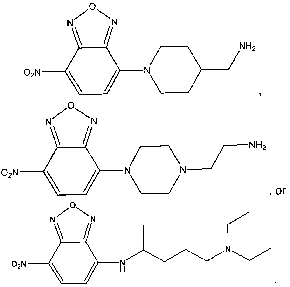
Preferred in the present invention and method for distinguishing phospholipidosis activators from autophagy pathway perturbation agents are the cationic amphiphilic tracer dye compounds having the structures:
compounds having the structures:


In carrying out this method, the test agent can a drug candidate or an
environmental contaminant. The mammalian cells contained in the sample can comprise lymphocytes, granulocytes, macrophages or monocytes, and a combination of any of the foregoing. The mammalian cells can be from a cell line, including a human cell line. The mammalian cells can be from a subject who can be a human.
In distinguishing between activators and agents using the method of the present invention, phospholipidosis is identified as increased accumulation of both the fluorescent phospholipid analog compound and the cationic amphiphilic fluorophore tracer compound. Additionally, the autophagy pathway perturbation agent is identified by no increased accumulation of the fluorescent phospholipid analog compound, but increased accumulation of the cationic amphiphilic fluorophore tracer compound.
The examples which follow are set forth to illustrate various aspects of the present invention but are not intended in any way to limit its scope as more particularly set forth and defined in the claims that follow thereafter.
Example 1 Synthesis of 1,4-bis(2-(dimethylamino)ethylamino)-2,3-difluoro-5,8- dihydroxyanthracene-9,10-dione (Dye 1)
A mixture of 1 ,2,3,4-tetrafluoro-5,8-dihydroxyanthraquinone (1.0 g, 3.2 mmol) and /V,N-dimethylethylenediamine (3 mL) in CH2CI2 (30 ml_) was stirred at room temperature for 12 hours. After evaporation of the solvents, the residue was purified by silica gel chromatography using isocratic solvent system of EtOAc/MeOH/Et3N (10:10:1) yielding 830 mgs of Dye 1 as dark blue product. Abs (max, PBS pH 7.4) = 568 nm; Em = 675 nm. The structure

Dye 1
Example 2 Synthesis of 1,4-bis(5-(diethylamino)pentan-2-ylamino)-2,3-difluoro- 5,8-dihydroxyanthracene-9,10-dione (Dye 2)
A mixture of 1 ,2,3,4-tetrafluoro-5,8-dihydroxyanthraquinone (0.47 g, 1.5 mmol) and 2-Amino-5-diethylaminopentane (2.3 mL) in CH2CI2 (20 mL) was stirred at room temperature for 12 hours. After evaporation of the solvents, the residue was purified by silica gel chromatography using isocratic solvent system of CH2Cl2/MeOH/Et3N
(100:10:1) yielding 364 mgs of Dye 2 as dark blue product. Abs (max, PBS pH 7.4) = 667 nm; Em = 700 nm. The structure of Dye 2 is given below:

Dye 2
Example 3 Synthesis of Dye 3
(a) Preparation of 2-((naphthalene-1-yloxy)methyl)oxirane (Compound 1)
To a solution of 1-naphthol (3.0 g, 0.02 mol) in DMSO (10 mL), potassium hydroxy (flakes, 2.0 g, 0.06 mol) was added. The combined mixture was stirred at room temperature for 30 min and then epichlorohydrin (5.6 g, 4.7 mL, 0.06 mol) was added slowly over a period of 45 min and stirring was continued for 16 hours. The reaction was quenched with water (30 mL) and extracted with chloroform (2 x 50 mL). The combined organic layers were washed with 1N aqueous NaOH (2 x 50 mL), water (2 x 100 mL) and brine (2 x 100 mL) and dried over MgSO4 . The solvent was removed
under reduced pressure to provide Compound 1 as a yellow liquid (3.26 g, 78%). This product was used without further purification. The structure of Compound 1 is given below:

Compound 1
(b) Preparation of 1-(3-ammoniopropyl)-4-methylquinolinium bromide (Compound 2)
A mixture of lepidine (3.0 g, 21 mmol) and 3-bromopropylamine hydrobromide (4.6 g, 21 mmol) in 1 ,2-dichlorobenzene (50 mL) was heated at 135 °C for 16 hours. The mixture was cooled and 1,2-dichlorobenzene was decanted. The residue thus obtained was suspended in methanol (25 mL) and refluxed for 10 min. It was cooled and separated solid was collected by centrifugation, washed with methanol (2 x 30 mL) and dried under high vacuum to yield Compound 2 as an light gray solid (1.4 g, 19%). This product was used without further purification. The structure of Compound 1 is given below:

Compound 2
(c) Preparation of 1-(3-(2-hydroxy-3-(naphthalen-1-yloxy)propylamino)propyl)-4- methylquinolinium bromide (Compound 3)
To a suspension of Compound 1 (0.17 g, 0.83 mmol) and Compound 2 (0.30 g, 0.83 mmol) in water (1 mL), triethylamine (0.10 g, 0.99 mmol) was added. The
combined mixture was stirred at room temperature for 24 hours and then evaporated in the rotary evaporator to provide Compound 3 as a solid. This product was used without any workup an n below:

Compound 3
(d) Preparation of Dye 3
To a mixture of Compound 3 (0.20 g, 0.5 mmol) and 4-dimethylamino
benzaldehyde (0.09 g, 0.5 mmol) in glacial acetic acid (5 mL), piperidine (0.043 g, 0.5 mmol) was added. The combined mixture was heated in an oil bath at 110 °C for 5 hours. Upon cooling the reaction mixture was added to 40% aqueous sodium iodide solution (15 mL) and kept at room temperature for 18 hours. Precipitated dye was collected by centrifugation, washed with water, dried under vacuum and purified by Biotage (SNAP 25 g column) using a gradient of methanol in methylene chloride (1% to 10% over 10 column volume). Appropriate fractions pooled together and dried in a rotary evaporator to provide Dye 3 as a purple solid (128 mg). Abs (max, methanol) = 565 and 295 nm. The structure of Dye 3 is given below:

Dye 3
Example 4 Synthesis of Dye 4
(a) Preparation of 3-(3,4-dihydroquinolin-1 (2H)-yl)propanoic acid (Compound 4)
A mixture of 1 ,2,3,4-tetrahydroquinoline (3.0 g, 22.5 mmol) and methylacrylate (3.88 g, 45.1 mmol) in glacial acetic acid (15 mL) was refluxed for 16 hours. Volatiles were then removed in the rotary evaporator and the residue thus obtained was co- evaporated with methanol (3 x 20 mL) and then dissolved in 15 mL methanol. To this mixture, an aqueous solution of LiOH (1.9 g, 45 mmol, 15 mL) was added and combined mixture was stirred at room temperature for 2 hours. Reaction mixture was made acidic with cone. HCI (pH ~ 2) and extracted with ethyl acetate (2 x 50 mL). The combined organic layers were washed with water (2 x 100 mL) and brine (2 x 100 mL) and dried over MgS04. The solvent was removed under reduced pressure to provide Compound 4 as a dark brown viscous liquid (2.91 g). This product was used without further purification. The struct below:

Compound 4
(b) Preparation of A/-(5-(diethylamino)pentan-2-yl)-3-(3,4-dihydroquinolin-1 (2H)- yl)propanamide (Compound 5)
To a solution of Compound 4 (1.9 g, 9.3 mmol) in DMF (35 mL), 2-succinimido- 1 ,1 ,3,3-tetramethyluronium tetrafluoroborate (3.4 g, 11.2 mmol) and Λ/,/V-diisopropyl ethylamine (3.4 mL, 19.5 mmol) were added. Combined mixture was stirred at room temperature for 30 mins and then a solution of 2-amino-5-diethylaminopentane (2.2 g, 13.9 mmol) in DMF (5 mL) was added. After stirring the mixture for additional 1 hour, it was poured in brine (100 mL) and extracted with ethyl acetate (2 x 75 mL). The combined organic layers were washed with brine (3 x 100 mL) and dried over MgSO4. The solvent was removed under reduced pressure to provide Compound 5 as a dark yellow viscous liquid (2.64 g, 82.5%). This product was used without further purification. The structure

Compound 5
(c) Preparation of A/-(5-(diethylamino)pentan-2-yl)-3-(6-formyl-3,4- dihydroquinolin- 1 (2/-/)-yl)propanamide (Compound 6)
POCI3 (2.37 g, 15.3 mmol) was added dropwise to DMF (1.8 mL) which was cooled in an ice bath. The combined mixture was stirred for 30 minutes and to this a solution of Compound 5 (2.64 g, 7.6 mmol) in DMF (25 mL) was added dropwise over a period of 20 min. After the addition was complete, the ice bath was removed and the reaction mixture was stirred at room temperature for 17 hour. An aqueous solution of sodium acetate (25% w/w, 25 mL) was then added to the reaction mixture and it was heated in an oil bath (T= 110 °C) for 15 min. The reaction mixture was cooled and pH was adjusted with 10 N NaOH to ~11. It was diluted with water (100 mL) and extracted with ethyl acetate. The organic layer was washed twice with water (75 mL) followed by
brine (3 x 50 mL), dried over magnesium sulfate and theh evaporated to dryness to yield 1.44 g of an orange viscous liquid which was used without any further purification. The structure of Compound 6 is given below:

Compound 6
(d) Preparation of Dye 4
To a mixture of Compound 3 (0.15 g, 0.3 mmol) and Compound 6 (0.13 g, 0.34 mmol) in glacial acetic acid (4 mL), piperidine (0.029 g, 0.34 mmol) was added. The combined mixture was heated in an oil bath at 110 °C for 5 hours. Upon cooling the reaction mixture was added to 40% aqueous sodium iodide solution (12 mL) and kept at room temperature for 18 hours. Precipitated dye was collected by centrifugation, washed with water, dried under vacuum and purified by Biotage (SNAP 25 g column) using a gradient of methanol in methylene chloride (1% to 10% over 10 column volume). Appropriate fractions pooled together and dried in a rotary evaporator to provide Dye 4 as a purple solid (75 mg). Abs (max, methanol) = 577 and 297 nm. The structure of Dye 4 is given below:

Dye 4
Example 5 Synthesis of Dye 5
(a) Preparation of /^-(acridin-Q-y ethane-l ,2-diamine hydrochloride (Compound 7)
A mixture of 9-chloroacridine (1.21 g, 5.7 mmol) and phenol (10.8 g, 57 mmol) was mixed and heated at 110 °C for 45 min under argon. Distilled ethylene diamine (3.4 g, 57 mmol) was added via syringe. After the reaction mixture was heated at 110 °C for additional 2 hrs, it was poured into 2N NaOH solution (60 mL). The product was extracted with ethyl ether (3 χ 100 mL). The combined organic layer was washed with 2N NaOH solution (40 mL) and water (2 χ 100 mL) and dried with anhydrous potassium carbonate. The solution was concentrated to 15 mL. Ethanol (15 mL) was added, followed by the addition of concentrated hydrochloric acid (10 mL). The mixture was cooled in freezer (-20 °C) for 4 hrs. The yellow solid formed was collected by centrifugation, washed with ethyl ether (10 mL) and dried under vacuum to provide Compound 7 as yellow powder 0.82 g (53 %) which was used without any further purification. The structure of Compound 7 is given below:
Η, HCI

Compound 7
(b) Preparation of Dye 5
To a solution of Compound 1 (200 mg, 1mmol) in diisopropylethylamine (0.6 mL) and dioxane (4 mL), was added Compound 7 (274 mg, 1 mmol). The reaction mixture was heated at 50 °C for 20 hrs. The yellow precipitate was collected by centrifugation and then further purified by flash chromatography (gradient of mobile phase:
dichloromethane/methanol = 50/1 to 5/1). The solvent was removed under vacuum to provide Dye 5 as a yellow solid (34.5 mg, 8%). Abs (max, methanol) = 412 and 440 nm; Em (max, methan = 426, 452 and 480 nm. The structure of Dye 5 is given below:

Dye 5
Example 6 Preparation of Dye 6
A mixture of 4-chloro-7-nitro-2,1 ,3-benzoxadiazole (0.5 g, 2.5 mmol), 2-amino-5- diethylaminopentane (0.58 mL, 3 mmol) and cesium carbonate (0.98 g, 3 mmol) in acetonitrile (5 mL) was refluxed for 1 hour. Reaction mixture cooled and volatiles were removed in the rotary evaporator. The residue thus obtained was dissolved in a mixture of ethyl acetate (40 mL) and water (40 mL). The organic layer was separated, washed
with water (2X), brine (2X), dried (MgS04) and evaporated. The crude dye thus obtained was purified on Biotage (Si, SNAP 25 g) using a gradient of methanol (2% to 40% over 18 column volume) in methylene chloride to provide Dye 6 as a yellow residue (61 mg). Abs (max, methanol) = 467 nm; ε = 30,200 M"1cm"1. The structure of Dye 6 is given bel

Dye 6
Example 6 Fluorescence Microscopy-Based Assay of Lysosomal Perturbation
Portions of Examples 6- 0 are published as Coleman et al., 2010, J.
Biomolecular Screening 15:398-405.
The Lysosomal Storage Disorder known as Fabry disease arises from a deficiency of the enzyme alpha-galactosidase A, which leads to an accumulation of the glycolipid known as globotriaosylceramide (aka ceramide trihexoside) within
endothelium, striated muscle (skeletal, cardiac), smooth muscle, and renal epithelium. This accumulation leads to an impairment of their proper function. The condition affects hemizygous males, as well as both heterozygous and homozygous females; males tend to experience the most severe clinical symptoms, while females vary from virtually no symptoms to those as serious as males. This variability is thought to be due to X- inactivation patterns during embryonic development. Diagnosis of Fabry disease can be difficult even in a homozygous male. Some patients, renal variants, may lack classic manifestation of Fabry disease such as acroparesthesia, angiokeratoma and corneal lesion. This difficulty is compounded in a heterozygous female who may be variably affected from lionization (from Dr. Mary Lyon's theory of random X-inactivation). It should also be recognized that serum-galactosidase A is frequently normal in female patients. It has recently been noted that chloroquine, hydroxychloroquine and
amiodarone therapy can result in abnormal storage of biochemically and
ultrastructurally similar vacuolar inclusions in many of the same cells as Fabry disease and consequently results in similar clinical manifestations (see Muller-Hocker et al, Human Pathology 34:285-289 (2003); Albay et al, Modern Pathology 18:733-738 (2005); and Bracamonte et al, Am J Kidney Disease 48:844-850 (2006), incorporated herein by reference).
Stable cell lines derived from patients with Fabry disease can not be found in cell line repositories, such as American Type Tissue Collection (Manassas, VA), and are not readily available to the research community. Consequently, an in vitro cell culture model mimicking Fabry disease was generated by incubation of cells with chloroquine.
U20S human osteosarcoma cells were maintained in McCoy's 5a modified medium supplemented with 10% FBS and 100 units/ml penicillin/ streptomycin
(Invitrogen, Carlsbad, CA) in a humidified 37°C, 5% CO2 environment. Cells were allowed to proliferate on glass slides or were seeded into 96-well plates at a density of 2 x 10⁴ cells/well and allowed to attach overnight. The cells were plated such that at the end of the experiment, they reached about 90% confluency.
The fluorescence-based lysosomal perturbation assay was established by first incubating cells cultivated on glass slides for 4-24 hours with chloroquine (8-64 μΜ final concentration), a well known phospholipidosis-inducing agent. Control cells were incubated with 0.1 % (v/v) dimethyl sulfoxide (DMSO), which served as the vehicle control. Cells were then briefly (15 min) incubated with a combination of Dye 1 and Hoechst 33342 dye. Following incubation, cells grown on glass microscope slides were covered with glass coverslips, sealed with nail polish and observed using an inverted Axiovert 200M microscope (Carl Zeiss, Inc., Oberkochen, Germany). Images were acquired with a 63X objective lens (Zeiss). Fluorescence emission intensity and wavelength maximum of Dye 1 is not significantly altered in the pH range of 4.0 to 9.0, allowing interpretation of any increase in fluorescence intensity as an indication of the accumulation of the probe within the cells, rather than as a variation in dye response to lysosomal pH values.
Examination of the distribution of the Dye 1 in cells treated with chloroquine, revealed a punctate pattern of cytoplasmic staining that increased in a dose-dependent manner (FIG. 1). Cells were subsequently stained with Dye 1 for 15 minutes. Nuclei were counter-stained with Hoechst 33342 dye. In FIG. 1 , control U20S cells are shown in (A), cells pre-treated for 18 hours with 8 μΜ chloroquine in (B), cells pre-treated for 18 hours with 16 μΜ chloroquine in (C) and cells pre-treated for 18 hours with 64 μΜ chloroquine in (D). This staining pattern was also consistently observed for other drugs that are known to induce phospholipidosis, such as chlorpromazine, fluoxetine, pimozide and verapamil (further described in Example 4). For all the cited drugs the levels of Dye 1 in the cells increased in a dose-dependent manner until cytotoxic doses of the test agent were attained, which led to a loss of cells. Typically at high doses of test agent the cells remaining on the slide displayed diffuse cytoplasmic staining with Dye 1. Negative control compounds, the vehicle control and untreated medium showed minimal accumulation of Dye 1 , which was strictly confined to the lysosomes. The ability of Dye 1 to detect vacuole accumulation in the described in vitro cell culture model mimicking Fabry disease strongly supports the present invention and its suitability for the detection of genetically derived lysosomal storage diseases.
Example 7 Fluorescence Microscopy with Dye 6
HeLa cells were maintained in Dulbecco's modified Eagles medium
supplemented with 10% FBS, 100 units/ml penicillin/ streptomycin (Invitrogen, Carlsbad, CA) and MEM non essential amino acids in a humidified 37°C, 5% CO2 environment. Cells were allowed to proliferate on glass slides at a density of 2 χ 104 cells/well and allowed to attach overnight. The cells were plated such that at the end of the
experiment, they reached about 90% confluency.
The fluorescence-based lysosomal perturbation assay was established by first incubating cells cultivated on glass slides for 6 hours with chloroquine (120 μΜ final concentration), a well known phospholipidosis-inducing agent. Control cells were incubated with 0.1 % (v/v) dimethyl sulfoxide (DMSO), which served as the vehicle
control. Cells were then briefly (15 min) incubated with Dye 6 at room temperature. Following incubation, cells grown on glass microscope slides were covered with glass coverslips, sealed with nail polish and observed using an Olympus BX51 microscope. Images were acquired with a 40X objective lens. Examination of the distribution of the Dye 6 in cells treated with chloroquine, revealed an increase in fluorescence intensity of lysosomal staining. The untreated cells gave a low level of fluorescent staining.
Example 8 Fluorescence Microplate Based Assay of Lysosomal Perturbation
In order to increase throughput and reduce handling time, the fluorescence microscopy-based lysosomal perturbation assay was scaled up to a 96-well microplate plate assay format and analyzed using a fluorescence microplate reader.
Monitoring cell number per well by determining overall blue fluorescence emission intensity, as indicated by the nuclear Hoechst 33342 dye, was performed to identify generalized compound toxicity. Fluorescence intensity of the cells cultured on the 96-well plates was measured using a FLUOstar OPTIMA Multifunction Microplate Reader (BMG LabTech, Offenburg, Germany). Dye 1 and Hoechst 33342 nuclear counter-stain can be read with Texas Red filter set and DAPI filter set, respectively (Hoechst excitation ~300 nm and emission -480 nm, Dye 1 excitation -570 nm and emission ~670 nm). Fluorescence intensity was expressed as percentage of the vehicle control value. Values for Dye 1 fluorescence can be normalized to those of Hoechst 33342 dye fluorescence to control for any loss in cell number. Results are provided in FIG. 2. Cells were treated with verapamil in (A), fluoxetine in (B), chlorpromazine in (C) and pimozide in (D) for 18 hours, and then stained with Dye 1 (solid lines with solid diamonds) and Hoechst 33342 (dashed line with squares). Concentrations of compounds that resulted in a greater than 30% decrease in overall cell number using the described assay were flagged as being toxic doses, as especially evident in FIGS. 2B and 2D. This benchmark has previously been established in a phospholipidosis assay using a fluorescent lipid analog as well as in a high content screening assay of autophagy (Nioi et al 2008; Zhang et al, 2007). Microscopic examination of wells highlighted as containing generally toxic doses of a test compound demonstrated
diffuse cytoplasmic staining of Dye 1 instead of the anticipated localized punctuate staining associated with lysosomes and lamellar bodies. Scaling-up to the higher throughput microplate format did not affect the sensitivity of detection nor the
reproducibility of the assay, as demonstrated by re-testing the reference compounds and comparing the results with those obtained in the fluorescence microscopy assay. However, it was found to be critical to check for any generalized toxicity-related artifacts thru monitoring Hoechst 33342 dye signal.
In high-throughput screening activities, experimenters often compare a large number (hundreds of thousands to tens of millions) of single measurements of unknown samples to positive and negative control samples. The particular choice of experimental conditions and measurements is called an assay. Large screens are expensive in time and resources. Therefore, prior to starting a large screen, smaller test (or pilot) screens are used to assess the quality of an assay, in an attempt to predict if it would be useful in a high-throughput setting. The Z-factor is an attempt to quantify the suitability of a particular assay for use in a full-scale, high-throughput screen. Extreme conservatism is used in high throughput screening, due to the large number of tests being performed.
The Z-factor is defined in terms of four parameters: the means and standard deviations of both the positive (p) and negative (n) controls (μρ, σρ, and μη, ση). Given these values, the Z-factor is defined as: , Λ 3 x ( σν + ση)
Z-factor = 1 -— ; - ;— .
\ ft-p - μ»\
In practice, the Z-factor is estimated from the sample means and sample standard deviations:
P f . , rj f 1
bsfcimated Z-iactor = 1— .

When tested in the 96-well assay format using the cited cationic amphiphilic drugs, the Dye 1 -based assay demonstrated a Z-factor value of at least 0.60. This falls within the range of 0.5 and 1 , which is defined as a high quality assay by the applied
statistical parameter (Zhang et al, 1999). EC50 values for the test drugs ranged from 6 μΜ for chlorpromazine to 30 μΜ for verapamil, with chloroquine itself having an EC5o value of 15 μΜ.
Example 9 Comparison with a Prototypical Fluorescent Phospholipid Analog- Based Assay
Many assays of phospholipidosis, including the Lipid-Tox assay (Invitrogen, Carlsbad, CA), are based upon co-incubating cells with a fluorescent phospholipid analog for an extended period of time, along with the drug or test agent being evaluated, and then monitoring signal generation in fixed and permeabilized cells. Dye 1 is not a fluorescent phospholipid analog, but instead is a cationic amphiphilic fluorophore tracer. Cells are incubated with the drug or test agent in isolation and then briefly treated with the tracer dye before evaluation in live cells. This prompted us to speculate that since the two assays are based upon fundamentally different
mechanisms, they might also differ in terms of the types of test agents that elicited a positive response. Chloroquine, a prototypical phospholipidosis-inducing agent, and bafilomycin A, an agent well known to induce the accumulation of autophagosomes, were evaluated using both Dye 1 and Lipid-Tox dye-based assays. FIG. 3 shows relative fluorescent intensity of U20S cells treated with chloroquine (A) or bafilomycin A1 (B) at different concentrations for 24 hours. Cells stained with Lipid-Tox dye (solid triangles dash-dot line) were incubated in the presence of the fluorescent lipid for 24 hours during treatment with the drugs. Cells stained with Dye 1 (solid diamond solid line) or Hoechst 33342 (solid square dashed line) were stained for 15 minutes post drug incubation. Both assays reliably predicted that chloroquine induced an expansion in the lysosomal compartment (FIG. 3A). However, only Dye 1 identified the increased vacuolation arising from bafilomycin A treatment (FIG. 3B). The calculated EC50 value for this particular compound was determined to be 3 nM in Dye 1 based assay, easily making it the most potent agent screened in our study. This suggests that the Lipid-Tox assay is strictly speaking a phospholipidosis assay while Dye 1 assay is capable of
detecting vacuolation arising from both phospholipidosis and disruption of trafficking in the autophagy pathway.
Thus, in addition to authentic compounds that cause phospholipidosis, agents that cause the accumulation of autophagosomes by blocking the downstream lysosomal pathway and/or intracellular trafficking of autophagosomes may also lead to increases in the accumulation of intracellular Dye 1 signal in the described assay. For example, Bafilomycin A1 , a macrolide antibiotic isolated from Streptomyces griseus, is a particularly potent agent in our assay. Initial screening of this compound using the ICCB library revealed it to be cytotoxic, but rescreening at much lower doses
demonstrated its ability to increase vacuole number and volume. Bafilomycin A1 is a rather specific vacuolar type H+-ATPase inhibitor which is known to increase the numbers of intracellular autophagosomes, purportedly by blocking the ability of the lysosome to degrade them [Yamamoto et al, 1998]. The bafilomycin A1 results suggest that the described assay should be considered a measure of generalized dysfunction of the lysosomal-dependent catabolic pathway, which leads to the accumulation of autophagosomes, and thus relevant to autophagy in general, as well as
phospholipidosis in particular.
Example 10 Compound Library Screening
Small molecule activators and inhibitors are commonly used to provide insight into both the mode of regulation and physiological roles of various subcellular targets. However, more extensive screening of these compounds generally reveals that most have overlapping specificities, complicating interpretation of their overall effects on biological systems. As proof-of-principle, U20S cells were incubated with the 480 compounds in the ICCB known bioactives compound library (Enzo Life Sciences) overnight at concentrations of 0.1-53 μΜ (1000-fold dilution of the stock), and then evaluated using the described lysosomal perturbation assay. In the preliminary screen, test agents were employed at a single dose in an effort to identify any potential lysosome-modifying agents. Our objective was to benchmark the assay workflow, not to exhaustively classify all of the compounds in the library. It would be advisable to use
a range of concentrations for each test agent in order to reduce the number of false negative results obtained with the described assay. The compounds listed in Table 1 increased staining by greater than three standard deviations compared with untreated control cells.
Table 1 : Positive compound hits from a screen of the ICCB Known Bioactive Compounds Library. U20S cells were treated with the highlighted compounds at the indicated concentrations for 18 hours.
Compounds that displayed increased fluorescence from incubation with Dye 1 by greater than 3 standard deviations relative to the untreated cells are summarized in the table. Relative Lyso stain represents the intensity of Dye 1 fluorescence of treated cells compared to the untreated control. Relative Hoechst stain represents the intensity of the Hoechst 33342 fluorescence of treated cells compared to the untreated control, which indicates the number of cells remaining after the assay. Compounds listed in boldface do not decrease the signal from Hoechst by more than 30%.
NAME Classification Action μ Relative Relative used Lyso Stain Hoechst Stai
Ro 20-1724 Inhibitors phosphodiesterase 18 μΜ 90% 53%
(PDE4 ) inhibitor
Bafilomycin A1 Inhibitors vaculolar ATPase 1 μΜ 176% 58% inhibitor
E6 Berbamine Inhibitors calmodulin inhibitor 7 μΜ 230% 100%
Ro 31-8220 Kinase PKC inhibitor 9 μΜ 146% 89% inhibitors
Propranolol (S-) CNS receptor adrenoceptor 17 μΜ 142% 88% ligands antagonist (beta)
SB -431542 Kinase ALK4, ALK5, ALK7 13 μΜ 154% 101% inhibitors inhibitor
Rottlerin Kinase inhibitors PKC delta inhibitor 10 μΜ 187% 34%
GF-109203X Kinase PKC inhibitor 12 μΜ 197% 87% inhibitors
Tyrphostin 9 Kinase inhibitors PDGF-R tyrosine 18 μΜ 200% 31 % kinase inhibitor
FIG. 4 highlights representative results obtained from four of the test compounds determined to generate a positive response in the assay. FIG. 4 shows relative fluorescent intensity of U20S cells treated with propranolol in FIG. 4A, E6-berbamine in FIG. 4B, GF-109203X in FIG. 4C or rottlerin in 4D for 18 hours, after staining with Dye 1 (solid lines with solid diamonds) and Hoechst 33342 dye (dashed line with squares). Many of the agents used in the screen caused a loss of cells, an indication of
generalized cellular toxicity at the compound concentration evaluated. Rottlerin (FIG. 4D) is an example of a generally cytotoxic compound, which was apparent from a substantial decrease in the Hoechst 33342 dye signal. Observation of rottlerin-treated
cells by fluorescence microscopy confirmed the microplate-based results, with those cells remaining attached to the tissue culture surface displaying diffuse cytoplasmic red staining. The cursory screening activity led to the identification of five compounds that induced increases in vacuolation without causing obvious cellular toxicity (boldface entries, Table 1). These agents all elicited a dose-dependent response, with estimated EC50 values ranging from 1 to 7 μΜ.
Two of the five agents, propranolol and GF-109203X, have previously been reported to induce phospholipidosis in cells. Propranolol is a well-known and
extensively investigated cationic amphiphilic drug that induces phospholipidosis
(Cramb, 1986; Lemieux et al, 2004). High micromolar concentrations of the drug are rapidly taken up and accumulated by a wide range of cultured cells, with a steady state being reached after 40-60 min administration and half maximum uptake within 4-10 min. The rate of propranolol dissociation is much slower than the rate of uptake, with 10% of the propranolol remaining associated with the cells after a 90 minute incubation period in the absence of the drug (Cramb, 1986).
In a recent cell-based HTS screen for anthrax toxin inhibitors, GF-109203X was identified as an agent displaying both protein kinase C-inhibitory and lysosomotropic activities (Sanchez et al, 2007). Screening of bisindolylmaleimide compounds related to GF-109203X which lack protein kinase C inhibitory activity but retain tertiary amines and thus the potential to neutralize endosomal pH, was subsequently undertaken by the investigators. This established that the observed toxin inhibitory activity correlated strongly with the presence of the tertiary amines, but not with protein kinase C inhibition (Sanchez et al, 2007). In our own screen, Ro 31-8220, a bisindolylmaleimide
compound closely related to GF-109203X, but not to our knowledge previously described as a lysosome-perturbation agent, was also found to be a positive hit. A structurally unrelated protein kinase C5-inhibitor, rottlerin, increased subcellular accumulation of Dye 1 but displayed significant toxicity, as demonstrated by loss of Hoechst 33342 dye signal in the assay. Re-examination of this compound over a broad range of concentrations revealed it to be generally cytotoxic (data not shown).
Intracellular accumulation of Dye 1 in treated cells was always diffuse and cytoplasmic,
demonstrating it to be a false positive hit. Our results, taken together with that of Sanchez et al, 2007, suggests that the observed accumulation of Dye 1 in vacuoles correlates strongly with the cationic amphophilic nature of the bisindolylmaleimide compounds, rather than any protein kinase C inhibitory activity.
Two other positive hits in our screen were SB-431542 and berbamine. SB- 431542 is reported to be a transforming growth factor- -receptor antagonist that serves as a potent competitive ATP binding site kinase inhibitor for the downstream activin receptor-like kinases, ALK4, ALK5 and ALK7 (Hjelmeland et al, 2004). Physiologically, SB-431542 has previously been shown to inhibit cell proliferation and block cell motility. Berbamine is reported to selectively induce caspase-3- dependent apoptosis of leukemia NB4 cells via a survivin-mediated pathway (Xu et al, 2006). We are not aware of any previous reports that these two agents have a propensity to induce vacuolation in cells, and indeed their chemical structures are not consistent with them belonging to the cationic amphiphilic class of compounds.
Example 11 Distinguishing between induction of phospholipidosis and inhibition of the autophagy pathway
Example 8 demonstrates that the fluorescent cationic amphillic tracer dyes while sharing with the fluorescent phospholipid analogs an ability to identify phospholipidosis activators also distinctly reveal agents that inhibit the autophagy pathway, leading to accumulation of vacuoles such as autophagosomes and autophagolysosomes. This relationship is highlighted schematically in FIG. 5. Agents such as NBD-PE (N-(-7- nitrobenz-2-oxa-1 , 3-diazol-4-yl)-1 , 2-dihexadecanoyl-sn-glycero-3- phosphoethanolamine), NBD-PC (1-acyl-2-(12[(7-nitro-2-1-3-benzoxadiazol-4yl)amino] dodecanoyl) phosphatidylcholine), CF-PE (1 ,2-dioleoyl-sn-glycero-3- phosphoethanolamine-N-(carboxyfluorescein)) and Lipid-Tox reagent (Invitrogen) can be used in a consolidated workflow along with cationic amphiphilic fluorophore tracers, like Dye 1 in order to differentiate between phospholipidosis activators and autophagy
pathway inhibitors, providing unprecedented insight into the overall dysfunction of the lysosomal-dependent catabolic pathway.
For example, CF-PE was suspended in ethanol and diluted in the cell culture medium (McCoy's 5a modified medium, as described in Example 3) to give a solution (CF-PE medium) with final concentration of 50 μΜ (0.46% ethanol). NBD-PE medium was then sonicated and filtered through a 0.22 mm filter. The prepared was then applied to U20S cells growing in a 96-well microplate. After an equilibration period of 24 h, the cell culture medium was replaced with CF-PE medium with or without each test compound, and the cells were incubated at 37°C for an additional 24 h. After incubation, the cells were rinsed with medium and further incubated with the same medium containing Dye 1 and Hoechst 33342 dye for an additional 15 min at 37°C in 5% C02 atmosphere.
Fluorescence signal was determined using a standard microplate reader, as described in example 4, except that CF-PE was additionally read at an excitation wavelength of 492 nm and an emission wavelength of 517 nm. Autophagy pathway inhibitors such as bafilomycin A generate an increased red signal but low green signal relative to vehicle-treated controls in the assay. Phospholipidosis inducers like chloroquine lead to both an increased green and red signal. Generalized cytotoxicity is evident as a substantial decrease in blue signal (> 30%) relative to vehicle-treated control cells.
Autophagy is known to be substantially up-regulated in colon cancer tissues when compared with the surrounding non-cancerous counterparts. The multiplexed assay of lysosomal catabolic dysfunction should find application in identifying drug candidates that form the basis of new therapeutic strategies based upon the regulation of autophagy.
Example 12 Fluorescence Microscopy-Based and Microplate Assay-Based
Staining using Dye 2
HeLa cervical cancer cells were maintained in Dulbecco's modified Eagle medium supplemented with 10% FBS, 1 % MEM non-essential amino acids and 100
units/ml penicillin/ streptomycin (Invitrogen, Carlsbad, CA) in a humidified 37°C, 5% CO2 environment. Cells were allowed to proliferate on glass slides or were seeded into 96-well plates at a density of 2 * 104 cells/well and allowed to attach overnight.
Cells grown on glass microscope slides were stained with 5 μΜ Dye 2, washed with PBS containing 2% FBS, covered with glass coverslips, sealed with nail polish and observed using an inverted Axiovert 200M microscope (Carl Zeiss, Inc., Oberkochen, Germany). Images were acquired with a 63X objective lens (Zeiss). Fluorescent and brightfield images are shown in FIG. 6.
The fluorescence-based lysosomal perturbation assay was established by first incubating cells cultivated on a microtiter plate for 5 hours with chloroquine (200 μΜ final concentration), a well known phospholipidosis-inducing agent. Control cells were incubated with 0.1% (v/v) dimethyl sulfoxide (DMSO), which served as the vehicle control. Cells were then briefly (30 min) incubated with 40 μΜ Dye 2 in PBS containing 2% FBS. Following incubation, cells were washed with PBS containing 2% FBS.
Finally 60 μΙ of PBS containing 2% FBS was added to each well, and the fluorescence of each well was determined using a Synergy Mx plate scanner (BioTek, Winooski, VT) set to excite at 660 nM and emit at 705 nM. The results are shown in FIG. 7.
Example 13 Fluorescence Microscopy-Based Staining using Dye 5
HeLa cervical cancer cells were maintained in Dulbecco's modified Eagle medium supplemented with 10% FBS, 1% MEM non-essential amino acids and 100 units/ml penicillin/ streptomycin (Invitrogen, Carlsbad, CA) in a humidified 37°C, 5% C02 environment. Cells were allowed to proliferate on glass slides and allowed to attach overnight.
Cells grown on glass microscope slides were stained with 10 μΜ Dye 5, washed with PBS containing 2% FBS, covered with glass coverslips, sealed with nail polish and observed using an inverted Axiovert 200M microscope (Carl Zeiss, Inc., Oberkochen, Germany). Images were acquired with a 63X objective lens (Zeiss). Fluorescent ant brightfield images are shown in FIG. 8.
Example 14: Model system that mimics a lysosomal storage disease (Niemann- Pick Disease)
Sphingomyelin is the primary sphingophospholipid in mammalian cells. A simplified reaction pathway scheme for its biosynthesis is presented below.
Sphingomyelinases catalyze the lysosomal degradation of sphingomyelin. Inherited deficiencies of acid sphingomyelinase activity result in various clinical forms of the Niemann-Pick disease, which are characterized by massive lysosomal accumulation of sphingomyelin.
sphingosine-1-P

Stable cell lines derived from patients with Niemann-Pick disease cannot be found in cell line repositories, such as American Type Tissue Collection (Manassas, VA), and are not readily available to the research community. Consequently, an in vitro tissue culture model of sphingolipidosis was devised using 2R,3S,4E)-N-methyl-5- (4'pentylphenyl)-2-aminopent-4-ene-1 ,3-diol, designated SK1-I (BML-258), a potent, water soluble, isozyme-specific inhibitor of sphingosine kinase-1. In contrast to pan- sphingosine kinase inhibitors, SK1-I does not inhibit sphingosine kinase-2, protein kinase C, or numerous other protein kinases. U-2-OS human osteosarcoma cells were seeded at 22,500 cells/well, grown overnight, and then treated with a final concentration of 0, 2.5, 5, 10 or 20 μΜ of SK1-1 for 18.5 hours. Next, the cells were briefly treated with compound 1 and the nuclear Hoechst 33342 dye, as described in Example 7.
Fluorescence signal was read on a BioTek Synergy Mx microplate reader at excitation
values of 340 and 540 nm and emission values of 480 and 680 nm. Dose-dependent increases in emission values at 680 nm were observed upon treatment with SK1-1 (Z' = 0.55), signifying excessive accumulation of sphingolipids within the vacuolar
compartment. Cell viability was maintained under all treatment conditions, as signified by relatively constant 480 nm emission intensity values from the Hoechst dye.
Fluorescence microscope-based examination of the wells confirmed that compound 1 staining of the cells was confined to the vacuolar structures. This in vitro model confirms the feasibility of using the assay for detecting aberrant accumulation of sphingolipids in cells, and by extension the feasibility of using the assay in monitoring inherited lysosomal storage diseases, such as Niemann-Pick disease.
Example 15 Preparation of Dye 7
To an aqueous solution (7 mL) of sodium carbonate (105 mg, 1 mmol), acetonitrile (15 mL) was added dropwise. To this mixture 4-(aminomethyl)piperidine (228 mg, 240 pL, 2 mmol) was added followed by a solution of 4-chloro-7-nitro-2,1 ,3- benzoxadiazole (200 mg, 1 mmol) in acetonitrile (10 mL). The combined mixture was stirred at room temperature for 15 minutes. Volatiles were then removed by rotary evaporation. The residue thus obtained was adsorbed on silica gel and purified on Biotage (Si, SNAP 25 g) using a gradient of methanol (7% to 60% over 10 column volume) in methylene chloride (+ 1 % triethylamine) to provide Dye 7 as a reddish residue (105 mg). Abs (max, methanol) = 343 and 467 nm; Em (max, methanol) = 540 nm; ε = 1 , 100 M" c "1. The structure of Dye 7 is given below:

Dye 7
Example 16 Preparation of Dye 8
To an aqueous solution (5 ml_) of sodium carbonate (105 mg, 1 mmol), acetonitrile (15 ml_) was added dropwise. To this mixture 1-(2-Aminoethyl)piperazine (258 mg, 263 μΐ_, 2 mmol) was added followed by a solution of 4-chloro-7-nitro-2,1 ,3- benzoxadiazole (200 mg, 1 mmol) in acetonitrile (10 ml_). The combined mixture was stirred at room temperature for 15 minutes. Volatiles were then removed by rotary evaporation. The residue thus obtained was dissolved in 7% methanol in
dichloromethane and purified on Biotage (Si, SNAP 25 g) using a gradient of methanol (7% to 60% over 10 column volume) in methylene chloride (+ 1 % triethylamine) to provide Dye 8 as a red powder (105 mg). Abs (max, methanol) = 335 and 467 nm; Em (max, methanol) = 5 = 16,867 M" cm"1. The structure of Dye 8 is given below:

Dye 8
Example 17 Green autophagy dye (Dye 8)-accumulation increases during starvation
The most well-known inducer of autophagy is nutrient starvation, both in cultured cells and in intact organisms, ranging from yeast to mammals. We evaluated the incorporation of Dye 8 green autophagy dye in cells where autophagy was stimulated by amino acid deprivation, a physiological stimulus of this process. Human cervical adenocarcinoma epithelial cell line HeLa, HepG2 and Human leukemic Jurkat cells were used. All cell lines were obtained from American Type Culture Collection (ATCC, Manassas, VA). HeLa & HepG2 cells were routinely cultured in Eagle's Minimum
Essential Medium (ATCC, Manassas, VA) with low glucose, supplemented with 10% FBS (ATCC, Manassas, VA) and 100 U /ml penicillin, 100 pg/ml streptomycin (Sigma- Aldrich, St. Louis, MO). Jurkat cells were grown in suspension in RPMI medium
supplemented with 10% (v/v) fetal bovine serum, penicillin (100 U /ml), streptomycin (100 pg/ml), and glutamine (200 mM). Cells were maintained in a saturated humidified atmosphere at 37°C, 5% CO2 and 95% air.
For induction of starvation, HeLa, Jurkat or HepG2 cells were washed three times with Earle's balanced salts solution (EBSS) (Sigma-Aldrich, St. Louis, MO), at 37°C incubated with EBSS medium at 37°C for one hour. Control cells were incubated in d-MEM medium. Following this incubation period, both starved and control cells were incubated with 20 μΜ of Dye 8 green autophagy dye for 10 minutes at 37°C and then washed once with 1x PBS. Cells were observed using a fluorescence microscope (Carl Zeiss, Inc) using an FITC filter set. Images were acquired with a 63X objective lens (Zeiss). As shown in Fig. 9, starvation conditions with HeLa cell induces an increase in Dye 8 green autophagy fluorescence intensity in punctate cytoplasmic structures.
Similar results were obtained with HepG2 and Jurkat cell line (data not shown).
Example 18 A population of the Dye 8-labeled vesicles co-localizes with LC3, a specific autophagosome marker
Under physiological conditions, autophagy is a constitutive self-degradative process involved both basal turnover of cellular components and an induced response to nutrient starvation in eukaryotes. During autophagy, portions of the cytoplasm are sequestered by elongation of double-membrane structures called phagophores, which form vesicles called autophagosomes. These vesicles then fuse with lysosomes to form autolysosomes, where their contents are degraded by acidic lysosomal hydrolases for subsequent recycling. LC3, a mammalian homologue of the ubiquitin-like (UBL) protein Atg8, has been used as a specific marker of autophagosomes in mammals.
In this example, HeLa cells at 70% confluence were transfected with RFP-LC3 using Lipofectamine Plus (Invitrogen) according to the manufacturer's instructions.
After 24 hours transfection, cells were treated with either vehicle or 10 μΜ tamoxifen overnight. The cells were then stained with Dye 8 green autophagy dye and observed using fluorescence microscope (Carl Zeiss, Inc) using an FITC filter set as indicated in Example 16. Images were acquired with a 63X objective lens (Zeiss).
Tamoxifen is a drug that is widely used in the treatment of breast cancer. It competes with estrogen for receptor binding and prevents the transcription of estrogen responsive genes, which results in reduced cell growth. There is some evidence that tamoxifen causes increased synthesis of Beclin 1 (BECN1 ; also known as Atg6), which may stimulate autophagy. We have demonstrated that Tamoxifen induces an increase in Dye 8 green autophagy fluorescence intensity in punctuate structures (FIG. 10a) that co-localize with RFP-LC3 (FIG. 10b). Note RFP-LC3 is not uniformly expressed in all cells due to the limited efficiency of transfection.
Example 19 Validating small molecules that affect the autophagy pathway
Besides Tamoxifen and starvation treatment, autophagy also can be induced by the various compounds indicated in Table 2. The compounds in Table 2 (Chloroquine, verapamil, Lithium, Trehalose, Bafilomycin A1, Hydroxychloroquine, Loperamide, Clonidine, MG-132, Norclomipramine), were obtained from Sigma-Aldrich Chemical Company (Saint Louis, MO). Following treatment, the cells were incubated with Dye 8 green autophagy detection reagent and analyzed using fluorescence microscope (Carl Zeiss, Inc) by the method indicated in Example 17.
Table 2.

A popular autophagy target is mTOR, which is a potent suppressor of autophagy. Rapamycin, an inhibitor of mTOR, increases Dye 8 signal (FIG. 1 1 B). It is known that 3-methyladenine (3-MA) is a specific inhibitor of early stages of the autophagic process
(Seglen and Gordon, 1982). To test if the incorporation of Dye 8 was indeed via autophagy, HeLa cells were pre-incubated for 3 hours with 10 mM 3-MA (Sigma) and autophagy was induced by 3 μΜ Rapamycin (Sigma) overnight. FIG. 11C shows that uptake of Dye 8 is almost completely inhibited in this situation. Taken together, the results indicate that the accumulation of Dye 8 in vesicles correlates with the induction of autophagy by rapamycin, and is abrogated by proven inhibitors of the autophagic pathway such as 3-MA. Therefore, autophagocytosis can be followed both
biochemically and morphologically in living cells using this system based upon the incorporation of Dye 8.
Several mTOR-independent autophagy activators have also been validated using the Dye 8 (Table 2). Lithium induces autophagy through inhibition of inositol
monophosphatase (an mTOR-independent pathway). Trehalose and small molecule enhancers of rapamycin (SMERs) also induce autophagy by mechanisms that are not well understood. Two FDA-approved compounds that induce autophagy in an mTOR- independent manner, loperamide hydrochloride and clonidine, also substantially increase green fluorescent signal in the assay. Bafilomycin A1 is a selective inhibitor of vacuolar (V)-type ATPases, which results in elevated lysosomal pH. Chloroquine, verapamil, norclomipramine and hydroxychloroquine are small molecule modulators that passively diffuse into the lysosome and become trapped upon protonation. All these agents also cause an increase in lysosomal pH, which inhibits lysosome function and blocks fusion of the autophagosome with the lysosome. The agents generate a positive signal with the Dye 8 green autophagy dye. Furthermore, MG-132, a potent cell- permeable and selective proteasome inhibitor, has been shown to induce autophagy, as demonstrated with the described assay. The ubiquitin-proteasome system (UPS) and autophagy serve as two complementary, reciprocally regulated protein degradation systems. Blockade of UPS by MG-132 is well known to activate autophagy.
Example 20 Quantifying autophagic activity by flow cytometry using Dye 8 in living cells
Autophagy is an important cellular process and has recently been recognized to be involved in various developmental processes and various diseases including cancer and neurodegeneration. However, experimental methods to accurately detect and quantify this process in mammalian cells are limited due to lack of autophagic markers. Recently, LC3, a mammalian homologue of the ubiquitin-like (UBL) protein Atg8, has been used as a specific marker of autophagosomes in mammals. However, current methods to quantify autophagic activity using LC3 by (1) microscopy, (2) biochemical methods, and (3) detection of protein modifications through SDS-PAGE and Western blotting are time-consuming, labor-intensive and require much experience for accurate interpretation. Thus, there is need to have a simple, reproducible and sensitive method to determine autophagic activity in living cells. We have developed a novel flow cytometry cell-based autophagy assay using Dye 8 to measure autophagic activity in living cells.
In this example, autophagy was induced by amino acid starvation using the method described in Example 17. Following this incubation period, both starved and control cells were incubated with 2.5 μΜ of Dye 8 for 10 minutes at 37°C. Experiments were performed using a FACS Calibur benchtop flow cytometer (BD Biosciences, San Jose, CA) equipped with a blue (488 nm) laser, and Dye 8 fluorescence was measured either in the FL1 or FL2 channel. No washing was required prior to the flow cytometry analysis.
All of the experiments were performed at least three times. Data analysis was carried out with the FlowJo software package. Specifically, data were analyzed by comparison of median fluorescence, by calculating Autophagic Activity Factor (AAF) values using the following formula: AAF=100*((MFIinducer-MFIo)/MFIinducer), wherein
MFIinducer and MFlo are the mean fluorescence intensity values measured from control and treated samples. This is using a similar formula used in evaluating multiple drug resistance (MAFvalues) (Hollo et al., 1998).
We show that Dye 8 fluorescent signal is increased during starvation in a time- dependent manner (FIG. 12). To analyze the kinetics of Dye 8 signal change, Jurkat
cells were incubated in EBSS medium for different periods of time and subsequently stained with Dye 8 green autophagy dye. Thirty minutes of amino acid deprivation was enough to increase by more than 50% the amount of Dye 8 signal that accumulated in the starved cells. This is consistent with previous results indicating that autophagy is rapidly induced following amino acid deprivation (Dunn, 1990). After 2 hours of starvation, the Dye 8 signal was maximal.
FIG. 13 shows the histogram overlays. Control Jurkat cells were stained as well but displayed low fluorescence. Dye 8 signal increases about 2.5-fold after 2 hour starvation, indicating that starvation causes an increase in autophagic vesicles in Jurkat cells. The AAF value is about 59 as compared with a typical cut-off value of about 20- 25 in most MDR flow cytometry experiments (Hollo et al., 1998). This indicated that the peak from straved cells is well separated from the control group. Starved cells switched back to growth medium for 1 hour demonstrate complete recovery.
Example 21 Utilizing Flow Cytometry to measure autophagic flux with Dye 8 green autophagy dye
Induction of autophagy results in the accumulation of insoluble, autophagosome- associated LC3-II in the cytoplasm of cells. However, the same observation can be made when autophagic degradation is inhibited. Therefore, it is important to measure the amount of LC3-II delivered to lysosomes by comparing LC3-II levels in the presence and absence of lysosomal protease inhibitors. Currently autophagic flux is measured either by western blot (Klionsky et al., 2008) or by EGFP-LC3 reporter-transfected cells using saponin extraction (Eng et al., 2010). However, both methods are labor intensive and time consuming.
We present a rapid quantitative assay using Dye 8 to measure autophagic flux. Staining protocol, data acquisition and analysis are the same as described in Example 20. FIG. 14 shows that 300 μΜ of chloroquine treatment for 2 hours lead to a modest increase in fluorescence intensity of Dye 8. A 2 hour starvation in Jurkat cells induces an increase in fluorescence intensity of Dye 8 about 3 fold. However, starvation in the presence of chloroquine increases Dye 8 fluorescence intensity about 4 fold. The
increase in signal represents the accumulation of the signal due to the ongoing constitutive generation of new autophagosomes under conditions where their lysosomal degradation is inhibited. These results demonstrate that the assay can easily study autophagic flux (separate from inhibition of degradation) by simply using Dye 8, in the presence of chloroquine, with flow cytometry.
References
Albay et al., Modern Pathology 2005;18:733-738
Anderson and Borlak, FEBS Lett 2006;580:5533-5540
Ballabio and Gieselmann, Biochimica et Biophysica Acta 2009;1793:684-696
Bassoe et al., Cytometry B Clin Cytom 2003;51 (1):21-29
Bhandari et al., Assay Drug Dev Technol 2008;6(3):407-419
Boldyrev et al. J Lipid Res 2007; 48(7): 1518-1532
Bondak et al., J. Anat 1988;160:161-174
Bracamonte et al., Am J Kidney Disease 2006;48:844-850
Casartelli et al., Cell Biol Toxicol. 2003; 19(3):161-176
Cornett et al., J. Histochem and Cytochem 1980;28:462-464
Cramb, G. Biochem Pharmacol 1986;35:1365-1372
Cramer and Ulrich, Toxicol In Vitro1994;8: 1083-1090
Deng et al., J Histochem Cytochem 1995;43(9):907-915
Dickens et al., J. Mol Cell Cardiol 2002;34:129-137
Diez-Blanco et al., Int. J. Biochem. 1987;19:693-698
Diwu et al., Chem Biol 1999; 6(7):411-418
Dunn, J Cell Biol. 1990;110:1923-1933
Eng, K et al., Autophagy 2010;6:634-641
Freundt et al., Cell Res 2007;17(11):956-958
Fujimura et al., Exp Toxicol Pathol 2007;58(6):375-382
Gum et al., Biochem Pharmacol 2001 ;62:1661-1673
Hjelmeland et al., Mol Cancer Ther. 2004;3(6):737-745
Hollo et al., Anticancer Res. 1998;18:2981-2987
Ikeda et al., Biochemical and Biophysical Research Communications 2008;377:268-274
Kasahara et al., Toxicol Sci 2006;90:133-141
Klionsky et al., Autophagy 2008;16:151-175
Lee et al., Biotechniques 2003;35(5): 1080-1085
Lemieux et al., Anal Biochem. 2004;327(2):247-251
Li et al., Cytometry A 2007;71 (5):297-307
Morelli et al., Cell Biol Toxicol. 2006;22(1):15-27
Muller-Hocker et al., Human Pathology 2003;34:285-289
Nadrigny et al., Biophys J 2007; 93(3):969-980
Natalie et al., Toxicol In Vitro 2009;23(2):217-226
Nioi et al., Toxicol Sci 2007;99:162-173
Nioi et al., Drug and Chemical Toxicology 2008;31 :515-528
Pagano et al., J Cell Bio1989;109(5):2067-2079
Pagano et al., J Cell Bio1991 ;113(6):1267-1279
Poot et al., J Histo Cytochem 1996;44(12): 1363-1372
Rutledge et al., Circ Res 2000;86(7):768-773
Rosania et al., J Am Chem Soc 2003;125(5):1130-1131
Sanchez et al., Antimicrobial Agents and Chemotherapy 2007;51(7):2403-2411
Seglen and Gordon, Proc Natl Acad Sci U S A 1982;79:1889-1892.
Tomizawa et al., The Journal of Toxicol Sci 2006;31 :315-324
Ulrich et al., Toxicol Methods 1991 ;1 :89-105
Ulrich and Cramer, Toxicol In Vitro 1991 ;5(3):239-245
Vibet et al., Drug Metabolism and Disposition 2007;35:822-828
Xu et al., Leukemia Research 2006;30:17-23
Yamamoto et al., Cell Struct Funct. 1998;23(1):33-42
Zhang et al., J. Biomol. Screen.1999;4:67-73
Zhang et al., Proc Natl Acad Sci USA 2007; 104(48): 19023-10928
U.S. Pat. No. 4868103
U.S. Pat. No. 5118801
U.S. Pat. No. 5210015
U.S. Pat. No. 5401847
U.S. Pat. No. 5538848
U.S. Pat. No. 5580990
U.S. Pat. No. 5646264
U.S. Pat. No. 5800996
U.S. Pat. No. 5852191
U.S. Pat. No. 5925517
U.S. Pat. No. 5981747
U.S. Pat. No. 5994056
U.S. Pat. No. 6008373
U.S. Pat. No. 6133445
U.S. Pat. No. 6174670
U.S. Pat. No. 6593465
U.S. Pat. No. 6664047
U.S. Pat. No. 6686145
U.S. Pat. Pub. No. 2003/0225247
U.S. Pat. Pub. No. 2005/0137388
U.S. Pat. Pub. No. 2010/0062429
U.S. Pat. Pub. No. 2010/0062460
In view of the above, it will be seen that several objectives of the invention are achieved and other advantages attained.
Many obvious variations will no doubt be suggested to those of ordinary skill in the art, in light of the above detailed description and examples of the present invention. It will be appreciated by those skilled in the art that any arrangement which is calculated to achieve the same purpose may be substituted for the specific embodiments shown. This application and invention are intended to cover any adaptations or variations of the present invention. All such variations are fully embraced by the scope and spirit of the invention as more particularly defined in the claims that now follow.
All references cited in this specification are hereby incorporated by reference. The discussion of the references herein is intended merely to summarize the assertions made by the authors and no admission is made that any reference constitutes prior art. Applicants reserve the right to challenge the accuracy and pertinence of the cited references.
Claims
WHAT IS CLAIMED IS:
1. A method of detecting the presence of a lysosomal storage disease in a subject comprising the steps of:
(a) obtaining a sample containing cells from said subject;
(b) contacting said sample with a cationic amphiphilic tracer compound that localizes to a vacuole in a cell; and
(c) detecting said cationic amphiphilic tracer compound, thereby determining whether there exists an excess above normal accumulation of vacuoles within said cells of the sample, said excess above normal accumulation of vacuoles being indicative of the lysosomal storage disease.
2. The method of claim 1 , wherein said lysosomal storage disease arises from deficiencies of one or more soluble lysosomal proteins residing in the lumen of the lysosome.
3. The method of claim 1 , wherein said lysosomal storage disease arises from a defect in one or more lysosomal membrane proteins.
4. The method of claim 1 , wherein said lysosomal storage disease arises from deficiencies of non-lysosomal proteins residing in a site comprising an endoplasmic reticulum, a Golgi apparatus, an endosomal pathway, or any combination thereof.
5. The method of claim 1 , wherein said lysosomal storage disease comprises a mucopolysaccharidoses, a lipidoses or a glycogenosis.
6. The method of claim 1 , wherein a subject is known to have a lysosomal storage disease based upon previous genetic or metabolic testing.
7. The method of claim 1 wherein said lysosomal storage disease comprises Activator Deficiency/GM2 Gangliosidosis, Alpha-mannosidosis, Aspartylglucosaminuria, Cholesteryl Ester Storage Disease, Chronic Hexosaminidase A Deficiency, Cystinosis, Danon Disease, Fabry Disease, Farber Disease, Fucosidosis, Galactosialidosis, Gaucher Disease, GM1 Gangliosidosis, l-Cell Disease/Mucolipidosis II, Infantile Free Sialic Acid Storage Disease/ISSD, Juvenile Hexosaminidase A Deficiency, Krabbe Disease, Metachromatic Leukodystrophy, Mucopolysaccharidoses Disorders, Pseudo- Hurler Polydystrophy/Mucolipidosis IIIA, MPSI Hurler Syndrome , MPSI Scheie
Syndrome, MPS I Hurler-Scheie Syndrome, MPS II Hunter Syndrome , Sanfilippo syndrome Type A/MPS III A, Sanfilippo syndrome Type B/MPS III B, Sanfilippo
Syndrome Type C/MPS III C, Sanfilippo Syndrome Type D/MPS III D, Morquio Type A MPS IVA, Morquio Type B/MPS IVB, MPS IX Hyaluronidase Deficiency, MPS VI Maroteaux-Lamy, MPS VII Sly Syndrome, Mucolipidosis l/Sialidosis, Mucolipidosis IMC, Mucolipidosis Type IV, Multiple Sulfatase Deficiency, Niemann-Pick Disease, Neuronal Ceroid Lipofuscinoses, CLN6 Disease, Batten-Spielmeyer-Vogt/Juvenile NCL/CLN3 Disease, Finnish Variant Late Infantile CLN5, Jansky-Bielschowsky Disease/Late infantile CLN2/TPP1 Disease, Kufs/Adult-onset NCL/CLN4 Disease, Northern
Epilepsy/Variant Late Infantile CLN8, Santavuori-Haltia/lnfantile CLN1/PPT Disease, Beta-Mannosidosis, Pompe Disease/Glycogen Storage Disease Type II,
Pycnodysostosis, Sandhoff disease/Adult Onset/GM2 Gangliosidosis, Sandhoff
Disease/GM2 Gangliosidosis - Infantile, Sandhoff Disease/GM2 Gangliosidosis - Juvenile, Schindler disease, Salla Disease/Sialic Acid Storage Disease, Tay- Sachs/GM2 Gangliosidosis, Wolman Disease, or any combination thereof.
8. The method of claim 1 , wherein said vacuole comprises a lysosome, a
phagophore, an autophagosomes, an autophagolysosome, or any combination thereof.
9. The method of claim 1 , wherein said cationic amphiphilic tracer compound comprises a fluorescent drug that induces phospholipidosis or a fluorescently labeled analog of a drug that induces phospholipidosis.
no
10. The method of claim 1 , wherein said cationic amphiphilic tracer compound comprises a halogenated and fluorescent drug that induces phospholipidosis or a halogenated and fluorescently labeled analog of a drug that induces phospholipidosis.
11. The method of claim 1 , wherein said cationic amphiphilic tracer compound comprises nitrobenzoxadiazole (NBD)-spiperone, nitrobenzoxadiazole (NBD)- propranolol, 9-amino-acridin-propanolol, or any combination thereof.
12. The method of claim 1 , wherein said cationic amphiphilic tracer compound has the structure:

in

112

13. The method of claim 12, wherein said cationic amphiphilic tracer compound has the structure


14. The method of claim 1 , further comprising the steps (a') of providing a reference sample of normal cells from a subject known not to have lysosomal storage disease; (b') contacting said sample of normal cells with the same cationic amphiphilic tracer compound of step (b); and (c') comparing the accumulation of vacuoles in the sample obtained from the subject known or suspected of having a lysosomal storage disease with the accumulation in said reference sample of normal cells.
15. The method of claim 1 , wherein said cells comprise lymphocytes, granulocytes, macrophages or monocytes, and a combination of any of the foregoing.
The method of claim 1 , wherein said subject is a mammal.
17. The method of claim 16, wherein said mammal is human.
18. A method of monitoring the progression of a lysosomal storage disease in a subject by comparing vacuole accumulation over a course of (T2-Ti) time interval(s), said method comprising the steps of:
(a) obtaining from said subject at time ΤΊ a first sample containing cells;
(b) contacting said first sample with a first cationic amphiphilic tracer compound that localizes to a vacuole in a cell;
(c) detecting said first cationic amphiphilic tracer compound, thereby determining the accumulation of vacuoles within said cells of the first sample at time ΤΊ;
(d) obtaining from said subject at a later time T2 a second sample containing cells;
(e) contacting said second sample with a second cationic amphiphilic tracer compound that localizes to a vacuole in a cell;
(f) detecting said second cationic amphiphilic tracer compound, thereby determining the accumulation of vacuoles within cells of the second sample at time T2; and
(g) comparing the accumulation of vacuoles between said first sample at time ΤΊ and said second sample at time T2, thereby monitoring the progression of lysosome storage disease in said subject.
19. The method of claim 18, wherein said lysosomal storage disease arises from deficiencies of one or more soluble lysosomal proteins residing in the lumen of the lysosome.
20. The method of claim 18, wherein said lysosomal storage disease arises from a defect in one or more lysosomal membrane proteins.
21. The method of claim 18, wherein said lysosomal storage disease arises from deficiencies of non-lysosomal proteins residing in a site comprising an endoplasmic reticulum, a Golgi apparatus or an endosomal pathway, and combinations of any of the foregoing.
22. The method of claim 18, wherein said lysosomal storage disease comprises a mucopolysaccharidoses, a lipidoses or a glycogenosis.
23. The method of claim 18, wherein a subject is known to have a lysosomal storage disease based upon previous genetic or metabolic testing.
24. The method of claim 18, wherein said lysosomal storage disease comprises Activator Deficiency/GM2 Gangliosidosis, Alpha-mannosidosis, Aspartylglucosaminuria, Cholesteryl Ester Storage Disease, Chronic Hexosaminidase A Deficiency, Cystinosis, Danon Disease, Fabry Disease, Farber Disease, Fucosidosis, Galactosialidosis, Gaucher Disease, GM1 Gangliosidosis, l-Cell Disease/Mucolipidosis II, Infantile Free Sialic Acid Storage Disease/ISSD, Juvenile Hexosaminidase A Deficiency, Krabbe Disease, Metachromatic Leukodystrophy, Mucopolysaccharidoses Disorders, Pseudo- Hurler Polydystrophy/Mucolipidosis IIIA, MPSI Hurler Syndrome , MPSI Scheie
Syndrome, MPS I Hurler-Scheie Syndrome, MPS II Hunter Syndrome , Sanfilippo syndrome Type A/MPS III A, Sanfilippo syndrome Type B/MPS III B, Sanfilippo
Syndrome Type C/MPS III C, Sanfilippo Syndrome Type D/MPS III D, Morquio Type A/MPS IVA, Morquio Type B/MPS IVB, MPS IX Hyaluronidase Deficiency, MPS VI Maroteaux-Lamy, MPS VII Sly Syndrome, Mucolipidosis l/Sialidosis, Mucolipidosis IIIC, Mucolipidosis Type IV, Multiple Sulfatase Deficiency, Niemann-Pick Disease, Neuronal Ceroid Lipofuscinoses, CLN6 Disease, Batten-Spielmeyer-Vogt/Juvenile NCL/CLN3 Disease, Finnish Variant Late Infantile CLN5, Jansky-Bielschowsky Disease/Late infantile CLN2/TPP1 Disease, Kufs/Adult-onset NCL/CLN4 Disease, Northern
Epilepsy/Variant Late Infantile CLN8, Santavuori-Haltia/lnfantile CLN1/PPT Disease, Beta-Mannosidosis, Pompe Disease/Glycogen Storage Disease Type II,
Pycnodysostosis, Sandhoff disease/Adult Onset GM2 Gangliosidosis, Sandhoff
Disease/GM2 Gangliosidosis - Infantile, Sandhoff Disease/GM2 Gangliosidosis - Juvenile, Schindler disease, Salla Disease/Sialic Acid Storage Disease, Tay- Sachs/GM2 Gangliosidosis, Wolman Disease, or any combination thereof.
25. The method of claim 18, wherein said vacuole comprises a lysosome, a phagophore, an autophagosomes, an autophagolysosome, or any combination thereof.
26. The method of claim 18, wherein said first cationic amphiphilic tracer compound and said second cationic amphiphilic tracer compound comprise a fluorescent drug that induces phospholipidosis or a fluorescently labeled analog of a drug that induces phospholipidosis.
27. The method of claim 8, wherein said first cationic amphiphilic tracer compound and said second cationic amphiphilic tracer compound comprise a halogenated and fluorescent drug that induces phospholipidosis or a halogenated and fluorescently labeled analog of a drug that induces phospholipidosis.
28. The method of claim 18, wherein said first cationic amphiphilic tracer compound and said second cationic amphiphilic tracer compound are the same compound.
29. The method of claim 18, wherein said cationic amphiphilic tracer compound comprises nitrobenzoxadiazole (NBD)-spiperone, nitrobenzoxadiazole (NBD)- propranolol, 9-amino-acridin-propanolol, or any combination thereof.
30. The method of claim 18, wherein said cationic amphiphilic tracer compound has the structure:


118

31. The method of claim 18, wherein said cationic amphiphilic tracer compound has the structure


32. The method of claim 18, wherein the difference (T2 - Ti) comprises time units measured in hours, days, weeks, months or years.
33. The method of claim 18, wherein said cells comprise lymphocytes, granulocytes, macrophages or monocytes, and a combination of any of the foregoing.
34. The method of claim 18, wherein said subject is a mammal.
35. The method of claim 34, wherein said mammal is human.
36. A method of monitoring the effectiveness of a drug or remedy used to manage, treat or cure a lysosomal storage disease in a subject, comprising the steps of:
(a) obtaining from said subject at time Ti a first sample containing cells;
(b) contacting said first sample with a first cationic amphiphilic tracer compound that localizes to a vacuole in a cell;
(c) detecting said first cationic amphiphilic tracer compound, thereby determining the accumulation of vacuoles within said cells of the first sample at time T-i ;
(d) obtaining from said subject at a later time T2 a second sample containing cells;
(e) contacting said second sample with a second cationic amphiphilic tracer compound that localizes to a vacuole in a cell;
(f) detecting said second cationic amphiphilic tracer compound, thereby determining the accumulation of vacuoles within cells of the second sample at time T2; and
(g) comparing the accumulation of vacuoles between said first sample at time T and said second sample at time T2, thereby monitoring the effectiveness of said drug used to manage, treat or cure lysosome storage.
37. The method of claim 36, wherein said lysosomal storage disease arises from deficiencies of one or more soluble lysosomal proteins residing in the lumen of the lysosome.
38. The method of claim 36, wherein said lysosomal storage disease arises from a defect in one or more lysosomal membrane proteins.
39. The method of claim 36, wherein said lysosomal storage disease arises from deficiencies of non-lysosomal proteins residing in a site comprising an endoplasmic reticulum, a Golgi apparatus, an endosomal pathway, or any combination thereof.
40. The method of claim 36, wherein said lysosomal storage disease comprises a mucopolysaccharidoses, a lipidoses or a glycogenosis.
41. The method of claim 36, wherein a subject is known to have a lysosomal storage disease based upon previous genetic or metabolic testing.
42. The method of claim 36, wherein said lysosomal storage disease comprises Activator Deficiency/GM2 Gangliosidosis, Alpha-mannosidosis, Aspartylglucosaminuria, Cholesteryl Ester Storage Disease, Chronic Hexosaminidase A Deficiency, Cystinosis, Danon Disease, Fabry Disease, Farber Disease, Fucosidosis, Galactosialidosis, Gaucher Disease, GM1 Gangliosidosis, l-Cell Disease/Mucolipidosis II, Infantile Free Sialic Acid Storage Disease/ISSD, Juvenile Hexosaminidase A Deficiency, Krabbe Disease, Metachromatic Leukodystrophy, Mucopolysaccharidoses Disorders, Pseudo- Hurler Polydystrophy/Mucolipidosis IMA, MPSI Hurler Syndrome , MPSI Scheie
Syndrome, MPS I Hurler-Scheie Syndrome, MPS II Hunter Syndrome , Sanfilippo syndrome Type A/MPS III A, Sanfilippo syndrome Type B/MPS III B, Sanfilippo
Syndrome Type C/MPS III C, Sanfilippo Syndrome Type D/MPS III D, Morquio Type A/MPS IVA, Morquio Type B/MPS IVB, MPS IX Hyaluronidase Deficiency, MPS VI Maroteaux-Lamy, MPS VII Sly Syndrome, Mucolipidosis l/Sialidosis, Mucolipidosis IIIC, Mucolipidosis Type IV, Multiple Sulfatase Deficiency, Niemann-Pick Disease, Neuronal Ceroid Lipofuscinoses, CLN6 Disease, Batten-Spielmeyer-Vogt/Juvenile NCL/CLN3 Disease, Finnish Variant Late Infantile CLN5, Jansky-Bielschowsky Disease/Late infantile CLN2/TPP1 Disease, Kufs/Adu It-onset NCL/CLN4 Disease, Northern
Epilepsy/Variant Late Infantile CLN8, Santavuori-Haltia/lnfantile CLN1/PPT Disease, Beta-Mannosidosis, Pompe Disease/Glycogen Storage Disease Type II,
Pycnodysostosis, Sandhoff disease/Adult Onset/GM2 Gangliosidosis, Sandhoff
Disease/GM2 Gangliosidosis - Infantile, Sandhoff Disease/GM2 Gangliosidosis - Juvenile, Schindler disease, Salla Disease/Sialic Acid Storage Disease, Tay- Sachs/GM2 Gangliosidosis, Wolman Disease, or any combination thereof.
43. The method of claim 36, wherein said vacuole comprises a lysosome, a phagophore, an autophagosomes, an autophagolysosome, or any combination thereof.
44. The method of claim 36, wherein said first cationic amphiphilic tracer compound and said second cationic amphiphilic tracer compound comprise a fluorescent drug that induces phospholipidosis or a fluorescently labeled analog of a drug that induces phospholipidosis.
45. The method of claim 36, wherein said first cationic amphiphilic tracer compound and said second cationic amphiphilic tracer compound comprise a halogenated and fluorescent drug that induces phospholipidosis or a halogenated and fluorescently labeled analog of a drug that induces phospholipidosis.
46. The method of claim 36, wherein said first cationic amphiphilic tracer compound and said second cationic amphiphilic tracer compound are the same compound.
47. The method of claim 36, wherein said cationic amphiphilic tracer compound comprises nitrobenzoxadiazole (NBD)-spiperone, nitrobenzoxadiazole (NBD)- propranolol, 9-amino-acridin-propanolol, or any combination thereof.
48. The method of claim 36, wherein said cationic amphiphilic tracer compound has the structure:


124

49. The method of claim 36, wherein said cationic amphiphilic tracer compound has the structure


50. The method of claim 36, wherein the difference 0Ί - T2) comprises time units measured in hours, days, weeks, months or years.
51. The method of claim 36, wherein said cells comprise lymphocytes, granulocytes, macrophages or monocytes, and a combination of any of the foregoing.
52. The method of claim 36, wherein said subject is a mammal.
53. The method of claim 52, wherein said mammal is human.
54. A method of screening drug candidates to determine their toxicity to cells, tissues or organs, said method comprising the steps of:
(a) providing:
(i) at least one first sample and one second sample of mammalian cells;
(ii) a drug candidate for screening;
(iii) a drug diluting vehicle or carrier; and
(iv) a cationic amphiphilic tracer compound that localizes to a vacuole in a cell;
(b) administering:
(i) said drug candidate (ii) to said first sample for a time sufficient to allow the drug candidate to partition into subcellular compartments within said cells to form treated cells; and
(ii) said vehicle or carrier (iii) to said second sample to form untreated reference cells;
(c) contacting said treated cells and said untreated reference cells with the cationic amphiphilic tracer (iv) for a period of time sufficient to accumulate in vacuoles in the cells, and
(d) detecting any increase in vacuole accumulation of said cationic
amphiphilic tracer compound in the treated cells relative to said untreated reference cells, thereby determining whether said drug candidate has accumulated in the vacuoles and is toxic to cells, tissues or organs.
55. The method of claim 54, wherein said vehicle or carrier comprises cell culture medium, buffer, organic solvent or water, and combinations of the foregoing.
56. The method of claim 55, wherein said organic solvent comprises
dimethylsulfoxide (DMSO).
57. The method of claim 54, wherein said vacuole comprises a lysosome, a phagophore, an autophagosomes, an autophagolysosome, or any combination thereof.
58. The method of claim 54, wherein said first cationic amphiphilic tracer compound and said second cationic amphiphilic tracer compound comprise a fluorescent drug that induces phospholipidosis or a fluorescently labeled analog of a drug that induces phospholipidosis.
59. The method of claim 54, wherein said first cationic amphiphilic tracer compound and said second cationic amphiphilic tracer compound comprise a halogenated and fluorescent drug that induces phospholipidosis or a halogenated and fluorescently labeled analog of a drug that induces phospholipidosis.
60. The method of claim 54, wherein said cationic amphiphilic tracer compound comprises nitrobenzoxadiazole (NBD)-spiperone, nitrobenzoxadiazole (NBD)- propranolol, 9-amino-acridin-propanolol, or any combination thereof.
61. The method of claim 54, wherein said cationic amphiphilic tracer compound has the structure:


129

62. The method of claim 54, wherein said cationic amphiphilic tracer compound has the structure


63. The method of claim 54, wherein said mammalian cells are from a cell line.
64. The method of claim 63, wherein said cell line is a human cell line.
65. The method of claim 54, wherein said mammalian cells are from a subject.
66. The method of claim 65, wherein said subject is a human.
67. The method of claim 54, wherein said cells comprise lymphocytes, granulocytes, macrophages or monocytes, and a combination of any of the foregoing.
68. A method of screening suspected toxic agents to determine their toxicity to cells, tissues or organs, said method comprising the steps of:
(a) providing:
(i) at least one first sample and one second sample of mammalian cells;
(ii) a suspected toxic agent for screening;
(iii) a diluting vehicle or carrier; and
(iv) a cationic amphiphilic tracer compound that localizes to a vacuole in a cell;
(b) administering:
(i) said suspected toxic agent (ii) to said first sample for a time
sufficient to allow said suspected toxic agent to partition into subcellular compartments within said cells to form treated cells; and
(ii) said diluting vehicle or carrier (iii) to said second sample to form untreated reference cells;
(c) contacting said treated cells and said untreated reference cells with the cationic amphiphilic tracer (iv) for a period of time sufficient to accumulate in vacuoles in the cells, and
(d) detecting any increase in vacuole accumulation of said cationic
amphiphilic tracer compound in the treated cells relative to said untreated reference cells, thereby determining whether said suspected toxic agent has accumulated in the vacuoles and is toxic to cells, tissues or organs.
69. The method of claim 68, wherein said vehicle or carrier comprises cell culture medium, buffer, organic solvent or water, and combinations of the foregoing.
70. The method of claim 69, wherein said organic solvent comprises
dimethylsulfoxide (DMSO).
71. The method of claim 68, wherein said vacuole comprises a lysosome, a phagophore, an autophagosomes, an autophagolysosome, or any combination thereof.
72. The method of claim 68, wherein said first cationic amphiphilic tracer compound and said second cationic amphiphilic tracer compound comprise a fluorescent drug that induces phospholipidosis or a fluorescently labeled analog of a drug that induces phospholipidosis.
73. The method of claim 68, wherein said first cationic amphiphilic tracer compound and said second cationic amphiphilic tracer compound comprise a halogenated and fluorescent drug that induces phospholipidosis or a halogenated and fluorescently labeled analog of a drug that induces phospholipidosis.
74. The method of claim 68, wherein said cationic amphiphilic tracer compound comprises nitrobenzoxadiazole (NBD)-spiperone, nitrobenzoxadiazole (NBD)- propranolol, 9-amino-acridin-propanolol, or any combination thereof.
75. The method of claim 68, wherein said cationic amphiphilic tracer compound has the structure:


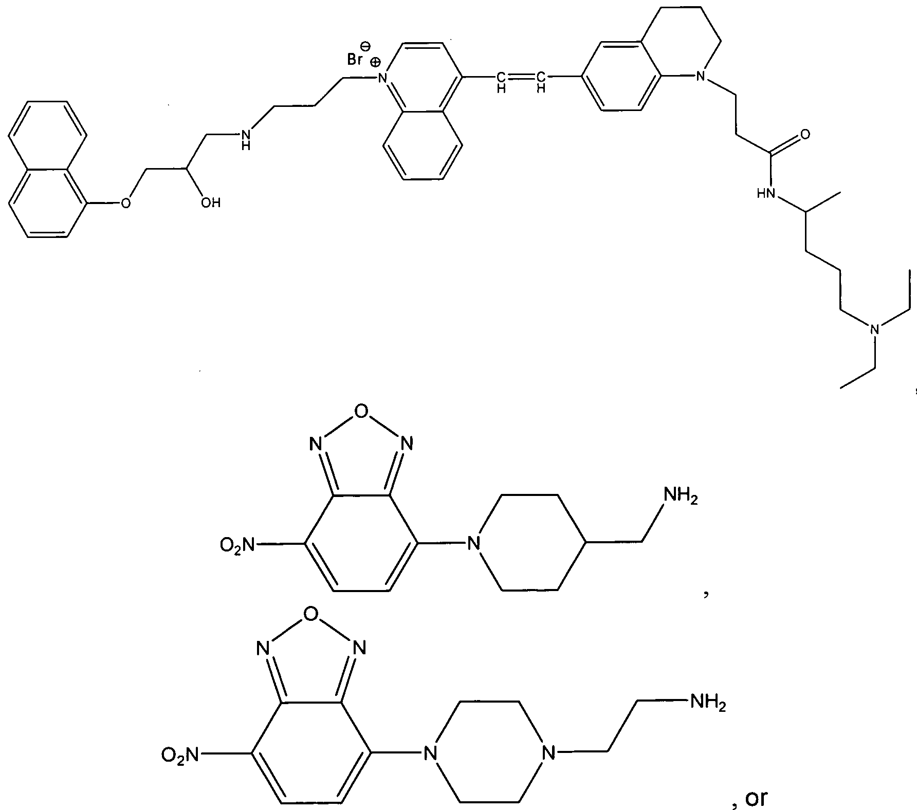
134
76. The method of claim 68, wherein said cationic amphiphilic tracer compound has the structure


The method of claim 68, wherein said mammalian cells are from a cell line.
The method of claim 77, wherein said cell line is a human cell line.
The method of claim 68, wherein said mammalian cells are from a subject.
The method of claim 79, wherein said subject is a human.
81. The method of claim 68, wherein said cells comprise lymphocytes, granulocytes, macrophages, monocytes, or any combination thereof.
82. A method of distinguishing between phospholipidosis activators and autophagy pathway perturbation agents, comprising the steps of:
(a) providing:
(i) a fluorescent phospholipid analog compound that forms
multilamellar vesicles;
(ii) a cationic amphiphilic fluorophore tracer compound that localizes in a vacuole in a cell;
(iii) a test agent; and
(iv) a sample of mammalian cells;
(b) administering to the cells in said sample (iv) the fluorescent phospholipid analog compound (i) and the test agent (iii) to form a testing mixture;
(c) contacting said testing mixture with the cationic amphiphilic fluorophore tracer (ii); and
(d) detecting any signals generated from said fluorescent phospholipid analog compound (i) and said cationic amphiphilic fluorophore tracer compound (ii), thereby differentiating between phospholipidosis activators and autophagy pathway perturbation agents based upon differential accumulation patterns of the fluorescent phospholipid analog and the cationic amphiphilic fluorophore tracer.
83. The method of claim 82, wherein said fluorescent phospholipid analog compound comprises NBD-PE (N-(-7-nitrobenz-2-oxa-1 , 3-diazol-4-yl)-1 , 2-dihexadecanoyl-sn- glycero-3-phosphoethanolamine), NBD-PC (1-acyl-2-(12[(7-nitro-2-1-3-benzoxadiazol- 4yl)amino] dodecanoyl) phosphatidylcholine), CF-PE (1 ,2-dioleoyl-sn-glycero-3- phosphoethanolamine-N-(carboxyfluorescein)) Lipid-Tox reagent, or any combination thereof.
84. The method of claim 82, wherein said vacuole comprises a lysosome, a phagophore, an autophagosomes, an autophagolysosome, or any combination thereof.
85. The method of claim 82, wherein said cationic amphiphilic tracer compound comprises a fluorescent drug that that induces phospholipidosis or a fluorescently labeled analog of a drug that induces phospholipidosis.
86. The method of claim 82, wherein said cationic amphiphilic tracer compound comprises a halogenated and fluorescent drug that induces phospholipidosis or a halogenated and fluorescently labeled analog of a drug that induces phospholipidosis.
87. The method of claim 82, wherein said cationic amphiphilic tracer compound has the structure:


88. The method of claim 82, wherein said cationic amphiphilic tracer compound has the structure

140

89. The method of claim 82, wherein the test agent is a drug candidate.
90. The method of claim 82, wherein the test agent is an environmental contaminant.
91. The method of claim 82, wherein said cells comprise lymphocytes, granulocytes, macrophages, monocytes, or any combination thereof.
92. The method of claim 82, wherein said mammalian cells are from a cell line.
93. The method of claim 92, wherein said cell line is a human cell line.
94. The method of claim 82, wherein said mammalian cells are from a subject.
95. The method of claim 94, wherein said subject is a human.
96. The method of claim 82, wherein phospholipidosis is identified as increased accumulation of both the fluorescent phospholipid analog compound and the cationic amphiphilic fluorophore tracer compound.
97. The method of claim 82, wherein the autophagy pathway perturbation agent is identified by no increased accumulation of the fluorescent phospholipid analog compound, but increased accumulation of the cationic amphiphilic fluorophore tracer compound.
98. A compound having the structure:

wherein and R2 are independently H, OH, or comprise
an alkyl group wherein said alkyl group is saturated or unsaturated, linear or branched, substituted or unsubstituted,
a phenyl group wherein said phenyl group is substituted or unsubstituted, or when taken together, Ri and R2 may form a 5 or 6 membered ring comprising carbon, sulfur, oxygen or nitrogen, or any combinations thereof; wherein the 5 or 6 membered ring is saturated or unsaturated, wherein the ring may be further substituted with a linker arm comprising carbon, sulfur, oxygen, nitrogen, or any combinations thereof and wherein said linker arm may be saturated or unsaturated, linear or branched, substituted or unsubstituted or any combinations thereof.
99. The compound of claim 98, wherein at least one of Rior R2 comprises Q wherein Q comprises a carboxyl group (CO2 ~), a carbonate ester (COER13), a sulfonate (SO3 ), a sulfonate ester (SO2ER 3), a sulfoxide (SOR13), a sulfone (SO2CR13R14R15). a sulfonamide (SO2NR13R14), a phosphate (PO4 =), a phosphate monoester (PO3 "ER 3), a phosphate diester (PO2ER-13ER14) , a phosphonate (PO3=) a phosphonate monoester (PO2 "ER13) a phosphonate diester (POER13ERi4), a thiophosphate (PSO3 =), a thiophosphate monoester (PSO2 "ERi3) a thiophosphate diester (PSOER13ER14), a thiophosphonate (PSO2 =), a thiophosphonate monoester (PSO"ER13) a thiophosphonate diester (PSERi3ER14), a phosphonamide (PONRi3Ri4NRigR2o), its thioanalogue
(PSNR13R14NR19R20), a phosphoramide (PONR13R14NR15NR19R20), its thioanalogue (PSNRi3R14NRi5NRi9R2o), a phosphoramidite (PO2RigNRi3Ri4) or its thioanalogue (POSRi9NRi3R14) where E can be independently O or S, wherein Q can further comprise a nucleophilic reactive group, an electrophilic reactive group, a terminal alkene, a terminal alkyne, a coordinate group or an alkylating agent;
wherein Q is attached directly, or indirectly through a linker arm comprising carbon, sulfur, oxygen, nitrogen, or any combinations thereof and wherein said linker arm may be saturated or unsaturated, linear or branched, substituted or unsubstituted or any combinations thereof.
100. The compound of claim 98, wherein Ri and R2 are independently H, or a saturated alkyl group that may be substituted at any one or multiple positions with an N, where Ri and R2 may form a 6 membered ring, provided that, when one of Ri and R2 is H, the other of Ri and R2 is not H.
101. The comp

A compound selected from the group consisting of


144
103. A compound selected from the group consisting of

where X comprises an anion,

where X comprises an anion,

where X comprises an anion,

where X comprises an anion,


147


148

where X comprises an anion,

where X comprises an anion,


150


151
Priority Applications (2)
| Application Number | Priority Date | Filing Date | Title |
|---|---|---|---|
| EP10760465.4A EP2499495B1 (en) | 2009-09-14 | 2010-09-14 | Autophagy and phospholipidosis pathway assays |
| EP15187852.7A EP2993476B1 (en) | 2009-09-14 | 2010-09-14 | Autophagy and phospholipidosis pathway assays |
Applications Claiming Priority (2)
| Application Number | Priority Date | Filing Date | Title |
|---|---|---|---|
| US12/584,958 US9250249B2 (en) | 2008-09-08 | 2009-09-14 | Autophagy and phospholipidosis pathway assays |
| US12/584,958 | 2009-09-14 |
Publications (1)
| Publication Number | Publication Date |
|---|---|
| WO2011031330A1 true WO2011031330A1 (en) | 2011-03-17 |
Family
ID=43016637
Family Applications (1)
| Application Number | Title | Priority Date | Filing Date |
|---|---|---|---|
| PCT/US2010/002494 WO2011031330A1 (en) | 2009-09-14 | 2010-09-14 | Autophagy and phospholipidosis pathway assays |
Country Status (3)
| Country | Link |
|---|---|
| US (1) | US9250249B2 (en) |
| EP (4) | EP2993476B1 (en) |
| WO (1) | WO2011031330A1 (en) |
Cited By (2)
| Publication number | Priority date | Publication date | Assignee | Title |
|---|---|---|---|---|
| US9637773B2 (en) | 2011-01-13 | 2017-05-02 | Enzo Life Sciences, Inc. | Compounds and methods for detection of enzymes that remove formyl, succinyl, methyl succinyl or myristoyl groups from ε-amino lysine moieties |
| CN106749004A (en) * | 2016-12-21 | 2017-05-31 | 兰州大学 | A kind of synthetic method of fluorescent molecular probe and the detection to chloride ion content in actual water sample |
Families Citing this family (10)
| Publication number | Priority date | Publication date | Assignee | Title |
|---|---|---|---|---|
| FI119745B (en) * | 2006-12-05 | 2009-02-27 | Paavo Kinnunen | Procedure for predicting the properties of a substance that causes phospholipidosis |
| US8460862B2 (en) * | 2009-03-11 | 2013-06-11 | Marker Gene Technologies | Enzyme substrates for visualizing acidic organelles |
| US8828678B2 (en) | 2010-11-16 | 2014-09-09 | Enzo Life Sciences, Inc. | Self-immolative probes for enzyme activity detection |
| WO2012154879A2 (en) | 2011-05-09 | 2012-11-15 | Van Andel Research Institute | Autophagy inhibitors |
| WO2014153083A1 (en) | 2013-03-14 | 2014-09-25 | The United States Of America, As Represented By The Secretary, Department Of Health & Human Services | Compositions and methods for predicting age of onset of a lysosomal storage disease or a disease associated with a lysosomal defect |
| GB201610971D0 (en) * | 2016-06-23 | 2016-08-10 | Biostatus Ltd | Compounds |
| US20200147070A1 (en) * | 2017-05-17 | 2020-05-14 | Nextcea Inc. | Inducing phospholipidosis for enhancing therapeutic efficacy |
| US20190095584A1 (en) * | 2017-09-26 | 2019-03-28 | International Business Machines Corporation | Mechanism of action derivation for drug candidate adverse drug reaction predictions |
| KR102140321B1 (en) * | 2018-10-04 | 2020-07-31 | 한국과학기술원 | Pharmaceutical composition for preventing or treating Fabry disease comprising inhibitors of TSP1 protein as an active ingradient |
| CN114544566B (en) * | 2022-01-04 | 2024-12-13 | 厦门大学 | A method for rapid high-throughput screening of natural products with transmembrane transport activity |
Citations (30)
| Publication number | Priority date | Publication date | Assignee | Title |
|---|---|---|---|---|
| US4868103A (en) | 1986-02-19 | 1989-09-19 | Enzo Biochem, Inc. | Analyte detection by means of energy transfer |
| US5118801A (en) | 1988-09-30 | 1992-06-02 | The Public Health Research Institute | Nucleic acid process containing improved molecular switch |
| US5210015A (en) | 1990-08-06 | 1993-05-11 | Hoffman-La Roche Inc. | Homogeneous assay system using the nuclease activity of a nucleic acid polymerase |
| US5338854A (en) | 1991-02-13 | 1994-08-16 | Molecular Probes, Inc. | Fluorescent fatty acids derived from dipyrrometheneboron difluoride dyes |
| US5401847A (en) | 1990-03-14 | 1995-03-28 | Regents Of The University Of California | DNA complexes with dyes designed for energy transfer as fluorescent markers |
| US5459268A (en) | 1993-10-25 | 1995-10-17 | Molecular Probes, Inc. | Xanthylium dyes that are well retained in mitochondria |
| US5538848A (en) | 1994-11-16 | 1996-07-23 | Applied Biosystems Division, Perkin-Elmer Corp. | Method for detecting nucleic acid amplification using self-quenching fluorescence probe |
| US5580990A (en) | 1990-07-19 | 1996-12-03 | Stichting Klinische Research Academisch Medisch Centrum | Pt-containing compound, process for its preparation, and application of such compounds |
| US5800996A (en) | 1996-05-03 | 1998-09-01 | The Perkin Elmer Corporation | Energy transfer dyes with enchanced fluorescence |
| US5852191A (en) | 1995-06-07 | 1998-12-22 | Carnegie Mellon University | Rigidized monomethine cyanines |
| US5869689A (en) | 1995-10-17 | 1999-02-09 | Molecular Probes, Inc | Stains for acidic organelles |
| US5925517A (en) | 1993-11-12 | 1999-07-20 | The Public Health Research Institute Of The City Of New York, Inc. | Detectably labeled dual conformation oligonucleotide probes, assays and kits |
| US5981747A (en) | 1995-06-07 | 1999-11-09 | Carnegie Mellon University | Monomethine cyanines rigidized by a two-carbon chain |
| US5994056A (en) | 1991-05-02 | 1999-11-30 | Roche Molecular Systems, Inc. | Homogeneous methods for nucleic acid amplification and detection |
| US6004536A (en) | 1995-11-14 | 1999-12-21 | Molecular Probes, Inc. | Lipophilic cyanine dyes with enchanced aqueous solubilty |
| US6008373A (en) | 1995-06-07 | 1999-12-28 | Carnegie Mellon University | Fluorescent labeling complexes with large stokes shift formed by coupling together cyanine and other fluorochromes capable of resonance energy transfer |
| US6133445A (en) | 1997-12-17 | 2000-10-17 | Carnegie Mellon University | Rigidized trimethine cyanine dyes |
| US6140500A (en) | 1999-09-03 | 2000-10-31 | Pe Corporation | Red-emitting [8,9]benzophenoxazine nucleic acid dyes and methods for their use |
| US6174670B1 (en) | 1996-06-04 | 2001-01-16 | University Of Utah Research Foundation | Monitoring amplification of DNA during PCR |
| US6291203B1 (en) | 1995-11-13 | 2001-09-18 | Molecular Probes, Inc. | Cyanine dyes that stain cells and mitochondria |
| US20020077366A1 (en) * | 1998-11-09 | 2002-06-20 | Council Of Scientific And Industrial Research | Novel cationic amphiphiles containing N-hydroxyalkyl group for intracellular delivery of biologically active molecules |
| US6593465B1 (en) | 1997-05-19 | 2003-07-15 | Mirus Corporation | Method for single-step attachment of a label to target molecules |
| US20030225247A1 (en) | 2002-03-12 | 2003-12-04 | Stavrianopoulos Jannis G. | Labeling reagents and labeled targets, target labeling processes and other processes for using same in nucleic acid determinations and analyses |
| US6664047B1 (en) | 1999-04-30 | 2003-12-16 | Molecular Probes, Inc. | Aza-benzazolium containing cyanine dyes |
| US6686145B1 (en) | 1997-12-17 | 2004-02-03 | Carnegie Mellon University | Rigidized trimethine cyanine dyes |
| US20050054006A1 (en) | 2002-09-09 | 2005-03-10 | New York University | Combinatorial fluorescent library based on the styryl scaffold |
| US20050137388A1 (en) | 2002-03-12 | 2005-06-23 | Elazar Rabbani | Real-time nucleic acid detection processes and compositions |
| US20070111251A1 (en) | 2003-09-02 | 2007-05-17 | Rosania Gustavo R | Chemical address tags |
| US20100062460A1 (en) | 2008-09-08 | 2010-03-11 | Praveen Pande | Novel fluorochromes for organelle tracing and multi-color imaging |
| US20100062429A1 (en) | 2008-09-08 | 2010-03-11 | Wayne Forrest Patton | Fluorochromes for organelle tracing and multi-color imaging |
Family Cites Families (25)
| Publication number | Priority date | Publication date | Assignee | Title |
|---|---|---|---|---|
| US585219A (en) | 1897-06-29 | Dough-raising cabinet and drier | ||
| US2195462A (en) | 1936-12-23 | 1940-04-02 | Gen Aniline & Film Corp | Compounds of the anthraquinone series |
| FR2085756B1 (en) | 1970-04-01 | 1974-04-05 | Ciba Geigy Ag | |
| CH544795A (en) | 1970-04-14 | 1973-11-30 | Ciba Geigy Ag | Process for the preparation of cationic anthraquinone dyes |
| US6022944A (en) | 1990-09-05 | 2000-02-08 | Milliken & Company | Anthraquinone and condensed anthraquinone colorants having sulfonamide linked poly(oxyalkylene) moieties and their preparation |
| JP3810791B2 (en) | 1993-09-10 | 2006-08-16 | ザ・トラスティーズ・オブ・コランビア・ユニバーシティー・イン・ザ・シティー・オブ・ニューヨーク | Use of green fluorescent protein |
| US5491084A (en) | 1993-09-10 | 1996-02-13 | The Trustees Of Columbia University In The City Of New York | Uses of green-fluorescent protein |
| US5777079A (en) | 1994-11-10 | 1998-07-07 | The Regents Of The University Of California | Modified green fluorescent proteins |
| US5625048A (en) | 1994-11-10 | 1997-04-29 | The Regents Of The University Of California | Modified green fluorescent proteins |
| AU4482996A (en) | 1995-09-22 | 1997-04-09 | Novo Nordisk A/S | Novel variants of green fluorescent protein, gfp |
| US5968738A (en) | 1995-12-06 | 1999-10-19 | The Board Of Trustees Of The Leland Stanford Junior University | Two-reporter FACS analysis of mammalian cells using green fluorescent proteins |
| US5804387A (en) | 1996-02-01 | 1998-09-08 | The Board Of Trustees Of The Leland Stanford Junior University | FACS-optimized mutants of the green fluorescent protein (GFP) |
| US6027881A (en) | 1996-05-08 | 2000-02-22 | The United States Of America As Represented By The Secretary Of The Department Of Health And Human Services | Mutant Aequorea victoria fluorescent proteins having increased cellular fluorescence |
| US6124128A (en) | 1996-08-16 | 2000-09-26 | The Regents Of The University Of California | Long wavelength engineered fluorescent proteins |
| US6090919A (en) | 1997-01-31 | 2000-07-18 | The Board Of Trustees Of The Leland Stanford Junior University | FACS-optimized mutants of the green fluorescent protein (GFP) |
| US5994077A (en) | 1997-01-31 | 1999-11-30 | The Board Of Trustees Of The Leland Stanford Junior University | Flourescence-based isolation of differentially induced genes |
| GB9813062D0 (en) | 1998-06-18 | 1998-08-19 | Univ Wales Medicine | An anthraquinone and its derivatives |
| US6495664B1 (en) | 1998-07-24 | 2002-12-17 | Aurora Biosciences Corporation | Fluorescent protein sensors of post-translational modifications |
| JP2003527833A (en) | 1999-10-14 | 2003-09-24 | クロンテック・ラボラトリーズ・インコーポレーテッド | Chromophores / phosphors derived from flower insects and their use |
| US6521514B1 (en) | 1999-11-17 | 2003-02-18 | North Carolina State University | Pendeoepitaxial methods of fabricating gallium nitride semiconductor layers on sapphire substrates |
| AU1172202A (en) | 2000-10-12 | 2002-04-22 | Clontech Lab Inc | Far red shifted fluorescent proteins |
| US6969597B2 (en) | 2001-02-21 | 2005-11-29 | Clontech Laboratories, Inc. | Nucleic acids encoding non aggregating fluorescent proteins and methods for using the same |
| CA2461824A1 (en) | 2001-10-12 | 2003-04-17 | Clontech Laboratories, Inc. | Nucleic acids encoding linked chromo/fluorescent domains and methods for using the same |
| JP2005509420A (en) | 2001-11-13 | 2005-04-14 | クローンテック ラボラトリーズ インク. | Novel chromophores / fluorescent chromophores and their use |
| EP1905801A1 (en) | 2006-09-27 | 2008-04-02 | Universität Heidelberg | Near infrared fluorophore for the selective labelling of membranes in cells |
-
2009
- 2009-09-14 US US12/584,958 patent/US9250249B2/en active Active
-
2010
- 2010-09-14 EP EP15187852.7A patent/EP2993476B1/en active Active
- 2010-09-14 WO PCT/US2010/002494 patent/WO2011031330A1/en active Application Filing
- 2010-09-14 EP EP15187858.4A patent/EP3001199B1/en active Active
- 2010-09-14 EP EP15187863.4A patent/EP2993477A1/en not_active Ceased
- 2010-09-14 EP EP10760465.4A patent/EP2499495B1/en active Active
Patent Citations (32)
| Publication number | Priority date | Publication date | Assignee | Title |
|---|---|---|---|---|
| US4868103A (en) | 1986-02-19 | 1989-09-19 | Enzo Biochem, Inc. | Analyte detection by means of energy transfer |
| US5118801A (en) | 1988-09-30 | 1992-06-02 | The Public Health Research Institute | Nucleic acid process containing improved molecular switch |
| US5646264A (en) | 1990-03-14 | 1997-07-08 | The Regents Of The University Of California | DNA complexes with dyes designed for energy transfer as fluorescent markers |
| US5401847A (en) | 1990-03-14 | 1995-03-28 | Regents Of The University Of California | DNA complexes with dyes designed for energy transfer as fluorescent markers |
| US5580990A (en) | 1990-07-19 | 1996-12-03 | Stichting Klinische Research Academisch Medisch Centrum | Pt-containing compound, process for its preparation, and application of such compounds |
| US5210015A (en) | 1990-08-06 | 1993-05-11 | Hoffman-La Roche Inc. | Homogeneous assay system using the nuclease activity of a nucleic acid polymerase |
| US5338854A (en) | 1991-02-13 | 1994-08-16 | Molecular Probes, Inc. | Fluorescent fatty acids derived from dipyrrometheneboron difluoride dyes |
| US5994056A (en) | 1991-05-02 | 1999-11-30 | Roche Molecular Systems, Inc. | Homogeneous methods for nucleic acid amplification and detection |
| US5459268A (en) | 1993-10-25 | 1995-10-17 | Molecular Probes, Inc. | Xanthylium dyes that are well retained in mitochondria |
| US5686261A (en) | 1993-10-25 | 1997-11-11 | Molecular Probes, Inc. | Xanthylium dyes that are well retained in mitochondria |
| US5925517A (en) | 1993-11-12 | 1999-07-20 | The Public Health Research Institute Of The City Of New York, Inc. | Detectably labeled dual conformation oligonucleotide probes, assays and kits |
| US5538848A (en) | 1994-11-16 | 1996-07-23 | Applied Biosystems Division, Perkin-Elmer Corp. | Method for detecting nucleic acid amplification using self-quenching fluorescence probe |
| US5852191A (en) | 1995-06-07 | 1998-12-22 | Carnegie Mellon University | Rigidized monomethine cyanines |
| US5981747A (en) | 1995-06-07 | 1999-11-09 | Carnegie Mellon University | Monomethine cyanines rigidized by a two-carbon chain |
| US6008373A (en) | 1995-06-07 | 1999-12-28 | Carnegie Mellon University | Fluorescent labeling complexes with large stokes shift formed by coupling together cyanine and other fluorochromes capable of resonance energy transfer |
| US5869689A (en) | 1995-10-17 | 1999-02-09 | Molecular Probes, Inc | Stains for acidic organelles |
| US6291203B1 (en) | 1995-11-13 | 2001-09-18 | Molecular Probes, Inc. | Cyanine dyes that stain cells and mitochondria |
| US6004536A (en) | 1995-11-14 | 1999-12-21 | Molecular Probes, Inc. | Lipophilic cyanine dyes with enchanced aqueous solubilty |
| US5800996A (en) | 1996-05-03 | 1998-09-01 | The Perkin Elmer Corporation | Energy transfer dyes with enchanced fluorescence |
| US6174670B1 (en) | 1996-06-04 | 2001-01-16 | University Of Utah Research Foundation | Monitoring amplification of DNA during PCR |
| US6593465B1 (en) | 1997-05-19 | 2003-07-15 | Mirus Corporation | Method for single-step attachment of a label to target molecules |
| US6686145B1 (en) | 1997-12-17 | 2004-02-03 | Carnegie Mellon University | Rigidized trimethine cyanine dyes |
| US6133445A (en) | 1997-12-17 | 2000-10-17 | Carnegie Mellon University | Rigidized trimethine cyanine dyes |
| US20020077366A1 (en) * | 1998-11-09 | 2002-06-20 | Council Of Scientific And Industrial Research | Novel cationic amphiphiles containing N-hydroxyalkyl group for intracellular delivery of biologically active molecules |
| US6664047B1 (en) | 1999-04-30 | 2003-12-16 | Molecular Probes, Inc. | Aza-benzazolium containing cyanine dyes |
| US6140500A (en) | 1999-09-03 | 2000-10-31 | Pe Corporation | Red-emitting [8,9]benzophenoxazine nucleic acid dyes and methods for their use |
| US20030225247A1 (en) | 2002-03-12 | 2003-12-04 | Stavrianopoulos Jannis G. | Labeling reagents and labeled targets, target labeling processes and other processes for using same in nucleic acid determinations and analyses |
| US20050137388A1 (en) | 2002-03-12 | 2005-06-23 | Elazar Rabbani | Real-time nucleic acid detection processes and compositions |
| US20050054006A1 (en) | 2002-09-09 | 2005-03-10 | New York University | Combinatorial fluorescent library based on the styryl scaffold |
| US20070111251A1 (en) | 2003-09-02 | 2007-05-17 | Rosania Gustavo R | Chemical address tags |
| US20100062460A1 (en) | 2008-09-08 | 2010-03-11 | Praveen Pande | Novel fluorochromes for organelle tracing and multi-color imaging |
| US20100062429A1 (en) | 2008-09-08 | 2010-03-11 | Wayne Forrest Patton | Fluorochromes for organelle tracing and multi-color imaging |
Non-Patent Citations (62)
| Title |
|---|
| ALBAY ET AL., MODEM PATHOLOGY, vol. 18, 2005, pages 733 - 738 |
| ANDERSON; BORLAK, FEBS LETT, vol. 580, 2006, pages 5533 - 5540 |
| BALLABIO; GIESELMANN, BIOCHIMICA ET BIOPHYSICA ACTA, vol. 1793, 2009, pages 684 - 696 |
| BASSOE ET AL., CYTOMETRY B CLIN CYTOM, vol. 51, no. 1, 2003, pages 21 - 29 |
| BHANDARI ET AL., ASSAY DRUG DEV TECHNOL, vol. 6, no. 3, 2008, pages 407 - 419 |
| BOLDYREV ET AL., J LIPID RES, vol. 48, no. 7, 2007, pages 1518 - 1532 |
| BONDAK ET AL., J. ANAT, vol. 160, 1988, pages 161 - 174 |
| BONDOK ET AL., J. ANAT., vol. 160, 1988, pages 161 - 174 |
| BRACAMONTE ET AL., AM J KIDNEY DISEASE, vol. 48, 2006, pages 844 - 850 |
| CASARTELLI ET AL., CELL BIOL TOXICOL., vol. 19, no. 3, 2003, pages 161 - 176 |
| COLEMAN ET AL., J. BIOMOLECULAR SCREENING, vol. 15, 2010, pages 398 - 405 |
| CORNETT ET AL., J. HISTOCHEM AND CYTOCHEM, vol. 28, 1980, pages 462 - 464 |
| CORNETT ET AL., J. HISTOCHEM. CYTOCHEM., vol. 28, 1980, pages 462 - 464 |
| COX T M: "Biomarkers in lysosomal storage diseases: a review.", ACTA PAEDIATRICA, vol. 94, no. Suppl. 447, March 2005 (2005-03-01), (OSLO, NORWAY : 1992), pages 39 - 42, XP002608596, ISSN: 0803-5326 * |
| CRAMB, G., BIOCHEM PHARMACOL, vol. 35, 1986, pages 1365 - 1372 |
| CRAMER; ULRICH, TOXICOL IN VITRO, vol. 8, 1994, pages 1083 - 1090 |
| DENG ET AL., J HISTOCHEM CYTOCHEM, vol. 43, no. 9, 1995, pages 907 - 915 |
| DICKENS ET AL., J MOL CELL CARDIOL, vol. 34, 2002, pages 129 - 137 |
| DICKENS ET AL., J. MOL CELL CARDIOL, vol. 34, 2002, pages 129 - 137 |
| DIEZ-BLANCO ET AL., INT. J. BIOCHEM., vol. 19, 1987, pages 693 - 698 |
| DIWU ET AL., CHEM BIOL, vol. 6, no. 7, 1999, pages 411 - 418 |
| DUNN, J CELL BIOL., vol. 110, 1990, pages 1923 - 1933 |
| ENG, K ET AL., AUTOPHAGY, vol. 6, 2010, pages 634 - 641 |
| FREUNDT ET AL., CELL RES, vol. 17, no. 11, 2007, pages 956 - 958 |
| FUJIMURA ET AL., EXP TOXICOL PATHOL, vol. 58, no. 6, 2007, pages 375 - 382 |
| GATT, S. ET AL: "A fluorometric determination of sphingomyelinase by use of fluorescent derivatives of sphingomyelin, and its application to diagnosis of Niemann - Pick disease", CLINICAL CHEMISTRY, vol. 26, no. 1, 1980, (WASHINGTON, DC, UNITED STATES), pages 93 - 96, XP002608594, ISSN: 0009-9147 * |
| GUM ET AL., BIOCHEM PHARMACOL, vol. 62, 2001, pages 1661 - 1673 |
| HJELMELAND ET AL., MOL CANCER THER., vol. 3, no. 6, 2004, pages 737 - 745 |
| HOLLO ET AL., ANTICANCER RES., vol. 18, 1998, pages 2981 - 2987 |
| IKEDA ET AL., BIOCHEMICAL AND BIOPHYSICAL RESEARCH COMMUNICATIONS, vol. 377, 2008, pages 268 - 274 |
| ISHIGURO KAZUHIRO ET AL: "Novel application of 4-nitro-7-(1-piperazinyl)-2,1,3-benzoxadiazole to visualize lysosomes in live cells", BIOTECHNIQUES, vol. 45, no. 4, 1 January 2008 (2008-01-01), INFORMA LIFE SCIENCES PUBLISHING, WESTBOROUGH, MA, US, pages 465 - 468, XP008128839, ISSN: 0736-6205 * |
| JESUS ALMELA MARIA ET AL: "Characterization of the phospholipidogenic potential of 4(1H)-pyridone antimalarial derivatives", TOXICOLOGY IN VITRO, vol. 23, no. 8, Sp. Iss. SI, 18 June 2009 (2009-06-18), pages 1528 - 1534, XP002608592, ISSN: 0887-2333 * |
| KASAHARA ET AL., TOXICOL SCI, vol. 90, 2006, pages 133 - 141 |
| KLIONSKY ET AL., AUTOPHAGY, vol. 16, 2008, pages 151 - 175 |
| LEE ET AL., BIOTECHNIQUES, vol. 35, no. 5, 2003, pages 1080 - 1085 |
| LEMIEUX B ET AL: "Quantitation of the lysosomotropic character of cationic amphiphilic drugs using the fluorescent basic amine Red DND-99", ANALYTICAL BIOCHEMISTRY, vol. 327, no. 2, 15 April 2004 (2004-04-15), ACADEMIC PRESS INC, NEW YORK, pages 247 - 251, XP004499135, ISSN: 0003-2697, DOI: 10.1016/J.AB.2004.01.010 * |
| LEMIEUX ET AL., ANAL BIOCHEM., vol. 327, no. 2, 2004, pages 247 - 251 |
| LI ET AL., CYTOMETRY A, vol. 71, no. 5, 2007, pages 297 - 307 |
| MOLLER-HÖCKER ET AL., HUMAN PATHOLOGY, vol. 34, 2003, pages 285 - 289 |
| MORELLI ET AL., CELL BIOL TOXICOL., vol. 22, no. 1, 2006, pages 15 - 27 |
| MULLER-HOCKER ET AL., HUMAN PATHOLOGY, vol. 34, 2003, pages 285 - 289 |
| NADRIGNY ET AL., BIOPHYS J, vol. 93, no. 3, 2007, pages 969 - 980 |
| NATALIE ET AL., TOXICOL IN VITRO, vol. 23, no. 2, 2009, pages 217 - 226 |
| NIOI ET AL., DRUG AND CHEMICAL TOXICOLOGY, vol. 31, 2008, pages 515 - 528 |
| NIOI ET AL., TOXICOL SCI, vol. 99, 2007, pages 162 - 173 |
| PAGANO ET AL., J CELL BIO, vol. 109, no. 5, 1989, pages 2067 - 2079 |
| PAGANO ET AL., J CELL BIO, vol. 113, no. 6, 1991, pages 1267 - 1279 |
| POOT ET AL., J HISTO CYTOCHEM, vol. 44, no. 12, 1996, pages 1363 - 1372 |
| RABEN NINA ET AL: "Monitoring autophagy in lysosomal storage disorders.", 15 April 2009 (2009-04-15), pages 1 - 24, XP002608595, Retrieved from the Internet <URL:http://www.ncbi.nlm.nih.gov/pmc/articles/PMC2669416/pdf/nihms101000.pdf?tool=pmcentrez> [retrieved on 20101115] * |
| ROSANIA ET AL., J AM CHEM SOC, vol. 125, no. 5, 2003, pages 1130 - 1131 |
| RUTLEDGE ET AL., CIRC RES, vol. 86, no. 7, 2000, pages 768 - 773 |
| SANCHEZ ET AL., ANTIMICROBIAL AGENTS AND CHEMOTHERAPY, vol. 51, no. 7, 2007, pages 2403 - 2411 |
| SEGLEN; GORDON, PROC NATL ACAD SCI USA, vol. 79, 1982, pages 1889 - 1892 |
| TOMIZAWA ET AL., THE JOURNAL OF TOXICOL SCI, vol. 31, 2006, pages 315 - 324 |
| ULRICH ET AL., TOXICOL METHODS, vol. 1, 1991, pages 89 - 105 |
| ULRICH; CRAMER, TOXICOL IN VITRO, vol. 5, no. 3, 1991, pages 239 - 245 |
| VIBET ET AL., DRUG METABOLISM AND DISPOSITION, vol. 35, 2007, pages 822 - 828 |
| XU ET AL., LEUKEMIA RESEARCH, vol. 30, 2006, pages 17 - 23 |
| YAMAMOTO ET AL., CELL STRUCT FUNCT., vol. 23, no. 1, 1998, pages 33 - 42 |
| ZEIGLER, M. ET AL: "Prenatal diagnosis of Krabbe disease using a fluorescent derivative of galactosylceramide", CLINICA CHIMICA ACTA, vol. 142, no. 3, 1984, pages 313 - 318, XP002608593, ISSN: 0009-8981 * |
| ZHANG ET AL., J. BIOMOL. SCREEN., vol. 4, 1999, pages 67 - 73 |
| ZHANG ET AL., PROC NATL ACAD SCI USA, vol. 104, no. 48, 2007, pages 19023 - 10928 |
Cited By (6)
| Publication number | Priority date | Publication date | Assignee | Title |
|---|---|---|---|---|
| US9637773B2 (en) | 2011-01-13 | 2017-05-02 | Enzo Life Sciences, Inc. | Compounds and methods for detection of enzymes that remove formyl, succinyl, methyl succinyl or myristoyl groups from ε-amino lysine moieties |
| US9963734B2 (en) | 2011-01-13 | 2018-05-08 | Enzo Life Sciences, Inc. | Compounds and methods for detection of enzymes that remove formyl, succinyl, methyl succinyl or myristoyl groups from epsilon-amino lysine moieties |
| US10308974B2 (en) | 2011-01-13 | 2019-06-04 | Enzo Life Sciences, Inc. | Compounds and methods for detection of enzymes that remove formyl, succinyl, methyl succinyl or myristoyl groups from epsilon-amino lysine moieties |
| US10648013B2 (en) | 2011-01-13 | 2020-05-12 | Enzo Life Sciences, Inc. | Compounds and methods for detection of enzymes that remove formyl, succinyl, methyl succinyl or myristoyl groups from epsilon-amino lysine moieties |
| US10920265B2 (en) | 2011-01-13 | 2021-02-16 | Enzo Life Sciences, Inc. | Compounds and methods for detection of enzymes that remove methyl succinyl groups from epsilon-amino lysine moieties |
| CN106749004A (en) * | 2016-12-21 | 2017-05-31 | 兰州大学 | A kind of synthetic method of fluorescent molecular probe and the detection to chloride ion content in actual water sample |
Also Published As
| Publication number | Publication date |
|---|---|
| EP2499495A1 (en) | 2012-09-19 |
| EP2499495B1 (en) | 2016-03-30 |
| EP2993476A1 (en) | 2016-03-09 |
| EP2993477A1 (en) | 2016-03-09 |
| US20100093004A1 (en) | 2010-04-15 |
| EP3001199B1 (en) | 2018-03-21 |
| EP2993476B1 (en) | 2018-12-26 |
| US9250249B2 (en) | 2016-02-02 |
| EP3001199A1 (en) | 2016-03-30 |
Similar Documents
| Publication | Publication Date | Title |
|---|---|---|
| EP2499495B1 (en) | Autophagy and phospholipidosis pathway assays | |
| Lemieux et al. | Quantitation of the lysosomotropic character of cationic amphiphilic drugs using the fluorescent basic amine Red DND-99 | |
| US9334281B2 (en) | Fluorochromes for organelle tracing and multi-color imaging | |
| Nadanaciva et al. | A high content screening assay for identifying lysosomotropic compounds | |
| Cho et al. | Screening of drug-induced steatosis and phospholipidosis using lipid droplet-selective two-photon probes | |
| Want et al. | Autophagy and mitochondrial dysfunction in tenon fibroblasts from exfoliation glaucoma patients | |
| US10745393B2 (en) | Small molecular probes, processes and use thereof | |
| US20120042398A1 (en) | Compositions for labeling and identifying autophagosomes and methods for making and using them | |
| US8715944B2 (en) | Fluorochromes for organelle tracing and multi-color imaging | |
| Ma et al. | Discovery of the first environment-sensitive near-infrared (NIR) fluorogenic ligand for α1-adrenergic receptors imaging in vivo | |
| US8460862B2 (en) | Enzyme substrates for visualizing acidic organelles | |
| ES2746929T3 (en) | High-throughput screening for compounds that modulate levels of cellular macromolecules | |
| He et al. | A unique type of pyrrole-based cyanine fluorophores with turn-on and ratiometric fluorescence signals at different pH regions for sensing pH in enzymes and living cells | |
| Chen et al. | Constant conversion rate of endolysosomes revealed by a pH-sensitive fluorescent probe | |
| Keswani et al. | A far‐red fluorescent probe for flow cytometry and image‐based functional studies of xenobiotic sequestering macrophages | |
| Sakamoto et al. | Construction of a fluorescent screening system of allosteric modulators for the GABAA receptor using a turn-on probe | |
| Zhang et al. | Imaging of Carbonic Anhydrase Level in Epilepsy with an environment-sensitive fluorescent probe | |
| Silswal et al. | Review on Lysosomal Metal Ion Detection Using Fluorescent Probes | |
| Luo et al. | Activatable NIR fluorescence probe for epinephrine detection and bioimaging based on anionic heptamethine cyanine | |
| Li et al. | Multiparameter assessment of foam cell formation progression using a dual-color switchable fluorescence probe | |
| Vollmer et al. | Application of Cell Impedance as a Screening Tool to Discover Modulators of Intraocular Pressure | |
| Fiala et al. | Chemical targeting of voltage sensitive dyes to specific cell types in the brain | |
| WO2022207813A1 (en) | Mitochondrial ph probes and uses thereof | |
| US20210356472A1 (en) | Methods and compositions for detection and visualization of oxidative stress-induced carbonylation in cells | |
| Tercel et al. | Preparation and properties of clickable amino analogues of the duocarmycins: factors that affect the efficiency of their fluorescent labelling of DNA |
Legal Events
| Date | Code | Title | Description |
|---|---|---|---|
| 121 | Ep: the epo has been informed by wipo that ep was designated in this application |
Ref document number: 10760465 Country of ref document: EP Kind code of ref document: A1 |
|
| NENP | Non-entry into the national phase |
Ref country code: DE |
|
| WWE | Wipo information: entry into national phase |
Ref document number: 2010760465 Country of ref document: EP |