一种 蓝藻病毒蛋白N 突变体、其修饰衍生物及应用
Cyanobacteria virus protein N mutant, modified derivative thereof and application thereof
技术领域 Technical field
本发明属于生物医药领域,具体涉及一种 蓝藻病毒蛋白 N ( CVN )
突变体、其PEG修饰衍生物和它们在制药中的应用。 The invention belongs to the field of biomedicine, and particularly relates to a cyanobacterial virus protein N (CVN)
Mutants, their PEG-modified derivatives and their use in pharmaceuticals.
背景技术 Background technique
蓝藻抗病毒蛋白 N ( Cyanovirin-N , CVN ) 是美国科学家 BOYD
等人从蓝藻可提取的一种具有抗 HIV 活性的蛋白。 CVN 能特异地、高亲合性地结合在人免疫缺陷病毒 HIV-1 的衣壳蛋白 gp120
上从而发挥抗病毒活性,这一特点使它可以不受病毒变异的干扰,同时它还具有抗病毒谱广,性质稳定等特点,这使得 CVN 蛋白成为一种很有价值的抗病毒药物 [
BOYD M R, GUSTAFSON K R, MCMAHON J B, et al. Discovery of cyanovirin-N, a novel
human immuno- deficiency virus-inactivating protein that binds viral surface
envelope glycoprotein gp120: Potential applications to microbicide development
[J]. Antimicrobial Agents and Chemotherapy, 1997, 41(7): 1521-30.] 。但因为该蛋白分子量较小
, 且分子中有二个二硫键 , 使得该蛋白在大肠杆菌中表达困难 , 产量低。同时作为一种原核生物来源的小分子蛋白类药物,它存在半衰期短、细胞毒性及
引起免疫应答等缺点。 Cyanovirin-N (CVN) is an American scientist BOYD
A protein that has anti-HIV activity that can be extracted from cyanobacteria. CVN binds specifically and highly affinity to the human immunodeficiency virus HIV-1 capsid protein gp120
It exerts antiviral activity, which makes it immune to virus mutation. It also has the characteristics of wide antiviral spectrum and stable nature, which makes CVN protein a valuable antiviral drug.
BOYD M R, GUSTAFSON K R, MCMAHON J B, et al. Discovery of cyanovirin-N, a novel
Human immuno- deficiency virus-inactivating protein that binds viral surface
Envelope glycoprotein gp120: Potential applications to microbicide development
[J]. Antimicrobial Agents and Chemotherapy, 1997, 41(7): 1521-30.]. But because the protein has a lower molecular weight
And there are two disulfide bonds in the molecule, making the protein difficult to express in E. coli and the yield is low. At the same time, as a prokaryotic source of small molecular protein drugs, it has a short half-life, cytotoxicity and
Causes shortcomings such as immune response.
聚乙二醇( PEG )
是一种无毒、无免疫原性的水溶性高分子物质,可以通过共价结合方式修饰蛋白质,聚乙二醇修饰是用来解决或缓解蛋白和多肽在药用过程中存在的稳定性差,半衰期短等问题的有效途径(
IANG Z Y, XU S W, WANG Y Q. Chemistry for pegylation of protein and peptide
molecules [J]. Chinese Journal of Organic Chemistry, 2003, 23 ( 12 ) : 1340-7.
)利用 PEG- 马来酰亚胺在酸性条件下修饰 CVN 已有文献报道, Zappe 等人选择了 62 位谷氨酰胺被半光氨酸替代的突变体 (CVN(Q62C))
用 mPEG- 马来酰亚胺 (mPEG-MAL) 在中性 PH 条件下对其进行修饰,在有效改善了其成药性( ZAPPE H, SNELL M E,
BOSSARD M J. PEGylation of cyanovirin-N, an entry inhibitor of HIV [J].
Advanced Drug Delivery Reviews, 2008, 60 ( 1 ) : 79-87. )。 Polyethylene glycol (PEG)
It is a non-toxic, non-immunogenic, water-soluble polymer substance that can be modified by covalent binding. Polyethylene glycol modification is used to solve or alleviate the poor stability of proteins and peptides in the medicinal process. An effective way to solve problems such as short half-life (
IANG Z Y, XU S W, WANG Y Q. Chemistry for pegylation of protein and peptide
Molecular [J]. Chinese Journal of Organic Chemistry, 2003, 23 ( 12 ) : 1340-7.
Using PEG-maleimide to modify CVN under acidic conditions It has been reported in the literature that Zappe et al. selected a mutant in which 62 glutamine was replaced by a semi-leucine (CVN(Q62C)).
Modification of mPEG-maleimide (mPEG-MAL) under neutral pH conditions has effectively improved its drug-forming properties (ZAPPE H, SNELL M E,
BOSSARD M J. PEGylation of cyanovirin-N, an entry inhibitor of HIV [J].
Advanced Drug Delivery Reviews, 2008, 60 ( 1 ) : 79-87. ).
Zappe 等人具体的修饰策略是:选择 62 和 14 位谷氨酰胺被半光氨酸替代的突变体分子量分别为
20KD 和 30KD 的 mPEG- 马来酰亚胺 (mPEG-MAL) 在中性 PH 和偏碱性 PH 条件下对其进行修饰,结果 14
位谷氨酰胺被半光氨酸替代的突变体 (CVN(Q14C)) 的修饰效率非常底,而 62 位谷氨酰胺被半光氨酸替代的突变体 (CVN(Q62C)) 在中性 PH
、 CVN 突变体与 mPEG-MAL 摩尔比为 1 : 3 的条件下修饰率较高。但是 CVN(Q62C) 抗 HIV 的活性较未突变的 CVN
要低,并且由于选择的修饰位点是在 CVN 的内部,所以经过 mPEG- 马来酰亚胺 30KDa (mPEG-MAL-30KDa) 修饰的体变体抗 HIV
的活性几乎丧失。 The specific modification strategy of Zappe et al. is that the molecular weights of the mutants in which 62 and 14 glutamines are replaced by hemi-amino acid are respectively
20KD and 30KD of mPEG-maleimide (mPEG-MAL) were modified under neutral pH and alkaline pH. Results 14
The mutant with glutamine replaced by hemi-amino acid (CVN(Q14C)) has a very low modification efficiency, while the mutant with 62 glutamine replaced by hemi-amino acid (CVN(Q62C)) is neutral pH.
The modification rate of the CVN mutant with mPEG-MAL molar ratio of 1:3 was higher. However, CVN (Q62C) is more active against HIV than unmutated CVN.
To be low, and since the selected modification site is inside the CVN, the body variant modified by mPEG-maleimide 30KDa (mPEG-MAL-30KDa) is anti-HIV
The activity is almost lost.
发明内容 Summary of the invention
针对现有技术存在的缺点和不足之处,本发明的目的首先是提供一种 CVN
突变体,其更容易在宿主中表达,更易于纯化,并且有利于进一步的修饰。 In view of the shortcomings and deficiencies of the prior art, the object of the present invention is firstly to provide a CVN.
Mutants, which are easier to express in the host, are easier to purify, and facilitate further modification.
本发明的另一目的是提供上述 CVN
突变体的修饰衍生物,以降低蛋白本身的细胞毒性和免疫原性,使其更适于应用。 Another object of the present invention is to provide the above CVN
Modified derivatives of mutants to reduce the cytotoxicity and immunogenicity of the protein itself, making it more suitable for use.
本发明的再一目的则是将上述突变体和修饰衍生物用于 制备预防和 / 或治疗艾滋病的药物。 A further object of the present invention is to use the above mutants and modified derivatives for the preparation of a medicament for the prevention and/or treatment of AIDS.
为实现上述目的,本发明提供以下技术方案: To achieve the above object, the present invention provides the following technical solutions:
一种蓝藻病毒蛋白 N 突变体,其氨基酸序列由序列 A 和序列 B 组成,序列 A 位于序列 B 的 N
端, A cyanobacterial virus protein N mutant having an amino acid sequence consisting of sequence A and sequence B, and sequence A is located in sequence B of N
End,
序列 A 如下述之一: Sequence A is one of the following:
a. 序列表中的 SEQ ID NO: 1 ; a. SEQ ID NO: 1 in the sequence listing;
b. 将序列表中的 SEQ ID NO: 1
的氨基酸残基序列经过取代、缺失或添加一个或几个氨基酸,其具有亲水柔性的特点; b. SEQ ID NO: 1 in the sequence listing
The amino acid residue sequence is substituted, deleted or added with one or several amino acids, which is hydrophilic and flexible;
序列 B 如下述之一: Sequence B is one of the following:
c. 序列表中的 SEQ ID NO: 2 ; c. SEQ ID NO: 2 in the sequence listing;
d. 将序列表中的 SEQ ID NO: 2
的氨基酸残基序列经过取代、缺失或添加一个或几个氨基酸,但不改变其 N 端的前三个残基,且其具有特异性地抗 HIV 病毒活性; d. SEQ ID NO: 2 in the sequence listing
The amino acid residue sequence is substituted, deleted or added with one or several amino acids, but does not change the first three residues at the N-terminus, and it has specific anti-HIV activity;
本发明还提供了蓝藻病毒蛋白 N 突变体的编码核苷酸序列。 The present invention also provides a nucleotide sequence encoding a cyanobacterial virus protein N mutant.
优选地,上述编码核苷酸序列包括下述序列之一: Preferably, the above-described coding nucleotide sequence comprises one of the following sequences:
e. 序列表中的 SEQ ID NO: 3 ; e. SEQ ID NO: 3 in the sequence listing;
f. 在高严谨条件下可与序列表中 SEQ ID NO: 3 限定的 DNA
序列杂交的核苷酸序列。 f. DNA that is compatible with SEQ ID NO: 3 in the Sequence Listing under high stringency conditions
The nucleotide sequence of the sequence hybridization.
这里的高严谨条件是指,低盐高温的杂交条件,如 0.1 × SSC , 0.1%SDS 和 65 ℃
温度。 The high stringency conditions here refer to low salt high temperature hybridization conditions such as 0.1 × SSC, 0.1% SDS and 65 °C.
Temperature.
上述核苷酸序列主要可用于在宿主中表达目标蛋白,含有上述核苷酸序列的表达载体,细胞系,宿主菌等均可以通过常规的技术手段得到。蛋白质从宿主中纯化过程也可采用常规的方法。
The above nucleotide sequence can be mainly used for expressing a target protein in a host, and an expression vector, a cell line, a host strain or the like containing the above nucleotide sequence can be obtained by a conventional technical means. The purification process of the protein from the host can also be carried out by a conventional method.
在得到纯化蛋白的基础上,本发明还提供了 CVN 突变体修饰衍生物,是将上述 CVN 突变体的 N
末端进行 PEG 修饰,修饰位点一般是针对 N 末端甘氨酸残基的α - 氨基。 Based on the obtained purified protein, the present invention also provides a CVN mutant modified derivative which is the N of the above CVN mutant.
The ends are PEG-modified, and the modification sites are generally directed to the α-amino group of the N-terminal glycine residue.
优选地,所述 PEG 修饰的修饰剂使用 mPEG-ALD (单甲氧基醚 PEG- 丙醛) ,所述
mPEG-ALD 的分子量优选为 10KD-20KD 。 Preferably, the PEG-modified modifier uses mPEG-ALD (monomethoxy ether PEG-propionaldehyde),
The molecular weight of mPEG-ALD is preferably 10KD-20KD.
上述突变体和 修饰衍生物均可用于在制备预防和 /
或治疗艾滋病的药物中的应用,基于相同的抗病毒机理,本发明的 突变体和 修饰衍生物可以应用于制备预防和 / 或治疗其他病毒微生物所致疾病的药物。 Both the above mutants and modified derivatives can be used in the preparation of prophylaxis and /
Or the use in the treatment of AIDS drugs, based on the same antiviral mechanism, the mutants and modified derivatives of the present invention can be applied to the preparation of a medicament for preventing and/or treating diseases caused by other viral microorganisms.
相对于现有技术,本发明具有如下有益效果: Compared with the prior art, the present invention has the following beneficial effects:
本发明提供的 CVN 突变体及其修饰衍生物是为了更好的实现体外重组 CVN 的抗病毒功能, 改造后的
L-CVN 抗 HIV 活性增强( WST 法和细胞融合法)。 由于本发明制备的 LCVN 末端引入了亲水柔性的多肽序列,因此可以进行 N
端的定点氨基修饰。我们的实验结果证明, LCVN 经 N 端定点氨基修饰后,不仅可以获得有活性的修饰产物,并且修饰产物的抗病毒活性进一步增强。 The CVN mutant and the modified derivative thereof provided by the invention are for better realization of the antiviral function of the recombinant CVN in vitro, and the modified
L-CVN has enhanced anti-HIV activity (WST method and cell fusion method). Since the LCVN end prepared by the present invention introduces a hydrophilic and flexible polypeptide sequence, N can be performed.
Fixed-point amino modification at the end. Our experimental results show that LCVN can not only obtain active modified products after N-terminal amino group modification, but also enhance the antiviral activity of the modified products.
附图说明 DRAWINGS
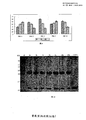
图 1 是 pET3c-6His-SUMO-LCVN 重组 质粒图。 Figure 1 is a diagram showing the recombinant plasmid pET3c-6His-SUMO-LCVN.
图 2 是重组 pET3C-6His-SUMO-LCVN 质粒 限制酶和 PCR 分析,其中 M:
DL2000 DNA marker ;泳道 1: pET3C-6His-SUMO-LCVN / Nde I+BamH I ; 泳道
2: PCR 。Figure 2 is a recombinant pET3C-6His-SUMO-LCVN plasmid restriction enzyme and PCR analysis, wherein M: DL2000 DNA marker; Lane 1: pET3C-6His-SUMO-LCVN / Nde I+ BamH I; Lane 2: PCR.
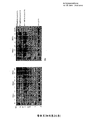
图 3 是 SDS-PAGE 电泳分析 BL21/ pET3c-6His-SUMO-LCVN
蛋白表达特性,其中:由左至右,各泳道依次为未加 IPTG 诱导、 IPTG 诱导 20h 、诱导表达破碎离心上清、诱导表达破碎离心沉淀。 Figure 3 is an SDS-PAGE electrophoresis analysis of BL21/ pET3c-6His-SUMO-LCVN
The expression characteristics of the protein were as follows: from left to right, the lanes were sequentially induced by IPTG without induction, IPTG induction for 20 hours, induced expression of disrupted centrifugal supernatant, and induction of fragmentation and centrifugation.
图 4 是 纯化目的蛋白 LCVN ,其中:由左至右,各泳道依次为 Sumo-LCVN 融合蛋白、
Sumo-LCVN 融合蛋白经 Sumo 蛋白酶酶切的混合物、 LCVN 蛋白。 Figure 4 shows the purified target protein LCVN, wherein: from left to right, each lane is Sumo-LCVN fusion protein,
Sumo-LCVN fusion protein was digested with Sumo protease, LCVN protein.
( * ) 表示 Sumo-LCVN 融合蛋白 ( * ) indicates Sumo-LCVN fusion protein
( ** ) 表示 LCVN 蛋白。 ( ** ) indicates the LCVN protein.
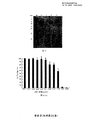
图 5 是 RP-HPL 分析重组 LCVN 的纯度。 Figure 5 shows the purity of recombinant LCVN analyzed by RP-HPL.
图 6 是 不同 pH 及投料比条件下 10K mPEG-ALD 修饰 LCVN 的
Tricine-SDS-PAGE电泳图,其中: 从左至右,泳道 1 是蛋白 Maker ,泳道 2 、 3 、 4 分别是 PH3.5 , LCVN 与
10K mPEG-ALD 摩尔比分别为 1:1 、 1:3 、 1:5 时的修饰产物电泳条带;泳道 5 、 6 、 7 分别是 PH4.0 , LCVN 与
10K mPEG-ALD 摩尔比分别为 1:1 、 1:3 、 1:5 时的修饰产物电泳条带;泳道 8 、 9 、 10 分别是 PH5.0 , LCVN 与
10K mPEG-ALD 摩尔比分别为 1:1 、 1:3 、 1:5 时的修饰产物电泳条带;泳道 11 、 12 、 13 分别是 PH6.0 , LCVN
与 10K mPEG-ALD 摩尔比分别为 1:1 、 1:3 、 1:5 时的修饰产物电泳条带;泳道 14 、 15 、 16 分别是 PH7.0 ,
LCVN 与 10K mPEG-ALD 摩尔比分别为 1:1 、 1:3 、 1:5 时的修饰产物电泳条带。 Figure 6 shows the 10K mPEG-ALD modified LCVN at different pH and feed ratios.
Tricine-SDS-PAGE electrophoresis map, where: from left to right, lane 1 is protein Maker, lanes 2, 3, and 4 are PH3.5, LCVN and
10K mPEG-ALD molar ratio is 1:1, 1:3, 1:5 modified product electrophoresis bands; lanes 5, 6, and 7 are PH4.0, LCVN and
10K mPEG-ALD molar ratio is 1:1, 1:3, 1:5 modified product electrophoresis bands; lanes 8, 9, 10 are PH5.0, LCVN and
10K mPEG-ALD molar ratio is 1:1, 1:3, 1:5 modified product electrophoresis bands; lanes 11, 12, 13 are PH6.0, LCVN
The electrophoresis bands of the modified products with the molar ratio of 10K mPEG-ALD were 1:1, 1:3, 1:5; the lanes 14, 15, and 16 were PH7.0, respectively.
The electrophoresis bands of the modified products when the molar ratio of LCVN to 10K mPEG-ALD were 1:1, 1:3, 1:5, respectively.
图 7 是不同 PH 及投料比条件下 10K mPEG-ALD 修饰 LCVN 的修饰率比较图。 Figure 7 is a comparison of the modification rates of 10K mPEG-ALD modified LCVN under different pH and feed ratio conditions.
图 8 是不同 PH 及投料比条件下 20K mPEG-ALD 修饰 LCVN 的
Tricine-SDS-PAGE电泳图,其中:从左至右,泳道 1 是蛋白 Maker ,泳道 2 、 3 、 4 分别是 PH3.5 , LCVN 与 20K
mPEG-ALD 摩尔比分别为 1:1 、 1:3 、 1:5 时的修饰产物电泳条带;泳道 5 、 6 、 7 分别是 PH4.0 , LCVN 与 20K
mPEG-ALD 摩尔比分别为 1:1 、 1:3 、 1:5 时的修饰产物电泳条带;泳道 8 、 9 、 10 分别是 PH5.0 , LCVN 与 20K
mPEG-ALD 摩尔比分别为 1:1 、 1:3 、 1:5 时的修饰产物电泳条带;泳道 11 、 12 、 13 分别是 PH6.0 , LCVN 与
20K mPEG-ALD 摩尔比分别为 1:1 、 1:3 、 1:5 时的修饰产物电泳条带;泳道 14 、 15 、 16 分别是 PH7.0 , LCVN
与 20K mPEG-ALD 摩尔比分别为 1:1 、 1:3 、 1:5 时的修饰产物电泳条带。 Figure 8 shows the 20K mPEG-ALD modified LCVN at different pH and feed ratios.
Tricine-SDS-PAGE electrophoresis map, wherein: from left to right, lane 1 is protein Maker, lanes 2, 3, and 4 are PH3.5, LCVN and 20K, respectively.
The mPEG-ALD molar ratio is 1:1, 1:3, 1:5 for the modified product electrophoresis bands; lanes 5, 6, and 7 are PH4.0, LCVN and 20K, respectively.
The mPEG-ALD molar ratio is 1:1, 1:3, 1:5 for the modified product electrophoresis bands; lanes 8, 9, and 10 are PH5.0, LCVN and 20K, respectively.
The mPEG-ALD molar ratio is 1:1, 1:3, 1:5 for the modified product electrophoresis bands; lanes 11, 12, and 13 are PH6.0, LCVN and
20K mPEG-ALD molar ratio is 1:1, 1:3, 1:5 modified product electrophoresis bands; lanes 14, 15, 16 are PH7.0, LCVN
The electrophoresis band of the modified product with a molar ratio of 20K mPEG-ALD of 1:1, 1:3, 1:5, respectively.
图 9 是不同 PH 及投料比条件下 20K mPEG-ALD 修饰 LCVN 的修饰率比较图。 Figure 9 is a comparison of the modification rates of 20K mPEG-ALD modified LCVN under different pH and feed ratio conditions.
图 10 是 不同时间下 , 10K mPEG-ALD 修饰 LCVN 的
Tricine-SDS-PAGE 电泳图 ,其中:泳道 1 是蛋白 Maker ,泳道 2-8 分别是反应 1h 、 3h 、 5h 、 7h 、 9h 、
12h 和 24h 后的样品电泳结果,泳道 9 是未修饰的 LCVN 。 Figure 10 shows the 10K mPEG-ALD modified LCVN at different times.
Tricine-SDS-PAGE electrophoresis map, where lane 1 is protein Maker and lanes 2-8 are 1h, 3h, 5h, 7h, 9h,
After 12h and 24h sample electrophoresis results, lane 9 is unmodified LCVN.
图 11 是 不同时间下 , 20K mPEG-ALD 修饰 LCVN 的
Tricine-SDS-PAGE 电泳图 ,其中:泳道 1 是蛋白 Maker ,泳道 2-8 分别是反应 1h 、 3h 、 5h 、 7h 、 9h 、
12h 和 24h 后的样品电泳结果,泳道 9 是未修饰的 LCVN 。 Figure 11 shows the 20K mPEG-ALD modified LCVN at different times.
Tricine-SDS-PAGE electrophoresis map, where lane 1 is protein Maker and lanes 2-8 are 1h, 3h, 5h, 7h, 9h,
After 12h and 24h sample electrophoresis results, lane 9 is unmodified LCVN.
图 12 是 10K mPEG-ALD 修饰 LCVN 经 SP-Sepharose
分离纯化的洗脱曲线。 Figure 12 is 10K mPEG-ALD modified LCVN via SP-Sepharose
The purified elution curve was isolated.
图 13 是 10K mPEG-ALD 修饰 LCVN 经 SP-Sepharose 分离纯化各组分的
Tricine- SDS- PAGE 电泳图,其中:泳道 1 是蛋白 Maker ,泳道 2-5 是修饰反应的混合物、上样穿透峰、 含有 80mM NaCl
的 buffer A 的洗脱峰 ; 含有 400mM NaCl 的 buffer A 的洗脱峰 。 Figure 13 shows the separation and purification of each component by 10K mPEG-ALD modified LCVN by SP-Sepharose
Tricine-SDS-PAGE electropherogram, where lane 1 is protein Maker, lane 2-5 is a mixture of modification reactions, loading peaks, containing 80 mM NaCl
The elution peak of buffer A; the elution peak of buffer A containing 400 mM NaCl.
图 14 是 20K mPEG-ALD 修饰 LCVN 经 SP-Sepharose
分离纯化的洗脱曲线。 Figure 14 is 20K mPEG-ALD modified LCVN via SP-Sepharose
The purified elution curve was isolated.
图 15 是 20K mPEG-ALD 修饰 LCVN 经 SP-Sepharose 分离纯化各组分的
Tricine-SDS- PAGE 电泳图,其中:泳道 1 是蛋白 Maker ,泳道 2-5 是修饰反应的混合物、上样穿透峰、 含有 70mM NaCl 的
buffer A 的洗脱峰 ; 含有 400mM NaCl 的 buffer A 的洗脱峰 。 Figure 15 is a 20K mPEG-ALD modified LCVN separated and purified by SP-Sepharose
Tricine-SDS-PAGE electrophoresis map, wherein: lane 1 is protein Maker, lanes 2-5 are mixtures of modification reactions, loading breakthrough peaks, containing 70 mM NaCl
The elution peak of buffer A; the elution peak of buffer A containing 400 mM NaCl.
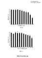
图 16 是 MT-4 经不同浓度 LCVN 及其修饰产物处理后的 细胞存活率( % )。 Figure 16 shows the cell viability (%) of MT-4 after treatment with different concentrations of LCVN and its modified products.
图 17 是 CVN 和 LCVN 对 HIV-1/IIIB 增殖的抑制率 ( % )。 Figure 17 shows the inhibition rate (%) of CVN and LCVN on HIV-1/IIIB proliferation.
图 18 是 LCVN 及其 PEG 修饰产物的抗人类免疫缺陷病毒活性,其中,图( a
)相差显微镜下观察培养 24h 后的 (1)MOLT-4 细胞、 (2) MOLT-4/IIIB 细胞、 (3) 共培养后的细胞、 (4)
假处理的共培养后的细胞、( 5-8 )浓度为 113nM 的 CVN/LCVN/10kPEG-LCVN/20kPEG-LCVN
处理后的共培养细胞,黑色箭头所示处为融合的巨大细胞。图 (b) CNV 、 LCVN 、 10kPEG-LCVN 、 20kPEG-LCVN
的融合抑制活性,所有实验至少是 3 次独立实验后的统计学处理结果。 Figure 18 is an anti-human immunodeficiency virus activity of LCVN and its PEG-modified product, wherein
(1) MOLT-4 cells, (2) MOLT-4/IIIB cells, and (3) cells after co-culture, observed under phase contrast microscopy, (4)
Pseudo-treated co-cultured cells, (5-8) CVN/LCVN/10kPEG-LCVN/20kPEG-LCVN at a concentration of 113 nM
Co-cultured cells after treatment, indicated by black arrows, are fused large cells. Figure (b) CNV, LCVN, 10kPEG-LCVN, 20kPEG-LCVN
The fusion inhibition activity, all experiments are at least 3 statistical results after independent experiments.
图 19 是 LCVN 及其 PEG 修饰产物 抗 HSV-1 活性 的 CPE
观察结果。其中: Figure 19 shows the CPE of LCVN and its PEG modified product against HSV-1 activity.
Observation results. among them:
A 、正常对照; B 、病毒对照; C 、阳性药物 ACV 对照( 1μg/ml ); D 、
L-CVN 样品( 1.562μg/ml ); E 、 SUMO-L-CVN 样品( 3.125μg/ml ); F 、
mPEG-ALD-10kDa-L-CVN ( 3.125μg/ml ); G 、 mPEG-ALD-20kDa-L-CVN ( 3.125μg/ml
)。 A, normal control; B, virus control; C, positive drug ACV control (1μg/ml); D,
L-CVN sample ( 1.562 μg/ml); E, SUMO-L-CVN sample ( 3.125 μg/ml); F ,
mPEG-ALD-10kDa-L-CVN ( 3.125 μg/ml ); G , mPEG-ALD-20kDa-L-CVN ( 3.125 μg/ml
).
具体实施方式 detailed description
以下列举一些本发明优选的实施例,以助于进一步理解本发明,但本发明的实施方式不限于此。 The preferred embodiments of the invention are listed below to assist in further understanding of the invention, but embodiments of the invention are not limited thereto.
本发明实施例涉及的主要材料如下 : 宿主菌大肠杆菌 BL21(DE3) (购自 Novagen 公司 )
、质粒 pET3c (购自 Novagen 公司) 、 pET3c-SUMO-CVN 由本室保存 (其构建方法已申请专利"重组蓝藻抗病毒蛋白的制备方法及应用
, 申请号 : 200810198926.0" ,质粒的构建亦可采用公知的基因工程方法,基本思路是首先通过 PCR 的方法得到 SUMO-CVN
融合序列,再连入 pET3c 载体即可得到 质粒 pET3c-SUMO-CVN ); SUMO 蛋白酶 ( 购自海基生物有限公司 ) ; Taq 酶、 T4
DNA 连接酶、 DNA 分子量标准、各种限制性内切酶购自大连宝生物公司 ; 蛋白质分子量标准品 ( 购自聚研生物科技有限公司 ) 、引物购自上海生工生物公司
; Ni2 + Sepharose Fast Flow 、 SP Sepharose Fast Flow 购自 GE
Healthcare 公司 ; MTT 、 WST 购自美国 SIGMA 公司 ; 单纯疱疹病毒 1 型 ( HSV-1 ) F 株来自 武汉大学病毒研究所
( CGMCC No.0396 ); Vero 细胞 ( CCL-81™ )、 MOLT-4 细胞 ( CRL-1582™ ) 、 MT-4 细胞 (
CRL-1942™ ) 、 HIV-I/IIIB 病毒 ( CRL-1973™ ) 等来自美国典型菌种保藏中心 ( ATCC ); mPEG-ALD(10K)
和 mPEG-ALD(20K) 购自北京凯正生物工程发展有限公司 。The main materials involved in the examples of the present invention are as follows: Host strain Escherichia coli BL21 (DE3) (purchased from Novagen), plasmid pET3c (purchased from Novagen), pET3c-SUMO-CVN is preserved by the laboratory (the construction method has been patented) The preparation method and application of recombinant cyanobacterial antiviral protein, application number: 200810198926.0", the construction of the plasmid can also adopt the well-known genetic engineering method, the basic idea is to first obtain the SUMO-CVN fusion sequence by PCR, and then connect to the pET3c vector. Plasmid pET3c-SUMO-CVN); SUMO protease (purchased from Haiji Biotechnology Co., Ltd.); Taq enzyme, T4 DNA ligase, DNA molecular weight standard, various restriction enzymes purchased from Dalian Bao Biotech Co., Ltd.; Standards (purchased from Juyan Biotechnology Co., Ltd.), primers purchased from Shanghai Shenggong Biotech Co., Ltd.; Ni 2 + Sepharose Fast Flow, SP Sepharose Fast Flow from GE Healthcare; MTT and WST from SIGMA, USA; Herpes simplex Virus type 1 ( HSV-1 ) F strain from Wuhan University Virus Research Institute ( CGMCC No.0396 ); Vero cells CCL-81TM), MOLT-4 cells (CRL-1582TM), MT-4 cells (CRL-1942TM), HIV-I/IIIB virus (CRL-1973TM), etc. from the American Type Culture Collection (ATCC) mPEG-ALD (10K) and mPEG-ALD (20K) were purchased from Beijing Kaizheng Bioengineering Development Co., Ltd.
NTA-0 buffer ( 20mmol/L Tris-HCl , pH 8.0 , 0.15mol
/L NaCl ,), NTA-20 buffer ( 20mmol/L Tris-HCl , pH 8.0 , 0.15mol /L NaCl ,
20mmol/L 咪唑 ), NTA-250 buffer ( 20mmol/L Tris-HCl , pH 8.0 , 0.15mol /L NaCl ,
250mmol/L 咪唑 ), 酶切缓冲液 ( 20mmol/L Tris-HCl , pH 8.0 , 0.15mol/L NaCl ), bufferA
( 20mmol/L NaAc-Ac , pH 4.0 ) 。 NTA-0 buffer ( 20mmol/L Tris-HCl , pH 8.0 , 0.15mol
/L NaCl ,), NTA-20 buffer ( 20mmol/L Tris-HCl , pH 8.0 , 0.15mol /L NaCl ,
20mmol/L imidazole), NTA-250 buffer (20mmol/L Tris-HCl, pH 8.0, 0.15mol / L NaCl,
250mmol/L imidazole), digestion buffer (20mmol/L Tris-HCl, pH 8.0, 0.15mol/L NaCl), bufferA
(20 mmol/L NaAc-Ac, pH 4.0).
实施例 Example
1 、 构建重组质粒 pET3c-6His-SUMO-LCVN 1. Construction of recombinant plasmid pET3c-6His-SUMO-LCVN
SUMO-L-CVN 基因的构建合成分两步进行,首先通过两次 PCR 合成 L-CVN 基因,第一次
PCR 以 pET3c-SUMO-CVN 质粒为模板 , 以 F1-CVN 、 R-CVN 为上下游引物 。反应体系为模板 1ng ,上下游引物各 1μM ,
20μl Taq PCR MasterMix ,加水至 40μl ,反应混合物 94 ℃ 变性 1min ,退火至 55 ℃ ,保持 1 min , 72 ℃
延伸 1min ,进行 29 个循环。反应产物进行 1% 琼脂糖凝胶电泳,胶回收目的片段,作为下一轮 PCR 的模板。 第二 次 PCR 以上 一轮 PCR
产物为模板 , F2-CVN 、 R-CVN 为上下游引物对(其中 F2-CVN 引物中含有编码 15 个氨基酸残基的柔性多肽),合成 L-CVN 全长序列。
SUMO 全长序列通过 PCR 方法从 pET3c-SUMO-CVN 中合成。利用 SUMO 序列末端与 L-CVN 序列前端的 26bp 重叠互补序列进行
PCR :以 SUMO 序列和 L-CVN 序列为延伸模板, F-SUMO , R-CVN 为上下游引物,在常规的 PCR 条件下进行反应,反应产物进行 1%
琼脂糖凝胶电泳,并做胶回收,得到 SUMO-L-CVN 全长序列。 The construction of the SUMO-L-CVN gene was synthesized in two steps. First, the L-CVN gene was synthesized by two PCRs, the first time.
PCR uses pET3c-SUMO-CVN plasmid as template and F1-CVN and R-CVN as upstream and downstream primers. The reaction system is 1 ng for the template and 1 μM for each of the upstream and downstream primers.
20μl Taq PCR MasterMix, add water to 40μl, the reaction mixture is denatured at 94 °C for 1min, annealed to 55 °C for 1 min, 72 °C
Extend 1 min for 29 cycles. The reaction product was subjected to 1% agarose gel electrophoresis, and the target fragment was recovered by gel as a template for the next round of PCR. Second PCR above one round of PCR
The product is a template, F2-CVN and R-CVN are upstream and downstream primer pairs (where the F2-CVN primer contains a flexible polypeptide encoding 15 amino acid residues), and the full-length sequence of L-CVN is synthesized.
The SUMO full-length sequence was synthesized from pET3c-SUMO-CVN by PCR. Using the 26 bp overlapping complementary sequence at the end of the SUMO sequence and the front end of the L-CVN sequence
PCR: using SUMO sequence and L-CVN sequence as extension template, F-SUMO and R-CVN are upstream and downstream primers, and the reaction was carried out under normal PCR conditions, and the reaction product was subjected to 1%.
Electrophoresis on agarose gel and gel recovery gave the full length sequence of SUMO-L-CVN.
将质粒 pET3C 和 6His-SUMO-LCVN 全长序列分别用 Nde I
和与 BamH I 双酶切,酶切产物 1% 琼脂糖凝胶电泳,回收酶切产物, T4DNA 连接酶,连接产物转化大肠杆菌
JM109 感受态细胞,涂布于含氨苄青霉素的 LB 平板上, 37 ℃ 培养过夜,提取质粒, PCR 扩增及 Nde I 与
BamH I 双酶切鉴定,阳性质粒送往英骏公司测序。The full-length sequences of plasmid pET3C and 6His-SUMO-LCVN were digested with Nde I and BamH I respectively, and the digested product was subjected to 1% agarose gel electrophoresis, and the digested product was recovered, T 4 DNA ligase, and the ligation product was transformed into the large intestine. Bacterial JM109 competent cells were plated on LB plates containing ampicillin and cultured overnight at 37 °C. Plasmids were extracted, amplified by PCR and identified by double digestion with Nde I and BamH I. The positive plasmids were sent to Yingjun for sequencing.
表 1 用于合成 SUMO-LCVN 全长序列的引物 Table 1 Primers used to synthesize the full length sequence of SUMO-LCVN
|
引物名称 Primer name
|
序列 sequence
|
|
F2-CVN F2-CVN
|
5'
-CAGATTGGTGGTGGTGGCGGAGGGAGCGGTGGAGGGGGCAGTGGCGGAG-3' 5'
-CAGATTGGTGGTGGTGGCGGAGGGAGCGGTGGAGGGGGCAGTGGCGGAG-3'
|
|
F1-CVN F1-CVN
|
5'
-GGAGGGGGCAGTGGCGGAGGAGGTAGCCTTGGTAAATTCTCCCAG-3' 5'
-GGAGGGGGCAGTGGCGGAGGAGGTAGCCTTGGTAAATTCTCCCAG-3'
|
|
R-CVN R-CVN
|
5' -AGA GGATCC TCATCATTCGTATTTCAGGGTAC-3'5 '-AGA GGATCC TCATCATTCGTATTTCAGGGTAC-3'
|
|
F-SUMO F-SUMO
|
5' -CAG CATATG CATCATCATCATC-3'5' -CAG CATATG CATCATCATCATC-3'
|
|
R-SUMO R-SUMO
|
5' -CTCCCTCCGCCACCACCACCAATCTGTTCTCTG-3' 5' -CTCCCTCCGCCACCACCACCAATCTGTTCTCTG-3'
|
2 、 LCVN 工程菌的摇瓶培养、蛋白纯化和纯度测定 2, LCVN engineering bacteria shake flask culture, protein purification and purity determination
测序正确的质粒转化大肠杆菌 BL21(DE3) , 得工程菌
BL21[pET3c-6His-SUMO-LCVN] , 挑选单克隆进行培养并诱导表达 , 收集菌体蛋白进行 SDS-PAGE 电泳分析。结果表明 BL21
[pET3c-6His-SUMO-LCVN] 阳性克隆菌株经诱导后 会表达大小约为 28kDa 的融合蛋白,
未诱导对照组在相应位置无肉眼可见表达,经超声破碎菌体后,发现 融合蛋白位于上清中,为可溶性表达,经凝胶光密度扫描,可溶性目的蛋白占上清总蛋白的
28.3±3.4 % (图 3 )。 Sequencing the correct plasmid into E. coli BL21 (DE3)
BL21[pET3c-6His-SUMO-LCVN], monoclonal clones were selected for culture and induced expression, and bacterial proteins were collected for SDS-PAGE electrophoresis analysis. The result shows that BL21
The [pET3c-6His-SUMO-LCVN] positive clone strain will express a fusion protein of about 28kDa after induction.
The uninduced control group showed no macroscopic expression at the corresponding position. After the bacteria were disrupted by sonication, the fusion protein was found to be soluble in the supernatant, and the soluble target protein accounted for the total protein of the supernatant.
28.3 ± 3.4 % (Figure 3).
选取高表达菌株接种到 1L 含氨苄青霉素( 100 mg/L )的 LB 培养基中, 37℃ , 180
rpm 培养至 OD600 =0. 6 ~ 1. 0 时,降温至 20 ℃ 加 IPTG 至终浓度 0.5mM 诱导表达 24h 。 4 ℃ 、 6000×g
、 10min 离心收集菌体,冻融一次,然后将菌体沉淀以 1 : 10 比例重新悬浮于 NTA-10 buffer ,超声破碎(工作时间 5s 、间歇时间
5s , 99 次,重复 3 遍 ) , 4℃ 、 25000×g 、 30min 离心收集上清。 High-expression strains were selected and inoculated into 1L LB medium containing ampicillin (100 mg/L), 37 ° C, 180
When rpm was cultured to OD600 =0. 6 ~ 1. 0, the temperature was lowered to 20 °C and IPTG was added to a final concentration of 0.5 mM to induce expression for 24 h. 4 °C, 6000×g
The cells were collected by centrifugation at 10 min, frozen and thawed, and then the cells were resuspended in NTA-10 buffer at a ratio of 1:10, sonicated (working time 5 s, intermittent time)
5s, 99 times, repeated 3 times), the supernatant was collected by centrifugation at 4 ° C, 25000 × g, 30 min.
上清液上样柱床体积为 20ml 的 Ni-NTA 亲和层析柱,流速 0.6ml/min, NTA-0
buffer 洗回基线,流速为 1ml/min, NTA-20 buffer 洗杂蛋白, NTA-250 buffer 洗脱目的蛋白。纯化后的目的蛋白
6His-SUMO-LCVN 经 Sephadex G-25 分子筛脱咪唑后进行 SUMO 蛋白酶酶切,除去 SUMO 融合蛋白。 The supernatant was loaded with a 20 ml Ni-NTA affinity column at a flow rate of 0.6 ml/min, NTA-0.
The buffer was washed back to the baseline at a flow rate of 1 ml/min, NTA-20 buffer washed protein, and NTA-250 buffer eluted the target protein. Purified target protein
6His-SUMO-LCVN was subjected to deamidation by Sephadex G-25 molecular sieve and then subjected to SUMO protease digestion to remove the SUMO fusion protein.
6His-SUMO-LCVN 调整浓度至 1mg/ml , 加入 1 U SUMO 蛋白酶 /mg
融合蛋白, 30℃ 酶切 1h 。因 6His-SUMO 标签、 SUMO 蛋白酶均含有 6×His 标签,酶切后的样品再次上样 Ni-NTA
亲和层柱进行纯化,除去带 6His 标签的 SUMO 、未酶切的 6His-SUMO-LCVN 和 SUMO 蛋白酶,得到非融合的目的蛋白 LCVN ,经
G-25 分子筛柱脱盐后冷冻干燥,得 LCVN 制品,供后续理化性质测定、活性测定或 PEG 修饰用。 6His-SUMO-LCVN Adjust the concentration to 1mg/ml and add 1 U SUMO protease /mg
Fusion protein, digested at 30 ° C for 1 h. Because the 6His-SUMO tag and SUMO protease both contain a 6×His tag, the digested sample is loaded again. Ni-NTA
The affinity column was purified to remove the 6His-tagged SUMO, un-cut 6His-SUMO-LCVN and SUMO protease to obtain the non-fused target protein LCVN.
The G-25 molecular sieve column is desalted and freeze-dried to obtain LCVN products for subsequent physical and chemical properties determination, activity determination or PEG modification.
图 3 是工程菌 BL21[ pET3c-6His-SUMO-LCVN] 经 IPTG
诱导后的蛋白表达图谱,可见经诱导后,在分子量 28kD 处出现明显增粗的蛋白带(泳道 3 ),与 6His-SUMO-LCVN
的理论分子量相符,超声破碎后,目的蛋白位于菌体破碎后的上清中,含量占菌体可溶性蛋白的 40% (泳道 2 )。图 4 是 SDS-PAGE 分析 LCVN
纯化过程,其中 * 所示处为 6His-SUMO-LCVN 融合蛋白, ** 所示处为 LCVN 蛋白。 图 5 是 LCVN 制品的反向高效液相色谱(
RP-HPLC )纯度分析结果,株型是 C-18 反向柱,检测器波长为 280nm ,流动相 A :含 0.1 %三氟乙酸( TFA )的超纯水,流动相 B
:乙氰,经 40 % -60 %的 B 进行梯度洗脱,保留时间在 4-6min 之间出现 LCVN 的洗脱峰。 Figure 3 is the engineering strain BL21[ pET3c-6His-SUMO-LCVN] by IPTG
The protein expression profile after induction showed that after induction, a significantly thickened protein band (lane 3) appeared at a molecular weight of 28 kD, and 6His-SUMO-LCVN
The theoretical molecular weight is consistent. After sonication, the target protein is located in the supernatant of the broken cell, which accounts for 40% of the soluble protein of the bacteria (lane 2). Figure 4 is an SDS-PAGE analysis of LCVN
Purification process, where * is shown as 6His-SUMO-LCVN fusion protein, and ** is shown as LCVN protein. Figure 5 is a reverse high performance liquid chromatography of LCVN products (
RP-HPLC) Purity analysis results, the plant type is a C-18 reverse column with a detector wavelength of 280 nm, mobile phase A: ultrapure water containing 0.1% trifluoroacetic acid (TFA), mobile phase B
: Ethyl cyanide, gradient elution with 40% -60% B, and elution peak of LCVN with retention time between 4-6 min.
3 、 LCVN 工程菌的中试发酵和蛋白制备 3. Pilot fermentation and protein preparation of LCVN engineering bacteria
工程菌 BL21[pET3c-6His-SUMO-LCVN] , 挑选单克隆进行培养并在试管中诱导表达
, 选取高表达菌株进行中试发酵。分别在锥形瓶中培养一级和二级种子,培养二级种子至合适浓度,以 10 %接种量接种到 15L 的发酵培养基中。自动控制温度为
37 ℃ ,适时调整转速和通气量以保持合适的溶氧水平, NaOH 和 HCl 调节 PH 为 7.0 ~ 7.2 。当菌体密度(
OD600 )为 12 左右时,温度降至 20 ℃ ,加 IPTG 至终浓度 0.5mM 诱导表达 20h 。 100ml/min ,
9000g ,连续流离心收集菌体, 15L 的发酵培养基可以收获约 550g 的湿菌体,收集的菌体于 -20 ℃ 冰箱保存。The engineered strain BL21[pET3c-6His-SUMO-LCVN] was selected and cultured and induced to express in a test tube, and a high expression strain was selected for pilot fermentation. Primary and secondary seeds were cultured in conical flasks, secondary seeds were incubated to appropriate concentrations, and inoculated into 15 L of fermentation medium at 10% inoculum. The temperature is automatically controlled at 37 °C, and the speed and ventilation are adjusted in time to maintain a proper dissolved oxygen level. The pH is adjusted to 7.0 to 7.2 with NaOH and HCl. When the cell density (OD 600 ) was about 12, the temperature was lowered to 20 °C, and IPTG was added to a final concentration of 0.5 mM to induce expression for 20 h. 100 ml/min, 9000 g, the cells were collected by continuous flow centrifugation, and about 550 g of wet cells were harvested in a 15 L fermentation medium, and the collected cells were stored in a refrigerator at -20 °C.
菌体沉淀以 1 : 10 比例悬浮于 NTA-0 buffer ,超声破碎(工作时间 5s 、间歇时间
5s , 99 次,重复 3 遍 ) , 4℃ 、 25000×g 、 30min 离心收集上清。柱床体积为 20ml 的 Ni-NTA 填料上样量为
250ml 上清,流速 1ml/min, NTA-0 buffer 洗回基线;流速为 1ml/min, NTA-20 buffer 洗杂蛋白, NTA-250
buffer 洗脱目的蛋白。纯化后的目的蛋白 6His-SUMO-LCVN 经 Sephadex G-25 分子筛脱咪唑后进行 SUMO 蛋白酶酶切,除去
SUMO 融合蛋白。 The cell pellet was suspended in NTA-0 buffer at a ratio of 1:10, sonicated (working time 5s, intermittent time)
5s, 99 times, repeated 3 times), the supernatant was collected by centrifugation at 4 ° C, 25000 × g, 30 min. The loading of Ni-NTA packing with a volume of 20 ml is
250ml supernatant, flow rate 1ml/min, NTA-0 buffer wash back to baseline; flow rate 1ml/min, NTA-20 buffer wash protein, NTA-250
Buffer elutes the protein of interest. The purified target protein 6His-SUMO-LCVN is subjected to SUMO protease digestion by removing the imidazole from Sephadex G-25 molecular sieve.
SUMO fusion protein.
6His-SUMO-LCVN 调整浓度至 1mg/ml , 加入 1 U SUMO 蛋白酶 /mg
融合蛋白, 30℃ 酶切 1h 。因 6His-SUMO 标签、 SUMO 蛋白酶均含有 6×His 标签,酶切后的样品再次上样 Ni-NTA
亲和层柱进行纯化,除去带 6His 标签的 SUMO 、未酶切的 6His-SUMO-LCVN 和 SUMO 蛋白酶,得到非融合的目的蛋白 LCVN ,经
G-25 分子筛柱更换缓冲液,再用截留量 3KD 的超滤管浓缩 LCVN 蛋白,供后续理化性质测定、活性测定或 PEG 修饰用。 6His-SUMO-LCVN Adjust the concentration to 1mg/ml and add 1 U SUMO protease /mg
Fusion protein, digested at 30 ° C for 1 h. Because the 6His-SUMO tag and SUMO protease both contain a 6×His tag, the digested sample is loaded again. Ni-NTA
The affinity column was purified to remove the 6His-tagged SUMO, un-cut 6His-SUMO-LCVN and SUMO protease to obtain the non-fused target protein LCVN.
Replace the buffer with G-25 molecular sieve column and concentrate the LCVN protein with a 3KD ultrafiltration tube for subsequent physical and chemical determination, activity determination or PEG modification.
4 、 LCVN 蛋白的 PEG 修饰 4, PEG modification of LCVN protein
对药用蛋白进行 PEG 修饰是改善其药代特性、稳定性和免疫原性等多种成药性质的有效方法。蛋白质可供
PEG 修饰的位点包括侧链氨基、 N 端氨基、侧链羧基、 C
端羧基、侧链巯基等多种。现有的羧基修饰技术容易产生非特异性交联反应,因此氨基修饰更为常用,技术发展也更为成熟。 Zappe 等的研究结果表明,对野生型 CVN
的侧链氨基或 N 端氨基的修饰都会破坏该蛋白的活性,因此,作者首先对 CVN 进行定点突变,获得 Q62C 突变体,然后对 62 位引入的 Cys
进行侧链巯基修饰,获得了有活性的蛋白。由于天然 CVN 中已有的 4 个 Cys 残基之间形成 2
对二硫键,并且在溶液中,蛋白质的二硫键处在动态异构和平衡状态,因此引入 Q62C 突变后,很难避免引入的 Cys 不干扰二硫键的正确搭配,或者非 62 位的
Cys 修饰,作者最后的实验结果也证明, CVN Q62C 突变体及其修饰产物的活性均低于野生型 CVN 。 PEG modification of pharmaceutical proteins is an effective method to improve various drug properties such as pharmacokinetic properties, stability and immunogenicity. Protein available
PEG-modified sites include side chain amino groups, N-terminal amino groups, side chain carboxyl groups, C
A terminal carboxyl group, a side chain thiol group, and the like. Existing carboxyl modification techniques are prone to non-specific cross-linking reactions, so amino modification is more common and technological development is more mature. Zappe et al.'s findings indicate that wild-type CVN
The modification of the side chain amino or N-terminal amino group will destroy the activity of the protein. Therefore, the author first performs site-directed mutagenesis of CVN to obtain the Q62C mutant, and then introduced the Cys introduced at position 62.
The side chain thiol modification was carried out to obtain an active protein. Due to the formation of 4 Cys residues in the native CVN 2
For disulfide bonds, and in solution, the disulfide bond of the protein is in a dynamic isomeric and equilibrium state. Therefore, after introducing the Q62C mutation, it is difficult to prevent the introduced Cys from interfering with the correct matching of the disulfide bond, or non-62 position.
Cys modification, the authors' final experimental results also showed that the activity of CVN Q62C mutant and its modified products were lower than wild-type CVN.
由于本发明制备的 LCVN 末端引入了亲水柔性的 15 肽序列,因此可以尝试进行 N
端的定点氨基修饰。我们的实验结果证明, LCVN 经 N 端定点氨基修饰后,不仅可以获得有活性的修饰产物,并且修饰产物的抗病毒活性进一步增强。 Since the LCVN end prepared by the present invention introduces a hydrophilic and flexible 15-peptide sequence, an attempt can be made to N.
Fixed-point amino modification at the end. Our experimental results show that LCVN can not only obtain active modified products after N-terminal amino group modification, but also enhance the antiviral activity of the modified products.
对氨基的 PEG 修饰方法很多,可供选择的修饰剂也很多,本发明选择 mPEG-ALD(10K) 和
mPEG-ALD(20K) 作为修饰剂,从底物 : 修饰剂比例、修饰 pH 、修饰反应时间等 3 个方面,筛选了 LCVN 的最佳 PEG 修饰条件。 There are many PEG modification methods for amino groups, and there are many alternative modifiers. The present invention selects mPEG-ALD (10K) and
mPEG-ALD (20K) was used as a modifier to screen the optimal PEG modification conditions for LCVN from three aspects: substrate ratio, modifier ratio, modification pH and modification reaction time.
选择 pH 为 3.5 , 4.0 , 5.0 , 60 , 7.0 ,离子强度为 20mM 的
Na-Ac 缓冲液,反应体系中 LCVN 的浓度为 5mg/ml , LCVN 与 mPEG-ALD(10K) 的摩尔比分别选择为 1:1 , 1:3 ,
1:5 ; NaCNBH3 的终浓度为 5mg/ml 。室温下,振荡器上摇动反应,在反应 3h 后取样,电泳鉴定修饰反应的效果。
mPEG-ALD(10K) 修饰 LCVN 的单修饰产物在 SDS-PAGE 上的表观分子量约 30KD , SDS-PAGE 分析结果表明,在 pH4.0
, LCVN 与 mPEG-ALD(10K) 的摩尔比为 1:3 的条件下,单修饰率最高(图 6 )。Select Na-Ac buffer with pH of 3.5, 4.0, 5.0, 60, 7.0 and ionic strength of 20 mM. The concentration of LCVN in the reaction system is 5 mg/ml, and the molar ratio of LCVN to mPEG-ALD (10K) is selected as 1 respectively. :1 , 1:3 , 1:5 ; The final concentration of NaCNBH 3 is 5 mg/ml. The reaction was shaken on a shaker at room temperature, and after 3 hours of reaction, the effect of the modification reaction was identified by electrophoresis. The apparent molecular weight of the single modified product of mPEG-ALD (10K) modified LCVN was about 30KD on SDS-PAGE. The SDS-PAGE analysis showed that the molar ratio of LCVN to mPEG-ALD (10K) was 1: at pH 4.0. Under the conditions of 3, the single modification rate was the highest (Fig. 6).
类似方法 , 研究 mPEG-ALD(20K) 修饰 LCVN 的最佳修饰 pH 和投料比 。
mPEG-ALD(20K) 修饰 LCVN 后,单修饰产物在 SDS-PAGE 上的表观分子量 约 50KD , 实验 结果表明:在 pH5.0 , LCVN
与 mPEG-ALD(20K) 的摩尔比为 1:1 的条件下,单修饰率最高(图 18 )。 A similar method was used to study the optimal pH and feed ratio of mPEG-ALD (20K) modified LCVN.
After mPEG-ALD (20K) modification of LCVN, the apparent molecular weight of the single modified product on SDS-PAGE is about 50KD. The experimental results show that at pH5.0, LCVN
The single modification rate was the highest with a molar ratio of mPEG-ALD (20K) of 1:1 (Fig. 18).
在确定了最佳的修饰 pH 和投料比下, mPEG-ALD(10K) 和 mPEG-ALD(20K) 修饰
LCVN 分别在反应 1h 、 3h 、 5h 、 7h 、 9h 、 12h 和 24h 取样, Tricine-SDS-PAGE 电泳鉴定最佳的反应时间。
SDS-PAGE 分析结果表明表明:反应时间对 mPEG-ALD(10K) 和 mPEG-ALD(20K) 修饰 LCVN
的反应没有显著影响,综合考虑时间成本,优选最佳修饰反应时间为室温振荡反应 2h (图 10 和图 11 )。 mPEG-ALD (10K) and mPEG-ALD (20K) modification at the optimum pH and feed ratio determined
LCVN was sampled at 1h, 3h, 5h, 7h, 9h, 12h and 24h, and the optimal reaction time was identified by Tricine-SDS-PAGE electrophoresis.
The results of SDS-PAGE analysis indicated that the reaction time was modified by mPEG-ALD (10K) and mPEG-ALD (20K).
The reaction did not have a significant effect, considering the time cost, preferably the optimal modification reaction time was 2 h at room temperature (Fig. 10 and Fig. 11).
5 、 LCVN 的 PEG 修饰产物的分离纯化 5, separation and purification of PEG modified products of LCVN
LCVN 经 PEG-ALD 修饰后 , 反应混合物中主要有未修饰的 LCVN , 剩余的 PEG ,
PEG 多修饰的 LCVN , PEG 单修饰的 LCVN 。采用 AKTA prime plus 分离纯化系统 , SP sepharose 层析柱
分离纯化单修饰的 mPEG-ALD-LCVN ,流动相为 20mM 的 Na-Ac 缓冲液 ( buffer A ), 上样前先用 5 倍柱体积的
bufferA 平衡柱子,上样后收集穿透峰,然后用含有不同浓度 NaCl 的 bufferA 进行洗脱,收集各个洗脱峰。流速是 1ml/min ,检测波长是
280nm 。收集到的样品进行 SDS-PAGE 凝胶电泳检测。 After LCVN is modified by PEG-ALD, the reaction mixture mainly contains unmodified LCVN and the remaining PEG.
PEG multi-modified LCVN, PEG single-modified LCVN. AKTA prime plus separation and purification system, SP sepharose column
The single modified mPEG-ALD-LCVN was isolated and purified, and the mobile phase was 20 mM Na-Ac buffer (buffer A), and 5 column volumes were used before loading.
bufferA equilibrate the column, collect the breakthrough peak after loading, and then elute with bufferA containing different concentrations of NaCl to collect each elution peak. The flow rate is 1ml/min and the detection wavelength is
280nm. The collected samples were detected by SDS-PAGE gel electrophoresis.
mPEG-ALD ( 10K )修饰 LCVN 的混合物,经 SP sepharose
阳离子层析纯化之后,修饰剂 mPEG-ALD 和多修饰产物不与 SP sepharose 结合而被穿透,含有 80mM NaCl 的 buffer A
洗脱产物为单修饰的 mPEG-ALD(10K)-LCVN ;含有 400mM NaCl 的 buffer A 洗脱产物是未修饰的
LCVN 。图 12 为 mPEG-ALD(10K)-LCVN 分离纯化的洗脱曲线,峰 1 为修饰剂和多修饰产物,峰 2
为单修饰产物,峰 3 为未修饰底物。图 13 为 SDS-PAGE 分析各洗脱组份,泳道 M 为蛋白质分子量标准;泳道 A
为上样样品,可见未修饰底物、单修饰产物和多修饰产物;泳道 1 是穿透峰;泳道 2 是单修饰产物,也就是目的蛋白
mPEG-ALD(10K)-LCVN ; 泳道 3 为未修饰产物。mPEG-ALD (10K) modified LCVN mixture, after purification by SP sepharose cation chromatography, the modifier mPEG-ALD and multi-modification products were not penetrated by SP sepharose, and the buffer A elution product containing 80 mM NaCl was single. Modified mPEG-ALD (10K) -LCVN; buffer A eluting product containing 400 mM NaCl is unmodified LCVN. Figure 12 shows the elution curve of mPEG-ALD (10K) -LCVN separation and purification. Peak 1 is the modifier and multi-modification product, peak 2 is the single modification product, and peak 3 is the unmodified substrate. Figure 13 shows the elution components by SDS-PAGE. Lane M is the protein molecular weight standard; Lane A is the sample, showing unmodified substrate, single modified product and multiple modified products; Lane 1 is the breakthrough peak; Lane 2 It is a single modified product, that is, the target protein mPEG-ALD (10K) -LCVN; Lane 3 is an unmodified product.
mPEG-ALD ( 20K )修饰 LCVN 的混合物,经 SP sepharose
阳离子层析纯化之后,修饰剂 mPEG-ALD 和多修饰产物也存在于上样穿透峰中,含有 70mM NaCl 的 buffer A 洗脱产物为单修饰的
mPEG-ALD(20K)-LCVN ;含有 400mM NaCl 的 buffer A 洗脱产物是未修饰的 LCVN 。mPEG-ALD (20K) modified LCVN mixture, after purification by SP sepharose cation chromatography, the modifier mPEG-ALD and multi-modification products were also present in the loading peak, and the buffer A eluting product containing 70 mM NaCl was single. Modified mPEG-ALD (20K) -LCVN; buffer A eluting product containing 400 mM NaCl is unmodified LCVN.
图 14 为 mPEG-ALD(20K)-LCVN 分离纯化的洗脱曲线,峰 1
为修饰剂和多修饰产物,峰 2 为单修饰产物,峰 3 为未修饰底物。图 15 为 SDS-PAGE 分析各洗脱组份,泳道 M 为蛋白质分子量标准;泳道 A
为上样样品,可见未修饰底物、单修饰产物和多修饰产物;泳道 1 是穿透峰;泳道 2 是单修饰产物,也就是目的蛋白
mPEG-ALD(10K)-LCVN ; 泳道 3 为 400mM NaCl 洗脱组份,可见 未修饰产物,但也含Figure 14 shows the elution curve of mPEG-ALD (20K) -LCVN separation and purification. Peak 1 is the modifier and multi-modification product, peak 2 is the single modification product, and peak 3 is the unmodified substrate. Figure 15 shows the elution components by SDS-PAGE. Lane M is the protein molecular weight standard; Lane A is the sample, showing unmodified substrate, single modified product and multiple modified products; Lane 1 is the breakthrough peak; Lane 2 Is a single modified product, that is, the target protein mPEG-ALD (10K) -LCVN; Lane 3 is a 400 mM NaCl elution component, showing unmodified products, but also
有部分单修饰产物。 There are some single modified products.
应用实施例 1 WST 法测定 LCVN 对 T 细胞的细胞毒性 Application Example 1 Determination of cytotoxicity of LCVN on T cells by WST method
CVN 、 LCVN 及 PEG 修饰产物以 RPMI-1640 培养液作 5 倍系列稀释,加至 96
孔细胞培养板中,每孔 50μl , CVN 和 LCVN 稀释范围为 10μg/ml 至 0.04μg/ml , PEG 修饰 LCVN 的稀释范围为
50μg/ml 至 0.19μg/ml 。 MT-4 细胞调整浓度至 1×10⁵/ml ,每孔 100μl ,混匀,置 37 ℃ 、
5% CO2 细胞培养箱中培养,同时设细胞对照和阳性药物对照( 63nM 叠氮胸苷, AZT) ,每个样品均平行测定 3 个复孔。 4
天后培养板中每孔加入 10 μl WST-1 (水溶性四氮唑, 5mmol/L )溶液,继续培养 4 小时,置读板仪上读取吸光值( A ),波长
450/650nm ,计算 细胞存活率 ( Relative percentage, RP , % ),并根据 RP 计算 50% 毒性浓度(
CC50 )。CVN, LCVN and PEG modified products were serially diluted in RPMI-1640 medium and added to 96-well cell culture plates at 50 μl per well. CVN and LCVN were diluted from 10 μg/ml to 0.04 μg/ml. PEG-modified LCVN The dilution range is from 50 μg/ml to 0.19 μg/ml. The MT-4 cells were adjusted to a concentration of 1×10 5 /ml, 100 μl per well, mixed, and cultured in a 37 ° C, 5% CO 2 cell incubator, with a cell control and a positive drug control (63 nM azidothymidine, AZT), 3 replicate wells were determined in parallel for each sample. After 4 days, 10 μl of WST-1 (water-soluble tetrazolium, 5 mmol/L) solution was added to each well of the culture plate, and the culture was continued for 4 hours. The absorbance (A) was read on the plate reader, and the wavelength was calculated at 450/650 nm. Cell viability ( RP , % ) and 50% toxic concentration (CC 50 ) based on RP.
细胞存活率 ( Relative percentage, % )=药物处理组 A 值 / 细胞对照组 A
值 ×100% 。 Cell viability (%) = drug treatment group A value / cell control group A
Value ×100%.
图 16 是不同浓度 CVN 、 LCVN 及其修饰产物等 4 种蛋白处理 MT-4 后的 细胞存活率
,图中 N.C. 指未处理的细胞对照,以细胞对照的密度为 100% 。 AZT 为阳性对照药物, 63nM 叠氮胸苷。经计算后的 50% 毒性浓度如表 2
所示。 Figure 16 shows cell viability after treatment with four different proteins, CVN, LCVN and their modified products.
In the figure, N.C. refers to an untreated cell control with a cell control density of 100%. AZT is a positive control drug, 63 nM azidothymidine. The calculated 50% toxic concentration is shown in Table 2.
Shown.
表 2 LCVN 及其 PEG 修饰产物的 50% 毒性浓度
| Sample | Cytotoxicity (CC50,μM) |
| AZT | > 4.482 |
| CVN | 0.160±0.009 |
| LCVN | 0.658±0.277 |
| mPEG-ALD(10K)-LCVN | 4.519±0.191 |
| mPEG-ALD(20K)-LCVN | 3.629±0.137 |
Table 2 50% toxic concentration of LCVN and its PEG modified products | Sample | Cytotoxicity (CC50, μM) |
| AZT | > 4.482 |
| CVN | 0.160±0.009 |
| LCVN | 0.658±0.277 |
| mPEG-ALD (10K) -LCVN | 4.519±0.191 |
| mPEG-ALD (20K) -LCVN | 3.629±0.137 |
应用实施例 2 WST 法测定 LCVN 的抗人类免疫缺陷病毒活性 Application Example 2 Determination of anti-human immunodeficiency virus activity of LCVN by WST method
CVN 、 LCVN 及 PEG 修饰产物以 RPMI-1640 培养液作 5 倍系列稀释,加至 96
孔细胞培养板中,每孔 50μl 。 MT-4 细胞调整浓度至 1×10⁵/ml ,每孔 100μl ,混匀,然后加入
HIV-1/IIIB 病毒悬液 50μl ,滴度为 100TCID50 。置 37 ℃ 、 5% CO2
细胞培养箱中培养,同时设细胞对照、病毒对照和阳性药物对照(叠氮胸苷, AZT) ,每个样品均平行测定 3 个复孔。 4 天后培养板中每孔加入 10 μl
WST-1 (水溶性四氮唑, 5mmol/L )溶液,继续培养 4 小时,置读板仪上读取吸光值( A ),波长 450/650nm 。CVN, LCVN and PEG modified products were serially diluted in RPMI-1640 medium and added to 96-well cell culture plates at 50 μl per well. Adjust the concentration of MT-4 cells to 1 × 10 5 /ml, 100 μl per well, mix, and then add 50 μl of HIV-1/IIIB virus suspension with a titer of 100 TCID50. The cells were cultured in a 37 ° C, 5% CO 2 cell incubator, and a cell control, virus control, and positive drug control (azidothymidine, AZT) were set. Three replicate wells were determined in parallel for each sample. After 4 days, 10 μl of WST-1 (water-soluble tetrazolium, 5 mmol/L) solution was added to each well of the culture plate, and incubation was continued for 4 hours. The absorbance (A) was read on a plate reader at a wavelength of 450/650 nm.
细胞存活率 ( Relative percentage, % )=药物处理组 A 值 / 细胞对照组 A
值 ×100% 。 Cell viability (%) = drug treatment group A value / cell control group A
Value ×100%.
病毒抑制率 ( % ) = ( 药物处理组 A450/650- 病毒对照组
A450/650 ) / ( 细胞对照组 A450/650- 病毒对照组 A450/650
) ×100%Virus inhibition rate (%) = (drug treatment group A 450/650 - virus control group A 450/650 ) / (cell control group A 450/650 - virus control group A 450/650 ) × 100%
Reed-Muench 法计算 50% 抑制浓度 ( IC50 ), 并根据实施例 × 的 50%
毒性浓度 ( CC50 ), 并算选择指数 ( TI ), TI=CC50/IC50 。 The Reed-Muench method calculates the 50% inhibitory concentration (IC50) and is 50% according to the example ×
Toxicity concentration (CC50), and the selection index (TI), TI=CC50/IC50.
由表 3 可以看出,对于阳性药物 AZT 而言, CVN 、 LCVN 对 HIV-1/IIIB
增殖的抑制活性更强 。 As can be seen from Table 3, for the positive drug AZT, CVN, LCVN vs HIV-1/IIIB
The inhibitory activity of proliferation is stronger.
表 3 CVN 和 LCVN 对 HIV-1/IIIB 增殖的抑制活性
| Sample | IC50 (nM) | CC50 (μM) | SI |
| AZT | 36.55±5.64 | > 4.482 | 122 |
| CVN | 21.83±2.79 | 0.160±0.009 | 7.3 |
| LCVN | 14.17±6.43 | 0.658±0.277 | 46.4 |
Table 3 Inhibitory activity of CVN and LCVN on HIV-1/IIIB proliferation | Sample | IC50 (nM) | CC50 (μM) | SI |
| AZT | 36.55±5.64 | > 4.482 | 122 |
| CVN | 21.83±2.79 | 0.160±0.009 | 7.3 |
| LCVN | 14.17±6.43 | 0.658±0.277 | 46.4 |
应用实施例 3 融合抑制法测定 LCVN 及其 PEG 修饰产物的抗人类免疫缺陷病毒活性 Application Example 3 Determination of anti-human immunodeficiency virus activity of LCVN and its PEG modified product by fusion inhibition method
现有研究表明, HIV 在 T 细胞之间的传播主要是通过感染细胞表面表达的 gp120
的介导,而与未感染的细胞上的受体结合,形成融合细胞而发生。 CVN 可以特异性结合 gp120
而抑制感染细胞与正常细胞之间的融合而阻断病毒的传播,因此,我们应用细胞融合抑制模型测定 LCVN 及其衍生物的抗病毒活性 [Tochikura TS,
Nakashima H, Tanabe A, Yamamoto N. Human immunodeficiency virus (HIV)-induced
cell fusion: quantification and its application for the simple and rapid
screening of anti-HIV substances in vitro. Virology, 1988, 164(2): 542-546]
。 Existing studies have shown that the spread of HIV between T cells is mainly through the gp120 expressed on the surface of infected cells.
It is mediated and binds to receptors on uninfected cells to form fused cells. CVN can specifically bind to gp120
Inhibition of the fusion between infected cells and normal cells blocks the spread of the virus. Therefore, we used the cell fusion inhibition model to determine the antiviral activity of LCVN and its derivatives [Tochikura TS,
Nakashima H, Tanabe A, Yamamoto N. Human immunodeficiency virus (HIV)-induced
Cell fusion: quantification and its application for the simple and rapid
Screening of anti-HIV substances in vitro. Virology, 1988, 164(2): 542-546]
.
生长至对数期的 MOLT-4 细胞和 MOLT-4/IIIB 细胞调整密度至
1×10⁶/ml ,各取 250μl 细胞悬液,等体积混合,加至 24 孔细胞培养板中(细胞总数为
5×10⁵/500μl/ 孔); CVN 、 LCVN 及其 PEG 修饰产物以含胎牛血清和抗生素的 RPMI-1640 培养基作 4
倍系列稀释,每种受试药物选择 3 个浓度( 452nM , 113nM , 28nM ),等体积加至细胞悬液中,同时设置各种对照组,所有样品平行 2 个复孔,
37 ℃ 、 5%CO2 培养 24 小时。 24h 后取细胞悬液,台盼蓝染色后充入细胞计数池,显微镜下计数存活的 MOLT-4
细胞数目。由于 MOLT-4/IIIB 细胞能不断产生 HIV-I/IIIB 病毒颗粒,并且通过与正常 MOLT-4
细胞的融合,形成大的多核细胞(合胞体)而感染正常宿主细胞,在细胞计数时,融合细胞无法进入计数池,因此,计数细胞计数池中正常大小 MOLT-4 或
MOLT-4/IIIB 细胞数,可以推算形成合胞体的细胞数目,比较给药共培养组的细胞数和未进行共培养组的 MOLT-4 细胞数,计算融合指数( FI ,
fusion index ):The MOLT-4 cells and MOLT-4/IIIB cells grown to log phase were adjusted to a density of 1×10 6 /ml, and 250 μl of each cell suspension was mixed in equal volumes and added to a 24-well cell culture plate (the total number of cells was 5×10 5 /500μl/well); CVN, LCVN and its PEG-modified products were serially diluted 4 times in RPMI-1640 medium containing fetal bovine serum and antibiotics, and 3 concentrations were selected for each test drug (452 nM, 113 nM). , 28 nM ), an equal volume was added to the cell suspension, and various control groups were set at the same time. All the samples were paralleled with 2 replicate wells, and cultured at 37 ° C, 5% CO 2 for 24 hours. After 24 h, the cell suspension was taken, stained with trypan blue, and filled into a cell counting cell, and the number of surviving MOLT-4 cells was counted under a microscope. Since MOLT-4/IIIB cells can continuously produce HIV-I/IIIB virus particles and form large multinucleated cells (synaptosomes) by fusion with normal MOLT-4 cells to infect normal host cells, at the time of cell counting, fusion The cells cannot enter the counting pool. Therefore, by counting the number of normal-sized MOLT-4 or MOLT-4/IIIB cells in the cell counting pool, the number of cells forming the syncytia can be estimated, and the number of cells in the co-culture group is compared and not co-cultured. The number of MOLT-4 cells in the group, calculate the fusion index (FI, fusion index):
FI=1 -(共培养细胞孔的细胞数 ÷ 只含 MOLT-4 细胞的对照孔中的细胞数) FI=1 - (Number of cells in co-cultured cell wells ÷ Number of cells in control wells containing only MOLT-4 cells)
比较给药共培养细胞和未给药共培养细胞的融合指数,计算融合抑制率( FIR , fusion
inhibition rate ): Compare the fusion index of the co-cultured cells and the unadministered co-cultured cells to calculate the fusion inhibition rate ( FIR , fusion
Inhibition rate ):
FIR (%)=[1-(FIT/FIC)] ×100
,其中 FIT 是给药样品的融合指数, FIC 是未给药共培养细胞的融合指数。FIR (%) = [1-(FI T /FI C )] × 100 , where FI T is the fusion index of the administered sample, and FI C is the fusion index of the unadministered co-cultured cells.
表 4 LCVN 及其 PEG 修饰产物对细胞的融合抑制率 (%)
| | 融合抑制率 ( 452nM ) | 融合抑制率 ( 113nM) | 融合抑制率 ( 28nM) |
| CVN | 69.97 ± 8.70 | 56.80 ± 12.79 | 34.54 ± 8.48 |
| L-CVN | 83.18 ± 6.27 | 79.43 ± 15.40 | 91.70 ± 1.45 |
| PEG10K-LCVN | 91.67 ± 7.66 | 92.77 ± 10.06 | 56.03 ± 13.96 |
| PEG20K-LCVN | 99.12 ± 2.17 | 89.90 ± 4.72 | 29.27 ± 8.51 |
Table 4 Inhibition rate of LCVN and its PEG-modified products on cells (%) | | Fusion inhibition rate ( 452nM ) | Fusion inhibition rate (113nM) | Fusion inhibition rate (28nM) |
| CVN | 69.97 ± 8.70 | 56.80 ± 12.79 | 34.54 ± 8.48 |
| L-CVN | 83.18 ± 6.27 | 79.43 ± 15.40 | 91.70 ± 1.45 |
| PEG10K-LCVN | 91.67 ± 7.66 | 92.77 ± 10.06 | 56.03 ± 13.96 |
| PEG20K-LCVN | 99.12 ± 2.17 | 89.90 ± 4.72 | 29.27 ± 8.51 |
表 4 可以看出, LCVN 对 HIV-1/IIIB 的融合抑制活性不论在何剂量组中,均明显高于
CVN ;另外,在中剂量和高剂量时, LCVN 的 PEG 修饰产物的活性要高于未修饰的 LCVN 。 As shown in Table 4, the fusion inhibitory activity of LCVN against HIV-1/IIIB was significantly higher than that of the dose group.
CVN; In addition, LCVN PEG modified products were more active than unmodified LCVN at medium and high doses.
图 18 ( a )是相差显微镜下观察培养 24h 后的 (1)MOLT-4 细胞、 (2)
MOLT-4/IIIB 细胞、 (3) 共培养后的细胞、 (4) 假处理的共培养后的细胞、( 5-8 )浓度为 113nM 的
CVN/LCVN/10kPEG-LCVN/20kPEG-LCVN
处理后的共培养细胞,黑色箭头所示处为融合的巨大细胞,可见未共培养组中无融合细胞,共培养组中出现典型融合细胞,而给药组基本看不到融合细胞。 Figure 18 (a) shows the (1) MOLT-4 cells after incubation for 24 h under a phase contrast microscope, (2)
MOLT-4/IIIB cells, (3) co-cultured cells, (4) sham-treated co-cultured cells, (5-8) at a concentration of 113 nM
CVN/LCVN/10kPEG-LCVN/20kPEG-LCVN
After the treatment, the co-cultured cells showed large cells fused as indicated by black arrows. No fused cells were observed in the unco-cultured group, and typical fused cells appeared in the co-culture group, while the conjugated cells were not observed in the drug-administered group.
图 18 (b) 是 CNV 、 LCVN 、 10kPEG-LCVN 、 20kPEG-LCVN
的融合抑制活性,所有实验至少是 3 次独立实验后的统计学处理结果。可见 LCVN 对 HIV-1/IIIB 的融合抑制活性不论在何剂量组中,均明显高于 CVN
;另外,在中剂量和高剂量时, LCVN 的 PEG 修饰产物随着分子量的增大,活性逐步增强,但在低剂量组中, PEG
修饰产物的活性随分子量增大而逐步降低。 Figure 18 (b) is CNV, LCVN, 10kPEG-LCVN, 20kPEG-LCVN
The fusion inhibition activity, all experiments are at least 3 statistical results after independent experiments. It can be seen that the fusion inhibitory activity of LCVN on HIV-1/IIIB is significantly higher than that of CVN in any dose group.
In addition, at medium and high doses, the PEG-modified product of LCVN gradually increased in activity with increasing molecular weight, but in the low-dose group, PEG
The activity of the modified product gradually decreases as the molecular weight increases.
应用实施例 4 MTT 法测定 LCVN 的抗单纯疱疹病毒( HSV-1 )活性 Application Example 4 Determination of anti-herpes simplex virus (HSV-1) activity of LCVN by MTT assay
单纯疱疹病毒 I 型 ( HSV-I) 是一种在人群中引起广泛感染的 DNA
病毒,人类是其唯一的宿主,健康成人中约有 90% 感染 HSV-1 。 HSV-I 可引起唇疱疹、疱疹性角膜结膜炎、新生儿脑炎等多种疾病,由于 HSV-I
可在神经节内潜伏感染,故症状易复发。本实验用 MTT 法测定重组 LCVN 及其修饰产物、野生型 CVN 对 Vero 细胞的毒性; CPE
法观察药物对细胞的活性。 Herpes simplex virus type 1 (HSV-I) is a DNA that causes widespread infection in the human population.
Virus, human being is the only host, and about 90% of healthy adults are infected with HSV-1. HSV-I can cause various diseases such as cold sore, herpetic keratoconjunctivitis and neonatal encephalitis due to HSV-I
It can be latent infection in the ganglion, so the symptoms are easy to relapse. In this experiment, the toxicity of recombinant LCVN and its modified products and wild-type CVN to Vero cells was determined by MTT assay; CPE
The method observes the activity of the drug on the cells.
单层 Vero 细胞中,加入不同稀释度的受试药物和 100TCID50 的 HSV-1 病毒液各 50
μ l ,同时设正常细胞对照及病毒对照组。 5% CO2 培养 48h , 每孔加入 5mg/ml MTT 10μl , 5%
CO2 继续培养 4h ,弃上清液,每孔加入 200μl DMSO ,室温避光放置 30min ,振摇培养板 10min
左右,酶标读数仪比色(波长 570nm ,参比波长 630nm ),测定吸光度并计算样品的 50% 毒性浓度( 50% cytotoxic
concentration, CC50 ) 。In the single-layered Vero cells, 50 μl of each test substance of different dilutions and 100 TCID50 of HSV-1 virus solution were added, and a normal cell control and a virus control group were set. Incubate with 5% CO 2 for 48 h, add 5 μl/ml MTT 10 μl per well, continue to culture for 5 h with 5% CO 2 , discard the supernatant, add 200 μl DMSO to each well, store at room temperature for 30 min in the dark, shake the plate for about 10 min, and lym. colorimetric reader (wavelength 570nm, reference wavelength of 630nm), the absorbance was measured and calculated 50% cytotoxic concentration of the sample (50% cytotoxic concentration, CC 50 ).
图 19
是一次典型实验后细胞在相差显微镜下的形态,可见病毒对细胞所致的细胞病变效应,以及药物对细胞的保护效应。结果表明 , LCVN 及其修饰产物均具有良好的抗
HSV-1 活性,在质量浓度接近、摩尔浓度更低的条件下, LCVN 及其 PEG 修饰产物表现出与阳性对照药物 ACV
基本接近的抗病毒活性。毒性测定结果表明, PEG 修饰后的 LCVN 对 Vero 细胞的毒性也明显降低。 Figure 19
It is the morphology of the cells under a phase contrast microscope after a typical experiment, showing the cytopathic effect of the virus on the cells and the protective effect of the drugs on the cells. The results showed that LCVN and its modified products have good resistance.
HSV-1 activity, LCVN and its PEG modified products showed positive control drug ACV under the conditions of close mass concentration and lower molar concentration.
Basically close to antiviral activity. The toxicity test results showed that the toxicity of LCVN modified by PEG to Vero cells was also significantly reduced.
上述实施例为本发明较佳的实施方式,但本发明的实施方式并不受上述实施例的限制,其他的任何未背离本发明的精神实质与原理下所作的改变、修饰、替代、组合、简化,均应为等效的置换方式,都包含在本发明的保护范围之内。
The above embodiments are preferred embodiments of the present invention, but the embodiments of the present invention are not limited to the above embodiments, and any other changes, modifications, substitutions, combinations, and combinations thereof may be made without departing from the spirit and scope of the invention. Simplifications should all be equivalent replacements and are included in the scope of the present invention.