WO2010119819A1 - Method of preparing human lung tissue stem cells and method of inducing differentiation into human alveolar epithelial cells - Google Patents
Method of preparing human lung tissue stem cells and method of inducing differentiation into human alveolar epithelial cells Download PDFInfo
- Publication number
- WO2010119819A1 WO2010119819A1 PCT/JP2010/056447 JP2010056447W WO2010119819A1 WO 2010119819 A1 WO2010119819 A1 WO 2010119819A1 JP 2010056447 W JP2010056447 W JP 2010056447W WO 2010119819 A1 WO2010119819 A1 WO 2010119819A1
- Authority
- WO
- WIPO (PCT)
- Prior art keywords
- cells
- human
- lung tissue
- alveolar epithelial
- human lung
- Prior art date
Links
Images


Classifications
-
- A—HUMAN NECESSITIES
- A61—MEDICAL OR VETERINARY SCIENCE; HYGIENE
- A61K—PREPARATIONS FOR MEDICAL, DENTAL OR TOILETRY PURPOSES
- A61K35/00—Medicinal preparations containing materials or reaction products thereof with undetermined constitution
- A61K35/12—Materials from mammals; Compositions comprising non-specified tissues or cells; Compositions comprising non-embryonic stem cells; Genetically modified cells
- A61K35/42—Respiratory system, e.g. lungs, bronchi or lung cells
-
- A—HUMAN NECESSITIES
- A61—MEDICAL OR VETERINARY SCIENCE; HYGIENE
- A61P—SPECIFIC THERAPEUTIC ACTIVITY OF CHEMICAL COMPOUNDS OR MEDICINAL PREPARATIONS
- A61P11/00—Drugs for disorders of the respiratory system
-
- C—CHEMISTRY; METALLURGY
- C12—BIOCHEMISTRY; BEER; SPIRITS; WINE; VINEGAR; MICROBIOLOGY; ENZYMOLOGY; MUTATION OR GENETIC ENGINEERING
- C12N—MICROORGANISMS OR ENZYMES; COMPOSITIONS THEREOF; PROPAGATING, PRESERVING, OR MAINTAINING MICROORGANISMS; MUTATION OR GENETIC ENGINEERING; CULTURE MEDIA
- C12N5/00—Undifferentiated human, animal or plant cells, e.g. cell lines; Tissues; Cultivation or maintenance thereof; Culture media therefor
- C12N5/06—Animal cells or tissues; Human cells or tissues
- C12N5/0602—Vertebrate cells
- C12N5/0688—Cells from the lungs or the respiratory tract
- C12N5/0689—Stem cells; Progenitors
-
- A—HUMAN NECESSITIES
- A61—MEDICAL OR VETERINARY SCIENCE; HYGIENE
- A61K—PREPARATIONS FOR MEDICAL, DENTAL OR TOILETRY PURPOSES
- A61K35/00—Medicinal preparations containing materials or reaction products thereof with undetermined constitution
- A61K35/12—Materials from mammals; Compositions comprising non-specified tissues or cells; Compositions comprising non-embryonic stem cells; Genetically modified cells
-
- C—CHEMISTRY; METALLURGY
- C12—BIOCHEMISTRY; BEER; SPIRITS; WINE; VINEGAR; MICROBIOLOGY; ENZYMOLOGY; MUTATION OR GENETIC ENGINEERING
- C12N—MICROORGANISMS OR ENZYMES; COMPOSITIONS THEREOF; PROPAGATING, PRESERVING, OR MAINTAINING MICROORGANISMS; MUTATION OR GENETIC ENGINEERING; CULTURE MEDIA
- C12N2502/00—Coculture with; Conditioned medium produced by
- C12N2502/13—Coculture with; Conditioned medium produced by connective tissue cells; generic mesenchyme cells, e.g. so-called "embryonic fibroblasts"
-
- G—PHYSICS
- G01—MEASURING; TESTING
- G01N—INVESTIGATING OR ANALYSING MATERIALS BY DETERMINING THEIR CHEMICAL OR PHYSICAL PROPERTIES
- G01N2500/00—Screening for compounds of potential therapeutic value
- G01N2500/10—Screening for compounds of potential therapeutic value involving cells
Definitions
- the present invention relates to a method for preparing human lung tissue stem cells, a method for inducing differentiation into human alveolar epithelial cells, and the like.
- iPS cells Induced Pluripotent Stem Cell
- iPS cells have been developed, and it has become possible to establish stem cells having patient-specific genetic information (Patent Documents 1 and 2).
- Patent Documents 1 and 2 There are great expectations that the use of these iPS cells will lead to the elucidation of new disease states and drug discovery for intractable diseases.
- iPS cells have been established from cases of intractable diseases such as muscular dystrophy, and research for elucidating the disease state is being promoted worldwide.
- iPS cells are very effective in elucidating diseases in which genes are the main factor.
- tissue stem cells hold an important key to fill the parts that cannot be covered by iPS cells.
- type II alveolar epithelial cell functions as a progenitor cell of type I alveolar epithelial cell (type I cell). That is, it is considered that type II cells that have proliferated after lung injury cover the damaged epithelium by differentiating into type I cells (Non-Patent Documents 1 to 6). However, type II cells are not considered stem cells having self-renewal ability and pluripotency.
- Non-patent Document 7 Stemcells ⁇ (Bronchioalveolar Stem Cells, BASCs) ⁇ ⁇ having self-renewal ability and differentiation ability into bronchiolar Clara cells, type I and type II cells have been identified in the mouse lung.
- Non-patent Document 7 the present inventors have reported that a stem cell group expressing a lung tissue stem cell marker proliferates after injury and participates in the repair process to the alveolar epithelium (Non-Patent Documents 8 and 9). ).
- Non-patent Document 10 fibroblast-like mesenchymal stem cells have been reported as stem cells derived from human lung tissue (Non-patent Document 10), and stem cells that can differentiate from human lung tissue to alveolar epithelial cells. There is no report.
- Lama VN, Smith, L., Badri, L., Flint, A., Andrei, A., Murray, S., Wang, Z., Liao, H., Toews, GB, Krebsbach, PH, Peters-Golden , M., Pinsky, DJ, Martinez, FJ, and Thannickal, VJ 2007.
- Stem cells that can differentiate into the alveolar epithelial system are cells that are involved in tissue repair after lung injury, and are therefore very important in clinical practice such as regenerative medicine. Furthermore, such cells are useful as a material for discovering new markers for identifying human lung tissue stem cells, and it is considered that these cells can be connected to new drug discovery by analyzing differentiation signals of these cells.
- the present inventor found for the first time that stem cells showing differentiation into alveolar epithelial system exist in human adult peripheral lung tissue. Isolation and identification of such human lung stem cells, culture method, differentiation into alveolar epithelium The induction method was successfully established and the present invention was completed.
- the present invention relates to the following aspects.
- a type II alveolar epithelial cell marker and a stem cell marker comprising the steps of isolating and extracting constituent cells from human lung tissue and separating and culturing lung tissue stem cells from the obtained isolated cells simultaneously A method for preparing cells to be expressed.
- Aspect 2 Human lung tissue stem cells that can be differentiated into human alveolar epithelial cells obtained by the above preparation method, or human lung tissue stem cells obtained by subculturing the cells.
- Aspect 3) A method for inducing differentiation into human lung epithelial cells, comprising culturing the above human lung tissue stem cells.
- the present invention relates to cells that simultaneously express a type II alveolar epithelial cell marker and a stem cell marker that show differentiation into the alveolar epithelial system in human adult peripheral lung tissue, for example, human lungs that are SP-C + / CD90 + cells
- the present invention provides a preparation method including isolation and culture of stem cells, and a method for inducing differentiation from the human lung tissue stem cells to alveolar epithelium.
- a first aspect of the present invention is a type II alveolar epithelial cell marker comprising a step of isolating and extracting constituent cells from human lung tissue and a step of separating and culturing lung tissue stem cells from the obtained isolated cells And a method for preparing cells that simultaneously express a stem cell marker, such as SP-C + / CD90 + cells.
- a stem cell marker such as SP-C + / CD90 + cells.
- SP-C + / CD90 + cells in addition to self-replicating ability, are classified into type I alveolar epithelial cells and type II alveolar cells by the differentiation induction method according to the present invention. It is a human lung tissue stem cell having the differentiation ability to differentiate into epithelial cells.
- Dispase II As a suitable method in the isolation and extraction process, after removing the pleura from human lung tissue, it is then released into the lung tissue, for example, Dispase II or Dispase I (registered), which is an enzyme derived from Bacillus polymyxa (EC 3.4.24.4) Trademark: Roche ⁇ ⁇ ⁇ ⁇ ⁇ ⁇ Applied Science, Mannheim, Germany) or DISPASE (registered trademark: Godo Sakesei Co., Ltd.) or other suitable neutral protease solution, followed by collagenase, the neutral protease, and deoxyribonuclease I and Mention may be made of a method comprising incubating in a solution containing a suitable deoxyribonuclease such as an endonuclease such as deoxyribonuclease II.
- a suitable deoxyribonuclease such as an endonuclease such as deoxyribonuclease II.
- Dispase II is preferably used as a neutral protease to be injected into lung tissue
- DISPASE is used as a neutral protease for lysing cells
- Deoxyribonuclease I is preferably used as a deoxyribonuclease.
- the cells can be cultured using feeder cells prepared by any method known to those skilled in the art, particularly feeder cells composed of C57BL / 6 mouse fetal fibroblasts. preferable. Suitable feeder cells that are commercially available can also be used.
- a feeder cell conditioned medium can be used without using feeder cells.
- Human lung tissue can be obtained from a part of the tissue excised at the time of surgery after obtaining prior consent from the patient.
- the human lung tissue stem cells prepared by the method of the present invention maintain pluripotency by subculture using any method known to those skilled in the art as described in the examples of the present specification. However, it is possible to self-replicate (proliferate). Therefore, the second aspect of the present invention is a human lung tissue stem cell that can be differentiated into alveolar epithelial cells obtained by such a preparation method and has self-replicating ability, such as SP-C + / CD90. + Cells or human lung tissue stem cells obtained by subculturing the cells for an appropriate period.
- a third aspect of the present invention relates to a method for inducing differentiation into human lung epithelial cells, comprising culturing the above human lung tissue stem cells.
- type I alveolar epithelial cells can be obtained inside the reconstituted basement membrane composition
- type II alveolar epithelial cells can be obtained from the bottom of the reconstituted basement membrane composition .
- the “reconstituted basement membrane composition” refers to proteins and glycoproteins such as laminin, collagen IV, heparan sulfate proteoglycan, and the like, contained in a thin membrane-like extracellular matrix existing in vivo under the cell layer, and These are obtained by extraction and preparation from appropriate cell tissues including various cell growth factors, activation factors, and the like.
- soluble basement membrane preparations extracted from mouse sarcomas such as Matrigel® (Matrigel, BD Biosciences) can be mentioned.
- Matrigel® Matrigel, BD Biosciences
- the fourth aspect of the present invention is a type I alveolar epithelial cell or a type II alveolar epithelial cell obtained by such a differentiation induction method.
- the human lung epithelial cells induced to differentiate by the method of the present invention can be subcultured for an appropriate period by any method known to those skilled in the art. Therefore, the present invention also relates to human alveolar epithelial cells obtained by subculture as described above.
- preparation method differentiation induction method, and other conditions / means for subculture in the present invention can be appropriately selected from those known to those skilled in the art.
- the fifth aspect of the present invention relates to various screening methods characterized by using such human lung tissue stem cells or human alveolar epithelial cells.
- it can be advantageously used in a screening method for a substance that promotes or inhibits differentiation induction of human lung tissue stem cells.
- the screening of the present invention can be performed by any method known to those skilled in the art.
- the screening method of the present invention can be carried out, for example, by the following steps. (a) contacting a test substance with human lung tissue stem cells or human alveolar epithelial cells; (b) observing or measuring differentiation induction in the cell, and (c) selecting a substance that promotes or inhibits the differentiation induction.
- the lung tissue is dissected to approximately 1 cm x 1 cm x 1 cm and Dispase II (final concentration 2.0 U / ml, Roche Applied Science, Mannheim, Germany) using a syringe and a 27 gauge needle.
- the cell suspension was centrifuged at 1500 rpm for 5 minutes at 4 ° C., and the supernatant was discarded. Then, 3 ml of erythrocyte hemolysis buffer (Roche Applied Science) was added and reacted at room temperature for 3 minutes. After adding 10 ml of basic medium supplemented with amphotericin B, the mixture was centrifuged at 1500 rpm for 5 minutes at 4 ° C., the supernatant was discarded, and a similar hemolysis reaction was performed again.
- CD45-positive cells were removed from whole lung cells by magnetic cell sorting system using microbeads coated with anti-CD45 antibody (Miltenyi Biotec, Bergisch Gladbach, Germany).
- mice Female mice (C57BL / 6) on day 13 or 14 were sacrificed by cervical dislocation and washed with 70% ethanol. Phosphate buffer (PBS, Wako Pure Chemicals, Osaka) containing penicillin (final concentration 100 units / ml), streptomycin (final concentration 100 ⁇ g / ml), amphotericin B (final concentration 0.25 ⁇ g / ml, Invitrogen, Carlsbad, CA) The uterus was transferred to a Petri dish containing 5 ml of Japan.
- PBS Phosphate buffer
- penicillin final concentration 100 units / ml
- streptomycin final concentration 100 ⁇ g / ml
- amphotericin B final concentration 0.25 ⁇ g / ml, Invitrogen, Carlsbad, CA
- the fetus was placed in a 50 ml conical tube, 2 ml of 0.25% trypsin / EDTA (Sigma-Aldrich) was added, and the mixture was shaken at 37 ° C. for 30 minutes.
- trypsin add 10 ml of basic medium, chop it with a scissors, pass 18 and 20 gauge needles with a syringe three times each, and pass through a 100 ⁇ m cell strainer (BD Biosciences). A suspension was created. The cell suspension was centrifuged at 1500 rpm for 5 minutes at 4 ° C., and the supernatant was discarded.
- erythrocyte hemolysis buffer (Roche Applied Science) was added and reacted at room temperature for 3 minutes. After adding 10 ml of basic medium and centrifuging at 4 ° C. and 1500 rpm for 5 minutes and discarding the supernatant, the same hemolysis reaction was performed again. Add 10 ml of basic medium, centrifuge at 1500 rpm for 5 minutes at 4 ° C, discard the supernatant, add 10 ml of basic medium, and pass through a 40 ⁇ m cell strainer (BD Biosciences) to create a single cell suspension did. 1-2 ⁇ 10 6 cells were seeded and cultured in 10 cm culture dishes coated with 0. 1% (w / v) gelatin. Cells from passage 1-3 were stored frozen in liquid nitrogen.
- Feeder cells are mouse fetal fibroblasts whose growth has been stopped with mitomycin C. 10 ug / ml mitomycin C (Sigma-Aldrich) diluted with basal medium was added to confluent mouse fetal fibroblasts at passage 2-4, and cultured at 37 ° C. for 2 hours to prepare feeder cells. Feeder cells were stored frozen in liquid nitrogen.
- Lung tissue stem cell culture Feeder cells were seeded in a 6-well plate (BD Falcon) at a density of 1 ⁇ 10 4 cells / cm 2 , cultured for 24 hours, and allowed to adhere to the bottom of the well. Lung cells 1-5 ⁇ 10 5 cells / cm 2 from which hematopoietic cells were removed were seeded on a plate coated with feeder cells, and cultured in basal medium at 5% CO 2 and 37 ° C. During the first 7 days, 0.25 ⁇ g / ml amphotericin B was added.
- the feeder cell conditioned medium is prepared by mixing a supernatant obtained by culturing feeder cells in a basic medium for 3 days with a 0.45 ⁇ m filter and a basic medium mixed 1: 1. It was stored frozen at -80 ° C.
- Flow cytometry Cell surface or intracellular antigens were analyzed with a FACSCalibur flow cytometer (BD Biosciences) for the 4th to 5th passage cells.
- the following antibodies were used.
- Fluorescent immunostaining Cells from the 5th to 6th passages were cultured on a culture slide glass bottle (BD Falcon). Human lung tissue was embedded in OCT compound and frozen sections were prepared. It was sliced with a cryostat to a thickness of 3 mm. In either case, 100% acetone was used and fixed at ⁇ 20 ° C. for 10 minutes, followed by blocking with 5% rabbit goat serum for 30 minutes at room temperature.
- rabbit anti-human pro SP-C polyclonal antibody (1: 1000, Millipore Corporation), mouse anti-human CD90 antibody (1:50, Serotec) reacted at 4 ° C overnight, and FITC-goat anti as a secondary antibody -rabbit IgG (1: 100, Vector), Alexa Fluora 647-goat anti-mouse IgG (1: 100, Molecular Probe) reacted at room temperature for 30 minutes.
- Limiting dilution method 1, 10 or 100 cells (5th passage) per well were seeded in a 96-well plate (Corning Incorporated, Corning, NY) and cultured in a basic medium for 14 days. The number of cells seeded per well was plotted on the X axis, and the percentage of wells in which no colonies were formed was plotted on the Y axis. A regression line was drawn and the number of cells containing one colony forming cell was determined from the X-axis value corresponding to 37% of the Y-axis (11). Colonies obtained from wells seeded as 1 cell per well were passaged and continued to be cultured in feeder conditioned medium. The cell surface and intracellular protein phenotypes were determined for the seventh passage cells and cryopreserved.
- the cells in Matrigel were crushed with a cooled PBS solution, washed with PBS and collected. In this operation, since the cells under Matrigel were not released, the cells under Matrigel were then suspended in EDTA / Trypsin solution and collected.
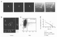
- Result 1 In the human lung, there are mesenchymal stem cell-like cells with alveolar epithelial phenotype. When human lung constituent cells from which hematopoietic cells were removed were cultured on a feeder, spindle-shaped cells proliferated and formed colonies after about 7 days (Fig. 1a). These cells could be passaged and reached confluence after 2-3 weeks. The expression of these cell surface markers was analyzed by flow cytometry (Fig. 1b). Many cells expressed CD73, CD90, and CD105, known as human mesenchymal stem cell markers (12). Expression of CD45, CD34, CD31 and VEGF receptor type 2, which are markers for blood cells and vascular endothelium, was not observed.
- tissue stem cell markers c-kit (13) and CD133 (14) were not observed. From the above, the obtained cell group had a mesenchymal stem cell-like surface marker.
- CD90 also called Thy-1, is a GPI-binding membrane protein, and is known not only as a marker for mesenchymal stem cells but also for hematopoietic stem cells (15) and liver stem cells (16) that express CD34.
- Surfactant protein-C SP-C
- SP-C is known to be specifically expressed in type II alveolar epithelial cells (17). Therefore, in order to examine whether the obtained cell population has an alveolar epithelial phenotype, double staining of SP-C and CD90 was performed by the immunofluorescent antibody method (Fig. 1c).
- SP-C + / CD90 + cells show self-replication in vitro.
- Tissue stem cells are defined as cells that are isolated from the tissue and exhibit self-renewal ability and differentiation ability into more mature cells (18).
- SP-C + / CD90 + cells as candidates for human lung tissue stem cells, and first examined their ability to self-renew. As shown in Fig. 2a, colony formation was observed 10 days after single cells obtained by limiting dilution. Next, a limiting dilution method was performed to determine the frequency of cells having colony-forming ability with respect to the cell group containing many SP-C + / CD90 + cells (Fig. 2b).
- Fig. 2 shows the expression of SP-C and CD90 by flow cytometry on secondary colonies made from wells seeded with 1 cell per well. As shown in 2c, it showed the same expression pattern as the original cell group. From the above, it was shown that SP-C + / CD90 + cells showed self-replication ability in vitro.
- SP-C + / CD90 + cells show differentiation into alveolar epithelial cells in vitro.
- a group of cells rich in SP-C + / CD90 + cells was cultured in Matrigel for 7 days, and each cell in Matrigel and under Matrigel RNA was extracted and the change of mRNA expression was examined by RT-PCR.
- Fig. As shown in 3a, expression of AQP5, a marker for type I cells, was enhanced in Matrigel, and expression of SP-C, a marker for type II cells, was enhanced under Matigel. From the above, induction into type I cells was observed within Matrigel, and type II cells were observed under Matrigel.
- SP-C + / CD90 + cells are present in the alveolar wall.
- frozen sections of human lung tissue were prepared and fluorescent immunostaining was performed.
- Fig 4a SP-C + / CD90 + cells were present in the alveolar wall.
- Observation with a confocal laser microscope revealed that SP-C was stained intracellularly, CD90 was partially observed on the cell surface, and showed the same staining pattern as SP-C + / CD90 + cells isolated from human lung (Fig. .4b).
- lung cells after removal of blood cells were analyzed by flow cytometry. As shown in Fig.
- SP-C + / CD90 + cells were found to contain 0.45% ⁇ 0.34% (mean ⁇ SD) and showed a significant inverse correlation with age (Fig. 4d). From the above examination, it became clear that SP-C + / CD90 + cells were present in the alveolar wall and decreased in number with age.
- human lung tissue has SP-C + / CD90 + cells, and is a human tissue stem cell exhibiting self-renewal ability and differentiation ability into alveolar epithelial cells.
- the number of SP-C + / CD90 + cells decreased with age. Since such stem cells that can differentiate into the alveolar epithelial system are cells involved in tissue repair after lung injury, they are also very important cells in clinical practice such as regenerative medicine. Furthermore, such cells are useful as a material for discovering new markers for identifying human lung tissue stem cells. By analyzing the differentiation signals of human tissue stem cells obtained in the present invention, new drugs can be discovered. It becomes possible to provide.
Abstract
Description
(態様2)上記の調製方法によって得られた、ヒト肺胞上皮細胞に分化することが出来るヒト肺組織幹細胞、又は、該細胞を継代培養して得られるヒト肺組織幹細胞。
(態様3)上記のヒト肺組織幹細胞を培養することから成る、ヒト肺上皮細胞への分化誘導方法。
(態様4)上記の分化誘導方法によって調製されたヒト肺胞上皮細胞、又は、該ヒト肺胞上皮細胞を継代培養して得られるヒト肺胞上皮細胞。
(態様5)上記のヒト肺組織幹細胞又はヒト肺胞上皮細胞を用いることを特徴とする、スクリーニング方法。 (Aspect 1) A type II alveolar epithelial cell marker and a stem cell marker comprising the steps of isolating and extracting constituent cells from human lung tissue and separating and culturing lung tissue stem cells from the obtained isolated cells simultaneously A method for preparing cells to be expressed.
(Aspect 2) Human lung tissue stem cells that can be differentiated into human alveolar epithelial cells obtained by the above preparation method, or human lung tissue stem cells obtained by subculturing the cells.
(Aspect 3) A method for inducing differentiation into human lung epithelial cells, comprising culturing the above human lung tissue stem cells.
(Aspect 4) Human alveolar epithelial cells prepared by the above differentiation induction method, or human alveolar epithelial cells obtained by subculturing the human alveolar epithelial cells.
(Aspect 5) A screening method using the above human lung tissue stem cells or human alveolar epithelial cells.
(a)ヒト肺組織幹細胞又はヒト肺胞上皮細胞に被検物質を接触させる工程、
(b)該細胞における分化誘導を観察又は測定する工程、及び
(c)該分化誘導を促進又は阻害する物質を選択する工程、を含む前記方法。 The screening method of the present invention can be carried out, for example, by the following steps.
(a) contacting a test substance with human lung tissue stem cells or human alveolar epithelial cells;
(b) observing or measuring differentiation induction in the cell, and (c) selecting a substance that promotes or inhibits the differentiation induction.
ヒト肺細胞の分離:ヒト肺細胞は過去の報告(10)に従い、いくらかの改良を加え分離した。本操作はクリーンベンチ内で行い、使用される器具は事前にオートクレーブにて滅菌処理した。提供された肺組織をペニシリン (最終濃度100 単位/ml)、ストレプトマイシン (最終濃度100 μg/ml)、アンホテリシンB (最終濃度0.25 ug/ml、Invitrogen、Carlsbad、CA) を含むリン酸緩衝液 (PBS, 和光純薬、大阪、日本) にて洗浄した。胸膜を鈍的に剥離した後、肺組織を約1 cm x 1 cm x 1 cmに切離し、シリンジと27ゲージの針を用いてDispase II (最終濃度 2.0 U/ml、Roche Applied Science, Mannheim, Germany) 2 mlを注入し、8 mlのDispase II、1 mlのCollagenase/Dispase (最終濃度1 mg/ml、Roche Applied Science)、1 mlのDeoxyribonuclease I (最終濃度0.1 mg/ml、Sigma-Aldrich, St. Luis, MO) をあらかじめ加えた50 mlのコニカルチューブへ移し、37℃で60分間振盪した。酵素処理した肺組織を剪刀にて鋭的に細切した後、18、20ゲージの針を各々3回通し、アンホテリシンB (最終濃度2.5 μg/ml) を添加した基本培地を10 ml加え、100 μmのセルストレイナー (BD Biosciences, San Jose, CA) を通し細胞懸濁液を作成した。基本培地はDulbecco’s Modified Eagle Medium、DMEM (Invitrogen)、10% ウシ胎児血清、FBS (Invitrogen)、1% アミノ酸 (Invitrogen)、ペニシリン (最終濃度100 単位/ml)、ストレプトマイシン (最終濃度 100 μg/ml、Sigma-Aldrich) より構成される。この細胞懸濁液を4℃、1500 rpmで5分遠心分離した後、上清を棄て、赤血球溶血緩衝液 (Roche Applied Science) 3 ml加え、室温で3分間反応させた。アンホテリシンBを添加した基本培地を10 ml加え、4℃、1500 rpmで5分遠心分離し、上清を棄てた後、再度同様の溶血反応を行った。 アンホテリシンB添加基本培地を10 ml加え、4℃、1500 rpmで5分遠心分離し上清を棄て、アンホテリシン添加基本培地10 mlを加え、40 umのセルストレイナー (BD Biosciences) を通し、単一細胞懸濁液を作成した。 [Method]
Isolation of human lung cells : Human lung cells were isolated according to previous reports (10) with some improvements. This operation was performed in a clean bench, and the instruments used were sterilized in advance by an autoclave. The supplied lung tissue is phosphate buffer (PBS) containing penicillin (
結果1:ヒト肺には、肺胞上皮の表現型をもつ間葉系幹細胞様の細胞群が存在する。
血球系細胞を除去したヒト肺構成細胞をフィーダー上で培養すると、紡錘形の細胞が増殖し、約7日後にコロニーを形成した (Fig. 1a)。これらの細胞は継代することができ、2~3週後にはコンフルエントに達した。これらの細胞表面マーカーの発現をフローサイトメトリーにて解析した (Fig.1b)。多くの細胞が、ヒト間葉系幹細胞マーカーとして知られているCD73、CD90、CD105を発現していた(12)。血球および血管内皮のマーカーである、CD45、CD34、CD31及びVEGF receptor type 2の発現は認められなかった。また、組織幹細胞マーカーであるc-kit(13) およびCD133(14) の発現も認めなかった。以上のことから、得られた細胞群は間葉系幹細胞様の表面マーカーを有していた。CD90は、Thy-1とも呼ばれ、GPI結合膜蛋白であり、間葉系幹細胞だけでなく、CD34を発現する造血系幹細胞(15)や肝の幹細胞(16)のマーカーとして知られている。また、surfactant protein-C (SP-C) はII型肺胞上皮細胞に特異的に発現することが知られている(17)。そこで、得られた細胞群が肺胞上皮の表現型を有するかを検討するため、SP-CとCD90の二重染色を免疫蛍光抗体法にて行った (Fig. 1c)。SP-C/CD90両陽性細胞が認められ、SP-C (緑色) は細胞内に顆粒状に染色され、CD90 (赤色) は細胞膜に沿って部分的に染色された。そして、フローサイトメトリーでは、大部分の細胞がSP-C+/CD90+細胞であった (Fig.1d)。以上の検討から、ヒト肺には、II型肺胞上皮細胞のマーカーであるSP-Cと、幹細胞マーカーであるCD90を同時に発現する新規細胞群が存在することが明らかになった。 [result]
Result 1 : In the human lung, there are mesenchymal stem cell-like cells with alveolar epithelial phenotype.
When human lung constituent cells from which hematopoietic cells were removed were cultured on a feeder, spindle-shaped cells proliferated and formed colonies after about 7 days (Fig. 1a). These cells could be passaged and reached confluence after 2-3 weeks. The expression of these cell surface markers was analyzed by flow cytometry (Fig. 1b). Many cells expressed CD73, CD90, and CD105, known as human mesenchymal stem cell markers (12). Expression of CD45, CD34, CD31 and
組織幹細胞は、その組織より分離され、自己複製能とより成熟した細胞への分化能を示す細胞と定義される(18)。我々は、SP-C+/CD90+細胞がヒト肺組織幹細胞の候補であると考え、まず自己複製能について検討した。Fig. 2aに示すように、限界希釈により得られた単一細胞から、10日後、コロニー形成が認められた。次に、SP-C+/CD90+細胞を多く含む細胞群に関し、コロニー形成能を有する細胞の頻度を決定するため、限界希釈法を行った (Fig.2b)。コロニー形成能をもつ細胞は、患者1、2では各々、細胞6、131個に1個の割合で含まれることが明らかになった。1ウエルに1個の細胞を播種したウエルよりできた2次コロニーについてフローサイトメトリーにてSP-C、CD90の発現を解析すると、Fig。 2cに示すように、元の細胞群と同様の発現パターンを示した。以上のことから、SP-C+/CD90+細胞はin vitroで自己複製能を示すことが示された。 Result 2 : SP-C + / CD90 + cells show self-replication in vitro.
Tissue stem cells are defined as cells that are isolated from the tissue and exhibit self-renewal ability and differentiation ability into more mature cells (18). We considered SP-C + / CD90 + cells as candidates for human lung tissue stem cells, and first examined their ability to self-renew. As shown in Fig. 2a, colony formation was observed 10 days after single cells obtained by limiting dilution. Next, a limiting dilution method was performed to determine the frequency of cells having colony-forming ability with respect to the cell group containing many SP-C + / CD90 + cells (Fig. 2b). It was revealed that cells having colony forming ability were contained in
SP-C+/CD90+細胞の肺胞上皮細胞への分化能を検討するため、SP-C+/CD90+細胞を多く含む細胞群をMatrigelにて7日間培養し、Matrigel内、Matrigel下の各々の細胞からRNAを抽出しRT-PCRにてmRNA発現の変化について検討した。Fig。3aに示すように、Matrigel内ではI型細胞のマーカーであるAQP5の発現が増強し、Matigel下ではII型細胞のマーカーであるSP-Cの発現が増強した。以上のことから、Matrigel内ではI型細胞へ、Matrigel下ではII型細胞への誘導が認められた。 Result 3 : SP-C + / CD90 + cells show differentiation into alveolar epithelial cells in vitro.
In order to examine the differentiation potential of SP-C + / CD90 + cells into alveolar epithelial cells, a group of cells rich in SP-C + / CD90 + cells was cultured in Matrigel for 7 days, and each cell in Matrigel and under Matrigel RNA was extracted and the change of mRNA expression was examined by RT-PCR. Fig. As shown in 3a, expression of AQP5, a marker for type I cells, was enhanced in Matrigel, and expression of SP-C, a marker for type II cells, was enhanced under Matigel. From the above, induction into type I cells was observed within Matrigel, and type II cells were observed under Matrigel.
SP-C+/CD90+細胞の局在を決定するため、ヒト肺組織の凍結切片を作成し蛍光免疫染色を行った。Fig 4aに示すように、SP-C+/CD90+細胞は肺胞壁に存在した。共焦点レーザー顕微鏡による観察では、SP-Cは細胞内に染色され、CD90は細胞表面に部分的に認められ、ヒト肺より分離したSP-C+/CD90+細胞と同一の染色パターンを示した (Fig.4b)。SP-C+/CD90+細胞の割合を検討するため、血球系細胞を除去した後の肺細胞をフローサイトメトリーで解析した。Fig.4cに示すように、SP-C+/CD90+細胞は、0.45% ± 0.34% (mean ± S.D.) 含まれ、年齢と有意な逆相関を示すことが分かった (Fig. 4d)。以上の検討より、SP-C+/CD90+細胞は、肺胞壁に存在し、年齢とともに数の減少を認めることが明らかになった。 Result 4 : SP-C + / CD90 + cells are present in the alveolar wall.
In order to determine the localization of SP-C + / CD90 + cells, frozen sections of human lung tissue were prepared and fluorescent immunostaining was performed. As shown in Fig 4a, SP-C + / CD90 + cells were present in the alveolar wall. Observation with a confocal laser microscope revealed that SP-C was stained intracellularly, CD90 was partially observed on the cell surface, and showed the same staining pattern as SP-C + / CD90 + cells isolated from human lung (Fig. .4b). In order to examine the ratio of SP-C + / CD90 + cells, lung cells after removal of blood cells were analyzed by flow cytometry. As shown in Fig. 4c, SP-C + / CD90 + cells were found to contain 0.45% ± 0.34% (mean ± SD) and showed a significant inverse correlation with age (Fig. 4d). From the above examination, it became clear that SP-C + / CD90 + cells were present in the alveolar wall and decreased in number with age.
1. Adamson, I.Y., and Bowden, D.H. 1974. The type 2 cell as progenitor of alveolar epithelial regeneration. A cytodynamic study in mice after exposure to oxygen. Lab Invest 30:35-42.
2. Evans, M.J., Cabral, L.J., Stephens, R.J., and Freeman, G. 1975. Transformation of alveolar type 2 cells to type 1 cells following exposure to NO2. Exp Mol Pathol 22:142-150.
3. Aso, Y., Yoneda, K., and Kikkawa, Y. 1976. Morphologic and biochemical study of pulmonary changes induced by bleomycin in mice. Lab Invest 35:558-568.
4. Anderson, W.R., and Thielen, K. 1992. Correlative study of adult respiratory distress syndrome by light, scanning, and transmission electron microscopy. Ultrastruct Pathol 16:615-628.
5. Ware, L.B., and Matthay, M.A. 2000. The acute respiratory distress syndrome. N Engl J Med 342:1334-1349.
6. Kawanami, O., Ferrans, V.J., and Crystal, R.G. 1982. Structure of alveolar epithelial cells in patients with fibrotic lung disorders. Lab Invest 46:39-53.
7. Kim, C.F., Jackson, E.L., Woolfenden, A.E., Lawrence, S., Babar, I., Vogel, S., Crowley, D., Bronson, R.T., and Jacks, T. 2005. Identification of bronchioalveolar stem cells in normal lung and lung cancer. Cell 121:823-835.
8. Kubo, H., Hegab, A.E., He, M., Ishizawa, K., and Yamada, M. 2008. Endogenous lung stem cells increased after lung injury. Proc Am Thorac Soc 5:362-363.
9. Hegab, A.E., Kubo, H., Yamaya, M., Asada, M., He, M., Fujino, N., Mizuno, S., and Nakamura, T. 2008. Intranasal HGF administration ameliorates the physiologic and morphologic changes in lung emphysema. Mol Ther 16:1417-1426.
10. Bortnick, A.E., Favari, E., Tao, J.Q., Francone, O.L., Reilly, M., Zhang, Y., Rothblat, G.H., and Bates, S.R. 2003. Identification and characterization of rodent ABCA1 in isolated type II pneumocytes. Am J Physiol Lung Cell Mol Physiol 285:L869-878.
11. Tropepe, V., Coles, B.L., Chiasson, B.J., Horsford, D.J., Elia, A.J., McInnes, R.R., and van der Kooy, D. 2000. Retinal stem cells in the adult mammalian eye. Science 287:2032-2036.
12. Dominici, M., Le Blanc, K., Mueller, I., Slaper-Cortenbach, I., Marini, F., Krause, D., Deans, R., Keating, A., Prockop, D., and Horwitz, E. 2006. Minimal criteria for defining multipotent mesenchymal stromal cells. The International Society for Cellular Therapy position statement. Cytotherapy 8:315-317.
13. Bearzi, C., Rota, M., Hosoda, T., Tillmanns, J., Nascimbene, A., De Angelis, A., Yasuzawa-Amano, S., Trofimova, I., Siggins, R.W., Lecapitaine, N., et al. 2007. Human cardiac stem cells. Proc Natl Acad Sci U S A 104:14068-14073.
14. Uchida, N., Buck, D.W., He, D., Reitsma, M.J., Masek, M., Phan, T.V., Tsukamoto, A.S., Gage, F.H., and Weissman, I.L. 2000. Direct isolation of human central nervous system stem cells. Proc Natl Acad Sci U S A 97:14720-14725.
15. Craig, W., Kay, R., Cutler, R.L., and Lansdorp, P.M. 1993. Expression of Thy-1 on human hematopoietic progenitor cells. J Exp Med 177:1331-1342.
16. Herrera, M.B., Bruno, S., Buttiglieri, S., Tetta, C., Gatti, S., Deregibus, M.C., Bussolati, B., and Camussi, G. 2006. Isolation and characterization of a stem cell population from adult human liver. Stem Cells 24:2840-2850.
17. Phelps, D.S., and Floros, J. 1991. Localization of pulmonary surfactant proteins using immunohistochemistry and tissue in situ hybridization. Exp Lung Res 17:985-995.
18. Weiss, D.J., Kolls, J.K., Ortiz, L.A., Panoskaltsis-Mortari, A., and Prockop, D.J. 2008. Stem cells and cell therapies in lung biology and lung diseases. Proc Am Thorac Soc 5:637-667.
[References]
1. Adamson, IY, and Bowden, DH 1974.The
2. Evans, MJ, Cabral, LJ, Stephens, RJ, and Freeman, G. 1975.Transformation of
3. Aso, Y., Yoneda, K., and Kikkawa, Y. 1976. Morphologic and biochemical study of pulmonary changes induced by bleomycin in mice.Lab Invest 35: 558-568.
4. Anderson, WR, and Thielen, K. 1992. Correlative study of adult respiratory distress syndrome by light, scanning, and transmission electron microscopy. Ultrastruct Pathol 16: 615-628.
5. Ware, LB, and Matthay, MA 2000. The acute respiratory distress syndrome. N Engl J Med 342: 1334-1349.
6. Kawanami, O., Ferrans, VJ, and Crystal, RG 1982.Structure of alveolar epithelial cells in patients with fibrotic lung disorders. Lab Invest 46: 39-53.
7. Kim, CF, Jackson, EL, Woolfenden, AE, Lawrence, S., Babar, I., Vogel, S., Crowley, D., Bronson, RT, and Jacks, T. 2005. Identification of bronchioalveolar stem cells in normal lung and lung cancer.Cell 121: 823-835.
8. Kubo, H., Hegab, AE, He, M., Ishizawa, K., and Yamada, M. 2008. Endogenous lung stem cells increased after lung injury. Proc Am Thorac Soc 5: 362-363.
9. Hegab, AE, Kubo, H., Yamaya, M., Asada, M., He, M., Fujino, N., Mizuno, S., and Nakamura, T. 2008. Intranasal HGF administration ameliorates the physiologic and morphologic changes in lung emphysema. Mol Ther 16: 1417-1426.
10. Bortnick, AE, Favari, E., Tao, JQ, Francone, OL, Reilly, M., Zhang, Y., Rothblat, GH, and Bates, SR 2003. Identification and characterization of rodent ABCA1 in isolated type II pneumocytes Am J Physiol Lung Cell Mol Physiol 285: L869-878.
11. Tropepe, V., Coles, BL, Chiasson, BJ, Horsford, DJ, Elia, AJ, McInnes, RR, and van der Kooy, D. 2000. Retinal stem cells in the adult mammalian eye. Science 287: 2032- 2036.
12. Dominici, M., Le Blanc, K., Mueller, I., Slaper-Cortenbach, I., Marini, F., Krause, D., Deans, R., Keating, A., Prockop, D., and Horwitz, E. 2006. Minimal criteria for defining multipotent mesenchymal stromal cells.The International Society for Cellular Therapy position statement.Cytotherapy 8: 315-317.
13. Bearzi, C., Rota, M., Hosoda, T., Tillmanns, J., Nascimbene, A., De Angelis, A., Yasuzawa-Amano, S., Trofimova, I., Siggins, RW, Lecapitaine , N., et al. 2007. Human cardiac stem cells. Proc Natl Acad Sci USA 104: 14068-14073.
14. Uchida, N., Buck, DW, He, D., Reitsma, MJ, Masek, M., Phan, TV, Tsukamoto, AS, Gage, FH, and Weissman, IL 2000.Direct isolation of human central nervous system stem cells.Proc Natl Acad Sci USA 97: 14720-14725.
15. Craig, W., Kay, R., Cutler, RL, and Lansdorp, PM 1993. Expression of Thy-1 on human hematopoietic progenitor cells. J Exp Med 177: 1331-1342.
16. Herrera, MB, Bruno, S., Buttiglieri, S., Tetta, C., Gatti, S., Deregibus, MC, Bussolati, B., and Camussi, G. 2006. Isolation and characterization of a stem cell population from adult human liver.Stem Cells 24: 2840-2850.
17. Phelps, DS, and Floros, J. 1991.Localization of pulmonary surfactant proteins using immunohistochemistry and tissue in situ hybridization.Exp Lung Res 17: 985-995.
18. Weiss, DJ, Kolls, JK, Ortiz, LA, Panoskaltsis-Mortari, A., and Prockop, DJ 2008. Stem cells and cell therapies in lung biology and lung diseases.Proc Am Thorac Soc 5: 637-667.
Claims (11)
- ヒトの肺組織より構成細胞を単離抽出する工程、及び、得られた単離細胞より肺組織幹細胞を分離培養する工程を含む、II型肺胞上皮細胞マーカー及び幹細胞マーカーを同時に発現する細胞の調製方法。 A method comprising: isolating and extracting constituent cells from human lung tissue; and isolating and culturing lung tissue stem cells from the obtained isolated cells. Preparation method.
- 該細胞がSP-C+/CD90+細胞である、請求項1記載の調製方法。 The preparation method according to claim 1, wherein the cells are SP-C + / CD90 + cells.
- 該細胞がヒト肺胞上皮細胞に分化することが出来るヒト肺組織幹細胞であることを特徴とする、請求項1又は2記載の調製方法。 The preparation method according to claim 1 or 2, wherein the cells are human lung tissue stem cells capable of differentiating into human alveolar epithelial cells.
- 単離抽出する工程において、ヒトの肺組織から胸膜を剥離除去した後、肺組織内へ中性プロテアーゼ溶液を注入し、その後、コラゲナーゼ、中性プロテアーゼ及びデオキシリボヌクレアーゼを含む溶液中で細胞を溶解することを含む、請求項1ないし3のいずれか一項に記載の調製方法。 In the step of isolation and extraction, after removing the pleura from human lung tissue, a neutral protease solution is injected into the lung tissue, and then the cells are lysed in a solution containing collagenase, neutral protease and deoxyribonuclease. The preparation method according to any one of claims 1 to 3, comprising:
- ヒト肺組織幹細胞を分離培養する工程において、マウス胎児線維芽細胞から成るフィーダー細胞を用いて培養することを含む、請求項1~4のいずれか一項に記載の調製方法。 The preparation method according to any one of claims 1 to 4, wherein the step of separating and culturing human lung tissue stem cells comprises culturing using feeder cells comprising mouse fetal fibroblasts.
- 請求項1~5のいずれか一項に記載の調製方法によって得られた、ヒト肺胞上皮細胞に分化することが出来るヒト肺組織幹細胞、又は、該細胞を継代培養して得られるヒト肺組織幹細胞。 A human lung tissue stem cell that can be differentiated into human alveolar epithelial cells obtained by the preparation method according to any one of claims 1 to 5, or a human lung obtained by subculturing the cells Tissue stem cells.
- 請求項6記載のヒト肺組織幹細胞を培養することから成る、ヒト肺上皮細胞への分化誘導方法。 A method for inducing differentiation into human lung epithelial cells, comprising culturing the human lung tissue stem cells according to claim 6.
- 再構成基底膜組成物を用いる培養によって、I型肺胞上皮細胞が再構成基底膜組成物内部に、II型肺胞上皮細胞が再構成基底膜組成物下部から得られることを特徴とする、請求項7記載の分化誘導方法。 By culturing with the reconstituted basement membrane composition, type I alveolar epithelial cells are obtained inside the reconstituted basement membrane composition, and type II alveolar epithelial cells are obtained from the bottom of the reconstituted basement membrane composition, The differentiation induction method according to claim 7.
- 再構成基底膜組成物がマウス肉腫から抽出した可溶性基底膜調製品である、請求項7又は8記載の分化誘導方法。 The differentiation-inducing method according to claim 7 or 8, wherein the reconstituted basement membrane composition is a soluble basement membrane preparation extracted from mouse sarcoma.
- 請求項7~9のいずれか一項に記載の分化誘導方法によって調製されたヒト肺胞上皮細胞、又は、該ヒト肺胞上皮細胞を継代培養して得られるヒト肺胞上皮細胞。 A human alveolar epithelial cell prepared by the differentiation induction method according to any one of claims 7 to 9, or a human alveolar epithelial cell obtained by subculturing the human alveolar epithelial cell.
- 請求項5記載のヒト肺組織幹細胞又は請求項9記載のヒト肺胞上皮細胞を用いることを特徴とする、スクリーニング方法。 A screening method comprising using the human lung tissue stem cell according to claim 5 or the human alveolar epithelial cell according to claim 9.
Priority Applications (3)
| Application Number | Priority Date | Filing Date | Title |
|---|---|---|---|
| US13/264,694 US20120094304A1 (en) | 2009-04-17 | 2010-04-09 | Method of preparing human lung tissue stem cells and method of inducing differentiation into human alveolar epithelial cells |
| EP10764403.1A EP2420566A4 (en) | 2009-04-17 | 2010-04-09 | Method of preparing human lung tissue stem cells and method of inducing differentiation into human alveolar epithelial cells |
| JP2011509275A JPWO2010119819A1 (en) | 2009-04-17 | 2010-04-09 | Method for preparing human lung tissue stem cells and method for inducing differentiation into human alveolar epithelial cells |
Applications Claiming Priority (2)
| Application Number | Priority Date | Filing Date | Title |
|---|---|---|---|
| JP2009100548 | 2009-04-17 | ||
| JP2009-100548 | 2009-04-17 |
Publications (1)
| Publication Number | Publication Date |
|---|---|
| WO2010119819A1 true WO2010119819A1 (en) | 2010-10-21 |
Family
ID=42982481
Family Applications (1)
| Application Number | Title | Priority Date | Filing Date |
|---|---|---|---|
| PCT/JP2010/056447 WO2010119819A1 (en) | 2009-04-17 | 2010-04-09 | Method of preparing human lung tissue stem cells and method of inducing differentiation into human alveolar epithelial cells |
Country Status (4)
| Country | Link |
|---|---|
| US (1) | US20120094304A1 (en) |
| EP (1) | EP2420566A4 (en) |
| JP (1) | JPWO2010119819A1 (en) |
| WO (1) | WO2010119819A1 (en) |
Cited By (4)
| Publication number | Priority date | Publication date | Assignee | Title |
|---|---|---|---|---|
| JP2016513469A (en) * | 2013-03-15 | 2016-05-16 | ザ ジャクソン ラボラトリー | Isolation and use of non-embryonic stem cells |
| JPWO2018194124A1 (en) * | 2017-04-20 | 2020-02-27 | 学校法人慶應義塾 | Reagent for differentiation of somatic cells into alveolar epithelial cells and use thereof |
| JP7319027B2 (en) | 2014-11-27 | 2023-08-01 | コーニンクレッカ ネザーランド アカデミー ヴァン ウェテンシャッペン | culture medium |
| US11725184B2 (en) | 2014-05-16 | 2023-08-15 | Koninklijke Nederlandse Akademie Van Wetenschappen | Culture method for organoids |
Families Citing this family (2)
| Publication number | Priority date | Publication date | Assignee | Title |
|---|---|---|---|---|
| CN110167349A (en) * | 2016-11-02 | 2019-08-23 | Aal科学有限公司 | Non- mesenchyma people Lung stem cells and the method that they are used to treat respiratory disorder |
| CN112608879B (en) * | 2021-01-12 | 2022-08-12 | 北京大学 | Method for obtaining lung epithelial cells by differentiation of embryonic stem cells and culture medium used by method |
Citations (3)
| Publication number | Priority date | Publication date | Assignee | Title |
|---|---|---|---|---|
| JP4183742B1 (en) | 2005-12-13 | 2008-11-19 | 国立大学法人京都大学 | Method for producing induced pluripotent stem cells |
| JP2008307007A (en) | 2007-06-15 | 2008-12-25 | Bayer Schering Pharma Ag | Human pluripotent stem cell induced from human tissue-originated undifferentiated stem cell after birth |
| JP2009514509A (en) * | 2005-10-17 | 2009-04-09 | アカデミア シニカ | Lung stem cells and related methods and kits |
Family Cites Families (3)
| Publication number | Priority date | Publication date | Assignee | Title |
|---|---|---|---|---|
| US8609412B2 (en) * | 1999-08-05 | 2013-12-17 | Regents Of The University Of Minnesota | Mapc generation of lung tissue |
| GB0218332D0 (en) * | 2002-08-07 | 2002-09-18 | Imp College Innovations Ltd | Preparation of type pneumocytes |
| US8343481B2 (en) * | 2007-02-21 | 2013-01-01 | Board Of Regents Of The University Of Texas System | Method of preparing lung alveolar epithelial type II cells derived from embryonic stem cells |
-
2010
- 2010-04-09 EP EP10764403.1A patent/EP2420566A4/en not_active Withdrawn
- 2010-04-09 US US13/264,694 patent/US20120094304A1/en not_active Abandoned
- 2010-04-09 WO PCT/JP2010/056447 patent/WO2010119819A1/en active Application Filing
- 2010-04-09 JP JP2011509275A patent/JPWO2010119819A1/en active Pending
Patent Citations (3)
| Publication number | Priority date | Publication date | Assignee | Title |
|---|---|---|---|---|
| JP2009514509A (en) * | 2005-10-17 | 2009-04-09 | アカデミア シニカ | Lung stem cells and related methods and kits |
| JP4183742B1 (en) | 2005-12-13 | 2008-11-19 | 国立大学法人京都大学 | Method for producing induced pluripotent stem cells |
| JP2008307007A (en) | 2007-06-15 | 2008-12-25 | Bayer Schering Pharma Ag | Human pluripotent stem cell induced from human tissue-originated undifferentiated stem cell after birth |
Non-Patent Citations (32)
| Title |
|---|
| ADAMSON, I.Y; BOWDEN, D.H.: "The type 2 cell as progenitor of alveolar epithelial regeneration. A cytodynamic study in mice after exposure to oxygen", LAB INVEST, vol. 30, 1974, pages 35 - 42 |
| ANDERSON, W.R.; THIELEN, K.: "Correlative study of adult respiratory distress syndrome by light, scanning, and transmission electron microscopy", ULTRASTRUCT PATHOL, vol. 16, 1992, pages 615 - 628 |
| ASO, Y; YONEDA, K.; KIKKAWA, Y: "Morphologic and biochemical study of pulmonary changes induced by bleomycin in mice", LAB INVEST, vol. 35, 1976, pages 558 - 568 |
| BEARZI, C.; ROTA, M.; HOSODA, T.; TILLMANNS, J.; NASCIMBENE, A.; DE ANGELIS, A.; YASUZAWA-AMANO, S.; TROFIMOVA, I.; SIGGINS, R.W.;: "Human cardiac stem cells", PROC NATL ACAD SCI U S A, vol. 104, 2007, pages 14068 - 14073 |
| BENDER KIM C.F. ET AL.: "Identification of bronchioalveolar stem cells in normal lung and lung cancer.", CELL, vol. 121, no. 6, 2005, pages 823 - 835, XP008131911 * |
| BORTNICK A.E. ET AL.: "Identification and characterization of rodent ABCA1 in isolated type II pneumocytes.", AM J PHYSIOL LUNG CELL MOL PHYSIOL., vol. 285, no. 4, 2003, pages L869 - L878, XP055089989 * |
| BORTNICK, A.E.; FAVARI, E.; TAO, J.Q.; FRANCONE, O.L.; REILLY, M.; ZHANG, Y.; ROTHBLAT, G.H.; BATES, S.R.: "Identification and characterization of rodent ABCA1 in isolated type II pneumocytes", AM J PHYSIOL LUNG CELL MOL PHYSIOL, vol. 285, 2003, pages L869 - 878 |
| CRAIG, W.; KAY, R.; CUTLER, R.L.; LANSDORP, P.M.: "Expression of Thy-1 on human hematopoietic progenitor cells", J EXP MED, vol. 177, 1993, pages 1331 - 1342 |
| DOBBS L.G. ET AL.: "An improved method for isolating type II cells in high yield and purity.", AM REV RESPIR DIS., vol. 134, no. 1, 1986, pages 141 - 145, XP008025659 * |
| DOMINICI, M.; LE BLANC, K.; MUELLER, I.; SLAPER-CORTENBACH, I.; MARINI, F.; KRAUSE, D.; DEANS, R.; KEATING, A.; PROCKOP, D.; HORWI: "Minimal criteria for defining multipotent mesenchymal stromal cells", THE INTERNATIONAL SOCIETY FOR CELLULAR THERAPY POSITION STATEMENT. CYTOTHERAPY, vol. 8, 2006, pages 315 - 317 |
| EVANS, M.J.; CABRAL, L.J.; STEPHENS, R.J.; FREEMAN, G.: "Transformation of alveolar type 2 cells to type 1 cells following exposure to N02", EXP MOL PATHOL, vol. 22, 1975, pages 142 - 150 |
| EVANS, M.J.; CABRAL, L.J.; STEPHENS, R.J.; FREEMAN, G.: "Transformation of alveolar type 2 cells to type 1 cells following exposure to N02.", EXP MOL PATHOL, vol. 22, 1975, pages 142 - 150 |
| HEGAB, A.E.; KUBO, H.; YAMAYA, M.; ASADA, M.; HE, M.; FUJINO, N.; MIZUNO, S.; NAKAMURA, T.: "Intranasal HGF administration ameliorates the physiologic and morphologic changes in lung emphysema", MOL THER, vol. 16, 2008, pages 1417 - 1426 |
| HERRERA, M.B.; BRUNO, S.; BUTTIGLIERI, S.; TETTA, C.; GATTI, S.; DEREGIBUS, M.C.; BUSSOLATI, B.; CAMUSSI, G.: "Isolation and characterization of a stem cell population from adult human liver", STEM CELLS, vol. 24, 2006, pages 2840 - 2850 |
| KAWANAMI, 0.; FERRANS, V.J.; CRYSTAL, R.G.: "Structure of alveolar epithelial cells in patients with fibrotic lung disorders", LAB INVEST, vol. 46, 1982, pages 39 - 53 |
| KAWANAMI, O.; FERRANS, V.J.; CRYSTAL, RG.: "Structure of alveolar epithelial cells in patients with fibrotic lung disorders", LAB INVEST, vol. 46, 1982, pages 39 - 53 |
| KAZUHIRO SUGAHARA ET AL.: "Effector Saibo kara Mita Kokyuki Shikkan no Byotai Haiho Johi Saibo -Kino to Byohen Shufuku", LUNG PERSPECT, vol. 17, no. 2, 10 April 2009 (2009-04-10), pages 162 - 165 * |
| KIM, C.F.; JACKSON, E.L.; WOOLFENDEN, A.E.; LAWRENCE, S.; BABAR, I.; VOGEL, S.; CROWLEY, D.; BRONSON, R.T.; JACKS, T.: "Identification of bronchioalveolar stem cells in normal lung and lung cancer", CELL, vol. 121, 2005, pages 823 - 835 |
| KUBO, H.; HEGAB, A.E.; HE, M.; ISHIZAWA, K.; YAMADA, M.: "Endogenous lung stem cells increased after lung injury", PROC AM THORAC SOC, vol. 5, 2008, pages 362 - 363 |
| KUBO: "Hai no Saisei to Kansaibo", KOKYU TO JUNKAN, vol. 56, no. 3, 2008, pages 285 - 291, XP009174573 * |
| LAMA V.N. ET AL.: "Evidence for tissue-resident mesenchymal stem cells in human adult lung from studies of transplanted allografts.", J CLIN INVEST., vol. 117, no. 4, 2007, pages 989 - 996, XP055089981 * |
| LAMA, V.N.; SMITH, L.; BADRI, L.; FLINT, A.; ANDREI, A.; MURRAY, S.; WANG, Z.; LIAO, H.; TOEWS, G.B.; KREBSBACH, P.H.: "Evidence for tissue-resident mesenchymal stem cells in human adult lung from studies of transplanted allografts", J CLIN INVEST, vol. 117, 2007, pages 989 - 996 |
| NAOYA FUJINO ET AL.: "Haiho Johi Saibo Marker no Hatsugen o Shimesu Hito Hai Soshiki Kan'yokei Saibo no Tanri", REGENERATIVE MEDICINE, vol. 8, 2009, pages 187, XP001526450 * |
| NAOYA FUJINO ET AL.: "Hito Haiho Johi II-gata Zenku Saibo no Bunri", REGENERATIVE MEDICINE, vol. 9, 5 February 2010 (2010-02-05), pages 253 * |
| PHELPS, D.S.; FLOROS, J.: "Localization of pulmonary surfactant proteins using immunohistochemistry and tissue in situ hybridization", EXP LUNG RES, vol. 17, 1991, pages 985 - 995 |
| See also references of EP2420566A4 * |
| SUMMER R ET AL.: "Isolation of an adult mouse lung mesenchymal progenitor cell population.", AM J RESPIR CELL MOL BIOL., vol. 37, no. 2, 2007, pages 152 - 159, XP055089991 * |
| TROPEPE, V.; COLES, B.L.; CHIASSON, B.J.; HORSFORD, D.J.; ELIA, A.J.; MCINNES, R.R.; VAN DER KOOY, D.: "Retinal stem cells in the adult mammalian eye", SCIENCE, vol. 287, 2000, pages 2032 - 2036 |
| UCHIDA, N.; BUCK, D.W.; HE, D.; REITSMA, M.J.; MASEK, M.; PHAN, T.V.; TSUKAMOTO, A.S.; GAGE, F.H.; WEISSMAN, I.L.: "Direct isolation of human central nervous system stem cells", PROC NATL ACAD SCI U S A, vol. 97, 2000, pages 14720 - 14725 |
| WARE, L.B.; MATTHAY, M.A.: "The acute respiratory distress syndrome", N ENGL J MED, vol. 342, 2000, pages 1334 - 1349 |
| WEISS, D.J.; KOLLS, J.K.; ORTIZ, L.A.; PANOSKALTSIS-MORTARI, A.; PROCKOP, D.J.: "Stem cells and cell therapies in lung biology and lung diseases", PROC AM THORAC SOC, vol. 5, 2008, pages 637 - 667 |
| YUJI KUBO: "Shikkan no Byoin to Byotai 7. Hai Sonsho to Shufuku no Mechanism", ANNUAL REVIEW KOKYUKI, 2008, pages 113 - 119 * |
Cited By (7)
| Publication number | Priority date | Publication date | Assignee | Title |
|---|---|---|---|---|
| JP2016513469A (en) * | 2013-03-15 | 2016-05-16 | ザ ジャクソン ラボラトリー | Isolation and use of non-embryonic stem cells |
| JP2020018321A (en) * | 2013-03-15 | 2020-02-06 | ザ ジャクソン ラボラトリーThe Jackson Laboratory | Isolation of non embryonic stem cells and uses thereof |
| US11725184B2 (en) | 2014-05-16 | 2023-08-15 | Koninklijke Nederlandse Akademie Van Wetenschappen | Culture method for organoids |
| JP7319027B2 (en) | 2014-11-27 | 2023-08-01 | コーニンクレッカ ネザーランド アカデミー ヴァン ウェテンシャッペン | culture medium |
| JPWO2018194124A1 (en) * | 2017-04-20 | 2020-02-27 | 学校法人慶應義塾 | Reagent for differentiation of somatic cells into alveolar epithelial cells and use thereof |
| US11718831B2 (en) | 2017-04-20 | 2023-08-08 | Keio University | Reagent for differentiating somatic cells into alveolar epithelial cells, and use of said reagent |
| JP7356626B2 (en) | 2017-04-20 | 2023-10-05 | 国立大学法人東海国立大学機構 | Reagents for differentiating somatic cells into alveolar epithelial cells and their use |
Also Published As
| Publication number | Publication date |
|---|---|
| EP2420566A4 (en) | 2014-01-15 |
| JPWO2010119819A1 (en) | 2012-10-22 |
| EP2420566A1 (en) | 2012-02-22 |
| US20120094304A1 (en) | 2012-04-19 |
Similar Documents
| Publication | Publication Date | Title |
|---|---|---|
| AU2009343787B2 (en) | Isolation of human umbilical cord blood-derived mesenchymal stem cells | |
| Raynaud et al. | Comprehensive characterization of mesenchymal stem cells from human placenta and fetal membrane and their response to osteoactivin stimulation | |
| Letouzey et al. | Isolation and characterisation of mesenchymal stem/stromal cells in the ovine endometrium | |
| US9546353B2 (en) | Optimized and defined method for isolation and preservation of precursor cells from human umbilical cord | |
| Xu et al. | Promising new potential for mesenchymal stem cells derived from human umbilical cord Wharton's jelly: sweat gland cell‐like differentiative capacity | |
| WO2010119819A1 (en) | Method of preparing human lung tissue stem cells and method of inducing differentiation into human alveolar epithelial cells | |
| US20150175970A1 (en) | Cells for therapy of the heart, method of obtaining a cell preparation, and cell preparation | |
| van Riet et al. | Organoid-based expansion of patient-derived primary alveolar type 2 cells for establishment of alveolus epithelial Lung-Chip cultures | |
| Zia et al. | Routine clonal expansion of mesenchymal stem cells derived from amniotic fluid for perinatal applications | |
| Li et al. | Isolation and identification of epithelial and stromal stem cells from eutopic endometrium of women with endometriosis | |
| Toyoda et al. | Multilineage-differentiating stress-enduring (Muse)-like cells exist in synovial tissue | |
| Wu et al. | Umbilical cord blood-derived non-hematopoietic stem cells retrieved and expanded on bone marrow-derived extracellular matrix display pluripotent characteristics | |
| Gao et al. | Clonal isolation of endothelial colony-forming cells from early gestation chorionic villi of human placenta for fetal tissue regeneration | |
| CN111492051A (en) | Mesenchymal stromal cells and method for obtaining mesenchymal stromal cells from umbilical cord | |
| Calenic et al. | Characterization of oral keratinocyte stem cells and prospects of its differentiation to oral epithelial equivalents | |
| US9700585B2 (en) | Multipotent prenatal stem cells | |
| Mazza et al. | Marker profile for the evaluation of human umbilical artery smooth muscle cell quality obtained by different isolation and culture methods | |
| Rygiel et al. | T cell‐mediated biliary epithelial‐to‐mesenchymal transition in liver allograft rejection | |
| WO2013118786A1 (en) | Immortalized cell lines of capillary vessel-forming cells in peripheral tissue | |
| Dorazehi et al. | Potential use of amniotic membrane-derived scaffold for cerebrospinal fluid applications | |
| Iachininoto et al. | In vitro cardiomyocyte differentiation of umbilical cord blood cells: crucial role for c-kit+ cells | |
| JP2022522973A (en) | Human umbilical cord mesenchymal stem cell sheet and its manufacturing method | |
| KR20060110542A (en) | Stem cells derived from normal breast tissue and breast cancer tissue, preparation method thereof and differentiated cells from the stem cell | |
| Hayashi et al. | A basic study on self-reconstitution of alveolar epithelium-like cells by tissue stem cells in mouse lung | |
| Kazemnejad et al. | Role of Wnt signaling on proliferation of menstrual blood derived stem cells |
Legal Events
| Date | Code | Title | Description |
|---|---|---|---|
| 121 | Ep: the epo has been informed by wipo that ep was designated in this application |
Ref document number: 10764403 Country of ref document: EP Kind code of ref document: A1 |
|
| DPE1 | Request for preliminary examination filed after expiration of 19th month from priority date (pct application filed from 20040101) | ||
| ENP | Entry into the national phase |
Ref document number: 2011509275 Country of ref document: JP Kind code of ref document: A |
|
| NENP | Non-entry into the national phase |
Ref country code: DE |
|
| WWE | Wipo information: entry into national phase |
Ref document number: 2010764403 Country of ref document: EP |
|
| WWE | Wipo information: entry into national phase |
Ref document number: 13264694 Country of ref document: US |