WO2010107939A2 - Human immunodeficiency virus (hiv) -neutralizing antibodies - Google Patents
Human immunodeficiency virus (hiv) -neutralizing antibodies Download PDFInfo
- Publication number
- WO2010107939A2 WO2010107939A2 PCT/US2010/027695 US2010027695W WO2010107939A2 WO 2010107939 A2 WO2010107939 A2 WO 2010107939A2 US 2010027695 W US2010027695 W US 2010027695W WO 2010107939 A2 WO2010107939 A2 WO 2010107939A2
- Authority
- WO
- WIPO (PCT)
- Prior art keywords
- mgrm
- seq
- antibody
- chronic
- hiv
- Prior art date
- Legal status (The legal status is an assumption and is not a legal conclusion. Google has not performed a legal analysis and makes no representation as to the accuracy of the status listed.)
- Ceased
Links
Classifications
-
- C—CHEMISTRY; METALLURGY
- C07—ORGANIC CHEMISTRY
- C07K—PEPTIDES
- C07K16/00—Immunoglobulins [IG], e.g. monoclonal or polyclonal antibodies
- C07K16/08—Immunoglobulins [IG], e.g. monoclonal or polyclonal antibodies against material from viruses
- C07K16/10—RNA viruses
- C07K16/112—Retroviridae (F), e.g. leukemia viruses
- C07K16/114—Lentivirus (G), e.g. human immunodeficiency virus [HIV], feline immunodeficiency virus [FIV] or simian immunodeficiency virus [SIV]
-
- A—HUMAN NECESSITIES
- A61—MEDICAL OR VETERINARY SCIENCE; HYGIENE
- A61K—PREPARATIONS FOR MEDICAL, DENTAL OR TOILETRY PURPOSES
- A61K39/00—Medicinal preparations containing antigens or antibodies
- A61K39/12—Viral antigens
- A61K39/21—Retroviridae, e.g. equine infectious anemia virus
-
- A—HUMAN NECESSITIES
- A61—MEDICAL OR VETERINARY SCIENCE; HYGIENE
- A61P—SPECIFIC THERAPEUTIC ACTIVITY OF CHEMICAL COMPOUNDS OR MEDICINAL PREPARATIONS
- A61P31/00—Antiinfectives, i.e. antibiotics, antiseptics, chemotherapeutics
- A61P31/12—Antivirals
- A61P31/14—Antivirals for RNA viruses
-
- C—CHEMISTRY; METALLURGY
- C07—ORGANIC CHEMISTRY
- C07K—PEPTIDES
- C07K16/00—Immunoglobulins [IG], e.g. monoclonal or polyclonal antibodies
- C07K16/08—Immunoglobulins [IG], e.g. monoclonal or polyclonal antibodies against material from viruses
- C07K16/10—RNA viruses
- C07K16/112—Retroviridae (F), e.g. leukemia viruses
- C07K16/114—Lentivirus (G), e.g. human immunodeficiency virus [HIV], feline immunodeficiency virus [FIV] or simian immunodeficiency virus [SIV]
- C07K16/1145—Env proteins, e.g. gp41, gp110/120, gp160, V3, principal neutralising domain [PND] or CD4-binding site
-
- A—HUMAN NECESSITIES
- A61—MEDICAL OR VETERINARY SCIENCE; HYGIENE
- A61K—PREPARATIONS FOR MEDICAL, DENTAL OR TOILETRY PURPOSES
- A61K39/00—Medicinal preparations containing antigens or antibodies
- A61K2039/505—Medicinal preparations containing antigens or antibodies comprising antibodies
-
- C—CHEMISTRY; METALLURGY
- C07—ORGANIC CHEMISTRY
- C07K—PEPTIDES
- C07K2317/00—Immunoglobulins specific features
- C07K2317/30—Immunoglobulins specific features characterized by aspects of specificity or valency
- C07K2317/33—Crossreactivity, e.g. for species or epitope, or lack of said crossreactivity
-
- C—CHEMISTRY; METALLURGY
- C07—ORGANIC CHEMISTRY
- C07K—PEPTIDES
- C07K2317/00—Immunoglobulins specific features
- C07K2317/50—Immunoglobulins specific features characterized by immunoglobulin fragments
- C07K2317/56—Immunoglobulins specific features characterized by immunoglobulin fragments variable (Fv) region, i.e. VH and/or VL
- C07K2317/565—Complementarity determining region [CDR]
-
- C—CHEMISTRY; METALLURGY
- C07—ORGANIC CHEMISTRY
- C07K—PEPTIDES
- C07K2317/00—Immunoglobulins specific features
- C07K2317/70—Immunoglobulins specific features characterized by effect upon binding to a cell or to an antigen
- C07K2317/76—Antagonist effect on antigen, e.g. neutralization or inhibition of binding
Definitions
- the present invention relates generally to therapy, diagnosis and monitoring of human immunodeficiency virus (HIV) infection.
- the invention is more specifically related to human neutralizing monoclonal antibodies specific for HIV-I, such as broad and potent neutralizing monoclonal antibodies specific for HIV-I and their manufacture and use. Broad neutralization suggests that the antibodies can neutralize HIV-I isolates from different individuals.
- Such antibodies are useful in pharmaceutical compositions for the prevention and treatment of HTV, and for the diagnosis and monitoring of HTV infection and for design of HIV vaccine immunogens.
- HIV human immunodeficiency virus
- opportunistic infections are caused by microbes such as viruses or bacteria that usually do not make healthy people sick.
- HTV is spread most often through unprotected sex with an infected partner. HIV also is spread through contact with infected blood.
- the human immunodeficiency virus is the cause of acquired immune deficiency syndrome (AIDS) (Barre-Sinoussi, F., et al., 1983, Science 220:868-870; Gallo, R., et al., 1984, Science 224:500-503).
- AIDS acquired immune deficiency syndrome
- the epidemic is growing most rapidly among minority populations and is a leading killer of African- American males ages 25 to 44. According, AIDS affects nearly seven times more African Americans and three times more Hispanics than whites. In recent years, an increasing number of African- American women and children are being affected by HIV/ AIDS.
- the present invention provides a novel method for isolating potent, broadly neutralizing monoclonal antibodies against HIV.
- Peripheral Blood Mononuclear Cells PBMCs
- PBMCs Peripheral Blood Mononuclear Cells
- the B cell culture supernatants are then screened by a primary neutralization assay in a high throughput format, and B cell cultures exhibiting neutralizing activity are selected for rescue of monoclonal antibodies. It is surprisingly observed that neutralizing antibodies obtained by this method do not always exhibit gpl20 or gp41 binding at levels that correlate with neutralization activity.
- the method of the invention therefore allows identification of novel antibodies with cross- clade neutralization properties.
- the present invention provides human monoclonal antibodies specifically directed against HIV.
- the invention provides human anti-HTV monoclonal antibodies and sister clones thereof.
- an exemplary sister clone of the 1443 C16 (PG16) antibody is the 1503 H05 (PG16) antibody, the 1456 A12 (PG16) antibody, the 1469 M23 (PG 16) antibody, the 1489 113 (PG 16) antibody, or the 1480J08 (PG 16) antibody.
- the invention provides an isolated anti-HIV antibody, wherein said antibody has a heavy chain with three CDRs including an amino acid sequence selected from the group consisting of the amino acid sequences of SGFTFHKYGMH (SEQ ID NO: 88), LISDDGMRKYHSDSMW (SEQ ID NO: 89), and
- EAGGPIWHDDVKYYDFNDGYYNYHYMDV (SEQ ID NO: 6), and a light chain with three CDRs that include an amino acid sequence selected from the group consisting of the amino acid sequences of NGTSSDVGGFDSVS (SEQ ID NO: 97), DVSHRPSG (SEQ ID NO: 95), and SSLTDRSHRI (SEQ ID NO: 41).
- the invention provides an isolated anti-HIV antibody, wherein said antibody has a heavy chain with three CDRs including an amino acid sequence selected from the group consisting of the amino acid sequences of SGFTFHKYGMH (SEQ ID NO: 88), LISDDGMRKYHSDSMW (SEQ ID NO: 89), and
- EAGGPIWHDDVKYYDFNDGYYNYHYMDV (SEQ ID NO: 6), and a light chain with three CDRs that include an amino acid sequence selected from the group consisting of the amino acid sequences of NGTRSDVGGFDSVS (SEQ ID NO: 92), DVSHRPSG (SEQ ID NO: 95), and SSLTDRSHRI (SEQ ID NO: 41).
- the invention provides an isolated anti-HTV antibody, wherein said antibody has a heavy chain with three CDRs including an amino acid sequence selected from the group consisting of the amino acid sequences of SGFTFHKYGMH (SEQ ID NO: 88), LISDDGMRKYHSDSMW (SEQ ID NO: 89), and
- EAGGPIWHDDVKYYDFNDGYYNYHYMDV (SEQ ID NO: 6), and a light chain with three CDRs that include an amino acid sequence selected from the group consisting of the amino acid sequences of NGTSRDVGGFDSVS (SEQ ID NO: 93), DVSHRPSG (SEQ ID NO: 95), and SSLTDRSHRI (SEQ ID NO: 41).
- the invention provides an isolated anti-HTV antibody, wherein said antibody has a heavy chain with three CDRs including an amino acid sequence selected from the group consisting of the amino acid sequences of SGFTFHKYGMH (SEQ ID NO: 88), LISDDGMRKYHSNSMW (SEQ ID NO: 98), and
- EAGGPIWHDDVKYYDFNDGYYNYHYMDV (SEQ ID NO: 6), and a light chain with three CDRs that include an amino acid sequence selected from the group consisting of the amino acid sequences of NGTSSDVGGFDSVS (SEQ ID NO: 97), DVSHRPSG (SEQ ID NO: 95), and SSLTDRSHRI (SEQ ID NO: 41).
- the invention provides an isolated anti-HTV antibody, wherein said antibody has a heavy chain with three CDRs including an amino acid sequence selected from the group consisting of the amino acid sequences of SGGTFSSYAFT (SEQ ID NO: 104), MVTPIFGEAKYSQRFE (SEQ ID NO: 105), and RAVPIATDNWLDP (SEQ ID NO: 102), and a light chain with three CDRs that include an amino acid sequence selected from the group consisting of the amino acid sequences of RASQTINNYLN (SEQ ID NO: 107), GASNLQNG (SEQ ID NO: 108), and QQSFSTPRT (SEQ ID NO: 42).
- the invention provides an isolated anti-HIV antibody, wherein said antibody has a heavy chain with three CDRs including an amino acid sequence selected from the group consisting of the amino acid sequences of SGGTFSSYAFT (SEQ ID NO: 104), MVTPIFGEAKYSQRFE (SEQ ID NO: 105), and RRA VPI ATDN WLDP (SEQ ID NO: 103), and a light chain with three CDRs that include an amino acid sequence selected from the group consisting of the amino acid sequences of RASQTINNYLN (SEQ ID NO: 107), GASNLQNG (SEQ ID NO: 108), and QQSFSTPRT (SEQ ID NO: 42).
- the invention provides an isolated anti-HIV antibody, wherein said antibody has a heavy chain with three CDRs including an amino acid sequence selected from the group consisting of the amino acid sequences of SGGAFSSYAFS (SEQ ID NO: 110), MITPVFGETKYAPRFQ (SEQ ID NO: 11 1), and RAVPIATDNWLDP (SEQ ID NO: 102), and a light chain with three CDRs that include an amino acid sequence selected from the group consisting of the amino acid sequences of RASQTIHTYL (SEQ ID NO: 113), GASTLQSG (SEQ ID NO: 1 14), and QQSYSTPRT (SEQ ID NO: 43).
- the invention provides an isolated anti-HIV antibody, wherein said antibody has a heavy chain with three CDRs including an amino acid sequence selected from the group consisting of the amino acid sequences of SGGAFSSYAFS (SEQ ID NO: 110), MITPVFGETKYAPRFQ (SEQ ID NO: 11 1), and RRAVPIATDNWLDP (SEQ ID NO: 103), and a light chain with three CDRs that include an amino acid sequence selected from the group consisting of the amino acid sequences of RASQTTHTYL (SEQ ID NO: 113), GASTLQSG (SEQ ID NO: 1 14), and QQSYSTPRT (SEQ ID NO: 43).
- the invention provides an isolated anti-HIV antibody, wherein said antibody has a heavy chain with three CDRs including an amino acid sequence selected from the group consisting of the amino acid sequences of SGYSFID YYLH (SEQ ID NO: 1 16), LIDPENGEARYAEKFQ (SEQ ID NO: 117), AVGADSGSWFDP (SEQ ID NO: 1 18), and a light chain with three CDRs that include an amino acid sequence selected from the group consisting of the amino acid sequences of SGSKLGDKYVS (SEQ ID NO: 120), ENDRRPSG (SEQ ID NO: 121), QAWETTTTTFVF (SEQ ID NO: 44).
- the invention provides an isolated anti-HIV antibody, wherein said antibody has a heavy chain with three CDRs including an amino acid sequence selected from the group consisting of the amino acid sequences of SGFDFSRQGMH (SEQ ID NO: 123), FIKYDGSEKYHADSVW (SEQ ID NO: 124), and
- EAGGPDYRNGYNYYDFYDGYYNYHYMDV (SEQ ID NO: 7), and a light chain with three CDRs that include an amino acid sequence selected from the group consisting of the amino acid sequences of NGTSND VGGYES VS (SEQ ID NO: 126), DVSKRPSG (SEQ ID NO: 127), and KSLTSTRRRV (SEQ ID NO: 45).
- the invention provides an isolated anti-HIV antibody, wherein said antibody has a heavy chain with three CDRs including an amino acid sequence selected from the group consisting of the amino acid sequences of SGFTFHKYGMH (SEQ ID NO: 88), LISDDGMRKYHSDSMW (SEQ ID NO: 89),
- EAGGPIWHDDVKYYDFNDGYYNYHYMDV (SEQ ID NO: 6), SGGTFSSYAFT (SEQ ID NO: 104), MVTPIFGEAKYSQRFE (SEQ ID NO: 105), RAVPIATDNWLDP (SEQ ID NO: 102), SGGAFSSYAFS (SEQ ID NO: 110), MITPVFGETKY APRFQ (SEQ ID NO: 111), SGYSFIDYYLH (SEQ ID NO: 116), LIDPENGEARY AEKFQ (SEQ ID NO: 117), AVGADSGSWFDP (SEQ ID NO: 118), SGFDFSRQGMH (SEQ ID NO: 123), FIKYDGSEKYHADSVW (SEQ ID NO: 124), EAGGPDYRNGYNYYDFYDGYYNYHYMDV (SEQ ID NO: 7),
- LISDDGMRKYHSNSMW (SEQ ID NO: 98), wherein said antibody binds to and neutralizes HIV-I.
- the invention provides an isolated anti-HIV antibody, wherein said antibody has a light chain with three CDRs that include an amino acid sequence selected from the group consisting of the amino acid sequences of NGTSSDVGGFDSVS (SEQ ID NO: 97), DVSHRPSG (SEQ ID NO: 95), SSLTDRSHRI (SEQ ID NO: 41), RASQTTNNYLN (SEQ ID NO: 107), GASNLQNG (SEQ ID NO: 108), QQSFSTPRT (SEQ ID NO: 42), RASQ ⁇ HTYL (SEQ ID NO: 113), GASTLQSG (SEQ ID NO: 114), QQSYSTPRT (SEQ ID NO: 43), SGSKLGDKYVS (SEQ ID NO: 120), ENDRRPSG (SEQ ID NO: 121), QAWETTTTTFVF (SEQ ID NO: 44), NGTSNDVGGYESVS (SEQ ID NO: 126), DVSKRPSG (SEQ ID NO: 127), KS
- the invention provides an isolated anti-HIV antibody, wherein said antibody has a heavy chain with three CDRs including an amino acid sequence selected from the group consisting of the amino acid sequences of SGFTFHKYGMH (SEQ ID NO: 88), LISDDGMRKYHSDSMW (SEQ ID NO: 89),
- EAGGPIWHDDVKYYDFNDGYYNYHYMDV (SEQ ID NO: 6), SGGTFSSYAFT (SEQ ID NO: 104), MVTPIFGEAKYSQRFE (SEQ ID NO: 105), RRAVPIATDNWLDP (SEQ ID NO: 103), SGGAFSSYAFS (SEQ ID NO: 110), MITPVFGETKYAPRFQ (SEQ ID NO: 1 11), SGYSFIDYYLH (SEQ ID NO: 116), LIDPENGEARYAEKFQ (SEQ ID NO: 1 17), ' AVGADSGSWFDP (SEQ ID NO: 118), SGFDFSRQGMH (SEQ ID NO: 123), FIKYDGSEKYHADSVW (SEQ ID NO: 124), EAGGPDYRNGYNYYDFYDGYYNYHYMDV (SEQ ID NO: 7),
- LISDDGMRKYHSNSMW (SEQ ID NO: 98), wherein said antibody binds to and neutralizes HIV-I.
- the invention provides an isolated anti-HIV antibody, wherein said antibody has a light chain with three CDRs that include an amino acid sequence selected from the group consisting of the amino acid sequences of NGTSSDVGGFDSVS (SEQ ID NO: 97), DVSHRPSG (SEQ ID NO: 95), SSLTDRSHRI (SEQ ID NO: 41), RASQTINNYLN (SEQ ID NO: 107), GASNLQNG (SEQ ID NO: 108), QQSFSTPRT (SEQ ID NO: 42), RASQTIHTYL (SEQ ID NO: 113), GASTLQSG (SEQ ID NO: 114), QQSYSTPRT (SEQ ID NO: 43), SGSKLGDKYVS (SEQ ID NO: 120), ENDRRPSG (SEQ ID NO: 121), QAWETTTTTFVF (SEQ ID NO: 44), NGTSND VGGYESVS (SEQ ID NO: 126), DVSKRPSG (SEQ ID NO: 127), KS
- the invention provides an isolated anti-HIV antibody or fragment thereof, wherein said antibody includes: (a) a V H CDRl region comprising the amino acid sequence of SEQ ID NO: 88, 104, 110, 1 16, or 123; (b) a V H CDR2 region comprising the amino acid sequence of SEQ ID NO: 98, 89, 91, 105, 1 11, 117, or 124; and (c) a V H CDR3 region comprising the amino acid sequence of SEQ ID NO: 6, 102, 103, 118, or 7, wherein said antibody binds to and neutralizes HIV-I.
- this antibody further includes: (a) a V L CDRl region comprising the amino acid sequence of SEQ ID NO: 93, 92, 97, 94, 107, 113, 120, or 126; (b) a V L CDR2 region comprising the amino acid sequence of SEQ ID NO: 95, 108, 114, 121, or 127; and (c) a V L CDR3 region comprising the amino acid sequence of SEQ ID NO: 41, 42, 43, 44, or 45.
- the invention provides an isolated fully human monoclonal anti-HTV antibody including: a) a heavy chain sequence comprising the amino acid sequence of SEQ ID NO: 31 and a light chain sequence comprising amino acid sequence SEQ ID NO: 32, or b) a heavy chain sequence comprising the amino acid sequence of SEQ ID NO: 33 and a light chain sequence comprising amino acid sequence SEQ ID NO: 34, or c) a heavy chain sequence comprising the amino acid sequence of SEQ ID NO: 35 and a light chain sequence comprising amino acid sequence SEQ ID NO: 36, or d) a heavy chain sequence comprising the amino acid sequence of SEQ ID NO: 37 and a light chain sequence comprising amino acid sequence SEQ ID NO: 38, or e) a heavy chain sequence comprising the amino acid sequence of SEQ ID NO: 39 and a light chain sequence comprising amino acid sequence SEQ ID NO: 40, or f) a heavy chain sequence comprising the amino acid sequence of SEQ ID NO: 140 and a light chain sequence comprising amino acid sequence SEQ ID NO
- the invention provides a composition including any one of the isolated anti-HIV antibodies described herein.
- an anti-HIV human monoclonal antibody of the invention is isolated from a B-cell from an HIV- 1 -infected human donor.
- the antibody is effective in neutralizing a plurality of different clades of HIV.
- the antibody is effective in neutralizing a plurality of different strain within the same clade of HTV- 1.
- the neutralizing antibody binds to the HIV envelope proteins gpl20, or gp41 or envelope protein on HIV-I pseudovirions or expressed on transfected or infected cell surfaces.
- the neutralizing antibody does not bind to recombinant or monomeric envelope proteins gpl20, or gp41 or envelope protein on HTV-I pseudovirions or expressed on transfected or infected cell surfaces but binds to natural trimeric forms of the HIV-I Env proteins.
- the present invention provides human monoclonal antibodies wherein the antibodies are potent, broadly neutralizing antibody (bNAb).
- a broadly neutralizing antibody is defined as a bNAb that neutralizes HIV-I species belonging to two or more different clades.
- the different clades are selected from the group consisting of clades A, B, C, D, E, AE, AG, G or F.
- the HIV-I strains from two or more clades comprise virus from non-B clades.
- a broadly neutralizing antibody is defined as a bNAb that neutralizes at least 60% of the HIV-I strains listed in Tables 18A-18F. In some embodiments, at least 70%, or at least 80%, or at least 90% of the HIV-I strains listed in Tables 18A-18F are neutralized.
- a potent, broadly neutralizing antibody is defined as a bNAb that displays a potency of neutralization of at least a plurality of HIV-I species with an IC50 value of less than 0.2 ⁇ g/mL. In some embodiments the potency of neutralization of the HIV- 1 species has an IC50 value of less than 0.15 ⁇ g/mL, or less than 0.10 ⁇ g/mL, or less than 0.05 ⁇ g/mL.
- a potent, broadly neutralizing antibody is also defined as a bNAb that displays a potency of neutralization of at least a plurality of HIV-I species with an IC90 value of less than 2.0 ⁇ g/mL.
- the potency of neutralization of the HIV-I species has an IC90 value of less than 1.0 ⁇ g/mL, or less than 0.5 ⁇ g/mL.
- Exemplary monoclonal antibodies that neutralize HTV-I include 1496 C09 (PG9), 1443_C16 (PG16), 1456_P20 (PG20), 1460_G14 (PGG14), and 1495_C14 (PGC14) described herein.
- the monoclonal antibody is an antibody that binds to the same epitope as 1496_C09 (PG9), 1443_C16 (PG 16), 1456_P20 (PG20), 1460_G14 (PGG 14), and 1495_C14 (PGC 14).
- monoclonal antibodies PG9 and PG 16 are broad and potent neutralizing antibodies. The antibodies are respectively referred to herein as HIV antibodies.
- the invention provides a number of isolated human monoclonal antibodies, wherein each said monoclonal antibody binds to HIV-I infected or transfected cells; and binds to HIV-I virus.
- a neutralizing antibody having potency in neutralizing HIV-I, or a fragment thereof is provided.
- a neutralizing antibody of the invention exhibits higher neutralization index and/or a higher affinity for binding to the envelope proteins gpl20, or gp41 than anti-HTV mAbs known in the art, such as the mAb bl2. (Burton DR et al., Science Vol. 266. no. 5187, pp. 1024 - 1027).
- Exemplary monoclonal antibodies 1496_C09 (PG9), 1443_C16 (PG16), 1456_P20 (PG20), 1460_G14 (PGG14), and 1495_C14 (PGC 14) exhibit binding to the envelope glycoprotein gpl20, but not gp41, in an ELISA assay, however gpl20 binding does not always correlate with neutralization activity against specific strains of HIV-I .
- monoclonal antibodies for example 1443_C16 (PG 16) and 1496_C09 (PG9), display none or weak gpl20 binding activity against a particular strain but bind to HIV-I trimer on transfected or infected cell surface and/or virion and exhibit broad and potent neutralization activity against that strain of HIV-I .
- the antibody is a monoclonal antibody comprising one or more polypeptides selected from the group consisting of 1496_C09 (PG9), 1443_C16 (PG 16), 1456_P20 (PG20), 1460_G14 (PGG14), and 1495_C14 (PGC14); comprising a heavy chain selected from the group consisting of the heavy chain of 1496_C09 (PG9), 1443_C16
- 1495_C14 comprising a light chain selected from the group consisting of the light chain of 1496_C09 (PG9), 1443_C16 (PG16), 1456_P20 (PG20), 1460_G14 (PGG14), and
- 1495_C14 comprising a light chain comprising a CDR selected from the group consisting of the CDRs of the light chain of 1496_C09 (PG9), 1443_C16 (PG16), 1456_P20
- the invention relates to an antibody or a fragment thereof, such as Fab, Fab 1 , F(ab')2 and Fv fragments that binds to an epitope or immunogenic polypeptide capable of binding to an antibody selected from 1496_C09 (PG9), 1443_C16 (PG 16), 1456_P20 (PG20),
- the invention also relates to immunogenic polypeptides encoding such epitopes.
- nucleic acid molecules encoding such antibodies, and vectors and cells carrying such nucleic acids are also provided.
- the invention relates to a pharmaceutical composition
- a pharmaceutical composition comprising at least one antibody or fragment as recited herein, together with a pharmaceutically acceptable carrier.
- the invention relates to a method of immunizing, preventing or inhibiting HIV infection or an HIV-related disease comprising the steps of identifying a patient in need of such treatment and administering to said patient a therapeutically effective amount of at least one monoclonal antibody as recited herein.
- HIV antibodies according to the invention are linked to a therapeutic agent or a detectable label.
- the invention provides methods for stimulating an immune response, treating, preventing or alleviating a symptom of an HIV viral infection by administering an
- the invention provides methods of administering the HIV antibody of the invention to a subject prior to, and/or after exposure to an HIV virus.
- the HIV antibody of the invention provides methods of administering the HIV antibody of the invention to a subject prior to, and/or after exposure to an HIV virus.
- HIV antibody of the invention is used to treat or prevent HIV infection.
- the HIV antibody is administered at a dose sufficient to promote viral clearance or eliminate HIV infected cells.
- Also included in the invention is a method for determining the presence of an HIV virus infection in a patient, by contacting a biological sample obtained from the patient with an HIV antibody; detecting an amount of the antibody that binds to the biological sample; and comparing the amount of antibody that binds to the biological sample to a control value.
- the invention further provides a diagnostic kit comprising an HTV monoclonal antibody.
- the invention relates to a broadly neutralizing antibody (bNAb) wherein the antibody neutralizes at least one member of each clade with a potency greater than that of the bNAbs bl2, 2G12, 2F5 and 4E10 respectively-
- the invention relates to a broadly neutralizing antibody (bNAb) wherein the antibody does not bind monomeric gpl20 or gp41 proteins of the HIV-I env gene.
- the antibody binds with higher affinity to trimeric forms of the HIV-I Env expressed on a cell surface than to the monomeric gpl20 or artificially trimerized gpl40.
- the antibody binds with high affinity to uncleaved HIV-I gpl60 trimers on a cell surface.
- the invention relates to a broadly neutralizing antibody (bNAb) wherein the antibody binds an epitope within the variable loop of gpl20, wherein the epitope comprises the conserved regions of V2 and V3 loops of gpl20, wherein the epitope comprises N- glycosylation site at residue Asn-160 within the V2 loop of gpl20, wherein the antibody binds an epitope presented by a trimeric spike of gpl20 on a cell surface, wherein the epitope is not presented when gpl20 is artificially trimerized.
- the antibody does not neutralize the HIV-I in the absence of N-glycosylation site at residue Asn-160 within the V2 loop of gpl20.
- the invention relates to a broadly neutralizing antibody (bNAb) selected from the group consisting of PG 16 and PG9.
- bNAb broadly neutralizing antibody
- the invention relates to an antigen or an immunogenic polypeptide, or a vaccine comprising such antigen or immunogenic polypeptide, for producing a broadly neutralizing antibody (bNAb) by an immune response, the antigen comprising an epitope within the variable loop of gpl20 according to the invention.
- bNAb broadly neutralizing antibody
- the invention relates to method for passive or active immunization of an individual against a plurality of HIV-I species across one or more clades, the method comprising: providing a broadly neutralizing antibody (bNAb) wherein the bNAb neutralizes HIV-I species belonging to two or more clades, and further wherein the potency of neutralization of at least one member of each clade is determined by an IC50 value of less than 0.005 ⁇ g/mL.
- bNAb broadly neutralizing antibody
- the antibody is selected from the group consisting of PG9 and PG 16.
- the antibody is produced by active immunization with an antigen comprising an epitope within the variable loop of gpl20, wherein the epitope comprises the conserved regions of V2 and V3 loops of gpl20 or, wherein the epitope comprises an N-glycosylation site at residue Asn-160 within the V2 loop of gpl20.
- the epitope is presented by a trimeric spike of gpl20 on a cell surface, and the epitope is not presented when gpl20 is monomeric or artficially trimerized. .
- Figure 1 A is a schematic tree diagram of Clustal W-aligned variable region sequences of heavy chains of the monoclonal antibodies.
- Figure IB is a schematic tree diagram of Clustal W-aligned variable region sequences of light chains of the monoclonal antibodies.
- Figure 2 is a flow chart of the process for isolation of monoclonal antibodies according to the invention.
- Figure 3A is a schematic diagram that summarizes the screening results for neutralization and HTV-env protein (gpl20 and gp41) binding assays from which B cell cultures were selected for antibody rescue and the monoclonal antibodies 1496 C09 (PG9),
- Figure 3B is a schematic diagram that summaries the neutralizing activity and HIV- env protein (gpl20 and gp41) binding activities of the monoclonal antibodies 1496_C09
- PG9 1443_C16 (PG 16), 1456_P20 (PG20), 1460_G14 (PGG 14), and 1495_C14 (PGC 14) as determined by ELISA assays among the B cell supernatants using a neutralization index cut-off value of 2.0.
- the neutralization index was expressed as the ratio of normalized relative luminescence units (RLU) of SIVmac239 to that of test viral strain derived from the same test B cell culture supernatant.
- RLU normalized relative luminescence units
- the cut-off values used to distinguish neutralizing hits were determined by the neutralization index of a large number of negative control wells containing B cell culture supernatants derived from healthy donors.
- Figure 4 is a series of graphs depicting the neutralization activity of monoclonal antibodies 1443_C16 (PG 16) and 1496_C09 (PG9) to additional pseudoviruses not included in Tables 17A and 17B.
- Figure 5 is a graph depicting the dose response curves of 1456 P20 (PG20),
- Figure 6 is a series of graphs depicting the results from ELISA binding assays of monoclonal antibodies 1443_C16 (PG 16) and 1496_C09 (PG9) to HIV-I YU2 gpl40, JR-
- CSFgpl20 membrane-proximal external regions (MPER) peptide of gp41 and V3 polypeptide.
- MPER membrane-proximal external regions
- Figure 7 is a graph depicting the results of a binding assay using monoclonal antibodies 1443_C16 (PG 16) and 1496_C09 (PG9) to HIV-I YU2 gpl60 expressed on the cell surface in the presence and absence of soluble CD4 (sCD4).
- Figure 8 is a graph depicting the results of a binding assay using monoclonal antibodies 1443_C16 (PG16) and 1496_C09 (PG9) to HIV-I gpl60 transfected cells.
- Figure 9 is a series of graphs depicting the results of a capture assay. The data describe capturing of entry-competent JRCSF pseudovirus by neutralizing monoclonal antibodies 1443_C16 (PG 16) and 1496_C09 (PG9) in a dose-dependent manner.
- Figure 1OA is a graph depicting the results of a competitive binding assay using monoclonal antibodies sCD4, PG 16 and PG9, wherein the claimed antibodies compete for the binding of monoclonal antibody 1443 C16 (PG16) to pseudovirus but control antibodies bl2,
- 2G12, 2F5 and 4E10 do not competitively bind to the pseudovirus.
- Figure 1OB is a graph depicting the results of a competitive binding assay using monoclonal antibodies sCD4, PG 16 and PG9, wherein the claimed antibodies compete for the binding of monoclonal antibody 1496 C09 (PG9) to pseudovirus but control antibodies bl2,
- 2G12, 2F5 and 4E10 do not competitively bind to the pseudovirus.
- FIG. 1 is a series of graphs depicting the results of a binding assay using PG9 and PG16. The data show that PG9 and PG16 bind to monomeric gpl20 and artificially trimerized gpl40 constructs as determined by ELISA. IgG bl2 was used as a control for
- Figure 1 IB is a series of graphs depicting the results of a binding assay using PG9 and
- FIG. 12 is a series of graphs depicting the results of a binding assay using PG9 and
- PG16 and cleavage-defective HIV-1YU2 trimers PG9 and PG16 bind with high 'affinity to cleavage-defective HIV- 1YU2 trimers as determined by flow cytometry. Binding curves were generated by plotting the MFI of antigen binding as a function of antibody concentration.
- Figure 13A-E is a series of graphs depicting the mapping the PG9 and PG 16 epitopes.
- Competitor antibody is indicated at the top of each graph. 2Gl 2 is included to control for cell surface Env expression.
- Figure 14 is a series of graphs depicting the results of competition ELISA assays using the monoclonal antibody PG9.
- Figure 15 is a graph depicting monoclonal antibody binding, PG9 or PG 16, to HTV-
- Figure 16 is a graph depicting monoclonal antibody PG9 binding to deglycosylated gpl20.
- Figure 17 is a series of graphs depicting the neutralization activity of PG9 and PG 16 against HTV- I SF 162 and HIV- ISF 162 K 160N, which was determined using a single-round replication luciferase reporter assay of pseudotyped virus.
- Figure 18 is a series of graphs depicting the binding of PG9 and PG 16 to mixed trimers.
- Alanine substitutions at positions 160 and 299 were introduced into HIV-I YU2 Env to abolish binding of PG9 and PG 16.
- An alanine substitution at position 295 was also introduced into the same construct to abrogate binding of 2G12.
- Co-transfection of 293T cells with WT and mutant plasmids in a 1 :2 ratio resulted in the expression of 29% mutant homotrimers, 44% heterotrimers with two mutant subunits, 23% heterotrimers with one mutant subunit, and 4% wild-type homotrimers.
- Figure 19 is a series of graphical depictions of the number of nucleotide or amino acid differences in the heavy chain sequences of sister clones of 1443 Cl 6 (PGl 6) among each other. Note that the single nucleotide difference of 1408 108 translates into an identical protein sequence of 1443 C16. The nucleotide sequence of the 1408 108 light chain is identical to the nucleotide sequence of the light chain of 1443 C16.
- Figure 2OA is a tree diagram illustrating the correlation of the heavy chain of 1443
- Figure 2OB is a tree diagram illustrating the correlation of the light chain of 1443 Cl 6 sister clones to the light chain of 1496 C09 at the nucleotide level.
- Figure 21 A is a tree diagram illustrating the correlation of the heavy chain of 1443
- Figure 21B is a tree diagram illustrating the correlation of the light chain of 1443 C16 sister clones to the light chain of 1496 C09 at the protein level.
- HIV-I human immunodeficiency virus type 1
- AIDS acquired immunodeficiency syndrome
- B-cells are used as fusion partners for the generation of human monoclonal anti-HIV antibodies.
- One major drawback to finding a vaccine composition suitable for more reliable prevention of human individuals from HIV-I infection and/or for more successful therapeutic treatment of infected patients is the ability of the HIV-I virus to escape antibody capture by genetic variation, which very often renders the remarkable efforts of the researchers almost useless.
- HIV-I is among the most genetically diverse viral pathogens. Of the three main branches of the HIV-I phylogenetic tree, the M (main), N (new), and O (outlier) groups, group M viruses are the most widespread, accounting for over 99% of global infections.
- Env is the most variable HIV-I gene, with up to 35% sequence diversity between clades, 20% sequence diversity within clades, and up to 10% sequence diversity in a single infected person (Shankarappa, R. et al. 1999. J. Virol. 73:10489-10502).
- Clade B is dominant in Europe, the Americas, and Australia.
- Clade C is common in southern Africa, China, and India and presently infects more people worldwide than any other clade (McCutchan, FE. 2000. Understanding the genetic diversity of HIV-I. AIDS 14(Suppl. 3):S31-S44).
- HIV human immunodeficiency virus
- CD4 cellular receptors
- CCR5 co-receptor
- CXCR4 co-receptor
- Most neutralizing antibodies bind to functional regions involved in receptor interactions and cell membrane fusion.
- the vast majority of neutralizing antibodies isolated to date do not recognize more than one clade, therefore exhibiting limited protective efficacy in vitro or in vivo.
- mAbs monoclonal antibodies
- a well-known characteristic of the HIV-I envelope glycoprotein is its extreme variability. It has been recognized that even relatively conserved epitopes on HIV-I, such as the CD4 binding site, show some variability between different isolates (Poignard, P., et al., Ann. Rev. Immunol. (2001) 19:253-274).
- the Fab bl2 was screened by panning for envelope glycoprotein gpl20 binding activity and neutralizing activity against the HIV-I (HXBc2) isolate was observed.
- HXBc2 HIV-I
- the monoclonal antibody 2F5 which had been shown to bind a sequence within the external domain of the gp41 envelope glycoprotein of HTV-I was found to have broad neutralization properties. (Conley AJ Proc. Natl. Acad. Sci. USA Vol. 91, pp. 3348-3352 (1994); Muster T et al., J. Virol. 67(1 1):6642-6647 (1993); Buchacher A et al., 1992, Vaccines 92:191-195).
- the monoclonal antibody 4E10 which binds to a novel epitope C terminal of the ELDKWA sequence in gp41 recognized by 2F5, has also been found to have potent cross-clade neutralization activity.
- bNAbs broadly neutralizing antibodies
- the present invention provides a novel method for isolating novel broad and potent neutralizing monoclonal antibodies against HIV.
- the method involves selection of a PBMC donor with high neutralization titer of antibodies in the plasma. B cells are screened for neutralization activity prior to rescue of antibodies. Novel broadly neutralizing antibodies are obtained by emphasizing neutralization as the initial screen.
- the invention relates to potent, broadly neutralizing antibody (bNAb) wherein the antibody neutralizes HIV-I species belonging to two or more clades, and further wherein the potency of neutralization of at least one member of each clade is determined by an IC50 value of less than 0.2 ⁇ g/mL.
- the clades are selected from Clade A, Clade B, Clade C, Clade D and Clade AE.
- the HIV-I belonging two or more clades are non-Clade B viruses.
- the broadly neutralizing antibody neutralizes at least 60% of the HIV-I strains listed in Tables 18A-18F.
- the invention relates to potent, broadly neutralizing antibody (bNAb) wherein the antibody neutralizes HTV-I species with a potency of neutralization of at least a plurality of HTV-I species with an IC50 value of less than 0.2 ⁇ g/mL.
- the potency of neutralization of the HIV-I species has an IC50 value of less than 0.15 ⁇ g/mL, or less than 0.10 ⁇ g/mL, or less than 0.05 ⁇ g/mL.
- a potent, broadly neutralizing antibody is defined as a bNAb that displays a potency of neutralization of at least a plurality of HIV-I species with an IC90 value of less than 2.0 ⁇ g/mL.
- the potency of neutralization of the HIV-I species has an IC90 value of less than 1.0 ⁇ g/mL, or less than 0.5 ⁇ g/mL.
- PBMCs Peripheral Blood Mononuclear Cells
- Memory B cells were isolated and B cell culture supernatants were subjected to a primary screen of neutralization assay in a high throughput format.
- HIV antigen binding assays using ELISA or like methods were also used as a screen.
- B cell lysates corresponding to supernatants exhibiting neutralizing activity were selected for rescue of monoclonal antibodies by standard recombinant methods.
- the recombinant rescue of the monoclonal antibodies involves use of a B cell culture system as described in Weitcamp J-H et al., J. Immunol. 171 :4680- 4688 (2003).
- Any other method for rescue of single B cells clones known in the art also may be employed such as EBV immortalization of B cells (Traggiai E., et al., Nat. Med. 10(8):871-875 (2004)), electrofusion (Buchacher, A., et al., 1994. AIDS Res. Hum. Retroviruses 10:359-369), and B cell hybridoma (Karpas A. et al., Proc. Natl. Acad. Sci.
- monoclonal antibodies were rescued from the B cell cultures using variable chain gene-specific RT-PCR, and transfectant with combinations of H and L chain clones were screened again for neutralization and HIV antigen binding activities. mAbs with neutralization properties were selected for further characterization.
- a novel high-throughput strategy was used to screen IgG-containing culture screening supernatants from approximately 30,000 activated memory B cells from a clade A infected donor for recombinant, monomeric gpl20JR-CSF and gp41HxB2 (Env) binding as well as neutralization activity against HTV- IJR-CSF and HTV- ISF 162 (See Table 1).
- Table 1 Memory B cell Screening.
- the antibodies were isolated from a human sample obtained through International AIDS Vaccine Initiative's (IAVI's) Protocol G, and are produced by the B cell cultures referred to asl443_C16, 1456_P20, 1460_G14, 1495_C14 or 1496_C09.
- Antibodies referred to as 1443_C16 (PG 16), 1456_P20 (PG20), 1460_G14 (PGG 14), 1495_C14 (PGC 14) or 1496_C09 (PG9) were isolated from the corresponding B cell cultures. These antibodies have been shown to neutralize HIV in vitro.
- Germ line gene sequences were determined using the IMGT database, which is publicly available at imgt.cines.fr. "L” and “K” refer to lamda and kappa chains, respectively, b Bolded amino acids denote differences between somatic variants.
- Table 17A shows neutralization profiles (IC50 values) of monoclonal antibodies 1443_C16 (PG 16), 1456_P20 (PG20), 1460_G14 (PGG 14), 1495_C14 (PGC 14) and 1496_C09 (PG9) and the known cross-clade neutralizing antibodies bl2, 2G12, 2F5 and 4E10 on a diverse panel of 16 HIV pseudoviruses from different clades.
- 1443_C16 (PG16) and 1496_C09 (PG9) neutralize HIV-I species from Clades A, B, C, D and CRF01_AE with better potency for most viral strains tested than known and generally accepted broad and potent neutralizing antibodies.
- Figure 4 shows neutralization activities of monoclonal antibodies 1443_C16 (PG16) and 1496_C09 (PG9) to six other HIV pseudoviruses (YU2, BaI, ADA, DUl 72, DU422, and ZM 197) for clades B and C not included in Tables 17A and 17B.
- PG9, PG 16, and PGC 14 were next evaluated on a large multi-clade pseudovirus panel consisting of 162 viruses to further assess the neutralization breadth and potency of these three antibodies (Tables 5A-5B, Tables 18A-18F and Tables 19A- 19B).
- the bNAbs bl2, 2G12, 2F5, and 4E10, as well as the donor's serum, were also included in the panel for comparison.
- PG9 neutralized 127 out of 162 and PG 16 neutralized 1 19 out of 162 viruses with a potency that frequently considerably exceeded that noted for the four control bNAbs.
- PG9 neutralized HTV-16535.30 approximately 185 times more potently than PG 16
- PG 16 neutralized HIV- lMGRM-AG-001 approximately 440 times more potently than PG9.
- the two antibodies also differed in neutralization breadth; PG9 neutralized nine viruses that were not affected by PG16, and PG 16 neutralized two viruses that were not affected by PG9.
- Boxes are color coded as follows: white, median potency >50 ⁇ g/mL; light grey, median potency between 2 and 20 ⁇ g/mL; medium grey, median potency between 0.2 and 2 ⁇ g/mL; dark grey, median potency ⁇ 0.2 ⁇ g/mL.
- CRF_07BC and CRF 08BC viruses are not included in the clade analysis because there was only one virus tested from each of these clades.
- Boxes are color coded as follows: white, no viruses neutralized; black, 1 to 30% of viruses neutralized; light grey, 30 to 60% of viruses neutralized; medium grey, 60 to 90% of viruses neutralized; dark grey, 90 to 100% of viruses neutralized.
- CRF 07BC and CRF 08BC viruses are not included in the clade analysis because there was only one virus tested from each of these clades.
- PG9 and PGl 6 are somatic variants, they exhibited different degrees of potency against a number of the viruses tested. For instance, PG9 neutralized the virus 6535.30 about 100 times more potently than PG16, and PG16 neutralized the virus MGRM-AG-001 about 3000 times more potently than PG9.
- the two antibodies also differed in neutralization breadth; PG9 neutralized seven viruses that were not neutralized by PG 16, and PG 16 neutralized three viruses that were not neutralized by PG9.
- broad serum neutralization might be mediated by somatic variants that recognize slightly different epitopes and display varying degrees of neutralization breadth and potency against any given virus.
- FIG. 5 shows dose response curves of 1456_P20 (PG20), 1495 C14 (PGC 14) and 1460_G14 (PGG14) binding to recombinant gpl20 in ELISA as compared to control anti- gpl20 (bl2).
- Figure 6 shows ELISA binding assays of monoclonal antibodies 1443_C16 (PG 16) and 1496_C09 (PG9) to HIV-I strain YU2 gp 140 and JR-CSF gpl20, the membrane proximal region (MPER) of HIV-I envelope glycoprotein gp41, and the V3 polypeptide.
- PG- 9 binds to YU2 gpl40 (IC 50 -20-40 nM), YU2 gpl20 and weakly binds to JR-CSF gpl20.
- PG16 weakly binds Yu2 gpl20, but not the soluble form of HIV-I envelope glycoprotein, gpl20 JR-CSF.
- mAb binds to JR-FL gpl20, JR-FL gpl40, MPER peptide of gp41 or V3 peptide.
- Figure 7 shows binding of monoclonal antibodies 1443_C16 (PG16) and 1496_C09 (PG9) to HIV-I YU2 gpl ⁇ O expressed on the cell surface in the presence and absence of sCD4.
- PG9 and PG 16 bind JR-CSF, ADA, and YU2 gpl ⁇ O transfected cells. PG9 and PG 16 do not bind JR- FL gpl60 transfected cells (cleaved or uncleaved). PG9 and PG16 do not bind ADA ⁇ V1/ ⁇ V2 transfected cells. PG9 and PG16 binding to JR-CSF gpl ⁇ O transfected cells is inhibited by sCD4.
- Figure 9 shows the capturing of entry-competent JR-CSF pseudovirus by neutralizing monoclonal antibodies 1443_C16 (PG 16) and 1496_C09 (PG9) in a dose-dependent manner.
- the ability of both antibodies to capture JR-CSF pseudovirus is higher than IgG bl2 but comparable to IgG 2Gl 2. It is postulated that the capture may be mediated by the binding of the mAbs to the HIV-I Env on the virions.
- Figure 1OA shows that sCD4, PG16 and PG9 compete for the binding of monoclonal antibody 1443_C16 (PG16) to JR-CSF pseudovirus but bl2, 2G12, 2F5 and 4E10 do not.
- Figure 1OB shows sCD4, PG 16 and PG9 compete for the binding of monoclonal antibody 1496_C09 (PG9) to JR-CSF pseudovirus but bl2, 2G12, 2F5 and 4E10 do not. This suggests that the PG 16 and PG9 mAbs bind gp 120 at a site different from those bound by bl2 and 2Gl 2.
- PG9 and PGl 6 binding to HTV-I envelope protein is competitively inhibited by sCD4.
- Pseudoviruses incorporating single Env alanine mutations were generated, and PG9 and PG 16 were tested for neutralization activity against each mutant pseudovirus. Mutations that resulted in viral escape from PG9 and PG 16 neutralization were considered important for formation of the PG9 and PG 16 epitopes (Tables 12 and 13).
- residues that form the epitopes recognized by PG9 and PG 16 appear to be located in conserved regions of the V2 and V3 loops of gpl20. Certain co-receptor binding site mutations also had an effect on PG9 and PGl 6 neutralization, albeit to a lesser extent. Generally, PG9 and PG 16 were dependent on the same residues, although PG 16 was more sensitive to mutations located in the tip of the V3 loop than PG9.
- HIV-I SF 162 contains a rare N to K polymorphism at position 160, and mutation of this residue to an Asn renders this isolate sensitive to PG9 and PGl 6 (Fig. 17).
- the preferential binding of PG9 and PG 16 to native trimers could either be a consequence of gpl20 subunit cross-linking or recognition of a preferred oligomeric gpl20 conformation.
- the binding profiles of PG9 and PG 16 to mixed HIV- 1YU2 trimers were examined, in which two gpl20 subunits containing point mutations abolished binding of the two antibodies.
- the invention is based on novel monoclonal antibodies and antibody fragments that broadly and potently neutralize HIV infection.
- these monoclonal antibodies and antibody fragments have a particularly high potency in neutralizing HIV infection in vitro across multiple clades or across a large number of different HIV species.
- Such antibodies are desirable, as only low concentrations are required to neutralize a given amount of virus. This facilitates higher levels of protection while administering lower amounts of antibody.
- Human monoclonal antibodies and the immortalized B cell clones that secrete such antibodies are included within the scope of the invention.
- the invention provides methods for using high throughput functional screening to select neutralizing antibodies with unprecedented breadth and potency.
- the invention relates to other potent, and broadly neutralizing antibodies that can be developed using the same methods.
- the invention relates to potent, broadly neutralizing antibodies against different strains of HIV, wherein the bNAbs bind poorly to recombinant forms of Env.
- the invention provides two neutralizing antibodies, PG9 and PG 16, with broad neutralizing activities particularly against non-clade B isolates.
- the invention provides vaccine-induced antibodies of high specificity that provide protection against a diverse range of the most prevalent isolates of HTV circulating worldwide.
- the invention provides antibodies with very high and broad neutralization potency, such as that exhibited by PG9 and PG 16 in vitro, which provides protection at relatively modest serum concentrations, and are generated by vaccination unlike the broad NAbs known in the art.
- the invention provides immunogens that can be designed that focus the immune response on conserved regions of variable loops in the context of the trimeric spike of the gpl20 subunit of the Env protein.
- the invention also relates to the characterization of the epitope to which the antibodies bind and the use of that epitope in raising an immune response.
- the invention also relates to various methods and uses involving the antibodies of the invention and the epitopes to which they bind.
- monoclonal antibodies according to the invention can be used as therapeutics.
- the monoclonal antibodies are used for adjuvant therapy.
- Adjuvant therapy refers to treatment with the therapeutic monoclonal antibodies, wherein the adjuvant therapy is administered after the primary treatment to increase the chances of a cure or reduce the statistical risk of relapse.
- the invention provides novel monoclonal or recombinant antibodies having particularly high potency in neutralizing HIV.
- the invention also provides fragments of these recombinant or monoclonal antibodies, particularly fragments that retain the antigen-binding activity of the antibodies, for example which retain at least one complementarity determining region (CDR) specific for HIV proteins.
- CDR complementarity determining region
- HIV is meant that an antibody molecule of the invention neutralizes HIV in a standard assay at a concentration lower than antibodies known in the art.
- the antibody molecule of the present invention can neutralize at a concentration of 0.16 ⁇ g/ml or lower (i.e. 0.15, 0.125, 0.1, 0.075, 0.05, 0.025, 0.02, 0.016,
- the antibodies of the invention are able to neutralize HIV.
- Monoclonal antibodies can be produced by known procedures, e.g., as described by R. Kennet et al. in "Monoclonal
- These antibodies can be used as prophylactic or therapeutic agents upon appropriate formulation, or as a diagnostic tool.
- a “neutralizing antibody” is one that can neutralize the ability of that pathogen to initiate and/or perpetuate an infection in a host and/or in target cells in vitro.
- the invention provides a neutralizing monoclonal human antibody, wherein the antibody recognizes an antigen from HIV.
- an antibody according to the invention is a novel monoclonal antibody referred to herein as 1496_C09 (PG9), 1443_C16 (PG 16), 1456_P20 (PG20), 1460_G14 (PGG14), and 1495_C14 (PGC14).
- 1496_C09 PG9
- 1443_C16 PG 16
- 1456_P20 PG20
- 1460_G14 PAG14
- 1495_C14 1495_C14
- the CDRs of the antibody heavy chains are referred to as CDRHl , CDRH2 and CDRH3, respectively.
- the CDRs of the antibody light chains are referred to as CDRLl, CDRL2 and CDRL3, respectively.
- the position of the CDR amino acids are defined according to the IMGT numbering system as: CDR1--IMGT positions 27 to 38, CDR2- IMGT positions 56 to 65 and CDR3--IMGT positions 105 to 117. (Lefranc, M P. et al. 2003 IMGT unique numbering for immunoglobulin and T cell receptor variable regions and Ig superfamily V-like domains. Dev Comp Immunol. 27(l):55-77; Lefranc, M P. 1997.
- a phylogram is a branching diagram (tree) assumed to be an estimate of phylogeny, branch lengths are proportional to the amount of inferred evolutionary change.
- Tree diagrams of the five heavy chains and the five light chains were prepared using ClustalW (Larkin M.A., BIackshields G., Brown N.P., Chenna R., McGettigan P.A., Mc William H., Valentin F., Wallace I.M., WiIm A., Lopez R., Thompson J.D., Gibson T.J. and Higgins D.G. Bioinformatics 23(21): 2947-2948 (2007); Higgins DG et al. Nucleic Acids Research 22: 4673-4680.
- the sequences of the antibodies were determined, including the sequences of the variable regions of the Gamma heavy and Kappa or Lambda light chains of the antibodies designated 1496_C09 (PG9), 1443_C16 (PG16), 1456_P20 (PG20), 1460_G14 (PGG14), and 1495_C14 (PGC 14).
- sequence of each of the polynucleotides encoding the antibody sequences was determined.
- 1443_C16 (PG16) gamma heavy chain amino acid sequence expressed protein with variable region in bold.
- CDR 1 SGFTFHKYGMH (SEQ ID NO: 88)
- CDR 2 LISDDGMRKYHSDSMW (SEQ ID NO: 89)
- CDR 3 EAGGPIWHDDVKYYDFNDGYYNYHYMDV (SEQ ID NO: 6)
- CDR 1 SGFTFHKYGMH (SEQ ID NO: 88)
- CDR 2 LISDDGMRKYHSDSMW (SEQ ID NO: 89)
- CDR 3 EAGGPIWHDDVKYYDFNDGYYNYHYMDV (SEQ ID NO: 6)
- CTGAGCCTGACGCC TGAGCAGTGGAAGTC CCCTACAGAATGTTCATAG (SEQ ID NO: 13)
- 1443_C16 (PG 16) lambda light chain Kabat CDRs: CDR 1 : NGTSSDVGGFDSVS (SEQ ID NO: 97)
- CDR 2 DVSHRPSG (SEQ ID NO: 95)
- CDR 3 SSLTDRSHRI (SEQ ID NO: 41)
- 1443_C16 (PG16) lambda light chain Chothia CDRs: CDR 1 : NGTSSDVGGFDSVS (SEQ ID NO: 97)
- 1456_P20 (PG20) gamma heavy chain amino acid sequence: expressed protein with variable region in bold.
- VFPLAPSSKSTSGGTAALGCL VKDYFPEPVTVSWNSGALTSGVHTFPA VLQSSGLYSLSSVVTVPSSSLG TQTYICNVNHKPSNTKVDKRVEPKSCDKTHTCPPCPAPELLGGPSVFLFPPKPKDTLMISRTPEVTCVVV DVSHEDPEVKFNWYVDGVEVHN AKTKPREEQYNSTYRWSVLTVLHQDWLNGKEYKCKVSNKALP APIEKTISKAKGQPREPQ VYTLPPSREEMTKNQVSLTCL VKGFYPSDIA VEWESNGQPENNYKTTPPVL DSDGSFFLYSKLTVDKSRWQQGNVFSCSVMHEALHNHYTQKSLSPGK (SEQ ID NO: 16)
- P20 gamma heavy chain variable region amino acid sequence: (Kabat CDRs underlined, Chothia CDRs in bold italics) 0VRLVQSGPEVKKPGSSVTVSC0A5GGrFraF ⁇ FmW0APGOGLEWLGMKr/7FG£ ⁇ /rreo;?F,EGR VTITADESTSTTSIELRGLTSEDTAI YYCARDRRA VPIA TDNWLDP ⁇ GOGTLV ⁇ VSS (SEQ ID NO: 33)
- CDR 1 SGGTFSSYAFT (SEQ ID NO: 104)
- CDR 2 MVTPIFGEAKYSQRFE (SEQ ID NO: 105)
- CDR 3 RAVPIATDNWLDP (SEQ ID NO: 102)
- CDR 1 SGGTFSSYAFT (SEQ ID NO: 104)
- CDR 2 MVTPIFGEAKYSQRFE (SEQ ID NO: 105)
- CDR 3 RRAVPIATDNWLDP (SEQ ID NO: 103)
- 1460_G14 (PGG14) gamma heavy chain nucleotide sequence 1460 G14 ⁇ l coding sequence (variable region in bold)
- CDR 1 SGGAFSSYAFS (SEQ ID NO: 110)
- CDR 2 MITPVFGETKYAPRFQ (SEQ ID NO: 1 11)
- CDR 3 RVVPMATDNWLDP (SEQ ID NO: 102)
- CDR 1 SGGAFSSYAFS (SEQ ID NO: 110)
- CDR 2 MITPVFGETKYAPRFQ (SEQ ID NO: 111)
- CDR 3 RRVVPMATDNWLDP (SEQ ID NO: 103)
- CDR 1 RASQTIHTYL (SEQ ID NO: 1 13)
- CDR 2 GASTLQSG (SEQ ID NO: 114)
- CDR 3 QQSYSTPRT (SEQ ID NO: 43)
- CDR 1 SGYSFIDYYLH (SEQ ID NO: 116)
- CDR 2 LIDPENGEARYAEKFQ (SEQ ID NO: 117)
- CDR 3 AVGADSGSWFDP (SEQ ID NO: 118)
- CDR 1 SGFDFSRQGMH (SEQ ID NO: 123)
- CDR 2 FIKYDGSEKYHADSVW (SEQ ID NO: 124)
- CDR 3 EAGGPDYRNGYNYYDFYDGYYNYHYMDV (SEQ ID NO: 7)
- CDR 1 SGFDFSRQGMH (SEQ ID NO: 123)
- CDR 2 FIKYDGSEKYHADSVW (SEQ ID NO: 124)
- CDR 3 EAGGPDYRNGYNYYDFYDGYYNYHYMDV (SEQ ID NO: 7)
- 1496_C09 (PG9) lambda light chain amino acid sequence: expressed protein with variable region in bold.
- the PGl 6 antibody includes a heavy chain variable region (SEQ ID NO: 31), encoded by the nucleic acid sequence shown in SEQ ID NO: 99, and a light chain variable region
- SEQ ID NO: 32 encoded by the nucleic acid sequence shown in SEQ ID NO: 100.
- the heavy chain CDRs of the PG 16 antibody have the following sequences per Kabat and Chothia definitions: SGFTFHKYGMH (SEQ ID NO: 88), LISDDGMRKYHSDSMW
- the light chain CDRs of the PG 16 antibody have the following sequences per Kabat and
- the PG20 antibody includes a heavy chain variable region (SEQ ID NO: 33), encoded by the nucleic acid sequence shown in SEQ ID NO: 101, and a light chain variable region
- SEQ ID NO: 34 encoded by the nucleic acid sequence shown in SEQ ID NO: 106.
- the heavy chain CDRs of the PG20 antibody have the following sequences per Kabat definition: SGGTFSSYAFT (SEQ ID NO: 104), MVTPIFGEAKYSQRFE (SEQ ID NO: 106), MVTPIFGEAKYSQRFE (SEQ ID NO: 106), MVTPIFGEAKYSQRFE (SEQ ID NO: 106), MVTPIFGEAKYSQRFE (SEQ ID NO:
- the light chain CDRs of the PG20 antibody have the following sequences per Kabat definition: RASQTTNNYLN (SEQ ID NO:
- the heavy chain CDRs of the PG20 antibody have the following sequences per
- the light chain CDRs of the PG20 antibody have the following sequences per Chothia definition: RASQTINNYLN (SEQ ID NO: 107), GASNLQNG (SEQ ID NO: 108), and QQSFSTPRT (SEQ ID NO: 42).
- the PGG14 antibody includes a heavy chain variable region (SEQ ID NO: 35), encoded by the nucleic acid sequence shown in SEQ ID NO: 109, and a light chain variable region (SEQ ID NO: 36) encoded by the nucleic acid sequence shown in SEQ ID NO: 1 12.
- the heavy chain CDRs of the PGG14 antibody have the following sequences per Kabat definition: SGGAFSSYAFS (SEQ ID NO: 110), MITPVFGETKY APRFQ (SEQ ID NO: 111), and RWPMATDNWLDP (SEQ ID NO: 102).
- the light chain CDRs of the PGG 14 antibody have the following sequences per Kabat definition: RASQTIHTYL (SEQ ID NO: 113), GASTLQSG (SEQ ID NO: 114), and QQSYSTPRT (SEQ ID NO: 43).
- the heavy chain CDRs of the PGG 14 antibody have the following sequences per Chothia definition: SGGAFSSYAFS (SEQ ID NO: 110), MITPVFGETKY APRFQ (SEQ ID NO: 11 1), RRVVPMATDNWLDP (SEQ ID NO: 103).
- the light chain CDRs of the PGG 14 antibody have the following sequences per Chothia definition: RASQTIHTYL (SEQ ID NO: 113), GASTLQSG (SEQ ID NO: 114), and QQSYSTPRT (SEQ ID NO: 43).
- the PGC 14 antibody includes a heavy chain variable region (SEQ ID NO: 37), encoded by the nucleic acid sequence shown in SEQ ID NO: 115, and a light chain variable region (SEQ ID NO: 38) encoded by the nucleic acid sequence shown in SEQ ID NO: 1 19.
- the heavy chain CDRs of the PGC 14 antibody have the following sequences per Kabat and Chothia definitions: SGYSFIDYYLH (SEQ ID NO: 1 16), LIDPENGEARYAEKFQ (SEQ ID NO: 1 17), and AVGADSGS WFDP (SEQ ID NO: 1 18).
- the light chain CDRs of the PGC 14 antibody have the following sequences per Kabat and Chothia definitions: SGSKLGDKYVS (SEQ ID NO: 120), ENDRRPSG (SEQ ID NO: 121), and QAWETTTTTFVF (SEQ ID NO: 44).
- the PG9 antibody includes a heavy chain variable region (SEQ ID NO: 39), encoded by the nucleic acid sequence shown in SEQ ID NO: 122, and a light chain variable region (SEQ ID NO: 40) encoded by the nucleic acid sequence shown in SEQ ID NO: 125.
- the heavy chain CDRs of the PG9 antibody have the following sequences per Kabat and Chothia definitions: SGFDFSRQGMH (SEQ ID NO: 123), FIKYDGSEKYHADSVW (SEQ ID NO: 124), and EAGGPD YRNGYNYYDFYDGYYNYHYMDV (SEQ ID NO: 7).
- the light chain CDRs of the PG9 antibody have the following sequences per Kabat and Chothia definitions: NGTSNDVGGYESVS (SEQ ID NO: 126), DVSKRPSG (SEQ ID NO: 127), and KSLTSTRRRV (SEQ ID NO: 45). [205] Table 6A. Heavy Chain Variable Region Protein Alignment
- CDR 1 SGFTFHKYGMH (SEQ ID NO: 88)
- CDR 2 LISDDGMRKYHSDSMW (SEQ ID NO: 89)
- CDR 3 EAGGPIWHDDVKYYDFNDGYYNY ⁇ YMDV (SEQ ID NO: 6)
- CDR 1 SGFTFHKYGMH (SEQ ID NO: 88)
- CDR 2 LISDDGMRKYHSDSMW (SEQ ID NO: 89)
- CDR 3 EAGGPIWHDDVKYYDFNDGYYNYHYMDV (SEQ ID NO: 6)
- NDG YYNYHYMD V ⁇ VGKGTKVTVSSASTKGPSVFPLAPSSKSTSGGTAALGCL VKD YFPEPV TVSWNSGALTSGVHTFPA VLQSSGLYSLSSVVTVPSSSLGTQTYICNVNHKPSNTKVDKRVEP KSCDKTHTCPPCPAPELLGGPSVFLFPPKPKDTLMISRTPEVTCVVVDVSHEDPEVKFNWYVD GVEVHNAKTKPREEQYNSTYRVVSVLTVLHQDWLNGKE YKCKVSNKALPAPIEKTISKAKG QPREPQVYTLPPSREEMTKNQVSLTCLVKGFYPSDIA VEWESNGQPENNYKTTPPVLDSDGSF FLYSKLTVDKSRWQQGNVFSCSVMHEALHNHYTQKSLSPGK (SEQ ID NO: 47)
- CDR 1 SGFTFHKYGMH (SEQ ID NO: 88)
- CDR 2 LISDDGMRKYHSDSMW (SEQ ID NO: 89)
- CDR 3 EAGGPIWHDDVKYYDFNDGY ⁇ NYHYMDV (SEQ ID NO: 6)
- CDR 1 SGFTFHKYGMH (SEQ ID NO: 88)
- CDR 2 LISDDGMRKYHSDSMW (SEQ ID NO: 89)
- CDR 3 EAGGPIWHDDVKYYDFNDGYYNYHYMDV (SEQ ID NO: 6) [226] 1456_A12 lambda light chain nucleotide sequence: 1456_A12 ⁇ 2 coding sequence (variable region in bold)
- CDR 1 NGTSRDVGGFDSVS (SEQ ID NO: 93)
- CDR 2 DVSHRPSG (SEQ ID NO: 95)
- CDR 3 SSLTDRSHRI (SEQ ID NO: 41)
- 1503 H05 gamma heavy chain nucleotide sequence 1503 H05 ⁇ 3 coding sequence (variable region in bold) ATGGAGTTTGGCTGAGCTGGGTTTTCCTCGCAACTCTGTTAAGAGTTGTGAAGTGTCAGGAAAAACTG GTGGAGTCTGGGGGAGGCGTGGTCCAGCCGGGGGGGTCCCTGAGACTCTCCTGTTTAGCGTCTGGATT CACCTTTCACAAATATGGCATGCACTGGGTCCGCCAGGCTCCAGGCAAGGGCCTGGAGTGGGTGGCAC TCATCTCAGATGACGGAATGAGGAAATATCATTCAGACTCCATGTGGGGCCGAGTCACCATCTCCAGA GACAATTCCAAGAACACTTTATATCTGCAATTCAGCAGCCTGAAAGTCGAAGACACGGCTATGTTCTT CTGTGCGAGAGGCTGGTGGGCCAATCTGGCATGACGACGTCAAATATTACGATTTTAATGACGGCT ACTACAATTACCACTACATGGACGTCTGGGGCAAGGGGACC
- CDR 2 LISDDGMRKYHSDSMW (SEQ ID NO: 89)
- CDR 3 EAGGPIWHDDVKYYDFNDGYYNYHYMDV (SEQ ID NO: 6)
- CDR 1 SGFTFHKYGMH (SEQ ID NO: 88)
- CDR 2 LISDDGMRKYHSDSMW (SEQ ID NO: 89)
- CDR 3 EAGGPIWHDDVKYYDFNDGYYNYHYMDV (SEQ ID NO: 6)
- 1503 H05 lambda light chain nucleotide sequence 1503 H05 ⁇ 2 coding sequence (variable region in bold)
- CDR 1 SGFTFHKYGMH (SEQ ID NO: 88)
- CDR 2 LISDDGMRKYHSNSMW (SEQ ID NO: 98)
- CDR 3 EAGGPIWHDDVKYYDFNDGYYNYHYMDV (SEQ ID NO: 6)
- CDR 1 SGFTFHKYGMH (SEQ ID NO: 88)
- CDR 2 LISDDGMRKYHSNSMW (SEQ ID NO: 98)
- CDR 3 EAGGPIWHDDVKYYDFNDGYYNYHYMDV (SEQ ID NO: 6)
- CDR 1 SGFTFHKYGMH (SEQ ID NO: 88)
- CDR 2 LISDDGMRKYHSDSMW (SEQ ID NO: 89)
- CDR 3 EAGGPI WHDDVKYYDFNDGYYNYHYMDV (SEQ ID NO: 6)
- CDR 1 SGFTFHKYGMH (SEQ ID NO: 88)
- CDR 2 LISDDGMRKYHSDSMW (SEQ ID NO: 89)
- CDR 3 EAGGPIWHDDVKYYDFNDGYYNYHYMDV (SEQ ID NO: 6)
- CDR 2 DVSHRPSG (SEQ ID NO: 95)
- the 1469_M23 (PG 16) antibody includes a heavy chain variable region (SEQ ID NO:
- SEQ ID NO: 142 a light chain variable region encoded by the nucleic acid sequence shown in SEQ ID NO: 129.
- the heavy chain CDRs of the 1469_M23 (PG 16) antibody have the following sequences per Kabat and Chothia definitions: SGFTFHKYGMH (SEQ ID NO: 88), LISDDGMRKYHSDSMW (SEQ ID NO: 89), and
- EAGGPIWHDDVKYYDFNDGYYNYHYMDV (SEQ ID NO: 6).
- the light chain CDRs of the 1469_M23 (PG16) antibody have the following sequences per Kabat and Chothia definitions: NGTRSDVGGFDSVS (SEQ ID NO: 92), DVSHRPSG (SEQ ID NO: 95), and SSLTDRSHRI (SEQ ID NO: 41).
- the 1456_A12 (PG 16) antibody includes a heavy chain variable region (SEQ ID NO: 47), encoded by the nucleic acid sequence shown in SEQ ID NO: 130, and a light chain variable region (SEQ ID NO: 50) encoded by the nucleic acid sequence shown in SEQ ID NO: 131.
- the heavy chain CDRs of the 1456_A12 (PG16) antibody have the following sequences per Kabat and Chothia definitions: SGFTFHKYGMH (SEQ ID NO: 88), LISDDGMRKYHSDSMW (SEQ ID NO: 89), and
- EAGGPIWHDDVKYYDFNDGYYNYHYMDV (SEQ ID NO: 6).
- the light chain CDRs of the 1456_A12 (PG 16) antibody have the following sequences per Kabat and Chothia definitions: NGTSRDVGGFDSVS (SEQ ID NO: 93), DVSHRPSG (SEQ ID NO: 95), and
- SSLTDRSHRI SEQ ID NO: 41.
- the 1503_H05 (PG 16) antibody includes a heavy chain variable region (SEQ ID NO:
- SEQ ID NO: 53 encoded by the nucleic acid sequence shown in SEQ ID NO: 132, and a light chain variable region (SEQ ID NO: 56) encoded by the nucleic acid sequence shown in SEQ ID NO: 53
- the heavy chain CDRs of the 1503 H05 (PG 16) antibody have the following sequences per Kabat and Chothia definitions: SGFTFHKYGMH (SEQ ID NO: 88),
- LISDDGMRKYHSDSMW (SEQ ID NO: 89)
- EAGGPIWHDDVKYYDFNDGYYNYHYMDV (SEQ ID NO: 6).
- the light chain CDRs of the 1503 H05 (PGl 6) antibody have the following sequences per Kabat and Chothia definitions: NGTRSDVGGFDSVS (SEQ ID NO: 92), DVSHRPSG (SEQ ID NO: 95), and
- SSLTDRSHRI SEQ ID NO: 41.
- the 1489 113 (PG16) antibody includes a heavy chain variable region (SEQ ID NO:
- the heavy chain CDRs of the 1489J13 (PG16) antibody have the following sequences per Kabat and Chothia definitions: SGFTFHKYGMH (SEQ ID NO: 88),
- LISDDGMRKYHSNSMW (SEQ ID NO: 98)
- EAGGPIWHDDVKYYDFNDGYYNYHYMDV (SEQ ID NO: 6).
- the light chain CDRs of the 1489 113 (PG16) antibody have the following sequences per Kabat and Chothia definitions: NGTSSDVGGFDSVS (SEQ ID NO: 97), DVSHRPSG (SEQ ID NO: 95), and
- SSLTDRSHRI SEQ ID NO: 41.
- the 1480J08 (PG 16) antibody includes a heavy chain variable region (SEQ ID NO:
- the heavy chain CDRs of the 1480J08 (PG 16) antibody have the following sequences per Kabat and Chothia definitions: SGFTFHKYGMH (SEQ ID NO: 88),
- LISDDGMRKYHSDSMW (SEQ ID NO: 89)
- EAGGPIWHDDVKYYDFNDGYYNYHYMDV (SEQ ID NO: 6).
- the light chain CDRs of the 1480_I08 (PG 16) antibody have the following sequences per Kabat and Chothia definitions: NGTSSDVGGFDSVS (SEQ ID NO: 97), DVSHRPSG (SEQ ID NO: 95), and SSLTDRSHRI (SEQ ID NO: 41).
- an antibody according to the invention contains a heavy chain having the amino acid sequence of SEQ ID NOs: 12, 16, 20, 24, 28, 139, 47, 53, 59, or 65 and a light chain having the amino acid sequence of SEQ ID NOs: 14, 18, 22, 26, 30, 142, 50, or 56.
- an antibody according to the invention contains a heavy chain variable region having the amino acid sequence of SEQ ID NOs: 31, 33, 35, 37, 39, 140, 48, 54, or 60 and a light chain variable region having the amino acid sequence of SEQ ID NOs: 32, 34, 36, 38, 40, 96, 51, or 57.
- an antibody according to the invention contains a heavy chain having the amino acid sequence encoded by the nucleic acid sequence of SEQ ID NOs: 11, 15, 19, 23, 27, 138, 46, 52, 58, or 64 and a light chain having the amino acid sequence encoded by the nucleic acid sequence of SEQ ID NOs: 13, 17, 21, 25, 29, 141, 49, 55, 61, or 67.
- an antibody according to the invention contains a heavy chain variable region having the amino acid sequence encoded by the nucleic acid sequence of SEQ ID NOs: 99, 101, 109, 115, 122, 128, 130, 132, 134, or 136 and a light chain variable region having the amino acid sequence encoded by the nucleic acid sequence of SEQ ID NOs: 100, 106, 112, 119, 125, 129, 131, 133, 135, or 137.
- an antibody according to the invention contains a heavy chain having the amino acid sequence encoded by a nucleic acid sequence of SEQ ID NOs: 11, 15, 19, 23, 27, 138, 46, 52, 58, or 64, which contains a silent or degenerate mutation, and a light chain having the amino acid sequence encoded by the nucleic acid sequence of SEQ ID NOs: 13, 17, 21, 25, 29, 141, 49, 55, 61, or 67, which contains a silent or degenerate mutation. Silent and degenerate mutations alter the nucleic acid sequence, but do not alter the resultant amino acid sequence.
- the three heavy chain CDRs include an amino acid sequence of at least 90%, 92%, 95%, 97%, 98%, 99%, or more identical to the amino acid sequence of SGFTFHKYGMH (SEQ ID NO: 88), LISDDGMRKYHSDSMW (SEQ ID NO: 89), EAGGPIWHDDVKYYDFNDGYYNYHYMDV (SEQ ID NO: 6), SGGTFSSYAFT (SEQ ID NO: 104), MVTPIFGEAKYSQRFE (SEQ ID NO: 105), RA VPI ATDNWLDP (SEQ ID NO: 102), SGGAFSSYAFS (SEQ ID NO: 110), MITPVFGETKY APRFQ (SEQ ID NO: 111), SGYSFIDYYLH (SEQ ID NO: 1 16), LIDPENGEARY AEKFQ (SEQ ID NO: 1 17), AVGADSGSWFDP (SEQ ID NO: 1 18), SGFDFSRQGMH (SEQ ID NO:
- LISDDGMRKYHSNSMW (SEQ ID NO: 98) (as determined by the Chothia method) and a light chain with three CDRs that include an amino acid sequence of at least 90%, 92%, 95%, 97%, 98%, 99%, or more identical to the amino acid sequence of NGTSSDVGGFDSVS (SEQ ID NO: 97), DVSHRPSG (SEQ ID NO: 95), SSLTDRSHRI (SEQ ID NO: 41), RASQTINNYLN (SEQ ID NO: 107), GASNLQNG (SEQ ID NO: 108), QQSFSTPRT (SEQ ID NO: 42), RASQTIHTYL (SEQ ID NO: 113), GASTLQSG (SEQ ID NO: 1 14), QQSYSTPRT (SEQ ID NO: 43), SGSKLGDKYVS (SEQ ID NO: 120), ENDRRPSG (SEQ ID NO: 121), QAWETTTTTFVF (SEQ ID NO: 44), NGTSND VGG
- the heavy chain of the anti-HIV monoclonal antibody is derived from a germ line variable (V) gene such as, for example, the IGHVl or IGHV3 germline gene.
- V germ line variable
- the anti-HIV antibodies of the invention include a variable heavy chain (V H ) region encoded by a human IGHVl or IGHV3 germline gene sequence. IGHVl germline gene sequences are shown, e.g., in Accession numbers L22582, X27506, X92340, M83132, X67905, L22583, Z29978, Z14309, Z14307, Z14300, Z14296, and Z14301.
- the anti-HTV antibodies of the invention include a VH region that is encoded by a nucleic acid sequence that is at least 80% homologous to the IGHVl or IGHV3 germline gene sequence.
- the nucleic acid sequence is at least 90%, 95%, 96%, 97% homologous to the IGHVl or IGHV3 germline gene sequence, and more preferably, at least 98%, 99% homologous to the IGHVl or IGHV3 germline gene sequence.
- the V H region of the anti-HIV antibody is at least 80% homologous to the amino acid sequence of the V H region encoded by the IGHVl or IGHV3 V H germline gene sequence.
- the amino acid sequence of V H region of the anti-HIV antibody is at least 90%, 95%, 96%, 97% homologous to the amino acid sequence encoded by the IGHVl or IGHV3 germline gene sequence, and more preferably, at least 98%, 99% homologous to the sequence encoded by the IGHVl or IGHV3 germline gene sequence.
- the light chain of the anti-HIV monoclonal antibody is derived from a germ line variable (V) gene such as, for example, the IGLV2, IGLV3 or IGKVl germline gene.
- V germ line variable
- the anti-HIV antibodies of the invention also include a variable light chain (V L ) region encoded by a human IGLV2, IGLV3 or IGKVl germline gene sequence.
- a human IGL V2 V L germline gene sequence is shown, e.g., Accession numbers Z73664, L27822, Y12412, and Y12413.
- a human IGL V3 V L germline gene sequence is shown, e.g., Accession number X57826.
- a human IGKVl V L germline gene sequence is shown, e.g., Accession numbers AF306358, AF490911, L12062, L12064, L12065, L12066, L12068, L12072, L12075, L12076, L12079, L12080, L12081, L12082, L12083, L12084, L12085, L12086, :12088, L12091, L12093, L12101, L12106, L12108, L121 10, L12112, M95721 , M95722, M95723, X73855, X73860, X98972, X98973, Z15073, Z15074, Z15075, Z15077, Z15079, Z15081.
- the anti-HIV antibodies include a V L region that is encoded by a nucleic acid sequence that is at least 80% homologous to the IGLV2, IGLV3 or IGKVl germline gene sequence.
- the nucleic acid sequence is at least 90%, 95%, 96%, 97% homologous to the IGL V2, IGLV3 or IGKVl germline gene sequence, and more preferably, at least 98%, 99% homologous to the IGLV2, IGLV3 or IGKVl germline gene sequence.
- the V L region of the anti-CMV antibody is at least 80% homologous to the amino acid sequence of the V L region encoded the IGL V2, IGLV3 or IGKVl germline gene sequence.
- the amino acid sequence of VL region of the anti-HIV antibody is at least 90%, 95%, 96%, 97% homologous to the amino acid sequence encoded by the IGLV2, IGLV3 or IGKVl germline gene sequence, and more preferably, at least 98%, 99% homologous to the sequence encoded by the IGL V2, IGLV3 or IGKVl germline gene sequence.
- X is C or G, or wherein X is an amino acid with similar physical properties to either C or G.
- X is A or G, or wherein X is an amino acid with similar physical properties to either A or G.
- X is T, C or G, or wherein X is an amino acid with similar physical properties to either T, C or G.
- Xi is C or A, or wherein Xi is an amino acid with similar physical properties to either C or A.
- X 2 is C or A, or wherein X 2 is an amino acid with similar physical properties to either C or A.
- X is D or N, or wherein X is an amino acid with similar physical properties to either D or N.
- CDR2 1443 C16 DVSHRPSG (SEQ ID NO: 95) 1469 M23 DVSHRPSG (SEQ ID NO: 95) 1456 A12 DVSHRPSG (SEQ ID NO: 95) 1503 H05 DVSHRPSG (SEQ ID NO: 95) 1489 113 DVSHRPSG (SEQ ID NO: 95) 1408 108 DVSHRPSG (SEQ ID NO: 95) Consensus DVSHRPSG (SEQ ID NO: 95) CDR3:
- Monoclonal and recombinant antibodies are particularly useful in identification and purification of the individual polypeptides or other antigens against which they are directed.
- the antibodies of the invention have additional utility in that they may be employed as reagents in immunoassays, radioimmunoassays (RIA) or enzyme-linked immunosorbent assays (ELISA).
- the antibodies can be labeled with an analytically- detectable reagent such as a radioisotope, a fluorescent molecule or an enzyme.
- the antibodies may also be used for the molecular identification and characterization (epitope mapping) of antigens.
- the antibodies of the invention can be used to map the epitopes to which they bind.
- the antibodies 1496_C09 (PG9), 1443_C16 (PG16), 1456_P20 (PG20), 1460_G14 (PGG14), 1495_C14 (PGC14), 1469_M23 (PG16), 1456_A12 (PG16), 1503_H05 (PG16), 1489J13 (PG16), and 1080J08 (PG16) neutralize HIV.
- the antibodies 1496_C09 (PG9), 1443_C16 (PG 16), 1456_P20 (PG20), 1460_G14 (PGG14), 1495_C14 (PGC14), 1469_M23 (PG16), 1456_A12 (PG16), 1503JH05 (PG 16), 1489J13 (PG 16), and/or 1080J08 (PG 16) bind to one or more conformational epitopes formed by HIVl -encoded proteins.
- 1443 C16 (PG 16) and 1496_C09 (PG9) high quantities of human IgG were determined to be present in the assay.
- 1443 C16 (PG16) and 1496_C09 (PG9) both were found to exhibit neutralizing activity against HTV-I strain JR-CSF, but not against strain SF 162.
- 1443_C16 (PG 16) and 1496_C09 both were found to exhibit neutralizing activity against HTV-I strain JR-CSF, but not against strain SF 162.
- the epitopes recognized by these antibodies may have a number of uses.
- the epitopes and mimotopes in purified or synthetic form can be used to raise immune responses (i.e. as a vaccine, or for the production of antibodies for other uses) or for screening patient serum for antibodies that immunoreact with the epitopes or mimotopes.
- such an epitope or mimotope, or antigen comprising such an epitope or mimotope is used as a vaccine for raising an immune response.
- the antibodies of the invention can also be used in a method to monitor the quality of vaccines in particular to check that the antigen in a vaccine contains the correct immunogenic epitope in the correct conformation.
- the epitopes may also be useful in screening for ligands that bind to said epitopes.
- Such ligands preferably block the epitopes and thus prevent infection.
- Such ligands are encompassed within the scope of the invention.
- DNA sequences may be synthesized completely or in part using oligonucleotide synthesis techniques. Site-directed mutagenesis and polymerase chain reaction (PCR) techniques may be used as appropriate.
- PCR polymerase chain reaction
- Any suitable host cell/vector system may be used for expression of the DNA sequences encoding the antibody molecules of the present invention or fragments thereof.
- E. coli Bacterial, for example E. coli, and other microbial systems may be used, in part, for expression of antibody fragments such as Fab and F(ab') 2 fragments, and especially Fv fragments and single chain antibody fragments, for example, single chain Fvs.
- Eukaryotic, e.g. mammalian, host cell expression systems may be used for production of larger antibody molecules, including complete antibody molecules. Suitable mammalian host cells include
- CHO, HEK293T, PER.C6, myeloma or hybridoma cells CHO, HEK293T, PER.C6, myeloma or hybridoma cells.
- the present invention also provides a process for the production of an antibody molecule according to the present invention comprising culturing a host cell comprising a vector of the present invention under conditions suitable for leading to expression of protein from DNA encoding the antibody molecule of the present invention, and isolating the antibody molecule.
- the antibody molecule may comprise only a heavy or light chain polypeptide, in which case only a heavy chain or light chain polypeptide coding sequence needs to be used to transfect the host cells.
- the cell line may be transfected with two vectors, a first vector encoding a light chain polypeptide and a second vector encoding a heavy chain polypeptide.
- a single vector may be used, the vector including sequences encoding light chain and heavy chain polypeptides.
- antibodies according to the invention may be produced by i) expressing a nucleic acid sequence according to the invention in a cell, and ii) isolating the expressed antibody product.
- the method may include iii) purifying the antibody.
- Transformed B cells are screened for those producing antibodies of the desired antigen specificity, and individual B cell clones can then be produced from the positive cells.
- the screening step may be carried out by ELISA, by staining of tissues or cells (including transfected cells), a neutralization assay or one of a number of other methods known in the art for identifying desired antigen specificity.
- the assay may select on the basis of simple antigen recognition, or may select on the additional basis of a desired function e.g.
- the cloning step for separating individual clones from the mixture of positive cells may be carried out using limiting dilution, micromanipulation, single cell deposition by cell sorting or another method known in the art. Preferably the cloning is carried out using limiting dilution.
- the immortalized B cell clones of the invention can be used in various ways e.g. as a source of monoclonal antibodies, as a source of nucleic acid (DNA or mRNA) encoding a monoclonal antibody of interest, for research, etc.
- antibody as used herein includes monoclonal antibodies, polyclonal antibodies, multispecific antibodies ⁇ e.g., bispecific antibodies), and antibody fragments, as long as they exhibit the desired biological activity.
- immunoglobulin Ig is used interchangeably with “antibody” herein.
- a “neutralizing antibody” may inhibit the entry of HTV-I virus for example SF 162 and/or JR-CSF with a neutralization index > 1.5 or >2.0.
- broad and potent neutralizing antibodies are meant antibodies that neutralize more than one HIV-I virus species (from diverse clades and different strains within a clade) in a neutralization assay.
- a broad neutralizing antibody may neutralize at least 2, 3, 4, 5, 6, 7, 8, 9 or more different strains of HIV-I, the strains belonging to the same or different clades.
- a broad neutralizing antibody may neutralize multiple HIV-I species belonging to at least 2, 3, 4, 5, or 6 different clades.
- the inhibitory concentration of the monoclonal antibody may be less than about 25 mg/ml to neutralize about 50% of the input virus in the neutralization assay.
- an "isolated antibody” is one that has been separated and/or recovered from a component of its natural environment. Contaminant components of its natural environment are materials that would interfere with diagnostic or therapeutic uses for the antibody, and may include enzymes, hormones, and other proteinaceous or nonproteinaceous solutes.
- the antibody is purified: (1) to greater than 95% by weight of antibody as determined by the Lowry method, and most preferably more than 99% by weight; (2) to a degree sufficient to obtain at least 15 residues of N-terminal or internal amino acid sequence by use of a spinning cup sequenator; or (3) to homogeneity by SDS-PAGE under reducing or non-reducing conditions using Coomassie blue or, preferably, silver stain.
- Isolated antibody includes the antibody in situ within recombinant cells since at least one component of the antibody's natural environment will not be present. Ordinarily, however, isolated antibody will be prepared by at least one purification step.
- the basic four-chain antibody unit is a heterotetrameric glycoprotein composed of two identical light (L) chains and two identical heavy (H) chains.
- An IgM antibody consists of 5 basic heterotetramer units along with an additional polypeptide called J chain, and therefore contain 10 antigen binding sites, while secreted IgA antibodies can polymerize to form polyvalent assemblages comprising 2-5 of the basic 4-chain units along with J chain. In the case of IgGs, the 4-chain unit is generally about 150,000 daltons.
- Each L chain is linked to an H chain by one covalent disulfide bond, while the two H chains are linked to each other by one or more disulfide bonds depending on the H chain isotype.
- Each H and L chain also has regularly spaced intrachain disulfide bridges.
- Each H chain has at the N-terminus, a variable region (V H ) followed by three constant domains (C H ) for each of the ⁇ and ⁇ chains and four C H domains for ⁇ and ⁇ isotypes.
- Each L chain has at the N-terminus, a variable region (V L ) followed by a constant domain (C L ) at its other end.
- V L is aligned with the V H and the C L is aligned with the first constant domain of the heavy chain (C H I )- Particular amino acid residues are believed to form an interface between the light chain and heavy chain variable regions.
- C H I constant domain of the heavy chain
- the L chain from any vertebrate species can be assigned to one of two clearly distinct types, called kappa (K) and lambda ( ⁇ ), based on the amino acid sequences of their constant domains (C L ).
- immunoglobulins can be assigned to different classes or isotypes. There are five classes of immunoglobulins: IgA, IgD, IgE, IgG, and IgM, having heavy chains designated alpha ( ⁇ ), delta ( ⁇ ), epsilon ( ⁇ ), gamma ( ⁇ ) and mu ( ⁇ ), respectively.
- the ⁇ and ⁇ classes are further divided into subclasses on the basis of relatively minor differences in CH sequence and function, e.g., humans express the following subclasses: IgGl, IgG2, IgG3, IgG4, IgAl, and IgA2.
- variable refers to the fact that certain segments of the V domains differ extensively in sequence among antibodies.
- the V domain mediates antigen binding and defines specificity of a particular antibody for its particular antigen.
- variability is not evenly distributed across the 110-amino acid span of the variable regions.
- the V regions consist of relatively invariant stretches called framework regions (FRs) of 15-30 amino acids separated by shorter regions of extreme variability called “hypervariable regions” that are each 9-12 amino acids long.
- FRs framework regions
- hypervariable regions that are each 9-12 amino acids long.
- the variable regions of native heavy and light chains each comprise four FRs, largely adopting a ⁇ -sheet configuration, connected by three hypervariable regions, which form loops connecting, and in some cases forming part of, the ⁇ -sheet structure.
- the hypervariable regions in each chain are held together in close proximity by the FRs and, with the hypervariable regions from the other chain, contribute to the formation of the antigen-binding site of antibodies (see Kabat et al, Sequences of Proteins of Immunological Interest, 5th Ed. Public Health Service, National Institutes of Health, Bethesda, Md. (1991)).
- the constant domains are not involved directly in binding an antibody to an antigen, but exhibit various effector functions, such as participation of the antibody in antibody dependent cellular cytotoxicity (ADCC).
- hypervariable region when used herein refers to the amino acid residues of an antibody that are responsible for antigen binding.
- the hypervariable region generally comprises amino acid residues from a "complementarity determining region" or "CDR" ⁇ e.g., around about residues 24-34 (Ll), 50-56 (L2) and 89-97 (L3) in the V L , and around about 31- 35 (Hl), 50-65 (H2) and 95-102 (H3) in the V H when numbered in accordance with the Kabat numbering system; Kabat et al., Sequences of Proteins of Immunological Interest, 5th Ed. Public Health Service, National Institutes of Health, Bethesda, Md.
- residues from a "hypervariable loop” ⁇ e.g., residues 24-34 (Ll), 50-56 (L2) and 89-97 (L3) in the V L , and 26-32 (Hl), 52-56 (H2) and 95-101 (H3) in the V H when numbered in accordance with the Chothia numbering system; Chothia and Lesk, J. MoI. Biol.
- the antibody has symmetrical insertions at one or more of the following points 28, 36 (Ll), 63, 74-75 (L2) and 123 (L3) in the V L , and 28, 36 (Hl), 63, 74-75 (H2) and 123 (H3) in the V H when numbered in accordance with AHo; Honneger, A. and Plunkthun, A. J. MoI. Biol. 309:657-670 (2001)).
- germline nucleic acid residue is meant the nucleic acid residue that naturally occurs in a germline gene encoding a constant or variable region.
- Germline gene is the DNA found in a germ cell (i.e., a cell destined to become an egg or in the sperm).
- a “germline mutation” refers to a heritable change in a particular DNA that has occurred in a germ cell or the zygote at the single-cell stage, and when transmitted to offspring, such a mutation is incorporated in every cell of the body.
- a germline mutation is in contrast to a somatic mutation which is acquired in a single body cell.
- nucleotides in a germline DNA sequence encoding for a variable region are mutated (i.e., a somatic mutation) and replaced with a different nucleotide.
- the term "monoclonal antibody” as used herein refers to an antibody obtained from a population of substantially homogeneous antibodies, i.e., the individual antibodies comprising the population are identical except for possible naturally occurring mutations that may be present in minor amounts. Monoclonal antibodies are highly specific, being directed against a single antigenic site. Furthermore, in contrast to polyclonal antibody preparations that include different antibodies directed against different determinants (epitopes), each monoclonal antibody is directed against a single determinant on the antigen. In addition to their specificity, the monoclonal antibodies are advantageous in that they may be synthesized uncontaminated by other antibodies. The modifier "monoclonal" is not to be construed as requiring production of the antibody by any particular method.
- the monoclonal antibodies useful in the present invention may be prepared by the hybridoma methodology first described by Kohler et al., Nature, 256:495 (1975), or may be made using recombinant DNA methods in bacterial, eukaryotic animal or plant cells (see, e.g., U.S. Pat. No. 4,816,567).
- the "monoclonal antibodies” may also be isolated from phage antibody libraries using the techniques described in Clackson et al., Nature, 352:624-628 (1991) and Marks et al., J. MoI. Biol., 222:581-597 (1991), for example.
- the alternative EBV immortalization method described in WO2004/076677 is used.
- B-cells producing the antibody of the invention can be transformed with EBV in the presence of a polyclonal B cell activator. Transformation with EBV is a Standard technique and can easily be adapted to include polyclonal B cell activators. Additional stimulants of cellular growth and differentiation may be added during the transformation step to further enhance the efficiency. These stimulants may be cytokines such as IL-2 and IL-15. In a particularly preferred aspect, IL-2 is added during the immortalization step to further improve the efficiency of immortalization, but its use is not essential.
- the monoclonal antibodies herein include "chimeric" antibodies in which a portion of the heavy and/or light chain is identical with or homologous to corresponding sequences in antibodies derived from a particular species or belonging to a particular antibody class or subclass, while the remainder of the chain(s) is identical with or homologous to corresponding sequences in antibodies derived from another species or belonging to another antibody class or subclass, as well as fragments of such antibodies, so long as they exhibit the desired biological activity (see U.S. Pat. No. 4,816,567; and Morrison et al, Proc. Natl. Acad. Sci. USA, 81 :6851-6855 (1984)).
- the present invention provides variable region antigen-binding sequences derived from human antibodies.
- chimeric antibodies of primary interest herein include antibodies having one or more human antigen binding sequences ⁇ e.g., CDRs) and containing one or more sequences derived from a non-human antibody, e.g., an FR or C region sequence.
- chimeric antibodies of primary interest herein include those comprising a human variable region antigen binding sequence of one antibody class or subclass and another sequence, e.g., FR or C region sequence, derived from another antibody class or subclass.
- Chimeric antibodies of interest herein also include those containing variable region antigen-binding sequences related to those described herein or derived from a different species, such as a non-human primate ⁇ e.g., Old World Monkey, Ape, etc).
- Chimeric antibodies also include primatized and humanized antibodies. [317] Furthermore, chimeric antibodies may comprise residues that are not found in the recipient antibody or in the donor antibody. These modifications are made to further refine antibody performance. For further details, see Jones et al, Nature 321 :522-525 (1986); Riechmann et al, Nature 332:323-329 (1988); and Presta, Curr. Op. Struct. Biol. 2:593-596 (1992).
- a "humanized antibody” is generally considered to be a human antibody that has one or more amino acid residues introduced into it from a source that is non-human. These non- human amino acid residues are often referred to as "import” residues, which are typically taken from an “import” variable region. Humanization is traditionally performed following the method of Winter and co-workers (Jones et al., Nature, 321 :522-525 (1986); Reichmann et al, Nature, 332:323-327 (1988); Verhoeyen et al., Science, 239:1534-1536 (1988)), by substituting import hypervariable region sequences for the corresponding sequences of a human antibody.
- Such "humanized” antibodies are chimeric antibodies (U.S. Pat. No. 4,816,567) wherein substantially less than an intact human variable region has been substituted by the corresponding sequence from a non-human species.
- human antibody is an antibody containing only sequences present in an antibody naturally produced by a human. However, as used herein, human antibodies may comprise residues or modifications not found in a naturally occurring human antibody, including those modifications and variant sequences described herein. These are typically made to further refine or enhance antibody performance.
- An "intact” antibody is one that comprises an antigen-binding site as well as a C L and at least heavy chain constant domains, C H 1, C H 2 and C H 3.
- the constant domains may be native sequence constant domains (e.g., human native sequence constant domains) or amino acid sequence variant thereof.
- the intact antibody has one or more effector functions.
- an "antibody fragment” comprises a portion of an intact antibody, preferably the antigen binding or variable region of the intact antibody.
- antibody fragments include Fab, Fab', F(ab') 2 , and Fv fragments; diabodies; linear antibodies (see U.S. Pat. No. 5,641,870; Zapata et al., Protein Eng. 8(10): 1057-1062 [1995]); single-chain antibody molecules; and multispecific antibodies formed from antibody fragments.
- the phrase "functional fragment or analog" of an antibody is a compound having qualitative biological activity in common with a full-length antibody.
- a functional fragment or analog of an anti-IgE antibody is one that can bind to an IgE immunoglobulin in such a manner so as to prevent or substantially reduce the ability of such molecule from having the ability to bind to the high affinity receptor, Fc ⁇ RI.
- Papain digestion of antibodies produces two identical antigen-binding fragments, called "Fab" fragments, and a residual "Fc" fragment, a designation reflecting the ability to crystallize readily.
- the Fab fragment consists of an entire L chain along with the variable region domain of the H chain (V H ), and the first constant domain of one heavy chain (C H 1). Each Fab fragment is monovalent with respect to antigen binding, i.e., it has a single antigen- binding site.
- Fab' fragments differ from Fab fragments by having additional few residues at the carboxy terminus of the CHI domain including one or more cysteines from the antibody hinge region.
- Fab'-SH is the designation herein for Fab' in which the cysteine residue(s) of the constant domains bear a free thiol group.
- F(ab') 2 antibody fragments originally were produced as pairs of Fab' fragments that have hinge cysteines between them. Other chemical couplings of antibody fragments are also known.
- the "Fc” fragment comprises the carboxy-terminal portions of both H chains held together by disulfides.
- the effector functions of antibodies are determined by sequences in the Fc region, which region is also the part recognized by Fc receptors (FcR) found on certain types of cells.
- Fv is the minimum antibody fragment that contains a complete antigen-recognition and -binding site. This fragment consists of a dimer of one heavy- and one light-chain variable region domain in tight, non-covalent association. From the folding of these two domains emanate six hypervariable loops (three loops each from the H and L chain) that contribute the amino acid residues for antigen binding and confer antigen binding specificity to the antibody. However, even a single variable region (or half of an Fv comprising only three CDRs specific for an antigen) has the ability to recognize and bind antigen, although at a lower affinity than the entire binding site.
- Single-chain Fv also abbreviated as “sFv” or “scFv” are antibody fragments that comprise the V H and V L antibody domains connected into a single polypeptide chain.
- the sFv polypeptide further comprises a polypeptide linker between the V H and V L domains that enables the sFv to form the desired structure for antigen binding.
- diabodies refers to small antibody fragments prepared by constructing sFv fragments (see preceding paragraph) with short linkers (about 5-10 residues) between the V H and V L domains such that inter-chain but not intra-chain pairing of the V domains is achieved, resulting in a bivalent fragment, i.e., fragment having two antigen-binding sites.
- Bispecific diabodies are heterodimers of two "crossover" sFv fragments in which the V H and V L domains of the two antibodies are present on different polypeptide chains.
- Diabodies are described more fully in, for example, EP 404,097; WO 93/11161; and Hollinger et al, Proc. Natl. Acad. Sci. USA, 90:6444-6448 (1993).
- dAbs Domain antibodies
- VH and VL immunoglobulins
- an antibody that "internalizes" is one that is taken up by ⁇ i.e., enters) the cell upon binding to an antigen on a mammalian cell (e.g., a cell surface polypeptide or receptor).
- the internalizing antibody will of course include antibody fragments, human or chimeric antibody, and antibody conjugates. For certain therapeutic applications, internalization in vivo is contemplated. The number of antibody molecules internalized will be sufficient or adequate to kill a cell or inhibit its growth, especially an infected cell.
- the uptake of a single antibody molecule into the cell is sufficient to kill the target cell to which the antibody binds.
- certain toxins are highly potent in killing such that internalization of one molecule of the toxin conjugated to the antibody is sufficient to kill the infected cell.
- an antibody is said to be “immunospecific,” “specific for” or to “specifically bind” an antigen if it reacts at a detectable level with the antigen, preferably with an affinity constant, K a , of greater than or equal to about 1 O ⁇ M" 1 , or greater than or equal to about 10 ⁇ M" 1, greater than or equal to about 10 ⁇ M"l, greater than or equal to about
- HIVl antibody specifically binds to an HIVl polypeptide if it binds with a K D of less than or equal to 10 " 4 M, less than or equal to about 10 ' ⁇ M, less than or equal to about 10 " 6 M, less than or equal to 10 " ? M, or less than or equal to 10 ' 8 M.
- K D dissociation constant
- Affinities of antibodies can be readily determined using conventional techniques, for example, those described by Scatchard et al. ⁇ Ann. N. Y. Acad. ScL USA 51 :660 (1949)).
- Binding properties of an antibody to antigens, cells or tissues thereof may generally be determined and assessed using immunodetection methods including, for example, immunofluorescence-based assays, such as immuno-histochemistry (IHC) and/or fluorescence-activated cell sorting (FACS).
- immunodetection methods including, for example, immunofluorescence-based assays, such as immuno-histochemistry (IHC) and/or fluorescence-activated cell sorting (FACS).
- An antibody having a "biological characteristic" of a designated antibody is one that possesses one or more of the biological characteristics of that antibody which distinguish it from other antibodies. For example, in certain embodiments, an antibody with a biological characteristic of a designated antibody will bind the same epitope as that bound by the designated antibody and/or have a common effector function as the designated antibody.
- the term "antagonist" antibody is used in the broadest sense, and includes an antibody that partially or fully blocks, inhibits, or neutralizes a biological activity of an epitope, polypeptide, or cell that it specifically binds.
- Methods for identifying antagonist antibodies may comprise contacting a polypeptide or cell specifically bound by a candidate antagonist antibody with the candidate antagonist antibody and measuring a detectable change in one or more biological activities normally associated with the polypeptide or cell.
- An "antibody that inhibits the growth of infected cells” or a “growth inhibitory” antibody is one that binds to and results in measurable growth inhibition of infected cells expressing or capable of expressing an HIVl epitope bound by an antibody.
- Preferred growth inhibitory antibodies inhibit growth of infected cells by greater than 20%, preferably from about 20% to about 50%, and even more preferably, by greater than 50% (e.g., from about 50% to about 100%) as compared to the appropriate control, the control typically being infected cells not treated with the antibody being tested.
- Growth inhibition can be measured at an antibody concentration of about 0.1 to 30 ⁇ g/ml or about 0.5 nM to 200 nM in cell culture, where the growth inhibition is determined 1-10 days after exposure of the infected cells to the antibody.
- Growth inhibition of infected cells in vivo can be determined in various ways known in the art.
- the antibody is growth inhibitory in vivo if administration of the antibody at about 1 ⁇ g/kg to about 100 mg/kg body weight results in reduction the percent of infected cells or total number of infected cells within about 5 days to 3 months from the first administration of the antibody, preferably within about 5 to 30 days.
- An antibody that "induces apoptosis" is one which induces programmed cell death as determined by binding of annexin V, fragmentation of DNA, cell shrinkage, dilation of endoplasmic reticulum, cell fragmentation, and/or formation of membrane vesicles (called apoptotic bodies).
- the cell is an infected cell.
- phosphatidyl serine (PS) translocation can be measured by annexin binding; DNA fragmentation can be evaluated through DNA laddering; and nuclear/chromatin condensation along with DNA fragmentation can be evaluated by any increase in hypodiploid cells.
- the antibody that induces apoptosis is one that results in about 2 to 50 fold, preferably about 5 to 50 fold, and most preferably about 10 to 50 fold, induction of annexin binding relative to untreated cell in an annexin binding assay.
- Antibody effector functions refer to those biological activities attributable to the Fc region (a native sequence Fc region or amino acid sequence variant Fc region) of an antibody, and vary with the antibody isotype. Examples of antibody effector functions include: CIq binding and complement dependent cytotoxicity; Fc receptor binding; antibody-dependent cell-mediated cytotoxicity (ADCC); phagocytosis; down regulation of cell surface receptors (e.g. , B cell receptor); and B cell activation.
- ADCC antibody-dependent cell-mediated cytotoxicity
- FcRs Fc receptors
- cytotoxic cells e.g., Natural Killer (NK) cells, neutrophils, and macrophages
- NK cells Natural Killer cells
- neutrophils neutrophils
- macrophages cytotoxic cells
- the antibodies “arm” the cytotoxic cells and are required for such killing.
- the primary cells for mediating ADCC, NK cells express Fc ⁇ RIII only, whereas monocytes express Fc ⁇ RI, Fc ⁇ RII and Fc ⁇ RIII.
- FcR expression on hematopoietic cells is summarized in Table 4 on page 464 of Ravetch and Kinet, Annu. Rev. Immunol 9:457-92 (1991).
- an in vitro ADCC assay such as that described in U.S. Pat. No. 5,500,362 or U.S. Pat. No. 5,821,337 may be performed.
- Useful effector cells for such assays include peripheral blood mononuclear cells (PBMC) and Natural Killer (NK) cells.
- ADCC activity of the molecule of interest may be assessed in vivo, e.g., in a animal model such as that disclosed in Clynes et al, Proc. Natl. Acad. Sci. (USA) 95:652-656 (1998).
- Fc receptor or “FcR” describes a receptor that binds to the Fc region of an antibody.
- the FcR is a native sequence human FcR.
- a preferred FcR is one that binds an IgG antibody (a gamma receptor) and includes receptors of the Fc-)RI, Fc ⁇ RII, and Fc ⁇ RIII subclasses, including allelic variants and alternatively spliced forms of these receptors.
- FC ⁇ RII receptors include Fc ⁇ RIIA (an “activating receptor”) and Fc ⁇ RIIB (an “inhibiting receptor”), which have similar amino acid sequences that differ primarily in the cytoplasmic domains thereof.
- Activating receptor Fc ⁇ RIIA contains an immunoreceptor tyros ine-based activation motif (ITAM) in its cytoplasmic domain.
- ITAM immunoreceptor tyros ine-based activation motif
- Inhibiting receptor Fc ⁇ RIIB contains an immunoreceptor tyros ine-based inhibition motif (ITIM) in its cytoplasmic domain, ⁇ see review M. in Daeron, Annu. Rev. Immunol. 15:203-234 (1997)). FcRs are reviewed in Ravetch and Kinet, Annu. Rev. Immunol 9:457-92 (1991); Capel et al, Immunomethods 4:25-34 (1994); and de Haas et al, J. Lab. Clin. Med. 126:330-41 (1995). Other FcRs, including those to be identified in the future, are encompassed by the term "FcR" herein.
- ITIM immunoreceptor tyros ine-based inhibition motif
- the term also includes the neonatal receptor, FcRn, which is responsible for the transfer of maternal IgGs to the fetus (Guyer et al, J. Immunol. 117:587 (1976) and Kim et al, J. Immunol. 24:249 (1994)).
- FcRn neonatal receptor
- Human effector cells are leukocytes that express one or more FcRs and perform effector functions. Preferably, the cells express at least Fc ⁇ RIII and perform ADCC effector function. Examples of human leukocytes that mediate ADCC include PBMC, NK cells, monocytes, cytotoxic T cells and neutrophils; with PBMCs and NK cells being preferred.
- the effector cells may be isolated from a native source, e.g., from blood.
- CDC complement dependent cytotoxicity
- CIq first component of the complement system
- antibodies of the appropriate subclass
- a CDC assay e.g., as described in Gazzano-Santoro et al, J. Immunol. Methods 202:163 (1996), may be performed.
- a "mammal” for purposes of treating an infection refers to any mammal, including humans, domestic and farm animals, and zoo, sports, or pet animals, such as dogs, cats, cattle, horses, sheep, pigs, goats, rabbits, etc. Preferably, the mammal is human.
- "Treating” or “treatment” or “alleviation” refers to both therapeutic treatment and prophylactic or preventative measures; wherein the object is to prevent or slow down (lessen) the targeted pathologic condition or disorder.
- Those in need of treatment include those already with the disorder as well as those prone to have the disorder or those in whom the disorder is to be prevented.
- a subject or mammal is successfully "treated" for an infection if, after receiving a therapeutic amount of an antibody according to the methods of the present invention, the patient shows observable and/or measurable reduction in or absence of one or more of the following: reduction in the number of infected cells or absence of the infected cells; reduction in the percent of total cells that are infected; and/or relief to some extent, one or more of the symptoms associated with the specific infection; reduced morbidity and mortality, and improvement in quality of life issues.
- the above parameters for assessing successful treatment and improvement in the disease are readily measurable by routine procedures familiar to a physician.
- terapéuticaally effective amount refers to an amount of an antibody or a drug effective to "treat" a disease or disorder in a subject or mammal. See preceding definition of "treating.”
- Chronic administration refers to administration of the agent(s) in a continuous mode as opposed to an acute mode, so as to maintain the initial therapeutic effect (activity) for an extended period of time.
- Intermittent administration is treatment that is not consecutively done without interruption, but rather is cyclic in nature.
- Carriers as used herein include pharmaceutically acceptable carriers, excipients, or stabilizers that are nontoxic to the cell or mammal being exposed thereto at the dosages and concentrations employed. Often the physiologically acceptable carrier is an aqueous pH buffered solution.
- physiologically acceptable carriers include buffers such as phosphate, citrate, and other organic acids; antioxidants including ascorbic acid; low molecular weight (less than about 10 residues) polypeptide; proteins, such as serum albumin, gelatin, or immunoglobulins; hydrophilic polymers such as polyvinylpyrrolidone; amino acids such as glycine, glutamine, asparagine, arginine or lysine; monosaccharides, disaccharides, and other carbohydrates including glucose, mannose, or dextrins; chelating agents such as EDTA; sugar alcohols such as mannitol or sorbitol; salt-forming counterions such as sodium; and/or nonionic surfactants such as TWEENTM polyethylene glycol (PEG), and PLURONICSTM.
- buffers such as phosphate, citrate, and other organic acids
- antioxidants including ascorbic acid
- proteins such as serum albumin, ge
- cytotoxic agent refers to a substance that inhibits or prevents the function of cells and/or causes destruction of cells.
- the term is intended to include radioactive isotopes (e.g., At 211 , 1 131 , 1 125 , Y 90 , Re 186 , Re 188 , Sm 153 , Bi 212 , P 32 and radioactive isotopes of Lu), chemotherapeutic agents e.g., methotrexate, adriamicin, vinca alkaloids (vincristine, vinblastine, etoposide), doxorubicin, melphalan, mitomycin C, chlorambucil, daunorubicin or other intercalating agents, enzymes and fragments thereof such as nucleolytic enzymes, antibiotics, and toxins such as small molecule toxins or enzymatically active toxins of bacterial, fungal, plant or animal origin, including fragments and/or variants thereof,
- a “growth inhibitory agent” when used herein refers to a compound or composition which inhibits growth of a cell, either in vitro or in vivo.
- growth inhibitory agents include agents that block cell cycle progression, such as agents that induce Gl arrest and M-phase arrest.
- Classical M-phase blockers include the vinca alkaloids (vincristine, vinorelbine and vinblastine), taxanes, and topoisomerase II inhibitors such as doxorubicin, epirubicin, daunorubicin, etoposide, and bleomycin.
- DNA alkylating agents such as tamoxifen, prednisone, dacarbazine, mechlorethamine, cisplatin, methotrexate, 5-fluorouracil, and ara-C.
- DNA alkylating agents such as tamoxifen, prednisone, dacarbazine, mechlorethamine, cisplatin, methotrexate, 5-fluorouracil, and ara-C.
- DNA alkylating agents such as tamoxifen, prednisone, dacarbazine, mechlorethamine, cisplatin, methotrexate, 5-fluorouracil, and ara-C.
- Docetaxel derived from the European yew, is a semisynthetic analogue of paclitaxel (TAXOL®, Bristol-Myers Squibb). Paclitaxel and docetaxel promote the assembly of microtubules from tubulin dimers and stabilize microtubules by preventing depolymerization, which results in the inhibition of mitosis in cells.
- Label refers to a detectable compound or composition that is conjugated directly or indirectly to the antibody so as to generate a "labeled" antibody. The label may be detectable by itself (e.g., radioisotope labels or fluorescent labels) or, in the case of an enzymatic label, may catalyze chemical alteration of a substrate compound or composition that is detectable.
- epitope tagged refers to a chimeric polypeptide comprising a polypeptide fused to a "tag polypeptide.”
- the tag polypeptide has enough residues to provide an epitope against which an antibody can be made, yet is short enough such that it does not interfere with activity of the polypeptide to which it is fused.
- the tag polypeptide is also preferably fairly unique so that the antibody does not substantially cross-react with other epitopes.
- Suitable tag polypeptides generally have at least six amino acid residues and usually between about 8 and 50 amino acid residues (preferably, between about 10 and 20 amino acid residues).
- a "small molecule” is defined herein to have a molecular weight below about 500 Daltons.
- nucleic acid and “polynucleotide” are used interchangeably herein to refer to single- or double-stranded RNA, DNA, or mixed polymers. Polynucleotides may include genomic sequences, extra-genomic and plasmid sequences, and smaller engineered gene segments that express, or may be adapted to express polypeptides.
- An "isolated nucleic acid” is a nucleic acid that is substantially separated from other genome DNA sequences as well as proteins or complexes such as ribosomes and polymerases, which naturally accompany a native sequence.
- the term embraces a nucleic acid sequence that has been removed from its naturally occurring environment, and includes recombinant or cloned DNA isolates and chemically synthesized analogues or analogues biologically synthesized by heterologous systems.
- a substantially pure nucleic acid includes isolated forms of the nucleic acid. Of course, this refers to the nucleic acid as originally isolated and does not exclude genes or sequences later added to the isolated nucleic acid by the hand of man.
- polypeptide is used in its conventional meaning, i.e., as a sequence of amino acids.
- the polypeptides are not limited to a specific length of the product.
- Peptides, oligopeptides, and proteins are included within the definition of polypeptide, and such terms may be used interchangeably herein unless specifically indicated otherwise.
- This term also does not refer to or exclude post-expression modifications of the polypeptide, for example, glycosylations, acetylations, phosphorylations and the like, as well as other modifications known in the art, both naturally occurring and non-naturally occurring.
- a polypeptide may be an entire protein, or a subsequence thereof.
- Particular polypeptides of interest in the context of this invention are amino acid subsequences comprising CDRs and being capable of binding an antigen or HIV-infected cell.
- an "isolated polypeptide” is one that has been identified and separated and/or recovered from a component of its natural environment.
- the isolated polypeptide will be purified (1) to greater than 95% by weight of polypeptide as determined by the Lo wry method, and most preferably more than 99% by weight, (2) to a degree sufficient to obtain at least 15 residues of N-terminal or internal amino acid sequence by use of a spinning cup sequenator, or (3) to homogeneity by SDS-PAGE under reducing or non-reducing conditions using Coomassie blue or, preferably, silver stain.
- Isolated polypeptide includes the polypeptide in situ within recombinant cells since at least one component of the polypeptide's natural environment will not be present.
- a "native sequence" polynucleotide is one that has the same nucleotide sequence as a polynucleotide derived from nature.
- a “native sequence” polypeptide is one that has the same amino acid sequence as a polypeptide (e.g., antibody) derived from nature (e.g., from any species).
- Such native sequence polynucleotides and polypeptides can be isolated from nature or can be produced by recombinant or synthetic means.
- a polynucleotide "variant,” as the term is used herein, is a polynucleotide that typically differs from a polynucleotide specifically disclosed herein in one or more substitutions, deletions, additions and/or insertions. Such variants may be naturally occurring or may be synthetically generated, for example, by modifying one or more of the polynucleotide sequences of the invention and evaluating one or more biological activities of the encoded polypeptide as described herein and/or using any of a number of techniques well known in the art.
- a polypeptide "variant,” as the term is used herein, is a polypeptide that typically differs from a polypeptide specifically disclosed herein in one or more substitutions, deletions, additions and/or insertions. Such variants may be naturally occurring or may be synthetically generated, for example, by modifying one or more of the above polypeptide sequences of the invention and evaluating one or more biological activities of the polypeptide as described herein and/or using any of a number of techniques well known in the art. [361] Modifications may be made in the structure of the polynucleotides and polypeptides of the present invention and still obtain a functional molecule that encodes a variant or derivative polypeptide with desirable characteristics. When it is desired to alter the amino acid sequence of a polypeptide to create an equivalent, or even an improved, variant or portion of a polypeptide of the invention, one skilled in the art will typically change one or more of the codons of the encoding DNA sequence.
- amino acids may be substituted for other amino acids in a protein structure without appreciable loss of its ability to bind other polypeptides (e.g., antigens) or cells. Since it is the binding capacity and nature of a protein that defines that protein's biological functional activity, certain amino acid sequence substitutions can be made in a protein sequence, and, of course, it's underlying DNA coding sequence, and nevertheless obtain a protein with like properties. It is thus contemplated that various changes may be made in the peptide sequences of the disclosed compositions, or corresponding DNA sequences that encode said peptides without appreciable loss of their biological utility or activity.
- a polypeptide variant will contain one or more conservative substitutions.
- a "conservative substitution” is one in which an amino acid is substituted for another amino acid that has similar properties, such that one skilled in the art of peptide chemistry would expect the secondary structure and hydropathic nature of the polypeptide to be substantially unchanged.
- the hydropathic index of amino acids may be considered.
- the importance of the hydropathic amino acid index in conferring interactive biologic function on a protein is generally understood in the art (Kyte and Doolittle, 1982). It is accepted that the relative hydropathic character of the amino acid contributes to the secondary structure of the resultant protein, which in turn defines the interaction of the protein with other molecules, for example, enzymes, substrates, receptors, DNA, antibodies, antigens, and the like.
- Each amino acid has been assigned a hydropathic index on the basis of its hydrophobicity and charge characteristics (Kyte and Doolittle, 1982).
- amino acid can be substituted for another having a similar hydrophilicity value and still obtain a biologically equivalent, and in particular, an immunologically equivalent protein.
- substitution of amino acids whose hydrophilicity values are within ⁇ 2 is preferred, those within ⁇ 1 are particularly preferred, and those within ⁇ 0.5 are even more particularly preferred.
- amino acid substitutions are generally therefore based on the relative similarity of the amino acid side-chain substituents, for example, their hydrophobicity, hydrophilicity, charge, size, and the like.
- substitutions that take various of the foregoing characteristics into consideration are well known to those of skill in the art and include: arginine and lysine; glutamate and aspartate; serine and threonine; glutamine and asparagine; and valine, leucine and isoleucine.
- Amino acid substitutions may further be made on the basis of similarity in polarity, charge, solubility, hydrophobicity, hydrophilicity and/or the amphipathic nature of the residues.
- negatively charged amino acids include aspartic acid and glutamic acid
- positively charged amino acids include lysine and arginine
- amino acids with uncharged polar head groups having similar hydrophilicity values include leucine, isoleucine and valine; glycine and alanine; asparagine and glutamine; and serine, threonine, phenylalanine and tyrosine.
- variant polypeptides differ from a native sequence by substitution, deletion or addition of five amino acids or fewer.
- Variants may also (or alternatively) be modified by, for example, the deletion or addition of amino acids that have minimal influence on the immunogenicity, secondary structure and hydropathic nature of the polypeptide.
- Polypeptides may comprise a signal (or leader) sequence at the N-terminal end of the protein, which co-translationally or post-translationally directs transfer of the protein.
- the polypeptide may also be conjugated to a linker or other sequence for ease of synthesis, purification or identification of the polypeptide (e.g., poly-His), or to enhance binding of the polypeptide to a solid support.
- a polypeptide may be conjugated to an immunoglobulin Fc region.
- two sequences are said to be “identical” if the sequence of nucleotides or amino acids in the two sequences is the same when aligned for maximum correspondence, as described below. Comparisons between two sequences are typically performed by comparing the sequences over a comparison window to identify and compare local regions of sequence similarity.
- a “comparison window” as used herein refers to a segment of at least about 20 contiguous positions, usually 30 to about 75, 40 to about 50, in which a sequence may be compared to a reference sequence of the same number of contiguous positions after the two sequences are optimally aligned.
- Optimal alignment of sequences for comparison may be conducted using the Megalign program in the Lasergene suite of bioinformatics software (DNASTAR, Inc., Madison, WI), using default parameters.
- This program embodies several alignment schemes described in the following references: Dayhoff, M.O. (1978) A model of evolutionary change in proteins - Matrices for detecting distant relationships. In Dayhoff, M.O. (ed.) Atlas of Protein Sequence and Structure, National Biomedical Research Foundation, Washington DC Vol. 5, Suppl. 3, pp. 345-358; Hein J. (1990) Unified Approach to Alignment and Phylogenes pp. 626-645 Methods in Enzymology vol.
- optimal alignment of sequences for comparison may be conducted by the local identity algorithm of Smith and Waterman (1981) ⁇ dtf. APL. Math 2:482, by the identity alignment algorithm of Needleman and Wunsch (1970) J. MoI. Biol. 48:443, by the search for similarity methods of Pearson and Lipman (1988) Proc. Natl. Acad. Sci. USA 85: 2444, by computerized implementations of these algorithms (GAP, BESTFIT, BLAST, FASTA, and TFASTA in the Wisconsin Genetics Software Package, Genetics Computer Group (GCG), 575 Science Dr., Madison, WI), or by inspection.
- BLAST and BLAST 2.0 are described in Altschul et al. (1977) Nucl. Acids Res. 25:3389-3402 and Altschul et al. (1990) J. MoI. Biol. 215:403-410, respectively.
- BLAST and BLAST 2.0 can be used, for example with the parameters described herein, to determine percent sequence identity for the polynucleotides and polypeptides of the invention.
- Software for performing BLAST analyses is publicly available through the National Center for Biotechnology Information.
- cumulative scores can be calculated using, for nucleotide sequences, the parameters M (reward score for a pair of matching residues; always >0) and N (penalty score for mismatching residues; always ⁇ 0). Extension of the word hits in each direction are halted when: the cumulative alignment score falls off by the quantity X from its maximum achieved value; the cumulative score goes to zero or below, due to the accumulation of one or more negative-scoring residue alignments; or the end of either sequence is reached.
- the BLAST algorithm parameters W, T and X determine the sensitivity and speed of the alignment.
- W wordlength
- E expectation
- B 50
- E expectation
- a scoring matrix can be used to calculate the cumulative score. Extension of the word hits in each direction are halted when: the cumulative alignment score falls off by the quantity X from its maximum achieved value; the cumulative score goes to zero or below, due to the accumulation of one or more negative-scoring residue alignments; or the end of either sequence is reached.
- the BLAST algorithm parameters W, T and X determine the sensitivity and speed of the alignment.
- the "percentage of sequence identity” is determined by comparing two optimally aligned sequences over a window of comparison of at least 20 positions, wherein the portion of the polynucleotide or polypeptide sequence in the comparison window may comprise additions or deletions ⁇ i.e., gaps) of 20 percent or less, usually 5 to 15 percent, or 10 to 12 percent, as compared to the reference sequences (which does not comprise additions or deletions) for optimal alignment of the two sequences.
- the percentage is calculated by determining the number of positions at which the identical nucleic acid bases or amino acid residues occur in both sequences to yield the number of matched positions, dividing the number of matched positions by the total number of positions in the reference sequence (i.e., the window size) and multiplying the results by 100 to yield the percentage of sequence identity.
- Homology refers to the percentage of residues in the polynucleotide or polypeptide sequence variant that are identical to the non-variant sequence after aligning the sequences and introducing gaps, if necessary, to achieve the maximum percent homology.
- polynucleotide and polypeptide variants have at least 70%, at least 75%, at least 80%, at least 90%, at least 95%, at least 98%, or at least 99% polynucleotide or polypeptide homology with a polynucleotide or polypeptide described herein.
- Vector includes shuttle and expression vectors.
- the plasmid construct will also include an origin of replication (e.g., the CoIEl origin of replication) and a selectable marker (e.g., ampicillin or tetracycline resistance), for replication and selection, respectively, of the plasmids in bacteria.
- An "expression vector” refers to a vector that contains the necessary control sequences or regulatory elements for expression of the antibodies including antibody fragment of the invention, in bacterial or eukaryotic cells.
- Suitable vectors are disclosed below.
- the invention also includes nucleic acid sequences encoding part or all of the light and heavy chains and CDRs of the present invention. Due to redundancy of the genetic code, variants of these sequences will exist that encode the same amino acid sequences.
- antibody sequences having improved affinity may be obtained using methods known in the art and are included within the scope of the invention.
- amino acid substitutions may be used to obtain antibodies with further improved affinity.
- codon optimization of the nucleotide sequence may be used to improve the efficiency of translation in expression systems for the production of the antibody.
- variant antibody sequences will share 70% or more (i.e. 80, 85, 90,
- sequence identity is calculated with regard to the full length of the reference sequence (i.e. the sequence recited in the application).
- vectors such as expression vectors, comprising a nucleic acid sequence according to the invention.
- Cells transformed with such vectors are also included within the scope of the invention.
- general description of antibodies herein and methods of preparing and using the same also apply to individual antibody polypeptide constituents and antibody fragments.
- the antibodies of the present invention may be polyclonal or monoclonal antibodies. However, in preferred embodiments, they are monoclonal. In particular embodiments, antibodies of the present invention are human antibodies. Methods of producing polyclonal and monoclonal antibodies are known in the art and described generally, e.g., in U.S. Patent No. 6,824,780.
- the antibodies of the present invention are produced recombinantly, using vectors and methods available in the art, as described further below.
- Human antibodies may also be generated by in vitro activated B cells (see U.S. Pat. Nos. 5,567,610 and 5,229,275).
- Human antibodies may also be produced in transgenic animals (e.g., mice) that are capable of producing a full repertoire of human antibodies in the absence of endogenous immunoglobulin production. For example, it has been described that the homozygous deletion of the antibody heavy-chain joining region (J H ) gene in chimeric and germ-line mutant mice results in complete inhibition of endogenous antibody production.
- J H antibody heavy-chain joining region
- antibodies of the present invention are chimeric antibodies that comprise sequences derived from both human and non-human sources.
- these chimeric antibodies are humanized or primatizedTM.
- humanized antibodies are typically human antibodies in which some hypervariable region residues and possibly some FR residues are substituted by residues from analogous sites in rodent antibodies.
- chimeric antibodies also include human antibodies wherein the human hypervariable region or one or more CDRs are retained, but one or more other regions of sequence have been replaced by corresponding sequences from a non-human animal.
- chimeric antibodies are prepared by a process of analysis of the parental sequences and various conceptual chimeric products using three-dimensional models of the parental human and non- human sequences. Three-dimensional immunoglobulin models are commonly available and are familiar to those skilled in the art. Computer programs are available which illustrate and display probable three-dimensional conformational structures of selected candidate immunoglobulin sequences.
- antibodies can be divided into five different classes, based on differences in the amino acid sequences in the constant region of the heavy chains. All immunoglobulins within a given class have very similar heavy chain constant regions. These differences can be detected by sequence studies or more commonly by serological means (i.e. by the use of antibodies directed to these differences).
- Antibodies, or fragments thereof, of the present invention may be any class, and may, therefore, have a gamma, mu, alpha, delta, or epsilon heavy chain.
- a gamma chain may be gamma 1, gamma 2, gamma 3, or gamma 4; and an alpha chain may be alpha 1 or alpha 2.
- an antibody of the present invention, or fragment thereof is an IgG.
- IgG is considered the most versatile immunoglobulin, because it is capable of carrying out all of the functions of immunoglobulin molecules.
- IgG is the major Ig in serum, and the only class of Ig that crosses the placenta. IgG also fixes complement, although the IgG4 subclass does not. Macrophages, monocytes, PMN's and some lymphocytes have Fc receptors for the Fc region of IgG. Not all subclasses bind equally well; IgG2 and IgG4 do not bind to Fc receptors.
- an antibody, or fragment thereof, of the present invention is an IgE.
- IgE is the least common serum Ig since it binds very tightly to Fc receptors on basophils and mast cells even before interacting with antigen. As a consequence of its binding to basophils and mast cells, IgE is involved in allergic reactions.
- IgE Binding of the allergen to the IgE on the cells results in the release of various pharmacological mediators that result in allergic symptoms. IgE also plays a role in parasitic helminth diseases. Eosinophils have Fc receptors for IgE and binding of eosinophils to IgE-coated helminths results in killing of the parasite. IgE does not fix complement.
- antibodies of the present invention comprise a variable light chain that is either kappa or lambda.
- the lamba chain may be any of subtype, including, e.g., lambda 1, lambda 2, lambda 3, and lambda 4.
- the present invention further provides antibody fragments comprising a polypeptide of the present invention.
- antibody fragments include: Fab, Fab', F(ab') 2 and Fv fragments; diabodies; linear antibodies; single-chain antibodies; and multispecif ⁇ c antibodies formed from antibody fragments.
- F(ab') 2 fragments can be isolated directly from recombinant host cell culture.
- Fab and F(ab') 2 fragment with increased in vivo half-life comprising a salvage receptor binding epitope residues are described in U.S. Pat. No. 5,869,046.
- Other techniques for the production of antibody fragments will be apparent to the skilled practitioner.
- the antibody of choice is a single chain Fv fragment (scFv). See WO 93/16185; U.S. Pat. Nos.
- Fv and sFv are the only species with intact combining sites that are devoid of constant regions. Thus, they are suitable for reduced nonspecific binding during in vivo use.
- sFv fusion proteins may be constructed to yield fusion of an effector protein at either the amino or the carboxy terminus of an sFv. See Antibody Engineering, ed. Borrebaeck, supra.
- the antibody fragment may also be a "linear antibody", e.g., as described in U.S. Pat. No. 5,641,870 for example. Such linear antibody fragments may be monospecific or bispecific.
- antibodies of the present invention are bispecific or multi- specific.
- Bispecific antibodies are antibodies that have binding specificities for at least two different epitopes.
- Exemplary bispecific antibodies may bind to two different epitopes of a single antigen.
- Other such antibodies may combine a first antigen binding site with a binding site for a second antigen.
- an anti- HIVl arm may be combined with an arm that binds to a triggering molecule on a leukocyte, such as a T-cell receptor molecule (e.g., CD3), or Fc receptors for IgG (Fc ⁇ R), such as Fc ⁇ RI (CD64), Fc ⁇ RII (CD32) and Fc ⁇ RIII (CDl 6), so as to focus and localize cellular defense mechanisms to the infected cell.
- a triggering molecule on a leukocyte such as a T-cell receptor molecule (e.g., CD3), or Fc receptors for IgG (Fc ⁇ R), such as Fc ⁇ RI (CD64), Fc ⁇ RII (CD32) and Fc ⁇ RIII (CDl 6), so as to focus and localize cellular defense mechanisms to the infected cell.
- Bispecific antibodies may also be used to localize cytotoxic agents to infected cells.
- bispecific antibodies possess an HIVl -binding arm and an arm that binds the cytotoxic agent (e.g., saporin, anti- interferon- ⁇ , vinca alkaloid, ricin A chain, methotrexate or radioactive isotope hapten).
- Bispecific antibodies can be prepared as full length antibodies or antibody fragments (e.g., F(ab') 2 bispecific antibodies).
- WO 96/16673 describes a bispecific anti-ErbB2/anti-Fc ⁇ RJII antibody and U.S. Pat. No. 5,837,234 discloses a bispecific anti-ErbB2/anti-Fc ⁇ RI antibody. A bispecific anti-ErbB2/Fc ⁇ antibody is shown in WO98/02463.
- U.S. Pat. No. 5,821 ,337 teaches a bispecific anti-ErbB2/anti-CD3 antibody.
- antibody variable regions with the desired binding specificities are fused to immunoglobulin constant domain sequences.
- the fusion is with an Ig heavy chain constant domain, comprising at least part of the hinge, C H 2, and C H 3 regions. It is preferred to have the first heavy-chain constant region (C H I) containing the site necessary for light chain bonding, present in at least one of the fusions.
- DNAs encoding the immunoglobulin heavy chain fusions and, if desired, the immunoglobulin light chain are inserted into separate expression vectors, and are co-transfected into a suitable host cell.
- This provides for greater flexibility in adjusting the mutual proportions of the three polypeptide fragments in embodiments when unequal ratios of the three polypeptide chains used in the construction provide the optimum yield of the desired bispecific antibody. It is, however, possible to insert the coding sequences for two or all three polypeptide chains into a single expression vector when the expression of at least two polypeptide chains in equal ratios results in high yields or when the ratios have no significant affect on the yield of the desired chain combination.
- the bispecific antibodies are composed of a hybrid immunoglobulin heavy chain with a first binding specificity in one arm, and a hybrid immunoglobulin heavy chain-light chain pair (providing a second binding specificity) in the other arm. It was found that this asymmetric structure facilitates the separation of the desired bispecific compound from unwanted immunoglobulin chain combinations, as the presence of an immunoglobulin light chain in only one half of the bispecific molecule provides for a facile way of separation. This approach is disclosed in WO 94/04690. For further details of generating bispecific antibodies see, for example, Suresh et al., Methods in Enzymology, 121 :210 (1986).
- the interface between a pair of antibody molecules can be engineered to maximize the percentage of heterodimers that are recovered from recombinant cell culture.
- the preferred interface comprises at least a part of the C H 3 domain.
- one or more small amino acid side chains from the interface of the first antibody molecule are replaced with larger side chains (e.g., tyrosine or tryptophan).
- Compensatory "cavities" of identical or similar size to the large side chain(s) are created on the interface of the second antibody molecule by replacing large amino acid side chains with smaller ones (e.g., alanine or threonine). This provides a mechanism for increasing the yield of the heterodimer over other unwanted end- products such as homodimers.
- Bispecific antibodies include cross-linked or "heteroconjugate" antibodies.
- one of the antibodies in the heteroconjugate can be coupled to avidin, the other to biotin.
- Such antibodies have, for example, been proposed to target immune system cells to unwanted cells (U.S. Pat. No. 4,676,980), and for treatment of fflV infection (WO 91/00360, WO 92/200373, and EP 03089).
- Heteroconjugate antibodies may be made using any convenient cross-linking methods. Suitable cross-linking agents are well known in the art, and are disclosed in U.S. Pat. No. 4,676,980, along with a number of cross-linking techniques.
- bispecific antibodies can be prepared using chemical linkage.
- Brennan et al, Science, 229: 81 (1985) describe a procedure wherein intact antibodies are proteolytically cleaved to generate F(ab') 2 fragments. These fragments are reduced in the presence of the dithiol complexing agent, sodium arsenite, to stabilize vicinal dithiols and prevent intermolecular disulfide formation.
- the Fab' fragments generated are then converted to thionitrobenzoate (TNB) derivatives.
- One of the Fab'-TNB derivatives is then reconverted to the Fab'-thiol by reduction with mercaptoethylamine and is mixed with an equimolar amount of the other Fab'-TNB derivative to form the bispecific antibody.
- the bispecific antibodies produced can be used as agents for the selective immobilization of enzymes.
- bispecific antibodies have been produced using leucine zippers.
- the leucine zipper peptides from the Fos and Jun proteins were linked to the Fab' portions of two different antibodies by gene fusion.
- the antibody homodimers were reduced at the hinge region to form monomers and then re-oxidized to form the antibody heterodimers. This method can also be utilized for the production of antibody homodimers.
- the fragments comprise a V H connected to a V L by a linker that is too short to allow pairing between the two domains on the same chain. Accordingly, the V H and V L domains of one fragment are forced to pair with the complementary V L and V H domains of another fragment, thereby forming two antigen-binding sites.
- Another strategy for making bispecific antibody fragments by the use of single-chain Fv (sFv) dimers has also been reported. See Gruber et al, J. Immunol., 152:5368 (1994).
- Antibodies with more than two valencies are contemplated.
- trispecific antibodies can be prepared. Tutt et al, J. Immunol. 147: 60 (1991).
- a multivalent antibody may be internalized (and/or catabolized) faster than a bivalent antibody by a cell expressing an antigen to which the antibodies bind.
- the antibodies of the present invention can be multivalent antibodies with three or more antigen binding sites (e.g., tetravalent antibodies), which can be readily produced by recombinant expression of nucleic acid encoding the polypeptide chains of the antibody.
- the multivalent antibody can comprise a dimerization domain and three or more antigen binding sites.
- the preferred dimerization domain comprises (or consists of) an Fc region or a hinge region.
- the antibody will comprise an Fc region and three or more antigen binding sites amino-terminal to the Fc region.
- the preferred multivalent antibody herein comprises (or consists of) three to about eight, but preferably four, antigen binding sites.
- the multivalent antibody comprises at least one polypeptide chain (and preferably two polypeptide chains), wherein the polypeptide chain(s) comprise two or more variable regions.
- the polypeptide chain(s) may comprise VDl-(Xl) n -VD2-(X2) n -Fc, wherein VDl is a first variable region, VD2 is a second variable region, Fc is one polypeptide chain of an Fc region, Xl and X2 represent an amino acid or polypeptide, and n is 0 or 1.
- the polypeptide chain(s) may comprise: VH-CHl -flexible linker-VH-CHl-Fc region chain; or VH-CHl -VH-CHl -Fc region chain.
- the multivalent antibody herein preferably further comprises at least two (and preferably four) light chain variable region polypeptides.
- the multivalent antibody herein may, for instance, comprise from about two to about eight light chain variable region polypeptides.
- the light chain variable region polypeptides contemplated here comprise a light chain variable region and, optionally, further comprise a C L domain.
- Antibodies of the invention further include single chain antibodies. In particular embodiments, antibodies of the invention are internalizing antibodies.
- Amino acid sequence modification(s) of the antibodies described herein are contemplated. For example, it may be desirable to improve the binding affinity and/or other biological properties of the antibody.
- Amino acid sequence variants of the antibody may be prepared by introducing appropriate nucleotide changes into a polynucleotide that encodes the antibody, or a chain thereof, or by peptide synthesis.
- Such modifications include, for example, deletions from, and/or insertions into and/or substitutions of, residues within the amino acid sequences of the antibody. Any combination of deletion, insertion, and substitution may be made to arrive at the final antibody, provided that the final construct possesses the desired characteristics.
- the amino acid changes also may alter post- translational processes of the antibody, such as changing the number or position of glycosylation sites. Any of the variations and modifications described above for polypeptides of the present invention may be included in antibodies of the present invention.
- a useful method for identification of certain residues or regions of an antibody that are preferred locations for mutagenesis is called "alanine scanning mutagenesis” as described by Cunningham and Wells in Science, 244: 1081-1085 (1989).
- a residue or group of target residues are identified (e.g., charged residues such as arg, asp, his, lys, and glu) and replaced by a neutral or negatively charged amino acid (most preferably alanine or polyalanine) to affect the interaction of the amino acids with PSCA antigen.
- a neutral or negatively charged amino acid most preferably alanine or polyalanine
- those amino acid locations demonstrating functional sensitivity to the substitutions then are refined by introducing further or other variants at, or for, the sites of substitution.
- the site for introducing an amino acid sequence variation is predetermined, the nature of the mutation per se need not be predetermined.
- Amino acid sequence insertions include amino- and/or carboxyl-terminal fusions ranging in length from one residue to polypeptides containing a hundred or more residues, as well as intrasequence insertions of single or multiple amino acid residues.
- terminal insertions include an antibody with an N-terminal methionyl residue or the antibody fused to a cytotoxic polypeptide.
- Other insertional variants of an antibody include the fusion to the N- or C-terminus of the antibody to an enzyme (e.g., for ADEPT) or a polypeptide that increases the serum half-life of the antibody.
- variants Another type of variant is an amino acid substitution variant. These variants have at least one amino acid residue in the antibody molecule replaced by a different residue.
- the sites of greatest interest for substitutional mutagenesis include the hypervariable regions, but FR alterations are also contemplated. Conservative and non-conservative substitutions are contemplated.
- Substantial modifications in the biological properties of the antibody are accomplished by selecting substitutions that differ significantly in their effect on maintaining (a) the structure of the polypeptide backbone in the area of the substitution, for example, as a sheet or helical conformation, (b) the charge or hydrophobicity of the molecule at the target site, or (c) the bulk of the side chain.
- cysteine residue not involved in maintaining the proper conformation of the antibody also may be substituted, generally with serine, to improve the oxidative stability of the molecule and prevent aberrant crosslinking.
- cysteine bond(s) may be added to the antibody to improve its stability (particularly where the antibody is an antibody fragment such as an Fv fragment).
- substitutional variant involves substituting one or more hypervariable region residues of a parent antibody.
- the resulting variant(s) selected for further development will have improved biological properties relative to the parent antibody from which they are generated.
- a convenient way for generating such substitutional variants involves affinity maturation using phage display. Briefly, several hypervariable region sites (e.g., 6-7 sites) are mutated to generate all possible amino substitutions at each site.
- the antibody variants thus generated are displayed in a monovalent fashion from filamentous phage particles as fusions to the gene III product of Ml 3 packaged within each particle. The phage-displayed variants are then screened for their biological activity (e.g., binding affinity) as herein disclosed.
- alanine scanning mutagenesis can be performed to identify hypervariable region residues contributing significantly to antigen binding.
- Such contact residues and neighboring residues are candidates for substitution according to the techniques elaborated herein.
- Another type of amino acid variant of the antibody alters the original glycosylation pattern of the antibody. By altering is meant deleting one or more carbohydrate moieties found in the antibody, and/or adding one or more glycosylation sites that are not present in the antibody.
- N-linked refers to the attachment of the carbohydrate moiety to the side chain of an asparagine residue.
- the tripeptide sequences asparagine-X-serine and asparagine-X-threonine, where X is any amino acid except proline, are the recognition sequences for enzymatic attachment of the carbohydrate moiety to the asparagine side chain.
- X is any amino acid except proline
- O-linked glycosylation refers to the attachment of one of the sugars N-aceylgalactosamine, galactose, or xylose to a hydroxyamino acid, most commonly serine or threonine, although 5- hydroxyproline or 5-hydroxylysine may also be used.
- glycosylation sites to the antibody is conveniently accomplished by altering the amino acid sequence such that it contains one or more of the above-described tripeptide sequences (for N-linked glycosylation sites).
- the alteration may also be made by the addition of, or substitution by, one or more serine or threonine residues to the sequence of the original antibody (for O-linked glycosylation sites).
- the antibody of the invention is modified with respect to effector function, e.g., so as to enhance antigen-dependent cell-mediated cyotoxicity (ADCC) and/or complement dependent cytotoxicity (CDC) of the antibody.
- ADCC antigen-dependent cell-mediated cyotoxicity
- CDC complement dependent cytotoxicity
- This may be achieved by introducing one or more amino acid substitutions in an Fc region of the antibody.
- cysteine residue(s) may be introduced in the Fc region, thereby allowing interchain disulfide bond formation in this region.
- the homodimeric antibody thus generated may have improved internalization capability and/or increased complement-mediated cell killing and antibody- dependent cellular cytotoxicity (ADCC). See Caron et al., J. Exp Med. 176: 1 191-1195 (1992) and Shopes, B. J.
- Homodimeric antibodies with enhanced anti-infection activity may also be prepared using heterobifunctional cross-linkers as described in Wolff et al, Cancer Research 53:2560-2565 (1993).
- an antibody can be engineered which has dual Fc regions and may thereby have enhanced complement lysis and ADCC capabilities. See Stevenson et al, Anti-Cancer Drug Design 3:219-230 (1989).
- a salvage receptor binding epitope refers to an epitope of the Fc region of an IgG molecule ⁇ e.g., IgGi, IgG 2 , IgG3, or IgG 4 ) that is responsible for increasing the in vivo serum half-life of the IgG molecule.
- Antibodies of the present invention may also be modified to include an epitope tag or label, e.g., for use in purification or diagnostic applications.
- the invention also pertains to therapy with immunoconjugates comprising an antibody conjugated to an anti-cancer agent such as a cytotoxic agent or a growth inhibitory agent.
- an anti-cancer agent such as a cytotoxic agent or a growth inhibitory agent.
- Chemotherapeutic agents useful in the generation of such immunoconjugates have been described above.
- Conjugates of an antibody and one or more small molecule toxins, such as a calicheamicin, maytansinoids, a trichothene, and CC 1065, and the derivatives of these toxins that have toxin activity, are also contemplated herein.
- an antibody (full length or fragments) of the invention is conjugated to one or more maytansinoid molecules.
- Maytansinoids are mitototic inhibitors that act by inhibiting tubulin polymerization. Maytansine was first isolated from the east African shrub Maytenus serrata (U.S. Pat. No. 3,896,1 11). Subsequently, it was discovered that certain microbes also produce maytansinoids, such as maytansinol and C-3 maytansinol esters (U.S. Pat. No. 4,151,042). Synthetic maytansinol and derivatives and analogues thereof are disclosed, for example, in U.S. Pat. Nos.
- maytansine and maytansinoids have been conjugated to antibodies specifically binding to tumor cell antigens.
- Immunoconjugates containing maytansinoids and their therapeutic use are disclosed, for example, in U.S. Pat. Nos. 5,208,020, 5,416,064 and European Patent EP 0 425 235 Bl . Liu et al., Proc. Natl. Acad. Sci. USA 93:8618-8623 (1996) described immunoconjugates comprising a maytansinoid designated DMl linked to the monoclonal antibody C242 directed against human colorectal cancer.
- Antibody-maytansinoid conjugates are prepared by chemically linking an antibody to a maytansinoid molecule without significantly diminishing the biological activity of either the antibody or the maytansinoid molecule.
- An average of 3-4 maytansinoid molecules conjugated per antibody molecule has shown efficacy in enhancing cytotoxicity of target cells without negatively affecting the function or solubility of the antibody, although even one molecule of toxin/antibody would be expected to enhance cytotoxicity over the use of naked antibody.
- Maytansinoids are well known in the art and can be synthesized by known techniques or isolated from natural sources.
- Suitable maytansinoids are disclosed, for example, in U.S. Pat. No. 5,208,020 and in the other patents and nonpatent publications referred to hereinabove.
- Preferred maytansinoids are maytansinol and maytansinol analogues modified in the aromatic ring or at other positions of the maytansinol molecule, such as various maytansinol esters.
- linking groups There are many linking groups known in the art for making antibody conjugates, including, for example, those disclosed in U.S. Pat. No. 5,208,020 or EP Patent 0 425 235 Bl, and Chari et al, Cancer Research 52: 127-131 (1992).
- the linking groups include disufide groups, thioether groups, acid labile groups, photolabile groups, peptidase labile groups, or esterase labile groups, as disclosed in the above-identified patents, disulfide and thioether groups being preferred.
- Immunoconjugates may be made using a variety of bifunctional protein coupling agents such as N-succinimidyl-3-(2-pyridyldithio)propionate (SPDP), succinimidyl-4-(N- maleimidomethyl)cyclohexane-l-carboxylate, iminothiolane (IT), bifunctional derivatives of imidoesters (such as dimethyl adipimidate HCL), active esters (such as disuccinimidyl suberate), aldehydes (such as glutareldehyde), bis-azido compounds (such as bis (p- azidobenzoyl)hexanediamine), bis-diazonium derivatives (such as bis-(p-diazoniumbenzoyl)- ethylenediamine), diisocyanates (such as toluene 2,6-diisocyanate), and bis-active fluorine compounds (such as l,5-difluorine compounds
- Particularly preferred coupling agents include N-succinimidyl-3-(2-pyridyldithio)propionate (SPDP) (Carlsson et al, Biochem. J. 173:723-737 [1978]) and N-succinimidyl-4-(2-pyridylthio)pentanoate (SPP) to provide for a disulfide linkage.
- SPDP N-succinimidyl-3-(2-pyridyldithio)propionate
- SPP N-succinimidyl-4-(2-pyridylthio)pentanoate
- a ricin immunotoxin can be prepared as described in Vitetta et al, Science 238: 1098 (1987).
- Carbon- 14-labeled l-isothiocyanatobenzyl-3-methyldiethylene triaminepentaacetic acid is an exemplary chelating agent for conjugation of radionucleotide to the antibody. See WO94/11026.
- the linker may be a "cleavable linker" facilitating release of the cytotoxic drug in the cell.
- an acid-labile linker Cancer Research 52: 127-131 (1992); U.S. Pat. No. 5,208,020
- Another immunoconjugate of interest comprises an antibody conjugated to one or more calicheamicin molecules.
- the calicheamicin family of antibiotics are capable of producing double-stranded DNA breaks at sub-picomolar concentrations.
- conjugates of the calicheamicin family see U.S. Pat. Nos. 5,712,374, 5,714,586, 5,739,1 16, 5,767,285, 5,770,701, 5,770,710, 5,773,001, 5,877,296 (all to American Cyanamid Company).
- Another drug that the antibody can be conjugated is QFA which is an antifolate.
- QFA is an antifolate.
- Both calicheamicin and QFA have intracellular sites of action and do not readily cross the plasma membrane. Therefore, cellular uptake of these agents through antibody mediated internalization greatly enhances their cytotoxic effects.
- agents that can be conjugated to the antibodies of the invention include BCNU, streptozoicin, vincristine and 5-fIuorouracil, the family of agents known collectively LL-E33288 complex described in U.S. Pat. Nos. 5,053,394, 5,770,710, as well as esperamicins (U.S. Pat. No. 5,877,296).
- Enzymatically active toxins and fragments thereof that can be used include, e.g., diphtheria A chain, nonbinding active fragments of diphtheria toxin, exotoxin A chain (from Pseudomonas aeruginosa), ricin A chain, abrin A chain, modeccin A chain, alpha-sarcin, Aleurites fordii proteins, dianthin proteins, Phytolaca americana proteins (PAPI, PAPII, and PAP-S), momordica charantia inhibitor, curcin, crotin, sapaonaria officinalis inhibitor, gelonin, mitogellin, restrictocin, phenomycin, enomycin and the tricothecenes. See, for example, WO 93/21232.
- the present invention further includes an immunoconjugate formed between an antibody and a compound with nucleolytic activity ⁇ e.g., a ribonuclease or a DNA endonuclease such as a deoxyribonuclease; DNase).
- a compound with nucleolytic activity e.g., a ribonuclease or a DNA endonuclease such as a deoxyribonuclease; DNase.
- the antibody For selective destruction of infected cells, the antibody includes a highly radioactive atom.
- a variety of radioactive isotopes are available for the production of radioconjugated anti-PSCA antibodies. Examples include At 211 , 1 131 , 1 125 , Y 90 , Re 186 , Rc 188 , Sm 153 , Bi 212 , P 32 , Pb 212 and radioactive isotopes of Lu.
- the conjugate When used for diagnosis, it may comprise a radioactive atom for scintigraphic studies, for example tc 99m or I 123 , or a spin label for nuclear magnetic resonance (NMR) imaging (also known as magnetic resonance imaging, mri), such as iodine- 123, iodine-131, indium-I l l, fluorine- 19, carbon- 13, nitrogen- 15, oxygen- 17, gadolinium, manganese or iron.
- NMR nuclear magnetic resonance
- the radio- or other label is incorporated in the conjugate in known ways.
- the peptide may be biosynthesized or may be synthesized by chemical amino acid synthesis using suitable amino acid precursors involving, for example, fluorine- 19 in place of hydrogen.
- Labels such as tc" m or I 123 , Re 186 , Re 188 and In 111 can be attached via a cysteine residue in the peptide.
- Yttrium-90 can be attached via a lysine residue.
- the IODOGEN method (Fraker et al. (1978) Biochem. Biophys. Res. Commun. 80: 49-57 can be used to incorporate iodine- 123. "Monoclonal Antibodies in Immunoscintigraphy" (Chatal,CRC Press 1989) describes other methods in detail.
- a fusion protein comprising the antibody and cytotoxic agent is made, e.g., by recombinant techniques or peptide synthesis.
- the length of DNA may comprise respective regions encoding the two portions of the conjugate either adjacent one another or separated by a region encoding a linker peptide which does not destroy the desired properties of the conjugate.
- the antibodies of the present invention are also used in antibody dependent enzyme mediated prodrug therapy (ADET) by conjugating the antibody to a prodrug-activating enzyme which converts a prodrug (e.g., a peptidyl chemotherapeutic agent, see WO81/01145) to an active anti-cancer drug (see, e.g., WO 88/07378 and U.S. Pat. No. 4,975,278).
- a prodrug e.g., a peptidyl chemotherapeutic agent, see WO81/01145
- an active anti-cancer drug see, e.g., WO 88/07378 and U.S. Pat. No. 4,975,278.
- the enzyme component of the immunoconjugate useful for ADEPT includes any enzyme capable of acting on a prodrug in such a way so as to covert it into its more active, cytotoxic form.
- Enzymes that are useful in the method of this invention include, but are not limited to, alkaline phosphatase useful for converting phosphate-containing prodrugs into free drugs; arylsulfatase useful for converting sulfate-containing prodrugs into free drugs; cytosine deaminase useful for converting non-toxic 5-fluorocytosine into the anti-cancer drug, 5-fluorouracil; proteases, such as serratia protease, thermolysin, subtilisin, carboxypeptidases and cathepsins (such as cathepsins B and L), that are useful for converting peptide-containing prodrugs into free drugs; D-alanylcarboxypeptidases, useful for converting prodrugs that contain D-amino acid substituents; carbohydrate-cleaving enzymes such as ⁇ -galactosidase and neuraminidase useful for converting glycosylated prodrugs into free drugs; ⁇
- antibodies with enzymatic activity can be used to convert the prodrugs of the invention into free active drugs (see, e.g., Massey, Nature 328: 457-458 (1987)).
- Antibody-abzyme conjugates can be prepared as described herein for delivery of the abzyme to a infected cell population.
- the enzymes of this invention can be covalently bound to the antibodies by techniques well known in the art such as the use of the heterobifunctional crosslinking reagents discussed above.
- fusion proteins comprising at least the antigen binding region of an antibody of the invention linked to at least a functionally active portion of an enzyme of the invention can be constructed using recombinant DNA techniques well known in the art (see, e.g., Neuberger et al, Nature, 312: 604-608 (1984).
- the antibody may be linked to one of a variety of nonproteinaceous polymers, e.g., polyethylene glycol, polypropylene glycol, polyoxyalkylenes, or copolymers of polyethylene glycol and polypropylene glycol.
- the antibody also may be entrapped in microcapsules prepared, for example, by coacervation techniques or by interfacial polymerization (for example, hydroxymethylcellulose or gelatin-microcapsules and poly-
- the antibodies disclosed herein are also formulated as immunoliposomes.
- a "liposome” is a small vesicle composed of various types of lipids, phospholipids and/or surfactant that is useful for delivery of a drug to a mammal.
- the components of the liposome are commonly arranged in a bilayer formation, similar to the lipid arrangement of biological membranes.
- Liposomes containing the antibody are prepared by methods known in the art, such as described in Epstein et al, Proc. Natl. Acad. Sci. USA, 82:3688 (1985); Hwang et al, Proc. Natl Acad. Sci. USA, 77:4030 (1980); U.S. Pat. Nos. 4,485,045 and 4,544,545; and WO97/38731 published Oct. 23, 1997. Liposomes with enhanced circulation time are disclosed in U.S. Pat. No. 5,013,556.
- Particularly useful liposomes can be generated by the reverse phase evaporation method with a lipid composition comprising phosphatidylcholine, cholesterol and PEG- derivatized phosphatidylethanolamine (PEG-PE). Liposomes are extruded through filters of defined pore size to yield liposomes with the desired a diameter.
- Fab' fragments of the antibody of the present invention can be conjugated to the liposomes as described in Martin et al, J. Biol. Chem. 257: 286-288 (1982) via a disulfide interchange reaction. A chemotherapeutic agent is optionally contained within the liposome. See Gabizon et al, J. National Cancer Inst. 81(19)1484 (1989).
- Antibodies of the present invention, or fragments thereof, may possess any of a variety of biological or functional characteristics.
- these antibodies are HIVl protein specific antibodies, indicating that they specifically bind to or preferentially bind to HIVl as compared to a normal control cell.
- an antibody of the present invention is an antagonist antibody, which partially or fully blocks or inhibits a biological activity of a polypeptide or cell to which it specifically or preferentially binds.
- an antibody of the present invention is a growth inhibitory antibody, which partially or fully blocks or inhibits the growth of an infected cell to which it binds.
- an antibody of the present invention induces apoptosis.
- an antibody of the present invention induces or promotes antibody-dependent cell-mediated cytotoxicity or complement dependent cytotoxicity.
- HTVl -expressing cells or virus described above are used to screen the biological sample obtained from a patient infected with HTVl for the presence of antibodies that preferentially bind to the cell expressing HIVl polypeptides using standard biological techniques.
- the antibodies may be labeled, and the presence of label associated with the cell detected, e.g., using FMAT or FACs analysis.
- the biological sample is blood, serum, plasma, bronchial lavage, or saliva. Methods of the present invention may be practiced using high throughput techniques.
- Identified human antibodies may then be characterized further.
- the particular conformational epitopes with in the HTVl polypeptides that are necessary or sufficient for binding of the antibody may be determined, e.g., using site-directed mutagenesis of expressed HIVl polypeptides.
- These methods may be readily adapted to identify human antibodies that bind any protein expressed on a cell surface.
- these methods may be adapted to determine binding of the antibody to the virus itself, as opposed to a cell expressing recombinant HIVl or infected with the virus.
- Polynucleotide sequences encoding the antibodies, variable regions thereof, or antigen-binding fragments thereof may be subcloned into expression vectors for the recombinant production of human anti-HIVl antibodies.
- this is accomplished by obtaining mononuclear cells from the patient from the serum containing the identified HIVl antibody was obtained; producing B cell clones from the mononuclear cells; inducing the B cells to become antibody-producing plasma cells; and screening the supernatants produced by the plasma cells to determine if it contains the HIVl antibody.
- B cell clone that produces an HIVl antibody is identified, reverse-transcription polymerase chain reaction (RT-PCR) is performed to clone the DNAs encoding the variable regions or portions thereof of the HIVl antibody.
- RT-PCR reverse-transcription polymerase chain reaction
- B cells isolated from peripheral blood or lymph nodes are sorted, e.g., based on their being CD 19 positive, and plated, e.g., as low as a single cell specificity per well, e.g., in 96, 384, or 1536 well configurations.
- the cells are induced to differentiate into antibody-producing cells, e.g., plasma cells, and the culture supernatants are harvested and tested for binding to cells expressing the infectious agent polypeptide on their surface using, e.g., FMAT or FACS analysis.
- Positive wells are then subjected to whole well RT-PCR to amplify heavy and light chain variable regions of the IgG molecule expressed by the clonal daughter plasma cells.
- the resulting PCR products encoding the heavy and light chain variable regions, or portions thereof, are subcloned into human antibody expression vectors for recombinant expression.
- the resulting recombinant antibodies are then tested to confirm their original binding specificity and may be further tested for pan-specificity across various strains of isolates of the infectious agent.
- a method of identifying HTVl antibodies is practiced as follows. First, full length or approximately full length HIVl cDNAs are transfected into a cell line for expression of HIVl polypeptides. Secondly, individual human plasma or sera samples are tested for antibodies that bind the cell-expressed HIVl polypeptides. And lastly, MAbs derived from plasma- or serum-positive individuals are characterized for binding to the same cell-expressed HIVl polypeptides. Further definition of the fine specificities of the MAbs can be performed at this point.
- Polynucleotides that encode the HTVl antibodies or portions thereof of the present invention may be isolated from cells expressing HTVl antibodies, according to methods available in the art and described herein, including amplification by polymerase chain reaction using primers specific for conserved regions of human antibody polypeptides. For example, light chain and heavy chain variable regions may be cloned from the B cell according to molecular biology techniques described in WO 92/02551; U.S. Patent No. 5,627,052; or Babcook et al., Proc. Natl. Acad. Sci. USA 93:7843-48 (1996).
- polynucleotides encoding all or a region of both the heavy and light chain variable regions of the IgG molecule expressed by the clonal daughter plasma cells expressing the HTVl antibody are subcloned and sequenced.
- the sequence of the encoded polypeptide may be readily determined from the polynucleotide sequence.
- Isolated polynucleotides encoding a polypeptide of the present invention may be subcloned into an expression vector to recombinantly produce antibodies and polypeptides of the present invention, using procedures known in the art and described herein.
- Binding properties of an antibody (or fragment thereof) to HIVl polypeptides or HIvI infected cells or tissues may generally be determined and assessed using immunodetection methods including, for example, immunofluorescence-based assays, such as immuno-histochemistry (IHC) and/or fluorescence-activated cell sorting (FACS). Immunoassay methods may include controls and procedures to determine whether antibodies bind specifically to HIVl polypeptides from one or more specific clades or strains of HIV, and do not recognize or cross-react with normal control cells.
- immunodetection methods including, for example, immunofluorescence-based assays, such as immuno-histochemistry (IHC) and/or fluorescence-activated cell sorting (FACS).
- Immunoassay methods may include controls and procedures to determine whether antibodies bind specifically to HIVl polypeptides from one or more specific clades or strains of HIV, and do not recognize or cross-react with normal control cells.
- the methods of the present invention typically include the isolation or purification of B cells from a biological sample previously obtained from a patient or subject.
- the patient or subject may be currently or previously diagnosed with or suspect or having a particular disease or infection, or the patient or subject may be considered free or a particular disease or infection.
- the patient or subject is a mammal and, in particular embodiments, a human.
- the biological sample may be any sample that contains B cells, including but not limited to, lymph node or lymph node tissue, pleural effusions, peripheral blood, ascites, tumor tissue, or cerebrospinal fluid (CSF).
- CSF cerebrospinal fluid
- B cells are isolated from different types of biological samples, such as a biological sample affected by a particular disease or infection. However, it is understood that any biological sample comprising B cells may be used for any of the embodiments of the present invention.
- the B cells are induced to produce antibodies, e.g., by culturing the B cells under conditions that support B cell proliferation or development into a plasmacyte, plasmablast, or plasma cell. The antibodies are then screened, typically using high throughput techniques, to identify an antibody that specifically binds to a target antigen, e.g., a particular tissue, cell, infectious agent, or polypeptide.
- a target antigen e.g., a particular tissue, cell, infectious agent, or polypeptide.
- B cells may be isolated from a biological sample, e.g., a tumor, tissue, peripheral blood or lymph node sample, by any means known and available in the art. B cells are typically sorted by FACS based on the presence on their surface of a B cell-specific marker, e.g., CD 19, CD 138, and/or surface IgG. However, other methods known in the art may be employed, such as, e.g., column purification using CD19 magnetic beads or IgG-specific magnetic beads, followed by elution from the column.
- the isolated cells are not sorted but, instead, phicol- purified mononuclear cells isolated from tumor are directly plated to the appropriate or desired number of specificities per well.
- the B cells are typically plated at low density (e.g., a single cell specificity per well, 1-10 cells per well, 10-100 cells per well, 1-100 cells per well, less than 10 cells per well, or less than 100 cells per well) in multi-well or microliter plates, e.g., in 96, 384, or 1536 well configurations.
- low density e.g., a single cell specificity per well, 1-10 cells per well, 10-100 cells per well, 1-100 cells per well, less than 10 cells per well, or less than 100 cells per well
- the methods of the present invention may include the step of subsequently diluting cells in a well identified as producing an antigen-specific antibody, until a single cell specificity per well is achieved, thereby facilitating the identification of the B cell that produces the antigen-specific antibody.
- Cell supernatants or a portion thereof and/or cells may be frozen and stored for future testing and later recovery of antibody polynucleotides.
- the B cells are cultured under conditions that favor the production of antibodies by the B cells.
- the B cells may be cultured under conditions favorable for B cell proliferation and differentiation to yield antibody-producing plasmablast, plasmacytes, or plasma cells.
- the B cells are cultured in the presence of a B cell mitogen, such as lipopolysaccharide (LPS) or CD40 ligand.
- B cells are differentiated to antibody-producing cells by culturing them with feed cells and/or other B cell activators, such as CD40 ligand.
- Cell culture supernatants or antibodies obtained therefrom may be tested for their ability to bind to a target antigen, using routine methods available in the art, including those described herein.
- culture supernatants are tested for the presence of antibodies that bind to a target antigen using high- throughput methods.
- B cells may be cultured in multi-well microtiter dishes, such that robotic plate handlers may be used to simultaneously sample multiple cell supernatants and test for the presence of antibodies that bind to a target antigen.
- antigens are bound to beads, e.g., paramagnetic or latex beads) to facilitate the capture of antibody /antigen complexes.
- antigens and antibodies are fluorescently labeled (with different labels) and FACS analysis is performed to identify the presence of antibodies that bind to target antigen.
- antibody binding is determined using FMATTM analysis and instrumentation (Applied Biosystems, Foster City, CA).
- FMATTM is a fluorescence macro-confocal platform for high-throughput screening, which mix-and-read, non-radioactive assays using live cells or beads.
- the antibody is considered to preferentially bind a particular target antigen if at least two-fold, at least three-fold, at least five-fold, or at least ten-fold more antibody binds to the particular target antigen as compared to the amount that binds a control sample.
- Polynucleotides encoding antibody chains, variable regions thereof, or fragments thereof may be isolated from cells utilizing any means available in the art.
- polynucleotides are isolated using polymerase chain reaction (PCR), e.g. , reverse transcription-PCR (RT-PCR) using oligonucleotide primers that specifically bind to heavy or light chain encoding polynucleotide sequences or complements thereof using routine procedures available in the art.
- PCR polymerase chain reaction
- RT-PCR reverse transcription-PCR
- positive wells are subjected to whole well RT-PCR to amplify the heavy and light chain variable regions of the IgG molecule expressed by the clonal daughter plasma cells. These PCR products may be sequenced.
- the resulting PCR products encoding the heavy and light chain variable regions or portions thereof are then subcloned into human antibody expression vectors and recombinantly expressed according to routine procedures in the art (see, e.g., US Patent No. 7,112,439).
- the nucleic acid molecules encoding a tumor-specific antibody or fragment thereof, as described herein, may be propagated and expressed according to any of a variety of well-known procedures for nucleic acid excision, ligation, transformation, and transfection.
- expression of an antibody fragment may be preferred in a prokaryotic host cell, such as Escherichia coli (see, e.g., Pluckthun et al., Methods Enzymol.
- expression of the antibody or an antigen-binding fragment thereof may be preferred in a eukaryotic host cell, including yeast (e.g. , Saccharomyces cerevisiae, Schizosaccharomyces pombe, and Pichia pastoris); animal cells (including mammalian cells); or plant cells.
- yeast e.g. , Saccharomyces cerevisiae, Schizosaccharomyces pombe, and Pichia pastoris
- animal cells including mammalian cells
- suitable animal cells include, but are not limited to, myeloma, COS, CHO, or hybridoma cells.
- plant cells include tobacco, corn, soybean, and rice cells.
- a nucleic acid vector may be designed for expressing foreign sequences in a particular host system, and then polynucleotide sequences encoding the tumor-specific antibody (or fragment thereof) may be inserted.
- the regulatory elements will vary according to the particular host.
- One or more replicable expression vectors containing a polynucleotide encoding a variable and/or constant region may be prepared and used to transform an appropriate cell line, for example, a non-producing myeloma cell line, such as a mouse NSO line or a bacterium, such as E.coli, in which production of the antibody will occur.
- the polynucleotide sequence in each vector should include appropriate regulatory sequences, particularly a promoter and leader sequence operatively linked to the variable region sequence.
- appropriate regulatory sequences particularly a promoter and leader sequence operatively linked to the variable region sequence.
- Particular methods for producing antibodies in this way are generally well known and routinely used. For example, molecular biology procedures are described by Sambrook et al. (Molecular Cloning, A Laboratory Manual, 2nd ed., Cold Spring Harbor Laboratory, New York, 1989; see also Sambrook et al., 3rd ed., Cold Spring Harbor Laboratory, New York, (2001)). While not required, in certain embodiments, regions of polynucleotides encoding the recombinant antibodies may be sequenced. DNA sequencing can be performed as described in Sanger et al. (Proc.
- the resulting recombinant antibodies or fragments thereof are then tested to confirm their original specificity and may be further tested for pan- specificity, e.g., with related infectious agents.
- an antibody identified or produced according to methods described herein is tested for cell killing via antibody dependent cellular cytotoxicity (ADCC) or apoptosis, and/or well as its ability to internalize.
- ADCC antibody dependent cellular cytotoxicity
- the present invention provides polynucleotide compositions.
- these polynucleotides encode a polypeptide of the invention, e.g., a region of a variable chain of an antibody that binds to HIVl .
- Polynucleotides of the invention are single-stranded (coding or antisense) or double-stranded DNA (genomic, cDNA or synthetic) or RNA molecules.
- RNA molecules include, but are not limited to, HnRNA molecules, which contain introns and correspond to a DNA molecule in a one-to-one manner, and mRNA molecules, which do not contain introns.
- coding or non-coding sequences are present within a polynucleotide of the present invention.
- a polynucleotide is linked to other molecules and/or support materials of the invention.
- Polynucleotides of the invention are used, e.g., in hybridization assays to detect the presence of an HIVl antibody in a biological sample, and in the recombinant production of polypeptides of the invention.
- the invention includes all polynucleotides that encode any polypeptide of the present invention.
- the invention provides polynucleotide variants having substantial identity to the sequences of 1443_C16, 1456_P20, 1460_G14, 1495_C14 or 1496_C09, for example those comprising at least 70% sequence identity, preferably at least 75%, 80%, 85%, 90%, 95%, 96%, 97%, 98%, or 99% or higher, sequence identity compared to a polynucleotide sequence of this invention, as determined using the methods described herein, (e.g., BLAST analysis using standard parameters).
- BLAST analysis e.g., BLAST analysis using standard parameters.
- polynucleotide variants typically contain one or more substitutions, additions, deletions and/or insertions, preferably such that the immunogenic binding properties of the polypeptide encoded by the variant polynucleotide is not substantially diminished relative to a polypeptide encoded by a polynucleotide sequence specifically set forth herein.
- the present invention provides polynucleotide fragments comprising various lengths of contiguous stretches of sequence identical to or complementary to one or more of the sequences disclosed herein.
- polynucleotides are provided by this invention that comprise at least about 10, 15, 20, 30, 40, 50, 75, 100, 150, 200, 300, 400, 500 or 1000 or more contiguous nucleotides of one or more of the sequences disclosed herein as well as all intermediate lengths there between.
- intermediate lengths is meant to describe any length between the quoted values, such as 16, 17, 18, 19, etc.; 21, 22, 23, etc.; 30, 31, 32, etc.; 50, 51, 52, 53, etc.; 100, 101, 102, 103, etc.; 150, 151, 152, 153, etc.; including all integers through 200-500; 500-1,000, and the like.
- polynucleotide compositions are provided that are capable of hybridizing under moderate to high stringency conditions to a polynucleotide sequence provided herein, or a fragment thereof, or a complementary sequence thereof.
- Hybridization techniques are well known in the art of molecular biology.
- suitable moderately stringent conditions for testing the hybridization of a polynucleotide of this invention with other polynucleotides include prewashing in a solution of 5 X SSC, 0.5% SDS, 1.0 mM EDTA (pH 8.0); hybridizing at 50°C-60°C, 5 X SSC, overnight; followed by washing twice at 65°C for 20 minutes with each of 2X, 0.5X and 0.2X SSC containing 0.1% SDS.
- suitable highly stringent hybridization conditions include those described above, with the exception that the temperature of hybridization is increased, e.g., to 60-65 0 C or 65-7O 0 C.
- the polypeptide encoded by the polynucleotide variant or fragment has the same binding specificity (i.e., specifically or preferentially binds to the same epitope or HIV strain) as the polypeptide encoded by the native polynucleotide.
- the polynucleotides described above, e.g., polynucleotide variants, fragments and hybridizing sequences encode polypeptides that have a level of binding activity of at least about 50%, preferably at least about 70%, and more preferably at least about 90% of that for a polypeptide sequence specifically set forth herein.
- polynucleotides of the present invention may be combined with other DNA sequences, such as promoters, polyadenylation signals, additional restriction enzyme sites, multiple cloning sites, other coding segments, and the like, such that their overall length may vary considerably.
- a nucleic acid fragment of almost any length is employed, with the total length preferably being limited by the ease of preparation and use in the intended recombinant DNA protocol.
- illustrative polynucleotide segments with total lengths of about 10,000, about 5000, about 3000, about 2,000, about 1,000, about 500, about 200, about 100, about 50 base pairs in length, and the like, (including all intermediate lengths) are included in many implementations of this invention.
- mutagenesis of the disclosed polynucleotide sequences is performed in order to alter one or more properties of the encoded polypeptide, such as its binding specificity or binding strength.
- Techniques for mutagenesis are well-known in the art, and are widely used to create variants of both polypeptides and polynucleotides.
- a mutagenesis approach, such as site-specific mutagenesis, is employed for the preparation of variants and/or derivatives of the polypeptides described herein.
- Site-specific mutagenesis allows the production of mutants through the use of specific oligonucleotide sequences include the nucleotide sequence of the desired mutation, as well as a sufficient number of adjacent nucleotides, to provide a primer sequence of sufficient size and sequence complexity to form a stable duplex on both sides of the deletion junction being traversed. Mutations are employed in a selected polynucleotide sequence to improve, alter, decrease, modify, or otherwise change the properties of the polynucleotide itself, and/or alter the properties, activity, composition, stability, or primary sequence of the encoded polypeptide.
- the polynucleotide sequences provided herein are used as probes or primers for nucleic acid hybridization, e.g., as PCR primers.
- the ability of such nucleic acid probes to specifically hybridize to a sequence of interest enables them to detect the presence of complementary sequences in a given sample.
- other uses are also encompassed by the invention, such as the use of the sequence information for the preparation of mutant species primers, or primers for use in preparing other genetic constructions.
- nucleic acid segments of the invention that include a sequence region of at least about a 15-nucleotide long contiguous sequence that has the same sequence as, or is complementary to, a 15 nucleotide long contiguous sequence disclosed herein is particularly useful.
- Longer contiguous identical or complementary sequences e.g., those of about 20, 30, 40, 50, 100, 200, 500, 1000 (including all intermediate lengths) including full length sequences, and all lengths in between, are also used in certain embodiments.
- Polynucleotide molecules having sequence regions consisting of contiguous nucleotide stretches of 10-14, 15-20, 30, 50, or even of 100-200 nucleotides or so (including intermediate lengths as well), identical or complementary to a polynucleotide sequence disclosed herein, are particularly contemplated as hybridization probes for use in, e.g., Southern and Northern blotting, and/or primers for use in, e.g. , polymerase chain reaction (PCR).
- Smaller fragments are generally used in hybridization embodiments, wherein the length of the contiguous complementary region may be varied, such as between about 15 and about 100 nucleotides, but larger contiguous complementarity stretches may be used, according to the length complementary sequences one wishes to detect.
- hybridization probe of about 15-25 nucleotides in length allows the formation of a duplex molecule that is both stable and selective.
- Molecules having contiguous complementary sequences over stretches greater than 12 bases in length are generally preferred, though, in order to increase stability and selectivity of the hybrid, and thereby improve the quality and degree of specific hybrid molecules obtained.
- Nucleic acid molecules having gene-complementary stretches of 15 to 25 contiguous nucleotides, or even longer where desired, are generally preferred.
- Hybridization probes are selected from any portion of any of the sequences disclosed herein. All that is required is to review the sequences set forth herein, or to any continuous portion of the sequences, from about 15-25 nucleotides in length up to and including the full length sequence, that one wishes to utilize as a probe or primer. The choice of probe and primer sequences is governed by various factors. For example, one may wish to employ primers from towards the termini of the total sequence.
- Polynucleotide of the present invention are readily prepared by, for example, directly synthesizing the fragment by chemical means, as is commonly practiced using an automated oligonucleotide synthesizer. Also, fragments are obtained by application of nucleic acid reproduction technology, such as the PCRTM technology of U. S. Patent 4,683,202, by introducing selected sequences into recombinant vectors for recombinant production, and by other recombinant DNA techniques generally known to those of skill in the art of molecular biology.
- the invention provides vectors and host cells comprising a nucleic acid of the present invention, as well as recombinant techniques for the production of a polypeptide of the present invention.
- Vectors of the invention include those capable of replication in any type of cell or organism, including, e.g., plasmids, phage, cosmids, and mini chromosomes.
- vectors comprising a polynucleotide of the present invention are vectors suitable for propagation or replication of the polynucleotide, or vectors suitable for expressing a polypeptide of the present invention. Such vectors are known in the art and commercially available.
- Polynucleotides of the present invention are synthesized, whole or in parts that are then combined, and inserted into a vector using routine molecular and cell biology techniques, including, e.g., subcloning the polynucleotide into a linearized vector using appropriate restriction sites and restriction enzymes.
- Polynucleotides of the present invention are amplified by polymerase chain reaction using oligonucleotide primers complementary to each strand of the polynucleotide. These primers also include restriction enzyme cleavage sites to facilitate subcloning into a vector.
- the replicable vector components generally include, but are not limited to, one or more of the following: a signal sequence, an origin of replication, and one or more marker or selectable genes.
- nucleotide sequences encoding the polypeptide, or functional equivalents are inserted into an appropriate expression vector, i.e., a vector that contains the necessary elements for the transcription and translation of the inserted coding sequence.
- an appropriate expression vector i.e., a vector that contains the necessary elements for the transcription and translation of the inserted coding sequence.
- variable regions of a gene expressing a monoclonal antibody of interest are amplified from a hybridoma cell using nucleotide primers.
- primers are synthesized by one of ordinary skill in the art, or may be purchased from commercially available sources (see, e.g., Stratagene (La Jolla, California), which sells primers for amplifying mouse and human variable regions.
- the primers are used to amplify heavy or light chain variable regions, which are then inserted into vectors such as ImmunoZAPTM H or ImmunoZAPTM L (Stratagene), respectively.
- vectors are then introduced into E. coli, yeast, or mammalian-based systems for expression. Large amounts of a single-chain protein containing a fusion of the V H and V L domains are produced using these methods (see Bird et al, Science l ⁇ lAli-Al ⁇ (1988)).
- control elements or "regulatory sequences” present in an expression vector are those non-translated regions of the vector, e.g., enhancers, promoters, 5' and 3' untranslated regions, that interact with host cellular proteins to carry out transcription and translation. Such elements may vary in their strength and specificity. Depending on the vector system and host utilized, any number of suitable transcription and translation elements, including constitutive and inducible promoters, are used.
- promoters suitable for use with prokaryotic hosts include the phoa promoter, ⁇ -lactamase and lactose promoter systems, alkaline phosphatase promoter, a tryptophan (trp) promoter system, and hybrid promoters such as the tac promoter.
- phoa promoter alkaline phosphatase promoter
- trp tryptophan
- hybrid promoters such as the tac promoter.
- Other known bacterial promoters are suitable. Promoters for use in bacterial systems also usually contain a Shine-Dalgarno sequence operably linked to the DNA encoding the polypeptide.
- Inducible promoters such as the hybrid lacZ promoter of the PBLUESCRIPT phagemid (Stratagene, La Jolla, Calif.) or PSPORTl plasmid (Gibco BRL, Gaithersburg, MD) and the like are used.
- a variety of promoter sequences are known for eukaryotes and any are used according to the present invention.
- Virtually all eukaryotic genes have an AT-rich region located approximately 25 to 30 bases upstream from the site where transcription is initiated.
- Another sequence found 70 to 80 bases upstream from the start of transcription of many genes is a CNCAAT region where N may be any nucleotide.
- N may be any nucleotide.
- At the 3' end of most eukaryotic genes is an AATAAA sequence that may be the signal for addition of the poly A tail to the 3' end of the coding sequence. All of these sequences are suitably inserted into eukaryotic expression vectors.
- promoters from mammalian genes or from mammalian viruses are generally preferred.
- Polypeptide expression from vectors in mammalian host cells are controlled, for example, by promoters obtained from the genomes of viruses such as polyoma virus, fowlpox virus, adenovirus (e.g., Adenovirus 2), bovine papilloma virus, avian sarcoma virus, cytomegalovirus (CMV), a retrovirus, hepatitis-B virus and most preferably Simian Virus 40 (SV40), from heterologous mammalian promoters, e.g., the actin promoter or an immunoglobulin promoter, and from heat-shock promoters, provided such promoters are compatible with the host cell systems.
- viruses such as polyoma virus, fowlpox virus, adenovirus (e.g., Adenovirus 2), bovine papilloma virus, avian sarcoma virus,
- vectors based on SV40 or EBV may be advantageously used with an appropriate selectable marker.
- a suitable expression vector is pcDNA-3.1 (Invitrogen, Carlsbad, CA), which includes a CMV promoter.
- a number of viral-based expression systems are available for mammalian expression of polypeptides.
- sequences encoding a polypeptide of interest may be ligated into an adenovirus transcription/translation complex consisting of the late promoter and tripartite leader sequence. Insertion in a non-essential El or E3 region of the viral genome may be used to obtain a viable virus that is capable of expressing the polypeptide in infected host cells (Logan, J. and Shenk, T. (1984) Proc. Natl. Acad. Sci. 81 :3655-3659).
- transcription enhancers such as the Rous sarcoma virus (RSV) enhancer, may be used to increase expression in mammalian host cells.
- RSV Rous sarcoma virus
- any of a number of expression vectors are selected depending upon the use intended for the expressed polypeptide.
- vectors that direct high level expression of fusion proteins that are readily purified are used.
- Such vectors include, but are not limited to, the multifunctional E. coli cloning and expression vectors such as BLUESCRIPT (Stratagene), in which the sequence encoding the polypeptide of interest may be ligated into the vector in frame with sequences for the amino- terminal Met and the subsequent 7 residues of ⁇ -galactosidase, so that a hybrid protein is produced; pIN vectors (Van Heeke, G. and S. M. Schuster (1989) J. Biol. Chem.
- pGEX Vectors are also used to express foreign polypeptides as fusion proteins with glutathione S-transferase (GST).
- GST glutathione S-transferase
- fusion proteins are soluble and can easily be purified from lysed cells by adsorption to glutathione- agarose beads followed by elution in the presence of free glutathione.
- Proteins made in such systems are designed to include heparin, thrombin, or factor XA protease cleavage sites so that the cloned polypeptide of interest can be released from the GST moiety at will.
- yeast Saccharomyces cerevisiae
- a number of vectors containing constitutive or inducible promoters such as alpha factor, alcohol oxidase, and PGH are used.
- suitable promoter sequences for use with yeast hosts include the promoters for 3- phosphoglycerate kinase or other glycolytic enzymes, such as enolase, glyceraldehyde-3- phosphate dehydrogenase, hexokinase, pyruvate decarboxylase, phosphofructokinase, glucose-6-phosphate isomerase, 3-phosphoglycerate mutase, pyruvate kinase, triosephosphate isomerase, phosphoglucose isomerase, and glucokinase.
- yeast promoters that are inducible promoters having the additional advantage of transcription controlled by growth conditions include the promoter regions for alcohol dehydrogenase 2, isocytochrome C, acid phosphatase, degradative enzymes associated with nitrogen metabolism, metallothionein, glyceraldehyde-3-phosphate dehydrogenase, and enzymes responsible for maltose and galactose utilization. Suitable vectors and promoters for use in yeast expression are further described in EP 73,657. Yeast enhancers also are advantageously used with yeast promoters.
- the expression of sequences encoding polypeptides are driven by any of a number of promoters.
- viral promoters such as the 35S and 19S promoters of CaMV are used alone or in combination with the omega leader sequence from TMV (Takamatsu, N. (1987) EMBOJ. 3:17-311.
- plant promoters such as the small subunit of RUBISCO or heat shock promoters are used (Coruzzi, G. et al. (1984) EMBO J. 5:1671-1680; Broglie, R. et al. (1984) Science 224:838-843; and Winter, J., et al. (1991) Results Probl.
- the sequences encoding the polypeptide are cloned into a non-essential region of the virus, such as the polyhedrin gene, and placed under control of the polyhedrin promoter. Successful insertion of the polypeptide-encoding sequence renders the polyhedrin gene inactive and produce recombinant virus lacking coat protein.
- the recombinant viruses are then used to infect, for example, S. frugiperda cells or Trichoplusia larvae, in which the polypeptide of interest is expressed (Engelhard, E. K. et al. (1994) Proc. Natl. Acad. Sci. 91 :3224-3227).
- Specific initiation signals are also used to achieve more efficient translation of sequences encoding a polypeptide of interest. Such signals include the ATG initiation codon and adjacent sequences. In cases where sequences encoding the polypeptide, its initiation codon, and upstream sequences are inserted into the appropriate expression vector, no additional transcriptional or translational control signals may be needed. However, in cases where only coding sequence, or a portion thereof, is inserted, exogenous translational control signals including the ATG initiation codon are provided. Furthermore, the initiation codon is in the correct reading frame to ensure correct translation of the inserted polynucleotide. Exogenous translational elements and initiation codons are of various origins, both natural and synthetic.
- Enhancer sequences are known, including, e.g., those identified in genes encoding globin, elastase, albumin, ⁇ -fetoprotein, and insulin.
- an enhancer from a eukaryotic cell virus is used. Examples include the SV40 enhancer on the late side of the replication origin (bp 100-270), the cytomegalovirus early promoter enhancer, the polyoma enhancer on the late side of the replication origin, and adenovirus enhancers.
- the enhancer is spliced into the vector at a position 5' or 3' to the polypeptide-encoding sequence, but is preferably located at a site 5' from the promoter.
- Expression vectors used in eukaryotic host cells typically also contain sequences necessary for the termination of transcription and for stabilizing the mRNA. Such sequences are commonly available from the 5' and, occasionally 3', untranslated regions of eukaryotic or viral DNAs or cDNAs. These regions contain nucleotide segments transcribed as polyadenylated fragments in the untranslated portion of the mRNA encoding anti-PSCA antibody.
- One useful transcription termination component is the bovine growth hormone polyadenylation region. See WO94/11026 and the expression vector disclosed therein.
- Suitable host cells for cloning or expressing the DNA in the vectors herein are the prokaryote, yeast, plant or higher eukaryote cells described above.
- suitable prokaryotes for this purpose include eubacteria, such as Gram-negative or Gram-positive organisms, for example, Enterobacteriaceae such as Escherichia, e.g., E. coli, Enterobacter, Erwinia, Klebsiella, Proteus, Salmonella, e.g., Salmonella typhimurium, Serratia, e.g., Serratia marcescans, and Shigella, as well as Bacilli such as B. subtilis and B.
- Enterobacteriaceae such as Escherichia, e.g., E. coli, Enterobacter, Erwinia, Klebsiella, Proteus
- Salmonella e.g., Salmonella typhimurium
- Serratia e.g.,
- E. coli cloning host is E. coli 294 (ATCC 31,446), although other strains such as E. coli B, E. coli Xl 776 (ATCC 31,537), and E. coli W3110 (ATCC 27,325) are suitable. These examples are illustrative rather than limiting.
- Saccharomyces cerevisiae or common baker's yeast, is the most commonly used among lower eukaryotic host microorganisms. However, a number of other genera, species, and strains are commonly available and used herein, such as Schizosaccharomyces pombe; Kluyveromyces hosts such as, e.g., K lactis, K.fragilis (ATCC 12,424), K. bulgaricus (ATCC 16,045), Kwickeramii (ATCC 24,178), K. waltii (ATCC 56,500), K. drosophilarum (ATCC 36,906), K. thermotolerans, and K.
- a host cell strain is chosen for its ability to modulate the expression of the inserted sequences or to process the expressed protein in the desired fashion.
- Such modifications of the polypeptide include, but are not limited to, acetylation, carboxylation. glycosylation, phosphorylation, lipidation, and acylation.
- Post-translational processing that cleaves a "prepro" form of the protein is also used to facilitate correct insertion, folding and/or function.
- Different host cells such as CHO, COS, HeLa, MDCK, HEK293, and WD 8, which have specific cellular machinery and characteristic mechanisms for such post-translational activities, are chosen to ensure the correct modification and processing of the foreign protein.
- antibody heavy and light chains, or fragments thereof are expressed from the same or separate expression vectors. In one embodiment, both chains are expressed in the same cell, thereby facilitating the formation of a functional antibody or fragment thereof.
- Full length antibody, antibody fragments, and antibody fusion proteins are produced in bacteria, in particular when glycosylation and Fc effector function are not needed, such as when the therapeutic antibody is conjugated to a cytotoxic agent (e.g., a toxin) and the immunoconjugate by itself shows effectiveness in infected cell destruction.
- a cytotoxic agent e.g., a toxin
- the immunoconjugate by itself shows effectiveness in infected cell destruction.
- TIR translation initiation region
- coli cell paste in a soluble fraction can be purified through, e.g., a protein A or G column depending on the isotype. Final purification can be carried out using a process similar to that used for purifying antibody expressed e.g., in CHO cells.
- Suitable host cells for the expression of glycosylated polypeptides and antibodies are derived from multicellular organisms. Examples of invertebrate cells include plant and insect cells.
- baculoviral strains and variants and corresponding permissive insect host cells from hosts such as Spodoptera frugiperda (caterpillar), Aedes aegypti (mosquito), Aedes albopicius (mosquito), Drosophila melanogaster (fruitfly), and Bombyx mori have been identified.
- a variety of viral strains for transfection are publicly available, e.g., the L-I variant of Autographa californica NPV and the Bm-5 strain o ⁇ Bombyx mori NPV, and such viruses are used as the virus herein according to the present invention, particularly for transfection of Spodoptera frugiperda cells.
- Plant cell cultures of cotton, corn, potato, soybean, petunia, tomato, and tobacco are also utilized as hosts.
- Methods of propagation of antibody polypeptides and fragments thereof in vertebrate cells in culture are encompassed by the invention.
- mammalian host cell lines used in the methods of the invention are monkey kidney CVl line transformed by SV40 (COS-7, ATCC CRL 1651); human embryonic kidney line (293 or 293 cells subcloned for growth in suspension culture, Graham et al, J. Gen Virol. 36:59 (1977)); baby hamster kidney cells (BHK, ATCC CCL 10); Chinese hamster ovary cells/-DHFR (CHO, Urlaub et al, Proc. Natl. Acad. Sci. USA 77:4216 (1980)); mouse Sertoli cells (TM4, Mather, Biol.
- monkey kidney cells (CVl ATCC CCL 70); African green monkey kidney cells (VERO-76, ATCC CRL-1587); human cervical carcinoma cells (HELA, ATCC CCL 2); canine kidney cells (MDCK, ATCC CCL 34); buffalo rat liver cells (BRL 3A, ATCC CRL 1442); human lung cells (W138, ATCC CCL 75); human liver cells (Hep G2, HB 8065); mouse mammary tumor (MMT 060562, ATCC CCL51); TRl cells (Mather et al, Annals N.Y. Acad. Sci. 383:44-68 (1982)); MRC 5 cells; FS4 cells; and a human hepatoma line (Hep G2).
- Host cells are transformed with the above-described expression or cloning vectors for polypeptide production and cultured in conventional nutrient media modified as appropriate for inducing promoters, selecting transformants, or amplifying the genes encoding the desired sequences.
- cell lines that stably express a polynucleotide of interest are transformed using expression vectors that contain viral origins of replication and/or endogenous expression elements and a selectable marker gene on the same or on a separate vector. Following the introduction of the vector, cells are allowed to grow for 1-2 days in an enriched media before they are switched to selective media.
- the purpose of the selectable marker is to confer resistance to selection, and its presence allows growth and recovery of cells that successfully express the introduced sequences.
- Resistant clones of stably transformed cells are proliferated using tissue culture techniques appropriate to the cell type.
- a plurality of selection systems are used to recover transformed cell lines.
- herpes simplex virus thymidine kinase (Wigler, M. et al. (1977) Cell 11:223-32) and adenine phosphoribosyltransferase (Lowy, I. et al. (1990) Cell 22:817-23) genes that are employed in tk ' or aprf cells, respectively.
- antimetabolite, antibiotic or herbicide resistance is used as the basis for selection; for example, dhfr, which confers resistance to methotrexate (Wigler, M. et al. (1980) Proc. Natl. Acad. Sci.
- npt which confers resistance to the aminoglycosides, neomycin and G-418 (Colbere- Garapin, F. et ⁇ a/.(1981) J. MoI. Biol. 750:1-14); and als or pat, which confer resistance to chlorsulfuron and phosphinotricin acetyltransferase, respectively (Murry, supra). Additional selectable genes have been described. For example, trpB allows cells to utilize indole in place of tryptophan, and hisD allows cells to utilize histinol in place of histidine (Hartman, S. C. and R. C. Mulligan (1988) Proc. Natl. Acad.
- marker gene expression suggests that the gene of interest is also present, its presence and expression is confirmed.
- sequence encoding a polypeptide is inserted within a marker gene sequence, recombinant cells containing sequences are identified by the absence of marker gene function.
- a marker gene is placed in tandem with a polypeptide-encoding sequence under the control of a single promoter. Expression of the marker gene in response to induction or selection usually indicates expression of the tandem gene as well.
- host cells that contain and express a desired polynucleotide sequence are identified by a variety of procedures known to those of skill in the art. These procedures include, but are not limited to, DNA-DNA or DNA-RNA hybridizations and protein bioassay or immunoassay techniques which include, for example, membrane, solution, or chip based technologies for the detection and/or quantification of nucleic acid or protein.
- ELISA enzyme-linked immunosorbent assay
- RIA radioimmunoassay
- FACS fluorescence activated cell sorting
- a two- site, monoclonal-based immunoassay utilizing monoclonal antibodies reactive to two non- interfering epitopes on a given polypeptide is preferred for some applications, but a competitive binding assay may also be employed.
- a competitive binding assay may also be employed.
- assays are described, among other places, in Hampton, R. et al. (1990; Serological Methods, a Laboratory Manual, APS Press, St Paul. Minn.) and Maddox, D. E. et al. (1983; J. Exp. Med. /55:1211-1216).
- Various labels and conjugation techniques are known by those skilled in the art and are used in various nucleic acid and amino acid assays.
- Means for producing labeled hybridization or PCR probes for detecting sequences related to polynucleotides include oligolabeling, nick translation, end-labeling or PCR amplification using a labeled nucleotide.
- the sequences, or any portions thereof are cloned into a vector for the production of an mRNA probe.
- Such vectors are known in the art, are commercially available, and are used to synthesize RNA probes in vitro by addition of an appropriate RNA polymerase such as T7, T3, or SP6 and labeled nucleotides. These procedures are conducted using a variety of commercially available kits.
- Suitable reporter molecules or labels include, but are not limited to, radionucleotides, enzymes, fluorescent, chemiluminescent, or chromogenic agents as well as substrates, cofactors, inhibitors, magnetic particles, and the like.
- polypeptide produced by a recombinant cell is secreted or contained intracellularly depending on the sequence and/or the vector used.
- Expression vectors containing polynucleotides of the invention are designed to contain signal sequences that direct secretion of the encoded polypeptide through a prokaryotic or eukaryotic cell membrane.
- a polypeptide of the invention is produced as a fusion polypeptide further including a polypeptide domain that facilitates purification of soluble proteins.
- purification-facilitating domains include, but are not limited to, metal chelating peptides such as histidine-tryptophan modules that allow purification on immobilized metals, protein A domains that allow purification on immobilized immunoglobulin, and the domain utilized in the FLAGS extension/affinity purification system (Amgen, Seattle, WA).
- the inclusion of cleavable linker sequences such as those specific for Factor XA or enterokinase (Invitrogen. San Diego, CA) between the purification domain and the encoded polypeptide are used to facilitate purification.
- An exemplary expression vector provides for expression of a fusion protein containing a polypeptide of interest and a nucleic acid encoding 6 histidine residues preceding a thioredoxin or an enterokinase cleavage site.
- the histidine residues facilitate purification on IMIAC (immobilized metal ion affinity chromatography) as described in Porath, J. et al. (1992, Prot. Exp. Purif. 3:263-281) while the enterokinase cleavage site provides a means for purifying the desired polypeptide from the fusion protein.
- IMIAC immobilized metal ion affinity chromatography
- a polypeptide of the present invention is fused with a heterologous polypeptide, which may be a signal sequence or other polypeptide having a specific cleavage site at the N-terminus of the mature protein or polypeptide.
- the heterologous signal sequence selected preferably is one that is recognized and processed (i.e., cleaved by a signal peptidase) by the host cell.
- the signal sequence is selected, for example, from the group of the alkaline phosphatase, penicillinase, lpp, or heat-stable enterotoxin II leaders.
- the signal sequence is selected from, e.g., the yeast invertase leader, ⁇ factor leader (including Saccharomyces and Kluyveromyces ⁇ factor leaders), or acid phosphatase leader, the C. albicans glucoamylase leader, or the signal described in WO 90/13646.
- yeast invertase leader e.g., the yeast invertase leader, ⁇ factor leader (including Saccharomyces and Kluyveromyces ⁇ factor leaders), or acid phosphatase leader, the C. albicans glucoamylase leader, or the signal described in WO 90/13646.
- mammalian signal sequences as well as viral secretory leaders for example, the herpes simplex gD signal, are available.
- the polypeptide or antibody is produced intracellular ⁇ , in the periplasmic space, or directly secreted into the medium. If the polypeptide or antibody is produced intracellularly, as a first step, the particulate debris, either host cells or lysed fragments, are removed, for example, by centrifugation or ultrafiltration. Carter et al, Bio/Technology 10:163-167 (1992) describe a procedure for isolating antibodies that are secreted to the periplasmic space of E. coli.
- cell paste is thawed in the presence of sodium acetate (pH 3.5), EDTA, and phenylmethylsulfonylfluoride (PMSF) over about 30 min.
- PMSF phenylmethylsulfonylfluoride
- Cell debris is removed by centrifugation.
- supernatants from such expression systems are generally first concentrated using a commercially available protein concentration filter, for example, an Amicon or Millipore Pellicon ultrafiltration unit.
- a protease inhibitor such as PMSF is included in any of the foregoing steps to inhibit proteolysis and antibiotics are included to prevent the growth of adventitious contaminants.
- the polypeptide or antibody composition prepared from the cells are purified using, for example, hydroxylapatite chromatography, gel electrophoresis, dialysis, and affinity chromatography, with affinity chromatography being the preferred purification technique.
- affinity chromatography is the preferred purification technique.
- the suitability of protein A as an affinity ligand depends on the species and isotype of any immunoglobulin Fc domain that is present in the polypeptide or antibody. Protein A is used to purify antibodies or fragments thereof that are based on human ⁇ i, ⁇ 2 , or ⁇ 4 heavy chains (Lindmark et ai, J. Immunol. Meth. 62:1-13 (1983)).
- Protein G is recommended for all mouse isotypes and for human 73 (Guss et al, EMBO J. 5:15671575 (1986)).
- the matrix to which the affinity ligand is attached is most often agarose, but other matrices are available.
- Mechanically stable matrices such as controlled pore glass or poly(styrenedivinyl)benzene allow for faster flow rates and shorter processing times than can be achieved with agarose.
- the polypeptide or antibody comprises a C H 3 domain
- the Bakerbond ABXTM resin J. T. Baker, Phillipsburg, NJ.
- the mixture comprising the polypeptide or antibody of interest and contaminants are subjected to low pH hydrophobic interaction chromatography using an elution buffer at a pH between about 2.5-4.5, preferably performed at low salt concentrations (e.g., from about 0-0.25M salt).
- the invention further includes pharmaceutical formulations including a polypeptide, antibody, or modulator of the present invention, at a desired degree of purity, and a pharmaceutically acceptable carrier, excipient, or stabilizer (Remingion's Pharmaceutical Sciences 16th edition, Osol, A. Ed. (1980)).
- pharmaceutical formulations are prepared to enhance the stability of the polypeptide or antibody during storage, e.g., in the form of lyophilized formulations or aqueous solutions.
- Acceptable carriers, excipients, or stabilizers are nontoxic to recipients at the dosages and concentrations employed, and include, e.g., buffers such as acetate, Tris, phosphate, citrate, and other organic acids; antioxidants including ascorbic acid and methionine; preservatives (such as octadecyldimethylbenzyl ammonium chloride; hexamethonium chloride; benzalkonium chloride, benzethonium chloride; phenol, butyl or benzyl alcohol; alkyl parabens such as methyl or propyl paraben; catechol; resorcinol; cyclohexanol; 3- pentanol; and m-cresol); low molecular weight (less than about 10 residues
- the therapeutic formulation preferably comprises the polypeptide or antibody at a concentration of between 5-200 mg/ml, preferably between 10-100 mg/ml.
- the formulations herein also contain one or more additional therapeutic agents suitable for the treatment of the particular indication, e.g., infection being treated, or to prevent undesired side-effects.
- the additional therapeutic agent has an activity complementary to the polypeptide or antibody of the resent invention, and the two do not adversely affect each other.
- an additional or second antibody, anti-viral agent, anti-infective agent and/or cardioprotectant is added to the formulation.
- Such molecules are suitably present in the pharmaceutical formulation in amounts that are effective for the purpose intended.
- the active ingredients are also entrapped in microcapsules prepared, for example, by coacervation techniques or by interfacial polymerization, for example, hydroxymethylcellulose or gelatin-microcapsules and polymethylmethacylate) microcapsules, respectively, in colloidal drug delivery systems (for example, liposomes, albumin microspheres, microemulsions, nano-particles and nanocapsules) or in macroemulsions.
- colloidal drug delivery systems for example, liposomes, albumin microspheres, microemulsions, nano-particles and nanocapsules
- sustained-release preparations are prepared. Suitable examples of sustained-release preparations include, but are not limited to, semi-permeable matrices of solid hydrophobic polymers containing the antibody, which matrices are in the form of shaped articles, e.g., films, or microcapsules. Nonlimiting examples of sustained-release matrices include polyesters, hydrogels (for example, poly(2-hydroxyethyl-methacrylate), or poly(vinylalcohol)), polylactides (U.S. Pat. No.
- Formulations to be used for in vivo administration are preferably sterile. This is readily accomplished by filtration through sterile filtration membranes.
- Antibodies of the invention can be coupled to a drug for delivery to a treatment site or coupled to a detectable label to facilitate imaging of a site comprising cells of interest, such as cells infected with HTV.
- Methods for coupling antibodies to drugs and detectable labels are well known in the art, as are methods for imaging using detectable labels.
- Labeled antibodies may be employed in a wide variety of assays, employing a wide variety of labels. Detection of the formation of an antibody-antigen complex between an antibody of the invention and an epitope of interest (an HIV epitope) can be facilitated by attaching a detectable substance to the antibody.
- Suitable detection means include the use of labels such as radionucleotides, enzymes, coenzymes, fluorescers, chemiluminescers, chromogens, enzyme substrates or co- factors, enzyme inhibitors, prosthetic group complexes, free radicals, particles, dyes, and the like.
- labels such as radionucleotides, enzymes, coenzymes, fluorescers, chemiluminescers, chromogens, enzyme substrates or co- factors, enzyme inhibitors, prosthetic group complexes, free radicals, particles, dyes, and the like.
- suitable enzymes include horseradish peroxidase, alkaline phosphatase, ⁇ - galactosidase, or acetylcholinesterase;
- suitable prosthetic group complexes include streptavidin/biotin and avidin/biotin;
- suitable fluorescent materials include umbelliferone, fluorescein, fluorescein isothiocyanate, rhodamine, dichlorotriazinylamine fluorescein, dansyl chloride or phycoerythrin; an example of a luminescent material is luminol;
- examples of bioluminescent materials include luciferase, luciferin, and aequorin; and
- suitable radioactive material include 125 1, 131 I, .
- labeled reagents may be used in a variety of well-known assays, such as radioimmunoassays, enzyme immunoassays, e.g., ELISA, fluorescent immunoassays, and the like.
- the antibodies are tagged with such labels by known methods. For instance, coupling agents such as aldehydes, carbodiimides, dimaleimide, imidates, succinimides, bid-diazotized benzadine and the like are used to tag the antibodies with the above-described fluorescent, chemiluminescent, and enzyme labels.
- An enzyme is typically combined with an antibody using bridging molecules such as carbodiimides, periodate, diisocyanates, glutaraldehyde and the like.
- bridging molecules such as carbodiimides, periodate, diisocyanates, glutaraldehyde and the like.
- An antibody according to the invention may be conjugated to a therapeutic moiety such as a cytotoxin, a therapeutic agent, or a radioactive metal ion or radioisotope.
- a therapeutic moiety such as a cytotoxin, a therapeutic agent, or a radioactive metal ion or radioisotope.
- radioisotopes include, but are not limited to, 1-131, 1-123, 1-125, Y-90, Re-188, Re-186, At-21 1, Cu-67, Bi-212, Bi-213, Pd- 109, Tc-99, In-111, and the like.
- Such antibody conjugates can be used for modifying a given biological response; the drug moiety is not to be construed as limited to classical chemical therapeutic agents.
- the drug moiety may be a protein or polypeptide possessing a desired biological activity.
- proteins may include, for example, a toxin such as abrin, ricin A, pseudomonas exotoxin, or dip
- Diagnostic methods generally involve contacting a biological sample obtained from a patient, such as, e.g., blood, serum, saliva, urine, sputum, a cell swab sample, or a tissue biopsy, with an HIVl antibody and determining whether the antibody preferentially binds to the sample as compared to a control sample or predetermined cut-off value, thereby indicating the presence of infected cells.
- a biological sample obtained from a patient such as, e.g., blood, serum, saliva, urine, sputum, a cell swab sample, or a tissue biopsy
- a pre-determined cut-off value is determined, e.g., by averaging the amount of HIVl antibody that binds to several different appropriate control samples under the same conditions used to perform the diagnostic assay of the biological sample being tested.
- Bound antibody is detected using procedures described herein and known in the art.
- diagnostic methods of the invention are practiced using HIVl antibodies that are conjugated to a detectable label, e.g., a fluorophore, to facilitate detection of bound antibody.
- a detectable label e.g., a fluorophore
- they are also practiced using methods of secondary detection of the HIVl antibody. These include, for example, RIA, ELISA, precipitation, agglutination, complement fixation and immuno-fiuorescence.
- HIVl antibodies of the present invention are capable of differentiating between patients with and patients without an HTV infection, and determining whether or not a patient has an infection, using the representative assays provided herein.
- a biological sample is obtained from a patient suspected of having or known to have HIVl infection.
- the biological sample includes cells from the patient.
- the sample is contacted with an HIVl antibody, e.g., for a time and under conditions sufficient to allow the HIVl antibody to bind to infected cells present in the sample.
- the sample is contacted with an HIVl antibody for 10 seconds, 30 seconds, 1 minute, 5 minutes, 10 minutes, 30 minutes, 1 hour, 6 hours, 12 hours, 24 hours, 3 days or any point in between.
- the amount of bound HIVl antibody is determined and compared to a control value, which may be, e.g., a pre-determined value or a value determined from normal tissue sample.
- an increased amount of antibody bound to the patient sample as compared to the control sample is indicative of the presence of infected cells in the patient sample.
- a biological sample obtained from a patient is contacted with an HIVl antibody for a time and under conditions sufficient to allow the antibody to bind to infected cells. Bound antibody is then detected, and the presence of bound antibody indicates that the sample contains infected cells. This embodiment is particularly useful when the HIVl antibody does not bind normal cells at a detectable level.
- HTVl antibodies possess different binding and specificity characteristics. Depending upon these characteristics, particular HTVl antibodies are used to detect the presence of one or more strains of HTVl. For example, certain antibodies bind specifically to only one or several strains of HIVl, whereas others bind to all or a majority of different strains of HIVl. Antibodies specific for only one strain of HIVl are used to identify the strain of an infection.
- antibodies that bind to an infected cell preferably generate a signal indicating the presence of an infection in at least about 20% of patients with the infection being detected, more preferably at least about 30% of patients. Alternatively, or in addition, the antibody generates a negative signal indicating the absence of the infection in at least about 90% of individuals without the infection being detected.
- Each antibody satisfies the above criteria; however, antibodies of the present invention are used in combination to improve sensitivity.
- kits useful in performing diagnostic and prognostic assays using the antibodies of the present invention include kits useful in performing diagnostic and prognostic assays using the antibodies of the present invention.
- Kits of the invention include a suitable container comprising an HTVl antibody of the invention in either labeled or unlabeled form.
- the kit further includes reagents for performing the appropriate indirect assay.
- the kit includes one or more suitable containers including enzyme substrates or derivatizing agents, depending on the nature of the label. Control samples and/or instructions are also included.
- Passive immunization has proven to be an effective and safe strategy for the prevention and treatment of viral diseases. ⁇ See Keller et al., Clin. Microbiol. Rev. 13:602-14 (2000); Casadevall, Nat. Biotechnol. 20:114 (2002); Shibata et al., Nat. Med. 5:204-10 (1999); and Igarashi et al., Nat. Med. 5:211-16 (1999), each of which are incorporated herein by reference)). Passive immunization using human monoclonal antibodies, provide an immediate treatment strategy for emergency prophylaxis and treatment of HIVl .
- HTVl antibodies and fragments thereof, and therapeutic compositions, of the invention specifically bind or preferentially bind to infected cells, as compared to normal control uninfected cells and tissue.
- these HIVl antibodies are used to selectively target infected cells or tissues in a patient, biological sample, or cell population.
- the present invention provides methods of regulating (e.g., inhibiting) the growth of infected cells, methods of killing infected cells, and methods of inducing apoptosis of infected cells. These methods include contacting an infected cell with an HIVl antibody of the invention. These methods are practiced in vitro, ex vivo, and in vivo.
- antibodies of the invention are intrinsically therapeutically active.
- antibodies of the invention are conjugated to a cytotoxic agent or growth inhibitory agent, e.g., a radioisotope or toxin that is used in treating infected cells bound or contacted by the antibody.
- Subjects at risk for HIVl -related diseases or disorders include patients who have come into contact with an infected person or who have been exposed to HIVl in some other way. Administration of a prophylactic agent can occur prior to the manifestation of symptoms characteristic of HIVl -related disease or disorder, such that a disease or disorder is prevented or, alternatively, delayed in its progression.
- a method includes administering to the subject an amount of an HIVl antibody effective to prevent an increase in HIVl titer, virus replication or an amount of an HIVl protein of one or more HIV strains or isolates in the subject.
- the patient is usually administered or provided a pharmaceutical formulation including an HTVl antibody of the invention.
- the antibodies of the invention are administered to the patient in therapeutically effective amounts (i.e., amounts that eliminate or reduce the patient's viral burden).
- the antibodies are administered to a human patient, in accord with known methods, such as intravenous administration, e.g., as a bolus or by continuous infusion over a period of time, by intramuscular, intraperitoneal, intracerobrospinal, subcutaneous, intra-articular, intrasynovial, intrathecal, oral, topical, or inhalation routes.
- the antibodies may be administered parenterally, when possible, at the target cell site, or intravenously. Intravenous or subcutaneous administration of the antibody is preferred in certain embodiments.
- Therapeutic compositions of the invention are administered to a patient or subject systemically, parenterally, or locally.
- the antibodies are formulated in a unit dosage injectable form (solution, suspension, emulsion) in association with a pharmaceutically acceptable, parenteral vehicle.
- a pharmaceutically acceptable, parenteral vehicle examples include water, saline, Ringer's solution, dextrose solution, and 5% human serum albumin.
- Nonaqueous vehicles such as fixed oils and ethyl oleate are also used.
- Liposomes are used as carriers.
- the vehicle contains minor amounts of additives such as substances that enhance isotonicity and chemical stability, e.g., buffers and preservatives.
- the antibodies are typically formulated in such vehicles at concentrations of about 1 mg/ml to 10 mg/ml.
- the dose and dosage regimen depends upon a variety of factors readily determined by a physician, such as the nature of the infection and the characteristics of the particular cytotoxic agent or growth inhibitory agent conjugated to the antibody (when used), e.g., its therapeutic index, the patient, and the patient's history.
- a therapeutically effective amount of an antibody is administered to a patient.
- the amount of antibody administered is in the range of about 0.1 mg/kg to about 50 mg/kg of patient body weight.
- 0.1 mg/kg to about 50 mg/kg body weight (e.g., about 0.1-15 mg/kg/dose) of antibody is an initial candidate dosage for administration to the patient, whether, for example, by one or more separate administrations, or by continuous infusion.
- the progress of this therapy is readily monitored by conventional methods and assays and based on criteria known to the physician or other persons of skill in the art.
- an immunoconjugate including the antibody conjugated with a cytotoxic agent is administered to the patient.
- the immunoconjugate is internalized by the cell, resulting in increased therapeutic efficacy of the immunoconjugate in killing the cell to which it binds.
- the cytotoxic agent targets or interferes with the nucleic acid in the infected cell. Examples of such cytotoxic agents are described above and include, but are not limited to, maytansinoids, calicheamicins, ribonucleases and DNA endonucleases.
- Other therapeutic regimens are combined with the administration of the HIVl antibody of the present invention.
- the combined administration includes co-administration, using separate formulations or a single pharmaceutical formulation, and consecutive administration in either order, wherein preferably there is a time period while both (or all) active agents simultaneously exert their biological activities.
- Preferably such combined therapy results in a synergistic therapeutic effect.
- the invention provides methods of administration of the antibody by gene therapy.
- administration of nucleic acid encoding the antibody is encompassed by the expression "administering a therapeutically effective amount of an antibody”. See, for example, PCT Patent Application Publication WO96/07321 concerning the use of gene therapy to generate intracellular antibodies.
- anti- HIVl antibodies of the invention are used to determine the structure of bound antigen, e.g., conformational epitopes, the structure of which is then used to develop a vaccine having or mimicking this structure, e.g., through chemical modeling and SAR methods. Such a vaccine could then be used to prevent HIVl infection.
- the following examples are included to demonstrate preferred embodiments of the invention. It should be appreciated by those of skill in the art that the techniques disclosed in the examples which follow represent techniques discovered by the inventor to function well in the practice of the invention, and thus can be considered to constitute preferred modes for its practice. However, those of skill in the art should, in light of the present disclosure, appreciate that many changes can be made in the specific embodiments which are disclosed and still obtain a like or similar result without departing from the spirit and scope of the invention.
- Example 1 Selection of patient sample
- a novel high-throughput strategy was used to screen IgG-containing culture supernatants from approximately 30,000 activated memory B cells from a clade A infected donor for recombinant, monomeric gpl20jR.cs F and gp4lHxB2 (Env) binding as well as neutralization activity against HTV- 1J R .CS F and HIV- 1 S FI 62 as shown in Table 1.
- the memory B cells were cultured at near clonal density such that the authentic antibody heavy and light chain pair could be reconstituted from each culture well.
- the human monoclonal antibody discovery platform utilized a short term B cell culture system to interrogate the memory B cell repertoire.
- 30,300 CDl 9 + and surface IgG- expressing memory B cells were isolated from ten million peripheral blood mononuclear cells (PBMC) of the HIV-I infected donor.
- PBMC peripheral blood mononuclear cells
- CD19 + /sIgG + B cells were then seeded in 384-well microtiter plates at an average of 1.3 cells/well under conditions that promoted B cell activation, proliferation, terminal differentiation and antibody secretion.
- Culture supernatants were screened in a high throughput format for binding reactivity to recombinant gpl20 and gp41 indirectly and directly immobilized on ELISA plates, respectively. In parallel, the culture supernatants were also screened for neutralization activity in a high throughput micro- neutralization assay.
- Heavy and light variable regions were isolated from lysates of selected neutralizing hits by RT-PCR amplification using family-specific primer sets. From positive family- specific PCR reactions, pools of the VH or VL-region clones were cloned into an expression vector upstream to human IgGl constant domain sequence. Minipreps (QIAGEN, Valencia, CA) of these DNA pools, derived from suspension bacterial cultures, were combined in all possible heavy and light chain family-specific pairs and used to transiently transfect 293 cells. All transfectant supernatants containing secreted recombinant antibodies were screened in ELISA and neutralization assays.
- VH and VL domain sequences were isolated.
- ELISA for B- cell wells positive for ELISA
- neutralization screens identified the heavy and light chain combination pools that reconstituted the binding and neutralizing activity as observed for the B-cell well.
- DNA sequences of the heavy and light chain variable regions for all neutralizing mAbs were confirmed by multiple sequencing reactions using purified DNA from maxipreps (QIAGEN).
- Example 3 Screening of Monoclonal Antibodies for Binding to Recombinant gpl20 and gp41 by ELISA assay
- Recombinant gpl20 with sequence derived from gpl20 of primary HIV-I isolate JR- CSF and expressed in insect cells was obtained from IAVI NAC repository.
- Recombinant gp41 generated with sequences derived from HxB2 clone of HIV-I and expressed in Pichia pastoris was manufactured by Vybion, Inc., obtained from IAVI NAC repository
- Sheep anti- gpl20 antibodies used as capturing agent to indirectly immobilize gpl20 on ELISA plates was purchased from Aalto Bio Reagents (Dublin, Ireland). All ELISA assays were conducted at 25 ⁇ L/well on MaxiSorp plates from Nunc.
- gpl20 In anti-gpl20 ELISA, recombinant gpl20 (0.5 ⁇ g/ml) was captured on 384 well ELISA plates pre-coated (at 4° C overnight) with goat anti-gpl20 (5 ⁇ g/ml) in BSA- containing assay buffer (PBS with 0.05% Tween-20) for 1 hr at room temperature. After excess gpl20 was removed and plates were washed thrice with assay buffer, B cell culture supernatants diluted 5-fold was added to incubate for 1 hr at room temperature.
- BSA- containing assay buffer PBS with 0.05% Tween-20
- TMB 3,3',5,5'-tetramethylbenzidine
- recombinant gp41 was directly immobilized on 384 well ELISA plates by adding 1 ⁇ g/ml and incubating at 4° C overnight, followed by blocking with BSA-containing assay buffer. The rest of the assay protocol was similar to that for anti- gp 120 ELISA.
- Example 4 Neutralization Assay for Screening Antibodies against Pseudotyped HIV Viruses
- the neutralization assay approach has been described previously (Binley JM, et al., (2004). Comprehensive Cross-Clade Neutralization Analysis of a Panel of Anti-Human Immunodeficiency Virus Type 1 Monoclonal Antibodies. J. Virol. 78: 13232-13252) and was modified and standardized for implementation in 384-well format.
- Neutralization by monoclonal antibodies and patient sera was performed using a single round of replication pseudovirus assay. (Richman, D.D., et al. Proc Natl Acad Sci USA 100, 4144-4149 (2003)).
- Pseudovirus neutralization assays were performed using HIV- I JR - CSF alanine mutants as described in Pantophlet, R., et al, J Virol 11, 642-658 (2003). Neutralization activity was measured as a reduction in viral infectivity compared to an antibody-free control using a TZM-BL assay. (Li, M., et al. J Virol 79, 10108-10125 (2005)). Monoclonal antibody neutralization assays using phytohaemgglutinin-activated peripheral blood mononuclear cells (PBMC) isolated from three healthy human donors as target cells were performed as described in Scarlatti, G. et al, (1993) J. Infect. Dis.
- PBMC phytohaemgglutinin-activated peripheral blood mononuclear cells
- Pseudotyped virus from SF 162 and JR-CSF isolates of HIV-I and SIV mac239 were generated by co-transfecting Human Embryonic Kidney 293 cells (293 cells) with 2 plasmids encoding the Envelope cDNA sequence and the rest of the HIV genome separately.
- the Env gene was replaced by the firefly luciferase gene.
- Transfectant supernatants containing pseudotyped virus were co- incubated overnight (18 hours) with B cell supernatants derived from activation of an infected donor's primary peripheral blood mononuclear cells (PBMCs).
- PBMCs peripheral blood mononuclear cells
- FIG. 5 summarizes the screening results from which B cell cultures were selected for antibody rescue and the monoclonal antibodies 1496 C09 (PG9), 1443 C16 (PG 16), 1456_P20 (PG20), 1460_G14 (PGG 14), and 1495_C14 (PGC 14) were derived.
- the results reveal that the majority of neutralizing B cell culture supernatants did not have binding reactivity to soluble recombinant gpl20 or gp41 proteins.
- Table 15 shows the screening results of the monoclonal antibodies 1496_C09 (PG9), 1443_C16 (PG16), 1456_P20 (PG20), 1460_G14 (PGG14), and 1495_C14 (PGC14) during the course of their identification in the method described in this invention.
- the neutralization activity of each antibody and its corresponding binding reactivity to soluble recombinant gpl20 or gp41, in the context of B cell culture supernatant and recombinant transfectant supernatants are illustrated.
- 1496 C09 ⁇ 3 clone 024 is likely a cross-contaminant in the recombinant DNA pool as it is identical to 1443 C16 ⁇ 2 019 in sequence. 1496 C09 ⁇ 2 017 sequence represents 21/22 clones in the pool.
- N/A not applicable because these hits were neither gp-120- nor gp-41 positive in B cell culture.
- the purified monoclonal antibodies 1496_C09 (PG9), 1443_C 16 (PG 16), 1456_P20 (PG20), 1460_G14 (PGG14), and 1495_C14 (PGC14) were tested for neutralization of 6 additional HIV strains from clades A (94UG 103), B (92BR020, JR-CSF), C (93IN905, IAVI_C22), and CRFO 1_AE (92TH021) (Table 16).
- the antibodies 1496_C09 (PG9), 1443_C16 (PG 16) and 1495 C14 (PGC 14) showed neutralization profile similar to that obtained with the donor sera neutralization profile.
- pseudoviruses were preincubated with each monoclonal antibody for 1 hour or 18 hours prior to the infection of target cells. IC50 values derived from 1 or 18 hours preincubation were similar. Therefore, in further neutralization assays testing purified monoclonal antibodies, 1 hour of preincubation was used.
- Table 17A shows the neutralization profiles for the 5 monoclonal antibodies 1496 C09 (PG9), 1443_C16 (PG 16), 1456_P20 (PG20), 1460_G14 (PGG 14), and 1495_C14 (PGC 14) in IC 50 values on an extended panel of 16 pseudoviruses, together with known cross-clade neutralizing antibodies bl2, 2G12, 2F5 and 4E10.
- Table 17B shows the IC 90 of two monoclonal antibodies, 1443_C 16 (PG 16) and 1496 C09 (PG9) on the same expanded diverse panel of 16 HIV pseudoviruses from different clades, together with known cross-clade neutralizing antibodies bl2, 2G12, 2F5 and 4E10.
- Figure 4 shows neutralization activity of monoclonal antibodies 1443 C16 (PG 16) and 1496 C09 (PG9) to 3 other pseudoviruses not included in Table 16.
- Table 16 Neutralizing Antibody Assay: IC50 Summary
- IC 50 Inhibitory concentration to inhibit 50% of the virus
Landscapes
- Life Sciences & Earth Sciences (AREA)
- Health & Medical Sciences (AREA)
- Chemical & Material Sciences (AREA)
- Virology (AREA)
- Organic Chemistry (AREA)
- Medicinal Chemistry (AREA)
- General Health & Medical Sciences (AREA)
- Molecular Biology (AREA)
- Immunology (AREA)
- Proteomics, Peptides & Aminoacids (AREA)
- Biochemistry (AREA)
- Genetics & Genomics (AREA)
- Biophysics (AREA)
- Public Health (AREA)
- Veterinary Medicine (AREA)
- Animal Behavior & Ethology (AREA)
- Pharmacology & Pharmacy (AREA)
- Communicable Diseases (AREA)
- Chemical Kinetics & Catalysis (AREA)
- Nuclear Medicine, Radiotherapy & Molecular Imaging (AREA)
- Oncology (AREA)
- General Chemical & Material Sciences (AREA)
- Hematology (AREA)
- Microbiology (AREA)
- Mycology (AREA)
- Epidemiology (AREA)
- Peptides Or Proteins (AREA)
- Medicines Containing Antibodies Or Antigens For Use As Internal Diagnostic Agents (AREA)
- AIDS & HIV (AREA)
- Preparation Of Compounds By Using Micro-Organisms (AREA)
- Medicines That Contain Protein Lipid Enzymes And Other Medicines (AREA)
Abstract
Description


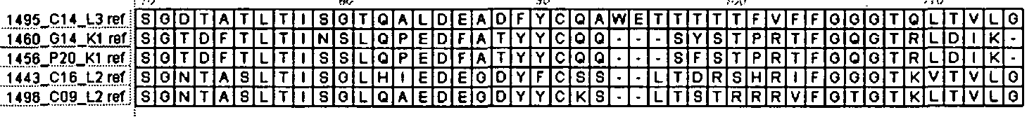


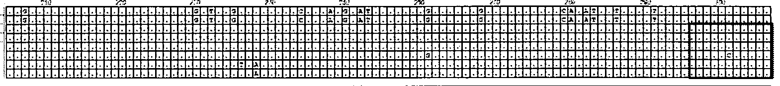


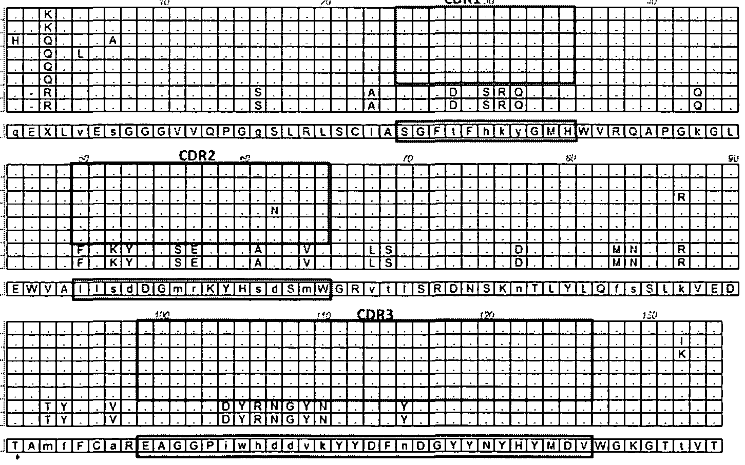


Claims
Priority Applications (22)
| Application Number | Priority Date | Filing Date | Title |
|---|---|---|---|
| EP18207707.3A EP3542817B1 (en) | 2009-03-17 | 2010-03-17 | Human immunodeficiency virus (hiv)-neutralizing antibodies |
| PL17194834T PL3323427T3 (en) | 2009-03-17 | 2010-03-17 | Human immunodeficiency virus (hiv)-neutralizing antibodies |
| ES10722810.8T ES2656234T3 (en) | 2009-03-17 | 2010-03-17 | Neutralizing antibodies of human immunodeficiency virus (HIV) |
| CA2755821A CA2755821C (en) | 2009-03-17 | 2010-03-17 | Human immunodeficiency virus (hiv)-neutralizing antibodies |
| SI201031616T SI2408476T1 (en) | 2009-03-17 | 2010-03-17 | HUMAN IMUNIFIED VIRUS (HIV) -NEUTRALIZING PROTITEL |
| LTEP10722810.8T LT2408476T (en) | 2009-03-17 | 2010-03-17 | Human immunodeficiency virus (hiv) -neutralizing antibodies |
| NO10722810A NO2408476T3 (en) | 2009-03-17 | 2010-03-17 | |
| EP17173548.3A EP3260136B1 (en) | 2009-03-17 | 2010-03-17 | Human immunodeficiency virus (hiv) -neutralizing antibodies |
| DK10722810.8T DK2408476T3 (en) | 2009-03-17 | 2010-03-17 | Human immunodeficiency virus (HIV) neutralizing antibodies |
| PL17173548T PL3260136T3 (en) | 2009-03-17 | 2010-03-17 | HUMAN IMMUNODEFICIENCY VIRUS (HIV) NEUTRALIZING ANTIBODIES |
| HRP20180027TT HRP20180027T1 (en) | 2009-03-17 | 2010-03-17 | Human immunodeficiency virus (hiv) -neutralizing antibodies |
| SM20180016T SMT201800016T1 (en) | 2009-03-17 | 2010-03-17 | Human immunodeficiency virus (hiv) -neutralizing antibodies |
| EP22171792.9A EP4085925A1 (en) | 2009-03-17 | 2010-03-17 | Human immunodeficiency virus (hiv)-neutralizing antibodies |
| AU2010226668A AU2010226668B2 (en) | 2009-03-17 | 2010-03-17 | Human immunodeficiency virus (HIV) -neutralizing antibodies |
| EP17194834.2A EP3323427B1 (en) | 2009-03-17 | 2010-03-17 | Human immunodeficiency virus (hiv)-neutralizing antibodies |
| EP10722810.8A EP2408476B1 (en) | 2009-03-17 | 2010-03-17 | Human immunodeficiency virus (hiv) -neutralizing antibodies |
| PL10722810T PL2408476T3 (en) | 2009-03-17 | 2010-03-17 | Human immunodeficiency virus (hiv) -neutralizing antibodies |
| AU2015234345A AU2015234345C1 (en) | 2009-03-17 | 2015-10-01 | Human immunodeficiency virus (HIV) -neutralizing antibodies |
| AU2017264985A AU2017264985B2 (en) | 2009-03-17 | 2017-11-20 | Human immunodeficiency virus (HIV) -neutralizing antibodies |
| CY20181100043T CY1121323T1 (en) | 2009-03-17 | 2018-01-15 | ANTIBODIES NEUTRALIZING THE HUMAN IMMUNE DISEASE VIRUS (HIV) |
| AU2020210242A AU2020210242B2 (en) | 2009-03-17 | 2020-07-30 | Human immunodeficiency virus (HIV) - neutralizing antibodies |
| AU2023204017A AU2023204017B2 (en) | 2009-03-17 | 2023-06-26 | Human immunodeficiency virus (HIV) -neutralizing antibodies |
Applications Claiming Priority (8)
| Application Number | Priority Date | Filing Date | Title |
|---|---|---|---|
| US16101009P | 2009-03-17 | 2009-03-17 | |
| US61/161,010 | 2009-03-17 | ||
| US16582909P | 2009-04-01 | 2009-04-01 | |
| US61/165,829 | 2009-04-01 | ||
| US22473909P | 2009-07-10 | 2009-07-10 | |
| US61/224,739 | 2009-07-10 | ||
| US28566409P | 2009-12-11 | 2009-12-11 | |
| US61/285,664 | 2009-12-11 |
Publications (2)
| Publication Number | Publication Date |
|---|---|
| WO2010107939A2 true WO2010107939A2 (en) | 2010-09-23 |
| WO2010107939A3 WO2010107939A3 (en) | 2011-05-26 |
Family
ID=42668183
Family Applications (1)
| Application Number | Title | Priority Date | Filing Date |
|---|---|---|---|
| PCT/US2010/027695 Ceased WO2010107939A2 (en) | 2009-03-17 | 2010-03-17 | Human immunodeficiency virus (hiv) -neutralizing antibodies |
Country Status (16)
| Country | Link |
|---|---|
| US (8) | US9051362B2 (en) |
| EP (5) | EP4085925A1 (en) |
| AU (5) | AU2010226668B2 (en) |
| CA (2) | CA2755821C (en) |
| CY (1) | CY1121323T1 (en) |
| DK (1) | DK2408476T3 (en) |
| ES (4) | ES2916404T3 (en) |
| HR (1) | HRP20180027T1 (en) |
| HU (1) | HUE036031T2 (en) |
| LT (1) | LT2408476T (en) |
| NO (1) | NO2408476T3 (en) |
| PL (4) | PL3260136T3 (en) |
| PT (4) | PT3260136T (en) |
| SI (4) | SI3260136T1 (en) |
| SM (1) | SMT201800016T1 (en) |
| WO (1) | WO2010107939A2 (en) |
Cited By (28)
| Publication number | Priority date | Publication date | Assignee | Title |
|---|---|---|---|---|
| WO2011092593A3 (en) * | 2010-01-20 | 2011-10-27 | Institute For Research In Biomedicine | Hiv-1 neutralizing antibodies and uses thereof |
| WO2012040562A2 (en) | 2010-09-24 | 2012-03-29 | International Aids Vaccine Initiative | Novel hiv-1 broadly neutralizing antibodies |
| EP2492279A1 (en) | 2011-02-25 | 2012-08-29 | Laboratorios Del. Dr. Esteve, S.A. | Rapid immunogen selection method using lentiviral display |
| WO2013039792A1 (en) | 2011-09-12 | 2013-03-21 | The United States Of America As Represented By The Secretary, Department Of Health And Human Services | Immunogens based on an hiv-1 gp120 v1v2 epitope |
| WO2013085550A3 (en) * | 2011-12-05 | 2013-10-24 | Duke University | V1v2 immunogens |
| EP2698377A1 (en) | 2012-08-17 | 2014-02-19 | Laboratorios Del. Dr. Esteve, S.A. | Enhanced rapid immunogen selection method for HIV gp120 variants |
| EP2611465A4 (en) * | 2010-08-31 | 2014-06-04 | Theraclone Sciences Inc | NEUTRALIZING ANTI-VIRUS ANTIBODIES FOR HUMAN IMMUNODEFICIENCY (HIV) |
| WO2016007765A1 (en) | 2014-07-11 | 2016-01-14 | Gilead Sciences, Inc. | Modulators of toll-like receptors for the treatment of hiv |
| EP2975053A1 (en) * | 2014-06-11 | 2016-01-20 | International Aids Vaccine Initiative | Broadly neutralizing antibody and uses thereof |
| EP2961427A4 (en) * | 2013-02-28 | 2016-11-02 | Therabiol Inc | HIV ANTIGENS AND ANTIBODIES AGAINST HIV ANTIGENS |
| EP3010549A4 (en) * | 2013-06-17 | 2017-03-15 | Emory University | Pathogen binding agents conjugated to radioisotopes and uses in imaging and therapeutic applications |
| WO2017048727A1 (en) | 2015-09-15 | 2017-03-23 | Gilead Sciences, Inc. | Modulators of toll-like recptors for the treatment of hiv |
| WO2017053906A1 (en) * | 2015-09-24 | 2017-03-30 | Abvitro Llc | Hiv antibody compositions and methods of use |
| CN107022027A (en) * | 2016-02-02 | 2017-08-08 | 中国疾病预防控制中心性病艾滋病预防控制中心 | HIV-1 wide spectrum neutralizing antibodies and application thereof |
| CN107033241A (en) * | 2016-02-03 | 2017-08-11 | 中国疾病预防控制中心性病艾滋病预防控制中心 | HIV-1 wide spectrum neutralizing antibodies and application thereof |
| US10239935B2 (en) | 2015-12-15 | 2019-03-26 | Gilead Sciences, Inc. | Human immunodeficiency virus neutralizing antibodies |
| US10508117B2 (en) | 2014-09-16 | 2019-12-17 | Gilead Sciences, Inc. | Solid forms of a toll-like receptor modulator |
| WO2020010107A1 (en) | 2018-07-03 | 2020-01-09 | Gilead Sciences, Inc. | Antibodies that target hiv gp120 and methods of use |
| US10882907B2 (en) | 2017-06-21 | 2021-01-05 | Gilead Sciences, Inc. | Multispecific antibodies that target HIV GP120 and CD3 |
| WO2021011544A1 (en) | 2019-07-16 | 2021-01-21 | Gilead Sciences, Inc. | Hiv vaccines and methods of making and using |
| US11110091B2 (en) | 2008-12-09 | 2021-09-07 | Gilead Sciences, Inc. | Modulators of toll-like receptors |
| WO2022046644A1 (en) | 2020-08-25 | 2022-03-03 | Gilead Sciences, Inc. | Multi-specific antigen binding molecules targeting hiv and methods of use |
| WO2022087149A2 (en) | 2020-10-22 | 2022-04-28 | Gilead Sciences, Inc. | Interleukin-2-fc fusion proteins and methods of use |
| WO2022161597A1 (en) * | 2021-01-26 | 2022-08-04 | Eth Zurich | Method for rapid identification of cross-reactive and/or rare antibodies |
| WO2022162009A1 (en) | 2021-01-26 | 2022-08-04 | Eth Zurich | Method for rapid identification of cross-reactive and/or rare antibodies |
| WO2024015741A1 (en) | 2022-07-12 | 2024-01-18 | Gilead Sciences, Inc. | Hiv immunogenic polypeptides and vaccines and uses thereof |
| US12239698B2 (en) | 2018-12-04 | 2025-03-04 | The Rockefeller University | HIV vaccine immunogens |
| US12611453B2 (en) | 2020-05-21 | 2026-04-28 | Gilead Sciences, Inc. | Method of inducing an HIV-1-specific immune response using a chimpanzee adenovirus vector encoding an hivacat t-cell immunogen and TLR7 agonist |
Families Citing this family (15)
| Publication number | Priority date | Publication date | Assignee | Title |
|---|---|---|---|---|
| WO2011109511A2 (en) * | 2010-03-02 | 2011-09-09 | International Aids Vaccine Initiative | Novel hiv-1 envelope glycoprotein |
| EP2739300B1 (en) | 2011-07-05 | 2019-06-19 | Duke University | N-terminal deleted gp120 immunogens |
| US8791108B2 (en) * | 2011-08-18 | 2014-07-29 | Bristol-Myers Squibb Company | Inhibitors of human immunodeficiency virus replication |
| CA2850745C (en) | 2011-10-03 | 2022-12-13 | Duke University | Vaccine |
| AU2012347453B2 (en) | 2011-12-08 | 2017-11-23 | The United States Of America, As Represented By The Secretary Department Of Health And Human Services | Neutralizing antibodies to HIV-1 and their use |
| EP2828294A1 (en) * | 2012-03-23 | 2015-01-28 | The United States of America, as represented by The Secretary, Department of Health and Human Services | Neutralizing antibodies to hiv-1 and their use |
| WO2015048517A1 (en) * | 2013-09-28 | 2015-04-02 | Duke University | Escape mutants |
| WO2015127108A1 (en) * | 2014-02-19 | 2015-08-27 | Duke University | Trimeric hiv-1 envelopes compositions and uses thereof |
| WO2016054053A2 (en) * | 2014-09-29 | 2016-04-07 | Duke University | Hiv-1 antibodies and uses thereof (adcc and bispecific abs) |
| US20200172601A1 (en) * | 2017-06-22 | 2020-06-04 | University Of Maryland, Baltimore | Broadly neutralizing antibodies against hiv |
| US12365722B2 (en) | 2018-10-19 | 2025-07-22 | University Of Maryland, College Park | Multispecific antibodies targeting multiple epitopes on the HIV-1 envelope |
| EP4051318A4 (en) * | 2019-10-29 | 2023-10-11 | International AIDS Vaccine Initiative | GENETICALLY MODIFIED ANTIBODIES AGAINST HIV ENV |
| US20210277093A1 (en) * | 2020-03-09 | 2021-09-09 | Adma Biologics, Inc. | Immunotherapeutic compositions and methods of production for coronavirus |
| US20240181051A1 (en) * | 2021-04-19 | 2024-06-06 | The Wistar Institute Of Anatomy And Biology | Sialydase linked hiv antibodies and methods of use |
| WO2026039311A1 (en) | 2024-08-10 | 2026-02-19 | Vectorgen | Lentiviral vector for treating human immunodeficiency virus infection |
Family Cites Families (90)
| Publication number | Priority date | Publication date | Assignee | Title |
|---|---|---|---|---|
| US3773919A (en) | 1969-10-23 | 1973-11-20 | Du Pont | Polylactide-drug mixtures |
| US3896111A (en) | 1973-02-20 | 1975-07-22 | Research Corp | Ansa macrolides |
| US4151042A (en) | 1977-03-31 | 1979-04-24 | Takeda Chemical Industries, Ltd. | Method for producing maytansinol and its derivatives |
| US4137230A (en) | 1977-11-14 | 1979-01-30 | Takeda Chemical Industries, Ltd. | Method for the production of maytansinoids |
| FR2413974A1 (en) | 1978-01-06 | 1979-08-03 | David Bernard | DRYER FOR SCREEN-PRINTED SHEETS |
| US4307016A (en) | 1978-03-24 | 1981-12-22 | Takeda Chemical Industries, Ltd. | Demethyl maytansinoids |
| US4265814A (en) | 1978-03-24 | 1981-05-05 | Takeda Chemical Industries | Matansinol 3-n-hexadecanoate |
| JPS5562090A (en) | 1978-10-27 | 1980-05-10 | Takeda Chem Ind Ltd | Novel maytansinoid compound and its preparation |
| JPS55164687A (en) | 1979-06-11 | 1980-12-22 | Takeda Chem Ind Ltd | Novel maytansinoid compound and its preparation |
| JPS5566585A (en) | 1978-11-14 | 1980-05-20 | Takeda Chem Ind Ltd | Novel maytansinoid compound and its preparation |
| US4256746A (en) | 1978-11-14 | 1981-03-17 | Takeda Chemical Industries | Dechloromaytansinoids, their pharmaceutical compositions and method of use |
| JPS55102583A (en) | 1979-01-31 | 1980-08-05 | Takeda Chem Ind Ltd | 20-acyloxy-20-demethylmaytansinoid compound |
| JPS55162791A (en) | 1979-06-05 | 1980-12-18 | Takeda Chem Ind Ltd | Antibiotic c-15003pnd and its preparation |
| JPS55164685A (en) | 1979-06-08 | 1980-12-22 | Takeda Chem Ind Ltd | Novel maytansinoid compound and its preparation |
| JPS55164686A (en) | 1979-06-11 | 1980-12-22 | Takeda Chem Ind Ltd | Novel maytansinoid compound and its preparation |
| US4309428A (en) | 1979-07-30 | 1982-01-05 | Takeda Chemical Industries, Ltd. | Maytansinoids |
| JPS5645483A (en) | 1979-09-19 | 1981-04-25 | Takeda Chem Ind Ltd | C-15003phm and its preparation |
| EP0028683A1 (en) | 1979-09-21 | 1981-05-20 | Takeda Chemical Industries, Ltd. | Antibiotic C-15003 PHO and production thereof |
| JPS5645485A (en) | 1979-09-21 | 1981-04-25 | Takeda Chem Ind Ltd | Production of c-15003pnd |
| WO1981001145A1 (en) | 1979-10-18 | 1981-04-30 | Univ Illinois | Hydrolytic enzyme-activatible pro-drugs |
| WO1982001188A1 (en) | 1980-10-08 | 1982-04-15 | Takeda Chemical Industries Ltd | 4,5-deoxymaytansinoide compounds and process for preparing same |
| US4450254A (en) | 1980-11-03 | 1984-05-22 | Standard Oil Company | Impact improvement of high nitrile resins |
| US4554101A (en) | 1981-01-09 | 1985-11-19 | New York Blood Center, Inc. | Identification and preparation of epitopes on antigens and allergens on the basis of hydrophilicity |
| US4315929A (en) | 1981-01-27 | 1982-02-16 | The United States Of America As Represented By The Secretary Of Agriculture | Method of controlling the European corn borer with trewiasine |
| US4313946A (en) | 1981-01-27 | 1982-02-02 | The United States Of America As Represented By The Secretary Of Agriculture | Chemotherapeutically active maytansinoids from Trewia nudiflora |
| JPS57192389A (en) | 1981-05-20 | 1982-11-26 | Takeda Chem Ind Ltd | Novel maytansinoid |
| US4485045A (en) | 1981-07-06 | 1984-11-27 | Research Corporation | Synthetic phosphatidyl cholines useful in forming liposomes |
| NZ201705A (en) | 1981-08-31 | 1986-03-14 | Genentech Inc | Recombinant dna method for production of hepatitis b surface antigen in yeast |
| US4816567A (en) | 1983-04-08 | 1989-03-28 | Genentech, Inc. | Recombinant immunoglobin preparations |
| DD266710A3 (en) | 1983-06-06 | 1989-04-12 | Ve Forschungszentrum Biotechnologie | Process for the biotechnical production of alkaline phosphatase |
| US4544545A (en) | 1983-06-20 | 1985-10-01 | Trustees University Of Massachusetts | Liposomes containing modified cholesterol for organ targeting |
| US4879231A (en) | 1984-10-30 | 1989-11-07 | Phillips Petroleum Company | Transformation of yeasts of the genus pichia |
| US4683202A (en) | 1985-03-28 | 1987-07-28 | Cetus Corporation | Process for amplifying nucleic acid sequences |
| US4676980A (en) | 1985-09-23 | 1987-06-30 | The United States Of America As Represented By The Secretary Of The Department Of Health And Human Services | Target specific cross-linked heteroantibodies |
| GB8610600D0 (en) | 1986-04-30 | 1986-06-04 | Novo Industri As | Transformation of trichoderma |
| US5567610A (en) | 1986-09-04 | 1996-10-22 | Bioinvent International Ab | Method of producing human monoclonal antibodies and kit therefor |
| IL85035A0 (en) | 1987-01-08 | 1988-06-30 | Int Genetic Eng | Polynucleotide molecule,a chimeric antibody with specificity for human b cell surface antigen,a process for the preparation and methods utilizing the same |
| GB8705477D0 (en) | 1987-03-09 | 1987-04-15 | Carlton Med Prod | Drug delivery systems |
| US4975278A (en) | 1988-02-26 | 1990-12-04 | Bristol-Myers Company | Antibody-enzyme conjugates in combination with prodrugs for the delivery of cytotoxic agents to tumor cells |
| US5606040A (en) | 1987-10-30 | 1997-02-25 | American Cyanamid Company | Antitumor and antibacterial substituted disulfide derivatives prepared from compounds possessing a methyl-trithio group |
| US5053394A (en) | 1988-09-21 | 1991-10-01 | American Cyanamid Company | Targeted forms of methyltrithio antitumor agents |
| US5770701A (en) | 1987-10-30 | 1998-06-23 | American Cyanamid Company | Process for preparing targeted forms of methyltrithio antitumor agents |
| GB8823869D0 (en) | 1988-10-12 | 1988-11-16 | Medical Res Council | Production of antibodies |
| EP0368684B2 (en) | 1988-11-11 | 2004-09-29 | Medical Research Council | Cloning immunoglobulin variable domain sequences. |
| US5175384A (en) | 1988-12-05 | 1992-12-29 | Genpharm International | Transgenic mice depleted in mature t-cells and methods for making transgenic mice |
| FR2646437B1 (en) | 1989-04-28 | 1991-08-30 | Transgene Sa | NOVEL DNA SEQUENCES, THEIR APPLICATION AS A SEQUENCE ENCODING A SIGNAL PEPTIDE FOR THE SECRETION OF MATURE PROTEINS BY RECOMBINANT YEASTS, EXPRESSION CASSETTES, PROCESSED YEASTS AND PROCESS FOR PREPARING THE SAME |
| EP0402226A1 (en) | 1989-06-06 | 1990-12-12 | Institut National De La Recherche Agronomique | Transformation vectors for yeast yarrowia |
| DE3920358A1 (en) | 1989-06-22 | 1991-01-17 | Behringwerke Ag | BISPECIFIC AND OLIGO-SPECIFIC, MONO- AND OLIGOVALENT ANTI-BODY CONSTRUCTS, THEIR PRODUCTION AND USE |
| ES2096590T3 (en) | 1989-06-29 | 1997-03-16 | Medarex Inc | BI-SPECIFIC REAGENTS FOR AIDS THERAPY. |
| US5013556A (en) | 1989-10-20 | 1991-05-07 | Liposome Technology, Inc. | Liposomes with enhanced circulation time |
| CA2026147C (en) | 1989-10-25 | 2006-02-07 | Ravi J. Chari | Cytotoxic agents comprising maytansinoids and their therapeutic use |
| US5208020A (en) | 1989-10-25 | 1993-05-04 | Immunogen Inc. | Cytotoxic agents comprising maytansinoids and their therapeutic use |
| US5229275A (en) | 1990-04-26 | 1993-07-20 | Akzo N.V. | In-vitro method for producing antigen-specific human monoclonal antibodies |
| DE69132709T2 (en) | 1990-06-29 | 2002-06-20 | Large Scale Biology Corp., Vacaville | MELANINE PRODUCTION BY TRANSFORMED MICROORGANISMS |
| EP0542810A1 (en) | 1990-08-02 | 1993-05-26 | B.R. Centre Limited | Methods for the production of proteins with a desired function |
| ATE300615T1 (en) | 1990-08-29 | 2005-08-15 | Genpharm Int | TRANSGENIC MICE CAPABLE OF PRODUCING HETEROLOGOUS ANTIBODIES |
| US5545806A (en) | 1990-08-29 | 1996-08-13 | Genpharm International, Inc. | Ransgenic non-human animals for producing heterologous antibodies |
| US5571894A (en) | 1991-02-05 | 1996-11-05 | Ciba-Geigy Corporation | Recombinant antibodies specific for a growth factor receptor |
| EP0586505A1 (en) | 1991-05-14 | 1994-03-16 | Repligen Corporation | Heteroconjugate antibodies for treatment of hiv infection |
| EP1400536A1 (en) | 1991-06-14 | 2004-03-24 | Genentech Inc. | Method for making humanized antibodies |
| WO1993006217A1 (en) | 1991-09-19 | 1993-04-01 | Genentech, Inc. | EXPRESSION IN E. COLI OF ANTIBODY FRAGMENTS HAVING AT LEAST A CYSTEINE PRESENT AS A FREE THIOL, USE FOR THE PRODUCTION OF BIFUNCTIONAL F(ab')2 ANTIBODIES |
| FI941572L (en) | 1991-10-07 | 1994-05-27 | Oncologix Inc | Combination and method of use of anti-erbB-2 monoclonal antibodies |
| WO1993008829A1 (en) | 1991-11-04 | 1993-05-13 | The Regents Of The University Of California | Compositions that mediate killing of hiv-infected cells |
| DE69232137T2 (en) | 1991-11-25 | 2002-05-29 | Enzon Inc | MULTIVALENT ANTI-BINDING PROTEINS |
| AU675929B2 (en) | 1992-02-06 | 1997-02-27 | Curis, Inc. | Biosynthetic binding protein for cancer marker |
| ZA932522B (en) | 1992-04-10 | 1993-12-20 | Res Dev Foundation | Immunotoxins directed against c-erbB-2(HER/neu) related surface antigens |
| EP0656064B1 (en) | 1992-08-17 | 1997-03-05 | Genentech, Inc. | Bispecific immunoadhesins |
| MD1367C2 (en) | 1992-11-13 | 2000-11-30 | Idec Pharmaceuticals Corporation | Methods of B-cells lymphoma treatment, anti-CD20 antibodies, hybridoma |
| WO1994025591A1 (en) | 1993-04-29 | 1994-11-10 | Unilever N.V. | PRODUCTION OF ANTIBODIES OR (FUNCTIONALIZED) FRAGMENTS THEREOF DERIVED FROM HEAVY CHAIN IMMUNOGLOBULINS OF $i(CAMELIDAE) |
| US5773001A (en) | 1994-06-03 | 1998-06-30 | American Cyanamid Company | Conjugates of methyltrithio antitumor agents and intermediates for their synthesis |
| US5910486A (en) | 1994-09-06 | 1999-06-08 | Uab Research Foundation | Methods for modulating protein function in cells using, intracellular antibody homologues |
| US5789199A (en) | 1994-11-03 | 1998-08-04 | Genentech, Inc. | Process for bacterial production of polypeptides |
| US6214388B1 (en) | 1994-11-09 | 2001-04-10 | The Regents Of The University Of California | Immunoliposomes that optimize internalization into target cells |
| JPH10511085A (en) | 1994-12-02 | 1998-10-27 | カイロン コーポレイション | Methods for promoting an immune response using bispecific antibodies |
| US5840523A (en) | 1995-03-01 | 1998-11-24 | Genetech, Inc. | Methods and compositions for secretion of heterologous polypeptides |
| US5731168A (en) | 1995-03-01 | 1998-03-24 | Genentech, Inc. | Method for making heteromultimeric polypeptides |
| US5641870A (en) | 1995-04-20 | 1997-06-24 | Genentech, Inc. | Low pH hydrophobic interaction chromatography for antibody purification |
| US5739277A (en) | 1995-04-14 | 1998-04-14 | Genentech Inc. | Altered polypeptides with increased half-life |
| US5869046A (en) | 1995-04-14 | 1999-02-09 | Genentech, Inc. | Altered polypeptides with increased half-life |
| US5837234A (en) | 1995-06-07 | 1998-11-17 | Cytotherapeutics, Inc. | Bioartificial organ containing cells encapsulated in a permselective polyether suflfone membrane |
| US5712374A (en) | 1995-06-07 | 1998-01-27 | American Cyanamid Company | Method for the preparation of substantiallly monomeric calicheamicin derivative/carrier conjugates |
| US5714586A (en) | 1995-06-07 | 1998-02-03 | American Cyanamid Company | Methods for the preparation of monomeric calicheamicin derivative/carrier conjugates |
| DE19544393A1 (en) | 1995-11-15 | 1997-05-22 | Hoechst Schering Agrevo Gmbh | Synergistic herbicidal mixtures |
| US5922845A (en) | 1996-07-11 | 1999-07-13 | Medarex, Inc. | Therapeutic multispecific compounds comprised of anti-Fcα receptor antibodies |
| US6815201B2 (en) | 1997-09-08 | 2004-11-09 | The Public Health Research Institute Of The City Of New York, Inc. | HIV-1 gp120 V1/V2 domain epitopes capable of generating neutralizing antibodies |
| US6824780B1 (en) | 1999-10-29 | 2004-11-30 | Genentech, Inc. | Anti-tumor antibody compositions and methods of use |
| EP1588166A4 (en) | 2003-01-09 | 2007-06-27 | Macrogenics Inc | Dual expression vector system for antibody expression in bacterial and mammalian cells |
| WO2004076677A2 (en) | 2003-02-26 | 2004-09-10 | Institute For Research In Biomedicine | Monoclonal antibody production by ebv transformation of b cells |
| US20070292390A1 (en) * | 2004-10-29 | 2007-12-20 | Dimitrov Dimiter S | Broadly Cross-Reactive Hiv-1 Neutralizing Human Monoclonal Antibodies |
| CA3249351A1 (en) * | 2010-08-31 | 2025-07-03 | Theraclone Sciences, Inc. | Human Immunodeficiency Virus (HIV)-Neutralizing Antibodies |
-
2010
- 2010-03-01 SI SI201032049T patent/SI3260136T1/en unknown
- 2010-03-17 SI SI201032112T patent/SI3542817T1/en unknown
- 2010-03-17 AU AU2010226668A patent/AU2010226668B2/en active Active
- 2010-03-17 PT PT171735483T patent/PT3260136T/en unknown
- 2010-03-17 EP EP22171792.9A patent/EP4085925A1/en active Pending
- 2010-03-17 SI SI201031616T patent/SI2408476T1/en unknown
- 2010-03-17 PL PL17173548T patent/PL3260136T3/en unknown
- 2010-03-17 DK DK10722810.8T patent/DK2408476T3/en active
- 2010-03-17 ES ES18207707T patent/ES2916404T3/en active Active
- 2010-03-17 SI SI201032062T patent/SI3323427T1/en unknown
- 2010-03-17 ES ES17194834T patent/ES2848203T3/en active Active
- 2010-03-17 CA CA2755821A patent/CA2755821C/en active Active
- 2010-03-17 CA CA2980883A patent/CA2980883C/en active Active
- 2010-03-17 PT PT182077073T patent/PT3542817T/en unknown
- 2010-03-17 WO PCT/US2010/027695 patent/WO2010107939A2/en not_active Ceased
- 2010-03-17 NO NO10722810A patent/NO2408476T3/no unknown
- 2010-03-17 PL PL18207707.3T patent/PL3542817T3/en unknown
- 2010-03-17 PL PL10722810T patent/PL2408476T3/en unknown
- 2010-03-17 EP EP17194834.2A patent/EP3323427B1/en active Active
- 2010-03-17 US US12/726,245 patent/US9051362B2/en active Active
- 2010-03-17 ES ES17173548T patent/ES2848555T3/en active Active
- 2010-03-17 PT PT107228108T patent/PT2408476T/en unknown
- 2010-03-17 PL PL17194834T patent/PL3323427T3/en unknown
- 2010-03-17 EP EP10722810.8A patent/EP2408476B1/en active Active
- 2010-03-17 HR HRP20180027TT patent/HRP20180027T1/en unknown
- 2010-03-17 LT LTEP10722810.8T patent/LT2408476T/en unknown
- 2010-03-17 SM SM20180016T patent/SMT201800016T1/en unknown
- 2010-03-17 ES ES10722810.8T patent/ES2656234T3/en active Active
- 2010-03-17 HU HUE10722810A patent/HUE036031T2/en unknown
- 2010-03-17 EP EP18207707.3A patent/EP3542817B1/en active Active
- 2010-03-17 PT PT171948342T patent/PT3323427T/en unknown
- 2010-03-17 EP EP17173548.3A patent/EP3260136B1/en active Active
-
2015
- 2015-04-21 US US14/692,483 patent/US9920111B2/en active Active
- 2015-10-01 AU AU2015234345A patent/AU2015234345C1/en active Active
-
2017
- 2017-11-20 AU AU2017264985A patent/AU2017264985B2/en active Active
-
2018
- 2018-01-15 CY CY20181100043T patent/CY1121323T1/en unknown
- 2018-03-12 US US15/918,343 patent/US10239934B2/en active Active
-
2019
- 2019-02-14 US US16/275,936 patent/US10407493B2/en active Active
- 2019-07-17 US US16/513,859 patent/US10865234B2/en active Active
-
2020
- 2020-07-30 AU AU2020210242A patent/AU2020210242B2/en active Active
- 2020-10-28 US US17/082,322 patent/US11584789B2/en active Active
-
2023
- 2023-01-06 US US18/150,957 patent/US12018067B2/en active Active
- 2023-06-26 AU AU2023204017A patent/AU2023204017B2/en active Active
-
2024
- 2024-06-05 US US18/734,520 patent/US12312396B2/en active Active
Non-Patent Citations (176)
| Title |
|---|
| A. PINTER ET AL., J VIROL, vol. 79, June 2005 (2005-06-01), pages 6909 |
| ALBERT J. ET AL., AIDS, vol. 4, 1990, pages 107 - 112 |
| ALTSCHUL ET AL., J. MOL. BIOL., vol. 215, 1990, pages 403 - 410 |
| ALTSCHUL ET AL., NUCL. ACIDS RES., vol. 25, 1977, pages 3389 - 3402 |
| BABCOOK ET AL., PROC. NATL. ACAD. SCI. USA, vol. 93, 1996, pages 7843 - 48 |
| BARBAS CF ET AL., PROC. NATL. ACAD. SCI. USA, vol. 89, 1992, pages 9339 - 9343 |
| BAROUCH, D.H., NATURE, vol. 455, 2008, pages 613 - 619 |
| BARRE-SINOUSSI, F. ET AL., SCIENCE, vol. 220, 1983, pages 868 - 870 |
| BINLEY JM ET AL., J. VIROL., vol. 78, no. 23, 2004, pages 13232 - 13252 |
| BINLEY JM ET AL.: "Comprehensive Cross-Clade Neutralization Analysis of a Panel of Anti-Human Immunodeficiency Virus Type 1 Monoclonal Antibodies", J. VIROL., vol. 78, 2004, pages 13232 - 13252, XP002464744, DOI: doi:10.1128/JVI.78.23.13232-13252.2004 |
| BIRD ET AL., SCIENCE, vol. 242, 1988, pages 423 - 426 |
| BOLTON; HUNTER, BIOCHEM · J., vol. 133, 1973, pages 529 |
| BRENNAN ET AL., SCIENCE, vol. 229, 1985, pages 81 |
| BROGLIE, R. ET AL., SCIENCE, vol. 224, 1984, pages 838 - 843 |
| BRUGGEMANN ET AL., YEAR IN IMMUNO., vol. 7, 1993, pages 33 |
| BUCHACHER A ET AL., VACCINES, vol. 92, 1992, pages 191 - 195 |
| BUCHACHER, A ET AL., AIDS RES. HUM. RETROVIRUSES, vol. 10, 1994, pages 359 - 369 |
| BUCHACHER, A. ET AL., AIDS RES. HUM. RETROVIRUSES, vol. 10, 1994, pages 359 - 369 |
| BURTON DP ET AL., PROC. NATL. ACAD. SCI. USA, vol. 88, 1991, pages 10134 - 10137 |
| BURTON DR ET AL., SCIENCE, vol. 266, no. 5187, pages 1024 - 1027 |
| BURTON DR ET AL.: "Efficient neutralization of primary isolates of HIV- by a recombinant human monoclonal antibody", SCIENCE, vol. 266, 1994, pages 1024 - 1027, XP001147878, DOI: doi:10.1126/science.7973652 |
| BURTON, D. R., NAT. REV. IMMUNOL., vol. 2, 2002, pages 706 - 713 |
| BURTON, D.R. ET AL., SCIENCE, vol. 266, 1994, pages 1024 - 1027 |
| CANCER RESEARCH, vol. 52, 1992, pages 127 - 131 |
| CAPEL ET AL., IMMUNOMETHODS, vol. 4, 1994, pages 25 - 34 |
| CARLSSON ET AL., BIOCHEM. J., vol. 173, 1978, pages 723 - 737 |
| CARON ET AL., J. EXP MED., vol. 176, 1992, pages 1191 - 1195 |
| CARTER ET AL., BIO/TECHNOLOGY, vol. 10, 1992, pages 163 - 167 |
| CASADEVALL, NAT. BIOTECHNOL., vol. 20, 2002, pages 114 |
| CHARI ET AL., CANCER RESEARCH, vol. 52, 1992, pages 127 - 131 |
| CHOTHIA; LESK, J. MOL. BIOL., vol. 196, 1987, pages 901 - 917 |
| CLACKSON, NATURE, vol. 352, 1991, pages 624 - 628 |
| CLYNES ET AL., PROC. NATL. ACAD. SCI. (USA), vol. 95, 1998, pages 652 - 656 |
| COLBERE-GARAPIN, F. ET AL., J. MOL. BIOL., vol. 150, 1981, pages 1 - 14 |
| CONLEY AJ, PROC. NATL. ACAD. SCI. USA, vol. 91, 1994, pages 3348 - 3352 |
| CORUZZI, G. ET AL., EMBO J., vol. 3, 1984, pages 1671 - 1680 |
| CUNNINGHAM; WELLS, SCIENCE, vol. 244, 1989, pages 1081 - 1085 |
| DAERON, ANNU. REV. IMMUNOL., vol. 15, 1997, pages 203 - 234 |
| DARBHA, R. ET AL., BIOCHEMISTRY, vol. 43, 2004, pages 1410 - 1417 |
| DE HAAS ET AL., J. LAB. CLIN. MED., vol. 126, 1995, pages 330 - 41 |
| ENGELHARD, E. K. ET AL., PROC. NATL. ACAD. SCI., vol. 91, 1994, pages 3224 - 3227 |
| EPSTEIN ET AL., PROC. NATL. ACAD. SCI. USA, vol. 82, 1985, pages 3688 |
| FRAKER ET AL., BIOCHEM. BIOPHYS. RES. COMMUN., vol. 80, 1978, pages 49 - 57 |
| GABIZON ET AL., J. NATIONAL CANCER INST., vol. 81, no. 19, 1989, pages 1484 |
| GALLO, R. ET AL., SCIENCE, vol. 224, 1984, pages 500 - 503 |
| GAZZANO-SANTORO ET AL., J. IMMUNOL. METHODS, vol. 202, 1996, pages 163 |
| GORNY, M.K. ET AL., J VIROL, vol. 79, 2005, pages 5232 - 5237 |
| GRAHAM, J. GEN VIROL., vol. 36, 1977, pages 59 |
| GRANT ET AL., METHODS ENZYMOL., vol. 153, 1987, pages 516 - 544 |
| GRUBER ET AL., J. IMMUNOL., vol. 152, 1994, pages 5368 |
| GUSS ET AL., EMBO J., vol. 5, 1986, pages 15671575 |
| GUYER ET AL., J. IMMUNOL., vol. 117, 1976, pages 587 |
| HARTMAN, S. C.; R. C. MULLIGAN, PROC. NATL. ACAD. SCI., vol. 85, 1988, pages 8047 - 51 |
| HENIKOFFAND HENIKOFF, PROC. NATL. ACAD. SCI. USA, vol. 89, 1989, pages 10915 |
| HESSELL, A.J. ET AL., PLOS PATHOG, vol. 5, 2009, pages E1000433 |
| HIGGINS DG ET AL., NUCLEIC ACIDS RESEARCH, vol. 22, 1994, pages 4673 - 4680 |
| HIGGINS, D.G.; SHARP, P.M., CABIOS, vol. 5, 1989, pages 151 - 153 |
| HOLLINGER ET AL., PROC. NATL. ACAD. SCI. USA, vol. 90, 1993, pages 6444 - 6448 |
| HONNEGER, A.; PLUNKTHUN, A., J. MOL. BIOL., vol. 309, 2001, pages 657 - 670 |
| HONNEN, W.J. ET AL., J VIROL, vol. 81, 2007, pages 1424 - 1432 |
| HWANG ET AL., PROC. NATL ACAD. SCI. USA, vol. 77, 1980, pages 4030 |
| ICHIYOSHI, Y.; CASALI, P., J EXP MED, vol. 180, 1994, pages 885 - 895 |
| IGARASHI ET AL., NAT. MED., vol. 5, 1999, pages 211 - 16 |
| J. VIROL., vol. 67, 1993, pages 6642 - 6647 |
| JAKOBOVITS ET AL., PROC. NATL. ACAD. SCI. USA, vol. 90, 1993, pages 2551 |
| JAKOBOVITS, NATURE, vol. 362, 1993, pages 255 - 258 |
| JOHNSTON, M.I.; FAUCI, A.S., N ENGL J MED, vol. 356, 2007, pages 2073 - 2081 |
| JONES ET AL., NATURE, vol. 321, 1986, pages 522 - 525 |
| KARLSSON HEDESTAM, G.B. ET AL., NAT REV MICROBIOL, vol. 6, 2008, pages 143 - 155 |
| KARPAS A. ET AL., PROC. NATL. ACAD. SCI. USA, vol. 98, 2001, pages 1799 - 1804 |
| KELLER ET AL., CLIN. MICROBIOL. REV., vol. 13, 2000, pages 602 - 14 |
| KIM ET AL., J. IMMUNOL., vol. 24, 1994, pages 249 |
| KOHLER ET AL., NATURE, vol. 256, 1975, pages 495 |
| KOSTELNY ET AL., J. IMMUNOL., vol. 148, no. 5, 1992, pages 1547 - 1553 |
| KOSTRIKIS LG ET AL., J VIROL., vol. 70, no. 1, 1996, pages 445 - 458 |
| KROLL, D. J. ET AL., DNA CELL BIOL., vol. 12, 1993, pages 441 - 453 |
| KWONG PD ET AL., NATURE, vol. 420, 2002, pages 678 - 682 |
| KWONG, P.D.; WILSON, I.A., NAT IMMUNOL, vol. 10, 2009, pages 573 - 578 |
| LARKIN M.A.; BLACKSHIELDS G.; BROWN N.P.; CHENNA R.; MCGETTIGAN P.A.; MCWILLIAM H.; VALENTIN F.; WALLACE I.M.; WILM A.; LOPEZ R., BIOINFORMATICS, vol. 23, no. 21, 2007, pages 2947 - 2948 |
| LASKY LA ET AL., CELL, vol. 50, 1987, pages 975 - 985 |
| LEFRANC, M P. ET AL.: "IMGT unique numbering for immunoglobulin and T cell receptor variable regions and Ig superfamily V-like domains", DEV COMP IMMUNOL., vol. 27, no. 1, 2003, pages 55 - 77, XP055144492, DOI: doi:10.1016/S0145-305X(02)00039-3 |
| LEFRANC, M P.: "The IMGT unique numbering for Immunoglobulins, T cell receptors and Ig-like domains", THE IMMUNOLOGIST, vol. 7, 1999, pages 132 - 136 |
| LEFRANC, M P.: "Unique database numbering system for immunogenetic analysis", IMMUNOLOGY TODAY, vol. 18, 1997, pages 509, XP004093509, DOI: doi:10.1016/S0167-5699(97)01163-8 |
| LEFRANC, M.P. ET AL., NUCL. ACIDS RES., vol. 27, 1999, pages 209 - 212 |
| LEONIDAS STAMATATOS, L.M.; DENNIS R BURTON; JOHN MASCOLA, NATURE MEDICINE, 14 June 2009 (2009-06-14) |
| LI, M. ET AL., J VIROL, vol. 79, 2005, pages 10108 - 10125 |
| LINDMARK ET AL., J. IMMUNOL. METH., vol. 62, 1983, pages 1 - 13 |
| LIU ET AL., PROC. NATL. ACAD. SCI. USA, vol. 93, 1996, pages 8618 - 8623 |
| LOGAN, J.; SHENK, T., PROC. NATL. ACAD. SCI., vol. 81, 1984, pages 3655 - 3659 |
| LOWY, I. ET AL., CELL, vol. 22, 1990, pages 817 - 23 |
| MADDOX, D. E. ET AL., J. EXP. MED., vol. 158, 1983, pages 1211 - 1216 |
| MARKS ET AL., J. MOL. BIOL., vol. 222, 1991, pages 581 - 597 |
| MARTIN ET AL., J. BIOL. CHEM., vol. 257, 1982, pages 286 - 288 |
| MASCOLA, J.R. ET AL., J VIROL, vol. 73, 1999, pages 4009 - 4018 |
| MASCOLA, J.R. ET AL., NAT MED, vol. 6, 2000, pages 207 - 210 |
| MASCOLA, J.R., VACCINE, vol. 20, 2002, pages 1922 - 1925 |
| MASSEY, NATURE, vol. 328, 1987, pages 457 - 458 |
| MATHER ET AL., ANNALS N.Y. ACAD. SCI., vol. 383, 1982, pages 44 - 68 |
| MATHER, BIOL. REPROD., vol. 23, 1980, pages 243 - 251 |
| MILLSTEIN ET AL., NATURE, vol. 305, 1983, pages 537 - 539 |
| MONTERO, M.; VAN HOUTEN, N.E.; WANG, X.; SCOTT, J.K., MICROBIOL MOL BIOL REV, vol. 72, 2008, pages 54 - 84 |
| MORIMOTO ET AL., JOURNAL OF BIOCHEMICAL AND BIOPHYSICAL METHODS, vol. 24, 1992, pages 107 - 117 |
| MORRISON ET AL., PROC. NATL. ACAD. SCI. USA, vol. 81, 1984, pages 6851 - 6855 |
| MORRISON, METHODS IN ENZYMOLOGY, vol. 32B, 1974, pages 103 |
| MUSTER T ET AL., J. VIROL., vol. 67, no. 11, 1993, pages 6642 - 6647 |
| MYERS, E.W.; MULLER W., CABIOS, vol. 4, 1988, pages 11 - 17 |
| NARA, P. L. ET AL., FASEB J., vol. 5, 1991, pages 2437 - 2455 |
| NEEDLEMAN; WUNSCH, J MOL. BIOL., vol. 48, 1970, pages 443 |
| NEUBERGER ET AL., NATURE, vol. 312, 1984, pages 604 - 608 |
| PANCERA M. ET AL., VIROLOGY, vol. 332, 2005, pages 145 |
| PANCERA, M.; WYATT, R., VIROLOGY, vol. 332, 2005, pages 145 - 156 |
| PANTOPHLET, R. ET AL., J VIROL, vol. 77, 2003, pages 642 - 658 |
| PANTOPHLET, R. ET AL., J VIROL, vol. 83, 2009, pages 1649 - 1659 |
| PANTOPHLET, R., J VIROL, vol. 77, 2003, pages 642 - 658 |
| PARREN, P.W. ET AL., J VIROL, vol. 75, 2001, pages 8340 - 8347 |
| PARREN, PW ET AL., AIDS, vol. 13, 1999, pages S137 - S162 |
| PEARSON; LIPMAN, PROC. NATL. ACAD. SCI. USA, vol. 85, 1988, pages 2444 |
| PERBAL, A PRACTICAL GUIDE TO MOLECULAR CLONING, 1984 |
| PHOGAT SK; WYATT RT, CURR PHARM DESIGN, vol. 13, no. 2, 2007, pages 213 - 227 |
| PHOGAT, S.; WYATT, R., CURR PHARM DESIGN, vol. 13, 2007, pages 213 - 227 |
| PLUCKTHUN ET AL., METHODS ENZYMOL., vol. 178, 1989, pages 497 - 515 |
| POIGNARD, P. ET AL., ANN. REV. IMMUNOL., vol. 19, 2001, pages 253 - 274 |
| POLONIS, V. ET AL., AIDS RES. HUM. RETROVIRUSES, vol. 17, 2001, pages 69 - 79 |
| PORATH, J. ET AL., PROT. EXP. PURIF., vol. 3, 1992, pages 263 - 281 |
| PRESTA, CURR. OP. STRUCT BIOL., vol. 2, 1992, pages 593 - 596 |
| RAVETCH; KINET, ANNU. REV. IMMUNOL, vol. 9, 1991, pages 457 - 92 |
| RAVETCH; KINET, REV. IMMUNOL, vol. 9, 1991, pages 457 - 92 |
| REICHMANN ET AL., NATURE, vol. 332, 1988, pages 323 - 327 |
| RHODES, C. A. ET AL., METHODS MOL. BIOL., vol. 55, 1995, pages 121 - 131 |
| RICHMAN, D.D. ET AL., PROC NATL ACAD SCI USA, vol. 100, 2003, pages 4144 - 4149 |
| RIECHMANN ET AL., NATURE, vol. 332, 1988, pages 323 - 329 |
| ROBEN P ET AL., J. VIROL., vol. 68, no. 8, 1994, pages 4821 - 4828 |
| ROBINSON, E.D., COMB. THEOR, vol. 1, 1971, pages 105 |
| RUIZ, M., NUCL. ACIDS RES., vol. 28, 2000, pages 219 - 221 |
| SANGER ET AL., PROC. NATL. ACAD. SCI. USA, vol. 74, 1977, pages 5463 |
| SANTOU, N.; NES, M., MOL. BIOL. EVOL., vol. 4, 1987, pages 406 - 425 |
| SCANLAN, C.N. ET AL., J VIROL, vol. 76, 2002, pages 7306 - 7321 |
| SCANLAN, C.N.; OFFER, J.; ZITZMANN, N.; DWEK, R.A., NATURE, vol. 446, 2007, pages 1038 - 1045 |
| SCARLATTI, G. ET AL., J. INFECT. DIS., vol. 168, 1993, pages 207 - 210 |
| SCATCHARD ET AL., ANN. N. Y ACAD. SCI. USA, vol. 51, 1949, pages 660 |
| SHALABY ET AL., J. EXP. MED., vol. 175, 1992, pages 217 - 225 |
| SHANKARAPPA, R. ET AL., J. VIROL., vol. 73, 1999, pages 10489 - 10502 |
| SHIBATA ET AL., NAT. MED., vol. 5, 1999, pages 204 - 10 |
| SHOPES, B., J. IMMUNOL., vol. 148, 1992, pages 2918 - 2922 |
| SIMEK, M.D., J VIROL, 2009 |
| SIMEK, M.D., J VIROL, vol. 83, no. 14, July 2009 (2009-07-01), pages 7337 - 48 |
| SMITH; WATERMAN, ADD. APL. MATH, vol. 2, 1981, pages 482 |
| STEVENSON ET AL., ANTI-CANCER DRUG DESIGN, vol. 3, 1989, pages 219 - 230 |
| STIEGLER, G. ET AL., AIDS RES HUM RETROVIRUSES, vol. 17, 2001, pages 1757 - 1765 |
| STIEGLER, G. ET AL., AIDS RES. HUM. RETROVIRUSES, vol. 17, no. 18, 2001, pages 1757 - 1765 |
| SUN N. ET AL., J. VIROL., vol. 63, 1989, pages 3579 - 3585 |
| SURESH ET AL., METHODS IN·ENZYMOLOGY, vol. 121, 1986, pages 210 |
| SYVANEN ET AL., J. BIOL. CHEM., vol. 284, 1973, pages 3762 |
| TAKAMATSU, N., EMBOJ., vol. 6, 1987, pages 307 - 311 |
| THORPE ET AL., IMMUNOL. REV., vol. 62, 1982, pages 119 - 158 |
| TRAGGIAI E. ET AL., NAT. MED., vol. 10, no. 8, 2004, pages 871 - 875 |
| TRAUNECKER ET AL., EMBO J., vol. 10, 1991, pages 3655 - 3659 |
| TRKOLA A. ET AL., J. VIROL., vol. 70, no. 2, 1996, pages 1100 - 1108 |
| TRKOLA, A. ET AL., J VIROL, vol. 69, 1995, pages 6609 - 6617 |
| TUTT ET AL., J. IMMUNOL., vol. 147, 1991, pages 60 |
| URLAUB ET AL., PROC. NATL. ACAD. SCI. USA, vol. 77, 1980, pages 4216 |
| VAN HEEKE, G.; S. M. SCHUSTER, J. BIOL. CHEM., vol. 264, 1989, pages 5503 - 5509 |
| VEAZEY, R.S. ET AL., NAT MED, vol. 9, 2003, pages 343 - 346 |
| VERHOEYEN ET AL., SCIENCE, vol. 239, 1988, pages 1534 - 1536 |
| VITETTA ET AL., SCIENCE, vol. 238, 1987, pages 1098 |
| W. J. HONNEN ET AL., J VIROL, vol. 81, February 2007 (2007-02-01), pages 1424 |
| WALKER, B.D.; BURTON, D.R., SCIENCE, vol. 320, 2008, pages 760 - 764 |
| WEITCAMP J-H ET AL., J. IMMUNOL., vol. 171, 2003, pages 4680 - 4688 |
| WIGLER, M. ET AL., CELL, vol. 11, 1977, pages 223 - 32 |
| WIGLER, M. ET AL., PROC. NATL. ACAD. SCI., vol. 77, 1980, pages 3567 - 70 |
| WILBUR, W.J.; LIPMAN, D.J., PROC. NATL. ACAD, SCI. USA, vol. 80, 1983, pages 726 - 730 |
| WINTER, J. ET AL., RESULTS PROBL. CELL DIFFER., vol. 17, 1991, pages 85 - 105 |
| WOLFF, CANCER RESEARCH, vol. 53, 1993, pages 2560 - 2565 |
| YANG, X.; KURTEVA, S.; LEE, S.; J. SODROSKI, J VIROL, vol. 79, no. 6, March 2005 (2005-03-01), pages 3500 - 3508 |
| YANIV, NATURE, vol. 297, 1982, pages 17 - 18 |
| ZAPATA ET AL., PROTEIN ENG., vol. 8, no. 10, 1995, pages 1057 - 1062 |
Cited By (77)
| Publication number | Priority date | Publication date | Assignee | Title |
|---|---|---|---|---|
| US11110091B2 (en) | 2008-12-09 | 2021-09-07 | Gilead Sciences, Inc. | Modulators of toll-like receptors |
| WO2011092593A3 (en) * | 2010-01-20 | 2011-10-27 | Institute For Research In Biomedicine | Hiv-1 neutralizing antibodies and uses thereof |
| US11319362B2 (en) | 2010-08-31 | 2022-05-03 | International Aids Vaccine Initiative | Methods of treating HIV-1 infection utilizing broadly neutralizing human immunodeficiency virus type 1 (HIV-1) GP120-specific monoclonal antibodies |
| AU2016202543B2 (en) * | 2010-08-31 | 2017-10-05 | International Aids Vaccine Initiative | Human immunodeficiency virus (hiv)-neutralizing antibodies |
| EP4226935A3 (en) * | 2010-08-31 | 2023-09-06 | Theraclone Sciences, Inc. | Human immunodeficiency virus (hiv)-neutralizing antibodies |
| AU2018200064B2 (en) * | 2010-08-31 | 2020-03-19 | International Aids Vaccine Initiative | Human immunodeficiency virus (HIV)-neutralizing antibodies |
| US10087239B2 (en) | 2010-08-31 | 2018-10-02 | Theraclone Sciences, Inc. | Broadly neutralizing human immunodeficiency virus type 1 (HIV-1) GP120-specific monoclonal antibody |
| AU2020203853C1 (en) * | 2010-08-31 | 2024-01-11 | International Aids Vaccine Initiative | Human immunodeficiency virus (HIV)-neutralizing antibodies |
| AU2020203853B2 (en) * | 2010-08-31 | 2023-06-29 | International Aids Vaccine Initiative | Human immunodeficiency virus (HIV)-neutralizing antibodies |
| EP2611465A4 (en) * | 2010-08-31 | 2014-06-04 | Theraclone Sciences Inc | NEUTRALIZING ANTI-VIRUS ANTIBODIES FOR HUMAN IMMUNODEFICIENCY (HIV) |
| EP2926830A3 (en) * | 2010-08-31 | 2015-11-11 | Theraclone Sciences, Inc. | Human immunodeficiency virus (HIV)-neutralizing antibodies |
| US12410243B2 (en) | 2010-08-31 | 2025-09-09 | International Aids Vaccine Initiative, Inc. | Methods of treating HIV-1 infection utilizing broadly neutralizing human immunodeficiency virus type 1 (HIV-1) GP120-specific monoclonal antibodies |
| EP4085924A1 (en) * | 2010-08-31 | 2022-11-09 | Theraclone Sciences, Inc. | Human immunodeficiency virus (hiv)-neutralizing antibodies |
| AU2011296065B2 (en) * | 2010-08-31 | 2016-01-28 | International Aids Vaccine Initiative | Human immunodeficiency virus (HIV)-neutralizing antibodies |
| EP3556396A1 (en) * | 2010-08-31 | 2019-10-23 | Theraclone Science, Int. | Human immunodeficiency virus (hiv)-neutralizing antibodies |
| US9464131B2 (en) | 2010-08-31 | 2016-10-11 | Theraclone Sciences, Inc. | Broadly neutralizing human immunodeficiency virus type 1 (HIV-1) GP120-specific monoclonal antibody |
| EP4549461A3 (en) * | 2010-08-31 | 2025-08-06 | Theraclone Sciences, Inc. | Human immunodeficiency virus (hiv)-neutralizing antibodies |
| US10836811B2 (en) | 2010-08-31 | 2020-11-17 | Theraclone Sciences, Inc. | Broadly neutralizing human immunodeficiency virus type 1 (HIV-1) GP120-specific monoclonal antibody |
| EP3178489A1 (en) * | 2010-08-31 | 2017-06-14 | Theraclone Science, Int. | Human immunodeficiency virus (hiv)-neutralizing antibodies |
| WO2012040562A2 (en) | 2010-09-24 | 2012-03-29 | International Aids Vaccine Initiative | Novel hiv-1 broadly neutralizing antibodies |
| US9382311B2 (en) | 2010-09-24 | 2016-07-05 | International Aids Vaccine Initiative | HIV-1 broadly neutralizing antibodies |
| US9796774B2 (en) | 2010-09-24 | 2017-10-24 | International Aids Vaccine Initiative | HIV-1 broadly neutralizing antibodies |
| EP2618843A4 (en) * | 2010-09-24 | 2014-04-30 | Int Aids Vaccine Initiative | NEW ANTIBODIES WITH EXCESSIVE HIV-1 NEUTRALIZATION |
| WO2012113921A1 (en) | 2011-02-25 | 2012-08-30 | Laboratorios Del Dr. Esteve, S.A. | Rapid selection method for hiv gp-120 variants |
| EP2492279A1 (en) | 2011-02-25 | 2012-08-29 | Laboratorios Del. Dr. Esteve, S.A. | Rapid immunogen selection method using lentiviral display |
| US9605030B2 (en) | 2011-02-25 | 2017-03-28 | Laboratorios Del Dr. Esteve, S.A. | Rapid selection method for HIV gp-120 variants |
| WO2013039792A1 (en) | 2011-09-12 | 2013-03-21 | The United States Of America As Represented By The Secretary, Department Of Health And Human Services | Immunogens based on an hiv-1 gp120 v1v2 epitope |
| WO2013085550A3 (en) * | 2011-12-05 | 2013-10-24 | Duke University | V1v2 immunogens |
| US9988424B2 (en) | 2011-12-05 | 2018-06-05 | Duke University | Immunogens comprising human immunodeficiency virus V1/V2 polypeptides |
| WO2014027082A1 (en) | 2012-08-17 | 2014-02-20 | Laboratorios Del Dr. Esteve, S.A. | Enhanced rapid immunogen selection method for hiv gp120 variants |
| EP2698377A1 (en) | 2012-08-17 | 2014-02-19 | Laboratorios Del. Dr. Esteve, S.A. | Enhanced rapid immunogen selection method for HIV gp120 variants |
| AU2014224049B2 (en) * | 2013-02-28 | 2018-11-29 | Therabiol, Inc. | HIV antigens and antibodies |
| EP2961427A4 (en) * | 2013-02-28 | 2016-11-02 | Therabiol Inc | HIV ANTIGENS AND ANTIBODIES AGAINST HIV ANTIGENS |
| EP3010549A4 (en) * | 2013-06-17 | 2017-03-15 | Emory University | Pathogen binding agents conjugated to radioisotopes and uses in imaging and therapeutic applications |
| EP2975053A1 (en) * | 2014-06-11 | 2016-01-20 | International Aids Vaccine Initiative | Broadly neutralizing antibody and uses thereof |
| EP4001304A1 (en) * | 2014-06-11 | 2022-05-25 | International Aids Vaccine Initiative | Broadly neutralizing antibody and uses thereof |
| EP3438127A1 (en) * | 2014-06-11 | 2019-02-06 | International AIDS Vaccine Initiative | Broadly neutralizing antibody and uses thereof |
| US10093720B2 (en) | 2014-06-11 | 2018-10-09 | International Aids Vaccine Initiative | Broadly neutralizing antibody and uses thereof |
| USRE50268E1 (en) | 2014-06-11 | 2025-01-14 | International Aids Vaccine Initiative | Broadly neutralizing antibody and uses thereof |
| USRE49037E1 (en) | 2014-06-11 | 2022-04-19 | International Aids Vaccine Initiative | Broadly neutralizing antibody and uses thereof |
| WO2016007765A1 (en) | 2014-07-11 | 2016-01-14 | Gilead Sciences, Inc. | Modulators of toll-like receptors for the treatment of hiv |
| EP4140485A1 (en) | 2014-07-11 | 2023-03-01 | Gilead Sciences, Inc. | Modulators of toll-like receptors for the treatment of hiv |
| US11116774B2 (en) | 2014-07-11 | 2021-09-14 | Gilead Sciences, Inc. | Modulators of toll-like receptors for the treatment of HIV |
| US10508117B2 (en) | 2014-09-16 | 2019-12-17 | Gilead Sciences, Inc. | Solid forms of a toll-like receptor modulator |
| US11072615B2 (en) | 2014-09-16 | 2021-07-27 | Gilead Sciences, Inc. | Solid forms of a toll-like receptor modulator |
| US11773098B2 (en) | 2014-09-16 | 2023-10-03 | Gilead Sciences, Inc. | Solid forms of a toll-like receptor modulator |
| US12486271B2 (en) | 2014-09-16 | 2025-12-02 | Gilead Sciences, Inc. | Solid forms of a toll-like receptor modulator |
| WO2017048727A1 (en) | 2015-09-15 | 2017-03-23 | Gilead Sciences, Inc. | Modulators of toll-like recptors for the treatment of hiv |
| WO2017053906A1 (en) * | 2015-09-24 | 2017-03-30 | Abvitro Llc | Hiv antibody compositions and methods of use |
| CN108289954A (en) * | 2015-09-24 | 2018-07-17 | 阿布维特罗有限责任公司 | HIV antibody compositions and methods of use |
| US11142565B2 (en) | 2015-09-24 | 2021-10-12 | Abvitro Llc | Broadly neutralizing anti-HIV-1 antibodies that bind to an N-glycan epitope on the envelope |
| CN108289954B (en) * | 2015-09-24 | 2022-05-31 | 阿布维特罗有限责任公司 | HIV antibody compositions and methods of use |
| IL258238A (en) * | 2015-09-24 | 2018-05-31 | Abvitro Llc | Hiv antibody compositions and methods of use |
| US11572403B2 (en) | 2015-09-24 | 2023-02-07 | Abvitro Llc | Broadly neutralizing anti-HIV-1 antibodies that bind to an N-glycan epitope on the envelope |
| EP3662930A1 (en) * | 2015-09-24 | 2020-06-10 | AbVitro LLC | Hiv antibody compositions and methods of use |
| US11655285B2 (en) | 2015-12-15 | 2023-05-23 | Gilead Sciences, Inc. | Human immunodeficiency virus neutralizing antibodies |
| US10239935B2 (en) | 2015-12-15 | 2019-03-26 | Gilead Sciences, Inc. | Human immunodeficiency virus neutralizing antibodies |
| US10882898B2 (en) | 2015-12-15 | 2021-01-05 | Gilead Sciences, Inc. | Human immunodeficiency virus neutralizing antibodies |
| CN107022027B (en) * | 2016-02-02 | 2022-03-08 | 中国疾病预防控制中心性病艾滋病预防控制中心 | HIV-1 broad-spectrum neutralizing antibody and its use |
| CN107022027A (en) * | 2016-02-02 | 2017-08-08 | 中国疾病预防控制中心性病艾滋病预防控制中心 | HIV-1 wide spectrum neutralizing antibodies and application thereof |
| CN107033241B (en) * | 2016-02-03 | 2022-03-08 | 中国疾病预防控制中心性病艾滋病预防控制中心 | HIV-1 broad-spectrum neutralizing antibody and its use |
| CN107033241A (en) * | 2016-02-03 | 2017-08-11 | 中国疾病预防控制中心性病艾滋病预防控制中心 | HIV-1 wide spectrum neutralizing antibodies and application thereof |
| US11597759B2 (en) | 2017-06-21 | 2023-03-07 | Gilead Sciences, Inc. | Multispecific antibodies that target HIV GP120 and CD3 |
| US10882907B2 (en) | 2017-06-21 | 2021-01-05 | Gilead Sciences, Inc. | Multispecific antibodies that target HIV GP120 and CD3 |
| WO2020010107A1 (en) | 2018-07-03 | 2020-01-09 | Gilead Sciences, Inc. | Antibodies that target hiv gp120 and methods of use |
| EP4257600A2 (en) | 2018-07-03 | 2023-10-11 | Gilead Sciences, Inc. | Antibodies that target hiv gp120 and methods of use |
| EP4624491A2 (en) | 2018-07-03 | 2025-10-01 | Gilead Sciences, Inc. | Antibodies that target hiv gp120 and methods of use |
| US12239698B2 (en) | 2018-12-04 | 2025-03-04 | The Rockefeller University | HIV vaccine immunogens |
| EP4464380A3 (en) * | 2018-12-04 | 2025-05-07 | The Rockefeller University | Hiv vaccine immunogens |
| WO2021011544A1 (en) | 2019-07-16 | 2021-01-21 | Gilead Sciences, Inc. | Hiv vaccines and methods of making and using |
| US12611453B2 (en) | 2020-05-21 | 2026-04-28 | Gilead Sciences, Inc. | Method of inducing an HIV-1-specific immune response using a chimpanzee adenovirus vector encoding an hivacat t-cell immunogen and TLR7 agonist |
| WO2022046644A1 (en) | 2020-08-25 | 2022-03-03 | Gilead Sciences, Inc. | Multi-specific antigen binding molecules targeting hiv and methods of use |
| WO2022087149A2 (en) | 2020-10-22 | 2022-04-28 | Gilead Sciences, Inc. | Interleukin-2-fc fusion proteins and methods of use |
| EP4667499A2 (en) | 2020-10-22 | 2025-12-24 | Gilead Sciences, Inc. | Interleukin-2-fc fusion proteins and methods of use |
| WO2022162009A1 (en) | 2021-01-26 | 2022-08-04 | Eth Zurich | Method for rapid identification of cross-reactive and/or rare antibodies |
| WO2022161597A1 (en) * | 2021-01-26 | 2022-08-04 | Eth Zurich | Method for rapid identification of cross-reactive and/or rare antibodies |
| WO2024015741A1 (en) | 2022-07-12 | 2024-01-18 | Gilead Sciences, Inc. | Hiv immunogenic polypeptides and vaccines and uses thereof |
Also Published As
Similar Documents
| Publication | Publication Date | Title |
|---|---|---|
| US12312396B2 (en) | Monoclonal antibodies directed against trimeric forms of the HIV-1 envelope glycoprotein with broad and potent neutralizing activity | |
| US11845789B2 (en) | Methods of treating HIV-1 infection utilizing broadly neutralizing human immunodeficiency virus type 1 (HIV-1) GP120-specific monoclonal antibodies | |
| HK40076894A (en) | Human immunodeficiency virus (hiv)-neutralizing antibodies |
Legal Events
| Date | Code | Title | Description |
|---|---|---|---|
| 121 | Ep: the epo has been informed by wipo that ep was designated in this application |
Ref document number: 10722810 Country of ref document: EP Kind code of ref document: A2 |
|
| WWE | Wipo information: entry into national phase |
Ref document number: 2010226668 Country of ref document: AU |
|
| WWE | Wipo information: entry into national phase |
Ref document number: 2755821 Country of ref document: CA |
|
| NENP | Non-entry into the national phase |
Ref country code: DE |
|
| ENP | Entry into the national phase |
Ref document number: 2010226668 Country of ref document: AU Date of ref document: 20100317 Kind code of ref document: A |
|
| REEP | Request for entry into the european phase |
Ref document number: 2010722810 Country of ref document: EP |
|
| WWE | Wipo information: entry into national phase |
Ref document number: 2010722810 Country of ref document: EP |