WO2010101248A1 - 細胞増殖抑制剤及びそのスクリーニング方法 - Google Patents
細胞増殖抑制剤及びそのスクリーニング方法 Download PDFInfo
- Publication number
- WO2010101248A1 WO2010101248A1 PCT/JP2010/053665 JP2010053665W WO2010101248A1 WO 2010101248 A1 WO2010101248 A1 WO 2010101248A1 JP 2010053665 W JP2010053665 W JP 2010053665W WO 2010101248 A1 WO2010101248 A1 WO 2010101248A1
- Authority
- WO
- WIPO (PCT)
- Prior art keywords
- polypeptide
- amino acid
- ars2
- region
- acid sequence
- Prior art date
- Legal status (The legal status is an assumption and is not a legal conclusion. Google has not performed a legal analysis and makes no representation as to the accuracy of the status listed.)
- Ceased
Links
Images



















Classifications
-
- C—CHEMISTRY; METALLURGY
- C07—ORGANIC CHEMISTRY
- C07K—PEPTIDES
- C07K14/00—Peptides having more than 20 amino acids; Gastrins; Somatostatins; Melanotropins; Derivatives thereof
- C07K14/435—Peptides having more than 20 amino acids; Gastrins; Somatostatins; Melanotropins; Derivatives thereof from animals; from humans
- C07K14/46—Peptides having more than 20 amino acids; Gastrins; Somatostatins; Melanotropins; Derivatives thereof from animals; from humans from vertebrates
- C07K14/47—Peptides having more than 20 amino acids; Gastrins; Somatostatins; Melanotropins; Derivatives thereof from animals; from humans from vertebrates from mammals
-
- A—HUMAN NECESSITIES
- A61—MEDICAL OR VETERINARY SCIENCE; HYGIENE
- A61P—SPECIFIC THERAPEUTIC ACTIVITY OF CHEMICAL COMPOUNDS OR MEDICINAL PREPARATIONS
- A61P35/00—Antineoplastic agents
-
- A—HUMAN NECESSITIES
- A61—MEDICAL OR VETERINARY SCIENCE; HYGIENE
- A61P—SPECIFIC THERAPEUTIC ACTIVITY OF CHEMICAL COMPOUNDS OR MEDICINAL PREPARATIONS
- A61P37/00—Drugs for immunological or allergic disorders
-
- A—HUMAN NECESSITIES
- A61—MEDICAL OR VETERINARY SCIENCE; HYGIENE
- A61P—SPECIFIC THERAPEUTIC ACTIVITY OF CHEMICAL COMPOUNDS OR MEDICINAL PREPARATIONS
- A61P43/00—Drugs for specific purposes, not provided for in groups A61P1/00-A61P41/00
-
- C—CHEMISTRY; METALLURGY
- C07—ORGANIC CHEMISTRY
- C07K—PEPTIDES
- C07K14/00—Peptides having more than 20 amino acids; Gastrins; Somatostatins; Melanotropins; Derivatives thereof
- C07K14/435—Peptides having more than 20 amino acids; Gastrins; Somatostatins; Melanotropins; Derivatives thereof from animals; from humans
- C07K14/46—Peptides having more than 20 amino acids; Gastrins; Somatostatins; Melanotropins; Derivatives thereof from animals; from humans from vertebrates
- C07K14/47—Peptides having more than 20 amino acids; Gastrins; Somatostatins; Melanotropins; Derivatives thereof from animals; from humans from vertebrates from mammals
- C07K14/4701—Peptides having more than 20 amino acids; Gastrins; Somatostatins; Melanotropins; Derivatives thereof from animals; from humans from vertebrates from mammals not used
- C07K14/4747—Apoptosis related proteins
-
- A—HUMAN NECESSITIES
- A61—MEDICAL OR VETERINARY SCIENCE; HYGIENE
- A61K—PREPARATIONS FOR MEDICAL, DENTAL OR TOILETRY PURPOSES
- A61K38/00—Medicinal preparations containing peptides
-
- G—PHYSICS
- G01—MEASURING; TESTING
- G01N—INVESTIGATING OR ANALYSING MATERIALS BY DETERMINING THEIR CHEMICAL OR PHYSICAL PROPERTIES
- G01N2500/00—Screening for compounds of potential therapeutic value
Definitions
- the present invention relates to a cell growth inhibitor, a screening method thereof, and various research tools (polypeptide, polynucleotide, expression vector, transformant, etc.) useful for the method.
- FADD-like IL-1 ⁇ -converting enzyme FLASH or caspase-8- as sociated h uge protein [FLASH or CASP8AP2] are those originally identified as a factor of Fas- caspase 8 mediated apoptosis inducing pathways (Non-patent Document 1), recently, it has been reported to be involved in Fas-mediated apoptosis signaling through the transition from the nucleus to the cytoplasm (Non-patent Document 2). FLASH has also been shown to act in the nucleus as an enhancer and / or repressor of steroid hormone receptor-mediated transcription (Non-Patent Documents 3 to 5).
- FLASH was found to be essential for cell division by high-throughput screening using a genome-scale library of siRNAs (short interfering RNAs) (Non-patent Document 6). FLASH has also been reported to be a potential prognostic marker in cases of lymphoblastic leukemia (Non-Patent Document 7). In recent years, FLASH has been shown to be an essential component of the Kahal body in the nucleus and play an important role in histone transcription and S-phase progression (Non-Patent Documents 8 and 9). Moreover, in these reports, it is shown that FLASH interacts with NPAT (p220 / Nuclear protein, ataxia-telangiectasia locus) which is a constituent factor of the Kahal body.
- NPAT p220 / Nuclear protein, ataxia-telangiectasia locus
- Non-patent Document 10 It has been reported that phosphorylation of NPAT by cyclin E / Cdk2 regulates the activation of histone transcription in S phase and is necessary for maintaining the structure of the Kahal body during the cell cycle. To 14). Thus, FLASH has been thought to play an essential role in S phase progression, possibly through interaction with NPAT. However, much remains unclear about the molecular mechanisms underlying the role of FLASH in cell cycle progression.
- Cajal bodies are small nuclear organelles first described in 1903 by Santiago Ramon y Cajal, small nuclear ribonucleoprotein particles (snRNPs) and small nuclear molecules It contributes to many nuclear functions including biosynthesis and transport of ribonucleoprotein particles (snoRNPs) and preribosomal RNA and replication-dependent histone mRNA processing (Non-patent Documents 15 and 16).
- snRNPs small nuclear ribonucleoprotein particles
- Non-patent Documents 15 and 16 Non-patent Documents 15 and 16.
- Kahal bodies have been found to contain FLASH and NPAT, but not to Coilin, a common marker of Kahal bodies, and variability within the Kahal bodies is a multiple of cells in cancer cell lines. It has been reported that it is related to sex (Non-patent Document 17).
- ARS2 Arsenite resistance protein 2
- ASR2 Arsenite resistance protein 2
- An object of the present invention is to elucidate the function of FLASH in cell cycle progression at the molecular level, and to provide a cell growth regulator based on the function, a screening method thereof, a research tool useful for the screening, and the like.
- the present inventors constructed a system that induces suppression of FLASH expression using various human cell lines. As a result, it was clarified that when the suppression of FLASH expression was induced, the cell cycle stopped in the S phase, but a weak DNA synthesis continued to be executed.
- FLASH has been reported to function in association with NPAT (Proc Natl Acad Sci USA. 103: 14802-7 & 14808-12, 2006), but the C-terminal region of FLASH is not required for S-phase progression, NPAT Suggests that the association between FLASH and NPAT is not necessary for the progression of S phase.
- the FLASH N-terminal region-deficient mutant did not show cell cycle progression activity.
- the FLASH C-terminal region deletion mutant had an activity equivalent to the transcription-inducing activity of full-length FLASH histones, whereas the N-terminal region deletion mutant did not.
- a coiled-coil region exists in the N-terminal region of FLASH, indicating that FLASH self-associates in this region.
- identification of a protein specifically associated with this C-terminal region deletion mutant was attempted by mass spectrometry. did.
- ARS2 Carcinogenesis, 20: 311-316.1999
- FARB F LASH- AR S2- b inding
- the present invention relates to the following.
- A (1) A region consisting of the amino acid sequence represented by SEQ ID NO: 5; (2) a region comprising an amino acid sequence having 69% or more identity with the amino acid sequence represented by SEQ ID NO: 5 and having an activity of binding to ARS2 polypeptide; and (3) an amino acid represented by SEQ ID NO: 5 Comprising any region selected from the group consisting of amino acid sequences in which 1 to 4 amino acids have been deleted, substituted, inserted or added in the sequence and having an activity to bind to ARS2 polypeptide; and A polypeptide having a length of 200 amino acids or less; and (B) a group of fusion polypeptides formed by fusing at least one tag polypeptide or signal sequence to the N-terminus and / or C-terminus of the polypeptide of (A) Any polypeptide selected from.
- [2] A polynucleotide encoding the polypeptide according to [1].
- [3] An expression vector comprising the polynucleotide according to [2].
- [4] A transformant comprising the expression vector according to [3].
- [5] (1) A region consisting of the amino acid sequence represented by SEQ ID NO: 5; (2) a region comprising an amino acid sequence having 69% or more identity with the amino acid sequence represented by SEQ ID NO: 5 and having an activity of binding to ARS2 polypeptide; and (3) an amino acid represented by SEQ ID NO: 5
- a polypeptide comprising any region selected from the group consisting of an amino acid sequence in which 1 to 4 amino acids are deleted, substituted, inserted or added in the sequence, and a region having an activity of binding to ARS2 polypeptide (However, a polypeptide containing a coiled-coil domain of a FLASH polypeptide is excluded), or a cell growth inhibitor comprising an expression vector capable of expressing the polypeptide.
- a cell growth inhibitor comprising an siRNA that can specifically suppress the expression of ARS2, an antisense nucleic acid, or an expression vector that can express these polynucleotides.
- [7] (1) A region consisting of the amino acid sequence represented by SEQ ID NO: 5; (2) a region comprising an amino acid sequence having 69% or more identity with the amino acid sequence represented by SEQ ID NO: 5 and having an activity of binding to ARS2 polypeptide; and (3) an amino acid represented by SEQ ID NO: 5
- a polypeptide comprising any region selected from the group consisting of an amino acid sequence in which 1 to 4 amino acids are deleted, substituted, inserted or added in the sequence, and a region having an activity of binding to ARS2 polypeptide
- (1 ′) a region consisting of the amino acid sequence represented by SEQ ID NO: 13; (2 ′) a region consisting of an amino acid sequence having 95% or more identity with the amino acid sequence represented by SEQ ID NO: 13 and having an activity of binding to a FLASH polypeptide
- a method for screening a compound capable of suppressing cell proliferation comprising selecting a compound that inhibits binding to a polypeptide as a compound capable of suppressing cell proliferation.
- [8] (1) A region consisting of the amino acid sequence represented by SEQ ID NO: 5; (2) a region comprising an amino acid sequence having 69% or more identity with the amino acid sequence represented by SEQ ID NO: 5 and having an activity of binding to ARS2 polypeptide; and (3) an amino acid represented by SEQ ID NO: 5 Binds to any polypeptide region selected from the group consisting of amino acid sequences in which 1 to 4 amino acids are deleted, substituted, inserted or added in the sequence and having an activity to bind to ARS2 polypeptide
- a method for screening a compound capable of inhibiting cell proliferation comprising selecting a compound to be inhibited as a compound capable of inhibiting cell proliferation.
- (1 ′) a region consisting of the amino acid sequence represented by SEQ ID NO: 13; (2 ′) a region consisting of an amino acid sequence having 95% or more identity with the amino acid sequence represented by SEQ ID NO: 13 and having an activity of binding to a FLASH polypeptide
- (3 ′) represented by SEQ ID NO: 13 Including any region selected from the group consisting of amino acid sequences in which 1 to 10 amino acids are deleted, substituted, inserted, or added, and
- a screening method for a compound capable of inhibiting cell proliferation comprising selecting a compound that decreases ARS2 expression as a compound capable of inhibiting cell proliferation.
- a screening method for a compound capable of suppressing cell proliferation comprising selecting a compound that inhibits self-association of FLASH polypeptide as a compound capable of suppressing cell proliferation.
- a screening method for a compound capable of inhibiting cell proliferation comprising selecting a compound that binds to the coiled-coil domain of a FLASH polypeptide as a compound capable of inhibiting cell proliferation.
- [13] for use in inhibiting cell proliferation (1) a region consisting of the amino acid sequence represented by SEQ ID NO: 5; (2) a region comprising an amino acid sequence having 69% or more identity with the amino acid sequence represented by SEQ ID NO: 5 and having an activity of binding to ARS2 polypeptide; and (3) an amino acid represented by SEQ ID NO: 5
- a polypeptide comprising any region selected from the group consisting of an amino acid sequence in which 1 to 4 amino acids are deleted, substituted, inserted or added in the sequence, and a region having an activity of binding to ARS2 polypeptide (However, a polypeptide containing a coiled-coil domain of FLASH polypeptide is excluded), or an expression vector capable of expressing the polypeptide.
- mammals (1) a region consisting of the amino acid sequence represented by SEQ ID NO: 5; (2) a region comprising an amino acid sequence having 69% or more identity with the amino acid sequence represented by SEQ ID NO: 5 and having an activity of binding to ARS2 polypeptide; and (3) an amino acid represented by SEQ ID NO: 5
- a polypeptide comprising any region selected from the group consisting of an amino acid sequence in which 1 to 4 amino acids are deleted, substituted, inserted or added in the sequence, and a region having an activity of binding to ARS2 polypeptide (However, the polypeptide containing the coiled-coil domain of FLASH polypeptide is excluded) Or the effective amount of the expression vector which can express this polypeptide is administered, The cell growth suppression method in this mammal is included.
- a preventive or therapeutic agent for a disease caused by abnormal cell proliferation comprising an siRNA, an antisense nucleic acid, or an expression vector capable of expressing these polynucleotides, which can specifically suppress ARS2 expression.
- a polypeptide comprising any region selected from the group consisting of an amino acid sequence in which 1 to 4 amino acids are deleted, substituted, inserted or added in the sequence, and a region having an activity of binding to ARS2 polypeptide (However, a polypeptide containing a coiled-coil domain of FLASH polypeptide is excluded), or an expression vector capable of expressing the polypeptide.
- mammals (1) a region consisting of the amino acid sequence represented by SEQ ID NO: 5; (2) a region comprising an amino acid sequence having 69% or more identity with the amino acid sequence represented by SEQ ID NO: 5 and having an activity of binding to ARS2 polypeptide; and (3) an amino acid represented by SEQ ID NO: 5
- a polypeptide comprising any region selected from the group consisting of an amino acid sequence in which 1 to 4 amino acids are deleted, substituted, inserted or added in the sequence, and a region having an activity of binding to ARS2 polypeptide (Except for a polypeptide comprising a coiled-coil domain of FLASH polypeptide), or administration of an effective amount of an expression vector capable of expressing the polypeptide, of a disease caused by abnormal cell proliferation in the mammal
- At least one glutamic acid selected from the group consisting of the fourth glutamic acid, the fifth glutamic acid and the seventh glutamic acid is an amino acid other than glutamic acid
- a cell growth inhibitor based on a new mechanism such as inhibition of interaction between FLASH and ARS2 and inhibition of self-association of FLASH, and a screening method thereof.
- FIGS. 1A-G show that the N-terminal region of FLASH is required for S phase progression.
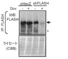
- FIG. 1A KB cells bearing Tet-On shFLASH (shRNA against FLASH) or shlacZ (shRNA against lacZ) were cultured for 3 days with or without 1 ⁇ g / ml Dox.
- Cell lysates were immunoprecipitated (IP) with anti-FLASH monoclonal antibody and analyzed by Western blotting (WB) using the same anti-FLASH monoclonal antibody. To confirm the total protein content, whole cell lysates were separated in SDS-PAGE and the gel was stained with Coomassie Brilliant Blue (CBB). Arrow, FLASH; *, non-specific band.
- IP immunoprecipitated
- WB Western blotting
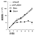
- KB cells with Tet-On shlacZ or shFLASH were treated with 1 ⁇ g / ml Dox for the indicated period.
- the cell number was quantified by trypan blue dye exclusion method using a hemocytometer. Representative data from three independent trials, all performed in triplicate. Error bars mean ⁇ s.e.m.
- KB cells with shFLASH expressing various mFLASH mutants fused with EGFP were treated with Dox for 3 days.
- Cell lysates were immunoprecipitated with anti-GFP antibody and analyzed by Western blotting (WB) using anti-FLASH antibody.
- WB Western blotting
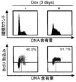
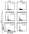
- the KB cells described in FIGS. 1A and 1E were treated with Dox for 3 days, stained with PI, and analyzed by flow cytometry. The percentage of cells in S phase are shown.
- 293T cells were transfected with an expression vector encoding mFLASH-EGFP and an empty vector ( ⁇ ) or an expression vector encoding mFLASH-FLAG3, ⁇ CC mFLASH-FLAG3 or ⁇ N194 mFLASH-FLAG3.
- mFLASH immunoprecipitates obtained with anti-Flag agarose resin or anti-GFP antibody were analyzed by Western blotting using anti-Flag antibody and anti-GFP antibody.
- 2A-E show that the interaction between FLASH and NPAT is not required for S phase progression. (FIG.
- 293T cells were transfected with empty vector ( ⁇ ) or mFLASH-EGFP, ⁇ N mFLASH-EGFP, ⁇ C1 mFLASH-EGFP, or ⁇ C2 mFLASH-EGFP.
- IP immunoprecipitate
- WB Western blotting
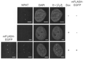
- KB cells with Tet-On shFLASH with or without mFLASH-EGFP are cultured for 3 days with or without 1 ⁇ ⁇ ⁇ g / ml Dox and NPAT stained with anti-NPAT antibody did.
- NPAT (red) and EGFP (green) were observed under a confocal fluorescence microscope.
- DNA was stained with DAPI (blue). Scale bar, 5 mm.
- KB cells with Tet-On shFLASH, with or without mFLASH-EGFP are cultured with or without 1 ⁇ g / ml Dox for 3 days, and Coilin is stained with anti-Coilin antibody did. Coilin (red) and EGFP (green) were observed under a confocal fluorescence microscope.
- FIGS. 3A-G show that ARS2 required for S phase progression interacts with FLASH.
- FIG. 3A KB cells with Tet-On shARS2 infected with a lentiviral vector encoding FLAG3-mutARS2 (ARS2 with silent mutation) were treated with 1 ⁇ g / ml Dox for 6 days. Immunoprecipitation was performed using anti-FLASH antibody, anti-FLAG agarose resin or control IgG, and the immunoprecipitate was analyzed by Western blotting (WB) using anti-FLASH monoclonal antibody or anti-Flag M2 monoclonal antibody.
- WB Western blotting
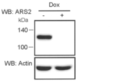
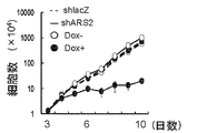
- KB cells with Tet-On shARS2 were cultured for 6 days with or without 1 ⁇ g / ml Dox, and knockdown of ARS2 was confirmed by Western blotting using anti-ARS2 polyclonal antibody. Actin was also detected as a loading control. Representative results from three independent tests are shown. KB cells with Tet-On shlacZ or shARS2 were treated with 1 ⁇ g / ml Dox for the indicated period. The cell number was quantified by trypan blue dye exclusion using a hemocytometer. Representative results from three independent trials, all performed in triplicate. Error bars indicate ⁇ s.e.m.
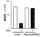
- KB cells with Tet-On shARS2 are infected with an expression vector encoding flag-mutARS2 or empty vector (Vector), seeded in a 24-well plate at 1.0 ⁇ 10 3 cells / well, and 1 ⁇ g / ml Dox Cultured for 9 days. The cell number was quantified by trypan blue dye exclusion method using a hemocytometer. Representative results from three independent trials, all performed in triplicate. Error bars indicate ⁇ sem. Cell cycle analysis using cells pulse-labeled with BrdU.
- KB cells bearing Tet-On® shARS2 were pulse-labeled with 10 ⁇ M BrdU for 30 minutes, stained with FITC-conjugated anti-BrdU antibody and PI, and analyzed by flow cytometry. The percentage of BrdU positive cells in S phase is shown. Representative data from three independent trials are shown. The cells in FIG. 3E were stained with anti-NPAT antibody or anti-Coilin antibody, and NPAT foci (upper) or Coilin foci (lower) were counted. Error bars mean ⁇ s.e.m.
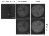
- KB cells with Tet-On shARS2 expressing both mFLASH-EGFP and HA-mutARS were treated with 1 ⁇ g / ml Dox for 6 days, stained with anti-HA antibody (red), and under a confocal fluorescence microscope Observed. DNA was stained with DAPI (blue). Scale bar, 5 mm.
- 3A to 3G are originally color drawings.
- Figures 4A-D show that ARS2 interacts with FLASH through the ARS2 domain. (FIG.
- KB cells harboring Tet-OnshARS2 were infected with an expression vector encoding DsRed-monomer-mutARS2 (DsRed-mutARS2) or an empty vector (Vector), and 1.25 ⁇ 10 3 cells / well in a 24-well plate. Seeded and cultured with 1 ⁇ g / ml Dox for 9 days. The cell number was quantified by trypan blue dye exclusion method using a hemocytometer. Representative data from 3 independent trials, all performed in triplicate. Error bars indicate ⁇ sem.
- the KB cells described in FIG. 4A were treated with or without 1 ⁇ g / ml Dox for 6 days and NPAT was stained with anti-NPAT antibody.
- FIGS. 5A-D show that FLASH interacts with ARS2 through its central region.
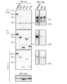
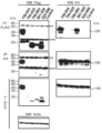
- FIG. 5A Schematic overview of mFLASH and its various deletion mutants. The number of the amino acid residue in mFLASH is shown. 293T was transiently transfected with an empty vector (Control) or an expression vector encoding mFLASH-FLAG3 or the indicated deletion mutant thereof together with an expression vector encoding HA-ARS2.
- Immunoprecipitates were analyzed by Western blotting. Actin was also detected as a loading control. Representative results from three independent trials are shown. **, IgG heavy chain; ***, IgG light chain. Alignment in the central region of human, mouse, bovine and chicken FLASH. Identical and similar amino acids are shown in black and shaded frames, respectively. The highly conserved area is surrounded by a dotted line. 6A-F show that the interaction between FLASH and ARS2 via the FARB region is required for S phase progression. (FIG. 6A) Schematic overview of mFLASH deletion mutants.
- ARS2 and EGFP or FARB-EGFP were immunoprecipitated using anti-HA antibody and anti-GFP antibody, respectively. Immunoprecipitates were analyzed by Western blotting. Actin was also detected as a loading control. Representative data from three independent trials are shown.
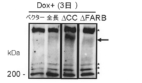
- KB cells bearing Tet-On shFLASH were infected with an empty vector (Vector) or a lentiviral expression vector encoding mFLASH-EGFP, ⁇ CC mFLASH-EGFP or ⁇ FARB mFLASH-EGFP. Cells were cultured with 1 ⁇ g / ml Dox for 3 days and lysed for Western blotting using anti-FLASH monoclonal antibody.
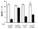
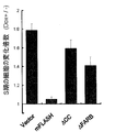
- FIG. 6D The KB cells shown in FIG. 6D were seeded in a 24-well plate at 2.5 ⁇ 10 3 cells / well and cultured with 1 ⁇ g / ml Dox for 3 days. The cell number was quantified by trypan blue dye exclusion using a hemocytometer. Representative data from three independent trials, all performed in triplicate. Error bars mean ⁇ sem.
- the cells described in FIG. 6E were pulse labeled with BrdU for 30 minutes and the percentage of S phase cells (BrdU positive cells) was determined by flow cytometry. The fold change, ie the percentage of cells expressing shFLASH (Dox +) relative to cells not expressing shFLASH (Dox ⁇ ) in S phase was calculated.
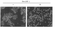
- FIG. 7A KB cells with Tet-On shFLASH were cultured for 3 days with or without 1 ⁇ g / ml Dox and observed under a phase contrast microscope. Scale bar, 100 mm. After culturing with or without 1 ⁇ g / ml Dox, the volume of KB cells with Tet-On shFLASH was analyzed by flow cytometry. FS, forward scatter; SS, side scatter. Northern blotting of replication-dependent histone mRNA in KB cells with Tet-On shFLASH.
- FIGS. 9A and B show the restored expression of exogenous ARS in ARS2 knockdown KB cells.
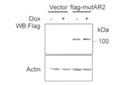
- FIG. 9A KB cells with Tet-On shARS2 stably expressing an empty vector (Vector) or flag-mutARS2 were treated with or without 1 ⁇ g / ml Dox for 6 days before anti-antibody treatment. Analysis was performed by Western blotting using a Flag antibody. Actin was also analyzed as a loading control. Representative results from three independent trials are shown.
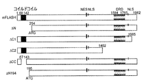
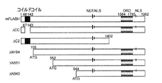
- FIG. 10A Schematic overview of full-length mouse FLASH (mFLASH) and its various deletion mutants. The number of the amino acid residue in mFLASH is shown.
- NLS nuclear localization signal
- NES nuclear export signal
- DRD DED recruiting domain.
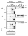
- 293T cells were transiently transfected with an empty vector (Control) or an expression vector encoding mFLASH-FLAG3 or the indicated deletion mutant of mFLASH-FLAG3 together with an expression vector encoding HA-ARS2.
- various mFLASH-FLAG3 deletion mutants or HA-ARS2 were immunoprecipitated using anti-FLAG resin and anti-HA antibody, respectively. Immunoprecipitates were analyzed by Western blotting using anti-Flag antibody or anti-HA antibody. Actin was also detected as a loading control. Representative results from three independent trials are shown.
- NC untransfected 293T cells.
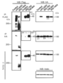
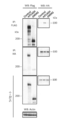
- 293T cells were transiently transfected with an empty vector (Control) or an expression vector encoding mFLASH-FLAG3 or the indicated deletion mutant of mFLASH-FLAG3 together with an expression vector encoding HA-ARS2. After 48 hours, various mFLASH-FLAG3 deletion mutants or HA-ARS2 were immunoprecipitated using anti-FLAG resin and anti-HA antibody, respectively. Immunoprecipitates were analyzed by Western blotting using anti-Flag antibody or anti-HA antibody. Actin was also detected as a loading control. Representative results from three independent trials are shown. NC, untransfected 293T cells.
- 293T cells were transiently transfected with an empty vector (Control) or an expression vector encoding mFLASH-FLAG3 or the indicated deletion mutant of mFLASH-FLAG3 together with an expression vector encoding HA-ARS2.
- various mFLASH-FLAG3 deletion mutants or HA-ARS2 were immunoprecipitated using anti-FLAG resin and anti-HA antibody, respectively. Immunoprecipitates were analyzed by Western blotting using anti-Flag antibody or anti-HA antibody. Actin was also detected as a loading control. Representative results from three independent trials are shown. NC, untransfected 293T cells. It is the result of the immunoprecipitation test which shows the coupling
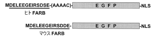
- hFARB-EGFP and mFARB-EGFP are schematically shown.
- the effect of hFARB-EGFP and mFARB-EGFP on the proliferation of KB cells is shown.
- a 4 C means a linker.
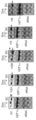
- mutation of the mutant FARB and ARS by coimmunoprecipitation is shown.
- the numbers shown above each lane correspond to each mutant FARB sequence shown in Table 3.
- IP represents the antigen recognized by the antibody used for co-immunoprecipitation, and WB represents the antigen recognized by the antibody used for Western blotting.
- mutation of the mutant FARB and ARS by coimmunoprecipitation is shown.
- IP represents the antigen recognized by the antibody used for co-immunoprecipitation
- WB represents the antigen recognized by the antibody used for Western blotting.
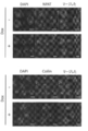
- the effect of 758-944 aa-EGFP-NLS on the S phase progression of KB cells is shown.
- polypeptide containing FARB region As shown in Examples below, the association of FLASH and ARS2 plays an important role in cell cycle progression (especially S phase progression). The specific region included in FLASH is essential for this meeting.
- the present invention areas of essential in FLASH meetings to this ARS2 provides polypeptides comprising (hereinafter, FARB region (F LASH- abbreviated as AR S2 b inding region)).
- the polypeptide of the present invention is useful as a cytostatic agent based on the inhibition of FLASH-ARS2 interaction as described in detail below, or as a reagent used for screening thereof.
- FLASH FLICE-associated huge protein
- CASP8AP2 FLICE-associated huge protein
- ARS2 arsenite resistance protein 2
- FLASH and ARS2 usually mean those derived from warm-blooded animals (mammals or birds).
- mammals include, for example, laboratory animals such as rodents and rabbits such as mice, rats, hamsters, and guinea pigs, domestic animals such as pigs, cows, goats, horses, sheep and minks, pets such as dogs and cats, humans, Examples include, but are not limited to, primates such as monkeys, cynomolgus monkeys, rhesus monkeys, marmosets, orangutans and chimpanzees.
- FLASH and ARS2 are preferably derived from mammals, and more preferably derived from primates (such as humans) or rodents (such as mice).
- nucleotide sequence and amino acid sequence of FLASH are known. Representative nucleotide sequences and amino acid sequences of human, mouse, bovine and chicken FLASH are registered in NCBI as follows.
- Nucleotide sequence (cDNA sequence) Accession number NM_001137668 (version NM_001137668.1) (SEQ ID NO: 1), Accession number NM_012115 (version NM_012115.3) (SEQ ID NO: 19)
- Amino acid sequence Accession number NP_001131140 (version NP_001131140.1) (SEQ ID NO: 2), Accession number NP_036247 (version NP_036247.1) (SEQ ID NO: 20)
- [Mouse FLASH] Nucleotide sequence (cDNA sequence): Accession number NM_011997 (Version NM_011997.2) (SEQ ID NO: 3) Amino acid sequence: Accession number NP_036127 (version NP_036127.2) (SEQ ID NO
- nucleotide sequence and amino acid sequence of ARS2 are known. Representative nucleotide sequences and amino acid sequences of human and mouse ARS2 are registered in NCBI as follows.
- the FARB region is (1) a region consisting of the amino acid sequence represented by SEQ ID NO: 5; (2) a region comprising an amino acid sequence having 69% or more identity with the amino acid sequence represented by SEQ ID NO: 5 and having an activity of binding to ARS2 polypeptide; and (3) an amino acid represented by SEQ ID NO: 5 It means any region selected from the group consisting of a region having an amino acid sequence in which 1 to 4 amino acids are deleted, substituted, inserted or added in the sequence and having an activity of binding to ARS2 polypeptide.
- the amino acid sequence represented by SEQ ID NO: 5 is the amino acid sequence of the FARB region of human FLASH, and corresponds to amino acids 931 to 943 of the amino acid sequence of human FLASH (SEQ ID NO: 2).
- the FARB region has an activity of binding to ARS2 polypeptide.
- the binding is strong enough to allow co-precipitation with an ARS2 polypeptide in an immunoprecipitation test.
- the presence or absence of such binding activity is determined by co-expressing a polypeptide containing the region to be evaluated and ARS2 polypeptide in human cells (eg, KB cells), preparing a cell lysate with an appropriate solubilization buffer, It can be confirmed by performing an immunoprecipitation test with an antibody that specifically recognizes the peptide on the lysate.
- both the polypeptide containing the region to be evaluated and the ARS2 polypeptide are found in the immunoprecipitate, it can be evaluated that the region has an activity to bind to the ARS2 polypeptide.
- polypeptide of the present invention Since the polypeptide of the present invention has a FARB region, it has an activity of binding to the ARS2 polypeptide.
- the amino acid sequence of the region (2) is an amino acid having 69% or more, preferably 76% or more, more preferably 84% or more, and still more preferably 92% or more identity with the amino acid sequence represented by SEQ ID NO: 5. Is an array.
- identity refers to an optimal alignment when two amino acid sequences are aligned using a mathematical algorithm known in the art (preferably the algorithm is an optimal alignment). Is the ratio of the same amino acid residue to the total overlapping amino acid residues in which one may consider the introduction of a gap into one or both of the sequences.
- NCBI blastp / Blast 2 sequences National Center for Biotechnology Information Basic Local Alignment Search Tool
- the amino acid sequence of the above region (3) has 1 to 4 (preferably 1 to 3, more preferably 1 or 2, most preferably 1) amino acids in the amino acid sequence represented by SEQ ID NO: 5.
- Deletion, substitution, insertion or addition of amino acid sequence for example, (1) 1 to 4 (preferably 1 to 3, more preferably 1 or 2 in the amino acid sequence shown in SEQ ID NO: 5, most preferably Is an amino acid sequence in which one amino acid is deleted, (2) 1 to 4 (preferably 1 to 3, more preferably 1 or 2 and most preferably 1) in the amino acid sequence shown in SEQ ID NO: 5 ) Amino acid sequence added, and (3) 1 to 4 (preferably 1 to 3, more preferably 1 or 2, most preferably 1) amino acids in the amino acid sequence shown in SEQ ID NO: 5.
- Amino acid sequence inserted with (4) sequence An amino acid sequence in which 1 to 4 (preferably 1 to 3, more preferably 1 or 2, most preferably 1) amino acids in the amino acid sequence shown in No. 5 are substituted with other amino acids, or ( 5) Amino acid sequence in which the mutations of (1) to (4) above are combined (in this case, the sum of the mutated amino acids is 1 to 4, preferably 1 to 3, more preferably 1 or 2, most preferably Is 1).
- the fourth, fifth and seventh glutamic acid residues in the amino acid sequence represented by SEQ ID NO: 5 are conserved. More preferably, the amino acid sequence in the region from the fourth glutamic acid to the seventh glutamic acid in the amino acid sequence represented by SEQ ID NO: 5 is preferably conserved. Deletion, substitution, insertion or addition of amino acid residues in this region is likely to reduce or lose binding activity to the ARS2 polypeptide. On the other hand, deletion, substitution, insertion or addition (particularly substitution) of amino acid residues in the amino acid sequence represented by SEQ ID NO: 5 other than this region is unlikely to reduce or lose the binding activity to the ARS2 polypeptide. .
- amino acid sequences in the above regions (2) and (3) include, for example, the amino acid sequences of mouse, bovine and chicken FARB regions (SEQ ID NOs: 6, 7 and 8, respectively).
- the amino acid sequence of the mouse FARB region consists of an amino acid sequence in which one amino acid is substituted in the amino acid sequence of the human FARB region, and has 92.3% (12/13) identity with the amino acid sequence of the human FARB region.
- the amino acid sequence of the bovine FARB region consists of an amino acid sequence in which two amino acids are substituted in the amino acid sequence of the human FARB region, and has 84.6% (11/13) identity with the amino acid sequence of the human FARB region.
- the amino acid sequence of the FARB region of chicken consists of an amino acid sequence in which 4 amino acids are substituted in the amino acid sequence of the human FARB region, and has 69.2% (9/13) identity with the amino acid sequence of the human FARB region. .
- amino acid sequences in the above regions (2) and (3) include amino acid sequences represented by SEQ ID NOs: 27, 28, 30, or 32-36.
- the length of the polypeptide of the present invention is not particularly limited as long as it has binding activity to the ARS2 polypeptide, and a polypeptide having a desired length can be selected according to the purpose of use.
- the polypeptide of the present invention has a length of 3000 amino acids or less, 2000 amino acids or less, 1000 amino acids or less, 500 amino acids or less, 400 amino acids or less, 300 amino acids or less, 250 amino acids The following, 200 amino acids or less, 150 amino acids or less, 100 amino acids or less, 90 amino acids or less, 80 amino acids or less, 70 amino acids or less, 60 amino acids or less, 50 amino acids or less 40 amino acids or less, 30 amino acids or less, 20 amino acids or less, 19 amino acids or less, 18 amino acids or less, 17 amino acids or less, 16 amino acids or less, 15 amino acids or less, It is possible to select those with 14 amino acids or less as appropriate. That.
- the polypeptide of the present invention has a length of 9 amino acids or more, preferably 11 amino acids or more, and most
- the amino acid sequence of the portion other than the FARB region in the polypeptide of the present invention is not particularly limited as long as the polypeptide of the present invention has binding activity to the ARS2 polypeptide.
- a fusion polypeptide in which at least one tag polypeptide or signal sequence is fused to the N-terminus and / or C-terminus of the polypeptide of the present invention is also included in one of the embodiments of the polypeptide of the present invention.
- the tag polypeptide refers to a polypeptide that is added to facilitate the detection and purification of the polypeptide.
- tag polypeptides include, but are not limited to, epitope tags, fluorescent polypeptides, immunoglobulin Fc regions, and the like.
- An epitope tag refers to a peptide that is specifically recognized by an antibody or other binding partner, specifically, a Flag tag, a polyhistidine tag, a c-Myc tag, an HA tag, an AU1 tag, a GST tag, an MBP tag. Etc. can be mentioned.
- the fluorescent polypeptide include GFP, YFP, RFP, CFP, BFP, EGFP and the like.
- tag polypeptides are well known to those skilled in the art, and various antibodies that specifically recognize the tag polypeptides are commercially available.
- the signal sequence refers to a polypeptide sequence that directs the transport and localization of the polypeptide from the synthesis site to a specific site inside the cell or outside the cell simultaneously with or after translation of the polypeptide.
- the signal sequence include a leader sequence that induces secretion of the polypeptide, a nuclear translocation signal sequence (for example, a nuclear translocation signal sequence of SV40 T antigen), a nuclear translocation signal sequence, a nucleolus localization signal, and the like. However, it is not limited to this.
- Such signal sequences are well known to those skilled in the art, and can be appropriately selected according to the purpose.
- the polypeptide of the present invention and the tag polypeptide or signal sequence are linked directly or via a linker.
- a linker refers to an amino acid sequence that covalently joins two polypeptide domains. By binding via a linker, freedom of rotation is provided to the linked polypeptide domains. Therefore, the binding activity of the polypeptide of the present invention to the ARS2 polypeptide is restricted due to the steric structure restriction by the tag polypeptide or signal sequence. The risk of being attenuated can be avoided.
- the length of the linker is not particularly limited as long as the polypeptide has binding activity to the ARS2 polypeptide, but is usually 1 to 25 amino acids, preferably 1 to 10 amino acids, and is well known in the art.
- the amino acid sequence of the linker is not particularly limited as long as the polypeptide has a binding activity to the ARS2 polypeptide.
- Specific examples of the amino acid sequence of the linker include, but are not limited to, AAAAC (SEQ ID NO: 16), glycine (4) -serine spacer (Chaudhary et al. (1989)), and the like.
- the length of the polypeptide is as long as it has binding activity to the ARS2 polypeptide.
- a polypeptide having a desired length can be selected according to the purpose of use.
- the length of the polypeptide other than the additional portion is 3000 amino acids or less, 2000 amino acids or less, 1000 amino acids or less, 500 amino acids or less, 400 amino acids The following, 300 amino acids or less, 250 amino acids or less, 200 amino acids or less, 150 amino acids or less, 100 amino acids or less, 90 amino acids or less, 80 amino acids or less, 70 amino acids or less One, 60 amino acids or less, 50 amino acids or less, 40 amino acids or less, 30 amino acids or less, 20 amino acids or less, 19 amino acids or less, 18 amino acids or less, 17 amino acids or less, Less than 16 amino acids, less than 15 amino acids Can be suitably selected such as those below 14 amino acids (Incidentally, when using the linker, the length indicates the length excluding the linker moiety).
- the length of the polypeptide other than the fused additional peptide is 9 amino acids or more, preferably 11 amino acids or more, and most preferably 13 amino acids or more.
- the polypeptide of the present invention binds to the ARS2 polypeptide, it is possible to inhibit the association between FLASH and ARS2 in the cell (preferably in the nucleus).
- the association between FLASH and ARS2 plays an important role in cell cycle progression (especially S phase progression).
- the polypeptide of the present invention has an activity of suppressing cell proliferation. This embodiment is referred to as “an embodiment having cell growth inhibitory activity”.
- the cell in which the polypeptide of this embodiment can suppress the growth is not particularly limited as long as it expresses FLASH and ARS2, but is usually a warm-blooded animal (mammal or avian) cell. Specific examples of mammals and birds are as described above.
- the cells are usually mammalian cells, more preferably primate (human etc.) or rodent (mouse etc.) cells.
- the presence or absence of cytostatic activity is determined by introducing the polypeptide to be evaluated into cells that can be grown using an expression vector (for example, KB cells), culturing in an appropriate medium for 3 days, It can be evaluated by analyzing whether the number of cells decreases in comparison. When the number of cells decreases compared to the empty vector introduction group, it can be determined that the polypeptide to be evaluated has a cell growth inhibitory activity.
- an expression vector for example, KB cells
- the polypeptide of this embodiment does not substitute for the activity of FLASH that promotes cell cycle progression (especially S phase progression).
- the presence or absence of this property is determined by introducing the polypeptide to be evaluated using an expression vector into a cell (FLASH knockdown cell) (for example, a KB cell) in which FLASH expression is suppressed by a specific siRNA against FLASH, and introducing an empty vector It can be evaluated by analyzing whether or not the proportion of S-phase cells is reduced by flow cytometry compared to the group.
- FLASH knockdown cells accumulate in S phase, but if the proportion of cells in S phase is significantly reduced compared to the empty vector introduction group, the polypeptide to be evaluated has an activity that substitutes for the function of FLASH. You can judge that you are doing.
- the polypeptide of this aspect preferably does not have the coiled-coil domain of FLASH. Since this polypeptide cannot associate with FLASH, it cannot replace the function of FLASH.
- a region consisting of the amino acid sequence represented by SEQ ID NO: 14 (1) a region consisting of the amino acid sequence represented by SEQ ID NO: 14; (2) a region comprising an amino acid sequence having 85% or more identity with the amino acid sequence represented by SEQ ID NO: 14 and having an activity of binding to a FLASH polypeptide; and (3) an amino acid represented by SEQ ID NO: 14 It means any region selected from the group consisting of a region having an amino acid sequence in which 1 to 9 amino acids are deleted, substituted, inserted or added in the sequence and having an activity of binding to FLASH polypeptide.
- the amino acid sequence represented by SEQ ID NO: 14 is the amino acid sequence of the coiled-coil domain of human FLASH, and corresponds to amino acids 68 to 142 of the amino acid sequence of human FLASH (SEQ ID NO: 2).
- the coiled coil domain of FLASH has an activity to bind to FLASH polypeptide.
- the binding is strong enough to allow co-precipitation in an immunoprecipitation test.
- the presence or absence of such binding activity is determined by co-expressing the polypeptide containing the region to be evaluated and the FLASH polypeptide in human cells, preparing a cell lysate with an appropriate solubilization buffer, and specifically expressing one of the polypeptides. It can be confirmed by performing an immunoprecipitation test with the antibody to be recognized on the lysate.
- both the polypeptide containing the region to be evaluated and the FLASH polypeptide are found in the immunoprecipitate, it can be evaluated that the region has an activity to bind to the FLASH polypeptide.
- the amino acid sequence of the region (3) is an amino acid sequence in which 1 to 9 amino acids are deleted, substituted, inserted or added in the amino acid sequence represented by SEQ ID NO: 14, for example, (1) SEQ ID NO: 14 An amino acid sequence in which 1 to 9 amino acids in the amino acid sequence shown are deleted, (2) an amino acid sequence in which 1 to 9 amino acids are added to the amino acid sequence shown in SEQ ID NO: 14, and (3) SEQ ID NO: 14 An amino acid sequence in which 1 to 9 amino acids are inserted into the amino acid sequence shown in (4), (4) an amino acid sequence in which 1 to 9 amino acids in the amino acid sequence shown in SEQ ID NO: 14 are substituted with other amino acids, or (5) An amino acid sequence in which the above mutations (1) to (4) are combined (in this case, the total number of mutated amino acids is 1 to 9).
- amino acid sequence of the coiled-coil domain of mouse FLASH (SEQ ID NO: 15) can be mentioned.
- the amino acid sequence of the coiled-coil domain of mouse FLASH is an amino acid sequence in which 9 amino acids are substituted in the amino acid sequence of the coiled-coil domain of human FLASH, and is 88.0% (66/75) identical to the amino acid sequence of the human FARB region. Have sex.
- the polypeptide of the present invention may be modified.
- modifications include lipid chain addition (aliphatic acylation (palmitoylation, myristoylation, etc.), prenylation (farnesylation, geranylgeranylation, etc.), phosphorylation (serine residue, threonine residue, tyrosine residue) Phosphorylation), acetylation, addition of sugar chain (N-glycosylation, O-glycosylation) and the like.
- the polypeptide of the present invention contains an appropriate labeling agent such as a radioisotope (eg, 125 I, 131 I, 3 H, 14 C, etc.), an enzyme (eg, ⁇ -galactosidase, ⁇ -glucosidase, alkaline phosphatase). , Peroxidase, malate dehydrogenase, etc.), fluorescent materials (eg, fluorescamine, fluorescein isothiocyanate, etc.), luminescent materials (eg, luminol, luminol derivatives, luciferin, lucigenin, etc.), affinity tags (eg, biotin) Etc.).
- a radioisotope eg, 125 I, 131 I, 3 H, 14 C, etc.
- an enzyme eg, ⁇ -galactosidase, ⁇ -glucosidase, alkaline phosphatase.
- fluorescent materials eg, fluorescamine, fluor
- polypeptide is used to mean a salt thereof.
- Polypeptide salts include salts with physiologically acceptable acids (eg, inorganic acids, organic acids) and bases (eg, alkali metal salts), and particularly physiologically acceptable acid addition salts. preferable.
- Such salts include, for example, salts with inorganic acids (eg hydrochloric acid, phosphoric acid, hydrobromic acid, sulfuric acid) or organic acids (eg acetic acid, formic acid, propionic acid, fumaric acid, maleic acid, succinic acid). Acid, tartaric acid, citric acid, malic acid, succinic acid, benzoic acid, methanesulfonic acid, benzenesulfonic acid) and the like.
- polypeptide of the present invention examples include, for example, a FLASH polypeptide, a partial FLASH polypeptide containing a FARB region, a fusion polypeptide of these polypeptides with a tag polypeptide and / or a signal sequence, and the like.
- the polypeptide of the present invention is preferably isolated or purified. “Isolation or purification” means that an operation for removing components other than the target component has been performed.
- the purity of the isolated or purified polypeptide of the present invention is usually 50% or more, preferably 70% or more, more preferably 90% or more, Most preferably, it is 95% or more (for example, substantially 100%).
- the method for producing the polypeptide of the present invention is not particularly limited, and may be produced according to a known peptide synthesis method or may be produced using a known genetic recombination technique.
- the peptide synthesis method may be, for example, either a solid phase synthesis method or a liquid phase synthesis method. If the partial peptide or amino acid that can constitute the polypeptide of the present invention is condensed with the remaining portion, and the product has a protecting group, the protecting polypeptide can be eliminated to produce the desired polypeptide.
- polypeptide of the present invention When the polypeptide of the present invention is produced using a gene recombination technique, first, a polynucleotide encoding the polypeptide of the present invention as described below is obtained, and the host is transformed with an expression vector capable of expressing the polynucleotide.
- the polypeptide can be produced by transforming and culturing the resulting transformant.
- the method for producing the polypeptide of the present invention using the polynucleotide and gene recombination techniques will be described later in this specification.
- the present invention provides a polynucleotide comprising a nucleotide sequence encoding the polypeptide of the present invention.
- the polynucleotide may be DNA or RNA, or may be a DNA / RNA chimera.
- the polynucleotide may be double-stranded or single-stranded. In the case of a double strand, it may be a double-stranded DNA, a double-stranded RNA or a DNA: RNA hybrid.
- polynucleotide of the present invention examples include a polynucleotide comprising the nucleotide sequence represented by SEQ ID NO: 17 or 18.
- the nucleotide sequence represented by SEQ ID NO: 17 is a polypeptide comprising a human FARB region (SEQ ID NO: 5)
- the nucleotide sequence represented by SEQ ID NO: 18 is a polypeptide comprising a mouse FARB region (SEQ ID NO: 6).
- the polynucleotide of the present invention can be easily produced by using a known gene recombination technique based on known sequence information or sequence information described in the sequence listing of the present specification. For example, by designing an appropriate primer based on sequence information, amplifying DNA encoding a component constituting the polypeptide of the present invention by PCR, and ligating the DNA fragment using an appropriate enzyme such as ligase, The polypeptide of the present invention can be produced. Or you may synthesize
- the obtained polynucleotide of the present invention can be used as it is depending on the purpose, or after digestion with a restriction enzyme or addition of a linker as desired.
- the polynucleotide may have ATG as a translation initiation codon on the 5 'end side, and may have TAA, TGA or TAG as a translation stop codon on the 3' end side. These translation initiation codon and translation termination codon can be added using an appropriate synthetic DNA adapter.
- the polynucleotide of the present invention is preferably isolated or purified.
- the present invention provides a vector comprising the polynucleotide of the present invention.
- the vector is preferably isolated or purified.
- the vector include an expression vector and a cloning vector, and can be selected according to the purpose.
- the vector is an expression vector.
- the expression vector can express the polypeptide of the present invention.
- the expression vector can be produced by operably linking the polynucleotide of the present invention downstream of a promoter in an appropriate expression vector.
- the vector include plasmid vectors and virus vectors, and can be appropriately selected depending on the host to be used.
- Examples of the host include Escherichia bacteria (Escherichia coli), Bacillus bacteria (Bacillus subtilis, etc.), yeast (Saccharomyces cerevisiae, etc.), insect cells (night theft) Straw larvae-derived cell lines (Spodoptera frugiperda cells; Sf cells), insects (larvae of silkworms), mammalian cells (rat neurons, monkey cells (COS-7, etc.), Chinese hamster cells (CHO cells, etc.) ) Etc.) are used.
- mammals include, for example, laboratory animals such as rodents and rabbits such as mice, rats, hamsters, and guinea pigs, domestic animals such as pigs, cows, goats, horses, sheep and minks, pets such as dogs and cats, humans, Examples include, but are not limited to, primates such as monkeys, cynomolgus monkeys, rhesus monkeys, marmosets, orangutans and chimpanzees.
- Plasmid vectors derived from Escherichia coli eg, pBR322, pBR325, pUC12, pUC13
- plasmid vectors derived from Bacillus subtilis eg, pUB110, pTP5, pC194
- yeast-derived plasmid vectors eg, pSH19, pSH15
- the type of virus vector can be appropriately selected according to the type of host used and the purpose of use. For example, when insect cells are used as the host, baculovirus vectors can be used. When mammalian cells are used as hosts, Moloney murine leukemia virus vectors, lentivirus vectors, Sindbis virus vectors and other retrovirus vectors, adenovirus vectors, herpes virus vectors, adeno-associated virus vectors, parvovirus vectors, Vaccinia virus vectors, Sendai virus vectors, and the like can be used.
- a promoter that can start transcription in the host can be selected according to the type of host to be used. For example, when the host is Escherichia, trp promoter, lac promoter, T7 promoter and the like are preferable. When the host is Bacillus, SPO1 promoter, SPO2 promoter, penP promoter and the like are preferable. When the host is yeast, PHO5 promoter, PGK promoter and the like are preferable. When the host is an insect cell, a polyhedrin promoter, a P10 promoter and the like are preferable. When the host is a mammalian cell, a subgenomic (26S) promoter, CMV promoter, SR ⁇ promoter and the like are preferable.
- the vector of the present invention may contain an enhancer, a splicing signal, a poly A addition signal, a selection marker, an SV40 replication origin (hereinafter sometimes abbreviated as SV40ori) and the like in a functional manner, if desired.
- selectable markers include dihydrofolate reductase (hereinafter sometimes abbreviated as dhfr) gene [methotrexate (MTX) resistance], ampicillin resistance gene (hereinafter sometimes abbreviated as Amp r ), neomycin resistance gene (hereinafter sometimes abbreviated as Neo r, G418 resistance).
- the above-mentioned host according to the present invention can be prepared by the gene transfer method known per se (for example, lipofection method, calcium phosphate method, microinjection method, protoplast fusion method, electroporation method, DEAE dextran method, gene transfer method using Gene Gun).
- a transformant into which the vector has been introduced (transformant of the present invention) can be produced.
- the transformant can express the polypeptide of the present invention.
- the transformant of the present invention is useful for producing the polypeptide of the present invention.
- the polypeptide of the present invention can be produced by culturing the transformant of the present invention by a method known per se according to the type of host and isolating the polypeptide of the present invention from the culture.
- the transformant whose host is Escherichia is cultured in an appropriate medium such as LB medium or M9 medium, usually at about 15 to 43 ° C. for about 3 to 24 hours.
- the transformant whose host is Bacillus is cultured in an appropriate medium, usually at about 30 to 40 ° C. for about 6 to 24 hours.
- Cultivation of the transformant whose host is yeast is usually carried out at about 20 ° C. to 35 ° C. for about 24 to 72 hours in a suitable medium such as a Burkholder medium.
- Cultivation of transformants whose host is an insect cell or an insect is carried out in a suitable medium such as Grace's Insect medium supplemented with about 10% bovine serum, usually at about 27 ° C for about 3 to 5 days.
- the transformant whose host is an animal cell is cultured in an appropriate medium such as a MEM medium supplemented with about 10% bovine serum, usually at about 30 ° C. to 40 ° C. for about 15 to 60 hours. In any culture, aeration and agitation may be performed as necessary.
- the cell lysate or the culture supernatant is subjected to a plurality of chromatography such as reverse phase chromatography, ion exchange chromatography, affinity chromatography and the like. Can be achieved.
- the present invention provides a cell growth inhibitor (cell growth inhibitor I) comprising the above-described polypeptide of the present invention (polypeptide containing the FARB region) or an expression vector capable of expressing the polypeptide. .
- the polypeptide containing the FARB region used for the cell growth inhibitor I is preferably the polypeptide of the present invention having the cell growth inhibitory activity in [1] above.
- cell growth inhibitor I is used to promote the transfer of the polypeptide containing the FARB region into the nucleus.
- the polypeptide containing the FARB region to be used preferably has a nuclear translocation signal.
- a substance having a molecular weight of 45000 Da or less, preferably a molecular weight of 40000 Da or less can easily penetrate the nuclear membrane and move from the cytoplasm into the nucleus.
- the molecular weight of the polypeptide to be included is preferably 45000 Da or less, more preferably 40000 Da or less.
- polypeptide containing the FARB region used for the cell growth inhibitor I may contain an additional sequence such as a protein-transduction domain (PTD) sequence for facilitating introduction of the polypeptide into the cell.
- PTD protein-transduction domain
- Examples of PTD include cell passage domains such as ANTENNAPEDIA, HIV / TAT, HSV / VP-22, polyarginine composed of 7 to 11 arginines, and the like.
- a polynucleotide (preferably DNA) encoding a polypeptide containing the above-mentioned FARB region is usually a promoter in a mammalian (preferably human) cell (eg, sarcoma cell) to be administered. It is operably linked to a promoter that can exert activity.
- the type of vector used for the expression vector used in the cytostatic I is not particularly limited, but suitable vectors for administration to mammals such as humans include viruses such as retroviruses, adenoviruses, and adeno-associated viruses.
- Vector adenovirus has advantages such as extremely high gene transfer efficiency and can be introduced into non-dividing cells.
- adeno-associated virus that is relatively high in gene transfer efficiency, can be introduced into non-dividing cells, and can be integrated into the chromosome via an inverted terminal repeat (ITR) Also preferred.
- ITR inverted terminal repeat
- the cell growth inhibitor I can contain any carrier, for example, a pharmaceutically acceptable carrier, in addition to a polypeptide containing the FARB region or an expression vector capable of expressing the same.
- Examples of pharmaceutically acceptable carriers include excipients such as sucrose and starch, binders such as cellulose and methylcellulose, disintegrants such as starch and carboxymethylcellulose, lubricants such as magnesium stearate and aerosil, citric acid, Fragrances such as menthol, preservatives such as sodium benzoate and sodium bisulfite, stabilizers such as citric acid and sodium citrate, suspensions such as methylcellulose and polyvinylpyrrolidone, dispersants such as surfactants, water, physiological Although diluents, such as salt solution, base wax, etc. are mentioned, it is not limited to them.
- excipients such as sucrose and starch
- binders such as cellulose and methylcellulose
- disintegrants such as starch and carboxymethylcellulose
- lubricants such as magnesium stearate and aerosil
- citric acid Fragrances such as menthol
- preservatives such as sodium benzoate and sodium bisulfite
- stabilizers
- the cell growth inhibitor I may further contain a nucleic acid introduction reagent in order to promote introduction of the polynucleotide into the cell.
- a nucleic acid introduction reagent in order to promote introduction of the polynucleotide into the cell.
- the polynucleotide is incorporated into a viral vector, particularly a retroviral vector, retronectin, fibronectin, polybrene, or the like can be used as a gene introduction reagent.
- lipofectin lipfectamine
- DOGS transfectum
- DOPE DOPE
- DOTAP DDAB
- DHDEAB DDAB
- DHDEAB DDAB
- HDEAB polybrene
- PEI poly (ethyleneimine) Cationic lipids such as
- Preparations suitable for oral administration include liquids, capsules, sachets, tablets, suspensions, emulsions and the like.
- Formulations suitable for parenteral administration include aqueous and non-aqueous isotonic sterile injection solutions, which include antioxidants Further, a buffer solution, an antibacterial agent, an isotonic agent and the like may be contained. Aqueous and non-aqueous sterile suspensions are also included, which may contain suspending agents, solubilizers, thickeners, stabilizers, preservatives and the like.
- the preparation can be enclosed in a container in unit doses or multiple doses like ampoules and vials.
- the active ingredient and a pharmaceutically acceptable carrier can be lyophilized and stored in a state that may be dissolved or suspended in a suitable sterile vehicle immediately before use.
- the content of the polypeptide containing the FARB region in the pharmaceutical composition or the expression vector capable of expressing the polypeptide is, for example, about 0.1 to 100% by weight of the whole pharmaceutical composition.
- the dose of the cytostatic I varies depending on the activity and type of the active ingredient, the severity of the disease, the species of animal to be administered, the drug acceptability of the administration target, body weight, age, etc.
- the amount of active ingredient per day for an adult is about 0.001 to about 500 mg / kg.
- the cytostatic agent I of the present invention is preferably a mammal (for example, rat, so that the above-mentioned polynucleotide as an active ingredient or an expression vector capable of expressing the same is delivered to a target cell.
- a mammal for example, rat
- the cell growth inhibitor I of the present invention is a disease caused by abnormal cell growth, for example, benign tumor (such as uterine fibroid); solid cancer (pharyngeal cancer, esophageal cancer, colon cancer, lung cancer, stomach cancer, pancreatic cancer, liver cancer, etc.
- benign tumor such as uterine fibroid
- solid cancer pharyngeal cancer, esophageal cancer, colon cancer, lung cancer, stomach cancer, pancreatic cancer, liver cancer, etc.
- Malignant tumors or cancers including glioma, leukemia, malignant lymphoma, etc .; nephritis (eg, glomerulonephritis), human autoimmune lymphocytes Proliferative syndrome, lymphoproliferative disorder, angioimmunoblastic lymphadenopathy, systemic lupus erythematosus, inflammatory bowel disease (Crohn's disease, ulcerative colitis, etc.), progressive systemic sclerosis, polymyositis (skin) Myositis), Sjogren's syndrome, myelodysplastic syndrome, scleroderma, rheumatoid arthritis, psoriasis, wound hyperplasia (eg, granulation), and the like.
- nephritis eg, glomerulonephritis
- human autoimmune lymphocytes Proliferative syndrome lymphoproliferative disorder, angioimmunoblastic lymphadenopathy, systemic
- Cell growth inhibitor II As shown in the examples described later, the binding of FLASH and ARS2 plays an important role in the progression of the cell cycle (especially the S phase progression), and suppresses the proliferation of cells by suppressing the expression of ARS2. Is possible. Accordingly, the present invention provides a cell growth inhibitor (cell growth inhibitor II) comprising an siRNA, an antisense nucleic acid, or an expression vector capable of expressing these polynucleotides that can specifically suppress ARS2 expression. Is.
- “Suppression of specific gene expression” means to suppress the expression of a target gene more strongly than the expression of other genes.
- siRNA capable of specifically suppressing ARS2 expression examples include (A) a nucleotide sequence of mRNA (mature mRNA or initial transcript) encoding ARS2, or a nucleotide sequence complementary to a partial sequence of 18 bases or more. A double-stranded RNA, and (B) a nucleotide sequence of 18 bases or more capable of hybridizing in an mRNA (mature mRNA or initial transcript) encoding ARS2 in a cell of a treated animal (preferably human), and Double-stranded RNA that suppresses ARS2 transcription by hybridizing Can be mentioned.
- nucleotide sequence of mRNA encoding ARS2 examples include a nucleotide sequence represented by SEQ ID NO: 9 (human ARS2) and a nucleotide sequence represented by SEQ ID NO: 11 (mouse ARS2).
- RNA interference RNA interference
- mRNA complementary to the RNA has been known in nematodes, insects, plants, etc. Recently, it has been confirmed that this phenomenon also occurs in animal cells [Nature, 411 (6836): 494-498 (2001)], and has attracted attention as an alternative to ribozyme.
- SiRNA is typically a double-stranded oligo RNA consisting of RNA having a sequence complementary to the nucleotide sequence of mRNA of the target gene or a partial sequence thereof (hereinafter referred to as target nucleotide sequence) and its complementary strand.
- target nucleotide sequence a sequence complementary to the nucleotide sequence of mRNA of the target gene or a partial sequence thereof (hereinafter referred to as target nucleotide sequence) and its complementary strand.
- first sequence complementary to a target nucleotide sequence first sequence
- second sequence complementary sequence
- RNA in which the first sequence forms a double-stranded structure with the second sequence small2hairpin RNA: shRNA
- the length of the portion complementary to the target nucleotide sequence contained in the siRNA is usually about 18 bases or more, preferably about 19 bases or more, more preferably about 21 bases or more. As long as it can be specifically suppressed, it is not particularly limited. If the siRNA is longer than 23 bases, the siRNA can be degraded in the cell to produce siRNA of about 20 bases, so theoretically the upper limit of the length of the portion complementary to the target nucleotide sequence is , The full length of the nucleotide sequence of the target gene mRNA (mature mRNA or early transcript).
- the length of the complementary portion is, for example, about 50 bases or less, preferably about 25 bases or less, and most preferably about 23 bases or less. is there. That is, the length of the complementary portion is usually about 18 to 50 bases, preferably about 19 to about 25 bases, more preferably about 21 to about 23 bases.
- each RNA strand constituting the siRNA is usually about 18 bases or more, preferably about 19 bases or more, more preferably about 21 bases or more, but it specifically suppresses the expression of the target gene.
- the length of siRNA is, for example, about 50 bases or less, preferably about 25 bases or less, and most preferably about 23 bases or less. That is, the length of each RNA strand is usually about 18 to 50 bases, preferably about 19 to about 25 bases, more preferably about 21 to about 23 bases.
- the length of shRNA shall be shown as the length of the double-stranded part when taking a double-stranded structure.
- the target nucleotide sequence and the sequence complementary to that contained in the siRNA are preferably completely complementary.
- the cleavage activity due to RNA interference is not completely lost. Partial activity may remain.
- the mutation of the base at the center of siRNA has a large influence, and the cleavage activity of mRNA due to RNA interference can be extremely reduced.
- SiRNA may have an additional base that does not form a base pair at the 5 'and / or 3' end.
- the length of the additional base is not particularly limited as long as siRNA can specifically suppress the expression of the target gene, but is usually 5 bases or less, for example, 2 to 4 bases.
- the additional base may be DNA or RNA, but the use of DNA can improve the stability of siRNA. Examples of such additional base sequences include ug-3 ', uu-3', tg-3 ', tt-3', ggg-3 ', guuu-3', gttt-3 ', ttttt-3 Examples include, but are not limited to, ', uuuuu-3'.
- the length of the loop portion of the shRNA hairpin loop is not particularly limited as long as the expression of the target gene can be specifically suppressed, but is usually about 5 to 25 bases.
- the nucleotide sequence of the loop part is not particularly limited as long as it can form a loop and the shRNA can specifically suppress the expression of the target gene.
- Antisense nucleic acid includes a nucleotide sequence that can hybridize to the target mRNA under physiological conditions of a cell that expresses the target mRNA (mature mRNA or early transcript), and the target mRNA in a hybridized state.
- a nucleic acid capable of inhibiting the translation of the polypeptide encoded by The type of the antisense nucleic acid may be DNA, RNA, or a DNA / RNA chimera, but is preferably DNA.
- Examples of the antisense nucleic acid that can specifically suppress the expression of ARS2 include (A) a nucleotide sequence of mRNA (mature mRNA or initial transcript) encoding ARS2, or a nucleotide sequence complementary to a partial sequence of 12 bases or more. And (B) a nucleotide sequence of 12 bases or more capable of hybridizing in an ARS2-encoding mRNA (mature mRNA or initial transcript) in the cells of the animal to be treated (preferably human), and hybridizing And nucleic acids that can inhibit translation into ARS2 polypeptide.
- A a nucleotide sequence of mRNA (mature mRNA or initial transcript) encoding ARS2, or a nucleotide sequence complementary to a partial sequence of 12 bases or more.
- B a nucleotide sequence of 12 bases or more capable of hybridizing in an ARS2-encoding mRNA (mature mRNA or initial transcript) in the cells of the animal to be treated (preferably human), and hybrid
- the length of the portion that hybridizes with the target mRNA in the antisense nucleic acid is not particularly limited as long as it specifically suppresses the expression of ARS2, and is usually about 12 bases or more, and is long (mature mRNA or initial transcription). Product)). In consideration of the specificity of hybridization, the length is preferably about 15 bases or more, more preferably about 18 bases or more. In consideration of easiness of synthesis, antigenicity problems, etc., the length of the portion that hybridizes with the target mRNA is usually about 200 bases or less, preferably about 50 bases or less, more preferably about 30 bases or less. . That is, the length of the portion that hybridizes with the target mRNA is, for example, about 12 to about 200 bases, preferably about 15 to about 50 bases, more preferably about 18 to about 30 bases.
- the target nucleotide sequence of the antisense nucleic acid is not particularly limited as long as it can specifically suppress the expression of ARS2, and even a full-length sequence of ARS2 mRNA (mature mRNA or initial transcript) is a partial sequence (for example, about 12 Bases or more, preferably about 15 bases or more, more preferably about 18 bases or more), or may be an intron portion of the initial transcript, but preferably the target sequence is 5 of the ARS2 mRNA. It is desirable to be located from the 'terminal to the C terminal of the coding region.
- nucleotide sequence of the portion that hybridizes with the target mRNA in the antisense nucleic acid varies depending on the base composition of the target sequence, it can hybridize with the ARS2 mRNA under physiological conditions. Usually about 90% or more (preferably 95% or more, most preferably 100%).
- the size of the antisense nucleic acid is usually about 12 bases or more, preferably about 15 bases or more, more preferably about 18 bases or more.
- the size is usually about 200 bases or less, preferably about 50 bases or less, more preferably about 30 bases or less, because of ease of synthesis, antigenicity problems, and the like.
- the antisense nucleic acid not only hybridizes with the ARS2 mRNA or the initial transcription product to inhibit translation, but also binds to the ARS2 gene, which is a double-stranded DNA, to form a triplex. It may be one that can inhibit transcription into the.
- Other important factors in the design of siRNA and antisense nucleic acids include increasing water solubility and cell membrane permeability, but these can also be overcome by devising dosage forms such as using liposomes and microspheres. Can do.
- SiRNA and antisense nucleic acid capable of specifically suppressing the expression of ARS2 determine the target sequence based on the mRNA sequence of ARS2 (for example, the nucleotide sequence represented by SEQ ID NO: 9) and the chromosomal DNA sequence, and commercially available DNA / It can be prepared by synthesizing a complementary nucleotide sequence using an automatic RNA synthesizer (Applied Biosystems, Beckman, etc.).
- siRNA a sense strand and an antisense strand are respectively synthesized by a DNA / RNA automatic synthesizer, denatured at about 90 to about 95 ° C. for about 1 minute in an appropriate annealing buffer, and then about 30 to about 70 ° C.
- longer double-stranded polynucleotides can be prepared by synthesizing complementary oligonucleotide strands so as to alternately overlap, annealing them, and then ligating with ligase.
- the cell growth inhibitor II can also contain as an active ingredient an expression vector that can express (encode) siRNA or antisense nucleic acid that specifically suppresses the expression of ARS2.
- an expression vector that can express (encode) siRNA or antisense nucleic acid that specifically suppresses the expression of ARS2.
- the above-described siRNA or antisense nucleic acid or a nucleic acid encoding it (preferably DNA) exhibits promoter activity in a mammalian (preferably human) cell (eg, sarcoma cell) to be administered. It is operably linked to a promoter that can be exerted.
- the promoter used is not particularly limited as long as it can function in mammalian cells to be administered.
- a pol I promoter, pol II promoter, pol III promoter, or the like can be used as the promoter.
- SV40-derived early promoter, viral promoter such as cytomegalovirus LTR, mammalian constituent protein gene promoter such as ⁇ -actin gene promoter, and RNA promoter such as tRNA promoter are used.
- a polIII promoter As a promoter, it is preferable to use a polIII promoter as a promoter.
- the polIII promoter include U6 promoter, H1 promoter, tRNA promoter and the like.
- the expression vector preferably contains a transcription termination signal, that is, a terminator region, downstream of the polynucleotide or the nucleic acid encoding it.
- a selection marker gene for selecting transformed cells a gene that imparts resistance to drugs such as tetracycline, ampicillin, and kanamycin, a gene that complements an auxotrophic mutation, and the like.
- the type of vector used for the expression vector in the present invention is not particularly limited, and examples of vectors suitable for administration to mammals such as humans include viral vectors such as retroviruses, adenoviruses, and adeno-associated viruses.
- viral vectors such as retroviruses, adenoviruses, and adeno-associated viruses.
- adenovirus has advantages such as extremely high gene transfer efficiency and can be introduced into non-dividing cells.
- gene expression is transient and usually lasts only about 4 weeks.
- the use of an adeno-associated virus that is relatively high in gene transfer efficiency, can be introduced into non-dividing cells, and can be integrated into the chromosome via an inverted terminal repeat (ITR) Also preferred.
- ITR inverted terminal repeat
- Administration of the cell growth inhibitor II is performed by an in vivo method in which a vector is directly administered into the body of the administration target and introduced.
- the viral vector is administered intravenously, intraarterially, subcutaneously, intradermally, intramuscularly, intraperitoneally, etc. in the form of an injection or the like.
- the production of neutralizing antibodies against viral vectors becomes a problem, if the vector is locally injected in the vicinity of the affected area (sarcoma tissue) (in-situ method), adverse effects due to the presence of antibodies can be reduced.
- the cell growth inhibitor II can contain any carrier, for example, a pharmaceutically acceptable carrier, in addition to siRNA or antisense nucleic acid that specifically suppresses the expression of ARS2, or an expression vector that can express these. It can be formulated similarly to the growth inhibitor I.
- the dosage of the cytostatic II varies depending on the activity and type of the active ingredient, the severity of the disease, the animal species to be administered, the drug acceptability of the administration target, body weight, age, etc.
- the amount of active ingredient per day for an adult is about 0.001 to about 500 mg / kg.
- the cytostatic agent II is preferably delivered to a mammal (eg, rat, mouse, etc.) so that its active ingredient, siRNA, antisense nucleic acid, or an expression vector capable of expressing these can be delivered to the target cells.
- a mammal eg, rat, mouse, etc.
- siRNA siRNA
- antisense nucleic acid or an expression vector capable of expressing these can be delivered to the target cells.
- an expression vector capable of expressing these can be delivered to the target cells.
- Cytostatic II is a disease caused by abnormal cell growth, for example, benign tumors (such as uterine fibroids); solid cancers (pharyngeal cancer, esophageal cancer, colon cancer, lung cancer, stomach cancer, pancreatic cancer, liver cancer, etc.) Malignant tumors or cancers (carcinoma, sarcoma) including epithelial and adenocarcinoma, or cerebral carcinoma), glioma, leukemia, malignant lymphoma, etc .; nephritis (such as glomerulonephritis), human autoimmune lymphoproliferative syndrome , Lymphoproliferative disease, vascular immunoblastic lymphadenopathy, systemic lupus erythematosus, inflammatory bowel disease (Crohn's disease, ulcerative colitis, etc.), progressive systemic sclerosis, polymyositis (dermatomyositis), It is useful as a prophylactic / therapeutic agent for Sjogren's syndrome
- Screening method I As shown in Examples described later, the association between FLASH and ARS2 plays an important role in cell cycle progression (especially S phase progression). The area responsible for this meeting is the FARB area in FLASH and the ARS2 area in ARS2. Therefore, a compound that inhibits the binding between a polypeptide containing the FARB region and a polypeptide containing the ARS2 region can suppress cell cycle progression (particularly S phase progression) and suppress cell proliferation. Based on these findings, the present invention relates to cell proliferation comprising selecting a compound that inhibits binding between a polypeptide containing the FARB region and a polypeptide containing the ARS2 region as a compound capable of suppressing cell proliferation. The present invention provides a screening method (screening method I) for compounds that can be suppressed.
- the ARS2 region is (1) a region consisting of the amino acid sequence represented by SEQ ID NO: 13; (2) a region comprising an amino acid sequence having 95% or more identity with the amino acid sequence represented by SEQ ID NO: 13 and having an activity of binding to a FLASH polypeptide; and (3) an amino acid represented by SEQ ID NO: 5 It means any region selected from the group consisting of a region having an amino acid sequence in which 1 to 10 amino acids are deleted, substituted, inserted or added in the sequence and having an activity of binding to FLASH polypeptide.
- the amino acid sequence represented by SEQ ID NO: 13 is the amino acid sequence of the ARS2 region of human ARS2 and mouse ARS2, and amino acids 636 to 857 of the amino acid sequence of human ARS2 (SEQ ID NO: 10) and the amino acid sequence of mouse ARS2, respectively. This corresponds to amino acids 635 to 856 of (SEQ ID NO: 12).
- the polypeptide containing the ARS2 region and the ARS2 region has an activity of binding to the FLASH polypeptide.
- the binding is strong enough to allow coprecipitation with FLASH polypeptides in immunoprecipitation studies.
- the presence or absence of such binding activity is determined by co-expressing the FLASH polypeptide and the polypeptide containing the region to be evaluated in human cells (for example, KB cells), preparing a cell lysate with an appropriate solubilization buffer, It can be confirmed by performing an immunoprecipitation test with an antibody that specifically recognizes the peptide on the lysate.
- human cells for example, KB cells
- an antibody that specifically recognizes the peptide on the lysate it can be evaluated that the region (or polypeptide) has an activity to bind to the FLASH polypeptide.
- the amino acid sequence in the region (2) above is an amino acid sequence having 95% or more, preferably 97% or more, more preferably 99% or more identity with the amino acid sequence represented by SEQ ID NO: 13.
- the amino acid sequence of the above region (3) has 1 to 10 (preferably 1 to 6, more preferably 1 or 2, most preferably 1) amino acids in the amino acid sequence represented by SEQ ID NO: 13.
- a deleted, substituted, inserted or added amino acid sequence for example, (1) 1 to 10 (preferably 1 to 6, more preferably 1 or 2, more preferably 1) in the amino acid sequence shown in SEQ ID NO: 13 Is an amino acid sequence in which one amino acid is deleted, (2) 1 to 10 (preferably 1 to 6, more preferably 1 or 2, and most preferably 1) in the amino acid sequence shown in SEQ ID NO: 13 ) Amino acid sequence added, (3) 1 to 10 (preferably 1 to 6, more preferably 1 or 2, most preferably 1) amino acids in the amino acid sequence shown in SEQ ID NO: 13.
- Ami inserted (4) 1 to 10 (preferably 1 to 6, more preferably 1 or 2, most preferably 1) amino acids in the amino acid sequence shown in SEQ ID NO: 13 are substituted with other amino acids Or (5) an amino acid sequence in which the mutations (1) to (4) above are combined (in this case, the total of the mutated amino acids is 1 to 10 (preferably 1 to 6, more preferably 1 or 2 and most preferably 1).
- the length of the polypeptide containing the ARS2 region used in the present invention is not particularly limited as long as it has binding activity to the FLASH polypeptide, and a polypeptide having a desired length can be selected according to the purpose of use. I can do it.
- the polypeptide of the present invention has a length of 3000 amino acids or less, 2000 amino acids or less, 1000 amino acids or less, 500 amino acids or less, 400 amino acids or less, 300 amino acids or less, 250 amino acids The following, those having 240 amino acids or less, those having 230 amino acids or less, those having 225 amino acids or less, etc. can be appropriately selected.
- the polypeptide containing the ARS2 region used in the present invention has a length of 212 amino acids or more, preferably 218 amino acids or more, and most preferably 222 amino acids or more.
- the amino acid sequence of a portion other than the ARS2 region in the polypeptide containing the ARS2 region used in the present invention is not particularly limited as long as the polypeptide has a binding activity to the FLASH polypeptide.
- a fusion polypeptide in which at least one tag polypeptide or signal sequence is fused to the N-terminus and / or C-terminus of the polypeptide containing the ARS2 region is also one of the preferred embodiments.
- the polypeptide containing the ARS2 region may be modified.
- modifications include lipid chain addition (aliphatic acylation (palmitoylation, myristoylation, etc.), prenylation (farnesylation, geranylgeranylation, etc.), phosphorylation (serine residue, threonine residue, tyrosine residue) Phosphorylation), acetylation, addition of sugar chain (N-glycosylation, O-glycosylation) and the like.
- a polypeptide containing the ARS2 region can be obtained by using an appropriate labeling agent such as a radioisotope (eg, 125 I, 131 I, 3 H, 14 C, etc.), an enzyme (eg, ⁇ -galactosidase, ⁇ -glucosidase, alkali Phosphatase, peroxidase, malate dehydrogenase, etc.), fluorescent substance (eg, fluorescamine, fluorescein isothiocyanate, etc.), luminescent substance (eg: luminol, luminol derivative, luciferin, lucigenin, etc.), affinity tag (eg: It may be labeled with biotin or the like.
- a radioisotope eg, 125 I, 131 I, 3 H, 14 C, etc.
- an enzyme eg, ⁇ -galactosidase, ⁇ -glucosidase, alkali Phosphatase,
- polypeptide containing the ARS2 region examples include, for example, an ARS2 polypeptide, a partial polypeptide of ARS2 containing the ARS2 region, a fusion polypeptide of these polypeptides with a tag polypeptide and / or a signal sequence, and the like. I can do it.
- polypeptide containing the ARS2 region used in the present invention is preferably isolated or purified.
- Screening method I specifically comprises the following steps: (I) evaluating the degree of binding (binding amount or binding strength) between the polypeptide containing the FARB region and the polypeptide containing the ARS2 region, respectively, in the presence and absence of the test compound; (II) comparing the degree of binding in the presence of the test compound with the degree of binding in the absence of the test compound; and (III) A test compound that reduces the degree of binding between the polypeptide containing the FARB region and the polypeptide containing the ARS2 region as compared to the absence of the test compound is selected as a compound capable of suppressing cell growth. To do.
- test compound to be used for screening method I may be any known compound and novel compound, and for example, prepared using nucleic acid, carbohydrate, lipid, protein, peptide, organic low molecular weight compound, combinatorial chemistry technology.
- the degree of binding between the polypeptide containing the FARB region and the polypeptide containing the ARS2 region is determined by a method known per se, for example, using a polypeptide containing the isolated FARB region and a polypeptide containing the isolated ARS2 region. In addition, it can be evaluated by a binding assay, a method using surface plasmon resonance (for example, using Biacore®).
- the degree of binding between the cells expressing the polypeptide containing the FARB region and the polypeptide containing the ARS2 region in the same cell is measured using an immunoprecipitation method or the like. Is also preferable.
- a test compound cells expressing a polypeptide containing the FARB region and a polypeptide containing the ARS2 region are cultured, the cells are solubilized with an appropriate solubilization buffer, and the resulting lysate contains The amount of the complex of the polypeptide containing the FARB region and the polypeptide containing the ARS2 region is compared with the amount of the complex in the absence of the test compound.
- the binding degree between the polypeptide containing the FARB region and the polypeptide containing the ARS2 region in the presence of the test compound is compared with the binding degree in the absence of the test compound.
- the comparison of the degree of binding is preferably performed based on the presence or absence of a significant difference.
- the degree of binding in the absence of the test compound may be a value measured in advance or a value measured simultaneously with respect to the measurement of the degree of binding in the presence of the test compound. A value measured simultaneously from the viewpoint of accuracy and reproducibility is preferable.
- a compound that inhibits the binding between the polypeptide containing the FARB region of the present invention and the polypeptide containing the ARS2 region are selected as compounds capable of inhibiting cell proliferation.
- the following steps are performed following the steps (I) to (III) described above: (IV) assessing the degree of cell proliferation in the presence and absence of the test compound, respectively; (V) comparing the degree of cell proliferation in the presence of the test compound with the degree of cell proliferation in the absence of the test compound; and (VI) To select a test compound that reduces the degree of cell proliferation compared to the absence of the test compound.
- the type of cell used in (IV) is not particularly limited as long as it can be proliferated by in vitro culture, but it is usually derived from a warm-blooded animal (mammal or avian).
- mammals include, for example, laboratory animals such as rodents and rabbits such as mice, rats, hamsters, and guinea pigs, domestic animals such as pigs, cows, goats, horses, sheep and minks, pets such as dogs and cats, humans, Examples include, but are not limited to, primates such as monkeys, cynomolgus monkeys, rhesus monkeys, marmosets, orangutans and chimpanzees.
- the cell is preferably derived from a mammal, more preferably from a primate (such as a human) or a rodent (such as a mouse).
- the test compound is expected to suppress cell cycle progression (ie, cell proliferation) by inhibiting the binding of FLASH and ARS2, the cells used in (IV) include FLASH and ARS2. It is preferable to use a cell that expresses.
- cells are cultured in a medium containing a test compound for a certain period, and the degree of proliferation of the cultured cells is compared with the degree of proliferation in the absence of the test compound.
- the type of culture medium to be used, culture conditions (temperature, humidity, time), etc. are well known in the art, and those skilled in the art can set appropriate conditions according to the type of cells to be used. I can do it.
- the degree of cell proliferation can be measured by a method known per se. For example, it can be evaluated by measuring the incorporation of 3 H-thymidine into cells.
- the degree of cell proliferation in the presence of the test compound is compared with the degree of cell proliferation in the absence of the test compound.
- the comparison of the degree of binding is preferably performed based on the presence or absence of a significant difference.
- the degree of binding in the absence of the test compound may be a value measured in advance or a value measured simultaneously with respect to the measurement of the degree of binding in the presence of the test compound. A value measured simultaneously from the viewpoint of accuracy and reproducibility is preferable.
- the compound obtained by the screening method I can suppress cell proliferation, diseases caused by abnormal cell proliferation, such as benign tumors (such as uterine fibroids); solid cancers (pharyngeal cancer, esophageal cancer, colon cancer, lung cancer) Malignant tumors or cancers (carcinoma, sarcoma), including squamous cell carcinoma and adenocarcinomas including gastric cancer, pancreatic cancer, liver cancer, glioma, leukemia, malignant lymphoma, etc .; nephritis (glomerulonephritis, etc.) ), Human autoimmune lymphoproliferative syndrome, lymphoproliferative disorder, angioimmunoblastic lymphadenopathy, systemic lupus erythematosus, inflammatory bowel disease (Crohn's disease, ulcerative colitis, etc.), progressive systemic Useful as a candidate compound for prophylactic and therapeutic drugs for sclerosis, polymyositis (dermatomyositis), S
- the present invention also provides a combination of a polypeptide containing the FARB region and a polypeptide containing the ARS2 region.
- the combination can be a kit.
- the combination of the present invention is useful as a kit for carrying out the above screening method I.
- the polypeptide containing the FARB region and the polypeptide containing the ARS2 region are each separately (or mixed if possible) in water or an appropriate buffer (eg, TE buffer, PBS, etc.) at an appropriate concentration. And can be stored at about -20 ° C to 4 ° C.
- an appropriate buffer eg, TE buffer, PBS, etc.
- the combination of the present invention may include other components (for example, components necessary for performing the screening method I).
- the combination of the present invention specifically adds a polypeptide containing the FARB region or a polypeptide containing the ARS2 region as an additional component.
- Antibody that specifically recognizes the tag polypeptide contained in these polypeptides labeled secondary antibody, chromogenic substrate, blocking solution, washing buffer solution, ELISA plate, blotting membrane and the like.
- Screening method II As shown in Examples described later, the association between FLASH and ARS2 plays an important role in cell cycle progression (especially S phase progression). The area responsible for this meeting is the FARB area in FLASH. Therefore, a compound that binds to the FARB region can inhibit the association of FLASH and ARS2, suppress the progression of the cell cycle (particularly the progression of the S phase), and suppress cell proliferation. Based on these findings, the present invention provides a screening method (screening method II) for a compound capable of suppressing cell growth, including selecting a compound that binds to the FARB region as a compound capable of suppressing cell growth. Is.
- Screening method II specifically comprises the following steps: (I) contacting a test compound with a polypeptide containing the FARB region and a control polypeptide not containing the FARB region, respectively, and evaluating the degree of binding between the test compound and each polypeptide; (II) comparing the degree of binding between the test compound and the polypeptide containing the FARB region with the degree of binding between the test compound and the control polypeptide; and (III) comparing the FARB region with the control polypeptide.
- a test compound having a high degree of binding with a polypeptide to be contained is selected as a compound capable of suppressing cell growth.
- test compound to be used in screening method II may be any known compound and novel compound, and for example, prepared using nucleic acid, carbohydrate, lipid, protein, peptide, organic low molecular weight compound, combinatorial chemistry technology.
- control polypeptide is not particularly limited as long as it does not include the FARB region. From the viewpoint of efficiently searching for a compound that specifically binds to the FARB region, the control polypeptide determines the FARB region to be used for evaluation.
- a polypeptide having a configuration in which the FARB region is deleted from the polypeptide to be contained, or a polypeptide in which the FARB region contained in the polypeptide containing the FARB region is substituted with an amino acid sequence other than the FARB region is preferable.
- the degree of binding between the test compound and the polypeptide can be evaluated by a method known per se, such as a binding assay or a method using surface plasmon resonance (for example, use of Biacore (registered trademark)).
- the binding degree between the test compound and the polypeptide containing the FARB region is compared with the binding degree between the test compound and the control peptide.
- the comparison of the degree of binding is preferably performed based on the presence or absence of a significant difference.
- the degree of binding with the control peptide may be a value measured in advance or a value measured simultaneously with the measurement of the degree of binding with the polypeptide containing the FARB region. It is preferable that it is the value measured simultaneously from a viewpoint.
- a test compound having a higher degree of binding to a polypeptide containing the FARB region than the control peptide is selected as a compound that can suppress cell growth.
- step (III) Whether the compound obtained in step (III) can inhibit the binding between the polypeptide containing the FARB region and the polypeptide containing the ARS2 region may be confirmed. In this case, after the steps (I) to (III), the compound obtained in step (III) is subjected to the step of screening method I.
- step (III) it may be confirmed whether or not the compound obtained in step (III) can actually inhibit cell proliferation.
- the compound obtained in step (III) is subjected to steps (IV) to (VI) of screening method I.
- the compound obtained by the screening method II can suppress cell proliferation, it is useful as a candidate compound for a prophylactic / therapeutic agent for diseases caused by various abnormal cell proliferations, similar to the compound obtained by the screening method I. is there.
- Screening method III As shown in Examples below, the association between FLASH and ARS2 plays an important role in cell cycle progression (especially S phase progression), and suppresses cell growth by suppressing ARS2 expression. Is possible. Therefore, the present invention provides a screening method (screening method III) for a compound capable of suppressing cell proliferation, comprising evaluating whether a test substance suppresses the expression of ARS2. In screening method III, a compound that suppresses the expression of ARS2 is selected as a compound that can suppress cell proliferation.
- Screening method III specifically comprises the following steps: (I) culturing cells capable of measuring ARS2 expression in the presence and absence of a test compound, respectively, and evaluating the expression level of ARS2 after the culture; (II) comparing the expression level of ARS2 in the presence of the test compound with the expression level of ARS2 in the absence of the test compound; and (III) A test compound that decreases the expression of ARS2 as compared with the absence of the test compound is selected as a compound that can suppress cell proliferation.
- test compound to be used in screening method III may be any known compound or novel compound, and for example, prepared using nucleic acid, carbohydrate, lipid, protein, peptide, organic low molecular weight compound, combinatorial chemistry technology.
- the cells used in screening method III are usually derived from mammals (eg, rat, mouse, guinea pig, rabbit, sheep, horse, pig, cow, monkey, human, preferably human).
- mammals eg, rat, mouse, guinea pig, rabbit, sheep, horse, pig, cow, monkey, human, preferably human.
- a cell capable of measuring expression refers to a cell capable of directly or indirectly evaluating the expression level of mRNA or protein of a measurement target gene.
- Examples of the cells that can directly evaluate the expression level of the mRNA or protein of the measurement target gene include cells that can naturally express the measurement target gene, while the expression level of the mRNA or protein of the measurement target gene is indirect. Examples of the cells that can be evaluated include cells that enable reporter assay for the transcriptional regulatory region of the gene to be measured.
- a cell that can naturally express ARS2 is not particularly limited as long as it can potentially express ARS2.
- a primary cultured cell a cell line derived from the primary cultured cell, or the like can be used.
- ARS2 is normally ubiquitously expressed in mammals, any mammalian cell can be used as a cell capable of naturally expressing ARS2.
- a cell that enables a reporter assay for the transcriptional regulatory region of ARS2 includes a transcriptional regulatory region of the ARS2 gene (for example, a DNA consisting of a base sequence of about 2 kbp upstream from the transcription start point), and a reporter gene operably linked to the region.
- a cell containing for example, a GFP gene.
- a sarcoma cell is preferred as the cell to be measured because it expresses a physiological transcriptional regulatory factor for ARS2 and is considered to be more suitable for evaluation of the expression regulation of ARS2.
- the medium is appropriately selected depending on the cells capable of measuring ARS2 expression, and is, for example, a minimal essential medium (MEM) containing about 5 to 20% fetal calf serum, Dulbecco's modified Eagle medium (DMEM), or the like. .
- MEM minimal essential medium
- DMEM Dulbecco's modified Eagle medium
- the culture conditions are appropriately determined in the same manner. For example, the pH of the medium is about 6 to about 8, the culture temperature is usually about 30 to about 40 ° C., and the culture time is about 12 to about 72 hours.
- the expression level of ARS2 polypeptide can be measured by an immunological technique using an antibody that specifically recognizes ARS2.
- immunological methods include flow cytometry analysis, radioisotope immunoassay (RIA method), ELISA method (Methods in Enzymol. 70: 419-439 (1980)), Western blotting, immunohistochemical staining, etc. Can do.
- the expression level of ARS2 ⁇ mRNA can be measured by a method known per se using a nucleic acid probe or primer capable of specifically detecting the polynucleotide. Examples of the measuring method include RT-PCR, Northern blotting, in situ hybridization, cDNA array and the like. When cells containing a reporter gene are used, the expression level is measured based on the signal intensity of the reporter gene.
- the comparison of expression levels can be preferably performed based on the presence or absence of a significant difference. It should be noted that the expression level of ARS2 in the control cells not contacted with the test compound was measured at the same time even when the expression level was measured in advance, compared with the measurement of the expression level of ARS2 in the cells contacted with the test substance. Although it may be an amount, it is preferably an expression amount measured simultaneously from the viewpoint of the accuracy and reproducibility of the experiment.
- the compound that suppresses the expression level of ARS2 obtained as a result of the comparison is selected as a compound that can suppress cell proliferation.
- step (III) It may be confirmed whether or not the compound obtained in step (III) can actually inhibit cell proliferation. In this case, after the steps (I) to (III), the compound obtained in step (III) is subjected to steps (IV) to (VI) of screening method I.
- the compound obtained by the screening method III can suppress cell proliferation, it can be used as a candidate compound for a prophylactic / therapeutic agent for diseases caused by various abnormal cell proliferations, similar to the compounds obtained by the screening methods I and II. Useful.
- Screening method IV As shown in Examples described later, self-association of FLASH polypeptide plays an important role in cell cycle progression (particularly S phase progression). Therefore, a compound that inhibits self-association of FLASH polypeptide can suppress cell cycle progression (particularly S phase progression) and suppress cell proliferation. Based on these findings, the present invention provides a method for screening a compound capable of suppressing cell proliferation (screening method IV), comprising selecting a compound that inhibits self-association of FLASH polypeptide as a compound capable of suppressing cell proliferation. ).
- Screening method IV specifically comprises the following steps: (I) evaluating the degree of self-association of the FLASH polypeptide in the presence and absence of the test compound, respectively; (II) comparing the degree of self-association in the presence of the test compound with the degree of self-association in the absence of the test compound; and (III) A test compound that has reduced self-association of FLASH polypeptide as compared to the absence of the test compound is selected as a compound that can suppress cell proliferation.
- test compound to be subjected to screening method IV may be any known compound or a novel compound, for example, prepared using nucleic acid, carbohydrate, lipid, protein, peptide, organic low molecular weight compound, combinatorial chemistry technology.
- the degree of self-association of FLASH polypeptide can be evaluated by immunoprecipitation, SDS-PAGE, gel filtration column chromatography and the like.
- a fusion peptide I of FLASH and tag polypeptide (X) is mixed with a fusion peptide II of FLASH and tag polypeptide (Y: Y is a tag different from X), and the mixture is used as a tag polypeptide.
- the degree of self-association of the FLASH polypeptide can be evaluated by subjecting it to immunoprecipitation with an antibody against (X) and evaluating the amount of fusion peptide II in the precipitate.
- the degree of self-association of the FLASH polypeptide can be evaluated by separating the isolated / purified FLASH polypeptide by SDS-PAGE or gel filtration chromatography and measuring the ratio of monomer to multimer. I can do it. After the FLASH polypeptide is incubated for a certain period of time in the presence of the test compound, the effect of the test compound on self-association can be examined by evaluating the degree of self-association.
- fusion polypeptides I and II it is also preferable to measure the association of both in the same cell (preferably mammalian cell) in the cells expressing the above-mentioned fusion polypeptides I and II using immunoprecipitation method or the like.
- cells expressing the fusion polypeptides I and II are cultured in the presence of the test compound, the cells are solubilized with an appropriate solubilization buffer, and the fusion polypeptide I and the fusion polypeptide in the lysate obtained are obtained.
- the amount of peptide II complex is compared to the amount of the complex in the absence of the test compound.
- the degree of self-association of the FLASH polypeptide in the presence of the test compound is then compared with the degree of self-association in the absence of the test compound.
- the comparison of the degree of self-association is preferably performed based on the presence or absence of a significant difference.
- the degree of self-association in the absence of the test compound may be a value measured in advance or a value measured simultaneously with respect to the measurement of the degree of self-association in the presence of the test compound. Is preferably a value measured simultaneously from the viewpoint of the accuracy and reproducibility of the experiment.
- a compound that inhibits self-association of FLASH polypeptide (a compound that reduces the degree of self-association of FLASH polypeptide) is selected as a compound that can suppress cell proliferation.
- step (III) It may be confirmed whether or not the compound obtained in step (III) can actually inhibit cell proliferation.
- the compound obtained in step (III) is subjected to steps (IV) to (VI) of screening method I.
- the compound obtained by the screening method IV of the present invention can suppress cell proliferation, it can be used as a prophylactic / therapeutic agent for diseases caused by various abnormal cell proliferations as well as the compounds obtained by the screening methods I to III. It is useful as a candidate compound.
- Screening method V As shown in Examples described later, self-association of FLASH polypeptide plays an important role in cell cycle progression (particularly S phase progression). In addition, the coiled coil domain in FLASH contributes to this self-association. Therefore, a compound that binds to the coiled-coil domain of the FLASH polypeptide can inhibit the self-association of the FLASH polypeptide, suppress cell cycle progression (particularly S phase progression), and suppress cell proliferation. Based on these findings, the present invention provides a method for screening a compound capable of inhibiting cell growth (screening method V), comprising selecting a compound that binds to the coiled-coil domain of FLASH polypeptide as a compound capable of inhibiting cell proliferation. ).
- Screening method V specifically comprises the following steps: (I) contacting a polypeptide containing a coiled-coil domain of FLASH polypeptide and a control polypeptide not containing the coiled-coil domain with a test compound, respectively, and evaluating the degree of binding between the test compound and each polypeptide; (II) comparing the degree of binding between the test compound and the polypeptide comprising the coiled-coil domain with the degree of binding between the test compound and the control polypeptide; and (III) comparing the coiled coil with the control polypeptide.
- a test compound having a high degree of binding to a polypeptide containing a domain is selected as a compound capable of suppressing cell growth.
- test compound to be subjected to the screening method V may be any known compound and novel compound, for example, prepared using nucleic acid, carbohydrate, lipid, protein, peptide, organic low molecular weight compound, combinatorial chemistry technology.
- control polypeptide is not particularly limited as long as it does not include the coiled-coil domain of the FLASH polypeptide. From the viewpoint of efficiently searching for a compound that specifically binds to the coiled-coil domain, the control polypeptide includes the coiled-coil domain.
- a polypeptide having a configuration in which the coiled-coil domain is deleted from a polypeptide containing the domain, or a polypeptide in which the coiled-coil domain contained in the polypeptide containing the coiled-coil domain is replaced with an amino acid sequence other than the coiled-coil domain is preferable. .
- the degree of binding between the test compound and the polypeptide can be evaluated by a method known per se, such as a binding assay or a method using surface plasmon resonance (for example, use of Biacore (registered trademark)).
- the binding degree between the test compound and the polypeptide containing the coiled-coil domain is compared with the binding degree between the test compound and the control peptide.
- the comparison of the degree of binding is preferably performed based on the presence or absence of a significant difference.
- the degree of binding with the control peptide may be a value measured in advance or a value measured at the same time as the degree of binding with the polypeptide containing the coiled coil domain. It is preferable that it is the value measured simultaneously from a viewpoint of property.
- a test compound having a higher degree of binding to the polypeptide containing the coiled-coil domain of the FLASH polypeptide compared to the control peptide is selected as a compound that can suppress cell growth.
- step (III) it may be confirmed whether or not the compound obtained in step (III) can inhibit the self-association of FLASH polypeptide.
- the compound obtained in the step is subjected to the step of screening method IV.
- step (III) it may be confirmed whether or not the compound obtained in step (III) can actually inhibit cell proliferation.
- the compound obtained in step (III) is subjected to steps (IV) to (VI) of screening method I.
- the compound obtained by the screening method V of the present invention can suppress cell proliferation, it can be used as a prophylactic / therapeutic agent for diseases caused by various abnormal cell proliferations as well as the compounds obtained by the screening methods I to IV. It is useful as a candidate compound.
- a polypeptide comprising a mutant FARB region provides a group consisting of the fourth glutamic acid, the fifth glutamic acid and the seventh glutamic acid in the amino acid sequence represented by SEQ ID NO: 5.
- a polypeptide having an amino acid sequence (hereinafter referred to as a mutant FARB sequence) in which at least one glutamic acid selected from is substituted with an amino acid other than glutamic acid and having no activity to bind to the ARS2 polypeptide (hereinafter referred to as a mutant FARB) Polypeptide).
- the mutant FARB polypeptide is useful as a negative control in a screening method (screening method I) using as an index the binding between the polypeptide of the present invention containing the above-mentioned FARB region and the polypeptide containing the ARS2 region.
- Preferred embodiments include an embodiment in which the 4th and 5th glutamic acids are substituted with an amino acid other than glutamic acid, and an 7th glutamic acid is substituted with an amino acid other than glutamic acid.
- amino acids other than glutamic acid examples include nonpolar hydrophobic amino acids such as alanine, leucine, isoleucine, valine, methionine, and phenylalanine, basic amino acids such as arginine, lysine, and histidine, and nonpolar amino acids such as glycine, asparagine, serine, cysteine, and glutamine. Examples include charged amino acids, acidic amino acids such as aspartic acid, proline, threonine, tryptophan, tyrosine and the like.
- the amino acid other than glutamic acid is preferably a nonpolar hydrophobic amino acid, more preferably alanine.
- preferred mutant FARB sequences include the amino acid sequence represented by SEQ ID NO: 29 or 31.
- the length of the mutant FARB polypeptide is not particularly limited as long as it does not have binding activity to the ARS2 polypeptide, and a polypeptide having a desired length can be selected according to the purpose of use.
- the polypeptide of the present invention has a length of 3000 amino acids or less, 2000 amino acids or less, 1000 amino acids or less, 500 amino acids or less, 400 amino acids or less, 300 amino acids or less, 250 amino acids The following, 200 amino acids or less, 150 amino acids or less, 100 amino acids or less, 90 amino acids or less, 80 amino acids or less, 70 amino acids or less, 60 amino acids or less, 50 amino acids or less 40 amino acids or less, 30 amino acids or less, 20 amino acids or less, 19 amino acids or less, 18 amino acids or less, 17 amino acids or less, 16 amino acids or less, 15 amino acids or less, It is possible to select those with 14 amino acids or less as appropriate. That.
- the amino acid sequence of the portion other than the mutant FARB region in the mutant FARB polypeptide is not particularly limited as long as the mutant FARB polypeptide has no binding activity to the ARS2 polypeptide.
- the binding is strong enough to allow co-precipitation with an ARS2 polypeptide in an immunoprecipitation test.
- the presence or absence of the bond can be evaluated by the method described in the above item [1].
- a fusion polypeptide in which at least one tag polypeptide or signal sequence is fused to the N-terminus and / or C-terminus of the mutant FARB polypeptide is also included in one of the embodiments of the mutant FARB polypeptide.
- the mutant FARB polypeptide and the tag polypeptide or signal sequence are linked directly or via a linker.
- the length of the linker is not particularly limited as long as the polypeptide does not have binding activity to the ARS2 polypeptide, but is usually 1 to 25 amino acids, preferably 1 to 10 amino acids, and is well known in the art.
- the amino acid sequence of the linker is not particularly limited as long as the polypeptide has a binding activity to the ARS2 polypeptide.
- the length of the polypeptide is not particularly limited as long as it does not have binding activity to the ARS2 polypeptide.
- a polypeptide having a desired length can be selected according to the purpose of use.
- the length of the polypeptide other than the additional portion is 3000 amino acids or less, 2000 amino acids or less, 1000 amino acids or less, 500 amino acids or less, 400 amino acids
- the mutant FARB polypeptide may be modified.
- modifications include lipid chain addition (aliphatic acylation (palmitoylation, myristoylation, etc.), prenylation (farnesylation, geranylgeranylation, etc.), phosphorylation (serine residue, threonine residue, tyrosine residue) Phosphorylation), acetylation, addition of sugar chain (N-glycosylation, O-glycosylation) and the like.
- Mutant FARB polypeptides can also be obtained by using appropriate labeling agents such as radioisotopes (eg, 125 I, 131 I, 3 H, 14 C, etc.), enzymes (eg, ⁇ -galactosidase, ⁇ -glucosidase, alkaline phosphatase, Peroxidase, malate dehydrogenase, etc.), fluorescent substances (eg, fluorescamine, fluorescein isothiocyanate, etc.), luminescent substances (eg, luminol, luminol derivatives, luciferin, lucigenin, etc.), affinity tags (eg, biotin, etc.) ) Or the like.
- radioisotopes eg, 125 I, 131 I, 3 H, 14 C, etc.
- enzymes eg, ⁇ -galactosidase, ⁇ -glucosidase, alkaline phosphatase, Peroxidase, malate dehydr
- the mutant FARB polypeptide is isolated or purified. “Isolation or purification” means that an operation for removing components other than the target component has been performed.
- the purity of the isolated or purified polypeptide of the present invention is usually 50% or more, preferably 70% or more, more preferably 90% or more, Most preferably, it is 95% or more (for example, substantially 100%).
- the production method of the mutant FARB polypeptide is not particularly limited, and may be produced according to a known peptide synthesis method or may be produced using a known gene recombination technique.
- polynucleotide comprising a nucleotide sequence encoding the above mutant FARB polypeptide.
- the polynucleotide may be DNA or RNA, or may be a DNA / RNA chimera.
- the polynucleotide may be double-stranded or single-stranded. In the case of a double strand, it may be a double-stranded DNA, a double-stranded RNA or a DNA: RNA hybrid.
- a mutant FARB polynucleotide can be easily produced by utilizing a known gene recombination technique based on the sequence information described in the sequence listing of the present specification.
- the mutant FARB polynucleotide is preferably isolated or purified.
- the present invention provides a vector containing the above-mentioned mutant FARB polynucleotide.
- the vector is preferably isolated or purified.
- the vector include an expression vector and a cloning vector, and can be selected according to the purpose.
- the vector is an expression vector.
- the expression vector can express the mutant FARB polypeptide.
- the expression vector can be produced by operably linking the mutant FARB polynucleotide downstream of the promoter in an appropriate expression vector.
- the vector include plasmid vectors and virus vectors, and can be appropriately selected depending on the host to be used.
- the present invention also provides a transformant comprising a vector containing a mutated FARB polynucleotide.
- the specific mode of the vector and the host of the transformant is the same as the above [3] except for the mutant FARB polynucleotide.
- Example 1 (Test method) Cell lines, culture conditions and transient transfections KB cells from human pharyngeal carcinomas and 293 cells from human embryonic kidney were treated with 10% FCS (Sigma), 100 U / ml penicillin, and 100 ⁇ g / ml streptomycin (Nacalai tesque). And cultured in Dulbecco's modified Eagle medium (Nacalai tesque) supplemented with 37 ° C. and 5% CO 2 . For transient transfection, 293T cells were transfected with the expression vector using Lipofectamine and Plus reagent (Invitrogen) according to the manufacturer's instructions.
- Lentiviral expression vector A lentiviral vector provided by H. Miyoshi (RIKEN) was prepared as described previously (Okamoto et al., 2006).
- Human ARS2 cDNA was extracted from human T lymphoma-derived Jurkat cell mRNA using 5′-ATGGGTGACAGTGATG-3 ′ (SEQ ID NO: 21) as a forward primer and 5′-TCAAAAGAAATCAACATCGT-3 ′ (SEQ ID NO: 22) as a reverse primer.
- mutant cDNA was amplified from wild-type cDNA by PCR using specific primers and CSII-EF-MCS or CSII-EF-MCS-IRES- Cloned into Hyg. All plasmids generated by PCR were verified by DNA sequencing.
- Tetracycline-induced shRNA expression system Specific gene silencing was used as previously described (Kobayashi and Yonehara, 2009) using a tetracycline-inducible shRNA expression system using a lentiviral-based vector (Tet-On shRNA system). ). ShRNA expression was induced by treating cells with 1 ⁇ g / ml doxycycline (Dox) (Nacalai tesque). Oligonucleotides of human FLASH shRNA (shFLASH) and human ARS2 shRNA (shARS2) are nucleotides 755-777 and ARS2 cDNA (GeneBank no. NM — 015908) of FLASH cDNA (GenBank no.
- cells were fixed with ice-cold 70% ethanol (in PBS), treated with 250 ⁇ g / ml RNase for 15 minutes at 37 ° C., and then 50 ⁇ g / ml. Incubated with PBS containing ml of propidium iodide (PI) for 30 minutes at 4 ° C.
- PI propidium iodide
- cells were labeled with 10 ⁇ M BrdU for 30 minutes and fixed in PBS [pH 2.0] containing 70% ethanol and 50 mM glycine for 30 minutes at 4 ° C.
- Anti-Flag M2 agarose resin Sigma
- anti-GFP 1: 1000; 1E4 monoclonal or polyclonal antibody
- MBL anti-NPAT
- anti-HA 1: 2000; 16B12 Covance
- anti-Flag M2 1: 1000; Sigma
- anti-ARS2 1: 500; Novus Biologicals
- anti-actin 1: 5000; Chemicon
- Fluorescence images were obtained under a confocal microscope TCS SP2 AOBS (Leica). Images were obtained using a DAS microscope (Leica) to count the number of nuclear foci in the cells.
- the following primary antibodies were used: anti-NPAT (1: 120; BD Pharmingen), and anti-Coilin (1: 125; ab11822, Abcam).
- sample buffer (62.5 mM Tris-HCl [pH 6.8], 2% SDS, 10% glycerol) was added and incubated at room temperature for 10 minutes, followed by incubation at 37 ° C. for 5 minutes.
- the eluted fractions were concentrated, separated by SDS-PAGE (5-15% gradient acrylamide gel) and visualized by silver staining.
- SDS-PAGE 5-15% gradient acrylamide gel
- the separated gel was cut into 13 sections and digested with trypsin (Promega). Peptide extracted from each section was applied to a hybrid mass spectrometer equipped with a nanoelectrospray ion source (QSTAR XL; Applied Biosystems).
- Northern blotting and real-time RT-PCT Total mRNA for Northern blotting and real-time RT-PCT was prepared using ISOGEN (Nippon gene) and RNeasy Mini kit (QIAGEN), respectively, according to the manufacturer's instructions.
- ISOGEN Natural gene
- QIAGEN RNeasy Mini kit
- H1c for H1 (44th to 685 nucleotides, NM_005319), H2ad for H2A (1-393 nucleotides, NM_021065), H2B H2bf (1-381 nucleotides, NM_003522) for H3, H3a (1-411 nucleotides, NM_003529) for H3, and H4a (1-312 nucleotides, NM_003538) for H4.
- H4a 1-312 nucleotides, NM_003538
- reverse transcripts were prepared using the High Capacity cDNA Reverse Transcription Kit (Applied biosystems) and quantitative PCR using the following primer set using the StepOne Real-Time PCR system (Applied Biosystems) 5′-cattgtcaagatgctggatg-3 ′ (SEQ ID NO: 23) and 5′-ctcatccttgtcgttggttt-3 ′ (SEQ ID NO: 24) for ARS2; 5′-gagtcaacggatttggtcgt-3 ′ (sequence for GAPDH as a standard) No. 25) and 5′-GACAAGCTTCCCGTTCTCAG-3 ′ (SEQ ID NO: 26).
- mFLASH-EGFP was transfected into 293 cells with full length mFLASH, mFLASH without the N-terminal coiled-coil domain (amino acids 68-142) or mFLASH without the N-terminal 194 amino acids: where all these mFLASH are Tag-labeled with 3 copies of FLAG (FLAG3) at the C-terminus (mFLASH-FLAG3, ⁇ CC mFLASH-FLAG3 and ⁇ N194 mFLASH-FLAG3, respectively) (FIG. 1D).
- FLASH interaction with NPAT is not required for S phase progression FLASH interacts with NPAT and it has been speculated that this interaction is important in S phase progression (Barcaroli et al., 2006a).
- ⁇ N mFLASH The expression level of ⁇ N mFLASH was too low to be detected by the anti-GFP antibody, but could be found in the immunoprecipitate by the anti-GFP antibody.
- mFLASH and ⁇ N mFLASH were shown to co-immunoprecipitate with NPAT, but not ⁇ C1 mFLASH and ⁇ C2 mFLASH (FIG. 2A).
- FLASH was shown to interact with NPAT via the C-terminal region.
- ⁇ C1 mFLASH and ⁇ C2 mFLASH which have no activity to bind to NPAT, were able to support the S phase progression, similar to full-length mFLASH. From the above, it was suggested that the interaction between FLASH and NPAT is not related to the function of FLASH in supporting the progression of S phase.
- ⁇ N mFLASH, ⁇ C1 mFLASH, and ⁇ C2 mFLASH all did not form foci in the nucleus, and none of the FLASH knockdown KB cells expressing ⁇ N mFLASH, ⁇ C1 mFLASH, or ⁇ C2 mFLASH was formed NPAT foc (FIG. 2D and Table 1).
- Table 1 summarizes the various biological functions of exogenously expressed FLASH and its variants.
- ++ normal; +, weak but significant; ⁇ , almost undetectable;-, undetectable.
- Coilin's foci does not depend on FLASH's foci, but depends on mFLASH constructs (full length mFLASH, ⁇ C1 mFLASH or ⁇ C2 mFLASH) capable of supporting S-phase progression, and is necessary for S-phase progression. There seemed to be.
- ARS2 expression is required for S-phase progression
- a protein that interacts with ⁇ C2 mFLASH-FLAG3 was purified in shFLASH-expressing KB cells.
- ARS2 was identified by mass spectrometry of ⁇ C2 mFLASH immunoprecipitates. The physical interaction between FLASH and ARS2 was confirmed by immunoprecipitation and Western blotting (FIG. 3A).
- ARS2 in KB cells was knocked down using the Tet-On shRNA expression system. ARS2 expression was down-regulated by treatment with Dox for 6 days (FIG. 3B).
- ARS2 increases the amount of replication-dependent histone mRNA when cells that are required for both histone replication-dependent transcription and NPAT foci formation enter S phase, which are rapidly degraded at the end of S phase.
- Replication-dependent histone mRNA is only metazoan mRNA that is not polyadenylated, while alternative (replication-independent) histone mRNA is polyadenylated (Dominski and Marzluff, 1999).
- Northern blotting was performed in order to examine the level of histone mRNA in KB cells expressing shARS2. By this method, the mRNAs of all variants of replication-dependent and replication-independent histones can be detected simultaneously as different bands.
- KB cells bearing Tet-On shARS2 were infected with retroviral vectors encoding mFLASH-EGFP and HA-tagged mutARS2 (HA-mutARS2). Fluorescent immunostaining of HA-mutARS2 with anti-HA antibody showed that ARS2 was localized in the nucleus and formed a nuclear foci colocalized with FLASH foci (FIG. 3G).
- KB cells bearing Tet-On shARS2 are infected with a retroviral vector encoding mutARS2 (DsRed-mutARS2) tagged with DsRed monomer at the N-terminus.
- ARS2 is required for the intranuclear foci of NPAT, just like FLASH.
- ARS2 was constructed in various deletion mutants of human ARS2, as shown in FIG. 4C. These ARS2 mutations were tagged with HA at the N-terminus and expressed in 293T cells with mFLASH-FLAG3. Mutants containing the ARS2 domain ( ⁇ N1 and ⁇ N2) co-immunoprecipitated with FLASH in the same way as full-length ARS2, whereas mutants without the ARS2 domain ( ⁇ C1 and ⁇ C2) did not interact with FLASH (FIG. 4D). These results indicated that the ARS2 domain is required for ARS2 to interact with FLASH.
- the co-immunoprecipitation test was performed using 293T cells expressed in (FIG. 10A and FIG. 5A). These mFLASH deletion mutants are tagged with 3 copies of FLAG at the C-terminus. It was confirmed that full-length mFLASH and ⁇ C2 mFLASH co-immunoprecipitated with HA-ARS2 (FIG. 10B).
- MFLASH ( ⁇ CC) lacking the coiled-coil domain and two N-terminal deletion mutants ⁇ N194 and ⁇ N551 (each containing amino acids 195-1962 and 552-1962 of mFLASH, respectively) are also HA- It interacted with ARS2 (FIGS. 10B and 10C).
- ⁇ N943 mFLASH which contains amino acids 944-1962 of mFLASH, cannot interact with HA-ARS2 (FIG. 10D), and this indicates that amino acids 552-943 of mFLASH are associated with ARS2. It was suggested that it is important. In order to investigate this region further, as shown in FIG.
- a deletion mutant of mFLASH consisting of amino acids 552 to 763 and a deletion mutant of mFLASH consisting of amino acids 758 to 944 were constructed.
- ARS2 was co-immunoprecipitated with 758-944 mFLASH, but not co-immunoprecipitated with 552-763 mFLASH (FIG. 5B). All the results showed that the central region of mFLASH composed of amino acids from 758 to 944 is necessary for the interaction with ARS2.
- FIG. 5A a deletion mutant of mFLASH composed of amino acids at 764-829th, 798-860th, 861-926th or 820-896th of mFLASH was constructed. .
- the present inventors have named the area to be constructed from the 914-926 amino acid of mFLASH as "FARB region” (F LASH- AR S2 b inding region ).
- the mouse and human FARB sequences are “DELEEGEIRSDDE” (SEQ ID NO: 6) and “DELEEGEIRSDSE” (SEQ ID NO: 5), respectively. Therefore, the inventors fused EGFP to the N-terminus of FARB.
- FARB-EGFP was able to interact with ARS2 (FIG. 6C). All the results suggested that the FARB region of FLASH is necessary and sufficient for interaction with ARS2.
- the proportion of S-phase cells was higher in shFLASH-transfected KB cells expressing ⁇ CC mFLASH and ⁇ FARB mFLASH compared to cells expressing full-length mFLASH (FIG. 6F).
- the present inventors investigated the ability of ⁇ FARB mFLASH to form the nuclear foci of FLASH, NPAT or Coilin in shFLASH-expressing KB cells. By confocal fluorescence immunomicroscopy, it was revealed that ⁇ FARB FLASH has a weak but significant ability to form an intranuclear foci composed of FLASH, NPAT and Coilin (Table 1).
- Table 2 shows the expression level of histone mRNA in KB cells with Tet-On shFLASH expressing various mFLASH deletion mutants. This table summarizes the effects of exogenously expressed FLASH and its variants on replication-dependent histone mRNA levels in KB cells with Tet-On-shFLASH. Cells were cultured for 3 days with or without 1 ⁇ g / ml Dox and histone mRNA levels were analyzed by Northern blotting as described in FIG. 7C. ++, normal; +, weak but significant; ⁇ , almost undetectable.
- Example 2 As in Example 1, HA tag-labeled human ARS2, and a human FARB fused with a flag tag at the C terminus and EGFP and NLS at the N terminus were introduced into 293T cells, and anti-HA antibody, anti-Flag antibody and antibody GRP antibody were introduced. The presence or absence of the association of both was examined by immunoprecipitation test using. As a result, human FARB and human ARS2 were coimmunoprecipitated. (Fig. 11) From this result, it was proved that it was possible to bind to human ARS2 only in the FARB region of human FLASH as in the case of mice.
- Example 3 According to Example 1, using a lentiviral vector provided by Mr. H. Miyoshi (RIKEN), a human or mouse FARB and EGFP fusion peptide was introduced into human pharyngeal carcinoma-derived KB cells to prevent cell proliferation. The effect was investigated. The SV40 T antigen nuclear translocation sequence was linked to the C-terminus of the introduced polypeptide.
- human FARB-EGFP fusion peptide hFARB-EGFP
- two types of polypeptides were prepared: a version in which a linker was provided between FARB and EGFP, and a version in which no linker was provided.
- hFARB-EGFP mouse FARB-EGFP fusion peptide
- FIG. 13A The structures of hFARB-EGFP and mouse FARB-EGFP fusion peptide (mFARB-EGFP) are shown in FIG.
- mFARB-EGFP mouse FARB-EGFP fusion peptide
- Example 4 A system in which one or two amino acid residues contained in the human FARB sequence are substituted with alanine is prepared, and the system for evaluating the association between the FARB region and ARS2 according to Examples 1 and 2 (FARB-EGFP) -Association between NLS polypeptide and HA-ARS2 polypeptide was evaluated by immunoprecipitation), and the association between the mutant and ARS2 was analyzed.
- FARB-EGFP FARB-EGFP
- Mutant human FARB sequences contained in the mutant are as follows.
- FARB-EGFP-NLS and HA-ARS2 were expressed in 293T cells, and expression in cell lysates was confirmed by Western blotting. Further, immunoprecipitation was performed from cell lysate with anti-GFP antibody or anti-HA antibody, and it was analyzed whether FARB-EGFP-NLS and HA-ARS2 co-immunoprecipitated.
- Example 5 According to Examples 1 and 3, a fusion polypeptide (758-944 aa-EGFP-NLS) in which a region consisting of 187 amino acid residues (amino acids 758 to 944) including FARB of human FLASH is bound to EGFP and NLS ) was forcibly expressed in KB cells, and the cell cycle of the cells was observed.
- Barcaroli D., Bongiorno-Borbone, L., Terrinoni, A., Hofmann, TG, Rossi, M., Knight, RA, Matera, AG, Melino, G., and De Laurenzi, V. (2006a). FLASH is required for histone transcription and S-phase progression.Proc Natl Acad Sci USA 103, 14808-14812. Barcaroli, D., Dinsdale, D., Neale, MH, Bongiorno-Borbone, L., Ranalli, M., Munarriz, E., Sayan, AE, McWilliam, JM, Smith, TM, Fava, E., et al (2006b).
- FLASH is an essential component of Cajal bodies. Proc Natl Acad Sci USA 103, 14802-14807. Bongiorno-Borbone, L., De Cola, A., Vernole, P., Finos, L., Barcaroli, D., Knight, RA, Melino, G., and De Laurenzi, V. (2008). FLASH and NPAT positive but not Coilin positive Cajal Bodies correlate with cell ploidy.Cell Cycle 7, 2357-2367. Cioce, M., and Lamond, A. I. (2005). Cajal bodies: a long history of discovery. Annu Rev Cell Dev Biol 21, 105-131.
- the CED-4-homologous protein FLASH is involved in Fas-mediated activation of caspase-8 during apoptosis.Nature 398, 777-785.
- Tumor necrosis factor alpha receptor- and Fas-associated FLASH inhibit transcriptional activity of the glucocorticoid receptor by binding to and interfering with its interaction with p160 type nuclear receptor coactivators.J Biol Chem 278 , 3023-3029.
- MST mammalian STE20-like kinase
- NPAT The cyclin E / Cdk2 substrate and Cajal body component p220 (NPAT) activates histone transcription through a novel LisH-like domain. Mol Cell Biol 23, 3669- 3680. Wilson, MD, Wang, D., Wagner, R., Breyssens, H., Gertsenstein, M., Lobe, C., Lu, X., Nagy, A., Burke, RD, Koop, BF, and Howard, PL (2008). ARS2 is a conserved eukaryotic gene essential for early mammalian development. Mol Cell Biol 28, 1503-1514.
- NPAT cyclin E / Cdk2 substrate p220
- the cell growth inhibitor based on the novel new mechanism called inhibition of the interaction of FLASH and ARS2, and inhibition of the self-association of FLASH, and its screening method are provided.
- This application is based on Japanese Patent Application No. 2009-052628 (filing date: March 5, 2009) filed in Japan, the contents of which are incorporated in full herein.
Landscapes
- Health & Medical Sciences (AREA)
- Chemical & Material Sciences (AREA)
- Organic Chemistry (AREA)
- Life Sciences & Earth Sciences (AREA)
- General Health & Medical Sciences (AREA)
- Medicinal Chemistry (AREA)
- Public Health (AREA)
- Nuclear Medicine, Radiotherapy & Molecular Imaging (AREA)
- Chemical Kinetics & Catalysis (AREA)
- Pharmacology & Pharmacy (AREA)
- General Chemical & Material Sciences (AREA)
- Animal Behavior & Ethology (AREA)
- Veterinary Medicine (AREA)
- Toxicology (AREA)
- Engineering & Computer Science (AREA)
- Bioinformatics & Cheminformatics (AREA)
- Zoology (AREA)
- Gastroenterology & Hepatology (AREA)
- Biochemistry (AREA)
- Biophysics (AREA)
- Genetics & Genomics (AREA)
- Molecular Biology (AREA)
- Proteomics, Peptides & Aminoacids (AREA)
- Immunology (AREA)
- Micro-Organisms Or Cultivation Processes Thereof (AREA)
- Medicines That Contain Protein Lipid Enzymes And Other Medicines (AREA)
Abstract
FLASH中のFARB領域が、ARS2中のARS2領域と会合することにより細胞周期の進行が促進されることが見出された。当該知見に基づき、本発明は、FARB領域を含むポリペプチドと、ARS2領域を含むポリペプチドとの結合を阻害する化合物を、細胞増殖を抑制し得る化合物として選択することを含む、細胞増殖を抑制し得る化合物のスクリーニング方法を提供する。また、本発明は当該スクリーニング方法の実施に有用なFARB領域を含むポリペプチド、該ポリペプチドをコードするポリヌクレオチド、該ポリヌクレオチドを含む発現ベクター等を提供する。
Description
本発明は、細胞増殖抑制剤、そのスクリーニング方法、及び該方法に有用な種々のリサーチツール(ポリペプチド、ポリヌクレオチド、発現ベクター、形質転換体等)に関する。
細胞周期の進行はサイクリンやCDKキナーゼ等の分子により制御されていることが知られている。そこで、細胞周期の進行に関わるこれらの分子を標的とした、異常な細胞増殖を伴う疾患(癌、自己免疫疾患等)の治療薬がこれまでに数多く開発されてきた。しかし、サイクリンやCDKキナーゼの機能を薬物により阻害すると、細胞の増殖が一旦は停止するが、薬物のウォッシュアウトにより阻害が解除された後には細胞増殖が復活してしまうため、効率的に細胞増殖を阻害するためには、薬物を投与し続けることにより標的細胞周辺の薬物濃度を一定レベル以上に保つ必要がある。そこで、細胞周期の進行の詳細なメカニズムを解明し、それに基づく新規な細胞増殖阻害剤の開発が望まれている。
FADD-like IL-1β-converting enzyme (FLICE)/caspase-8-associated huge protein[FLASH又はCASP8AP2]は、元々はFas-カスパーゼ8媒介アポトーシス誘導経路の構成因子として同定されたものであるが(非特許文献1)、近年、核から細胞質への移行を通じたFas媒介アポトーシスシグナリングに関与することが報告されている(非特許文献2)。FLASHは、ステロイドホルモン受容体媒介転写のエンハンサー及び/又はリプレッサーとして核内において作用することも示されている(非特許文献3~5)。一方、siRNAs(short interfering RNAs)のゲノム規模ライブラリーを用いたハイスループットスクリーニングにより、FLASHが細胞分裂に必須であることが見出された(非特許文献6)。FLASHは、リンパ芽球性白血病の症例における潜在的予後マーカーであることも報告されている(非特許文献7)。近年、FLASHが核内におけるカハール体の必須の構成因子であり、ヒストン転写及びS期進行において重要な役割を果たすことが示された(非特許文献8及び9)。また、これらの報告においては、FLASHがカハール体の構成因子であるNPAT(p220/Nuclear protein, ataxia-telangiectasia locus)と相互作用することが示されている。サイクリンE/Cdk2によるNPATのリン酸化がS期におけるヒストン転写の活性化を制御しており、細胞周期の間のカハール体の構造の維持に必要であることが報告されている(非特許文献10~14)。従って、FLASHは、おそらくはNPATとの相互作用を介してS期進行において必須の役割を果たしていると考えられてきた。しかしながら、細胞周期進行におけるFLASHの役割の根底にある分子メカニズムについては、多くが不明のままである。
カハール体は、1903年にサンディアゴ・ラモン・イ・カハールにより最初に記載された小さな核内小器官であり、核内低分子リボ核タンパク質粒子(small nuclear ribonucleoprotein particles; snRNPs)及び核小体低分子リボ核タンパク質粒子(small nucleolar ribonucleoprotein particles; snoRNPs)の生合成及び輸送、並びにプレリボソーマルRNA及び複製依存的ヒストンmRNAのプロセシングを含む多くの核機能に寄与している(非特許文献15及び16)。近年、かなりのカハール体は、FLASH及びNPATを含有するが、カハール体の通常のマーカーであるCoilinを含有しないことが見出され、カハール体の中の可変性が、癌細胞株における細胞の倍数性と関連することが報告されている(非特許文献17)。
亜ヒ酸抵抗性タンパク質2(arsenite resistance protein 2;ARS2;最初はASR2)は、最初は、亜ヒ酸高感受性細胞株に亜ヒ酸への抵抗性を付与する遺伝子産物として同定された(非特許文献18)。ARS2は、哺乳動物、ゼブラフィッシュ及び植物において高度に保存されていることが報告されている(非特許文献19~22)。近年、ARS2ノックアウトマウス由来のARS2ヌル胚の解析に基づき、ARS2は哺乳動物の初期発生において必須であることが示されている(非特許文献22)。しかしながら、ARS2の生物学的機能については、特に分子レベルにおいて、ほとんど知られていない。
Nature, 398, 777-785 (1999)
Embo J, 26, 391-401 (2007)
J Biol Chem, 278, 3023-3029 (2003)
J Steroid Biochem Mol Biol, 92, 357-363 (2004)
Mol Pharmacol, 65, 761-769 (2004)
Nature, 432, 1036-1040 (2004)
Blood 108, 1050-1057 (2006)
Proc Natl Acad Sci U S A, 103, 14808-14812 (2006)
Proc Natl Acad Sci U S A, 103, 14802-14807 (2006)
Mol Cell Biol, 28, 435-447 (2008)
Genes Dev, 14, 2298-2313 (2000)
Mol Cell Biol, 23, 3669-3680 (2003)
Mol Cell Biol, 23, 8586-8600 (2003)
Genes Dev, 14, 2283-2297 (2000)
Annu Rev Cell Dev Biol, 21, 105-131 (2005)
Annu Rev Cell Dev Biol, 16, 273-300 (2000)
Cell Cycle, 7, 2357-2367 (2008)
Carcinogenesis, 20, 311-316 (1999)
Proc Natl Acad Sci U S A, 101, 12792-12797 (2004)
Nat Genet, 31, 135-140 (2002)
Plant Cell, 13, 1263-1279 (2001)
Mol Cell Biol, 28, 1503-1514 (2008)
本発明の目的は、細胞周期進行におけるFLASHの機能を分子レベルで解明し、その機能に基づく細胞増殖調節剤、そのスクリーニング方法、該スクリーニングに有用なリサーチツール等を提供することである。
本発明者らは、FLASHの発現抑制を誘導する系を種々のヒト細胞株を用いて構築した。その結果、FLASHの発現抑制を誘導すると、細胞周期がS期で停止するが、弱いDNA合成は実行され続けるという奇妙な状態に陥ることが明らかとなった。FLASHはNPATと会合して機能すると報告されていたが(Proc Natl Acad Sci USA. 103:14802-7 & 14808-12, 2006)、S期進行にはFLASHのC末端領域は必要ないこと、NPATはFLASHのC末端領域と会合すること等からFLASHとNPATとの会合はS期進行には必要ではないことが示唆された。
一方、FLASHのN末端領域欠損変異体は細胞周期進行活性を示さなかった。FLASHのC末端領域欠失変異体は全長FLASHのヒストンの転写誘導活性と同等の活性を有していたが、N末端領域欠失変異体はこれを有していなかった。FLASHのN末端領域にはコイルドコイル領域が存在し、この領域でFLASHが自己会合していることが示された。
次に、全長FLASHと同等のS期進行維持活性を有するC末端領域欠失変異体に注目し、このC末端領域欠失変異体と特異的に会合するタンパク質の同定を、質量分析法により試行した。その結果、ヒ素に耐性の表現型を付与する遺伝子として同定されたARS2(Carcinogenesis, 20:311-316.1999)というタンパク質が同定された。shRNAによりARS2の発現を抑制したところ、FLASHの発現を抑制したときと同様に、細胞周期のS期進行が阻害された。また、ARS2との会合が、共免疫沈降実験により確認された。
種々の欠失変異体を用いてFLASHとARS2の会合領域を解析したところ、ARS2中のC末端側のARS領域が、FLASHの中央部分の領域と会合することが明らかとなった。FLASHにおける会合領域を狭めていったところ、生物種を超えてFLASHに高度に保存されている13アミノ酸残基(ヒト:DELEEGEIRSDSE、マウス:DELEEGEIRSDDE)からなる領域を同定し、この短いペプチドの配列を、FARB(FLASH-ARS2-binding)配列と命名した。そして、GFPとFARBとの融合タンパク質がARS2と会合し得ること、またFARBを欠失したFLASH変異体(ΔFARB-FLASH)はARS2と会合できないことから、FARBがFLASHとARS2との会合に必要十分であることが示唆された。更に、ヒトFARB配列に含まれる2個のアミノ酸をアラニンに置換した変異体を作成し、その変異体とARS2との会合を解析し、FARBとARS2との会合に重要なFARB中のアミノ酸を特定した。また、ΔFARB-FLASHは、FLASHノックダウン細胞で認められる、細胞周期S期停滞やヒストン転写低下という表現型を相補できなかったことから、FLASHとARS2との会合が細胞周期におけるS期進行に極めて重要であることが明らかとなった。
更に、EGFP、FARB及び核移行シグナルペプチドからなる融合タンパク質をヒト鼻咽喉癌由来細胞株KB細胞へ導入すると、細胞増殖は認められず、細胞が死滅したことから、FLASHとARS2との会合を阻害することにより、細胞増殖を抑制し得ることが示された。
以上の知見に基づき、本発明が完成された。
一方、FLASHのN末端領域欠損変異体は細胞周期進行活性を示さなかった。FLASHのC末端領域欠失変異体は全長FLASHのヒストンの転写誘導活性と同等の活性を有していたが、N末端領域欠失変異体はこれを有していなかった。FLASHのN末端領域にはコイルドコイル領域が存在し、この領域でFLASHが自己会合していることが示された。
次に、全長FLASHと同等のS期進行維持活性を有するC末端領域欠失変異体に注目し、このC末端領域欠失変異体と特異的に会合するタンパク質の同定を、質量分析法により試行した。その結果、ヒ素に耐性の表現型を付与する遺伝子として同定されたARS2(Carcinogenesis, 20:311-316.1999)というタンパク質が同定された。shRNAによりARS2の発現を抑制したところ、FLASHの発現を抑制したときと同様に、細胞周期のS期進行が阻害された。また、ARS2との会合が、共免疫沈降実験により確認された。
種々の欠失変異体を用いてFLASHとARS2の会合領域を解析したところ、ARS2中のC末端側のARS領域が、FLASHの中央部分の領域と会合することが明らかとなった。FLASHにおける会合領域を狭めていったところ、生物種を超えてFLASHに高度に保存されている13アミノ酸残基(ヒト:DELEEGEIRSDSE、マウス:DELEEGEIRSDDE)からなる領域を同定し、この短いペプチドの配列を、FARB(FLASH-ARS2-binding)配列と命名した。そして、GFPとFARBとの融合タンパク質がARS2と会合し得ること、またFARBを欠失したFLASH変異体(ΔFARB-FLASH)はARS2と会合できないことから、FARBがFLASHとARS2との会合に必要十分であることが示唆された。更に、ヒトFARB配列に含まれる2個のアミノ酸をアラニンに置換した変異体を作成し、その変異体とARS2との会合を解析し、FARBとARS2との会合に重要なFARB中のアミノ酸を特定した。また、ΔFARB-FLASHは、FLASHノックダウン細胞で認められる、細胞周期S期停滞やヒストン転写低下という表現型を相補できなかったことから、FLASHとARS2との会合が細胞周期におけるS期進行に極めて重要であることが明らかとなった。
更に、EGFP、FARB及び核移行シグナルペプチドからなる融合タンパク質をヒト鼻咽喉癌由来細胞株KB細胞へ導入すると、細胞増殖は認められず、細胞が死滅したことから、FLASHとARS2との会合を阻害することにより、細胞増殖を抑制し得ることが示された。
以上の知見に基づき、本発明が完成された。
即ち、本発明は以下に関する。
[1](A)(1)配列番号5で表されるアミノ酸配列からなる領域;
(2)配列番号5で表されるアミノ酸配列と69%以上の同一性を有するアミノ酸配列からなり、且つARS2ポリペプチドへ結合する活性を有する領域;及び
(3)配列番号5で表されるアミノ酸配列において1~4個のアミノ酸が欠失、置換、挿入又は付加されたアミノ酸配列からなり、且つARS2ポリペプチドへ結合する活性を有する領域
からなる群から選択されるいずれかの領域を含み、且つ長さが200アミノ酸以下であるポリペプチド;並びに
(B)(A)のポリペプチドのN末端及び/又はC末端に少なくとも1つのタグポリペプチド又はシグナル配列が融合してなる融合ポリペプチド
からなる群から選択されるいずれかのポリペプチド。
[2][1]記載のポリペプチドをコードするポリヌクレオチド。
[3][2]記載のポリヌクレオチドを含む発現ベクター。
[4][3]記載の発現ベクターを含む形質転換体。
[5](1)配列番号5で表されるアミノ酸配列からなる領域;
(2)配列番号5で表されるアミノ酸配列と69%以上の同一性を有するアミノ酸配列からなり、且つARS2ポリペプチドへ結合する活性を有する領域;及び
(3)配列番号5で表されるアミノ酸配列において1~4個のアミノ酸が欠失、置換、挿入又は付加されたアミノ酸配列からなり、且つARS2ポリペプチドへ結合する活性を有する領域
からなる群から選択されるいずれかの領域を含むポリペプチド(但し、FLASHポリペプチドのコイルドコイルドメインを含むポリペプチドを除く)、又は該ポリペプチドを発現し得る発現ベクターを含む、細胞増殖抑制剤。
[6]ARS2の発現を特異的に抑制し得るsiRNA、アンチセンス核酸、又はこれらのポリヌクレオチドを発現し得る発現ベクターを含む、細胞増殖抑制剤。
[7](1)配列番号5で表されるアミノ酸配列からなる領域;
(2)配列番号5で表されるアミノ酸配列と69%以上の同一性を有するアミノ酸配列からなり、且つARS2ポリペプチドへ結合する活性を有する領域;及び
(3)配列番号5で表されるアミノ酸配列において1~4個のアミノ酸が欠失、置換、挿入又は付加されたアミノ酸配列からなり、且つARS2ポリペプチドへ結合する活性を有する領域
からなる群から選択されるいずれかの領域を含むポリペプチドと、
(1’)配列番号13で表されるアミノ酸配列からなる領域;
(2’)配列番号13で表されるアミノ酸配列と95%以上の同一性を有するアミノ酸配列からなり、且つFLASHポリペプチドへ結合する活性を有する領域;及び
(3’)配列番号13で表されるアミノ酸配列において1~10個のアミノ酸が欠失、置換、挿入又は付加されたアミノ酸配列からなり、且つFLASHポリペプチドへ結合する活性を有する領域
からなる群から選択されるいずれかの領域を含むポリペプチドとの結合を阻害する化合物を、細胞増殖を抑制し得る化合物として選択することを含む、細胞増殖を抑制し得る化合物のスクリーニング方法。
[8](1)配列番号5で表されるアミノ酸配列からなる領域;
(2)配列番号5で表されるアミノ酸配列と69%以上の同一性を有するアミノ酸配列からなり、且つARS2ポリペプチドへ結合する活性を有する領域;及び
(3)配列番号5で表されるアミノ酸配列において1~4個のアミノ酸が欠失、置換、挿入又は付加されたアミノ酸配列からなり、且つARS2ポリペプチドへ結合する活性を有する領域
からなる群から選択されるいずれかのポリペプチド領域へ結合する化合物を、細胞増殖を抑制し得る化合物として選択することを含む、細胞増殖を抑制し得る化合物のスクリーニング方法。
[9](1)配列番号5で表されるアミノ酸配列からなる領域;
(2)配列番号5で表されるアミノ酸配列と69%以上の同一性を有するアミノ酸配列からなり、且つARS2ポリペプチドへ結合する活性を有する領域;及び
(3)配列番号5で表されるアミノ酸配列において1~4個のアミノ酸が欠失、置換、挿入又は付加されたアミノ酸配列からなり、且つARS2ポリペプチドへ結合する活性を有する領域
からなる群から選択されるいずれかの領域を含むポリペプチドと、
(1’)配列番号13で表されるアミノ酸配列からなる領域;
(2’)配列番号13で表されるアミノ酸配列と95%以上の同一性を有するアミノ酸配列からなり、且つFLASHポリペプチドへ結合する活性を有する領域;及び
(3’)配列番号13で表されるアミノ酸配列において1~10個のアミノ酸が欠失、置換、挿入又は付加されたアミノ酸配列からなり、且つFLASHポリペプチドへ結合する活性を有する領域
からなる群から選択されるいずれかの領域を含むポリペプチド
との組み合わせ物。
[10]ARS2の発現を低下させる化合物を、細胞増殖を抑制し得る化合物として選択することを含む、細胞増殖を抑制し得る化合物のスクリーニング方法。
[11]FLASHポリペプチドの自己会合を阻害する化合物を、細胞増殖を抑制し得る化合物として選択することを含む、細胞増殖を抑制し得る化合物のスクリーニング方法。
[12]FLASHポリペプチドのコイルドコイルドメインへ結合する化合物を、細胞増殖を抑制し得る化合物として選択することを含む、細胞増殖を抑制し得る化合物のスクリーニング方法。
[13]細胞増殖の抑制において使用するための、
(1)配列番号5で表されるアミノ酸配列からなる領域;
(2)配列番号5で表されるアミノ酸配列と69%以上の同一性を有するアミノ酸配列からなり、且つARS2ポリペプチドへ結合する活性を有する領域;及び
(3)配列番号5で表されるアミノ酸配列において1~4個のアミノ酸が欠失、置換、挿入又は付加されたアミノ酸配列からなり、且つARS2ポリペプチドへ結合する活性を有する領域
からなる群から選択されるいずれかの領域を含むポリペプチド(但し、FLASHポリペプチドのコイルドコイルドメインを含むポリペプチドを除く)、又は該ポリペプチドを発現し得る発現ベクター。
[14]細胞増殖の抑制において使用するための、ARS2の発現を特異的に抑制し得るsiRNA、アンチセンス核酸、又はこれらのポリヌクレオチドを発現し得る発現ベクター。
[15]哺乳動物に対して、
(1)配列番号5で表されるアミノ酸配列からなる領域;
(2)配列番号5で表されるアミノ酸配列と69%以上の同一性を有するアミノ酸配列からなり、且つARS2ポリペプチドへ結合する活性を有する領域;及び
(3)配列番号5で表されるアミノ酸配列において1~4個のアミノ酸が欠失、置換、挿入又は付加されたアミノ酸配列からなり、且つARS2ポリペプチドへ結合する活性を有する領域
からなる群から選択されるいずれかの領域を含むポリペプチド(但し、FLASHポリペプチドのコイルドコイルドメインを含むポリペプチドを除く)、又は該ポリペプチドを発現し得る発現ベクターの有効量を投与することを含む、該哺乳動物における細胞増殖の抑制方法。
[16]哺乳動物に対して、ARS2の発現を特異的に抑制し得るsiRNA、アンチセンス核酸、又はこれらのポリヌクレオチドを発現し得る発現ベクターの有効量を投与することを含む、該哺乳動物における細胞増殖の抑制方法。
[17](1)配列番号5で表されるアミノ酸配列からなる領域;
(2)配列番号5で表されるアミノ酸配列と69%以上の同一性を有するアミノ酸配列からなり、且つARS2ポリペプチドへ結合する活性を有する領域;及び
(3)配列番号5で表されるアミノ酸配列において1~4個のアミノ酸が欠失、置換、挿入又は付加されたアミノ酸配列からなり、且つARS2ポリペプチドへ結合する活性を有する領域
からなる群から選択されるいずれかの領域を含むポリペプチド(但し、FLASHポリペプチドのコイルドコイルドメインを含むポリペプチドを除く)、又は該ポリペプチドを発現し得る発現ベクターを含む、異常な細胞増殖に起因する疾患の予防又は治療剤。
[18]ARS2の発現を特異的に抑制し得るsiRNA、アンチセンス核酸、又はこれらのポリヌクレオチドを発現し得る発現ベクターを含む、異常な細胞増殖に起因する疾患の予防又は治療剤。
[19]異常な細胞増殖に起因する疾患の予防又は治療において使用するための、
(1)配列番号5で表されるアミノ酸配列からなる領域;
(2)配列番号5で表されるアミノ酸配列と69%以上の同一性を有するアミノ酸配列からなり、且つARS2ポリペプチドへ結合する活性を有する領域;及び
(3)配列番号5で表されるアミノ酸配列において1~4個のアミノ酸が欠失、置換、挿入又は付加されたアミノ酸配列からなり、且つARS2ポリペプチドへ結合する活性を有する領域
からなる群から選択されるいずれかの領域を含むポリペプチド(但し、FLASHポリペプチドのコイルドコイルドメインを含むポリペプチドを除く)、又は該ポリペプチドを発現し得る発現ベクター。
[20]異常な細胞増殖に起因する疾患の予防又は治療において使用するための、ARS2の発現を特異的に抑制し得るsiRNA、アンチセンス核酸、又はこれらのポリヌクレオチドを発現し得る発現ベクター。
[21]哺乳動物に対して、
(1)配列番号5で表されるアミノ酸配列からなる領域;
(2)配列番号5で表されるアミノ酸配列と69%以上の同一性を有するアミノ酸配列からなり、且つARS2ポリペプチドへ結合する活性を有する領域;及び
(3)配列番号5で表されるアミノ酸配列において1~4個のアミノ酸が欠失、置換、挿入又は付加されたアミノ酸配列からなり、且つARS2ポリペプチドへ結合する活性を有する領域
からなる群から選択されるいずれかの領域を含むポリペプチド(但し、FLASHポリペプチドのコイルドコイルドメインを含むポリペプチドを除く)、又は該ポリペプチドを発現し得る発現ベクターの有効量を投与することを含む、該哺乳動物における異常な細胞増殖に起因する疾患の予防又は治療の抑制方法。
[22]哺乳動物に対して、ARS2の発現を特異的に抑制し得るsiRNA、アンチセンス核酸、又はこれらのポリヌクレオチドを発現し得る発現ベクターの有効量を投与することを含む、該哺乳動物における異常な細胞増殖に起因する疾患の予防又は治療方法。
[23](A)配列番号5で表されるアミノ酸配列において、第4番目のグルタミン酸、第5番目のグルタミン酸及び第7番目のグルタミン酸からなる群から選択される少なくとも1つのグルタミン酸がグルタミン酸以外のアミノ酸で置換されているアミノ酸配列を含み、且つARS2ポリペプチドへ結合する活性を有していないポリペプチド、並びに
(B)(A)のポリペプチドのN末端及び/又はC末端に少なくとも1つのタグポリペプチド又はシグナル配列が融合してなるポリペプチド
からなる群から選択されるいずれかのポリペプチド。
[1](A)(1)配列番号5で表されるアミノ酸配列からなる領域;
(2)配列番号5で表されるアミノ酸配列と69%以上の同一性を有するアミノ酸配列からなり、且つARS2ポリペプチドへ結合する活性を有する領域;及び
(3)配列番号5で表されるアミノ酸配列において1~4個のアミノ酸が欠失、置換、挿入又は付加されたアミノ酸配列からなり、且つARS2ポリペプチドへ結合する活性を有する領域
からなる群から選択されるいずれかの領域を含み、且つ長さが200アミノ酸以下であるポリペプチド;並びに
(B)(A)のポリペプチドのN末端及び/又はC末端に少なくとも1つのタグポリペプチド又はシグナル配列が融合してなる融合ポリペプチド
からなる群から選択されるいずれかのポリペプチド。
[2][1]記載のポリペプチドをコードするポリヌクレオチド。
[3][2]記載のポリヌクレオチドを含む発現ベクター。
[4][3]記載の発現ベクターを含む形質転換体。
[5](1)配列番号5で表されるアミノ酸配列からなる領域;
(2)配列番号5で表されるアミノ酸配列と69%以上の同一性を有するアミノ酸配列からなり、且つARS2ポリペプチドへ結合する活性を有する領域;及び
(3)配列番号5で表されるアミノ酸配列において1~4個のアミノ酸が欠失、置換、挿入又は付加されたアミノ酸配列からなり、且つARS2ポリペプチドへ結合する活性を有する領域
からなる群から選択されるいずれかの領域を含むポリペプチド(但し、FLASHポリペプチドのコイルドコイルドメインを含むポリペプチドを除く)、又は該ポリペプチドを発現し得る発現ベクターを含む、細胞増殖抑制剤。
[6]ARS2の発現を特異的に抑制し得るsiRNA、アンチセンス核酸、又はこれらのポリヌクレオチドを発現し得る発現ベクターを含む、細胞増殖抑制剤。
[7](1)配列番号5で表されるアミノ酸配列からなる領域;
(2)配列番号5で表されるアミノ酸配列と69%以上の同一性を有するアミノ酸配列からなり、且つARS2ポリペプチドへ結合する活性を有する領域;及び
(3)配列番号5で表されるアミノ酸配列において1~4個のアミノ酸が欠失、置換、挿入又は付加されたアミノ酸配列からなり、且つARS2ポリペプチドへ結合する活性を有する領域
からなる群から選択されるいずれかの領域を含むポリペプチドと、
(1’)配列番号13で表されるアミノ酸配列からなる領域;
(2’)配列番号13で表されるアミノ酸配列と95%以上の同一性を有するアミノ酸配列からなり、且つFLASHポリペプチドへ結合する活性を有する領域;及び
(3’)配列番号13で表されるアミノ酸配列において1~10個のアミノ酸が欠失、置換、挿入又は付加されたアミノ酸配列からなり、且つFLASHポリペプチドへ結合する活性を有する領域
からなる群から選択されるいずれかの領域を含むポリペプチドとの結合を阻害する化合物を、細胞増殖を抑制し得る化合物として選択することを含む、細胞増殖を抑制し得る化合物のスクリーニング方法。
[8](1)配列番号5で表されるアミノ酸配列からなる領域;
(2)配列番号5で表されるアミノ酸配列と69%以上の同一性を有するアミノ酸配列からなり、且つARS2ポリペプチドへ結合する活性を有する領域;及び
(3)配列番号5で表されるアミノ酸配列において1~4個のアミノ酸が欠失、置換、挿入又は付加されたアミノ酸配列からなり、且つARS2ポリペプチドへ結合する活性を有する領域
からなる群から選択されるいずれかのポリペプチド領域へ結合する化合物を、細胞増殖を抑制し得る化合物として選択することを含む、細胞増殖を抑制し得る化合物のスクリーニング方法。
[9](1)配列番号5で表されるアミノ酸配列からなる領域;
(2)配列番号5で表されるアミノ酸配列と69%以上の同一性を有するアミノ酸配列からなり、且つARS2ポリペプチドへ結合する活性を有する領域;及び
(3)配列番号5で表されるアミノ酸配列において1~4個のアミノ酸が欠失、置換、挿入又は付加されたアミノ酸配列からなり、且つARS2ポリペプチドへ結合する活性を有する領域
からなる群から選択されるいずれかの領域を含むポリペプチドと、
(1’)配列番号13で表されるアミノ酸配列からなる領域;
(2’)配列番号13で表されるアミノ酸配列と95%以上の同一性を有するアミノ酸配列からなり、且つFLASHポリペプチドへ結合する活性を有する領域;及び
(3’)配列番号13で表されるアミノ酸配列において1~10個のアミノ酸が欠失、置換、挿入又は付加されたアミノ酸配列からなり、且つFLASHポリペプチドへ結合する活性を有する領域
からなる群から選択されるいずれかの領域を含むポリペプチド
との組み合わせ物。
[10]ARS2の発現を低下させる化合物を、細胞増殖を抑制し得る化合物として選択することを含む、細胞増殖を抑制し得る化合物のスクリーニング方法。
[11]FLASHポリペプチドの自己会合を阻害する化合物を、細胞増殖を抑制し得る化合物として選択することを含む、細胞増殖を抑制し得る化合物のスクリーニング方法。
[12]FLASHポリペプチドのコイルドコイルドメインへ結合する化合物を、細胞増殖を抑制し得る化合物として選択することを含む、細胞増殖を抑制し得る化合物のスクリーニング方法。
[13]細胞増殖の抑制において使用するための、
(1)配列番号5で表されるアミノ酸配列からなる領域;
(2)配列番号5で表されるアミノ酸配列と69%以上の同一性を有するアミノ酸配列からなり、且つARS2ポリペプチドへ結合する活性を有する領域;及び
(3)配列番号5で表されるアミノ酸配列において1~4個のアミノ酸が欠失、置換、挿入又は付加されたアミノ酸配列からなり、且つARS2ポリペプチドへ結合する活性を有する領域
からなる群から選択されるいずれかの領域を含むポリペプチド(但し、FLASHポリペプチドのコイルドコイルドメインを含むポリペプチドを除く)、又は該ポリペプチドを発現し得る発現ベクター。
[14]細胞増殖の抑制において使用するための、ARS2の発現を特異的に抑制し得るsiRNA、アンチセンス核酸、又はこれらのポリヌクレオチドを発現し得る発現ベクター。
[15]哺乳動物に対して、
(1)配列番号5で表されるアミノ酸配列からなる領域;
(2)配列番号5で表されるアミノ酸配列と69%以上の同一性を有するアミノ酸配列からなり、且つARS2ポリペプチドへ結合する活性を有する領域;及び
(3)配列番号5で表されるアミノ酸配列において1~4個のアミノ酸が欠失、置換、挿入又は付加されたアミノ酸配列からなり、且つARS2ポリペプチドへ結合する活性を有する領域
からなる群から選択されるいずれかの領域を含むポリペプチド(但し、FLASHポリペプチドのコイルドコイルドメインを含むポリペプチドを除く)、又は該ポリペプチドを発現し得る発現ベクターの有効量を投与することを含む、該哺乳動物における細胞増殖の抑制方法。
[16]哺乳動物に対して、ARS2の発現を特異的に抑制し得るsiRNA、アンチセンス核酸、又はこれらのポリヌクレオチドを発現し得る発現ベクターの有効量を投与することを含む、該哺乳動物における細胞増殖の抑制方法。
[17](1)配列番号5で表されるアミノ酸配列からなる領域;
(2)配列番号5で表されるアミノ酸配列と69%以上の同一性を有するアミノ酸配列からなり、且つARS2ポリペプチドへ結合する活性を有する領域;及び
(3)配列番号5で表されるアミノ酸配列において1~4個のアミノ酸が欠失、置換、挿入又は付加されたアミノ酸配列からなり、且つARS2ポリペプチドへ結合する活性を有する領域
からなる群から選択されるいずれかの領域を含むポリペプチド(但し、FLASHポリペプチドのコイルドコイルドメインを含むポリペプチドを除く)、又は該ポリペプチドを発現し得る発現ベクターを含む、異常な細胞増殖に起因する疾患の予防又は治療剤。
[18]ARS2の発現を特異的に抑制し得るsiRNA、アンチセンス核酸、又はこれらのポリヌクレオチドを発現し得る発現ベクターを含む、異常な細胞増殖に起因する疾患の予防又は治療剤。
[19]異常な細胞増殖に起因する疾患の予防又は治療において使用するための、
(1)配列番号5で表されるアミノ酸配列からなる領域;
(2)配列番号5で表されるアミノ酸配列と69%以上の同一性を有するアミノ酸配列からなり、且つARS2ポリペプチドへ結合する活性を有する領域;及び
(3)配列番号5で表されるアミノ酸配列において1~4個のアミノ酸が欠失、置換、挿入又は付加されたアミノ酸配列からなり、且つARS2ポリペプチドへ結合する活性を有する領域
からなる群から選択されるいずれかの領域を含むポリペプチド(但し、FLASHポリペプチドのコイルドコイルドメインを含むポリペプチドを除く)、又は該ポリペプチドを発現し得る発現ベクター。
[20]異常な細胞増殖に起因する疾患の予防又は治療において使用するための、ARS2の発現を特異的に抑制し得るsiRNA、アンチセンス核酸、又はこれらのポリヌクレオチドを発現し得る発現ベクター。
[21]哺乳動物に対して、
(1)配列番号5で表されるアミノ酸配列からなる領域;
(2)配列番号5で表されるアミノ酸配列と69%以上の同一性を有するアミノ酸配列からなり、且つARS2ポリペプチドへ結合する活性を有する領域;及び
(3)配列番号5で表されるアミノ酸配列において1~4個のアミノ酸が欠失、置換、挿入又は付加されたアミノ酸配列からなり、且つARS2ポリペプチドへ結合する活性を有する領域
からなる群から選択されるいずれかの領域を含むポリペプチド(但し、FLASHポリペプチドのコイルドコイルドメインを含むポリペプチドを除く)、又は該ポリペプチドを発現し得る発現ベクターの有効量を投与することを含む、該哺乳動物における異常な細胞増殖に起因する疾患の予防又は治療の抑制方法。
[22]哺乳動物に対して、ARS2の発現を特異的に抑制し得るsiRNA、アンチセンス核酸、又はこれらのポリヌクレオチドを発現し得る発現ベクターの有効量を投与することを含む、該哺乳動物における異常な細胞増殖に起因する疾患の予防又は治療方法。
[23](A)配列番号5で表されるアミノ酸配列において、第4番目のグルタミン酸、第5番目のグルタミン酸及び第7番目のグルタミン酸からなる群から選択される少なくとも1つのグルタミン酸がグルタミン酸以外のアミノ酸で置換されているアミノ酸配列を含み、且つARS2ポリペプチドへ結合する活性を有していないポリペプチド、並びに
(B)(A)のポリペプチドのN末端及び/又はC末端に少なくとも1つのタグポリペプチド又はシグナル配列が融合してなるポリペプチド
からなる群から選択されるいずれかのポリペプチド。
本発明によれば、FLASHとARS2との相互作用の阻害や、FLASHの自己会合の阻害という、これまでにない新たなメカニズムに立脚した細胞増殖抑制剤や、そのスクリーニング方法が提供される。
[1]FARB領域を含むポリペプチド
後述の実施例に示されるように、細胞周期の進行(特にS期進行)においてFLASHとARS2との会合が重要な役割を果たしており、また、FLASHのARS2への会合にはFLASHに含まれる特定の領域が必須である。本発明は、このARS2への会合に必須なFLASH中の領域(以下、FARB領域(FLASH-ARS2 binding region)と略称する)を含むポリペプチドを提供する。本発明のポリペプチドは、下記に詳述するようなFLASH-ARS2相互作用の阻害に基づく細胞増殖抑制剤として、又はそのスクリーニングを行う際に使用する試薬として有用である。
後述の実施例に示されるように、細胞周期の進行(特にS期進行)においてFLASHとARS2との会合が重要な役割を果たしており、また、FLASHのARS2への会合にはFLASHに含まれる特定の領域が必須である。本発明は、このARS2への会合に必須なFLASH中の領域(以下、FARB領域(FLASH-ARS2 binding region)と略称する)を含むポリペプチドを提供する。本発明のポリペプチドは、下記に詳述するようなFLASH-ARS2相互作用の阻害に基づく細胞増殖抑制剤として、又はそのスクリーニングを行う際に使用する試薬として有用である。
FLASH(FLICE-associated huge protein)は、CASP8AP2とも呼ばれ、Fas-カスパーゼ8媒介アポトーシス誘導経路の構成因子として同定された公知の遺伝子であり(非特許文献1)、S期進行において重要な役割を果たすことが報告されている(非特許文献9)。また、ARS2(arsenite resistance protein 2)は、亜ヒ酸高感受性細胞株に亜ヒ酸への抵抗性を付与する遺伝子産物として同定された公知の遺伝子である。
本明細書中、FLASH及びARS2は通常、温血動物(哺乳動物又は鳥類)由来のものを意味する。哺乳動物としては、例えば、マウス、ラット、ハムスター、モルモット等のげっ歯類やウサギ等の実験動物、ブタ、ウシ、ヤギ、ウマ、ヒツジ、ミンク等の家畜、イヌ、ネコ等のペット、ヒト、サル、カニクイザル、アカゲザル、マーモセット、オランウータン、チンパンジーなどの霊長類等を挙げることが出来るが、これらに限定されるものではない。鳥類としては、ニワトリ、ウズラ、アヒル、ガチョウ、シチメンチョウ、エミュ、ダチョウ、ホロホロ鳥、ハト等を挙げることができる。FLASH及びARS2は、好ましくは哺乳動物由来のものであり、より好ましくは霊長類(ヒト等)又はげっ歯類(マウス等)由来のものである。
FLASHのヌクレオチド配列やアミノ酸配列は公知である。ヒト、マウス、ウシ及びニワトリのFLASHの代表的なヌクレオチド配列及びアミノ酸配列が、NCBIに以下の通りに登録されている。
[ヒトFLASH]
ヌクレオチド配列(cDNA配列):アクセッション番号 NM_001137668(バージョンNM_001137668.1)(配列番号1)、アクセッション番号 NM_012115(バージョンNM_012115.3)(配列番号19)
アミノ酸配列:アクセッション番号 NP_001131140(バージョンNP_001131140.1)(配列番号2)、アクセッション番号 NP_036247(バージョンNP_036247.1)(配列番号20)
[マウスFLASH]
ヌクレオチド配列(cDNA配列):アクセッション番号 NM_011997
(バージョンNM_011997.2)(配列番号3)
アミノ酸配列:アクセッション番号 NP_036127(バージョンNP_036127.2)(配列番号4)
[ウシFLASH]
ヌクレオチド配列(cDNA配列):アクセッション番号 XM_581379(バージョンXM_581379.3)
アミノ酸配列:アクセッション番号 XP_581379(バージョンXP_581379.3)
[ニワトリFLASH]
ヌクレオチド配列(cDNA配列):アクセッション番号 XM_419834(バージョンXM_419834.2)
アミノ酸配列:アクセッション番号 XP_419834(バージョンXP_419834.2)
[ヒトFLASH]
ヌクレオチド配列(cDNA配列):アクセッション番号 NM_001137668(バージョンNM_001137668.1)(配列番号1)、アクセッション番号 NM_012115(バージョンNM_012115.3)(配列番号19)
アミノ酸配列:アクセッション番号 NP_001131140(バージョンNP_001131140.1)(配列番号2)、アクセッション番号 NP_036247(バージョンNP_036247.1)(配列番号20)
[マウスFLASH]
ヌクレオチド配列(cDNA配列):アクセッション番号 NM_011997
(バージョンNM_011997.2)(配列番号3)
アミノ酸配列:アクセッション番号 NP_036127(バージョンNP_036127.2)(配列番号4)
[ウシFLASH]
ヌクレオチド配列(cDNA配列):アクセッション番号 XM_581379(バージョンXM_581379.3)
アミノ酸配列:アクセッション番号 XP_581379(バージョンXP_581379.3)
[ニワトリFLASH]
ヌクレオチド配列(cDNA配列):アクセッション番号 XM_419834(バージョンXM_419834.2)
アミノ酸配列:アクセッション番号 XP_419834(バージョンXP_419834.2)
ARS2のヌクレオチド配列やアミノ酸配列は公知である。ヒト及びマウスのARS2の代表的なヌクレオチド配列及びアミノ酸配列が、NCBIに以下の通りに登録されている。
[ヒトARS2]
ヌクレオチド配列(cDNA配列):アクセッション番号 NM_015908(バージョンNM_015908.5)(配列番号9)
アミノ酸配列:アクセッション番号 NP_056992(バージョンNP_056992.4)(配列番号10)
[マウスARS2]
ヌクレオチド配列(cDNA配列):アクセッション番号 NM_031405(バージョンNM_031405.2)(配列番号11)
アミノ酸配列:アクセッション番号 NP_113582(バージョンNP_113582.1)(配列番号12)
[ヒトARS2]
ヌクレオチド配列(cDNA配列):アクセッション番号 NM_015908(バージョンNM_015908.5)(配列番号9)
アミノ酸配列:アクセッション番号 NP_056992(バージョンNP_056992.4)(配列番号10)
[マウスARS2]
ヌクレオチド配列(cDNA配列):アクセッション番号 NM_031405(バージョンNM_031405.2)(配列番号11)
アミノ酸配列:アクセッション番号 NP_113582(バージョンNP_113582.1)(配列番号12)
本明細書中、FARB領域とは、
(1)配列番号5で表されるアミノ酸配列からなる領域;
(2)配列番号5で表されるアミノ酸配列と69%以上の同一性を有するアミノ酸配列からなり、且つARS2ポリペプチドへ結合する活性を有する領域;及び
(3)配列番号5で表されるアミノ酸配列において1~4個のアミノ酸が欠失、置換、挿入又は付加されたアミノ酸配列からなり、且つARS2ポリペプチドへ結合する活性を有する領域
からなる群から選択されるいずれかの領域を意味する。
(1)配列番号5で表されるアミノ酸配列からなる領域;
(2)配列番号5で表されるアミノ酸配列と69%以上の同一性を有するアミノ酸配列からなり、且つARS2ポリペプチドへ結合する活性を有する領域;及び
(3)配列番号5で表されるアミノ酸配列において1~4個のアミノ酸が欠失、置換、挿入又は付加されたアミノ酸配列からなり、且つARS2ポリペプチドへ結合する活性を有する領域
からなる群から選択されるいずれかの領域を意味する。
配列番号5で表されるアミノ酸配列は、ヒトFLASHのFARB領域のアミノ酸配列であり、ヒトFLASHのアミノ酸配列(配列番号2)の第931~943番アミノ酸に相当する。
FARB領域は、ARS2ポリペプチドへ結合する活性を有する。当該結合は、免疫沈降試験においてARS2ポリペプチドとの共沈降を可能にする程度の強度である。かかる結合活性の有無は、ヒト細胞(例えばKB細胞)において評価対象の領域を含むポリペプチドとARS2ポリペプチドを共発現させ、適切な可溶化緩衝液により細胞ライセートを調製し、いずれか一方のポリペプチドを特異的に認識する抗体による免疫沈降試験を当該ライセートについて実施することにより確認することが出来る。免疫沈降物中に評価対象の領域を含むポリペプチドとARS2ポリペプチドの双方が認められた場合、当該領域はARS2ポリペプチドへ結合する活性を有すると評価することが出来る。
本発明のポリペプチドは、FARB領域を有するので、ARS2ポリペプチドへ結合する活性を有する。
上記(2)の領域のアミノ酸配列は、配列番号5で表されるアミノ酸配列と69%以上、好ましくは76%以上、より好ましくは84%以上、更に好ましくは92%以上の同一性を有するアミノ酸配列である。
本明細書においてアミノ酸配列の「同一性」とは、当該技術分野において公知の数学的アルゴリズムを用いて2つのアミノ酸配列をアラインさせた場合の、最適なアラインメント(好ましくは、該アルゴリズムは最適なアラインメントのために配列の一方もしくは両方へのギャップの導入を考慮し得るものである)における、オーバーラップする全アミノ酸残基に対する、同一アミノ酸残基の割合(%)を意味する。
本明細書におけるアミノ酸配列の同一性は、NCBIのインターネットホームページ上に公開されている相同性計算アルゴリズムNCBI blastp/Blast 2 sequences(National Center for Biotechnology Information Basic Local Alignment Search Tool)を用い、以下の条件(Short queries=off/Expect threshold=10/Matrix=BLOSUM62/Gap Costs=Existence:11 Extension:1/Compositional adjustments=Conditional compositional score matrix adjustment/filter=off/Mask=off)にて計算することができる。アミノ酸配列の同一性を決定するためのアルゴリズムとしては、例えば、Karlin et al., Proc. Natl. Acad. Sci. USA, 90: 5873-5877 (1993) に記載のアルゴリズム[該アルゴリズムはNBLASTおよびXBLASTプログラム(version 2.0) に組み込まれている(Altschul et al., Nucleic Acids Res., 25: 3389-3402 (1997))]、Needleman et al., J. Mol. Biol., 48:444-453 (1970) に記載のアルゴリズム[該アルゴリズムはGCGソフトウェアパッケージ中のGAPプログラムに組み込まれている]、Myers and Miller, CABIOS, 4: 11-17 (1988) に記載のアルゴリズム[該アルゴリズムはCGC配列アラインメントソフトウェアパッケージの一部であるALIGNプログラム (version 2.0) に組み込まれている]、Pearson et al., Proc. Natl. Acad. Sci. USA, 85: 2444-2448 (1988) に記載のアルゴリズム[該アルゴリズムはGCGソフトウェアパッケージ中のFASTAプログラムに組み込まれている]等が挙げられるが、それらに限定されない。
上記(3)の領域のアミノ酸配列は、配列番号5で表されるアミノ酸配列において1~4個(好ましくは1~3個、より好ましくは1又は2個、最も好ましくは1個)のアミノ酸が欠失、置換、挿入又は付加されたアミノ酸配列、例えば、(1)配列番号5に示されるアミノ酸配列中の1~4個(好ましくは1~3個、より好ましくは1又は2個、最も好ましくは1個)のアミノ酸が欠失したアミノ酸配列、(2)配列番号5に示されるアミノ酸配列に1~4個(好ましくは1~3個、より好ましくは1又は2個、最も好ましくは1個)のアミノ酸が付加されたアミノ酸配列、(3)配列番号5に示されるアミノ酸配列に1~4個(好ましくは1~3個、より好ましくは1又は2個、最も好ましくは1個)のアミノ酸が挿入されたアミノ酸配列、(4)配列番号5に示されるアミノ酸配列中の1~4個(好ましくは1~3個、より好ましくは1又は2個、最も好ましくは1個)のアミノ酸が他のアミノ酸で置換されたアミノ酸配列、または(5)上記(1)~(4)の変異が組み合わされたアミノ酸配列(この場合、変異したアミノ酸の総和が1~4個、好ましくは1~3個、より好ましくは1又は2個、最も好ましくは1個)である。
更なる局面において、上記(2)又は(3)の領域のアミノ酸配列においては、配列番号5で表されるアミノ酸配列における第4番目、第5番目及び第7番目のグルタミン酸残基が保存されていることが好ましく、より好ましくは配列番号5で表されるアミノ酸配列における第4番目のグルタミン酸~第7番目のグルタミン酸までの領域のアミノ酸配列が保存されていることが好ましい。この領域におけるアミノ酸残基の欠失、置換、挿入又は付加は、ARS2ポリペプチドへの結合活性を低下又は喪失させる可能性が高い。一方、この領域以外の配列番号5で表されるアミノ酸配列におけるアミノ酸残基の欠失、置換、挿入又は付加(特に置換)は、ARS2ポリペプチドへの結合活性を低下又は喪失させる可能性が低い。
上記(2)及び(3)の領域のアミノ酸配列の好適な例としては、例えば、マウス、ウシ及びニワトリのFARB領域のアミノ酸配列(それぞれ、配列番号6、7及び8)を挙げることが出来る。マウスのFARB領域のアミノ酸配列は、ヒトFARB領域のアミノ酸配列において1個のアミノ酸が置換されたアミノ酸配列からなり、ヒトFARB領域のアミノ酸配列と92.3%(12/13)の同一性を有する。ウシのFARB領域のアミノ酸配列は、ヒトFARB領域のアミノ酸配列において2個のアミノ酸が置換されたアミノ酸配列からなり、ヒトFARB領域のアミノ酸配列と84.6%(11/13)の同一性を有する。ニワトリのFARB領域のアミノ酸配列は、ヒトFARB領域のアミノ酸配列において4個のアミノ酸が置換されたアミノ酸配列からなり、ヒトFARB領域のアミノ酸配列と69.2%(9/13)の同一性を有する。
更なる局面において、上記(2)及び(3)の領域のアミノ酸配列の好適な例としては、配列番号27、28、30、又は32~36で表されるアミノ酸配列を挙げることが出来る。
本発明のポリペプチドは、ARS2ポリペプチドへの結合活性を有する限り、その長さは特に制限されず、使用目的に応じて所望の長さのポリペプチドを選択することが出来る。例えば、本発明のポリペプチドとして、長さが3000アミノ酸以下のもの、2000アミノ酸以下のもの、1000アミノ酸以下のもの、500アミノ酸以下のもの、400アミノ酸以下のもの、300アミノ酸以下のもの、250アミノ酸以下のもの、200アミノ酸以下のもの、150アミノ酸以下のもの、100アミノ酸以下のもの、90アミノ酸以下のもの、80アミノ酸以下のもの、70アミノ酸以下のもの、60アミノ酸以下のもの、50アミノ酸以下のもの、40アミノ酸以下のもの、30アミノ酸以下のもの、20アミノ酸以下のもの、19アミノ酸以下のもの、18アミノ酸以下のもの、17アミノ酸以下のもの、16アミノ酸以下のもの、15アミノ酸以下のもの、14アミノ酸以下のもの等を適宜選択することが出来る。本発明のポリペプチドは、9アミノ酸以上、好ましくは11アミノ酸以上、最も好ましくは13アミノ酸以上の長さを有する。
本発明のポリペプチドにおける、FARB領域以外の部分のアミノ酸配列は、本発明のポリペプチドがARS2ポリペプチドへの結合活性を有する限り、特に限定されない。
上記本発明のポリペプチドのN末端及び/又はC末端に少なくとも1つのタグポリペプチド又はシグナル配列が融合してなる融合ポリペプチドも、本発明のポリペプチドの態様の一つに含まれる。
タグポリペプチドとは、ポリペプチドの検出や精製等を容易ならしめるために付加されるポリペプチドをいう。タグポリペプチドとしては、エピトープタグ、蛍光ポリペプチド、イムノグロブリンFc領域等を挙げることが出来るがこれに限定されない。エピトープタグとは、抗体または他の結合パートナーによって特異的に認識されるペプチドをいい、具体的には、Flagタグ、ポリヒスチジンタグ、c-Mycタグ、HAタグ、AU1タグ、GSTタグ、MBPタグ等を挙げることが出来る。蛍光ポリペプチドとしては、GFP、YFP、RFP、CFP、BFP、EGFP等を挙げることが出来る。このようなタグポリペプチドは当業者に周知であり、当該タグポリペプチドを特異的に認識する多様な抗体が市販されている。
シグナル配列とは、ポリペプチドの翻訳と同時にまたは翻訳後に、合成部位から細胞内部の特定部位、又は細胞外部へのポリペプチドの運搬や局在を指示するポリペプチド配列をいう。シグナル配列には、ポリペプチドの分泌を誘導するリーダー配列、核移行シグナル配列(例えば、SV40 T抗原の核移行シグナル配列)、核外移行シグナル配列、核小体局在シグナル等を挙げることが出来るがこれに限定されない。このようなシグナル配列は当業者に周知であり、目的に応じて適宜選択することが出来る。
上記本発明のポリペプチドとタグポリペプチド又はシグナル配列とは直接又はリンカーを介して連結される。リンカーとは、2つのポリペプチドドメインを共有結合するアミノ酸配列をいう。リンカーを介して結合することにより、連結したポリペプチドドメインに回転自由が提供されるので、タグポリペプチド又はシグナル配列による立体構造上の制限により本発明のポリペプチドのARS2ポリペプチドへの結合活性が減弱されるリスクを回避することができる。リンカーの長さは、ポリペプチドがARS2ポリペプチドへの結合活性を有する限り特に制限されないが、通常1~25アミノ酸、好ましくは1~10アミノ酸であって、当該技術分野において周知である。リンカーのアミノ酸配列も、ポリペプチドがARS2ポリペプチドへの結合活性を有する限り特に制限されない。リンカーのアミノ酸配列の具体例としては、AAAAC(配列番号16)、グリシン(4)-セリンスペーサー(Chaudhary et al. (1989))等を挙げることができるが、これらに限定されるわけではない。
上記本発明のポリペプチドのN末端及び/又はC末端に少なくとも1つのタグポリペプチド又はシグナル配列が融合している場合、当該ポリペプチドの長さは、ARS2ポリペプチドへの結合活性を有する限り、特に制限されず、使用目的に応じて所望の長さのポリペプチドを選択することが出来る。この場合、付加部分(タグポリペプチド又はシグナル配列)以外の部分のポリペプチドの長さとして、3000アミノ酸以下のもの、2000アミノ酸以下のもの、1000アミノ酸以下のもの、500アミノ酸以下のもの、400アミノ酸以下のもの、300アミノ酸以下のもの、250アミノ酸以下のもの、200アミノ酸以下のもの、150アミノ酸以下のもの、100アミノ酸以下のもの、90アミノ酸以下のもの、80アミノ酸以下のもの、70アミノ酸以下のもの、60アミノ酸以下のもの、50アミノ酸以下のもの、40アミノ酸以下のもの、30アミノ酸以下のもの、20アミノ酸以下のもの、19アミノ酸以下のもの、18アミノ酸以下のもの、17アミノ酸以下のもの、16アミノ酸以下のもの、15アミノ酸以下のもの、14アミノ酸以下のもの等を適宜選択することが出来る(尚、リンカーを使用する場合には、当該長さはリンカー部分を除いた長さを示す)。融合した付加ペプチド以外の部分のポリペプチドの長さは、9アミノ酸以上、好ましくは11アミノ酸以上、最も好ましくは13アミノ酸以上である。
本発明のポリペプチドは、ARS2ポリペプチドへ結合するので、細胞内(好ましくは核内)においてFLASHとARS2との会合を阻害することが可能である。FLASHとARS2との会合は細胞周期の進行(特にS期の進行)に重要な役割を果たす。従って、1つの態様において、本発明のポリペプチドは、細胞増殖を抑制する活性を有する。本態様を「細胞増殖抑制活性を有する態様」と称する。
本態様のポリペプチドが増殖を抑制することの出来る細胞は、FLASH及びARS2を発現する細胞であれば特に限定されないが、通常、温血動物(哺乳動物又は鳥類)の細胞である。哺乳動物や鳥類の具体例は上述の通りである。細胞は通常は哺乳動物の細胞であり、より好ましくは霊長類(ヒト等)又はげっ歯類(マウス等)の細胞である。
細胞増殖抑制活性の有無は、発現ベクターを用いて増殖可能な細胞(例えばKB細胞)内へ評価対象のポリペプチドを導入し、適切な培地中で3日間培養した後で、空ベクター導入群と比較して細胞数が減少するか否か解析することにより評価することが出来る。空ベクター導入群と比較して細胞数が減少した場合は、評価対象のポリペプチドは細胞増殖抑制活性を有すると判断することが出来る。
また、本態様のポリペプチドは、細胞周期の進行(特にS期進行)を促進するというFLASHの活性を代替しない。該特性の有無は、FLASHに対する特異的siRNAにより、FLASHの発現を抑制した細胞(FLASHノックダウン細胞)(例えばKB細胞)へ、発現ベクターを用いて評価対象のポリペプチドを導入し、空ベクター導入群と比較してフローサイトメトリーによりS期の細胞の割合が減少するか否か解析することにより評価することが出来る。FLASHノックダウン細胞はS期に蓄積されるが、空ベクター導入群よりもS期の細胞の割合を有意に減少させた場合には、評価対象のポリペプチドはFLASHの機能を代替する活性を有していると判断することが出来る。
後述の実施例において示されるように、FLASHが細胞周期の進行(特にS期進行)を促進する機能を発揮するためには、FLASH中に含まれるコイルドコイルドメインを介するFLASHの自己会合が必須であり、このドメインを欠失したFLASH変異体は、細胞周期の進行(特にS期進行)というFLASHの機能を十分に代替することができない。従って、本態様のポリペプチドは、好ましくはFLASHのコイルドコイルドメインを有さない。このポリペプチドは、FLASHへ会合できないため、FLASHの機能を代替することができない。
FLASHのコイルドコイルドメインとは、
(1)配列番号14で表されるアミノ酸配列からなる領域;
(2)配列番号14で表されるアミノ酸配列と85%以上の同一性を有するアミノ酸配列からなり、且つFLASHポリペプチドへ結合する活性を有する領域;及び
(3)配列番号14で表されるアミノ酸配列において1~9個のアミノ酸が欠失、置換、挿入又は付加されたアミノ酸配列からなり、且つFLASHポリペプチドへ結合する活性を有する領域
からなる群から選択されるいずれかの領域を意味する。
(1)配列番号14で表されるアミノ酸配列からなる領域;
(2)配列番号14で表されるアミノ酸配列と85%以上の同一性を有するアミノ酸配列からなり、且つFLASHポリペプチドへ結合する活性を有する領域;及び
(3)配列番号14で表されるアミノ酸配列において1~9個のアミノ酸が欠失、置換、挿入又は付加されたアミノ酸配列からなり、且つFLASHポリペプチドへ結合する活性を有する領域
からなる群から選択されるいずれかの領域を意味する。
配列番号14で表されるアミノ酸配列は、ヒトFLASHのコイルドコイルドメインのアミノ酸配列であり、ヒトFLASHのアミノ酸配列(配列番号2)の第68~142番アミノ酸に相当する。
FLASHのコイルドコイルドメインは、FLASHポリペプチドへ結合する活性を有する。当該結合は、免疫沈降試験において共沈降を可能にする程度の強度である。かかる結合活性の有無は、ヒト細胞において評価対象の領域を含むポリペプチドとFLASHポリペプチドを共発現させ、適切な可溶化緩衝液により細胞ライセートを調製し、いずれか一方のポリペプチドを特異的に認識する抗体による免疫沈降試験を当該ライセートについて実施することにより確認することが出来る。免疫沈降物中に評価対象の領域を含むポリペプチドとFLASHポリペプチドの双方が認められた場合、当該領域はFLASHポリペプチドへ結合する活性を有すると評価することが出来る。
上記(3)の領域のアミノ酸配列は、配列番号14で表されるアミノ酸配列において1~9個のアミノ酸が欠失、置換、挿入又は付加されたアミノ酸配列、例えば、(1)配列番号14に示されるアミノ酸配列中の1~9個のアミノ酸が欠失したアミノ酸配列、(2)配列番号14に示されるアミノ酸配列に1~9個のアミノ酸が付加されたアミノ酸配列、(3)配列番号14に示されるアミノ酸配列に1~9個のアミノ酸が挿入されたアミノ酸配列、(4)配列番号14に示されるアミノ酸配列中の1~9個のアミノ酸が他のアミノ酸で置換されたアミノ酸配列、または(5)上記(1)~(4)の変異が組み合わされたアミノ酸配列(この場合、変異したアミノ酸の総和が1~9個)である。
上記(2)及び(3)の領域のアミノ酸配列の好適な例としては、例えば、マウスFLASHのコイルドコイルドメインのアミノ酸配列(配列番号15)を挙げることが出来る。マウスFLASHのコイルドコイルドメインのアミノ酸配列は、ヒトFLASHのコイルドコイルドメインのアミノ酸配列において9個のアミノ酸が置換されたアミノ酸配列からなり、ヒトFARB領域のアミノ酸配列と88.0%(66/75)の同一性を有する。
本発明のポリペプチドは修飾されていてもよい。該修飾としては、脂質鎖の付加(脂肪族アシル化(パルミトイル化、ミリストイル化等)、プレニル化(ファルネシル化、ゲラニルゲラニル化等)等)、リン酸化(セリン残基、スレオニン残基、チロシン残基等におけるリン酸化)、アセチル化、糖鎖の付加(Nグリコシル化、Oグリコシル化)等を挙げることが出来る。
また、本発明のポリペプチドは、適当な標識剤、例えば、放射性同位元素(例:125I、131I、3H、14C等)、酵素(例:β-ガラクトシダーゼ、β-グルコシダーゼ、アルカリフォスファターゼ、パーオキシダーゼ、リンゴ酸脱水素酵素等)、蛍光物質(例:フルオレスカミン、フルオレッセンイソチオシアネート等)、発光物質(例:ルミノール、ルミノール誘導体、ルシフェリン、ルシゲニン等)、アフィニティタグ(例:ビオチン等)などで標識されていてもよい。
また、本明細書において用語「ポリペプチド」は、その塩をも含む意味として用いられる。ポリペプチドの塩としては生理学的に許容される酸(例:無機酸、有機酸)や塩基(例:アルカリ金属塩)などとの塩が用いられ、とりわけ生理学的に許容される酸付加塩が好ましい。この様な塩としては、例えば、無機酸(例えば、塩酸、リン酸、臭化水素酸、硫酸)との塩、あるいは有機酸(例えば、酢酸、ギ酸、プロピオン酸、フマル酸、マレイン酸、コハク酸、酒石酸、クエン酸、リンゴ酸、蓚酸、安息香酸、メタンスルホン酸、ベンゼンスルホン酸)との塩などが挙げられる。
本発明のポリペプチドの例としては、例えば、FLASHポリペプチド、FARB領域を含むFLASHの部分ポリペプチド、これらのポリペプチドとタグポリペプチド及び/又はシグナル配列との融合ポリペプチド等を挙げることが出来る。
本発明のポリペプチドは単離又は精製されていることが好ましい。「単離又は精製」とは、目的とする成分以外の成分を除去する操作が施されていることを意味する。単離又は精製された本発明のポリペプチドの純度(全ポリペプチド重量に対する、本発明のポリペプチドの重量の割合)は、通常50%以上、好ましくは70%以上、より好ましくは90%以上、最も好ましくは95%以上(例えば実質的に100%)である。
本発明のポリペプチドの製造方法については特に制限はなく、公知のペプチド合成法に従って製造してもよく、また公知の遺伝子組み換え技術を用いて製造してもよい。ペプチド合成法は、例えば、固相合成法、液相合成法のいずれであってもよい。本発明のポリペプチドを構成し得る部分ペプチドもしくはアミノ酸と残余部分とを縮合し、生成物が保護基を有する場合は保護基を脱離することにより目的とするポリペプチドを製造することができる。
遺伝子組み換え技術を用いて本発明のポリペプチドを製造する場合には、先ず後述するような本発明のポリペプチドをコードするポリヌクレオチドを取得し、該ポリヌクレオチドを発現し得る発現ベクターで宿主を形質転換し、得られる形質転換体を培養することによって、該ポリペプチドを製造することができる。該ポリヌクレオチド、遺伝子組み換え技術を用いた本発明のポリペプチドの製造方法については本明細書中後述する。
[2]FARB領域を含むポリペプチドをコードするポリヌクレオチド
本発明は上記本発明のポリペプチドをコードするヌクレオチド配列を含むポリヌクレオチドを提供するものである。該ポリヌクレオチドは、DNAであってもRNAであってもよく、あるいはDNA/RNAキメラであってもよい。また、該ポリヌクレオチドは二本鎖であっても、一本鎖であってもよい。二本鎖の場合は、二本鎖DNA、二本鎖RNAまたはDNA:RNAのハイブリッドでもよい。
本発明は上記本発明のポリペプチドをコードするヌクレオチド配列を含むポリヌクレオチドを提供するものである。該ポリヌクレオチドは、DNAであってもRNAであってもよく、あるいはDNA/RNAキメラであってもよい。また、該ポリヌクレオチドは二本鎖であっても、一本鎖であってもよい。二本鎖の場合は、二本鎖DNA、二本鎖RNAまたはDNA:RNAのハイブリッドでもよい。
本発明のポリヌクレオチドとしては、配列番号17又は18で表されるヌクレオチド配列を含むポリヌクレオチドを挙げることが出来る。配列番号17で表されるヌクレオチド配列はヒトFARB領域からなるポリペプチド(配列番号5)を、配列番号18で表されるヌクレオチド配列は、マウスFARB領域からなるポリペプチド(配列番号6)を、それぞれコードする。
本発明のポリヌクレオチドは、公知の配列情報や本明細書の配列表に記載された配列情報に基づき、公知の遺伝子組換え技術を利用することにより容易に製造することが出来る。例えば、配列情報に基づき適当なプライマーを設計し、本発明のポリペプチドを構成する構成要素をコードするDNAをPCRによって増幅し、DNAフラグメントをリガーゼなどの適切な酵素を用いて連結することにより、本発明のポリペプチドを製造することが出来る。或いは、配列情報に基づいて、ポリヌクレオチド合成装置により各構成要素をコードするポリヌクレオチドを合成してもよい。
取得された本発明のポリヌクレオチドは、目的によりそのまま、または所望により制限酵素で消化するか、リンカーを付加した後に、使用することができる。該ポリヌクレオチドはその5’末端側に翻訳開始コドンとしてのATGを有し、また3’末端側には翻訳終止コドンとしてのTAA、TGAまたはTAGを有していてもよい。これらの翻訳開始コドンや翻訳終止コドンは、適当な合成DNAアダプターを用いて付加することができる。
本発明のポリヌクレオチドは、好ましくは単離又は精製されている。
[3]ベクター及び形質転換体
本発明は、上記本発明のポリヌクレオチドを含むベクターを提供するものである。該ベクターは好ましくは単離又は精製されている。ベクターとしては発現ベクター、クローニングベクター等を挙げることができ、目的に応じて選択することが可能であるが、好ましくは、ベクターは発現ベクターである。該発現ベクターは、本発明のポリペプチドを発現し得る。該発現ベクターは、本発明のポリヌクレオチドを適当な発現ベクター中のプロモーターの下流に機能的に連結することにより製造することができる。ベクターの種類としては、プラスミドベクター、ウイルスベクター等を挙げることができ、用いる宿主に応じて適宜選択することが出来る。
本発明は、上記本発明のポリヌクレオチドを含むベクターを提供するものである。該ベクターは好ましくは単離又は精製されている。ベクターとしては発現ベクター、クローニングベクター等を挙げることができ、目的に応じて選択することが可能であるが、好ましくは、ベクターは発現ベクターである。該発現ベクターは、本発明のポリペプチドを発現し得る。該発現ベクターは、本発明のポリヌクレオチドを適当な発現ベクター中のプロモーターの下流に機能的に連結することにより製造することができる。ベクターの種類としては、プラスミドベクター、ウイルスベクター等を挙げることができ、用いる宿主に応じて適宜選択することが出来る。
宿主としては、例えば、エシェリヒア属菌(エシェリヒア・コリ(Escherichia coli)等)、バチルス属菌(バチルス・サブチルス(Bacillus subtilis)等)、酵母(サッカロマイセス・セレビシエ(Saccharomyces cerevisiae)等)、昆虫細胞(夜盗蛾の幼虫由来株化細胞(Spodoptera frugiperda cell;Sf細胞)等)、昆虫(カイコの幼虫等)、哺乳動物細胞(ラット神経細胞、サル細胞(COS-7等)、チャイニーズハムスター細胞(CHO細胞等)等)などが用いられる。
哺乳動物としては、例えば、マウス、ラット、ハムスター、モルモット等のげっ歯類やウサギ等の実験動物、ブタ、ウシ、ヤギ、ウマ、ヒツジ、ミンク等の家畜、イヌ、ネコ等のペット、ヒト、サル、カニクイザル、アカゲザル、マーモセット、オランウータン、チンパンジーなどの霊長類等を挙げることが出来るが、これらに限定されるものではない。
プラスミドベクターとしては、大腸菌由来のプラスミドベクター(例、pBR322,pBR325,pUC12,pUC13)、枯草菌由来のプラスミドベクター(例、pUB110,pTP5,pC194)、酵母由来プラスミドベクター(例、pSH19,pSH15)等を挙げることができ、用いる宿主の種類や使用目的に応じて適宜選択することが出来る。
ウイルスベクターの種類は、用いる宿主の種類や使用目的に応じて適宜選択することが出来る。例えば、宿主として昆虫細胞を用いる場合には、バキュロウイルスベクター等を用いることが出来る。また、宿主として哺乳動物細胞を用いる場合には、モロニーマウス白血病ウイルスベクター、レンチウイルスベクター、シンドビスウイルスベクター等のレトロウイルスベクター、アデノウイルスベクター、ヘルペスウイルスベクター、アデノ随伴ウイルスベクター、パルボウイルスベクター、ワクシニアウイルスベクター、センダイウイルスベクター等を用いることが出来る。
また、プロモーターは、用いる宿主の種類に対応して、該宿主内で転写を開始可能なものを選択することが出来る。例えば、宿主がエシェリヒア属菌である場合、trpプロモーター、lacプロモーター、T7プロモーターなどが好ましい。宿主がバチルス属菌である場合、SPO1プロモーター、SPO2プロモーター、penPプロモーターなどが好ましい。宿主が酵母である場合、PHO5プロモーター、PGKプロモーターなどが好ましい。宿主が昆虫細胞である場合、ポリヘドリンプロモーター、P10プロモーターなどが好ましい。宿主が哺乳動物細胞である場合、サブゲノミック(26S)プロモーター、CMVプロモーター、SRαプロモーターなどが好ましい。
本発明のベクターは、所望によりエンハンサー、スプライシングシグナル、ポリA付加シグナル、選択マーカー、SV40複製オリジン(以下、SV40oriと略称する場合がある)などを、それぞれ機能可能な態様で含有していてもよい。選択マーカーとしては、例えば、ジヒドロ葉酸還元酵素(以下、dhfrと略称する場合がある)遺伝子〔メソトレキセート(MTX)耐性〕、アンピシリン耐性遺伝子(以下、Amprと略称する場合がある)、ネオマイシン耐性遺伝子(以下、Neorと略称する場合がある、G418耐性)等が挙げられる。
上記本発明のベクターを、自体公知の遺伝子導入法(例えば、リポフェクション法、リン酸カルシウム法、マイクロインジェクション法、プロトプラスト融合法、エレクトロポレーション法、DEAEデキストラン法、Gene Gunによる遺伝子導入法等)に従って上記宿主へ導入することにより、該ベクターが導入された形質転換体(本発明の形質転換体)を製造することができる。導入されるベクターとして発現ベクターを使用することにより、該形質転換体は本発明のポリペプチドを発現し得る。本発明の形質転換体は、本発明のポリペプチドの製造等に有用である。
本発明の形質転換体を、宿主の種類に応じて、自体公知の方法で培養し、培養物から本発明のポリペプチドを単離することにより、本発明のポリペプチドを製造することが出来る。宿主がエシェリヒア属菌である形質転換体の培養は、LB培地やM9培地等の適切な培地中、通常約15~43℃で、約3~24時間行なわれる。宿主がバチルス属菌である形質転換体の培養は、適切な培地中、通常約30~40℃で、約6~24時間行なわれる。宿主が酵母である形質転換体の培養は、バークホールダー培地等の適切な培地中、通常約20℃~35℃で、約24~72時間行なわれる。宿主が昆虫細胞または昆虫である形質転換体の培養は、約10%のウシ血清が添加されたGrace’s Insect medium等の適切な培地中、通常約27℃で、約3~5日間行なわれる。宿主が動物細胞である形質転換体の培養は、約10%のウシ血清が添加されたMEM培地等の適切な培地中、通常約30℃~40℃で、約15~60時間行なわれる。いずれの培養においても、必要に応じて通気や撹拌を行ってもよい。培養物からの本発明のポリペプチドの単離又は精製は、例えば、菌体溶解液や培養上清を、逆相クロマトグラフィー、イオン交換クロマトグラフィー、アフィニティクロマトグラフィーなどの複数のクロマトグラフィーに供することにより達成することができる。
[4]細胞増殖抑制剤I
後述の実施例に示されるように、細胞周期の進行(特にS期進行)においてFLASHとARS2との会合が重要な役割を果たす。また、この会合に責を負う領域は、FLASHにおいてはFARB領域である。従って、FARB領域を含むポリペプチドによって、FLASHとARS2との会合を阻害することにより、細胞の増殖を抑制することが可能となる。従って、本発明は、上記本発明のポリペプチド(FARB領域を含むポリペプチド)又は該ポリペプチドを発現し得る発現ベクターを含む、細胞増殖抑制剤(細胞増殖抑制剤I)を提供するものである。
後述の実施例に示されるように、細胞周期の進行(特にS期進行)においてFLASHとARS2との会合が重要な役割を果たす。また、この会合に責を負う領域は、FLASHにおいてはFARB領域である。従って、FARB領域を含むポリペプチドによって、FLASHとARS2との会合を阻害することにより、細胞の増殖を抑制することが可能となる。従って、本発明は、上記本発明のポリペプチド(FARB領域を含むポリペプチド)又は該ポリペプチドを発現し得る発現ベクターを含む、細胞増殖抑制剤(細胞増殖抑制剤I)を提供するものである。
細胞増殖抑制剤Iに用いられるFARB領域を含むポリペプチドとしては、上記[1]における細胞増殖抑制活性を有する態様の本発明のポリペプチドが好ましい。
細胞周期の進行(S期進行)に寄与するFLASHとARS2との会合は、通常核内で起こるので、FARB領域を含むポリペプチドの核内への移行を促進するため、細胞増殖抑制剤Iに用いられるFARB領域を含むポリペプチドは、核移行シグナルを有していることが好ましい。また、通常、分子量45000Da以下、好ましくは分子量40000Da以下の物質であれば、容易に核膜を透過し、細胞質から核内へ移行することが出来るので、細胞増殖抑制剤Iに用いられるFARB領域を含むポリペプチドの分子量は、好ましくは45000Da以下、より好ましくは40000Da以下である。
また、細胞増殖抑制剤Iに用いられるFARB領域を含むポリペプチドは、ポリペプチドの細胞内への導入を容易ならしめるためのProtein Transduction Domain(PTD)等の配列等の付加配列を含んでいてもよい。PTDとしては、ANTENNAPEDIA、HIV/TAT、HSV/VP-22等の細胞通過ドメイン、7~11個のアルギニンからなるポリアルギニン等を挙げることが出来る。
細胞増殖抑制剤Iにおいて用いられる発現ベクターとしては、上記[3]の項で記載したものを用いることが出来る。該発現ベクターにおいては、通常、上述のFARB領域を含むポリペプチドをコードするポリヌクレオチド(好ましくはDNA)が、投与対象である哺乳動物(好ましくはヒト)の細胞(例えば、肉腫細胞)内でプロモーター活性を発揮し得るプロモーターに機能的に連結されている。
細胞増殖抑制剤Iにおいて用いられる発現ベクターに使用されるベクターの種類は特に制限されないが、ヒト等の哺乳動物への投与に好適なベクターとしては、レトロウイルス、アデノウイルス、アデノ随伴ウイルス等のウイルスベクターが挙げられる。このうち、アデノウイルスは、遺伝子導入効率が極めて高く、非分裂細胞にも導入可能である等の利点を有する。但し、導入遺伝子の宿主染色体への組込みは極めて稀であるので、遺伝子発現は一過性で通常約4週間程度しか持続しない。治療効果の持続性を考慮すれば、比較的遺伝子導入効率が高く、非分裂細胞にも導入可能で、且つ逆位末端繰り返し配列(ITR)を介して染色体に組み込まれ得るアデノ随伴ウイルスの使用もまた好ましい。
細胞増殖抑制剤Iは、FARB領域を含むポリペプチド又はこれを発現し得る発現ベクターに加え、任意の担体、例えば医薬上許容される担体を含むことができる。
医薬上許容される担体としては、例えば、ショ糖、デンプン等の賦形剤、セルロース、メチルセルロース等の結合剤、デンプン、カルボキシメチルセルロース等の崩壊剤、ステアリン酸マグネシウム、エアロジル等の滑剤、クエン酸、メントール等の芳香剤、安息香酸ナトリウム、亜硫酸水素ナトリウム等の保存剤、クエン酸、クエン酸ナトリウム等の安定剤、メチルセルロース、ポリビニルピロリドン等の懸濁剤、界面活性剤等の分散剤、水、生理食塩水等の希釈剤、ベースワックス等が挙げられるが、それらに限定されるものではない。
細胞増殖抑制剤Iの有効成分が発現ベクターである場合、ポリヌクレオチドの細胞内への導入を促進するために、細胞増殖抑制剤Iは更に核酸導入用試薬を含むことができる。該ポリヌクレオチドがウイルスベクター、特にレトロウイルスベクターに組み込まれている場合には、遺伝子導入試薬としてはレトロネクチン、ファイブロネクチン、ポリブレン等を用いることができる。また、該ポリヌクレオチドがプラスミドベクターに組み込まれている場合は、リポフェクチン、リプフェクタミン(lipfectamine)、DOGS(トランスフェクタム)、DOPE、DOTAP、DDAB、DHDEAB、HDEAB、ポリブレン、あるいはポリ(エチレンイミン)(PEI)等の陽イオン性脂質を用いることが出来る。
経口投与に好適な製剤としては、液剤、カプセル剤、サッシェ剤、錠剤、懸濁液剤、乳剤等を挙げることができる。
非経口的な投与(例えば、皮下注射、筋肉注射、局所注入、腹腔内投与など)に好適な製剤としては、水性および非水性の等張な無菌の注射液剤があり、これには抗酸化剤、緩衝液、制菌剤、等張化剤等が含まれていてもよい。また、水性および非水性の無菌の懸濁液剤が挙げられ、これには懸濁剤、可溶化剤、増粘剤、安定化剤、防腐剤等が含まれていてもよい。当該製剤は、アンプルやバイアルのように単位投与量あるいは複数回投与量ずつ容器に封入することができる。また、有効成分および医薬上許容される担体を凍結乾燥し、使用直前に適当な無菌のビヒクルに溶解または懸濁すればよい状態で保存することもできる。
医薬組成物中のFARB領域を含むポリペプチド又は該ポリペプチドを発現し得る発現ベクターの含有量は、例えば、医薬組成物全体の約0.1ないし100重量%である。
細胞増殖抑制剤Iの投与量は、有効成分の活性や種類、病気の重篤度、投与対象となる動物種、投与対象の薬物受容性、体重、年齢等によって異なり一概に云えないが、通常、成人1日あたり有効成分量として約0.001~約500mg/kgである。
本発明の細胞増殖抑制剤Iは、好ましくは、その有効成分である上記のポリヌクレオチド又はこれを発現し得る発現ベクターが、標的となる細胞に送達されるように、哺乳動物(例えば、ラット、マウス、モルモット、ウサギ、ヒツジ、ウマ、ブタ、ウシ、サル、ヒト)に対して安全に投与される。
本発明の細胞増殖抑制剤Iは、異常な細胞増殖に起因する疾患、例えば、良性腫瘍(子宮筋腫など);固形癌(咽頭癌、食道癌、大腸癌、肺癌、胃癌、膵臓癌、肝癌などを含む扁平上皮癌および腺癌、または脳腫癌)、神経膠腫、白血病、悪性リンパ腫などを含む悪性腫瘍もしくは癌(癌腫、肉腫);腎炎(糸球体腎炎など)、ヒト自己免疫性リンパ球増殖性症候群、リンパ球増殖性疾患、血管免疫芽細胞性リンパ節症、全身性エリテマトーデス、炎症性腸疾患(クローン病、潰瘍性大腸炎等)、進行性全身性硬化症、多発性筋炎(皮膚筋炎)、シェーグレン症候群、骨髄異形成症候群、強皮症、慢性関節リウマチ、乾癬、創傷の過形成(肉芽等)等の予防・治療薬として有用である。
[5]細胞増殖抑制剤II
後述の実施例に示されるように、細胞周期の進行(特にS期進行)においてFLASHとARS2との結合が重要な役割を果たし、ARS2の発現を抑制することにより、細胞の増殖を抑制することが可能となる。従って、本発明は、ARS2の発現を特異的に抑制し得るsiRNA、アンチセンス核酸、又はこれらのポリヌクレオチドを発現し得る発現ベクターを含む、細胞増殖抑制剤(細胞増殖抑制剤II)を提供するものである。
後述の実施例に示されるように、細胞周期の進行(特にS期進行)においてFLASHとARS2との結合が重要な役割を果たし、ARS2の発現を抑制することにより、細胞の増殖を抑制することが可能となる。従って、本発明は、ARS2の発現を特異的に抑制し得るsiRNA、アンチセンス核酸、又はこれらのポリヌクレオチドを発現し得る発現ベクターを含む、細胞増殖抑制剤(細胞増殖抑制剤II)を提供するものである。
「特異的な遺伝子発現の抑制」とは、標的とする遺伝子の発現を、それ以外の遺伝子の発現よりも強く抑制することを意味する。
ARS2の発現を特異的に抑制し得るsiRNAとしては、例えば
(A)ARS2をコードするmRNA(成熟mRNA又は初期転写産物)のヌクレオチド配列又は18塩基以上のその部分配列に相補的なヌクレオチド配列を含む2本鎖のRNA、及び
(B)ARS2をコードするmRNA(成熟mRNA又は初期転写産物)と治療対象動物(好ましくはヒト)の細胞内でハイブリダイズし得る18塩基以上のヌクレオチド配列を含み、且つハイブリダイズすることによりARS2の転写を抑制する2本鎖のRNA
を挙げることができる。
(A)ARS2をコードするmRNA(成熟mRNA又は初期転写産物)のヌクレオチド配列又は18塩基以上のその部分配列に相補的なヌクレオチド配列を含む2本鎖のRNA、及び
(B)ARS2をコードするmRNA(成熟mRNA又は初期転写産物)と治療対象動物(好ましくはヒト)の細胞内でハイブリダイズし得る18塩基以上のヌクレオチド配列を含み、且つハイブリダイズすることによりARS2の転写を抑制する2本鎖のRNA
を挙げることができる。
ARS2をコードするmRNAのヌクレオチド配列としては、例えば、配列番号9で表されるヌクレオチド配列(ヒトARS2)、配列番号11で表されるヌクレオチド配列(マウスARS2)を挙げることが出来る。
短い二本鎖RNAを細胞内に導入するとそのRNAに相補的なmRNAが分解される、いわゆるRNA干渉(RNAi)と呼ばれる現象は、以前から線虫、昆虫、植物等で知られていたが、最近、この現象が動物細胞でも起こることが確認されたことから[Nature, 411(6836): 494-498 (2001)]、リボザイムの代替技術として注目されている。
siRNAは、代表的には、標的遺伝子のmRNAのヌクレオチド配列又はその部分配列(以下、標的ヌクレオチド配列)と相補的な配列を有するRNAとその相補鎖からなる2本鎖オリゴRNAである。また、ヘアピンループ部分を介して、標的ヌクレオチド配列に相補的な配列(第1の配列)と、その相補配列(第2の配列)とが連結された一本鎖RNAであって、ヘアピンループ型の構造をとることにより、第1の配列が第2の配列と2本鎖構造を形成するRNA(small hairpin RNA: shRNA)もsiRNAの好ましい態様の1つである。
siRNAに含まれる、標的ヌクレオチド配列と相補的な部分の長さは、通常、約18塩基以上、好ましくは約19塩基以上、より好ましくは約21塩基以上の長さであるが、標的遺伝子の発現を特異的に抑制可能である限り、特に限定されない。siRNAが23塩基よりも長い場合には、該siRNAは細胞内で分解されて、約20塩基前後のsiRNAを生じ得るので、理論的には標的ヌクレオチド配列と相補的な部分の長さの上限は、標的遺伝子のmRNA(成熟mRNAもしくは初期転写産物)のヌクレオチド配列の全長である。しかし、インターフェロン誘導の回避、合成の容易さ、抗原性の問題等を考慮すると、該相補部分の長さは、例えば約50塩基以下、好ましくは約25塩基以下、最も好ましくは約23塩基以下である。即ち、該相補部分の長さは、通常、約18~50塩基、好ましくは約19~約25塩基、より好ましくは約21~約23塩基である。
また、siRNAを構成する各RNA鎖の長さも、通常、約18塩基以上、好ましくは約19塩基以上、より好ましくは約21塩基以上の長さであるが、標的遺伝子の発現を特異的に抑制可能である限り、特に限定されず、理論的には各RNA鎖の長さの上限はない。しかし、インターフェロン誘導の回避、合成の容易さ、抗原性の問題等を考慮すると、siRNAの長さは、例えば約50塩基以下、好ましくは約25塩基以下、最も好ましくは約23塩基以下である。即ち、各RNA鎖の長さは、例えば通常、約18~50塩基、好ましくは約19~約25塩基、より好ましくは約21~約23塩基である。なお、shRNAの長さは、2本鎖構造をとった場合の2本鎖部分の長さとして示すものとする。
標的ヌクレオチド配列と、siRNAに含まれるそれに相補的な配列とは、完全に相補的であることが好ましい。しかし、siRNAの中央から外れた位置についての塩基の変異(少なくとも90%以上、好ましくは95%以上の同一性の範囲内であり得る)については、完全にRNA干渉による切断活性がなくなるのではなく、部分的な活性が残存し得る。他方、siRNAの中央部の塩基の変異は影響が大きく、RNA干渉によるmRNAの切断活性が極度に低下し得る。
siRNAは、5’及び/又は3’末端に塩基対を形成しない、付加的な塩基を有していてもよい。該付加的塩基の長さは、siRNAが標的遺伝子の発現を特異的に抑制可能である限り特に限定されないが、通常5塩基以下、例えば2~4塩基である。該付加的塩基は、DNAでもRNAでもよいが、DNAを用いるとsiRNAの安定性を向上させることができる。このような付加的塩基の配列としては、例えばug-3’、uu-3’、tg-3’、tt-3’、ggg-3’、guuu-3’、gttt-3’、ttttt-3’、uuuuu-3’などの配列が挙げられるが、これに限定されるものではない。
shRNAのヘアピンループのループ部分の長さは、標的遺伝子の発現を特異的に抑制可能である限り、特に限定されないが、通常、5~25塩基程度である。該ループ部分のヌクレオチド配列は、ループを形成することができ、且つ、shRNAが標的遺伝子の発現を特異的に抑制可能である限り、特に限定されない。
「アンチセンス核酸」とは、標的mRNA(成熟mRNA又は初期転写産物)を発現する細胞の生理的条件下で該標的mRNAとハイブリダイズし得るヌクレオチド配列を含み、且つハイブリダイズした状態で該標的mRNAにコードされるポリペプチドの翻訳を阻害し得る核酸をいう。アンチセンス核酸の種類はDNAであってもRNAであってもよいし、あるいはDNA/RNAキメラであってもよいが、好ましくはDNAである。
ARS2の発現を特異的に抑制し得るアンチセンス核酸としては、例えば
(A)ARS2をコードするmRNA(成熟mRNA又は初期転写産物)のヌクレオチド配列又は12塩基以上のその部分配列に相補的なヌクレオチド配列を含む核酸、及び
(B)ARS2をコードするmRNA(成熟mRNA又は初期転写産物)と治療対象動物(好ましくはヒト)の細胞内でハイブリダイズし得る12塩基以上のヌクレオチド配列を含み、且つハイブリダイズした状態でARS2ポリペプチドへの翻訳を阻害し得る核酸
等を挙げることが出来る。
(A)ARS2をコードするmRNA(成熟mRNA又は初期転写産物)のヌクレオチド配列又は12塩基以上のその部分配列に相補的なヌクレオチド配列を含む核酸、及び
(B)ARS2をコードするmRNA(成熟mRNA又は初期転写産物)と治療対象動物(好ましくはヒト)の細胞内でハイブリダイズし得る12塩基以上のヌクレオチド配列を含み、且つハイブリダイズした状態でARS2ポリペプチドへの翻訳を阻害し得る核酸
等を挙げることが出来る。
アンチセンス核酸中の標的mRNAとハイブリダイズする部分の長さは、ARS2の発現を特異的に抑制する限り特に制限はなく、通常、約12塩基以上であり、長いものでmRNA(成熟mRNA又は初期転写産物)の全長配列と同一の長さである。ハイブリダイゼーションの特異性を考慮すると、該長さは好ましくは約15塩基以上、より好ましくは約18塩基以上である。また、合成の容易さや抗原性の問題等を考慮すると、標的mRNAとハイブリダイズする部分の長さは、通常、約200塩基以下、好ましくは約50塩基以下、より好ましくは約30塩基以下である。即ち、標的mRNAとハイブリダイズする部分の長さは、例えば約12~約200塩基、好ましくは約15~約50塩基、より好ましくは約18~約30塩基である。
アンチセンス核酸の標的ヌクレオチド配列は、ARS2の発現を特異的に抑制可能であれば特に制限はなく、ARS2のmRNA(成熟mRNA又は初期転写産物)の全長配列であっても部分配列(例えば約12塩基以上、好ましくは約15塩基以上、より好ましくは約18塩基以上)であってもよいし、あるいは初期転写産物のイントロン部分であってもよいが、好ましくは、標的配列はARS2のmRNAの5’末端からコード領域のC末端までに位置することが望ましい。
アンチセンス核酸中の標的mRNAとハイブリダイズする部分のヌクレオチド配列は、標的配列の塩基組成によっても異なるが、生理的条件下でARS2のmRNAとハイブリダイズし得るために、標的配列の相補配列に対して通常約90%以上(好ましくは95%以上、最も好ましくは100%)の同一性を有するものである。
アンチセンス核酸の大きさは、通常約12塩基以上、好ましくは約15塩基以上、より好ましくは約18塩基以上である。該大きさは、合成の容易さや抗原性の問題等から、通常約200塩基以下、好ましくは約50塩基以下、より好ましくは約30塩基以下である。
さらに、アンチセンス核酸は、ARS2のmRNAもしくは初期転写産物とハイブリダイズして翻訳を阻害するだけでなく、二本鎖DNAであるARS2遺伝子と結合して三重鎖(トリプレックス)を形成し、mRNAへの転写を阻害し得るものであってもよい。
天然型の核酸は、細胞中に存在する核酸分解酵素によってそのリン酸ジエステル結合が容易に分解されるので、本発明において使用されるsiRNAやアンチセンス核酸は、分解酵素に安定なチオリン酸型(リン酸結合のP=OをP=Sに置換)や2’-O-メチル型等の修飾ヌクレオチドを用いて合成することもできる。siRNAやアンチセンス核酸の設計に重要な他の要素として、水溶性及び細胞膜透過性を高めること等が挙げられるが、これらはリポソームやマイクロスフェアを使用するなどの剤形の工夫によっても克服することができる。
ARS2の発現を特異的に抑制し得るsiRNA及びアンチセンス核酸は、ARS2のmRNA配列(例えば配列番号9で表されるヌクレオチド配列)や染色体DNA配列に基づいて標的配列を決定し、市販のDNA/RNA自動合成機(アプライド・バイオシステムズ社、ベックマン社等)を用いて、これに相補的なヌクレオチド配列を合成することにより調製できる。siRNAは、センス鎖及びアンチセンス鎖をDNA/RNA自動合成機でそれぞれ合成し、適当なアニーリング緩衝液中、約90~約95℃で約1分程度変性させた後、約30~約70℃で約1~約8時間アニーリングさせることにより調製できる。また、相補的なオリゴヌクレオチド鎖を交互にオーバーラップするように合成して、これらをアニーリングさせた後リガーゼでライゲーションすることにより、より長い2本鎖ポリヌクレオチドを調製できる。
細胞増殖抑制剤IIは、ARS2の発現を特異的に抑制するsiRNA又はアンチセンス核酸を発現し得る(コードする)発現ベクターを有効成分とすることもできる。当該発現ベクターにおいては、上述のsiRNA又はアンチセンス核酸或いはそれをコードする核酸(好ましくはDNA)が、投与対象である哺乳動物(好ましくはヒト)の細胞(例えば、肉腫細胞)内でプロモーター活性を発揮し得るプロモーターに機能的に連結されている。
使用されるプロモーターは、投与対象である哺乳動物の細胞内で機能し得るものであれば特に制限はない。プロモーターとしては、polI系プロモーター、polII系プロモーター、polIII系プロモーター等を使用することができる。具体的には、SV40由来初期プロモーター、サイトメガロウイルスLTR等のウイルスプロモーター、β-アクチン遺伝子プロモーター等の哺乳動物の構成蛋白質遺伝子プロモーター、並びにtRNAプロモーター等のRNAプロモーター等が用いられる。
siRNAの発現を意図する場合には、プロモーターとしてpolIII系プロモーターを使用することが好ましい。polIII系プロモーターとしては、例えば、U6プロモーター、H1プロモーター、tRNAプロモーター等を挙げることができる。
上記発現ベクターは、好ましくは上述のポリヌクレオチド又はそれをコードする核酸の下流に転写終結シグナル、すなわちターミネーター領域を含有する。さらに、形質転換細胞選択のための選択マーカー遺伝子(テトラサイクリン、アンピシリン、カナマイシン等の薬剤に対する抵抗性を付与する遺伝子、栄養要求性変異を相補する遺伝子等)をさらに含有することもできる。
本発明において発現ベクターに使用されるベクターの種類は特に制限されないが、ヒト等の哺乳動物への投与に好適なベクターとしては、レトロウイルス、アデノウイルス、アデノ随伴ウイルス等のウイルスベクターが挙げられる。このうち、アデノウイルスは、遺伝子導入効率が極めて高く、非分裂細胞にも導入可能である等の利点を有する。但し、導入遺伝子の宿主染色体への組込みは極めて稀であるので、遺伝子発現は一過性で通常約4週間程度しか持続しない。治療効果の持続性を考慮すれば、比較的遺伝子導入効率が高く、非分裂細胞にも導入可能で、且つ逆位末端繰り返し配列(ITR)を介して染色体に組み込まれ得るアデノ随伴ウイルスの使用もまた好ましい。
細胞増殖抑制剤IIの投与は、投与対象の体内に直接ベクターを投与して導入を行うin vivo法で行われる。この場合、ウイルスベクターは、注射剤等の形態で静脈内、動脈内、皮下、皮内、筋肉内、腹腔内等に投与される。ウイルスベクターに対する中和抗体の産生が問題となる場合が、患部(肉腫組織)付近に局所的にベクターを注入すれば(in situ法)、抗体の存在による悪影響を軽減することができる。
細胞増殖抑制剤IIは、ARS2の発現を特異的に抑制するsiRNA又はアンチセンス核酸或いはこれらを発現し得る発現ベクターに加え、任意の担体、例えば医薬上許容される担体を含むことができ、細胞増殖抑制剤Iと同様に製剤化することができる。
細胞増殖抑制剤IIの投与量は、有効成分の活性や種類、病気の重篤度、投与対象となる動物種、投与対象の薬物受容性、体重、年齢等によって異なり一概に云えないが、通常、成人1日あたり有効成分量として約0.001~約500mg/kgである。
細胞増殖抑制剤IIは、好ましくは、その有効成分であるsiRNA、アンチセンス核酸、或いはこれらを発現し得る発現ベクターが標的とする細胞に送達されるように、哺乳動物(例えば、ラット、マウス、モルモット、ウサギ、ヒツジ、ウマ、ブタ、ウシ、サル、ヒト)に対して安全に投与される。
細胞増殖抑制剤IIは、異常な細胞増殖に起因する疾患、例えば、良性腫瘍(子宮筋腫など);固形癌(咽頭癌、食道癌、大腸癌、肺癌、胃癌、膵臓癌、肝癌などを含む扁平上皮癌および腺癌、または脳腫癌)、神経膠腫、白血病、悪性リンパ腫などを含む悪性腫瘍もしくは癌(癌腫、肉腫);腎炎(糸球体腎炎など)、ヒト自己免疫性リンパ球増殖性症候群、リンパ球増殖性疾患、血管免疫芽細胞性リンパ節症、全身性エリテマトーデス、炎症性腸疾患(クローン病、潰瘍性大腸炎等)、進行性全身性硬化症、多発性筋炎(皮膚筋炎)、シェーグレン症候群、骨髄異形成症候群、強皮症、慢性関節リウマチ、乾癬、創傷の過形成(肉芽等)等の予防・治療薬として有用である。
[6]スクリーニング方法I
後述の実施例に示されるように、細胞周期の進行(特にS期進行)においてFLASHとARS2との会合が重要な役割を果たす。また、この会合に責を負う領域は、FLASHにおいてはFARB領域であり、ARS2においてはARS2領域である。従って、FARB領域を含むポリペプチドと、ARS2領域を含むポリペプチドとの結合を阻害する化合物は、細胞周期の進行(特にS期進行)を抑制し、細胞増殖を抑制し得る。これらの知見に基づき、本発明は、FARB領域を含むポリペプチドと、ARS2領域を含むポリペプチドとの結合を阻害する化合物を、細胞増殖を抑制し得る化合物として選択することを含む、細胞増殖を抑制し得る化合物のスクリーニング方法(スクリーニング方法I)を提供するものである。
後述の実施例に示されるように、細胞周期の進行(特にS期進行)においてFLASHとARS2との会合が重要な役割を果たす。また、この会合に責を負う領域は、FLASHにおいてはFARB領域であり、ARS2においてはARS2領域である。従って、FARB領域を含むポリペプチドと、ARS2領域を含むポリペプチドとの結合を阻害する化合物は、細胞周期の進行(特にS期進行)を抑制し、細胞増殖を抑制し得る。これらの知見に基づき、本発明は、FARB領域を含むポリペプチドと、ARS2領域を含むポリペプチドとの結合を阻害する化合物を、細胞増殖を抑制し得る化合物として選択することを含む、細胞増殖を抑制し得る化合物のスクリーニング方法(スクリーニング方法I)を提供するものである。
本明細書中、ARS2領域とは、
(1)配列番号13で表されるアミノ酸配列からなる領域;
(2)配列番号13で表されるアミノ酸配列と95%以上の同一性を有するアミノ酸配列からなり、且つFLASHポリペプチドへ結合する活性を有する領域;及び
(3)配列番号5で表されるアミノ酸配列において1~10個のアミノ酸が欠失、置換、挿入又は付加されたアミノ酸配列からなり、且つFLASHポリペプチドへ結合する活性を有する領域
からなる群から選択されるいずれかの領域を意味する。
(1)配列番号13で表されるアミノ酸配列からなる領域;
(2)配列番号13で表されるアミノ酸配列と95%以上の同一性を有するアミノ酸配列からなり、且つFLASHポリペプチドへ結合する活性を有する領域;及び
(3)配列番号5で表されるアミノ酸配列において1~10個のアミノ酸が欠失、置換、挿入又は付加されたアミノ酸配列からなり、且つFLASHポリペプチドへ結合する活性を有する領域
からなる群から選択されるいずれかの領域を意味する。
配列番号13で表されるアミノ酸配列は、ヒトARS2及びマウスARS2のARS2領域のアミノ酸配列であり、それぞれ、ヒトARS2のアミノ酸配列(配列番号10)の第636~857番アミノ酸、マウスARS2のアミノ酸配列(配列番号12)の第635~856番アミノ酸に相当する。
ARS2領域及びARS2領域を含むポリペプチドは、FLASHポリペプチドへ結合する活性を有する。当該結合は、免疫沈降試験においてFLASHポリペプチドとの共沈降を可能にする程度の強度である。かかる結合活性の有無は、ヒト細胞(例えばKB細胞)において評価対象の領域を含むポリペプチドとFLASHポリペプチドを共発現させ、適切な可溶化緩衝液により細胞ライセートを調製し、いずれか一方のポリペプチドを特異的に認識する抗体による免疫沈降試験を当該ライセートについて実施することにより確認することが出来る。免疫沈降物中に評価対象の領域を含むポリペプチドとFLASHポリペプチドの双方が認められた場合、当該領域(又はポリペプチド)はFLASHポリペプチドへ結合する活性を有すると評価することが出来る。
上記(2)の領域のアミノ酸配列は、配列番号13で表されるアミノ酸配列と95%以上、好ましくは97%以上、より好ましくは99%以上の同一性を有するアミノ酸配列である。
上記(3)の領域のアミノ酸配列は、配列番号13で表されるアミノ酸配列において1~10個(好ましくは1~6個、より好ましくは1又は2個、最も好ましくは1個)のアミノ酸が欠失、置換、挿入又は付加されたアミノ酸配列、例えば、(1)配列番号13に示されるアミノ酸配列中の1~10個(好ましくは1~6個、より好ましくは1又は2個、最も好ましくは1個)のアミノ酸が欠失したアミノ酸配列、(2)配列番号13に示されるアミノ酸配列に1~10個(好ましくは1~6個、より好ましくは1又は2個、最も好ましくは1個)のアミノ酸が付加されたアミノ酸配列、(3)配列番号13に示されるアミノ酸配列に1~10個(好ましくは1~6個、より好ましくは1又は2個、最も好ましくは1個)のアミノ酸が挿入されたアミノ酸配列、(4)配列番号13に示されるアミノ酸配列中の1~10個(好ましくは1~6個、より好ましくは1又は2個、最も好ましくは1個)のアミノ酸が他のアミノ酸で置換されたアミノ酸配列、または(5)上記(1)~(4)の変異が組み合わされたアミノ酸配列(この場合、変異したアミノ酸の総和が1~10個(好ましくは1~6個、より好ましくは1又は2個、最も好ましくは1個)である)である。
本発明において使用するARS2領域を含むポリペプチドは、FLASHポリペプチドへの結合活性を有する限り、その長さは特に制限されず、使用目的に応じて所望の長さのポリペプチドを選択することが出来る。例えば、本発明のポリペプチドとして、長さが3000アミノ酸以下のもの、2000アミノ酸以下のもの、1000アミノ酸以下のもの、500アミノ酸以下のもの、400アミノ酸以下のもの、300アミノ酸以下のもの、250アミノ酸以下のもの、240アミノ酸以下のもの、230アミノ酸以下のもの、225アミノ酸以下のもの等を適宜選択することが出来る。本発明において使用するARS2領域を含むポリペプチドは、212アミノ酸以上、好ましくは218アミノ酸以上、最も好ましくは222アミノ酸以上の長さを有する。
本発明において使用するARS2領域を含むポリペプチドにおける、ARS2領域以外の部分のアミノ酸配列は、当該ポリペプチドがFLASHポリペプチドへの結合活性を有する限り、特に限定されない。
上記ARS2領域を含むポリペプチドのN末端及び/又はC末端に少なくとも1つのタグポリペプチド又はシグナル配列が融合してなる融合ポリペプチドも、好適な態様の一つである。
ARS2領域を含むポリペプチドは修飾されていてもよい。該修飾としては、脂質鎖の付加(脂肪族アシル化(パルミトイル化、ミリストイル化等)、プレニル化(ファルネシル化、ゲラニルゲラニル化等)等)、リン酸化(セリン残基、スレオニン残基、チロシン残基等におけるリン酸化)、アセチル化、糖鎖の付加(Nグリコシル化、Oグリコシル化)等を挙げることが出来る。
また、ARS2領域を含むポリペプチドは、適当な標識剤、例えば、放射性同位元素(例:125I、131I、3H、14C等)、酵素(例:β-ガラクトシダーゼ、β-グルコシダーゼ、アルカリフォスファターゼ、パーオキシダーゼ、リンゴ酸脱水素酵素等)、蛍光物質(例:フルオレスカミン、フルオレッセンイソチオシアネート等)、発光物質(例:ルミノール、ルミノール誘導体、ルシフェリン、ルシゲニン等)、アフィニティタグ(例:ビオチン等)などで標識されていてもよい。
ARS2領域を含むポリペプチドの例としては、例えば、ARS2ポリペプチド、ARS2領域を含むARS2の部分ポリペプチド、これらのポリペプチドとタグポリペプチド及び/又はシグナル配列との融合ポリペプチド等を挙げることが出来る。
本発明に用いるARS2領域を含むポリペプチドは、好ましくは単離又は精製されている。
スクリーニング方法Iは、具体的には以下の工程を含む:
(I)被検化合物の存在下および非存在下で、それぞれ、FARB領域を含むポリペプチドと、ARS2領域を含むポリペプチドとの結合度(結合量又は結合強度)を評価すること;
(II)被検化合物の存在下での結合度と、被検化合物の非存在下での結合度とを比較すること;及び
(III)被検化合物の非存在下と比較して、FARB領域を含むポリペプチドと、ARS2領域を含むポリペプチドとの結合度を低下させる被検化合物を、細胞増殖を抑制し得る化合物として選択すること。
(I)被検化合物の存在下および非存在下で、それぞれ、FARB領域を含むポリペプチドと、ARS2領域を含むポリペプチドとの結合度(結合量又は結合強度)を評価すること;
(II)被検化合物の存在下での結合度と、被検化合物の非存在下での結合度とを比較すること;及び
(III)被検化合物の非存在下と比較して、FARB領域を含むポリペプチドと、ARS2領域を含むポリペプチドとの結合度を低下させる被検化合物を、細胞増殖を抑制し得る化合物として選択すること。
スクリーニング方法Iに供される被検化合物は、いかなる公知化合物及び新規化合物であってもよく、例えば、核酸、糖質、脂質、蛋白質、ペプチド、有機低分子化合物、コンビナトリアルケミストリー技術を用いて作製された化合物ライブラリー、固相合成やファージディスプレイ法により作製されたランダムペプチドライブラリー、あるいは微生物、動植物、海洋生物等由来の天然成分等が挙げられる。
FARB領域を含むポリペプチドとARS2領域を含むポリペプチドとの結合度は、自体公知の方法、例えば単離されたFARB領域を含むポリペプチドと、単離されたARS2領域を含むポリペプチドとを用いた、バインディングアッセイ、表面プラズモン共鳴を利用する方法(例えば、Biacore(登録商標)の使用)により評価することができる。
また、同一の細胞(好ましくは哺乳動物細胞)内に、FARB領域を含むポリペプチド及びARS2領域を含むポリペプチドを発現した細胞内における両者の結合度を、免疫沈降法等を用いて測定することも好ましい。例えば、被検化合物の存在下で、FARB領域を含むポリペプチド及びARS2領域を含むポリペプチドを発現する細胞を培養し、適切な可溶化緩衝液で細胞を可溶化し、得られたライセート中におけるFARB領域を含むポリペプチドとARS2領域を含むポリペプチドの複合体の量を、被検化合物の不在下での該複合体の量と比較する。
次いで、被検化合物の存在中でのFARB領域を含むポリペプチドとARS2領域を含むポリペプチドとの結合度が、被検化合物の不在中での該結合度と比較される。結合の程度の比較は、好ましくは、有意差の有無に基づいて行なわれる。被検化合物の不在中での結合度は、被検化合物の存在中での結合度の測定に対し、事前に測定した値であっても、同時に測定した値であってもよいが、実験の精度、再現性の観点から同時に測定した値であることが好ましい。
比較の結果、本発明のFARB領域を含むポリペプチドとARS2領域を含むポリペプチドとの結合を阻害する化合物(FARB領域を含むポリペプチドとARS2領域を含むポリペプチドとの結合度を低下させる化合物)が細胞増殖を抑制し得る化合物として選択される。
このようにして獲得した化合物が、実際に細胞増殖を抑制し得るか否かを確認してもよい。この場合、以下の工程が上記(I)~(III)の工程に続けて行われる:
(IV)被検化合物の存在下および非存在下で、それぞれ、細胞の増殖度を評価すること;
(V)被検化合物の存在下での細胞増殖度と、被検化合物の非存在下での細胞増殖度とを比較すること;及び
(VI)被検化合物の非存在下と比較して、細胞増殖度を低下させる被検化合物を選択すること。
(IV)被検化合物の存在下および非存在下で、それぞれ、細胞の増殖度を評価すること;
(V)被検化合物の存在下での細胞増殖度と、被検化合物の非存在下での細胞増殖度とを比較すること;及び
(VI)被検化合物の非存在下と比較して、細胞増殖度を低下させる被検化合物を選択すること。
(IV)において用いられる細胞の種類は、インビトロでの培養により増殖し得る細胞であれば特に限定されないが、通常は、温血動物(哺乳動物又は鳥類)由来のものである。哺乳動物としては、例えば、マウス、ラット、ハムスター、モルモット等のげっ歯類やウサギ等の実験動物、ブタ、ウシ、ヤギ、ウマ、ヒツジ、ミンク等の家畜、イヌ、ネコ等のペット、ヒト、サル、カニクイザル、アカゲザル、マーモセット、オランウータン、チンパンジーなどの霊長類等を挙げることが出来るが、これらに限定されるものではない。鳥類としては、ニワトリ、ウズラ、アヒル、ガチョウ、シチメンチョウ、エミュ、ダチョウ、ホロホロ鳥、ハト等を挙げることができる。細胞は、好ましくは哺乳動物由来のものであり、より好ましくは霊長類(ヒト等)又はげっ歯類(マウス等)由来のものである。
被検化合物は、FLASHとARS2との結合を阻害することにより、細胞周期進行(即ち、細胞増殖)を抑制することが期待されることから、(IV)において用いられる細胞としては、FLASH及びARS2を発現した細胞を用いることが好ましい。
例えば、被検化合物を含む培地中で細胞を一定期間培養し、培養後の細胞の増殖度を、被検化合物の不在下での増殖度と比較する。使用する培地の種類や、培養条件(温度、湿度、時間)等は、当該技術分野において周知であり、当業者であれば、使用する細胞の種類に併せて、適切な条件を設定することが出来る。
細胞の増殖度は、自体公知の方法で測定することが可能であり、例えば、3H-チミジンの細胞内への取り込みを測定すること等により評価することが出来る。
次いで、被検化合物の存在中での細胞増殖度が、被検化合物の不在中での細胞増殖度と比較される。結合の程度の比較は、好ましくは、有意差の有無に基づいて行なわれる。被検化合物の不在中での結合度は、被検化合物の存在中での結合度の測定に対し、事前に測定した値であっても、同時に測定した値であってもよいが、実験の精度、再現性の観点から同時に測定した値であることが好ましい。
比較の結果、細胞増殖を抑制する活性を有することが確認された化合物が、最終的に選択される。
スクリーニング方法Iにより得られた化合物は、細胞増殖を抑制し得るので、異常な細胞増殖に起因する疾患、例えば、良性腫瘍(子宮筋腫など);固形癌(咽頭癌、食道癌、大腸癌、肺癌、胃癌、膵臓癌、肝癌などを含む扁平上皮癌および腺癌、または脳腫癌)、神経膠腫、白血病、悪性リンパ腫などを含む悪性腫瘍もしくは癌(癌腫、肉腫);腎炎(糸球体腎炎など)、ヒト自己免疫性リンパ球増殖性症候群、リンパ球増殖性疾患、血管免疫芽細胞性リンパ節症、全身性エリテマトーデス、炎症性腸疾患(クローン病、潰瘍性大腸炎等)、進行性全身性硬化症、多発性筋炎(皮膚筋炎)、シェーグレン症候群、骨髄異形成症候群、強皮症、慢性関節リウマチ、乾癬、創傷の過形成(肉芽等)等の予防・治療薬の候補化合物として有用である。
また、本発明は、FARB領域を含むポリペプチドと、ARS2領域を含むポリペプチドとの組み合わせ物を提供するものである。該組み合わせ物はキットであり得る。本発明の組み合わせ物は、上記スクリーニング方法Iを実施するためのキットとして有用である。
FARB領域を含むポリペプチド及びARS2領域を含むポリペプチドは、各々別個に(あるいは可能であれば混合した状態で)水もしくは適当な緩衝液(例:TEバッファー、PBSなど)中に適当な濃度となるように溶解し、約-20℃~4℃で保存することができる。
本発明の組み合わせは、他の構成要素(例えば上記スクリーニング方法Iの実施に必要な構成要素)を含んでいてもよい。例えば、2種のポリペプチドの結合を免疫沈降法で検出する場合などにおいては、本発明の組み合わせは、追加的な構成要素として、FARB領域を含むポリペプチドやARS2領域を含むポリペプチドを特異的に認識する抗体、これらのポリペプチドに含まれるタグポリペプチドを特異的に認識する抗体、標識二次抗体、発色基質、ブロッキング液、洗浄緩衝液、ELISAプレート、ブロッティング膜等を含むことが出来る。
[7]スクリーニング方法II
後述の実施例に示されるように、細胞周期の進行(特にS期進行)においてFLASHとARS2との会合が重要な役割を果たす。また、この会合に責を負う領域は、FLASHにおいてはFARB領域である。従って、FARB領域へ結合する化合物は、FLASHとARS2との会合を阻害し、細胞周期の進行(特にS期進行)を抑制し、細胞増殖を抑制し得る。これらの知見に基づき、本発明は、FARB領域へ結合する化合物を、細胞増殖を抑制し得る化合物として選択することを含む、細胞増殖を抑制し得る化合物のスクリーニング方法(スクリーニング方法II)を提供するものである。
後述の実施例に示されるように、細胞周期の進行(特にS期進行)においてFLASHとARS2との会合が重要な役割を果たす。また、この会合に責を負う領域は、FLASHにおいてはFARB領域である。従って、FARB領域へ結合する化合物は、FLASHとARS2との会合を阻害し、細胞周期の進行(特にS期進行)を抑制し、細胞増殖を抑制し得る。これらの知見に基づき、本発明は、FARB領域へ結合する化合物を、細胞増殖を抑制し得る化合物として選択することを含む、細胞増殖を抑制し得る化合物のスクリーニング方法(スクリーニング方法II)を提供するものである。
スクリーニング方法IIは、具体的には以下の工程を含む:
(I)FARB領域を含むポリペプチド及びFARB領域を含まないコントロールポリペプチドを、それぞれ被検化合物に接触させ、被検化合物と各ポリペプチドとの結合度を評価すること;
(II)被検化合物とFARB領域を含むポリペプチドとの結合度を、被検化合物とコントロールポリペプチドとの結合度と比較すること;及び
(III)コントロールポリペプチドと比較して、FARB領域を含むポリペプチドとの結合度が高い被検化合物を、細胞増殖を抑制し得る化合物として選択すること。
(I)FARB領域を含むポリペプチド及びFARB領域を含まないコントロールポリペプチドを、それぞれ被検化合物に接触させ、被検化合物と各ポリペプチドとの結合度を評価すること;
(II)被検化合物とFARB領域を含むポリペプチドとの結合度を、被検化合物とコントロールポリペプチドとの結合度と比較すること;及び
(III)コントロールポリペプチドと比較して、FARB領域を含むポリペプチドとの結合度が高い被検化合物を、細胞増殖を抑制し得る化合物として選択すること。
スクリーニング方法IIに供される被検化合物は、いかなる公知化合物及び新規化合物であってもよく、例えば、核酸、糖質、脂質、蛋白質、ペプチド、有機低分子化合物、コンビナトリアルケミストリー技術を用いて作製された化合物ライブラリー、固相合成やファージディスプレイ法により作製されたランダムペプチドライブラリー、あるいは微生物、動植物、海洋生物等由来の天然成分等が挙げられる。
コントロールポリペプチドの種類は、FARB領域を含まない限り、特に限定されないが、FARB領域へ特異的に結合する化合物を効率的に探索するという観点から、コントロールポリペプチドは、評価に使用するFARB領域を含むポリペプチドから、FARB領域を欠失させた構成を有するポリペプチド、又は該FARB領域を含むポリペプチドに含まれるFARB領域をFARB領域以外のアミノ酸配列で置換したポリペプチドが好ましい。
ポリペプチドへの被検化合物への接触は、適切な緩衝液中で行われる。被検化合物とポリペプチドとの結合度は、自体公知の方法、例えばバインディングアッセイ、表面プラズモン共鳴を利用する方法(例えば、Biacore(登録商標)の使用)により評価することができる。
次いで、被検化合物とFARB領域を含むポリペプチドとの結合度が、被検化合物とコントロールペプチドとの結合度と比較される。結合の程度の比較は、好ましくは、有意差の有無に基づいて行なわれる。コントロールペプチドとの結合度は、FARB領域を含むポリペプチドとの結合度の測定に対し、事前に測定した値であっても、同時に測定した値であってもよいが、実験の精度、再現性の観点から同時に測定した値であることが好ましい。
比較の結果、コントロールペプチドと比較して、FARB領域を含むポリペプチドとの結合度が高い被検化合物が、細胞増殖を抑制し得る化合物として選択される。
工程(III)で獲得した化合物が、FARB領域を含むポリペプチドとARS2領域を含むポリペプチドとの結合を阻害し得るか否かを確認してもよい。この場合、上記(I)~(III)の工程の後で、工程(III)で得られた化合物がスクリーニング方法Iの工程に付される。
あるいは、工程(III)で獲得した化合物が、実際に細胞増殖を抑制し得るか否かを確認してもよい。この場合、上記(I)~(III)の工程の後で、工程(III)で獲得した化合物が、スクリーニング方法Iの工程(IV)~(VI)に付される。
スクリーニング方法IIにより得られた化合物は、細胞増殖を抑制し得るので、スクリーニング方法Iにより得られる化合物と同様に、種々の異常な細胞増殖に起因する疾患の予防・治療薬の候補化合物として有用である。
[8]スクリーニング方法III
後述の実施例に示されるように、細胞周期の進行(特にS期進行)においてFLASHとARS2との会合が重要な役割を果たし、ARS2の発現を抑制することにより、細胞の増殖を抑制することが可能となる。従って、本発明は、被検物質がARS2の発現を抑制するか否かを評価することを含む、細胞増殖を抑制し得る化合物のスクリーニング方法(スクリーニング方法III)を提供する。スクリーニング方法IIIにおいては、ARS2の発現を抑制する化合物が、細胞増殖を抑制し得る化合物として選択される。
後述の実施例に示されるように、細胞周期の進行(特にS期進行)においてFLASHとARS2との会合が重要な役割を果たし、ARS2の発現を抑制することにより、細胞の増殖を抑制することが可能となる。従って、本発明は、被検物質がARS2の発現を抑制するか否かを評価することを含む、細胞増殖を抑制し得る化合物のスクリーニング方法(スクリーニング方法III)を提供する。スクリーニング方法IIIにおいては、ARS2の発現を抑制する化合物が、細胞増殖を抑制し得る化合物として選択される。
スクリーニング方法IIIは、具体的には以下の工程を含む:
(I)被検化合物の存在下および非存在下で、それぞれ、ARS2の発現を測定可能な細胞を培養し、培養後のARS2の発現レベルを評価すること;
(II)被検化合物の存在下でのARS2の発現量と、被検化合物の非存在下でのARS2の発現量とを比較すること;及び
(III)被検化合物の非存在下と比較して、ARS2の発現を低下させる被検化合物を、細胞増殖を抑制し得る化合物として選択すること。
(I)被検化合物の存在下および非存在下で、それぞれ、ARS2の発現を測定可能な細胞を培養し、培養後のARS2の発現レベルを評価すること;
(II)被検化合物の存在下でのARS2の発現量と、被検化合物の非存在下でのARS2の発現量とを比較すること;及び
(III)被検化合物の非存在下と比較して、ARS2の発現を低下させる被検化合物を、細胞増殖を抑制し得る化合物として選択すること。
スクリーニング方法IIIに供される被検化合物は、いかなる公知化合物及び新規化合物であってもよく、例えば、核酸、糖質、脂質、蛋白質、ペプチド、有機低分子化合物、コンビナトリアルケミストリー技術を用いて作製された化合物ライブラリー、ランダムペプチドライブラリー、あるいは微生物、動植物、海洋生物等由来の天然成分等が挙げられる。
スクリーニング方法IIIに使用される細胞は、通常、哺乳動物(例えば、ラット、マウス、モルモット、ウサギ、ヒツジ、ウマ、ブタ、ウシ、サル、ヒト、好ましくはヒト)由来である。
「発現を測定可能な細胞」とは、測定対象遺伝子のmRNA又は蛋白質の発現量を直接的又は間接的に評価可能な細胞をいう。測定対象遺伝子のmRNA又は蛋白質の発現量を直接的に評価可能な細胞としては、測定対象遺伝子を天然で発現可能な細胞が挙げられ、一方、測定対象遺伝子のmRNA又は蛋白質の発現量を間接的に評価可能な細胞としては、測定対象遺伝子の転写調節領域についてレポーターアッセイを可能とする細胞が挙げられる。
ARS2を天然で発現可能な細胞は、ARS2を潜在的に発現するものである限り特に限定されず、当該細胞として、初代培養細胞、当該初代培養細胞から誘導された細胞株などを用いることができる。ARS2は哺乳動物においては、通常、ユビキタスに発現しているので、任意の哺乳動物細胞を、ARS2を天然で発現可能な細胞として使用することができる。
ARS2の転写調節領域についてレポーターアッセイを可能とする細胞は、ARS2遺伝子の転写調節領域(例えば、転写開始点から上流約2kbpの塩基配列からなるDNA)、当該領域に機能可能に連結されたレポーター遺伝子(例えばGFP遺伝子)を含む細胞である。ARS2に対する生理的な転写調節因子を発現し、ARS2の発現調節の評価により適切であると考えられることから、測定対象の細胞としては肉腫細胞が好ましい。
ARS2の発現を測定可能な細胞の培養は、適切な培地中で行われる。培地は、ARS2の発現を測定可能な細胞に応じて適宜選択されるが、例えば、約5~20%のウシ胎仔血清を含む最少必須培地(MEM)、ダルベッコ改変イーグル培地(DMEM)などである。培養条件も同様に適宜決定されるが、例えば、培地のpHは約6~約8であり、培養温度は通常約30~約40℃であり、培養時間は約12~約72時間である。
ARS2を発現可能な細胞を用いた場合、ARS2ポリペプチドの発現レベルの測定は、ARS2を特異的に認識する抗体を用いた、免疫学的手法により測定することができる。免疫学的手法としては、フローサイトメトリー解析、放射性同位元素免疫測定法(RIA法)、ELISA法(Methods in Enzymol. 70: 419-439 (1980))、ウェスタンブロッティング、免疫組織染色等を挙げることができる。また、ARS2 mRNAの発現レベルは、該ポリヌクレオチドを特異的に検出し得る核酸プローブ又はプライマーを用いて、自体公知の方法により測定することが出来る。該測定方法としては、例えば、RT-PCR、ノザンブロッティング、in situハイブリダイゼーション、cDNAアレイ等を挙げることができる。レポーター遺伝子を含む細胞が用いられた場合、発現量は、レポーター遺伝子のシグナル強度に基づき測定される。
発現量の比較は、好ましくは、有意差の有無に基づいて行なわれ得る。なお、被検化合物を接触させない対照細胞におけるARS2の発現量は、被検物質を接触させた細胞におけるARS2の発現量の測定に対し、事前に測定した発現量であっても、同時に測定した発現量であってもよいが、実験の精度、再現性の観点から同時に測定した発現量であることが好ましい。
そして、比較の結果得られた、ARS2の発現量を抑制する化合物が、細胞増殖を抑制し得る化合物として選択される。
工程(III)で獲得した化合物が、実際に細胞増殖を抑制し得るか否かを確認してもよい。この場合、上記(I)~(III)の工程の後で、工程(III)で獲得した化合物が、スクリーニング方法Iの工程(IV)~(VI)に付される。
スクリーニング方法IIIにより得られた化合物は、細胞増殖を抑制し得るので、スクリーニング方法I及びIIにより得られる化合物と同様に、種々の異常な細胞増殖に起因する疾患の予防・治療薬の候補化合物として有用である。
[9]スクリーニング方法IV
後述の実施例に示されるように、細胞周期の進行(特にS期進行)においてFLASHポリペプチドの自己会合が重要な役割を果たしている。従って、FLASHポリペプチドの自己会合を阻害する化合物は、細胞周期の進行(特にS期進行)を抑制し、細胞増殖を抑制し得る。これらの知見に基づき、本発明は、FLASHポリペプチドの自己会合を阻害する化合物を、細胞増殖を抑制し得る化合物として選択することを含む、細胞増殖を抑制し得る化合物のスクリーニング方法(スクリーニング方法IV)を提供するものである。
後述の実施例に示されるように、細胞周期の進行(特にS期進行)においてFLASHポリペプチドの自己会合が重要な役割を果たしている。従って、FLASHポリペプチドの自己会合を阻害する化合物は、細胞周期の進行(特にS期進行)を抑制し、細胞増殖を抑制し得る。これらの知見に基づき、本発明は、FLASHポリペプチドの自己会合を阻害する化合物を、細胞増殖を抑制し得る化合物として選択することを含む、細胞増殖を抑制し得る化合物のスクリーニング方法(スクリーニング方法IV)を提供するものである。
スクリーニング方法IVは、具体的には以下の工程を含む:
(I)被検化合物の存在下および非存在下で、それぞれ、FLASHポリペプチドの自己会合の程度を評価すること;
(II)被検化合物の存在下での自己会合の程度と、被検化合物の非存在下での自己会合の程度とを比較すること;及び
(III)被検化合物の非存在下と比較して、FLASHポリペプチドの自己会合を低下させた被検化合物を、細胞増殖を抑制し得る化合物として選択すること。
(I)被検化合物の存在下および非存在下で、それぞれ、FLASHポリペプチドの自己会合の程度を評価すること;
(II)被検化合物の存在下での自己会合の程度と、被検化合物の非存在下での自己会合の程度とを比較すること;及び
(III)被検化合物の非存在下と比較して、FLASHポリペプチドの自己会合を低下させた被検化合物を、細胞増殖を抑制し得る化合物として選択すること。
スクリーニング方法IVに供される被検化合物は、いかなる公知化合物及び新規化合物であってもよく、例えば、核酸、糖質、脂質、蛋白質、ペプチド、有機低分子化合物、コンビナトリアルケミストリー技術を用いて作製された化合物ライブラリー、固相合成やファージディスプレイ法により作製されたランダムペプチドライブラリー、あるいは微生物、動植物、海洋生物等由来の天然成分等が挙げられる。
FLASHポリペプチドの自己会合の程度は、免疫沈降法、SDS-PAGE、ゲルろ過カラムクロマトグラフィー等により、評価することが出来る。例えば、FLASHとタグポリペプチド(X)との融合ペプチドIと、FLASHとタグポリペプチド(Y:YはXとは異なるタグである)との融合ペプチドIIとを混合し、混合物をタグポリペプチド(X)に対する抗体による免疫沈降操作に付し、沈降物中の融合ペプチドIIの量を評価することにより、FLASHポリペプチドの自己会合の程度を評価することが出来る。或いは、単離/精製したFLASHポリペプチドをSDS-PAGEやゲルろ過クロマトグラフィーで分離し、単量体と多量体との比を測定することによっても、FLASHポリペプチドの自己会合の程度を評価することが出来る。被検化合物の存在下で、FLASHポリペプチドを一定時間インキュベートした後で、自己会合の程度を評価することにより、自己会合への被検化合物の作用を調べることが出来る。
また、同一の細胞(好ましくは哺乳動物細胞)内に、上述の融合ポリペプチドI及びIIを発現した細胞内における両者の会合を、免疫沈降法等を用いて測定することも好ましい。例えば、被検化合物の存在下で、融合ポリペプチドI及びIIを発現した細胞を培養し、適切な可溶化緩衝液で細胞を可溶化し、得られたライセート中における融合ポリペプチドIと融合ポリペプチドIIの複合体の量を、被検化合物の不在下での該複合体の量と比較する。
次いで、被検化合物の存在中でのFLASHポリペプチドの自己会合の程度が、被検化合物の不在中での該自己会合の程度と比較される。自己会合の程度の比較は、好ましくは、有意差の有無に基づいて行なわれる。被検化合物の不在中での自己会合の程度は、被検化合物の存在中での自己会合の程度の測定に対し、事前に測定した値であっても、同時に測定した値であってもよいが、実験の精度、再現性の観点から同時に測定した値であることが好ましい。
比較の結果、FLASHポリペプチドの自己会合を阻害する化合物(FLASHポリペプチドの自己会合の程度を低下させる化合物)が細胞増殖を抑制し得る化合物として選択される。
工程(III)で獲得した化合物が、実際に細胞増殖を抑制し得るか否かを確認してもよい。この場合、上記(I)~(III)の工程に続けて、工程(III)で獲得した化合物が、スクリーニング方法Iの工程(IV)~(VI)に付される。
本発明のスクリーニング方法IVにより得られた化合物は、細胞増殖を抑制し得るので、スクリーニング方法I~IIIにより得られる化合物と同様に、種々の異常な細胞増殖に起因する疾患の予防・治療薬の候補化合物として有用である。
[10]スクリーニング方法V
後述の実施例に示されるように、細胞周期の進行(特にS期進行)においてFLASHポリペプチドの自己会合が重要な役割を果たしている。また、この自己会合にはFLASH中のコイルドコイルドメインが寄与している。従って、FLASHポリペプチドのコイルドコイルドメインへ結合する化合物は、FLASHポリペプチドの自己会合を阻害し、細胞周期の進行(特にS期進行)を抑制し、細胞増殖を抑制し得る。これらの知見に基づき、本発明は、FLASHポリペプチドのコイルドコイルドメインへ結合する化合物を、細胞増殖を抑制し得る化合物として選択することを含む、細胞増殖を抑制し得る化合物のスクリーニング方法(スクリーニング方法V)を提供するものである。
後述の実施例に示されるように、細胞周期の進行(特にS期進行)においてFLASHポリペプチドの自己会合が重要な役割を果たしている。また、この自己会合にはFLASH中のコイルドコイルドメインが寄与している。従って、FLASHポリペプチドのコイルドコイルドメインへ結合する化合物は、FLASHポリペプチドの自己会合を阻害し、細胞周期の進行(特にS期進行)を抑制し、細胞増殖を抑制し得る。これらの知見に基づき、本発明は、FLASHポリペプチドのコイルドコイルドメインへ結合する化合物を、細胞増殖を抑制し得る化合物として選択することを含む、細胞増殖を抑制し得る化合物のスクリーニング方法(スクリーニング方法V)を提供するものである。
スクリーニング方法Vは、具体的には以下の工程を含む:
(I)FLASHポリペプチドのコイルドコイルドメインを含むポリペプチド及び該コイルドコイルドメインを含まないコントロールポリペプチドを、それぞれ被検化合物に接触させ、被検化合物と各ポリペプチドとの結合度を評価すること;
(II)被検化合物と該コイルドコイルドメインを含むポリペプチドとの結合度を、被検化合物とコントロールポリペプチドとの結合度と比較すること;及び
(III)コントロールポリペプチドと比較して、該コイルドコイルドメインを含むポリペプチドとの結合度が高い被検化合物を、細胞増殖を抑制し得る化合物として選択すること。
(I)FLASHポリペプチドのコイルドコイルドメインを含むポリペプチド及び該コイルドコイルドメインを含まないコントロールポリペプチドを、それぞれ被検化合物に接触させ、被検化合物と各ポリペプチドとの結合度を評価すること;
(II)被検化合物と該コイルドコイルドメインを含むポリペプチドとの結合度を、被検化合物とコントロールポリペプチドとの結合度と比較すること;及び
(III)コントロールポリペプチドと比較して、該コイルドコイルドメインを含むポリペプチドとの結合度が高い被検化合物を、細胞増殖を抑制し得る化合物として選択すること。
スクリーニング方法Vに供される被検化合物は、いかなる公知化合物及び新規化合物であってもよく、例えば、核酸、糖質、脂質、蛋白質、ペプチド、有機低分子化合物、コンビナトリアルケミストリー技術を用いて作製された化合物ライブラリー、固相合成やファージディスプレイ法により作製されたランダムペプチドライブラリー、あるいは微生物、動植物、海洋生物等由来の天然成分等が挙げられる。
コントロールポリペプチドの種類は、FLASHポリペプチドのコイルドコイルドメインを含まない限り、特に限定されないが、該コイルドコイルドメインへ特異的に結合する化合物を効率的に探索するという観点から、コントロールポリペプチドは、該コイルドコイルドメインを含むポリペプチドから、該コイルドコイルドメインを欠失させた構成を有するポリペプチド、又は該コイルドコイルドメインを含むポリペプチドに含まれる該コイルドコイルドメインを該コイルドコイルドメイン以外のアミノ酸配列で置換したポリペプチドが好ましい。
ポリペプチドへの被検化合物への接触は、適切な緩衝液中で行われる。被検化合物とポリペプチドとの結合度は、自体公知の方法、例えばバインディングアッセイ、表面プラズモン共鳴を利用する方法(例えば、Biacore(登録商標)の使用)により評価することができる。
次いで、被検化合物と該コイルドコイルドメインを含むポリペプチドとの結合度が、被検化合物とコントロールペプチドとの結合度と比較される。結合の程度の比較は、好ましくは、有意差の有無に基づいて行なわれる。コントロールペプチドとの結合度は、該コイルドコイルドメインを含むポリペプチドとの結合度の測定に対し、事前に測定した値であっても、同時に測定した値であってもよいが、実験の精度、再現性の観点から同時に測定した値であることが好ましい。
比較の結果、コントロールペプチドと比較して、FLASHポリペプチドのコイルドコイルドメインを含むポリペプチドとの結合度が高い被検化合物が、細胞増殖を抑制し得る化合物として選択される。
工程(III)で獲得した化合物が、FLASHポリペプチドの自己会合を阻害し得るか否かを確認してもよい。この場合、上記(I)~(III)の工程の後で、該工程で得られた化合物が、スクリーニング方法IVの工程に付される。
あるいは、工程(III)で獲得した化合物が、実際に細胞増殖を抑制し得るか否かを確認してもよい。この場合、上記(I)~(III)の工程の後で、工程(III)で獲得した化合物が、スクリーニング方法Iの工程(IV)~(VI)に付される。
本発明のスクリーニング方法Vにより得られた化合物は、細胞増殖を抑制し得るので、スクリーニング方法I~IVにより得られる化合物と同様に、種々の異常な細胞増殖に起因する疾患の予防・治療薬の候補化合物として有用である。
[11]変異FARB領域を含むポリペプチド
更なる局面において、本発明は、配列番号5で表されるアミノ酸配列において、第4番目のグルタミン酸、第5番目のグルタミン酸及び第7番目のグルタミン酸からなる群から選択される少なくとも1つのグルタミン酸がグルタミン酸以外のアミノ酸で置換されているアミノ酸配列(以下、変異FARB配列)を含み、且つARS2ポリペプチドへ結合する活性を有していないポリペプチド(以下、変異FARBポリペプチド)を提供する。変異FARBポリペプチドは、上述のFARB領域を含む本発明のポリペプチドとARS2領域を含むポリペプチドとの結合を指標とするスクリーニング方法(スクリーニング方法I)において、ネガティブコントロールとして有用である。
更なる局面において、本発明は、配列番号5で表されるアミノ酸配列において、第4番目のグルタミン酸、第5番目のグルタミン酸及び第7番目のグルタミン酸からなる群から選択される少なくとも1つのグルタミン酸がグルタミン酸以外のアミノ酸で置換されているアミノ酸配列(以下、変異FARB配列)を含み、且つARS2ポリペプチドへ結合する活性を有していないポリペプチド(以下、変異FARBポリペプチド)を提供する。変異FARBポリペプチドは、上述のFARB領域を含む本発明のポリペプチドとARS2領域を含むポリペプチドとの結合を指標とするスクリーニング方法(スクリーニング方法I)において、ネガティブコントロールとして有用である。
変異FARBポリペプチドにおいては、第4番目のグルタミン酸、第5番目のグルタミン酸及び第7番目のグルタミン酸からなる群から選択される少なくとも1つ、例えば1、2又は3個のグルタミン酸がグルタミン酸以外のアミノ酸で置換されている。好ましい態様としては、第4番目及び第5番目のグルタミン酸がグルタミン酸がグルタミン酸以外のアミノ酸で置換されている態様、第7番目のグルタミン酸がグルタミン酸以外のアミノ酸で置換されている態様を挙げることが出来る。
グルタミン酸以外のアミノ酸としては、アラニン、ロイシン、イソロイシン、バリン、メチオニン、フェニルアラニン等の非極性疎水性アミノ酸、アルギニン、リジン、ヒスチジン等の塩基性アミノ酸、グリシン、アスパラギン、セリン、システイン、グルタミン等の極性無電荷アミノ酸、アスパラギン酸等の酸性アミノ酸、プロリン、スレオニン、トリプトファン、チロシン等を挙げることが出来る。グルタミン酸以外のアミノ酸は、好ましくは非極性疎水性アミノ酸であり、より好ましくはアラニンである。
好ましい変異FARB配列の具体例としては、配列番号29又は31で表されるアミノ酸配列を挙げることが出来る。
変異FARBポリペプチドは、ARS2ポリペプチドへの結合活性を有しない限り、その長さは特に制限されず、使用目的に応じて所望の長さのポリペプチドを選択することが出来る。例えば、本発明のポリペプチドとして、長さが3000アミノ酸以下のもの、2000アミノ酸以下のもの、1000アミノ酸以下のもの、500アミノ酸以下のもの、400アミノ酸以下のもの、300アミノ酸以下のもの、250アミノ酸以下のもの、200アミノ酸以下のもの、150アミノ酸以下のもの、100アミノ酸以下のもの、90アミノ酸以下のもの、80アミノ酸以下のもの、70アミノ酸以下のもの、60アミノ酸以下のもの、50アミノ酸以下のもの、40アミノ酸以下のもの、30アミノ酸以下のもの、20アミノ酸以下のもの、19アミノ酸以下のもの、18アミノ酸以下のもの、17アミノ酸以下のもの、16アミノ酸以下のもの、15アミノ酸以下のもの、14アミノ酸以下のもの等を適宜選択することが出来る。
変異FARBポリペプチドにおける、変異FARB領域以外の部分のアミノ酸配列は、変異FARBポリペプチドがARS2ポリペプチドへの結合活性を有しない限り、特に限定されない。当該結合は、免疫沈降試験においてARS2ポリペプチドとの共沈降を可能にする程度の強度である。該結合の有無は、上記[1]の項に記載した方法により、評価することが出来る。
変異FARBポリペプチドのN末端及び/又はC末端に少なくとも1つのタグポリペプチド又はシグナル配列が融合してなる融合ポリペプチドも、変異FARBポリペプチドの態様の一つに含まれる。
変異FARBポリペプチドとタグポリペプチド又はシグナル配列とは直接又はリンカーを介して連結される。リンカーの長さは、ポリペプチドがARS2ポリペプチドへの結合活性を有しない限り特に制限されないが、通常1~25アミノ酸、好ましくは1~10アミノ酸であって、当該技術分野において周知である。リンカーのアミノ酸配列も、ポリペプチドがARS2ポリペプチドへの結合活性を有する限り特に制限されない。
変異FARBポリペプチドのN末端及び/又はC末端に少なくとも1つのタグポリペプチド又はシグナル配列が融合している場合、当該ポリペプチドの長さは、ARS2ポリペプチドへの結合活性を有しない限り、特に制限されず、使用目的に応じて所望の長さのポリペプチドを選択することが出来る。この場合、付加部分(タグポリペプチド又はシグナル配列)以外の部分のポリペプチドの長さとして、3000アミノ酸以下のもの、2000アミノ酸以下のもの、1000アミノ酸以下のもの、500アミノ酸以下のもの、400アミノ酸以下のもの、300アミノ酸以下のもの、250アミノ酸以下のもの、200アミノ酸以下のもの、150アミノ酸以下のもの、100アミノ酸以下のもの、90アミノ酸以下のもの、80アミノ酸以下のもの、70アミノ酸以下のもの、60アミノ酸以下のもの、50アミノ酸以下のもの、40アミノ酸以下のもの、30アミノ酸以下のもの、20アミノ酸以下のもの、19アミノ酸以下のもの、18アミノ酸以下のもの、17アミノ酸以下のもの、16アミノ酸以下のもの、15アミノ酸以下のもの、14アミノ酸以下のもの等を適宜選択することが出来る(尚、リンカーを使用する場合には、当該長さはリンカー部分を除いた長さを示す)。
変異FARBポリペプチドは修飾されていてもよい。該修飾としては、脂質鎖の付加(脂肪族アシル化(パルミトイル化、ミリストイル化等)、プレニル化(ファルネシル化、ゲラニルゲラニル化等)等)、リン酸化(セリン残基、スレオニン残基、チロシン残基等におけるリン酸化)、アセチル化、糖鎖の付加(Nグリコシル化、Oグリコシル化)等を挙げることが出来る。
また、変異FARBポリペプチドは、適当な標識剤、例えば、放射性同位元素(例:125I、131I、3H、14C等)、酵素(例:β-ガラクトシダーゼ、β-グルコシダーゼ、アルカリフォスファターゼ、パーオキシダーゼ、リンゴ酸脱水素酵素等)、蛍光物質(例:フルオレスカミン、フルオレッセンイソチオシアネート等)、発光物質(例:ルミノール、ルミノール誘導体、ルシフェリン、ルシゲニン等)、アフィニティタグ(例:ビオチン等)などで標識されていてもよい。
変異FARBポリペプチドは単離又は精製されていることが好ましい。「単離又は精製」とは、目的とする成分以外の成分を除去する操作が施されていることを意味する。単離又は精製された本発明のポリペプチドの純度(全ポリペプチド重量に対する、本発明のポリペプチドの重量の割合)は、通常50%以上、好ましくは70%以上、より好ましくは90%以上、最も好ましくは95%以上(例えば実質的に100%)である。
変異FARBポリペプチドの製造方法については特に制限はなく、公知のペプチド合成法に従って製造してもよく、また公知の遺伝子組み換え技術を用いて製造してもよい。
[12]変異FARBポリペプチドをコードするポリヌクレオチド
本発明は上記変異FARBポリペプチドをコードするヌクレオチド配列を含むポリヌクレオチド(変異FARBポリヌクレオチド)を提供するものである。該ポリヌクレオチドは、DNAであってもRNAであってもよく、あるいはDNA/RNAキメラであってもよい。また、該ポリヌクレオチドは二本鎖であっても、一本鎖であってもよい。二本鎖の場合は、二本鎖DNA、二本鎖RNAまたはDNA:RNAのハイブリッドでもよい。
本発明は上記変異FARBポリペプチドをコードするヌクレオチド配列を含むポリヌクレオチド(変異FARBポリヌクレオチド)を提供するものである。該ポリヌクレオチドは、DNAであってもRNAであってもよく、あるいはDNA/RNAキメラであってもよい。また、該ポリヌクレオチドは二本鎖であっても、一本鎖であってもよい。二本鎖の場合は、二本鎖DNA、二本鎖RNAまたはDNA:RNAのハイブリッドでもよい。
変異FARBポリヌクレオチドは、本明細書の配列表に記載された配列情報に基づき、公知の遺伝子組換え技術を利用することにより容易に製造することが出来る。
変異FARBポリヌクレオチドは、好ましくは単離又は精製されている。
[13]変異ベクター及び形質転換体
本発明は、上記変異FARBポリヌクレオチドを含むベクターを提供するものである。該ベクターは好ましくは単離又は精製されている。ベクターとしては発現ベクター、クローニングベクター等を挙げることができ、目的に応じて選択することが可能であるが、好ましくは、ベクターは発現ベクターである。該発現ベクターは、上記変異FARBポリペプチドを発現し得る。該発現ベクターは、変異FARBポリヌクレオチドを適当な発現ベクター中のプロモーターの下流に機能的に連結することにより製造することができる。ベクターの種類としては、プラスミドベクター、ウイルスベクター等を挙げることができ、用いる宿主に応じて適宜選択することが出来る。
本発明は、上記変異FARBポリヌクレオチドを含むベクターを提供するものである。該ベクターは好ましくは単離又は精製されている。ベクターとしては発現ベクター、クローニングベクター等を挙げることができ、目的に応じて選択することが可能であるが、好ましくは、ベクターは発現ベクターである。該発現ベクターは、上記変異FARBポリペプチドを発現し得る。該発現ベクターは、変異FARBポリヌクレオチドを適当な発現ベクター中のプロモーターの下流に機能的に連結することにより製造することができる。ベクターの種類としては、プラスミドベクター、ウイルスベクター等を挙げることができ、用いる宿主に応じて適宜選択することが出来る。
また、本発明は、変異FARBポリヌクレオチドを含むベクターを含む形質転換体を提供する。
ベクター及び形質転換体の宿主の具体的な態様については、変異FARBポリヌクレオチドを除き、上記[3]に準ずる。
以下、実施例を示して本発明をより具体的に説明するが、本発明は以下に示す実施例によって何ら限定されるものではない。
[実施例1]
(試験方法)
細胞株、培養条件及び一過性トランスフェクション
ヒト咽頭カルシノーマ由来のKB細胞及びヒト胚性腎臓由来の293細胞を、10% FCS (Sigma)、100U/mlペニシリン、及び100μg/mlストレプトマイシン(Nacalai tesque)を添加したダルベッコ改変イーグル培地(Nacalai tesque)中にて、37℃、5%CO2で培養した。一過性トランスフェクションのため、製造者の指示書に従い、リポフェクタミン及びプラス試薬(Invitrogen)を用いて、293T細胞へ発現ベクターをトランスフェクトした。
(試験方法)
細胞株、培養条件及び一過性トランスフェクション
ヒト咽頭カルシノーマ由来のKB細胞及びヒト胚性腎臓由来の293細胞を、10% FCS (Sigma)、100U/mlペニシリン、及び100μg/mlストレプトマイシン(Nacalai tesque)を添加したダルベッコ改変イーグル培地(Nacalai tesque)中にて、37℃、5%CO2で培養した。一過性トランスフェクションのため、製造者の指示書に従い、リポフェクタミン及びプラス試薬(Invitrogen)を用いて、293T細胞へ発現ベクターをトランスフェクトした。
レンチウイルス発現ベクター
H.Miyoshi氏(RIKEN)より提供されたレンチウイルスベクターを既述(Okamoto et al., 2006)のように調製した。ヒトARS2 cDNAを、ヒトTリンパ腫由来Jurkat細胞のmRNAから、フォワードプライマーとして5’-ATGGGTGACAGTGATG-3’(配列番号21)を、リバースプライマーとして5’-TCAAAAGAAATCAACATCGT-3’(配列番号22)を用いてRT-PCTにより増幅し、レンチウイルスベクターであるCSII-EF-MCS-IRES-Hyg(Okamoto et al., 2006)中に、ユニークな制限酵素部位であるXhoI及びBamHIを利用することにより、HA、DsRedモノマー又はFLAG3タグと共に、インフレームにクローニングした。マウスFLASH cDNA (Imai et al., 1999)を、CSII-EF-MCS中へ、EGFP又はFLAG3タグと共にインフレームにサブクローニングした。マウスFLASH及びヒトARS2の多様な欠損変異体の構築のため、変異体cDNAを、野生型cDNAから特異的プライマーを用いてPCRにより増幅し、CSII-EF-MCS又はCSII-EF-MCS-IRES-Hyg中へクローニングした。PCRで作成された全てのプラスミドについて、DNA配列決定により検証した。
H.Miyoshi氏(RIKEN)より提供されたレンチウイルスベクターを既述(Okamoto et al., 2006)のように調製した。ヒトARS2 cDNAを、ヒトTリンパ腫由来Jurkat細胞のmRNAから、フォワードプライマーとして5’-ATGGGTGACAGTGATG-3’(配列番号21)を、リバースプライマーとして5’-TCAAAAGAAATCAACATCGT-3’(配列番号22)を用いてRT-PCTにより増幅し、レンチウイルスベクターであるCSII-EF-MCS-IRES-Hyg(Okamoto et al., 2006)中に、ユニークな制限酵素部位であるXhoI及びBamHIを利用することにより、HA、DsRedモノマー又はFLAG3タグと共に、インフレームにクローニングした。マウスFLASH cDNA (Imai et al., 1999)を、CSII-EF-MCS中へ、EGFP又はFLAG3タグと共にインフレームにサブクローニングした。マウスFLASH及びヒトARS2の多様な欠損変異体の構築のため、変異体cDNAを、野生型cDNAから特異的プライマーを用いてPCRにより増幅し、CSII-EF-MCS又はCSII-EF-MCS-IRES-Hyg中へクローニングした。PCRで作成された全てのプラスミドについて、DNA配列決定により検証した。
テトラサイクリン誘導shRNA発現系
特異的遺伝子サイレンシングを誘導するため、レンチウイルスベースのベクターを用いたテトラサイクリン誘導可能shRNA発現系(Tet-On shRNA系)を既述のように使用した(Kobayashi and Yonehara, 2009)。細胞を1μg/mlドキシサイクリン(Dox)(Nacalai tesque)で処理することにより、shRNAの発現を誘導した。ヒトFLASHのshRNA(shFLASH)及びヒトARS2のshRNA(shARS2)のオリゴヌクレオチドは、FLASH cDNA(GenBank no. NM_012115)(配列番号19)の第755-777番目のヌクレオチド及びARS2 cDNA (GeneBank no. NM_015908)(配列番号9)の第380-400番目のヌクレオチドからそれぞれ作成した。lacZのサイレンシングのためのshRNA(shlacZ)のオリゴヌクレオチドはlacZ cDNA (GenBank no. V00296)の第407-425番目のヌクレオチドから作成した。
特異的遺伝子サイレンシングを誘導するため、レンチウイルスベースのベクターを用いたテトラサイクリン誘導可能shRNA発現系(Tet-On shRNA系)を既述のように使用した(Kobayashi and Yonehara, 2009)。細胞を1μg/mlドキシサイクリン(Dox)(Nacalai tesque)で処理することにより、shRNAの発現を誘導した。ヒトFLASHのshRNA(shFLASH)及びヒトARS2のshRNA(shARS2)のオリゴヌクレオチドは、FLASH cDNA(GenBank no. NM_012115)(配列番号19)の第755-777番目のヌクレオチド及びARS2 cDNA (GeneBank no. NM_015908)(配列番号9)の第380-400番目のヌクレオチドからそれぞれ作成した。lacZのサイレンシングのためのshRNA(shlacZ)のオリゴヌクレオチドはlacZ cDNA (GenBank no. V00296)の第407-425番目のヌクレオチドから作成した。
フローサイトメトリー
DNA含有量の解析(セルサイクル解析)のため、細胞を氷冷した70%エタノール(PBS中)で固定し、250μg/ml RNaseで15分間、37℃にて処理した後、50μg/mlのプロピジウム・イオダイド(PI)を含むPBSで30分間、4℃にてインキュベートした。BrdU取込みアッセイのため、細胞を10μM BrdUで30分間ラベルし、70%エタノール及び50mMグリシンを含むPBS [pH 2.0]中で30分間、4℃にて固定した。4N HClで、20分間、室温にて処理した後、取込まれたBrdUをFITC結合抗BrdU抗体 (BD biosciences)又はAPC結合抗BrdU抗体 (BD Pharmingen)で、45分間、37℃にて染色した。解析の前に、細胞を10μg/ml PIを含むPBS中で、30分間、4℃にてインキュベートした。細胞は、EPICS XLフローサイトメーター(Beckman Coulter)又はFACSCanto II(Becton Dickinson)で解析した。データは、EXPO32ソフトウェア(Beckman Coulter)又はBD FACSDivaソフトウェア (Becton Dickinson)を用いて解析した。
DNA含有量の解析(セルサイクル解析)のため、細胞を氷冷した70%エタノール(PBS中)で固定し、250μg/ml RNaseで15分間、37℃にて処理した後、50μg/mlのプロピジウム・イオダイド(PI)を含むPBSで30分間、4℃にてインキュベートした。BrdU取込みアッセイのため、細胞を10μM BrdUで30分間ラベルし、70%エタノール及び50mMグリシンを含むPBS [pH 2.0]中で30分間、4℃にて固定した。4N HClで、20分間、室温にて処理した後、取込まれたBrdUをFITC結合抗BrdU抗体 (BD biosciences)又はAPC結合抗BrdU抗体 (BD Pharmingen)で、45分間、37℃にて染色した。解析の前に、細胞を10μg/ml PIを含むPBS中で、30分間、4℃にてインキュベートした。細胞は、EPICS XLフローサイトメーター(Beckman Coulter)又はFACSCanto II(Becton Dickinson)で解析した。データは、EXPO32ソフトウェア(Beckman Coulter)又はBD FACSDivaソフトウェア (Becton Dickinson)を用いて解析した。
免疫沈降及びウェスタンブロッティング
全細胞ライセートを、コンプリートプロテアーゼ阻害剤カクテル(Roche Molecular Biochemicals)を添加した、可溶化緩衝液(20mM Tris-HCl [pH 7.4]、10% グリセロール、1% Triton X-100、0.5% NP-40、150 mM NaCl、1mM EDTA及び1mM EGTA)又はRIPA緩衝液(50mM Tris-HCl [pH 7.5]、150mM NaCl、1mM EDTA、1% NP-40及び0.5% デオキシコール酸ナトリウム)を用いて調製した。既述(Lee and Yonehara,2002)のように、免疫沈降を行った後で、細胞ライセート及び溶出した免疫沈降物をSDS-PAGEにより展開し、ウェスタンブロッティングにより解析した。FLASHの検出については、FLASHがナイロン膜へ転写される効率が非常に低いので、十分な感度を達成するために、分離ゲルとして4%アクリルアミドを用いた。使用した抗体は、抗FLASH モノクローナル抗体(1:1000;マウスFLASHの第1260-1575番アミノ酸のペプチドに対して本発明者らの研究室にて作成されたものであり、マウスFLASH及びヒトFLASHの両方に反応できる)、抗Flag M2アガロース樹脂 (Sigma)、抗GFP(1:1000;1E4 モノクローナル抗体又はポリクローナル抗体;MBL) 抗NPAT(1:100;BD biosciences)、抗HA(1:2000;16B12;Covance)、抗Flag M2(1:1000;Sigma)、抗ARS2 (1:500;Novus Biologicals)及び抗アクチン(1:5000;Chemicon)である。
全細胞ライセートを、コンプリートプロテアーゼ阻害剤カクテル(Roche Molecular Biochemicals)を添加した、可溶化緩衝液(20mM Tris-HCl [pH 7.4]、10% グリセロール、1% Triton X-100、0.5% NP-40、150 mM NaCl、1mM EDTA及び1mM EGTA)又はRIPA緩衝液(50mM Tris-HCl [pH 7.5]、150mM NaCl、1mM EDTA、1% NP-40及び0.5% デオキシコール酸ナトリウム)を用いて調製した。既述(Lee and Yonehara,2002)のように、免疫沈降を行った後で、細胞ライセート及び溶出した免疫沈降物をSDS-PAGEにより展開し、ウェスタンブロッティングにより解析した。FLASHの検出については、FLASHがナイロン膜へ転写される効率が非常に低いので、十分な感度を達成するために、分離ゲルとして4%アクリルアミドを用いた。使用した抗体は、抗FLASH モノクローナル抗体(1:1000;マウスFLASHの第1260-1575番アミノ酸のペプチドに対して本発明者らの研究室にて作成されたものであり、マウスFLASH及びヒトFLASHの両方に反応できる)、抗Flag M2アガロース樹脂 (Sigma)、抗GFP(1:1000;1E4 モノクローナル抗体又はポリクローナル抗体;MBL) 抗NPAT(1:100;BD biosciences)、抗HA(1:2000;16B12;Covance)、抗Flag M2(1:1000;Sigma)、抗ARS2 (1:500;Novus Biologicals)及び抗アクチン(1:5000;Chemicon)である。
蛍光免疫染色
チャンバースライド中の細胞を3%ホルムアルデヒド(PBS中)で、4℃にて20分間固定し、0.5% Triton緩衝液(0.5% Triton X-100、20mM HEPES-KOH [pH 7.4]、50mM NaCl、3mM MgCl2及び 300mMスクロース)で、10分間、室温にて透過処理をした。10%BSA(PBS中)で1時間ブロッキングした後、細胞を一次抗体で一晩処理し、Alexa Fluor 488又はAlexa Fluor 594を結合した二次抗体(1:1000;Molecular probes)と共に1時間インキュベートした。DNAを1μg/ml DAPI(PBS中)で20分間染色した。蛍光イメージを共焦点顕微鏡TCS SP2 AOBS(Leica)下で得た。細胞中の核fociの数をカウントするため、イメージをDAS顕微鏡(Leica)を用いて得た。以下の一次抗体を使用した:抗NPAT(1:120; BD Pharmingen)、及び抗Coilin(1:125; ab11822, Abcam)。
チャンバースライド中の細胞を3%ホルムアルデヒド(PBS中)で、4℃にて20分間固定し、0.5% Triton緩衝液(0.5% Triton X-100、20mM HEPES-KOH [pH 7.4]、50mM NaCl、3mM MgCl2及び 300mMスクロース)で、10分間、室温にて透過処理をした。10%BSA(PBS中)で1時間ブロッキングした後、細胞を一次抗体で一晩処理し、Alexa Fluor 488又はAlexa Fluor 594を結合した二次抗体(1:1000;Molecular probes)と共に1時間インキュベートした。DNAを1μg/ml DAPI(PBS中)で20分間染色した。蛍光イメージを共焦点顕微鏡TCS SP2 AOBS(Leica)下で得た。細胞中の核fociの数をカウントするため、イメージをDAS顕微鏡(Leica)を用いて得た。以下の一次抗体を使用した:抗NPAT(1:120; BD Pharmingen)、及び抗Coilin(1:125; ab11822, Abcam)。
マススペクトロメトリー
ΔC2 mFLASH-FLAG3とのタンパク質複合体のアフィニティ精製のため、10枚の150mmディッシュ中で、Tet-On shFLASHを有し、ΔC2 mFLASH-FLAG3を発現しているKB細胞を、1μg/ml Doxで3日間処理し、氷冷したRIPA緩衝液を用いて細胞ライセートを調製した。9,400×gにて50分間遠心分離することにより可溶性画分を回収し、100μlのプロテインGビーズで、1時間、4℃にてプレクリアーした。次に、上清を更に100μlの抗Flag M2アガロース樹脂(Sigma)と共に、5時間、4℃にてインキュベートした。樹脂を可溶化緩衝液で7回洗浄した。溶出のため、サンプルバッファー (62.5mM Tris-HCl [pH6.8]、2% SDS、10%グリセロール)を加え、室温にて10分間、それに引き続き37℃にて5分間インキュベートした。溶出された画分を濃縮し、SDS-PAGE(5-15% グラジエントアクリルアミドゲル)により分離し、銀染色により可視化した。マススペクトル解析のため、分離されたゲルを13枚の切片に切断し、トリプシン(Promega)で消化した。各切片から抽出されたペプチドを、ナノエレクトロスプレーイオン源を備えたハイブリッドマススペクトロメーター (QSTAR XL;Applied Biosystems)で解析した。
ΔC2 mFLASH-FLAG3とのタンパク質複合体のアフィニティ精製のため、10枚の150mmディッシュ中で、Tet-On shFLASHを有し、ΔC2 mFLASH-FLAG3を発現しているKB細胞を、1μg/ml Doxで3日間処理し、氷冷したRIPA緩衝液を用いて細胞ライセートを調製した。9,400×gにて50分間遠心分離することにより可溶性画分を回収し、100μlのプロテインGビーズで、1時間、4℃にてプレクリアーした。次に、上清を更に100μlの抗Flag M2アガロース樹脂(Sigma)と共に、5時間、4℃にてインキュベートした。樹脂を可溶化緩衝液で7回洗浄した。溶出のため、サンプルバッファー (62.5mM Tris-HCl [pH6.8]、2% SDS、10%グリセロール)を加え、室温にて10分間、それに引き続き37℃にて5分間インキュベートした。溶出された画分を濃縮し、SDS-PAGE(5-15% グラジエントアクリルアミドゲル)により分離し、銀染色により可視化した。マススペクトル解析のため、分離されたゲルを13枚の切片に切断し、トリプシン(Promega)で消化した。各切片から抽出されたペプチドを、ナノエレクトロスプレーイオン源を備えたハイブリッドマススペクトロメーター (QSTAR XL;Applied Biosystems)で解析した。
ノザンブロッティング及びリアルタイムRT-PCT
ノザンブロッティング及びリアルタイムRT-PCTのための全mRNAを、ISOGEN (Nippon gene)及びRNeasy Mini kit(QIAGEN)をそれぞれ用い、製造者の指示書に従って調製した。ノザンブロッティングのため、全RNA(20μg)を電気泳動用1.0%ホルムアルデヒドアガロースゲルへ付し、Hybond N+ナイロン膜(GE healthcare)へ転写した。AlkPhos Direct(GE Healthcare)を用いてアルカリフォスファターゼでラベルされた、ヒストン及びヒトeEF1A1 (hEF1α)用DNAプローブにより、膜をハイブリダイズした。ヒストンmRNA用のプローブとして、以下のそれぞれのヒストンのコーディング領域をPCRにより増幅した:H1用としてH1c (第44-685ヌクレオチド、NM_005319)、H2A用としてH2ad(第1-393ヌクレオチ、NM_021065)、H2B用としてH2bf(第1-381ヌクレオチド、NM_003522)、H3用としてH3a(第1-411ヌクレオチド、NM_003529)、及びH4用としてH4a(第1-312ヌクレオチド, NM_003538)。ヒトeEF1A1(hEF1α) cDNA用のプローブをスタンダードとして用いた。shRNAによるノックダウン効率をチェックするため、High Capacity cDNA Reverse Transcription Kit (Applied biosystems)により逆転写産物を調製し、StepOne Real-Time PCR system (Applied Biosystems)を用いた、以下のプライマーセットによる定量的PCRにより解析した:ARS2用として5’-cattgtcaagatgctggatg-3’(配列番号23)及び、5’-ctcatccttgtcgttggttt-3’(配列番号24);スタンダードとしてのGAPDH用として、5’-gagtcaacggatttggtcgt-3’(配列番号25)及び5’-GACAAGCTTCCCGTTCTCAG-3’(配列番号26)。
ノザンブロッティング及びリアルタイムRT-PCTのための全mRNAを、ISOGEN (Nippon gene)及びRNeasy Mini kit(QIAGEN)をそれぞれ用い、製造者の指示書に従って調製した。ノザンブロッティングのため、全RNA(20μg)を電気泳動用1.0%ホルムアルデヒドアガロースゲルへ付し、Hybond N+ナイロン膜(GE healthcare)へ転写した。AlkPhos Direct(GE Healthcare)を用いてアルカリフォスファターゼでラベルされた、ヒストン及びヒトeEF1A1 (hEF1α)用DNAプローブにより、膜をハイブリダイズした。ヒストンmRNA用のプローブとして、以下のそれぞれのヒストンのコーディング領域をPCRにより増幅した:H1用としてH1c (第44-685ヌクレオチド、NM_005319)、H2A用としてH2ad(第1-393ヌクレオチ、NM_021065)、H2B用としてH2bf(第1-381ヌクレオチド、NM_003522)、H3用としてH3a(第1-411ヌクレオチド、NM_003529)、及びH4用としてH4a(第1-312ヌクレオチド, NM_003538)。ヒトeEF1A1(hEF1α) cDNA用のプローブをスタンダードとして用いた。shRNAによるノックダウン効率をチェックするため、High Capacity cDNA Reverse Transcription Kit (Applied biosystems)により逆転写産物を調製し、StepOne Real-Time PCR system (Applied Biosystems)を用いた、以下のプライマーセットによる定量的PCRにより解析した:ARS2用として5’-cattgtcaagatgctggatg-3’(配列番号23)及び、5’-ctcatccttgtcgttggttt-3’(配列番号24);スタンダードとしてのGAPDH用として、5’-gagtcaacggatttggtcgt-3’(配列番号25)及び5’-GACAAGCTTCCCGTTCTCAG-3’(配列番号26)。
(結果)
FLASHのN末端領域がS期進行に必要である
FLASHに特異的なショートヘアピンRNA(shRNA)(shFLASH)を一過性に発現する細胞の解析により、FLASHがS期進行に決定的な役割を果たしていることが以前に示されている(Alm-Kristiansen et al., 2008; Barcaroli et al., 2006a; Barcaroli et al., 2006b)。S期におけるFLASHの機能を詳細に調べるため、レンチウイルスベースの発現ベクターを用いた以前確立したテトラサイクリン(Tet)誘導可能(Tet-On)shFLASH発現系を使用した(Kobayashi and Yonehara, 2009)。Tet-On shFLASHを有するKB細胞を、ドキシサイクリン(Dox)で3日間処理することによりFLASH発現が下方制御された(図1A)。同様に、位相差顕微鏡を用いた解析により、FLASHノックダウンKB細胞は、培養ディッシュから中程度にはがれ、正常細胞よりも大きく見えることが示された(図7A)。フローサイトメトリーにより、FLASH発現の下方制御における細胞体積の上昇が確認された(図7B)。また、その下方制御により細胞増殖が阻害された(図1B)。shFLASHを発現しており、5-ブロモ-2’-デオキシウリジン (BrdU)で追加ラベルされたKB細胞の細胞周期解析により、細胞がS期に集積するが、低いレベルでBrdUを取込み続けることが示された(図1C)。FLASH発現の下方制御の同様の効果がU2OS細胞においても認められた。
本発明者らは、S期進行において必要とされるFLASHの領域について解析した。カルボキシ(C)末端に高感度緑色蛍光タンパク質(EGFP)を融合させた、多様なマウスFLASH(mFLASH)の欠失変異体をコードするレンチウイルスベースの発現ベクターを感染させることにより、Tet-On shFLASHを有するKB細胞におけるFLASH発現を回復させた(図1D及び1E)。このレンチウイルス発現系では、ほとんど全ての細胞がEGFP発現を示し、トランスフェクション効率がほとんど100%であることが示された。shFLASH発現KB細胞におけるS期進行の欠損が、EGFPを融合した全長mFLASH(mFLASH-EGFP)の発現により回復した(図1F)。アミノ(N)末端の第1-253番目のアミノ酸(a.a.)を欠損した欠失変異体の発現では、S期進行におけるFLASHの機能を回復させることは出来なかった。対照的に、C末端の第1886-1962番目のアミノ酸の欠失体(ΔC1)及び第1403-1962番目のアミノ酸の欠失体(ΔC2)は、FLASHの機能を確かに回復させた(図1F)。従って、コイルドコイルドメイン(第68-142番目のアミノ酸)を含有するN末端領域が、S期進行におけるFLASHの機能に必要であることが示唆された。
コイルドコイルドメインを含むFLASHのN末端領域の役割を更に調べるため、mFLASHの多様な変異体を、全長mFLASHと共に使用した免疫沈降試験を行った。mFLASH-EGFPを、全長mFLASH、N末端コイルドコイルドメイン(第68-142番目のアミノ酸)のないmFLASH又はN末端の194アミノ酸がないmFLASHとともに293細胞へトランスフェクトした:ここで、これらの全てのmFLASHはC末端において3コピーのFLAG(FLAG3)でタグ標識されたものである(それぞれ、mFLASH-FLAG3、ΔCC mFLASH-FLAG3及びΔN194 mFLASH-FLAG3)(図1D)。mFLASH-EGFPはmFLASH-FLAG3と免疫共沈降したが、ΔCC mFLASH-FLAG3やΔN194 mFLASH-FLAG3とは免疫共沈降しなかった(図1G)。これらの結果から、N末端コイルドコイルドメインを介して、FLASHタンパク質が自己会合することが示された。
FLASHのN末端領域がS期進行に必要である
FLASHに特異的なショートヘアピンRNA(shRNA)(shFLASH)を一過性に発現する細胞の解析により、FLASHがS期進行に決定的な役割を果たしていることが以前に示されている(Alm-Kristiansen et al., 2008; Barcaroli et al., 2006a; Barcaroli et al., 2006b)。S期におけるFLASHの機能を詳細に調べるため、レンチウイルスベースの発現ベクターを用いた以前確立したテトラサイクリン(Tet)誘導可能(Tet-On)shFLASH発現系を使用した(Kobayashi and Yonehara, 2009)。Tet-On shFLASHを有するKB細胞を、ドキシサイクリン(Dox)で3日間処理することによりFLASH発現が下方制御された(図1A)。同様に、位相差顕微鏡を用いた解析により、FLASHノックダウンKB細胞は、培養ディッシュから中程度にはがれ、正常細胞よりも大きく見えることが示された(図7A)。フローサイトメトリーにより、FLASH発現の下方制御における細胞体積の上昇が確認された(図7B)。また、その下方制御により細胞増殖が阻害された(図1B)。shFLASHを発現しており、5-ブロモ-2’-デオキシウリジン (BrdU)で追加ラベルされたKB細胞の細胞周期解析により、細胞がS期に集積するが、低いレベルでBrdUを取込み続けることが示された(図1C)。FLASH発現の下方制御の同様の効果がU2OS細胞においても認められた。
本発明者らは、S期進行において必要とされるFLASHの領域について解析した。カルボキシ(C)末端に高感度緑色蛍光タンパク質(EGFP)を融合させた、多様なマウスFLASH(mFLASH)の欠失変異体をコードするレンチウイルスベースの発現ベクターを感染させることにより、Tet-On shFLASHを有するKB細胞におけるFLASH発現を回復させた(図1D及び1E)。このレンチウイルス発現系では、ほとんど全ての細胞がEGFP発現を示し、トランスフェクション効率がほとんど100%であることが示された。shFLASH発現KB細胞におけるS期進行の欠損が、EGFPを融合した全長mFLASH(mFLASH-EGFP)の発現により回復した(図1F)。アミノ(N)末端の第1-253番目のアミノ酸(a.a.)を欠損した欠失変異体の発現では、S期進行におけるFLASHの機能を回復させることは出来なかった。対照的に、C末端の第1886-1962番目のアミノ酸の欠失体(ΔC1)及び第1403-1962番目のアミノ酸の欠失体(ΔC2)は、FLASHの機能を確かに回復させた(図1F)。従って、コイルドコイルドメイン(第68-142番目のアミノ酸)を含有するN末端領域が、S期進行におけるFLASHの機能に必要であることが示唆された。
コイルドコイルドメインを含むFLASHのN末端領域の役割を更に調べるため、mFLASHの多様な変異体を、全長mFLASHと共に使用した免疫沈降試験を行った。mFLASH-EGFPを、全長mFLASH、N末端コイルドコイルドメイン(第68-142番目のアミノ酸)のないmFLASH又はN末端の194アミノ酸がないmFLASHとともに293細胞へトランスフェクトした:ここで、これらの全てのmFLASHはC末端において3コピーのFLAG(FLAG3)でタグ標識されたものである(それぞれ、mFLASH-FLAG3、ΔCC mFLASH-FLAG3及びΔN194 mFLASH-FLAG3)(図1D)。mFLASH-EGFPはmFLASH-FLAG3と免疫共沈降したが、ΔCC mFLASH-FLAG3やΔN194 mFLASH-FLAG3とは免疫共沈降しなかった(図1G)。これらの結果から、N末端コイルドコイルドメインを介して、FLASHタンパク質が自己会合することが示された。
FLASHのNPATとの相互作用は、S期進行に必要ではない
FLASHはNPATと相互作用し、そしてこの相互作用がS期進行において重要であると推測されてきた(Barcaroli et al., 2006a)。NPATと相互作用するFLASHの領域を特定するため、mFLASH-EGFP、ΔN mFLASH-EGFP、ΔC1 mFLASH-EGFP又はΔC2 mFLASH-EGFPをコードする発現ベクターを293T細胞へ一過性にトランスフェクトし、NPAT及びmFLASHの欠失変異体を用いて免疫共沈降解析を行った。ΔN mFLASHの発現レベルが低すぎて抗GFP抗体により検出することが出来なかったが、抗GFP抗体による免疫沈降物中に見出すことができた。mFLASH及びΔN mFLASHはNPATと免疫共沈降することが示されたがΔC1 mFLASH及びΔC2 mFLASHでは認められなかった(図2A)。従って、FLASHはNPATとC末端領域を介して相互作用することが示された。一方、NPATへ結合する活性のないΔC1 mFLASH及びΔC2 mFLASHは、全長mFLASHと同様にS期進行を支持できた。以上より、FLASHとNPATとの相互作用はS期進行の支持におけるFLASHの機能には関係ないことが示唆された。
FLASHはNPATと相互作用し、そしてこの相互作用がS期進行において重要であると推測されてきた(Barcaroli et al., 2006a)。NPATと相互作用するFLASHの領域を特定するため、mFLASH-EGFP、ΔN mFLASH-EGFP、ΔC1 mFLASH-EGFP又はΔC2 mFLASH-EGFPをコードする発現ベクターを293T細胞へ一過性にトランスフェクトし、NPAT及びmFLASHの欠失変異体を用いて免疫共沈降解析を行った。ΔN mFLASHの発現レベルが低すぎて抗GFP抗体により検出することが出来なかったが、抗GFP抗体による免疫沈降物中に見出すことができた。mFLASH及びΔN mFLASHはNPATと免疫共沈降することが示されたがΔC1 mFLASH及びΔC2 mFLASHでは認められなかった(図2A)。従って、FLASHはNPATとC末端領域を介して相互作用することが示された。一方、NPATへ結合する活性のないΔC1 mFLASH及びΔC2 mFLASHは、全長mFLASHと同様にS期進行を支持できた。以上より、FLASHとNPATとの相互作用はS期進行の支持におけるFLASHの機能には関係ないことが示唆された。
Coilinを含有するカハール体の形成は、FLASH依存的S期進行と相関する
以前の報告では、FLASHはカハール体中でNPATと共局在し、FLASH発現の下方制御によりNPATの核foci及びカハール体の主要な構成要素であるCoilinの核fociの破壊が引き起こされることが示されている(Barcaroli et al., 2006a; Barcaroli et al., 2006b)。本発明者らは、shFLASHを発現するKB細胞において、mFLASH-EGFPとそれと同様にEGFPへ融合した欠失変異体の、NPAT又はCoilinの核fociに対する影響を調べた。Tet-On shFLASHを有するKB細胞におけるNPAT foci及びCoilin fociの両方のシグナルは、Doxによる3日間の処理後に消失した(図2B及び2C)。全長mFLASH-EGFPはshFLASH発現KB細胞において核fociの形成が認められ、核内においてFLASH-EGFP fociはNPAT fociと完全に共局在した(図2B)。これらの知見はこれまでの報告(Barcaroli et al., 2006a; Barcaroli et al., 2006b)と一貫性がある。一方、FLASH fociの約20%及びCoilin fociの約21%が、Coilin foci及びFLASH fociとそれぞれ共局在した(図2C)。ΔN mFLASH、ΔC1 mFLASH及びΔC2 mFLASHは、いずれも核内でfociを形成せず、ΔN mFLASH、ΔC1 mFLASH又はΔC2 mFLASHを発現するFLASHノックダウン KB細胞のいずれにおいてもNPAT fociの形成は認められなかった(図2D及び表1)。
以前の報告では、FLASHはカハール体中でNPATと共局在し、FLASH発現の下方制御によりNPATの核foci及びカハール体の主要な構成要素であるCoilinの核fociの破壊が引き起こされることが示されている(Barcaroli et al., 2006a; Barcaroli et al., 2006b)。本発明者らは、shFLASHを発現するKB細胞において、mFLASH-EGFPとそれと同様にEGFPへ融合した欠失変異体の、NPAT又はCoilinの核fociに対する影響を調べた。Tet-On shFLASHを有するKB細胞におけるNPAT foci及びCoilin fociの両方のシグナルは、Doxによる3日間の処理後に消失した(図2B及び2C)。全長mFLASH-EGFPはshFLASH発現KB細胞において核fociの形成が認められ、核内においてFLASH-EGFP fociはNPAT fociと完全に共局在した(図2B)。これらの知見はこれまでの報告(Barcaroli et al., 2006a; Barcaroli et al., 2006b)と一貫性がある。一方、FLASH fociの約20%及びCoilin fociの約21%が、Coilin foci及びFLASH fociとそれぞれ共局在した(図2C)。ΔN mFLASH、ΔC1 mFLASH及びΔC2 mFLASHは、いずれも核内でfociを形成せず、ΔN mFLASH、ΔC1 mFLASH又はΔC2 mFLASHを発現するFLASHノックダウン KB細胞のいずれにおいてもNPAT fociの形成は認められなかった(図2D及び表1)。

表1に、外因的に発現させたFLASH及びその変異体の種々の生物学的機能をまとめた。Tet-On shFLASHを有するKB細胞を1μg/ml Doxと共に、又はそれ無しで、3日間培養し、FLASH、NPAT及びCoilinの核内foci;ヒストンmRNAsの発現レベル;並びに細胞周期進行を図1、2B-E及び図7Cに示したように調べた。++、正常;+、弱いが有意である;±、ほとんど検出不可能;-、検出不可能。
一方、mFLASH-EGFP、ΔC1 mFLASH-EGFP又はΔC2 mFLASH-EGFPを発現するFLASHノックダウン KB細胞のいずれにおいてもCoilin fociの形成は回復したが、ΔN mFLASH-EGFPを発現する細胞においては回復しなかった(図2E及び表1)。まとめると、FLASHとNPATは同一のfoci内において共局在し、FLASHによるfociの形成は、FLASHのN末端及びC末端の両方に依存しており、NPATのそれとよく相関した。しかしながら、FLASH foci及びNPAT fociの形成はいずれも、S期進行を支持するFLASHの能力とは相関しなかった(表1)。一方、Coilinのfociは、FLASHのfociに依存しないが、S期進行を支持する能力のあるmFLASHコンストラクト(全長mFLASH、ΔC1 mFLASH又はΔC2 mFLASH)には依存しており、S期進行には必要であると思われた。
ARS2の発現はS期進行に必要である
S期進行においてΔC2 mFLASHがどのように機能するか明らかにするため、shFLASHを発現しているKB細胞においてΔC2 mFLASH-FLAG3と相互作用するタンパク質を精製した。ΔC2 mFLASHの免疫沈降物のマススペクトロメトリーによりARS2を同定した。FLASHとARS2との間の物理的相互作用を免疫沈降及びウェスタンブロッティングにより確認した(図3A)。
その生理学的機能を調べるため、Tet-On shRNA発現系を用いて、KB細胞におけるARS2をノックダウンした。ARS2の発現をDoxで6日間処理することにより下方制御した(図3B)。ARS2に特異的なshRNA(shARS2)の発現により、細胞増殖停止が誘導されたが(図3C)、その増殖は、shARS2の標的ヌクレオチド部位にサイレント変異を有する外因性のflagタグ標識化ARS2(flag-mutARS2)を発現させることにより回復した(図3D及び図9A)。興味深いことに、BrdUで追加標識したshARS2発現細胞の解析により、ARS2のノックダウンにより、FLASHノックダウン細胞で観察されたのと同様のS期停止が誘導されたことが示された(図3E)。
以上の結果から、ARS2の発現を抑制することにより、細胞増殖を抑制できることが示唆された。
S期進行においてΔC2 mFLASHがどのように機能するか明らかにするため、shFLASHを発現しているKB細胞においてΔC2 mFLASH-FLAG3と相互作用するタンパク質を精製した。ΔC2 mFLASHの免疫沈降物のマススペクトロメトリーによりARS2を同定した。FLASHとARS2との間の物理的相互作用を免疫沈降及びウェスタンブロッティングにより確認した(図3A)。
その生理学的機能を調べるため、Tet-On shRNA発現系を用いて、KB細胞におけるARS2をノックダウンした。ARS2の発現をDoxで6日間処理することにより下方制御した(図3B)。ARS2に特異的なshRNA(shARS2)の発現により、細胞増殖停止が誘導されたが(図3C)、その増殖は、shARS2の標的ヌクレオチド部位にサイレント変異を有する外因性のflagタグ標識化ARS2(flag-mutARS2)を発現させることにより回復した(図3D及び図9A)。興味深いことに、BrdUで追加標識したshARS2発現細胞の解析により、ARS2のノックダウンにより、FLASHノックダウン細胞で観察されたのと同様のS期停止が誘導されたことが示された(図3E)。
以上の結果から、ARS2の発現を抑制することにより、細胞増殖を抑制できることが示唆された。
ARS2は、ヒストンの複製依存的転写及びNPAT fociの形成の双方に必要である
細胞がS期に入ると、複製依存的ヒストンmRNAの量が増加し、それはS期の終わりには急速に分解される(Sittman et al., 1983)。複製依存的ヒストンmRNAは、ポリアデニル化されていない後生動物mRNAのみであるが、代替の(複製非依存的)ヒストンmRNAはポリアデニル化されている(Dominski and Marzluff, 1999)。shARS2を発現するKB細胞におけるヒストンmRNAのレベルを調べるため、ノザンブロッティングを行った。この方法によって、複製依存的及び複製非依存的なヒストンの全てのバリアントのmRNAを、異なるバンドとして同時に検出できる。shARS2を発現するKB細胞においては、複製依存的リンカーヒストン(H1)及びコアヒストン(H2A、H2B、H3及びH4)のmRNAレベルが減少していた(図8A)。報告されているように(Barcaroli et al., 2006a)、shFLASHを発現するKB細胞において、ヒストンmRNAの同様の減少が観察された(図7C)。従って、FLASHのみならずARS2も複製依存的なヒストン遺伝子の転写に必要であることが示された。
本発明者らは、shARS2をKB細胞に発現させた後で、NPAT及びCoilinの核内fociを解析し、ARS2発現の下方制御によりNPATのシグナルは途絶されるが、Coilinのシグナルはそうではないことを見出した(図3F及び図8B)。
本発明者らは、次に、ARS2及びFLASHの細胞内分布を解析した。Tet-On shARS2を有するKB細胞に、mFLASH-EGFP及びHAタグ標識したmutARS2 (HA-mutARS2)をコードするレトロウイルスベクターを感染させた。抗HA抗体によるHA-mutARS2の蛍光免疫染色により、ARS2は核内に局在し、FLASH fociと共局在した核内fociを形成することが示された(図3G)。
ARS2がNPAT fociの形成に必要であることを確認するため、Tet-On shARS2を有するKB細胞へ、N末端にDsRedモノマーをタグ標識したmutARS2(DsRed-mutARS2)をコードするレトロウイルスベクターを感染させた。Doxで6日間処理後、NPATの核内fociを共焦点蛍光免疫顕微鏡下で観察した。DsRed-mutARS2の発現は、ARS2ノックダウン細胞における細胞増殖を回復させることができ(図4A)、shARS2を発現するKB細胞においてNPATのfociを形成する能力を回復させることが示された(図4B及び図9)。従って、ARS2は、FLASHと全く同じように、NPATの核内fociに必要である。
細胞がS期に入ると、複製依存的ヒストンmRNAの量が増加し、それはS期の終わりには急速に分解される(Sittman et al., 1983)。複製依存的ヒストンmRNAは、ポリアデニル化されていない後生動物mRNAのみであるが、代替の(複製非依存的)ヒストンmRNAはポリアデニル化されている(Dominski and Marzluff, 1999)。shARS2を発現するKB細胞におけるヒストンmRNAのレベルを調べるため、ノザンブロッティングを行った。この方法によって、複製依存的及び複製非依存的なヒストンの全てのバリアントのmRNAを、異なるバンドとして同時に検出できる。shARS2を発現するKB細胞においては、複製依存的リンカーヒストン(H1)及びコアヒストン(H2A、H2B、H3及びH4)のmRNAレベルが減少していた(図8A)。報告されているように(Barcaroli et al., 2006a)、shFLASHを発現するKB細胞において、ヒストンmRNAの同様の減少が観察された(図7C)。従って、FLASHのみならずARS2も複製依存的なヒストン遺伝子の転写に必要であることが示された。
本発明者らは、shARS2をKB細胞に発現させた後で、NPAT及びCoilinの核内fociを解析し、ARS2発現の下方制御によりNPATのシグナルは途絶されるが、Coilinのシグナルはそうではないことを見出した(図3F及び図8B)。
本発明者らは、次に、ARS2及びFLASHの細胞内分布を解析した。Tet-On shARS2を有するKB細胞に、mFLASH-EGFP及びHAタグ標識したmutARS2 (HA-mutARS2)をコードするレトロウイルスベクターを感染させた。抗HA抗体によるHA-mutARS2の蛍光免疫染色により、ARS2は核内に局在し、FLASH fociと共局在した核内fociを形成することが示された(図3G)。
ARS2がNPAT fociの形成に必要であることを確認するため、Tet-On shARS2を有するKB細胞へ、N末端にDsRedモノマーをタグ標識したmutARS2(DsRed-mutARS2)をコードするレトロウイルスベクターを感染させた。Doxで6日間処理後、NPATの核内fociを共焦点蛍光免疫顕微鏡下で観察した。DsRed-mutARS2の発現は、ARS2ノックダウン細胞における細胞増殖を回復させることができ(図4A)、shARS2を発現するKB細胞においてNPATのfociを形成する能力を回復させることが示された(図4B及び図9)。従って、ARS2は、FLASHと全く同じように、NPATの核内fociに必要である。
ARS2はARS2ドメインを介してFLASHと相互作用する
FLASHへ結合するARS2の領域を同定するため、図4Cに示すように、ヒトARS2の種々の欠失変異体を構築した。これらのARS2変異をN末端でHAでタグ標識し、mFLASH-FLAG3と共に293T細胞中で発現させた。ARS2ドメインを含む変異体(ΔN1及びΔN2)は、全長ARS2と同様にFLASHと免疫共沈降したが、ARS2ドメインのない変異体(ΔC1及びΔC2)はFLASHと相互作用しなかった(図4D)。これらの結果から、ARS2ドメインは、ARS2がFLASHへ相互作用するのに必要であることが示された。
FLASHへ結合するARS2の領域を同定するため、図4Cに示すように、ヒトARS2の種々の欠失変異体を構築した。これらのARS2変異をN末端でHAでタグ標識し、mFLASH-FLAG3と共に293T細胞中で発現させた。ARS2ドメインを含む変異体(ΔN1及びΔN2)は、全長ARS2と同様にFLASHと免疫共沈降したが、ARS2ドメインのない変異体(ΔC1及びΔC2)はFLASHと相互作用しなかった(図4D)。これらの結果から、ARS2ドメインは、ARS2がFLASHへ相互作用するのに必要であることが示された。
FLASHは、13アミノ酸から構成される中央領域を介して、ARS2と相互作用する
ARS2へ結合するFLASHの領域を同定するため、HAタグ標識したARS2を多様なmFLASHの欠失変異体と共に一過性に発現している293T細胞を用いて、免疫共沈降試験を行った(図10A及び図5A)。これらのmFLASHの欠失変異体は、C末端において3コピーのFLAGでタグ標識されている。全長mFLASH及びΔC2 mFLASHがHA-ARS2と免疫共沈降することを確認した(図10B)。コイルドコイルドメインを欠失したmFLASH (ΔCC)及び2種類のN末端欠失変異体ΔN194及びΔN551(それぞれ、mFLASHの第195-1962番目のアミノ酸及び第552-1962番目のアミノ酸を含む)もまたHA-ARS2と相互作用した(図10B及び図10C)。しかしながら、mFLASHの第944-1962番目のアミノ酸を含むΔN943 mFLASHは、HA-ARS2と相互作用できず(図10D)、このことから、mFLASHの第552-943番目のアミノ酸がARS2との相互作用に重要であることが示唆された。この領域を更に調べるため、図5Aに示すように、第552-763番目のアミノ酸からなるmFLASHの欠失変異体及び第758-944番目のアミノ酸からなるmFLASHの欠失変異体を構築した。ARS2は758-944 mFLASHと免疫共沈降したが、552-763 mFLASHとは免疫共沈降しないことが
示された(図5B)。全ての結果から、第758-944番目のアミノ酸から構成されるmFLASHの中央領域がARS2との相互作用に必要であることが示された。
次に、図5Aに示すように、mFLASHの第764-829番目、第798-860番目、第861-926番目又は第820-896番目のアミノ酸から構成されるmFLASHの欠失変異体を構築した。861-926 mFLASHのみがARS2と相互作用した(図5C)。マウスFLASHのこの領域(第861-926番目のアミノ酸)をヒト、ウシ及びニワトリのFLASHの対応する領域とアラインすると(図5D)、マウスFLASHの第887-896番目及び第914-926番目のアミノ酸の2つの領域が高度に保存されていることが判明した。本発明者らは、図6Aに示すように、mFLASHの第887-896番目のアミノ酸がないもの、及び第914-926番目のアミノ酸がないものの2種類のFLASH欠失変異体を構築した。Δ887-896 mFLASHはARS2に結合したが、Δ914-926 mFLASHは結合しなかった(図6B)。本発明者らは、mFLASHの第914-926番目のアミノ酸から構築される領域を「FARB領域」(FLASH-ARS2 binding region)と命名した。マウス及びヒトのFARB配列は、それぞれ、「DELEEGEIRSDDE」(配列番号6)及び「DELEEGEIRSDSE」(配列番号5)である。
そこで、本発明者らは、FARBのN末端にEGFPを融合させた。FARB-EGFPはARS2と相互作用可能であった(図6C)。全ての結果から、FLASHのFARB領域が、ARS2との相互作用に必要十分であることが示唆された。
ARS2へ結合するFLASHの領域を同定するため、HAタグ標識したARS2を多様なmFLASHの欠失変異体と共に一過性に発現している293T細胞を用いて、免疫共沈降試験を行った(図10A及び図5A)。これらのmFLASHの欠失変異体は、C末端において3コピーのFLAGでタグ標識されている。全長mFLASH及びΔC2 mFLASHがHA-ARS2と免疫共沈降することを確認した(図10B)。コイルドコイルドメインを欠失したmFLASH (ΔCC)及び2種類のN末端欠失変異体ΔN194及びΔN551(それぞれ、mFLASHの第195-1962番目のアミノ酸及び第552-1962番目のアミノ酸を含む)もまたHA-ARS2と相互作用した(図10B及び図10C)。しかしながら、mFLASHの第944-1962番目のアミノ酸を含むΔN943 mFLASHは、HA-ARS2と相互作用できず(図10D)、このことから、mFLASHの第552-943番目のアミノ酸がARS2との相互作用に重要であることが示唆された。この領域を更に調べるため、図5Aに示すように、第552-763番目のアミノ酸からなるmFLASHの欠失変異体及び第758-944番目のアミノ酸からなるmFLASHの欠失変異体を構築した。ARS2は758-944 mFLASHと免疫共沈降したが、552-763 mFLASHとは免疫共沈降しないことが
示された(図5B)。全ての結果から、第758-944番目のアミノ酸から構成されるmFLASHの中央領域がARS2との相互作用に必要であることが示された。
次に、図5Aに示すように、mFLASHの第764-829番目、第798-860番目、第861-926番目又は第820-896番目のアミノ酸から構成されるmFLASHの欠失変異体を構築した。861-926 mFLASHのみがARS2と相互作用した(図5C)。マウスFLASHのこの領域(第861-926番目のアミノ酸)をヒト、ウシ及びニワトリのFLASHの対応する領域とアラインすると(図5D)、マウスFLASHの第887-896番目及び第914-926番目のアミノ酸の2つの領域が高度に保存されていることが判明した。本発明者らは、図6Aに示すように、mFLASHの第887-896番目のアミノ酸がないもの、及び第914-926番目のアミノ酸がないものの2種類のFLASH欠失変異体を構築した。Δ887-896 mFLASHはARS2に結合したが、Δ914-926 mFLASHは結合しなかった(図6B)。本発明者らは、mFLASHの第914-926番目のアミノ酸から構築される領域を「FARB領域」(FLASH-ARS2 binding region)と命名した。マウス及びヒトのFARB配列は、それぞれ、「DELEEGEIRSDDE」(配列番号6)及び「DELEEGEIRSDSE」(配列番号5)である。
そこで、本発明者らは、FARBのN末端にEGFPを融合させた。FARB-EGFPはARS2と相互作用可能であった(図6C)。全ての結果から、FLASHのFARB領域が、ARS2との相互作用に必要十分であることが示唆された。
FLASHとARS2との相互作用はS期進行に必要である
FLASHとARS2との間の相互作用がS期進行に必須であることを確認するため、mFLASH、ΔCC mFLASH又はΔFARB mFLASH (Δ914-926 mFLASH)を発現しているshFLASHトランスフェクトKB細胞の増殖を解析した(図6D)。ΔFARB mFLASHは、ΔCC mFLASHがそうであった様に、全長mFLASHと比較して、細胞の増殖を促進する能力が減少していた(図6E)。S期の細胞数を調べるため、BrdUが取り込まれた細胞を用いて細胞周期解析を行った。S期の細胞の割合は、ΔCC mFLASHやΔFARB mFLASHを発現している、shFLASHトランスフェクトKB細胞において、全長mFLASHを発現している細胞と比較して上昇していた(図6F)。
本発明者らは、ΔFARB mFLASHの、shFLASH発現KB細胞における、FLASH、NPAT又はCoilinの核内fociを形成する能力について調べた。共焦点蛍光免疫顕微鏡により、ΔFARB FLASHは、FLASH、NPAT及びCoilinから構成される核内fociを形成する能力を、弱いながらも有意に保持していることが明らかとなった(表1)。
ΔFARB mFLASHを発現しているshFLASHトランスフェクトKB細胞におけるヒストンmRNAのレベルを調べるため、ノザンブロッティングを行い、その結果、ΔFARB mFLASHを発現する細胞においては、全てのリンカー及びコアヒストンのmRNAレベルが著しく減少していることが示された(表2)。
FLASHとARS2との間の相互作用がS期進行に必須であることを確認するため、mFLASH、ΔCC mFLASH又はΔFARB mFLASH (Δ914-926 mFLASH)を発現しているshFLASHトランスフェクトKB細胞の増殖を解析した(図6D)。ΔFARB mFLASHは、ΔCC mFLASHがそうであった様に、全長mFLASHと比較して、細胞の増殖を促進する能力が減少していた(図6E)。S期の細胞数を調べるため、BrdUが取り込まれた細胞を用いて細胞周期解析を行った。S期の細胞の割合は、ΔCC mFLASHやΔFARB mFLASHを発現している、shFLASHトランスフェクトKB細胞において、全長mFLASHを発現している細胞と比較して上昇していた(図6F)。
本発明者らは、ΔFARB mFLASHの、shFLASH発現KB細胞における、FLASH、NPAT又はCoilinの核内fociを形成する能力について調べた。共焦点蛍光免疫顕微鏡により、ΔFARB FLASHは、FLASH、NPAT及びCoilinから構成される核内fociを形成する能力を、弱いながらも有意に保持していることが明らかとなった(表1)。
ΔFARB mFLASHを発現しているshFLASHトランスフェクトKB細胞におけるヒストンmRNAのレベルを調べるため、ノザンブロッティングを行い、その結果、ΔFARB mFLASHを発現する細胞においては、全てのリンカー及びコアヒストンのmRNAレベルが著しく減少していることが示された(表2)。

表2は、種々のmFLASHの欠失変異体を発現している、Tet-On shFLASHを有するKB細胞におけるヒストンmRNAの発現レベルを示す。この表は、Tet-On shFLASHを有するKB細胞における複製依存的ヒストンmRNAのレベルに対する、外因性に発現させたFLASH及びその変異体の効果をまとめたものである。細胞を、1μg/ml Doxと共に、又はそれ無しで、3日間培養し、ヒストンmRNAのレベルを、図7Cで述べたようにノザンブロッティングにより解析した。++、正常;+、弱いが有意;±、ほとんど検出不可能。
これらの結果から、FLASHとARS2との間の相互作用が、S期進行を促進するとともに、ヒストンmRNAレベルを維持するのにも重要であることが示された。
[実施例2]
実施例1と同様に、HAタグ標識したヒトARS2、並びにC末端にFlagタグ、N末端にEGFP及びNLSを融合したヒトFARBを293T細胞へ導入し、抗HA抗体、抗Flag抗体及び抗体GRP抗体を用いた免疫沈降試験により両者の会合の有無を調べた。
その結果、ヒトFARBとヒトARS2とは共免疫沈降した。(図11)
この結果から、マウスと同様に、ヒトFLASHのFARB領域のみで、ヒトARS2と結合可能であることが立証された。
実施例1と同様に、HAタグ標識したヒトARS2、並びにC末端にFlagタグ、N末端にEGFP及びNLSを融合したヒトFARBを293T細胞へ導入し、抗HA抗体、抗Flag抗体及び抗体GRP抗体を用いた免疫沈降試験により両者の会合の有無を調べた。
その結果、ヒトFARBとヒトARS2とは共免疫沈降した。(図11)
この結果から、マウスと同様に、ヒトFLASHのFARB領域のみで、ヒトARS2と結合可能であることが立証された。
[実施例3]
実施例1に準じ、H.Miyoshi氏(RIKEN)より提供されたレンチウイルスベクターを用いて、ヒト又はマウスのFARBとEGFPとの融合ペプチドをヒト咽頭カルシノーマ由来のKB細胞へ導入し、細胞増殖に対する効果を調べた。導入したポリペプチドのC末端にSV40 T抗原の核移行配列を連結させた。また、ヒトFARB-EGFP融合ペプチド(hFARB-EGFP)については、FARBとEGFPとの間にリンカーを設けたバージョンと、リンカーを設けないバージョンの2種類のポリペプチドを作成した。hFARB-EGFP及びマウスFARB-EGFP融合ペプチド(mFARB-EGFP)の構造を図12に示す。コントロールとしては、C末端にSV40 T抗原の核移行配列を連結したEGFPを用いた。
KB細胞をレンチウイルスベクターで感染させ、24時間後にフローサイトメトリーにより該細胞を解析したところ、ほぼ全ての細胞がEGFPを発現していた。即ち、感染効率はほぼ100%であった。
KB細胞を更に3日間培養した後に、細胞を回収し、細胞数を計測した。
その結果、hFARB-EGFPの導入により、細胞増殖が抑制された(図13A)。同様の効果がmFARB-EGFPによっても認められた(図13B)。FARBとEGFPの間にリンカーを挿入すると、細胞増殖抑制効果が上昇した(図13A)。
以上の結果から、FARB領域を含むポリペプチドの導入により、細胞増殖を抑制し得ることが立証された。
実施例1に準じ、H.Miyoshi氏(RIKEN)より提供されたレンチウイルスベクターを用いて、ヒト又はマウスのFARBとEGFPとの融合ペプチドをヒト咽頭カルシノーマ由来のKB細胞へ導入し、細胞増殖に対する効果を調べた。導入したポリペプチドのC末端にSV40 T抗原の核移行配列を連結させた。また、ヒトFARB-EGFP融合ペプチド(hFARB-EGFP)については、FARBとEGFPとの間にリンカーを設けたバージョンと、リンカーを設けないバージョンの2種類のポリペプチドを作成した。hFARB-EGFP及びマウスFARB-EGFP融合ペプチド(mFARB-EGFP)の構造を図12に示す。コントロールとしては、C末端にSV40 T抗原の核移行配列を連結したEGFPを用いた。
KB細胞をレンチウイルスベクターで感染させ、24時間後にフローサイトメトリーにより該細胞を解析したところ、ほぼ全ての細胞がEGFPを発現していた。即ち、感染効率はほぼ100%であった。
KB細胞を更に3日間培養した後に、細胞を回収し、細胞数を計測した。
その結果、hFARB-EGFPの導入により、細胞増殖が抑制された(図13A)。同様の効果がmFARB-EGFPによっても認められた(図13B)。FARBとEGFPの間にリンカーを挿入すると、細胞増殖抑制効果が上昇した(図13A)。
以上の結果から、FARB領域を含むポリペプチドの導入により、細胞増殖を抑制し得ることが立証された。
[実施例4]
ヒトFARB配列に含まれる1個又は2個のアミノ酸残基をアラニンに置換した変異体を作成し、実施例1及び2に準じて、FARB領域とARS2との会合を評価する系(FARB-EGFP-NLSポリペプチドとHA-ARS2ポリペプチドとの会合を免疫沈降にて評価する系)を用い、該変異体とARS2との会合を解析した。
ヒトFARB配列に含まれる1個又は2個のアミノ酸残基をアラニンに置換した変異体を作成し、実施例1及び2に準じて、FARB領域とARS2との会合を評価する系(FARB-EGFP-NLSポリペプチドとHA-ARS2ポリペプチドとの会合を免疫沈降にて評価する系)を用い、該変異体とARS2との会合を解析した。
変異体に含まれるミュータントヒトFARB配列は、以下の通りである。
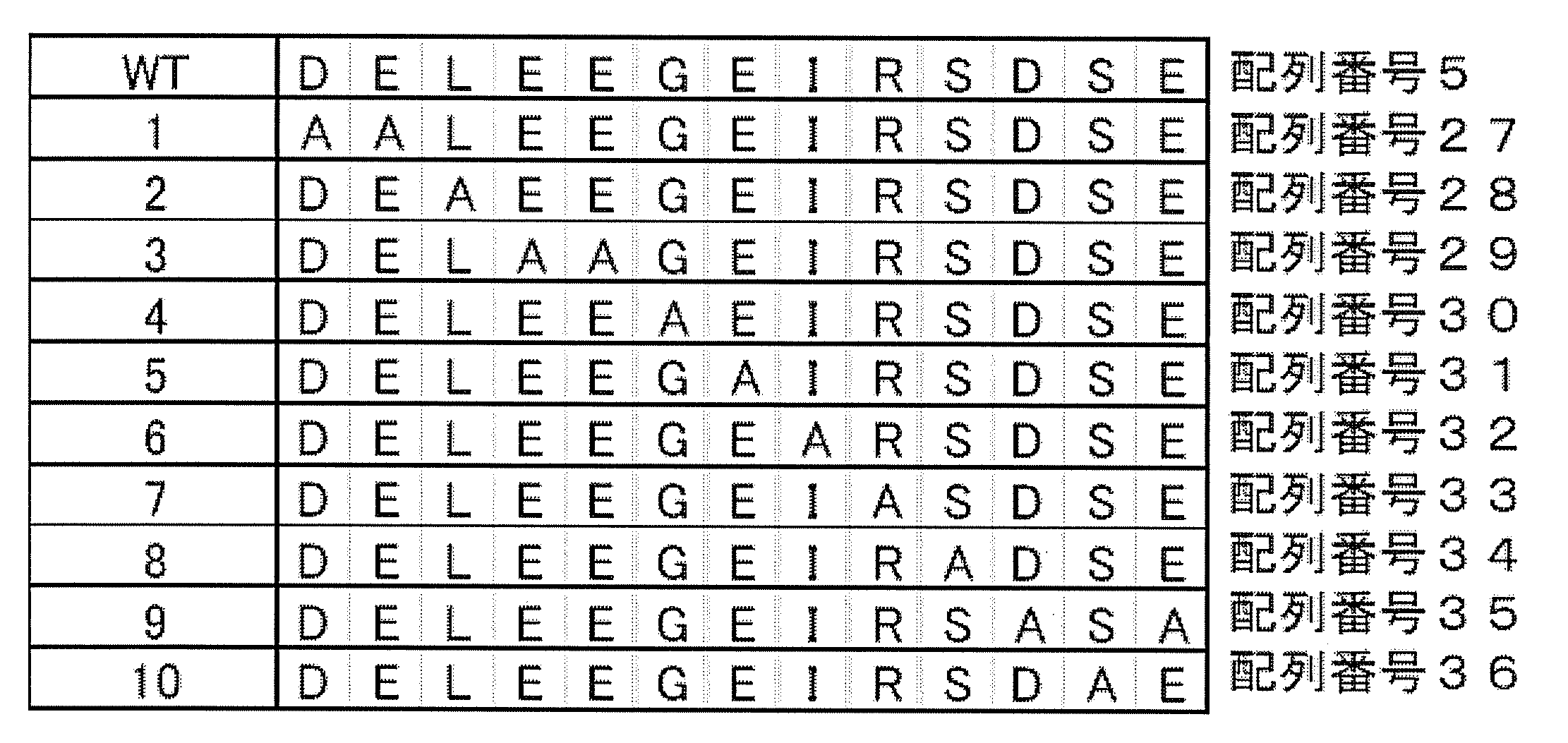
FARB-EGFP-NLSとHA-ARS2とを293T細胞に発現させ、ウェスタンブロッティングにより細胞ライセート中での発現を確認した。また、細胞ライセートから抗GFP抗体又は抗HA抗体で免疫沈降を行い、FARB-EGFP-NLSとHA-ARS2とが共免疫沈降するか解析した。
結果を図14及び表4に示す。FARB配列のN末端側から4番目と5番目のグルタミン酸残基をアラニンに置換した場合(#3)、及び7番目のグルタミン酸をアラニンに置換した場合(#5)には、FARB-EGFP-NLSとHA-ARS2との結合が検出できなかった。4番目のグリシンをアラニンに置換した場合(#4)には、両者の弱い結合が検出された。その他の位置のアミノ酸残基を置換した場合には、野生型FARB配列を用いた場合と同程度のHA-ARS2との結合が検出された。
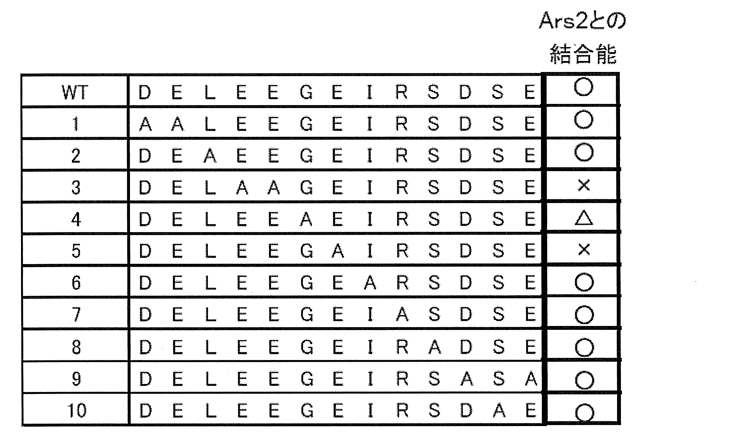
○:結合する、△:弱く結合する、×:結合を検出できない。
以上の結果より、FARB配列における第4番目、第5番目及び第7番目のグルタミン酸が、ARS2との会合に必須であることが示された。
[実施例5]
実施例1及び3に準じて、ヒトFLASHのFARBを含む187アミノ酸残基(第758~944番目のアミノ酸)からなる領域をEGFP及びNLSと結合した融合ポリペプチド(758-944 aa-EGFP-NLS)をKB細胞に強制発現させ、該細胞の細胞周期を観察した。
実施例1及び3に準じて、ヒトFLASHのFARBを含む187アミノ酸残基(第758~944番目のアミノ酸)からなる領域をEGFP及びNLSと結合した融合ポリペプチド(758-944 aa-EGFP-NLS)をKB細胞に強制発現させ、該細胞の細胞周期を観察した。
結果を図15に示す。上記融合ポリペプチドの発現により細胞の増殖がS期で停止した。
参考文献
Alm-Kristiansen, A. H., Saether, T., Matre, V., Gilfillan, S., Dahle, O., and Gabrielsen, O. S. (2008). FLASH acts as a co-activator of the transcription factor c-Myb and localizes to active RNA polymerase II foci. Oncogene 27, 4644-4656.
Amsterdam, A., Nissen, R. M., Sun, Z., Swindell, E. C., Farrington, S., and Hopkins, N. (2004). Identification of 315 genes essential for early zebrafish development. Proc Natl Acad Sci U S A 101, 12792-12797.
Barcaroli, D., Bongiorno-Borbone, L., Terrinoni, A., Hofmann, T. G., Rossi, M., Knight, R. A., Matera, A. G., Melino, G., and De Laurenzi, V. (2006a). FLASH is required for histone transcription and S-phase progression. Proc Natl Acad Sci U S A 103, 14808-14812.
Barcaroli, D., Dinsdale, D., Neale, M. H., Bongiorno-Borbone, L., Ranalli, M., Munarriz, E., Sayan, A. E., McWilliam, J. M., Smith, T. M., Fava, E., et al. (2006b). FLASH is an essential component of Cajal bodies. Proc Natl Acad Sci U S A 103, 14802-14807.
Bongiorno-Borbone, L., De Cola, A., Vernole, P., Finos, L., Barcaroli, D., Knight, R. A., Melino, G., and De Laurenzi, V. (2008). FLASH and NPAT positive but not Coilin positive Cajal Bodies correlate with cell ploidy. Cell Cycle 7, 2357-2367.
Cioce, M., and Lamond, A. I. (2005). Cajal bodies: a long history of discovery. Annu Rev Cell Dev Biol 21, 105-131.
DeRan, M., Pulvino, M., Greene, E., Su, C., and Zhao, J. (2008). Transcriptional activation of histone genes requires NPAT-dependent recruitment of TRRAP-Tip60 complex to histone promoters during the G1/S phase transition. Mol Cell Biol 28, 435-447.
Dominski, Z., and Marzluff, W. F. (1999). Formation of the 3' end of histone mRNA. Gene 239, 1-14.
Ewing, R. M., Chu, P., Elisma, F., Li, H., Taylor, P., Climie, S., McBroom-Cerajewski, L., Robinson, M. D., O'Connor, L., Li, M., et al. (2007). Large-scale mapping of human protein-protein interactions by mass spectrometry. Mol Syst Biol 3, 89.
Flotho, C., Coustan-Smith, E., Pei, D., Iwamoto, S., Song, G., Cheng, C., Pui, C. H., Downing, J. R., and Campana, D. (2006). Genes contributing to minimal residual disease in childhood acute lymphoblastic leukemia: prognostic significance of CASP8AP2. Blood 108, 1050-1057.
Gall, J. G. (2000). Cajal bodies: the first 100 years. Annu Rev Cell Dev Biol 16, 273-300.
Golling, G., Amsterdam, A., Sun, Z., Antonelli, M., Maldonado, E., Chen, W., Burgess, S., Haldi, M., Artzt, K., Farrington, S., et al. (2002). Insertional mutagenesis in zebrafish rapidly identifies genes essential for early vertebrate development. Nat Genet 31, 135-140.
Imai, Y., Kimura, T., Murakami, A., Yajima, N., Sakamaki, K., and Yonehara, S. (1999). The CED-4-homologous protein FLASH is involved in Fas-mediated activation of caspase-8 during apoptosis. Nature 398, 777-785.
Kino, T., and Chrousos, G. P. (2003). Tumor necrosis factor alpha receptor- and Fas-associated FLASH inhibit transcriptional activity of the glucocorticoid receptor by binding to and interfering with its interaction with p160 type nuclear receptor coactivators. J Biol Chem 278, 3023-3029.
Kino, T., Ichijo, T., and Chrousos, G. P. (2004). FLASH interacts with p160 coactivator subtypes and differentially suppresses transcriptional activity of steroid hormone receptors. J Steroid Biochem Mol Biol 92, 357-363.
Kittler, R., Putz, G., Pelletier, L., Poser, I., Heninger, A. K., Drechsel, D., Fischer, S., Konstantinova, I., Habermann, B., Grabner, H., et al. (2004). An endoribonuclease-prepared siRNA screen in human cells identifies genes essential for cell division. Nature 432, 1036-1040.
Kobayashi, Y., and Yonehara, S. (2009). Novel cell death by downregulation of eEF1A1 expression in tetraploids. Cell Death Differ 16, 139-150.
Lee, K. K., and Yonehara, S. (2002). Phosphorylation and dimerization regulate nucleocytoplasmic shuttling of mammalian STE20-like kinase (MST). J Biol Chem 277, 12351-12358.
Ma, T., Van Tine, B. A., Wei, Y., Garrett, M. D., Nelson, D., Adams, P. D., Wang, J., Qin, J., Chow, L. T., and Harper, J. W. (2000). Cell cycle-regulated phosphorylation of p220(NPAT) by cyclin E/Cdk2 in Cajal bodies promotes histone gene transcription. Genes Dev 14, 2298-2313.
Milovic-Holm, K., Krieghoff, E., Jensen, K., Will, H., and Hofmann, T. G. (2007). FLASH links the CD95 signaling pathway to the cell nucleus and nuclear bodies. Embo J 26, 391-401.
Obradovic, D., Tirard, M., Nemethy, Z., Hirsch, O., Gronemeyer, H., and Almeida, O. F. (2004). DAXX, FLASH, and FAF-1 modulate mineralocorticoid and glucocorticoid receptor-mediated transcription in hippocampal cells--toward a basis for the opposite actions elicited by two nuclear receptors? Mol Pharmacol 65, 761-769.
Okamoto, K., Fujisawa, J., Reth, M., and Yonehara, S. (2006). Human T-cell leukemia virus type-I oncoprotein Tax inhibits Fas-mediated apoptosis by inducing cellular FLIP through activation of NF-kappaB. Genes Cells 11, 177-191.
Prigge, M. J., and Wagner, D. R. (2001). The arabidopsis serrate gene encodes a zinc-finger protein required for normal shoot development. Plant Cell 13, 1263-1279.
Rossman, T. G., and Wang, Z. (1999). Expression cloning for arsenite-resistance resulted in isolation of tumor-suppressor fau cDNA: possible involvement of the ubiquitin system in arsenic carcinogenesis. Carcinogenesis 20, 311-316.
Sakashita, E., Tatsumi, S., Werner, D., Endo, H., and Mayeda, A. (2004). Human RNPS1 and its associated factors: a versatile alternative pre-mRNA splicing regulator in vivo. Mol Cell Biol 24, 1174-1187.
Sittman, D. B., Graves, R. A., and Marzluff, W. F. (1983). Histone mRNA concentrations are regulated at the level of transcription and mRNA degradation. Proc Natl Acad Sci U S A 80, 1849-1853.
Wei, Y., Jin, J., and Harper, J. W. (2003). The cyclin E/Cdk2 substrate and Cajal body component p220(NPAT) activates histone transcription through a novel LisH-like domain. Mol Cell Biol 23, 3669-3680.
Wilson, M. D., Wang, D., Wagner, R., Breyssens, H., Gertsenstein, M., Lobe, C., Lu, X., Nagy, A., Burke, R. D., Koop, B. F., and Howard, P. L. (2008). ARS2 is a conserved eukaryotic gene essential for early mammalian development. Mol Cell Biol 28, 1503-1514.
Ye, X., Wei, Y., Nalepa, G., and Harper, J. W. (2003). The cyclin E/Cdk2 substrate p220(NPAT) is required for S-phase entry, histone gene expression, and Cajal body maintenance in human somatic cells. Mol Cell Biol 23, 8586-8600.
Zhao, J., Kennedy, B. K., Lawrence, B. D., Barbie, D. A., Matera, A. G., Fletcher, J. A., and Harlow, E. (2000). NPAT links cyclin E-Cdk2 to the regulation of replication-dependent histone gene transcription. Genes Dev 14, 2283-2297.
Alm-Kristiansen, A. H., Saether, T., Matre, V., Gilfillan, S., Dahle, O., and Gabrielsen, O. S. (2008). FLASH acts as a co-activator of the transcription factor c-Myb and localizes to active RNA polymerase II foci. Oncogene 27, 4644-4656.
Amsterdam, A., Nissen, R. M., Sun, Z., Swindell, E. C., Farrington, S., and Hopkins, N. (2004). Identification of 315 genes essential for early zebrafish development. Proc Natl Acad Sci U S A 101, 12792-12797.
Barcaroli, D., Bongiorno-Borbone, L., Terrinoni, A., Hofmann, T. G., Rossi, M., Knight, R. A., Matera, A. G., Melino, G., and De Laurenzi, V. (2006a). FLASH is required for histone transcription and S-phase progression. Proc Natl Acad Sci U S A 103, 14808-14812.
Barcaroli, D., Dinsdale, D., Neale, M. H., Bongiorno-Borbone, L., Ranalli, M., Munarriz, E., Sayan, A. E., McWilliam, J. M., Smith, T. M., Fava, E., et al. (2006b). FLASH is an essential component of Cajal bodies. Proc Natl Acad Sci U S A 103, 14802-14807.
Bongiorno-Borbone, L., De Cola, A., Vernole, P., Finos, L., Barcaroli, D., Knight, R. A., Melino, G., and De Laurenzi, V. (2008). FLASH and NPAT positive but not Coilin positive Cajal Bodies correlate with cell ploidy. Cell Cycle 7, 2357-2367.
Cioce, M., and Lamond, A. I. (2005). Cajal bodies: a long history of discovery. Annu Rev Cell Dev Biol 21, 105-131.
DeRan, M., Pulvino, M., Greene, E., Su, C., and Zhao, J. (2008). Transcriptional activation of histone genes requires NPAT-dependent recruitment of TRRAP-Tip60 complex to histone promoters during the G1/S phase transition. Mol Cell Biol 28, 435-447.
Dominski, Z., and Marzluff, W. F. (1999). Formation of the 3' end of histone mRNA. Gene 239, 1-14.
Ewing, R. M., Chu, P., Elisma, F., Li, H., Taylor, P., Climie, S., McBroom-Cerajewski, L., Robinson, M. D., O'Connor, L., Li, M., et al. (2007). Large-scale mapping of human protein-protein interactions by mass spectrometry. Mol Syst Biol 3, 89.
Flotho, C., Coustan-Smith, E., Pei, D., Iwamoto, S., Song, G., Cheng, C., Pui, C. H., Downing, J. R., and Campana, D. (2006). Genes contributing to minimal residual disease in childhood acute lymphoblastic leukemia: prognostic significance of CASP8AP2. Blood 108, 1050-1057.
Gall, J. G. (2000). Cajal bodies: the first 100 years. Annu Rev Cell Dev Biol 16, 273-300.
Golling, G., Amsterdam, A., Sun, Z., Antonelli, M., Maldonado, E., Chen, W., Burgess, S., Haldi, M., Artzt, K., Farrington, S., et al. (2002). Insertional mutagenesis in zebrafish rapidly identifies genes essential for early vertebrate development. Nat Genet 31, 135-140.
Imai, Y., Kimura, T., Murakami, A., Yajima, N., Sakamaki, K., and Yonehara, S. (1999). The CED-4-homologous protein FLASH is involved in Fas-mediated activation of caspase-8 during apoptosis. Nature 398, 777-785.
Kino, T., and Chrousos, G. P. (2003). Tumor necrosis factor alpha receptor- and Fas-associated FLASH inhibit transcriptional activity of the glucocorticoid receptor by binding to and interfering with its interaction with p160 type nuclear receptor coactivators. J Biol Chem 278, 3023-3029.
Kino, T., Ichijo, T., and Chrousos, G. P. (2004). FLASH interacts with p160 coactivator subtypes and differentially suppresses transcriptional activity of steroid hormone receptors. J Steroid Biochem Mol Biol 92, 357-363.
Kittler, R., Putz, G., Pelletier, L., Poser, I., Heninger, A. K., Drechsel, D., Fischer, S., Konstantinova, I., Habermann, B., Grabner, H., et al. (2004). An endoribonuclease-prepared siRNA screen in human cells identifies genes essential for cell division. Nature 432, 1036-1040.
Kobayashi, Y., and Yonehara, S. (2009). Novel cell death by downregulation of eEF1A1 expression in tetraploids. Cell Death Differ 16, 139-150.
Lee, K. K., and Yonehara, S. (2002). Phosphorylation and dimerization regulate nucleocytoplasmic shuttling of mammalian STE20-like kinase (MST). J Biol Chem 277, 12351-12358.
Ma, T., Van Tine, B. A., Wei, Y., Garrett, M. D., Nelson, D., Adams, P. D., Wang, J., Qin, J., Chow, L. T., and Harper, J. W. (2000). Cell cycle-regulated phosphorylation of p220(NPAT) by cyclin E/Cdk2 in Cajal bodies promotes histone gene transcription. Genes Dev 14, 2298-2313.
Milovic-Holm, K., Krieghoff, E., Jensen, K., Will, H., and Hofmann, T. G. (2007). FLASH links the CD95 signaling pathway to the cell nucleus and nuclear bodies. Embo J 26, 391-401.
Obradovic, D., Tirard, M., Nemethy, Z., Hirsch, O., Gronemeyer, H., and Almeida, O. F. (2004). DAXX, FLASH, and FAF-1 modulate mineralocorticoid and glucocorticoid receptor-mediated transcription in hippocampal cells--toward a basis for the opposite actions elicited by two nuclear receptors? Mol Pharmacol 65, 761-769.
Okamoto, K., Fujisawa, J., Reth, M., and Yonehara, S. (2006). Human T-cell leukemia virus type-I oncoprotein Tax inhibits Fas-mediated apoptosis by inducing cellular FLIP through activation of NF-kappaB. Genes Cells 11, 177-191.
Prigge, M. J., and Wagner, D. R. (2001). The arabidopsis serrate gene encodes a zinc-finger protein required for normal shoot development. Plant Cell 13, 1263-1279.
Rossman, T. G., and Wang, Z. (1999). Expression cloning for arsenite-resistance resulted in isolation of tumor-suppressor fau cDNA: possible involvement of the ubiquitin system in arsenic carcinogenesis. Carcinogenesis 20, 311-316.
Sakashita, E., Tatsumi, S., Werner, D., Endo, H., and Mayeda, A. (2004). Human RNPS1 and its associated factors: a versatile alternative pre-mRNA splicing regulator in vivo. Mol Cell Biol 24, 1174-1187.
Sittman, D. B., Graves, R. A., and Marzluff, W. F. (1983). Histone mRNA concentrations are regulated at the level of transcription and mRNA degradation. Proc Natl Acad Sci U S A 80, 1849-1853.
Wei, Y., Jin, J., and Harper, J. W. (2003). The cyclin E/Cdk2 substrate and Cajal body component p220(NPAT) activates histone transcription through a novel LisH-like domain. Mol Cell Biol 23, 3669-3680.
Wilson, M. D., Wang, D., Wagner, R., Breyssens, H., Gertsenstein, M., Lobe, C., Lu, X., Nagy, A., Burke, R. D., Koop, B. F., and Howard, P. L. (2008). ARS2 is a conserved eukaryotic gene essential for early mammalian development. Mol Cell Biol 28, 1503-1514.
Ye, X., Wei, Y., Nalepa, G., and Harper, J. W. (2003). The cyclin E/Cdk2 substrate p220(NPAT) is required for S-phase entry, histone gene expression, and Cajal body maintenance in human somatic cells. Mol Cell Biol 23, 8586-8600.
Zhao, J., Kennedy, B. K., Lawrence, B. D., Barbie, D. A., Matera, A. G., Fletcher, J. A., and Harlow, E. (2000). NPAT links cyclin E-Cdk2 to the regulation of replication-dependent histone gene transcription. Genes Dev 14, 2283-2297.
本発明によれば、FLASHとARS2との相互作用の阻害や、FLASHの自己会合の阻害という、これまでにない新たなメカニズムに立脚した細胞増殖抑制剤や、そのスクリーニング方法が提供される。
本出願は日本で出願された特願2009-052628(出願日:2009年3月5日)を基礎としており、その内容は本明細書に全て包含されるものである。
本出願は日本で出願された特願2009-052628(出願日:2009年3月5日)を基礎としており、その内容は本明細書に全て包含されるものである。
Claims (23)
- (A)(1)配列番号5で表されるアミノ酸配列からなる領域;
(2)配列番号5で表されるアミノ酸配列と69%以上の同一性を有するアミノ酸配列からなり、且つARS2ポリペプチドへ結合する活性を有する領域;及び
(3)配列番号5で表されるアミノ酸配列において1~4個のアミノ酸が欠失、置換、挿入又は付加されたアミノ酸配列からなり、且つARS2ポリペプチドへ結合する活性を有する領域
からなる群から選択されるいずれかの領域を含み、且つ長さが200アミノ酸以下であるポリペプチド;並びに
(B)(A)のポリペプチドのN末端及び/又はC末端に少なくとも1つのタグポリペプチド又はシグナル配列が融合してなる融合ポリペプチド
からなる群から選択されるいずれかのポリペプチド。 - 請求項1記載のポリペプチドをコードするポリヌクレオチド。
- 請求項2記載のポリヌクレオチドを含む発現ベクター。
- 請求項3記載の発現ベクターを含む形質転換体。
- (1)配列番号5で表されるアミノ酸配列からなる領域;
(2)配列番号5で表されるアミノ酸配列と69%以上の同一性を有するアミノ酸配列からなり、且つARS2ポリペプチドへ結合する活性を有する領域;及び
(3)配列番号5で表されるアミノ酸配列において1~4個のアミノ酸が欠失、置換、挿入又は付加されたアミノ酸配列からなり、且つARS2ポリペプチドへ結合する活性を有する領域
からなる群から選択されるいずれかの領域を含むポリペプチド(但し、FLASHポリペプチドのコイルドコイルドメインを含むポリペプチドを除く)、又は該ポリペプチドを発現し得る発現ベクターを含む、細胞増殖抑制剤。 - ARS2の発現を特異的に抑制し得るsiRNA、アンチセンス核酸、又はこれらのポリヌクレオチドを発現し得る発現ベクターを含む、細胞増殖抑制剤。
- (1)配列番号5で表されるアミノ酸配列からなる領域;
(2)配列番号5で表されるアミノ酸配列と69%以上の同一性を有するアミノ酸配列からなり、且つARS2ポリペプチドへ結合する活性を有する領域;及び
(3)配列番号5で表されるアミノ酸配列において1~4個のアミノ酸が欠失、置換、挿入又は付加されたアミノ酸配列からなり、且つARS2ポリペプチドへ結合する活性を有する領域
からなる群から選択されるいずれかの領域を含むポリペプチドと、
(1’)配列番号13で表されるアミノ酸配列からなる領域;
(2’)配列番号13で表されるアミノ酸配列と95%以上の同一性を有するアミノ酸配列からなり、且つFLASHポリペプチドへ結合する活性を有する領域;及び
(3’)配列番号13で表されるアミノ酸配列において1~10個のアミノ酸が欠失、置換、挿入又は付加されたアミノ酸配列からなり、且つFLASHポリペプチドへ結合する活性を有する領域
からなる群から選択されるいずれかの領域を含むポリペプチドとの結合を阻害する化合物を、細胞増殖を抑制し得る化合物として選択することを含む、細胞増殖を抑制し得る化合物のスクリーニング方法。 - (1)配列番号5で表されるアミノ酸配列からなる領域;
(2)配列番号5で表されるアミノ酸配列と69%以上の同一性を有するアミノ酸配列からなり、且つARS2ポリペプチドへ結合する活性を有する領域;及び
(3)配列番号5で表されるアミノ酸配列において1~4個のアミノ酸が欠失、置換、挿入又は付加されたアミノ酸配列からなり、且つARS2ポリペプチドへ結合する活性を有する領域
からなる群から選択されるいずれかのポリペプチド領域へ結合する化合物を、細胞増殖を抑制し得る化合物として選択することを含む、細胞増殖を抑制し得る化合物のスクリーニング方法。 - (1)配列番号5で表されるアミノ酸配列からなる領域;
(2)配列番号5で表されるアミノ酸配列と69%以上の同一性を有するアミノ酸配列からなり、且つARS2ポリペプチドへ結合する活性を有する領域;及び
(3)配列番号5で表されるアミノ酸配列において1~4個のアミノ酸が欠失、置換、挿入又は付加されたアミノ酸配列からなり、且つARS2ポリペプチドへ結合する活性を有する領域
からなる群から選択されるいずれかの領域を含むポリペプチドと、
(1’)配列番号13で表されるアミノ酸配列からなる領域;
(2’)配列番号13で表されるアミノ酸配列と95%以上の同一性を有するアミノ酸配列からなり、且つFLASHポリペプチドへ結合する活性を有する領域;及び
(3’)配列番号13で表されるアミノ酸配列において1~10個のアミノ酸が欠失、置換、挿入又は付加されたアミノ酸配列からなり、且つFLASHポリペプチドへ結合する活性を有する領域
からなる群から選択されるいずれかの領域を含むポリペプチド
との組み合わせ物。 - ARS2の発現を低下させる化合物を、細胞増殖を抑制し得る化合物として選択することを含む、細胞増殖を抑制し得る化合物のスクリーニング方法。
- FLASHポリペプチドの自己会合を阻害する化合物を、細胞増殖を抑制し得る化合物として選択することを含む、細胞増殖を抑制し得る化合物のスクリーニング方法。
- FLASHポリペプチドのコイルドコイルドメインへ結合する化合物を、細胞増殖を抑制し得る化合物として選択することを含む、細胞増殖を抑制し得る化合物のスクリーニング方法。
- 細胞増殖の抑制において使用するための、
(1)配列番号5で表されるアミノ酸配列からなる領域;
(2)配列番号5で表されるアミノ酸配列と69%以上の同一性を有するアミノ酸配列からなり、且つARS2ポリペプチドへ結合する活性を有する領域;及び
(3)配列番号5で表されるアミノ酸配列において1~4個のアミノ酸が欠失、置換、挿入又は付加されたアミノ酸配列からなり、且つARS2ポリペプチドへ結合する活性を有する領域
からなる群から選択されるいずれかの領域を含むポリペプチド(但し、FLASHポリペプチドのコイルドコイルドメインを含むポリペプチドを除く)、又は該ポリペプチドを発現し得る発現ベクター。 - 細胞増殖の抑制において使用するための、ARS2の発現を特異的に抑制し得るsiRNA、アンチセンス核酸、又はこれらのポリヌクレオチドを発現し得る発現ベクター。
- 哺乳動物に対して、
(1)配列番号5で表されるアミノ酸配列からなる領域;
(2)配列番号5で表されるアミノ酸配列と69%以上の同一性を有するアミノ酸配列からなり、且つARS2ポリペプチドへ結合する活性を有する領域;及び
(3)配列番号5で表されるアミノ酸配列において1~4個のアミノ酸が欠失、置換、挿入又は付加されたアミノ酸配列からなり、且つARS2ポリペプチドへ結合する活性を有する領域
からなる群から選択されるいずれかの領域を含むポリペプチド(但し、FLASHポリペプチドのコイルドコイルドメインを含むポリペプチドを除く)、又は該ポリペプチドを発現し得る発現ベクターの有効量を投与することを含む、該哺乳動物における細胞増殖の抑制方法。 - 哺乳動物に対して、ARS2の発現を特異的に抑制し得るsiRNA、アンチセンス核酸、又はこれらのポリヌクレオチドを発現し得る発現ベクターの有効量を投与することを含む、該哺乳動物における細胞増殖の抑制方法。
- (1)配列番号5で表されるアミノ酸配列からなる領域;
(2)配列番号5で表されるアミノ酸配列と69%以上の同一性を有するアミノ酸配列からなり、且つARS2ポリペプチドへ結合する活性を有する領域;及び
(3)配列番号5で表されるアミノ酸配列において1~4個のアミノ酸が欠失、置換、挿入又は付加されたアミノ酸配列からなり、且つARS2ポリペプチドへ結合する活性を有する領域
からなる群から選択されるいずれかの領域を含むポリペプチド(但し、FLASHポリペプチドのコイルドコイルドメインを含むポリペプチドを除く)、又は該ポリペプチドを発現し得る発現ベクターを含む、異常な細胞増殖に起因する疾患の予防又は治療剤。 - ARS2の発現を特異的に抑制し得るsiRNA、アンチセンス核酸、又はこれらのポリヌクレオチドを発現し得る発現ベクターを含む、異常な細胞増殖に起因する疾患の予防又は治療剤。
- 異常な細胞増殖に起因する疾患の予防又は治療において使用するための、
(1)配列番号5で表されるアミノ酸配列からなる領域;
(2)配列番号5で表されるアミノ酸配列と69%以上の同一性を有するアミノ酸配列からなり、且つARS2ポリペプチドへ結合する活性を有する領域;及び
(3)配列番号5で表されるアミノ酸配列において1~4個のアミノ酸が欠失、置換、挿入又は付加されたアミノ酸配列からなり、且つARS2ポリペプチドへ結合する活性を有する領域
からなる群から選択されるいずれかの領域を含むポリペプチド(但し、FLASHポリペプチドのコイルドコイルドメインを含むポリペプチドを除く)、又は該ポリペプチドを発現し得る発現ベクター。 - 異常な細胞増殖に起因する疾患の予防又は治療において使用するための、ARS2の発現を特異的に抑制し得るsiRNA、アンチセンス核酸、又はこれらのポリヌクレオチドを発現し得る発現ベクター。
- 哺乳動物に対して、
(1)配列番号5で表されるアミノ酸配列からなる領域;
(2)配列番号5で表されるアミノ酸配列と69%以上の同一性を有するアミノ酸配列からなり、且つARS2ポリペプチドへ結合する活性を有する領域;及び
(3)配列番号5で表されるアミノ酸配列において1~4個のアミノ酸が欠失、置換、挿入又は付加されたアミノ酸配列からなり、且つARS2ポリペプチドへ結合する活性を有する領域
からなる群から選択されるいずれかの領域を含むポリペプチド(但し、FLASHポリペプチドのコイルドコイルドメインを含むポリペプチドを除く)、又は該ポリペプチドを発現し得る発現ベクターの有効量を投与することを含む、該哺乳動物における異常な細胞増殖に起因する疾患の予防又は治療の抑制方法。 - 哺乳動物に対して、ARS2の発現を特異的に抑制し得るsiRNA、アンチセンス核酸、又はこれらのポリヌクレオチドを発現し得る発現ベクターの有効量を投与することを含む、該哺乳動物における異常な細胞増殖に起因する疾患の予防又は治療方法。
- (A)配列番号5で表されるアミノ酸配列において、第4番目のグルタミン酸、第5番目のグルタミン酸及び第7番目のグルタミン酸からなる群から選択される少なくとも1つのグルタミン酸がグルタミン酸以外のアミノ酸で置換されているアミノ酸配列を含み、且つARS2ポリペプチドへ結合する活性を有していないポリペプチド、並びに
(B)(A)のポリペプチドのN末端及び/又はC末端に少なくとも1つのタグポリペプチド又はシグナル配列が融合してなるポリペプチド
からなる群から選択されるいずれかのポリペプチド。
Applications Claiming Priority (2)
| Application Number | Priority Date | Filing Date | Title |
|---|---|---|---|
| JP2009052628 | 2009-03-05 | ||
| JP2009-052628 | 2009-03-05 |
Publications (1)
| Publication Number | Publication Date |
|---|---|
| WO2010101248A1 true WO2010101248A1 (ja) | 2010-09-10 |
Family
ID=42709801
Family Applications (1)
| Application Number | Title | Priority Date | Filing Date |
|---|---|---|---|
| PCT/JP2010/053665 Ceased WO2010101248A1 (ja) | 2009-03-05 | 2010-03-05 | 細胞増殖抑制剤及びそのスクリーニング方法 |
Country Status (1)
| Country | Link |
|---|---|
| WO (1) | WO2010101248A1 (ja) |
-
2010
- 2010-03-05 WO PCT/JP2010/053665 patent/WO2010101248A1/ja not_active Ceased
Non-Patent Citations (5)
| Title |
|---|
| BARCAROLI, D. ET AL.: "FLASH is required for histone transcription and S-phase progression.", PROC. NATL. ACAD. SCI. USA, vol. 103, no. 40, 3 October 2006 (2006-10-03), pages 14808 - 14812 * |
| DATABASE UNIPROTKB [online] 3 March 2009 (2009-03-03), "C8AP2_HUMAN.", Database accession no. Q9UKL3 * |
| KIRIYAMA, M. ET AL.: "Interaction of FLASH with arsenite resistance protein 2 is involved in cell cycle progression at S phase.", MOL. CELL. BIOL., vol. 29, no. 17, September 2009 (2009-09-01), pages 4729 - 4741 * |
| MARIA KIRIYAMA ET AL.: "Saibo Shuki S-ki no Shinko ni Hitsuyo na FLASH to ARS2(Arsenite Resistance Protein2) tono Kaigo", DAI 82 KAI THE JAPANESE BIOCHEMICAL SOCIETY TAIKAI PROGRAM ? KOEN YOSHISHU, 25 September 2009 (2009-09-25), pages 3T10A-4 - (3P-483) * |
| WILSON, M.D. ET AL.: "ARS2 is a conserved eukaryotic gene essential for early mammalian development.", MOL. CELL. BIOL., vol. 28, no. 5, March 2008 (2008-03-01), pages 1503 - 1514 * |
Similar Documents
| Publication | Publication Date | Title |
|---|---|---|
| US20230313190A1 (en) | Compositions and Methods for Degradation of Misfolded Proteins | |
| Amit et al. | Tal, a Tsg101-specific E3 ubiquitin ligase, regulates receptor endocytosis and retrovirus budding | |
| WO2007138810A1 (ja) | 神経変性疾患の予防・治療剤 | |
| Shen et al. | LUBAC regulates ciliogenesis by promoting CP110 removal from the mother centriole | |
| US20090087436A1 (en) | Compositions and methods for treating diseases | |
| EP0723402A1 (en) | Senescent cell-derived inhibitors of dna synthesis | |
| JP2015173601A (ja) | がん幹細胞の増殖抑制物質のスクリーニング方法 | |
| US20080306007A1 (en) | Agents for prophylaxis or treatment of neurological related diseases and conditions | |
| WO2010101248A1 (ja) | 細胞増殖抑制剤及びそのスクリーニング方法 | |
| US7390625B2 (en) | Apoptosis-associated protein and use thereof | |
| JP2010195684A (ja) | MTI−MMP、iFIH、FIH−1によるHIF−1の制御 | |
| US7998684B2 (en) | Screening method for an agent for treatment of neurodegenerative disease | |
| US20050014689A1 (en) | Remedies for life style-related diseases or cibophobia and method of screening the same | |
| CA3096425A1 (en) | Methods and uses of modulators of anaphase promoting complex (apc) activity for treating cancer | |
| US20110104705A1 (en) | Musclin receptor and use thereof | |
| WO2005049868A1 (en) | Methods and agents for screening for compounds capable of modulating her2 expression | |
| EP1760087B1 (en) | Novel protein complex and use thereof | |
| Lee | The Molecular Switch of Store Operated Ca2+ Entry: The Activation of ER Ca2+ Sensor STIM1 via Intra-/Inter-molecular Interactions | |
| JP2007230910A (ja) | Vegf産生の低減方法 | |
| US20200147120A1 (en) | Function inhibitor of swi/snf complexes | |
| US20060111556A1 (en) | Preventives/remedies for neurodegenerative diseases | |
| Jeffries | THE ROLE OF NUCLEAR LOCALIZATION AND N ic-PROTEIN COMPLEX FORMATION IN NOTCH INDUCED NEOPLASIA | |
| US20100190685A1 (en) | Prognostic factor in sarcoma, and metastasis inhibitor | |
| JP2008206398A (ja) | Runxのユビキチン化方法 |
Legal Events
| Date | Code | Title | Description |
|---|---|---|---|
| 121 | Ep: the epo has been informed by wipo that ep was designated in this application |
Ref document number: 10748841 Country of ref document: EP Kind code of ref document: A1 |
|
| NENP | Non-entry into the national phase |
Ref country code: DE |
|
| 122 | Ep: pct application non-entry in european phase |
Ref document number: 10748841 Country of ref document: EP Kind code of ref document: A1 |
|
| NENP | Non-entry into the national phase |
Ref country code: JP |