WO2010079919A2 - 다층기질겔을 이용한 기질분해효소의 스크리닝 방법 - Google Patents
다층기질겔을 이용한 기질분해효소의 스크리닝 방법 Download PDFInfo
- Publication number
- WO2010079919A2 WO2010079919A2 PCT/KR2009/007913 KR2009007913W WO2010079919A2 WO 2010079919 A2 WO2010079919 A2 WO 2010079919A2 KR 2009007913 W KR2009007913 W KR 2009007913W WO 2010079919 A2 WO2010079919 A2 WO 2010079919A2
- Authority
- WO
- WIPO (PCT)
- Prior art keywords
- gel
- substrate
- sample
- electrophoresis
- screening
- Prior art date
- Legal status (The legal status is an assumption and is not a legal conclusion. Google has not performed a legal analysis and makes no representation as to the accuracy of the status listed.)
- Ceased
Links
Images
Classifications
-
- C—CHEMISTRY; METALLURGY
- C12—BIOCHEMISTRY; BEER; SPIRITS; WINE; VINEGAR; MICROBIOLOGY; ENZYMOLOGY; MUTATION OR GENETIC ENGINEERING
- C12Q—MEASURING OR TESTING PROCESSES INVOLVING ENZYMES, NUCLEIC ACIDS OR MICROORGANISMS; COMPOSITIONS OR TEST PAPERS THEREFOR; PROCESSES OF PREPARING SUCH COMPOSITIONS; CONDITION-RESPONSIVE CONTROL IN MICROBIOLOGICAL OR ENZYMOLOGICAL PROCESSES
- C12Q1/00—Measuring or testing processes involving enzymes, nucleic acids or microorganisms; Compositions therefor; Processes of preparing such compositions
- C12Q1/34—Measuring or testing processes involving enzymes, nucleic acids or microorganisms; Compositions therefor; Processes of preparing such compositions involving hydrolase
-
- G—PHYSICS
- G01—MEASURING; TESTING
- G01N—INVESTIGATING OR ANALYSING MATERIALS BY DETERMINING THEIR CHEMICAL OR PHYSICAL PROPERTIES
- G01N33/00—Investigating or analysing materials by specific methods not covered by groups G01N1/00 - G01N31/00
- G01N33/48—Biological material, e.g. blood, urine; Haemocytometers
- G01N33/50—Chemical analysis of biological material, e.g. blood, urine; Testing involving biospecific ligand binding methods; Immunological testing
- G01N33/53—Immunoassay; Biospecific binding assay; Materials therefor
- G01N33/558—Immunoassay; Biospecific binding assay; Materials therefor using diffusion or migration of antigen or antibody
- G01N33/561—Immunoelectrophoresis
Definitions
- the present invention relates to a method for screening and detecting two or more different substrate degrading enzymes, and more particularly, gel electrophoresis of a sample containing an enzyme, followed by electrophoresis to a multilayer substrate gel having different substrates.
- the present invention relates to a method for screening two or more matrixases simultaneously and detecting activity by analyzing multilayer substrate gels.
- Electrophoretic separation methods include electrophoresis, isochophoresis, and isoelectric focussing.
- electrophoresis is carried out in the presence of denaturing media (SDS, urea, guanidinium salts, mercaptoethanol, etc.), which denaturation removes all biological activity (ligand binding, substrate binding and substrate transformation) of the protein component.
- denaturing media SDS, urea, guanidinium salts, mercaptoethanol, etc.
- denatured proteins by treatment with Triton X-100 can be re-renewed and refolded into their active natural structure. This regeneration is performed directly in the gel.
- hydrolases metabolize the reaction between the substrate and water, breaking down large molecules into smaller units. These enzymes are ⁇ -1-4-glycosidic in cellulose. Degradation of dick bonds ( ⁇ -1-4-glycosidic linkages) (cellulase, EC 3.2.1), degradation of peptide bonds in proteins (protease, EC 3.4) and ester bonds in lipids Metabolize (lipase, EC 3.1.1).
- Zymography is a technique that can directly detect enzyme activity on SDS gel electrophoresis. This method performs electrophoresis after solidifying by incorporating a substrate for a specific enzyme into an SDS gel. Enzyme activity can be confirmed by the degradation of the substrate by the protein developed on the gel after electrophoresis.
- Zymography technology has not only a very high sensitivity (ng level) but also a molecular weight or a measurement of a protein having enzymatic activity on a gel, which is widely used in enzyme engineering research such as enzyme screening and purification (TM Leber & FR). Balkwill, Anal Biochem, 249: 24 , 1997; SH Kim et al, Anal Biochem, 263: Weg 115, 1998).
- Brahimi-Horn et al Used an electro-transfer technique for the detection of lipolytic enzymes under undenatured conditions (MC Brahimi-Horn et al., Anal. Biochem ., 196: 76). , 1991). This method transfers lipolytic enzymes to the nitrocellulose membrane and then either ⁇ -naphthyl acetate or ⁇ -naphthyl palmitate, which is water-insoluble for enzyme detection. Is a method of staining.
- the present inventors have made an effort to develop a new enzyme detection method capable of detecting two or more different matrix degrading enzymes at once by improving the conventional enzyme detection method, such as a zymography method, and thus, a sample containing an enzyme.
- the gel electrophoresis was carried out, and then the electrophoretic gel sample was electrophoresed into a multilayer substrate gel having a different substrate, thereby analyzing the multilayer substrate gel.
- a zymography method a sample containing an enzyme.
- Another object of the present invention is to provide a new enzyme detection method capable of simultaneously detecting two or more matrixases.
- the present invention comprises the steps of (a) injecting a sample to the gel to perform gel electrophoresis; (b) applying a voltage to transfer the sample from the gel subjected to electrophoresis to a multi-layered substrate gel in which a gel comprising different substrates each comprises two or more layers; And (c) provides a method for screening the substrate lyase using a multiple-layer substrate gel (multiple-layer substrate gel) comprising the step of screening the lyticase by analyzing the multilayer substrate gel.
- the present invention also comprises the steps of (a) injecting a sample into the gel to perform gel electrophoresis; (b) applying a voltage to transfer the sample from the gel subjected to electrophoresis to a multilayer substrate gel, wherein each gel comprises two or more layers; And (c) analyzing the multilayer substrate gel to detect the activity of the stromatase.
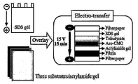
- Figure 1 is a schematic diagram showing an embodiment of a method for detecting a matrix enzyme using a multilayer substrate gel according to the present invention.
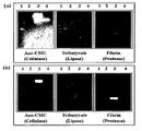
- Figure 2 (a) is a gel photograph showing the results of the detection method using a multi-layer substrate gel according to the present invention, (b) shows the results of performing the detection three times each by the conventional zymography method Gel picture.
- the present invention comprises the steps of (a) injecting a sample into the gel to perform gel electrophoresis; (b) applying a voltage to transfer the sample from the gel subjected to electrophoresis to a multi-layered substrate gel in which a gel comprising different substrates each comprises two or more layers; And (c) screening the hydrolase by analyzing the multi-layered substrate gel.
- the present invention relates to a screening method of matrixase using a multiple-layer substrate gel.
- the present invention in another aspect, (a) injecting a sample into the gel to perform gel electrophoresis; (b) applying a voltage to transfer the sample from the gel subjected to electrophoresis to a multi-layered substrate gel in which a gel comprising different substrates each comprises two or more layers; And (c) analyzing the multilayer substrate gel to detect the activity of the matrixase.
- FIG. 1 is a schematic view showing an embodiment of a hydrolase screening and enzyme detection method using a multilayer substrate gel according to the present invention.
- stromaclease refers to an enzyme having an activity capable of breaking down a substrate, ie, a biological substance, lipases for breaking down lipids, carbohydrases for breaking down carbohydrates, proteases for breaking down proteins, And nucleases that degrade nucleic acids.
- the sample is also expected to contain or contain a substrate degrading enzyme to detect screening or enzymatic activity, collected in any one or more of water, soil, air, food, waste, flora and fauna and animal tissues It may be characterized as being, but is not limited thereto.
- the water includes precipitation, seawater, lakes and rainwater
- wastes include sewage, wastewater, and the like.
- a “substrate gel” refers to a gel containing a substrate that can be degraded by a matrix enzyme to be screened or activated, wherein the gel is polyacrylamide, agarose, starch. And the like can be prepared by a conventionally known method.
- Such substrate gel may be laminated in a sandwich form to form a multilayer substrate gel, as shown in FIG. 1.
- multilayer substrate gel means that the substrate gel including the substrate is laminated in a sandwich form of two or more to be composed of multiple layers.
- a gel containing no substrate may be further introduced between the substrate gels so as not to influence the substrate gels.
- the screening step of the enzyme of step (c) is performed by analyzing a multilayer substrate gel, an enzyme capable of degrading the substrate contained in the gel in which the band appeared in the multilayer substrate gel. Corresponds to the enzyme contained in the sample.
- the enzyme activity detection step of the step (c) is also made by analyzing a multi-layer substrate gel, the substrate contained in the gel band appeared in the multi-layer substrate gel This enzyme corresponds to a substrate degrading enzyme having a degrading activity.
- the screening and activity detection of the substrate lyase is performed by the appearance of a band on at least one substrate gel of the multilayer substrate gel, which band is decomposed by the substrate lyase in the sample electrophoreted with the multilayer substrate gel. It is the result of decomposing the substrate at that location when moving to the substrate gel.
- the substrate gel for lipase detection the glycerol trictylate gel
- the gel containing carboxymethyl cellulose (CMC) as a substrate gel for cellulase detection was observed only after staining with 0.1% (w / v) Congo red and decolorizing with 1 M NaCl to identify the band as a result of the substrate decomposition reaction.
- CMC carboxymethyl cellulose
- the band may be identified as a result of the substrate decomposition reaction by incubating at room temperature.
- casein, gelatin, or steam milk may be used as a substrate of the substrate gel for protease detection, but the fibrin used in the embodiment of the present invention is a cross-linked polymer having a molecular weight of approximately 340 kDa, which is different from other proteas. Since fibrin is relatively less affected by electro-transfer than the first substrate, fibrin is preferably used.
- a zymogram activation buffer solution (20 mM Tris-HCl, 200 mM NaCl, 10 mM CaCl 2 , 0.02% [w / v] NaN 3 ; pH 7.4) and incubated at 37 °C for 24 hours, and then stained with Coomassie blue (Coomassie blue) can be confirmed that the formation of a zona pellucida as a result of protease substrate degradation reaction.
- three types of hydrolase, cellulase, lipase, and protease were detected at one time using one SDS and an electrophoretic system. That is, three cellulase (from a Paenibacillus sp. And two Bacillus sp. Strains) from four soil microbial samples by loading a sample on one SDS-gel according to the method of the present invention, followed by electrophoresis into a multilayer substrate gel. It was confirmed that one lipase (from a Staphylococcus sp.), And two proteases (from two Bacillus sp. Strains) can be screened simultaneously.
- any one of glycerol tributyrate, Azo-CMC (Azo-carboxymethyl Cellulose) and fibrin is used as a substrate, and correspondingly, any one of lipase, cellulase and protease is correspondingly used. It is possible to detect one matrixase, but not limited thereto.
- the method according to the present invention can be used for screening of other matrixases. It will be self-evident to those of ordinary skill in Esau.
- Samples were prepared with three volumes of cold acetone (900 ⁇ l) in 300 ⁇ l of four crude culture supernats obtained from four soil microorganisms ( Paenibacillus sp., Staphylococcus sp. And two Bacillus sp. Strains ). It was prepared by concentrating at -20 °C for 3 hours.
- the gels were incubated for 30 minutes at room temperature with a rotary shaker at 2.5% (v / v) Triton X-100 and 50 mM Tris-HCl (pH 7.4). It was. The gel was then washed with distilled water for 30 minutes to remove Triton X-100.
- Bovine fibrinogen F8630, Sigma
- bovine thrombin T4648, Sigma
- Fibrin was used as a substrate for proteases, as opposed to the usual casein, gelatin or steam milk. This is because cross-linked fibrin affects electro-transfer to a lesser extent than other protease substrates such as casein, gelatin or steam milk at its high molecular weight (approximately 340 kDa).
- Example 1-2 The three substrate gels prepared in Example 1-2 were stacked in the order of fibrin gel, Azo-CMC gel, and tributyrate gel from the bottom in the form of a sandwich, as shown in FIG. 1. At this time, it was confirmed that Azo-CMC migrates to the adjacent substrate gel, but this did not affect the protease activity. However, a substrateless acrylamide gel as shown in Figure 1 was further introduced between the Azo-CMC gel and the fibrin gel.
- the SDS gel subjected to the electrophoresis of Example 1-1 was mounted on the sandwich substrate multilayer substrate gel so that the sample protein was transferred to the multilayer substrate gel. Electro-transfer. Electro-migration was performed at 15V for 15 minutes on a TRANS-BLOT apparatus (Bio-Rad, Hercules, CA, USA).
- the substrate gel for lipase detection (glycerol trictylate gel) and the substrate gel for cellulase detection (Azo-CMC gel) were incubated at room temperature until a clear band was observed.
- Traditional zymography methods, including reverse zymography for cellulase detection require long incubation, and cellulase activity in CMC gels can only be observed after staining with 0.1% (w / v) Congo red and decolorizing with 1 M NaCl. Can be visualized In this experiment, however, Azo-CMC was used for direct activity detection without staining or discoloration.
- the substrate gel (fibrin gel) for protease detection is a zymogram reaction buffer (30 mM Tris, 200 mM NaCl, and 0.02% [w / v] NaN 3 ; pH 7.4)) And reacted at 37 ° C. for 24 hours, followed by staining with Coomassie blue.
- the protein fraction with fibrin resolution will form a zona pellucida upon Coomassie blue staining.
- Example 1-1 the sample of Example 1-1 in three SDS-polyacrylamide gels containing glycerol tributyrate, Azo-CMC (Azo-carboxymethyl Cellulose), and fibrin as substrates, respectively, as a control group.
- Each was subjected to zymography.
- electrophoresis was performed at a constant current of 20 mA, and then the gel was subjected to 2.5% (v / v) Triton X-100 and 50 mM Tris-HCl (pH 7.4) to reactivate the enzyme inactivated by SDS. Incubate for 30 minutes at room temperature with a rotary shaker (rotary shaker).
- the gel was then washed with distilled water for 30 minutes to remove Triton X-100.
- the gel containing glycerol trictylate and the gel containing Azo-CMC were then incubated at room temperature until a clear band was observed, and the gel containing fibrin was also described above.
- the gel containing fibrin was also described above.
- the gel containing fibrin was also described above. In the same manner as in the Zymogram reaction buffer treatment, and stained with Coomassie blue.
- Fig. 2 (a) when the detection was carried out by the method according to the present invention, enzymes secreted from four soil microorganisms were detected simultaneously (lane 1: Paenibacillus sp .; lane 2: Staphylococcus sp). Lanes 3 and 4: Bacillus sp.strains). That is, three cellulases (from Paenibacillus sp. And 2 Bacillus sp. Strains ), one lipase (from Staphylococcus sp. Strain) and two proteases (from Bacillus sp. Strains) were detected simultaneously.
- the screening method and the enzyme activity detection method of the matrix enzyme according to the present invention is useful because it allows the screening of two or more matrix enzymes at the same time by loading a sample on one gel. It is also useful because it has the advantage of accelerating the screening of unknown enzymes directly from microorganisms or environmental sources.
Landscapes
- Health & Medical Sciences (AREA)
- Life Sciences & Earth Sciences (AREA)
- Chemical & Material Sciences (AREA)
- Immunology (AREA)
- Engineering & Computer Science (AREA)
- Molecular Biology (AREA)
- Organic Chemistry (AREA)
- Microbiology (AREA)
- Wood Science & Technology (AREA)
- Biomedical Technology (AREA)
- Biotechnology (AREA)
- General Health & Medical Sciences (AREA)
- Hematology (AREA)
- Urology & Nephrology (AREA)
- Zoology (AREA)
- Proteomics, Peptides & Aminoacids (AREA)
- Physics & Mathematics (AREA)
- Analytical Chemistry (AREA)
- Biochemistry (AREA)
- Food Science & Technology (AREA)
- General Physics & Mathematics (AREA)
- Pathology (AREA)
- Biophysics (AREA)
- Medicinal Chemistry (AREA)
- Cell Biology (AREA)
- Electrochemistry (AREA)
- Chemical Kinetics & Catalysis (AREA)
- Bioinformatics & Cheminformatics (AREA)
- General Engineering & Computer Science (AREA)
- Genetics & Genomics (AREA)
- Measuring Or Testing Involving Enzymes Or Micro-Organisms (AREA)
Abstract
본 발명은 2이상의 서로 다른 기질분해효소의 스크리닝의 스크리닝 및 활성검출방법에 관한 것으로, 보다 상세하게는 효소를 포함하는 시료를 겔 전기영동을 수행한 다음, 서로 다른 기질을 가지는 다층기질겔로 전기이동시켜 다층기질겔을 분석함으로써 2이상의 기질분해효소를 동시에 스크리닝하고 활성을 검출해내는것을 특징으로 하는 방법에 관한 것이다. 본 발명에 따른 기질분해효소의 스크리닝 및 효소활성 검출방법은 기존의 방법과 달리 하나의 겔에 시료를 로딩함으로써 동시에 2이상의 기질분해 효소의 스크리닝이가능하게 하는 장점이 있다. 또한, 미생물이나 환경소스로부터 미지의 효소를 직접 스크리닝하는 것을 가속화시키는 장점이 있으므로 유용하다.
Description
본 발명은 2 이상의 서로 다른 기질분해효소의 스크리닝 및 활성검출방법에 관한 것으로, 보다 상세하게는 효소를 포함하는 시료를 겔 전기영동을 수행한 다음, 서로 다른 기질을 가지는 다층기질겔로 전기이동시켜 다층기질겔을 분석함으로써 2 이상의 기질분해효소를 동시에 스크리닝하고 활성을 검출해내는 것을 특징으로 하는 방법에 관한 것이다.
종래 단백질 복합 혼합물의 고분해 분리를 위해서 폴리아크릴아마이드, 아가로오스, 전분 등의 다양한 겔을 사용한 전기영동법이 사용되어져 오고 있다. 전기영동적인 분리 방법은 전기영동(electrophoresis), 등속도영동(isotachophoresis), 등전점 포커싱(isoelectric focussing)을 포함한다. 특히, 전기영동은 변성 매질(SDS, 우레아, 구아니디늄 염, 머캅토에탄올 등)의 존재 하에서 수행되는데, 이러한 변성으로 단백질 성분의 모든 생물학적 활성(리간드 결합, 기질 결합 및 기질 형질전환)이 제거되나, Triton X-100을 처리함으로써 변성된 단백질들은 다시 재생(renaturation)되어 그들의 활성적인 천연 구조로 재형성(refold)될 수 있다. 이러한 재생은 겔 중에서 직접 수행된다.
이러한 전기영동법에서 더 나아가 가수분해효소의 검출을 위하여 자이모그래피 법이 사용되어 오고 있다. 가수분해효소(hydrolases, EC 3)는 기질과 물 사이의 반응을 대사하는 것으로, 큰 분자(lager molecules)를 작은 단위로 분해하는 것으로, 이러한 종류의 효소들은 셀룰로오스에서는 β-1-4-글리코시딕 결합(β-1-4-glycosidic linkages)의 분해를 (셀룰라아제[cellulase], EC 3.2.1), 단백질에서는 펩티드 결합의 분해를 (프로티아제[protease], EC 3.4), 지질에서는 에스테르 결합의 분해를 (리파아제[lipase], EC 3.1.1) 대사한다.
자이모그래피 법(zymography)은 SDS 겔 전기영동 상에서 효소 활성을 직접적으로 검출하는 것이 가능한 기술로서 이 방법은 특정효소에 대한 기질을 SDS 겔 속에 포함시켜 고형화시킨 후 전기영동을 수행한다. 효소 활성은 전기 영동 수행 후 겔 상에 전개된 단백질에 의해 기질이 분해됨으로써 확인할 수 있다. 자이모그래피 기술은 아주 높은 민감도 (ng 수준)를 가질 뿐만 아니라, 겔 상에서 효소 활성을 지닌 단백질의 분자량 또는 측정이 가능하여 효소 스크리닝 및 정제 등 효소 공학연구에 많이 이용되어지고 있다 (T.M. Leber & F.R. Balkwill, Anal. Biochem., 249:24, 1997; S.H. Kim et al., Anal. Biochem., 263:115, 1998).
Brahimi-Horn 등은 비변성 조건에서 지질분해효소(lipolytic enzymes)의 검출을 위하여 전기-이동 기술(electro-transfer technique)을 사용하였다 (M.C. Brahimi-Horn et al., Anal. Biochem., 196:76, 1991). 이 방법은 지질분해효소를 니트로셀룰로오스 막으로 이동시킨 다음, 효소 검출을 위하여 α-나프틸 아세테이트 (α-naphthyl acetate) 또는 수불용성(water-insoluble)인 α-나프틸 팔미테이트 (α-naphthyl palmitate)로 염색을 수행하는 방법이다.
그러나, Brahimi-Horn 등의 방법을 포함하여 알려진 기법들은 모두 한번에 하나의 기질분해효소만을 검출하기 위한 방법일 뿐으로, 다수개의 기질분해효소의 활성을 한 번에 검출할 수 없는 단점이 있었다. 특히 미생물이나 환경의 소스로부터 미지의 효소를 직접 스크리닝하기 위해서는 많은 시간, 노력 및 비용을 요하는 단점이 있었다.
이에 본 발명자들은 종래의 효소 검출방법인 자이모그래피 법 등을 개량하여, 2 이상의 서로 다른 기질분해효소를 한 번에 검출할 수 있는 새로운 효소 검출방법을 개발하고자 예의 노력한 결과, 효소를 포함하는 시료를 겔 전기영동을 수행한 다음, 전기영동된 겔의 시료를 서로 다른 기질을 가지는 다층 기질 겔로 전기이동시켜 다층기질겔을 분석함으로써 2 이상의 기질분해효소를 동시에 검출할 수 있음을 발견하고, 본 발명을 완성하였다.
발명의 요약
본 발명의 목적은 시료로부터 미지의 기질분해효소를 스크리닝하는 방법을 제공하는 것이다.
본 발명의 다른 목적은 2 이상의 기질분해효소를 동시에 검출해 낼 수 있는 새로운 효소 검출방법을 제공하는 것이다.
상기 목적을 달성하기 위하여, 본 발명은 (a) 겔에 시료를 주입하여 겔 전기영동을 수행하는 단계; (b) 상기 전기영동이 수행된 겔로부터, 각각 서로 다른 기질을 포함하는 겔이 2 이상의 층으로 구성되는 다층 기질겔로 시료가 이동하도록 전압을 가하는 단계; 및 (c) 상기 다층 기질겔을 분석하여 기질분해효소를 스크리닝하는 단계를 포함하는 다층 기질겔(multiple-layer substrate gel)을 이용한 기질분해효소의 스크리닝 방법을 제공한다.
본 발명은 또한, (a) 겔에 시료를 주입하여 겔 전기영동을 수행하는 단계; (b) 상기 전기영동이 수행된 겔로부터, 각각 서로 다른 기질을 포함하는 겔이 2 이상의 층으로 구성되는 다층 기질겔로 시료가 이동하도록 전압을 가하는 단계; 및 (c) 상기 다층 기질겔을 분석하여 기질분해효소의 활성을 검출하는 단계를 포함하는 다층 기질겔(multiple-layer substrate gel)을 이용한 기질분해효소의 활성 검출(detection) 방법을 제공한다.
본 발명의 다른 특징 및 구현예는 다음의 상세한 설명 및 첨부된 특허청구범위로부터 더욱 명백해 질 것이다.
도 1은 본 발명에 따른 다층 기질 겔을 이용한 기질분해효소 검출방법의 일 실시예를 나타낸 개략도이다.
도 2의 (a)는 본 발명에 따른 다층 기질 겔을 이용한 검출방법의 결과를 나타내는 겔 사진이고, (b)는 종래의 자이모그래피 법에 의하여 각각 세 차례에 걸쳐 검출을 수행한 결과를 나타내는 겔 사진이다.
발명의 상세한 설명 및 구체적인 구현예
다른 식으로 정의되지 않는 한, 본 명세서에서 사용된 모든 기술적 및 과학적 용어들은 본 발명이 속하는 기술분야에서 숙련된 전문가에 의해서 통상적으로 이해되는 것과 동일한 의미를 갖는다. 일반적으로, 본 명세서에서 사용된 명명법 및 이하에 기술하는 실험 방법은 본 기술분야에서 잘 알려져 있고 통상적으로 사용되는 것이다.
본 발명은 일 관점에서, (a) 겔에 시료를 주입하여 겔 전기영동을 수행하는 단계; (b) 상기 전기영동이 수행된 겔로부터, 각각 서로 다른 기질을 포함하는 겔이 2 이상의 층으로 구성되는 다층 기질겔로 시료가 이동하도록 전압을 가하는 단계; 및 (c) 상기 다층 기질겔을 분석하여 가수분해효소를 스크리닝하는 단계를 포함하는 다층 기질(multiple-layer substrate gel)을 이용한 기질분해효소의 스크리닝 방법에 관한 것이다.
본 발명은 또한 다른 관점에서, (a) 겔에 시료를 주입하여 겔 전기영동을 수행하는 단계; (b) 상기 전기영동이 수행된 겔로부터, 각각 서로 다른 기질을 포함하는 겔이 2 이상의 층으로 구성되는 다층 기질겔로 시료가 이동하도록 전압을 가하는 단계; 및 (c) 상기 다층 기질겔을 분석하여 기질분해효소의 활성을 검출하는 단계를 포함하는 기질분해효소의 활성을 검출하는 방법에 관한 것이다.
도 1은 본 발명에 따른 다층기질겔을 이용한 가수분해효소 스크리닝 및 효소검출방법의 일 실시예를 나타낸 개략도이다.
본원에서, "기질분해효소"란 기질, 즉 생물학적 물질을 분해할 수 있는 활성을 가지는 효소를 의미하며, 지질을 분해하는 리파아제, 탄수화물을 분해하는 카보하이드라아제, 단백질을 분해하는 프로티아제, 핵산을 분해하는 뉴클레아제 등을 들 수 있다. 본원에서 또한, 상기 시료는 스크리닝 또는 효소활성을 검출하고자 하는 기질분해효소를 포함하거나 포함하고 있는 것으로 예상되는 것으로서, 물, 토양, 공기, 식품, 폐기물, 동식물 장내 및 동식물 조직 중 어느 하나 이상에서 수집된 것임을 특징으로 할 수 있으나, 이에 한정되는 것은 아니다. 이때, 상기 물은 강수(江水), 해수(海水), 호수(湖水) 및 우수(雨水) 등을 포함하고, 폐기물은 하수, 폐수 등을 포함하며, 상기 동식물은 인체를 포함한다.
본원에서, “기질겔(substrate gel)”은 스크리닝 또는 활성여부를 검출하고자 하는 기질분해효소에 의해 분해될 수 있는 기질을 포함하고 있는 겔을 말하는 것으로서, 겔은 폴리아크릴아마이드, 아가로오스, 전분 등 종래 알려진 방법에 의하여 제조될 수 있다. 이러한 기질겔은 도 1에 나타난 바와 같이, 샌드위치 형태로 적층되어 다층기질겔을 형성할 수 있다. 본 발명에서, “다층기질겔”이란 상기 기질을 포함하는 기질 겔이 2 이상 샌드위치 형태로 적층되어 다층으로 구성된 것을 의미한다. 이때, 바람직하게는 기질 겔 상호간 영향을 미치지 않기 위하여 기질겔 사이에 기질을 포함하지 않은 겔이 추가로 도입될 수 있다.
본 발명의 기질분해효소의 스크리닝 방법에 있어서, 상기 (c) 단계의 효소의 스크리닝 단계는 다층기질겔을 분석함으로써 이루어지는데, 다층기질겔 중 밴드가 나타난 겔에 포함된 기질을 분해할 수 있는 효소가 시료에 포함된 효소에 해당한다. 한편, 본 발명의 기질분해효소의 활성 검출방법에 있어서, 상기 (c) 단계의 효소 활성검출 단계 역시 다층기질겔을 분석함으로써 이루어지는데, 다층기질겔 중 밴드가 나타난 겔에 포함된 기질을 분해할 수 있는 효소가 분해활성을 가지는 기질분해효소에 해당한다. 즉, 상기 기질분해효소의 스크리닝 및 활성 검출은 다층기질겔 중 어느 하나 이상의 기질 겔 상에 밴드가 나타남으로써 이루어지는데, 이러한 밴드는 다층기질겔로 전기이동된 시료 중의 기질분해효소가 자신이 분해할 수 있는 기질겔로 이동 시 해당 위치에서 그 위치의 기질을 분해한 결과물이다.
보다 상세하게는, 리파아제 검출을 위한 기질 겔인 글리세롤 트리브틸레이트 겔은 전기이동 후 실온에서 인큐베이션함으로써 기질분해반응에 의한 밴드가 나타나는 것을 확인할 수 있다. 또한, 셀룰라아제 검출을 위한 기질 겔로 카르복시메틸 셀룰로오스(CMC)를 포함하는 겔의 경우 기질분해반응결과로서 밴드를 확인하기 위해서는 0.1% (w/v) Congo red로 염색하고 1 M NaCl로 탈색한 후에야 관찰될(visualize) 수 있으나, 본 발명의 실시예에 사용된 Azo-CMC (CMC 기질에 Azo 염색약이 결합된 복합체)를 사용하는 경우 실온에서 인큐베이션함으로써 기질분해반응의 결과물로서 밴드를 확인할 수 있다. 또한, 프로티아제 검출을 위한 기질 겔의 기질로서 카제인, 젤라틴 또는 스팀 밀크 등을 이용할 수 있으나, 본 발명의 실시예에 사용된 피브린의 경우 크로스-링크된 고분자로 분자량이 대략 340kDa으로 다른 프로티아제 기질보다 전기-이동(electro-transfer) 시 전기적 이동이 상대적으로 영향을 적게 받기 때문에, 바람직하게는 피브린을 사용한다. 이때, 프로티아제 검출을 위해 피브린을 기질로서 포함하는 피브린 겔의 경우, 자이모그램 활성반응 완충용액(20 mM Tris-HCl, 200 mM NaCl, 10 mM CaCl2, 0.02% [w/v] NaN3; pH 7.4)에 침적하여 37℃에서 24 시간 인큐베이션한 다음, 코마시 블루(Coomassie blue)로 염색함으로써 프로티아제 기질분해반응 결과로서 투명대가 형성됨을 확인할 수 있다.
본 발명의 일 실시예에서는 하나의 SDS과 전기이동 시스템을 이용하여 한번에 세 종류의 가수분해효소, 즉 셀룰라아제, 리파아제 및 프로티아제를 검출하였다. 즉, 본 발명의 방법에 따라 하나의 SDS-겔에 시료를 로딩한 후, 다층기질겔로 전기이동시킴으로써 4 종의 토양미생물 시료로부터 3개의 셀룰라아제(from a Paenibacillus sp. and two Bacillus sp. strains), 하나의 리파아제 (from a Staphylococcus sp.), 및 2개의 프로티아제(from two Bacillus sp. strains)가 동시에 스크리닝될 수 있음을 확인하였다.
따라서, 본 발명에 있어서, 글리세롤 트리부티레이트 (Glycerol tributyrate), Azo-CMC (Azo-carboxymethyl Cellulose) 및 피브린(Fibrin) 중 어느 하나를 기질로서 사용하여 각각 이에 대응하여 리파아제, 셀룰라아제 및 프로티아제 중 어느 하나의 기질분해효소를 검출할 수 있으나, 이에 한정되지 않고 다양한 기질을 본 발명에 따른 시스템에 도입함으로써 다른 기질분해효소의 스크리닝을 위해서도 본 발명에 따른 방법을 이용할 수 있음은 본 발명이 속하는 기술분야에서 통상의 지식을 가지는 자에게 자명하다 할 것이다.
실시예
이하, 실시예를 통하여 본 발명을 더욱 상세히 설명하고자 한다. 이들 실시예는 오로지 본 발명을 예시하기 위한 것으로서, 본 발명의 범위가 이들 실시예에 의해 제한되는 것으로 해석되지는 않는 것은 당업계에서 통상의 지식을 가진 자에게 있어서 자명할 것이다.
특히, 하기 실시예에서는 셀룰라아제, 리파아제 및 프로티아제의 스크리닝 및 효소검출 효과를 확인하였으나, 다른 가수분해효소의 검출을 위하여 사용하여서도 동일한 결과를 얻을 수 있다는 것은 당업계에서 통상의 지식을 가진 자에게 자명한 사항이라 할 것이다.
실시예 1: 셀룰라아제, 리파아제 및 프로티아제의 검출
1-1: 전기영동의 수행
시료는 4종의 토양 미생물(Paenibacillus sp., Staphylococcus sp. 및 2개의 Bacillus sp. strains)로부터 수득한 4개의 원배양상등액(crude culture supernats) 300㎕에 세 배 부피의 콜드 아세톤(900㎕)을 가하여 -20℃에서 3시간 동안 농축하여 준비하였다. 각 시료는 자이모그램 샘플 버퍼 (0.5 M Tris-HCl, 10% [w/v] SDS, 20% [v/v] glycerol, and 0.03% [w/v] bromophenol blue; pH 6.8)로 5 배 희석한 후, Laemmli법(U.K. Laemmli, Nature, 227:680, 1970)에 의하여 Bio-Rad Mini-PROTEAN 키트(Bio-Rad, Hercules, CA, USA)를 사용하여 12% SDS-폴리아크릴아마이드 겔 (두께 1.0mm)을 제작한 후 겔 상에 로딩(loading)하여 전기영동을 수행하였다.
전기영동을 수행한 후 SDS를 제거하기 위하여, 겔은 2.5% (v/v) Triton X-100와 50 mM Tris-HCl (pH 7.4)에서 회전진탕기(rotary shaker)로 실온에서 30분 동안 인큐베이션하였다. 그리고 나서, Triton X-100를 제거하기 위하여, 겔은 30분 동안 증류수로 씻었다.
1-2: 다층 기질겔의 제조
표 1에 기재된 바와 같이, 각각 기질로서 글리세롤 트리부티레이트(Glycerol tributyrate), Azo-CMC(Azo-carboxymethyl Cellulose), 피브린(Fibrin)을 함유하고 있는 세 개의 기질 겔을 제조하였다.
표 1 
a T8626 (Sigma, St. Louis, MO, USA)
b Azo-CMC (2% [w/v], LOT 60601b, Megazyme, Wicklow, Ireland)
c Bovine fibrinogen (F8630, Sigma)과 bovine thrombin (T4648, Sigma)
d 10 NIH unit/ml
e 최대 스피드로 30초간 2번, APS(ammonium persulfate)와 TEMED(N,N,N’, N’-tetramethylenediamine)를 섞기 전에 수행함 (기질 용액이 혼탁한 회색이 되도록 함)
프로티아제를 위한 기질로서는 통상 카제인, 젤라틴 또는 스팀 밀크를 사용하는 것과 달리, 피브린을 사용하였다. 이는 크로스-링크된 피브린은 그의 높은 분자량(대략 340kDa)으로 카제인, 젤라틴 또는 스팀 밀크과 같은 다른 프로티아제 기질보다 전기-이동(electro-transfer)에 적은 정도로 영향을 주기 때문이다.
1-3: 전기이동 (electro-transfer) 수행
실시예 1-2에서 제조된 상기 세 개의 기질 겔을 도 1에 나타난 바와같이, 샌드위치 형태로 겹쳐서 하단부터 피브린겔, Azo-CMC 겔, 트리부티레이트겔의 순서로 적층하였다. 이때, Azo-CMC가 근접한 기질 겔에도 이동하는 것을 확인하였으나, 이것이 프로티아제 활성에 영향을 미치지는 않았다. 그러나, 도 1과 같이 기질이 없는 아크릴아마이드 겔을 추가로 Azo-CMC겔과 피브린 겔 사이에 도입하였다.
실시예 1-1의 전기영동을 수행한 겔로부터 시료 단백질이 상기 다층 기질겔로 이동하도록 상기 샌드위치 형태의 다층 기질겔에, 실시예 1-1의 전기영동을 수행한 SDS 겔을 올린 다음 전압을 가하였다(electro-transfer). 전기-이동은 TRANS-BLOT apparatus (Bio-Rad, Hercules, CA, USA)로 15분동안 15V로 수행하였다.
1-4: 셀룰라아제, 리파아제 및 프로티아제의 검출
효소 활성을 검출하기 위하여, 리파아제 검출을 위한 기질 겔(글리세롤 트리브틸레이트 겔)과 셀룰라아제 검출을 위한 기질 겔(Azo-CMC 겔)은 명확한 밴드가 관찰될 때까지 실온에서 인큐베이션하였다. 셀룰라아제 검출을 위한 역 자이모그래피법을 포함한 전통적인 자이모그래피 방법은 긴 인큐베이션이 요구되며, CMC 겔에서 셀룰라아제 활성은 0.1% (w/v) Congo red로 염색하고 1 M NaCl로 탈색한 후에야 관찰될(visualize) 수 있다. 그러나, 본 실험에서는 염색이나 탈색 없이 직접적인 활성검출을 위해 Azo-CMC를 사용하였다.
한편, 프로티아제 검출을 위한 기질 겔(피브린 겔)은 자이모그램 활성반응 완충용액(zymogram reaction buffer (30 mM Tris, 200 mM NaCl, and 0.02% [w/v] NaN3; pH 7.4))으로 37℃에서 24시간 동안 반응시킨 다음, 코마시 블루(Coomassie blue)로 염색하였다. 피브린 분해능을 지닌 단백질 분획은 코마시 블루 염색 시에 투명대를 형성하게 된다.
한편, 대조군으로서 각각 글리세롤 트리부티레이트(Glycerol tributyrate), Azo-CMC(Azo-carboxymethyl Cellulose), 피브린(Fibrin)을 기질로서 포함하는 세 개의 SDS-폴리아크릴아마이드 겔에 실시예 1-1의 시료를 레인에 로딩하여 각각 자이모그래피를 수행하였다. 즉, 20mA의 일정한 전류를 걸어 전기영동을 실시한 다음, SDS에 의해 불활성화된 효소를 재활성화하기 위하여 겔을 2.5% (v/v) Triton X-100와 50 mM Tris-HCl (pH 7.4)에서 회전진탕기(rotary shaker)로 실온에서 30분동안 배양하였다. 그리고 나서, Triton X-100를 제거하기 위하여, 겔은 30분 동안 증류수로 씻었다. 그 다음, 시료 중 각 효소 활성을 검출하기 위하여 상기와 동일하게 글리세롤 트리브틸레이트를 포함한 겔과 Azo-CMC을 포함한 겔은 명확한 밴드가 관찰될 때까지 실온에서 인큐베이션하였고, 피브린을 포함한 겔은 역시 상기와 동일하게 자이모그램 반응 버퍼 처리 후, 코마시 블루로 염색하였다.
그 결과, 도 2(a)에 나타난 바와 같이, 본 발명에 따른 방법에 의해 검출을 수행한 경우 4개의 토양 미생물로부터 분비된 효소들이 동시에 검출되었다(레인 1: Paenibacillus sp.; 레인 2: Staphylococcus sp.; 레인 3 및 4: Bacillus sp. strains). 즉, 3개의 셀룰라아제(Paenibacillus sp. 및 2 Bacillus sp. strains 유래), 1개의 리파아제 (Staphylococcus sp. strain 유래) 및 2개의 프로티아제 (Bacillus sp. strains 유래)가 동시에 검출되었다.
먼저, Azo-CMC가 기질로 첨가된 Azo-CMC 겔의 경우 1번, 3번 및 4번 레인에서 밴드가 확인되었으며, 이에 Paenibacillus sp. 균주 및 2개의 Bacillus sp. 균주가 셀룰라아제를 분비함을 확인하였다.
또한, 두 번째 글리세롤 트리브틸레이트가 기질로 첨가된 Tribytyrate 겔의 경우 2번 레인에서만 밴드가 확인되었으며, 이에 Staphylococcus sp. 균주가 리파아제를 분비함을 확인하였다.
또한, 세 번째 피브린이 기질로 첨가된 피브린 겔의 경우 3번과 4번 레인에서 2개의 밴드가 확인되었다. 즉, 2개의 Bacillus sp. strains로부터 각각 유래된 2개의 프로티아제가 검출되었다.
한편, 이를 3번에 걸쳐 각각 자이모그래피를 수행한 결과(도 2(b))와 비교하면, 본 발명에 따른 방법에 의한 실험결과와 밴드가 정확히 일치하는 것을 확인할 수 있었다.
따라서, 본 발명에 따른 효소 검출방법에 의하여 종래 둘 이상의 효소의 검출을 위해서는 수차례에 걸쳐 자이모그래피를 수행하여야 한 것과 달리, 한번에 동시에 수개의 효소를 검출할 수 있으며, 이에 특히 미지의 효소의 검출에 유용한 것으로 나타났다.
이상 설명한 바와 같이, 본 발명에 따른 기질분해효소의 스크리닝 및 효소활성 검출방법은 기존의 방법과 달리 하나의 겔에 시료를 로딩함으로써 동시에 2 이상의 기질분해효소의 스크리닝이 가능하게 하므로 유용하다. 또한, 미생물이나 환경 소스로부터 미지의 효소를 직접 스크리닝하는 것을 가속화시키는 장점이 있으므로 유용하다.
이상으로 본 발명 내용의 특정한 부분을 상세히 기술하였는 바, 당업계의 통상의 지식을 가진 자에게 있어서, 이러한 구체적 기술은 단지 바람직한 실시양태일 뿐이며, 이에 의해 본 발명의 범위가 제한되는 것이 아닌 점은 명백할 것이다. 따라서 본 발명의 실질적인 범위는 첨부된 청구항들과 그것들의 등가물에 의하여 정의된다고 할 것이다.
Claims (12)
- 다음 단계를 포함하는, 다층 기질겔(Multiple-layer substrate gel)을 이용한 기질분해효소의 스크리닝 방법:(a) 겔에 시료를 주입하여 겔 전기영동을 수행하는 단계;(b) 상기 전기영동이 수행된 겔로부터, 각각 서로 다른 기질을 포함하는 겔이 2 이상의 층으로 구성되는 다층 기질겔로 시료가 이동하도록 전압을 가하는 단계; 및(c) 상기 다층 기질겔을 분석하여 기질분해효소를 스크리닝하는 단계.
- 제1항에 있어서, 상기 (b) 단계의 기질은 스크리닝하고자 하는 기질분해효소에 의하여 분해될 수 있는 기질인 것을 특징으로 하는 방법.
- 제2항에 있어서, 상기 기질은 글리세롤 트리부티레이트 (Glycerol tributyrate), Azo-CMC (Azo-carboxymethyl Cellulose) 및 피브린(Fibrin) 중 어느 하나인 것을 특징으로 하는 방법.
- 제1항에 있어서, 상기 (b) 단계의 다층 기질겔은 기질겔 사이에 기질 미포함 겔을 추가로 포함하는 것을 특징으로 하는 방법.
- 제1항에 있어서, 상기 (c) 단계에서 밴드가 나타난 겔에 포함된 기질을 분해할 수 있는 기질분해효소가 스크리닝되는 것임을 특징으로 하는 방법.
- 제1항에 있어서, 상기 (c) 단계에서 스크리닝되는 가수분해효소는 리파아제, 셀룰라아제 및 프로티아제 중 어느 하나임을 특징으로 하는 방법.
- 다음 단계를 포함하는, 다층 기질겔(Multiple-layer substrate gel)을 이용한 기질분해효소의 활성 검출(detection) 방법:(a) 겔에 시료를 주입하여 겔 전기영동을 수행하는 단계;(b) 상기 전기영동이 수행된 겔로부터, 각각 서로 다른 기질을 포함하는 겔이 2 이상의 층으로 구성되는 다층 기질겔로 시료가 이동하도록 전압을 가하는 단계; 및(c) 상기 다층 기질겔을 분석하여 기질분해효소의 활성을 검출하는 단계.
- 제7항에 있어서, 상기 (b) 단계의 기질은 검출하고자 하는 기질분해효소에 의하여 분해될 수 있는 기질인 것을 특징으로 하는 방법.
- 제8항에 있어서, 상기 기질은 글리세롤 트리부티레이트 (Glycerol tributyrate), Azo-CMC (Azo-carboxymethyl Cellulose) 및 피브린(Fibrin) 중 어느 하나인 것을 특징으로 하는 방법.
- 제7항에 있어서, 상기 (b) 단계의 다층 기질겔은 기질겔 사이에 기질 미포함 겔을 추가로 포함하는 것을 특징으로 하는 방법.
- 제7항에 있어서, 상기 (c) 단계에서 밴드가 나타난 겔에 포함된 기질을 분해할 수 있는 기질분해효소가 활성 효소임을 특징으로 하는 방법.
- 제7항에 있어서, 상기 (c) 단계에서 검출되는 가수분해효소는 리파아제, 셀룰라아제 및 프로티아제 중 어느 하나임을 특징으로 하는 방법.
Applications Claiming Priority (2)
| Application Number | Priority Date | Filing Date | Title |
|---|---|---|---|
| KR10-2009-0000919 | 2009-01-06 | ||
| KR1020090000919A KR101062828B1 (ko) | 2009-01-06 | 2009-01-06 | 다층기질겔을 이용한 기질분해효소의 스크리닝 방법 |
Publications (2)
| Publication Number | Publication Date |
|---|---|
| WO2010079919A2 true WO2010079919A2 (ko) | 2010-07-15 |
| WO2010079919A3 WO2010079919A3 (ko) | 2010-10-21 |
Family
ID=42316956
Family Applications (1)
| Application Number | Title | Priority Date | Filing Date |
|---|---|---|---|
| PCT/KR2009/007913 Ceased WO2010079919A2 (ko) | 2009-01-06 | 2009-12-29 | 다층기질겔을 이용한 기질분해효소의 스크리닝 방법 |
Country Status (2)
| Country | Link |
|---|---|
| KR (1) | KR101062828B1 (ko) |
| WO (1) | WO2010079919A2 (ko) |
Family Cites Families (5)
| Publication number | Priority date | Publication date | Assignee | Title |
|---|---|---|---|---|
| JP2002223792A (ja) * | 2001-01-31 | 2002-08-13 | Nippon Hikaku Kenkyusho | リアルタイムザイモグラフィーによる酵素およびその阻害物質の検出・活性測定方法 |
| JP3860130B2 (ja) * | 2002-08-21 | 2006-12-20 | 東洋鋼鈑株式会社 | 固体支持体及び該固体支持体上に固定化された複数の物質又は複合体を脱離/イオン化することにより質量分析する方法 |
| KR20050021146A (ko) * | 2003-08-26 | 2005-03-07 | 김승 | 동충하초 추출물 유래의 혈전용해 효소 |
| JP4113139B2 (ja) | 2004-02-23 | 2008-07-09 | 東洋鋼鈑株式会社 | 固体支持体及び該固体支持体上に複数の物質又は複合体を脱離/イオン化することにより質量分析する方法 |
| WO2006041130A1 (ja) | 2004-10-14 | 2006-04-20 | National University Corporation Hokkaido University | ザイモグラフィー用ゲル |
-
2009
- 2009-01-06 KR KR1020090000919A patent/KR101062828B1/ko active Active
- 2009-12-29 WO PCT/KR2009/007913 patent/WO2010079919A2/ko not_active Ceased
Also Published As
| Publication number | Publication date |
|---|---|
| KR20100081615A (ko) | 2010-07-15 |
| KR101062828B1 (ko) | 2011-09-07 |
| WO2010079919A3 (ko) | 2010-10-21 |
Similar Documents
| Publication | Publication Date | Title |
|---|---|---|
| Brandes et al. | Histochemical techniques for electron microscopy: alkaline phosphatase | |
| Barichievich et al. | Supernatant protein and cellulase activities of the anaerobic ruminal fungus Neocallimastix frontalis EB188 | |
| Tannock et al. | Reconstitution of the gastrointestinal microflora of lactobacillus-free mice | |
| Brown et al. | Amino acid substitution in an amidase produced by an acetanilide-utilizing mutant of Pseudomonas aeruginosa | |
| Leger et al. | Production of cuticle-degrading enzymes by the entomopathogen Metarhizium anisopliae during infection of cuticles from Calliphora vomitoria and Manduca sexta | |
| Enger et al. | Multiple cellulase system from Streptomyces antibioticus | |
| Webb | The Action of Lysozyme on Heat-killed Gram-positive Micro-orǵanisms | |
| Naval et al. | Characterization of membrane vesicles secreted by seaweed associated bacterium Alteromonas macleodii KS62 | |
| Cortez et al. | Xylanase and β-xylosidase separation by fractional precipitation | |
| Arunachallam et al. | Purification and biochemical characterization of α-amylase from Aspergillus tamarii MTCC5152 | |
| EP0257003A2 (en) | An enzyme preparation capable of degrading glycosaminoglycan, and a method for producing said preparation | |
| WO2010079919A2 (ko) | 다층기질겔을 이용한 기질분해효소의 스크리닝 방법 | |
| Gawande et al. | Purification of Aspergillus sp xylanase by precipitation with an anionic polymer Eudragit S100 | |
| Maijala et al. | Hemicellulolytic enzymes in P-and S-strains of Heterobasidion annosum | |
| Murao et al. | Enzymes lytic against Pseudomonas aeruginosa produced by Bacillus subtilis YT-25 | |
| Choi et al. | Multiple-layer substrate zymography for detection of several enzymes in a single sodium dodecyl sulfate gel | |
| Zhang et al. | Isolation and purification of alkaline keratinase from Bacillus sp. 50-3 | |
| CN102174641B (zh) | 一种测定碱性蛋白酶活力的酶谱方法 | |
| Biely et al. | Acetylxylan esterase of Schizophyllum commune | |
| McGregor et al. | Detecting and identifying glycoside hydrolases using cyclophellitol-derived activity-based probes | |
| Calza | Regulation of protein and cellulase excretion in the ruminal fungus Neocallimastix frontalis EB188 | |
| MITSUMORI et al. | Presence of several cellulose-binding proteins in cell lysate of Fibrobacter succinogenes S85 | |
| Terra et al. | Final digestion of starch in Musca domestica larvae. Distribution and properties of midgut α-D-glucosidases and glucoamylase | |
| Bush | Enzymatic lysis of the pseudomurein-containing methanogen Methanobacterium formicicum | |
| KR20100099484A (ko) | 혼합기질겔을 이용한 2 이상의 서로 다른 기질분해효소의 스크리닝 방법 |
Legal Events
| Date | Code | Title | Description |
|---|---|---|---|
| 121 | Ep: the epo has been informed by wipo that ep was designated in this application |
Ref document number: 09837679 Country of ref document: EP Kind code of ref document: A2 |
|
| NENP | Non-entry into the national phase |
Ref country code: DE |
|
| 122 | Ep: pct application non-entry in european phase |
Ref document number: 09837679 Country of ref document: EP Kind code of ref document: A2 |