WO2010073544A1 - 冠状動脈疾患リスクの検査方法 - Google Patents
冠状動脈疾患リスクの検査方法 Download PDFInfo
- Publication number
- WO2010073544A1 WO2010073544A1 PCT/JP2009/006918 JP2009006918W WO2010073544A1 WO 2010073544 A1 WO2010073544 A1 WO 2010073544A1 JP 2009006918 W JP2009006918 W JP 2009006918W WO 2010073544 A1 WO2010073544 A1 WO 2010073544A1
- Authority
- WO
- WIPO (PCT)
- Prior art keywords
- mkl1
- subject
- coronary artery
- artery disease
- detecting
- Prior art date
- Legal status (The legal status is an assumption and is not a legal conclusion. Google has not performed a legal analysis and makes no representation as to the accuracy of the status listed.)
- Ceased
Links
Images


Classifications
-
- C—CHEMISTRY; METALLURGY
- C12—BIOCHEMISTRY; BEER; SPIRITS; WINE; VINEGAR; MICROBIOLOGY; ENZYMOLOGY; MUTATION OR GENETIC ENGINEERING
- C12Q—MEASURING OR TESTING PROCESSES INVOLVING ENZYMES, NUCLEIC ACIDS OR MICROORGANISMS; COMPOSITIONS OR TEST PAPERS THEREFOR; PROCESSES OF PREPARING SUCH COMPOSITIONS; CONDITION-RESPONSIVE CONTROL IN MICROBIOLOGICAL OR ENZYMOLOGICAL PROCESSES
- C12Q1/00—Measuring or testing processes involving enzymes, nucleic acids or microorganisms; Compositions therefor; Processes of preparing such compositions
- C12Q1/68—Measuring or testing processes involving enzymes, nucleic acids or microorganisms; Compositions therefor; Processes of preparing such compositions involving nucleic acids
- C12Q1/6876—Nucleic acid products used in the analysis of nucleic acids, e.g. primers or probes
- C12Q1/6883—Nucleic acid products used in the analysis of nucleic acids, e.g. primers or probes for diseases caused by alterations of genetic material
-
- A—HUMAN NECESSITIES
- A61—MEDICAL OR VETERINARY SCIENCE; HYGIENE
- A61P—SPECIFIC THERAPEUTIC ACTIVITY OF CHEMICAL COMPOUNDS OR MEDICINAL PREPARATIONS
- A61P43/00—Drugs for specific purposes, not provided for in groups A61P1/00-A61P41/00
-
- A—HUMAN NECESSITIES
- A61—MEDICAL OR VETERINARY SCIENCE; HYGIENE
- A61P—SPECIFIC THERAPEUTIC ACTIVITY OF CHEMICAL COMPOUNDS OR MEDICINAL PREPARATIONS
- A61P9/00—Drugs for disorders of the cardiovascular system
-
- A—HUMAN NECESSITIES
- A61—MEDICAL OR VETERINARY SCIENCE; HYGIENE
- A61P—SPECIFIC THERAPEUTIC ACTIVITY OF CHEMICAL COMPOUNDS OR MEDICINAL PREPARATIONS
- A61P9/00—Drugs for disorders of the cardiovascular system
- A61P9/10—Drugs for disorders of the cardiovascular system for treating ischaemic or atherosclerotic diseases, e.g. antianginal drugs, coronary vasodilators, drugs for myocardial infarction, retinopathy, cerebrovascula insufficiency, renal arteriosclerosis
-
- G—PHYSICS
- G01—MEASURING; TESTING
- G01N—INVESTIGATING OR ANALYSING MATERIALS BY DETERMINING THEIR CHEMICAL OR PHYSICAL PROPERTIES
- G01N33/00—Investigating or analysing materials by specific methods not covered by groups G01N1/00 - G01N31/00
- G01N33/48—Biological material, e.g. blood, urine; Haemocytometers
- G01N33/50—Chemical analysis of biological material, e.g. blood, urine; Testing involving biospecific ligand binding methods; Immunological testing
- G01N33/5005—Chemical analysis of biological material, e.g. blood, urine; Testing involving biospecific ligand binding methods; Immunological testing involving human or animal cells
- G01N33/5008—Chemical analysis of biological material, e.g. blood, urine; Testing involving biospecific ligand binding methods; Immunological testing involving human or animal cells for testing or evaluating the effect of chemical or biological compounds, e.g. drugs, cosmetics
- G01N33/502—Chemical analysis of biological material, e.g. blood, urine; Testing involving biospecific ligand binding methods; Immunological testing involving human or animal cells for testing or evaluating the effect of chemical or biological compounds, e.g. drugs, cosmetics for testing non-proliferative effects
- G01N33/5023—Chemical analysis of biological material, e.g. blood, urine; Testing involving biospecific ligand binding methods; Immunological testing involving human or animal cells for testing or evaluating the effect of chemical or biological compounds, e.g. drugs, cosmetics for testing non-proliferative effects on expression patterns
-
- C—CHEMISTRY; METALLURGY
- C12—BIOCHEMISTRY; BEER; SPIRITS; WINE; VINEGAR; MICROBIOLOGY; ENZYMOLOGY; MUTATION OR GENETIC ENGINEERING
- C12Q—MEASURING OR TESTING PROCESSES INVOLVING ENZYMES, NUCLEIC ACIDS OR MICROORGANISMS; COMPOSITIONS OR TEST PAPERS THEREFOR; PROCESSES OF PREPARING SUCH COMPOSITIONS; CONDITION-RESPONSIVE CONTROL IN MICROBIOLOGICAL OR ENZYMOLOGICAL PROCESSES
- C12Q2600/00—Oligonucleotides characterized by their use
- C12Q2600/136—Screening for pharmacological compounds
-
- C—CHEMISTRY; METALLURGY
- C12—BIOCHEMISTRY; BEER; SPIRITS; WINE; VINEGAR; MICROBIOLOGY; ENZYMOLOGY; MUTATION OR GENETIC ENGINEERING
- C12Q—MEASURING OR TESTING PROCESSES INVOLVING ENZYMES, NUCLEIC ACIDS OR MICROORGANISMS; COMPOSITIONS OR TEST PAPERS THEREFOR; PROCESSES OF PREPARING SUCH COMPOSITIONS; CONDITION-RESPONSIVE CONTROL IN MICROBIOLOGICAL OR ENZYMOLOGICAL PROCESSES
- C12Q2600/00—Oligonucleotides characterized by their use
- C12Q2600/156—Polymorphic or mutational markers
-
- G—PHYSICS
- G01—MEASURING; TESTING
- G01N—INVESTIGATING OR ANALYSING MATERIALS BY DETERMINING THEIR CHEMICAL OR PHYSICAL PROPERTIES
- G01N2500/00—Screening for compounds of potential therapeutic value
- G01N2500/10—Screening for compounds of potential therapeutic value involving cells
-
- G—PHYSICS
- G01—MEASURING; TESTING
- G01N—INVESTIGATING OR ANALYSING MATERIALS BY DETERMINING THEIR CHEMICAL OR PHYSICAL PROPERTIES
- G01N2800/00—Detection or diagnosis of diseases
- G01N2800/32—Cardiovascular disorders
- G01N2800/324—Coronary artery diseases, e.g. angina pectoris, myocardial infarction
Definitions
- the present invention relates to a method for examining a subject's risk for coronary artery disease.
- Coronary artery disease is one of the important health problems in developed countries due to its increased morbidity and high mortality. Coronary artery disease is mainly caused by atheroma of coronary artery inner wall containing lipids mainly composed of cholesterol ester, mucopolysaccharide, fibrous tissue and the like.
- the lesion includes smooth muscle cells and macrophages that are foamed with a large amount of fat droplets as cell components.
- As the atheroma rises in the coronary arteries surrounding the heart the lumen is narrowed and blood flow is partially blocked. As a result, there is a shortage of blood supplied to the myocardium, causing myocardial ischemia, and the heart cannot normally contract and pump blood. Myocardial necrosis can also occur when the atheroma completely blocks the artery.
- Reversible ischemia in the coronary arteries is angina and irreversible ischemia is myocardial infarction, causing sudden chest pain.
- Non-Patent Document 1 Although environmental factors such as smoking, hypertension, hypercholesterolemia, and diabetes are deeply involved in the onset of coronary artery disease (see, for example, Non-Patent Document 1), genetic factors also play an important role. There is also evidence to show that this is present (for example, see Non-Patent Document 2). Accordingly, it is generally recognized that both environmental and genetic factors are associated with the pathogenesis of coronary artery disease.
- an object of the present invention is to provide a method for examining coronary artery disease using an SNP or the like that has been sufficiently verified to be associated with a disease, a reagent used in the method, and the like.
- cytosine (C) at position ⁇ 184 in the promoter region of megakaryoblastic leukemia factor-1 (hereinafter referred to as “MKL1”) gene is thymine.
- the SNP replaced with (T) is highly related to the prevalence of coronary artery disease; if the SNP is present, the promoter activity of the MKL1 gene is enhanced; if the SNP is present, the SNP is against the promoter of the MKL1 gene Reduced affinity of the nuclear factor, which appears to be a suppressor, to the promoter; increased expression of MKL1 protein in foamed macrophages in atherosclerotic lesions; based on these facts, the MKL1 gene was strongly suggested to be one of the pathogenic genes of coronary artery disease, and the present invention was completed.
- a method for examining the risk of coronary artery disease in a subject comprising a step of detecting a mutation that increases MKL1 gene expression or an increase in MKL1 gene expression in a sample collected from the subject; [2]
- the step of detecting a mutation that enhances the expression of the MKL1 gene includes a step of detecting a polymorphism at a site corresponding to position 817 of the base sequence described in SEQ ID NO: 1 in the MKL1 gene.
- a test agent for coronary artery disease risk comprising at least one of the following (1) to (3): (1) A nucleic acid that hybridizes to a nucleic acid consisting of a region of at least 10 bases including position 817 in the base sequence shown in SEQ ID NO: 1 or a nucleic acid consisting of a region complementary thereto (2) described in
- a method for diagnosing coronary artery disease risk in a subject comprising a step of detecting a mutation that increases MKL1 gene expression or an increase in MKL1 gene expression in a sample collected from the subject;
- the step of detecting a mutation that enhances MKL1 gene expression in a sample collected from the subject includes a step of preparing a nucleic acid sample from a sample collected from the subject, and a sequence in the MKL1 gene in the nucleic acid sample.
- Detecting the polymorphism of the site corresponding to position 817 of the base sequence described in No. 1 the method according to [7] above; [9] The method according to [8] above, wherein when the polymorphism in which the base corresponding to the position 817 is thymine (T) is detected, the subject is diagnosed with a higher risk of coronary artery disease than usual. ; [10] The step of detecting the increase in MKL1 gene expression in the sample collected from the subject includes the step of collecting a sample containing activated macrophages from the subject, and the expression of the MKL1 gene in the activated macrophages.
- a method of detecting the enhancement of [11] The method according to [10] above, wherein when the expression of the MKL1 gene is enhanced, the subject is diagnosed with a coronary artery disease risk higher than usual; [12] A method for screening a prophylactic or therapeutic agent for coronary artery disease, comprising a step of contacting a cell capable of expressing the MKL1 gene with the prophylactic or therapeutic drug candidate, and the expression of the MKL1 gene or the function of the MKL1 protein Selecting a candidate that inhibits or suppresses; [13] DNA encoding the MKL1 protein consisting of a base sequence in which the base corresponding to position 817 in the base sequence described in SEQ ID NO: 1 is T; [14] A recombinant vector comprising the DNA of [13] above; [15] A transformant containing the recombinant vector according to [14] above.
- a subject is diagnosed with coronary artery disease in a subject by detecting the presence or absence of a mutation that increases MKL1 gene expression or the presence or absence of an increase in MKL1 gene expression. Presence / absence and risk of coronary artery disease can be determined.
- the SNP at the position corresponding to position 817 of the base sequence described in SEQ ID NO: 1 in the MKL1 gene has been confirmed by a replication study and is sufficiently related to the disease. It has been verified.
- the coronary artery disease risk of the subject can be easily determined.
- the onset of coronary artery disease depends on environmental factors as well as genetic factors. Therefore, when it is determined by the method according to the present invention that the risk of coronary artery disease due to genetic factors is high, the onset can be suppressed by suppressing environmental factors.
- a preventive or therapeutic agent for coronary artery disease can be identified.
- administering the prophylactic or therapeutic agent it is possible to reduce the expression level of the MKL1 gene, which is considered to be one of the causes of coronary artery disease, or to suppress the function of the MKL1 protein.
- FIG. 1 shows an outline of the procedure for genome-wide screening.
- FIG. 2 shows the results of genome-wide screening.
- FIG. 3 shows the position of the MS marker CAD1-MS on chromosome 22q13.
- FIG. 4 shows the results of expression analysis. 4A and 4B show the results of a reporter assay using K562 cells and HeLa cells, respectively. In any cell, it was confirmed that the luciferase activity driven by the sensitive allele T was higher than the luciferase activity driven by the allele C.
- FIG. 4C shows the result of quantitative real-time PCR comparing the expression level of the MKL1 gene in a B cell line having a T allele homozygote and a B cell line having a C allele homozygote.
- FIG. 5 shows the results of the gel shift assay.
- FIG. 5A shows that the DNA-protein complex exhibits a higher affinity for -184C than for -184T.
- FIG. 5B shows that the efficiency of competition with unlabeled DNA is higher when the -184C probe is included than when the -184T probe is included.
- FIG. 6 shows the results of immunohistochemical staining.
- FIG. 6A shows that MKL1 protein is expressed in the core of atherosclerotic plaque.
- FIG. 6B shows the results of staining adjacent regions with anti-CD68 antibody. It was confirmed that the MKL1-positive cells were foamed macrophages.
- FIG. 6C shows the result of staining with anti-human smooth muscle ⁇ -actin monoclonal antibody. It was confirmed that the MKL1 protein was also expressed in smooth muscle ⁇ -actin positive cells in the neointimal.
- the method for examining coronary artery disease risk includes a step of detecting a mutation that increases MKL1 gene expression or an increase in MKL1 gene expression in a sample collected from a subject.
- the coronary artery disease risk test method refers to a method for determining the high possibility of having coronary artery disease for a subject.
- the inspection method according to the present invention can be implemented by, for example, an inspection company.
- the first aspect of the method for examining coronary artery disease risk includes a step of detecting a mutation that enhances MKL1 gene expression in a sample collected from a subject.
- the mutation that enhances the expression of the MKL1 gene may be any mutation, and includes mutations at the DNA level, transcription level, and translation level.
- a site corresponding to position 817 of the base sequence described in SEQ ID NO: 1 It is preferable to detect a polymorphism at position M-184.
- this method will be specifically described.
- the test method according to the present invention not only examines and diagnoses the risk that a subject who does not currently suffer from coronary artery disease will suffer from the future, but also examines whether or not he / she currently suffers from coronary artery disease. It can also be used for diagnosis.
- the “sample collected from the subject” may be any sample as long as the polymorphism of the MKL1 gene can be detected.
- DNA may be isolated from these samples to detect the polymorphism, or RNA can be isolated and detected. It can also be detected using cDNA prepared from RNA.
- the method for isolating DNA or RNA from a sample is not particularly limited, and chromosomal DNA or RNA can be extracted and isolated by methods known to those skilled in the art.
- the method for detecting the polymorphism is not particularly limited, and various methods known to those skilled in the art can be employed. For example, the following methods can be used.
- PCR-RFLP Restriction Fragment Length Polymorphism
- a gene having a specific polymorphism is a method for detecting a polymorphism by utilizing the fact that the lengths of fragments produced by cleavage with a restriction enzyme are different.
- a region containing the polymorphism to be detected is amplified by the PCR method.
- a restriction enzyme that recognizes the polymorphic site is allowed to act on the amplified product, and the fragments are separated and identified by electrophoresis or the like. From the length of the generated fragment, it is possible to confirm the presence or absence of restriction enzyme digestion and thus the presence or absence of polymorphism.
- cDNA can be obtained by reverse transcriptase and used instead of chromosomal DNA.
- Allele-specific PCR When PCR is performed using a primer that hybridizes to the region containing the polymorphism (allele-specific oligonuceoide: ASO) and there is a polymorphism, mismatch occurs between the primer and the template DNA, so the annealing temperature is high.
- ASO allele-specific oligonuceoide
- the presence or absence of polymorphism in the sample DNA can be known by separating and identifying the amplification product by electrophoresis or the like and confirming the presence or absence of amplification.
- chromosomal DNA obtained from a subject is amplified by PCR using a primer labeled with 32 P or biotin.
- the obtained labeled DNA fragment is denatured by heating to give a single-stranded DNA, which is separated by polyacrylamide gel electrophoresis, and a change in the position of the band is detected by autoradiography.
- DGGE Denaturing gradient gel electrophoresis
- the gel is dissociated into a single strand at a lower concentration of the denaturing agent, so that the migration speed becomes slower in the gel region where the denaturing agent concentration is low. Therefore, by comparing with the band of the normal DNA sample, it is possible to detect the presence or absence of mismatch, and as a result, the presence or absence of the polymorphism can be detected.
- concentration gradient of the denaturant may be applied vertically (vertical gradient method) or in parallel (parallel gradient method).
- TGGE temperature gradient gel electrophoresis
- (v) Method using microarray This is a method for detecting the presence of polymorphism by detecting specific hybridization between probe DNA immobilized on a microarray and a DNA or RNA sample.
- a method for detecting the presence or absence of hybridization hybridization is performed by matching the 3 'end of the probe DNA with the expected polymorphic site, and labeled dideoxynucleotide and DNA polymerase are added to detect the presence or absence of an extension reaction.
- labeled dideoxynucleotide and DNA polymerase are added to detect the presence or absence of an extension reaction.
- Various kinds of labels such as fluorescent dyes, radioactive substances, and compounds that can be detected electrochemically can be selected.
- the specific hybridization means specific hybridization under normal hybridization conditions, preferably under highly stringent hybridization conditions.
- highly stringent conditions refer to, for example, hybridization in a 65 ° C. solution of at least about 6 ⁇ SSC and 1% SDS, and an initial wash at 20 ° C. of 20% (v / v) formamide ( (In 0.1 ⁇ SSC) for 10 minutes, and the following washing can be performed under conditions of 0.2 ⁇ SSC and 0.1% SDS at 65 ° C., but is not limited thereto, and those skilled in the art can appropriately select the conditions. .
- Other methods for detecting the polymorphism at position -184 include using a mass spectrometer (MALDI-TOF-MS, etc.) and detecting the polymorphism based on the difference in mass; alleles labeled with a quencher and a fluorescent dye.
- MALDI-TOF-MS mass spectrometer
- TaqMan PCR method in which PCR is performed using specific oligo and Taq DNA polymerase; so-called invader method; rolling circle method; method for analyzing sample DNA sequence using sequencer, denatured HPLC method, Melting TM temperature
- invader method so-called invader method
- rolling circle method method for analyzing sample DNA sequence using sequencer, denatured HPLC method, Melting TM temperature
- methods include, but are not limited to, analysis, PCR-SSOP (sequence-specific oligo nucleotide probe) method, PCR-PHFA (preferential homo-formation assay) method, PCR-RSCA (reference strand conformation assay) method, and the like.
- the second embodiment of the method for examining coronary artery disease risk in the present invention includes a step of detecting an increase in MKL1 gene expression in a sample collected from a subject.
- the method for detecting the enhancement of MKL1 gene expression is not particularly limited, and a gene expression analysis method known to those skilled in the art can be used. For example, it can be detected at the transcription level or the translation level, or it can be detected by measuring the function or the like of the promoter, but is not limited thereto. Hereinafter, this method will be specifically described.
- the present inventors have found that the expression of MKL1 gene is significantly enhanced at the protein level in human coronary atherosclerotic site pathology specimens obtained from patients with severe coronary stenosis . By immunostaining, MKL protein was strongly expressed in foamed macrophages, and expression of MKL1 protein was also confirmed in ⁇ -actin positive smooth muscle cells.
- the present inventors have determined by a reporter gene assay that the polymorphism at position -184 is involved in the expression of the MKL1 gene at the transcription level, and further has a polymorphism of -184C> T.
- gel shift assay confirms that the affinity between a nuclear factor that is considered to be a transcriptional repressor and the MKL1 gene near position -184 is reduced, and the mechanism of the enhancement of MKL1 gene expression by -184C> T is confirmed. Revealed.
- Atherosclerosis is thought to be an inflammatory response of macrophages and lymphocytes to pathogenic lipoproteins that erode arterial vascular walls (Libby, P. et al., Circulation., 105, 2002, 1135-1143; Glass , C, K. et al., Cell., 104, 2001, 503-516).
- the MKL1 gene is known as a transcriptional cofactor and may play an important role in the regulation of cholesterol uptake into cells and the expression of cytokines in macrophages during atherogenesis.
- Activated macrophages are obtained from a subject by blood collection, surgery, or intimal biopsy.
- the method for measuring the expression of the MKL1 gene in activated or foamed macrophages is not particularly limited, and a method known to those skilled in the art or a method equivalent thereto can be used, and it can be detected at either the transcription level or the translation level. May be.
- MKL1 gene expression at the transcription level can be performed by a method known to those skilled in the art or a method analogous thereto, and examples thereof include the following methods.
- RNA is extracted according to a conventional method, and this is heated in a solution containing glyoxal, formamide, formalin, methylmercury, etc., and intramolecular hydrogen bonding is performed. Detach and break the higher order structure into a line. Thereafter, electrophoresis is performed on an agarose gel containing formalin. Transfer the gel to a nylon membrane or nitrocellulose membrane with a high salt solution of 15-20 x SSC. In the case of a nitrocellulose membrane, RNA is fixed by treating in a vacuum oven at 80 ° C for about 2 hours.
- the RNA is immobilized by crosslinking by irradiating with ultraviolet rays.
- a probe is prepared from the cloned cDNA, labeled, and after contacting the probe and membrane under specific conditions, only the mRNA bound to the cDNA probe is detected. it can.
- Hybridization conditions can be appropriately selected by those skilled in the art depending on salt concentration, temperature, base length, composition, and the like.
- RNA sample is subjected to a reverse transcription reaction using a reverse transcriptase and an oligo (dT) primer, the obtained cDNA is amplified by a PCR method, and then the cDNA is detected using a labeled complementary nucleic acid.
- Dot blot method A modification of the Northern blot method, in which mRNA extracted from activated macrophages derived from a subject is denatured by the methylmercury hydroxide method, etc. Spotting on the filter as dots (dots) and hybridization with a probe labeled with a radioactive label or the like detects the intensity of the signal. Although it is simpler than the Northern blot method, there is a possibility that another mRNA having a homology to the probe or one having a different length from the target mRNA may be detected.
- RNase protection assay This is a method that utilizes the property that RNase does not degrade double-stranded RNA hybridized in perfect agreement with the target RNA, but cleaves when there is a mismatch.
- mRNA labeled with 32 P or the like is used as a probe to hybridize with sample RNA, and then digested with RNAse.
- the reaction product is electrophoresed on an agarose gel or polyacrylamide gel, and the size is measured. If the transcript is completely complementary to the probe RNA, a large single band is displayed, but if there is a mismatch, the size of the band will be reduced or two or more bands will be generated. The presence or absence of RNA can be confirmed.
- detection of MKL1 gene expression at the transcription level can also be performed by a method using a DNA microarray.
- detection of MKL1 gene expression at the translation level can be carried out by a method known to those skilled in the art or a method analogous thereto, and examples thereof include the following methods.
- Immunostaining This is a method of chemically fixing a protein in a tissue sample and detecting its localization. For example, it can be performed by the following procedure. A tissue containing activated macrophages derived from a subject is washed with PBS or the like, and then a frozen section of about 5 to 20 ⁇ m is prepared according to a conventional method and attached to a silane-coated slide glass or the like. The sections are air-dried and then fixed in paraformaldehyde solution on ice for 10-30 minutes.
- the test agent for coronary artery disease risk is a nucleic acid that hybridizes to a nucleic acid consisting of a region of at least 10 bases including position 817 in the base sequence set forth in SEQ ID NO: 1, or the sequence set forth in SEQ ID NO: 1. It includes a nucleic acid that hybridizes to a nucleic acid consisting of a region complementary to a nucleic acid consisting of a region consisting of at least 10 bases including position 817 in the base sequence.
- nucleic acid can be used as a probe for detecting a target DNA or RNA sequence in the above-described method for detecting the polymorphism at position -184 or the method for detecting MKL1 gene expression at the transcription level.
- the nucleic acid can be synthesized by a method known to those skilled in the art. At the time of synthesis, a method using a nucleotide derivative labeled with a fluorescent dye or the like, or a method of labeling the synthesized nucleic acid with a fluorescent dye or the like Can also be detectably modified.
- the nucleic acid is preferably 10 to 100 bases long, more preferably 15 to 50 bases long, and further preferably 15 to 30 bases long.
- a second aspect of the coronary artery disease risk test agent according to the present invention is a nucleic acid consisting of a nucleic acid consisting of a region of at least 10 bases including position 817 in the base sequence shown in SEQ ID NO: 1, or a nucleic acid consisting of a region complementary thereto.
- Including primers capable of amplifying The primer is used in the above-described method for detecting the polymorphism at position -184 and the method for detecting MKL1 gene expression at the transcription level. There is no particular limitation as long as the region including the -184 position can be amplified, and the length of the amplified region is also arbitrary.
- the primer can be appropriately designed and synthesized by a method known to those skilled in the art based on the base sequence of the region to be amplified.
- the primer is preferably 10 to 100 bases long, more preferably 15 to 50 bases long, and still more preferably 15 to 30 bases long.
- the third aspect of the test agent for coronary artery disease risk according to the present invention includes an antibody against MKL1 protein.
- Such an antibody is used, for example, in detecting MKL1 protein in the above-described method for detecting MKL1 gene expression at the translation level.
- the antibody may be a monoclonal antibody or a polyclonal antibody.
- Monoclonal antibodies can be produced by isolating antibody-producing cells from animals immunized with MKL1 protein, fusing them with myeloma cells and the like to produce hybridomas, and producing the hybridomas.
- Polyclonal antibodies can be obtained from the sera of animals immunized with MKL1 protein.
- the antibody contained in the test drug according to the present invention can be labeled with a fluorescent substance, a radioactive substance or the like.
- the test agent may contain a secondary antibody for detecting an anti-MKL1 protein antibody.
- test drug according to the present invention may contain various enzymes, enzyme substrates, buffer solutions, and the like depending on the test method.
- the present invention also includes a method for diagnosing coronary artery disease risk, which comprises the step of detecting the presence or absence of a mutation that enhances MKL1 gene expression or the presence or absence of enhancement of MKL1 gene expression.
- the step of detecting the presence or absence of a mutation that enhances MKL1 gene expression or the presence or absence of enhancement of MKL1 gene expression can be performed in the same manner as in the test method according to the present invention, and thus the description thereof is omitted here.
- MKL1 gene is enhanced in activated macrophages accumulated in lipids in atherosclerotic lesions and smooth muscle cells in neointimal by immunohistochemical staining did. This strongly suggests that the MKL1 gene is one of the causes of coronary artery disease.
- the method for preventing or treating coronary artery disease according to the present invention includes a step of inhibiting or suppressing the expression of the MKL1 gene or the function of the MKL1 protein. Inhibition or suppression of gene expression can be performed according to methods known to those skilled in the art, and examples thereof include the following methods.
- Antisense method RNA having a sequence complementary to the transcription product of the MKL1 gene (antisense RNA) is introduced into cells to suppress the function of the transcription product.
- the sequence of the antisense RNA can be designed based on the base sequence of the target MKL1 gene.
- chemical modification may be performed according to a known method.
- Antisense RNA can be obtained by inserting a DNA encoding the antisense RNA into an expression vector, introducing it into a cell and transcribing it inside the cell, or introducing an antisense RNA synthesized outside the cell into the cell. Introduced in.
- RNA molecules synthesized outside the cell When producing by transcription in a cell, it can be efficiently transferred into the cytoplasm by providing a signal sequence.
- a lipofection method embedded in liposomes or the like when administering an RNA molecule synthesized outside the cell, a lipofection method embedded in liposomes or the like may be used. By adding an antibody to the liposome surface, it can be efficiently transferred to the target cells.
- Ribozyme method This method utilizes RNA molecules (ribozymes) that hydrolyze and cleave a predetermined position of RNA.
- a ribozyme consists of a positioning sequence for recognizing a cleavage site and a catalytic sequence having catalytic activity. By making the positioning sequence complementary to a predetermined position of the transcription product of the MKL1 gene, the transcription product is cleaved and the expression of the MKL1 gene is suppressed.
- the base sequence of ribozyme is designed according to a known method.
- ribozymes can be inserted into an expression vector by inserting a ribozyme-encoding DNA into the cell and transcribed inside the cell, or antisense RNA synthesized outside the cell can be introduced into the cell. Is introduced into the cell.
- antisense RNA it is also preferable to perform chemical modification and addition of a signal sequence.
- RNAi method In this method, the function of the target gene is specifically disrupted by double stranded RNA (dsRNA).
- dsRNA double stranded RNA
- MKL1 gene cDNA as a template and introducing it into cells, or by expressing hairpin RNA and expressing double-stranded RNA in cells.
- the base sequence of dsRNA is designed based on the base sequence of MKL1 gene.
- RNA or DNA molecules capable of binding to a target substance.
- aptamers specifically bind to MKL1 protein, thereby suppressing the physiological activity of MKL1 protein in cells.
- Aptamers that target MKL1 protein or nucleic acid encoding MKL1 protein can be selected, for example, from a randomly synthesized DNA library according to a known screening method.
- Decoy method uses a short nucleic acid (decoy) containing the sequence of the transcription factor binding site of the target gene.
- the decoy is a decoy for the transcription factor and inhibits the transcription factor from binding to the transcription factor binding site of the target gene.
- the decoy can be designed based on the base sequence of the transcription factor binding site of the MKL1 gene.
- Trans-splicing method This is a method of repairing mutant mRNA by a ribozyme (trans-splicing ribozyme) to which a nucleic acid having a normal base sequence is added.
- a trans-splicing ribozyme can be designed to cut off the region containing the -184 position in MKL1 mRNA, and by adding a nucleic acid with cytosine at -184 position to the trans-splicing ribozyme.
- the nucleic acid can be linked to MKL1 mRNA.
- An antibody that inactivates the MKL1 protein may be administered to inhibit the function of the MKL1 protein.
- An antibody against the MKL1 protein can be prepared by the same method as described for the above-mentioned test agent, and screening can be performed to select those that inactivate the MKL1 protein.
- the method for screening a prophylactic or therapeutic agent for coronary artery disease comprises a step of contacting a cell capable of expressing the MKL1 gene with a prophylactic or therapeutic drug candidate, and the therapeutic agent candidate that suppresses the expression of the MKL1 gene.
- Selecting Cells capable of expressing the MKL1 gene can be prepared according to methods known to those skilled in the art. For example, various host cells are transformed with a vector containing the MKL1 gene having a polymorphism of -184C> T in the promoter region. Can be obtained. Host cells and vectors can be appropriately selected and used by those skilled in the art.
- a candidate for a prophylactic or therapeutic agent for coronary artery disease is administered to the cells expressing the MKL1 gene thus obtained to detect whether the expression is suppressed.
- Prophylactic or therapeutic drug candidates are not particularly limited, and examples thereof include pharmaceuticals containing low molecular weight compounds, proteins, and nucleic acids.
- the nucleic acid include the above-described antisense RNA, ribozyme, dsRNA, aptamer, decoy and the like, and examples of the protein include antibodies.
- RNA and protein can be administered as they are, or can be administered in a form that is transcribed and translated in cells.
- MKL1 gene expression can be performed using the method described above.
- the present invention also includes DNA encoding the MKL1 protein, which consists of a base sequence in which the base corresponding to position 817 in the base sequence set forth in SEQ ID NO: 1 is T.
- DNA is a novel gene sequence encoding MKL1 protein containing a novel single nucleotide polymorphism of ⁇ 184C> T in the promoter region.
- the DNA has a lower affinity for a nuclear factor that suppresses expression than the known MKL1 protein gene, and as a result, the MKL1 protein is highly expressed.
- the present invention also includes a recombinant vector containing such DNA and a transformant containing the recombinant vector.
- the recombinant vector and transformant can be prepared by those skilled in the art according to a known method by selecting an optimal vector and host, and have a remarkable effect of highly expressing the MKL1 protein. is there.
- [Target] A total of two groups of 1,738 patients with coronary artery disease and 3,354 controls were studied.
- the first group Japanese group A
- Screening of coronary artery disease-related gene loci using genome-wide microsatellite markers was performed using 392 case samples arbitrarily selected from here and 392 control samples.
- Japanese group B Japanese group B is the Gifu Prefectural General Medical Center, Gifu Prefectural Tarimi Hospital, Hirosaki University Hospital, Reimei Rehabilitation Hospital, and Hirosaki Stroke Center, who visited the outpatients with various symptoms, or for health examinations. 3,231 (including 1,109 patients with myocardial infarction and 2,122 controls). Table 1 shows the properties of each sample.
- HT hypertension, hypertension 140 mmHg or higher, minimum blood pressure 90 mmHg or higher, or antihypertensive agent
- HC hyperlipidemia Mean that serum total cholesterol is 5.72 mmol / l (220 mg / dl) or who is taking a lipid-lowering drug
- DM indicates diabetes, fasting blood glucose is 6.93 mmol / l (126 mg / dl), glycosyl Those who have a hemoglobin content of 6.5% or more, or who are taking antidiabetic drugs.
- Coronary atherosclerosis was classified as 0, 1, 2 or 3 vessel disease (VD) according to the number of coronary arteries with severe stenosis (luminal stenosis by angiography> 50%).
- VD vessel disease
- Controls include individuals with no history of coronary heart disease, peripheral arterial occlusive disease or other atherosclerosis; ischemic or hemorrhagic stroke or other brain disease; thrombotic, embolic or bleeding disorders did. Each disease was diagnosed according to normal diagnostic criteria. Informed consent was given to each participant, and this study was approved by the ethics committee of each medical institution.
- a set of first to third DNA sample pools was stocked according to the method of Tamiya et al. (Tamiya, G. et al. Hum Mol Genet., 2005, 2305-2321). Specifically, the first set is 100 cases of 3VD myocardial infarction, 100 controls, the second set is 100 cases of 2VD myocardial infarction, 100 controls, the third set is 50 years of onset There were 192 myocardial infarction cases (2 cases of 3VD, 46 cases of 2VD, 144 cases of 1VD) and 192 controls.
- DNA sample stock preparation use double-stranded DNA quantification kit (Pico Green from MolecularMProbes) according to the method of Collins et al. (Collins, HE et al., Hum Genet. 106, 2000, 218-226) Then, the genomic DNA concentration was measured in triplicate, and each genomic DNA was adjusted to 8 ng / ⁇ l.
- MS markers associated with coronary artery disease were identified by a case control study using the first case / control DNA sample pool set.
- the second case / control for MS markers that showed a significant association with coronary artery disease in the first screening (p ⁇ 0.05 in 2 by 2 analysis and / or 2 by multiple analysis) Analysis using a set of DNA sample pools. That is, the allele frequency in each DNA sample pool was determined by the peak height, and each allele frequency was obtained by dividing the peak height of each allele by the sum of the peak heights of all alleles. . Differences in allelic frequencies between the two DNA samples were evaluated by chi-square test using 2 ⁇ 2 and 2 ⁇ m (m is the number of alleles) contingency tables. Six MS markers were evaluated by Fisher's exact test. In all experiments, a P value of 0.05 or less was considered significant.
- MS markers that yielded statistically significant results in both the first and second screening For MS markers that yielded statistically significant results in both the first and second screening, the individual samples that make up the first and second DNA sample sets are typed (individual typing) The relevance of was verified. MS markers that have been found to be significantly associated with coronary artery disease are individually identified using a third case and control DNA sample set to test the hypothesis that these MS markers are associated with coronary artery disease. It was subjected to a third screening by typing. Thus, by performing a multi-step screening using a plurality of DNA sample sets, it is possible to reduce the possibility of false positives relating to a disease.
- FIG. 1 The outline of the genome-wide screening described above is shown in FIG. 1, and the results are shown in FIG. In the first screening, 1,818 MS markers were significantly associated with coronary artery disease, of which 190 MS markers remained after the second screening. When these MS markers were individually typed for 96 cases and 96 controls arbitrarily extracted from the first and second sets, 42 MS markers showed a significant association. Furthermore, similar results were obtained by screening using another 96 cases and 96 controls arbitrarily extracted from the first and second sets.
- MS markers were identified as 6 MS markers related to coronary artery disease.
- the six markers are shown in Table 2.
- the linkage disequilibrium (LD) structure around these six MS markers was analyzed based on HapMap JPT + CHB data.
- D ′ and r 2 were calculated with Haploview software.
- CAD3, CAD4, and CAD5 are located in LD blocks that do not contain known genes
- CAD1, CAD2, and CAD6 exist in LD blocks that contain known genes MKL1, PCSK9, and K125001, respectively.
- genotyping of the PCSK9 gene revealed that CAD2-MS showed a specific association with hypercholesterolemia. The results are shown in Table 3.
- CAD1-MS located in the MKL1 gene is present on chromosome 22q13 (FIG. 3).
- the recombination hotspot predicted from the HapMap data (Phase II, release 21) was found to be 10 kb upstream and 3 ′ untranslated region of MKL1, and only the MKL1 gene was contained in the CAD1-related LD block.
- CAD1-MS was further genotyped for cases and controls. The genotype distribution and SNP allele frequency were compared by chi-square test, and the strength of association was expressed by odds ratio (OR). The results are shown in Table 4.
- SNP analysis in MKL1 Furthermore, SNP analysis was performed on the locus related to coronary artery disease. The relationship between specific SNPs and diseases was further verified by replication studies using different groups (Japanese group B).
- Reporter assays were performed using a luciferase reporter construct containing the -184C> T allele. Based on the transcription start site database, a 249 bp fragment of the promoter region of the MKL1 gene was determined and amplified by PCR using the following primer pairs. 5'-CCACGCGTTCCTCAGAGATGAGGTCAT-3 '(SEQ ID NO: 14) 5'-TGCTCGAGCCGGCTCCTCTCAGCCACGG-3 '(SEQ ID NO: 15)
- Genomic DNA was obtained from homozygous individuals with -184C> T and used as a template.
- the PCR product was digested with MluI and XhoI restriction enzymes and inserted upstream of the firefly luciferase gene of pGL3-basic vector (Promega).
- K562 or HeLa cells were cultured in RPMI medium-1840 or Dulbecco's modified Eagle medium supplemented with 10% fetal bovine serum (FBS) and penicillin / streptomycin 1% under conditions of 37 ° C. and 5% CO 2 .
- Each construct was introduced into K562 cells (2 ⁇ g each) by electroporation or HeLa cells (1 ⁇ g each) by lipofection together with 0.05 ⁇ g of pRL-tk (Promega) as an internal control.
- Cells were collected 24 hours after transfection, and a cell lysate was prepared according to the method of Shibata et al. (Shibata, H. et al. Hum Immunol, 67, 2006, 363-373).
- the luciferase activity of each cell lysate was determined using the DUAL Luciferase Reporter Assay System (Promega) and firefly luciferase levels were normalized to the Renilla luciferase levels from pRL-tk. Data were collected in 4 independent experiments (each quadruplicate).
- the luciferase activity driven by the sensitive allele T is 1.2 times higher than the transcription activity of the resistant allele C in both K562 cells (FIG. 4A) and HeLa cells (FIG. 4B).
- FIG. 4A the luciferase activity driven by the sensitive allele T is 1.2 times higher than the transcription activity of the resistant allele C in both K562 cells (FIG. 4A) and HeLa cells (FIG. 4B).
- the selected cell line was RPMI medium-1840 supplemented with 10% FBS and 1% penicillin / streptomycin, cultured at 5% CO 2 , collected at the growth stage, and then used with RNeasy mini kit (Qiagen) Total RNA was obtained from about 10 7 cells.
- Total RNA (5 ⁇ g) was subjected to cDNA synthesis by Superscript II reverse transcriptase (Invitrogen).
- Quantitative real-time PCR was performed using iCycler iQ Real-Time PCR Detection System (Bio-Rad) and SYBR Premix Ex Taq (Takara) to measure the relative amount of mRNA.
- the relative amount of transcript of MKL1 gene was normalized by dividing by the relative amount of GAPDH.
- the primer pairs used are as follows.
- MKL1 5'-TAGCCGATGACCTCAATGAG-3 '(SEQ ID NO: 16) 5'-ATCGAAGGAAGAGCTGTCTG-3 '(SEQ ID NO: 17)
- GAPDH 5'-CTTCACCACCATGGAGAAGGC-3 '(SEQ ID NO: 18) 5'-GGCATGGACTGTGGTCATGAG-3 '(SEQ ID NO: 19)
- the experiment was repeated three times, and in each experiment the sample was triplicated.
- the B cell line having the T allele homozygote had about 1.8 times (1.5 to 2.1 times) higher MKL1 gene expression than the C allele homozygote. This further supports that the -184T allele is involved in the increased expression of the MKL1 gene.
- DIG digoxigenin
- Nuclear extracts were prepared from K562 cells using Nuclear and Cytoplasmic Extraction Reagents (Thermo Fisher Scientific (Rockford)). The DIG-labeled probe and K562 nuclear extract were incubated at 4 ° C. for 30 minutes and then separated by electrophoresis on a 6% non-denaturing polyacrylamide gel with 0.5 ⁇ TBE running buffer.
- the DNA-protein complex was electroblotted onto a positively charged nylon membrane and visualized according to the manual for DIG-Gel-Shift-Kit-2nd generation.
- K562 nuclear extracts were pre-incubated with various concentrations of unlabeled competitor for use in competition assays.
- the DNA-protein complex showed higher affinity at -184C than at -184T.
- this DNA-protein interaction was more competitive with unlabeled DNA when the -184C probe was included than when the -184T probe was included (FIG. 5B).
- TFSEARCH http://mbs.cbrc.jp/research/db/TFSEARCHJ.html
- EMSA band supershift large electrophoretic delay
- Atherosclerotic lesions were collected by pathoanatomy of patients with severe coronary stenosis and immunohistochemically stained.
- tissue section embedded in paraffin was deparaffinized with xylene and hydrated with deionized water from ethanol.
- Tissue sections were blocked with DakoCytomation Protein Block (DAKO) for 5 minutes, and incubated with anti-human MKL1 polyclonal antibody (Santa Cruz; sc-32909) at 1: 100 dilution for 2 hours at room temperature. After washing with a washing buffer and a phosphate buffer solution, incubation with a 3% aqueous hydrogen peroxide solution for 5 minutes blocked the endogenous peroxidase activity.
- DAKO DakoCytomation Protein Block
- the sections were washed with a washing buffer and a phosphate buffer solution, then incubated with a secondary antibody (DAKO ChemMate Envision / HRP), followed by incubation with a DAB chromogen, and counterstaining with Mayer's hematoxylin.
- DAKO ChemMate Envision / HRP secondary antibody
- Immunohistochemical analysis of adjacent tissues using cell type specific antibodies was performed using anti-human CD68 monoclonal antibody (DAKO) and anti-human smooth muscle ⁇ -actin monoclonal antibody (DAKO).
- MKL1 protein is expressed in the lipid core of atherosclerotic plaque.
- the adjacent region was stained with an anti-CD68 antibody, it was found that the MKL1-positive cells were foamed macrophages (see FIG. 6B).
- MKL1 protein was also expressed in smooth muscle ⁇ -actin positive cells in the neointima (FIG. 6C).
- SEQ ID NO: 1 is the base sequence of the MKL1 genomic gene containing an upstream sequence 1000Bp and a downstream sequence 500bp.
- SEQ ID NO: 2 is a forward primer for amplifying the MS marker CAD1-MS.
- SEQ ID NO: 3 is a reverse primer for amplifying the MS marker CAD1-MS.
- SEQ ID NO: 4 is a forward primer for amplifying the MS marker CAD2-MS.
- SEQ ID NO: 5 is a reverse primer for amplifying MS marker CAD2-MS.
- SEQ ID NO: 6 is a forward primer for amplifying the MS marker CAD3-MS.
- SEQ ID NO: 7 is a reverse primer for amplifying MS marker CAD3-MS.
- SEQ ID NO: 8 is a forward primer for amplifying MS marker CAD4-MS.
- SEQ ID NO: 9 is a reverse primer for amplifying the MS marker CAD4-MS.
- SEQ ID NO: 10 is a forward primer for amplifying MS marker CAD5-MS.
- SEQ ID NO: 11 is a reverse primer for amplifying MS marker CAD5-MS.
- SEQ ID NO: 12 is a forward primer for amplifying the MS marker CAD6-MS.
- SEQ ID NO: 13 is a reverse primer for amplifying the MS marker CAD6-MS.
- SEQ ID NO: 14 is a forward primer for amplifying a fragment of the promoter region of MKL1 gene.
- SEQ ID NO: 15 is a reverse primer for amplifying a fragment of the promoter region of MKL1 gene.
- SEQ ID NO: 16 is a forward primer used for quantitative real-time PCR on MKL1 transcript in B cell line.
- SEQ ID NO: 17 is a reverse primer used for quantitative real-time PCR on MKL1 transcript in B cell line.
- SEQ ID NO: 18 is a forward primer used for quantitative real-time PCR for GAPDH transcripts in B cell lines.
- SEQ ID NO: 19 is a reverse primer used for quantitative real-time PCR on GADPH transcript in B cell line.
- SEQ ID NO: 20 is a forward primer for -184C allele used for EMSA.
- SEQ ID NO: 21 is a reverse primer for the -184C allele used for EMSA.
- SEQ ID NO: 22 is a forward primer for -184T allele used for EMSA.
- SEQ ID NO: 23 is a reverse primer for the -184T allele used for EMSA.
Landscapes
- Health & Medical Sciences (AREA)
- Life Sciences & Earth Sciences (AREA)
- Chemical & Material Sciences (AREA)
- Engineering & Computer Science (AREA)
- Bioinformatics & Cheminformatics (AREA)
- Organic Chemistry (AREA)
- General Health & Medical Sciences (AREA)
- Immunology (AREA)
- Medicinal Chemistry (AREA)
- General Chemical & Material Sciences (AREA)
- Pharmacology & Pharmacy (AREA)
- Animal Behavior & Ethology (AREA)
- Nuclear Medicine, Radiotherapy & Molecular Imaging (AREA)
- Public Health (AREA)
- Veterinary Medicine (AREA)
- Urology & Nephrology (AREA)
- Biomedical Technology (AREA)
- Molecular Biology (AREA)
- Analytical Chemistry (AREA)
- Proteomics, Peptides & Aminoacids (AREA)
- Chemical Kinetics & Catalysis (AREA)
- Pathology (AREA)
- Hematology (AREA)
- Physics & Mathematics (AREA)
- Genetics & Genomics (AREA)
- Biotechnology (AREA)
- Heart & Thoracic Surgery (AREA)
- Microbiology (AREA)
- Cardiology (AREA)
- Wood Science & Technology (AREA)
- Zoology (AREA)
- Biochemistry (AREA)
- General Engineering & Computer Science (AREA)
- Vascular Medicine (AREA)
- Toxicology (AREA)
- Tropical Medicine & Parasitology (AREA)
- Cell Biology (AREA)
- Biophysics (AREA)
- Food Science & Technology (AREA)
- General Physics & Mathematics (AREA)
Abstract
本発明は、疾患との関連が十分に検証されたSNP等を利用する冠状動脈疾患の検査方法、当該方法に用いられる試薬等を提供することを目的とする。 本発明は、被検者の冠状動脈疾患リスクの検査方法であって、被験者から採取した試料におけるMKL1遺伝子発現を亢進させる変異又はMKL1遺伝子発現の亢進を検出する工程を含む方法を提供するものである。
Description
本発明は、被検者の冠状動脈疾患リスクを検査する方法に関する。
冠状動脈疾患は、その罹患率の上昇及び死亡率の高さから、先進国において重要な健康問題の一つとなっている。冠状動脈疾患は、コレステロールエステルを主とする脂質、ムコ多糖類、線維性組織等を含む冠状動脈内壁のアテロームを主病変とする。病変部には、細胞成分として、平滑筋細胞や、脂肪滴を多量に含んで泡沫化したマクロファージも含まれる。心臓を取り囲む冠状動脈内でアテロームが隆起すると内腔が狭められ、血流が部分的に遮断される。その結果、心筋へ供給される血液が不足し、心筋虚血を引き起こし、心臓は正常に収縮して血液を送り出すことができなくなる。アテロームが動脈を完全にふさぐと、心筋の壊死も生じうる。冠状動脈における可逆的な虚血は狭心症、非可逆的な虚血は心筋梗塞であり、突発性の胸痛を引き起こす。
冠状動脈疾患の発症には、喫煙、高血圧、高コレステロール血症、糖尿病などの環境的要因が深く関与しているが(例えば、非特許文献1参照。)、遺伝的要因も重要な役割を果たしていることを示す証拠もある(例えば、非特許文献2参照)。従って、一般に、冠状動脈疾患の病因には、環境的要因と遺伝的要因の両方が関連していると認識されている。
近年、冠状動脈疾患を含む様々なヒトの疾患について、マイクロアレイ技術を利用した一塩基多型(single nucleotide polymorphism; SNP)のゲノムワイド関連解析により、易罹患性に関与する遺伝子についての情報が収集されている。例えば、染色体9p21のrs1333049は冠状動脈疾患に関与し、当該関与は、いくつかの集団サンプルにおいてレプリケートされているとの報告もある(非特許文献3-8等参照)。
しかしながら、冠状動脈疾患リスクを高めると報告されている遺伝的多型の多くは、複数のケースコントロール解析におけるレプリケーションスタディによる確認がなされておらず、疾患との関連が十分に検証されていない。
Wang, Q. et al. Curr Opin Cardiol., 20, 2005, 182-188
Ciruzzi, M. et al. Am J Cardiol., 80, 1997, 122-127
Wellcome Trust Case Control Consortium, Nature, 447, 2007, 661-678
McPherson, R. et al. Science, 316, 2007, 1488-1491
Helgadottir, A. Science, 316, 2007, 1491-1493
Samani, N.J. et al. N Engl J Med, 357, 2007, 443-453
Hinohara, K. et al. J Hum Genet., 53, 2008, 357-359
Heribert, S. et al. Circulation, 117(13), 2008, 1675-84.
そこで、本発明は、疾患との関連が十分に検証されたSNP等を利用する冠状動脈疾患の検査方法、当該方法に用いられる試薬等を提供することを目的とする。
本発明者らは、上記課題に鑑みて鋭意研究を重ねた結果、巨核芽球性白血病因子-1(以下、「MKL1」という。)遺伝子のプロモーター領域における-184位のシトシン(C)がチミン(T)に置換されたSNPは、冠状動脈疾患の罹患率と極めて関連が高いこと;当該SNPが存在すると、MKL1遺伝子のプロモーター活性が亢進すること;当該SNPが存在すると、MKL1遺伝子のプロモーターに対する抑制因子と見られる核内因子の当該プロモーターに対するアフィニティが低下すること;アテローム性動脈硬化の病変部における泡沫化したマクロファージにおいてMKL1タンパクの発現が亢進していること;これらの事実に基づくとMKL1遺伝子が冠状動脈疾患の病因遺伝子の一つであることが強く示唆されること、を見出し、本発明を完成するに至った。
すなわち、本発明は、
〔1〕被検者の冠状動脈疾患リスクの検査方法であって、被検者から採取した試料におけるMKL1遺伝子発現を亢進させる変異又はMKL1遺伝子発現の亢進を検出する工程を含む方法;
〔2〕前記MKL1遺伝子発現を亢進させる変異を検出する工程は、MKL1遺伝子における配列番号:1に記載の塩基配列の817位に対応する部位の多型を検出する工程を含む、上記〔1〕に記載の方法;
〔3〕前記817位に対応する塩基がチミン(T)である多型を検出したときに、前記被検者の冠動脈疾患リスクが通常より高いと判断する、上記〔2〕に記載の方法;
〔4〕前記MKL1遺伝子発現の亢進を検出する工程は、被検者から採取した活性化マクロファージ内におけるMKL1遺伝子発現の亢進を検出する工程を含む、上記〔1〕に記載の方法;
〔5〕前記MKL1遺伝子の発現が亢進しているときに、前記被検者の冠動脈疾患リスクが通常より高いと判断する、上記〔4〕に記載の方法;
〔6〕冠状動脈疾患リスクの検査薬であって、以下の(1)から(3)の少なくとも1つを含む、検査薬、
(1)配列番号:1に記載の塩基配列における817位を含む少なくとも10塩基の領域からなる核酸又はこれに相補的な領域からなる核酸にハイブリダイズする核酸
(2)配列番号:1に記載の塩基配列における817位を含む少なくとも10塩基の領域からなる核酸又はこれに相補的な領域からなる核酸を増幅可能なプライマー
(3)MKL1タンパク質に対する抗体;
〔7〕被検者の冠状動脈疾患リスクの診断方法であって、被検者から採取した試料におけるMKL1遺伝子発現を亢進させる変異又はMKL1遺伝子発現の亢進を検出する工程を含む方法;
〔8〕前記被検者から採取した試料におけるMKL1遺伝子発現を亢進させる変異を検出する工程は、被検者から採取した試料から核酸試料を調製する工程と、前記核酸試料において、MKL1遺伝子における配列番号:1に記載の塩基配列の817位に対応する部位の多型を検出する工程と、を含む上記〔7〕に記載の方法;
〔9〕前記817位に対応する塩基がチミン(T)である多型を検出したときに、前記被検者の冠状動脈疾患リスクが通常より高いと診断する、上記〔8〕に記載の方法;
〔10〕前記被検者から採取した試料におけるMKL1遺伝子発現の亢進を検出する工程は、前記被検者から活性化マクロファージを含む試料を採取する工程と、前記活性化マクロファージ内におけるMKL1遺伝子の発現の亢進を検出する工程と、を含む上記〔7〕に記載の方法;
〔11〕前記MKL1遺伝子の発現が亢進しているときに、前記被検者の冠状動脈疾患リスクが通常より高いと診断する、上記〔10〕に記載の方法;
〔12〕冠状動脈疾患の予防又は治療薬のスクリーニング方法であって、MKL1遺伝子を発現可能な細胞と、前記予防又は治療薬候補とを接触させる工程と、前記MKL1遺伝子の発現若しくはMKL1タンパク質の機能を阻害又は抑制する候補を選択する工程と、を含む方法;
〔13〕配列番号:1に記載の塩基配列において817位に対応する塩基がTである塩基配列からなる、MKL1タンパク質をコードするDNA;
〔14〕上記〔13〕に記載のDNAを含む組換えベクター;
〔15〕上記〔14〕に記載の組換えベクターを含む形質転換体、に関する。
〔1〕被検者の冠状動脈疾患リスクの検査方法であって、被検者から採取した試料におけるMKL1遺伝子発現を亢進させる変異又はMKL1遺伝子発現の亢進を検出する工程を含む方法;
〔2〕前記MKL1遺伝子発現を亢進させる変異を検出する工程は、MKL1遺伝子における配列番号:1に記載の塩基配列の817位に対応する部位の多型を検出する工程を含む、上記〔1〕に記載の方法;
〔3〕前記817位に対応する塩基がチミン(T)である多型を検出したときに、前記被検者の冠動脈疾患リスクが通常より高いと判断する、上記〔2〕に記載の方法;
〔4〕前記MKL1遺伝子発現の亢進を検出する工程は、被検者から採取した活性化マクロファージ内におけるMKL1遺伝子発現の亢進を検出する工程を含む、上記〔1〕に記載の方法;
〔5〕前記MKL1遺伝子の発現が亢進しているときに、前記被検者の冠動脈疾患リスクが通常より高いと判断する、上記〔4〕に記載の方法;
〔6〕冠状動脈疾患リスクの検査薬であって、以下の(1)から(3)の少なくとも1つを含む、検査薬、
(1)配列番号:1に記載の塩基配列における817位を含む少なくとも10塩基の領域からなる核酸又はこれに相補的な領域からなる核酸にハイブリダイズする核酸
(2)配列番号:1に記載の塩基配列における817位を含む少なくとも10塩基の領域からなる核酸又はこれに相補的な領域からなる核酸を増幅可能なプライマー
(3)MKL1タンパク質に対する抗体;
〔7〕被検者の冠状動脈疾患リスクの診断方法であって、被検者から採取した試料におけるMKL1遺伝子発現を亢進させる変異又はMKL1遺伝子発現の亢進を検出する工程を含む方法;
〔8〕前記被検者から採取した試料におけるMKL1遺伝子発現を亢進させる変異を検出する工程は、被検者から採取した試料から核酸試料を調製する工程と、前記核酸試料において、MKL1遺伝子における配列番号:1に記載の塩基配列の817位に対応する部位の多型を検出する工程と、を含む上記〔7〕に記載の方法;
〔9〕前記817位に対応する塩基がチミン(T)である多型を検出したときに、前記被検者の冠状動脈疾患リスクが通常より高いと診断する、上記〔8〕に記載の方法;
〔10〕前記被検者から採取した試料におけるMKL1遺伝子発現の亢進を検出する工程は、前記被検者から活性化マクロファージを含む試料を採取する工程と、前記活性化マクロファージ内におけるMKL1遺伝子の発現の亢進を検出する工程と、を含む上記〔7〕に記載の方法;
〔11〕前記MKL1遺伝子の発現が亢進しているときに、前記被検者の冠状動脈疾患リスクが通常より高いと診断する、上記〔10〕に記載の方法;
〔12〕冠状動脈疾患の予防又は治療薬のスクリーニング方法であって、MKL1遺伝子を発現可能な細胞と、前記予防又は治療薬候補とを接触させる工程と、前記MKL1遺伝子の発現若しくはMKL1タンパク質の機能を阻害又は抑制する候補を選択する工程と、を含む方法;
〔13〕配列番号:1に記載の塩基配列において817位に対応する塩基がTである塩基配列からなる、MKL1タンパク質をコードするDNA;
〔14〕上記〔13〕に記載のDNAを含む組換えベクター;
〔15〕上記〔14〕に記載の組換えベクターを含む形質転換体、に関する。
本発明に係る検査方法又は診断方法によれば、被検者について、MKL1遺伝子発現を亢進させる変異の有無又はMKL1遺伝子発現の亢進の有無を検出することにより、被検者における冠状動脈疾患罹患の有無や、冠状動脈疾患の罹患リスクを判定することができる。MKL1遺伝子発現を亢進させる変異の中でも、特に、MKL1遺伝子における配列番号:1に記載の塩基配列の817位に対応する部位のSNPは、レプリケーションスタディによっても確認され、疾患との関連性が十分に検証されたものである。
また、本発明に係る冠状動脈疾患リスクの検査薬によれば、被検者の冠状動脈疾患リスクを簡便に判定することができる。
冠状動脈疾患の発症は、遺伝的要因に加え環境的要因にも依存する。したがって、本発明に係る方法により、遺伝的要因による冠状動脈疾患リスクが高いと判断される場合には、環境的要因を抑えることによって発症を抑制することも可能となる。
さらに、本発明に係るスクリーニング方法によれば、冠状動脈疾患の予防又は治療薬を同定することができる。当該予防又は治療薬を投与することによって、冠状動脈疾患の原因の一つと考えられるMKL1遺伝子の発現量を減少させ、あるいはMKL1タンパク質の機能を抑制することが可能となる。
本発明に係る冠状動脈疾患リスクの検査方法は、被検者から採取した試料におけるMKL1遺伝子発現を亢進させる変異又はMKL1遺伝子発現の亢進を検出する工程を含む。
本発明において、冠状動脈疾患リスクの検査方法とは、被検者に関し、冠状動脈疾患を罹患する可能性の高さを判定する方法をいう。本発明に係る検査方法は、例えば検査会社等で実施されうる。
本発明に係る冠状動脈疾患リスクの検査方法の第一の態様は、被検者から採取した試料におけるMKL1遺伝子発現を亢進させる変異を検出する工程を含む。MKL1遺伝子発現を亢進させる変異はいかなる変異であってもよく、DNAレベル、転写レベル、翻訳レベルの変異が含まれるが、特に配列番号:1に記載の塩基配列の817位に対応する部位、すなわちMKL1遺伝子-184位の多型を検出することが好ましい。以下、この方法について具体的に説明する。
(冠状動脈疾患リスクの検査方法:-184位の多型の検出による方法)
本発明者らは、ゲノムワイド関連解析により、MKL1遺伝子のプロモーター領域における-184位(配列番号1に記載の塩基配列の817位)のシトシンがチミンに置換されたSNP(以下、「-184C>T」ともいう。)が、冠状動脈疾患の罹患率と極めて高い関連性を有することを見出した。この結果は、2つの異なる日本人集団によるレプリケーションスタディによって検証された。従って、-184C>Tを有する被検者は、冠状動脈疾患リスクが通常よりも高いと判断される。
本発明者らは、ゲノムワイド関連解析により、MKL1遺伝子のプロモーター領域における-184位(配列番号1に記載の塩基配列の817位)のシトシンがチミンに置換されたSNP(以下、「-184C>T」ともいう。)が、冠状動脈疾患の罹患率と極めて高い関連性を有することを見出した。この結果は、2つの異なる日本人集団によるレプリケーションスタディによって検証された。従って、-184C>Tを有する被検者は、冠状動脈疾患リスクが通常よりも高いと判断される。
なお、本発明に係る検査方法は、現在冠状動脈疾患を罹患していない被検者が将来罹患するリスクを検査・診断するのみでなく、現在冠状動脈疾患を罹患しているか否かの検査・診断に利用することもできる。
本明細書において、「被検者から採取した試料」は、MKL1遺伝子の多型を検出できる限りどのような試料であってもよいが、例えば、血液その他の体液、皮膚、組織、細胞等が挙げられる。
被検者について、MKL1遺伝子の多型を検出するためには、これらの試料からDNAを単離して多型を検出してもよいし、RNAを単離して検出することも可能であり、またRNAから調製したcDNAを利用して検出することもできる。試料
からDNA又はRNAを単離する方法は特に限定されず、当業者に公知の方法で染色体DNA又はRNAを抽出、単離することができる。
被検者について、MKL1遺伝子の多型を検出するためには、これらの試料からDNAを単離して多型を検出してもよいし、RNAを単離して検出することも可能であり、またRNAから調製したcDNAを利用して検出することもできる。試料
からDNA又はRNAを単離する方法は特に限定されず、当業者に公知の方法で染色体DNA又はRNAを抽出、単離することができる。
多型を検出する方法は特に限定されず、当業者に公知の種々の方法を採用することができ、例えば、以下のような方法を用いることができる。
(i) PCR-RFLP(Restriction Fragment Length Polymorphism)
特定の多型を有する遺伝子は制限酵素による切断によって生じる断片の長さが異なることを利用して、多型を検出する方法であり、例えば以下の手順により行うことができる。被検者から得た染色体DNAについて、検出しようとする多型を含む領域をPCR法により増幅する。増幅産物に多型部位を認識する制限酵素を作用させ、断片を電気泳動法等により分離、同定する。生じた断片の長さから制限酵素による切断の有無、ひいては多型の有無を確認することができる。
本方法は、被検者から得たRNAを使用して、逆転写酵素によりcDNAを得て、これを染色体DNAの代わりに用いることもできる。
特定の多型を有する遺伝子は制限酵素による切断によって生じる断片の長さが異なることを利用して、多型を検出する方法であり、例えば以下の手順により行うことができる。被検者から得た染色体DNAについて、検出しようとする多型を含む領域をPCR法により増幅する。増幅産物に多型部位を認識する制限酵素を作用させ、断片を電気泳動法等により分離、同定する。生じた断片の長さから制限酵素による切断の有無、ひいては多型の有無を確認することができる。
本方法は、被検者から得たRNAを使用して、逆転写酵素によりcDNAを得て、これを染色体DNAの代わりに用いることもできる。
(ii) 対立遺伝子特異的PCR
多型を含む領域にハイブリダイズするプライマー(allele-specific oligonuceoide: ASO)を用いてPCRを行い、多型を有する場合、当該プライマーと鋳型DNAとの間にミスマッチが生じるため、アニール温度が高い場合に伸長反応が起こらないことを利用する方法である。増幅産物を電気泳動法等により分離、同定し、増幅の有無を確認することによって、試料DNAの多型の有無を知ることができる。
多型を含む領域にハイブリダイズするプライマー(allele-specific oligonuceoide: ASO)を用いてPCRを行い、多型を有する場合、当該プライマーと鋳型DNAとの間にミスマッチが生じるため、アニール温度が高い場合に伸長反応が起こらないことを利用する方法である。増幅産物を電気泳動法等により分離、同定し、増幅の有無を確認することによって、試料DNAの多型の有無を知ることができる。
(iii) 一本鎖DNA高次構造多型(SSCP)法
DNAをPCRで増幅した後、一本鎖に解離させると、一本鎖DNAは塩基対形成をはじめとする種々の分子内相互作用で塩基配列に依存した固有の高次構造をとる。安定な二重らせん構造をとる二本鎖DNAに比べ、一本鎖DNAの高次構造は、一塩基の違いが存在しても変化することがある。そのため一本鎖DNAをポリアクリルアミド中で電気泳動すると、高次構造の違いに応じて移動度も異なり、この移動度の違いを検出することによって多型の有無を判定することができる。
具体的には、例えば、被検者から得られた染色体DNAを、32Pやビオチンで標識したプライマーを用いてPCRにより増幅する。得られた標識DNA断片を加熱変性して一本鎖DNAとし、これをポリアクリルアミドゲル電気泳動で分離して、オートラジオグラフィーによりバンドの位置変化を検出する。
DNAをPCRで増幅した後、一本鎖に解離させると、一本鎖DNAは塩基対形成をはじめとする種々の分子内相互作用で塩基配列に依存した固有の高次構造をとる。安定な二重らせん構造をとる二本鎖DNAに比べ、一本鎖DNAの高次構造は、一塩基の違いが存在しても変化することがある。そのため一本鎖DNAをポリアクリルアミド中で電気泳動すると、高次構造の違いに応じて移動度も異なり、この移動度の違いを検出することによって多型の有無を判定することができる。
具体的には、例えば、被検者から得られた染色体DNAを、32Pやビオチンで標識したプライマーを用いてPCRにより増幅する。得られた標識DNA断片を加熱変性して一本鎖DNAとし、これをポリアクリルアミドゲル電気泳動で分離して、オートラジオグラフィーによりバンドの位置変化を検出する。
(iv) 変性剤濃度勾配ゲル電気泳動(denaturing gradient gel electrophoresis: DGGE)法
変性剤による変性は二本鎖DNAにミスマッチが存在すると起こりやすくなること、二本鎖DNAが部分的に融解した状態では電気泳動の移動度が著しく低下することを利用して、多型の有無を検出する方法である。
具体的には、尿素、ホルムアミド等の変性剤の濃度に勾配をつけたポリアクリルアミドゲル上で、必要に応じて制限酵素等で処理した被検者のDNAサンプルと、正常DNA断片と、これらに相補的なプローブ核酸との混合物の電気泳動を行ってサンプルを分離する。多型に起因するミスマッチが存在すると、より低い濃度の変性剤で一本鎖に解離するため、変性剤濃度が低いゲル領域で泳動速度が遅くなる。従って、正常DNAサンプルのバンドと比較することによって、ミスマッチの存在の有無を検出することが可能となり、結果として多型の有無を検出することができる。
変性剤の濃度勾配は、垂直につけてもよいし(垂直勾配法)と平行につけてもよい(平行勾配法)。また、DGGEと類似の原理を利用した温度勾配ゲル電気泳動法(temperature gradient gel electrophoresis: TGGE)も開発されている。
変性剤による変性は二本鎖DNAにミスマッチが存在すると起こりやすくなること、二本鎖DNAが部分的に融解した状態では電気泳動の移動度が著しく低下することを利用して、多型の有無を検出する方法である。
具体的には、尿素、ホルムアミド等の変性剤の濃度に勾配をつけたポリアクリルアミドゲル上で、必要に応じて制限酵素等で処理した被検者のDNAサンプルと、正常DNA断片と、これらに相補的なプローブ核酸との混合物の電気泳動を行ってサンプルを分離する。多型に起因するミスマッチが存在すると、より低い濃度の変性剤で一本鎖に解離するため、変性剤濃度が低いゲル領域で泳動速度が遅くなる。従って、正常DNAサンプルのバンドと比較することによって、ミスマッチの存在の有無を検出することが可能となり、結果として多型の有無を検出することができる。
変性剤の濃度勾配は、垂直につけてもよいし(垂直勾配法)と平行につけてもよい(平行勾配法)。また、DGGEと類似の原理を利用した温度勾配ゲル電気泳動法(temperature gradient gel electrophoresis: TGGE)も開発されている。
(v) マイクロアレイを用いる方法
マイクロアレイ上に固定されたプローブDNAと、DNA又はRNAサンプルとの特異的なハイブリダイゼーションを検出して、多型の有無を解析する方法である。ハイブリダイゼーションの有無を検出する方法や、プローブDNAの3'末端を多型の予想される部位にあわせてハイブリダイゼーションさせ、標識したジデオキシヌクレオチドとDNAポリメラーゼを添加して、伸長反応の有無を検出する方法等がある。標識は、蛍光色素、放射性物質、電気化学的に検出できる化合物等各種選択することができる。
なお、特異的なハイブリダイゼーションとは、通常のハイブリダイゼーション条件で、好ましくは高ストリンジェントなハイブリダイゼーション条件下における特異的なハイブリダイゼーションを意味する。本明細書において高ストリンジェントな条件とは、例えば、少なくとも約6×SSC及び1%SDSの65℃溶液中でハイブリダイゼーションを行い、最初の洗浄を42℃の20%(v/v)ホルムアミド(0.1×SSC中)で10分、次の洗浄を65℃の0.2×SSC及び0.1%SDSで行う条件とすることができるが、これに限定されず、当業者は適宜条件を選択することができる。
マイクロアレイ上に固定されたプローブDNAと、DNA又はRNAサンプルとの特異的なハイブリダイゼーションを検出して、多型の有無を解析する方法である。ハイブリダイゼーションの有無を検出する方法や、プローブDNAの3'末端を多型の予想される部位にあわせてハイブリダイゼーションさせ、標識したジデオキシヌクレオチドとDNAポリメラーゼを添加して、伸長反応の有無を検出する方法等がある。標識は、蛍光色素、放射性物質、電気化学的に検出できる化合物等各種選択することができる。
なお、特異的なハイブリダイゼーションとは、通常のハイブリダイゼーション条件で、好ましくは高ストリンジェントなハイブリダイゼーション条件下における特異的なハイブリダイゼーションを意味する。本明細書において高ストリンジェントな条件とは、例えば、少なくとも約6×SSC及び1%SDSの65℃溶液中でハイブリダイゼーションを行い、最初の洗浄を42℃の20%(v/v)ホルムアミド(0.1×SSC中)で10分、次の洗浄を65℃の0.2×SSC及び0.1%SDSで行う条件とすることができるが、これに限定されず、当業者は適宜条件を選択することができる。
(vi) Pyrosequencing法
シーケンス反応を化学発光で検出する方法である。被検者由来のDNAをPCRで増幅した後、一本鎖DNAを精製し、適当なプライマーをハイブリダイズさせてからデオキシヌクレオチドを一塩基ずつ添加する。伸長反応に伴って発生するピロリン酸がカスケード反応を開始させ、生じたATPによりルシフェリンを基質としてルシフェラーゼが発光する。この発光を検出することにより被検DNAの塩基配列を決定することができ、高精度に多型を検出できる(Alderborn, A. et al.: Genome Res. (2000) 28:1249-1258)
シーケンス反応を化学発光で検出する方法である。被検者由来のDNAをPCRで増幅した後、一本鎖DNAを精製し、適当なプライマーをハイブリダイズさせてからデオキシヌクレオチドを一塩基ずつ添加する。伸長反応に伴って発生するピロリン酸がカスケード反応を開始させ、生じたATPによりルシフェリンを基質としてルシフェラーゼが発光する。この発光を検出することにより被検DNAの塩基配列を決定することができ、高精度に多型を検出できる(Alderborn, A. et al.: Genome Res. (2000) 28:1249-1258)
その他、-184位の多型を検出する方法としては、質量分析計(MALDI TOF-MS等)を用いて、質量の違いにより多型を検出する方法;クエンチャーと蛍光色素によって標識されたアレル特異的オリゴと、Taq DNAポリメラーゼを用いたPCR反応を行ってタイピングするTaqMan PCR法;いわゆるインベーダー法;ローリングサークル法;シーケンサーを使用して試料DNAの配列を解析する方法、denatured HPLC法、Melting Temperature解析、PCR-SSOP(sequence-specific oligonucleotide probe)法、PCR-PHFA(preferential homoduplex formation assay)法、PCR-RSCA(reference strand conformation assay)法等も挙げられるがこれらに限定されない。
次に、本発明における冠状動脈疾患リスクの検査方法の第二の態様は、被験者から採取した試料におけるMKL1遺伝子発現の亢進を検出する工程を含む。MKL1遺伝子発現の亢進の検出方法は、特に限定されず、当業者に公知の遺伝子発現解析方法を用いることができる。例えば、転写レベルまたは翻訳レベルで検出することもできるし、プロモーターの機能等を測定して検出することもできるが、これらに限定されない。以下、この方法について具体的に説明する。
(冠状動脈疾患リスクの検査方法:MKL1遺伝子発現の検出による方法)
本発明者らは、冠状動脈の重度の狭窄を伴う患者から得たヒト冠状動脈のアテローム性動脈硬化部位病理標本サンプルにおいて、MKL1遺伝子の発現がタンパク質レベルで有意に亢進していることを見出した。免疫染色によって、MKLタンパク質は泡沫化したマクロファージにおいて強く発現し、αアクチン陽性の平滑筋細胞でもMKL1タンパク質の発現が確認された。また、本発明者らは、レポーター遺伝子を用いたアッセイにより、-184位の多型が転写レベルでMKL1遺伝子の発現に関与していることを突き止め、さらに、-184C>Tの多型を有する場合、ある種の転写抑制因子と考えられる核内因子と-184位付近のMKL1遺伝子とのアフィニティが低下することをゲルシフトアッセイによって確認し、-184C>TによるMKL1遺伝子発現の亢進の機序を明らかにした。
本発明者らは、冠状動脈の重度の狭窄を伴う患者から得たヒト冠状動脈のアテローム性動脈硬化部位病理標本サンプルにおいて、MKL1遺伝子の発現がタンパク質レベルで有意に亢進していることを見出した。免疫染色によって、MKLタンパク質は泡沫化したマクロファージにおいて強く発現し、αアクチン陽性の平滑筋細胞でもMKL1タンパク質の発現が確認された。また、本発明者らは、レポーター遺伝子を用いたアッセイにより、-184位の多型が転写レベルでMKL1遺伝子の発現に関与していることを突き止め、さらに、-184C>Tの多型を有する場合、ある種の転写抑制因子と考えられる核内因子と-184位付近のMKL1遺伝子とのアフィニティが低下することをゲルシフトアッセイによって確認し、-184C>TによるMKL1遺伝子発現の亢進の機序を明らかにした。
アテローム性動脈硬化は、動脈血管壁を侵食する病原性リポタンパク質に対するマクロファージ及びリンパ球の炎症反応と考えられている(Libby, P. et al., Circulation., 105, 2002, 1135-1143; Glass, C,K. et al., Cell., 104, 2001, 503-516)。MKL1遺伝子は、転写補助因子として知られており、アテローム形成において細胞内へのコレステロール取り込み制御やマクロファージ中のサイトカイン発現において重要な役割を果たしている可能性がある。
以上より、被検者に由来する活性化マクロファージ、ことに泡沫化マクロファージにおいて、MKL1タンパク質の発現が亢進している場合、被検者は、冠状動脈疾患リスクが高いと判断される。
活性化マクロファージは、被検者から採血、手術や内膜バイオプシーによって得られる。活性化ないし泡沫化マクロファージ内における、MKL1遺伝子の発現を測定する方法は特に限定されず、当業者に公知の方法、又はそれに準ずる方法を用いることができ、転写レベル、翻訳レベルのいずれで検出してもよい。
MKL1遺伝子発現の転写レベルでの検出は、当業者に公知の方法又はそれに準ずる方法で行うことができるが、例えば、以下の方法が挙げられる。
(i) ノーザンブロット法
被検者に由来する活性化したマクロファージから、常法に従ってmRNAを取り出し、これをグリオキサール、ホルムアミド、ホルマリン、メチル水銀などを含む溶液中で加熱し、分子内の水素結合をはずして、高次構造を壊して線上にする。その後、ホルマリンを含むアガロースゲルで電気泳動する。ゲルを15~20×SSCの高塩溶液でナイロンメンブレンやニトロセルロースメンブレンにトランスファーする。ニトロセルロースメンブレンの場合は、80℃で約2時間真空オーブンで処理し、RNAを固定する。ナイロンメンブレンの場合は、たとえば紫外線を照射して架橋によりRNAを固定する。
次に、メンブレン上の特定のmRNAを同定するために、クローン化したcDNAからプローブを作製して標識し、プローブとメンブレンを特定条件下で接触させた後、cDNAプローブが結合したmRNAのみを検出できる。ハイブリダイゼーションの条件は、塩濃度、温度、塩基の長さ、組成等によって、当業者が適宜選択することができる。
標的mRNAが微量の場合は、RT-PCR法を組み合わせることもできる。RNA試料に対して、逆転写酵素とオリゴ(dT)プライマーを用いて逆転写反応を行い、得られたcDNAをPCR法により増幅した後、標識した相補的核酸を使用してcDNAを検出する。
被検者に由来する活性化したマクロファージから、常法に従ってmRNAを取り出し、これをグリオキサール、ホルムアミド、ホルマリン、メチル水銀などを含む溶液中で加熱し、分子内の水素結合をはずして、高次構造を壊して線上にする。その後、ホルマリンを含むアガロースゲルで電気泳動する。ゲルを15~20×SSCの高塩溶液でナイロンメンブレンやニトロセルロースメンブレンにトランスファーする。ニトロセルロースメンブレンの場合は、80℃で約2時間真空オーブンで処理し、RNAを固定する。ナイロンメンブレンの場合は、たとえば紫外線を照射して架橋によりRNAを固定する。
次に、メンブレン上の特定のmRNAを同定するために、クローン化したcDNAからプローブを作製して標識し、プローブとメンブレンを特定条件下で接触させた後、cDNAプローブが結合したmRNAのみを検出できる。ハイブリダイゼーションの条件は、塩濃度、温度、塩基の長さ、組成等によって、当業者が適宜選択することができる。
標的mRNAが微量の場合は、RT-PCR法を組み合わせることもできる。RNA試料に対して、逆転写酵素とオリゴ(dT)プライマーを用いて逆転写反応を行い、得られたcDNAをPCR法により増幅した後、標識した相補的核酸を使用してcDNAを検出する。
(ii) ドットブロット法
ノーザンブロット法の変法であり、被検者に由来する活性化したマクロファージから取り出したmRNAを水酸化メチル水銀法等で変成させた後、いろいろな濃度でニトロセルロース等のフィルターに点(ドット)としてスポッティングし、放射性標識等で標識したプローブとハイブリダイゼーションさせ、シグナルの強さを検出する。ノーザンブロット法よりも簡便であるが、プローブに相同性を有する別のmRNAや、目的mRNAとは長さの異なるものを検出する可能性がある。
ノーザンブロット法の変法であり、被検者に由来する活性化したマクロファージから取り出したmRNAを水酸化メチル水銀法等で変成させた後、いろいろな濃度でニトロセルロース等のフィルターに点(ドット)としてスポッティングし、放射性標識等で標識したプローブとハイブリダイゼーションさせ、シグナルの強さを検出する。ノーザンブロット法よりも簡便であるが、プローブに相同性を有する別のmRNAや、目的mRNAとは長さの異なるものを検出する可能性がある。
(iii) RNase保護アッセイ
RNaseが標的RNAと完全に一致してハイブリダイズした二本鎖RNAは分解しないが、ミスマッチがあるとそこで切断するという性質を利用する方法である。まず、32P等で標識したmRNAをプローブとして試料RNAとハイブリダイズさせたのち、RNAseによって消化する。反応産物をアガロースゲル又はポリアクリルアミドゲル等で電気泳動し、サイズを測定する。転写産物がプローブRNAと完全に相補的であれば、大きな一つのバンドを示すが、ミスマッチが存在する場合、バンドのサイズが小さくなったり、2本以上のバンドが生じたりするので、目的とするRNAの有無を確認できる。
RNaseが標的RNAと完全に一致してハイブリダイズした二本鎖RNAは分解しないが、ミスマッチがあるとそこで切断するという性質を利用する方法である。まず、32P等で標識したmRNAをプローブとして試料RNAとハイブリダイズさせたのち、RNAseによって消化する。反応産物をアガロースゲル又はポリアクリルアミドゲル等で電気泳動し、サイズを測定する。転写産物がプローブRNAと完全に相補的であれば、大きな一つのバンドを示すが、ミスマッチが存在する場合、バンドのサイズが小さくなったり、2本以上のバンドが生じたりするので、目的とするRNAの有無を確認できる。
(iv) in situ ハイブリダイゼーション
被検者から得た活性化したマクロファージを含む組織をタンパク質分解酵素や塩酸などで前処理した後、プローブの非特異的な結合を抑えるためにサケ精子DNAやアルブミンなどでブロッキングする。その後、標識物質でラベルしたプローブRNAと組織標本とを約24時間かけてハイブリダイズさせ、その後組織標本を洗浄して、オートラジオグラフィーや免疫組織化学的方法により標的部位を検出する。また、標的mRNAが微量の場合には、in situ RT-PCR法が用いられる。逆転写酵素とオリゴ(dT)プライマーを用いて逆転写反応を行い、得られたcDNAをPCR法により増幅した後、標識した相補的核酸を使用してcDNAを検出する。
被検者から得た活性化したマクロファージを含む組織をタンパク質分解酵素や塩酸などで前処理した後、プローブの非特異的な結合を抑えるためにサケ精子DNAやアルブミンなどでブロッキングする。その後、標識物質でラベルしたプローブRNAと組織標本とを約24時間かけてハイブリダイズさせ、その後組織標本を洗浄して、オートラジオグラフィーや免疫組織化学的方法により標的部位を検出する。また、標的mRNAが微量の場合には、in situ RT-PCR法が用いられる。逆転写酵素とオリゴ(dT)プライマーを用いて逆転写反応を行い、得られたcDNAをPCR法により増幅した後、標識した相補的核酸を使用してcDNAを検出する。
その他、MKL1遺伝子発現の転写レベルでの検出は、DNAマイクロアレイを用いた方法によっても行うことができる。
一方、MKL1遺伝子発現の翻訳レベルでの検出は、当業者に公知の方法又はそれに準ずる方法で行うことができるが、例えば、以下の方法が挙げられる。
(i) ウエスタンブロット法
被検者由来の活性化マクロファージから常法に従ってタンパク質試料を得て、SDSなどを含むバッファーでタンパク質の高次構造を破壊し変成させた後、SDS-PAGEによりタンパク質を分子量に従って分離する。泳動後、ゲルをメンブレン(ニトロセルロース、ナイロン、PVDF等)と重ねて転写装置にセットし、ゲル内に展開したタンパク質のバンドを電気的にメンブレン上に転写(ブロッティング)する。
タンパク質への非特異的吸着を防ぐためのブロッキングを行った後、抗MKL1タンパク質抗体と一次抗体反応させる。続いて、発色酵素等で標識した一次抗体分子を特異的に認識する二次抗体を一次抗体と反応させ、二次抗体の検出を介して、MKL1タンパク質を検出する。
被検者由来の活性化マクロファージから常法に従ってタンパク質試料を得て、SDSなどを含むバッファーでタンパク質の高次構造を破壊し変成させた後、SDS-PAGEによりタンパク質を分子量に従って分離する。泳動後、ゲルをメンブレン(ニトロセルロース、ナイロン、PVDF等)と重ねて転写装置にセットし、ゲル内に展開したタンパク質のバンドを電気的にメンブレン上に転写(ブロッティング)する。
タンパク質への非特異的吸着を防ぐためのブロッキングを行った後、抗MKL1タンパク質抗体と一次抗体反応させる。続いて、発色酵素等で標識した一次抗体分子を特異的に認識する二次抗体を一次抗体と反応させ、二次抗体の検出を介して、MKL1タンパク質を検出する。
(ii) 免疫染色
組織サンプル中でタンパク質を化学的に固定し、その局在を検出する方法であり、例えば、次の手順で行うことができる。被検者由来の活性化マクロファージを含む組織をPBS等洗浄した後、常法に従って5~20μm程度の凍結切片を作製し、シランコートしておいたスライドグラス等に貼り付ける。
切片を風乾した後、パラホルムアルデヒド溶液中で10~30分氷上に置き固定する。PBSで数回洗浄後、過酸化水素溶液中に浸して30分間室温で静置し、再度PBSで数回洗浄後、ブロッキング溶液(例えば1gのBSAを100mlのPBSに懸濁した溶液)を切片に添加する。続いて、一次抗体として抗MKL1タンパク質抗体を加え、室温で30~60分反応させる。洗浄バッファーで洗浄後、二次抗体を添加し、室温で30分反応させる。二次抗体の検出を介して、MKL1タンパク質を検出する。
組織サンプル中でタンパク質を化学的に固定し、その局在を検出する方法であり、例えば、次の手順で行うことができる。被検者由来の活性化マクロファージを含む組織をPBS等洗浄した後、常法に従って5~20μm程度の凍結切片を作製し、シランコートしておいたスライドグラス等に貼り付ける。
切片を風乾した後、パラホルムアルデヒド溶液中で10~30分氷上に置き固定する。PBSで数回洗浄後、過酸化水素溶液中に浸して30分間室温で静置し、再度PBSで数回洗浄後、ブロッキング溶液(例えば1gのBSAを100mlのPBSに懸濁した溶液)を切片に添加する。続いて、一次抗体として抗MKL1タンパク質抗体を加え、室温で30~60分反応させる。洗浄バッファーで洗浄後、二次抗体を添加し、室温で30分反応させる。二次抗体の検出を介して、MKL1タンパク質を検出する。
(検査薬)
本発明に係る冠状動脈疾患リスクの検査薬は、配列番号:1に記載の塩基配列における817位を含む少なくとも10塩基の領域からなる核酸にハイブリダイズする核酸、又は、配列番号:1に記載の塩基配列における817位を含む少なくとも10塩基の領域からなる核酸に相補的な領域からなる核酸にハイブリダイズする核酸を含む。
本発明に係る冠状動脈疾患リスクの検査薬は、配列番号:1に記載の塩基配列における817位を含む少なくとも10塩基の領域からなる核酸にハイブリダイズする核酸、又は、配列番号:1に記載の塩基配列における817位を含む少なくとも10塩基の領域からなる核酸に相補的な領域からなる核酸にハイブリダイズする核酸を含む。
かかる核酸は、上述した-184位の多型の検出による方法や、転写レベルでMKL1遺伝子発現を検出する方法において、目的とするDNA又はRNA配列を検出するためのプローブとして使用することができる。当該核酸は、当業者に公知の方法で合成することが可能であり、合成の際、蛍光色素等で標識されたヌクレオチド誘導体を使用する方法や、合成された核酸を蛍光色素等で標識する方法によって、検出可能に修飾することもできる。プローブとして使用する場合、当該核酸は、好ましくは10~100塩基長、より好ましくは15~50塩基長、さらに好ましくは15~30塩基長である。
本発明に係る冠状動脈疾患リスクの検査薬の第二の態様は、配列番号:1に記載の塩基配列における817位を含む少なくとも10塩基の領域からなる核酸又はこれに相補的な領域からなる核酸を増幅可能なプライマーを含む。当該プライマーは、上述した-184位の多型の検出による方法や、転写レベルでMKL1遺伝子発現を検出する方法に用いられる。-184位を含む領域を増幅できるものである限り特に限定されず、増幅される領域の長さも任意である。当該プライマーは、増幅する領域の塩基配列に基づいて、当業者に公知の方法で適宜設計し合成することができる。当該プライマーは、好ましくは10~100塩基長、より好ましくは15~50塩基長、さらに好ましくは15~30塩基長である。
本発明に係る冠状動脈疾患リスクの検査薬の第三の態様は、MKL1タンパク質に対する抗体を含む。かかる抗体は、例えば上述した翻訳レベルでMKL1遺伝子発現を検出する方法においてMKL1タンパク質を検出する際に用いられる。当該抗体は、モノクローナル抗体であっても、ポリクローナル抗体であってもよい。モノクローナル抗体は、MKL1タンパク質で免疫した動物から抗体産生細胞を単離し、これを骨髄腫細胞等と融合させてハイブリドーマを作製し、このハイブリドーマに産生させることができる。ポリクローナル抗体は、MKL1タンパク質で免疫した動物の血清等から得ることができる。本発明に係る検査薬に含まれる抗体は、蛍光物質、放射性物質等で標識しておくこともできる。また、当該検査薬は、抗MKL1タンパク質抗体を検出するための二次抗体を含んでいても良い。
上記本発明に係る検査薬は、第一から第三のいずれの態様においても、検査方法に応じて、各種酵素、酵素の基質、緩衝液等を含んでいてもよい。
(冠状動脈疾患リスクの診断方法)
本発明は、MKL1遺伝子発現を亢進させる変異の有無又はMKL1遺伝子発現の亢進の有無を検出する工程を含む冠状動脈疾患リスクの診断方法も包含する。MKL1遺伝子発現を亢進させる変異の有無又はMKL1遺伝子発現の亢進の有無を検出する工程については、本発明に係る検査方法と同様に行うことができるので、ここでは説明を省略する。
本発明は、MKL1遺伝子発現を亢進させる変異の有無又はMKL1遺伝子発現の亢進の有無を検出する工程を含む冠状動脈疾患リスクの診断方法も包含する。MKL1遺伝子発現を亢進させる変異の有無又はMKL1遺伝子発現の亢進の有無を検出する工程については、本発明に係る検査方法と同様に行うことができるので、ここでは説明を省略する。
(冠状動脈疾患の予防又は治療方法)
本発明者らは、免疫組織染色により、アテローム性動脈硬化の病変部における脂質が蓄積された活性化マクロファージおよび新生内膜中の平滑筋細胞において、MKL1遺伝子の発現が亢進していることを確認した。このことは、MKL1遺伝子が冠状動脈疾患の原因の一つであることを強く示唆するものである。
本発明者らは、免疫組織染色により、アテローム性動脈硬化の病変部における脂質が蓄積された活性化マクロファージおよび新生内膜中の平滑筋細胞において、MKL1遺伝子の発現が亢進していることを確認した。このことは、MKL1遺伝子が冠状動脈疾患の原因の一つであることを強く示唆するものである。
そこで、本発明に係る冠状動脈疾患の予防又は治療方法は、MKL1遺伝子の発現又はMKL1タンパク質の機能を阻害又は抑制する工程を含む。遺伝子発現の阻害又は抑制は、当業者に公知の方法に従って行うことができるが、例えば、以下の方法が挙げられる。
(i) アンチセンス法
MKL1遺伝子の転写産物に相補的な配列のRNA(アンチセンスRNA)を細胞内に導入して当該転写産物の機能を抑制する方法である。アンチセンスRNAの配列は、標的とするMKL1遺伝子の塩基配列に基づいて設計することができる。ヌクレアーゼ等で分解されにくくするために、公知の方法に従って化学修飾してもよい。
アンチセンスRNAは、アンチセンスRNAをコードするDNAを発現ベクターに挿入して細胞内に導入し細胞内で転写させる方法や、細胞外で合成したアンチセンスRNAを細胞内に導入する方法によって、細胞内に導入される。細胞内で転写により作製する場合は、シグナル配列を付与しておくことによって、細胞質内へ効率よく移行させることができる。また、細胞外で合成したRNA分子を投与する場合、リポソーム等で包埋するリポフェクション法を用いてもよい。リポソーム表面に抗体を付加することにより標的細胞に効率よく移行させることもできる。
MKL1遺伝子の転写産物に相補的な配列のRNA(アンチセンスRNA)を細胞内に導入して当該転写産物の機能を抑制する方法である。アンチセンスRNAの配列は、標的とするMKL1遺伝子の塩基配列に基づいて設計することができる。ヌクレアーゼ等で分解されにくくするために、公知の方法に従って化学修飾してもよい。
アンチセンスRNAは、アンチセンスRNAをコードするDNAを発現ベクターに挿入して細胞内に導入し細胞内で転写させる方法や、細胞外で合成したアンチセンスRNAを細胞内に導入する方法によって、細胞内に導入される。細胞内で転写により作製する場合は、シグナル配列を付与しておくことによって、細胞質内へ効率よく移行させることができる。また、細胞外で合成したRNA分子を投与する場合、リポソーム等で包埋するリポフェクション法を用いてもよい。リポソーム表面に抗体を付加することにより標的細胞に効率よく移行させることもできる。
(ii) リボザイム法
RNAの所定の位置を加水分解して切断するRNA分子(リボザイム)を利用する方法である。リボザイムは、切断部位を認識するための位置決め配列と、触媒活性を有する触媒配列からなる。位置決め配列を、MKL1遺伝子の転写産物の所定の位置に相補的な配列とすることにより、当該転写産物が切断され、MKL1遺伝子の発現が抑制される。リボザイムの塩基配列は、公知の方法に従って設計される。リボザイムは、アンチセンスRNAと同様に、リボザイムをコードするDNAを発現ベクターに挿入して細胞内に導入し細胞内で転写させる方法や、細胞外で合成したアンチセンスRNAを細胞内に導入する方法によって、細胞内に導入される。
アンチセンスRNAと同様に、化学修飾、シグナル配列の付与を行うことも好ましい。
RNAの所定の位置を加水分解して切断するRNA分子(リボザイム)を利用する方法である。リボザイムは、切断部位を認識するための位置決め配列と、触媒活性を有する触媒配列からなる。位置決め配列を、MKL1遺伝子の転写産物の所定の位置に相補的な配列とすることにより、当該転写産物が切断され、MKL1遺伝子の発現が抑制される。リボザイムの塩基配列は、公知の方法に従って設計される。リボザイムは、アンチセンスRNAと同様に、リボザイムをコードするDNAを発現ベクターに挿入して細胞内に導入し細胞内で転写させる方法や、細胞外で合成したアンチセンスRNAを細胞内に導入する方法によって、細胞内に導入される。
アンチセンスRNAと同様に、化学修飾、シグナル配列の付与を行うことも好ましい。
(iii) RNAi法
二本鎖RNA(double stranded RNA; dsRNA)によって、標的遺伝子の機能を特異的に破壊する方法である。dsRNAは、MKL1遺伝子のcDNAを鋳型にして二本鎖RNAを合成して細胞に導入する方法や、ヘアピン型RNAを発現させ二本鎖RNAを細胞内で発現させる方法によって、細胞内に導入される。dsRNAの塩基配列は、MKL1遺伝子の塩基配列に基づいて設計される。
二本鎖RNA(double stranded RNA; dsRNA)によって、標的遺伝子の機能を特異的に破壊する方法である。dsRNAは、MKL1遺伝子のcDNAを鋳型にして二本鎖RNAを合成して細胞に導入する方法や、ヘアピン型RNAを発現させ二本鎖RNAを細胞内で発現させる方法によって、細胞内に導入される。dsRNAの塩基配列は、MKL1遺伝子の塩基配列に基づいて設計される。
(iv) アプタマー法
標的物質に結合能を有するRNA又はDNA分子(アプタマー)を用いる方法である。例えば、アプタマーは、MKL1タンパク質に特異的に結合することによって、細胞内でのMKL1タンパク質の生理活性を抑制する。MKL1タンパク質やMKL1タンパク質をコードする核酸を標的とするアプタマーは、例えば、ランダムに合成されたDNAライブラリーから、公知のスクリーニング方法に従って選択することができる。
標的物質に結合能を有するRNA又はDNA分子(アプタマー)を用いる方法である。例えば、アプタマーは、MKL1タンパク質に特異的に結合することによって、細胞内でのMKL1タンパク質の生理活性を抑制する。MKL1タンパク質やMKL1タンパク質をコードする核酸を標的とするアプタマーは、例えば、ランダムに合成されたDNAライブラリーから、公知のスクリーニング方法に従って選択することができる。
(v) デコイ法
標的遺伝子の転写因子結合部位の配列を含む短い核酸(デコイ)を用いる方法である。デコイは、転写因子に対するおとりとなって、転写因子が標的遺伝子の転写因子結合部位に結合するのを阻害する。デコイは、MKL1遺伝子の転写因子結合部位の塩基配列に基づいて設計することができる。
標的遺伝子の転写因子結合部位の配列を含む短い核酸(デコイ)を用いる方法である。デコイは、転写因子に対するおとりとなって、転写因子が標的遺伝子の転写因子結合部位に結合するのを阻害する。デコイは、MKL1遺伝子の転写因子結合部位の塩基配列に基づいて設計することができる。
(vi) トランス・スプライシング法
正常な塩基配列を有する核酸を付加したリボザイム(トランス・スプライシング・リボザイム)によって、変異mRNAを修復する方法である。例えば、トランス・スプライシング・リボザイムは、MKL1のmRNAにおける-184位を含む領域を切り離すように設計することができ、トランス・スプライシング・リボザイムに-184位がシトシンである核酸を付加しておくことにより、当該核酸をMKL1のmRNAに連結することができる。
正常な塩基配列を有する核酸を付加したリボザイム(トランス・スプライシング・リボザイム)によって、変異mRNAを修復する方法である。例えば、トランス・スプライシング・リボザイムは、MKL1のmRNAにおける-184位を含む領域を切り離すように設計することができ、トランス・スプライシング・リボザイムに-184位がシトシンである核酸を付加しておくことにより、当該核酸をMKL1のmRNAに連結することができる。
(vii) 抗体による方法
MKL1タンパク質を不活性化する抗体を投与して、MKL1タンパク質の機能を阻害してもよい。MKL1タンパク質に対する抗体は、上述の検査薬について説明した方法と同様の方法で作製することができ、スクリーニングを行って、MKL1タンパク質を不活性化するものを選択することができる。
MKL1タンパク質を不活性化する抗体を投与して、MKL1タンパク質の機能を阻害してもよい。MKL1タンパク質に対する抗体は、上述の検査薬について説明した方法と同様の方法で作製することができ、スクリーニングを行って、MKL1タンパク質を不活性化するものを選択することができる。
(スクリーニング方法)
本発明に係る冠状動脈疾患の予防又は治療薬のスクリーニング方法は、MKL1遺伝子を発現可能な細胞と、予防又は治療薬候補とを接触させる工程と、MKL1遺伝子の発現を抑制する前記治療剤候補を選択する工程と、を含む。MKL1遺伝子を発現可能な細胞は、当業者が公知の方法に従って作製することができるが、例えば、プロモーター領域に-184C>Tの多型を有するMKL1遺伝子を含むベクターで各種の宿主細胞を形質転換して得ることができる。宿主細胞及びベクターは、当業者が適宜選択して使用することが可能である。
本発明に係る冠状動脈疾患の予防又は治療薬のスクリーニング方法は、MKL1遺伝子を発現可能な細胞と、予防又は治療薬候補とを接触させる工程と、MKL1遺伝子の発現を抑制する前記治療剤候補を選択する工程と、を含む。MKL1遺伝子を発現可能な細胞は、当業者が公知の方法に従って作製することができるが、例えば、プロモーター領域に-184C>Tの多型を有するMKL1遺伝子を含むベクターで各種の宿主細胞を形質転換して得ることができる。宿主細胞及びベクターは、当業者が適宜選択して使用することが可能である。
こうして得られたMKL1遺伝子を発現する細胞に、冠状動脈疾患の予防又は治療薬候補を投与し、発現が抑制されるかどうか検出する。予防又は治療薬候補は特に限定されないが、例えば低分子化合物、タンパク質、核酸を含む医薬が挙げられる。核酸としては、例えば、上述したアンチセンスRNA、リボザイム、dsRNA、アプタマー、デコイ等、タンパク質としては抗体が挙げられる。RNAやタンパク質は、そのまま投与することもできるし、細胞内で転写、翻訳される形態で投与してもよい。
MKL1遺伝子の発現は、上述の方法を用いて行うことができる。
(MKL1タンパク質をコードするDNA等)
本発明はまた、配列番号:1に記載の塩基配列において817位に対応する塩基がTである塩基配列からなる、MKL1タンパク質をコードするDNAも包含する。かかるDNAは、プロモーター領域に-184C>Tという新規な一塩基多型を含む、MKL1タンパク質をコードする新規な遺伝子配列である。当該DNAは、上述のとおり、公知のMKL1タンパク質遺伝子に比較して、発現を抑制する核内因子とのアフィニティが低く、その結果、MKL1タンパク質を高発現させる。
本発明はまた、配列番号:1に記載の塩基配列において817位に対応する塩基がTである塩基配列からなる、MKL1タンパク質をコードするDNAも包含する。かかるDNAは、プロモーター領域に-184C>Tという新規な一塩基多型を含む、MKL1タンパク質をコードする新規な遺伝子配列である。当該DNAは、上述のとおり、公知のMKL1タンパク質遺伝子に比較して、発現を抑制する核内因子とのアフィニティが低く、その結果、MKL1タンパク質を高発現させる。
さらに、本発明は、かかるDNAを含む組換えベクター及び該組換えベクターを含む形質転換体も包含する。当該組み換えベクター及び形質転換体は、当業者であれば最適なベクター及び宿主を選択して公知の方法に従って作製することができるものであり、MKL1タンパク質を高発現させるという顕著な効果を奏するものである。
以下、本発明を実施例によってさらに具体的に説明するが、本発明はこれらの実施例により何ら限定されるものではない。
〔対象〕
合計で冠状動脈疾患患者1,738人とコントロール3,354人の2つの群を対象として研究を行った。
1つめの群(日本人群A)は、629人の症例と1,232人のコントロールである。症例は北里大学病院とその関連病院で集められ、コントロールは、ランダムに選択された健常者(n=632)、及び財団法人ヒューマンサイエンス振興財団から得た健常ドナー由来のEBウイルスで形質転換されたヒトB細胞株(n=600)からなる。ここから任意に選択された392の症例サンプルと、392のコントロールサンプルを用いてゲノムワイドにマイクロサテライトマーカーを用いた冠状動脈疾患関連遺伝子座位のスクリーニングを実施した。
もう1つの群(日本人群B)は、岐阜県立総合医療センター、岐阜県立多治見病院、弘前大学病院、黎明郷リハビリテーション病院、及び弘前脳卒中センターに様々な症状で外来を訪れた人、又は健康診断のために来た3,231人(うち心筋梗塞患者1,109人、コントロール2,122人)である。表1に各サンプルの性質を示す。
合計で冠状動脈疾患患者1,738人とコントロール3,354人の2つの群を対象として研究を行った。
1つめの群(日本人群A)は、629人の症例と1,232人のコントロールである。症例は北里大学病院とその関連病院で集められ、コントロールは、ランダムに選択された健常者(n=632)、及び財団法人ヒューマンサイエンス振興財団から得た健常ドナー由来のEBウイルスで形質転換されたヒトB細胞株(n=600)からなる。ここから任意に選択された392の症例サンプルと、392のコントロールサンプルを用いてゲノムワイドにマイクロサテライトマーカーを用いた冠状動脈疾患関連遺伝子座位のスクリーニングを実施した。
もう1つの群(日本人群B)は、岐阜県立総合医療センター、岐阜県立多治見病院、弘前大学病院、黎明郷リハビリテーション病院、及び弘前脳卒中センターに様々な症状で外来を訪れた人、又は健康診断のために来た3,231人(うち心筋梗塞患者1,109人、コントロール2,122人)である。表1に各サンプルの性質を示す。

表1において数値は平均値±標準偏差及び百分率で示されている。喫煙者は、現在または過去において1日10本以上喫煙する者;HTは高血圧を意味し、最高血圧140mmHg以上、最低血圧90mmHg以上、又は降圧剤を服用している者;HCは高脂血症を意味し、血清総コレステロールが5.72mmol/l (220mg/dl)、又は脂質低下薬を服用している者;DMは糖尿病を示し、空腹時血糖が6.93mmol/l (126mg/dl)、グリコシル化ヘモグロビンが6.5%以上、又は糖尿病治療薬を服用している者、である。
冠状動脈のアテローム性動脈硬化は、深刻な狭窄(血管造影による内腔狭窄が50%を超える)を有する冠状動脈の数に従って、0、1、2又は3 vessel disease (VD)と分類した。
コントロールは、冠状動脈性心臓病、末梢動脈閉塞性疾患又はその他のアテローム性動脈硬化症;虚血性若しくは出血性脳卒中又はその他の脳疾患;血栓性、塞栓性又は出血性障害の病歴がない人とした。各疾患の診断は通常の診断基準に従って行った。各参加者にはインフォームドコンセントがなされ、本研究は各医療機関の倫理委員会に承認された。
〔ゲノムワイドスクリーニング〕
日本人群Aについて、冠状動脈疾患の候補ローカスを同定するために合計18,880のマイクロサテライト(MS)マーカーを使って3段階のゲノムワイドスクリーニングを実施した。
日本人群Aについて、冠状動脈疾患の候補ローカスを同定するために合計18,880のマイクロサテライト(MS)マーカーを使って3段階のゲノムワイドスクリーニングを実施した。
タミヤらの方法(Tamiya, G. et al. Hum Mol Genet., 2005, 2305-2321)に従って、第一~第三のDNAサンプルプールのセットをストックした。具体的には、第一のセットは3VDの心筋梗塞症例100例、コントロール100例、第二のセットは2VDの心筋梗塞症例100例、コントロール100例、第三のセットは、発症年齢が50歳未満の心筋梗塞症例192例(うち3VDが2例、2VDが46例、1VDが144例)、コントロール192例とした。DNAサンプルのストックの調製のため、コリンズらの方法(Collins, H.E. et al., Hum Genet. 106, 2000, 218-226)に従い、二本鎖DNA定量キット(Molecular Probes社製Pico Green)を使用して、ゲノムDNA濃度をトリプリケートで測定し、各ゲノムDNAを8ng/μlに調整した。
第一のスクリーニングでは、第一の症例/コントロールのDNAサンプルプールのセットを使用したケースコントロールスタディにより、冠状動脈疾患に関連するMSマーカーを同定した。第二のスクリーニングでは、第一のスクリーニングで冠状動脈疾患に対して有意な関連性(2 by 2 analysis及び/又は2 by multiple analysisでp<0.05)を示したMSマーカーについて第二の症例/コントロールのDNAサンプルプールのセットを使って分析した。すなわち、各DNAサンプルプール中のアレル頻度は、ピークの高さで判断し、各アレル頻度は、それぞれのアレルのピークの高さをすべてのアレルのピークの高さの合計で除して求めた。2つのDNAサンプル間のアレル頻度の差異は、2×2及び2×m(mはアレルの数)分割表を使用してカイ二乗検定で評価した。6つのMSマーカーは、フィッシャーの正確確率検定により評価した。すべての実験において、P値が0.05以下の場合、有意と判断した。
第一及び第二のスクリーニングの双方において統計学的に有意な結果を生じたMSマーカーについて、第一及び第二のDNAサンプルセットを構成する個々のサンプルについてタイピング(個別タイピング)を行い、疾患との関連性を検証した。冠状動脈疾患への有意な関連性が確認されたMSマーカーは、これらのMSマーカーが冠状動脈疾患に関連するとの仮説をテストするために、第三の症例とコントロールのDNAサンプルセットを用いて個別タイピングを行うことで第三のスクリーニングに供した。このように、複数DNAサンプルセットを使用して複数ステップのスクリーニングを行うことにより、疾患との関連について偽陽性が生じる可能性を低下できる。
上述したゲノムワイドスクリーニングの概略を図1に、結果を図2に示す。
第一のスクリーニングでは、1,818のMSマーカーが冠状動脈疾患と有意な関連を示し、このうち190のMSマーカーが第二のスクリーニング後も残った。これらのMSマーカーについて、第一及び第二のセットから任意に抽出した症例96例、コントロール96例について個別タイピングを行ったところ、42のMSマーカーが有意な関連を示した。さらに、第一及び第二のセットから任意に抽出した別の症例96例、コントロール96例を使用したスクリーニングでも同様の結果が得られた。
第一のスクリーニングでは、1,818のMSマーカーが冠状動脈疾患と有意な関連を示し、このうち190のMSマーカーが第二のスクリーニング後も残った。これらのMSマーカーについて、第一及び第二のセットから任意に抽出した症例96例、コントロール96例について個別タイピングを行ったところ、42のMSマーカーが有意な関連を示した。さらに、第一及び第二のセットから任意に抽出した別の症例96例、コントロール96例を使用したスクリーニングでも同様の結果が得られた。
42のMSマーカーについて、第三のセットを用いた関連解析を行った結果、冠状動脈疾患に関連するMSマーカーとして6つが同定された。6つのマーカーを表2に示す。
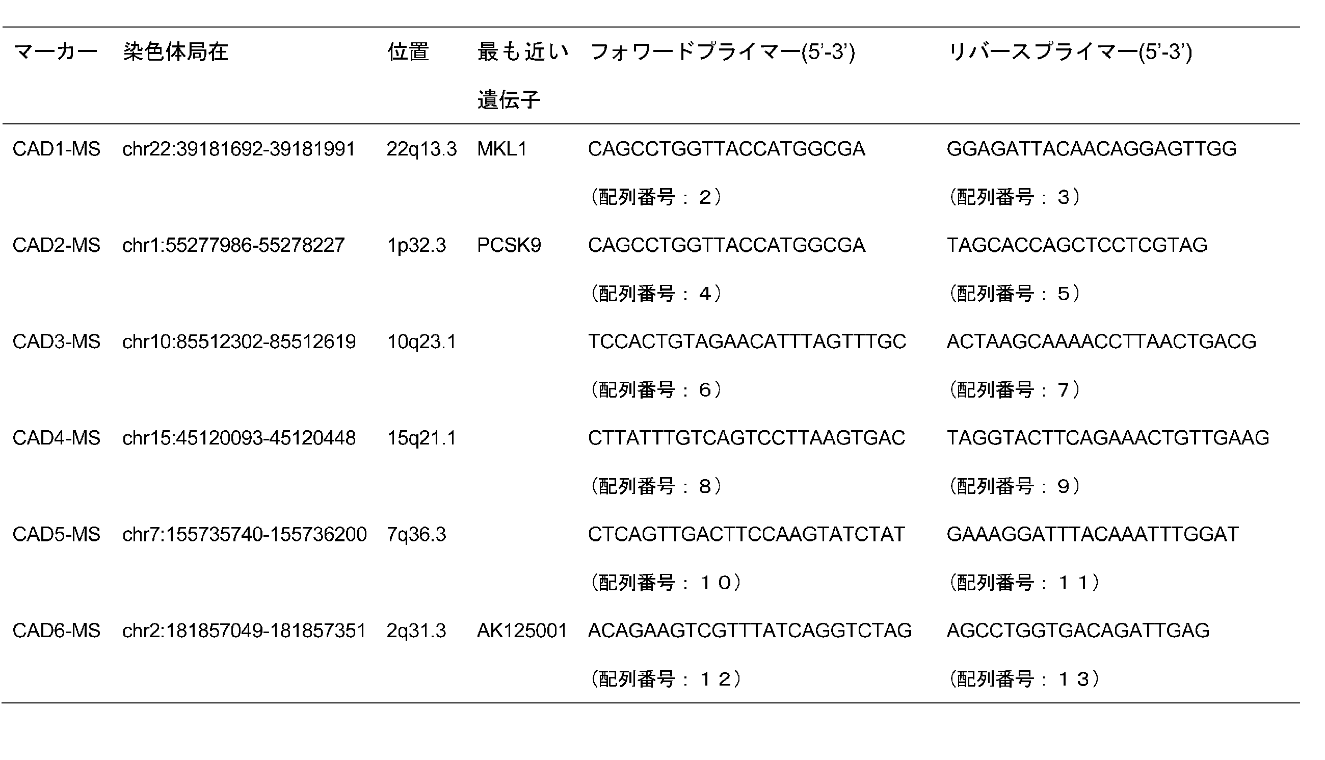
これらの6つのMSマーカー周辺領域の連鎖不均衡(linkage disequilibrium; LD)構造を、HapMap JPT+CHBデータに基づいて解析した。22q13染色体領域のLDの評価のために、HaploviewソフトウエアでD’及びr2を計算した。その結果、CAD3、CAD4及びCAD5は既知の遺伝子を含まないLDブロック内に位置し、CAD1、CAD2及びCAD6はそれぞれ既知の遺伝子MKL1、PCSK9及びK125001を含むLDブロック内に存在することがわかった。さらにPCSK9遺伝子のジェノタイピングを行い、CAD2-MSは高コレステロール血症に特異的な関連性を示すことが判明した。結果を表3に示す。
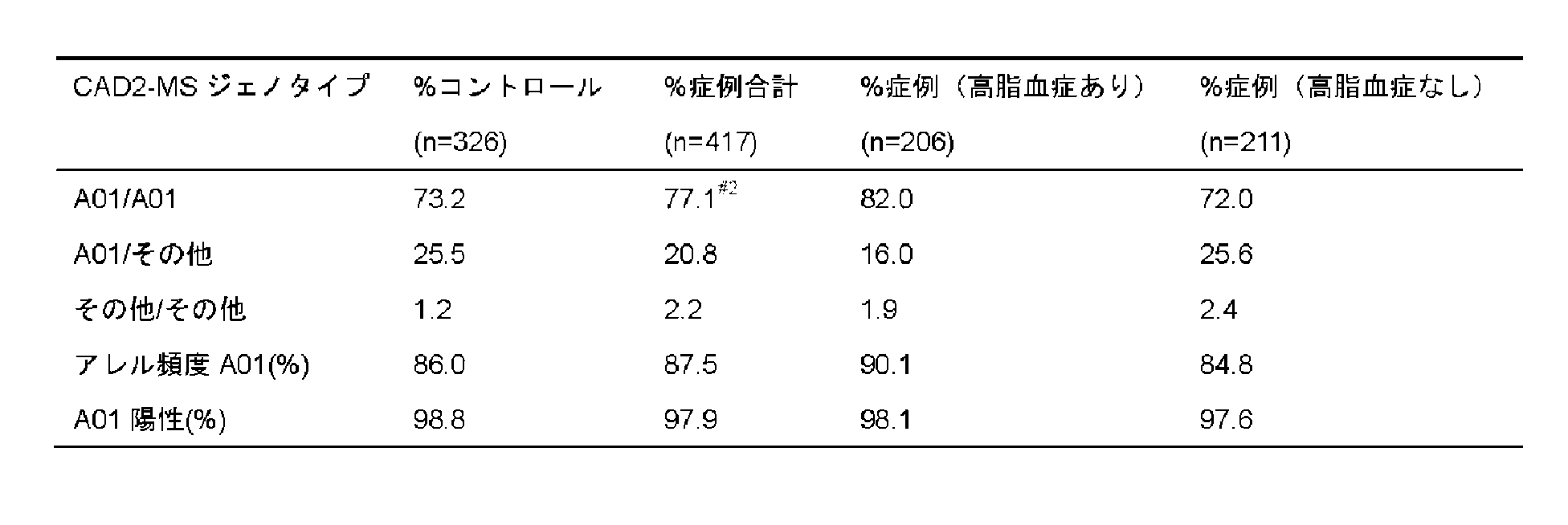
表3中、A01アレルは(Leu)9を示し、A01/A01のジェノタイプ頻度は、最初のスクリーニング(OR=1.49、p=0.045)及び#2で(OR=1.66、p=0.02)、コントロール(n=278)において72.6%、症例(n=283)において79.9%であった。
一方、MKL1遺伝子内に位置するCAD1-MSは、染色体22q13上に存在する(図3)。HapMapデータ(Phase II, release 21)から予測される組換えホットスポットは、MKL1の10kb上流と3’非翻訳領域であり、MKL1遺伝子のみがCAD1関連LDブロックに含まれていることがわかった。CAD1-MSについて、さらに症例とコントロールのジェノタイピングを行った。ジェノタイプ分布とSNPのアレル頻度は、カイ二乗検定で比較し、関連の強さはオッズ比(OR)で表した。結果を表4に示す。(ATAC)9アレル(表1のA05アレル)の頻度は、症例(n=374)で0.132、コントロール(n=339)で0.090であり、疾患感受性に有意な関連を示した(OR=1.54、P=0.012)。一方、(ATAC)13アレル(表1のA09アレル)の頻度は、症例で0.118、コントロールで0.173であり、CADに対する防御との関連が示唆された(OR=0.64、P=0.0040)。

〔MKL1におけるSNP解析〕
さらに、冠状動脈疾患に関連するローカスについて、SNP解析を行った。特定のSNPと疾患との関連は、異なる群(日本人群B)を使用したレプリケーションスタディによって、さらに検証を行った。
さらに、冠状動脈疾患に関連するローカスについて、SNP解析を行った。特定のSNPと疾患との関連は、異なる群(日本人群B)を使用したレプリケーションスタディによって、さらに検証を行った。
エクソン及び隣接するイントロン、並びにMKL1遺伝子上流のプロモーター領域(約1kb)における配列の変異を同定するために、32人の日本人DNAサンプル(CAD1-MS1アレルA05のホモ接合体6例、ヘテロ接合体6例;CAD1-MS1アレルA09のホモ接合体6例、ヘテロ接合体6例;及びCAD1-MS1アレルA05及びA09を有しない8例)を、自動DNAシーケンサー(Applied Biosystems社製ABI PRISM 3100 DNA sequencer)を用いたPCRダイレクトシーケンス法により分析した。
その結果、表5に示すとおり、7個の新規SNPを含む12個のSNPsが同定された。2つのプロモーターSNP(-184C>T及び-173T/C(rs4140512))及び2つのコーディングSNP(1848G>A及び1942A>G(rs878756))は、マイナーアレル頻度が5%を超えていたので、日本人群Aの症例432例、コントロール432例で関連解析を行った。さらに、HapMapデータベースから選択されたMKL1遺伝子の7つのタグSNPについてもケースコントロール分析を行った。その結果、-184C>Tが冠状動脈疾患と有意に関連を示した。(アレル頻度が症例で0.104、コントロールで0.080。OR=1.44、p=0.033)

CAD1-MSとSNP解析の結果は、-184C>TがCAD1-MSのA05アレル(D’=0.88、r2=0.75)と強い連鎖不平衡(linkage disequilibrium, LD)であることを示し、CAD1ローカスの冠状動脈疾患への関連は-184C>Tによるものであることを示唆する。これらの知見をさらに補強するために、日本人群Aのすべての使用可能なサンプル(症例629例、コントロール1,232例。表1参照)についてジェノタイピングを行ったところ、データは有意な関連を示した(OR=1.30、95%信頼区間=1.04-1.64、P=0.024。表2参照)。
〔異なる群での関連解析〕
関連分析のレプリケートスタディのために、日本人群B(症例1,109例、コントロール2,122例。表1)において、-184C>Tを解析した。表6に示されるとおり、日本人群Bにおいて-184C>Tと冠状動脈疾患との関連がレプリケートされた(OR=1.25、95%信頼区間=1.04-1.49、P=0.015)。
関連分析のレプリケートスタディのために、日本人群B(症例1,109例、コントロール2,122例。表1)において、-184C>Tを解析した。表6に示されるとおり、日本人群Bにおいて-184C>Tと冠状動脈疾患との関連がレプリケートされた(OR=1.25、95%信頼区間=1.04-1.49、P=0.015)。
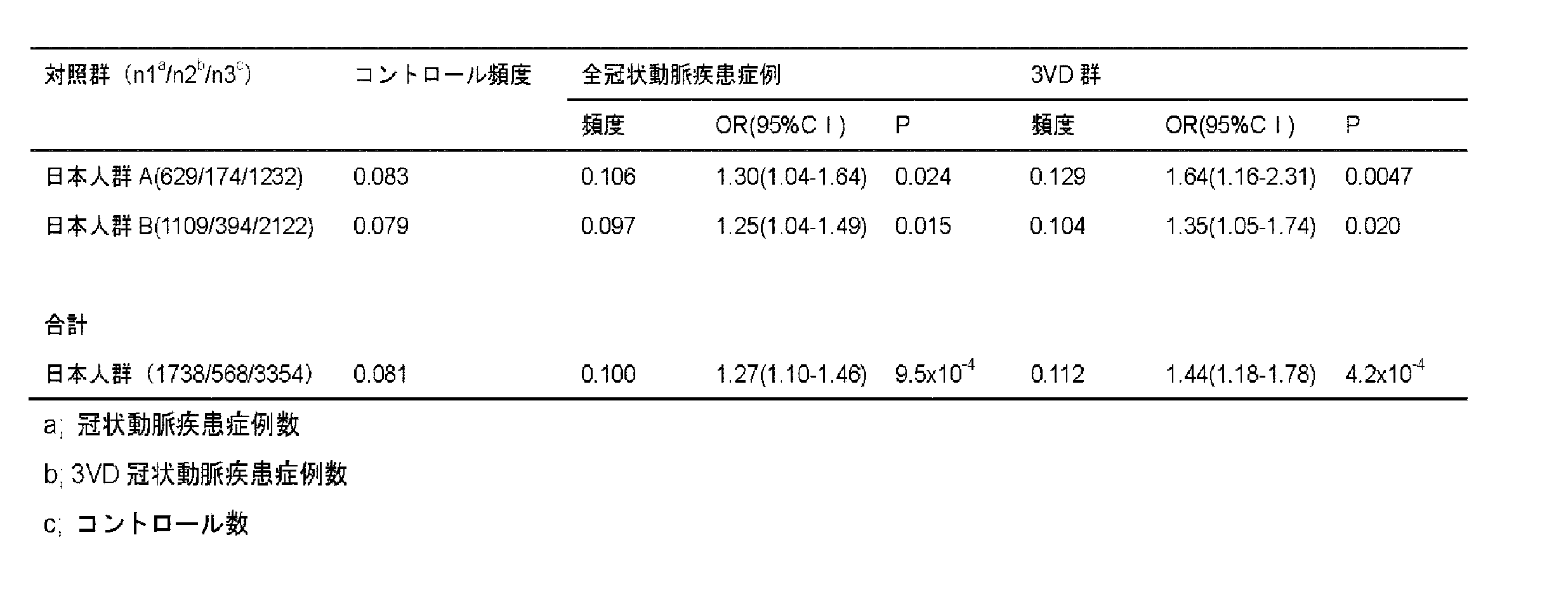
日本人群A及びBのデータについてメタアナリシス(Mantel-Haenszel検定を使用)を行った結果、リスクアレル-184Tについて、冠状動脈疾患との関連は有意(P=9.5×10⁻⁴)であり、OR=1.27(95%信頼区間=1.10-1.46)であった。遺伝形式を解明するために、-184C>Tについてジェノタイプ特異的ORを分析した結果を表7に示す。CC、CT及びTTのORは、それぞれ0.79、1.24及び1.77であった。この結果は、リスクの遺伝が相加的モデルに従うことを示唆した。

リスクアレルと冠状動脈疾患の臨床的背景との相関性を調べるため、-184C>Tとアテローム性動脈硬化の重篤度との関連を分析したところ、いずれの群においても-184C>Tと3VDとの間に強い関連が見られた(表6参照)。データを分析した結果、-184Tアレルは3VD群において特にリスクを高くすることがわかった(OR=1.44、P=4.2×10⁻⁴)(表2参照)。-184C>Tと、高血圧、高脂血症及び糖尿病を含む古典的な冠状動脈疾患リスクファクターとの層別解析によれば、特定の古典的リスクファクターへの関連傾向は見られなかった。表8に結果を示す。

〔発現解析(レポーターアッセイ)〕
-184C>TがMKL1遺伝子のプロモーター領域と見られるローカスに位置したため、この多型が転写レベルに与える影響をレポーターアッセイで測定した。
-184C>TがMKL1遺伝子のプロモーター領域と見られるローカスに位置したため、この多型が転写レベルに与える影響をレポーターアッセイで測定した。
レポーターアッセイは、-184C>Tアレルを含むルシフェラーゼレポーター構築物を使って行われた。転写開始部位のデータベースからに基づいてMKL1遺伝子のプロモーター領域の249bpのフラグメントを決定し、以下のプライマーペアを用いてPCRで増幅した。
5’-CCACGCGTTCCTCAGAGATGAGGTCAT-3’(配列番号:14)
5’-TGCTCGAGCCGGCTCCTCTCAGCCACGG-3’(配列番号:15)
5’-CCACGCGTTCCTCAGAGATGAGGTCAT-3’(配列番号:14)
5’-TGCTCGAGCCGGCTCCTCTCAGCCACGG-3’(配列番号:15)
ゲノムDNAを-184C>Tのホモ接合体の個人から得て、テンプレートとして用いた。PCR産物は、MluI及びXhoI制限酵素で消化し、pGL3-ベーシックベクター(Promega社製)のホタルルシフェラーゼ遺伝子の上流に挿入した。ウシ胎児血清(FBS)10%、ペニシリン/ストレプトマイシン1%を添加したRPMI培地-1840又はDulbeccoの修正イーグル培地で、37℃、5%CO2の条件で、K562又はHeLa細胞を培養した。各構築物は、エレクトロポレーション法でK562細胞(各2μg)に、又はリポフェクション法でHeLa細胞(各1μg)に、内部コントロールとして0.05μgのpRL-tk(Promega社製)と共に導入した。
細胞は、トランスフェクションの24時間後に回収し、シバタらの方法(Shibata, H. et al. Hum Immunol, 67, 2006, 363-373)に従って細胞溶解物を調製した。各細胞溶解物のルシフェラーゼ活性はDUAL Luciferase Reporter Assay System(Promega社製)を使用して決定し、ホタルルシフェラーゼレベルは、pRL-tkからのレニラルシフェラーゼレベルに対して標準化した。データは、4つの独立した実験(各quadruplicate)で採取した。
図4に示すとおり、K562細胞(図4A)及びHeLa細胞(図4B)のいずれにおいても、感受性アレルTに駆動されたルシフェラーゼ活性は、抵抗性対立遺伝子アレルCの転写活性よりも1.2倍高いことが確認された。
〔発現解析(B細胞株におけるMKL1転写物の発現)〕
定常状態でのMKL1遺伝子の発現を、EBウイルスで形質転換したヒトB細胞株の-184Cのホモ接合体又は-184Tのホモ接合体におけるmRNAレベルで検出した。第10回国際組織適合性ワークショップBリンパ芽球様細胞株(http://www.ihwg.org/cellbank/cell_lines/blcl.html)にリストされたヒトB細胞株、又は財団法人ヒューマンサイエンス振興財団から入手したヒトB細胞株のプロモーターの-184位の多型をジェノタイピングし、-184C/C又は184T/Tの遺伝子型を有する5つのB細胞株を選択した。
定常状態でのMKL1遺伝子の発現を、EBウイルスで形質転換したヒトB細胞株の-184Cのホモ接合体又は-184Tのホモ接合体におけるmRNAレベルで検出した。第10回国際組織適合性ワークショップBリンパ芽球様細胞株(http://www.ihwg.org/cellbank/cell_lines/blcl.html)にリストされたヒトB細胞株、又は財団法人ヒューマンサイエンス振興財団から入手したヒトB細胞株のプロモーターの-184位の多型をジェノタイピングし、-184C/C又は184T/Tの遺伝子型を有する5つのB細胞株を選択した。
選択された細胞株は、10%FBS及び1%ペニシリン/ストレプトマイシンを添加したRPMI培地-1840で、5%CO2で培養し、成長段階で回収し、RNeasy mini kit(Qiagen社製)を用いて細胞約107個からトータルRNAを得た。トータルRNA(5μg)はSuperscript II逆転写酵素(Invitrogen社製)によりcDNA合成に供された。iCycler iQ Real-Time PCR Detection System(Bio-Rad社製)及びSYBR Premix Ex Taq(Takara社製)を使用して定量的リアルタイムPCRを行い、mRNAの相対量を測定した。MKL1遺伝子の転写物の相対量は、GAPDHの相対量で除して標準化した。使用したプライマーペアは以下のとおりである。
MKL1用
5’-TAGCCGATGACCTCAATGAG-3’(配列番号:16)
5’-ATCGAAGGAAGAGCTGTCTG-3’(配列番号:17)
GAPDH用
5’-CTTCACCACCATGGAGAAGGC-3’(配列番号:18)
5’-GGCATGGACTGTGGTCATGAG-3’(配列番号:19)
MKL1用
5’-TAGCCGATGACCTCAATGAG-3’(配列番号:16)
5’-ATCGAAGGAAGAGCTGTCTG-3’(配列番号:17)
GAPDH用
5’-CTTCACCACCATGGAGAAGGC-3’(配列番号:18)
5’-GGCATGGACTGTGGTCATGAG-3’(配列番号:19)
実験は3回繰り返し、各実験においてサンプルはトリプリケートとした。
図4Cに示すとおり、Tアレルのホモ接合体を有するB細胞株は、Cアレルのホモ接合体に比較してMKL1遺伝子発現が約1.8倍(1.5倍から2.1倍)高かった。このことは、-184TアレルがMKL1遺伝子の発現亢進に関与することをさらに支持する。
図4Cに示すとおり、Tアレルのホモ接合体を有するB細胞株は、Cアレルのホモ接合体に比較してMKL1遺伝子発現が約1.8倍(1.5倍から2.1倍)高かった。このことは、-184TアレルがMKL1遺伝子の発現亢進に関与することをさらに支持する。
〔ゲルシフトアッセイ(Electrophoretic mobility shift assay; EMSA)〕
-184C>Tの変異がMKL1遺伝子の発現レベルに関与することがわかったので、MKL1遺伝子のプロモーター領域にある-184C>T付近に結合する核内因子をEMSAにより探索した。核抽出物は、ヒト慢性骨髄性白血病細胞株K562から調製した。
-184C>Tの変異がMKL1遺伝子の発現レベルに関与することがわかったので、MKL1遺伝子のプロモーター領域にある-184C>T付近に結合する核内因子をEMSAにより探索した。核抽出物は、ヒト慢性骨髄性白血病細胞株K562から調製した。
プロモーター領域の多型-184C>Tの各アレルに特異的な21塩基長のジゴキシゲニン(DIG)標識二本鎖オリゴヌクレオチドを使用して、DIG Gel Shift Kit 2nd Generation(Roche Diagnostics社製)により、EMSAを行った。二本鎖プローブの塩基配列は以下のとおりである。
-184Cアレル用
5’-CACAGGAAGTCAGGCCGCCGT -3’(配列番号:20)
5’- ACGGCGGCCTGACTTCCTGTG -3’(配列番号:21)
-184Tアレル用
5’-CACAGGAAGTTAGGCCGCCGT -3’(配列番号:22)
5’- ACGGCGGCCTAACTTCCTGTG -3’(配列番号:23)
-184Cアレル用
5’-CACAGGAAGTCAGGCCGCCGT -3’(配列番号:20)
5’- ACGGCGGCCTGACTTCCTGTG -3’(配列番号:21)
-184Tアレル用
5’-CACAGGAAGTTAGGCCGCCGT -3’(配列番号:22)
5’- ACGGCGGCCTAACTTCCTGTG -3’(配列番号:23)
Nuclear and Cytoplasmic Extraction Reagents(Thermo Fisher Scientific社(Rockford)製)を使用し、K562細胞から核抽出物を調製した。DIG標識プローブとK562核抽出物とを4℃で30分インキュベートした後、0.5×TBEランニングバッファーと共に、6%非変性ポリアクリルアミドゲル上で電気泳動によって分離した。
DNA-タンパク質複合体は、プラスにチャージされたナイロンメンブレン上にエレクトロブロッティングされ、DIG Gel Shift Kit 2nd generationのマニュアルに従って可視化された。K562核抽出物は、競合アッセイに使用するために、様々な濃度の標識されていない競合物質と予めインキュベートした。
その結果、図5Aに示されるとおり、DNA-タンパク質複合体は、-184Tより-184Cの場合に高いアフィニティを示した。また、このDNA-タンパク質相互作用は、-184Tプローブを含む場合より、-184Cプローブを含む場合に、非標識DNAによる競合の効率が高かった(図5B)。
TFSEARCH(http://mbs.cbrc.jp/research/db/TFSEARCHJ.html)を使用して-184C>Tに特異的に結合する転写因子を探索したところ、-184C>T周辺の塩基配列は、Ets-1、Nrf2、Elk-1、P300及びMybの結合領域に類似することがわかった。しかしながら、これらの核内因子に対する抗体を加えても、EMSAバンドのスーパーシフト(電気泳動性の大きな遅延)は見られなかった。
〔免疫組織化学染色〕
冠状動脈の狭窄が深刻な患者の病理解剖でアテローム性動脈硬化の病変部を採取し、免疫組織化学染色を行った。
冠状動脈の狭窄が深刻な患者の病理解剖でアテローム性動脈硬化の病変部を採取し、免疫組織化学染色を行った。
パラフィンに包埋した3μmの組織切片をキシレンで脱パラフィンし、エタノールから脱イオン水で水和した。組織切片は、DakoCytomation Protein Block(DAKO社製)で5分間ブロッキングし、1:100希釈で抗ヒトMKL1ポリクローナル抗体(Santa Cruz社製; sc-32909)と共に2時間、室温でインキュベートした。洗浄用バッファーとリン酸バッファー溶液で洗浄後、3%過酸化水素水溶液で5分間インキュベートして、内在性ペルオキシダーゼ活性をブロックした。切片を洗浄用バッファーとリン酸バッファー溶液で洗浄後、二次抗体(DAKO ChemMate Envision/HRP)とインキュベートし、続いてDAB色原体とインキュベートし、Mayerのヘマトキシリンとの対比染色を行った。細胞型特異的抗体を用いた隣接する組織の免疫組織化学分析は、抗ヒトCD68モノクローナル抗体(DAKO社製)及び抗ヒト平滑筋αアクチンモノクローナル抗体(DAKO社製)を使用して実施した。
図6Aに示されるとおり、MKL1タンパクはアテローム斑の脂質コアで発現している。隣接する領域を抗CD68抗体で染色したところ、MKL1-陽性細胞は、泡沫化したマクロファージであることがわかった(図6B参照)。また、MKL1タンパクは、新生内膜中の平滑筋αアクチン陽性細胞でも発現していた(図6C)。
配列番号:1は、上流配列1000Bp及び下流配列500bpを含むMKL1ゲノム遺伝子の塩基配列である。
配列番号:2は、MSマーカーCAD1-MSを増幅するフォワードプライマーである。
配列番号:3は、MSマーカーCAD1-MSを増幅するリバースプライマーである。
配列番号:4は、MSマーカーCAD2-MSを増幅するフォワードプライマーである。
配列番号:5は、MSマーカーCAD2-MSを増幅するリバースプライマーである。
配列番号:6は、MSマーカーCAD3-MSを増幅するフォワードプライマーである。
配列番号:7は、MSマーカーCAD3-MSを増幅するリバースプライマーである。
配列番号:8は、MSマーカーCAD4-MSを増幅するフォワードプライマーである。
配列番号:9は、MSマーカーCAD4-MSを増幅するリバースプライマーである。
配列番号:10は、MSマーカーCAD5-MSを増幅するフォワードプライマーである。
配列番号:11は、MSマーカーCAD5-MSを増幅するリバースプライマーである。
配列番号:12は、MSマーカーCAD6-MSを増幅するフォワードプライマーである。
配列番号:13は、MSマーカーCAD6-MSを増幅するリバースプライマーである。
配列番号:14は、MKL1遺伝子のプロモーター領域のフラグメントを増幅するフォワードプライマーである。
配列番号:15は、MKL1遺伝子のプロモーター領域のフラグメントを増幅するリバースプライマーである。
配列番号:16は、B細胞株におけるMKL1転写物に対する定量的リアルタイムPCRに用いたフォワードプライマーである。
配列番号:17は、B細胞株におけるMKL1転写物に対する定量的リアルタイムPCRに用いたリバースプライマーである。
配列番号:18は、B細胞株におけるGAPDH転写物に対する定量的リアルタイムPCRに用いたフォワードプライマーである。
配列番号:19は、B細胞株におけるGADPH転写物に対する定量的リアルタイムPCRに用いたリバースプライマーである。
配列番号:20は、EMSAに用いた-184Cアレル用フォワードプライマーである。
配列番号:21は、EMSAに用いた-184Cアレル用リバースプライマーである。
配列番号:22は、EMSAに用いた-184Tアレル用フォワードプライマーである。
配列番号:23は、EMSAに用いた-184Tアレル用リバースプライマーである。
配列番号:2は、MSマーカーCAD1-MSを増幅するフォワードプライマーである。
配列番号:3は、MSマーカーCAD1-MSを増幅するリバースプライマーである。
配列番号:4は、MSマーカーCAD2-MSを増幅するフォワードプライマーである。
配列番号:5は、MSマーカーCAD2-MSを増幅するリバースプライマーである。
配列番号:6は、MSマーカーCAD3-MSを増幅するフォワードプライマーである。
配列番号:7は、MSマーカーCAD3-MSを増幅するリバースプライマーである。
配列番号:8は、MSマーカーCAD4-MSを増幅するフォワードプライマーである。
配列番号:9は、MSマーカーCAD4-MSを増幅するリバースプライマーである。
配列番号:10は、MSマーカーCAD5-MSを増幅するフォワードプライマーである。
配列番号:11は、MSマーカーCAD5-MSを増幅するリバースプライマーである。
配列番号:12は、MSマーカーCAD6-MSを増幅するフォワードプライマーである。
配列番号:13は、MSマーカーCAD6-MSを増幅するリバースプライマーである。
配列番号:14は、MKL1遺伝子のプロモーター領域のフラグメントを増幅するフォワードプライマーである。
配列番号:15は、MKL1遺伝子のプロモーター領域のフラグメントを増幅するリバースプライマーである。
配列番号:16は、B細胞株におけるMKL1転写物に対する定量的リアルタイムPCRに用いたフォワードプライマーである。
配列番号:17は、B細胞株におけるMKL1転写物に対する定量的リアルタイムPCRに用いたリバースプライマーである。
配列番号:18は、B細胞株におけるGAPDH転写物に対する定量的リアルタイムPCRに用いたフォワードプライマーである。
配列番号:19は、B細胞株におけるGADPH転写物に対する定量的リアルタイムPCRに用いたリバースプライマーである。
配列番号:20は、EMSAに用いた-184Cアレル用フォワードプライマーである。
配列番号:21は、EMSAに用いた-184Cアレル用リバースプライマーである。
配列番号:22は、EMSAに用いた-184Tアレル用フォワードプライマーである。
配列番号:23は、EMSAに用いた-184Tアレル用リバースプライマーである。
Claims (15)
- 被検者の冠状動脈疾患リスクの検査方法であって、被検者から採取した試料におけるMKL1遺伝子発現を亢進させる変異又はMKL1遺伝子発現の亢進を検出する工程を含む方法。
- 前記MKL1遺伝子発現を亢進させる変異を検出する工程は、
MKL1遺伝子における配列番号:1に記載の塩基配列の817位に対応する部位の多型を検出する工程を含む、請求項1に記載の方法。 - 前記817位に対応する塩基がチミン(T)である多型を検出したときに、前記被検者の冠動脈疾患リスクが通常より高いと判断する、請求項2に記載の方法。
- 前記MKL1遺伝子発現の亢進を検出する工程は、被検者から採取した活性化マクロファージ内におけるMKL1遺伝子発現の亢進を検出する工程を含む、請求項1に記載の方法。
- 前記MKL1遺伝子の発現が亢進しているときに、前記被検者の冠動脈疾患リスクが通常より高いと判断する、請求項4に記載の方法。
- 冠状動脈疾患リスクの検査薬であって、以下の(1)から(3)の少なくとも1つを含む、検査薬。
(1)配列番号:1に記載の塩基配列における817位を含む少なくとも10塩基の領域からなる核酸又はこれに相補的な領域からなる核酸にハイブリダイズする核酸
(2)配列番号:1に記載の塩基配列における817位を含む少なくとも10塩基の領域からなる核酸又はこれに相補的な領域からなる核酸を増幅可能なプライマー
(3)MKL1タンパク質に対する抗体 - 被検者の冠状動脈疾患リスクの診断方法であって、被検者から採取した試料におけるMKL1遺伝子発現を亢進させる変異又はMKL1遺伝子発現の亢進を検出する工程を含む方法。
- 前記被検者から採取した試料におけるMKL1遺伝子発現を亢進させる変異を検出する工程は、
被検者から採取した試料から核酸試料を調製する工程と、
前記核酸試料において、MKL1遺伝子における配列番号:1に記載の塩基配列の817位に対応する部位の多型を検出する工程と、を含む請求項7に記載の方法。 - 前記817位に対応する塩基がチミン(T)である多型を検出したときに、前記被検者の冠状動脈疾患リスクが通常より高いと診断する、請求項8に記載の方法。
- 前記被検者から採取した試料におけるMKL1遺伝子発現の亢進を検出する工程は、
前記被検者から活性化マクロファージを含む試料を採取する工程と、
前記活性化マクロファージ内におけるMKL1遺伝子の発現の亢進を検出する工程と、を含む請求項7に記載の方法。 - 前記MKL1遺伝子の発現が亢進しているときに、前記被検者の冠状動脈疾患リスクが通常より高いと診断する、請求項10に記載の方法。
- 冠状動脈疾患の予防又は治療薬のスクリーニング方法であって、
MKL1遺伝子を発現可能な細胞と、前記予防又は治療薬候補とを接触させる工程と、
前記MKL1遺伝子の発現若しくはMKL1タンパク質の機能を阻害又は抑制する候補を選択する工程と、を含む方法。 - 配列番号:1に記載の塩基配列において817位に対応する塩基がT(チミン)である塩基配列からなる、MKL1タンパク質をコードするDNA。
- 請求項13に記載のDNAを含む組換えベクター。
- 請求項14に記載の組換えベクターを含む形質転換体。
Priority Applications (1)
| Application Number | Priority Date | Filing Date | Title |
|---|---|---|---|
| JP2010543810A JPWO2010073544A1 (ja) | 2008-12-24 | 2009-12-16 | 冠状動脈疾患リスクの検査方法 |
Applications Claiming Priority (2)
| Application Number | Priority Date | Filing Date | Title |
|---|---|---|---|
| JP2008-327960 | 2008-12-24 | ||
| JP2008327960 | 2008-12-24 |
Publications (1)
| Publication Number | Publication Date |
|---|---|
| WO2010073544A1 true WO2010073544A1 (ja) | 2010-07-01 |
Family
ID=42287199
Family Applications (1)
| Application Number | Title | Priority Date | Filing Date |
|---|---|---|---|
| PCT/JP2009/006918 Ceased WO2010073544A1 (ja) | 2008-12-24 | 2009-12-16 | 冠状動脈疾患リスクの検査方法 |
Country Status (2)
| Country | Link |
|---|---|
| JP (1) | JPWO2010073544A1 (ja) |
| WO (1) | WO2010073544A1 (ja) |
-
2009
- 2009-12-16 JP JP2010543810A patent/JPWO2010073544A1/ja not_active Withdrawn
- 2009-12-16 WO PCT/JP2009/006918 patent/WO2010073544A1/ja not_active Ceased
Non-Patent Citations (9)
| Title |
|---|
| DATABASE DDBJ/EMBL/GENBANK [online] 7 September 2001 (2001-09-07), retrieved from http://www.ncbi.nlm.nih.gov/ sviewer/viewer.fcgi?14161366:NCBI:2651775 Database accession no. AF368061 * |
| DATABASE POST GENOME JIDAI NO IDEN YODOSHA CO., LTD.; 10 March 2002 (2002-03-10), NAOYUKI KAMATANI ET AL. * |
| HINOHARA, K. ET AL.: "Megakaryoblastic leukemia factor-1 gene in the susceptibility to coronary artery disease", HUM. GENET., vol. 126, no. 4, October 2009 (2009-10-01), pages 539 - 547 * |
| KAWASHIMA, M. ET AL.: "Genomewide association analysis of human narcolepsy and a new resistance gene", AM. J. HUM. GENET., vol. 79, no. 2, August 2006 (2006-08-01), pages 252 - 263 * |
| MA, Z. ET AL., DEFINITION: HOMO SAPIENS MEGAKARYOBLASTIC LEUKEMIA-1 PROTEIN(MKL1) MRNA, 9 February 2010 (2010-02-09) * |
| MA, Z. ET AL.: "Fusion of two novel genes, RBM15 and MKL1, in the t(1;22)(p13;q13) of acute megakaryoblastic leukemia", NAT.GENET., vol. 28, no. 3, July 2001 (2001-07-01), pages 220 - 221 * |
| MITSUHIRO YOKOTA ET AL.: "Shikkan Genome Kaiseki 4. Idenshitakei ni yoru Kandomyaku Shikkan Risk no Yosoku", EXPERIMENTAL MEDICINE, vol. 23, no. 4, 15 February 2005 (2005-02-15), pages 49 - 58 * |
| YASUHARU TABARA ET AL.: "Idenshi Kaiseki no Shinpo Genome Wide Sokan Kaiseki ni yoru Koketsuatsu Kanjusei Idenshi no Tansaku - Millennium Genome Project", MOLECULAR MEDICINE, vol. 42, 5 September 2005 (2005-09-05), pages 208 - 213 * |
| YOSHIJI YAMADA ET AL.: "Bunshi Level no Saishin Shikkan Kenkyu 11) Idenshitakei ni yoru Shinkin Kosoku Kikendo no Yosoku", EXPERIMENTAL MEDICINE, vol. 21, no. 9, 1 June 2003 (2003-06-01), pages 1258 - 1262 * |
Also Published As
| Publication number | Publication date |
|---|---|
| JPWO2010073544A1 (ja) | 2012-06-07 |
Similar Documents
| Publication | Publication Date | Title |
|---|---|---|
| DeJesus-Hernandez et al. | Expanded GGGGCC hexanucleotide repeat in noncoding region of C9ORF72 causes chromosome 9p-linked FTD and ALS | |
| EP2527472B1 (en) | A genetic lesion associated with cancer | |
| US10172916B2 (en) | Methods of treating heart failure with agonists of hypocretin receptor 2 | |
| KR20220153109A (ko) | Rnaset2를 통한 염증성 장 질환의 진단 방법 | |
| KR20220063311A (ko) | 간질 폐렴의 위험을 예측하는 방법 | |
| CN101501194A (zh) | 老年性黄斑变性的预防和治疗 | |
| KR20160111925A (ko) | 자살 위험과 연관된 유전적 마커 및 그의 사용 방법 | |
| Suzuki et al. | A novel genetic marker for coronary spasm in women from a genome-wide single nucleotide polymorphism analysis | |
| EP2393939B1 (en) | A snp marker of breast and ovarian cancer risk | |
| US20110035818A1 (en) | Diagnostic marker and platform for drug design in myocardial infarction and heart failure | |
| WO2012018258A1 (en) | Markers of febrile seizures and temporal lobe epilepsy | |
| KR101304535B1 (ko) | Klotho 유전자의 단일염기다형을 이용한 심혈관계 질환 예측 방법 | |
| WO2010073544A1 (ja) | 冠状動脈疾患リスクの検査方法 | |
| JP2008506354A (ja) | 高脂血症、異常脂質血症、および不完全な炭水化物代謝と関連するsnpの同定 | |
| JP5540340B2 (ja) | 炎症性疾患の判定方法 | |
| KR102646011B1 (ko) | Brd3의 단일염기다형성을 이용한 비소세포폐암의 예후 진단방법 | |
| JP2003504076A (ja) | 遺伝子テスト | |
| CN101466832A (zh) | 炎症性疾病的判定方法 | |
| JP4845486B2 (ja) | 糖尿病性腎症感受性遺伝子、および糖尿病性腎症の予防または治療剤の有効成分のスクリーニング方法 | |
| Cho | Elucidating the Roles and Mechanisms by which a Long Non-coding RNA, ANRIL, Regulates Endothelial Cell Activities in the Development of Coronary Artery Disease | |
| US20080194419A1 (en) | Genetic Association of Polymorphisms in the Atf6-Alpha Gene with Insulin Resistance Phenotypes | |
| JP5422805B2 (ja) | 新規糖尿病性腎症感受性遺伝子 | |
| KR20230150127A (ko) | 심근경색과 연관된 유전자 다형성 및 그의 용도 | |
| JP2017006074A (ja) | 末梢動脈疾患検査方法及び検査用試薬 | |
| WO2010033825A2 (en) | Genetic variants associated with abdominal aortic aneurysms |
Legal Events
| Date | Code | Title | Description |
|---|---|---|---|
| 121 | Ep: the epo has been informed by wipo that ep was designated in this application |
Ref document number: 09834356 Country of ref document: EP Kind code of ref document: A1 |
|
| DPE1 | Request for preliminary examination filed after expiration of 19th month from priority date (pct application filed from 20040101) | ||
| ENP | Entry into the national phase |
Ref document number: 2010543810 Country of ref document: JP Kind code of ref document: A |
|
| NENP | Non-entry into the national phase |
Ref country code: DE |
|
| 122 | Ep: pct application non-entry in european phase |
Ref document number: 09834356 Country of ref document: EP Kind code of ref document: A1 |