WO2010016587A1 - Method for evaluating specific incorporation of d-glucose into cells - Google Patents
Method for evaluating specific incorporation of d-glucose into cells Download PDFInfo
- Publication number
- WO2010016587A1 WO2010016587A1 PCT/JP2009/064053 JP2009064053W WO2010016587A1 WO 2010016587 A1 WO2010016587 A1 WO 2010016587A1 JP 2009064053 W JP2009064053 W JP 2009064053W WO 2010016587 A1 WO2010016587 A1 WO 2010016587A1
- Authority
- WO
- WIPO (PCT)
- Prior art keywords
- molecule
- glucose
- glucose derivative
- fluorescent chromophore
- group
- Prior art date
Links
- GIMXPYIZHUYHET-UHFFFAOYSA-N CS(C(Nc(c1n[o]nc11)ccc1[N+]([O-])=O)=C1)OC(CO)C1(CO)O Chemical compound CS(C(Nc(c1n[o]nc11)ccc1[N+]([O-])=O)=C1)OC(CO)C1(CO)O GIMXPYIZHUYHET-UHFFFAOYSA-N 0.000 description 1
- XDODIEWVSJOFHI-YUOPPWCSSA-N [O-][N+](c(c1n[o]nc11)ccc1NC(COC(CO)[C@@H]1O)C1O)=O Chemical compound [O-][N+](c(c1n[o]nc11)ccc1NC(COC(CO)[C@@H]1O)C1O)=O XDODIEWVSJOFHI-YUOPPWCSSA-N 0.000 description 1
- GVEBOQDOPLERMC-LFWJNZRMSA-N [O-][N+](c(c1n[o]nc11)ccc1NC([C@@H]1O)C(O)OC(CO)[C@H]1O)=O Chemical compound [O-][N+](c(c1n[o]nc11)ccc1NC([C@@H]1O)C(O)OC(CO)[C@H]1O)=O GVEBOQDOPLERMC-LFWJNZRMSA-N 0.000 description 1
Images
Classifications
-
- G—PHYSICS
- G01—MEASURING; TESTING
- G01N—INVESTIGATING OR ANALYSING MATERIALS BY DETERMINING THEIR CHEMICAL OR PHYSICAL PROPERTIES
- G01N33/00—Investigating or analysing materials by specific methods not covered by groups G01N1/00 - G01N31/00
- G01N33/48—Biological material, e.g. blood, urine; Haemocytometers
- G01N33/50—Chemical analysis of biological material, e.g. blood, urine; Testing involving biospecific ligand binding methods; Immunological testing
- G01N33/58—Chemical analysis of biological material, e.g. blood, urine; Testing involving biospecific ligand binding methods; Immunological testing involving labelled substances
- G01N33/582—Chemical analysis of biological material, e.g. blood, urine; Testing involving biospecific ligand binding methods; Immunological testing involving labelled substances with fluorescent label
-
- C—CHEMISTRY; METALLURGY
- C09—DYES; PAINTS; POLISHES; NATURAL RESINS; ADHESIVES; COMPOSITIONS NOT OTHERWISE PROVIDED FOR; APPLICATIONS OF MATERIALS NOT OTHERWISE PROVIDED FOR
- C09B—ORGANIC DYES OR CLOSELY-RELATED COMPOUNDS FOR PRODUCING DYES, e.g. PIGMENTS; MORDANTS; LAKES
- C09B51/00—Nitro or nitroso dyes
-
- C—CHEMISTRY; METALLURGY
- C09—DYES; PAINTS; POLISHES; NATURAL RESINS; ADHESIVES; COMPOSITIONS NOT OTHERWISE PROVIDED FOR; APPLICATIONS OF MATERIALS NOT OTHERWISE PROVIDED FOR
- C09B—ORGANIC DYES OR CLOSELY-RELATED COMPOUNDS FOR PRODUCING DYES, e.g. PIGMENTS; MORDANTS; LAKES
- C09B57/00—Other synthetic dyes of known constitution
-
- C—CHEMISTRY; METALLURGY
- C12—BIOCHEMISTRY; BEER; SPIRITS; WINE; VINEGAR; MICROBIOLOGY; ENZYMOLOGY; MUTATION OR GENETIC ENGINEERING
- C12Q—MEASURING OR TESTING PROCESSES INVOLVING ENZYMES, NUCLEIC ACIDS OR MICROORGANISMS; COMPOSITIONS OR TEST PAPERS THEREFOR; PROCESSES OF PREPARING SUCH COMPOSITIONS; CONDITION-RESPONSIVE CONTROL IN MICROBIOLOGICAL OR ENZYMOLOGICAL PROCESSES
- C12Q1/00—Measuring or testing processes involving enzymes, nucleic acids or microorganisms; Compositions therefor; Processes of preparing such compositions
- C12Q1/02—Measuring or testing processes involving enzymes, nucleic acids or microorganisms; Compositions therefor; Processes of preparing such compositions involving viable microorganisms
-
- C—CHEMISTRY; METALLURGY
- C12—BIOCHEMISTRY; BEER; SPIRITS; WINE; VINEGAR; MICROBIOLOGY; ENZYMOLOGY; MUTATION OR GENETIC ENGINEERING
- C12Q—MEASURING OR TESTING PROCESSES INVOLVING ENZYMES, NUCLEIC ACIDS OR MICROORGANISMS; COMPOSITIONS OR TEST PAPERS THEREFOR; PROCESSES OF PREPARING SUCH COMPOSITIONS; CONDITION-RESPONSIVE CONTROL IN MICROBIOLOGICAL OR ENZYMOLOGICAL PROCESSES
- C12Q1/00—Measuring or testing processes involving enzymes, nucleic acids or microorganisms; Compositions therefor; Processes of preparing such compositions
- C12Q1/54—Measuring or testing processes involving enzymes, nucleic acids or microorganisms; Compositions therefor; Processes of preparing such compositions involving glucose or galactose
-
- G—PHYSICS
- G01—MEASURING; TESTING
- G01N—INVESTIGATING OR ANALYSING MATERIALS BY DETERMINING THEIR CHEMICAL OR PHYSICAL PROPERTIES
- G01N33/00—Investigating or analysing materials by specific methods not covered by groups G01N1/00 - G01N31/00
- G01N33/48—Biological material, e.g. blood, urine; Haemocytometers
- G01N33/50—Chemical analysis of biological material, e.g. blood, urine; Testing involving biospecific ligand binding methods; Immunological testing
- G01N33/5005—Chemical analysis of biological material, e.g. blood, urine; Testing involving biospecific ligand binding methods; Immunological testing involving human or animal cells
-
- G—PHYSICS
- G01—MEASURING; TESTING
- G01N—INVESTIGATING OR ANALYSING MATERIALS BY DETERMINING THEIR CHEMICAL OR PHYSICAL PROPERTIES
- G01N33/00—Investigating or analysing materials by specific methods not covered by groups G01N1/00 - G01N31/00
- G01N33/48—Biological material, e.g. blood, urine; Haemocytometers
- G01N33/50—Chemical analysis of biological material, e.g. blood, urine; Testing involving biospecific ligand binding methods; Immunological testing
- G01N33/53—Immunoassay; Biospecific binding assay; Materials therefor
- G01N33/531—Production of immunochemical test materials
- G01N33/532—Production of labelled immunochemicals
- G01N33/533—Production of labelled immunochemicals with fluorescent label
Definitions
- the present invention relates to a method for accurately evaluating specific uptake of D-glucose into cells.
- Glucose is known as the most important energy source for maintaining the survival of cells from mammals to Escherichia coli and yeast.
- the brain uses glucose as the only energy source.
- Glucose has enantiomers of D-glucose and L-glucose. Among them, only D-glucose can be used as an energy source by living organisms, and living cells can use D-glucose as a glucose transporter. It has a mechanism for selectively taking in and using transport proteins in cell membranes.
- This method utilizes the property that 2-NBDG is selectively taken up into living cells, and the dynamic activity of D-glucose uptake by cells is monitored by tracking changes in fluorescence intensity due to uptake. Since it can be quantitatively known, it has been evaluated by researchers all over the world as an innovative method for studying how organisms take D-glucose into cells and uses it. It is positioned as a standard protocol indispensable in this research field (Non-patent Document 2).
- the method for evaluating the specific uptake of D-glucose of the present invention in contrast to L-glucose according to the present invention has a fluorescent chromophore in the molecule as described in claim 1.
- a D-glucose derivative specifically incorporated into the cell and an L-glucose derivative having a fluorescent chromophore in the molecule are brought into contact with separate cells of the cell type to be evaluated,
- the fluorescence emitted from the D-glucose derivative specifically incorporated into the cells contained in the molecule is compared with the fluorescence emitted from the L-glucose derivative having a fluorescent chromophore in the molecule, and the difference in fluorescence intensity between the two is compared.
- D-glucose is evaluated as specific uptake into cells in comparison with L-glucose.
- the method according to claim 2 is the same as the method according to claim 1, except that N- (7-nitrobenz-) is a D-glucose derivative that is specifically taken into a cell having a fluorescent chromophore in the molecule.
- a D-glucose derivative having a 2-oxa-1,3-diazol-4-yl) amino group in the molecule is used.
- the method according to claim 3 is the same as the method according to claim 2, wherein the D-glucose derivative has an N- (7-nitrobenz-2-oxa-1,3-diazol-4-yl) amino group in the molecule.
- the method according to claim 5 is the same as the method according to claim 4, wherein the L-glucose derivative has an N- (7-nitrobenz-2-oxa-1,3-diazol-4-yl) amino group in the molecule.
- L-deoxyglucose in which an N- (7-nitrobenz-2-oxa-1,3-diazol-4-yl) amino group is bonded to the 2-position is used.
- the method according to claim 6 is the method according to claim 1, wherein 2- (N-methylamino) is used as a D-glucose derivative that is specifically taken up into a cell having a fluorescent chromophore in the molecule.
- a D-glucose derivative having a benzoylamino group in the molecule is used.
- the method according to claim 7 is the same as the method according to claim 6, wherein 2- (N-methylamino) benzoylamino is used as a D-glucose derivative having a 2- (N-methylamino) benzoylamino group in the molecule. It is characterized by using D-deoxyglucose having a group bonded to the 2-position.
- the method according to claim 8 is the method according to claim 6, wherein the L-glucose derivative having a fluorescent chromophore in the molecule is an L-glucose having a 2- (N-methylamino) benzoylamino group in the molecule.
- the method according to claim 11 is characterized in that, in the method according to claim 10, the difference in maximum fluorescence wavelength is at least 20 nm.
- the method according to claim 15 is the method according to claim 14, wherein 2- (N-methylamino) benzoylamino is used as a D-glucose derivative having a 2- (N-methylamino) benzoylamino group in the molecule. It is characterized by using D-deoxyglucose having a group bonded to the 2-position.
- the 5-hydroxyl group of L-mannose is acetylated, the 1-position acetoxy group is substituted with bromine, and the 1-position is ortho-substituted.
- the present invention also provides use of L-glucosamine or a salt thereof for producing an L-glucose derivative having a fluorescent chromophore in the molecule, as defined in claim 25.
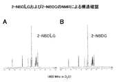
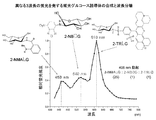
- FIG. 2 is a 1 H-NMR chart of 2-NBDG and 2-NBDLG.
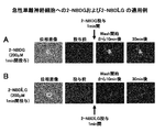
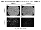
- FIG. 2 is an image obtained by ordinary fluorescence microscopy when 2-NBDG and 2-NBDLG are administered to acutely isolated neurons in Example 1.
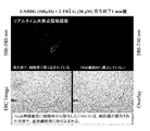
- FIG. 3 is an image obtained by real-time laser scanning confocal fluorescence microscopy when 2-NBDG and 2-TRLG are administered to acutely isolated neurons in Example 2.
- the method for evaluating the specific uptake of D-glucose into cells of the present invention can be roughly divided into the following two methods. (1) Separate D-glucose derivatives that are specifically incorporated into cells having a fluorescent chromophore in the molecule and L-glucose derivatives that have the fluorescent chromophore in the molecule, as cell types to be evaluated. Fluorescence emitted from a D-glucose derivative that is specifically taken into a cell having a fluorescent chromophore in the molecule, and fluorescence emitted from an L-glucose derivative having a fluorescent chromophore in the molecule.
- a D-glucose derivative (which may be in the form of a salt such as hydrochloride) that is specifically taken into a cell having a fluorescent chromophore in the molecule refers to the fluorescent chromophore.
- Is a D-glucose derivative such as 2-NBDG
- N -(7-Nitrobenz-2-oxa-1,3-diazol-4-yl) not limited to combinations of L-glucose derivatives (such as 2-NBDLG) having an amino group in the molecule, but blue fluorescence
- a D-glucose derivative having a 2- (N-methylamino) benzoylamino group in the molecule and a 2- (N-methylamino) benzoylamino group in the molecule It may be a combination of L- glucose derivative having the.
Abstract
Disclosed is a method for accurately evaluating the specific incorporation of D-glucose into cells. The method is characterized by: contacting each of a D-glucose derivative that can be incorporated into cells specifically and has a fluorescent chromophore in the molecule and an L-glucose derivative that has a fluorescent chromophore in the molecule with cells of a cell species to be evaluated; comparing the fluorescence emitted from the D-glucose derivative that can be incorporated into cells specifically and has the fluorescent chromophore in the molecule with the fluorescence emitted from the L-glucose derivative that has the fluorescent chromophore in the molecule; and determining the difference between the above-mentioned fluorescence intensities as the specific incorporation of D-glucose into cells relative to that of L-glucose.
Description
本発明は、D-グルコースの細胞内への特異的な取り込みを正確に評価するための方法に関する。
The present invention relates to a method for accurately evaluating specific uptake of D-glucose into cells.
グルコース(ブドウ糖)は、哺乳類から大腸菌・酵母に至るまで細胞の生存維持に最も重要なエネルギー源として知られ、特に脳はグルコースを唯一のエネルギー源としている。グルコースにはD-グルコースとL-グルコースの鏡像異性体が存在するが、このうち生物がエネルギー源として利用できるのはD-グルコースのみであり、生きた細胞はD-グルコースをグルコーストランスポーターなどの細胞膜中の輸送タンパク質を介して選択的に取り込んで利用する仕組みを持つ。
Glucose (glucose) is known as the most important energy source for maintaining the survival of cells from mammals to Escherichia coli and yeast. In particular, the brain uses glucose as the only energy source. Glucose has enantiomers of D-glucose and L-glucose. Among them, only D-glucose can be used as an energy source by living organisms, and living cells can use D-glucose as a glucose transporter. It has a mechanism for selectively taking in and using transport proteins in cell membranes.
従来、生物がD-グルコースをどのようにして細胞内に取り込んで利用するのかについての研究は、例えばラジオアイソトープで標識したD-グルコースやその誘導体(D-デオキシグルコースなど)を用いて細胞内のラジオアイソトープ量を測定することで行われてきた。しかしながら、この方法は定量性に優れるものの、感度が低いといった問題があることに加え、測定手法上、生きた細胞がD-グルコースを取り込む様子をリアルタイムで連続的に観察することができないという欠点を有する。従って、この方法は、生きた細胞のD-グルコースの動的な取り込みプロセスの研究には使用することができない。
Conventionally, studies on how D-glucose is taken up into cells and used by organisms have been conducted by using, for example, radioisotope-labeled D-glucose and its derivatives (such as D-deoxyglucose). This has been done by measuring the amount of radioisotope. However, although this method is excellent in quantification, in addition to the problem that sensitivity is low, the measurement method has a drawback that it is impossible to continuously observe in real time how living cells take up D-glucose. Have. Therefore, this method cannot be used to study the dynamic uptake process of D-glucose in living cells.
以上のような状況下、本発明者のグループは、生きた細胞のD-グルコースの動的な取り込みプロセスの研究に使用することができる方法として、D-デオキシグルコースの2位に蛍光発色団としてN-(7-ニトロベンズ-2-オキサ-1,3-ジアゾール-4-イル)アミノ基を結合せしめた、緑色の蛍光を発する下記の構造式で示される2-[N-(7-ニトロベンズ-2-オキサ-1,3-ジアゾール-4-イル)アミノ]-2-デオキシ-D-グルコース(2-NBDG)を用いる方法を提案し、その有用性を哺乳動物の各種の細胞を用いて実証した(非特許文献1)。
Under the circumstances as described above, the group of the present inventors, as a method that can be used to study the dynamic D-glucose uptake process of living cells, has a fluorescent chromophore at the 2-position of D-deoxyglucose. 2- [N- (7-Nitrobenz--N- (7-Nitrobenz-2-oxa-1,3-diazol-4-yl) amino group, which has a green fluorescence and has the following structural formula. 2-oxa-1,3-diazol-4-yl) amino] -2-deoxy-D-glucose (2-NBDG) was proposed and its usefulness was demonstrated using various mammalian cells (Non-Patent Document 1).

この方法は、2-NBDGが生きた細胞内に選択的に取り込まれる性質を利用したものであり、取り込みによる蛍光強度の変化を追跡することで細胞のD-グルコースの取り込みについての動的活動を定量的に知ることができることから、生物がD-グルコースをどのようにして細胞内に取り込んで利用するのかを研究する上での画期的な方法として世界中の研究者に評価され、今や、この研究分野において欠かすことができない標準的なプロトコルとして位置付けられている(非特許文献2)。
This method utilizes the property that 2-NBDG is selectively taken up into living cells, and the dynamic activity of D-glucose uptake by cells is monitored by tracking changes in fluorescence intensity due to uptake. Since it can be quantitatively known, it has been evaluated by researchers all over the world as an innovative method for studying how organisms take D-glucose into cells and uses it. It is positioned as a standard protocol indispensable in this research field (Non-patent Document 2).
非特許文献1において本発明者のグループは、低親和性(高Km値)のグルコーストランスポーターであるGLUT2を発現する代表的な細胞であるインスリン分泌細胞に2-NBDGを投与し、蛍光強度の変化から、濃度、時間ならびに温度に依存して2-NBDGが細胞内に取り込まれること、そのKm値はD-グルコースに対して報告されている値に匹敵することを明らかにした。従って、この方法によれば、高Km値のグルコーストランスポーターであるGLUT2を介するD-グルコースの取り込みについては定量的な評価が可能であるといえる。けれども、細胞膜の表面や膜中に非特異的や特異的に吸着した2-NBDGや、グルコーストランスポーターを介さずに細胞内に非特異的に取り込まれた2-NBDGなどが存在する場合、2-NBDGの投与によって得られた蛍光強度にはこれらの2-NBDGが発する蛍光が含まれていることになる。よって、D-グルコースの細胞内への特異的な取り込みを正確に評価するためには、このような2-NBDGの存在を考慮する必要があり、このことは、多くの細胞に発現する高親和性(低Km値)のグルコーストランスポーターであるGLUT1や神経細胞に発現するGLUT3などを介した2-NBDGの取り込みのキネティクスを検討する際には非常に重要となることが考えられる。しかしながら、この点を実験手法にどのように反映させるかについての報告はこれまでに存在しない。
In Non-Patent Document 1, the group of the present inventors administered 2-NBDG to insulin-secreting cells, which are representative cells expressing GLUT2, which is a low-affinity (high Km value) glucose transporter. The changes revealed that 2-NBDG was taken up into cells depending on concentration, time and temperature, and that their Km values were comparable to those reported for D-glucose. Therefore, according to this method, it can be said that D-glucose uptake through GLUT2, which is a glucose transporter having a high Km value, can be quantitatively evaluated. However, when there is 2-NBDG adsorbed non-specifically or specifically on the surface of the cell membrane, or 2-NBDG incorporated non-specifically into the cell without using a glucose transporter, 2 The fluorescence intensity obtained by administration of -NBDG includes fluorescence emitted by these 2-NBDG. Therefore, in order to accurately evaluate the specific uptake of D-glucose into cells, it is necessary to consider the presence of such 2-NBDG, which is a high affinity expressed in many cells. It is considered to be very important when examining the kinetics of 2-NBDG uptake through GLUT1, which is a sex (low Km value) glucose transporter, and GLUT3 expressed in neurons. However, there has been no report on how this point is reflected in the experimental method.
そこで本発明は、D-グルコースの細胞内への特異的な取り込みを正確に評価するための方法を提供することを目的とする。
Therefore, an object of the present invention is to provide a method for accurately evaluating the specific uptake of D-glucose into cells.
本発明者は上記の点に鑑みて鋭意研究を進めた結果、評価対象とする細胞の2-NBDGの投与前後の蛍光強度の差を計測する際、対照として細胞内への特異的な取り込みがなされない蛍光L-グルコース誘導体を用いるという概念を着想した。
The present inventor conducted extensive research in view of the above points, and as a result, when measuring the difference in fluorescence intensity before and after administration of 2-NBDG in the cells to be evaluated, specific uptake into the cells as a control. The concept of using a fluorescent L-glucose derivative that was not made was conceived.
上記の着想に基づく本発明のD-グルコースのL-グルコースとの対比における細胞内への特異的な取り込みを評価するための方法は、請求項1記載の通り、蛍光発色団を分子内に有してなる細胞内に特異的に取り込まれるD-グルコース誘導体と、蛍光発色団を分子内に有するL-グルコース誘導体を、評価対象とする細胞種の別個の細胞にそれぞれ接触させ、蛍光発色団を分子内に有してなる細胞内に特異的に取り込まれるD-グルコース誘導体が発する蛍光と、蛍光発色団を分子内に有するL-グルコース誘導体が発する蛍光を比較し、両者の蛍光強度の差を、D-グルコースのL-グルコースとの対比における細胞内への特異的な取り込みとして評価することを特徴とする。
また、請求項2記載の方法は、請求項1記載の方法において、蛍光発色団を分子内に有してなる細胞内に特異的に取り込まれるD-グルコース誘導体として、N-(7-ニトロベンズ-2-オキサ-1,3-ジアゾール-4-イル)アミノ基を分子内に有するD-グルコース誘導体を用いることを特徴とする。
また、請求項3記載の方法は、請求項2記載の方法において、N-(7-ニトロベンズ-2-オキサ-1,3-ジアゾール-4-イル)アミノ基を分子内に有するD-グルコース誘導体として、N-(7-ニトロベンズ-2-オキサ-1,3-ジアゾール-4-イル)アミノ基を2位に結合せしめたD-デオキシグルコースを用いることを特徴とする。
また、請求項4記載の方法は、請求項2記載の方法において、蛍光発色団を分子内に有するL-グルコース誘導体として、N-(7-ニトロベンズ-2-オキサ-1,3-ジアゾール-4-イル)アミノ基を分子内に有するL-グルコース誘導体を用いることを特徴とする。
また、請求項5記載の方法は、請求項4記載の方法において、N-(7-ニトロベンズ-2-オキサ-1,3-ジアゾール-4-イル)アミノ基を分子内に有するL-グルコース誘導体として、N-(7-ニトロベンズ-2-オキサ-1,3-ジアゾール-4-イル)アミノ基を2位に結合せしめたL-デオキシグルコースを用いることを特徴とする。
また、請求項6記載の方法は、請求項1記載の方法において、蛍光発色団を分子内に有してなる細胞内に特異的に取り込まれるD-グルコース誘導体として、2-(N-メチルアミノ)ベンゾイルアミノ基を分子内に有するD-グルコース誘導体を用いることを特徴とする。
また、請求項7記載の方法は、請求項6記載の方法において、2-(N-メチルアミノ)ベンゾイルアミノ基を分子内に有するD-グルコース誘導体として、2-(N-メチルアミノ)ベンゾイルアミノ基を2位に結合せしめたD-デオキシグルコースを用いることを特徴とする。
また、請求項8記載の方法は、請求項6記載の方法において、蛍光発色団を分子内に有するL-グルコース誘導体として、2-(N-メチルアミノ)ベンゾイルアミノ基を分子内に有するL-グルコース誘導体を用いることを特徴とする。
また、請求項9記載の方法は、請求項8記載の方法において、2-(N-メチルアミノ)ベンゾイルアミノ基を分子内に有するL-グルコース誘導体として、2-(N-メチルアミノ)ベンゾイルアミノ基を2位に結合せしめたL-デオキシグルコースを用いることを特徴とする。
また、本発明のD-グルコースのL-グルコースとの対比における細胞内への特異的な取り込みを評価するための方法は、請求項10記載の通り、蛍光発色団を分子内に有してなる細胞内に特異的に取り込まれるD-グルコース誘導体と、蛍光発色団を分子内に有してなる細胞内に特異的に取り込まれるD-グルコース誘導体が発する蛍光の波長とは異なる波長の蛍光を発する蛍光発色団を分子内に有するL-グルコース誘導体との混合物を評価対象とする細胞に接触させ、蛍光発色団を分子内に有してなる細胞内に特異的に取り込まれるD-グルコース誘導体が発する蛍光を検出することで行うことを特徴とする。
また、請求項11記載の方法は、請求項10記載の方法において、最大蛍光波長の差を少なくとも20nmとすることを特徴とする。
また、請求項12記載の方法は、請求項10記載の方法において、蛍光発色団を分子内に有してなる細胞内に特異的に取り込まれるD-グルコース誘導体として、N-(7-ニトロベンズ-2-オキサ-1,3-ジアゾール-4-イル)アミノ基を分子内に有するD-グルコース誘導体を用いることを特徴とする。
また、請求項13記載の方法は、請求項12記載の方法において、N-(7-ニトロベンズ-2-オキサ-1,3-ジアゾール-4-イル)アミノ基を分子内に有するD-グルコース誘導体として、N-(7-ニトロベンズ-2-オキサ-1,3-ジアゾール-4-イル)アミノ基を2位に結合せしめたD-デオキシグルコースを用いることを特徴とする。
また、請求項14記載の方法は、請求項10記載の方法において、蛍光発色団を分子内に有してなる細胞内に特異的に取り込まれるD-グルコース誘導体として、2-(N-メチルアミノ)ベンゾイルアミノ基を分子内に有するD-グルコース誘導体を用いることを特徴とする。
また、請求項15記載の方法は、請求項14記載の方法において、2-(N-メチルアミノ)ベンゾイルアミノ基を分子内に有するD-グルコース誘導体として、2-(N-メチルアミノ)ベンゾイルアミノ基を2位に結合せしめたD-デオキシグルコースを用いることを特徴とする。
また、請求項16記載の方法は、請求項12または14記載の方法において、蛍光発色団を分子内に有してなる細胞内に特異的に取り込まれるD-グルコース誘導体が発する蛍光の波長とは異なる波長の蛍光を発する蛍光発色団を分子内に有するL-グルコース誘導体として、スルホローダミンを結合せしめたL-グルコース誘導体を用いることを特徴とする。
また、請求項17記載の方法は、請求項12記載の方法において、蛍光発色団を分子内に有してなる細胞内に特異的に取り込まれるD-グルコース誘導体が発する蛍光の波長とは異なる波長の蛍光を発する蛍光発色団を分子内に有するL-グルコース誘導体として、2-(N-メチルアミノ)ベンゾイルアミノ基を分子内に有するL-グルコース誘導体を用いることを特徴とする。
また、請求項18記載の方法は、請求項17記載の方法において、2-(N-メチルアミノ)ベンゾイルアミノ基を分子内に有するL-グルコース誘導体として、2-(N-メチルアミノ)ベンゾイルアミノ基を2位に結合せしめたL-デオキシグルコースを用いることを特徴とする。
また、請求項19記載の方法は、請求項14記載の方法において、蛍光発色団を分子内に有してなる細胞内に特異的に取り込まれるD-グルコース誘導体が発する蛍光の波長とは異なる波長の蛍光を発する蛍光発色団を分子内に有するL-グルコース誘導体として、N-(7-ニトロベンズ-2-オキサ-1,3-ジアゾール-4-イル)アミノ基を分子内に有するL-グルコース誘導体を用いることを特徴とする。
また、請求項20記載の方法は、請求項19記載の方法において、N-(7-ニトロベンズ-2-オキサ-1,3-ジアゾール-4-イル)アミノ基を分子内に有するL-グルコース誘導体として、N-(7-ニトロベンズ-2-オキサ-1,3-ジアゾール-4-イル)アミノ基を2位に結合せしめたL-デオキシグルコースを用いることを特徴とする。
また、請求項21記載の方法は、請求項10記載の方法において、蛍光発色団を分子内に有してなる細胞内に特異的に取り込まれるD-グルコース誘導体が発する蛍光の波長とは異なる波長の蛍光を発する蛍光発色団を分子内に有するL-グルコース誘導体として、異なる蛍光発色団を分子内に有する2種類以上のL-グルコース誘導体を混合して用いることを特徴とする。
また、本発明のL-グルコース誘導体は、請求項22記載の通り、蛍光発色団を分子内に有してなることを特徴とする。
また、本発明の蛍光発色団を分子内に有してなるL-グルコース誘導体の製造方法は、請求項23記載の通り、L-マンノースの5つの水酸基をアセチル化し、1位のアセトキシ基を臭素置換し、1,2位にオルトエステル基を導入し、3,4,5位のアセチル基をベンジル基に変換し、1,2位に導入したオルトエステル基を外して1位をメトキシ基に変換するとともに2位を水酸基とし、2位の水酸基をトリフルオロメタンスルホニル化またはメタンスルホニル化した後、アジド化し、アジド基を水素還元するとともに脱ベンジル化し、最後に1位のメトキシ基を水酸基に変換することで得られるL-グルコサミンまたはその塩の2位に蛍光発色団を導入することを特徴とする。
また、本発明のL-グルコサミンまたはその塩の製造方法は、請求項24記載の通り、L-マンノースの5つの水酸基をアセチル化し、1位のアセトキシ基を臭素置換し、1,2位にオルトエステル基を導入し、3,4,5位のアセチル基をベンジル基に変換し、1,2位に導入したオルトエステル基を外して1位をメトキシ基に変換するとともに2位を水酸基とし、2位の水酸基をトリフルオロメタンスルホニル化またはメタンスルホニル化した後、アジド化し、アジド基を水素還元するとともに脱ベンジル化し、最後に1位のメトキシ基を水酸基に変換することを特徴とする。
また、本発明は、請求項25記載の通り、蛍光発色団を分子内に有してなるL-グルコース誘導体を製造するためのL-グルコサミンまたはその塩の使用である。 Based on the above idea, the method for evaluating the specific uptake of D-glucose of the present invention in contrast to L-glucose according to the present invention has a fluorescent chromophore in the molecule as described inclaim 1. A D-glucose derivative specifically incorporated into the cell and an L-glucose derivative having a fluorescent chromophore in the molecule are brought into contact with separate cells of the cell type to be evaluated, The fluorescence emitted from the D-glucose derivative specifically incorporated into the cells contained in the molecule is compared with the fluorescence emitted from the L-glucose derivative having a fluorescent chromophore in the molecule, and the difference in fluorescence intensity between the two is compared. , D-glucose is evaluated as specific uptake into cells in comparison with L-glucose.
The method according toclaim 2 is the same as the method according to claim 1, except that N- (7-nitrobenz-) is a D-glucose derivative that is specifically taken into a cell having a fluorescent chromophore in the molecule. A D-glucose derivative having a 2-oxa-1,3-diazol-4-yl) amino group in the molecule is used.
The method according to claim 3 is the same as the method according toclaim 2, wherein the D-glucose derivative has an N- (7-nitrobenz-2-oxa-1,3-diazol-4-yl) amino group in the molecule. As a characteristic feature, D-deoxyglucose in which an N- (7-nitrobenz-2-oxa-1,3-diazol-4-yl) amino group is bonded to the 2-position is used.
The method according to claim 4 is the same as the method according toclaim 2, wherein N- (7-nitrobenz-2-oxa-1,3-diazole-4 is used as an L-glucose derivative having a fluorescent chromophore in the molecule. -Yl) An L-glucose derivative having an amino group in the molecule is used.
The method according to claim 5 is the same as the method according to claim 4, wherein the L-glucose derivative has an N- (7-nitrobenz-2-oxa-1,3-diazol-4-yl) amino group in the molecule. As described above, L-deoxyglucose in which an N- (7-nitrobenz-2-oxa-1,3-diazol-4-yl) amino group is bonded to the 2-position is used.
The method according to claim 6 is the method according toclaim 1, wherein 2- (N-methylamino) is used as a D-glucose derivative that is specifically taken up into a cell having a fluorescent chromophore in the molecule. ) A D-glucose derivative having a benzoylamino group in the molecule is used.
The method according to claim 7 is the same as the method according to claim 6, wherein 2- (N-methylamino) benzoylamino is used as a D-glucose derivative having a 2- (N-methylamino) benzoylamino group in the molecule. It is characterized by using D-deoxyglucose having a group bonded to the 2-position.
The method according to claim 8 is the method according to claim 6, wherein the L-glucose derivative having a fluorescent chromophore in the molecule is an L-glucose having a 2- (N-methylamino) benzoylamino group in the molecule. A glucose derivative is used.
The method according to claim 9 is the method according to claim 8, wherein the L-glucose derivative having a 2- (N-methylamino) benzoylamino group in the molecule is 2- (N-methylamino) benzoylamino. L-deoxyglucose having a group bonded to the 2-position is used.
The method for evaluating the specific uptake of D-glucose into cells in comparison with L-glucose of the present invention comprises a fluorescent chromophore in the molecule as described in claim 10. It emits fluorescence having a wavelength different from the wavelength of fluorescence emitted by the D-glucose derivative specifically taken into the cell and the D-glucose derivative specifically taken into the cell having a fluorescent chromophore in the molecule. A mixture of an L-glucose derivative having a fluorescent chromophore in the molecule is brought into contact with a cell to be evaluated, and a D-glucose derivative that is specifically taken into the cell having the fluorescent chromophore in the molecule is emitted. This is performed by detecting fluorescence.
The method according to claim 11 is characterized in that, in the method according to claim 10, the difference in maximum fluorescence wavelength is at least 20 nm.
The method according to claim 12 is the same as the method according to claim 10, wherein N- (7-nitrobenz-) is used as a D-glucose derivative that is specifically taken into a cell having a fluorescent chromophore in the molecule. A D-glucose derivative having a 2-oxa-1,3-diazol-4-yl) amino group in the molecule is used.
The method according to claim 13 is the same as the method according to claim 12, wherein the D-glucose derivative has an N- (7-nitrobenz-2-oxa-1,3-diazol-4-yl) amino group in the molecule. As a characteristic feature, D-deoxyglucose in which an N- (7-nitrobenz-2-oxa-1,3-diazol-4-yl) amino group is bonded to the 2-position is used.
The method according to claim 14 is the method according to claim 10, wherein 2- (N-methylamino) is used as a D-glucose derivative that is specifically taken up into a cell having a fluorescent chromophore in the molecule. ) A D-glucose derivative having a benzoylamino group in the molecule is used.
The method according to claim 15 is the method according to claim 14, wherein 2- (N-methylamino) benzoylamino is used as a D-glucose derivative having a 2- (N-methylamino) benzoylamino group in the molecule. It is characterized by using D-deoxyglucose having a group bonded to the 2-position.
The method according to claim 16 is the method according to claim 12 or 14, wherein the wavelength of the fluorescence emitted by the D-glucose derivative that is specifically taken up into a cell having a fluorescent chromophore in the molecule is An L-glucose derivative combined with sulforhodamine is used as an L-glucose derivative having fluorescent chromophores that emit fluorescence of different wavelengths in the molecule.
The method according to claim 17 is the method according to claim 12, wherein a wavelength different from a wavelength of fluorescence emitted by a D-glucose derivative specifically taken into a cell having a fluorescent chromophore in the molecule. An L-glucose derivative having a 2- (N-methylamino) benzoylamino group in its molecule is used as the L-glucose derivative having in its molecule a fluorescent chromophore that emits the above fluorescence.
The method according to claim 18 is the method according to claim 17, wherein the L-glucose derivative having a 2- (N-methylamino) benzoylamino group in the molecule is 2- (N-methylamino) benzoylamino. L-deoxyglucose having a group bonded to the 2-position is used.
The method according to claim 19 is the method according to claim 14, wherein a wavelength different from a wavelength of fluorescence emitted by a D-glucose derivative specifically taken into a cell having a fluorescent chromophore in the molecule. L-glucose derivative having an N- (7-nitrobenz-2-oxa-1,3-diazol-4-yl) amino group in the molecule as an L-glucose derivative having in the molecule a fluorescent chromophore that emits the fluorescence of It is characterized by using.
The method according to claim 20 is the method according to claim 19, wherein the L-glucose derivative has an N- (7-nitrobenz-2-oxa-1,3-diazol-4-yl) amino group in the molecule. As described above, L-deoxyglucose in which an N- (7-nitrobenz-2-oxa-1,3-diazol-4-yl) amino group is bonded to the 2-position is used.
The method according to claim 21 is different from the method according to claim 10 in that the wavelength of fluorescence emitted from a D-glucose derivative specifically taken into a cell having a fluorescent chromophore in the molecule is emitted. As the L-glucose derivative having in its molecule a fluorescent chromophore that emits the above fluorescence, a mixture of two or more L-glucose derivatives having different fluorescent chromophores in the molecule is used.
The L-glucose derivative of the present invention is characterized in that it has a fluorescent chromophore in the molecule as described in claim 22.
The method for producing an L-glucose derivative having the fluorescent chromophore in the molecule according to the present invention comprises acetylating five hydroxyl groups of L-mannose as defined in claim 23 and converting the acetoxy group atposition 1 to bromine. Substitution, introduction of ortho ester group at positions 1, 2, conversion of acetyl groups at positions 3, 4, 5 to benzyl group, removal of ortho ester group introduced at positions 1, 2, and conversion of 1 position to methoxy group After conversion, the 2-position hydroxyl group is converted into a hydroxyl group at the 2-position, trifluoromethanesulfonylated or methanesulfonylated, then azided, the azide group is reduced with hydrogen and debenzylated, and finally the 1-position methoxy group is converted to a hydroxyl group. A fluorescent chromophore is introduced at the 2-position of L-glucosamine or a salt thereof obtained by the above.
Further, according to the method for producing L-glucosamine or a salt thereof of the present invention, as described in claim 24, the 5-hydroxyl group of L-mannose is acetylated, the 1-position acetoxy group is substituted with bromine, and the 1-position is ortho-substituted. Introducing an ester group, converting the 3,4,5-position acetyl group to a benzyl group, removing the orthoester group introduced at the 1,2-position, converting the 1-position to a methoxy group, and converting the 2-position to a hydroxyl group, It is characterized in that the hydroxyl group at the 2-position is trifluoromethanesulfonylated or methanesulfonylated, then azide, the azide group is reduced with hydrogen and debenzylated, and finally the methoxy group at the 1-position is converted to a hydroxyl group.
The present invention also provides use of L-glucosamine or a salt thereof for producing an L-glucose derivative having a fluorescent chromophore in the molecule, as defined inclaim 25.
また、請求項2記載の方法は、請求項1記載の方法において、蛍光発色団を分子内に有してなる細胞内に特異的に取り込まれるD-グルコース誘導体として、N-(7-ニトロベンズ-2-オキサ-1,3-ジアゾール-4-イル)アミノ基を分子内に有するD-グルコース誘導体を用いることを特徴とする。
また、請求項3記載の方法は、請求項2記載の方法において、N-(7-ニトロベンズ-2-オキサ-1,3-ジアゾール-4-イル)アミノ基を分子内に有するD-グルコース誘導体として、N-(7-ニトロベンズ-2-オキサ-1,3-ジアゾール-4-イル)アミノ基を2位に結合せしめたD-デオキシグルコースを用いることを特徴とする。
また、請求項4記載の方法は、請求項2記載の方法において、蛍光発色団を分子内に有するL-グルコース誘導体として、N-(7-ニトロベンズ-2-オキサ-1,3-ジアゾール-4-イル)アミノ基を分子内に有するL-グルコース誘導体を用いることを特徴とする。
また、請求項5記載の方法は、請求項4記載の方法において、N-(7-ニトロベンズ-2-オキサ-1,3-ジアゾール-4-イル)アミノ基を分子内に有するL-グルコース誘導体として、N-(7-ニトロベンズ-2-オキサ-1,3-ジアゾール-4-イル)アミノ基を2位に結合せしめたL-デオキシグルコースを用いることを特徴とする。
また、請求項6記載の方法は、請求項1記載の方法において、蛍光発色団を分子内に有してなる細胞内に特異的に取り込まれるD-グルコース誘導体として、2-(N-メチルアミノ)ベンゾイルアミノ基を分子内に有するD-グルコース誘導体を用いることを特徴とする。
また、請求項7記載の方法は、請求項6記載の方法において、2-(N-メチルアミノ)ベンゾイルアミノ基を分子内に有するD-グルコース誘導体として、2-(N-メチルアミノ)ベンゾイルアミノ基を2位に結合せしめたD-デオキシグルコースを用いることを特徴とする。
また、請求項8記載の方法は、請求項6記載の方法において、蛍光発色団を分子内に有するL-グルコース誘導体として、2-(N-メチルアミノ)ベンゾイルアミノ基を分子内に有するL-グルコース誘導体を用いることを特徴とする。
また、請求項9記載の方法は、請求項8記載の方法において、2-(N-メチルアミノ)ベンゾイルアミノ基を分子内に有するL-グルコース誘導体として、2-(N-メチルアミノ)ベンゾイルアミノ基を2位に結合せしめたL-デオキシグルコースを用いることを特徴とする。
また、本発明のD-グルコースのL-グルコースとの対比における細胞内への特異的な取り込みを評価するための方法は、請求項10記載の通り、蛍光発色団を分子内に有してなる細胞内に特異的に取り込まれるD-グルコース誘導体と、蛍光発色団を分子内に有してなる細胞内に特異的に取り込まれるD-グルコース誘導体が発する蛍光の波長とは異なる波長の蛍光を発する蛍光発色団を分子内に有するL-グルコース誘導体との混合物を評価対象とする細胞に接触させ、蛍光発色団を分子内に有してなる細胞内に特異的に取り込まれるD-グルコース誘導体が発する蛍光を検出することで行うことを特徴とする。
また、請求項11記載の方法は、請求項10記載の方法において、最大蛍光波長の差を少なくとも20nmとすることを特徴とする。
また、請求項12記載の方法は、請求項10記載の方法において、蛍光発色団を分子内に有してなる細胞内に特異的に取り込まれるD-グルコース誘導体として、N-(7-ニトロベンズ-2-オキサ-1,3-ジアゾール-4-イル)アミノ基を分子内に有するD-グルコース誘導体を用いることを特徴とする。
また、請求項13記載の方法は、請求項12記載の方法において、N-(7-ニトロベンズ-2-オキサ-1,3-ジアゾール-4-イル)アミノ基を分子内に有するD-グルコース誘導体として、N-(7-ニトロベンズ-2-オキサ-1,3-ジアゾール-4-イル)アミノ基を2位に結合せしめたD-デオキシグルコースを用いることを特徴とする。
また、請求項14記載の方法は、請求項10記載の方法において、蛍光発色団を分子内に有してなる細胞内に特異的に取り込まれるD-グルコース誘導体として、2-(N-メチルアミノ)ベンゾイルアミノ基を分子内に有するD-グルコース誘導体を用いることを特徴とする。
また、請求項15記載の方法は、請求項14記載の方法において、2-(N-メチルアミノ)ベンゾイルアミノ基を分子内に有するD-グルコース誘導体として、2-(N-メチルアミノ)ベンゾイルアミノ基を2位に結合せしめたD-デオキシグルコースを用いることを特徴とする。
また、請求項16記載の方法は、請求項12または14記載の方法において、蛍光発色団を分子内に有してなる細胞内に特異的に取り込まれるD-グルコース誘導体が発する蛍光の波長とは異なる波長の蛍光を発する蛍光発色団を分子内に有するL-グルコース誘導体として、スルホローダミンを結合せしめたL-グルコース誘導体を用いることを特徴とする。
また、請求項17記載の方法は、請求項12記載の方法において、蛍光発色団を分子内に有してなる細胞内に特異的に取り込まれるD-グルコース誘導体が発する蛍光の波長とは異なる波長の蛍光を発する蛍光発色団を分子内に有するL-グルコース誘導体として、2-(N-メチルアミノ)ベンゾイルアミノ基を分子内に有するL-グルコース誘導体を用いることを特徴とする。
また、請求項18記載の方法は、請求項17記載の方法において、2-(N-メチルアミノ)ベンゾイルアミノ基を分子内に有するL-グルコース誘導体として、2-(N-メチルアミノ)ベンゾイルアミノ基を2位に結合せしめたL-デオキシグルコースを用いることを特徴とする。
また、請求項19記載の方法は、請求項14記載の方法において、蛍光発色団を分子内に有してなる細胞内に特異的に取り込まれるD-グルコース誘導体が発する蛍光の波長とは異なる波長の蛍光を発する蛍光発色団を分子内に有するL-グルコース誘導体として、N-(7-ニトロベンズ-2-オキサ-1,3-ジアゾール-4-イル)アミノ基を分子内に有するL-グルコース誘導体を用いることを特徴とする。
また、請求項20記載の方法は、請求項19記載の方法において、N-(7-ニトロベンズ-2-オキサ-1,3-ジアゾール-4-イル)アミノ基を分子内に有するL-グルコース誘導体として、N-(7-ニトロベンズ-2-オキサ-1,3-ジアゾール-4-イル)アミノ基を2位に結合せしめたL-デオキシグルコースを用いることを特徴とする。
また、請求項21記載の方法は、請求項10記載の方法において、蛍光発色団を分子内に有してなる細胞内に特異的に取り込まれるD-グルコース誘導体が発する蛍光の波長とは異なる波長の蛍光を発する蛍光発色団を分子内に有するL-グルコース誘導体として、異なる蛍光発色団を分子内に有する2種類以上のL-グルコース誘導体を混合して用いることを特徴とする。
また、本発明のL-グルコース誘導体は、請求項22記載の通り、蛍光発色団を分子内に有してなることを特徴とする。
また、本発明の蛍光発色団を分子内に有してなるL-グルコース誘導体の製造方法は、請求項23記載の通り、L-マンノースの5つの水酸基をアセチル化し、1位のアセトキシ基を臭素置換し、1,2位にオルトエステル基を導入し、3,4,5位のアセチル基をベンジル基に変換し、1,2位に導入したオルトエステル基を外して1位をメトキシ基に変換するとともに2位を水酸基とし、2位の水酸基をトリフルオロメタンスルホニル化またはメタンスルホニル化した後、アジド化し、アジド基を水素還元するとともに脱ベンジル化し、最後に1位のメトキシ基を水酸基に変換することで得られるL-グルコサミンまたはその塩の2位に蛍光発色団を導入することを特徴とする。
また、本発明のL-グルコサミンまたはその塩の製造方法は、請求項24記載の通り、L-マンノースの5つの水酸基をアセチル化し、1位のアセトキシ基を臭素置換し、1,2位にオルトエステル基を導入し、3,4,5位のアセチル基をベンジル基に変換し、1,2位に導入したオルトエステル基を外して1位をメトキシ基に変換するとともに2位を水酸基とし、2位の水酸基をトリフルオロメタンスルホニル化またはメタンスルホニル化した後、アジド化し、アジド基を水素還元するとともに脱ベンジル化し、最後に1位のメトキシ基を水酸基に変換することを特徴とする。
また、本発明は、請求項25記載の通り、蛍光発色団を分子内に有してなるL-グルコース誘導体を製造するためのL-グルコサミンまたはその塩の使用である。 Based on the above idea, the method for evaluating the specific uptake of D-glucose of the present invention in contrast to L-glucose according to the present invention has a fluorescent chromophore in the molecule as described in
The method according to
The method according to claim 3 is the same as the method according to
The method according to claim 4 is the same as the method according to
The method according to claim 5 is the same as the method according to claim 4, wherein the L-glucose derivative has an N- (7-nitrobenz-2-oxa-1,3-diazol-4-yl) amino group in the molecule. As described above, L-deoxyglucose in which an N- (7-nitrobenz-2-oxa-1,3-diazol-4-yl) amino group is bonded to the 2-position is used.
The method according to claim 6 is the method according to
The method according to claim 7 is the same as the method according to claim 6, wherein 2- (N-methylamino) benzoylamino is used as a D-glucose derivative having a 2- (N-methylamino) benzoylamino group in the molecule. It is characterized by using D-deoxyglucose having a group bonded to the 2-position.
The method according to claim 8 is the method according to claim 6, wherein the L-glucose derivative having a fluorescent chromophore in the molecule is an L-glucose having a 2- (N-methylamino) benzoylamino group in the molecule. A glucose derivative is used.
The method according to claim 9 is the method according to claim 8, wherein the L-glucose derivative having a 2- (N-methylamino) benzoylamino group in the molecule is 2- (N-methylamino) benzoylamino. L-deoxyglucose having a group bonded to the 2-position is used.
The method for evaluating the specific uptake of D-glucose into cells in comparison with L-glucose of the present invention comprises a fluorescent chromophore in the molecule as described in claim 10. It emits fluorescence having a wavelength different from the wavelength of fluorescence emitted by the D-glucose derivative specifically taken into the cell and the D-glucose derivative specifically taken into the cell having a fluorescent chromophore in the molecule. A mixture of an L-glucose derivative having a fluorescent chromophore in the molecule is brought into contact with a cell to be evaluated, and a D-glucose derivative that is specifically taken into the cell having the fluorescent chromophore in the molecule is emitted. This is performed by detecting fluorescence.
The method according to claim 11 is characterized in that, in the method according to claim 10, the difference in maximum fluorescence wavelength is at least 20 nm.
The method according to claim 12 is the same as the method according to claim 10, wherein N- (7-nitrobenz-) is used as a D-glucose derivative that is specifically taken into a cell having a fluorescent chromophore in the molecule. A D-glucose derivative having a 2-oxa-1,3-diazol-4-yl) amino group in the molecule is used.
The method according to claim 13 is the same as the method according to claim 12, wherein the D-glucose derivative has an N- (7-nitrobenz-2-oxa-1,3-diazol-4-yl) amino group in the molecule. As a characteristic feature, D-deoxyglucose in which an N- (7-nitrobenz-2-oxa-1,3-diazol-4-yl) amino group is bonded to the 2-position is used.
The method according to claim 14 is the method according to claim 10, wherein 2- (N-methylamino) is used as a D-glucose derivative that is specifically taken up into a cell having a fluorescent chromophore in the molecule. ) A D-glucose derivative having a benzoylamino group in the molecule is used.
The method according to claim 15 is the method according to claim 14, wherein 2- (N-methylamino) benzoylamino is used as a D-glucose derivative having a 2- (N-methylamino) benzoylamino group in the molecule. It is characterized by using D-deoxyglucose having a group bonded to the 2-position.
The method according to claim 16 is the method according to claim 12 or 14, wherein the wavelength of the fluorescence emitted by the D-glucose derivative that is specifically taken up into a cell having a fluorescent chromophore in the molecule is An L-glucose derivative combined with sulforhodamine is used as an L-glucose derivative having fluorescent chromophores that emit fluorescence of different wavelengths in the molecule.
The method according to claim 17 is the method according to claim 12, wherein a wavelength different from a wavelength of fluorescence emitted by a D-glucose derivative specifically taken into a cell having a fluorescent chromophore in the molecule. An L-glucose derivative having a 2- (N-methylamino) benzoylamino group in its molecule is used as the L-glucose derivative having in its molecule a fluorescent chromophore that emits the above fluorescence.
The method according to claim 18 is the method according to claim 17, wherein the L-glucose derivative having a 2- (N-methylamino) benzoylamino group in the molecule is 2- (N-methylamino) benzoylamino. L-deoxyglucose having a group bonded to the 2-position is used.
The method according to claim 19 is the method according to claim 14, wherein a wavelength different from a wavelength of fluorescence emitted by a D-glucose derivative specifically taken into a cell having a fluorescent chromophore in the molecule. L-glucose derivative having an N- (7-nitrobenz-2-oxa-1,3-diazol-4-yl) amino group in the molecule as an L-glucose derivative having in the molecule a fluorescent chromophore that emits the fluorescence of It is characterized by using.
The method according to claim 20 is the method according to claim 19, wherein the L-glucose derivative has an N- (7-nitrobenz-2-oxa-1,3-diazol-4-yl) amino group in the molecule. As described above, L-deoxyglucose in which an N- (7-nitrobenz-2-oxa-1,3-diazol-4-yl) amino group is bonded to the 2-position is used.
The method according to claim 21 is different from the method according to claim 10 in that the wavelength of fluorescence emitted from a D-glucose derivative specifically taken into a cell having a fluorescent chromophore in the molecule is emitted. As the L-glucose derivative having in its molecule a fluorescent chromophore that emits the above fluorescence, a mixture of two or more L-glucose derivatives having different fluorescent chromophores in the molecule is used.
The L-glucose derivative of the present invention is characterized in that it has a fluorescent chromophore in the molecule as described in claim 22.
The method for producing an L-glucose derivative having the fluorescent chromophore in the molecule according to the present invention comprises acetylating five hydroxyl groups of L-mannose as defined in claim 23 and converting the acetoxy group at
Further, according to the method for producing L-glucosamine or a salt thereof of the present invention, as described in claim 24, the 5-hydroxyl group of L-mannose is acetylated, the 1-position acetoxy group is substituted with bromine, and the 1-position is ortho-substituted. Introducing an ester group, converting the 3,4,5-position acetyl group to a benzyl group, removing the orthoester group introduced at the 1,2-position, converting the 1-position to a methoxy group, and converting the 2-position to a hydroxyl group, It is characterized in that the hydroxyl group at the 2-position is trifluoromethanesulfonylated or methanesulfonylated, then azide, the azide group is reduced with hydrogen and debenzylated, and finally the methoxy group at the 1-position is converted to a hydroxyl group.
The present invention also provides use of L-glucosamine or a salt thereof for producing an L-glucose derivative having a fluorescent chromophore in the molecule, as defined in
本発明によれば、D-グルコースの細胞内への特異的な取り込みを正確に評価するための方法を提供することができる。
According to the present invention, a method for accurately evaluating the specific uptake of D-glucose into cells can be provided.
本発明のD-グルコースの細胞内への特異的な取り込みを評価するための方法は、以下の2つの方法に大別することができる。
(1)蛍光発色団を分子内に有してなる細胞内に特異的に取り込まれるD-グルコース誘導体と、蛍光発色団を分子内に有するL-グルコース誘導体を、評価対象とする細胞種の別個の細胞にそれぞれ接触させ、蛍光発色団を分子内に有してなる細胞内に特異的に取り込まれるD-グルコース誘導体が発する蛍光と、蛍光発色団を分子内に有するL-グルコース誘導体が発する蛍光を比較し、両者の蛍光強度の差を、D-グルコースのL-グルコースとの対比における細胞内への特異的な取り込みとして評価する方法。
(2)蛍光発色団を分子内に有してなる細胞内に特異的に取り込まれるD-グルコース誘導体と、蛍光発色団を分子内に有してなる細胞内に特異的に取り込まれるD-グルコース誘導体が発する蛍光の波長とは異なる波長の蛍光を発する蛍光発色団を分子内に有するL-グルコース誘導体との混合物を評価対象とする細胞に接触させ、蛍光発色団を分子内に有してなる細胞内に特異的に取り込まれるD-グルコース誘導体が発する蛍光を検出することで行う方法。
以下、上記の方法のそれぞれを説明する。 The method for evaluating the specific uptake of D-glucose into cells of the present invention can be roughly divided into the following two methods.
(1) Separate D-glucose derivatives that are specifically incorporated into cells having a fluorescent chromophore in the molecule and L-glucose derivatives that have the fluorescent chromophore in the molecule, as cell types to be evaluated. Fluorescence emitted from a D-glucose derivative that is specifically taken into a cell having a fluorescent chromophore in the molecule, and fluorescence emitted from an L-glucose derivative having a fluorescent chromophore in the molecule. And the difference in fluorescence intensity between the two is evaluated as specific uptake into cells in comparison with D-glucose and L-glucose.
(2) D-glucose derivative that is specifically taken into a cell having a fluorescent chromophore in the molecule and D-glucose that is specifically taken into a cell having a fluorescent chromophore in the molecule A mixture of an L-glucose derivative having a fluorescent chromophore that emits fluorescence having a wavelength different from that of the fluorescence emitted by the derivative is brought into contact with a cell to be evaluated, and the fluorescent chromophore is contained in the molecule. A method carried out by detecting fluorescence emitted from a D-glucose derivative that is specifically taken up into cells.
Hereinafter, each of the above methods will be described.
(1)蛍光発色団を分子内に有してなる細胞内に特異的に取り込まれるD-グルコース誘導体と、蛍光発色団を分子内に有するL-グルコース誘導体を、評価対象とする細胞種の別個の細胞にそれぞれ接触させ、蛍光発色団を分子内に有してなる細胞内に特異的に取り込まれるD-グルコース誘導体が発する蛍光と、蛍光発色団を分子内に有するL-グルコース誘導体が発する蛍光を比較し、両者の蛍光強度の差を、D-グルコースのL-グルコースとの対比における細胞内への特異的な取り込みとして評価する方法。
(1) Separate D-glucose derivatives that are specifically incorporated into cells having a fluorescent chromophore in the molecule and L-glucose derivatives that have the fluorescent chromophore in the molecule, as cell types to be evaluated. Fluorescence emitted from a D-glucose derivative that is specifically taken into a cell having a fluorescent chromophore in the molecule, and fluorescence emitted from an L-glucose derivative having a fluorescent chromophore in the molecule. And the difference in fluorescence intensity between the two is evaluated as specific uptake into cells in comparison with D-glucose and L-glucose.
この方法において、蛍光発色団を分子内に有してなる細胞内に特異的に取り込まれるD-グルコース誘導体(塩酸塩などのような塩の形態であってもよい)とは、蛍光発色団を分子内に有するD-グルコース誘導体であって、L-グルコースやその誘導体(L-グルコサミンやL-デオキシグルコースなど)との対比において細胞内に特異的に取り込まれる化合物を意味し、具体的には、N-(7-ニトロベンズ-2-オキサ-1,3-ジアゾール-4-イル)アミノ基を分子内に有するD-グルコース誘導体、例えば、非特許文献1や非特許文献2に記載の2-NBDGや、N-(7-ニトロベンズ-2-オキサ-1,3-ジアゾール-4-イル)アミノ基を6位に結合せしめたD-デオキシグルコース(6-[N-(7-ニトロベンズ-2-オキサ-1,3-ジアゾール-4-イル)アミノ]-6-デオキシ-D-グルコース:6-NBDG:Speizer L. et al., Biochim. Biophys. Acta 815:75-84, 1985)などが挙げられる。
In this method, a D-glucose derivative (which may be in the form of a salt such as hydrochloride) that is specifically taken into a cell having a fluorescent chromophore in the molecule refers to the fluorescent chromophore. Means a D-glucose derivative possessed in a molecule, which is specifically incorporated into cells in comparison with L-glucose and its derivatives (L-glucosamine, L-deoxyglucose, etc.), specifically , D-glucose derivatives having an N- (7-nitrobenz-2-oxa-1,3-diazol-4-yl) amino group in the molecule, such as 2-methyl derivatives described in Non-Patent Document 1 and Non-Patent Document 2, NBDG, or D-deoxyglucose (6- [N- (7-nitrobenz), in which an N- (7-nitrobenz-2-oxa-1,3-diazol-4-yl) amino group is bonded to the 6-position, 2-oxa-1,3-diazol-4-yl) amino] -6-deoxy-D-glucose: 6-NBDG: SpeizerpeL. Et al., Biochim. Biophys. Acta 815: 75-84, 1985) Is mentioned.
蛍光発色団を分子内に有するL-グルコース誘導体(塩酸塩などのような塩の形態であってもよい)は、L体であるが故、本来的に細胞内への特異的な取り込みがなされないものであり、本発明において、2-NBDGなどの蛍光発色団を分子内に有してなる細胞内に特異的に取り込まれるD-グルコース誘導体の対照となるものである。具体的には、N-(7-ニトロベンズ-2-オキサ-1,3-ジアゾール-4-イル)アミノ基を分子内に有するL-グルコース誘導体、例えば、N-(7-ニトロベンズ-2-オキサ-1,3-ジアゾール-4-イル)アミノ基を2位に結合せしめたL-デオキシグルコース(2-[N-(7-ニトロベンズ-2-オキサ-1,3-ジアゾール-4-イル)アミノ]-2-デオキシ-L-グルコース:2-NBDLG)や、N-(7-ニトロベンズ-2-オキサ-1,3-ジアゾール-4-イル)アミノ基を6位に結合せしめたL-デオキシグルコース(6-[N-(7-ニトロベンズ-2-オキサ-1,3-ジアゾール-4-イル)アミノ]-6-デオキシ-L-グルコース:6-NBDLG)などが挙げられる。
An L-glucose derivative (which may be in the form of a salt such as hydrochloride) having a fluorescent chromophore in the molecule is an L-form and therefore inherently does not specifically take into cells. In the present invention, this is a control for a D-glucose derivative that is specifically taken into a cell having a fluorescent chromophore such as 2-NBDG in the molecule. Specifically, an L-glucose derivative having an N- (7-nitrobenz-2-oxa-1,3-diazol-4-yl) amino group in the molecule, such as N- (7-nitrobenz-2-oxa L-deoxyglucose (2- [N- (7-nitrobenz-2-oxa-1,3-diazol-4-yl) amino conjugated with a 1,3-diazol-4-yl) amino group at the 2-position ] -2-deoxy-L-glucose: 2-NBDLG) and L-deoxyglucose having an N- (7-nitrobenz-2-oxa-1,3-diazol-4-yl) amino group bonded to the 6-position (6- [N- (7-nitrobenz-2-oxa-1,3-diazol-4-yl) amino] -6-deoxy-L-glucose: 6-NBDLG).
例えば、2-NBDLGは、以下に示すように2-NBDGと鏡像異性体の関係にあり、旋光度の符合の違いを除いて、両者の物理化学的特性は同じである。
For example, 2-NBDLG has an enantiomeric relationship with 2-NBDG as shown below, and the physicochemical characteristics of both are the same except for the difference in the sign of optical rotation.

従って、2-NBDLGの細胞膜の表面や膜中への非特異的な吸着特性や、グルコーストランスポーターを介さない細胞内への非特異的な取り込まれ方などは、2-NBDGのそれらと同一または少なくとも同等であると考えられる。よって、2-NBDGの投与によって得られた蛍光強度に、細胞膜の表面や膜中に非特異的に吸着した2-NBDGや、グルコーストランスポーターを介さずに細胞内に非特異的に取り込まれた2-NBDGなどが発する蛍光が含まれている場合、それらの蛍光強度と少なくとも同等の蛍光強度が対照とする2-NBDLGの投与によっても得られるので、2-NBDGが発する蛍光と、2-NBDLGが発する蛍光を比較し、両者の蛍光強度の差を目視や数値化などによって求めることにより、その差を2-NBDGの2-NBDLGとの対比における細胞内への特異的な取り込み、即ち、D-グルコースのL-グルコースとの対比における細胞内への特異的な取り込みとして評価することができる。
Therefore, the non-specific adsorption characteristics of 2-NBDLG on the surface of the cell membrane and in the membrane, the non-specific incorporation into the cell without the glucose transporter, etc. are the same as those of 2-NBDG or It is considered at least equivalent. Therefore, the fluorescence intensity obtained by the administration of 2-NBDG was incorporated nonspecifically into the cell without interfering with 2-NBDG adsorbed nonspecifically on the surface of the cell membrane or in the membrane. When fluorescence emitted by 2-NBDG or the like is included, fluorescence intensity at least equivalent to the fluorescence intensity can also be obtained by administration of 2-NBDLG as a control, so that fluorescence emitted by 2-NBDG and 2-NBDLG Are compared, and the difference in fluorescence intensity between the two is visually or numerically calculated, whereby the difference is specifically taken into the cell in comparison with 2-NBDG and 2-NBDLG, ie, D -It can be evaluated as specific uptake into cells relative to glucose with L-glucose.
なお、この方法においては、蛍光発色団を分子内に有してなる細胞内に特異的に取り込まれるD-グルコース誘導体が有する蛍光発色団の化学構造と、蛍光発色団を分子内に有するL-グルコース誘導体が有する蛍光発色団の化学構造の違いによって、両者の物理化学的特性が大きく異なってしまうことは望ましくない。従って、両者の蛍光発色団の化学構造は、完全同一または側鎖として有していてもよい置換基を除いて同一であることが望ましい。
In this method, the chemical structure of the fluorescent chromophore possessed by the D-glucose derivative that is specifically taken into the cell having the fluorescent chromophore in the molecule, and the L- having the fluorescent chromophore in the molecule. It is not desirable that the physicochemical properties of the two derivatives differ greatly depending on the chemical structure of the fluorescent chromophore possessed by the glucose derivative. Therefore, it is desirable that the chemical structures of both fluorescent chromophores are the same except for the substituents which may be completely the same or as side chains.
また、この方法は、原理上、蛍光発色団を分子内に有してなる細胞内に特異的に取り込まれるD-グルコース誘導体と、蛍光発色団を分子内に有するL-グルコース誘導体を、評価対象とする細胞種の別個の細胞にそれぞれ接触させて行うものであるので(1つの細胞に両者を投与したのでは蛍光強度の差を求めることはできない)、それぞれを投与する細胞の活動性は同一または少なくとも同等であることが望ましい。この条件を担保するため、実験中は細胞の活動性を確認することが望ましい。細胞の活動性を確認する方法としては、パッチクランプなどによって細胞膜電位が正常であることを確認する方法や活動電位を測定する方法(神経細胞の場合に好適である)、代謝活性を測定する方法などが挙げられる。蛍光発色団を分子内に有してなる細胞内に特異的に取り込まれるD-グルコース誘導体と、蛍光発色団を分子内に有するL-グルコース誘導体のそれぞれを接触させる細胞の数を複数とし(n≧2)、統計処理を行ってもよい。
In addition, in principle, this method includes a D-glucose derivative that is specifically taken into a cell having a fluorescent chromophore in the molecule and an L-glucose derivative that has a fluorescent chromophore in the molecule. (The difference in fluorescence intensity cannot be obtained by administering both to one cell), so the activity of the cells to be administered is the same. Or at least equivalent. In order to ensure this condition, it is desirable to confirm cell activity during the experiment. As a method for confirming cell activity, a method for confirming that the cell membrane potential is normal by patch clamp or the like, a method for measuring action potential (suitable for nerve cells), and a method for measuring metabolic activity Etc. The number of cells in contact with each of a D-glucose derivative that is specifically taken into a cell having a fluorescent chromophore in the molecule and an L-glucose derivative having a fluorescent chromophore in the molecule is defined as (n ≧ 2) Statistical processing may be performed.
蛍光発色団を分子内に有するL-グルコース誘導体は、L-グルコースまたはその誘導体(L-グルコサミンやL-デオキシグルコースなど)に蛍光発色団を導入することにより製造することができる。蛍光発色団を分子内に有するL-グルコース誘導体の製造に有用なL-グルコサミンまたはその塩(塩酸塩など)は、例えば、L-マンノースの5つの水酸基をアセチル化し、1位のアセトキシ基を臭素置換し、1,2位にオルトエステル基を導入し、3,4,5位のアセチル基をベンジル基に変換し、1,2位に導入したオルトエステル基を外して1位をメトキシ基に変換するとともに2位を水酸基とし、2位の水酸基をトリフルオロメタンスルホニル化またはメタンスルホニル化した後、アジド化し、アジド基を水素還元するとともに脱ベンジル化し、最後に1位のメトキシ基を水酸基に変換することで得ることができる。
An L-glucose derivative having a fluorescent chromophore in the molecule can be produced by introducing a fluorescent chromophore into L-glucose or a derivative thereof (such as L-glucosamine or L-deoxyglucose). L-glucosamine or a salt thereof (hydrochloride, etc.) useful for the production of an L-glucose derivative having a fluorescent chromophore in the molecule, for example, acetylates the five hydroxyl groups of L-mannose and converts the acetoxy group at position 1 to bromine Substitution, introduction of ortho ester group at positions 1, 2, conversion of acetyl groups at positions 3, 4, 5 to benzyl group, removal of ortho ester group introduced at positions 1, 2, and conversion of 1 position to methoxy group After conversion, the 2-position hydroxyl group is converted to a trifluoromethanesulfonylation or methanesulfonylation, then azide, azide group is hydrogen reduced and debenzylated, and finally the 1-position methoxy group is converted to a hydroxyl group. You can get it.
なお、この方法において使用することができる、蛍光発色団を分子内に有してなる細胞内に特異的に取り込まれるD-グルコース誘導体と、蛍光発色団を分子内に有するL-グルコース誘導体の組み合わせは、上記の緑色の蛍光を発するN-(7-ニトロベンズ-2-オキサ-1,3-ジアゾール-4-イル)アミノ基を分子内に有するD-グルコース誘導体(2-NBDGなど)と、N-(7-ニトロベンズ-2-オキサ-1,3-ジアゾール-4-イル)アミノ基を分子内に有するL-グルコース誘導体(2-NBDLGなど)の組み合わせに限定されるものではなく、青色の蛍光を発する2-(N-メチルアミノ)ベンゾイルアミノ基を分子内に有するD-グルコース誘導体と、2-(N-メチルアミノ)ベンゾイルアミノ基を分子内に有するL-グルコース誘導体の組み合わせなどであってもよい。2-(N-メチルアミノ)ベンゾイルアミノ基を分子内に有するD-グルコース誘導体としては、2-(N-メチルアミノ)ベンゾイルアミノ基を2位に結合せしめたD-デオキシグルコース(2-[N-2-(N’-メチルアミノ)ベンゾイルアミノ]-2-デオキシ-D-グルコース:2-NMAG)などが挙げられ、2-(N-メチルアミノ)ベンゾイルアミノ基を分子内に有するL-グルコース誘導体としては、2-(N-メチルアミノ)ベンゾイルアミノ基を2位に結合せしめたL-デオキシグルコース(2-[N-2-(N’-メチルアミノ)ベンゾイルアミノ]-2-デオキシ-L-グルコース:2-NMALG)などが挙げられる。
A combination of a D-glucose derivative that can be used in this method and specifically incorporated into a cell having a fluorescent chromophore in the molecule and an L-glucose derivative having a fluorescent chromophore in the molecule. Is a D-glucose derivative (such as 2-NBDG) having an N- (7-nitrobenz-2-oxa-1,3-diazol-4-yl) amino group in the molecule, which emits green fluorescence, and N -(7-Nitrobenz-2-oxa-1,3-diazol-4-yl) not limited to combinations of L-glucose derivatives (such as 2-NBDLG) having an amino group in the molecule, but blue fluorescence A D-glucose derivative having a 2- (N-methylamino) benzoylamino group in the molecule and a 2- (N-methylamino) benzoylamino group in the molecule It may be a combination of L- glucose derivative having the. As a D-glucose derivative having a 2- (N-methylamino) benzoylamino group in the molecule, D-deoxyglucose having 2- (N-methylamino) benzoylamino group bonded to the 2-position (2- [N -(N'-methylamino) benzoylamino] -2-deoxy-D-glucose: 2-NMAG) and the like, and L-glucose having a 2- (N-methylamino) benzoylamino group in the molecule Derivatives include L-deoxyglucose (2- [N-2- (N′-methylamino) benzoylamino] -2-deoxy-L having a 2- (N-methylamino) benzoylamino group bonded to the 2-position. -Glucose: 2-NMLG) and the like.
(2)蛍光発色団を分子内に有してなる細胞内に特異的に取り込まれるD-グルコース誘導体と、蛍光発色団を分子内に有してなる細胞内に特異的に取り込まれるD-グルコース誘導体が発する蛍光の波長とは異なる波長の蛍光を発する蛍光発色団を分子内に有するL-グルコース誘導体との混合物を評価対象とする細胞に接触させ、蛍光発色団を分子内に有してなる細胞内に特異的に取り込まれるD-グルコース誘導体が発する蛍光を検出することで行う方法。
(2) D-glucose derivative that is specifically taken into a cell having a fluorescent chromophore in the molecule and D-glucose that is specifically taken into a cell having a fluorescent chromophore in the molecule A mixture of an L-glucose derivative having a fluorescent chromophore that emits fluorescence having a wavelength different from that of the fluorescence emitted by the derivative is brought into contact with a cell to be evaluated, and the fluorescent chromophore is contained in the molecule. A method carried out by detecting fluorescence emitted from a D-glucose derivative that is specifically taken up into cells.
この方法において、蛍光発色団を分子内に有してなる細胞内に特異的に取り込まれるD-グルコース誘導体(塩酸塩などのような塩の形態であってもよい)とは、蛍光発色団を分子内に有するD-グルコース誘導体であって、L-グルコースやその誘導体(L-グルコサミンやL-デオキシグルコースなど)との対比において細胞内に特異的に取り込まれる化合物を意味し、具体的には、N-(7-ニトロベンズ-2-オキサ-1,3-ジアゾール-4-イル)アミノ基を分子内に有するD-グルコース誘導体、例えば、非特許文献1や非特許文献2に記載の2-NBDGや、N-(7-ニトロベンズ-2-オキサ-1,3-ジアゾール-4-イル)アミノ基を6位に結合せしめたD-デオキシグルコース(6-[N-(7-ニトロベンズ-2-オキサ-1,3-ジアゾール-4-イル)アミノ]-6-デオキシ-D-グルコース:6-NBDG:Speizer L. et al., Biochim. Biophys. Acta 815:75-84, 1985)などが挙げられる。
In this method, a D-glucose derivative (which may be in the form of a salt such as hydrochloride) that is specifically taken into a cell having a fluorescent chromophore in the molecule refers to the fluorescent chromophore. Means a D-glucose derivative possessed in a molecule, which is specifically incorporated into cells in comparison with L-glucose and its derivatives (L-glucosamine, L-deoxyglucose, etc.), specifically , D-glucose derivatives having an N- (7-nitrobenz-2-oxa-1,3-diazol-4-yl) amino group in the molecule, such as 2-methyl derivatives described in Non-Patent Document 1 and Non-Patent Document 2, NBDG, or D-deoxyglucose (6- [N- (7-nitrobenz), in which an N- (7-nitrobenz-2-oxa-1,3-diazol-4-yl) amino group is bonded to the 6-position, 2-oxa-1,3-diazol-4-yl) amino] -6-deoxy-D-glucose: 6-NBDG: SpeizerpeL. Et al., Biochim. Biophys. Acta 815: 75-84, 1985) Is mentioned.
蛍光発色団を分子内に有してなる細胞内に特異的に取り込まれるD-グルコース誘導体が発する蛍光の波長とは異なる波長の蛍光を発する蛍光発色団を分子内に有するL-グルコース誘導体(塩酸塩などのような塩の形態であってもよい)としては、例えば、スルホローダミン(スルホローダミンB、スルホローダミンG、スルホローダミン101など)を結合せしめたL-グルコース誘導体、具体的には、2位にスルホローダミン101をスルホンアミド結合せしめたL-デオキシグルコース(2-TRLGと称する)などが挙げられる。
An L-glucose derivative (hydrochloric acid) having a fluorescent chromophore in the molecule that emits fluorescence having a wavelength different from that of the fluorescence emitted by the D-glucose derivative that is specifically taken into the cell having the fluorescent chromophore in the molecule The salt may be in the form of a salt such as a salt, for example, an L-glucose derivative to which sulforhodamine (sulforhodamine B, sulforhodamine G, sulforhodamine 101, etc.) is bound, specifically 2 Examples include L-deoxyglucose (referred to as 2-TRLG) in which sulforhodamine 101 is bound to sulfonamide at the position.
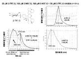
蛍光発色団を分子内に有してなる細胞内に特異的に取り込まれるD-グルコース誘導体と、蛍光発色団を分子内に有してなる細胞内に特異的に取り込まれるD-グルコース誘導体が発する蛍光の波長とは異なる波長の蛍光を発する蛍光発色団を分子内に有するL-グルコース誘導体との混合液として、2-NBDGと2-TRLGとの混合液を用いた場合を例に挙げると、前者の最大蛍光波長は540nm~550nm、後者の最大蛍光波長は600nm~610nmであるので、両者を混合した溶液についてそれぞれの最大蛍光波長付近の蛍光を検出した場合、2-NBDGが発する緑色の蛍光と、2-TRLGが発する赤色の蛍光をそれぞれ検出することができ、検出された両方の蛍光を合成することで結果として黄色の蛍光を検出することができる。2-NBDGと2-TRLGとの混合液を細胞に投与した後、2-NBDGが細胞内に特異的に取り込まれると、細胞内から緑色の蛍光が検出される。一方、細胞膜の一部が破壊されている場合や、破壊にまでは至らなくとも膜状態がグルコースやその誘導体が非特異的に取り込まれてしまうような状態にある場合、2-NBDGと2-TRLGの両方が細胞内に取り込まれると、細胞内から黄色の蛍光が検出される。従って、2-NBDGと2-TRLGとの混合液を細胞に投与した後の2-NBDGが発する緑色の蛍光を検出することで、D-グルコースのL-グルコースとの対比における細胞内への特異的な取り込みをリアルタイムで正確に評価することができる。
A D-glucose derivative that is specifically taken into a cell having a fluorescent chromophore in the molecule and a D-glucose derivative that is specifically taken into a cell having a fluorescent chromophore in the molecule are emitted. As an example, when a mixed solution of 2-NBDG and 2-TRLG is used as a mixed solution with an L-glucose derivative having in its molecule a fluorescent chromophore that emits fluorescence having a wavelength different from the fluorescence wavelength, The maximum fluorescence wavelength of the former is 540 nm to 550 nm, and the maximum fluorescence wavelength of the latter is 600 nm to 610 nm. Therefore, when fluorescence near the maximum fluorescence wavelength is detected in a mixed solution of both, green fluorescence emitted by 2-NBDG is emitted. And the red fluorescence emitted by 2-TRLG can be detected, respectively, and the resulting fluorescence is detected by synthesizing both detected fluorescences. It is possible. After the mixed solution of 2-NBDG and 2-TRLG is administered to the cells, when 2-NBDG is specifically taken into the cells, green fluorescence is detected from the cells. On the other hand, when a part of the cell membrane is destroyed, or when the membrane state is such that glucose or a derivative thereof is taken up nonspecifically even if it does not reach destruction, 2-NBDG and 2-NBDG When both TRLGs are taken up into the cell, yellow fluorescence is detected from inside the cell. Therefore, by detecting the green fluorescence emitted by 2-NBDG after the mixture of 2-NBDG and 2-TRLG is administered to the cells, the intracellular specificity in comparison with D-glucose and L-glucose is detected. Real-time uptake can be accurately assessed in real time.
なお、この方法は、蛍光発色団を分子内に有してなる細胞内に特異的に取り込まれるD-グルコース誘導体が発する蛍光と、蛍光発色団を分子内に有するL-グルコース誘導体が発する蛍光の波長の違いを利用するものであるので、両者の最大蛍光波長の差が大きいほどその違いを簡易かつ正確に識別することができる。従って、両者の最大蛍光波長には少なくとも20nmの差があることが望ましい。蛍光発色団を分子内に有してなる細胞内に特異的に取り込まれるD-グルコース誘導体として2-NBDGを用いる場合、蛍光発色団を分子内に有するL-グルコース誘導体として、7-(N,N-ジメチルアミノスルホニル)ベンズ-2-オキサ-1,3-ジアゾール-4-イル)アミノ基を分子内に有するL-グルコース誘導体、具体的には、7-(N,N-ジメチルアミノスルホニル)ベンズ-2-オキサ-1,3-ジアゾール-4-イル)アミノ基を2位に結合せしめたL-デオキシグルコース(2-[N-7-(N’,N’-ジメチルアミノスルホニル)ベンズ-2-オキサ-1,3-ジアゾール-4-イル)アミノ]-2-デオキシ-L-グルコース:2-DBDLG)を用いることもできる(その蛍光は黄色~黄緑色であり最大蛍光波長は570nm~580nmである)。また、蛍光発色団を分子内に有するL-グルコース誘導体として、2-(N-メチルアミノ)ベンゾイルアミノ基を分子内に有するL-グルコース誘導体、具体的には、2-(N-メチルアミノ)ベンゾイルアミノ基を2位に結合せしめたL-デオキシグルコース(2-[N-2-(N’-メチルアミノ)ベンゾイルアミノ]-2-デオキシ-L-グルコース:2-NMALG)を用いることもできる(その蛍光は青色であり最大蛍光波長は450nm~460nmである)。
In this method, there are fluorescence emitted from a D-glucose derivative that is specifically taken into a cell having a fluorescent chromophore in the molecule, and fluorescence emitted from an L-glucose derivative having a fluorescent chromophore in the molecule. Since the difference in wavelength is used, the difference between the maximum fluorescence wavelengths can be easily and accurately identified as the difference between the maximum fluorescence wavelengths increases. Therefore, it is desirable that there is a difference of at least 20 nm between the maximum fluorescence wavelengths. When 2-NBDG is used as a D-glucose derivative that is specifically taken into a cell having a fluorescent chromophore in the molecule, 7- (N, N-dimethylaminosulfonyl) benz-2-oxa-1,3-diazol-4-yl) L-glucose derivative having an amino group in the molecule, specifically, 7- (N, N-dimethylaminosulfonyl) L-deoxyglucose (2- [N-7- (N ′, N′-dimethylaminosulfonyl) benz-) having a benz-2-oxa-1,3-diazol-4-yl) amino group bonded to the 2-position 2-oxa-1,3-diazol-4-yl) amino] -2-deoxy-L-glucose: 2-DBDLG) (the fluorescence is yellow to yellow-green, most Fluorescence wavelength is 570nm ~ 580nm). Further, as an L-glucose derivative having a fluorescent chromophore in the molecule, an L-glucose derivative having a 2- (N-methylamino) benzoylamino group in the molecule, specifically, 2- (N-methylamino) L-deoxyglucose (2- [N-2- (N′-methylamino) benzoylamino] -2-deoxy-L-glucose: 2-NMLG) having a benzoylamino group bonded to the 2-position can also be used. (The fluorescence is blue and the maximum fluorescence wavelength is 450 nm to 460 nm).
また、蛍光発色団を分子内に有してなる細胞内に特異的に取り込まれるD-グルコース誘導体として、2-(N-メチルアミノ)ベンゾイルアミノ基を分子内に有するD-グルコース誘導体、具体的には、2-(N-メチルアミノ)ベンゾイルアミノ基を2位に結合せしめたD-デオキシグルコース(2-[N-2-(N’-メチルアミノ)ベンゾイルアミノ]-2-デオキシ-D-グルコース:2-NMAG)を用いることもできる(その蛍光は青色であり最大蛍光波長は450nm~460nmである)。この場合、蛍光発色団を分子内に有するL-グルコース誘導体としては、2-NBDLGや2-TRLGを用いることができる。
Further, as a D-glucose derivative that is specifically taken into a cell having a fluorescent chromophore in the molecule, a D-glucose derivative having a 2- (N-methylamino) benzoylamino group in the molecule, specifically, Includes D-deoxyglucose (2- [N-2- (N′-methylamino) benzoylamino] -2-deoxy-D-) having a 2- (N-methylamino) benzoylamino group bonded to the 2-position. (Glucose: 2-NMAG) can also be used (the fluorescence is blue and the maximum fluorescence wavelength is 450 nm to 460 nm). In this case, 2-NBDLG or 2-TRLG can be used as the L-glucose derivative having a fluorescent chromophore in the molecule.
また、蛍光発色団を分子内に有してなる細胞内に特異的に取り込まれるD-グルコース誘導体が発する蛍光と、蛍光発色団を分子内に有するL-グルコース誘導体が発する蛍光の間に強度の違いがある場合、それぞれが発する蛍光の強度が同程度となるように、例えば1:100~100:1(モル比)の範囲で両者の混合比を調整することが望ましい。
Also, there is an intensity between the fluorescence emitted by the D-glucose derivative that is specifically taken into the cell having the fluorescent chromophore in the molecule and the fluorescence emitted by the L-glucose derivative having the fluorescent chromophore in the molecule. If there is a difference, it is desirable to adjust the mixing ratio of the two in the range of, for example, 1: 100 to 100: 1 (molar ratio) so that the intensity of the fluorescence emitted from each of them is the same.
なお、この方法においては、蛍光発色団を分子内に有してなる細胞内に特異的に取り込まれるD-グルコース誘導体が発する蛍光の波長とは異なる波長の蛍光を発する蛍光発色団を分子内に有するL-グルコース誘導体として、異なる蛍光発色団を分子内に有する2種類以上のL-グルコース誘導体を混合して用いてもよい(例えば2-TRLGと2-NMALGの組み合わせなど)。異なる蛍光発色団を分子内に有する2種類以上のL-グルコース誘導体を混合して用いることで、例えば、細胞膜の悪化状態の程度の相違に基づく非特異的に取り込まれるL-グルコース誘導体の相違を検出することができ、これにより、D-グルコースのL-グルコースとの対比における細胞内への特異的な取り込みの評価とともに、細胞膜の悪化状態の程度の評価をあわせて行うことができる。
In this method, a fluorescent chromophore that emits fluorescence having a wavelength different from that of the fluorescence emitted by the D-glucose derivative that is specifically taken into the cell having the fluorescent chromophore in the molecule is contained in the molecule. As the L-glucose derivative, two or more kinds of L-glucose derivatives having different fluorescent chromophores in the molecule may be mixed and used (for example, a combination of 2-TRLG and 2-NMLG). By mixing and using two or more types of L-glucose derivatives having different fluorescent chromophores in the molecule, for example, the difference in non-specifically incorporated L-glucose derivatives based on the degree of deterioration of the cell membrane Thus, the specific uptake of D-glucose in comparison with L-glucose and the evaluation of the degree of deterioration of the cell membrane can be performed together.
本発明のD-グルコースの細胞内への特異的な取り込みを評価するための方法の実験条件は、公知の条件に準じればよい(必要であれば非特許文献2を参照のこと)。
The experimental conditions of the method for evaluating the specific uptake of D-glucose into cells of the present invention may be in accordance with known conditions (see Non-Patent Document 2 if necessary).
本発明のD-グルコースの細胞内への特異的な取り込みを評価するための方法は、ヒトを含めた哺乳動物のあらゆる部位の細胞(神経細胞など)の他、大腸菌や酵母などの微生物の細胞、植物の細胞、受精卵などを評価対象とすることができ、生物がD-グルコースをどのようにして細胞内に取り込んで利用するのかについての研究の発展に寄与する他、D-グルコースの取り込みの程度の違いに基づいて正常細胞と腫瘍細胞を判別するためや、微生物汚染の予防や対策のため(微生物の計数や状態評価など)といった医療分野や衛生分野などにおいてもその有用性が期待される。なお、D-グルコースの細胞内への特異的な取り込みの評価は、上記の2種類の方法のいずれかを選択して行ってもよいが、両方を組み合わせて行えば、より正確な評価を行うことができる。
The method for evaluating the specific uptake of D-glucose into cells of the present invention includes cells of any part of mammals including humans (eg, nerve cells), as well as cells of microorganisms such as E. coli and yeast. Plant cells, fertilized eggs, etc. can be evaluated, contributing to the development of research on how organisms incorporate D-glucose into cells and use it, as well as D-glucose uptake It is expected to be useful in the medical and hygiene fields, such as for distinguishing between normal cells and tumor cells based on the difference in the degree of infection, and for prevention and countermeasures against microbial contamination (e.g., microbial count and status assessment). The The specific uptake of D-glucose into the cell may be evaluated by selecting one of the above two methods, but a more accurate evaluation can be performed by combining both methods. be able to.
以下、本発明を実施例によって詳細に説明するが、本発明は以下の記載に限定して解釈されるものではない。
Hereinafter, the present invention will be described in detail by way of examples, but the present invention is not construed as being limited to the following description.
実施例1:神経細胞への2-NBDGおよび2-NBDLGの適用例
1-1:2-NBDGの合成
非特許文献1記載の方法に準じて行った。 Example 1: Application of 2-NBDG and 2-NBDLG to nerve cells 1-1: Synthesis of 2-NBDG The procedure was as described inNon-Patent Document 1.
1-1:2-NBDGの合成
非特許文献1記載の方法に準じて行った。 Example 1: Application of 2-NBDG and 2-NBDLG to nerve cells 1-1: Synthesis of 2-NBDG The procedure was as described in
1-2:2-NBDLGの合成
(A)L-グルコサミン塩酸塩の合成
以下の経路に従って行った。 1-2: Synthesis of 2-NBDLG (A) Synthesis of L-glucosamine hydrochloride The following route was used.
(A)L-グルコサミン塩酸塩の合成
以下の経路に従って行った。 1-2: Synthesis of 2-NBDLG (A) Synthesis of L-glucosamine hydrochloride The following route was used.

(1)1,2,3,4,6-ペンタ-O-アセチル-L-マンノピラノース(1)の合成
L-(-)-マンノース(9.0 g, 49.96 mmol)をピリジン(120 ml)に溶解し、氷冷した。これに無水酢酸(60 ml)を約15分間かけて滴下した。徐々に室温にもどしながら終夜攪拌した後に反応混合物を減圧濃縮し、得られた残渣にトルエンを加えて共沸する操作を2回繰り返した。残渣に酢酸エチルを加えて溶解し、得られた溶液を飽和NaHCO3水、水および飽和食塩水で順次洗浄した。有機層を無水硫酸マグネシウムで乾燥後に濾過し、濾液を減圧濃縮した。得られた残渣を減圧下よく乾燥させて目的物を得た(粗収量:21.04 g(酢酸エチル等を含むので理論収量を越える、49.96 mmolとして次の反応を行った))。
1H-NMRスペクトル(400 MHz、重クロロホルム、δ、ppm):6.10 (d, 1H, J=1.9 Hz, H-1), 5.26-5.36 (m, 3H, H-2, H-3, and H-4), 4.29 (dd, 1H, J=5.0 and 12.3 Hz, H-6a), 4.11 (dd, 1H, J=2.4 Hz and 12.3 Hz, H-6b), 4.06 (m, 1H, H-5), 2.19 (s, 3H, Ac), 2.18 (s, 3H, Ac), 2.11 (s, 3H, Ac), 2.06 (s, 3H, Ac), 2.02 (s, 3H, Ac) (1) Synthesis of 1,2,3,4,6-penta-O-acetyl-L-mannopyranose (1) L-(-)-mannose (9.0 g, 49.96 mmol) into pyridine (120 ml) Dissolved and ice-cooled. Acetic anhydride (60 ml) was added dropwise thereto over about 15 minutes. The reaction mixture was stirred overnight while gradually returning to room temperature, and then the reaction mixture was concentrated under reduced pressure. To the resulting residue, toluene was added and azeotroped twice. Ethyl acetate was added to the residue for dissolution, and the resulting solution was washed successively with saturated aqueous NaHCO 3 , water and saturated brine. The organic layer was dried over anhydrous magnesium sulfate and filtered, and the filtrate was concentrated under reduced pressure. The obtained residue was dried well under reduced pressure to obtain the desired product (crude yield: 21.04 g (exceeding the theoretical yield because it contained ethyl acetate and the like, and the following reaction was performed as 49.96 mmol)).
1 H-NMR spectrum (400 MHz, deuterated chloroform, δ, ppm): 6.10 (d, 1H, J = 1.9 Hz, H-1), 5.26-5.36 (m, 3H, H-2, H-3, and H-4), 4.29 (dd, 1H, J = 5.0 and 12.3 Hz, H-6a), 4.11 (dd, 1H, J = 2.4 Hz and 12.3 Hz, H-6b), 4.06 (m, 1H, H- 5), 2.19 (s, 3H, Ac), 2.18 (s, 3H, Ac), 2.11 (s, 3H, Ac), 2.06 (s, 3H, Ac), 2.02 (s, 3H, Ac)
(2)2,3,4,6-テトラ-O-アセチル-α-L-マンノピラノシル ブロマイド(2)の合成
1,2,3,4,6-ペンタ-O-アセチル-L-マンノピラノース(1)(21.04 g)を脱水ジクロロメタン(150 ml)に溶解し、氷冷した。これに30%HBr/AcOH(27.5 ml)を約10分間かけて滴下した。滴下終了後に氷浴からはずし、室温で3時間攪拌した。反応混合物を減圧濃縮し、得られた残渣にトルエンを加えて共沸する操作を2回繰り返した。残渣に酢酸エチルを加えて溶解し、得られた溶液を飽和NaHCO3水、水および飽和食塩水で順次洗浄した。有機層を無水硫酸マグネシウムで乾燥後に濾過し、濾液を減圧濃縮した。得られた残渣を減圧下よく乾燥させて目的物を得た(粗収量:21.45 g(酢酸エチルおよびトルエンを含むので理論収量を越える、49.96 mmolとして次の反応を行った))。
1H-NMRスペクトル(400 MHz、重クロロホルム、δ、ppm):6.30 (d, 1H, J=1.5 Hz, H-1), 5.72 (dd, 1H, J=3.6 Hz and 10.3 Hz, H-3), 5.46 (dd, 1H, J=1.5 Hz and 3.6 Hz, H-2), 5.38 (dd, 1H, J=10.3 Hz and 10.3 Hz, H-4), 4.34 (dd, 1H, J=4.9 Hz and 12.5 Hz, H-6a), 4.22 (ddd, 1H, J=2.1 Hz, 4.9 Hz, and 10.3 Hz, H-5), 4.15 (dd, 1H, J=2.1 Hz and 12.5 Hz, H-6b), 2.18 (s, 3H, Ac), 2.11 (s, 3H, Ac), 2.08 (s, 3H, Ac), 2.02 (s, 3H, Ac) (2) Synthesis of 2,3,4,6-tetra-O-acetyl-α-L-mannopyranosyl bromide (2) 1,2,3,4,6-penta-O-acetyl-L-mannopyranose ( 1) (21.04 g) was dissolved in dehydrated dichloromethane (150 ml) and cooled on ice. To this, 30% HBr / AcOH (27.5 ml) was added dropwise over about 10 minutes. After completion of the dropwise addition, the solution was removed from the ice bath and stirred at room temperature for 3 hours. The reaction mixture was concentrated under reduced pressure, and the operation of azeotropically adding toluene to the resulting residue was repeated twice. Ethyl acetate was added to the residue for dissolution, and the resulting solution was washed successively with saturated aqueous NaHCO 3 , water and saturated brine. The organic layer was dried over anhydrous magnesium sulfate and filtered, and the filtrate was concentrated under reduced pressure. The obtained residue was dried well under reduced pressure to obtain the desired product (crude yield: 21.45 g (exceeding the theoretical yield because ethyl acetate and toluene were included, and the following reaction was performed as 49.96 mmol)).
1 H-NMR spectrum (400 MHz, deuterated chloroform, δ, ppm): 6.30 (d, 1H, J = 1.5 Hz, H-1), 5.72 (dd, 1H, J = 3.6 Hz and 10.3 Hz, H-3 ), 5.46 (dd, 1H, J = 1.5 Hz and 3.6 Hz, H-2), 5.38 (dd, 1H, J = 10.3 Hz and 10.3 Hz, H-4), 4.34 (dd, 1H, J = 4.9 Hz and 12.5 Hz, H-6a), 4.22 (ddd, 1H, J = 2.1 Hz, 4.9 Hz, and 10.3 Hz, H-5), 4.15 (dd, 1H, J = 2.1 Hz and 12.5 Hz, H-6b) , 2.18 (s, 3H, Ac), 2.11 (s, 3H, Ac), 2.08 (s, 3H, Ac), 2.02 (s, 3H, Ac)
(3)3,4,6-トリ-O-アセチル-β-L-マンノピラノース 1,2-(メチルオルトアセテート)(3)の合成
2,3,4,6-テトラ-O-アセチル-α-L-マンノピラノシル ブロマイド(2)(21.45 g)を脱水ジクロロメタン(120 ml)に溶解した。アルゴン雰囲気下で2,6-ルチジン(15.73 ml)/脱水メタノール(120 ml)を約30分間かけて滴下した。アルゴン雰囲気下、室温で終夜攪拌した。24時間後にジクロロメタンで希釈した後、得られた溶液を飽和NaHCO3水、水で順次洗浄した。有機層を減圧濃縮し、得られた残渣にトルエンを加えて共沸する操作を3回繰り返した。残渣をメタノール(100 ml)に溶解し、水(800 ml)を少量ずつ加えて析出した結晶を濾取し、水で洗浄後に減圧下よく乾燥させた(1st, 8.92 g)。続いて(1st)の濾液と洗浄液を合わせて減圧濃縮し、液量を減らした。析出した結晶を濾取し、水で洗浄後に減圧下よく乾燥させ(2nd, 2.62 g)、目的物を得た(総収量:11.54 g(L-(-)-マンノースより3工程の総収率:64 %))。
1H-NMRスペクトル(400 MHz、重クロロホルム、δ、ppm):5.50 (d, 1H, J=2.8 Hz, H-1), 5.31 (dd, 1H, J=9.4 Hz and 10.0 Hz, H-4), 5.15 (dd, 1H, J=3.9 Hz and 10.0 Hz, H-3), 4.62 (dd, 1H, J=2.8 Hz and 3.9 Hz, H-2), 4.24 (dd, 1H, J=4.9 Hz and 12.2 Hz, H-6a), 4.15 (dd, 1H, J=3.0 Hz and 12.2 Hz, H-6b), 3.69(ddd, 1H, J=3.0 Hz, 4.9 Hz and 9.4 Hz, H-5), 2.13 (s, 3H, Ac), 2.08 (s, 3H, Ac), 2.06 (s, 3H, Ac), 1.75 (s, 3H, Me) (3) Synthesis of 3,4,6-tri-O-acetyl-β-L-mannopyranose 1,2- (methylorthoacetate) (3) 2,3,4,6-tetra-O-acetyl- α-L-Mannopyranosyl bromide (2) (21.45 g) was dissolved in dehydrated dichloromethane (120 ml). Under an argon atmosphere, 2,6-lutidine (15.73 ml) / dehydrated methanol (120 ml) was added dropwise over about 30 minutes. The mixture was stirred overnight at room temperature under an argon atmosphere. After 24 hours, diluted with dichloromethane, the resulting solution was washed successively with saturated aqueous NaHCO 3 and water. The organic layer was concentrated under reduced pressure, and the operation of adding toluene to the resulting residue and azeotroping was repeated three times. The residue was dissolved in methanol (100 ml), water (800 ml) was added little by little, and the precipitated crystals were collected by filtration, washed with water and dried well under reduced pressure (1st, 8.92 g). Subsequently, the (1st) filtrate and the washing liquid were combined and concentrated under reduced pressure to reduce the liquid volume. The precipitated crystals were collected by filtration, washed with water and dried well under reduced pressure (2nd, 2.62 g) to obtain the desired product (total yield: 11.54 g (total yield of 3 steps from L-(-)-mannose) : 64%)).
1 H-NMR spectrum (400 MHz, deuterated chloroform, δ, ppm): 5.50 (d, 1H, J = 2.8 Hz, H-1), 5.31 (dd, 1H, J = 9.4 Hz and 10.0 Hz, H-4 ), 5.15 (dd, 1H, J = 3.9 Hz and 10.0 Hz, H-3), 4.62 (dd, 1H, J = 2.8 Hz and 3.9 Hz, H-2), 4.24 (dd, 1H, J = 4.9 Hz and 12.2 Hz, H-6a), 4.15 (dd, 1H, J = 3.0 Hz and 12.2 Hz, H-6b), 3.69 (ddd, 1H, J = 3.0 Hz, 4.9 Hz and 9.4 Hz, H-5), 2.13 (s, 3H, Ac), 2.08 (s, 3H, Ac), 2.06 (s, 3H, Ac), 1.75 (s, 3H, Me)
2,3,4,6-テトラ-O-アセチル-α-L-マンノピラノシル ブロマイド(2)(21.45 g)を脱水ジクロロメタン(120 ml)に溶解した。アルゴン雰囲気下で2,6-ルチジン(15.73 ml)/脱水メタノール(120 ml)を約30分間かけて滴下した。アルゴン雰囲気下、室温で終夜攪拌した。24時間後にジクロロメタンで希釈した後、得られた溶液を飽和NaHCO3水、水で順次洗浄した。有機層を減圧濃縮し、得られた残渣にトルエンを加えて共沸する操作を3回繰り返した。残渣をメタノール(100 ml)に溶解し、水(800 ml)を少量ずつ加えて析出した結晶を濾取し、水で洗浄後に減圧下よく乾燥させた(1st, 8.92 g)。続いて(1st)の濾液と洗浄液を合わせて減圧濃縮し、液量を減らした。析出した結晶を濾取し、水で洗浄後に減圧下よく乾燥させ(2nd, 2.62 g)、目的物を得た(総収量:11.54 g(L-(-)-マンノースより3工程の総収率:64 %))。
1H-NMRスペクトル(400 MHz、重クロロホルム、δ、ppm):5.50 (d, 1H, J=2.8 Hz, H-1), 5.31 (dd, 1H, J=9.4 Hz and 10.0 Hz, H-4), 5.15 (dd, 1H, J=3.9 Hz and 10.0 Hz, H-3), 4.62 (dd, 1H, J=2.8 Hz and 3.9 Hz, H-2), 4.24 (dd, 1H, J=4.9 Hz and 12.2 Hz, H-6a), 4.15 (dd, 1H, J=3.0 Hz and 12.2 Hz, H-6b), 3.69(ddd, 1H, J=3.0 Hz, 4.9 Hz and 9.4 Hz, H-5), 2.13 (s, 3H, Ac), 2.08 (s, 3H, Ac), 2.06 (s, 3H, Ac), 1.75 (s, 3H, Me) (3) Synthesis of 3,4,6-tri-O-acetyl-β-L-
1 H-NMR spectrum (400 MHz, deuterated chloroform, δ, ppm): 5.50 (d, 1H, J = 2.8 Hz, H-1), 5.31 (dd, 1H, J = 9.4 Hz and 10.0 Hz, H-4 ), 5.15 (dd, 1H, J = 3.9 Hz and 10.0 Hz, H-3), 4.62 (dd, 1H, J = 2.8 Hz and 3.9 Hz, H-2), 4.24 (dd, 1H, J = 4.9 Hz and 12.2 Hz, H-6a), 4.15 (dd, 1H, J = 3.0 Hz and 12.2 Hz, H-6b), 3.69 (ddd, 1H, J = 3.0 Hz, 4.9 Hz and 9.4 Hz, H-5), 2.13 (s, 3H, Ac), 2.08 (s, 3H, Ac), 2.06 (s, 3H, Ac), 1.75 (s, 3H, Me)
(4)3,4,6-トリ-O-ベンジル-β-L-マンノピラノース 1,2-(メチルオルトアセテート)(4)の合成
3,4,6-トリ-O-アセチル-β-L-マンノピラノース 1,2-(メチルオルトアセテート)(3)(11.52 g)をメタノール(200 ml)に溶解し、5.2M-NaOMeのメタノール溶液(184 μl)を加えた。室温で1時間攪拌した後にアンバーリスト15を加えて中和し、濾過した後、樹脂をメタノールで洗浄し、濾液と洗浄液を合わせて減圧濃縮した。得られた残渣を減圧下よく乾燥させた。乾燥させた残渣の全量を乾燥DMF(100 ml)に溶解し、氷冷した。これに水素化ナトリウム(NaH)(60% oil suspension, 7.38 g)を少量ずつ加えた。水素の発生による発泡が収まった後に臭化ベンジル(12.1 ml)を滴下した。徐々に室温に戻しながら終夜攪拌した。反応混合物を氷冷し、過剰のNaHを分解させるため、メタノールを少量ずつ発泡しなくなるまで加えた。クロロホルムを加えて希釈し、得られた溶液を水および飽和食塩水で順次洗浄した。有機層を無水硫酸マグネシウムで乾燥後に濾過し、濾液を減圧濃縮した。得られた残渣を減圧下よく乾燥させ、フリーザー内で終夜保存した。翌朝、沈殿とオイルに分離した。パスツールピペットを用いてオイル(不要物)を除去した。沈殿(湿重量:17.97 g)をシリカゲルカラムクロマトグラフィーにより精製して目的物を得た(収量:11.98 g、収率:74 %)。
1H-NMRスペクトル(400 MHz、重クロロホルム、δ、ppm):7.22-7.41 (m, 15H, Ph), 5.35 (d, 1H, J=2.5 Hz), 4.79 (ABq, 2H, J=12.4 Hz, CH 2 -Ph), 4.75 (ABq, 2H, J=11.0 Hz, CH 2 -Ph), 4.58 (ABq, 2H, J=12.4 Hz, CH 2 -Ph), 4.40 (dd, 1H, J=2.5 Hz and 4.1 Hz, H-2), 3.92 (dd, 1H, J=9.7 Hz and 9.7 Hz, H-4), 3.69-3.77 (m, 3H, H-3, H-6a, and H-6b), 3.42 (ddd, 1H, J=2.3 Hz, 4.5 Hz, and 9.7 Hz, H-5), 3.29 (s, 3H, OMe), 1.74 (s, 3H, Me) (4) Synthesis of 3,4,6-tri-O-benzyl-β-L-mannopyranose 1,2- (methylorthoacetate) (4) 3,4,6-tri-O-acetyl-β- L-Mannopyranose 1,2- (methyl orthoacetate) (3) (11.52 g) was dissolved in methanol (200 ml), and a methanol solution of 5.2M-NaOMe (184 μl) was added. After stirring at room temperature for 1 hour, Amberlyst 15 was added to neutralize and filtered, and then the resin was washed with methanol, and the filtrate and washings were combined and concentrated under reduced pressure. The resulting residue was well dried under reduced pressure. The entire amount of the dried residue was dissolved in dry DMF (100 ml) and ice-cooled. To this was added sodium hydride (NaH) (60% oil suspension, 7.38 g) in small portions. After the bubbling due to hydrogen evolution ceased, benzyl bromide (12.1 ml) was added dropwise. The mixture was stirred overnight while gradually returning to room temperature. The reaction mixture was ice-cooled and methanol was added in small portions until no more foaming occurred to decompose excess NaH. Chloroform was added for dilution, and the resulting solution was washed successively with water and saturated brine. The organic layer was dried over anhydrous magnesium sulfate and filtered, and the filtrate was concentrated under reduced pressure. The resulting residue was dried well under reduced pressure and stored overnight in a freezer. The next morning, it separated into precipitate and oil. Oil (unnecessary matter) was removed using a Pasteur pipette. The precipitate (wet weight: 17.97 g) was purified by silica gel column chromatography to obtain the desired product (yield: 11.98 g, yield: 74%).
1 H-NMR spectrum (400 MHz, deuterated chloroform, δ, ppm): 7.22-7.41 (m, 15H, Ph), 5.35 (d, 1H, J = 2.5 Hz), 4.79 (ABq, 2H, J = 12.4 Hz , CH 2 -Ph), 4.75 (ABq, 2H, J = 11.0 Hz, CH 2 -Ph), 4.58 (ABq, 2H, J = 12.4 Hz, CH 2 -Ph), 4.40 (dd, 1H, J = 2.5 Hz and 4.1 Hz, H-2), 3.92 (dd, 1H, J = 9.7 Hz and 9.7 Hz, H-4), 3.69-3.77 (m, 3H, H-3, H-6a, and H-6b) , 3.42 (ddd, 1H, J = 2.3 Hz, 4.5 Hz, and 9.7 Hz, H-5), 3.29 (s, 3H, OMe), 1.74 (s, 3H, Me)
3,4,6-トリ-O-アセチル-β-L-マンノピラノース 1,2-(メチルオルトアセテート)(3)(11.52 g)をメタノール(200 ml)に溶解し、5.2M-NaOMeのメタノール溶液(184 μl)を加えた。室温で1時間攪拌した後にアンバーリスト15を加えて中和し、濾過した後、樹脂をメタノールで洗浄し、濾液と洗浄液を合わせて減圧濃縮した。得られた残渣を減圧下よく乾燥させた。乾燥させた残渣の全量を乾燥DMF(100 ml)に溶解し、氷冷した。これに水素化ナトリウム(NaH)(60% oil suspension, 7.38 g)を少量ずつ加えた。水素の発生による発泡が収まった後に臭化ベンジル(12.1 ml)を滴下した。徐々に室温に戻しながら終夜攪拌した。反応混合物を氷冷し、過剰のNaHを分解させるため、メタノールを少量ずつ発泡しなくなるまで加えた。クロロホルムを加えて希釈し、得られた溶液を水および飽和食塩水で順次洗浄した。有機層を無水硫酸マグネシウムで乾燥後に濾過し、濾液を減圧濃縮した。得られた残渣を減圧下よく乾燥させ、フリーザー内で終夜保存した。翌朝、沈殿とオイルに分離した。パスツールピペットを用いてオイル(不要物)を除去した。沈殿(湿重量:17.97 g)をシリカゲルカラムクロマトグラフィーにより精製して目的物を得た(収量:11.98 g、収率:74 %)。
1H-NMRスペクトル(400 MHz、重クロロホルム、δ、ppm):7.22-7.41 (m, 15H, Ph), 5.35 (d, 1H, J=2.5 Hz), 4.79 (ABq, 2H, J=12.4 Hz, CH 2 -Ph), 4.75 (ABq, 2H, J=11.0 Hz, CH 2 -Ph), 4.58 (ABq, 2H, J=12.4 Hz, CH 2 -Ph), 4.40 (dd, 1H, J=2.5 Hz and 4.1 Hz, H-2), 3.92 (dd, 1H, J=9.7 Hz and 9.7 Hz, H-4), 3.69-3.77 (m, 3H, H-3, H-6a, and H-6b), 3.42 (ddd, 1H, J=2.3 Hz, 4.5 Hz, and 9.7 Hz, H-5), 3.29 (s, 3H, OMe), 1.74 (s, 3H, Me) (4) Synthesis of 3,4,6-tri-O-benzyl-β-L-
1 H-NMR spectrum (400 MHz, deuterated chloroform, δ, ppm): 7.22-7.41 (m, 15H, Ph), 5.35 (d, 1H, J = 2.5 Hz), 4.79 (ABq, 2H, J = 12.4 Hz , CH 2 -Ph), 4.75 (ABq, 2H, J = 11.0 Hz, CH 2 -Ph), 4.58 (ABq, 2H, J = 12.4 Hz, CH 2 -Ph), 4.40 (dd, 1H, J = 2.5 Hz and 4.1 Hz, H-2), 3.92 (dd, 1H, J = 9.7 Hz and 9.7 Hz, H-4), 3.69-3.77 (m, 3H, H-3, H-6a, and H-6b) , 3.42 (ddd, 1H, J = 2.3 Hz, 4.5 Hz, and 9.7 Hz, H-5), 3.29 (s, 3H, OMe), 1.74 (s, 3H, Me)
(5)メチル 3,4,6-トリ-O-ベンジル-α-L-マンノピラノシド(5)の合成
3,4,6-トリ-O-ベンジル-β-L-マンノピラノース 1,2-(メチルオルトアセテート)(4)(11.98 g)を脱水メタノール(240 ml)に溶解し、65℃で加熱攪拌した。これに塩化アセチル(6.73 ml)を滴下した。滴下終了後にオイルバスの設定温度を75℃にして加熱還流した。3時間後にオイルバスのスイッチを切り、終夜攪拌した。反応混合物を減圧濃縮し、得られた残渣にクロロホルムを加えて溶解し、得られた溶液を飽和NaHCO3水、水および飽和食塩水で順次洗浄した。有機層を無水硫酸マグネシウムで乾燥後に濾過し、濾液を減圧濃縮した。得られた残渣をシリカゲルカラムクロマトグラフィーにより精製して目的物を得た(収量:9.74 g、収率:89 %)。
1H-NMRスペクトル(400 MHz、重クロロホルム、δ、ppm):7.24-7.36 (m, 15H, Ph), 4.80 (d, 1H, J=1.6 Hz, H-1), 4.69 (ABq, 2H, J=11.9 Hz, CH 2 -Ph), 4.67 (ABq, 2H, J=11.3 Hz, CH 2 -Ph), 4.60 (ABq, 2H, J=12.4 Hz, CH 2 -Ph), 4.03 (m, 1H, H-2), 3.70-3.88 (m, 5H, H-3, H-4, H-5, H-6a and H-6b), 3.37 (s, 3H, OMe), 2.49 (br.d, 1H, J=2.5 Hz, C2-OH) (5) Synthesis of methyl 3,4,6-tri-O-benzyl-α-L-mannopyranoside (5) 3,4,6-tri-O-benzyl-β-L-mannopyranose 1,2- ( Methyl orthoacetate) (4) (11.98 g) was dissolved in dehydrated methanol (240 ml) and stirred with heating at 65 ° C. Acetyl chloride (6.73 ml) was added dropwise thereto. After completion of the dropping, the oil bath was set to 75 ° C. and heated to reflux. After 3 hours, the oil bath was switched off and stirred overnight. The reaction mixture was concentrated under reduced pressure, and the resulting residue was dissolved by adding chloroform. The obtained solution was washed successively with saturated aqueous NaHCO 3 , water and saturated brine. The organic layer was dried over anhydrous magnesium sulfate and filtered, and the filtrate was concentrated under reduced pressure. The obtained residue was purified by silica gel column chromatography to obtain the desired product (yield: 9.74 g, yield: 89%).
1 H-NMR spectrum (400 MHz, deuterated chloroform, δ, ppm): 7.24–7.36 (m, 15H, Ph), 4.80 (d, 1H, J = 1.6 Hz, H-1), 4.69 (ABq, 2H, J = 11.9 Hz, CH 2 -Ph), 4.67 (ABq, 2H, J = 11.3 Hz, CH 2 -Ph), 4.60 (ABq, 2H, J = 12.4 Hz, CH 2 -Ph), 4.03 (m, 1H , H-2), 3.70-3.88 (m, 5H, H-3, H-4, H-5, H-6a and H-6b), 3.37 (s, 3H, OMe), 2.49 (br.d, (1H, J = 2.5 Hz, C2-OH)
3,4,6-トリ-O-ベンジル-β-L-マンノピラノース 1,2-(メチルオルトアセテート)(4)(11.98 g)を脱水メタノール(240 ml)に溶解し、65℃で加熱攪拌した。これに塩化アセチル(6.73 ml)を滴下した。滴下終了後にオイルバスの設定温度を75℃にして加熱還流した。3時間後にオイルバスのスイッチを切り、終夜攪拌した。反応混合物を減圧濃縮し、得られた残渣にクロロホルムを加えて溶解し、得られた溶液を飽和NaHCO3水、水および飽和食塩水で順次洗浄した。有機層を無水硫酸マグネシウムで乾燥後に濾過し、濾液を減圧濃縮した。得られた残渣をシリカゲルカラムクロマトグラフィーにより精製して目的物を得た(収量:9.74 g、収率:89 %)。
1H-NMRスペクトル(400 MHz、重クロロホルム、δ、ppm):7.24-7.36 (m, 15H, Ph), 4.80 (d, 1H, J=1.6 Hz, H-1), 4.69 (ABq, 2H, J=11.9 Hz, CH 2 -Ph), 4.67 (ABq, 2H, J=11.3 Hz, CH 2 -Ph), 4.60 (ABq, 2H, J=12.4 Hz, CH 2 -Ph), 4.03 (m, 1H, H-2), 3.70-3.88 (m, 5H, H-3, H-4, H-5, H-6a and H-6b), 3.37 (s, 3H, OMe), 2.49 (br.d, 1H, J=2.5 Hz, C2-OH) (5) Synthesis of methyl 3,4,6-tri-O-benzyl-α-L-mannopyranoside (5) 3,4,6-tri-O-benzyl-β-L-
1 H-NMR spectrum (400 MHz, deuterated chloroform, δ, ppm): 7.24–7.36 (m, 15H, Ph), 4.80 (d, 1H, J = 1.6 Hz, H-1), 4.69 (ABq, 2H, J = 11.9 Hz, CH 2 -Ph), 4.67 (ABq, 2H, J = 11.3 Hz, CH 2 -Ph), 4.60 (ABq, 2H, J = 12.4 Hz, CH 2 -Ph), 4.03 (m, 1H , H-2), 3.70-3.88 (m, 5H, H-3, H-4, H-5, H-6a and H-6b), 3.37 (s, 3H, OMe), 2.49 (br.d, (1H, J = 2.5 Hz, C2-OH)
(6)メチル 3,4,6-トリ-O-ベンジル-2-O-トリフルオロメタンスルホニル-α-L-マンノピラノシド(6)の合成
メチル 3,4,6-トリ-O-ベンジル-α-L-マンノピラノシド(5)(9.74 g)を脱水ジクロロメタン(220 ml)に溶解し、ピリジン(4.24 ml)を加えてアルゴン雰囲気下で-15℃に冷却した。これにトリフルオロメタンスルホン酸無水物(5.29 ml)を加え、室温で攪拌した。1時間後に反応混合物に飽和NaHCO3水を加え、ジクロロメタンで抽出した。有機層を合わせ、飽和NaHCO3水、水および飽和食塩水で順次洗浄し、無水硫酸マグネシウムで乾燥後に濾過し、濾液を減圧濃縮した。得られた残渣にトルエンを加えて共沸する操作を3回繰り返した。残渣をシリカゲルカラムクロマトグラフィーにより精製して目的物を得た(収量:12.23 g、収率:98 %)。
1H-NMRスペクトル(400 MHz、重クロロホルム、δ、ppm):7.10-7.38 (m, 15H, Ph), 5.11 (m, 1H, H-2), 4.90 (d, 1H, J=1.9 Hz, H-1), 4.69 (ABq, 2H, J=12.0 Hz, CH 2 -Ph), 4.64 (ABq, 2H, J=10.7 Hz, CH 2 -Ph), 4.61 (ABq, 2H, J=11.7 Hz, CH 2 -Ph), 4.00 (dd, 1H, J=2.9 Hz and 8.9 Hz, H-3), 3.69-3.84 (m, 4H, H-4, H-5, H-6a and H-6b), 3.40 (s, 3H, OMe) (6) Synthesis of methyl 3,4,6-tri-O-benzyl-2-O-trifluoromethanesulfonyl-α-L-mannopyranoside (6) Methyl 3,4,6-tri-O-benzyl-α-L -Mannopyranoside (5) (9.74 g) was dissolved in dehydrated dichloromethane (220 ml), pyridine (4.24 ml) was added, and the mixture was cooled to -15 ° C under an argon atmosphere. To this was added trifluoromethanesulfonic anhydride (5.29 ml), and the mixture was stirred at room temperature. After 1 hour, saturated aqueous NaHCO 3 was added to the reaction mixture, and the mixture was extracted with dichloromethane. The organic layers were combined, washed successively with saturated aqueous NaHCO 3 , water and saturated brine, dried over anhydrous magnesium sulfate and filtered, and the filtrate was concentrated under reduced pressure. The operation of azeotropically adding toluene to the obtained residue was repeated three times. The residue was purified by silica gel column chromatography to obtain the desired product (yield: 12.23 g, yield: 98%).
1 H-NMR spectrum (400 MHz, deuterated chloroform, δ, ppm): 7.10-7.38 (m, 15H, Ph), 5.11 (m, 1H, H-2), 4.90 (d, 1H, J = 1.9 Hz, H-1), 4.69 (ABq, 2H, J = 12.0 Hz, CH 2 -Ph), 4.64 (ABq, 2H, J = 10.7 Hz, CH 2 -Ph), 4.61 (ABq, 2H, J = 11.7 Hz, CH 2 -Ph), 4.00 (dd, 1H, J = 2.9 Hz and 8.9 Hz, H-3), 3.69-3.84 (m, 4H, H-4, H-5, H-6a and H-6b), 3.40 (s, 3H, OMe)
(7)メチル 2-アジド-3,4,6-トリ-O-ベンジル-2-デオキシ-α-L-グルコピラノシド(7)の合成
メチル 3,4,6-トリ-O-ベンジル-2-O-トリフルオロメタンスルホニル-α-L-マンノピラノシド(6)(12.23 g)を脱水ベンゼン(400 ml)に溶解し、テトラブチルアンモニウム アジド[(n-Bu)4NN3](29.16 g)をアルゴン雰囲気下で加えた。3日後に(n-Bu)4NN3(3.61 g)を追加した。さらに11日間(計14日間)室温で攪拌した。反応混合物をそのままシリカゲルカラムにチャージして、シリカゲルカラムクロマトグラフィーおよび、調製的薄層クロマトグラフィーを併用することにより精製して目的物を得た(収量:3.58 g、収率:36 %)。
1H-NMRスペクトル(400 MHz、重クロロホルム、δ、ppm):7.15-7.38 (m, 15H, Ph), 4.87 (ABq, 2H, J=12.4 Hz, CH 2 -Ph), 4.83 (d, 1H, J=3.5 Hz, H-1), 4.66 (ABq, 2H, J=10.7 Hz, CH 2 -Ph), 4.57 (ABq, 2H, J=12.4 Hz, CH 2 -Ph), 3.98 (dd, 1H, J=8.9 Hz and 10.2 Hz, H-3), 3.66-3.80 (m, 4H, H-4, H-5, H-6a, and H-6b), 3.45 (dd, 1H, J=3.5 Hz and 10.4 Hz, H-2), 3.43 (s, 3H, OMe) (7) Synthesis of methyl 2-azido-3,4,6-tri-O-benzyl-2-deoxy-α-L-glucopyranoside (7) Methyl 3,4,6-tri-O-benzyl-2-O -Trifluoromethanesulfonyl-α-L-mannopyranoside (6) (12.23 g) was dissolved in dehydrated benzene (400 ml), and tetrabutylammonium azide [(n-Bu) 4 NN 3 ] (29.16 g) was dissolved in an argon atmosphere. Added in. Three days later, (n-Bu) 4 NN 3 (3.61 g) was added. The mixture was further stirred at room temperature for 11 days (total 14 days). The reaction mixture was directly charged to a silica gel column and purified by using silica gel column chromatography and preparative thin layer chromatography in combination to obtain the desired product (yield: 3.58 g, yield: 36%).
1 H-NMR spectrum (400 MHz, deuterated chloroform, δ, ppm): 7.15-7.38 (m, 15H, Ph), 4.87 (ABq, 2H, J = 12.4 Hz, CH 2 -Ph), 4.83 (d, 1H , J = 3.5 Hz, H-1), 4.66 (ABq, 2H, J = 10.7 Hz, CH 2 -Ph), 4.57 (ABq, 2H, J = 12.4 Hz, CH 2 -Ph), 3.98 (dd, 1H , J = 8.9 Hz and 10.2 Hz, H-3), 3.66-3.80 (m, 4H, H-4, H-5, H-6a, and H-6b), 3.45 (dd, 1H, J = 3.5 Hz and 10.4 Hz, H-2), 3.43 (s, 3H, OMe)
(8)メチル 2-アミノ-2-デオキシ-α-L-グルコピラノシド塩酸塩(8)の合成
メチル 2-アジド-3,4,6-トリ-O-ベンジル-2-デオキシ-α-L-グルコピラノシド(7)(1.98 g, 4.04 mmol)をメタノール(70 ml)に溶解し、1M-HCl(4.85 ml)およびパラジウム-黒(500 mg)を加えて、8気圧で加圧接触還元した。22時間後に常圧に戻し、TLCで1スポットに収束したことを確認した後に触媒を濾過した。触媒をメタノールで洗浄し、濾液と洗浄液を合わせて減圧濃縮した。得られた残渣に水を加えて減圧濃縮する操作を2回繰り返した。残渣を水に溶解して凍結乾燥して目的物を得た(粗収量:963 mg(水および塩酸を含むので理論収量を越える、1H-NMR測定に使った残り960 mgを4.03 mmolとして最終脱保護となる次の反応を行った))。
1H-NMRスペクトル(400 MHz、重水、δ、ppm):4.89 (d, 1H, J=3.7Hz, H-1), 3.75 (dd, 1H, J=2.5 Hz and 12.3 Hz, H-6a), 3.72 (dd, 1H, J=9.2 Hz and 10.5 Hz, H-3), 3.66 (dd, 1H, J=5.2 Hz and 12.3 Hz, H-6b), 3.57 (ddd, 1H, J=2.5 Hz, 5.2 Hz, and 9.8 Hz, H-5), 3.35 (dd, 1H, J=9.2 Hz and 9.8 Hz, H-4), 3.31 (s, 3H, OMe), 3.20 (dd, 1H, J=3.7 Hz and 10.5 Hz, H-2) (8) Synthesis of methyl 2-amino-2-deoxy-α-L-glucopyranoside hydrochloride (8) Methyl 2-azido-3,4,6-tri-O-benzyl-2-deoxy-α-L-glucopyranoside (7) (1.98 g, 4.04 mmol) was dissolved in methanol (70 ml), 1M-HCl (4.85 ml) and palladium-black (500 mg) were added, and catalytic reduction was performed under pressure at 8 atm. After 22 hours, the pressure was returned to normal pressure, and after confirming that it converged to one spot by TLC, the catalyst was filtered. The catalyst was washed with methanol, and the filtrate and the washing solution were combined and concentrated under reduced pressure. The operation of adding water to the obtained residue and concentrating under reduced pressure was repeated twice. The residue was dissolved in water and lyophilized to obtain the desired product (crude yield: 963 mg (exceeding the theoretical yield because it contains water and hydrochloric acid, and the final 960 mg used for 1 H-NMR measurement was 4.03 mmol). The next reaction was deprotected))).
1 H-NMR spectrum (400 MHz, heavy water, δ, ppm): 4.89 (d, 1H, J = 3.7Hz, H-1), 3.75 (dd, 1H, J = 2.5 Hz and 12.3 Hz, H-6a) , 3.72 (dd, 1H, J = 9.2 Hz and 10.5 Hz, H-3), 3.66 (dd, 1H, J = 5.2 Hz and 12.3 Hz, H-6b), 3.57 (ddd, 1H, J = 2.5 Hz, 5.2 Hz, and 9.8 Hz, H-5), 3.35 (dd, 1H, J = 9.2 Hz and 9.8 Hz, H-4), 3.31 (s, 3H, OMe), 3.20 (dd, 1H, J = 3.7 Hz and 10.5 Hz, H-2)
(9)L-グルコサミン塩酸塩(L-GlcNH2・HCl)の合成
メチル 2-アミノ-2-デオキシ-α-L-グルコピラノシド塩酸塩(8)(960 mg, 4.03 mmol相当として計算)を6N-HCl(40 ml)に溶解し、100℃で加熱攪拌した。計30時間反応させた後に室温に戻して減圧濃縮した。得られた残渣に水を加えて減圧濃縮する操作を5回繰り返した。残渣を水に溶解して凍結乾燥した。得られた残渣をCHP-20に通し、素通り部分を集めて凍結乾燥し(粗収量:805 mg(3.73 mmol、相当粗収率:93 %))、さらに531 mg(2.46 mmol相当)を順相のHPLC(TSK-Gel Amide-80)により精製し、目的の画分を集めて減圧濃縮した。残渣に水を加えて凍結乾燥する操作を3回繰り返して目的物を得た(収量: 474 mg(2.20 mmol)、収率:83 %)。
1H-NMRスペクトル(400 MHz、重水、δ、ppm):5.36 [d, 0.7H, J=3.5 Hz, H-1(α体), 4.85 [d, 0.3H, J=8.3 Hz, H-1(β体)], 3.36-3.84 [m, 5H, H-3, H-4, H-5, H-6a, and H-6b(α体およびβ体), 3.21 [dd, 0.7H, J=3.5 Hz and 10.6 Hz, H-2(α体), 2.92 [dd, 0.3H, J=8.3 Hz and 10.6 Hz, H-2(β体)]
(元素分析結果)
計算値:C;33.42(%), H;6.54(%), N;6.50(%)
実測値(1回目):C;33.31(%), H;6.46(%), N;6.36(%)
実測値(2回目):C;33.30(%), H;6.48(%), N;6.36(%)
(20℃における [α]D)
実測値:-72.05(c1.0, H2O)(水に溶解して24時間後)
(参考データ:市販品のD-グルコサミン塩酸塩の20℃における[α]D)
実測値:+72.20(c1.0, H2O)(水に溶解して24時間後) (9) Synthesis of L-glucosamine hydrochloride (L-GlcNH 2 .HCl) Methyl 2-amino-2-deoxy-α-L-glucopyranoside hydrochloride (8) (calculated as 960 mg, equivalent to 4.03 mmol) Dissolved in HCl (40 ml) and stirred with heating at 100 ° C. After reacting for a total of 30 hours, the mixture was returned to room temperature and concentrated under reduced pressure. The operation of adding water to the obtained residue and concentrating under reduced pressure was repeated 5 times. The residue was dissolved in water and lyophilized. The obtained residue was passed through CHP-20, and the flow-through portion was collected and freeze-dried (crude yield: 805 mg (3.73 mmol, equivalent crude yield: 93%)), and 531 mg (corresponding to 2.46 mmol) was normal phase. Were purified by HPLC (TSK-Gel Amide-80), and the desired fractions were collected and concentrated under reduced pressure. The operation of adding water to the residue and freeze-drying was repeated three times to obtain the desired product (yield: 474 mg (2.20 mmol), yield: 83%).
1 H-NMR spectrum (400 MHz, heavy water, δ, ppm): 5.36 [d, 0.7H, J = 3.5 Hz, H-1 (α form), 4.85 [d, 0.3H, J = 8.3 Hz, H- 1 (β form)], 3.36-3.84 [m, 5H, H-3, H-4, H-5, H-6a, and H-6b (alpha form and β form), 3.21 (dd, 0.7H, J = 3.5 Hz and 10.6 Hz, H-2 (α form), 2.92 [dd, 0.3H, J = 8.3 Hz and 10.6 Hz, H-2 (beta form)]
(Elemental analysis results)
Calculated values: C; 33.42 (%), H; 6.54 (%), N; 6.50 (%)
Actual value (first time): C; 33.31 (%), H; 6.46 (%), N; 6.36 (%)
Actual value (second time): C; 33.30 (%), H; 6.48 (%), N; 6.36 (%)
([Α] D at 20 ℃)
Found: -72.05 (c1.0, H 2 O) (24 hours after dissolving in water)
(Reference data: [α] D of commercial D-glucosamine hydrochloride at 20 ° C)
Actual value: +72.20 (c1.0, H 2 O) (24 hours after dissolution in water)
(B)2-NBDLGの合成(その1)
(B) Synthesis of 2-NBDLG (Part 1)

褐色フラスコに4-クロロ-7-ニトロ-2,1,3-ベンゾオキサジアゾール(NBD-Cl)(483 mg)を入れ、メタノール(14.0 ml)に溶解した。アルゴン雰囲気下、L-グルコサミン塩酸塩(280 mg)を0.3M-NaHCO3水(5.57 ml)に溶解したものを加え、室温で攪拌した。24時間後に減圧濃縮によりメタノールを除き、生じた沈殿(NBD-Cl由来の不要物)を濾過した後、沈殿を水で洗浄した。濾液と洗浄液を合わせてHPLCで精製した。目的の画分を集めて凍結乾燥して目的物を得た(収量:102 mg、収率:23 %)
1H-NMRスペクトル(400 MHz、重水、δ、ppm):8.52 (d, 1H, J=9.1 Hz, H6’), 6.56 and 6.54 (d x 2, 0.5H x 2, J=9.1 Hz and J=9.1 Hz, H5’), 5.38 [d, 0.5H, J=2.8 Hz, H-1(α体)], 4.89 [d, 0.5H, J=8.1 Hz, H-1 (β体)], 3.50-4.02 (m, 6H, H-2, H-3, H-4, H-5, H-6a, and H-6b(α体およびβ体)](図1参照)
ESI-MS:calcd for C12H15N4O8 [M+H]+343.1, found 343.1 4-Chloro-7-nitro-2,1,3-benzooxadiazole (NBD-Cl) (483 mg) was placed in a brown flask and dissolved in methanol (14.0 ml). Under argon atmosphere, L-glucosamine hydrochloride (280 mg) dissolved in 0.3M-NaHCO 3 water (5.57 ml) was added and stirred at room temperature. After 24 hours, methanol was removed by concentration under reduced pressure, and the resulting precipitate (unnecessary substance derived from NBD-Cl) was filtered, and the precipitate was washed with water. The filtrate and washings were combined and purified by HPLC. The desired fraction was collected and lyophilized to obtain the desired product (yield: 102 mg, yield: 23%)
1 H-NMR spectrum (400 MHz, heavy water, δ, ppm): 8.52 (d, 1H, J = 9.1 Hz, H6 ′), 6.56 and 6.54 (dx 2, 0.5H x 2, J = 9.1 Hz and J = 9.1 Hz, H5 '), 5.38 [d, 0.5H, J = 2.8 Hz, H-1 (α form)], 4.89 [d, 0.5H, J = 8.1 Hz, H-1 (β form)], 3.50 -4.02 (m, 6H, H-2, H-3, H-4, H-5, H-6a, and H-6b (alpha and beta forms)] (see Fig. 1)
ESI-MS: calcd for C 12 H 15 N 4 O 8 [M + H] + 343.1, found 343.1
1H-NMRスペクトル(400 MHz、重水、δ、ppm):8.52 (d, 1H, J=9.1 Hz, H6’), 6.56 and 6.54 (d x 2, 0.5H x 2, J=9.1 Hz and J=9.1 Hz, H5’), 5.38 [d, 0.5H, J=2.8 Hz, H-1(α体)], 4.89 [d, 0.5H, J=8.1 Hz, H-1 (β体)], 3.50-4.02 (m, 6H, H-2, H-3, H-4, H-5, H-6a, and H-6b(α体およびβ体)](図1参照)
ESI-MS:calcd for C12H15N4O8 [M+H]+343.1, found 343.1 4-Chloro-7-nitro-2,1,3-benzooxadiazole (NBD-Cl) (483 mg) was placed in a brown flask and dissolved in methanol (14.0 ml). Under argon atmosphere, L-glucosamine hydrochloride (280 mg) dissolved in 0.3M-NaHCO 3 water (5.57 ml) was added and stirred at room temperature. After 24 hours, methanol was removed by concentration under reduced pressure, and the resulting precipitate (unnecessary substance derived from NBD-Cl) was filtered, and the precipitate was washed with water. The filtrate and washings were combined and purified by HPLC. The desired fraction was collected and lyophilized to obtain the desired product (yield: 102 mg, yield: 23%)
1 H-NMR spectrum (400 MHz, heavy water, δ, ppm): 8.52 (d, 1H, J = 9.1 Hz, H6 ′), 6.56 and 6.54 (
ESI-MS: calcd for C 12 H 15 N 4 O 8 [M + H] + 343.1, found 343.1
(C)2-NBDLGの合成(その2)
褐色フラスコにL-グルコサミン塩酸塩(40.0 mg)およびNaHCO3(26.0 mg)を入れ、水(800 μl)に溶解した。ここにメタノール(4.0 ml)に溶解した4-フルオロ-7-ニトロ-2,1,3-ベンゾオキサジアゾール(NBD-F)(92.0 mg)を加え、アルゴン雰囲気下、浴温(37℃)で2時間攪拌し、その後、さらに室温で40分間攪拌した。減圧濃縮によりメタノールを除き、生じた沈殿(NBD-F由来の不要物)を濾過した後、沈殿を水で洗浄した。濾液と洗浄液を合わせてHPLCで精製した。目的の画分を集めて凍結乾燥して目的物を得た(収量:48.7 mg、収率:76 %)。 (C) Synthesis of 2-NBDLG (2)
A brown flask was charged with L-glucosamine hydrochloride (40.0 mg) and NaHCO 3 (26.0 mg) and dissolved in water (800 μl). 4-Fluoro-7-nitro-2,1,3-benzoxadiazole (NBD-F) (92.0 mg) dissolved in methanol (4.0 ml) was added thereto, and the bath temperature (37 ° C.) was added under an argon atmosphere. For 2 hours, and then further stirred at room temperature for 40 minutes. Methanol was removed by concentration under reduced pressure, the resulting precipitate (unnecessary matter derived from NBD-F) was filtered, and the precipitate was washed with water. The filtrate and washings were combined and purified by HPLC. The desired fraction was collected and lyophilized to obtain the desired product (yield: 48.7 mg, yield: 76%).
1-3:急性単離神経細胞への2-NBDGおよび2-NBDLGの適用(通常蛍光顕微鏡による画像取得)
(適用方法)
(A)急性単離神経細胞の調製
(1)マウス脳スライスの作製
生後13-15日令(幼若期)もしくは33-45日令(成熟期)のC57BL/6Jマウスにウレタン深麻酔(i.p. 1.6g/kg)を施行し、常法に従い断頭後、脳を取り出し、冷リンガー液注1)中(0℃)に1-2分浸漬して冷やした後、マイクロスライサー(堂阪Zero1)にて厚さ400μmの冠状断脳スライスを作製した。次いで95%O2-5%CO2を通気したリンガー液が循環するチャンバー(ハーバード社製アクリルインキュベーションチャンバー)中にて室温で1時間回復させた。
(2)脳スライスの酵素処理
蛋白分解酵素Pronase(10mg/60mL)を上記リンガー液に溶解して作製した酵素液(31℃;95%O2-5%CO2通気によりpH7.4に保持したもの)に脳スライスを浸漬して酵素処理を行った(浸漬時間は幼若マウスの脳の場合には18-20分、成熟マウスの脳の場合には50-70分)。所定時間経過後、直ちに室温にて100mLの10mM glucose含有HEPES溶液注2)に脳スライスを浸漬して酵素反応を停止させた。
(3)中脳黒質網様部の分離
底部にシリコンを貼付した60mmプラスチックディッシュに10mM glucose含有HEPES溶液注2)(室温)を満たしておき、上記脳スライスを液中に浸漬した。二本の27G注射針で脳スライスを固定した上、顕微鏡下にて左右の中脳黒質網様部を18G注射針の先端を楕円形にしたものでパンチアウトし、室温で上記HEPES溶液注2)(35mmカルチャーディッシュ)中に保存した。
(4)黒質網様部神経細胞の単離
各種先端径に調整したガラスピペットにてtriturationにより神経細胞を単離し、poly-L-lysineコート注3)したガラスボトムカルチャーディッシュ注4)(35mm)に付着させた。
注1)単離用リンガー液
NaCl 124mM, NaHCO3 26mM, KCl 5mM, KH2PO4 1.24mM, CaCl2 2.4mM, MgSO4・7H2O 1.3mM, Glucose 10mM, 95%O2-5%CO2通気によりpH7.4に保持。
注2)HEPES溶液
NaCl 150mM, KCl 5mM, MgCl2 1mM, CaCl2 2mM, HEPES 10mM, 1M Tris(2-amino-2-hydroxymethyl-1,3-propanediol)溶液にてpH7.4に調整。Glucose 濃度はサンプル調製時は10mM。2-NBDGおよび2-NBDLG溶液はglucose-free溶液を使用。
注3)カバースリップのpoly-L-lysineコート(神経細胞用)
5mgのpoly-L-lysine hydrobromide(SIGMA P6282)を50mLの0.15Mホウ酸(NaOHにてpH 8.4に調整)に溶解したものをストックし、冷蔵保存する。本ストック溶液を0.15Mホウ酸 で1/1000に希釈したものをカバースリップ(13mm x 22mm松浪No.0ガラス)の中心部に滴下し、室温にて20分放置した後、蒸留水ですすぎ、自然乾燥させる。
注4)ガラスボトムカルチャーディッシュの作製
上面をpoly-L-lysineコート注3)した上記ガラスカバースリップを、底部に楕円形穴に空いた35mmカルチャーディッシュ(松浪code D109501)の楕円形穴(5mm x 14mm)に、シリコングリース(Dow Corning111Sealant)を用いて裏面から張り、水が漏れないように固定する。
(B)2-NBDG溶液および2-NBDLG溶液の調製
(1)凍結乾燥品の調製
HPLCで精製して純度をNMRおよびHPLCで確認し、元素分析で含量を確認した2-NBDG(もしくは2-NBDLG)を秤量し、水に溶解し、1バイアルあたり正味0.5mgが含まれるように分注したものを凍結乾燥し、-20℃以下で遮光保存した。
(2)2-NBDG(もしくは2-NBDLG)溶液の作製
使用時は、上記2-NBDG(もしくは2-NBDLG)バイアル一本全量をglucose-free HEPES溶液注2)7.3mLに溶解することで、終濃度200μMの2-NBDG(もしくは2-NBDLG)溶液とした。
(C)通常型蛍光顕微鏡を使用した際の2-NBDG(もしくは2-NBDLG)溶液灌流装置および蛍光検出装置
Zeiss倒立型蛍光顕微鏡 Axiovert 135(100W Xenon光源)の加温式XYステージ上(Zeiss Tempcontrol37-2)に載せた自家製灌流チャンバー(非特許文献2)に35mmガラスボトムカルチャーディッシュ注4)をセットし、ディッシュ内にセットされているガラスカバースリップ((A)の方法で神経細胞を載せたもの)上に記録用HEPES溶液を1.3mL/分の速度で灌流した。
灌流液の導入は、メジウムビンから伸ばしたチューブ(ステンレス配管とPharmedチューブを組み合わせ、ガス分圧が変化しないようにした)をペリスタルティックポンプ(Ismatec社 MCPポンプ12 rollers)を用いて、35mmガラスボトムカルチャーディッシュ注4)に導入した。溶液の切り替えはペリスタルティックポンプ直前に配置した3方活栓を使用した。なお上記チューブは、チャンバーに導入する前のステンレス配管部分で温度調節用のニクロム線を通して、チャンバー中での灌流液の温度が36-37℃になるように灌流速度にあわせてあらかじめ直流電源により加温制御した。
35mmガラスボトムカルチャーディッシュ注4)に導入した灌流液は、圧力調整を行った真空ポンプを用いて緩やかに吸引し、吸引口の調整により細胞上にある溶液の液面高を調節し、また色素溶液および極細サーミスタープローブ(Physitemp社IT-23)を用いてあらかじめ流れの均一性と再現性、還流液温度を各実験前に確認した(非特許文献2)。
蛍光検出には、Zeiss社製の標準的な450-490nmの励起用バンドパスフィルター、510nmのダイクロイックミラー、515-565nmのエミッションフィルターを使用し、NDフィルターは使用せず、対物レンズはZeiss Plan Neofluar x20レンズ(NA 0.50)を使用した。コンデンサーにはlong working distanceタイプを使用し、透過光はNDフィルターで強度を調整した。画像取得には浜松ホトニクス社製SITカメラC2400-08および同カメラコントローラーC2400、画像取得ソフトウェアArgus50を使用した。
(D)2-NBDG溶液および2-NBDLG溶液の投与法および画像取得プロトコル
(1)2-NBDG溶液の投与法と画像取得プロトコル
SITカメラのシャッターを閉じた状態で画像(512X483, 16bit)を取得し、用いたSITカメラに特有のバックグラウンドノイズパターンを取得した(図は省略)。
次いでSITカメラへの光路のシャッターを開け、2-NBDG投与前の細胞の位相差像および自家蛍光像を取得した。
ペリスタルティックポンプ前の三方活栓を2-NBDG溶液側に倒し、200μMの2-NBDG溶液を1分間投与して2-NBDGを取り込ませた後、すみやかに2-NBDGを含まないHEPES溶液に戻して10分間および30分間洗い流した後の蛍光像を取得した。なおこの30分間に細胞形態に変化のないことを30分後に位相差像を取得して確認した。
蛍光画像データ処理法は以下の方法を採用した。
(a)バックグラウンド減算(Background subtraction)
SITカメラへの光路のシャッターを開けて取得した蛍光取得画像を表示する際には、シャッターを閉じた状態で取得したバックグラウンドノイズパターンを差し引いた画像として表示した。
(b)シェーディング補正(Shading Correction)
細胞の無いディッシュに200μMの2-NBDG溶液を流し、実験時と同一の画像取得条件で蛍光画像を取得したものを標準シェーディング補正用スタンダード画像とした。本画像は、光源ムラならびにレンズ・カメラを含めた光学システムの特性に起因する取得蛍光強度の二次元分布の様子を示す。2-NBDGの細胞への投与結果が、上記の光学ムラ等の影響を受けないように、各蛍光取得画像と、上記シェーディング補正用スタンダード画像とを、Argus50のシェーディング補正アルゴリズム注5)に適用し、補正後の蛍光画像をシェーディング補正済み蛍光画像として保存した。
(c)ディッシュ内位置の違いによる洗い流し(wash)の程度の補正
ディッシュのカバースリップ部分の大きさ(5mm x 14mm)は細胞の大きさの1000倍程度あり、観察に供する細胞がディッシュ内のどの位置に存在するかによって、2-NBDG溶液の灌流および洗い流し(wash)の時間経過に若干の差異を生じる。そこで細胞内への2-NBDGの取り込みの程度の評価に対するこうした灌流の部位的差異による影響を軽減するため、以下の補正を行った。すなわちSITカメラで観察している画面のほぼ中央(x,y)=(256, 242)に、30pixel x 30pixelの大きさのエリアを設定し、それぞれの蛍光取得画像について、このエリアの平均蛍光強度B0を検出した。ただし画面中央に細胞が来ないようあらかじめ画像取得時にはxyステージ位置を調整した。次いで各点の蛍光強度B(x,y)をこのB0で割った値B(x,y)/B0をそれぞれの(x,y)の地点における蛍光強度とした。
(d)蛍光強度のガンマによる強調(Enhance)
画像は32回の積算(duration 1sec)により取得した。また2-NBDGによる蛍光画像は、退色を最低限におさえるためにレーザーのExcitation強度を下げているため、部位による強度差がわかりやすいようにガンマ0.3をかけて強調し、画像処理の目的で16bitから8bitに落として表示した。
注5)Argus50のシェーディング補正アルゴリズム
C(x,y) = A(x,y) x [Bmax / B(x,y)]
C(x,y) = 補正後の画像
A(x,y) = 補正前の画像
B(x,y) = シェーディングスタンダード画像
Bmax = B(x,y)の最大輝度
A, Bにはバックグラウンドノイズ分を差し引いた後の画像を使用する。
(2)2-NBDLG溶液の投与法と画像取得プロトコル
2-NBDG溶液の投与法と画像取得プロトコルと全く同じものとした。 1-3: Application of 2-NBDG and 2-NBDLG to acutely isolated neurons (usually acquiring images with a fluorescence microscope)
(Method of applying)
(A) Preparation of acutely isolated neurons
(1) Preparation of mouse brain slices Urethane deep anesthesia (ip 1.6 g / kg) was performed on C57BL / 6J mice aged 13-15 days (childhood) or 33-45 days (mature stage). after decapitation accordance law, brains removed, after cooling by immersion 1-2 minutes in a cold Ringer Note 1) (0 ° C.), a coronal brain slice thickness 400μm by micro (Dosaka Zero1) Produced. Subsequently, the reaction was allowed to recover at room temperature for 1 hour in a chamber (Acrylic incubation chamber manufactured by Harvard) in which the Ringer solution aerated with 95% O 2 -5% CO 2 was circulated.
(2) Enzyme treatment of brain slice Enzyme solution (31 ° C; 95% O 2 -5% CO 2 aerated) maintained at pH 7.4 by dissolving proteolytic enzyme Pronase (10 mg / 60 mL) in the above Ringer solution The brain slices were soaked in the enzyme and treated with enzymes (immersion time was 18-20 minutes for young mouse brains and 50-70 minutes for mature mouse brains). Immediately after elapse of a predetermined time, the brain slice was immersed in 100 mL of 10 mM glucose-containing HEPES solution Note 2) at room temperature to stop the enzyme reaction.
(3) Separation of mesencephalic substantia nigra-like part A 60 mm plastic dish with silicon attached to the bottom was filled with 10 mM glucose-containing HEPES solution Note 2) (room temperature), and the brain slice was immersed in the liquid. On fixed brain slices in two of 27G injection needle to punch out the tip of the 18G injection needle nigra pars reticulata in the left and right under a microscope in what was an oval, the HEPES solution note at room temperature 2) Stored in (35mm culture dish).
(4) Isolation of substantia nigra-like neurons Nerve cells were isolated by trituration using glass pipettes adjusted to various tip diameters, and a poly-L-lysine coat * 3) glass bottom culture dish * 4) (35mm ) ).
Note 1) Isolation Ringer's solution NaCl 124mM, NaHCO 3 26mM, KCl 5mM,KH 2 PO 4 1.24mM, CaCl 2 2.4mM, MgSO 4 · 7H 2 O 1.3mM, Glucose 10mM, 95% O 2 -5% CO 2 Hold at pH 7.4 by aeration.
Note 2) HEPES solution adjusted to pH 7.4 with NaCl 150 mM, KCl 5 mM,MgCl 2 1 mM, CaCl 2 2 mM, HEPES 10 mM, 1M Tris (2-amino-2-hydroxymethyl-1,3-propanediol). Glucose concentration is 10 mM during sample preparation. Use 2-glucose-free solution for 2-NBDG and 2-NBDLG solutions.
Note 3) Cover slip poly-L-lysine coat (for nerve cells)
Stock 5 mg of poly-L-lysine hydrobromide (SIGMA P6282) dissolved in 50 mL of 0.15 M boric acid (adjusted to pH 8.4 with NaOH), and store in a refrigerator. This stock solution diluted 1 / 1000M with 0.15M boric acid is added dropwise to the center of a coverslip (13mm x 22mm Matsunami No. 0 glass), left at room temperature for 20 minutes, rinsed with distilled water, Let dry naturally.
Note 4) Preparation of glass bottom culture dish The above glass cover slip with poly-L-lysine coating on the top surface Note 3) Oval hole (5mm x 35mm culture dish (Matsunami code D109501) with an oval hole in the bottom. 14mm), use silicone grease (Dow Corning111Sealant) from the back and fix it so that water does not leak.
(B) Preparation of 2-NBDG solution and 2-NBDLG solution
(1) Preparation of freeze-dried product 2-NBDG (or 2-NBDLG), which was purified by HPLC and checked for purity by NMR and HPLC, and its content was confirmed by elemental analysis, was weighed and dissolved in water. Dispensed so as to contain 0.5 mg net, it was lyophilized and stored protected from light at -20 ° C or lower.
(2) Preparation of 2-NBDG (or 2-NBDLG) solutionWhen using, dissolve the whole amount of the above 2-NBDG (or 2-NBDLG) vial in glucose-free HEPES solution Note 2) 7.3 mL, A 2-NBDG (or 2-NBDLG) solution with a final concentration of 200 μM was used.
(C) 2-NBDG (or 2-NBDLG) solution perfusion device and fluorescence detection device when using a conventional fluorescence microscope On the heated XY stage of Zeiss inverted fluorescence microscope Axiovert 135 (100W Xenon light source) (Zeiss Tempcontrol37 -2) Set a 35mm glass bottom culture dish Note 4) in the homemade perfusion chamber (Non-patent document 2) placed on 2), and placed the nerve cells by the method of (A) in the glass cover slip set in the dish The recording HEPES solution was perfused at a rate of 1.3 mL / min.
The perfusate was introduced using a peristaltic pump (Ismatec MCP pump 12 rollers) with a tube extended from a medium bottle (a combination of stainless steel piping and a Pharmed tube so that the gas partial pressure did not change). Introduced in dish Note 4) . The solution was switched using a three-way stopcock placed just before the peristaltic pump. In addition, the tube is pre-heated by a DC power source in advance according to the perfusion rate so that the temperature of the perfusate in the chamber is 36-37 ° C through a nichrome wire for temperature adjustment in the stainless steel pipe part before introduction into the chamber. The temperature was controlled.
The perfusate introduced into the 35mm glass bottom culture dish Note 4) is gently sucked using a vacuum pump with adjusted pressure, and the liquid level of the solution on the cells is adjusted by adjusting the suction port. The uniformity and reproducibility of the flow and the reflux temperature were confirmed in advance using a solution and an ultrafine thermistor probe (Physitemp IT-23) before each experiment (Non-patent Document 2).
Fluorescence detection uses a standard 450-490nm excitation bandpass filter from Zeiss, 510nm dichroic mirror, 515-565nm emission filter, no ND filter, objective lens is Zeiss Plan Neofluar A x20 lens (NA 0.50) was used. A long working distance type was used for the condenser, and the intensity of the transmitted light was adjusted with an ND filter. SIT camera C2400-08, camera controller C2400, and image acquisition software Argus50 manufactured by Hamamatsu Photonics were used for image acquisition.
(D) Administration method of 2-NBDG solution and 2-NBDLG solution and image acquisition protocol
(1) 2-NBDG solution administration method and image acquisition protocol Images (512X483, 16bit) were acquired with the shutter of the SIT camera closed, and a background noise pattern specific to the SIT camera used was acquired (the figure shows (Omitted).
Next, the shutter of the optical path to the SIT camera was opened, and the phase contrast image and autofluorescence image of the cells before administration of 2-NBDG were obtained.
Tilt the three-way stopcock in front of the peristaltic pump to the 2-NBDG solution side, administer 200 μM 2-NBDG solution for 1 minute to incorporate 2-NBDG, then immediately return to the HEPES solution without 2-NBDG Fluorescence images were acquired after washing for 10 and 30 minutes. In addition, it was confirmed by acquiring a phase contrast image 30 minutes later that there was no change in cell morphology during this 30 minutes.
The fluorescence image data processing method employs the following method.
(a) Background subtraction
When displaying the fluorescence acquired image acquired by opening the shutter of the optical path to the SIT camera, it was displayed as an image obtained by subtracting the background noise pattern acquired with the shutter closed.
(b) Shading Correction
A 200 μM 2-NBDG solution was poured into a dish without cells, and a fluorescence image obtained under the same image acquisition conditions as in the experiment was used as a standard image for standard shading correction. This image shows the state of the two-dimensional distribution of acquired fluorescence intensity due to the light source unevenness and the characteristics of the optical system including the lens / camera. Each fluorescence acquisition image and the above-mentioned standard image for shading correction are applied to the shading correction algorithm Note 5) of Argus50 so that the result of administration of 2-NBDG to cells is not affected by the above optical unevenness. The corrected fluorescence image was stored as a shading corrected fluorescence image.
(c) Correction of the degree of washing due to the difference in the position of the dish The size of the cover slip part of the dish (5 mm x 14 mm) is about 1000 times the size of the cell, and the cells to be observed are in the dish Depending on the location, there will be a slight difference in the time course of perfusion and wash of the 2-NBDG solution. Therefore, in order to reduce the influence of such perfusion site differences on the evaluation of the degree of 2-NBDG uptake into cells, the following correction was performed. In other words, an area with a size of 30 pixels x 30 pixels is set at the approximate center (x, y) = (256, 242) of the screen observed by the SIT camera, and the average fluorescence intensity of this area is obtained for each acquired fluorescence image. B0 was detected. However, the xy stage position was adjusted in advance when acquiring images so that cells would not come to the center of the screen. Next, a value B (x, y) / B0 obtained by dividing the fluorescence intensity B (x, y) at each point by this B0 was defined as the fluorescence intensity at each point (x, y).
(d) Enhancement of fluorescence intensity by gamma (Enhance)
Images were acquired by 32 times of accumulation (duration 1 sec). In addition, the fluorescence image by 2-NBDG has been lowered by lowering the laser excitation intensity to minimize fading, so it is emphasized by applying gamma 0.3 so that the difference in intensity depending on the region is easy to understand. Dropped to 8bit and displayed.
Note 5) Argus50 shading correction algorithm C (x, y) = A (x, y) x [Bmax / B (x, y)]
C (x, y) = Image after correction A (x, y) = Image before correction B (x, y) = Shaded standard image Bmax = Maximum brightness of B (x, y) Use the image after subtracting the noise.
(2) Administration method of 2-NBDLG solution and image acquisition protocol The administration method of 2-NBDG solution and the image acquisition protocol were exactly the same.
(適用方法)
(A)急性単離神経細胞の調製
(1)マウス脳スライスの作製
生後13-15日令(幼若期)もしくは33-45日令(成熟期)のC57BL/6Jマウスにウレタン深麻酔(i.p. 1.6g/kg)を施行し、常法に従い断頭後、脳を取り出し、冷リンガー液注1)中(0℃)に1-2分浸漬して冷やした後、マイクロスライサー(堂阪Zero1)にて厚さ400μmの冠状断脳スライスを作製した。次いで95%O2-5%CO2を通気したリンガー液が循環するチャンバー(ハーバード社製アクリルインキュベーションチャンバー)中にて室温で1時間回復させた。
(2)脳スライスの酵素処理
蛋白分解酵素Pronase(10mg/60mL)を上記リンガー液に溶解して作製した酵素液(31℃;95%O2-5%CO2通気によりpH7.4に保持したもの)に脳スライスを浸漬して酵素処理を行った(浸漬時間は幼若マウスの脳の場合には18-20分、成熟マウスの脳の場合には50-70分)。所定時間経過後、直ちに室温にて100mLの10mM glucose含有HEPES溶液注2)に脳スライスを浸漬して酵素反応を停止させた。
(3)中脳黒質網様部の分離
底部にシリコンを貼付した60mmプラスチックディッシュに10mM glucose含有HEPES溶液注2)(室温)を満たしておき、上記脳スライスを液中に浸漬した。二本の27G注射針で脳スライスを固定した上、顕微鏡下にて左右の中脳黒質網様部を18G注射針の先端を楕円形にしたものでパンチアウトし、室温で上記HEPES溶液注2)(35mmカルチャーディッシュ)中に保存した。
(4)黒質網様部神経細胞の単離
各種先端径に調整したガラスピペットにてtriturationにより神経細胞を単離し、poly-L-lysineコート注3)したガラスボトムカルチャーディッシュ注4)(35mm)に付着させた。
注1)単離用リンガー液
NaCl 124mM, NaHCO3 26mM, KCl 5mM, KH2PO4 1.24mM, CaCl2 2.4mM, MgSO4・7H2O 1.3mM, Glucose 10mM, 95%O2-5%CO2通気によりpH7.4に保持。
注2)HEPES溶液
NaCl 150mM, KCl 5mM, MgCl2 1mM, CaCl2 2mM, HEPES 10mM, 1M Tris(2-amino-2-hydroxymethyl-1,3-propanediol)溶液にてpH7.4に調整。Glucose 濃度はサンプル調製時は10mM。2-NBDGおよび2-NBDLG溶液はglucose-free溶液を使用。
注3)カバースリップのpoly-L-lysineコート(神経細胞用)
5mgのpoly-L-lysine hydrobromide(SIGMA P6282)を50mLの0.15Mホウ酸(NaOHにてpH 8.4に調整)に溶解したものをストックし、冷蔵保存する。本ストック溶液を0.15Mホウ酸 で1/1000に希釈したものをカバースリップ(13mm x 22mm松浪No.0ガラス)の中心部に滴下し、室温にて20分放置した後、蒸留水ですすぎ、自然乾燥させる。
注4)ガラスボトムカルチャーディッシュの作製
上面をpoly-L-lysineコート注3)した上記ガラスカバースリップを、底部に楕円形穴に空いた35mmカルチャーディッシュ(松浪code D109501)の楕円形穴(5mm x 14mm)に、シリコングリース(Dow Corning111Sealant)を用いて裏面から張り、水が漏れないように固定する。
(B)2-NBDG溶液および2-NBDLG溶液の調製
(1)凍結乾燥品の調製
HPLCで精製して純度をNMRおよびHPLCで確認し、元素分析で含量を確認した2-NBDG(もしくは2-NBDLG)を秤量し、水に溶解し、1バイアルあたり正味0.5mgが含まれるように分注したものを凍結乾燥し、-20℃以下で遮光保存した。
(2)2-NBDG(もしくは2-NBDLG)溶液の作製
使用時は、上記2-NBDG(もしくは2-NBDLG)バイアル一本全量をglucose-free HEPES溶液注2)7.3mLに溶解することで、終濃度200μMの2-NBDG(もしくは2-NBDLG)溶液とした。
(C)通常型蛍光顕微鏡を使用した際の2-NBDG(もしくは2-NBDLG)溶液灌流装置および蛍光検出装置
Zeiss倒立型蛍光顕微鏡 Axiovert 135(100W Xenon光源)の加温式XYステージ上(Zeiss Tempcontrol37-2)に載せた自家製灌流チャンバー(非特許文献2)に35mmガラスボトムカルチャーディッシュ注4)をセットし、ディッシュ内にセットされているガラスカバースリップ((A)の方法で神経細胞を載せたもの)上に記録用HEPES溶液を1.3mL/分の速度で灌流した。
灌流液の導入は、メジウムビンから伸ばしたチューブ(ステンレス配管とPharmedチューブを組み合わせ、ガス分圧が変化しないようにした)をペリスタルティックポンプ(Ismatec社 MCPポンプ12 rollers)を用いて、35mmガラスボトムカルチャーディッシュ注4)に導入した。溶液の切り替えはペリスタルティックポンプ直前に配置した3方活栓を使用した。なお上記チューブは、チャンバーに導入する前のステンレス配管部分で温度調節用のニクロム線を通して、チャンバー中での灌流液の温度が36-37℃になるように灌流速度にあわせてあらかじめ直流電源により加温制御した。
35mmガラスボトムカルチャーディッシュ注4)に導入した灌流液は、圧力調整を行った真空ポンプを用いて緩やかに吸引し、吸引口の調整により細胞上にある溶液の液面高を調節し、また色素溶液および極細サーミスタープローブ(Physitemp社IT-23)を用いてあらかじめ流れの均一性と再現性、還流液温度を各実験前に確認した(非特許文献2)。
蛍光検出には、Zeiss社製の標準的な450-490nmの励起用バンドパスフィルター、510nmのダイクロイックミラー、515-565nmのエミッションフィルターを使用し、NDフィルターは使用せず、対物レンズはZeiss Plan Neofluar x20レンズ(NA 0.50)を使用した。コンデンサーにはlong working distanceタイプを使用し、透過光はNDフィルターで強度を調整した。画像取得には浜松ホトニクス社製SITカメラC2400-08および同カメラコントローラーC2400、画像取得ソフトウェアArgus50を使用した。
(D)2-NBDG溶液および2-NBDLG溶液の投与法および画像取得プロトコル
(1)2-NBDG溶液の投与法と画像取得プロトコル
SITカメラのシャッターを閉じた状態で画像(512X483, 16bit)を取得し、用いたSITカメラに特有のバックグラウンドノイズパターンを取得した(図は省略)。
次いでSITカメラへの光路のシャッターを開け、2-NBDG投与前の細胞の位相差像および自家蛍光像を取得した。
ペリスタルティックポンプ前の三方活栓を2-NBDG溶液側に倒し、200μMの2-NBDG溶液を1分間投与して2-NBDGを取り込ませた後、すみやかに2-NBDGを含まないHEPES溶液に戻して10分間および30分間洗い流した後の蛍光像を取得した。なおこの30分間に細胞形態に変化のないことを30分後に位相差像を取得して確認した。
蛍光画像データ処理法は以下の方法を採用した。
(a)バックグラウンド減算(Background subtraction)
SITカメラへの光路のシャッターを開けて取得した蛍光取得画像を表示する際には、シャッターを閉じた状態で取得したバックグラウンドノイズパターンを差し引いた画像として表示した。
(b)シェーディング補正(Shading Correction)
細胞の無いディッシュに200μMの2-NBDG溶液を流し、実験時と同一の画像取得条件で蛍光画像を取得したものを標準シェーディング補正用スタンダード画像とした。本画像は、光源ムラならびにレンズ・カメラを含めた光学システムの特性に起因する取得蛍光強度の二次元分布の様子を示す。2-NBDGの細胞への投与結果が、上記の光学ムラ等の影響を受けないように、各蛍光取得画像と、上記シェーディング補正用スタンダード画像とを、Argus50のシェーディング補正アルゴリズム注5)に適用し、補正後の蛍光画像をシェーディング補正済み蛍光画像として保存した。
(c)ディッシュ内位置の違いによる洗い流し(wash)の程度の補正
ディッシュのカバースリップ部分の大きさ(5mm x 14mm)は細胞の大きさの1000倍程度あり、観察に供する細胞がディッシュ内のどの位置に存在するかによって、2-NBDG溶液の灌流および洗い流し(wash)の時間経過に若干の差異を生じる。そこで細胞内への2-NBDGの取り込みの程度の評価に対するこうした灌流の部位的差異による影響を軽減するため、以下の補正を行った。すなわちSITカメラで観察している画面のほぼ中央(x,y)=(256, 242)に、30pixel x 30pixelの大きさのエリアを設定し、それぞれの蛍光取得画像について、このエリアの平均蛍光強度B0を検出した。ただし画面中央に細胞が来ないようあらかじめ画像取得時にはxyステージ位置を調整した。次いで各点の蛍光強度B(x,y)をこのB0で割った値B(x,y)/B0をそれぞれの(x,y)の地点における蛍光強度とした。
(d)蛍光強度のガンマによる強調(Enhance)
画像は32回の積算(duration 1sec)により取得した。また2-NBDGによる蛍光画像は、退色を最低限におさえるためにレーザーのExcitation強度を下げているため、部位による強度差がわかりやすいようにガンマ0.3をかけて強調し、画像処理の目的で16bitから8bitに落として表示した。
注5)Argus50のシェーディング補正アルゴリズム
C(x,y) = A(x,y) x [Bmax / B(x,y)]
C(x,y) = 補正後の画像
A(x,y) = 補正前の画像
B(x,y) = シェーディングスタンダード画像
Bmax = B(x,y)の最大輝度
A, Bにはバックグラウンドノイズ分を差し引いた後の画像を使用する。
(2)2-NBDLG溶液の投与法と画像取得プロトコル
2-NBDG溶液の投与法と画像取得プロトコルと全く同じものとした。 1-3: Application of 2-NBDG and 2-NBDLG to acutely isolated neurons (usually acquiring images with a fluorescence microscope)
(Method of applying)
(A) Preparation of acutely isolated neurons
(1) Preparation of mouse brain slices Urethane deep anesthesia (ip 1.6 g / kg) was performed on C57BL / 6J mice aged 13-15 days (childhood) or 33-45 days (mature stage). after decapitation accordance law, brains removed, after cooling by immersion 1-2 minutes in a cold Ringer Note 1) (0 ° C.), a coronal brain slice thickness 400μm by micro (Dosaka Zero1) Produced. Subsequently, the reaction was allowed to recover at room temperature for 1 hour in a chamber (Acrylic incubation chamber manufactured by Harvard) in which the Ringer solution aerated with 95% O 2 -5% CO 2 was circulated.
(2) Enzyme treatment of brain slice Enzyme solution (31 ° C; 95% O 2 -5% CO 2 aerated) maintained at pH 7.4 by dissolving proteolytic enzyme Pronase (10 mg / 60 mL) in the above Ringer solution The brain slices were soaked in the enzyme and treated with enzymes (immersion time was 18-20 minutes for young mouse brains and 50-70 minutes for mature mouse brains). Immediately after elapse of a predetermined time, the brain slice was immersed in 100 mL of 10 mM glucose-containing HEPES solution Note 2) at room temperature to stop the enzyme reaction.
(3) Separation of mesencephalic substantia nigra-like part A 60 mm plastic dish with silicon attached to the bottom was filled with 10 mM glucose-containing HEPES solution Note 2) (room temperature), and the brain slice was immersed in the liquid. On fixed brain slices in two of 27G injection needle to punch out the tip of the 18G injection needle nigra pars reticulata in the left and right under a microscope in what was an oval, the HEPES solution note at room temperature 2) Stored in (35mm culture dish).
(4) Isolation of substantia nigra-like neurons Nerve cells were isolated by trituration using glass pipettes adjusted to various tip diameters, and a poly-L-lysine coat * 3) glass bottom culture dish * 4) (35mm ) ).
Note 1) Isolation Ringer's solution NaCl 124mM, NaHCO 3 26mM, KCl 5mM,
Note 2) HEPES solution adjusted to pH 7.4 with NaCl 150 mM, KCl 5 mM,
Note 3) Cover slip poly-L-lysine coat (for nerve cells)
Stock 5 mg of poly-L-lysine hydrobromide (SIGMA P6282) dissolved in 50 mL of 0.15 M boric acid (adjusted to pH 8.4 with NaOH), and store in a refrigerator. This stock solution diluted 1 / 1000M with 0.15M boric acid is added dropwise to the center of a coverslip (13mm x 22mm Matsunami No. 0 glass), left at room temperature for 20 minutes, rinsed with distilled water, Let dry naturally.
Note 4) Preparation of glass bottom culture dish The above glass cover slip with poly-L-lysine coating on the top surface Note 3) Oval hole (5mm x 35mm culture dish (Matsunami code D109501) with an oval hole in the bottom. 14mm), use silicone grease (Dow Corning111Sealant) from the back and fix it so that water does not leak.
(B) Preparation of 2-NBDG solution and 2-NBDLG solution
(1) Preparation of freeze-dried product 2-NBDG (or 2-NBDLG), which was purified by HPLC and checked for purity by NMR and HPLC, and its content was confirmed by elemental analysis, was weighed and dissolved in water. Dispensed so as to contain 0.5 mg net, it was lyophilized and stored protected from light at -20 ° C or lower.
(2) Preparation of 2-NBDG (or 2-NBDLG) solutionWhen using, dissolve the whole amount of the above 2-NBDG (or 2-NBDLG) vial in glucose-free HEPES solution Note 2) 7.3 mL, A 2-NBDG (or 2-NBDLG) solution with a final concentration of 200 μM was used.
(C) 2-NBDG (or 2-NBDLG) solution perfusion device and fluorescence detection device when using a conventional fluorescence microscope On the heated XY stage of Zeiss inverted fluorescence microscope Axiovert 135 (100W Xenon light source) (Zeiss Tempcontrol37 -2) Set a 35mm glass bottom culture dish Note 4) in the homemade perfusion chamber (Non-patent document 2) placed on 2), and placed the nerve cells by the method of (A) in the glass cover slip set in the dish The recording HEPES solution was perfused at a rate of 1.3 mL / min.
The perfusate was introduced using a peristaltic pump (Ismatec MCP pump 12 rollers) with a tube extended from a medium bottle (a combination of stainless steel piping and a Pharmed tube so that the gas partial pressure did not change). Introduced in dish Note 4) . The solution was switched using a three-way stopcock placed just before the peristaltic pump. In addition, the tube is pre-heated by a DC power source in advance according to the perfusion rate so that the temperature of the perfusate in the chamber is 36-37 ° C through a nichrome wire for temperature adjustment in the stainless steel pipe part before introduction into the chamber. The temperature was controlled.
The perfusate introduced into the 35mm glass bottom culture dish Note 4) is gently sucked using a vacuum pump with adjusted pressure, and the liquid level of the solution on the cells is adjusted by adjusting the suction port. The uniformity and reproducibility of the flow and the reflux temperature were confirmed in advance using a solution and an ultrafine thermistor probe (Physitemp IT-23) before each experiment (Non-patent Document 2).
Fluorescence detection uses a standard 450-490nm excitation bandpass filter from Zeiss, 510nm dichroic mirror, 515-565nm emission filter, no ND filter, objective lens is Zeiss Plan Neofluar A x20 lens (NA 0.50) was used. A long working distance type was used for the condenser, and the intensity of the transmitted light was adjusted with an ND filter. SIT camera C2400-08, camera controller C2400, and image acquisition software Argus50 manufactured by Hamamatsu Photonics were used for image acquisition.
(D) Administration method of 2-NBDG solution and 2-NBDLG solution and image acquisition protocol
(1) 2-NBDG solution administration method and image acquisition protocol Images (512X483, 16bit) were acquired with the shutter of the SIT camera closed, and a background noise pattern specific to the SIT camera used was acquired (the figure shows (Omitted).
Next, the shutter of the optical path to the SIT camera was opened, and the phase contrast image and autofluorescence image of the cells before administration of 2-NBDG were obtained.
Tilt the three-way stopcock in front of the peristaltic pump to the 2-NBDG solution side, administer 200 μM 2-NBDG solution for 1 minute to incorporate 2-NBDG, then immediately return to the HEPES solution without 2-NBDG Fluorescence images were acquired after washing for 10 and 30 minutes. In addition, it was confirmed by acquiring a phase contrast image 30 minutes later that there was no change in cell morphology during this 30 minutes.
The fluorescence image data processing method employs the following method.
(a) Background subtraction
When displaying the fluorescence acquired image acquired by opening the shutter of the optical path to the SIT camera, it was displayed as an image obtained by subtracting the background noise pattern acquired with the shutter closed.
(b) Shading Correction
A 200 μM 2-NBDG solution was poured into a dish without cells, and a fluorescence image obtained under the same image acquisition conditions as in the experiment was used as a standard image for standard shading correction. This image shows the state of the two-dimensional distribution of acquired fluorescence intensity due to the light source unevenness and the characteristics of the optical system including the lens / camera. Each fluorescence acquisition image and the above-mentioned standard image for shading correction are applied to the shading correction algorithm Note 5) of Argus50 so that the result of administration of 2-NBDG to cells is not affected by the above optical unevenness. The corrected fluorescence image was stored as a shading corrected fluorescence image.
(c) Correction of the degree of washing due to the difference in the position of the dish The size of the cover slip part of the dish (5 mm x 14 mm) is about 1000 times the size of the cell, and the cells to be observed are in the dish Depending on the location, there will be a slight difference in the time course of perfusion and wash of the 2-NBDG solution. Therefore, in order to reduce the influence of such perfusion site differences on the evaluation of the degree of 2-NBDG uptake into cells, the following correction was performed. In other words, an area with a size of 30 pixels x 30 pixels is set at the approximate center (x, y) = (256, 242) of the screen observed by the SIT camera, and the average fluorescence intensity of this area is obtained for each acquired fluorescence image. B0 was detected. However, the xy stage position was adjusted in advance when acquiring images so that cells would not come to the center of the screen. Next, a value B (x, y) / B0 obtained by dividing the fluorescence intensity B (x, y) at each point by this B0 was defined as the fluorescence intensity at each point (x, y).
(d) Enhancement of fluorescence intensity by gamma (Enhance)
Images were acquired by 32 times of accumulation (
Note 5) Argus50 shading correction algorithm C (x, y) = A (x, y) x [Bmax / B (x, y)]
C (x, y) = Image after correction A (x, y) = Image before correction B (x, y) = Shaded standard image Bmax = Maximum brightness of B (x, y) Use the image after subtracting the noise.
(2) Administration method of 2-NBDLG solution and image acquisition protocol The administration method of 2-NBDG solution and the image acquisition protocol were exactly the same.
(適用結果)
図2に示す。図2から明らかなように、この実験では、2-NBDGの投与によっては細胞からの緑色の蛍光が認められたが、2-NBDLGの投与によっては細胞からの緑色の蛍光はほとんど認められなかった。従って、この差は、2-NBDGの細胞内への特異的な取り込みに基づくものであると評価することができた。なお、実験中、2-NBDGを投与した細胞と2-NBDLGを投与した細胞のパッチクランプを行ってそれぞれの活動電位を調べ、その同等性を確認することで、細胞の活動性の違いが蛍光強度に影響する可能性を排除し、評価の妥当性を確保することができた。以上の結果から、2-NBDGの対照として2-NBDLGを投与することで、D-グルコースの細胞内への特異的な取り込みを正確に評価することができることがわかった。 (Application result)
As shown in FIG. As is apparent from FIG. 2, in this experiment, green fluorescence from the cells was observed by administration of 2-NBDG, but almost no green fluorescence from the cells was observed by administration of 2-NBDLG. . Therefore, this difference could be evaluated to be based on specific uptake of 2-NBDG into cells. During the experiment, patch-clamping of cells administered with 2-NBDG and cells administered with 2-NBDLG was performed to examine the action potentials of each cell, and the equivalence was confirmed. The possibility of affecting the strength was eliminated, and the validity of the evaluation could be secured. From the above results, it was found that specific uptake of D-glucose into cells can be accurately evaluated by administering 2-NBDLG as a control for 2-NBDG.
実施例2:神経細胞への2-NBDGおよび2-TRLGの適用例
2-1:2-NBDGの合成
非特許文献1記載の方法に準じて行った。 Example 2: Application of 2-NBDG and 2-TRLG to nerve cells 2-1: Synthesis of 2-NBDG The procedure was as described inNon-Patent Document 1.
2-1:2-NBDGの合成
非特許文献1記載の方法に準じて行った。 Example 2: Application of 2-NBDG and 2-TRLG to nerve cells 2-1: Synthesis of 2-NBDG The procedure was as described in
2-2:2-TRLGの合成
2-2: Synthesis of 2-TRLG

褐色フラスコにL-グルコサミン塩酸塩(51.9 mg)を入れ、水(300 μl)に溶解し、脱水DMF(1.70 ml)を加えた。アルゴン雰囲気下、これにジイソプロピルエチルアミン(84.1 μl)を加え、さらにスルホローダミン 101 クロリド(30.0 mg)を脱水DMF(1.50 ml)に溶解したものを加えた後、室温で攪拌した。35分後に50%酢酸(6.00 ml)を加え、HPLCで精製した。目的の画分を集めて凍結乾燥した。ここで蛍光試薬のスルホローダミン 101 クロリドはオルトとパラの2種類の異性体の混合物であることから、生成物もまたオルトとパラの2種類の異性体の混合物となり、HPLCにおける保持時間の短いもの(高極性2-TRLG)およびHPLCにおける保持時間の長いもの(低極性2-TRLG)を分離・精製した。
高極性2-TRLGの収量:10.3 mg
低極性2-TRLGの収量: 6.0 mg
総収量:16.3 mg
総収率:44 %
(高極性2-TRLG)
1H-NMR (400 MHz、重メタノール、δ、ppm):8.57 [d, 0.7H, J=1.7 Hz, H3’of Ph (α体)], 8.49 [d, 0.3H, J=1.7Hz, H3’of Ph (β体)], 8.02 [dd, 0.7H, J=1.7 Hz and 8.0 Hz, H5’ of Ph (α体)], 7.98 [dd, 0.3H, J=1.7 Hz and 8.0 Hz, H5’of Ph (β体)], 7.24 [d, 0.7H, J=8.0 Hz, H6’of Ph (α体)], 7.19 [d, 0.3H, J=8.0 Hz, H6’of Ph (β体)], 6.84 and 6.68 [s x 2, 0.3H x 2, H8’’and H10’’(β体)], 6.73 and 6.55 [s x 2, 0.7H x 2, H8’’and H10’’(α体)], 4.35 [d, 0.3H, J=8.5 Hz, H1 (β体)], 2.60-3.72 [m, 21H, H3, H4, H5, H6a, and H6b, H1’’, H3’’, H5’’, H7’’, H11’’, H13’’, H15’’ and H17’’ (α体およびβ体) ], 2.01 and 1.86 [m, 8H, H2’’, H6’’, H12’’and H16’’ (α体およびβ体) ]
ESI-MS:calcd for C37H42N3O11S2[M+H]+ 768.2, found 768.2
(低極性2-TRLG)
1H-NMR (400 MHz、重メタノール、δ、ppm):8.58 [d, 0.75H, J=1.8 Hz, H3’of Ph (α体)], 8.54 [d, 0.25H, J=1.8 Hz, H3’of Ph (β体)], 8.02 [dd, 0.75H, J=1.8 Hz and 8.0 Hz, H5’of Ph (α体)], 7.98 [dd, 0.25H, J=1.8 Hz and 8.0 Hz, H5’of Ph (β体)], 7.27 [d, 0.75H, J=8.0 Hz, H6’of Ph (α体)], 7.23 [d, 0.25H, J=8.0 Hz, H6’of Ph (β体)], 6.56 and 6.54 [br.s x 2, 2H, H8’’and H10’’(α体およびβ体)], 4.90 [d, 0.75H, J=3.4 Hz, H1 (α体)], 4.42 [d, 0.25H, J=8.3 Hz, H1 (β体)], 2.60-3.70 [m, 21H, H3, H4, H5, H6a, H6b, H1’’, H3’’, H5’’, H7’’, H11’’, H13’’, H15’’ and H17’’ (α体およびβ体) ], 2.00 and 1.85 [m, 8H, H2’’, H6’’, H12’’and H16’’ (α体およびβ体) ]
ESI-MS:calcd for C37H42N3O11S2[M+H]+ 768.2, found 768.2 L-glucosamine hydrochloride (51.9 mg) was placed in a brown flask, dissolved in water (300 μl), and dehydrated DMF (1.70 ml) was added. Under an argon atmosphere, diisopropylethylamine (84.1 μl) was added thereto, and sulforhodamine 101 chloride (30.0 mg) dissolved in dehydrated DMF (1.50 ml) was added thereto, followed by stirring at room temperature. After 35 minutes, 50% acetic acid (6.00 ml) was added and purified by HPLC. The desired fraction was collected and lyophilized. Here, since the fluorescent reagent sulforhodamine 101 chloride is a mixture of two isomers of ortho and para, the product is also a mixture of two isomers of ortho and para and has a short retention time in HPLC. (High polarity 2-TRLG) and those with long retention time in HPLC (Low polarity 2-TRLG) were separated and purified.
Yield of highly polar 2-TRLG: 10.3 mg
Yield of low polarity 2-TRLG: 6.0 mg
Total yield: 16.3 mg
Total yield: 44%
(High polarity 2-TRLG)
1 H-NMR (400 MHz, deuterated methanol, δ, ppm): 8.57 [d, 0.7H, J = 1.7 Hz, H3'of Ph (α form)], 8.49 [d, 0.3H, J = 1.7 Hz, H3'of Ph (β form)], 8.02 [dd, 0.7H, J = 1.7 Hz and 8.0 Hz, H5 'of Ph (alpha form)], 7.98 [dd, 0.3H, J = 1.7 Hz and 8.0 Hz, H5'of Ph (β form)], 7.24 [d, 0.7H, J = 8.0 Hz, H6'of Ph (alpha form)], 7.19 [d, 0.3H, J = 8.0 Hz, H6'of Ph (β Field)], 6.84 and 6.68 [sx 2, 0.3H x 2, H8''and H10 '' (β body)], 6.73 and 6.55 [sx 2, 0.7H x 2, H8''and H10 '' (α Field)], 4.35 [d, 0.3H, J = 8.5 Hz, H1 (beta)], 2.60-3.72 [m, 21H, H3, H4, H5, H6a, and H6b, H1``, H3 '', H5 '', H7 '', H11 '', H13 '', H15 '' and H17 '' (alpha and beta forms)], 2.01 and 1.86 [m, 8H, H2 '', H6 '', H12 ''andH16''(alpha and beta forms)]
ESI-MS: calcd for C 37 H 42 N 3 O 11 S 2 [M + H] + 768.2, found 768.2
(Low polarity 2-TRLG)
1 H-NMR (400 MHz, deuterated methanol, δ, ppm): 8.58 [d, 0.75H, J = 1.8 Hz, H3'of Ph (α form)], 8.54 [d, 0.25H, J = 1.8 Hz, H3'of Ph (β form)], 8.02 [dd, 0.75H, J = 1.8 Hz and 8.0 Hz, H5'of Ph (alpha form)], 7.98 [dd, 0.25H, J = 1.8 Hz and 8.0 Hz, H5'of Ph (β form)], 7.27 [d, 0.75H, J = 8.0 Hz, H6'of Ph (alpha form)], 7.23 [d, 0.25H, J = 8.0 Hz, H6'of Ph (β Field)], 6.56 and 6.54 [br.sx 2, 2H, H8''and H10 '' (alpha and beta forms)], 4.90 [d, 0.75H, J = 3.4 Hz, H1 (alpha form)], 4.42 [d, 0.25H, J = 8.3 Hz, H1 (β form)], 2.60-3.70 [m, 21H, H3, H4, H5, H6a, H6b, H1``, H3 '', H5 '', H7 '', H11 '', H13 '', H15 '' and H17 '' (alpha and beta forms)], 2.00 and 1.85 [m, 8H, H2 '', H6 '', H12''and H16 '' (α-form and β-form)]
ESI-MS: calcd for C 37 H 42 N 3 O 11 S 2 [M + H] + 768.2, found 768.2
2-3:急性単離神経細胞への2-NBDGおよび2-TRLGの適用(リアルタイムレーザースキャン共焦点蛍光顕微鏡による画像取得)
(A)急性単離神経細胞の調製
実施例1と同様にして行った。
(B)2-TRLG + 2-NBDG混合溶液の調製
2-TRLGは水に不溶性のため、1.54mgのTRLGを30%アセトニトリル-70%H2O液に溶解してまず2mM 2-TRLG溶液とした後、この2mM溶液10μLを、100μMの2-NBDGを含有するglucose-free HEPES buffer溶液注6)1mLに溶解することで100倍に希釈し、20μM 2-TRLG + 100μM 2-NBDG混合溶液を得た。
注6)100μMの2-NBDGを含有するglucose-free HEPES buffer溶液の調製
実施例1の1-3の(B)の(1)に記載した方法で調製された0.5mgの2-NBDGバイアルに、glucose-free HEPES溶液注2)14.6mLを加えて調製した。
(C)リアルタイムレーザースキャン共焦点顕微鏡を使用した場合の2-TRLG + 2-NBDG 混合溶液の投与法および画像取得プロトコル
(1) 20μM 2-TRLG + 100μM 2-NBDG混合溶液の投与法
(a)マイクロピペットの作製
市販の1mm管ガラスキャピラリー(Sutter社B100-75-15)をプラー(Sutter社P-97)で引き、2本の先端を閉じた状態のガラスピペットを作製した。次いでマイクロフォージ(ナリシゲ社製MF-90)を利用して、その白金ヒーター先端部に付着させた小さなガラス球をガラス球が赤くならない程度に加熱し、ガラスピペット先端に瞬間的に接触させると同時にヒーターの加熱を切ることで、先端外径が3-4ミクロン(内径3ミクロン以下1.5ミクロン以上)でかつ先端部が開口したマイクロピペットを得た。
(b)2-TRLG + 2-NBDG混合溶液の充填
(B)で調製した20μM 2-TRLG + 100μM 2-NBDG混合溶液をマイクロチューブの蓋部に100μL取っておき、(a)で作製したマイクロピペット後端部に細いタイゴンチューブ(Tygon)を接続して3方活栓を介して10ccシリンジを接続した。次いでこのマイクロピペットの先端部を、マイクロマニピュレーターを利用して上記混合溶液中に漬け、シリンジで急速に引くことでマイクロピペット先端部に混合溶液を充填した。次にマイクロピペット後端部から、細いシリンジニードル(WPI社製MicroFil)を用いて混合溶液を充填した。
(c)マイクロインジェクタへのマイクロピペットの接続
市販のマイクロインジェクタ(Eppendorf社製Transjector5246)のユニバーサルホルダーに、混合溶液を充填したマイクロピペットを接続した。次いでユニバーサルホルダーを、共焦点顕微鏡ステージ上のマイクロマニピュレーター(ナリシゲ社3次元ジョイスティック方式水圧マニピュレーター)に接続した。細胞を含まないディッシュを用意し、HEPES溶液注2)中にマイクロピペットを漬けて、インジェクションモードで陽圧(1500hPa)をかけて先端部からの色素溶液の吐出を蛍光により確認した。次いでマイクロインジェクタの自動圧力調節機能を利用してマイクロピペットに補正圧をかけることで、マイクロピペット先端部からの混合溶液の漏出、あるいは先端部への外液の吸引が無い状態に保持した。
(d)ガラスピペットの細胞近傍へのアプローチ
神経細胞をガラスボトム部に付着させた35mmガラスボトムカルチャーディッシュ注4)に、3mLのHEPES溶液注2)を加え、更にミネラルオイル(SIGMA M8410)を3mL積層した。次いでこのガラスボトムカルチャーディッシュを共焦点顕微鏡のステージ上のユニバーサルフレーム(Leica 11600234)に固定し、Liveモードで観察しながらマイクロマニピュレーターを操作して、x63レンズの視野内(Zoom1.7)に入るようにマイクロピペットを細胞近傍(ピペット先端部から細胞中心部まで80ミクロンとなるように)に近づけ、ピペットから吐出される蛍光溶液が細胞全体を一様に覆うようなxyz位置にピペットを固定した。
(2)画像取得条件の詳細
Leica製TCS-SP5リアルタイムレーザースキャン共焦点顕微鏡(本体はDMI6000 CS trino)を高速(8000Hz)レゾナントスキャンモードで使用した。
使用レーザーは488nmアルゴンレーザー、出力は15%で使用した。レンズはx63のoilレンズ(HCX PL APO Lambda Blue, NA1.40)を使用し、500nmのダイクロイックミラー(RSP500)を用いた。
緑の波長領域用photomultiplier検出器(PMT1)と赤の波長領域用検出器(PMT2)、これに透過光用検出器(PMTTrans)を加えた合計三つの検出器(チャネル)を同時に使用して、512x512の画素数で画像取得した。検出感度はPMT1(868V)、PMT2(868V)、PMTtrans(245V)とした。なお緑(500-580nm)と赤(580-740nm)の蛍光検出波長領域はEmissionフィルター方式によらず、プリズム分光とスリットを組み合わせた方式(Leica社TCS-SP5の標準)によって最適値を選択した。
緑と赤、ならびに透過光のレーザースキャンは同時に行った。透過光画像取得については微分干渉方式(DIC)を用いるとポラライザーやアナライザーを光路に入れる為の切り替え時間が必要となるため、本スキャン時には単純明視野画像とした。
ズームはレゾナントモードにおける最低値である1.7倍、Pinhole sizeはx63レンズを使用した場合の最大値である6.28を使用した。このpinhole sizeでもz軸方向に細胞内の核と細胞質を実用的に区別し得ることが取得画像で確認された。3回のスキャニングによって平均画像を取得した。
(3)2-TRLG + 2-NBDG混合溶液の投与法
投与は24時間一定室温(24℃)に保持された部屋で行った。
(a)投与前画像の取得
混合溶液投与直前の緑、赤の各波長領域の蛍光像、および透過光像を上記(2)に記した方法で取得した。
(b)混合溶液の投与
20μM 2-TRLG + 100μM 2-NBDG混合溶液を、神経細胞全体を完全に覆うように1500hPaの吐出圧を用いて1分間投与(投与量0.6μL)した。投与液が細胞全体にかかっていることは投与中の緑、赤の各波長領域の蛍光像、および透過光像の取得により毎回かならず確認した。混合溶液の吐出を停止すると、混合溶液は速やかにHEPES溶液全体(3mL)に拡散して5000倍に希釈されるため、1分後には背景の蛍光は無視できる程度となった。また細胞内に取り込まれる量も実験時間内(最長1時間)では装置の検出限界より十分低い濃度であると考えられた。
(c)投与後画像の取得
混合溶液投与停止後、5分おきに緑、赤の各波長領域の蛍光像、および透過光像を(2)に記した方法で取得した。 2-3: Application of 2-NBDG and 2-TRLG to acutely isolated neurons (real-time laser scanning confocal fluorescence microscope image acquisition)
(A) Preparation of acutely isolated nerve cells The preparation was performed in the same manner as in Example 1.
(B) Preparation of 2-TRLG + 2-NBDG Mixed Solution Since 2-TRLG is insoluble in water, 1.54 mg of TRLG is dissolved in 30% acetonitrile-70% H 2 O solution to obtain 2 mM 2-TRLG solution. After that, 10 μL of this 2 mM solution is diluted 100-fold by dissolving in 1 mL of glucose-free HEPES buffer solution * 6) containing 100 μM 2-NBDG, and 20 μM 2-TRLG + 100 μM 2-NBDG mixed solution is added. Obtained.
Note 6) Preparation of glucose-free HEPES buffer solution containing 100 μM 2-NBDG In a 0.5 mg 2-NBDG vial prepared by the method described in 1-3 (B) (1) of Example 1 A glucose-free HEPES solution Note 2) was prepared by adding 14.6 mL.
(C) 2-TRLG + 2-NBDG mixed solution administration method and image acquisition protocol when using a real-time laser scanning confocal microscope
(1) 20μM 2-TRLG + 100μM 2-NBDG mixed solution administration method
(a) Production of micropipette A commercially available 1 mm tube glass capillary (Sutter B100-75-15) was pulled with a puller (Sutter P-97) to produce a glass pipette with two tips closed. Next, using a microforge (MF-90 manufactured by Narishige Co., Ltd.), a small glass sphere attached to the platinum heater tip is heated to the extent that the glass sphere does not turn red, and at the same time it is brought into contact with the tip of the glass pipette instantaneously. By turning off the heater, a micropipette having a tip outer diameter of 3-4 microns (inner diameter of 3 microns or less and 1.5 microns or more) and an open end was obtained.
(b) Filling 2-TRLG + 2-NBDGmixed solution 100 μL of the 20 μM 2-TRLG + 100 μM 2-NBDG mixed solution prepared in (B) is placed in the lid of the microtube, and after the micropipette prepared in (a) A thin Tygon tube (Tygon) was connected to the end, and a 10 cc syringe was connected via a three-way stopcock. Next, the tip of the micropipette was immersed in the mixed solution using a micromanipulator, and the tip of the micropipette was filled with the mixed solution by rapid drawing with a syringe. Next, the mixed solution was filled from the rear end of the micropipette using a thin syringe needle (MicroFil manufactured by WPI).
(c) Connection of micropipette to microinjector A micropipette filled with the mixed solution was connected to a universal holder of a commercially available microinjector (Eppendorf Transjector 5246). The universal holder was then connected to a micromanipulator (Narishige 3D joystick hydraulic manipulator) on a confocal microscope stage. A dish containing no cells was prepared, a micropipette was immersed in a HEPES solution Note 2) , positive pressure (1500 hPa) was applied in the injection mode, and the discharge of the dye solution from the tip was confirmed by fluorescence. Subsequently, by applying a correction pressure to the micropipette using the automatic pressure adjusting function of the microinjector, the mixed solution was kept from leaking from the tip of the micropipette or not sucked from the outside liquid to the tip.
(d) Approach to cell vicinity of glass pipette Add 3 mL of HEPES solution Note 2) to 35 mm glass bottom culture dish Note 4) with neurons attached to the glass bottom, and then add 3 mL of mineral oil (SIGMA M8410). Laminated. Next, fix this glass bottom culture dish to the universal frame (Leica 11600234) on the stage of the confocal microscope, and operate the micromanipulator while observing in Live mode so that it enters the field of view of the x63 lens (Zoom1.7) The micropipette was brought close to the vicinity of the cell (so that the distance from the pipette tip to the cell center was 80 microns), and the pipette was fixed at the xyz position so that the fluorescent solution discharged from the pipette uniformly covered the entire cell.
(2) Details of image acquisition conditions A Leica TCS-SP5 real-time laser scan confocal microscope (main unit: DMI6000 CS trino) was used in a high-speed (8000 Hz) resonant scan mode.
The laser used was a 488 nm argon laser and the output was 15%. The lens used was an x63 oil lens (HCX PL APO Lambda Blue, NA1.40) and a 500 nm dichroic mirror (RSP500).
Using a total of three detectors (channels), including a green wavelength region photomultiplier detector (PMT1) and a red wavelength region detector (PMT2), plus a transmitted light detector (PMTTrans), Images were acquired with 512x512 pixels. The detection sensitivities were PMT1 (868V), PMT2 (868V), and PMTtrans (245V). In addition, the fluorescence detection wavelength range of green (500-580nm) and red (580-740nm) was selected by the combination of prism spectroscopy and slit (Leica TCS-SP5 standard), not the emission filter method. .
Laser scans of green, red, and transmitted light were performed simultaneously. For transmission light image acquisition, if a differential interference method (DIC) is used, it takes a switching time to put the polarizer and analyzer into the optical path, so a simple bright-field image was used during this scan.
The zoom used 1.7 times, which is the lowest value in the resonant mode, and the pinhole size used was 6.28, which is the maximum value when using an x63 lens. It was confirmed in the acquired image that the nucleus and cytoplasm in the cell could be practically distinguished in the z-axis direction even with this pinhole size. Average images were acquired by 3 scans.
(3) Method of administration of 2-TRLG + 2-NBDG mixed solution The administration was performed in a room maintained at a constant room temperature (24 ° C) for 24 hours.
(a) Acquisition of pre-administration image Green and red fluorescence images and transmitted light images immediately before administration of the mixed solution were acquired by the method described in (2) above.
(b) Administration of mixed solution A 20 μM 2-TRLG + 100 μM 2-NBDG mixed solution was administered for 1 minute (dose 0.6 μL) using a discharge pressure of 1500 hPa so as to completely cover the entire nerve cells. It was confirmed every time that the administration liquid was applied to the whole cell by acquiring fluorescent images and transmitted light images of green and red wavelength regions during administration. When the discharge of the mixed solution was stopped, the mixed solution quickly diffused to the whole HEPES solution (3 mL) and diluted 5000 times, so that the background fluorescence became negligible after 1 minute. The amount taken up into the cells was considered to be sufficiently lower than the detection limit of the device within the experimental time (up to 1 hour).
(c) Acquisition of image after administration After the administration of the mixed solution was stopped, fluorescent images and transmitted light images of green and red wavelength regions were acquired every 5 minutes by the method described in (2).
(A)急性単離神経細胞の調製
実施例1と同様にして行った。
(B)2-TRLG + 2-NBDG混合溶液の調製
2-TRLGは水に不溶性のため、1.54mgのTRLGを30%アセトニトリル-70%H2O液に溶解してまず2mM 2-TRLG溶液とした後、この2mM溶液10μLを、100μMの2-NBDGを含有するglucose-free HEPES buffer溶液注6)1mLに溶解することで100倍に希釈し、20μM 2-TRLG + 100μM 2-NBDG混合溶液を得た。
注6)100μMの2-NBDGを含有するglucose-free HEPES buffer溶液の調製
実施例1の1-3の(B)の(1)に記載した方法で調製された0.5mgの2-NBDGバイアルに、glucose-free HEPES溶液注2)14.6mLを加えて調製した。
(C)リアルタイムレーザースキャン共焦点顕微鏡を使用した場合の2-TRLG + 2-NBDG 混合溶液の投与法および画像取得プロトコル
(1) 20μM 2-TRLG + 100μM 2-NBDG混合溶液の投与法
(a)マイクロピペットの作製
市販の1mm管ガラスキャピラリー(Sutter社B100-75-15)をプラー(Sutter社P-97)で引き、2本の先端を閉じた状態のガラスピペットを作製した。次いでマイクロフォージ(ナリシゲ社製MF-90)を利用して、その白金ヒーター先端部に付着させた小さなガラス球をガラス球が赤くならない程度に加熱し、ガラスピペット先端に瞬間的に接触させると同時にヒーターの加熱を切ることで、先端外径が3-4ミクロン(内径3ミクロン以下1.5ミクロン以上)でかつ先端部が開口したマイクロピペットを得た。
(b)2-TRLG + 2-NBDG混合溶液の充填
(B)で調製した20μM 2-TRLG + 100μM 2-NBDG混合溶液をマイクロチューブの蓋部に100μL取っておき、(a)で作製したマイクロピペット後端部に細いタイゴンチューブ(Tygon)を接続して3方活栓を介して10ccシリンジを接続した。次いでこのマイクロピペットの先端部を、マイクロマニピュレーターを利用して上記混合溶液中に漬け、シリンジで急速に引くことでマイクロピペット先端部に混合溶液を充填した。次にマイクロピペット後端部から、細いシリンジニードル(WPI社製MicroFil)を用いて混合溶液を充填した。
(c)マイクロインジェクタへのマイクロピペットの接続
市販のマイクロインジェクタ(Eppendorf社製Transjector5246)のユニバーサルホルダーに、混合溶液を充填したマイクロピペットを接続した。次いでユニバーサルホルダーを、共焦点顕微鏡ステージ上のマイクロマニピュレーター(ナリシゲ社3次元ジョイスティック方式水圧マニピュレーター)に接続した。細胞を含まないディッシュを用意し、HEPES溶液注2)中にマイクロピペットを漬けて、インジェクションモードで陽圧(1500hPa)をかけて先端部からの色素溶液の吐出を蛍光により確認した。次いでマイクロインジェクタの自動圧力調節機能を利用してマイクロピペットに補正圧をかけることで、マイクロピペット先端部からの混合溶液の漏出、あるいは先端部への外液の吸引が無い状態に保持した。
(d)ガラスピペットの細胞近傍へのアプローチ
神経細胞をガラスボトム部に付着させた35mmガラスボトムカルチャーディッシュ注4)に、3mLのHEPES溶液注2)を加え、更にミネラルオイル(SIGMA M8410)を3mL積層した。次いでこのガラスボトムカルチャーディッシュを共焦点顕微鏡のステージ上のユニバーサルフレーム(Leica 11600234)に固定し、Liveモードで観察しながらマイクロマニピュレーターを操作して、x63レンズの視野内(Zoom1.7)に入るようにマイクロピペットを細胞近傍(ピペット先端部から細胞中心部まで80ミクロンとなるように)に近づけ、ピペットから吐出される蛍光溶液が細胞全体を一様に覆うようなxyz位置にピペットを固定した。
(2)画像取得条件の詳細
Leica製TCS-SP5リアルタイムレーザースキャン共焦点顕微鏡(本体はDMI6000 CS trino)を高速(8000Hz)レゾナントスキャンモードで使用した。
使用レーザーは488nmアルゴンレーザー、出力は15%で使用した。レンズはx63のoilレンズ(HCX PL APO Lambda Blue, NA1.40)を使用し、500nmのダイクロイックミラー(RSP500)を用いた。
緑の波長領域用photomultiplier検出器(PMT1)と赤の波長領域用検出器(PMT2)、これに透過光用検出器(PMTTrans)を加えた合計三つの検出器(チャネル)を同時に使用して、512x512の画素数で画像取得した。検出感度はPMT1(868V)、PMT2(868V)、PMTtrans(245V)とした。なお緑(500-580nm)と赤(580-740nm)の蛍光検出波長領域はEmissionフィルター方式によらず、プリズム分光とスリットを組み合わせた方式(Leica社TCS-SP5の標準)によって最適値を選択した。
緑と赤、ならびに透過光のレーザースキャンは同時に行った。透過光画像取得については微分干渉方式(DIC)を用いるとポラライザーやアナライザーを光路に入れる為の切り替え時間が必要となるため、本スキャン時には単純明視野画像とした。
ズームはレゾナントモードにおける最低値である1.7倍、Pinhole sizeはx63レンズを使用した場合の最大値である6.28を使用した。このpinhole sizeでもz軸方向に細胞内の核と細胞質を実用的に区別し得ることが取得画像で確認された。3回のスキャニングによって平均画像を取得した。
(3)2-TRLG + 2-NBDG混合溶液の投与法
投与は24時間一定室温(24℃)に保持された部屋で行った。
(a)投与前画像の取得
混合溶液投与直前の緑、赤の各波長領域の蛍光像、および透過光像を上記(2)に記した方法で取得した。
(b)混合溶液の投与
20μM 2-TRLG + 100μM 2-NBDG混合溶液を、神経細胞全体を完全に覆うように1500hPaの吐出圧を用いて1分間投与(投与量0.6μL)した。投与液が細胞全体にかかっていることは投与中の緑、赤の各波長領域の蛍光像、および透過光像の取得により毎回かならず確認した。混合溶液の吐出を停止すると、混合溶液は速やかにHEPES溶液全体(3mL)に拡散して5000倍に希釈されるため、1分後には背景の蛍光は無視できる程度となった。また細胞内に取り込まれる量も実験時間内(最長1時間)では装置の検出限界より十分低い濃度であると考えられた。
(c)投与後画像の取得
混合溶液投与停止後、5分おきに緑、赤の各波長領域の蛍光像、および透過光像を(2)に記した方法で取得した。 2-3: Application of 2-NBDG and 2-TRLG to acutely isolated neurons (real-time laser scanning confocal fluorescence microscope image acquisition)
(A) Preparation of acutely isolated nerve cells The preparation was performed in the same manner as in Example 1.
(B) Preparation of 2-TRLG + 2-NBDG Mixed Solution Since 2-TRLG is insoluble in water, 1.54 mg of TRLG is dissolved in 30% acetonitrile-70% H 2 O solution to obtain 2 mM 2-TRLG solution. After that, 10 μL of this 2 mM solution is diluted 100-fold by dissolving in 1 mL of glucose-free HEPES buffer solution * 6) containing 100 μM 2-NBDG, and 20 μM 2-TRLG + 100 μM 2-NBDG mixed solution is added. Obtained.
Note 6) Preparation of glucose-free HEPES buffer solution containing 100 μM 2-NBDG In a 0.5 mg 2-NBDG vial prepared by the method described in 1-3 (B) (1) of Example 1 A glucose-free HEPES solution Note 2) was prepared by adding 14.6 mL.
(C) 2-TRLG + 2-NBDG mixed solution administration method and image acquisition protocol when using a real-time laser scanning confocal microscope
(1) 20μM 2-TRLG + 100μM 2-NBDG mixed solution administration method
(a) Production of micropipette A commercially available 1 mm tube glass capillary (Sutter B100-75-15) was pulled with a puller (Sutter P-97) to produce a glass pipette with two tips closed. Next, using a microforge (MF-90 manufactured by Narishige Co., Ltd.), a small glass sphere attached to the platinum heater tip is heated to the extent that the glass sphere does not turn red, and at the same time it is brought into contact with the tip of the glass pipette instantaneously. By turning off the heater, a micropipette having a tip outer diameter of 3-4 microns (inner diameter of 3 microns or less and 1.5 microns or more) and an open end was obtained.
(b) Filling 2-TRLG + 2-NBDG
(c) Connection of micropipette to microinjector A micropipette filled with the mixed solution was connected to a universal holder of a commercially available microinjector (Eppendorf Transjector 5246). The universal holder was then connected to a micromanipulator (Narishige 3D joystick hydraulic manipulator) on a confocal microscope stage. A dish containing no cells was prepared, a micropipette was immersed in a HEPES solution Note 2) , positive pressure (1500 hPa) was applied in the injection mode, and the discharge of the dye solution from the tip was confirmed by fluorescence. Subsequently, by applying a correction pressure to the micropipette using the automatic pressure adjusting function of the microinjector, the mixed solution was kept from leaking from the tip of the micropipette or not sucked from the outside liquid to the tip.
(d) Approach to cell vicinity of glass pipette Add 3 mL of HEPES solution Note 2) to 35 mm glass bottom culture dish Note 4) with neurons attached to the glass bottom, and then add 3 mL of mineral oil (SIGMA M8410). Laminated. Next, fix this glass bottom culture dish to the universal frame (Leica 11600234) on the stage of the confocal microscope, and operate the micromanipulator while observing in Live mode so that it enters the field of view of the x63 lens (Zoom1.7) The micropipette was brought close to the vicinity of the cell (so that the distance from the pipette tip to the cell center was 80 microns), and the pipette was fixed at the xyz position so that the fluorescent solution discharged from the pipette uniformly covered the entire cell.
(2) Details of image acquisition conditions A Leica TCS-SP5 real-time laser scan confocal microscope (main unit: DMI6000 CS trino) was used in a high-speed (8000 Hz) resonant scan mode.
The laser used was a 488 nm argon laser and the output was 15%. The lens used was an x63 oil lens (HCX PL APO Lambda Blue, NA1.40) and a 500 nm dichroic mirror (RSP500).
Using a total of three detectors (channels), including a green wavelength region photomultiplier detector (PMT1) and a red wavelength region detector (PMT2), plus a transmitted light detector (PMTTrans), Images were acquired with 512x512 pixels. The detection sensitivities were PMT1 (868V), PMT2 (868V), and PMTtrans (245V). In addition, the fluorescence detection wavelength range of green (500-580nm) and red (580-740nm) was selected by the combination of prism spectroscopy and slit (Leica TCS-SP5 standard), not the emission filter method. .
Laser scans of green, red, and transmitted light were performed simultaneously. For transmission light image acquisition, if a differential interference method (DIC) is used, it takes a switching time to put the polarizer and analyzer into the optical path, so a simple bright-field image was used during this scan.
The zoom used 1.7 times, which is the lowest value in the resonant mode, and the pinhole size used was 6.28, which is the maximum value when using an x63 lens. It was confirmed in the acquired image that the nucleus and cytoplasm in the cell could be practically distinguished in the z-axis direction even with this pinhole size. Average images were acquired by 3 scans.
(3) Method of administration of 2-TRLG + 2-NBDG mixed solution The administration was performed in a room maintained at a constant room temperature (24 ° C) for 24 hours.
(a) Acquisition of pre-administration image Green and red fluorescence images and transmitted light images immediately before administration of the mixed solution were acquired by the method described in (2) above.
(b) Administration of mixed solution A 20 μM 2-TRLG + 100 μM 2-NBDG mixed solution was administered for 1 minute (dose 0.6 μL) using a discharge pressure of 1500 hPa so as to completely cover the entire nerve cells. It was confirmed every time that the administration liquid was applied to the whole cell by acquiring fluorescent images and transmitted light images of green and red wavelength regions during administration. When the discharge of the mixed solution was stopped, the mixed solution quickly diffused to the whole HEPES solution (3 mL) and diluted 5000 times, so that the background fluorescence became negligible after 1 minute. The amount taken up into the cells was considered to be sufficiently lower than the detection limit of the device within the experimental time (up to 1 hour).
(c) Acquisition of image after administration After the administration of the mixed solution was stopped, fluorescent images and transmitted light images of green and red wavelength regions were acquired every 5 minutes by the method described in (2).
(適用結果)
図3に投与停止1分後の蛍光取得画像および透過光像を示す。図3から明らかなように、この実験では、2-TRLGの細胞内への取り込みに基づく黄色~赤色の蛍光は認められず、2-NBDGによる緑色の蛍光のみが認められることから、この細胞においては、細胞膜の一部が破壊されていたり、膜状態が2-NBDGと2-TRLGの両方が非特異的に取り込まれてしまうような状態にあったりすることはなく、2-NBDGが細胞内に特異的に取り込まれたと評価することができた(例えば細胞膜のヘミチャンネルは開口していると分子量が約1000以下の分子を通過させるので、分子量が768の2-TRLGも細胞内に取り込まれるが、2-TRLGの取り込みが認められないことはヘミチャンネルを介した分子の取り込みはないことを意味する)。なお、この評価結果の妥当性は、20μMの2-TRLGと100μMの2-NBDGの混合溶液を使用すれば、540nm付近の蛍光を検出することで2-NBDGが発する緑色の蛍光を、610nm付近の蛍光を検出することで2-TRLGが発する赤色の蛍光をそれぞれ検出することができることから裏付けられている(pH7.4の場合、2-NBDGの最大蛍光波長は540nmであり、2-TRLGの最大蛍光波長は610nmである:図4参照)。また、共焦点顕微鏡を用いれば、細胞の内と外を区別できるので、共焦点顕微鏡は本発明において有効な装置であるといえる。 (Application result)
FIG. 3 shows a fluorescence acquired image and a transmitted light image one minute after the administration was stopped. As is clear from FIG. 3, in this experiment, yellow to red fluorescence based on 2-TRLG incorporation into cells was not observed, but only green fluorescence from 2-NBDG was observed. In this case, part of the cell membrane is not destroyed or the membrane state is such that neither 2-NBDG nor 2-TRLG is taken up non-specifically, and 2-NBDG is intracellular. (For example, when the hemichannel of the cell membrane is opened, molecules having a molecular weight of about 1000 or less pass through, so 2-TRLG with a molecular weight of 768 is also taken into the cell.) However, the absence of 2-TRLG uptake means no uptake of molecules via hemichannels). The validity of this evaluation result is that if a mixed solution of 20 μM 2-TRLG and 100 μM 2-NBDG is used, the green fluorescence emitted by 2-NBDG is detected at around 610 nm by detecting the fluorescence at around 540 nm. This is supported by the fact that red fluorescence emitted by 2-TRLG can be detected by detecting the fluorescence of 2-TRLG (in the case of pH 7.4, the maximum fluorescence wavelength of 2-NBDG is 540 nm, The maximum fluorescence wavelength is 610 nm: see FIG. Moreover, if a confocal microscope is used, the inside and outside of a cell can be distinguished, so that the confocal microscope can be said to be an effective device in the present invention.
実施例3:2-DBDLGの合成

Example 3: Synthesis of 2-DBDLG
褐色フラスコに4-(N,N-ジメチルアミノスルホニル)-7-フルオロ-2,1,3-ベンゾオキサジアゾール(DBD-F)(100 mg)を入れ、メタノール(2.00 ml)およびTHF(2.00 ml)の混合溶媒に溶解した。アルゴン雰囲気下、L-グルコサミン塩酸塩(49.8 mg)を0.3M-NaHCO3水(1.08 ml)に溶解したものを加えた後、室温で攪拌した。48時間後に減圧濃縮によりメタノールを除き、生じた沈殿(DBD-F由来の不要物)を濾過した後、沈殿を水で洗浄した。濾液と洗浄液を合わせてHPLCで精製した。目的の画分を集めて凍結乾燥して目的物を得た(収量:35.8 mg、収率:38 %)。
1H-NMRスペクトル(400 MHz、重水、δ、ppm):7.82 and 7.84 (d x 2, 0.5H x 2, J=8.3 Hz and 8.3 Hz, H-6’), 6.42 and 6.46 (d x 2, 0.5H x 2, J=8.3 Hz and 8.3 Hz, H-5’), 5.31 [br.d, 0.5H, J=2.7 Hz, H-1(α体)], 4.79 [br.d, 0.5H, J=8.3 Hz, H-1(β体)], 3.43-3.90 [m, 6H, H-2, H-3, H-4, H-5, H-6a, and H-6b(α体およびβ体)], 2.65 (s, 6H, Me2NSO2-)
ESI-MS:calcd for C14H21N4O8S [M+H]+405.1, found 405.1 A brown flask is charged with 4- (N, N-dimethylaminosulfonyl) -7-fluoro-2,1,3-benzooxadiazole (DBD-F) (100 mg), methanol (2.00 ml) and THF (2.00 mg). ml). Under argon atmosphere, L-glucosamine hydrochloride (49.8 mg) dissolved in 0.3M-NaHCO 3 water (1.08 ml) was added, followed by stirring at room temperature. After 48 hours, methanol was removed by concentration under reduced pressure, and the resulting precipitate (unnecessary substance derived from DBD-F) was filtered, and the precipitate was washed with water. The filtrate and washings were combined and purified by HPLC. The desired fraction was collected and lyophilized to obtain the desired product (yield: 35.8 mg, yield: 38%).
1 H-NMR spectrum (400 MHz, heavy water, δ, ppm): 7.82 and 7.84 (dx 2, 0.5H x 2, J = 8.3 Hz and 8.3 Hz, H-6 '), 6.42 and 6.46 (dx 2, 0.5 H x 2, J = 8.3 Hz and 8.3 Hz, H-5 '), 5.31 [br.d, 0.5H, J = 2.7 Hz, H-1 (α form)], 4.79 [br.d, 0.5H, J = 8.3 Hz, H-1 (β form)], 3.43-3.90 [m, 6H, H-2, H-3, H-4, H-5, H-6a, and H-6b (alpha form and β-form)], 2.65 (s, 6H, Me 2 NSO 2- )
ESI-MS: calcd for C 14 H 21 N 4 O 8 S [M + H] + 405.1, found 405.1
1H-NMRスペクトル(400 MHz、重水、δ、ppm):7.82 and 7.84 (d x 2, 0.5H x 2, J=8.3 Hz and 8.3 Hz, H-6’), 6.42 and 6.46 (d x 2, 0.5H x 2, J=8.3 Hz and 8.3 Hz, H-5’), 5.31 [br.d, 0.5H, J=2.7 Hz, H-1(α体)], 4.79 [br.d, 0.5H, J=8.3 Hz, H-1(β体)], 3.43-3.90 [m, 6H, H-2, H-3, H-4, H-5, H-6a, and H-6b(α体およびβ体)], 2.65 (s, 6H, Me2NSO2-)
ESI-MS:calcd for C14H21N4O8S [M+H]+405.1, found 405.1 A brown flask is charged with 4- (N, N-dimethylaminosulfonyl) -7-fluoro-2,1,3-benzooxadiazole (DBD-F) (100 mg), methanol (2.00 ml) and THF (2.00 mg). ml). Under argon atmosphere, L-glucosamine hydrochloride (49.8 mg) dissolved in 0.3M-NaHCO 3 water (1.08 ml) was added, followed by stirring at room temperature. After 48 hours, methanol was removed by concentration under reduced pressure, and the resulting precipitate (unnecessary substance derived from DBD-F) was filtered, and the precipitate was washed with water. The filtrate and washings were combined and purified by HPLC. The desired fraction was collected and lyophilized to obtain the desired product (yield: 35.8 mg, yield: 38%).
1 H-NMR spectrum (400 MHz, heavy water, δ, ppm): 7.82 and 7.84 (
ESI-MS: calcd for C 14 H 21 N 4 O 8 S [M + H] + 405.1, found 405.1
実施例4:大腸菌の状態評価への適用
大腸菌Escherichia coli K12を一晩37℃で振とう培養し、その1mLを遠心分離して集菌し、1mLのPhosphate Buffer Saline (PBS;0.01Mリン酸バッファーと0.137MのNaClの混合液、pH 7.4)に懸濁した。懸濁液の90μLを分取し、これに2-NBDG溶液もしくは2-NBDLG溶液を10μL加えて終濃度50μMとなるように調製した後、37℃にて10分間取り込み反応を行なわせた。反応後、遠心分離した菌体をPBSに再懸濁し、顕微鏡観察し(対物レンズx10)、顕微鏡観察用CCDカメラで記録した。結果を図5に示す。図5から明らかなように、2-NBDGの細胞内への取りこみによる顕著な蛍光は検出することができたが、2-NBDGの測定条件と同じ測定条件では2-NBDLGの細胞内への取り込みによる蛍光は検出することができなかった。なお、生菌に対して70%エタノール処理を室温で10分間行うことで調製した死菌について同様の実験を行った場合、2-NBDGと2-NBDLGのそれぞれの蛍光検出の結果に差は認められなかった(別途の実験より)。 Example 4: Application to Evaluating Escherichia coli Condition Escherichia coli K12 was cultured overnight at 37 ° C with shaking, 1 mL of the culture was centrifuged and collected, and 1 mL of Phosphate Buffer Saline (PBS; 0.01 M phosphate buffer) And 0.137M NaCl, pH 7.4). 90 μL of the suspension was fractionated, 10 μL of 2-NBDG solution or 2-NBDLG solution was added thereto to prepare a final concentration of 50 μM, and then the reaction was carried out at 37 ° C. for 10 minutes. After the reaction, the centrifuged cells were resuspended in PBS, observed with a microscope (objective lens x10), and recorded with a microscope CCD camera. The results are shown in FIG. As is clear from FIG. 5, significant fluorescence due to 2-NBDG uptake into cells could be detected, but uptake of 2-NBDLG into cells under the same measurement conditions as that of 2-NBDG. The fluorescence due to could not be detected. In addition, when a similar experiment was performed on dead bacteria prepared by subjecting viable bacteria to 70% ethanol treatment at room temperature for 10 minutes, there was a difference in the results of fluorescence detection between 2-NBDG and 2-NBDLG. Not (from a separate experiment).
大腸菌Escherichia coli K12を一晩37℃で振とう培養し、その1mLを遠心分離して集菌し、1mLのPhosphate Buffer Saline (PBS;0.01Mリン酸バッファーと0.137MのNaClの混合液、pH 7.4)に懸濁した。懸濁液の90μLを分取し、これに2-NBDG溶液もしくは2-NBDLG溶液を10μL加えて終濃度50μMとなるように調製した後、37℃にて10分間取り込み反応を行なわせた。反応後、遠心分離した菌体をPBSに再懸濁し、顕微鏡観察し(対物レンズx10)、顕微鏡観察用CCDカメラで記録した。結果を図5に示す。図5から明らかなように、2-NBDGの細胞内への取りこみによる顕著な蛍光は検出することができたが、2-NBDGの測定条件と同じ測定条件では2-NBDLGの細胞内への取り込みによる蛍光は検出することができなかった。なお、生菌に対して70%エタノール処理を室温で10分間行うことで調製した死菌について同様の実験を行った場合、2-NBDGと2-NBDLGのそれぞれの蛍光検出の結果に差は認められなかった(別途の実験より)。 Example 4: Application to Evaluating Escherichia coli Condition Escherichia coli K12 was cultured overnight at 37 ° C with shaking, 1 mL of the culture was centrifuged and collected, and 1 mL of Phosphate Buffer Saline (PBS; 0.01 M phosphate buffer) And 0.137M NaCl, pH 7.4). 90 μL of the suspension was fractionated, 10 μL of 2-NBDG solution or 2-NBDLG solution was added thereto to prepare a final concentration of 50 μM, and then the reaction was carried out at 37 ° C. for 10 minutes. After the reaction, the centrifuged cells were resuspended in PBS, observed with a microscope (objective lens x10), and recorded with a microscope CCD camera. The results are shown in FIG. As is clear from FIG. 5, significant fluorescence due to 2-NBDG uptake into cells could be detected, but uptake of 2-NBDLG into cells under the same measurement conditions as that of 2-NBDG. The fluorescence due to could not be detected. In addition, when a similar experiment was performed on dead bacteria prepared by subjecting viable bacteria to 70% ethanol treatment at room temperature for 10 minutes, there was a difference in the results of fluorescence detection between 2-NBDG and 2-NBDLG. Not (from a separate experiment).
実施例5:2-NMAGの合成

Example 5: Synthesis of 2-NMAG
褐色フラスコにD-グルコサミン塩酸塩(215 mg)を入れ、水(1.0 ml)に溶解し、DMF(9.0 ml)を加えた。アルゴン雰囲気下、ここにN-メチルアントラニル酸(別名:2-(メチルアミノ)ベンゼンカルボン酸)(151 mg)と1-ヒドロキシベンゼントリアゾール(135 mg)を加えて氷冷した。さらに1-エチル-3-(3-ジメチルアミノプロピル)-カルボジイミド(187 μl)を加え、0℃で攪拌した。2時間後に減圧濃縮し、得られた残渣に水を加えて凍結乾燥した。凍結乾燥物に水を加え、水に難溶である試薬由来の不要物を濾過した後、不要物を水で洗浄した。濾液と洗浄液を合わせてHPLCで精製した。目的の画分を集めて凍結乾燥して目的物を得た(収量:140 mg、収率:45%)。
1H-NMRスペクトル(400MHz、重水、δ、ppm):7.44 (m, 2H, Ar), 6.86 (d, 1H, J=8.2 Hz, Ar), 6.78(dd, 1H, J=7.5 and 7.5 Hz, Ar), 5.29(d, 0.5H, J=3.7Hz, H-1α), 4.80(d, 0.5H, J=8.3Hz, H-1β), 3.48-4.05(m, 6H, H-2, H-3, H-4, H-5, H-6, H-6), 2.78(s, 3H, Me)
ESI-MS: calcd for C14H21N4O8S [M+H]+ 313.1, found 313.1 D-glucosamine hydrochloride (215 mg) was placed in a brown flask, dissolved in water (1.0 ml), and DMF (9.0 ml) was added. Under an argon atmosphere, N-methylanthranilic acid (also known as 2- (methylamino) benzenecarboxylic acid) (151 mg) and 1-hydroxybenzenetriazole (135 mg) were added thereto, and the mixture was ice-cooled. Further, 1-ethyl-3- (3-dimethylaminopropyl) -carbodiimide (187 μl) was added and stirred at 0 ° C. After 2 hours, the solution was concentrated under reduced pressure, and water was added to the resulting residue and freeze-dried. Water was added to the lyophilized product, and unnecessary substances derived from reagents that were hardly soluble in water were filtered, and then the unnecessary substances were washed with water. The filtrate and washings were combined and purified by HPLC. The desired fraction was collected and lyophilized to obtain the desired product (yield: 140 mg, yield: 45%).
1 H-NMR spectrum (400 MHz, heavy water, δ, ppm): 7.44 (m, 2H, Ar), 6.86 (d, 1H, J = 8.2 Hz, Ar), 6.78 (dd, 1H, J = 7.5 and 7.5 Hz) , Ar), 5.29 (d, 0.5H, J = 3.7Hz, H-1α), 4.80 (d, 0.5H, J = 8.3Hz, H-1β), 3.48-4.05 (m, 6H, H-2, H-3, H-4, H-5, H-6, H-6), 2.78 (s, 3H, Me)
ESI-MS: calcd for C 14 H 21 N 4 O 8 S [M + H] + 313.1, found 313.1
実施例6:2-NMALGの合成
D-グルコサミン塩酸塩のかわりにL-グルコサミン塩酸塩を用いること以外は2-NMAGの合成方法と同様にして2-NMALGを合成した。 Example 6: Synthesis of 2-NMAL 2-NMAL was synthesized in the same manner as the synthesis of 2-NMAG except that L-glucosamine hydrochloride was used instead of D-glucosamine hydrochloride.
D-グルコサミン塩酸塩のかわりにL-グルコサミン塩酸塩を用いること以外は2-NMAGの合成方法と同様にして2-NMALGを合成した。 Example 6: Synthesis of 2-NMAL 2-NMAL was synthesized in the same manner as the synthesis of 2-NMAG except that L-glucosamine hydrochloride was used instead of D-glucosamine hydrochloride.
実施例7:2-NBDGと2-TRLGと2-NMALGを混合して使用した場合の有用性
蛍光発色団を分子内に有してなる細胞内に特異的に取り込まれるD-グルコース誘導体として2-NBDGを、蛍光発色団を分子内に有するL-グルコース誘導体として2-TRLGと2-NMALGを1:1:25(モル比)で混合した溶液を405nmのレーザー光で励起した場合の蛍光スペクトルのチャートを図6に示す。図6から明らかなように、この組み合わせによれば、2-NMALGの最大蛍光波長である450nm付近では2-NMALGが発する青色の蛍光を、2-NBDGの最大蛍光波長である540nm付近では2-NBDGが発する緑色の蛍光を、2-TRLGの最大蛍光波長である610nm付近では2-TRLGが発する赤色の蛍光をそれぞれ検出することができる。2-TRLGと2-NMALGはいずれもL-グルコース誘導体であるので、細胞内へは非特異的にしか取り込まれないが、両者の蛍光発色団は大きさ(かさ高さ)が異なることから、例えば、細胞膜の悪化状態の程度が小さい場合には小さな蛍光発色団を有する2-NMALGのみが取り込まれ、大きな蛍光発色団を有する2-TRLGは細胞膜の悪化状態の程度が大きくなってはじめて取り込まれる(別途のモデル実験によって確認済み)。従って、細胞膜の悪化状態の程度の相違に基づく非特異的に取り込まれるL-グルコース誘導体の相違を検出することができ、これにより、D-グルコースのL-グルコースとの対比における細胞内への特異的な取り込みの評価とともに、細胞膜の悪化状態の程度の評価をあわせて行うことができる。 Example 7: Usefulness of a mixture of 2-NBDG, 2-TRLG, and 2-NMLG As a D-glucose derivative that is specifically taken up into a cell having a fluorescent chromophore in the molecule. -Fluorescence spectrum when NBDG is excited as an L-glucose derivative having a fluorescent chromophore in the molecule by mixing 2-TRLG and 2-NMLG at 1: 1: 25 (molar ratio) with 405 nm laser light This chart is shown in FIG. As apparent from FIG. 6, according to this combination, the blue fluorescence emitted by 2-NMLG is around 450 nm, which is the maximum fluorescence wavelength of 2-NMLG, and the 2-fluorescence is about 540 nm, which is the maximum fluorescence wavelength of 2-NBDG. Green fluorescence emitted from NBDG can be detected, and red fluorescence emitted from 2-TRLG can be detected in the vicinity of 610 nm, which is the maximum fluorescence wavelength of 2-TRLG. Since 2-TRLG and 2-NMLG are both L-glucose derivatives, they can be taken into cells only non-specifically, but both fluorescent chromophores differ in size (bulk height). For example, when the degree of deterioration of the cell membrane is small, only 2-NMLG having a small fluorescent chromophore is incorporated, and 2-TRLG having a large fluorescent chromophore is incorporated only when the degree of degradation of the cell membrane is large. (Confirmed by a separate model experiment). Therefore, it is possible to detect a difference in non-specifically incorporated L-glucose derivatives based on the difference in the degree of deterioration of the cell membrane, and thereby the intracellular specificity in comparison with D-glucose and L-glucose. In addition to the evaluation of general uptake, the degree of deterioration of the cell membrane can be evaluated together.
蛍光発色団を分子内に有してなる細胞内に特異的に取り込まれるD-グルコース誘導体として2-NBDGを、蛍光発色団を分子内に有するL-グルコース誘導体として2-TRLGと2-NMALGを1:1:25(モル比)で混合した溶液を405nmのレーザー光で励起した場合の蛍光スペクトルのチャートを図6に示す。図6から明らかなように、この組み合わせによれば、2-NMALGの最大蛍光波長である450nm付近では2-NMALGが発する青色の蛍光を、2-NBDGの最大蛍光波長である540nm付近では2-NBDGが発する緑色の蛍光を、2-TRLGの最大蛍光波長である610nm付近では2-TRLGが発する赤色の蛍光をそれぞれ検出することができる。2-TRLGと2-NMALGはいずれもL-グルコース誘導体であるので、細胞内へは非特異的にしか取り込まれないが、両者の蛍光発色団は大きさ(かさ高さ)が異なることから、例えば、細胞膜の悪化状態の程度が小さい場合には小さな蛍光発色団を有する2-NMALGのみが取り込まれ、大きな蛍光発色団を有する2-TRLGは細胞膜の悪化状態の程度が大きくなってはじめて取り込まれる(別途のモデル実験によって確認済み)。従って、細胞膜の悪化状態の程度の相違に基づく非特異的に取り込まれるL-グルコース誘導体の相違を検出することができ、これにより、D-グルコースのL-グルコースとの対比における細胞内への特異的な取り込みの評価とともに、細胞膜の悪化状態の程度の評価をあわせて行うことができる。 Example 7: Usefulness of a mixture of 2-NBDG, 2-TRLG, and 2-NMLG As a D-glucose derivative that is specifically taken up into a cell having a fluorescent chromophore in the molecule. -Fluorescence spectrum when NBDG is excited as an L-glucose derivative having a fluorescent chromophore in the molecule by mixing 2-TRLG and 2-NMLG at 1: 1: 25 (molar ratio) with 405 nm laser light This chart is shown in FIG. As apparent from FIG. 6, according to this combination, the blue fluorescence emitted by 2-NMLG is around 450 nm, which is the maximum fluorescence wavelength of 2-NMLG, and the 2-fluorescence is about 540 nm, which is the maximum fluorescence wavelength of 2-NBDG. Green fluorescence emitted from NBDG can be detected, and red fluorescence emitted from 2-TRLG can be detected in the vicinity of 610 nm, which is the maximum fluorescence wavelength of 2-TRLG. Since 2-TRLG and 2-NMLG are both L-glucose derivatives, they can be taken into cells only non-specifically, but both fluorescent chromophores differ in size (bulk height). For example, when the degree of deterioration of the cell membrane is small, only 2-NMLG having a small fluorescent chromophore is incorporated, and 2-TRLG having a large fluorescent chromophore is incorporated only when the degree of degradation of the cell membrane is large. (Confirmed by a separate model experiment). Therefore, it is possible to detect a difference in non-specifically incorporated L-glucose derivatives based on the difference in the degree of deterioration of the cell membrane, and thereby the intracellular specificity in comparison with D-glucose and L-glucose. In addition to the evaluation of general uptake, the degree of deterioration of the cell membrane can be evaluated together.
本発明は、D-グルコースの細胞内への特異的な取り込みを正確に評価するための方法を提供することができる点において産業上の利用可能性を有する。
The present invention has industrial applicability in that it can provide a method for accurately evaluating the specific uptake of D-glucose into cells.
The present invention has industrial applicability in that it can provide a method for accurately evaluating the specific uptake of D-glucose into cells.
Claims (25)
- D-グルコースのL-グルコースとの対比における細胞内への特異的な取り込みを評価するための方法であって、蛍光発色団を分子内に有してなる細胞内に特異的に取り込まれるD-グルコース誘導体と、蛍光発色団を分子内に有するL-グルコース誘導体を、評価対象とする細胞種の別個の細胞にそれぞれ接触させ、蛍光発色団を分子内に有してなる細胞内に特異的に取り込まれるD-グルコース誘導体が発する蛍光と、蛍光発色団を分子内に有するL-グルコース誘導体が発する蛍光を比較し、両者の蛍光強度の差を、D-グルコースのL-グルコースとの対比における細胞内への特異的な取り込みとして評価することを特徴とする方法。 A method for evaluating the specific uptake of D-glucose into a cell in comparison with L-glucose, which is specifically incorporated into a cell having a fluorescent chromophore in the molecule. A glucose derivative and an L-glucose derivative having a fluorescent chromophore in the molecule are brought into contact with separate cells of the cell type to be evaluated, respectively, so that the cell specifically contains the fluorescent chromophore in the molecule. The fluorescence emitted from the incorporated D-glucose derivative is compared with the fluorescence emitted from the L-glucose derivative having a fluorescent chromophore in the molecule, and the difference in fluorescence intensity between the two is compared with that of L-glucose in D-glucose. A method characterized by evaluating as specific uptake into the body.
- 蛍光発色団を分子内に有してなる細胞内に特異的に取り込まれるD-グルコース誘導体として、N-(7-ニトロベンズ-2-オキサ-1,3-ジアゾール-4-イル)アミノ基を分子内に有するD-グルコース誘導体を用いることを特徴とする請求項1記載の方法。 N- (7-nitrobenz-2-oxa-1,3-diazol-4-yl) amino group is a molecule as a D-glucose derivative that is specifically incorporated into cells having a fluorescent chromophore in the molecule. The method according to claim 1, wherein the D-glucose derivative contained therein is used.
- N-(7-ニトロベンズ-2-オキサ-1,3-ジアゾール-4-イル)アミノ基を分子内に有するD-グルコース誘導体として、N-(7-ニトロベンズ-2-オキサ-1,3-ジアゾール-4-イル)アミノ基を2位に結合せしめたD-デオキシグルコースを用いることを特徴とする請求項2記載の方法。 N- (7-nitrobenz-2-oxa-1,3-diazole) is used as a D-glucose derivative having an N- (7-nitrobenz-2-oxa-1,3-diazol-4-yl) amino group in the molecule. The method according to claim 2, wherein D-deoxyglucose having a -4-yl) amino group bonded to the 2-position is used.
- 蛍光発色団を分子内に有するL-グルコース誘導体として、N-(7-ニトロベンズ-2-オキサ-1,3-ジアゾール-4-イル)アミノ基を分子内に有するL-グルコース誘導体を用いることを特徴とする請求項2記載の方法。 As an L-glucose derivative having a fluorescent chromophore in the molecule, an L-glucose derivative having an N- (7-nitrobenz-2-oxa-1,3-diazol-4-yl) amino group in the molecule is used. 3. A method according to claim 2, characterized in that
- N-(7-ニトロベンズ-2-オキサ-1,3-ジアゾール-4-イル)アミノ基を分子内に有するL-グルコース誘導体として、N-(7-ニトロベンズ-2-オキサ-1,3-ジアゾール-4-イル)アミノ基を2位に結合せしめたL-デオキシグルコースを用いることを特徴とする請求項4記載の方法。 As an L-glucose derivative having an N- (7-nitrobenz-2-oxa-1,3-diazol-4-yl) amino group in the molecule, N- (7-nitrobenz-2-oxa-1,3-diazole 5. The method according to claim 4, wherein L-deoxyglucose having a -4-yl) amino group bonded to the 2-position is used.
- 蛍光発色団を分子内に有してなる細胞内に特異的に取り込まれるD-グルコース誘導体として、2-(N-メチルアミノ)ベンゾイルアミノ基を分子内に有するD-グルコース誘導体を用いることを特徴とする請求項1記載の方法。 A D-glucose derivative having a 2- (N-methylamino) benzoylamino group in the molecule is used as a D-glucose derivative specifically incorporated into a cell having a fluorescent chromophore in the molecule. The method according to claim 1.
- 2-(N-メチルアミノ)ベンゾイルアミノ基を分子内に有するD-グルコース誘導体として、2-(N-メチルアミノ)ベンゾイルアミノ基を2位に結合せしめたD-デオキシグルコースを用いることを特徴とする請求項6記載の方法。 As the D-glucose derivative having a 2- (N-methylamino) benzoylamino group in the molecule, D-deoxyglucose having a 2- (N-methylamino) benzoylamino group bonded to the 2-position is used. The method according to claim 6.
- 蛍光発色団を分子内に有するL-グルコース誘導体として、2-(N-メチルアミノ)ベンゾイルアミノ基を分子内に有するL-グルコース誘導体を用いることを特徴とする請求項6記載の方法。 The method according to claim 6, wherein an L-glucose derivative having a 2- (N-methylamino) benzoylamino group in the molecule is used as the L-glucose derivative having a fluorescent chromophore in the molecule.
- 2-(N-メチルアミノ)ベンゾイルアミノ基を分子内に有するL-グルコース誘導体として、2-(N-メチルアミノ)ベンゾイルアミノ基を2位に結合せしめたL-デオキシグルコースを用いることを特徴とする請求項8記載の方法。 As the L-glucose derivative having a 2- (N-methylamino) benzoylamino group in the molecule, L-deoxyglucose having a 2- (N-methylamino) benzoylamino group bonded to the 2-position is used. The method according to claim 8.
- D-グルコースのL-グルコースとの対比における細胞内への特異的な取り込みを評価するための方法であって、蛍光発色団を分子内に有してなる細胞内に特異的に取り込まれるD-グルコース誘導体と、蛍光発色団を分子内に有してなる細胞内に特異的に取り込まれるD-グルコース誘導体が発する蛍光の波長とは異なる波長の蛍光を発する蛍光発色団を分子内に有するL-グルコース誘導体との混合物を評価対象とする細胞に接触させ、蛍光発色団を分子内に有してなる細胞内に特異的に取り込まれるD-グルコース誘導体が発する蛍光を検出することで行うことを特徴とする方法。 A method for evaluating the specific uptake of D-glucose into a cell in comparison with L-glucose, which is specifically incorporated into a cell having a fluorescent chromophore in the molecule. L-having a fluorescent chromophore in the molecule that emits fluorescence having a wavelength different from that of the fluorescence emitted by the glucose derivative and the D-glucose derivative specifically taken into the cell having the fluorescent chromophore in the molecule. It is carried out by bringing a mixture with a glucose derivative into contact with a cell to be evaluated, and detecting fluorescence emitted from a D-glucose derivative that is specifically taken into the cell having a fluorescent chromophore in the molecule. And how to.
- 最大蛍光波長の差を少なくとも20nmとすることを特徴とする請求項10記載の方法。 The method according to claim 10, wherein the difference in maximum fluorescence wavelength is at least 20 nm.
- 蛍光発色団を分子内に有してなる細胞内に特異的に取り込まれるD-グルコース誘導体として、N-(7-ニトロベンズ-2-オキサ-1,3-ジアゾール-4-イル)アミノ基を分子内に有するD-グルコース誘導体を用いることを特徴とする請求項10記載の方法。 N- (7-nitrobenz-2-oxa-1,3-diazol-4-yl) amino group is a molecule as a D-glucose derivative that is specifically incorporated into cells having a fluorescent chromophore in the molecule. The method according to claim 10, wherein a D-glucose derivative contained therein is used.
- N-(7-ニトロベンズ-2-オキサ-1,3-ジアゾール-4-イル)アミノ基を分子内に有するD-グルコース誘導体として、N-(7-ニトロベンズ-2-オキサ-1,3-ジアゾール-4-イル)アミノ基を2位に結合せしめたD-デオキシグルコースを用いることを特徴とする請求項12記載の方法。 N- (7-nitrobenz-2-oxa-1,3-diazole) is used as a D-glucose derivative having an N- (7-nitrobenz-2-oxa-1,3-diazol-4-yl) amino group in the molecule. 13. The method according to claim 12, wherein D-deoxyglucose having a -4-yl) amino group bonded to the 2-position is used.
- 蛍光発色団を分子内に有してなる細胞内に特異的に取り込まれるD-グルコース誘導体として、2-(N-メチルアミノ)ベンゾイルアミノ基を分子内に有するD-グルコース誘導体を用いることを特徴とする請求項10記載の方法。 A D-glucose derivative having a 2- (N-methylamino) benzoylamino group in the molecule is used as a D-glucose derivative specifically incorporated into a cell having a fluorescent chromophore in the molecule. The method according to claim 10.
- 2-(N-メチルアミノ)ベンゾイルアミノ基を分子内に有するD-グルコース誘導体として、2-(N-メチルアミノ)ベンゾイルアミノ基を2位に結合せしめたD-デオキシグルコースを用いることを特徴とする請求項14記載の方法。 As the D-glucose derivative having a 2- (N-methylamino) benzoylamino group in the molecule, D-deoxyglucose having a 2- (N-methylamino) benzoylamino group bonded to the 2-position is used. The method of claim 14.
- 蛍光発色団を分子内に有してなる細胞内に特異的に取り込まれるD-グルコース誘導体が発する蛍光の波長とは異なる波長の蛍光を発する蛍光発色団を分子内に有するL-グルコース誘導体として、スルホローダミンを結合せしめたL-グルコース誘導体を用いることを特徴とする請求項12または14記載の方法。 As an L-glucose derivative having a fluorescent chromophore in the molecule that emits fluorescence having a wavelength different from that of the fluorescence emitted by the D-glucose derivative specifically taken into the cell having the fluorescent chromophore in the molecule, 15. The method according to claim 12, wherein an L-glucose derivative conjugated with sulforhodamine is used.
- 蛍光発色団を分子内に有してなる細胞内に特異的に取り込まれるD-グルコース誘導体が発する蛍光の波長とは異なる波長の蛍光を発する蛍光発色団を分子内に有するL-グルコース誘導体として、2-(N-メチルアミノ)ベンゾイルアミノ基を分子内に有するL-グルコース誘導体を用いることを特徴とする請求項12記載の方法。 As an L-glucose derivative having a fluorescent chromophore in the molecule that emits fluorescence having a wavelength different from that of the fluorescence emitted by the D-glucose derivative specifically taken into the cell having the fluorescent chromophore in the molecule, 13. The method according to claim 12, wherein an L-glucose derivative having a 2- (N-methylamino) benzoylamino group in the molecule is used.
- 2-(N-メチルアミノ)ベンゾイルアミノ基を分子内に有するL-グルコース誘導体として、2-(N-メチルアミノ)ベンゾイルアミノ基を2位に結合せしめたL-デオキシグルコースを用いることを特徴とする請求項17記載の方法。 As the L-glucose derivative having a 2- (N-methylamino) benzoylamino group in the molecule, L-deoxyglucose having a 2- (N-methylamino) benzoylamino group bonded to the 2-position is used. The method according to claim 17.
- 蛍光発色団を分子内に有してなる細胞内に特異的に取り込まれるD-グルコース誘導体が発する蛍光の波長とは異なる波長の蛍光を発する蛍光発色団を分子内に有するL-グルコース誘導体として、N-(7-ニトロベンズ-2-オキサ-1,3-ジアゾール-4-イル)アミノ基を分子内に有するL-グルコース誘導体を用いることを特徴とする請求項14記載の方法。 As an L-glucose derivative having a fluorescent chromophore in the molecule that emits fluorescence having a wavelength different from that of the fluorescence emitted by the D-glucose derivative specifically taken into the cell having the fluorescent chromophore in the molecule, 15. The method according to claim 14, wherein an L-glucose derivative having an N- (7-nitrobenz-2-oxa-1,3-diazol-4-yl) amino group in the molecule is used.
- N-(7-ニトロベンズ-2-オキサ-1,3-ジアゾール-4-イル)アミノ基を分子内に有するL-グルコース誘導体として、N-(7-ニトロベンズ-2-オキサ-1,3-ジアゾール-4-イル)アミノ基を2位に結合せしめたL-デオキシグルコースを用いることを特徴とする請求項19記載の方法。 As an L-glucose derivative having an N- (7-nitrobenz-2-oxa-1,3-diazol-4-yl) amino group in the molecule, N- (7-nitrobenz-2-oxa-1,3-diazole 20. The method according to claim 19, wherein L-deoxyglucose having a -4-yl) amino group bonded to the 2-position is used.
- 蛍光発色団を分子内に有してなる細胞内に特異的に取り込まれるD-グルコース誘導体が発する蛍光の波長とは異なる波長の蛍光を発する蛍光発色団を分子内に有するL-グルコース誘導体として、異なる蛍光発色団を分子内に有する2種類以上のL-グルコース誘導体を混合して用いることを特徴とする請求項10記載の方法。 As an L-glucose derivative having a fluorescent chromophore in the molecule that emits fluorescence having a wavelength different from that of the fluorescence emitted by the D-glucose derivative specifically taken into the cell having the fluorescent chromophore in the molecule, The method according to claim 10, wherein two or more kinds of L-glucose derivatives having different fluorescent chromophores in the molecule are used in combination.
- 蛍光発色団を分子内に有してなることを特徴とするL-グルコース誘導体。 An L-glucose derivative characterized by having a fluorescent chromophore in the molecule.
- L-マンノースの5つの水酸基をアセチル化し、1位のアセトキシ基を臭素置換し、1,2位にオルトエステル基を導入し、3,4,5位のアセチル基をベンジル基に変換し、1,2位に導入したオルトエステル基を外して1位をメトキシ基に変換するとともに2位を水酸基とし、2位の水酸基をトリフルオロメタンスルホニル化またはメタンスルホニル化した後、アジド化し、アジド基を水素還元するとともに脱ベンジル化し、最後に1位のメトキシ基を水酸基に変換することで得られるL-グルコサミンまたはその塩の2位に蛍光発色団を導入することを特徴とする蛍光発色団を分子内に有してなるL-グルコース誘導体の製造方法。 L-mannose is hydroxylated with 5 hydroxyl groups, the acetoxy group at the 1-position is substituted with bromine, the ortho ester group is introduced at the 1- and 2-positions, and the acetyl groups at the 3,4,5-positions are converted into benzyl groups. The ortho ester group introduced at the 2-position is removed to convert the 1-position to a methoxy group, the 2-position is a hydroxyl group, the 2-position hydroxyl group is trifluoromethanesulfonylated or methanesulfonylated, then azide, and the azide group is hydrogenated Introducing a fluorescent chromophore characterized by introducing a fluorescent chromophore into the 2-position of L-glucosamine or a salt thereof obtained by reduction and debenzylation, and finally converting the 1-position methoxy group to a hydroxyl group A process for producing an L-glucose derivative.
- L-マンノースの5つの水酸基をアセチル化し、1位のアセトキシ基を臭素置換し、1,2位にオルトエステル基を導入し、3,4,5位のアセチル基をベンジル基に変換し、1,2位に導入したオルトエステル基を外して1位をメトキシ基に変換するとともに2位を水酸基とし、2位の水酸基をトリフルオロメタンスルホニル化またはメタンスルホニル化した後、アジド化し、アジド基を水素還元するとともに脱ベンジル化し、最後に1位のメトキシ基を水酸基に変換することを特徴とするL-グルコサミンまたはその塩の製造方法。 L-mannose is hydroxylated with 5 hydroxyl groups, the acetoxy group at the 1-position is substituted with bromine, the ortho ester group is introduced at the 1- and 2-positions, and the acetyl groups at the 3,4,5-positions are converted into benzyl groups. The ortho ester group introduced at the 2-position is removed to convert the 1-position to a methoxy group, the 2-position is a hydroxyl group, the 2-position hydroxyl group is trifluoromethanesulfonylated or methanesulfonylated, then azide, and the azide group is hydrogenated A method for producing L-glucosamine or a salt thereof, which comprises reduction and debenzylation, and finally converting the methoxy group at the 1-position to a hydroxyl group.
- 蛍光発色団を分子内に有してなるL-グルコース誘導体を製造するためのL-グルコサミンまたはその塩の使用。
Use of L-glucosamine or a salt thereof for producing an L-glucose derivative having a fluorescent chromophore in the molecule.
Priority Applications (3)
| Application Number | Priority Date | Filing Date | Title |
|---|---|---|---|
| EP09805069.3A EP2325327B1 (en) | 2008-08-08 | 2009-08-07 | Method for evaluating specific incorporation of d-glucose into cells |
| US13/058,061 US8986656B2 (en) | 2008-08-08 | 2009-08-07 | Method for evaluating specific incorporation of D-glucose into cells |
| US14/614,854 US9958450B2 (en) | 2008-08-08 | 2015-02-05 | L-glucose derivatives having fluorescent chromophore |
Applications Claiming Priority (2)
| Application Number | Priority Date | Filing Date | Title |
|---|---|---|---|
| JP2008205708 | 2008-08-08 | ||
| JP2008-205708 | 2008-08-08 |
Related Child Applications (2)
| Application Number | Title | Priority Date | Filing Date |
|---|---|---|---|
| US13/058,061 A-371-Of-International US8986656B2 (en) | 2008-08-08 | 2009-08-07 | Method for evaluating specific incorporation of D-glucose into cells |
| US14/614,854 Division US9958450B2 (en) | 2008-08-08 | 2015-02-05 | L-glucose derivatives having fluorescent chromophore |
Publications (1)
| Publication Number | Publication Date |
|---|---|
| WO2010016587A1 true WO2010016587A1 (en) | 2010-02-11 |
Family
ID=41663799
Family Applications (1)
| Application Number | Title | Priority Date | Filing Date |
|---|---|---|---|
| PCT/JP2009/064053 WO2010016587A1 (en) | 2008-08-08 | 2009-08-07 | Method for evaluating specific incorporation of d-glucose into cells |
Country Status (4)
| Country | Link |
|---|---|
| US (2) | US8986656B2 (en) |
| EP (1) | EP2325327B1 (en) |
| JP (1) | JP5682881B2 (en) |
| WO (1) | WO2010016587A1 (en) |
Cited By (4)
| Publication number | Priority date | Publication date | Assignee | Title |
|---|---|---|---|---|
| CN102199176A (en) * | 2011-04-12 | 2011-09-28 | 江苏澳新生物工程有限公司 | Preparation method for 2-amino-2-deoxy-D-glucose and salt thereof |
| WO2012133688A1 (en) | 2011-03-31 | 2012-10-04 | 国立大学法人弘前大学 | Method for detecting cancer cell using fluorescently labeled l-glucose derivative, and cancer cell-imaging agent comprising fluorescently labeled l-glucose derivative |
| WO2014054601A1 (en) | 2012-10-03 | 2014-04-10 | 国立大学法人弘前大学 | Method for imaging cell using fluorescence-labeled sugar derivative having coumarin derivative bound thereto, and imaging agent |
| JP2015205870A (en) * | 2014-04-08 | 2015-11-19 | 国立大学法人弘前大学 | Novel glucose derivative, and cell imaging method and imaging agent using the same |
Families Citing this family (1)
| Publication number | Priority date | Publication date | Assignee | Title |
|---|---|---|---|---|
| EP3435136B1 (en) | 2016-03-31 | 2021-08-04 | Hirosaki University | Multi-surface image acquisition system |
Family Cites Families (4)
| Publication number | Priority date | Publication date | Assignee | Title |
|---|---|---|---|---|
| JP2958447B2 (en) * | 1997-03-11 | 1999-10-06 | 東京農工大学長 | Imaging method of activity state of living tissue and the like and its use |
| AU2002360766A1 (en) * | 2001-12-21 | 2003-07-30 | Threshold Pharmaceuticals, Inc. | Methods for cancer imaging |
| US7504233B2 (en) * | 2002-11-04 | 2009-03-17 | The Regents Of The University Of California | Methods for determining the metabolism of sugars and fats in an individual |
| US8211654B2 (en) * | 2008-01-31 | 2012-07-03 | Wisconsin Alumni Research Foundation | High-throughput assay for sugar-mediated drug transport |
-
2009
- 2009-08-07 JP JP2009185010A patent/JP5682881B2/en active Active
- 2009-08-07 EP EP09805069.3A patent/EP2325327B1/en active Active
- 2009-08-07 US US13/058,061 patent/US8986656B2/en active Active
- 2009-08-07 WO PCT/JP2009/064053 patent/WO2010016587A1/en active Application Filing
-
2015
- 2015-02-05 US US14/614,854 patent/US9958450B2/en active Active
Non-Patent Citations (11)
| Title |
|---|
| CONDE, C. ET AL.: "An Hg-sensitive channel mediates the diffusional component of glucose transport in olive cells.", BIOCHIM.BIOPHYS. ACTA., vol. 1768, no. 11, 2007, pages 2801 - 2811, XP022338998 * |
| HALAMA, J.R. ET AL.: "Validation of 3-deoxy-3-fluoro-D-glucose as a glucose transport analogue in rat heart.", AM. J. PHYSIOL., vol. 247, no. 5.PT2., 1984, pages H754 - H759, XP008142384 * |
| LEVI, J. ET AL.: "Fluorescent fructose derivatives for imaging breast cancer cells.", BIOCONJUG.CHEM., vol. 18, no. 3, 2007, pages 628 - 634, XP008142401 * |
| See also references of EP2325327A4 * |
| SPEIZER L. ET AL., BIOCHIM. BIOPHYS. ACTA, vol. 815, 1985, pages 75 - 84 |
| SPEIZER, L. ET AL.: "Asymmetric transport of a fluorescent glucose analogue by human erythrocytes.", BIOCHIM.BIOPHYS.ACTA, vol. 815, no. 1, 1985, pages 75 - 84, XP023364434 * |
| YAMADA K. ET AL., J. BIOL. CHEM., vol. 275, 2000, pages 22278 - 22283 |
| YAMADA K. ET AL., NAT. PROTOC., vol. 2, 2007, pages 753 - 762 |
| YAMADA, K. ET AL.: "A real-time method of imaging glucose uptake in single, living mammalian cells.", NAT. PROTOC., vol. 2, no. 3, 2007, pages 753 - 762, XP008142453 * |
| YAMADA, K. ET AL.: "Application of highly pure fluorescent glucose derivative 2-NBDG into neurons.", NEUROSCIENCE RESEARCH, vol. 61, no. 1, July 2008 (2008-07-01), pages S138, XP008142399 * |
| YAMAMOTO, T. ET AL.: "Synthesis of 2-NBDLG, a fluorescent derivative of 1-glucosamine; the antipode of d-glucose tracer 2-NBDG.", TETRAHEDRON LETTERS, vol. 49, no. 48, 24 November 2008 (2008-11-24), pages 6876 - 6878, XP025533080 * |
Cited By (13)
| Publication number | Priority date | Publication date | Assignee | Title |
|---|---|---|---|---|
| EP2703495A4 (en) * | 2011-03-31 | 2014-11-12 | Univ Hirosaki | Method for detecting cancer cell using fluorescently labeled l-glucose derivative, and cancer cell-imaging agent comprising fluorescently labeled l-glucose derivative |
| WO2012133688A1 (en) | 2011-03-31 | 2012-10-04 | 国立大学法人弘前大学 | Method for detecting cancer cell using fluorescently labeled l-glucose derivative, and cancer cell-imaging agent comprising fluorescently labeled l-glucose derivative |
| CN103502465A (en) * | 2011-03-31 | 2014-01-08 | 国立大学法人弘前大学 | Method for detecting cancer cell using fluorescently labeled L-glucose derivative, and cancer cell-imaging agent comprising fluorescently labeled L-glucose derivative |
| EP2703495A1 (en) * | 2011-03-31 | 2014-03-05 | Hirosaki University | Method for detecting cancer cell using fluorescently labeled l-glucose derivative, and cancer cell-imaging agent comprising fluorescently labeled l-glucose derivative |
| US20140154717A1 (en) * | 2011-03-31 | 2014-06-05 | Hirosaki University | Method for detecting cancer cell using fluorescently labeled l-glucose derivative, and cancer cell-imaging agent comprising fluorescently labeled l-glucose derivative |
| JP6019500B2 (en) * | 2011-03-31 | 2016-11-02 | 国立大学法人弘前大学 | Method for detecting cancer cells using fluorescently labeled L-glucose derivative and imaging agent for cancer cells containing the derivative |
| CN102199176A (en) * | 2011-04-12 | 2011-09-28 | 江苏澳新生物工程有限公司 | Preparation method for 2-amino-2-deoxy-D-glucose and salt thereof |
| WO2014054601A1 (en) | 2012-10-03 | 2014-04-10 | 国立大学法人弘前大学 | Method for imaging cell using fluorescence-labeled sugar derivative having coumarin derivative bound thereto, and imaging agent |
| US10288604B2 (en) | 2012-10-03 | 2019-05-14 | Hirosaki University | Method for imaging cell using fluorescence-labeled sugar derivative having coumarin derivative bound thereto, and imaging agent |
| JP2015205870A (en) * | 2014-04-08 | 2015-11-19 | 国立大学法人弘前大学 | Novel glucose derivative, and cell imaging method and imaging agent using the same |
| KR20160143774A (en) | 2014-04-08 | 2016-12-14 | 고쿠리츠다이가쿠호징 히로사키다이가쿠 | Novel glucose derivative, and cell imaging method and imaging agent using said derivative |
| US10001487B2 (en) | 2014-04-08 | 2018-06-19 | Hirosaki University | Glucose derivative, and cell imaging method and imaging agent using said derivative |
| US10509041B2 (en) | 2014-04-08 | 2019-12-17 | Hirosaki University | Glucose derivative, and cell imaging method and imaging agent using said derivative |
Also Published As
| Publication number | Publication date |
|---|---|
| US20110189708A1 (en) | 2011-08-04 |
| JP2010057484A (en) | 2010-03-18 |
| EP2325327A1 (en) | 2011-05-25 |
| JP5682881B2 (en) | 2015-03-11 |
| EP2325327A4 (en) | 2011-10-19 |
| US20150212094A1 (en) | 2015-07-30 |
| US8986656B2 (en) | 2015-03-24 |
| EP2325327B1 (en) | 2013-04-24 |
| US9958450B2 (en) | 2018-05-01 |
Similar Documents
| Publication | Publication Date | Title |
|---|---|---|
| Shi et al. | Specific detection of integrin αvβ3 by light-up bioprobe with aggregation-induced emission characteristics | |
| US9958450B2 (en) | L-glucose derivatives having fluorescent chromophore | |
| CN103502465B (en) | Use the method for the L-Derived from D-Glucose analyte detection cancerous cell of fluorescent labeling and the developer of the cancerous cell containing this derivant | |
| JP6327565B2 (en) | Cell imaging method and imaging agent using fluorescently labeled sugar derivative bound with coumarin derivative | |
| JPH10501615A (en) | Fluorescence detection method for nucleic acids and cytoskeletal components using bis-dication arylfurans | |
| CN104120164A (en) | Specific fluorescence probe substrates of human carboxylesterase 2 and application thereof | |
| CN112062755B (en) | Near-infrared fluorescent molecular probe for detecting aspartyl aminopeptidase, preparation method and application | |
| US20210214559A1 (en) | Fluorescently labeled polysaccharide, preparation method therefor, and use thereof | |
| US6207136B1 (en) | Fluorescent imaging method of saccharide uptake activity of living tissue | |
| CN108558967A (en) | A kind of cell membrane imaging fluorescence probe and its application | |
| Nakagawa et al. | Molecular basis of mannose recognition by pradimicins and their application to microbial cell surface imaging | |
| Wu et al. | Near-infrared light controlled fluorogenic labeling of glycoengineered sialic acids in vivo with upconverting photoclick nanoprobe | |
| EP2880045A1 (en) | Fluorogenic glycosidase substrate and associated detection method | |
| JP2010057484A5 (en) | ||
| JP2003066035A (en) | Water-soluble tissue clear solution | |
| CN107325062A (en) | A kind of hydrogen peroxide activated fluorescence probe of detection and preparation and application | |
| US9500590B2 (en) | Assays for pathogen detection using microwaves for lysing and accelerating metal-enhanced fluorescence | |
| EP3572468B1 (en) | Cyanine dyes and their usage for in vivo staining of microorganisms | |
| KR101998175B1 (en) | Novel norbornene-based amphiphiles and uses thereof | |
| CN113943572B (en) | Fluorescein carbon spot staining reagent for fungus detection, staining method and application | |
| CN112538089B (en) | Near-infrared silicon-based rhodamine fluorescent dye, preparation method and application thereof in-situ wash-free imaging of mitochondrial ridge membrane | |
| KR102045870B1 (en) | Novel amphiphiles with dendronic hydrophobic group and uses thereof | |
| CN114478352B (en) | Self-immobilized small molecular fluorescent probe and preparation method and application thereof | |
| Li et al. | Copper Nanosheet-Based Wash-Free Fluorescence Imaging of Cancer Cells | |
| Sanaee et al. | Coincident Fluorescence Burst Analysis of dUTP-Loaded Exosome-Mimetic Nanovesicles |
Legal Events
| Date | Code | Title | Description |
|---|---|---|---|
| 121 | Ep: the epo has been informed by wipo that ep was designated in this application |
Ref document number: 09805069 Country of ref document: EP Kind code of ref document: A1 |
|
| NENP | Non-entry into the national phase |
Ref country code: DE |
|
| WWE | Wipo information: entry into national phase |
Ref document number: 2009805069 Country of ref document: EP |
|
| WWE | Wipo information: entry into national phase |
Ref document number: 13058061 Country of ref document: US |