WO2009102018A1 - 骨固定具用材及び骨固定具 - Google Patents
骨固定具用材及び骨固定具 Download PDFInfo
- Publication number
- WO2009102018A1 WO2009102018A1 PCT/JP2009/052410 JP2009052410W WO2009102018A1 WO 2009102018 A1 WO2009102018 A1 WO 2009102018A1 JP 2009052410 W JP2009052410 W JP 2009052410W WO 2009102018 A1 WO2009102018 A1 WO 2009102018A1
- Authority
- WO
- WIPO (PCT)
- Prior art keywords
- bone
- metal
- porous
- porous metal
- filled
- Prior art date
- Legal status (The legal status is an assumption and is not a legal conclusion. Google has not performed a legal analysis and makes no representation as to the accuracy of the status listed.)
- Ceased
Links
Images















Classifications
-
- A—HUMAN NECESSITIES
- A61—MEDICAL OR VETERINARY SCIENCE; HYGIENE
- A61B—DIAGNOSIS; SURGERY; IDENTIFICATION
- A61B17/00—Surgical instruments, devices or methods
- A61B17/56—Surgical instruments or methods for treatment of bones or joints; Devices specially adapted therefor
- A61B17/58—Surgical instruments or methods for treatment of bones or joints; Devices specially adapted therefor for osteosynthesis, e.g. bone plates, screws or setting implements
- A61B17/68—Internal fixation devices, including fasteners and spinal fixators, even if a part thereof projects from the skin
- A61B17/84—Fasteners therefor or fasteners being internal fixation devices
- A61B17/86—Pins or screws or threaded wires; nuts therefor
- A61B17/866—Material or manufacture
-
- A—HUMAN NECESSITIES
- A61—MEDICAL OR VETERINARY SCIENCE; HYGIENE
- A61L—METHODS OR APPARATUS FOR STERILISING MATERIALS OR OBJECTS IN GENERAL; DISINFECTION, STERILISATION OR DEODORISATION OF AIR; CHEMICAL ASPECTS OF BANDAGES, DRESSINGS, ABSORBENT PADS OR SURGICAL ARTICLES; MATERIALS FOR BANDAGES, DRESSINGS, ABSORBENT PADS OR SURGICAL ARTICLES
- A61L31/00—Materials for other surgical articles, e.g. stents, stent-grafts, shunts, surgical drapes, guide wires, materials for adhesion prevention, occluding devices, surgical gloves, tissue fixation devices
- A61L31/12—Composite materials, i.e. containing one material dispersed in a matrix of the same or different material
- A61L31/121—Composite materials, i.e. containing one material dispersed in a matrix of the same or different material having an inorganic matrix
- A61L31/124—Composite materials, i.e. containing one material dispersed in a matrix of the same or different material having an inorganic matrix of other specific inorganic materials not covered by A61L31/122 or A61L31/123
-
- A—HUMAN NECESSITIES
- A61—MEDICAL OR VETERINARY SCIENCE; HYGIENE
- A61L—METHODS OR APPARATUS FOR STERILISING MATERIALS OR OBJECTS IN GENERAL; DISINFECTION, STERILISATION OR DEODORISATION OF AIR; CHEMICAL ASPECTS OF BANDAGES, DRESSINGS, ABSORBENT PADS OR SURGICAL ARTICLES; MATERIALS FOR BANDAGES, DRESSINGS, ABSORBENT PADS OR SURGICAL ARTICLES
- A61L31/00—Materials for other surgical articles, e.g. stents, stent-grafts, shunts, surgical drapes, guide wires, materials for adhesion prevention, occluding devices, surgical gloves, tissue fixation devices
- A61L31/14—Materials characterised by their function or physical properties, e.g. injectable or lubricating compositions, shape-memory materials, surface modified materials
- A61L31/148—Materials at least partially resorbable by the body
-
- A—HUMAN NECESSITIES
- A61—MEDICAL OR VETERINARY SCIENCE; HYGIENE
- A61L—METHODS OR APPARATUS FOR STERILISING MATERIALS OR OBJECTS IN GENERAL; DISINFECTION, STERILISATION OR DEODORISATION OF AIR; CHEMICAL ASPECTS OF BANDAGES, DRESSINGS, ABSORBENT PADS OR SURGICAL ARTICLES; MATERIALS FOR BANDAGES, DRESSINGS, ABSORBENT PADS OR SURGICAL ARTICLES
- A61L2430/00—Materials or treatment for tissue regeneration
- A61L2430/02—Materials or treatment for tissue regeneration for reconstruction of bones; weight-bearing implants
Definitions
- the present invention relates to a bone anchor used for bone fixation, and a bone anchor material used for forming the bone anchor.
- the bone anchor must sufficiently protect the bone from various external forces received after implantation. Therefore, conventionally, bone anchors made of a metal material such as stainless steel or cobalt alloy having strength and toughness have been used (see, for example, Patent Documents 1 and 2). JP-T-2004-517657 JP 2006-130317 A
- the present invention has been made in view of the above circumstances, and an object of the present invention is to provide a bone fastener material and a bone fastener that can sufficiently protect bones and suppress bone resorption.
- the present inventors have found a material that has a high Young's modulus immediately after implantation into a living body and whose Young's modulus decreases with time, and has completed the present invention. Specifically, the present invention provides the following.
- a bone anchoring material comprising a metal porous body and a ceramic body filled in the metal porous body and soluble in a living environment.
- porous metal body is formed of one or more selected from the group consisting of titanium and titanium-based alloys.
- the ceramic body is formed of at least one selected from the group consisting of ⁇ -type tricalcium phosphate, ⁇ -type tricalcium phosphate, and hydroxyapatite. Fixture material.
- the porosity of the porous metal body is reduced by reducing the porosity of the porous metal body.
- the porosity of the metal porous body is decreased or increased by increasing or decreasing the temperature and / or hot press pressure within a temperature range in which the metal of the metal porous body has an ⁇ crystal structure.
- a bone anchor material including a porous metal body and a ceramic body that is filled in the porous metal body and is soluble in a living environment, the metal porous body or the introduction body filled with the ceramic body is sintered.
- the bone anchoring material immediately after embedding, since the ceramic body is filled in the metal porous body, the bone anchoring material has a relatively high Young's modulus. For this reason, the weak bone immediately after implantation can be sufficiently protected. Moreover, since the ceramic body is dissolved or disintegrated with the passage of time after implantation, the Young's modulus of the bone anchoring material is lowered. Thereby, since the mechanical stimulation given to a bone is gradually strengthened with a bone repair, bone resorption can be suppressed.
- the material for bone anchors according to the present invention includes a metal porous body and a ceramic body. Hereinafter, each component will be described.
- the metal porous body is a metal material in which a large number of pores are formed.
- a porous metal body is not particularly limited as long as it is a structure in which a large number of pores are formed, and examples thereof include those in which fine beads are sintered and fibrous materials.
- the porosity of the metal porous body is preferably 5% or more and 50% or less from the viewpoint that the ceramic body described later can be sufficiently filled and sufficient strength can be maintained.
- the porosity of the metal porous body contributes deeply to the Young's modulus and fatigue strength of the bone anchoring material. Specifically, when the porosity decreases, the Young's modulus of the bone anchoring material decreases. It was discovered that the fatigue strength increases and decreases with increasing.
- the porosity of such a metal porous body can be adjusted by appropriately setting the heating temperature and / or hot press pressure of the fine beads, and specifically, if the heating temperature and / or hot press pressure is set high.
- the average particle diameter of the fine beads is not particularly limited, but may be 10 ⁇ m or more and 1000 ⁇ m or less.
- the porosity and average particle diameter are measured by a conventionally known method, for example, a mercury intrusion method.
- the material of the metal porous body preferably has a Young's modulus similar to that of cortical bone, and specifically includes at least one selected from the group consisting of titanium and titanium alloys. These materials are also preferable in terms of light weight, low toxicity to the living body, high ability to promote bone formation, and high biocompatibility.
- the titanium alloy is an alloy of titanium and a simple substance or compound containing other elements.
- the other element include one or more selected from the group consisting of aluminum, vanadium, niobium, zirconium, tantalum, molybdenum, palladium, iron, and the like.
- Such a porous metal body can be produced by subjecting the material to injection molding, sintering, hot pressing or the like.
- the metal fine bead group is sintered under the condition that the metal has a ⁇ crystal structure.
- sintering under the condition of taking a ⁇ crystal structure may be performed after the ceramic body is filled, but in this case, oxygen is easily transferred to the metal porous body and easily embrittled. Preferably it is done before.
- a metal porous body having a desired porosity can be formed by heating under a predetermined hot press pressure at a predetermined temperature within a condition that the metal has an ⁇ crystal structure.
- sintering may be performed only by heating within a temperature range in which the metal has an ⁇ crystal structure.
- the metal porous body described above is preferably filled with a ceramic body that can be dissolved in a living environment, but may be filled with another biodegradable substance (for example, a biodegradable polymer that can be softened). That is, the present invention relates to a method for producing a porous metal body that is filled with a biodegradable substance and used as a material for a bone anchor, and heats a group of fine metal beads under the condition that the metal has an ⁇ crystal structure. And a manufacturing method including a step of sintering under the condition that the metal has a ⁇ crystal structure.
- the porous metal body is filled with a ceramic body.
- the ceramic body is a sintered body or powder such as a metal oxide and can be dissolved in a living environment. Thus, when the ceramic body is embedded in the living body, it is dissolved or disintegrated with time and diffuses out of the porous metal body.
- the ceramic body is released into the living environment, it is preferable that the ceramic body is formed of a material having high biocompatibility.
- a material having high biocompatibility include one or more selected from the group consisting of ⁇ -type tricalcium phosphate, ⁇ -type tricalcium phosphate, and hydroxyapatite.
- the filling rate of the ceramic body is preferably 5% or more and 100% or less in that sufficient strength can be imparted and Young's modulus can be sufficiently lowered over time.
- the filling rate is measured by a conventionally known method, for example, a mercury intrusion method.
- the bone fastener material having the above configuration can be manufactured by introducing a ceramic body material into a porous metal body and sintering the introduced body.
- the material of the ceramic body can be introduced into the porous metal body by, for example, immersing the porous metal body in the raw material-containing solution or injecting the raw material-containing solution into the porous metal body.
- the immersion is preferably performed under reduced pressure, and more preferably while the system is vibrated, in that the filling rate can be improved.
- the ceramic body material can be introduced by subjecting the porous metal body to cathodic polarization treatment and then immersing it in a solution in which phosphate and calcium salt are dissolved.
- Examples of the supporting electrolyte contained in the electrolyte solution used for cathodic polarization include common neutral supporting electrolytes such as sodium chloride, sodium sulfate, potassium sulfate, and magnesium sulfate.
- the concentration of the supporting electrolyte is preferably 0.1 mol / L or more in consideration of insufficient electrical conductivity of the electrolyte solution if insufficient. Further, when the electrolyte solution contains chloride ions, fluorine ions, carbonate ions, etc., it is necessary to set the concentration appropriately because there is a concern that the electrode will corrode if the concentration becomes excessive.
- the electrolyte solution preferably contains an effective amount of hydrogen peroxide.
- hydrogen gas is consumed by the next reaction.
- alkalization is promoted by the following reaction.
- precipitation of calcium phosphate in the metal porous body can be promoted.
- the “effective amount” refers to the amount of hydrogen peroxide necessary to reduce the generated hydrogen gas to a desired level, and excludes the minimum amount that may be present by chance as an impurity.
- the effective amount may be appropriately set according to the conditions, but if it is excessive, hydrogen peroxide itself penetrates into the conductive material and weakens the conductive material. In consideration of not being reduced, it is usually preferably 0.01% by mass or more and 1% by mass or less.
- Addition of hydrogen peroxide to the electrolyte is usually performed before the cathode polarization treatment, but may be performed continuously or intermittently during the cathode polarization.
- the set temperature during the cathodic polarization treatment is not particularly limited and may be usually room temperature, but is preferably about 5 ° C., particularly when suppression of hydrogen intrusion into the conductive material is required.
- a large amount of calcium phosphate is deposited inside the porous metal body by immersing the porous metal body after the cathodic polarization treatment in a solution containing a phosphate and a calcium salt.
- the solution used here may be a conventionally well-known solution, for example, a Hanks solution.
- Sintering is preferably performed at a predetermined temperature within the condition that the metal of the metal porous body has a ⁇ crystal structure.
- the fatigue strength of a metal porous body and by extension, a bone fixing material can be improved.
- the sintering temperature is particularly limited when the metal porous body to be used has sufficient fatigue strength such that the metal has already been sintered under the condition of having a ⁇ crystal structure. is not.
- the preferable conditions described above are merely examples, and sintering may be performed only by heating within a temperature range in which the metal has an ⁇ crystal structure.
- a bone anchor is produced by processing the material for a bone anchor according to the present invention into an appropriate shape and size.
- the specific shape of the bone anchor is not particularly limited, and may be a shape suitable for an implant (for example, a dental implant or an intramedullary nail), a shape for supporting a bone from the outside (for example, a plate, a screw), or the like. .
- a porous metal body was obtained by sintering titanium beads having an average particle diameter of 300 ⁇ m.
- the porosity of this metal porous body was 38.3%.
- the porosity is a value measured by the Archimedes method from the apparent volume.
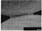
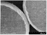
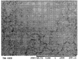
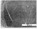
- FIGS. 1A to 1D optical microscopic images of the obtained metal porous body are shown in FIGS. 1A to 1D. As shown in FIGS. 1A to 1D, it was confirmed that fine beads were bonded together by sintering and a large number of pores were formed.
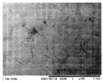
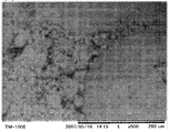
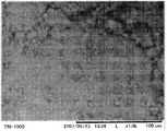
- FIGS. 2A to 2D show the results of observing the microstructure of the surface of the obtained bone anchor material with an optical microscope.
- ⁇ -type tricalcium phosphate has penetrated into the pores near the surface of the bone anchor material, but almost penetrated into the non-surface pores of the bone anchor material. (Not shown).
- the filling rate of ⁇ -type tricalcium phosphate in the bone anchoring material was 3.92% when calculated from the weight change.
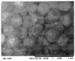
- 3A to 3D show the results of observation of the microstructure of the surface of the bone anchoring material with an optical microscope. As shown in FIGS. 3A to D, hydroxyapatite penetrated into the pores near the surface of the bone anchoring material, but hardly penetrated into the non-surface pores of the bone anchoring material ( Not shown). As a result, the filling rate of hydroxyapatite in the bone anchoring material was 3.33% when calculated from the weight change.
- Table 1 shows the Young's modulus and compressive strength calculated together with the graph of FIG.
- the materials for the bone anchor produced in Examples 1 and 2 have a smaller degree of strain with respect to the compressive force than the control group, and can maintain an elastic state with respect to a high compressive force. It was. This was also confirmed in Table 1, and the bone anchoring materials produced in Examples 1 and 2 had higher elastic modulus and compressive strength than the control group. Thereby, it was confirmed that the material for bone anchors produced in Examples 1 and 2 has sufficient strength immediately after implantation in the living body and can sufficiently protect the bone.
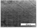
- FIGS. 5A to 5D the cross section of the non-surface portion of the bone fastener material of Example 1 after the compression test was observed with an optical microscope. The results are shown in FIGS. 5A to 5D. As shown in FIGS. 5A to 5D, it was confirmed that the non-surface portion of the bone fastener material was not sufficiently filled with ⁇ -type tricalcium phosphate even after compression.
- Example 3 An aqueous solution in which 5 g of ⁇ -type tricalcium phosphate was dissolved in 10 mL of ultrapure water was poured into the casing. After immersing the porous metal body used in Example 1 in this aqueous solution, the internal pressure of the casing was reduced to 10 ⁇ 1 Pa. This condition was maintained at room temperature for 10 minutes, during which time 50 Hz vibrations continued to be applied to the system. In this way, a bone fixing material was produced.
- Example 4 The metal porous body used in Example 1 was immersed in a 0.1 M nitric acid aqueous solution for 300 seconds.
- a porous metal after nitric acid treatment was used as a working electrode, a platinum electrode as a counter electrode, and a saturated calomel electrode as a reference electrode, and these electrodes were immersed in a Hanks solution at pH 7.4.
- a potentiostat “HA-501G” manufactured by Hokuto Denko Co., Ltd.
- an alternating voltage with a frequency of 1 Hz and ⁇ 1 to 1 V saturated calomel electrode standard
- the composition of the Hanks solution is shown in Table 2, and the current response density during the cathodic polarization treatment is shown in FIG.
- Example 5 A bone anchoring material was prepared in the same procedure as in Example 4 except that the nitric acid treatment was performed for 300 seconds.
- Example 6 A bone fastener material was prepared in the same procedure as in Example 5 except that ultrasonic treatment was performed during nitric acid treatment.
- Example 7 A bone fastener material was prepared in the same procedure as in Example 4 except that nitric acid treatment was not performed.
- Example 8 A material for a bone anchor was prepared in the same procedure as in Example 7 except that cathodic polarization treatment and immersion for 1 week in Hanks solution were not performed.
- FIGS. 8A to 8F The surface of the bone anchor material produced in Examples 4 to 8 was observed with a scanning electron microscope. The results are shown in FIGS. 8A to 8F together with an untreated porous metal body (control group).
- 8A shows the results of the control group
- B shows the results of Example 7
- C shows the results of Example 8
- D shows the results of Example 4
- E shows the results of Example 5
- F shows the results of Example 6.
- r-HAp powder 1.2 g of r-HAp powder was pressed and hardened at a pressure of 100 kg / cm 2 using a press “ServoPulser” (manufactured by Shimadzu Corporation). The obtained tablet was heated in an electric furnace at 800 ° C. for 2 hours (heating rate and cooling rate: 2 ° C./min) to sinter r-HAp, thereby preparing a sample.
- Reference Example 2 A sample was prepared in the same procedure as in Reference Example 1 except that ⁇ -type tricalcium phosphate powder was used instead of r-HAp powder and the sintering temperature was 900 ° C.
- Reference Example 3 A sample was prepared in the same procedure as in Reference Example 1 except that ⁇ -type tricalcium phosphate powder was used instead of r-HAp powder and the sintering temperature was 1200 ° C.
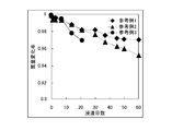
- Test Example 1 The samples prepared in Reference Examples 1 to 3 were immersed in 20 mL of Hanks solution adjusted to pH 5.0 with hydrochloric acid and maintained at 37 ° C. Samples were taken periodically to determine the change in mass. The result is shown in FIG. As a control test, the samples prepared in Reference Examples 1 to 3 were immersed in 20 mL of a hydrochloric acid solution having a pH of 5.0 and maintained at 37 ° C. Samples were taken periodically to determine the change in mass. The result is shown in FIG.
- the mass decreased with time.
- the mass reduction rate was higher in the system containing the Hanks solution (FIG. 9) than in the control test (FIG. 10) which differs only in that the system did not contain the Hanks solution. Since the Hanks solution has a composition similar to that of body fluids, the calcium phosphate ceramics used in Reference Examples 1 to 3 are expected to dissolve at an appropriate rate after being implanted in the living body.
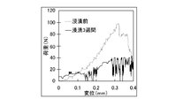
- Test Example 2 A sample was collected after 3 weeks of immersion in the Hanks solution in Test Example 1, and this sample and the sample before immersion were sampled using a compression tester “ServoPulser” (manufactured by Shimadzu Corporation) with a sample cross-sectional area of 25 mm 2 and 0.1 mm. A compression test was performed while measuring strain with a CCD camera at a speed of / min, and a load displacement curve was calculated. The results are shown in FIGS. 11 shows Reference Example 1, FIG. 12 shows Reference Example 2, and FIG. 13 shows Reference Example 3.
- Example 9 Using titanium beads having an average particle diameter of 300 to 355 ⁇ m or 600 to 710 ⁇ m, the temperature was heated to 1073 K or 1173 K at a heating rate of 10 K / min under a reduced pressure of 10 ⁇ 3 Pa, A porous metal body was obtained in the same procedure as in Example 1 except that hot pressing was performed at a load stress of 1.0 to 7.0 MPa. The porosity of each of these metal porous bodies was measured, and the relationship between the hot press pressure and the porosity is shown in FIG.
- the Young's modulus is equivalent to that of bone within a load stress range of 3 to 7 MPa. It turned out that a metal porous body is obtained. Since such a bone fixing material filled with a biodegradable substance in a metal porous body has a relatively high Young's modulus immediately after implantation, it can sufficiently protect weak bone immediately after implantation and can also be embedded. It is presumed that bone resorption can be suppressed because the Young's modulus decreases to the same level as the bone with the passage of time after entering.
- ⁇ -type tricalcium phosphate was introduced. Thereafter, the introduced body was placed in a constant temperature dryer at a temperature of 368 K for 18 hours to be dried, thereby obtaining a composite. This composite was heated to a temperature of 1173 K under a reduced pressure of 10 ⁇ 2 Pa or less at a temperature increase rate of 10 K / min, and held for 2 hours to produce a bone fixing material.
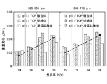
- FIG. 16 shows the weights of the composite and the bone fastener material before and after use.
- the weight increases, that is, the ⁇ -type tricalcium phosphate filling rate increases. It was.
- the weight of the bone anchoring material is smaller after the immersion than before the immersion, it was estimated that the ⁇ -type tricalcium phosphate filled in the metal porous body is dissolved or disintegrated in the living body. .
- the Young's modulus was calculated using the free resonance Young's modulus measuring device “ANJE-RT” (manufactured by Nippon Techno Plus Co., Ltd.) for the metal porous body, the composite, and the bone fastener material before and after immersion. The results are shown in Table 3.
- the Young's modulus of the bone anchoring material before immersion was higher by about 2 GPa than the composite.
- the sintering of ⁇ -type tricalcium phosphate was proceeding by heating under the above conditions.
- the Young's modulus of the bone anchoring material is lower after the immersion than before the immersion, and considering this together with FIG. 16, when the bone anchoring material is embedded in the living body, the passage of time thereafter At the same time, it was estimated that the ceramic body was dissolved or disintegrated, and the Young's modulus of the bone anchoring material decreased.
- the porosity of the metal porous body obtained by hot pressing at a temperature of 973 K was 24 to 32%, and was obtained by hot pressing at a temperature of 1173 K.
- the porosity of the metal porous body was 25% or 30%, and the porosity of the metal porous body obtained by performing the sintering at a temperature of 1173K or 1373K after hot pressing at a temperature of 973K was 30%.
- FIG. 17 shows the relationship between the porosity of the metal porous body and the fatigue strain.
- the fatigue strain of the metal porous body had a clear positive correlation with the porosity.
- the fatigue strength of the metal porous body, and consequently the bone fastener material can be increased or decreased by decreasing the porosity of the metal porous body.
- the porosity of 32.4% of the porous metal body had been destroyed fatigue cycles 10 6 th earlier, from the viewpoint of improving the fatigue strength, the metal porous body having a porosity of less than 32.4% is It turned out to be preferable.
- the yield stress ⁇ y of this metal porous body was 50 MPa, and the longitudinal elastic modulus E was 15 GPa.
- FIG. 18 shows the relationship between the maximum stress loaded on the metal porous body and the fatigue strain. As a result, as shown in FIG. 18, it was confirmed that the fatigue strain increases as the maximum stress increases.
- the porous metal body obtained by hot pressing at a temperature of 973K Furthermore, the porous metal body obtained by hot pressing at a temperature of 973K, the porous metal body obtained by hot pressing at a temperature of 1173K, and obtained by sintering at a temperature of 1173K or 1373K after hot pressing at a temperature of 973K. Further, fatigue tests were conducted on the porous metal bodies (both of which had a porosity of 30%) under the same conditions as described above (stress amplitude ( ⁇ max) was 50 MPa). The result is shown in FIG.
Landscapes
- Health & Medical Sciences (AREA)
- Surgery (AREA)
- Life Sciences & Earth Sciences (AREA)
- Chemical & Material Sciences (AREA)
- General Health & Medical Sciences (AREA)
- Orthopedic Medicine & Surgery (AREA)
- Veterinary Medicine (AREA)
- Public Health (AREA)
- Heart & Thoracic Surgery (AREA)
- Animal Behavior & Ethology (AREA)
- Vascular Medicine (AREA)
- Engineering & Computer Science (AREA)
- Inorganic Chemistry (AREA)
- Epidemiology (AREA)
- Medical Informatics (AREA)
- Nuclear Medicine, Radiotherapy & Molecular Imaging (AREA)
- Biomedical Technology (AREA)
- Neurology (AREA)
- Composite Materials (AREA)
- Materials Engineering (AREA)
- Molecular Biology (AREA)
- Materials For Medical Uses (AREA)
- Surgical Instruments (AREA)
Abstract
本発明の課題は、骨を充分に保護でき且つ骨吸収を抑制できる骨固定具用材及び骨固定具を提供することである。 骨固定具用材は、金属多孔体と、この金属多孔体に充填され且つ生体環境中で溶解可能なセラミック体とを含む。かかる骨固定具用材は、生体埋入直後には金属多孔体にセラミック体が充填されているので、比較的高いヤング率を有する。このため、埋入直後の弱い骨を充分に保護できる。また、埋入後の時間の経過とともに、セラミック体が溶解又は崩壊するため、骨固定具用材のヤング率が低下する。これにより、骨の修復に伴って、骨に与えられる力学的刺激が徐々に増強されるため、骨吸収を抑制できる。
Description
本発明は、骨の固定に用いられる骨固定具、及び骨固定具の形成に用いられる骨固定具用材に関する。
骨折や整形手術の際には、骨が適切な位置及び方向で修復されるよう、骨を固定する必要がある。そこで、骨の固定に用いられる種々の骨固定具が開発されてきた。
骨固定具は、生体埋入後に受ける様々な外力から骨を充分に保護しなければならない。そこで、従来、強度及び靭性を備えるステンレス鋼やコバルト合金といった金属素材で形成された骨固定具が使用されている(例えば、特許文献1及び2参照)。
特表2004-517657号公報
特開2006-130317号公報
しかし、前述の特許文献1及び2に示される骨固定具では、その素材のヤング率(約200GPa)が皮質骨(約20GPa)よりもはるかに大きいため、外力による荷重が骨固定具に必要以上に吸収される。かかるストレスシールディング(荷重遮断)によって、骨に与えられる力学的刺激が不足し、骨吸収現象が生じるおそれがある。
本発明は、以上の実情に鑑みてなされたものであり、骨を充分に保護でき且つ骨吸収を抑制できる骨固定具用材及び骨固定具を提供することを目的とする。
本発明者らは、生体への埋入直後には高いヤング率を有しつつ、経時的にヤング率が低下する材料を見出し、本発明を完成するに至った。具体的には、本発明は以下のようなものを提供する。
(1) 金属多孔体と、この金属多孔体に充填され且つ生体環境中で溶解可能なセラミック体とを含む骨固定具用材。
(2) 前記金属多孔体は、チタン及びチタン系合金からなる群より選ばれる1種以上で形成される(1)記載の骨固定具用材。
(3) 前記金属多孔体は、金属製の微細ビーズ群が焼結されたものである(1)又は(2)記載の骨固定具用材。
(4) 前記セラミック体は、α型リン酸三カルシウム、β型リン酸三カルシウム、及びハイドロキシアパタイトからなる群より選ばれる1種以上で形成される(1)から(3)いずれか記載の骨固定具用材。
(5) (1)から(4)いずれか記載の骨固定具用材で形成された骨固定具。
(6) 生分解性物質が充填されて骨固定具用材として用いられる金属多孔体の製造方法であって、
金属製の微細ビーズ群を、前記金属がα結晶構造をとる条件で加熱する工程と、
前記金属がβ結晶構造をとる条件で焼結する工程と、を有する製造方法。
金属製の微細ビーズ群を、前記金属がα結晶構造をとる条件で加熱する工程と、
前記金属がβ結晶構造をとる条件で焼結する工程と、を有する製造方法。
(7) 金属多孔体に所定のセラミック体を充填することで、骨固定具用材の弾性率を初期状態では増加させつつ、経時的には低下させる方法。
(8) 金属多孔体と、この金属多孔体に充填され且つ生体環境中で溶解可能なセラミック体とを含む骨固定具用材について、前記金属多孔体の気孔率を減増することで、前記骨固定具用材のヤング率及び/又は疲労強度を調節する方法。
(9) 前記金属多孔体の気孔率の減増は、前記金属多孔体の金属がα結晶構造をとる温度範囲内で、温度及び/又はホットプレス圧を増減することで行う(8)記載の方法。
(10) 金属多孔体と、この金属多孔体に充填され且つ生体環境中で溶解可能なセラミック体とを含む骨固定具用材について、前記金属多孔体、又はセラミック体が充填された導入体の焼結温度を増減することで、骨固定具用材の疲労強度を増減する方法。
本発明によれば、埋入直後には、金属多孔体にセラミック体が充填されているので、骨固定具用材は比較的高いヤング率を有する。このため、埋入直後の弱い骨を充分に保護できる。また、埋入後の時間の経過とともに、セラミック体が溶解又は崩壊するため、骨固定具用材のヤング率が低下する。これにより、骨の修復に伴って、骨に与えられる力学的刺激が徐々に増強されるため、骨吸収を抑制できる。
以下、本発明の実施形態について説明するが、本発明を限定することを意図するものではない。
<骨固定具用材>
本発明に係る骨固定具用材は、金属多孔体及びセラミック体を含む。以下、各構成要素について説明する。
本発明に係る骨固定具用材は、金属多孔体及びセラミック体を含む。以下、各構成要素について説明する。
[金属多孔体]
金属多孔体は、多数の細孔が形成された金属製の材料である。かかる金属多孔体は、多数の細孔が形成された構造物である限りにおいて特に限定されず、微細ビーズ群が焼結されたもの、繊維状物等が挙げられる。
金属多孔体は、多数の細孔が形成された金属製の材料である。かかる金属多孔体は、多数の細孔が形成された構造物である限りにおいて特に限定されず、微細ビーズ群が焼結されたもの、繊維状物等が挙げられる。
金属多孔体の気孔率は、後述のセラミック体を充分に充填でき且つ充分な強度を保持できる点で5%以上50%以下であることが好ましい。本発明者らによって、金属多孔体の気孔率が骨固定具用材のヤング率及び疲労強度に深く寄与すること、具体的には、気孔率が減増すると、骨固定具用材のヤング率が減増するとともに、疲労強度が増減することが発見された。このような金属多孔体の気孔率は、微細ビーズ群の加熱温度及び/又はホットプレス圧を適宜設定することによって調節でき、具体的には、加熱温度及び/又はホットプレス圧を高く設定すれば、気孔率が減少し、加熱温度及び/又はホットプレス圧を低く設定すれば、気孔率が増加する。なお、微細ビーズの平均粒子径は、特に限定されないが、10μm以上1000μm以下であってよい。気孔率及び平均粒子径は、従来周知の方法、例えば水銀圧入法で測定される。
金属多孔体の素材は、皮質骨に類似したヤング率を有するものが好ましく、具体的にはチタン及びチタン合金等からなる群より選ばれる1種以上が挙げられる。これらの素材は、軽量、生体に対する低毒性、骨形成の高促進能、高生体適合性といった点においても好ましい。
ここで、チタン合金とは、チタンと、他の元素を含む単体又は化合物との合金である。他の元素としては、アルミニウム、バナジウム、ニオブ、ジルコニウム、タンタル、モリブデン、パラジウム、及び鉄等からなる群より選ばれる1種以上が挙げられる。
かかる金属多孔体は、素材を射出成形、焼結、ホットプレス等することで製造できる。特に限定されないが、金属製の微細ビーズ群は、金属がβ結晶構造をとる条件で焼結されたものであることが好ましい。これにより、金属多孔体、ひいては骨固定具用材の疲労強度を向上できる。なお、β結晶構造をとる条件での焼結は、セラミック体の充填後に行ってもよいが、この場合には酸素が金属多孔体に移行し、脆化を招きやすいことから、セラミック体の充填前に行うことが好ましい。
また、本発明者らにより、気孔率の調節が、金属が温度及び/又はホットプレス圧の調節によって行うことができること、特に金属がα結晶構造をとる温度範囲内で加熱することで優れた気孔率が得られやすいことが発見された。そこで、微細ビーズ群を焼結する前に、金属がα結晶構造をとる条件内の所定温度で、所定ホットプレス圧下、加熱することにより、所望の気孔率を有する金属多孔体を形成することが好ましい。ただし、前述した好ましい条件は例示にすぎず、金属がα結晶構造をとる温度範囲内での加熱のみによって焼結を行ってもよい。
以上の金属多孔体は、生体環境中で溶解可能なセラミック体が充填されることが好ましいが、他の生分解性物質(例えば、軟化可能な生分解性ポリマー)が充填されていてもよい。即ち、本発明は、生分解性物質が充填されて骨固定具用材として用いられる金属多孔体の製造方法であって、金属製の微細ビーズ群を、前記金属がα結晶構造をとる条件で加熱する工程と、前記金属がβ結晶構造をとる条件で焼結する工程と、を有する製造方法も包含するものである。
[セラミック体]
以上の金属多孔体にはセラミック体が充填されている。セラミック体は金属酸化物等の焼結体又は粉末であり、生体環境中で溶解可能である。これにより、セラミック体は、生体内に埋入されると、経時的に溶解又は崩壊して金属多孔体外へと拡散する。
以上の金属多孔体にはセラミック体が充填されている。セラミック体は金属酸化物等の焼結体又は粉末であり、生体環境中で溶解可能である。これにより、セラミック体は、生体内に埋入されると、経時的に溶解又は崩壊して金属多孔体外へと拡散する。
このように、セラミック体は、生体環境へと放出されることから、高い生体適合性を有する素材で形成されることが好ましい。かかる素材としては、α型リン酸三カルシウム、β型リン酸三カルシウム、及びハイドロキシアパタイトからなる群より選ばれる1種以上が挙げられる。
かかるセラミック体の充填率は、充分な強度を付与でき且つ経時的に充分にヤング率を低下できる点で、5%以上100%以下であることが好ましい。充填率は、従来周知の方法、例えば水銀圧入法で測定される。
[製造方法]
以上の構成を備える骨固定具用材は、セラミック体の素材を金属多孔体内に導入し、この導入体に焼結を行うことで製造できる。
以上の構成を備える骨固定具用材は、セラミック体の素材を金属多孔体内に導入し、この導入体に焼結を行うことで製造できる。
セラミック体の素材の金属多孔体内への導入は、例えば、素材含有溶液中へ金属多孔体を浸漬する又は素材含有溶液を金属多孔体に注入することで行うことができる。この場合、浸漬は、充填率を向上できる点で、減圧条件下で行うことが好ましく、系を振動させながら行うことがより好ましい。
その他、セラミック体の素材の導入は、金属多孔体をカソード分極処理にふした後、リン酸塩及びカルシウム塩が溶解された溶液に浸漬することで行うこともできる。
カソード分極に用いられる電解質溶液が含有する支持電解質としては、塩化ナトリウム、硫酸ナトリウム、硫酸カリウム、硫酸マグネシウム等の一般的な中性支持電解質が挙げられる。
支持電解質の濃度は、不足すると電解質溶液の電気伝導性が不充分となる点を考慮して、0.1モル/L以上であることが好ましい。また、電解質溶液に塩化物イオン、フッ素イオン、炭酸イオン等が含まれる場合、濃度過剰となると、電極が腐食することが懸念されるため、濃度を適宜設定する必要がある。
カソード分極処理を行うことで、次のような反応が起こるため、金属多孔体近傍がアルカリ性化する。
O2+2H2O+4e-→4OH-
2H2O+2e-→H2→2OH-
しかし、副産物として水素ガスが発生し、この水素ガスが金属多孔体内に侵入して金属多孔体を脆弱化したり、分極を阻害したり、対流現象によって金属多孔体近傍のアルカリ性条件を損なったりすることが懸念される。
O2+2H2O+4e-→4OH-
2H2O+2e-→H2→2OH-
しかし、副産物として水素ガスが発生し、この水素ガスが金属多孔体内に侵入して金属多孔体を脆弱化したり、分極を阻害したり、対流現象によって金属多孔体近傍のアルカリ性条件を損なったりすることが懸念される。
そこで、上記の支持電解質の他、電解質溶液は、有効量の過酸化水素を含有することが好ましい。これにより、次の反応で水素ガスが消費される。
H2+H2O2→2H2O
しかも、次の反応によって、アルカリ化が促進される。
H2O2+2e-→2OH-
HO2 -+H2O+2e-→3OH-
このため、次の浸漬手順において、金属多孔体内へのリン酸カルシウムの析出を促進できる。
H2+H2O2→2H2O
しかも、次の反応によって、アルカリ化が促進される。
H2O2+2e-→2OH-
HO2 -+H2O+2e-→3OH-
このため、次の浸漬手順において、金属多孔体内へのリン酸カルシウムの析出を促進できる。
ここで、「有効量」とは、発生する水素ガスを所望の程度にまで低減するために必要な過酸化水素量を指し、不純物として偶然に存在し得るような最低量は除外される。なお、有効量は、条件に応じて適宜設定されてよいが、過剰であると過酸化水素自体が導電性材料体内に侵入して導電性材料を脆弱化させる一方、不足すると水素ガスが充分に低減されないことを考慮して、通常、0.01質量%以上1質量%以下であることが好ましい。
電解質への過酸化水素の添加は、通常、カソード分極処理の前に行うが、カソード分極の間に連続的又は間欠的に行ってもよい。
カソード分極処理において印加する電圧は、過剰であると許容量を超える水素ガスが発生する一方、不足すると処理に大幅な時間がかかることを考慮して、標準水素電極に対して-1V以上-5V以下であることが好ましい。また、カソードの表面積に対する電流密度は、過剰であると許容量を超える水素ガスが発生する一方、不足すると電解質溶液の電気伝導性が不充分となることを考慮して、1×10⁻²A/dm2以上1×101A/dm2以下であることが好ましい。
なお、カソード分極処理中の設定温度は、特に限定されず、通常室温であってよいが、特に導電性材料内への水素侵入の抑制が要求される場合、約5℃が好ましい。また、リン酸カルシウムの析出を促進できる点で、カソード分極処理前に金属多孔体を硝酸水溶液等の中に浸漬し、腐食処理を行うことが好ましく、かかる腐食処理を超音波処理と併行することがより好ましい。
カソード分極処理後の金属多孔体を、リン酸塩及びカルシウム塩を含有する溶液に浸漬することで、金属多孔体の内部に多量のリン酸カルシウムが析出する。ここで用いられる溶液は、従来周知のものであってよく、例えばHanks溶液が挙げられる。
焼結は、金属多孔体の金属がβ結晶構造をとる条件内の所定温度で加熱することが好ましい。これにより、金属多孔体、ひいては骨固定具用材の疲労強度を向上できる。なお、用いる金属多孔体が、既に金属がβ結晶構造をとる条件で焼結されたものであるように、充分な疲労強度を有している場合には、焼結温度は特に限定されるものではない。ただし、前述した好ましい条件は例示にすぎず、金属がα結晶構造をとる温度範囲内での加熱のみによって焼結を行ってもよい。
<骨固定具>
本発明に係る骨固定具用材を適宜の形状、寸法に加工することで、骨固定具が作製される。骨固定具の具体的な形状は、特に限定されず、インプラントに適した形状(例えば、歯科インプラント、髄内釘)、骨を外側から支持する形状(例えば、プレート、スクリュ)等であってよい。
本発明に係る骨固定具用材を適宜の形状、寸法に加工することで、骨固定具が作製される。骨固定具の具体的な形状は、特に限定されず、インプラントに適した形状(例えば、歯科インプラント、髄内釘)、骨を外側から支持する形状(例えば、プレート、スクリュ)等であってよい。
<実施例1>
まず、平均粒子径300μmのチタン製ビーズを焼結することで金属多孔体を得た。この金属多孔体の気孔率は38.3%であった。なお、気孔率は、見かけ上の体積からアルキメデス法によって測定した値である。
まず、平均粒子径300μmのチタン製ビーズを焼結することで金属多孔体を得た。この金属多孔体の気孔率は38.3%であった。なお、気孔率は、見かけ上の体積からアルキメデス法によって測定した値である。
また、得られた金属多孔体の光学顕微鏡像を図1A~Dに示す。図1A~Dに示されるように、焼結によって微細ビーズ同士が結合するとともに、多数の細孔が形成されていることが確認された。
かかる金属多孔体を、水:β型リン酸三カルシウム=1:3の容積率のβ型リン酸三カルシウム水溶液中に37℃で24時間に亘り浸漬することで、骨固定具用材を作製した。
得られた骨固定具用材の表面の微細構造を光学顕微鏡で観察した結果を図2A~Dに示す。図2A~Dに示されるように、β型リン酸三カルシウムは骨固定具用材の表面近傍の細孔には侵入していたが、骨固定具用材の非表面の細孔にはほとんど侵入していなかった(図示せず)。その結果、骨固定具用材におけるβ型リン酸三カルシウムの充填率は、重量変化から計算したとき、3.92%であった。
<実施例2>
β型リン酸三カルシウム水溶液の代わりに、水:ハイドロキシアパタイト=1:3の容積率のハイドロキシアパタイト水溶液を用いた点を除き、実施例1と同様の手順で、骨固定具用材を作製した。
β型リン酸三カルシウム水溶液の代わりに、水:ハイドロキシアパタイト=1:3の容積率のハイドロキシアパタイト水溶液を用いた点を除き、実施例1と同様の手順で、骨固定具用材を作製した。
この骨固定具用材の表面の微細構造を光学顕微鏡で観察した結果を図3A~Dに示す。図3A~Dに示されるように、ハイドロキシアパタイトは骨固定具用材の表面近傍の細孔には侵入していたが、骨固定具用材の非表面の細孔にはほとんど侵入していなかった(図示せず)。その結果、骨固定具用材におけるハイドロキシアパタイトの充填率は、重量変化から計算したとき、3.33%であった。
[評価]
実施例1及び2で作製した骨固定具用材について、圧縮試験機「ServoPulser」(島津製作所社製)を用い、試料断面積25mm2、0.1mm/minの速度で、CCDカメラで歪みを測定しながら圧縮試験を行った。この結果を、浸漬を行う前の金属多孔体(対照区)とともに図4に示す。
実施例1及び2で作製した骨固定具用材について、圧縮試験機「ServoPulser」(島津製作所社製)を用い、試料断面積25mm2、0.1mm/minの速度で、CCDカメラで歪みを測定しながら圧縮試験を行った。この結果を、浸漬を行う前の金属多孔体(対照区)とともに図4に示す。
また、図4のグラフとともに算出したヤング率及び圧縮強さを表1に示す。
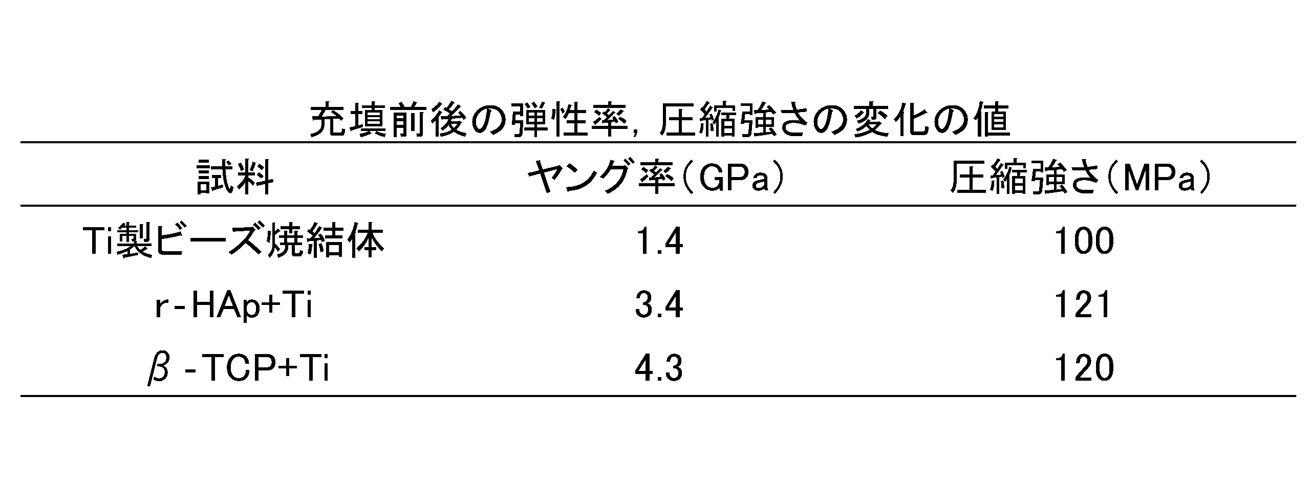
図4に示されるように、実施例1及び2で作製した骨固定具用材では、対照区に比べ、圧縮力に対するひずみの程度が小さいとともに、高圧縮力に対しても弾性状態を保持できていた。このことは表1においても確認され、実施例1及び2で作製した骨固定具用材は、対照区に比べて高い弾性率及び圧縮強さを有していた。これにより、実施例1及び2で作製した骨固定具用材は、生体埋入直後には充分な強度を奏し、骨を充分に保護できることが確認された。
次に、圧縮試験後における実施例1の骨固定具用材の非表面部における断面を光学顕微鏡で観察した。この結果を図5A~Dに示す。図5A~Dに示されるように、骨固定具用材の非表面部には、圧縮後においても、β型リン酸三カルシウムが充分に充填されていないことが確認された。
<実施例3>
ケーシング内にβ型リン酸三カルシウム5gが超純水10mLに溶解された水溶液を注いだ。この水溶液中に、実施例1で使用した金属多孔体を浸漬した後、ケーシング内圧を10-1Paまで減圧した。この条件を室温で10分間に亘り維持し、この間、50Hzの振動を系に与え続けた。このようにして骨固定具用材を作製した。
ケーシング内にβ型リン酸三カルシウム5gが超純水10mLに溶解された水溶液を注いだ。この水溶液中に、実施例1で使用した金属多孔体を浸漬した後、ケーシング内圧を10-1Paまで減圧した。この条件を室温で10分間に亘り維持し、この間、50Hzの振動を系に与え続けた。このようにして骨固定具用材を作製した。
[評価]
得られた骨固定具用材の非表面部の断面を光学顕微鏡で観察した。この結果を図6に示す。図6に示されるように、β型リン酸三カルシウムは、骨固定具用材の非表面の細孔にまで充分に侵入していた。その結果、骨固定具用材におけるβ型リン酸三カルシウムの充填率は、重量変化から計算したとき、24.6%であった。これにより、減圧条件下で充填を行うことで、充分量のセラミック体が骨固定具用材の非表面にまで充填できることが確認された。かかる骨固定具用材は、より高い強度を奏し、骨をより充分に保護できることが推測される。
得られた骨固定具用材の非表面部の断面を光学顕微鏡で観察した。この結果を図6に示す。図6に示されるように、β型リン酸三カルシウムは、骨固定具用材の非表面の細孔にまで充分に侵入していた。その結果、骨固定具用材におけるβ型リン酸三カルシウムの充填率は、重量変化から計算したとき、24.6%であった。これにより、減圧条件下で充填を行うことで、充分量のセラミック体が骨固定具用材の非表面にまで充填できることが確認された。かかる骨固定具用材は、より高い強度を奏し、骨をより充分に保護できることが推測される。
<実施例4>
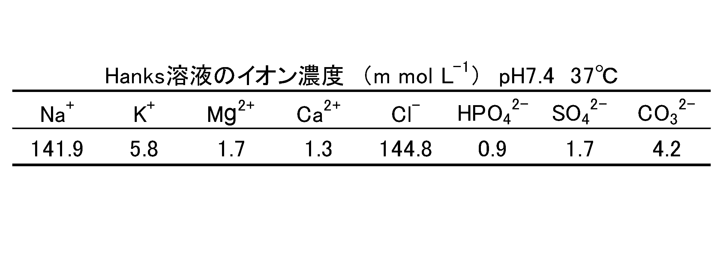
実施例1で使用した金属多孔体を、0.1M硝酸水溶液中に300秒間浸した。作用極として硝酸処理後の金属多孔体、対極として白金電極、参照極として飽和カロメル電極を用い、これら電極をpH7.4のHanks溶液に浸した。これら電極に、ポテンショスタット「HA-501G」(北斗電工社製)を用い、周波数1Hz、-1~1V(飽和カロメル電極基準)の交流電圧を37℃にて1時間に亘って印加した。Hanks溶液の組成を表2に、カソード分極処理中の電流応答密度を図7に示す。
実施例1で使用した金属多孔体を、0.1M硝酸水溶液中に300秒間浸した。作用極として硝酸処理後の金属多孔体、対極として白金電極、参照極として飽和カロメル電極を用い、これら電極をpH7.4のHanks溶液に浸した。これら電極に、ポテンショスタット「HA-501G」(北斗電工社製)を用い、周波数1Hz、-1~1V(飽和カロメル電極基準)の交流電圧を37℃にて1時間に亘って印加した。Hanks溶液の組成を表2に、カソード分極処理中の電流応答密度を図7に示す。
印加後の金属多孔体を、Hanks溶液中に浸漬し、1週間放置した。その後、金属多孔体を水:β型リン酸三カルシウム=1:3の容積率のβ型リン酸三カルシウム水溶液中に室温で24時間に亘り浸漬することで、骨固定具用材を作製した。
<実施例5>
硝酸処理を300秒間行った点を除き、実施例4と同様の手順で骨固定具用材を作製した。
硝酸処理を300秒間行った点を除き、実施例4と同様の手順で骨固定具用材を作製した。
<実施例6>
硝酸処理の間、超音波処理を併せて行った点を除き、実施例5と同様の手順で骨固定具用材を作製した。
硝酸処理の間、超音波処理を併せて行った点を除き、実施例5と同様の手順で骨固定具用材を作製した。
<実施例7>
硝酸処理を行わなかった点を除き、実施例4と同様の手順で骨固定具用材を作製した。
硝酸処理を行わなかった点を除き、実施例4と同様の手順で骨固定具用材を作製した。
<実施例8>
カソード分極処理及びHanks溶液中での1週間の浸漬を行わなかった点を除き、実施例7と同様の手順で骨固定具用材を作製した。
カソード分極処理及びHanks溶液中での1週間の浸漬を行わなかった点を除き、実施例7と同様の手順で骨固定具用材を作製した。
[評価]
実施例4~8で作製した骨固定具用材の表面を走査型電子顕微鏡で観察した。この結果を、未処理の金属多孔体(対照区)とともに図8A~Fに示す。図8Aは対照区、Bは実施例7、Cは実施例8、Dは実施例4、Eは実施例5、Fは実施例6の結果をそれぞれ示す。
実施例4~8で作製した骨固定具用材の表面を走査型電子顕微鏡で観察した。この結果を、未処理の金属多孔体(対照区)とともに図8A~Fに示す。図8Aは対照区、Bは実施例7、Cは実施例8、Dは実施例4、Eは実施例5、Fは実施例6の結果をそれぞれ示す。
図8A~Fに示されるように、実施例4~8ではリン酸カルシウムの白色斑点が多数観察されたのに対し、対照区では白色斑点が観察されなかった。特に実施例4~6、中でも実施例6では白色斑点の密度が大きく、多量のリン酸カルシウムが析出していることが確認された。これにより、カソード分極処理でリン酸カルシウムを金属多孔体に充填でき、カソード分極処理前に硝酸による腐食処理、更には超音波処理を行うことで、リン酸カルシウムの充填効率を向上できることが分かった。
〔参考例〕 セラミック体素材の検討
(参考例1)
ウシ骨由来焼成ハイドロキシアパタイト(b-HAp)を0.1M硝酸水溶液45mLに溶解し、この溶液に1000mLまで超純水を加え、充分に撹拌を行うことでb-HAp溶液を得た。b-HAp溶液にアンモニア500mLを室温で撹拌しながら滴下し、pHを10~11とした。このようにして得た溶解析出ハイドロキシアパタイト(r-HAp)スラリーを24時間熟成した後、濾過し、蒸留水で洗浄した。濾液を50℃で24時間、続いて120℃で24時間に亘り乾燥することで、r-HAp粉末を得た。r-HAp粉末1.2gを、プレス機「ServoPulser」(島津製作所社製)を用いて100kg/cm2の圧力で押し固めた。得られたタブレットを電気炉内、800℃で2時間に亘り加熱(昇温速度及び降温速度:2℃/分)してr-HApを焼結させることで、試料を作製した。
(参考例1)
ウシ骨由来焼成ハイドロキシアパタイト(b-HAp)を0.1M硝酸水溶液45mLに溶解し、この溶液に1000mLまで超純水を加え、充分に撹拌を行うことでb-HAp溶液を得た。b-HAp溶液にアンモニア500mLを室温で撹拌しながら滴下し、pHを10~11とした。このようにして得た溶解析出ハイドロキシアパタイト(r-HAp)スラリーを24時間熟成した後、濾過し、蒸留水で洗浄した。濾液を50℃で24時間、続いて120℃で24時間に亘り乾燥することで、r-HAp粉末を得た。r-HAp粉末1.2gを、プレス機「ServoPulser」(島津製作所社製)を用いて100kg/cm2の圧力で押し固めた。得られたタブレットを電気炉内、800℃で2時間に亘り加熱(昇温速度及び降温速度:2℃/分)してr-HApを焼結させることで、試料を作製した。
(参考例2)
r-HAp粉末の代わりにβ型リン酸三カルシウム粉末を使用し、焼結温度を900℃とした点を除き、参考例1と同様の手順で試料を作製した。
r-HAp粉末の代わりにβ型リン酸三カルシウム粉末を使用し、焼結温度を900℃とした点を除き、参考例1と同様の手順で試料を作製した。
(参考例3)
r-HAp粉末の代わりにα型リン酸三カルシウム粉末を使用し、焼結温度を1200℃とした点を除き、参考例1と同様の手順で試料を作製した。
r-HAp粉末の代わりにα型リン酸三カルシウム粉末を使用し、焼結温度を1200℃とした点を除き、参考例1と同様の手順で試料を作製した。
〔試験例1〕
参考例1~3で作製した試料を、塩酸でpH5.0に調整したHanks溶液20mLの中に浸漬し、37℃に維持した。試料を定期的に採取し、質量の変化を決定した。この結果を図9に示す。対照の試験として、参考例1~3で作製した試料を、pH5.0の塩酸溶液20mLの中に浸漬し、37℃に維持した。試料を定期的に採取し、質量の変化を決定した。この結果を図10に示す。
参考例1~3で作製した試料を、塩酸でpH5.0に調整したHanks溶液20mLの中に浸漬し、37℃に維持した。試料を定期的に採取し、質量の変化を決定した。この結果を図9に示す。対照の試験として、参考例1~3で作製した試料を、pH5.0の塩酸溶液20mLの中に浸漬し、37℃に維持した。試料を定期的に採取し、質量の変化を決定した。この結果を図10に示す。
図9に示されるように、参考例1~3のいずれにおいても、経時的に質量が低下していた。しかも、Hanks溶液を含まない系である点のみ異なる対照試験(図10)よりも、Hanks溶液を含む系(図9)の方が、質量低下速度が大きかった。Hanks溶液は体液に類似する組成を有することから、参考例1~3で用いたリン酸カルシウム系セラミックは、生体内に埋入された後、適切な速度で溶解することが期待される。
なお、参考例3では、浸漬初期段階から急速に質量が低下し(図9)、形状が崩壊していた(図示せず)。これに対して、参考例1~2では、略一定速度で徐々に質量が低下し、形状崩壊の程度も緩やかであった。よって、r-HAp及びβ型リン酸三カルシウムがセラミック体の素材として特に好ましいことが推測される。
〔試験例2〕
試験例1におけるHanks溶液への浸漬3週間後に試料を採取し、この試料及び浸漬前の試料について、圧縮試験機「ServoPulser」(島津製作所社製)を用い、試料断面積25mm2、0.1mm/minの速度で、CCDカメラで歪みを測定しながら圧縮試験を行い、荷重変位曲線を算出した。この結果を図11~13に示す。図11は参考例1、図12は参考例2、図13は参考例3をそれぞれ示す。
試験例1におけるHanks溶液への浸漬3週間後に試料を採取し、この試料及び浸漬前の試料について、圧縮試験機「ServoPulser」(島津製作所社製)を用い、試料断面積25mm2、0.1mm/minの速度で、CCDカメラで歪みを測定しながら圧縮試験を行い、荷重変位曲線を算出した。この結果を図11~13に示す。図11は参考例1、図12は参考例2、図13は参考例3をそれぞれ示す。
図11~13に示されるように、参考例2及び3の試料は、浸漬3週間後において、荷重に対する高い変位値を呈した。これにより、参考例2及び3で用いたリン酸カルシウム系セラミックは、生体内に埋入された後、経時的に強度が低下し、崩壊しやすくなることが確認された。なお、図12に示されるように、参考例2の試料は、浸漬3週間後において既に崩壊していることが確認され、β型リン酸三カルシウムがセラミック体の素材として特に好ましいことが推測される。
<実施例9>
平均粒子径300~355μm、又は600~710μmのチタン製ビーズを用い、10-3Paの減圧下、10K/分の昇温速度で温度を1073K又は1173Kに加熱し、0.5時間に亘り、1.0~7.0MPaの負荷応力でホットプレスを行った点を除き、実施例1と同様の手順で、金属多孔体を得た。これら金属多孔体の各々の気孔率を測定し、ホットプレス圧と気孔率との関係を図14に示す。
平均粒子径300~355μm、又は600~710μmのチタン製ビーズを用い、10-3Paの減圧下、10K/分の昇温速度で温度を1073K又は1173Kに加熱し、0.5時間に亘り、1.0~7.0MPaの負荷応力でホットプレスを行った点を除き、実施例1と同様の手順で、金属多孔体を得た。これら金属多孔体の各々の気孔率を測定し、ホットプレス圧と気孔率との関係を図14に示す。
図14に示されるように、1073Kで加熱した場合、負荷応力3~7MPaの範囲で、負荷応力と気孔率との間に明確な負の相関が見られた。これにより、ホットプレス圧を高く設定すれば、気孔率が減少し、加熱温度及び/又はホットプレス圧を低く設定すれば、気孔率が増加することが分かった。また、同等のホットプレス圧であっても、加熱温度が1073Kであるものと、1173Kであるものとでは、気孔率に約15%もの差が存在していた。これは、チタンがα結晶構造をとる温度(1073K)に比べ、β結晶構造をとる温度(1155K以上)では、容易に変形するために緻密化が促進されたものと推測される。更に、チタン製ビーズの平均粒子径の大小にかかわらず、ホットプレス圧及び/又は加熱温度に依存して気孔率が決定されることが分かった。
得られた金属多孔体について、気孔率とヤング率との関係に着目した。すると、図15に示されるように、金属多孔体の気孔率とヤング率との間には、指数関数的な負の相関が見られた。これにより、気孔率が減増すると、骨固定具用材のヤング率が減増することが分かった。また、金属多孔体のヤング率は、チタン製ビーズの平均粒子径の大小にかかわらず、気孔率25~35%であると、骨と同等のヤング率(約10~30GPa)を示すことが分かった。この結果を図14と併せて考慮すれば、チタン製ビーズをチタンがα結晶構造をとる温度(1073K)で加熱することにより、負荷応力3~7MPaの範囲で、骨と同等のヤング率を有する金属多孔体が得られることが分かった。このような金属多孔体に生分解性物質が充填された骨固定具用材は、埋入直後には比較的高いヤング率を有するため、埋入直後の弱い骨を充分に保護でき、且つ、埋入後の時間の経過とともに、ヤング率が骨と同等にまで低下するため、骨吸収を抑制できることが推測される。
以上の金属多孔体の各々を、水:β型リン酸三カルシウム=1:2の容積率のβ型リン酸三カルシウム水溶液中に10-3Paの減圧下、37℃で浸漬することで、β型リン酸三カルシウムを導入した。その後、この導入体を、温度368Kの恒温乾燥機内に18時間に亘り置くことで、乾燥させることで、複合体を得た。この複合体を、10-2Pa以下の減圧下、昇温速度10K/分で温度1173Kまで加熱し、2時間に亘って保持することで、骨固定具用材を作製した。
これらの骨固定具用材(6mm×36mm×2mmの板状体)を、pH7.4に調整したHanks溶液50mLの中に浸漬し、温度310Kで7日間に亘って維持した。その後、温度368Kの恒温乾燥機内に16時間に亘り置くことで、乾燥させた。
前述した複合体、及び使用前後の骨固定具用材の重量を図16に示す。図16に示されるように、チタン製ビーズの平均粒子径の大小にかかわらず、金属多孔体の気孔率が増すにつれ、重量が大きい、つまりβ型リン酸三カルシウムの充填率が高いことが分かった。また、骨固定具用材の重量が浸漬前に比べて浸漬後の方が小さいことから、金属多孔体内に充填されたβ型リン酸三カルシウムが、生体内で溶解又は崩壊することが推測された。
また、前述した金属多孔体、複合体、及び浸漬前後の骨固定具用材について、自由共振式ヤング率測定装置「ANJE-RT」(日本テクノプラス社製)を用いて、ヤング率を算出した。この結果を表3に示す。
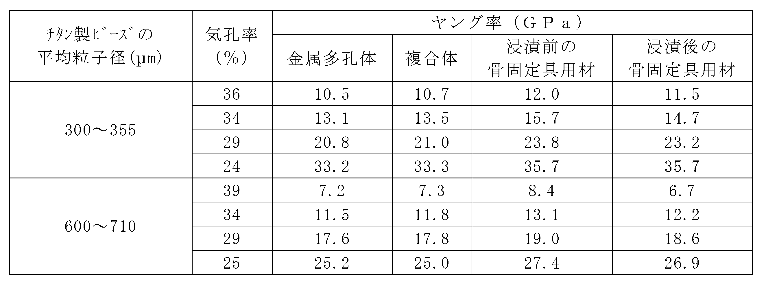
表3に示されるように、複合体に比べて、浸漬前の骨固定具用材の方が、ヤング率が約2GPa高かった。これにより、前記条件での加熱により、β型リン酸三カルシウムの焼結が進行していることが確認された。また、骨固定具用材のヤング率が浸漬前に比べて浸漬後の方が低く、これを図16と併せて考慮すると、この骨固定具用材を生体内に埋入すると、その後の時間の経過とともに、セラミック体が溶解又は崩壊して、骨固定具用材のヤング率が低下することが推測された。
(参考例4)
平均粒子径300~355μmのチタン製ビーズを用い、10-2Paの減圧下、温度を973K又は1173Kに加熱し、0.5時間に亘り、1.0~7.0MPaの負荷応力でホットプレスを行った後、973Kでホットプレスを行ったものについてのみ、10-2Paの減圧下、温度1173K又は1373Kで0.2時間に亘り焼結を行った点を除き、実施例1と同様の手順で、金属多孔体を得た。
平均粒子径300~355μmのチタン製ビーズを用い、10-2Paの減圧下、温度を973K又は1173Kに加熱し、0.5時間に亘り、1.0~7.0MPaの負荷応力でホットプレスを行った後、973Kでホットプレスを行ったものについてのみ、10-2Paの減圧下、温度1173K又は1373Kで0.2時間に亘り焼結を行った点を除き、実施例1と同様の手順で、金属多孔体を得た。
得られた金属多孔体の各々の気孔率を算出した結果、温度973Kでのホットプレスで得られた金属多孔体の気孔率は24~32%であり、温度1173Kでのホットプレスで得られた金属多孔体の気孔率は25%又は30%であり、温度973Kでのホットプレス後に温度1173K又は1373Kでの焼結を行って得られた金属多孔体の気孔率は30%であった。
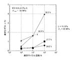
温度973Kでのホットプレスで得られた金属多孔体について、疲労試験を行った。この試験は、応力が5~50MPa(振幅(σmax)が50MPa)、応力比(R)が0.1の荷重振幅条件下、周波数20Hzの正弦波状繰返し荷重を負荷することで行った。金属多孔体の気孔率と疲労ひずみとの関係を図17に示す。
図17に示されるように、金属多孔体の疲労ひずみは、気孔率との間に、明らかな正の相関を有していた。これにより、金属多孔体の気孔率を減増することで、金属多孔体、ひいては骨固定具用材の疲労強度を増減できることが推測された。なお、気孔率32.4%の金属多孔体は疲労サイクル106回目以前に破壊していたことから、疲労強度の向上の観点からは、32.4%未満の気孔率を有する金属多孔体が好ましいことが分かった。また、この金属多孔体の降伏応力σyは50MPaであり、縦弾性係数Eは15GPaであることが分かった。
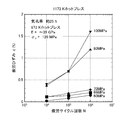
次に、温度973Kでのホットプレスで得られた金属多孔体について、疲労試験を行った。この試験は、応力振幅(σmax)を40~100MPaとした点を除き、前述の試験と同様の条件で行った。金属多孔体に負荷された最大応力と疲労ひずみとの関係を図18に示す。この結果、図18に示されるように、最大応力の増加に伴って、疲労ひずみが増加することが確認された。
更に、温度973Kでのホットプレスで得られた金属多孔体、温度1173Kでのホットプレスで得られた金属多孔体、温度973Kでのホットプレス後に温度1173K又は1373Kでの焼結を行って得られた金属多孔体(いずれも気孔率30%)について、前述と同様の条件(応力振幅(σmax)が50MPa)で、疲労試験を行った。この結果を図19に示す。
図19に示されるように、焼結温度を増加することで、疲労ひずみをより抑制できることが分かった。また、1173K及び1373Kで焼結したものは、973Kでホットプレスのみ行ったものに比べ、疲労ひずみを大幅に抑制できることが分かった。これにより、チタンがβ結晶構造をとる温度(1155K以上)で焼結することにより、金属多孔体及びそれを用いて得られる骨固定具用材の疲労強度を大幅に向上できることが分かった。
Claims (10)
- 金属多孔体と、この金属多孔体に充填され且つ生体環境中で溶解可能なセラミック体とを含む骨固定具用材。
- 前記金属多孔体は、チタン及びチタン系合金からなる群より選ばれる1種以上で形成される請求項1記載の骨固定具用材。
- 前記金属多孔体は、金属製の微細ビーズ群が焼結されたものである請求項1又は2記載の骨固定具用材。
- 前記セラミック体は、α型リン酸三カルシウム、β型リン酸三カルシウム、及びハイドロキシアパタイトからなる群より選ばれる1種以上で形成される請求項1から3いずれか記載の骨固定具用材。
- 請求項1から4いずれか記載の骨固定具用材で形成された骨固定具。
- 生分解性物質が充填されて骨固定具用材として用いられる金属多孔体の製造方法であって、
金属製の微細ビーズ群を、前記金属がα結晶構造をとる条件で加熱する工程と、
前記金属がβ結晶構造をとる条件で焼結する工程と、を有する製造方法。 - 金属多孔体に所定のセラミック体を充填することで、骨固定具用材の弾性率を初期状態では増加させつつ、経時的には低下させる方法。
- 金属多孔体と、この金属多孔体に充填され且つ生体環境中で溶解可能なセラミック体とを含む骨固定具用材について、前記金属多孔体の気孔率を減増することで、前記骨固定具用材のヤング率及び/又は疲労強度を調節する方法。
- 前記金属多孔体の気孔率の減増は、前記金属多孔体の金属がα結晶構造をとる温度範囲内で、温度及び/又はホットプレス圧を増減することで行う請求項8記載の方法。
- 金属多孔体と、この金属多孔体に充填され且つ生体環境中で溶解可能なセラミック体とを含む骨固定具用材について、前記金属多孔体、又はセラミック体が充填された導入体の焼結温度を、前記金属多孔体の金属がβ結晶構造をとる温度範囲内で増減することで、骨固定具用材の疲労強度を増減する方法。
Applications Claiming Priority (2)
| Application Number | Priority Date | Filing Date | Title |
|---|---|---|---|
| JP2008032345A JP2011083297A (ja) | 2008-02-13 | 2008-02-13 | 骨固定具用材及び骨固定具 |
| JP2008-032345 | 2008-02-13 |
Publications (1)
| Publication Number | Publication Date |
|---|---|
| WO2009102018A1 true WO2009102018A1 (ja) | 2009-08-20 |
Family
ID=40957048
Family Applications (1)
| Application Number | Title | Priority Date | Filing Date |
|---|---|---|---|
| PCT/JP2009/052410 Ceased WO2009102018A1 (ja) | 2008-02-13 | 2009-02-13 | 骨固定具用材及び骨固定具 |
Country Status (2)
| Country | Link |
|---|---|
| JP (1) | JP2011083297A (ja) |
| WO (1) | WO2009102018A1 (ja) |
Cited By (1)
| Publication number | Priority date | Publication date | Assignee | Title |
|---|---|---|---|---|
| CN102850079A (zh) * | 2012-09-10 | 2013-01-02 | 四川大学 | 表面由磷酸钙纳米粒子构建的多孔生物陶瓷及其制备方法 |
Citations (4)
| Publication number | Priority date | Publication date | Assignee | Title |
|---|---|---|---|---|
| JPH09234243A (ja) * | 1995-12-25 | 1997-09-09 | Takiron Co Ltd | 複合化された高強度インプラント材料及びその製造方法 |
| JP2006518807A (ja) * | 2003-01-24 | 2006-08-17 | ウニヴェルズィテート デス ザールランデス | セラミック層を有する金属成形体の製法、金属成形体およびその使用 |
| JP2007000635A (ja) * | 2005-06-23 | 2007-01-11 | Depuy Products Inc | 梨地状表面を有するインプラントおよびその製造方法 |
| JP2008012032A (ja) * | 2006-07-05 | 2008-01-24 | Kitagawa Iron Works Co Ltd | アパタイト系材料を含有する生体用部材及びその製造方法 |
-
2008
- 2008-02-13 JP JP2008032345A patent/JP2011083297A/ja active Pending
-
2009
- 2009-02-13 WO PCT/JP2009/052410 patent/WO2009102018A1/ja not_active Ceased
Patent Citations (4)
| Publication number | Priority date | Publication date | Assignee | Title |
|---|---|---|---|---|
| JPH09234243A (ja) * | 1995-12-25 | 1997-09-09 | Takiron Co Ltd | 複合化された高強度インプラント材料及びその製造方法 |
| JP2006518807A (ja) * | 2003-01-24 | 2006-08-17 | ウニヴェルズィテート デス ザールランデス | セラミック層を有する金属成形体の製法、金属成形体およびその使用 |
| JP2007000635A (ja) * | 2005-06-23 | 2007-01-11 | Depuy Products Inc | 梨地状表面を有するインプラントおよびその製造方法 |
| JP2008012032A (ja) * | 2006-07-05 | 2008-01-24 | Kitagawa Iron Works Co Ltd | アパタイト系材料を含有する生体用部材及びその製造方法 |
Cited By (1)
| Publication number | Priority date | Publication date | Assignee | Title |
|---|---|---|---|---|
| CN102850079A (zh) * | 2012-09-10 | 2013-01-02 | 四川大学 | 表面由磷酸钙纳米粒子构建的多孔生物陶瓷及其制备方法 |
Also Published As
| Publication number | Publication date |
|---|---|
| JP2011083297A (ja) | 2011-04-28 |
Similar Documents
| Publication | Publication Date | Title |
|---|---|---|
| JP6027894B2 (ja) | 生分解性移植材およびその製造方法 | |
| He et al. | HA coating fabricated by electrochemical deposition on modified Ti6Al4V alloy | |
| Jelliti et al. | Effect of surface nanocrystallization on the corrosion behavior of Ti–6Al–4V titanium alloy | |
| Lamolle et al. | Titanium implant surface modification by cathodic reduction in hydrofluoric acid: surface characterization and in vivo performance | |
| Zhuravleva et al. | Porous low modulus Ti40Nb compacts with electrodeposited hydroxyapatite coating for biomedical applications | |
| Wang et al. | Preparation and properties of titanium oxide film on NiTi alloy by micro-arc oxidation | |
| EP2204196A1 (en) | Magnesium-based medical device and process for producing the same | |
| Bakhsheshi-Rad et al. | The mechanical properties and corrosion behavior of double-layered nano hydroxyapatite-polymer coating on Mg-Ca alloy | |
| JP2018512500A (ja) | 生体分解性マグネシウムおよび生体分解性マグネシウムの分解速度の制御方法 | |
| Xiong et al. | Effect of texture evolution on corrosion resistance of AZ80 magnesium alloy subjected to applied force in simulated body fluid | |
| Mousa et al. | Surface modification of magnesium and its alloys using anodization for orthopedic implant application | |
| CN106413636B (zh) | 骨接合用植入物 | |
| Chuan et al. | Formation characteristic of Ca–P coatings on magnesium alloy surface | |
| de la Rosa et al. | Enhancing corrosion resistance and bioactive behavior of porous metallic scaffolds through electrochemical coatings | |
| WO2009102018A1 (ja) | 骨固定具用材及び骨固定具 | |
| Zhang et al. | Weak fatigue notch sensitivity in a biomedical titanium alloy exhibiting nonlinear elasticity | |
| Fojt et al. | Influence of porosity on corrosion behaviour of Ti-39Nb alloy for dental applications | |
| CN106163580B (zh) | 用于制造用于生物医学应用的多孔金属材料的方法和通过所述方法获得的材料 | |
| RU2363775C1 (ru) | Способ получения покрытий на изделиях, выполненных из титана и его сплавов | |
| KR101095909B1 (ko) | 생체뼈 유도성의 인공뼈와 그 제조방법 | |
| Sonmez et al. | Corrosion protection of AA6061-T4 alloy by sol–gel derived micro and nano-scale hydroxyapatite (HA) coating | |
| JP5019346B2 (ja) | 骨親和性インプラント及びその製造方法 | |
| KR100453289B1 (ko) | 임프란트 표면 처리용 전해질 용액 및 상기 전해질 용액을이용한 임프란트 표면 처리 방법 | |
| dos Santos Bonfim et al. | Development of titanium dental implants using techniques of powder metallurgy | |
| KR102150326B1 (ko) | 생체 조직 친화성 금속재료의 표면처리 방법 |
Legal Events
| Date | Code | Title | Description |
|---|---|---|---|
| 121 | Ep: the epo has been informed by wipo that ep was designated in this application |
Ref document number: 09710888 Country of ref document: EP Kind code of ref document: A1 |
|
| NENP | Non-entry into the national phase |
Ref country code: DE |
|
| 122 | Ep: pct application non-entry in european phase |
Ref document number: 09710888 Country of ref document: EP Kind code of ref document: A1 |
|
| NENP | Non-entry into the national phase |
Ref country code: JP |