WO2007109236A2 - Microrna fingerprints during human megakaryocytopoiesis - Google Patents
Microrna fingerprints during human megakaryocytopoiesis Download PDFInfo
- Publication number
- WO2007109236A2 WO2007109236A2 PCT/US2007/006824 US2007006824W WO2007109236A2 WO 2007109236 A2 WO2007109236 A2 WO 2007109236A2 US 2007006824 W US2007006824 W US 2007006824W WO 2007109236 A2 WO2007109236 A2 WO 2007109236A2
- Authority
- WO
- WIPO (PCT)
- Prior art keywords
- mir
- gene product
- cancer
- myeloproliferative disorder
- expression
- Prior art date
Links
Classifications
-
- C—CHEMISTRY; METALLURGY
- C12—BIOCHEMISTRY; BEER; SPIRITS; WINE; VINEGAR; MICROBIOLOGY; ENZYMOLOGY; MUTATION OR GENETIC ENGINEERING
- C12N—MICROORGANISMS OR ENZYMES; COMPOSITIONS THEREOF; PROPAGATING, PRESERVING, OR MAINTAINING MICROORGANISMS; MUTATION OR GENETIC ENGINEERING; CULTURE MEDIA
- C12N15/00—Mutation or genetic engineering; DNA or RNA concerning genetic engineering, vectors, e.g. plasmids, or their isolation, preparation or purification; Use of hosts therefor
- C12N15/09—Recombinant DNA-technology
- C12N15/11—DNA or RNA fragments; Modified forms thereof; Non-coding nucleic acids having a biological activity
- C12N15/113—Non-coding nucleic acids modulating the expression of genes, e.g. antisense oligonucleotides; Antisense DNA or RNA; Triplex- forming oligonucleotides; Catalytic nucleic acids, e.g. ribozymes; Nucleic acids used in co-suppression or gene silencing
- C12N15/1135—Non-coding nucleic acids modulating the expression of genes, e.g. antisense oligonucleotides; Antisense DNA or RNA; Triplex- forming oligonucleotides; Catalytic nucleic acids, e.g. ribozymes; Nucleic acids used in co-suppression or gene silencing against oncogenes or tumor suppressor genes
-
- A—HUMAN NECESSITIES
- A61—MEDICAL OR VETERINARY SCIENCE; HYGIENE
- A61K—PREPARATIONS FOR MEDICAL, DENTAL OR TOILETRY PURPOSES
- A61K31/00—Medicinal preparations containing organic active ingredients
- A61K31/70—Carbohydrates; Sugars; Derivatives thereof
- A61K31/7088—Compounds having three or more nucleosides or nucleotides
- A61K31/7105—Natural ribonucleic acids, i.e. containing only riboses attached to adenine, guanine, cytosine or uracil and having 3'-5' phosphodiester links
-
- A—HUMAN NECESSITIES
- A61—MEDICAL OR VETERINARY SCIENCE; HYGIENE
- A61K—PREPARATIONS FOR MEDICAL, DENTAL OR TOILETRY PURPOSES
- A61K45/00—Medicinal preparations containing active ingredients not provided for in groups A61K31/00 - A61K41/00
- A61K45/06—Mixtures of active ingredients without chemical characterisation, e.g. antiphlogistics and cardiaca
-
- A—HUMAN NECESSITIES
- A61—MEDICAL OR VETERINARY SCIENCE; HYGIENE
- A61P—SPECIFIC THERAPEUTIC ACTIVITY OF CHEMICAL COMPOUNDS OR MEDICINAL PREPARATIONS
- A61P35/00—Antineoplastic agents
-
- A—HUMAN NECESSITIES
- A61—MEDICAL OR VETERINARY SCIENCE; HYGIENE
- A61P—SPECIFIC THERAPEUTIC ACTIVITY OF CHEMICAL COMPOUNDS OR MEDICINAL PREPARATIONS
- A61P35/00—Antineoplastic agents
- A61P35/02—Antineoplastic agents specific for leukemia
-
- A—HUMAN NECESSITIES
- A61—MEDICAL OR VETERINARY SCIENCE; HYGIENE
- A61P—SPECIFIC THERAPEUTIC ACTIVITY OF CHEMICAL COMPOUNDS OR MEDICINAL PREPARATIONS
- A61P35/00—Antineoplastic agents
- A61P35/04—Antineoplastic agents specific for metastasis
-
- C—CHEMISTRY; METALLURGY
- C12—BIOCHEMISTRY; BEER; SPIRITS; WINE; VINEGAR; MICROBIOLOGY; ENZYMOLOGY; MUTATION OR GENETIC ENGINEERING
- C12N—MICROORGANISMS OR ENZYMES; COMPOSITIONS THEREOF; PROPAGATING, PRESERVING, OR MAINTAINING MICROORGANISMS; MUTATION OR GENETIC ENGINEERING; CULTURE MEDIA
- C12N15/00—Mutation or genetic engineering; DNA or RNA concerning genetic engineering, vectors, e.g. plasmids, or their isolation, preparation or purification; Use of hosts therefor
- C12N15/09—Recombinant DNA-technology
- C12N15/11—DNA or RNA fragments; Modified forms thereof; Non-coding nucleic acids having a biological activity
- C12N15/113—Non-coding nucleic acids modulating the expression of genes, e.g. antisense oligonucleotides; Antisense DNA or RNA; Triplex- forming oligonucleotides; Catalytic nucleic acids, e.g. ribozymes; Nucleic acids used in co-suppression or gene silencing
-
- C—CHEMISTRY; METALLURGY
- C12—BIOCHEMISTRY; BEER; SPIRITS; WINE; VINEGAR; MICROBIOLOGY; ENZYMOLOGY; MUTATION OR GENETIC ENGINEERING
- C12Q—MEASURING OR TESTING PROCESSES INVOLVING ENZYMES, NUCLEIC ACIDS OR MICROORGANISMS; COMPOSITIONS OR TEST PAPERS THEREFOR; PROCESSES OF PREPARING SUCH COMPOSITIONS; CONDITION-RESPONSIVE CONTROL IN MICROBIOLOGICAL OR ENZYMOLOGICAL PROCESSES
- C12Q1/00—Measuring or testing processes involving enzymes, nucleic acids or microorganisms; Compositions therefor; Processes of preparing such compositions
- C12Q1/68—Measuring or testing processes involving enzymes, nucleic acids or microorganisms; Compositions therefor; Processes of preparing such compositions involving nucleic acids
- C12Q1/6876—Nucleic acid products used in the analysis of nucleic acids, e.g. primers or probes
- C12Q1/6883—Nucleic acid products used in the analysis of nucleic acids, e.g. primers or probes for diseases caused by alterations of genetic material
- C12Q1/6886—Nucleic acid products used in the analysis of nucleic acids, e.g. primers or probes for diseases caused by alterations of genetic material for cancer
-
- C—CHEMISTRY; METALLURGY
- C12—BIOCHEMISTRY; BEER; SPIRITS; WINE; VINEGAR; MICROBIOLOGY; ENZYMOLOGY; MUTATION OR GENETIC ENGINEERING
- C12Q—MEASURING OR TESTING PROCESSES INVOLVING ENZYMES, NUCLEIC ACIDS OR MICROORGANISMS; COMPOSITIONS OR TEST PAPERS THEREFOR; PROCESSES OF PREPARING SUCH COMPOSITIONS; CONDITION-RESPONSIVE CONTROL IN MICROBIOLOGICAL OR ENZYMOLOGICAL PROCESSES
- C12Q2600/00—Oligonucleotides characterized by their use
- C12Q2600/106—Pharmacogenomics, i.e. genetic variability in individual responses to drugs and drug metabolism
-
- C—CHEMISTRY; METALLURGY
- C12—BIOCHEMISTRY; BEER; SPIRITS; WINE; VINEGAR; MICROBIOLOGY; ENZYMOLOGY; MUTATION OR GENETIC ENGINEERING
- C12Q—MEASURING OR TESTING PROCESSES INVOLVING ENZYMES, NUCLEIC ACIDS OR MICROORGANISMS; COMPOSITIONS OR TEST PAPERS THEREFOR; PROCESSES OF PREPARING SUCH COMPOSITIONS; CONDITION-RESPONSIVE CONTROL IN MICROBIOLOGICAL OR ENZYMOLOGICAL PROCESSES
- C12Q2600/00—Oligonucleotides characterized by their use
- C12Q2600/158—Expression markers
-
- C—CHEMISTRY; METALLURGY
- C12—BIOCHEMISTRY; BEER; SPIRITS; WINE; VINEGAR; MICROBIOLOGY; ENZYMOLOGY; MUTATION OR GENETIC ENGINEERING
- C12Q—MEASURING OR TESTING PROCESSES INVOLVING ENZYMES, NUCLEIC ACIDS OR MICROORGANISMS; COMPOSITIONS OR TEST PAPERS THEREFOR; PROCESSES OF PREPARING SUCH COMPOSITIONS; CONDITION-RESPONSIVE CONTROL IN MICROBIOLOGICAL OR ENZYMOLOGICAL PROCESSES
- C12Q2600/00—Oligonucleotides characterized by their use
- C12Q2600/178—Oligonucleotides characterized by their use miRNA, siRNA or ncRNA
Definitions
- MicroRNAs are a small non-coding family of 19-25 nucleotide RNAs that regulate gene expression by targeting messenger RNAs (mRNA) in a sequence specific manner, inducing translational repression or mRNA degradation depending on the degree of complementarity between miRNAs and their targets (Bartel, D.P. (2004) Cell 116, 281-297; Ambros, V. (2004) Nature 431, 350-355). Many miRNAs are conserved in sequence between distantly related organisms, suggesting that these molecules participate in essential processes. Indeed, miRNAs are involved in the regulation of gene expression during development (Xu, P., et al. (2003) Curr. Biol. 13, 790-795), cell proliferation (Xu, P., et al.
- miRNAs play a role in mammalian hematopoiesis.
- miR-181, miR-223 and miR-142 are differentially expressed in hematopoietic tissues, and their expression is regulated during hematopoiesis and lineage commitment (Chen, C.Z., et al. (2004) Science 303, 83-86).
- the ectopic expression of miR-181 in murine hematopoietic progenitor cells led to proliferation in the B-cell compartment (Chen, C.Z., et al. (2004) Science 303, 83-86).
- miR-223 was found to be a key member of a regulatory circuit involving C/EBPa and NFI-A, which controls granulocytic differentiation in all-trans retinoic acid-treated acute promyelocyte leukemic cell lines (Fazi, F., et al. (2005) Cell 123, 819-831).
- miRNAs have also been found deregulated in hematopoietic malignancies. Indeed, the first report linking miRNAs and cancer involved the deletion and down regulation of the miR- 15a and miR-16-1 cluster, located at chromosome 13ql4.3, a commonly-deleted region in chronic lymphocytic leukemia (Calin, G.A, etal. (2002) Proc. Natl. Acad. ScL USA 99, 1554- 15529). High expression of miR-155 and host gene BIC was also reported in B-cell lymphomas (Metzler M., et al. (2004) Genes Chromosomes and Cancer 39; 167-169).
- Platelets play an essential role in hemostasis and thrombosis. They are produced from in large numbers from their parent cells, bone marrow megakaryocytes, and arise from fragmentation of the cytoplasm. Only recently has the molecular basis of what may turn out to be a large family of related disorders affecting platelet production started to be defined. If the level of circulating platelets drops below a certain number (thrombocytopenia), the patient runs the risk of catastrophic hemorrhage. Patients with cancer who have received chemotherapy or bone marrow transplants usually have thrombocytopenia, and the slow recovery of platelet count in these patients has been a concern.
- the present invention is based, in part, on the identification of specific miRNAs that are involved in megakaryocyte differentiation and/or have altered expression levels in- cancerous cells (e.g., in acute megakaryoblastic leukemia (AMKL cell lines)).
- AKL cell lines acute megakaryoblastic leukemia
- the miRNA gene expression in human megakaryocyte cultures from bone marrow CD34 + progenitors and acute megakaryoblastic leukemia cell lines was investigated. The results of this analysis indicate that several miRNAs are downregulated during normal megakaryocy e differentiation. The results further demonstrate that these miRNAs target genes involved in megakaryocytopoiesis, while others are over expressed in cancer cells.
- the invention encompasses methods of diagnosing or prognosticating cancer and/or a myeloproliferative disorder in a subject (e.g., a human).
- a subject e.g., a human
- the level of at least one miR gene product in a test sample from the subject is compared to the level of a corresponding miR gene product in a control sample.
- An alteration e.g., an increase, a decrease
- the level of the miR gene product in the test sample from the subject is greater than that of the control.
- the at least one miR gene product is selected from the group consisting of miR-101, miR-126, miR-99a, miR-99-prec, miR-106, miR-339, miR-99b, miR-149, miR-33, miR-135 and miR-20.
- the at least one miR gene product is selected from the group consisting of miR- 101, miR-126, miR- 106, miR-20 and miR-135.
- the at least one miR gene product is selected from the group consisting of miR-106, miR-20 and miR-135.
- the cancer that is diagnosed or prognosticated is a leukemia (e.g., acute myeloid leukemia (e.g., acute megakaryoblastic leukemia)) or multiple myeloma.
- the myeloproliferative disorder is selected from the group consisting of essential thrombocytemia (ET), polycythemia vera (PV), myelodisplasia, myelofibrosis (e.g., agnogenic myeloid metaplasia (AMM) (also referred to as idiopathic myelofibrosis)) and chronic myelogenous leukemia (CML).
- EMM essential thrombocytemia
- PV polycythemia vera
- myelodisplasia myelofibrosis
- myelofibrosis e.g., agnogenic myeloid metaplasia (AMM) (also referred to as idiopathic myelofibrosis
- the invention is a method of treating a cancer and/or a myeloproliferative disorder in a subject (e.g., a human).
- a subject e.g., a human
- an effective amount of a compound for inhibiting expression of at least one miR gene product selected from the group consisting of miR-101, miR-126, miR-99a, miR-99-prec, miR-106, miR-339, miR-99b, miR- 149, miR-33, miR-135 and miR-20 is administered to the subject.
- the compound for inhibiting expression of at least one miR gene product inhibits expression of a miR gene product selected from the group consisting of miR-101, miR-126, miR-106, miR-20 and miR-135. In another embodiment, the compound for inhibiting expression of at least one miR gene product inhibits expression of a miR gene product selected from the group consisting of miR-106, miR-20 and miR-135.
- the cancer that is treated is a leukemia (e.g., acute myeloid leukemia (e.g., acute megakaryoblastic leukemia)) or multiple myeloma.
- the myeloproliferative disorder is selected from the group consisting of essential thrombocytemia (ET), polycythemia vera (PV), myelodisplasia, myelofibrosis (e.g., agnogenic myeloid metaplasia (AMM)) and chronic myelogenous leukemia (CML).
- EGF essential thrombocytemia
- PV polycythemia vera
- myelodisplasia e.g., myelofibrosis (e.g., agnogenic myeloid metaplasia (AMM)) and chronic myelogenous leukemia (CML).
- AMM agnogenic myeloid metaplasia
- CML chronic myelogenous leukemia
- the invention is a method of treating a cancer and/or a myeloproliferative disorder associated with overexpression of a MAFB gene product in a subject (e.g., a human).
- a subject e.g., a human
- an effective amount of at least one miR gene product or a variant or biologically-active fragment thereof, which binds to, and decreases expression of, the MAFB gene product is administered to the subject.
- the at least one miR gene product, variant or biologically-active fragment thereof comprises a nucleotide sequence that is complementary to a nucleotide sequence in the MAFB gene product.
- the at least one miR gene product is miR-130a or a variant or biologically-active fragment thereof. Cancers and myeloproliferative disorders suitable for treatment using this method include, for example, those described herein.
- the invention is a method of treating a cancer and/or a myeloproliferative disorder associated with overexpression of a HOXAl gene product in a subject (e.g., a human).
- a subject e.g., a human
- an effective amount of at least one miR gene product or a variant or biologically-active fragment thereof, which binds to, and decreases expression of, the HOXAl gene product is administered to the subject.
- the at least one miR gene product, variant or biologically-active fragment thereof comprises a nucleotide sequence that is complementary to a nucleotide sequence in the HOXAl gene product.
- the at least one miR gene product is miR-lOa or a variant or biologically- active fragment thereof. Cancers and myeloproliferative disorders suitable for treatment using this method include, for example, those described herein.
- the invention is a method of determining and/or predicting megakaryocytic differentiation.
- the level of at least one miR gene product in a sample e.g., a sample from a subject (e.g., a human)
- a sample e.g., a sample from a subject (e.g., a human)
- megakaryocyte progeny and/or megakaryocytes is determined. That level is compared to the level of the corresponding miR gene product in a control.
- An alteration in the level of the at least one miR gene product in the sample, relative to that of the control is indicative of megakaryocytic differentiation.
- the alteration is a decrease in the level of the at least one miR gene product in the sample.
- the at least one miR gene product is selected from the group consisting of miR-lOa, miR-126, miR-106, miR-OlOb, miR-130a, miR-130a-prec, miR- 124a, miR-032-prec, miR-101, miR-30c, miR-213, miR-132-prec, miR-150, miR-020, miR- 339, let-7a, let-7d, miR-181c, miR-181b and miR-017.
- the at least one miR gene product is selected from the group consisting of miR-lOa, miR-10b, miR- 30c, miR-106, miR-126, miR-130a, miR-132, and miR-143.
- the invention further provides pharmaceutical compositions for treating cancer and/or a myeloproliferative disorder.
- the pharmaceutical compositions of the invention comprise at least one miR expression-inhibition compound and a pharmaceutical Iy- acceptable carrier.
- the at least one miR expression-inhibition compound is specific for a miR gene product whose expression is greater in cancer cells (e.g., acute megakaryoblastic leukemia (AMKL) cells) than control cells (i.e., it is upregulated).
- the miR expression-inhibition compound is specific for one or more miR gene products selected from the group consisting of miR- 101, miR-126, m ⁇ R-99a, miR-99- prec, miR-106, miR-339, miR-99b, miR-149, miR-33, miR-135 and miR-20.
- the miR expression-inhibition compound is specific for one or more miR gene products selected from the group consisting of miR-101, miR-126, miR-106, miR-20, and miR-135.
- the miR expression-inhibition compound is specific for one or more miR gene products selected from the group consisting of miR-106, miR-20 and miR-135.
- the pharmaceutical composition further comprises at least one anti-cancer agent.
- the invention is a pharmaceutical composition for treating a cancer associated with overexpression of a MAFB gene product and/or a myeloproliferative disorder associated with overexpression of a MAFB gene product.
- Such pharmaceutical compositions comprise an effective amount of at least one miR gene product and a pharmaceutically- acceptable carrier, wherein the at least one miR gene product binds to, and decreases expression of, the MAFB gene product.
- the at least one miR gene product comprises a nucleotide sequence that is complementary to a nucleotide sequence in the MAFB gene product.
- the at least one miR gene product is miR- 130a or a variant or biologically-active fragment thereof.
- the pharmaceutical composition further comprises at least one anti-cancer agent.
- the invention is a pharmaceutical composition for treating a cancer associated with overexpression of a HOXAI gene product and/or a myeloproliferative disorder associated with overexpression of a HOXAl gene product.
- Such pharmaceutical compositions comprise an effective amount of at least one miR gene product and a pharmaceutically- acceptable carrier, wherein the at least one miR gene product binds to, and decreases expression of, the HOXAl gene product.
- the at least one miR gene product comprises a nucleotide sequence that is complementary to a nucleotide sequence in the HOXAl gene product.
- the at least one miR gene product is miR- 1 Oa or a variant or biologically-active fragment thereof.
- the pharmaceutical composition further comprises at least one anti-cancer agent.
- FIGS. 1 A-ID depict Northern Blots and Real Time miRNA-PCR results, which validate microRNA chip data in CD34 progenitor differentiation experiments.
- FIG. IA depicts Northern Blots for miR-130a, miR-10a and miR-223.
- a loading RNA control was performed with U6.
- FIG. IB is a graph depicting RT-miRNA-PCR for miR-lOa, miR-106, miR-126 and miR-I30a. miRNA expression is presented as fold difference with respect to CD34 + cells before culture.
- FIG. 1C is a graph depicting temporal expression of miR-223 by microarray.
- FIG. ID is a graph depicting temporal expression of miR-15-1 and miR-16-1 by RT- miRNA PCR.
- FIGS. 2A-2C demonstrate that MAFB is a target of miR-130a.
- FIG.2A depicts MAFB mRNA and protein expression data in CD34 + progenitors induced to megakaryocyte differentiation. ⁇ -Actin was used for RT-PCR and Western blot loading controls.
- FIG. 2B is a graph depicting relative repression of luciferase activity in MEGOl cells co-transfected with miR-10a and PGL3 3'UTR MAFB, miR-lOa with PGL3 3'UTR, mJR-lOa seed match mutated and scramble with mutated, and wild type 3'UTR MAFB.
- FIG. 2C depicts Western blots of MAFB total protein lysates in K562 cells transfected with miR-130a and scramble.
- FIGS. 3A-3G demonstrate that MiR-IOa downregulates HOXAl by mediating RNA cleavage.
- FIG.3A is a graph depicting RT-PCR results for HOXAl gene expression in differentiated megakaryocytes (Relative amount of transcript with respect to CD34 + progenitors at baseline).
- FIG. 3B is a Western blot showing hoxal protein expression in differentiated megakaryocytes.
- FIG. 3C is a graph depicting relative repression of luciferase activity of HOXAl 3' UTR cloned PGL3 reporter plasm id when co-transfected with miR-1 Oa and control scramble.
- FIG.3D is a schematic showing complementarity between miR-1 Oa and the HOXAl 3'UTR as predicted by PICTAR.
- FIG. 3E depicts RT-PCR results for miR-10a gene expression in scramble and miR-10a precursor transfected K562 cells.
- FIG. 3F depicts RT-PCR results for HOXA 1 gene expression in scramble and miR-1 Oa precursor transfected K562 cells.
- FIG. 3G is a Western blot showing HOXAl expression in K562 cells transfected with control scramble and precursor miR-10a.
- FIGS.4A and 4B show phenotypic characterization results of in v/ ⁇ - ⁇ -differentiated CD34 + progenitors.
- FIG. 4A depicts May-Giemsa stains that were performed on cytospin preparations from CD34 + progenitors in culture at different days of culture (day 6, day 10, day 12 and day 14). At day 4, most of the cells were immature, as evidenced by the high ⁇ ucleous:cytoplasmic ratio. Larger and multinuclear cells were observed by day 10. At day 14, predominantly larger, polyploid cells with long cytoplasmic processes and numerous membrane blebs with invaginations and vacuoles (original magnification 400X) were observed.
- FIG. 4B depicts FACS analysis of CD34 in vz ⁇ ro-differentiated megakaryocytes.
- the membrane phenotype of CD34 + progenitor cells that are grown in culture is shown.
- Cells were harvested at days 10 (D+ 10), 14 (D+ 14) and 16 (D+ 16) and were analyzed by single fluorescent labeling using an anti-CD41 antibody, an anti-CD61 a antibody, an anti-CD42a antibody and their respective isotype monoclonal antibodies (D + 10 isotype; D + 14 isotype; D + 16 isotype).
- Double labeling was performed with anti-CD4la and CD61b monoclonal Abs at day 14 only.
- FIG. 5 is a graph depicting RT-PCR expression results for miR-20 and miR-17 in differentiated megakaryocytes. The results are presented as fold difference with respect to CD34 + cells at baseline after normalization with 18S and delta Ct calculations.
- FIG. 6A is a graph depicting temporal expression of miR-16-1 during megakaryocyte differentiation.
- the absolute expression value of miR-16-1 was determined by a per-chip median normalization.
- FIG. 6B is a graph depicting temporal expression of miR-142 during megakaryocyte differentiation.
- the absolute expression value of miR-142 was determined by a per-chip median normalization.
- FIG. 6C is a graph depicting temporal expression of miR- 181b during megakaryocyte differentiation.
- the absolute expression value of miR-181b was determined by a per-chip median normalization.
- FIG. 7 is a Northern Blot of total RNA obtained from K562 cells transfected with miR- 130a precursor and scramble sequences hybridized with the probe for miR-130a. An RNA loading control was performed using U6 hybridization.
- FIG. 8 is a schematic depicting microRNAs that are located in the HOXA, HOXB, HOXC and HOXD gene clusters.
- FIG. 9A is a graph depicting HOXB4 gene expression in differentiated megakaryocytes. RT-PCR results for HOXB4 are shown as fold difference in the expression level with respect to CD34 + progenitors at baseline (before culture).
- FIG. 9B is a graph depicting HOXB5 gene expression in differentiated megakaryocytes. RT-PCR results for HOXB5 are shown as fold difference in the expression levels with respect to CD34 + progenitors at baseline (before culture).
- FIG. 10 is a graph depicting microRNA expression in acute megakaryoblastic cell lines by RT-PCR. Results are expressed as fold difference with respect to CD34-differentiated megakaryocytes after normalization with 18S and delta Ct calculations.
- the present invention is based, in part, on the identification of specific microRNAs (miRNAs) that are involved in megakaryocytic differentiation and/or have altered expression levels in cancerous cells (e.g., in acute megakaryoblastic leukemia (AMKL cell lines)).
- miRNAs specific microRNAs
- the invention is further based, in part, on association of these miRNAs with particular diagnostic, prognostic and therapeutic features. As described and exemplified herein:
- the transcription factor MAFB is a target for miR ⁇ 130a;
- miR-lOa expression parallels that of HOXB gene expression
- miR-lOa downregulates HOXAl expression
- v) particular miRNA are upregulated in cancerous cells (e.g., acute megakaryoblastic leukemia (AMKL) cells).
- cancerous cells e.g., acute megakaryoblastic leukemia (AMKL) cells.
- a "miR gene product,” “microRNA,” “miR,” “miR” or “miRNA” refers to the unprocessed or processed RNA transcript from a miR gene. As the miR gene products are not translated into protein, the term “miR gene products” does not include proteins.
- the unprocessed miR gene transcript is also called a “miR precursor,” and typically comprises an RNA transcript of about 70-100 nucleotides in length.
- the miR precursor can be processed by digestion with an RNAse (for example, Dicer, Argonaut, RNAse III (e.g., E. coli RNAse III)) into an active 19-25 nucleotide RNA molecule. This active 19-25 nucleotide RNA molecule is also called the "processed" miR gene transcript or "mature” miRNA.
- the active 19-25 nucleotide RNA molecule can be obtained from the miR precursor through natural processing routes (e.g., using intact cells or cell lysates) or by synthetic processing routes (e.g., using isolated processing enzymes, such as isolated Dicer, Argonaut, or RNAse III). It is understood that the active 19-25 nucleotide RNA molecule can also be produced directly by biological or chemical synthesis, without having to be processed from the miR precursor. When a microRNA is referred to herein by name, the name corresponds to both the precursor and mature forms, unless otherwise indicated.
- Tables Ia and 1 b depict the nucleotide sequences of particular precursor and mature human microRNAs.
- Table Ia Human microRNA Precursor Sequences.
- An underlined sequence within a precursor sequence corresponds to a mature processed miR transcript (see Table Ib). Some precursor sequences have two underlined sequences denoting two different mature miRs that are derived from the same precursor. All sequences are human.
- Table Ib Human Mature microRNA Sequences.
- the present invention encompasses methods of diagnosing or prognosticating whether a subject has, or is at risk for developing, a cancer and/or myeloproliferative disorder.
- the methods comprise determining the level of at least one miR gene product in a sample from the subject and comparing the level of the miR gene product in the sample to a control.
- a "subject" can be any mammal that has, or is suspected of having, a cancer and/or myeloproliferative disorder.
- the subject is a human who has, or is suspected of having, a cancer, myeloproliferative disorder and/or a platelet disorder.
- the level of at least one miR gene product can be measured in cells of a biological sample obtained from the subject.
- a tissue sample can be removed from a subject suspected of having cancer and/or a myeloproliferative disorder by conventional biopsy techniques.
- a blood sample can be removed from the subject, and white blood cells can be isolated for DNA extraction by standard techniques.
- the blood or tissue sample is obtained from the subject prior to initiation of radiotherapy, chemotherapy or other therapeutic treatment.
- a corresponding control tissue or blood sample, or a control reference sample can be obtained from unaffected tissues of the subject, from a normal human individual or population of normal individuals, or from cultured cells corresponding to the majority of cells in the subject's sample.
- the control tissue or blood sample can then processed along with the sample from the subject, so that the levels of miR gene product produced from a given miR gene in cells from the subject's sample can be compared to the corresponding miR gene product levels from cells of the control sample.
- a reference sample can be obtained and processed separately (e.g., at a different time) from the test sample and the level of a miR gene product produced from a given miR gene in cells from the test sample can be compared to the corresponding miR gene product level from the reference sample.
- the level of the at least one miR gene product in the test sample is greater than the level of the corresponding miR gene product in the control sample (i.e., expression of the miR gene product is "upregulated”).
- expression of a miR gene product is "upregulated” when the amount of miR gene product in a cell or tissue sample from a subject is greater than the amount of the same gene product in a control (e.g., a reference standard, a control cell sample, a control tissue sample).
- the level of the at least one miR gene product in the test sample is less than the level of the corresponding miR gene product in the control sample (i.e., expression of the miR gene product is "downregulated").
- RNA expression in a miR gene is "downregulated" when the amount of miR gene product produced from that gene in a cell or tissue sample from a subject is less than the amount produced from the same gene in a control cell or tissue sample.
- the relative miR gene expression in the control and normal samples can be determined with respect to one or more RNA expression standards.
- the standards can comprise, for example, a zero miR gene expression level, the miR gene expression level in a standard cell line, the miR gene expression level in unaffected tissues of the subject, or the average level of miR gene expression previously obtained for a population of normal human controls (e.g., a control reference standard).
- An alteration i.e., an increase or decrease
- the level of the at least one miR gene product in the test sample is greater than the level of the corresponding miR gene product in the control sample.
- miR gene products having higher expression levels in cancer cell lines e.g., AMKL cell lines
- control cells e.g., in vitro CD34 + -different ⁇ ated megakaryocytes
- the at least one miR gene product is selected from the group consisting of miR-101, miR-126, miR-99a, miR-99-prec, miR-106, miR-339, miR-99b, miR-149, miR-33, miR-135, miR-20 and combinations thereof.
- the at least one miR gene product is selected from the group consisting of miR-101, miR-126, miR-106, miR-20 and miR-135 and combinations thereof.
- the at least one miR gene product is selected from the group consisting of miR-106, miR-20 and miR-135 and combinations thereof.
- the increased expression of such miR gene products discriminates cancerous cells from corresponding non-cancerous cells.
- the diagnostic and prognostic methods of the invention can be used to diagnose or prognosticate cancers and/or myeloproliferative disorders.
- the diagnostic and prognostic methods are used to diagnose or prognosticate a cancer in a subject, tissue sample, cell sample or fluid sample.
- the diagnostic and prognostic methods can be used to diagnose or prognosticate any type of cancer.
- the diagnostic and prognostic methods can be used to diagnose or prognosticate a leukemia.
- the leukemia that is diagnosed or prognosticated is acute myeloid leukemia (e.g., acute megakaryoblastic leukemia).
- the diagnostic and prognostic methods can be used to diagnose or prognosticate multiple myeloma.
- the diagnostic and prognostic methods of the invention can also be used to diagnose or prognosticate hematologic malignancies (e.g., myeloproliferative disorders).
- the myeloproliferative disorder that is diagnosed or prognosticated is selected from the group consisting of essential thrombocytemia (ET), polycythemia vera (PV), myelodisplasia, myelofibrosis (e.g., agnogenic myeloid metaplasia (AMM) (also referred to as idiopathic myelofibrosis)) and chronic myelogenous leukemia (CML).
- EMT essential thrombocytemia
- PV polycythemia vera
- myelodisplasia e.g., myelofibrosis (e.g., agnogenic myeloid metaplasia (AMM) (also referred to as idiopathic myelofibrosis)) and chronic myelog
- the diagnostic, prognostic and therapeutic methods of the invention can also be used to diagnose, prognosticate and/or treat platelet disorders (e.g., inherited platelet disorders).
- the diagnostic, prognostic and therapeutic methods can be used to diagnose, prognosticate and/or treat defects in platelet-vessel wall interactions (i.e., disorders of adhesion).
- adhesion disorders include, e.g., von Willebrand disease (deficiency or defect in plasma vWF) and Bernard-Soulier syndrome (deficiency or defect in GPIb).
- the diagnostic, prognostic and therapeutic methods can be used to diagnose, prognosticate and/or treat defects in platelet-platelet interaction (i.e., disorders of aggregation).
- aggregation disorders include, e.g., congenital afibrinogenemia (deficiency of plasma fibrinogen) and glanzmann thrombasthenia (deficiency or defect in GPIIb-HIa).
- the diagnostic, prognostic and therapeutic methods can be used to diagnose, prognosticate and/or treat disorders of platelet secretion and abnormalities of granules.
- Such disorders of platelet secretion and abnormalities of granules include, e.g., storage pool deficiency and Quebec platelet disorder.
- the diagnostic, prognostic and therapeutic methods can be used to diagnose, prognosticate and/or treat disorders of platelet secretion and signal transduction (primary secretion defects).
- Such primary secretion defects include, e.g., defects in platelet-agonist interaction (receptor defects) (e.g., thromboxane A 2 , collagen, ADP, epinephrine), defects in G-protein activation (e.g., G ⁇ q deficiency, Gas abnormalities, G ⁇ i deficiency), defects in phosphatidylinositol metabolism (e.g., phospholipase C-2 deficiency), defects in calcium mobilization, defects in protein phosphorylation (pleckstrin) PKC-y deficiency, and abnormalities in arachidonic acid pathways and thromboxane synthesis (e.g., cyclooxygenase deficiency, thromboxane synthase deficiency).
- receptor defects e.g., thromboxane A 2 , collagen, ADP, epinephrine
- defects in G-protein activation e.g.,
- the diagnostic, prognostic and therapeutic methods can be used to diagnose, prognosticate and/or treat defects in cytoskeletal regulation (e.g., Wiskott- Aldrich syndrome).
- the diagnostic, prognostic and therapeutic methods can be used to diagnose, prognosticate and/or treat disorders of platelet coagulant- protein interaction (membrane phospholipid defects) (e.g., Scott syndrome).
- Other platelet disorders e.g., inherited platelet disorders
- the invention also provides methods of determining the prognosis of a subject with cancer and/or a myeloproliferative disorder.
- the level of at least one miR gene product which is associated with a particular prognosis in cancer and/or a myeloproliferative disorder (e.g., a good or positive prognosis, a poor or adverse prognosis) is measured in a test sample from the subject.
- an alteration e.g., an increase, a decrease
- the miR gene product is associated with an adverse (i.e., poor) prognosis.
- an adverse prognosis include, but are not limited to, low survival rate and rapid disease progression.
- the level of the at least one miR gene product in the test sample is greater than the level of the corresponding miR gene product in a control sample (i.e., it is upregulated).
- the at least one miR gene product that is upregulated is selected from the group consisting of miR- 101, miR-126, miR-99a, miR-99-prec, miR-106, miR-339, miR-99b, miR-149, miR-33, miR- 135, miR-20 and combinations thereof.
- the at least one miR gene product that is upregulated is selected from the group consisting of miR-101, miR-126, miR- 106, miR-20 and miR-135 and combinations thereof.
- the at least one miR gene product that is upregulated is selected from the group consisting of miR-106, miR-20 and miR-135 and combinations thereof.
- the increased expression of such miR gene products can correlate with an adverse prognosis and the severity of a subject's cancer and/or myeloproliferative disorder.
- the level of the at least one miR gene product is measured by reverse transcribing RNA from a test sample obtained from the subject to provide a set of target oligodeoxynucleotides, hybridizing the target oligodeoxynucleotides to a microarray that comprises miRNA-specific probe oligonucleotides to provide a hybridization profile for the test sample, and comparing the test sample hybridization profile to a hybridization profile generated from a control sample.
- Targets of particular miR gene products can aid in elucidating mechanisms of action of microRNAs.
- particular targets and putative targets of select microRNAs were identified (see, e.g., Tables 2, 3 and 5 and Exemplification).
- the transcription factor MAFB was identified as a target of mi- 130a (Example 2).
- HOXAl was identified as a target of miR-lOa (Example 5).
- direct interaction of the miR with the 3' UTR of its respective target was demonstrated (Examples 2 and 5).
- expression of pre-miR-130a resulted in decreased expression of MAFB (see, e.g., FIG.2C) while expression of pre-miR- 10a resulted in decreased expression of HOXAl (see, e.g., FIGS. 3C, 3F and 3G).
- expression of target genes of particular microRNAs can be used to diagnose cancer and/or a myeloproliferative disorder. Such target genes display inverse expression to the respective miR that targets it.
- the target gene that is measured is MAFB or HOXAl .
- the level of the at least one miR gene product can be measured using a variety of techniques that are well known to those of skill in the art (e.g., quantitative or semi-quantitative RT-PCR, Northern blot analysis, solution hybridization detection).
- the level of at least one miR gene product is measured by reverse transcribing RNA from a test sample obtained from the subject to provide a set of target oligodeoxynucleotides, hybridizing the target oligodeoxynucleotides to one or more miRNA-specific probe oligonucleotides (e.g., a microarray that comprises miRNA-specif ⁇ c probe oligonucleotides) to provide a hybridization profile for the test sample, and comparing the test sample hybridization profile to a hybridization profile generated from a control sample.
- miRNA-specific probe oligonucleotides e.g., a microarray that comprises miRNA-specif ⁇ c probe oligonucleotides
- an alteration in the signal of at least one miRNA in the test sample relative to the control sample is indicative of the subject either having, or being at risk for developing cancer and/or a myeloproliferative disorder.
- the signal of at least one miRNA is upregulated, relative to the signal generated from the control sample.
- the signal of at least one miRNA is downregulated, relative to the signal generated from the control sample.
- the microarray comprises miRNA-specif ⁇ c probe oligonucleotides for a substantial portion of all known human miRNAs (e.g., the miRNAs listed in Tables Ia and Ib plus other known or discovered miRNAs).
- the microarray comprises miRNA-specif ⁇ c probe oligonucleotides for one or more miRNAs selected from the group consisting of miR-101, miR-126, miR-99a, miR-99-prec, miR-106, miR-339, miR-99b, miR-149, miR-33, miR-135, miR-20 and a combination thereof.
- the microarray comprises miRNA-specific probe oligonucleotides for one or more miRNAs selected from the group consisting of miR-101, miR-126, miR-106, miR-20, miR-135 and a combination thereof.
- the microarray can be prepared from gene-specific oligonucleotide probes generated from known miRNA sequences.
- the array may contain two different oligonucleotide probes for each miRNA, one containing the active, mature sequence and the other being specific for the precursor of the miRNA.
- the array may also contain controls, such as one or more mouse sequences differing from human orthologs by only a few bases, which can serve as controls for hybridization stringency conditions.
- tRNAs and other RNAs e.g., rRNAs, mRNAs
- sequences are selected based upon the absence of any homology with any known miRNAs.
- the microarray may be fabricated using techniques known in the art. For example, probe oligonucleotides of an appropriate length, e.g., 40 nucleotides, are 5 '-amine modified at position C6 and printed using commercially available microarray systems, e.g., the GeneMachine OmniGridTM 100 Microarrayer and Amersham CodeLinkTM activated slides. Labeled cDNA oligomer corresponding to the target RNAs is prepared by reverse transcribing the target RNA with labeled primer. Following first strand synthesis, the RNA/DNA hybrids are denatured to degrade the RNA templates.
- probe oligonucleotides of an appropriate length, e.g., 40 nucleotides, are 5 '-amine modified at position C6 and printed using commercially available microarray systems, e.g., the GeneMachine OmniGridTM 100 Microarrayer and Amersham CodeLinkTM activated slides. Labeled cDNA oligomer corresponding to the
- the labeled target cDNAs thus prepared are then hybridized to the microarray chip under hybridizing conditions, e.g., 6X SSPE/30% formamide at 25 0 C for 18 hours, followed by washing in 0.75X TNT at 37 0 C for 40 minutes. At positions on the array where the immobilized probe DNA recognizes a complementary target cDNA in the sample, hybridization occurs.
- the labeled target cDNA marks the exact position on the array where binding occurs, allowing automatic detection and quantification.
- the output consists of a list of hybridization events, indicating the relative abundance of specific cDNA sequences, and therefore the relative abundance of the corresponding complementary miRs, in the patient sample.
- the labeled cDNA oligomer is a biotin- labeled cDNA, prepared from a biotin-labeled primer.
- the microarray is then processed by direct detection of the biotin-containing transcripts using, e.g., Streptavidin-Alexa647 conjugate, and scanned utilizing conventional scanning methods. Image intensities of each spot on the array are proportional to the abundance of the corresponding miR in the patient sample.
- the use of the array has several advantages for miRNA expression detection.
- the relatively limited number of miRNAs allows the construction of a common microarray for several species, with distinct oligonucleotide probes for each. Such a tool would allow for analysis of trans-species expression for each known miR under various conditions.
- a microchip containing miRNA-specific probe oligonucleotides corresponding to a substantial portion of the miRNome, preferably the entire miRNome may be employed to carry out miR gene expression profiling, for analysis of miR expression patterns. Distinct miR signatures can be associated with established disease markers, or directly with a disease state.
- total RNA from a sample from a subject suspected of having a cancer and/or a myeloproliferative disorder is quantitatively reverse transcribed to provide a set of labeled target oligodeoxynucleotides complementary to the RNA in the sample.
- the target oligodeoxynucleotides are then hybridized to a microarray comprising miRNA-specific probe oligonucleotides to provide a hybridization profile for the sample.
- the result is a hybridization profile for the sample representing the expression pattern of miRNA in the sample.
- the hybridization profile comprises the signal from the binding of the target oligodeoxynucleotides from the sample to the miRNA-specific probe oligonucleotides in the microarray.
- the profile may be recorded as the presence or absence of binding (signal vs. zero signal). More preferably, the profile recorded includes the intensity of the signal from each hybridization.
- the profile is compared to the hybridization profile generated from a normal (e.g., noncancerous, non- myeloproliferative disorder) control sample or reference sample. An alteration in the signal is indicative of the presence of, or propensity to develop, cancer in the subject.
- the invention also provides methods of diagnosing whether a subject has, or is at risk for developing, a cancer and/or myeloproliferative disorder with an adverse prognosis.
- the level of at least one miR gene product, which is associated with an adverse prognosis in a cancer and/or myeloproliferative disorder is measured by reverse transcribing RNA from a test sample obtained from the subject to provide a set of target oligodeoxynucleotides.
- the target oligodeoxynucleotides are then hybridized to one or more miRNA-specific probe oligonucleotides (e.g., a microarray that comprises miRNA-specific probe oligonucleotides) to provide a hybridization profile for the test sample, and the test sample hybridization profile is compared to a hybridization profile generated from a control sample.
- miRNA-specific probe oligonucleotides e.g., a microarray that comprises miRNA-specific probe oligonucleotides
- An alteration in the signal of at least one miRNA in the test sample relative to the control sample is indicative of the subject either having, or being at risk for developing, a cancer and/or myeloproliferative disorder with an adverse prognosis.
- miRs suitable for use in this method include, e.g., those that are upregulated in cancerous cells (e.g., AMKX, cells).
- the miR gene product is not one or more of let7a-2, Iet-7c, let-7g, let-7i, miR-7-2, miR-7-3, miR-9, miR-9-1, miR-10a, miR-15a, miR-15b, miR-16-1, miR-16-2, miR-17-5p, miR-20a, miR-21, miR-24-1, miR-24-2, miR-25, miR-29b-2, miR-30, miR-30a-5p, miR-30c, miR-30d, miR-31, miR-32, miR-34, miR- 34a, miR-34a prec, miR-34a-l, miR-34a-2, miR-92-2, miR-96, miR-99a, miR-99b prec, miR- 100, miR-103, miR-106a, miR-107, mi
- the level of a miR gene product in a sample can be measured using any technique that is suitable for detecting RNA expression levels in a biological sample. Suitable techniques (e.g., Northern blot analysis, RT-PCR, in situ hybridization) for determining RNA expression levels in a biological sample (e.g., cells, tissues) are well known to those of skill in the art.
- the level of at least one miR gene product is detected using Northern blot analysis. For example, total cellular RNA can be purified from cells by homogenization in the presence of nucleic acid extraction buffer, followed by centrifugation. Nucleic acids are precipitated, and DNA is removed by treatment with DNase and precipitation.
- RNA molecules are then separated by gel electrophoresis on agarose gels according to standard techniques, and transferred to nitrocellulose filters.
- the RNA is then immobilized on the filters by heating. Detection and quantification of specific RNA is accomplished using appropriately labeled DNA or RNA probes complementary to the RNA in question. See, for example, Molecular Cloning: A Laboratory Manual, J. Sambrook et al., eds., 2nd edition, Cold Spring Harbor Laboratory Press, 1989, Chapter 7, the entire disclosure of which is incorporated by reference.
- Suitable probes for Northern blot hybridization of a given miR gene product can be produced from the nucleic acid sequences provided in Table Ia and Table Ib and include, but are not limited to, probes having at least about 70%, 75%, 80%, 85%, 90%, 95%, 98% or 99% complementarity to a miR gene product of interest, as well as probes that have complete complementarity to a miR gene product of interest.
- Methods for preparation of labeled DNA and RNA probes, and the conditions for hybridization thereof to target nucleotide sequences are described in Molecular Cloning: A Laboratory Manual, J. Sambrook et al., eds., 2nd edition, Cold Spring Harbor Laboratory Press, 1989, Chapters 10 and 11, the disclosures of which are incorporated herein by reference.
- the nucleic acid probe can be labeled with, e.g., a radionuclide, such as 3 H, 32 P, 33 P, 14 C, or 35 S; a heavy metal; a ligand capable of functioning as a specific binding pair member for a labeled ligand (e.g., biotin, avidin or an antibody); a fluorescent molecule; a chemiluminescent molecule; an enzyme or the like.
- a radionuclide such as 3 H, 32 P, 33 P, 14 C, or 35 S
- a heavy metal such as 3 H, 32 P, 33 P, 14 C, or 35 S
- a heavy metal such as 3 H, 32 P, 33 P, 14 C, or 35 S
- a heavy metal such as 3 H, 32 P, 33 P, 14 C, or 35 S
- a heavy metal such as 3 H, 32 P, 33 P, 14 C, or 35 S
- a heavy metal such as 3 H, 32 P, 33 P, 14 C, or 35 S
- Probes can be labeled to high specific activity by either the nick translation method of Rigby et al. (1977), J. MoI. Biol. 113:237-251 or by the random priming method of Fienberg et al. (1983), Anal. Biochem. 132:6-13, the entire disclosures of which are incorporated herein by reference. The latter is the method of choice for synthesizing 32 P-labeIed probes of high specific activity from single-stranded DNA or from RNA templates.
- nick translation method by replacing preexisting nucleotides with highly radioactive nucleotides according to the nick translation method, it is possible to prepare 32 P-labeIed nucleic acid probes with a specific activity well in excess of 10 8 cpm/microgram. Autoradiographic detection of hybridization can then be performed by exposing hybridized filters to photographic film. Densitometric scanning of the photographic films exposed by the hybridized filters provides an accurate measurement of miR gene transcript levels. Using another approach, miR gene transcript levels can be quantified by computerized imaging systems, such as the Molecular Dynamics 400-B 2D Phosphorimager available from Amersham Biosciences, Piscataway, NJ.
- the random- primer method can be used to incorporate an analogue, for example, the dTTP analogue 5-(N- (N-biotinyl-epsilon-aminocaproyl)-3-aminoallyl)deoxyuridine triphosphate, into the probe molecule.
- analogue for example, the dTTP analogue 5-(N- (N-biotinyl-epsilon-aminocaproyl)-3-aminoallyl)deoxyuridine triphosphate
- the biotinylated probe oligonucleotide can be detected by reaction with biotin- binding proteins, such as avidin, streptavidin and antibodies (e.g., anti-biotin antibodies) coupled to fluorescent dyes or enzymes that produce color reactions.
- RNA transcripts can be accomplished using the technique of in situ hybridization.
- This technique requires fewer cells than the Northern blotting technique and involves depositing whole cells onto a microscope cover slip and probing the nucleic acid content of the cell with a solution containing radioactive or otherwise labeled nucleic acid (e.g., cDNA or RNA) probes.
- This technique is particularly well-suited for analyzing tissue biopsy samples from subjects.
- the practice of the in situ hybridization technique is described in more detail in U.S. Patent No. 5,427,916, the entire disclosure of which is incorporated herein by reference.
- Suitable probes for in situ hybridization of a given miR gene product can be produced from the nucleic acid sequences provided in Table Ia and Table Ib, and include, but are not limited to, probes having at least about 70%, 75%, 80%, 85%, 90%, 95%, 98% or 99% complementarity to a miR gene product of interest, as well as probes that have complete complementarity to a miR gene product of interest, as described above.
- the relative number of miR gene transcripts in cells can also be determined by reverse transcription of miR gene transcripts, followed by amplification of the reverse-transcribed transcripts by polymerase chain reaction (RT-PCR), for example, as exemplified herein.
- the levels of miR gene transcripts can be quantified in comparison with an internal standard, for example, the level of mRNA from a "housekeeping" gene present in the same sample.
- a suitable "housekeeping" gene for use as an internal standard includes, e.g., U6 small nuclear RNA, myosin or glyceraldehyde-3-phosphate dehydrogenase (G3PDH).
- RNA e.g., at least 20 ⁇ g for each Northern blot
- autoradiographic techniques that require radioactive isotopes.
- an oligolibrary in microchip format (i.e., a microarray), may be constructed containing a set of oligonucleotide (e.g., oligodeoxynucleotide) probes that are specific for a set of miR genes.
- oligonucleotide e.g., oligodeoxynucleotide
- the expression level of multiple microRNAs in a biological sample can be determined by reverse transcribing the RNAs to generate a set of target oligodeoxynucleotides, and hybridizing them to probe the oligonucleotides on the microarray to generate a hybridization, or expression, profile.
- hybridization profile of the test sample can then be compared to that of a control sample to determine which microRNAs have an altered expression level in cancer cells and/or cells exhibiting a myeloproliferative disorder.
- probe oligonucleotide or “probe oligodeoxynucleotide” refers to an oligonucleotide that is capable of hybridizing to a target .
- oligonucleotide or “target oligodeoxynucleotide” refers to a molecule to be detected (e.g., via hybridization).
- mtR-specific probe oligonucleotide or "probe oligonucleotide specific for a miR” is meant a probe oligonucleotide that has a sequence selected to hybridize to a specific miR gene product, or to a reverse transcript of the specific miR gene product.
- An "expression profile” or "hybridization profile” of a particular sample is essentially a fingerprint of the state of the sample; while two states may have any particular gene similarly expressed, the evaluation of a number of genes simultaneously allows the generation of a gene expression profile that is unique to the state of the cell. That is, normal tissue, cell or fluid samples may be distinguished from corresponding cancerous and/or myeloproliferative disorder-exhibiting tissue, cell or fluid samples. Within cancerous and/or myeloproliferative disorder-exhibiting tissue, cell or fluid samples, different prognosis states (for example, good or poor long term survival prospects) may be determined.
- alterations in the level of one or more miR gene products in cells can result in the deregulation of one or more intended targets for these miRs, which can lead to aberrant megakaryocyte differentiation and/or the formation of cancer, a myeloproliferative disorder and/or a platelet disorder. Therefore, altering the level of the miR gene product (e.g., by decreasing the level of a miR that is upregulated in cancerous and/or myeloproliferative disorder-exhibiting cells, by increasing the level of a miR that is downregulated in cancerous and/or myeloproliferative disorder-exhibiting cells) may successfully treat the cancer, myeloproliferative disorder and/or platelet disorder.
- the present invention encompasses methods of treating a cancer and/or myeloproliferative disorder in a subject, wherein at least one miR gene product is deregulated (e.g., downregulated, upregulated) in the cells (e.g., cancerous cells and/or myeloproliferative disorder-exhibiting cells) of the subject.
- at least one miR gene product is deregulated (e.g., downregulated, upregulated) in the cells (e.g., cancerous cells and/or myeloproliferative disorder-exhibiting cells) of the subject.
- the level of at least one miR gene product in a test sample e.g., a sample comprising cancerous and/or myeloproliferative disorder-exhibiting tissues, cells or fluid
- a control or reference sample e.g., a sample comprising cancerous and/or myeloproliferative disorder-exhibiting tissues, cells or fluid
- the level of at least one miR gene product in a test sample is less than the level of the corresponding miR gene product in a control sample.
- the method comprises administering an effective amount of the at least one isolated miR gene product, or an isolated variant or biologically-active fragment thereof, such that proliferation of the cancerous and/or myeloproliferative disorder-exhibiting cells in the subject is inhibited.
- a miR gene product when a miR gene product is downregulated in a cancer cell in a subject, administering an effective amount of an isolated miR gene product to the subject can inhibit proliferation of the cancer cell.
- the isolated miR gene product that is administered to the subject can be identical to an endogenous wild-type miR gene product (e.g., a miR gene product shown in Table Ia or Table Ib) that is downregulated in the cancer cell or it can be a variant or biologically-active fragment thereof.
- a "variant" of a miR gene product refers to a miRNA that has less than 100% identity to a corresponding wild-type miR gene product and possesses one or more biological activities of the corresponding wild-type miR gene product.
- Such biological activities include, but are not limited to, inhibition of expression of a target RNA molecule (e.g., inhibiting translation of a target RNA molecule, modulating the stability of a target RNA molecule, inhibiting processing of a target RNA molecule) and inhibition of a cellular process associated with cancer and/or a myeloproliferative disorder (e.g., cell differentiation, cell growth, cell death).
- a target RNA molecule e.g., inhibiting translation of a target RNA molecule, modulating the stability of a target RNA molecule, inhibiting processing of a target RNA molecule
- a myeloproliferative disorder e.g., cell differentiation, cell growth, cell death.
- These variants include species variants and variants that are the consequence of one or more mutations (e.g., a substitution, a deletion, an insertion) in a miR gene.
- the variant is at least about 70%, 75%, 80%, 85%, 90%, 95%, 9
- a "biologically-active fragment" of a miR gene product refers to an RNA fragment of a miR gene product that possesses one or more biological activities of a corresponding wild-type miR gene product.
- biological activities include, but are not limited to, inhibition of expression of a target RNA molecule and inhibition of a cellular process associated with cancer and/or a myeloproliferative disorder.
- the biologically-active fragment is at least about 5, 7, 10, 12, 15, or 17 nucleotides in length.
- an isolated miR gene product can be administered to a subject in combination with one or more additional anti-cancer treatments. Suitable anti-cancer treatments include, but are not limited to, chemotherapy, radiation therapy and combinations thereof (e.g., chemoradiation).
- the method comprises administering to the subject an effective amount of a compound that inhibits expression of the at least one miR gene product, such that proliferation of the cancer and/or myeloproliferative disorder-exhibiting cells is inhibited.
- a compound that inhibits expression of the at least one miR gene product such that proliferation of the cancer and/or myeloproliferative disorder-exhibiting cells is inhibited.
- Such compounds are referred to herein as miR gene expression-inhibition compounds.
- suitable miR gene expression-inhibition compounds include, but are not limited to, those described herein (e.g., double-stranded RNA 5 antisense nucleic acids and enzymatic RNA molecules).
- a miR gene expression-inhibiting compound can be administered to a subject in combination with one or more additional anti-cancer treatments. Suitable anti-cancer treatments include, but are not limited to, chemotherapy, radiation therapy and combinations thereof (e.g., chemoradiation).
- the method comprises administering to the subject an effective amount of at least one compound for inhibiting expression of the at least one miR gene product, such that proliferation of cancer cells is inhibited.
- the compound for inhibiting expression of the at least one miR gene product inhibits a miR gene product selected from the group consisting of miR-101, miR-126, miR-99a, miR-99-prec, miR-106, miR-339, miR-99b, miR- 149, miR-33, miR- 135, miR-20 and a combination thereof.
- the compound for inhibiting expression of the at least one miR gene product inhibits a miR gene product selected from the group consisting of miR-101, miR-126, miR-106, miR-20, miR-135 and a combination thereof. In yet another embodiment, the compound for inhibiting expression of the at least one miR gene product inhibits a miR gene product selected from the group consisting of miR-106, miR-20, miR-135 and a combination thereof.
- the transcription factor MAFB which is upregulated in megakaryocyte differentiation, is a target of miR-130a. Moreover, an inverse relation in the expression of miR-130a and its respective target were demonstrated. Thus, expression of pre-miR-130a resulted in decreased expression of MAFB (see, e.g., FIG. 2C).
- MAFB is known to be deregulated in cancer (e.g., multiple myeloma and acute myeloid leukemia). For example, ectopic expression of MAFB has been observed in human myeloma cells carrying (14;20)(q32;ql 1) chromosomal translocations (Hanamura, I., et al.
- the invention is a method of treating a cancer and/or myeloproliferative disorder in a subject comprising administering an effective amount of at least one miR gene product or an isolated variant or biologically-active fragment thereof to the subject, wherein:
- the cancer and/or myeloproliferative disorder is associated with overexpression of a MAFB gene product
- the at least one miR gene product binds to, and decreases expression of, the MAFB gene product.
- the at least one miR gene product or isolated variant or biologically-active fragment thereof comprises a nucleotide sequence that is complementary to a nucleotide sequence in the MAFB gene product (e.g., complementary to the 3' UTR of MAFB).
- the at least one miR gene product is miR-130a or an isolated variant or biologically-active fragment thereof.
- HOXAl mRNA of HOXAl, one of the members of the HOX family of proteins, is upregulated 7-fold in megakaryocytic differentiation (see, e.g., Example 4).
- HOXAl is a target of miR-lOa and its expression is inversely related to the expression of miR-1 Oa.
- expression of pre-miR-lOa resulted in decreased expression of HOXAl (see, e.g., FIGS. 3C, 3F and 3G).
- HOXAl is a target of miR-lOa and its expression is inversely related to the expression of miR-1 Oa.
- HOXAl has been demonstrated to be sufficient to result in the oncogenic transformation of immortalized human mammary epithelial cells with aggressive in vivo tumor formation (Zhang, X., et al., (2002) J. Biol. Chem. 278(9):7580-7590). Further, forced expression of HOXAl in mammary carcinoma cells, in a Bcl-2-dependent manner, resulted in a dramatic enhancement of anchorage-independent proliferation and colony formation in soft agar. Id.
- the invention is a method of treating a cancer and/or myeloproliferative disorder in a subject comprising administering an effective amount of at least one miR gene product or an isolated variant or biologically-active fragment thereof to the subject, wherein:
- the cancer and/or myeloproliferative disorder is associated with overexpression of a HOXAl gene product
- the at least one miR gene product binds to, and decreases expression of, the HOXAl gene product.
- the at least one miR gene product or isolated variant or biologically-active fragment thereof comprises a nucleotide sequence that is.complementary to a nucleotide sequence in the HOXAl gene product (e.g., complementary to the 3' UTR of HOXAl).
- the at least one miR gene product is miR- 10a or an isolated variant or biologically-active fragment thereof.
- the methods of treating cancer and/or a myeloproliferative disorder in a subject additionally comprise the step of first determining the amount of at least one miR gene product in a sample from the subject, and comparing that level of the miR gene product to the level of a corresponding miR gene product in a control. If expression of the miR gene product is deregulated (e.g., downregulated, upregulated) in the sample from the subject, the methods further comprise altering the amount of the at least one miR gene product expressed in the sample from the subject.
- the amount of the miR gene product expressed in the sample from the subject is less than the amount of the miR gene product expressed in the control, and an effective amount of the miR gene product, or an isolated variant or biologically-active fragment thereof, is administered to the subject.
- the amount of the miR gene product expressed in the sample from the subject is greater than the amount of the miR gene product expressed in the control, and an effective amount of at least one compound for inhibiting expression of the at least one miR gene is administered to the subject.
- Suitable miRs and compounds that inhibit expression of miR genes include, for example, those described herein.
- treat refers to ameliorating symptoms associated with a disease or condition, for example, cancer and/or a myeloproliferative disorder, including preventing or delaying the onset of the disease symptoms, and/or lessening the severity or frequency of symptoms of the disease or condition.
- subject refers to include animals, such as mammals, including, but not limited to, primates, cows, sheep, goats, horses, dogs, cats, rabbits, guinea pigs, rats, mice or other bovine, ovine, equine, canine, feline, rodent, or murine species. In a preferred embodiment, the animal is a human.
- an "effective amount" of an isolated miR gene product is an amount sufficient to inhibit proliferation of cells (e.g., cancerous cells, cells exhibiting a myeloproliferative disorder) in a subject suffering from cancer and/or a myeloproliferative disorder.
- cells e.g., cancerous cells, cells exhibiting a myeloproliferative disorder
- an effective amount of a miR gene product to be administered to a given subject by taking into account factors, such as the size and weight of the subject; the extent of disease penetration; the age, health and sex of the subject; the route of administration; and whether the administration is regional or systemic.
- an effective amount of an isolated miR gene product can be based on the approximate weight of a tumor mass to be treated.
- the approximate weight of a tumor mass can be determined by calculating the approximate volume of the mass, wherein one cubic centimeter of volume is roughly equivalent to one gram.
- An effective amount of the isolated miR gene product based on the weight of a tumor mass can be in the range of about 10-500 micrograms/gram of tumor mass.
- the tumor mass can be at least about 10 micrograms/gram of tumor mass, at least about 60 micrograms/gram of tumor mass or at least about 100 micrograms/gram of tumor mass.
- an effective amount of an isolated raiR gene product can also be based on the approximate or estimated body weight of a subject to be treated. Preferably, such effective amounts are administered parenterally or enterally, as described herein.
- an effective amount of the isolated miR gene product that is administered to a subject can range from about 5 - 3000 micrograms/kg of body weight, from about 700 - 1000 micrograms/kg of body weight, or greater than about 1000 micrograms/kg of body weight.
- a miR gene product can be administered to the subject once (e.g., as a single injection or deposition).
- a miR gene product can be administered once or twice daily to a subject for a period of from about three to about twenty-eight days, more particularly from about seven to about ten days.
- a miR gene product is administered once a day for seven days.
- a dosage regimen comprises multiple administrations, it is understood that the effective amount of the miR gene product administered to the subject can comprise the total amount of gene product administered over the entire dosage regimen.
- an "isolated" miR gene product is one that is synthesized, or altered or removed from the natural state through human intervention.
- a synthetic miR gene product, or a miR gene product partially or completely separated from the coexisting materials of its natural state is considered to be “isolated.”
- An isolated miR gene product can exist in a substantially-purified form, or can exist in a cell into which the miR gene product has been delivered.
- a miR gene product that is deliberately delivered to, or expressed in, a cell is considered an "isolated” miR gene product.
- a miR gene product produced inside a cell from a miR precursor molecule is also considered to be an "isolated” molecule.
- the isolated miR gene products described herein can be used for the manufacture of a medicament for treating cancer and/or a myeloproliferative disorder in a subject (e.g., a human).
- Isolated miR gene products can be obtained using a number of standard techniques.
- the miR gene products can be chemically synthesized or recombinantly produced using methods known in the art.
- miR gene products are chemically synthesized using appropriately protected ribonucleoside phosphoramidites and a conventional DNA/RNA synthesizer.
- Commercial suppliers of synthetic RNA molecules or synthesis reagents include, e.g., Proligo (Hamburg, Germany), Dharmacon Research (Lafayette, CO, U.S.A.), Pierce Chemical (part of Perbio Science, Rockford, IL, U.S.A.), Glen Research
- the miR gene products can be expressed from recombinant circular or linear DNA plasmids using any suitable promoter.
- suitable promoters for expressing RNA from a plasmid include, e.g., the U6 or Hl RNA pol III promoter sequences, or the cytomegalovirus promoters. Selection of other suitable promoters is within the skill in the art.
- the recombinant plasmids of the invention can also comprise inducible or regulatable promoters for expression of the miR gene products in cells (e.g., cancerous cells, cells exhibiting a myeloproliferative disorder).
- the miR gene products that are expressed from recombinant plasmids can be isolated from cultured cell expression systems by standard techniques.
- the miR gene products that are expressed from recombinant plasmids can also be delivered to, and expressed directly in, cells (e.g., cancerous cells, cells exhibiting a myeloproliferative disorder).
- cells e.g., cancerous cells, cells exhibiting a myeloproliferative disorder.
- the use of recombinant plasmids to deliver the miR gene products to cells is discussed in more detail below.
- the miR gene products can be expressed from a separate recombinant plasmid, or they can be expressed from the same recombinant plasmid.
- the miR gene products are expressed as RNA precursor molecules from a single plasmid, and the precursor molecules are processed into the functional miR gene product by a suitable processing system, including, but not limited to, processing systems extant within a cancer cell.
- suitable processing systems include, e.g., the in vitro Drosophila cell lysate system (e.g., as described in U.S. Published Patent Application No. 2002/0086356 to Tuschl et al., the entire disclosure of which is incorporated herein by reference) and the E. coli RNAse III system (e.g., as described in U.S. Published Patent Application No. 2004/0014113 to Yang et ah, the entire disclosure of which is incorporated herein by reference).
- a plasmid expressing the miR gene products comprises a sequence encoding a miR precursor RNA under the control of the CMV intermediate-early promoter.
- "under the control" of a promoter means that the nucleic acid sequences encoding the miR gene product are located 3' of the promoter, so that the promoter can initiate transcription of the miR gene product coding sequences.
- the miR gene products can also be expressed from recombinant viral vectors. It is contemplated that the miR gene products can be expressed from two separate recombinant viral vectors, or from the same viral vector.
- the RNA expressed from the recombinant viral vectors can either be isolated from cultured cell expression systems by standard techniques, or can be expressed directly in cells (e.g., cancerous cells, cells exhibiting a myeloproliferative disorder). The use of recombinant viral vectors to deliver the miR gene products to cells (e.g., cancerous cells, cells exhibiting a myeloproliferative disorder) is discussed in more detail below.
- the recombinant viral vectors of the invention comprise sequences encoding the miR gene products and any suitable promoter for expressing the RNA sequences.
- suitable promoters include, but are not limited to, the U6 or Hl RNA pol III promoter sequences, or the cytomegalovirus promoters. Selection of other suitable promoters is within the skill in the art.
- the recombinant viral vectors of the invention can also comprise inducible or regulatable promoters for expression of the miR gene products in a cancer cell.
- Any viral vector capable of accepting the coding sequences for the miR gene products can be used; for example, vectors derived from adenovirus (AV); adeno-associated virus (AAV); retroviruses (e.g., lentiviruses (LV), Rhabdoviruses, murine leukemia virus); herpes virus, and the like.
- AV adenovirus
- AAV adeno-associated virus
- retroviruses e.g., lentiviruses (LV), Rhabdoviruses, murine leukemia virus
- herpes virus and the like.
- the tropism of the viral vectors can be modified by pseudotyping the vectors with envelope proteins or other surface antigens from other viruses, or by substituting different viral capsid proteins, as appropriate.
- lentiviral vectors of the invention can be pseudotyped with surface proteins from vesicular stomatitis virus (VSV), rabies, Ebola, Mokola, and the like.
- AAV vectors of the invention can be made to target different cells by engineering the vectors to express different capsid protein serotypes.
- an AAV vector expressing a serotype 2 capsid on a serotype 2 genome is called AAV 2/2.
- This serotype 2 capsid gene in the AAV 2/2 vector can be replaced by a serotype 5 capsid gene to produce an AAV 2/5 vector.
- AAV vectors that express different capsid protein serotypes are within the skill in the art; see, e.g., Rabinowitz, J.E., et al. (2002), J. Virol. 76:791-801, the entire disclosure of which is incorporated herein by reference.
- Particularly suitable viral vectors are those derived from AV and AAV.
- a suitable AV vector for expressing the miR gene products, a method for constructing the recombinant AV vector, and a method for delivering the vector into target cells are described in Xia et al. (2002), NaL Biotech. 20:1006-1010, the entire disclosure of which is incorporated herein by reference.
- Suitable AAV vectors for expressing the miR gene products, methods for constructing the recombinant AAV vector, and methods for delivering the vectors into target cells are described in Samulski et al (1987), J. Virol. 61:3096-3101; Fishery al. (1996), J.
- the miR gene products are expressed from a single recombinant AAV vector comprising the CMV intermediate early promoter.
- a recombinant AAV viral vector of the invention comprises a nucleic acid sequence encoding a miR precursor RNA in operable connection with a polyT termination sequence under the control of a human U6 RNA promoter.
- operable connection with a polyT termination sequence means that the nucleic acid sequences encoding the sense or antisense strands are immediately adjacent to the polyT termination signal in the 5' direction.
- the polyT termination signals act to terminate transcription.
- an effective amount of at least one compound that inhibits miR expression can be administered to the subject.
- inhibiting miR expression means that the production of the precursor and/or active, mature form of miR gene product after treatment is less than the amount produced prior to treatment.
- One skilled in the art can readily determine whether miR expression has been inhibited in cells (e.g., cancerous cells, cells exhibiting a myeloproliferative disorder), using, for example, the techniques for determining miR transcript level discussed herein.
- an "effective amount" of a compound that inhibits miR expression is an amount sufficient to inhibit proliferation of cells (e.g., cancerous cells, cells exhibiting a myeloproliferative disorder) in a subject suffering from cancer and/or a myeloproliferative disorder.
- One skilled in the art can readily determine an effective amount of a miR expression- inhibiting compound to be administered to a given subject, by taking into account factors, such as the size and weight of the subject; the extent of disease penetration; the age, health and sex of the subject; the route of administration; and whether the administration is regional or systemic.
- an effective amount of the expression-inhibiting compound can be based on the approximate weight of a tumor mass to be treated, as described herein.
- An effective amount of a compound that inhibits miR expression can also be based on the approximate or estimated body weight of a subject to be treated, as described herein.
- RNA molecules such as ribozymes.
- siRNA short- or small-interfering RNA
- enzymatic RNA molecules such as ribozymes.
- Each of these compounds can be targeted to a given miR gene product and interfere with the expression (e.g., by inhibiting translation, by inducing cleavage and/or degradation) of the target miR gene product.
- RNA interference of the miR gene can be inhibited by inducing RNA interference of the miR gene with an isolated double-stranded RNA ("dsRNA") molecule which has at least 90%, for example, at least 95%, at least 98%, at least 99%, or 100%, sequence homology with at least a portion of the miR gene product.
- dsRNA isolated double-stranded RNA
- the dsRNA molecule is a "short or small interfering RNA" or "siRNA.”
- siRNA useful in the present methods comprise short double-stranded RNA from about 17 nucleotides to about 29 nucleotides in length, preferably from about 19 to about 25 nucleotides in length.
- the siRNA comprise a sense RNA strand and a complementary antisense RNA strand annealed together by standard Watson-Crick base-pairing interactions (hereinafter "base-paired").
- the sense strand comprises a nucleic acid sequence that is substantially identical to a nucleic acid sequence contained within the target miR gene product.
- nucleic acid sequence in an siRNA that is "substantially identical" to a target sequence contained within the target mRNA is a nucleic acid sequence that is identical to the target sequence, or that differs from the target sequence by one or two nucleotides.
- the sense and antisense strands of the siRNA can comprise two complementary, single-stranded
- RNA molecules or can comprise a single molecule in which two complementary portions are base-paired and are covalently linked by a single-stranded "hairpin" area.
- the siRNA can also be altered RNA that differs from naturally-occurring RNA by the addition, deletion, substitution and/or alteration of one or more nucleotides.
- Such alterations can include addition of non-nucleotide material, such as to the end(s) of the siRNA or to one or more internal nucleotides of the siRNA, or modifications that make the siRNA resistant to nuclease digestion, or the substitution of one or more nucleotides in the siRNA with deoxyribonucleotides.
- One or both strands of the siRNA can also comprise a 3 1 overhang.
- a "3' overhang” refers to at least one unpaired nucleotide extending from the 3 r -end of a duplexed RNA strand.
- the siRNA comprises at least one 3 1 overhang of from 1 to about 6 nucleotides (which includes ribonucleotides or deoxyribonucleotides) in length, from 1 to about 5 nucleotides in length, from 1 to about 4 nucleotides in length, or from about 2 to about 4 nucleotides in length.
- the 3' overhang is present on both strands of the siRNA, and is 2 nucleotides in length.
- each strand of the siRNA can comprise 3' overhangs of dithymidylic acid ("TT") or diuridylic acid ('W)-
- the siRNA can be produced chemically or biologically, or can be expressed from a recombinant plasmid or viral vector, as described above for the isolated miR gene products.
- Exemplary methods for producing and testing dsRNA or siRNA molecules are described in U.S. Published Patent Application No. 2002/0173478 to Gewirtz and in U.S. Published Patent Application No. 2004/0018176 to Reich et ah, the entire disclosures of both of which are incorporated herein by reference.
- an antise nucleic acid refers to a nucleic acid molecule that binds to target RNA by means of RNA-RNA, RNA-DNA or RNA-peptide nucleic acid interactions, which alters the activity of the target RNA.
- Antisense nucleic acids suitable for use in the present methods are single-stranded nucleic acids ⁇ e.g., RNA, DNA, RNA-DNA chimeras, peptide nucleic acids (PNA)) that generally comprise a nucleic acid sequence complementary to a contiguous nucleic acid sequence in a miR gene product.
- the antisense nucleic acid can comprise a nucleic acid sequence that is 50-100% complementary, 75-100% complementary, or 95-100% complementary to a contiguous nucleic acid sequence in a miR gene product. Nucleic acid sequences of particular human miR gene products are provided in Table Ia and Table Ib. Without wishing to be bound by any theory, it is believed that the antisense nucleic acids activate RNase H or another cellular nuclease that digests the miR gene product/antisense nucleic acid duplex.
- Antisense nucleic acids can also contain modifications to the nucleic acid backbone or to the sugar and base moieties (or their equivalent) to enhance target specificity, nuclease resistance, delivery or other properties related to efficacy of the molecule.
- modifications include cholesterol moieties, duplex intercalators, such as acridine, or one or more nuclease- resistant groups.
- Antisense nucleic acids can be produced chemically or biologically, or can be expressed from a recombinant plasmid or viral vector, as described above for the isolated miR gene products. Exemplary methods for producing and testing are within the skill in the art; see, e.g., Stein and Cheng (1993), Science 261:1004 and U.S. Patent No. 5,849,902 to Woolf et ah, the entire disclosures of which are incorporated herein by reference.
- an "enzymatic nucleic acid” refers to a nucleic acid comprising a substrate binding region that has complementarity to a contiguous nucleic acid sequence of a miR gene product, and which is able to specifically cleave the miR gene product.
- the enzymatic nucleic acid substrate binding region can be, for example, 50-100% complementary, 75-100% complementary, or 95-100% complementary to a contiguous nucleic acid sequence in a miR gene product.
- the enzymatic nucleic acids can also comprise modifications at the base, sugar, and/or phosphate groups.
- An exemplary enzymatic nucleic acid for use in the present methods is a ribozyme.
- the enzymatic nucleic acids can be produced chemically or biologically, or can be expressed from a recombinant plasmid or viral vector, as described above for the isolated miR gene products.
- exemplary methods for producing and testing dsRNA or siRNA molecules are described in Werner and Uhlenbeck (1995), Nucleic Acids Res. 23:2092-96; Hammann et ah (1999), Antisense and Nucleic Acid Drug Dev. 9:25-31 ; and U.S. Patent No. 4,987,071 to Cech et al, the entire disclosures of which are incorporated herein by reference.
- Administration of at least one miR gene product, or at least one compound for inhibiting miR expression will inhibit the proliferation of cells (e.g., cancerous cells, cells exhibiting a myeloproliferative disorder) in a subject who has a cancer and/or a myeloproliferative disorder.
- cells e.g., cancerous cells, cells exhibiting a myeloproliferative disorder
- to "inhibit the proliferation of cancerous cells or cells exhibiting a myeloproliferative disorder” means to kill the cells, or permanently or temporarily arrest or slow the growth of the cells. Inhibition of cell proliferation can be inferred if the number of such cells in the subject remains constant or decreases after administration of the miR gene products or miR gene expression-inhibiting compounds.
- An inhibition of proliferation of cancerous cells or cells exhibiting a myeloproliferative disorder can also be inferred if the absolute number of such cells increases, but the rate of tumor growth decreases.
- the number of cancer cells in the body of a subject can be determined by direct measurement, or by estimation from the size of primary or metastatic tumor masses.
- the number of cancer cells in a subject can be measured by immunohistological methods, flow cytometry, or other techniques designed to detect characteristic surface markers of cancer cells.
- the size of a tumor mass can be ascertained by direct visual observation, or by diagnostic imaging methods, such as X-ray, magnetic resonance imaging, ultrasound, and scintigraphy. Diagnostic imaging methods used to ascertain size of the tumor mass can be employed with or without contrast agents, as is known in the art.
- the size of a tumor mass can also be ascertained by physical means, such as palpation of the tissue mass or measurement of the tissue mass with a measuring instrument, such as a caliper.
- the miR gene products or miR gene expression-inhibiting compounds can be administered to a subject by any means suitable for delivering these compounds to cells (e.g., cancer cells, cells exhibiting a myeloproliferative disorder) of the subject.
- the miR gene products or miR expression-inhibiting compounds can be administered by methods suitable to transfect cells of the subject with these compounds, or with nucleic acids comprising sequences encoding these compounds.
- the cells are transfected with a plasmid or viral vector comprising sequences encoding at least one miR gene product or miR gene expression-inhibiting compound.
- Transfection methods for eukaryotic cells include, e.g., direct injection of the nucleic acid into the nucleus or pronucleus of a cell; electroporation; liposome transfer or transfer mediated by lipophilic materials; receptor-mediated nucleic acid delivery, bioballistic or particle acceleration; calcium phosphate precipitation, and transfection mediated by viral vectors.
- cells can be transfected with a liposomal transfer compound, e.g., DOTAP (N-[I -(2,3-dioleoyloxy)propyl]-N,N,N-trimethyl-ammonium methylsulfate, Boehringer-Mannheim) or an equivalent, such as LIPOFECTIN.
- DOTAP N-[I -(2,3-dioleoyloxy)propyl]-N,N,N-trimethyl-ammonium methylsulfate, Boehringer-Mannheim
- LIPOFECTIN LIPOFECTIN
- nucleic acid/10 ⁇ 5 cells 100 micrograms of nucleic acid/10 ⁇ 5 cells.
- a ratio of about 0.5 micrograms of plasmid vector in 3 micrograms of DOTAP per 10 s cells can be used.
- a miR gene product or miR gene expression-inhibiting compound can also be administered to a subject by any suitable enteral or parenteral administration route.
- Suitable enteral administration routes for the present methods include, e.g., oral, rectal, or intranasal delivery.
- Suitable parenteral administration routes include, e.g., intravascular administration (e.g., intravenous bolus injection, intravenous infusion, intra-arterial bolus injection, intraarterial infusion and catheter instillation into the vasculature); peri- and intra-tissue injection (e.g., peri-tumoral and intra-tumoral injection, intra-retinal injection, or subretinal injection); subcutaneous injection or deposition, including subcutaneous infusion (such as by osmotic pumps); direct application to the tissue of interest, for example by a catheter or other placement device (e.g., a retinal pellet or a suppository or an implant comprising a porous, non-porous, or gelatinous material); and inhalation.
- Particularly suitable administration routes are injection, infusion and direct injection into the tumor.
- a miR gene product or miR gene product expression-inhibiting compound can be administered to the subject either as naked RNA, in combination with a delivery reagent, or as a nucleic acid (e.g., a recombinant plasmid or viral vector) comprising sequences that express the miR gene product or miR gene expression-inhibiting compound.
- Suitable delivery reagents include, e.g., the Mirus Transit TKO lipophilic reagent; LIPOFECTIN; Hpofectamine; cellfectin; polycations (e.g., polylysine) and liposomes.
- Recombinant plasm ids and viral vectors comprising sequences that express the miR gene products or miR gene expression-inhibiting compounds, and techniques for delivering such plasmids and vectors to cancer cells, are discussed herein and/or are well known in the art.
- liposomes are used to deliver a miR gene product or miR gene expression-inhibiting compound (or nucleic acids comprising sequences encoding them) to a subject.
- Liposomes can also increase the blood half-life of the gene products or nucleic acids.
- Suitable liposomes for use in the invention can be formed from standard vesicle- forming lipids, which generally include neutral or negatively charged phospholipids and a sterol, such as cholesterol. The selection of lipids is generally guided by consideration of factors, such as the desired liposome size and half-life of the liposomes in the blood stream.
- the liposomes for use in the present methods can comprise a ligand molecule that targets the liposome to cancer cells.
- Ligands that bind to receptors prevalent in cancer cells such as monoclonal antibodies that bind to tumor cell antigens, are preferred.
- the liposomes for use in the present methods can also be modified so as to avoid clearance by the mononuclear macrophage system ("MMS") and reticuloendothelial system ("RES").
- MMS mononuclear macrophage system
- RES reticuloendothelial system
- opsonization-inhibition moieties on the surface or incorporated into the liposome structure.
- a liposome of the invention can comprise both an opsonization-inhibition moiety and a ligand.
- Opsonization-inhibiting moieties for use in-preparing the liposomes of the invention are typically large hydrophilic polymers that are bound to the liposome membrane.
- an opsonization-inhibiting moiety is "bound" to a liposome membrane when it is chemically or physically attached to the membrane, e.g., by the intercalation of a lipid-soluble anchor into the membrane itself, or by binding directly to active groups of membrane lipids.
- These opsonization-inhibiting hydrophilic polymers form a protective surface layer that significantly decreases the uptake of the liposomes by the MMS and RES; e.g., as described in U.S. Patent No. 4,920,016, the entire disclosure of which is incorporated herein by reference.
- Opsonization-inhibiting moieties suitable for modifying liposomes are preferably water-soluble polymers with a number-average molecular weight from about 500 to about 40,000 daltons, and more preferably from about 2,000 to about 20,000 daltons.
- Such polymers include polyethylene glycol (PEG) or polypropylene glycol (PPG) or derivatives thereof; e.g., methoxy PEG or PPG, and PEG or PPG stearate; synthetic polymers, such as polyacrylamide or poly N-vinyl pyrrolidone; linear, branched, or dendrimeric polyamidoamines; polyacrylic acids; polyalcohols, e.g., polyv ⁇ nylalcohol and polyxylitol to which carboxylic or amino groups are chemically linked, as well as gangliosides, such as ganglioside GMl .
- PEG polyethylene glycol
- PPG polypropylene glycol
- synthetic polymers such as polyacrylamide or poly N-vinyl pyrrolidone; linear, branched, or dendrimeric polyamidoamines; polyacrylic acids; polyalcohols, e.g., polyv ⁇ nylalcohol and polyx
- Copolymers of PEG, methoxy PEG, or methoxy PPG, or derivatives thereof, are also suitable.
- the opsonization-inhibiting polymer can be a block copolymer of PEG and either a polyamino acid, polysaccharide, polyamidoamine, polyethyleneamine, or polynucleotide.
- the opsonization-inhibiting polymers can also be natural polysaccharides containing amino acids or carboxylic acids, e.g., galacturonic acid, glucuronic acid, mannuronic acid, hyaluronic acid, pectic acid, neuraminic acid, alginic acid, carrageenan; aminated polysaccharides or oligosaccharides (linear or branched); or carboxylated polysaccharides or oligosaccharides, e.g., reacted with derivatives of carbonic acids with resultant linking of carboxylic groups.
- the opsonization-inhibiting moiety is a PEG, PPG, or a derivative thereof. Liposomes modified with PEG or PEG-derivatives are sometimes called "PEGylated liposomes.”
- the opsonization-inhibiting moiety can be bound to the liposome membrane by any one of numerous well-known techniques.
- an N-hydroxysuccinimide ester of PEG can be bound to a phosphatidyl-ethanolamine lipid-soluble anchor, and then bound to a membrane.
- a dextran polymer can be derivatized with a stearylamine lipid-soluble anchor via reductive amination using Na(CN)BHb and a solvent mixture, such as tetrahydrofiiran and water in a 30:12 ratio at 6O 0 C.
- Liposomes modified with opsonization-inhibition moieties remain in the circulation much longer than unmodified liposomes. For this reason, such liposomes are sometimes called "stealth" liposomes. Stealth liposomes are known to accumulate in tissues fed by porous or "leaky” microvasculature. Thus, tissue characterized by such microvasculature defects, for example, solid tumors, will efficiently accumulate these liposomes; see Gabizon, et al. (1988), Proc. Natl. Acad. Sci., U.S.A., 18:6949-53.
- liposomes that are modified with opsonization-inhibition moieties are particularly suited to deliver the miR gene products or miR gene expression-inhibition compounds (or nucleic acids comprising sequences encoding them) to tumor cells.
- the miR gene products or miR gene expression-inhibition compounds can be formulated as pharmaceutical compositions, sometimes called “medicaments,” prior to administering them to a subject, according to techniques known in the art. Accordingly, the invention encompasses pharmaceutical compositions for treating cancer and/or a myeloproliferative disorder.
- the pharmaceutical composition of the invention comprises at least one miR expression-inhibition compound and a pharmaceutical ly-acceptable carrier.
- the at least one miR expression-inhibition compound is specific for a miR gene product whose expression is greater in cancer cells than control cells (i.e., it is upregulated).
- the miR expression-inhibition compound is specific for one or more miR gene products selected from the group consisting of miR-101, miR-126, miR- 99a, miR-99-prec, miR-106, miR-339, miR-99b, miR-149, miR-33, miR-135 and miR-20.
- the miR expression-inhibition compound is specific for one or more miR gene products selected from the group consisting of miR-101. miR-126, miR-106, miR-20, and miR-135. In yet another embodiment, the miR expression-inhibition compound is specific for one or more miR gene products selected from the group consisting of miR-106, miR-20 and miR-135.
- the pharmaceutical compositions comprise an effective amount of at least one miR gene product, or an isolated variant or biologically-active fragment thereof, and a pharmaceutically-acceptable carrier.
- the invention is a pharmaceutical composition for treating a cancer and/or a myeloproliferative disorder, wherein the cancer and/or myeloproliferative disorder is associated with overexpression of a MAFB gene product.
- the pharmaceutical composition comprises at least one miR gene product that binds to, and decreases expression of, the MAFB gene product.
- the at least one miR gene product comprises a nucleotide sequence that is complementary to a nucleotide sequence in the MAFB gene product.
- the at least one miR gene product is miR-130a or an isolated variant or biologically-active fragment thereof.
- the invention is a pharmaceutical composition for treating a cancer and/or a myeloproliferative disorder, wherein the cancer and/or myeloproliferative disorder is associated with overexpression of a HOXAl gene product.
- the pharmaceutical composition comprises at least one miR gene product that binds to, and decreases expression of, the HOXAl gene product.
- the at least one miR gene product comprises a nucleotide sequence that is complementary to a nucleotide sequence in the HOXAl gene product.
- the at least one miR gene product is miR-lOa or an isolated variant or biologically-active fragment thereof.
- compositions of the present invention are characterized as being at least sterile and pyrogen-free.
- pharmaceutical compositions include formulations for human and veterinary use. Methods for preparing pharmaceutical compositions of the invention are within the skill in the art, for example, as described in Remington's Pharmaceutical Science, 17th ed., Mack Publishing Company, Easton, PA. (1985), the entire disclosure of which is incorporated herein by reference.
- the present pharmaceutical compositions comprise at least one miR gene product or miR gene expression-inhibition compound (or at least one nucleic acid comprising a sequence encoding the miR gene product or miR gene expression-inhibition compound) (e.g., 0.1 to 90% by weight), or a physiologically-acceptable salt thereof, mixed with a pharmaceutical Iy- acceptable carrier.
- the pharmaceutical composition of the invention additionally comprises one or more anti-cancer agents (e.g., chemotherapeutic agents).
- the pharmaceutical formulations of the invention can also comprise at least one miR gene product or miR gene expression-inhibition compound (or at least one nucleic acid comprising a sequence encoding the miR gene product or miR gene expression-inhibition compound), which are encapsulated by liposomes and a pharmaceutical ly-acceptable carrier.
- the pharmaceutical composition comprises a miR gene or gene product that is not miR-15, miR-16, miR-143 and/or miR-145.
- Especially suitable pharmaceutically-acceptable carriers are water,. buffered water, ⁇ normal saline, 0.4% saline, 0.3% glycine, hyaluronic acid and the like.
- the pharmaceutical compositions of the invention comprise at least one miR gene product or miR gene expression-inhibition compound (or at least one nucleic acid comprising a sequence encoding the miR gene product or miR gene expression- inhibition compound) that is resistant to degradation by nucleases.
- nucleic acids that are nuclease resistant, for example by incorporating one or more ribonucleotides that is modified at the 2'-position into the miR gene product.
- Suitable T- modif ⁇ ed ribonucleotides include those modified at the 2'-position with fluoro, amino, alkyl, alkoxy and O-allyl.
- compositions of the invention can also comprise conventional pharmaceutical excipients and/or additives.
- Suitable pharmaceutical excipients include stabilizers, antioxidants, osmolality adjusting agents, buffers, and pH adjusting agents.
- Suitable additives include, e.g., physiologically biocompatible buffers (e.g., tromethamine hydrochloride), additions of chelants (such as, for example, DTPA or DTPA-bisamide) or calcium chelate complexes (such as, for example, calcium DTPA, CaNaDTP A-bisamide), or, optionally, additions of calcium or sodium salts (for example, calcium chloride, calcium ascorbate, calcium gluconate or calcium lactate).
- Pharmaceutical compositions of the invention can be packaged for use in liquid form, or can be lyophilized.
- solid pharmaceutically-acceptable carriers for solid pharmaceutical compositions of the invention, conventional nontoxic solid pharmaceutically-acceptable carriers can be used; for example, pharmaceutical grades of mannitol, lactose, starch, magnesium stearate, sodium saccharin, talcum, cellulose, glucose, sucrose, magnesium carbonate, and the like.
- a solid pharmaceutical composition for oral administration can comprise any of the carriers and excipients listed above and 10-95%, preferably 25%-75%, of the at least one miR gene product or miR gene expression-inhibition compound (or at least one nucleic acid comprising sequences encoding them).
- a pharmaceutical composition for aerosol (inhalational) administration can comprise 0.01-20% by weight, preferably l%-10% by weight, of the at least one miR gene product or miR gene expression-inhibition compound (or at least one nucleic acid comprising a sequence encoding the miR gene product or miR gene expression-inhibition compound) encapsulated in a liposome as described above, and a propellant.
- a carrier can also be included as desired; e.g., lecithin for intranasal delivery.
- compositions of the invention can further comprise one or more anti-cancer agents.
- the compositions comprise at least one miR gene product or miR gene expression-inhibition compound (or at least one nucleic acid comprising a sequence encoding the miR gene product or miR gene expression-inhibition compound) and at least one chemotherapeutic agent.
- Chemotherapeutic agents that are suitable for the methods of the invention include, but are not limited to, DNA-alkylating agents, antitumor antibiotic agents, anti-metabolic agents, tubulin stabilizing agents, tubulin destabilizing agents, hormone antagonist agents, topoisomerase inhibitors, protein kinase inhibitors, HMG- CoA inhibitors, CDK inhibitors, cyclin inhibitors, caspase inhibitors, metalloproteinase inhibitors, antisense nucleic acids, triple-helix DNAs, nucleic acids aptamers, and molecularly- modified viral, bacterial and exotoxic agents.
- suitable agents for the compositions of the present invention include, but are not limited to, cytidine arabinoside, methotrexate, vincristine, etoposide (VP- 16), doxorubicin (adriamycin), cisplatin (CDDP), dexamethasone, arglabin, cyclophosphamide,
- the invention also encompasses methods of identifying an anti-cancer agent, comprising providing a test agent to a cell and measuring the level of at least one miR gene product in the cell.
- the method comprises providing a test agent to a cell and measuring the level of at least one miR gene product associated with increased expression levels in cancer cells (e.g., in AMKL cells).
- a decrease in the level of the miR gene product that is associated with increased expression levels in cancer, relative to a suitable control e.g., the level of the miR gene product in control cells, is indicative of the test agent being an anticancer agent.
- the at least one miR gene product associated with increased expression levels in cancer cells is selected from the group consisting of miR-101, miR-126, miR-99a, miR-99-prec, miR-106, miR-339, miR-99b, miR-149, miR-33, miR-135 and miR-20.
- the at least one miR gene product associated with increased expression levels in cancer cells is selected from the group consisting of miR-101, miR-126, miR-106, miR-20 and miR-135.
- the at least one miR gene product associated with increased expression levels in cancer cells is selected from the group consisting of miR-106, miR-20 and miR-135.
- the miR gene product is not one or more of let7a-2, let-7c, let-7g, let-7i, miR-7-2, miR-7-3, miR-9, miR-9-1, miR- 10a, miR-15a, miR-15b, miR-16-1, miR-16-2, miR-17-5p, miR-20a, miR-21, miR-24-1, miR- 24-2, miR-25, miR-29b-2, miR-30, miR-30a-5p, miR-30c, miR-30d, miR-31, miR-32, miR-34, miR-34a, miR-34a prec, miR-34a-l, miR-34a-2, miR-92-2, miR-96, miR-99a, miR-99b prec, miR-100, miR-103, miR-106a, miR-107, miR-123, miR-124a-l, miR-125b-l, miR-125b-2, miR-126, miR
- the method comprises providing a test agent to a cell and measuring the level of at least one miR gene product associated with decreased expression levels in cancerous cells.
- Suitable agents include, but are not limited to drugs (e.g., small molecules, peptides), and biological macromolecules (e.g., proteins, nucleic acids).
- the agent can be produced recombinantly, synthetically, or it may be isolated (i.e., purified) from a natural source.
- Various methods for providing such agents to a cell e.g., transfection
- Methods for detecting the expression of at least one miR gene product e.g., Northern blotting, in situ hybridization, RT- PCR, expression profiling
- Several of these methods are also described herein.
- the human chronic myeloid leukemia (CML) blast crisis cell lines K-562 and MEG-Ol were obtained from American Type Tissue Culture (ATCC, Manassas, VA) and maintained in RPMl 1640 (GIBCO, Carlsbad, CA) containing 10% FBS with penicillin-gentamycin at 37 0 C with 5% CO2.
- the human megakaryoblastic leukemia cells UT-7, and CMK, and the chronic myeloid leukemia (CML) in blast crisis LAMA were obtained from DSMZ (Braunsweig, Germany).
- TPO human recombinant thrombopoietin
- SCF cytokine mixture CC- 200, Stemcell Technologies
- Cytospin preparations of CD34 + progenitors in culture were performed and stained with May-Grunwald Giemsa at different time points during the megakaryocyte differentiation induction.
- the primary antibodies that were used were as follows: CD41 A, CD61 A, CD42B, and CD34 with their respective isotypes (BD Pharmingen, San Diego, CA). Cytometric studies were performed as previously described (Tajima, S., et al. (1996) J. Exp. Med. 184, 1357-1364) using a FACScalibur (BD Biosciences) and the CELLQUEST software (BD Biosciences).
- RT-PCR Reverse Transcriptase PCR
- Real Time PCR Real Time PCR
- RNA isolated with Trizol reagent (Invitrogen, Carlsbad, CA) was processed after DNAase treatment (Ambion, Austin, TX) directly to cDNA by reverse transcription using Superscript II (Invitrogen). Comparative real-time PCR was performed in triplicate. Primers and probes were obtained from Applied Biosystems (Foster City, CA) for the following genes: HOXAl, HOXA3, HOXB4, HOXB5, and HOXDlO. Gene expression levels were quantified by using the ABI Prism 7900 Sequence detection system (Applied Biosystems). Normalization was performed by using the 18S RNA primer kit. Relative expression was calculated by using the computed tomography (CT) method. RT-PCR also was performed by using the following oligonucleotide primers:
- MAFB FW 5 '-AACTTTGTCTTGGGGGACAC-3 ' (SEQ ID NO:499);
- MAFB RW 5'-GAGGGGAGGATCTGTTTTCC-S' (SEQ ID NO:500);
- miRNA target prediction of the differentially expressed miRNAs was performed by using TARGETSCAN (www.genes.mit.edu/targetscan), MIRANDA (www.mskc.miranda.org), and PICTAR (www.pictar.bio.nyu.edu ) software.
- miRNA precursors miR-10a and miR-130a were purchased from Ambion: Five million K562 cells were nucleoporated by using Amaxa (Gaithesburg, MD) with 5 ⁇ g of precursor oligonucleotides in a total volume of 10 ml. The expression of the oligonucleotides was assessed by Northern blots and RT-PCR as described.
- MAFB FW 5 '-GCATCTAGAGCACCCCAGAGGAGTGT-3 ' (SEQ ID NO:503); [00196] MAFB RW 5 '-GCATCTAGACAAGC ACCATGCGGTTC-3' (SEQ ID NO:504);
- CML chronic myeloid leukemia
- MEG- 01 megakaryoblastic crisis cell line
- Lipofectamine 2000 Invitrogen
- 0.4 ⁇ g of the firefly luciferase report vector 0.08 ⁇ g of the control vector containing Renilla luciferase, pRL-TK (Promega).
- 10 nM of the premiR-130a and premiR-10a precursors were used for each well. Firefly and Renilla luciferase activities were measured consecutively by using the dual luciferase assays (Promega) 24 hours after transfection.
- Total and nuclear protein extracts from K562 cells transfected with miR-10a and miR- 130a, as well as CD34 + cells at different stages of megakaryocyte differentiation were extracted by using RlPA buffer or Nuclear extraction Kit (Pierce, Rockford, IL). Protein expression was analyzed by Western blotting with the following primary antibodies: MAFB (Santa Cruz Biotechnology, Santa Cruz, CA), HOXAl (R&D Systems, Minneapolis, MN), ⁇ - Actin and Nucleolin (Santa Cruz Biotechnology). Appropriate secondary antibodies were used (Santa Cruz Biotechnology).
- Example 1 miRNA Expression During in Vitro Megakarvoevtic Differentiation of CD34 * Progenitors.
- thrombopoietin thrombopoietin
- SCF and IL-3 nonspecific cytokines
- miRNAs upregulated during megakaryocyte differentiation There were no statistically significant miRNAs upregulated during megakaryocyte differentiation.
- PAM predictive analysis of microarray
- RUNXl (AMLl) is predicted to be the target of miR-106, miR-181b, miR-101, Iet7d and the miR-17-92 cluster.
- the multiplicity of miRNAs predicted to target AMLl suggests a combinatorial model of regulation.
- miR-181b miR-155, miR-106a, miR-17, and miR-20 were downregulated during differentiation (FIG. 6).
- the temporal variation of the expression of miR-223 and miR-15a/mir-16 ⁇ l suggests a stage-specific function.
- NA ⁇ miRNA precursor sequence that does not contain the mature miRNA, therefore no putative target is shown.
- Example 2 MAFB Transcription Factor is a Target of miR-130a.
- TARGETSC AN www.genes.mit.edu/targetscan
- MIRANDA www.microrna.org/rniranda_new.html
- PICTAR www.pictar.bio.nyu.edu
- Example 3 MiR-IOa Correlates with HOXB Gene Expression.
- HOXB4 and HOXB5 are the genes located 5' and 3', respectively, to miR-lOa in the HOXB cluster.
- HOXB4 and HOXB5 expression paralleled that of miR-lOa, suggesting a common regulatory mechanism.
- Example 4 MiR-IOa Down regulates HOXAl.
- miR-196 induces cleavage of HOXB8 mRNA, pointing to a posttranscriptional restriction mechanism of HOX gene expression (Yekta, S., et al. (2004) Science, 304:594-596). Contrary to the miR-196-HOXB8 interaction, where an almost perfect complementarity exists, the degree of pairing between miR-1 Oa and the human HOXAl 3' UTR is suboptimal (FIG.4). Although our results indicated target mRNA degradation, further studies are needed to determine whether cleavage or translational repression is the primary mechanism of downregulation of the HOXAl gene in this system.
- Example 5 miRNA Profiling in Acute Megakarvoblastic Leukemia (AMKL) Cell Lines.
- miRNAs are as follows (in order of the fold increase with respect to differentiated megakaryocytes): miR-101, miR-126, miR-99a, miR-99-prec, miR-106, miR-339, miR-99b, miR-149, miR-33 and miR-135. Results were validated by RT-PCR as shown in FIG. 10. Using PAM, we compared miRNA expression in CD34 + cells with in v ⁇ ro-differentiated megakaryocytes and AMKL cell lines (FIG. 10).
- PAM selected microRNAs. The fold change of miRNA expression is shown alongside / test score (SAM) and putative targets.
- Table 6 Three class analysis showing the different regulated microRNAs among the three cell types: CD34 + progenitors, acute megakaryoblastic leukemia cell lines TABLE 6 Chromosomal CD3 ⁇ r AML M7 cell In K/Yro-differentiated microRNA Location Score lines score Megakaryocytes Score hsa-mir-010a 17q21 1.0198 0 -0.3562 hsa-mir-101 Ip31.3 0 0.814 -0.432 hsa-mir-126 9q34 0.0621 0.4882 -0.4514 hsa-mir-099a 21q21 0 0.4685 -0.2875 hsa-mir-033 22ql3 0 0.4258 -0.2294 hsa-mir-095 4pl6 0 0.4142 -0.3567 hsa-mir-010b 2q31 0.3308 0 0 hsa-mir-155 21q21 0 -0.3217
- ANKL v//ro-differentiated megakaryocytes
- the first pattern is defined by miRNA highly expressed in CD34 + cells and downregulated in AMKL and differentiated megakaryocytes. miR-lOa and miR-130a follow this pattern of expression; however, miR-10a is upregulated in AMKL relative to differentiated megakaryocytes.
- the second pattern is miRNA that is upregulated in AMKL, downregulated in CD34 + cells and differentiated megakaryocytes and includes the following miRNAs: miR- 126, miR-99, miR-101, let 7A, and miR-100.
- the last two miRNAs are equally expressed in CD34 + and differentiated megakaryocytes, rather than showing a gradual decline in expression, as evidenced by miR-126, miR-99 and miR-101.
- the last pattern includes miRNA-106 and miRNA-135-2, which are upregulated in CD34 + cells and AMKL, but low in differentiated megakaryocytes.
- MicroRNAs are a highly conserved class of non-coding RNAs with important regulatory functions in proliferation, apoptosis, development and differentiation.
- microRNA expression profiling of in v//ro-differentiated megakaryocytes derived from CD34 + hematopoietic progenitors.
- miR-lOa miR-126, miR-106, miR-10b, miR-17 and miR-20.
- miR-130a targets the transcription factor MAFB, which is involved in the activation of the GPIIB promoter, a key protein for platelet physiology.
- MAFB transcription factor
- miR-10a expression in differentiated megakaryocytes is inverse to that of HOXAl, and HOXAl is a direct target of miR-lOa.
- the microRNA expression of megakaryoblastic leukemic cell lines was compared to that of in vftr ⁇ -differentiated megakaryocytes and CD34 + progenitors. This analysis revealed upregulation of miR-101, miR-126, miR-99a, miR-135, and miR-20 in the cancerous cell line.
- the data and results described herein delineate the expression of microRNAs during megakaryocytopoiesis and demonstrate a regulatory role of microRNAs in this process by targeting megakaryocytic transcription factors.
Abstract
Description


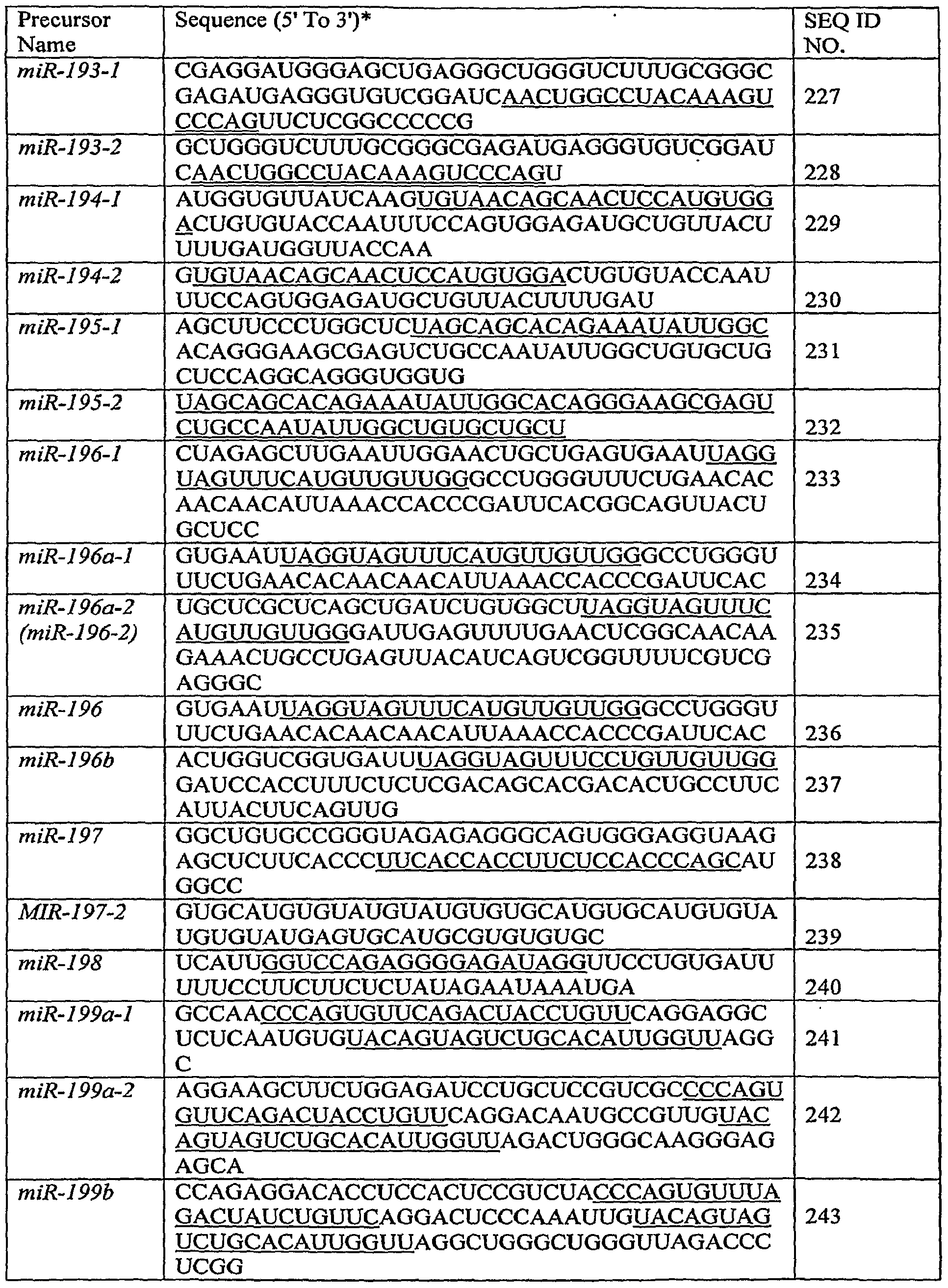


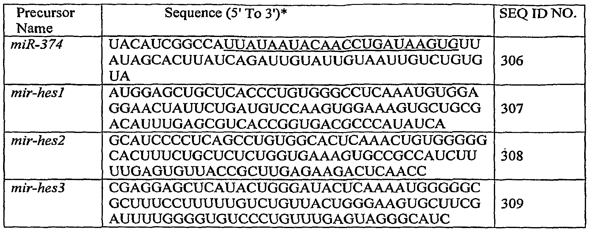


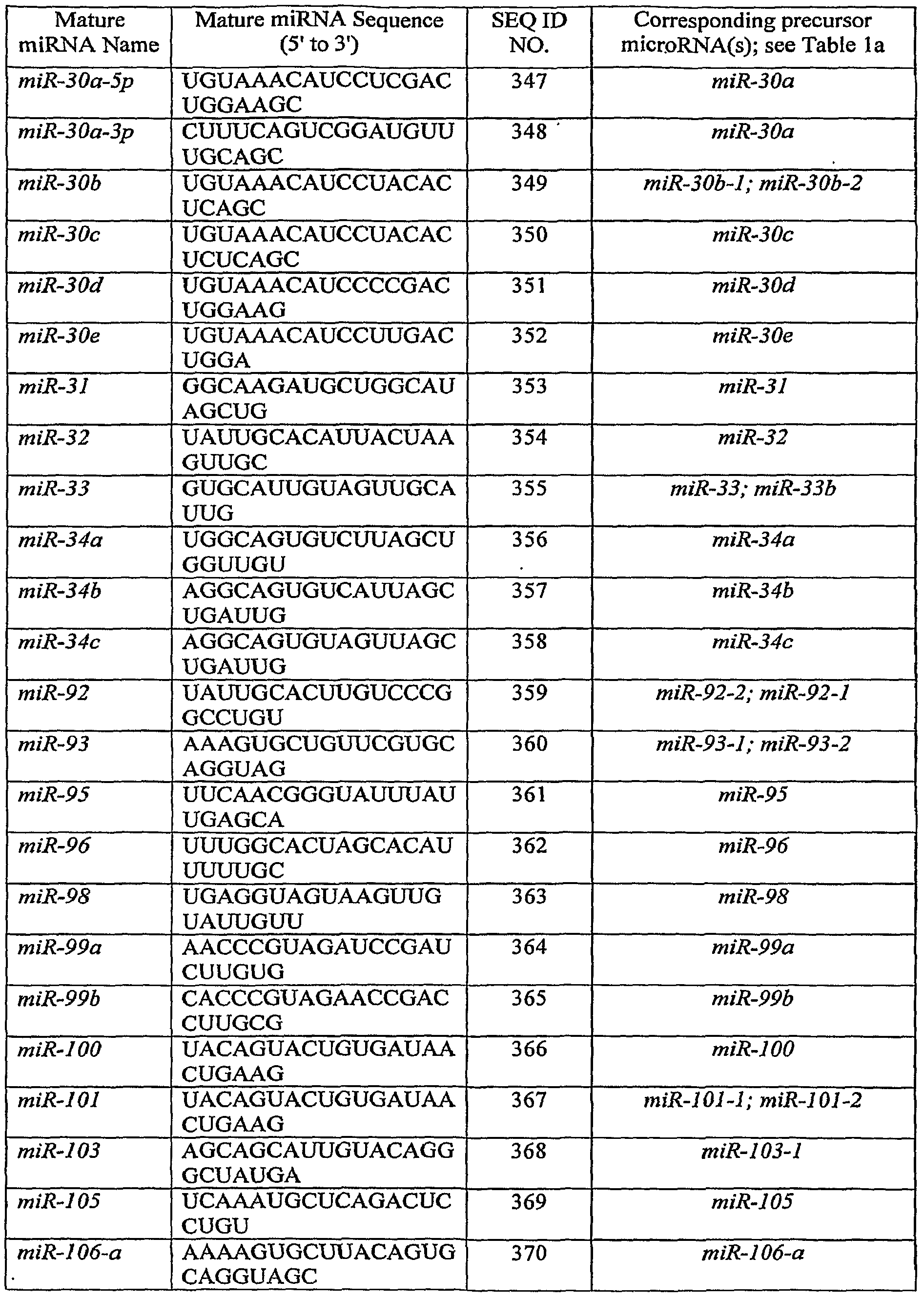


Claims
Priority Applications (12)
| Application Number | Priority Date | Filing Date | Title |
|---|---|---|---|
| JP2009501495A JP5523825B2 (en) | 2006-03-20 | 2007-03-19 | MicroRNA fingerprints during human megakaryocyte formation |
| EP20070753450 EP1996731A2 (en) | 2006-03-20 | 2007-03-19 | Microrna fingerprints during human megakaryocytopoiesis |
| CA 2646051 CA2646051A1 (en) | 2006-03-20 | 2007-03-19 | Microrna fingerprints during human megakaryocytopoiesis |
| US12/293,471 US7985584B2 (en) | 2006-03-20 | 2007-03-19 | MicroRNA fingerprints during human megakaryocytopoiesis |
| AU2007227423A AU2007227423B2 (en) | 2006-03-20 | 2007-03-19 | MicroRNA fingerprints during human megakaryocytopoiesis |
| US13/169,184 US8354224B2 (en) | 2006-03-20 | 2011-06-27 | MicroRNA fingerprints during human megakaryocytopoiesis |
| US13/595,204 US8486913B2 (en) | 2006-03-20 | 2012-08-27 | MicroRNA fingerprints during human megakaryocytopoiesis |
| US13/595,200 US8486912B2 (en) | 2006-03-20 | 2012-08-27 | MicroRNA fingerprints during human megakaryocytopoiesis |
| US13/595,207 US8492083B2 (en) | 2006-03-20 | 2012-08-27 | MicroRNA fingerprints during human megakaryocytopoiesis |
| US13/925,947 US20140161869A1 (en) | 2006-03-20 | 2013-06-25 | MicroRNA Fingerprints During Human Megakaryocytopoiesis |
| US13/925,957 US8911936B2 (en) | 2006-03-20 | 2013-06-25 | MicroRNA fingerprints during human megakaryocytopoiesis |
| US13/925,954 US8865885B2 (en) | 2006-03-20 | 2013-06-25 | MicroRNA fingerprints during human megakaryocytopoiesis |
Applications Claiming Priority (2)
| Application Number | Priority Date | Filing Date | Title |
|---|---|---|---|
| US74358506P | 2006-03-20 | 2006-03-20 | |
| US60/743,585 | 2006-03-20 |
Related Child Applications (2)
| Application Number | Title | Priority Date | Filing Date |
|---|---|---|---|
| US12/293,471 A-371-Of-International US7985584B2 (en) | 2006-03-20 | 2007-03-19 | MicroRNA fingerprints during human megakaryocytopoiesis |
| US13/169,184 Division US8354224B2 (en) | 2006-03-20 | 2011-06-27 | MicroRNA fingerprints during human megakaryocytopoiesis |
Publications (3)
| Publication Number | Publication Date |
|---|---|
| WO2007109236A2 true WO2007109236A2 (en) | 2007-09-27 |
| WO2007109236A3 WO2007109236A3 (en) | 2008-04-24 |
| WO2007109236A8 WO2007109236A8 (en) | 2008-07-03 |
Family
ID=38269114
Family Applications (1)
| Application Number | Title | Priority Date | Filing Date |
|---|---|---|---|
| PCT/US2007/006824 WO2007109236A2 (en) | 2006-03-20 | 2007-03-19 | Microrna fingerprints during human megakaryocytopoiesis |
Country Status (8)
| Country | Link |
|---|---|
| US (8) | US7985584B2 (en) |
| EP (5) | EP2369011A1 (en) |
| JP (2) | JP5523825B2 (en) |
| CN (1) | CN101448958A (en) |
| AU (1) | AU2007227423B2 (en) |
| CA (1) | CA2646051A1 (en) |
| ES (1) | ES2446362T3 (en) |
| WO (1) | WO2007109236A2 (en) |
Cited By (33)
| Publication number | Priority date | Publication date | Assignee | Title |
|---|---|---|---|---|
| WO2008068046A1 (en) * | 2006-12-05 | 2008-06-12 | Istituto Superiore di Sanità | Micro rna targeting aml1 |
| WO2009091904A2 (en) * | 2008-01-15 | 2009-07-23 | The Johns Hopkins University | Microrna based methods and compositions for the treatment of cancer |
| EP2250496A2 (en) * | 2008-02-28 | 2010-11-17 | The Ohio State University Research Foundation | Microrna signatures associated with cytogenetics and prognosis in acute myeloid leukemia (aml) and uses thereof |
| US7943318B2 (en) | 2006-01-05 | 2011-05-17 | The Ohio State University Research Foundation | Microrna-based methods and compositions for the diagnosis, prognosis and treatment of lung cancer |
| EP2323677A1 (en) * | 2008-08-12 | 2011-05-25 | The Ohio State University Research Foundation | Micro-rna-based compositions and methods for the diagnosis, prognosis and treatment of multiple myeloma |
| US7985584B2 (en) | 2006-03-20 | 2011-07-26 | The Ohio State University Research Foundation | MicroRNA fingerprints during human megakaryocytopoiesis |
| US8034560B2 (en) | 2007-01-31 | 2011-10-11 | The Ohio State University Research Foundation | MicroRNA-based methods and compositions for the diagnosis, prognosis and treatment of acute myeloid leukemia (AML) |
| US8053186B2 (en) | 2007-06-15 | 2011-11-08 | The Ohio State University Research Foundation | Oncogenic ALL-1 fusion proteins for targeting Drosha-mediated microRNA processing |
| US8084199B2 (en) | 2006-07-13 | 2011-12-27 | The Ohio State University Research Foundation | Method of diagnosing poor survival prognosis colon cancer using microRNA-21 |
| US8252538B2 (en) | 2006-11-01 | 2012-08-28 | The Ohio State University | MicroRNA expression signature for predicting survival and metastases in hepatocellular carcinoma |
| US8367632B2 (en) | 2007-07-31 | 2013-02-05 | Ohio State University Research Foundation | Methods for reverting methylation by targeting methyltransferases |
| US8389210B2 (en) | 2006-01-05 | 2013-03-05 | The Ohio State University Research Foundation | MicroRNA expression abnormalities in pancreatic endocrine and acinar tumors |
| US8440636B2 (en) | 2007-07-31 | 2013-05-14 | The Board Of Regents, The University Of Texas System | Micro-RNA family that modulates fibrosis and uses thereof |
| WO2013068730A1 (en) * | 2011-11-11 | 2013-05-16 | King's College London | Micro -rnas as marker for platelet activity |
| US8466119B2 (en) | 2007-08-22 | 2013-06-18 | The Ohio State University Research Foundation | Methods and compositions for inducing deregulation of EPHA7 and ERK phosphorylation in human acute leukemias |
| US8658370B2 (en) | 2005-08-01 | 2014-02-25 | The Ohio State University Research Foundation | MicroRNA-based methods and compositions for the diagnosis, prognosis and treatment of breast cancer |
| US8664192B2 (en) | 2011-03-07 | 2014-03-04 | The Ohio State University | Mutator activity induced by microRNA-155 (miR-155) links inflammation and cancer |
| US8859202B2 (en) | 2012-01-20 | 2014-10-14 | The Ohio State University | Breast cancer biomarker signatures for invasiveness and prognosis |
| US8916533B2 (en) | 2009-11-23 | 2014-12-23 | The Ohio State University | Materials and methods useful for affecting tumor cell growth, migration and invasion |
| US8946187B2 (en) | 2010-11-12 | 2015-02-03 | The Ohio State University | Materials and methods related to microRNA-21, mismatch repair, and colorectal cancer |
| US9085804B2 (en) | 2007-08-03 | 2015-07-21 | The Ohio State University Research Foundation | Ultraconserved regions encoding ncRNAs |
| US9150858B2 (en) | 2011-08-04 | 2015-10-06 | Yeda Research And Development Co. Ltd. | Micro-RNAs and compositions comprising same for the treatment and diagnosis of serotonin-, adrenalin-, noradrenalin-, glutamate-, and corticotropin-releasing hormone- associated medical conditions |
| US9249468B2 (en) | 2011-10-14 | 2016-02-02 | The Ohio State University | Methods and materials related to ovarian cancer |
| US9334498B2 (en) | 2012-05-10 | 2016-05-10 | Uab Research Foundation | Methods and compositions for modulating MIR-204 activity |
| CN105838784A (en) * | 2016-03-23 | 2016-08-10 | 中国医学科学院血液病医院(血液学研究所) | Application of miR-99a as acute myelogenous leukemia stem cell molecular marker |
| US9441222B2 (en) | 2011-01-11 | 2016-09-13 | Interna Technologies B.V. | MiRNA for treating diseases and conditions associated with neo-angiogenesis |
| US9481885B2 (en) | 2011-12-13 | 2016-11-01 | Ohio State Innovation Foundation | Methods and compositions related to miR-21 and miR-29a, exosome inhibition, and cancer metastasis |
| US9682095B2 (en) | 2010-07-06 | 2017-06-20 | Interna Technologies B.V. | MiRNA and its diagnostic and therapeutic uses in diseases or conditions associated with melanoma, or in diseases or conditions associated with activated BRAF pathway |
| US9771585B2 (en) | 2015-06-05 | 2017-09-26 | MiRagen Therapeutics, Inc. | miR-155 inhibitors for treating cutaneous T cell lymphoma (CTCL) |
| US10125365B2 (en) | 2014-02-05 | 2018-11-13 | Yeda Research And Development Co. Ltd. | Micro-RNAs and compositions comprising same for the treatment and diagnosis of serotonin-, adrenalin-, noradrenalin-, glutamate-, and corticotropin-releasing hormone- associated medical conditions |
| CN109091492A (en) * | 2018-08-02 | 2018-12-28 | 广州安镝声生物医药科技有限公司 | The target miRNA miR-106b-5p of lncRNA H19 and its application |
| US10758619B2 (en) | 2010-11-15 | 2020-09-01 | The Ohio State University | Controlled release mucoadhesive systems |
| US11584932B2 (en) | 2016-11-01 | 2023-02-21 | The Research Foundation For The State University Of New York | 5-halouracil-modified microRNAs and their use in the treatment of cancer |
Families Citing this family (18)
| Publication number | Priority date | Publication date | Assignee | Title |
|---|---|---|---|---|
| EP1937280B1 (en) | 2005-09-12 | 2014-08-27 | The Ohio State University Research Foundation | Compositions for the therapy of bcl2-associated cancers |
| CN103642900B (en) | 2006-01-05 | 2016-04-13 | 俄亥俄州立大学研究基金会 | For the diagnosis and treatment of the method and composition based on Microrna of solid carcinoma |
| CA2663027A1 (en) | 2006-09-19 | 2008-08-14 | The Ohio State University Research Foundation | Tcl1 expression in chronic lymphocytic leukemia (cll) regulated by mir-29 and mir-181 |
| US8415096B2 (en) * | 2007-05-23 | 2013-04-09 | University Of South Florida | Micro-RNAs modulating immunity and inflammation |
| EP2559773B1 (en) | 2007-06-08 | 2015-04-22 | The Government of the United States of America as represented by the Secretary of the Department of Health and Human Services | Methods for determining a hepatocellular carcinoma subtype |
| CN102137927B (en) | 2007-10-26 | 2014-03-12 | 俄亥俄州立大学研究基金会 | Methods for identifying fragile histidine triad (Fhit) interaction and uses thereof |
| US20110160285A1 (en) * | 2008-03-13 | 2011-06-30 | The Regents Of The University Of Colorado | Identification of mirna profiles that are diagnostic of hypertrophic cardiomyopathy |
| ES2433940T3 (en) | 2008-06-11 | 2013-12-13 | The Government Of The United States Of America As Represented By The Secretary Of The Department Of Health And Human Services | Use of the miR-26 family as a predictive marker of hepatocellular carcinoma and sensitivity to therapy |
| US20100311815A1 (en) * | 2009-02-23 | 2010-12-09 | The Regents Of The University Of Michigan | Mir-101 cancer markers |
| CN102234687A (en) * | 2010-04-29 | 2011-11-09 | 中国科学院上海生命科学研究院 | Application of micro ribose nucleic acid (miRNA) or precursor thereof of inhibitor zinc finger protein Zinc finger and BTBdomain-containing protein 20 (ZBTB20) |
| WO2012019053A2 (en) * | 2010-08-04 | 2012-02-09 | The Ohio State University | Methods for impairing the p53/hdm2 auto-regulatory loop in multiple myeloma development using mir-192, mir-194 and mir-215 |
| CN102533983A (en) * | 2011-12-19 | 2012-07-04 | 苏州福英基因科技有限公司 | Horizontal in-situ hybridization detection kit and detection method as well as application for MICRORNA-330 in earlier stage of cancer pathologic evolution |
| CN102628078A (en) * | 2011-12-27 | 2012-08-08 | 芮屈生物技术(上海)有限公司 | MicroRNA-155 level in various types of cancerous lesions prophase in situ hybridization detection kit, detection method and application thereof |
| WO2015171641A1 (en) * | 2014-05-05 | 2015-11-12 | Brigham And Women's Hospital, Inc. | Coordinate control of pathogenic signaling by the mir-130/301 family in pulmonary hypertension and fibroproliferative diseases |
| US10801026B2 (en) * | 2015-10-15 | 2020-10-13 | City Of Hope | Compounds and compositions including phosphorothioated oligodeoxynucleotide, and methods of use thereof |
| CN105567805B (en) * | 2015-12-07 | 2019-06-25 | 中山大学附属第一医院 | A kind of kit of sense organ transplant patient cytomegalovirus infection |
| CN106754927B (en) * | 2016-12-26 | 2019-10-08 | 中国人民解放军军事医学科学院野战输血研究所 | Nucleic acid is improving the purposes in cell macronucleus differentiation efficiency |
| CN107828882B (en) * | 2017-11-24 | 2021-04-27 | 中国中医科学院西苑医院 | miRNA (micro ribonucleic acid) regulated platelet Gelsolin and application thereof in screening antiplatelet drugs |
Citations (2)
| Publication number | Priority date | Publication date | Assignee | Title |
|---|---|---|---|---|
| WO2005078139A2 (en) * | 2004-02-09 | 2005-08-25 | Thomas Jefferson University | DIAGNOSIS AND TREATMENT OF CANCERS WITH MicroRNA LOCATED IN OR NEAR CANCER-ASSOCIATED CHROMOSOMAL FEATURES |
| US20060019286A1 (en) * | 2004-06-30 | 2006-01-26 | Horvitz H R | High throughput methods relating to microRNA expression analysis |
Family Cites Families (211)
| Publication number | Priority date | Publication date | Assignee | Title |
|---|---|---|---|---|
| US519941A (en) | 1894-05-15 | Reversing mechanism for metal shaping and planing machines | ||
| US4196265A (en) | 1977-06-15 | 1980-04-01 | The Wistar Institute | Method of producing antibodies |
| US4235871A (en) | 1978-02-24 | 1980-11-25 | Papahadjopoulos Demetrios P | Method of encapsulating biologically active materials in lipid vesicles |
| US4172124A (en) | 1978-04-28 | 1979-10-23 | The Wistar Institute | Method of producing tumor antibodies |
| US4608337A (en) | 1980-11-07 | 1986-08-26 | The Wistar Institute | Human hybridomas and the production of human monoclonal antibodies by human hybridomas |
| US4501728A (en) | 1983-01-06 | 1985-02-26 | Technology Unlimited, Inc. | Masking of liposomes from RES recognition |
| US5019369A (en) | 1984-10-22 | 1991-05-28 | Vestar, Inc. | Method of targeting tumors in humans |
| US4701409A (en) | 1984-11-15 | 1987-10-20 | The Wistar Institute | Detection of B-cell neoplasms |
| US4693975A (en) | 1984-11-20 | 1987-09-15 | The Wistar Institute | Human hybridroma fusion partner for production of human monoclonal antibodies |
| US5139941A (en) | 1985-10-31 | 1992-08-18 | University Of Florida Research Foundation, Inc. | AAV transduction vectors |
| US5015568A (en) | 1986-07-09 | 1991-05-14 | The Wistar Institute | Diagnostic methods for detecting lymphomas in humans |
| US5202429A (en) | 1986-07-09 | 1993-04-13 | The Wistar Institute | DNA molecules having human BCL-2 gene sequences |
| US4987071A (en) | 1986-12-03 | 1991-01-22 | University Patents, Inc. | RNA ribozyme polymerases, dephosphorylases, restriction endoribonucleases and methods |
| US4837028A (en) | 1986-12-24 | 1989-06-06 | Liposome Technology, Inc. | Liposomes with enhanced circulation time |
| US4920016A (en) | 1986-12-24 | 1990-04-24 | Linear Technology, Inc. | Liposomes with enhanced circulation time |
| ATE120497T1 (en) | 1988-05-09 | 1995-04-15 | Univ Temple | METHOD FOR PREDICTING THE EFFECTIVENESS OF AN ANTINEOPLASTIC TREATMENT IN INDIVIDUAL PATIENTS. |
| US5198338A (en) | 1989-05-31 | 1993-03-30 | Temple University | Molecular probing for human t-cell leukemia and lymphoma |
| US5149628A (en) | 1989-11-15 | 1992-09-22 | Temple University | Methods for detecting bcl-3 gene in human leukemias |
| US5252479A (en) | 1991-11-08 | 1993-10-12 | Research Corporation Technologies, Inc. | Safe vector for gene therapy |
| US5633135A (en) | 1991-12-11 | 1997-05-27 | Thomas Jefferson University | Chimeric nucleic acids and proteins resulting from ALL-1 region chromosome abnormalities |
| US6040140A (en) | 1991-12-11 | 2000-03-21 | Thomas Jefferson University | Methods for screening and treating leukemias resulting from all-1 region chromosome abnormalities |
| WO1993012136A1 (en) | 1991-12-11 | 1993-06-24 | Thomas Jefferson University | Detection and treatment of acute leukemias resulting from chromosome abnormalities in the all-1 region |
| US5587308A (en) | 1992-06-02 | 1996-12-24 | The United States Of America As Represented By The Department Of Health & Human Services | Modified adeno-associated virus vector capable of expression from a novel promoter |
| US5674682A (en) | 1992-10-29 | 1997-10-07 | Thomas Jefferson University | Nucleic acid primers for detecting micrometastasis of prostate cancer |
| JPH08502889A (en) | 1992-10-29 | 1996-04-02 | トーマス・ジェファーソン・ユニバーシティ | Method of detecting micrometastases of prostate cancer |
| US5478745A (en) | 1992-12-04 | 1995-12-26 | University Of Pittsburgh | Recombinant viral vector system |
| US7175995B1 (en) | 1994-10-27 | 2007-02-13 | Thomas Jefferson University | TCL-1 protein and related methods |
| US5985598A (en) | 1994-10-27 | 1999-11-16 | Thomas Jefferson University | TCL-1 gene and protein and related methods and compositions |
| US5695944A (en) | 1995-05-05 | 1997-12-09 | Thomas Jefferson University | Modulation of bcl-2 phosphorylation |
| US5567586A (en) | 1995-05-18 | 1996-10-22 | Thomas Jefferson University | Methods of indentifying solid tumors with chromosome abnormalities in the ALL-1 region |
| US5928884A (en) | 1996-02-09 | 1999-07-27 | Croce; Carlo M. | FHIT proteins and nucleic acids and methods based thereon |
| US6242212B1 (en) | 1996-02-09 | 2001-06-05 | Thomas Jefferson University | Fragile histidine triad (FHIT) nucleic acids and methods of producing FHIT proteins |
| US5849902A (en) | 1996-09-26 | 1998-12-15 | Oligos Etc. Inc. | Three component chimeric antisense oligonucleotides |
| WO1998035707A1 (en) | 1997-02-18 | 1998-08-20 | Thomas Jefferson University | Compositions that bind to pancreatic cancer cells and methods of using the same |
| AU6951098A (en) | 1997-04-04 | 1998-10-30 | The Texas A & M University System | Noninvasive detection of colonic biomarkers using fecal messenger rna |
| US6303323B1 (en) | 1997-10-21 | 2001-10-16 | Cancer Research Campaign Technology Limited | Detection of dysplastic or neoplastic cells using anti-MCM5 antibodies |
| PE20000121A1 (en) | 1997-10-21 | 2000-02-17 | Cancer Res Campaign Tech | DETERMINATION OF ABNORMALITY IN CELLULAR GROWTH |
| WO2000003685A2 (en) | 1998-07-20 | 2000-01-27 | Thomas Jefferson University | Nitrilase homologs |
| AU5128999A (en) | 1998-07-24 | 2000-02-14 | Yeda Research And Development Co. Ltd. | Prevention of metastasis with 5-aza-2'-deoxycytidine |
| US7141417B1 (en) | 1999-02-25 | 2006-11-28 | Thomas Jefferson University | Compositions, kits, and methods relating to the human FEZ1 gene, a novel tumor suppressor gene |
| CA2367906A1 (en) | 1999-03-15 | 2000-09-21 | Thomas Jefferson University | Tcl-1b gene and protein and related methods and compositions |
| US7163801B2 (en) | 1999-09-01 | 2007-01-16 | The Burnham Institute | Methods for determining the prognosis for cancer patients using tucan |
| US6891031B2 (en) | 2000-02-18 | 2005-05-10 | The Regents Of The University Of California | Coordinate cytokine regulatory sequences |
| US20010026796A1 (en) | 2000-03-14 | 2001-10-04 | Croce Carlo M. | TCL1 enhances Akt kinase activity and mediates its nuclear translocation |
| ATE450621T2 (en) | 2000-03-30 | 2009-12-15 | Whitehead Biomedical Inst | MEDIATORS OF RNA INTERFERENCE THAT ARE RNA SEQUENCE SPECIFIC |
| EP1276879A4 (en) | 2000-04-11 | 2004-12-22 | Univ Jefferson | MUIR-TORRE-LIKE SYNDROME IN Fhit DEFICIENT MICE |
| US20020086331A1 (en) | 2000-05-16 | 2002-07-04 | Carlo Croce | Crystal structure of worm NitFhit reveals that a Nit tetramer binds two Fhit dimers |
| US7060811B2 (en) | 2000-10-13 | 2006-06-13 | Board Of Regents, The University Of Texas System | WWOX: a tumor suppressor gene mutated in multiple cancers |
| US20020173478A1 (en) | 2000-11-14 | 2002-11-21 | The Trustees Of The University Of Pennsylvania | Post-transcriptional gene silencing by RNAi in mammalian cells |
| US20040033502A1 (en) | 2001-03-28 | 2004-02-19 | Amanda Williams | Gene expression profiles in esophageal tissue |
| US20050176025A1 (en) | 2001-05-18 | 2005-08-11 | Sirna Therapeutics, Inc. | RNA interference mediated inhibition of B-cell CLL/Lymphoma-2 (BCL-2) gene expression using short interfering nucleic acid (siNA) |
| EP2385122B1 (en) | 2001-09-28 | 2018-04-25 | Max-Planck-Gesellschaft zur Förderung der Wissenschaften e.V. | MicroRNA molecules |
| US7371736B2 (en) | 2001-11-07 | 2008-05-13 | The Board Of Trustees Of The University Of Arkansas | Gene expression profiling based identification of DKK1 as a potential therapeutic targets for controlling bone loss |
| US7455995B2 (en) | 2001-11-09 | 2008-11-25 | The Ohio State University Research Foundation | BAALC expression as a diagnostic marker for acute leukemia |
| GB0128898D0 (en) | 2001-12-03 | 2002-01-23 | Biotech Res Ventures Pte Ltd | Materials and methods relating to the stabilization and activation of a tumour suppressor protein |
| WO2003069853A1 (en) | 2002-02-12 | 2003-08-21 | Nokia Corporation | Method for controlling data transmission, and data transmission system |
| EP1442143A4 (en) | 2002-02-20 | 2005-02-16 | Sirna Therapeutics Inc | RNA INTERFERENCE MEDIATED INHIBITION OF BCL2 GENE EXPRESSION USING SHORT INTERFERING NUCLEIC ACID (siNA) |
| NZ535101A (en) | 2002-03-08 | 2007-07-27 | Eisai Co Ltd | Macrocyclic compounds useful as pharmaceuticals |
| US20060084059A1 (en) | 2002-04-08 | 2006-04-20 | Tai-Tung Yip | Serum biomarkers in hepatocellular carcinoma |
| JP2005523712A (en) | 2002-04-29 | 2005-08-11 | トマス ジェファソン ユニバーシティ | Human chronic lymphocytic leukemia modeled in mice with targeted TCL1 expression |
| US7217568B2 (en) | 2002-05-31 | 2007-05-15 | The Board Of Trustees Of The Leland Stanford Junior University | Methods of identifying and isolating stem cells and cancer stem cells |
| JP2005536195A (en) | 2002-05-31 | 2005-12-02 | ザ リージェント オブ ザ ユニバーシティ オブ カリフォルニア | Method for efficient RNA interference in mammalian cells |
| US7148342B2 (en) | 2002-07-24 | 2006-12-12 | The Trustees Of The University Of Pennyslvania | Compositions and methods for sirna inhibition of angiogenesis |
| US20050260639A1 (en) | 2002-09-30 | 2005-11-24 | Oncotherapy Science, Inc. | Method for diagnosing pancreatic cancer |
| WO2004033659A2 (en) | 2002-10-11 | 2004-04-22 | Thomas Jefferson University | Novel tumor suppressor gene and compositions and methods for making and using the same |
| US20050266443A1 (en) | 2002-10-11 | 2005-12-01 | Thomas Jefferson University | Novel tumor suppressor gene and compositions and methods for making and using the same |
| JP4939055B2 (en) | 2002-11-13 | 2012-05-23 | トマス ジェファソン ユニバーシティ | Compositions and methods for diagnosis and treatment of cancer |
| US7250496B2 (en) | 2002-11-14 | 2007-07-31 | Rosetta Genomics Ltd. | Bioinformatically detectable group of novel regulatory genes and uses thereof |
| WO2004071464A2 (en) | 2003-02-12 | 2004-08-26 | Johns Hopkins University School Of Medicine | Diagnostic application of differentially-expressed genes in lympho-hematopoietic stem cells |
| WO2004079013A1 (en) | 2003-03-03 | 2004-09-16 | Arizona Board Of Regents On Behalf Of The University Of Arizona | Ecto-5’-nucleotidase (cd73) used in the diagnosis and the treatment of pancreatic cancer |
| US7183384B2 (en) | 2003-03-06 | 2007-02-27 | A & G Pharmaceutical, Inc. | Monoclonal antibody 7H11 reactive with human cancer |
| WO2004098377A2 (en) | 2003-05-02 | 2004-11-18 | Thomas Jefferson University | Methods and compositions for diagnosis and therapy of parkin-associated disorders |
| WO2006031210A1 (en) | 2003-05-29 | 2006-03-23 | Board Of Regents, The University Of Texas Systems | Jabi as a prognostic marker and a therapeutic target for human cancer |
| AU2004289953B2 (en) | 2003-06-18 | 2008-09-25 | Genelux Corporation | Modified recombinant vaccina viruses and other microorganisms, uses thereof |
| CA2533701A1 (en) | 2003-07-31 | 2005-02-17 | Isis Pharmaceuticals, Inc. | Oligomeric compounds and compositions for use in modulation of small non-coding rnas |
| US8106180B2 (en) | 2003-08-07 | 2012-01-31 | Whitehead Institute For Biomedical Research | Methods and products for expression of micro RNAs |
| US20050037362A1 (en) | 2003-08-11 | 2005-02-17 | Eppendorf Array Technologies, S.A. | Detection and quantification of siRNA on microarrays |
| US8412541B2 (en) | 2003-08-14 | 2013-04-02 | Edda Technology, Inc. | Method and system for intelligent qualitative and quantitative analysis for medical diagnosis |
| US20050084883A1 (en) | 2003-08-25 | 2005-04-21 | The Johns Hopkins University School Of Medicine | Method of diagnosis and treatment of pancreatic endocrine neoplasms based on differential gene expression analysis |
| JPWO2005028648A1 (en) | 2003-09-22 | 2007-11-15 | 愛知県 | Lymphoma type and prognosis |
| AU2004276823A1 (en) | 2003-09-22 | 2005-04-07 | Merck And Co., Inc | Synthetic lethal screen using RNA interference |
| CN1890382A (en) | 2003-09-24 | 2007-01-03 | 肿瘤疗法科学股份有限公司 | Method for diagnosing hepatocellular carcinomas |
| WO2005047477A2 (en) | 2003-11-07 | 2005-05-26 | University Of Massachusetts | Interspersed repetitive element rnas as substrates, inhibitors and delivery vehicles for rnai |
| US20050164252A1 (en) | 2003-12-04 | 2005-07-28 | Yeung Wah Hin A. | Methods using non-genic sequences for the detection, modification and treatment of any disease or improvement of functions of a cell |
| CA2550447A1 (en) | 2003-12-19 | 2005-07-07 | The Regents Of The University Of California | Methods and materials for assessing prostate cancer therapies |
| JP4120002B2 (en) | 2004-01-07 | 2008-07-16 | 愛知県 | Cancer prognosis determination method using miRNA, cancer gene therapy vector, and cancer therapeutic pharmaceutical composition |
| US20050256072A1 (en) | 2004-02-09 | 2005-11-17 | University Of Massachusetts | Dual functional oligonucleotides for use in repressing mutant gene expression |
| WO2005079397A2 (en) | 2004-02-13 | 2005-09-01 | Rockefeller University | Anti-microrna oligonucleotide molecules |
| CN101088089A (en) | 2004-02-23 | 2007-12-12 | 鹿特丹伊拉斯姆斯大学医疗中心 | Classification, diagnosis and prognosis of acute myeloid leukemia by gene expression profiling |
| WO2005094263A2 (en) | 2004-03-23 | 2005-10-13 | Pintex Pharmaceuticals, Inc. | Methods of determining the prognosis and treatment of subjects with colon cancer |
| US20060134639A1 (en) | 2004-04-06 | 2006-06-22 | Huffel Christophe V | Method for the determination of cellular transcriptional regulation |
| US7365058B2 (en) | 2004-04-13 | 2008-04-29 | The Rockefeller University | MicroRNA and methods for inhibiting same |
| WO2005103298A2 (en) | 2004-04-20 | 2005-11-03 | Genaco Biomedical Products, Inc. | Method for detecting ncrna |
| CA2566519C (en) | 2004-05-14 | 2020-04-21 | Rosetta Genomics Ltd. | Micrornas and uses thereof |
| EP1745125A1 (en) * | 2004-05-14 | 2007-01-24 | Becton, Dickinson and Company | Cell culture environments for the serum-free expansion of mesenchymal stem cells |
| EP2471921A1 (en) | 2004-05-28 | 2012-07-04 | Asuragen, Inc. | Methods and compositions involving microRNA |
| US20060116321A1 (en) | 2004-07-01 | 2006-06-01 | Robbins Paul D | Immunosuppressive exosomes |
| US20060037088A1 (en) | 2004-08-13 | 2006-02-16 | Shulin Li | Gene expression levels as predictors of chemoradiation response of cancer |
| US7893034B2 (en) | 2004-09-02 | 2011-02-22 | Yale University | Regulation of oncogenes by microRNAs |
| US7642348B2 (en) | 2004-10-04 | 2010-01-05 | Rosetta Genomics Ltd | Prostate cancer-related nucleic acids |
| US7592441B2 (en) | 2004-10-04 | 2009-09-22 | Rosetta Genomics Ltd | Liver cancer-related nucleic acids |
| FR2877350B1 (en) | 2004-11-03 | 2010-08-27 | Centre Nat Rech Scient | IDENTIFICATION AND USE OF miRNAs INVOLVED IN THE DIFFERENTIATION OF CELLS FROM MYELOID LEUKEMIA |
| EP2302054B1 (en) * | 2004-11-12 | 2014-07-16 | Asuragen, Inc. | Methods and compositions involving miRNA and miRNA inhibitor molecules |
| EP1662259A1 (en) | 2004-11-25 | 2006-05-31 | Cellzome Ag | Use of Eph receptor inhibitors for the treatment of neurodegenerative diseases |
| US7361752B2 (en) | 2004-12-14 | 2008-04-22 | Alnylam Pharmaceuticals, Inc. | RNAi modulation of MLL-AF4 and uses thereof |
| US20060185027A1 (en) | 2004-12-23 | 2006-08-17 | David Bartel | Systems and methods for identifying miRNA targets and for altering miRNA and target expression |
| WO2006069584A2 (en) | 2004-12-29 | 2006-07-06 | Exiqon A/S | NOVEL OLIGONUCLEOTIDE COMPOSITIONS AND PROBE SEQUENCES USEFUL FOR DETECTION AND ANALYSIS OF microRNAs AND THEIR TARGET mRNAs |
| US8071306B2 (en) | 2005-01-25 | 2011-12-06 | Merck Sharp & Dohme Corp. | Methods for quantitating small RNA molecules |
| JP2008528003A (en) | 2005-01-25 | 2008-07-31 | ロゼッタ インファーマティックス エルエルシー | Method for quantifying small RNA molecules |
| WO2006086737A2 (en) | 2005-02-11 | 2006-08-17 | Wisconsin Alumni Reasearch Foundation | Mir-155 assay |
| US20070065840A1 (en) | 2005-03-23 | 2007-03-22 | Irena Naguibneva | Novel oligonucleotide compositions and probe sequences useful for detection and analysis of microRNAS and their target mRNAS |
| GB2425311A (en) | 2005-04-15 | 2006-10-25 | Ist Superiore Sanita | Micro RNA against kit protein |
| EP2631293A3 (en) | 2005-04-29 | 2013-11-20 | The Rockefeller University | Human microRNAs and methods for inhibiting same |
| US20090209621A1 (en) | 2005-06-03 | 2009-08-20 | The Johns Hopkins University | Compositions and methods for decreasing microrna expression for the treatment of neoplasia |
| US20070065844A1 (en) | 2005-06-08 | 2007-03-22 | Massachusetts Institute Of Technology | Solution-based methods for RNA expression profiling |
| US20060292616A1 (en) | 2005-06-23 | 2006-12-28 | U.S. Genomics, Inc. | Single molecule miRNA-based disease diagnostic methods |
| CN102533966B (en) | 2005-08-01 | 2014-03-12 | 俄亥俄州立大学研究基金会 | Micro-RNA-based methods and compositions for diagnosis, prognosis and treatment of breast cancer |
| US20070213292A1 (en) | 2005-08-10 | 2007-09-13 | The Rockefeller University | Chemically modified oligonucleotides for use in modulating micro RNA and uses thereof |
| AU2006279906B2 (en) | 2005-08-10 | 2012-05-10 | Alnylam Pharmaceuticals, Inc. | Chemically modified oligonucleotides for use in modulating micro RNA and uses thereof |
| EP1937280B1 (en) | 2005-09-12 | 2014-08-27 | The Ohio State University Research Foundation | Compositions for the therapy of bcl2-associated cancers |
| JP2009511482A (en) | 2005-10-05 | 2009-03-19 | ジ・オハイオ・ステイト・ユニバーシティ・リサーチ・ファウンデイション | WWOX gene, vectors containing the same, and use in the treatment of cancer |
| US20070092882A1 (en) | 2005-10-21 | 2007-04-26 | Hui Wang | Analysis of microRNA |
| US8445198B2 (en) | 2005-12-01 | 2013-05-21 | Medical Prognosis Institute | Methods, kits and devices for identifying biomarkers of treatment response and use thereof to predict treatment efficacy |
| US7390792B2 (en) | 2005-12-15 | 2008-06-24 | Board Of Regents, The University Of Texas System | MicroRNA1 therapies |
| AU2007205234B2 (en) | 2006-01-05 | 2012-07-12 | The Ohio State University Research Foundation | MicroRNA-based methods and compositions for the diagnosis, prognosis and treatment of lung cancer |
| CN103642900B (en) | 2006-01-05 | 2016-04-13 | 俄亥俄州立大学研究基金会 | For the diagnosis and treatment of the method and composition based on Microrna of solid carcinoma |
| AU2007205257B2 (en) | 2006-01-05 | 2013-07-25 | The Ohio State University Research Foundation | MicroRNA expression abnormalities in pancreatic endocrine and acinar tumors |
| WO2007084486A2 (en) | 2006-01-13 | 2007-07-26 | Battelle Memorial Institute | Animal model for assessing copd-related diseases |
| JP5523825B2 (en) | 2006-03-20 | 2014-06-18 | ジ・オハイオ・ステイト・ユニバーシティ・リサーチ・ファウンデイション | MicroRNA fingerprints during human megakaryocyte formation |
| WO2007112097A2 (en) | 2006-03-24 | 2007-10-04 | Children's Medical Center Corporation | Novel signature self renewal gene expression programs |
| EP2194129A3 (en) | 2006-04-03 | 2012-12-26 | Santaris Pharma A/S | Pharmaceutical composition comprising anti-miRNA antisense oligonucleotides |
| US7667090B2 (en) | 2006-04-24 | 2010-02-23 | The Ohio State University Research Foundation | Transgenic mouse model of B cell malignancy |
| JP2010500867A (en) | 2006-06-30 | 2010-01-14 | ロゼッタ ジノミクス リミテッド | Nucleic acid detection method |
| EP2369017B8 (en) | 2006-07-13 | 2014-03-12 | The Ohio State University Research Foundation | Micro-RNA-based methods and compositions for the diagnosis and treatment of colon related diseases |
| EP2063887B1 (en) | 2006-08-30 | 2013-05-29 | The Regents of the University of Michigan | New small molecule inhibitors of mdm2 and the uses thereof |
| US20080193943A1 (en) | 2006-09-05 | 2008-08-14 | Abbott Laboratories | Companion diagnostic assays for cancer therapy |
| CA2663962A1 (en) | 2006-09-19 | 2008-03-27 | Asuragen, Inc. | Mir-15, mir-26, mir-31,mir-145, mir-147, mir-188, mir-215, mir-216, mir-331, mmu-mir-292-3p regulated genes and pathways as targets for therapeutic intervention |
| AU2007299804A1 (en) | 2006-09-19 | 2008-03-27 | Asuragen, Inc. | MiR-200 regulated genes and pathways as targets for therapeutic intervention |
| CA2663027A1 (en) | 2006-09-19 | 2008-08-14 | The Ohio State University Research Foundation | Tcl1 expression in chronic lymphocytic leukemia (cll) regulated by mir-29 and mir-181 |
| WO2008036765A2 (en) | 2006-09-19 | 2008-03-27 | Asuragen, Inc. | Micrornas differentially expressed in pancreatic diseases and uses thereof |
| JP2008086201A (en) | 2006-09-29 | 2008-04-17 | Gifu Prefecture Kenkyu Kaihatsu Zaidan | Method for detecting production of micro-rna and diagnosis/treatment of cancer, and micro-rna production adjusting agent |
| ES2425416T3 (en) | 2006-11-01 | 2013-10-15 | The Ohio State University Research Foundation | Signature of microRNA expression to predict survival and metastasis in hepatocellular carcinoma |
| JP2010510769A (en) | 2006-11-28 | 2010-04-08 | キャピタルバイオ コーポレーション | Methods and compositions for diagnosis of esophageal cancer and prognosis and improvement of patient survival |
| US8293684B2 (en) | 2006-11-29 | 2012-10-23 | Exiqon | Locked nucleic acid reagents for labelling nucleic acids |
| WO2008070082A2 (en) | 2006-12-04 | 2008-06-12 | The Johns Hopkins University | Stem-progenitor cell specific micro-ribonucleic acids and uses thereof |
| CN101622350A (en) | 2006-12-08 | 2010-01-06 | 奥斯瑞根公司 | miR-126 regulated genes and pathways as targets for therapeutic intervention |
| EP2104735A2 (en) | 2006-12-08 | 2009-09-30 | Asuragen, INC. | Mir-21 regulated genes and pathways as targets for therapeutic intervention |
| WO2008073919A2 (en) | 2006-12-08 | 2008-06-19 | Asuragen, Inc. | Mir-20 regulated genes and pathways as targets for therapeutic intervention |
| EP2104737B1 (en) | 2006-12-08 | 2013-04-10 | Asuragen, INC. | Functions and targets of let-7 micro rnas |
| CA2671270A1 (en) | 2006-12-29 | 2008-07-17 | Asuragen, Inc. | Mir-16 regulated genes and pathways as targets for therapeutic intervention |
| CN103555825B (en) | 2007-01-31 | 2015-09-30 | 俄亥俄州立大学研究基金会 | For the method and composition based on microRNA of the diagnosis of acute myelocytic leukemia (AML), prognosis and treatment |
| CA2699418A1 (en) | 2007-02-27 | 2008-09-04 | Moshe Oren | Composition and methods for modulating cell proliferation and cell death |
| EP2126584B1 (en) | 2007-03-16 | 2012-12-19 | CovalX AG | Direct mass spectrometric analysis of drug candidates targeting protein complexes |
| US7745134B2 (en) | 2007-04-10 | 2010-06-29 | National Taiwan University | Predicting post-treatment survival in cancer patients with microRNAs |
| JP5592251B2 (en) | 2007-04-30 | 2014-09-17 | ジ・オハイオ・ステイト・ユニバーシティ・リサーチ・ファウンデイション | Method for distinguishing pancreatic cancer from normal pancreatic function and / or chronic pancreatitis |
| US20090005336A1 (en) | 2007-05-08 | 2009-01-01 | Zhiguo Wang | Use of the microRNA miR-1 for the treatment, prevention, and diagnosis of cardiac conditions |
| US20090232893A1 (en) | 2007-05-22 | 2009-09-17 | Bader Andreas G | miR-143 REGULATED GENES AND PATHWAYS AS TARGETS FOR THERAPEUTIC INTERVENTION |
| US20090131354A1 (en) | 2007-05-22 | 2009-05-21 | Bader Andreas G | miR-126 REGULATED GENES AND PATHWAYS AS TARGETS FOR THERAPEUTIC INTERVENTION |
| US20090099034A1 (en) | 2007-06-07 | 2009-04-16 | Wisconsin Alumni Research Foundation | Reagents and Methods for miRNA Expression Analysis and Identification of Cancer Biomarkers |
| EP2559773B1 (en) | 2007-06-08 | 2015-04-22 | The Government of the United States of America as represented by the Secretary of the Department of Health and Human Services | Methods for determining a hepatocellular carcinoma subtype |
| EP2167138A2 (en) | 2007-06-08 | 2010-03-31 | Asuragen, INC. | Mir-34 regulated genes and pathways as targets for therapeutic intervention |
| CA2690749A1 (en) | 2007-06-15 | 2008-12-24 | The Ohio State University Research Foundation | Oncogenic all-1 fusion proteins for targeting drosha-mediated microrna processing |
| ES2420973T3 (en) | 2007-07-25 | 2013-08-28 | University Of Louisville Research Foundation, Inc. | Micro-RNA associated with exosome as a diagnostic marker |
| WO2009018303A2 (en) | 2007-07-31 | 2009-02-05 | The Ohio State University Research Foundation | Methods for reverting methylation by targeting dnmt3a and dnmt3b |
| EP2657353B1 (en) | 2007-08-03 | 2017-04-12 | The Ohio State University Research Foundation | Ultraconserved regions encoding ncRNAs |
| CA2926831A1 (en) | 2007-08-22 | 2009-02-26 | The Ohio State University Research Foundation | Methods and compositions for inducing deregulation of epha7 and erk phosphorylation in human acute leukemias |
| US20090061424A1 (en) | 2007-08-30 | 2009-03-05 | Sigma-Aldrich Company | Universal ligation array for analyzing gene expression or genomic variations |
| JP5401460B2 (en) | 2007-09-06 | 2014-01-29 | ジ・オハイオ・ステイト・ユニバーシティ・リサーチ・ファウンデイション | MicroRNA signature in human ovarian cancer |
| US20090239818A1 (en) | 2007-09-07 | 2009-09-24 | Cheng Jin Q | Effective treatment of ovarian cancer using triciribine and related compounds |
| WO2009036236A1 (en) | 2007-09-14 | 2009-03-19 | The Ohio State University Research Foundation | Mirna expression in human peripheral blood microvesicles and uses thereof |
| EP2208499A4 (en) | 2007-10-03 | 2011-12-28 | Kyowa Hakko Kirin Co Ltd | Nucleic acid capable of regulating the proliferation of cell |
| DK2205737T3 (en) | 2007-10-04 | 2013-05-21 | Santaris Pharma As | Micromirers |
| CN103937876B (en) | 2007-10-11 | 2016-08-17 | 俄亥俄州立大学研究基金会 | For diagnosing and treat the method and composition of adenocarcinoma of esophagus |
| CN102137927B (en) | 2007-10-26 | 2014-03-12 | 俄亥俄州立大学研究基金会 | Methods for identifying fragile histidine triad (Fhit) interaction and uses thereof |
| JP5535076B2 (en) | 2007-10-29 | 2014-07-02 | レグルス・セラピューティクス・インコーポレイテッド | Targeted microRNA for treating liver cancer |
| AU2008321253B2 (en) | 2007-11-12 | 2014-01-16 | Masaryk Memorial Cancer Institute | Therapeutic applications of p53 isoforms in regenerative medicine, aging and cancer |
| US20090123933A1 (en) | 2007-11-12 | 2009-05-14 | Wake Forest University Health Sciences | Microrna biomarkers in lupus |
| EP2225396A4 (en) | 2007-11-30 | 2011-03-02 | Univ Ohio State Res Found | Microrna expression profiling and targeting in peripheral blood in lung cancer |
| WO2009070805A2 (en) | 2007-12-01 | 2009-06-04 | Asuragen, Inc. | Mir-124 regulated genes and pathways as targets for therapeutic intervention |
| WO2009086156A2 (en) | 2007-12-21 | 2009-07-09 | Asuragen, Inc. | Mir-10 regulated genes and pathways as targets for therapeutic intervention |
| DK2245199T3 (en) | 2008-02-01 | 2014-02-03 | Gen Hospital Corp | APPLICATION OF MICROVESCIES IN DIAGNOSTICATION, PROGRAMMING AND TREATMENT OF MEDICAL DISEASES AND CONDITIONS |
| WO2009100430A2 (en) | 2008-02-08 | 2009-08-13 | Asuragen, Inc | miRNAs DIFFERENTIALLY EXPRESSED IN LYMPH NODES FROM CANCER PATIENTS |
| CA2717026A1 (en) | 2008-02-28 | 2009-09-03 | The Ohio State University Research Foundation | Microrna signatures associated with human chronic lymphocytic leukemia (ccl) and uses thereof |
| US20110034538A1 (en) | 2008-02-28 | 2011-02-10 | The Ohio State University Research Foundation | MicroRNA-Based Methods and Compositions for the Diagnosis, Prognosis and Treatment of Gastric Cancer |
| CA2717030A1 (en) | 2008-02-28 | 2009-09-03 | The Ohio State University Research Foundation | Microrna signatures associated with cytogenetics and prognosis in acute myeloid leukemia (aml) and uses thereof |
| CN104031984A (en) | 2008-02-28 | 2014-09-10 | 俄亥俄州立大学研究基金会 | Microrna-based methods and compositions for the diagnosis, prognosis and treatment of prostate related disorders |
| WO2009111375A2 (en) | 2008-03-01 | 2009-09-11 | Abraxis Bioscience, Llc | Treatment, diagnostic, and method for discovering antagonist using sparc specific mirnas |
| WO2009111643A2 (en) | 2008-03-06 | 2009-09-11 | Asuragen, Inc. | Microrna markers for recurrence of colorectal cancer |
| US20090253780A1 (en) | 2008-03-26 | 2009-10-08 | Fumitaka Takeshita | COMPOSITIONS AND METHODS RELATED TO miR-16 AND THERAPY OF PROSTATE CANCER |
| WO2009131887A2 (en) | 2008-04-24 | 2009-10-29 | Merck & Co., Inc. | Methods of using mir210 as a biomarker for hypoxia and as a therapeutic agent for treating cancer |
| WO2009137807A2 (en) | 2008-05-08 | 2009-11-12 | Asuragen, Inc. | Compositions and methods related to mirna modulation of neovascularization or angiogenesis |
| US20110107440A1 (en) | 2008-06-04 | 2011-05-05 | Andor Pivarcsi | Skin cancer associated micrornas |
| WO2009149418A2 (en) | 2008-06-06 | 2009-12-10 | Asuragen, Inc. | Novel compositions for the in vivo delivery of rnai agents |
| ES2433940T3 (en) | 2008-06-11 | 2013-12-13 | The Government Of The United States Of America As Represented By The Secretary Of The Department Of Health And Human Services | Use of the miR-26 family as a predictive marker of hepatocellular carcinoma and sensitivity to therapy |
| US20100021734A1 (en) | 2008-07-22 | 2010-01-28 | Covalent Materials Corporation | Ceramic particles and producing method thereof |
| KR101031305B1 (en) | 2008-07-23 | 2011-04-29 | 국립암센터 | Composition containing microRNA-21 inhibitor for enhancing radiation sensitivity |
| US20110152357A1 (en) | 2008-08-12 | 2011-06-23 | The Ohio State University | Micro-RNA-Based Compositions and Methods for the Diagnosis, Prognosis and Treatment of Multiple Myeloma |
| US20100179213A1 (en) | 2008-11-11 | 2010-07-15 | Mirna Therapeutics, Inc. | Methods and Compositions Involving miRNAs In Cancer Stem Cells |
| EP2352524A1 (en) | 2008-11-21 | 2011-08-10 | The Ohio State University Research Foundation | Tcl1 AS A TRANSCRIPTIONAL REGULATOR |
| CA2745746A1 (en) | 2008-12-05 | 2010-06-10 | The Ohio State University Research Foundation | Microrna-based methods and compositions for the diagnosis and treatment of ovarian cancer |
| CA2753562A1 (en) | 2009-02-26 | 2010-09-02 | The Ohio State University Research Foundation | Micrornas in never-smokers and related materials and methods |
| CA2715518A1 (en) | 2009-09-23 | 2011-03-23 | Wilson Roa | Microrna expression profiles associated with lung cancer |
| US20120219958A1 (en) | 2009-11-09 | 2012-08-30 | Yale University | MicroRNA Signatures Differentiating Uterine and Ovarian Papillary Serous Tumors |
| CA2781547A1 (en) | 2009-11-23 | 2011-05-26 | The Ohio State University | Materials and methods useful for affecting tumor cell growth, migration and invasion |
| EP2354246A1 (en) | 2010-02-05 | 2011-08-10 | febit holding GmbH | miRNA in the diagnosis of ovarian cancer |
| AU2011232669B2 (en) | 2010-03-26 | 2016-08-25 | The Ohio State University | Materials and methods related to modulation of mismatch repair and genomic stability by miR-155 |
| CN103109183A (en) | 2010-06-24 | 2013-05-15 | 俄亥俄州立大学 | Chronic lymphocytic leukemia modeled in mouse by targeted mir-29 expression |
| WO2012019053A2 (en) | 2010-08-04 | 2012-02-09 | The Ohio State University | Methods for impairing the p53/hdm2 auto-regulatory loop in multiple myeloma development using mir-192, mir-194 and mir-215 |
| WO2012065049A1 (en) | 2010-11-12 | 2012-05-18 | The Ohio State University Research Foundation | Materials and methods related to microrna-21, mismatch repair, and colorectal cancer |
| WO2012097047A1 (en) | 2011-01-11 | 2012-07-19 | The Ohio State University Research Foundation | Methods to identify chronic lymphocytic leukemia disease progression |
| AU2012225506B2 (en) | 2011-03-07 | 2016-11-17 | The Ohio State University | Mutator activity induced by microRNA-155 (miR-155) links inflammation and cancer |
-
2007
- 2007-03-19 JP JP2009501495A patent/JP5523825B2/en not_active Expired - Fee Related
- 2007-03-19 EP EP20110151749 patent/EP2369011A1/en not_active Withdrawn
- 2007-03-19 AU AU2007227423A patent/AU2007227423B2/en not_active Ceased
- 2007-03-19 CN CNA2007800184963A patent/CN101448958A/en active Pending
- 2007-03-19 US US12/293,471 patent/US7985584B2/en not_active Expired - Fee Related
- 2007-03-19 ES ES11151772T patent/ES2446362T3/en active Active
- 2007-03-19 EP EP20110151772 patent/EP2371971B1/en not_active Not-in-force
- 2007-03-19 EP EP20070753450 patent/EP1996731A2/en not_active Ceased
- 2007-03-19 EP EP20110151769 patent/EP2369012A1/en not_active Withdrawn
- 2007-03-19 CA CA 2646051 patent/CA2646051A1/en not_active Abandoned
- 2007-03-19 EP EP20110151771 patent/EP2369013A1/en not_active Withdrawn
- 2007-03-19 WO PCT/US2007/006824 patent/WO2007109236A2/en active Application Filing
-
2011
- 2011-06-27 US US13/169,184 patent/US8354224B2/en not_active Expired - Fee Related
-
2012
- 2012-08-27 US US13/595,200 patent/US8486912B2/en not_active Expired - Fee Related
- 2012-08-27 US US13/595,204 patent/US8486913B2/en not_active Expired - Fee Related
- 2012-08-27 US US13/595,207 patent/US8492083B2/en not_active Expired - Fee Related
- 2012-12-27 JP JP2012284018A patent/JP5778655B2/en not_active Expired - Fee Related
-
2013
- 2013-06-25 US US13/925,954 patent/US8865885B2/en not_active Expired - Fee Related
- 2013-06-25 US US13/925,957 patent/US8911936B2/en active Active
- 2013-06-25 US US13/925,947 patent/US20140161869A1/en not_active Abandoned
Patent Citations (2)
| Publication number | Priority date | Publication date | Assignee | Title |
|---|---|---|---|---|
| WO2005078139A2 (en) * | 2004-02-09 | 2005-08-25 | Thomas Jefferson University | DIAGNOSIS AND TREATMENT OF CANCERS WITH MicroRNA LOCATED IN OR NEAR CANCER-ASSOCIATED CHROMOSOMAL FEATURES |
| US20060019286A1 (en) * | 2004-06-30 | 2006-01-26 | Horvitz H R | High throughput methods relating to microRNA expression analysis |
Non-Patent Citations (8)
| Title |
|---|
| AKAHOSHI M ET AL: "Myeloproliferative disorders terminating in acute megakaryoblastic leukemia with chromosome 3q26 abnormality." CANCER 1 DEC 1987, vol. 60, no. 11, 1 December 1987 (1987-12-01), pages 2654-2661, XP002465980 ISSN: 0008-543X * |
| CALIN G A ET AL: "MicroRNA profiling reveals distinct signatures in B cell chronic lymphocytic leukemias" PROCEEDINGS OF THE NATIONAL ACADEMY OF SCIENCES OF USA, NATIONAL ACADEMY OF SCIENCE, WASHINGTON, DC, US, vol. 101, no. 32, 10 August 2004 (2004-08-10), pages 11755-11760, XP002339402 ISSN: 0027-8424 * |
| GARZON RAMIRO ET AL: "MicroRNA fingerprints during human megakaryocytopoiesis." PROCEEDINGS OF THE NATIONAL ACADEMY OF SCIENCES OF THE UNITED STATES OF AMERICA 28 MAR 2006, vol. 103, no. 13, 20 March 2006 (2006-03-20), pages 5078-5083, XP002465978 ISSN: 0027-8424 * |
| GUIMARAES-STERNBERG ET AL: "MicroRNA modulation of megakaryoblast fate involves cholinergic signaling" LEUKEMIA RESEARCH, NEW YORK,NY, US, vol. 30, no. 5, October 2005 (2005-10), pages 583-595, XP005331352 ISSN: 0145-2126 * |
| IORIO MARILENA V ET AL: "MicroRNA gene expression deregulation in human breast cancer" CANCER RESEARCH, AMERICAN ASSOCIATION FOR CANCER RESEARCH, BALTIMORE, MD, US, vol. 65, no. 16, August 2005 (2005-08), pages 7065-7070, XP002457421 ISSN: 0008-5472 * |
| KAWASAKI HIROAKI ET AL: "MicroRNA-196 inhibits HOXB8 expression in myeloid differentiation of HL60 cells." NUCLEIC ACIDS SYMPOSIUM SERIES (2004) 2004, no. 48, 2004, pages 211-212, XP002465979 ISSN: 1746-8272 * |
| SCHMITTGEN T D ET AL: "A high-throughput method to monitor the expression of microRNA precursors" NUCLEIC ACIDS RESEARCH, OXFORD UNIVERSITY PRESS, SURREY, GB, vol. 32, no. 4, 25 February 2004 (2004-02-25), page e43, XP002389980 ISSN: 0305-1048 * |
| VOLINIA STEFANO ET AL: "A microRNA expression signature of human solid tumors defines cancer gene targets" PROCEEDINGS OF THE NATIONAL ACADEMY OF SCIENCES OF USA, NATIONAL ACADEMY OF SCIENCE, WASHINGTON, DC, US, vol. 103, no. 7, 14 February 2006 (2006-02-14), pages 2257-2261, XP002460699 ISSN: 0027-8424 * |
Cited By (59)
| Publication number | Priority date | Publication date | Assignee | Title |
|---|---|---|---|---|
| US8658370B2 (en) | 2005-08-01 | 2014-02-25 | The Ohio State University Research Foundation | MicroRNA-based methods and compositions for the diagnosis, prognosis and treatment of breast cancer |
| US8361710B2 (en) | 2006-01-05 | 2013-01-29 | The Ohio State University Research Foundation | MicroRNA-based methods and compositions for the diagnosis, prognosis and treatment of lung cancer using miR-21 |
| US8389210B2 (en) | 2006-01-05 | 2013-03-05 | The Ohio State University Research Foundation | MicroRNA expression abnormalities in pancreatic endocrine and acinar tumors |
| US7943318B2 (en) | 2006-01-05 | 2011-05-17 | The Ohio State University Research Foundation | Microrna-based methods and compositions for the diagnosis, prognosis and treatment of lung cancer |
| US8377637B2 (en) | 2006-01-05 | 2013-02-19 | The Ohio State University Research Foundation | MicroRNA-based methods and compositions for the diagnosis, prognosis and treatment of lung cancer using miR-17-3P |
| US7985584B2 (en) | 2006-03-20 | 2011-07-26 | The Ohio State University Research Foundation | MicroRNA fingerprints during human megakaryocytopoiesis |
| US8084199B2 (en) | 2006-07-13 | 2011-12-27 | The Ohio State University Research Foundation | Method of diagnosing poor survival prognosis colon cancer using microRNA-21 |
| US8252538B2 (en) | 2006-11-01 | 2012-08-28 | The Ohio State University | MicroRNA expression signature for predicting survival and metastases in hepatocellular carcinoma |
| WO2008068046A1 (en) * | 2006-12-05 | 2008-06-12 | Istituto Superiore di Sanità | Micro rna targeting aml1 |
| US8034560B2 (en) | 2007-01-31 | 2011-10-11 | The Ohio State University Research Foundation | MicroRNA-based methods and compositions for the diagnosis, prognosis and treatment of acute myeloid leukemia (AML) |
| US8349560B2 (en) | 2007-06-15 | 2013-01-08 | The Ohio State University Research | Method for diagnosing acute lymphomic leukemia (ALL) using miR-222 |
| US8361722B2 (en) | 2007-06-15 | 2013-01-29 | The Ohio State University Research Foundation | Method for diagnosing acute lymphomic leukemia (ALL) using miR-221 |
| US8053186B2 (en) | 2007-06-15 | 2011-11-08 | The Ohio State University Research Foundation | Oncogenic ALL-1 fusion proteins for targeting Drosha-mediated microRNA processing |
| US8940712B2 (en) | 2007-07-31 | 2015-01-27 | The Board Of Regents, The University Of Texas System | Micro-RNA family that modulates fibrosis and uses thereof |
| US10392618B2 (en) | 2007-07-31 | 2019-08-27 | The Board Of Regents, The University Of Texas System | Micro-RNA family that modulates extracellular matrix genes and uses thereof |
| US9719086B2 (en) | 2007-07-31 | 2017-08-01 | The Board Of Regents, The University Of Texas System | Micro-RNA family that modulates fibrosis and uses thereof |
| US9719087B2 (en) | 2007-07-31 | 2017-08-01 | The Board Of Regents, The University Of Texas System | Micro-RNA family that modulates fibrosis and uses thereof |
| US8367632B2 (en) | 2007-07-31 | 2013-02-05 | Ohio State University Research Foundation | Methods for reverting methylation by targeting methyltransferases |
| US9719088B2 (en) | 2007-07-31 | 2017-08-01 | The Board Of Regents, The University Of Texas System | Micro-RNA family that modulates fibrosis and uses thereof |
| US8940711B2 (en) | 2007-07-31 | 2015-01-27 | The Board Of Regents, The University Of Texas System | Micro-RNA family that modulates fibrosis and uses thereof |
| US8440636B2 (en) | 2007-07-31 | 2013-05-14 | The Board Of Regents, The University Of Texas System | Micro-RNA family that modulates fibrosis and uses thereof |
| US8940713B2 (en) | 2007-07-31 | 2015-01-27 | The Board Of Regents, The University Of Texas System | Micro-RNA family that modulates fibrosis and uses thereof |
| US9085804B2 (en) | 2007-08-03 | 2015-07-21 | The Ohio State University Research Foundation | Ultraconserved regions encoding ncRNAs |
| US8466119B2 (en) | 2007-08-22 | 2013-06-18 | The Ohio State University Research Foundation | Methods and compositions for inducing deregulation of EPHA7 and ERK phosphorylation in human acute leukemias |
| WO2009091904A3 (en) * | 2008-01-15 | 2009-10-08 | The Johns Hopkins University | Microrna based methods and compositions for the treatment of cancer |
| WO2009091904A2 (en) * | 2008-01-15 | 2009-07-23 | The Johns Hopkins University | Microrna based methods and compositions for the treatment of cancer |
| EP2250496A4 (en) * | 2008-02-28 | 2011-08-31 | Univ Ohio State Res Found | Microrna signatures associated with cytogenetics and prognosis in acute myeloid leukemia (aml) and uses thereof |
| JP2011516033A (en) * | 2008-02-28 | 2011-05-26 | ジ・オハイオ・ステイト・ユニバーシティ・リサーチ・ファウンデイション | MicroRNA signatures associated with cytogenetics and prognosis in acute myeloid leukemia (AML) and uses thereof |
| EP2250496A2 (en) * | 2008-02-28 | 2010-11-17 | The Ohio State University Research Foundation | Microrna signatures associated with cytogenetics and prognosis in acute myeloid leukemia (aml) and uses thereof |
| EP2323677A4 (en) * | 2008-08-12 | 2013-01-09 | Univ Ohio State Res Found | Micro-rna-based compositions and methods for the diagnosis, prognosis and treatment of multiple myeloma |
| EP2323677A1 (en) * | 2008-08-12 | 2011-05-25 | The Ohio State University Research Foundation | Micro-rna-based compositions and methods for the diagnosis, prognosis and treatment of multiple myeloma |
| US8916533B2 (en) | 2009-11-23 | 2014-12-23 | The Ohio State University | Materials and methods useful for affecting tumor cell growth, migration and invasion |
| US9682095B2 (en) | 2010-07-06 | 2017-06-20 | Interna Technologies B.V. | MiRNA and its diagnostic and therapeutic uses in diseases or conditions associated with melanoma, or in diseases or conditions associated with activated BRAF pathway |
| US8946187B2 (en) | 2010-11-12 | 2015-02-03 | The Ohio State University | Materials and methods related to microRNA-21, mismatch repair, and colorectal cancer |
| US11679157B2 (en) | 2010-11-15 | 2023-06-20 | The Ohio State University | Controlled release mucoadhesive systems |
| US10758619B2 (en) | 2010-11-15 | 2020-09-01 | The Ohio State University | Controlled release mucoadhesive systems |
| US9441222B2 (en) | 2011-01-11 | 2016-09-13 | Interna Technologies B.V. | MiRNA for treating diseases and conditions associated with neo-angiogenesis |
| US8664192B2 (en) | 2011-03-07 | 2014-03-04 | The Ohio State University | Mutator activity induced by microRNA-155 (miR-155) links inflammation and cancer |
| US9150858B2 (en) | 2011-08-04 | 2015-10-06 | Yeda Research And Development Co. Ltd. | Micro-RNAs and compositions comprising same for the treatment and diagnosis of serotonin-, adrenalin-, noradrenalin-, glutamate-, and corticotropin-releasing hormone- associated medical conditions |
| US10238681B2 (en) | 2011-08-04 | 2019-03-26 | Yeda Research And Develoment Co. Ltd. | Micro-RNAs and compositions comprising same for the treatment and diagnosis of serotonin-, adrenalin-, noradrenalin-, glutamate-, and corticotropin-releasing hormone-associated medical conditions |
| US9901593B2 (en) | 2011-08-04 | 2018-02-27 | Yeda Research And Development Co., Ltd. | Micro-RNAs and compositions comprising same for the treatment and diagnosis of serotonin-, adrenalin-, noradrenalin-, glutamate-, and corticotropin-releasing hormone-associated medical conditions |
| US9713625B2 (en) * | 2011-10-14 | 2017-07-25 | The Ohio State University | MicroRNAs useful for treating ovarian cancer |
| US20160120895A1 (en) * | 2011-10-14 | 2016-05-05 | The Ohio State University | MicroRNAs useful for Treating Ovarian Cancer |
| US9249468B2 (en) | 2011-10-14 | 2016-02-02 | The Ohio State University | Methods and materials related to ovarian cancer |
| WO2013068730A1 (en) * | 2011-11-11 | 2013-05-16 | King's College London | Micro -rnas as marker for platelet activity |
| US9481885B2 (en) | 2011-12-13 | 2016-11-01 | Ohio State Innovation Foundation | Methods and compositions related to miR-21 and miR-29a, exosome inhibition, and cancer metastasis |
| US9434995B2 (en) | 2012-01-20 | 2016-09-06 | The Ohio State University | Breast cancer biomarker signatures for invasiveness and prognosis |
| US8859202B2 (en) | 2012-01-20 | 2014-10-14 | The Ohio State University | Breast cancer biomarker signatures for invasiveness and prognosis |
| US9334498B2 (en) | 2012-05-10 | 2016-05-10 | Uab Research Foundation | Methods and compositions for modulating MIR-204 activity |
| US11680263B2 (en) | 2014-02-05 | 2023-06-20 | Yeda Research And Development Co. Ltd. | Micro-RNAS and compositions comprising same for the treatment and diagnosis of serotonin-, adrenalin-, noradrenalin-, glutamate-, and corticotropin-releasing hormone- associated medical conditions |
| US10975373B2 (en) | 2014-02-05 | 2021-04-13 | Yeda Research And Development Co. Ltd. | Micro-RNAS and compositions comprising same for the treatment and diagnosis of serotonin-, adrenalin-, noradrenalin-, glutamate-, and corticotropin-releasing hormone-associated medical conditions |
| US10125365B2 (en) | 2014-02-05 | 2018-11-13 | Yeda Research And Development Co. Ltd. | Micro-RNAs and compositions comprising same for the treatment and diagnosis of serotonin-, adrenalin-, noradrenalin-, glutamate-, and corticotropin-releasing hormone- associated medical conditions |
| US9994852B2 (en) | 2015-06-05 | 2018-06-12 | MiRagen Therapeutics, Inc. | Oligonucleotide compositions and uses thereof |
| US10316318B2 (en) | 2015-06-05 | 2019-06-11 | MiRagen Therapeutics, Inc. | Oligonucleotide compositions and uses thereof |
| US9771585B2 (en) | 2015-06-05 | 2017-09-26 | MiRagen Therapeutics, Inc. | miR-155 inhibitors for treating cutaneous T cell lymphoma (CTCL) |
| CN105838784B (en) * | 2016-03-23 | 2019-08-06 | 中国医学科学院血液病医院(血液学研究所) | Application of the miR-99a as acute myeloid leukemia stem cell molecular marker |
| CN105838784A (en) * | 2016-03-23 | 2016-08-10 | 中国医学科学院血液病医院(血液学研究所) | Application of miR-99a as acute myelogenous leukemia stem cell molecular marker |
| US11584932B2 (en) | 2016-11-01 | 2023-02-21 | The Research Foundation For The State University Of New York | 5-halouracil-modified microRNAs and their use in the treatment of cancer |
| CN109091492A (en) * | 2018-08-02 | 2018-12-28 | 广州安镝声生物医药科技有限公司 | The target miRNA miR-106b-5p of lncRNA H19 and its application |
Also Published As
| Publication number | Publication date |
|---|---|
| JP2013121348A (en) | 2013-06-20 |
| CN101448958A (en) | 2009-06-03 |
| AU2007227423B2 (en) | 2013-11-07 |
| CA2646051A1 (en) | 2007-09-27 |
| AU2007227423A1 (en) | 2007-09-27 |
| US8865885B2 (en) | 2014-10-21 |
| WO2007109236A8 (en) | 2008-07-03 |
| EP2371971B1 (en) | 2013-11-27 |
| EP1996731A2 (en) | 2008-12-03 |
| US20120322859A1 (en) | 2012-12-20 |
| US20110287970A1 (en) | 2011-11-24 |
| US8354224B2 (en) | 2013-01-15 |
| EP2369011A1 (en) | 2011-09-28 |
| US7985584B2 (en) | 2011-07-26 |
| JP2009529916A (en) | 2009-08-27 |
| ES2446362T3 (en) | 2014-03-07 |
| JP5778655B2 (en) | 2015-09-16 |
| US8486912B2 (en) | 2013-07-16 |
| JP5523825B2 (en) | 2014-06-18 |
| US8492083B2 (en) | 2013-07-23 |
| EP2369012A1 (en) | 2011-09-28 |
| US20140161869A1 (en) | 2014-06-12 |
| EP2369013A1 (en) | 2011-09-28 |
| US20120329859A1 (en) | 2012-12-27 |
| US20130273151A1 (en) | 2013-10-17 |
| US8911936B2 (en) | 2014-12-16 |
| US20090209450A1 (en) | 2009-08-20 |
| WO2007109236A3 (en) | 2008-04-24 |
| US20130274140A1 (en) | 2013-10-17 |
| US8486913B2 (en) | 2013-07-16 |
| US20120329053A1 (en) | 2012-12-27 |
| EP2371971A1 (en) | 2011-10-05 |
Similar Documents
| Publication | Publication Date | Title |
|---|---|---|
| JP5778655B2 (en) | MicroRNA fingerprints during human megakaryocyte formation | |
| EP2109687B1 (en) | Micro-rna-based methods for the treatment of acute myeloid leukemia | |
| US20170175123A1 (en) | Diagnosis And Treatment Of Cancers With MicroRNA Located In Or Near Cancer-Associated Chromosomal Features | |
| WO2009036236A1 (en) | Mirna expression in human peripheral blood microvesicles and uses thereof | |
| AU2016203848B2 (en) | MicroRNA-based methods and compositions for the diagnosis, prognosis and treatment of lung cancer |
Legal Events
| Date | Code | Title | Description |
|---|---|---|---|
| WWE | Wipo information: entry into national phase |
Ref document number: 200780018496.3 Country of ref document: CN |
|
| 121 | Ep: the epo has been informed by wipo that ep was designated in this application |
Ref document number: 07753450 Country of ref document: EP Kind code of ref document: A2 |
|
| WWE | Wipo information: entry into national phase |
Ref document number: 2646051 Country of ref document: CA |
|
| WWE | Wipo information: entry into national phase |
Ref document number: 2007753450 Country of ref document: EP |
|
| WWE | Wipo information: entry into national phase |
Ref document number: 2007227423 Country of ref document: AU |
|
| WWE | Wipo information: entry into national phase |
Ref document number: 2009501495 Country of ref document: JP |
|
| NENP | Non-entry into the national phase |
Ref country code: DE |
|
| ENP | Entry into the national phase |
Ref document number: 2007227423 Country of ref document: AU Date of ref document: 20070319 Kind code of ref document: A |
|
| WWE | Wipo information: entry into national phase |
Ref document number: 12293471 Country of ref document: US |