WO2007000466A1 - Polypeptide marker for detecting the rejection of transplanted kidneys at an early stage - Google Patents
Polypeptide marker for detecting the rejection of transplanted kidneys at an early stage Download PDFInfo
- Publication number
- WO2007000466A1 WO2007000466A1 PCT/EP2006/063635 EP2006063635W WO2007000466A1 WO 2007000466 A1 WO2007000466 A1 WO 2007000466A1 EP 2006063635 W EP2006063635 W EP 2006063635W WO 2007000466 A1 WO2007000466 A1 WO 2007000466A1
- Authority
- WO
- WIPO (PCT)
- Prior art keywords
- markers
- rejection
- sample
- marker
- polypeptide
- Prior art date
Links
- 108090000765 processed proteins & peptides Proteins 0.000 title claims abstract description 72
- 102000004196 processed proteins & peptides Human genes 0.000 title claims abstract description 66
- 229920001184 polypeptide Polymers 0.000 title claims abstract description 62
- 239000003550 marker Substances 0.000 title claims abstract description 37
- 210000003734 kidney Anatomy 0.000 title claims abstract description 18
- 238000000034 method Methods 0.000 claims abstract description 44
- 238000013508 migration Methods 0.000 claims abstract description 13
- 230000005012 migration Effects 0.000 claims abstract description 13
- 238000002054 transplantation Methods 0.000 claims abstract description 9
- 238000005251 capillar electrophoresis Methods 0.000 claims description 22
- BDAGIHXWWSANSR-UHFFFAOYSA-N methanoic acid Natural products OC=O BDAGIHXWWSANSR-UHFFFAOYSA-N 0.000 claims description 14
- WEVYAHXRMPXWCK-UHFFFAOYSA-N Acetonitrile Chemical compound CC#N WEVYAHXRMPXWCK-UHFFFAOYSA-N 0.000 claims description 12
- 210000004369 blood Anatomy 0.000 claims description 12
- 239000008280 blood Substances 0.000 claims description 12
- 238000005259 measurement Methods 0.000 claims description 12
- 210000002700 urine Anatomy 0.000 claims description 11
- 238000004949 mass spectrometry Methods 0.000 claims description 9
- OSWFIVFLDKOXQC-UHFFFAOYSA-N 4-(3-methoxyphenyl)aniline Chemical compound COC1=CC=CC(C=2C=CC(N)=CC=2)=C1 OSWFIVFLDKOXQC-UHFFFAOYSA-N 0.000 claims description 7
- 235000019253 formic acid Nutrition 0.000 claims description 7
- XLYOFNOQVPJJNP-UHFFFAOYSA-N water Substances O XLYOFNOQVPJJNP-UHFFFAOYSA-N 0.000 claims description 7
- 238000004128 high performance liquid chromatography Methods 0.000 claims description 3
- 210000002966 serum Anatomy 0.000 claims description 3
- 239000003480 eluent Substances 0.000 claims description 2
- 239000011521 glass Substances 0.000 claims description 2
- 238000001616 ion spectroscopy Methods 0.000 claims description 2
- 238000011156 evaluation Methods 0.000 claims 2
- 239000000523 sample Substances 0.000 description 45
- 238000000738 capillary electrophoresis-mass spectrometry Methods 0.000 description 12
- 238000000926 separation method Methods 0.000 description 12
- 210000001736 capillary Anatomy 0.000 description 10
- 238000001514 detection method Methods 0.000 description 9
- 150000002500 ions Chemical class 0.000 description 8
- OKKJLVBELUTLKV-UHFFFAOYSA-N Methanol Chemical compound OC OKKJLVBELUTLKV-UHFFFAOYSA-N 0.000 description 6
- 108090000623 proteins and genes Proteins 0.000 description 6
- 102000004169 proteins and genes Human genes 0.000 description 6
- 239000002904 solvent Substances 0.000 description 5
- 238000001962 electrophoresis Methods 0.000 description 4
- 238000000132 electrospray ionisation Methods 0.000 description 4
- 238000010606 normalization Methods 0.000 description 4
- 238000000746 purification Methods 0.000 description 4
- 239000000243 solution Substances 0.000 description 4
- 238000004458 analytical method Methods 0.000 description 3
- 230000008569 process Effects 0.000 description 3
- 238000000108 ultra-filtration Methods 0.000 description 3
- QGZKDVFQNNGYKY-UHFFFAOYSA-N Ammonia Chemical compound N QGZKDVFQNNGYKY-UHFFFAOYSA-N 0.000 description 2
- KFZMGEQAYNKOFK-UHFFFAOYSA-N Isopropanol Chemical compound CC(C)O KFZMGEQAYNKOFK-UHFFFAOYSA-N 0.000 description 2
- VYPSYNLAJGMNEJ-UHFFFAOYSA-N Silicium dioxide Chemical compound O=[Si]=O VYPSYNLAJGMNEJ-UHFFFAOYSA-N 0.000 description 2
- 238000001042 affinity chromatography Methods 0.000 description 2
- 230000008901 benefit Effects 0.000 description 2
- 238000003795 desorption Methods 0.000 description 2
- 238000003745 diagnosis Methods 0.000 description 2
- 239000003814 drug Substances 0.000 description 2
- 229940079593 drug Drugs 0.000 description 2
- 238000001914 filtration Methods 0.000 description 2
- 238000001597 immobilized metal affinity chromatography Methods 0.000 description 2
- 238000002347 injection Methods 0.000 description 2
- 239000007924 injection Substances 0.000 description 2
- 239000007788 liquid Substances 0.000 description 2
- 238000004895 liquid chromatography mass spectrometry Methods 0.000 description 2
- 239000011159 matrix material Substances 0.000 description 2
- 238000000691 measurement method Methods 0.000 description 2
- 239000000203 mixture Substances 0.000 description 2
- 210000000056 organ Anatomy 0.000 description 2
- 239000013610 patient sample Substances 0.000 description 2
- 210000002381 plasma Anatomy 0.000 description 2
- 238000002360 preparation method Methods 0.000 description 2
- 230000035945 sensitivity Effects 0.000 description 2
- 210000001519 tissue Anatomy 0.000 description 2
- 238000001419 two-dimensional polyacrylamide gel electrophoresis Methods 0.000 description 2
- 108010088751 Albumins Proteins 0.000 description 1
- 102000009027 Albumins Human genes 0.000 description 1
- 108010039627 Aprotinin Proteins 0.000 description 1
- 101710198774 Envelope protein US9 Proteins 0.000 description 1
- 108060003951 Immunoglobulin Proteins 0.000 description 1
- 206010023439 Kidney transplant rejection Diseases 0.000 description 1
- 108090001090 Lectins Proteins 0.000 description 1
- 102000004856 Lectins Human genes 0.000 description 1
- 241000124008 Mammalia Species 0.000 description 1
- 241001465754 Metazoa Species 0.000 description 1
- 102000016943 Muramidase Human genes 0.000 description 1
- 108010014251 Muramidase Proteins 0.000 description 1
- 108010062010 N-Acetylmuramoyl-L-alanine Amidase Proteins 0.000 description 1
- 102000006382 Ribonucleases Human genes 0.000 description 1
- 108010083644 Ribonucleases Proteins 0.000 description 1
- 206010052779 Transplant rejections Diseases 0.000 description 1
- XSQUKJJJFZCRTK-UHFFFAOYSA-N Urea Chemical compound NC(N)=O XSQUKJJJFZCRTK-UHFFFAOYSA-N 0.000 description 1
- 208000027418 Wounds and injury Diseases 0.000 description 1
- 230000001133 acceleration Effects 0.000 description 1
- 239000002253 acid Substances 0.000 description 1
- 230000029936 alkylation Effects 0.000 description 1
- 238000005804 alkylation reaction Methods 0.000 description 1
- 229910021529 ammonia Inorganic materials 0.000 description 1
- 239000012491 analyte Substances 0.000 description 1
- 150000001450 anions Chemical class 0.000 description 1
- 238000013459 approach Methods 0.000 description 1
- 229960004405 aprotinin Drugs 0.000 description 1
- 210000001367 artery Anatomy 0.000 description 1
- 210000000941 bile Anatomy 0.000 description 1
- 230000015572 biosynthetic process Effects 0.000 description 1
- 210000001124 body fluid Anatomy 0.000 description 1
- 239000004202 carbamide Substances 0.000 description 1
- 238000005277 cation exchange chromatography Methods 0.000 description 1
- 150000001768 cations Chemical class 0.000 description 1
- 238000005119 centrifugation Methods 0.000 description 1
- 210000001175 cerebrospinal fluid Anatomy 0.000 description 1
- 238000012512 characterization method Methods 0.000 description 1
- 238000006243 chemical reaction Methods 0.000 description 1
- 238000004587 chromatography analysis Methods 0.000 description 1
- 239000011248 coating agent Substances 0.000 description 1
- 238000000576 coating method Methods 0.000 description 1
- 238000004891 communication Methods 0.000 description 1
- 238000004590 computer program Methods 0.000 description 1
- 239000012141 concentrate Substances 0.000 description 1
- 239000012468 concentrated sample Substances 0.000 description 1
- 230000008878 coupling Effects 0.000 description 1
- 238000010168 coupling process Methods 0.000 description 1
- 238000005859 coupling reaction Methods 0.000 description 1
- 230000006378 damage Effects 0.000 description 1
- 239000007857 degradation product Substances 0.000 description 1
- 230000003111 delayed effect Effects 0.000 description 1
- 238000000502 dialysis Methods 0.000 description 1
- 238000010790 dilution Methods 0.000 description 1
- 239000012895 dilution Substances 0.000 description 1
- 201000010099 disease Diseases 0.000 description 1
- 208000037265 diseases, disorders, signs and symptoms Diseases 0.000 description 1
- 230000006334 disulfide bridging Effects 0.000 description 1
- 238000005370 electroosmosis Methods 0.000 description 1
- 238000002330 electrospray ionisation mass spectrometry Methods 0.000 description 1
- 238000001425 electrospray ionisation time-of-flight mass spectrometry Methods 0.000 description 1
- 238000005516 engineering process Methods 0.000 description 1
- 238000001704 evaporation Methods 0.000 description 1
- 230000008020 evaporation Effects 0.000 description 1
- 238000004880 explosion Methods 0.000 description 1
- 230000002349 favourable effect Effects 0.000 description 1
- 239000000706 filtrate Substances 0.000 description 1
- 239000012530 fluid Substances 0.000 description 1
- 230000002496 gastric effect Effects 0.000 description 1
- 238000001502 gel electrophoresis Methods 0.000 description 1
- 230000036252 glycation Effects 0.000 description 1
- 230000002209 hydrophobic effect Effects 0.000 description 1
- 102000018358 immunoglobulin Human genes 0.000 description 1
- 229940072221 immunoglobulins Drugs 0.000 description 1
- ZPNFWUPYTFPOJU-LPYSRVMUSA-N iniprol Chemical compound C([C@H]1C(=O)NCC(=O)NCC(=O)N[C@H]2CSSC[C@H]3C(=O)N[C@@H](CCCCN)C(=O)N[C@@H](C)C(=O)N[C@@H](CCCNC(N)=N)C(=O)N[C@H](C(N[C@H](C(=O)N[C@@H](CCCNC(N)=N)C(=O)N[C@@H](CC=4C=CC(O)=CC=4)C(=O)N[C@@H](CC=4C=CC=CC=4)C(=O)N[C@@H](CC=4C=CC(O)=CC=4)C(=O)N[C@@H](CC(N)=O)C(=O)N[C@@H](C)C(=O)N[C@@H](CCCCN)C(=O)N[C@@H](C)C(=O)NCC(=O)N[C@@H](CC(C)C)C(=O)N[C@@H](CSSC[C@H](NC(=O)[C@H](CC(O)=O)NC(=O)[C@H](CCC(O)=O)NC(=O)[C@H](C)NC(=O)[C@H](CO)NC(=O)[C@H](CCCCN)NC(=O)[C@H](CC=4C=CC=CC=4)NC(=O)[C@H](CC(N)=O)NC(=O)[C@H](CC(N)=O)NC(=O)[C@H](CCCNC(N)=N)NC(=O)[C@H](CCCCN)NC(=O)[C@H](C)NC(=O)[C@H](CCCNC(N)=N)NC2=O)C(=O)N[C@@H](CCSC)C(=O)N[C@@H](CCCNC(N)=N)C(=O)N[C@@H]([C@@H](C)O)C(=O)N[C@@H](CSSC[C@H](NC(=O)[C@H](CC=2C=CC=CC=2)NC(=O)[C@H](CC(O)=O)NC(=O)[C@H]2N(CCC2)C(=O)[C@@H](N)CCCNC(N)=N)C(=O)N[C@@H](CC(C)C)C(=O)N[C@@H](CCC(O)=O)C(=O)N2[C@@H](CCC2)C(=O)N2[C@@H](CCC2)C(=O)N[C@@H](CC=2C=CC(O)=CC=2)C(=O)N[C@@H]([C@@H](C)O)C(=O)NCC(=O)N2[C@@H](CCC2)C(=O)N3)C(=O)NCC(=O)NCC(=O)N[C@@H](C)C(O)=O)C(=O)N[C@@H](CCC(N)=O)C(=O)N[C@H](C(=O)N[C@@H](CC=2C=CC=CC=2)C(=O)N[C@H](C(=O)N1)C(C)C)[C@@H](C)O)[C@@H](C)CC)=O)[C@@H](C)CC)C1=CC=C(O)C=C1 ZPNFWUPYTFPOJU-LPYSRVMUSA-N 0.000 description 1
- 208000014674 injury Diseases 0.000 description 1
- 230000000968 intestinal effect Effects 0.000 description 1
- 238000004255 ion exchange chromatography Methods 0.000 description 1
- 238000005040 ion trap Methods 0.000 description 1
- 239000002523 lectin Substances 0.000 description 1
- 239000003446 ligand Substances 0.000 description 1
- 238000012417 linear regression Methods 0.000 description 1
- 238000004811 liquid chromatography Methods 0.000 description 1
- 210000002751 lymph Anatomy 0.000 description 1
- 235000010335 lysozyme Nutrition 0.000 description 1
- 229960000274 lysozyme Drugs 0.000 description 1
- 239000004325 lysozyme Substances 0.000 description 1
- 238000001840 matrix-assisted laser desorption--ionisation time-of-flight mass spectrometry Methods 0.000 description 1
- 239000002184 metal Substances 0.000 description 1
- 230000027939 micturition Effects 0.000 description 1
- 238000005065 mining Methods 0.000 description 1
- 239000006199 nebulizer Substances 0.000 description 1
- 230000007935 neutral effect Effects 0.000 description 1
- 238000004305 normal phase HPLC Methods 0.000 description 1
- 239000002245 particle Substances 0.000 description 1
- 230000026731 phosphorylation Effects 0.000 description 1
- 238000006366 phosphorylation reaction Methods 0.000 description 1
- 229920003023 plastic Polymers 0.000 description 1
- 230000004481 post-translational protein modification Effects 0.000 description 1
- 238000001556 precipitation Methods 0.000 description 1
- 238000004007 reversed phase HPLC Methods 0.000 description 1
- 230000028327 secretion Effects 0.000 description 1
- 238000001228 spectrum Methods 0.000 description 1
- 239000007921 spray Substances 0.000 description 1
- 238000011425 standardization method Methods 0.000 description 1
- 238000000672 surface-enhanced laser desorption--ionisation Methods 0.000 description 1
- 238000000756 surface-enhanced laser desorption--ionisation time-of-flight mass spectrometry Methods 0.000 description 1
- 210000004243 sweat Anatomy 0.000 description 1
- 208000024891 symptom Diseases 0.000 description 1
- 210000001179 synovial fluid Anatomy 0.000 description 1
- 210000001138 tear Anatomy 0.000 description 1
- 238000002560 therapeutic procedure Methods 0.000 description 1
- 238000005199 ultracentrifugation Methods 0.000 description 1
- 210000003462 vein Anatomy 0.000 description 1
Classifications
-
- G—PHYSICS
- G01—MEASURING; TESTING
- G01N—INVESTIGATING OR ANALYSING MATERIALS BY DETERMINING THEIR CHEMICAL OR PHYSICAL PROPERTIES
- G01N27/00—Investigating or analysing materials by the use of electric, electrochemical, or magnetic means
- G01N27/26—Investigating or analysing materials by the use of electric, electrochemical, or magnetic means by investigating electrochemical variables; by using electrolysis or electrophoresis
- G01N27/416—Systems
- G01N27/447—Systems using electrophoresis
-
- G—PHYSICS
- G01—MEASURING; TESTING
- G01N—INVESTIGATING OR ANALYSING MATERIALS BY DETERMINING THEIR CHEMICAL OR PHYSICAL PROPERTIES
- G01N33/00—Investigating or analysing materials by specific methods not covered by groups G01N1/00 - G01N31/00
- G01N33/48—Biological material, e.g. blood, urine; Haemocytometers
- G01N33/50—Chemical analysis of biological material, e.g. blood, urine; Testing involving biospecific ligand binding methods; Immunological testing
- G01N33/68—Chemical analysis of biological material, e.g. blood, urine; Testing involving biospecific ligand binding methods; Immunological testing involving proteins, peptides or amino acids
- G01N33/6893—Chemical analysis of biological material, e.g. blood, urine; Testing involving biospecific ligand binding methods; Immunological testing involving proteins, peptides or amino acids related to diseases not provided for elsewhere
-
- G—PHYSICS
- G01—MEASURING; TESTING
- G01N—INVESTIGATING OR ANALYSING MATERIALS BY DETERMINING THEIR CHEMICAL OR PHYSICAL PROPERTIES
- G01N33/00—Investigating or analysing materials by specific methods not covered by groups G01N1/00 - G01N31/00
- G01N33/48—Biological material, e.g. blood, urine; Haemocytometers
- G01N33/50—Chemical analysis of biological material, e.g. blood, urine; Testing involving biospecific ligand binding methods; Immunological testing
- G01N33/68—Chemical analysis of biological material, e.g. blood, urine; Testing involving biospecific ligand binding methods; Immunological testing involving proteins, peptides or amino acids
- G01N33/6803—General methods of protein analysis not limited to specific proteins or families of proteins
- G01N33/6848—Methods of protein analysis involving mass spectrometry
-
- G—PHYSICS
- G01—MEASURING; TESTING
- G01N—INVESTIGATING OR ANALYSING MATERIALS BY DETERMINING THEIR CHEMICAL OR PHYSICAL PROPERTIES
- G01N2800/00—Detection or diagnosis of diseases
- G01N2800/24—Immunology or allergic disorders
- G01N2800/245—Transplantation related diseases, e.g. graft versus host disease
Definitions
- the present invention relates to the use of the presence or absence of one or more peptide markers in a sample of an individual for the early detection of rejection (rejection) of transplanted kidneys and a method for early detection of rejection of transplanted kidneys, wherein the presence or absence of the or the peptide marker (s) is indicative of the presence of kidney transplant rejection (NTx).
- NTx kidney transplant rejection
- Kidney transplantation is the most commonly performed organ transplantation in Germany. The waiting time for a new kidney is still around 6-8 years.
- the goal of therapy after successful kidney transplantation (NTx) is to maintain functional capability over the longest possible period and to prevent rejection of the transplant (rejection). On average, 10-15% of organs lose NTx from various complications. A timely detection of the onset of rejection is therefore extremely important, since it must be immediately intervened with medication to obtain the graft.
- an object of the present invention is the use of the presence or absence and amplitude of at least one polypeptide marker in an individual's sample for detecting rejection of transplanted kidney, wherein the polypeptide marker is selected from polypeptide labels Nos. 1 to 767, the characterized by the molecular masses given in Table 1 and their migration times.
- Table 1 Polypeptide marker for detecting the rejection of transplanted kidneys and their molecular masses (in Da) and migration times (CE time in minutes):
- the present invention it is possible to diagnose the rejection very early. As a result, the onset of rejection can be treated with medication at an early stage.
- the invention further enables cost-effective, rapid and reliable detection of the rejection by means of interventions that are in part not or only minimally invasive.
- the migration time is determined by means of capillary electrophoresis (CE), as described in Example 2, for example.
- CE capillary electrophoresis
- a 90 cm long glass capillary with an inner diameter (ID) of 50 microns and an outer diameter (OD) of 360 microns at an applied voltage of 25 or 30 kV operated.
- the eluent used is 30% methanol, 0.5% formic acid or 20% acetonitrile and 0.25 M formic acid in water.
- CE migration time can vary. Nevertheless, the order in which the polypeptide labels elute is typically the same for each CE system used under the conditions indicated. To even out any differences in migration time, the system can be normalized using standards for which migration times are known. These standards may e.g. be the polypeptides given in the examples (see example point 3).
- the characterization of the polypeptides shown in Tables 1 to 2 was determined by capillary electrophoresis mass spectrometry (CE-MS), a method which was described e.g. in detail by Neuhoff et al. (Rapid Communications in mass spectrometry, 2004, Vol. 20, pages 149-156).
- CE-MS capillary electrophoresis mass spectrometry
- the variation of molecular masses between individual measurements or between different mass spectrometers is relatively small with exact calibration, typically in the range of ⁇ 0.1%, preferably in the range of ⁇ 0.05%, more preferably ⁇ 0.03%.
- polypeptide markers according to the invention are proteins or peptides or degradation products of proteins or peptides. They may be chemically modified, e.g. by post-translational modifications such as glycation, phosphorylation, alkylation or disulfide bridging, or by other reactions, e.g. in the context of mining, to be changed. In addition, the polypeptide markers may also be chemically altered as part of the purification of the samples, e.g. oxidized, be.
- polypeptide markers Based on the parameters that determine the polypeptide markers (molecular mass and migration time), it is possible to identify the sequence of the corresponding polypeptides by methods known in the art.
- the polypeptides of the invention are used to diagnose incipient rejection of the graft. Diagnosis is the process of gaining knowledge by assigning symptoms or phenomena to a disease or injury. In the present case, the presence or absence or differences in the amplitude of certain polypeptide markers implies rejection.
- the polypeptide markers according to the invention are determined in a sample of an individual, their presence or absence and their signal intensity / amplitude suggesting the presence of a rejection. The presence or absence and amplitude of a polypeptide marker can be measured by any method known in the art. Methods that can be used are exemplified below.
- a polypeptide marker is present when its reading is at least as high as the threshold. If its reading is below that, the polypeptide marker is absent.
- the threshold value can either be determined by the sensitivity of the measurement method (detection limit) or defined based on experience.
- the threshold is preferably exceeded when the sample reading for a given molecular mass is at least twice that of a blank (e.g., only buffer or solvent).
- the polypeptide marker (s) is / are used to measure its presence or absence, the presence or absence being indicative of rejection (frequency marker # 1 - # 242, Table 2).
- polypeptide markers that are typically present in patients after renal transplantation (NTx) and rejection, such as polypeptide markers Nos. 1-5, but are uncommon in NTx patients but without rejection (control).
- polypeptide markers which are frequently present in patients without rejection but which occur less frequently or not at all in patients with rejection eg 6 to 15 (Table 2).
- the amplitude markers indicated in Tables 3 and 4 can also be used to diagnose renal transplant rejections (Numbers 243-767). Amplitude markers are used in a manner that does not determine the presence or absence, but decides the magnitude of the signal (amplitude) in the presence of the signal in both groups. Two standardization methods are possible in order to achieve comparability between differently concentrated samples or different measurement methods: In the first approach, all peptide signals of a sample are normalized to a total ampli- tude of 1 million counts. The respective mean amplitudes of the individual markers are therefore given as parts per million (ppm). The amplitude markers resulting from this procedure are shown in Table 3 (number 243-627). In addition, it is possible to define additional amplitude markers via an alternative standardization procedure:
- all peptide signals of a sample are scaled with a common normalization factor.
- a linear regression is formed between the peptide amplitudes of the individual samples and the reference values of known polypeptides in the sample.
- the slope of the regression line just corresponds to the relative concentration and is used as a normalization factor for this sample.
- the markers characterized by this method are shown in Table 4.
- All groups used consist of at least 19, preferably at least 20 individual patient or control samples to obtain a reliable mean amplitude.
- the decision to make a diagnosis will depend on how high the amplitude of the respective polypeptide markers in the patient sample is compared to the mean amplitudes in the control group or rejection group. If the amplitude corresponds more closely to the mean amplitudes of the rejection group, it can be assumed that the kidney transplant is rejected and corresponds more closely to the mean amplitudes of the control group; it can not be assumed to be a rejection. A more precise definition should be given using marker no. 247 (Table 3).
- the mean amplitude of the marker is markedly increased upon rejection of the kidney transplant (654 ppm versus 75 ppm in the group without rejection). If the value for this marker is 0 to 75 ppm in a patient sample, or a maximum of 20% above this, ie 0 to 90 ppm, this sample belongs to the control group without rejection. If the value is 654 ppm, or 20% lower, or higher, ie between 523 ppm and very high values, rejection after kidney transplantation can be assumed. Table 3: Amplitude markers (ppm normalization)
- the individual from whom the sample is derived, in which the presence or absence and the amplitude of one or more polypeptide markers is determined may be any individual who may suffer from a rejection to NTx, e.g. an animal or a human.
- the subject is a mammal, most preferably a human.
- not only a polypeptide marker but a combination of markers is used to diagnose NTx rejection. Their presence or absence, as well as differences in amplitude, indicate the presence of a rejection after NTx closed.
- the falsification of the overall result can be reduced or avoided by individual individual deviations from the typical probability of presence in the patient or control individual.
- the sample measuring the presence or absence as well as the amplitude of the polypeptide marker (s) of the invention may be any sample recovered from the subject's body.
- the sample is a sample that has a polypeptide composition suitable for making statements about the condition of the individual (rejection to NTx or not).
- it may be blood, urine, synovial fluid, tissue fluid, body secretions, sweat, cerebrospinal fluid, lymph, intestinal, gastric, pancreatic, bile, tears, tissue, sperm, vaginal fluid, or a stool sample.
- it is a liquid sample.
- the sample is a urine sample or a blood sample, where a blood sample may be a serum (blood) or plasma (blood) sample.
- Urine samples may be known as known in the art.
- a mid-jet urine sample is used.
- the urine sample may e.g. by means of a catheter or also with the aid of a urination apparatus, as described in WO 01/74275.
- Blood samples may be taken by methods known in the art, for example from a vein, artery or capillary.
- a blood sample is obtained by removing venous blood from an individual, for example, from the arm by means of a syringe.
- the term blood sample also includes samples obtained from blood by further purification and separation techniques known in the art, such as blood plasma or blood serum.
- the presence or absence and amplitude of a polypeptide marker in the sample can be determined by any method known in the art suitable for measuring polypeptide markers. Those skilled in such methods are known. In principle, the presence or absence and the amplitude of a polypeptide marker can be determined by direct methods, such as, for example, mass spectrometry, or indirect methods, such as, for example, by means of ligands.
- the sample of the individual e.g. the urine or blood sample
- the treatment may e.g. a purification, separation, dilution or concentration.
- the methods may be, for example, centrifugation, filtration, ultrafiltration, dialysis, precipitation or chromatographic methods such as affinity separation or separation by means of ion exchange chromatography, or an electrophoretic separation.
- the sample is separated by electrophoresis prior to its measurement, purified by ultracentrifugation and / or separated by ultrafiltration into fractions containing polypeptide labels of a specific molecular size.
- a mass spectrometric method is used to determine the presence or absence and amplitude of a polypeptide marker, which method can be preceded by a purification or separation of the sample.
- the mass spectrometric analysis has the advantage over current methods that the concentration of many (> 100) polypeptides of a sample can be determined by a single analysis. Any type of mass spectrometer can be used.
- mass spectrometry it is possible to routinely measure 10 fmoles of a polypeptide marker, ie 0.1 ng of a 10 kDa protein with a measurement accuracy of approximately ⁇ 0.01% from a complex mixture.
- an ion-forming unit is coupled to a suitable analyzer.
- electrospray ionization (ESI) interfaces are most commonly used to measure ions from liquid samples, whereas the matrix assisted laser desorption / ionization (MALDI) technique is used to measure ions from sample crystallized with a matrix.
- MALDI matrix assisted laser desorption / ionization
- quadrupoles, ion traps or time-of-flight (TOF) analyzers can be used to analyze the resulting ions.
- electrospray ionization the molecules present in solution i.a. is sprayed under the influence of high voltage (e.g., 1-8 kV) to form charged droplets, which become smaller due to evaporation of the solvent.
- high voltage e.g. 1-8 kV
- Coulomb explosions lead to the formation of free ions, which can then be analyzed and detected.
- TOF analyzers have a very high scanning speed and achieve a very high resolution.
- Preferred methods for determining the presence or absence and amplitude of polypeptide markers include gas phase ion spectrometry, such as laser desorption / ionization mass spectrometry, MALDI-TOF-MS, SELDI-TOF-MS (surface enhanced laser desorption ionization), LC-MS ( Liquid chromatography mass spectrometry), 2D-PAGE-MS and capillary electrophoresis mass spectrometry (CE-MS). All of the methods mentioned are known to the person skilled in the art.
- gas phase ion spectrometry such as laser desorption / ionization mass spectrometry, MALDI-TOF-MS, SELDI-TOF-MS (surface enhanced laser desorption ionization), LC-MS ( Liquid chromatography mass spectrometry), 2D-PAGE-MS and capillary electrophoresis mass spectrometry (CE-MS). All of the methods mentioned are known to the person skilled in the art.
- CE-MS in which capillary electrophoresis is coupled with mass spectrometry. This process is described in detail, for example, in German patent application DE 10021737, in Kaiser et al. (J. Chromatogr. A 1 2003, Vol. 1013: 157-171, and Electrophoresis, 2004, 25: 2044-2055) and in Wittke et al. (J. Chromatogr. A 1 2003, 1013: 173-181 and Electrophoresis, 2005, 26 (7-8): 1476-87).
- the CE-MS technique allows the presence and amplitude of several hundred polypeptide markers of a sample to be determined simultaneously in a short time, with a low volume and high sensitivity.
- a pattern of the measured polypeptide markers is prepared. This can be compared with reference patterns of ill or healthy individuals. In most cases, it will be sufficient to use a limited number of polypeptide markers to detect rejection of NTx. More preferred is a CE-MS method which includes CE coupled online to an ESI-TOF-MS.
- solvents for CE-MS, the use of volatile solvents is preferred, and it is best to work under essentially salt-free conditions.
- suitable solvents include acetonitrile, methanol and the like.
- the solvents may be diluted with water and treated with a weak acid (e.g., 0.1% to 1% formic acid) to protonate the analyte, preferably the polypeptides.
- Capillary electrophoresis makes it possible to separate molecules according to their charge and size. Neutral particles migrate at the rate of electro-osmotic flow when creating a current, cations are accelerated and anions delayed.
- the advantage of capillaries in electrophoresis is the favorable surface-to-volume ratio, which enables a good removal of the Joule heat arising during the current flow. This in turn allows the application of high voltages (usually up to 30 kV) and thus a high separation efficiency and short analysis times.
- quartz glass capillaries with internal diameters of typically 50 to 75 ⁇ m are normally used. The used lengths are 30-100 cm.
- the capillaries usually consist of plastic-coated quartz glass.
- the capillaries may be both untreated, i. on the inside show their hydrophilic groups, as well as be coated on the inside. A hydrophobic coating can be used to improve the resolution.
- a pressure which is typically in the range of 0-1 psi may also be applied. The pressure can also be created during the separation or changed during the process.
- the markers of the sample are separated by capillary electrophoresis, then directly ionized and transferred online to a mass spectrometer coupled thereto for detection.
- polypeptide markers can be used to detect a rejection of NTx in the method according to the invention.
- at least three polypeptide markers may be used, for example, markers 1, 2 and 3; 1, 2 and 4; etc.
- Urine was used to detect polypeptide markers for the detection of NTx rejection. Urine was collected from patients after NTx with rejection and from patients after NTx without rejection (control group).
- the proteins also present in urine of patients in higher concentration such as albumin and immunoglobulins were separated by ultrafiltration.
- 700 .mu.l urine were removed and treated with 700 ml_ filtration buffer (2M urea, 1OmM ammonia, 0.02% SDS). These 1.4 ml sample volumes were ultrafiltered (20 kDa, Sartorius, Göttingen, Germany). The UF was carried out at 3000 rpm in a centrifuge until 1.1 ml ultrafiltrate was obtained.
- CE-MS measurements were carried out using a Beckman Coulter capillary electrophoresis system (P / ACE MDQ system, Beckman Coulter Ine, Fullerton, USA) and Bruker ESI-TOF mass spectrometer (micro-TOF MS, Bruker Daltonik, Bremen, D).
- the CE capillaries were purchased from Beckman Coulter, having an ID / OD of 50/360 ⁇ m and a length of 90 cm.
- the mobile phase for the CE separation consisted of 20% acetonitrile, 0.25 M formic acid in water. 30% isopropanol with 0.5% formic acid was used for the "sheath flow" on the MS, here with a flow rate of 2 ⁇ l / min.
- the coupling of CE and MS was performed by a CE-ESI-MS sprayer kit (Agilent Technologies , Waldbronn, DE).
- the duration of the injection was 99 seconds. With these parameters about 150 nl of the sample were injected into the capillary, this corresponds to about 10% of the capillary volume.
- a "stacking" technique was used, injecting an IM NH 3 solution for 7 sec (at 1 psi) before injecting the sample and injecting a 2M formic acid solution for 5 sec after sample injection the separation voltage (25 kV), the analytes are automatically concentrated between these solutions.
- CE separation was performed with a pressure method: 0 psi for 40 minutes, 0.1 psi for 2 minutes, 0.2 psi for 2 minutes, 0.3 psi for 2 minutes, 0.4 psi for 2 minutes, finally 32 min at 0.5 psi.
- the total duration of a separation run was thus 80 minutes.
- the "Nebulizer gas” was set to the lowest possible value.
- the voltage applied to the spray needle to generate the electrospray was 3700 - 4100 V.
- the other settings on the mass spectrometer were made as instructed The spectra were recorded over a mass range from m / z 400 to m / z 3000 and accumulated every 3 seconds.
- ELM sequence: ELMTGELPYSHINNRDQIIFMVGR 23.4 min
- the proteins / polypeptides are each used in a concentration of 10 pmol / ⁇ l in water. "RE ⁇ T,” ELM “,” KINCON “and” GIVLY "represent synthetic peptides.
Abstract
The invention relates to a method for detecting a rejection after kidney transplantation (NTx). Said method comprises the step of determining the presence or absence of at least one polypeptide marker in a sample (frequency marker) or the step of determining the amplitude of at least one polypeptide marker (amplitude marker), which are characterized by values for the molecular weight and the migration time (CE time).
Description
Polvpeptidmarker zur frühzeitigen Erkennung der Reiektion von transplantierten Nieren Polvpeptide marker for early detection of the repression of transplanted kidneys
Die vorliegende Erfindung betrifft die Verwendung der An- oder Abwesenheit eines oder mehrerer Peptidmarker in einer Probe eines Individuums zur frühzeitigen Erkennung der Abstoßung (Rejektion) von transplantierten Nieren sowie ein Verfahren zur frühzeitigen Erkennung der Rejektion von transplantierten Nieren, wobei die An- oder Abwesenheit des oder der Peptidmarker(s) indikativ für das Vorliegen einer Rejektion nach Nierentransplantation (NTx) ist.The present invention relates to the use of the presence or absence of one or more peptide markers in a sample of an individual for the early detection of rejection (rejection) of transplanted kidneys and a method for early detection of rejection of transplanted kidneys, wherein the presence or absence of the or the peptide marker (s) is indicative of the presence of kidney transplant rejection (NTx).
Die Nierentransplantation ist die am häufigsten durchgeführte Organtransplantation in Deutschland. Die Wartezeit auf eine neue Niere liegt dennoch bei ca. 6-8 Jahren. Ziel der Therapie nach erfolgter Nierentransplantation (NTx) ist der Erhalt der Funktionsfähigkeit über einen möglichst langen Zeitraum und eine Verhinderung der Abstoßung des Transplantates (Rejektion). Durchschnittlich gehen 10-15% der Organe nach NTx an verschiedenen Komplikationen verloren. Eine rechtzeitige Erkennung der beginnenden Rejektion ist deshalb außerordentlich wichtig, da sofort medikamentös eingegriffen werden muss, um das Transplantat zu erhalten.Kidney transplantation is the most commonly performed organ transplantation in Germany. The waiting time for a new kidney is still around 6-8 years. The goal of therapy after successful kidney transplantation (NTx) is to maintain functional capability over the longest possible period and to prevent rejection of the transplant (rejection). On average, 10-15% of organs lose NTx from various complications. A timely detection of the onset of rejection is therefore extremely important, since it must be immediately intervened with medication to obtain the graft.
Überraschender Weise wurde nun gefunden, das bestimmte Peptidmarker in einer Probe eines Individuums zur Erkennung der Rejektion von transplantierten Nieren verwendet werden können.Surprisingly, it has now been found that certain peptide markers can be used in a sample of an individual to detect the rejection of transplanted kidneys.
Folglich ist ein Gegenstand der vorliegenden Erfindung die Verwendung der An- oder Abwesenheit und der Amplitude mindestens eines Polypeptidmarkers in einer Probe eines Individuums zur Erkennung der Rejektion von transplantierten Nieren, wobei der Polypeptidmarker ausgewählt ist aus den Polypeptidmarkern Nr. 1 bis Nr. 767, die durch die die in Tabelle 1 angegebenen Molekularmassen und ihre Migrationszeiten charakterisiert sind.
Tabelle 1: Polypeptid marker zur Erkennung der Rejektion von transplantierten Nieren sowie ihre Molekularmassen (in Da) und Migrationszeiten (CE-Zeit in Minuten):Accordingly, an object of the present invention is the use of the presence or absence and amplitude of at least one polypeptide marker in an individual's sample for detecting rejection of transplanted kidney, wherein the polypeptide marker is selected from polypeptide labels Nos. 1 to 767, the characterized by the molecular masses given in Table 1 and their migration times. Table 1: Polypeptide marker for detecting the rejection of transplanted kidneys and their molecular masses (in Da) and migration times (CE time in minutes):


Mit der vorliegenden Erfindung ist es möglich, die Rejektion sehr frühzeitig zu diagnostizieren. Dadurch kann die beginnende Abstoßung in einem frühen Stadium medikamentös behandelt werden. Die Erfindung ermöglicht weiterhin eine kostengünstige, schnelle und zuverlässige Erkennung der Rejektion mittels zum Teil nicht oder nur minimal invasiven Eingriffen.With the present invention it is possible to diagnose the rejection very early. As a result, the onset of rejection can be treated with medication at an early stage. The invention further enables cost-effective, rapid and reliable detection of the rejection by means of interventions that are in part not or only minimally invasive.
Die Migrationszeit wird mittels Kapillarelektrophorese (capillary electrophoresis, CE) - wie z.B. in Beispiel unter Punkt 2 ausgeführt- bestimmt. In diesem Beispiel wird eine 90 cm lange Glaskapillare mit einem inneren Durchmesser (ID) von
50 μm und einem äußeren Durchmesser (OD) von 360 μm bei einer angelegten Spannung von 25 oder 30 kV betrieben. Als Laufmittel wird 30% Methanol, 0,5% Ameisensäure oder 20% Acetonitril und 0,25 M Ameisensäure in Wasser verwendet.The migration time is determined by means of capillary electrophoresis (CE), as described in Example 2, for example. In this example, a 90 cm long glass capillary with an inner diameter (ID) of 50 microns and an outer diameter (OD) of 360 microns at an applied voltage of 25 or 30 kV operated. The eluent used is 30% methanol, 0.5% formic acid or 20% acetonitrile and 0.25 M formic acid in water.
Es ist bekannt, das die CE-Migrationszeit variieren kann. Dennoch ist die Reihenfolge, mit der die Polypeptidmarker eluieren, für jedes verwendete CE System unter den angegebenen Bedingungen typischerweise gleich. Um dennoch auftretende Unterschiede in der Migrationszeit auszugleichen, kann das System unter Verwendung von Standards, für die die Migrationszeiten genau bekannt sind, normiert werden. Diese Standards können z.B. die in den Beispielen angegebenen Polypeptide sein (siehe Beispiel Punkt 3).It is known that the CE migration time can vary. Nevertheless, the order in which the polypeptide labels elute is typically the same for each CE system used under the conditions indicated. To even out any differences in migration time, the system can be normalized using standards for which migration times are known. These standards may e.g. be the polypeptides given in the examples (see example point 3).
Die Charakterisierung der Polypeptide, die in den Tabellen 1 bis 2 gezeigt sind, wurde mittels Kapillarelektrophorese-Massenspektrometrie (CE-MS) bestimmt, einem Verfahren, das z.B. ausführlich von Neuhoff et al. (Rapid Communications in mass spectrometry , 2004, Bd. 20, Seite 149-156) beschrieben wurde. Die Variation der Molekülmassen zwischen einzelnen Messungen oder zwischen verschiedenen Massenspektrometern ist bei exakter Kalibrierung relativ klein, typischerweise im Bereich von ± 0,1%, vorzugsweise im Bereich von ± 0,05%, mehr bevorzugt ± 0,03%.The characterization of the polypeptides shown in Tables 1 to 2 was determined by capillary electrophoresis mass spectrometry (CE-MS), a method which was described e.g. in detail by Neuhoff et al. (Rapid Communications in mass spectrometry, 2004, Vol. 20, pages 149-156). The variation of molecular masses between individual measurements or between different mass spectrometers is relatively small with exact calibration, typically in the range of ± 0.1%, preferably in the range of ± 0.05%, more preferably ± 0.03%.
Die erfindungsgemäßen Polypeptidmarker sind Proteine oder Peptide oder Abbauprodukte von Proteinen oder Peptiden. Sie können chemisch modifiziert sein, z.B. durch posttranslationale Modifikationen wie Glykolisierung, Phosphorylierung, Alkylierung oder Disulfidverbrückung, oder durch andere Reaktionen, z.B. im Rahmen des Abbaus, verändert sein. Darüber hinaus können die Polypeptidmarker auch im Rahmen der Aufreinigung der Proben chemisch verändert, z.B. oxidiert, sein.The polypeptide markers according to the invention are proteins or peptides or degradation products of proteins or peptides. They may be chemically modified, e.g. by post-translational modifications such as glycation, phosphorylation, alkylation or disulfide bridging, or by other reactions, e.g. in the context of mining, to be changed. In addition, the polypeptide markers may also be chemically altered as part of the purification of the samples, e.g. oxidized, be.
Ausgehend von den Parametern, die die Polypeptidmarker bestimmen (Molekularmasse und Migrationszeit), ist es möglich, durch im Stand der Technik bekannte Verfahren die Sequenz der entsprechenden Polypeptide zu identifizieren.
Die erfindungsgemäßen Polypeptide (siehe Tabelle 1 bis 4) werden verwendet, um eine beginnende Rejektion des Transplantats zu diagnostizieren. Unter Diagnose versteht man den Vorgang der Erkenntnisgewinnung durch die Zuordnung von Symptomen oder Phänomenen zu einer Krankheit oder Verletzung. Im vorliegenden Fall wird von der An- oder Abwesenheit oder Unterschieden in der Amplitude bestimmter Polypeptidmarker auf eine Rejektion geschlossen. Hierzu werden die erfindungsgemäßen Polypeptidmarker in einer Probe eines Individuums bestimmt, wobei ihre An- oder Abwesenheit und ihre Signalintensität/Amplitude auf das Vorliegen einer Rejektion schließen lässt. Die An- oder Abwesenheit und die Amplitude eines Polypeptidmarkers kann durch jedes im Stand der Technik bekannte Verfahren gemessen werden. Verfahren, die verwendet werden können, sind weiter unten beispielhaft aufgeführt.Based on the parameters that determine the polypeptide markers (molecular mass and migration time), it is possible to identify the sequence of the corresponding polypeptides by methods known in the art. The polypeptides of the invention (see Tables 1-4) are used to diagnose incipient rejection of the graft. Diagnosis is the process of gaining knowledge by assigning symptoms or phenomena to a disease or injury. In the present case, the presence or absence or differences in the amplitude of certain polypeptide markers implies rejection. For this purpose, the polypeptide markers according to the invention are determined in a sample of an individual, their presence or absence and their signal intensity / amplitude suggesting the presence of a rejection. The presence or absence and amplitude of a polypeptide marker can be measured by any method known in the art. Methods that can be used are exemplified below.
Ein Polypeptidmarker ist anwesend, wenn sein Messwert mindestens so hoch ist wie der Schwellenwert. Liegt sein Messwert darunter, ist der Polypeptidmarker abwesend. Der Schwellenwert kann entweder durch die Sensitivität des Messverfahrens (Nachweisgrenze) bestimmt werden oder anhand von Erfahrungen definiert werden.A polypeptide marker is present when its reading is at least as high as the threshold. If its reading is below that, the polypeptide marker is absent. The threshold value can either be determined by the sensitivity of the measurement method (detection limit) or defined based on experience.
Im Zusammenhang mit der vorliegenden Erfindung wird der Schwellenwert vorzugsweise überschritten, wenn der Messwert der Probe für eine bestimmte Molekularmasse mindestens doppelt so hoch ist, wie der einer Leerprobe (z.B. nur Puffer oder Lösungsmittel).In the context of the present invention, the threshold is preferably exceeded when the sample reading for a given molecular mass is at least twice that of a blank (e.g., only buffer or solvent).
Der oder die Polypeptidmarker wird/werden in der Weise verwendet, dass seine/ihre An- oder Abwesenheit gemessen wird, wobei die An- oder Abwesenheit indikativ für eine Rejektion ist (Frequenzmarker Nr. 1 - Nr. 242; Tabelle 2). So gibt es Polypeptidmarker, die typischerweise bei Patienten nach Nierentransplantation (NTx) und einer Rejektion häufig vorhanden sind, wie z.B. Polypeptidmarker Nr. 1 bis 5, jedoch bei Patienten nach NTx aber ohne Rejektion (Kontrolle) selten vorhanden sind. Weiterhin gibt es Polypeptidmarker, die bei Patienten ohne Rejektion häufig vorhanden sind, jedoch bei Patienten mit Rejektion seltener oder gar nicht auftreten, z.B. 6 bis 15 (Tabelle 2)
Tabelle 2: Polypeptidmarker (Frequenzmarker) zur Erkennung der Rejektion von Nierentransplantaten, ihre Molekularmassen und Migrationszeiten sowie ihre An- und Abwesenheit in Patientengruppen nach NTx mit Rejektion sowie Kontrollgruppen Patienten nach NTx ohne Rejektion als Faktor (1 = 100%, 0=0%; Probenaufarbeitung und Messung wie im Beispiel beschrieben).The polypeptide marker (s) is / are used to measure its presence or absence, the presence or absence being indicative of rejection (frequency marker # 1 - # 242, Table 2). Thus, there are polypeptide markers that are typically present in patients after renal transplantation (NTx) and rejection, such as polypeptide markers Nos. 1-5, but are uncommon in NTx patients but without rejection (control). Furthermore, there are polypeptide markers which are frequently present in patients without rejection but which occur less frequently or not at all in patients with rejection, eg 6 to 15 (Table 2). Table 2: Polypeptide markers (frequency markers) for the detection of rejection of kidney transplants, their molecular masses and migration times and their presence and absence in patient groups after NTx with rejection and control groups Patients after NTx without rejection as factor (1 = 100%, 0 = 0%; Sample preparation and measurement as described in the example).


Zusätzlich oder auch alternativ zu den Frequenzmarkern (Bestimmung der An- oder Abwesenheit) können auch die in Tabelle 3 und 4 angegebenen Amplitu- denmarker zur Diagnose von Rejektionen der Nierentransplantate verwendet werden (Nummer 243-767). Amplitudenmarker werden in der Weise verwendet, das nicht die An oder Abwesenheit entscheidend ist, sondern die Höhe des Signals (die Amplitude) bei Anwesenheit des Signals in beiden Gruppen entscheidet. Dabei sind zwei Normierungsverfahren möglich, um eine Vergleichbarkeit zwischen unterschiedlich konzentrierten Proben oder unterschiedlichen Messmethoden zu erreichen:
Im ersten Ansatz werden alle Peptidsignale einer Probe auf eine Gesamtamplitu- de von 1 Million Counts normiert. Die jeweiligen mittleren Amplituden der Ein- zelmarker sind daher als parts per million (ppm) angegeben. Die sich nach diesem Verfahren ergebenden Amplitudenmarker sind in Tabelle 3 gezeigt (Nummer 243-627). Zusätzlich besteht die Möglichkeit über ein alternatives Normierungsverfahren weitere Amplitudenmarker zu definieren:In addition or as an alternative to the frequency markers (determination of the presence or absence), the amplitude markers indicated in Tables 3 and 4 can also be used to diagnose renal transplant rejections (Numbers 243-767). Amplitude markers are used in a manner that does not determine the presence or absence, but decides the magnitude of the signal (amplitude) in the presence of the signal in both groups. Two standardization methods are possible in order to achieve comparability between differently concentrated samples or different measurement methods: In the first approach, all peptide signals of a sample are normalized to a total ampli- tude of 1 million counts. The respective mean amplitudes of the individual markers are therefore given as parts per million (ppm). The amplitude markers resulting from this procedure are shown in Table 3 (number 243-627). In addition, it is possible to define additional amplitude markers via an alternative standardization procedure:
In diesem Fall werden alle Peptidsignale einer Probe mit einem gemeinsamen Normierungsfaktor skaliert. Dazu wird eine lineare Regression zwischen den Pep- tid-Amplituden der einzelnen Proben und den Referenzwerten bekannter Polypeptide in der Probe gebildet. Die Steigung der Regressionsgeraden entspricht gerade der relativen Konzentration und wird als Normierungsfaktor für diese Probe verwandt. Die Marker, die durch dieses Verfahren charakterisiert sind, werden in Tabelle 4 gezeigt.In this case, all peptide signals of a sample are scaled with a common normalization factor. For this purpose, a linear regression is formed between the peptide amplitudes of the individual samples and the reference values of known polypeptides in the sample. The slope of the regression line just corresponds to the relative concentration and is used as a normalization factor for this sample. The markers characterized by this method are shown in Table 4.
Alle verwendeten Gruppen bestehen aus mindestens 19, bevorzugt mindestens 20 einzelnen Patienten- oder Kontrollproben, um eine verlässliche mittlere Amplitude zu erhalten. Die Entscheidung zu einer Diagnose (Rejektion des Nierentransplantates oder nicht) fällt dabei je nachdem, wie hoch die Amplitude der jeweiligen Polypeptidmarker in der Patientenprobe im Vergleich zu den mittleren Amplituden in der Kontrollgruppe bzw. der Gruppe mit Rejektion ist. Entspricht die Amplitude eher den mittleren Amplituden der Rejektions-Gruppe, ist von einer Rejektion des Nierentransplantates auszugehen, entspricht sie eher den mittleren Amplituden der Kontroll-Gruppe, ist nicht von einer Rejektion auszugehen. Eine genauere Definition soll anhand von Marker Nr. 247 (Tabelle 3) gegeben werden. Die mittlere Amplitude des Markers ist bei Rejektion des Nierentransplantates deutlich erhöht (654 ppm gegen 75 ppm in der Gruppe ohne Rejektion). Liegt nun in einer Patientenprobe der Wert für diesen Marker bei 0 bis 75 ppm, bzw. maximal 20% darüber, also 0 bis 90 ppm, gehört diese Probe zur Kontrollgruppe ohne Rejektion. Liegt der Wert bei 654 ppm, bzw. 20% darunter, oder höher, also zwischen 523 ppm und sehr hohen Werten, ist von einer Rejektion nach Nierentransplantation auszugehen.
Tabelle 3: Amplitudenmarker (ppm-Normierung)All groups used consist of at least 19, preferably at least 20 individual patient or control samples to obtain a reliable mean amplitude. The decision to make a diagnosis (rejection of the kidney transplant or not) will depend on how high the amplitude of the respective polypeptide markers in the patient sample is compared to the mean amplitudes in the control group or rejection group. If the amplitude corresponds more closely to the mean amplitudes of the rejection group, it can be assumed that the kidney transplant is rejected and corresponds more closely to the mean amplitudes of the control group; it can not be assumed to be a rejection. A more precise definition should be given using marker no. 247 (Table 3). The mean amplitude of the marker is markedly increased upon rejection of the kidney transplant (654 ppm versus 75 ppm in the group without rejection). If the value for this marker is 0 to 75 ppm in a patient sample, or a maximum of 20% above this, ie 0 to 90 ppm, this sample belongs to the control group without rejection. If the value is 654 ppm, or 20% lower, or higher, ie between 523 ppm and very high values, rejection after kidney transplantation can be assumed. Table 3: Amplitude markers (ppm normalization)


Tabelle 4: Amplitudenmarker (mit Normierungsfunktion)Table 4: Amplitude marker (with normalization function)

Das Individuum, von dem die Probe stammt, in der die An- oder Abwesenheit und die Amplitude eines oder mehrerer Polypeptidmarker bestimmt wird, kann jedes Individuum sein, das an einer Rejektion nach NTx leiden kann, z.B. ein Tier oder ein Mensch. Vorzugsweise handelt es sich bei dem Individuum um ein Säugetier, am meisten bevorzugt handelt es sich um einen Menschen.The individual from whom the sample is derived, in which the presence or absence and the amplitude of one or more polypeptide markers is determined, may be any individual who may suffer from a rejection to NTx, e.g. an animal or a human. Preferably, the subject is a mammal, most preferably a human.
In einer bevorzugten Ausführungsform der Erfindung wird nicht nur ein Polypeptidmarker, sondern eine Kombination von Markern verwendet, um die Rejektion nach NTx zu diagnostizieren. Dabei wird durch ihre An- oder Abwesenheit sowie durch Unterschiede in der Amplitude auf das Vorliegen einer Rejektion nach NTx
geschlossen. Durch Vergleich einer Mehrzahl von Polypeptidmarkern kann die Verfälschung des Gesamtergebnisses durch einzelne individuelle Abweichungen von der typischen Anwesenheitswahrscheinlichkeit im Kranken- oder Kontrollindividuum reduziert oder vermieden werden.In a preferred embodiment of the invention, not only a polypeptide marker but a combination of markers is used to diagnose NTx rejection. Their presence or absence, as well as differences in amplitude, indicate the presence of a rejection after NTx closed. By comparing a plurality of polypeptide markers, the falsification of the overall result can be reduced or avoided by individual individual deviations from the typical probability of presence in the patient or control individual.
Bei der Probe, in der die An- oder Abwesenheit sowie die Amplitude des oder der erfindungsgemäßen Polypeptidmarker gemessen werden, kann es sich um jede Probe handeln, die aus dem Körper des Individuums gewonnen wird. Bei der Probe handelt es sich um eine Probe, die über eine Polypeptidzusammensetzung verfügt, die geeignet ist, Aussagen über den Zustand des Individuums (Rejektion nach NTx oder nicht) zu treffen. Beispielsweise kann es sich um Blut, Urin, eine Gelenkflüssigkeit, eine Gewebeflüssigkeit, ein Körpersekret, Schweiß, Liquor, Lymphe, Darm-, Magen-, Pankreassaft, Galle, Tränenflüssigkeit, eine Gewebeprobe, Sperma, Vaginalflüssigkeit oder eine Stuhlprobe handeln. Vorzugsweise handelt es sich um eine Flüssigprobe.The sample measuring the presence or absence as well as the amplitude of the polypeptide marker (s) of the invention may be any sample recovered from the subject's body. The sample is a sample that has a polypeptide composition suitable for making statements about the condition of the individual (rejection to NTx or not). For example, it may be blood, urine, synovial fluid, tissue fluid, body secretions, sweat, cerebrospinal fluid, lymph, intestinal, gastric, pancreatic, bile, tears, tissue, sperm, vaginal fluid, or a stool sample. Preferably, it is a liquid sample.
In einer bevorzugten Ausführungsform handelt es sich bei der Probe um eine Urinprobe oder eine Blutprobe, wobei es sich bei einer Blutprobe um eine (Blut)serum- oder (Blut)plasmaprobe handeln kann.In a preferred embodiment, the sample is a urine sample or a blood sample, where a blood sample may be a serum (blood) or plasma (blood) sample.
Urinproben können wie im Stand der Technik bekannt genommen werden. Vorzugsweise wird im Zusammenhang mit der vorliegenden Erfindung eine Mittelstrahlurinprobe verwendet. Die Urinprobe kann z.B. mittels eines Katheters oder auch mit Hilfe eines Urinierungsapparates, wie in WO 01/74275 beschrieben, entnommen werden.Urine samples may be known as known in the art. Preferably, in the context of the present invention, a mid-jet urine sample is used. The urine sample may e.g. by means of a catheter or also with the aid of a urination apparatus, as described in WO 01/74275.
Blutproben können durch im Stand der Technik bekannte Verfahren beispielsweise aus einer Vene, Arterie oder Kapillare entnommen werden. Für gewöhnlich wird eine Blutprobe erhalten, indem einem Individuum venöses Blut mittels einer Spritze z.B. aus dem Arm entnommen wird. Der Begriff Blutprobe bezieht auch Proben ein, die aus Blut durch weitere, aus dem Stand der Technik bekannte Aufreinigungs- und Trennverfahren gewonnen wurden, wie z.B. Blutplasma oder Blutserum.
Die An- oder Abwesenheit und die Amplitude eines Polypeptidmarkers in der Probe kann durch jedes im Stand der Technik bekannte Verfahren, das zur Messung von Polypeptidmarkern geeignet ist, bestimmt werden. Dem Fachmann sind solche Verfahren bekannt. Grundsätzlich kann die An- oder Abwesenheit und die Amplitude eines Polypeptidmarkers durch direkte Verfahren, wie z.B. Mas- senspektrometrie, oder indirekte Verfahren, wie z.B. mittels Liganden, bestimmt werden.Blood samples may be taken by methods known in the art, for example from a vein, artery or capillary. Usually, a blood sample is obtained by removing venous blood from an individual, for example, from the arm by means of a syringe. The term blood sample also includes samples obtained from blood by further purification and separation techniques known in the art, such as blood plasma or blood serum. The presence or absence and amplitude of a polypeptide marker in the sample can be determined by any method known in the art suitable for measuring polypeptide markers. Those skilled in such methods are known. In principle, the presence or absence and the amplitude of a polypeptide marker can be determined by direct methods, such as, for example, mass spectrometry, or indirect methods, such as, for example, by means of ligands.
Falls erforderlich oder wünschenswert kann die Probe des Individuums, z.B. die Urin- oder Blutprobe, vor der Messung der An- oder Abwesenheit und der Amplitude des oder der Polypeptidmarker durch jedes geeignete Mittel vorbehandelt und z.B. aufgereinigt oder aufgetrennt werden. Die Behandlung kann z.B. eine Aufreinigung, Trennung, Verdünnung oder Konzentrierung umfassen. Die Verfahren können beispielsweise eine Zentrifugation, Filtration, Ultrafiltration, Dialyse, eine Fällung oder chromatographische Verfahren wie Affinitätstrennung oder Trennung mittels Ionenaustauscher-Chromatographie, oder eine elektrophoreti- sche Trennung sein. Besondere Beispiele hierfür sind Gelelektrophorese, zweidimensionale Polyacrylamidgelelektrophorese (2D-PAGE), Kapillarelektrophorese, Metallaffinitätschromatographie, immobilisierte Metallaffinitätschromatographie (IMAC), Affinitätschromatographie auf der Basis von Lektinen, Flüssigchromatographie, Hochleistungsflüssigchromatographie (HPLC), Normal- und Umkehr- phasen-HPLC, Kationenaustauscherchromatographie und selektive Bindung an Oberflächen. Alle diese Verfahren sind dem Fachmann gut bekannt und der Fachmann wird das Verfahren in Abhängigkeit von der verwendeten Probe und dem Verfahren zur Bestimmung der An- oder Abwesenheit des oder der Polypeptidmarker auswählen können.If necessary or desirable, the sample of the individual, e.g. the urine or blood sample, pretreated prior to measurement of the presence or absence and the amplitude of the polypeptide marker (s) by any suitable means, and e.g. be cleaned or separated. The treatment may e.g. a purification, separation, dilution or concentration. The methods may be, for example, centrifugation, filtration, ultrafiltration, dialysis, precipitation or chromatographic methods such as affinity separation or separation by means of ion exchange chromatography, or an electrophoretic separation. Specific examples include gel electrophoresis, two-dimensional polyacrylamide gel electrophoresis (2D-PAGE), capillary electrophoresis, metal affinity chromatography, immobilized metal affinity chromatography (IMAC), lectin affinity chromatography, liquid chromatography, high performance liquid chromatography (HPLC), normal and reverse phase HPLC, cation exchange chromatography, and selective Bonding to surfaces. All of these methods are well known to those skilled in the art and one skilled in the art will be able to select the method depending on the sample used and the method for determining the presence or absence of the polypeptide marker (s).
In einer Ausführungsform der Erfindung wird die Probe vor ihrer Messung mittels Elektrophorese aufgetrennt, mittels Ultrazentrifugation gereinigt und/oder mittels Ultrafiltration in Fraktionen, die Polypeptidmarker bestimmter molekularer Größe enthalten, aufgetrennt.
Vorzugsweise wird ein massenspektrometrisches Verfahren verwendet, um die An- oder Abwesenheit und Amplitude eines Polypeptidmarkers zu bestimmen, wobei diesem Verfahren eine Aufreinigung oder Auftrennung der Probe vorgeschaltet werden kann. Die massenspektrometrische Analyse besitzt gegenüber den derzeit gängigen Verfahren den Vorteil, dass die Konzentration vieler (> 100) Polypeptide einer Probe mittels einer einzigen Analyse bestimmt werden kann. Jeder Typ eines Massenspektrometers kann verwendet werden. Mit der Mas- senspektrometrie ist es möglich, routinemäßig 10 fmol eines Polypeptidmarkers, also 0,1 ng eines 10 kDa Proteins mit einer Messgenauigkeit von ca. ±0,01% aus einem komplexen Gemisch zu vermessen. Bei Massenspektrometern ist eine Ionen-bildende Einheit mit einem geeigneten Analysegerät gekoppelt. Zum Beispiel werden meistens Elektrospray-Ionisations (ESI) Interfaces verwendet, um Ionen aus Flüssigproben zu vermessen, wohingegen die Matrix-assisted-laser- desorption/ionisation (MALDI) Technik verwendet wird, um Ionen aus mit einer Matrix kristallisierten Probe zu vermessen. Zur Analyse der entstandenen Ionen können z.B. Quadrupole, Ionenfallen oder Time-of-flight (TOF) Analysatoren verwendet werden.In one embodiment of the invention, the sample is separated by electrophoresis prior to its measurement, purified by ultracentrifugation and / or separated by ultrafiltration into fractions containing polypeptide labels of a specific molecular size. Preferably, a mass spectrometric method is used to determine the presence or absence and amplitude of a polypeptide marker, which method can be preceded by a purification or separation of the sample. The mass spectrometric analysis has the advantage over current methods that the concentration of many (> 100) polypeptides of a sample can be determined by a single analysis. Any type of mass spectrometer can be used. With mass spectrometry it is possible to routinely measure 10 fmoles of a polypeptide marker, ie 0.1 ng of a 10 kDa protein with a measurement accuracy of approximately ± 0.01% from a complex mixture. In mass spectrometers, an ion-forming unit is coupled to a suitable analyzer. For example, electrospray ionization (ESI) interfaces are most commonly used to measure ions from liquid samples, whereas the matrix assisted laser desorption / ionization (MALDI) technique is used to measure ions from sample crystallized with a matrix. For example, quadrupoles, ion traps or time-of-flight (TOF) analyzers can be used to analyze the resulting ions.
Bei der Elektrosprayionisation (ESI) werden die in Lösung vorliegenden Moleküle u.a. unter dem Einfluss von Hochspannung (z.B. 1-8 kV) versprüht, wobei sich geladenen Tröpfchen bilden, die durch Verdampfen des Lösungsmittels kleiner werden. Schließlich kommt es durch sog. Coulomb-Explosionen zur Bildung freier Ionen, die dann analysiert und detektiert werden können.In electrospray ionization (ESI), the molecules present in solution i.a. is sprayed under the influence of high voltage (e.g., 1-8 kV) to form charged droplets, which become smaller due to evaporation of the solvent. Finally, so-called Coulomb explosions lead to the formation of free ions, which can then be analyzed and detected.
Bei der Analyse der Ionen mittels TOF wird eine bestimmte Beschleunigungsspannung angelegt, die den Ionen eine gleich große kinetische Energie verleiht. Dann wird sehr genau die Zeit gemessen, die die jeweiligen Ionen benötigen, um eine Driftstrecke durch das Flugrohr zurückzulegen. Da bei gleicher kinetische Energie die Geschwindigkeit der Ionen von Ihrer Masse abhängt, kann diese somit bestimmt werden. TOF-Analysatoren haben eine sehr hohe Scan- Geschwindigkeit und erreichen eine sehr hohe Auflösung.
Bevorzugte Verfahren zur Bestimmung der An- oder Abwesenheit und Amplitude von Polypeptid-markern schließen Gasphasenionenspektrometrie, wie Laserde- sorptions /Ionisations-Massenspektrometrie, MALDI-TOF-MS, SELDI-TOF-MS (Surface enhanced laser desorption ionisation), LC-MS (Liquid chromatography- mass spectrometry), 2D-PAGE-MS und Kapillarelektrophorese- Massenspektrometrie (CE-MS) ein. Alle genannten Verfahren sind dem Fachmann bekannt.In the analysis of the ions by TOF, a certain acceleration voltage is applied, which gives the ions an equal kinetic energy. Then, the time required for the respective ions to travel a drift path through the flight tube is measured very accurately. Since with the same kinetic energy, the speed of the ions depends on your mass, this can thus be determined. TOF analyzers have a very high scanning speed and achieve a very high resolution. Preferred methods for determining the presence or absence and amplitude of polypeptide markers include gas phase ion spectrometry, such as laser desorption / ionization mass spectrometry, MALDI-TOF-MS, SELDI-TOF-MS (surface enhanced laser desorption ionization), LC-MS ( Liquid chromatography mass spectrometry), 2D-PAGE-MS and capillary electrophoresis mass spectrometry (CE-MS). All of the methods mentioned are known to the person skilled in the art.
Ein besonders bevorzugtes Verfahren ist CE-MS, in welchem die Kapillarelektrophorese mit Massenspektrometrie gekoppelt wird. Dieses Verfahren ist ausführlich z.B. in der deutschen Patentanmeldung DE 10021737, bei Kaiser et al. (J. Chromatogr. A1 2003, Bd. 1013: 157-171, sowie Electrophoresis, 2004, 25:2044-2055) und bei Wittke et al. (J. Chromatogr. A1 2003, 1013: 173-181 und Electrophoresis. 2005, 26 (7-8): 1476-87) beschrieben. Die CE-MS Technik erlaubt, das Vorhandensein und die Amplitude einiger Hunderter Polypeptidmarker einer Probe gleichzeitig in kurzer Zeit, einem geringen Volumen und hoher Sensi- tivität zu bestimmen. Nachdem eine Probe vermessen wurde, wird ein Muster der gemessenen Polypeptidmarker hergestellt. Dieses kann mit Referenzmustern von kranken bzw. gesunden Individuen verglichen werden. In den meisten Fällen ist es ausreichend, eine begrenzte Anzahl von Polypeptidmarkern für die Erkennung einer Rejektion nach NTx zu verwenden. Weiter bevorzugt ist ein CE-MS Verfahren, das CE online an ein ESI-TOF-MS gekoppelt, einschließt.A particularly preferred method is CE-MS, in which capillary electrophoresis is coupled with mass spectrometry. This process is described in detail, for example, in German patent application DE 10021737, in Kaiser et al. (J. Chromatogr. A 1 2003, Vol. 1013: 157-171, and Electrophoresis, 2004, 25: 2044-2055) and in Wittke et al. (J. Chromatogr. A 1 2003, 1013: 173-181 and Electrophoresis, 2005, 26 (7-8): 1476-87). The CE-MS technique allows the presence and amplitude of several hundred polypeptide markers of a sample to be determined simultaneously in a short time, with a low volume and high sensitivity. After a sample has been measured, a pattern of the measured polypeptide markers is prepared. This can be compared with reference patterns of ill or healthy individuals. In most cases, it will be sufficient to use a limited number of polypeptide markers to detect rejection of NTx. More preferred is a CE-MS method which includes CE coupled online to an ESI-TOF-MS.
Für CE-MS ist die Verwendung von flüchtigen Lösungsmitteln bevorzugt, außerdem arbeitet man am besten unter im Wesentlichen salzfreien Bedingungen. Beispiele geeigneter Lösungsmittel umfassen Acetonitril, Methanol und ähnliche. Die Lösungsmittel können mit Wasser verdünnt und mit einer schwachen Säure (z.B. 0,1% bis 1% Ameisensäure) versetzt sein, um den Analyten, vorzugsweise die Polypeptide, zu protonieren.For CE-MS, the use of volatile solvents is preferred, and it is best to work under essentially salt-free conditions. Examples of suitable solvents include acetonitrile, methanol and the like. The solvents may be diluted with water and treated with a weak acid (e.g., 0.1% to 1% formic acid) to protonate the analyte, preferably the polypeptides.
Mit der Kapillarelektrophorese ist es möglich, Moleküle nach ihrer Ladung und Größe zu trennen. Neutrale Teilchen wandern beim Anlegen eines Stromes mit der Geschwindigkeit des elektroosmotischen Flusses, Kationen werden zur Ka-
thode beschleunigt und Anionen verzögert. Der Vorteil von Kapillaren in der E- lektrophorese besteht im günstigen Verhältnis von Oberfläche zu Volumen, was einen guten Abtransport der beim Stromfluss entstehenden Jouleschen Wärme ermöglicht. Dies wiederum erlaubt das Anlegen hoher Spannungen (üblicherweise bis 30 kV) und damit eine hohe Trennleistung und kurze Analysezeiten.Capillary electrophoresis makes it possible to separate molecules according to their charge and size. Neutral particles migrate at the rate of electro-osmotic flow when creating a current, cations are accelerated and anions delayed. The advantage of capillaries in electrophoresis is the favorable surface-to-volume ratio, which enables a good removal of the Joule heat arising during the current flow. This in turn allows the application of high voltages (usually up to 30 kV) and thus a high separation efficiency and short analysis times.
Bei der Kapillarelektrophorese werden normalerweise Quarzglaskapillaren mit Innendurchmessern von typischerweise 50 bis 75 μm eingesetzt. Die verwendeten Längen betragen 30-100 cm. Darüber hinaus bestehen die Kapillaren in der Regel aus kunststoffumhüllten Quarzglas. Die Kapillaren können sowohl unbehandelt sei, d.h. auf der Innenseite ihre hydrophilen Gruppen zeigen, als auch auf der Innenseite beschichtet sein. Eine hydrophobe Beschichtung kann verwendet werden, um die Auflösung zu verbessern. Zusätzlich zur Spannung kann auch ein Druck angelegt werden, der typischerweise im Bereich von 0-1 psi liegt. Der Druck kann dabei auch erst während der Trennung angelegt oder währenddessen verändert werden.In capillary electrophoresis, quartz glass capillaries with internal diameters of typically 50 to 75 μm are normally used. The used lengths are 30-100 cm. In addition, the capillaries usually consist of plastic-coated quartz glass. The capillaries may be both untreated, i. on the inside show their hydrophilic groups, as well as be coated on the inside. A hydrophobic coating can be used to improve the resolution. In addition to the voltage, a pressure which is typically in the range of 0-1 psi may also be applied. The pressure can also be created during the separation or changed during the process.
In einem bevorzugten Verfahren zur Messung von Polypeptidmarkern werden die Marker der Probe mittels Kapillarelektrophorese getrennt, anschließend direkt ionisiert und online in ein daran gekoppeltes Massenspektrometer zur Detektion überführt.In a preferred method of measuring polypeptide markers, the markers of the sample are separated by capillary electrophoresis, then directly ionized and transferred online to a mass spectrometer coupled thereto for detection.
In dem erfindungsgemäßen Verfahren können vorteilhafter Weise mehrere PoIy- peptidmarker zur Erkennung einer Rejektion nach NTx verwendet werden. Insbesondere können mindestens drei Polypeptidmarker verwendet werden, beispielsweise die Marker 1, 2 und 3; 1, 2 und 4; usw.Advantageously, several polypeptide markers can be used to detect a rejection of NTx in the method according to the invention. In particular, at least three polypeptide markers may be used, for example, markers 1, 2 and 3; 1, 2 and 4; etc.
Mehr bevorzugt ist die Verwendung von mindestens 4, 5, oder 6 Markern.More preferred is the use of at least 4, 5, or 6 markers.
Noch mehr bevorzugt ist die Verwendung von mindestens 10 Markern, beispielsweise die Marker 1 bis 10.Even more preferred is the use of at least 10 markers, for example, markers 1 to 10.
Am meisten bevorzugt ist die Verwendung aller 767 in den Tabellen 1 bzw. 2 aufgeführten Marker.
Um die Wahrscheinlichkeit für das Vorliegen einer Rejektion nach NTx bei Verwendung mehrerer Marker zu bestimmen, können dem Fachmann bekannte statistische Verfahren verwendet werden. Beispielsweise kann das von Weissinger et al. (Kidney Int., 2004, 65:2426-2434) beschriebene Random-Forests- Verfahren unter Verwendung eines Computerprogramms wie z.B. S-Plus verwendet werden.Most preferred is the use of all 767 markers listed in Tables 1 and 2, respectively. In order to determine the likelihood of NTx rejection using multiple markers, statistical techniques known to those skilled in the art may be used. For example, the Weissinger et al. (Kidney Int., 2004, 65: 2426-2434) using a computer program such as S-Plus.
Beispiel:Example:
1. Probenvorbereitunq:1. Sample preparation:
Zur Detektion der Polypeptidmarker für die Erkennung einer Rejektion nach NTx wurde Urin verwendet. Urin wurde von Patienten nach NTx mit Rejektion sowie von Patienten nach NTx ohne Rejektion (Kontrollgruppe) abgenommen.Urine was used to detect polypeptide markers for the detection of NTx rejection. Urine was collected from patients after NTx with rejection and from patients after NTx without rejection (control group).
Die klinischen Daten der Patienten beider Gruppen zum Zeitpunkt der Transplantation sind in der folgenden Tabelle 5 dargestellt:The clinical data of the patients of both groups at the time of transplantation are shown in the following Table 5:
Tabelle 5: Klinische Daten der verwendeten Patientengruppen bei der TransplantationTable 5: Clinical data of the patient groups used in the transplantation

Für die nachfolgende CE-MS Messung wurden die auch in Urin von Patienten in höherer Konzentration vorkommenden Proteine wie Albumin und Immunoglobuline durch Ultrafiltration abgetrennt werden. Dazu wurden 700 μl Urin entnommen und mit 700 ml_ Filtrationspuffer (2M Harnstoff, 1OmM Ammoniak, 0,02% SDS) versetzt. Diese 1,4 ml Probenvolumen wurden ultrafiltriert (20 kDa, Sartorius,
Göttingen, Deutschland). Die UF wurde bei 3000 U/min in einer Zentrifuge durchgeführt bis 1,1 ml Ultrafiltrat erhalten wurden.For the subsequent CE-MS measurement, the proteins also present in urine of patients in higher concentration such as albumin and immunoglobulins were separated by ultrafiltration. For this purpose, 700 .mu.l urine were removed and treated with 700 ml_ filtration buffer (2M urea, 1OmM ammonia, 0.02% SDS). These 1.4 ml sample volumes were ultrafiltered (20 kDa, Sartorius, Göttingen, Germany). The UF was carried out at 3000 rpm in a centrifuge until 1.1 ml ultrafiltrate was obtained.
Die erhaltenen 1,1 ml Filtrat wurden dann zum Entsalzen auf eine PD-10-Säule (Amersham Bioscience, Uppsala, Schweden) aufgetragen und mit 2,5 ml_ 0,01% NH4OH in Wasser eluiert und das Eluat anschließend lyophilisiert. Zur CE-MS Messung wurden die Polypeptide dann mit 20 μl Wasser (HPLC-Reinheit, Merck) resuspendiert.The resulting 1.1 ml of filtrate was then desalted on a PD-10 column (Amersham Bioscience, Uppsala, Sweden) and eluted with 2.5 ml of 0.01% NH 4 OH in water, and the eluate was then lyophilized. For CE-MS measurement, the polypeptides were then resuspended with 20 μl of water (HPLC grade, Merck).
2. CE-MS Messung:2. CE-MS measurement:
Die CE-MS Messungen wurden mit einem Kapillarelektrophoresesystem von Beckman Coulter (P/ACE MDQ System; Beckman Coulter Ine, Fullerton, USA) und einem ESI-TOF Massenspektrometer von Bruker (micro-TOF MS, Bruker Daltonik, Bremen, D) durchgeführt.The CE-MS measurements were carried out using a Beckman Coulter capillary electrophoresis system (P / ACE MDQ system, Beckman Coulter Ine, Fullerton, USA) and Bruker ESI-TOF mass spectrometer (micro-TOF MS, Bruker Daltonik, Bremen, D).
Die CE Kapillaren wurden von Beckman Coulter bezogen, sie hatten einen ID/OD von 50/360 μm und eine Länge von 90 cm. Die mobile Phase für die CE Trennung bestand aus 20% Acetonitril, 0,25 M Ameisensäure in Wasser. Für den „Sheath-Flow" am MS wurde 30% Isopropanol mit 0,5% Ameisensäure verwendet, hier mit einer Flussrate von 2 μl/min. Die Kopplung von CE und MS wurde durch ein CE-ESI-MS Sprayer Kit (Agilent Technologies, Waldbronn, DE) realisiert.The CE capillaries were purchased from Beckman Coulter, having an ID / OD of 50/360 μm and a length of 90 cm. The mobile phase for the CE separation consisted of 20% acetonitrile, 0.25 M formic acid in water. 30% isopropanol with 0.5% formic acid was used for the "sheath flow" on the MS, here with a flow rate of 2 μl / min The coupling of CE and MS was performed by a CE-ESI-MS sprayer kit (Agilent Technologies , Waldbronn, DE).
Um die Probe zu injizieren, wurde 1 bis max. 6 psi Druck angelegt, die Dauer der Injektion betrug 99 Sekunden. Mit diesen Parametern wurden ca. 150 nl der Probe in die Kapillare injiziert, dieses entspricht ca. 10% des Kapillarvolumens. Um die Probe in der Kapillare aufzukonzentrieren wurde eine „Stacking" -Technik verwendet. Dabei wird vor der Probeninjektion für 7 Sek. (bei 1 psi) eine IM NH3 Lösung injiziert, nach der Probeninjektion für 5 Sek. eine 2M Ameisensäurelösung. Nach Anlegen der Trennspannung (25 kV) werden die Analyten zwischen diesen Lösungen automatisch aufkonzentriert.
Die folgende CE-Trennung wurde mit einer Druckmethode durchgeführt: 40 Minuten mit 0 psi, dann für 2 min 0,1 psi, für 2 min 0,2 psi, für 2 min 0,3 psi, für 2 min 0,4 psi, abschließend 32 min bei 0,5 psi. Die Gesamtdauer eines Trennlaufes betrug damit 80 Minuten.To inject the sample, 1 to max. 6 psi pressure applied, the duration of the injection was 99 seconds. With these parameters about 150 nl of the sample were injected into the capillary, this corresponds to about 10% of the capillary volume. To concentrate the sample in the capillary, a "stacking" technique was used, injecting an IM NH 3 solution for 7 sec (at 1 psi) before injecting the sample and injecting a 2M formic acid solution for 5 sec after sample injection the separation voltage (25 kV), the analytes are automatically concentrated between these solutions. The following CE separation was performed with a pressure method: 0 psi for 40 minutes, 0.1 psi for 2 minutes, 0.2 psi for 2 minutes, 0.3 psi for 2 minutes, 0.4 psi for 2 minutes, finally 32 min at 0.5 psi. The total duration of a separation run was thus 80 minutes.
Um auf der Seite des MS eine möglichst gute Signalintensität zu erhalten, wurde das „Nebulizer Gas" auf den niedrigsten möglichen Wert eingestellt. Die an der Spraynadel angelegte Spannung zur Erzeugung des Elektrosprays betrug 3700 - 4100 V. Die übrigen Einstellungen am Massenspektrometer wurden gemäß Anweisung des Herstellers für Peptiddetektion optimiert. Die Spektren wurden über einen Massenbereich von m/z 400 bis m/z 3000 aufgenommen und alle 3 Sek. akkumuliert.In order to obtain the best possible signal intensity on the MS side, the "Nebulizer gas" was set to the lowest possible value.The voltage applied to the spray needle to generate the electrospray was 3700 - 4100 V. The other settings on the mass spectrometer were made as instructed The spectra were recorded over a mass range from m / z 400 to m / z 3000 and accumulated every 3 seconds.
3. Standards für die CE-Messunq3. Standards for the CE measurement
Zur Kontrolle und Kalibrierung der CE-Messung wurden die folgenden Proteine bzw. Polypeptide eingesetzt, welche unter den gewählten Bedingungen durch die unten aufgeführten CE-Migrationszeiten charakterisiert sind:To control and calibrate the CE measurement, the following proteins or polypeptides were used, which are characterized under the selected conditions by the CE migration times listed below:
Protein/Polypeptid CE-MigrationszeitProtein / polypeptide CE migration time
Aprotinin, (SIGMA, Taufkirchen, DE; Kat.Nr. Al 153) 9,2 minAprotinin, (SIGMA, Taufkirchen, DE, cat.No. Al 153) 9.2 min
Ribonuclease, SIGMA, Taufkirchen, DE; KatNr.; R4875 10,9 minRibonuclease, Sigma, Taufkirchen, DE; KatNr .; R4875 10.9 min
Lysozym, SIGMA, Taufkirchen, DE; Kat.Nr.; L7651 8,9 minLysozyme, Sigma, Taufkirchen, DE; Kat.Nr .; L7651 8.9 min
"REV", Sequenz: REVQSKIGYGRQIIS 15,6 min"REV", sequence: REVQSKIGYGRQIIS 15.6 min
"ELM", Sequenz: ELMTGELPYSHINNRDQIIFMVGR 23,4 min"ELM", sequence: ELMTGELPYSHINNRDQIIFMVGR 23.4 min
"KINCON", Sequenz: TGSLPYSHIGSRDQIIFMVGR 20,0 min"KINCON", sequence: TGSLPYSHIGSRDQIIFMVGR 20.0 min
"GIVLY" Sequenz: GIVLYELMTGELPYSHIN 36,8 min"GIVLY" sequence: GIVLYELMTGELPYSHIN 36.8 min
Die Proteine/Polypeptide werden jeweils in einer Konzentration von 10 pmol/μl in Wasser eingesetzt. „RE\T, „ELM", „KINCON" und „GIVLY" stellen synthetische Peptide dar.The proteins / polypeptides are each used in a concentration of 10 pmol / μl in water. "RE \ T," ELM "," KINCON "and" GIVLY "represent synthetic peptides.
Die Molekularmassen der Peptide sowie die in der MS sichtbaren m/z Verhältnisse der einzelnen Ladungszustände sind in der folgenden Tabelle angegeben:
 The molecular masses of the peptides and the m / z ratios of the individual charge states that are visible in the MS are given in the following table:
Claims
1. Verfahren zur Erkennung einer Rejektion (Abstoßung) nach Nierentransplantation (NTx) umfassend den Schritt der Bestimmung einer An- oder Abwesenheit mindestens eines Polypeptidmarkers in einer Probe, wobei der PoIy- peptidmarker ausgewählt ist aus den Markern 1-242 (Frequenzmarker), oder der Bestimmung der Amplitude mindestens eines Polypeptidmarkers, ausgewählt aus den Markern 243-767 (Amplitudenmarker), die durch folgende Werte für die Molekularmassen und die Migrationszeit (CE-Zeit) charakterisiert sind:A method for detecting a rejection after kidney transplantation (NTx) comprising the step of determining an absence or presence of at least one polypeptide marker in a sample, wherein the polypeptide marker is selected from the markers 1-242 (frequency marker), or the determination of the amplitude of at least one polypeptide marker selected from the markers 243-767 (amplitude markers) characterized by the following values for the molecular masses and the migration time (CE time):


-26--26-


2. Verfahren nach Anspruch 1, dadurch gekennzeichnet, dass eine Auswertung der bestimmten An- oder Abwesenheit der Marker 1-242 anhand folgender Referenzwerte erfolgt:  2. The method according to claim 1, characterized in that an evaluation of the specific presence or absence of the marker 1-242 takes place on the basis of the following reference values:

3. Verfahren nach Anspruch 1, dadurch gekennzeichnet, dass eine Auswertung der Amplitude der Marker 243-767 anhand folgender Referenzwerte erfolgt: 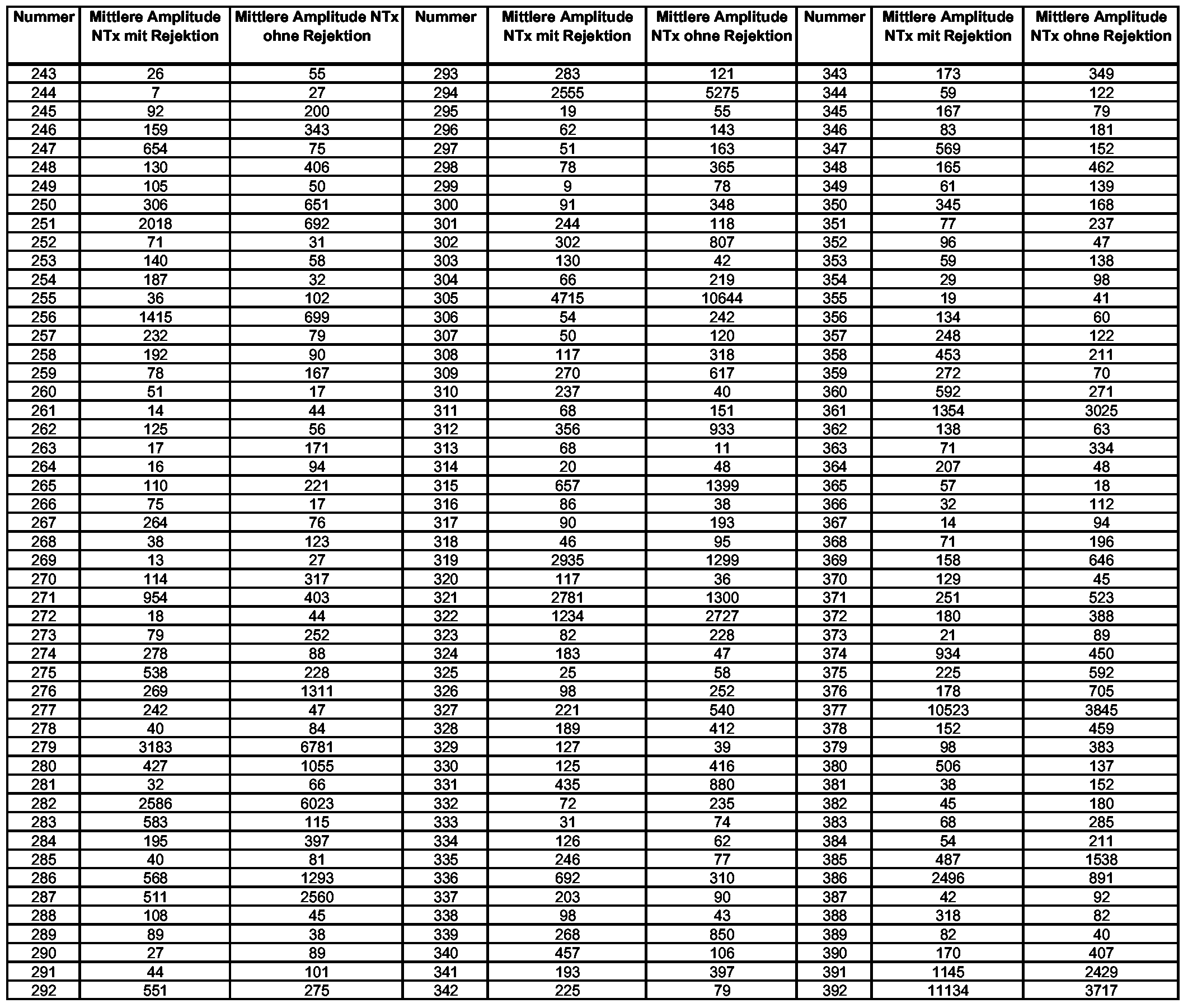 3. The method according to claim 1, characterized in that an evaluation of the amplitude of the markers 243-767 takes place on the basis of the following reference values:


4. Verfahren nach Anspruch 1 bis 3, wobei mindestens zwei oder mindestens drei oder mindestens fünf oder mindestens zehn oder alle Polypeptidmarker verwendet werden, wie sie in Anspruch 1 definiert sind.4. The method of claim 1 to 3, wherein at least two or at least three or at least five or at least ten or all polypeptide markers are used, as defined in claim 1.
5. Verfahren nach einem der Ansprüche 1 bis 4, wobei die Probe eines Individuums eine Urinprobe oder eine Blutprobe (Serum- oder Plasmaprobe) ist.The method of any one of claims 1 to 4, wherein the sample of an individual is a urine sample or a blood sample (serum or plasma sample).
6. Verfahren nach einem der Ansprüche 1 bis 5, wobei Kapillarelektrophorese, HPLC, Gasphasenionenspektrometrie und/oder Massenspektrometrie zum Nachweis der An- oder Abwesenheit des/der Polypeptidmarker verwendet wird. A method according to any one of claims 1 to 5, wherein capillary electrophoresis, HPLC, gas phase ion spectrometry and / or mass spectrometry is used to detect the presence or absence of the polypeptide marker (s).
7. Verfahren nach einem der Ansprüche 1 bis 6, wobei vor der Messung der Molekularmasse der Polypeptidmarker eine Kapillarelektrophorese durchgeführt wird.7. The method according to any one of claims 1 to 6, wherein before the measurement of the molecular mass of the polypeptide marker, a capillary electrophoresis is performed.
8. Verfahren nach einem der Ansprüche 1 bis 7, wobei Massenspektrometrie zum Nachweis der An- oder Abwesenheit des/der Polypeptidmarker verwendet wird.The method of any one of claims 1 to 7, wherein mass spectrometry is used to detect the presence or absence of the polypeptide marker (s).
9. Verfahren nach mindestens einem der Ansprüche 1 bis 8, dadurch gekennzeichnet, dass die CE-Zeit bezogen ist auf eine 90 cm lange Glaskapillare mit einem inneren Durchmesser (ID) von 50 μm bei einer angelegten Spannung von 25 kV, wobei als Laufmittel 20% Acetonitril, 0,25 M Ameisensäure in Wasser verwendet wird.9. The method according to at least one of claims 1 to 8, characterized in that the CE time is based on a 90 cm long glass capillary having an inner diameter (ID) of 50 microns at an applied voltage of 25 kV, being used as eluent 20th % Acetonitrile, 0.25 M formic acid in water.
10. Verwendung mindestens eines Polypeptidmarkers ausgewählt aus den Markern Nr. 1 bis 767, der durch die folgenden Werte für die Molekularmassen und die Migrationszeit charakterisiert ist 10. Use of at least one polypeptide marker selected from the markers Nos. 1 to 767, which is characterized by the following values for the molecular masses and the migration time


-37--37-


zur Erkennung einer Rejektion (Abstoßung) nach NTx.to detect a rejection (rejection) to NTx.
11. Markerkombination umfassend mindestens drei Marker ausgewählt aus  11. Marker combination comprising at least three markers selected from

- 41 -- 41 -


Priority Applications (4)
| Application Number | Priority Date | Filing Date | Title |
|---|---|---|---|
| JP2008518827A JP5155857B2 (en) | 2005-06-29 | 2006-06-28 | Polypeptide markers for early recognition of transplanted kidney rejection |
| EP06763931A EP1896856A1 (en) | 2005-06-29 | 2006-06-28 | Polypeptide marker for detecting the rejection of transplanted kidneys at an early stage |
| US11/922,550 US20100200401A1 (en) | 2005-06-29 | 2006-06-28 | Polypeptide Markers for the Early Recognition of the Rejection of Transplanted Kidneys |
| US14/554,368 US20150126405A1 (en) | 2005-06-29 | 2014-11-26 | Polypeptide markers for the early recognition of the rejection of transplanted kidneys |
Applications Claiming Priority (6)
| Application Number | Priority Date | Filing Date | Title |
|---|---|---|---|
| EP05105822 | 2005-06-29 | ||
| EP05105822.0 | 2005-06-29 | ||
| DE102005058514.0 | 2005-12-08 | ||
| DE102005058514 | 2005-12-08 | ||
| EP05112767 | 2005-12-22 | ||
| EP05112767.8 | 2005-12-22 |
Related Child Applications (2)
| Application Number | Title | Priority Date | Filing Date |
|---|---|---|---|
| US11/922,550 A-371-Of-International US20100200401A1 (en) | 2005-06-29 | 2006-06-28 | Polypeptide Markers for the Early Recognition of the Rejection of Transplanted Kidneys |
| US14/554,368 Continuation US20150126405A1 (en) | 2005-06-29 | 2014-11-26 | Polypeptide markers for the early recognition of the rejection of transplanted kidneys |
Publications (1)
| Publication Number | Publication Date |
|---|---|
| WO2007000466A1 true WO2007000466A1 (en) | 2007-01-04 |
Family
ID=36933512
Family Applications (1)
| Application Number | Title | Priority Date | Filing Date |
|---|---|---|---|
| PCT/EP2006/063635 WO2007000466A1 (en) | 2005-06-29 | 2006-06-28 | Polypeptide marker for detecting the rejection of transplanted kidneys at an early stage |
Country Status (4)
| Country | Link |
|---|---|
| US (2) | US20100200401A1 (en) |
| EP (1) | EP1896856A1 (en) |
| JP (1) | JP5155857B2 (en) |
| WO (1) | WO2007000466A1 (en) |
Cited By (2)
| Publication number | Priority date | Publication date | Assignee | Title |
|---|---|---|---|---|
| WO2010012306A1 (en) * | 2008-07-31 | 2010-02-04 | Queen Mary And Westfield College, University Of London | Healthy kidney biomarkers |
| WO2011017685A1 (en) * | 2009-08-07 | 2011-02-10 | Rules-Based Medicine, Inc. | Methods and devices for detecting kidney transplant rejection |
Families Citing this family (1)
| Publication number | Priority date | Publication date | Assignee | Title |
|---|---|---|---|---|
| US20200174014A1 (en) | 2016-12-19 | 2020-06-04 | Osaka University | METHOD FOR IN VITRO EARLY DIAGNOSIS OF ANTIBODY MEDIATED REJECTION AFTER ORGAN TRANSPLANTATION USING IgM-TYPE MEMORY B CELL DIFFERENTIATION CULTURE SYSTEM |
Citations (3)
| Publication number | Priority date | Publication date | Assignee | Title |
|---|---|---|---|---|
| WO1993003381A1 (en) * | 1991-07-26 | 1993-02-18 | Board Of Regents, The University Of Texas System | Assay for free secretory component and methods for monitoring organ rejection |
| US20030132114A1 (en) * | 2000-05-04 | 2003-07-17 | Harald Mischak | Method and device for the qualitative and / or quantitative analysis of a protein and/or peptide pattern of a liquid samle that is derived from the human or animal body |
| WO2005024409A1 (en) * | 2003-09-06 | 2005-03-17 | Mosaiques Diagnostics And Therapeutics Ag | Device and method for the quantitative evaluation of the polypeptides contained in a body fluid sample, and marker for the detection of pathological states |
-
2006
- 2006-06-28 EP EP06763931A patent/EP1896856A1/en not_active Withdrawn
- 2006-06-28 WO PCT/EP2006/063635 patent/WO2007000466A1/en active Application Filing
- 2006-06-28 JP JP2008518827A patent/JP5155857B2/en not_active Expired - Fee Related
- 2006-06-28 US US11/922,550 patent/US20100200401A1/en not_active Abandoned
-
2014
- 2014-11-26 US US14/554,368 patent/US20150126405A1/en not_active Abandoned
Patent Citations (3)
| Publication number | Priority date | Publication date | Assignee | Title |
|---|---|---|---|---|
| WO1993003381A1 (en) * | 1991-07-26 | 1993-02-18 | Board Of Regents, The University Of Texas System | Assay for free secretory component and methods for monitoring organ rejection |
| US20030132114A1 (en) * | 2000-05-04 | 2003-07-17 | Harald Mischak | Method and device for the qualitative and / or quantitative analysis of a protein and/or peptide pattern of a liquid samle that is derived from the human or animal body |
| WO2005024409A1 (en) * | 2003-09-06 | 2005-03-17 | Mosaiques Diagnostics And Therapeutics Ag | Device and method for the quantitative evaluation of the polypeptides contained in a body fluid sample, and marker for the detection of pathological states |
Non-Patent Citations (7)
| Title |
|---|
| CLARKE WILLIAM ET AL: "Characterization of renal allograft rejection by urinary proteomic analysis.", ANNALS OF SURGERY. MAY 2003, vol. 237, no. 5, May 2003 (2003-05-01), pages 660 - 664 ; dis, XP009066802, ISSN: 0003-4932 * |
| FLISER DANILO ET AL: "Capillary electrophoresis coupled to mass spectrometry for clinical diagnostic purposes.", ELECTROPHORESIS. JUL 2005, vol. 26, no. 14, July 2005 (2005-07-01), pages 2708 - 2716, XP002381570, ISSN: 0173-0835 * |
| O'RIORDAN EDMOND ET AL: "Bioinformatic analysis of the urine proteome of acute allograft rejection.", JOURNAL OF THE AMERICAN SOCIETY OF NEPHROLOGY : JASN. DEC 2004, vol. 15, no. 12, December 2004 (2004-12-01), pages 3240 - 3248, XP002381568, ISSN: 1046-6673 * |
| O'RIORDAN EDMOND ET AL: "Emerging studies of the urinary proteome: the end of the beginning?", CURRENT OPINION IN NEPHROLOGY AND HYPERTENSION. NOV 2005, vol. 14, no. 6, November 2005 (2005-11-01), pages 579 - 585, XP009066798, ISSN: 1062-4821 * |
| SCHAUB STEFAN ET AL: "Proteomic-based detection of urine proteins associated with acute renal allograft rejection.", JOURNAL OF THE AMERICAN SOCIETY OF NEPHROLOGY : JASN. JAN 2004, vol. 15, no. 1, January 2004 (2004-01-01), pages 219 - 227, XP002381566, ISSN: 1046-6673 * |
| WEISSINGER & MISCHAK, TRANSPLANT MED., vol. 16, 2004, pages 2 - 7, XP001246681 * |
| WITTKE STEFAN ET AL: "Detection of acute tubulointerstitial rejection by proteomic analysis of urinary samples in renal transplant recipients.", AMERICAN JOURNAL OF TRANSPLANTATION : OFFICIAL JOURNAL OF THE AMERICAN SOCIETY OF TRANSPLANTATION AND THE AMERICAN SOCIETY OF TRANSPLANT SURGEONS. OCT 2005, vol. 5, no. 10, October 2005 (2005-10-01), pages 2479 - 2488, XP002381482, ISSN: 1600-6135 * |
Cited By (5)
| Publication number | Priority date | Publication date | Assignee | Title |
|---|---|---|---|---|
| WO2010012306A1 (en) * | 2008-07-31 | 2010-02-04 | Queen Mary And Westfield College, University Of London | Healthy kidney biomarkers |
| JP2011529453A (en) * | 2008-07-31 | 2011-12-08 | ホスピタル クリニク アイ プロビンシアル デ バルセロナ | Healthy kidney biomarkers |
| US8859725B2 (en) | 2008-07-31 | 2014-10-14 | Queen Mary And Westfield College, University Of London | Healthy kidney biomarkers |
| WO2011017685A1 (en) * | 2009-08-07 | 2011-02-10 | Rules-Based Medicine, Inc. | Methods and devices for detecting kidney transplant rejection |
| US8735080B2 (en) | 2009-08-07 | 2014-05-27 | Rules-Based Medicine, Inc. | Methods and devices for detecting obstructive uropathy and associated disorders |
Also Published As
| Publication number | Publication date |
|---|---|
| US20100200401A1 (en) | 2010-08-12 |
| JP5155857B2 (en) | 2013-03-06 |
| US20150126405A1 (en) | 2015-05-07 |
| EP1896856A1 (en) | 2008-03-12 |
| JP2008545130A (en) | 2008-12-11 |
Similar Documents
| Publication | Publication Date | Title |
|---|---|---|
| EP1757939A1 (en) | Polypeptide markers for the diagnosis of bladder cancer | |
| EP2255203A2 (en) | Method and marker for diagnosis of tubular kidney damage and illnesses | |
| WO2010031822A1 (en) | Kidney cell carcinoma | |
| EP1287348A2 (en) | Method and device for the qualitative and/or quantitative analysis of a protein and/or peptide pattern of a liquid sample that is derived from the human or animal body | |
| EP1955064A1 (en) | Polypeptide marker for the diagnosis and evaluation of ureteropelvic junction | |
| WO2008110593A2 (en) | Method and markers for the diagnosis of renal diseases | |
| WO2006106129A2 (en) | Polypeptide markers for the diagnosis of prostate cancer | |
| WO2011000938A1 (en) | Method and markers for diagnosing acute renal failure | |
| WO2009047280A2 (en) | Polypeptide marker for the diagnosis of prostate cancer | |
| WO2009050300A1 (en) | Method and marker for diagnosing diabetes mellitus | |
| EP2478377A2 (en) | Polypeptide marker for diagnosing and assessing vascular diseases | |
| WO2010079076A2 (en) | Autosomal dominant polycystic kidney disease (adpkd) | |
| EP2810077A2 (en) | Polypeptide markers for diagnosis and assessment of heart failure | |
| EP1896856A1 (en) | Polypeptide marker for detecting the rejection of transplanted kidneys at an early stage | |
| EP1784649B1 (en) | Polypeptide marker for the diagnosis of arteriosclerosis | |
| DE60225140T2 (en) | METHOD FOR ANALYZING A PROTEIN HAVING A CELL OR A SUBSTANCE INTERACTING WITH THE PROTEIN | |
| WO2006106115A2 (en) | Polypeptide marker for diagnosing alzheimer's disease | |
| WO2012076723A1 (en) | Method and marker for the diagnosis of a bile duct stricture and of a cholangiocellular carcinoma in bile | |
| WO2007063089A2 (en) | Polypeptide marker for the diagnosis and evaluation of vascular diseases | |
| WO2012123527A1 (en) | Method and markers for the diagnosis of subclinical and clinical forms of t-cell-mediated tubulointerstitial rejection after kidney transplantation | |
| WO2013150132A2 (en) | Polypeptide markers for diagnosis and assessment of strokes |
Legal Events
| Date | Code | Title | Description |
|---|---|---|---|
| 121 | Ep: the epo has been informed by wipo that ep was designated in this application | ||
| WWE | Wipo information: entry into national phase |
Ref document number: 2006763931 Country of ref document: EP |
|
| WWE | Wipo information: entry into national phase |
Ref document number: 2008518827 Country of ref document: JP |
|
| NENP | Non-entry into the national phase |
Ref country code: DE |
|
| WWW | Wipo information: withdrawn in national office |
Country of ref document: DE |
|
| WWP | Wipo information: published in national office |
Ref document number: 2006763931 Country of ref document: EP |
|
| WWE | Wipo information: entry into national phase |
Ref document number: 11922550 Country of ref document: US |