WO2002024933A2 - Viral vectors having tissue tropism for t-lymphocytes, b- and mast cells - Google Patents
Viral vectors having tissue tropism for t-lymphocytes, b- and mast cells Download PDFInfo
- Publication number
- WO2002024933A2 WO2002024933A2 PCT/EP2001/011086 EP0111086W WO0224933A2 WO 2002024933 A2 WO2002024933 A2 WO 2002024933A2 EP 0111086 W EP0111086 W EP 0111086W WO 0224933 A2 WO0224933 A2 WO 0224933A2
- Authority
- WO
- WIPO (PCT)
- Prior art keywords
- cells
- adenovirus
- cell
- nucleic acid
- serotype
- Prior art date
- Legal status (The legal status is an assumption and is not a legal conclusion. Google has not performed a legal analysis and makes no representation as to the accuracy of the status listed.)
- Ceased
Links
Classifications
-
- C—CHEMISTRY; METALLURGY
- C12—BIOCHEMISTRY; BEER; SPIRITS; WINE; VINEGAR; MICROBIOLOGY; ENZYMOLOGY; MUTATION OR GENETIC ENGINEERING
- C12N—MICROORGANISMS OR ENZYMES; COMPOSITIONS THEREOF; PROPAGATING, PRESERVING, OR MAINTAINING MICROORGANISMS; MUTATION OR GENETIC ENGINEERING; CULTURE MEDIA
- C12N15/00—Mutation or genetic engineering; DNA or RNA concerning genetic engineering, vectors, e.g. plasmids, or their isolation, preparation or purification; Use of hosts therefor
- C12N15/09—Recombinant DNA-technology
- C12N15/63—Introduction of foreign genetic material using vectors; Vectors; Use of hosts therefor; Regulation of expression
- C12N15/79—Vectors or expression systems specially adapted for eukaryotic hosts
- C12N15/85—Vectors or expression systems specially adapted for eukaryotic hosts for animal cells
- C12N15/86—Viral vectors
-
- A—HUMAN NECESSITIES
- A61—MEDICAL OR VETERINARY SCIENCE; HYGIENE
- A61K—PREPARATIONS FOR MEDICAL, DENTAL OR TOILETRY PURPOSES
- A61K48/00—Medicinal preparations containing genetic material which is inserted into cells of the living body to treat genetic diseases; Gene therapy
-
- C—CHEMISTRY; METALLURGY
- C12—BIOCHEMISTRY; BEER; SPIRITS; WINE; VINEGAR; MICROBIOLOGY; ENZYMOLOGY; MUTATION OR GENETIC ENGINEERING
- C12N—MICROORGANISMS OR ENZYMES; COMPOSITIONS THEREOF; PROPAGATING, PRESERVING, OR MAINTAINING MICROORGANISMS; MUTATION OR GENETIC ENGINEERING; CULTURE MEDIA
- C12N2710/00—MICROORGANISMS OR ENZYMES; COMPOSITIONS THEREOF; PROPAGATING, PRESERVING, OR MAINTAINING MICROORGANISMS; MUTATION OR GENETIC ENGINEERING; CULTURE MEDIA dsDNA viruses
- C12N2710/00011—Details
- C12N2710/10011—Adenoviridae
- C12N2710/10311—Mastadenovirus, e.g. human or simian adenoviruses
- C12N2710/10322—New viral proteins or individual genes, new structural or functional aspects of known viral proteins or genes
-
- C—CHEMISTRY; METALLURGY
- C12—BIOCHEMISTRY; BEER; SPIRITS; WINE; VINEGAR; MICROBIOLOGY; ENZYMOLOGY; MUTATION OR GENETIC ENGINEERING
- C12N—MICROORGANISMS OR ENZYMES; COMPOSITIONS THEREOF; PROPAGATING, PRESERVING, OR MAINTAINING MICROORGANISMS; MUTATION OR GENETIC ENGINEERING; CULTURE MEDIA
- C12N2710/00—MICROORGANISMS OR ENZYMES; COMPOSITIONS THEREOF; PROPAGATING, PRESERVING, OR MAINTAINING MICROORGANISMS; MUTATION OR GENETIC ENGINEERING; CULTURE MEDIA dsDNA viruses
- C12N2710/00011—Details
- C12N2710/10011—Adenoviridae
- C12N2710/10311—Mastadenovirus, e.g. human or simian adenoviruses
- C12N2710/10341—Use of virus, viral particle or viral elements as a vector
- C12N2710/10343—Use of virus, viral particle or viral elements as a vector viral genome or elements thereof as genetic vector
-
- C—CHEMISTRY; METALLURGY
- C12—BIOCHEMISTRY; BEER; SPIRITS; WINE; VINEGAR; MICROBIOLOGY; ENZYMOLOGY; MUTATION OR GENETIC ENGINEERING
- C12N—MICROORGANISMS OR ENZYMES; COMPOSITIONS THEREOF; PROPAGATING, PRESERVING, OR MAINTAINING MICROORGANISMS; MUTATION OR GENETIC ENGINEERING; CULTURE MEDIA
- C12N2710/00—MICROORGANISMS OR ENZYMES; COMPOSITIONS THEREOF; PROPAGATING, PRESERVING, OR MAINTAINING MICROORGANISMS; MUTATION OR GENETIC ENGINEERING; CULTURE MEDIA dsDNA viruses
- C12N2710/00011—Details
- C12N2710/10011—Adenoviridae
- C12N2710/10311—Mastadenovirus, e.g. human or simian adenoviruses
- C12N2710/10341—Use of virus, viral particle or viral elements as a vector
- C12N2710/10345—Special targeting system for viral vectors
-
- C—CHEMISTRY; METALLURGY
- C12—BIOCHEMISTRY; BEER; SPIRITS; WINE; VINEGAR; MICROBIOLOGY; ENZYMOLOGY; MUTATION OR GENETIC ENGINEERING
- C12N—MICROORGANISMS OR ENZYMES; COMPOSITIONS THEREOF; PROPAGATING, PRESERVING, OR MAINTAINING MICROORGANISMS; MUTATION OR GENETIC ENGINEERING; CULTURE MEDIA
- C12N2810/00—Vectors comprising a targeting moiety
- C12N2810/50—Vectors comprising as targeting moiety peptide derived from defined protein
- C12N2810/60—Vectors comprising as targeting moiety peptide derived from defined protein from viruses
- C12N2810/6009—Vectors comprising as targeting moiety peptide derived from defined protein from viruses dsDNA viruses
- C12N2810/6018—Adenoviridae
-
- C—CHEMISTRY; METALLURGY
- C12—BIOCHEMISTRY; BEER; SPIRITS; WINE; VINEGAR; MICROBIOLOGY; ENZYMOLOGY; MUTATION OR GENETIC ENGINEERING
- C12N—MICROORGANISMS OR ENZYMES; COMPOSITIONS THEREOF; PROPAGATING, PRESERVING, OR MAINTAINING MICROORGANISMS; MUTATION OR GENETIC ENGINEERING; CULTURE MEDIA
- C12N2810/00—Vectors comprising a targeting moiety
- C12N2810/50—Vectors comprising as targeting moiety peptide derived from defined protein
- C12N2810/60—Vectors comprising as targeting moiety peptide derived from defined protein from viruses
- C12N2810/6045—RNA rev transcr viruses
- C12N2810/6054—Retroviridae
Definitions
- the invention relates to the field of molecular genetics and medicine.
- the present invention relates to the field of functional genomics and gene therapy, in particular, methods useful in ex vivo gene therapy and functional genomics using adenovirus vectors.
- genetic information with unknown function but somehow pre-selected for is usually delivered to a host cell in order to either correct (supplement) a genetic deficiency in said cell, or to inhibit an undesired function in said cell or to otherwise induce a phenotype.
- the genetic information can also be intended to provide the host cell with a desired function, e.g. to supply a secreted protein or express a transcription factor.
- adenoviral vectors are relatively easy to concentrate and purify. For functional genomics adenoviral vectors are also ideally suited.
- adenoviral vectors can be used in vitro as well as in vivo using either in situ or in vitro cell or tissue based assays or appropriate animal models.
- adenoviral vectors Some characteristics of the current adenoviral vectors limit their use in specific applications. For instance endothelial cells, smooth muscle cells, T-lymphocytes and mast cells are not easily transduced by the current generation of adenoviral vectors. For many gene therapy or functional genomics applications, preferably these types of cells should be genetically modified. Disease areas for which efficient gene transfer into these cell types is desirable include but are not limited to autoimmune disorders, cancer, infectious diseases, cardiovascular diseases and bone disorders.
- T-lymphocytes are formed in the bone marrow, migrate to and mature in the thymus and then enter the peripheral blood and lymphatic circulation. T-lymphocytes are subdivided into three distinct types of cells: helper T-lymphocytes, suppressor T- Iymphocytes, and cytotoxic T-lymphocytes. T-lymphocytes, unlike B-lymphocytes, do not produce antibody molecules, but express a heterodimeric cell surface receptor that recognizes peptide fragments of antigenic proteins that are attached to proteins of the major histocompatibility complex (MHC) and expressed on the surfaces of target T- lymphocytes (e.g. Abbas et al, 1991).
- MHC major histocompatibility complex
- Human cytotoxic T-lymphocytes are typically of the CD3 + , CD8 + , CD4 " phenotype and lyse cells that display fragments of foreign antigens associated with MHC class I molecules on their cell surfaces.
- Target T-lymphocytes for CTL recognition include normal cells expressing antigens after infection by viruses or other pathogens; and tumor cells that have undergone transformation and are expressing mutated proteins or are over-expressing normal proteins.
- Helper T-lymphocytes are also CD3 + but can be distinguished from cytotoxic T- lymphocytes by expression of CD4 but absence of the CD8 membrane protein.
- CD4* helper T-lymphocytes recognize fragments of antigens presented in association with MHC class II molecules, and primarily function to produce cytokines that amplify antigen-specific T- and B-cell responses and activate accessory immune cells such as monocytes or macrophages (e.g. Abbas et al, 1991).
- CD4 + helper and CD8 + cytotoxic T-lymphocytes are important components of the host immune response to viruses, bacterial pathogens and tumors.
- individuals with congenital, acquired or iatrogenic T-cell immunodeficiency diseases may develop life threatening infections or malignancies (for example, SCTD, AIDS, etc.).
- Persons with diseases that are related to a deficiency of immunologically competent T-lymphocytes can potentially have specific immunity restored through adoptive immunotherapy, alternatively called adoptive transfer.
- adoptive immunotherapy one or more specific immunities can be conferred upon an individual by transferring T-lymphocytes having the desired antigenic specificities.
- the cells of interest are derived from the immunodeficient host or from a compatible specifically immunized host.
- the latter source is of course especially important in situations in which the immunodeficient host has an insufficient number of T-lymphocytes, or has T-lymphocytes that are insufficiently effective.
- Efficient and reproducible gene transfer into T-lymphocytes, in particular human T-lymphocytes, is of prime importance in the validation of new genes, and for the development of new immuno or gene therapies.
- T-lymphocytes can be isolated or enriched for by using cell immuno affinity methods based for example on magnetic beads having anti CD3, CD4 or CD 8 antibodies on their surface, thus cell isolation is based on T-cell specific cell surface markers.
- Common methodology used is the technology developed by Miltenyi et al. Preferred is to use the technology for depletion of cell types other than T-lymphocytes so that activation of the resting T-cells by for example CD3 antibodies is avoided. This is done by using antibodies against markers for other cell types of the hemopoietic system such as monocytes and B-lymphocytes.
- Mast cells are a family of cells generally found around the blood vessels in the connective tissues, in the lining of the gut, and in the lungs. They are large mononuclear cells, heavily granulated and deeply stained by basic dyes. Mast cells have their origin in the bone marrow and are derived from CD34+ hematopoietic progenitor cells that migrate in the form of immature progenitors to the tissue, where they differentiate into mature mast cells. A key feature of mast cells is that they express receptors (Fc ⁇ RI) on their cell membranes that bind with high affinity to the Fc portion of IgE.
- Fc ⁇ RI receptors
- mast cells secrete mediators that are either preformed and granule-associated (e.g. histamine, proteoglycans, and neutral proteases) or are synthesized de novo (e.g. leukotriene C 4 , platelet activated factor and prostaglandin D 2 ).
- mediators that are either preformed and granule-associated (e.g. histamine, proteoglycans, and neutral proteases) or are synthesized de novo (e.g. leukotriene C 4 , platelet activated factor and prostaglandin D 2 ).
- mast cells are potential sources of many cytokines. Being as effector cells in IgE-assiociated immune responses, mast cells play a prominent role in allergic diseases, including asthma, and in host resistance to parasites. Moreover, they are implicated in the genesis of other diseases such as pulmonary fibrosis.
- vectors used to transduce cells include the adenoviral vectors.
- Adenoviruses contain a linear double-stranded DNA molecule of approximately 36000 base pairs. It contains identical Inverted Terminal Repeats (ITR) of approximately 90-140 base pairs with the exact length depending on the serotype.
- ITR Inverted Terminal Repeats
- the viral origins of replication are within the ITRs exactly at the genome ends.
- the transcription units are divided in early and late regions. Shortly after infection the El A and EIB proteins are expressed and function in transactivation of cellular and adenoviral genes.
- the early regions E2A and E2B encode proteins (DNA binding protein, pre-terminal protein and polymerase) required for the replication of the adenoviral genome (reviewed in van der Vliet, 1995).
- the early region E4 encodes several proteins with pleiotropic functions e.g. transactivation of the E2 early promoter, facilitating transport and accumulation of viral mRNAs in the late phase of infection and increasing nuclear stability of major late pre-mRNAs (reviewed in Leppard, 1997).
- the early region 3 encodes proteins that are involved in modulation of the immune response of the host (Wold et al, 1995).
- the late region is transcribed from one single promoter (major late promoter) and is activated at the onset of DNA replication.
- RNA species coding for core proteins, capsid proteins (penton, hexon, fiber and associated proteins), viral protease and proteins necessary for the assembly of the capsid and shutdown of host protein translation (Imperiale et al, 1995).
- the interaction of the virus with the host cell has mainly been investigated with the serotype C viruses Ad2 and Ad5. Binding occurs via interaction of the knob region of the protruding fiber with a cellular receptor.
- a receptor for Ad2, Ad5 and probably more adenoviruses is known as the 'Coxsackievirus and Adenovirus Receptor' or CAR protein (Bergelson et al, 1997). Internalization is mediated through interaction of the RGD (Arg, Gly, Asp) sequence present in the penton base with cellular ⁇ l- integrins (Wickham et al, 1993). This may not be true for all serotypes, for example serotype 40 and 41 do not contain a RGD sequence in their penton base sequence (Kidd et al, 1993).
- the initial step for successful infection is binding of adenovirus to its target cell, a process mediated through the fiber protein.
- the fiber protein has a trimeric structure (Stouten et al, 1992) with different lengths depending on the virus serotype (Signas et al, 1985; Kidd et al, 1993). Different serotypes have polypeptides with structurally similar N- and C-termini, but different middle stem regions. The first 30 amino acids at the N-terminus are involved in anchoring of the fiber to the penton base (Chroboczek et al, 1995), especially the conserved FNPVYP region in the tail (Arnberg et al, 1997).
- the C-terminus is responsible for initial interaction with the cellular adenovirus receptor. After this initial binding, secondary binding between the capsid penton base and cell-surface integrins leads to internalization of viral particles in coated pits and endocytosis (Morgan et al, 1969; Svensson and Persson, 1984; Varga et al, 1991; Greber et al, 1993; Wickham et al, 1993). Integrins are ⁇ -heterodimers of which at least 19 ⁇ -subunits and 8 ⁇ -subunits have been identified (see http://nciarray.nci.nih.gov/cgi-bin/cards). The array of integrins expressed in cells is complex and will vary between cell types and cellular environment. Although the knob contains some conserved regions between serotypes, the knob proteins show a high degree of variability, indicating that different adenovirus receptors exist.
- a serotype is defined on the basis of its immunological distinctiveness as determined by quantitative neutralization with animal antiserum (horse, rabbit).
- adenoviruses in subgroup C such as Ad2 and Ad5 bind to different receptors as compared to adenoviruses from subgroup B such as Ad3 and Ad7 (Defer et al, 1990; Gall et al, 1996).
- receptor specificity could be altered by exchanging the Ad3 knob protein with the Ad 5 knob protein, and vice versa (Krasnykh et al, 1996; Stevenson et al, 1995 and 1997).
- Serotypes 2, 4, 5 and 7 all have a natural affiliation towards lung epithelia and other respiratory tissues.
- serotypes 40 and 41 have a natural affiliation towards the gastrointestinal tract.
- serotypes differ in at least capsid proteins (penton-base, hexon), proteins responsible for cell binding (fiber protein), and proteins involved in adenovirus replication. It is unknown to what extent the capsid proteins determine the differences in tropism found between the serotypes. It may very well be that post- infection mechanisms determine cell-type specificity of adenoviruses. It has been shown that adenoviruses from subgroups A (Adl2 and Ad31), C (Ad2 and Ad5), D (Ad9 and Adl5), E (Ad4) and F (Ad41) are all able to bind labeled soluble CAR (sCAR) protein when immobilized on nitrocellulose.
- capsid proteins penton-base, hexon
- fiber protein proteins responsible for cell binding
- proteins involved in adenovirus replication It is unknown to what extent the capsid proteins determine the differences in tropism found between the serotypes. It may very well be that post- infection mechanisms determine cell-
- viruses within one subgroup displayed different infection efficiencies.
- the importance of fiber binding for the improved infection of Adl7 in CAE was shown by Armentano et al (WO 98/22609 Al) who made a recombinant Ad2/LacZ virus with a fiber gene from Adl7 and showed that the chimaeric virus infected CAE more efficient then Ad2 LacZ viruses with Ad2 fibers.
- adenoviral gene delivery vectors currently used in functional genomics, gene therapy or vaccination are derived from subgroup C adenoviruses Ad2 or Ad5.
- the vectors have at least a deletion in the El region that renders the recombinant virus replication defective. In this region, novel genetic information can then be introduced. It has been demonstrated extensively that recombinant adenoviruses, in particular serotype 5, are suitable for efficient transfer of genes in vivo to the liver, the airway epithelium and solid tumors in animal models and human xenografts in immunodeficient mice (Bout 1996, 1997; Blaese et al, 1995).
- adenoviral vectors in functional genomics includes building gene expression libraries and in vitro and in vivo gene validation with appropriate meaningful cell based assays or animal models for a particular human disease. Transfer and subsequent expression of a cDNA into a desired cell-type may lead to relevant phenotypic changes that may or may not confirm the role a particular cDNA plays in a particular disease. Alternatively such an exercise may lead to better insight into the validity of using a particular cDNA as a target for therapeutic intervention. In addition to sense copies of a gene or genes under investigation, antisense copies is cloned into the adenoviral vector and used for validation studies.
- Gene transfer vectors derived from adenoviruses have a number of features that make them particularly useful for gene transfer:
- the virus is extremely efficient in introducing its DNA into the host cell
- the virus can infect a wide variety of cells and has a broad host-range
- the virus can be produced at high titers in large quantities, 6) and the virus can be rendered replication defective by deletion of the early- region 1 (El) of the viral genome (Brody and Crystal, 1994),
- adenoviruses especially the well investigated serotypes Ad2 and Ad5, usually elicit an immune response by the host into which they are introduced,
- the serotypes Ad2 and Ad5 are not ideally suited for delivering additional genetic material to organs other than the liver. Delivery of vectors derived from Ad2 or Ad5 via the bloodstream leads to a significant delivery of these vectors to the cells of the liver. In therapies where other cell types then liver cells need to be transduced, some means of liver exclusion must be applied to prevent uptake of the vector by these cells. Current methods rely on the physical separation of the vector from the liver cells. This can be done by localizing the vector and/or the target organ via surgery, balloon angioplasty or direct injection into an organ via for instance needles. Liver exclusion is also being practiced by surgical targeting in which the vector is delivered to compartments in the body that are essentially isolated from the bloodstream.
- HSV herpes simplex virus
- TK thymidine kinase
- T-lymphocytes are primary targets in numerous gene therapy protocols.
- Ad2 or Ad5 subgroup C adenovirus serotypes 2 or 5
- mast cells are derived from haemopoietic stem cells and are from bone marrow origin. The cells can be cultured from CD34+ progenitors. Mast cells play a distinct role in acute inflammation. During the sensibilisation-phase, the immune system becomes stimulated by an allergen. Antigens from microbes stimulate antigen-specific B-cells to produce antibodies. Some of these (IgE) bind to mast cells, which becomes sensitized. After a second contact with the same allergen, the sensitized mast cells are triggered to release inflammatory mediators (like histamine) from its granules.
- IgE inflammatory mediators
- bispecific antibodies are done using chemical coupling methods such as succinimidyl-3-(2-pyridyldithiol)-propionate (SPDP) as a cross linking agent.
- SPDP succinimidyl-3-(2-pyridyldithiol)-propionate
- adenoviral vector needs to be pre- incubated with the bi-specific antibodies to generate the targeted adenoviral vector, creating another variable in the procedure.
- anti-CD3 monoclonal antibodies activate the T-cell receptor activation signal (normally provided by antigen and antigen-presenting cells).
- the anti-CD3 monoclonal antibody most commonly used is OKT.sub.3, which is commercially available from Ortho Pharmaceuticals .
- the present invention was made in the course of the manipulation of adenoviral vectors to obtain efficient gene transfer into T-lymphocytes, and in particular human T- lymphocytes.
- the present invention provides functional genomics and gene therapy methods, using gene delivery vehicles provided with a tissue tropism for human T-lymphocytes, mast cells and B cells, useful in applications where primary T-lymphocytes, mast cells or B cells comprise the target cell type.
- the present invention relates to a method of introducing an expressible non- viral nucleic acid sequence into a cell having a common non-universal binding receptor and selected from T lymphocytes, B-, and mast cells, comprising contacting said cell with a viral vector comprising a recombinant nucleic acid sequence containing sequence for said expressible non-viral nucleic acid and comprising a modified viral coat consisting of native viral coat proteins and modified coat protein containing adenoviral amino acid sequence from an adenoviral serotype 35 or 51 fibre protein, wherein said adenoviral sequence of said modified protein is a ligand for said binding receptor.
- the present invention relates to the use of a viral vector as defined above for the preparation of a medicament for introducing an expressible non- viral nucleic acid sequence into a selected cell as defined above.
- the present invention relates to a method of introducing an expressible non-viral nucleic acid sequence into a cell having a common non-universal binding receptor and selected from T lymphocytes, B-, and mast cells, comprising contacting said cell with a viral vector comprising a recombinant adenoviral nucleic acid sequence containing sequence for said expressible non-viral nucleic acid and for sequence coding for a viral capsid consisting of native adenoviral capsid proteins and modified capsid protein containing amino acid sequence from an adenoviral serotype other than the serotype of said native capsid proteins, wherein said modified protein is a ligand for said binding receptor.
- the present invention relates to the use of a viral vector as defined above for the preparation of a medicament for introducing an expressible non-viral nucleic acid sequence into a selected cell as defined above.
- the present invention relates to a method for transducing a cell selected from the group consisting of T lymphocytes, B cells, and mast cells comprising contacting said cells with an adenovirus particle comprising a non- adenovirus nucleic acid sequence and a chimeric capsid protein comprising amino acid sequence derived from at least two adenovirus serotypes, wherein said particle has a greater tropism for said cells relative to at least one of the adenovirus serotypes comprising said chimeric capsid protein.
- the present invention relates to the use of an adenovirus particle as defined above for the preparation of a medicament for transducing a selected cell as defined above.
- the present invention also relates to a transduced cell selected from the group consisting of T lymphocytes, B-, and mast cells and comprising a replication incompetent recombinant adenoviral nucleic acid sequence containing sequence for an expressible non- viral nucleic acid and coding for a viral capsid consisting of native adenoviral capsid proteins and modified capsid protein containing amino acid sequence from an adenoviral serotype other than said native capsid proteins, wherein said modified protein is a ligand for a binding receptor on said cell.
- the present invention also relates to a method for ex vivo transduction of a population of cells comprising (a) obtaining from a mammal said population of cells selected from the group consisting of T lymphocytes, B cells and/or mast cells, and (b) transducing said cell population in vitro with a replication incompetent viral vector comprising a recombinant adenoviral nucleic acid sequence containing sequence for said expressible non-viral nucleic acid and for a viral capsid consisting of native adenoviral capsid proteins and modified capsid protein containing amino acid sequence from an adenoviral serotype other than said native capsid proteins, wherein said modified protein is a ligand for a binding receptor on said cells.
- the present invention further relates to a method of administering to a human or other mammalian animal subject a population of cells genetically modified ex vivo with an expressible recombinant nucleic acid, comprising (a) obtaining from said subject said population of cells selected from the group consisting of T lymphocytes, B cells, mast cells and/or dendritic cells, (b) contacting said cell population with a replication incompetent viral vector comprising a recombinant adenoviral nucleic acid sequence containing sequence for said expressible non-viral nucleic acid and for a viral capsid consisting of native adenoviral capsid proteins and modified capsid protein containing amino acid sequence from an adenoviral serotype other than said native capsid proteins, wherein said modified protein is a ligand for a binding receptor on said cells, thereby obtaining a transduced population of cells; and (c) introducing said transduced cells into said subject.
- the present invention relates to the use of
- the present invention also relates to a method for identifying the function of a subject nucleic acid in hematopoietic cells, comprising (a) contacting a first population of cells selected from the group consisting of T-lymphocytes, B-, and mast cells with a replication incompetent viral vector comprising a recombinant adenoviral nucleic acid sequence containing an expressible sequence for said subject nucleic acid and for a viral capsid consisting of native adenoviral capsid proteins and modified capsid protein containing amino acid sequence from an adenoviral serotype other than said native capsid proteins, wherein said modified protein is a ligand for binding receptor on said cell, thereby transducing said cell population; and (b)observing a change in the function of said transduced cell population.
- the invention provides an improved means and method for providing a desired nucleotide sequence to a target cell, and in particular to a T-lymphocyte, mast cell or B cell. It is an object of the current invention to provide materials and methods to overcome the limitations of the prior art adenoviral vectors.
- the invention provides methods using adenoviral viruses, derived in whole or in part from adenovirus serotypes different from Ad5, combining genes of adenovirus serotypes with preferred characteristics in a chimaeric vector to give rise to a vector better suited for specific applications.
- Preferred characteristics include, but are not limited to, improved infection of a specific target cell, reduced infection of non- target cells, improved stability of the virus, reduced toxicity to target cells, reduced neutralization in humans or animals, reduced or increased CTL response in humans or animals, better and/or prolonged transgene expression, increased penetration capacity in tissues, improved yields in packaging cell lines, etc.
- Figure 1 is a schematic drawing of the pBr/Ad.Bam-rlTR construct used in Example 1.
- Figure 2 is a schematic representation of the strategy used to generate plasmid pBr/Ad.Bam-rITRDfib in which the adenovirus type 5 fiber DNA is replaced by a short stretch containing an unique Nsil site.
- Figure 3 is a schematic drawing of construct pBr/Ad.Bam-rITRDfib.pac.
- Figure 4 shows amino acid-sequences of the fiber proteins of adenovirus serotypes 35 and 51. Bold letters represent part of the tail of adenovirus type 5. At the end of the sequence the stop codon of the fiber is presented by a dot.
- Figure 5 is a schematic drawing of the adapter construct pAdApt/eGFP.
- Figure 6 is a schematic presentation of the method to generate recombinant adenoviruses using two overlapping fragments. This system requires only one recombination event. Early (E) and late regions (L) are indicated. L5 is the fiber coding sequence.
- Figure 7 is an analysis of T-cells isolated from peripheral human blood as described in Example 2. The total cell-population before isolation and the isolated T- cell population were unstained (resp. a and c) and stained for CD3 (-PE) and CD45 (-
- FIG. 8 is an analysis of transduced T-lymphocytes.
- the T-cells were harvested and stained for CD3 (-PE) expression, 48 hours after transduction, followed by flow cytometry analysis to determine the percentage of CD3 + eGFP + T-lymphocytes. Percentages given, are average percentages of eGFP + cells in the CD3 + cell-population (average of two wells).
- Figure 9 depicts the flow cytometry results of transduced A549-, SupTl- and T- cells.
- a flow cytometer was used to determine the percentage of eGFP + cells for A549 (a) and SupTl (b).
- the T-cells were first stained with CD3-PE, and then the percentage of CD3 + eGFP + T-cells was determined (c). (Percentages given are averages percentages of two wells.) Crude stands for crude lysate used as viruses, pure stands for purified viruses.
- FIG. 10 shows pictures taken with an inverted fluorescence microscope 72 hours after transduction of mature (L16) and immature (BW) mast cells as described in Example 4. Mature mast cells transduced with Ad5 ⁇ dEl.fib51.pAdApt/eGFP crude
- Figure 11 shows an analysis of transduced mature (L16) and immature (BW) mast cells.
- the mast cells were harvested 96 hours after transduction, followed by flow cytometry analysis to determine the percentage of eGFP + mast cells. Percentages given are percentages of eGFP + cells.
- Figure 12 shows flow cytometry results of transduced mature and immature mast cells.
- a flow cytometer was used to determine the percentage of eGFP + cells for mature mast cells (A) and immature mast cells (B). The mast cells were harvested 96 hours after transduction, followed by flow cytometry analysis to determine the percentage of eGFP + mast cells.
- Cr stands for crude lysate adenoviral vectors and p stands for purified adenoviral vectors.
- MOI is in VP/cell.
- Figure 13 shows the ⁇ -hexoseaminidase assay results of transduced mature and immature mast cells.
- the assay was performed 48 hours after transduction of the mature (L20) mast cells (A) and immature (BW) mast cells (B). Absorbance was read at 405 nm.
- Figures 250, 1000 and 2500 stand for the MOIs used in VP/cell, buffer stands for untreated cells, flask stand for cells that were taken freshly from a culture- flask before performing the assay and TNP-1 and-10 stand for the concentration 1 and 10 ng/ml antigen (TNP) added during the assay.
- TNP concentration 1 and 10 ng/ml antigen
- Figure 14 shows the analysis of transduced Ramos B-cells described in Example 5.
- the B-cells were harvested one week after transduction, followed by flow cytometry analysis to determine the percentage of eGFP + cells.
- Figure 15 shows the flow cytometry results of transduced Ramos B-cells and SupTl cells described in Example 5.
- One week after transduction the cells were harvested and a flow cytometer was used to determine the percentage of eGFP + cells for Ramos B-cells transduced with normal culture medium (A), with activation medium (B), Optimem (C) and SupTl cells (D).
- Cr stands for crude lysate adenoviral vectors and P stands for purified adenoviral vectors.
- MOI is in VP/cell.
- a "viral vector” as referred to herein is in the form of a viral particle comprising a viral coat (e.g. an envelope or a capsid) in which is contained/packaged a nucleic acid that encodes said desired nucleotide sequence, and usually also (at least part of) the viral genome.
- a viral coat e.g. an envelope or a capsid
- Tropism as used herein is intended to mean the ability or affinity of a particular viral particle to bind to a particular cell type or types relative to other cell types. An increased tropism means a greater affinity or ability to bind to a cell type or types, while a decreased tropism means a lesser affinity or ability to bind to a cell type or types.
- a limited tropism means an ability to bind to a subset of cells as opposed to a broad or general tropism that means the ability to bind to a large number of different cell types or to all cell types.
- a particular tropism means the ability of a particular virus to bind to a particular subset of cell types.
- a preferred limited tropism is the tropism for nuclear hematopoietic cells, and most particularly a tropism for T-lymphocytes, B cells and mast cells.
- the present invention uses viral vectors that have increased tropism for T- lymphocytes, mast cells or B cells, i.e. compared to the commonly used adenoviral vectors Ad2 and Ad5, and that still has all the advantages of (Ad2 or Ad5) adenoviral vectors.
- the invention uses a (chimaeric) virus or virus particle that is suitable for use as a viral vector, and that has been provided with an altered/modified viral coat that confers upon said virus particle increased tropism for T-lymphocytes, B cells or mast cells.
- (viral) coat as used herein (also) encompasses viral capsid(s) and/or viral envelope(s).
- the term "coat protein(s)” as used herein comprises any and all proteins that (together) constitute the viral coat, capsid and/or envelope, including but not limited to any fiber(s), penton(s) or hexon(s).
- a viral particle that comprises a capsid - such as an adenovirus particle - will be preferred.
- any one or more of the proteins which form the viral coat is modified, altered and/or replaced (e.g. essentially fully or in part) by one or more corresponding coat proteins derived from another virus, to provide the chimaeric virus particle with increased tropism for a T-lymphocyte, B cell or a mast cell as described herein.
- the (native) viral particle which is provided with the increased tropism for T-lymphocytes, B cells or mast cells is (derived from) any virus particle known per se, including but not limited to retrovirus, lentivirus, alphavirus, adeno- associated virus, or influenza virus.
- said viral particle will be derived from an adenovirus.
- one or more coat protein(s) and/or a fiber - or any part(s) of such coat proteins or fiber - is replaced by one or more coat proteins and or a fiber from another virus
- said one or more coat proteins and/or said fiber - or said part(s) thereof - may derived from any one or more suitable (viral) sources, including but not limited to adenovirus, retrovirus, adeno associated virus (AAV), lentivirus, alphavirus or influenza virus.
- said coat protein(s) and/or fiber - or said part(s) thereof - will be derived from an adenovirus.
- the coat protein(s) and/or fiber that are used to replace the coat protein(s) and/or fiber in the native viral particle is derived from a virus (particle) that belongs to a different type or species than said native virus particle, for instance when a fiber or another coat protein of an adenovirus is build into a retrovirus or a lentivirus.
- the coat protein(s) and/or fiber that are used to replace the coat protein(s) and/or fiber in the native viral particle is derived from virus (particle) that belongs to a different subgroup than the subgroup of the native virus particle, for instance when a fiber or another coat protein from an adenovirus of subgroup C is build into an adenovirus of subgroup B.
- the coat protein(s) and/or fiber that are used to replace the coat protein(s) and/or fiber in the native viral particle is derived from a virus (particle) from a different subtype or serotype than the native viral particle, for instance when a fiber or another coat protein from an adenovirus from subtype Ad35 or Ad51 is build into an adenovirus particle of subtype Ad2 or Ad5 (in which Ad35/Ad51 and Ad2/Ad5, respectively, in this case also belong to different subgroups of adenovirus).
- one or more of the proteins which form the viral coat will be (essentially fully) replaced by one or more coat proteins derived from another (type, subtype or serotype of) virus to provide the chimaeric virus particle with increased tropism for a T-lymphocyte, B cell or a mast cell as described herein.
- the at least one coat protein that is altered, modified and/or replaced e.g. essentially fully and/or in part
- said at least one fiber will be replaced by a fiber derived from another (type, subtype or serotype of) virus so as to provide the chimaeric virus particle with increased tropism for a T-lymphocyte, B cell or mast cell as described herein.
- said virus particle is provided with a coat comprising one or more coat proteins, at least one of which is altered, is modified and/or is replaced by a coat protein from another (type, subtype and/or serotype of) virus, so as to confer upon said virus particle increased tropism for T-lymphocytes, B cells or mast cells, i.e. compared to the native virus particle.
- a coat comprising one or more coat proteins, at least one of which is altered, is modified and/or is replaced by a coat protein from another (type, subtype and/or serotype of) virus, so as to confer upon said virus particle increased tropism for T-lymphocytes, B cells or mast cells, i.e. compared to the native virus particle.
- said at least one coat protein that is altered, modified and/or replaced will be a fiber (fibre).
- the fiber may for instance be replaced by a fiber from another (type or subtype of) virus; or one or more parts of the amino acid sequence of the (native) fiber is replaced by one or more parts of the amino acid sequence of a fiber from another (type, subtype and/or serotype of) virus.
- the chimaeric virus particle will have been derived from a "first" virus type or subtype (for example from adenovirus Ad2 or
- the chimaeric viral particles of the invention may generally be prepared starting from a genetic construct that encodes the at least one desired nucleotide sequence and that further may encode (at least part of) the chimeric coat that provides the final viral particle with increased tropism for T-lymphocytes, B cells or mast cells, as well as one or more further viral elements as further mentioned below.
- said preparation of the viral particles is carried out by "packaging" said genetic construct in a suitable
- the invention provides a chimaeric virus particle suitable for use as a vehicle for delivering at least one desired nucleotide sequence to a target cell, and in particular to a T-lymphocyte, B cell or mast cell; which chimaeric virus particle comprises a (viral) coat, in which said coat is different from the coat that occurs in the native virus (particle), i.e. the virus (particle) from which the chimaeric virus particle has been derived, and provides said virus particle with increased tropism for a T-lymphocyte, B cell or mast cell (e.g. compared to the native virus particle).
- the invention provides a chimaeric virus particle suitable for use as a vehicle for delivering at least one desired nucleotide sequence to a target cell, and in particular to a T-lymphocyte, B cell or mast cell; which chimaeric virus particle comprises a coat, which coat comprises one or more coat proteins, at least one of which is different from the (corresponding) coat protein that occurs in the native virus (particle), and provides said chimaeric virus particle with increased tropism for a T- lymphocyte, B cell or mast cell (e.g. compared to the native virus particle).
- the invention provides a chimaeric virus particle suitable for use as a vehicle for delivering at least one desired nucleotide sequence to a target cell, and in particular to a T-lymphocyte, B cell or mast cell; which chimaeric virus particle comprises a coat, which coat comprises at least one fiber, in which said fiber is different from the fiber that occurs in the native virus (particle), and provides said chimaeric virus particle with increased tropism for a T-lymphocyte, B cell or mast cell (e.g. compared to the native virus particle).
- the native virus particle with increased tropism for T-lymphocytes, B cells or mast cells by altering and/or modifying the at least one coat protein, e.g. by replacing one or more of the native coat proteins, and in particular by replacing the native fiber, with one or more coat proteins and/or a fiber the amino acid sequence of which has been altered and/or modified, such that the resulting altered/modified coat ⁇ rotein(s) or fiber provides the virus particle with increased tropism for T lymphocytes, B cells or mast cells.
- Alterations/modifications may for instance comprise substitution, addition, deletion and/or insertion of one or more amino acid residues, compared to the native amino acid sequence of the coat protein(s) and/or fiber.
- an analog, variant, mutant, part and/or fragment of a naturally occurring coat protein and/or fiber is used, provided that such an analog, variant, mutant, part and/or fragment is different from the coat protein fiber that occurs in the native virus (particle); and provided that such an analog, variant, mutant, part and/or fragment is capable of providing said chimaeric virus particle with increased tropism for a T-lymphocyte, B cell or mast cell, i.e. compared to the native virus particle.
- such an analog, etc. is derived from a virus (particle) of a different type/species, from a virus (particle) of a different subgroup, or from a virus
- particle of a different sub- or serotype.
- an analog, etc. is derived from the coat protein and/or fiber that natively occur in the virus (particle) from which the chimaeric virus particle has been derived.
- a (native) coat protein such as a (native) fiber
- a (native) coat protein is modified in that at least one part of the amino acid sequence of said coat protein has been replaced by at least one amino acid sequence derived from at least one other coat protein (and usually a corresponding coat protein) or fiber - i.e.from at least one other virus - so as to provide a chimaeric virus particle with increased tropism for T-lymphocytes, B cells or mast cells.
- a fiber that is comprised of amino acid sequences derived from two or more different viruses - e.g.
- adenovirus subtypes/serotypes which may (also) include one or more sequences derived from the native adenovirus - which amino acid sequences together form the fiber that provides the viral vector with increased tropism for T-lymphocytes, B cells or mast cells, as described above.
- the chimaeric virus particle of the invention preferably has increased tropism for at least one (type of) T-lymphocyte, B cell or mast cell, in particular for at least one (type of) T-lymphocyte derived from at least one species of animal, and more in particular for at least one (type of) T-lymphocyte or mast cell derived from at least one species of mammal, including but not limited to T-lymphocytes, B cells or mast cells derived from such mammals as human beings, rats, monkeys, horses and bovine.
- the chimaeric virus particle of the invention has increased tropism for at least one (type of) T-lymphocyte derived from a human being.
- the chimaeric virus particle of the present invention is suitable for use as a vehicle for delivering at least one desired nucleotide to a mast cell.
- the chimaeric virus particles of the invention have improved tropism for T-lymphocytes, B cells or mast cells - and thus are most preferably used to provide the at least one desired nucleotide sequence to a T- lymphocyte, B cell or mast cell -
- the chimaeric virus particle of the invention may in its broadest sense be used to deliver the desired nucleotide sequence to any desired target cell.
- These may include, but are not limited to, cells that are kept in vitro (e.g. in culture, for instance for functional genomics applications as described herein) or is cells in vivo, e.g.
- These may include target cells such as, but not limited to T-lymphocytes (and/or subtypes thereof, including but not limited to CD3 + cells, CD3 + CD4 + CD8 + , CD3 + CD69 + , CD69 + , CD3 + CD4 + CD8-CD69 + , CD3 + CD4 + CD8 " CD69 " , CD3 + CD4 " CD8 + CD69 + or CD3 + CD4 " CD8 + CD69 " cells), B-lymphocytes, dendritic cells, and/or CD34 + -cells.
- T-lymphocytes and/or subtypes thereof, including but not limited to CD3 + cells, CD3 + CD4 + CD8 + , CD3 + CD69 + , CD69 + , CD3 + CD4 + CD8-CD69 + , CD3 + CD4 +
- the target cells may therefore also consist of mast cells, which can be cultured from CD34+ progenitors.
- the target cell is a T-lymphocyte, in particular a T- lymphocyte of a mammal, and more in particular a T-lymphocyte of a human being, which may again be present in vitro (e.g. in a culture of T-lymphocytes) or in vivo (e.g. in the body of such a animal, mammal and/or human being).
- the target cell in the present invention preferably consists of a mast cell.
- Mast cells are bone marrow-derived resident tissue cells. They develop in situ from progenitor cells found in the peripheral blood that migrate into various tissues and differentiate into mature mast cells under the influence of microenvironmental factors. As a result, mast cells can be found in a wide variety of tissues including the skin, connective tissues of various organs, and mucosal epithelial tissue of the respiratory, genitourinary, and digestive tract. Mast cells have large numbers of cytoplasmic granules containing histamine and other pharmacologically active substances. Therefore, mast cells play a pivotal role in the pathophysiology of acute allergic reactions. Mast cells is obtained by any method known in the art, including, but not limited to the preparation methods described hereunder such as isolation from biological tissues or fluids, or in vitro growth from tissue cultures.
- mast cells are harvested from human lung or skin through a series of tissue digestions and a long isolation procedure.
- human mast cells may also be grown in vitro from hematopoietic progenitors found in bone marrow, peripheral blood, umbilical cord blood, and fetal liver, when maintained in liquid culture in the presence of recombinant human (rh) stem cell factor (SCF).
- rh human stem cell factor
- SCF recombinant human
- human mast cells in vitro from cord blood progenitors, such as CD34+ progenitor cells, cultured in the presence of rhSCF, rhIL-6 and prostaglandin (PG)-E2.
- PG prostaglandin
- Yet another method consists of growing mast cells from embryonic stem cells.
- virus indicates a particle that at least comprises a coat (meaning e.g. a capsid or an envelope) and at least one nucleic acid packaged within said coat, which nucleic acid encodes the nucleotide sequence to be provided to the target cell and preferably also (at least part of) the viral genome.
- a coat meaning e.g. a capsid or an envelope
- nucleic acid encodes the nucleotide sequence to be provided to the target cell and preferably also (at least part of) the viral genome.
- the at least one nucleotide sequence to be provided to the target cell is present in the viral particle - i.e. in the nucleic acid packaged in said viral particle - in such a way that, upon infection of the target cell with the chimaeric virus particle, said at least one nucleotide sequence is transferred to the target cell, e.g. in a manner that allows for expression of said at least one nucleotide sequence in said target cell, and/or otherwise allows said at least one nucleotide sequence to provide and or carry out its (intended) biological function in the target cell.
- a chimaeric virus particle of the invention may also include one or more further viral elements known per se, including but not limited to one or more core proteins; one or more viral protease(s),one or more proteins necessary for the assembly of the coat and shut-down of host protein translation, one or more DNA binding proteins, DNA- or RNA polymerases, and Reverse transcriptases.
- the chimaeric virus particle is preferably such that it is capable of providing the at least one desired nucleotide sequence to the target cell.
- said chimaeric virus particle (and/or the nucleic acid packaged therein) should at least contain - i.e. besides the one or more proteins that form the coat - one or more, and preferably all, of the viral elements required for providing said at least one desired nucleotide sequence to the target cell.
- the chimaeric virus particle should preferably be such that it is incapable of independent replication.
- virus particles and their preparation will be known per se to the skilled person and/or will be as further described herein. For instance, for instance, for
- RCA-free adenovirus vectors and their production reference is generally made to International Application WO 97/00326.
- the at least one desired nucleotide sequence is any nucleotide sequence, either of known biological function, or of unknown biological function (e.g. when said function is to be determined, for instance as part of a functional genomics program).
- the desired nucleotide sequence may encode an amino acid sequence (e.g. a protein such as an enzyme, a transporter, a kinase, phosphatase, a transcription factor or polypeptide) or an RNA sequence (e.g. mRNA, rRNA or tRNA); and/or may for instance be a cDNA, genomic DNA, previously cloned DNA, gene, EST, synthetic oligonucleotide, random sequence, antisense nucleic acid or genetic suppressor element.
- an amino acid sequence e.g. a protein such as an enzyme, a transporter, a kinase, phosphatase, a transcription factor or polypeptide
- RNA sequence e.g. mRNA, rRNA or
- the chimaeric virus particle is or has been derived from an adenovirus (particle), i.e. to provide an adenoviral vector.
- the invention thus provides a chimaeric virus particle, derived from an adenovirus (particle) and suitable for use as a vehicle for delivering at least one desired nucleotide sequence to a target cell, and in particular to a T-lymphocyte, B cell or mast cell; which chimaeric virus particle comprises a capsid that is different from the capsid that occurs in the native adenovirus (particle), provides said virus particle with increased tropism for a T-lymphocyte, B cell or mast cell(e.g. compared to the native adenovirus particle).
- the invention provides a chimaeric virus particle derived from an adenovirus (particle) and suitable for use as a vehicle for delivering at least one desired nucleotide sequence to a target cell, and in particular to a T-lymphocyte, B cell or mast cell; which chimaeric virus particle comprises a capsid comprising one or more capsid proteins, at least one of which is different from the (corresponding) capsid protein that occurs in the native adenovirus (particle); and provides said virus particle with increased tropism for a T-lymphocyte, B cell or mast cell (e.g. compared to the native adenovirus particle).
- the at least one capsid protein that provides said chimaeric adenovirus particle with increased tropism for a T-lymphocyte, B cell or mast cell is a fiber, a hexon, a penton, any combination thereof or a mutant derived thereof.
- the at least one capsid protein that is altered, modified and/or replaced to provide said chimaeric virus particle with increased tropism for a T-lymphocyte, B cell or mast cell is a fiber.
- said fiber is replaced by a fiber derived from another adenovirus (e.g. from another subgroup and/or another subtype or serotype).
- the invention thus provides a chimaeric virus particle derived from an adenovirus (particle) and suitable for use as a vehicle for delivering at least one desired nucleotide sequence to a target cell, and in particular to a T- lymphocyte, B cell or mast cell; which chimaeric virus particle comprises a capsid, which capsid comprises at least a fiber, in which said fiber is different from the fiber that occurs in the native adenovirus (particle), provides said virus particle with increased tropism for a T-lymphocyte, B cell or mast cell (e.g. compared to the native adenovirus particle).
- the at least one capsid protein or fiber that provides said chimaeric adenovirus particle with increased tropism for T-lymphocytes, B cells or mast cells is also derived from an adenovirus.
- the at least one capsid protein and/or fiber is derived from a "first" sub- or serotype of adenovirus, whereas the at least one capsid protein or fiber may have been derived from a different, "second" sub- or serotype of adenovirus; in which these "first" and "second" sub- or serotypes belong to the same or different subgroups.
- Subgroup B 1 Ad3, Ad7, Adl6, Ad21, Ad51 ,
- Subgroup B2 Adl 1 , Adl4, Ad34, Ad35, - Subgroup C: Adl, Ad2, Ad5, Ad6,
- Subgroup D Ad8, Ad9, AdlO, Adl3, Adl5, Adl7, Adl9, Ad20, Ad22-30, Ad32, Ad33, Ad36-39, Ad42-50
- the first adenovirus (particle) - i.e. to which the capsid protein(s)/fiber is provided to afford a chimaeric adenovirus particle of the invention - is an adenovirus of subgroup C, and more preferably Ad2 or Ad5, with Ad5 being particularly preferred.
- the adenovirus from which the capsid protein/fiber is derived from is an adenovirus of subgroup B, and more preferably Ad35 or Ad51.
- Ad51 adenovirus serotype as referred to herein, it should be noted that said serotype has been described in the article by de Jong et al., Journal of Clinical Microbiology, Dec. 1999, p.
- the chimaeric adenovirus particle of the invention is an adenovirus particle of the sub-or serotype Ad5 at least provided with at least one capsid protein, and in particular the fiber, from an adenovirus of sub-or serotype Ad35 or Ad51.
- the chimaeric adenovirus particle of the invention is an adenovirus particle of the sub- or serotype Ad2 at least provided with at least one capsid protein, and in particular the fiber, from an adenovirus of sub- or serotype Ad35 or Ad51.
- an analog, variant, mutant, part and/or fragment of a naturally occurring adenoviral capsid protein and/or fiber is used, which may again have been derived from an adenovirus of a different subgroup, subtype and/or serotype than the adenovirus (particle) from which the chimaeric adenovirus particle has been derived, provided that such an analog, variant, mutant, part and/or fragment is capable of providing said chimaeric adenovirus particle with increased tropism for a T- lymphocyte, B cell or mast cell, i.e. compared to the native adenovirus particle.
- analogs, variants, mutants, parts and/or fragments may also have been derived from the capsid protein and or fiber that natively occurs in the adenovirus (particle) from which the chimaeric adenovirus particle has been derived, again provided that such an analog, variant, mutant, part or fragment is different from the capsid protein that occurs in the native adenovirus (particle); and provided that such an analog, variant, mutant, part or fragment is capable of providing said chimaeric adenovirus particle with increased tropism for a T-lymphocyte, B cell or mast cell compared to the native adenovirus particle.
- the invention provides a chimaeric virus particle derived from a first sub- or serotype of adenovirus and suitable for use as a vehicle for delivering at least one desired nucleotide sequence to a target cell, and in particular to a T-lymphocyte, B cell or mast cell; which chimaeric virus particle comprises a capsid comprising one or more capsid proteins, in which at least one capsid protein is derived from a sub- or serotype of adenovirus different from said first sub- or serotype; and in which said at least one capsid protein provides said virus particle with increased tropism for a T-lymphocyte, B cell or mast cell (e.g. compared to a native adenovirus particle of said first sub- or serotype).
- the invention provides a chimaeric virus particle derived from a first sub- or serotype of adenovirus and suitable for use as a vehicle for delivering at least one desired nucleotide sequence to a target cell, and in particular to a T-lymphocyte, B cell or mast cell; which chimaeric virus particle comprises a capsid, which capsid comprises at least a fiber, in which said fiber is derived from a sub- or serotype of adenovirus different from said first sub-or serotype; and in which said fiber provides said chimaeric virus particle with increased tropism for a T-lymphocyte, B cell or mast cell (e.g. compared to a native adenovirus particle of said first subtype).
- the invention provides a chimaeric virus particle derived from a first sub- or serotype of adenovirus and suitable for use as a vehicle for delivering at least one desired nucleotide sequence to a target cell, and in particular to a T-lymphocyte; which chimaeric virus particle comprises a capsid, which capsid comprises a fiber and one or more further capsid protein, in which at least said fiber is derived from a sub- or serotype of adenovirus different from said first sub- or serotype, said fiber provides said chimaeric virus particle with increased tropism for a T-lymphocyte (e.g. compared to a native adenovirus particle of said first sub- or serotype), and in which optionally, at least one of the further capsid proteins is derived from the first sub- or serotype of adenovirus.
- one or more further capsid proteins may also have been derived from the "second" adenovirus, provided that at least one of the capsid proteins is (still) derived from the "first" adenovirus.
- the chimaeric virus particle of the invention in addition contains one or more further viral elements (e.g. as listed above) - and or nucleotide sequences encoding such viral elements - that have been derived from the "second" virus (particle).
- the "first" adenovirus (particle) - i.e. to which the capsid protein(s)/fiber is provided to afford a chimaeric adenovirus particle of the invention - is an adenovirus of subgroup C, and more preferably Ad2 or Ad5, with Ad5 being particularly prefened.
- the "second" adenovirus - i.e. from which the capsid protein fiber is derived - is an adenovirus of subgroup B, and more preferably Ad35 or Ad51.
- the chimaeric adenovirus particle of the invention is an adenovirus particle of the sub- or serotype Ad5 at least provided with at least one capsid protein, and in particular the fiber, from an adenovirus of sub- or serotype Ad35 or Ad51.
- the chimaeric adenovirus particle of the invention is an adenovirus particle of the sub- or serotype Ad2 at least provided with at least one capsid protein, and in particular the fiber, from an adenovirus of sub- or serotype Ad35 or Ad51.
- the fiber protein of adenovirus Ad35 (with the amino acid sequence shown in Figure 4 A and SEQ ID NO: 1) and/or the fiber protein of adenovirus Ad51 (with the amino acid sequence shown in Figure 4B and SEQ ID NO:
- Ad2 and Ad5 are used to provide the "first" adenovirus (particle), and in particular Ad2 and or Ad5, with increased tropism for T-lymphocytes.
- a mutant, analog, variant, part or fragment of the amino acid sequence of SEQ ID NO:l and/or SEQ ID NO:2 is used, e.g. obtained by substitution, deletion, addition and/or insertion of one or more amino acid residues into or from the sequence of SEQ ID NO: 1 and/or SEQ ID NO:2.
- such a mutant, analog, variant, part or fragment still has a degree of amino acid homology with SEQ ID NO:l and or SEQ ID NO:2 of 50 %, preferably at least 70%, more preferably at least 80%, even more preferably at least 90%, with the sequence of SEQ ID NO:l and/or SEQ ID NO:2; in which the percentage amino acid homology is calculated by dividing the total number of amino acid residues that are identical to the amino acid residues on the corresponding amino acid position of SEQ ID NO: 1 (or SEQ ID NO:2 by the total number of amino acid residues of SEQ ID NO: 1 (or SEQ ID NO:2); and multiplying by 100%, each substitution, insertion, deletion or addition of an amino acid is considered an alteration at a single amino acid position; and "conservative" amino acid substitutions are taken into account.
- the amount of amino acid homology is determined using a suitable computer algorithm such as BLAST or PC-GENE at standard settings.
- a suitable computer algorithm such as BLAST or PC-GENE at standard settings.
- a naturally occurring analog or variant of the amino acid sequence of SEQ ID NO:l and/or SEQ ID NO:2 is used, i.e. derived from a sub- or serotype of adenovirus different from Ad35 or Ad51.
- such a natural analog or variant preferably has a degree of amino acid homology (calculated as set out above) with SEQ ID NO: 1 and/or SEQ ID NO:2 of 50 %, preferably at least 70%, more preferably at least 80%, even more preferably at least 90%.
- the capsid protein/fiber is such that it provides the chimaeric virus particle of the invention, and in particular the chimaeric adenovirus particle of the invention, with a tropism for (at least one type of) T-lymphocytes, B cells or mast cells
- Ad2 and/or Ad5 adenovirus e.g. of at least one species of animal (mammal); and in particular from a human being, that is higher than the tropism of native Ad2 and/or Ad5 adenovirus.
- the capsid protein/fiber is such that it provides the chimaeric virus particle of the invention, and in particular the chimaeric adenovirus particle of the invention, with a tropism for (at least one type of) T-lymphocytes, as determined by the test described in Example 2 involving the introduction of eGFP into a T-lymphocyte, of at least 10%, preferably at least 30%, and in particular 40% or more. (By comparison, in said test, native Ad2 and Ad5 provide no more than 5 %).
- the invention relates to the use of a chimaeric virus particle/viral vector as described above in providing at least one desired nucleotide sequence to a target cell.
- the invention also relates to a genetic construct that is used for providing a chimaeric virus particle/viral vector as described above.
- a genetic construct that is used for providing a chimaeric virus particle/viral vector as described above.
- a construct will be in the form of a nucleic acid (e.g. a DNA or RNA, and preferably a DNA) that encodes (at least part of) the genome of the chimaeric viral particle, and in particular (at least part of) the viral coat.
- Into said genetic construct may also be or have been inserted therein the one or more desired nucleotide sequences that are to be provided to the target cell.
- the genetic construct is preferably such that it is packaged in a suitable cell - such as a cell or a packaging cell line - so as to form a chimeric viral particle as described above, said particle at least comprising a viral coat with packaged therein a nucleotide sequence (e.g. encoding the viral genome and the at least one nucleotide sequence to be provided to the target cell).
- a suitable cell - such as a cell or a packaging cell line - so as to form a chimeric viral particle as described above, said particle at least comprising a viral coat with packaged therein a nucleotide sequence (e.g. encoding the viral genome and the at least one nucleotide sequence to be provided to the target cell).
- Such a genetic construct encodes a chimeric adenovirus particle as described above, it may in particular be essentially as described in the international applications WO 97/00326 and/or PCT/NL/00367, which applications describe a range of El-deleted adenovirus vectors that can be packaged and amplified using a suitable El -complementing cell line, and optionally a suitable helper plasmid.
- such a construct will at least contain, in an operable configuration, an expression cassette containing the one or more nucleotide sequences to be provided to the target cell, at least a left hand inverted terminal repeat, a packaging signal, and will essentially contain no El region sequences.
- the constructs is used to transfect/transduce a suitable cell or cell line, such as an El -complementing cell line, so as to produce a chimeric viral particle of the invention.
- This viral particle may then be used to transfect the target cell, either in vitro or in vivo, e.g. so as to provide the intended nucleotide sequence to the target cell, e.g. for expression by/in the target cell.
- the chimeric viral particles/vectors - and/or the genetic constructs encoding such chimeric viral particles/vectors of the invention - may also be in the form of such a set, array, collection or library, i.e. containing at least 2, preferably at least 10 different viral particles - or constructs - in which the different viral vectors - or constructs - contained within said library may for instance differ in the nucleotide sequence to be provided to the target cell that they contain; and/or in their tropism for at least one T-lymphocytes, B cells or mast cells (e.g. because each contruct/vector encodes/contains (a) different coat protein(s), leading to differences in such tropism.).
- a library - by which is meant a set or collection which covers the majority of, and up to essentially the entire, genome present in, and/or the majority of, and up to essentially all, cDNA's produced by a cell or organism of interest will comprises at least 2 different sequences, e.g. between 5 and 1000 different sequences.
- Such a set, array, collection or library may further be, and is produced and/or used, essentially as described in PCT/NL99/00367 and/or in the non-published US provisional application 60191491, filed on March 21, 2000 and entitled "Method for the preservation of virus particles", in that said set, collection or library may for instance be associated with a suitable carrier, such as a multi-well plate.
- a suitable carrier such as a multi-well plate.
- said genetic construct encoding the genome of the chimaeric virus particle will have been derived from the "first" virus as meant hereinabove (e.g. Ad2 or Ad5), in which the nucleotide sequences encoding the at least one capsid protein/fiber as meant hereinabove has been removed (or at least inactivated) and replaced with (at least) a nucleotide sequence encoding (at least) the capsid protein(s)/fiber derived from the "second" virus (particle), e.g. Ad35 or Ad51, and or with a (usually synthetic) nucleotide sequence encoding an analog, mutant, variant, part or fragment as meant hereinabove.
- first virus e.g. Ad2 or Ad5
- the nucleotide sequences encoding the at least one capsid protein/fiber as meant hereinabove has been removed (or at least inactivated) and replaced with (at least) a nucleotide sequence encoding (at least) the capsi
- the invention also relates to the use of the constructs described above in providing a chimaeric virus particle as described above, i.e. by packaging said construct in a suitable cell, and in particular a suitable packaging cell, so as to provide a chimaeric virus particle of the invention. Again, this is carried out essentially as described in WO 97/00326 and or PCT NL99/00367. Again, this may also be carried out in a multi- well format and/or be automated.
- the invention generally provides a gene delivery vehicle having been provided with at least a cell tropism for T-lymphocytes, B cells or mast cells.
- Said cell tropism is preferably being provided by a virus capsid, in which said capsid more preferably comprises protein fragments from at least two different viruses, of which viruses even more preferably at least one is an adenovirus, and in particular an adenovirus of subgroup B. Even more in particular, said subgroup B adenovirus is adenovirus 35 or 51.
- At least one of said protein fragments comprises a tissue tropism determining fragment of a fiber protein derived from a subgroup B adenovirus, whereas the protein fragments not derived from an adenovirus of subgroup
- B are preferably derived from an adenovirus of subgroup C, preferably of adenovirus 5.
- the vehicle of the invention also preferably comprises a nucleic acid derived from an adenovirus, which may in particular be derived from at least two different adenoviruses.
- said nucleic acid comprises at least one sequence encoding a fiber protein comprising at least a tissue or cell tropism determining fragment of a subgroup B adenovirus fiber protein, preferably of adenovirus 35 or 51.
- said adenovirus nucleic acid is modified such that the capacity of said adenovirus nucleic acid to replicate in a target cell has been reduced or disabled.
- said adenovirus nucleic acid is modified such that the capacity of a host immune system to mount an immune response against adenovirus proteins encoded by said adenovirus nucleic acid has been reduced or disabled.
- the vehicle of the invention comprises a minimal adenoviral vector or an Ad/AAV chimaeric vector (http://patent.womplex.ibm.com/cgi- bin/viewpat.cmd/WO09932647Al).
- the vehicle of the invention may further comprise at least one non-adenovirus nucleic acid, which is preferably a gene selected from the group of genes encoding RANLIJODF, T-cell receptor genes and T-cell specific transcription factors.
- said non-adenovirus nucleic acids are nucleic acid(s) is taken from a gene collection or library. Also, when the nucleic acid forms part of such a collection or library, said nucleic acid(s) and/or vehicle(s) is arrayed and or pooled.
- the invention also relates to a cell for the production of a vehicle/vector as described above, said cell comprising means for the assembly of said vectors wherein said means includes a means for the production of an adenovirus fiber protein, wherein said fiber protein comprises at least a tissue tropism determining fragment of a subgroup B adenovirus fiber protein.
- said cell is, or is derived from, a PER.C6 cell (ECACC deposit number 96022940).
- the vehicle of the invention is useful as a pharmaceutical, e.g. for the treatment of cardiovascular disease, bone disorders, and/or a disease, treatable by transfer of a therapeutic nucleic acid to T-lymphocytes, B cells or mast cells.
- the invention relates to an adenovirus capsid with, or provided with, a tissue tropism for cells wherein said capsid preferably comprises proteins from at least two different adenoviruses and wherein at least a cell tropism determining fragment of a fiber protein is derived from a subgroup B adenovirus, preferably of adenovirus 35 or 51.
- the above adenovirus may for instance be used for the delivery of nucleic acid to T-lymphocytes, B cells or mast cells, and/or in a medicament - e.g. a gene therapy agent- for the treatment of a disease.
- the invention also relates to one or more of the following constructs (further described below):
- - pBr/AdBamRfib51 at least comprising adenovirus 5 sequences 21562-31094 and 32794-35938, further comprising an adenovirus 51 gene encoding fiber protein
- - pBr/AdBamR.pac/fib51 at least comprising adenovirus 5 sequences 21562-31094 and 32794-35938, further comprising an adenovirus 51 gene encoding fiber protein, and further comprising a unique Pad-site in the proximity of the adenovirus 5 right terminal repeat, in the non-adenovirus sequence backbone of said construct;
- pBr/AdBamRfib35 at least comprising adenovirus 5 sequences 21562-31094 and 32794-35938, further comprising an adenovirus 35 gene encoding fiber protein;
- - pBr/AdBamR.pac/fib35 at least comprising adenovirus 5 sequences 21562-31094 and 32794-35938, further comprising an adenovirus 35 gene encoding fiber protein, and further comprising a unique Pad-site in the proximity of the adenovirus 5 right terminal repeat, in the non-adenovirus sequence backbone of said construct;
- - pWE/Ad.AflIMTRf-b35 at least comprising adenovirus 5 sequences 3534-31094 and 32794-35938, further comprising an adenovirus 35 gene encoding fiber protein;
- constructs may optionally comprise at least one non-adenoviral nucleotide sequence, e.g. for delivery to a target cell as described herein.
- the invention also relates to the use of a construct as generally described above, and or to the use of one of the specific constructs described above, in or for the generation of a vehicle as mentioned above and/or an adenovirus capsid as mentioned above.
- the invention also relates to the production of a vehicle as described above and/or of an adenovirus capsid as described above, which preferably at least comprises packaging a construct as described above in a suitable (packaging) cell, optionally using at least one suitable helper plasmid.
- the invention also relates to the use of a vehicle as described above in or for the generation of a gene library.
- the invention also relates to the use of (a nucleotide sequence encoding) a fiber protein of adenovirus 35 and/or 51 for (providing a vehicle for) the delivery of nucleic acid to T-lymphocytes, B cells or mast cells, in which said vehicle is preferably as described above.
- One aspect of the present invention facilitates the combination of the low immunogenicity of some adenoviruses with the characteristics of other adenoviruses that allow efficient gene delivery. Such characteristics is a high specificity for certain host cells, a high rate of infection in certain host cells, low infection efficiency in non- target cells, etc.
- the invention may thus provide chimaeric adenoviruses having the useful properties of at least two adenoviruses of different serotypes.
- the present invention provides adenovirus-derived vectors, which can be used as cassettes to insert different adenoviral genes from different adenovirus serotypes at the required sites.
- a vector capable of producing a chimaeric adenovirus whereby of course also a gene of interest can be inserted (for instance at the site of El of the original adenovirus).
- the chimaeric adenovirus to be produced can be adapted to the requirements and needs of certain hosts in need of gene therapy for certain disorders.
- a packaging cell will generally be needed in order to produce a sufficient amount of safe chimaeric adenoviruses.
- the present invention provides adenoviral vectors comprising at least a fragment of a fiber protein.
- Said fiber protein is the native fiber protein of the adenoviral vector or is derived from a serotype different from the serotype the adenoviral vector is based on.
- the adenoviral vector according to the invention is a chimaeric adenovirus displaying at least a fragment of the fiber protein derived from subgroup B adenoviruses, which fragment comprising at least the receptor binding sequence.
- a virus will be produced using a vector (typically a plasmid, a cosmid or a baculoviral vector).
- a preferred vector is a vector that can be used to make a chimaeric recombinant virus specifically adapted to the host to be treated and the disorder to be treated.
- the present invention also provides a chimaeric adenovirus based on adenovirus type 5 but having at least a fragment of the fiber sequence from adenovirus type 35 or 51, whereby the fragment of the fiber of Ad35 or Ad51 comprises the fragment of the fiber protein that is involved in binding a host cell.
- the present invention also provides chimaeric adenoviral vectors that show improved infection as compared to adenoviruses from other subgroups in specific host cells for example, but not limited to, CD3 + primary T-lymphocytes and mast cells of human origin.
- An important feature of the present invention is the means to produce the chimaeric virus.
- Such a cell is usually called a packaging cell.
- the invention thus also provides a packaging cell for producing a chimaeric adenovirus according to the invention, comprising in trans all elements necessary for adenovirus production not present on the adenoviral vector according to the invention.
- vector and packaging cell have to be adapted to one another in that they have all the necessary elements, but that they do not have overlapping elements which lead to replication competent virus by recombination.
- the invention also provides a kit of parts comprising a packaging cell according to the invention and a recombinant vector according to the invention whereby there is essentially no sequence overlap leading to recombination, resulting in the production of replication competent adenovirus, between said cell and said vector.
- a library of adenoviral genes is provided whereby the genes to be exchanged are located on plasmid- or cosmid- based adenoviral constructs whereby the genes or the sequences to be exchanged are flanked by restriction sites.
- the preferred genes or sequences can be selected from the library and inserted in the adenoviral constructs that are used to generate the viruses.
- such a method comprises a number of restriction and ligation steps and transfection of a packaging cell.
- the adenoviral vector can be transfected in one piece, or as two or more overlapping fragments, whereby viruses are generated by homologous recombination.
- the adenoviral vector is built up from two or more overlapping sequences for insertion or replacements of a gene of interest in for example the El region, for insertion or replacements in penton and/or hexon sequences, and for insertions or replacements into fiber sequences.
- a preferred aspect of the present chimaeric adenoviruses comprises the base (i.e. "tail") of one serotype and the shaft and the knob from another serotype. In this manner it becomes possible to have the parts of the protein responsible for assembly of viral particles originate from one serotype, thereby enhancing the production of intact viral particles.
- the invention also provides a chimaeric adenovirus according to the invention, wherein the hexon, penton, fiber and/or other capsid proteins are chimaeric proteins originating from different adenovirus serotypes. Besides generating chimaeric adenoviruses by swapping entire wild type capsid (protein) genes etc.
- capsid (protein) genes etc. carrying non-adeno viral sequences or mutations such as point mutations, deletions, insertions, etc. which can be easily screened for preferred characteristics such as temperature stability, assembly, anchoring, redirected infection, altered immune response etc. Again other chimaeric combinations can also be produced and are within the scope of the present invention.
- this invention describes adenoviral vectors that are, amongst others, especially suited for gene delivery to human primary T-lymphocytes and T-cell derived cell-lines important for functional genomics based gene validation as well as for therapeutic interventions involving T-lymphocytes.
- the adenoviral vectors of this invention are also especially suited for gene delivery to B and mast cells.
- the adenoviral vectors preferably are derived from subgroup B adenoviruses or contain at least a functional part of the fiber protein from an adenovirus from subgroup B comprising at least the cell-binding moiety of the fiber protein.
- adenoviral vectors are chimaeric vectors based on adenovirus type 5 and contain at least a functional part of the fiber protein from adenovirus type 51.
- the adenoviral vectors is derived from the serotype having the desired properties or that the adenoviral vector is based on an adenovirus from one serotype and contains the sequences comprising the desired functions of another serotype, these sequences replacing the native sequences in the said serotype.
- this invention describes chimaeric adenoviruses and methods to generate these viruses that have an altered tropism different from that of adenovirus serotype 5. For example, viruses based on adenovirus serotype 5 but displaying any adenovirus fiber existing in nature. This chimaeric adenovirus serotype 5 is able to infect certain cell types more efficiently, or less efficiently in vitro and in vivo than the adenovirus serotype 5.
- Such cells include but are not limited to T-lymphocytes (and subtypes thereof as mentioned above), mast cells, endothelial cells, smooth muscle cells, dendritic cells, hemopoietic stem cells, monocytic/macrophage cells, tumor cells, leukemic cells, skeletal muscle cells, synoviocytes, etc.
- mast cells are obtained in different ways, including, but not limited to, harvesting them from biological isolated tissues or fluids, or growing them in vitro from tissue cultures.
- the invention describes the construction and use of libraries consisting of distinct parts of adenovirus serotype 5 in which one or more genes or sequences have been replaced with DNA derived from alternative human or animal serotypes. This set of constructs, in total encompassing the complete adenovirus genome, allows for the construction of unique chimaeric adenoviruses customized for a certain disease, group of patients or even a single individual.
- the chimaeric adenoviruses may, or may not, contain deletions in the El region and insertions of heterologous genes linked to a promoter. Furthermore, chimaeric adenoviruses may, or may not, contain deletions in the E3 region and insertions of heterologous genes linked to a promoter. Furthermore, chimaeric adenoviruses may, or may not, contain deletions in the E2 and/or E4 region and insertions of heterologous genes linked to a promoter. In the latter case E2 and/or E4 complementing cell lines are required to generate recombinant adenoviruses.
- these genes can be brought under transcriptional regulation such that these genes are only expressed when producing virus and not when using the vectors for functional genomics studies or therapeutic intervention.
- any gene in the genome of the viral vector can be taken out and supplied in trans or be regulated.
- chimaeric viruses do not contain any adenoviral genes in their genome and are by definition minimal adenoviral vectors. In this case all adenoviral functions are supplied in trans using stable cell lines and/or transient expression of these genes.
- a method for producing minimal adenoviral vectors is described in WO97/00326 and is taken as reference herein.
- the invention provides a gene delivery vehicle having been provided with at least a tissue tropism for T-lymphocytes, B cells or mast cells.
- said gene delivery vehicle is provided with a tissue tropism for at least T-lymphocytes using a fiber protein derived from a subgroup B adenovirus, preferably of adenovirus 35 or 51.
- said gene delivery vehicle comprises a virus capsid.
- said virus capsid comprises a virus capsid derived in whole or in part from an adenovirus of subgroup B, preferably from adenovirus 35 or 51, or it comprises proteins, or parts thereof, from an adenovirus of subgroup B, preferably of adenovirus 35 or 51.
- said virus capsid comprises proteins, or fragments thereof, from at least two different viruses, preferably adenoviruses.
- at least one of said virus is an adenovirus of subgroup B, preferably adenovirus 35 or 51.
- said gene delivery vehicle comprises an adenovirus fiber protein or fragments thereof.
- Said fiber protein is preferably derived from an adenovirus of subgroup B, preferably of adenovirus 35 or 51.
- Said gene delivery vehicle may further comprise other fiber proteins, or fragments thereof, from other adenoviruses.
- Said gene delivery vehicle may, or may not, comprise other adenovirus proteins.
- Nucleic acid is linked directly to fiber proteins, or fragments thereof, but may also be linked indirectly.
- indirect linkages include, but are not limited to, packaging of nucleic acid into adenovirus capsids or packaging of nucleic acid into liposomes, wherein a fiber protein, or a fragment thereof, is incorporated into an adenovirus capsid or linked to a liposome.
- Direct linkage of nucleic acid to a fiber protein, or a fragment thereof, is performed when said fiber protein, or a fragment thereof, is not part of a complex or when said fiber protein, or a fragment thereof, is part of complex such as an adenovirus capsid.
- a gene delivery vehicle comprising an adenovirus fiber protein wherein said fiber protein comprises a tissue- determining fragment of an adenovirus of subgroup B adenovirus preferably of adenovirus 35 or 51.
- Adenovirus fiber protein comprises three functional domains. One domain, the base, is responsible for anchoring the fiber to a penton base of the adenovirus capsid. Another domain, the knob, is responsible for receptor recognition whereas the shaft domain functions as a spacer separating the base from the knob. The different domains may also have other functions. For instance, the shaft is presumably also involved in target cell specificity. Each of the domains mentioned above is used to define a fragment of a fiber.
- the knob domain comprises of a receptor binding fragment and a shaft- binding fragment.
- the base domain comprises of a penton base binding fragment and a shaft-binding fragment.
- the shaft comprises of repeated stretches of amino acids. Each of these repeated stretches is a fragment.
- a tissue tropism determining fragment of a fiber protein is a single fragment of a fiber protein or a combination of fragments of at least one fiber protein, wherein said tissue tropism determining fragment, either alone or in combination with a virus capsid, determines the efficiency with which a gene delivery vehicle can transduce a given cell or cell type, preferably but not necessarily in a positive way.
- a tissue or cell tropism for T-lymphocytes B cells or mast cells is meant a tissue or cell tropism for cells having T-cell or mast cell functions.
- a tropism for a certain tissue is provided by decreasing the efficiency with which other cells than the cells of said tissue are transduced.
- Fiber proteins possess tissue tropism determining properties. The most well described fragment of the fiber protein involved in tissue tropism is the knob domain. However, the shaft domain of the fiber protein also possesses tissue tropism determining properties. However, not all of the tissue tropism determining properties of an adenovirus capsid are incorporated into a fiber protein.
- a fiber protein derived from a subgroup B adenovirus preferably adenovirus 35 or 51, is combined with the non-fiber capsid proteins from an adenovirus of subgroup C, preferably of adenovirus 5.
- a gene delivery vehicle comprising a nucleic acid derived from an adenovirus is provided.
- said adenovirus nucleic acid comprises at least one nucleic acid sequence encoding a fiber protein comprising at least a tissue tropism determining fragment of a subgroup B adenovirus fiber protein, preferably of adenovirus 35 or 51.
- said adenovirus comprises nucleic acid from at least two different adenoviruses.
- said adenovirus comprises nucleic acid from at least two different adenoviruses wherein at least one nucleic acid sequence encoding a fiber protein comprising at least a tissue tropism determining fragment of a subgroup B adenovirus fiber protein, preferably of adenovirus 35 or 51.
- the invention provides an adenovirus capsid with, or provided with, a tissue tropism for T-lymphocytes, B cells or mast cells wherein said capsid preferably comprises proteins from at least two different adenoviruses and wherein at least a tissue tropism determining fragment of a fiber protein is derived from a subgroup B adenovirus, preferably of adenovirus 35 or 51.
- the invention provides an adenovirus capsid deprived of a tissue tropism for liver cells wherein said capsid preferably comprises proteins from at least two different adenoviruses and wherein at least a tissue tropism determining fragment of a fiber protein is derived from a subgroup B adenovirus, preferably of adenovirus 35 or 51.
- construct pBr/Ad.BamR ⁇ Fib comprising adenovirus 5 sequences 21562-31094 and 32794-35938.
- construct pBr/AdBamRfib35 comprising adenovirus 5 sequences 21562-31094 and 32794-35938, further comprising an adenovirus 35 gene encoding fiber protein.
- construct pBr/AdBamR.pac/fib35 comprising adenovirus 5 sequences 21562-31094 and 32794-35938, further comprising an adenovirus 35 gene encoding fiber protein, and further comprising a unique Pad-site in the proximity of the adenovirus 5 right terminal repeat, in the non-adenovirus sequence backbone of said construct.
- construct comprising Ad5 sequence 3534-31094 and 32794-35938, further comprising an adenovirus 35 gene encoding fiber protein.
- construct comprising Ad5 sequences 3534-22443 and 24033- 31094 and 32794-35938, further comprising an adenovirus 35 gene encoding fiber protein.
- construct pBr/AdBamRfib51 comprising adenovirus 5 sequences 21562-31094 and 32794-35938, further comprising an adenovirus 51 gene encoding fiber protein.
- construct pBr/AdBamR.pac/fib51 comprising adenovirus 5 sequences 21562-31094 and 32794-35938, further comprising an adenovirus 51 gene encoding fiber protein, and further comprising a unique Pad-site in the proximity of the adenovirus 5 right terminal repeat, in the non-adenovirus sequence backbone of said construct.
- construct comprising Ad5 sequence 3534-31094 and 32794-35938, further comprising an adenovirus 51 gene encoding fiber protein.
- construct comprising Ad5 sequences 3534-22443 and 24033- 31094 and 32794-35938, further comprising an adenovirus 51 gene encoding fiber protein.
- said constructs are used for the generation of a gene delivery vehicle or an adenovirus capsid with a tissue tropism for T-lymphocytes.
- the invention provides a library of adenoviral vectors, or gene delivery vehicles which is one and the same or not, comprising a large selection of non- adenovirus nucleic acids.
- adenovirus genes encoding capsid proteins are used to generate a library of adenovirus capsids comprising of proteins derived from at least two different adenoviruses, said adenoviruses preferably being derived from two different serotypes, wherein preferably one serotype is an adenovirus of subgroup B.
- a library of adenovirus capsids is generated comprising proteins from at least two different adenoviruses and wherein at least a tissue fropism-determining fragment of fiber protein is derived from an adenovirus of subgroup B, preferably of adenovirus 35 or 51.
- a fiber protein of adenovirus 35 or 51 preferably comprises of the sequence given in Figure 4.
- analogous sequences is obtained through using codon degeneracy.
- amino-acid substitutions or insertions or deletions are performed as long as the tissue tropism determining property is not significantly altered. Such amino-acid substitutions are within the same polarity group or without.
- said adenovirus nucleic acid is modified such that the capacity of said adenovirus nucleic acid to replicate in a target cell has been reduced or disabled. This is achieved through inactivating or deleting genes encoding early region 1 proteins.
- An adenovirus nucleic acid is altered further or instead of one or more of the alterations mentioned above, by inactivating or deleting genes encoding adenovirus late proteins such as but not-limited to, hexon, penton, fiber and or protein IX.
- all genes encoding adenovirus proteins are deleted from said adenovirus nucleic acid, turning said nucleic acid into a minimal adenoviral vector.
- a vector or a nucleic acid which is one and the same or not, according to the invention further comprises at least one non-adenovirus gene.
- at least one of said non-adenovirus genes is selected from the group of genes encoding: T-cell relevant genes such as T-cell receptor genes, genes encoding T-cell specific secreted proteins and T-cell genes or anti T-cell genes involved in osteoclast differentiation such as RANKL/ODF.
- a cDNA from the library of genes such as described in WO 99/64582A2 can be the at least one non-adenovirus gene.
- arrayed or non-arrayed cDNA libraries can be build in adenoviral vectors, where the adenviral vector has tropism for at least T-lymphocytes.
- These libraries can be used in combination with T-lymphocyte specific assays such as T-lymphocyte proliferation and T-lymphocyte mediated cyto-toxicity.
- the invention provides a cell for the production of a gene delivery vehicle provided with at least a tissue tropism for T-lymphocytes, B cells or mast cells preferably of human origin.
- the invention provides a cell for the production of a gene delivery vehicle deprived of at least a tissue tropism for liver cells.
- said cell is an adenovirus packaging cell, wherein an adenovirus nucleic acid is packaged into an adenovirus capsid.
- all proteins required for the replication and packaging of an adenovirus nucleic acid, except for the proteins encoded by early region 1, are provided by genes incorporated in said adenovirus nucleic acid.
- the early region 1 encoded proteins in this aspect of the invention is encoded by genes incorporated into the cells genomic DNA.
- said cell is PER.C6 (ECACC deposit number 96022940).
- the packaging cell provides them, either by transient transfection, or through stable transformation of said packaging cell.
- a gene product provided by the packaging cell may also be provided by a gene present on said adenovirus nucleic acid.
- the packaging cell provides fiber protein, for instance through transient transfection, and is encoded by the adenovirus nucleic acid. This feature can among others be used to generate adenovirus capsids comprising of fiber proteins from two different viruses.
- the gene delivery vehicles of the invention are useful for the treatment of diseases treatable by nucleic acid delivery to T-lymphocytes, B cells or mast cells.
- diseases treatable by nucleic acid delivery to T-lymphocytes, B cells or mast cells are for instance genetic disorders in which T-cells or mast cells are involved such as SCID and AIDS.
- gene delivery vehicles of the invention are used as a pharmaceutical for the treatment of said diseases.
- gene delivery vehicles of the invention are used for the preparation of a medicament for the treatment of said diseases.
- the vectors of the present invention are useful for transducing T-lymphocytes, B cells and/or mast cells in vitro.
- This method can be used as a research tool in the study of adenoviral attachment and infection of cells and in a method of assaying nucleic acid function, the binding site-ligand interaction.
- the recombinant coat protein comprising an adenoviral serotype 35 or 51 fiber ligand binding amino acid sequence can be used to produce heterologous virus chimera, by using such sequence to supplement or replace the native receptor binding sequence(s) in a coat protein of a carrier virus, such as a retrovirus, lentivirus, rotavirus, pox virus, etc.
- the sequence coding for the Ad35 or Ad 51 chimera protein described herein may be introduced into the viral packaging cell to modify the extracellular domains of one or more ubiquitous endogenous cell membrane proteins.
- the present invention produces an efficient and practical method for administering a vector (particularly an adenoviral vector) to transduce T-lymphocytes, B-cells and/or mast cells in an animal for purposes of gene therapy.
- Gene therapy using the chimeric vectors disclosed herein can be used to treat diseases, disorders, or conditions associated with different tissues that ostensibly lack high levels of the receptor to which wild-type adenovirus type 2 or 5 fiber protein binds, and thus for which current adenoviral-mediated approaches to gene therapy are less than optimal (e.g., for delivery to T-lymphocytes, B-cells and or mast cells).
- the chimeric vectors of the present invention can be used to treat any one of a number of diseases by delivering to T-lymphocytes, B cells and/or mast cells corrective DNA, i.e., DNA encoding a function that is either absent or impaired, or a discrete killing agent, e.g., DNA encoding a cytotoxin that, for example, is active only intracellularly.
- corrective DNA i.e., DNA encoding a function that is either absent or impaired
- a discrete killing agent e.g., DNA encoding a cytotoxin that, for example, is active only intracellularly.
- Diseases that are candidates for such treatment include, for example, cancer, e.g., melanoma, glioma or lung cancers; genetic disorders, e.g., cystic fibrosis, hemophilia or muscular dystrophy; pathogenic infections, e.g., human immunodeficiency vims, tuberculosis or hepatitis; heart disease, e.g., preventing restenosis following angioplasty or promoting angiogenesis to reperfuse necrotic tissue; and autoimmune disorders, e.g., Crohn's disease, colitis or rheumatoid arthritis.
- cancer e.g., melanoma, glioma or lung cancers
- genetic disorders e.g., cystic fibrosis, hemophilia or muscular dystrophy
- pathogenic infections e.g., human immunodeficiency vims, tuberculosis or hepatitis
- heart disease e.g.,
- the vectors useful in the present invention can be employed to contact cells either in vitro or in vivo.
- "contacting" comprises any means by which a vector is introduced intracellularly; the method is not dependent on any particular means of introduction and is not to be so construed. Means of introduction are well known to those skilled in the art, and also are exemplified herein.
- T-lymphocytes, B-cells and/or mast cells can be effected, for instance, either in vitro (e.g., in an ex vivo type method of gene therapy or in tissue culture studies) or in vivo by electroporation, transformation, transduction, conjugation or triparental mating, (co-) transfection, (co-) infection, membrane fusion with cationic lipids, high velocity bombardment with DNA-coated microprojectiles, incubation with calcium phosphate-DNA precipitate, direct microinjection into single cells, and the like.
- the vectors can be introduced by means of cationic lipids, e.g., liposomes.
- liposomes are commercially available (e.g., Lipofectin®, LipofectamineTM, and the like, supplied by Life Technologies, Gibco BRL, Gaithersburg, Md.). Moreover, liposomes having increased transfer capacity and/or reduced toxicity in vivo (see, e.g., PCT patent application WO 95/21259) can be employed in the present invention. Other methods also are available and are known to those skilled in the art.
- a "subject” encompasses any subject, into which a vector of the invention can be introduced, and thus encompasses an animal, including, but not limited to, an amphibian, bird, fish, insect, reptile, or mammal.
- a subject is a mammal, for instance, rodent, primate (such as chimpanzee, monkey, ape, gorilla, orangutan, or gibbon), feline, canine, ungulate (such as ruminant or swine), as well as, in particular, human.
- the present method of ex vivo gene therapy can be used to transduce T- lymphocytes, B cells and/or mast cells, and particularly, tumor infiltrating lymphocytes
- TIL tumor infiltrating lymphocytes
- TILs tumor infiltrating lymphocytes
- TJJ s are a lymphocyte subpopulation that show promise as vehicles for delivery of anti-cancer therapeutics to tumor sites. These lymphocytes infiltrate into tumors, as part of an attempt by the host's immune system to mount an immunological response. TIL cells also show promise for use in methods of genetic therapy, particularly cancer therapy,
- the cell used is a lymphocyte subpopulation that targets tumors.
- the cell used is a human TIL cell.
- These cells are particularly susceptible to ex vivo gene delivery (sometimes referred to as somatic cell therapy).
- the genes to be delivered would be those that will enhance the ability of such cells to target and fight the tumor such as TNF, cytokines such as interleukin (EL) (e.g., IL-2, IL-4, IL-10, IL-12), interferons (IFN) (e.g., IFN-.gamma.), Granulocyte macrophage colony stimulating factor (GM-CSF) and co-stimulatory factor (e.g., B7).
- EL interleukin
- IFN interferons
- GM-CSF Granulocyte macrophage colony stimulating factor
- co-stimulatory factor e.g., B7
- the genetic material that is delivered to the target cell using the method of the present invention may be a gene, for example, those that encode a variety of proteins including anticancer and antiviral agents. Such genes include those encoding various hormones, growth factors, enzymes, cytokines, receptors, MHC molecules and the like.
- the term "genes" includes nucleic acid sequences both exogenous and endogenous to cells into which the virus vector, for example, a chimeric adenoviral ad5/ad51 vector containing the human TNF gene may be introduced.
- genes for delivery are those genes encoding polypeptides either absent, produced in diminished quantities, or produced in mutant form in individuals suffering from a genetic disease, such as adenosine deaminase (ADA) or immunoglobulin. Additionally, it is of interest to use genes encoding polypeptides for secretion from the target cell so as to provide for a systemic effect by the protein encoded by the gene. Specific genes of interest include those encoding TNF, TGF-.alpha, and TGF-beta.
- hemoglobin interleukin- 1, interleukin-2, interleukin-3, interleukin-4, interleukin-5, interleukin-6, interleukin-7, interleukin-8, interleukin-9, interleukin- 10, interleukin-11, interleukin- 12 etc., GM-CSF, G-CSF, M-CSF, human growth factor, co-stimulatory factor B7, insulin, factor VIII, factor IX, PDGF, EGF, NGF, IL-ira, EPO, beta.-globin and the like, as well as biologically active muteins of these proteins.
- Genes for insertion into the viral vectors may be from a variety of species; however, preferred species sources for genes of interest are those species into which the viral vector containing the gene of interest is to be inserted.
- the gene may further encode a product that regulates expression of another gene product or blocks one or more steps in a biological pathway, such as the sepsis pathway.
- the gene may encode a toxin fused to a polypeptide, e.g., a receptor ligand, or an antibody that directs the toxin to a target, such as a tumor cell or a virus.
- the gene may encode a therapeutic protein fused to a targeting polypeptide, to deliver a therapeutic effect to a diseased tissue or organ.
- the gene may also encode a marker, such as beta-galactosidase, CAT, neomycin or methotrexate resistance, whereby the target cells may be selected or detected.
- a marker such as beta-galactosidase, CAT, neomycin or methotrexate resistance
- the use of such a marker allows the skilled artisan to screen various viral vectors for those that are non-lytic or non-cytopathic in a particular target cell.
- the gene encoding beta-galactosidase (lacZ) can be inserted into a viral vector, the modified virus vector is then introduced into the target cell and the production of beta-galactosidase is measured. Expression of beta-gal provides an indication of viral infectivity and gene expression.
- the method of the present invention as mentioned above is useful for delivery of multiple genes to the target cell, whether it is a T-lymphocyte, B cell or mast cell, and particularly TIL.
- the construction of multivalent vectors capable of delivering multiple genes is within the level of skill in the art and may be effected by known methodologies.
- the co-expression of a lymphokine such as GM-CSF and an antigenic polypeptide, such as a cancer antigen, by a chimeric vector ensures that they are produced together by the same target cells in a very localized area.
- the vector can also be used to deliver genes to T-lymphocytes, B cells and/or mast cells to enhance the ability of the immune system to fight a particular disease or tumor.
- a vector delivering one or more cytokines e.g., IL-2
- TIL cells for use as target cells for gene delivery can be produced in vitro by incubating resected human tumors, such as kidney, colon or breast tumors, melanomas, and sarcomas in vitro in appropriate tissue culture medium that contains interleukin-2 (IL-2).
- IL-2 interleukin-2
- the IL-2 in the medium results in the expansion and activation of T cells within the tumor, the TIL cells, and the destruction of tumor cells or tissue.
- the tumor cells After 2-8 weeks in culture, the tumor cells have been destroyed and the culture primarily contains lymphoid cells that have the phenotype of cytolytic T lymphocytes (CTL) (see, e.g., Rosenberg, et al., New Engl. J.
- CTL cytolytic T lymphocytes
- lxlO 5 and a maximum of 2xlO ⁇ cells per infusion are administered in, for example, one to three infusions of 200 to 250 ml each over a period of 30 to 60 minutes.
- the patient may be treated with recombinant interleukin-2 with a dose of 720,000 IU per kilogram of body weight intravenously every eight hours; some doses can be omitted depending on the patient's tolerance for the drug.
- TD s can also be modified by introduction of a viral vector containing a DNA encoding TNF and reintroduced into a subject in an effort to enhance the anti-tumor activity of the TK- cells.
- Other cytokines can also be used.
- the method of the present invention may be used to deliver genes encoding, for example, TNF and/or interleukin-2 (IL-2) to tumor cells. It is expected that secretion of these cytokines will stimulate a tumor-specific immune response that would either result in tumor destruction at other sites or allow the collection of more effective TEL from lymph nodes near the site of the injected tumor cells.
- TNF TNF and/or interleukin-2
- IL-2 interleukin-2
- excipients also are well known to those who are skilled in the art, and are readily available. The choice of excipient will be determined in part by the particular method used to administer the recombinant vector. Accordingly, there are a wide variety of suitable formulations for use in the context of the present invention. The following methods and excipients are merely exemplary and are in no way limiting.
- Formulations suitable for oral administration can consist of (a) liquid solutions, such as an effective amount of the compound dissolved in diluents, such as water, saline, or orange juice; (b) capsules, sachets or tablets, each containing a predetermined amount of the active ingredient, as solids or granules; (c) suspensions in an appropriate liquid; and (d) suitable emulsions.
- Tablet forms can include one or more of lactose, mannitol, cornstarch, potato starch, microcrystalline cellulose, acacia, gelatin, colloidal silicon dioxide, croscarmellose sodium, talc, magnesium stearate, stearic acid, and other excipients, colorants, diluents, buffering agents, moistening agents, preservatives, flavoring agents, and pharmacologically compatible excipients.
- Lozenge forms can comprise the active ingredient in a flavor, usually sucrose and acacia or tragacanth, as well as pastilles comprising the active ingredient in an inert base, such as gelatin and glycerin, or sucrose and acacia, emulsions, gels, and the like containing, in addition to the active ingredient, such excipients as are known in the art.
- a flavor usually sucrose and acacia or tragacanth
- pastilles comprising the active ingredient in an inert base, such as gelatin and glycerin, or sucrose and acacia, emulsions, gels, and the like containing, in addition to the active ingredient, such excipients as are known in the art.
- a vector of the present invention can be made into aerosol formulations to be administered via inhalation.
- aerosol formulations can be placed into pressurized acceptable propellants, such as dichlorodifluoromethane, propane, nitrogen, and the like. They may also be formulated as pharmaceuticals for non-pressured preparations such as in a nebulizer or an atomizer.
- Formulations suitable for parenteral administration include aqueous and non- aqueous, isotonic sterile injection solutions, which can contain anti-oxidants, buffers, bacteriostats, and solutes that render the formulation isotonic with the blood of the intended recipient, and aqueous and non-aqueous sterile suspensions that can include suspending agents, solubilizers, thickening agents, stabilizers, and preservatives.
- the formulations can be presented in unit-dose or multi-dose sealed containers, such as ampules and vials, and can be stored in a freeze-dried (lyophilized) condition requiring only the addition of the sterile liquid excipient, for example, water, for injections, immediately prior to use.
- Extemporaneous injection solutions and suspensions can be prepared from sterile powders, granules, and tablets of the kind previously described. Additionally, a vector of the present invention can be made into suppositories by mixing with a variety of bases such as emulsifying bases or water-soluble bases. Formulations suitable for vaginal administration can be presented as pessaries, tampons, creams, gels, pastes, foams, or spray formulas containing, in addition to the active ingredient, such carriers as are known in the art to be appropriate.
- the dose administered to an animal, particularly a human, in the context of the present invention will vary with the gene of interest, the composition employed, the method of administration, and the particular site and organism being treated. However, the dose should be sufficient to effect a therapeutic response.
- Example 1 Generation of adenovirus serotype 5 based viruses with chimaeric fiber proteins.
- adenovirus serotype 5 sequences The method described infra to generate recombinant adenoviruses by co- transfection of two, or more separate cloned adenovirus sequences. These cloned adenoviral sequences were subsequently used to remove specific adenovirus serotype 5 sequences in order to generate "template clones" which allow for the easy introduction of DNA sequences derived from other adenovirus serotypes. As an example of these template clones, the construction of plasmids enabling swapping of DNA encoding for fiber protein is given.
- the fiber coding sequence of adenovirus serotype 5 is located between nucleotides 31042 and 32787.
- construct pBr/Ad.Bam-rITR Figure 1; ECACC deposit p97082122.
- pBr322 plasmid DNA was digested with Ndel after which protruding ends were filled using Klenow-enzyme. This pBr322 plasmid was then re-ligated, digested with Ndel and transformed into E.coli DH5 .
- the obtained pBr/ ⁇ Ndel plasmid was digested with Seal and Sail and the resulting 3198 bp vector fragment was ligated to the 15349 bp Seal-Sail fragment derived from pBr/Ad.BamrlTR, resulting in plasmid pBr/Ad.Bam- rITR ⁇ Ndel that hence contained a unique Ndel site.
- a PCR was performed with oligonucleotides "NY-up" and "NY-down”.
- a schematic presentation of the strategy used to delete the fiber gene is shown in Figure2.
- both an Ndel and an Nsil restriction site were introduced to facilitate cloning of the amplified fiber DNAs. Amplification consisted of
- the PCR reaction contained 25 pmol of oligonucleotides NY-up or NY-down, 2mM dNTP, PCR buffer with 1.5 mM MgCl2, and 1 unit of Elongase heat stable polymerase (Gibco, The Netherlands).
- PCR product was run on an agarose gel, which demonstrated that the expected DNA fragment of ⁇ 2200 bp was amplified. This PCR fragment was subsequently purified using Geneclean kit system (BiolOl Inc.).
- both the construct pBr/Ad.Bam-rHR ⁇ NdeI as well as the PCR product were digested with restriction enzymes Ndel and Sbfl.
- the PCR fragment was subsequently cloned using T4 ligase enzyme into the Ndel and Sbfl digested pBr/Ad.Bam-rITR ⁇ NdeI, generating pBr/Ad.BamR ⁇ Fib.
- This plasmid allows insertion of any PCR amplified fiber sequence through the unique Ndel and Nsil sites that are inserted in place of the removed fiber sequence.
- Viruses can be generated by a double homologous recombination in packaging cells described infra using an adapter plasmid, construct pBr/Ad.Aflll-EcoRI digested with Pad and EcoRI and a pBr/Ad.BamR ⁇ Fib construct in which heterologous fiber sequences have been inserted.
- pBr/Ad.BamR ⁇ Fib was modified to generate a Pad site flanking the right TTR.
- pBr/Ad.BamR ⁇ Fib was digested with Avrll and the 5 kb adenofragment was isolated and introduced into vector pBr/Ad.Bam-rITR.pac#8 (ECACC deposit p97082121) replacing the corresponding Avrll fragment.
- the resulting construct was named pBr/Ad.BamR ⁇ Fib.pac ( Figure 3).
- the fiber modified right hand adenovirus clone is introduced into a large cosmid clone pWE/Ad.Aflll-rlTR.
- a large cosmid clone allows generation of adenovirus by only one homologous recombination making the process extremely efficient.
- oligonucleotides were synthesized. For this purpose, first known DNA sequences encoding for fiber proteins of alternative serotypes were aligned to identify conserved regions in both the tail-region as well as the knob-region of the fiber protein. From the alignment, (degenerate) oligonucleotides were synthesized (Table 1). Also shown in Table 1 is the combination of oligonucleotides used to amplify the DNA encoding fiber protein of a specific serotype. The amplification reaction (50 ⁇ l) contained
- Oligonucleotides and degenerate oligonucleotides used for the amplification of DNA encoding fiber proteins derived from alternative human adenovirus serotypes Bold letters represent an Ndel restriction site (A-E . an Nsil restriction site (1-6. 8). or a Pad restriction site (7).
- oligonucleotides A-E are given as SEQ ED's 3-7, and oligonucleotides 1-8 are given as SEQ ED's 8-15, respectively.
- Both amplified fiber DNAs and the vector (pBr/Ad.BamR ⁇ Fib) were digested with Ndel and Nsil.
- the digested DNAs were subsequently run on an agarose gel after which the fragments were isolated from the gel and purified using the Geneclean kit (BiolOl Inc).
- the PCR fragments were then cloned into the Ndel and Nsil sites of pBr/AdBamR ⁇ Fib, thus generating pBr/AdBamRFib35 and pBr/AdBamRFib51.
- the inserts generated by PCR were sequenced to confirm correct amplification. The obtained sequences of the different fiber genes are shown in Figure 4.
- Ad5 fiber amino acids 5-35 were confirmed by DNA sequencing.
- the sequence of the N-terminal 4 amino acids i.e. MSNS or Met Ser Val Ser
- the sequence of adenovirus fiber similarities and differences between serotypes 2 and 5.
- the full sequences are listed in Figure 4A and 4B (SEQ ID NO 1 and SEQ ID NO 2).
- Ad5 virus canying the fiber of serotype 35 or 51
- two constructs pAdApt/eGFP Figure 5
- pWE/Ad.AflII-rITR/Fib35 or pWE/Ad.Afll ⁇ - rITR Fib51 were transfected into adenovirus producing cells ( Figure 6).
- This flask contained 2xl0 6 PER.C6 cells that were seeded 24-hours prior to transfection. Two hours later, the DNA-lipofectamine complex containing medium was diluted once by the addition of 2.5 ml DMEM supplemented with 9 mM MgCl 2 and 20% fetal calf serum. Again
- a T175 cm 2 flasks with adherent perC6 cells with a confluency of ⁇ 70% are infected with 2,5 ml virus until this resulted in full cytopathogenic effect (CPE) within 3-5 days.
- CPE cytopathogenic effect
- the adenovirus was then harvested and freeze-thawed 3 times. Cellular debris was removed by centrifugation for 5 min at 3000 rpm and room temperature. Then 2.5 ml of the harvested virus was used to infect 4 T175 cm 2 3-layer flasks that contained adherent PER.C6 cells with a confluency of ⁇ 70%. Three days after infection, the cells were harvested and pelleted by centrifugation for 5 min at 1500 rpm at room temperature.
- the chimaeric adenovirus present in the pelleted cells was subsequently extracted and purified using the following downstream processing protocol.
- the pellet was dissolved in 20 ml 10 mM NaPO4 with 7% glycerol and frozen at -20°C. After thawing at 37°C, 2.25 ml deoxycholate (5% w/v) was added after which the solution was homogenized. The solution was subsequently incubated for 5 minutes at 37°C to completely crack the cells. After homogenizing the solution again, 750 ⁇ l 1M MgCl2 and 150 ⁇ l DNase (10 mg ml; 10 6 U/ml) was added. The solution is than homogenized and incubated for 15 minutes at 37°C.
- the virus Upon centrifugation at 21000 rpm for 2 hours at 10°C the virus was purified from remaining protein and cell-debris since the virus band will be positioned on the border of the 1.2 g/ml and the 1.4 g/ml caesium chloride solution.
- the virus band is isolated after which a second purification using a Tris/HCl (1M) buffered continues gradient of 1.33 g/ml of caesiumchloride is performed. After virus loading on top of this gradient the virus is centrifuged for 17 hours at 55000 rpm at 10°C. Subsequently the virus band is isolated and 50 w/v% sucrose is added to the virus to a final concentration of 1 w/v%.
- Excess caesium chloride is removed by three rounds of dialysis, each round comprising at least 1 hour.
- the virus is transferred to dialysis slides (Slide-a-lyzer, cut off 10000 kDa, Pierce, USA).
- the buffers used for dialysis are PBS, which are supplemented with an increasing concentration of sucrose (round 1 to 3: 30 ml, 60 ml, and 150 ml 50 w/v% sucrose / 1.5 liter cold PBS, all supplemented with 7.5 ml 2 w/v% CaMgC ⁇ ).
- the virus is removed from the slide-a-lyzer after which it is aliquoted in portions of 50 ⁇ l upon which the virus is stored at -85°C.
- 50 ⁇ l of the virus batch is run on a high-pressure liquid chromatograph (HPLC) as described by Shamram et al (1997).
- HPLC high-pressure liquid chromatograph
- the virus titers that were found are: Ad5 ⁇ dEl.fib35. ⁇ AdApt/eGFP 1.3 x 10 12 vp/ ml; Ad5 ⁇ dEl.fib51.pAdApt/eGFP 1.7 x 10 12 vp/ ml.
- Example 2 Adenoviral transduction of human CD3 + T-lymphocytes with crude and purified vector preparations.
- CD3 + cells were isolated from peripheral human blood. First the mononuclear cells were isolated from the blood by spinning the blood through a Vacutainer Cell Preparation Tube with Sodium Heparine (Becton Dickinson) for 30 minutes at 3700 rpm, low acceleration and no brake. The cells were washed once with PBS. Then the mononuclear cells were subjected to cell affinity chromatography using the Pan T-cell isolation kit and the Macs system of Miltenyi Biotec for isolation of CD3 + cells. The isolated cells were treated with an ammonium-chloride solution of 155 mM for a period of 2 minutes on ice to eliminate the remaining erythrocytes.
- an ammonium-chloride solution of 155 mM for a period of 2 minutes on ice to eliminate the remaining erythrocytes.
- the percentage of CD3 + cells was determined by staining the isolated cells with
- CD3 antibodies labelled with PE Becton and Dickinson
- CD45 antibodies labelled with PerCp Becton and Dickinson followed by flow cytometric analysis ( Figure 7).
- the CD3 + T-lymphocytes were then cultured in 24 well plates with 2 x 10 5 cells per well using RPMI 1640 medium containing 10% heat inactivated FBS and incubation in a humidified CO 2 incubator set at 37°C and 10% CO 2 .
- Transduction was performed in duplicate with 11 different adenoviral vectors carrying the eGFP transgene under the control of a CMV-promoter.
- the MOI that was used varied from 250 till 25000 VP/cell (Table II) in a total volume of 300 ⁇ ls.
- Cells with virus were centrifuged for 5 min at 1500 rpm and incubated in a humidified CO 2 incubator set at 37°C and 10% CO 2 .
- Control transductions included a human T-cell-line SupTl and the ovarian cancer cell-line A549.
- Example 2 Compilation of viruses used in Example 2. MOIs are given in virusparticles (VP) /cell (* means assumed MOI). Crude stands for crude lysates from adenoviral vector producing PER.C6 or PER.C6/E2A cells. Pure stands for viral vector 10 purified through CsCl-banding.
- A549 cells a human lung carcinoma cell-line
- A549 cells were seeded in 24 well plates with a density of 2 x 10 5 cells/well and incubated in a humidified CO 2 incubator set at 37°C and 10% CO 2 .
- Medium used was DMEM containing 10% heat inactivated FBS.
- MOI that was used varied from 250 till 2500 VP/cell (Table II) in a total volume of 300 ⁇ ls.
- SupTl cells a human T-cell-line
- SupTl cells were seeded in 24 well plates with a density of 2 x 10 5 cells/well and incubated in a humidified CO 2 incubator set at 37°C and 10% CO 2 .
- Medium used was RPMI 1640 containing 10% heat inactivated FBS.
- MOI that was used varied from 250 till 25000 VP/cell (Table II) in a total volume of 300 ⁇ ls.
- the adenoviral vector with the fiber of adenovirus serotype 5 show undetectable levels of eGFP expression.
- A549-cells and SupTl -cells are well transduced with all used viruses.
- Example 3 Transduction of activated T-lymphocytes and examining the possible effects of transduction on the activation status of na ⁇ ve T-lymphocytes.
- III-l Isolation and transduction of primary T-lymphocytes
- transductions were performed with chimaeric adenoviral vectors as described under Example 1. After transduction, the CD3 + and or CD3 + CD69 + cells were stained for expression of CD69, a marker for activated T-cells. First the CD3 + T-lymphocytes were isolated from peripheral human blood, as described in Example 2.
- the percentage of CD3 + cells was determined by staining the isolated cells with CD3 antibodies labelled with PE (Becton and Dickinson) and CD45 antibodies labelled with PerCp (Becton and Dickinson) followed by flow cytometric analysis, to see if the correct cells were isolated.
- the percentage of CD3 + CD69 + cells pretransduction was determined by staining the isolated cells with CD3 antibodies labelled with PE (Becton and Dickinson) and CD69 antibodies labelled with APC (Becton and Dickinson) followed by flow cytometric analysis. This is done to determine the activation status of the cells before transduction and after treatment with T-lymphocyte mitogens that activate T- lymphocytes, such as concanavalin A.
- the CD3 + T-lymphocytes activated or not were then cultured and transduced as described in Example 2. Control transductions included a human T-cell-line, SupTl. These transductions were performed in the same way as described in Example 2.
- the cells were transferred to tubes, washed once with PBS containing 0.5% BSA by centrifugation. Washing was followed by incubation with anti CD3-antibodies labelled with PE (Becton and Dickinson) and CD69-antibodies labelled with APC (Becton and Dickinson) for 30 minutes on ice. The cells were then washed 2 times and resuspended in 200 ⁇ ls of PBS with 0.5% BSA. The number of eGFP + CD3 + and the number of eGFP + CD3 + CD69 + T-lymphocytes was then determined using a flow cytometer.
- PE Becton and Dickinson
- APC Becton and Dickinson
- Example 3 Adenoviral transduction of human mature and immature mast cells with crude and purified vector preparations.
- first CD34+ progenitors were isolated from human peripheral blood using the Miltenyi Biotec magnetic beads. Mast cells were then cultured from these CD34 + progenitors, according the method to culture cord blood derived mast cells, described by Saito et al (1996), but with some modifications. Mast cells were cultured for 3-5 weeks in medium containing Kit Ligand (KL, 100 ng/ml) and conditioned medium from the MCCM cell line (5-8% of a 20x serum free concentrate). The MCCM cell line is a haematopoietic cell line established from a mast cell culture from a healthy donor.
- Kit Ligand Kit Ligand
- MCCM cell line is a haematopoietic cell line established from a mast cell culture from a healthy donor.
- the basal medium for mast cells is EVEDM with supplements and 30% non heat inactivated FCS.
- Mast cells were cultured under conditions which allow the cells to remain immature or trigger them to become mature.
- the cells were plated in a 96-well plate at a density of 2,5xl0 4 cells per well using mast cell medium and incubated in a humidified CO 2 incubator set at 37°C and 5% CO 2 till transduction was performed.
- Transduction was performed in triplicate with 8 different adenoviral vectors (Table 3) carrying the eGFP transgene under the control of a CMN-promoter.
- the MOI that was used varied from 250 till 2500 VP/cell in a total volume of 200 ⁇ ls for crude lysate and 120 ⁇ l for the purified adenoviral vector batches.
- Cells were incubated in a humidified CO 2 incubator set at 37°C and 5% CO 2 . Control transductions were performed with the human bronchial carcinoma cell-line A549.
- Example 4 Compilation of viruses used in Example 4-IV-l. MOIs are given in virus particles (VP) /cell. Crude stands for crude lysates from adenoviral vector producing
- PER.C6 or PER.C6/E2A cells Pure stands for viral vector purified through CsCl- banding.
- Ad5 ⁇ dEl .dE2A.pIPspAdApt6/empty Ad5 ⁇ dEl .fib5.pIPspAdApt6/eGFP,
- A549 cells a human lung carcinoma cell-line
- A549 cells were seeded in 96-well plates with a density of 1 x 10 4 cells/well and incubated in a humidified CO 2 incubator set at 37°C and 10% CO 2 till transduction was performed.
- Medium used was DMEM containing 10% heat inactivated FBS.
- Transduction was performed in triplicate with 8 different adenoviral vectors (Table 3) carrying the eGFP transgene under the control of a CMV- promoter.
- the MOI that was used was 1000 VP/cell in a total volume of 200 ⁇ ls for crude lysate adenoviral vector batches and 120 ⁇ l for the purified adenoviral vector batches. Cells were incubated in a humidified CO 2 incubator set at 37°C and 10% CO 2 .
- TV- 3 Results transduction Clearly 3 out of 8 vectors tested as a PER.C6 or PER.C6.E2A crude lysate adenoviral vectors show high transduction levels. For mature (L16) mast cells, transduction levels are varying from 49.72 to 95.15% and for immature (BW) mast cells from 65.85 to 92.28% eGFP positive cells. Same transduction levels were obtained using 2 out of the 7 purified PER.C6 or PER.C6.E2A adenoviral vectors. For mature mast cells transduction levels are varying from 76.70 to 95.99% and for immature mast cells from 79.09 to 90.87% eGFP positive cells.
- the adenoviral vectors with fibers of adenovirus serotype 35 and 51 are clearly positive for eGFP and thus, express the eGFP transgene.
- the adenoviral vectors with the fibers of adenovirus serotype 5, 17, 40L and 45 show very low levels of eGFP expression.
- A549-cells are well transduced with all used viruses.
- Example 4 Results of the eGFP scoring using the fluorescent microscope. Crude stands for crude lysates from adenoviral vector producing PER.C6 or PER.C6/E2A cells. Pure stands for viral vector purified through CsCl-banding.
- a ⁇ -hexoseaminidase assay was performed. This assay is used to determine whether the cells are still able to become activated and to release its inflammatory mediators from its granules.
- the cells were plated in a 24-well plate at a density of 1x10 s cells per well using mast cell medium and incubated in a humidified CO 2 incubator set at 37°C and 5% CO 2 till transduction was performed.
- Transduction was performed in quadruplicate with crude lysate of Ad5 ⁇ dEl.fib51.pAdApt/eGFP and Ad5 ⁇ dEl.dE2A.dE3(Xba).TeTOE4.fib51.pIPspAdApt6/eGFP at MOI 250, 1000 and 2500 VP/cell and Ad5 ⁇ dEl.dE2A.pIPspAdA ⁇ t6/empty at an MOI of 2500 VP/cell in a total volume of 1 ml. Cells were incubated in a humidified CO 2 incubator set at 37°C and 5% CO 2 .
- the cells were scored for eGFP expression using the fluorescent microscope (Table 5). After scoring for eGFP, the ⁇ -hexoseaminidase assay was performed.
- the transduced mature (L20) and immature (BW) mast cells in the 24-well plate were sensitised with IgE specific to tri-nitrophenol (T ⁇ P) from the murine hybridoma IgELA2 (ATCC cat# TIB 142). Then the cells were washed twice in activation buffer consisting of -MDM + 0,5% BSA + 100 ng/ml KL, followed by plating in 96-well plates at a density of 5 ⁇ l0 3 - lxlO 4 cells per well.
- T ⁇ P-BSA antigen was added at two different concentrations, 1 and 10 ng/ml followed by incubation at 37°C and 5% CO 2 for 30 minutes. The supernatant from the cells was transferred to a new 96 well plate and the release of ⁇ -hexoseaminidase will be measured, ⁇ -hexoseaminidase activity is measured using substrate 4-nitrophenyl-2- acetamido-2-deoxy-B-D-glucopyranoside. After 90 minutes incubation in citrate buffer, glycine is added and absorbance is measured at 405 nm( Figure 13 A and B). As positive controls, both mature (L20) and immature (BW) mast cells that were taken freshly from a culture-flask before performing the assay were taken along.
- Ad5 ⁇ dEl.fib51.pAdApt/eGFP crude lysate as an adenoviral vector shows again good transduction levels.
- transduction levels are ranging from 20% eGFP positive cells for MOI 250, 36% for MOI 1000 and 70% for MOI2500.
- transduction levels are ranging from 33% eGFP positive cells for MOI 250, 50% for MOI 1000 and 80% for MOI2500.
- the level of activation of infected cells and not infected cells at any of the MOIs is comparable.
- the overall level of activation in the immature (BW) mast cells is lower than the level of activation of the mature (L20) mast cells. This is explained by the fact that the immature mast cell culture is much older. For some reason, TNP at a concentration of 1 ng/ml gives a higher percentage release than TNP at a concentration of 10 ng/ml. This may also be explained by the fact that the immature mast cell culture is much older and is giving a lower level of activation overall. The virus is not toxic to the cells and it is not influencing the level of activation of both the mature and the immature mast cells.
- Example 4 Results of the eGFP scoring using the fluorescent microscope. - : no eGFP positive cells,
- +/-- less than 1% eGFP positive cells, +/- : about 10% eGFP positive cells, ++/- : about 25% eGFP positive cells, + : about 50% eGFP positive cells, about 75% eGFP positive cells, 90 to 100% eGFP positive cells
- Example 5 Adenoviral transduction of the human Ramos B-cell-line with crude and purified vector preparations.
- V-l Transduction of the human Ramos B-cell-line
- the human Ramos B-cell-line was transduced with chimaeric adenoviral vectors as described under Example 1.
- the normal medium for the Ramos B-cells is RPMI1640 supplemented with 10% heat inactivated FBS and lx.Penicillin/Streptomycin/Glutamine (Gibco BRL).
- the cells were cultured in a humidified CO 2 incubator at 37°C and 5% CO 2 .
- the cells were plated in a 96-well plate at a density of 2.0xl0 4 cells per well using three different kinds of medium, the normal culture medium, the normal culture medium supplemented with 10 ng/ml hEL4 and 0.5 ⁇ g/ml -CD40 (activation medium), or Optimem (Gibco BRL).
- the same day transduction was performed in duplicate with 4 different adenoviral vectors (Table 1) canying the eGFP transgene under the control of a CMV-promoter.
- the MOI that was used varied from 250 till 25000 VP/cell in a total volume of 130 ⁇ l.
- the next day the Optimem medium was replaced by 150 ⁇ l normal culture medium. Control transduction was performed with the human T-cell-line SupTl. Table 1
- Example 5 Compilation of viruses used in Example 5-V-l. MOIs are given in virus particles (VP) /cell. Crude stands for crude lysates from adenoviral vector producing PER.C6 or PER.C6 E2A cells. Pure stands for viral vector purified by CsCl-banding.
- VP virus particles
- V-2 Transduction of human SupTl cells
- SupTl cells were seeded in 96-well plates with a density of 3.0 x 10 4 cells/well and incubated in a humidified CO 2 incubator at 37°C and 10% CO 2 .
- Medium used was RPMI1640 containing 10% heat inactivated FBS.
- transduction was performed in duplicate with 5 different adenoviral vectors (Table 1) carrying the eGFP transgene.
- the MOIs used were 250 and 2500 VP/cell in a total volume of 130 ⁇ l.
- V-3 Determination of the percentage eGFP + cells
- Transduced cells were scored for eGFP-expression after one- week incubation.
- the cells from two wells transduced with the same MOI were pooled, transferred to tubes and washed once with PBS. The cells were then resuspended in 250 ⁇ l of PBS. (For the SupTl cells only MOI 2500 was used in the FACS-experiment.) The number of eGFP + Ramos B-cells and SupTl cells were determined using a flow cytometer ( Figure 1 and 2).
- the adenoviral vectors with the fiber of adenovirus serotype 51 are clearly positive for eGFP.
- the commonly used adenoviral vector Ad5 shows very low levels of eGFP expression. SupTl cells
- the chimeric vectors Ad5/Ad51 and Ad5/Ad35 described herein bind to and infect the T cells, B cells and/or mast cells more effectively than the native adenovirus serotype 5.
- the above examples show that the chimera tropism for such cells is much greater than adenovirus 5 is both crude and purified virus preparations. This relationship is evident for MOI ranging from 250 to 25000.
- Purified Ad5/Ad35 at a MOI of from 250 to 2500 transduces about 75% to 100% of mature mast cells.
- Purified Ad5/Ad51 at a MOI of from 250 to 2500 transduces about 75% to 100% of mature mast cells. Ion contrast, purified Ad5 at these MOI transduces only about 5 to 25% of mature mast cells.
- Purified Ad5/Ad35 at a MOI of from 250 to 2500 transduces about 50% to 75% of immature mast cells
- Purified Ad5/Ad51 at a MOI of from 250 to 2500 transduces about 75% of immature mast cells.
- purified Ad5 at these MOI transduce only about 8 to 25% of immature mast cells.
- Purified Ad5/Ad51 at a MOI of from 2500 to 25000 transduces about 27 % to 77% of Ramos B cell line B cells.
- Crude Ad5/Ad51 at a MOI of from 2500 to 25000 transduces about 15 % to 45% of Ramos B cell line B cells.
- crude Ad5 at these MOI transduces only about 15 to 20% of Ramos B cell line B cells.
- Adenovirus type 5 and 7 capsid chimera fiber replacement alters receptor tropism without affecting primary immune neutralization epitopes. Journal of Virology 70(4): 2116- 2123.
- Adenovirus type 5 fiber knob binds to MHC class I (2 domain at the surface of human epithelial and B lymphoblastoid cells. EMBO J. 16: 2294-2306.
- Adenovirus type 40 virions contain two distinct fibers.
- Adenovirus type 2 (Ad2) and Ad9 utilize the same cellular fiber receptor but use different binding stratagies for attachemnt. J.
- the coxsackie-adenovirus receptor protein can function as a cellular attachment protein for adenovirus serotypes from subgroups A, C, D, E, and F. J. Virol. 72: 7909-7915.
- Type 17 fiber enhances gene transfer Abstract 1 st Annual Meeting American Society of Gene Therapy, Seattle, Washington.
Landscapes
- Life Sciences & Earth Sciences (AREA)
- Health & Medical Sciences (AREA)
- Genetics & Genomics (AREA)
- Engineering & Computer Science (AREA)
- Zoology (AREA)
- Bioinformatics & Cheminformatics (AREA)
- Biomedical Technology (AREA)
- Organic Chemistry (AREA)
- Biotechnology (AREA)
- General Engineering & Computer Science (AREA)
- Chemical & Material Sciences (AREA)
- Wood Science & Technology (AREA)
- Microbiology (AREA)
- Physics & Mathematics (AREA)
- Plant Pathology (AREA)
- Virology (AREA)
- Molecular Biology (AREA)
- Biochemistry (AREA)
- General Health & Medical Sciences (AREA)
- Biophysics (AREA)
- Micro-Organisms Or Cultivation Processes Thereof (AREA)
- Medicines Containing Material From Animals Or Micro-Organisms (AREA)
- Medicines That Contain Protein Lipid Enzymes And Other Medicines (AREA)
Abstract
Description


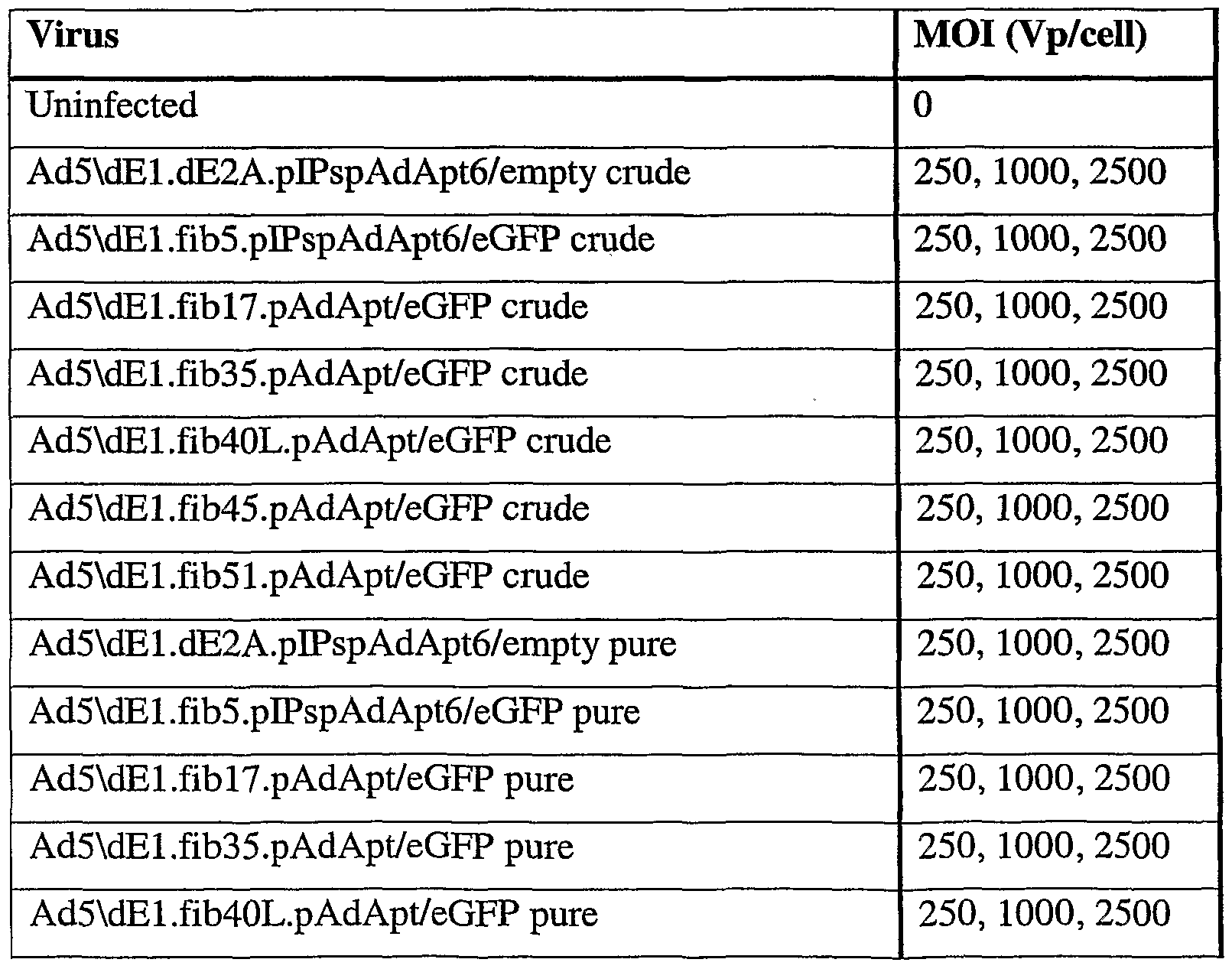


Claims
Priority Applications (4)
| Application Number | Priority Date | Filing Date | Title |
|---|---|---|---|
| NZ524907A NZ524907A (en) | 2000-09-25 | 2001-09-25 | Adenoviral vectors having tissue tropism for T-lymphocytes, B- and mast cells |
| AU2002220567A AU2002220567A1 (en) | 2000-09-25 | 2001-09-25 | Viral vectors having tissue tropism for T-lymphocytes, B- and mast cells |
| EP01985274A EP1320622A2 (en) | 2000-09-25 | 2001-09-25 | Viral vectors having tissue tropism for t-lymphocytes, b- and mast cells |
| CA002423091A CA2423091A1 (en) | 2000-09-25 | 2001-09-25 | Viral vectors having tissue tropism for t-lymphocytes, b- and mast cells |
Applications Claiming Priority (4)
| Application Number | Priority Date | Filing Date | Title |
|---|---|---|---|
| EP00203375.1 | 2000-09-25 | ||
| EP00203375A EP1191105A1 (en) | 2000-09-25 | 2000-09-25 | Gene delivery vectors provided with a tissue tropism for T-lymphocytes |
| US29040301P | 2001-05-11 | 2001-05-11 | |
| US60/290,403 | 2001-05-11 |
Publications (2)
| Publication Number | Publication Date |
|---|---|
| WO2002024933A2 true WO2002024933A2 (en) | 2002-03-28 |
| WO2002024933A3 WO2002024933A3 (en) | 2002-07-11 |
Family
ID=8172083
Family Applications (1)
| Application Number | Title | Priority Date | Filing Date |
|---|---|---|---|
| PCT/EP2001/011086 Ceased WO2002024933A2 (en) | 2000-09-25 | 2001-09-25 | Viral vectors having tissue tropism for t-lymphocytes, b- and mast cells |
Country Status (6)
| Country | Link |
|---|---|
| EP (2) | EP1191105A1 (en) |
| AU (1) | AU2002220567A1 (en) |
| CA (1) | CA2423091A1 (en) |
| NZ (1) | NZ524907A (en) |
| WO (1) | WO2002024933A2 (en) |
| ZA (1) | ZA200302124B (en) |
Cited By (5)
| Publication number | Priority date | Publication date | Assignee | Title |
|---|---|---|---|---|
| WO2002029073A3 (en) * | 2000-10-06 | 2002-10-03 | Crucell Holland Bv | Gene delivery vectors for stem cells |
| WO2008012682A3 (en) * | 2006-07-28 | 2008-06-19 | Csiro Livestock Ind | Methods and compositions for increasing tissue tropism of recombinant adenoviral vectors |
| WO2018064523A1 (en) * | 2016-09-30 | 2018-04-05 | Genvec, Inc. | Adenovectors for delivery of therapeutic genetic material into t cells |
| US10647963B2 (en) | 2013-04-18 | 2020-05-12 | Tilt Biotherapeutics Oy | Enhanced adoptive cell therapy |
| US12421502B2 (en) | 2013-04-18 | 2025-09-23 | Tilt Biotherapeutics Oy | Enhanced adoptive cell therapy |
Families Citing this family (1)
| Publication number | Priority date | Publication date | Assignee | Title |
|---|---|---|---|---|
| EP1811031A1 (en) | 2006-01-18 | 2007-07-25 | Millegen | Method for selecting a peptide or polypeptide which binds to a target molecule |
Family Cites Families (4)
| Publication number | Priority date | Publication date | Assignee | Title |
|---|---|---|---|---|
| US20030017138A1 (en) * | 1998-07-08 | 2003-01-23 | Menzo Havenga | Chimeric adenoviruses |
| EP1141357A1 (en) * | 1999-01-14 | 2001-10-10 | Novartis AG | Adenovirus vectors, packaging cell lines, compositions, and methods for preparation and use |
| ES2372823T3 (en) * | 1999-05-17 | 2012-01-26 | Crucell Holland B.V. | ADENOVIRUS RECOMBINANT OF SEROTIPO AD11. |
| EP1181382B1 (en) * | 1999-06-01 | 2005-03-23 | The University of Washington | Recombinant adenoviral vectors expressing chimeric fiber proteins for cell specific infection and genome integration |
-
2000
- 2000-09-25 EP EP00203375A patent/EP1191105A1/en not_active Withdrawn
-
2001
- 2001-09-25 NZ NZ524907A patent/NZ524907A/en unknown
- 2001-09-25 WO PCT/EP2001/011086 patent/WO2002024933A2/en not_active Ceased
- 2001-09-25 EP EP01985274A patent/EP1320622A2/en not_active Withdrawn
- 2001-09-25 CA CA002423091A patent/CA2423091A1/en not_active Abandoned
- 2001-09-25 ZA ZA200302124A patent/ZA200302124B/en unknown
- 2001-09-25 AU AU2002220567A patent/AU2002220567A1/en not_active Abandoned
Cited By (9)
| Publication number | Priority date | Publication date | Assignee | Title |
|---|---|---|---|---|
| WO2002029073A3 (en) * | 2000-10-06 | 2002-10-03 | Crucell Holland Bv | Gene delivery vectors for stem cells |
| WO2008012682A3 (en) * | 2006-07-28 | 2008-06-19 | Csiro Livestock Ind | Methods and compositions for increasing tissue tropism of recombinant adenoviral vectors |
| JP2009544303A (en) * | 2006-07-28 | 2009-12-17 | コモンウェルス サイエンティフィック アンド インダストリアル リサーチ オーガニゼイション | Methods and compositions for increasing tissue affinity of recombinant adenoviral vectors |
| US8163544B2 (en) | 2006-07-28 | 2012-04-24 | Commonwealth Scientific And Industrial Research Organisation | Methods and compositions for increasing tissue tropism of recombinant adenoviral vectors |
| US10647963B2 (en) | 2013-04-18 | 2020-05-12 | Tilt Biotherapeutics Oy | Enhanced adoptive cell therapy |
| US10787645B2 (en) | 2013-04-18 | 2020-09-29 | Tilt Biotherapeutics Oy | Enhanced adoptive cell therapy |
| US12421502B2 (en) | 2013-04-18 | 2025-09-23 | Tilt Biotherapeutics Oy | Enhanced adoptive cell therapy |
| WO2018064523A1 (en) * | 2016-09-30 | 2018-04-05 | Genvec, Inc. | Adenovectors for delivery of therapeutic genetic material into t cells |
| US11155832B2 (en) | 2016-09-30 | 2021-10-26 | Genvec, Inc. | Adenovectors for delivery of therapeutic genetic material into T cells |
Also Published As
| Publication number | Publication date |
|---|---|
| CA2423091A1 (en) | 2002-03-28 |
| AU2002220567A1 (en) | 2002-04-02 |
| WO2002024933A3 (en) | 2002-07-11 |
| EP1191105A1 (en) | 2002-03-27 |
| NZ524907A (en) | 2004-11-26 |
| EP1320622A2 (en) | 2003-06-25 |
| ZA200302124B (en) | 2004-03-17 |
Similar Documents
| Publication | Publication Date | Title |
|---|---|---|
| US7332337B2 (en) | Viral vectors having tissue tropism for T-lymphocytes, B- and mast cells | |
| EP1681353B1 (en) | Adenovirus derived gene delivery vehicles comprising at least one element of adenovirus type 35 | |
| US6913922B1 (en) | Serotype of adenovirus and uses thereof | |
| US6803234B2 (en) | Gene delivery vectors with cell type specificity for primary human chondrocytes | |
| US7968087B2 (en) | Gene delivery vectors provided with a tissue tropism for smooth muscle cells, and/or endothelial cells | |
| CA2451577C (en) | Gene delivery vectors with cell type specificity for mesenchymal stem cells | |
| AU2002315947A1 (en) | Adenoviral vectors with cell type specificity for mesenchymal cells | |
| US20060104953A1 (en) | Methods and means for enhancing skin transplantation using gene delivery vehicles having tropism for primary fibroblasts, as well as other uses thereof | |
| EP1020529B1 (en) | Gene delivery vectors provided with a tissue tropism for smooth muscle cells, and/or endothelial cells | |
| AU770780B2 (en) | Gene delivery vectors provided with a tissue tropism for smooth muscle cells, and/or endothelial cells | |
| WO2002024933A2 (en) | Viral vectors having tissue tropism for t-lymphocytes, b- and mast cells | |
| EP1195440A1 (en) | Gene delivery vectors for stem cells | |
| US20040043489A1 (en) | Gene delivery vectors provided with a tissue tropism for dendritic cells and methods of use | |
| WO2002029073A2 (en) | Gene delivery vectors for stem cells |
Legal Events
| Date | Code | Title | Description |
|---|---|---|---|
| AK | Designated states |
Kind code of ref document: A2 Designated state(s): AE AG AL AM AT AU AZ BA BB BG BR BY BZ CA CH CN CO CR CU CZ DE DK DM DZ EC EE ES FI GB GD GE GH GM HR HU ID IL IN IS JP KE KG KP KR KZ LC LK LR LS LT LU LV MA MD MG MK MN MW MX MZ NO NZ PH PL PT RO RU SD SE SG SI SK SL TJ TM TR TT TZ UA UG US UZ VN YU ZA ZW |
|
| AL | Designated countries for regional patents |
Kind code of ref document: A2 Designated state(s): GH GM KE LS MW MZ SD SL SZ TZ UG ZW AM AZ BY KG KZ MD RU TJ TM AT BE CH CY DE DK ES FI FR GB GR IE IT LU MC NL PT SE TR BF BJ CF CG CI CM GA GN GQ GW ML MR NE SN TD TG |
|
| 121 | Ep: the epo has been informed by wipo that ep was designated in this application | ||
| AK | Designated states |
Kind code of ref document: A3 Designated state(s): AE AG AL AM AT AU AZ BA BB BG BR BY BZ CA CH CN CO CR CU CZ DE DK DM DZ EC EE ES FI GB GD GE GH GM HR HU ID IL IN IS JP KE KG KP KR KZ LC LK LR LS LT LU LV MA MD MG MK MN MW MX MZ NO NZ PH PL PT RO RU SD SE SG SI SK SL TJ TM TR TT TZ UA UG US UZ VN YU ZA ZW |
|
| AL | Designated countries for regional patents |
Kind code of ref document: A3 Designated state(s): GH GM KE LS MW MZ SD SL SZ TZ UG ZW AM AZ BY KG KZ MD RU TJ TM AT BE CH CY DE DK ES FI FR GB GR IE IT LU MC NL PT SE TR BF BJ CF CG CI CM GA GN GQ GW ML MR NE SN TD TG |
|
| DFPE | Request for preliminary examination filed prior to expiration of 19th month from priority date (pct application filed before 20040101) | ||
| WWE | Wipo information: entry into national phase |
Ref document number: 2002220567 Country of ref document: AU |
|
| WWE | Wipo information: entry into national phase |
Ref document number: 2003/02124 Country of ref document: ZA Ref document number: 200302124 Country of ref document: ZA |
|
| WWE | Wipo information: entry into national phase |
Ref document number: 2423091 Country of ref document: CA |
|
| WWE | Wipo information: entry into national phase |
Ref document number: 2001985274 Country of ref document: EP Ref document number: 524907 Country of ref document: NZ |
|
| WWP | Wipo information: published in national office |
Ref document number: 2001985274 Country of ref document: EP |
|
| REG | Reference to national code |
Ref country code: DE Ref legal event code: 8642 |
|
| WWP | Wipo information: published in national office |
Ref document number: 524907 Country of ref document: NZ |
|
| WWG | Wipo information: grant in national office |
Ref document number: 524907 Country of ref document: NZ |
|
| NENP | Non-entry into the national phase |
Ref country code: JP |
|
| WWW | Wipo information: withdrawn in national office |
Ref document number: 2001985274 Country of ref document: EP |