US9795643B2 - Oncolytic measles virus - Google Patents
Oncolytic measles virus Download PDFInfo
- Publication number
- US9795643B2 US9795643B2 US14/719,906 US201514719906A US9795643B2 US 9795643 B2 US9795643 B2 US 9795643B2 US 201514719906 A US201514719906 A US 201514719906A US 9795643 B2 US9795643 B2 US 9795643B2
- Authority
- US
- United States
- Prior art keywords
- cells
- measles virus
- seq
- scd
- suicide gene
- Prior art date
- Legal status (The legal status is an assumption and is not a legal conclusion. Google has not performed a legal analysis and makes no representation as to the accuracy of the status listed.)
- Active
Links
- XAZKFISIRYLAEE-UHFFFAOYSA-N CC1CC(C)CC1 Chemical compound CC1CC(C)CC1 XAZKFISIRYLAEE-UHFFFAOYSA-N 0.000 description 1
Images


Classifications
-
- A—HUMAN NECESSITIES
- A61—MEDICAL OR VETERINARY SCIENCE; HYGIENE
- A61K—PREPARATIONS FOR MEDICAL, DENTAL OR TOILETRY PURPOSES
- A61K35/00—Medicinal preparations containing materials or reaction products thereof with undetermined constitution
- A61K35/66—Microorganisms or materials therefrom
- A61K35/76—Viruses; Subviral particles; Bacteriophages
- A61K35/768—Oncolytic viruses not provided for in groups A61K35/761 - A61K35/766
-
- A—HUMAN NECESSITIES
- A61—MEDICAL OR VETERINARY SCIENCE; HYGIENE
- A61K—PREPARATIONS FOR MEDICAL, DENTAL OR TOILETRY PURPOSES
- A61K31/00—Medicinal preparations containing organic active ingredients
- A61K31/70—Carbohydrates; Sugars; Derivatives thereof
- A61K31/7088—Compounds having three or more nucleosides or nucleotides
-
- A—HUMAN NECESSITIES
- A61—MEDICAL OR VETERINARY SCIENCE; HYGIENE
- A61P—SPECIFIC THERAPEUTIC ACTIVITY OF CHEMICAL COMPOUNDS OR MEDICINAL PREPARATIONS
- A61P35/00—Antineoplastic agents
-
- C—CHEMISTRY; METALLURGY
- C12—BIOCHEMISTRY; BEER; SPIRITS; WINE; VINEGAR; MICROBIOLOGY; ENZYMOLOGY; MUTATION OR GENETIC ENGINEERING
- C12N—MICROORGANISMS OR ENZYMES; COMPOSITIONS THEREOF; PROPAGATING, PRESERVING, OR MAINTAINING MICROORGANISMS; MUTATION OR GENETIC ENGINEERING; CULTURE MEDIA
- C12N7/00—Viruses; Bacteriophages; Compositions thereof; Preparation or purification thereof
-
- C—CHEMISTRY; METALLURGY
- C12—BIOCHEMISTRY; BEER; SPIRITS; WINE; VINEGAR; MICROBIOLOGY; ENZYMOLOGY; MUTATION OR GENETIC ENGINEERING
- C12N—MICROORGANISMS OR ENZYMES; COMPOSITIONS THEREOF; PROPAGATING, PRESERVING, OR MAINTAINING MICROORGANISMS; MUTATION OR GENETIC ENGINEERING; CULTURE MEDIA
- C12N9/00—Enzymes; Proenzymes; Compositions thereof; Processes for preparing, activating, inhibiting, separating or purifying enzymes
- C12N9/10—Transferases (2.)
- C12N9/1048—Glycosyltransferases (2.4)
- C12N9/1077—Pentosyltransferases (2.4.2)
-
- C—CHEMISTRY; METALLURGY
- C12—BIOCHEMISTRY; BEER; SPIRITS; WINE; VINEGAR; MICROBIOLOGY; ENZYMOLOGY; MUTATION OR GENETIC ENGINEERING
- C12N—MICROORGANISMS OR ENZYMES; COMPOSITIONS THEREOF; PROPAGATING, PRESERVING, OR MAINTAINING MICROORGANISMS; MUTATION OR GENETIC ENGINEERING; CULTURE MEDIA
- C12N9/00—Enzymes; Proenzymes; Compositions thereof; Processes for preparing, activating, inhibiting, separating or purifying enzymes
- C12N9/14—Hydrolases (3)
- C12N9/78—Hydrolases (3) acting on carbon to nitrogen bonds other than peptide bonds (3.5)
-
- C—CHEMISTRY; METALLURGY
- C07—ORGANIC CHEMISTRY
- C07K—PEPTIDES
- C07K2319/00—Fusion polypeptide
-
- C—CHEMISTRY; METALLURGY
- C12—BIOCHEMISTRY; BEER; SPIRITS; WINE; VINEGAR; MICROBIOLOGY; ENZYMOLOGY; MUTATION OR GENETIC ENGINEERING
- C12N—MICROORGANISMS OR ENZYMES; COMPOSITIONS THEREOF; PROPAGATING, PRESERVING, OR MAINTAINING MICROORGANISMS; MUTATION OR GENETIC ENGINEERING; CULTURE MEDIA
- C12N2760/00—MICROORGANISMS OR ENZYMES; COMPOSITIONS THEREOF; PROPAGATING, PRESERVING, OR MAINTAINING MICROORGANISMS; MUTATION OR GENETIC ENGINEERING; CULTURE MEDIA ssRNA viruses negative-sense
- C12N2760/00011—Details
- C12N2760/18011—Paramyxoviridae
- C12N2760/18411—Morbillivirus, e.g. Measles virus, canine distemper
- C12N2760/18421—Viruses as such, e.g. new isolates, mutants or their genomic sequences
-
- C—CHEMISTRY; METALLURGY
- C12—BIOCHEMISTRY; BEER; SPIRITS; WINE; VINEGAR; MICROBIOLOGY; ENZYMOLOGY; MUTATION OR GENETIC ENGINEERING
- C12N—MICROORGANISMS OR ENZYMES; COMPOSITIONS THEREOF; PROPAGATING, PRESERVING, OR MAINTAINING MICROORGANISMS; MUTATION OR GENETIC ENGINEERING; CULTURE MEDIA
- C12N2760/00—MICROORGANISMS OR ENZYMES; COMPOSITIONS THEREOF; PROPAGATING, PRESERVING, OR MAINTAINING MICROORGANISMS; MUTATION OR GENETIC ENGINEERING; CULTURE MEDIA ssRNA viruses negative-sense
- C12N2760/00011—Details
- C12N2760/18011—Paramyxoviridae
- C12N2760/18411—Morbillivirus, e.g. Measles virus, canine distemper
- C12N2760/18432—Use of virus as therapeutic agent, other than vaccine, e.g. as cytolytic agent
-
- C—CHEMISTRY; METALLURGY
- C12—BIOCHEMISTRY; BEER; SPIRITS; WINE; VINEGAR; MICROBIOLOGY; ENZYMOLOGY; MUTATION OR GENETIC ENGINEERING
- C12N—MICROORGANISMS OR ENZYMES; COMPOSITIONS THEREOF; PROPAGATING, PRESERVING, OR MAINTAINING MICROORGANISMS; MUTATION OR GENETIC ENGINEERING; CULTURE MEDIA
- C12N2760/00—MICROORGANISMS OR ENZYMES; COMPOSITIONS THEREOF; PROPAGATING, PRESERVING, OR MAINTAINING MICROORGANISMS; MUTATION OR GENETIC ENGINEERING; CULTURE MEDIA ssRNA viruses negative-sense
- C12N2760/00011—Details
- C12N2760/18011—Paramyxoviridae
- C12N2760/18411—Morbillivirus, e.g. Measles virus, canine distemper
- C12N2760/18441—Use of virus, viral particle or viral elements as a vector
- C12N2760/18443—Use of virus, viral particle or viral elements as a vector viral genome or elements thereof as genetic vector
-
- C—CHEMISTRY; METALLURGY
- C12—BIOCHEMISTRY; BEER; SPIRITS; WINE; VINEGAR; MICROBIOLOGY; ENZYMOLOGY; MUTATION OR GENETIC ENGINEERING
- C12N—MICROORGANISMS OR ENZYMES; COMPOSITIONS THEREOF; PROPAGATING, PRESERVING, OR MAINTAINING MICROORGANISMS; MUTATION OR GENETIC ENGINEERING; CULTURE MEDIA
- C12N2760/00—MICROORGANISMS OR ENZYMES; COMPOSITIONS THEREOF; PROPAGATING, PRESERVING, OR MAINTAINING MICROORGANISMS; MUTATION OR GENETIC ENGINEERING; CULTURE MEDIA ssRNA viruses negative-sense
- C12N2760/00011—Details
- C12N2760/18011—Paramyxoviridae
- C12N2760/18411—Morbillivirus, e.g. Measles virus, canine distemper
- C12N2760/18471—Demonstrated in vivo effect
-
- C—CHEMISTRY; METALLURGY
- C12—BIOCHEMISTRY; BEER; SPIRITS; WINE; VINEGAR; MICROBIOLOGY; ENZYMOLOGY; MUTATION OR GENETIC ENGINEERING
- C12N—MICROORGANISMS OR ENZYMES; COMPOSITIONS THEREOF; PROPAGATING, PRESERVING, OR MAINTAINING MICROORGANISMS; MUTATION OR GENETIC ENGINEERING; CULTURE MEDIA
- C12N2830/00—Vector systems having a special element relevant for transcription
- C12N2830/60—Vector systems having a special element relevant for transcription from viruses
-
- C—CHEMISTRY; METALLURGY
- C12—BIOCHEMISTRY; BEER; SPIRITS; WINE; VINEGAR; MICROBIOLOGY; ENZYMOLOGY; MUTATION OR GENETIC ENGINEERING
- C12Y—ENZYMES
- C12Y305/00—Hydrolases acting on carbon-nitrogen bonds, other than peptide bonds (3.5)
- C12Y305/04—Hydrolases acting on carbon-nitrogen bonds, other than peptide bonds (3.5) in cyclic amidines (3.5.4)
- C12Y305/04001—Cytosine deaminase (3.5.4.1)
Definitions
- the present invention pertains to a pharmaceutical composition comprising a recombinant measles virus comprising a suicide gene for use in the treatment of malignant cells with primary or secondary resistances against an oncolytic measles virus without suicide gene activity. Further, the present invention pertains to a recombinant measles virus based on the genome of measles vaccine strain Schwarz comprising a suicide gene, which comprises a fusion of yeast cytosine deaminase and yeast uracil phosphoribosyltransferase, to a method and a kit for preparing the recombinant measles virus as claimed herein.
- Measles virus is an enveloped, single-stranded, negative-sense paramyxo-virus of the genus Morbillivirus that is causing the infectious measles disease, an infection of the respiratory system.
- the genome of the measles virus contains six genes encoding eight proteins: the nucleocapsid (N), phospho- (P), matrix (M), fusion (F), hemagglutinin (H) and large (L) proteins, and two accessory proteins, termed C and V.
- the virus enters the target cells via pH-independent membrane fusion.
- the H and F proteins are involved in receptor binding and membrane fusion, respectively.
- CD46 is ubiquitously present on nucleated primate cells, but is frequently over-expressed in tumors, and the signaling lymphocyte-activation molecule (SLAM) that is primarily located on B- and T-cells.
- SLAM signaling lymphocyte-activation molecule
- CD46 is a membrane-associated complement regulatory protein that protects human cells against autologous complement lysis by acting as a cofactor in the proteolytic inactivation of C3b and C4b complement products, thus providing protection for tumor cells against complement-mediated lysis.
- Receptor recognition by the H protein leads to conformational changes of the F protein resulting in fusion with target cell membranes and subsequent viral entry.
- Infected cells including tumor cells, express the viral F and H proteins on the cell surface. Recognition of the viral receptor in neighboring infected or uninfected cells similarly triggers cell-to-cell fusion. Therefore, the typical cytopathic effect of measles virus is the formation of giant mononuclear cell aggregates (syncytia).
- measles virus preparations used for measles vaccines or for the research on oncolytic measles virus are based on an attenuated live measles virus derived from the so-called Edmonston vaccine strain, an isolate originally obtained in 1954, which was used to create the Edmonston-Enders cell line, and based on that, Edmonston A and B seed lines by serial passages on human cells and subsequent adaptation to chicken embryo fibroblastic (CEF) cells.
- CEF chicken embryo fibroblastic
- measles virus preparations are currently rescued from cell lines not being approved for vaccine production like chicken embryo fibroblastic (CEF) cells or 293 human embryonic kidney cells, both complicating and thereby increasing the costs for the large-scale production of recombinant measles virus particles in conformity with GMP requirements.
- CEF chicken embryo fibroblastic
- tumors derived from certain cell lines were resistant to treatment with oncolytic measles virus despite repeated virus injections (Peng K W, Facteau S, Wegman T, O'Kane D, Russell S J. Non-invasive in vivo monitoring of trackable viruses expressing soluble marker peptides. Nat Med. (2002); 8:527-31).
- a first object of the present invention is to provide an improved pharmaceutical composition comprising a recombinant measles virus with oncolytic activity against tumors that are resistant to measles virus of the prior art.
- the second object of the present invention is an improved pharmaceutical composition
- a recombinant measles virus having a viral genome corresponding to the genome of established attenuated live measles vaccines.
- the third object of the present invention is a safer and cheaper method for rescuing recombinant measles virus for use in the treatment of malignant cells.
- a pharmaceutical composition comprising a recombinant measles virus comprising a suicide gene for use in the treatment of malignant cells with primary or secondary resistances against an oncolytic measles virus without suicide gene activity.
- the invention of a recombinant measles virus encoding a suicide gene for use in the treatment of malignant cells with primary or secondary resistances against an oncolytic measles virus without suicide gene activity enables a much more efficient treatment of solid tumors which concomitantly allows the employment of a reduced number of infectious measles virus particles without any loss in anti-tumor efficiency. Thereby, costs of virotherapeutic therapy can be reduced significantly.
- the present invention relates to a recombinant measles virus based on measles vaccine strain Schwarz comprising a suicide gene, which comprises a fusion of a cytosine deaminase, particularly yeast cytosine deaminase, and a uracil phosphoribosyltransferase, particularly yeast uracil phosphoribosyltransferase, particularly wherein said suicide gene comprises a sequence according to SEQ-ID NO. 2.
- the present invention relates to a method of treatment of malignant cells with primary or secondary resistances against an oncolytic measles virus without suicide gene activity, comprising the step of administering a recombinant measles virus comprising a suicide gene according to the present invention to a patient in need thereof.
- the present invention relates to a method for generating the recombinant measles virus according to the present invention, comprising the step of (a) cloning (i) the genome of measles vaccine strain Schwarz, and (ii) a suicide gene, which comprises a fusion of a cytosine deaminase, particularly yeast cytosine deaminase, and a uracil phosphoribosyltransferase, particularly yeast uracil phosphoribosyltransferase, into a plasmid under the control of an RNA polymerase II promoter.
- the present invention relates to a kit comprising
- a plasmid comprising (i) the genome of measles vaccine strain Schwarz, and (ii) a suicide gene, which comprises a fusion of a cytosine deaminase, particularly yeast cytosine deaminase, and a uracil phosphoribosyltransferase, particularly yeast uracil phosphoribosyltransferase, under the control of an RNA polymerase II promoter, particularly wherein said suicide gene comprises a sequence according to SEQ-ID No. 2, particularly wherein the plasmid has the sequence according to SEQ-ID No. 4 SEQ-ID No. 8, or SEQ-ID No. 9, more particularly SEQ-ID No. 4; (b) at least one plasmid comprising measles virus helper genes N, P and L, in form of single genes, each under the control of an RNA polymerase II promoter.
- FIG. 1 shows the genetic sequence of measles vaccine strain Schwarz (SEQ-ID NO. 1).
- FIG. 2 shows genetic sequence of fusion of yeast cytosine deaminase and yeast uracil phosphoribosyltransferase (with linker sequence) (SEQ-ID NO. 2).
- FIG. 3 shows the complete genetic sequence of the plasmid pc3MerV2 Id-Trka coding for the basic recombinant measles virus engineered from the MeV Schwarz strain without any transgenes but with additional transcription cassette (SEQ-ID NO. 3).
- FIG. 4 shows the genetic sequence of vector pc3MerV2 Id-SCD (coding for MeV Id-SCD) (SEQ-ID NO. 4).
- FIG. 5 shows the genetic sequence of the helper plasmid required for the expression of the N gene (SEQ-ID No. 5), which is based on the CMV promoter variant construct pc3, encompassing CMV promoter nucleotides from ⁇ 301 until ⁇ 1 (+1 being defined as transcription initiation site) plus three nucleotides (TGG) additionally inserted after position ⁇ 1:
- FIG. 6 shows the genetic sequence of the helper plasmid required for the expression of the P gene (SEQ-ID No. 6), which is based on the CMV promoter variant construct pc3:
- FIG. 7 shows the genetic sequence of the helper plasmid required for the expression of the L gene (SEQ-ID No. 7), which is based on the CMV promoter variant construct pc3:
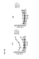
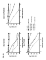
- FIG. 8 shows the results of an SRB proliferation assay of HuCCT1, RBE, and TFK-1 cells (human cholangiocarcinoma cells) treated with viral particles rescued from the plasmid pc3MerV2 Id-SCD (Id-SCD) and incubated with prodrug 5-FC.
- FIG. 9 shows the results of an LDH release assay of HuCCT1, RBE, and TFK-1 cells (human cholangiocarcinoma cells) treated with viral particles rescued from the plasmid pc3MerV2 Id-SCD (Id-SCD).
- FIG. 10 shows the results of an SRB proliferation assay of SAS and HTB-43 FaDu cells (human Head & Neck (H&N) cancer cells) treated with viral particles rescued from the plasmid pc3MerV2 Id-SCD (Id-SCD).
- FIG. 11 shows the results of an LDH release assay of SAS and HTB-43 FaDu cells (human H&N cancer cells) treated with viral particles rescued from the plasmid pc3MerV2 Id-SCD (Id-SCD).
- FIG. 12 shows the results of an SRB proliferation assay of A 673, BRZ, and SRH cells (human sarcoma cells) treated with viral particles rescued from the plasmid pc3MerV2 Id-SCD (Id-SCD).
- FIG. 13 shows the results of an LDH release assay of A 673 and SRH cells (human sarcoma cells) treated with viral particles rescued from pc3MerV2 Id-SCD (Id-SCD).
- FIG. 14 shows the results of an SRB proliferation assay of SRH cells treated with viral particles rescued from the plasmid pc3MerV2 Id-SCD (Id-SCD).
- FIG. 15 shows the results of an SRB proliferation assay of sarcoma tumor cells (cell lines CCS, LM) treated with viral particles rescued from the plasmid pc3MerV2 Id-SCD (Id-SCD).
- FIG. 16 shows the results of an SRB proliferation assay of sarcoma tumor cells (cell lines STO, ZAF, KD) treated with viral particles rescued from the plasmid pc3MerV2 Id-SCD (Id-SCD).
- FIG. 17 shows the results of an SRB proliferation assay of glioblastoma tumor cells (cell lines LNT 229, LNT 229 CTS-1) treated with viral particles rescued from the plasmid pc3MerV2 Id-SCD (Id-SCD).
- FIG. 18 shows the results of an SRB proliferation assay of glioblastoma tumor cells (cell lines LN 18, LN 18 Apoptosis resistant) treated with viral particles rescued from the plasmid pc3MerV2 Id-SCD (Id-SCD).
- FIG. 19 shows the results of an SRB proliferation assay of renal cell carcinoma (ACHN), pulmonary adenocarcinoma (HOP-62) and melanoma (M14) tumor cells treated with viral particles rescued from the plasmid pc3MerV2 Id-SCD (Id-SCD).
- FIG. 20 shows the results of an SRB proliferation assay of colonic adenocarcinoma tumor cells (cell lines KM-12, HCT-15) treated with viral particles rescued from the plasmid pc3MerV2 Id-SCD (Id-SCD).
- FIG. 21 shows the effect of three different armed MeV vectors on Hep3B human hepatocellular carcinoma cells (vectors pc3MerV2 Id-SCD, pc3MerV2 Id-VP22SCD, and pMerV2 P-SCD); all experiments were performed in quadruplicates; experiments were repeated three times; values: mean+SEM.
- FIG. 22 shows the effect of three different armed MeV vectors on HepG2 human hepatocellular carcinoma cells (vectors pc3MerV2 Id-SCD, pc3MerV2 Id-VP22SCD, and pMerV2 P-SCD); all experiments were performed in quadruplicates; experiments were repeated three times; values: mean+SEM.
- FIG. 23 shows the effect of three different armed MeV vectors on PLC/PRF/5 human hepatocellular carcinoma cells (vectors pc3MerV2 Id-SCD, pc3MerV2 Id-VP22SCD, and pMerV2 P-SCD); all experiments were performed in quadruplicates; experiments were repeated three times; values: mean+SEM.
- FIG. 24 shows the results of a determination of tumor volumes in a xenograft animal HCC tumor model (Hep3B model).
- FIG. 25 shows the results of a determination of survival data in a xenograft animal HCC tumor model (Hep3B model).
- FIG. 26 shows the results of a determination of tumor volumes in a xenograft animal CC tumor model (TFK-1 model).
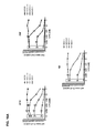
- FIG. 27 shows a schematic overview of viral cDNAs and the respective viral vectors. Plasmids (A) pc3MerV2 Id-SCD (20,841 bp), (B) pc3MerV2 Id-VP22SCD (21,759 bp), and (C) pMerV2 P-SCD (20,546 bp) are shown. Open reading frames are displayed as arrows (viral genes in white, transgenes in dark). Nontranslated regions and the plasmid backbone are depicted as straight lines.
- N Nucleocapsid protein
- P Phosphoprotein
- M Matrix protein
- F Fusion protein
- H Hemaggluttinin
- L Large protein
- SCD Super-cytosine deaminase
- VP22SCD Fusion of SCD and the herpes simplex virus protein VP22.
- FIG. 28 shows the genetic sequence of vector pMerV2 P-SCD (coding for MeV P-SCD) (SEQ-ID NO. 8).
- FIG. 29 shows the genetic sequence of vector pc3MerV2 Id-VP22SCD (coding for MeV Id-VP22SCD) (SEQ-ID NO. 9).
- the present invention relates to a pharmaceutical composition
- a pharmaceutical composition comprising a recombinant measles virus comprising a suicide gene for use in the treatment of malignant cells with primary or secondary resistances against an oncolytic measles virus without suicide gene activity.
- suicide gene refers to a gene, the expression of which in a host cell causes or results in a reduced viability of such host cells.
- the suicide gene will cause a cell to kill itself through apoptosis.
- expression of the suicide gene will result in an enzyme, which catalyzes the generation of a cytotoxic drug from a non-toxic prodrug.
- the malignant cells are identified by performing the following step:
- the recombinant measles virus is based on measles vaccine strain Schwarz.
- the oncolytic measles virus without suicide gene activity is from measles vaccine strain Schwarz. More particularly, the oncolytic measles virus without suicide gene activity has a sequence according to SEQ-ID No. 1 (see FIG. 1 ).
- the term “has a sequence according to SEQ-ID No. 1” depends on the context given and refers to either the DNA sequence as depicted in SEQ-ID No. 1 (cDNA of measles virus particles from the Schwarz strain), or to the corresponding RNA sequence, as found packaged in the viral particle rescued from a vector comprising such cDNA sequence.
- the suicide gene comprises a cytosine deaminase, particularly yeast cytosine deaminase.
- Cytosine deaminases particularly yeast cytosine deaminase, and their use as prodrug-converting enzyme in cancer gene therapy has been discussed and examined in various publications (see, for example: Kievit, E., Nyati, M. K., Ng, E., Stegman, L. D., Parsels, J., Ross, B. D., Rehemtulla, A., Lawrence, T. S. Yeast cytosine deaminase improves radiosensitization and bystander effect by 5-fluorocytosine of human colorectal cancer xenografts. Cancer Res. 60 (2000) 6649-55).
- the suicide gene further comprises a uracil phosphoribosyltransferase, particularly yeast uracil phosphoribosyltransferase.
- the suicide gene comprises a fusion of a cytosine deaminase, particularly yeast cytosine deaminase, and a uracil phosphoribosyltransferase, particularly yeast uracil phosphoribosyltransferase, called SCD (SuperCD).
- a cytosine deaminase particularly yeast cytosine deaminase
- a uracil phosphoribosyltransferase particularly yeast uracil phosphoribosyltransferase
- a fusion gene comprising a cytosine deaminase and a uracil phosphoribosyltransferase has been described for an adenoviral system (Erbs, P., Regulier, E., Kintz, J., Leroy, P., Poitevin, Y., Exinger, F., Jund, R., and Mehtali, M.
- adenoviral system Erbs, P., Regulier, E., Kintz, J., Leroy, P., Poitevin, Y., Exinger, F., Jund, R., and Mehtali, M.
- adenovirus-mediated transfer of a bifunctional yeast cytosine deaminase/uracil phosphoribosyltransferase fusion gene Cancer Res. 60 (2000) 3813-22).
- the suicide gene comprises a sequence according to SEQ-ID NO. 2 (see FIG. 2 ).
- the term “comprises a sequence according to SEQ-ID No. . . . ” depends on the context given and refers to either the DNA sequence as depicted in said SEQ-ID No., or to the corresponding RNA sequence, as found packaged in the viral particle rescued from a vector comprising such DNA sequence.
- the recombinant measles virus comprises an RNA sequence corresponding to SEQ-ID No. 3 (see FIG. 3 ), SEQ-ID No. 4 (see FIG. 4 ), SEQ-ID No. 8 (see FIG. 28 ), or SEQ-ID No. 9 (see FIG. 29 ), particularly SEQ-ID No. 4.
- the malignant cells are additionally non-responsive to chemotherapeutics and/or radiation therapy.
- the malignant cells are selected from the list of: malignant cells from cholangiocarcinoma, head and neck cancer, and sarcoma.
- the pharmaceutical composition is for use in a treatment, which is a repeated treatment, particularly every week, or every two weeks, or every three weeks, or every four weeks, employing such a recombinant measles virus comprising a suicide gene for use in the treatment of malignant cells with primary or secondary resistances against an oncolytic measles virus without suicide gene activity.
- the present invention relates to a recombinant measles virus based on measles vaccine strain Schwarz encoding a suicide gene, which comprises a fusion of a cytosine deaminase, particularly a yeast cytosine deaminase, and a uracil phosphoribosyltransferase, particularly a yeast uracil phosphoribosyltransferase.
- the suicide gene comprises a sequence according to SEQ-ID NO. 2 (see FIG. 2 ).
- the recombinant measles virus comprises an RNA sequence according to SEQ-ID No. 3 (see FIG. 3 ), SEQ-ID No. 4 (see FIG. 4 ), SEQ-ID No. 8 (see FIG. 28 ), or SEQ-ID No. 9 (see FIG. 29 ), particularly SEQ-ID No. 4.
- the present invention relates to a method of treatment of malignant cells with primary or secondary resistances against an oncolytic measles virus without suicide gene activity, comprising the step of administering a recombinant measles virus comprising a suicide gene according to the present invention to a patient in need thereof.
- the treatment is the treatment of a cholangiocarcinoma, head and neck cancer, or sarcoma.
- the method of treatment is a repeated treatment, particularly every week, or every two weeks, or every three weeks, or every four weeks.
- the present invention relates to a method for generating the recombinant measles virus according to the present invention, comprising the step of
- RNA polymerase II promoter system for the efficient expression of antigenomic RNA (cRNA) from cDNA of Mononegavirales has been shown for Borna disease virus (BDV) and measles virus (Martin, A., Staeheli, P., and Schneider, U. RNA Polymerase II-Controlled Expression of Antigenomic RNA Enhances the Rescue Efficacies of Two Different Members of the Mononegavirales Independently of the Site of Viral Genome Replication, J. Virol. 80 (2006) 5708-5715).
- the suicide gene comprises a cytosine deaminase, particularly yeast cytosine deaminase.
- the suicide gene further comprises a uracil phosphoribosyltransferase, particularly yeast uracil phosphoribosyltransferase.
- the suicide gene comprises a fusion of a cytosine deaminase, particularly yeast cytosine deaminase, and a uracil phosphoribosyltransferase, particularly yeast uracil phosphoribosyltransferase.
- the suicide gene comprises a sequence according to SEQ-ID NO. 2 (see FIG. 2 ).
- the method further comprises the step of
- the viral helper genes N, P and L are each cloned into a separate vector under the control of an RNA polymerase II promoter, particularly a plasmid vector, resulting in (i) a plasmid encoding the measles virus helper gene N, (ii) a plasmid encoding the measles virus helper gene P, and (iii) a plasmid encoding the measles virus helper gene L.
- steps (a) and/or (b), or (a) and/or (b) to (d), further comprise the step of removing putative splicing sequences from said genome and/or said helper genes.
- step (a) results in the plasmid having the sequence according to SEQ-ID No. 4 (see FIG. 4 ), SEQ-ID No. 8 (see FIG. 28 ), or SEQ-ID No. 9 (see FIG. 29 ), particularly SEQ-ID No. 4.
- steps (b) to (d) result in plasmids having the sequences according to SEQ-ID No. 5, SEQ-ID No. 6, and SEQ-ID No. 7 (see FIGS. 5 to 7 ).
- the method according to the present invention further comprises the step of
- step (e) transfecting host cells with plasmids of steps (a) and (b), particularly host cells from a certified cell line approved for vaccine production, particularly from Vero or MRC-5 cell lines.
- Genetic stability includes questions related to the potential reversion of the vaccine strain to more virulent forms, the recombination with other viral sequences to produce potentially pathogenic viruses, and any genetic drift that could result in decrease of immunogenicity and efficacy.
- propagation of live vaccine strains in Vero and MRC-5 cell lines maintains genetic stability of the vaccine strains (see, for example, Laassri M, Meseda C A, Williams O, Merchlinsky M, Weir J P, Chumakov K., Microarray assay for evaluation of the genetic stability of modified vaccinia virus Ankara B5R gene. J. Med. Virol. 79 (2007) 791-802).
- the method further comprises the step of
- step (f) rescuing recombinant measles virus from the host cell transfected in step (e).
- the present invention relates to a kit comprising
- a plasmid comprising (i) the genome of measles vaccine strain Schwarz, and (ii) a suicide gene, which comprises a fusion of a cytosine deaminase, particularly yeast cytosine deaminase, and a uracil phosphoribosyltransferase, particularly yeast uracil phosphoribosyltransferase, under the control of an RNA polymerase II promoter, particularly wherein said suicide gene comprises a sequence according to SEQ-ID No. 2 (see FIG. 2 ), particularly wherein the plasmid has the sequence according to SEQ-ID No. 4 (see FIG. 4 ) SEQ-ID No. 8 (see FIG.
- SEQ-ID No. 9 see FIG. 29 ), more particularly SEQ-ID No. 4; (b) at least one plasmid comprising measles virus helper genes N, P, and L, each in form of a single gene under the control of an RNA polymerase II promoter.
- the viral helper genes N, P, and L are each cloned into a separate plasmid, each under the control of an RNA polymerase II promoter particularly wherein the plasmids have the sequences according to SEQ-ID No. 5, SEQ-ID No. 6, and SEQ-ID No. 7 (see FIGS. 5 to 7 ).
- pc3MerV2 Id-Trka The sequence of pc3MerV2 Id-Trka (see FIG. 1 ) can be described as follows with reference to the position of the nucleotides (nt); UTR—untranslated region; ORF—open reading frame:
- nt 1-55 MeV leader sequence
- nt 56-66 gene start for transgene transcription (gene start sequence obtained from the MeV N gene)
- nt 67-103 5′-UTR (5′-UTR sequence obtained from the MeV N gene)
- nt 103-108 cloning site, exhibiting recognition site for restriction endonuclease Xhol (C′TCGAG)
- nt 109-114 cloning site, exhibiting recognition sites for restriction endonuclease Paul (G′CGCGC) or Ascl (GG′CGCGCC); both recognition site represent unique sites being present only once in the complete vector sequence
- nt 115-126 3′-UTR derived from MeV N gene
- nt 127-136 gene end for transgene transcription (obtained from the MeV N gene)
- plasmid pUC-SCD For generation of the recombinant MeV Id-SCD (Id-SCD) measles virus vector, plasmid pUC-SCD, encoding the SCD suicide fusion gene, was digested with restriction endonuclease MluI and the fragment containing the open reading frame of the SCD suicide fusion gene was ligated into basic vector pc3MerV2 Id-Trka (derivative of parental vector pc3 encompassing the optimized (shortened and modified) CMV RNA polymerase II (Pol II) promoter) which had been linearized with restriction endonuclease AscI. The correct integration of the fragment was verified by restriction digest and sequencing. Infectious viral particles were successfully produced by the subsequent rescue procedure.
- pc3MerV2 Id-SCD (SEQ-ID NO. 4).
- the sequence of pc3MerV2 Id-SCD (see FIG. 2 ) can be described as follows with reference to the position of the nt:
- nt 1-55 MeV leader sequence
- nt 56-66 gene start for transgene transcription (gene start sequence obtained from the MeV N gene)
- nt 67-103 5′-UTR (5′-UTR sequence obtained from the MeV N gene)
- nt 103-108 cloning site, exhibiting recognition site for restriction endonuclease Xhol (C′TCGAG)
- nt 1243-1248 cloning sites, exhibiting recognition sites for restriction endonucleases Mlul (A′CGCGT) + Paul (A′CGCGC) nt 1249-1260: 3′-UTR nt
- helper plasmids carrying either the N, the P, or the L gene, respectively were performed essentially as described recently (Martin A, Staeheli P, Schneider U. J Virol. 2006; 80:5708-15), but with the following important modifications:
- Example 4 Rescue of Measles Virus Particles from MeV Id-SCD (Id-SCD) Vector
- Vero cells ATCC CCL-81 were seeded in a 6-well plate at a density of 4 ⁇ 10 5 cells/well. On day 1, the Vero cells were transfected using the following transfection conditions
- the cells were washed two times with PBS. After addition of 1.8 ml DMEM+2% FCS+PS, the transfection mixture was added drop wise to the cells, and the plate was swirled.
- the cell culture was incubated at 37° C. and 5% CO 2 .
- Vero cells were plated in 10 cm dishes for overlay (one confluent T75 in 10 ml, seed approximately 0.5 ml per dish).
- Vero cells were plated in a 6-well plate for passage 0 of virus (P.0).
- FIG. 8 shows the results of an SRB proliferation assay of HuCCT1, RBE, and TFK-1 cells (human cholangiocarcinoma cells) treated with MeV Id-SCD viral particles rescued from the plasmid pc3MerV2 Id-SCD (Id-SCD) and incubated with prodrug 5-FC.
- SRB proliferation assay cells were seeded on day 0. and infected after 24 h with either 0.001, 0.01, 0.1, 1 or 10 MOI (see the individual experiments and/or accompanying Figures) of viral particles, and in the case of prodrug treatment, 5-FC was added 3 h post infection.
- the SRB assay was performed 96 h post infection.
- HuCCT1 cells being infected with MeV Id-SCD (MOI 1) and then cultivated without the addition of the prodrug 5-FC demonstrated a loss in cell mass after 96 h only in the range of 42% in comparison to non-infected control cells (set to a cell mass of 100%).
- HuCCT1 cells display a primary resistance to measles virus of the prior art (no employment of an additional suicide gene function).
- FIG. 9 shows the results of an LDH release assay of HuCCT1, RBE, and TFK-1 cells (human cholangiocarcinoma cells) treated with MeV Id-SCD viral particles rescued from the plasmid pc3MerV2 Id-SCD (Id-SCD) and incubated with prodrug 5-FC.
- HuCCT1 cells being infected with MeV Id-SCD (MOI 1) and then cultivated without the addition of the prodrug 5-FC demonstrated a release of the enzyme LDH after 96 h only in the range of 31% in comparison to non-infected control cells, respectively. Thereby, it again was demonstrated that HuCCT1 cells display a primary resistance to measles virus of the prior art (no employment of an additional suicide gene function).
- MeV Id-SCD MOI 1
- MOI 1 MeV Id-SCD (MOI 1)-infected HuCCT1 cells were cultivated with the addition of the prodrug 5-FC (ranging from 10 ⁇ 4 to 10 0 mM)
- FIG. 10 shows the results of an SRB proliferation assay of SAS and HTB-43 FaDu cells (human Head & Neck (H&N) cancer cells) treated with MeV Id-SCD viral particles rescued from the plasmid pc3MerV2 Id-SCD (Id-SCD) and incubated with prodrug 5-FC.
- SAS and HTB-43 FaDu cells human Head & Neck (H&N) cancer cells
- MeV Id-SCD viral particles rescued from the plasmid pc3MerV2 Id-SCD (Id-SCD) and incubated with prodrug 5-FC.
- SAS cells infected with MeV Id-SCD (MOI 1) and then cultivated without the addition of the prodrug 5-FC demonstrated no loss in cell mass at all (0%) after 96 h in comparison to non-infected control cells.
- MeV Id-SCD (MOI 1)-infected SAS cells were cultivated with the addition of the prodrug 5-FC (ranging from 10 ⁇ 4 to 10 0 mM) a loss in cell mass after 96 h was demonstrated in the range of up to 83% in comparison to non-infected control cells, respectively.
- FIG. 11 shows the results of an LDH release assay of SAS and HTB-43 FaDu cells (human H&N cancer cells) treated with MeV Id-SCD viral particles rescued from the plasmid pc3MerV2 Id-SCD (Id-SCD) and incubated with prodrug 5-FC.
- Example 9 SRB Proliferation Assay of A 673, BRZ, and SRH Cells
- FIG. 12 shows the results of an SRB proliferation assay of A 673, BRZ, and SRH cells (human sarcoma cells) treated with MeV Id-SCD viral particles rescued from the plasmid pc3MerV2 Id-SCD (Id-SCD) and incubated with prodrug 5-FC.
- SRH cells being infected with MeV Id-SCD (MOI 1) and then cultivated without the addition of the prodrug 5-FC demonstrated a loss in cell mass after 96 h only in the range of 10% in comparison to non-infected control cells.
- SRH cells display a primary resistance to measles virus of the prior art (no employment of an additional suicide gene function).
- MeV Id-SCD-infected SRH cells (MOI 1) were cultivated with the addition of the prodrug 5-FC (ranging from 10 ⁇ 4 to 10 0 mM) a loss in cell mass after 96 h was demonstrated in the range of up to 55% in comparison to non-infected control cells.
- FIG. 13 shows the results of an LDH release assay of A 673 and SRH cells (human sarcoma cells) treated with MeV Id-SCD viral particles rescued from pc3MerV2 Id-SCD (Id-SCD) and incubated with prodrug 5-FC.
- SRH cells being infected with MeV Id-SCD (MOI 1) and then cultivated without the addition of the prodrug 5-FC demonstrated a release of the enzyme LDH after 96 h only in the range of 23% in comparison to non-infected control cells.
- MeV Id-SCD (MOI 1)-infected SRH cells were cultivated with the addition of the prodrug 5-FC (ranging from 10 ⁇ 4 to 10 0 mM) an increase in the release of the enzyme LDH of 31% after 96 h was demonstrated in comparison to non-infected control cells (set to a cell mass of 100%).
- FIG. 14 shows the results of an SRB proliferation assay of SRH cells treated with MeV Id-SCD viral particles rescued from the plasmid pc3MerV2 Id-SCD (Id-SCD) at an elevated MOI of 10 (usually, a MOI of 1 is used; here increase in “viral load” was employed to overcome phenomena of resistance observed at MOI 1 in the presence of the prodrug 5-FC).
- SRH cells being infected with MeV Id-SCD at MOI 10 and then cultivated without the addition of the prodrug 5-FC demonstrated a loss in cell mass after 96 h in the range of now 68% in comparison to non-infected control cells, which is significantly improved in comparison to the results obtained at MOI 1, at which only 28% of loss in cell mass was obtained in comparison to non-infected control cells.
- MeV Id-SCD (MOI 10)-infected SRH cells were cultivated with the addition of the prodrug 5-FC (ranging from 10-4 to 10 0 mM) a loss in cell mass after 96 h was demonstrated in the range of up to 89% in comparison to non-infected control cells.
- Example 12 SRB Proliferation Assay of Sarcoma Tumor Cells (Cell Lines CCS, LM)
- FIG. 15 shows the results of an SRB proliferation assay of sarcoma tumor cells (cell lines CCS, LM) treated with viral particles rescued from the plasmid pc3MerV2 Id-SCD (Id-SCD) and incubated with prodrug 5-FC.
- CCS or LM cells infected with vector MeV Id-SCD at MOI 1 and then cultivated without the addition of the prodrug 5-FC demonstrated a loss in cell mass after 96 h in the range of 5% or 25%, respectively, in comparison to non-infected control cells (values encircled by the rectangle placed on the left hand side of the graphics).
- both CCS and LM cells display a primary resistance to measles virus of the prior art (no employment of an additional suicide gene function).
- MeV Id-SCD-infected CCS or LM cells (MOI 1) were cultivated with the addition of the prodrug 5-FC (ranging from 10 ⁇ 4 to 10 0 mM) a loss in cell mass after 96 h was demonstrated in the range of up to 79% or 94%, respectively, in comparison to non-infected control cells.
- Example 13 SRB Proliferation Assay of Sarcoma Tumor Cells (Cell Lines STO, ZAF, KD)
- FIG. 16 shows the results of an SRB proliferation assay of sarcoma tumor cells (cell lines STO, ZAF, KD) treated with MeV Id-SCD viral particles rescued from the plasmid pc3MerV2 Id-SCD (Id-SCD) and incubated with prodrug 5-FC.
- STO, ZAF, or KD cells do not display a primary resistance to measles virus of the prior art (no employment of an additional suicide gene function).
- MeV Id-SCD-infected STO, ZAF, or KD cells MOI 1
- a loss in cell mass after 96 h was demonstrated in the range of up to 94%, 99%, or 98%, respectively, in comparison to non-infected control cells.
- Example 14 SRB Proliferation Assay of Sarcoma Tumor Cells (Cell Lines CCS, LM)
- FIG. 17 shows the results of an SRB proliferation assay of glioblastoma tumor cells (cell lines LNT 229, LNT 229 CTS-1) treated with MeV Id-SCD viral particles rescued from the plasmid pc3MerV2 Id-SCD (Id-SCD) and incubated with prodrug 5-FC.
- LNT 229 or LNT 229 CTS-1 cells infected with MeV Id-SCD at MOI 1 and then cultivated without the addition of the prodrug 5-FC demonstrated a loss in cell mass after 96 h in the range of 56% or 27%, respectively, in comparison to non-infected control cells (values encircled by the rectangle placed on the left hand side of the graphics).
- both LNT 229 or LNT 229 CTS-1 cells display a primary resistance to measles virus of the prior art (no employment of an additional suicide gene function).
- MeV Id-SCD-infected LNT 229 or LNT 229 CTS-1 cells were cultivated with the addition of the prodrug 5-FC (ranging from 10 ⁇ 4 to 10 0 mM) a loss in cell mass after 96 h was demonstrated in the range of up to 97% or 96%, respectively, in comparison to non-infected control cells.
- Example 15 SRB Proliferation Assay of Glioblastoma Tumor Cells (Cell Lines LN 18, LN 18 Apoptosis Resistant)
- FIG. 18 shows the results of an SRB proliferation assay of glioblastoma tumor cells (cell lines LN 18, LN 18 Apoptosis resistant) treated with MeV Id-SCD viral particles rescued from the plasmid pc3MerV2 Id-SCD (Id-SCD) and incubated with prodrug 5-FC.
- LN 18 or LN 18 Apoptosis resistant cells infected with MeV Id-SCD at MOI 1 and then cultivated without the addition of the prodrug 5-FC demonstrated a loss in cell mass after 96 h in the range of 33% or 22%, respectively, in comparison to non-infected control cells (values encircled by the rectangle placed on the left hand side of the graphics).
- both LN 18 or LN 18 Apoptosis resistant cells display a primary resistance to measles virus of the prior art (no employment of an additional suicide gene function).
- MeV Id-SCD-infected LN 18 or LN 18 Apoptosis resistant cells were cultivated with the addition of the prodrug 5-FC (ranging from 10 ⁇ 4 to 10 0 mM) a loss in cell mass after 96 h was demonstrated in the range of up to 97% or 83%, respectively, in comparison to non-infected control cells.
- Example 16 SRB Proliferation Assay of Renal Cell Carcinoma (ACHN), Pulmonary Adenocarcinoma (HOP-62) and Melanoma (M14) Tumor Cells
- FIG. 19 shows the results of an SRB proliferation assay of renal cell carcinoma (ACHN), pulmonary adenocarcinoma (HOP-62) and melanoma (M14) tumor cells treated with MeV Id-SCD viral particles rescued from the plasmid pc3MerV2 Id-SCD (Id-SCD) and incubated with prodrug 5-FC.
- ACBN renal cell carcinoma
- HOP-62 pulmonary adenocarcinoma
- M14 melanoma
- ACHN, HOP-62, or M14 cells infected with MeV Id-SCD at MOI 1 and then cultivated without the addition of the prodrug 5-FC demonstrated a loss in cell mass after 96 h in the range of 20%, 16%, or 16%, respectively, in comparison to non-infected control cells.
- ACHN, HOP-62, and M14 cells display a primary resistance to measles virus of the prior art (no employment of an additional suicide gene function).
- MeV Id-SCD-infected ACHN, HOP-62, or M14 cells were cultivated with the addition of 1 mM prodrug 5-FC, a loss in cell mass after 96 h was demonstrated in the range of up to 85%, 86% or 76%, respectively, in comparison to non-infected control cells.
- Example 17 SRB Proliferation Assay of Colonic Adenocarcinoma Tumor Cells (Cell Lines KM-12, HCT-15)
- FIG. 20 shows the results of an SRB proliferation assay of colonic adenocarcinoma tumor cells (cell lines KM-12, HCT-15) treated with viral particles rescued from the plasmid pc3MerV2 Id-SCD (Id-SCD) and incubated with prodrug 5-FC.
- KM-12 or HCT-15 cells infected with vector MeV Id-SCD at MOI 1 and then cultivated without the addition of the prodrug 5-FC demonstrated a loss in cell mass after 96 h in the range of 13% or 2%, respectively, in comparison to non-infected control cells (values encircled by the rectangle placed on the left hand side of the graphics).
- both KM-12 and HCT-15 cells display a primary resistance to measles virus of the prior art (no employment of an additional suicide gene function).
- HSV-1 Herpes simplex virus type 1
- Genome position one for SCD in vector pc3MerV2 Id-SCD genome position three for SCD in vector pMerV2 P-SCD.
- vector pc3MerV2 Id-VP22SCD a third vector, vector pc3MerV2 Id-VP22SCD, was constructed and compared with the other two vectors.
- this vector pc3MerV2 Id-VP22SCD expresses a fusion of the SCD transgene with the Herpes simplex virus type 1 (HSV-1) tegument protein VP22 gene, which mediates the function of protein spreading of fusion proteins (here: VP22SCD) from expressing cells to neighboring cells, which have not been infected hitherto with this MeV Id-VP22SCD viral particles rescued from vector pc3MerV2 Id-VP22SCD, thus making it a promising tool for compensation of inadequate primary vector transfer efficiencies.
- HSV-1 Herpes simplex virus type 1
- the open reading frame (ORF) for VP22SCD was derived from the plasmid pUC29-VP22SCD. Since this ORF contains two SalI restriction sites and as further cloning steps in the MeV full length construct would be most likely carried out via SalI, these restriction sites had to be removed first by site-directed mutagenesis. As the ORF was already flanked by MluI sites which produces termini identical to those produced by PauI, and as the MluI-MluI fragment derived from this plasmid already contained the correct number of nucleotides to produce a MeV genome according to the rule of six, it was possible to insert this fragment into the MeV cDNA after removal of the SalI sites without any further modification. Thus, the cloning strategy was composed of three steps:
- Insertion of VP22SCD into pc3MerV2 Id-Trka The recipient plasmid pc3MerV2 Id-Trka was digested with PauI. To avoid mechanical shearing of the ⁇ 20 kb plasmid, the linearized plasmid was not gel-purified. The restriction enzyme was heat-inactivated and the DNA fragment was dephosphorylated and subjected directly to ligation. pUC29-VP22SCD_w/oSalI was digested with MluI, gel-purified and subjected to ligation. Successful insertion of the transgene was shown by HindIII-digest and by sequencing. The resulting plasmid was named pc3MerV2 Id-VP22SCD.
- Example 18 The three different viral vectors synthesized as described in Example 18 were compared with the aim to identify the most effective one (see FIGS. 21 to 23 ).
- FIG. 21 shows the effect of viral particles MeV Id-SCD, MeV Id-VP22SCD, and MeV P-SCD on Hep3B human hepatocellular carcinoma cells.
- Hep3B cells were infected at an MOI of 0.001 or 0.01 with viral particles rescued from pc3MerV2 Id-SCD, pMerV2 P-SCD, and pc3MerV2 Id-VP22SCD, and tumor cell mass was determined either in the presence of 1 mM or the absence of 5-FC over time.
- FIG. 22 shows the effect of viral particles rescued from vectors pc3MerV2 Id-SCD, pMerV2 P-SCD, and pc3MerV2 Id-VP22SCD on HepG2 human hepatocellular carcinoma cells.
- HepG2 cells were infected at an MOI of 0.001 or 0.01 with viral particles rescued from pc3MerV2 Id-SCD, pMerV2 P-SCD, and pc3MerV2 Id-VP22SCD, and tumor cell mass was determined either in the presence of 1 mM or the absence of 5-FC over time.
- MeV Id-SCD viral particles exhibited a substantially higher oncolytic potential even in the absence of 5-FC in the case of an MOI of 0.01, and led to a substantially higher loss of tumor cell mass after 6 days even in the case of an MOI of 0.001 in the presence of 5-FC.
- FIG. 23 shows the effect of viral particles rescued from vectors pc3MerV2 Id-SCD, pMerV2 P-SCD, and pc3MerV2 Id-VP22SCD on PLC/PRF/5 human hepatocellular carcinoma cells.
- PLC/PRF/5 cells were infected at an MOI of 0.001 or 0.01 with viral particles rescued from vectors pc3MerV2 Id-SCD, pMerV2 P-SCD, and pc3MerV2 Id-VP22SCD, and tumor cell mass was determined either in the presence of 1 mM or the absence of 5-FC over time.
- viral particles rescued from vector pc3MerV2 Id-SCD exhibited a substantially higher oncolytic potential even in the absence of 5-FC in the case of an MOI of 0.01, and led to a substantially higher loss of tumor cell mass after 6 days even in the case of an MOI of 0.001 in the presence of 5-FC.
- a murine xenograft model of human HCC was applied. As measles virus does not infect murine cells, it is not possible to use a murine model with syngeneic tumors to test vectors without special modifications of the surface glycoproteins.
- a mouse model of subcutaneously grown Hep3B tumors was chosen. This decision was based on the facts (i) that Hep3B can be efficiently infected by MeV and support MeV replication at high levels, (ii) that in Hep3B, the conversion of 5-FC into 5-FU is fast and efficient and (iii) that over an incubation of 1-4 days after addition of the prodrug to infected cells, an increasing gap between the treatment with virus alone and the combination treatment can be observed. Additionally, a mouse model of Hep3B has been described before (Blechacz B, Splinter P L, Greiner S, Myers R, Peng K W, Federspiel M J, Russell S J, LaRusso N F. Hepatology.
- mice received subcutaneous injections of 100 ⁇ l cell suspension containing 10 6 Hep3B cells in PBS. As expected, the cells grafted in 80% of the mice, although the duration between tumor cell implantation and appearance of the tumors was found to be quite heterogeneous. The tumors were very well vascularized and appeared blue through the nude skin of the mice. After the tumors had grown to a diameter of approximately 5 mm, the mice received five intratumoral injections of the respective virus (MeV Id-SCD, MeV Id-VP22SCD or MeV P-SCD) with 2*10 6 pfu per injection and one injection daily (total dose: 107 pfu). Control groups were treated with medium only. On the following seven days, the mice received 500 mg/kg bodyweight 5-FC in PBS intraperitoneally. Tumor volume and weight of the animals were determined three times a week.
- the respective virus MeV Id-SCD, MeV Id-VP22SCD or MeV P-SCD
- mice were monitored daily and weight was determined three times a week.
- the combinatorial treatment (MeV-SCD+5-FC) did neither lead to any visible changes in the behavior of the animals, nor did they show reactions like enhanced sensitivity to touch.
- mice from both groups treated with MeV P-SCD were sacrificed before day 100, whereas six mice from the other treatment groups survived tumor-free more than 150 days (1 mouse each for MeV Id-SCD only and MeV Id-VP22SCD+5-FC; two mice each for MeV Id-SCD+5-FC and for MeV Id-VP22SCD only).
- Some mice from different treatment groups developed a visible and palpable secondary tumor inside the peritoneal cavity. As this was considered a treatment failure, these mice were not excluded from the statistical analysis.
Abstract
The present invention pertains to a pharmaceutical composition comprising a recombinant measles virus encoding a suicide gene for use in the treatment of malignant cells with primary or secondary resistances against an oncolytic measles virus without suicide gene activity. Further, the present invention pertains to a recombinant measles virus based on measles vaccine strain Schwarz encoding a suicide gene, which comprises a fusion of a cytosine deaminase, particularly yeast cytosine deaminase, and a uracil phosphoribosyltransferase, particularly yeast uracil phosphoribosyltransferase, to a method and a kit for preparing the recombinant measles virus as claimed herein.
Description
The present invention pertains to a pharmaceutical composition comprising a recombinant measles virus comprising a suicide gene for use in the treatment of malignant cells with primary or secondary resistances against an oncolytic measles virus without suicide gene activity. Further, the present invention pertains to a recombinant measles virus based on the genome of measles vaccine strain Schwarz comprising a suicide gene, which comprises a fusion of yeast cytosine deaminase and yeast uracil phosphoribosyltransferase, to a method and a kit for preparing the recombinant measles virus as claimed herein.
Despite significant progress in the past, for example in the development of chemotherapeutics, antibody-based therapies, tumor vaccination, and radiation therapy, there is still an urgent and unmet need for the development of novel therapeutics and therapeutic approaches for the treatment of tumors and related malignant diseases.
While gene therapy approaches have substantial promise for such treatments, and while anti-tumoral results have been observed in different gene therapy models, certain limitations, particularly in the transfer of genes of interest to the tumor cells, have hampered the further development of such approaches.
In the past, it had occasionally been observed that natural infections or vaccinations with measles virus resulted in spontaneous tumor remissions, particularly in hematological malignancies, such as leukemias, resulting in the identification of the oncolytic potential of measles virus.
Measles virus is an enveloped, single-stranded, negative-sense paramyxo-virus of the genus Morbillivirus that is causing the infectious measles disease, an infection of the respiratory system. The genome of the measles virus contains six genes encoding eight proteins: the nucleocapsid (N), phospho- (P), matrix (M), fusion (F), hemagglutinin (H) and large (L) proteins, and two accessory proteins, termed C and V. The virus enters the target cells via pH-independent membrane fusion. The H and F proteins are involved in receptor binding and membrane fusion, respectively. Entry of measles virus into cells occurs via interaction of the surface H glycoprotein with the two known receptors for measles virus: CD46, which is ubiquitously present on nucleated primate cells, but is frequently over-expressed in tumors, and the signaling lymphocyte-activation molecule (SLAM) that is primarily located on B- and T-cells. CD46 is a membrane-associated complement regulatory protein that protects human cells against autologous complement lysis by acting as a cofactor in the proteolytic inactivation of C3b and C4b complement products, thus providing protection for tumor cells against complement-mediated lysis. Receptor recognition by the H protein leads to conformational changes of the F protein resulting in fusion with target cell membranes and subsequent viral entry. Infected cells, including tumor cells, express the viral F and H proteins on the cell surface. Recognition of the viral receptor in neighboring infected or uninfected cells similarly triggers cell-to-cell fusion. Therefore, the typical cytopathic effect of measles virus is the formation of giant mononuclear cell aggregates (syncytia).
Most of the measles virus preparations used for measles vaccines or for the research on oncolytic measles virus are based on an attenuated live measles virus derived from the so-called Edmonston vaccine strain, an isolate originally obtained in 1954, which was used to create the Edmonston-Enders cell line, and based on that, Edmonston A and B seed lines by serial passages on human cells and subsequent adaptation to chicken embryo fibroblastic (CEF) cells. Use of the originally developed live attenuated vaccine based on the Edmonston B lineage had to be stopped due to its high reactogenicity. By further attenuation of the Edmonston lines Edmonston-Enders and Edmonston A and B, additional measles derivatives were developed (Edmonston-Enders: AIK-C, Edmonston Zagreb; Edmonston A: Schwarz; Edmonston B: Moraten).
Despite the progress that has been made since the discovery of the oncolytic potential of measles virus, which is summarized in a recent review article (Msaouel, P., Dispenzieri, A., and Galanis, E., Clinical testing of engineered oncolytic measles virus strains in the treatment of cancer: An overview, Curr Opin Mol Ther. 2009; 11: 43-53), no therapeutic product based on oncolytic measles virus has yet reached the market, a fact that may, at least in part, be due to the potential of wild-type viruses to cause serious side effects, and particularly technical limitations in manufacturing virus preparations of high purity for clinical use. While the expanding knowledge of the biology of measles virus, as well as the development of a reverse genetics system that allows rescue of recombinant measles virus strains and viral engineering, have opened new opportunities for the development of measles virus as therapeutic in cancer treatment, there are still several limitations in the constructs that are presently in use.
For example, many of the research and development programs that are currently being pursued are based on the work of Martin Billeter and colleagues (Radecke, F., Spielhofer, P., Schneider, H., Kaelin, K., Huber, M., Dötsch, K, Christiansen, G., and Billeter, M., Rescue of measles viruses from cloned DNA. EMBO Journal 14 (1995) 5773-5784; WO 97/06270). Subsequently, it was shown that the sequence of the cloned viral genome deviated from the Edmonston B sequence, being closer to the wild-type Edmonston strain and having substitutions being related to the Edmonston subgroup (Parks et al., J. Virol. 75 (2001) 910-920; Parks et al., J. Virol. 75 (2001) 921-933).
Furthermore, measles virus preparations are currently rescued from cell lines not being approved for vaccine production like chicken embryo fibroblastic (CEF) cells or 293 human embryonic kidney cells, both complicating and thereby increasing the costs for the large-scale production of recombinant measles virus particles in conformity with GMP requirements.
Additionally, it has been shown that, for example, tumors derived from certain cell lines (eg, RPMI 8226 and HT1080) were resistant to treatment with oncolytic measles virus despite repeated virus injections (Peng K W, Facteau S, Wegman T, O'Kane D, Russell S J. Non-invasive in vivo monitoring of trackable viruses expressing soluble marker peptides. Nat Med. (2002); 8:527-31).
Therefore, there remains a continuous need for an improved pharmaceutical composition comprising a recombinant measles virus for use in the treatment of a malignant cells and improved methods for the generation of such pharmaceutical compositions and recombinant measles virus.
Accordingly, in view of the problems of the prior art, a first object of the present invention is to provide an improved pharmaceutical composition comprising a recombinant measles virus with oncolytic activity against tumors that are resistant to measles virus of the prior art.
The second object of the present invention is an improved pharmaceutical composition comprising a recombinant measles virus having a viral genome corresponding to the genome of established attenuated live measles vaccines.
The third object of the present invention is a safer and cheaper method for rescuing recombinant measles virus for use in the treatment of malignant cells.
These and other objects are solved by a pharmaceutical composition comprising a recombinant measles virus comprising a suicide gene for use in the treatment of malignant cells with primary or secondary resistances against an oncolytic measles virus without suicide gene activity.
The invention of a recombinant measles virus encoding a suicide gene for use in the treatment of malignant cells with primary or secondary resistances against an oncolytic measles virus without suicide gene activity enables a much more efficient treatment of solid tumors which concomitantly allows the employment of a reduced number of infectious measles virus particles without any loss in anti-tumor efficiency. Thereby, costs of virotherapeutic therapy can be reduced significantly.
In another aspect, the present invention relates to a recombinant measles virus based on measles vaccine strain Schwarz comprising a suicide gene, which comprises a fusion of a cytosine deaminase, particularly yeast cytosine deaminase, and a uracil phosphoribosyltransferase, particularly yeast uracil phosphoribosyltransferase, particularly wherein said suicide gene comprises a sequence according to SEQ-ID NO. 2.
In another aspect, the present invention relates to a method of treatment of malignant cells with primary or secondary resistances against an oncolytic measles virus without suicide gene activity, comprising the step of administering a recombinant measles virus comprising a suicide gene according to the present invention to a patient in need thereof.
In another aspect, the present invention relates to a method for generating the recombinant measles virus according to the present invention, comprising the step of (a) cloning (i) the genome of measles vaccine strain Schwarz, and (ii) a suicide gene, which comprises a fusion of a cytosine deaminase, particularly yeast cytosine deaminase, and a uracil phosphoribosyltransferase, particularly yeast uracil phosphoribosyltransferase, into a plasmid under the control of an RNA polymerase II promoter.
In yet another aspect, the present invention relates to a kit comprising
(a) a plasmid comprising (i) the genome of measles vaccine strain Schwarz, and (ii) a suicide gene, which comprises a fusion of a cytosine deaminase, particularly yeast cytosine deaminase, and a uracil phosphoribosyltransferase, particularly yeast uracil phosphoribosyltransferase, under the control of an RNA polymerase II promoter, particularly wherein said suicide gene comprises a sequence according to SEQ-ID No. 2, particularly wherein the plasmid has the sequence according to SEQ-ID No. 4 SEQ-ID No. 8, or SEQ-ID No. 9, more particularly SEQ-ID No. 4;
(b) at least one plasmid comprising measles virus helper genes N, P and L, in form of single genes, each under the control of an RNA polymerase II promoter.
(b) at least one plasmid comprising measles virus helper genes N, P and L, in form of single genes, each under the control of an RNA polymerase II promoter.
-
- nt 1358-2935: N ORF of MeV Schwarz (1578 nt=525 aa+stop codon)
-
- nt 1358-2881: P ORF of MeV Schwarz (1524 nt=507 aa+stop codon)
-
- nt 1358-7909: L ORF of MeV Schwarz (6552 nt=2183 aa+stop codon)
The present invention relates to a pharmaceutical composition comprising a recombinant measles virus comprising a suicide gene for use in the treatment of malignant cells with primary or secondary resistances against an oncolytic measles virus without suicide gene activity.
The terms “comprise” and “contain” within the meaning of the invention introduce a non-exhaustive list of features. Likewise, the word “one” is to be understood in the sense of “at least one”.
In the context of the present invention, the term “suicide gene” refers to a gene, the expression of which in a host cell causes or results in a reduced viability of such host cells. In particular settings, the suicide gene will cause a cell to kill itself through apoptosis. In certain such settings, expression of the suicide gene will result in an enzyme, which catalyzes the generation of a cytotoxic drug from a non-toxic prodrug.
In a particular embodiment of the pharmaceutical composition according to the present invention, the malignant cells are identified by performing the following step:
(a) determining the percentage of living cells in a population of cells derived from said malignant cells 72 h, or preferably 96 h, after infecting said population with the oncolytic measles virus at a multiplicity of infection of 1, wherein a percentage of 40% or more of living cells in said population, particularly 50% or more, more particularly 60% or more, is indicative for said malignant cells being primarily or secondarily resistant against an oncolytic measles virus without suicide gene activity.
In a particular embodiment, the recombinant measles virus is based on measles vaccine strain Schwarz.
A description of the measles vaccine strain Schwarz, including its full genomic sequence and its comparison with other vaccine strains, can be found in Tillieux et al. (Tillieux, S. L., Halseyb, W. S., Satheb, G. M., and Vassilev, V. Comparative analysis of the complete nucleotide sequences of measles, mumps, and rubella strain genomes contained in Priorix-Tetra™ and ProQuad™ live attenuated combined vaccines. Vaccine 27 (2009) 2265-2273).
In a particular embodiment, the oncolytic measles virus without suicide gene activity is from measles vaccine strain Schwarz. More particularly, the oncolytic measles virus without suicide gene activity has a sequence according to SEQ-ID No. 1 (see FIG. 1 ).
In the context of the present invention, the term “has a sequence according to SEQ-ID No. 1” depends on the context given and refers to either the DNA sequence as depicted in SEQ-ID No. 1 (cDNA of measles virus particles from the Schwarz strain), or to the corresponding RNA sequence, as found packaged in the viral particle rescued from a vector comprising such cDNA sequence.
In a particular embodiment of the pharmaceutical composition according to the present invention, the suicide gene comprises a cytosine deaminase, particularly yeast cytosine deaminase.
Cytosine deaminases, particularly yeast cytosine deaminase, and their use as prodrug-converting enzyme in cancer gene therapy has been discussed and examined in various publications (see, for example: Kievit, E., Nyati, M. K., Ng, E., Stegman, L. D., Parsels, J., Ross, B. D., Rehemtulla, A., Lawrence, T. S. Yeast cytosine deaminase improves radiosensitization and bystander effect by 5-fluorocytosine of human colorectal cancer xenografts. Cancer Res. 60 (2000) 6649-55).
In a particular embodiment, the suicide gene further comprises a uracil phosphoribosyltransferase, particularly yeast uracil phosphoribosyltransferase.
It could be shown that the concomitant expression of a cytosine deaminase and a uracil phosphoribosyltransferase improves the enzymatic conversion of 5-fluorocytosine fluorocytosine to cytotoxic metabolites (Tiraby M, Cazaux C, Baron M, Drocourt D, Reynes J P, Tiraby G. FEMS Microbiol Lett. 167 (1998) 41-9).
More particularly, the suicide gene comprises a fusion of a cytosine deaminase, particularly yeast cytosine deaminase, and a uracil phosphoribosyltransferase, particularly yeast uracil phosphoribosyltransferase, called SCD (SuperCD).
The use of a fusion gene comprising a cytosine deaminase and a uracil phosphoribosyltransferase has been described for an adenoviral system (Erbs, P., Regulier, E., Kintz, J., Leroy, P., Poitevin, Y., Exinger, F., Jund, R., and Mehtali, M. In vivo cancer gene therapy by adenovirus-mediated transfer of a bifunctional yeast cytosine deaminase/uracil phosphoribosyltransferase fusion gene. Cancer Res. 60 (2000) 3813-22).
Most particularly, the suicide gene comprises a sequence according to SEQ-ID NO. 2 (see FIG. 2 ).
In the context of the present invention, the term “comprises a sequence according to SEQ-ID No. . . . ” depends on the context given and refers to either the DNA sequence as depicted in said SEQ-ID No., or to the corresponding RNA sequence, as found packaged in the viral particle rescued from a vector comprising such DNA sequence.
In a particular embodiment, the recombinant measles virus comprises an RNA sequence corresponding to SEQ-ID No. 3 (see FIG. 3 ), SEQ-ID No. 4 (see FIG. 4 ), SEQ-ID No. 8 (see FIG. 28 ), or SEQ-ID No. 9 (see FIG. 29 ), particularly SEQ-ID No. 4.
In a particular embodiment of the pharmaceutical composition according to said the present invention, the malignant cells are additionally non-responsive to chemotherapeutics and/or radiation therapy.
In a particular embodiment, the malignant cells are selected from the list of: malignant cells from cholangiocarcinoma, head and neck cancer, and sarcoma.
In a particular embodiment, the pharmaceutical composition is for use in a treatment, which is a repeated treatment, particularly every week, or every two weeks, or every three weeks, or every four weeks, employing such a recombinant measles virus comprising a suicide gene for use in the treatment of malignant cells with primary or secondary resistances against an oncolytic measles virus without suicide gene activity.
Recently, employment of a clinical regime of repetitive application of a recombinant measles virus without suicide gene activity (every 4 weeks for up to 6 cycles) has demonstrated that serum anti-measles antibody levels at baseline and on study completion remained stable both in blood and in peritoneal fluid as compared with baseline, indicating a lack of significant boost to the humoral immune response (Galanis E, Hartmann L C, Cliby W A, Long H J, Peethambaram P P, Barrette B A, Kaur J S, Haluska P J Jr, Aderca I, Zollman P J, Sloan J A, Keeney G, Atherton P J, Podratz K C, Dowdy S C, Stanhope C R, Wilson T O, Federspiel M J, Peng K W, Russell S J. Cancer Res. 2010; 70(3):875-82). Therefore, repetitive application of recombinant measles viruses was found to be feasible in the treatment of cancer patients.
In another aspect, the present invention relates to a recombinant measles virus based on measles vaccine strain Schwarz encoding a suicide gene, which comprises a fusion of a cytosine deaminase, particularly a yeast cytosine deaminase, and a uracil phosphoribosyltransferase, particularly a yeast uracil phosphoribosyltransferase.
In a particular embodiment of the recombinant measles virus according to the present invention, the suicide gene comprises a sequence according to SEQ-ID NO. 2 (see FIG. 2 ).
In a particular embodiment, the recombinant measles virus comprises an RNA sequence according to SEQ-ID No. 3 (see FIG. 3 ), SEQ-ID No. 4 (see FIG. 4 ), SEQ-ID No. 8 (see FIG. 28 ), or SEQ-ID No. 9 (see FIG. 29 ), particularly SEQ-ID No. 4.
In another aspect, the present invention relates to a method of treatment of malignant cells with primary or secondary resistances against an oncolytic measles virus without suicide gene activity, comprising the step of administering a recombinant measles virus comprising a suicide gene according to the present invention to a patient in need thereof.
In certain embodiments of that aspect of the invention, the treatment is the treatment of a cholangiocarcinoma, head and neck cancer, or sarcoma.
In certain embodiment, the method of treatment is a repeated treatment, particularly every week, or every two weeks, or every three weeks, or every four weeks.
In another aspect, the present invention relates to a method for generating the recombinant measles virus according to the present invention, comprising the step of
(a) cloning (i) the genome of measles vaccine strain Schwarz, and (ii) a suicide gene, into a plasmid under the control of an RNA polymerase II promoter.
The use of the RNA polymerase II promoter system for the efficient expression of antigenomic RNA (cRNA) from cDNA of Mononegavirales has been shown for Borna disease virus (BDV) and measles virus (Martin, A., Staeheli, P., and Schneider, U. RNA Polymerase II-Controlled Expression of Antigenomic RNA Enhances the Rescue Efficacies of Two Different Members of the Mononegavirales Independently of the Site of Viral Genome Replication, J. Virol. 80 (2006) 5708-5715).
In a particular embodiment of the method according to the present invention, the suicide gene comprises a cytosine deaminase, particularly yeast cytosine deaminase.
In another particular embodiment, the suicide gene further comprises a uracil phosphoribosyltransferase, particularly yeast uracil phosphoribosyltransferase.
In another particular embodiment, the suicide gene comprises a fusion of a cytosine deaminase, particularly yeast cytosine deaminase, and a uracil phosphoribosyltransferase, particularly yeast uracil phosphoribosyltransferase.
In a particular embodiment of the method according to the present invention, the suicide gene comprises a sequence according to SEQ-ID NO. 2 (see FIG. 2 ).
In a particular embodiment, the method further comprises the step of
(b) cloning measles virus helper genes N, P and L, each under the control of an RNA polymerase II promoter, into at least one vector.
In a particular embodiment, the viral helper genes N, P and L, are each cloned into a separate vector under the control of an RNA polymerase II promoter, particularly a plasmid vector, resulting in (i) a plasmid encoding the measles virus helper gene N, (ii) a plasmid encoding the measles virus helper gene P, and (iii) a plasmid encoding the measles virus helper gene L.
Thus, such embodiment relates to a method comprising the steps of
(b) cloning measles virus helper genes N under the control of an RNA polymerase II promoter, into a first vector, particularly a plasmid vector;
(c) cloning measles virus helper gene P under the control of an RNA polymerase II promoter into a second vector, particularly a plasmid vector;
(d) cloning measles virus helper gene L under the control of an RNA polymerase II promoter into a third vector, particularly a plasmid vector.
In a particular embodiment, steps (a) and/or (b), or (a) and/or (b) to (d), further comprise the step of removing putative splicing sequences from said genome and/or said helper genes.
In a particular embodiment, step (a) results in the plasmid having the sequence according to SEQ-ID No. 4 (see FIG. 4 ), SEQ-ID No. 8 (see FIG. 28 ), or SEQ-ID No. 9 (see FIG. 29 ), particularly SEQ-ID No. 4.
20. In a particular embodiment, steps (b) to (d) result in plasmids having the sequences according to SEQ-ID No. 5, SEQ-ID No. 6, and SEQ-ID No. 7 (see FIGS. 5 to 7 ).
In a particular embodiment, the method according to the present invention further comprises the step of
(e) transfecting host cells with plasmids of steps (a) and (b), particularly host cells from a certified cell line approved for vaccine production, particularly from Vero or MRC-5 cell lines.
One of the most important factors for the production of live viral vaccines is their genetic stability. Genetic stability includes questions related to the potential reversion of the vaccine strain to more virulent forms, the recombination with other viral sequences to produce potentially pathogenic viruses, and any genetic drift that could result in decrease of immunogenicity and efficacy. In various cases it has been shown that propagation of live vaccine strains in Vero and MRC-5 cell lines maintains genetic stability of the vaccine strains (see, for example, Laassri M, Meseda C A, Williams O, Merchlinsky M, Weir J P, Chumakov K., Microarray assay for evaluation of the genetic stability of modified vaccinia virus Ankara B5R gene. J. Med. Virol. 79 (2007) 791-802).
In a particular embodiment, the method further comprises the step of
(f) rescuing recombinant measles virus from the host cell transfected in step (e).
In another aspect, the present invention relates to a kit comprising
(a) a plasmid comprising (i) the genome of measles vaccine strain Schwarz, and (ii) a suicide gene, which comprises a fusion of a cytosine deaminase, particularly yeast cytosine deaminase, and a uracil phosphoribosyltransferase, particularly yeast uracil phosphoribosyltransferase, under the control of an RNA polymerase II promoter, particularly wherein said suicide gene comprises a sequence according to SEQ-ID No. 2 (see FIG. 2 ), particularly wherein the plasmid has the sequence according to SEQ-ID No. 4 (see FIG. 4 ) SEQ-ID No. 8 (see FIG. 28 ), or SEQ-ID No. 9 (see FIG. 29 ), more particularly SEQ-ID No. 4;
(b) at least one plasmid comprising measles virus helper genes N, P, and L, each in form of a single gene under the control of an RNA polymerase II promoter.
(b) at least one plasmid comprising measles virus helper genes N, P, and L, each in form of a single gene under the control of an RNA polymerase II promoter.
In a particular embodiment of the kit according to the present invention, the viral helper genes N, P, and L, are each cloned into a separate plasmid, each under the control of an RNA polymerase II promoter particularly wherein the plasmids have the sequences according to SEQ-ID No. 5, SEQ-ID No. 6, and SEQ-ID No. 7 (see FIGS. 5 to 7 ).
The invention is now described with reference to the following examples. These examples are provided for the purpose of illustration only and the invention should not be construed as being limited to these examples, but rather should be construed to encompass any and all variations which become evident as a result of the teaching provided herein. The following materials and methods are provided with respect to the subsequent examples but do not limit a multiplicity of materials and methodologies encompassed by the present invention.
Preparation of the measles virus cDNA vector was performed essentially as described recently (Inoue K, Shoji Y, Kurane I, Iijima T, Sakai T, Morimoto K. J Virol Methods. 2003; 107:229-36; Martin A, Staeheli P, Schneider U. J Virol. 2006; 80:5708-15), but with the following important modifications.
-
- initially, it was unclear (i) which sequence parts/segments of the widely used cytomegalovirus (CMV) RNA polymerase II (Pol II) promoter had to be considered as optimal when employed for the rescue and amplification of recombinant measles virus vectors and (ii) which of the different CMV virus genomes described in GenBank (M60321, M21295) should be used for the generation of a CMV-based minimal promoter; therefore, a systematic analysis on different minimal CMV-derived promoter constructs had to be undertaken first; the results of this study revealed that the promoter variant construct pc3, encompassing CMV promoter nucleotides from −301 until −1 (+1 being defined as transcription initiation site) plus three nucleotides (TGG) additionally inserted after position −1, gave an optimal yield in both viral rescue and virus amplification;
- this very short version of the cytomegalovirus-derived promoter, exhibiting only 301 plus 3 nucleotides (nt), provides the further advantage of shortening the overall length of the plasmid containing the recombinant measles virus vector genome, which enhances efficiency of plasmid replication;
- furthermore, lack of intron sequences in the minimal pc3 CMV promoter (301 nt+3 nt) hinders the direction of mRNAs to the splicing machinery before mRNA export from the nucleus; a thereby reduced splicing efficiency improves both measles virus rescue and amplification;
- in addition, purposeful incorporation of a 3′ placed hepatitis delta virus (HDV) ribozyme was used to exactly process the 3′-end of all transcripts.
The sequence of pc3MerV2 Id-Trka (see FIG. 1 ) can be described as follows with reference to the position of the nucleotides (nt); UTR—untranslated region; ORF—open reading frame:
| nt 1-55: | MeV leader sequence |
| nt 56-66: | gene start for transgene transcription (gene start |
| sequence obtained from the MeV N gene) | |
| nt 67-103: | 5′-UTR (5′-UTR sequence obtained from the MeV N gene) |
| nt 103-108: | cloning site, exhibiting recognition site for restriction |
| endonuclease Xhol (C′TCGAG) | |
| nt 109-114: | cloning site, exhibiting recognition sites for restriction |
| endonuclease Paul (G′CGCGC) or Ascl (GG′CGCGCC); | |
| both recognition site represent unique sites being present | |
| only once in the complete vector sequence | |
| nt 115-126: | 3′-UTR derived from MeV N gene |
| nt 127-136: | gene end for transgene transcription (obtained from the |
| MeV N gene) | |
| nt 140-150: | gene start of MeV N |
| nt 192-1769: | N ORF (1578 nt = 525 aa + stop codon) |
| nt 1891-3414: | P ORF (1524 nt = 507 aa + stop codon) |
| nt 2036-2043: | 3′-cloning site, exhibiting recognition site for restriction |
| endonuclease Sdal (CCTGCA′GG), unique site being | |
| present only once in the complete vector sequence | |
| nt 1913-2473: | C ORF (non-structural gene; 561 nt = 186 aa + stop |
| codon) | |
| nt 2575-2582: | A5G3 editing box; single nucleotide (G) insertion after |
| nt 2580 | |
| nt 1891-2789: | V trans-frame ORF after mRNA editing (non-structural |
| gene; 900 nt = 299 aa + stop codon) | |
| nt 3522-4529: | M ORF (1008 nt = 335 aa + stop codon) |
| nt 5533-7194: | F ORF (1662 nt = 553 aa + stop codon) |
| nt 7355-9208: | H ORF (1854 nt = 617 aa + stop codon) |
| nt 9318-15869: | L ORF (6552 nt = 2183 aa + stop codon) |
| nt 15942-15978: | MeV trailer sequence (37 nt) |
For generation of the recombinant MeV Id-SCD (Id-SCD) measles virus vector, plasmid pUC-SCD, encoding the SCD suicide fusion gene, was digested with restriction endonuclease MluI and the fragment containing the open reading frame of the SCD suicide fusion gene was ligated into basic vector pc3MerV2 Id-Trka (derivative of parental vector pc3 encompassing the optimized (shortened and modified) CMV RNA polymerase II (Pol II) promoter) which had been linearized with restriction endonuclease AscI. The correct integration of the fragment was verified by restriction digest and sequencing. Infectious viral particles were successfully produced by the subsequent rescue procedure.
pc3MerV2 Id-SCD (SEQ-ID NO. 4). The sequence of pc3MerV2 Id-SCD (see FIG. 2 ) can be described as follows with reference to the position of the nt:
| nt 1-55: | MeV leader sequence |
| nt 56-66: | gene start for transgene transcription (gene start |
| sequence obtained from the MeV N gene) | |
| nt 67-103: | 5′-UTR (5′-UTR sequence obtained from the MeV N gene) |
| nt 103-108: | cloning site, exhibiting recognition site for restriction |
| endonuclease Xhol (C′TCGAG) | |
| nt 109-114: | cloning site, exhibiting recognition sites for restriction |
| endonuclease Paul (G′CGCGC) or Ascl (GG′CGCGCC); | |
| both recognition site represent unique sites being present | |
| only once in the complete vector sequence | |
| nt 121-1242: | SCD ORF (1122 nt = 373 aa + stop codon) |
| nt 1243-1248: | cloning sites, exhibiting recognition sites for restriction |
| endonucleases Mlul (A′CGCGT) + Paul (A′CGCGC) | |
| nt 1249-1260: | 3′-UTR |
| nt 1262-1270: | gene end for transgene transcription (obtained from the |
| MeV N gene) | |
| nt 1274-1284: | gene start of MeV N |
| nt 1326-2903: | N ORF (1578 nt = 525 aa + stop codon) |
| nt 3025-4548: | P ORF (1524 nt = 507 aa + stop codon) |
| nt 3047-3607: | C ORF (non-structural gene; 561 nt = 186 aa + stop |
| codon) | |
| nt 3709-3716: | A5G3 editing box; single nucleotide (G) insertion after |
| nt 2580 | |
| nt 3025-3923: | V trans-frame ORF after mRNA editing (non-structural |
| gene; 900 nt = 299 aa + stop codon) | |
| nt 4656-5663: | M ORF (1008 nt = 335 aa + stop codon) |
| nt 6667-8328: | F ORF (1662 nt = 553 aa + stop codon) |
| nt 8489-10342: | H ORF (1854 nt = 617 aa + stop codon) |
| nt 10452-17003: | L ORF (6552 nt = 2183 aa + stop codon) |
| nt 17076-17112: | MeV trailer sequence (37 nt) |
Preparation of helper plasmids carrying either the N, the P, or the L gene, respectively, was performed essentially as described recently (Martin A, Staeheli P, Schneider U. J Virol. 2006; 80:5708-15), but with the following important modifications:
-
- as a result of a systematic analysis of different minimal CMV-derived promoter constructs the promoter variant construct pc3, being found to give an optimal yield in both viral rescue and virus amplification, was employed also for the helper plasmid generation;
- lack of intron sequences in the minimal pc3 CMV promoter (301 nt+3 nt) hinders the direction of mRNAs to the splicing machinery before mRNA export from the nucleus; a thereby reduced splicing efficiency improves both measles virus rescue and amplification;
- in addition, purposeful incorporation of a 3′ placed hepatitis delta virus (HDV) ribozyme was used to exactly process the 3′-end of all transcripts.
- finally, this promoter variant pc3 was found not only to enable efficient Pol II-mediated transcription of the N, the P, and the L gene of measles virus, but transcripts of the N, the P, and the L gene of measles virus are also efficiently exported from the nucleus without abundant splicing of cryptic splice sites.
On day 0, Vero cells (ATCC CCL-81) were seeded in a 6-well plate at a density of 4×10⁵ cells/well. On day 1, the Vero cells were transfected using the following transfection conditions
-
- 200 μl DMEM medium were pipetted into a 1.5 ml tube. Then, the following amounts of plasmid DNA were added:
| Plasmid | Amount [μg] | ||
| pcDI-DsRed | 0.1 | ||
| pcDIMER-N | 0.5 | ||
| pcDIMER-P | 0.1 | ||
| pcDIMER-L | 0.5 | ||
| MeV full length plasmid | 5.0 | ||
After mixing, the mixture was spun down and 18.6 μl FuGene HD (Roche) (i.e. 3 μl FuGene/1 μg DNA) were added directly into the liquid. After vortexing, the mixture was spun down. The reaction mixture was incubated for 25 min at room temperature.
The cells were washed two times with PBS. After addition of 1.8 ml DMEM+2% FCS+PS, the transfection mixture was added drop wise to the cells, and the plate was swirled.
The cell culture was incubated at 37° C. and 5% CO2.
On days 2 and 3, the medium was changed (1 ml DMEM+2% FCS+PS).
When syncytia appeared (approx. at day 4), the Vero cells were plated in 10 cm dishes for overlay (one confluent T75 in 10 ml, seed approximately 0.5 ml per dish).
On the next day, an overlay was done by scraping rescue cells in medium, pipetting up and drop wise down on Vero cells.
When syncytia appeared (approx. 1 day post overlay), 3×10⁵ Vero cells were plated in a 6-well plate for passage 0 of virus (P.0).
On the next day syncytia were picked (2-3 per construct). Medium was removed from the 10 cm dish and 5-10 μl medium were pipetted directly onto a syncytium. By pipetting up and down and by scraping, the syncytium was dislodged and pipetted on the fresh Vero cells in a 6-well plate.
When syncytia appeared, cells were scraped into medium and stored at −80° C. (=P.0).
As a result of our procedure employing these plasmids encoding
-
- (a) the genome of measles vaccine strain Schwarz, and a suicide gene, which comprises a fusion of yeast cytosine deaminase and yeast uracil phosphoribosyltransferase,
- (b) the measles virus helper gene N,
- (c) the measles virus helper gene P,
- (d) the measles virus helper gene L,
each under the control of an RNA polymerase II promoter, a maximum of 16.000 infectious spots (syncytia) could be achieved at passage 1 (P.1), which is much higher when compared with the results expressing the same genomes and transgenes under the control of bacteriophage T7 RNA polymerase, yielding only a maximum of 8.800 infectious spots (syncytia) at P.1. Thus, it has been demonstrated that usage of the CMV-derived RNA polymerase II promoter system results in a highly efficient production of infectious recombinant measles virus particles.
In general, for the SRB proliferation assay, cells were seeded on day 0. and infected after 24 h with either 0.001, 0.01, 0.1, 1 or 10 MOI (see the individual experiments and/or accompanying Figures) of viral particles, and in the case of prodrug treatment, 5-FC was added 3 h post infection. The SRB assay was performed 96 h post infection.
When RBE or TFK-1 cells were infected with MeV Id-SCD at a multiplicity of infection of 1 (MOI 1) and then cultivated without the addition of the prodrug 5-FC, the loss in cell mass after 96 h (as measured by the SRB proliferation assay) was calculated to be in the range of 58% (RBE) or 86% (TFK-1) in comparison to non-infected control cells (set to a cell mass of 100%), respectively (values encircled by the rectangle placed on the left hand side of the graphics); in contrast.
HuCCT1 cells being infected with MeV Id-SCD (MOI 1) and then cultivated without the addition of the prodrug 5-FC demonstrated a loss in cell mass after 96 h only in the range of 42% in comparison to non-infected control cells (set to a cell mass of 100%). Thereby, HuCCT1 cells display a primary resistance to measles virus of the prior art (no employment of an additional suicide gene function). However, when MeV Id-SCD (MOI 1)-infected HuCCT1 cells were cultivated with the addition of the prodrug 5-FC (ranging from 10−4 to 100 mM) a loss in cell mass after 96 h was demonstrated in a range of up to 99% in comparison to non-infected control cells (set to a cell mass of 100%). Thus it was shown, that a primary resistance to measles virus of the prior art (no employment of an additional suicide gene function) of HuCCT1 cells could be overcome through usage of the SCD suicide gene function which catalyzes the generation of a cytotoxic drug (5-FU and derivatives) from a non-toxic prodrug (5-FC).
When RBE or TFK-1 cells were infected with MeV Id-SCD at MOI 1 and then cultivated without the addition of the prodrug 5-FC, the release of the enzyme LDH (here used as a surrogate parameter for loss of cell integrity) after 96 h (as measured by the LDH release assay) was calculated to be in the range of 70% (RBE) or 85% (TFK-1) in comparison to non-infected control cells being completely lysed by treatment with the detergent Triton X-100 (set to a LDH release of 100%), respectively (values encircled by the rectangle placed on the left hand side of the graphics).
In contrast, HuCCT1 cells being infected with MeV Id-SCD (MOI 1) and then cultivated without the addition of the prodrug 5-FC demonstrated a release of the enzyme LDH after 96 h only in the range of 31% in comparison to non-infected control cells, respectively. Thereby, it again was demonstrated that HuCCT1 cells display a primary resistance to measles virus of the prior art (no employment of an additional suicide gene function). However, when MeV Id-SCD (MOI 1)-infected HuCCT1 cells were cultivated with the addition of the prodrug 5-FC (ranging from 10−4 to 100 mM) a highly significant increase in the release of the enzyme LDH after 96 h was demonstrated in a range of up to 82% in comparison to non-infected control cells (set to a cell mass of 100%), respectively.
Thus it again was shown, that a primary resistance to measles virus of the prior art (no employment of an additional suicide gene function) of HuCCT1 cells could be overcome through usage of the SCD suicide gene function.
When HTB-43 FaDu cells were infected with MeV Id-SCD at MOI 1 and then cultivated without the addition of the prodrug 5-FC, the loss in cell mass after 96 h was calculated to be in the range of 66% in comparison to non-infected control cells (values encircled by the rectangle placed on the left hand side of the graphics).
In contrast, SAS cells infected with MeV Id-SCD (MOI 1) and then cultivated without the addition of the prodrug 5-FC demonstrated no loss in cell mass at all (0%) after 96 h in comparison to non-infected control cells. Thereby, SAS cells display a primary resistance to measles virus of the prior art (no employment of an additional suicide gene function). However, when MeV Id-SCD (MOI 1)-infected SAS cells were cultivated with the addition of the prodrug 5-FC (ranging from 10−4 to 100 mM) a loss in cell mass after 96 h was demonstrated in the range of up to 83% in comparison to non-infected control cells, respectively.
Thus it was shown, that a primary resistance to measles virus of the prior art (no employment of an additional suicide gene function) of SAS cells could be overcome through usage of the SCD suicide gene function.
When HTB-43 FaDu cells were infected with MeV Id-SCD at MOI 1 and then cultivated without the addition of the prodrug 5-FC, the release of the enzyme LDH after 96 h was calculated to be in the range of 16% in comparison to non-infected control cells being completely lysed by treatment with the detergent Triton X-100, respectively (values encircled by the rectangle placed on the left hand side of the graphics).
SAS cells being infected with MeV Id-SCD (MOI 1) and then cultivated without the addition of the prodrug 5-FC demonstrated a release of the enzyme LDH after 96 h only in the range of 18% in comparison to non-infected control cells, respectively. However, when MeV Id-SCD (MOI 1)-infected SAS cells were cultivated with the addition of the prodrug 5-FC (ranging from 10E-4 to 10E0 mM) a strong increase in the release of the enzyme LDH after 96 h was demonstrated in a range of up to 38% in comparison to non-infected control cells (set to a cell mass of 100%).
Thus it again was shown, that a primary resistance to measles virus of the prior art (no employment of an additional suicide gene function) of SAS cells could be overcome through usage of the SCD suicide gene function.
When A 673 or BRZ cells were infected with MeV Id-SCD at MOI 1 and then cultivated without the addition of the prodrug 5-FC, the loss in cell mass after 96 h was calculated to be in the range of 96% or 75%, respectively, in comparison to non-infected control cells (values encircled by the rectangle placed on the left hand side of the graphics).
In contrast, SRH cells being infected with MeV Id-SCD (MOI 1) and then cultivated without the addition of the prodrug 5-FC demonstrated a loss in cell mass after 96 h only in the range of 10% in comparison to non-infected control cells. Thereby, SRH cells display a primary resistance to measles virus of the prior art (no employment of an additional suicide gene function). However, when MeV Id-SCD-infected SRH cells (MOI 1) were cultivated with the addition of the prodrug 5-FC (ranging from 10−4 to 100 mM) a loss in cell mass after 96 h was demonstrated in the range of up to 55% in comparison to non-infected control cells.
Thus it was shown, that a primary resistance to measles virus of the prior art (no employment of an additional suicide gene function) of SAS cells could be significantly improved, but not yet overcome under the chosen conditions (MOI 1) through usage of the SCD suicide gene function.
When A 673 cells were infected with MeV Id-SCD at MOI 1 and then cultivated without the addition of the prodrug 5-FC, the release of the enzyme LDH after 96 h was calculated to be in the range of 54% in comparison to non-infected control cells being completely lysed by treatment with the detergent Triton X-100 (values encircled by the rectangle placed on the left hand side of the graphics).
SRH cells being infected with MeV Id-SCD (MOI 1) and then cultivated without the addition of the prodrug 5-FC demonstrated a release of the enzyme LDH after 96 h only in the range of 23% in comparison to non-infected control cells. Of note, when MeV Id-SCD (MOI 1)-infected SRH cells were cultivated with the addition of the prodrug 5-FC (ranging from 10−4 to 100 mM) an increase in the release of the enzyme LDH of 31% after 96 h was demonstrated in comparison to non-infected control cells (set to a cell mass of 100%).
Thus it was shown, that a primary resistance to measles virus of the prior art (no employment of an additional suicide gene function) of SRH cells could not yet be sufficiently overcome under the chosen conditions (MOI 1) through usage of the SCD suicide gene function.
SRH cells being infected with MeV Id-SCD at MOI 10 and then cultivated without the addition of the prodrug 5-FC demonstrated a loss in cell mass after 96 h in the range of now 68% in comparison to non-infected control cells, which is significantly improved in comparison to the results obtained at MOI 1, at which only 28% of loss in cell mass was obtained in comparison to non-infected control cells. Furthermore, when MeV Id-SCD (MOI 10)-infected SRH cells were cultivated with the addition of the prodrug 5-FC (ranging from 10-4 to 100 mM) a loss in cell mass after 96 h was demonstrated in the range of up to 89% in comparison to non-infected control cells.
Thus it was shown, that a primary resistance to measles virus of the prior art (no employment of an additional suicide gene function) of SRH cells could be overcome under the chosen conditions (MOI 10) through additional usage of the SCD suicide gene function.
CCS or LM cells infected with vector MeV Id-SCD at MOI 1 and then cultivated without the addition of the prodrug 5-FC demonstrated a loss in cell mass after 96 h in the range of 5% or 25%, respectively, in comparison to non-infected control cells (values encircled by the rectangle placed on the left hand side of the graphics). Thus, both CCS and LM cells display a primary resistance to measles virus of the prior art (no employment of an additional suicide gene function). However, when MeV Id-SCD-infected CCS or LM cells (MOI 1) were cultivated with the addition of the prodrug 5-FC (ranging from 10−4 to 100 mM) a loss in cell mass after 96 h was demonstrated in the range of up to 79% or 94%, respectively, in comparison to non-infected control cells.
Thus it was shown, that a primary resistance to measles virus of the prior art (no employment of an additional suicide gene function) of CCS and LM cells could be strongly improved, and in the case of LM cells almost fully overcome under the chosen conditions (MOI 1) through usage of the SCD suicide gene function.
When STO, ZAF, or KD cells were infected with MeV Id-SCD at MOI 1 and then cultivated without the addition of the prodrug 5-FC, the loss in cell mass after 96 h was calculated to be in the range of 76%, 87% or 94%, respectively, in comparison to non-infected control cells (values encircled by the rectangle placed on the left hand side of the graphics).
Thus, STO, ZAF, or KD cells do not display a primary resistance to measles virus of the prior art (no employment of an additional suicide gene function). When MeV Id-SCD-infected STO, ZAF, or KD cells (MOI 1) were cultivated with the addition of the prodrug 5-FC (ranging from 10−4 to 100 mM) a loss in cell mass after 96 h was demonstrated in the range of up to 94%, 99%, or 98%, respectively, in comparison to non-infected control cells.
Thus it was shown, that a primary resistance to measles virus of the prior art (no employment of an additional suicide gene function) of LNT 229 or LNT 229 CTS-1 cells could be almost fully overcome under the chosen conditions (MOI 1) through usage of the SCD suicide gene function.
Thus it was shown, that a primary resistance to measles virus of the prior art (no employment of an additional suicide gene function) of LN 18 or LN 18 Apoptosis resistant cells could be strongly improved, and in the case of LN 18 cells almost fully overcome under the chosen conditions (MOI 1) through usage of the SCD suicide gene function.
ACHN, HOP-62, or M14 cells infected with MeV Id-SCD at MOI 1 and then cultivated without the addition of the prodrug 5-FC demonstrated a loss in cell mass after 96 h in the range of 20%, 16%, or 16%, respectively, in comparison to non-infected control cells. Thus, ACHN, HOP-62, and M14 cells display a primary resistance to measles virus of the prior art (no employment of an additional suicide gene function). However, when MeV Id-SCD-infected ACHN, HOP-62, or M14 cells (MOI 1) were cultivated with the addition of 1 mM prodrug 5-FC, a loss in cell mass after 96 h was demonstrated in the range of up to 85%, 86% or 76%, respectively, in comparison to non-infected control cells.
Thus it was shown, that a primary resistance to measles virus of the prior art (no employment of an additional suicide gene function) of ACHN, HOP-62, and M14 cells could be strongly improved under the chosen conditions (MOI 1) through usage of the SCD suicide gene function.
KM-12 or HCT-15 cells infected with vector MeV Id-SCD at MOI 1 and then cultivated without the addition of the prodrug 5-FC demonstrated a loss in cell mass after 96 h in the range of 13% or 2%, respectively, in comparison to non-infected control cells (values encircled by the rectangle placed on the left hand side of the graphics). Thus, both KM-12 and HCT-15 cells display a primary resistance to measles virus of the prior art (no employment of an additional suicide gene function). However, when MeV Id-SCD-infected KM-12 or HCT-15 cells (MOI 1) were cultivated with the addition of 1 mM prodrug 5-FC, a loss in cell mass after 96 h was demonstrated in the range of up to 94% or 65%, respectively, in comparison to non-infected control cells.
Thus it was shown, that a primary resistance to measles virus of the prior art (no employment of an additional suicide gene function) of KM-12 or HCT-15 cells could be strongly improved, and in the case of KM-12 cells almost fully overcome under the chosen conditions (MOI 1) through usage of the SCD suicide gene function.
Three vectors differing in their suicide gene array concerning the (i) positioning within the Measles virus genome as well as (ii) their context of SCD gene expression (here in the context of a fusion gene; i.e. with the Herpes simplex virus type 1 (HSV-1) tegument protein VP22 which mediates the function of protein spreading of fusion proteins) were constructed (see FIG. 27 ).
As the genome position at which a transgene is inserted in the Measles virus genome effects both viral replication and transgene expression, we compared two versions of positioning of the super-cytosine deaminase (SCD) transgene. Genome position one for SCD in vector pc3MerV2 Id-SCD, genome position three for SCD in vector pMerV2 P-SCD.
Additionally, a third vector, vector pc3MerV2 Id-VP22SCD, was constructed and compared with the other two vectors. At genome position one, this vector pc3MerV2 Id-VP22SCD expresses a fusion of the SCD transgene with the Herpes simplex virus type 1 (HSV-1) tegument protein VP22 gene, which mediates the function of protein spreading of fusion proteins (here: VP22SCD) from expressing cells to neighboring cells, which have not been infected hitherto with this MeV Id-VP22SCD viral particles rescued from vector pc3MerV2 Id-VP22SCD, thus making it a promising tool for compensation of inadequate primary vector transfer efficiencies.
DETAILS of the construction of pc3MerV2 Id-VP22SCD: To newly generate a third armed measles vaccine virus (MeV Id-VP22SCD) the plasmid pc3MerV2 Id-Trka was used as the starting basis. This plasmid codes for the viral cDNA with 100% identity to the vaccine strain Schwarz and contains an empty additional transcription unit (ATU, or Trka, transcription cassette) between the leader sequence and the N-gene. This additional transcription unit contains all regulatory sequences needed for expression of a transgene from the viral genome. The gene-start and the gene-end sequence and the nontranslated regions are identical to those from the N gene. Transgenes can be inserted via restriction enzyme sites XhoI or PauI.
The open reading frame (ORF) for VP22SCD was derived from the plasmid pUC29-VP22SCD. Since this ORF contains two SalI restriction sites and as further cloning steps in the MeV full length construct would be most likely carried out via SalI, these restriction sites had to be removed first by site-directed mutagenesis. As the ORF was already flanked by MluI sites which produces termini identical to those produced by PauI, and as the MluI-MluI fragment derived from this plasmid already contained the correct number of nucleotides to produce a MeV genome according to the rule of six, it was possible to insert this fragment into the MeV cDNA after removal of the SalI sites without any further modification. Thus, the cloning strategy was composed of three steps:
-
- Removal of the SalI sites by site-directed mutagenesis
- Insertion of VP22SCD into pc3MerV2 Id-Trka
- Rescue of the virus
Removal of the SalI sites: Two rounds of a single-site mutagenesis were applied. In the first round, the SalI site at position 802 was successfully removed, as shown by restriction analysis. The produced plasmid was then digested by Dpnl and amplified in XL-1 blue supercompetent bacteria and was used as a template for the second round of mutagenesis, where the site at position 1127 was removed, as shown again by restriction analysis. Removal of both sites as well as the correct sequence of the ORF was verified by sequencing. The resulting plasmid was named pUC29-VP22SCD_w/oSalI.
Insertion of VP22SCD into pc3MerV2 Id-Trka: The recipient plasmid pc3MerV2 Id-Trka was digested with PauI. To avoid mechanical shearing of the ˜20 kb plasmid, the linearized plasmid was not gel-purified. The restriction enzyme was heat-inactivated and the DNA fragment was dephosphorylated and subjected directly to ligation. pUC29-VP22SCD_w/oSalI was digested with MluI, gel-purified and subjected to ligation. Successful insertion of the transgene was shown by HindIII-digest and by sequencing. The resulting plasmid was named pc3MerV2 Id-VP22SCD.
The three different viral vectors synthesized as described in Example 18 were compared with the aim to identify the most effective one (see FIGS. 21 to 23 ).
Hep3B cells were infected at an MOI of 0.001 or 0.01 with viral particles rescued from pc3MerV2 Id-SCD, pMerV2 P-SCD, and pc3MerV2 Id-VP22SCD, and tumor cell mass was determined either in the presence of 1 mM or the absence of 5-FC over time.
As can be seen, viral particles rescued from pc3MerV2 Id-SCD exhibited a drastically higher oncolytic potential even in the absence of 5-FC in the case of an MOI of 0.01, and led to an almost complete loss of tumor cell mass after 6 days even in the case of an MOI of 0.001 in the presence of 5-FC.
HepG2 cells were infected at an MOI of 0.001 or 0.01 with viral particles rescued from pc3MerV2 Id-SCD, pMerV2 P-SCD, and pc3MerV2 Id-VP22SCD, and tumor cell mass was determined either in the presence of 1 mM or the absence of 5-FC over time.
As can be seen, MeV Id-SCD viral particles exhibited a substantially higher oncolytic potential even in the absence of 5-FC in the case of an MOI of 0.01, and led to a substantially higher loss of tumor cell mass after 6 days even in the case of an MOI of 0.001 in the presence of 5-FC.
PLC/PRF/5 cells were infected at an MOI of 0.001 or 0.01 with viral particles rescued from vectors pc3MerV2 Id-SCD, pMerV2 P-SCD, and pc3MerV2 Id-VP22SCD, and tumor cell mass was determined either in the presence of 1 mM or the absence of 5-FC over time.
As can be seen, viral particles rescued from vector pc3MerV2 Id-SCD exhibited a substantially higher oncolytic potential even in the absence of 5-FC in the case of an MOI of 0.01, and led to a substantially higher loss of tumor cell mass after 6 days even in the case of an MOI of 0.001 in the presence of 5-FC.
Thus, surprisingly it was found that the positioning of the SCD suicide gene within the Measles virus genome as well as the context of SCD gene expression had a strong influence of the oncolytic activity of the viral particles, with particles from vector pc3MerV2 Id-SCD being substantially better than particles rescued from vectors pMerV2 P-SCD, and pc3MerV2 Id-VP22SCD.
To test the armed vectors pc3MerV2 Id-SCD, pMerV2 P-SCD, and pc3MerV2 Id-VP22SCD in vivo, a murine xenograft model of human HCC was applied. As measles virus does not infect murine cells, it is not possible to use a murine model with syngeneic tumors to test vectors without special modifications of the surface glycoproteins.
To compare the armed vectors, a mouse model of subcutaneously grown Hep3B tumors was chosen. This decision was based on the facts (i) that Hep3B can be efficiently infected by MeV and support MeV replication at high levels, (ii) that in Hep3B, the conversion of 5-FC into 5-FU is fast and efficient and (iii) that over an incubation of 1-4 days after addition of the prodrug to infected cells, an increasing gap between the treatment with virus alone and the combination treatment can be observed. Additionally, a mouse model of Hep3B has been described before (Blechacz B, Splinter P L, Greiner S, Myers R, Peng K W, Federspiel M J, Russell S J, LaRusso N F. Hepatology. 2006 December; 44(6):1465-77) with a high engraftment ratio (80%, personal communication with Boris Blechacz, Mayo Clinic, Rochester, Minn., USA). In contrast, this ratio is much lower in PLC/PRF/5 (60%) and in HepG2, both determined in our laboratory.
For this in vivo experiment, six week old nude mice received subcutaneous injections of 100 μl cell suspension containing 106 Hep3B cells in PBS. As expected, the cells grafted in 80% of the mice, although the duration between tumor cell implantation and appearance of the tumors was found to be quite heterogeneous. The tumors were very well vascularized and appeared blue through the nude skin of the mice. After the tumors had grown to a diameter of approximately 5 mm, the mice received five intratumoral injections of the respective virus (MeV Id-SCD, MeV Id-VP22SCD or MeV P-SCD) with 2*106 pfu per injection and one injection daily (total dose: 107 pfu). Control groups were treated with medium only. On the following seven days, the mice received 500 mg/kg bodyweight 5-FC in PBS intraperitoneally. Tumor volume and weight of the animals were determined three times a week.
To ensure that the above described treatment schedule does not lead to toxicities, mice were monitored daily and weight was determined three times a week. The combinatorial treatment (MeV-SCD+5-FC) did neither lead to any visible changes in the behavior of the animals, nor did they show reactions like enhanced sensitivity to touch. The skin of the mice looked healthy, no changes in their movement or feeding behaviors were observed and their weight remained stable.
Treatment of tumor-bearing mice with the suicide gene therapy both reduces tumor volume and extends survival significantly.
As a result of the in vivo testing, a strong oncolytic effect of either of the viruses could be documented. As expected, the administration of the prodrug alone had no effect: 5-FC injection in virus-treated mice did not lead to an altered outcome compared to the treatment with virus alone, but importantly it also did not inhibit the virus-mediated effects by early abrogation of virus replication. Additionally, no significant oncolytic differences were observed between the three vectors. In this experimental setting, the direct effect of oncolytic measles vaccine virus seems to be highly effective, so that the additional application of 5-FC does not lead to an enhanced oncolysis or to any survival benefits. Survival of the mice was significantly improved by all treatments compared to the controls. The median survival of control-treated mice was 35 days and that of mice treated with 5-FC only was 32 days (see FIG. 24 ). All mice from these groups had to be sacrificed before day 50. In contrast, the median survival in the treatment groups ranged between 49 days (MeV P-SCD only) and 82.5 days (MeV Id-SCD only) (see FIG. 24 ). The mice from both groups treated with MeV P-SCD were sacrificed before day 100, whereas six mice from the other treatment groups survived tumor-free more than 150 days (1 mouse each for MeV Id-SCD only and MeV Id-VP22SCD+5-FC; two mice each for MeV Id-SCD+5-FC and for MeV Id-VP22SCD only). Some mice from different treatment groups developed a visible and palpable secondary tumor inside the peritoneal cavity. As this was considered a treatment failure, these mice were not excluded from the statistical analysis.
In all treatment groups, tumors with different growth behaviors were observed. Some tumors grew very quickly and reached the defined endpoint of >2000 mm3 around day 30 after initiation of treatment which was similar to control-treated mice. Several other mice had a retarded tumor growth, with a low tumor volume in the first weeks after treatment initiation, but eventually, their tumors reinitiated growing and reached the endpoint volume. Some other tumors completely disappeared and mice were tumor-free for more than 195 days. This differential behavior is shown exemplarily for both MeV Id-SCD treatment groups. Also, some tumors had an oscillating growth behavior, which was probably due to virus replication reducing tumor volume, followed by growth of remaining tumor cells, offering the virus a new basis for replication, leading again to oncolysis and remission of the tumor. This shrinking and growing of the tumor was accompanied by the loss and regaining of tumor vascularization as detected by the blue color shimmering through the skin of the mice. In the example shown here, the tumor ultimately disappeared at day 91. This oscillating growth was considered as a first hint for a long-term virus replication in tumors.
Similar results were obtained with additional xenograft animal models (HepG2 human hepatocellular carcinoma cells, and PLC/PRF/5 human hepatocellular carcinoma cells): see FIGS. 25 and 26 , respectively.
- SEQ-ID NO. 1: genetic sequence of measles vaccine strain Schwarz (see
FIG. 1 ) - SEQ-ID NO. 2: genetic sequence of fusion of yeast cytosine deaminase and yeast uracil phosphoribosyltransferase (with linker sequence) (see
FIG. 2 ) - SEQ-ID NO. 3: genetic sequence of basic recombinant measles virus without any trangenes but insertion cassettes (see
FIG. 3 ) - SEQ-ID NO. 4: genetic sequence of vector pc3MerV2 Id-SCD (see
FIG. 4 ) - SEQ-ID No. 5: genetic sequence of helper plasmid required for the expression of the N gene (see
FIG. 5 ) - SEQ-ID No. 6: genetic sequence of helper plasmid required for the expression of the P gene (see
FIG. 6 ) - SEQ-ID No. 7: genetic sequence of helper plasmid required for the expression of the L gene (see
FIG. 7 ) - SEQ-ID No. 8: genetic sequence of vector pMerV2 P-SCD, (see
FIG. 28 ) - SEQ-ID No. 9: genetic sequence of vector pc3MerV2 Id-VP22SCD (see
FIG. 29 )
Claims (27)
1. A pharmaceutical composition comprising a recombinant measles virus comprising the sequence according to SEQ-ID No. 1 and further comprising a suicide gene comprising a fusion of a cytosine deaminase and a uracil phosphoribosyltransferase incorporated therein for use in the treatment of malignant cells.
2. The pharmaceutical composition according to claim 1 , wherein said suicide gene comprises a fusion of a yeast cytosine deaminase and a yeast uracil phosphoribosyltransferase.
3. The pharmaceutical composition according to claim 2 , wherein said suicide gene comprises a sequence according to SEQ-ID NO. 2.
4. The pharmaceutical composition according to claim 1 , wherein said malignant cells have primary or secondary resistances against an oncolytic measles virus without suicide gene activity.
5. The pharmaceutical composition according to claim 4 , wherein said malignant cells are identified by performing the following step:
(a) determining the percentage of living cells in a population of cells derived from said malignant cells 72 h, or 96 h, after infecting said population with the oncolytic measles virus at a multiplicity of infection of 1, wherein a percentage of 40% or more of living cells in said population, or 50% or more, or 60% or more, is indicative for said malignant cells being primarily or secondarily resistant to an oncolytic measles virus without suicide gene activity.
6. The pharmaceutical composition according to claim 4 , wherein said oncolytic measles virus without suicide gene activity has the sequence according to SEQ-ID No. 1.
7. The pharmaceutical composition according to claim 1 , wherein said malignant cells are additionally non-responsive to chemotherapeutics and/or radiation therapy.
8. The pharmaceutical composition according to claim 1 , wherein said malignant cells are selected from malignant cells from human sarcoma.
9. The pharmaceutical composition according to claim 1 , wherein said malignant cells are selected from the list of: malignant cells from cholangiocarcinoma, hepatocellular carcinoma, head and neck cancer, and sarcoma.
10. The pharmaceutical composition according to claim 1 , wherein said treatment is a repeated treatment of every week, or every two weeks, or every three weeks, or every four weeks.
11. A recombinant measles virus comprising the sequence according to SEQ-ID No. 1 and further comprising a suicide gene comprising a fusion of a cytosine deaminase and a uracil phosphoribosyltransferase incorporated therein.
12. The recombinant measles virus according to claim 11 , wherein said suicide gene comprises a fusion of a yeast cytosine deaminase and a yeast uracil phosphoribosyltransferase.
13. The recombinant measles virus according to claim 12 , wherein said suicide gene comprises a sequence according to SEQ-ID NO. 2.
14. A method for generating the recombinant measles virus, wherein the recombinant measles virus:
comprises a sequence according to SEQ-ID No. 1 and further comprises a suicide gene comprising a fusion of a cytosine deaminase and a uracil phosphoribosyltransferase incorporated therein;
comprises a sequence according to SEQ-ID No. 1, and further comprises a suicide gene comprising a fusion of a yeast cytosine deaminase and a yeast uracil phosphoribosyltransferase incorporated therein; or
comprises a sequence according to SEQ-ID No. 1, and further comprises a suicide gene comprising a sequence according to SEQ ID No. 2
the method comprising the step of
(a) cloning (i) the genome of measles vaccine strain Schwarz, and (ii) a suicide gene comprising a fusion of a cytosine deaminase and a uracil phosphoribosyltransferase into a plasmid under the control of an RNA polymerase II promoter.
15. The method according to claim 14 , wherein said suicide gene comprises a sequence according to SEQ-ID NO. 2.
16. The method according to claim 14 , further comprising the step of
(b) cloning measles virus helper genes N, P and L, each under the control of an RNA polymerase II promoter, into at least one vector.
17. The method according to claim 16 , wherein the viral helper genes N, P and L are each cloned into a separate vector.
18. The method according to claim 17 , wherein step (b) comprises the substeps of
(i) cloning measles virus helper genes N under the control of an RNA polymerase II promoter into a first plasmid vector;
(ii) cloning measles virus helper gene P under the control of an RNA polymerase II promoter into a second plasmid vector; and
(iii) cloning measles virus helper gene L under the control of an RNA polymerase II promoter into a third plasmid vector.
19. The method according to claim 18 , wherein step (a) and/or (b), or (a), (i), (ii) and/or (iii), further comprise the step of removing putative splicing sequences from said genome and/or any of said helper genes.
20. The method according to claim 18 , wherein substeps (i) to (iii) result in plasmids having the sequences according to SEQ-ID No. 5, SEQ-ID No. 6, and SEQ-ID No. 7.
21. The method according to claim 18 , further comprising the step of
(c) transfecting host cells with plasmids of steps (a) and (b), or (a) and (i) to (iii).
22. The method according to claim 21 , wherein said host cells are from cell lines Vero or MRC 5.
23. The method according to claim 21 , further comprising the step of
(d) rescuing recombinant measles virus from the host cell transformed in step (c).
24. A kit comprising
(a) a plasmid comprising (i) the genome of measles vaccine strain Schwarz, and (ii) a suicide gene, which comprises a fusion of a cytosine deaminase, and a uracil phosphoribosyltransferase under the control of an RNA polymerase II promoter, (b) at least one plasmid comprising the measles virus helper genes N, P and L, each in form of a single gene under the control of an RNA polymerase II promoter.
25. The kit according to claim 24 , wherein said suicide gene comprises a sequence according to SEQ-ID No. 2.
26. The kit according to claim 24 , wherein the viral helper genes N, P and L are each cloned into a separate plasmid, wherein the plasmids have the sequences according to SEQ-ID No. 5, SEQ-ID No. 6, and SEQ-ID No. 7.
27. A method of treating malignant cells comprising administering to a subject in need thereof a pharmaceutical composition according to any one of claims 1 -6 , 8 -9 or 10 .
Priority Applications (1)
| Application Number | Priority Date | Filing Date | Title |
|---|---|---|---|
| US14/719,906 US9795643B2 (en) | 2010-08-20 | 2015-05-22 | Oncolytic measles virus |
Applications Claiming Priority (6)
| Application Number | Priority Date | Filing Date | Title |
|---|---|---|---|
| EP10008726.1 | 2010-08-20 | ||
| EP10008726 | 2010-08-20 | ||
| EP10008726A EP2420242A1 (en) | 2010-08-20 | 2010-08-20 | Oncolytic measles virus |
| PCT/EP2011/004200 WO2012022495A1 (en) | 2010-08-20 | 2011-08-19 | Oncolytic measles virus |
| US201313818053A | 2013-05-06 | 2013-05-06 | |
| US14/719,906 US9795643B2 (en) | 2010-08-20 | 2015-05-22 | Oncolytic measles virus |
Related Parent Applications (2)
| Application Number | Title | Priority Date | Filing Date |
|---|---|---|---|
| PCT/EP2011/004200 Division WO2012022495A1 (en) | 2010-08-20 | 2011-08-19 | Oncolytic measles virus |
| US13/818,053 Division US9272008B2 (en) | 2010-08-20 | 2011-08-19 | Oncolytic measles virus |
Publications (2)
| Publication Number | Publication Date |
|---|---|
| US20150250838A1 US20150250838A1 (en) | 2015-09-10 |
| US9795643B2 true US9795643B2 (en) | 2017-10-24 |
Family
ID=43589508
Family Applications (2)
| Application Number | Title | Priority Date | Filing Date |
|---|---|---|---|
| US13/818,053 Active US9272008B2 (en) | 2010-08-20 | 2011-08-19 | Oncolytic measles virus |
| US14/719,906 Active US9795643B2 (en) | 2010-08-20 | 2015-05-22 | Oncolytic measles virus |
Family Applications Before (1)
| Application Number | Title | Priority Date | Filing Date |
|---|---|---|---|
| US13/818,053 Active US9272008B2 (en) | 2010-08-20 | 2011-08-19 | Oncolytic measles virus |
Country Status (8)
| Country | Link |
|---|---|
| US (2) | US9272008B2 (en) |
| EP (2) | EP2420242A1 (en) |
| JP (1) | JP6001535B2 (en) |
| CN (1) | CN103200947A (en) |
| BR (1) | BR112013003892A8 (en) |
| ES (1) | ES2540526T3 (en) |
| MX (1) | MX348369B (en) |
| WO (1) | WO2012022495A1 (en) |
Families Citing this family (6)
| Publication number | Priority date | Publication date | Assignee | Title |
|---|---|---|---|---|
| EP2759301A1 (en) * | 2013-01-24 | 2014-07-30 | Institut Pasteur | Use of a genetically modified infectious measles virus with enhanced pro-apoptotic properties (MV-DeltaC virus) |
| GB2583296A (en) | 2015-01-05 | 2020-10-21 | Seagull Biosolutions Pvt Ltd | Dna molecules producing custom designed replicating and non-replicating negative stranded rna viruses and uses there of |
| JP7007720B2 (en) | 2018-03-07 | 2022-01-25 | 株式会社オクジュー | Holder and construction method of partition wall and partition wall using this |
| EP3957650A1 (en) | 2020-08-17 | 2022-02-23 | Themis Bioscience GmbH | Co-stimulatory 4-1bbl ectodomain polypeptides for immunomodulation |
| CN113293148B (en) * | 2021-03-10 | 2022-10-25 | 上海青赛生物科技有限公司 | Construction of H gene replaced chimeric measles attenuated strain |
| WO2023250055A1 (en) * | 2022-06-23 | 2023-12-28 | Merck Sharp & Dohme Llc | Immunogenic compositions for herpes simplex virus proteins |
Citations (5)
| Publication number | Priority date | Publication date | Assignee | Title |
|---|---|---|---|---|
| WO1997006270A1 (en) | 1995-08-09 | 1997-02-20 | Schweiz. Serum- & Impfinstitut Bern | cDNA CORRESPONDING TO THE ANTIGENOME OF NONSEGMENTED NEGATIVE STRAND RNA VIRUSES, AND PROCESS FOR THE PRODUCTION OF SUCH VIRUSES ENCODING ADDITIONAL ANTIGENICALLY ACTIVE PROTEINS |
| US5654136A (en) | 1991-10-14 | 1997-08-05 | The Kitasato Institute | Attenuated measles virus vaccine, containing specific nucleotide sequence and a method for its absolute identification |
| WO1998013501A2 (en) | 1996-09-27 | 1998-04-02 | American Cyanamid Company | 3' genomic promoter region and polymerase gene mutations responsible for attenuation in viruses of the order designated mononegavirales |
| WO1999049017A2 (en) | 1998-03-26 | 1999-09-30 | American Cyanamid Company | Mutations responsible for attenuation in measles virus or human respiratory syncytial virus subgroup b |
| WO2004000876A1 (en) | 2002-06-20 | 2003-12-31 | Institut Pasteur | INFECTIOUS cDNA OF AN APPROVED VACCINE STRAIN OF MEASLES VIRUS, USE FOR IMMUNOGENIC COMPOSITIONS |
-
2010
- 2010-08-20 EP EP10008726A patent/EP2420242A1/en not_active Withdrawn
-
2011
- 2011-08-19 JP JP2013525175A patent/JP6001535B2/en active Active
- 2011-08-19 WO PCT/EP2011/004200 patent/WO2012022495A1/en active Application Filing
- 2011-08-19 MX MX2013001930A patent/MX348369B/en active IP Right Grant
- 2011-08-19 BR BR112013003892A patent/BR112013003892A8/en not_active Application Discontinuation
- 2011-08-19 US US13/818,053 patent/US9272008B2/en active Active
- 2011-08-19 CN CN2011800510776A patent/CN103200947A/en active Pending
- 2011-08-19 ES ES11748282.8T patent/ES2540526T3/en active Active
- 2011-08-19 EP EP11748282.8A patent/EP2605783B1/en active Active
-
2015
- 2015-05-22 US US14/719,906 patent/US9795643B2/en active Active
Patent Citations (6)
| Publication number | Priority date | Publication date | Assignee | Title |
|---|---|---|---|---|
| US5654136A (en) | 1991-10-14 | 1997-08-05 | The Kitasato Institute | Attenuated measles virus vaccine, containing specific nucleotide sequence and a method for its absolute identification |
| WO1997006270A1 (en) | 1995-08-09 | 1997-02-20 | Schweiz. Serum- & Impfinstitut Bern | cDNA CORRESPONDING TO THE ANTIGENOME OF NONSEGMENTED NEGATIVE STRAND RNA VIRUSES, AND PROCESS FOR THE PRODUCTION OF SUCH VIRUSES ENCODING ADDITIONAL ANTIGENICALLY ACTIVE PROTEINS |
| WO1998013501A2 (en) | 1996-09-27 | 1998-04-02 | American Cyanamid Company | 3' genomic promoter region and polymerase gene mutations responsible for attenuation in viruses of the order designated mononegavirales |
| WO1999049017A2 (en) | 1998-03-26 | 1999-09-30 | American Cyanamid Company | Mutations responsible for attenuation in measles virus or human respiratory syncytial virus subgroup b |
| WO2004000876A1 (en) | 2002-06-20 | 2003-12-31 | Institut Pasteur | INFECTIOUS cDNA OF AN APPROVED VACCINE STRAIN OF MEASLES VIRUS, USE FOR IMMUNOGENIC COMPOSITIONS |
| EP1375512B1 (en) | 2002-06-20 | 2009-07-22 | Institut Pasteur | Infectious cDNA of an approved vaccine strain of measles virus. Use for immunogenic compositions |
Non-Patent Citations (38)
Also Published As
| Publication number | Publication date |
|---|---|
| EP2420242A1 (en) | 2012-02-22 |
| EP2605783A1 (en) | 2013-06-26 |
| MX2013001930A (en) | 2014-02-27 |
| WO2012022495A1 (en) | 2012-02-23 |
| MX348369B (en) | 2017-06-08 |
| US9272008B2 (en) | 2016-03-01 |
| BR112013003892A2 (en) | 2020-01-21 |
| BR112013003892A8 (en) | 2021-02-02 |
| ES2540526T3 (en) | 2015-07-10 |
| US20130217757A1 (en) | 2013-08-22 |
| CN103200947A (en) | 2013-07-10 |
| JP6001535B2 (en) | 2016-10-05 |
| EP2605783B1 (en) | 2015-03-25 |
| JP2013538202A (en) | 2013-10-10 |
| US20150250838A1 (en) | 2015-09-10 |
Similar Documents
| Publication | Publication Date | Title |
|---|---|---|
| US9795643B2 (en) | Oncolytic measles virus | |
| Miest et al. | Envelope-chimeric entry-targeted measles virus escapes neutralization and achieves oncolysis | |
| Murphy et al. | Current status of gene therapy for brain tumors | |
| Altomonte et al. | Engineered newcastle disease virus as an improved oncolytic agent against hepatocellular carcinoma | |
| TWI653334B (en) | Composition characterized by attenuated Xincheng chicken plague virus and method for treating tumor formation | |
| ES2865420T3 (en) | Hybrid VSV / NDV viruses for cancer oncolytic therapy | |
| US20100092430A1 (en) | Attenuated oncolytic paramyxoviruses encoding avian cytokines | |
| Liu et al. | Optimization of oncolytic effect of Newcastle disease virus Clone30 by selecting sensitive tumor host and constructing more oncolytic viruses | |
| US11932879B2 (en) | Mumps virus as a potential oncolytic agent | |
| He et al. | Newcastle disease virus chimeras expressing the hemagglutinin-neuraminidase protein of mesogenic strain exhibits an enhanced anti-hepatoma efficacy | |
| Dey et al. | Antiglioma oncolytic virotherapy: unattainable goal or a success story in the making? | |
| Lundstrom | Application of viruses for gene therapy and vaccine development | |
| Zhang et al. | Recent advances of engineered oncolytic viruses-based combination therapy for liver cancer | |
| Iida et al. | Concept and technology underlying Sendai virus (SeV) vector development | |
| Saha et al. | Viral oncolysis of glioblastoma | |
| CN113348244A (en) | VSV chimeric vectors | |
| Pazarentzos et al. | Anticancer gene transfer for cancer gene therapy | |
| EP3795161A1 (en) | Recombinant oncolytic newcastle disease viruses with increased activity and improved viral safety | |
| Ceiler et al. | AAV Vector Biology | |
| AU2020348973A1 (en) | Recombinant oncolytic Newcastle Disease Viruses with increased activity | |
| Patricio et al. | Prenylation of rab6a as a potency assay for choroideremia gene therapy | |
| AU2019426340A1 (en) | Recombinant oncolytic Newcastle Disease Viruses with increased activity | |
| CN114901297A (en) | Producer virus for in situ generation of retroviruses | |
| Mustafa | In Vitro and Preclinical Evaluation of Newcastle Disease Virus Strain V4upm as an Oncolytic Virus Candidate for Novel Human Malignant Glioma Therapy | |
| Yoon et al. | Synergistic Antitumor Effect Mediated by a Paclitaxel-Conjugated Polymeric Micelle-Coated Oncolytic Adenovirus |
Legal Events
| Date | Code | Title | Description |
|---|---|---|---|
| STCF | Information on status: patent grant |
Free format text: PATENTED CASE |
|
| FEPP | Fee payment procedure |
Free format text: SURCHARGE FOR LATE PAYMENT, SMALL ENTITY (ORIGINAL EVENT CODE: M2554); ENTITY STATUS OF PATENT OWNER: SMALL ENTITY |
|
| MAFP | Maintenance fee payment |
Free format text: PAYMENT OF MAINTENANCE FEE, 4TH YR, SMALL ENTITY (ORIGINAL EVENT CODE: M2551); ENTITY STATUS OF PATENT OWNER: SMALL ENTITY Year of fee payment: 4 |