US8992517B2 - Irreversible electroporation to treat aberrant cell masses - Google Patents
Irreversible electroporation to treat aberrant cell masses Download PDFInfo
- Publication number
- US8992517B2 US8992517B2 US12/491,151 US49115109A US8992517B2 US 8992517 B2 US8992517 B2 US 8992517B2 US 49115109 A US49115109 A US 49115109A US 8992517 B2 US8992517 B2 US 8992517B2
- Authority
- US
- United States
- Prior art keywords
- electrode
- tumor
- tissue
- ire
- pulse
- Prior art date
- Legal status (The legal status is an assumption and is not a legal conclusion. Google has not performed a legal analysis and makes no representation as to the accuracy of the status listed.)
- Active, expires
Links
Images


Classifications
-
- A—HUMAN NECESSITIES
- A61—MEDICAL OR VETERINARY SCIENCE; HYGIENE
- A61B—DIAGNOSIS; SURGERY; IDENTIFICATION
- A61B18/00—Surgical instruments, devices or methods for transferring non-mechanical forms of energy to or from the body
- A61B18/04—Surgical instruments, devices or methods for transferring non-mechanical forms of energy to or from the body by heating
- A61B18/12—Surgical instruments, devices or methods for transferring non-mechanical forms of energy to or from the body by heating by passing a current through the tissue to be heated, e.g. high-frequency current
- A61B18/14—Probes or electrodes therefor
- A61B18/1477—Needle-like probes
-
- A—HUMAN NECESSITIES
- A61—MEDICAL OR VETERINARY SCIENCE; HYGIENE
- A61N—ELECTROTHERAPY; MAGNETOTHERAPY; RADIATION THERAPY; ULTRASOUND THERAPY
- A61N1/00—Electrotherapy; Circuits therefor
- A61N1/18—Applying electric currents by contact electrodes
- A61N1/32—Applying electric currents by contact electrodes alternating or intermittent currents
- A61N1/327—Applying electric currents by contact electrodes alternating or intermittent currents for enhancing the absorption properties of tissue, e.g. by electroporation
-
- C—CHEMISTRY; METALLURGY
- C12—BIOCHEMISTRY; BEER; SPIRITS; WINE; VINEGAR; MICROBIOLOGY; ENZYMOLOGY; MUTATION OR GENETIC ENGINEERING
- C12N—MICROORGANISMS OR ENZYMES; COMPOSITIONS THEREOF; PROPAGATING, PRESERVING, OR MAINTAINING MICROORGANISMS; MUTATION OR GENETIC ENGINEERING; CULTURE MEDIA
- C12N13/00—Treatment of microorganisms or enzymes with electrical or wave energy, e.g. magnetism, sonic waves
-
- A—HUMAN NECESSITIES
- A61—MEDICAL OR VETERINARY SCIENCE; HYGIENE
- A61B—DIAGNOSIS; SURGERY; IDENTIFICATION
- A61B18/00—Surgical instruments, devices or methods for transferring non-mechanical forms of energy to or from the body
- A61B18/04—Surgical instruments, devices or methods for transferring non-mechanical forms of energy to or from the body by heating
- A61B18/12—Surgical instruments, devices or methods for transferring non-mechanical forms of energy to or from the body by heating by passing a current through the tissue to be heated, e.g. high-frequency current
- A61B18/1206—Generators therefor
-
- A—HUMAN NECESSITIES
- A61—MEDICAL OR VETERINARY SCIENCE; HYGIENE
- A61B—DIAGNOSIS; SURGERY; IDENTIFICATION
- A61B18/00—Surgical instruments, devices or methods for transferring non-mechanical forms of energy to or from the body
- A61B2018/00315—Surgical instruments, devices or methods for transferring non-mechanical forms of energy to or from the body for treatment of particular body parts
- A61B2018/00434—Neural system
- A61B2018/00446—Brain
-
- A—HUMAN NECESSITIES
- A61—MEDICAL OR VETERINARY SCIENCE; HYGIENE
- A61B—DIAGNOSIS; SURGERY; IDENTIFICATION
- A61B18/00—Surgical instruments, devices or methods for transferring non-mechanical forms of energy to or from the body
- A61B2018/00571—Surgical instruments, devices or methods for transferring non-mechanical forms of energy to or from the body for achieving a particular surgical effect
- A61B2018/00613—Irreversible electroporation
-
- A—HUMAN NECESSITIES
- A61—MEDICAL OR VETERINARY SCIENCE; HYGIENE
- A61N—ELECTROTHERAPY; MAGNETOTHERAPY; RADIATION THERAPY; ULTRASOUND THERAPY
- A61N1/00—Electrotherapy; Circuits therefor
- A61N1/02—Details
- A61N1/04—Electrodes
- A61N1/05—Electrodes for implantation or insertion into the body, e.g. heart electrode
Definitions
- the present invention relates to the field of biomedical engineering and medical treatment of diseases and disorders. More specifically, the invention relates to devices and methods for destroying aberrant cell masses, including tumor tissues, such as cancerous tissues of the brain.
- Treatment of abnormal cell growth in or on normal body tissues and organs can be achieved in many different ways to achieve reduced cell growth, reduction of the resulting aberrant cell mass, and even destruction of the aberrant cell mass.
- treatments known in the art involve surgical intervention to physically remove the aberrant cell mass, radiation to kill the cells of the aberrant cell mass, exposure of aberrant cells to toxic chemicals (i.e., chemotherapy), or a combination of two or all three of these. While each treatment modality has shown significant effectiveness in treatment of various cell proliferative diseases, no one technique has been shown to be highly effective at treating all types of cell proliferative diseases and disorders. Furthermore, each technique has significant drawbacks.
- surgical intervention is highly effective at removal of solid tumors on tissues and organs that are physically accessible and capable of sustaining physical damage or capable of regeneration.
- surgical intervention can be difficult to perform on tumors that are not readily accessible or on organs that do not regenerate (e.g., brain tumors), and can involve substantial physical damage to the patient, requiring extensive recuperation times and follow-on treatments.
- treatment with radiation can result in collateral damage to tissue surrounding the tumor, and can cause long-lasting side-effects, which can lower the quality of life of the patient.
- chemotherapeutic treatments cause systemic damage to the patient, and can result in significant side-effects that might require a long recuperation period or permanent damage to the patient.
- IRE irreversible electroporation
- the present invention provides an advancement over tissue ablation techniques previously devised by providing improved devices and methods for precisely and rapidly ablating diseased, damaged, disordered, or otherwise undesirable biological tissues in situ.
- ablation is used to indicate destruction of cells, but not necessarily destruction of the underlying extracellular matrix.
- the present invention provides new devices and methods for ablating target tissues for the treatment of diseases and disorders, and particularly tumors of the brain, using IRE.
- Use of IRE to decellularize diseased tissue provides a controlled, precise way to destroy aberrant cells of a tissue or organ, such as tumor or cancer cells or masses of the brain.
- Non-thermal IRE is a method to kill undesirable cells using electric fields in tissue while preserving the ECM, blood vessels, and neural tubes/myelin sheaths.
- Certain electrical fields, when applied across a cell, have the ability to permeabilize the cell membrane through a process that has come to be called “electroporation”. When electrical fields permeabilize the cell membrane temporarily, after which the cells survive, the process is known as “reversible electroporation”. Reversible electroporation has become an important tool in biotechnology and medicine. Other electrical fields can cause the cell membrane to become permeabilized, after which the cells die. This deadly process is known as “irreversible electroporation”.
- non-thermal irreversible electroporation is a minimally invasive surgical technique to ablate undesirable tissue, for example, tumor tissue.
- the technique is easy to apply, can be monitored and controlled, is not affected by local blood flow, and does not require the use of adjuvant drugs.
- the minimally invasive procedure involves placing needle-like electrodes into or around the targeted area to deliver a series of short and intense electric pulses that induce structural changes in the cell membranes that promote cell death. The voltages are applied in order to electroporate tissue without inducing significant Joule heating that would significantly damage major blood vessels and the ECM.
- the primary parameter determining the volume irreversibly electroporated is the electric field distribution within the tissue.
- the present invention provides a method for treating aberrant cell growth in animals.
- the method comprises inserting one or more electrodes into or immediately adjacent to aberrant cell masses and applying IRE to cause irreversible cell death to the aberrant cells.
- two or more electrodes are used to treat aberrant cell masses and effect cell death.
- the electrodes may be present on the same or different devices.
- the parameters for IRE are selected to minimize or avoid excessive heating of the treated tissue and surrounding tissue, thus reducing collateral damage to healthy tissue near the aberrant cell mass.
- the methods are particularly well suited for treatment of aberrant cell growths in or on the brain, as it is important to avoid collateral damage to brain tissue during treatments of that organ.
- the methods also can be applied to treat a number of other of cancers, including liver cancer, prostate cancer, and pancreatic adenocarcinoma.
- the method for treating aberrant cell growth in animals can be considered a method of treating an animal (including humans) having an aberrant cell growth or mass in or on a tissue or an organ.
- the organ is a brain
- the aberrant cell mass is a benign or malignant tumor.
- the method can be a method of treating an animal suffering from a disease or disorder resulting from aberrant cell growth by reducing or eliminating some or all of a mass (e.g., tumor) produced by the aberrant cell growth.
- the present invention provides devices designed to treat aberrant cell masses using irreversible electroporation (IRE). While IRE devices have been disclosed prior to the priority date of this document, advanced surgical tools for in vivo IRE to treat diseased tissues and organs had not been developed.
- the present invention provides devices suitable for in vivo IRE treatment of diseases and disorders, particularly those associated with abnormal cell growth in or on a tissue or organ, which allow for minimally invasive treatment of patients suffering from such abnormal cell growth.
- the present inventors have designed microsurgical tools to treat currently inoperable tumors in humans and other animals through IRE, and in particular brain tumors. While not so limited, the designs provided herein are sufficient to ablate the majority of tumors smaller than about 3 cm in diameter, such as those about 14 cc in volume or less.
- FIGS. 1A-C are magnetic resonance imaging (MRI) images of tissue after non-thermal IRE on canine tissue.
- the images show that non-thermal IRE decellularization zones were sharply demarcated T1 iso- to hypo-intense, T2 hyperintense and mild and peripherally contrast enhancing following intravenous administration of gadolinium, consistent with fluid accumulation within decellularization sites and a focal disruption of the blood-brain-barrier.
- FIG. 1A shows an MRI before IRE, T2 weighted
- FIG. 1B shows superficial non-thermal IRE decellularization site, T2 weighted
- FIG. 1C shows post-contrast T1 weighted; the dog's right is conventionally projected on the left.
- FIG. 2 shows an ultrasound image of brain tissue 24 hour post-IRE treatment.
- the IRE decellularization zone is clearly visible as a well demarcated, hypoechoic circular lesion with a hyperechoic rim.
- FIG. 3 depicts images of brain tissue after non-thermal IRE treatment.
- FIG. 3A shows a sharp delineation of brain tissue showing the regions of normal and necrotic canine brain tissue after IRE.
- FIG. 3B shows IRE treated brain tissue showing sparing of major blood vessels.
- FIGS. 4A-B are photographs of fixed brain sections to show position and character of decellularized volume.
- FIG. 5 shows a three-dimensional MRI source reconstruction of a superficial lesion site.
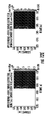
- FIG. 6 shows a bar graph indicating results of IRE performed in vitro on J3T glioma cells at different pulse values.
- FIGS. 7A-E are schematic drawings showing various exemplary embodiments of a device according to the invention.
- Panel A depicts a device, showing a connector, wiring, and electrodes disposed at the tip.
- Panels B-E depict alternative placement of electrodes, which can be retractable.
- FIGS. 8A-C are schematic drawings showing an expanded view of an electrode tip according to one embodiment of the invention.
- Panel A depicts an exploded view of the various concentric layers of materials making up the electrode tip.
- Panel B depicts a side view of the electrode of Panel A, showing the various layers in cut-away fashion.
- Panel C depicts the electrode tip viewed along the proximal-distal plane.
- FIGS. 9A-B are schematic drawings showing an embodiment of an assembled electrode tip for an exemplary treatment where the tip is inserted within a tumor embedded within benign tissue.
- FIG. 10 depicts yet another embodiment of a device according to the invention, in which the outer, non-conductive sheath is adjustable to allow for selection of an appropriate depth/length of electrically conductive material to be exposed to tissue to be treated.
- the embodiment includes screw tappings (not shown) to allow real-time adjustment of the electrode layer lengths to customize electrode dimensions prior to a procedure.
- FIG. 11 depicts an exemplary system according to the invention, which includes an adjustable height electrode, a handle for physician guidance of the device into the patient, and a power source/controller to provide and control electrical pulses.
- FIGS. 12A-E are electrical field outputs from various configurations employing two electrodes.
- Panel A depicts the use of two electrodes spaced 0.5 cm apart.
- Panel B depicts the use of two electrodes spaced 1.0 cm apart.
- Panel C depicts the use of two electrodes spaced 1.5 cm apart.
- Panel D depicts the use of two electrodes spaced 1.0 cm apart.
- Panel E depicts a side view of electrical field outputs from one device having two electrically conductive regions separated by an insulative region of a length of 0.5 cm.
- FIGS. 13A-C are electrical field outputs from various configurations employing three needle electrodes having different diameters.
- Panel A depicts the use of electrodes of 2 mm, 0.5 mm, and 1 mm (from left to right).
- Panel B depicts the use of electrodes of 2 mm, 1 mm, and 0.5 mm (from left to right).
- Panel C depicts the use of electrodes of 1 mm, 2 mm, and 0.5 mm (from left to right).
- FIGS. 14A-J are electrical field outputs for various combinations of electrodes emitting different charges.
- Panel A depicts a two-dimensional display for the use of four electrodes of alternating polarity.
- Panel B depicts an axis symmetric display for the use of four similar electrodes of alternating polarity.
- Panel C depicts a two-dimensional display for the use of four charged electrodes, the center two at 5000V and 0V and the outer two at 2500V.
- Panel D depicts an axis symmetric display for the use of a similar electrode set up as Panel C.
- Panel E depicts a two-dimensional display for the use of three electrodes with the center one at 2500V and the outer two at 0V.
- Panel F depicts an axis symmetric display for the use of three electrodes similar to Panel E.
- Panel G depicts a two-dimensional display for the use of three charged electrodes, the center at 0V, the left at 5000V, and the right at 2500V.
- Panel H depicts an axis symmetric display for the use of a similar electrode set up as Panel G.
- Panel I depicts a two-dimensional display for the use of three charged electrodes, the center at 1750V, the left at 3000V, and the right at 0V.
- Panel J depicts an axis symmetric display for the use of a similar electrode set up as Panel I.
- FIGS. 15A-D are schematics showing thermal effects from use of three needle electrodes, with and without use of a cooling element in the electrode.
- Panel A shows thermal effects without cooling
- Panel B shows the thermal effects under the same pulsing conditions, but with electrode cooling
- Panel C shows thermal effects without cooling
- Panel D shows the thermal effects under the same pulsing conditions, but with electrode cooling.
- FIGS. 16A-C are schematics showing thermal effects from use of two bipolar electrodes and an intervening balloon.
- Electroporation is the phenomenon in which permeability of the cell membrane to ions and macromolecules is increased by exposing the cell to short (microsecond to millisecond) high voltage electric pulses.
- the application of the electric pulses can have no effect, can have a transient effect known as reversible electroporation, or can cause permanent permeation known as irreversible electroporation (IRE), which leads to non-thermal cell death by necrosis.
- IRE irreversible electroporation
- IRE can be used as an independent drug-free tissue ablation modality for particular use in cancer therapy.
- This minimally invasive procedure involves placing electrodes into or around the targeted area to deliver a series of short and intense electric pulses to induce the irreversible structural changes in the membranes.
- the electric field in the targeted region needs to be above a critical value, which is dependent on a variety of conditions such as tissue type and pulse parameters.
- the present invention extends and improves on prior techniques for IRE by providing new methods and devices for IRE treatment of solid tumors, including those associated with brain cancer. Because the brain is susceptible to small fluctuations in temperature, the present invention provides devices and techniques for non-thermal IRE to kill undesirable cells and tissues. In addition, because the brain functions by way of electrical charges, the present invention provides devices and techniques that limit or precisely control the amount of electrical charge delivered to tissue. To achieve the invention, a device has been developed that contains both conducting and non-conducting surfaces and that is capable of delivering controlled pulses of electricity to tumor tissues while substantially protecting surrounding healthy tissue. In exemplary embodiments, the device has a laminate structure of at least one electrically conductive and at least one electrically insulative material.
- the device has at least two concentric disk electrodes separated by an insulating material similar in dimensions to those already used in deep brain stimulation (DBS).
- DBS deep brain stimulation
- DBS is an FDA approved therapy that alleviates the symptoms of otherwise treatment-resistant disorders, such as chronic pain, Parkinson's disease, tremor, and dystonia.
- the methods employ the unique designs discussed herein, which provide improved controlled delivery of electrical pulses with controlled three-dimensional patterns and controlled thermal outputs.
- the present devices and systems provide surgical tools and methods for IRE treatment of subcutaneous tumors that expand the application space for this new technology, with the potential to treat a number of cancers, including brain, liver, prostate and pancreatic adenocarcinoma.
- the present invention provides a method for treating aberrant cell growth in animals.
- the aberrant cell growth can be any type of aberrant cell growth, but in exemplary embodiments detailed herein, it is generally described in terms of tumors, such as brain tumors.
- the method of treating comprises temporarily implanting one or more electrodes, which may be present on the same or different devices, into or immediately adjacent a tumor, and applying an electrical field to the tumor in multiple pulses or bursts over a prescribed or predetermined period of time to cause irreversible cell death to some or all of the tumor cells.
- irreversible damage to non-tumor cells in proximity to the tumor is minimal and does not result in significant or long-lasting damage to healthy tissues or organs (or a significant number of cells of those tissues or organs).
- cell killing is predominantly, essentially, or completely due to non-thermal effects of the electrical pulsing.
- the method further comprises removing the electrode(s) after suitable treatment with the electrical fields.
- the method involves temporary implantation of relatively small electrodes, it is minimally invasive and does not result in the need for significant post-treatment procedures or care. Likewise, it does not result in significant ancillary or collateral damage to the subject being treated.
- the number of electrodes, either on a single or multiple devices, used can be selected by the practitioner based on the size and shape of the tumor to be treated and the size and shape of the electrode.
- embodiments of the invention include the use of one, two, three, four, five, or more electrodes.
- Each electrode can be independently sized, shaped, and positioned in or adjacent the tumor to be treated.
- the number and spacing of electrodes on a single device can be adjusted as desired.
- the location, shape, and size of electrodes can be selected to produce three-dimensional killing zones of numerous shapes and sizes, allowing for non-thermal treatment of tumors of varying shapes and sizes.
- the present invention recognizes for the first time that, in contrast to prior disclosures relating to IRE, the pulse length for highly efficient tissue ablation can be lower than 100 microseconds (100 us). Indeed, it has surprisingly been determined that a pulse length of 25 us or lower can successfully cause non-thermal cell death.
- the method of treatment uses pulse lengths of 10 us, 15 us, 20 us, 25 us, 30 us, 35 us, 40 us, 45 us, 50 us, 55 us, 60 us, 65 us, 70 us, 75 us, 80 us, 85 us, or 90 us.
- pulse lengths are limited to 90 us or less, for example 50 us or less, such as 25 us.
- the pulse length As compared to prior art techniques for IRE, larger electric fields can be applied to the treatment area while avoiding thermal damage to non-target tissue (as well as to target tissue).
- the methods of the invention allow for treatment of tissues having higher volumes (e.g., larger tumors) than possible if prior art methods were to be employed for in vivo treatment of tumors.
- IRE is performed using voltages of between 4000 V/cm to 1500 V/cm.
- the present invention provides for use of voltages of much lower power.
- the present methods can be performed using less than 1500 V/cm.
- 2000 V/cm can cause excessive edema and stroke in patients when applied to brain tissue.
- applied fields of about 500 V/cm to 1000 V/cm are used.
- applied fields of less than 1000 V/cm can be used.
- the number of electrical pulses that can be applied to successfully treat tumors can be quite high.
- Prior art methods of using IRE for various purposes included the use of relatively few pulses, for example 8 pulses or so. Reports of use of up to 80 pulses for IRE have been published; however, to the inventors' knowledge, a higher number of pulses has not been recommended.
- the present invention provides for the use of a relatively high number of pulses, on the order of 90 pulses or greater. For example, in exemplary embodiments, 90 pulses are used. Other embodiments include the use of more than 90 pulses, such as 100 pulses, 110 pulses, or more.
- cycle times for pulses are set generally about 1 Hz. Furthermore, it has been found that alternating polarity of adjacent electrodes minimizes charge build up and provides a more uniform treatment zone. More specifically, in experiments performed by the inventors, a superficial focal ablative IRE lesion was created in the cranial aspect of the temporal lobe (ectosylvian gyrus) using the NanoKnifeB (Angiodynamics, Queensbury, N.Y.) generator, blunt tip bipolar electrode (Angiodynamics, No.
- the method of the invention encompasses the use of multiple electrodes and different voltages applied for each electrode to precisely control the three-dimensional shape of the electric field for tissue ablation. More specifically, it has been found that varying the amount of electrical energy emitted by different electrodes placed in a tissue to be treated allows the practitioner to finely tune the three-dimensional shape of the electrical field that irreversibly disrupts cell membranes, causing cell death. Likewise, the polarity of electrodes can be varied to achieve different three-dimensional electrical fields. Furthermore, one of the advantages of embodiments of the invention is to generate electric field distributions that match complex tumor shapes by manipulating the potentials of multiple electrodes. In these embodiments, multiple electrodes are energized with different potential combinations, as opposed to an “on/off” system like radio frequency ablation, to maximize tumor treatment and minimize damage to surrounding healthy tissue.
- the separation of the electrodes within or about the tissue to be treated can be varied to provide a desired result.
- the distance between two or more electrodes can be varied to achieve different three-dimensional electrical fields for irreversible disruption of cell membranes.
- the three-dimensional shape can thus be set to ablate diseased tissue, but partially or completely avoid healthy tissue in situations where the interface between healthy and diseased tissue shows a complex three dimensional shape.
- the methods of the invention are well suited for treatment of tumors using non-thermal IRE.
- the method can further comprise cooling the electrodes during the treatment process.
- a heat sink such as a cooling element in an electrode (discussed below)
- generation of heat in and around tissue in close proximity to the electrodes can be minimized, resulting in a more consistent application of non-thermal IRE to the tissue and a more controlled application of cell killing to only those tissues desired to be treated.
- the method of the invention includes the use of electrodes of different sizes and shapes. Studies performed by the inventors have shown that the electrical field distribution may be altered by use of electrodes having different diameters, lengths, and shapes. Thus, the use of different sizes and shapes of conducting surfaces can be used to control the electrical fields used for cell ablation.
- the method includes the use of a variable size electrode. For example, an electrode may be used that, in one configuration has a relatively small diameter, which is used for minimally invasive implantation of the electrode into the site to be treated. Once inserted, a sheath or other covering can be retracted to allow expansion of the electrode tip to a different size for application of the electric field.
- the expandable element can be thought of as a balloon structure, which can have varying diameters and shapes, depending on original material shape and size.
- the methods of the invention comprise, in embodiments, treatment of tissue surrounding tumor tissue.
- the surrounding tissue is treated by way of reversible electroporation.
- bioactive agents can be introduced into the reversibly electroporated cells.
- additional cell killing, under controlled conditions can be effected in healthy tissue.
- Such a treatment is preferred when treating highly aggressive malignant tumors, which often show invasion of healthy tissue surrounding the tumor.
- the bioactive agents can provide a protective effect to cells in which they are introduced via reversible electroporation.
- the method for treating aberrant cell growth in animals is a method of treating a subject suffering from a tumor. It thus may be a method of treating a subject suffering from cancer.
- the method can be a method of treating a tumor or a method of treating cancer.
- the method can be a method of treating either a benign tumor or a malignant tumor.
- the method is best suited for treatment of solid tumors.
- the method is a method of treating a subject suffering from a brain tumor, such as brain cancer.
- the method of treating according to the invention can have ameliorative effects or curative effects. That is, a method of treating a subject can provide a reduction in cell growth of a tumor, a reduction in tumor size, or total ablation of the tumor.
- the method of the invention can include a single round of treatment or two or more rounds of treatment. That is, the method of cell ablation, either intentionally or as a result of tumor size or shape, can result in less than complete destruction of a tumor. In such a situation, the method can be repeated one or more times to effect the desired level of tumor reduction. As the method of the invention is relatively minimally invasive, multiple rounds of treatment are not as harmful to the patient than multiple rounds of traditional surgical intervention.
- the method of the invention can be part of a multi-modal treatment.
- the method thus may comprise other cell-killing techniques known in the art.
- the method may further comprise exposing the tumor to radiation, or treating the patient with a chemotherapeutic agent. It likewise may be performed after or between surgical intervention to remove all or part of a tumor.
- the method of the invention is implemented using devices and systems.
- the devices according to the invention are suitable for minimally invasive temporary implantation into a patient, emission of a tissue-ablating level of electricity, and removal from the patient.
- the device according to the invention thus may be used in the treatment of tumors and the treatment of patients suffering from tumors.
- the devices can take multiple forms, based on the desired three-dimensional shape of the electrical field for cell killing. However, in general, the devices include two or more regions of differing conductivity.
- the device comprises alternating regions of conductivity, for example a region of electrical conductivity, which is adjacent a region of electrical non-conductivity, which is adjacent a different region of conductivity.
- the device comprises two or more layers of conductive and insulative materials, in a laminate structure with alternating conductive properties.
- the outer layer can be insulative except at the region where treatment is to be effected.
- the amount of conductive material exposed to the tissue to be treated can be adjusted by a movable non-conductive element disposed on the outer surface of the device.
- the device takes a rod-like shape, with one dimension (i.e., length) being substantially longer than the other (i.e., width or diameter).
- one dimension i.e., length
- the other i.e., width or diameter
- the cross-sectional shape of the electrode can take any suitable geometric shape. It thus may be circular, square, rectangular, oval, elliptical, triangular, pentagonal, hexagonal, octagonal, etc.
- the devices of the invention comprise one or more electrodes, which are electrically conductive portions of the device.
- the devices are thus electrically conductive elements suitable for temporary implantation into living tissue that are capable of delivering an electrical pulse to the living tissue.
- the device of the invention has a proximal end and a distal end.
- the proximal end is defined as the end at which the device is attached to one or more other elements, for control of the function of the device.
- the distal end is defined by the end that contacts target tissue and delivers electrical pulses to the tissue.
- the distal end thus comprises an exposed or exposable electrically conductive material for implantation into a target tissue.
- the distal end is described as including a “tip” to denote the region of the distal end from which an electrical pulse is delivered to a tissue.
- the device further comprises at least one surface defining the length and circumference of the device.
- the device comprises a laminate structure, with alternating conductive and non-conductive or insulative layers expanding outward from the proximal-distal center axis to the surface of the device.
- the center most layer which shows the smallest diameter or width, is electrically conductive and comprises a part of the electrode tip.
- the center-most layer is an open channel through which a fluid may be passed or through which additional physical elements may be placed.
- the center-most layer may comprise an insulative material.
- the structure can provide a more customizable electric field distribution by varying the separation distances between electrically conductive regions.
- certain electrically conductive regions can be exposed or concealed by movement of an outer, non-conductive sheath.
- the separation lengths can be achieved by disposing on the surface non-conductive materials at various regions.
- one or more substantially open channels are disposed along the center axis or in place of one of the conductive or insulative layers.
- the channel(s) may be used as heat sinks for heat produced by the device during use.
- water or another fluid is held or entrained in the channel to absorb and/or remove heat.
- the device of the invention comprises an electrode tip at the distal end.
- the electrode tip functions to deliver electrical pulses to target tissue.
- the tip may be represented by a single conductive layer of the device or may be represented by two or more conductive layers that are exposed to the tissue to be treated.
- the tip may be designed to have any number of geometrical shapes. Exemplary embodiments include tips having a needle-like shape (i.e., electrical pulses emanate from a small cone-like structure at the distal end of the device) or having a circular shape (i.e., electrical pulses emanate from the cylindrical outer surface of the device, which is a section of the device where the outer insulative layer has been removed to expose the next layer, which is conductive).
- the tip advantageously comprises a blunt or rounded end to minimize laceration of brain tissue.
- the rounded or blunt end comprises a hole that allows for a sharp or needle-like structure to be deployed into tumor tissue at the appropriate time.
- the device comprises a proximal end, which generally functions for attachment of the device to a power source/controller and a handle.
- the proximal end thus may comprise connections for electrical wires that run from the power source/controller to the electrically conductive layers of the device. Standard electrical connections may be used to connect the conductive elements to the wires.
- the device is attached to a handle for ease of use by a human. While not limited in the means for attaching the device to the handle, in embodiments, the connection is made by way of a friction fit between the outer surface of the device and the handle, for example by way of an insulative O-ring (e.g., a Norprene O-ring) on the handle.
- an insulative O-ring e.g., a Norprene O-ring
- the device comprises, on its outer surface, ridges or other surface features that mate with surface features present on the handle.
- the proximal end comprises one or more structures that allow for controlled movement of the outer surface along the length of the device.
- the outer surface will comprise an outer sheath that is electrically non-conductive, and which surrounds an electrically conductive layer. Using the structures at the proximal end, the outer sheath may be moved, relative to the rest of the device, to expose or conceal varying portions of the electrically conductive material beneath it. In this way, the amount of surface area of the conductive material at the tip can be adjusted to provide a desired height of exposure of tissue to the electrode tip.
- other structures for securely fastening the device to a holder may be used, such as clips, set screws, pins, and the like. The device is not limited by the type of structure used to connect the device to the holder.
- the device of the invention can be designed to have any desired size. Typically, it is designed to be minimally invasive yet at the same time suitable for delivery of an effective electrical field for IRE.
- the diameter or width is thus on the order of 0.5 mm to 1 cm. Preferably, the diameter or width is about 0.5 mm to about 5 mm, such as about 1 mm, 2 mm, 3 mm, or 4 mm.
- the length of the device is not particularly limited, but is generally set such that a surgeon can use the device comfortably to treat tumors at any position in the body.
- the device is typically on the order of 40 cm or less in length, such as about 30 cm, 25 cm, or 15 cm, whereas for veterinary use, the length can be much larger, depending on the size of animal to be treated.
- the length can be on the order of 40 cm.
- the device or a portion of it, is flexible.
- a flexible device is advantageous for use in accessing tumors non-invasively or minimally invasively through natural body cavities.
- the shape of the device can change based on contact with body tissues, can be pre-set, or can be altered in real-time through use of wires or other control elements, as known in the art, for example in use with laparoscopic instruments.
- the device of the invention can be part of a system.
- the system can comprise a handle into or onto which the device is disposed.
- the handle can take any of a number of shapes, but is generally designed to allow a surgeon to use the device of the invention to treat a patient in need. It thus typically has a connector for connecting the device to the holder, and a structure for the surgeon to grasp and maneuver the device.
- the handle further can comprise a trigger or other mechanism that allows the surgeon to control delivery of electrical pulses to the device, and thus to the tissue to be treated.
- the trigger can be a simple on/off switch or can comprise a variable control that allows for control of the amount of power to be delivered to the device.
- the handle may be created in such a manner that it may be attached to additional pieces of equipment, such as ones that allow precise placement of the electrode relative to an inertial or the patient's frame of reference, allowing steady and accurate electrode positioning throughout an entire procedure, which may entail the application of electric pulses in addition to radiotherapy, imaging, and injections (systemically and locally) of bioactive agents.
- the handle may be attached to machines that are operated remotely by practitioners (e.g., the Da Vinci machine).
- the system can further comprise a power source and/or a power control unit.
- the power source and control unit are the same object.
- the power source provides electrical power to the device, typically by way of an electrical connection through the handle.
- the power source can be any suitable source that can deliver the proper amount of electrical power to the device of the invention. Suitable power sources are commercially available, and the invention is not limited by the type or manufacturer.
- the power control unit provides the user with the ability to set the power output and pulse time for electrical pulses to be delivered to the device, and thus to the tissue to be treated. Suitable control units are commercially available, and the invention is not limited by the type or manufacturer.
- an appropriate power source/controller is available from Angiodynamics (Queensbury, N.Y.).
- the device of the invention can be disposable or reusable. Where the device is designed to be reusable, it is preferably fabricated from materials that can be sterilized multiple times without destruction of the device.
- the device can be fabricated from rust-resistant metals or alloys, such as stainless steel, and plastic or other synthetic polymeric materials that can withstand cleaning and sterilization. Exemplary materials are those that can be subjected to detergents, steam heat (e.g., autoclaving), and/or irradiation for at least one cycle of sterilization.
- rust-resistant metals or alloys such as stainless steel
- plastic or other synthetic polymeric materials that can withstand cleaning and sterilization.
- Exemplary materials are those that can be subjected to detergents, steam heat (e.g., autoclaving), and/or irradiation for at least one cycle of sterilization.
- Those of skill in the art can select the appropriate materials without undue experimentation, based on materials used in other medical devices designed to withstand common sterilization techniques.
- a healthy female purpose bred beagle was used.
- Nine sets of ten pulses were delivered with alternating polarity between the sets to minimize charge build-up on the electrode surfaces.
- the maximum voltage-to-distance ratio used was 2000 V/cm because the resulting current did not exceed 2 amps.
- the charge that was delivered to the brain during the IRE procedure was 22.5 mC, assuming ninety pulses (50 ⁇ s pulse durations) that would result from a maximum hypothetical current of 5 amps.
- a routine parietotemporal craniectomy defect was created to expose the right temporal lobe of the brain.
- Two decellularization sites were performed: 1) a deep lesion within the caudal aspect of the temporal lobe using a monopolar electrode configuration (6 mm electrode insertion depth perpendicular to the surface of the target gyrus, with 5 mm interelectrode distance), and 2) a superficial lesion in the cranial aspect of the temporal lobe using a bipolar electrode (inserted 2 cm parallel to the rostrocaudal length of the target gyrus, and 2 mm below the external surface of the gyrus).
- Intraoperative adverse effects that were encountered included gross microhemorrhages around the sharp monopolar electrode needles following insertion into the gyrus. This hemorrhage was controlled with topical application of hemostatic foam. Subject motion was completely obliterated prior to ablating the superficial site by escalating the dose of atracurium to 0.4 mg/kg. Grossly visible brain edema and surface blanching of the gyrus overlying the bipolar electrode decellularization site was apparent within 2 minutes of completion of IRE at this site. This edema resolved completely following intravenous administration of 1.0 g/kg of 20% mannitol.
- a unique advantage of IRE to ablate tissues in vivo is its ability to be monitored in real-time using imaging techniques, such as electrical impedance tomography, MRI, and ultrasound. Below, this Example shows MRI examinations performed immediate post-operatively, which demonstrate that IRE decellularization zones were sharply demarcated ( FIGS. 1A-C ).
- neurosonography performed intraoperatively and at 1 hour and 24 hours post-procedure demonstrated clearly demarcated decellularization zones and visible needle tracts within the targeted brain parenchyma.
- the decellularization zones appeared as hypoechoic foci with needle tracts appearing as distinct hyperechoic regions ( FIG. 2 ).
- the IRE decellularization zone was hypoechoic with a hyperechoic rim ( FIG. 2 ).
- the IRE decellularization zone appeared slightly larger (1-2 mm increase in maximal, two dimensional diameter).
- EEG performed in the post-operative period revealed focal slowing of the background rhythm over the right temporal region in association with the decellularization zones.
- the brain was collected within 2 hours of the time of death and removed from the cranium. Care was taken to inspect soft tissues and areas of closure created at the time of surgery. The brain was placed in 10% neutral buffered formalin solution for a minimum of 48 hours. Then, the brain was sectioned at 3 mm intervals across the short axis of the brain, in order to preserve symmetry and to compare lesions. Following gross dissection of fixed tissues, photographs were taken of brain sections in order to document the position and character of lesions, as shown in FIG. 3 . Readily apparent in gross photographs of the sectioned brain are lesions created either by the physical penetration of brain substance with electrodes or created by the application of pulse through the electrodes. There are relatively well-demarcated zones of hemorrhage and malacia at the sites of pulse delivery.
- mice Microscopic lesions correlated well with macroscale appearance. Areas of treatment are represented by foci of malacia and dissociation of white and grey matter. Small perivascular hemorrhages are present and there is sparing of major blood vessels (see FIG. 4B ). Notable in multiple sections is a relatively sharp line of demarcation (approximately 20-30 micrometers) between areas of frank malacia and more normal, organized brain substance (see FIG. 4A ).
- IRE threshold To determine the electric field needed to irreversibly electroporate tissue, one can correlate the lesion size that was observed in the ultrasound and MRI images with that in the histopathological analysis to determine the percentage of lesion growth. Decellularized site volumes can be determined after identification and demarcation of IRE decellularization zones from surrounding brain tissue using hand-drawn regions of interest (ROI). A representative source sample image is provided in FIG. 5 .
- IRE to treat cancer has advantages over existing thermal ablation, including the ability to: monitor what area has been irreversibly electroporated in real-time using ultra-sound or other imaging techniques; spare neural tubes and blood vessels, enabling treatment in otherwise inoperable areas; preserve the microvasculature, which promotes rapid healing of the treated volume; predict the affected area using numerical modeling for designing protocols; not be affected by blood flow as is the temperature distribution during thermal therapies; image the success of the treatment using MRI or other imaging techniques; and administer the electric fields in time periods of less than 5 minutes.
- the present methods and devices provide a technology for treatment of tumors with IRE.
- devices designed to irreversibly electroporate deep tissues did not exist.
- the experiments conducted and reported in the prior art utilized reversible electroporation systems. These devices usually consist of large plate electrodes (intended for transdermal drug delivery), needle electrodes with a large probe (intended for targeting in or for small animal studies), or cuvettes (used for in vitro applications).
- Applying an electric pulse over the skin presents challenges for deep tissue applications due to the significant voltage drop across the skin, generating considerable skin damage. (The same issue arises with an organ containing an epithelial layer.)
- Other devices that use needle electrodes are limited to superficial tumors.
- these tools have a large mechanical housing making the treatment of subcutaneous tumors impossible without invasive surgery.
- a tool designed specifically for IRE for subcutaneous delivery of the electric field dramatically enhances the application space of IRE for tissue ablation.
- a device was used to kill brain cells in vitro. Representing a unique pathobiological phenomenon, high-grade canine gliomas exhibit essentially the same properties as human gliomas, including pathology (markers), genetics, behavior (invasiveness), lack of metastases, and a similar clinical course of the disease. Dogs diagnosed with these tumors have poor prognosis and most are humanely euthanized to prevent further suffering from the progression of the disease. Primary brain tumors (PBTs) account for 1-3% of all deaths in aged dogs where necropsy is performed. The many similarities of glial tumors in people and dogs make these tumors in dogs an excellent translational approach for new diagnostic and treatment methods.
- FIG. 6 cell proliferation of canine glioma cells was significantly reduced or eliminated by treatment with IRE.
- FIG. 1 shows the results of J3T glioma cells after treatment with electric pulses of length 50 microseconds (us) for 2 electric fields (1000 V/cm and 1500 V/cm) and multiple numbers of pulses.
- a WST-1 cell proliferation assay was performed on J3T glioma cells, and the data collected 24 hours post-IRE treatment.
- Two electric fields (1000 and 1500 V/cm) at 5 different pulse combinations were analyzed.
- a value of relative absorbance of 0.2 represents 100% cell death. Therefore, it is clear that for as low as 1000V/cm at 50 pulses total will achieve complete cell death for 50 us length pulses, proving this a viable IRE treatment parameter.
- the present invention provides simple and elegant minimally invasive microsurgical tools to treat currently inoperable tumors in humans and animals through IRE. Exemplary designs are shown in FIGS. 7-11 .
- FIG. 7 Panel A, depicts an example of a device 700 according to one embodiment of the invention.
- This embodiment is fully compatible with existing electroporation electronics and comprises a surgical probe/electrode tip 710 at its distal end, which includes both ground electrodes 711 and energized electrodes 712 .
- the device further comprises a universal connector 750 at its proximal end.
- the device also comprises internal wiring 770 to deliver electrical impulses to the tip 710 .
- the body of the device is defined by surface 718 .
- tip 710 can comprises retractable conductive spikes 713 emanating from a blunt end tip 710 and disposed, when deployed, at an acute angle to tip 710 (see FIG. 7 , Panel B).
- tip 710 can be fashioned as a point or needle, and can include retractable accordion-type conductive elements 714 (see FIG. 7 , Panel C).
- tip 710 can comprise multiple retractable spikes 715 that, when deployed, emanate at 90° C. from tip 710 (see FIG. 7 , Panel D).
- tip 710 can comprise retractable conductive spikes 716 emanating from a needle-end tip 710 and disposed, when deployed, at an acute angle to tip 710 (see FIG. 7 , Panel E).
- FIG. 7 , Panels B, D, and E show probes with parallel circular channels 717 of approximately 1 mm that protrude through the length of the electrode holder. Each channel has the capability of guiding individual 1 mm electrodes to the treatment area. Towards the bottom of the holder, the channels deviate from their straight path at a specific angle.
- the electrodes can be Platinum/Iridium with an insulating polyurethane jacket to ensure biocompatibility, similar to materials that are used in DBS implants. Different protrusion depths of the electrodes within the tissue as well as the applied voltage can be used to control the size of the treated area.
- the devices can comprise interchangeable surgical tips, allowing for versatility in creating a device well suited for different tissues or different sized or shaped tumors. Varying electrode diameters (varied in part by selection of the type and length of deployable spikes) and separation distances will be sufficient to ablate the majority of tumors about or smaller than 3 cm by selecting the appropriate voltages to match different tumor sizes and shapes. As shown in later figures, some of the embodiments of the device comprise an element at the tip to introduce anti-cancer drugs for ECT, cytotoxic proteins, or other bioactive agents into the targeted area.
- embodiments of the device comprise durable carbon coatings over portions of the device that act both to insulate normal tissue and to increase the efficiency of IRE pulsing.
- the device contains a primary blunt-end tip with a hole disposed in the end, for insertion through delicate, soft brain tissue.
- the device of these embodiments further comprises a secondary sharp tip, which can be deployed through the hole in the blunt-end primary tip, which allows for penetration into the tumor tissue, which can be substantially more dense or hard, and not easily punctured by a blunt-end tip.
- the device of the invention is typically similar in dimensions (2 mm) to those already used in deep brain stimulation (DBS), which ensures that they are feasible for surgical applications.
- DBS deep brain stimulation
- DBS uses electrodes in an FDA approved therapy to alleviate the symptoms of otherwise treatment-resistant disorders, such as Parkinson's disease and dystonia.
- the electrode positioning frame which is used in stereotactic surgery in conjunction with imaging systems, can be used to position the surgical probes and ensure that the position of the electrodes is optimal. Simulations of a design similar to the one in FIG. 7A show treatment volumes comparable to typical brain tumors.
- Panels A-C depict in more detail an embodiment of a tip 810 according to the invention.
- Panel A depicts an exploded view of tip 810 , showing multiple concentric layers of conducting 820 and non-conducting 830 materials.
- An outer layer or sheath 860 of non-conducting material is shown with perforations 861 .
- An outer, perforated layer 832 is disposed around the concentric rings of materials, to allow for delivery of bioactive substances to cells in proximity to the device when in use.
- Perforated layer 832 may be disposed in full, direct contact with the outermost layer of the concentric ring structure, or may be substantially separated from the ring structure by chamber 833 that holds cooling fluid.
- device tip 810 has multiple alternating layers of conducting 820 and non-conducting 830 materials surrounding an non-conducting inner core 831 .
- the top and bottom conducting regions 820 are energized electrodes while the middle conducting region 820 is a ground electrode.
- the present invention provides the conducting and non-conducting (insulative) regions in varying lengths to fine tune electrical field generation. More specifically, using imaging techniques directed at the tumor to be treated, a surgeon can determine what type of electrical field is best suited for the tumor size and shape.
- the device can comprise one or more movable elements on the surface of the tip (not depicted) or can be designed such that one or more of the alternating conducting 820 or non-conducting 830 elements is movable. Through movement and setting of the outer element(s) or inner elements 820 or 830 , the surgeon can configure the device to deliver a three-dimensional electrical killing field to suit the needs of the particular situation.
- FIG. 8 Panel C, depicts the concentric laminate structure of tip 810 , viewed from the distal end along the distal-proximal axis, showing again the laminate nature of the device.
- adapting the physical dimensions of the probe also allows flexibility in tailoring the treatment area to match the dimensions of the tumor.
- altering the electrode parameters, including diameter, length, separation distance, and type it is possible to conveniently tailor the treatment to affect only specific, targeted regions.
- developing an electrode capable of altering and adapting to these dimensional demands greatly enhances its usability and adaptability to treatment region demands.
- IRE treatments may involve coupled procedures, incorporating several discrete aspects during the same treatment.
- One embodiment of the invention provides a device with a needle-like tip 910 with an incorporated hollow needle 990 with either an end outlet 991 (shown in Panel A) or mixed dispersion regions 961 (shown in Panel B).
- Such a configuration allows for highly accurate distribution of injectable solutions, including those comprising bioactive agents.
- Use of such a device limits the dose of treatment required as well as ensures the correct placement of the materials prior to, during, and/or after the treatment.
- Some of the possible treatment enhancers that would benefit from this technology are: single or multi-walled carbon nanotubes (CNTs); chemotherapeutic agents; conductive gels to homogenize the electric field; antibiotics; anti-inflammatories; anaesthetics; muscle relaxers; nerve relaxers; or any other substance of interest.
- CNTs carbon nanotubes
- FIG. 9 show two basic hollow needle designs that may be implemented to enhance solution delivery prior to, during, or after IRE treatment. They both have multiple conducting surfaces 920 that may act as charged electrodes, grounded electrodes, or electric resistors, depending on the treatment protocol.
- Panel A shows a hollow tip 910 for injection of agents at its end while Panel B has distributed pores 961 throughout for a more generalized agent distribution. As shown in Panel B, the pores are disposed in the non-conducting regions 930 of the device.
- Irreversible Electroporation a new minimally invasive technique we invented to treat tumors, can be enhanced using carbon nanotubes (CNTs).
- CNTs carbon nanotubes
- the technique can be used on a variety of tumors including liver, prostate, pancreatic adenocarcinoma and renal carcinoma.
- Focal ablation techniques, such as IRE are not selective and thus cannot distinguish between healthy and cancerous cells.
- nanoparticles can be incorporated into IRE therapy. Nanomaterials offer a potential means for energy focusing, because they present a toolset with a unique size range closely matching that of cells (1 to 1,000 nm), and substantial multi-functional capability.
- nanoparticles exhibit a “lightning rod” effect when exposed to electric fields, amplifying the field at the nanoparticle's tip, thereby producing a significantly larger electric potential compared to its surroundings and reducing the possibility of sub-lethal joule heating.
- This localized amplification of electric fields could thus be used as a means to induce IRE from relatively small electric fields; residual adverse effects to surrounding tissue would subsequently be reduced.
- Targeting of nanoparticles through tumor specific antibodies to the desired tissue region will allow treatment up to and beyond the tumor margin using IRE, and offer the opportunity to lower the IRE applied field, thereby minimizing damage to surrounding, non-cancerous tissue during treatment. Integration of CNTs into IRE could more selectively localize the electric field and thermal profile to cancer cells through antibody targeting and more precisely control the induction of cell death and HSP expression.
- CNTs carbon nanotubes
- an induced dipole is generated that tends to align the axis of the CNT parallel to the electric field.
- two sets of electric fields delivered subsequent to and at right angles to each other is a technique that can be used to align the CNTs and electroporate the cells.
- cells electroporated using CNTs may result in cells having a higher vitality than when electroporated without the use of CNTs.
- the use of CNTs injected into a region of tissue, with or without targeting antibodies, to mediate IRE for tumor ablation is another method covered by the present invention.
- N-TIRE therapy the local electric field distribution dictates the treatment area.
- N-TIRE possesses a clear therapeutic advantage in that there is no induction of thermal injury in the ablated area, thereby preserving important tissue components such as the extracellular matrix, major blood vessels, myelin sheaths, and nerves. Since N-TIRE is a focal ablation technique, it does not selectively kill infiltrative cancer cells with the potential for re-growth and metastasis beyond the tumor margin without affecting the surrounding healthy cells.
- the ablation area can be enlarged without inducing joule heating and the selectivity of N-TIRE can be enhanced through the use of CNTs.
- N-TIRE Localized amplification of electric fields from CNTs could induce N-TIRE in adjacent cells from relatively small electric fields, without affecting healthy surrounding cells. Further, antibody targeting of CNTs to tumor cells could permit localized CNT-mediated electric field amplification at selected tumor cell membranes causing targeted cell death due to permanent membrane destabilization. Even further, it is advantageous to incorporate CNTs into N-TIRE protocols in order to simultaneously lower the voltage for N-TIRE and expand the treatable area.
- Combinatorial CNT-mediated N-TIRE cancer therapies can include treatment of a number of cancers including prostate, liver, kidney, and pancreatic.
- Breast cancer is a particularly apt application since this combinatorial therapy can directly address the need of scar reduction and mitigate the likelihood of metastasis, which have proven in some circumstances to be helpful for improved treatment.
- Adapting N-TIRE treatments for breast carcinomas has several unique challenges. Among these are the diverse and dynamic physical and electrical properties of breast tissue. The fatty and connective tissues within the breast region surrounding a tumor have low water content, and thus significantly reduced electrical conductivity and permittivity than tumors. It has been shown that N-TIRE treatment area is highly predictable based on electric field distribution. CNTs will provide a means to raise the electric field magnitude within the tumor and increase N-TIRE treatment area in localized breast carcinomas.
- the device comprises a cooling system within the electrode to reduce the highly localized temperature changes that occur from Joule heating.
- the highest quantity of heat generation is at the electrode-tissue interface.
- cooling provides a heat sink for the nearby tissue, further reducing thermal effects. This allows more flexibility in treating larger tissue regions with IRE while keeping thermal effects negligible, providing a greater advantage for IRE over conventional thermal techniques.
- Cooling can be achieved by placement of one or more hollow chambers within the body of the device.
- the cooling chambers can be closed or open. Open chambers can be attached at the proximal end to fluid pumping elements to allow for circulation of the fluid (e.g., water) through the device during use.
- the device comprises an outer protector that is designed to be movable up and down along the length of the device.
- FIG. 10 depicts such a movable outer protector. More specifically, FIG. 10 depicts a device 1000 comprising tip 1010 that includes outer protector 1062 that can be moved up and down along the length of device 1000 .
- outer protector 1062 is disposed fully or partially encasing outer sheath 1060 . After or during insertion into tissue to be treated, outer protector 1062 is retracted partially to expose outer sheath 1060 , which in the embodiment depicted comprises mixed dispersion outlets 1061 . As such, the number of dispersion outlets 1061 exposed to the tissue during treatment can be adjusted to deliver varying amounts of bioactive agent to different portions of the tissue being treated.
- any mechanism for movement of the outer sheath along the device may be used.
- screw threads are disposed on the upper portion of the device, allowing for easy adjustment by simple twisting of the outer sheath.
- set screws may be disposed in the outer sheath, allowing for locking of the sheath in place after adjustment.
- an exemplary system can comprise a device 1100 reversibly attached to holder 1140 .
- Holder 1140 can comprise trigger 1141 , which allows the user to control the flow of electricity from power source/controller 1142 to device 1100 .
- device 1100 comprises further elements for use. More specifically, device 1100 comprises a height adjustment apparatus 1151 at its proximal end to effect movement of outer sheath 1160 . Outer sheath 1160 further comprises markings or scores 1168 on its surface to indicate amount of movement of outer sheath 1160 after implantation of device 1100 into tumor tissue.
- the invention provides a system for accurately controlling the distances between multiple electrodes of singular or multiple polarities during a charge.
- the device places electrode types within an adjustable part of a handle that may be maneuvered by a surgeon or attached to a harness system, as described above.
- the adjustable portion of the handle may be used to control the relative depths of penetration as well as separation distances of each electrode relative to one or more additional electrodes placed within the system.
- the system and method of the invention can include the use of multiple devices for treatment of tumors.
- the devices can be implanted in the tumor at varying distances from each other to achieve desired cell killing.
- the system and method can include the use of a single device having multiple electrodes along its tip. Modeling of placement of multiple devices or a single device with multiple electrodes in tissue was performed, and exemplary electrical fields generated are depicted in FIG. 12 .
- the outputs depicted in the figure demonstrate the variability in IRE treatment region that results from altering the separation distance of the conducting electrode surfaces. More specifically, FIG. 12 , Panels A-C, show three model outputs for 2-dimensional needles (left panels) and an axis symmetric electrode (right panels).
- Numerical models representing two needles and an axis symmetric needle electrode configuration have been developed to compare the increase in treatment area shown by the electric field distribution for the same thermal effects between 100 and 50 us pulse lengths.
- the area/volume of tissue that increased by at least 1 degree Kelvin was determined for a 100 us pulse. This area/volume was then used for the 50 us pulse to determine the electric field magnitude that would cause the same increase in temperature.
- a contour line has been created within these models to represent the region treated with the IRE threshold of 700V/cm. The results are shown in FIG. 12 , Panel D.
- 2-D needle electrodes with 3.13 mm 2 area of tissue increased by one degree Kelvin for 100 us pulse at 2500V/cm with 226.2 mm 2 area treated by IRE (Panel D, left side) and 50 us pulse at 3525V/cm with 325.6 mm 2 area affected by IRE (Panel D, right side).
- Axis symmetric needle electrode with 3.95 mm 3 volume of tissue increased by 1 degree Kelvin for 100 us at 1500V with 81.1 mm 3 volume affected by IRE (Panel E, left side) and 50 us pulse at 2120V with a 133 mm 3 volume within IRE range (Panel E, right side).
- the size of the electrode tips may be adjusted.
- integrating multiple electrode types within the same procedure can make a large impact on enhancing electric field distribution selectivity. This can be done by incorporating such variations as a needle electrode with a single probe or parallel needle electrodes with the conductive surface of one being a different dimension (e.g., longer) than the other.
- the electrical field output can be altered based on the arrangement of electrode types. More specifically, the figure shows model outputs displaying the electric field distribution for three needle electrodes, with a contour of 700V/cm.
- Panel A shows the use of tips having, from left to right, 2 mm diameter, 0.5 mm diameter, and 1 mm diameter, providing a 700V/cm threshold of 215.41 mm 2 .
- Panel B shows the use of tips having, from left to right, 1 mm diameter, 1 mm diameter, and 0.5 mm diameter, providing a 700V/cm threshold of 243.26 mm 2 .
- Panel C shows the use of tips having, from left to right, 1 mm diameter, 2 mm diameter, and 0.5 mm diameter, providing a 700V/cm threshold of 271.54 mm 2 .
- Panels A and B four charged electrodes of alternating polarity at 2500V and ground were used to develop a 2-D readout (Panel A) and axis symmetric electrode configurations (Panel B).
- Four charged electrodes with the center two at 5000V and 0V and the outer two electrodes at 2500V were used to develop a 2-D readout (Panel C) and axis symmetric electrode configurations (Panel D).
- Three charged electrodes with the center one at 2500V and the outer two at 0V were used for 2-D (Panel E) and axis symmetric electrode (Panel F) configurations.
- Panels A-D display the modeling outputs of thermal effects during a typical IRE treatment, but for extended treatment periods.
- the images in Panels A and C display the thermal effects without convective cooling, while the images in Panels B and D have the same treatment parameters, but incorporate convective cooling of the needle.
- Panels A and B IRE treatment with 3 needles (1 second post-IRE) without (Panel A) and with (Panel B) convective cooling at the electrode-tissue interface. It can be seen, particularly on the large center electrode that the temperature of the tissue contacting the electrode is the region of highest temperature without cooling, but is actually a lower temperature than the peripheral regions of the tissue.
- Panels C and D IRE treatment with 3 needles (5 seconds post-IRE) without (Panel C) and with (Panel D) convective cooling at the electrode-tissue interface. It can be seen, particularly on the large center electrode, that the temperature of the tissue contacting the electrode is the region of highest temperature without cooling, but is actually a lower temperature than the peripheral regions of the tissue.
- the electric field distribution may be altered, and thus controlled, by changing the diameter and shape of the electrode between the conducting surfaces.
- This fact can be used to design and develop an electrode with an expandable/contractible interior and deformable exterior to change its size in real-time before or during a treatment to alter, and thus specify the electric field distribution in a manner that may be desirable during treatment.
- the ability to adjust this dimension in real-time is made additionally useful by the fact that a significantly smaller electrode may be inserted to keep it minimally invasive, and then expand the dimension once the electrode has reached the target tissue.
- the invention includes the use of a balloon between regions of charge that may be inflated/deflated during treatment to alter field distribution.
- Panels A-C depict modeling of a bulging region between the charges in a bipolar electrode.
- Three different models that study the inclusion of a balloon between the two electrodes in a bipolar design are shown.
- Panel A (861.21 mm 3 treated area) has no balloon for comparison purposes.
- the middle design of Panel B (795.71 mm 3 treated area) has an elongated balloon that is in close proximity to the electrodes.
- the bottom design of Panel C (846.79 mm 3 treated area) has a smaller balloon that helps distribute the electric field.
- Pulse length 5 us-1 ms
- Pulse shape square, exponential decay, sawtooth, sinusoidal, alternating polarity
- Positive, negative, and neutral electrode charge pulses (changing polarity within probe)
- Needle electrode(s) 0.001 mm-1 cm diameter
- Needle diameter 0.001 mm-1 cm
- Electrode length (needle): 0.1 mm to 30 cm
- Electrode separation 0.1 mm to 5 cm
- IRE can be an effective technique for minimally invasive brain tumor removal.
- the treatment does not induce substantial thermal effects in the brain, protecting the integrity of this organ, which is susceptible to small fluctuations in temperature.
- the method includes delivering electrical signal(s) through tissue to determine its electrical properties before administering IRE by monitoring the voltage and current. Following from that, one may apply intermittent and post-IRE pulse(s), which can be used to determine the success of the procedure and adjust IRE pulse parameters.
- IRE irreversible electroporation
- the specific electrical conductivity of tissue during an irreversible electroporation (IRE) procedure allows the physicians to: determine the current threshold; minimize the electric current dose; decrease the Joule heating; and reduce damage to surrounding healthy tissue.
- the physician must: establish the electrode geometry (shape factor); determine the physical dimensions of the tissue; apply a small excitation AC voltage signal (1 to 10 mV); measure the AC current response; calculate the specific conductivity ( ⁇ ) using results from the prior steps.
- This procedure will not generate tissue damage (low amplitude AC signals) and will supply the physician (software) with the required information to optimize IRE treatment planning, especially in sensitive organs like the brain which is susceptible to high electrical currents and temperatures.
- the IRE procedure is well monitored and can also serve as a feedback system in between series of pulses and even after the treatment to evaluate the area of ablation.
- V e voltage on the hot electrode (the highest voltage), [V]
- R 1 radius of electrode with highest voltage (inner radius), [m]
- R 2 radius at which the outer electrodes are arranged (outer radius), [m]
- the specific conductivity ( ⁇ ) of the tissue can be calculated since the voltage signal (V e ) and the current responses (i) are known.
- I ( t ) I 0 Sin( wt+q )
- I(t) is the response signal
- I 0 is the amplitude of the response (I 0 1 V 0 )
- q is the phase shift of the signal.
- is the electrical resistance of the tissue.
- the electrical resistivity (W m) can be determined from the resistance and the physical dimensions of the tissue in addition to the electrode geometry (shape factor). The reciprocal of the electrical resistivity is the electrical conductivity (S/m). Therefore, after deriving the electrical resistivity from the methods described above, the conductivity may be determined.
Landscapes
- Health & Medical Sciences (AREA)
- Life Sciences & Earth Sciences (AREA)
- Engineering & Computer Science (AREA)
- General Health & Medical Sciences (AREA)
- Biomedical Technology (AREA)
- Surgery (AREA)
- Veterinary Medicine (AREA)
- Public Health (AREA)
- Animal Behavior & Ethology (AREA)
- Nuclear Medicine, Radiotherapy & Molecular Imaging (AREA)
- Wood Science & Technology (AREA)
- Zoology (AREA)
- Bioinformatics & Cheminformatics (AREA)
- Chemical & Material Sciences (AREA)
- Organic Chemistry (AREA)
- Genetics & Genomics (AREA)
- Medical Informatics (AREA)
- Plasma & Fusion (AREA)
- Otolaryngology (AREA)
- Heart & Thoracic Surgery (AREA)
- Molecular Biology (AREA)
- Physics & Mathematics (AREA)
- Radiology & Medical Imaging (AREA)
- Biophysics (AREA)
- General Engineering & Computer Science (AREA)
- Biotechnology (AREA)
- Biochemistry (AREA)
- Microbiology (AREA)
- Electrotherapy Devices (AREA)
- Pharmaceuticals Containing Other Organic And Inorganic Compounds (AREA)
- Surgical Instruments (AREA)
- Medicinal Preparation (AREA)
- Medicines That Contain Protein Lipid Enzymes And Other Medicines (AREA)
Abstract
Description
| TABLE 1 |
| IRE pulse parameters |
| EXPOSURE | GAP | VOLTAGE TO | PULSE | |||
| LENGTH | DISTANCE | VOLTAGE | DISTANCE | DURATION | ||
| ELECTRODES | [mm] | [mm] | [V] | RATIO [V/cm] | PULSES | [μs] |
| 1 |
5 | 5 | 500 | 1000 | 90 | 50 | |
| | Standard | 7 | 1600 | 2000 | 90 | 50 | |
where the shape factor (S) corresponding to the electrode dimensions and configuration is given by,
V(t)=V 0 Sin(wt)
where V(t) is the potential at time t, V0 is the amplitude of the excitation signal and w is the frequency in radians/s. The reason for using a small excitation signal is to get a response that is pseudo-linear since in this manner we can determine the value for the impedance indicating the ability of a system (tissue) to resist the flow of electrical current. The measured AC current (response) that is generated by the excitation signal is described by
I(t)=I 0 Sin(wt+q)
where I(t) is the response signal, I0 is the amplitude of the response (I0 1V0) and q is the phase shift of the signal. The impedance (Z) of the system (tissue) is described by,
Z=(V(t))/(I(t))=(V 0 Sin(wt))/(I 0 Sin(wt+q))=Z 0(Sin(wt))/(Sin(wt+q))
It is important to note that the measurement of the response is at the same excitation frequency as the AC voltage signal to prevent interfering signals that could compromise the results. The magnitude of the impedance |Z0| is the electrical resistance of the tissue. The electrical resistivity (W m) can be determined from the resistance and the physical dimensions of the tissue in addition to the electrode geometry (shape factor). The reciprocal of the electrical resistivity is the electrical conductivity (S/m). Therefore, after deriving the electrical resistivity from the methods described above, the conductivity may be determined.
Claims (31)
Priority Applications (39)
| Application Number | Priority Date | Filing Date | Title |
|---|---|---|---|
| US12/491,151 US8992517B2 (en) | 2008-04-29 | 2009-06-24 | Irreversible electroporation to treat aberrant cell masses |
| US12/609,779 US8465484B2 (en) | 2008-04-29 | 2009-10-30 | Irreversible electroporation using nanoparticles |
| PCT/US2009/062806 WO2010151277A1 (en) | 2009-06-24 | 2009-10-30 | Irreversible electroporation using nanoparticles |
| US12/906,923 US9198733B2 (en) | 2008-04-29 | 2010-10-18 | Treatment planning for electroporation-based therapies |
| US13/989,175 US9867652B2 (en) | 2008-04-29 | 2011-11-23 | Irreversible electroporation using tissue vasculature to treat aberrant cell masses or create tissue scaffolds |
| US13/919,640 US8814860B2 (en) | 2008-04-29 | 2013-06-17 | Irreversible electroporation using nanoparticles |
| US14/012,832 US9283051B2 (en) | 2008-04-29 | 2013-08-28 | System and method for estimating a treatment volume for administering electrical-energy based therapies |
| US14/017,210 US10245098B2 (en) | 2008-04-29 | 2013-09-03 | Acute blood-brain barrier disruption using electrical energy based therapy |
| US14/558,631 US10117707B2 (en) | 2008-04-29 | 2014-12-02 | System and method for estimating tissue heating of a target ablation zone for electrical-energy based therapies |
| US14/627,046 US10245105B2 (en) | 2008-04-29 | 2015-02-20 | Electroporation with cooling to treat tissue |
| US14/808,679 US11655466B2 (en) | 2008-04-29 | 2015-07-24 | Methods of reducing adverse effects of non-thermal ablation |
| US14/940,863 US10238447B2 (en) | 2008-04-29 | 2015-11-13 | System and method for ablating a tissue site by electroporation with real-time monitoring of treatment progress |
| US15/011,752 US10470822B2 (en) | 2008-04-29 | 2016-02-01 | System and method for estimating a treatment volume for administering electrical-energy based therapies |
| US15/424,335 US10272178B2 (en) | 2008-04-29 | 2017-02-03 | Methods for blood-brain barrier disruption using electrical energy |
| US15/843,888 US10537379B2 (en) | 2008-04-29 | 2017-12-15 | Irreversible electroporation using tissue vasculature to treat aberrant cell masses or create tissue scaffolds |
| US15/881,414 US10154874B2 (en) | 2008-04-29 | 2018-01-26 | Immunotherapeutic methods using irreversible electroporation |
| US16/152,743 US11272979B2 (en) | 2008-04-29 | 2018-10-05 | System and method for estimating tissue heating of a target ablation zone for electrical-energy based therapies |
| US16/177,745 US10828085B2 (en) | 2008-04-29 | 2018-11-01 | Immunotherapeutic methods using irreversible electroporation |
| US16/232,962 US10828086B2 (en) | 2008-04-29 | 2018-12-26 | Immunotherapeutic methods using irreversible electroporation |
| US16/275,429 US10959772B2 (en) | 2008-04-29 | 2019-02-14 | Blood-brain barrier disruption using electrical energy |
| US16/280,511 US20190175248A1 (en) | 2008-04-29 | 2019-02-20 | System and method for ablating a tissue site by electroporation with real-time monitoring of treatment progress |
| US16/375,878 US11952568B2 (en) | 2008-04-29 | 2019-04-05 | Device and methods for delivery of biphasic electrical pulses for non-thermal ablation |
| US16/404,392 US11254926B2 (en) | 2008-04-29 | 2019-05-06 | Devices and methods for high frequency electroporation |
| US16/535,451 US11453873B2 (en) | 2008-04-29 | 2019-08-08 | Methods for delivery of biphasic electrical pulses for non-thermal ablation |
| US16/655,845 US11607271B2 (en) | 2008-04-29 | 2019-10-17 | System and method for estimating a treatment volume for administering electrical-energy based therapies |
| US16/747,219 US11974800B2 (en) | 2008-04-29 | 2020-01-20 | Irreversible electroporation using tissue vasculature to treat aberrant cell masses or create tissue scaffolds |
| US17/069,359 US11737810B2 (en) | 2008-04-29 | 2020-10-13 | Immunotherapeutic methods using electroporation |
| US17/172,731 US12059197B2 (en) | 2008-04-29 | 2021-02-10 | Blood-brain barrier disruption using reversible or irreversible electroporation |
| US17/338,960 US11890046B2 (en) | 2008-04-29 | 2021-06-04 | System and method for ablating a tissue site by electroporation with real-time monitoring of treatment progress |
| US17/591,992 US12390268B2 (en) | 2008-04-29 | 2022-02-03 | System and method for estimating tissue heating of a target ablation zone for electrical-energy based therapies |
| US18/100,835 US20230157759A1 (en) | 2008-04-29 | 2023-01-24 | System and method for estimating a treatment volume for administering electrical-energy based therapies |
| US18/120,158 US12173280B2 (en) | 2008-04-29 | 2023-03-10 | Methods of reducing adverse effects of non-thermal ablation |
| US18/348,605 US20240008911A1 (en) | 2008-04-29 | 2023-07-07 | Immunotherapeutic methods using electroporation |
| US18/528,051 US20240173063A1 (en) | 2008-04-29 | 2023-12-04 | System and method for ablating a tissue site by electroporation with real-time monitoring of treatment progress |
| US18/767,746 US20250000569A1 (en) | 2008-04-29 | 2024-07-09 | Blood-brain barrier disruption using reversible or irreversible electroporation |
| US18/930,252 US20250120762A1 (en) | 2008-04-29 | 2024-10-29 | Methods of reducing adverse effects of non-thermal ablation |
| US19/019,625 US20250228602A1 (en) | 2008-04-29 | 2025-01-14 | Methods of reducing adverse effects of non-thermal ablation |
| US19/026,703 US20250152230A1 (en) | 2008-04-29 | 2025-01-17 | Immunotherapeutic methods using electroporation |
| US19/302,564 US20260033889A1 (en) | 2008-04-29 | 2025-08-18 | System and method for estimating tissue heating of a target ablation zone for electrical-energy based therapies |
Applications Claiming Priority (6)
| Application Number | Priority Date | Filing Date | Title |
|---|---|---|---|
| US12584008P | 2008-04-29 | 2008-04-29 | |
| US7521608P | 2008-06-24 | 2008-06-24 | |
| US16799709P | 2009-04-09 | 2009-04-09 | |
| US17156409P | 2009-04-22 | 2009-04-22 | |
| US12/432,295 US9598691B2 (en) | 2008-04-29 | 2009-04-29 | Irreversible electroporation to create tissue scaffolds |
| US12/491,151 US8992517B2 (en) | 2008-04-29 | 2009-06-24 | Irreversible electroporation to treat aberrant cell masses |
Related Parent Applications (6)
| Application Number | Title | Priority Date | Filing Date |
|---|---|---|---|
| US12/432,295 Continuation-In-Part US9598691B2 (en) | 2008-04-29 | 2009-04-29 | Irreversible electroporation to create tissue scaffolds |
| US12/609,779 Continuation-In-Part US8465484B2 (en) | 2008-04-29 | 2009-10-30 | Irreversible electroporation using nanoparticles |
| US13/550,307 Continuation-In-Part US10702326B2 (en) | 2008-04-29 | 2012-07-16 | Device and method for electroporation based treatment of stenosis of a tubular body part |
| US16/152,743 Continuation-In-Part US11272979B2 (en) | 2008-04-29 | 2018-10-05 | System and method for estimating tissue heating of a target ablation zone for electrical-energy based therapies |
| US16/404,392 Continuation-In-Part US11254926B2 (en) | 2008-04-29 | 2019-05-06 | Devices and methods for high frequency electroporation |
| US16/535,451 Continuation-In-Part US11453873B2 (en) | 2008-04-29 | 2019-08-08 | Methods for delivery of biphasic electrical pulses for non-thermal ablation |
Related Child Applications (10)
| Application Number | Title | Priority Date | Filing Date |
|---|---|---|---|
| US12/432,295 Continuation-In-Part US9598691B2 (en) | 2008-04-29 | 2009-04-29 | Irreversible electroporation to create tissue scaffolds |
| US12/609,779 Continuation-In-Part US8465484B2 (en) | 2008-04-29 | 2009-10-30 | Irreversible electroporation using nanoparticles |
| US12/757,901 Continuation-In-Part US8926606B2 (en) | 2008-04-29 | 2010-04-09 | Integration of very short electric pulses for minimally to noninvasive electroporation |
| US12/906,923 Continuation-In-Part US9198733B2 (en) | 2008-04-29 | 2010-10-18 | Treatment planning for electroporation-based therapies |
| US13/989,175 Continuation-In-Part US9867652B2 (en) | 2008-04-29 | 2011-11-23 | Irreversible electroporation using tissue vasculature to treat aberrant cell masses or create tissue scaffolds |
| PCT/US2011/062067 Continuation-In-Part WO2012071526A2 (en) | 2008-04-29 | 2011-11-23 | Irreversible electroporation using tissue vasculature to treat aberrant cell masses or create tissue scaffolds |
| US13/332,133 Continuation-In-Part US10448989B2 (en) | 2008-04-29 | 2011-12-20 | High-frequency electroporation for cancer therapy |
| US14/012,832 Continuation-In-Part US9283051B2 (en) | 2008-04-29 | 2013-08-28 | System and method for estimating a treatment volume for administering electrical-energy based therapies |
| US14/017,210 Continuation-In-Part US10245098B2 (en) | 2008-04-29 | 2013-09-03 | Acute blood-brain barrier disruption using electrical energy based therapy |
| US14/627,046 Continuation US10245105B2 (en) | 2008-04-29 | 2015-02-20 | Electroporation with cooling to treat tissue |
Publications (2)
| Publication Number | Publication Date |
|---|---|
| US20100030211A1 US20100030211A1 (en) | 2010-02-04 |
| US8992517B2 true US8992517B2 (en) | 2015-03-31 |
Family
ID=43386807
Family Applications (6)
| Application Number | Title | Priority Date | Filing Date |
|---|---|---|---|
| US12/491,151 Active 2030-09-22 US8992517B2 (en) | 2008-04-29 | 2009-06-24 | Irreversible electroporation to treat aberrant cell masses |
| US12/609,779 Active 2030-08-30 US8465484B2 (en) | 2008-04-29 | 2009-10-30 | Irreversible electroporation using nanoparticles |
| US13/919,640 Active US8814860B2 (en) | 2008-04-29 | 2013-06-17 | Irreversible electroporation using nanoparticles |
| US14/627,046 Active 2030-02-19 US10245105B2 (en) | 2008-04-29 | 2015-02-20 | Electroporation with cooling to treat tissue |
| US16/275,429 Active 2029-09-10 US10959772B2 (en) | 2008-04-29 | 2019-02-14 | Blood-brain barrier disruption using electrical energy |
| US17/172,731 Active 2030-02-21 US12059197B2 (en) | 2008-04-29 | 2021-02-10 | Blood-brain barrier disruption using reversible or irreversible electroporation |
Family Applications After (5)
| Application Number | Title | Priority Date | Filing Date |
|---|---|---|---|
| US12/609,779 Active 2030-08-30 US8465484B2 (en) | 2008-04-29 | 2009-10-30 | Irreversible electroporation using nanoparticles |
| US13/919,640 Active US8814860B2 (en) | 2008-04-29 | 2013-06-17 | Irreversible electroporation using nanoparticles |
| US14/627,046 Active 2030-02-19 US10245105B2 (en) | 2008-04-29 | 2015-02-20 | Electroporation with cooling to treat tissue |
| US16/275,429 Active 2029-09-10 US10959772B2 (en) | 2008-04-29 | 2019-02-14 | Blood-brain barrier disruption using electrical energy |
| US17/172,731 Active 2030-02-21 US12059197B2 (en) | 2008-04-29 | 2021-02-10 | Blood-brain barrier disruption using reversible or irreversible electroporation |
Country Status (2)
| Country | Link |
|---|---|
| US (6) | US8992517B2 (en) |
| WO (1) | WO2010151277A1 (en) |
Cited By (53)
| Publication number | Priority date | Publication date | Assignee | Title |
|---|---|---|---|---|
| US9198733B2 (en) | 2008-04-29 | 2015-12-01 | Virginia Tech Intellectual Properties, Inc. | Treatment planning for electroporation-based therapies |
| US20160066977A1 (en) * | 2008-04-29 | 2016-03-10 | Angiodynamics, Inc. | System and Method for Ablating a Tissue Site by Electroporation with Real-Time monitoring of Treatment Progress |
| US9283051B2 (en) | 2008-04-29 | 2016-03-15 | Virginia Tech Intellectual Properties, Inc. | System and method for estimating a treatment volume for administering electrical-energy based therapies |
| US9598691B2 (en) | 2008-04-29 | 2017-03-21 | Virginia Tech Intellectual Properties, Inc. | Irreversible electroporation to create tissue scaffolds |
| US9757196B2 (en) | 2011-09-28 | 2017-09-12 | Angiodynamics, Inc. | Multiple treatment zone ablation probe |
| US9867652B2 (en) | 2008-04-29 | 2018-01-16 | Virginia Tech Intellectual Properties, Inc. | Irreversible electroporation using tissue vasculature to treat aberrant cell masses or create tissue scaffolds |
| US10004558B2 (en) | 2009-01-12 | 2018-06-26 | Ethicon Endo-Surgery, Inc. | Electrical ablation devices |
| US10105141B2 (en) | 2008-07-14 | 2018-10-23 | Ethicon Endo-Surgery, Inc. | Tissue apposition clip application methods |
| US10117707B2 (en) | 2008-04-29 | 2018-11-06 | Virginia Tech Intellectual Properties, Inc. | System and method for estimating tissue heating of a target ablation zone for electrical-energy based therapies |
| US10154874B2 (en) | 2008-04-29 | 2018-12-18 | Virginia Tech Intellectual Properties, Inc. | Immunotherapeutic methods using irreversible electroporation |
| US10206709B2 (en) | 2012-05-14 | 2019-02-19 | Ethicon Llc | Apparatus for introducing an object into a patient |
| US10245105B2 (en) | 2008-04-29 | 2019-04-02 | Virginia Tech Intellectual Properties, Inc. | Electroporation with cooling to treat tissue |
| US10258406B2 (en) | 2011-02-28 | 2019-04-16 | Ethicon Llc | Electrical ablation devices and methods |
| US10272178B2 (en) | 2008-04-29 | 2019-04-30 | Virginia Tech Intellectual Properties Inc. | Methods for blood-brain barrier disruption using electrical energy |
| US10278761B2 (en) | 2011-02-28 | 2019-05-07 | Ethicon Llc | Electrical ablation devices and methods |
| US10292755B2 (en) | 2009-04-09 | 2019-05-21 | Virginia Tech Intellectual Properties, Inc. | High frequency electroporation for cancer therapy |
| US10314603B2 (en) | 2008-11-25 | 2019-06-11 | Ethicon Llc | Rotational coupling device for surgical instrument with flexible actuators |
| US10342598B2 (en) | 2012-08-15 | 2019-07-09 | Ethicon Llc | Electrosurgical system for delivering a biphasic waveform |
| US10471254B2 (en) | 2014-05-12 | 2019-11-12 | Virginia Tech Intellectual Properties, Inc. | Selective modulation of intracellular effects of cells using pulsed electric fields |
| US10478248B2 (en) | 2007-02-15 | 2019-11-19 | Ethicon Llc | Electroporation ablation apparatus, system, and method |
| US10492880B2 (en) | 2012-07-30 | 2019-12-03 | Ethicon Llc | Needle probe guide |
| US10589092B2 (en) | 2016-06-07 | 2020-03-17 | The Board Of Trustees Of The Leland Stanford Junior University | Methods for enhancing and modulating reversible and irreversible electroporation lesions by manipulating pulse waveforms |
| US10694972B2 (en) | 2014-12-15 | 2020-06-30 | Virginia Tech Intellectual Properties, Inc. | Devices, systems, and methods for real-time monitoring of electrophysical effects during tissue treatment |
| US10702337B2 (en) | 2016-06-27 | 2020-07-07 | Galary, Inc. | Methods, apparatuses, and systems for the treatment of pulmonary disorders |
| US10702326B2 (en) | 2011-07-15 | 2020-07-07 | Virginia Tech Intellectual Properties, Inc. | Device and method for electroporation based treatment of stenosis of a tubular body part |
| US10779882B2 (en) | 2009-10-28 | 2020-09-22 | Ethicon Endo-Surgery, Inc. | Electrical ablation devices |
| WO2020236558A1 (en) * | 2019-05-17 | 2020-11-26 | Mayo Foundation For Medical Education And Research | Catheters that deliver pulsed electrical field for targeted cellular ablation |
| WO2021116774A1 (en) | 2019-12-09 | 2021-06-17 | Biosense Webster (Israel) Ltd. | Oriented irreversible-electroporation (ire) pulses to compensate for cell size and orientation |
| US11254926B2 (en) | 2008-04-29 | 2022-02-22 | Virginia Tech Intellectual Properties, Inc. | Devices and methods for high frequency electroporation |
| US11272979B2 (en) | 2008-04-29 | 2022-03-15 | Virginia Tech Intellectual Properties, Inc. | System and method for estimating tissue heating of a target ablation zone for electrical-energy based therapies |
| US11311329B2 (en) | 2018-03-13 | 2022-04-26 | Virginia Tech Intellectual Properties, Inc. | Treatment planning for immunotherapy based treatments using non-thermal ablation techniques |
| US11382681B2 (en) | 2009-04-09 | 2022-07-12 | Virginia Tech Intellectual Properties, Inc. | Device and methods for delivery of high frequency electrical pulses for non-thermal ablation |
| US11484191B2 (en) | 2013-02-27 | 2022-11-01 | Cilag Gmbh International | System for performing a minimally invasive surgical procedure |
| US20230000543A1 (en) * | 2019-11-27 | 2023-01-05 | North Carolina State University | Methods for controlling treatment volumes, thermal gradients, muscle stimulation, and immune responses in pulsed electric field treatments |
| US11607537B2 (en) | 2017-12-05 | 2023-03-21 | Virginia Tech Intellectual Properties, Inc. | Method for treating neurological disorders, including tumors, with electroporation |
| US11638603B2 (en) | 2009-04-09 | 2023-05-02 | Virginia Tech Intellectual Properties, Inc. | Selective modulation of intracellular effects of cells using pulsed electric fields |
| US11707629B2 (en) | 2009-05-28 | 2023-07-25 | Angiodynamics, Inc. | System and method for synchronizing energy delivery to the cardiac rhythm |
| US11723710B2 (en) | 2016-11-17 | 2023-08-15 | Angiodynamics, Inc. | Techniques for irreversible electroporation using a single-pole tine-style internal device communicating with an external surface electrode |
| US11925405B2 (en) | 2018-03-13 | 2024-03-12 | Virginia Tech Intellectual Properties, Inc. | Treatment planning system for immunotherapy enhancement via non-thermal ablation |
| US11931096B2 (en) | 2010-10-13 | 2024-03-19 | Angiodynamics, Inc. | System and method for electrically ablating tissue of a patient |
| US11931569B2 (en) | 2018-03-12 | 2024-03-19 | Tel Hashomer Medical Research, Infrastructure & Services Ltd. | Method for changing blood brain barrier permeability |
| US11938317B2 (en) | 2017-12-26 | 2024-03-26 | Galvanize Therapeutics, Inc. | Optimization of energy delivery for various applications |
| US11950835B2 (en) | 2019-06-28 | 2024-04-09 | Virginia Tech Intellectual Properties, Inc. | Cycled pulsing to mitigate thermal damage for multi-electrode irreversible electroporation therapy |
| US11957405B2 (en) | 2013-06-13 | 2024-04-16 | Angiodynamics, Inc. | Methods of sterilization and treating infection using irreversible electroporation |
| US12102376B2 (en) | 2012-02-08 | 2024-10-01 | Angiodynamics, Inc. | System and method for increasing a target zone for electrical ablation |
| US12114911B2 (en) | 2014-08-28 | 2024-10-15 | Angiodynamics, Inc. | System and method for ablating a tissue site by electroporation with real-time pulse monitoring |
| US12201349B2 (en) | 2009-04-03 | 2025-01-21 | Angiodynamics, Inc. | Congestive obstruction pulmonary disease (COPD) |
| US12214189B2 (en) | 2019-07-24 | 2025-02-04 | Virginia Tech Intellectual Properties, Inc. | Fourier analysis spectroscopy for monitoring tissue impedance changes and treatment outcome during electroporation-based-therapies |
| US12390262B2 (en) | 2018-03-13 | 2025-08-19 | Virginia Tech Intellectual Properties, Inc. | Treatment planning system for immunotherapy enhancement via non-thermal ablation |
| US12403305B2 (en) | 2016-06-27 | 2025-09-02 | Galvanize Therapeutics, Inc. | Immunostimulation in the treatment of viral infection |
| US12458434B2 (en) | 2019-07-16 | 2025-11-04 | Galvanize Therapeutics, Inc. | Treatment of the reproductive tract with pulsed electric fields |
| US12485279B2 (en) | 2020-11-25 | 2025-12-02 | Virginia Tech Intellectual Properties, Inc. | Methods for modulating temporal infrastructure of pulsed electric fields |
| US12599432B2 (en) | 2019-04-18 | 2026-04-14 | Galvanize Therapeutics, Inc. | Devices, systems and methods for the treatment of abnormal tissue |
Families Citing this family (160)
| Publication number | Priority date | Publication date | Assignee | Title |
|---|---|---|---|---|
| US6994706B2 (en) | 2001-08-13 | 2006-02-07 | Minnesota Medical Physics, Llc | Apparatus and method for treatment of benign prostatic hyperplasia |
| US8353906B2 (en) * | 2005-08-01 | 2013-01-15 | Ceramatec, Inc. | Electrochemical probe and method for in situ treatment of a tissue |
| US9521736B2 (en) * | 2007-04-23 | 2016-12-13 | Plasmology4, Inc. | Cold plasma electroporation of medication and associated methods |
| US8568410B2 (en) * | 2007-08-31 | 2013-10-29 | Ethicon Endo-Surgery, Inc. | Electrical ablation surgical instruments |
| US8262655B2 (en) * | 2007-11-21 | 2012-09-11 | Ethicon Endo-Surgery, Inc. | Bipolar forceps |
| US20090062795A1 (en) * | 2007-08-31 | 2009-03-05 | Ethicon Endo-Surgery, Inc. | Electrical ablation surgical instruments |
| US8579897B2 (en) * | 2007-11-21 | 2013-11-12 | Ethicon Endo-Surgery, Inc. | Bipolar forceps |
| US8480657B2 (en) * | 2007-10-31 | 2013-07-09 | Ethicon Endo-Surgery, Inc. | Detachable distal overtube section and methods for forming a sealable opening in the wall of an organ |
| US20090112059A1 (en) * | 2007-10-31 | 2009-04-30 | Nobis Rudolph H | Apparatus and methods for closing a gastrotomy |
| JP5017065B2 (en) * | 2007-11-21 | 2012-09-05 | 日野自動車株式会社 | Exhaust purification device |
| US20240173063A1 (en) * | 2008-04-29 | 2024-05-30 | Virginia Tech Intellectual Properties Inc. | System and method for ablating a tissue site by electroporation with real-time monitoring of treatment progress |
| US20240008911A1 (en) * | 2008-04-29 | 2024-01-11 | Virginia Tech Intellectual Properties, Inc. | Immunotherapeutic methods using electroporation |
| US8679003B2 (en) | 2008-05-30 | 2014-03-25 | Ethicon Endo-Surgery, Inc. | Surgical device and endoscope including same |
| US8771260B2 (en) | 2008-05-30 | 2014-07-08 | Ethicon Endo-Surgery, Inc. | Actuating and articulating surgical device |
| US8906035B2 (en) | 2008-06-04 | 2014-12-09 | Ethicon Endo-Surgery, Inc. | Endoscopic drop off bag |
| US8403926B2 (en) | 2008-06-05 | 2013-03-26 | Ethicon Endo-Surgery, Inc. | Manually articulating devices |
| US8361112B2 (en) * | 2008-06-27 | 2013-01-29 | Ethicon Endo-Surgery, Inc. | Surgical suture arrangement |
| US20100010298A1 (en) * | 2008-07-14 | 2010-01-14 | Ethicon Endo-Surgery, Inc. | Endoscopic translumenal flexible overtube |
| US8262563B2 (en) | 2008-07-14 | 2012-09-11 | Ethicon Endo-Surgery, Inc. | Endoscopic translumenal articulatable steerable overtube |
| US8409200B2 (en) | 2008-09-03 | 2013-04-02 | Ethicon Endo-Surgery, Inc. | Surgical grasping device |
| US20100056862A1 (en) * | 2008-09-03 | 2010-03-04 | Ethicon Endo-Surgery, Inc. | Access needle for natural orifice translumenal endoscopic surgery |
| US8337394B2 (en) | 2008-10-01 | 2012-12-25 | Ethicon Endo-Surgery, Inc. | Overtube with expandable tip |
| WO2010093692A2 (en) * | 2009-02-10 | 2010-08-19 | Hobbs Eamonn P | Irreversible electroporation and tissue regeneration |
| US20230248414A1 (en) * | 2009-04-09 | 2023-08-10 | Virginia Tech Intellectual Properties Inc. | Selective modulation of intracellular effects of cells using pulsed electric fields |
| WO2010118387A1 (en) * | 2009-04-09 | 2010-10-14 | Virginia Tech Intellectual Properties, Inc. | Integration of very short electric pulses for minimally to noninvasive electroporation |
| US8608652B2 (en) * | 2009-11-05 | 2013-12-17 | Ethicon Endo-Surgery, Inc. | Vaginal entry surgical devices, kit, system, and method |
| US8496574B2 (en) | 2009-12-17 | 2013-07-30 | Ethicon Endo-Surgery, Inc. | Selectively positionable camera for surgical guide tube assembly |
| US8353487B2 (en) * | 2009-12-17 | 2013-01-15 | Ethicon Endo-Surgery, Inc. | User interface support devices for endoscopic surgical instruments |
| US20110152923A1 (en) * | 2009-12-18 | 2011-06-23 | Ethicon Endo-Surgery, Inc. | Incision closure device |
| US8506564B2 (en) | 2009-12-18 | 2013-08-13 | Ethicon Endo-Surgery, Inc. | Surgical instrument comprising an electrode |
| US9028483B2 (en) | 2009-12-18 | 2015-05-12 | Ethicon Endo-Surgery, Inc. | Surgical instrument comprising an electrode |
| US9005198B2 (en) * | 2010-01-29 | 2015-04-14 | Ethicon Endo-Surgery, Inc. | Surgical instrument comprising an electrode |
| GB201006841D0 (en) | 2010-04-26 | 2010-06-09 | Thomsen Lars | Method, device and system for targetted cell lysis |
| US8652130B2 (en) * | 2010-08-18 | 2014-02-18 | Invasix Ltd. | Method and device for soft tissue ablation |
| EP2651505A4 (en) | 2010-12-15 | 2014-06-11 | Old Dominion Univ Res Found | ELECTRO-SENSITIVE ELECTROSENSITIZATION |
| US9044245B2 (en) * | 2011-01-05 | 2015-06-02 | Medtronic Ablation Frontiers Llc | Multipolarity epicardial radiofrequency ablation |
| US10092291B2 (en) | 2011-01-25 | 2018-10-09 | Ethicon Endo-Surgery, Inc. | Surgical instrument with selectively rigidizable features |
| US9314620B2 (en) | 2011-02-28 | 2016-04-19 | Ethicon Endo-Surgery, Inc. | Electrical ablation devices and methods |
| US9049987B2 (en) | 2011-03-17 | 2015-06-09 | Ethicon Endo-Surgery, Inc. | Hand held surgical device for manipulating an internal magnet assembly within a patient |
| KR101307775B1 (en) * | 2011-10-26 | 2013-09-12 | 한국과학기술원 | Acupuncture and Acupuncture Platform |
| GB2495970A (en) * | 2011-10-27 | 2013-05-01 | Giantcode Corp Pte Ltd | Electropration of cells assisted by nanoparticles |
| US8986199B2 (en) | 2012-02-17 | 2015-03-24 | Ethicon Endo-Surgery, Inc. | Apparatus and methods for cleaning the lens of an endoscope |
| JP6106259B2 (en) * | 2012-03-21 | 2017-03-29 | コーニンクレッカ フィリップス エヌ ヴェKoninklijke Philips N.V. | Clinical workstation integrating medical imaging and biopsy data and method of using the same |
| US9078662B2 (en) | 2012-07-03 | 2015-07-14 | Ethicon Endo-Surgery, Inc. | Endoscopic cap electrode and method for using the same |
| US9572623B2 (en) | 2012-08-02 | 2017-02-21 | Ethicon Endo-Surgery, Inc. | Reusable electrode and disposable sheath |
| US10314649B2 (en) | 2012-08-02 | 2019-06-11 | Ethicon Endo-Surgery, Inc. | Flexible expandable electrode and method of intraluminal delivery of pulsed power |
| JP6301926B2 (en) | 2012-08-09 | 2018-03-28 | ユニバーシティ オブ アイオワ リサーチ ファウンデーション | Catheter, catheter system, and method for piercing tissue structure |
| WO2014066655A2 (en) * | 2012-10-25 | 2014-05-01 | Oncosec Medical Incorporation | Electroporation device |
| WO2014105964A1 (en) * | 2012-12-27 | 2014-07-03 | The General Hospital Corporation | Systems and methods for delivering pulsed electric fields to skin tissue |
| US9888956B2 (en) | 2013-01-22 | 2018-02-13 | Angiodynamics, Inc. | Integrated pump and generator device and method of use |
| WO2014160886A2 (en) * | 2013-03-27 | 2014-10-02 | Washington State University | Systems and methods for treating material surfaces |
| US9242041B2 (en) | 2013-06-17 | 2016-01-26 | Quantum Cure, Inc. | Method and apparatus for cancer treatment |
| WO2015085162A1 (en) | 2013-12-05 | 2015-06-11 | Rfemb Holdings, Llc | Cancer immunotherapy by radiofrequency electrical membrane breakdown (rf-emb) |
| US10154869B2 (en) | 2013-08-02 | 2018-12-18 | Gary M. Onik | System and method for creating radio-frequency energy electrical membrane breakdown for tissue ablation |
| EP3030185B1 (en) | 2013-08-06 | 2023-05-10 | Memorial Sloan Kettering Cancer Center | System and computer-accessible medium for in-vivo tissue ablation and/or damage |
| WO2015042339A1 (en) * | 2013-09-20 | 2015-03-26 | Med-El Elektromedizinische Geraete Gmbh | Tissue penetrating electrode |
| WO2015073877A1 (en) | 2013-11-14 | 2015-05-21 | Paul Mikus | Methods, systems, and apparatuses for delivery of electrolysis products |
| WO2015103574A1 (en) | 2014-01-06 | 2015-07-09 | Iowa Approach Inc. | Apparatus and methods for renal denervation ablation |
| EP4670627A3 (en) | 2014-05-07 | 2026-03-25 | Boston Scientific Scimed, Inc. | Methods and apparatus for selective tissue ablation |
| WO2015175944A1 (en) | 2014-05-16 | 2015-11-19 | Gary Long | Methods and apparatus for multi-catheter tissue ablation |
| WO2015192018A1 (en) | 2014-06-12 | 2015-12-17 | Iowa Approach Inc. | Method and apparatus for rapid and selective tissue ablation with cooling |
| EP3154463B1 (en) | 2014-06-12 | 2019-03-27 | Farapulse, Inc. | Apparatus for rapid and selective transurethral tissue ablation |
| US10368938B2 (en) * | 2014-10-07 | 2019-08-06 | City Of Hope | Margin extension device and method |
| EP3206613B1 (en) | 2014-10-14 | 2019-07-03 | Farapulse, Inc. | Apparatus for rapid and safe pulmonary vein cardiac ablation |
| US10835311B2 (en) | 2014-10-24 | 2020-11-17 | Northwestern University | Electroporation apparatus and method of using same for ablation of an arbitrary volume |
| US11141216B2 (en) | 2015-01-30 | 2021-10-12 | Immunsys, Inc. | Radio-frequency electrical membrane breakdown for the treatment of high risk and recurrent prostate cancer, unresectable pancreatic cancer, tumors of the breast, melanoma or other skin malignancies, sarcoma, soft tissue tumors, ductal carcinoma, neoplasia, and intra and extra luminal abnormal tissue |
| AU2015200886A1 (en) * | 2015-02-20 | 2016-09-08 | Monash University | Carbon-based surface plasmon source and applications thereof |
| ES2807439T3 (en) * | 2015-03-31 | 2021-02-23 | Oncosec Medical Inc | Systems for enhanced electroporation based on tissue detection |
| HK1254629A1 (en) * | 2015-04-10 | 2019-07-26 | 安吉戴尼克公司 | System and method for irreversible electroporation with thermally controlled electrodes |
| US10342611B2 (en) | 2015-04-29 | 2019-07-09 | Innoblative Designs, Inc. | Cavitary tissue ablation |
| EP3288471A1 (en) * | 2015-05-01 | 2018-03-07 | Inter Science GmbH | Methods, systems, and apparatuses for tissue ablation using pulse shape designs |
| US20160361109A1 (en) * | 2015-06-11 | 2016-12-15 | Massachusetts Institute Of Technology | Methods for inducing electroporation and tissue ablation |
| US10243251B2 (en) | 2015-07-31 | 2019-03-26 | Agc Automotive Americas R&D, Inc. | Multi-band antenna for a window assembly |
| DE202016009219U1 (en) * | 2015-08-06 | 2024-05-13 | Medtronic, Inc. | Cardiac ablation using pulsed field |
| US10744208B2 (en) | 2015-08-20 | 2020-08-18 | Robert E. Sandstrom | Method of attacking target cells |
| US9962443B2 (en) | 2015-09-16 | 2018-05-08 | LA JOLLA NANOMEDICAL. Inc. | Cellular activity targeted nanoparticle system and methods of producing the nanoparticle system |
| AU2016335755B2 (en) | 2015-10-07 | 2021-07-01 | Mayo Foundation For Medical Education And Research | Electroporation for obesity or diabetes treatment |
| US12207863B2 (en) | 2015-10-29 | 2025-01-28 | Innoblative Designs, Inc. | Screen sphere tissue ablation devices and methods |
| WO2017075366A1 (en) | 2015-10-29 | 2017-05-04 | Innoblative Designs, Inc. | Screen sphere tissue ablation devices and methods |
| US20170143947A1 (en) * | 2015-11-20 | 2017-05-25 | Covidien Lp | Devices, systems and methods for treating ulcerative colitis and other inflammatory bowel diseases |
| US12144541B2 (en) | 2016-01-05 | 2024-11-19 | Boston Scientific Scimed, Inc. | Systems, apparatuses and methods for delivery of ablative energy to tissue |
| US10130423B1 (en) | 2017-07-06 | 2018-11-20 | Farapulse, Inc. | Systems, devices, and methods for focal ablation |
| US10172673B2 (en) | 2016-01-05 | 2019-01-08 | Farapulse, Inc. | Systems devices, and methods for delivery of pulsed electric field ablative energy to endocardial tissue |
| US20170189097A1 (en) | 2016-01-05 | 2017-07-06 | Iowa Approach Inc. | Systems, apparatuses and methods for delivery of ablative energy to tissue |
| US10660702B2 (en) | 2016-01-05 | 2020-05-26 | Farapulse, Inc. | Systems, devices, and methods for focal ablation |
| KR20180108655A (en) * | 2016-01-15 | 2018-10-04 | 알에프이엠비 홀딩스, 엘엘씨 | Immunological treatment of cancer |
| WO2017136262A1 (en) | 2016-02-02 | 2017-08-10 | Innoblative Designs, Inc. | Cavitary tissue ablation system |
| US10548665B2 (en) | 2016-02-29 | 2020-02-04 | Pulse Biosciences, Inc. | High-voltage analog circuit pulser with feedback control |
| US10874451B2 (en) | 2016-02-29 | 2020-12-29 | Pulse Biosciences, Inc. | High-voltage analog circuit pulser and pulse generator discharge circuit |
| WO2017151431A1 (en) | 2016-03-01 | 2017-09-08 | Innoblative Designs, Inc. | Resecting and coagulating tissue |
| US10252050B2 (en) | 2016-05-16 | 2019-04-09 | Pulse Biosciences, Inc. | Pulse applicator |
| CN105950476B (en) * | 2016-06-08 | 2019-12-10 | 广东工业大学 | Method for driving saccharomyces cerevisiae cell wall breaking based on cooperation of pulsed electric field and carbon nano tube |
| EP3471631A4 (en) | 2016-06-16 | 2020-03-04 | Farapulse, Inc. | GUIDE WIRE DISTRIBUTION SYSTEMS, APPARATUSES AND METHODS |
| EP3487400A4 (en) * | 2016-07-22 | 2020-01-08 | The Trustees of the University of Pennsylvania | METHOD AND APPARATUS FOR IMPROVING COGNITIVE PERFORMANCE BY CORTICAL STIMULATION |
| US10543357B2 (en) | 2016-09-19 | 2020-01-28 | Pulse Biosciences, Inc. | High voltage connectors for pulse generators |
| WO2018057953A2 (en) * | 2016-09-23 | 2018-03-29 | Beth Isreal Deaconess Medical Center, Inc. | System and methods for cancer treatment using alternating electric fields |
| ES3039648T3 (en) | 2016-10-04 | 2025-10-23 | Avent Inc | Cooled rf probes |
| JP2019536509A (en) | 2016-10-17 | 2019-12-19 | イノブレイティブ デザインズ, インコーポレイテッド | Treatment device and method |
| US10792088B2 (en) | 2016-10-20 | 2020-10-06 | Old Dominion University Research Foundation | Methods and devices for using sub-microsecond electric pulses to trigger apoptosis |
| JP7158820B2 (en) * | 2016-10-26 | 2022-10-24 | ロレアル | Electroporation device and method for controlling electroporation device |
| JP6875757B2 (en) | 2016-11-08 | 2021-05-26 | イノブレイティブ デザインズ, インコーポレイテッド | Electrosurgical tissue and vascular seal device |
| US10946193B2 (en) | 2017-02-28 | 2021-03-16 | Pulse Biosciences, Inc. | Pulse generator with independent panel triggering |
| US9987081B1 (en) | 2017-04-27 | 2018-06-05 | Iowa Approach, Inc. | Systems, devices, and methods for signal generation |
| US10617867B2 (en) | 2017-04-28 | 2020-04-14 | Farapulse, Inc. | Systems, devices, and methods for delivery of pulsed electric field ablative energy to esophageal tissue |
| US11077475B2 (en) * | 2017-05-23 | 2021-08-03 | International Business Machines Corporation | Neuro-chemical sensor with inhibition of fouling on nano-electrode |
| JP6884037B2 (en) * | 2017-05-29 | 2021-06-09 | 株式会社ジェイメック | Pulse voltage therapy device |
| US11786297B2 (en) | 2017-07-26 | 2023-10-17 | Innoblative Designs, Inc. | Minimally invasive articulating assembly having ablation capabilities |
| KR102180645B1 (en) * | 2017-09-06 | 2020-11-20 | 고려대학교 산학협력단 | Needle type electrode apparatus for endoscopic irreversible electroporation |
| CN115844523A (en) | 2017-09-12 | 2023-03-28 | 波士顿科学医学有限公司 | Systems, devices, and methods for ventricular focal ablation |
| US10857347B2 (en) | 2017-09-19 | 2020-12-08 | Pulse Biosciences, Inc. | Treatment instrument and high-voltage connectors for robotic surgical system |
| US10583282B2 (en) | 2017-11-13 | 2020-03-10 | International Business Machines Corporation | Neuro-stem cell stimulation and growth enhancement with implantable nanodevice |
| US11045648B2 (en) | 2017-11-14 | 2021-06-29 | Boston Scientific Scimed, Inc. | Irreversible electroporation through a combination of substance injection and electrical field application |
| US11364070B2 (en) | 2018-01-23 | 2022-06-21 | Boston Scientific Scimed, Inc. | Enhanced needle array and therapies for tumor ablation |
| EP3749238B1 (en) | 2018-02-08 | 2023-08-16 | Farapulse, Inc. | Apparatus for controlled delivery of pulsed electric field ablative energy to tissue |
| US20190336198A1 (en) | 2018-05-03 | 2019-11-07 | Farapulse, Inc. | Systems, devices, and methods for ablation using surgical clamps |
| CN116327352A (en) | 2018-05-07 | 2023-06-27 | 波士顿科学医学有限公司 | epicardial ablation catheter |
| EP3790483B1 (en) | 2018-05-07 | 2024-08-28 | Boston Scientific Scimed, Inc. | Systems for filtering high voltage noise induced by pulsed electric field ablation |
| CN115836908A (en) | 2018-05-07 | 2023-03-24 | 波士顿科学医学有限公司 | Systems, devices, and methods for delivering ablation energy to tissue |
| WO2020051241A1 (en) | 2018-09-04 | 2020-03-12 | Inter Science Gmbh | Methods, systems, and apparatuses for tissue ablation using a modulated exponential decay pulse |
| CN112955088B (en) | 2018-09-20 | 2024-11-26 | 波士顿科学医学有限公司 | Systems, devices and methods for delivering pulsed electric field ablation energy to endocardial tissue |
| US11071860B2 (en) | 2019-02-06 | 2021-07-27 | Oncosec Medical Incorporated | Systems and methods for detecting fault conditions in electroporation therapy |
| US11571569B2 (en) | 2019-02-15 | 2023-02-07 | Pulse Biosciences, Inc. | High-voltage catheters for sub-microsecond pulsing |
| WO2020190693A1 (en) | 2019-03-15 | 2020-09-24 | Boston Scientific Scimed, Inc. | Waveform generator and control for selective cell ablation |
| CN113853174B (en) | 2019-03-15 | 2024-10-11 | 波士顿科学国际有限公司 | Time-multiplexed waveforms for selective cell ablation |
| US11832872B2 (en) | 2019-04-01 | 2023-12-05 | Anya L. Getman | Resonating probe with optional sensor, emitter, and/or injection capability |
| US20220313347A1 (en) * | 2019-05-20 | 2022-10-06 | Innoblative Designs, Inc. | Tissue ablation devices and methods |
| US10625080B1 (en) | 2019-09-17 | 2020-04-21 | Farapulse, Inc. | Systems, apparatuses, and methods for detecting ectopic electrocardiogram signals during pulsed electric field ablation |
| CN114945338A (en) | 2019-10-15 | 2022-08-26 | 波士顿科学国际有限公司 | Control system and user interface for an ablation system |
| WO2021072507A1 (en) * | 2019-10-18 | 2021-04-22 | Newsouth Innovations Pty Limited | Electrotransfer therapeutic delivery device, system and method |
| CN114786605A (en) | 2019-10-21 | 2022-07-22 | 英杜基奈克斯公司 | Devices, systems, and methods for duodenal pulsed electric field therapy |
| US11497541B2 (en) | 2019-11-20 | 2022-11-15 | Boston Scientific Scimed, Inc. | Systems, apparatuses, and methods for protecting electronic components from high power noise induced by high voltage pulses |
| US11065047B2 (en) | 2019-11-20 | 2021-07-20 | Farapulse, Inc. | Systems, apparatuses, and methods for protecting electronic components from high power noise induced by high voltage pulses |
| US10842572B1 (en) | 2019-11-25 | 2020-11-24 | Farapulse, Inc. | Methods, systems, and apparatuses for tracking ablation devices and generating lesion lines |
| CN114786763A (en) * | 2019-12-11 | 2022-07-22 | 诺沃库勒有限责任公司 | Composition and method for altering the electrical impedance of an alternating electric field |
| US11992260B2 (en) | 2020-03-31 | 2024-05-28 | Boston Scientific Scimed, Inc. | Smart probe identification for ablation modalities |
| KR102490645B1 (en) | 2020-07-16 | 2023-01-25 | 고려대학교 산학협력단 | System and Method for Planning Electric Fields Therapy based on Absorbed Energy |
| JP7536995B2 (en) | 2020-07-24 | 2024-08-20 | ボストン サイエンティフィック サイムド,インコーポレイテッド | Electric field application for single-shot cardiac ablation by irreversible electroporation |
| US12310652B2 (en) | 2020-07-24 | 2025-05-27 | Boston Scientific Scimed, Inc. | Hybrid electroporation ablation catheter |
| US11819265B2 (en) * | 2020-07-28 | 2023-11-21 | Biosense Webster (Israel) Ltd. | Cautious irreversible-electroporation (IRE) protocol for avoiding bubble generation |
| US12076071B2 (en) | 2020-08-14 | 2024-09-03 | Kardium Inc. | Systems and methods for treating tissue with pulsed field ablation |
| US12496123B2 (en) | 2020-09-08 | 2025-12-16 | Boston Scientific Scimed, Inc. | Contoured electrodes for pulsed electric field ablation, and systems, devices, and methods thereof |
| US20250134583A9 (en) * | 2020-09-16 | 2025-05-01 | Neuronoff, Inc. | System and method for minimally invasive treatment with injectable electrodes |
| WO2022072385A2 (en) | 2020-09-30 | 2022-04-07 | Boston Scientific Scimed Inc | Pretreatment waveform for irreversible electroporation |
| US11869151B2 (en) | 2021-01-26 | 2024-01-09 | Beth Israel Deaconess Medical Center | Systems and methods for finite element analysis of tumor treating fields |
| EP4284276A1 (en) | 2021-01-27 | 2023-12-06 | Boston Scientific Scimed, Inc. | Voltage controlled pulse sequences for irreversible electroporation ablations |
| KR102868326B1 (en) | 2021-04-07 | 2025-10-13 | 비티엘 메디컬 디벨롭먼트 에이.에스. | Pulsed field ablation device and method |
| US12527622B2 (en) | 2021-04-26 | 2026-01-20 | Innoblative Designs, Inc. | System for minimally invasive tissue ablation and sealing |
| TWI806062B (en) * | 2021-06-07 | 2023-06-21 | 新加坡商克雷多生醫有限公司 | Method and device for opening the external layer structure of cells using laser |
| HRP20250794T1 (en) | 2021-07-06 | 2025-08-29 | Btl Medical Development A.S. | PULSE FIELD ABLATION DEVICE |
| CN115678850A (en) * | 2021-07-30 | 2023-02-03 | 苏州博思得电气有限公司 | A method for promoting tumor cell apoptosis |
| KR102569592B1 (en) * | 2021-09-13 | 2023-08-22 | 재단법인 아산사회복지재단 | Catheter with electroporation and drug release |
| KR20240110882A (en) * | 2021-11-29 | 2024-07-16 | 보스톤 싸이엔티픽 싸이메드 인코포레이티드 | Bipolar needle with adjustable electrodes for geometrically controlled thermal ablation of biological tissue |
| US20230255677A1 (en) * | 2022-02-17 | 2023-08-17 | Medtronic Holding Company Sàrl | Surgical tool with proximal thermocouple |
| CR20250188A (en) | 2022-10-05 | 2025-08-22 | Btl Medical Dev A S | Pulsed field ablation device and method |
| KR102544822B1 (en) * | 2022-11-30 | 2023-06-16 | 주식회사 밀알 | Fusion electrode and electroporation device using the same |
| WO2025010318A1 (en) * | 2023-07-05 | 2025-01-09 | Boston Scientific Scimed, Inc. | Adjustable needle electrodes for medical procedures and associated systems and methods |
| US12484963B2 (en) | 2024-04-03 | 2025-12-02 | Field Medical, Inc. | Systems and methods for affecting a target tissue via a pulsed field |
| WO2025227077A1 (en) * | 2024-04-25 | 2025-10-30 | Board Of Supervisors Of Louisiana State University And Agricultural And Mechanical College | Radiofrequency ablation device |
| WO2026041982A1 (en) * | 2024-08-20 | 2026-02-26 | Cilag Gmbh International | Coordinated control of therapeutic treatment effects |
| CN120227138B (en) * | 2025-04-01 | 2025-10-28 | 苏州赛纳思医疗技术有限公司 | Pulse ablation system and control method thereof |
Citations (221)
| Publication number | Priority date | Publication date | Assignee | Title |
|---|---|---|---|---|
| US1653819A (en) | 1926-08-07 | 1927-12-27 | Northcott Ephraim | Electrotherapeutical apparatus |
| DE863111C (en) | 1951-07-03 | 1953-01-15 | Walter Hallegger | Instrument for transcutaneous and subcutaneous heating and iontophoresis and method of its use |
| US4016886A (en) | 1974-11-26 | 1977-04-12 | The United States Of America As Represented By The United States Energy Research And Development Administration | Method for localizing heating in tumor tissue |
| US4226246A (en) | 1977-05-27 | 1980-10-07 | Carba Societe Anonyme | Apparatus for maintaining the negative potential of human, animal, and plant cells |
| US4262672A (en) | 1978-01-02 | 1981-04-21 | Horst Kief | Acupuncture instrument |
| US4407943A (en) | 1976-12-16 | 1983-10-04 | Millipore Corporation | Immobilized antibody or antigen for immunoassay |
| US4416276A (en) | 1981-10-26 | 1983-11-22 | Valleylab, Inc. | Adaptive, return electrode monitoring system |
| US4810963A (en) | 1984-04-03 | 1989-03-07 | Public Health Laboratory Service Board | Method for investigating the condition of a bacterial suspension through frequency profile of electrical admittance |
| US4822470A (en) | 1987-10-09 | 1989-04-18 | Baylor College Of Medicine | Method of and apparatus for cell poration and cell fusion using radiofrequency electrical pulses |
| US4907601A (en) | 1988-06-15 | 1990-03-13 | Etama Ag | Electrotherapy arrangement |
| US4946793A (en) | 1986-05-09 | 1990-08-07 | Electropore, Inc. | Impedance matching for instrumentation which electrically alters vesicle membranes |
| US5019034A (en) | 1988-01-21 | 1991-05-28 | Massachusetts Institute Of Technology | Control of transport of molecules across tissue using electroporation |
| DE4000893A1 (en) | 1990-01-15 | 1991-07-18 | Bosch Gmbh Robert | Multichannel appts. for electro-simulation - provides several current circuits for patient with electrodes applying pulse signals |
| US5052391A (en) | 1990-10-22 | 1991-10-01 | R.F.P., Inc. | High frequency high intensity transcutaneous electrical nerve stimulator and method of treatment |
| US5058605A (en) | 1989-02-22 | 1991-10-22 | Ceske Vysoke Uceni Technicke | Method and device for the controlled local, non-invasive application of dc pulses to human and animal tissues |
| US5098843A (en) | 1987-06-04 | 1992-03-24 | Calvin Noel M | Apparatus for the high efficiency transformation of living cells |
| US5134070A (en) | 1990-06-04 | 1992-07-28 | Casnig Dael R | Method and device for cell cultivation on electrodes |
| US5173158A (en) | 1991-07-22 | 1992-12-22 | Schmukler Robert E | Apparatus and methods for electroporation and electrofusion |
| US5192312A (en) | 1991-03-05 | 1993-03-09 | Colorado State University Research Foundation | Treated tissue for implantation and methods of treatment and use |
| US5193537A (en) | 1990-06-12 | 1993-03-16 | Zmd Corporation | Method and apparatus for transcutaneous electrical cardiac pacing |
| US5273525A (en) | 1992-08-13 | 1993-12-28 | Btx Inc. | Injection and electroporation apparatus for drug and gene delivery |
| US5318563A (en) | 1992-06-04 | 1994-06-07 | Valley Forge Scientific Corporation | Bipolar RF generator |
| US5328451A (en) | 1991-08-15 | 1994-07-12 | Board Of Regents, The University Of Texas System | Iontophoretic device and method for killing bacteria and other microbes |
| US5389069A (en) | 1988-01-21 | 1995-02-14 | Massachusetts Institute Of Technology | Method and apparatus for in vivo electroporation of remote cells and tissue |
| US5403311A (en) | 1993-03-29 | 1995-04-04 | Boston Scientific Corporation | Electro-coagulation and ablation and other electrotherapeutic treatments of body tissue |
| US5425752A (en) | 1991-11-25 | 1995-06-20 | Vu'nguyen; Dung D. | Method of direct electrical myostimulation using acupuncture needles |
| US5439440A (en) | 1993-04-01 | 1995-08-08 | Genetronics, Inc. | Electroporation system with voltage control feedback for clinical applications |
| US5458625A (en) | 1994-05-04 | 1995-10-17 | Kendall; Donald E. | Transcutaneous nerve stimulation device and method for using same |
| US5533999A (en) | 1993-08-23 | 1996-07-09 | Refractec, Inc. | Method and apparatus for modifications of visual acuity by thermal means |
| US5536240A (en) | 1992-08-12 | 1996-07-16 | Vidamed, Inc. | Medical probe device and method |
| US5575811A (en) | 1993-07-08 | 1996-11-19 | Urologix, Inc. | Benign prostatic hyperplasia treatment catheter with urethral cooling |
| EP0378132B1 (en) | 1989-01-09 | 1996-11-27 | Cit Ionofor, S.L. | A device for the administration of medication by iontopheresis for local - regional treatment. |
| WO1996039531A1 (en) | 1995-06-06 | 1996-12-12 | Massachusetts Institute Of Technology | Delivery of nucleotides into organisms by electroporation |
| US5626146A (en) | 1992-12-18 | 1997-05-06 | British Technology Group Limited | Electrical impedance tomography |
| US5634899A (en) | 1993-08-20 | 1997-06-03 | Cortrak Medical, Inc. | Simultaneous cardiac pacing and local drug delivery method |
| US5674267A (en) | 1993-03-30 | 1997-10-07 | Centre National De La Recherche Scientifique | Electric pulse applicator using pairs of needle electrodes for the treatment of biological tissue |
| US5702359A (en) | 1995-06-06 | 1997-12-30 | Genetronics, Inc. | Needle electrodes for mediated delivery of drugs and genes |
| US5718246A (en) | 1996-01-03 | 1998-02-17 | Preferential, Inc. | Preferential induction of electrically mediated cell death from applied pulses |
| US5720921A (en) | 1995-03-10 | 1998-02-24 | Entremed, Inc. | Flow electroporation chamber and method |
| US5778894A (en) | 1996-04-18 | 1998-07-14 | Elizabeth Arden Co. | Method for reducing human body cellulite by treatment with pulsed electromagnetic energy |
| US5782882A (en) | 1995-11-30 | 1998-07-21 | Hewlett-Packard Company | System and method for administering transcutaneous cardiac pacing with transcutaneous electrical nerve stimulation |
| US5810762A (en) | 1995-04-10 | 1998-09-22 | Genetronics, Inc. | Electroporation system with voltage control feedback for clinical applications |
| US5836905A (en) | 1994-06-20 | 1998-11-17 | Lemelson; Jerome H. | Apparatus and methods for gene therapy |
| US5843026A (en) | 1992-08-12 | 1998-12-01 | Vidamed, Inc. | BPH ablation method and apparatus |
| US5843182A (en) | 1994-03-14 | 1998-12-01 | Cryolife, Inc. | Treated tissue for implantation and methods of preparation |
| US5873849A (en) | 1997-04-24 | 1999-02-23 | Ichor Medical Systems, Inc. | Electrodes and electrode arrays for generating electroporation inducing electrical fields |
| US5919142A (en) | 1995-06-22 | 1999-07-06 | Btg International Limited | Electrical impedance tomography method and apparatus |
| US5947889A (en) | 1995-01-17 | 1999-09-07 | Hehrlein; Christoph | Balloon catheter used to prevent re-stenosis after angioplasty and process for producing a balloon catheter |
| US5983131A (en) | 1995-08-11 | 1999-11-09 | Massachusetts Institute Of Technology | Apparatus and method for electroporation of tissue |
| US5991697A (en) | 1996-12-31 | 1999-11-23 | The Regents Of The University Of California | Method and apparatus for optical Doppler tomographic imaging of fluid flow velocity in highly scattering media |
| US5999847A (en) | 1997-10-21 | 1999-12-07 | Elstrom; John A. | Apparatus and method for delivery of surgical and therapeutic agents |
| US6009347A (en) | 1998-01-27 | 1999-12-28 | Genetronics, Inc. | Electroporation apparatus with connective electrode template |
| US6010613A (en) | 1995-12-08 | 2000-01-04 | Cyto Pulse Sciences, Inc. | Method of treating materials with pulsed electrical fields |
| US6016452A (en) | 1996-03-19 | 2000-01-18 | Kasevich; Raymond S. | Dynamic heating method and radio frequency thermal treatment |
| US6029090A (en) | 1997-01-27 | 2000-02-22 | Herbst; Ewa | Multi-functional electrical stimulation system |
| US6041252A (en) | 1995-06-07 | 2000-03-21 | Ichor Medical Systems Inc. | Drug delivery system and method |
| US6055453A (en) | 1997-08-01 | 2000-04-25 | Genetronics, Inc. | Apparatus for addressing needle array electrodes for electroporation therapy |
| US6085115A (en) | 1997-05-22 | 2000-07-04 | Massachusetts Institite Of Technology | Biopotential measurement including electroporation of tissue surface |
| US6090106A (en) | 1996-01-09 | 2000-07-18 | Gyrus Medical Limited | Electrosurgical instrument |
| US6102885A (en) | 1996-08-08 | 2000-08-15 | Bass; Lawrence S. | Device for suction-assisted lipectomy and method of using same |
| US6106521A (en) | 1996-08-16 | 2000-08-22 | United States Surgical Corporation | Apparatus for thermal treatment of tissue |
| WO2000020554A9 (en) | 1998-10-08 | 2000-08-24 | Astrazeneca Uk Ltd | Microfabricated cell injector |
| US6109270A (en) | 1997-02-04 | 2000-08-29 | The United States Of America As Represented By The Administrator Of The National Aeronautics And Space Administration | Multimodality instrument for tissue characterization |
| US6122599A (en) | 1998-02-13 | 2000-09-19 | Mehta; Shailesh | Apparatus and method for analyzing particles |
| US6132419A (en) | 1992-05-22 | 2000-10-17 | Genetronics, Inc. | Electroporetic gene and drug therapy |
| US6159163A (en) | 1998-05-07 | 2000-12-12 | Cedars-Sinai Medical Center | System for attenuating pain during bone marrow aspiration and method |
| WO2001007583A1 (en) | 1999-07-21 | 2001-02-01 | The Regents Of The University Of California | Cell/tissue analysis via controlled electroporation |
| US6208893B1 (en) | 1998-01-27 | 2001-03-27 | Genetronics, Inc. | Electroporation apparatus with connective electrode template |
| US6210402B1 (en) | 1995-11-22 | 2001-04-03 | Arthrocare Corporation | Methods for electrosurgical dermatological treatment |
| US6212433B1 (en) | 1998-07-28 | 2001-04-03 | Radiotherapeutics Corporation | Method for treating tumors near the surface of an organ |
| US6216034B1 (en) | 1997-08-01 | 2001-04-10 | Genetronics, Inc. | Method of programming an array of needle electrodes for electroporation therapy of tissue |
| US6219577B1 (en) | 1998-04-14 | 2001-04-17 | Global Vascular Concepts, Inc. | Iontophoresis, electroporation and combination catheters for local drug delivery to arteries and other body tissues |
| US6241725B1 (en) * | 1993-12-15 | 2001-06-05 | Sherwood Services Ag | High frequency thermal ablation of cancerous tumors and functional targets with image data assistance |
| US6241702B1 (en) | 1992-08-12 | 2001-06-05 | Vidamed, Inc. | Radio frequency ablation device for treatment of the prostate |
| WO2001010319A8 (en) | 1999-08-04 | 2001-06-14 | Eastern Virginia Med School | Method and apparatus for intracellular electro-manipulation |
| WO2001048153A1 (en) | 1999-12-29 | 2001-07-05 | Children's Medical Center Corporation | Reconstructing organs from decellularized biomaterial scaffold |
| US6261831B1 (en) | 1999-03-26 | 2001-07-17 | The United States Of America As Represented By The Secretary Of The Air Force | Ultra-wide band RF-enhanced chemotherapy for cancer treatmeat |
| WO2001007585A8 (en) | 1999-07-21 | 2001-07-19 | Univ California | Electrical impedance tomography to control electroporation |
| WO2001007584A8 (en) | 1999-07-21 | 2001-07-26 | Univ California | Controlled electroporation and mass transfer across cell membranes |
| WO2001081533A1 (en) | 2000-04-21 | 2001-11-01 | Igea S.R.L. | Electroporation device and method, where amplitude of the electric pulse or pulses is automatically set according to pre-pulse measurement of electric properties of the sample |
| US20010044596A1 (en) | 2000-05-10 | 2001-11-22 | Ali Jaafar | Apparatus and method for treatment of vascular restenosis by electroporation |
| US20020010491A1 (en) | 1999-08-04 | 2002-01-24 | Schoenbach Karl H. | Method and apparatus for intracellular electro-manipulation |
| US6347247B1 (en) | 1998-05-08 | 2002-02-12 | Genetronics Inc. | Electrically induced vessel vasodilation |
| US6349233B1 (en) | 1993-02-22 | 2002-02-19 | Angeion Corporation | Neuro-stimulation to control pain during cardioversion defibrillation |
| US6351674B2 (en) | 1998-11-23 | 2002-02-26 | Synaptic Corporation | Method for inducing electroanesthesia using high frequency, high intensity transcutaneous electrical nerve stimulation |
| US20020055731A1 (en) | 1997-10-24 | 2002-05-09 | Anthony Atala | Methods for promoting cell transfection in vivo |
| US20020077676A1 (en) | 1999-04-09 | 2002-06-20 | Schroeppel Edward A. | Implantable device and method for the electrical treatment of cancer |
| US20020082543A1 (en) | 2000-12-14 | 2002-06-27 | Jung-Hwan Park | Microneedle devices and production thereof |
| US20020099323A1 (en) | 1998-07-13 | 2002-07-25 | Nagendu B. Dev | Skin and muscle-targeted gene therapy by pulsed electrical field |
| US20020115208A1 (en) | 2000-08-16 | 2002-08-22 | Shannon Mitchell | Decellularized tissue engineered constructs and tissues |
| US20020119437A1 (en) | 2000-09-20 | 2002-08-29 | Grooms Jamie M. | Method of preparing and processing transplant tissue |
| US20020138117A1 (en) | 2000-06-21 | 2002-09-26 | Son Young Tae | Apparatus and method for selectively removing a body fat mass in human body |
| US20020137121A1 (en) | 1999-07-21 | 2002-09-26 | Boris Rubinsky | Cell viability detection using electrical measurements |
| US6470211B1 (en) | 1997-06-03 | 2002-10-22 | Uab Research Foundation | Method and apparatus for treating cardiac arrhythmia |
| WO2002089686A1 (en) | 2001-05-10 | 2002-11-14 | Rita Medical Systems, Inc. | Rf tissue ablation apparatus and method |
| US20020183684A1 (en) | 1996-06-24 | 2002-12-05 | Genetronics | Electroporation-enhanced inhibition of vascular neointimal hyperplasia |
| US6493592B1 (en) | 1999-12-01 | 2002-12-10 | Vertis Neuroscience, Inc. | Percutaneous electrical therapy system with electrode position maintenance |
| US20020193831A1 (en) | 2001-04-26 | 2002-12-19 | Smith Edward Dewey | Method and apparatus for the treatment of cosmetic skin conditions |
| US6500173B2 (en) | 1992-01-07 | 2002-12-31 | Ronald A. Underwood | Methods for electrosurgical spine surgery |
| US20030009110A1 (en) | 2001-07-06 | 2003-01-09 | Hosheng Tu | Device for tumor diagnosis and methods thereof |
| US6526320B2 (en) | 1998-11-16 | 2003-02-25 | United States Surgical Corporation | Apparatus for thermal treatment of tissue |
| US20030060856A1 (en) | 2001-08-13 | 2003-03-27 | Victor Chornenky | Apparatus and method for treatment of benign prostatic hyperplasia |
| US20030088189A1 (en) | 2001-11-05 | 2003-05-08 | Hosheng Tu | Apparatus and methods for monitoring tissue impedance |
| US20030096407A1 (en) | 2001-11-16 | 2003-05-22 | Anthony Atala | Creation of tissue engineered female reproductive organs |
| US20030130711A1 (en) | 2001-09-28 | 2003-07-10 | Pearson Robert M. | Impedance controlled tissue ablation apparatus and method |
| US6607529B1 (en) | 1995-06-19 | 2003-08-19 | Medtronic Vidamed, Inc. | Electrosurgical device |
| US6611706B2 (en) | 1998-11-09 | 2003-08-26 | Transpharma Ltd. | Monopolar and bipolar current application for transdermal drug delivery and analyte extraction |
| WO2002100459A3 (en) | 2001-06-11 | 2003-08-28 | Endobionics Inc | Electroporation microneedle and methods for its use |
| US6613211B1 (en) | 1999-08-27 | 2003-09-02 | Aclara Biosciences, Inc. | Capillary electrokinesis based cellular assays |
| US20030170898A1 (en) | 2001-12-04 | 2003-09-11 | Gundersen Martin A. | Method for intracellular modifications within living cells using pulsed electric fields |
| US6627421B1 (en) | 1999-04-13 | 2003-09-30 | Imarx Therapeutics, Inc. | Methods and systems for applying multi-mode energy to biological samples |
| US20030199050A1 (en) | 1997-09-04 | 2003-10-23 | Joseph Mangano | Cell separation using electric fields |
| US20030208200A1 (en) | 2002-05-03 | 2003-11-06 | Palanker Daniel V. | Method and apparatus for plasma-mediated thermo-electrical ablation |
| US6653091B1 (en) | 1998-09-30 | 2003-11-25 | Cyngnus, Inc. | Method and device for predicting physiological values |
| US20030225360A1 (en) | 2002-03-11 | 2003-12-04 | Jonathan Eppstein | Transdermal drug delivery patch system, method of making same and method of using same |
| WO2003099382A1 (en) | 2002-05-23 | 2003-12-04 | Gendel Limited | Ablation device |
| US6669691B1 (en) | 2000-07-18 | 2003-12-30 | Scimed Life Systems, Inc. | Epicardial myocardial revascularization and denervation methods and apparatus |
| US6678558B1 (en) | 1999-03-25 | 2004-01-13 | Genetronics, Inc. | Method and apparatus for reducing electroporation-mediated muscle reaction and pain response |
| US20040009459A1 (en) | 2002-05-06 | 2004-01-15 | Anderson James H. | Simulation system for medical procedures |
| US20040019371A1 (en) | 2001-02-08 | 2004-01-29 | Ali Jaafar | Apparatus and method for reducing subcutaneous fat deposits, virtual face lift and body sculpturing by electroporation |
| US6692493B2 (en) | 1998-02-11 | 2004-02-17 | Cosman Company, Inc. | Method for performing intraurethral radio-frequency urethral enlargement |
| US6697670B2 (en) | 2001-08-17 | 2004-02-24 | Minnesota Medical Physics, Llc | Apparatus and method for reducing subcutaneous fat deposits by electroporation with improved comfort of patients |
| WO2002078527A3 (en) | 2001-03-30 | 2004-02-26 | Ethicon Endo Surgery Inc | Endoscopic ablation system with sealed sheath |
| US6702808B1 (en) | 2000-09-28 | 2004-03-09 | Syneron Medical Ltd. | Device and method for treating skin |
| US20040059389A1 (en) | 2002-08-13 | 2004-03-25 | Chornenky Victor I. | Apparatus and method for the treatment of benign prostatic hyperplasia |
| US20040146877A1 (en) | 2001-04-12 | 2004-07-29 | Diss James K.J. | Diagnosis and treatment of cancer:I |
| US20040153057A1 (en) | 1998-11-20 | 2004-08-05 | Arthrocare Corporation | Electrosurgical apparatus and methods for ablating tissue |
| US20040176855A1 (en) | 2003-03-07 | 2004-09-09 | Acell, Inc. | Decellularized liver for repair of tissue and treatment of organ deficiency |
| US6795728B2 (en) | 2001-08-17 | 2004-09-21 | Minnesota Medical Physics, Llc | Apparatus and method for reducing subcutaneous fat deposits by electroporation |
| US20040193097A1 (en) | 1999-05-10 | 2004-09-30 | Hofmann Gunter A. | Devices for needle-free injection and electroporation |
| US6801804B2 (en) | 2002-05-03 | 2004-10-05 | Aciont, Inc. | Device and method for monitoring and controlling electrical resistance at a tissue site undergoing iontophoresis |
| US20040236376A1 (en) | 2001-06-04 | 2004-11-25 | Damijan Miklavcic | Electroporation device which reduces muscle contraction and pain sensation |
| US20040243107A1 (en) | 2001-10-01 | 2004-12-02 | Macoviak John A | Methods and devices for treating atrial fibrilation |
| US20040267189A1 (en) | 2001-10-24 | 2004-12-30 | Daniela Mavor | Device and method for controlled delivery of active substance into the skin |
| US20050013870A1 (en) | 2003-07-17 | 2005-01-20 | Toby Freyman | Decellularized extracellular matrix of conditioned body tissues and uses thereof |
| WO2004037341A3 (en) | 2002-05-07 | 2005-01-27 | Edward A Schroeppel | Method and device for treating concer with electrical therapy in conjunction with chemotherapeutic agents and radiation therapy |
| US20050043726A1 (en) | 2001-03-07 | 2005-02-24 | Mchale Anthony Patrick | Device II |
| US20050049541A1 (en) | 2001-10-12 | 2005-03-03 | Francine Behar | Device for medicine delivery by intraocular iontophoresis or electroporation |
| EP0935482B1 (en) | 1996-07-18 | 2005-05-04 | Radinvent AB | An apparatus for treating tumoral diseases (cancer) |
| US6912417B1 (en) | 2002-04-05 | 2005-06-28 | Ichor Medical Systmes, Inc. | Method and apparatus for delivery of therapeutic agents |
| US20050165393A1 (en) | 1996-12-31 | 2005-07-28 | Eppstein Jonathan A. | Microporation of tissue for delivery of bioactive agents |
| US20050171574A1 (en) * | 2003-12-24 | 2005-08-04 | The Regents Of The University Of California | Electroporation to interrupt blood flow |
| US6962587B2 (en) | 2000-07-25 | 2005-11-08 | Rita Medical Systems, Inc. | Method for detecting and treating tumors using localized impedance measurement |
| US20050261672A1 (en) | 2004-05-18 | 2005-11-24 | Mark Deem | Systems and methods for selective denervation of heart dysrhythmias |
| US6972014B2 (en) | 2003-01-04 | 2005-12-06 | Endocare, Inc. | Open system heat exchange catheters and methods of use |
| US20050282284A1 (en) | 1999-07-21 | 2005-12-22 | The Regents Of The University Of California | Controlled electroporation and mass transfer across cell membranes in tissue |
| US20050288730A1 (en) | 2002-04-08 | 2005-12-29 | Mark Deem | Methods and apparatus for renal neuromodulation |
| US20060004356A1 (en) | 2002-11-26 | 2006-01-05 | Bilski W J | Cooling Element for electrosurgery |
| US20060015147A1 (en) | 1998-03-31 | 2006-01-19 | Aditus Medical Ab. | Apparatus for controlling the generation of electric fields |
| US20060025760A1 (en) | 2002-05-06 | 2006-02-02 | Podhajsky Ronald J | Blood detector for controlling anesu and method therefor |
| US20060024359A1 (en) | 1995-06-07 | 2006-02-02 | Walker Jeffrey P | Drug delivery system and method |
| US20060079883A1 (en) | 2004-10-13 | 2006-04-13 | Ahmed Elmouelhi | Transurethral needle ablation system |
| US7063698B2 (en) | 2002-06-14 | 2006-06-20 | Ncontact Surgical, Inc. | Vacuum coagulation probes |
| US20060142801A1 (en) | 2002-04-08 | 2006-06-29 | Ardian, Inc. | Methods and apparatus for intravascularly-induced neuromodulation |
| US20060212078A1 (en) | 2002-04-08 | 2006-09-21 | Ardian, Inc. | Methods and apparatus for treating congestive heart failure |
| US7113821B1 (en) | 1999-08-25 | 2006-09-26 | Johnson & Johnson Consumer Companies, Inc. | Tissue electroperforation for enhanced drug delivery |
| US20060235474A1 (en) | 2002-04-08 | 2006-10-19 | Ardian, Inc. | Methods and apparatus for multi-vessel renal neuromodulation |
| US20060264807A1 (en) | 2002-03-07 | 2006-11-23 | Advisys, Inc. | Electrode assembly for constant-current electroporation and use |
| US20060264752A1 (en) * | 2005-04-27 | 2006-11-23 | The Regents Of The University Of California | Electroporation controlled with real time imaging |
| US20060269531A1 (en) | 2003-07-18 | 2006-11-30 | Eastern Virginia Medical School | Apparatus for generating electrical pulses and methods of using the same |
| US20060293730A1 (en) | 2005-06-24 | 2006-12-28 | Boris Rubinsky | Methods and systems for treating restenosis sites using electroporation |
| US20060293731A1 (en) * | 2005-06-24 | 2006-12-28 | Boris Rubinsky | Methods and systems for treating tumors using electroporation |
| US20060293713A1 (en) * | 2005-06-24 | 2006-12-28 | Boris Rubinsky | Methods and systems for treating BPH using electroporation |
| US20060293725A1 (en) * | 2005-06-24 | 2006-12-28 | Boris Rubinsky | Methods and systems for treating fatty tissue sites using electroporation |
| US20070021803A1 (en) | 2005-07-22 | 2007-01-25 | The Foundry Inc. | Systems and methods for neuromodulation for treatment of pain and other disorders associated with nerve conduction |
| US20070025919A1 (en) | 2005-07-22 | 2007-02-01 | The Foundry Inc. | Systems and methods for delivery of a therapeutic agent |
| US20070060989A1 (en) * | 2005-09-07 | 2007-03-15 | Deem Mark E | Apparatus and method for disrupting subcutaneous structures |
| US7211083B2 (en) | 2003-03-17 | 2007-05-01 | Minnesota Medical Physics, Llc | Apparatus and method for hair removal by electroporation |
| US20070129711A1 (en) | 1999-01-08 | 2007-06-07 | Altshuler Gregory B | Cooling system for a photocosmetic device |
| US20070156135A1 (en) | 2006-01-03 | 2007-07-05 | Boris Rubinsky | System and methods for treating atrial fibrillation using electroporation |
| US20070230757A1 (en) | 2006-04-04 | 2007-10-04 | John Trachtenberg | System and method of guided treatment within malignant prostate tissue |
| US20070287950A1 (en) | 2006-02-11 | 2007-12-13 | Rune Kjeken | Device and method for single-needle in vivo electroporation |
| US20080033417A1 (en) | 2006-08-04 | 2008-02-07 | Nields Morgan W | Apparatus for planning and performing thermal ablation |
| US20080033340A1 (en) | 2000-02-22 | 2008-02-07 | University Of South Florida | Electroporation and Electrophoresis System and Method for Achieving Molecular Penetration into Cells In Vivo |
| US20080045880A1 (en) | 2006-02-11 | 2008-02-21 | Rune Kjeken | Device and method for single-needle in vivo electroporation |
| US20080052786A1 (en) | 2006-08-24 | 2008-02-28 | Pei-Cheng Lin | Animal Model of Prostate Cancer and Use Thereof |
| US20080071262A1 (en) * | 2006-09-14 | 2008-03-20 | Larry Azure | Tissue ablation and removal |
| US20080097422A1 (en) | 1998-02-19 | 2008-04-24 | Curon Medical Inc. | Graphical user interface for association with an electrode structure deployed in contact with a tissue region |
| US20080103529A1 (en) | 2006-10-26 | 2008-05-01 | Old Dominion University | Apparatus and methods for performing cellular electro-manipulations |
| US20080132885A1 (en) | 2006-12-01 | 2008-06-05 | Boris Rubinsky | Methods for treating tissue sites using electroporation |
| US20080132884A1 (en) | 2006-12-01 | 2008-06-05 | Boris Rubinsky | Systems for treating tissue sites using electroporation |
| US20080200912A1 (en) | 2007-02-15 | 2008-08-21 | Long Gary L | Electroporation ablation apparatus, system, and method |
| US20080214986A1 (en) | 2006-10-16 | 2008-09-04 | The Regents Of The University Of California | Gels with predetermined conductivity used in electroporation of tissue |
| US20090029407A1 (en) | 2005-12-19 | 2009-01-29 | Yissum Research Development Company Of The Hebrew University Of Jerusalem | Systems And Methods For Analyzing And Manipulating Biological Samples |
| US20090062788A1 (en) | 2007-08-31 | 2009-03-05 | Long Gary L | Electrical ablation surgical instruments |
| US20090105703A1 (en) | 2000-12-09 | 2009-04-23 | Shadduck John H | Method for treating tissue |
| US20090198231A1 (en) | 2007-12-06 | 2009-08-06 | Massachusetts Institute Of Technology | Methods to treat unwanted tissue with electric pulses |
| US20090247933A1 (en) | 2008-03-27 | 2009-10-01 | The Regents Of The University Of California; Angiodynamics, Inc. | Balloon catheter method for reducing restenosis via irreversible electroporation |
| US20090269317A1 (en) | 2008-04-29 | 2009-10-29 | Davalos Rafael V | Irreversible electroporation to create tissue scaffolds |
| US20090281477A1 (en) | 2008-05-09 | 2009-11-12 | Angiodynamics, Inc. | Electroporation device and method |
| US20090318905A1 (en) | 2008-06-23 | 2009-12-24 | Angiodynamics, Inc. | Treatment Devices and Methods |
| US20090326436A1 (en) | 2003-12-24 | 2009-12-31 | The Regents Of The University Of California | Electroporation to deliver chemotherapeutics and enhance tumor regression |
| US20100023004A1 (en) | 2008-07-28 | 2010-01-28 | David Francischelli | Systems and methods for cardiac tissue electroporation ablation |
| US20100030211A1 (en) | 2008-04-29 | 2010-02-04 | Rafael Davalos | Irreversible electroporation to treat aberrant cell masses |
| US20100049190A1 (en) | 2008-08-25 | 2010-02-25 | Ethicon Endo-Surgery, Inc. | Electrical ablation devices |
| US20100057074A1 (en) | 2008-09-02 | 2010-03-04 | Roman Ricardo D | Irrigated Ablation Catheter System and Methods |
| US20100152725A1 (en) | 2008-12-12 | 2010-06-17 | Angiodynamics, Inc. | Method and system for tissue treatment utilizing irreversible electroporation and thermal track coagulation |
| US20100179530A1 (en) | 2009-01-12 | 2010-07-15 | Ethicon Endo-Surgery, Inc. | Electrical ablation devices |
| US20100204560A1 (en) | 2008-11-11 | 2010-08-12 | Amr Salahieh | Low profile electrode assembly |
| US20100204638A1 (en) | 2009-02-10 | 2010-08-12 | Angiodynamics, Inc. | Irreversible electroporation and tissue regeneration |
| US20100250209A1 (en) | 2009-03-31 | 2010-09-30 | Pearson Robert M | System and method for estimating a treatment region for a medical treatment device |
| US20100255795A1 (en) | 2007-06-18 | 2010-10-07 | The Regents Of The University Of California | Cellular Phone Enabled Medical Imaging System |
| US20100256630A1 (en) | 2009-04-07 | 2010-10-07 | Angiodynamics, Inc. | Irreversible electroporation (ire) for esophageal disease |
| US20100261994A1 (en) | 2009-04-09 | 2010-10-14 | Rafael Davalos | Integration of very short electric pulses for minimally to noninvasive electroporation |
| US20110034209A1 (en) | 2007-06-18 | 2011-02-10 | Boris Rubinsky | Wireless technology as a data conduit in three-dimensional ultrasonogray |
| US20110106221A1 (en) | 2008-04-29 | 2011-05-05 | Neal Ii Robert E | Treatment planning for electroporation-based therapies |
| US20110118732A1 (en) | 2009-11-19 | 2011-05-19 | The Regents Of The University Of California | Controlled irreversible electroporation |
| US20120034131A1 (en) | 2009-03-30 | 2012-02-09 | Boris Rubinsky | apparatus, system and method for preventing biological contamination to materials during storage using pulsed electrical energy |
| US20120089009A1 (en) | 2010-10-11 | 2012-04-12 | Omary Reed A | Methods and apparatus to deliver nanoparticles to tissue using electronanotherapy |
| US20120109122A1 (en) | 2009-04-09 | 2012-05-03 | Arena Christopher B | High-frequency electroporation for cancer therapy |
| US20120226218A1 (en) | 2009-09-04 | 2012-09-06 | Mary Phillips | Extracellular matrix material created using non-thermal irreversible electroporation |
| US20130090646A1 (en) | 2011-09-28 | 2013-04-11 | Angiodynamics, Inc. | Multiple Treatment Zone Ablation Probe |
| US20130108667A1 (en) | 2011-10-27 | 2013-05-02 | Soiwisa Soikum | Method, apparatus and system for electroporation |
| US20130184702A1 (en) | 2011-07-15 | 2013-07-18 | II Robert E. Neal | Device and Method for Electroporation Based Treatment of Stenosis of a Tubular Body Part |
| US20130197425A1 (en) | 2011-12-16 | 2013-08-01 | The Regents Of The University Of California | Current cage for reduction of a non-target tissue exposure to electric fields in electroporation based treatment |
| US20130196441A1 (en) | 2010-06-03 | 2013-08-01 | The Regents Of The University Of California | Electroporation electrode configuration and methods |
| US20130202766A1 (en) | 2012-02-06 | 2013-08-08 | Elmedtech, LLC | Composition, Methods and Devices for Reduction of Cells in a Volume of Matter Using Low Voltage High Electric Field (LVHEF) Electrical Energy |
| US8518031B2 (en) | 2008-10-31 | 2013-08-27 | The Invention Science Fund I, Llc | Systems, devices and methods for making or administering frozen particles |
| US20130253415A1 (en) | 2008-04-29 | 2013-09-26 | Virginia Tech Intellectual Properties, Inc. | Irreversible electroporation using tissue vasculature to treat aberrant cell masses or create tissue scaffolds |
| US20130345779A1 (en) | 2012-01-12 | 2013-12-26 | The Regents Of The University Of California | Two dimensional and one dimensional field electroporation |
| US20140088578A1 (en) | 2011-04-01 | 2014-03-27 | The Regents Of The University Of California | Cryoelectric systems and methods for treatment of biological matter |
Family Cites Families (522)
| Publication number | Priority date | Publication date | Assignee | Title |
|---|---|---|---|---|
| US3746004A (en) | 1971-06-14 | 1973-07-17 | B Jankelson | Disposable electrodes for electrical stimulation of muscles and nerves of the head |
| US3730238A (en) | 1971-09-21 | 1973-05-01 | R Butler | Friction type screwdriver |
| US3871359A (en) | 1973-06-25 | 1975-03-18 | Interscience Technology Corp | Impedance measuring system |
| US4037341A (en) | 1973-08-13 | 1977-07-26 | Johns-Manville Corporation | Luminaire for lighting a sign and method |
| US4016866A (en) * | 1975-12-18 | 1977-04-12 | General Electric Company | Implantable electrochemical sensor |
| DE2705734C3 (en) | 1977-02-11 | 1982-04-22 | Akzo Gmbh, 5600 Wuppertal | Dialysis membrane for hemodialysis |
| US4299217A (en) | 1977-06-03 | 1981-11-10 | Terumo Corporation | Intravascular catheter |
| US4216860A (en) | 1978-12-11 | 1980-08-12 | Electro-Catheter Corporation | Medical device container and method of manufacture |
| US4469098A (en) | 1978-12-18 | 1984-09-04 | Davi Samantha K | Apparatus for and method of utilizing energy to excise pathological tissue |
| US4336881A (en) | 1979-06-14 | 1982-06-29 | Diachem, Inc. | Aqueous acid concentrate for hemodialysis dialysate |
| US4278092A (en) | 1979-07-05 | 1981-07-14 | American Hospital Supply Corporation | Peritoneal catheter |
| US4406827A (en) | 1979-09-04 | 1983-09-27 | Minnesota Mining And Manufacturing Company | Cohesive nonsticky electrically conductive gel composition |
| GB2068735B (en) | 1980-01-16 | 1983-08-10 | Kubota Y | Device for determining location of the tip of catheter and pathological condition in the thorax |
| US4756838A (en) | 1980-02-21 | 1988-07-12 | Veltman Preston Leonard | Preparation of dry dialysate products |
| CA1148819A (en) | 1980-05-08 | 1983-06-28 | Gabor Zellerman | Peritoneal catheter |
| US4311148A (en) | 1980-05-19 | 1982-01-19 | Mitchell V. Kaminski, Jr. | Micro-jejunostomy feeding tube |
| US4489535A (en) | 1980-10-02 | 1984-12-25 | Veltman Preston Leonard | Materials and method for preparing dialysis solutions containing bicarbonate ions |
| US4447235A (en) | 1981-05-07 | 1984-05-08 | John M. Clarke | Thoracentesis device |
| CA1244889A (en) | 1983-01-24 | 1988-11-15 | Kureha Chemical Ind Co Ltd | Device for hyperthermia |
| US4580572A (en) | 1983-06-01 | 1986-04-08 | Bio-Stimu Trend Corp. | Garment apparatus for delivering or receiving electric impulses |
| US4512765A (en) | 1983-06-09 | 1985-04-23 | Rudolph Muto | Selective tracheal bronchial catheter |
| US4672969A (en) | 1983-10-06 | 1987-06-16 | Sonomo Corporation | Laser healing method |
| US4636199A (en) | 1984-07-09 | 1987-01-13 | Victor Lyle D | Device for inserting a catheter within the intercostal space |
| CA1245931A (en) | 1984-09-21 | 1988-12-06 | Sophia Pesotchinsky | Positionable tissue interfacing device for the management of percutaneous conduits |
| US4687471A (en) | 1985-05-01 | 1987-08-18 | Curators Of The University Of Missouri | Peritoneal dialysis catheter |
| US4772269A (en) | 1985-05-01 | 1988-09-20 | Curators Of The University Of Missouri | Peritoneal dialysis catheter |
| USD294519S (en) | 1985-07-29 | 1988-03-01 | Peter LaHaye | Instrument for tattooing |
| NL8502382A (en) | 1985-08-30 | 1987-03-16 | Martinus Jacobus Antonius Joha | CATHETER SUITABLE FOR MULTIPLE PURPOSES. |
| US4976709A (en) | 1988-12-15 | 1990-12-11 | Sand Bruce J | Method for collagen treatment |
| JPS6346171A (en) | 1986-06-06 | 1988-02-27 | 旭光学工業株式会社 | Support of medical device stayed in living body |
| US4716896A (en) | 1986-08-01 | 1988-01-05 | Ackrad Laboratories | Bronchial catheter |
| US4840172A (en) | 1986-09-04 | 1989-06-20 | Augustine Scott D | Device for positioning an endotracheal tube |
| US4723549A (en) | 1986-09-18 | 1988-02-09 | Wholey Mark H | Method and apparatus for dilating blood vessels |
| US4987895A (en) | 1986-10-06 | 1991-01-29 | Heimlich Henry J | Tracheal tube |
| US4886502A (en) | 1986-12-09 | 1989-12-12 | Thermedics, Inc. | Peritoneal access catheter |
| US4813929A (en) | 1987-02-19 | 1989-03-21 | Neal Semrad | Chest tube device and method of inserting device |
| US4836204A (en) | 1987-07-06 | 1989-06-06 | Landymore Roderick W | Method for effecting closure of a perforation in the septum of the heart |
| US4863426A (en) | 1987-08-18 | 1989-09-05 | Ferragamo Michael C | Percutaneous venous catheter |
| US4819637A (en) | 1987-09-01 | 1989-04-11 | Interventional Therapeutics Corporation | System for artificial vessel embolization and devices for use therewith |
| US4886496A (en) | 1988-02-04 | 1989-12-12 | Conoscenti Craig S | Bronchoscopic balloon tipped catheter and method of making the same |
| DE3812754C1 (en) | 1988-04-16 | 1989-04-27 | Rudolf Schoen | |
| US5186800A (en) | 1988-04-18 | 1993-02-16 | Bio-Rad Laboratories, Inc. | Electroporation of prokaryotic cells |
| US4903707A (en) | 1988-04-22 | 1990-02-27 | Camino Laboratories | Ventricular catheter assembly |
| DE8805477U1 (en) | 1988-04-26 | 1988-07-07 | Muti, Arturo, Dr., 4270 Dorsten | Bronchial examination catheter |
| US4919148A (en) | 1988-06-13 | 1990-04-24 | Muccio Philip E | Apparatus and method for transcutaneous electrical stimulation |
| US4921484A (en) | 1988-07-25 | 1990-05-01 | Cordis Corporation | Mesh balloon catheter device |
| US4885003A (en) | 1988-07-25 | 1989-12-05 | Cordis Corporation | Double mesh balloon catheter device |
| US4920978A (en) | 1988-08-31 | 1990-05-01 | Triangle Research And Development Corporation | Method and apparatus for the endoscopic treatment of deep tumors using RF hyperthermia |
| US4889634A (en) | 1988-10-04 | 1989-12-26 | Gynex, Inc. | Dialysate solution containing hydroxypropyl-beta-cyclodextrin and method of using same |
| US5564417A (en) | 1991-01-24 | 1996-10-15 | Non-Invasive Technology, Inc. | Pathlength corrected oximeter and the like |
| US5290263A (en) | 1989-02-02 | 1994-03-01 | Regents Of The University Of Minnesota | Bidirectional check valve catheter |
| US5071558A (en) | 1989-08-11 | 1991-12-10 | Nikkiso Co., Ltd. | Sodium bicarbonate dialysate |
| US4986810A (en) | 1989-09-01 | 1991-01-22 | Neal Semrad | Toggle catheter |
| WO1991004014A1 (en) | 1989-09-21 | 1991-04-04 | Synergen, Inc. | Method for transporting compositions across the blood brain barrier |
| US5137517A (en) | 1989-11-28 | 1992-08-11 | Scimed Life Systems, Inc. | Device and method for gripping medical shaft |
| DE3943412A1 (en) | 1989-12-30 | 1991-07-11 | Braun Melsungen Ag | TRANSCUTANE IMPLANT CATHETER |
| US5209723A (en) | 1990-01-08 | 1993-05-11 | The Curators Of The University Of Missouri | Multiple lumen catheter for hemodialysis |
| US5405320A (en) | 1990-01-08 | 1995-04-11 | The Curators Of The University Of Missouri | Multiple lumen catheter for hemodialysis |
| US5391144A (en) | 1990-02-02 | 1995-02-21 | Olympus Optical Co., Ltd. | Ultrasonic treatment apparatus |
| US5031775A (en) | 1990-02-14 | 1991-07-16 | Angeion Corporation | Medical instrument holder |
| USD329496S (en) | 1990-02-20 | 1992-09-15 | Celia Clarke | Needle depth gauge |
| US5053013A (en) | 1990-03-01 | 1991-10-01 | The Regents Of The University Of Michigan | Implantable infusion device |
| US5112596A (en) | 1990-04-23 | 1992-05-12 | Alkermes, Inc. | Method for increasing blood-brain barrier permeability by administering a bradykinin agonist of blood-brain barrier permeability |
| US5122137A (en) | 1990-04-27 | 1992-06-16 | Boston Scientific Corporation | Temperature controlled rf coagulation |
| US5190541A (en) | 1990-10-17 | 1993-03-02 | Boston Scientific Corporation | Surgical instrument and method |
| US5186715A (en) | 1990-12-06 | 1993-02-16 | E-Z-Em, Inc. | Biliary drainage method |
| US6394973B1 (en) | 1990-12-14 | 2002-05-28 | Robert L. Cucin | Power-assisted liposuction instrument with cauterizing cannula assembly |
| US5308325A (en) | 1991-01-28 | 1994-05-03 | Corpak, Inc. | Retention balloon for percutaneous catheter |
| US5188592A (en) | 1991-06-24 | 1993-02-23 | Hakki Sam I | Dynamic pressurized catheter with simultaneous oxygen delivery and suction |
| US5540737A (en) | 1991-06-26 | 1996-07-30 | Massachusetts Institute Of Technology | Minimally invasive monopole phased array hyperthermia applicators and method for treating breast carcinomas |
| US5990095A (en) | 1991-07-03 | 1999-11-23 | Hyal Pharmaceutical Corporation | Use of hyaluronic acid and forms to prevent arterial restenosis |
| US5383917A (en) | 1991-07-05 | 1995-01-24 | Jawahar M. Desai | Device and method for multi-phase radio-frequency ablation |
| US5215530A (en) | 1991-07-11 | 1993-06-01 | City Of Hope | Sleeved extension and anchoring system for percutaneous catheters |
| DE9109006U1 (en) | 1991-07-22 | 1991-10-10 | Schmitz-Rode, Thomas, Dipl.-Ing. Dr.med., 5100 Aachen | Atherectomy angioplasty catheter |
| US5141499A (en) | 1991-10-09 | 1992-08-25 | Zappacosta Anthony R | Peritoneal dialysis catheter |
| USD343687S (en) | 1992-01-06 | 1994-01-25 | Becton, Dickinson And Company | Biopsy procedure tray |
| US6142992A (en) | 1993-05-10 | 2000-11-07 | Arthrocare Corporation | Power supply for limiting power in electrosurgery |
| US5697882A (en) | 1992-01-07 | 1997-12-16 | Arthrocare Corporation | System and method for electrosurgical cutting and ablation |
| US5224933A (en) | 1992-03-23 | 1993-07-06 | C. R. Bard, Inc. | Catheter purge device |
| US5586982A (en) | 1992-04-10 | 1996-12-24 | Abela; George S. | Cell transfection apparatus and method |
| US5281213A (en) | 1992-04-16 | 1994-01-25 | Implemed, Inc. | Catheter for ice mapping and ablation |
| US5277201A (en) | 1992-05-01 | 1994-01-11 | Vesta Medical, Inc. | Endometrial ablation apparatus and method |
| US5562720A (en) | 1992-05-01 | 1996-10-08 | Vesta Medical, Inc. | Bipolar/monopolar endometrial ablation device and method |
| US5484400A (en) | 1992-08-12 | 1996-01-16 | Vidamed, Inc. | Dual channel RF delivery system |
| US5242415A (en) | 1992-08-14 | 1993-09-07 | L-Vad Technology, Inc. | Percutaneous access device |
| US5279564A (en) | 1992-09-11 | 1994-01-18 | Edward Weck Incorporated | Cannula retention device |
| US5227730A (en) | 1992-09-14 | 1993-07-13 | Kdc Technology Corp. | Microwave needle dielectric sensors |
| US5318543A (en) | 1992-10-08 | 1994-06-07 | Abbott Laboratories | Laparoscopic jejunostomy instrumentation kit |
| AU5665694A (en) | 1992-11-04 | 1994-05-24 | Denver Biomaterials Inc. | Apparatus for removal of pleural effusion fluid |
| US5807306A (en) | 1992-11-09 | 1998-09-15 | Cortrak Medical, Inc. | Polymer matrix drug delivery apparatus |
| US5620479A (en) | 1992-11-13 | 1997-04-15 | The Regents Of The University Of California | Method and apparatus for thermal therapy of tumors |
| US5348554A (en) * | 1992-12-01 | 1994-09-20 | Cardiac Pathways Corporation | Catheter for RF ablation with cooled electrode |
| DE4242926C2 (en) | 1992-12-18 | 1994-12-15 | Fresenius Ag | Dialysis solution for peritoneal dialysis |
| JP3146332B2 (en) | 1992-12-24 | 2001-03-12 | 允 石崎 | CAPD catheter |
| USD351661S (en) | 1993-02-16 | 1994-10-18 | Ultradent Products, Inc. | Combined organizer and tray for an endodontic dental kit |
| US5462522A (en) | 1993-04-19 | 1995-10-31 | Olympus Optical Co., Ltd. | Ultrasonic therapeutic apparatus |
| US5308338A (en) | 1993-04-22 | 1994-05-03 | Helfrich G Baird | Catheter or the like with medication injector to prevent infection |
| US5921982A (en) | 1993-07-30 | 1999-07-13 | Lesh; Michael D. | Systems and methods for ablating body tissue |
| US5807395A (en) | 1993-08-27 | 1998-09-15 | Medtronic, Inc. | Method and apparatus for RF ablation and hyperthermia |
| US6958062B1 (en) | 1993-11-08 | 2005-10-25 | Rita Medical Systems, Inc. | Multiple antenna ablation apparatus and method |
| US5683384A (en) | 1993-11-08 | 1997-11-04 | Zomed | Multiple antenna ablation apparatus |
| US5536267A (en) * | 1993-11-08 | 1996-07-16 | Zomed International | Multiple electrode ablation apparatus |
| US5334167A (en) | 1993-11-19 | 1994-08-02 | Cocanower David A | Modified nasogastric tube for use in enteral feeding |
| US5487385A (en) | 1993-12-03 | 1996-01-30 | Avitall; Boaz | Atrial mapping and ablation catheter system |
| US6530922B2 (en) | 1993-12-15 | 2003-03-11 | Sherwood Services Ag | Cluster ablation electrode system |
| US5462521A (en) | 1993-12-21 | 1995-10-31 | Angeion Corporation | Fluid cooled and perfused tip for a catheter |
| WO1995020348A1 (en) | 1994-01-28 | 1995-08-03 | Ep Technologies, Inc. | Matching electrical characteristics and propagation velocities to locate ablation sites |
| US5391158A (en) | 1994-02-24 | 1995-02-21 | Peters; Michael J. | Nasogastric tube |
| US6405732B1 (en) | 1994-06-24 | 2002-06-18 | Curon Medical, Inc. | Method to treat gastric reflux via the detection and ablation of gastro-esophageal nerves and receptors |
| US6009877A (en) | 1994-06-24 | 2000-01-04 | Edwards; Stuart D. | Method for treating a sphincter |
| US6056744A (en) | 1994-06-24 | 2000-05-02 | Conway Stuart Medical, Inc. | Sphincter treatment apparatus |
| US5509902A (en) | 1994-07-25 | 1996-04-23 | Raulerson; J. Daniel | Subcutaneous catheter stabilizing devices and methods for securing a catheter using the same |
| US20080167649A1 (en) | 1994-08-12 | 2008-07-10 | Angiodynamics, Inc. | Ablation apparatus and method |
| US5810742A (en) | 1994-10-24 | 1998-09-22 | Transcan Research & Development Co., Ltd. | Tissue characterization based on impedance images and on impedance measurements |
| US5588960A (en) | 1994-12-01 | 1996-12-31 | Vidamed, Inc. | Transurethral needle delivery device with cystoscope and method for treatment of urinary incontinence |
| AU4252596A (en) * | 1994-12-13 | 1996-07-03 | Torben Lorentzen | An electrosurgical instrument for tissue ablation, an apparatus, and a method for providing a lesion in damaged and diseased tissue from a mammal |
| DE69635311T2 (en) | 1995-01-30 | 2007-04-19 | Boston Scientific Corp., Natick | ELECTROCHIRUGIC TISSUE REMOVAL |
| US6312428B1 (en) | 1995-03-03 | 2001-11-06 | Neothermia Corporation | Methods and apparatus for therapeutic cauterization of predetermined volumes of biological tissue |
| US5616126A (en) | 1995-03-03 | 1997-04-01 | Malekmehr; Farshad | Low residual bladder catheter |
| US5647871A (en) | 1995-03-10 | 1997-07-15 | Microsurge, Inc. | Electrosurgery with cooled electrodes |
| US6503248B1 (en) | 2000-10-30 | 2003-01-07 | Seedling Enterprises, Llc | Cooled, non-sticking electrosurgical devices |
| WO1996034571A1 (en) | 1995-05-04 | 1996-11-07 | Cosman Eric R | Cool-tip electrode thermosurgery system |
| US6575969B1 (en) | 1995-05-04 | 2003-06-10 | Sherwood Services Ag | Cool-tip radiofrequency thermosurgery electrode system for tumor ablation |
| US6994689B1 (en) | 1995-06-05 | 2006-02-07 | Medtronic Vascular, Inc. | Occlusion of a vessel |
| US5697905A (en) | 1995-06-19 | 1997-12-16 | Leo T. d'Ambrosio | Triple-lumen intra-aortic catheter |
| US5588424A (en) | 1995-06-28 | 1996-12-31 | The Cleveland Clinic Foundation | Bronchial blocker endotracheal apparatus |
| USD376652S (en) | 1995-07-07 | 1996-12-17 | Hunt Ilyssa A | Medical instrument tray |
| USD380272S (en) | 1995-07-07 | 1997-06-24 | Becton, Dickinson And Company | Skin preparation tray |
| USD450391S1 (en) | 1995-07-07 | 2001-11-13 | Arrow International, Inc. | Medical instrument tray |
| US6059780A (en) | 1995-08-15 | 2000-05-09 | Rita Medical Systems, Inc. | Multiple antenna ablation apparatus and method with cooling element |
| US5735847A (en) | 1995-08-15 | 1998-04-07 | Zomed International, Inc. | Multiple antenna ablation apparatus and method with cooling element |
| US5800484A (en) | 1995-08-15 | 1998-09-01 | Rita Medical Systems, Inc. | Multiple antenna ablation apparatus with expanded electrodes |
| US6689127B1 (en) | 1995-08-15 | 2004-02-10 | Rita Medical Systems | Multiple antenna ablation apparatus and method with multiple sensor feedback |
| US6090105A (en) | 1995-08-15 | 2000-07-18 | Rita Medical Systems, Inc. | Multiple electrode ablation apparatus and method |
| US6235023B1 (en) | 1995-08-15 | 2001-05-22 | Rita Medical Systems, Inc. | Cell necrosis apparatus |
| US5672173A (en) | 1995-08-15 | 1997-09-30 | Rita Medical Systems, Inc. | Multiple antenna ablation apparatus and method |
| AUPN487495A0 (en) * | 1995-08-18 | 1995-09-14 | Cardiac Crc Nominees Pty Limited | A multipolar transmural probe |
| US6198970B1 (en) | 1995-10-27 | 2001-03-06 | Esd Limited Liability Company | Method and apparatus for treating oropharyngeal respiratory and oral motor neuromuscular disorders with electrical stimulation |
| US5807272A (en) | 1995-10-31 | 1998-09-15 | Worcester Polytechnic Institute | Impedance spectroscopy system for ischemia monitoring and detection |
| US5700252A (en) | 1995-11-01 | 1997-12-23 | Klingenstein; Ralph James | Lumen-seeking nasogastric tube and method |
| US5830184A (en) | 1996-03-06 | 1998-11-03 | Medical Components, Inc. | Composite catheter stabilizing devices, methods of making the same and catheter extracting device |
| US5645855A (en) | 1996-03-13 | 1997-07-08 | Ridge Scientific Enterprises, Inc. | Adhesive compositions including polyvinylpyrrolidone acrylic acid polymers, and polyamines |
| US6419673B1 (en) | 1996-05-06 | 2002-07-16 | Stuart Edwards | Ablation of rectal and other internal body structures |
| US5690620A (en) | 1996-05-14 | 1997-11-25 | Knott; Michael Mcfarland | Anatomically conforming nasogastric tube with normally-curved tip and method for using same |
| US5904648A (en) | 1996-06-18 | 1999-05-18 | Cook Incorporated | Guided endobronchial blocker catheter |
| US5944710A (en) | 1996-06-24 | 1999-08-31 | Genetronics, Inc. | Electroporation-mediated intravascular delivery |
| US6620155B2 (en) | 1996-07-16 | 2003-09-16 | Arthrocare Corp. | System and methods for electrosurgical tissue contraction within the spine |
| AU3976697A (en) | 1996-09-11 | 1998-04-02 | Aksys, Ltd. | Batch quantity dialysate chemical formulations |
| US5954740A (en) | 1996-09-23 | 1999-09-21 | Boston Scientific Corporation | Catheter balloon having raised radial segments |
| US6004339A (en) | 1996-11-13 | 1999-12-21 | Angiodynamics Incorporated | Balloon catheter with multiple distensibilities |
| US6419674B1 (en) | 1996-11-27 | 2002-07-16 | Cook Vascular Incorporated | Radio frequency dilator sheath |
| EP1006908A2 (en) | 1997-02-12 | 2000-06-14 | Oratec Interventions, Inc. | Concave probe for arthroscopic surgery |
| US5972026A (en) | 1997-04-07 | 1999-10-26 | Broncus Technologies, Inc. | Bronchial stenter having diametrically adjustable electrodes |
| US6488673B1 (en) | 1997-04-07 | 2002-12-03 | Broncus Technologies, Inc. | Method of increasing gas exchange of a lung |
| US6634363B1 (en) | 1997-04-07 | 2003-10-21 | Broncus Technologies, Inc. | Methods of treating lungs having reversible obstructive pulmonary disease |
| US6273907B1 (en) | 1997-04-07 | 2001-08-14 | Broncus Technologies, Inc. | Bronchial stenter |
| US6283988B1 (en) | 1997-04-07 | 2001-09-04 | Broncus Technologies, Inc. | Bronchial stenter having expandable electrodes |
| US6411852B1 (en) | 1997-04-07 | 2002-06-25 | Broncus Technologies, Inc. | Modification of airways by application of energy |
| US7027869B2 (en) | 1998-01-07 | 2006-04-11 | Asthmatx, Inc. | Method for treating an asthma attack |
| US6132397A (en) | 1997-05-01 | 2000-10-17 | Chase Medical Inc. | Integral aortic arch infusion clamp catheter |
| US5868708A (en) | 1997-05-07 | 1999-02-09 | Applied Medical Resources Corporation | Balloon catheter apparatus and method |
| US5954745A (en) | 1997-05-16 | 1999-09-21 | Gertler; Jonathan | Catheter-filter set having a compliant seal |
| US6312426B1 (en) | 1997-05-30 | 2001-11-06 | Sherwood Services Ag | Method and system for performing plate type radiofrequency ablation |
| US5957919A (en) | 1997-07-02 | 1999-09-28 | Laufer; Michael D. | Bleb reducer |
| US6096037A (en) | 1997-07-29 | 2000-08-01 | Medtronic, Inc. | Tissue sealing electrosurgery device and methods of sealing tissue |
| EP1493397B2 (en) | 1997-07-25 | 2015-07-29 | Covidien AG | Cluster ablation electrode system |
| JP2001513370A (en) | 1997-08-07 | 2001-09-04 | グプタ,アジャイ | Dialysis solution containing water-soluble vitamins and nutrients |
| AU1187899A (en) | 1997-10-09 | 1999-05-03 | Camran Nezhat | Methods and systems for organ resection |
| US6071281A (en) | 1998-05-05 | 2000-06-06 | Ep Technologies, Inc. | Surgical method and apparatus for positioning a diagnostic or therapeutic element within the body and remote power control unit for use with same |
| USD442697S1 (en) | 1997-10-27 | 2001-05-22 | Mohammed Ali Hajianpour | Accessory tray for hip replacement surgery |
| US5984896A (en) | 1997-10-28 | 1999-11-16 | Ojp #73, Inc. | Fixated catheter |
| FR2770409B1 (en) | 1997-10-31 | 2000-06-23 | Soprane Sa | UNIVERSAL CATHETER |
| TW368420B (en) | 1997-11-04 | 1999-09-01 | Genetronics Inc | Apparatus and method for transdermal molecular delivery by applying sufficient amplitude of electric field to induce migration of molecules through pores in the stratum corneum |
| US6775569B2 (en) | 1997-11-05 | 2004-08-10 | Hisamitsu Pharmaceutical Co., Inc. | Electroporation device for in vivo delivery of therapeutic agents |
| US7456012B2 (en) | 1997-11-06 | 2008-11-25 | Cellectricon Ab | Method and apparatus for spatially confined electroporation |
| US6375634B1 (en) | 1997-11-19 | 2002-04-23 | Oncology Innovations, Inc. | Apparatus and method to encapsulate, kill and remove malignancies, including selectively increasing absorption of x-rays and increasing free-radical damage to residual tumors targeted by ionizing and non-ionizing radiation therapy |
| US6280441B1 (en) | 1997-12-15 | 2001-08-28 | Sherwood Services Ag | Apparatus and method for RF lesioning |
| US6051019A (en) | 1998-01-23 | 2000-04-18 | Del Mar Medical Technologies, Inc. | Selective organ hypothermia method and apparatus |
| US6120493A (en) | 1998-01-27 | 2000-09-19 | Genetronics, Inc. | Method for the introduction of therapeutic agents utilizing an electroporation apparatus |
| US6045551A (en) | 1998-02-06 | 2000-04-04 | Bonutti; Peter M. | Bone suture |
| US5947284A (en) | 1998-02-13 | 1999-09-07 | United States Surgical Corporation | Package with guide for flexible medical instruments |
| US6068121A (en) | 1998-03-11 | 2000-05-30 | Schneider (Usa) Inc. | Universal catheter tray |
| AUPP229498A0 (en) | 1998-03-11 | 1998-04-09 | Oldfield Family Holdings Pty Limited | Endotracheal tube for selective bronchial occlusion |
| EP1063287A4 (en) | 1998-03-12 | 2006-12-06 | Toudai Tlo Ltd | METHODS FOR PIERCING A CELL IN A SPECIFIC SITE |
| GB9807303D0 (en) | 1998-04-03 | 1998-06-03 | Gyrus Medical Ltd | An electrode assembly for an electrosurgical instrument |
| US7892229B2 (en) | 2003-01-18 | 2011-02-22 | Tsunami Medtech, Llc | Medical instruments and techniques for treating pulmonary disorders |
| US6540695B1 (en) | 1998-04-08 | 2003-04-01 | Senorx, Inc. | Biopsy anchor device with cutter |
| US6059778A (en) | 1998-05-05 | 2000-05-09 | Cardiac Pacemakers, Inc. | RF ablation apparatus and method using unipolar and bipolar techniques |
| US6171305B1 (en) | 1998-05-05 | 2001-01-09 | Cardiac Pacemakers, Inc. | RF ablation apparatus and method having high output impedance drivers |
| US6050994A (en) | 1998-05-05 | 2000-04-18 | Cardiac Pacemakers, Inc. | RF ablation apparatus and method using controllable duty cycle with alternate phasing |
| US6312425B1 (en) | 1998-05-05 | 2001-11-06 | Cardiac Pacemakers, Inc. | RF ablation catheter tip electrode with multiple thermal sensors |
| US6558378B2 (en) | 1998-05-05 | 2003-05-06 | Cardiac Pacemakers, Inc. | RF ablation system and method having automatic temperature control |
| US6327505B1 (en) | 1998-05-07 | 2001-12-04 | Medtronic, Inc. | Method and apparatus for rf intraluminal reduction and occlusion |
| US6493589B1 (en) | 1998-05-07 | 2002-12-10 | Medtronic, Inc. | Methods and apparatus for treatment of pulmonary conditions |
| USD430015S (en) | 1998-06-11 | 2000-08-29 | Pharmacia & Upjohn Ab | Blister pack for a syringe |
| JP2000005180A (en) | 1998-06-25 | 2000-01-11 | Olympus Optical Co Ltd | Acoustic impedance measuring device |
| US6972013B1 (en) | 1998-07-13 | 2005-12-06 | Genetronics, Inc. | Enhanced delivery of naked DNA to skin by non-invasive in vivo electroporation |
| US6074374A (en) | 1998-07-31 | 2000-06-13 | Angiodynamics, Inc. | Catheter with lumen occluding means |
| US6010452A (en) * | 1998-08-24 | 2000-01-04 | Fitnessage, Inc. | Article, system and method for determining a fitness age for an individual |
| US6139545A (en) | 1998-09-09 | 2000-10-31 | Vidaderm | Systems and methods for ablating discrete motor nerve regions |
| US7012061B1 (en) | 1998-10-19 | 2006-03-14 | New York University | Method for increasing the permeability of the blood brain barrier |
| US6150148A (en) | 1998-10-21 | 2000-11-21 | Genetronics, Inc. | Electroporation apparatus for control of temperature during the process |
| US6398779B1 (en) | 1998-10-23 | 2002-06-04 | Sherwood Services Ag | Vessel sealing system |
| US6090016A (en) * | 1998-11-18 | 2000-07-18 | Kuo; Hai Pin | Collapsible treader with enhanced stability |
| US6178354B1 (en) | 1998-12-02 | 2001-01-23 | C. R. Bard, Inc. | Internal mechanism for displacing a slidable electrode |
| FR2786397B1 (en) | 1998-11-30 | 2003-01-10 | Synt Em | PEPTIDE VECTORS OF SUBSTANCES THROUGH THE HEMATOENCEPHALIC BARRIER FOR USE IN DIAGNOSIS OR THERAPY OF CNS DISEASE |
| US7449019B2 (en) | 1999-01-25 | 2008-11-11 | Smith & Nephew, Inc. | Intervertebral decompression |
| US6113593A (en) | 1999-02-01 | 2000-09-05 | Tu; Lily Chen | Ablation apparatus having temperature and force sensing capabilities |
| US6233490B1 (en) | 1999-02-09 | 2001-05-15 | Kai Technologies, Inc. | Microwave antennas for medical hyperthermia, thermotherapy and diagnosis |
| US6193715B1 (en) | 1999-03-19 | 2001-02-27 | Medical Scientific, Inc. | Device for converting a mechanical cutting device to an electrosurgical cutting device |
| US6463331B1 (en) | 1999-04-19 | 2002-10-08 | Novasys Medical, Inc. | Application of energy and substances in the treatment of uro-genital disorders |
| US6643535B2 (en) | 1999-05-26 | 2003-11-04 | Endocare, Inc. | System for providing computer guided ablation of tissue |
| US6116330A (en) | 1999-06-23 | 2000-09-12 | The University Of Dayton | Heat storage system utilizing phase change materials government rights |
| DE60026313D1 (en) | 1999-07-23 | 2006-04-27 | Uutech Ltd | SENSITIZATION OF RED BLOOD BODIES AGAINST ULTRASOUND BY IMPACT OF AN ELECTRIC FIELD |
| US6692494B1 (en) | 1999-08-05 | 2004-02-17 | Broncus Technologies, Inc. | Methods and devices for creating collateral channels in the lungs |
| US6293951B1 (en) | 1999-08-24 | 2001-09-25 | Spiration, Inc. | Lung reduction device, system, and method |
| US6328689B1 (en) | 2000-03-23 | 2001-12-11 | Spiration, Inc., | Lung constriction apparatus and method |
| US20050112141A1 (en) | 2000-08-30 | 2005-05-26 | Terman David S. | Compositions and methods for treatment of neoplastic disease |
| SE517346C2 (en) | 1999-09-15 | 2002-05-28 | Delaval Holding Ab | Milking device with washing and rinsing of different parts and with interval control |
| US6287293B1 (en) | 1999-09-28 | 2001-09-11 | C. R. Bard, Inc. | Method and apparatus for locating the injection point of an implanted medical device |
| JP4676042B2 (en) * | 1999-10-01 | 2011-04-27 | 帝國製薬株式会社 | Topical analgesic / anti-inflammatory patch containing felbinac |
| US6514248B1 (en) | 1999-10-15 | 2003-02-04 | Neothermia Corporation | Accurate cutting about and into tissue volumes with electrosurgically deployed electrodes |
| US6287304B1 (en) | 1999-10-15 | 2001-09-11 | Neothermia Corporation | Interstitial cauterization of tissue volumes with electrosurgically deployed electrodes |
| USD437941S1 (en) | 1999-10-27 | 2001-02-20 | J F Medical L.L.C. | Equipment storage tray |
| USD445198S1 (en) | 1999-10-27 | 2001-07-17 | J F Medical L.L.C. | Equipment storage tray |
| US6613046B1 (en) | 1999-11-22 | 2003-09-02 | Scimed Life Systems, Inc. | Loop structures for supporting diagnostic and therapeutic elements in contact with body tissue |
| US6428802B1 (en) | 1999-12-29 | 2002-08-06 | Children's Medical Center Corp. | Preparing artificial organs by forming polylayers of different cell populations on a substrate |
| EP3143958B1 (en) | 2000-02-03 | 2018-08-08 | Baylor College of Medicine | Devices for intraosseous nerve ablation |
| US7465566B2 (en) | 2000-02-23 | 2008-12-16 | The Trustees Of The University Of Pennsylvania | Regulation of genes via application of specific and selective electrical and electromagnetic signals |
| US6679264B1 (en) | 2000-03-04 | 2004-01-20 | Emphasys Medical, Inc. | Methods and devices for use in performing pulmonary procedures |
| US8024048B2 (en) | 2000-03-13 | 2011-09-20 | Ionix Medical Inc. | Method and device for treating cancer with electrical therapy in conjunction with chemotherapeutic agents and radiation therapy |
| US6770070B1 (en) | 2000-03-17 | 2004-08-03 | Rita Medical Systems, Inc. | Lung treatment apparatus and method |
| US6537198B1 (en) | 2000-03-21 | 2003-03-25 | Myocor, Inc. | Splint assembly for improving cardiac function in hearts, and method for implanting the splint assembly |
| USD443360S1 (en) | 2000-03-22 | 2001-06-05 | Dexterity Surgical Inc. | Distal end of obturator for a trocar |
| US6569162B2 (en) | 2001-03-29 | 2003-05-27 | Ding Sheng He | Passively self-cooled electrode design for ablation catheters |
| US6488680B1 (en) | 2000-04-27 | 2002-12-03 | Medtronic, Inc. | Variable length electrodes for delivery of irrigated ablation |
| DE60138319D1 (en) | 2000-05-03 | 2009-05-20 | Bard Inc C R | Apparatus for imaging and ablation in electrophysiological procedures |
| US8486065B2 (en) | 2000-06-07 | 2013-07-16 | Wisconsin Alumni Research Foundation | Radio-frequency ablation system and method using multiple electrodes |
| US20020022864A1 (en) | 2000-06-07 | 2002-02-21 | Mahvi David M. | Multipolar electrode system for radiofrequency ablation |
| US7520877B2 (en) | 2000-06-07 | 2009-04-21 | Wisconsin Alumni Research Foundation | Radiofrequency ablation system using multiple prong probes |
| US6638277B2 (en) | 2000-07-06 | 2003-10-28 | Scimed Life Systems, Inc. | Tumor ablation needle with independently activated and independently traversing tines |
| WO2002009810A2 (en) | 2000-07-27 | 2002-02-07 | Medtronic, Inc. | Algorithm for synchronization of atrial cardioversion shock |
| US8251986B2 (en) | 2000-08-17 | 2012-08-28 | Angiodynamics, Inc. | Method of destroying tissue cells by eletroporation |
| US6482221B1 (en) | 2000-08-21 | 2002-11-19 | Counter Clockwise, Inc. | Manipulatable delivery catheter for occlusive devices (II) |
| EP1582166B1 (en) | 2000-09-07 | 2007-06-27 | Sherwood Services AG | Apparatus for the treatment of the intervertebral disc |
| JP2004508140A (en) | 2000-09-11 | 2004-03-18 | クロージャー メディカル コーポレイション | Bronchial obstruction method and device |
| US20020133324A1 (en) | 2000-11-03 | 2002-09-19 | Weaver James C. | Functional simulation method |
| US6821274B2 (en) | 2001-03-07 | 2004-11-23 | Gendel Ltd. | Ultrasound therapy for selective cell ablation |
| WO2002054941A2 (en) | 2001-01-11 | 2002-07-18 | Rita Medical Systems Inc | Bone-treatment instrument and method |
| WO2002057303A2 (en) | 2001-01-12 | 2002-07-25 | Hybrigenics | Protein-protein interactions between shigella flexneri polypeptides and mammalian polypeptides |
| US6733516B2 (en) | 2001-01-17 | 2004-05-11 | Scimed Life Systems, Inc. | Method and apparatus for limiting revascularization to viable tissue |
| US20020104318A1 (en) | 2001-02-08 | 2002-08-08 | Ali Jaafar | Miniature thermoelectric cooler |
| WO2002066095A2 (en) | 2001-02-16 | 2002-08-29 | Cordis Corporation | Method of balloon catheter stent delivery system with ridges |
| US20020112729A1 (en) | 2001-02-21 | 2002-08-22 | Spiration, Inc. | Intra-bronchial obstructing device that controls biological interaction with the patient |
| US7029916B2 (en) | 2001-02-21 | 2006-04-18 | Maxcyte, Inc. | Apparatus and method for flow electroporation of biological samples |
| US6533784B2 (en) | 2001-02-24 | 2003-03-18 | Csaba Truckai | Electrosurgical working end for transecting and sealing tissue |
| CA2435416C (en) | 2001-02-28 | 2012-11-13 | Rex Medical, L.P. | Apparatus for delivering ablation fluid to treat lesions |
| US7087040B2 (en) | 2001-02-28 | 2006-08-08 | Rex Medical, L.P. | Apparatus for delivering ablation fluid to treat lesions |
| US7011094B2 (en) | 2001-03-02 | 2006-03-14 | Emphasys Medical, Inc. | Bronchial flow control devices and methods of use |
| US7798147B2 (en) | 2001-03-02 | 2010-09-21 | Pulmonx Corporation | Bronchial flow control devices with membrane seal |
| GB0107669D0 (en) | 2001-03-27 | 2001-05-16 | Habib Nagy A | Improvements relating to liver surgery |
| US6666858B2 (en) | 2001-04-12 | 2003-12-23 | Scimed Life Systems, Inc. | Cryo balloon for atrial ablation |
| US6989010B2 (en) | 2001-04-26 | 2006-01-24 | Medtronic, Inc. | Ablation system and method of use |
| US7250048B2 (en) | 2001-04-26 | 2007-07-31 | Medtronic, Inc. | Ablation system and method of use |
| US6623638B2 (en) | 2001-06-01 | 2003-09-23 | Baxter International Inc. | Hemodialyzer having improved dialysate perfusion |
| US6520183B2 (en) | 2001-06-11 | 2003-02-18 | Memorial Sloan-Kettering Cancer Center | Double endobronchial catheter for one lung isolation anesthesia and surgery |
| US20020188242A1 (en) | 2001-06-12 | 2002-12-12 | Allan Wu | Method and invention for the treatment of diseases and disorders of the cervix |
| US6860847B2 (en) | 2001-07-10 | 2005-03-01 | Spiration, Inc. | Constriction device viewable under X ray fluoroscopy |
| US6491706B1 (en) | 2001-07-10 | 2002-12-10 | Spiration, Inc. | Constriction device including fixation structure |
| US6638253B2 (en) | 2001-07-17 | 2003-10-28 | Eugene Michael Breznock | Method and apparatus for chest drainage |
| US6628234B2 (en) | 2001-07-18 | 2003-09-30 | Fast Location.Net, Llc | Method and system for processing positioning signals in a stand-alone mode |
| USRE42016E1 (en) | 2001-08-13 | 2010-12-28 | Angiodynamics, Inc. | Apparatus and method for the treatment of benign prostatic hyperplasia |
| US20030050648A1 (en) | 2001-09-11 | 2003-03-13 | Spiration, Inc. | Removable lung reduction devices, systems, and methods |
| US6743228B2 (en) | 2001-09-12 | 2004-06-01 | Manoa Medical, Inc. | Devices and methods for tissue severing and removal |
| US6761716B2 (en) | 2001-09-18 | 2004-07-13 | Cardiac Pacemakers, Inc. | System and method for assessing electrode-tissue contact and lesion quality during RF ablation by measurement of conduction time |
| CA2458595C (en) | 2001-10-11 | 2007-12-04 | Peter M. Wilson | Bronchial flow control devices and methods of use |
| JP4460807B2 (en) | 2001-10-16 | 2010-05-12 | 大研医器株式会社 | Expansion aid for medical device with cuff and broncho-occlusion device provided with the same |
| US6589161B2 (en) | 2001-10-18 | 2003-07-08 | Spiration, Inc. | Constriction device including tear resistant structures |
| US6895267B2 (en) | 2001-10-24 | 2005-05-17 | Scimed Life Systems, Inc. | Systems and methods for guiding and locating functional elements on medical devices positioned in a body |
| US6592594B2 (en) | 2001-10-25 | 2003-07-15 | Spiration, Inc. | Bronchial obstruction device deployment system and method |
| WO2003041779A1 (en) | 2001-11-14 | 2003-05-22 | Emphasys Medical, Inc. | Active pump bronchial implant and methods of use thereof |
| US20050267407A1 (en) | 2002-02-01 | 2005-12-01 | Vascular Designs, Inc. | Multi-function catheter and use thereof |
| US7813798B2 (en) | 2002-02-14 | 2010-10-12 | Pacesetter, Inc. | Systems and methods for preventing, detecting, and terminating pacemaker mediated tachycardia in biventricular implantable cardiac stimulation systems |
| US20030154988A1 (en) | 2002-02-21 | 2003-08-21 | Spiration, Inc. | Intra-bronchial device that provides a medicant intra-bronchially to the patient |
| USD471641S1 (en) | 2002-02-28 | 2003-03-11 | Kimberly-Clark Worldwide, Inc. | Surgical kit tray |
| USD480816S1 (en) | 2002-02-28 | 2003-10-14 | Kimberly-Clark Worldwide, Inc. | Surgical kit for percutaneous endoscopic gastrostomy procedures |
| USD471640S1 (en) | 2002-02-28 | 2003-03-11 | Kimberly-Clark Worldwide, Inc. | Surgical kit for percutaneous endoscopic gastrostomy procedures |
| US8518036B2 (en) | 2002-03-05 | 2013-08-27 | Kimberly-Clark Inc. | Electrosurgical tissue treatment method |
| US7412977B2 (en) | 2002-03-08 | 2008-08-19 | Emphasys Medical, Inc. | Methods and devices for inducing collapse in lung regions fed by collateral pathways |
| US20030216769A1 (en) | 2002-05-17 | 2003-11-20 | Dillard David H. | Removable anchored lung volume reduction devices and methods |
| US6960189B2 (en) | 2002-03-29 | 2005-11-01 | Gore Enterprise Holdings | Proximal catheter assembly allowing for natural and suction-assisted aspiration |
| US8145317B2 (en) | 2002-04-08 | 2012-03-27 | Ardian, Inc. | Methods for renal neuromodulation |
| US8774913B2 (en) | 2002-04-08 | 2014-07-08 | Medtronic Ardian Luxembourg S.A.R.L. | Methods and apparatus for intravasculary-induced neuromodulation |
| US8347891B2 (en) | 2002-04-08 | 2013-01-08 | Medtronic Ardian Luxembourg S.A.R.L. | Methods and apparatus for performing a non-continuous circumferential treatment of a body lumen |
| US20070129761A1 (en) | 2002-04-08 | 2007-06-07 | Ardian, Inc. | Methods for treating heart arrhythmia |
| US20030195385A1 (en) | 2002-04-16 | 2003-10-16 | Spiration, Inc. | Removable anchored lung volume reduction devices and methods |
| US7110815B2 (en) | 2002-05-06 | 2006-09-19 | Cardiac Pacemakers, Inc. | System and method for providing temporary stimulation therapy to optimize chronic electrical performance for electrodes used in conjunction with a cardiac rhythm management system |
| US20030212412A1 (en) | 2002-05-09 | 2003-11-13 | Spiration, Inc. | Intra-bronchial obstructing device that permits mucus transport |
| US20040089306A1 (en) | 2002-05-28 | 2004-05-13 | Ronald Hundertmark | Devices and methods for removing bronchial isolation devices implanted in the lung |
| US6881213B2 (en) | 2002-06-28 | 2005-04-19 | Ethicon, Inc. | Device and method to expand treatment array |
| US20040059263A1 (en) | 2002-09-24 | 2004-03-25 | Spiration, Inc. | Device and method for measuring the diameter of an air passageway |
| CA2487405A1 (en) | 2002-07-26 | 2004-02-05 | Emphasys Medical, Inc. | Bronchial flow control devices with membrane seal |
| US7427280B2 (en) | 2002-09-06 | 2008-09-23 | Medtronic, Inc. | Method, system and device for treating disorders of the pelvic floor by delivering drugs to various nerves or tissues |
| US6695861B1 (en) | 2002-09-20 | 2004-02-24 | Interrad Medical, Inc. | Sutureless retention device |
| US20040068228A1 (en) | 2002-10-04 | 2004-04-08 | Jon Cunningham | Device and method for stabilizing catheters |
| US7455675B2 (en) | 2002-11-06 | 2008-11-25 | Angiodynamics, Inc. | Device and method for withdrawing a tubular body part |
| CN1713926B (en) | 2002-11-21 | 2010-12-22 | 兴和株式会社 | peritoneal dialysis |
| US7195628B2 (en) | 2002-12-11 | 2007-03-27 | St. Jude Medical, Atrial Fibrillation Division, Inc. | Atrial fibrillation therapy with pulmonary vein support |
| US20040267340A1 (en) | 2002-12-12 | 2004-12-30 | Wit Ip Corporation | Modular thermal treatment systems with single-use disposable catheter assemblies and related methods |
| JP4226893B2 (en) | 2002-12-25 | 2009-02-18 | アスモ株式会社 | Actuator |
| WO2004060403A2 (en) | 2003-01-06 | 2004-07-22 | Angiochem Inc. | Aprotinin and anglos as carriers across the blood-brain barrier |
| US6847848B2 (en) | 2003-01-07 | 2005-01-25 | Mmtc, Inc | Inflatable balloon catheter structural designs and methods for treating diseased tissue of a patient |
| US7162300B2 (en) | 2003-01-13 | 2007-01-09 | Medtronic, Inc. | Synchronized atrial anti-tachy pacing system and method |
| DE10302310A1 (en) | 2003-01-20 | 2004-07-29 | Freitag, Lutz, Dr. | Patient lung reduction method, e.g. for treating pulmonary emphysema, whereby a bronchial catheter is inserted into an over-swollen lung area and the supplying bronchopulmonary closed in synchronism with patient breathing |
| US7257450B2 (en) | 2003-02-13 | 2007-08-14 | Coaptus Medical Corporation | Systems and methods for securing cardiovascular tissue |
| US7331949B2 (en) | 2003-02-27 | 2008-02-19 | Margaret Grahn Marisi | Urinary catheter with check valve |
| US20040210248A1 (en) | 2003-03-12 | 2004-10-21 | Spiration, Inc. | Apparatus, method and assembly for delivery of intra-bronchial devices |
| US20050020965A1 (en) | 2003-03-20 | 2005-01-27 | Scimed Life Systems, Inc. | Devices and methods for delivering agents to tissue region while preventing leakage |
| US7270634B2 (en) | 2003-03-27 | 2007-09-18 | Koninklijke Philips Electronics N.V. | Guidance of invasive medical devices by high resolution three dimensional ultrasonic imaging |
| JP4565810B2 (en) | 2003-03-31 | 2010-10-20 | 三洋電機株式会社 | Laminated battery |
| US7100616B2 (en) | 2003-04-08 | 2006-09-05 | Spiration, Inc. | Bronchoscopic lung volume reduction method |
| WO2004093656A2 (en) | 2003-04-22 | 2004-11-04 | Ekos Corporation | Ultrasound enhanced central venous catheter |
| US7036510B2 (en) | 2003-04-28 | 2006-05-02 | Cook Critical Care Incorporated | Percutaneous tracheostomy balloon apparatus |
| US6916310B2 (en) | 2003-05-30 | 2005-07-12 | Codman & Shurtleff, Inc. | Percutaneous access device |
| USD489973S1 (en) | 2003-06-02 | 2004-05-18 | Vascular Solutions, Inc. | Medical device package |
| US7632291B2 (en) | 2003-06-13 | 2009-12-15 | Trivascular2, Inc. | Inflatable implant |
| USD495807S1 (en) | 2003-06-23 | 2004-09-07 | Codman & Shurtleff, Inc. | Tray |
| US7097612B2 (en) | 2003-07-29 | 2006-08-29 | Endoscopic Technologies, Inc. | Tissue positioner |
| US7533671B2 (en) | 2003-08-08 | 2009-05-19 | Spiration, Inc. | Bronchoscopic repair of air leaks in a lung |
| ES2564694T3 (en) | 2003-09-12 | 2016-03-28 | Vessix Vascular, Inc. | Selectable eccentric remodeling and / or ablation system of atherosclerotic material |
| US7341558B2 (en) | 2003-09-19 | 2008-03-11 | Medcanica, Llc | Pericardial retractor |
| WO2005039416A1 (en) | 2003-10-23 | 2005-05-06 | Hitachi Medical Corporation | Image processor for medical treatment support |
| US7232437B2 (en) | 2003-10-30 | 2007-06-19 | Medical Cv, Inc. | Assessment of lesion transmurality |
| WO2005049105A2 (en) | 2003-11-10 | 2005-06-02 | Angiotech International Ag | Medical implants and anti-scarring agents |
| US20050171522A1 (en) | 2004-01-30 | 2005-08-04 | Christopherson Mark A. | Transurethral needle ablation system with needle position indicator |
| US7647112B2 (en) | 2004-02-11 | 2010-01-12 | Ethicon, Inc. | System and method for selectively stimulating different body parts |
| EP1722716B1 (en) | 2004-03-08 | 2017-08-16 | Pulmonx, Inc | Implanted bronchial isolation devices and methods |
| US7703459B2 (en) | 2004-03-09 | 2010-04-27 | Usgi Medical, Inc. | Apparatus and methods for mapping out endoluminal gastrointestinal surgery |
| US7565208B2 (en) | 2004-03-25 | 2009-07-21 | Boston Scientific Scimed, Inc. | Catheter with sensor tips, tool and device and methods of use of same |
| USD549332S1 (en) | 2004-04-09 | 2007-08-21 | Olympus Corporation | Endoscope |
| JP4929159B2 (en) | 2004-04-16 | 2012-05-09 | ヌーヴ セラピュティクス、インコーポレイテッド | Apparatus for improving image-guided tissue ablation |
| NL1026190C2 (en) | 2004-05-13 | 2005-11-15 | Peter Tjong Joe Wai | Bronchus blocker and ventilation system with bronchus blocker and ventilation tube. |
| US20050283149A1 (en) | 2004-06-08 | 2005-12-22 | Thorne Jonathan O | Electrosurgical cutting instrument |
| JP2008503254A (en) | 2004-06-16 | 2008-02-07 | ヌームアールエックス・インコーポレーテッド | Intrabronchial lung volume reduction system |
| US20060074413A1 (en) | 2004-06-28 | 2006-04-06 | Kamran Behzadian | Method and apparatus for substantial and uniform ablation about a linear bipolar array of electrodes |
| US7544301B2 (en) | 2004-08-19 | 2009-06-09 | Hhd Llc | Citrate-based dialysate chemical formulations |
| US20060085054A1 (en) | 2004-09-09 | 2006-04-20 | Zikorus Arthur W | Methods and apparatus for treatment of hollow anatomical structures |
| US8920414B2 (en) | 2004-09-10 | 2014-12-30 | Vessix Vascular, Inc. | Tuned RF energy and electrical tissue characterization for selective treatment of target tissues |
| US20060079845A1 (en) | 2004-10-08 | 2006-04-13 | Eben Howard And Pamela A. Howard | Movable inflatable anchor for medical devices |
| US20060079838A1 (en) | 2004-10-08 | 2006-04-13 | Walker Steven C | Movable Balloon anchor for medical devices |
| US7229438B2 (en) | 2004-10-14 | 2007-06-12 | Boston Scientific Scimed, Inc. | Ablation probe with distal inverted electrode array |
| US20060089635A1 (en) | 2004-10-22 | 2006-04-27 | Scimed Life Systems, Inc. | Methods and apparatus for focused bipolar tissue ablation using an insulated shaft |
| US7937143B2 (en) | 2004-11-02 | 2011-05-03 | Ardian, Inc. | Methods and apparatus for inducing controlled renal neuromodulation |
| WO2006052940A2 (en) | 2004-11-05 | 2006-05-18 | Asthmatx, Inc. | Medical device with procedure improvement features |
| US7451765B2 (en) | 2004-11-18 | 2008-11-18 | Mark Adler | Intra-bronchial apparatus for aspiration and insufflation of lung regions distal to placement or cross communication and deployment and placement system therefor |
| US20060278241A1 (en) | 2004-12-14 | 2006-12-14 | Gualberto Ruano | Physiogenomic method for predicting clinical outcomes of treatments in patients |
| JP2008526463A (en) | 2005-01-18 | 2008-07-24 | エス・ディー・エム・エイチ・プロプライエタリー・リミテッド | Apparatus and method for thermal ablation of biological tissue using a spherical ablation pattern |
| US8496006B2 (en) | 2005-01-20 | 2013-07-30 | Pulmonx Corporation | Methods and devices for passive residual lung volume reduction and functional lung volume expansion |
| US20060173490A1 (en) | 2005-02-01 | 2006-08-03 | Boston Scientific Scimed, Inc. | Filter system and method |
| CA2596490A1 (en) | 2005-02-09 | 2006-08-17 | Angiodynamics, Inc. | Reinforced balloon for a catheter |
| US7892228B2 (en) | 2005-02-25 | 2011-02-22 | Boston Scientific Scimed, Inc. | Dual mode lesion formation apparatus, systems and methods |
| CA2601284C (en) | 2005-03-03 | 2011-11-01 | St. Jude Medical, Atrial Fibrillation Division, Inc. | Method and apparatus for locating the fossa ovalis, creating a virtual fossa ovalis and performing transseptal puncture |
| GB0504988D0 (en) | 2005-03-10 | 2005-04-20 | Emcision Ltd | Device and method for the treatment of diseased tissue such as tumors |
| US20120226271A1 (en) | 2005-03-25 | 2012-09-06 | Peter Callas | Vacuum Ablation Apparatus and Method |
| EP2438877B1 (en) | 2005-03-28 | 2016-02-17 | Vessix Vascular, Inc. | Intraluminal electrical tissue characterization and tuned RF energy for selective treatment of atheroma and other target tissues |
| US7542800B2 (en) | 2005-04-05 | 2009-06-02 | Cardiac Pacemakers, Inc. | Method and apparatus for synchronizing neural stimulation to cardiac cycles |
| BRPI0610079B8 (en) | 2005-04-22 | 2021-06-22 | Becton Dickinson Co | pre-packaged medical device, packaging tray and method |
| EP2929862B1 (en) | 2005-04-27 | 2017-02-08 | Zoll Circulation, Inc. | System for providing enhanced heat transfer from a body |
| EP1874210B1 (en) | 2005-04-29 | 2010-02-24 | Stryker Corporation | Medical bipolar electrode assembly with cannula and removable supply electrode |
| USD575402S1 (en) | 2005-05-23 | 2008-08-19 | Sandor Marilyn C | Disposable prophylaxis |
| US8672990B2 (en) | 2005-05-27 | 2014-03-18 | Boston Scientific Scimed, Inc. | Fiber mesh controlled expansion balloon catheter |
| US20070016125A1 (en) * | 2005-06-29 | 2007-01-18 | National Cheng Kung University | Painless electroporating apparatus |
| US8080009B2 (en) | 2005-07-01 | 2011-12-20 | Halt Medical Inc. | Radio frequency ablation device for the destruction of tissue masses |
| US20070010805A1 (en) | 2005-07-08 | 2007-01-11 | Fedewa Russell J | Method and apparatus for the treatment of tissue |
| WO2007029255A1 (en) | 2005-09-06 | 2007-03-15 | Meditor Pharmaceuticals Ltd. | Prevention of hypotension and stabilization of blood pressure in hemodialysis patients |
| CA2626787A1 (en) | 2005-09-30 | 2007-04-12 | Angiodynamics, Inc. | Implantable medical device |
| US20070093789A1 (en) | 2005-09-30 | 2007-04-26 | Transcutaneous Technologies Inc. | Iontophoresis apparatus and method for delivery of angiogenic factors to enhance healing of injured tissue |
| US7744596B2 (en) * | 2005-10-13 | 2010-06-29 | Boston Scientific Scimed, Inc. | Magnetically augmented radio frequency ablation |
| US9265605B2 (en) | 2005-10-14 | 2016-02-23 | Boston Scientific Scimed, Inc. | Bronchoscopic lung volume reduction valve |
| US8317783B2 (en) | 2005-12-06 | 2012-11-27 | St. Jude Medical, Atrial Fibrillation Division, Inc. | Assessment of electrode coupling for tissue ablation |
| US8449535B2 (en) | 2005-12-06 | 2013-05-28 | St. Jude Medical, Atrial Fibrillation Division, Inc. | System and method for assessing coupling between an electrode and tissue |
| US8369922B2 (en) | 2005-12-06 | 2013-02-05 | St. Jude Medical Atrial Fibrillation Division, Inc. | Method for displaying catheter electrode-tissue contact in electro-anatomic mapping and navigation system |
| US8603084B2 (en) | 2005-12-06 | 2013-12-10 | St. Jude Medical, Atrial Fibrillation Division, Inc. | System and method for assessing the formation of a lesion in tissue |
| US20090177111A1 (en) | 2006-12-06 | 2009-07-09 | Miller Stephan P | System and method for displaying contact between a catheter and tissue |
| US9492226B2 (en) | 2005-12-06 | 2016-11-15 | St. Jude Medical, Atrial Fibrillation Division, Inc. | Graphical user interface for real-time RF lesion depth display |
| US8406866B2 (en) | 2005-12-06 | 2013-03-26 | St. Jude Medical, Atrial Fibrillation Division, Inc. | System and method for assessing coupling between an electrode and tissue |
| WO2007070361A2 (en) | 2005-12-06 | 2007-06-21 | St. Jude Medical, Atrial Fibrillation Division, Inc. | Assessment of electrode coupling for tissue ablation |
| US8403925B2 (en) | 2006-12-06 | 2013-03-26 | St. Jude Medical, Atrial Fibrillation Division, Inc. | System and method for assessing lesions in tissue |
| US10362959B2 (en) | 2005-12-06 | 2019-07-30 | St. Jude Medical, Atrial Fibrillation Division, Inc. | System and method for assessing the proximity of an electrode to tissue in a body |
| US7575720B2 (en) | 2006-01-05 | 2009-08-18 | Applied Nanotech Holdings, Inc. | Capacitance based biosensor |
| WO2007088517A2 (en) | 2006-02-01 | 2007-08-09 | Ecole Polytechnique Federale De Lausanne (Epfl) | Apparatus for manipulating, modifying and characterizing particles in a micro channel |
| US7532928B2 (en) | 2006-02-15 | 2009-05-12 | Biotronik Crm Patent Ag | Atrial defibrillator |
| AU2007221182B2 (en) | 2006-02-24 | 2011-11-03 | Eastern Virginia Medical School | Nanosecond pulsed electric fields cause melanomas to self-destruct |
| JP2009528131A (en) | 2006-03-03 | 2009-08-06 | ジェネトロニクス,インコーポレイティド | Method and apparatus for treating microscopic residual tumor remaining in tissue after surgical resection |
| US7691151B2 (en) | 2006-03-31 | 2010-04-06 | Spiration, Inc. | Articulable Anchor |
| EP3756604A1 (en) | 2006-05-12 | 2020-12-30 | Vytronus, Inc. | Device for ablating body tissue |
| GB0609854D0 (en) | 2006-05-18 | 2006-06-28 | City Hospitals Sunderland Nhs | Catheter |
| US20080025959A1 (en) | 2006-05-24 | 2008-01-31 | Richard Daneman | Permeability of blood-brain barrier |
| US7771401B2 (en) | 2006-06-08 | 2010-08-10 | Angiodynamics, Inc. | Selective renal cannulation and infusion systems and methods |
| US20070295337A1 (en) | 2006-06-22 | 2007-12-27 | Nelson Donald S | Endotracheal cuff and technique for using the same |
| US20080236593A1 (en) | 2006-06-22 | 2008-10-02 | Nellcor Puritan Bennett Llc | Endotracheal cuff and technique for using the same |
| US8434487B2 (en) | 2006-06-22 | 2013-05-07 | Covidien Lp | Endotracheal cuff and technique for using the same |
| US20080097139A1 (en) | 2006-07-14 | 2008-04-24 | Boston Scientific Scimed, Inc. | Systems and methods for treating lung tissue |
| WO2008063195A1 (en) | 2006-10-12 | 2008-05-29 | St. Jude Medical, Atrial Fibrillation Division, Inc. | Assessment of electrode coupling for tissue ablation |
| TR200605770A2 (en) | 2006-10-16 | 2007-10-22 | Ykk Saglik Hizmetleri Anonim Sirketi | Flexible and rigid catheter resector balloon |
| USD571478S1 (en) | 2006-10-25 | 2008-06-17 | American Sterilizer Company | Container for reprocessing medical devices |
| US8016022B2 (en) | 2006-11-27 | 2011-09-13 | Honeywell International Inc. | Systems and methods for passive thermal management using phase change material |
| US20080140064A1 (en) | 2006-12-07 | 2008-06-12 | Cierra, Inc. | Energy delivery apparatus with tissue piercing thermocouple |
| EP1930045A1 (en) | 2006-12-08 | 2008-06-11 | BIOTRONIK CRM Patent AG | Implantable medical system with acoustic sensor to measure mitral blood flow |
| US8267927B2 (en) | 2007-01-24 | 2012-09-18 | Koninklijke Philips Electronics N.V. | Advanced ablation planning |
| US20080200911A1 (en) | 2007-02-15 | 2008-08-21 | Long Gary L | Electrical ablation apparatus, system, and method |
| US20080210243A1 (en) | 2007-03-02 | 2008-09-04 | Jessica Clayton | Endotracheal cuff and technique for using the same |
| USD565743S1 (en) | 2007-04-12 | 2008-04-01 | Vector Surgical, Inc. | Surgical container with applicators |
| JP2010524591A (en) * | 2007-04-19 | 2010-07-22 | ザ ファウンドリー, インコーポレイテッド | Method and apparatus for reducing sweat production |
| US8496653B2 (en) | 2007-04-23 | 2013-07-30 | Boston Scientific Scimed, Inc. | Thrombus removal |
| US20080283065A1 (en) | 2007-05-15 | 2008-11-20 | Portaero, Inc. | Methods and devices to maintain patency of a lumen in parenchymal tissue of the lung |
| US20080306427A1 (en) | 2007-06-05 | 2008-12-11 | Cook Incorporated | Chronic Hemodialysis Catheter with Balloon |
| US7753889B2 (en) | 2007-06-15 | 2010-07-13 | Interrad Medical, Inc. | Anchor instrumentation and methods |
| US20090062795A1 (en) | 2007-08-31 | 2009-03-05 | Ethicon Endo-Surgery, Inc. | Electrical ablation surgical instruments |
| US8568410B2 (en) | 2007-08-31 | 2013-10-29 | Ethicon Endo-Surgery, Inc. | Electrical ablation surgical instruments |
| CA2699782C (en) * | 2007-09-14 | 2017-04-11 | Lazure Technologies, Llc | Prostate cancer ablation |
| US20090081272A1 (en) | 2007-09-24 | 2009-03-26 | John Clarke | Medical devices having a metal particulate composition for controlled diffusion |
| US8267936B2 (en) | 2007-09-28 | 2012-09-18 | Tyco Healthcare Group Lp | Insulating mechanically-interfaced adhesive for electrosurgical forceps |
| WO2009046176A1 (en) | 2007-10-02 | 2009-04-09 | C. R. Bard, Inc. | Drainage catheter with one-way valve |
| US8388678B2 (en) | 2007-12-12 | 2013-03-05 | Boston Scientific Scimed, Inc. | Medical devices having porous component for controlled diffusion |
| USD647628S1 (en) | 2008-02-12 | 2011-10-25 | Stryker Leibinger Gmbh & Co. Kg | Storage module for surgical devices, in particular for boneplate templates |
| EP2274007B1 (en) | 2008-03-10 | 2016-12-14 | Cornell University | Modulation of blood brain barrier permeability |
| US20100004623A1 (en) | 2008-03-27 | 2010-01-07 | Angiodynamics, Inc. | Method for Treatment of Complications Associated with Arteriovenous Grafts and Fistulas Using Electroporation |
| US11272979B2 (en) | 2008-04-29 | 2022-03-15 | Virginia Tech Intellectual Properties, Inc. | System and method for estimating tissue heating of a target ablation zone for electrical-energy based therapies |
| US10238447B2 (en) | 2008-04-29 | 2019-03-26 | Virginia Tech Intellectual Properties, Inc. | System and method for ablating a tissue site by electroporation with real-time monitoring of treatment progress |
| US10272178B2 (en) | 2008-04-29 | 2019-04-30 | Virginia Tech Intellectual Properties Inc. | Methods for blood-brain barrier disruption using electrical energy |
| US10117707B2 (en) | 2008-04-29 | 2018-11-06 | Virginia Tech Intellectual Properties, Inc. | System and method for estimating tissue heating of a target ablation zone for electrical-energy based therapies |
| US11254926B2 (en) | 2008-04-29 | 2022-02-22 | Virginia Tech Intellectual Properties, Inc. | Devices and methods for high frequency electroporation |
| US9283051B2 (en) | 2008-04-29 | 2016-03-15 | Virginia Tech Intellectual Properties, Inc. | System and method for estimating a treatment volume for administering electrical-energy based therapies |
| US20240173063A1 (en) | 2008-04-29 | 2024-05-30 | Virginia Tech Intellectual Properties Inc. | System and method for ablating a tissue site by electroporation with real-time monitoring of treatment progress |
| US10245098B2 (en) | 2008-04-29 | 2019-04-02 | Virginia Tech Intellectual Properties, Inc. | Acute blood-brain barrier disruption using electrical energy based therapy |
| US20230157759A1 (en) | 2008-04-29 | 2023-05-25 | Virginia Tech Intellectual Properties Inc. | System and method for estimating a treatment volume for administering electrical-energy based therapies |
| US20240008911A1 (en) | 2008-04-29 | 2024-01-11 | Virginia Tech Intellectual Properties, Inc. | Immunotherapeutic methods using electroporation |
| CN102083354B (en) | 2008-05-01 | 2014-02-26 | 斯波瑞申有限公司 | Direct Lung Sensor Systems and Devices |
| US20190083169A1 (en) | 2008-05-09 | 2019-03-21 | Angiodynamics, Inc. | Techniques for Controlling an Irreversible Electroporation System |
| US20090306544A1 (en) | 2008-06-09 | 2009-12-10 | Ho-Kin Ng | Instillation/aspiration device |
| US20090306545A1 (en) | 2008-06-09 | 2009-12-10 | Mamdouh Elsakka | Bronchoalveolar lavage catheter assembly |
| US20090301480A1 (en) | 2008-06-09 | 2009-12-10 | Mamdouh Elsakka | Diagnostic sample collection system and method of use |
| USD595422S1 (en) | 2008-06-18 | 2009-06-30 | Wire • Caddy, LLC | Support device for interventional instruments |
| WO2009155526A2 (en) | 2008-06-20 | 2009-12-23 | Angiodynamics, Inc. | Device and method for the ablation of fibrin sheath formation on a venous catheter |
| DE102008030242A1 (en) | 2008-06-25 | 2010-01-07 | Siemens Aktiengesellschaft | Method for monitoring the image of an irreversible electroporation treatment and associated device |
| US20090326570A1 (en) | 2008-06-27 | 2009-12-31 | Kenneth Brown | Treated needle holding tube for use in tattooing |
| USD613418S1 (en) | 2008-07-23 | 2010-04-06 | Smiths Medical Asd, Inc. | Tray assembly |
| US9144461B2 (en) | 2008-12-03 | 2015-09-29 | Koninklijke Philips N.V. | Feedback system for integrating interventional planning and navigation |
| US20100147701A1 (en) | 2008-12-17 | 2010-06-17 | Tennant Company | Method and apparatus for applying electrical charge through a liquid to enhance sanitizing properties |
| US8753335B2 (en) | 2009-01-23 | 2014-06-17 | Angiodynamics, Inc. | Therapeutic energy delivery device with rotational mechanism |
| US8246565B2 (en) | 2009-02-25 | 2012-08-21 | The Invention Science Fund I, Llc | Device for passively removing a target component from blood or lymph of a vertebrate subject |
| US8262574B2 (en) | 2009-02-27 | 2012-09-11 | Gynesonics, Inc. | Needle and tine deployment mechanism |
| WO2010104856A2 (en) | 2009-03-09 | 2010-09-16 | Virginia Tech Intellectual Properties, Inc. | Devices and methods for contactless dielectrophoresis for cell or particle manipulation |
| US20120085649A1 (en) | 2010-03-09 | 2012-04-12 | Virginia Tech Intellectual Properties, Inc. | Dielectrophoresis devices and methods therefor |
| US8632534B2 (en) | 2009-04-03 | 2014-01-21 | Angiodynamics, Inc. | Irreversible electroporation (IRE) for congestive obstructive pulmonary disease (COPD) |
| US11638603B2 (en) | 2009-04-09 | 2023-05-02 | Virginia Tech Intellectual Properties, Inc. | Selective modulation of intracellular effects of cells using pulsed electric fields |
| US11382681B2 (en) | 2009-04-09 | 2022-07-12 | Virginia Tech Intellectual Properties, Inc. | Device and methods for delivery of high frequency electrical pulses for non-thermal ablation |
| US20230248414A1 (en) | 2009-04-09 | 2023-08-10 | Virginia Tech Intellectual Properties Inc. | Selective modulation of intracellular effects of cells using pulsed electric fields |
| USD630321S1 (en) | 2009-05-08 | 2011-01-04 | Angio Dynamics, Inc. | Probe handle |
| WO2010135602A1 (en) | 2009-05-20 | 2010-11-25 | Osseon Therapeutics, Inc. | Steerable curvable ablation catheter for vertebroplasty |
| WO2010138919A2 (en) | 2009-05-28 | 2010-12-02 | Angiodynamics, Inc. | System and method for synchronizing energy delivery to the cardiac rhythm |
| US9895189B2 (en) | 2009-06-19 | 2018-02-20 | Angiodynamics, Inc. | Methods of sterilization and treating infection using irreversible electroporation |
| EP2467189A4 (en) | 2009-08-20 | 2013-03-06 | Angiodynamics Inc | Multi-electrode energy delivery device and method of using the same |
| CN102482625A (en) | 2009-09-03 | 2012-05-30 | 荒川化学工业株式会社 | Cleaner for removing water-soluble lead-free flux, removal method and cleaning method |
| EP2488251A4 (en) | 2009-10-16 | 2014-02-19 | Virginia Tech Intell Prop | TREATMENT PLANNING FOR ELECTROPORATION THERAPIES |
| US9095349B2 (en) | 2009-12-11 | 2015-08-04 | St. Jude Medical, Atrial Fibrillation Division, Inc. | Systems and methods for determining the likelihood of endocardial barotrauma in tissue during ablation |
| US10039588B2 (en) | 2009-12-16 | 2018-08-07 | Covidien Lp | System and method for tissue sealing |
| US8506564B2 (en) * | 2009-12-18 | 2013-08-13 | Ethicon Endo-Surgery, Inc. | Surgical instrument comprising an electrode |
| DE102010004307B4 (en) | 2010-01-11 | 2013-01-31 | Technische Universität München | Magnetic stimulation with freely selectable pulse shape |
| US8421890B2 (en) | 2010-01-15 | 2013-04-16 | Picofield Technologies, Inc. | Electronic imager using an impedance sensor grid array and method of making |
| US20110238057A1 (en) | 2010-02-16 | 2011-09-29 | Angiodynamics, Inc. | Dual Bracketed Energy Delivery Probe and Method of Use |
| US9675406B2 (en) | 2010-02-16 | 2017-06-13 | Angiodynamics, Inc. | Ablation device with guide sleeves |
| US8425455B2 (en) | 2010-03-30 | 2013-04-23 | Angiodynamics, Inc. | Bronchial catheter and method of use |
| WO2011127216A2 (en) | 2010-04-06 | 2011-10-13 | Innovative Pulmonary Solutions, Inc. | System and method for pulmonary treatment |
| EP2568905A4 (en) | 2010-05-12 | 2017-07-26 | Shifamed Holdings, LLC | Low profile electrode assembly |
| US8465464B2 (en) | 2010-08-06 | 2013-06-18 | WalkMed Infusion LLC | Infusion pump and slide clamp apparatus and method |
| US9289606B2 (en) | 2010-09-02 | 2016-03-22 | St. Jude Medical, Atrial Fibrillation Division, Inc. | System for electroporation therapy |
| EP2627274B1 (en) | 2010-10-13 | 2022-12-14 | AngioDynamics, Inc. | System for electrically ablating tissue of a patient |
| EP2642937A4 (en) | 2010-11-23 | 2014-05-07 | Virginia Tech Intell Prop | IRREVERSIBLE ELECTROPORATION USING TISSUE VASCULARIZATION TO TREAT CELLULAR ABERRANT MASSES OR CREATE TISSUE SCAFFOLDING |
| US20120310230A1 (en) | 2011-06-01 | 2012-12-06 | Angiodynamics, Inc. | Coaxial dual function probe and method of use |
| US8968297B2 (en) | 2011-07-19 | 2015-03-03 | Covidien Lp | Microwave and RF ablation system and related method for dynamic impedance matching |
| US20130110106A1 (en) | 2011-10-28 | 2013-05-02 | Boston Scientific Scimed, Inc. | Expandable structure for off-wall ablation electrode |
| WO2013091657A1 (en) | 2011-12-22 | 2013-06-27 | Herlev Hospital | Therapeutic applications of calcium electroporation to effectively induce tumor necrosis |
| US9414881B2 (en) | 2012-02-08 | 2016-08-16 | Angiodynamics, Inc. | System and method for increasing a target zone for electrical ablation |
| CN104520441A (en) | 2012-05-01 | 2015-04-15 | 得克萨斯大学体系董事会 | Method for determining complete response to anticancer therapy |
| USD677798S1 (en) | 2012-06-25 | 2013-03-12 | Angiodynamics, Inc. | Medical device package tray |
| CA2782942C (en) | 2012-07-12 | 2019-08-27 | Canadian Blood Services | Method for inducing immune tolerance using polymer-modified antigenic leukocytes |
| US9113911B2 (en) | 2012-09-06 | 2015-08-25 | Medtronic Ablation Frontiers Llc | Ablation device and method for electroporating tissue cells |
| WO2014066655A2 (en) | 2012-10-25 | 2014-05-01 | Oncosec Medical Incorporation | Electroporation device |
| US9888956B2 (en) | 2013-01-22 | 2018-02-13 | Angiodynamics, Inc. | Integrated pump and generator device and method of use |
| US9179974B2 (en) | 2013-03-15 | 2015-11-10 | Medtronic Ardian Luxembourg S.A.R.L. | Helical push wire electrode |
| AU2014275297B2 (en) | 2013-06-03 | 2017-02-16 | Pulse Biosciences, Inc. | Methods and devices for stimulating an immune response using nanosecond pulsed electric fields |
| WO2015085162A1 (en) | 2013-12-05 | 2015-06-11 | Rfemb Holdings, Llc | Cancer immunotherapy by radiofrequency electrical membrane breakdown (rf-emb) |
| US20150289923A1 (en) | 2014-04-14 | 2015-10-15 | Virginia Tech Intellectual Properties, Inc. | Treatment planning for electrical-energy based therapies based on cell characteristics |
| US9717552B2 (en) | 2014-05-06 | 2017-08-01 | Cosman Intruments, Llc | Electrosurgical generator |
| GB201408119D0 (en) | 2014-05-08 | 2014-06-25 | Univ Cork | Method |
| AU2015259303B2 (en) | 2014-05-12 | 2021-10-28 | Arena, Christopher B. | Selective modulation of intracellular effects of cells using pulsed electric fields |
| US20180198218A1 (en) | 2014-11-10 | 2018-07-12 | Rhythmlink International, Llc | Electrode cable system for neurological monitoring and imaging |
| US10694972B2 (en) | 2014-12-15 | 2020-06-30 | Virginia Tech Intellectual Properties, Inc. | Devices, systems, and methods for real-time monitoring of electrophysical effects during tissue treatment |
| US20210137410A1 (en) | 2014-12-15 | 2021-05-13 | Virginia Tech Intellectual Properties, Inc. | Intelligent surgical probe for real-time monitoring of electroporation-based therapies |
| WO2016161113A1 (en) | 2015-03-31 | 2016-10-06 | University Of South Florida | System and method for electroporation controlled by electrical impedance measurements |
| HK1254629A1 (en) | 2015-04-10 | 2019-07-26 | 安吉戴尼克公司 | System and method for irreversible electroporation with thermally controlled electrodes |
| JP7019578B2 (en) | 2015-12-30 | 2022-02-15 | セルジーン コーポレイション | Method of producing T lymphocytes and T lymphocytes produced thereby |
| KR20180108655A (en) | 2016-01-15 | 2018-10-04 | 알에프이엠비 홀딩스, 엘엘씨 | Immunological treatment of cancer |
| US20170319851A1 (en) | 2016-05-06 | 2017-11-09 | Pulse Biosciences, Inc. | Low-voltage impedance check pulse generator |
| US10589092B2 (en) | 2016-06-07 | 2020-03-17 | The Board Of Trustees Of The Leland Stanford Junior University | Methods for enhancing and modulating reversible and irreversible electroporation lesions by manipulating pulse waveforms |
| US12083339B2 (en) | 2016-10-06 | 2024-09-10 | Virginia Tech Intellectual Properties, Inc. | Induced cell morphology electroporation |
| US10905492B2 (en) | 2016-11-17 | 2021-02-02 | Angiodynamics, Inc. | Techniques for irreversible electroporation using a single-pole tine-style internal device communicating with an external surface electrode |
| US11607537B2 (en) | 2017-12-05 | 2023-03-21 | Virginia Tech Intellectual Properties, Inc. | Method for treating neurological disorders, including tumors, with electroporation |
| US11311329B2 (en) | 2018-03-13 | 2022-04-26 | Virginia Tech Intellectual Properties, Inc. | Treatment planning for immunotherapy based treatments using non-thermal ablation techniques |
| US11925405B2 (en) | 2018-03-13 | 2024-03-12 | Virginia Tech Intellectual Properties, Inc. | Treatment planning system for immunotherapy enhancement via non-thermal ablation |
| EP3852868B1 (en) | 2018-09-18 | 2025-02-12 | Virginia Tech Intellectual Properties, Inc. | Treatment planning system for immunotherapy enhancement via non-thermal ablation |
| US11950835B2 (en) | 2019-06-28 | 2024-04-09 | Virginia Tech Intellectual Properties, Inc. | Cycled pulsing to mitigate thermal damage for multi-electrode irreversible electroporation therapy |
| US12214189B2 (en) | 2019-07-24 | 2025-02-04 | Virginia Tech Intellectual Properties, Inc. | Fourier analysis spectroscopy for monitoring tissue impedance changes and treatment outcome during electroporation-based-therapies |
| US20210052882A1 (en) | 2019-08-21 | 2021-02-25 | Virginia Tech Intellectual Properties, Inc. | Enhanced treatment volume and selective ablation using electroporation with adjuvant calcium |
| WO2021041979A1 (en) | 2019-08-30 | 2021-03-04 | Virginia Tech Intellectual Properties Inc. | Method for intracellular delivery of compounds using cell force and shape with electric fields |
| CN114786605A (en) | 2019-10-21 | 2022-07-22 | 英杜基奈克斯公司 | Devices, systems, and methods for duodenal pulsed electric field therapy |
| WO2022066768A1 (en) | 2020-09-22 | 2022-03-31 | Virginia Tech Intellectual Properties, Inc. | Electroporation-based platform for generation of tumor-activated t cells |
| US12485279B2 (en) | 2020-11-25 | 2025-12-02 | Virginia Tech Intellectual Properties, Inc. | Methods for modulating temporal infrastructure of pulsed electric fields |
| WO2023172773A1 (en) | 2022-03-11 | 2023-09-14 | Virginia Tech Intellectual Properties, Inc. | Systems and methods of using pulsed electric fields to spatially control cell death mechanisms |
| WO2024081749A2 (en) | 2022-10-11 | 2024-04-18 | Virginia Tech Intellectual Properties, Inc. | Modifying ph of tissue to reverse immunosupression |
-
2009
- 2009-06-24 US US12/491,151 patent/US8992517B2/en active Active
- 2009-10-30 US US12/609,779 patent/US8465484B2/en active Active
- 2009-10-30 WO PCT/US2009/062806 patent/WO2010151277A1/en not_active Ceased
-
2013
- 2013-06-17 US US13/919,640 patent/US8814860B2/en active Active
-
2015
- 2015-02-20 US US14/627,046 patent/US10245105B2/en active Active
-
2019
- 2019-02-14 US US16/275,429 patent/US10959772B2/en active Active
-
2021
- 2021-02-10 US US17/172,731 patent/US12059197B2/en active Active
Patent Citations (289)
| Publication number | Priority date | Publication date | Assignee | Title |
|---|---|---|---|---|
| US1653819A (en) | 1926-08-07 | 1927-12-27 | Northcott Ephraim | Electrotherapeutical apparatus |
| DE863111C (en) | 1951-07-03 | 1953-01-15 | Walter Hallegger | Instrument for transcutaneous and subcutaneous heating and iontophoresis and method of its use |
| US4016886A (en) | 1974-11-26 | 1977-04-12 | The United States Of America As Represented By The United States Energy Research And Development Administration | Method for localizing heating in tumor tissue |
| US4407943A (en) | 1976-12-16 | 1983-10-04 | Millipore Corporation | Immobilized antibody or antigen for immunoassay |
| US4226246A (en) | 1977-05-27 | 1980-10-07 | Carba Societe Anonyme | Apparatus for maintaining the negative potential of human, animal, and plant cells |
| US4262672A (en) | 1978-01-02 | 1981-04-21 | Horst Kief | Acupuncture instrument |
| US4416276A (en) | 1981-10-26 | 1983-11-22 | Valleylab, Inc. | Adaptive, return electrode monitoring system |
| US4810963A (en) | 1984-04-03 | 1989-03-07 | Public Health Laboratory Service Board | Method for investigating the condition of a bacterial suspension through frequency profile of electrical admittance |
| US4946793A (en) | 1986-05-09 | 1990-08-07 | Electropore, Inc. | Impedance matching for instrumentation which electrically alters vesicle membranes |
| US5098843A (en) | 1987-06-04 | 1992-03-24 | Calvin Noel M | Apparatus for the high efficiency transformation of living cells |
| US4822470A (en) | 1987-10-09 | 1989-04-18 | Baylor College Of Medicine | Method of and apparatus for cell poration and cell fusion using radiofrequency electrical pulses |
| US5019034A (en) | 1988-01-21 | 1991-05-28 | Massachusetts Institute Of Technology | Control of transport of molecules across tissue using electroporation |
| US5389069A (en) | 1988-01-21 | 1995-02-14 | Massachusetts Institute Of Technology | Method and apparatus for in vivo electroporation of remote cells and tissue |
| US5019034B1 (en) | 1988-01-21 | 1995-08-15 | Massachusetts Inst Technology | Control of transport of molecules across tissue using electroporation |
| US4907601A (en) | 1988-06-15 | 1990-03-13 | Etama Ag | Electrotherapy arrangement |
| EP0378132B1 (en) | 1989-01-09 | 1996-11-27 | Cit Ionofor, S.L. | A device for the administration of medication by iontopheresis for local - regional treatment. |
| US5058605A (en) | 1989-02-22 | 1991-10-22 | Ceske Vysoke Uceni Technicke | Method and device for the controlled local, non-invasive application of dc pulses to human and animal tissues |
| DE4000893A1 (en) | 1990-01-15 | 1991-07-18 | Bosch Gmbh Robert | Multichannel appts. for electro-simulation - provides several current circuits for patient with electrodes applying pulse signals |
| US5134070A (en) | 1990-06-04 | 1992-07-28 | Casnig Dael R | Method and device for cell cultivation on electrodes |
| US5193537A (en) | 1990-06-12 | 1993-03-16 | Zmd Corporation | Method and apparatus for transcutaneous electrical cardiac pacing |
| US5052391A (en) | 1990-10-22 | 1991-10-01 | R.F.P., Inc. | High frequency high intensity transcutaneous electrical nerve stimulator and method of treatment |
| US5192312A (en) | 1991-03-05 | 1993-03-09 | Colorado State University Research Foundation | Treated tissue for implantation and methods of treatment and use |
| US5173158A (en) | 1991-07-22 | 1992-12-22 | Schmukler Robert E | Apparatus and methods for electroporation and electrofusion |
| US5283194A (en) | 1991-07-22 | 1994-02-01 | Schmukler Robert E | Apparatus and methods for electroporation and electrofusion |
| US5328451A (en) | 1991-08-15 | 1994-07-12 | Board Of Regents, The University Of Texas System | Iontophoretic device and method for killing bacteria and other microbes |
| US5425752A (en) | 1991-11-25 | 1995-06-20 | Vu'nguyen; Dung D. | Method of direct electrical myostimulation using acupuncture needles |
| US6500173B2 (en) | 1992-01-07 | 2002-12-31 | Ronald A. Underwood | Methods for electrosurgical spine surgery |
| US6132419A (en) | 1992-05-22 | 2000-10-17 | Genetronics, Inc. | Electroporetic gene and drug therapy |
| US5318563A (en) | 1992-06-04 | 1994-06-07 | Valley Forge Scientific Corporation | Bipolar RF generator |
| US5800378A (en) | 1992-08-12 | 1998-09-01 | Vidamed, Inc. | Medical probe device and method |
| US5843026A (en) | 1992-08-12 | 1998-12-01 | Vidamed, Inc. | BPH ablation method and apparatus |
| US5536240A (en) | 1992-08-12 | 1996-07-16 | Vidamed, Inc. | Medical probe device and method |
| US6241702B1 (en) | 1992-08-12 | 2001-06-05 | Vidamed, Inc. | Radio frequency ablation device for treatment of the prostate |
| US5273525A (en) | 1992-08-13 | 1993-12-28 | Btx Inc. | Injection and electroporation apparatus for drug and gene delivery |
| US5626146A (en) | 1992-12-18 | 1997-05-06 | British Technology Group Limited | Electrical impedance tomography |
| US6349233B1 (en) | 1993-02-22 | 2002-02-19 | Angeion Corporation | Neuro-stimulation to control pain during cardioversion defibrillation |
| US5403311A (en) | 1993-03-29 | 1995-04-04 | Boston Scientific Corporation | Electro-coagulation and ablation and other electrotherapeutic treatments of body tissue |
| US5674267A (en) | 1993-03-30 | 1997-10-07 | Centre National De La Recherche Scientifique | Electric pulse applicator using pairs of needle electrodes for the treatment of biological tissue |
| US5439440A (en) | 1993-04-01 | 1995-08-08 | Genetronics, Inc. | Electroporation system with voltage control feedback for clinical applications |
| US5575811A (en) | 1993-07-08 | 1996-11-19 | Urologix, Inc. | Benign prostatic hyperplasia treatment catheter with urethral cooling |
| US5634899A (en) | 1993-08-20 | 1997-06-03 | Cortrak Medical, Inc. | Simultaneous cardiac pacing and local drug delivery method |
| US5533999A (en) | 1993-08-23 | 1996-07-09 | Refractec, Inc. | Method and apparatus for modifications of visual acuity by thermal means |
| US6241725B1 (en) * | 1993-12-15 | 2001-06-05 | Sherwood Services Ag | High frequency thermal ablation of cancerous tumors and functional targets with image data assistance |
| US5843182A (en) | 1994-03-14 | 1998-12-01 | Cryolife, Inc. | Treated tissue for implantation and methods of preparation |
| US5458625A (en) | 1994-05-04 | 1995-10-17 | Kendall; Donald E. | Transcutaneous nerve stimulation device and method for using same |
| US5836905A (en) | 1994-06-20 | 1998-11-17 | Lemelson; Jerome H. | Apparatus and methods for gene therapy |
| US5947889A (en) | 1995-01-17 | 1999-09-07 | Hehrlein; Christoph | Balloon catheter used to prevent re-stenosis after angioplasty and process for producing a balloon catheter |
| US5720921A (en) | 1995-03-10 | 1998-02-24 | Entremed, Inc. | Flow electroporation chamber and method |
| US5810762A (en) | 1995-04-10 | 1998-09-22 | Genetronics, Inc. | Electroporation system with voltage control feedback for clinical applications |
| US5702359A (en) | 1995-06-06 | 1997-12-30 | Genetronics, Inc. | Needle electrodes for mediated delivery of drugs and genes |
| WO1996039531A1 (en) | 1995-06-06 | 1996-12-12 | Massachusetts Institute Of Technology | Delivery of nucleotides into organisms by electroporation |
| US20060024359A1 (en) | 1995-06-07 | 2006-02-02 | Walker Jeffrey P | Drug delivery system and method |
| US6041252A (en) | 1995-06-07 | 2000-03-21 | Ichor Medical Systems Inc. | Drug delivery system and method |
| US6607529B1 (en) | 1995-06-19 | 2003-08-19 | Medtronic Vidamed, Inc. | Electrosurgical device |
| US5919142A (en) | 1995-06-22 | 1999-07-06 | Btg International Limited | Electrical impedance tomography method and apparatus |
| US5983131A (en) | 1995-08-11 | 1999-11-09 | Massachusetts Institute Of Technology | Apparatus and method for electroporation of tissue |
| US6210402B1 (en) | 1995-11-22 | 2001-04-03 | Arthrocare Corporation | Methods for electrosurgical dermatological treatment |
| US5782882A (en) | 1995-11-30 | 1998-07-21 | Hewlett-Packard Company | System and method for administering transcutaneous cardiac pacing with transcutaneous electrical nerve stimulation |
| US6010613A (en) | 1995-12-08 | 2000-01-04 | Cyto Pulse Sciences, Inc. | Method of treating materials with pulsed electrical fields |
| US5718246A (en) | 1996-01-03 | 1998-02-17 | Preferential, Inc. | Preferential induction of electrically mediated cell death from applied pulses |
| US6090106A (en) | 1996-01-09 | 2000-07-18 | Gyrus Medical Limited | Electrosurgical instrument |
| US6016452A (en) | 1996-03-19 | 2000-01-18 | Kasevich; Raymond S. | Dynamic heating method and radio frequency thermal treatment |
| US5778894A (en) | 1996-04-18 | 1998-07-14 | Elizabeth Arden Co. | Method for reducing human body cellulite by treatment with pulsed electromagnetic energy |
| US20020183684A1 (en) | 1996-06-24 | 2002-12-05 | Genetronics | Electroporation-enhanced inhibition of vascular neointimal hyperplasia |
| EP0935482B1 (en) | 1996-07-18 | 2005-05-04 | Radinvent AB | An apparatus for treating tumoral diseases (cancer) |
| US6102885A (en) | 1996-08-08 | 2000-08-15 | Bass; Lawrence S. | Device for suction-assisted lipectomy and method of using same |
| US6106521A (en) | 1996-08-16 | 2000-08-22 | United States Surgical Corporation | Apparatus for thermal treatment of tissue |
| US20050165393A1 (en) | 1996-12-31 | 2005-07-28 | Eppstein Jonathan A. | Microporation of tissue for delivery of bioactive agents |
| US5991697A (en) | 1996-12-31 | 1999-11-23 | The Regents Of The University Of California | Method and apparatus for optical Doppler tomographic imaging of fluid flow velocity in highly scattering media |
| US6029090A (en) | 1997-01-27 | 2000-02-22 | Herbst; Ewa | Multi-functional electrical stimulation system |
| US6109270A (en) | 1997-02-04 | 2000-08-29 | The United States Of America As Represented By The Administrator Of The National Aeronautics And Space Administration | Multimodality instrument for tissue characterization |
| US6278895B1 (en) | 1997-04-24 | 2001-08-21 | Ichor Medical Systems, Inc. | Electrodes and electrode arrays for generating electroporation inducing electrical fields |
| US5873849A (en) | 1997-04-24 | 1999-02-23 | Ichor Medical Systems, Inc. | Electrodes and electrode arrays for generating electroporation inducing electrical fields |
| US6085115A (en) | 1997-05-22 | 2000-07-04 | Massachusetts Institite Of Technology | Biopotential measurement including electroporation of tissue surface |
| US6470211B1 (en) | 1997-06-03 | 2002-10-22 | Uab Research Foundation | Method and apparatus for treating cardiac arrhythmia |
| US6068650A (en) | 1997-08-01 | 2000-05-30 | Gentronics Inc. | Method of Selectively applying needle array configurations |
| US6055453A (en) | 1997-08-01 | 2000-04-25 | Genetronics, Inc. | Apparatus for addressing needle array electrodes for electroporation therapy |
| US6216034B1 (en) | 1997-08-01 | 2001-04-10 | Genetronics, Inc. | Method of programming an array of needle electrodes for electroporation therapy of tissue |
| US20030199050A1 (en) | 1997-09-04 | 2003-10-23 | Joseph Mangano | Cell separation using electric fields |
| US5999847A (en) | 1997-10-21 | 1999-12-07 | Elstrom; John A. | Apparatus and method for delivery of surgical and therapeutic agents |
| US20020055731A1 (en) | 1997-10-24 | 2002-05-09 | Anthony Atala | Methods for promoting cell transfection in vivo |
| US6208893B1 (en) | 1998-01-27 | 2001-03-27 | Genetronics, Inc. | Electroporation apparatus with connective electrode template |
| US6009347A (en) | 1998-01-27 | 1999-12-28 | Genetronics, Inc. | Electroporation apparatus with connective electrode template |
| US6692493B2 (en) | 1998-02-11 | 2004-02-17 | Cosman Company, Inc. | Method for performing intraurethral radio-frequency urethral enlargement |
| US6122599A (en) | 1998-02-13 | 2000-09-19 | Mehta; Shailesh | Apparatus and method for analyzing particles |
| US20080097422A1 (en) | 1998-02-19 | 2008-04-24 | Curon Medical Inc. | Graphical user interface for association with an electrode structure deployed in contact with a tissue region |
| US20070118069A1 (en) | 1998-03-31 | 2007-05-24 | Aditus Medical Ab | Apparatus for controlling the generation of electric fields |
| US20060015147A1 (en) | 1998-03-31 | 2006-01-19 | Aditus Medical Ab. | Apparatus for controlling the generation of electric fields |
| US6219577B1 (en) | 1998-04-14 | 2001-04-17 | Global Vascular Concepts, Inc. | Iontophoresis, electroporation and combination catheters for local drug delivery to arteries and other body tissues |
| US6159163A (en) | 1998-05-07 | 2000-12-12 | Cedars-Sinai Medical Center | System for attenuating pain during bone marrow aspiration and method |
| US6347247B1 (en) | 1998-05-08 | 2002-02-12 | Genetronics Inc. | Electrically induced vessel vasodilation |
| US6865416B2 (en) | 1998-05-08 | 2005-03-08 | Genetronics, Inc. | Electrically induced vessel vasodilation |
| US20020099323A1 (en) | 1998-07-13 | 2002-07-25 | Nagendu B. Dev | Skin and muscle-targeted gene therapy by pulsed electrical field |
| US6697669B2 (en) | 1998-07-13 | 2004-02-24 | Genetronics, Inc. | Skin and muscle-targeted gene therapy by pulsed electrical field |
| US6212433B1 (en) | 1998-07-28 | 2001-04-03 | Radiotherapeutics Corporation | Method for treating tumors near the surface of an organ |
| US6653091B1 (en) | 1998-09-30 | 2003-11-25 | Cyngnus, Inc. | Method and device for predicting physiological values |
| WO2000020554A9 (en) | 1998-10-08 | 2000-08-24 | Astrazeneca Uk Ltd | Microfabricated cell injector |
| US6611706B2 (en) | 1998-11-09 | 2003-08-26 | Transpharma Ltd. | Monopolar and bipolar current application for transdermal drug delivery and analyte extraction |
| US6526320B2 (en) | 1998-11-16 | 2003-02-25 | United States Surgical Corporation | Apparatus for thermal treatment of tissue |
| US20040153057A1 (en) | 1998-11-20 | 2004-08-05 | Arthrocare Corporation | Electrosurgical apparatus and methods for ablating tissue |
| US6351674B2 (en) | 1998-11-23 | 2002-02-26 | Synaptic Corporation | Method for inducing electroanesthesia using high frequency, high intensity transcutaneous electrical nerve stimulation |
| US20070129711A1 (en) | 1999-01-08 | 2007-06-07 | Altshuler Gregory B | Cooling system for a photocosmetic device |
| US6678558B1 (en) | 1999-03-25 | 2004-01-13 | Genetronics, Inc. | Method and apparatus for reducing electroporation-mediated muscle reaction and pain response |
| US7054685B2 (en) | 1999-03-25 | 2006-05-30 | Genetronics, Inc. | Method and apparatus for reducing electroporation-mediated muscle reaction and pain response |
| US6261831B1 (en) | 1999-03-26 | 2001-07-17 | The United States Of America As Represented By The Secretary Of The Air Force | Ultra-wide band RF-enhanced chemotherapy for cancer treatmeat |
| US20020077676A1 (en) | 1999-04-09 | 2002-06-20 | Schroeppel Edward A. | Implantable device and method for the electrical treatment of cancer |
| US20090024075A1 (en) | 1999-04-09 | 2009-01-22 | Schroeppel Edward A | Method and Device for Treating Abnormal Tissue Growth With Electrical Therapy |
| US6627421B1 (en) | 1999-04-13 | 2003-09-30 | Imarx Therapeutics, Inc. | Methods and systems for applying multi-mode energy to biological samples |
| US20040193097A1 (en) | 1999-05-10 | 2004-09-30 | Hofmann Gunter A. | Devices for needle-free injection and electroporation |
| WO2001007585A8 (en) | 1999-07-21 | 2001-07-19 | Univ California | Electrical impedance tomography to control electroporation |
| WO2001007584A8 (en) | 1999-07-21 | 2001-07-26 | Univ California | Controlled electroporation and mass transfer across cell membranes |
| WO2001007583A1 (en) | 1999-07-21 | 2001-02-01 | The Regents Of The University Of California | Cell/tissue analysis via controlled electroporation |
| US20050282284A1 (en) | 1999-07-21 | 2005-12-22 | The Regents Of The University Of California | Controlled electroporation and mass transfer across cell membranes in tissue |
| US6403348B1 (en) | 1999-07-21 | 2002-06-11 | The Regents Of The University Of California | Controlled electroporation and mass transfer across cell membranes |
| US7955827B2 (en) | 1999-07-21 | 2011-06-07 | The Regents Of The University Of California | Controlled electroporation and mass transfer across cell membranes |
| US6562604B2 (en) | 1999-07-21 | 2003-05-13 | The Regents Of The University Of California | Controlled electroporation and mass transfer across cell membranes |
| US20100196984A1 (en) | 1999-07-21 | 2010-08-05 | The Regents Of The University Of California | Controlled electroporation and mass transfer across cell membranes |
| US7718409B2 (en) | 1999-07-21 | 2010-05-18 | The Regents Of The University Of California | Controlled electroporation and mass transfer across cell membranes |
| US6482619B1 (en) | 1999-07-21 | 2002-11-19 | The Regents Of The University Of California | Cell/tissue analysis via controlled electroporation |
| US6927049B2 (en) * | 1999-07-21 | 2005-08-09 | The Regents Of The University Of California | Cell viability detection using electrical measurements |
| US6387671B1 (en) | 1999-07-21 | 2002-05-14 | The Regents Of The University Of California | Electrical impedance tomography to control electroporation |
| US7053063B2 (en) | 1999-07-21 | 2006-05-30 | The Regents Of The University Of California | Controlled electroporation and mass transfer across cell membranes in tissue |
| US20030166181A1 (en) | 1999-07-21 | 2003-09-04 | The Regents Of The University Of California | Controlled electroporation and mass transfer across cell membranes |
| US6300108B1 (en) | 1999-07-21 | 2001-10-09 | The Regents Of The University Of California | Controlled electroporation and mass transfer across cell membranes |
| US20020137121A1 (en) | 1999-07-21 | 2002-09-26 | Boris Rubinsky | Cell viability detection using electrical measurements |
| US20030194808A1 (en) | 1999-07-21 | 2003-10-16 | Boris Rubinsky | Controlled electroporation and mass transfer across cell membranes |
| US20060121610A1 (en) | 1999-07-21 | 2006-06-08 | The Regents Of The University Of California | Controlled electroporation and mass transfer across cell membranes |
| US20010051366A1 (en) | 1999-07-21 | 2001-12-13 | Boris Rubinsky | Controlled electroporation and mass transfer across cell membranes |
| US20010046706A1 (en) | 1999-07-21 | 2001-11-29 | Boris Rubinsky | Controlled electroporation and mass transfer across cell membranes |
| US6326177B1 (en) | 1999-08-04 | 2001-12-04 | Eastern Virginia Medical School Of The Medical College Of Hampton Roads | Method and apparatus for intracellular electro-manipulation |
| US20020010491A1 (en) | 1999-08-04 | 2002-01-24 | Schoenbach Karl H. | Method and apparatus for intracellular electro-manipulation |
| WO2001010319A8 (en) | 1999-08-04 | 2001-06-14 | Eastern Virginia Med School | Method and apparatus for intracellular electro-manipulation |
| US7113821B1 (en) | 1999-08-25 | 2006-09-26 | Johnson & Johnson Consumer Companies, Inc. | Tissue electroperforation for enhanced drug delivery |
| US6613211B1 (en) | 1999-08-27 | 2003-09-02 | Aclara Biosciences, Inc. | Capillary electrokinesis based cellular assays |
| US6493592B1 (en) | 1999-12-01 | 2002-12-10 | Vertis Neuroscience, Inc. | Percutaneous electrical therapy system with electrode position maintenance |
| WO2001048153A1 (en) | 1999-12-29 | 2001-07-05 | Children's Medical Center Corporation | Reconstructing organs from decellularized biomaterial scaffold |
| US20080033340A1 (en) | 2000-02-22 | 2008-02-07 | University Of South Florida | Electroporation and Electrophoresis System and Method for Achieving Molecular Penetration into Cells In Vivo |
| WO2001081533A1 (en) | 2000-04-21 | 2001-11-01 | Igea S.R.L. | Electroporation device and method, where amplitude of the electric pulse or pulses is automatically set according to pre-pulse measurement of electric properties of the sample |
| US20010044596A1 (en) | 2000-05-10 | 2001-11-22 | Ali Jaafar | Apparatus and method for treatment of vascular restenosis by electroporation |
| US20020138117A1 (en) | 2000-06-21 | 2002-09-26 | Son Young Tae | Apparatus and method for selectively removing a body fat mass in human body |
| US6669691B1 (en) | 2000-07-18 | 2003-12-30 | Scimed Life Systems, Inc. | Epicardial myocardial revascularization and denervation methods and apparatus |
| US6962587B2 (en) | 2000-07-25 | 2005-11-08 | Rita Medical Systems, Inc. | Method for detecting and treating tumors using localized impedance measurement |
| US20020115208A1 (en) | 2000-08-16 | 2002-08-22 | Shannon Mitchell | Decellularized tissue engineered constructs and tissues |
| US20050182462A1 (en) | 2000-08-17 | 2005-08-18 | Chornenky Victor I. | Apparatus and method for reducing subcutaneous fat deposits, virtual face lift and body sculpturing by electroporation |
| US20020119437A1 (en) | 2000-09-20 | 2002-08-29 | Grooms Jamie M. | Method of preparing and processing transplant tissue |
| US6702808B1 (en) | 2000-09-28 | 2004-03-09 | Syneron Medical Ltd. | Device and method for treating skin |
| US20090105703A1 (en) | 2000-12-09 | 2009-04-23 | Shadduck John H | Method for treating tissue |
| US20020082543A1 (en) | 2000-12-14 | 2002-06-27 | Jung-Hwan Park | Microneedle devices and production thereof |
| US20040019371A1 (en) | 2001-02-08 | 2004-01-29 | Ali Jaafar | Apparatus and method for reducing subcutaneous fat deposits, virtual face lift and body sculpturing by electroporation |
| US6892099B2 (en) | 2001-02-08 | 2005-05-10 | Minnesota Medical Physics, Llc | Apparatus and method for reducing subcutaneous fat deposits, virtual face lift and body sculpturing by electroporation |
| US20050043726A1 (en) | 2001-03-07 | 2005-02-24 | Mchale Anthony Patrick | Device II |
| WO2002078527A3 (en) | 2001-03-30 | 2004-02-26 | Ethicon Endo Surgery Inc | Endoscopic ablation system with sealed sheath |
| US20040146877A1 (en) | 2001-04-12 | 2004-07-29 | Diss James K.J. | Diagnosis and treatment of cancer:I |
| US20020193831A1 (en) | 2001-04-26 | 2002-12-19 | Smith Edward Dewey | Method and apparatus for the treatment of cosmetic skin conditions |
| WO2002089686A1 (en) | 2001-05-10 | 2002-11-14 | Rita Medical Systems, Inc. | Rf tissue ablation apparatus and method |
| US20040236376A1 (en) | 2001-06-04 | 2004-11-25 | Damijan Miklavcic | Electroporation device which reduces muscle contraction and pain sensation |
| WO2002100459A3 (en) | 2001-06-11 | 2003-08-28 | Endobionics Inc | Electroporation microneedle and methods for its use |
| US20030009110A1 (en) | 2001-07-06 | 2003-01-09 | Hosheng Tu | Device for tumor diagnosis and methods thereof |
| US6994706B2 (en) | 2001-08-13 | 2006-02-07 | Minnesota Medical Physics, Llc | Apparatus and method for treatment of benign prostatic hyperplasia |
| US20030060856A1 (en) | 2001-08-13 | 2003-03-27 | Victor Chornenky | Apparatus and method for treatment of benign prostatic hyperplasia |
| US20060217703A1 (en) | 2001-08-13 | 2006-09-28 | Chornenky Victor I | Apparatus and method for treatment of benign prostatic hyperplasia |
| US6795728B2 (en) | 2001-08-17 | 2004-09-21 | Minnesota Medical Physics, Llc | Apparatus and method for reducing subcutaneous fat deposits by electroporation |
| US6697670B2 (en) | 2001-08-17 | 2004-02-24 | Minnesota Medical Physics, Llc | Apparatus and method for reducing subcutaneous fat deposits by electroporation with improved comfort of patients |
| US20030130711A1 (en) | 2001-09-28 | 2003-07-10 | Pearson Robert M. | Impedance controlled tissue ablation apparatus and method |
| US7344533B2 (en) | 2001-09-28 | 2008-03-18 | Angiodynamics, Inc. | Impedance controlled tissue ablation apparatus and method |
| US20040243107A1 (en) | 2001-10-01 | 2004-12-02 | Macoviak John A | Methods and devices for treating atrial fibrilation |
| US20050049541A1 (en) | 2001-10-12 | 2005-03-03 | Francine Behar | Device for medicine delivery by intraocular iontophoresis or electroporation |
| US20040267189A1 (en) | 2001-10-24 | 2004-12-30 | Daniela Mavor | Device and method for controlled delivery of active substance into the skin |
| US20030088189A1 (en) | 2001-11-05 | 2003-05-08 | Hosheng Tu | Apparatus and methods for monitoring tissue impedance |
| US20030096407A1 (en) | 2001-11-16 | 2003-05-22 | Anthony Atala | Creation of tissue engineered female reproductive organs |
| US20030170898A1 (en) | 2001-12-04 | 2003-09-11 | Gundersen Martin A. | Method for intracellular modifications within living cells using pulsed electric fields |
| US20060264807A1 (en) | 2002-03-07 | 2006-11-23 | Advisys, Inc. | Electrode assembly for constant-current electroporation and use |
| US20030225360A1 (en) | 2002-03-11 | 2003-12-04 | Jonathan Eppstein | Transdermal drug delivery patch system, method of making same and method of using same |
| US6912417B1 (en) | 2002-04-05 | 2005-06-28 | Ichor Medical Systmes, Inc. | Method and apparatus for delivery of therapeutic agents |
| US20050288730A1 (en) | 2002-04-08 | 2005-12-29 | Mark Deem | Methods and apparatus for renal neuromodulation |
| US20060235474A1 (en) | 2002-04-08 | 2006-10-19 | Ardian, Inc. | Methods and apparatus for multi-vessel renal neuromodulation |
| US20060142801A1 (en) | 2002-04-08 | 2006-06-29 | Ardian, Inc. | Methods and apparatus for intravascularly-induced neuromodulation |
| US20060212078A1 (en) | 2002-04-08 | 2006-09-21 | Ardian, Inc. | Methods and apparatus for treating congestive heart failure |
| US20030208200A1 (en) | 2002-05-03 | 2003-11-06 | Palanker Daniel V. | Method and apparatus for plasma-mediated thermo-electrical ablation |
| US6801804B2 (en) | 2002-05-03 | 2004-10-05 | Aciont, Inc. | Device and method for monitoring and controlling electrical resistance at a tissue site undergoing iontophoresis |
| US20060025760A1 (en) | 2002-05-06 | 2006-02-02 | Podhajsky Ronald J | Blood detector for controlling anesu and method therefor |
| US20040009459A1 (en) | 2002-05-06 | 2004-01-15 | Anderson James H. | Simulation system for medical procedures |
| WO2004037341A3 (en) | 2002-05-07 | 2005-01-27 | Edward A Schroeppel | Method and device for treating concer with electrical therapy in conjunction with chemotherapeutic agents and radiation therapy |
| WO2003099382A1 (en) | 2002-05-23 | 2003-12-04 | Gendel Limited | Ablation device |
| US7063698B2 (en) | 2002-06-14 | 2006-06-20 | Ncontact Surgical, Inc. | Vacuum coagulation probes |
| US20040059389A1 (en) | 2002-08-13 | 2004-03-25 | Chornenky Victor I. | Apparatus and method for the treatment of benign prostatic hyperplasia |
| US7130697B2 (en) | 2002-08-13 | 2006-10-31 | Minnesota Medical Physics Llc | Apparatus and method for the treatment of benign prostatic hyperplasia |
| US20060004356A1 (en) | 2002-11-26 | 2006-01-05 | Bilski W J | Cooling Element for electrosurgery |
| US6972014B2 (en) | 2003-01-04 | 2005-12-06 | Endocare, Inc. | Open system heat exchange catheters and methods of use |
| US20040176855A1 (en) | 2003-03-07 | 2004-09-09 | Acell, Inc. | Decellularized liver for repair of tissue and treatment of organ deficiency |
| US7211083B2 (en) | 2003-03-17 | 2007-05-01 | Minnesota Medical Physics, Llc | Apparatus and method for hair removal by electroporation |
| US7267676B2 (en) | 2003-03-17 | 2007-09-11 | Minnesota Medical Physics Llc | Method for hair removal by electroporation |
| US20050013870A1 (en) | 2003-07-17 | 2005-01-20 | Toby Freyman | Decellularized extracellular matrix of conditioned body tissues and uses thereof |
| US20060269531A1 (en) | 2003-07-18 | 2006-11-30 | Eastern Virginia Medical School | Apparatus for generating electrical pulses and methods of using the same |
| US8282631B2 (en) | 2003-12-24 | 2012-10-09 | The Regents Of The University Of California | Tissue ablation with irreversible electroporation |
| US20120071874A1 (en) | 2003-12-24 | 2012-03-22 | The Regents Of The University Of California | Tissue ablation with irreversible electroporation |
| US20070043345A1 (en) * | 2003-12-24 | 2007-02-22 | Rafael Davalos | Tissue ablation with irreversible electroporation |
| US20050171574A1 (en) * | 2003-12-24 | 2005-08-04 | The Regents Of The University Of California | Electroporation to interrupt blood flow |
| US20080269586A1 (en) | 2003-12-24 | 2008-10-30 | The Regents Of The University Of California | Electroporation to interrupt blood flow |
| US20090326436A1 (en) | 2003-12-24 | 2009-12-31 | The Regents Of The University Of California | Electroporation to deliver chemotherapeutics and enhance tumor regression |
| US20120277741A1 (en) | 2003-12-24 | 2012-11-01 | The Regents Of The University Of California | Tissue ablation with irreversible electroporation |
| US8298222B2 (en) | 2003-12-24 | 2012-10-30 | The Regents Of The University Of California | Electroporation to deliver chemotherapeutics and enhance tumor regression |
| US20050171523A1 (en) * | 2003-12-24 | 2005-08-04 | The Regents Of The University Of California | Irreversible electroporation to control bleeding |
| US8048067B2 (en) | 2003-12-24 | 2011-11-01 | The Regents Of The University Of California | Tissue ablation with irreversible electroporation |
| US20050261672A1 (en) | 2004-05-18 | 2005-11-24 | Mark Deem | Systems and methods for selective denervation of heart dysrhythmias |
| US20060079883A1 (en) | 2004-10-13 | 2006-04-13 | Ahmed Elmouelhi | Transurethral needle ablation system |
| US20060264752A1 (en) * | 2005-04-27 | 2006-11-23 | The Regents Of The University Of California | Electroporation controlled with real time imaging |
| US20080021371A1 (en) | 2005-06-24 | 2008-01-24 | Boris Rubinsky | Methods and systems for treating restenosis using electroporation |
| US20060293731A1 (en) * | 2005-06-24 | 2006-12-28 | Boris Rubinsky | Methods and systems for treating tumors using electroporation |
| US20060293725A1 (en) * | 2005-06-24 | 2006-12-28 | Boris Rubinsky | Methods and systems for treating fatty tissue sites using electroporation |
| US20060293730A1 (en) | 2005-06-24 | 2006-12-28 | Boris Rubinsky | Methods and systems for treating restenosis sites using electroporation |
| US8114070B2 (en) | 2005-06-24 | 2012-02-14 | Angiodynamics, Inc. | Methods and systems for treating BPH using electroporation |
| US20060293713A1 (en) * | 2005-06-24 | 2006-12-28 | Boris Rubinsky | Methods and systems for treating BPH using electroporation |
| US20080015571A1 (en) | 2005-06-24 | 2008-01-17 | Boris Rubinsky | Methods and systems for treating tumors using electroporation |
| US8603087B2 (en) | 2005-06-24 | 2013-12-10 | Angiodynamics, Inc. | Methods and systems for treating restenosis using electroporation |
| US20090292342A1 (en) | 2005-06-24 | 2009-11-26 | Boris Rubinsky | Methods and Systems for Treating BPH Using Electroporation |
| US20070021803A1 (en) | 2005-07-22 | 2007-01-25 | The Foundry Inc. | Systems and methods for neuromodulation for treatment of pain and other disorders associated with nerve conduction |
| US20070025919A1 (en) | 2005-07-22 | 2007-02-01 | The Foundry Inc. | Systems and methods for delivery of a therapeutic agent |
| US20070060989A1 (en) * | 2005-09-07 | 2007-03-15 | Deem Mark E | Apparatus and method for disrupting subcutaneous structures |
| US7951582B2 (en) | 2005-12-19 | 2011-05-31 | Yissum Research Development Company Of The Hebrew University Of Jerusalem | Systems and methods for analyzing and manipulating biological samples |
| US20090029407A1 (en) | 2005-12-19 | 2009-01-29 | Yissum Research Development Company Of The Hebrew University Of Jerusalem | Systems And Methods For Analyzing And Manipulating Biological Samples |
| US20110217730A1 (en) | 2005-12-19 | 2011-09-08 | Yissum Research Development Company Of The Hebrew University Of Jerusalem | Systems and methods for analyzing and manipulating biological samples |
| US20070156135A1 (en) | 2006-01-03 | 2007-07-05 | Boris Rubinsky | System and methods for treating atrial fibrillation using electroporation |
| US20070287950A1 (en) | 2006-02-11 | 2007-12-13 | Rune Kjeken | Device and method for single-needle in vivo electroporation |
| US20080045880A1 (en) | 2006-02-11 | 2008-02-21 | Rune Kjeken | Device and method for single-needle in vivo electroporation |
| US20070230757A1 (en) | 2006-04-04 | 2007-10-04 | John Trachtenberg | System and method of guided treatment within malignant prostate tissue |
| US20080033417A1 (en) | 2006-08-04 | 2008-02-07 | Nields Morgan W | Apparatus for planning and performing thermal ablation |
| US20080052786A1 (en) | 2006-08-24 | 2008-02-28 | Pei-Cheng Lin | Animal Model of Prostate Cancer and Use Thereof |
| US7680543B2 (en) * | 2006-09-14 | 2010-03-16 | Lazure Technologies, Llc | Tissue ablation and removal |
| US20080071262A1 (en) * | 2006-09-14 | 2008-03-20 | Larry Azure | Tissue ablation and removal |
| US20100160850A1 (en) | 2006-10-16 | 2010-06-24 | The Regents Of The University Of California | Gels with predetermined conductivity used in electroporation of tissue |
| US20120179091A1 (en) | 2006-10-16 | 2012-07-12 | Antoni Ivorra | Gels with predetermined conductivity used in electroporation of tissue |
| US8162918B2 (en) | 2006-10-16 | 2012-04-24 | The Regents Of The University Of California | Gels with predetermined conductivity used in electroporation of tissue |
| US7674249B2 (en) | 2006-10-16 | 2010-03-09 | The Regents Of The University Of California | Gels with predetermined conductivity used in electroporation of tissue |
| US20080214986A1 (en) | 2006-10-16 | 2008-09-04 | The Regents Of The University Of California | Gels with predetermined conductivity used in electroporation of tissue |
| US8348921B2 (en) | 2006-10-16 | 2013-01-08 | The Regents Of The University Of California | Gels with predetermined conductivity used in electroporation of tissue |
| US20080103529A1 (en) | 2006-10-26 | 2008-05-01 | Old Dominion University | Apparatus and methods for performing cellular electro-manipulations |
| US20120071872A1 (en) | 2006-12-01 | 2012-03-22 | Boris Rubinsky | Systems for Treating Tissue Sites Using Electroporation |
| US20080132885A1 (en) | 2006-12-01 | 2008-06-05 | Boris Rubinsky | Methods for treating tissue sites using electroporation |
| US20080132884A1 (en) | 2006-12-01 | 2008-06-05 | Boris Rubinsky | Systems for treating tissue sites using electroporation |
| US20080200912A1 (en) | 2007-02-15 | 2008-08-21 | Long Gary L | Electroporation ablation apparatus, system, and method |
| US8425505B2 (en) | 2007-02-15 | 2013-04-23 | Ethicon Endo-Surgery, Inc. | Electroporation ablation apparatus, system, and method |
| US7655004B2 (en) * | 2007-02-15 | 2010-02-02 | Ethicon Endo-Surgery, Inc. | Electroporation ablation apparatus, system, and method |
| US20100087813A1 (en) | 2007-02-15 | 2010-04-08 | Ethicon Endo-Surgery, Inc. | Electroporation ablation apparatus, system, and method |
| US20100130975A1 (en) | 2007-02-15 | 2010-05-27 | Ethicon Endo-Surgery, Inc. | Electroporation ablation apparatus, system, and method |
| US20110034209A1 (en) | 2007-06-18 | 2011-02-10 | Boris Rubinsky | Wireless technology as a data conduit in three-dimensional ultrasonogray |
| US20100255795A1 (en) | 2007-06-18 | 2010-10-07 | The Regents Of The University Of California | Cellular Phone Enabled Medical Imaging System |
| US20090062788A1 (en) | 2007-08-31 | 2009-03-05 | Long Gary L | Electrical ablation surgical instruments |
| US20090198231A1 (en) | 2007-12-06 | 2009-08-06 | Massachusetts Institute Of Technology | Methods to treat unwanted tissue with electric pulses |
| US20140163551A1 (en) | 2008-03-27 | 2014-06-12 | The Regents Of The University Of California | Irreversible electroporation device and method for attenuating neointimal formation |
| US20090247933A1 (en) | 2008-03-27 | 2009-10-01 | The Regents Of The University Of California; Angiodynamics, Inc. | Balloon catheter method for reducing restenosis via irreversible electroporation |
| US20090248012A1 (en) | 2008-03-27 | 2009-10-01 | The Regents Of The University Of California | Irreversible electroporation device and method for attenuating neointimal |
| US20110106221A1 (en) | 2008-04-29 | 2011-05-05 | Neal Ii Robert E | Treatment planning for electroporation-based therapies |
| US20130253415A1 (en) | 2008-04-29 | 2013-09-26 | Virginia Tech Intellectual Properties, Inc. | Irreversible electroporation using tissue vasculature to treat aberrant cell masses or create tissue scaffolds |
| US20130281968A1 (en) | 2008-04-29 | 2013-10-24 | Virginia Tech Intellectual Properties, Inc. | Irreversible Electroporation using Nanoparticles |
| US8465484B2 (en) | 2008-04-29 | 2013-06-18 | Virginia Tech Intellectual Properties, Inc. | Irreversible electroporation using nanoparticles |
| US20100331758A1 (en) | 2008-04-29 | 2010-12-30 | Davalos Rafael V | Irreversible electroporation using nanoparticles |
| US20090269317A1 (en) | 2008-04-29 | 2009-10-29 | Davalos Rafael V | Irreversible electroporation to create tissue scaffolds |
| US20100030211A1 (en) | 2008-04-29 | 2010-02-04 | Rafael Davalos | Irreversible electroporation to treat aberrant cell masses |
| US20090281477A1 (en) | 2008-05-09 | 2009-11-12 | Angiodynamics, Inc. | Electroporation device and method |
| US20090318905A1 (en) | 2008-06-23 | 2009-12-24 | Angiodynamics, Inc. | Treatment Devices and Methods |
| US20100023004A1 (en) | 2008-07-28 | 2010-01-28 | David Francischelli | Systems and methods for cardiac tissue electroporation ablation |
| US20100049190A1 (en) | 2008-08-25 | 2010-02-25 | Ethicon Endo-Surgery, Inc. | Electrical ablation devices |
| US20100057074A1 (en) | 2008-09-02 | 2010-03-04 | Roman Ricardo D | Irrigated Ablation Catheter System and Methods |
| US8518031B2 (en) | 2008-10-31 | 2013-08-27 | The Invention Science Fund I, Llc | Systems, devices and methods for making or administering frozen particles |
| US20100204560A1 (en) | 2008-11-11 | 2010-08-12 | Amr Salahieh | Low profile electrode assembly |
| US20100152725A1 (en) | 2008-12-12 | 2010-06-17 | Angiodynamics, Inc. | Method and system for tissue treatment utilizing irreversible electroporation and thermal track coagulation |
| US20100179530A1 (en) | 2009-01-12 | 2010-07-15 | Ethicon Endo-Surgery, Inc. | Electrical ablation devices |
| US20100204638A1 (en) | 2009-02-10 | 2010-08-12 | Angiodynamics, Inc. | Irreversible electroporation and tissue regeneration |
| US20120034131A1 (en) | 2009-03-30 | 2012-02-09 | Boris Rubinsky | apparatus, system and method for preventing biological contamination to materials during storage using pulsed electrical energy |
| US20100250209A1 (en) | 2009-03-31 | 2010-09-30 | Pearson Robert M | System and method for estimating a treatment region for a medical treatment device |
| US20100249771A1 (en) | 2009-03-31 | 2010-09-30 | Pearson Robert M | System and method for interactively planning and controlling a treatment of a patient with a medical treatment device |
| US20100256630A1 (en) | 2009-04-07 | 2010-10-07 | Angiodynamics, Inc. | Irreversible electroporation (ire) for esophageal disease |
| US20120109122A1 (en) | 2009-04-09 | 2012-05-03 | Arena Christopher B | High-frequency electroporation for cancer therapy |
| WO2010118387A1 (en) | 2009-04-09 | 2010-10-14 | Virginia Tech Intellectual Properties, Inc. | Integration of very short electric pulses for minimally to noninvasive electroporation |
| US8926606B2 (en) | 2009-04-09 | 2015-01-06 | Virginia Tech Intellectual Properties, Inc. | Integration of very short electric pulses for minimally to noninvasive electroporation |
| US20100261994A1 (en) | 2009-04-09 | 2010-10-14 | Rafael Davalos | Integration of very short electric pulses for minimally to noninvasive electroporation |
| US20120226218A1 (en) | 2009-09-04 | 2012-09-06 | Mary Phillips | Extracellular matrix material created using non-thermal irreversible electroporation |
| US8835166B2 (en) | 2009-09-04 | 2014-09-16 | The Regents Of The University Of California | Extracellular matrix material created using non-thermal irreversible electroporation |
| US20110118732A1 (en) | 2009-11-19 | 2011-05-19 | The Regents Of The University Of California | Controlled irreversible electroporation |
| US20130196441A1 (en) | 2010-06-03 | 2013-08-01 | The Regents Of The University Of California | Electroporation electrode configuration and methods |
| US20120089009A1 (en) | 2010-10-11 | 2012-04-12 | Omary Reed A | Methods and apparatus to deliver nanoparticles to tissue using electronanotherapy |
| US20140088578A1 (en) | 2011-04-01 | 2014-03-27 | The Regents Of The University Of California | Cryoelectric systems and methods for treatment of biological matter |
| US20130184702A1 (en) | 2011-07-15 | 2013-07-18 | II Robert E. Neal | Device and Method for Electroporation Based Treatment of Stenosis of a Tubular Body Part |
| US20130090646A1 (en) | 2011-09-28 | 2013-04-11 | Angiodynamics, Inc. | Multiple Treatment Zone Ablation Probe |
| US20130108667A1 (en) | 2011-10-27 | 2013-05-02 | Soiwisa Soikum | Method, apparatus and system for electroporation |
| US20130197425A1 (en) | 2011-12-16 | 2013-08-01 | The Regents Of The University Of California | Current cage for reduction of a non-target tissue exposure to electric fields in electroporation based treatment |
| US20130345779A1 (en) | 2012-01-12 | 2013-12-26 | The Regents Of The University Of California | Two dimensional and one dimensional field electroporation |
| US20130202766A1 (en) | 2012-02-06 | 2013-08-08 | Elmedtech, LLC | Composition, Methods and Devices for Reduction of Cells in a Volume of Matter Using Low Voltage High Electric Field (LVHEF) Electrical Energy |
Non-Patent Citations (212)
| Title |
|---|
| Agerholm-Larsen, B., et al., "Preclinical Validation of Electrochemotherapy as an Effective Treatment for Brain Tumors", Cancer Research 71: 3753-3762 (2011). |
| Al-Sakere, B. et al., 2007, "Tumor ablation with irreversible electroporation." PLoS ONE 2. |
| Amasha, et al., Quantitative Assessment of Impedance Tomography for Temperature Measurements in Microwave Hyperthermia, Clin. Phys. Physiol. Meas., 1998, Suppl. A, 49-53. |
| Andreason, Electroporation as a Technique for the Transfer of Macromolecules into Mammalian Cell Lines, J. Tiss. Cult. Meth., 15:56-62, 1993. |
| Appelbaum, L., et al., "US Findings after Irreversible Electroporation Ablation: Radiologic-Pathologic Correlation" Radiology 262(1), 117-125 (2012). |
| Arena, Christopher B., et al., "Towards the development of latent heat storage electrodes for electroporation-based therapies", Applied Physics Letters, 101, 083902 (2012). |
| Arena, Christopher B., et al.,"Phase Change Electrodes for Reducing Joule Heating During Irreversible Electroporation". Proceedings of the ASME 2012 Summer Bioengineering Conference, SBC2012, Jun. 20-23, 2012, Fajardo, Puerto Rico. |
| Bagla, S. and Papadouris, D., "Percutaneous Irreversible Electroporation of Surgically Unresectable Pancreatic Cancer: A Case Report" J. Vascular Int. Radiol. 23(1), 142-145 (2012). |
| Baker, et al., Calcium-Dependent Exocytosis in Bovine Adrenal Medullary Cells with Leaky Plasma Membranes, Nature, vol. 276, pp. 620-622, 1978. |
| Bancroft, et al., Design of a Flow Perfusion Bioreactor System for Bone Tissue-Engineering Applications, Tissue Engineering, vol. 9, No. 3, 2003, p. 549-554. |
| Barber, Electrical Impedance Tomography Applied Potential Tomography, Advances in Biomedical Engineering, Beneken and Thevenin, eds., IOS Press, 1993. |
| Beebe, S.J., et al., Nanosecond pulsed electric field (nsPEF) effects on cells and tissues: apoptosis induction and tumor growth inhibition. PPPS-2001 Pulsed Power Plasma Science 2001, 28th IEEE International Conference on Plasma Science and 13th IEEE International Pulsed Power Conference, Digest of Technical Papers (Cat. No. 01CH37251). IEEE, Part vol. 1, 2001, pp. 211-215, vol. I, Piscataway, NJ, USA. |
| Ben-David, E.,et al., "Characterization of Irreversible Electroporation Ablation in In Vivo Procine Liver" Am. J. Roentgenol. 198(1), W62-W68 (2012). |
| Beneken and Thevenia (eds) IOS Press pp. 165-173 (1993). |
| Blad, et al., Impedance Spectra of Tumour Tissue in Comparison with Normal Tissue; a Possible Clinical Application for Electrical Impedance Tomography, Physiol. Meas. 17 (1996) A105-A115. |
| Bolland, F., et al., "Development and characterisation of a full-thickness acellular porcine bladder matrix for tissue engineering", Biomaterials, Elsevier Science Publishers, Barking, GB, vol. 28, No. 6, Nov. 28, 2006, pp. 1061-1070. |
| Boone, K., Barber, D. & Brown, B. Review—Imaging with electricity: report of the European Concerted Action on Impedance Tomography. J. Med. Eng. Technol. 21, 201-232 (1997). |
| BPH Management Strategies: Improving Patient Satisfaction, Urology Times, May 2001, vol. 29, Supplement 1. |
| Brown, et al., Blood Flow Imaging Using Electrical Impedance Tomography, Clin. Phys. Physiol. Meas., 1992, vol. 13, Suppl. A, 175-179. |
| Brown, S.G., Phototherapy of tumors. World J. Surgery, 1983. 7: p. 700-9. |
| Cemazar M, Parkins CS, Holder AL, Chaplin DJ, Tozer GM, et al., "Electroporation of human microvascular endothelial cells: evidence for an anti-vascular mechanism of electrochemotherapy", Br J Cancer 84: 565-570 (2001). |
| Chandrasekar, et al., Transurethral Needle Ablation of the Prostate (TUNA)-a Propsective Study, Six Year Follow Up, (Abstract), Presented at 2001 National Meeting, Anaheim, CA, Jun. 5, 2001. |
| Coates, C.W.,et al., "The Electrical Discharge of the Electric Eel, Electrophorous Electricus," Zoologica, 1937, 22(1), pp. 1-32. |
| Cook, et al., ACT3: A High-Speed, High-Precision Electrical Impedance Tomograph, IEEE Transactions on Biomedical Engineering, vol. 41, No. 8, Aug. 1994. |
| Co-Pending Application No. 12/432,295, Response to Final Office Action dated Jul. 23, 2012, 10 pages. |
| Co-Pending Application No. PCT/US04/43477, filed Dec. 21, 2004. |
| Co-Pending Application No. PCT/US09/42100, filed Apr. 29, 2009. |
| Co-Pending Application No. PCT/US09/62806, filed Oct. 30, 2009. |
| Co-Pending Application No. PCT/US10/30629, filed Apr. 9, 2010. |
| Co-Pending Application No. PCT/US10/53077, filed Oct. 18, 2010. |
| Co-Pending Application No. PCT/US11/32067, filed Nov. 23, 2011. |
| Co-Pending Application No. PCT/US11/62067, filed Nov. 23, 2011. |
| Co-Pending Application No. PCT/US11/66239, filed Dec. 20, 2011. |
| Co-pending European Application No. 10 824 248.8, Invitation Pursuant to rule 62a(1) EPC (Sep. 25, 2013). |
| Co-pending U.S. Appl. No. 10/571,162, filed Oct. 18, 2006 (published as 2007/0043345 on Feb. 22, 2007). |
| Co-Pending U.S. Appl. No. 12/432,295, filed Apr. 29, 2009. |
| Co-Pending U.S. Appl. No. 12/432,295, Final Office Action dated Mar. 21, 2012, 13 pages. |
| Co-Pending U.S. Appl. No. 12/432,295, Final Rejection dated Jun. 16, 2014, 14 pages. |
| Co-Pending U.S. Appl. No. 12/432,295, Non-Final Office Action dated Nov. 26, 2013, 15 pages. |
| Co-Pending U.S. Appl. No. 12/432,295, Non-Final Rejection dated Nov. 10, 2011, 10 pages. |
| Co-Pending U.S. Appl. No. 12/432,295, Requirement for Restriction/Election dated Aug. 9, 2011, 7 pages. |
| Co-Pending U.S. Appl. No. 12/432,295, Response to Jun. 16, 2014 Final Rejection filed Oct. 16, 2014, 13 pages. |
| Co-Pending U.S. Appl. No. 12/432,295, Response to Non-Final Office Action, dated Apr. 28, 2014, 14 pages. |
| Co-Pending U.S. Appl. No. 12/432,295, Response to Non-Final Rejection dated Jan. 23, 2012, 9 pages. |
| Co-Pending U.S. Appl. No. 12/432,295, Response to Requirement for Restriction/Election dated Sep. 2, 2011, 2 pages. |
| Co-Pending U.S. Appl. No. 12/491,151, filed Jun. 24, 2009. |
| Co-pending U.S. Appl. No. 12/609,779, filed Oct. 30, 2009. |
| Co-Pending U.S. Appl. No. 12/609,779, Final Rejection dated Oct. 26, 2012, 20 pages. |
| Co-Pending U.S. Appl. No. 12/609,779, Non-Final Rejection dated May 23, 2012, 17 pages. |
| Co-Pending U.S. Appl. No. 12/609,779, Notice of Allowance dated Feb. 12, 2013, 7 pages. |
| Co-Pending U.S. Appl. No. 12/609,779, Notice of Allowance dated May 23, 2013, 2 pages. |
| Co-Pending U.S. Appl. No. 12/609,779, Response to Non-Final Rejection dated Sep. 24, 2012, 37 pages. |
| Co-Pending U.S. Appl. No. 12/609,779, Response with RCE to Final Rejection dated Dec. 18, 2012, 20 pages. |
| Co-pending U.S. Appl. No. 12/757,901, filed Apr. 9, 2010. |
| Co-Pending U.S. Appl. No. 12/757,901, Final Rejection dated Oct. 2, 2013, 11 pages. |
| Co-Pending U.S. Appl. No. 12/757,901, Non-Final Rejection dated Mar. 11, 2013, 12 pages. |
| Co-Pending U.S. Appl. No. 12/757,901, Response to Non-Final Rejection dated Aug. 12, 2013, 11 pages. |
| Co-Pending U.S. Appl. No. 12/906,923, filed Oct. 18, 2010. |
| Co-Pending U.S. Appl. No. 12/906,923, Non-Final Office Action dated Oct. 24, 2014, 11 pages. |
| Co-Pending U.S. Appl. No. 12/906,923, Requirement for Restriction/Election, dated Jan. 29, 2014, 9 pages. |
| Co-Pending U.S. Appl. No. 12/906,923, Response to Restriction Requirement, dated Mar. 19, 2014, 3 pages. |
| Co-Pending U.S. Appl. No. 13/332,133, filed Dec. 20, 2011. |
| Co-Pending U.S. Appl. No. 13/550,307, filed Jul. 16, 2012. |
| Co-Pending U.S. Appl. No. 13/919,640, filed Jun. 17, 2013. |
| Co-Pending U.S. Appl. No. 13/919,640, Notice of Allowance dated Mar. 17, 2014, 6 pages. |
| Co-Pending U.S. Appl. No. 13/919,640, Notice of Allowance dated Nov. 25, 2013, 15 pages. |
| Co-Pending U.S. Appl. No. 13/919,640, Response to Notice of Allowance with RCE dated Feb. 21, 2014, 5 pages. |
| Co-Pending U.S. Appl. No. 13/919,640, Supplemental Notice of Allowance dated Apr. 10, 2014, 5 pages. |
| Co-Pending U.S. Appl. No. 13/958,152, filed Aug. 2, 2013. |
| Co-Pending U.S. Appl. No. 13/989,175, filed May 23, 2013. |
| Co-Pending U.S. Appl. No. 14/012,832, filed Aug. 28, 2013. |
| Co-Pending U.S. Appl. No. 14/017,210, filed Sep. 3, 2013. |
| Co-Pending U.S. Appl. No. 14/558,631, filed Dec. 2, 2014. |
| Corovic, S., et al., "Analytical and numerical quantification and comparison of the local electric field in the tissue for—different electrode configurations," Biomed Eng Online, 6, 2007. |
| Cowley, Good News for Boomers, Newsweek, Dec. 30, 1996/Jan. 6, 1997. |
| Cox, et al., Surgical Treatment of Atrial Fibrillation: A Review, Europace (2004) 5, S20-S-29. |
| Crowley, Electrical Breakdown of Biomolecular Lipid Membranes as an Electromechanical Instability, Biophysical Journal, vol. 13, pp. 711-724, 1973. |
| Daud, A.I., et al., "Phase I Trial of Interleukin-12 Plasmid Electroporation in Patients With Metastatic Melanoma," Journal of Clinical Oncology, 26, 5896-5903, Dec. 20, 2008. |
| Davalos, et al ., Theoretical Analysis of the Thermal Effects During In Vivo Tissue Electroporation, Bioelectrochemistry, vol. 61, pp. 99-107, 2003. |
| Davalos, et al., A Feasibility Study for Electrical Impedance Tomography as a Means to Monitor T issue Electroporation for Molecular Medicine, IEEE Transactions on Biomedical Engineering, vol. 49, No. 4, Apr. 2002. |
| Davalos, et al., Tissue Ablation with Irreversible Electroporation, Annals of Biomedical Engineering, vol. 33, No. 2, Feb. 2005. |
| Davalos, R. V. & Rubinsky, B. Temperature considerations during irreversible electroporation. International Journal of Heat and Mass Transfer 51, 5617-5622, doi:10.1016/j.ijheatmasstransfer.2008.04.046 (2008). |
| Davalos, R.V. et al., 2005, "Tissue ablation with irreversible electroporation." Annals of Biomedical Engineering, 3 (2):223-231. |
| Davalos, R.V., et al., "Electrical impedance tomography for imaging tissue electroporation," IEEE Transactions on Biomedical Engineering, 51, 761-767, 2004. |
| Davalos, Real-Time Imaging for Molecular Medicine through Electrical Impedance Tomography of Electroporation, Dissertation for Ph.D in Engineering-Mechanical Engineering, Graduate Division of University of California, Berkeley, 2002. |
| Dean, Nonviral Gene Transfer to Skeletal, Smooth, and Cardiac Muscle in Living Animals, Am J. Physiol Cell Physiol 289: 233-245, 2005. |
| Demirbas, M. F., "Thermal Energy Storage and Phase Change Materials: An Overview" Energy Sources Part B 1(1), 85-95 (2006). |
| Dev, et al., Medical Applications of Electroporation, IEEE Transactions of Plasma Science, vol. 28, No. 1, pp. 206-223, Feb. 2000. |
| Dev, et al., Sustained Local Delivery of Heparin to the Rabbit Arterial Wall with an Electroporation Catheter, Catheterization and Cardiovascular Diagnosis, Nov. 1998, vol. 45, No. 3, pp. 337-343. |
| Duraiswami, et al., Boundary Element Techniques for Efficient 2-D and 3-D Electrical Impedance Tomography, Chemical Engineering Science, vol. 52, No. 13, pp. 2185-2196, 1997. |
| Duraiswami, et al., Efficient 2D and 3D Electrical Impedance Tomography Using Dual Reciprocity Boundary Element Techniques, Engineering Analysis with Boundary Elements 22, (1998) 13-31. |
| Duraiswami, et al., Solution of Electrical Impedance Tomography Equations Using Boundary Element Methods, Boundary Element Technology XII, 1997, pp. 226-237. |
| Edd, J., et al., In-Vivo Results of a New Focal Tissue Ablation Technique: Irreversible Electroporaton, IEEE Trans. Biomed. Eng. 53 (2006) p. 1409-1415. |
| Edd, J.F, et al., 2007, "Mathematical modeling of irreversible electroporation for treatment planning.", Technology in Cancer Research and Treatment., 6:275-286. |
| Ellis TL, Garcia PA, Rossmeisl JH, Jr., Henao-Guerrero N, Robertson J, et al., "Nonthermal irreversible electroporation for intracranial surgical applications. Laboratory investigation", J Neurosurg 114: 681-688 (2011). |
| Erez, et al., Controlled Destruction and Temperature Distributions in Biological Tissues Subjected to Monoactive Electrocoagulation, Transactions of the ASME: Journal of Mechanical Design, vol. 102, Feb. 1980. |
| Extended European Search Report. May 11, 2012. PCT/US2009042100 from EP 09739678.2. |
| Faroja, M., et al., "Irreversible Electroporation Ablation: Is the entire Damage Nonthermal?", Radiology, 266(2), 462-470 (2013). |
| Foster RS, "High-intensity focused ultrasound in the treatment of prostatic disease", European Urology, 1993, vol. 23 Suppl 1, pp. 29-33. |
| Foster, R.S., et al., High-intensity focused ultrasound in the treatment of prostatic disease. Eur. Urol., 1993. 23: 44-7). |
| Foster, R.S., et al., Production of Prostatic Lesions in Canines Using Transrectally Administered High-Intensity Focused Ultrasound. Eur. Urol., 1993; 23: 330-336. |
| Fox, et al., Sampling Conductivity Images via MCMC, Mathematics Department, Auckland University, New Zealand, May 1997. |
| Garcia et al., "Irreversible electroporation (IRE) to treat brain cancer." ASME Summer Bioengineering Conference, Marco Island, FL, Jun. 25-29, 2008. |
| Garcia P.A., et al., "7.0-T Magnetic Resonance Imaging Characterization of Acute Blood-Brain-Barrier Disruption Achieved with Intracranial Irreversible Electroporation", PLOS ONE, Nov. 2012, 7:11, e50482. |
| Garcia P.A., et al., "Pilot study of irreversible electroporation for intracranial surgery", Conf Proc IEEE Eng Med Biol Soc, 2009:6513-6516, 2009. |
| Garcia PA, Rossmeisl JH, Jr., Neal RE, 2nd, Ellis TL, Davalos RV, "A Parametric Study Delineating Irreversible Electroporation from Thermal Damage Based on a Minimally Invasive Intracranial Procedure", Biomed Eng Online 10: 34 (2011). |
| Garcia, P. A., et al., "Non-thermal Irreversible Electroporation (N-TIRE) and Adjuvant Fractioned Radiotherapeutic Multimodal Therapy for Intracranial Malignant Glioma in a Canine Patient" Technol. Cancer Res. Treatment 10(1), 73-83 (2011). |
| Garcia, P. A., et al., "Towards a predictive model of electroporation-based therapies using pre-pulse electrical measurements," Conf Proc IEEE Eng Med Biol Soc, vol. 2012, pp. 2575-2578, 2012. |
| Garcia, P. et al. Intracranial nonthermal irreversible electroporation: in vivo analysis. J Membr Biol 236, 127-136 (2010). |
| Gauger, et al., A Study of Dielectric Membrane Breakdown in the Fucus Egg, J. Membrane Biol., vol. 48, No. 3, pp. 249-264, 1979. |
| Gehl, et al., In Vivo Electroporation of Skeletal Muscle: Threshold, Efficacy and Relation to Electric Field Distribution, Biochimica et Biphysica Acta 1428, 1999, pp. 233-240. |
| Gençer, et al., Electrical Impedance Tomography: Induced-Current Imaging Achieved with a Multiple Coil System, IEEE Transactions on Biomedical Engineering, vol. 43, No. 2, Feb. 1996. |
| Gilbert, et al., Novel Electrode Designs for Electrochemotherapy, Biochimica et Biophysica Acta 1334, 1997, pp. 9-14. |
| Gilbert, et al., The Use of Ultrasound Imaging for Monitoring Cryosurgery, Proceedings 6th Annual Conference, IEEE Engineering in Medicine and Biology, 107-111, 1984. |
| Gilbert, T. W., et al., "Decellularization of tissues and organs", Biomaterials, Elsevier Science Publishers, Barking, GB, vol. 27, No. 19, Jul. 1, 2006, pp. 3675-3683. |
| Glidewell, et al., The Use of Magnetic Resonance Imaging Data and the Inclusion of Anisotropic Regions in Electrical Impedance Tomography, Biomed, Sci. Instrum. 1993; 29: 251-7. |
| Gothelf, et al., Electrochemotherapy: Results of Cancer Treatment Using Enhanced Delivery of Bleomycin by Electroporation, Cancer Treatment Reviews 2003: 29: 371-387. |
| Griffiths, et al., A Dual-Frequency Electrical Impedance Tomography System, Phys. Med. Biol., 1989, vol. 34, No. 10, pp. 1465-1476. |
| Griffiths, The Importance of Phase Measurement in Electrical Impedance Tomography, Phys. Med. Biol., 1987, vol. 32, No. 11, pp. 1435-1444. |
| Griffiths, Tissue Spectroscopy with Electrical Impedance Tomography: Computer Simulations, IEEE Transactions on Biomedical Engineering, vol. 42, No. 9, Sep. 1995. |
| Gumerov, et al., The Dipole Approximation Method and Its Coupling with the Regular Boundary Element Method for Efficient Electrical Impedance Tomography, Boundary Element Technology XIII, 1999. |
| Hapala, Breaking the Barrier: Methods for Reversible Permeabilization of Cellular Membranes, Critical Reviews in Biotechnology, 17(2): 105-122, 1997. |
| Heller, et al., Clinical Applications of Electrochemotherapy, Advanced Drug Delivery Reviews, vol. 35, pp. 119-129, 1999. |
| Hjouj, M., et al., "Electroporation-Induced BBB Disruption and Tissue Damage Depicted by MRI", Neuro-Oncology 13: Issue suppl 3, abstract ET-32 (2011). |
| Hjouj, M., et al., "MRI Study on Reversible and Irreversible Electroporation Induced Blood Brain Barrier Disruption", PLOS ONE, Aug. 2012, 7:8, e42817. |
| Ho, et al., Electroporation of Cell Membranes: A Review, Critical Reviews in Biotechnology, 16(4): 349-362, 1996. |
| Holder, et al., Assessment and Calibration of a Low-Frequency System for Electrical Impedance Tomography (EIT), Optimized for Use in Imaging Brain Function in Ambulant Human Subjects, Annals of the New York Academy of Science, vol. 873, Issue 1, Electrical BI, pp. 512-519, 1999. |
| Huang, et al., Micro-Electroporation: Improving the Efficiency and Understanding of Electrical Permeabilization of Cells, Biomedical Microdevices, vol. 2, pp. 145-150, 1999. |
| Hughes, et al., An Analysis of Studies Comparing Electrical Impedance Tomography with X-Ray Videofluoroscopy in the Assessment of Swallowing, Physiol. Meas. 15, 1994, pp. A199-A209. |
| International Preliminary Report on Patentability of Corresponding International Application No. PCT/2011/062067, dated May 28, 2013. |
| International Preliminary Report on Patentability of Corresponding International Application No. PCT/2011/066239, dated Jun. 25, 2013. |
| Issa, et al., The TUNA Procedure for BPH: Review of the Technology: The TUNA Procedure for BPH: Basic Procedure and Clinical Results, Reprinted from Infections in Urology, Jul./Aug. 1998 and Sep./Oct. 1998. |
| Ivanu{hacek over (s)}a, et al., MRI Macromolecular Contrast Agents as Indicators of Changed Tumor Blood Flow, Radiol. Oncol. 2001; 35(2): 139-47. |
| Jaroszeski, et al., In Vivo Gene Delivery by Electroporation, Advanced Drug Delivery Review, vol. 35, pp. 131-137, 1999. |
| Jossinet et al., Electrical Impedance Endo-Tomography: Imaging Tissue From Inside, IEEE Transactions on Medical Imaging, vol. 21, No. 6, Jun. 2002, pp. 560-565. |
| Kinosita, et al., Hemolysis of Human Erythrocytes by a Transient Electric Field, Proc. Natl. Acad. Sci. USA, vol. 74, No. 5, pp. 1923-1927, 1977. |
| Lee, E. W. et al. Advanced Hepatic Ablation Technique for Creating Complete Cell Death : Irreversible Electroporation. Radiology 255, 426-433, doi:10.1148/radio1.10090337 (2010). |
| Lee, E.W., et al., "Imaging guided percutaneous irreversible electroporation: ultrasound and immunohistological correlation", Technol Cancer Res Treat 6: 287-294 (2007). |
| Li, W., et al., "The Effects of Irreversible Electroporation (IRE) on Nerves" PloS One, Apr. 2011, 6(4), e18831. |
| Liu, et al., Measurement of Pharyngeal Transit Time by Electrical Impedance Tomography, Clin. Phys. Physiol. Meas., 1992, vol. 13, Suppl. A, pp. 197-200. |
| Lundqvist, et al., Altering the Biochemical State of Individual Cultured Cells and Organelles with Ultramicroelectrodes, Proc. Natl. Acad. Sci. USA, vol. 95, pp. 10356-10360, Sep. 1998. |
| Lurquin, Gene Transfer by Electroporation, Molecular Biotechnology, vol. 7, 1997. |
| Lynn, et al., A New Method for the Generation and Use of Focused Ultrasound in Experimental Biology, The Journal of General Physiology, vol. 26, 179-193, 1942. |
| Mahmood, F., et al., "Diffusion-Weighted MRI for Verification of Electroporation-Based Treatments", Journal of Membrane Biology 240: 131-138 (2011). |
| Mahnic-Kalamiza, S., et al., "Educational application for visualization and analysis of electric field strength in multiple electrode electroporation," BMC Med Educ, vol. 12, p. 102, 2012. |
| Maor et al., The Effect of Irreversible Electroporation on Blood Vessels, Tech. in Cancer Res. and Treatment, vol. 6, No. 4, Aug. 2007, pp. 307-312. |
| Maor, E., A. Ivorra, and B. Rubinsky, Non Thermal Irreversible Electroporation: Novel Technology for Vascular Smooth Muscle Cells Ablation, PLoS ONE, 2009, 4(3): p. e4757. |
| Maor, E., A. Ivorra, J. Leor, and B. Rubinsky, Irreversible electroporation attenuates neointimal formation after angioplasty, IEEE Trans Biomed Eng, 2008, 55(9): p. 2268-74. |
| Marty, M., et al., "Electrochemotherapy—An easy, highly effective and safe treatment of cutaneous and subcutaneous metastases: Results of ESOPE (European Standard Operating Procedures of Electrochemotherapy) study," European Journal of Cancer Supplements, 4, 3-13, 2006. |
| Miklav{hacek over (c)}i{hacek over (c)}, et al., A Validated Model of an in Vivo Electric Field Distribution in Tissues for Electrochemotherapy and for DNA Electrotransfer for Gene Therapy, Biochimica et Biophysica Acta 1523 (2000), pp. 73-83. |
| Miklav{hacek over (c)}i{hacek over (c)}, et al., The Importance of Electric Field Distribution for Effective in Vivo Electroporation of Tissues, Biophysical Journal, vol. 74, May 1998, pp. 2152-2158. |
| Miller, L., et al., Cancer cells ablation with irreversible electroporation, Technology in Cancer Research and Treatment 4 (2005) 699-706. |
| Mir et al., "Mechanisms of Electrochemotherapy" Advanced Drug Delivery Reviews 35:107-118 (1999). |
| Mir, et al., Effective Treatment of Cutaneous and Subcutaneous Malignant Tumours by Electrochemotherapy, British Journal of Cancer, vol. 77, No. 12, pp. 2336-2342, 1998. |
| Mir, et al., Electrochemotherapy Potentiation of Antitumour Effect of Bleomycin by Local Electric Pulses, European Journal of Cancer, vol. 27, No. 1, pp. 68-72, 1991. |
| Mir, et al., Electrochemotherapy, a Novel Antitumor Treatment: First Clinical Trial, C.R. Acad. Sci. Paris, Ser. III, vol. 313, pp. 613-618, 1991. |
| Mir, L.M. and Orlowski, S., The basis of electrochemotherapy, in Electrochemotherapy, electrogenetherapy, and transdermal drug delivery: electrically mediated delivery of molecules to cells, M.J. Jaroszeski, R. Heller, R. Gilbert, Editors, 2000, Humana Press, p. 99-118. |
| Mir, L.M., et al., Electric Pulse-Mediated Gene Delivery to Various Animal Tissues, in Advances in Genetics, Academic Press, 2005, p. 83-114. |
| Mir, Therapeutic Perspectives of In Vivo Cell Electropermeabilization, Bioelectrochemistry, vol. 53, pp. 1-10, 2000. |
| Narayan, et al., Establishment and Characterization of a Human Primary Prostatic Adenocarcinoma Cell Line (ND-1), The Journal of Urology, vol. 148, 1600-1604, Nov. 1992. |
| Naslund, Cost-Effectiveness of Minimally Invasive Treatments and Transurethral Resection (TURP) in Benign Prostatic Hyperplasia (BPH), (Abstract), Presented at 2001 AUA National Meeting Anaheim, CA, Jun. 5, 2001. |
| Naslund, Michael J., Transurethral Needle Ablation of the Prostate, Urology, vol. 50, No. 2, Aug. 1997. |
| Neal II, R. E., et al., "Experimental characterization and numerical modeling of tissue electrical conductivity during pulsed electric fields for irreversible electroporation treatment planning," IEEE Trans Biomed Eng., vol. 59:4, pp. 1076-1085. Epub 2012 Jan. 6, 2012. |
| Neal II, R. E., et al., "Successful Treatment of a Large Soft Tissue Sarcoma with Irreversible Electroporation", Journal of Clinical Oncology, 29:13, e372-e377 (2011). |
| Neal II, Robert E. and R.V. Davalos, The Feasibility of Irreversible Electroporation for the Treatment of Breast Cancer and Other Heterogeneous Systems, Ann Biomed Eng, 2009, 37(12): p. 2615-2625. |
| Neumann, et al., Gene Transfer into Mouse Lyoma Cells by Electroporation in High Electric Fields, J. EMBO., vol. 1, No. 7, pp. 841-845, 1982. |
| Neumann, et al., Permeability Changes Induced by Electric Impulses in Vesicular Membranes, J. Membrane Biol., vol. 10, pp. 279-290, 1972. |
| Okino, et al., Effects of High-Voltage Electrical Impulse and an Anticancer Drug on In Vivo Growing Tumors, Japanese Journal of Cancer Research, vol. 78, pp. 1319-1321, 1987. |
| Onik, et al., Sonographic Monitoring of Hepatic Cryosurgery in an Experimental Animal Model, AJR American J. of Roentgenology, vol. 144, pp. 1043-1047, May 1985. |
| Onik, et al., Ultrasonic Characteristics of Frozen Liver, Cryobiology, vol. 21, pp. 321-328, 1984. |
| Organ, L.W., Electrophysiological principles of radiofrequency lesion making, Apply. Neurophysiol., 1976. 39: p. 69-76. |
| Ott, H. C., et al., "Perfusion-decellularized matrix: using nature's platform to engineer a bioartificial heart", Nature Medicine, Nature Publishing Group, New York, NY, US, vol. 14, No. 2, Feb. 1, 2008, pp. 213-221. |
| Pavselj, N. et al. The course of tissue permeabilization studied on a mathematical model of a subcutaneous tumor in small animals. IEEE Trans Biomed Eng 52, 1373-1381 (2005). |
| PCT International Preliminary Report on Patentability for PCT/US2009/042100, dated Nov. 2, 2010 (7 pages). |
| PCT International Preliminary Report on Patentability from PCT/US2010/030629 dated Oct. 11, 2011. |
| PCT International Search Report (Aug. 2, 2011), Written Opinion (Aug. 2, 2011), and International Preliminary Report on Patentability (Apr. 17, 2012) of PCT/US10/53077. |
| PCT International Search Report (Aug. 22, 2012), and Written Opinion (Aug. 22, 2012) of PCT/US11/66239. |
| PCT International Search Report (Aug. 26, 2005), Written Opinion (Aug. 26, 2005), and International Preliminary Report on Patentability (Jun. 26, 2006) of PCT/US2004/043477. |
| PCT International Search Report (Jan. 19, 2010), Written Opinion (Jan. 19, 2010), and International Preliminary Report on Patentability (Jan. 4, 2010) of PCT/US09/62806, 15 pgs. |
| PCT International Search Report (Jul. 9, 2009), Written Opinion (Jul. 9, 2009), and International Preliminary Report on Patentability (Nov. 2, 2010) of PCT/US2009/042100. |
| PCT International Search Report and Written Opinion (Jul. 25, 2012) of PCT/US2011/062067. |
| PCT International Search Report for PCT/US2009/042100, dated Jul. 9, 2009 (1 page). |
| PCT International Search Report for PCT/US2009/062806, dated Jan. 19, 2010. |
| PCT International Search Report for PCT/US2010/030629 (WO 20101118387), dated Jul. 15, 2010. |
| PCT Written Opinion for PCT/US09/62806, dated Jan. 10, 2010, 5pgs. |
| Phillips, M., Maor, E. & Rubinsky, B. Non-Thermal Irreversible Electroporation for Tissue Decellularization. J. Biomech. Eng, doi:10.1115/1.4001882 (2010). |
| Piñero, et al., Apoptotic and Necrotic Cell Death Are Both Induced by Electroporation in HL60 Human Promyeloid Leukaemia Cells, Apoptosis, vol. 2, No. 3, 330-336, Aug. 1997. |
| Rajagopal, V. and S.G. Rockson, Coronary restenosis: a review of mechanisms and management, The American Journal of Medicine, 2003, 115(7): p. 547-553. |
| Rols, M.P., et al., Highly Efficient Transfection of Mammalian Cells by Electric Field Pulses: Application to Large Volumes of Cell Culture by Using a Flow System, Eur. J. Biochem. 1992, 206, pp. 115-121. |
| Rubinsky, B., "Irreversible Electroporation in Medicine", Technology in Cancer Research and Treatment, vol. 6, No. 4, Aug. 1, 2007, pp. 255-259. |
| Rubinsky, B., ed, Cryosurgery. Annu Rev. Biomed. Eng. vol. 2 2000. 157-187. |
| Rubinsky, B., et al., "Irreversible Electroporation: A New Ablation Modality—Clinical Implications" Technol. Cancer Res. Treatment 6(1), 37-48 (2007). |
| Salford, L.G., et al., "A new brain tumour therapy combining bleomycin with in vivo electropermeabilization", Biochem. Biophys. Res. Commun., 194(2): 938-943 (1993). |
| Sano, M. B., et al., "Towards the creation of decellularized organ constructs using irreversible electroporation and active mechanical perfusion", Biomedical Engineering Online, Biomed Central LTD, London, GB, vol. 9, No. 1, Dec. 10, 2010, p. 83. |
| Schmukler, Impedance Spectroscopy of Biological Cells, Engineering in Medicine and Biology Society, Engineering Advances: New Opportunities for Biomedical Engineers, Proceedings of the 16th Annual Internal Conference of the IEEE, vol. 1, p. A74, downloaded from IEEE Xplore website, 1994. |
| Sel, D. et al. Sequential finite element model of tissue electropermeabilization. IEEE Transactions on Biomedical Engineering 52, 816-827, doi:10.1109/tbme.2005.845212 (2005). |
| Sel, D., Lebar, A. M. & Miklavcic, D. Feasibility of employing model-based optimization of pulse amplitude and electrode distance for effective tumor electropermeabilization. IEEE Trans Biomed Eng 54, 773-781 (2007). |
| Sersa, et al., Reduced Blood Flow and Oxygenation in SA-1 Tumours after Electrochemotherapy with Cisplatin, British Journal of Cancer, 87, 1047-1054, 2002. |
| Sersa, et al., Tumour Blood Flow Modifying Effects of Electrochemotherapy: a Potential Vascular Targeted Mechanism, Radiol. Oncol., 37(1): 43-8, 2003. |
| Sharma, A. , et al., "Review on Thermal Energy Storage with Phase Change Materials and Applications", Renewable Sustainable Energy Rev. 13(2), 318-345 (2009). |
| Sharma, et al., Poloxamer 188 Decreases Susceptibility of Artificial Lipid Membranes to Electroporation, Biophysical Journal, vol. 71, No. 6, pp. 3229-3241, Dec. 1996. |
| Shiina, S., et al, Percutaneous ethanol injection therapy for hepatocellular carcinoma: results in 146 patients. AJR, 1993, 160: p. 1023-8. |
| Tekle, Ephrem, R. Dean Astumian, and P. Boon Chock, Electroporation by using bipolar oscillating electric field: An improved method for DNA transfection of NIH 3T3 cells, Proc. Natl. Acad. Sci., vol. 88, pp. 4230-4234, May 1991, Biochemistry. |
| Thompson, et al., To determine whether the temperature of 2% lignocaine gel affects the initial discomfort which may be associated with its instillation into the male urethra, BJU International (1999), 84, 1035-1037. |
| Thomson, K. R., et al., "Investigation of the Safety of Irreversible Electroporation in Humans" J. Vascular Int. Radiol. 22 (5), 611-621 (2011). |
| VIDAMED, Inc., Transurethral Needle Ablation (TUNA): Highlights from Worldwide Clinical Studies, Vidamed's Office TUNA System, 2001. |
| Weaver, Electroporation: A General Phenomenon for Manipulating Cells and Tissues, Journal of Cellular Biochemistry, 51: 426-435, 1993. |
| Weaver, et al., Theory of Electroporation: A Review, Bioelectrochemistry and Bioenergetics, vol. 41, pp. 136-160, 1996. |
| Weaver, J. C., Electroporation of biological membranes from multicellular to nano scales, IEEE Trns. Dielectr. Electr. Insul. 10, 754-768 (2003). |
| Zimmermann, et al., Dielectric Breakdown of Cell Membranes, Biophysical Journal, vol. 14, No. 11, pp. 881-899, 1974. |
| Zlotta, et al., Long-Term Evaluation of Transurethral Needle Ablation of the Prostate (TUNA) for Treatment of Benign Prostatic Hyperplasia (BPH): Clinical Outcome After 5 Years. (Abstract) Presented at 2001 AUA National Meeting, Anaheim, CA-Jun. 5, 2001. |
| Zlotta, et al., Possible Mechanisms of Action of Transurethral Needle Ablation of the Prostate on Benign Prostatic Hyperplasia Symptoms: a Neurohistochemical Study, Reprinted from Journal of Urology, vol. 157, No. 3, Mar. 1997, pp. 894-899. |
Cited By (83)
| Publication number | Priority date | Publication date | Assignee | Title |
|---|---|---|---|---|
| US10478248B2 (en) | 2007-02-15 | 2019-11-19 | Ethicon Llc | Electroporation ablation apparatus, system, and method |
| US10245105B2 (en) | 2008-04-29 | 2019-04-02 | Virginia Tech Intellectual Properties, Inc. | Electroporation with cooling to treat tissue |
| US10238447B2 (en) * | 2008-04-29 | 2019-03-26 | Virginia Tech Intellectual Properties, Inc. | System and method for ablating a tissue site by electroporation with real-time monitoring of treatment progress |
| US9598691B2 (en) | 2008-04-29 | 2017-03-21 | Virginia Tech Intellectual Properties, Inc. | Irreversible electroporation to create tissue scaffolds |
| US11655466B2 (en) | 2008-04-29 | 2023-05-23 | Virginia Tech Intellectual Properties, Inc. | Methods of reducing adverse effects of non-thermal ablation |
| US9867652B2 (en) | 2008-04-29 | 2018-01-16 | Virginia Tech Intellectual Properties, Inc. | Irreversible electroporation using tissue vasculature to treat aberrant cell masses or create tissue scaffolds |
| US11453873B2 (en) | 2008-04-29 | 2022-09-27 | Virginia Tech Intellectual Properties, Inc. | Methods for delivery of biphasic electrical pulses for non-thermal ablation |
| US11737810B2 (en) | 2008-04-29 | 2023-08-29 | Virginia Tech Intellectual Properties, Inc. | Immunotherapeutic methods using electroporation |
| US10117707B2 (en) | 2008-04-29 | 2018-11-06 | Virginia Tech Intellectual Properties, Inc. | System and method for estimating tissue heating of a target ablation zone for electrical-energy based therapies |
| US10154874B2 (en) | 2008-04-29 | 2018-12-18 | Virginia Tech Intellectual Properties, Inc. | Immunotherapeutic methods using irreversible electroporation |
| US12173280B2 (en) | 2008-04-29 | 2024-12-24 | Virginia Tech Intellectual Properties, Inc. | Methods of reducing adverse effects of non-thermal ablation |
| US20160066977A1 (en) * | 2008-04-29 | 2016-03-10 | Angiodynamics, Inc. | System and Method for Ablating a Tissue Site by Electroporation with Real-Time monitoring of Treatment Progress |
| US11607271B2 (en) | 2008-04-29 | 2023-03-21 | Virginia Tech Intellectual Properties, Inc. | System and method for estimating a treatment volume for administering electrical-energy based therapies |
| US10245098B2 (en) | 2008-04-29 | 2019-04-02 | Virginia Tech Intellectual Properties, Inc. | Acute blood-brain barrier disruption using electrical energy based therapy |
| US9198733B2 (en) | 2008-04-29 | 2015-12-01 | Virginia Tech Intellectual Properties, Inc. | Treatment planning for electroporation-based therapies |
| US10272178B2 (en) | 2008-04-29 | 2019-04-30 | Virginia Tech Intellectual Properties Inc. | Methods for blood-brain barrier disruption using electrical energy |
| US11272979B2 (en) | 2008-04-29 | 2022-03-15 | Virginia Tech Intellectual Properties, Inc. | System and method for estimating tissue heating of a target ablation zone for electrical-energy based therapies |
| US10286108B2 (en) | 2008-04-29 | 2019-05-14 | Virginia Tech Intellectual Properties, Inc. | Irreversible electroporation to create tissue scaffolds |
| US11254926B2 (en) | 2008-04-29 | 2022-02-22 | Virginia Tech Intellectual Properties, Inc. | Devices and methods for high frequency electroporation |
| US11890046B2 (en) | 2008-04-29 | 2024-02-06 | Virginia Tech Intellectual Properties, Inc. | System and method for ablating a tissue site by electroporation with real-time monitoring of treatment progress |
| US10959772B2 (en) | 2008-04-29 | 2021-03-30 | Virginia Tech Intellectual Properties, Inc. | Blood-brain barrier disruption using electrical energy |
| US11952568B2 (en) | 2008-04-29 | 2024-04-09 | Virginia Tech Intellectual Properties, Inc. | Device and methods for delivery of biphasic electrical pulses for non-thermal ablation |
| US10470822B2 (en) | 2008-04-29 | 2019-11-12 | Virginia Tech Intellectual Properties, Inc. | System and method for estimating a treatment volume for administering electrical-energy based therapies |
| US9283051B2 (en) | 2008-04-29 | 2016-03-15 | Virginia Tech Intellectual Properties, Inc. | System and method for estimating a treatment volume for administering electrical-energy based therapies |
| US10828086B2 (en) | 2008-04-29 | 2020-11-10 | Virginia Tech Intellectual Properties, Inc. | Immunotherapeutic methods using irreversible electroporation |
| US12390268B2 (en) | 2008-04-29 | 2025-08-19 | Virginia Tech Intellectual Properties, Inc. | System and method for estimating tissue heating of a target ablation zone for electrical-energy based therapies |
| US10537379B2 (en) | 2008-04-29 | 2020-01-21 | Virginia Tech Intellectual Properties, Inc. | Irreversible electroporation using tissue vasculature to treat aberrant cell masses or create tissue scaffolds |
| US10828085B2 (en) | 2008-04-29 | 2020-11-10 | Virginia Tech Intellectual Properties, Inc. | Immunotherapeutic methods using irreversible electroporation |
| US12059197B2 (en) | 2008-04-29 | 2024-08-13 | Virginia Tech Intellectual Properties, Inc. | Blood-brain barrier disruption using reversible or irreversible electroporation |
| US11974800B2 (en) | 2008-04-29 | 2024-05-07 | Virginia Tech Intellectual Properties, Inc. | Irreversible electroporation using tissue vasculature to treat aberrant cell masses or create tissue scaffolds |
| US10105141B2 (en) | 2008-07-14 | 2018-10-23 | Ethicon Endo-Surgery, Inc. | Tissue apposition clip application methods |
| US11399834B2 (en) | 2008-07-14 | 2022-08-02 | Cilag Gmbh International | Tissue apposition clip application methods |
| US10314603B2 (en) | 2008-11-25 | 2019-06-11 | Ethicon Llc | Rotational coupling device for surgical instrument with flexible actuators |
| US10004558B2 (en) | 2009-01-12 | 2018-06-26 | Ethicon Endo-Surgery, Inc. | Electrical ablation devices |
| US12201349B2 (en) | 2009-04-03 | 2025-01-21 | Angiodynamics, Inc. | Congestive obstruction pulmonary disease (COPD) |
| US10448989B2 (en) | 2009-04-09 | 2019-10-22 | Virginia Tech Intellectual Properties, Inc. | High-frequency electroporation for cancer therapy |
| US11382681B2 (en) | 2009-04-09 | 2022-07-12 | Virginia Tech Intellectual Properties, Inc. | Device and methods for delivery of high frequency electrical pulses for non-thermal ablation |
| US10292755B2 (en) | 2009-04-09 | 2019-05-21 | Virginia Tech Intellectual Properties, Inc. | High frequency electroporation for cancer therapy |
| US11638603B2 (en) | 2009-04-09 | 2023-05-02 | Virginia Tech Intellectual Properties, Inc. | Selective modulation of intracellular effects of cells using pulsed electric fields |
| US11707629B2 (en) | 2009-05-28 | 2023-07-25 | Angiodynamics, Inc. | System and method for synchronizing energy delivery to the cardiac rhythm |
| US10779882B2 (en) | 2009-10-28 | 2020-09-22 | Ethicon Endo-Surgery, Inc. | Electrical ablation devices |
| US11931096B2 (en) | 2010-10-13 | 2024-03-19 | Angiodynamics, Inc. | System and method for electrically ablating tissue of a patient |
| US10278761B2 (en) | 2011-02-28 | 2019-05-07 | Ethicon Llc | Electrical ablation devices and methods |
| US10258406B2 (en) | 2011-02-28 | 2019-04-16 | Ethicon Llc | Electrical ablation devices and methods |
| US12232792B2 (en) | 2011-07-15 | 2025-02-25 | Virginia Tech Intellectual Properties, Inc. | Device and method for electroporation based treatment |
| US10702326B2 (en) | 2011-07-15 | 2020-07-07 | Virginia Tech Intellectual Properties, Inc. | Device and method for electroporation based treatment of stenosis of a tubular body part |
| US11779395B2 (en) | 2011-09-28 | 2023-10-10 | Angiodynamics, Inc. | Multiple treatment zone ablation probe |
| US9757196B2 (en) | 2011-09-28 | 2017-09-12 | Angiodynamics, Inc. | Multiple treatment zone ablation probe |
| US12102376B2 (en) | 2012-02-08 | 2024-10-01 | Angiodynamics, Inc. | System and method for increasing a target zone for electrical ablation |
| US11284918B2 (en) | 2012-05-14 | 2022-03-29 | Cilag GmbH Inlernational | Apparatus for introducing a steerable camera assembly into a patient |
| US10206709B2 (en) | 2012-05-14 | 2019-02-19 | Ethicon Llc | Apparatus for introducing an object into a patient |
| US10492880B2 (en) | 2012-07-30 | 2019-12-03 | Ethicon Llc | Needle probe guide |
| US10342598B2 (en) | 2012-08-15 | 2019-07-09 | Ethicon Llc | Electrosurgical system for delivering a biphasic waveform |
| US11484191B2 (en) | 2013-02-27 | 2022-11-01 | Cilag Gmbh International | System for performing a minimally invasive surgical procedure |
| US11957405B2 (en) | 2013-06-13 | 2024-04-16 | Angiodynamics, Inc. | Methods of sterilization and treating infection using irreversible electroporation |
| US11406820B2 (en) | 2014-05-12 | 2022-08-09 | Virginia Tech Intellectual Properties, Inc. | Selective modulation of intracellular effects of cells using pulsed electric fields |
| US10471254B2 (en) | 2014-05-12 | 2019-11-12 | Virginia Tech Intellectual Properties, Inc. | Selective modulation of intracellular effects of cells using pulsed electric fields |
| US12114911B2 (en) | 2014-08-28 | 2024-10-15 | Angiodynamics, Inc. | System and method for ablating a tissue site by electroporation with real-time pulse monitoring |
| US11903690B2 (en) | 2014-12-15 | 2024-02-20 | Virginia Tech Intellectual Properties, Inc. | Devices, systems, and methods for real-time monitoring of electrophysical effects during tissue treatment |
| US10694972B2 (en) | 2014-12-15 | 2020-06-30 | Virginia Tech Intellectual Properties, Inc. | Devices, systems, and methods for real-time monitoring of electrophysical effects during tissue treatment |
| US10589092B2 (en) | 2016-06-07 | 2020-03-17 | The Board Of Trustees Of The Leland Stanford Junior University | Methods for enhancing and modulating reversible and irreversible electroporation lesions by manipulating pulse waveforms |
| US10702337B2 (en) | 2016-06-27 | 2020-07-07 | Galary, Inc. | Methods, apparatuses, and systems for the treatment of pulmonary disorders |
| US12403305B2 (en) | 2016-06-27 | 2025-09-02 | Galvanize Therapeutics, Inc. | Immunostimulation in the treatment of viral infection |
| US12575879B2 (en) | 2016-06-27 | 2026-03-17 | Galvanize Therapeutics, Inc. | Methods, apparatuses, and systems for the treatment of pulmonary disorders |
| US12349967B2 (en) | 2016-06-27 | 2025-07-08 | Galvanize Therapeutics, Inc. | Methods, apparatuses, and systems for the treatment of pulmonary disorders |
| US11369433B2 (en) | 2016-06-27 | 2022-06-28 | Galvanize Therapeutics, Inc. | Methods, apparatuses, and systems for the treatment of pulmonary disorders |
| US10939958B2 (en) | 2016-06-27 | 2021-03-09 | Galary, Inc. | Methods, apparatuses, and systems for the treatment of pulmonary disorders |
| US11723710B2 (en) | 2016-11-17 | 2023-08-15 | Angiodynamics, Inc. | Techniques for irreversible electroporation using a single-pole tine-style internal device communicating with an external surface electrode |
| US11607537B2 (en) | 2017-12-05 | 2023-03-21 | Virginia Tech Intellectual Properties, Inc. | Method for treating neurological disorders, including tumors, with electroporation |
| US11938317B2 (en) | 2017-12-26 | 2024-03-26 | Galvanize Therapeutics, Inc. | Optimization of energy delivery for various applications |
| US11931569B2 (en) | 2018-03-12 | 2024-03-19 | Tel Hashomer Medical Research, Infrastructure & Services Ltd. | Method for changing blood brain barrier permeability |
| US12390262B2 (en) | 2018-03-13 | 2025-08-19 | Virginia Tech Intellectual Properties, Inc. | Treatment planning system for immunotherapy enhancement via non-thermal ablation |
| US11311329B2 (en) | 2018-03-13 | 2022-04-26 | Virginia Tech Intellectual Properties, Inc. | Treatment planning for immunotherapy based treatments using non-thermal ablation techniques |
| US11925405B2 (en) | 2018-03-13 | 2024-03-12 | Virginia Tech Intellectual Properties, Inc. | Treatment planning system for immunotherapy enhancement via non-thermal ablation |
| US12599432B2 (en) | 2019-04-18 | 2026-04-14 | Galvanize Therapeutics, Inc. | Devices, systems and methods for the treatment of abnormal tissue |
| WO2020236558A1 (en) * | 2019-05-17 | 2020-11-26 | Mayo Foundation For Medical Education And Research | Catheters that deliver pulsed electrical field for targeted cellular ablation |
| US11950835B2 (en) | 2019-06-28 | 2024-04-09 | Virginia Tech Intellectual Properties, Inc. | Cycled pulsing to mitigate thermal damage for multi-electrode irreversible electroporation therapy |
| US12458434B2 (en) | 2019-07-16 | 2025-11-04 | Galvanize Therapeutics, Inc. | Treatment of the reproductive tract with pulsed electric fields |
| US12214189B2 (en) | 2019-07-24 | 2025-02-04 | Virginia Tech Intellectual Properties, Inc. | Fourier analysis spectroscopy for monitoring tissue impedance changes and treatment outcome during electroporation-based-therapies |
| US20230000543A1 (en) * | 2019-11-27 | 2023-01-05 | North Carolina State University | Methods for controlling treatment volumes, thermal gradients, muscle stimulation, and immune responses in pulsed electric field treatments |
| US12599429B2 (en) * | 2019-11-27 | 2026-04-14 | North Carolina State University | Methods for controlling treatment volumes, thermal gradients, muscle stimulation, and immune responses in pulsed electric field treatments |
| WO2021116774A1 (en) | 2019-12-09 | 2021-06-17 | Biosense Webster (Israel) Ltd. | Oriented irreversible-electroporation (ire) pulses to compensate for cell size and orientation |
| US12485279B2 (en) | 2020-11-25 | 2025-12-02 | Virginia Tech Intellectual Properties, Inc. | Methods for modulating temporal infrastructure of pulsed electric fields |
Also Published As
| Publication number | Publication date |
|---|---|
| US8814860B2 (en) | 2014-08-26 |
| US20130096485A9 (en) | 2013-04-18 |
| US20150164584A1 (en) | 2015-06-18 |
| WO2010151277A1 (en) | 2010-12-29 |
| US20210186600A1 (en) | 2021-06-24 |
| US20100030211A1 (en) | 2010-02-04 |
| US8465484B2 (en) | 2013-06-18 |
| US12059197B2 (en) | 2024-08-13 |
| US20130281968A1 (en) | 2013-10-24 |
| US20190175260A1 (en) | 2019-06-13 |
| US20100331758A1 (en) | 2010-12-30 |
| US10959772B2 (en) | 2021-03-30 |
| US10245105B2 (en) | 2019-04-02 |
Similar Documents
| Publication | Publication Date | Title |
|---|---|---|
| US12059197B2 (en) | Blood-brain barrier disruption using reversible or irreversible electroporation | |
| US12173280B2 (en) | Methods of reducing adverse effects of non-thermal ablation | |
| US20250000569A1 (en) | Blood-brain barrier disruption using reversible or irreversible electroporation | |
| CN111615371B (en) | Enhanced needle arrays and treatments for tumor ablation | |
| Ellis et al. | Nonthermal irreversible electroporation for intracranial surgical applications | |
| US9931161B2 (en) | Coaxial dual function probe and method of use | |
| Golberg et al. | Towards electroporation based treatment planning considering electric field induced muscle contractions | |
| EP0977616B1 (en) | Electroporation apparatus with connective electrode template | |
| US8926606B2 (en) | Integration of very short electric pulses for minimally to noninvasive electroporation | |
| US8377057B2 (en) | Cool-tip combined electrode introducer | |
| US20250228602A1 (en) | Methods of reducing adverse effects of non-thermal ablation | |
| CN116322890A (en) | Implantable arrays for delivering tumor treatment fields | |
| TW201422276A (en) | Electroporation device | |
| CN114746145B (en) | Implantable arrays for delivering tumor-treating fields | |
| US20250352830A1 (en) | Simultaneous ultrasonic imaging and low intensity focused ultrasound therapy | |
| CN101888874A (en) | Electroporation device for improved electric field control | |
| AU2012367867A1 (en) | Apparatus and method for irradiating biological tissue | |
| Granata et al. | Electroporation-based treatments in minimally invasive percutaneous, laparoscopy and endoscopy procedures for treatment of deep-seated tumors. | |
| Cindrič et al. | Electrodes and electric field distribution in clinical practice | |
| US12533532B2 (en) | Image-guided pulsed volume focused ultrasound | |
| US20130172659A1 (en) | Electromagnetic Deep Tissue Excitation | |
| HK40073711A (en) | Implantable arrays for providing tumor treating fields | |
| HK40073711B (en) | Implantable arrays for providing tumor treating fields | |
| HK1024645B (en) | Electroporation apparatus with connective electrode template |
Legal Events
| Date | Code | Title | Description |
|---|---|---|---|
| AS | Assignment |
Owner name: VIRGINIA POLYTECHNIC INSTITUTE AND STATE UNIVERSIT Free format text: ASSIGNMENT OF ASSIGNORS INTEREST;ASSIGNORS:DAVALOS, RAFAEL V.;GARCIA, PAULO A.;ROBERTSON, JOHN L.;AND OTHERS;SIGNING DATES FROM 20090715 TO 20090818;REEL/FRAME:023453/0247 |
|
| AS | Assignment |
Owner name: VIRGINIA TECH INTELLECTUAL PROPERTIES, INC.,VIRGIN Free format text: ASSIGNMENT OF ASSIGNORS INTEREST;ASSIGNOR:VIRGINIA POLYTECHNIC INSTITUTE AND STATE UNIVERSITY;REEL/FRAME:023460/0745 Effective date: 20090917 Owner name: VIRGINIA TECH INTELLECTUAL PROPERTIES, INC., VIRGI Free format text: ASSIGNMENT OF ASSIGNORS INTEREST;ASSIGNOR:VIRGINIA POLYTECHNIC INSTITUTE AND STATE UNIVERSITY;REEL/FRAME:023460/0745 Effective date: 20090917 |
|
| FEPP | Fee payment procedure |
Free format text: PAYOR NUMBER ASSIGNED (ORIGINAL EVENT CODE: ASPN); ENTITY STATUS OF PATENT OWNER: LARGE ENTITY |
|
| STCF | Information on status: patent grant |
Free format text: PATENTED CASE |
|
| MAFP | Maintenance fee payment |
Free format text: PAYMENT OF MAINTENANCE FEE, 4TH YEAR, LARGE ENTITY (ORIGINAL EVENT CODE: M1551); ENTITY STATUS OF PATENT OWNER: LARGE ENTITY Year of fee payment: 4 |
|
| MAFP | Maintenance fee payment |
Free format text: PAYMENT OF MAINTENANCE FEE, 8TH YEAR, LARGE ENTITY (ORIGINAL EVENT CODE: M1552); ENTITY STATUS OF PATENT OWNER: LARGE ENTITY Year of fee payment: 8 |