US8940545B2 - Mass spectrometry quantitation of P450 isoforms in hepatocytes - Google Patents
Mass spectrometry quantitation of P450 isoforms in hepatocytes Download PDFInfo
- Publication number
- US8940545B2 US8940545B2 US12/904,520 US90452010A US8940545B2 US 8940545 B2 US8940545 B2 US 8940545B2 US 90452010 A US90452010 A US 90452010A US 8940545 B2 US8940545 B2 US 8940545B2
- Authority
- US
- United States
- Prior art keywords
- cyp
- seq
- value
- drug
- monitoring
- Prior art date
- Legal status (The legal status is an assumption and is not a legal conclusion. Google has not performed a legal analysis and makes no representation as to the accuracy of the status listed.)
- Active, expires
Links
Images


Classifications
-
- C—CHEMISTRY; METALLURGY
- C12—BIOCHEMISTRY; BEER; SPIRITS; WINE; VINEGAR; MICROBIOLOGY; ENZYMOLOGY; MUTATION OR GENETIC ENGINEERING
- C12Q—MEASURING OR TESTING PROCESSES INVOLVING ENZYMES, NUCLEIC ACIDS OR MICROORGANISMS; COMPOSITIONS OR TEST PAPERS THEREFOR; PROCESSES OF PREPARING SUCH COMPOSITIONS; CONDITION-RESPONSIVE CONTROL IN MICROBIOLOGICAL OR ENZYMOLOGICAL PROCESSES
- C12Q1/00—Measuring or testing processes involving enzymes, nucleic acids or microorganisms; Compositions therefor; Processes of preparing such compositions
- C12Q1/26—Measuring or testing processes involving enzymes, nucleic acids or microorganisms; Compositions therefor; Processes of preparing such compositions involving oxidoreductase
-
- C—CHEMISTRY; METALLURGY
- C12—BIOCHEMISTRY; BEER; SPIRITS; WINE; VINEGAR; MICROBIOLOGY; ENZYMOLOGY; MUTATION OR GENETIC ENGINEERING
- C12N—MICROORGANISMS OR ENZYMES; COMPOSITIONS THEREOF; PROPAGATING, PRESERVING, OR MAINTAINING MICROORGANISMS; MUTATION OR GENETIC ENGINEERING; CULTURE MEDIA
- C12N9/00—Enzymes; Proenzymes; Compositions thereof; Processes for preparing, activating, inhibiting, separating or purifying enzymes
- C12N9/0004—Oxidoreductases (1.)
- C12N9/0071—Oxidoreductases (1.) acting on paired donors with incorporation of molecular oxygen (1.14)
- C12N9/0077—Oxidoreductases (1.) acting on paired donors with incorporation of molecular oxygen (1.14) with a reduced iron-sulfur protein as one donor (1.14.15)
-
- G—PHYSICS
- G01—MEASURING; TESTING
- G01N—INVESTIGATING OR ANALYSING MATERIALS BY DETERMINING THEIR CHEMICAL OR PHYSICAL PROPERTIES
- G01N33/00—Investigating or analysing materials by specific methods not covered by groups G01N1/00 - G01N31/00
- G01N33/48—Biological material, e.g. blood, urine; Haemocytometers
- G01N33/50—Chemical analysis of biological material, e.g. blood, urine; Testing involving biospecific ligand binding methods; Immunological testing
- G01N33/5005—Chemical analysis of biological material, e.g. blood, urine; Testing involving biospecific ligand binding methods; Immunological testing involving human or animal cells
- G01N33/5008—Chemical analysis of biological material, e.g. blood, urine; Testing involving biospecific ligand binding methods; Immunological testing involving human or animal cells for testing or evaluating the effect of chemical or biological compounds, e.g. drugs, cosmetics
- G01N33/502—Chemical analysis of biological material, e.g. blood, urine; Testing involving biospecific ligand binding methods; Immunological testing involving human or animal cells for testing or evaluating the effect of chemical or biological compounds, e.g. drugs, cosmetics for testing non-proliferative effects
- G01N33/5023—Chemical analysis of biological material, e.g. blood, urine; Testing involving biospecific ligand binding methods; Immunological testing involving human or animal cells for testing or evaluating the effect of chemical or biological compounds, e.g. drugs, cosmetics for testing non-proliferative effects on expression patterns
-
- G—PHYSICS
- G01—MEASURING; TESTING
- G01N—INVESTIGATING OR ANALYSING MATERIALS BY DETERMINING THEIR CHEMICAL OR PHYSICAL PROPERTIES
- G01N33/00—Investigating or analysing materials by specific methods not covered by groups G01N1/00 - G01N31/00
- G01N33/48—Biological material, e.g. blood, urine; Haemocytometers
- G01N33/50—Chemical analysis of biological material, e.g. blood, urine; Testing involving biospecific ligand binding methods; Immunological testing
- G01N33/5005—Chemical analysis of biological material, e.g. blood, urine; Testing involving biospecific ligand binding methods; Immunological testing involving human or animal cells
- G01N33/5008—Chemical analysis of biological material, e.g. blood, urine; Testing involving biospecific ligand binding methods; Immunological testing involving human or animal cells for testing or evaluating the effect of chemical or biological compounds, e.g. drugs, cosmetics
- G01N33/5044—Chemical analysis of biological material, e.g. blood, urine; Testing involving biospecific ligand binding methods; Immunological testing involving human or animal cells for testing or evaluating the effect of chemical or biological compounds, e.g. drugs, cosmetics involving specific cell types
- G01N33/5067—Liver cells
-
- G—PHYSICS
- G01—MEASURING; TESTING
- G01N—INVESTIGATING OR ANALYSING MATERIALS BY DETERMINING THEIR CHEMICAL OR PHYSICAL PROPERTIES
- G01N33/00—Investigating or analysing materials by specific methods not covered by groups G01N1/00 - G01N31/00
- G01N33/48—Biological material, e.g. blood, urine; Haemocytometers
- G01N33/50—Chemical analysis of biological material, e.g. blood, urine; Testing involving biospecific ligand binding methods; Immunological testing
- G01N33/68—Chemical analysis of biological material, e.g. blood, urine; Testing involving biospecific ligand binding methods; Immunological testing involving proteins, peptides or amino acids
- G01N33/6803—General methods of protein analysis not limited to specific proteins or families of proteins
- G01N33/6848—Methods of protein analysis involving mass spectrometry
-
- G—PHYSICS
- G01—MEASURING; TESTING
- G01N—INVESTIGATING OR ANALYSING MATERIALS BY DETERMINING THEIR CHEMICAL OR PHYSICAL PROPERTIES
- G01N30/00—Investigating or analysing materials by separation into components using adsorption, absorption or similar phenomena or using ion-exchange, e.g. chromatography or field flow fractionation
- G01N30/02—Column chromatography
- G01N30/88—Integrated analysis systems specially adapted therefor, not covered by a single one of the groups G01N30/04 - G01N30/86
- G01N2030/8809—Integrated analysis systems specially adapted therefor, not covered by a single one of the groups G01N30/04 - G01N30/86 analysis specially adapted for the sample
- G01N2030/8813—Integrated analysis systems specially adapted therefor, not covered by a single one of the groups G01N30/04 - G01N30/86 analysis specially adapted for the sample biological materials
- G01N2030/8831—Integrated analysis systems specially adapted therefor, not covered by a single one of the groups G01N30/04 - G01N30/86 analysis specially adapted for the sample biological materials involving peptides or proteins
-
- G—PHYSICS
- G01—MEASURING; TESTING
- G01N—INVESTIGATING OR ANALYSING MATERIALS BY DETERMINING THEIR CHEMICAL OR PHYSICAL PROPERTIES
- G01N2333/00—Assays involving biological materials from specific organisms or of a specific nature
- G01N2333/90—Enzymes; Proenzymes
- G01N2333/902—Oxidoreductases (1.)
- G01N2333/90245—Oxidoreductases (1.) acting on paired donors with incorporation of molecular oxygen (1.14)
-
- G—PHYSICS
- G01—MEASURING; TESTING
- G01N—INVESTIGATING OR ANALYSING MATERIALS BY DETERMINING THEIR CHEMICAL OR PHYSICAL PROPERTIES
- G01N2500/00—Screening for compounds of potential therapeutic value
- G01N2500/04—Screening involving studying the effect of compounds C directly on molecule A (e.g. C are potential ligands for a receptor A, or potential substrates for an enzyme A)
-
- G—PHYSICS
- G01—MEASURING; TESTING
- G01N—INVESTIGATING OR ANALYSING MATERIALS BY DETERMINING THEIR CHEMICAL OR PHYSICAL PROPERTIES
- G01N2500/00—Screening for compounds of potential therapeutic value
- G01N2500/10—Screening for compounds of potential therapeutic value involving cells
-
- G—PHYSICS
- G01—MEASURING; TESTING
- G01N—INVESTIGATING OR ANALYSING MATERIALS BY DETERMINING THEIR CHEMICAL OR PHYSICAL PROPERTIES
- G01N2500/00—Screening for compounds of potential therapeutic value
- G01N2500/20—Screening for compounds of potential therapeutic value cell-free systems
-
- G—PHYSICS
- G01—MEASURING; TESTING
- G01N—INVESTIGATING OR ANALYSING MATERIALS BY DETERMINING THEIR CHEMICAL OR PHYSICAL PROPERTIES
- G01N2560/00—Chemical aspects of mass spectrometric analysis of biological material
-
- G—PHYSICS
- G01—MEASURING; TESTING
- G01N—INVESTIGATING OR ANALYSING MATERIALS BY DETERMINING THEIR CHEMICAL OR PHYSICAL PROPERTIES
- G01N30/00—Investigating or analysing materials by separation into components using adsorption, absorption or similar phenomena or using ion-exchange, e.g. chromatography or field flow fractionation
- G01N30/02—Column chromatography
- G01N30/62—Detectors specially adapted therefor
- G01N30/72—Mass spectrometers
- G01N30/7233—Mass spectrometers interfaced to liquid or supercritical fluid chromatograph
-
- Y—GENERAL TAGGING OF NEW TECHNOLOGICAL DEVELOPMENTS; GENERAL TAGGING OF CROSS-SECTIONAL TECHNOLOGIES SPANNING OVER SEVERAL SECTIONS OF THE IPC; TECHNICAL SUBJECTS COVERED BY FORMER USPC CROSS-REFERENCE ART COLLECTIONS [XRACs] AND DIGESTS
- Y10—TECHNICAL SUBJECTS COVERED BY FORMER USPC
- Y10T—TECHNICAL SUBJECTS COVERED BY FORMER US CLASSIFICATION
- Y10T436/00—Chemistry: analytical and immunological testing
- Y10T436/24—Nuclear magnetic resonance, electron spin resonance or other spin effects or mass spectrometry
Definitions
- the present teachings relate to cytochrome P450 enzymes (CYPs) and detection of enzymes using mass spectrometry.
- CYPs cytochrome P450 enzymes
- Cytochrome P450 enzymes are major drug metabolizing enzymes and experimental pharmaceutical compounds are generally evaluated for their CYP induction potential early in the development process. Measurement of a CYP induction profile in response to a chemical can be used as a fundamental aspect of drug safety evaluation, but expression of these proteins is regulated by transcriptional, post transcriptional and translational mechanisms. As a result, mRNA-based assays are not reliable predictors of CYP induction. Further, CYPs exhibit extensive amino acid sequence homology, particularly within subfamilies, so that P450 protein expression methods are also poorly discriminatory.
- a method of quantitating the 1A2, 2B6, 3A4, and 3A5 isoforms of cytochrome P450 enzymes is provided without the need for any chemical labeling.
- Isoform-specific tryptic peptides can be observed in liquid chromatography-Tandem Mass Spectrometry (LC-MSMS) analysis of samples derived from hepatocytes, for example, in microsomes, along with their optimal Q1 and Q3 transitions. Those observed peptides and transitions, can be used to enable a reliable CYP quantitation of the isoforms 1A2, 2B6, 3A4, and 3A5.
- a set of peptides and optimal MRM transitions are provided as “house keeping” microsomol proteins whose concentrations are unaffected by drug incubation.
- the set can be used as normalization proteins for quantitative analysis.
- quantitation can be performed by spiking into the sample heavy forms of the isoform-specific peptides, for example, forms that have been enriched with C13 and/or N15.
- FIG. 1 shows a typical MRM analysis for CYP 1A2, CYP2B6, and CYP3A3/3A4 from human microsomes, wherein some of the isoform-specific peptides have been labeled in FIG. 1 .
- the optimized transitions for each of a set of observed, most sensitive isoform-specific peptides, according to various embodiments of the present teachings, is shown in Tables 1-4 below.
- FIG. 1 shows an MRM analysis of 20 ⁇ g of a microsomal preparation for CYP 1A2, CYP 2B6, and CYP 3A4 (extracted ion chromatogram (XIC) of +MRM (412 pairs)).
- FIG. 2 shows an MRM analysis of 1 ⁇ l of a microsomal preparation for CYP 2B6 (extracted ion chromatogram of +MRM (15 pairs)).
- FIG. 3 shows an MRM analysis of 2 ⁇ g of a microsomal preparation for CYP 1A2 (extracted ion chromatogram of +MRM (57 pairs)).
- FIG. 4 shows an MRM analysis of 1 ⁇ l of a microsomal preparation for CYP 3A4 (extracted ion chromatogram of +MRM (34 pairs)).
- FIG. 5A shows an MRM analysis for 30 ⁇ l of a microsomal preparation for 3A5 (extracted ion chromatogram of +MRM (48 pairs)).
- FIG. 5B shows an MRM analysis for 30 ⁇ l of a microsomal preparation for 3A5 (extracted ion chromatogram of +MRM (48 pairs)).
- FIG. 5C shows an MRM analysis for 30 ⁇ l of a microsomal preparation for 3M (extracted ion chromatogram of +MRM (48 pairs)).
- FIG. 6 shows a graph showing protein expression changes observed in a hepatocyte sample preparation treated with inducers for CYP 2B6, using RNA assays, enzyme activity assays and the protein quantitation method of the present teachings.
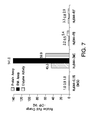
- FIG. 7 shows a graph showing protein expression changes observed in a hepatocyte sample preparation treated with inducers for CYP 1A2, using RNA assays, enzyme activity assays and the protein quantitation method of the present teachings.
- FIG. 8 shows a graph showing protein expression changes observed in a hepatocyte sample preparation treated with inducers for CYP 3A4, using RNA assays, enzyme activity assays and the protein quantitation method of the present teachings.
- a method for screening a drug for cytochrome P450 (CYP) induction comprises incubating the drug with a microsome-containing biological sample and quantitating at least one CYP isoform.
- the isoforms can comprise one or more isoform selected from 2B6, 3A4, 1A2, and 3A5 isoforms.
- the method can comprise using a liquid chromatography tandem mass spectrometry (LC-MSMS) technique to quantitate the amount of each isoform.
- the quantitated value of each can be compared to a threshold value, and the drug can be identified as having an acceptable CYP induction potential when the quantitated value does not exceed the threshold value.
- the threshold value can be selected or predetermined based on a desired CYP induction potential or based on the CYP induction potential of one or more different drugs, similar or non-similar to the drug being screened.
- the microsome-containing biological sample can be derived from a mammal, from a primate, or from a human.
- the drug can be incubated with a sample containing human hepatocytes.
- the sample containing human hepatocytes can be used to obtain at least one microsome fraction by, for example, 16 G centrifugation.
- the microsome fraction can be analyzed for CYP induction by the drug, by detecting isolated peptides specific to CYP (isoform-specific peptides).
- the sample containing human hepatocytes can be used to obtain at least one S9 fraction by, for example, 9 G centrifugation.
- the S9 fraction can be analyzed to detect CYP induction by the drug, for example, by detecting isolated peptides specific to CYP (isoform-specific peptides).
- the microsome fraction or the S9 fraction can be analyzed using a liquid chromatography tandem mass spectrometry (LC-MSMS) technique in order to quantitate at least one CYP isoform.
- LC-MSMS liquid chromatography tandem mass spectrometry
- the quantitated value of each can be compared to a threshold value, and the drug can be identified as having an acceptable CYP induction potential when the quantitated value does not exceed the threshold value.
- a method for directly analyzing CYP from hepatocytes.
- antibody peptides can be used to pull the isoform-specific peptides directly out of hepatocyctes.
- using antibody peptides to pull the isoform-specific peptides directly out of hepatocytes would have the advantage of not needing to prepare S9 or microsome fractions, and would require less hepatocyte cells for drug incubation.
- the method comprises comparing detected induction to a control. For example, because little or no drug induction of CYPs is desirable, a threshold can be set such that the drug must show less than ( ⁇ ) 40% induction compared to the positive control, to be considered acceptable. In some embodiments, the drug must show less than ( ⁇ ) 30% induction compared to the positive control, to be considered acceptable. In other embodiments, the drug must show less than ( ⁇ ) 20% induction compared to the positive control, to be considered acceptable.
- a method for determining an amount of at least one isoform of cytochrome P450 (CYP) in a sample is provided.
- the method can comprise the use of a mass spectrometry technique, wherein the at least one isoform of cytochrome P450 comprises at least one of CYP 2B6, CYP 3A4, CYP 1A2, and CYP 3A5.
- the mass spectrometry technique can comprise a tandem mass spectrometry (MS/MS) technique and/or a liquid chromatography tandem mass spectrometry (LC-MS/MS) technique.
- the technique comprises an LC-MS/MS technique and the use of a triple quadrupole instrument and Multiple Reaction Monitoring (MRM).
- MRM Multiple Reaction Monitoring
- FIG. 1 shows a typical MRM analysis for CYP 1A2, CYP 2B6, and CYP 3A4, as well as the “housekeeping” microsomal proteins (Microsomal GST, Corticosteroid 11 beta, and Microsomal Tryglyceride), from a microsomal sample preparation prepared as described below in the Examples.
- the quantity of CYP 1 A2, CYP 2B6, and CYP 3A4 can be determined by, for example, Isotope Dilution Mass Spectrometry, wherein the sample preparation is spiked with heavy forms of the isoform-specific peptides.
- the quantity of CYP 1A2, CYP 2B6, and CYP 3A4 can also be determined using other conventional methods known in the art.
- the method uses LC-MSMS with multiple reaction monitoring (MRM) quantitation of the isoform-specific peptides and isotope-coded affinity tags (ICAT) to generate a CYP induction profile.
- MRM multiple reaction monitoring
- ICAT isotope-coded affinity tags
- the method can use, for example, approaches similar to the approaches presented by Pennington et al. to quantitate isoform-specific cysteine-containing peptides labeled with ICAT as described in Proteomics, 6(6), pages 1934-1947 (March 2006), which is incorporated herein in its entirety by reference.
- FIG. 2 shows a typical MRM analysis for CYP 2B6 from the microsomal preparation prepared as described herein.
- Isolated peptides comprising the amino acid sequence of SEQ ID NOS: 1, 2, 3, or 4 identified in Table 1 below are specific to CYP 2B6.
- FIG. 3 shows a typical MRM analysis for CYP 1A2 from the microsomal preparation prepared as described herein.
- the isolated peptides comprising the amino acid sequence of SEQ ID NOS: 8, 9, 10, 11, 12, 13, or 14 identified in Table 3 below are specific to CYP 1A2.
- FIG. 4 shows a typical MRM analysis for CYP 3A4 from the microsomal preparation prepared as described herein.
- the isolated peptides comprising the amino acid sequence of SEQ ID NOs: 5, 6, or 7 identified in Table 2 below are specific to CYP 3A4. It should be understood that peptides comprising the amino acid sequence of SEQ ID NOS: 5, 6, or 7 can also be used to identify and/or quantify CYP 3A3.
- the isolated peptides comprising the amino acid sequence of SEQ ID NOs: 15, 16, or 17 identified in Table 4 below are specific to CYP 3A5.
- FIGS. 5A-5C show three different panes of a typical MRM analysis for CYP 3A5 from the microsomal preparation prepared as described herein. Each pane relates to specific transitions used for the particular peptide.
- the isolated peptides comprising the amino acid sequence of SEQ ID NOS: 15, 16, or 17 are specific to CYP 3A5.
- the method can comprise determining an amount of CYP 2B6 in the sample by detecting an isolated peptide specific to cytochrome P450 (CYP) isoform CYP 2B6, for example, one or more of the isoforms comprising the amino acid sequence of SEQ ID NO: 1, SEQ ID NO: 2, or SEQ ID NO: 3 identified herein.
- the amount can be determined using a triple quadrupole instrument and Multiple Reaction Monitoring (MRM).
- MRM Multiple Reaction Monitoring
- the isolated peptide can comprise the amino acid sequence of SEQ ID NO: 1 identified herein, and the method can comprise monitoring precursor-product ion pair transitions having an m/z value of about 548/911, 548/681, or 548/566, wherein the term “about” as used herein means within a range of +/ ⁇ one (1) atomic mass unit.
- the isolated peptide can comprise the amino acid sequence of SEQ ID NO: 2 identified herein, and the method can comprise monitoring precursor-product ion pair transitions having an m/z value of about 494/777, 494/437, or 494/874.
- the isolated peptide can comprise the amino acid sequence of SEQ ID NO: 3 identified herein, and the method can comprise monitoring precursor-product ion pair transitions having an m/z value of about 421/508, 4211607, or 421/694. In some embodiments, the isolated peptide can comprise the amino acid sequence of SEQ ID NO: 4, and the method can comprise monitoring precursor-product ion pair transitions having an m/z value of about 479/499, 479/614, or 479/727.
- the method can comprise determining an amount of CYP 3A4 in the sample by detecting an isolated peptide specific to cytochrome P450 (CYP) isoform CYP 3A4, comprising the amino acid sequence of SEQ ID NO: 5, SEQ ID NO: 6, or SEQ ID NO: 7 identified herein.
- the method can use, for example, a triple quadrupole instrument and Multiple Reaction Monitoring (MRM).
- MRM Multiple Reaction Monitoring
- the isolated peptide can comprise the amino acid sequence of SEQ ID NO: 5 identified herein, and the method can comprise monitoring precursor-product ion pair transitions having an m/z value of about 440/549, 440/650, or 440/532.
- the isolated peptide can comprise the amino acid sequence of SEQ ID NO: 6 identified herein, and the method can comprise monitoring precursor-product ion pair transitions having an m/z value of about 704/794, 704/929, 564/689, 564/745, or 564/790.
- the isolated peptide can comprise the amino acid sequence of SEQ ID NO: 7 identified herein, and the method can comprise monitoring precursor-product ion pair transitions having an m/z value of about 798/819, 798/932, or 798/1004.
- the method can comprise determining an amount of CYP 1A2 in the sample by detecting an isolated peptide specific to cytochrome P450 (CYP) isoform CYP 1A2, comprising the amino acid sequence of SEQ ID NO: 8, SEQ ID NO: 9, SEQ ID NO: 10, SEQ ID NO: 11, SEQ ID NO: 12, SEQ ID NO: 13, or SEQ ID NO: 14, identified herein.
- CYP cytochrome P450
- the method can use, for example, a triple quadrupole instrument and Multiple Reaction Monitoring (MRM).
- MRM Multiple Reaction Monitoring
- the isolated peptide can comprise the amino acid sequence of SEQ ID NO: 8 identified herein, and the method can comprise monitoring precursor-product ion pair transitions having an m/z value of about 432/636, 432/535, or 432/478.
- the isolated peptide can comprise the amino acid sequence of SEQ ID NO: 9 identified herein, and the method can comprise monitoring precursor-product ion pair transitions having an m/z value of about 482/800, 482/628, or 482/743.
- the isolated peptide can comprise the amino acid sequence of SEQ ID NO: 10 identified herein, and the method can comprise monitoring precursor-product ion pair transitions having an m/z value of about 491/721, 491/834, or 491/535.
- the isolated peptide can comprise the amino acid sequence of SEQ ID NO: 11 identified herein, and the method can comprise monitoring precursor-product ion pair transitions having an m/z value of about 528/501, 528/614, or 528/727.
- the isolated peptide can comprise the amino acid sequence of SEQ ID NO: 12 identified herein, and the method can comprise monitoring precursor-product ion pair transitions having an m/z value of about 571/783, 571/971, 571/1028, 381/587, 381/474, or 381/375.
- the isolated peptide can comprise the amino acid sequence of SEQ ID NO: 13 identified herein, and the method can comprise monitoring precursor-product ion pair transitions having an m/z value of about 695/695, 695/837, or 695/950.
- the isolated peptide can comprise the isolated peptide comprises the amino acid sequence of SEQ ID NO: 14 identified herein, and the method can comprise monitoring precursor-product ion pair transitions having an m/z value of about 536/795, 536/584, or 536/698.
- the method can comprise determining an amount of CYP 3A5 in the sample by detecting an isolated peptide specific to cytochrome P450 (CYP) isoform CYP 3A5, comprising the amino acid sequence of SEQ ID NO: 15, SEQ ID NO: 16, or SEQ ID NO: 17 identified herein.
- the method can use, for example, a triple quadrupole instrument and Multiple Reaction Monitoring (MRM).
- MRM Multiple Reaction Monitoring
- the isolated peptide can comprise an amino acid sequence of SEQ ID NO: 15 identified herein, and the method can comprise monitoring precursor-product ion pair transitions having an m/z value of about 468/581, 468/679, or 468/736.
- the isolated peptide can comprise an amino acid sequence of SEQ ID NO: 16 identified herein, and the method can comprise monitoring precursor-product ion pair transitions having an m/z value of about 470/494, 470/608, or 470/722. In some embodiments, the isolated peptide can comprise an amino acid sequence of SEQ ID NO: 17 identified herein, and the method can comprise monitoring precursor-product ion pair transitions having an m/z value of about 589/747, 589/696, or 589/647.
- a kit can comprise one or more of the isolated peptides specific to one or more of cytochrome P450 (CYP) isoform CYP 2B6, CYP 3A4, CYP 1A2, and CYP 3A5.
- the kit can comprise one or more isolated proteins specific to CYP 2B6, comprising the amino acid sequence of SEQ ID NO: 1, SEQ ID NO: 2, SEQ ID NO: 3, or SEQ ID NO: 4.
- the kit can comprise one or more isolated proteins specific to CYP isoform CYP 3A4, comprising the amino acid sequence of SEQ ID NO: 5, SEQ ID NO: 6, or SEQ ID NO: 7.
- the kit can comprise one or more isolated proteins specific to CYP isoform CYP 1A2, comprising the amino acid sequence of SEQ ID NO: 8, SEQ ID NO: 9, SEQ ID NO: 10, SEQ ID NO: 11, SEQ ID NO: 12, SEQ ID NO: 13, or SEQ ID NO: 14.
- the kit can comprise one or more isolated proteins specific to CYP isoform CYP 3A5, comprising the amino acid sequence of SEQ ID NO: 15, SEQ ID NO: 16, or SEQ ID NO: 17.
- the kit can comprise at least one isolated peptide specific to each of CYP isoforms CYP 2B6, CYP 3A4, CYP 1A2, and CYP 3A5.
- the kit can comprise each of the isolated peptides of SEQ ID NOS: 1-17 identified herein, and further can comprise instructions for measuring Q1 and Q3 transition values for each of the isoform-specific peptides.
- the kit can comprise enzyme digestion components including buffers and enzymes, other buffers, and optionally other reagents and/or components.
- the kit can comprise, for example, a homogeneous assay such that the user need only add a sample.
- the kit can comprise calibration or normalization reagents or standards.
- Information pertaining to instrument settings that can or should be used to perform an assay can also be included in the kit.
- Information pertaining to sample preparation, operating conditions, volumetric amounts, temperature settings, and the like, can be included with the kit.
- the kit can comprise different transition values and/or suggested settings, useful to make comparative measurements between a sample and one or more control reagents.
- the kit can include instructions to measure specific pairs of transition values, for example, the Q1/Q3 transition pair, or the values of one or more different transition pairs.
- the kit can be packaged in a hermetically sealed container containing one or more regent vessels and appropriate instructions.
- An electronic medium can be included in the kit, having stored thereon electronic information pertaining to one or more assays, measurement values, transition pairs, operating instructions, software for carrying out operations, a combination thereof, or the like.
- CYP quantitation method of the present teachings was used to determine the amount of CYP in the sample after treatment with the liver enzyme inducers.
- CYP activity was measured by metabolite formation from selective substrates (phenacetin, bupropion and testosterone, respectively) and mRNA was measured by qRT-PCR (Taqman®, Applied Biosystems).
- Microsomal subcellular fractions were prepared by lysing treated hepatocytes in homogenization buffer (50 mM TRIS-HCl, pH 7.0, 150 mM KCl, 2 mM EDTA) followed by centrifugation at 9,000 ⁇ g for 20 minutes at 4° C. The supernatant (S9 fraction) was then spun at 100,000 ⁇ g for 60 minutes at 4° C. The resulting microsomal pellet was resuspended in 0.25 M sucrose and stored at ⁇ 80° C. until analysis.
- homogenization buffer 50 mM TRIS-HCl, pH 7.0, 150 mM KCl, 2 mM EDTA
- Quantitative data was processed using MultiQuantTM 1.2 software available from Applied Biosystems, LLC of Foster City, Calif.
- FIG. 6 is a graph comparing the changes in expression of CYP 2B6 observed using the RNA assay, the CYP activity assay (designated “enzyme activity assay” in the figures), and a CYP quantitation method of the present teachings (designated “protein assay” in the figures).
- the 3-MC is a vehicle control and induces basal levels of CYP 2B6.
- the PB is a prototypical inducer for CYP 2B6, by CAR nuclear receptor activation.
- the RIF is also a known inducer of CYP 2B6.
- protein expression changes observed in the CYP activity assay and RNA assay generally mirror the expression changes observed using a CYP quantitation method of the present teachings.
- FIG. 7 is a graph comparing the changes in expression of CYP 1A2 observed using the RNA assay, the CYP activity assay (designated “enzyme activity assay” in the figures), and the CYP quantitation method of the present teachings (designated “protein assay” in the figures).
- the 3-MC is a prototypical inducer of CYP 1A2 by AhR nuclear receptor activation.
- the PB and RIF are vehicle controls inducing basal levels of 1A2, if any.
- protein expression changes observed in the CYP activity assay and RNA assay generally mirror the expression changes observed using a CYP quantitation method of the present teachings, except that the RNA assay for the sample treated with 3MC exhibits very high levels of RNA.
- the RNA assay cannot accurately quantify proteins because not all mRNA is converted to protein.
- FIG. 8 is a graph comparing the changes in expression of CYP 3A4 observed using the RNA assay, the CYP activity assay (designated “enzyme activity assay” in the figures), and a CYP quantitation method of the present teachings (designated “protein assay” in the figures).
- the 3-MC minimally induces CYP 3A4.
- the PB significantly induces 3A4.
- the RIF is a prototypical inducer of CYP 3A4 by PXR nuclear receptor activation.
- protein expression changes observed in the CYP activity assay and RNA assay generally mirror the expression changes observed using the CYP quantitation method of the present teachings.
- Table 1 shows sequences of the peptides determined, according to the present teachings, to be specific to cytochrome P450 (CYP) isoform CYP 2B6, along with their optimal MRM Q1, Q3 transitions. According to various embodiments, these observed peptides and transitions can be used to enable a reliable CYP quantitation of the isoform CYP 2B6.
- CYP cytochrome P450
- Table 2 shows sequences of the peptides determined, according to the present teachings, to be specific to cytochrome P450 (CYP) isoform CYP 3A4, along with their optimal MRM Q1, Q3 transitions. According to various embodiments, these observed peptides and transitions can be used to enable a reliable CYP quantitation of the isoform CYP 3A4.
- CYP cytochrome P450
- Table 3 shows sequences of the peptides determined, according to the present teachings, to be specific to cytochrome P450 (CYP) isoform CYP 1A2, along with their optimal MRM Q1, Q3 transitions. According to various embodiments, these observed peptides and transitions can be used to enable a reliable CYP quantitation of the isoform CYP 1A2.
- CYP cytochrome P450
- Table 4 shows sequences of the peptides determined, according to the present teachings, to be specific to cytochrome P450 (CYP) isoform CYP 3A5, along with their optimal MRM Q1, Q3 transitions. According to various embodiments, these observed peptides and transitions can be used to enable a reliable CYP quantitation of the isoform CYP 3A5.
- CYP cytochrome P450
- Table 5 shows sequences of the peptides along with their optimal MRM Q1, Q3 transitions for the house-keeping microsomal protein Microsomal GST. According to various embodiments, the concentration of this observed peptide is unaffected by drug incubation and thus the peptide can be useful as a normalization protein to enable reliable CYP quantitation.
- Table 6 shows sequences of the peptides along with their optimal MRM Q1, Q3 transitions for the house-keeping microsomal protein Microsomal Triglyceride. According to various embodiments, the concentration of this observed peptide is unaffected by drug incubation and thus the peptide can be useful as a normalization protein to enable reliable CYP quantitation.
- Table 7 shows sequences of the peptides along with their optimal MRM Q1, Q3 transitions for the house-keeping microsomal protein Corticosteroid 11 beta. It should be understood that the Q1 and Q3 masses for the peptide of SEQ ID NO: 22 in Table 7 refers to the MMTS alkylated peptide and that changing to a different alkylating reagent will change the Q1, Q3 masses. In addition, alkylating reagents other than MMTS can be used. According to various embodiments, the concentration of this observed peptide is unaffected by drug incubation and thus the peptide can be useful as a normalization protein to enable reliable CYP quantitation.
Landscapes
- Health & Medical Sciences (AREA)
- Life Sciences & Earth Sciences (AREA)
- Engineering & Computer Science (AREA)
- Chemical & Material Sciences (AREA)
- Molecular Biology (AREA)
- Biomedical Technology (AREA)
- Immunology (AREA)
- Bioinformatics & Cheminformatics (AREA)
- Hematology (AREA)
- Urology & Nephrology (AREA)
- Physics & Mathematics (AREA)
- Biotechnology (AREA)
- Microbiology (AREA)
- Biochemistry (AREA)
- General Health & Medical Sciences (AREA)
- Medicinal Chemistry (AREA)
- Organic Chemistry (AREA)
- Cell Biology (AREA)
- Analytical Chemistry (AREA)
- Zoology (AREA)
- Wood Science & Technology (AREA)
- Food Science & Technology (AREA)
- General Physics & Mathematics (AREA)
- Pathology (AREA)
- Genetics & Genomics (AREA)
- Proteomics, Peptides & Aminoacids (AREA)
- Bioinformatics & Computational Biology (AREA)
- Tropical Medicine & Parasitology (AREA)
- Toxicology (AREA)
- Biophysics (AREA)
- General Engineering & Computer Science (AREA)
- Spectroscopy & Molecular Physics (AREA)
- Gastroenterology & Hepatology (AREA)
- Other Investigation Or Analysis Of Materials By Electrical Means (AREA)
- Peptides Or Proteins (AREA)
- Enzymes And Modification Thereof (AREA)
- Measuring Or Testing Involving Enzymes Or Micro-Organisms (AREA)
- Investigating Or Analysing Biological Materials (AREA)
Priority Applications (3)
| Application Number | Priority Date | Filing Date | Title |
|---|---|---|---|
| US12/904,520 US8940545B2 (en) | 2009-10-16 | 2010-10-14 | Mass spectrometry quantitation of P450 isoforms in hepatocytes |
| US14/465,460 US20150045256A1 (en) | 2009-10-16 | 2014-08-21 | Mass spectrometry quantitation of p450 protein isoforms in hepatocytes |
| US14/602,917 US9546393B2 (en) | 2009-10-16 | 2015-01-22 | Mass spectrometry quantitation of P450 isoforms in hepatocytes |
Applications Claiming Priority (3)
| Application Number | Priority Date | Filing Date | Title |
|---|---|---|---|
| US25243009P | 2009-10-16 | 2009-10-16 | |
| US25264809P | 2009-10-17 | 2009-10-17 | |
| US12/904,520 US8940545B2 (en) | 2009-10-16 | 2010-10-14 | Mass spectrometry quantitation of P450 isoforms in hepatocytes |
Related Child Applications (2)
| Application Number | Title | Priority Date | Filing Date |
|---|---|---|---|
| US14/465,460 Division US20150045256A1 (en) | 2009-10-16 | 2014-08-21 | Mass spectrometry quantitation of p450 protein isoforms in hepatocytes |
| US14/602,917 Division US9546393B2 (en) | 2009-10-16 | 2015-01-22 | Mass spectrometry quantitation of P450 isoforms in hepatocytes |
Publications (2)
| Publication Number | Publication Date |
|---|---|
| US20110091981A1 US20110091981A1 (en) | 2011-04-21 |
| US8940545B2 true US8940545B2 (en) | 2015-01-27 |
Family
ID=43876537
Family Applications (3)
| Application Number | Title | Priority Date | Filing Date |
|---|---|---|---|
| US12/904,520 Active 2032-11-28 US8940545B2 (en) | 2009-10-16 | 2010-10-14 | Mass spectrometry quantitation of P450 isoforms in hepatocytes |
| US14/465,460 Abandoned US20150045256A1 (en) | 2009-10-16 | 2014-08-21 | Mass spectrometry quantitation of p450 protein isoforms in hepatocytes |
| US14/602,917 Expired - Fee Related US9546393B2 (en) | 2009-10-16 | 2015-01-22 | Mass spectrometry quantitation of P450 isoforms in hepatocytes |
Family Applications After (2)
| Application Number | Title | Priority Date | Filing Date |
|---|---|---|---|
| US14/465,460 Abandoned US20150045256A1 (en) | 2009-10-16 | 2014-08-21 | Mass spectrometry quantitation of p450 protein isoforms in hepatocytes |
| US14/602,917 Expired - Fee Related US9546393B2 (en) | 2009-10-16 | 2015-01-22 | Mass spectrometry quantitation of P450 isoforms in hepatocytes |
Country Status (5)
| Country | Link |
|---|---|
| US (3) | US8940545B2 (enExample) |
| EP (2) | EP2862870B1 (enExample) |
| JP (2) | JP5936273B2 (enExample) |
| CA (1) | CA2777690C (enExample) |
| WO (1) | WO2011047150A1 (enExample) |
Families Citing this family (2)
| Publication number | Priority date | Publication date | Assignee | Title |
|---|---|---|---|---|
| JP2012056880A (ja) * | 2010-09-08 | 2012-03-22 | Idemitsu Kosan Co Ltd | インドロカルバゾール化合物、有機エレクトロルミネッセンス素子用材料、及びそれを用いた有機エレクトロルミネッセンス素子 |
| WO2014096914A1 (en) * | 2012-12-20 | 2014-06-26 | Dh Technologies Development Pte. Ltd. | Scheduled ms3 for quantitation |
Citations (2)
| Publication number | Priority date | Publication date | Assignee | Title |
|---|---|---|---|---|
| US20070092926A1 (en) * | 2005-10-14 | 2007-04-26 | Alterman Michail A | Analysis of protein isoforms using unique tryptic peptides by mass spectrometry and immunochemistry |
| US20080206737A1 (en) * | 2004-05-19 | 2008-08-28 | Hunter Christie L | Expression quantification using mass spectrometry |
Family Cites Families (3)
| Publication number | Priority date | Publication date | Assignee | Title |
|---|---|---|---|---|
| JP2007538262A (ja) * | 2004-05-19 | 2007-12-27 | アプレラ コーポレイション | 質量分析を使用する発現の定量化 |
| US20080020673A1 (en) * | 2005-03-08 | 2008-01-24 | Cara Sills | Doll having hand-assemblable costume |
| JP5299956B2 (ja) * | 2008-09-29 | 2013-09-25 | 国立大学法人東北大学 | 質量分析装置を用いた代謝酵素群の一斉タンパク質定量に用いるペプチド |
-
2010
- 2010-10-14 CA CA2777690A patent/CA2777690C/en not_active Expired - Fee Related
- 2010-10-14 EP EP14193907.4A patent/EP2862870B1/en active Active
- 2010-10-14 WO PCT/US2010/052665 patent/WO2011047150A1/en not_active Ceased
- 2010-10-14 EP EP10824088.8A patent/EP2488545B1/en active Active
- 2010-10-14 US US12/904,520 patent/US8940545B2/en active Active
- 2010-10-14 JP JP2012534356A patent/JP5936273B2/ja not_active Expired - Fee Related
-
2014
- 2014-08-21 US US14/465,460 patent/US20150045256A1/en not_active Abandoned
-
2015
- 2015-01-22 US US14/602,917 patent/US9546393B2/en not_active Expired - Fee Related
-
2016
- 2016-04-04 JP JP2016074921A patent/JP6116730B2/ja active Active
Patent Citations (2)
| Publication number | Priority date | Publication date | Assignee | Title |
|---|---|---|---|---|
| US20080206737A1 (en) * | 2004-05-19 | 2008-08-28 | Hunter Christie L | Expression quantification using mass spectrometry |
| US20070092926A1 (en) * | 2005-10-14 | 2007-04-26 | Alterman Michail A | Analysis of protein isoforms using unique tryptic peptides by mass spectrometry and immunochemistry |
Non-Patent Citations (3)
| Title |
|---|
| Applied Biosystems Sciex, "Advanced Hybrid Triple Quadrupole-Linear Ion Trap Technology, 4000 QTRAP LC/MS/MS System"; Brochure, 2010, pp. 1-8. * |
| Redlich et al. "Distinction between Human Cytochrome P450 (CYP) Isoforms and Identification of New Phosphorylation Sites by Mass Spectrometry", Journal of Proteome Research; published online Oct. 2008; 2008, pp. 4678-4688. * |
| Wu, Genbank Accession No. AAF13602, 1995. * |
Also Published As
| Publication number | Publication date |
|---|---|
| JP6116730B2 (ja) | 2017-04-19 |
| US20110091981A1 (en) | 2011-04-21 |
| JP5936273B2 (ja) | 2016-06-22 |
| EP2862870A3 (en) | 2015-08-12 |
| EP2862870B1 (en) | 2020-08-12 |
| WO2011047150A1 (en) | 2011-04-21 |
| CA2777690C (en) | 2019-05-21 |
| US20150140600A1 (en) | 2015-05-21 |
| EP2488545A4 (en) | 2013-05-15 |
| JP2013507924A (ja) | 2013-03-07 |
| US20150045256A1 (en) | 2015-02-12 |
| EP2488545B1 (en) | 2014-12-24 |
| JP2016119915A (ja) | 2016-07-07 |
| US9546393B2 (en) | 2017-01-17 |
| EP2488545A1 (en) | 2012-08-22 |
| CA2777690A1 (en) | 2011-04-21 |
| EP2862870A2 (en) | 2015-04-22 |
Similar Documents
| Publication | Publication Date | Title |
|---|---|---|
| Kitteringham et al. | Multiple reaction monitoring for quantitative biomarker analysis in proteomics and metabolomics | |
| Kaspar et al. | Urinary amino acid analysis: a comparison of iTRAQ®–LC–MS/MS, GC–MS, and amino acid analyzer | |
| Cooke et al. | Evaluation of enzyme-linked immunosorbent assay and liquid chromatography–tandem mass spectrometry methodology for the analysis of 8-oxo-7, 8-dihydro-2′-deoxyguanosine in saliva and urine | |
| Liu et al. | Determination of polyamine metabolome in plasma and urine by ultrahigh performance liquid chromatography–tandem mass spectrometry method: Application to identify potential markers for human hepatic cancer | |
| Weiß et al. | Direct quantification of cytochromes P450 and drug transporters—a rapid, targeted mass spectrometry-based immunoassay panel for tissues and cell culture lysates | |
| Sato et al. | Protein quantification of UDP-glucuronosyltransferases 1A1 and 2B7 in human liver microsomes by LC-MS/MS and correlation with glucuronidation activities | |
| De Bock et al. | Development and validation of a fast and sensitive UPLC–MS/MS method for the quantification of six probe metabolites for the in vitro determination of cytochrome P450 activity | |
| Vildhede et al. | Comparison of proteomic quantification approaches for hepatic drug transporters: multiplexed global quantitation correlates with targeted proteomic quantitation | |
| US20220057412A1 (en) | Method for Measuring Testosterone Using LC-MSMS | |
| Fernández et al. | Multiple heart-cutting two dimensional liquid chromatography and isotope dilution tandem mass spectrometry for the absolute quantification of proteins in human serum | |
| Newman et al. | Addressing MISEV guidance using targeted LC‐MS/MS: A method for the detection and quantification of extracellular vesicle‐enriched and contaminant protein markers from blood | |
| Sykes et al. | Quantitative proteomic analysis of ribosome assembly and turnover in vivo | |
| US8039794B2 (en) | Mass spectrometry assay for thiopurine-S-methyl transferase activity and products generated thereby | |
| US9546393B2 (en) | Mass spectrometry quantitation of P450 isoforms in hepatocytes | |
| Williamson et al. | Quantitative protein determination for CYP induction via LC‐MS/MS | |
| Ji et al. | A highly sensitive and robust LC-MS platform for host cell protein characterization in biotherapeutics | |
| Fu et al. | Multiple and selective reaction monitoring using triple quadrupole mass spectrometer: preclinical large cohort analysis | |
| Thompson et al. | Investigation of oxidative phosphorylation activity and complex composition in mitochondrial disease | |
| Muschong et al. | Improvement of Workflows and Assay Reproducibility by The Introduction of “Assay-Ready” Culturing of MDCK Cells for Transport Studies | |
| Browne et al. | Semi quantitative detection of signature peptides in body fluids by liquid chromatography tandem mass spectrometry (LC–MS/MS) | |
| CN102175804A (zh) | Cyp450酶亚型生物质谱绝对定量方法 | |
| Al‐Dirbashi et al. | Reliable prenatal diagnosis of Canavan disease by measuring N‐acetylaspartate in amniotic fluid using liquid chromatography tandem mass spectrometry | |
| Bouquié et al. | Validation and application of a fast semi-automated whole blood extraction for LC-MS/MS simultaneous quantification of cyclosporine A, tacrolimus, sirolimus and everolimus–application to high throughput routine therapeutic drug monitoring | |
| Williams | LC-MS/MS measurement of serum steroids in the clinical laboratory | |
| Viana et al. | Mass spectrometry multiplexed detection of SARS-CoV-2 |
Legal Events
| Date | Code | Title | Description |
|---|---|---|---|
| AS | Assignment |
Owner name: DH TECHNOLOGIES DEVELOPMENT PTE. LTD., SINGAPORE Free format text: ASSIGNMENT OF ASSIGNORS INTEREST;ASSIGNORS:WILLIAMSON, BRIAN L.;PURKAYASTHA, SUBHASISH;SIGNING DATES FROM 20101119 TO 20101122;REEL/FRAME:025530/0227 |
|
| STCF | Information on status: patent grant |
Free format text: PATENTED CASE |
|
| MAFP | Maintenance fee payment |
Free format text: PAYMENT OF MAINTENANCE FEE, 4TH YEAR, LARGE ENTITY (ORIGINAL EVENT CODE: M1551) Year of fee payment: 4 |
|
| MAFP | Maintenance fee payment |
Free format text: PAYMENT OF MAINTENANCE FEE, 8TH YEAR, LARGE ENTITY (ORIGINAL EVENT CODE: M1552); ENTITY STATUS OF PATENT OWNER: LARGE ENTITY Year of fee payment: 8 |