US8841432B2 - Shuttle vectors for mycobacteria-escherichia coli and uses thereof - Google Patents

Shuttle vectors for mycobacteria-escherichia coli and uses thereof Download PDF

Info

Publication number
US8841432B2
US8841432B2 US13/450,689 US201213450689A US8841432B2 US 8841432 B2 US8841432 B2 US 8841432B2 US 201213450689 A US201213450689 A US 201213450689A US 8841432 B2 US8841432 B2 US 8841432B2
Authority
US
United States
Prior art keywords
promoter
protein
antigen
vector according
cells
Prior art date
Legal status (The legal status is an assumption and is not a legal conclusion. Google has not performed a legal analysis and makes no representation as to the accuracy of the status listed.)
Active
Application number
US13/450,689
Other versions
US20120270322A1 (en
Inventor
Bum-Joon Kim
Hyungki LEE
Current Assignee (The listed assignees may be inaccurate. Google has not performed a legal analysis and makes no representation or warranty as to the accuracy of the list.)
SNU R&DB Foundation
Original Assignee
SNU R&DB Foundation
Priority date (The priority date is an assumption and is not a legal conclusion. Google has not performed a legal analysis and makes no representation as to the accuracy of the date listed.)
Filing date
Publication date
Application filed by SNU R&DB Foundation filed Critical SNU R&DB Foundation
Assigned to SNU R&DB FOUNDATION reassignment SNU R&DB FOUNDATION ASSIGNMENT OF ASSIGNORS INTEREST (SEE DOCUMENT FOR DETAILS). Assignors: KIM, BUM-JOON, LEE, HYUNGKI
Publication of US20120270322A1 publication Critical patent/US20120270322A1/en
Application granted granted Critical
Publication of US8841432B2 publication Critical patent/US8841432B2/en
Active legal-status Critical Current
Anticipated expiration legal-status Critical

Links

Images

Classifications

    • CCHEMISTRY; METALLURGY
    • C12BIOCHEMISTRY; BEER; SPIRITS; WINE; VINEGAR; MICROBIOLOGY; ENZYMOLOGY; MUTATION OR GENETIC ENGINEERING
    • C12NMICROORGANISMS OR ENZYMES; COMPOSITIONS THEREOF; PROPAGATING, PRESERVING, OR MAINTAINING MICROORGANISMS; MUTATION OR GENETIC ENGINEERING; CULTURE MEDIA
    • C12N15/00Mutation or genetic engineering; DNA or RNA concerning genetic engineering, vectors, e.g. plasmids, or their isolation, preparation or purification; Use of hosts therefor
    • C12N15/09Recombinant DNA-technology
    • C12N15/11DNA or RNA fragments; Modified forms thereof; Non-coding nucleic acids having a biological activity
    • CCHEMISTRY; METALLURGY
    • C12BIOCHEMISTRY; BEER; SPIRITS; WINE; VINEGAR; MICROBIOLOGY; ENZYMOLOGY; MUTATION OR GENETIC ENGINEERING
    • C12NMICROORGANISMS OR ENZYMES; COMPOSITIONS THEREOF; PROPAGATING, PRESERVING, OR MAINTAINING MICROORGANISMS; MUTATION OR GENETIC ENGINEERING; CULTURE MEDIA
    • C12N15/00Mutation or genetic engineering; DNA or RNA concerning genetic engineering, vectors, e.g. plasmids, or their isolation, preparation or purification; Use of hosts therefor
    • C12N15/09Recombinant DNA-technology
    • C12N15/63Introduction of foreign genetic material using vectors; Vectors; Use of hosts therefor; Regulation of expression
    • C12N15/74Vectors or expression systems specially adapted for prokaryotic hosts other than E. coli, e.g. Lactobacillus, Micromonospora
    • CCHEMISTRY; METALLURGY
    • C12BIOCHEMISTRY; BEER; SPIRITS; WINE; VINEGAR; MICROBIOLOGY; ENZYMOLOGY; MUTATION OR GENETIC ENGINEERING
    • C12NMICROORGANISMS OR ENZYMES; COMPOSITIONS THEREOF; PROPAGATING, PRESERVING, OR MAINTAINING MICROORGANISMS; MUTATION OR GENETIC ENGINEERING; CULTURE MEDIA
    • C12N1/00Microorganisms, e.g. protozoa; Compositions thereof; Processes of propagating, maintaining or preserving microorganisms or compositions thereof; Processes of preparing or isolating a composition containing a microorganism; Culture media therefor
    • C12N1/20Bacteria; Culture media therefor
    • CCHEMISTRY; METALLURGY
    • C12BIOCHEMISTRY; BEER; SPIRITS; WINE; VINEGAR; MICROBIOLOGY; ENZYMOLOGY; MUTATION OR GENETIC ENGINEERING
    • C12NMICROORGANISMS OR ENZYMES; COMPOSITIONS THEREOF; PROPAGATING, PRESERVING, OR MAINTAINING MICROORGANISMS; MUTATION OR GENETIC ENGINEERING; CULTURE MEDIA
    • C12N15/00Mutation or genetic engineering; DNA or RNA concerning genetic engineering, vectors, e.g. plasmids, or their isolation, preparation or purification; Use of hosts therefor
    • C12N15/09Recombinant DNA-technology
    • C12N15/63Introduction of foreign genetic material using vectors; Vectors; Use of hosts therefor; Regulation of expression
    • C12N15/70Vectors or expression systems specially adapted for E. coli

Definitions

  • the present disclosure relates to shuttle vectors for Mycobacteria and Escherichia coli and their use.
  • the transgene is usually introduced through a process called bactofection using viruses or bacteria as delivery systems. Consequently bacteria harboring protein encoding plasmids enter a eukaryotic cell and release the plasmid for uptake into the nucleus, where the plasmid encoded genes are expressed endogenously, and the plasmid may be either stably integrated into the genome of the cell or be present in the cytoplasm without being integrated into the genome.
  • Mycobacteria can be used advantageously as a delivery system for inducing/enhancing an immune response to proteins encoded in the plasmid because of its ability of disrupting immune tolerance in host at the cytotoxic T-level.
  • pAL5000 replicon is the most widely used vector as a Mycobacteria - Escherichia coli shuttle plasmid for a variety of uses.
  • the system has some drawbacks that the protein expressed from the vector in mycobacteria is not correctly folded and modified. Therefore there are demands for the new vector system which can correctly and reliably produce the encoded proteins.
  • the present disclosure provides a replicable DNA molecule derived from Mycobacteria having a nucleic acid sequence as disclosed in SEQ ID NO: 1.
  • the present disclosure provides a Mycobacteria - Escherichia coli shuttle vector comprising: (a) an origin of replication having a nucleic acid sequence as disclosed in SEQ ID NO: 2 (oriM); (b) an origin of replication for prokaryotic cells; (c) a promoter; and (d) a nucleic acid sequence encoding a target material, which is operatively linked to the promoter.
  • the oriM in the shuttle vector according to the present disclosure contains A+T rich region and direct repeat region.
  • the promoter which may be used for the present disclosure includes a heat shock protein promoter, a CMV promoter, a promoter for 65 kDa common antigen of mycobacteria, ribosome RNA promoter from Mycobacteria , a promoter for MPB70, MPB59 or MPB64 antigen from Mycobacterium bovis , P1 promoter from bacteriophage Lamda, tac promoter, trp promoter, lac promoter, lacUV5 promoter, Ipp promoter, PL ⁇ promoter, PR ⁇ promoter, rac5 promoter, amp promoter, recA promoter, SP6 promoter, T7 promoter, a promoter for kanamycin resistance gene of transposon Tn903 or Tn5, a promoter for metallothionine, a promoter for growth hormone or a hybrid promoter between eukaryotic and prokaryotic promoter, or a combination thereof.
  • a shuttle vector which may encode a protein, antisense oligonucleotide, siRNA, shRNA, miRNA or piRNA.
  • a shuttle vector which encoded a reporter protein, which includes, for example, a fluorescent protein, a beta-galactosidase, a chloramphenicol acetyl transferase, a human growth hormone, a urease or an alkaline phosphatase.
  • a reporter protein which includes, for example, a fluorescent protein, a beta-galactosidase, a chloramphenicol acetyl transferase, a human growth hormone, a urease or an alkaline phosphatase.
  • a shuttle vector which encoded a fluorescent protein which includes, for example, GFP (green fluorescent protein), RFP (red fluorescent protein), CFP (cyan fluorescent protein), YFP (yellow fluorescent protein), BFP (blue fluorescent protein) or its variants.
  • GFP green fluorescent protein
  • RFP red fluorescent protein
  • CFP cyan fluorescent protein
  • YFP yellow fluorescent protein
  • BFP blue fluorescent protein
  • a shuttle vector which further includes one or more selective markers.
  • the selective markers include for example genes conferring resistance to antibiotics which include kanamycin, hygromycin, ampicillin, streptomycin, penicillin, chloramphenicol, gentamicin, carbenicillin, geneticin, neomycin or tetracycline.
  • a shuttle vector wherein the origin of replication is provided in a separate expression vector as a co-transformation.
  • the present disclosure relates to a cell transformed with a vector as disclosed in the present disclosure.
  • the transformed cells in the present disclosure are derived from cells which include Mycobacteria or Escherichia coli.
  • the Mycobacteria includes M. smegmatis, M. bovis -BCG, M. avium, M. phlei, M. fortuitum, M. lufu, M. partuberculosis, M. habana, M. scrofulaceum , or M. intracellulare.
  • a method of using a first and a second vector for expression of heterologous transgenes in a eukaryotic cell wherein the first vector is the vector according to claim 1 or 2 and the second vector is pSE100 in eukaryotic cells.
  • the transgenes encoded in the vector of the present method include a protein, antisense oligonucleotide, siRNA, ashRNA, miRNA or piRNA, and the transgene encoded by the first and the second vector is different.
  • the protein encoded in the vector of the present method includes a porter protein, an antigen or a therapeutic protein.
  • the reporter protein encoded in the vector of the present method includes fluorescent protein, beta-galactosidase, chloramphenicol acetyl transferase, human growth hormone, urease or alkaline phosphatase; wherein the antigen is derived from virulent pathogens; and wherein the therapeutic protein includes IL-12 or GM-CSF.
  • FIG. 1 is the agarose gel analysis result showing the genome from Mycobacteria and the genome digested with a restriction enzyme (left panel); and a schematic representation of the structure of the linear 18 kb plasmid pM90 (right panel).
  • the each lane of the gel indicates: 1: genome of M. intracellulare; 2: genome of MOTT90; and 3: genome of MOTT90 digested with XhoI.
  • FIG. 2 is a schematic representation of the putative ORFs identified in the plasmid (A) in accordance of the present disclosure and the gel analysis result showing the expression from each ORF (B).
  • FIG. 3 is a schematic representation of the structure of the oriM of the plasmid in accordance of the present disclosure (A), which shows that the ori contains an AT-rich region, two 14 bp repeated regions that are disclosed as SEQ ID NO: 30 and a terminal inverted repeat of 68 bp, which is disclosed as SEQ ID NO: 29; and the alignment of the sequences of the conserved region from p05-1390, pCLP, pJAZ38, pMFI, pVT2 and pMSC262 (B), each of which is disclosed as SEQ ID NOs: 31, 32, 33, 34, 35 and 36, respectively.
  • FIG. 4 is a schematic representation of the structure of Topo-pM90 vector.
  • the oriM contained in pM90 was PCR amplified and was cloned into a TOPO TA vector.
  • FIG. 5 is the results of an assay to determine the growth pattern of M. smegmatis transformed with Topo-pM90.
  • FIGS. 5 a to 5 c each represents a growth curve determined in a medium without any antibiotics; or a medium with kanamycin; or hygromycin, respectively. This confirms that kanamycin resistance gene contained in TOPO-pM90 vector of the present disclosure is properly working in cells.
  • pSE100 is a control vector having a hygromycin resistance gene.
  • FIG. 6 is the results of an assay to determine the stability of Topo-pM90 transformed into M. smegmatis .
  • the stability of the present vector ( ⁇ , pM90-TOPO) was similar to that of the control vector ( ⁇ , pSE100).
  • FIG. 7 is the results of an assay to determine the compatibility of pSE100 and pM90-TOPO.
  • the two plasmids were co-transformed into M. smegmatis and were cultured in a medium containing antibiotics.
  • the results shows that the ability of the transformed cells to grow in a medium containing all the antibiotics tested ( : Kanamycin; ⁇ : Hygromycin; ⁇ : Kanamycin plus Hygromycin)
  • FIG. 8 is a schematic representation showing the construction process of Topo-pM90-EGFPh.
  • HSP60 promoter and EGFP fragments were amplified by PCR and then the two fragments were fused using a sense primer for HSP60 and a reverse primer for EGFP. The fused fragment was then ligated into pTopo05-1390 to produce pM90-EFGPh vector.
  • FIG. 9 is a schematic representation showing the construction process of Topo-pM90-EGFe.
  • pIRES2-EGFP was digested with NsiI and ligated to a pTopo05-1390 digested with NsiI and treated with CIAP to produce Topo-pM90-EGFe vector.
  • FIG. 10 is the results of FACS and microscopic analysis performed using M. smegmatis transformed with pM90-TOPO, Topo-pM90-EGFPh or Topo-pM90-EGFPe.
  • FIG. 10 a represents a FACS result showing the fluorescence from GFP;
  • FIG. 10 b shows the GFP fluorescence within a cell observed using an optical microscope (A, B, E, and F) or a confocal microscope (C, D, G, and H).
  • FIG. 11 is the FACS analysis results showing that Topo-pM90-EGFPh of the present disclosure have a better GFP expression level and a better vector stability compared to pAL5000 vectors in M. smegmatis.
  • FIG. 12 is the results of FACS (A to F) and microscopic analysis (G to J) done using J774 cells infected with M. smegmatis containing Topo-pM90-EGFPh.
  • the cells infected with M. smegmatis containing Topo-pM90-EGFPh show more GFP fluorescence compared to the control cells infected with pM90-TOPO (A, B, C).
  • the fluorescent (G, H) and confocal microscopic analysis (I, J) showed the same results.
  • the number of cells used for the infection was 10 (A,D), 50 (B, E) and 100 (C, F) MOI (multiplicity of infection).
  • FIGS. 13 a and 13 b each represents the results of FACS and microscopic analysis, respectively, done using J774 cells infected with M. smegmatis containing Topo-pM90-EGFPe.
  • the cells infected with M. smegmatis containing Topo-pM90-EGFPe show more GFP fluorescence compared to the control cells infected with pM90-TOPO; (A, B, C).
  • the fluorescent (G, H) and confocal microscopic analysis (I, J, K, L) showed the same results.
  • the number of cells used for the infection was 10 (A, D), 50 (B, E) and 100 (C, F) MOI.
  • FIG. 14 is a graph showing GFP expression levels in J774 cells infected with different MOIs of Mycobacteria containing pM90-EGFPh (A) and pM90-EGFPe (B). The results indicate that the GFP expression level was excellent at all the MOI tested.
  • the present inventors strived to develop a novel Mycobacteria - Escherichia coli shuttle vectors and identified a linear plasmid of about 18 kb in size comprising 16 open reading frames from Mycobacteria (for example 05-1390 strain) and a replication origin (designated as oriM) and Rep-like protein region which is required for replication.
  • the present disclosure relates to a plasmid from Mycobacteria and its use, and the plasmid of the present disclosure includes an origin of replication (oriM) and open reading frames (ORFs) including a protein such as rep-like protein and the like, which are required for the replication (refers to FIGS. 2 to 4 ). Therefore, the plasmid of the present disclosure is utilized as a gene and/or protein delivery system, and also in a therapeutic system such as immune therapeutics by effectively delivering DNAs and proteins into cells, in which they can function as antigens or therapeutic agents.
  • an origin of replication ICM
  • ORFs open reading frames
  • transgene refers to a desired heterologous DNA sequence or a gene introduced into the vector to be expressed in prokaryotes particularly in Mycobacteria , and/or in mammalian cells, including but not limited to genes or DNA sequences which may not normally be present in the genome, genes which are present, but not normally transcribed and translated (“expressed”) in a given genome, or any other genes or DNA sequences which one desires to introduce into the genome.
  • the present disclosure relates to a replicable DNA molecule derived from Mycobacteria , the DNA molecule having a nucleic acid sequence as disclosed in SEQ ID NO: 1.
  • the DNA molecule contains nucleic acid sequences as disclosed in SEQ ID NOs: 3 to 15, whose corresponding amino acid sequences are disclosed as SEQ ID NOs: 16 to 28, respectively.
  • the present disclosure relates to a Mycobacteria - Escherichia coli shuttle vector
  • the vector comprises an origin of replication designated as oriM having a sequence as disclosed in SEQ ID NO: 2.
  • the Mycobacteria - Escherichia coli shuttle vector comprises: (a) an origin of replication having a nucleic acid sequence as disclosed in SEQ ID NO: 2 (oriM); (b) an origin of replication for prokaryotic cells; (c) a promoter; and (d) a nucleic acid sequence encoding a target material, which is operatively linked to the promoter.
  • the nucleic acid sequence of oriM as disclosed in SEQ ID NO:2 contains a part of A+T rich region and direct repeat region.
  • the oriM contains two 14 bp repeated regions (5′-TTCGTCTCTGGAGT-3′) in the AT rich region.
  • the plasmid or vector of the present disclosure can be constructed to be used as a vector for cloning or expression. Still the present vector can be constructed to be used in prokaryotic and/or eukaryotic cells as a host.
  • the host for the present vector is prokaryotic cells in view of that the nucleic acid molecules of the present disclosure are derived from prokaryotic cells such as Mycobacteria and in consideration of the convenience of cell culture.
  • the vector for the present disclosure when the vector for the present disclosure is constructed as an expression vector and the host is a prokaryotic cells, the vector includes a strong promoter for transcription including such as tac promoter, lac promoter, lacUV5 promoter, Ipp promoter, PL ⁇ promoter, PR ⁇ promoter, rac5 promoter, amp promoter, recA promoter, SP6 promoter, trp promoter and T7 promoter, but the promoter is not limited thereto; a ribosomal binding site for initiating translation; and transcriptional/translational termination sites.
  • the regulatory elements which may be used for the present disclosure include but are not limited to operators and promoters for tryptophan biosynthesis in E.
  • promoter indicates DNA sequences which regulate the expression of sequences encoding a protein or a functional RNA.
  • the nucleic acid sequences encoding a target material to be expressed are operatively linked to a promoter as described above.
  • operatively linked indicates a functional link between a regulatory sequence for the expression of nucleic acids including, for example, promoter sequences, signal sequences, or transcription factor binding site, and other nucleic acid sequences.
  • the regulatory sequence regulates the transcription or translation of the other nucleic acid sequences linked thereto.
  • the present vector system can be constructed using various methods known in the art. For example Sambrook et al., Molecular Cloning, A Laboratory Manual, Cold Spring Harbor Laboratory Press (2001) may be referred, the entire content of which is incorporated herein by reference.
  • the cloning is practiced by Polymerase chain reaction (PCR).
  • PCR Polymerase chain reaction
  • the primes for the present discourse are used for gene amplification reaction.
  • PCR is a widely used method for amplifying nucleic acids and many modifications thereof are known in the art. For example, a touch down PCR, a hot start PCR, a nested PCR and booster PCR are developed to improve specificity or sensitivity of PCR. Also developed are real time PCR, differential display PCR, rapid amplification of cDNA ends, multiplex PCR, inverse polymerase chain reaction, vectorette PCR and thermal asymmetric interlaced PCR. A detailed explanation on PCR may be found in M. J., and Moller, S. G. PCR. BIOS Scientific Publishers, Springer-Verlag New York Berlin Heidelberg, N.Y. (2000), which is incorporated herein by reference.
  • amplification reaction refers to a reaction amplifying nucleic acid molecules.
  • Various methods for amplification are known in the art, which for example include PCR (U.S. Pat. Nos. 4,683,195, 4,683,202, and 4,800,159), Reverse Transcription PCR (RT-PCR) (Sambrook et al., ibid), method as disclosed in WO 89/06700 by Miller, H. I. and EP 329,822 by Davey, C. et al., Ligase chain reaction (LCR) (Wiedmann M et al., 1994.
  • the vectors of the present disclosure may be constructed using plasmids including such as for example pSC101, ColE1, pBR322, pUC8/9, pHC79, pUC19, pET and the like, phage such as for example, ⁇ gt4 ⁇ B, ⁇ -Charon, ⁇ z1 and M13 and the like, or virus such as for example SV40 and the like, which are known in the art.
  • plasmids including such as for example pSC101, ColE1, pBR322, pUC8/9, pHC79, pUC19, pET and the like, phage such as for example, ⁇ gt4 ⁇ B, ⁇ -Charon, ⁇ z1 and M13 and the like, or virus such as for example SV40 and the like, which are known in the art.
  • the promoter which may be used for the present vector includes, but is not limited to, a heat shock protein promoter, a CMV promoter, a promoter for 65 kDa common antigen of mycobacteria, ribsome RNA promoter from Mycobacteria , a promoter for MPB70, MPB59 or MPB64 antigen from Mycobacterium bovis , P1 promoter from bacteriophage Lamda, tac promoter, trp promoter, lac promoter, lacUV5 promoter, Ipp promoter, PL ⁇ promoter, PR ⁇ promoter, rac5 promoter, amp promoter, recA promoter, SP6 promoter, T7 promoter, a promoter for kanamycin resistance gene of transposon Tn903 or Tn5, a promoter for metallothionine, a growth hormone promoter or a hybrid promoter between eukaryotic and prokaryotic promoters, or a combination thereof.
  • a heat shock protein promoter a CMV promote
  • the nucleic acid sequence encoding a target material to be expressed in cells includes any nucleic acid sequence of interest, which includes, but is not limited to, for example proteins such as reporter proteins, antigens and therapeutic proteins, and RNAs such as antisense oligonucleotides, siRNAs, shRNAs, miRNAs and piRNAs.
  • proteins such as reporter proteins, antigens and therapeutic proteins
  • RNAs such as antisense oligonucleotides, siRNAs, shRNAs, miRNAs and piRNAs.
  • the target material or transgene or heterologous expressible DNA encoded in the present vector may be translated into proteins in Mycobacteria and the translated products are then transferred to the eukaryotic cells of interest through Mycobacterial infection, in which case the transgene is operatively linked to a promoter suitable to direct the expression in prokaryotic cells, particularly in Mycobacteria .
  • promoters are as listed above.
  • the target material or heterologous expressible DNA encoded in the present vector may be translated in the eukaryotic cells where the target material is expected to work, in which case the transgene is operatively linked to a promoter suitable to direct the expression in eukaryotic cells. Examples of such promoters are as listed above.
  • the heterologous expressible DNA or the transgene can provide an antigen or a therapeutic agent to the cells of interest.
  • the antigen includes, but is not limited to, antigens which are used in vaccine therapies, for example, antigens from virulent pathogens such as hepatitis B virus (HBV) surface antigen, HBV core antigen, human immune-deficiency (HIV) gag protein and Mycobacterium tuberculosis Antigen 85A.
  • antigens from virulent pathogens such as hepatitis B virus (HBV) surface antigen, HBV core antigen, human immune-deficiency (HIV) gag protein and Mycobacterium tuberculosis Antigen 85A.
  • the therapeutic proteins which may be encoded in the present vector include, but are not limited to, IL-12 or GM-CSF.
  • the reporter protein includes, but is not limited to a fluorescent protein, beta-galactosidase, chloramphenicol acetyl transferase, human growth hormone, urease and alkaline phosphatase and the like.
  • the fluorescent protein includes, but is not limited to, GFP (green fluorescent protein), RFP (red fluorescent protein), CFP (cyan fluorescent protein), YFP (yellow fluorescent protein), and BFP (blue fluorescent protein) and its variants.
  • the expression status and/level of the reporter gene may be measured by various methods known in the art.
  • the methods for measuring the expression of luciferase, chloramphenicol acetyl transferase, beta-galactosidase, human growth hormone, and GFP may be found in de Wet J. et al, Mol. Cell. Biol., 7: 725-737 (1987), Gorman C. et al, Mol. Cell. Biol., 2: 1044-1051 (1982), Hall C. V. et al, J. Mol. Appl. Genet., 2: 101-109 (1983), Selden R. et al., Mol. Cell. Biol., 6: 3173-3179 (1986), Chalfie M. et al, Science, 263: 802-805 (1994), respectively.
  • the present vector may further comprise one or more selective markers.
  • the present vector may comprise genes encoding a protein conferring resistance to antibiotics, which include, but are not limited to, genes conferring resistance to kanamycin, hygromycin, ampicillin, streptomycin, penicillin, chloramphenicol, gentamicin, carbenicillin, geneticin, neomycin or tetracycline.
  • the shuttle vector of the present disclosure may be used in a co-transformation with other vectors (for example, pAL 5000 vector) which have an origin of replication different from the shuttle vector of the present invention.
  • other vectors for example, pAL 5000 vector
  • the present disclosure provides cells transformed with the present shuttle vectors.
  • the host cells which may be transformed with the present vector include, but are not limited to, E. coli DH5 ⁇ , E. coli JM109, E. coli BL21(DE3), E. coli RR1, E. coli LE392, E. coli B, E. coli X 1776, E. coli W3110, and Mycobacterium spp.
  • Methods to deliver the present vector to the cells are known in the art.
  • CaCl2 precipitation method Cohen, S. N. et al., Proc. Natl. Acac. Sci. USA, 9:2110-2114 (1973)
  • Hananhan's method Hananahan, D., J. Mol. Biol., 166:557-580 (1983)
  • electroporation method Dower, W. J. et al., Nucleic. Acids Res., 16:6127-6145 (1988)
  • the cells which may be transformed with the present vector include Mycobacteria and E. coli .
  • Mycobacteria which may be transformed with the present vector include M. smegmatis, M. bovis -BCG, M. avium, M. phlei, M. fortuitum, M. lufu, M. partuberculosis, M. habana, M. scrofulaceum , and M. intracellulare .
  • M. smegmatis, M. bovis -BCG, M. avium, M. partuberculosis, M. scrofulaceum , and M. intracellulare are included.
  • M. smegmatis, M. bovis -BCG, M. avium and M. partuberculosis are included.
  • M. smegmatis and M. bovis -BCG are included.
  • the present vectors may be provided as a vaccine composition comprising: (a) a pharmaceutically effective amount of cells transformed with anyone of the present vectors; and (b) a pharmaceutically acceptable carrier.
  • pharmaceutically effective amount refers to the amount sufficient to effecting the desired efficacy or activity of the cells transformed with the present vectors.
  • the target material which may be contained in the present vector to be expressed in cells includes antisense oligonucleotide, siRNA, shRNA and mRNA, but is not limited thereto.
  • antisense oligonucleotide refers to a DNA or RNA or derivatives thereof having a sequence complementary to mRNA, which inhibits translation of mRNA into a protein by binding to the complementary sequence in the mRNA.
  • the antisense sequence of the present disclosure refers to a DNA or RNA, which has a sequence complementary to the target gene and thus be able to bind to the corresponding mRNA inhibiting the translation of mRNA into a protein, the translocation and/or maturation of mRNA or other essential biological functions.
  • the length of the antisense oligonucleotides is about 6 bases to 100 bases, particularly about 8 to 60 bases, more particularly about 10 to 40 bases.
  • siRNA refers to a nucleic acid molecule which can mediate RNA hindrance or gene silencing (refer to WO 00/44895, WO 01/36646, WO 99/32619, WO 01/29058, WO 99/07409 and WO 00/44914).
  • the siRNA can repress the expression of the target gene and thus is used in the field such as gene knock-down and gene therapy.
  • siRNA which was originally discovered in plants, insects and fireflies and parasites now is used in mammalian cell research (Degot S, et al. 2002; Degot S, et al. 2004; Ballut L, et al. 2005).
  • the siRNA of the present disclosure may be double stranded having a sense strand and an antisense strand.
  • the siRNA of the present disclosure may be a single stranded which is self-complementary having a sense and antisense sequence in one strand.
  • the siRNA of the present disclosure is not limited to a complete match but includes ones containing a mismatch (the corresponding base is not complementary), a bulge (no corresponding base is present) and the like.
  • the length of siRNA may be about 10 bases to 100 bases, particularly about 15 bases to 80 bases, more particularly about 20 to about 70 bases.
  • RNA transcribed from such nucleic acid sequences form a hair pin structure by intra-molecular hybridization and takes a stem and loop structure.
  • the stem and loop structure is processed in vitro and in vivo and generates siRNA molecues which mediate RNAi reaction.
  • miRNA refers to a single strand RNA molecule of about 21 to 25 bases in length and regulates the expression of genes in eukaryotic cells by degrading mRNAs or inhibiting the translational process.
  • the miRNA is produced by two step: in the first step, primary miRNA transcripts transcribed are processed to pre miRNA having a stem and loop structure of about 70-90 bases in length by an RNase III type enzyme called Drosha; in the second step, the pre miRNA migrates to the cytoplasm and further processed to produce a mature miRNA of about 21 to 25 bases digested by an enzyme called Dicer.
  • the produced miRNA functions as a post transcriptional gene suppressor by complementary binding to a target sequence and thus induce mRNA instability and translational suppression. miRNAs are known to be involved in a variety of physiological functions and disease.
  • RNA Piwi-interacting RNA
  • the pharmaceutically acceptable carriers which may be included in the present composition include, but are not limited to, lactose, dextrose, sucrose, sorbitol, mannitol, starch, acasia gum, calcium phosphate, alginate, gelatin, calcium silicate, micro crystalline cellulose, polyvinylpyrrolidone, cellulose, water, syrup, methyl cellulose, methyl hydroxybenzoate, propylhyroxybenzoate, talc, magnesium stearate and mineral oil.
  • the present composition may further includes lubricants, wetting agents, sweetening agents, flavors, emulsifier, suspending agents.
  • suitable pharmaceutically acceptable carrier and agents are described in detail in Remington's Pharmaceutical Sciences (19th ed., 1995).
  • the present composition may be formulated for example as a unit dosage form by using a pharmaceutically accepted carriers and/or excipients in accordance to the methods which may be practiced easily by one skilled in the art.
  • the formulation may be powder, granulates, tablets, capsules or gel (such as hydrogel), which may further include dispersion agents or stabilizers.
  • the present disclosure relates to a method of using a first and a second vector for expression of heterologous transgenes in a eukaryotic cell, wherein the first vector is the vector according to the present disclosure and the second vector is pSE100 and its derivatives in eukaryotic cells.
  • the pSE100 vector is a Mycobacterial-Ecoli shuttle vector commercially available from addgene and is described in for example, Guo, X V, M Monteleone, M Klotzsche, A Kamionka, W Hillen, M Braunstein, S Ehrtand D Schnappinger (2007) Silencing Mycobacterium smegmatis by using tetracycline repressors. J Bacteriol 189(13): 4614-23.
  • the present disclosure includes pSE100 or its derivatives.
  • the derivatives for example include, but is not limited to, a vector having the same origin of replication as the pSE.
  • the pSE is compatible with the present vector and can be used for two or more heterologous proteins.
  • the transgenes which may be encoded by the vector used for the present method are as described above.
  • the present disclosure relates to a novel Mycobacteri- E. coli shuttle vectors and their uses.
  • the plasmid of the present disclosure having a size of about 18 kb derived from Mycobacteria contains 16 ORFs, oriM and Rep-like protein essential for the replication.
  • the plasmids of the present disclosure is useful in gene delivery system and research and also may be used in immune therapeutics by effectively delivering a nucleic acid sequence or recombinant protein.
  • E. coli DH5 ⁇ strain (RBC) was used.
  • the E. coli cells grown at 37 in LB medium were heat shocked at 42 for transformation with plasmids.
  • the cells transformed with the plasmids were then selected by growing the cells in a medium containing an antibiotic.
  • the selected colonies were then cultured and the plasmids in the cells were extracted using PureLinkTM HiPure Plasmid Filter Maxiprep Kit (Invitrogen, USA) in accordance with the manufacturer's instruction.
  • PureLinkTM HiPure Plasmid Filter Maxiprep Kit Invitrogen, USA
  • the sample from a patient 05-1390, M. smegmatis MC2155 and Mycobacterial strain BCG-Japan were grown at 37 in a 7H10 liquid medium or a 7H10 solid medium, which were transformed with the plasmid using an electroporation method.
  • Mycobacterial strain 05-1390 was inoculated onto a 7H10 solid medium and allowed to form a colony by incubating it at 37.
  • the colonies were picked using a loop and suspended in 400 ⁇ l of TE (Sigma, USA) in a 1.5 ml tube.
  • the cells were then treated at 80 for 10 min to kill Mycobacteria .
  • 10 mg/ml of lysozyme (Roche diagnostics, USA) was added thereto and incubated at 37 for 1 hour.
  • 70 ⁇ l of 10% SDS (USB, USA) and 5 ⁇ l of 10 mg/ml proteinase K (Bioline, USA) was added and mixed by vortexing and incubated at 65 for 10 min.
  • the HCl solution was removed from the gel and the gel was immersed and rocked in 400 ml of 0.5M NaOH (Junsei)/1.5M NaCl solution for 20 min followed by another 20 min incubation in a 1.5M NaOH (Junsei)/1.5M NaCl solution.
  • the buffer was then removed and the gel was blotted with 3 sheets of filter papers (Whatman, USA) presoaked with 10 ⁇ SSC.
  • a nylon membrane (Amersham, USA) having the size of the gel was placed onto the gel and 3 sheets of filter papers (Whatman) was placed on top of the nylon membrane.
  • the paper towel was stacked on the filter paper in 10 cm thick and was pressed with a weight of about 400 g.
  • the entire stack was then wrapped with a plastic film to prevent evaporation and left for 24 hours at RT.
  • the DNA transferred to the membrane was cross-linked at 120 mHcm-2 using a UV cross linker.
  • the membrane was then immersed in DIG Easy Hybridization solution (Roche diagnostics USA) pre-equilibrilized at 37 in a tube and rocked for 15 min at 42.
  • the probe prepared by PCR was boiled for 5 min and cooled down in ice and added to the membrane in the DIG Easy Hybridization solution as prepared above at 37.
  • the DNA probe (342 bases) was prepared by PCR using PCR DIG Probe Synthesis Kit (Roche diagnostics) in accordance with the manufacturer's instruction from 05-1390 gDNA.
  • sense ORF9_F
  • antisense ORF9_R
  • the genome from M. intracellulare and Mycobacterium MOTT90 were extracted and southern-blotted as described above. The results are shown in FIG. 1 , which indicated the presence of a plasmid of about 18 kb in size only in Mycobacterium MOTT90 and the linear plasmid was confirmed by two bands after digestion with Xho I.
  • ORF finder program from NCBI (http://www.ncbi.nlm.nih.gov/projects/gorf/) was used to search putative ORFs in 05-1390 linear plasmid (18090 kb) encoding 150 amino acids in length at the least.
  • the 16 ORFs identified using the program was compared with the linear plasmid sequence from M. celatum using the Blast search. As a result, a total of 16 ORFs were found to be present in the plasmid of the present disclosure.
  • Table 1 out of 16 ORFs present in the plasmid, the rep-like protein essential for replication was confirmed to be present.
  • the 05-1390 cells was cultured to the exponential phase in 100 ml of a 7H9 medium at 37.
  • the cell culture was centrifuged at 3000 ⁇ g for 15 min at 4 and the supernatant was decanted and the precipitated cells were mixed with 1 ml of RNAprotect bacterial reagent (Quiagen, USA). Then, the mixture was washed by centrifugation at 3000 ⁇ g for 15 min at 4. 1 ml of TRIsure regent (Bioline) and 0.1-mm glass beads were added to the obtained cells, which were then disrupted at a bead beater (BioSpec, USA) at 5000 rpm for 40 sec. The tube was then immediately transferred to ice and left for 5 min.
  • RNA pellet was air dried for 5 min at RT.
  • the pellet was dissolved in ddH2O treated with DEPC (diethyl polycarbonate; Bioline) and kept was at ⁇ 20 before use.
  • RNA extracted as above was reverse transcribed and amplified using ONE-STEP RT-PCR PreMix Kit (INTRON, Korea) in accordance with the manufacturer's instruction.
  • the PCR primers used were designed 19 to 25 bases in length to amplify ORFs of 134 to 685 bp in length as described in Table 2.8 ⁇ l of ONE-STEP-RTPCR PreMix Kit, 100 ng of total RNA, 0.5 ⁇ M of sense and antisense primer was mixed in a total volume of 20 ⁇ l in ddH2O.
  • PCR was performed in a thermal cycler 9700 (Applied Biosystems, USA) at the condition of pre-treatment at 45 for 30 min; 30 cycles of 30 sec at 94 and 30 sec at 60 and 1 min at 72; and final extension for 5 min at 72; and soaking at 4.
  • the PCR products were analyzed by electrophoresis on a 1% agarose gel followed by visualization with ethidium bromide illuminated with UV light.
  • the 2,469 bp region identified to contain oriM corresponding to the bases from 9,466 to 11,914 was amplified suing primers: p05-1390.sub.—336(5′-AAGCTTAGGCGGGCAACACGACATCTC-3′: SEQ ID NO: 37) and p05-1390.sub.—2785(5′-CCATGGGGGTTGTGGGCCGATGTGCT-3′: SEQ ID NO: 38).
  • the PCR conditions were as follows: 1 cycle of pre-denaturation step of 2 min at 94 .degree. C.; 30 cycles of amplification step of 30 sec at 95 .degree. C. and 30 sec at 65 .degree. C.
  • the 200 ⁇ l of the competent cells as prepared above was thawed slowly on ice and then mixed with 5 ⁇ g of TOPO05-1390 or pSE100 and the mixture was left on ice for 20 min.
  • the mixture was then transferred to a 0.2 cm cuvette (Bio-Rad, USA) kept cold on ice and the electric field was applied for 25 ⁇ F, 2.50 kV and 1000 ⁇ using Gene pulser and pulse controller accessories from Bio-Rad and left for 5 min on ice.
  • the cells were then added to 5 ml of a 7H9 liquid medium in a 15 ml tube and cultured for 4 hours at 37.
  • 1/100 dilution of the culture was spread onto a LB agar plate containing kanamycin (20 ⁇ l/ml) and incubated for about 7 to 10 days at 37 until colonies were formed.
  • kanamycin 20 ⁇ l/ml
  • pSE100 1/100 dilution of the culture was spread onto a LB agar plate containing hygromycin (50 ⁇ l/ml) and incubated for about 7 to 10 days at 37 until colonies were formed.
  • a transformed colony (TOPO05-1390 and pSE100) was picked and inoculated to 5 ml of 7H9 liquid medium for 5 days, during which at every 24 hours, 1/100 dilution of the culture was spread onto a LB agar plate containing corresponding antibiotics and a LB agar plate without antibiotics and the number of colonies were counted and compared.
  • FIG. 5 The results are shown in FIG. 5 .
  • the results confirm that the kanamycin resistance gene contained in TOPO-pM90 functions properly in cells.
  • pM90-TOPO and pSE100 was co-transformed into M. smegmatis using as described above.
  • the colonies formed/cultured were inoculated into a 5 ml of 7H9 liquid medium containing kanamycin (20 ⁇ l/ml) and hygromycin (50 ⁇ l/ml) and incubated for about 7 days at 37, during which at every 24 hours, 1/100 dilution of the culture was spread onto a LB agar plate containing the above antibiotics and a LB agar plate without antibiotics and the number of colonies were counted and compared.
  • the results as shown in FIG. 7 indicates the compatibility between pM90-TOPO and pSE100.
  • HSP promoter fused with EGFP gene was inserted into TOPO05-1390 by PCR ( FIG. 8 ) using pIRES2-EGFP as a template.
  • the primers used were: sense prime; 5′-ATCCGGAGGAATCACTTCGCAATGGCCACAACCATGGTGAGC-3′), which contains at its 5′ end the 20 bp promoter region corresponding to the end of HSP 60 promoter from M. bovis and reverse primer; 5′-CCCGATATCTTACTTGTACAGCTCGTCCA-3′, which encodes at its 3′ end an EcoRV restriction site.
  • the PCR conditions as follows: 1 cycle of pre-denaturation step at 94 for 2 min; 30 cycles of amplification step at 95 for 30 sec and at 65 for 30 sec and at 72 for 1 min; and 1 cycle of a final extension step at 72 for 10 min.
  • the amplified products war run on a 1% agarose gel and purified by PCR quick spin kit (Intron). The purified product was then digested with EcoRV (NEB, USA) at 37 for 16 hours and isopropanol was added thereto and the mixture was incubated at ⁇ 20 overnight to precipitate DNA.
  • the precipitated product was then centrifuged at 13,000 rpm for 15 min and the pellet was washed once with 300 ⁇ l of 70% ethanol and the washed pellet was air dried and dissolved in TE (201).
  • the TOPO05-1390 was digested with EcoRV as above and the digested DNA was treated with CTAP (1 ⁇ l) at 37 for 15 min and 55 for 15 min. The process to precipitate the DNA was done as described above.
  • the constructed hsp-EGFP and TOPO05-1390 was mixed at 3:1 ratio, which then was mixed with T4 DNA ligase (1 ⁇ l) and incubated for 4 hours at 4. After that the ligated product (1 ⁇ l) was added to E.
  • coli DH5 ⁇ (RBC) the competent cells followed by heat shock for 45 sec at 42. After the heat shock, 250 ⁇ l of SOC medium was added to the cells followed by incubation at 37 for 1 hour. After the incubation, 80 ⁇ l of the cells was spread onto a solid LB agar plate containing kanamycin (100 ⁇ l/ml) and incubated at 37 for 16 hours. The colonies formed was picked and was used for PCR using the corresponding primer set as described above and the ligated product was confirmed on a 1% agarose gel.
  • EGFP gene construct having a CMV promoter was inserted into TOPO05-1390 ( FIG. 9 ).
  • pIRES-EGFP (1 ⁇ g) was digested with NsiI (NEB) at 37 for 16 hours. The restricted product was run on a 1% agarose gel and the 2,095 bp fragment corresponding from CMV to polyA tail was purified from the gel by using gel extraction kit (Qiagen, USA).
  • TOPO05-1390 plasmid was digested as described above for pIRES-EGFP was treated with CIAP (1 ⁇ l) at 37 for 15 min and 55 for 15 min. The process to precipitate the DNA was done as described above.
  • the pIRES2-EGFP — 2095 bp and TOPO05-1390 was mixed at 3:1 ratio, which was then mixed with T4 DNA ligase (1 ⁇ l) and incubated for 4 hours at 4. After that the ligated product (1 ⁇ l) was added to E. coli DH5 ⁇ (RBC) the competent cells followed by heat shock for 45 sec at 42. After the heat shock, 250 ⁇ l of SOC medium was added to the cells followed by incubation at 37 for 1 hour. After the incubation, 80 ⁇ l of the cells was spread onto a solid LB agar plate containing kanamycin (100 ⁇ l/ml) and incubated at 37 for 16 hours. The colonies formed was picked and used for PCR using the corresponding primer set as described above and the ligated product was confirmed by electrophoresis on a 1% agarose gel.
  • pM90-EGFPh 5 ⁇ g
  • pM90-EGFPe 5 ⁇ g
  • the cells were spread on the LB agar plate containing kanamycin (100 ⁇ l/ml) and incubated for 10 days. After 10 days, a colony was inoculated into a 3 ml of 7H9 liquid medium and incubated and passaged every 3 days. The cells were then observed using Fluorescent microscope (Olympus), confocal microscopy (Olympus) or FACS (BD science) for GFP expression.
  • Fluorescent microscope Olympus
  • confocal microscopy Olympus
  • FACS BD science
  • the cells at the 4 passages were used for microscopic examination.
  • FACS 500 ⁇ l of the cells at the 4 passages was washed twice with 500 ⁇ l of PBS and then resuspended with 1 ml of PBS and used for GFP.
  • Mycobacterium smegmatis transformed with pM90-EGFP h , pM90-EGFP e or pM90-TOPO was analyzed by FACS. The results showed that the fluorescent signal was not detected in EGFPe or pM90-TOPO but was detected in pM90-EGFPh cells ( FIG. 10 a ).
  • the fluorescent signal from pM90-EGFPh was detected in Mycobacterium smegmatis (A, B, C, D) and BCG (E, G, F, H)( FIG. 10 b ). This indicates that the stability of GFP expression in Mycobacterium smegmatis was better compared to the conventional pAL5000 vector ( FIG. 11 ).
  • pM90-EGFPh 5 ⁇ g
  • pM90-EGFPe 5 ⁇ g
  • the transformed Mycobacteria was diluted in DMEM with 10% FBS at 10, 50 or 100 MOI (multiplicity of infection) and 1 ml of the diluted cells was then used to infect J774 cells (ATCC TIB-67), which were prepared 24 hours in advance at 3 ⁇ 10 5 cells/well of a 12 well plate for 1 hour. After the infection, gentamycin (10 ⁇ g/ ⁇ l, Sigma) was added thereto to remove Mycobacteria . After one hour, the medium was replaced with DMEM containing 10% FVS without an antibiotics and the cells were examined using FACS, confocal microscope and fluorescent microscope for GFP expression.

Abstract

The present disclosure provides a DNA molecule capable of replication in Mycobacteria having a nucleic acid sequence as disclosed in SEQ ID NO: 1, a shuttle vector constructed using it and a transformed cells containing the present vector. The vector of about 18 kb of the present disclosure contains 16 ORFs, a replication origin and a rep-like protein essential for replication. Therefore, the plasmid of the present disclosure can be utilized as a gene delivery system/research, and also in a therapeutic system such as immune therapeutics by effectively delivering proteins or heterologous DNA and expressing the encoded DNA in cells.

Description

CROSS-REFERENCE TO RELATED APPLICATIONS
This application claims the benefit of Korean Application No. 10-2011-0037147, filed Apr. 21, 2011, in the Korean Intellectual Property Office, the disclosure of which is incorporated herein by reference.
BACKGROUND OF THE INVENTION
1. Field of the Invention
The present disclosure relates to shuttle vectors for Mycobacteria and Escherichia coli and their use.
2. Description of the Related Art
For the expression of heterologous proteins in eukaryotic cells, the transgene is usually introduced through a process called bactofection using viruses or bacteria as delivery systems. Consequently bacteria harboring protein encoding plasmids enter a eukaryotic cell and release the plasmid for uptake into the nucleus, where the plasmid encoded genes are expressed endogenously, and the plasmid may be either stably integrated into the genome of the cell or be present in the cytoplasm without being integrated into the genome. Particularly, Mycobacteria can be used advantageously as a delivery system for inducing/enhancing an immune response to proteins encoded in the plasmid because of its ability of disrupting immune tolerance in host at the cytotoxic T-level. At present, pAL5000 replicon is the most widely used vector as a Mycobacteria-Escherichia coli shuttle plasmid for a variety of uses. However the system has some drawbacks that the protein expressed from the vector in mycobacteria is not correctly folded and modified. Therefore there are demands for the new vector system which can correctly and reliably produce the encoded proteins.
SUMMARY OF THE INVENTION
The present disclosure provides a replicable DNA molecule derived from Mycobacteria having a nucleic acid sequence as disclosed in SEQ ID NO: 1.
In one aspect, the present disclosure provides a Mycobacteria-Escherichia coli shuttle vector comprising: (a) an origin of replication having a nucleic acid sequence as disclosed in SEQ ID NO: 2 (oriM); (b) an origin of replication for prokaryotic cells; (c) a promoter; and (d) a nucleic acid sequence encoding a target material, which is operatively linked to the promoter.
In another aspect, the oriM in the shuttle vector according to the present disclosure contains A+T rich region and direct repeat region.
In still other aspect, the promoter which may be used for the present disclosure includes a heat shock protein promoter, a CMV promoter, a promoter for 65 kDa common antigen of mycobacteria, ribosome RNA promoter from Mycobacteria, a promoter for MPB70, MPB59 or MPB64 antigen from Mycobacterium bovis, P1 promoter from bacteriophage Lamda, tac promoter, trp promoter, lac promoter, lacUV5 promoter, Ipp promoter, PLλ promoter, PRλ promoter, rac5 promoter, amp promoter, recA promoter, SP6 promoter, T7 promoter, a promoter for kanamycin resistance gene of transposon Tn903 or Tn5, a promoter for metallothionine, a promoter for growth hormone or a hybrid promoter between eukaryotic and prokaryotic promoter, or a combination thereof.
In still other aspect, there is provided a shuttle vector which may encode a protein, antisense oligonucleotide, siRNA, shRNA, miRNA or piRNA.
In still other aspect, there is provided a shuttle vector which encoded a reporter protein, which includes, for example, a fluorescent protein, a beta-galactosidase, a chloramphenicol acetyl transferase, a human growth hormone, a urease or an alkaline phosphatase.
In still other aspect, there is provided a shuttle vector which encoded a fluorescent protein which includes, for example, GFP (green fluorescent protein), RFP (red fluorescent protein), CFP (cyan fluorescent protein), YFP (yellow fluorescent protein), BFP (blue fluorescent protein) or its variants.
In still other aspect, there is provided a shuttle vector which further includes one or more selective markers. The selective markers include for example genes conferring resistance to antibiotics which include kanamycin, hygromycin, ampicillin, streptomycin, penicillin, chloramphenicol, gentamicin, carbenicillin, geneticin, neomycin or tetracycline.
In still other aspect, there is provided a shuttle vector, wherein the origin of replication is provided in a separate expression vector as a co-transformation.
Also the present disclosure relates to a cell transformed with a vector as disclosed in the present disclosure.
In one aspect, the transformed cells in the present disclosure are derived from cells which include Mycobacteria or Escherichia coli.
In other aspect, the Mycobacteria includes M. smegmatis, M. bovis-BCG, M. avium, M. phlei, M. fortuitum, M. lufu, M. partuberculosis, M. habana, M. scrofulaceum, or M. intracellulare.
In other aspect, there is further provided a method of using a first and a second vector for expression of heterologous transgenes in a eukaryotic cell, wherein the first vector is the vector according to claim 1 or 2 and the second vector is pSE100 in eukaryotic cells.
In still other aspect, the transgenes encoded in the vector of the present method include a protein, antisense oligonucleotide, siRNA, ashRNA, miRNA or piRNA, and the transgene encoded by the first and the second vector is different.
In still other aspect, the protein encoded in the vector of the present method includes a porter protein, an antigen or a therapeutic protein.
In still other aspect, the reporter protein encoded in the vector of the present method includes fluorescent protein, beta-galactosidase, chloramphenicol acetyl transferase, human growth hormone, urease or alkaline phosphatase; wherein the antigen is derived from virulent pathogens; and wherein the therapeutic protein includes IL-12 or GM-CSF.
The foregoing summary is illustrative only and is not intended to be in any way limiting. Additional aspects and/or advantages of the invention will be set forth in part in the description which follows and, in part, will be obvious from the description, or may be learned by practice of the invention.
BRIEF DESCRIPTION OF THE DRAWINGS
These and/or other aspects and advantages of the invention will become apparent and more readily appreciated from the following description of the embodiments, taken in conjunction with the accompanying drawings of which:
FIG. 1 is the agarose gel analysis result showing the genome from Mycobacteria and the genome digested with a restriction enzyme (left panel); and a schematic representation of the structure of the linear 18 kb plasmid pM90 (right panel). The each lane of the gel indicates: 1: genome of M. intracellulare; 2: genome of MOTT90; and 3: genome of MOTT90 digested with XhoI.
FIG. 2 is a schematic representation of the putative ORFs identified in the plasmid (A) in accordance of the present disclosure and the gel analysis result showing the expression from each ORF (B).
FIG. 3 is a schematic representation of the structure of the oriM of the plasmid in accordance of the present disclosure (A), which shows that the ori contains an AT-rich region, two 14 bp repeated regions that are disclosed as SEQ ID NO: 30 and a terminal inverted repeat of 68 bp, which is disclosed as SEQ ID NO: 29; and the alignment of the sequences of the conserved region from p05-1390, pCLP, pJAZ38, pMFI, pVT2 and pMSC262 (B), each of which is disclosed as SEQ ID NOs: 31, 32, 33, 34, 35 and 36, respectively.
FIG. 4 is a schematic representation of the structure of Topo-pM90 vector. The oriM contained in pM90 was PCR amplified and was cloned into a TOPO TA vector.
FIG. 5 is the results of an assay to determine the growth pattern of M. smegmatis transformed with Topo-pM90. FIGS. 5 a to 5 c, each represents a growth curve determined in a medium without any antibiotics; or a medium with kanamycin; or hygromycin, respectively. This confirms that kanamycin resistance gene contained in TOPO-pM90 vector of the present disclosure is properly working in cells. pSE100 is a control vector having a hygromycin resistance gene.
FIG. 6 is the results of an assay to determine the stability of Topo-pM90 transformed into M. smegmatis. During the 7 day incubation period, the stability of the present vector (▪, pM90-TOPO) was similar to that of the control vector (□, pSE100).
FIG. 7 is the results of an assay to determine the compatibility of pSE100 and pM90-TOPO. The two plasmids were co-transformed into M. smegmatis and were cultured in a medium containing antibiotics. The results shows that the ability of the transformed cells to grow in a medium containing all the antibiotics tested (
Figure US08841432-20140923-P00001
: Kanamycin; □: Hygromycin; ▪: Kanamycin plus Hygromycin)
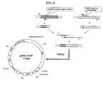
FIG. 8 is a schematic representation showing the construction process of Topo-pM90-EGFPh. HSP60 promoter and EGFP fragments were amplified by PCR and then the two fragments were fused using a sense primer for HSP60 and a reverse primer for EGFP. The fused fragment was then ligated into pTopo05-1390 to produce pM90-EFGPh vector.
FIG. 9 is a schematic representation showing the construction process of Topo-pM90-EGFe. pIRES2-EGFP was digested with NsiI and ligated to a pTopo05-1390 digested with NsiI and treated with CIAP to produce Topo-pM90-EGFe vector.
FIG. 10 is the results of FACS and microscopic analysis performed using M. smegmatis transformed with pM90-TOPO, Topo-pM90-EGFPh or Topo-pM90-EGFPe. FIG. 10 a represents a FACS result showing the fluorescence from GFP; FIG. 10 b shows the GFP fluorescence within a cell observed using an optical microscope (A, B, E, and F) or a confocal microscope (C, D, G, and H).
FIG. 11 is the FACS analysis results showing that Topo-pM90-EGFPh of the present disclosure have a better GFP expression level and a better vector stability compared to pAL5000 vectors in M. smegmatis.
FIG. 12 is the results of FACS (A to F) and microscopic analysis (G to J) done using J774 cells infected with M. smegmatis containing Topo-pM90-EGFPh. The cells infected with M. smegmatis containing Topo-pM90-EGFPh (D, E, F) show more GFP fluorescence compared to the control cells infected with pM90-TOPO (A, B, C). The fluorescent (G, H) and confocal microscopic analysis (I, J) showed the same results. The number of cells used for the infection was 10 (A,D), 50 (B, E) and 100 (C, F) MOI (multiplicity of infection).
FIGS. 13 a and 13 b, each represents the results of FACS and microscopic analysis, respectively, done using J774 cells infected with M. smegmatis containing Topo-pM90-EGFPe. The cells infected with M. smegmatis containing Topo-pM90-EGFPe (D, E, F) show more GFP fluorescence compared to the control cells infected with pM90-TOPO; (A, B, C). The fluorescent (G, H) and confocal microscopic analysis (I, J, K, L) showed the same results. The number of cells used for the infection was 10 (A, D), 50 (B, E) and 100 (C, F) MOI.
FIG. 14 is a graph showing GFP expression levels in J774 cells infected with different MOIs of Mycobacteria containing pM90-EGFPh (A) and pM90-EGFPe (B). The results indicate that the GFP expression level was excellent at all the MOI tested.
DETAILED DESCRIPTION OF THE EMBODIMENTS
Reference will now be made in detail to the present embodiments of the present invention, examples of which are illustrated in the accompanying drawings, wherein like reference numerals refer to the like elements throughout. The embodiments are described below in order to explain the present invention by referring to the figures.
The present inventors strived to develop a novel Mycobacteria-Escherichia coli shuttle vectors and identified a linear plasmid of about 18 kb in size comprising 16 open reading frames from Mycobacteria (for example 05-1390 strain) and a replication origin (designated as oriM) and Rep-like protein region which is required for replication.
In one aspect, the present disclosure relates to a plasmid from Mycobacteria and its use, and the plasmid of the present disclosure includes an origin of replication (oriM) and open reading frames (ORFs) including a protein such as rep-like protein and the like, which are required for the replication (refers to FIGS. 2 to 4). Therefore, the plasmid of the present disclosure is utilized as a gene and/or protein delivery system, and also in a therapeutic system such as immune therapeutics by effectively delivering DNAs and proteins into cells, in which they can function as antigens or therapeutic agents.
The term “transgene”, “target material” or “heterologous expressible DNA encoded” as used herein are used interchangeably and refers to a desired heterologous DNA sequence or a gene introduced into the vector to be expressed in prokaryotes particularly in Mycobacteria, and/or in mammalian cells, including but not limited to genes or DNA sequences which may not normally be present in the genome, genes which are present, but not normally transcribed and translated (“expressed”) in a given genome, or any other genes or DNA sequences which one desires to introduce into the genome.
In other aspect the present disclosure relates to a replicable DNA molecule derived from Mycobacteria, the DNA molecule having a nucleic acid sequence as disclosed in SEQ ID NO: 1. The DNA molecule contains nucleic acid sequences as disclosed in SEQ ID NOs: 3 to 15, whose corresponding amino acid sequences are disclosed as SEQ ID NOs: 16 to 28, respectively.
In still other aspect, the present disclosure relates to a Mycobacteria-Escherichia coli shuttle vector, the vector comprises an origin of replication designated as oriM having a sequence as disclosed in SEQ ID NO: 2. The Mycobacteria-Escherichia coli shuttle vector comprises: (a) an origin of replication having a nucleic acid sequence as disclosed in SEQ ID NO: 2 (oriM); (b) an origin of replication for prokaryotic cells; (c) a promoter; and (d) a nucleic acid sequence encoding a target material, which is operatively linked to the promoter.
In one exemplary embodiment, the nucleic acid sequence of oriM as disclosed in SEQ ID NO:2 contains a part of A+T rich region and direct repeat region. In particular, the oriM contains two 14 bp repeated regions (5′-TTCGTCTCTGGAGT-3′) in the AT rich region.
The plasmid or vector of the present disclosure can be constructed to be used as a vector for cloning or expression. Still the present vector can be constructed to be used in prokaryotic and/or eukaryotic cells as a host. In particular, the host for the present vector is prokaryotic cells in view of that the nucleic acid molecules of the present disclosure are derived from prokaryotic cells such as Mycobacteria and in consideration of the convenience of cell culture. For example, when the vector for the present disclosure is constructed as an expression vector and the host is a prokaryotic cells, the vector includes a strong promoter for transcription including such as tac promoter, lac promoter, lacUV5 promoter, Ipp promoter, PLλ promoter, PRλ promoter, rac5 promoter, amp promoter, recA promoter, SP6 promoter, trp promoter and T7 promoter, but the promoter is not limited thereto; a ribosomal binding site for initiating translation; and transcriptional/translational termination sites. When E. coli is used as a host cell, the regulatory elements which may be used for the present disclosure include but are not limited to operators and promoters for tryptophan biosynthesis in E. coli (Yanofsky, C., J. Bacteriol., 158:1018-1024 (1984)), and a leftward promoter of phage Lamda (PLλ promoter, Herskowitz, I. and Hagen, D., Ann. Rev. Genet., 14:399-445 (1980)).
The term “promoter” as used herein indicates DNA sequences which regulate the expression of sequences encoding a protein or a functional RNA. The nucleic acid sequences encoding a target material to be expressed are operatively linked to a promoter as described above. The term “operatively linked” as used herein indicates a functional link between a regulatory sequence for the expression of nucleic acids including, for example, promoter sequences, signal sequences, or transcription factor binding site, and other nucleic acid sequences. Here the regulatory sequence regulates the transcription or translation of the other nucleic acid sequences linked thereto.
The present vector system can be constructed using various methods known in the art. For example Sambrook et al., Molecular Cloning, A Laboratory Manual, Cold Spring Harbor Laboratory Press (2001) may be referred, the entire content of which is incorporated herein by reference.
In one preferred embodiment of the present disclosure, the cloning is practiced by Polymerase chain reaction (PCR). In one embodiment, the primes for the present discourse are used for gene amplification reaction.
PCR is a widely used method for amplifying nucleic acids and many modifications thereof are known in the art. For example, a touch down PCR, a hot start PCR, a nested PCR and booster PCR are developed to improve specificity or sensitivity of PCR. Also developed are real time PCR, differential display PCR, rapid amplification of cDNA ends, multiplex PCR, inverse polymerase chain reaction, vectorette PCR and thermal asymmetric interlaced PCR. A detailed explanation on PCR may be found in M. J., and Moller, S. G. PCR. BIOS Scientific Publishers, Springer-Verlag New York Berlin Heidelberg, N.Y. (2000), which is incorporated herein by reference.
The term “amplification reaction” as used herein refers to a reaction amplifying nucleic acid molecules. Various methods for amplification are known in the art, which for example include PCR (U.S. Pat. Nos. 4,683,195, 4,683,202, and 4,800,159), Reverse Transcription PCR (RT-PCR) (Sambrook et al., ibid), method as disclosed in WO 89/06700 by Miller, H. I. and EP 329,822 by Davey, C. et al., Ligase chain reaction (LCR) (Wiedmann M et al., 1994. PCR Methods Appl), Gap-LCR (WO 90/01069), repair chain reaction (EP 439,182), transcription-mediated amplification (TMA) (WO 88/10315), self-sustained sequence replication (WO90/06995), selective amplification of target polynucleotide sequences) (U.S. Pat. No. 6,410,276), consensus sequence primed polymerase chain reaction (CP-PCR) (U.S. Pat. No. 4,437,975), arbitrarily primed polymerase chain reaction (AP-PCR) (U.S. Pat. Nos. 5,413,909 and 5,861,245) and nucleic acid sequence based amplification (NASBA) (U.S. Pat. Nos. 5,130,238, 5,409,818, 5,554,517 and 6,063,603), but are not limited thereto. Other methods which may be used also described in U.S. Pat. Nos. 5,242,794, 5,494,810, 4,988,617 and application Ser. No. 09/854,317.
Also, the vectors of the present disclosure may be constructed using plasmids including such as for example pSC101, ColE1, pBR322, pUC8/9, pHC79, pUC19, pET and the like, phage such as for example, λgt4λB, λ-Charon, λΔz1 and M13 and the like, or virus such as for example SV40 and the like, which are known in the art.
In one exemplary embodiment, the promoter which may be used for the present vector includes, but is not limited to, a heat shock protein promoter, a CMV promoter, a promoter for 65 kDa common antigen of mycobacteria, ribsome RNA promoter from Mycobacteria, a promoter for MPB70, MPB59 or MPB64 antigen from Mycobacterium bovis, P1 promoter from bacteriophage Lamda, tac promoter, trp promoter, lac promoter, lacUV5 promoter, Ipp promoter, PLλ promoter, PRλ promoter, rac5 promoter, amp promoter, recA promoter, SP6 promoter, T7 promoter, a promoter for kanamycin resistance gene of transposon Tn903 or Tn5, a promoter for metallothionine, a growth hormone promoter or a hybrid promoter between eukaryotic and prokaryotic promoters, or a combination thereof.
The nucleic acid sequence encoding a target material to be expressed in cells includes any nucleic acid sequence of interest, which includes, but is not limited to, for example proteins such as reporter proteins, antigens and therapeutic proteins, and RNAs such as antisense oligonucleotides, siRNAs, shRNAs, miRNAs and piRNAs.
In one embodiment, the target material or transgene or heterologous expressible DNA encoded in the present vector may be translated into proteins in Mycobacteria and the translated products are then transferred to the eukaryotic cells of interest through Mycobacterial infection, in which case the transgene is operatively linked to a promoter suitable to direct the expression in prokaryotic cells, particularly in Mycobacteria. Examples of such promoters are as listed above. In other embodiment, the target material or heterologous expressible DNA encoded in the present vector may be translated in the eukaryotic cells where the target material is expected to work, in which case the transgene is operatively linked to a promoter suitable to direct the expression in eukaryotic cells. Examples of such promoters are as listed above. In either case, the heterologous expressible DNA or the transgene can provide an antigen or a therapeutic agent to the cells of interest.
In one illustrative embodiment, the antigen includes, but is not limited to, antigens which are used in vaccine therapies, for example, antigens from virulent pathogens such as hepatitis B virus (HBV) surface antigen, HBV core antigen, human immune-deficiency (HIV) gag protein and Mycobacterium tuberculosis Antigen 85A. The therapeutic proteins which may be encoded in the present vector include, but are not limited to, IL-12 or GM-CSF.
In one illustrative embodiment, the reporter protein includes, but is not limited to a fluorescent protein, beta-galactosidase, chloramphenicol acetyl transferase, human growth hormone, urease and alkaline phosphatase and the like.
In one exemplary embodiment, the fluorescent protein includes, but is not limited to, GFP (green fluorescent protein), RFP (red fluorescent protein), CFP (cyan fluorescent protein), YFP (yellow fluorescent protein), and BFP (blue fluorescent protein) and its variants.
The expression status and/level of the reporter gene may be measured by various methods known in the art. For example, the methods for measuring the expression of luciferase, chloramphenicol acetyl transferase, beta-galactosidase, human growth hormone, and GFP may be found in de Wet J. et al, Mol. Cell. Biol., 7: 725-737 (1987), Gorman C. et al, Mol. Cell. Biol., 2: 1044-1051 (1982), Hall C. V. et al, J. Mol. Appl. Genet., 2: 101-109 (1983), Selden R. et al., Mol. Cell. Biol., 6: 3173-3179 (1986), Chalfie M. et al, Science, 263: 802-805 (1994), respectively.
In addition, the present vector may further comprise one or more selective markers. In one illustrative embodiment, the present vector may comprise genes encoding a protein conferring resistance to antibiotics, which include, but are not limited to, genes conferring resistance to kanamycin, hygromycin, ampicillin, streptomycin, penicillin, chloramphenicol, gentamicin, carbenicillin, geneticin, neomycin or tetracycline.
In one embodiment, the shuttle vector of the present disclosure may be used in a co-transformation with other vectors (for example, pAL 5000 vector) which have an origin of replication different from the shuttle vector of the present invention.
In other aspect, the present disclosure provides cells transformed with the present shuttle vectors.
Any cells known in the art which would be able to consistently clone and express the present vectors may be used. The host cells which may be transformed with the present vector include, but are not limited to, E. coli DH5α, E. coli JM109, E. coli BL21(DE3), E. coli RR1, E. coli LE392, E. coli B, E. coli X 1776, E. coli W3110, and Mycobacterium spp.
Methods to deliver the present vector to the cells are known in the art. For example, when the host cells are eukaryotes, CaCl2 precipitation method (Cohen, S. N. et al., Proc. Natl. Acac. Sci. USA, 9:2110-2114 (1973)), Hananhan's method (Hanahan, D., J. Mol. Biol., 166:557-580 (1983)) and/or electroporation method (Dower, W. J. et al., Nucleic. Acids Res., 16:6127-6145 (1988)) may be used.
In one exemplary embodiment, the cells which may be transformed with the present vector include Mycobacteria and E. coli. In other embodiment, Mycobacteria which may be transformed with the present vector include M. smegmatis, M. bovis-BCG, M. avium, M. phlei, M. fortuitum, M. lufu, M. partuberculosis, M. habana, M. scrofulaceum, and M. intracellulare. In another embodiment, M. smegmatis, M. bovis-BCG, M. avium, M. partuberculosis, M. scrofulaceum, and M. intracellulare are included. In still another embodiment, M. smegmatis, M. bovis-BCG, M. avium and M. partuberculosis are included. In still another embodiment, M. smegmatis and M. bovis-BCG are included.
In the present disclosure, the present vectors may be provided as a vaccine composition comprising: (a) a pharmaceutically effective amount of cells transformed with anyone of the present vectors; and (b) a pharmaceutically acceptable carrier. The term “pharmaceutically effective amount” refers to the amount sufficient to effecting the desired efficacy or activity of the cells transformed with the present vectors.
The target material which may be contained in the present vector to be expressed in cells includes antisense oligonucleotide, siRNA, shRNA and mRNA, but is not limited thereto.
The term “antisense oligonucleotide” as used herein refers to a DNA or RNA or derivatives thereof having a sequence complementary to mRNA, which inhibits translation of mRNA into a protein by binding to the complementary sequence in the mRNA. The antisense sequence of the present disclosure refers to a DNA or RNA, which has a sequence complementary to the target gene and thus be able to bind to the corresponding mRNA inhibiting the translation of mRNA into a protein, the translocation and/or maturation of mRNA or other essential biological functions. The length of the antisense oligonucleotides is about 6 bases to 100 bases, particularly about 8 to 60 bases, more particularly about 10 to 40 bases.
The term “siRNA” as used herein refers to a nucleic acid molecule which can mediate RNA hindrance or gene silencing (refer to WO 00/44895, WO 01/36646, WO 99/32619, WO 01/29058, WO 99/07409 and WO 00/44914). The siRNA can repress the expression of the target gene and thus is used in the field such as gene knock-down and gene therapy. siRNA which was originally discovered in plants, insects and fireflies and parasites now is used in mammalian cell research (Degot S, et al. 2002; Degot S, et al. 2004; Ballut L, et al. 2005).
The siRNA of the present disclosure may be double stranded having a sense strand and an antisense strand. In other embodiment, the siRNA of the present disclosure may be a single stranded which is self-complementary having a sense and antisense sequence in one strand.
The siRNA of the present disclosure is not limited to a complete match but includes ones containing a mismatch (the corresponding base is not complementary), a bulge (no corresponding base is present) and the like. The length of siRNA may be about 10 bases to 100 bases, particularly about 15 bases to 80 bases, more particularly about 20 to about 70 bases.
In the siRNA of the present disclosure, a short stretch of nucleic acid sequence of about 5-15 bases may be present between a sense and antisense sequence in self-complementary strand. In this case, the RNA transcribed from such nucleic acid sequences form a hair pin structure by intra-molecular hybridization and takes a stem and loop structure. The stem and loop structure is processed in vitro and in vivo and generates siRNA molecues which mediate RNAi reaction.
The term “miRNA (micro RNA)” as used herein refers to a single strand RNA molecule of about 21 to 25 bases in length and regulates the expression of genes in eukaryotic cells by degrading mRNAs or inhibiting the translational process. The miRNA is produced by two step: in the first step, primary miRNA transcripts transcribed are processed to pre miRNA having a stem and loop structure of about 70-90 bases in length by an RNase III type enzyme called Drosha; in the second step, the pre miRNA migrates to the cytoplasm and further processed to produce a mature miRNA of about 21 to 25 bases digested by an enzyme called Dicer. The produced miRNA functions as a post transcriptional gene suppressor by complementary binding to a target sequence and thus induce mRNA instability and translational suppression. miRNAs are known to be involved in a variety of physiological functions and disease.
The term “piRNA (Piwi-interacting RNA)” as used herein refers to a single strand RNA molecule of about 26 o 31 bases in length and forms RNA-protein complexes through interactions with piwi proteins. It has been linked to both epigenetic and post-transcriptional gene silencing of retrotransposons and other genetic elements in germ line cells (Siomi M C et al., Nat Rev Mol Cell Biol., 12:246-258 (2011)).
The pharmaceutically acceptable carriers which may be included in the present composition include, but are not limited to, lactose, dextrose, sucrose, sorbitol, mannitol, starch, acasia gum, calcium phosphate, alginate, gelatin, calcium silicate, micro crystalline cellulose, polyvinylpyrrolidone, cellulose, water, syrup, methyl cellulose, methyl hydroxybenzoate, propylhyroxybenzoate, talc, magnesium stearate and mineral oil. The present composition may further includes lubricants, wetting agents, sweetening agents, flavors, emulsifier, suspending agents. The suitable pharmaceutically acceptable carrier and agents are described in detail in Remington's Pharmaceutical Sciences (19th ed., 1995).
The present composition may be formulated for example as a unit dosage form by using a pharmaceutically accepted carriers and/or excipients in accordance to the methods which may be practiced easily by one skilled in the art. The formulation may be powder, granulates, tablets, capsules or gel (such as hydrogel), which may further include dispersion agents or stabilizers.
In other aspect, the present disclosure relates to a method of using a first and a second vector for expression of heterologous transgenes in a eukaryotic cell, wherein the first vector is the vector according to the present disclosure and the second vector is pSE100 and its derivatives in eukaryotic cells.
The pSE100 vector is a Mycobacterial-Ecoli shuttle vector commercially available from addgene and is described in for example, Guo, X V, M Monteleone, M Klotzsche, A Kamionka, W Hillen, M Braunstein, S Ehrtand D Schnappinger (2007) Silencing Mycobacterium smegmatis by using tetracycline repressors. J Bacteriol 189(13): 4614-23. The present disclosure includes pSE100 or its derivatives. The derivatives for example include, but is not limited to, a vector having the same origin of replication as the pSE. The pSE is compatible with the present vector and can be used for two or more heterologous proteins. The transgenes which may be encoded by the vector used for the present method are as described above.
The present disclosure relates to a novel Mycobacteri-E. coli shuttle vectors and their uses. The plasmid of the present disclosure having a size of about 18 kb derived from Mycobacteria contains 16 ORFs, oriM and Rep-like protein essential for the replication. The plasmids of the present disclosure is useful in gene delivery system and research and also may be used in immune therapeutics by effectively delivering a nucleic acid sequence or recombinant protein.
The present disclosure is further explained in more detail with reference to the following examples. These examples, however, should not be interpreted as limiting the scope of the present invention in any manner.
EXAMPLES
Materials and Methods
Cells and Cell Culture
For the amplification of plasmids, E. coli DH5α strain (RBC) was used. The E. coli cells grown at 37 in LB medium were heat shocked at 42 for transformation with plasmids. The cells transformed with the plasmids were then selected by growing the cells in a medium containing an antibiotic. The selected colonies were then cultured and the plasmids in the cells were extracted using PureLink™ HiPure Plasmid Filter Maxiprep Kit (Invitrogen, USA) in accordance with the manufacturer's instruction. The sample from a patient 05-1390, M. smegmatis MC2155 and Mycobacterial strain BCG-Japan were grown at 37 in a 7H10 liquid medium or a 7H10 solid medium, which were transformed with the plasmid using an electroporation method.
Extraction of DNA from 05-1390
Mycobacterial strain 05-1390 was inoculated onto a 7H10 solid medium and allowed to form a colony by incubating it at 37. The colonies were picked using a loop and suspended in 400 μl of TE (Sigma, USA) in a 1.5 ml tube. The cells were then treated at 80 for 10 min to kill Mycobacteria. Then 10 mg/ml of lysozyme (Roche diagnostics, USA) was added thereto and incubated at 37 for 1 hour. 70 μl of 10% SDS (USB, USA) and 5 μl of 10 mg/ml proteinase K (Bioline, USA) was added and mixed by vortexing and incubated at 65 for 10 min. 750 μl of chloroform/isoamyl alcohol (Sigma, USA) (24:1, v/v) was added and vortexed for 10 sec and centrifuged at RT for 5 min at 12,000×g. Then 180 μl of the supernatant was then transferred to a new tube and 450 μl of isopropanol was added and the mixture was incubated for 30 min at −20. The DNA contained in the mixture was then precipitated by centrifugation at 12,000×g for 15 min at 4. The precipitated DNA was then washed using 70% alcohol at 4 and centrifuged at 12,000×g for 5 min at 4 to remove the supernatant. The pellet was then air dried and dissolved in 200 μl of TE.
Southern Blotting and Western Blotting
To confirm the presence of a linear plasmid in 05-1390 sample, High prime DNA labeling and detection starter kit II (Roche diagnostics, USA) was used to extract the genome in accordance with the manufacturer's instruction. The genome DNA was then electrophoresed on a 1% agarose gel (Bioline) at 100V for 3 hours. The agarose gel was immersed and rocked in 400 ml of 0.25 M HCl (Junsei, Japan) solution for 20 min. The HCl solution was removed from the gel and the gel was immersed and rocked in 400 ml of 0.5M NaOH (Junsei)/1.5M NaCl solution for 20 min followed by another 20 min incubation in a 1.5M NaOH (Junsei)/1.5M NaCl solution. The buffer was then removed and the gel was blotted with 3 sheets of filter papers (Whatman, USA) presoaked with 10×SSC. A nylon membrane (Amersham, USA) having the size of the gel was placed onto the gel and 3 sheets of filter papers (Whatman) was placed on top of the nylon membrane. And the paper towel was stacked on the filter paper in 10 cm thick and was pressed with a weight of about 400 g. The entire stack was then wrapped with a plastic film to prevent evaporation and left for 24 hours at RT. After that, the DNA transferred to the membrane was cross-linked at 120 mHcm-2 using a UV cross linker. The membrane was then immersed in DIG Easy Hybridization solution (Roche diagnostics USA) pre-equilibrilized at 37 in a tube and rocked for 15 min at 42. The probe prepared by PCR was boiled for 5 min and cooled down in ice and added to the membrane in the DIG Easy Hybridization solution as prepared above at 37. The DNA probe (342 bases) was prepared by PCR using PCR DIG Probe Synthesis Kit (Roche diagnostics) in accordance with the manufacturer's instruction from 05-1390 gDNA. The primers used as follows: sense (ORF9_F): 5′-gcggtgccacagtgccagtag-3′; and antisense (ORF9_R): 5′-tcatggacgaagccgacagagc-3′. After the hybridization, the membrane was washed 2× for 5 min each with 2×SSC/0.1% SDS solution followed by washing 2× for 15 min each with 0.5×SSC/0.1% SDS.
For western blotting, the membrane as prepared above was washed for 1 to 5 min in 0.1 M maleic/0.15 M NaCl/0.3% Tween®20 solution. Then the membrane was washed in a blocking solution (10× blocking solution:maleic acid=1:10) for 30 min. The blocked membrane then was immersed and rocked in a solution containing antibody, which was then washed twice for 15 min each in 100 ml of 0.1M maleic acid/0.15M NaCl/0.3% Tween®20. The membrane was then incubated with 20 ml of detection solution for 2 to 5 min at RT. Then 1 ml of CSPD (Roche diagnostics) was spread onto the membrane and covered with a plastic film to prevent from drying. After that, the excess liquid was removed from the membrane and left for 10 min at 37 and the fluorescent signal was read using Las4000 (Fujifilm, Japan).
Results
The Confirmation of a Novel Linear Plasmid in a New Strain 05-1390
The genome from M. intracellulare and Mycobacterium MOTT90 were extracted and southern-blotted as described above. The results are shown in FIG. 1, which indicated the presence of a plasmid of about 18 kb in size only in Mycobacterium MOTT90 and the linear plasmid was confirmed by two bands after digestion with Xho I.
ORF Search and Sequence Analysis
ORF finder program from NCBI (http://www.ncbi.nlm.nih.gov/projects/gorf/) was used to search putative ORFs in 05-1390 linear plasmid (18090 kb) encoding 150 amino acids in length at the least. The 16 ORFs identified using the program was compared with the linear plasmid sequence from M. celatum using the Blast search. As a result, a total of 16 ORFs were found to be present in the plasmid of the present disclosure. As described in Table 1, out of 16 ORFs present in the plasmid, the rep-like protein essential for replication was confirmed to be present.
TABLE 1
Genes found in pM90 and analysis of sequence homology
Expected
ORF length identity The nearest homolog (homolog) Known or potential
No. (aa) (%) (species and strain or plasmid) function Accession No.
1 134 68 Streptosporangium roseum ATP-dependent protease YP_003336723.1
CIp
ATPase subunit like protein
2 493 95 Mycobacterium tuberculosis Transposase NP_217307.1
3 264 100 Mycobacterium sp. MCS plasmid 1 hypothetical protein YP_642699.1
4 200 65 Aspergillus oryzae RIB40 hypothetical protein XP_001816628.2
5 806 54 Mycobacterium sp. MCS plasmid 1 hypothetical protein YP_642669.1
Mmcs_5513
6 209 86 Mycobacterium sp. MCS plasmid 1 hypothetical protein YP_642670.1
Mmcs_5514
7 121 85 Mycobacterium sp. MCS plasmid 1 hypothetical protein YP_642672.1
Mmcs_5516
8 416 70 Mycobacterium celatum, pCLP Rep-like protein NP_862580.1
9 364 62 Mycobacterium vanbaalenii hypothetical protein YP_954523.1
PYR-1 Mvan_3735
10 217 97 Mycobacterium celatum, ParA-like protein NP_862577.1
pCLP
11 629 98 Mycobacterium gilvum hypothetical protein YP_001136809.1
PYR-GCK, pMFLV01 Mflv_5560
12 243 85 Mycobacterium gilvum hypothetical protein YP_001136810.1
PYR-GCK, pMFLV01 Mflv_5561
13 108 99 Mycobacterium smegmatis pemK-like protein YP_888722.1
MazF
14 76 90 Methylobacterium nodulans hypothetical protein YP_002496361.1
Mnod_1051
15 176 67 Mycobacterium abscessus hypothetical protein YP_001701532.1
MAB_0782
16 60 96 gliobacterium violoacea Methionine NP_926067.1
adenosyltransferase
As described in Table 1, out of 16 ORFs present in the plasmid, the rep-like protein essential for replication was confirmed to be present.
Characterization of the Linear Plasmid at Molecular Biological Level
05-1390 Total RNA Extraction from 05-1390
The 05-1390 cells was cultured to the exponential phase in 100 ml of a 7H9 medium at 37. The cell culture was centrifuged at 3000×g for 15 min at 4 and the supernatant was decanted and the precipitated cells were mixed with 1 ml of RNAprotect bacterial reagent (Quiagen, USA). Then, the mixture was washed by centrifugation at 3000×g for 15 min at 4. 1 ml of TRIsure regent (Bioline) and 0.1-mm glass beads were added to the obtained cells, which were then disrupted at a bead beater (BioSpec, USA) at 5000 rpm for 40 sec. The tube was then immediately transferred to ice and left for 5 min. 0.25M chloroform solution was added to the tube and vortexed for a few seconds and the cells were left for 2 min at RT, which was then centrifuged at 12,000×g for 15 min and the supernatant was transferred to a new tube and mixed with 0.5 ml of isoprorpanol and left for 15 min at RT. The mixture was then centrifuged at 12,000×g for 15 min and the supernatant was removed and the pellet was washed with 1 m of 75% ethanol by centrifugation at 12,000×g for 5 min. The supernatant was removed and the RNA pellet was air dried for 5 min at RT. The pellet was dissolved in ddH2O treated with DEPC (diethyl polycarbonate; Bioline) and kept was at −20 before use.
The Confirmation of Transcription from the ORFs by RT-PCR (Reverse Transcription-Polymerase Chain Reaction)
The total RNA extracted as above was reverse transcribed and amplified using ONE-STEP RT-PCR PreMix Kit (INTRON, Korea) in accordance with the manufacturer's instruction. The PCR primers used were designed 19 to 25 bases in length to amplify ORFs of 134 to 685 bp in length as described in Table 2.8 μl of ONE-STEP-RTPCR PreMix Kit, 100 ng of total RNA, 0.5 μM of sense and antisense primer was mixed in a total volume of 20 μl in ddH2O. PCR was performed in a thermal cycler 9700 (Applied Biosystems, USA) at the condition of pre-treatment at 45 for 30 min; 30 cycles of 30 sec at 94 and 30 sec at 60 and 1 min at 72; and final extension for 5 min at 72; and soaking at 4. The PCR products were analyzed by electrophoresis on a 1% agarose gel followed by visualization with ethidium bromide illuminated with UV light.
TABLE 2
ORFs found in the present plasmid and primers used 
for the amplification.
Expected
ORF Protein size Sequence (5′->3′) size
No. (aa) Sense primer Antisense primer (bp)
 1 Clp domain GCTACGCCGCACTACCTTTAT GTGTTTGACGAGCTGACGAGTG 520
protein
 2 Transposase CGCCTCGGCTCCCATTGTC TGGTGGCCCGCAGACATTC 631
 3 Hypothetical ACGCGCTGGTAGTGCTCCCTTAG CGAACACAAGCGCGACCACTACA 262
protein
 4 unknown CCTCGGCGGCGTAGTCAGTCA GCCGGCCATATCACGATTCATTAC 295
 5 Hypothetical TTCCACCCGCGGCATCGTA GCGCCGCCGAGCAATACA 275
protein
Mmcs_5513
 6 Hypothetical CTTGCTTTCGAGGTCTTTGA GCAACGCGCCGCCGAGCAATAC 603
protein
Mmcs_5514
 7 Hypothetical TGGATCAGGCCCGTAGGACA TCATGGACGAAGCCGACAGAGC 665
protein
Mmcs_5516
 8 Rep-like GCGGTGCCACAGTGCCAGTAG TCATGGACGAAGCCGACAGAGC 342
protein
 9 Hypothetical ACACGGCGATCACGGGCTTAT CGAGACACCATCCACCGAGAAAT 197
protein
Mvan_3735
10 ParA-like CCGCTGCCGCACGAATACAT CGTTGGCGGTCGATTCTTCACT 477
protein
11 Hypothetical GTTCGGTGCGGCGTTCAAG GGCGGGCGAACTGGTCAATAC 290
protein
Mflv_5560
12 Hypothetical GCGCAGCGGGCAATGGAG ACACCCGCACCCCGTCTC 285
protein
Mflv_5561
13 pemK-like TCCGAGGAAGACGAGTAGG TCCGTCACAATCTGCCCCCTCACA 215
protein,
MazF
14 Hypothetical GCGGAGATGGCATCCAC GCTACGCCCCCTTCAAATA 134
protein
Mnod_1051
15 Unknown CGGGTGAGTCTTGGCGGCGGGGTA ATCTCGTGCACGTAGAAGGAAA 187
16 Unknown TTCTGTGTCGCCTATGCGGCCGGC TCTGGCGATCGTGAAGACGAGCAC 174
Of the total 16 putative 16 ORFs, 13 ORFs except 12 and 14 was confirmed to be transcribed using RT-PCR (FIG. 2). The analysis of the entire plasmid revealed the presence of the AT rich region and direct repeat region (FIG. 3).
The Amplification of oriM of the Linear Plasmid
Using 05-1390 genomic DNA as a template, the 2,469 bp region identified to contain oriM corresponding to the bases from 9,466 to 11,914 was amplified suing primers: p05-1390.sub.—336(5′-AAGCTTAGGCGGGCAACACGACATCTC-3′: SEQ ID NO: 37) and p05-1390.sub.—2785(5′-CCATGGGGGTTGTGGGCCGATGTGCT-3′: SEQ ID NO: 38). The PCR conditions were as follows: 1 cycle of pre-denaturation step of 2 min at 94 .degree. C.; 30 cycles of amplification step of 30 sec at 95 .degree. C. and 30 sec at 65 .degree. C. and 4 min at 95 .degree. C.; and 1 cycle of final extension step for 10 min at 72 .degree. C. 1 .mu.l of PCR product, 1 .mu.l of TOPO TA vector (Invitrogen, USA). 1 .mu.l of TA cloning buffer and 3 .mu.l of ddH2O were mixed and left for 5 min at RT. The 7 .mu.l of reaction product was mixed with 50 .mu.l of E. coli DH5.alpha. (RBC) competent cells and heat shocked for 45 sec at 42 .degree. C. Then 250 .mu.l of SOC medium was added and the mixture was left for 1 hour at 37 .degree. C. 80 .mu.l of the mixture was then spread on to a LB agar plate containing Kanamycin (100 .mu.l/ml) and IPTG, which was incubated at 37 .degree. C. for 16 hours. The colonies formed were then analyzed by PCR for the presence of the plasmid of interest using the primers as described above and analyzed on a 1% agarose gel. The results are shown in FIG. 4.
The Stability of TOPO 05-1390 and Compatibility with pSE100
Preparation of Competent Cells of Mycobacteria
2.5 liters of M. smegmatis cells competent for electroporation was cultured to have an O.D. of 0.8 to 1.0 at 600 nm and cooled down for 2 hours on ice. The culture was then centrifuged at 3,000×g for 5 min and suspended and sequentially washed with 20 ml, 10 ml and 5 ml pf 10% glycerol by centrifugation at 3,000×g for 5 min. After the final washing step the cells were suspended at 5 ml of 10% glycerol, the 1/500 volume of the initial culture volume. The 200 μl aliquot of the competent cell's was prepared in a 1.5 ml eppendorf tube and kept at −70 until use.
The Stability of TOPO 05-1390
The 200 μl of the competent cells as prepared above was thawed slowly on ice and then mixed with 5 μg of TOPO05-1390 or pSE100 and the mixture was left on ice for 20 min. The mixture was then transferred to a 0.2 cm cuvette (Bio-Rad, USA) kept cold on ice and the electric field was applied for 25 μF, 2.50 kV and 1000Ω using Gene pulser and pulse controller accessories from Bio-Rad and left for 5 min on ice. The cells were then added to 5 ml of a 7H9 liquid medium in a 15 ml tube and cultured for 4 hours at 37. For TOPO05-1390, 1/100 dilution of the culture was spread onto a LB agar plate containing kanamycin (20 μl/ml) and incubated for about 7 to 10 days at 37 until colonies were formed. For pSE100, 1/100 dilution of the culture was spread onto a LB agar plate containing hygromycin (50 μl/ml) and incubated for about 7 to 10 days at 37 until colonies were formed. After that, a transformed colony (TOPO05-1390 and pSE100) was picked and inoculated to 5 ml of 7H9 liquid medium for 5 days, during which at every 24 hours, 1/100 dilution of the culture was spread onto a LB agar plate containing corresponding antibiotics and a LB agar plate without antibiotics and the number of colonies were counted and compared.
The results are shown in FIG. 5. The transformed cells containing the recombinant plasmid of the present disclosure, TOPO-pM90, grew well both in the regular medium without antibiotics and also in the medium with kanamycin. This indicates that TOPO-pM90 maintains the resistance to kanamycin (FIGS. 5 a and 5 b). For hygromycin, only pSE100 used as control shows resistance to hygromycin (FIG. 5 c). The results confirm that the kanamycin resistance gene contained in TOPO-pM90 functions properly in cells.
Also, the stability of M. smegmatis transformed with pM90-TOPO was found to be similar to that of pSE100 during the 7 day incubation period (FIG. 6).
Compatibility with pSE100
pM90-TOPO and pSE100 was co-transformed into M. smegmatis using as described above. The colonies formed/cultured were inoculated into a 5 ml of 7H9 liquid medium containing kanamycin (20 μl/ml) and hygromycin (50 μl/ml) and incubated for about 7 days at 37, during which at every 24 hours, 1/100 dilution of the culture was spread onto a LB agar plate containing the above antibiotics and a LB agar plate without antibiotics and the number of colonies were counted and compared. The results as shown in FIG. 7 indicates the compatibility between pM90-TOPO and pSE100.
The Construction of a Shuttle Vector Using pM90-TOPO
TOPO05-1390EGFPh
HSP promoter fused with EGFP gene was inserted into TOPO05-1390 by PCR (FIG. 8) using pIRES2-EGFP as a template. The primers used were: sense prime; 5′-ATCCGGAGGAATCACTTCGCAATGGCCACAACCATGGTGAGC-3′), which contains at its 5′ end the 20 bp promoter region corresponding to the end of HSP 60 promoter from M. bovis and reverse primer; 5′-CCCGATATCTTACTTGTACAGCTCGTCCA-3′, which encodes at its 3′ end an EcoRV restriction site. The PCR conditions as follows: 1 cycle of pre-denaturation step at 94 for 2 min; 30 cycles of amplification step at 95 for 30 sec and at 65 for 30 sec and at 72 for 1 min; and 1 cycle of a final extension step at 72 for 10 min. The amplified products war run on a 1% agarose gel and purified by PCR quick spin kit (Intron). The purified product was then digested with EcoRV (NEB, USA) at 37 for 16 hours and isopropanol was added thereto and the mixture was incubated at −20 overnight to precipitate DNA. The precipitated product was then centrifuged at 13,000 rpm for 15 min and the pellet was washed once with 300 μl of 70% ethanol and the washed pellet was air dried and dissolved in TE (201). The TOPO05-1390 was digested with EcoRV as above and the digested DNA was treated with CTAP (1 μl) at 37 for 15 min and 55 for 15 min. The process to precipitate the DNA was done as described above. The constructed hsp-EGFP and TOPO05-1390 was mixed at 3:1 ratio, which then was mixed with T4 DNA ligase (1 μl) and incubated for 4 hours at 4. After that the ligated product (1 μl) was added to E. coli DH5α (RBC) the competent cells followed by heat shock for 45 sec at 42. After the heat shock, 250 μl of SOC medium was added to the cells followed by incubation at 37 for 1 hour. After the incubation, 80 μl of the cells was spread onto a solid LB agar plate containing kanamycin (100 μl/ml) and incubated at 37 for 16 hours. The colonies formed was picked and was used for PCR using the corresponding primer set as described above and the ligated product was confirmed on a 1% agarose gel.
TOPO05-1390EGFPe
EGFP gene construct having a CMV promoter was inserted into TOPO05-1390 (FIG. 9). In detail, pIRES-EGFP (1 μg) was digested with NsiI (NEB) at 37 for 16 hours. The restricted product was run on a 1% agarose gel and the 2,095 bp fragment corresponding from CMV to polyA tail was purified from the gel by using gel extraction kit (Qiagen, USA). TOPO05-1390 plasmid was digested as described above for pIRES-EGFP was treated with CIAP (1 μl) at 37 for 15 min and 55 for 15 min. The process to precipitate the DNA was done as described above. The pIRES2-EGFP2095 bp and TOPO05-1390 was mixed at 3:1 ratio, which was then mixed with T4 DNA ligase (1 μl) and incubated for 4 hours at 4. After that the ligated product (1 μl) was added to E. coli DH5α (RBC) the competent cells followed by heat shock for 45 sec at 42. After the heat shock, 250 μl of SOC medium was added to the cells followed by incubation at 37 for 1 hour. After the incubation, 80 μl of the cells was spread onto a solid LB agar plate containing kanamycin (100 μl/ml) and incubated at 37 for 16 hours. The colonies formed was picked and used for PCR using the corresponding primer set as described above and the ligated product was confirmed by electrophoresis on a 1% agarose gel.
Confirmation of Protein Expression from the Recombinant Vector in Mycobacteria
Each of pM90-EGFPh (5 μg) and pM90-EGFPe (5 μg) constructed as described above was used for transformation into Mycobacteria as described above. The cells were spread on the LB agar plate containing kanamycin (100 μl/ml) and incubated for 10 days. After 10 days, a colony was inoculated into a 3 ml of 7H9 liquid medium and incubated and passaged every 3 days. The cells were then observed using Fluorescent microscope (Olympus), confocal microscopy (Olympus) or FACS (BD science) for GFP expression.
To confirm GFP expression in the cells, for pM90-EGFPh, the cells at the 4 passages were used for microscopic examination. For FACS, 500 μl of the cells at the 4 passages was washed twice with 500 μl of PBS and then resuspended with 1 ml of PBS and used for GFP. Mycobacterium smegmatis transformed with pM90-EGFPh, pM90-EGFPe or pM90-TOPO was analyzed by FACS. The results showed that the fluorescent signal was not detected in EGFPe or pM90-TOPO but was detected in pM90-EGFPh cells (FIG. 10 a). The fluorescent signal from pM90-EGFPh was detected in Mycobacterium smegmatis (A, B, C, D) and BCG (E, G, F, H)(FIG. 10 b). This indicates that the stability of GFP expression in Mycobacterium smegmatis was better compared to the conventional pAL5000 vector (FIG. 11).
Confirmation of Protein Expression from the Recombinant Vector in Eukaryotic Cells
Each of pM90-EGFPh (5 μg) and pM90-EGFPe (5 μg) constructed as above was used for transformation into Mycobacteria as described above. The transformed Mycobacteria was diluted in DMEM with 10% FBS at 10, 50 or 100 MOI (multiplicity of infection) and 1 ml of the diluted cells was then used to infect J774 cells (ATCC TIB-67), which were prepared 24 hours in advance at 3×10⁵ cells/well of a 12 well plate for 1 hour. After the infection, gentamycin (10 μg/μl, Sigma) was added thereto to remove Mycobacteria. After one hour, the medium was replaced with DMEM containing 10% FVS without an antibiotics and the cells were examined using FACS, confocal microscope and fluorescent microscope for GFP expression.
The results from the infection of J774 cells by Mycobacteria having pM90-EGFPh (FIG. 12) or pM90-EGFPe (FIG. 13) showed that the vectors of the present disclosure showed higher expression level of GFP in J774 cells compared to the control cells infected by pM90-TOPO. Also the results from the infection of J774 cells by Mycobacteria having pM90-EGFPh and pM90-EGFPe (FIGS. 14 a and 14 b) showed very high expression level of GFP at each MOI.
With respect to the use of substantially any plural and/or singular terms herein, those having skill in the art can translate from the plural to the singular and/or from the singular to the plural as is appropriate to the context and/or application.
The various singular/plural permutations may be expressly set forth herein for sake of clarity. Although a few embodiments of the present disclosure have been shown and described, it would be appreciated by those skilled in the art that changes may be made in this embodiment without departing from the principles and sprit of the invention, the scope of which is defined in the claims and their equivalents.

Claims (19)

What is claimed is:
1. An isolated DNA molecule capable of replicating in a mycobacterium, wherein the DNA molecule comprises the nucleic acid sequence of SEQ ID NO: 1 and a transgene that is heterologous to mycobacteria.
2. A Mycobacterium-Escherichia coli shuttle vector comprising:
(a) an origin of replication having the nucleic acid sequence as disclosed in SEQ ID NO: 2 (oriM);
(b) an origin of replication for prokaryotic cells;
(c) a promoter; and
(d) a transgene, which is operatively linked to the promoter.
3. The shuttle vector according to claim 2, wherein the promoter is selected from the group consisting of a heat shock protein promoter, a CMV promoter, a promoter from a 65 kDa common antigen gene of mycobacteria, a ribosomal RNA promoter from Mycobacteria, a Mycobacterium bovis MPB70, MPB59, or MPB64 antigen promoter, a P1 promoter from bacteriophage Lambda, a tac promoter, a trp promoter, a lac promoter, a lacUV5 promoter, an Ipp promoter, a PL λ promoter, a PR λ promoter, a rac5 promoter, a beta lactamase, a recA promoter, a SP6 promoter, a T7 promoter, a promoter from a kanamycin resistance gene of transposon Tn903 or Tn5, a metallothionine promoter, a growth hormone promoter, and a hybrid promoter between a eukaryotic promoter and a prokaryotic promoter.
4. The shuttle vector according to claim 2, wherein the transgene encodes a protein, antisense oligonucleotide, siRNA, shRNA, miRNA or piRNA.
5. The shuttle vector according to claim 4, wherein the protein is a reporter protein, an antigen or a therapeutic protein.
6. The shuttle vector according to claim 5, wherein the protein is a reporter protein selected from the group consisting of a fluorescent protein, beta-galactosidase, chloramphenicol acetyltransferase, human growth hormone, urease, and alkaline phosphatase.
7. The shuttle vector according to claim 6, wherein the fluorescent protein is selected from the group consisting of GFP (green fluorescent protein), RFP (red fluorescent protein), CFP (cyan fluorescent protein), YFP (yellow fluorescent protein), BFP (blue fluorescent protein) and their variants.
8. The shuttle vector according to claim 5, wherein the antigen is an antigen derived from virulent pathogens.
9. The shuttle vector according to claim 8, wherein the antigen is selected from the group consisting of a hepatitis B virus (HBV) surface antigen, a HBV core antigen, a human immune-deficiency (HIV) gag protein, and Mycobacterium tuberculosis Antigen 85A.
10. The shuttle vector according to claim 5, wherein the therapeutic protein is IL-12 or GM-CSF.
11. The shuttle vector according to claim 2, wherein the vector further includes one or more selective markers.
12. The shuttle vector according to claim 11, wherein the markers are selected from genes conferring resistance to an antibiotic selected from the group consisting of kanamycin, hygromycin, ampicillin, streptomycin, penicillin, chloramphenicol, gentamicin, carbenicillin, geneticin, neomycin, and tetracycline.
13. A cell transformed with the vector according to claim 2.
14. The cell according to claim 13 wherein the cell is a Mycobacterium or an Escherichia coli.
15. The cell according to claim 14 wherein the Mycobacterium is selected from the group consisting of M. smegmatis, M. bovis-BCG, M. avium, M. phlei, M. fortuitum, M. lufu, M. partuberculosis, M. habana, M. scrofulaceum, and M. intracellulare.
16. A method of using a first and a second vector for expression of transgenes in a eukaryotic cell, wherein the first vector is the vector according to claim 2, and the second vector encodes a transgene and comprises a pSE100 replication origin, the method comprising transfecting the eukaryotic cell with each of the first and second vectors.
17. The method of claim 16 wherein each transgene encodes a protein, antisense oligonucleotide, siRNA, shRNA, miRNA, or piRNA, and the transgenes encoded by the first and second vectors are different.
18. The method of claim 17 wherein at least one of said transgenes encodes a protein selected from the group consisting of a reporter protein, an antigen, and a therapeutic protein.
19. The method of claim 18 wherein:
the reporter protein is selected from the group consisting of a fluorescent protein, beta-galactosidase, chloramphenicol acetyltransferase, human growth hormone, urease and alkaline phosphatase;
the antigen is derived from virulent pathogens; and
the therapeutic protein is IL-12 or GM-CSF.
US13/450,689 2011-04-21 2012-04-19 Shuttle vectors for mycobacteria-escherichia coli and uses thereof Active US8841432B2 (en)

Applications Claiming Priority (2)

Application Number Priority Date Filing Date Title
KR1020110037147A KR101291668B1 (en) 2011-04-21 2011-04-21 Shuttle Vectors for Mycobacteria-Escherichia coli and Uses Thereof
KR10-2011-0037147 2011-04-21

Publications (2)

Publication Number Publication Date
US20120270322A1 US20120270322A1 (en) 2012-10-25
US8841432B2 true US8841432B2 (en) 2014-09-23

Family

ID=47021638

Family Applications (1)

Application Number Title Priority Date Filing Date
US13/450,689 Active US8841432B2 (en) 2011-04-21 2012-04-19 Shuttle vectors for mycobacteria-escherichia coli and uses thereof

Country Status (2)

Country Link
US (1) US8841432B2 (en)
KR (1) KR101291668B1 (en)

Cited By (1)

* Cited by examiner, † Cited by third party
Publication number Priority date Publication date Assignee Title
KR20190130471A (en) 2018-05-14 2019-11-22 서울대학교산학협력단 A recombinant BCG expressing HIV-1 p24 using a pMyong2 vector system and use as a HIV-1 vaccine thereof

Families Citing this family (4)

* Cited by examiner, † Cited by third party
Publication number Priority date Publication date Assignee Title
CN105779455B (en) * 2016-03-30 2018-11-20 中国科学院遗传与发育生物学研究所 Efficient pig constitutive promoter and application
WO2019221383A1 (en) * 2018-05-14 2019-11-21 서울대학교 산학협력단 Recombinant bcg employing pmyong2 vector system to express hiv-1 p24 and use thereof as hiv-1 vaccine
CN110129246B (en) * 2019-04-29 2021-02-19 华南农业大学 Donor bacterium, construction method and application thereof, and plasmid inhibitor screening method
CN113462711B (en) * 2021-07-01 2022-06-14 苏州大学 Screening system, construction method and application of mycobacterium tuberculosis protein intron splicing inhibitor

Citations (14)

* Cited by examiner, † Cited by third party
Publication number Priority date Publication date Assignee Title
WO1988010315A1 (en) 1987-06-19 1988-12-29 Siska Diagnostics, Inc. Transcription-based nucleic acid amplification/detection systems
WO1989006700A1 (en) 1988-01-21 1989-07-27 Genentech, Inc. Amplification and detection of nucleic acid sequences
EP0329822A2 (en) 1988-02-24 1989-08-30 Cangene Corporation Nucleic acid amplification process
WO1990001069A1 (en) 1988-07-20 1990-02-08 Segev Diagnostics, Inc. Process for amplifying and detecting nucleic acid sequences
WO1990006995A1 (en) 1988-12-16 1990-06-28 Siska Diagnostics, Inc. Self-sustained sequence replication system
EP0439182A2 (en) 1990-01-26 1991-07-31 Abbott Laboratories Improved method of amplifying target nucleic acids applicable to both polymerase and ligase chain reactions
WO1999007409A1 (en) 1997-08-04 1999-02-18 Societe De Conseils De Recherches Et D'applications Scientifiques (S.C.R.A.S.) Product comprising at least a double stranded rna combined with at least an antiviral agent
WO1999032619A1 (en) 1997-12-23 1999-07-01 The Carnegie Institution Of Washington Genetic inhibition by double-stranded rna
WO2000044895A1 (en) 1999-01-30 2000-08-03 Roland Kreutzer Method and medicament for inhibiting the expression of a defined gene
WO2000044914A1 (en) 1999-01-28 2000-08-03 Medical College Of Georgia Research Institute, Inc. Composition and method for in vivo and in vitro attenuation of gene expression using double stranded rna
WO2001029058A1 (en) 1999-10-15 2001-04-26 University Of Massachusetts Rna interference pathway genes as tools for targeted genetic interference
WO2001036646A1 (en) 1999-11-19 2001-05-25 Cancer Research Ventures Limited Inhibiting gene expression with dsrna
US20030170211A1 (en) * 1999-10-26 2003-09-11 Jaap Goudsmit Invasive bacterial vectors for expressing alphavirus replicons
US20080268541A1 (en) * 2005-01-12 2008-10-30 Jacobs William R Attenuated Mycobacteria as Vectors for Gene Delivery to Mammalian Cells

Family Cites Families (3)

* Cited by examiner, † Cited by third party
Publication number Priority date Publication date Assignee Title
US5981182A (en) 1997-03-13 1999-11-09 Albert Einstein College Of Medicine Of Yeshiva University Vector constructs for the selection and identification of open reading frames
KR101030547B1 (en) * 2009-04-30 2011-04-21 목포대학교산학협력단 A plasmid shuttle vector
KR101078001B1 (en) * 2010-08-23 2011-10-31 목포대학교산학협력단 A replication factor of lactic acid bacteria and vector comprising the same

Patent Citations (14)

* Cited by examiner, † Cited by third party
Publication number Priority date Publication date Assignee Title
WO1988010315A1 (en) 1987-06-19 1988-12-29 Siska Diagnostics, Inc. Transcription-based nucleic acid amplification/detection systems
WO1989006700A1 (en) 1988-01-21 1989-07-27 Genentech, Inc. Amplification and detection of nucleic acid sequences
EP0329822A2 (en) 1988-02-24 1989-08-30 Cangene Corporation Nucleic acid amplification process
WO1990001069A1 (en) 1988-07-20 1990-02-08 Segev Diagnostics, Inc. Process for amplifying and detecting nucleic acid sequences
WO1990006995A1 (en) 1988-12-16 1990-06-28 Siska Diagnostics, Inc. Self-sustained sequence replication system
EP0439182A2 (en) 1990-01-26 1991-07-31 Abbott Laboratories Improved method of amplifying target nucleic acids applicable to both polymerase and ligase chain reactions
WO1999007409A1 (en) 1997-08-04 1999-02-18 Societe De Conseils De Recherches Et D'applications Scientifiques (S.C.R.A.S.) Product comprising at least a double stranded rna combined with at least an antiviral agent
WO1999032619A1 (en) 1997-12-23 1999-07-01 The Carnegie Institution Of Washington Genetic inhibition by double-stranded rna
WO2000044914A1 (en) 1999-01-28 2000-08-03 Medical College Of Georgia Research Institute, Inc. Composition and method for in vivo and in vitro attenuation of gene expression using double stranded rna
WO2000044895A1 (en) 1999-01-30 2000-08-03 Roland Kreutzer Method and medicament for inhibiting the expression of a defined gene
WO2001029058A1 (en) 1999-10-15 2001-04-26 University Of Massachusetts Rna interference pathway genes as tools for targeted genetic interference
US20030170211A1 (en) * 1999-10-26 2003-09-11 Jaap Goudsmit Invasive bacterial vectors for expressing alphavirus replicons
WO2001036646A1 (en) 1999-11-19 2001-05-25 Cancer Research Ventures Limited Inhibiting gene expression with dsrna
US20080268541A1 (en) * 2005-01-12 2008-10-30 Jacobs William R Attenuated Mycobacteria as Vectors for Gene Delivery to Mammalian Cells

Non-Patent Citations (12)

* Cited by examiner, † Cited by third party
Title
C. Yanofsky et al., "Repression Is Relieved Before Attenuation in the trp Operon of Escherichia coli as Tryptophan Starvation Becomes Increasingly Severe", Journal of Bacteriology, Jun. 1984, p. 1018-1024.
D. Hanahan, "Studies on Transformation of Escherichia coli with Plasmids", J. Mol. Biol. (1983) 166, 557-580.
I. Herskowitz et al., "The Lysis-Lysogeny Decision of Phage lambda: Explicit Programming and Responsiveness", Ann. Rev. Genet. 1980, 14:399-445.
I. Herskowitz et al., "The Lysis-Lysogeny Decision of Phage λ: Explicit Programming and Responsiveness", Ann. Rev. Genet. 1980, 14:399-445.
J. R. de Wet et al., "Firefly Luciferase Gene: Structure and Expression in Mammalian Cells", Molecular and Cellular Biology, Feb. 1987, p. 725-737.
M. C. Siomi et al., "PIWI-interacting small RNAs: the vanguard of genome defence", Nature Reviews, Molecular Cell Biology, vol. 12, Apr. 2011, pp. 246-258.
M. Chalfie et al., "Green Fluorescent Protein as a Marker for Gene Expression", Science, New Series, vol. 263, No. 5148, (Feb. 11, 1994), pp. 802-805.
Pashley et al (Applied and Environmental Microbiology, Jan. 2003, p. 517-523 vol. 69, No. 1). *
R. F. Selden et al., "Human Growth Hormone as a Reporter Gene in Regulation Studies Employing Transient Gene Expression", Molecular and Cellular Biology, Sep. 1986, p. 3173-3179.
Stolt et al (Nucleic Acids Research, 1997, vol. 25, No. 19: 3840-3846). *
W. J. Dower, et al., "High efficiency transformation of E.coli by high voltage electroporation", Nucleic Acids Research, vol. 16, No. 13, 1988, pp. 6127-6145.
Xinzheng V. Guo et al., "Silencing Essential Protein Secretion in Mycobacterium smegmatis by Using Tetracycline Repressors", Journal of Bacteriology, Jul. 2007, pp. 4614-4623.

Cited By (1)

* Cited by examiner, † Cited by third party
Publication number Priority date Publication date Assignee Title
KR20190130471A (en) 2018-05-14 2019-11-22 서울대학교산학협력단 A recombinant BCG expressing HIV-1 p24 using a pMyong2 vector system and use as a HIV-1 vaccine thereof

Also Published As

Publication number Publication date
US20120270322A1 (en) 2012-10-25
KR20120119307A (en) 2012-10-31
KR101291668B1 (en) 2013-08-01

Similar Documents

Publication Publication Date Title
JP7085597B2 (en) Reporter system based on non-replicating transduced particles and transduced particles
US8841432B2 (en) Shuttle vectors for mycobacteria-escherichia coli and uses thereof
US10227663B2 (en) Non-replicative transduction particles and transduction particle-based reporter systems
Theimer et al. Structural and functional characterization of human telomerase RNA processing and cajal body localization signals
Letek et al. DivIVA is required for polar growth in the MreB-lacking rod-shaped actinomycete Corynebacterium glutamicum
Przybilski et al. Csy4 is responsible for CRISPR RNA processing in Pectobacterium atrosepticum
TW201629229A (en) Novel CHO integration sites and uses thereof
Feretzaki et al. Gene network polymorphism illuminates loss and retention of novel RNAi silencing components in the Cryptococcus pathogenic species complex
ES2950736T3 (en) Viral and non-viral nanoplasmid vectors with improved production
EP2964770A1 (en) In vitro and in vivo delivery of genes and proteins using the bacteriophage t4 dna packaging machine
JP6744224B2 (en) Nucleic acid vector and its use
EP3429336A1 (en) Methods and compositions for increased double stranded rna production
Lee et al. The development of a novel Mycobacterium-Escherichia coli shuttle vector system using pMyong2, a linear plasmid from Mycobacterium yongonense DSM 45126T
JP2020508693A (en) Genome editing composition containing C2c1 endonuclease and genome editing method using the same
Luke et al. Development of antibiotic-free selection system for safer DNA vaccination
US20220220459A1 (en) Synthesis of transcripts using vsw-3 rna polymerase
Pilousova et al. Retron Se72 utilizes a unique strategy of the self-priming initiation of reverse transcription
JP5995218B2 (en) Sorbitol-inducible promoter
CN111534520A (en) Construction and application of lentivirus and recombinant vector for specifically inhibiting K-ras gene expression
MUTALIB DEVELOPMENT OF IRES-INCORPORATED LACTOCOCCAL BICISTRONIC VECTOR FOR GENE DELIVERY INTO EUKARYOTIC CELLS
Hanson DNA target site recognition and toward gene targeting in mammalian cells by the Ll. LtrB group II intron RNP
Wang Studies of bacterial retroelements
Orru Analysis of HPV-16 late gene expression
Tsuda et al. Characterization of the
Asanuma et al. Repetitive DNA promotes RNAi-mediated heterochromatin formation via an anti-silencing factor in fission yeast.

Legal Events

Date Code Title Description
AS Assignment

Owner name: SNU R&DB FOUNDATION, KOREA, REPUBLIC OF

Free format text: ASSIGNMENT OF ASSIGNORS INTEREST;ASSIGNORS:KIM, BUM-JOON;LEE, HYUNGKI;REEL/FRAME:028111/0596

Effective date: 20120412

STCF Information on status: patent grant

Free format text: PATENTED CASE

FEPP Fee payment procedure

Free format text: PAYOR NUMBER ASSIGNED (ORIGINAL EVENT CODE: ASPN); ENTITY STATUS OF PATENT OWNER: SMALL ENTITY

MAFP Maintenance fee payment

Free format text: PAYMENT OF MAINTENANCE FEE, 4TH YR, SMALL ENTITY (ORIGINAL EVENT CODE: M2551)

Year of fee payment: 4

MAFP Maintenance fee payment

Free format text: PAYMENT OF MAINTENANCE FEE, 8TH YR, SMALL ENTITY (ORIGINAL EVENT CODE: M2552); ENTITY STATUS OF PATENT OWNER: SMALL ENTITY

Year of fee payment: 8