US8753340B2 - Catheter structure and method for locating tissue in a body organ and simultaneously delivering therapy and evaluating the therapy delivered - Google Patents
Catheter structure and method for locating tissue in a body organ and simultaneously delivering therapy and evaluating the therapy delivered Download PDFInfo
- Publication number
- US8753340B2 US8753340B2 US13/409,138 US201213409138A US8753340B2 US 8753340 B2 US8753340 B2 US 8753340B2 US 201213409138 A US201213409138 A US 201213409138A US 8753340 B2 US8753340 B2 US 8753340B2
- Authority
- US
- United States
- Prior art keywords
- electrodes
- targeted tissue
- catheter
- therapy delivery
- evg
- Prior art date
- Legal status (The legal status is an assumption and is not a legal conclusion. Google has not performed a legal analysis and makes no representation as to the accuracy of the status listed.)
- Active - Reinstated, expires
Links
Images



Classifications
-
- A—HUMAN NECESSITIES
- A61—MEDICAL OR VETERINARY SCIENCE; HYGIENE
- A61B—DIAGNOSIS; SURGERY; IDENTIFICATION
- A61B18/00—Surgical instruments, devices or methods for transferring non-mechanical forms of energy to or from the body
- A61B18/04—Surgical instruments, devices or methods for transferring non-mechanical forms of energy to or from the body by heating
- A61B18/12—Surgical instruments, devices or methods for transferring non-mechanical forms of energy to or from the body by heating by passing a current through the tissue to be heated, e.g. high-frequency current
- A61B18/14—Probes or electrodes therefor
- A61B18/1492—Probes or electrodes therefor having a flexible, catheter-like structure, e.g. for heart ablation
-
- A—HUMAN NECESSITIES
- A61—MEDICAL OR VETERINARY SCIENCE; HYGIENE
- A61B—DIAGNOSIS; SURGERY; IDENTIFICATION
- A61B18/00—Surgical instruments, devices or methods for transferring non-mechanical forms of energy to or from the body
- A61B18/02—Surgical instruments, devices or methods for transferring non-mechanical forms of energy to or from the body by cooling, e.g. cryogenic techniques
-
- A—HUMAN NECESSITIES
- A61—MEDICAL OR VETERINARY SCIENCE; HYGIENE
- A61B—DIAGNOSIS; SURGERY; IDENTIFICATION
- A61B18/00—Surgical instruments, devices or methods for transferring non-mechanical forms of energy to or from the body
- A61B18/04—Surgical instruments, devices or methods for transferring non-mechanical forms of energy to or from the body by heating
-
- A—HUMAN NECESSITIES
- A61—MEDICAL OR VETERINARY SCIENCE; HYGIENE
- A61B—DIAGNOSIS; SURGERY; IDENTIFICATION
- A61B18/00—Surgical instruments, devices or methods for transferring non-mechanical forms of energy to or from the body
- A61B18/04—Surgical instruments, devices or methods for transferring non-mechanical forms of energy to or from the body by heating
- A61B18/12—Surgical instruments, devices or methods for transferring non-mechanical forms of energy to or from the body by heating by passing a current through the tissue to be heated, e.g. high-frequency current
-
- A—HUMAN NECESSITIES
- A61—MEDICAL OR VETERINARY SCIENCE; HYGIENE
- A61B—DIAGNOSIS; SURGERY; IDENTIFICATION
- A61B18/00—Surgical instruments, devices or methods for transferring non-mechanical forms of energy to or from the body
- A61B18/18—Surgical instruments, devices or methods for transferring non-mechanical forms of energy to or from the body by applying electromagnetic radiation, e.g. microwaves
-
- A—HUMAN NECESSITIES
- A61—MEDICAL OR VETERINARY SCIENCE; HYGIENE
- A61B—DIAGNOSIS; SURGERY; IDENTIFICATION
- A61B18/00—Surgical instruments, devices or methods for transferring non-mechanical forms of energy to or from the body
- A61B18/18—Surgical instruments, devices or methods for transferring non-mechanical forms of energy to or from the body by applying electromagnetic radiation, e.g. microwaves
- A61B18/20—Surgical instruments, devices or methods for transferring non-mechanical forms of energy to or from the body by applying electromagnetic radiation, e.g. microwaves using laser
- A61B18/22—Surgical instruments, devices or methods for transferring non-mechanical forms of energy to or from the body by applying electromagnetic radiation, e.g. microwaves using laser the beam being directed along or through a flexible conduit, e.g. an optical fibre; Couplings or hand-pieces therefor
- A61B18/24—Surgical instruments, devices or methods for transferring non-mechanical forms of energy to or from the body by applying electromagnetic radiation, e.g. microwaves using laser the beam being directed along or through a flexible conduit, e.g. an optical fibre; Couplings or hand-pieces therefor with a catheter
-
- A—HUMAN NECESSITIES
- A61—MEDICAL OR VETERINARY SCIENCE; HYGIENE
- A61B—DIAGNOSIS; SURGERY; IDENTIFICATION
- A61B5/00—Measuring for diagnostic purposes; Identification of persons
- A61B5/05—Detecting, measuring or recording for diagnosis by means of electric currents or magnetic fields; Measuring using microwaves or radio waves
-
- A—HUMAN NECESSITIES
- A61—MEDICAL OR VETERINARY SCIENCE; HYGIENE
- A61B—DIAGNOSIS; SURGERY; IDENTIFICATION
- A61B5/00—Measuring for diagnostic purposes; Identification of persons
- A61B5/08—Measuring devices for evaluating the respiratory organs
-
- A—HUMAN NECESSITIES
- A61—MEDICAL OR VETERINARY SCIENCE; HYGIENE
- A61B—DIAGNOSIS; SURGERY; IDENTIFICATION
- A61B5/00—Measuring for diagnostic purposes; Identification of persons
- A61B5/24—Detecting, measuring or recording bioelectric or biomagnetic signals of the body or parts thereof
- A61B5/25—Bioelectric electrodes therefor
- A61B5/279—Bioelectric electrodes therefor specially adapted for particular uses
- A61B5/296—Bioelectric electrodes therefor specially adapted for particular uses for electromyography [EMG]
-
- A—HUMAN NECESSITIES
- A61—MEDICAL OR VETERINARY SCIENCE; HYGIENE
- A61B—DIAGNOSIS; SURGERY; IDENTIFICATION
- A61B5/00—Measuring for diagnostic purposes; Identification of persons
- A61B5/24—Detecting, measuring or recording bioelectric or biomagnetic signals of the body or parts thereof
- A61B5/316—Modalities, i.e. specific diagnostic methods
- A61B5/389—Electromyography [EMG]
-
- A—HUMAN NECESSITIES
- A61—MEDICAL OR VETERINARY SCIENCE; HYGIENE
- A61B—DIAGNOSIS; SURGERY; IDENTIFICATION
- A61B5/00—Measuring for diagnostic purposes; Identification of persons
- A61B5/24—Detecting, measuring or recording bioelectric or biomagnetic signals of the body or parts thereof
- A61B5/316—Modalities, i.e. specific diagnostic methods
- A61B5/389—Electromyography [EMG]
- A61B5/392—Detecting gastrointestinal contractions
-
- A—HUMAN NECESSITIES
- A61—MEDICAL OR VETERINARY SCIENCE; HYGIENE
- A61B—DIAGNOSIS; SURGERY; IDENTIFICATION
- A61B5/00—Measuring for diagnostic purposes; Identification of persons
- A61B5/68—Arrangements of detecting, measuring or recording means, e.g. sensors, in relation to patient
- A61B5/6846—Arrangements of detecting, measuring or recording means, e.g. sensors, in relation to patient specially adapted to be brought in contact with an internal body part, i.e. invasive
- A61B5/6847—Arrangements of detecting, measuring or recording means, e.g. sensors, in relation to patient specially adapted to be brought in contact with an internal body part, i.e. invasive mounted on an invasive device
- A61B5/6852—Catheters
-
- A—HUMAN NECESSITIES
- A61—MEDICAL OR VETERINARY SCIENCE; HYGIENE
- A61N—ELECTROTHERAPY; MAGNETOTHERAPY; RADIATION THERAPY; ULTRASOUND THERAPY
- A61N1/00—Electrotherapy; Circuits therefor
- A61N1/02—Details
- A61N1/04—Electrodes
- A61N1/05—Electrodes for implantation or insertion into the body, e.g. heart electrode
- A61N1/0507—Electrodes for the digestive system
-
- A—HUMAN NECESSITIES
- A61—MEDICAL OR VETERINARY SCIENCE; HYGIENE
- A61N—ELECTROTHERAPY; MAGNETOTHERAPY; RADIATION THERAPY; ULTRASOUND THERAPY
- A61N1/00—Electrotherapy; Circuits therefor
- A61N1/02—Details
- A61N1/04—Electrodes
- A61N1/05—Electrodes for implantation or insertion into the body, e.g. heart electrode
- A61N1/0507—Electrodes for the digestive system
- A61N1/0509—Stomach and intestinal electrodes
-
- A—HUMAN NECESSITIES
- A61—MEDICAL OR VETERINARY SCIENCE; HYGIENE
- A61N—ELECTROTHERAPY; MAGNETOTHERAPY; RADIATION THERAPY; ULTRASOUND THERAPY
- A61N1/00—Electrotherapy; Circuits therefor
- A61N1/18—Applying electric currents by contact electrodes
- A61N1/32—Applying electric currents by contact electrodes alternating or intermittent currents
- A61N1/36—Applying electric currents by contact electrodes alternating or intermittent currents for stimulation
- A61N1/36007—Applying electric currents by contact electrodes alternating or intermittent currents for stimulation of urogenital or gastrointestinal organs, e.g. for incontinence control
-
- A—HUMAN NECESSITIES
- A61—MEDICAL OR VETERINARY SCIENCE; HYGIENE
- A61B—DIAGNOSIS; SURGERY; IDENTIFICATION
- A61B18/00—Surgical instruments, devices or methods for transferring non-mechanical forms of energy to or from the body
- A61B18/04—Surgical instruments, devices or methods for transferring non-mechanical forms of energy to or from the body by heating
- A61B18/06—Surgical instruments, devices or methods for transferring non-mechanical forms of energy to or from the body by heating caused by chemical reaction, e.g. moxaburners
-
- A—HUMAN NECESSITIES
- A61—MEDICAL OR VETERINARY SCIENCE; HYGIENE
- A61B—DIAGNOSIS; SURGERY; IDENTIFICATION
- A61B18/00—Surgical instruments, devices or methods for transferring non-mechanical forms of energy to or from the body
- A61B18/04—Surgical instruments, devices or methods for transferring non-mechanical forms of energy to or from the body by heating
- A61B18/08—Surgical instruments, devices or methods for transferring non-mechanical forms of energy to or from the body by heating by means of electrically-heated probes
-
- A—HUMAN NECESSITIES
- A61—MEDICAL OR VETERINARY SCIENCE; HYGIENE
- A61B—DIAGNOSIS; SURGERY; IDENTIFICATION
- A61B18/00—Surgical instruments, devices or methods for transferring non-mechanical forms of energy to or from the body
- A61B18/18—Surgical instruments, devices or methods for transferring non-mechanical forms of energy to or from the body by applying electromagnetic radiation, e.g. microwaves
- A61B18/1815—Surgical instruments, devices or methods for transferring non-mechanical forms of energy to or from the body by applying electromagnetic radiation, e.g. microwaves using microwaves
-
- A—HUMAN NECESSITIES
- A61—MEDICAL OR VETERINARY SCIENCE; HYGIENE
- A61B—DIAGNOSIS; SURGERY; IDENTIFICATION
- A61B18/00—Surgical instruments, devices or methods for transferring non-mechanical forms of energy to or from the body
- A61B18/18—Surgical instruments, devices or methods for transferring non-mechanical forms of energy to or from the body by applying electromagnetic radiation, e.g. microwaves
- A61B18/20—Surgical instruments, devices or methods for transferring non-mechanical forms of energy to or from the body by applying electromagnetic radiation, e.g. microwaves using laser
-
- A—HUMAN NECESSITIES
- A61—MEDICAL OR VETERINARY SCIENCE; HYGIENE
- A61B—DIAGNOSIS; SURGERY; IDENTIFICATION
- A61B17/00—Surgical instruments, devices or methods
- A61B17/32—Surgical cutting instruments
- A61B17/320068—Surgical cutting instruments using mechanical vibrations, e.g. ultrasonic
- A61B2017/320069—Surgical cutting instruments using mechanical vibrations, e.g. ultrasonic for ablating tissue
-
- A—HUMAN NECESSITIES
- A61—MEDICAL OR VETERINARY SCIENCE; HYGIENE
- A61B—DIAGNOSIS; SURGERY; IDENTIFICATION
- A61B18/00—Surgical instruments, devices or methods for transferring non-mechanical forms of energy to or from the body
- A61B2018/00315—Surgical instruments, devices or methods for transferring non-mechanical forms of energy to or from the body for treatment of particular body parts
- A61B2018/00482—Digestive system
- A61B2018/00494—Stomach, intestines or bowel
-
- A—HUMAN NECESSITIES
- A61—MEDICAL OR VETERINARY SCIENCE; HYGIENE
- A61B—DIAGNOSIS; SURGERY; IDENTIFICATION
- A61B18/00—Surgical instruments, devices or methods for transferring non-mechanical forms of energy to or from the body
- A61B2018/00571—Surgical instruments, devices or methods for transferring non-mechanical forms of energy to or from the body for achieving a particular surgical effect
- A61B2018/00577—Ablation
-
- A—HUMAN NECESSITIES
- A61—MEDICAL OR VETERINARY SCIENCE; HYGIENE
- A61B—DIAGNOSIS; SURGERY; IDENTIFICATION
- A61B18/00—Surgical instruments, devices or methods for transferring non-mechanical forms of energy to or from the body
- A61B2018/00636—Sensing and controlling the application of energy
- A61B2018/00773—Sensed parameters
- A61B2018/00839—Bioelectrical parameters, e.g. ECG, EEG
-
- A—HUMAN NECESSITIES
- A61—MEDICAL OR VETERINARY SCIENCE; HYGIENE
- A61B—DIAGNOSIS; SURGERY; IDENTIFICATION
- A61B18/00—Surgical instruments, devices or methods for transferring non-mechanical forms of energy to or from the body
- A61B18/02—Surgical instruments, devices or methods for transferring non-mechanical forms of energy to or from the body by cooling, e.g. cryogenic techniques
- A61B2018/0212—Surgical instruments, devices or methods for transferring non-mechanical forms of energy to or from the body by cooling, e.g. cryogenic techniques using an instrument inserted into a body lumen, e.g. catheter
-
- A—HUMAN NECESSITIES
- A61—MEDICAL OR VETERINARY SCIENCE; HYGIENE
- A61B—DIAGNOSIS; SURGERY; IDENTIFICATION
- A61B18/00—Surgical instruments, devices or methods for transferring non-mechanical forms of energy to or from the body
- A61B18/18—Surgical instruments, devices or methods for transferring non-mechanical forms of energy to or from the body by applying electromagnetic radiation, e.g. microwaves
- A61B18/1815—Surgical instruments, devices or methods for transferring non-mechanical forms of energy to or from the body by applying electromagnetic radiation, e.g. microwaves using microwaves
- A61B2018/1861—Surgical instruments, devices or methods for transferring non-mechanical forms of energy to or from the body by applying electromagnetic radiation, e.g. microwaves using microwaves with an instrument inserted into a body lumen or cavity, e.g. a catheter
-
- A—HUMAN NECESSITIES
- A61—MEDICAL OR VETERINARY SCIENCE; HYGIENE
- A61B—DIAGNOSIS; SURGERY; IDENTIFICATION
- A61B90/00—Instruments, implements or accessories specially adapted for surgery or diagnosis and not covered by any of the groups A61B1/00 - A61B50/00, e.g. for luxation treatment or for protecting wound edges
- A61B90/39—Markers, e.g. radio-opaque or breast lesions markers
- A61B2090/3937—Visible markers
- A61B2090/395—Visible markers with marking agent for marking skin or other tissue
-
- A—HUMAN NECESSITIES
- A61—MEDICAL OR VETERINARY SCIENCE; HYGIENE
- A61B—DIAGNOSIS; SURGERY; IDENTIFICATION
- A61B5/00—Measuring for diagnostic purposes; Identification of persons
- A61B5/42—Detecting, measuring or recording for evaluating the gastrointestinal, the endocrine or the exocrine systems
- A61B5/4222—Evaluating particular parts, e.g. particular organs
- A61B5/4238—Evaluating particular parts, e.g. particular organs stomach
Definitions
- the invention relates to an endoscopic catheter and, more particularly, to a catheter structure and method for locating the main pathways of electrical generation in an organ of the body, delivery therapy to the located pathways, and for evaluating the therapy delivered.
- Gastric myoelectrical activity comprises slow waves or pacesetter potentials and, action potential activity.
- Patients with unexplained dyspepsia symptoms or unexplained nausea and vomiting often have no obvious cause for these symptoms when no peptic ulcer disease, reflux disease or gallbladder abnormalities are found.
- Gastric dysrhythmias are frequent pathophysiological findings in these patients.
- Gastric dysrhythmias are termed bradygastrias (1.0-2.5 cpm) and tachygastrias (3.7-10.0 cpm). These gastric dysrhythmias have been defined in many different patient groups where dyspepsia symptoms are present.
- U.S. Pat. No. 6,795,725 B2 discloses a catheter structure that can be placed into a human organ, such as the stomach, under direct vision via an endoscope to record myoelectric activity of the organ. These recordings indicate the presence of normal 3-cpm activity at baseline and in other activity in response to a variety of foods or drugs.
- the catheter structure is effective in locating the source of gastric myoelectrical activity, the catheter structure is limited in providing treatment or therapy at the source of the myoelectrical activity.
- catheter structure that can not only locate the main pathways of electrical generation in a human organ, but can simultaneously delivery a variety of therapies and evaluate the effectiveness of the therapy at the tissue having the located pathways.
- An object of the invention is to fulfill the need referred to above.
- this objective is achieved by providing a catheter structure for use with an electroviscerogram (EVG) system.
- the catheter structure includes an elongated tube structure having distal and proximal ends.
- Three electrodes are associated with distal end of the tube structure and are constructed and arranged to obtain signals relating to myoelectrical activity internally of an intra-abdominal organ to thereby locate targeted tissue that includes main pathways of electrical generation in the organ.
- Therapy delivery structure associated with the distal end of the tube structure and separate from the electrodes, is constructed and arranged to provide therapy at the targeted tissue simultaneously as the electrodes obtain the signals at the targeted tissue so that effectiveness of the therapy can be monitored.
- a method provides therapy to an intra-abdominal body organ of a patient.
- the method provides catheter structure including electrodes and therapy delivery structure, separate from the electrodes.
- the catheter structure is inserted into an internal cavity of the organ endoscopically.
- a baseline electroviscerogram (EVG) rhythm of the organ is determined by using the electrodes.
- Signals are obtained with the electrodes that relate to myoelectrical activity of the organ to thereby locate targeted tissue that includes main pathways of electrical generation in the organ.
- Therapy is provided with the therapy delivery structure at the targeted tissue simultaneously as the electrodes obtain the signals at the targeted tissue.
- the signals are evaluated to determine the effectiveness of the therapy provided.
- the results of the therapy provided are compared to the baseline EVG rhythm.
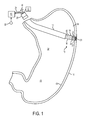
- FIG. 1 is perspective view of a catheter structure, provided in accordance with the principles of the invention, shown inserted into a stomach with electrodes and therapy delivery structure thereof engaged with targeted tissue of the stomach.
- FIG. 2 is an enlarged distal end view of the catheter structure of FIG. 1 .
- FIG. 3 is an enlarged distal end view of a second embodiment of the catheter structure having separate lumens for the electrodes and therapy delivery structure.
- FIG. 4 is an enlarged distal end view of a third embodiment of the catheter structure, showing an implant as the therapy delivery structure.
- FIG. 5 is a partial view of another embodiment of the therapy delivery structure in the form of a needle.
- FIG. 6 is an enlarged side view of a distal end of a catheter structure provided in accordance with a fourth embodiment.
- FIG. 7 is a flow chart showing steps for using the catheter structure to determine an optimal location of tissue to be ablated while simultaneously monitoring to evaluate the effects of the ablation.
- FIG. 8 is a flow chart showing steps for using the catheter structure to determine an optimal location of tissue to be stimulated while simultaneously monitoring to evaluate the effects of the stimulation.
- FIG. 9 is a flow chart showing steps for using the catheter structure to determine an optimal location of tissue to be treated, marking the tissue and simultaneously monitoring to evaluate the effects of the marking.
- a catheter structure is shown generally indicated at 10 inserted via an endoscope 12 into a human organ, such as a stomach 14 .
- the catheter structure 10 includes an elongated tube structure 16 having a distal end 18 and a proximal end 20 .
- Three electrodes 22 are associated with the distal end 18 of the tube structure 16 so as to preferably be moved from a substantially retracted position with respect to distal end 18 of the tube structure 16 to an operative position extending directly from the distal end 18 .
- a signal wire 24 is associated with each electrode 22 for obtaining signals from the electrodes as will be explained more fully below.
- the wires 24 extend within the tube structure 16 to the proximal end 20 thereof.
- An end view of the catheter structure 10 is shown in FIG. 2 .
- Therapy delivery structure 25 is also provided in the tube structure 16 of the catheter structure 10 .
- the therapy delivery structure 25 is provided generally centrally in the tube structure 16 along with the electrodes 22 .
- the therapy delivery structure 25 can be provided in any orientation with respect to the electrodes 22 and thus need not be centrally located.
- a first lumen 27 can contain all of the electrodes 22 and a second, separate lumen 28 can contain the therapy delivery structure 25 .
- the therapy delivery structure 25 is a device connected with a source of energy 30 to deliver the energy to targeted tissue 31 of the human organ 14 .
- the therapy delivery structure 25 can be 1) a radio frequency ablation device such as an needle 29 for delivering radio frequency energy to ablate tissue of the organ 14 ; 2) a microwave ablation device for delivering microwave energy to ablate tissue of the organ 14 ; 3) an ultrasonic ablation device for delivering ultrasonic wave energy to ablate tissue of the organ 14 ; 4) a cryoablation device for delivering cryogenic energy to freeze and kill the tissue of the organ 14 ; and 5) a laser ablation device for delivering laser energy to ablate tissue of the organ 14 , all of which are disclosed in U.S. Patent Application Publication No. 2005/0240239A1, the contents of which is hereby incorporated by reference into this specification.
- the therapy delivery structure 25 can be any device capable of altering tissue of a human organ by using, besides those mentioned above, chemical energy or agents, thermal energy, mechanical sources, bipolar, multipolar or impedance controlled radio frequency electrical energy, or other sources of energy.
- “ablate” can include any means of altering tissue such as, for example, cutting, destroying, burning, killing, etc. to provide a negative therapeutic impact on the physiology of the organ 14 .
- the therapy delivery structure can include a tissue harvesting device such as a biopsy needle or forceps or scalpel to remove or cut tissue from the organ.
- the therapy delivery structure 25 can include an electrical stimulation device as an implant 32 provided in the tube structure 16 ′′.
- the implant 32 can include a barb 34 at an end thereof and can be delivered through the catheter 10 ′ so as to be implanted and left in targeted tissue 31 of the human organ 14 , as explained more fully below.
- the implant 32 may be delivered by a plunger (not shown) of the therapy delivery structure 25 ′.
- the implant 32 can provide synthetic materials, cells, tissues/bioengineered tissues and/or chemicals.
- the therapy delivery structure 25 ′′ may include a needle that delivers, for example, India ink 35 , or may be a device that can heat and gently burn portions of the targeted tissue 31 or tissue nearby. Since the embodiment of FIG. 5 shows a separate lumen 28 for the therapy delivery structure 25 , the needle for the ink 35 can be provided in the lumen 28 to mark the tissue. The needle can thereafter be removed from the lumen 28 and then another therapy delivery structure such as the electrode 33 or an ablating device can be inserted into the lumen 28 to provide treatment or therapy to the marked tissue.
- another therapy delivery structure such as the electrode 33 or an ablating device can be inserted into the lumen 28 to provide treatment or therapy to the marked tissue.
- the electrodes 22 and therapy delivery structure 25 When being inserted into the human organ 14 , the electrodes 22 and therapy delivery structure 25 are in an insertion position. More particularly, the electrodes 22 and therapy delivery structure 25 are retracted, disposed near the distal end 18 of the tube structure 16 . In the illustrated embodiment, the electrodes 22 are delivered into the lumen 36 of the stomach body 38 and/or antrum 40 in order to record myoelectrical activity from the stomach (or other hollow organ) 14 within the abdominal cavity.
- the electrodes 22 and therapy delivery structure 25 are delivered via the tube structure 16 that is passed through the biopsy channel of a standard endoscope 12 .
- three electrodes 22 are provided, one for a positive signal, one for a negative signal and one for ground.
- Each electrode 22 is a recording electrode and is preferably of the type disclosed in U.S. Pat. No. 6,795,725 B2, the content of which is hereby incorporated by reference into this specification.
- actuating structure 42 , 44 is provided to move the electrodes 22 and the therapy delivery structure 25 between the retracted and extended positions.
- the actuating structure can be, for example, one or more wires 42 operatively associated with the electrodes 22 that are manually movable separately, or in unison at the proximal end of the tube structure 16 .
- FIG. 1 shows a single plunger 44 coupled to the wires 42 . Movement of the plunger 44 extends and retracts the electrodes 22 in unison.
- the catheter 10 ′′′ can include a plurality of lumens 46 within the tube structure 16 .
- Each lumen 46 contains a different electrode 22 so that each electrode 22 can be directed separately in different directions to expand the diagnostic tracking location and allow multi-treatment and sensing sites.
- the electrodes can be retractable for ease of inserting/removing the catheter 10 ′′ to/from the human organ 14 .
- the electrodes 22 are extended to impinge upon the mucosal lining 48 or pierce the mucosa to a depth of preferably 2-4 mm. In the extended position, the electrodes are spaced apart between about 1-10 mm and are arranged substantially on a common curvilinear surface. In this way gastric myoelectrical activity from the interstial cells of Cajal and/or smooth muscle and/or enteric neurons are recorded from the three electrodes 22 .
- the output of each electrode 22 is a raw signal.
- the raw signal is a bioelectrical signal recorded from the lining 48 that reflects the myoelectrical activity of the stomach.
- the outputs of the electrodes 22 are sent to an electroviscerogram (EVG) system 50 preferably of the type disclosed in U.S. Pat. No. 7,124,654, the content of which is hereby incorporated by reference into this specification.
- EVG electroviscerogram
- Respiration rate from the patient is also detected via sensor 52 placed on the chest of the patient.
- the respiration signal from sensor 52 is used by the system 50 to monitor artifact caused by respiration movements and/or body movements of the patient.
- the electrical recordings at each site within the organ 14 continue for at least 2 minutes but may continue as long as feasible.
- Signals from the electrodes 22 received by the system 50 are used to locate main pathways of electrical generation in a human organ 14 (in the stomach, for example, the pacemaker region) to locate the targeted tissue 31 .
- the electrodes 22 may need to be moved around the human organ 14 to find these pathways of electrical generation.
- Data from the system 50 can be used by a physician to analyze the signals and perform clinically relevant analyses for interpretation to study myoelectric activity, as disclosed in U.S. Pat. No. 7,124,654.
- the catheter 10 ′ can include a bio-probe or sensor 54 .
- the sensor 54 is constructed and arranged to detect hormone concentrations or cell-specific chemicals to locate the most optimum level in the targeted tissue 31 for treatment.
- a method of ablating targeted tissue in a human organ is shown.
- step 56 when the catheter structure 10 is inserted into the stomach 14 , a baseline electroviscerogram (EVG) or EGG rhythm is determined by the EVG system 50 using signals from the electrodes 22 engaged with the lining 48 .
- step 58 the optimal location (targeted tissue 31 ) is determined for the application of therapy using the electrodes 22 and EVG system 50 .
- the therapy delivery structure 25 is moved to the optimal location in step 60 .
- the therapy delivery structure 25 is used to ablate at least portions of the targeted tissue 31 at the optimal location and the results of the ablation are monitored simultaneously via the electrodes 22 (still in place at the optimal location) and the EVG system 50 .
- the ablation provides a negative impact on the physiology of the human organ or stomach 14 .
- the effects of the therapeutic maneuver (ablation) are evaluated via the electrodes 22 and EVG system 50 .
- the post-therapeutic results are compared to the baseline finding of step 56 via a processor of the EVG system 50 .
- step 68 if adequate results are achieved, the method is ended, if not, the method returns to step 58 .
- a method of stimulating targeted tissue in a human organ is shown.
- step 70 when the catheter structure 10 is inserted into the stomach 14 , a baseline electroviscerogram (EVG) or EGG rhythm is determined by the EVG system 50 using signals from the electrodes 22 engaged with the lining 48 .
- step 72 the optimal location (targeted tissue 31 ) is determined for the application of therapy using the electrodes 22 and EVG system 50 .
- the therapy delivery structure 25 is moved to the optimal location in step 74 .
- the therapy delivery structure 25 is used to electrically stimulate at least portions of the targeted tissue 31 at the optimal location and the results of the stimulation are monitored simultaneously via the electrodes 22 (still in place at the optimal location) and the EVG system 50 .
- Step 76 can include using the electrode 33 of FIG. 5 to stimulate tissue or can include placing the implant 32 of FIG. 4 into the tissue to stimulate the tissue.
- the electrical stimulation provides a positive impact on the physiology of the human organ or stomach 14 .
- the effects of the stimulation are evaluated via the electrodes 22 and EVG system 50 .
- the post-stimulation results are compared to the baseline finding of step 70 via a processor of the EVG system 50 .
- step 80 if adequate results are achieved, the method is ended, if not, the method returns to step 72 if using the electrode 33 . If using the implant 32 , the implant is removed from the tissue and then the method returns to step 72 to determine the better location to place the implant 32 .
- a method of marking targeted tissue in a human organ for later treatment is shown.
- step 84 when the catheter structure 10 is inserted into the stomach 14 , a baseline electroviscerogram (EVG) or EGG rhythm is determined by the EVG system 50 using signals from the electrodes 22 engaged with the lining 48 .
- step 86 the optimal location (targeted tissue 31 ) is determined for the application of therapy using the electrodes 22 and EVG system 50 .
- the therapy delivery structure 25 is moved to the optimal location in step 88 .
- step 90 the therapy delivery structure 25 is used to mark at least portions of the targeted tissue 31 at or near the optimal location and the results of the marking are monitored simultaneously via the electrodes 22 (still in place at the optimal location) and the EVG system 50 .
- step 92 the effects of the marking are evaluated via the electrodes 22 and EVG system 50 .
- step 94 the post-marking results are compared to the baseline finding of step 84 via a processor of the EVG system 50 .
- step 96 if adequate results are achieved, the method is ended, if not, the method returns to step 86 .
- the optimal location or source of the myoelectrical activity can be determined by the EVG system 50 because the electrodes 22 are placed inside the organ under direct vision via the endoscope 12 ;
- the frequency and amplitude of the myoelectrical activity are directly measured without interference from abdominal musculature, adipose tissue or skin;
- the electrical patterns in the organ can be mapped in order to determine normal electrical pathways
- the electrical patterns in the organ regarding various diseases or disorders can be mapped and extent of damage to the electrical network in these diseases and disorders can be determined;
- the therapy delivery structure 25 simultaneously, with the electrode 22 monitoring, ablates, stimulates or marks the tissue at the optimal location;
- the electrodes 22 and EVG system 50 permits evaluation of the ablation, stimulation or marking since the electrodes remain in the tissue at the optimal location during the ablation, simulation or marking procedure;
- catheter structure was shown for use inside the stomach it can be appreciated that the catheter structure can be modified by changing the length thereof when designed for use in the following other organs such as, for example, Duodenum and jejunum; Bile duct; Rectum and sigmoid colon, terminal ileum, ascending and transverse colon; Urinary bladder; Uterus and oviducts.
- organs such as, for example, Duodenum and jejunum; Bile duct; Rectum and sigmoid colon, terminal ileum, ascending and transverse colon; Urinary bladder; Uterus and oviducts.
Landscapes
- Health & Medical Sciences (AREA)
- Life Sciences & Earth Sciences (AREA)
- Surgery (AREA)
- Engineering & Computer Science (AREA)
- Public Health (AREA)
- Animal Behavior & Ethology (AREA)
- Biomedical Technology (AREA)
- Veterinary Medicine (AREA)
- General Health & Medical Sciences (AREA)
- Heart & Thoracic Surgery (AREA)
- Nuclear Medicine, Radiotherapy & Molecular Imaging (AREA)
- Molecular Biology (AREA)
- Medical Informatics (AREA)
- Physics & Mathematics (AREA)
- Otolaryngology (AREA)
- Pathology (AREA)
- Biophysics (AREA)
- Radiology & Medical Imaging (AREA)
- Cardiology (AREA)
- Electromagnetism (AREA)
- Gastroenterology & Hepatology (AREA)
- Plasma & Fusion (AREA)
- Optics & Photonics (AREA)
- Physiology (AREA)
- Pulmonology (AREA)
- Surgical Instruments (AREA)
- Endoscopes (AREA)
- Media Introduction/Drainage Providing Device (AREA)
- Measurement And Recording Of Electrical Phenomena And Electrical Characteristics Of The Living Body (AREA)
Priority Applications (9)
| Application Number | Priority Date | Filing Date | Title |
|---|---|---|---|
| US13/409,138 US8753340B2 (en) | 2012-03-01 | 2012-03-01 | Catheter structure and method for locating tissue in a body organ and simultaneously delivering therapy and evaluating the therapy delivered |
| PCT/US2013/026008 WO2013130270A1 (en) | 2012-03-01 | 2013-02-14 | Catheter structure and method for locating tissue in a body organ and simultaneously delivering therapy and evaluating the therapy delivered |
| JP2014559912A JP6119056B2 (ja) | 2012-03-01 | 2013-02-14 | 身体器官内の組織の存在場所を突き止め、治療手段を送出すると同時に送出された治療手段を評価するカテーテル構造体及び方法 |
| CN201380011437.9A CN104144640B (zh) | 2012-03-01 | 2013-02-14 | 导管结构和用于定位身体器官中的组织并且同时提供治疗并评估所提供的治疗的方法 |
| KR1020147024451A KR102059213B1 (ko) | 2012-03-01 | 2013-02-14 | 신체 장기의 조직을 찾아내는 동시에 테라피를 전달하고 전달된 테라피를 평가하기 위한 방법 및 카테터 구조 |
| EP13755102.4A EP2819575B1 (en) | 2012-03-01 | 2013-02-14 | Catheter structure for locating tissue in a body organ and simultaneously delivering therapy and evaluating the therapy delivered |
| US14/267,967 US9138291B2 (en) | 2012-03-01 | 2014-05-02 | Catheter structure and method for locating tissue in a body organ and simultaneously delivering therapy and evaluating the therapy delivered |
| IN6851DEN2014 IN2014DN06851A (he) | 2012-03-01 | 2014-08-14 | |
| IL234171A IL234171B (he) | 2012-03-01 | 2014-08-18 | קטטר ושיטה לאיתור רקמה בגוף תוך כדי ריפוי והערכת הריפוי שבוצע |
Applications Claiming Priority (1)
| Application Number | Priority Date | Filing Date | Title |
|---|---|---|---|
| US13/409,138 US8753340B2 (en) | 2012-03-01 | 2012-03-01 | Catheter structure and method for locating tissue in a body organ and simultaneously delivering therapy and evaluating the therapy delivered |
Related Child Applications (1)
| Application Number | Title | Priority Date | Filing Date |
|---|---|---|---|
| US14/267,967 Division US9138291B2 (en) | 2012-03-01 | 2014-05-02 | Catheter structure and method for locating tissue in a body organ and simultaneously delivering therapy and evaluating the therapy delivered |
Publications (2)
| Publication Number | Publication Date |
|---|---|
| US20130231646A1 US20130231646A1 (en) | 2013-09-05 |
| US8753340B2 true US8753340B2 (en) | 2014-06-17 |
Family
ID=49043245
Family Applications (2)
| Application Number | Title | Priority Date | Filing Date |
|---|---|---|---|
| US13/409,138 Active - Reinstated 2032-03-06 US8753340B2 (en) | 2012-03-01 | 2012-03-01 | Catheter structure and method for locating tissue in a body organ and simultaneously delivering therapy and evaluating the therapy delivered |
| US14/267,967 Active US9138291B2 (en) | 2012-03-01 | 2014-05-02 | Catheter structure and method for locating tissue in a body organ and simultaneously delivering therapy and evaluating the therapy delivered |
Family Applications After (1)
| Application Number | Title | Priority Date | Filing Date |
|---|---|---|---|
| US14/267,967 Active US9138291B2 (en) | 2012-03-01 | 2014-05-02 | Catheter structure and method for locating tissue in a body organ and simultaneously delivering therapy and evaluating the therapy delivered |
Country Status (8)
| Country | Link |
|---|---|
| US (2) | US8753340B2 (he) |
| EP (1) | EP2819575B1 (he) |
| JP (1) | JP6119056B2 (he) |
| KR (1) | KR102059213B1 (he) |
| CN (1) | CN104144640B (he) |
| IL (1) | IL234171B (he) |
| IN (1) | IN2014DN06851A (he) |
| WO (1) | WO2013130270A1 (he) |
Families Citing this family (15)
| Publication number | Priority date | Publication date | Assignee | Title |
|---|---|---|---|---|
| US10076381B2 (en) * | 2014-09-08 | 2018-09-18 | Olympus Corporation | Method of marking lesion in tubular organ of an object |
| AU2016235142B2 (en) * | 2015-03-25 | 2020-05-07 | Boston Scientific Scimed, Inc. | Methods and devices for identifying treatment sites |
| WO2016171238A1 (ja) * | 2015-04-23 | 2016-10-27 | オリンパス株式会社 | 外科処置装置 |
| CN105943037B (zh) * | 2016-05-23 | 2018-09-07 | 新乡医学院第三附属医院 | 一种结合腹腔镜的肌电检测装置及其操作方法 |
| US11284939B2 (en) | 2017-07-06 | 2022-03-29 | Biosense Webster (Israel) Ltd. | Irrigated catheter with improved ablation tip electrode fluid distribution |
| CN109498989A (zh) * | 2018-12-20 | 2019-03-22 | 宁波贝思转化医学研究中心有限公司 | 一种用于判断胃肠电刺激是否有效的装置及其使用方法 |
| KR102233408B1 (ko) | 2019-05-07 | 2021-03-29 | 고려대학교 산학협력단 | 전기 자극 장치 |
| KR102174222B1 (ko) * | 2019-05-08 | 2020-11-04 | 고려대학교 산학협력단 | 근 변위 측정 장치 및 그를 포함하는 내시경 |
| KR102633526B1 (ko) * | 2020-11-17 | 2024-02-06 | 가톨릭관동대학교산학협력단 | 길이에 따른 신호 보정이 가능한 카테터 장치 |
| KR102636816B1 (ko) * | 2020-11-17 | 2024-02-16 | 가톨릭관동대학교산학협력단 | 정확한 레벨의 신호 인가가 가능한 카테터 장치 |
| WO2022108273A1 (ko) * | 2020-11-17 | 2022-05-27 | 가톨릭관동대학교산학협력단 | 이종 기전의 융합치료에 적합한 카테터 장치 |
| KR102638356B1 (ko) * | 2020-11-17 | 2024-02-21 | 가톨릭관동대학교산학협력단 | 약물의 특성에 따른 신호 제어가 가능한 카테터 장치 |
| US12138057B2 (en) * | 2021-06-04 | 2024-11-12 | Endosure, Inc. | Method and system for monitoring internal bodily disorders by detecting and analyzing tissue frequencies |
| CN120379581A (zh) * | 2022-02-09 | 2025-07-25 | 恩多舒公司 | 通过检测和分析组织频率来监测身体内部紊乱的方法和系统 |
| CN115501487A (zh) * | 2022-09-23 | 2022-12-23 | 上海美生医疗器械有限公司 | 伸缩式的电极装置 |
Citations (11)
| Publication number | Priority date | Publication date | Assignee | Title |
|---|---|---|---|---|
| US5690691A (en) | 1996-05-08 | 1997-11-25 | The Center For Innovative Technology | Gastro-intestinal pacemaker having phased multi-point stimulation |
| US5776073A (en) | 1994-05-19 | 1998-07-07 | Board Of Regents, University Of Texas System | Method and apparatus for analyzing uterine electrical activity from surface measurements for obstetrical diagnosis |
| US6351665B1 (en) | 1999-05-07 | 2002-02-26 | Kenneth L Koch | Method and apparatus for evaluating myoelectric signals and identifying artifact |
| US20030114770A1 (en) | 2001-12-18 | 2003-06-19 | Koch Kenneth L. | Method and apparatus for endoscopic measurement of myoelectrical activity from the stomach and other hollow intra-abdominal organs |
| US20050096638A1 (en) | 2003-10-31 | 2005-05-05 | Medtronic, Inc. | Ablation of exterior of stomach to treat obesity |
| US20050240239A1 (en) | 2005-06-29 | 2005-10-27 | Boveja Birinder R | Method and system for gastric ablation and gastric pacing to provide therapy for obesity, motility disorders, or to induce weight loss |
| US20060015162A1 (en) | 1998-02-19 | 2006-01-19 | Curon Medical, Inc. | Method to treat gastric reflux via the detection and ablation of gastro-esophageal nerves and receptors |
| US7160254B2 (en) | 2004-03-08 | 2007-01-09 | Mark Noar | Intelligent self-interpreting electroviscerogram system and method |
| US20080097350A1 (en) * | 2006-07-06 | 2008-04-24 | Bell Barbara A | Medical devices |
| US7468060B2 (en) | 1998-02-19 | 2008-12-23 | Respiratory Diagnostic, Inc. | Systems and methods for treating obesity and other gastrointestinal conditions |
| US8055334B2 (en) * | 2008-12-11 | 2011-11-08 | Proteus Biomedical, Inc. | Evaluation of gastrointestinal function using portable electroviscerography systems and methods of using the same |
Family Cites Families (13)
| Publication number | Priority date | Publication date | Assignee | Title |
|---|---|---|---|---|
| US6296608B1 (en) * | 1996-07-08 | 2001-10-02 | Boston Scientific Corporation | Diagnosing and performing interventional procedures on tissue in vivo |
| US6961602B2 (en) * | 2001-12-31 | 2005-11-01 | Biosense Webster, Inc. | Catheter having multiple spines each having electrical mapping and location sensing capabilities |
| US20030153905A1 (en) * | 2002-01-25 | 2003-08-14 | Edwards Stuart Denzil | Selective ablation system |
| US20040019388A1 (en) * | 2002-07-24 | 2004-01-29 | Starkebaum Warren L. | Methods and implants for retarding stomach emptying to treat eating disorders |
| US20040215180A1 (en) * | 2003-04-25 | 2004-10-28 | Medtronic, Inc. | Ablation of stomach lining to treat obesity |
| US7840263B2 (en) * | 2004-02-27 | 2010-11-23 | Cardiac Pacemakers, Inc. | Method and apparatus for device controlled gene expression |
| WO2007064847A2 (en) * | 2005-12-02 | 2007-06-07 | Synapse Biomedical, Inc. | Transvisceral neurostimulation mapping device and method |
| US8715281B2 (en) * | 2006-03-09 | 2014-05-06 | Olympus Medical Systems Corp. | Treatment device for endoscope |
| JP5336465B2 (ja) * | 2007-03-26 | 2013-11-06 | ボストン サイエンティフィック リミテッド | 高解像度電気生理学カテーテル |
| JP2009051905A (ja) * | 2007-08-24 | 2009-03-12 | Jfe Engineering Kk | 包接水和物を生成する性質を有する水溶液、第四級アンモニウム塩をゲスト化合物とする包接水和物及び当該包接水和物のスラリ並びに、包接水和物の生成方法、包接水和物が生成又は成長する速度を増加させる方法及び包接水和物が生成又は成長する際に起こる過冷却現象を防止又は抑制する方法 |
| EP2467055A1 (en) * | 2009-08-21 | 2012-06-27 | Auckland UniServices Limited | System and method for mapping gastro-intestinal electrical activity |
| CN102198305B (zh) * | 2010-03-23 | 2013-06-05 | 上海微创医疗器械(集团)有限公司 | 用于局部输送治疗剂的装置和系统 |
| US8948837B2 (en) * | 2011-01-13 | 2015-02-03 | Rhythmia Medical, Inc. | Electroanatomical mapping |
-
2012
- 2012-03-01 US US13/409,138 patent/US8753340B2/en active Active - Reinstated
-
2013
- 2013-02-14 KR KR1020147024451A patent/KR102059213B1/ko active Active
- 2013-02-14 CN CN201380011437.9A patent/CN104144640B/zh not_active Expired - Fee Related
- 2013-02-14 JP JP2014559912A patent/JP6119056B2/ja active Active
- 2013-02-14 WO PCT/US2013/026008 patent/WO2013130270A1/en not_active Ceased
- 2013-02-14 EP EP13755102.4A patent/EP2819575B1/en active Active
-
2014
- 2014-05-02 US US14/267,967 patent/US9138291B2/en active Active
- 2014-08-14 IN IN6851DEN2014 patent/IN2014DN06851A/en unknown
- 2014-08-18 IL IL234171A patent/IL234171B/he not_active IP Right Cessation
Patent Citations (12)
| Publication number | Priority date | Publication date | Assignee | Title |
|---|---|---|---|---|
| US5776073A (en) | 1994-05-19 | 1998-07-07 | Board Of Regents, University Of Texas System | Method and apparatus for analyzing uterine electrical activity from surface measurements for obstetrical diagnosis |
| US5690691A (en) | 1996-05-08 | 1997-11-25 | The Center For Innovative Technology | Gastro-intestinal pacemaker having phased multi-point stimulation |
| US20060015162A1 (en) | 1998-02-19 | 2006-01-19 | Curon Medical, Inc. | Method to treat gastric reflux via the detection and ablation of gastro-esophageal nerves and receptors |
| US7468060B2 (en) | 1998-02-19 | 2008-12-23 | Respiratory Diagnostic, Inc. | Systems and methods for treating obesity and other gastrointestinal conditions |
| US6351665B1 (en) | 1999-05-07 | 2002-02-26 | Kenneth L Koch | Method and apparatus for evaluating myoelectric signals and identifying artifact |
| US20030114770A1 (en) | 2001-12-18 | 2003-06-19 | Koch Kenneth L. | Method and apparatus for endoscopic measurement of myoelectrical activity from the stomach and other hollow intra-abdominal organs |
| US6795725B2 (en) | 2001-12-18 | 2004-09-21 | 3Pcm Company | Method and apparatus for endoscopic measurement of myoelectrical activity from the stomach and other hollow intra-abdominal organs |
| US20050096638A1 (en) | 2003-10-31 | 2005-05-05 | Medtronic, Inc. | Ablation of exterior of stomach to treat obesity |
| US7160254B2 (en) | 2004-03-08 | 2007-01-09 | Mark Noar | Intelligent self-interpreting electroviscerogram system and method |
| US20050240239A1 (en) | 2005-06-29 | 2005-10-27 | Boveja Birinder R | Method and system for gastric ablation and gastric pacing to provide therapy for obesity, motility disorders, or to induce weight loss |
| US20080097350A1 (en) * | 2006-07-06 | 2008-04-24 | Bell Barbara A | Medical devices |
| US8055334B2 (en) * | 2008-12-11 | 2011-11-08 | Proteus Biomedical, Inc. | Evaluation of gastrointestinal function using portable electroviscerography systems and methods of using the same |
Non-Patent Citations (1)
| Title |
|---|
| PCT Search Report & Written Opinion in PCT/US2013/026008, dated Apr. 18, 2013. |
Also Published As
| Publication number | Publication date |
|---|---|
| JP6119056B2 (ja) | 2017-04-26 |
| KR20150014431A (ko) | 2015-02-06 |
| EP2819575A4 (en) | 2015-10-28 |
| CN104144640A (zh) | 2014-11-12 |
| US20130231646A1 (en) | 2013-09-05 |
| US20140243814A1 (en) | 2014-08-28 |
| US9138291B2 (en) | 2015-09-22 |
| EP2819575A1 (en) | 2015-01-07 |
| EP2819575B1 (en) | 2019-12-04 |
| IL234171B (he) | 2018-11-29 |
| CN104144640B (zh) | 2017-08-18 |
| KR102059213B1 (ko) | 2019-12-24 |
| JP2015516179A (ja) | 2015-06-11 |
| WO2013130270A1 (en) | 2013-09-06 |
| IN2014DN06851A (he) | 2015-05-22 |
Similar Documents
| Publication | Publication Date | Title |
|---|---|---|
| US8753340B2 (en) | Catheter structure and method for locating tissue in a body organ and simultaneously delivering therapy and evaluating the therapy delivered | |
| US12343060B2 (en) | Non-invasive and minimally invasive denervation methods and systems for performing the same | |
| US20250325232A1 (en) | Systems and methods for regulating organ and/or tumor growth rates, function, and/or development | |
| US20230066858A1 (en) | Systems and methods for regulating organ and/or tumor growth rates, function, and/or development | |
| JP6538673B2 (ja) | リング電極を有するフォーリーカテーテル | |
| US10010364B2 (en) | Devices and methods for detection and treatment of the aorticorenal ganglion | |
| CN102014779B (zh) | 用于治疗支气管树的系统、组件和方法 | |
| US20150045675A1 (en) | Percutaneous methods and devices for carotid body ablation | |
| JP2018027490A (ja) | 制御された交感神経切除並びに顕微焼灼システム及び方法 | |
| JP2003510160A (ja) | 胃の逆流の処置方法 | |
| EP3226792B1 (en) | Systems for regulating organ and/or tumor growth rates, function, and/or development | |
| WO2015057916A1 (en) | Percutaneous methods and devices for carotid body ablation |
Legal Events
| Date | Code | Title | Description |
|---|---|---|---|
| FEPP | Fee payment procedure |
Free format text: MAINTENANCE FEE REMINDER MAILED (ORIGINAL EVENT CODE: REM.) |
|
| LAPS | Lapse for failure to pay maintenance fees |
Free format text: PATENT EXPIRED FOR FAILURE TO PAY MAINTENANCE FEES (ORIGINAL EVENT CODE: EXP.) |
|
| PRDP | Patent reinstated due to the acceptance of a late maintenance fee |
Effective date: 20180720 |
|
| FEPP | Fee payment procedure |
Free format text: SURCHARGE, PETITION TO ACCEPT PYMT AFTER EXP, UNINTENTIONAL. (ORIGINAL EVENT CODE: M2558); ENTITY STATUS OF PATENT OWNER: SMALL ENTITY Free format text: PETITION RELATED TO MAINTENANCE FEES FILED (ORIGINAL EVENT CODE: PMFP) Free format text: PETITION RELATED TO MAINTENANCE FEES GRANTED (ORIGINAL EVENT CODE: PMFG) |
|
| MAFP | Maintenance fee payment |
Free format text: PAYMENT OF MAINTENANCE FEE, 4TH YR, SMALL ENTITY (ORIGINAL EVENT CODE: M2551) Year of fee payment: 4 |
|
| STCF | Information on status: patent grant |
Free format text: PATENTED CASE |
|
| FP | Lapsed due to failure to pay maintenance fee |
Effective date: 20180617 |
|
| FP | Lapsed due to failure to pay maintenance fee |
Effective date: 20180617 |
|
| FEPP | Fee payment procedure |
Free format text: MAINTENANCE FEE REMINDER MAILED (ORIGINAL EVENT CODE: REM.); ENTITY STATUS OF PATENT OWNER: SMALL ENTITY |
|
| FEPP | Fee payment procedure |
Free format text: 7.5 YR SURCHARGE - LATE PMT W/IN 6 MO, SMALL ENTITY (ORIGINAL EVENT CODE: M2555); ENTITY STATUS OF PATENT OWNER: SMALL ENTITY |
|
| MAFP | Maintenance fee payment |
Free format text: PAYMENT OF MAINTENANCE FEE, 8TH YR, SMALL ENTITY (ORIGINAL EVENT CODE: M2552); ENTITY STATUS OF PATENT OWNER: SMALL ENTITY Year of fee payment: 8 |
|
| AS | Assignment |
Owner name: ENDOSURE, INC., MARYLAND Free format text: ASSIGNMENT OF ASSIGNORS INTEREST;ASSIGNOR:NOAR, MARK;REEL/FRAME:062020/0318 Effective date: 20221009 |
|
| FEPP | Fee payment procedure |
Free format text: MAINTENANCE FEE REMINDER MAILED (ORIGINAL EVENT CODE: REM.); ENTITY STATUS OF PATENT OWNER: SMALL ENTITY |