US8670941B2 - Methods of determining levels of free amino acids and dipeptides and diagnosing Alzheimer's disease - Google Patents
Methods of determining levels of free amino acids and dipeptides and diagnosing Alzheimer's disease Download PDFInfo
- Publication number
- US8670941B2 US8670941B2 US12/377,765 US37776507A US8670941B2 US 8670941 B2 US8670941 B2 US 8670941B2 US 37776507 A US37776507 A US 37776507A US 8670941 B2 US8670941 B2 US 8670941B2
- Authority
- US
- United States
- Prior art keywords
- individual
- diagnosis
- level
- plasma
- levels
- Prior art date
- Legal status (The legal status is an assumption and is not a legal conclusion. Google has not performed a legal analysis and makes no representation as to the accuracy of the status listed.)
- Expired - Fee Related, expires
Links
Images




Classifications
-
- G—PHYSICS
- G01—MEASURING; TESTING
- G01N—INVESTIGATING OR ANALYSING MATERIALS BY DETERMINING THEIR CHEMICAL OR PHYSICAL PROPERTIES
- G01N33/00—Investigating or analysing materials by specific methods not covered by groups G01N1/00 - G01N31/00
- G01N33/48—Biological material, e.g. blood, urine; Haemocytometers
- G01N33/50—Chemical analysis of biological material, e.g. blood, urine; Testing involving biospecific ligand binding methods; Immunological testing
- G01N33/68—Chemical analysis of biological material, e.g. blood, urine; Testing involving biospecific ligand binding methods; Immunological testing involving proteins, peptides or amino acids
- G01N33/6893—Chemical analysis of biological material, e.g. blood, urine; Testing involving biospecific ligand binding methods; Immunological testing involving proteins, peptides or amino acids related to diseases not provided for elsewhere
- G01N33/6896—Neurological disorders, e.g. Alzheimer's disease
-
- G—PHYSICS
- G01—MEASURING; TESTING
- G01N—INVESTIGATING OR ANALYSING MATERIALS BY DETERMINING THEIR CHEMICAL OR PHYSICAL PROPERTIES
- G01N33/00—Investigating or analysing materials by specific methods not covered by groups G01N1/00 - G01N31/00
- G01N33/48—Biological material, e.g. blood, urine; Haemocytometers
- G01N33/50—Chemical analysis of biological material, e.g. blood, urine; Testing involving biospecific ligand binding methods; Immunological testing
- G01N33/68—Chemical analysis of biological material, e.g. blood, urine; Testing involving biospecific ligand binding methods; Immunological testing involving proteins, peptides or amino acids
- G01N33/6803—General methods of protein analysis not limited to specific proteins or families of proteins
- G01N33/6806—Determination of free amino acids
- G01N33/6812—Assays for specific amino acids
-
- G—PHYSICS
- G01—MEASURING; TESTING
- G01N—INVESTIGATING OR ANALYSING MATERIALS BY DETERMINING THEIR CHEMICAL OR PHYSICAL PROPERTIES
- G01N33/00—Investigating or analysing materials by specific methods not covered by groups G01N1/00 - G01N31/00
- G01N33/48—Biological material, e.g. blood, urine; Haemocytometers
- G01N33/50—Chemical analysis of biological material, e.g. blood, urine; Testing involving biospecific ligand binding methods; Immunological testing
- G01N33/94—Chemical analysis of biological material, e.g. blood, urine; Testing involving biospecific ligand binding methods; Immunological testing involving narcotics or drugs or pharmaceuticals, neurotransmitters or associated receptors
- G01N33/9406—Neurotransmitters
- G01N33/9413—Dopamine
-
- G—PHYSICS
- G01—MEASURING; TESTING
- G01N—INVESTIGATING OR ANALYSING MATERIALS BY DETERMINING THEIR CHEMICAL OR PHYSICAL PROPERTIES
- G01N2410/00—Assays, e.g. immunoassays or enzyme assays, involving peptides of less than 20 animo acids
-
- G—PHYSICS
- G01—MEASURING; TESTING
- G01N—INVESTIGATING OR ANALYSING MATERIALS BY DETERMINING THEIR CHEMICAL OR PHYSICAL PROPERTIES
- G01N2800/00—Detection or diagnosis of diseases
- G01N2800/28—Neurological disorders
- G01N2800/2814—Dementia; Cognitive disorders
- G01N2800/2821—Alzheimer
Definitions
- This application pertains to methods of diagnosing Alzheimer's disease based on the levels of certain free amino acids or dipeptides.
- the application also pertains to methods of determining levels of free amino acids and dipeptides using liquid chromatography tandem mass spectrometry methods.
- AD Alzheimer's disease
- AD is a neurodegenerative disorder with an estimated worldwide prevalence of over eighteen million people, and is predicted to increase with an increasing elderly population. AD is characterized by cognitive deficits and memory impairment, and there is currently no cure for this disease.
- Several genetic, age, demographic and environmental factors are linked to AD pathophysiology. Chapman et al., 2001, Trends in Genet. 17, 254-261; Cummings et al., 1998, Neurology, 51, S2-17; Doraiswamy et al., 2002, Bio. Sci. Med. Sci., 57, M173-M177.
- a ⁇ amyloid beta
- APP amyloid precursor protein
- AD cognitive decline leading to memory loss.
- Memory dysfunction involves impairment of learning new information which is often characterized as short-term memory loss.
- recall of remote well-learned material may appear to be preserved, but new information cannot be adequately incorporated into memory.
- Disorientation to time is also closely related to memory disturbance.
- AD Language impairments are also a prominent part of AD. These are often manifest first as word finding difficulty in spontaneous speech.
- the language of the AD patient is often vague, lacking in specifics and may have increased automatic phrases and clichés. Difficulty in naming everyday objects is often prominent.
- Complex deficits in visual function are present in many AD patients, as are other focal cognitive deficits such as apraxia, acalculia and left-right disorientation. Impairments of judgment and problem solving are frequently seen.
- Non-cognitive or behavioral symptoms are also common in AD.
- Personality changes are commonly reported and range from progressive passivity to marked agitation. Patients may exhibit changes such as decreased expressions of affection, depressive symptoms, anxiety, and psychosis. In some cases, personality changes may predate cognitive abnormality.
- AD Alzheimer's disease
- temporary e.g., depression or vitamin B12 deficiency
- permanent e.g., stroke
- AD cannot be conclusively diagnosed until after death, when autopsy reveals the disease's characteristic amyloid plaques and neurofibrillary tangles in a patient's brain.
- clinical diagnostic procedures are only helpful after patients have begun displaying significant, abnormal memory loss or personality changes. By then, a patient has likely had AD for years. There is therefore a need for other methods of diagnosing and aiding diagnosis of AD.
- Mass spectrometers are often coupled with chromatography systems in order to identify and characterize eluting species from a test sample.
- the eluting solvent is ionized and a series of mass spectrograms are obtained.
- liquid chromatography is coupled with tandem mass spectrometry (LCMS 2 ) for better analysis of test samples. See, e.g., U.S. Pat. No. 7,009,174; Jansen et al., J. Chromatography, 830:196-200 (2006).
- the invention in one aspect provides a method of diagnosing Alzheimer's disease (“AD”) in an individual, comprising: a) comparing the level of at least one AD diagnosis marker in a body fluid sample (such as plasma, urine, and CSF) from the individual with a reference level, and b) determining whether the individual has AD based on a characteristic change in the level of at least one AD diagnosis biomarker, wherein the AD diagnosis marker is a free amino acid or dipeptide selected from the group consisting of: an imidazole-containing free amino acid or dipeptide having antioxidant properties, an aromatic-containing free amino acid that is a neurotransmitter, a free amino acid or dipeptide associated with urea metabolism or detoxification and NO formation, a glutamate-derived free amino acid or dipeptide, and an asparate or serine-derived free amino acid.
- AD diagnosis marker is a free amino acid or dipeptide selected from the group consisting of: an imidazole-containing free amino acid or dipeptide having antioxidant properties, an aromatic-containing free amino acid
- a method of providing information for diagnosis of AD in an individual comprising: a) determining the level of at least one AD diagnosis marker in the body fluid sample of the individual, and b) providing information about the level of at least one AD diagnosis marker for diagnosis of AD, wherein the level of the at least one AD diagnosis marker is used as basis for diagnosing AD, wherein a characteristic change in the level of the at least one AD diagnosis biomarker is indicative of AD, and wherein the AD diagnosis marker is a free amino acid or dipeptide selected from the group consisting of: an imidazole-containing free amino acid or dipeptide having antioxidant properties, an aromatic-containing free amino acid that is a neurotransmitter, a free amino acid or dipeptide associated with urea metabolism or detoxification and NO formation, a glutamate-derived free amino acid or dipeptide, and an asparate or serine-derived free amino acid.
- the levels of the AD diagnosis markers are determined by liquid chromatography tandem mass spectrometry (“LCMS 2 ”) method
- the invention provides a method of aiding diagnosis of Alzheimer's disease (“AD”) in an individual, comprising: a) comparing the level of at least one AD diagnosis marker in a body fluid sample (such as plasma, urine, and CSF) from the individual with a reference level, and b) determining whether the individual has AD based on a characteristic change in the level of at least one AD diagnosis biomarker and at least one other method of diagnosing AD, wherein the AD diagnosis marker is a free amino acid or dipeptide selected from the group consisting of: an imidazole-containing free amino acid or dipeptide having antioxidant properties, an aromatic-containing free amino acid that is a neurotransmitter, a free amino acid or dipeptide associated with urea metabolism or detoxification and NO formation, a glutamate-derived free amino acid or dipeptide, and an asparate or serine-derived free amino acid.
- AD diagnosis marker is a free amino acid or dipeptide selected from the group consisting of: an imidazole-containing free amino acid or dipeptide having antioxidant
- a method of providing information for aiding diagnosis of AD in an individual comprising: a) determining the level of at least one AD diagnosis marker in the body fluid sample of the individual, and b) providing information about the level of at least one AD diagnosis marker for diagnosis of AD, wherein the level of the at least one AD diagnosis marker is used as basis for diagnosing AD, wherein a characteristic change in the level of the at least one AD diagnosis biomarker is suggestive of AD, and wherein the AD diagnosis marker is a free amino acid or dipeptide selected from the group consisting of: an imidazole-containing free amino acid or dipeptide having antioxidant properties, an aromatic-containing free amino acid that is a neurotransmitter, a free amino acid or dipeptide associated with urea metabolism or detoxification and NO formation, a glutamate-derived free amino acid or dipeptide, and an asparate or serine-derived free amino acid.
- the levels of the AD diagnosis markers are determined by LCMS 2 method.
- the body fluid sample is cerebrospinal fluid (CSF). In some embodiments, the body fluid sample is plasma. In some embodiments, the body fluid sample is urine. In some embodiments, the body fluid sample is selected from the group consisting of CSF, plasma, and urine. In some embodiments, the body fluid sample is a peripheral body fluid (including plasma and urine).
- CSF cerebrospinal fluid
- the body fluid sample is plasma.
- the body fluid sample is urine.
- the body fluid sample is selected from the group consisting of CSF, plasma, and urine. In some embodiments, the body fluid sample is a peripheral body fluid (including plasma and urine).
- At least one AD diagnosis marker is an imidazole-containing free amino acid or dipeptide having antioxidant properties, such as an imidazole-containing free amino acid or dipeptide selected from the group consisting of histidine, 1-methyl-histidine, 3-methyl-histidine, carnosine, and anserine.
- the imidazole-containing free amino acid or dipeptide is carnosine.
- At least one AD diagnosis marker is an aromatic-containing free amino acid that is a neurotransmitter, such as an aromatic-containing free amino acid selected from the group consisting of tryptophan, phenylalanine, tyrosine, dopamine, and DOPA.
- the aromatic-containing free amino acid is dopamine.
- the aromatic-containing free amino acid is DOPA.
- At least one AD diagnosis marker is an imidazole-containing free amino acid or dipeptide having antioxidant properties and at least one AD diagnosis marker is an aromatic-containing free amino acid that is a neurotransmitter. In some embodiments, at least one AD diagnosis marker is a glutamate-derived free amino acid or dipeptide. In some embodiments, at least one AD diagnosis marker is an imidazole-containing free amino acid or dipeptide having antioxidant properties, at least one AD diagnosis marker is an aromatic-containing free amino acid that is a neurotransmitter, and at least one AD diagnosis marker is a glutamate-derived free amino acid or dipeptide.
- the AD diagnosis marker is a free amino acid or dipeptide selected from the group consisting of: histidine, 1-methyl-histidine, 3-methyl-histidine, carnosine, anserine, tryptophan, phenylalanine, tyrosine, dopamine, DOPA, arginine, citrulline, and ornithine.
- a characteristic change of at least two (such as at least any of 3, 4, 5, 6, 7, 8, 9, 10, 11, 12, or 13) free amino acids or dipeptides selected from the group consisting of dopamine, histidine, 1-methyl-histidine, 3-methyl-histidine, carnosine, anserine, tryptophan, phenylalanine, tyrosine, DOPA, arginine, citrulline, and ornithine is indicative or suggestive of AD.
- the AD diagnosis marker is any of the free amino acid or dipeptide listed in Table 5.
- a method of diagnosing AD in an individual comprising: a) comparing the CSF level of at least one AD diagnosis marker from the individual with a reference level, and b) determining whether the individual has AD based on a characteristic change in the CSF level of at least one AD diagnosis biomarker, wherein the AD diagnosis marker is selected from the group consisting of: histidine, 1-methyl histidine, 3-methyl histidine, tryptophan, phenylalanine, arginine, citrulline, pyroglutamine, 4-hydroxyproline, glycyl proline, asparagine, isoleucine, lysine, cysteine, valine, leucine, tyrosine, dopamine, glutamine, proline-hydroxyproline, proline, cystine, and glycine.
- an increase in the CSF level of at least one (such as at least any of 2, 3, 4, 5, 6, 7) of tyrosine, dopamine, glutamine, proline-hydroxyproline, proline, cystine, and glycine (collectively designated as “Group 2 AD diagnosis markers”) is indicative of AD.
- a decrease in the CSF level of at least one (such as at least any of 2, 3, 5, 10, 15, or more) Group 1 AD diagnosis marker and an increase in the CSF level of at least one (such as at least any of 2, 3, 4, 5, 6, 7) Group 2 AD diagnosis marker is indicative of AD.
- the invention provides a method of aiding diagnosis of AD in an individual, comprising: a) comparing the CSF level of at least one AD diagnosis marker from the individual with a reference level, and b) determining whether the individual has AD based on a characteristic change in the CSF level of at least one AD diagnosis biomarker and one other AD diagnosis method, wherein the AD diagnosis marker is selected from the group consisting of: histidine, 1-methyl histidine, 3-methyl histidine, tryptophan, phenylalanine, arginine, citrulline, pyroglutamine, 4-hydroxyproline, glycyl proline, asparagine, isoleucine, lysine, cysteine, valine, leucine, tyrosine, dopamine, glutamine, proline-hydroxyproline, proline, cystine, and glycine.
- the AD diagnosis marker is selected from the group consisting of: histidine, 1-methyl histidine, 3-methyl histidine, tryptophan, pheny
- a decrease in the CSF level of at least one (such as at least any of 2, 3, 5, 10, 15, or more) of histidine, 1-methyl histidine, 3-methyl histidine, tryptophan, phenylalanine, arginine, citrulline, pyroglutamine, 4-hydroxyproline, glycyl proline, asparagine, isoleucine, lysine, cysteine, valine, and leucine (“Group 1 AD diagnosis markers”) is suggestive of AD.
- an increase in the CSF level of at least one (such as at least any of 2, 3, 4, 5, 6, 7) of tyrosine, dopamine, glutamine, proline-hydroxyproline, proline, cystine, and glycine (“Group 2 AD diagnosis markers”) is suggestive of AD.
- a decrease in the CSF level of at least one (such as at least any of 2, 3, 5, 10, 15, or more) Group 1 AD diagnosis marker and an increase in the CSF level of at least one (such as at least any of 2, 3, 4, 5, 6, 7) Group 2 AD diagnosis marker is suggestive of AD.
- a method of diagnosing AD in an individual comprising: a) comparing the plasma level of at least one AD diagnosis marker from the individual with a reference level, and b) determining whether the individual has AD based on a characteristic change in the plasma level of at least one AD diagnosis biomarker, wherein the AD diagnosis marker is selected from the group consisting of: histidine, 1-methyl histidine, 3-methyl histidine, carnosine, phenylalanine, tyrosine, dopamine, arginine, citrulline, pyroglutamine, GABA, ⁇ ABA, ABA, 4-hydroyproline, hydroxy lysine, lysyl alanine, aminopimelic acid, anserine, tryptophan, DOPA, ornithine, glutamic acid, ⁇ -glutamyl- ⁇ -lysine, DABA, glutamine, proline-hydroxyproline, glycyl proline, aspartic acid, isoleu
- a decrease in the plasma level of at least one (such as at least any of 2, 5, 10, 15, or more) Group 3 AD diagnosis marker and an increase in the plasma level of at least one (such as at least any of 5, 10, 15, or 20) Group 4 AD diagnosis marker is indicative of AD.
- the invention provides a method of aiding diagnosis of AD in an individual, comprising: a) comparing the plasma level of at least one AD diagnosis marker from the individual with a reference level, and b) determining whether the individual has AD based on a characteristic change in the plasma level of at least one AD diagnosis biomarker and one other AD diagnosis method, wherein the AD diagnosis marker is selected from the group consisting of: histidine, 1-methyl histidine, 3-methyl histidine, carnosine, phenylalanine, tyrosine, dopamine, arginine, citrulline, pyroglutamine, GABA, ⁇ ABA, ABA, 4-hydroyproline, hydroxy lysine, lysyl alanine, aminopimelic acid, anserine, tryptophan, DOPA, ornithine, glutamic acid, ⁇ -glutamyl- ⁇ -lysine, DABA, glutamine, proline-hydroxyproline, glycyl proline, as
- a decrease in the plasma level of at least one (such as at least any of 2, 5, 10, 15, or more) Group 3 AD diagnosis marker and an increase in the plasma level of at least one (such as at least any of 5, 10, 15, or 20) Group 4 AD diagnosis marker is suggestive of AD.
- a method of diagnosing AD in an individual comprising: a) comparing the urine level of at least one AD diagnosis marker from the individual with a reference level, and b) determining whether the individual has AD based on a characteristic change in the urine level of at least one AD diagnosis biomarker, wherein the AD diagnosis marker is selected from the group consisting of: 1-methyl-histindine, anserine, pyroglutamine, DABA, threonine, lysine, cystathionine, valine, histidine, 3-methyl histidine, carnosine, tryptophan, phenylalanine, tyrosine, arginine, citrulline, ornithine, glutamine, proline-hydroproline, 4-hydroxyproline, glycyl proline, asparagine, isoleucine, lysyl alanine, cysteine, cystine, glycine, and leucine.
- the AD diagnosis marker is selected from the group consisting of: 1-
- a decrease in the urine level of at least one is indicative of AD.
- at least one such as at least any of 2, 3, 4, 5, 6, 7, or 8 of 1-methyl-histindine, anserine, pyroglutamine, DABA, threonine, lysine, cystathionine, valine (collectively designated as “Group 5 AD diagnosis markers”) is indicative of AD.
- a decrease in urine level of at least one (such as at least any of 2, 3, 4, 5, 6, 7, or 8) Group 5 AD diagnosis marker and an increase in urine level of at least one (such as at least any of 5, 10, 15, or 20) Group 6 AD diagnosis marker is indicative of AD.
- the invention provides a method of aiding diagnosis of AD in an individual, comprising: a) comparing the urine level of at least one AD diagnosis marker from the individual with a reference level, and b) determining whether the individual has AD based on a characteristic change in the urine level of at least one AD diagnosis biomarker and one other AD diagnosis method, wherein the AD diagnosis marker is selected from the group consisting of: 1-methyl-histindine, anserine, pyroglutamine, DABA, threonine, lysine, cystathionine, valine, histidine, 3-methyl histidine, carnosine, tryptophan, phenylalanine, tyrosine, arginine, citrulline, ornithine, glutamine, proline-hydroproline, 4-hydroxyproline, glycyl proline, asparagine, isoleucine, lysyl alanine, cysteine, cystine, glycine, and leucine.
- a decrease in the urine level of at least one (such as at least any of 2, 3, 4, 5, 6, 7, or 8) of 1-methyl-histindine, anserine, pyroglutamine, DABA, threonine, lysine, cystathionine, valine (collectively designated as “Group 5 AD diagnosis markers”) is suggestive of AD.
- a decrease in urine level of at least one (such as at least any of 2, 3, 4, 5, 6, 7, or 8) Group 5 AD diagnosis marker and an increase in urine level of at least one (such as at least any of 5, 10, 15, or 20) Group 6 AD diagnosis marker is suggestive of AD.
- the levels of AD diagnosis markers in more than one body fluid is determined.
- the plasma level of at least one Group 3 or Group 4 AD diagnosis markers and the urine level of at least one Group 5 or Group 6 AD diagnosis markers are determined, and diagnosis of AD can be based on a characteristic change of one or more of these AD diagnosis markers.
- MCI mild cognitive impairment
- assessing cognitive function assessing cognitive impairment
- predicting risk of developing AD monitoring AD progression of AD
- monitoring AD treatment and screening for individuals having AD.
- the method comprises: a) comparing the plasma level of carnosine of the individual with a reference level, and b) determining whether the individual has AD based on a decrease in the plasma level of carnosine. For example, in some embodiments, a decrease in the plasma level of carnosine by at least about 40% is indicative of AD.
- the method comprises: a) comparing the relative activity of carnosinase versus carnosine synthetase in the plasma of the individual with a reference relative activity, and b) determining whether the individual has AD based on an increase in the relative activity. In some embodiments, the method comprises: a) comparing the activity of carnosine synthetase in the plasma of the individual with a reference activity, and b) determining whether the individual has AD based on a decrease in the activity of carnosine synthetase.
- the method comprises: a) comparing the activity of carnosinase or carnosine-N-methyltransferase in the plasma of the individual with a reference activity, and b) determining whether the individual has AD based on an increase in the activity of carnosinase or carnosine-N-methyltransferase.
- a method of treating AD in an individual comprising administering to the individual an effective amount of an agent that increases the plasma level of carnosine, wherein diagnosis of AD is based on a decrease in the plasma level of carnosine in the individual as compared to a reference level.
- a method of treating AD in an individual comprising: a) determining whether the individual has AD based on a decrease in the plasma level of carnosine in the individual as compared to a reference level, and b) administering to the individual an effective amount of an agent that increases the plasma level of carnosine.
- Agents that increase the plasma level of carnosine include, for example, carnosine itself, agents that inhibit the activity of carnosinase, and agents that enhance the activity of carnosine synthetase in the plasma.
- AD Alzheimer's disease
- a method of diagnosing Alzheimer's disease (“AD”) in an individual comprising: a) comparing the plasma level of dopamine of the individual with a reference level, and b) determining whether the individual has AD based on a decrease in the plasma level of dopamine and an increase in the plasma level of DOPA.
- a decrease in the plasma level of dopamine by at least about 15% is indicative of AD.
- an increase in the plasma level of DOPA by at least about 50% is indicative of AD.
- an increase in the plasma level of DOPA and decrease in the plasma level of dopamine is indicative of AD. In some embodiments, an increase in the plasma level of DOPA by at least about 50% and a decrease in the plasma level of dopamine by at least about 15% is indicative of AD.
- a method of diagnosing AD in an individual comprising: a) comparing the relative activity of tyrosine-3-hydrolase versus DOPA decarboxylase in the plasma of the individual with a reference relative activity, and b) determining whether the individual has AD based on an increase in the relative activity.
- a method of diagnosing AD in an individual comprising: a) comparing the activity of DOPA decarboxylase in the plasma of the individual with a reference activity, and b) determining whether the individual has AD based on a decrease in the activity of DOPA decarboxylase.
- a method of diagnosing AD in an individual comprising: a) comparing the activity of tyrosine-3-hydrolase in the plasma of the individual with a reference activity, and b) determining whether the individual has AD based on an increase in the activity of tyrosine-3-hydrolase.
- a method of treating AD in an individual comprising administering to the individual an effective amount of an agent that increases the plasma level of dopamine or decreases the plasma level of DOPA, wherein diagnosis of AD is based on a decrease in the plasma level of dopamine or an increase in the plasma level of DOPA in the individual as compared to a reference level.
- a method of treating AD in an individual comprising: a) determining whether the individual has AD based on a decrease in the plasma level of dopamine or an increase in the plasma level of DOPA in the individual as compared to a reference level, and b) administering to the individual an effective amount of an agent that increases the plasma level of dopamine or decreases the plasma level of DOPA.
- Methods of monitoring effectiveness of AD treatment and methods of continuing to treat AD based on plasma level of carnosine are also provided.
- Agents that increase the plasma level of dopamine or decrease the plasma level of DOPA include, for example, dopamine itself, agents that inhibit tyrosine-3-hydrolase, and agents that enhance DOPA dehydroxylase in the plasma.
- LCMS 2 method methods of simultaneously determining the levels of at least two free amino acids or dipeptides in a biological fluid sample using liquid chromatography tandem mass spectrometry (LCMS 2 method).
- LCMS 2 method a method of simultaneously measuring the levels of at least two (such as at least any of 3, 5, 10, 15, 20, 30, 40, 50, 60, 70, 80, or more) free amino acids or dipeptides in a biological fluid sample, comprising subjecting the sample to LCMS 2 , wherein the levels of at least two (such as at least any of 3, 5, 10, 15, 20, 30, 40, 50, 60, 70, 80, or more) free amino acids or dipeptides are determined.
- a method of assessing changes in levels of at least one free amino acids or dipeptides in a biological fluid sample comprising simultaneously detecting levels of at least two (such as at least any of 2, 3, 5, 10, 20, 30, 40, 50, 60, 70, 80, or more) free amino acids or dipeptides in the sample by LCMS 2 and comparing the detected levels with reference levels.
- a characteristic change in at least one of the free amino acids or dipeptides are indicative of a disease, such as AD.
- a method of identifying one or more diagnosis markers of a disease comprising simultaneously measuring the levels of at least two free amino acids or dipeptides from a set of biological fluid samples by LCMS 2 , wherein the set of biological fluid samples is divisible into subsets on the basis of a disease, comparing the levels from each subset for at least one biomarker, and identifying at least one marker for which the levels are significantly different between the two subsets.
- the method comprises simultaneously measuring the levels of at least 2, 3, 5, 10, 20, 30, 40, 50, 60, 70, 80, or more free amino acids or dipeptides.
- the disease is AD.
- FIG. 1A provides the TIC (Total Ion Current) of free amino acid and dipeptide standards.
- FIGS. 1B-1F provides the extracted SRM chromatograph of five internal standards.
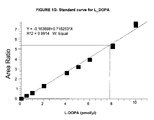
- FIG. 1G provides the standard curve for L-DOPA using [ 2 H 4 ]-DOPA as an internal standard.
- FIG. 2A provides the TIC of amino acids and dipeptides in CSF obtained by LCMS in the SRM mode.
- FIGS. 2B-2F provide spectra of homophenylalanine internal standard (B), citrulline (C), carnosine (D), phenylalanine (E), and L-DOPA (F). The arrows indicate peaks corresponding to the retention times of authentic free amino acid standards.
- FIG. 3A provides TIC of amino acids and dipeptides in plasma obtained by LCMS 2 in the SRM mode.
- FIGS. 3A-3F provide spectra of homophenylalanine internal standard (B), citrulline (C), carnosine (D), phenylalanine (E), and L-DOPA (F).
- the arrows indicate peaks corresponding to the retention times of authentic free amino acid standards.
- FIG. 4A provides TIC of amino acids and dipeptides in urine obtained by LCMS 2 in the SRM mode.
- FIGS. 4A-4F provide spectra of homophenylalanine internal standard (B), citrulline (C), carnosine (D), phenylalanine (E), and L-DOPA (F). The arrows indicate peaks corresponding to the retention times of authentic free amino acid standards.
- FIG. 8 shows the biochemical pathway of histidine metabolism.
- Arrows stands for the proposed changes (up arrows for increase, down arrows for decrease) in free amino acids and dipeptides in body fluids (e.g., plasma) and the proposed changes in enzyme activity and/or levels that likely account for these changes.
- FIG. 9 shows the biochemical pathway of dopamine metabolism.
- Arrows stands for the proposed changes (up arrows for increase, down arrows for decrease) in free amino acids and dipeptides in body fluids (e.g., plasma) and the proposed changes in enzyme activity and/or levels that likely account for these changes.
- LCMS 2 liquid chromatography tandem mass spectrometry method
- the invention in one aspect provides methods of diagnosing AD or aiding diagnosis of AD based on the levels of AD diagnosis markers.
- methods of diagnosing AD based on carnosine level and/or carnosine synthesis activities in the plasma are also provided.
- methods of diagnosing AD based on dopamine/DOPA level and/or dopamine synthesis activities in the plasma are also provided.
- methods of simultaneously determining the levels of two or more free amino acids or dipeptides in a biological fluid sample are useful for assessing changes in free amino acids or dipeptides in a biological fluid sample, identifying disease markers (for example for the purpose of diagnosing disease), and elucidating biochemical pathways underlying diseases.
- the present invention in one aspect provides a method of diagnosing Alzheimer's disease (“AD”) in an individual, comprising: a) comparing the level of at least one AD diagnosis marker in a body fluid sample from the individual (such as an individual suspected of having or being susceptible to AD) with a reference level, and b) determining whether the individual has AD based on a characteristic change in the level of at least one AD diagnosis biomarker.
- AD Alzheimer's disease
- the AD diagnosis marker is a free amino acid or dipeptide selected from the group consisting of: an imidazole-containing free amino acid or dipeptide having antioxidant properties, an aromatic-containing free amino acid that is a neurotransmitter, a free amino acid or dipeptide associated with urea metabolism or detoxification and NO formation, a glutamate-derived free amino acid or dipeptide, and an asparate or serine-derived free amino acid.
- the AD diagnosis marker is a free amino acid or dipeptide selected from the group consisting of histidine, 1-methyl-histidine, 3-methyl-histidine, carnosine, anserine, tryptophan, phenylalanine, tyrosine, dopamine, DOPA, arginine, citrulline, and ornithine.
- the AD diagnosis marker is a free amino acid or dipeptide listed in Table 5.
- the AD diagnosis markers are present in peripheral body fluids (e.g., plasma and urine) of individuals, allowing simple and non-invasive diagnosis of AD.
- the method comprises: a) providing a body fluid sample from the individual (such as an individual suspected of having or being susceptible to AD), b) comparing the level of at least one AD diagnosis marker in a body fluid sample from the individual with a reference level, and c) determining whether the individual has AD based on a characteristic change in the level of at least one AD diagnosis biomarker.
- a method of diagnosing AD in an individual comprising: a) determining the level of at least one AD diagnosis marker in the body fluid sample of the individual (such as an individual suspected of having or being susceptible to AD), b) comparing the level of at least one AD diagnosis marker in a body fluid sample with a reference level, and c) determining whether the individual has AD based on a characteristic change in the level of at least one AD diagnosis biomarker.
- a method of diagnosing AD in an individual comprising: a) providing a body fluid sample from the individual, b) determining the level of at least one AD diagnosis marker in the body fluid sample, c) comparing the level of at least one AD diagnosis marker in a body fluid sample with a reference level, and d) determining whether the individual has AD based on a characteristic change in the level of at least one AD diagnosis biomarker.
- the present invention also encompasses methods of providing information about the levels of AD diagnosis markers in an individual (such as an individual suspected of having or being susceptible to AD). Such information (either quantitative or qualitative) can then be used for diagnosis of AD.
- a method comprising: a) determining the level of at least one AD diagnosis marker in the body fluid sample of the individual (such as an individual suspected of having or being susceptible to AD), and b) providing information about the level of at least one AD diagnosis marker for diagnosis of AD, wherein the level of the at least one AD diagnosis marker is used as basis for diagnosing AD, and wherein a characteristic change in the level of the at least one AD diagnosis biomarker is indicative of AD.
- methods of evaluating results of the analytical methods described herein Such evaluation generally entails reviewing the result(s) and can assist, for example, in advising regarding clinical and/or diagnostic follow-up and/or treatment options.
- AD diagnosis markers described herein are also useful for aiding diagnosis of AD.
- methods for “aiding diagnosis” refer to methods that assist in making a clinical determination regarding the presence or nature of the AD, and may or may not be conclusive with respect to the definitive diagnosis.
- the methods described herein may be used in conjunction with other methods for diagnosing AD.
- Other methods of diagnosing AD are known in the art, and include, for example, ADAS-COG, MMSE, magnetic resonance imaging, magnetic resonance spectrometry, and measurement of ⁇ -amyloid and tau levels.
- a method of aiding diagnosis of AD comprising: a) comparing the level of at least one AD diagnosis marker in a body fluid sample from the individual with a reference level, and b) determining whether the individual has AD based on a characteristic change in the level of at least one AD diagnosis biomarker and at least one other method of diagnosing AD.
- the other method is a cognition test.
- the other method is a behavior test.
- the method comprises: a) providing a body fluid sample from the individual (such as an individual suspected of having or being susceptible to AD), b) comparing the level of at least one AD diagnosis marker in a body fluid sample from the individual with a reference level, and c) determining whether the individual has AD based on a characteristic change in the level of at least one AD diagnosis biomarker and at least one other method of diagnosing AD.
- a method of aiding diagnosis of AD in an individual comprising: a) determining the level of at least one AD diagnosis marker in the body fluid sample of the individual (such as an individual suspected of having or being susceptible to AD), b) comparing the level of at least one AD diagnosis marker in a body fluid sample with a reference level, and c) determining whether the individual has AD based on a characteristic change in the level of at least one AD diagnosis biomarker and at least one other method of diagnosing AD.
- a method of aiding diagnosis of AD in an individual comprising: a) providing a body fluid sample from the individual, b) determining the level of at least one AD diagnosis marker in the body fluid sample, and c) comparing the level of at least one AD diagnosis marker in a body fluid sample with a reference level, and d) determining whether the individual has AD based on a characteristic change in the level of at least one AD diagnosis biomarker and at least one other method of diagnosing AD.
- a method of aiding diagnosis of AD comprising: a) determining the level of at least one AD diagnosis marker in the body fluid sample of the individual, and b) providing information about the level of at least one AD diagnosis marker for diagnosis of AD, wherein the level of the at least one AD diagnosis marker is used as basis for aiding diagnosis of AD, and wherein a characteristic change in the level of at least one AD diagnosis marker is suggestive of AD.
- the invention provides a method of diagnosing MCI in an individual, comprising: a) comparing the level of at least one AD diagnosis marker in a body fluid sample from the individual (such as an individual suspected of having or being susceptible to MCI) with a reference level, and b) determining whether the individual has MCI based on a characteristic change in the level of at least one AD diagnosis biomarker.
- a method comprising: a) determining the level of at least one AD diagnosis marker in the body fluid sample of the individual (such as an individual suspected of having or being susceptible to MCI), and b) providing information about the level of at least one AD diagnosis marker for diagnosis of MCI, wherein the level of the at least one AD diagnosis marker is used as basis for diagnosing MCI, and wherein a characteristic change in the level of the at least one AD diagnosis biomarker is indicative of MCI.
- a method of aiding diagnosis of MCI comprising: a) comparing the level of at least one AD diagnosis marker in a body fluid sample from the individual with a reference level, and b) determining whether the individual has MCI based on a characteristic change in the level of at least one AD diagnosis biomarker and at least one other method of diagnosing MCI.
- the other method is a cognition test.
- Other methods for diagnosing MCI include, for example, neurocognitive testing such as the Wechsler adult intelligence testing and neuroimaging such as measurements of the hippocampal volume.
- a method of aiding diagnosis of MCI comprising: a) determining the level of at least one AD diagnosis marker in the body fluid sample of the individual, and b) providing information about the level of at least one AD diagnosis marker for diagnosis of MCI, wherein the level of the at least one AD diagnosis marker is used as basis for aiding diagnosis of MCI, and wherein a characteristic change in the level of at least one AD diagnosis marker is suggestive of MCI.
- stratifying refers to sorting individuals into different classes or strata based on the features of a neurological disease.
- stratifying a population of individual with AD may involve assigning the individual on the basis of the severity of the disease (e.g., mild, moderate, advanced, etc.).
- stratifying a population of individual with AD may also involve assigning the individual on the basis of progression rate of AD (e.g., slow, fast, etc.).
- predicting whether the individual is at risk of developing AD refers to making a finding that an individual has a significantly enhanced probability of developing an AD.
- AD diagnosis markers described herein are also useful for one or more of the following: monitoring AD progression in AD patients, monitoring AD treatment in AD patients, screening for individuals having AD, assessing cognitive function, assessing cognitive impairment, and diagnosing or aiding diagnosis of cognitive impairment, based on the levels of one or more AD diagnosis levels in a body fluid sample.
- an “individual” as used herein refers to a vertebrate, preferably a mammal, more preferably a human. Mammals include, but are not limited to, farm animals, sport animals, pets, primates, mice and rats. In some embodiments, the individual is human. In some embodiments, the individual is an individual other than human. In some embodiments, the individual is an animal model for the study of AD. Animal models for AD studies are known in the art. See, for example, Haltzman et al., Proc. Natl. Acad. Sci. USA, 2000, 97(6):2892-7.
- the individual is a normal individual, e.g., an individual having a Mini-Mental State Examination (MMSE) score or would achieve a MMSE score in the range of 25-30.
- the individual is an individual with mild AD, e.g., an individual who has either been assessed with the Mini-Mental State Examination (MMSE) and scored 22-27 or would achieve a score of 22-27 upon MMSE testing.
- the individual is an individual with moderate AD, e.g., an individual who has either been assessed with the MMSE and scored 16-21 or would achieve a score of 16-21 upon MMSE testing.
- the individual is an individual with severe AD, e.g., an individual who has either been assessed with the MMSE and scored 12-15 or would achieve a score of 12-15 upon MMSE testing.
- the individual is suspected of having AD or MCI. In some embodiments, the individual is an individual for whom cognitive assessment is desired. Alternatively, an individual involved in for example research and/or clinical research may desire an assessment without any indication of AD, suspected AD, or at risk of AD. In some embodiments, the individual is 65 years or older. In some embodiments, the individual is a man. In some embodiments, the individual is a woman.
- a “biological fluid sample” described herein encompasses a variety of fluid sample types obtained from an individual and can be used in a diagnostic or monitoring assay.
- the definition encompasses body fluid (such as plasma, cerebral spinal fluid (CSF), urine).
- the biological fluid sample (such as a body fluid sample) may have been manipulated in any way after their procurement, such as by treatment with reagents, solubilization, or enrichment for certain components, such as proteins or polynucleotides.
- AD diagnosis markers is used herein as a term of convenience to refer to the markers described herein and their use, and is not intended to indicate that the markers are only used to diagnose AD. As this disclosure makes clear, these markers are useful for, for example, diagnosing and aiding diagnosis of MCI, assessing cognitive function, and assessing risk of developing AD.
- the AD diagnosis marker is a free amino acid or dipeptide selected from the group consisting of: an imidazole-containing free amino acid or dipeptide having antioxidant properties, an aromatic-containing free amino acid that is a neurotransmitter, a free amino acid or dipeptide associated with urea metabolism or detoxification and NO formation, a glutamate-derived free amino acid or dipeptide, and an asparate or serine-derived free amino acid.
- the AD diagnosis marker is a free amino acid or dipeptide selected from the group consisting of histidine, 1-methyl-histidine, 3-methyl-histidine, carnosine, anserine, tryptophan, phenylalanine, tyrosine, dopamine, DOPA, arginine, citrulline, and ornithine.
- the AD diagnosis marker is a free amino acid or dipeptide listed in Table 5.
- Imidazole-containing free amino acids or dipeptides having antioxidant properties include, for example, histidine, 1-methyl-histidine, 3-methyl-histidine, carnosine, and anserine.
- Aromatic-containing free amino acids that are neurotransmitters include, for example, tryptophan, phenylalanine, tyrosine, DOPA, and dopamine.
- Free amino acids or dipeptides associated with urea metabolism or detoxification and NO formation include, for example, arginine, citrulline, and ornithine.
- Glutamate-derived free amino acids or dipeptides include, for example, glutamic acid, pyroglutamine, ⁇ -glutamyl- ⁇ -lysine, GABA, ⁇ ABA, ABA, DABA, and glutamine.
- Asparate or serine-derived free amino acids include, for example, aspartic acid, asparagine, isoleucine, threonine, lysyl alanine, lysine, hydroxy lysine, serine, cystathionine, cystine, and cysteine.
- the AD diagnosis marker may also be a proline containing free amino acid or dipeptide, including, for example, proline-hydroxyproline, 4-hydroxyproline, glycylproline, and proline.
- the AD diagnosis marker is glycine, valine, leucine, allo-leucine, and aminopimelic acid.
- the AD diagnosis marker is a free amino acid or dipeptide selected from the groups consisting of histidine, 1-methyl histidine, 3-methyl histidine, tryptophan, phenylalanine, arginine, citrulline, pyroglutamine, 4-hydroxyproline, glycyl proline, asparagine, isoleucine, lysine, cysteine, valine, and leucine.
- the CSF levels of these free amino acids or dipeptides (collectively designated as “Group 1 AD diagnosis markers”) decrease in individuals having AD as compared to reference levels.
- the AD diagnosis marker is a free amino acid or dipeptide selected from the group consisting of tyrosine, dopamine, glutamine, proline-hydroxyproline, proline, cystine, and glycine.
- the CSF levels of these free amino acids and dipeptides (collectively designated as ‘Group 2 AD diagnosis markers”) increase in individuals having AD as compared to reference levels.
- the AD diagnosis marker is a free amino acid or dipeptide selected from the group consisting of histidine, 1-methyl histidine, 3-methyl histidine, carnosine, phenylalanine, tyrosine, dopamine, arginine, citrulline, pyroglutamine, GABA, ⁇ ABA, ABA, 4-hydroyproline, hydroxy lysine, lysyl alanine, aminopimelic acid.
- the plasma levels of these free amino acids or dipeptides (collectively designated as “Group 3 AD diagnosis markers”) decrease in individuals having AD as compared to reference levels.
- the AD diagnosis marker is a free amino acid or dipeptide selected from the group consisting of anserine, tryptophan, DOPA, ornithine, glutamic acid, ⁇ -glutamyl- ⁇ -lysine, DABA, glutamine, proline-hydroxyproline, glycyl proline, aspartic acid, isoleucine, lysine, serine, cysteine, cystine, cystathionine, glycine, valine, and allo-leucine.
- the plasma levels of these free amino acids or dipeptides (collective designated as “Group 4 AD diagnosis markers) increase in individuals having AD as compared to reference levels.
- the AD diagnosis marker is a free amino acid or dipeptide selected from the group consisting of 1-methyl-histindine, anserine, pyroglutamine, DABA, threonine, lysine, cystathionine, and valine.
- the urine levels of these free amino acids or dipeptides decrease in individuals with AD as compared to reference levels.
- the AD diagnosis marker is a free amino acid or dipeptide selected from the group consisting of histidine, 3-methyl histidine, carnosine, tryptophan, phenylalanine, tyrosine, arginine, citrulline, ornithine, glutamine, proline-hydroproline, 4-hydroxyproline, glycyl proline, asparagine, isoleucine, lysyl alanine, cysteine, cystine, glycine, and leucine.
- the urine levels of these free amino acids or dipeptides (collectively designated as “Group 6 AD diagnosis markers”) increase in individuals with AD as compared to reference levels.
- AD diagnosis marker Although acceptable levels of sensitivity and specificity with a single AD diagnosis marker can be achieved for practice of the methods described herein, the effectiveness (e.g., sensitivity and/or specificity) of the methods described herein are generally enhanced when at least two AD diagnosis markers are utilized. In some embodiments, at least 3, 4, 5, 6, 7, 8, 9, 10, 15, 20, 25, 30, or more AD diagnosis markers are utilized.
- the levels of at least two of imidazole-containing free amino acids or dipeptides having antioxidant properties in a body fluid are determined.
- the levels of at least two of aromatic-containing free amino acids that are neurotransmitters in a body fluid are determined.
- the levels of at least two free amino acids or dipeptides associated with urea metabolism or detoxification and NO formation in a body fluid are determined.
- the levels of at least two glutamate-derived free amino acids or dipeptides in a body fluid are determined.
- the levels of at least two aspirate or serine-derived free amino acids in a body fluid are determined.
- the CSF levels of at least two (such as at least any of 3, 4, 5, 10, 15, or more) of Group 1 AD diagnosis markers are determined. In some embodiments, the CSF levels of at least two (such as at least any of 3, 4, 5, 6, or 7) of Group 2 AD diagnosis markers are determined. In some embodiments, the plasma levels of at least two (such as at least any of 3, 5, 10, 15, or more) of Group 3 AD diagnosis markers are determined. In some embodiments, the plasma levels of at least two (such as at least any of 5, 10, 15, or 20) of Group 4 AD diagnosis markers are determined. In some embodiments, the urine levels of at least two (such as at least any of 3, 4, 5, 6, 7, or 8) of Group 5 AD diagnosis markers are determined. In some embodiments, the urine levels of at least two (such as at least any of 3, 5, 10, 15, 20, or more) of Group 6 AD diagnosis markers are determined.
- the AD diagnosis markers may be partially diverse, i.e., the AD diagnosis markers may represent at least two different clusters.
- one set of AD diagnosis markers are imidazole-containing free amino acids or dipeptides having antioxidant properties, and one set of AD diagnosis markers are aromatic-containing free amino acids that are neurotransmitters.
- AD diagnosis markers include, for example, an imidazole-containing free amino acid or dipeptide having antioxidant properties combined with one or more of: an aromatic-containing free amino acid that is a neurotransmitter, a free amino acid or dipeptide associated with urea metabolism or detoxification and NO formation, a glutamate-derived free amino acid or dipeptide, and an asparate or serine-derived free amino acid; an aromatic-containing free amino acid that is a neurotransmitter combined with one or more of: an imidazole-containing free amino acid or dipeptide having antioxidant properties, a free amino acid or dipeptide associated with urea metabolism or detoxification and NO formation, a glutamate-derived free amino acid or dipeptide, and an asparate or serine-derived free amino acid; a glutamate-derived free amino acid or dipeptide combined with one or more of: an imidazole-containing free amino acid or dipeptide having antioxidant properties, an aromatic-containing free amino acid that is a neurotransmitter, a free amino acid or dipeptide
- At least one of the AD diagnosis markers is an imidazole-containing free amino acid or dipeptide having antioxidant properties and at least one of the AD diagnosis markers is an aromatic-containing free amino acid that is a neurotransmitter. In some embodiments, at least one of the AD diagnosis markers is an imidazole-containing free amino acid or dipeptide having antioxidant properties, at least one of the AD diagnosis markers is an aromatic-containing free amino acid that is a neurotransmitter, and at least one of the AD diagnosis markers is a glutamate-derived free amino acid or dipeptide.
- AD diagnosis markers include: Group 1 AD diagnosis marker(s) combined with one or more of any of Group 2, 3, 4, 5, or 6 AD diagnosis markers; Group 2 AD diagnosis marker(s) combined with one or more of any of Group 1, 3, 4, 5, or 6 AD diagnosis markers; Group 3 AD diagnosis marker(s) combined with one or more Groups 1, 2, 4, 5, or 6 AD diagnosis markers; Group 4 AD diagnosis marker(s) combined with one or more of any of Group 1, 2, 3, 5, or 6 AD diagnosis markers; Group 5 diagnosis marker(s) combined with one or more of any of Group 1, 2, 3, 4, or 6 AD diagnosis markers; Group 6 AD diagnosis marker(s) combined with any of Group 1, 2, 3, 4, or 5 AD diagnosis markers.
- the CSF levels of at least one Group 1 AD diagnosis marker and at least one Group 2 AD diagnosis marker are determined.
- the plasma levels of at least one Group 3 AD diagnosis marker and at least one Group 4 AD diagnosis marker are determined.
- the urine levels of at least one Group 5 AD diagnosis marker and at least one Group 6 AD diagnosis markers are determined.
- the plasma level of at least one Group 3 or Group 4 AD diagnosis marker and the urine level of at least one Group 5 or Group 6 AD diagnosis markers are determined. Other combinations are also contemplated.
- AD diagnosis markers are known in the art. For example, HPLC with derivatized samples, low capacity cation-exchange HPLC, TLC, microfluidic devices, NMR, immunoassays, and capillary electrophoresis can all be used. Methods for determining levels of free amino acids and/or dipeptides can be found, for example, in Durkin et al., 1988, J. Chromatogr. 428, 9-15; Husek, 1991, FEBS Lett. 280, 354-356; Petritis et al., 2000, J. Chromatogr. A 896, 253-263; Piraud et al., 2005, Rapid Commun. Mass Spectrom.
- the levels of the AD diagnosis markers are determined using liquid chromatography mass spectrometry, such as the LCMS 2 method described further below.
- levels of the AD diagnosis markers are obtained from an individual at more than one time point.
- Such “serial” sampling is well suited for the aspects of the invention relating to monitoring progression of AD in an individual having AD.
- Serial sampling can be performed on any desired timeline, such as monthly, quarterly, semi-annually, annually, biennially, or less frequently.
- the comparison between the measured levels and the reference level may be carried out each time a new sample is measured, or the data relating to levels may be held for less frequent analysis.
- the reference level is generally a predetermined level that is considered “normal” for the particular AD diagnosis marker (e.g., an average level for age-matched or gender-matched individuals not diagnosed with AD). In some embodiments, the reference level is determined contemporaneously (e.g., a reference level that is derived from a pool of samples including the sample being tested).
- the “reference level” is typically a predetermined reference level, such as an average of levels obtained from a population that is not afflicted with AD or MCI.
- the predetermined reference level is derived from (e.g., is the mean or median of) levels obtained from an age-matched or gender-matched population.
- Age-matched populations from which reference values may be obtained are ideally the same age as the individual being tested, but approximately age-matched population is also acceptable.
- Approximately age-matched populations may be in 2, 3, 4, 5, 6, 7, 8, 9, or 10 year increments (e.g., a “5 year increment” group which serves as the source for reference values for a 62 year old individual might include 58-62 year old individuals, 59-63 year old individuals, 60-64 year old individuals, 61-65 year old individuals, or 62-66 year old individuals).
- the reference level is typically a predetermined reference level, such as an average of levels obtained from a population that is not afflicted with AD or MCI.
- the predetermined reference level is derived from (e.g., is the mean or median of) levels obtained from an age-matched or gender-matched population.
- the reference level may be a predetermined level, such as an average of levels obtained from a population that is not afflicted with AD or MCI.
- the reference level may be a historical reference level for the particular patient (e.g., a carnosine level that was obtained from a sample derived from the same individual, but at an earlier point in time).
- the predetermined reference level is derived from (e.g., is the mean or median of) levels obtained from an age-matched or gender-matched population.
- the reference level may be a predetermined reference level that is the mean or median of levels from a population which has been diagnosed with AD or MCI.
- the predetermined reference level is derived from (e.g., is the mean or median of) levels obtained from an age-matched or gender-matched population.
- a “reference value” can be an absolute value; a relative value; a value that has an upper and/or lower limit; a range of values; an average value; a median value, a mean value, or a value as compared to a particular control or baseline value.
- a comparison to a reference level may be performed for 1, 2, 3, 4, 5, 6, 7, 8, 9, 10, 20, 30, or more AD diagnosis markers.
- the process of comparing the levels of an AD diagnosis marker with a reference level can be carried out in any convenient manner appropriate to the type of measured values for the AD diagnosis marker at issue. For example, when a qualitative colorimetric assay is used to measure AD diagnosis marker levels, the levels may be compared by visually comparing the intensity of the colored reaction product, or by comparing data from densitometric or spectrometric measurements of the colored reaction product. For quantitative measures, the comparison can be made by inspecting the numerical data, inspecting representations of data (e.g., inspecting graphical representations such as bar or line graphs). The process of comparing may be manual (such as visual inspection by the practitioner of the methods) or it may be automated.
- the comparison is performed to determine the magnitude of the difference between the measured and reference levels (e.g., comparing the “fold” or percentage difference between the measured and reference levels).
- the phrase “fold difference” refers to a numerical representation of the magnitude difference between a measured value and a reference value for an AD marker. Fold difference is calculated mathematically by division of the numeric measured value with the numeric reference value or measured value, whichever is bigger. For example, if a measured value for an AD marker is 600 nmol/dL, and the reference value is 300 nmol/dL, the fold difference is 50%.
- a measured value for an AD marker is 300 nmol/dL, and the reference value is 600 nmol/dL
- the fold difference is ⁇ 50%.
- a fold difference can be determined by measuring the absolute concentration of the AD diagnosis marker and comparing that to the absolute concentration of the AD diagnosis marker in the reference, or a fold difference can be measured by the relative difference between a reference value and a sample value, wherein neither value is a measure of absolute concentration, and/or wherein both values are measured simultaneously.
- a “characteristic change” in the levels of an AD diagnosis marker can simply be a decrease or increase in level of AD diagnosis marker in the individual as compared to a reference level.
- Table 5 provides a summary of changes of the listed AD diagnosis markers in one exemplary method.
- a characteristic change of the levels of the AD diagnostic markers are used as a basis for diagnosing AD. For example, in some embodiments when the CSF levels of at least one Group 1 AD diagnosis marker and at least one Group 2 AD diagnosis marker are determined, a decrease in the CSF levels of Group 1 AD diagnosis markers and/or an increase in the CSF levels of Group 2 AD diagnosis markers is indicative or suggestive of AD.
- a decrease in the plasma levels of at least one Group 3 AD diagnosis marker and at least one Group 4 AD diagnosis marker is indicative or suggestive of AD.
- a decrease in the urine levels of at least one Group 5 AD diagnosis marker and at least one Group 6 AD diagnosis markers is determined, a decrease in the urine levels of Group 5 AD diagnosis markers and/or an increase in the urine levels of Group 6 AD diagnosis markers is indicative or suggestive of AD.
- a concomitant decrease in the plasma level of carnosine and an increase in the urine level of carnosine are indicative or suggestive of AD.
- a decrease in the plasma level of carnosine and urine level of anserine and a concomitant increase in the plasma level of anserine and a decrease in the urine level of anserine are indicative of AD.
- a characteristic change can be represented by any one or more of the fold difference indicated in Tables 5.
- a fold change of ⁇ 50% in the plasma level of carnosine is indicative of AD.
- the “majority” suggestion or indication may be considered the result of the assay.
- the method utilizes five AD diagnosis markers, 3 of which suggest/indicate AD, the result may be considered as suggesting or indicating a diagnosis of AD for the individual.
- a diagnosis of AD requires a characteristic change of at least one, or more, specific AD diagnosis marker(s).
- one of the AD diagnosis marker is carnosine
- a decreased level of carnosine in some embodiments may be a prerequisite for a diagnosis of AD.
- both carnosine and dopamine are determined, a decreased levels of both carnosine and dopamine in some embodiments may be a prerequisite for a diagnosis of AD.
- the invention in another aspect provides a method of diagnosing AD based on. changes in the biochemical pathway that leads to an increased level of carnosine.
- a correlation between AD and a decrease in carnosine level in the plasma was observed.
- carnosine synthesis activities may be lower in AD patients as compared to normal individuals, or the rate of carnosine degradation by carnosinase is higher in AD patients as compared to normal individuals ( FIG. 8 ).
- These enzymatic activities may therefore also serve as indicators of AD.
- a method of diagnosing AD in an individual comprising: a) comparing the plasma level of carnosine of the individual with a reference level, and b) determining whether the individual has AD based on a decrease in the plasma level of carnosine.
- a decrease in the plasma level of carnosine by at least about any of 20%, 30%, 40%, 50%, 60%, 60%, 70%, or more, is indicative of AD.
- the method further comprises comparing the plasma levels of histidine, 1-methyl-L-histidine, and anserine with a reference level, wherein a characteristic change of one or more of these free amino acids or dipeptides are used as a basis for diagnosing AD.
- changes in the activities of carnosine synthetase, carnosine-N-methyltransferase, and/or carnosinase are used as indicators of AD.
- the method comprises: a) comparing the relative activity of carnosinase (or carnosine-N-methyltransferase) versus carnosine synthetase in the plasma of the individual with a reference relative activity, and b) determining whether the individual has AD based on an increase in the relative activity.
- an increase in the relative activity of carnosinase versus carnosine synthetase by at least about any of 20%, 30%, 40%, 50%, 60%, 60%, 70%, or more, is indicative of AD.
- the method comprises: a) comparing the activity of carnosine synthetase in the plasma of the individual with a reference activity, and b) determining whether the individual has AD based on a decrease in the activity of carnosine synthetase.
- a decrease in the activity of carnosine synthetase by at least about any of 20%, 30%, 40%, 50%, 60%, 60%, 70%, or more, is indicative of AD.
- the method comprises: a) comparing the activity of carnosinase or carnosine-N-methyltransferase in the plasma of the individual with a reference activity, and b) determining whether the individual has AD based on an increase in the activity of carnosinase or carnosine-N-methyltransferase.
- the methods described herein may further comprise providing a plasma sample from the individual and/or determining the activities of at least one of carnosine synthetase, carnosinase, and carnosine-N-methyltransferase in the plasma.
- the present invention also encompasses methods of providing information about levels of carnosine and/or activities of one or more of carnosine synthetase, carnosinase, and carnosine-N-methyltransferase. Such information can then be used for diagnosis of AD. Accordingly, in some embodiments, there is provided a method comprising: a) determining the plasma level of carnosine in the individual, and b) providing information about the plasma level of carnosine, wherein the plasma level of carnosine is used as basis for diagnosing AD, and wherein a decrease in the plasma level of carnosine is indicative of AD.
- a method comprising: a) determining the relative activity of carnosinase (and carnosine-N-methyltransferase versus carnosine synthetase in the plasma of the individual, and b) providing information about the relative activity, wherein the relative activity of carnosinase versus carnosine synthetase in the plasma is used as a basis for diagnosing AD, and wherein an increase in the relative activity is indicative of AD.
- a method comprising: a) determining the activity of carnosine synthetase in the plasma of the individual, and b) providing information about the carnosine synthetase activity, wherein the carnosine synthetase activity in the plasma is used as a basis for diagnosing AD, and wherein a decrease in carnosine synthetase activity in the plasma is indicative of AD.
- a method comprising: a) determining the activity of carnosinase or carnosine-N-methyltransferase in the plasma of the individual, and b) providing information about the enzyme activity, wherein the carnosinase or carnosine-N-methyltransferase activity in the plasma is used as a basis for diagnosing AD, and wherein an increase in carnosinase or carnosine-N-methyltransferase activity in the plasma is indicative of AD.
- the methods described herein are also useful for one or more of the following: aiding diagnosis of AD, predicting risk of developing AD, monitoring AD progression in AD patients, monitoring AD treatment in AD patients, stratifying AD patients, assessing cognitive function, assessing cognitive impairment, and diagnosing or aiding diagnosis of mild cognitive impairment (MCI), and diagnosing or aiding diagnosis of cognitive impairment.
- aiding diagnosis of AD predicting risk of developing AD
- monitoring AD progression in AD patients monitoring AD treatment in AD patients
- stratifying AD patients assessing cognitive function
- assessing cognitive impairment and diagnosing or aiding diagnosis of mild cognitive impairment (MCI), and diagnosing or aiding diagnosis of cognitive impairment.
- MCI mild cognitive impairment
- the plasma levels of carnosine and carnosine synthesis activities may also be used as a basis for selecting the individual to receive or continuing to receive treatment.
- plasma levels of carnosine and carnosine synthesis activities may be used as a basis by a clinician in assessing any of the following: (a) probable or likely suitability of an individual to initially receive treatment(s); (b) probable or likely unsuitability of an individual to initially receive treatment(s); (c) responsiveness to treatment(s); (d) probable or likely suitability of an individual to continue to receive treatment(s); (e) probable or unsuitability of an individual to continue to receive treatment(s); (f) adjusting dosage(s); (g) predicting likelihood of clinical benefits.
- measurement of plasma levels of carnosine and/or carnosine synthesis activities may be a clear indication that this parameter is used as a basis for initiating, continuing, adjusting and/or ceasing administration of the treatments described herein.
- treatment refers to the alleviation, amelioration, and/or stabilization of symptoms, as well as delay in progression of symptoms of a particular disorder.
- treatment of AD includes any one or more of elimination of one or more symptoms of AD, reduction of one or more symptoms of AD, stabilization of the symptoms of AD (e.g., failure to progress to more advanced stages of AD), and delay in progression (i.e., worsening) of one or more symptoms of AD.
- a method of treating AD in an individual comprising administering to the individual an effective amount of an agent that increases the plasma level of carnosine, wherein diagnosis of AD is based on a decrease in the plasma level of carnosine in the individual as compared to a reference level.
- a method of treating AD in an individual comprising: a) determining whether the individual has AD based on a decrease in the plasma level of carnosine in the individual as compared to a reference level, and b) administering to the individual an effective amount of an agent that increases the plasma level of carnosine.
- the method further comprises monitoring the levels of plasma carnosine level and determining whether the individual is suitable for continued treatment with an agent that increases the plasma level of carnosine. In some embodiments, the method further comprises continuing to treat the individual with an agent that increases the plasma level of carnosine.
- Agents that increase the plasma level of carnosine include, for example, carnosine itself, enhancers of carnosine synthetase activity (such as carnosine analogs), inhibitors of carnosinase, inhibitors of carnosine-N-methyltransferase, and agents that delay the clearance of carnosine (such as modifiers of carnosine).
- enhancers of carnosine synthetase activity such as carnosine analogs
- inhibitors of carnosinase inhibitors of carnosinase
- inhibitors of carnosine-N-methyltransferase inhibitors of carnosine-N-methyltransferase
- agents that delay the clearance of carnosine such as modifiers of carnosine
- a method of treating AD in an individual comprising administering to the individual an effective amount of an agent that inhibits the activity of carnosinase or an agent that enhances the activity of carnosine synthetase in the plasma.
- a method of treating AD in an individual comprising: a) determining whether the individual has AD based on a decrease in the plasma level of carnosine in the individual as compared to a reference level, and b) administering to the individual an effective amount of an agent that inhibits the activity of carnosinase or an agent that enhances the activity of carnosine synthetase in the plasma.
- a method of treating AD in an individual comprising: a) determining whether the individual has AD based on the relative activity of carnosinase versus carnosine synthetase in the plasma of the individual, and b) administering to the individual an effective amount of an agent that inhibits the activity of carnosinase or an agent that enhances the activity of carnosine synthetase in the plasma.
- AD Alzheimer's disease
- the plasma level of DOPA is significantly increased in AD patients while the plasma level of dopamine is decreased.
- Biochemical changes that may account for the decrease in plasma dopamine concomitant with the increase in DOPA include an increase in tyrosine hydrolase activity and/or a decrease in DOPA decarboxylase activity in the plasma of AD patients ( FIG. 9 ).
- a method of diagnosing AD in an individual comprising: a) comparing the plasma level of dopamine of the individual with a reference level, and b) determining whether the individual has AD based on a decrease in the plasma level of dopamine.
- a decrease in the plasma level of dopamine by at least about any of 5%, 10%, 15%, 20%, 25%, or more is indicative of AD.
- a method of diagnosing AD in an individual comprising: a) comparing the plasma level of DOPA of the individual with a reference level, and b) determining whether the individual has AD based on an increase in the plasma level of DOPA.
- the diagnosis of AD is based on an increase in the plasma level of DOPA and a concomitant decrease in the plasma level of dopamine.
- an increase in the plasma level of DOPA by at least about any of 20%, 30%, 40%, 50%, 60%, 70%, or more, and a decrease in the plasma level of dopamine by at least about any of 5%, 10%, 15%, 20%, 25%, or more, is indicative of AD.
- the method further comprises comparing the plasma levels of phenylalanine and tyrosine, wherein a characteristic change of one or more of these free amino acids are used as a basis for diagnosing AD.
- a method of diagnosing AD in an individual comprising: a) comparing the relative activity of tyrosine-3-hydrolase versus DOPA decarboxylase in the plasma of the individual with a reference relative activity, and b) determining whether the individual has AD based on an increase in the relative activity.
- an increase in the relative activity of tyrosine-3-hydrolase versus DOPA decarboxylase in the plasma by at least about any of 20%, 30%, 40%, 50%, 60%, 60%, 70%, or more, is indicative of AD.
- a method of diagnosing AD in an individual comprising: a) comparing the activity of DOPA decarboxylase in the plasma of the individual with a reference activity, and b) determining whether the individual has AD based on a decrease in the activity of DOPA decarboxylase.
- a decrease in the activity of DOPA decarboxylase by at least about any of 20%, 30%, 40%, 50%, 60%, 60%, 70%, or more, is indicative of AD.
- a method of diagnosing AD in an individual comprising: a) comparing the activity of tyrosine-3-hydrolase in the plasma of the individual with a reference activity, and b) determining whether the individual has AD based on an increase in the activity of tyrosine-3-hydrolase.
- an increase in the activity of tyrosine-3-hydrolase by at least about any of 20%, 30%, 40%, 50%, 60%, 60%, 70%, or more, is indicative of AD.
- the present invention also encompasses methods of providing information about the levels of dopamine or DOPA, and/or activities of DOPA decarboxylase and/or tyrosine-3-hydrolase. Such information can then be used for diagnosis of AD. Accordingly, in some embodiments, there is provided a method comprising: a) determining the plasma level of dopamine and/or DOPA in the individual, and b) providing information about the plasma level of dopamine and/or DOPA, wherein the plasma level of dopamine and/or DOPA is used as basis for diagnosing AD, and wherein an increase in the plasma level of dopamine, and decrease in the plasma level of DOPA, or both, is indicative of AD.
- a method comprising: a) determining the relative activity of tyrosine-3-hydrolase versus DOPA decarboxylase in the plasma of the individual, and b) providing information about the relative activity, wherein the relative activity of tyrosine-3-hydrolase versus DOPA decarboxylase in the plasma is used as a basis for diagnosing AD, and wherein an increase in the relative activity is indicative of AD.
- a method comprising: a) determining the activity of tyrosine-3-hydrolase in the plasma of the individual, and b) providing information about the tyrosine-3-hydrolase activity, wherein the tyrosine-3-hydrolase activity in the plasma is used as a basis for diagnosing AD, and wherein an increase in tyrosine-3-hydrolase activity in the plasma is indicative of AD.
- a method comprising: a) determining the activity of DOPA decarboxylase in the plasma of the individual, and b) providing information about the enzyme activity, wherein the DOPA decarboxylase activity in the plasma is used as a basis for diagnosing AD, and wherein a decrease in the DOPA decarboxylase activity in the plasma is indicative of AD.
- the methods described herein are also useful for one or more of the following: aiding diagnosis of AD, predicting risk of developing AD, monitoring AD progression in AD patients, monitoring AD treatment in AD patients, stratifying AD patients, assessing cognitive function, assessing cognitive impairment, diagnosing or aiding diagnosis of mild cognitive impairment (MCI), and diagnosing or aiding diagnosis of cognitive impairment.
- the plasma level of DOPA, dopamine, dopamine synthesis activities may also be used as a basis for selecting the individual to receive or continuing to receive treatment.
- plasma level of DOPA, dopamine, and dopamine synthesis activities may be used as a basis by a clinician in assessing any of the following: (a) probable or likely suitability of an individual to initially receive treatment(s); (b) probable or likely unsuitability of an individual to initially receive treatment(s); (c) responsiveness to treatment(s); (d) probable or likely suitability of an individual to continue to receive treatment(s); (e) probable or unsuitability of an individual to continue to receive treatment(s); (f) adjusting dosage(s); (g) predicting likelihood of clinical benefits.
- measurement of plasma levels of dopamine, DOPA, and dopamine synthesis activities may be a clear indication that this parameter is used as a basis for initiating, continuing, adjusting and/or ceasing administration of the treatments described herein.
- a method of treating AD in an individual comprising administering to the individual an effective amount of an agent that increases the plasma level of dopamine or decreases the level of DOPA, wherein diagnosis of AD is based on a decrease in the plasma level of dopamine or an increase in the plasma level of DOPA in the individual as compared to a reference level.
- diagnosis of AD is based on a decrease in the plasma level of dopamine or an increase in the plasma level of DOPA in the individual as compared to a reference level.
- a method of treating AD in an individual comprising: a) determining whether the individual has AD based on a decrease in the plasma level of dopamine or an increase in the plasma level of DOPA in the individual as compared to a reference level, and b) administering to the individual an effective amount of an agent that increases the plasma level of dopamine or decreases the level of DOPA.
- the method further comprises monitoring the levels of plasma dopamine or DOPA level and determining whether the individual is suitable for continued treatment with an agent that increases the plasma level of dopamine or an agent that decreases the plasma level of DOPA. In some embodiments, the method further comprises continuing to treat the individual with an agent that increases the plasma level of dopamine or an agent that decreases the plasma level of DOPA.
- Agents that increase the plasma level of dopamine include, for example, dopamine itself, agents that inhibit the activity of tyrosine-L-transferase, agents that enhance the activity of DOPA decarboxylase, dopamine receptor agonists (such as bromocriptine), and agents that retards degradation of dopamine (such as selegiline).
- a method of treating AD in an individual comprising administering to the individual an effective amount of an agent that inhibits the activity of tyrosine-L-transferase or an agent that enhances the activity of DOPA decarboxylase activity in the plasma.
- a method of treating AD in an individual comprising: a) determining whether the individual has AD based on a decrease in the plasma level of dopamine and/or an increase in the plasma level of DOPA in the individual as compared to a reference level, and b) administering to the individual an effective amount of an agent that inhibits the activity of tyrosine-L-transferase or an agent that enhances the activity of DOPA decarboxylase activity in the plasma.
- a method of treating AD in an individual comprising: a) determining whether the individual has AD based on an increase in the relative activity of tyrosine-3-hydrolase versus DOPA decarboxylase in the plasma of the individual, and b) administering to the individual an effective amount of an agent that inhibits the activity of tyrosine-L-transferase or an agent that enhances the activity of DOPA decarboxylase activity in the plasma.
- LCMS 2 liquid chromatography tandem mass spectrometry
- LCMS 2 can be used to determine the concentrations of many free amino acids and dipeptides that vary in concentration over many thousand folds in the same sample.
- serial dilutions (0-100 ⁇ mol) of free amino acids and dipeptides is carried out with a constant amount (50 ⁇ mol) of deuterated internal standard added to each standard. 50 ⁇ mol deuterated internal standard is also added to 100-400 ul body fluids (plasma, urine, or CSF) to monitor recovery and for quantification.
- Free amino acids and dipeptides are extracted from biological fluid samples using solid phase extraction and are derivatized to chloroformate derivatives using the EZFaast kit from Phenomenex.
- Precursor ions (m/z) of each free amino acids and dipeptides are determined using positive ion ESI/MS. Tandem MS is used to obtain the major product ions, which are verified using a product ion scan followed by fragmentation by increasing collisional induced dissociation (CID) pressure. Mechanism of formation of specific products is verified using Mass Frontier software (ThermoElectron). For the most intense product, we perform precursor ion to product ion transitions by selective reaction monitoring (SRM) using collision energy (CE) and tube lens voltage as variables.
- SRM selective reaction monitoring
- CE collision energy
- the method provided herein is very sensitive with very low limit of detection (LOD), requiring small amounts of body fluids.
- LOD very low limit of detection
- the sensitivity and specificity of this technology makes it possible to simultaneously determine the levels of at least two, or more, free amino acids or dipeptides in a single biological fluid sample, thus allowing for simultaneous determination of metabolites and enzymes that are altered.
- the approach also has broad application in the study of metabolic diseases or of biochemical pathways that involve changes in the concentrations of several metabolites.
- a method of simultaneously measuring the levels of at least two free amino acids or dipeptides in a biological fluid sample comprising subjecting the sample to LCMS 2 , wherein the levels of at least two (such as at least any of 3, 5, 10, 15, 20, 30, 40, 50, 60, 70, 80, or more) free amino acids or dipeptides (such as free amino acids or dipeptides identified in Table 1) are determined.
- the term “simultaneously” used herein refers to the fact that the levels of two or more free amino acids or dipeptides are detected from a single biological fluid sample for the LCMS 2 experiment.
- the levels of at least two (such as at least 3, 5, 10, 15, 20, 30, 40, 50, 60, 70, 80, or more) free amino acids or dipeptides are used for diagnosis of a disease, such as AD.
- a method of assessing changes in levels of at least two free amino acids or dipeptides in a biological body fluid sample comprising detecting level of at least one (such as at least any of 3, 5, 10, 20, 30, 40, 50, 60, 70, 80, or more) free amino acid or dipeptide (such as free amino acids or dipeptides identified in Table 1) in the sample by LCMS 2 method and comparing the detected levels with reference levels.
- a characteristic change in at least one (such as at least any of 2, 3, 5, 10, 20, or more) of the free amino acids or dipeptides are indicative of a disease, such as AD.
- a method of identifying one or more diagnosis markers of a disease comprising simultaneously measuring the levels of at least two free amino acids or dipeptides (such as free amino acids or dipeptides identified in Table 1) from a set of biological fluid samples by LCMS 2 , wherein the set of biological fluid samples is divisible into subsets on the basis of a disease, comparing the levels from each subset for at least one biomarker, and identifying at least one marker for which the levels are significantly different between the two subsets.
- the method comprises simultaneously measuring the levels of at least 2, 3, 5, 10, 20, 30, 40, 50, 60, 70, 80, or more free amino acids or dipeptides.
- the disease is AD.
- the present invention therefore also provide methods of determining a biochemical pathway underlying a disease pathology, comprising simultaneously measuring the levels of at least two free amino acids or dipeptides (such as free amino acids or dipeptides identified in Table 1) from a set of biological fluid samples of an individual having a disease by LCMS 2 , wherein the levels of the free amino acids or dipeptides indicate or suggest the involvement of a biochemical pathway underlying the disease pathology.
- This example shows quantification of free amino acids (“FAAs”) and dipeptides (“DPs”) using isotopic dilution liquid chromatography and electrospray ionization tandem mass spectrometry.
- Ammonium formate, ultrapure HPLC grade water and methanol were bought from VWR (West Chester, Pa.).
- Bovine serum albumin and FAA standards including the internal standard ([ 2 H 3 ]-L-DOPA, [ 15 N]-glutamine) not provided in the EZ-Faast kit were bought from Sigma (St Louis, Mo.).
- Protein dye reagent concentrate was from BioRad Laboratories (Hercules, Calif.).
- CSF was drip collected in three consecutive fractions ( ⁇ 7 mL each), centrifuged at 3000 ⁇ g to pellet cells, fractionated into 1.0 ml aliquots and stored at ⁇ 80° C.
- Plasma fractions were stored at ⁇ 80° C.
- Urine samples were centrifuged at 3000 ⁇ g to remove particulates, fractionated into 5 ml aliquots and stored at ⁇ 80° C. until needed for analysis.
- the extraction and derivatization of FAA procedure consisted of a solid phase extraction step, followed by a derivatization procedure using the EZ-Faast amino acid analyses kit from Phenomenex. Briefly, internal standards were added to plasma (100 ⁇ l), CSF (200 ⁇ l) or urine (200 ⁇ l) and FAAs and DPs were extracted using Sorbent Tips and reagents from the EZ-Faast kit following the instructions of the kit's manufacturer (Phenomenex). Extracted FAAs and DPs were converted to chloroformates (Husek, 1998) and analyzed by LC tandem MS as described below.
- FAAs were eluted from the column using a linear methanol gradient starting with water:methanol (32/68 v/v) containing 10 mM ammonium formate at 0.2 ml/min over 10 min. The total run time including column equilibration was 35.5 min.
- the HPLC eluent was directly interphased to the electrospray ionization (ESI) source of a triple quadrupole mass spectrometer (TSQ Quantum from Thermo Electron, San Jose, Calif.) operated at a spray voltage of 4500 V, sheath Gas pressure of 30 units, auxiliary gas pressure of 0, capillary temperature of 300° C. and collision pressure of 1.5 units.
- ESI electrospray ionization
- TSQ Quantum from Thermo Electron, San Jose, Calif.
- Serial dilutions of authentic FAA and DP standards were made with a fixed amount (100 pmol) of five IS.
- An example of the total ion current (TIC) of authentic FAAs or DPs and extracted SRM of 5 internal standards is shown in FIG. 1A-F .
- Freshly made stock solutions of FAAs and DPs yielded one major ion while after storage, some standards gave multiple peaks, likely because of oxidation or degradation of the amino acids.
- the relative retention time of all standards deviated ⁇ 2.5% relative to the expected retention time or when compared to internal standards for experiments using the same solvent composition and LC gradient.
- the limit of detection (LOD) obtained by serial dilution of each standard FAA and DP was in the low pmol level when multiple samples were monitored. This method is sensitive with a LOD for most FAAs and DPs in the 0.05-1 pmol range (Table 1, columns 6-8).
- the dynamic range (0.01-100 pmol) is 3-5 orders of magnitude for the different metabolites.
- the major histidine-containing FAAs and DPs are histidine, 1-methyl-histidine, 3-methyl-histidine, carnosine and anserine (Table 2). For most of these compounds, the concentration was highest in urine>plasma>CSF.
- Tryptophan, phenylalanine, tyrosine, L-DOPA and dopamine were the major aromatic-containing FAA measured in CSF, plasma or urine (Table 2). Concentrations of these FAAs were highest in the plasma>urine>CSF.
- Arginine, citrulline and ornithine were the major FAAs associated with urea metabolism and NO synthesis detected in CSF, plasma or urine.
- the concentration gradient was urine>plasma>CSF except arginine that was highest in plasma (Table 2).
- FAAs derived from glutamate metabolism were detected in CSF, plasma and urine.
- Aspartate-derived FAAs asparagine, threonine, isoleucine and lysine
- serine derived FAAs cyste, glycine
- pyruvate-derived FAAs valine, leucine
- AD patients we recruited AD patients (“pAD”) and Control (“CT”) study participants prospectively from the North Los Angeles area for our IRS-approved research.
- CT Control
- a neurologist obtained a complete medical history for each participant using a structured interview.
- a neuropsychologist administered a battery of cognitive assessments including the MMSE and ADAS-cog tests (Mendiondo et al., 2000; Weyer et al., 1997) to decide mental and cognitive deficits that fulfilled the criteria for pAD.
- AD Alzheimer's Disease
- antihistamines (2 CT, 1 pAD), Ca 2+ channel blocker (1 CT, 1 pAD), 5HT1 receptor antagonist (2 pAD), alpha1 adrenergic blocker (2 pAD), cysteinyl leukotriene receptor inhibitor (1 CT), hydroxymethylglutaryl-coenzyme A reductase inhibitor (1 pAD) and a selective cyclooxygenase inhibitor (1 pAD).
- Our immediate goal was to identify changes in FAA and DP concentrations in a selected age- and gender-matched CT and pAD cohort.
- Our long-term goal is study these identified biochemical pathways in a larger sample size that would include pAD classification based on disease severity to better dissect disease-specific changes.
- 16 gender- and aged-matched study participants were recruited. As shown on Table 3, there was no significant difference in the mean age of the pAD and the CT group. Also, no age differences were found between gender groups (data not shown).
- a battery of neuropsychological tests that included MMSE and ADAS-Cog complemented neurological examination and diagnosis.
- biochemical or functional families include: 1) Histidine (imidazole)-containing FAAs or DPs that have antioxidant properties. 2) Aromatic-containing FAAs that are neurotransmitters. 3) FAAs and DPs associated with urea metabolism/detoxification and NO formation. 4) Glutamate-derived FAAs and DPs, 5) Aspartate and serine-derived FAAs. Results outlining differences in these FAA families in CT and pAD samples are described below.
- Tryptophan, phenylalanine, tyrosine, L-DOPA and dopamine were the major aromatic-containing FAA measured in CSF, plasma or urine ( FIG. 5 ).
- CSF CSF
- CT and pAD concentrations of aromatic FAAs FIG. 5
- CT plasma FIG. 7B
- the ratio of L-DOPA to tyrosine was significantly higher in pAD than CT ( FIG.
- CSF arginine and citrulline concentrations were slightly lower in CSF from pAD ( FIG. 5 ).
- CT and pAD difference for males (1,972.99 nmol) was ⁇ 23 fold higher than the difference for females (83.16 nmol).
- ornithine was 26.8% more abundant in pAD than in CT while the other FAAs are were not altered ( FIG. 5 ).
- the concentration of FAAs and DPs in CSF or plasma will be dictated in part by how much protein one consumes and by metabolic processes that control the absorption, transport, degradation and excretion of these molecules.
- concentrations of FAAs and DPs in urine will be influenced by the rate of excretion of these molecules.
- FAAs or DPs that are altered in pAD may serve as metabolic markers or metabolic risk factors.
Landscapes
- Life Sciences & Earth Sciences (AREA)
- Health & Medical Sciences (AREA)
- Engineering & Computer Science (AREA)
- Molecular Biology (AREA)
- Biomedical Technology (AREA)
- Immunology (AREA)
- Chemical & Material Sciences (AREA)
- Urology & Nephrology (AREA)
- Hematology (AREA)
- Physics & Mathematics (AREA)
- Biotechnology (AREA)
- Biochemistry (AREA)
- Cell Biology (AREA)
- Pathology (AREA)
- General Physics & Mathematics (AREA)
- Food Science & Technology (AREA)
- Medicinal Chemistry (AREA)
- General Health & Medical Sciences (AREA)
- Analytical Chemistry (AREA)
- Microbiology (AREA)
- Proteomics, Peptides & Aminoacids (AREA)
- Bioinformatics & Cheminformatics (AREA)
- Neurology (AREA)
- Neurosurgery (AREA)
- Bioinformatics & Computational Biology (AREA)
- Biophysics (AREA)
- Pharmacology & Pharmacy (AREA)
- Investigating Or Analysing Biological Materials (AREA)
- Other Investigation Or Analysis Of Materials By Electrical Means (AREA)
Priority Applications (1)
| Application Number | Priority Date | Filing Date | Title |
|---|---|---|---|
| US12/377,765 US8670941B2 (en) | 2006-08-18 | 2007-08-17 | Methods of determining levels of free amino acids and dipeptides and diagnosing Alzheimer's disease |
Applications Claiming Priority (3)
| Application Number | Priority Date | Filing Date | Title |
|---|---|---|---|
| US83860606P | 2006-08-18 | 2006-08-18 | |
| US12/377,765 US8670941B2 (en) | 2006-08-18 | 2007-08-17 | Methods of determining levels of free amino acids and dipeptides and diagnosing Alzheimer's disease |
| PCT/US2007/018297 WO2008021515A2 (en) | 2006-08-18 | 2007-08-17 | Methods of determining levels of free amino acids and dipeptides and diagnosing alzheimer's diseases |
Related Parent Applications (1)
| Application Number | Title | Priority Date | Filing Date |
|---|---|---|---|
| PCT/US2007/018297 A-371-Of-International WO2008021515A2 (en) | 2006-08-18 | 2007-08-17 | Methods of determining levels of free amino acids and dipeptides and diagnosing alzheimer's diseases |
Related Child Applications (1)
| Application Number | Title | Priority Date | Filing Date |
|---|---|---|---|
| US14/153,961 Continuation US20140308753A1 (en) | 2006-08-18 | 2014-01-13 | Methods of determining levels of free amino acids and dipeptides and diagnosing alzheimer's disease |
Publications (2)
| Publication Number | Publication Date |
|---|---|
| US20110097736A1 US20110097736A1 (en) | 2011-04-28 |
| US8670941B2 true US8670941B2 (en) | 2014-03-11 |
Family
ID=38792823
Family Applications (3)
| Application Number | Title | Priority Date | Filing Date |
|---|---|---|---|
| US12/377,765 Expired - Fee Related US8670941B2 (en) | 2006-08-18 | 2007-08-17 | Methods of determining levels of free amino acids and dipeptides and diagnosing Alzheimer's disease |
| US14/153,961 Abandoned US20140308753A1 (en) | 2006-08-18 | 2014-01-13 | Methods of determining levels of free amino acids and dipeptides and diagnosing alzheimer's disease |
| US14/728,921 Abandoned US20150346219A1 (en) | 2006-08-18 | 2015-06-02 | Methods of determining levels of free amino acids and dipeptides and diagnosing alzheimer's disease |
Family Applications After (2)
| Application Number | Title | Priority Date | Filing Date |
|---|---|---|---|
| US14/153,961 Abandoned US20140308753A1 (en) | 2006-08-18 | 2014-01-13 | Methods of determining levels of free amino acids and dipeptides and diagnosing alzheimer's disease |
| US14/728,921 Abandoned US20150346219A1 (en) | 2006-08-18 | 2015-06-02 | Methods of determining levels of free amino acids and dipeptides and diagnosing alzheimer's disease |
Country Status (3)
| Country | Link |
|---|---|
| US (3) | US8670941B2 (de) |
| EP (1) | EP2052255A2 (de) |
| WO (1) | WO2008021515A2 (de) |
Families Citing this family (21)
| Publication number | Priority date | Publication date | Assignee | Title |
|---|---|---|---|---|
| JP5548695B2 (ja) * | 2009-10-30 | 2014-07-16 | 学校法人慶應義塾 | 抗がん剤の感受性判定マーカー |
| US8703424B2 (en) | 2010-03-22 | 2014-04-22 | Stemina Biomarker Discovery, Inc. | Predicting human developmental toxicity of pharmaceuticals using human stem-like cells and metabolomics |
| AU2011248464B2 (en) | 2010-04-29 | 2016-08-11 | Stemina Biomarker Discovery, Inc. | Metabolic biomarkers of autism |
| WO2013038014A1 (en) * | 2011-09-16 | 2013-03-21 | Centre National De La Recherche Scientifique (C.N.R.S) | Method for diagnosing alzheimer's disease |
| US9176113B1 (en) | 2014-04-11 | 2015-11-03 | Synapdx Corporation | Methods and systems for determining autism spectrum disorder risk |
| US20150294081A1 (en) | 2014-04-11 | 2015-10-15 | Synapdx Corporation | Methods and systems for determining autism spectrum disorder risk |
| US10890589B2 (en) | 2014-05-27 | 2021-01-12 | Georgetown University | Metabolic biomarkers for memory loss |
| EP2950102A1 (de) | 2014-05-30 | 2015-12-02 | Biocross, S.L. | Verfahren zur Diagnose von Morbus Alzheimer und milder kognitiver Beeinträchtigung |
| US10525020B2 (en) | 2015-02-11 | 2020-01-07 | Laboratory Corporation Of America Holdings | Metabolic markers of attention deficit hyperactivity disorder |
| US10900980B2 (en) | 2015-05-18 | 2021-01-26 | Georgetown University | Metabolic biomarkers for memory loss |
| US20160363580A1 (en) * | 2015-06-11 | 2016-12-15 | The Texas A&M University System | Methods of Metabolic Kinetic Phenotyping and Uses Thereof |
| WO2018049268A1 (en) * | 2016-09-08 | 2018-03-15 | Duke University | Biomarkers for the diagnosis and characterization of alzheimer's disease |
| WO2019194144A1 (ja) * | 2018-04-02 | 2019-10-10 | 味の素株式会社 | 乳癌の評価方法、算出方法、評価装置、算出装置、評価プログラム、算出プログラム、記録媒体、評価システム、及び端末装置 |
| KR20250079925A (ko) * | 2018-09-26 | 2025-06-04 | 아지노모토 가부시키가이샤 | 경도 인지 장애의 평가 방법, 산출 방법, 평가 장치, 산출 장치, 평가 프로그램, 산출 프로그램, 기록 매체, 평가 시스템 및 단말 장치 |
| JPWO2020255743A1 (de) * | 2019-06-18 | 2020-12-24 | ||
| EP4051812A1 (de) * | 2019-10-28 | 2022-09-07 | AgenT | Biomarker und verwendungen davon zum diagnostizieren der stillen phase von morbus alzheimer |
| US20250025442A1 (en) * | 2020-12-10 | 2025-01-23 | Texas Tech University System | BCAA-Lowering Compounds for Prevention and/or Treatment of Alzheimer's Disease and Related Disorders |
| CN115791986A (zh) * | 2021-09-10 | 2023-03-14 | 中国科学院深圳先进技术研究院 | 一种阿尔兹海默症生物标志物l-缬氨酸及其应用 |
| CN115808492A (zh) * | 2021-09-14 | 2023-03-17 | 中国科学院深圳先进技术研究院 | 一种阿尔兹海默症生物标志物天冬酰胺及其应用 |
| CN113917034A (zh) * | 2021-10-19 | 2022-01-11 | 北京豪思生物科技有限公司 | 用于评估阿尔茨海默症的生物标记组合及其应用和试剂盒 |
| CN115060834B (zh) * | 2022-04-29 | 2023-11-10 | 无锡市妇幼保健院 | 一种与icp辅助诊断相关的血清/血浆代谢分子标志物及其应用 |
Citations (1)
| Publication number | Priority date | Publication date | Assignee | Title |
|---|---|---|---|---|
| US7009174B2 (en) | 2003-04-09 | 2006-03-07 | Mds Inc. | Dynamic background signal exclusion in chromatography/mass spectrometry data-dependent, data acquisition |
-
2007
- 2007-08-17 EP EP07837009A patent/EP2052255A2/de not_active Withdrawn
- 2007-08-17 WO PCT/US2007/018297 patent/WO2008021515A2/en not_active Ceased
- 2007-08-17 US US12/377,765 patent/US8670941B2/en not_active Expired - Fee Related
-
2014
- 2014-01-13 US US14/153,961 patent/US20140308753A1/en not_active Abandoned
-
2015
- 2015-06-02 US US14/728,921 patent/US20150346219A1/en not_active Abandoned
Patent Citations (1)
| Publication number | Priority date | Publication date | Assignee | Title |
|---|---|---|---|---|
| US7009174B2 (en) | 2003-04-09 | 2006-03-07 | Mds Inc. | Dynamic background signal exclusion in chromatography/mass spectrometry data-dependent, data acquisition |
Non-Patent Citations (29)
| Title |
|---|
| Bourcier, S. et al. (2006). "Detection of 28 Neurotransmitters And Related Compounds In Biological Fluids by Liquid Chromatography/Tandem Mass Spectrometry," Rapid Commun. Mass Spectrom. 20(9):1405-1421. |
| Bowers LD, Clin Chem., 35/7:1282-1287, 1989. * |
| Cellar, N.A. et al. (Nov. 1, 2005). "Microfluidic Chip For Low-Flow Push-Pull Perfusion Sampling In Vivo With On-Line Analysis of Amino Acids," Anal. Chem. 77:7067-7073. |
| Chapman, P.F. et al. (May 2001). "Genes, Models and Alzheimer's Disease," Trends in Genet. 17(5):254-261. |
| Coon et al., Tandem Mass Spectrometry for Peptide and Protein Sequence Analysis, BioTechniques 38(4):519-523, 2005. * |
| Cummings, J.L. et al. (Jul. 1998). "Alzheimer's Disease: Etiologies, Pathophysiology, Cognitive Reserve, And Treatment Opportunities," Neurology 51(1 Suppl. 1):S65-S67. |
| Doraiswamy, P.M. et al. (Mar. 2002). "Prevalence And Impact of Medical Comorbidity in Alzheimer's Disease," J. Gerontol. Bio. Sci. Med. Sci. 57A(3):M173-M177. |
| Ducci et al., Polish J Vet Sci, 9(3):159-163, 2006. * |
| Durkin, T.A. et al. (Jun. 24, 1988). "High-Performance Liquid Chromatographic Analysis of Neurotransmitter Amino Acids In Brain," J. Chromatogr. 428(1):9-15. |
| Fonteh, A.N. et al. (Feb. 2007). "Free Amino Acid and Dipeptide Changes in the Body Fluids from Alzheimer's Disease Subjects," Amino Acids 32(2):213-224. |
| Harrington, M.G. et al. (Jul./Aug. 2006). "Cerebrospinal Fluid Sodium Increases In Migraine," Headache 46(7):1128-1135. |
| Hipkiss AR, Carnosine, a protective, anti-ageing peptide?, International J of Biochem and Cell Biol., 30:863-868, 1998. * |
| Holtzman, D.M. et al. (Mar. 14, 2000). "Apolipoprotein E Isoform-Dependent Amyloid Deposition and Neuritic Degeneration In A Mouse Model of Alzheimer's Disease," PNAS 97(6):2892-2897. |
| Husek, P. (Mar. 25, 1991). "Amino Acid Derivatization And Analysis In Five Minutes," FEBS Letters 280(2):354-356. |
| Husek, P. (Oct. 9, 1998). "Chloroformates In Gas Chromatography As General Purpose Derivatizing Agents," J. Chromatogr. B. 717(1-2):57-91. |
| International Search Report mailed on Apr. 9, 2008, for PCT Application PCT/US2007/018297, filed on Aug. 17, 2007, 5 pages. |
| Jansen, E.E.W. et al. (Jan. 18, 2006). "A Novel, Quantitative Assay For Homocarnosine In Cerebrospinal Fluid Using Stable-Isotope Dilution Liquid Chromatography-Tandem Mass Spectrometry," J. Chromatography 830(2):196-200. |
| Jiménez-Jiménez, F.J. et al. (1998). "Neurotransmitter Amino Acids in Cerebrospinal Fluid of Patients with Alzheimer's Disease," Journal of Neural Transmission 105(2-3):269-277. |
| Martinez, M. et al. (1993). "Amino Acid Concentrations in Cerebrospinal Fluid and Serum in Alzheimer's Disease and Vascular Dementia," Journal of Neural Transmission (Parkinson's Disease and Dementia Section) 6(1):1-9. |
| Molina et al., Cerebrospinal fluid levels of non-neurotransmitter amino acids in patients with Alzheimer's disease, J Neural Transm 105: 279-286, 1998. * |
| Molina et al., J Neural Transmission, 105:279-286, 1998. * |
| Molina, J.A. et al. (1998). "Cerebrospinal Fluid Levels of Non-Neurotransmitter Amino Acids in Patients with Alzheimer's Disease," Journal of Neural Transmission 105(2-3):279-286. |
| Petritis, K. et al. (Oct. 27, 2000). "Parameter Optimization For The Analysis of Underivatized Protein Amino Acids By Liquid Chromatography And Ionspray Tandem Mass Spectrometry," J. Chromatogr. A. 896(1-2):253-263. |
| Piraud, M. et al. (2005, e-pub. Oct. 18, 2005). "A New Reversed-Phase Liquid Chromatographic/Tandem Mass Spectrometric Method For Analysis of Underivatised Amino Acids: Evaluation For The Diagnosis And The Management of Inherited Disorders of Amino Acid Metabolism," Rapid Commun. Mass Spectrom. 19(22):3287-3297. |
| Pomara et al.,Glutamate and other CSF amino acids in Alzheimer's disease, Am J Psychiatry 149: 251-254, 1992. * |
| Schwarz, E.L. et al. (Apr. 2005). "Analysis of Plasma Amino Acids By HPLC With Photodiode Array And Fluorescence Detection," Clin. Chim. Acta 354(1-2):83-90. |
| Storga et al., Monoaminergic neurotransmitters, their precursors and metabolites in brains of Alzheimer patients, Neurosci Lett 203: 29-32, 1996. * |
| Written Opinion of the International Searching Authority mailed on Apr. 9, 2008, for PCT Application PCT/US2007/018297, filed on Aug. 17, 2007, 10 pages. |
| Yasuda, M. et al. (1993). "Peptide Histidine Methionine in Cerebrospinal Fluid of Patients with Senile Dementia of the Alzheimer Type," Japanese Journal of Psychiatry and Neurology 47(1):85-90. |
Also Published As
| Publication number | Publication date |
|---|---|
| WO2008021515A2 (en) | 2008-02-21 |
| EP2052255A2 (de) | 2009-04-29 |
| US20110097736A1 (en) | 2011-04-28 |
| WO2008021515A3 (en) | 2008-07-24 |
| US20150346219A1 (en) | 2015-12-03 |
| US20140308753A1 (en) | 2014-10-16 |
Similar Documents
| Publication | Publication Date | Title |
|---|---|---|
| US8670941B2 (en) | Methods of determining levels of free amino acids and dipeptides and diagnosing Alzheimer's disease | |
| Ugrumov | Development of early diagnosis of Parkinson's disease: Illusion or reality? | |
| Ashton et al. | An update on blood-based biomarkers for non-Alzheimer neurodegenerative disorders | |
| EP2466312B1 (de) | Phosphoethanolamin als Biomarker für Depression | |
| JP6166016B2 (ja) | 生体分子の濃度を測定するための方法 | |
| EP3028049B1 (de) | Diagnoseinstrumente für morbus alzheimer | |
| CN101374555A (zh) | 神经衍生的生物分子体内代谢的检测方法 | |
| US20180275144A1 (en) | Diagnostic tools for alzheimer's disease | |
| PT1866650E (pt) | Marcadores neurodegenerativos para a depressão | |
| WO2009062152A1 (en) | Methods for measuring the metabolism of cns derived biomolecules in vivo | |
| US20150140672A1 (en) | Methods for detecting amyloid beta amyloidosis | |
| Zhang et al. | Dysregulated urinary arginine metabolism in older adults with amnestic mild cognitive impairment | |
| US20180364260A1 (en) | Methods for extracting and measuring concentrations of biomolecules in complex matrices without the need for immunocapture | |
| Lin et al. | The identification of a potential plasma metabolite marker for Alzheimer’s disease by LC-MS untargeted metabolomics | |
| US20160238619A1 (en) | Methods for measuring concentrations of biomolecules | |
| Luchsinger et al. | Tracking the potential involvement of metabolic disease in Alzheimer's disease—Biomarkers and beyond | |
| Qureshi et al. | The neurochemical markers in cerebrospinal fluid to differentiate between aseptic and tuberculous meningitis | |
| Janzen et al. | UPLC–MS/MS analysis of C5-acylcarnitines in dried blood spots | |
| Ebrahimi et al. | Glutamatergic and GABAergic metabolite levels in Alzheimer’s disease: a systematic review and meta-analysis | |
| Fleszar et al. | Systemic hepcidin quantified with LC–MS/MS in dementia in association with disease pathology and severity and with structural changes in the brain | |
| WO2025123398A1 (zh) | 一种基于脑代谢产物的阿尔兹海默症生物标志物及其应用 | |
| Wang et al. | The role of metabolomics in stroke: biomarkers of diagnosis and therapy | |
| KR102351108B1 (ko) | 자폐 스펙트럼 장애의 진단용 조성물, 키트 및 이를 이용한 자폐 스펙트럼 장애의 진단방법 | |
| Gervasoni et al. | Ultra-performance liquid chromatography–mass spectrometry analysis of post-mortem brain tissue reveals specific amino acid profile dysregulation in Parkinson’s disease and Alzheimer’s disease patients | |
| Nasiri et al. | Serum Amino Acid Levels are Associated with Brain metabolism in Patients across the Alzheimer’s Disease Continuum |
Legal Events
| Date | Code | Title | Description |
|---|---|---|---|
| AS | Assignment |
Owner name: HUNTINGTON MEDICAL RESEARCH INSTITUTES, CALIFORNIA Free format text: ASSIGNMENT OF ASSIGNORS INTEREST;ASSIGNORS:FONTEH, ALFRED N.;HARRINGTON, MICHAEL G.;REEL/FRAME:025582/0158 Effective date: 20101129 |
|
| FEPP | Fee payment procedure |
Free format text: PAYOR NUMBER ASSIGNED (ORIGINAL EVENT CODE: ASPN); ENTITY STATUS OF PATENT OWNER: SMALL ENTITY |
|
| STCF | Information on status: patent grant |
Free format text: PATENTED CASE |
|
| FEPP | Fee payment procedure |
Free format text: SURCHARGE FOR LATE PAYMENT, SMALL ENTITY (ORIGINAL EVENT CODE: M2554) |
|
| MAFP | Maintenance fee payment |
Free format text: PAYMENT OF MAINTENANCE FEE, 4TH YR, SMALL ENTITY (ORIGINAL EVENT CODE: M2551) Year of fee payment: 4 |
|
| MAFP | Maintenance fee payment |
Free format text: PAYMENT OF MAINTENANCE FEE, 8TH YR, SMALL ENTITY (ORIGINAL EVENT CODE: M2552); ENTITY STATUS OF PATENT OWNER: SMALL ENTITY Year of fee payment: 8 |
|
| FEPP | Fee payment procedure |
Free format text: MAINTENANCE FEE REMINDER MAILED (ORIGINAL EVENT CODE: REM.); ENTITY STATUS OF PATENT OWNER: SMALL ENTITY |
|
| LAPS | Lapse for failure to pay maintenance fees |
Free format text: PATENT EXPIRED FOR FAILURE TO PAY MAINTENANCE FEES (ORIGINAL EVENT CODE: EXP.); ENTITY STATUS OF PATENT OWNER: SMALL ENTITY |
|
| STCH | Information on status: patent discontinuation |
Free format text: PATENT EXPIRED DUE TO NONPAYMENT OF MAINTENANCE FEES UNDER 37 CFR 1.362 |