US6700373B2 - Method for operating a magnetic resonance apparatus employing a superimposed anatomical image and functional image to designate an unreliable region of the functional image - Google Patents
Method for operating a magnetic resonance apparatus employing a superimposed anatomical image and functional image to designate an unreliable region of the functional image Download PDFInfo
- Publication number
- US6700373B2 US6700373B2 US10/037,740 US3774001A US6700373B2 US 6700373 B2 US6700373 B2 US 6700373B2 US 3774001 A US3774001 A US 3774001A US 6700373 B2 US6700373 B2 US 6700373B2
- Authority
- US
- United States
- Prior art keywords
- image
- magnetic resonance
- functional
- region
- anatomical
- Prior art date
- Legal status (The legal status is an assumption and is not a legal conclusion. Google has not performed a legal analysis and makes no representation as to the accuracy of the status listed.)
- Expired - Lifetime
Links
- 238000000034 method Methods 0.000 title claims abstract description 41
- 238000012937 correction Methods 0.000 claims description 12
- 230000012447 hatching Effects 0.000 claims 1
- 238000002595 magnetic resonance imaging Methods 0.000 claims 1
- 238000003384 imaging method Methods 0.000 description 12
- 210000004556 brain Anatomy 0.000 description 10
- XEEYBQQBJWHFJM-UHFFFAOYSA-N Iron Chemical compound [Fe] XEEYBQQBJWHFJM-UHFFFAOYSA-N 0.000 description 6
- 230000000694 effects Effects 0.000 description 6
- 230000000638 stimulation Effects 0.000 description 5
- 238000002599 functional magnetic resonance imaging Methods 0.000 description 4
- 230000001953 sensory effect Effects 0.000 description 4
- 239000008280 blood Substances 0.000 description 3
- 210000004369 blood Anatomy 0.000 description 3
- 238000005516 engineering process Methods 0.000 description 3
- 229910052742 iron Inorganic materials 0.000 description 3
- 230000002490 cerebral effect Effects 0.000 description 2
- 238000006073 displacement reaction Methods 0.000 description 2
- 239000003814 drug Substances 0.000 description 2
- 238000002592 echocardiography Methods 0.000 description 2
- 238000000265 homogenisation Methods 0.000 description 2
- 230000007246 mechanism Effects 0.000 description 2
- 238000012986 modification Methods 0.000 description 2
- 230000004048 modification Effects 0.000 description 2
- 206010009244 Claustrophobia Diseases 0.000 description 1
- 102000001554 Hemoglobins Human genes 0.000 description 1
- 108010054147 Hemoglobins Proteins 0.000 description 1
- 210000004883 areola Anatomy 0.000 description 1
- QVGXLLKOCUKJST-UHFFFAOYSA-N atomic oxygen Chemical compound [O] QVGXLLKOCUKJST-UHFFFAOYSA-N 0.000 description 1
- 230000008901 benefit Effects 0.000 description 1
- 238000006243 chemical reaction Methods 0.000 description 1
- 230000036992 cognitive tasks Effects 0.000 description 1
- 230000001419 dependent effect Effects 0.000 description 1
- 238000013461 design Methods 0.000 description 1
- 238000011161 development Methods 0.000 description 1
- 230000018109 developmental process Effects 0.000 description 1
- 238000002059 diagnostic imaging Methods 0.000 description 1
- 229940079593 drug Drugs 0.000 description 1
- 238000004519 manufacturing process Methods 0.000 description 1
- 210000000653 nervous system Anatomy 0.000 description 1
- 230000001537 neural effect Effects 0.000 description 1
- 238000001208 nuclear magnetic resonance pulse sequence Methods 0.000 description 1
- 210000000056 organ Anatomy 0.000 description 1
- 229910052760 oxygen Inorganic materials 0.000 description 1
- 239000001301 oxygen Substances 0.000 description 1
- 230000001575 pathological effect Effects 0.000 description 1
- 208000019899 phobic disease Diseases 0.000 description 1
- 230000035790 physiological processes and functions Effects 0.000 description 1
- 230000008569 process Effects 0.000 description 1
- 238000012545 processing Methods 0.000 description 1
- 238000010791 quenching Methods 0.000 description 1
- 230000000171 quenching effect Effects 0.000 description 1
- 238000005070 sampling Methods 0.000 description 1
- 230000003068 static effect Effects 0.000 description 1
- 239000000126 substance Substances 0.000 description 1
- 230000009466 transformation Effects 0.000 description 1
- 230000001960 triggered effect Effects 0.000 description 1
- 230000000007 visual effect Effects 0.000 description 1
Images


Classifications
-
- G—PHYSICS
- G01—MEASURING; TESTING
- G01R—MEASURING ELECTRIC VARIABLES; MEASURING MAGNETIC VARIABLES
- G01R33/00—Arrangements or instruments for measuring magnetic variables
- G01R33/20—Arrangements or instruments for measuring magnetic variables involving magnetic resonance
- G01R33/44—Arrangements or instruments for measuring magnetic variables involving magnetic resonance using nuclear magnetic resonance [NMR]
- G01R33/48—NMR imaging systems
- G01R33/54—Signal processing systems, e.g. using pulse sequences ; Generation or control of pulse sequences; Operator console
Definitions
- the present invention is directed to a method for the operation of a magnetic resonance apparatus.
- Magnetic resonance technology is a known technique for acquiring images of the inside of the body of a subject to be examined.
- a magnetic resonance apparatus rapidly switched gradient fields are superimposed on a static basic magnetic field.
- radio-frequency signals are emitted into the examination subject, the resulting magnetic resonance signals that are triggered being picked up, and image data sets and magnetic resonance images being produced on the basis thereof.
- the magnetic resonance signals are detected by a radio-frequency system, are demodulated in phase-sensitive fashion, and are converted into complex quantities by sampling and analog-to-digital conversion. These complex quantities are deposited as data points in a k-space dataset from which an image dataset, and thus a magnetic resonance image can be reconstructed with a multidimensional Fourier transformation.
- functional imaging in medicine encompass all methods that utilize a repeated scanning of a structure of organs and tissues in order to image temporally changing processes such as physiological functions or pathological events.
- functional imaging in magnetic resonance technology is understood as measuring methods that make it possible to identify and image sensory stimuli and/or areolae in the nervous system stimulated by a motor, sensory or cognitive task, particularly the cerebral areolae of a patient.
- Acoustic and visual stimuli are examples of such sensory stimuli.
- one of the sensory tasks comprises a defined movement, for example movement of the hand or of a finger.
- the BOLD effect (Blood Oxygen Level Dependent) is the basis of functional magnetic resonance imaging.
- the BOLD effect is based on different magnetic properties of oxygenated and de-oxygenated hemoglobin in the blood.
- An intensified neural activity in the brain is assumed to be locally connected with an increased delivery of oxygenated blood, which causes a corresponding intensity boost at a corresponding location in a magnetic resonance image generated with a gradient echo sequence.
- the BOLD effect thereby occurs with a time delay of a few seconds relative to an event triggering the stimulation.
- image datasets of the brain are registered every two through four seconds, for example with an echo planar method.
- Echo planar methods thereby have the advantage that image dataset registration, at less than 100 ms required for an individual image dataset, is very fast.
- Image datasets with or without stimulation are thereby registered at different points in time.
- the image datasets registered with stimulation are subtracted from those without stimulation, i.e. the datasets are compared to one another for signal differences for identifying active brain areas.
- the homogeneity of the basic magnetic field is a decisive factor for the quality of the magnetic resonance images.
- Inhomogeneities of the basic magnetic field within an imaging volume of a magnetic resonance apparatus cause geometrical distortions of the magnetic resonance image that are proportional to the inhomogeneities.
- the homogeneity in sequences referred to as fast pulse sequences is especially important, for example in the echo planar method.
- Recent developments in magnetic resonance apparatuses have been directed to creating devices with an examination space for the acceptance of the examination subject, for example a patient, that is accessible from all sides insofar as possible for the purpose of intra-operational interventions, and that is designed as spacious and open as possible because of patients having a tendency toward claustrophobia.
- the problem of distortions, particularly at the edges of the imaging volume and given an apparatus with a strong basic magnetic field, is intensified in these types of devices due to their very design.
- Shim systems are utilized for improving the basic magnetic field homogeneity within the imaging volume.
- iron plates are attached in a suitable arrangement within the imaging volume.
- the basic magnetic field within the imaging volume is measured before the iron plates are attached.
- a calculating program determines the suitable number and arrangement of the iron plates from the measured values.
- an active shim system shim coils that can be respectively charged with direct currents are utilized for homogenization of the basic magnetic field.
- Power pack devices that supply extremely constant and reproducibly adjustable direct currents are required for the operation of the shim coils.
- an active shim system is used for fine correction when it is a matter of extremely high homogeneity is needed, for example in order to correct the field distortions caused by the examination subject at least partly placed in the imaging volume, particularly the field distortion within the examination subject.
- the basic magnetic field within the imaging volume can be described with coefficients of a spherical function series expansion.
- This document also discloses correction of a linear inhomogeneity of the basic magnetic field, i.e. a field disturbance of the first order, by charging a gradient coil with an offset current.
- the offset current is a constant current that is superimposed on a gradient coil current that implements the gradient sequence.
- a respective shim coils that each essentially compensate one of the coefficients are provided in conformity with the field disturbance to be compensated.
- a shim setting procedure is implemented during the course of production of magnetic resonance images. Shim currents for the individual shim coils and offset currents for the gradient coils identified, for example once the region to be imaged has been positioned in the imaging volume. According to the aforementioned German PS 196 11 791, magnetic resonance signals of the examination subject are generated therefor with different echo times for forming two three-dimensionally spatially resolved raw datasets. The raw datasets are further-processed for determining corresponding shim and offset currents.
- non-compensated inhomogeneities of a higher order that continue to cause distortions in the magnetic resonance images are among the things that remain.
- the distortions in the phase-coding direction are dominant in the classic echo planar method.
- These distortions can be calculated out of the magnetic resonance image to a certain extent using a field map that describes inhomogeneities of the basic magnetic field.
- the field map is generated temporally after a setting of the active shim system, for example with a double echo gradient echo sequence. This technique for distortion-correction with a field map is explained in greater detail in, for example, the article by P.
- An object of the invention is to provide an improved method for the operation of a magnetic resonance apparatus with which, among other things, the quality of the functional information can be taken into consideration.
- This object is achieved in accordance with the invention in a method for the operation of a magnetic resonance apparatus wherein an anatomical image of a region of an examination subject to be imaged is generated with a prescribable resolution and quality, a functional image of the region to be imaged is generated, the functional image is superimposed on the anatomical image, and at least one area of the region to be imaged wherein the functional image contains unreliable information is correspondingly identified in the superimposition.
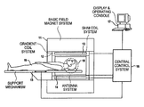
- FIG. 1 is a schematic illustration of a magnetic resonance apparatus constructed and operating in accordance with the invention.
- FIG. 2 is a flowchart for a method for the operation of a magnetic resonance apparatus in accordance with the invention.
- FIG. 3 is a field map of a coronary slice of a head used in an embodiment of the inventive method.
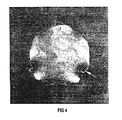
- FIG. 4 is the field map of FIG. 3 showing isometric lines of a polynomial surface obtained by curve fitting.
- FIG. 5 is an anatomical image of the coronary slice of the image of FIG. 3 .
- FIG. 6 is an anatomical image of the brain with functional information and with an area identified as containing unreliable functional information in accordance with the inventive method.
- FIG. 1 shows a sketch of a magnetic resonance apparatus.
- the apparatus has a basic field magnet system 11 for generating a basic magnetic field and a gradient coil system 12 for generating gradient fields.
- a shim coil system 13 is integrated into the gradient coil system 12 for homogenization of the basic magnetic field.
- the apparatus further has an antenna system 14 that emits radio-frequency signals into an examination subject for triggering magnetic resonance signals and picks up the magnetic resonance signals that are generated.
- the apparatus comprises a movable bearing device 15 on which the examination subject, for example a patient 19 to be examined, is placed.
- the shim coil system 13 For controlling currents in the shim coil system 13 , as well as for controlling currents in the gradient coil system 12 on the basis of a sequence, the shim coil system 13 as well as the gradient coil system 12 are connected to a central control system 16 .
- the antenna system 14 is likewise connected to the central control system 16 for controlling the radio-frequency signals to be emitted according to the sequence as well as for the further-processing and storing of the magnetic resonance signals picked up by the antenna system 14 .
- the support device 15 For controlling displacement of the movable support device 15 , for example in order to position a region of the patient 19 to be examined in an imaging volume 18 of the apparatus, the support device 15 also is connected to the central control system 16 .
- the central control system 16 is connected to a display and operating console 17 via which the inputs of an operator, for example the desired sequence type and sequence parameters, are supplied to the central control system 16 .
- the generated magnetic resonance images are displayed at the display and operating console 17 .
- FIG. 2 shows a flowchart for the inventive method for the operation of a magnetic resonance apparatus.
- the magnetic resonance apparatus shown in FIG. 1 is referenced as an example.
- the region of the patient placed on the support mechanism 15 to be imaged for example the patient's head, is positioned in the imaging volume 18 of the apparatus by displacement of the support mechanism 15 .
- a shim-setting procedure is implemented in step 21 of the flowchart of FIG. 2 .
- Shim currents are thereby determined for the shim coil system 13 and offset currents are determined for the gradient coil system 12 , for example according to a method of German PS 195 11 791, which has already been cited.
- magnetic resonance signals of the head are generated with different echo times for forming two three-dimensionally spatially resolved raw datasets, and the raw datasets are further-processed for determining corresponding shim and offset currents.
- a significantly lower resolution than for the diagnostic imaging is generally adequate.
- an anatomical magnetic resonance image of the head is generated with a high resolution and quality.
- a multi-slice technique based on spin echos, for example, is utilized, slices of the anatomical image having a resolution of 256 ⁇ 256 voxels, for example, being able to be generated therewith.
- step 23 of the flowchart a field map with respect to inhomogeneities of the basic magnetic field is generated at least for the region to be imaged, using a double echo gradient echo sequence similar to the shim-setting procedure. A high resolution similar to that for the anatomical magnetic resonance image is sought.
- step 24 of the flowchart functional images of the head, particularly of the brain, are taken by functional magnetic resonance imaging.
- three-dimensional image datasets of the head are registered with an echo planar method before and after a stimulation of the patient 19 , before and after a task carried out by the patient 19 , these being subtracted from one another for forming the aforementioned functional images.
- echo planar methods enable a very fast registration of comparatively large three-dimensional image data sets, they exhibit a comparatively poor image quality and, generally, a lower resolution of, for example, 128 ⁇ 128 ⁇ 128 voxels.
- the functional images are distortion-corrected in a further step 25 .
- the distortion correction is implemented according to the method that is described in the initially cited article by P. Jezzard.
- An unambiguous distortion correction is not possible for all sub-areas of the region to be imaged.
- the distortion is very pronounced locally, so that a registered voxel must be distortion-corrected with respect to twenty voxels, this cannot be implemented with unambiguous values for all twenty voxels.
- the reason for this is that the informational content of one registered voxel is inadequate by itself in order to be able to reconstruct the values for the twenty voxels that correspond to the actual conditions.
- the distortion-corrected functional images are superimposed on the anatomical image in a further step 26 .
- areas of the brain from which no reliable functional information can be acquired and/or for which no unambiguous distortion correction can be implemented are identified by designated areas, such as blocked-out areas. Areas from which no reliable functional information can be acquired correspond to those areas of the field map in which the inhomogeneity of the basic magnetic field exceeds a prescribable value, so that no magnetic resonance signals can be acquired from these areas with the echo planar method due to dephasing effects. Further, the areas for which no unambiguous distortion correction can be implemented can likewise be identified with the assistance of the field map. Areas of the field map in which the inhomogeneity of the basic magnetic field lies in a prescribable region thereby indicate the areas that cannot be accurately distortion-corrected.
- the steps 22 and 23 are combined, so that the anatomical image and the field map are generated simultaneously, for example with a double echo gradient echo sequence.
- the shim-setting procedure of step 21 is implemented with such a high resolution that the field map can be simultaneously derived therefrom while eliminating the step 23 .
- FIG. 3 shows a field map that is generated with the double echo gradient echo sequence.
- Respective image dataset that each contain complex quantities are registered for the two echos of the double echo gradient echo sequence that exhibit different echo times.
- the phase values of the appertaining complex quantities for corresponding data points of the two image datasets are subtracted from one another.
- the double echo gradient echo sequence is designed such that artifacts in the field map due to a chemical shift, for example due to a fat resonance, are suppressed.
- FIG. 4 shows a part of the field map of FIG. 3 essentially pertaining to the brain 60 of the head in which isomers of a polynomial surface, obtained by curve fitting, are displayed. The fitting is thereby likewise described in the initially cited article by P. Jezzard.
- P. Jezzard In order to identify the points for FIG. 4 from the field map of FIG. 3 that essentially relate to the brain 60 that are of interest for the functional magnetic resonance imaging, for example, only those data points of the image datasets are further-processed whose complex quantities exceed a prescribable limit in terms of amount.
- areas having a comparatively high inhomogeneity of the basic magnetic field are shown dark and areas having a comparatively good homogeneity are shown light.
- FIG. 5 shows an anatomical image for the coronary slice of FIG. 3, this image proceeding, for example, from one of the two image datasets.
- a white arrow marks an area in the region of an auditory canal. Due to a tissue-air boundary surface of the auditory canal, a dephasing of magnetic resonance signals occurs in this area and, thus, a signal quenching occurs in the magnetic resonance image. This area is thereby shown congruently in FIG. 4 as an area of high inhomogeneity.
- FIG. 6 shows an anatomical image of the brain 60 as a basis for the superimposition of functional images of said coronary slice of the brain 60 .
- a stimulated cerebral areola 62 is shown as the functional information.
- the area of high inhomogeneity identified according to FIG. 4 is permanently occupied with a blocked-out surface 64 .
- the blocked-out surface 64 identifies a region from which no reliable functional information can be acquired as a consequence of dephasing effects and/or ambiguous distortion correction of a functional image.
Landscapes
- Physics & Mathematics (AREA)
- Engineering & Computer Science (AREA)
- Signal Processing (AREA)
- High Energy & Nuclear Physics (AREA)
- Condensed Matter Physics & Semiconductors (AREA)
- General Physics & Mathematics (AREA)
- Magnetic Resonance Imaging Apparatus (AREA)
- Image Processing (AREA)
- Image Analysis (AREA)
Applications Claiming Priority (3)
| Application Number | Priority Date | Filing Date | Title |
|---|---|---|---|
| DE10056457A DE10056457C2 (de) | 2000-11-14 | 2000-11-14 | Verfahren zum Betrieb eines Magnetresonanzgeräts mit funktioneller Bildgebung |
| DE10056457 | 2000-11-14 | ||
| DE10056457.7 | 2000-11-14 |
Publications (2)
| Publication Number | Publication Date |
|---|---|
| US20020057086A1 US20020057086A1 (en) | 2002-05-16 |
| US6700373B2 true US6700373B2 (en) | 2004-03-02 |
Family
ID=7663298
Family Applications (1)
| Application Number | Title | Priority Date | Filing Date |
|---|---|---|---|
| US10/037,740 Expired - Lifetime US6700373B2 (en) | 2000-11-14 | 2001-11-09 | Method for operating a magnetic resonance apparatus employing a superimposed anatomical image and functional image to designate an unreliable region of the functional image |
Country Status (3)
| Country | Link |
|---|---|
| US (1) | US6700373B2 (de) |
| JP (1) | JP4046212B2 (de) |
| DE (1) | DE10056457C2 (de) |
Cited By (6)
| Publication number | Priority date | Publication date | Assignee | Title |
|---|---|---|---|---|
| US20080146914A1 (en) * | 2006-12-19 | 2008-06-19 | General Electric Company | System, method and apparatus for cancer imaging |
| US20090174405A1 (en) * | 2007-12-06 | 2009-07-09 | Yoshimori Kassai | Magnetic resonance imaging apparatus and magnetic resonance imaging method |
| US20100036233A1 (en) * | 2008-08-08 | 2010-02-11 | Michigan State University | Automatic Methods for Combining Human Facial Information with 3D Magnetic Resonance Brain Images |
| US7847552B2 (en) | 2007-01-10 | 2010-12-07 | General Electric Company | Exclusion of compromised PET data during simultaneous PET-MR acquisition |
| US9254111B2 (en) | 2012-11-27 | 2016-02-09 | General Electric Company | PET acquisition scheduling based on MR scout images |
| US9953397B2 (en) | 2014-09-05 | 2018-04-24 | General Electric Company | System and method for medical image correction |
Families Citing this family (6)
| Publication number | Priority date | Publication date | Assignee | Title |
|---|---|---|---|---|
| DE10206192B4 (de) * | 2002-02-14 | 2005-05-04 | Siemens Ag | Verfahren zum Darstellen eines Magnetresonanzbildes |
| JP4801892B2 (ja) * | 2004-09-10 | 2011-10-26 | 株式会社東芝 | 医用画像表示装置 |
| WO2007057855A2 (en) * | 2005-11-17 | 2007-05-24 | Philips Intellectual Property & Standards Gmbh | Method for displaying high resolution image data together with time-varying low resolution image data |
| US9316712B2 (en) * | 2009-04-17 | 2016-04-19 | Siemens Plc | Magnetic resonance method and apparatus using dual echoes for data acquisition |
| WO2017186522A1 (en) * | 2016-04-28 | 2017-11-02 | Koninklijke Philips N.V. | A treatment plan evaluation tool |
| WO2018036986A1 (en) * | 2016-08-25 | 2018-03-01 | Koninklijke Philips N.V. | Bo-corrected sensitivity encoding magnetic resonance imaging |
Citations (9)
| Publication number | Priority date | Publication date | Assignee | Title |
|---|---|---|---|---|
| US4558462A (en) * | 1982-09-02 | 1985-12-10 | Hitachi Medical Corporation | Apparatus for correcting image distortions automatically by inter-image processing |
| US5565777A (en) * | 1993-09-13 | 1996-10-15 | Kabushiki Kaisha Toshiba | Method/apparatus for NMR imaging using an imaging scheme sensitive to inhomogeneity and a scheme insensitive to inhomogeneity in a single imaging step |
| US5603322A (en) * | 1993-01-19 | 1997-02-18 | Mcw Research Foundation | Time course MRI imaging of brain functions |
| US5614827A (en) | 1995-03-30 | 1997-03-25 | Siemens Aktiengesellschaft | Method and apparatus for shimming a magnet system of a nuclear magnetic resonance tomography system |
| US5850486A (en) * | 1996-04-29 | 1998-12-15 | The Mclean Hospital Corporation | Registration of image data |
| US5869964A (en) * | 1993-09-14 | 1999-02-09 | Kabushiki Kaisha Toshiba | Magnetic resonance imaging apparatus in which gradient echo signals are acquired at a time distant from the center of a gradient echo |
| US6073041A (en) * | 1996-07-18 | 2000-06-06 | Regents Of The University Of Minnesota | Physiological corrections in functional magnetic resonance imaging |
| US6076004A (en) * | 1995-09-05 | 2000-06-13 | Kabushiki Kaisha Toshiba | Magnetic resonance image correction method and magnetic resonance imaging apparatus using the same |
| US6226352B1 (en) * | 1998-09-08 | 2001-05-01 | Veritas Pharmaceuticals, Inc. | System and method for radiographic imaging of tissue |
-
2000
- 2000-11-14 DE DE10056457A patent/DE10056457C2/de not_active Expired - Fee Related
-
2001
- 2001-11-09 JP JP2001344334A patent/JP4046212B2/ja not_active Expired - Fee Related
- 2001-11-09 US US10/037,740 patent/US6700373B2/en not_active Expired - Lifetime
Patent Citations (9)
| Publication number | Priority date | Publication date | Assignee | Title |
|---|---|---|---|---|
| US4558462A (en) * | 1982-09-02 | 1985-12-10 | Hitachi Medical Corporation | Apparatus for correcting image distortions automatically by inter-image processing |
| US5603322A (en) * | 1993-01-19 | 1997-02-18 | Mcw Research Foundation | Time course MRI imaging of brain functions |
| US5565777A (en) * | 1993-09-13 | 1996-10-15 | Kabushiki Kaisha Toshiba | Method/apparatus for NMR imaging using an imaging scheme sensitive to inhomogeneity and a scheme insensitive to inhomogeneity in a single imaging step |
| US5869964A (en) * | 1993-09-14 | 1999-02-09 | Kabushiki Kaisha Toshiba | Magnetic resonance imaging apparatus in which gradient echo signals are acquired at a time distant from the center of a gradient echo |
| US5614827A (en) | 1995-03-30 | 1997-03-25 | Siemens Aktiengesellschaft | Method and apparatus for shimming a magnet system of a nuclear magnetic resonance tomography system |
| US6076004A (en) * | 1995-09-05 | 2000-06-13 | Kabushiki Kaisha Toshiba | Magnetic resonance image correction method and magnetic resonance imaging apparatus using the same |
| US5850486A (en) * | 1996-04-29 | 1998-12-15 | The Mclean Hospital Corporation | Registration of image data |
| US6073041A (en) * | 1996-07-18 | 2000-06-06 | Regents Of The University Of Minnesota | Physiological corrections in functional magnetic resonance imaging |
| US6226352B1 (en) * | 1998-09-08 | 2001-05-01 | Veritas Pharmaceuticals, Inc. | System and method for radiographic imaging of tissue |
Non-Patent Citations (4)
| Title |
|---|
| "Bold Magnetic Resonance Imaging in Real Time," Thesen et al, electromedica 68, vol. 1 (2000) pp. 45-52. |
| "Correction for Geometric Distortion in Echo Planar Images from B0 Field Variations," Jezzard et al, Magnetic R Medicine, vol. 34 (1995) pp. 65-73. |
| "Funktionelle Bildgebund mit der Magnetresonanztomographie," Klose et al, electromedica 67, vol. 1 (1999) pp. 27. |
| "Segmented Spin-Echo Pulses to Increase fMRI Signal; Repeated Intrinsic Diffusional Enhancement," Song et al, Rep Intrinsic Diffusional Enhancement, Song et al, Magnetic Resonance in Medicine, vol. 42 (1999) pp. 631-635. |
Cited By (7)
| Publication number | Priority date | Publication date | Assignee | Title |
|---|---|---|---|---|
| US20080146914A1 (en) * | 2006-12-19 | 2008-06-19 | General Electric Company | System, method and apparatus for cancer imaging |
| US7847552B2 (en) | 2007-01-10 | 2010-12-07 | General Electric Company | Exclusion of compromised PET data during simultaneous PET-MR acquisition |
| US20090174405A1 (en) * | 2007-12-06 | 2009-07-09 | Yoshimori Kassai | Magnetic resonance imaging apparatus and magnetic resonance imaging method |
| US7843194B2 (en) * | 2007-12-06 | 2010-11-30 | Kabushiki Kaisha Toshiba | Magnetic resonance imaging apparatus and magnetic resonance imaging method |
| US20100036233A1 (en) * | 2008-08-08 | 2010-02-11 | Michigan State University | Automatic Methods for Combining Human Facial Information with 3D Magnetic Resonance Brain Images |
| US9254111B2 (en) | 2012-11-27 | 2016-02-09 | General Electric Company | PET acquisition scheduling based on MR scout images |
| US9953397B2 (en) | 2014-09-05 | 2018-04-24 | General Electric Company | System and method for medical image correction |
Also Published As
| Publication number | Publication date |
|---|---|
| DE10056457A1 (de) | 2002-05-23 |
| DE10056457C2 (de) | 2002-11-07 |
| JP4046212B2 (ja) | 2008-02-13 |
| JP2002204791A (ja) | 2002-07-23 |
| US20020057086A1 (en) | 2002-05-16 |
Similar Documents
| Publication | Publication Date | Title |
|---|---|---|
| US6559641B2 (en) | Method for operating a magnetic resonance tomography apparatus wherein the location coding is adapted to a positional change of the examination subject | |
| Reese et al. | Automated shimming at 1.5 T using echo‐planar image frequency maps | |
| US7683614B2 (en) | Magnetic resonance spectroscopy with sparse spectral sampling and interleaved dynamic shimming | |
| US6490472B1 (en) | MRI system and method for producing an index indicative of alzheimer's disease | |
| US5565777A (en) | Method/apparatus for NMR imaging using an imaging scheme sensitive to inhomogeneity and a scheme insensitive to inhomogeneity in a single imaging step | |
| EP1362550B1 (de) | Ganzkörper-Bildgebung mittels magnetischer Resonanz, kontinuierlich verschiebbarem Tisch und ein interaktives Steuerungssystem | |
| Kamei et al. | Neuronal current distribution imaging using magnetic resonance | |
| US8138759B2 (en) | System for adjusting a magnetic field for MR and other use | |
| KR101703382B1 (ko) | 생체 검사 대상자를 자극하는 것에 의한 검사 대상자의 사전 결정된 체적 세그먼트의 mr 영상화 방법 및 자기 공명 시스템 | |
| US9250307B2 (en) | Magnetic resonance system and method to generate diffusion information | |
| US20050264286A1 (en) | Method for acquiring magnetic resonance data from a large examination region | |
| US8536868B2 (en) | Magnetic resonance projection angiography with continuous table displacement | |
| US10031201B2 (en) | Method and apparatus for magnetic resonance imaging | |
| US11660016B2 (en) | Single-sided 3D magnet and magnetic resonance imaging (MRI) system | |
| US20070265520A1 (en) | Magnetic resonance spectroscopy with real-time correction of motion and frequency drift, and real-time shimming | |
| US6700373B2 (en) | Method for operating a magnetic resonance apparatus employing a superimposed anatomical image and functional image to designate an unreliable region of the functional image | |
| US8395387B2 (en) | Method for imaging in magnetic resonance tomography with spectral fat saturation or spectral water excitation | |
| US20010053877A1 (en) | Method for operating a magnetic resonance apparatus having an active shim system | |
| US10976397B2 (en) | MRI apparatus utilizing non-ultrashort TE(UTE) imaging to generate a mask image for performance of mask processing | |
| US8185187B2 (en) | Magnetic resonance lmethod and apparatus with gated shimming of the basic magnetic field | |
| US20110115487A1 (en) | Method and magnetic resonance system for imaging particles | |
| Pfeuffer et al. | Functional MR imaging in the awake monkey: effects of motion on dynamic off-resonance and processing strategies | |
| US20140320128A1 (en) | Method and magnetic resonance apparatus to acquire image data sets of an examination subject | |
| US20020183614A1 (en) | Magnetic resonance tomography apparatus with step-free fade between displayed different spin ensembles | |
| US9354290B2 (en) | Method and magnetic resonance system to generate an MR image with a tracking factor |
Legal Events
| Date | Code | Title | Description |
|---|---|---|---|
| AS | Assignment |
Owner name: SIEMENS AKTIENGESELLSCHAFT, GERMANY Free format text: ASSIGNMENT OF ASSIGNORS INTEREST;ASSIGNORS:MUELLER, EDGAR;THESEN, STEFAN;REEL/FRAME:012455/0844 Effective date: 20011105 |
|
| STCF | Information on status: patent grant |
Free format text: PATENTED CASE |
|
| FPAY | Fee payment |
Year of fee payment: 4 |
|
| FPAY | Fee payment |
Year of fee payment: 8 |
|
| FPAY | Fee payment |
Year of fee payment: 12 |
|
| AS | Assignment |
Owner name: SIEMENS HEALTHCARE GMBH, GERMANY Free format text: ASSIGNMENT OF ASSIGNORS INTEREST;ASSIGNOR:SIEMENS AKTIENGESELLSCHAFT;REEL/FRAME:039271/0561 Effective date: 20160610 |