US6303153B1 - Preparation of a therapeutic composition - Google Patents
Preparation of a therapeutic composition Download PDFInfo
- Publication number
- US6303153B1 US6303153B1 US09/344,095 US34409599A US6303153B1 US 6303153 B1 US6303153 B1 US 6303153B1 US 34409599 A US34409599 A US 34409599A US 6303153 B1 US6303153 B1 US 6303153B1
- Authority
- US
- United States
- Prior art keywords
- kda
- product
- molecular weights
- molecules
- nucleotides
- Prior art date
- Legal status (The legal status is an assumption and is not a legal conclusion. Google has not performed a legal analysis and makes no representation as to the accuracy of the status listed.)
- Expired - Lifetime
Links
- 239000000203 mixture Substances 0.000 title claims abstract description 27
- 230000001225 therapeutic effect Effects 0.000 title abstract description 7
- 238000002360 preparation method Methods 0.000 title description 10
- 108090000765 processed proteins & peptides Proteins 0.000 claims abstract description 22
- 239000002773 nucleotide Substances 0.000 claims abstract description 14
- 102000004196 processed proteins & peptides Human genes 0.000 claims abstract description 14
- 125000003729 nucleotide group Chemical group 0.000 claims abstract description 11
- 238000010521 absorption reaction Methods 0.000 claims abstract description 9
- 238000000034 method Methods 0.000 claims description 19
- 235000018102 proteins Nutrition 0.000 claims description 14
- 102000004169 proteins and genes Human genes 0.000 claims description 14
- 108090000623 proteins and genes Proteins 0.000 claims description 14
- XLYOFNOQVPJJNP-UHFFFAOYSA-N water Substances O XLYOFNOQVPJJNP-UHFFFAOYSA-N 0.000 claims description 14
- 239000001888 Peptone Substances 0.000 claims description 10
- 108010080698 Peptones Proteins 0.000 claims description 10
- 235000019319 peptone Nutrition 0.000 claims description 10
- 239000005018 casein Substances 0.000 claims description 8
- BECPQYXYKAMYBN-UHFFFAOYSA-N casein, tech. Chemical compound NCCCCC(C(O)=O)N=C(O)C(CC(O)=O)N=C(O)C(CCC(O)=N)N=C(O)C(CC(C)C)N=C(O)C(CCC(O)=O)N=C(O)C(CC(O)=O)N=C(O)C(CCC(O)=O)N=C(O)C(C(C)O)N=C(O)C(CCC(O)=N)N=C(O)C(CCC(O)=N)N=C(O)C(CCC(O)=N)N=C(O)C(CCC(O)=O)N=C(O)C(CCC(O)=O)N=C(O)C(COP(O)(O)=O)N=C(O)C(CCC(O)=N)N=C(O)C(N)CC1=CC=CC=C1 BECPQYXYKAMYBN-UHFFFAOYSA-N 0.000 claims description 8
- 235000021240 caseins Nutrition 0.000 claims description 8
- 235000015278 beef Nutrition 0.000 claims description 7
- 108091003079 Bovine Serum Albumin Proteins 0.000 claims description 4
- 229940098773 bovine serum albumin Drugs 0.000 claims description 4
- 108091093037 Peptide nucleic acid Proteins 0.000 claims description 3
- 108020005089 Plant RNA Proteins 0.000 claims description 3
- 238000007865 diluting Methods 0.000 claims 1
- 238000000862 absorption spectrum Methods 0.000 abstract description 5
- 208000036142 Viral infection Diseases 0.000 abstract description 4
- 230000009385 viral infection Effects 0.000 abstract description 4
- 230000004936 stimulating effect Effects 0.000 abstract description 2
- 210000000987 immune system Anatomy 0.000 abstract 1
- 230000031700 light absorption Effects 0.000 abstract 1
- 239000000047 product Substances 0.000 description 79
- HEMHJVSKTPXQMS-UHFFFAOYSA-M Sodium hydroxide Chemical compound [OH-].[Na+] HEMHJVSKTPXQMS-UHFFFAOYSA-M 0.000 description 21
- 238000002415 sodium dodecyl sulfate polyacrylamide gel electrophoresis Methods 0.000 description 18
- 239000000243 solution Substances 0.000 description 16
- 238000011282 treatment Methods 0.000 description 15
- IJGRMHOSHXDMSA-UHFFFAOYSA-N Atomic nitrogen Chemical compound N#N IJGRMHOSHXDMSA-UHFFFAOYSA-N 0.000 description 12
- QTBSBXVTEAMEQO-UHFFFAOYSA-N Acetic acid Chemical compound CC(O)=O QTBSBXVTEAMEQO-UHFFFAOYSA-N 0.000 description 11
- 206010057249 Phagocytosis Diseases 0.000 description 11
- 230000008782 phagocytosis Effects 0.000 description 11
- ZMANZCXQSJIPKH-UHFFFAOYSA-N Triethylamine Chemical compound CCN(CC)CC ZMANZCXQSJIPKH-UHFFFAOYSA-N 0.000 description 9
- 210000004027 cell Anatomy 0.000 description 9
- 239000007858 starting material Substances 0.000 description 8
- 230000000694 effects Effects 0.000 description 7
- 239000000706 filtrate Substances 0.000 description 7
- 238000004128 high performance liquid chromatography Methods 0.000 description 7
- 239000011535 reaction buffer Substances 0.000 description 7
- 102000004190 Enzymes Human genes 0.000 description 6
- 108090000790 Enzymes Proteins 0.000 description 6
- 108020002230 Pancreatic Ribonuclease Proteins 0.000 description 6
- 102000005891 Pancreatic ribonuclease Human genes 0.000 description 6
- 102000000447 Peptide-N4-(N-acetyl-beta-glucosaminyl) Asparagine Amidase Human genes 0.000 description 6
- 108010055817 Peptide-N4-(N-acetyl-beta-glucosaminyl) Asparagine Amidase Proteins 0.000 description 6
- 235000001014 amino acid Nutrition 0.000 description 6
- 229940024606 amino acid Drugs 0.000 description 6
- 150000001413 amino acids Chemical class 0.000 description 6
- 229940088598 enzyme Drugs 0.000 description 6
- 239000011261 inert gas Substances 0.000 description 6
- 238000004519 manufacturing process Methods 0.000 description 6
- 229910052757 nitrogen Inorganic materials 0.000 description 6
- 102000039446 nucleic acids Human genes 0.000 description 6
- 108020004707 nucleic acids Proteins 0.000 description 6
- 150000007523 nucleic acids Chemical class 0.000 description 6
- QKNYBSVHEMOAJP-UHFFFAOYSA-N 2-amino-2-(hydroxymethyl)propane-1,3-diol;hydron;chloride Chemical compound Cl.OCC(N)(CO)CO QKNYBSVHEMOAJP-UHFFFAOYSA-N 0.000 description 5
- 108090000317 Chymotrypsin Proteins 0.000 description 5
- 108010067770 Endopeptidase K Proteins 0.000 description 5
- 108010059712 Pronase Proteins 0.000 description 5
- 108090000631 Trypsin Proteins 0.000 description 5
- 102000004142 Trypsin Human genes 0.000 description 5
- 239000003443 antiviral agent Substances 0.000 description 5
- 229960002376 chymotrypsin Drugs 0.000 description 5
- 239000012153 distilled water Substances 0.000 description 5
- 239000012588 trypsin Substances 0.000 description 5
- 102000002260 Alkaline Phosphatase Human genes 0.000 description 4
- 108020004774 Alkaline Phosphatase Proteins 0.000 description 4
- XKRFYHLGVUSROY-UHFFFAOYSA-N Argon Chemical compound [Ar] XKRFYHLGVUSROY-UHFFFAOYSA-N 0.000 description 4
- FAPWRFPIFSIZLT-UHFFFAOYSA-M Sodium chloride Chemical compound [Na+].[Cl-] FAPWRFPIFSIZLT-UHFFFAOYSA-M 0.000 description 4
- 238000002835 absorbance Methods 0.000 description 4
- 229960000583 acetic acid Drugs 0.000 description 4
- 210000004899 c-terminal region Anatomy 0.000 description 4
- 238000006243 chemical reaction Methods 0.000 description 4
- 230000007062 hydrolysis Effects 0.000 description 4
- 238000006460 hydrolysis reaction Methods 0.000 description 4
- UXVMQQNJUSDDNG-UHFFFAOYSA-L Calcium chloride Chemical compound [Cl-].[Cl-].[Ca+2] UXVMQQNJUSDDNG-UHFFFAOYSA-L 0.000 description 3
- 238000004458 analytical method Methods 0.000 description 3
- 210000004369 blood Anatomy 0.000 description 3
- 239000008280 blood Substances 0.000 description 3
- 239000001110 calcium chloride Substances 0.000 description 3
- 229910001628 calcium chloride Inorganic materials 0.000 description 3
- 230000001925 catabolic effect Effects 0.000 description 3
- 238000005194 fractionation Methods 0.000 description 3
- 239000012634 fragment Substances 0.000 description 3
- 230000006870 function Effects 0.000 description 3
- 230000008569 process Effects 0.000 description 3
- 238000004007 reversed phase HPLC Methods 0.000 description 3
- 230000001954 sterilising effect Effects 0.000 description 3
- 238000004659 sterilization and disinfection Methods 0.000 description 3
- 238000003756 stirring Methods 0.000 description 3
- 238000012360 testing method Methods 0.000 description 3
- 108010088751 Albumins Proteins 0.000 description 2
- 102000009027 Albumins Human genes 0.000 description 2
- 239000004475 Arginine Substances 0.000 description 2
- DCXYFEDJOCDNAF-UHFFFAOYSA-N Asparagine Natural products OC(=O)C(N)CC(N)=O DCXYFEDJOCDNAF-UHFFFAOYSA-N 0.000 description 2
- 241000894006 Bacteria Species 0.000 description 2
- BHPQYMZQTOCNFJ-UHFFFAOYSA-N Calcium cation Chemical compound [Ca+2] BHPQYMZQTOCNFJ-UHFFFAOYSA-N 0.000 description 2
- WHUUTDBJXJRKMK-UHFFFAOYSA-N Glutamic acid Natural products OC(=O)C(N)CCC(O)=O WHUUTDBJXJRKMK-UHFFFAOYSA-N 0.000 description 2
- DHMQDGOQFOQNFH-UHFFFAOYSA-N Glycine Chemical compound NCC(O)=O DHMQDGOQFOQNFH-UHFFFAOYSA-N 0.000 description 2
- 239000007836 KH2PO4 Substances 0.000 description 2
- QNAYBMKLOCPYGJ-REOHCLBHSA-N L-alanine Chemical compound C[C@H](N)C(O)=O QNAYBMKLOCPYGJ-REOHCLBHSA-N 0.000 description 2
- DCXYFEDJOCDNAF-REOHCLBHSA-N L-asparagine Chemical compound OC(=O)[C@@H](N)CC(N)=O DCXYFEDJOCDNAF-REOHCLBHSA-N 0.000 description 2
- CKLJMWTZIZZHCS-REOHCLBHSA-N L-aspartic acid Chemical compound OC(=O)[C@@H](N)CC(O)=O CKLJMWTZIZZHCS-REOHCLBHSA-N 0.000 description 2
- WHUUTDBJXJRKMK-VKHMYHEASA-N L-glutamic acid Chemical compound OC(=O)[C@@H](N)CCC(O)=O WHUUTDBJXJRKMK-VKHMYHEASA-N 0.000 description 2
- ROHFNLRQFUQHCH-YFKPBYRVSA-N L-leucine Chemical compound CC(C)C[C@H](N)C(O)=O ROHFNLRQFUQHCH-YFKPBYRVSA-N 0.000 description 2
- COLNVLDHVKWLRT-QMMMGPOBSA-N L-phenylalanine Chemical compound OC(=O)[C@@H](N)CC1=CC=CC=C1 COLNVLDHVKWLRT-QMMMGPOBSA-N 0.000 description 2
- OUYCCCASQSFEME-QMMMGPOBSA-N L-tyrosine Chemical compound OC(=O)[C@@H](N)CC1=CC=C(O)C=C1 OUYCCCASQSFEME-QMMMGPOBSA-N 0.000 description 2
- ROHFNLRQFUQHCH-UHFFFAOYSA-N Leucine Natural products CC(C)CC(N)C(O)=O ROHFNLRQFUQHCH-UHFFFAOYSA-N 0.000 description 2
- KDXKERNSBIXSRK-UHFFFAOYSA-N Lysine Natural products NCCCCC(N)C(O)=O KDXKERNSBIXSRK-UHFFFAOYSA-N 0.000 description 2
- 239000004472 Lysine Substances 0.000 description 2
- TWRXJAOTZQYOKJ-UHFFFAOYSA-L Magnesium chloride Chemical compound [Mg+2].[Cl-].[Cl-] TWRXJAOTZQYOKJ-UHFFFAOYSA-L 0.000 description 2
- JLVVSXFLKOJNIY-UHFFFAOYSA-N Magnesium ion Chemical compound [Mg+2] JLVVSXFLKOJNIY-UHFFFAOYSA-N 0.000 description 2
- 102000035195 Peptidases Human genes 0.000 description 2
- 108091005804 Peptidases Proteins 0.000 description 2
- 239000004365 Protease Substances 0.000 description 2
- 240000004808 Saccharomyces cerevisiae Species 0.000 description 2
- 108010022999 Serine Proteases Proteins 0.000 description 2
- 102000012479 Serine Proteases Human genes 0.000 description 2
- BQCADISMDOOEFD-UHFFFAOYSA-N Silver Chemical compound [Ag] BQCADISMDOOEFD-UHFFFAOYSA-N 0.000 description 2
- 241000700584 Simplexvirus Species 0.000 description 2
- ISAKRJDGNUQOIC-UHFFFAOYSA-N Uracil Chemical compound O=C1C=CNC(=O)N1 ISAKRJDGNUQOIC-UHFFFAOYSA-N 0.000 description 2
- DRTQHJPVMGBUCF-XVFCMESISA-N Uridine Chemical group O[C@@H]1[C@H](O)[C@@H](CO)O[C@H]1N1C(=O)NC(=O)C=C1 DRTQHJPVMGBUCF-XVFCMESISA-N 0.000 description 2
- OIRDTQYFTABQOQ-KQYNXXCUSA-N adenosine Chemical compound C1=NC=2C(N)=NC=NC=2N1[C@@H]1O[C@H](CO)[C@@H](O)[C@H]1O OIRDTQYFTABQOQ-KQYNXXCUSA-N 0.000 description 2
- 235000004279 alanine Nutrition 0.000 description 2
- 230000004075 alteration Effects 0.000 description 2
- 239000003708 ampul Substances 0.000 description 2
- ODKSFYDXXFIFQN-UHFFFAOYSA-N arginine Natural products OC(=O)C(N)CCCNC(N)=N ODKSFYDXXFIFQN-UHFFFAOYSA-N 0.000 description 2
- 229910052786 argon Inorganic materials 0.000 description 2
- 235000009582 asparagine Nutrition 0.000 description 2
- 229960001230 asparagine Drugs 0.000 description 2
- 235000003704 aspartic acid Nutrition 0.000 description 2
- OQFSQFPPLPISGP-UHFFFAOYSA-N beta-carboxyaspartic acid Natural products OC(=O)C(N)C(C(O)=O)C(O)=O OQFSQFPPLPISGP-UHFFFAOYSA-N 0.000 description 2
- 230000008827 biological function Effects 0.000 description 2
- 230000008859 change Effects 0.000 description 2
- 239000003153 chemical reaction reagent Substances 0.000 description 2
- 239000003795 chemical substances by application Substances 0.000 description 2
- 238000007796 conventional method Methods 0.000 description 2
- 238000001914 filtration Methods 0.000 description 2
- 238000001502 gel electrophoresis Methods 0.000 description 2
- 238000002523 gelfiltration Methods 0.000 description 2
- 239000011521 glass Substances 0.000 description 2
- 235000013922 glutamic acid Nutrition 0.000 description 2
- 229960002989 glutamic acid Drugs 0.000 description 2
- 239000004220 glutamic acid Substances 0.000 description 2
- UYTPUPDQBNUYGX-UHFFFAOYSA-N guanine Chemical group O=C1NC(N)=NC2=C1N=CN2 UYTPUPDQBNUYGX-UHFFFAOYSA-N 0.000 description 2
- 230000001965 increasing effect Effects 0.000 description 2
- 230000014759 maintenance of location Effects 0.000 description 2
- 229930182817 methionine Natural products 0.000 description 2
- 229910000402 monopotassium phosphate Inorganic materials 0.000 description 2
- 239000002777 nucleoside Substances 0.000 description 2
- 125000003835 nucleoside group Chemical group 0.000 description 2
- 230000000242 pagocytic effect Effects 0.000 description 2
- COLNVLDHVKWLRT-UHFFFAOYSA-N phenylalanine Natural products OC(=O)C(N)CC1=CC=CC=C1 COLNVLDHVKWLRT-UHFFFAOYSA-N 0.000 description 2
- 239000011148 porous material Substances 0.000 description 2
- GNSKLFRGEWLPPA-UHFFFAOYSA-M potassium dihydrogen phosphate Chemical compound [K+].OP(O)([O-])=O GNSKLFRGEWLPPA-UHFFFAOYSA-M 0.000 description 2
- 235000019419 proteases Nutrition 0.000 description 2
- 239000002510 pyrogen Substances 0.000 description 2
- 238000012163 sequencing technique Methods 0.000 description 2
- 229910052709 silver Inorganic materials 0.000 description 2
- 239000004332 silver Substances 0.000 description 2
- 239000011780 sodium chloride Substances 0.000 description 2
- OUYCCCASQSFEME-UHFFFAOYSA-N tyrosine Natural products OC(=O)C(N)CC1=CC=C(O)C=C1 OUYCCCASQSFEME-UHFFFAOYSA-N 0.000 description 2
- 239000008215 water for injection Substances 0.000 description 2
- UHDGCWIWMRVCDJ-UHFFFAOYSA-N 1-beta-D-Xylofuranosyl-NH-Cytosine Natural products O=C1N=C(N)C=CN1C1C(O)C(O)C(CO)O1 UHDGCWIWMRVCDJ-UHFFFAOYSA-N 0.000 description 1
- QFVHZQCOUORWEI-UHFFFAOYSA-N 4-[(4-anilino-5-sulfonaphthalen-1-yl)diazenyl]-5-hydroxynaphthalene-2,7-disulfonic acid Chemical compound C=12C(O)=CC(S(O)(=O)=O)=CC2=CC(S(O)(=O)=O)=CC=1N=NC(C1=CC=CC(=C11)S(O)(=O)=O)=CC=C1NC1=CC=CC=C1 QFVHZQCOUORWEI-UHFFFAOYSA-N 0.000 description 1
- 229930024421 Adenine Natural products 0.000 description 1
- 229920000936 Agarose Polymers 0.000 description 1
- 208000002109 Argyria Diseases 0.000 description 1
- 108090000145 Bacillolysin Proteins 0.000 description 1
- 206010004146 Basal cell carcinoma Diseases 0.000 description 1
- 108010017384 Blood Proteins Proteins 0.000 description 1
- 102000004506 Blood Proteins Human genes 0.000 description 1
- 241000283690 Bos taurus Species 0.000 description 1
- 239000002126 C01EB10 - Adenosine Substances 0.000 description 1
- 102000019034 Chemokines Human genes 0.000 description 1
- 108010012236 Chemokines Proteins 0.000 description 1
- UHDGCWIWMRVCDJ-PSQAKQOGSA-N Cytidine Natural products O=C1N=C(N)C=CN1[C@@H]1[C@@H](O)[C@@H](O)[C@H](CO)O1 UHDGCWIWMRVCDJ-PSQAKQOGSA-N 0.000 description 1
- 229920002307 Dextran Polymers 0.000 description 1
- 208000000655 Distemper Diseases 0.000 description 1
- 108010059378 Endopeptidases Proteins 0.000 description 1
- 102000005593 Endopeptidases Human genes 0.000 description 1
- 102000002494 Endoribonucleases Human genes 0.000 description 1
- 108010093099 Endoribonucleases Proteins 0.000 description 1
- 102000018389 Exopeptidases Human genes 0.000 description 1
- 108010091443 Exopeptidases Proteins 0.000 description 1
- 239000004471 Glycine Substances 0.000 description 1
- 102000003886 Glycoproteins Human genes 0.000 description 1
- 108090000288 Glycoproteins Proteins 0.000 description 1
- 108010022901 Heparin Lyase Proteins 0.000 description 1
- 206010020751 Hypersensitivity Diseases 0.000 description 1
- XQFRJNBWHJMXHO-RRKCRQDMSA-N IDUR Chemical compound C1[C@H](O)[C@@H](CO)O[C@H]1N1C(=O)NC(=O)C(I)=C1 XQFRJNBWHJMXHO-RRKCRQDMSA-N 0.000 description 1
- 102000008070 Interferon-gamma Human genes 0.000 description 1
- 108010074328 Interferon-gamma Proteins 0.000 description 1
- 102000000589 Interleukin-1 Human genes 0.000 description 1
- 108010002352 Interleukin-1 Proteins 0.000 description 1
- 102000004889 Interleukin-6 Human genes 0.000 description 1
- 108090001005 Interleukin-6 Proteins 0.000 description 1
- 238000007696 Kjeldahl method Methods 0.000 description 1
- ONIBWKKTOPOVIA-BYPYZUCNSA-N L-Proline Chemical compound OC(=O)[C@@H]1CCCN1 ONIBWKKTOPOVIA-BYPYZUCNSA-N 0.000 description 1
- AGPKZVBTJJNPAG-WHFBIAKZSA-N L-isoleucine Chemical compound CC[C@H](C)[C@H](N)C(O)=O AGPKZVBTJJNPAG-WHFBIAKZSA-N 0.000 description 1
- FFEARJCKVFRZRR-BYPYZUCNSA-N L-methionine Chemical compound CSCC[C@H](N)C(O)=O FFEARJCKVFRZRR-BYPYZUCNSA-N 0.000 description 1
- QIVBCDIJIAJPQS-VIFPVBQESA-N L-tryptophane Chemical compound C1=CC=C2C(C[C@H](N)C(O)=O)=CNC2=C1 QIVBCDIJIAJPQS-VIFPVBQESA-N 0.000 description 1
- KZSNJWFQEVHDMF-BYPYZUCNSA-N L-valine Chemical compound CC(C)[C@H](N)C(O)=O KZSNJWFQEVHDMF-BYPYZUCNSA-N 0.000 description 1
- 108010006035 Metalloproteases Proteins 0.000 description 1
- 102000005741 Metalloproteases Human genes 0.000 description 1
- XUYPXLNMDZIRQH-LURJTMIESA-N N-acetyl-L-methionine Chemical compound CSCC[C@@H](C(O)=O)NC(C)=O XUYPXLNMDZIRQH-LURJTMIESA-N 0.000 description 1
- 102000035092 Neutral proteases Human genes 0.000 description 1
- 108091005507 Neutral proteases Proteins 0.000 description 1
- 102000011931 Nucleoproteins Human genes 0.000 description 1
- 108010061100 Nucleoproteins Proteins 0.000 description 1
- 229910019142 PO4 Inorganic materials 0.000 description 1
- 241001631646 Papillomaviridae Species 0.000 description 1
- 102000004160 Phosphoric Monoester Hydrolases Human genes 0.000 description 1
- 108090000608 Phosphoric Monoester Hydrolases Proteins 0.000 description 1
- ONIBWKKTOPOVIA-UHFFFAOYSA-N Proline Natural products OC(=O)C1CCCN1 ONIBWKKTOPOVIA-UHFFFAOYSA-N 0.000 description 1
- 241000015473 Schizothorax griseus Species 0.000 description 1
- MTCFGRXMJLQNBG-UHFFFAOYSA-N Serine Natural products OCC(N)C(O)=O MTCFGRXMJLQNBG-UHFFFAOYSA-N 0.000 description 1
- 108010071390 Serum Albumin Proteins 0.000 description 1
- 102000007562 Serum Albumin Human genes 0.000 description 1
- AYFVYJQAPQTCCC-UHFFFAOYSA-N Threonine Natural products CC(O)C(N)C(O)=O AYFVYJQAPQTCCC-UHFFFAOYSA-N 0.000 description 1
- 239000004473 Threonine Substances 0.000 description 1
- QIVBCDIJIAJPQS-UHFFFAOYSA-N Tryptophan Natural products C1=CC=C2C(CC(N)C(O)=O)=CNC2=C1 QIVBCDIJIAJPQS-UHFFFAOYSA-N 0.000 description 1
- KZSNJWFQEVHDMF-UHFFFAOYSA-N Valine Natural products CC(C)C(N)C(O)=O KZSNJWFQEVHDMF-UHFFFAOYSA-N 0.000 description 1
- 241000700605 Viruses Species 0.000 description 1
- 229960000643 adenine Drugs 0.000 description 1
- 229960005305 adenosine Drugs 0.000 description 1
- GFFGJBXGBJISGV-UHFFFAOYSA-N adenyl group Chemical group N1=CN=C2N=CNC2=C1N GFFGJBXGBJISGV-UHFFFAOYSA-N 0.000 description 1
- 125000001931 aliphatic group Chemical group 0.000 description 1
- 208000026935 allergic disease Diseases 0.000 description 1
- 125000003277 amino group Chemical group 0.000 description 1
- 230000002052 anaphylactic effect Effects 0.000 description 1
- 230000000840 anti-viral effect Effects 0.000 description 1
- 230000000890 antigenic effect Effects 0.000 description 1
- 125000003118 aryl group Chemical group 0.000 description 1
- 238000003556 assay Methods 0.000 description 1
- QLTSDROPCWIKKY-PMCTYKHCSA-N beta-D-glucosaminyl-(1->4)-beta-D-glucosamine Chemical group O[C@@H]1[C@@H](N)[C@H](O)O[C@H](CO)[C@H]1O[C@H]1[C@H](N)[C@@H](O)[C@H](O)[C@@H](CO)O1 QLTSDROPCWIKKY-PMCTYKHCSA-N 0.000 description 1
- DRTQHJPVMGBUCF-PSQAKQOGSA-N beta-L-uridine Natural products O[C@H]1[C@@H](O)[C@H](CO)O[C@@H]1N1C(=O)NC(=O)C=C1 DRTQHJPVMGBUCF-PSQAKQOGSA-N 0.000 description 1
- 230000015572 biosynthetic process Effects 0.000 description 1
- 229910001424 calcium ion Inorganic materials 0.000 description 1
- 238000004364 calculation method Methods 0.000 description 1
- 244000309466 calf Species 0.000 description 1
- 208000014058 canine distemper Diseases 0.000 description 1
- 150000001720 carbohydrates Chemical class 0.000 description 1
- 235000014633 carbohydrates Nutrition 0.000 description 1
- 125000003178 carboxy group Chemical group [H]OC(*)=O 0.000 description 1
- -1 carboypepsidases Proteins 0.000 description 1
- 238000012512 characterization method Methods 0.000 description 1
- 150000001875 compounds Chemical class 0.000 description 1
- UHDGCWIWMRVCDJ-ZAKLUEHWSA-N cytidine Chemical group O=C1N=C(N)C=CN1[C@H]1[C@H](O)[C@@H](O)[C@H](CO)O1 UHDGCWIWMRVCDJ-ZAKLUEHWSA-N 0.000 description 1
- 230000007812 deficiency Effects 0.000 description 1
- 238000013461 design Methods 0.000 description 1
- AIUDWMLXCFRVDR-UHFFFAOYSA-N dimethyl 2-(3-ethyl-3-methylpentyl)propanedioate Chemical class CCC(C)(CC)CCC(C(=O)OC)C(=O)OC AIUDWMLXCFRVDR-UHFFFAOYSA-N 0.000 description 1
- 201000010099 disease Diseases 0.000 description 1
- 208000037265 diseases, disorders, signs and symptoms Diseases 0.000 description 1
- BNIILDVGGAEEIG-UHFFFAOYSA-L disodium hydrogen phosphate Chemical compound [Na+].[Na+].OP([O-])([O-])=O BNIILDVGGAEEIG-UHFFFAOYSA-L 0.000 description 1
- 229910000397 disodium phosphate Inorganic materials 0.000 description 1
- 239000003480 eluent Substances 0.000 description 1
- 229940066758 endopeptidases Drugs 0.000 description 1
- 238000005516 engineering process Methods 0.000 description 1
- 230000002708 enhancing effect Effects 0.000 description 1
- 230000006862 enzymatic digestion Effects 0.000 description 1
- 210000003743 erythrocyte Anatomy 0.000 description 1
- ZMMJGEGLRURXTF-UHFFFAOYSA-N ethidium bromide Chemical compound [Br-].C12=CC(N)=CC=C2C2=CC=C(N)C=C2[N+](CC)=C1C1=CC=CC=C1 ZMMJGEGLRURXTF-UHFFFAOYSA-N 0.000 description 1
- 229960005542 ethidium bromide Drugs 0.000 description 1
- 238000000684 flow cytometry Methods 0.000 description 1
- 239000012530 fluid Substances 0.000 description 1
- 238000001641 gel filtration chromatography Methods 0.000 description 1
- 239000012362 glacial acetic acid Substances 0.000 description 1
- 208000005252 hepatitis A Diseases 0.000 description 1
- 208000002672 hepatitis B Diseases 0.000 description 1
- HNDVDQJCIGZPNO-UHFFFAOYSA-N histidine Natural products OC(=O)C(N)CC1=CN=CN1 HNDVDQJCIGZPNO-UHFFFAOYSA-N 0.000 description 1
- 230000002209 hydrophobic effect Effects 0.000 description 1
- 230000009610 hypersensitivity Effects 0.000 description 1
- 230000008105 immune reaction Effects 0.000 description 1
- 238000011534 incubation Methods 0.000 description 1
- 208000015181 infectious disease Diseases 0.000 description 1
- 206010022000 influenza Diseases 0.000 description 1
- 239000004615 ingredient Substances 0.000 description 1
- 239000003112 inhibitor Substances 0.000 description 1
- 238000002347 injection Methods 0.000 description 1
- 239000007924 injection Substances 0.000 description 1
- 229960003130 interferon gamma Drugs 0.000 description 1
- 229940100601 interleukin-6 Drugs 0.000 description 1
- AGPKZVBTJJNPAG-UHFFFAOYSA-N isoleucine Natural products CCC(C)C(N)C(O)=O AGPKZVBTJJNPAG-UHFFFAOYSA-N 0.000 description 1
- 229960000310 isoleucine Drugs 0.000 description 1
- 238000011068 loading method Methods 0.000 description 1
- 229910001629 magnesium chloride Inorganic materials 0.000 description 1
- 229910001425 magnesium ion Inorganic materials 0.000 description 1
- 239000003550 marker Substances 0.000 description 1
- 239000000463 material Substances 0.000 description 1
- 238000005259 measurement Methods 0.000 description 1
- 230000015286 negative regulation of phagocytosis Effects 0.000 description 1
- 230000007935 neutral effect Effects 0.000 description 1
- 231100000252 nontoxic Toxicity 0.000 description 1
- 230000003000 nontoxic effect Effects 0.000 description 1
- 229920001542 oligosaccharide Polymers 0.000 description 1
- 150000002482 oligosaccharides Chemical class 0.000 description 1
- 239000002245 particle Substances 0.000 description 1
- 239000000863 peptide conjugate Substances 0.000 description 1
- 229940066779 peptones Drugs 0.000 description 1
- 238000000053 physical method Methods 0.000 description 1
- 239000002244 precipitate Substances 0.000 description 1
- 238000003133 propidium iodide exclusion Methods 0.000 description 1
- 238000003908 quality control method Methods 0.000 description 1
- 239000010453 quartz Substances 0.000 description 1
- 229920002477 rna polymer Polymers 0.000 description 1
- 230000035945 sensitivity Effects 0.000 description 1
- 210000002966 serum Anatomy 0.000 description 1
- VYPSYNLAJGMNEJ-UHFFFAOYSA-N silicon dioxide Inorganic materials O=[Si]=O VYPSYNLAJGMNEJ-UHFFFAOYSA-N 0.000 description 1
- 239000011734 sodium Substances 0.000 description 1
- 238000001228 spectrum Methods 0.000 description 1
- 238000010186 staining Methods 0.000 description 1
- 229910001220 stainless steel Inorganic materials 0.000 description 1
- 239000010935 stainless steel Substances 0.000 description 1
- 238000007619 statistical method Methods 0.000 description 1
- 239000000126 substance Substances 0.000 description 1
- 238000006467 substitution reaction Methods 0.000 description 1
- 239000000725 suspension Substances 0.000 description 1
- 238000003786 synthesis reaction Methods 0.000 description 1
- 210000001541 thymus gland Anatomy 0.000 description 1
- 210000001519 tissue Anatomy 0.000 description 1
- 241000701161 unidentified adenovirus Species 0.000 description 1
- 241000712461 unidentified influenza virus Species 0.000 description 1
- 229940035893 uracil Drugs 0.000 description 1
- DRTQHJPVMGBUCF-UHFFFAOYSA-N uracil arabinoside Natural products OC1C(O)C(CO)OC1N1C(=O)NC(=O)C=C1 DRTQHJPVMGBUCF-UHFFFAOYSA-N 0.000 description 1
- 229940045145 uridine Drugs 0.000 description 1
- 239000004474 valine Substances 0.000 description 1
- 230000003612 virological effect Effects 0.000 description 1
Images


Classifications
-
- A—HUMAN NECESSITIES
- A61—MEDICAL OR VETERINARY SCIENCE; HYGIENE
- A61K—PREPARATIONS FOR MEDICAL, DENTAL OR TOILETRY PURPOSES
- A61K38/00—Medicinal preparations containing peptides
- A61K38/01—Hydrolysed proteins; Derivatives thereof
- A61K38/012—Hydrolysed proteins; Derivatives thereof from animals
- A61K38/014—Hydrolysed proteins; Derivatives thereof from animals from connective tissue peptides, e.g. gelatin, collagen
-
- A—HUMAN NECESSITIES
- A61—MEDICAL OR VETERINARY SCIENCE; HYGIENE
- A61K—PREPARATIONS FOR MEDICAL, DENTAL OR TOILETRY PURPOSES
- A61K38/00—Medicinal preparations containing peptides
- A61K38/01—Hydrolysed proteins; Derivatives thereof
- A61K38/011—Hydrolysed proteins; Derivatives thereof from plants
-
- A—HUMAN NECESSITIES
- A61—MEDICAL OR VETERINARY SCIENCE; HYGIENE
- A61K—PREPARATIONS FOR MEDICAL, DENTAL OR TOILETRY PURPOSES
- A61K38/00—Medicinal preparations containing peptides
- A61K38/01—Hydrolysed proteins; Derivatives thereof
- A61K38/012—Hydrolysed proteins; Derivatives thereof from animals
- A61K38/017—Hydrolysed proteins; Derivatives thereof from animals from blood
-
- A—HUMAN NECESSITIES
- A61—MEDICAL OR VETERINARY SCIENCE; HYGIENE
- A61K—PREPARATIONS FOR MEDICAL, DENTAL OR TOILETRY PURPOSES
- A61K38/00—Medicinal preparations containing peptides
- A61K38/01—Hydrolysed proteins; Derivatives thereof
- A61K38/012—Hydrolysed proteins; Derivatives thereof from animals
- A61K38/018—Hydrolysed proteins; Derivatives thereof from animals from milk
-
- A—HUMAN NECESSITIES
- A61—MEDICAL OR VETERINARY SCIENCE; HYGIENE
- A61P—SPECIFIC THERAPEUTIC ACTIVITY OF CHEMICAL COMPOUNDS OR MEDICINAL PREPARATIONS
- A61P31/00—Antiinfectives, i.e. antibiotics, antiseptics, chemotherapeutics
- A61P31/12—Antivirals
Definitions
- the present invention relates to an improved method of making a therapeutic composition, Product R 1 , as hereinafter defined, which contains peptides and nucleotides.
- Product R 1 contains peptides and nucleotides.
- the components of Product R have molecular weights not more than 14 kilodaltons (KDa).
- Product R was used as synonyms of RETICULOSE in some literature.
- Product R and RETICULOSE represent two distinct products.
- an antiviral agent composed of peptones, peptides, proteins and nucleic acid was originated in 1934. After some years of experimentation, such an antiviral agent was modified by using bovine serum albumin in combination with peptone, and ribonucleic acid to produce an antiviral biotic agent which is nontoxic, free from anaphylactogenic properties and is miscible with tissue fluids and blood sera.
- RETICULOSE® was reported as an antiviral agent for treating a variety of human viral infections, such as influenza, herpes, hepatitis A and B. It was then assumed that RETICULOSE® acts as an antiviral agent at least by increasing leukogenesis, synthesis of antibodies and enhancing phagocytosis. RETICULOSE® was last sold in the United States in 1964.
- the starting materials for making RETICULOSE® consist of, by weight, 40-50% of casein, 1-10% of blood albumin, 15-40% of beef peptone, 10-25% of RNA and 5-25% of sodium hydroxide. These starting materials are suspended in water which yields a ratio of proteins (casein, peptone and blood albumin) to water equals to about 4.3 to about 100 by weight. After an autoclaving treatment of the mixture of the starting materials, the resulting solution is filtered and pH is adjusted to approximately 8.5 and then to 7.8, after which the neutralized solution is filtered again. The pH is further adjusted to approximately 7.5 after the solution is diluted. Such process yields a mixture of peptides and nucleic acids having molecular weights in a range of approximately 1 to 25 KDa.
- the components over 15 KDa of the conventional composition of RETICULOSE® are more effective in treating viral diseases such as HIV, influenza virus, herpes simplex virus, etc. while the components in a range of approximately 1 to 15 KDa function as phagocytosis inhibitors.
- the conventional methods suffers from several disadvantages: 1) the method does not ensure that each preparation produces the finished components having the same ratio, thereby the product is not reproducible; 2) the conventional method produces a wide range of the finished components, which makes the quality control of the preparation extremely difficult, if possible, because too many parameters need to be determined; 3) the presence of the higher molecular weight components, such as 25 KDa component, essentially peptides, increases the risk of hypersensitivity or immune reaction and renders the product less stable.
- an object of the present invention is directed to a novel therapeutic composition, Product R.
- Product R is reproducible, highly stable and non-antigenic.
- Product R is a wide range antiviral agent for treating viral infections such as infections of human immnunodeficiency virus (HIV), herpes simplex virus, adenovirus and papilloma virus.
- HIV human immnunodeficiency virus
- Product R has been proven to be effective in stimulating the production of chemokines including interferon-gamma, interleukin-6 and interleukin-1 (J. Investig Med 1996; 44:347-351), the production of red blood cells (U.S. Pat. No. 5,807,839), treating basal cell carcinoma (U.S. Pat. No. 5,902,786) and treating canine distemper viral infections (U.S. Pat. No. 5,807,840).
- chemokines including interferon-gamma, interleukin-6 and interleukin-1 (J
- Another object of the present invention is directed to an improved method for making the novel therapeutic composition Product R.
- Product R according to the present improved method comprises novel components that generate a novel UV absorption spectrum and a novel molecular weight profile.
- Product R comprises molecules having molecular weights not more than 14 KDa.
- a further object of the present invention is to define the components of Product R based on chemical and physical methods.
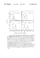
- FIG. 1 shows a representative ultraviolet absorption profile of Product R
- FIG. 2 shows a representative chromatogram of Product R obtained form a reverse phase HPLC analysis
- FIG. 3 shows a BioGel P-2 fractionation profile of Product R
- FIG. 4 shows the components of fraction I of the BioGel P-2 fractionation profile resolved on a 16% of SDS-Polyacrylamide gel electrophoresis (SDS-PAGE);
- FIG. 5 shows the relative mass (Mr.) of the two major peptide components of Product R resolved on a 16% SDS-PAGE
- FIG. 6 is a 16% SDS-PAGE, showing the effects of a variety of catabolic enzymes on Product R;
- FIG. 7 is a flow cytometric histograms, showing the effect of Product R on phagocytosis of Dextran-FITC.
- FIG. 8 is a flow cytometric histograms, showing the effect of Product R on phagocytosis of Dextran-BoDipyFL.
- Product R is prepared according to the following manner.
- the starting materials casein, beef peptone, RNA, BSA, and sodium hydroxide are suspended in proportions of, by weight, 35-50% (casein), 15-40% (beef peptone), 10-25% (RNA), 1-10% (BSA) and 5-25% (sodium hydroxide) in an appropriate volume of distilled water. All starting materials are generally available or otherwise can be readily prepared by a person of ordinary skill in the art. While any RNA is suitable for the intended purpose of the present invention, plant RNA is preferred and yeast RNA is the most preferred.
- the ratio of total proteins versus the volume of distilled water is generally about 1.5-2.5 to about 100 by weight, preferably about 2.2 to about 100 by weight. This means that every 1.5-2.5 grams of the total proteins are suspended in about 100 milliliters of distilled water.
- the suspension as prepared above is then autoclaved at a pressure of approximately 5-15 lbs., preferably 8-10 lbs. under an elevated temperature in a range, for example about 150°-300° F. preferably about 200°-230° F. over a period of approximately 2-10 hours, preferably more than 3 hours.
- RNA may be completely hydrolyzed into nucleotides.
- the solution is cooled down to room temperature, and then allowed to stay at a temperature of 3° to 8° C. for at least 12 hours to precipitate insoluble elements. Alternatively, the cooled solution may be centrifuged at a temperature below 8° C. to remove the precipitates.
- the resulting solution is then filtered through a 2 micron and a 0.45 micron filters under an inert gas such as nitrogen or argon at a pressure of about 1-6 psi.
- an inert gas such as nitrogen or argon at a pressure of about 1-6 psi.
- the solution is filtered again through a pyrogen retention filter, preferably 0.2 micron.
- the solution may be cooled at 3 to 8° C. again for at least about 12 hours and filtered again in the same way as described above.
- the resulting filtrate is then assayed for total nitrogen content using methods known to a person of ordinary skill in the art such as Kjeldahl method, J. G. C. D. Kjeldahl, Z. Anal. Chem., Vol. 22, p366 (1883), and its improvements. Based on the assay, the filtrate is then diluted with chilled distilled water to an appropriate volume having a preferred total nitrogen content ranging from 165 to 210 mg/ml.
- the pH of the diluted solution is then adjusted with HCl to a physiologically acceptable pH, preferably to about 7.3 to 7.6, after which the diluted solution is filtered again through a 0.2 micron filter under an inert gas as described above.
- Product R so produced contains essentially nucleotides, nucleosides and free nucleic acid bases of low molecular weights from a complete hydrolysis of RNA and small peptides from partial hydrolysis of the proteins. It is possible that the base hydrolysis of the proteins also produces free amino acids.
- filtration technique is essentially to remove bacteria or other particles having similar size to or larger size than bacteria.
- any filter regardless its manufacturer or material from which it is made is suitable for the intended purpose. All filters used in the present process are widely available to a person of ordinary skill in the art.
- the final filtrate is then filled and sealed into appropriate vials, such as 2 ml or 10 ml glass vials under an inert gas.
- appropriate vials such as 2 ml or 10 ml glass vials under an inert gas.
- the filled vials are autoclaved for final sterilization, after which they are ready for use.
- Product R is administered parenterally or topically to a patient in need as described in U.S. Pat. Nos. 5,807,839, 5,807,840 and 5,902,786, the contents of which are herein incorporated by reference in their entirety.
- FIG. 1 is a representative ultraviolet absorption spectrum of Product R measured in 1 cm path length quartz microcuvette (100 ⁇ l capacity) using a Shimadzu Model UV-1201 UV-VIS Spectrophotometer.
- Product R was diluted with distilled water by 100 fold.
- the spectrum is recorded between 220-320 nm and shows a maximum absorption at 260 nm and a trough at 235 nm.
- the ratio of the absorbance (A) at 260 nm over absorbance at 280 nm is 1.998 ( ⁇ 10%), and A at 260 nm over A at 230 nm is 1.359 ( ⁇ 10%).
- FIG. 2 is a representative chromatogram of Product R obtained from a reverse phase HPLC analysis using a Hewlett Packard 1100 HPLC system (Hewlett Packard Co.) that includes a binary pump (Model G1312A), a diode array detector (Model G1315A), a column thermostat (Model G 1316A), a thermostatted autosampler (Model G1329A), a sample thermostat and a vacuum degasser (Model G 1322A); and a stainless steel YMC-pack ODS-AQ S-5 uM column (YMC, Inc. 3223 Burnt Mill Dr., Wilmington, N.C. 28403) that has a size of 250 ⁇ 10 mm ID and pore size 120 A.
- the mobile phase consisting of a 0.1 M acetic acid: trietheylamine is prepared as follows: 6.0 ml of glacial acetic acid are dissolved in 1000 ml of HPLC grade water. The stirred solution of acetic acid is titrated with triethylamine to pH 4.8. The solution is allowed to equilibrate overnight at room temperature and then filtered through a 0.45 ⁇ M pore size and 52 mm diameter filter. The pH of the solution is readjusted to pH 4.8 if necessary with the addition of triethylamine prior to use. The mobile phase is degassed by the vacuum degasser built into the HPLC flow system.
- FIG. 3 shows a fractionation profile of Product R on a BioGel P-2 (Bio-Rad Laboratories Inc.) column having a size of 2.6 cm ⁇ 55 cm packed size.
- the column is eluted with a 0.1X PBS, preferably DULBECCO's PBS, free of calcium ion (Ca ++ ) and magnesium ion (Mg ++ ), at a flow rate of 0.5 milliliters per minute.
- 1X PBS contains 1.47 mM KH 2 PO 4 , 2.67 mM KCI, 138 mM NaCl and 8.1 mM Na 2 HPO 4 7H 2 O.
- the eluent passes through a “Uvcord SII” monitor, which is attached to a REC 101 chart recorder and fitted with a 254 nm filter, and is collected at 12 minutes per fraction in a “Frac 200” fraction collector.
- the gel filtration chromatography under such conditions results in 9 fractions: I, Ia, II, IIa, IIb, III, Illa, IV and IVa.
- Each individual peak is compared with known nucleotides, nucleosides and free nucleic acid bases eluted at the same or very close to the volumes of respective fractions as shown in TABLE I. Known compounds having comparable values are shown in Remarks column.
- fractions are then concentrated and analyzed by SDS-PAGE (see the following) on a 16% gel.
- Silver staining of the gel demonstrates that only fraction I shows essentially two major silverstainable bands having apparent molecular weights of 4.3 KDa, 5.2 KDa and a minor 7.6 KDa band as shown in FIG. 4 .
- FIG. 5 shows the relative mass (measurement of molecular weight) of the two major peptide components of Product R resolved on a 16% SDS-Polyacrylamide gel electrophoresis (SDS-PAGE) and stained by silver stain using ‘SilverXpress’staining kit from NOVEX, following manufacturer-suggested protocol.
- Product R is resolved into two major silverstainable bands having apparent molecular weight of about 4.3 and about 5.2 KDa.
- a minor silverstainable component having molecular weight of about 7.6 KDa is also visible on an overloaded SDS-PAGE gel, and there may be trace amounts of other silverstainable peptides having molecular weights ranging from about 5 KDa to about 14 KDa.
- Coomassie Blue a universal protein stain, stains the 4.3 KDa band extremely poorly.
- Product R consists essentially of molecules having molecular weights below 8 KDa.
- TABLE II shows the amino acid compositions of the 5.2 KDa and the 4.3 KDa components.
- Amino acid analysis of the 5.2 KDa band (sample A) and the 4.3 KDa band (sample B) was performed on a PE Bio-system 420 analyzer with automatic hydrolysis using standard pheno-iso-thiocyanite (PTIC) chemistry.
- PTIC pheno-iso-thiocyanite
- Proteinase K is a non-specific broad spectrum protease that cleaves peptide bonds at the C-terminal of aliphatic, aromatic and hydrophobic amino acids. It may cleave all serum peptides completely at 50 ⁇ g/ml within one hour.
- a Product R sample is incubated in a reaction buffer having 10 mM Tris-HCl, pH 7.6; 0.5% of SDS; 1 mM CaCl 2; 100 ⁇ g/ml of proteinase K at 40° C. for 30 minutes and then subject to SDS-PAGE on a 16% gel as described above. Under such condition, the silver stain of Product R does not show significant change. However, when the amount of proteinase K is increased to 800 ⁇ g/ml and the incubation time is extended to one hour, the 5.2 KDa band disappears but there is no obvious change of the 4.3 KDa band.
- Trypsin is a serine protease, which specifically cleaves peptide bonds of lysine and arginine at the C-terminal at pH 7.5-9.0.
- a Product R sample is incubated in a reaction buffer having 100 mM Tris-HCl, pH 8.0 , 0.1% SDS and 250 ⁇ g/ml of sequencing grade trypsin at 25° C. for 19 and then subject to SDS-PAGE on a 16% gel. While serum proteins will be broken down to peptides smaller than 4.3 KDa under such reaction conditions, none of the silverstainable components of Product R are affected by trypsin.
- Chymotrypsin is a serine protease that specifcally hydrolyses the peptide bonds of tyrosine, phenylalanine and tryptophan at C-terminals. It also cleaves peptide bonds of leucine, methionin, alanine, aspartic acid and glutamicacid at C-terminals at relatively lower rates.
- a Product R sample is incubated in a reaction buffer containing 100 mM Tris-HCl, pH 7.6, 10 mM CaCl 2 and 250 ⁇ g/ml of sequencing grade chymotrypsin at 25° C. for 19 hours and then subject to SDS-PAGE on a 16% gel. Chymotrypsin treatment significantly reduces the intensity of the 5.2 KDa and the 7.6 KDa bands but have no apparent effect on the 4.3 KDa band.
- Pronase is a non-specific protease, acts on both native and denatured proteins. It breaks down virtually all proteins into their individual amino acids.
- the preparation contains various types of endo-peptidases such as srine and metalloproteases, exo-peptidases such as carboypepsidases, neutral protease and neutral and alkaline phosphatases.
- a Product R sample is incubated in a reaction buffer containing 100 mM Tris-HCl, pH 7.4; 10 mM CaCl 2 ; 0.1% SDS and 2 mg/ml of pronase from S. griseus at 40 ° C. for 75 minutes and then subject to SDS-PAGE on a 16% gel. All silverstainable components disappear after such treatment of pronase.
- N-glycosidase F cleaves all types of asparagine bound N-glycans provided that the amino group and the carboxyl group are present in a peptide linkage and the oligosaccharide has the minimum length of the chitobiose core unit.
- a Product R sample is incubated in a reaction buffer containing 0.4X Dulbecco's PBS (where 1X PBS contains 1.47 mM KH 2 PO 4 , 2.67 mM KCl, 138 mM NaCl and 8.1 mM Na 2 PO 4 7H 2 O), 0.1% SDS, 0.5% NP40 and 50 units/ml of recombinant N-glycosidase F at 37° C.
- N-glycosidase F for 4 hours and subject to SDS-PAGE on a 16% gel.
- the treatment N-glycosidase F does not alter the intensity of any of Product R bands on the 16% SDS gel.
- the resistance to N-glycosidase F indicates the lack of asparagine bound N-glycan, which is commonly observed in glycoproteins.
- Ribonuclease A is a pryimidine specific endoribonuclease that acts on single stranded RNA.
- a Product R sample is incubated in a reaction buffer containing 10 mM Tris-HCl, pH 7.4, 3 mM MgCl 2 , and I mg/ml of bovine pancreatic Ribonuclease A at 37° C. for about 1 hour and subject to SDS-Page on a 16% gel. Ribonuclease A does not alter the intensity of any of the Product R bands resolved by 16% SDS-PAGE gel.
- the resistance to ribonuclease A excludes the possibility of the presence of a RNA fragment attached to the peptide.
- Calf thymus alkaline phosphatase is a phosphomonoesterase that hydrolyses 5′-phosphate groups from DNA, RNA and nucleotides.
- a Product R sample is incubated in a reaction buffer provided by the manufacturer of the enzyme and 200 units/ml CIAP at 37° C. for about one hour and subjected to SDS-PAGE on a 16% gel.
- CIAP does not alter the intensity of any of the Product R bands resolved by SDS-PAGE.
- FIGS. 7 and 8 are flow cytomeric histograms representing the cell-associated fluorescence, showing the effect of Product R on phagocytosis of Dextran-FITC or Dextran-BoDipyFL after 24 hours and 8 days of the Product R treatment, respectively.
- the effects of Product R on phagocytosis is tested using a human monocytic cell line, U 937 .
- the U 937 cells are cultured in a medium having 5% of Product R, or 5% of PBS as a control, for 24 hours prior to the Dextran-FITC test, or 8 days prior to Dextran-BoBipyFl test.
- the cells are continuously fed with a phagocytic marker such as fluorescently-labeled Dextran-FITC for 5, 15, 30 and 45 minutes as indicated in FIG. 5, or Dextran-BoDipyFL for 5, 15, 25 and 40 minutes as indicated in FIG. 6 at 37° C.
- a phagocytic marker such as fluorescently-labeled Dextran-FITC for 5, 15, 30 and 45 minutes as indicated in FIG. 5, or Dextran-BoDipyFL for 5, 15, 25 and 40 minutes as indicated in FIG. 6 at 37° C.
- the quantity of a cell-associated fluorescence following phagocytic uptake is monitored using flow cytometry analysis according essentially to the method described by Sallusto, F. et al. (1995), J. Exp. Med., 182:389-400, which is herein incorporated by reference in its entirety. In these tests, the background values have been subtracted from those of the experimental samples and dead cells have been excluded from the data using propidium iod
- FIGS. 7 and 8 shows an overlay of the log fluorescence versus cell number for the PBS control (purple), the Product R treatment (green) and the background Dextran binding to cells (black).
- the purple curves (PBS control) are substantially overlapped with the green curves (Product R) at each time point, indicating that Product R does not inhibit phagocytosis of human monocytic cells.
- composition of Product R prepared according to the present described methods comprises nucleotides and peptides having molecular weights not more than 14 KDa, primarily not more than 8 KDa.
- the peptide components of Product R are unevenly distributed and typically located at two major silverstainable bands having molecular weights of 4.3 KDa, 5.2 KDa and a minor band of 7.6 KDa.
- the UV absorption spectrum of Product R typically shows a maximum absorption at 260 nm and a trough at 235 nm, and the characteristic ratios of the absorbance of 260 nm over absorbance at 280 nm is 1.998 and at 260 nm over 230 nm is 1.359.
- HPLC profile of Product R comprises fractions of A, B, C, D, E, F, G, H, I, J, K, L and M as shown in FIG. 2 .
- the BioGel P-2 Gel filtration profile of Product R comprises fractions of I, Ia, II, IIa, IIb, III, IIIa, IV and IVa as shown in FIG. 3 .
- composition of Product R as made according to the teachings of the present invention is compared with the conventional composition of RETICULOSE® with respect to their molecular weights (MW) and ultraviolet (UV) absorbancies (A) at wavelength of 230 nm, 260 nm and 280 nm, as shown in TABLE IV. While the components having molecular weights below 15 KDa of RETICULOSE® have been reported to inhibit the phagocytosis, the present application demonstrates that Product R does not inhibit the phagocytosis.
- Product R differs substantially from RETICULOSE® in their composition and bilogical functions.
- TABLE V is a comparison between the relative amounts of the starting materials used for the preparations of the present therapeutic composition Product R and the conventional composition RETICULOSE®.
- the resulting solution is filtered through 2 micron and 0.45 micron filters using inert gas such as nitrogen or argon at low pressure (1-6 psi). In a similar manner the solution is filtered again through 0.2 micron pyrogen retention filters. The resulting filtrate is sampled and assayed for total nitrogen. A calculation is then performed to determine the quantity of cooled water for injection to be added to the filtrate to yield a diluted filtrate with a nitrogen content between about 165-210 mg/100 ml, the final volume is approximately 5 liters. The pH is then adjusted with either concentrated HCl (reagent grade ACS) or 1.0 normal NaOH to about 7.3-7.6 range.
- concentrated HCl concentrated HCl
- 1.0 normal NaOH to about 7.3-7.6 range.
- the diluted solution is then filtered again through 0.2 micron filters with inert gas at low pressure.
- the final filtrate is then filled and sealed into 2 ml glass ampules while in an inert gas atmosphere.
- the amplules are collected and autoclaved for final sterilization at 240° F. and 14-16 pounds pressure for about 30 minutes. Following the sterilization cycle, the ampules with Product R are cooled and washed.
Abstract
Description
| TABLE I | ||
| Experimental Value | ||
| Peak | λmax | λmin | A260/A280 | A260/A230 | Remarks |
| Peak I | ˜275 nm | ˜255 nm | 0.976 | 0.300 | Mostly peptides and peptide |
| conjugates | |||||
| Peak Ia | ˜260 nm | ˜240 nm | 1.636 | 0.943 | Nucleoprotein and/peptide |
| nucleic acid | |||||
| Peak Is | ˜270 nm | ˜245 nm | 1.258 | 0.939 | Major component is CMP |
| Peak IIα | ˜260 nm | ˜230 nm | 2.893 | 3.12 | Major components are AMP, |
| UMP | |||||
| Peak IIβ | ˜250 nm | ˜225 nm | 1.509 | 1.988 | Major component is GMP |
| Peak IIa | ˜250 nm | ˜230 nm | 1.257 | 1.176 | Mixed components |
| Peak IIb | ˜270 nm | ˜250 nm | 1.142 | 0.941 | Major component is Cytidine |
| Peak III | ˜260 nm | ˜230 nm | 2.695 | 3.664 | Major component is Uridine |
| Peak IIIa | ˜260 nm | ˜225 nm | 5.15 | 4.24 | Major components are |
| Uracil, Adenosine | |||||
| Peak IV | ˜260 nm | ˜225 nm | 5.406 | 3.892 | Major component is Adenine |
| Peak IVa | ˜245 nm | ˜225 nm | 1.016 | 1.285 | Major component is Guanine |
| TABLE II | ||||
| Sample A (˜5.2 kDa) | Sample B (˜4.3 kDa) | |||
| Amino acid | mol. % | mol. % | ||
| Aspartic acid | 9.92 | 8.95 | ||
| Glutamic acid | 19.27 | 17.30 | ||
| Serine | 1.03 | 1.23 | ||
| Glycine | 5.74 | 13.87 | ||
| Histidine | 2.58 | 3.11 | ||
| Arginine | 0.69 | 0.52 | ||
| Threonine | 0.73 | 1.78 | ||
| Alanine | 5.49 | 8.19 | ||
| Proline | 13.05 | 15.28 | ||
| Tyrosine | 4.39 | 3.37 | ||
| Valine | 9.95 | 5.39 | ||
| Methionine | 2.92 | 2.21 | ||
| Isoleucine | 5.47 | 3.45 | ||
| Leucine | 10.99 | 4.37 | ||
| Phenylalanine | 3.27 | 1.45 | ||
| Lysine | 5.12 | 9.53 | ||
| TABLE III | ||
| Sensitivity of the Peptide Components | ||
| of Product R (SDS-PAGE) | ||
| Enzyme | 4.3 KDa | 5.2 KDa | 7.6 KDa |
| Proteinase K (100 μg/ml) | − | +/− | ?* |
| (800 μg/ml) | − | + | ?* |
| Trypsin (250 μg/ml) | − | − | − |
| Chymotrypsin (250 μg/ml) | − | + | + |
| Pronase (2 mg/ml) | + | + | + |
| N-glycosidase F (50 units/ml) | − | − | − |
| Ribonuclease A (1 mg/ml) | − | − | − |
| Alkaline Phosphatase | − | − | − |
| (200 units/ml) | |||
| * This band is not clearly identified because of the presence of the enzyme fragments in that region. | |||
| TABLE IV | |||
| UV | |||
| MW | A 260/280 | A 260/230 | I/PH* | ||
| Product R | <14 KDa | 1.998 | 1.359 | No |
| RETICULOSE ® | 1-25 KDa | 2.839 | 1.198 | Yes |
| *inhibition of phagocytosis by molecules having molecular weight below 15 KDa | ||||
| TABLE V | ||||||
| STARTING MATERIALS | ||||||
| FOR INITIAL REACTION | ||||||
| IN TEN LITERS | RETICULOSE ® | Product R | ||||
| casein | 250 |
140 | ||||
| beef peptone | ||||||
| 150 grams | 68.4 grams | |||||
| serum albumin | 15 grams | 13 | ||||
| RNA | ||||||
| 80 grams | 88 grams | |||||
| NaOH | 75 grams | 66 grams | ||||
Claims (11)
Priority Applications (16)
| Application Number | Priority Date | Filing Date | Title |
|---|---|---|---|
| US09/344,095 US6303153B1 (en) | 1996-10-22 | 1999-06-25 | Preparation of a therapeutic composition |
| PCT/US2000/017447 WO2001000306A1 (en) | 1999-06-25 | 2000-06-23 | Preparation of a therapeutic composition |
| AU57657/00A AU5765700A (en) | 1999-06-25 | 2000-06-23 | Preparation of a therapeutic composition |
| EP00943141A EP1206313A4 (en) | 1999-06-25 | 2000-06-23 | Preparation of a therapeutic composition |
| ARP000103191A AR029168A1 (en) | 1999-06-25 | 2000-06-23 | A COMPOSITION PEPTIDIC NUCLEIC ACIDS AND A METHOD TO PREPARE IT |
| CNB008114935A CN1199716C (en) | 1999-06-25 | 2000-06-23 | Preparation of therapeutic composition |
| MXPA02000190A MXPA02000190A (en) | 1999-06-25 | 2000-06-23 | Preparation of a therapeutic composition. |
| CA002376931A CA2376931A1 (en) | 1999-06-25 | 2000-06-23 | Preparation of a therapeutic composition |
| IL14749600A IL147496A0 (en) | 1999-06-25 | 2000-06-23 | Preparation of a therapeutic composition |
| US09/764,017 US6528098B2 (en) | 1996-10-22 | 2001-01-17 | Preparation of a therapeutic composition |
| US10/201,206 US7074767B2 (en) | 1996-10-22 | 2002-07-22 | Preparation of a therapeutic composition |
| US10/201,210 US6921542B2 (en) | 1996-10-22 | 2002-07-22 | Preparation of a therapeutic composition |
| US11/346,146 US7524661B2 (en) | 1996-10-22 | 2006-02-01 | Preparation of a therapeutic composition |
| US12/430,554 US8084239B2 (en) | 1996-10-22 | 2009-04-27 | Preparation of a therapeutic composition |
| US13/336,485 US20120271032A1 (en) | 1996-10-22 | 2011-12-23 | Preparation of a Therapeutic Composition |
| US13/892,942 US20130338069A1 (en) | 1996-10-22 | 2013-05-13 | Preparation of a Therapeutic Composition |
Applications Claiming Priority (2)
| Application Number | Priority Date | Filing Date | Title |
|---|---|---|---|
| US73523696A | 1996-10-22 | 1996-10-22 | |
| US09/344,095 US6303153B1 (en) | 1996-10-22 | 1999-06-25 | Preparation of a therapeutic composition |
Related Parent Applications (1)
| Application Number | Title | Priority Date | Filing Date |
|---|---|---|---|
| US73523696A Continuation-In-Part | 1996-10-22 | 1996-10-22 |
Related Child Applications (1)
| Application Number | Title | Priority Date | Filing Date |
|---|---|---|---|
| US09/764,017 Continuation-In-Part US6528098B2 (en) | 1996-10-22 | 2001-01-17 | Preparation of a therapeutic composition |
Publications (1)
| Publication Number | Publication Date |
|---|---|
| US6303153B1 true US6303153B1 (en) | 2001-10-16 |
Family
ID=23349031
Family Applications (1)
| Application Number | Title | Priority Date | Filing Date |
|---|---|---|---|
| US09/344,095 Expired - Lifetime US6303153B1 (en) | 1996-10-22 | 1999-06-25 | Preparation of a therapeutic composition |
Country Status (9)
| Country | Link |
|---|---|
| US (1) | US6303153B1 (en) |
| EP (1) | EP1206313A4 (en) |
| CN (1) | CN1199716C (en) |
| AR (1) | AR029168A1 (en) |
| AU (1) | AU5765700A (en) |
| CA (1) | CA2376931A1 (en) |
| IL (1) | IL147496A0 (en) |
| MX (1) | MXPA02000190A (en) |
| WO (1) | WO2001000306A1 (en) |
Cited By (9)
| Publication number | Priority date | Publication date | Assignee | Title |
|---|---|---|---|---|
| US20010049351A1 (en) * | 1999-02-25 | 2001-12-06 | Shalom Z. Hirschman | Method of determining down-regulation of the expression of hiv coreceptor, ccr5 with product r |
| US6528098B2 (en) * | 1996-10-22 | 2003-03-04 | Advanced Viral Research Corp. | Preparation of a therapeutic composition |
| US6670118B2 (en) * | 1997-04-15 | 2003-12-30 | Advanced Viral Research Corp. | Method for treating papillomavirus infections |
| US20040033244A1 (en) * | 2002-05-28 | 2004-02-19 | Advanced Viral Research, Corp. | Treatment of cancers of lymphocytic cells with product R |
| US20050145573A1 (en) * | 1996-01-31 | 2005-07-07 | Toshiki Nanko | Adsorbent and method for adsorbing a chemokine in body fluid |
| EP1658092A1 (en) * | 2003-06-05 | 2006-05-24 | Advanced Viral Research Corp. | A method for treating cancer patients undergoing chemotherapy |
| WO2006026604A3 (en) * | 2004-08-27 | 2006-12-07 | Advanced Viral Res Corp | Methods for promoting wound healing |
| WO2006132623A1 (en) | 2005-06-03 | 2006-12-14 | Advanced Viral Research Corp. | Methods for providing palliative care with avr118 |
| US20090311236A1 (en) * | 2008-06-11 | 2009-12-17 | Immune @Work, Inc. | Therapeutic Peptide Compositions And Methods Of Making And Using Same |
Families Citing this family (1)
| Publication number | Priority date | Publication date | Assignee | Title |
|---|---|---|---|---|
| US7324705B2 (en) * | 2003-11-06 | 2008-01-29 | Compudigm International Limited | Image distortion system and method |
Citations (4)
| Publication number | Priority date | Publication date | Assignee | Title |
|---|---|---|---|---|
| US5807840A (en) * | 1997-11-04 | 1998-09-15 | Advanced Viral Research Corp. | Method for treating canine distemper |
| US5807839A (en) * | 1997-04-15 | 1998-09-15 | Advanced Viral Research Corp. | Method for stimulating red blood cell production |
| US5849196A (en) * | 1996-10-07 | 1998-12-15 | Immune Modulation Maximum | Composition containing peptides and nucleic acids and methods of making same |
| US5902786A (en) * | 1997-04-15 | 1999-05-11 | Advanced Viral Research Corp. | Treatment of basal cell carcinoma with product R, a peptide-nucleic acid preparation |
Family Cites Families (1)
| Publication number | Priority date | Publication date | Assignee | Title |
|---|---|---|---|---|
| AU748594B2 (en) * | 1997-04-15 | 2002-06-06 | Ohr Pharmaceutical, Inc. | A combination therapy for HIV infections |
-
1999
- 1999-06-25 US US09/344,095 patent/US6303153B1/en not_active Expired - Lifetime
-
2000
- 2000-06-23 WO PCT/US2000/017447 patent/WO2001000306A1/en active Application Filing
- 2000-06-23 AU AU57657/00A patent/AU5765700A/en not_active Abandoned
- 2000-06-23 CA CA002376931A patent/CA2376931A1/en not_active Abandoned
- 2000-06-23 CN CNB008114935A patent/CN1199716C/en not_active Expired - Fee Related
- 2000-06-23 AR ARP000103191A patent/AR029168A1/en unknown
- 2000-06-23 MX MXPA02000190A patent/MXPA02000190A/en active IP Right Grant
- 2000-06-23 EP EP00943141A patent/EP1206313A4/en not_active Ceased
- 2000-06-23 IL IL14749600A patent/IL147496A0/en unknown
Patent Citations (4)
| Publication number | Priority date | Publication date | Assignee | Title |
|---|---|---|---|---|
| US5849196A (en) * | 1996-10-07 | 1998-12-15 | Immune Modulation Maximum | Composition containing peptides and nucleic acids and methods of making same |
| US5807839A (en) * | 1997-04-15 | 1998-09-15 | Advanced Viral Research Corp. | Method for stimulating red blood cell production |
| US5902786A (en) * | 1997-04-15 | 1999-05-11 | Advanced Viral Research Corp. | Treatment of basal cell carcinoma with product R, a peptide-nucleic acid preparation |
| US5807840A (en) * | 1997-11-04 | 1998-09-15 | Advanced Viral Research Corp. | Method for treating canine distemper |
Non-Patent Citations (25)
| Title |
|---|
| Anderson, Robert H. and Thompson, Ralph M., Treatment of Viral Syndrome with a Lipoprotein-Nucleic Acid Compound (Reticulose), A Report of Five Cases, Virginia Medical Monthly, 84: 347-353, 1957. |
| Anderson, Robert H., Encephalitis, Symposium, pp. 39-52, 1960. |
| Behbehani, Abbas M., Haberman Sol and Race, George J, The Effect of Reticulose on Viral Infections of Experimental Animals, Southern Medical Journal, Feb., 1962, 185-188. |
| Brazier, Anne D., Method for in Vitro Antiviral Evaluation Human Immunodeficiency Virus (HIV), Personal Communication with Dr. Bernard Friedland, Oct. 4, 1989. |
| Catterall, R.A., Lumpur, Kuala, A New Treatment of Herpes Zoster, Vaccinia And Chicken Pox, J. Roy. Coll. Gen. Practit., 1970, 19, 182. |
| Chinnici, Angelo A., Reticulose in Treatment Aids patients, Personal Communication to William Bregman, Jul. 6, 1992. |
| Cohen, Matthew, The Efficacy of a Peptide-Nucleic Acid Solution (Reticulose) for the Treatment of Hepatitis A and Hepatitis B-a Preliminary Controlled Human Clinical Trial, J. Roy. Soc. Health, Dec., 1992, 266-270. |
| Cooke, Stanford B., Upper Respiratory Viral Manifestations, Clinical Symposium on Viral Diseases Demonstrating the Anti-viral Biotic Properties of the Drug Reticulose (Symposium), Sep., 1960, Miami Beach, Florida, pp. 25-32. |
| Cott, Rafael A., Summary of 11 Cases of Viral Infections Treated with Reticulose, Private Communication with Advance Viral Research Corp., 1992?. |
| Friedland, Bernard, In Vitro Antiviral Activity of a Peptide-Nucleic Acid Solution Against the Human Immunodeficiency Virus and Influenza A Virus, J. Roy. Soc. Health, Oct. 1991, 170-171. |
| Kempe, Henry C., Fulginiti, Vincent A., and Vincent, Leone St., Failure to Demonstrate Antiviral Activity of Reticulose, Diseases of Children, vol. 103, No. 5, 655-657, 1962. |
| Kosaka, K and Shimada, Y., Infectious Hepatitis, Symposium, pp. 61-74, 1960. |
| Kozima, Fumio, Osawa, Mitsuo and Oyama, Mitsuko, Animal Tests on Reticulose ("Key"), Kensan Report No. Sho 43-22, Sep. 4, 1968. |
| Kuckku, Morris E., Herpetic Diseases, Symposium, pp. 7-13, 1960. |
| Medoff, Lawrence R., Infectious Mononucleosis, Symposium, pp. 33-37, 1960. |
| Mundschenk, David D., In Vitro Antiviral Activity of Reticulose vs Influenaz A, Personal Communication with William Bregman, May 1, 1990. |
| Plucinski, Stanisloff J., Suspected Viral Varieties, Symposium, pp. 53-59, 1960. |
| Resnick, Lionel, Anti-HIV in Vitro Activitiy of Two Samples of Peptide-nucleic Acid Solution, Personal Communication with Dr. Bernard Friedland, Dec. 22, 1989. |
| Reynolds, Margaret R., Generalized Vaccinia Successfully Treated With Lipoprotein-Nucleic Acid Complex (Reticulose), Archives of Pediatrics, 77:421-422, 1960. |
| Reynolds, Margaret R., Generalized Vaccinia, Symposium, pp. 5-6, 1960. |
| Sanders, Murray, Controlled Animal Studies with Reticulose Illustrating the Interference of Lipoprotein-Nucleic Acid Complex in the Experimental Animal Infected with Human Pathogenic Viral Entities, Southern Medical Association Scientific Exhibit, Dallas, Texas, Nov., 1961. |
| Schaeffer, Oden A., Influenza, Symposium, pp. 15-21, 1960. |
| Seydel, Frank, Epidemic, Asian Influenza, Symposium, pp. 23-24, 1960. |
| Treatment of Viral Diseases with A Lipo-protein Nucleic Acid Complex (Reticulose)-A Clinical Study, Scientific Exhibit: Virginia State Medical Society Meeting, Washington, D.C., Nov., 1957. |
| Wegryn, Stanley P., Marks, Robert A. and Baugh, John R., Herpes Gestationis, A Report of 2 Cases, American Journal of Obstetrics and Gynecology, 79:812-814, 1960. |
Cited By (18)
| Publication number | Priority date | Publication date | Assignee | Title |
|---|---|---|---|---|
| US7279106B2 (en) * | 1996-01-31 | 2007-10-09 | Kaneka Corporation | Adsorbent and method for adsorbing a chemokine in body fluid |
| US20050145573A1 (en) * | 1996-01-31 | 2005-07-07 | Toshiki Nanko | Adsorbent and method for adsorbing a chemokine in body fluid |
| US6528098B2 (en) * | 1996-10-22 | 2003-03-04 | Advanced Viral Research Corp. | Preparation of a therapeutic composition |
| US20030158107A1 (en) * | 1996-10-22 | 2003-08-21 | Advanced Viral Research Corp. | Preparation of a therapeutic composition |
| US8084239B2 (en) | 1996-10-22 | 2011-12-27 | Ohr Pharmaceuticals, Inc | Preparation of a therapeutic composition |
| US6921542B2 (en) * | 1996-10-22 | 2005-07-26 | Advanced Viral Research Corp. | Preparation of a therapeutic composition |
| US20100080820A1 (en) * | 1996-10-22 | 2010-04-01 | Bbm Holdings, Inc. | Preparation of a Therapeutic Composition |
| US6670118B2 (en) * | 1997-04-15 | 2003-12-30 | Advanced Viral Research Corp. | Method for treating papillomavirus infections |
| US20010049351A1 (en) * | 1999-02-25 | 2001-12-06 | Shalom Z. Hirschman | Method of determining down-regulation of the expression of hiv coreceptor, ccr5 with product r |
| US7465711B2 (en) | 2002-05-28 | 2008-12-16 | Advanced Viral Research Corporation | Treatment of cancers of lymphocytic cells with product R |
| US20070207969A1 (en) * | 2002-05-28 | 2007-09-06 | Advanced Viral Research Corporation | Treatment of cancers of lymphocytic cells with product R |
| US20040033244A1 (en) * | 2002-05-28 | 2004-02-19 | Advanced Viral Research, Corp. | Treatment of cancers of lymphocytic cells with product R |
| EP1658092A1 (en) * | 2003-06-05 | 2006-05-24 | Advanced Viral Research Corp. | A method for treating cancer patients undergoing chemotherapy |
| EP1658092A4 (en) * | 2003-06-05 | 2010-09-01 | Advanced Viral Res Corp | A method for treating cancer patients undergoing chemotherapy |
| WO2006026604A3 (en) * | 2004-08-27 | 2006-12-07 | Advanced Viral Res Corp | Methods for promoting wound healing |
| WO2006132623A1 (en) | 2005-06-03 | 2006-12-14 | Advanced Viral Research Corp. | Methods for providing palliative care with avr118 |
| US20090305944A1 (en) * | 2005-06-03 | 2009-12-10 | Bbm Holdings, Inc | Methods for Providing Palliative Care with AVR 118 |
| US20090311236A1 (en) * | 2008-06-11 | 2009-12-17 | Immune @Work, Inc. | Therapeutic Peptide Compositions And Methods Of Making And Using Same |
Also Published As
| Publication number | Publication date |
|---|---|
| WO2001000306A1 (en) | 2001-01-04 |
| CA2376931A1 (en) | 2001-01-04 |
| EP1206313A1 (en) | 2002-05-22 |
| CN1368899A (en) | 2002-09-11 |
| CN1199716C (en) | 2005-05-04 |
| IL147496A0 (en) | 2002-08-14 |
| MXPA02000190A (en) | 2003-05-23 |
| WO2001000306A9 (en) | 2002-06-06 |
| AR029168A1 (en) | 2003-06-18 |
| AU5765700A (en) | 2001-01-31 |
| EP1206313A4 (en) | 2006-04-19 |
Similar Documents
| Publication | Publication Date | Title |
|---|---|---|
| US7524661B2 (en) | Preparation of a therapeutic composition | |
| Kresina et al. | Isolation and characterization of basement membrane collagen from human placental tissue. Evidence for the presence of two genetically distinct collagen chains | |
| US6303153B1 (en) | Preparation of a therapeutic composition | |
| EP0584558A2 (en) | A composition for suppressing infection and growth of human immunodeficiency virus using an iron-binding protein | |
| JPH059411B2 (en) | ||
| JPS6123167B2 (en) | ||
| US4234570A (en) | Proteinic active substances | |
| Baker et al. | The linkage of corneal keratan sulfate to protein | |
| JPS5948422A (en) | Novel polypeptide and isolation and purification | |
| Mahieu | Biochemical structure of glomerular basement membrane in chronic glomerulonephritis. I. Lobular and membrano-proliferative glomerulonephritis | |
| JPH01308300A (en) | Heparine bondable brain mitogen | |
| US7189389B2 (en) | Pharmaceutical composition of human interferon-α2 and interferon α8 subtypes | |
| JPH07506344A (en) | Amadori reaction compounds and products, their manufacturing processes and their uses | |
| IE861095L (en) | Preparing recombinant interferon | |
| Jonas et al. | Limited digestion of citraconylated bovine serum albumin with. alpha.-chymotrypsin | |
| Ye et al. | Purification and characterization of glycolactin, a novel glycoprotein from bovine milk | |
| JPH0232026A (en) | Antiretrovirus agent | |
| US6613737B1 (en) | Process for the purification of a new motility-promoting protein from buffalo serum: a slaughter house waste | |
| US4866033A (en) | Oligopeptides from bovine blood | |
| Poola et al. | Purification and saccharide-binding characteristics of a rice lectin | |
| CN117843718A (en) | Egg-derived bifunctional bioactive peptide and preparation method and application thereof | |
| Potgieter et al. | The amino acid composition of serum albumin in patients suffering from kwashiorkor | |
| JPH0533240B2 (en) | ||
| JP2004502740A (en) | A new kind of bioactive glycoprotein |
Legal Events
| Date | Code | Title | Description |
|---|---|---|---|
| AS | Assignment |
Owner name: ADVANCED VIRAL RESEARCH CORP., FLORIDA Free format text: ASSIGNMENT OF ASSIGNORS INTEREST;ASSIGNORS:FRIEDLAND, BERNARD;HIRSCHMAN, SHALOM Z.;REEL/FRAME:010077/0390;SIGNING DATES FROM 19990622 TO 19990624 |
|
| STCF | Information on status: patent grant |
Free format text: PATENTED CASE |
|
| FPAY | Fee payment |
Year of fee payment: 4 |
|
| CC | Certificate of correction | ||
| AS | Assignment |
Owner name: ADVANCED VIRAL RESEARCH CORP., NEW YORK Free format text: FORECLOSURE BY THIRD PARTY;ASSIGNOR:YA GLOBAL INVESTMENTS, L.P. (F/K/A CORNELL CAPITAL PARTNERS, L.P.);REEL/FRAME:022510/0187 Effective date: 20090318 Owner name: TRIAD BIOTHERAPEUTICS, INC., NEW YORK Free format text: FORECLOSURE BY THIRD PARTY;ASSIGNOR:YA GLOBAL INVESTMENTS, L.P. (F/K/A CORNELL CAPITAL PARTNERS, L.P.);REEL/FRAME:022510/0187 Effective date: 20090318 |
|
| AS | Assignment |
Owner name: YA GLOBAL INVESTMENTS, L.P. (F/K/A CORNELL CAPITAL Free format text: CORRECTIVE ASSIGNMENT TO CORRECT THE ASSIGNOR/ASSIGNEE NAMES PREVIOUSLY RECORDED ON REEL 022510 FRAME 0187;ASSIGNORS:ADVANCED VIRAL RESEARCH CORP.;TRIAD BIOTHERAPEUTICS, INC.;REEL/FRAME:022542/0001 Effective date: 20090318 |
|
| FPAY | Fee payment |
Year of fee payment: 8 |
|
| AS | Assignment |
Owner name: BBM HOLDINGS, INC., UTAH Free format text: CORRECTIVE ASSIGNMENT TO CORRECT THE RECEIVING PARTY PREVIOUSLY RECORDED ON REEL 022542 FRAME 0001;ASSIGNORS:ADVANCED VIRAL RESEARCH CORP.;TRIAD BIOTHERAPEUTICS, INC.;REEL/FRAME:022629/0569 Effective date: 20090318 |
|
| AS | Assignment |
Owner name: YA GLOBAL INVESTMENTS, L.P., NEW JERSEY Free format text: SECURITY AGREEMENT;ASSIGNOR:BBM HOLDINGS, INC.;REEL/FRAME:022708/0679 Effective date: 20090318 |
|
| AS | Assignment |
Owner name: OHR PHARMACEUTICAL, INC., NEW YORK Free format text: CHANGE OF NAME;ASSIGNOR:BBM HOLDINGS, INC.;REEL/FRAME:030204/0718 Effective date: 20090804 |
|
| FPAY | Fee payment |
Year of fee payment: 12 |