US6147052A - Method of differentiating erythrocyte progenitor cells - Google Patents
Method of differentiating erythrocyte progenitor cells Download PDFInfo
- Publication number
- US6147052A US6147052A US08/921,195 US92119597A US6147052A US 6147052 A US6147052 A US 6147052A US 92119597 A US92119597 A US 92119597A US 6147052 A US6147052 A US 6147052A
- Authority
- US
- United States
- Prior art keywords
- cells
- seq
- eddf1
- protein
- mel
- Prior art date
- Legal status (The legal status is an assumption and is not a legal conclusion. Google has not performed a legal analysis and makes no representation as to the accuracy of the status listed.)
- Expired - Lifetime
Links
- 238000000034 method Methods 0.000 title claims abstract description 27
- 210000003743 erythrocyte Anatomy 0.000 title claims abstract description 22
- 210000000130 stem cell Anatomy 0.000 title claims abstract description 17
- 210000001995 reticulocyte Anatomy 0.000 claims description 6
- 229920001184 polypeptide Polymers 0.000 claims 4
- 102000004196 processed proteins & peptides Human genes 0.000 claims 4
- 108090000765 processed proteins & peptides Proteins 0.000 claims 4
- 230000004069 differentiation Effects 0.000 abstract description 31
- 230000000925 erythroid effect Effects 0.000 abstract description 25
- 210000004027 cell Anatomy 0.000 description 54
- 108090000623 proteins and genes Proteins 0.000 description 52
- 102000004169 proteins and genes Human genes 0.000 description 47
- 108020004414 DNA Proteins 0.000 description 24
- 230000001605 fetal effect Effects 0.000 description 22
- 241000700159 Rattus Species 0.000 description 19
- 239000012634 fragment Substances 0.000 description 18
- 229940040511 liver extract Drugs 0.000 description 18
- 239000002299 complementary DNA Substances 0.000 description 17
- 239000013598 vector Substances 0.000 description 14
- 241001529936 Murinae Species 0.000 description 13
- 208000032839 leukemia Diseases 0.000 description 13
- 230000000694 effects Effects 0.000 description 11
- 241000894006 Bacteria Species 0.000 description 10
- 108091028043 Nucleic acid sequence Proteins 0.000 description 10
- 102000037602 Platelet Endothelial Cell Adhesion Molecule-1 Human genes 0.000 description 10
- 108010069381 Platelet Endothelial Cell Adhesion Molecule-1 Proteins 0.000 description 10
- 210000001185 bone marrow Anatomy 0.000 description 10
- 238000000338 in vitro Methods 0.000 description 9
- LFQSCWFLJHTTHZ-UHFFFAOYSA-N Ethanol Chemical compound CCO LFQSCWFLJHTTHZ-UHFFFAOYSA-N 0.000 description 8
- 150000001413 amino acids Chemical class 0.000 description 8
- 238000002955 isolation Methods 0.000 description 8
- 238000003556 assay Methods 0.000 description 7
- 230000001413 cellular effect Effects 0.000 description 7
- 230000006870 function Effects 0.000 description 7
- 239000002609 medium Substances 0.000 description 7
- HEDRZPFGACZZDS-UHFFFAOYSA-N Chloroform Chemical compound ClC(Cl)Cl HEDRZPFGACZZDS-UHFFFAOYSA-N 0.000 description 6
- 230000004071 biological effect Effects 0.000 description 6
- 238000004113 cell culture Methods 0.000 description 6
- 239000000499 gel Substances 0.000 description 6
- 238000000746 purification Methods 0.000 description 6
- 101000621107 Enterobacteria phage N4 Probable portal protein Proteins 0.000 description 5
- 241000701959 Escherichia virus Lambda Species 0.000 description 5
- 239000006137 Luria-Bertani broth Substances 0.000 description 5
- 230000010261 cell growth Effects 0.000 description 5
- 102000037865 fusion proteins Human genes 0.000 description 5
- 108020001507 fusion proteins Proteins 0.000 description 5
- 210000004185 liver Anatomy 0.000 description 5
- 108020004707 nucleic acids Proteins 0.000 description 5
- 150000007523 nucleic acids Chemical class 0.000 description 5
- 102000039446 nucleic acids Human genes 0.000 description 5
- 230000035755 proliferation Effects 0.000 description 5
- 238000001712 DNA sequencing Methods 0.000 description 4
- IAZDPXIOMUYVGZ-UHFFFAOYSA-N Dimethylsulphoxide Chemical compound CS(C)=O IAZDPXIOMUYVGZ-UHFFFAOYSA-N 0.000 description 4
- 108010070675 Glutathione transferase Proteins 0.000 description 4
- 102100029100 Hematopoietic prostaglandin D synthase Human genes 0.000 description 4
- 101710159910 Movement protein Proteins 0.000 description 4
- AVKUERGKIZMTKX-NJBDSQKTSA-N ampicillin Chemical compound C1([C@@H](N)C(=O)N[C@H]2[C@H]3SC([C@@H](N3C2=O)C(O)=O)(C)C)=CC=CC=C1 AVKUERGKIZMTKX-NJBDSQKTSA-N 0.000 description 4
- 229960000723 ampicillin Drugs 0.000 description 4
- 239000000427 antigen Substances 0.000 description 4
- 102000036639 antigens Human genes 0.000 description 4
- 108091007433 antigens Proteins 0.000 description 4
- AIYUHDOJVYHVIT-UHFFFAOYSA-M caesium chloride Chemical compound [Cl-].[Cs+] AIYUHDOJVYHVIT-UHFFFAOYSA-M 0.000 description 4
- 238000005119 centrifugation Methods 0.000 description 4
- 239000003795 chemical substances by application Substances 0.000 description 4
- 238000003501 co-culture Methods 0.000 description 4
- 230000012010 growth Effects 0.000 description 4
- 238000002649 immunization Methods 0.000 description 4
- 230000003053 immunization Effects 0.000 description 4
- 230000002401 inhibitory effect Effects 0.000 description 4
- 230000005764 inhibitory process Effects 0.000 description 4
- 238000002360 preparation method Methods 0.000 description 4
- 238000000926 separation method Methods 0.000 description 4
- 239000006144 Dulbecco’s modified Eagle's medium Substances 0.000 description 3
- 241000588724 Escherichia coli Species 0.000 description 3
- 241000699670 Mus sp. Species 0.000 description 3
- ISWSIDIOOBJBQZ-UHFFFAOYSA-N Phenol Chemical compound OC1=CC=CC=C1 ISWSIDIOOBJBQZ-UHFFFAOYSA-N 0.000 description 3
- HEMHJVSKTPXQMS-UHFFFAOYSA-M Sodium hydroxide Chemical compound [OH-].[Na+] HEMHJVSKTPXQMS-UHFFFAOYSA-M 0.000 description 3
- 238000001042 affinity chromatography Methods 0.000 description 3
- 238000004458 analytical method Methods 0.000 description 3
- 230000001580 bacterial effect Effects 0.000 description 3
- 230000009089 cytolysis Effects 0.000 description 3
- 210000000805 cytoplasm Anatomy 0.000 description 3
- 230000001419 dependent effect Effects 0.000 description 3
- 238000001962 electrophoresis Methods 0.000 description 3
- 238000002474 experimental method Methods 0.000 description 3
- 238000011534 incubation Methods 0.000 description 3
- 238000005259 measurement Methods 0.000 description 3
- 230000004048 modification Effects 0.000 description 3
- 238000012986 modification Methods 0.000 description 3
- 230000000877 morphologic effect Effects 0.000 description 3
- 238000012163 sequencing technique Methods 0.000 description 3
- 239000000243 solution Substances 0.000 description 3
- 239000006228 supernatant Substances 0.000 description 3
- 108010047303 von Willebrand Factor Proteins 0.000 description 3
- 102100036537 von Willebrand factor Human genes 0.000 description 3
- 229960001134 von willebrand factor Drugs 0.000 description 3
- IJGRMHOSHXDMSA-UHFFFAOYSA-N Atomic nitrogen Chemical compound N#N IJGRMHOSHXDMSA-UHFFFAOYSA-N 0.000 description 2
- 108010084313 CD58 Antigens Proteins 0.000 description 2
- 108010022366 Carcinoembryonic Antigen Proteins 0.000 description 2
- 102100025475 Carcinoembryonic antigen-related cell adhesion molecule 5 Human genes 0.000 description 2
- 102000016289 Cell Adhesion Molecules Human genes 0.000 description 2
- 108010067225 Cell Adhesion Molecules Proteins 0.000 description 2
- 102000007644 Colony-Stimulating Factors Human genes 0.000 description 2
- 108010071942 Colony-Stimulating Factors Proteins 0.000 description 2
- 108020004635 Complementary DNA Proteins 0.000 description 2
- 102000003951 Erythropoietin Human genes 0.000 description 2
- 108090000394 Erythropoietin Proteins 0.000 description 2
- 108050001049 Extracellular proteins Proteins 0.000 description 2
- 102000000646 Interleukin-3 Human genes 0.000 description 2
- 108010002386 Interleukin-3 Proteins 0.000 description 2
- 102100030984 Lymphocyte function-associated antigen 3 Human genes 0.000 description 2
- 108010013731 Myelin-Associated Glycoprotein Proteins 0.000 description 2
- 102000017099 Myelin-Associated Glycoprotein Human genes 0.000 description 2
- 108010025020 Nerve Growth Factor Proteins 0.000 description 2
- 102000007072 Nerve Growth Factors Human genes 0.000 description 2
- 101150044441 PECAM1 gene Proteins 0.000 description 2
- 102000007056 Recombinant Fusion Proteins Human genes 0.000 description 2
- 108010008281 Recombinant Fusion Proteins Proteins 0.000 description 2
- FAPWRFPIFSIZLT-UHFFFAOYSA-M Sodium chloride Chemical compound [Na+].[Cl-] FAPWRFPIFSIZLT-UHFFFAOYSA-M 0.000 description 2
- 239000007984 Tris EDTA buffer Substances 0.000 description 2
- HFACYLZERDEVSX-UHFFFAOYSA-N benzidine Chemical compound C1=CC(N)=CC=C1C1=CC=C(N)C=C1 HFACYLZERDEVSX-UHFFFAOYSA-N 0.000 description 2
- 230000008614 cellular interaction Effects 0.000 description 2
- 238000006243 chemical reaction Methods 0.000 description 2
- 238000013373 clone screening Methods 0.000 description 2
- 238000004590 computer program Methods 0.000 description 2
- 238000011161 development Methods 0.000 description 2
- 230000029087 digestion Effects 0.000 description 2
- LOKCTEFSRHRXRJ-UHFFFAOYSA-I dipotassium trisodium dihydrogen phosphate hydrogen phosphate dichloride Chemical compound P(=O)(O)(O)[O-].[K+].P(=O)(O)([O-])[O-].[Na+].[Na+].[Cl-].[K+].[Cl-].[Na+] LOKCTEFSRHRXRJ-UHFFFAOYSA-I 0.000 description 2
- 210000003013 erythroid precursor cell Anatomy 0.000 description 2
- 229940105423 erythropoietin Drugs 0.000 description 2
- 239000013604 expression vector Substances 0.000 description 2
- 210000003754 fetus Anatomy 0.000 description 2
- 239000003102 growth factor Substances 0.000 description 2
- 210000003958 hematopoietic stem cell Anatomy 0.000 description 2
- 210000004408 hybridoma Anatomy 0.000 description 2
- 238000001727 in vivo Methods 0.000 description 2
- 102000006495 integrins Human genes 0.000 description 2
- 108010044426 integrins Proteins 0.000 description 2
- 229940076264 interleukin-3 Drugs 0.000 description 2
- 239000012528 membrane Substances 0.000 description 2
- 108020004999 messenger RNA Proteins 0.000 description 2
- 238000000386 microscopy Methods 0.000 description 2
- 210000002161 motor neuron Anatomy 0.000 description 2
- 210000003205 muscle Anatomy 0.000 description 2
- 239000013642 negative control Substances 0.000 description 2
- 210000003924 normoblast Anatomy 0.000 description 2
- 239000008188 pellet Substances 0.000 description 2
- 239000002953 phosphate buffered saline Substances 0.000 description 2
- 239000013612 plasmid Substances 0.000 description 2
- OXCMYAYHXIHQOA-UHFFFAOYSA-N potassium;[2-butyl-5-chloro-3-[[4-[2-(1,2,4-triaza-3-azanidacyclopenta-1,4-dien-5-yl)phenyl]phenyl]methyl]imidazol-4-yl]methanol Chemical compound [K+].CCCCC1=NC(Cl)=C(CO)N1CC1=CC=C(C=2C(=CC=CC=2)C2=N[N-]N=N2)C=C1 OXCMYAYHXIHQOA-UHFFFAOYSA-N 0.000 description 2
- 238000001556 precipitation Methods 0.000 description 2
- 230000008569 process Effects 0.000 description 2
- 238000012216 screening Methods 0.000 description 2
- 210000000278 spinal cord Anatomy 0.000 description 2
- 239000000758 substrate Substances 0.000 description 2
- 210000001519 tissue Anatomy 0.000 description 2
- VLEIUWBSEKKKFX-UHFFFAOYSA-N 2-amino-2-(hydroxymethyl)propane-1,3-diol;2-[2-[bis(carboxymethyl)amino]ethyl-(carboxymethyl)amino]acetic acid Chemical compound OCC(N)(CO)CO.OC(=O)CN(CC(O)=O)CCN(CC(O)=O)CC(O)=O VLEIUWBSEKKKFX-UHFFFAOYSA-N 0.000 description 1
- QKNYBSVHEMOAJP-UHFFFAOYSA-N 2-amino-2-(hydroxymethyl)propane-1,3-diol;hydron;chloride Chemical compound Cl.OCC(N)(CO)CO QKNYBSVHEMOAJP-UHFFFAOYSA-N 0.000 description 1
- QGZCUOLOTMJILH-UHFFFAOYSA-N 2h-tetrazol-2-ium;bromide Chemical compound [Br-].C1=N[NH+]=NN1 QGZCUOLOTMJILH-UHFFFAOYSA-N 0.000 description 1
- FWMNVWWHGCHHJJ-SKKKGAJSSA-N 4-amino-1-[(2r)-6-amino-2-[[(2r)-2-[[(2r)-2-[[(2r)-2-amino-3-phenylpropanoyl]amino]-3-phenylpropanoyl]amino]-4-methylpentanoyl]amino]hexanoyl]piperidine-4-carboxylic acid Chemical compound C([C@H](C(=O)N[C@H](CC(C)C)C(=O)N[C@H](CCCCN)C(=O)N1CCC(N)(CC1)C(O)=O)NC(=O)[C@H](N)CC=1C=CC=CC=1)C1=CC=CC=C1 FWMNVWWHGCHHJJ-SKKKGAJSSA-N 0.000 description 1
- 229920001817 Agar Polymers 0.000 description 1
- NCQMBSJGJMYKCK-ZLUOBGJFSA-N Ala-Ser-Ser Chemical compound [H]N[C@@H](C)C(=O)N[C@@H](CO)C(=O)N[C@@H](CO)C(O)=O NCQMBSJGJMYKCK-ZLUOBGJFSA-N 0.000 description 1
- HCBKAOZYACJUEF-XQXXSGGOSA-N Ala-Thr-Gln Chemical compound N[C@@H](C)C(=O)N[C@@H]([C@H](O)C)C(=O)N[C@@H](CCC(N)=O)C(=O)O HCBKAOZYACJUEF-XQXXSGGOSA-N 0.000 description 1
- YDJVIBMKAMQPPP-LAEOZQHASA-N Asp-Glu-Val Chemical compound [H]N[C@@H](CC(O)=O)C(=O)N[C@@H](CCC(O)=O)C(=O)N[C@@H](C(C)C)C(O)=O YDJVIBMKAMQPPP-LAEOZQHASA-N 0.000 description 1
- 208000037260 Atherosclerotic Plaque Diseases 0.000 description 1
- 238000011725 BALB/c mouse Methods 0.000 description 1
- 108010035532 Collagen Proteins 0.000 description 1
- 102000008186 Collagen Human genes 0.000 description 1
- 102000012410 DNA Ligases Human genes 0.000 description 1
- 108010061982 DNA Ligases Proteins 0.000 description 1
- 102000016911 Deoxyribonucleases Human genes 0.000 description 1
- 108010053770 Deoxyribonucleases Proteins 0.000 description 1
- KCXVZYZYPLLWCC-UHFFFAOYSA-N EDTA Chemical compound OC(=O)CN(CC(O)=O)CCN(CC(O)=O)CC(O)=O KCXVZYZYPLLWCC-UHFFFAOYSA-N 0.000 description 1
- 238000002965 ELISA Methods 0.000 description 1
- 108010067770 Endopeptidase K Proteins 0.000 description 1
- 241000672609 Escherichia coli BL21 Species 0.000 description 1
- 108010049003 Fibrinogen Proteins 0.000 description 1
- 102000008946 Fibrinogen Human genes 0.000 description 1
- 108010067306 Fibronectins Proteins 0.000 description 1
- 102000016359 Fibronectins Human genes 0.000 description 1
- ITZWDGBYBPUZRG-KBIXCLLPSA-N Gln-Ile-Ser Chemical compound [H]N[C@@H](CCC(N)=O)C(=O)N[C@@H]([C@@H](C)CC)C(=O)N[C@@H](CO)C(O)=O ITZWDGBYBPUZRG-KBIXCLLPSA-N 0.000 description 1
- IESFZVCAVACGPH-PEFMBERDSA-N Glu-Asp-Ile Chemical compound CC[C@H](C)[C@@H](C(O)=O)NC(=O)[C@H](CC(O)=O)NC(=O)[C@@H](N)CCC(O)=O IESFZVCAVACGPH-PEFMBERDSA-N 0.000 description 1
- XHUCVVHRLNPZSZ-CIUDSAMLSA-N Glu-Gln-Glu Chemical compound [H]N[C@@H](CCC(O)=O)C(=O)N[C@@H](CCC(N)=O)C(=O)N[C@@H](CCC(O)=O)C(O)=O XHUCVVHRLNPZSZ-CIUDSAMLSA-N 0.000 description 1
- KRGZZKWSBGPLKL-IUCAKERBSA-N Glu-Gly-Lys Chemical compound C(CCN)C[C@@H](C(=O)O)NC(=O)CNC(=O)[C@H](CCC(=O)O)N KRGZZKWSBGPLKL-IUCAKERBSA-N 0.000 description 1
- CUYLIWAAAYJKJH-RYUDHWBXSA-N Gly-Glu-Tyr Chemical compound NCC(=O)N[C@@H](CCC(O)=O)C(=O)N[C@H](C(O)=O)CC1=CC=C(O)C=C1 CUYLIWAAAYJKJH-RYUDHWBXSA-N 0.000 description 1
- 102000003886 Glycoproteins Human genes 0.000 description 1
- 108090000288 Glycoproteins Proteins 0.000 description 1
- PHRWFSFCNJPWRO-PPCPHDFISA-N Ile-Leu-Thr Chemical compound CC[C@H](C)[C@@H](C(=O)N[C@@H](CC(C)C)C(=O)N[C@@H]([C@@H](C)O)C(=O)O)N PHRWFSFCNJPWRO-PPCPHDFISA-N 0.000 description 1
- 108060003951 Immunoglobulin Proteins 0.000 description 1
- 206010061218 Inflammation Diseases 0.000 description 1
- 241000880493 Leptailurus serval Species 0.000 description 1
- 241000124008 Mammalia Species 0.000 description 1
- 102000018697 Membrane Proteins Human genes 0.000 description 1
- 108010052285 Membrane Proteins Proteins 0.000 description 1
- 102000006386 Myelin Proteins Human genes 0.000 description 1
- 108010083674 Myelin Proteins Proteins 0.000 description 1
- 108010002311 N-glycylglutamic acid Proteins 0.000 description 1
- 239000000020 Nitrocellulose Substances 0.000 description 1
- 229930040373 Paraformaldehyde Natural products 0.000 description 1
- 102000003992 Peroxidases Human genes 0.000 description 1
- VFDRDMOMHBJGKD-UFYCRDLUSA-N Phe-Tyr-Arg Chemical compound C1=CC=C(C=C1)C[C@@H](C(=O)N[C@@H](CC2=CC=C(C=C2)O)C(=O)N[C@@H](CCCN=C(N)N)C(=O)O)N VFDRDMOMHBJGKD-UFYCRDLUSA-N 0.000 description 1
- 206010035226 Plasma cell myeloma Diseases 0.000 description 1
- 108091036407 Polyadenylation Proteins 0.000 description 1
- 229920002594 Polyethylene Glycol 8000 Polymers 0.000 description 1
- 102100024147 Protein phosphatase 1 regulatory subunit 14A Human genes 0.000 description 1
- 241000700157 Rattus norvegicus Species 0.000 description 1
- 102000006382 Ribonucleases Human genes 0.000 description 1
- 108010083644 Ribonucleases Proteins 0.000 description 1
- OZPDGESCTGGNAD-CIUDSAMLSA-N Ser-Ser-Lys Chemical compound NCCCC[C@@H](C(O)=O)NC(=O)[C@H](CO)NC(=O)[C@@H](N)CO OZPDGESCTGGNAD-CIUDSAMLSA-N 0.000 description 1
- BQCADISMDOOEFD-UHFFFAOYSA-N Silver Chemical compound [Ag] BQCADISMDOOEFD-UHFFFAOYSA-N 0.000 description 1
- 210000000662 T-lymphocyte subset Anatomy 0.000 description 1
- KEGBFULVYKYJRD-LFSVMHDDSA-N Thr-Ala-Phe Chemical compound C[C@@H](O)[C@H](N)C(=O)N[C@@H](C)C(=O)N[C@H](C(O)=O)CC1=CC=CC=C1 KEGBFULVYKYJRD-LFSVMHDDSA-N 0.000 description 1
- STUAPCLEDMKXKL-LKXGYXEUSA-N Thr-Ser-Asn Chemical compound [H]N[C@@H]([C@@H](C)O)C(=O)N[C@@H](CO)C(=O)N[C@@H](CC(N)=O)C(O)=O STUAPCLEDMKXKL-LKXGYXEUSA-N 0.000 description 1
- CJEHCEOXPLASCK-MEYUZBJRSA-N Thr-Tyr-Lys Chemical compound NCCCC[C@@H](C(O)=O)NC(=O)[C@@H](NC(=O)[C@@H](N)[C@H](O)C)CC1=CC=C(O)C=C1 CJEHCEOXPLASCK-MEYUZBJRSA-N 0.000 description 1
- 108090000190 Thrombin Proteins 0.000 description 1
- 108060008245 Thrombospondin Proteins 0.000 description 1
- 102000002938 Thrombospondin Human genes 0.000 description 1
- DYIXEGROAOVQPK-VFAJRCTISA-N Trp-Thr-Lys Chemical compound C[C@H]([C@@H](C(=O)N[C@@H](CCCCN)C(=O)O)NC(=O)[C@H](CC1=CNC2=CC=CC=C21)N)O DYIXEGROAOVQPK-VFAJRCTISA-N 0.000 description 1
- ZMDCGGKHRKNWKD-LAEOZQHASA-N Val-Asn-Glu Chemical compound CC(C)[C@@H](C(=O)N[C@@H](CC(=O)N)C(=O)N[C@@H](CCC(=O)O)C(=O)O)N ZMDCGGKHRKNWKD-LAEOZQHASA-N 0.000 description 1
- HGJRMXOWUWVUOA-GVXVVHGQSA-N Val-Leu-Gln Chemical compound CC(C)C[C@@H](C(=O)N[C@@H](CCC(=O)N)C(=O)O)NC(=O)[C@H](C(C)C)N HGJRMXOWUWVUOA-GVXVVHGQSA-N 0.000 description 1
- YTNGABPUXFEOGU-SRVKXCTJSA-N Val-Pro-Arg Chemical compound CC(C)[C@H](N)C(=O)N1CCC[C@H]1C(=O)N[C@@H](CCCN=C(N)N)C(O)=O YTNGABPUXFEOGU-SRVKXCTJSA-N 0.000 description 1
- 108010031318 Vitronectin Proteins 0.000 description 1
- 102100035140 Vitronectin Human genes 0.000 description 1
- SXEHKFHPFVVDIR-UHFFFAOYSA-N [4-(4-hydrazinylphenyl)phenyl]hydrazine Chemical compound C1=CC(NN)=CC=C1C1=CC=C(NN)C=C1 SXEHKFHPFVVDIR-UHFFFAOYSA-N 0.000 description 1
- AXIKDPDWFVPGOD-UHFFFAOYSA-O [7-(dimethylamino)phenothiazin-3-ylidene]-dimethylazanium;2-(2,4,5,7-tetrabromo-3,6-dihydroxyxanthen-10-ium-9-yl)benzoic acid Chemical compound C1=CC(=[N+](C)C)C=C2SC3=CC(N(C)C)=CC=C3N=C21.OC(=O)C1=CC=CC=C1C1=C(C=C(Br)C(O)=C2Br)C2=[O+]C2=C1C=C(Br)C(O)=C2Br AXIKDPDWFVPGOD-UHFFFAOYSA-O 0.000 description 1
- 238000009825 accumulation Methods 0.000 description 1
- 239000002253 acid Substances 0.000 description 1
- 150000007513 acids Chemical class 0.000 description 1
- 239000002671 adjuvant Substances 0.000 description 1
- 239000008272 agar Substances 0.000 description 1
- 239000011543 agarose gel Substances 0.000 description 1
- 238000000246 agarose gel electrophoresis Methods 0.000 description 1
- DAPUDVOJPZKTSI-UHFFFAOYSA-L ammonium nickel sulfate Chemical compound [NH4+].[NH4+].[Ni+2].[O-]S([O-])(=O)=O.[O-]S([O-])(=O)=O DAPUDVOJPZKTSI-UHFFFAOYSA-L 0.000 description 1
- 230000003321 amplification Effects 0.000 description 1
- 238000002399 angioplasty Methods 0.000 description 1
- 108010013835 arginine glutamate Proteins 0.000 description 1
- 230000008901 benefit Effects 0.000 description 1
- 230000008827 biological function Effects 0.000 description 1
- 230000033228 biological regulation Effects 0.000 description 1
- 229960002685 biotin Drugs 0.000 description 1
- 239000011616 biotin Substances 0.000 description 1
- 230000000903 blocking effect Effects 0.000 description 1
- 230000021164 cell adhesion Effects 0.000 description 1
- 230000032823 cell division Effects 0.000 description 1
- 239000013592 cell lysate Substances 0.000 description 1
- 230000012292 cell migration Effects 0.000 description 1
- 230000004663 cell proliferation Effects 0.000 description 1
- 239000002458 cell surface marker Substances 0.000 description 1
- 230000003833 cell viability Effects 0.000 description 1
- 238000010367 cloning Methods 0.000 description 1
- 229920001436 collagen Polymers 0.000 description 1
- 238000000432 density-gradient centrifugation Methods 0.000 description 1
- 210000002889 endothelial cell Anatomy 0.000 description 1
- 210000003989 endothelium vascular Anatomy 0.000 description 1
- 230000002708 enhancing effect Effects 0.000 description 1
- 230000002255 enzymatic effect Effects 0.000 description 1
- 238000006911 enzymatic reaction Methods 0.000 description 1
- 230000010437 erythropoiesis Effects 0.000 description 1
- 238000000605 extraction Methods 0.000 description 1
- 210000004700 fetal blood Anatomy 0.000 description 1
- 229940012952 fibrinogen Drugs 0.000 description 1
- 238000001502 gel electrophoresis Methods 0.000 description 1
- 102000035122 glycosylated proteins Human genes 0.000 description 1
- 108091005608 glycosylated proteins Proteins 0.000 description 1
- 239000008187 granular material Substances 0.000 description 1
- 230000003394 haemopoietic effect Effects 0.000 description 1
- 230000002440 hepatic effect Effects 0.000 description 1
- 108010040030 histidinoalanine Proteins 0.000 description 1
- 102000018358 immunoglobulin Human genes 0.000 description 1
- 238000000099 in vitro assay Methods 0.000 description 1
- 238000005462 in vivo assay Methods 0.000 description 1
- 239000000411 inducer Substances 0.000 description 1
- 230000006698 induction Effects 0.000 description 1
- 230000004054 inflammatory process Effects 0.000 description 1
- 208000014674 injury Diseases 0.000 description 1
- 210000004692 intercellular junction Anatomy 0.000 description 1
- 239000007928 intraperitoneal injection Substances 0.000 description 1
- 108010078274 isoleucylvaline Proteins 0.000 description 1
- BPHPUYQFMNQIOC-NXRLNHOXSA-N isopropyl beta-D-thiogalactopyranoside Chemical compound CC(C)S[C@@H]1O[C@H](CO)[C@H](O)[C@H](O)[C@H]1O BPHPUYQFMNQIOC-NXRLNHOXSA-N 0.000 description 1
- 239000007788 liquid Substances 0.000 description 1
- 108010009298 lysylglutamic acid Proteins 0.000 description 1
- 108010017391 lysylvaline Proteins 0.000 description 1
- 238000004519 manufacturing process Methods 0.000 description 1
- 239000011159 matrix material Substances 0.000 description 1
- 239000000203 mixture Substances 0.000 description 1
- 238000010369 molecular cloning Methods 0.000 description 1
- 210000001616 monocyte Anatomy 0.000 description 1
- 210000005012 myelin Anatomy 0.000 description 1
- 201000000050 myeloid neoplasm Diseases 0.000 description 1
- VMGAPWLDMVPYIA-HIDZBRGKSA-N n'-amino-n-iminomethanimidamide Chemical compound N\N=C\N=N VMGAPWLDMVPYIA-HIDZBRGKSA-N 0.000 description 1
- 230000001537 neural effect Effects 0.000 description 1
- 229920001220 nitrocellulos Polymers 0.000 description 1
- 229910052757 nitrogen Inorganic materials 0.000 description 1
- 238000003199 nucleic acid amplification method Methods 0.000 description 1
- 230000003287 optical effect Effects 0.000 description 1
- -1 osteoprotein Proteins 0.000 description 1
- 229920002866 paraformaldehyde Polymers 0.000 description 1
- 230000002093 peripheral effect Effects 0.000 description 1
- 108040007629 peroxidase activity proteins Proteins 0.000 description 1
- 239000002504 physiological saline solution Substances 0.000 description 1
- 210000001778 pluripotent stem cell Anatomy 0.000 description 1
- 229920001223 polyethylene glycol Polymers 0.000 description 1
- 239000013641 positive control Substances 0.000 description 1
- 125000002924 primary amino group Chemical group [H]N([H])* 0.000 description 1
- 239000000047 product Substances 0.000 description 1
- 230000001737 promoting effect Effects 0.000 description 1
- 102000005962 receptors Human genes 0.000 description 1
- 108020003175 receptors Proteins 0.000 description 1
- 238000005215 recombination Methods 0.000 description 1
- 230000006798 recombination Effects 0.000 description 1
- 238000011084 recovery Methods 0.000 description 1
- BOLDJAUMGUJJKM-LSDHHAIUSA-N renifolin D Natural products CC(=C)[C@@H]1Cc2c(O)c(O)ccc2[C@H]1CC(=O)c3ccc(O)cc3O BOLDJAUMGUJJKM-LSDHHAIUSA-N 0.000 description 1
- 230000000284 resting effect Effects 0.000 description 1
- 238000010839 reverse transcription Methods 0.000 description 1
- 210000000468 rubriblast Anatomy 0.000 description 1
- 150000003839 salts Chemical class 0.000 description 1
- 239000000523 sample Substances 0.000 description 1
- 230000035945 sensitivity Effects 0.000 description 1
- 229910052709 silver Inorganic materials 0.000 description 1
- 239000004332 silver Substances 0.000 description 1
- 239000011780 sodium chloride Substances 0.000 description 1
- 210000000952 spleen Anatomy 0.000 description 1
- 208000010110 spontaneous platelet aggregation Diseases 0.000 description 1
- 238000012453 sprague-dawley rat model Methods 0.000 description 1
- 238000010186 staining Methods 0.000 description 1
- 238000010561 standard procedure Methods 0.000 description 1
- 125000003831 tetrazolyl group Chemical group 0.000 description 1
- 230000001225 therapeutic effect Effects 0.000 description 1
- 230000002103 transcriptional effect Effects 0.000 description 1
- 230000009466 transformation Effects 0.000 description 1
- 230000008733 trauma Effects 0.000 description 1
- 230000001960 triggered effect Effects 0.000 description 1
- 230000001228 trophic effect Effects 0.000 description 1
- 108010003137 tyrosyltyrosine Proteins 0.000 description 1
- 230000002792 vascular Effects 0.000 description 1
- 238000012800 visualization Methods 0.000 description 1
- 238000003260 vortexing Methods 0.000 description 1
Images


Classifications
-
- C—CHEMISTRY; METALLURGY
- C07—ORGANIC CHEMISTRY
- C07K—PEPTIDES
- C07K14/00—Peptides having more than 20 amino acids; Gastrins; Somatostatins; Melanotropins; Derivatives thereof
- C07K14/435—Peptides having more than 20 amino acids; Gastrins; Somatostatins; Melanotropins; Derivatives thereof from animals; from humans
- C07K14/475—Growth factors; Growth regulators
-
- A—HUMAN NECESSITIES
- A61—MEDICAL OR VETERINARY SCIENCE; HYGIENE
- A61K—PREPARATIONS FOR MEDICAL, DENTAL OR TOILETRY PURPOSES
- A61K38/00—Medicinal preparations containing peptides
Definitions
- the present invention relates to human genes which encode a specialized group of proteins which function to promote the growth, differentiation, and denucleation of immature erythrocytes (red blood cells) and erythroid leukemic cells.
- red blood cells are matured in fetal liver or adult bone marrow through the proliferation and differentiation of committed progenitor cells, which are the erythroid burst forming unit and the erythroid-colony forming unit. These progenitor cells are dependent upon lineage-specific growth factors for their proliferation and differentiation.
- committed progenitor cells which are the erythroid burst forming unit and the erythroid-colony forming unit.
- progenitor cells are dependent upon lineage-specific growth factors for their proliferation and differentiation.
- primitive pluripotent hematopoietic stem cells which are generally quiescent and are triggered to proliferate only when a need is expressed in the periphery, respond to a combination of multiple hematopoietic growth factors.
- burst promoting factor BPF
- colony stimulating factor CSF
- IL-3 interleukin-3
- EPO erythropoietin
- Integrins cell-surface proteins
- extracellular proteins such as fibronectin, collagen, osteoprotein, fibrinogen, vitronectin, thrombospondin, and Von Willebrand factor (VWF), which function in the attachment of these cells to their surroundings.
- Integrins which act as receptors for several of these aforementioned extracellular proteins, have been identified with human platelet glycoprotein--which functions to mediate VWF-dependent adhesion of platelets to exposed vascular endothelium.
- CAMs cellular adhesion molecules
- NCAM neuronal cellular adhesion molecule
- MAG myelin-associated glycoprotein
- IAM intercellular adhesion molecule
- LFA-3 lymphocyte function-associated antigen-3
- CD-4 T-cell subset cell-surface marker CD-4
- Po peripheral myelin
- CEA carcinoembryonic antigen
- PECAM-1 platelet-endothelial cell adhesion molecule 1
- PECAM-1 platelet-endothelial cell adhesion molecule 1
- These cellular adhesion molecules typically are comprised of 711 amino acids and possess a molecular weight of 130 kd.
- PECAM-1 has been demonstrated to be expressed on platelets, circulating monocytes, and at the intercellular junctions of resting endothelial cells. See e.g., Ashman & Aylett, 38 Tissue Antigen 208 (1991).
- PECAM-1's are important mediators of platelet-platelet, platelet-leukocyte, and platelet-endothelial cell interactions in the process of platelet aggregation.
- these molecules may also be involved in the development of atherosclerotic plaque and thrombi from vascular trauma (e.g., from angioplasty), as well as in leukocyte-endothelial cell interactions in inflammation and transendothelial cell migration.
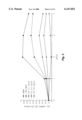
- FIG. 1 Illustrates a flow-chart of the various experimental methodologies utilized in these series of experiments.
- FIG. 3 Illustrates the influence, as determined by an MTT microcolormetric assay, of varying concentration of fetal rat liver extract on the growth and differentiation of murine erythroid leukemia (MEL) cells in vitro.
- MEL murine erythroid leukemia
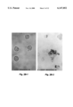
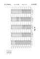
- FIG. 4A illustrates the percentage of murine erythroid leukemia, as determined by a differential cell count (FIGS. 4B-4D), of (MEL) cells following a 7-day co-culture with various electrophoretically-separated protein bands from fetal rat liver extract.
- FIG. 5 Illustrates the inhibitory effect, as determined by a differential cell count, of selected monoclonal antibodies specific for fetal rat liver extract co-cultured with murine erythroid leukemia (MEL) cells.
- MEL murine erythroid leukemia
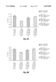
- FIG. 6 FIG. 6A--Illustrates in histogram form the inhibitory effect, as determined by MTT microcolormetric assay, of the #15 and #59 monoclonal antibodies (specific for the 94 kD EDDF protein) directed against fetal liver extract on murine erythroid leukemia (MEL) cells.
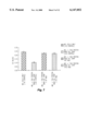
- FIG. 7 Illustrates in histogram form the inhibitory effect, as determined by MTT microcolormetric assay, of monoclonal antibodies directed against individual PhastGel strips containing electrophoretically-separated fetal rat liver extract proteins co-cultured with murine erythroid leukemia (MEL) cells.
- MEL murine erythroid leukemia
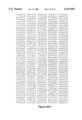
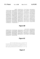
- FIG. 8 8A--DNA sequence of the large, 2721 bp EDDF1 EcoRl-generated cDNA fragment, SEQ ID NO: 1 derived from the human bone marrow cDNA library.
- 8B --depicts the DNA sequence of the smaller, 650 bp EcoRl-generated cDNA fragment, SEQ ID NO: 2.
- 8C -depicts the DNA sequence of the 288 bp EcoRl-generated, translatable hrEDDF1, SEQ ID NO: 3. By standard convention the DNA sequence is shown in the 5' to 3' orientation.
- FIG. 9 depicts the amino acid sequence, SEQ ID NO:4, encoded by the 288 bp. EcoRl-generated, translatable hrEDDF1. By standard convention the amino acid sequence is shown from the --NH 2 to --COOH terminus.
- the present invention is directed to a family of erythroid differentiation and denucleation factors (EDDFs) including EDDF1 (17 kDa) and EDDF2 (94 kDa), which have been shown to possess diagnostic and therapeutic applicability in mammals.
- EDDFs erythroid differentiation and denucleation factors
- the present invention is also directed to a novel genomic DNA sequence which encodes human EDDF1, a vector which contains this novel DNA sequence, an expression system and associated transformed host which contains the novel DNA sequence, and which is also capable of expressing the novel recombinant human EDDF1 protein.
- the present invention utilizes a methodology named "Protein Band-Fishing by Cells" (see Chau, R. M. W., et al., Muscle Neuronotrophic Factors Specific for Anterior Horn Motoneurons of Rat Spinal Cord. In: Recent Advances in Cellular and Molecular Biology, Vol. 5, Peeters Press, Leuven, Belgium, pp. 89-94 (1992)) in an attempt to identify various erythroid differentiation and denucleation factors (EDDFs).
- the inserts from the selected clones were isolated and their DNA was sequenced. Some sequences were subdloned to obtain the most efficient clones of the EDDF genes. The subdlones were tested for expression of proteins with the appropriate biological activity. The amino acid sequence of the protein was then determined. It was discovered that there was homology between the isolated EDDF protein and one exon of an unrelated cell adhesion protein, PECAM. However, the subcloned sequence which expressed this EDDF is somewhat shorter than the PECAM sequence disclosed.
- FIG. 1 A flow-chart of the various methodologies utilized in the following series of experiments is illustrated in FIG. 1.
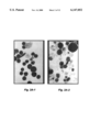
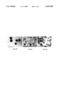
- FIG. 2A depicts the morphological characteristics of the MEL cells co-cultured with the FRLE and illustrates a basophilic staining of the MEL cell cytoplasm after day 4. Generally, it was found that the nuclei of the MEL cells became smaller, more compact, and more intensely stained as a function of their total time in culture. The total number of MEL cells without a nucleus (reticulocytes) gradually increased from 5.4% on day 2 to 56% on day 7.
- FIG. 2B panel b illustrates the results of measurement of benzidine (a substrate for the peroxidase activity B-globin). Measurable enzymatic reaction (as shown by the accumulation of dark-greenish granules in the cytoplasm) was only found in the cytoplasm of MEL cells co-cultured with FRLE.
- MEL cells were co-cultured with FRLE at concentrations of 0.1, 0.2, and 0.4 mg/ml. 1.8% dimethyl sulfoxide (DMSO), a differentiation inducer, was utilized as a negative control for cell growth, whereas DMEM medium was utilized as a positive control.
- DMSO dimethyl sulfoxide
- the determination of MEL cell viability and the influence of the FRLE on MEL cell growth was performed utilizing both MTT microassay wherein the optical density of the cell culture was determined at 570-630 nm and morphological measurement via inverted phase microscopy (Zeiss Axiophot Inverted Microscope, West Germany).
- MTT is a tetrazolium derivative [3-(4,5-dimethylthiazol-2-yl)-2,5-dipheny tetrazolium bromide] which can be converted by viable cells to form a formazan product which can subsequently be measured with an ELISA colorimeter.
- results of the MTT colormetric assays demonstrated that greater concentrations of FRLE caused increased inhibition of MEL cell growth.
- concentrations of FRLE caused increased inhibition of MEL cell growth.
- a 5.5-fold inhibition of MEL cell growth was observed on day 3 when compared with the negative control (cells cultured with DMEM medium alone). It was found that the higher the concentration of FRLE, the greater the inhibition of MEL cell growth in culture.
- EDDF erythroid differentiation and denucleation factor
- the livers from embryonic 15 day-old Sprague Dawley rats fetuses were aseptically dissected into small sections and washed 3-times in Ca +2 /Mg +2 -free Hank's medium.
- the hepatic tissue was then homogenized in 10 mM Tris-HCl (pH 7.2).
- a cell lysate was obtained by centrifugation at 3,000 r.p.m. for 10 minutes, and the supernatant was filtered utilizing a 0.2 ⁇ m Millipore filter membrane (Millipore Corp., Waltham, Mass.).
- the filtered, fetal rat liver extract (FRLE, 10-20 ⁇ l of a 1 mg/ml solution) was then applied to a pre-cast 20% native PHASTGEL® (50 ⁇ 40 ⁇ 0.45 mm, Pharmacia LKB Biotech AB, Upsala, Sweden) for separation by the PhastSystem gel electrophoresis (Pharmacia LKB Biotech AB, Upsala, Sweden).
- the electrophoretic conditions utilized were those suggested by the computer program of Olsson, I., et al., Computer Program for Optimizing Electrophoretic Protein Separation, 9 Electrophoresis 16 (1988).
- PHASTGELTM/MEL cell culture was fixed in 0.4% paraformaldehyde in Phosphate-buffered saline (PBS) for approximately 2 hours, stained using a silver stain-based methodology, and the degree of differentiation and denucleation was determined using inverted-phase microscopy (Zeiss Axiophot Inverted Microscope, West Germany).
- FIG. 4D revealed that 60-70% of the MEL cells in the cultures containing the 17 kDa protein band (EDDF1) were differentiated and denucleated into reticulocytes and mature RBCs, as compared to 20% in the control cultures with no MAbs present.
- FIG. 4C revealed that 70% of the MEL cells in the cultures containing the 94 kDa protein band (EDDF2) were differentiated and denucleated into reticulocytes and mature RBCs.
- FIG. 4B ⁇ 80% of the MEL cells in cultures with control gel strips (no EDDF) were found to be proerythroblasts and basophilic erythroblasts.
- the two bands were excised from the PHASTGEL® and the proteins contained therein were used as antigen in the immunization of BALB/c mice for subsequent preparation of anti-EDDF monoclonal antibodies (MAbs).
- the 17 kDa and 94 kDa protein bands were excised from a PhastSystem gel (1 ⁇ 30 mm gel containing 100 ng trophic factor) and utilized as antigens in the immunization of separate groups of Balb/c mice. Specifically, the EDDF-containing PHASTGEL® sections were excised and finely minced.
- the PHASTGEL® pieces were then mixed with an equal volume of complete Freund's adjuvant and directly injected intraperitoneally into the Balb/c mice.
- a total of 3 antigen immunizations were performed, with intraperitoneal injections of EDDF1-containing and EDDF2-containing PHASTGEL® in physiological saline on the 7th and 21st day following the initial immunization.
- the spleens of the Balb/c mice were harvested and allowed to fuse with either NS-1 or SP2/0 myeloma cells.
- hybridomas were then screened utilizing MTT microassays and microscopic examination. From each hybridoma, two clones were selected as a function of their ability to inhibit, in MEL cell cultures, the differentiation and denucleation activities of EDDFs present in the extract and/or of the 17 kDa and 94 kDa proteins in the PHASTGEL® strips.
- the two MAbs specific for the 94 kD protein were designated MAb #15 and #59; whereas the 17 kDa MAbs were designated B7 or A12.
- FIG. 5 demonstrated that approximately 70-74% of the MEL cells remained undifferentiated in the cultures with the EDDF and the specific MAb, as compared to ⁇ 20% in the control cultures with no MAb.
- FIG. 6A panel a illustrates the results obtained by MTT microassay of cell culture O.D. of the MEL cells incubated with FRLE and the anti-94 kDa MAbs #15 and #59.
- FIG. 6B illustrates the results obtained by MTT microassay of cell culture O.D. of the MEL cells incubated with FRLE and the anti-17 kDa MAbs B7 and A12.
- EDDF-Mabs The selected EDDF-blocking monoclonal antibodies (EDDF-Mabs) were next utilized to immunoselect clones of human EDDF from a human bone marrow cDNA library (produced by Clonetech Co., Palo Alto, Calif.).
- the immunoscreening procedure utilized was a modification of that described in Young, R. A., and Davis, R. W., Efficient Isolation of Genes Using Antibody Probes, 80 Proc. Nat'l Acad. Sci USA 1194 (1983), whose disclosure is incorporated herein by reference.
- the aforementioned modification consisted of using ammonium nickel sulfate as an enhancing agent to increase 100-fold the sensitivity of the peroxidase-avidin-biotin complex reaction utilizing diamino-benzidine as the substrate.
- a maximum of 4 nitrocellulose membrane "replicas" were made from each colony plate for immunoscreening and only the most intensely-stained clones were selected in the immunoscreening procedure. The selected clones were then allowed to express the recombinant proteins which were subsequently assayed to determine their potential EDDF-specific biological activity in vitro. It should be noted that only the clones with the highest EDDF-specific biological activity in the MEL cell cultures were selected for further analysis.
- a total of four EDDF-specific monoclonal antibodies were selected.
- the two MAbs specific for the 94 kDa protein (#15 and #59) and the two 17 kDa-specific MAbs (B7 and A12) were utilized as immunoprobes in the screening of positive expression clones from a selected human bone marrow cDNA library.
- the precipitation procedure for the cDNA library involved the isolation of mRNAs from cells of the human bone marrow library, followed by reverse transcription to synthesize the EDDF cDNAs.
- the selected, EDDF-specific cDNAs were ligated into a gt-11 phage vector and subsequently transformed in an E. coli strain Y1090 host bacteria.
- the cDNA insert of the gt-11 clone would then be sub-cloned into an in vitro expression vector system for protein expression.
- a cDNA was synthesized from a 17 kDa EDDF1 mRNA selected from the human bone marrow library.
- the EDDF1 cDNA was ligated into a gt-11 phage and the resulting recombinant gt-11 clone was designated Lambda.17 kDa.EDDF1 (or EDDF1-A12).
- Purified Lambda.17 kDa.EDDF1 was obtained by the following methodology: 10 plaque-forming units (pfu) of EDDF/gt-11 phage was inoculated into a 500 ml overnight culture of E. coli stain Y1090 until complete lysis of the bacteria was observed. The Lambda phage were recovered and purified by centrifugation and enzymatic treatment with RNase and DNase in an NaCl/PEG 8000 solution, followed by high speed CsCl density gradient centrifugation to collect the purified phage at a final gradient density of approximately 1.5 gm CsCl/ml.
- Isolation and purification of the Lambda.17 kDa.EDDF1 DNA was facilitated by initial digestion of the Lambda phage (1 ml) in EDTA, SDS, and proteinase K, followed by repeated extractions with phenol, phenol/chloroform, and chloroform.
- the EDDP1 DNA was then ethanol precipitated with 95% ethanol and collected by centrifugation. Following repeated washes in 70% ethanol, the resultant Lambda.17 kDa.EDDF1 DNA pellet was redissolved in Tris-EDTA buffer.
- the 0.65 kbp EDDF1 DNA fragment was visualized utilizing U.V. light, excised from the agarose gel, and recovered via standard techniques.
- the recovered EDDF1 cDNA was then prepared for High Protein Expression by recombination with the pGEX-1 Lambda T EcoRl/BAP vector.
- Lambda.17 kDa.EDDF1 was sequenced utilizing 5' and 3' Lambda phage sequencing primers and TAQ® polymerase (Pharmacia Biotech) in an automated DNA sequencing apparatus (ABI). This sequencing methodology provided extremely accurate and reproducible results with respect to the DNA sequencing of the Lambda phage "hosting" the EcoRl-generated fragments.
- FIG. 8A depicts the DNA sequence of the large, 2721 bp EDDF1 EcoRl-generated cDNA, SEQ ID NO:1 fragment derived from the human bone marrow cDNA library.
- FIG. 8B depicts the DNA sequence of the smaller, 656 bp EcoRl-generated cDNA fragment, SEQ ID NO:2.
- FIG. 8C depicts the DNA sequence of the 288 bp EcoRl-generated, translatable hrEDDF1, SEQ ID NO:3. By standard convention the DNA sequence is shown in the 5' to 3' orientation.
- the EcoRl-generated, 288 bp EDDF1 DNA fragment was sub-cloned into the pGEX-1 Lambda T EcoRl/BAP High Protein Expression vector (hereinafter pGEX).
- the pGEX vector was selected due to the following factors: (1) it possessed a high efficiency transcriptional promotor at its 5'-terminus and a 3'-terminus poly(A) tail; and (2) it provided an easy methodology for the purification of the two EDDF recombinant proteins via affinity column chromatography-based purification of the glutathione-S-transferase (GST)-containing EDDF1 fusion proteins.
- GST glutathione-S-transferase
- the pGEX vector was digested with EcoRl. The digested pGEX vector was then incubated overnight with the EcoRl-generated, 288 bp EDDF1 DNA fragments (hereinafter EDDF1-288, SEQ ID NO:3) in the presence of T4 DNA ligase. Following ligation, competent E. coli strain DH5 bacteria were transformed with the recombinant vector and transferred onto Luria broth (LB) agar plates containing 100 g/ml ampicillin for overnight incubation. Due to the fact that the pGEX vector contained a gene which conferred ampicillin resistance to the transformed bacteria, the use of ampicillin screening allowed the exclusive selection of transformed bacterial colonies, as only those bacteria containing the recombinant vector would be viable in its presence.
- LB Luria broth
- the transformed bacterial colonies were individually selected, inoculated into a small volume of LB medium containing 100 g/ml ampicillin, and incubated for approximately 3 hours. Following incubation, the transformed bacteria were collected via centrifuigation for subsequent isolation of the recombinant vector DNA by the alkaline lysis "mini prep" technique as described in Maniatis, T., Fritsch, E. F., and Sambrook, J., Molecular Cloning, 2nd ed., Cold Spring Harbor Laboratory, Cold Spring Harbor, N.Y. pp. 134-136 (1986). With this technique, the collected bacteria were lysed by the addition of a Tris-EDTA/NaOH/SDS solution with vigorous vortexing.
- the recombinant vector DNA-containing supernatant was aspirated, extracted with phenol and chloroform, and precipitated with 95% ethanol. The mixture was centrifuged to collect the precipitated DNA and the nucleic acid pellet was dissolved in Tris-EDTA buffer.
- the collected recombinant vector DNA was then digested with EcoRl to release the EDDF1-288, SEQ ID NO:3 insert from the 4.9 Kbp pGEX vector.
- the digested DNA was subjected to agarose gel electrophoresis and the individual DNA bands were identified via U.V. light visualization.
- “Positive" bacterial colonies i.e., those which contained the EDDF1-288, SEQ ID NO:3 insert
- LB medium for large scale plasmid purification via alkaline lysis and PEG precipitation.
- the purified recombinant vector DNA was then transfected into an E. coli strain BL-21 host bacterium to facilitate high levels of expression of human recombinant EDDF1-288 (hrEDDF1-288) protein, SEQ ID NO:4.
- the pGEX-EDDF1-288 recombinant molecule was transfected into E. coil stain BL-21 competent bacteria in 50 ml of LB medium. IPTG was utilized to induce high levels of expression of the human recombinant EDDF1-228 (hrEDDF1-288) protein, SEQ ID NO:4. Collected bacteria were lysed, frozen, and thawed a total of 4-times using liquid nitrogen, sonicated, and centrifuged. The supernatant, containing the hrEDDF1-288 fusion protein, SEQ ID NO:4 was then passed through an anti-GST affinity chromatography column containing anti-GST monoclonal antibodies (Pharmacia).
- the MEL cell line was utilized to determine the biological activity (i.e., the differentiation and denucleation activity) of the hrEDDF1-288 protein in vitro.
- the MEL cells were co-cultured with the hrEDDF1-288 protein, SEQ ID NO:4 for 3 days in DMEM medium supplemented with 15% FCS. After 3 days of co-culture, an MTT microassay was performed to determine the degree of inhibition of MEL cell proliferation by the hrEDDF1-288 protein, SEQ ID NO:4. After 6 days of co-culture, a differential cell count assay was performed to determine the degree of differentiation and denucleation of the MEL cells into mature erythrocytes.
- FIG. 9 depicts the amino acid sequence of the hrEDDF1-288 protein, SEQ ID NO:4 translated from the 288 bp hrEDDF1-288 DNA fragment, SEQ ID NO:3.
- hrEDDF1-288 protein, SEQ ID NO:4 possessed almost complete homology with human platelet-endothelial cell adhesion molecule-1 (PECAM-1).
- PECAM-1 platelet-endothelial cell adhesion molecule-1
- the hrEDDF1-288 protein, SEQ ID NO:4 was homologous to that which was found in exon 8 of the PECAM-1 molecule. See Newman, P. J., U.S. Pat. No. 5,264,554; Newman, P. J.
Landscapes
- Chemical & Material Sciences (AREA)
- Health & Medical Sciences (AREA)
- Life Sciences & Earth Sciences (AREA)
- Organic Chemistry (AREA)
- General Health & Medical Sciences (AREA)
- Gastroenterology & Hepatology (AREA)
- Biochemistry (AREA)
- Biophysics (AREA)
- Zoology (AREA)
- Genetics & Genomics (AREA)
- Medicinal Chemistry (AREA)
- Molecular Biology (AREA)
- Proteomics, Peptides & Aminoacids (AREA)
- Toxicology (AREA)
- Micro-Organisms Or Cultivation Processes Thereof (AREA)
- Preparation Of Compounds By Using Micro-Organisms (AREA)
- Peptides Or Proteins (AREA)
Abstract
A method of differentiating erythrocyte progenitor cells comprising administering to the erythrocyte progenitor cells an effective amount of an Erythroid Differentiation and Denucleation Factor (EDDF), such that the erythrocyte progenitor cells differentiate.
Description
This Patent application is a continuation in part of Provisional Patent Application U.S. Ser. No. 60/033,231, filed on Dec. 5, 1996, which is hereby incorporated by reference.
The present invention relates to human genes which encode a specialized group of proteins which function to promote the growth, differentiation, and denucleation of immature erythrocytes (red blood cells) and erythroid leukemic cells.
During erythropoiesis, red blood cells are matured in fetal liver or adult bone marrow through the proliferation and differentiation of committed progenitor cells, which are the erythroid burst forming unit and the erythroid-colony forming unit. These progenitor cells are dependent upon lineage-specific growth factors for their proliferation and differentiation. In contrast, primitive pluripotent hematopoietic stem cells, which are generally quiescent and are triggered to proliferate only when a need is expressed in the periphery, respond to a combination of multiple hematopoietic growth factors. Recently, several factors have been identified that appear to be involved in the triggering of cell division in the quiescent hematopoietic progenitor cells and in the differentiation of committed progenitor cells. It has been demonstrated that burst promoting factor (BPF), colony stimulating factor (CSF), and interleukin-3 (IL-3) have a dependent-effect upon the proliferation and differentiation of pluripotent stem cells and progenitor cells. Additional studies have also determined that erythropoietin (EPO) is the sole factor involved in the late stages of erythroid differentiation prior to the stage of basophilic erythroblast. However, the role of these factors in the regulation of the final differentiation stages, beyond basophilic eythroblast, are still uncertain.
Numerous types of cells express cell-surface proteins known as "integrins" which are recognized by extracellular proteins such as fibronectin, collagen, osteoprotein, fibrinogen, vitronectin, thrombospondin, and Von Willebrand factor (VWF), which function in the attachment of these cells to their surroundings. Integrins, which act as receptors for several of these aforementioned extracellular proteins, have been identified with human platelet glycoprotein--which functions to mediate VWF-dependent adhesion of platelets to exposed vascular endothelium.
Another common group of adhesion-promoting molecules are referred to generically as "cellular adhesion molecules" (CAMs) which are glycosylated proteins belonging to the immunoglobulin super-family. The classified CAMs include: neuronal cellular adhesion molecule (NCAM); myelin-associated glycoprotein (MAG); intercellular adhesion molecule (ICAM); lymphocyte function-associated antigen-3 (LFA-3); the T-cell subset cell-surface marker CD-4; the major glycoprotein of peripheral myelin (Po); carcinoembryonic antigen (CEA); and platelet-endothelial cell adhesion molecule 1 (PECAM-1). See e.g., Williams, D. F. & Barclay, T., 6 Ann. Rev. Immunol. 381 (1988).
Of particular interest in the instant invention, is platelet-endothelial cell adhesion molecule 1 (PECAM-1). These cellular adhesion molecules typically are comprised of 711 amino acids and possess a molecular weight of 130 kd. PECAM-1 has been demonstrated to be expressed on platelets, circulating monocytes, and at the intercellular junctions of resting endothelial cells. See e.g., Ashman & Aylett, 38 Tissue Antigen 208 (1991). PECAM-1's are important mediators of platelet-platelet, platelet-leukocyte, and platelet-endothelial cell interactions in the process of platelet aggregation. In addition, these molecules may also be involved in the development of atherosclerotic plaque and thrombi from vascular trauma (e.g., from angioplasty), as well as in leukocyte-endothelial cell interactions in inflammation and transendothelial cell migration.
The present invention may be better-understood and its advantages appreciated by those individuals skilled in the relevant art by referring to the accompanying figures wherein:
FIG. 1 Illustrates a flow-chart of the various experimental methodologies utilized in these series of experiments.
FIG. 2A--Illustrates the morphological characteristics of murine erythroid leukemia (MEL) cells co-cultured with fetal rat liver extract by use of Wright stain. 2B--Illustrates benzidine reaction measurements for -globin.
FIG. 3 Illustrates the influence, as determined by an MTT microcolormetric assay, of varying concentration of fetal rat liver extract on the growth and differentiation of murine erythroid leukemia (MEL) cells in vitro.
FIG. 4 FIG. 4A illustrates the percentage of murine erythroid leukemia, as determined by a differential cell count (FIGS. 4B-4D), of (MEL) cells following a 7-day co-culture with various electrophoretically-separated protein bands from fetal rat liver extract.
FIG. 5 Illustrates the inhibitory effect, as determined by a differential cell count, of selected monoclonal antibodies specific for fetal rat liver extract co-cultured with murine erythroid leukemia (MEL) cells.
FIG. 6 FIG. 6A--Illustrates in histogram form the inhibitory effect, as determined by MTT microcolormetric assay, of the #15 and #59 monoclonal antibodies (specific for the 94 kD EDDF protein) directed against fetal liver extract on murine erythroid leukemia (MEL) cells. 6B--Illustrates in histogram form the inhibitory effect, as determined by MTT microcolormetric assay, of the B7 and A12 monoclonal antibodies (specific for the 17 kD EDDF protein) directed against fetal liver extract on murine erythroid leukemia (MEL) cells.
FIG. 7 Illustrates in histogram form the inhibitory effect, as determined by MTT microcolormetric assay, of monoclonal antibodies directed against individual PhastGel strips containing electrophoretically-separated fetal rat liver extract proteins co-cultured with murine erythroid leukemia (MEL) cells.
FIG. 8 8A--DNA sequence of the large, 2721 bp EDDF1 EcoRl-generated cDNA fragment, SEQ ID NO: 1 derived from the human bone marrow cDNA library. 8B--depicts the DNA sequence of the smaller, 650 bp EcoRl-generated cDNA fragment, SEQ ID NO: 2. 8C--depicts the DNA sequence of the 288 bp EcoRl-generated, translatable hrEDDF1, SEQ ID NO: 3. By standard convention the DNA sequence is shown in the 5' to 3' orientation.
FIG. 9 depicts the amino acid sequence, SEQ ID NO:4, encoded by the 288 bp. EcoRl-generated, translatable hrEDDF1. By standard convention the amino acid sequence is shown from the --NH2 to --COOH terminus.
The present invention is directed to a family of erythroid differentiation and denucleation factors (EDDFs) including EDDF1 (17 kDa) and EDDF2 (94 kDa), which have been shown to possess diagnostic and therapeutic applicability in mammals.
The present invention is also directed to a novel genomic DNA sequence which encodes human EDDF1, a vector which contains this novel DNA sequence, an expression system and associated transformed host which contains the novel DNA sequence, and which is also capable of expressing the novel recombinant human EDDF1 protein.
The present invention utilizes a methodology named "Protein Band-Fishing by Cells" (see Chau, R. M. W., et al., Muscle Neuronotrophic Factors Specific for Anterior Horn Motoneurons of Rat Spinal Cord. In: Recent Advances in Cellular and Molecular Biology, Vol. 5, Peeters Press, Leuven, Belgium, pp. 89-94 (1992)) in an attempt to identify various erythroid differentiation and denucleation factors (EDDFs).
The novel methodology of "Protein Band-Fishing by Cells" was applied to identify the erythroid differentiation and denucleation factor (EDDF) activities in fetal rat liver extract on the growth and differentiation of murine erythroid leukemia (MEL) cells in vitro. See Chan, S. S. W., et al., Erythroid Differentiation and Denucleation Factors from Fetal Rat Liver: Monoclonal Antibodies Preparation for Clone Screening from Human Bone Marrow cDNA Library, (Abstract), 6th International Congress on Cell Biology (1996); Chau, R. M. W. et al., Effects of the Rat Fetal Liver Extract on the Proliferation and Differentiation of Murine Erythroid Leukemia Cells, (Abstract), 6th International Congress on Cell Biology (1996), whose disclosures are incorporated herein by reference. As the fetal liver has been shown to be one of the progenitors of fetal blood stem cells (which contain factors for erythroid differentiation) prior to the development of the bone marrow, fetal liver extract, obtained from embryonic-15-day Sprague Dawley rat fetus, was used as the source of EDDF in an attempt to identify and isolate specific EDDF(s) in vitro. Proteins present in the fetal rat liver extract were electrophoretically-separated in native form utilizing the PHASTGEL electrophoresis system (LKB Pharmacia).
After isolation of protein bands, monoclonal antibodies were made to the proteins and screened for their ability to inhibit the differentiation and denucleation activities of the isolated proteins. These antibodies were then used to immunoselect clones containing genes coding for the desired functions.
The inserts from the selected clones were isolated and their DNA was sequenced. Some sequences were subdloned to obtain the most efficient clones of the EDDF genes. The subdlones were tested for expression of proteins with the appropriate biological activity. The amino acid sequence of the protein was then determined. It was discovered that there was homology between the isolated EDDF protein and one exon of an unrelated cell adhesion protein, PECAM. However, the subcloned sequence which expressed this EDDF is somewhat shorter than the PECAM sequence disclosed.
A flow-chart of the various methodologies utilized in the following series of experiments is illustrated in FIG. 1.
Murine erythroid leukemia (MEL) cells were co-cultured with fetal rat liver extract (FRLE) at a concentration of 0.4 mg/ml. FIG. 2A depicts the morphological characteristics of the MEL cells co-cultured with the FRLE and illustrates a basophilic staining of the MEL cell cytoplasm after day 4. Generally, it was found that the nuclei of the MEL cells became smaller, more compact, and more intensely stained as a function of their total time in culture. The total number of MEL cells without a nucleus (reticulocytes) gradually increased from 5.4% on day 2 to 56% on day 7.
FIG. 2B, panel b illustrates the results of measurement of benzidine (a substrate for the peroxidase activity B-globin). Measurable enzymatic reaction (as shown by the accumulation of dark-greenish granules in the cytoplasm) was only found in the cytoplasm of MEL cells co-cultured with FRLE.
Similarly, MEL cells were co-cultured with FRLE at concentrations of 0.1, 0.2, and 0.4 mg/ml. 1.8% dimethyl sulfoxide (DMSO), a differentiation inducer, was utilized as a negative control for cell growth, whereas DMEM medium was utilized as a positive control. The determination of MEL cell viability and the influence of the FRLE on MEL cell growth was performed utilizing both MTT microassay wherein the optical density of the cell culture was determined at 570-630 nm and morphological measurement via inverted phase microscopy (Zeiss Axiophot Inverted Microscope, West Germany). MTT is a tetrazolium derivative [3-(4,5-dimethylthiazol-2-yl)-2,5-dipheny tetrazolium bromide] which can be converted by viable cells to form a formazan product which can subsequently be measured with an ELISA colorimeter.
The results of the MTT colormetric assays, illustrated in FIG. 3, demonstrated that greater concentrations of FRLE caused increased inhibition of MEL cell growth. For example, with a 0.2 mg/ml FRLE concentration, a 5.5-fold inhibition of MEL cell growth was observed on day 3 when compared with the negative control (cells cultured with DMEM medium alone). It was found that the higher the concentration of FRLE, the greater the inhibition of MEL cell growth in culture.
The erythroid differentiation and denucleation factor (EDDF) in fetal rat liver extract (FRLE) was isolated utilizing the proprietary "Protein Band-Fishing by Cells" methodology as initially reported in Chau, R. M. W., et al., Muscle Neuronotrophic Factors Specific for Anterior Horn Motoneurons of Rat Spinal Cord. In: Recent Advances in Cellular and Molecular Biology, Vol. 5, Peeters Press, Leuven, Belgium, pp. 89-94 (1992). Additionally, this methodology allowed for the determination of the effect of EDDF on the growth and differentiation of murine erythroid leukemia (MEL) cells in vitro. See Chan, S. S. W., et al., Erythroid Differentiation and Denucleation Factors from Fetal Rat Liver: Monoclonal Antibodies Preparation for Clone Screening from Human Bone Marrow cDNA Library, (Abstract), 6th International Congress on Cell Biology (1996); Chau, R. M. W. et al., Effects of the Rat Fetal Liver Extract on the Proliferation and Differentiation of Murine Erythroid Leukemia Cells, (Abstract), 6th International Congress on Cell Biology (1996).
Erythroid differentiation and denucleation factors are generally found in minute quantities in vivo this can potentially cause tremendous difficulties in their isolation utilizing traditional biochemical methodologies. In view of this fact, a novel technique designated "Protein Band-Fishing by Cells" was developed in which viable MEL cells were co-cultured with an electrophoretic gel containing the separated proteins from fetal rat liver extract (FRLE). This methodology thus allowed the MEL cells to "fish-out" those fetal rat liver extract proteins which exhibited biological activity specific for those erythroid leukemic cells (i.e., differentiation and denucleation of MEL cells into reticulocytes or mature erythrocytes).
For electrophoretic protein separation, the livers from embryonic 15 day-old Sprague Dawley rats fetuses were aseptically dissected into small sections and washed 3-times in Ca+2 /Mg+2 -free Hank's medium. The hepatic tissue was then homogenized in 10 mM Tris-HCl (pH 7.2). A cell lysate was obtained by centrifugation at 3,000 r.p.m. for 10 minutes, and the supernatant was filtered utilizing a 0.2 μm Millipore filter membrane (Millipore Corp., Waltham, Mass.).
The filtered, fetal rat liver extract (FRLE, 10-20 μl of a 1 mg/ml solution) was then applied to a pre-cast 20% native PHASTGEL® (50×40×0.45 mm, Pharmacia LKB Biotech AB, Upsala, Sweden) for separation by the PhastSystem gel electrophoresis (Pharmacia LKB Biotech AB, Upsala, Sweden). The electrophoretic conditions utilized were those suggested by the computer program of Olsson, I., et al., Computer Program for Optimizing Electrophoretic Protein Separation, 9 Electrophoresis 16 (1988). Following electrophoretic separation, 2×10⁴ MEL cells were seeded onto the surface of the protein-containing PHASTGEL® and cultured for 7 days, thus allowing for differentiation and denucleation. After the 7 day incubation period, the PHASTGEL™/MEL cell culture was fixed in 0.4% paraformaldehyde in Phosphate-buffered saline (PBS) for approximately 2 hours, stained using a silver stain-based methodology, and the degree of differentiation and denucleation was determined using inverted-phase microscopy (Zeiss Axiophot Inverted Microscope, West Germany).
The results, as illustrated in FIG. 4A, demonstrated that at the PHASTGEL® regions which contained protein bands with apparent molecular weights of 17 kDa (EDDF1) and 94 kDa (EDDF2) there were increased numbers of differentiated and denucleated reticulocytes and mature red blood cells found to be present. In order to verify the presence of EDDF activities in these two bands, the PHASTGEL® was cut into 50 strips from top to bottom and each strip was co-cultured with MEL cells. The results, as illustrated in FIG. 4D, revealed that 60-70% of the MEL cells in the cultures containing the 17 kDa protein band (EDDF1) were differentiated and denucleated into reticulocytes and mature RBCs, as compared to 20% in the control cultures with no MAbs present. Similarly, FIG. 4C, revealed that 70% of the MEL cells in the cultures containing the 94 kDa protein band (EDDF2) were differentiated and denucleated into reticulocytes and mature RBCs. In contrast, FIG. 4B ˜80% of the MEL cells in cultures with control gel strips (no EDDF) were found to be proerythroblasts and basophilic erythroblasts.
In order to confirm the specific EDDF activity of the 17 kDa and 94 kDa proteins, the two bands were excised from the PHASTGEL® and the proteins contained therein were used as antigen in the immunization of BALB/c mice for subsequent preparation of anti-EDDF monoclonal antibodies (MAbs). The 17 kDa and 94 kDa protein bands were excised from a PhastSystem gel (1×30 mm gel containing 100 ng trophic factor) and utilized as antigens in the immunization of separate groups of Balb/c mice. Specifically, the EDDF-containing PHASTGEL® sections were excised and finely minced. The PHASTGEL® pieces were then mixed with an equal volume of complete Freund's adjuvant and directly injected intraperitoneally into the Balb/c mice. A total of 3 antigen immunizations were performed, with intraperitoneal injections of EDDF1-containing and EDDF2-containing PHASTGEL® in physiological saline on the 7th and 21st day following the initial immunization. The spleens of the Balb/c mice were harvested and allowed to fuse with either NS-1 or SP2/0 myeloma cells.
The resultant hybridomas were then screened utilizing MTT microassays and microscopic examination. From each hybridoma, two clones were selected as a function of their ability to inhibit, in MEL cell cultures, the differentiation and denucleation activities of EDDFs present in the extract and/or of the 17 kDa and 94 kDa proteins in the PHASTGEL® strips. The two MAbs specific for the 94 kD protein were designated MAb #15 and #59; whereas the 17 kDa MAbs were designated B7 or A12.
The results, illustrated in FIG. 5, demonstrated that approximately 70-74% of the MEL cells remained undifferentiated in the cultures with the EDDF and the specific MAb, as compared to ˜20% in the control cultures with no MAb. FIG. 6A, panel a illustrates the results obtained by MTT microassay of cell culture O.D. of the MEL cells incubated with FRLE and the anti-94 kDa MAbs #15 and #59. Similarly, FIG. 6B, illustrates the results obtained by MTT microassay of cell culture O.D. of the MEL cells incubated with FRLE and the anti-17 kDa MAbs B7 and A12.
Additionally, an experiment was performed in which a PHASTGEL®, containing electrophoretically-separated FRLE proteins, was cut into 50 strips (1 mm/strip, with the orientation of the strip going from the top-to-bottom of the gel). The individual gel strips were co-cultured with 2×10⁴ MEL cells with the two anti-17 kDa MAbs (B 7 and A12) in a 96-well culture plate for 7 days. The results of this assay are illustrated in histogram form in FIG. 7. These results suggested that clones had been selected which expressed fusion proteins with EDDF-like activities that possessed the ability to induce the differentiation and denucleation of 60% of the MEL cells. In addition, this aforementioned induction was inhibited by their specific "blocking" monoclonal antibodies.
The selected EDDF-blocking monoclonal antibodies (EDDF-Mabs) were next utilized to immunoselect clones of human EDDF from a human bone marrow cDNA library (produced by Clonetech Co., Palo Alto, Calif.). The immunoscreening procedure utilized was a modification of that described in Young, R. A., and Davis, R. W., Efficient Isolation of Genes Using Antibody Probes, 80 Proc. Nat'l Acad. Sci USA 1194 (1983), whose disclosure is incorporated herein by reference. The aforementioned modification consisted of using ammonium nickel sulfate as an enhancing agent to increase 100-fold the sensitivity of the peroxidase-avidin-biotin complex reaction utilizing diamino-benzidine as the substrate. A maximum of 4 nitrocellulose membrane "replicas" were made from each colony plate for immunoscreening and only the most intensely-stained clones were selected in the immunoscreening procedure. The selected clones were then allowed to express the recombinant proteins which were subsequently assayed to determine their potential EDDF-specific biological activity in vitro. It should be noted that only the clones with the highest EDDF-specific biological activity in the MEL cell cultures were selected for further analysis.
A total of four EDDF-specific monoclonal antibodies were selected. The two MAbs specific for the 94 kDa protein (#15 and #59) and the two 17 kDa-specific MAbs (B7 and A12) were utilized as immunoprobes in the screening of positive expression clones from a selected human bone marrow cDNA library. In brief, the precipitation procedure for the cDNA library involved the isolation of mRNAs from cells of the human bone marrow library, followed by reverse transcription to synthesize the EDDF cDNAs. The selected, EDDF-specific cDNAs were ligated into a gt-11 phage vector and subsequently transformed in an E. coli strain Y1090 host bacteria. The cDNA insert of the gt-11 clone would then be sub-cloned into an in vitro expression vector system for protein expression.
(a) Preparation and Purification of 17 kD EDDF1/gt-11 Phage
A cDNA was synthesized from a 17 kDa EDDF1 mRNA selected from the human bone marrow library. The EDDF1 cDNA was ligated into a gt-11 phage and the resulting recombinant gt-11 clone was designated Lambda.17 kDa.EDDF1 (or EDDF1-A12).
Purified Lambda.17 kDa.EDDF1 was obtained by the following methodology: 10 plaque-forming units (pfu) of EDDF/gt-11 phage was inoculated into a 500 ml overnight culture of E. coli stain Y1090 until complete lysis of the bacteria was observed. The Lambda phage were recovered and purified by centrifugation and enzymatic treatment with RNase and DNase in an NaCl/PEG 8000 solution, followed by high speed CsCl density gradient centrifugation to collect the purified phage at a final gradient density of approximately 1.5 gm CsCl/ml.
Isolation and purification of the Lambda.17 kDa.EDDF1 DNA was facilitated by initial digestion of the Lambda phage (1 ml) in EDTA, SDS, and proteinase K, followed by repeated extractions with phenol, phenol/chloroform, and chloroform. The EDDP1 DNA was then ethanol precipitated with 95% ethanol and collected by centrifugation. Following repeated washes in 70% ethanol, the resultant Lambda.17 kDa.EDDF1 DNA pellet was redissolved in Tris-EDTA buffer.
(b) Recovery of the 17 kDa EDDF1 cDNA Insert from Lambda.17 kDa.EDDF1
10 g of Lambda.17 kDa.EDDF1 DNA was digested overnight with EcoRl and electrophoresed. The results of the EcoRl digestion demonstrated the presence of three discreet DNA fragments: (1) 0.65 kbp fragment designated as the EDDF1; (2) a 2.06 kbp fragment designated as the large DNA fragment; and (3) a 46 kbp fragment designated as the Lambda phage DNA. Three discreet DNA fragments were observed due to the presence of an internal EcoRl restriction site within the Lambda.17 kDa.EDDF1 cDNA insert located at the position of base pair 2066.
Following electrophoresis, the 0.65 kbp EDDF1 DNA fragment was visualized utilizing U.V. light, excised from the agarose gel, and recovered via standard techniques. The recovered EDDF1 cDNA was then prepared for High Protein Expression by recombination with the pGEX-1 Lambda T EcoRl/BAP vector.
Lambda.17 kDa.EDDF1 was sequenced utilizing 5' and 3' Lambda phage sequencing primers and TAQ® polymerase (Pharmacia Biotech) in an automated DNA sequencing apparatus (ABI). This sequencing methodology provided extremely accurate and reproducible results with respect to the DNA sequencing of the Lambda phage "hosting" the EcoRl-generated fragments. The DNA sequencing indicated that the large EDDF1 fragment consisted of 2721 bp, SEQ ID NO:1 whereas the smaller DNA fragment was found to consist of 656 bp, SEQ ID NO:2. Moreover, only 288 bp, SEQ ID NO:3 of the aforementioned 656 bp, SEQ ID NO:2 sequence was subsequently shown (by computer-based intron/exon analysis) to be translated into the human, recombinant EDDF1 (hrEDDF1) protein.
FIG. 8A depicts the DNA sequence of the large, 2721 bp EDDF1 EcoRl-generated cDNA, SEQ ID NO:1 fragment derived from the human bone marrow cDNA library. FIG. 8B depicts the DNA sequence of the smaller, 656 bp EcoRl-generated cDNA fragment, SEQ ID NO:2. FIG. 8C depicts the DNA sequence of the 288 bp EcoRl-generated, translatable hrEDDF1, SEQ ID NO:3. By standard convention the DNA sequence is shown in the 5' to 3' orientation.
The EcoRl-generated, 288 bp EDDF1 DNA fragment (see FIG. 8C and SEQ ID NO. 3) was sub-cloned into the pGEX-1 Lambda T EcoRl/BAP High Protein Expression vector (hereinafter pGEX). The pGEX vector was selected due to the following factors: (1) it possessed a high efficiency transcriptional promotor at its 5'-terminus and a 3'-terminus poly(A) tail; and (2) it provided an easy methodology for the purification of the two EDDF recombinant proteins via affinity column chromatography-based purification of the glutathione-S-transferase (GST)-containing EDDF1 fusion proteins.
a. Transformation of Recombinant Plasmid
The pGEX vector was digested with EcoRl. The digested pGEX vector was then incubated overnight with the EcoRl-generated, 288 bp EDDF1 DNA fragments (hereinafter EDDF1-288, SEQ ID NO:3) in the presence of T4 DNA ligase. Following ligation, competent E. coli strain DH5 bacteria were transformed with the recombinant vector and transferred onto Luria broth (LB) agar plates containing 100 g/ml ampicillin for overnight incubation. Due to the fact that the pGEX vector contained a gene which conferred ampicillin resistance to the transformed bacteria, the use of ampicillin screening allowed the exclusive selection of transformed bacterial colonies, as only those bacteria containing the recombinant vector would be viable in its presence.
b. Identification and Isolation of the EDDF1-288 Transformants
The transformed bacterial colonies were individually selected, inoculated into a small volume of LB medium containing 100 g/ml ampicillin, and incubated for approximately 3 hours. Following incubation, the transformed bacteria were collected via centrifuigation for subsequent isolation of the recombinant vector DNA by the alkaline lysis "mini prep" technique as described in Maniatis, T., Fritsch, E. F., and Sambrook, J., Molecular Cloning, 2nd ed., Cold Spring Harbor Laboratory, Cold Spring Harbor, N.Y. pp. 134-136 (1986). With this technique, the collected bacteria were lysed by the addition of a Tris-EDTA/NaOH/SDS solution with vigorous vortexing. After centrifugation to collect contaminating cellular debris, the recombinant vector DNA-containing supernatant was aspirated, extracted with phenol and chloroform, and precipitated with 95% ethanol. The mixture was centrifuged to collect the precipitated DNA and the nucleic acid pellet was dissolved in Tris-EDTA buffer.
The collected recombinant vector DNA was then digested with EcoRl to release the EDDF1-288, SEQ ID NO:3 insert from the 4.9 Kbp pGEX vector. The digested DNA was subjected to agarose gel electrophoresis and the individual DNA bands were identified via U.V. light visualization.
"Positive" bacterial colonies (i.e., those which contained the EDDF1-288, SEQ ID NO:3 insert) were selected and inoculated into LB medium for large scale plasmid purification via alkaline lysis and PEG precipitation. The purified recombinant vector DNA was then transfected into an E. coli strain BL-21 host bacterium to facilitate high levels of expression of human recombinant EDDF1-288 (hrEDDF1-288) protein, SEQ ID NO:4.
The pGEX-EDDF1-288 recombinant molecule was transfected into E. coil stain BL-21 competent bacteria in 50 ml of LB medium. IPTG was utilized to induce high levels of expression of the human recombinant EDDF1-228 (hrEDDF1-288) protein, SEQ ID NO:4. Collected bacteria were lysed, frozen, and thawed a total of 4-times using liquid nitrogen, sonicated, and centrifuged. The supernatant, containing the hrEDDF1-288 fusion protein, SEQ ID NO:4 was then passed through an anti-GST affinity chromatography column containing anti-GST monoclonal antibodies (Pharmacia). The use of this type of affinity chromatography allowed purification of the GST-hrEDDF1-288 fusion protein which was bound to the matrix through the GST moiety. Following a high salt wash, the GST-hrEDDF1-288 fusion protein was eluted. The proteolytic enzyme thrombin was utilized to cleave the linkage between the hrEDDF1-288 protein, SEQ ID NO:4 and the GST moiety and the purified hrEDDF1-288 protein, SEQ ID NO:4 was collected for subsequent in vivo and in vitro assays.
The MEL cell line was utilized to determine the biological activity (i.e., the differentiation and denucleation activity) of the hrEDDF1-288 protein in vitro. The MEL cells were co-cultured with the hrEDDF1-288 protein, SEQ ID NO:4 for 3 days in DMEM medium supplemented with 15% FCS. After 3 days of co-culture, an MTT microassay was performed to determine the degree of inhibition of MEL cell proliferation by the hrEDDF1-288 protein, SEQ ID NO:4. After 6 days of co-culture, a differential cell count assay was performed to determine the degree of differentiation and denucleation of the MEL cells into mature erythrocytes.
FIG. 9 depicts the amino acid sequence of the hrEDDF1-288 protein, SEQ ID NO:4 translated from the 288 bp hrEDDF1-288 DNA fragment, SEQ ID NO:3. Subsequent analysis following both nucleic acid and amino acid sequencing indicated that hrEDDF1-288 protein, SEQ ID NO:4 possessed almost complete homology with human platelet-endothelial cell adhesion molecule-1 (PECAM-1). Specifically, the hrEDDF1-288 protein, SEQ ID NO:4 was homologous to that which was found in exon 8 of the PECAM-1 molecule. See Newman, P. J., U.S. Pat. No. 5,264,554; Newman, P. J. et al., PCT Publication WO 96/01271. Although there is sequence homology between these two molecules, the in vivo and in vitro biological functions are completely diverse in that EDDF1 functions in the differentiation and denucleation of erythrocytes and PECAM-1 is involved in cellular adhesion processes.
While embodiments and applications of the present invention have been described in some detail by way of illustration and example for purposes of clarity and understanding, it would be apparent to those individuals whom are skilled within the relevant art that many additional modifications would be possible without departing from the inventive concepts contained herein. The invention, therefore, is not to be restricted in any manner except in the spirit of the appended claims.
__________________________________________________________________________
#             SEQUENCE LISTING
- (1) GENERAL INFORMATION:
-    (iii) NUMBER OF SEQUENCES: 4
- (2) INFORMATION FOR SEQ ID NO:1:
-      (i) SEQUENCE CHARACTERISTICS:
#pairs    (A) LENGTH: 2721 base
          (B) TYPE: nucleic acid
          (C) STRANDEDNESS: single
          (D) TOPOLOGY: linear
-     (ii) MOLECULE TYPE: DNA (genomic)
-     (xi) SEQUENCE DESCRIPTION: SEQ ID NO:1:
- CTAATGGTGT GAGGCATACA AAAAAGAAGA CATATTCTTT GTTTCAATGC TG - #TGGTAAGA
  60
- AACACAAGCT CTCCTAATGA AAATGATGGA CAAACATCTG AATCATACTA CC - #AATAAGCA
 120
- TAGAAAAAAT GTTGGGGGTC ATGTTTGGTT GTCACGTGAA CTATATCCTT AC - #AGTGATGG
 180
- TGATAGTAAT TTAGGGTATG CCAGACTTCA TCTAGCTTAA GTGGGTAAAC AT - #TGTGAAAA
 240
- AGCTGGGCTA GGTGCCAGGG CTTGAGAATG GGTGGCCAGA GAAGGCTGAA GA - #TGGCTGAA
 300
- CATCTCCAGC AAACACATGA GCCAAAAGGT CCCATGGGGC ACTTCAAAAG AC - #TGTGCGCA
 360
- GCCAGGTGCG GTGGCTCACG CCTATAATCC CAGCACTTTG GGAGACCGAA TG - #GGGTGGAT
 420
- CACTTGAGCC CAGAGGTTTG TGACTAGCTT GGCCAACATG GCAAAACCCC GT - #CTCTACTA
 480
- AAAATACAAA AATTAGCCCA GCGTGGTGGT GGGTGTCCTG TAGCCCCAGC TA - #CTCAGGTG
 540
- GCTGAGGTGG TAGAATCACT TGAATCCAGG AGGCAGAGGT TGCAGTGAGC CA - #AGATCGTG
 600
- CCACTGCACT CCAGCCTGGG TGACAGAGTG AGACTCTATC TCCACAAAAA AA - #AAAAAAAA
 660
- AAAAAAAATT AAAGGACTGT GGCCAAATCA GATGGCTGGA AACAAAGGCT GG - #AGTTTGGG
 720
- AATGGAGAAT CACCGGATAT GAGCTGAAAA AGTGGCTGAG CCTAAGCGTG AC - #AGGTGTCA
 780
- GGTGCCAGTC TCAGGAGTAG GCAAGTGTCC TGCATGCAGT GAAAAGCCAG AA - #GATGGAAG
 840
- GAAGAACAGG ATGCAAATGA GTTCTCGGAA CGATCCACCT GGTGGCTGGG TC - #AGGGAGCA
 900
- GGCATGGTGA CTTCAGACCT CATGGTACGT TAGAGGCTAA TGTGAAGCCC AT - #GTGAAGCT
 960
- GTTGGTTTAA ACTGGGTCGA TATCAGTGGC ACACATTTAC TGACCATGTG TC - #CAGCCCTG
1020
- TGTGAAGTAC TGTAGTAAAT TGCTCCAATG GAAACTCACA ATAACCACAG AA - #GGCCAGTA
1080
- ACAGCATTGT CGTTATTTTA TCATGACGCA ACTGAGGCTT AGGGCAGACA GC - #TGGTGGGT
1140
- GGTGGGACTG GGATTTGAGC CCACTGGTGT CCCAGGCCCG GAGCTTGGCT TC - #TTCCATTG
1200
- TCTTACCACA GCCTGCACTC ACAGGAGAGT GACCTATAAG TTACAATACC AT - #CTGCTGAC
1260
- CATCTGCTCT CACACTAGAA GGAAAGTCTA CTTGGGGAGA CAATTTAGGA TC - #CGAATTTT
1320
- GGTAGTTGAG GATGGAGCTA GGAAAAGCGG ATACAGGAGG TAGCCAAGTT CT - #GCTTGGAC
1380
- CTGCAGGGAG TGAGGCTGGC CGGGCTCCAG GTGGAAATCC CCAGGTGAAA AG - #GGAGACTT
1440
- GGAGGTCAGG AAAGTAACCT GGACTGGAGC CATAGGTTTA GGTGTCAGTG GC - #TCAGAGAC
1500
- AGAAGCTCAG CGTGTAGGTG AAATCACCCA GGAGGAGAAT GGGGATGGAA AA - #CTGAGGAT
1560
- TGAATTTTGC AAAATGTTCA TACTTCCGGG GAAAACAAAG AATAACCAGT GA - #ATAAGAAA
1620
- GGGGTGCCAG GTAAGAAGGG AAGAGAATCA GAGTCATGAG GAACCCCAGA AC - #CCCAGAAA
1680
- AAGCTGAGTT CCACGTAAGA CCTGGGCAAC AGTGAAGTAT GGAGAGCCCA AG - #ATTGGGAG
1740
- CGTGGAGGAA GAGCATCCAC CACTGAATTT AATCAGCCCC GGACTCAGGG AC - #GTTGGTTG
1800
- GGGAATCAAG TGACCTTCCC AGTTTCTTCA AAACTTGAGA GAGAGTGCAG TG - #TCACAAGA
1860
- TTGTGACTAC AAAAGAGTGC AGTCAGATTT CAGGGGTAAC AAGAAAGTGT GA - #AATAAGGG
1920
- AGTCAAAGCA TAAAGGAAAA AGGAGAAAAA ATGGCCGATA GCTAGAGAAG GC - #GTGGGTCA
1980
- AGATTGTCTG TGGCCTGGCA TGGTGGCTTA TGCCTGTAAT CCCAGCATTT TG - #GAAGGCCG
2040
- AGGTGGGCAA ATCACCTGAG GTCAGGAATT CAAGACCAGC CTGGCCAACA GG - #GCAAAACC
2100
- CCGTCTCTAA AACAACAACA ACAACAAAAA AATCCAAAAA GTTAGCTGGG CC - #TGGTGGGC
2160
- GCACCTGTCA TTCCAGCTAC TCGGGAGGCT GAGGCAGGAG ATTTGCTTGA AC - #CCAGGAGG
2220
- CACACGTTGC GGTAAGCTGA GATTATACCA CTGCACTCCA GCCTGGGTGA TA - #AGAGCGGG
2280
- ACTCTGTCTC AGAGGAAAAA AAAAAAAGTT GAGCAGTGGC TGTCTCATGT TC - #CTCTTCCT
2340
- CTGCCCTTCT TTGCTCAGTG TGAATCCTTT TCCTGCTTTT CAGCCCCGGT GG - #ATGAGGTC
2400
- CAGATTTCTA TCCTGTCAAG TAAGGTGGTG GAGTCTGGAG AGGACATTGT GC - #TGCAATGT
2460
- GCTGTGAATG AAGGATCTGG TCCCATCACC TATAAGTTTT ACAGAGAAAA AG - #AGGGCAAA
2520
- CCCTTCTATC AAATGACCTC AAATGCCACC CAGGCATTTT GGACCAAGCA GA - #AGGCTAAC
2580
- AAGGAACAGG AGGGAGAGTA TTACTGCACA GCCTTCAACA GAGCCAACCA CG - #CCTCCAGT
2640
- GTCCCCAGAA GCAAAATACT GACAGTCAGA GGTGAGTCAG GGTCTCCATA GC - #AAGCTGTG
2700
#                2721AA G
- (2) INFORMATION FOR SEQ ID NO:2:
-      (i) SEQUENCE CHARACTERISTICS:
#pairs    (A) LENGTH: 656 base
          (B) TYPE: nucleic acid
          (C) STRANDEDNESS: single
          (D) TOPOLOGY: linear
-     (ii) MOLECULE TYPE: DNA (genomic)
-     (xi) SEQUENCE DESCRIPTION: SEQ ID NO:2:
- GAATTCAAGA CCAGCCTGGC CAACAGGGCA AAACCCCGTC TCTAAAACAA CA - #ACAACAAC
  60
- AAAAAAATCC AAAAAGTTAG CTGGGCCTGG TGGGCGCACC TGTCATTCCA GC - #TACTCGGG
 120
- AGGCTGAGGC AGGAGATTTG CTTGAACCCA GGAGGCACAC GTTGCGGTAA GC - #TGAGATTA
 180
- TACCACTGCA CTCCAGCCTG GGTGATAAGA GCGGGACTCT GTCTCAGAGG AA - #AAAAAAAA
 240
- AAGTTGAGCA GTGGCTGTCT CATGTTCCTC TTCCTCTGCC CTTCTTTGCT CA - #GTGTGAAT
 300
- CCTTTTCCTG CTTTTCAGCC CCGGTGGATG AGGTCCAGAT TTCTATCCTG TC - #AAGTAAGG
 360
- TGGTGGAGTC TGGAGAGGAC ATTGTGCTGC AATGTGCTGT GAATGAAGGA TC - #TGGTCCCA
 420
- TCACCTATAA GTTTTACAGA GAAAAAGAGG GCAAACCCTT CTATCAAATG AC - #CTCAAATG
 480
- CCACCCAGGC ATTTTGGACC AAGCAGAAGG CTAACAAGGA ACAGGAGGGA GA - #GTATTACT
 540
- GCACAGCCTT CAACAGAGCC AACCACGCCT CCAGTGTCCC CAGAAGCAAA AT - #ACTGACAG
 600
- TCAGAGGTGA GTCAGGGTCT CCATAGCAAG CTGTGCTGTG GGCCCCCAAG GG - #CAAG
 656
- (2) INFORMATION FOR SEQ ID NO:3:
-      (i) SEQUENCE CHARACTERISTICS:
#pairs    (A) LENGTH: 288 base
          (B) TYPE: nucleic acid
          (C) STRANDEDNESS: single
          (D) TOPOLOGY: linear
-     (ii) MOLECULE TYPE: DNA (genomic)
-     (xi) SEQUENCE DESCRIPTION: SEQ ID NO:3:
- GCCCCGGTGG ATGAGGTCCA GATTTCTATC CTGTCAAGTA AGGTGGTGGA GT - #CTGGAGAG
  60
- GACATTGTGC TGCAATGTGC TGTGAATGAA GGATCTGGTC CCATCACCTA TA - #AGTTTTAC
 120
- AGAGAAAAAG AGGGCAAACC CTTCTATCAA ATGACCTCAA ATGCCACCCA GG - #CATTTTGG
 180
- ACCAAGCAGA AGGCTAACAA GGAACAGGAG GGAGAGTATT ACTGCACAGC CT - #TCAACAGA
 240
#               288GTGT CCCCAGAAGC AAAATACTGA CAGTCAGA
- (2) INFORMATION FOR SEQ ID NO:4:
-      (i) SEQUENCE CHARACTERISTICS:
#acids    (A) LENGTH: 96 amino
          (B) TYPE: amino acid
          (C) STRANDEDNESS: single
          (D) TOPOLOGY: linear
-     (ii) MOLECULE TYPE: protein
-     (xi) SEQUENCE DESCRIPTION: SEQ ID NO:4:
- Ala Pro Val Asp Glu Val Gln Ile Ser Ile Le - #u Ser Ser Lys Val Val
#                15
- Glu Ser Gly Glu Asp Ile Val Leu Gln Cys Al - #a Val Asn Glu Gly Ser
#            30
- Gly Pro Ile Thr Tyr Lys Phe Tyr Arg Glu Ly - #s Glu Gly Lys Pro Phe
#        45
- Tyr Gln Met Thr Ser Asn Ala Thr Gln Ala Ph - #e Trp Thr Lys Gln Lys
#    60
- Ala Asn Lys Glu Gln Glu Gly Glu Tyr Tyr Cy - #s Thr Ala Phe Asn Arg
#80
- Ala Asn His Ala Ser Ser Val Pro Arg Ser Ly - #s Ile Leu Thr Val Arg
#                95
__________________________________________________________________________
Claims (4)
1. A method of differentiating erythrocyte progenitor cells comprising administering to the erythrocyte progenitor cells an effective amount of a purified polypeptide having a sequence according to SEQ ID NO:4, such that the erythrocyte progenitor cells differentiate.
2. The method of claim 1, where the effective amount of a purified polypeptide having a sequence according to SEQ ID NO:4 causes the erythrocyte progenitor cells to differentiate into reticulocytes.
3. The method of claim 1, where the effective amount of a purified polypeptide having a sequence according to SEQ ID NO:4 causes the erythrocyte progenitor cells to differentiate into mature erythrocytes.
4. A method of causing denucleation of erythrocyte progenitor cells comprising administering to the erythrocyte progenitor cells an effective amount of a purified polypeptide having a sequence according to SEQ ID NO:4, such that the erythrocyte progenitor cells denucleate.
Priority Applications (3)
| Application Number | Priority Date | Filing Date | Title |
|---|---|---|---|
| US08/921,195 US6147052A (en) | 1996-12-05 | 1997-08-29 | Method of differentiating erythrocyte progenitor cells |
| PCT/US1997/021989 WO1998024903A1 (en) | 1996-12-05 | 1997-12-01 | Isolation and use of erythroid differentiation and denucleation factors |
| AU53684/98A AU5368498A (en) | 1996-12-05 | 1997-12-01 | Isolation and use of erythroid differentiation and denucleation factors |
Applications Claiming Priority (2)
| Application Number | Priority Date | Filing Date | Title |
|---|---|---|---|
| US3323196P | 1996-12-05 | 1996-12-05 | |
| US08/921,195 US6147052A (en) | 1996-12-05 | 1997-08-29 | Method of differentiating erythrocyte progenitor cells |
Publications (1)
| Publication Number | Publication Date |
|---|---|
| US6147052A true US6147052A (en) | 2000-11-14 |
Family
ID=26709445
Family Applications (1)
| Application Number | Title | Priority Date | Filing Date |
|---|---|---|---|
| US08/921,195 Expired - Lifetime US6147052A (en) | 1996-12-05 | 1997-08-29 | Method of differentiating erythrocyte progenitor cells |
Country Status (3)
| Country | Link |
|---|---|
| US (1) | US6147052A (en) |
| AU (1) | AU5368498A (en) |
| WO (1) | WO1998024903A1 (en) |
Cited By (1)
| Publication number | Priority date | Publication date | Assignee | Title |
|---|---|---|---|---|
| US20070082392A1 (en) * | 2005-10-07 | 2007-04-12 | Glaser Lawrence F | Modified erythrocytes and uses thereof |
Families Citing this family (1)
| Publication number | Priority date | Publication date | Assignee | Title |
|---|---|---|---|---|
| WO2000034329A2 (en) * | 1998-12-08 | 2000-06-15 | Erasmus Universiteit Rotterdam | Differentiation signal |
Citations (2)
| Publication number | Priority date | Publication date | Assignee | Title |
|---|---|---|---|---|
| US5264554A (en) * | 1990-01-19 | 1993-11-23 | The Blood Center Of Southeastern Wisconsin, Inc. | Platelet cell adhesion molecule and variants thereof |
| WO1996001271A1 (en) * | 1994-07-05 | 1996-01-18 | The Blood Center Research Foundation, Inc. | Platelet-endothelial cell adhesion molecule-1 compositions and methods |
-
1997
- 1997-08-29 US US08/921,195 patent/US6147052A/en not_active Expired - Lifetime
- 1997-12-01 WO PCT/US1997/021989 patent/WO1998024903A1/en active Application Filing
- 1997-12-01 AU AU53684/98A patent/AU5368498A/en not_active Abandoned
Patent Citations (2)
| Publication number | Priority date | Publication date | Assignee | Title |
|---|---|---|---|---|
| US5264554A (en) * | 1990-01-19 | 1993-11-23 | The Blood Center Of Southeastern Wisconsin, Inc. | Platelet cell adhesion molecule and variants thereof |
| WO1996001271A1 (en) * | 1994-07-05 | 1996-01-18 | The Blood Center Research Foundation, Inc. | Platelet-endothelial cell adhesion molecule-1 compositions and methods |
Non-Patent Citations (29)
| Title |
|---|
| Biography of Tsiftsoglou, downloaded site from the global computer network, http://www.gsrt.gr/html.eng1/bioethics/memb 6.htm, Jan. 7, 2000. * |
| Blinder, J., "Molecular Biology of Eukaryotic Chromosomes," Madrid, Sep. 29, 1999, as found on downloaded site from the global computer network, http://www.cib.csic.es/˜eucar/eucariot en.html, 3 pages, Jan. 7, 2000. |
| Blinder, J., Molecular Biology of Eukaryotic Chromosomes, Madrid, Sep. 29, 1999, as found on downloaded site from the global computer network, http://www.cib.csic.es/ eucar/eucariot en.html, 3 pages, Jan. 7, 2000. * |
| Bloom et al., J. Clin. Invest., 91:1265 1266, Apr. 1993. * |
| Bloom et al., J. Clin. Invest., 91:1265-1266, Apr. 1993. |
| Chan, S.S.W. et al.: "Erythroid differentiation and denucleation factors from fetal rat liver: Monoclonal antibodies preparation for clone screening from human bone marrow cDNA library." Molecular Biology Of The Cell, vol. 7, Suppl., Dec. 1996, p 533a, XP002063341, Abstract only #3100. |
| Chan, S.S.W. et al.: Erythroid differentiation and denucleation factors from fetal rat liver: Monoclonal antibodies preparation for clone screening from human bone marrow cDNA library. Molecular Biology Of The Cell, vol. 7, Suppl., Dec. 1996, p 533a, XP002063341, Abstract only 3100. * |
| Chau, R.M. et al.: "Erythroid differentiation and denucleation factors from fetal rat liver: identification." Molecular Biology Of The Cell, vol. 7, Suppl., Dec. 1996, p. 519a, XP002063342, Abstract only #3021. |
| Chau, R.M. et al.: Erythroid differentiation and denucleation factors from fetal rat liver: identification. Molecular Biology Of The Cell, vol. 7, Suppl., Dec. 1996, p. 519a, XP002063342, Abstract only 3021. * |
| Chau, R.M.W. et al.: "Muscle neurotrophic factors specific for anterior horn motoneurons of rat spinal cord." Recent Advance In Cellular And Molecular Biology, vol. 5, 1991, pp. 89-94, XP002063339. |
| Chau, R.M.W. et al.: Muscle neurotrophic factors specific for anterior horn motoneurons of rat spinal cord. Recent Advance In Cellular And Molecular Biology, vol. 5, 1991, pp. 89 94, XP002063339. * |
| Chemical Abstracts, vol. 123, No. 5, Jul. 31, 1995, Columbus, Ohio, US; abstract No. 48717, Zhang, Shi Fu et al: Preparation of EDDF monoclonal antibody by cell fishing protein band and screening of its DNA Library XP002063343, Abstract only. * |
| Chemical Abstracts, vol. 123, No. 5, Jul. 31, 1995, Columbus, Ohio, US; abstract No. 48717, Zhang, Shi-Fu et al: "Preparation of EDDF monoclonal antibody by cell--fishing protein band and screening of its DNA Library" XP002063343, Abstract only. |
| D Andrea, Alan D., Abstract as found on downloaded site from the global computer network, http://www.hms.harvard.edu/dma/virology/vir d andrea.html, Jan. 7, 2000. * |
| D'Andrea, Alan D., Abstract as found on downloaded site from the global computer network, http://www.hms.harvard.edu/dma/virology/vir d'andrea.html, Jan. 7, 2000. |
| De Sauvage, F.J. et al.: "Stimulation of megakaryocytopoiesis and thrombopoiesis by the c-Mpl ligand." Nature, vol. 369, No. 1994, pp. 533-538, XP002063340. |
| De Sauvage, F.J. et al.: Stimulation of megakaryocytopoiesis and thrombopoiesis by the c Mpl ligand. Nature, vol. 369, No. 1994, pp. 533 538, XP002063340. * |
| English translation of Ziran Zazhi (1995), 17(1), 53 Coden: TJTCD4;ISSN: 0253 9608, 1995. * |
| English translation of Ziran Zazhi (1995), 17(1), 53 Coden: TJTCD4;ISSN: 0253-9608, 1995. |
| Jelinek, W., "Terminal Differentiation in Murine Erythroleukemia Cells," as found on downloaded site from the global computer network, http://www.med.nyu.edu/Research/W.Jelinek-res.html, 2 pages, Jan. 7, 2000. |
| Jelinek, W., Terminal Differentiation in Murine Erythroleukemia Cells, as found on downloaded site from the global computer network, http://www.med.nyu.edu/Research/W.Jelinek res.html, 2 pages, Jan. 7, 2000. * |
| Kirschbaum, N.E. et al.: "Organization of the gene for human platelet.endothelial cell adhesion molecule-1 shows alternatively spliced isoforms and functionally complex cytoplasmic domain." Blood, vol. 84, No. 12, 1994, pp. 4028-4037, XP002063338. |
| Kirschbaum, N.E. et al.: Organization of the gene for human platelet.endothelial cell adhesion molecule 1 shows alternatively spliced isoforms and functionally complex cytoplasmic domain. Blood, vol. 84, No. 12, 1994, pp. 4028 4037, XP002063338. * |
| Marks, P. et al., "Erythroleukemic Differentiation," Ann. Rev. Biochem., 47:419-48, 1978 by Annual Reviews, Inc. |
| Marks, P. et al., Erythroleukemic Differentiation, Ann. Rev. Biochem. , 47:419 48, 1978 by Annual Reviews, Inc. * |
| Tsiftsoglou, A.S. et al., Abstract of "Molecular and cellular mechanisms of leukemic hemopoietic cell differentiation: an analysis of the Friend system," Anticancer Res., 5(1):81-99, Jan. 1985, as found on downloaded site from the global computer network, http://www.ncbi.nlm.nih.gov/htbin-..ery?uid=3888045&form=6&db=m&Dopt=b, Jan. 7, 2000 |
| Tsiftsoglou, A.S. et al., Abstract of Molecular and cellular mechanisms of leukemic hemopoietic cell differentiation: an analysis of the Friend system, Anticancer Res. , 5(1):81 99, Jan. 1985, as found on downloaded site from the global computer network, http://www.ncbi.nlm.nih.gov/htbin ..ery uid 3888045&form 6&db m&Dopt b, Jan. 7, 2000 * |
| Ziran Zazhi (1995), 17(1), 53 Coden: TJTCD4;ISSN: 0253 9608, 1995. * |
| Ziran Zazhi (1995), 17(1), 53 Coden: TJTCD4;ISSN: 0253-9608, 1995. |
Cited By (2)
| Publication number | Priority date | Publication date | Assignee | Title |
|---|---|---|---|---|
| US20070082392A1 (en) * | 2005-10-07 | 2007-04-12 | Glaser Lawrence F | Modified erythrocytes and uses thereof |
| US7462485B2 (en) | 2005-10-07 | 2008-12-09 | Glaser Lawrence F | Modified erythrocytes and uses thereof |
Also Published As
| Publication number | Publication date |
|---|---|
| AU5368498A (en) | 1998-06-29 |
| WO1998024903A1 (en) | 1998-06-11 |
Similar Documents
| Publication | Publication Date | Title |
|---|---|---|
| US5607918A (en) | Vascular endothelial growth factor-B and DNA coding therefor | |
| KR100631766B1 (en) | Polypeptides, cDNAs encoding the polypeptides and uses thereof | |
| JPH07501212A (en) | TGF-β type receptor cDNA and its uses | |
| JP2002504818A (en) | NTN-2 members of the ligand family | |
| KR100355159B1 (en) | Human erythropoietin receptor fragment and antibodies thereto | |
| JPH07500019A (en) | Cadherin substances and methods | |
| KR100629185B1 (en) | Human lysophosphatidic acid receptor and use thereof | |
| WO1999054505A1 (en) | A protein that enhances expression of potassium channels on cell surfaces and nucleic acids that encode the same | |
| US6147052A (en) | Method of differentiating erythrocyte progenitor cells | |
| US6015665A (en) | Method and composition for regulating apoptosis | |
| AU714793B2 (en) | Novel stress proteins | |
| KR100934594B1 (en) | New polypeptides and genes encoding them | |
| AU704606B2 (en) | Human restrictin and nucleic acid sequences | |
| US7749758B2 (en) | Human and mammalian stem cell-derived neuron survival factors | |
| JPH07506244A (en) | Antibody that recognizes the fourth immunoglobulin-like domain of VCAM1 | |
| EP1428891A1 (en) | Novel gene nedl-1 | |
| EP1325929B1 (en) | Cytotoxic cyplasin of the sea hare, Aplysia punctata, cDNA cloning and expression of bioreactive recombinants | |
| AU771772B2 (en) | PRV-1 gene and the use thereof | |
| JPH10316582A (en) | Agent for controlling vascular cell | |
| JPH10127296A (en) | Ext2 gene | |
| CN115991780A (en) | Anti-human platelet collagen receptor GP-VI antibody or antigen binding fragment thereof and application thereof | |
| CA2157531A1 (en) | Htfiiia gene | |
| LeMosy | Biochemical analysis of a human glioma laminin: evidence for a novel glial A chain | |
| JPH1084971A (en) | New stress protein | |
| JPH05219959A (en) | New dna and protein coded therewith |
Legal Events
| Date | Code | Title | Description |
|---|---|---|---|
| AS | Assignment |
Owner name: KM BIOTECH, INC., CALIFORNIA Free format text: ASSIGNMENT OF ASSIGNORS INTEREST;ASSIGNOR:CHAU, RAYMOND MING WAH;REEL/FRAME:008875/0603 Effective date: 19971121 |
|
| STCF | Information on status: patent grant |
Free format text: PATENTED CASE |
|
| FPAY | Fee payment |
Year of fee payment: 4 |
|
| FPAY | Fee payment |
Year of fee payment: 8 |
|
| FPAY | Fee payment |
Year of fee payment: 12 |