FIELD OF THE INVENTION
The invention relates generally to optical sensors, methods of sensor fabrication and uses of such sensors, and more particularly the use of such sensors for the detection if nitric oxide.
BACKGROUND
Recent elucidation of the fact that nitric oxide plays many biological roles has spurred special interest in this molecule. For instance, nitric oxide is believed to play a role in vasodilation. See Marletta et al., "Unraveling the biological significance of nitric oxide" Biofactors 2:219 (1990). Nitric oxide also appears to inhibit platelet aggregation by elevating intraplatelet levels of cyclic GMP. See Diodati et al., "Complexes of Nitric Oxide with Nucleophiles as Agents for the Controlled Biological Release of Nitric Oxide: Antiplatelet Effect" Thrombosis and Haemostasis 70:654 (1993)
More recently, nitric oxide is emerging as one of the main neurotransmitters in the central and peripheral nervous systems. See Snyder, "Janus faces of nitric oxide" Nature 364:577 (1993). It appears to play both neurotoxic roles, such as in AIDS dementia, and neuroprotective roles in degenerative problems such as Parkinson's and Huntington's diseases.
Given the growing importance of the molecule, there have been a number of attempts to develop means to measure cellular levels of nitric oxide. For example, a fiber optic nitric oxide chemiluminescent sensor has been developed. See Zhou and Arnold, "Response Characteristics and Mathematical Modeling for a Nitric Oxide Fiber-Optic Chemical Sensor" Anal. Chem. 68:1748 (1996). This sensor was constructed by holding a small amount of an internal reagent solution at the tip of a fiber-optic bundle with a piece of gas-permeable membrane. Nitric oxide diffuses across the membrane into this internal solution, where a chemiluminescent reaction between nitric oxide, hydrogen peroxide, and luminol takes place. The drawbacks of this sensor include the following: 1) the response time (approximately 8-17 seconds) is longer than the time needed for nitric oxide in the solution to be converted to nitrite; 2) the detection of nitric oxide is complicated by interferences from dopamine, uric acid, ascorbic acid, and cysteine, 3) the sensor is relatively large in size (greater than 6 mm in diameter) and thus difficult to use for the measurement of cellular nitric oxide levels (and impossible for intracellular measurements); and 4) the sensor has relatively poor sensitivity, i.e. a relatively high limit of detection (approximately 1.3 MM of nitric oxide).
Sensors involving sol-gel technology have also been attempted. The process involves hydrolyzing an alkoxide of silicon to produce a sol, which then undergoes polycondensation to form a gel. Biomolecules are immobilized by being entrapped in the sol-gel. In one case, horse-heart cytochrome c was encapsulated in a sol-gel and absorbance-based spectral shifts were used to monitor the binding of nitric oxide. See Blyth et al., "Sol-Gel Encapsulation of Metalloproteins for the Development of Optical Biosensors for Nitrogen Monoxide and Carbon Monoxide" Analyst 120:2725 (1995). Unfortunately, the sensor reaction is reported to have taken two hours to reverse, making dynamic measurements impossible.
What is needed is a sensor of relatively small size and good sensitivity that measures nitric oxide with little or no interference from other analytes in a short enough time period to permit dynamic measurements.
SUMMARY OF THE INVENTION
The invention relates generally to optical sensors, methods of sensor fabrication and uses of such sensors, and more particularly the use of such sensors for the detection if nitric oxide. The present invention contemplates both fiber-optic sensors and optical fiberless sensors comprising nitric oxide-binding compounds, such compounds permitting the specific binding of nitric oxide (e.g. non-covalent binding) with little or no interference from other analytes.
With regard to fiber-optic sensors, the present invention contemplates an optical fiber having a fiber tip, said tip comprising an immobilized nitric oxide-binding compound. It is not intended that the present invention be limited by the means by which the nitric oxide-binding compound is immobilized. In one embodiment, the tip of the fiber is treated so as to have reactive groups and the nitric oxide-binding compound is covalently linked directly to the fiber via the reactive groups. In another embodiment, the tip has an inert coating (i.e. inert relative to nitric oxide) such as a metal layer (preferably, a non-linear layer and more preferably, spheres comprising metal) and the nitric oxide-binding compound is immobilized on the metal layer. In a preferred embodiment, the tip is treated to create reactive groups (e.g. thiol groups), spheres of metal colloid are attached to the tip via the reactive groups, and the nitric oxide-binding compound is immobilized on the metal colloid spheres.
It is not intended that the present invention be limited to the nature or dimensions of the metal layer. A variety of metals and metal colloids are contemplated, including but not limited to, colloids of gold, silver, tungsten, thoriasol, antimony pentoxide, carbon, red iron oxide, titanium dioxide and platinum (available commercially from Vector Laboratories, Inc., Burlingame, Calif.; Nanoprobes, Inc., Stony Brook, N.Y.; and Polysciences, Inc., Warrington, Pa.). In a preferred embodiment, the metal layer is a monolayer of spheres comprising gold colloid, said spheres attached to an end of a fiber as a substrate for spontaneous attachment of the nitric-oxide-binding compound. While not limited to particular dimensions, the size of the gold colloid does produce a marked difference in the fluorescence intensity measured. The present invention contemplates colloid sizes (and in particular gold colloid sizes) ranging from very small, 2 nm, to very large, 250 nm (and more preferably, between 5 nm and 100 nm), said colloids immobilized on the end of a fiber to provide a base for protein attachment. While a precise understanding of the mechanism for this phenomenon is not necessary in order to practice the invention, it is surmised the intensity changes seen in the fluorescence emission are not a result of surface coverage, and availability of sites for protein adsorption, but instead a quenching or enhancement by the gold itself. In general, the optimum fluorescence is achieved with particles sizes of approximately 100 nm.
It is also not intended that the present invention be limited by the nature of the particular nitric oxide-binding compound. In one embodiment, the nitric oxide-binding compound is a porphyrin group- or heme group-containing protein. In another embodiment, the nitric oxide-binding compound is a heme-binding protein. Regardless of whether the protein is a heme-group-containing protein or a heme-binding protein, in one embodiment, the present invention contemplates that the protein (or peptide) is dye-labeled (e.g. with dyes which can be used for protein labeling that do not react to nitric oxide, such as Oregon Green dyes). This has been found to increase the signal to noise ratio of the sensors of the present invention.
It is not intended that the present invention be limited to specific heme-group-containing proteins. The heme-group-containing proteins are limited only in the respect that they bind nitric oxide, and more preferably, they bind nitric oxide specifically (i.e. they do not bind interfering substances). The preferred heme-group-containing protein is cytochrome c' (as distinct from cytochrome c). It is not intended that the present invention be limited to the source of cytochrome c'. Nonetheless, preferred sources include, but are not limited to, microorganisms, more preferably bacterial sources, and more particularly, purple phototropic bacteria, aerobic nitrogen-fixing bacteria, and facultatively denitrifying bacteria, and still more particularly,.sources such as C. vinosum, R. purpureus, and R. gelatinosa.
Insects have been shown to have both heme group-containing proteins that bind nitric oxide (M. C. Ribeiro et al., "Reversible Binding of Nitric Oxide by a Salivary Heme Prtoein from a Bloodsucking Insect," Science 260:539 (1993); J. G. Valenzuela et al., "A Salivary Nitrophorin (Nitric-Oxide-Carrying Hemoprotein) In The Bedbug Cimex lectularius," J. Exper. Biol. 198:1519 (1995)], as well as heme-binding proteins [P. L. Oliveira et al., "A Heme-binding Protein from Hemolymph and Oocytes of the Blood-sucking Insect, Rhodnius prolixus," J Biol. Chem. 270:10897 (1995)]. The present invention contemplates both groups of proteins as useful in the preparation of optical sensors.
It is not intended that the present invention be limited to specific heme-binding proteins. The heme-binding proteins are limited only in the respect that they bind nitric oxide, and more preferably, they bind nitric oxide specifically (i.e. they do not bind interfering substances). The preferred heme-binding protein is the heme-binding protein isolated and characterized from both the hemolymph and oocytes of the blood-sucking insect, Rhodnius prolixus.
The invention also contemplates optical fiberless sensors capable of detecting nitric oxide. The sensors of the present invention are: (1) small enough to enter a single mammalian cell relatively non-invasively, (2) fast and sensitive enough to catch even minor alterations in the concentration of nitric oxide and (3) mechanically stable enough to withstand the manipulation of the sensor to specific locations within the cell. Importantly, the fiberless sensors of the present invention are non-toxic and permit the simultaneous monitoring of several cellular processes.
In one embodiment, the present invention contemplates fiberless optical sensors comprising a nitric oxide-binding compound. It is not intended that the present invention be limited by the precise composition of the fiberless sensors. The fiberless sensors of the present invention are either solid or semisolid particles ranging in size between approximately 1 micrometer and 1 nanometer in diameter, and more preferably, betweeen 5 nanometers and 250 nanometers. The ultimate small size is attained by fine grinding and filtering or by micro-emulsion techniques used to form mono-disperse colloidal particles (rather than nano-fabrication). In one embodiment, the sensor is selected from the group consisting of polymer fiberless sensors, acrylamide fiberless sensors, sol-gel fiberless sensors and metal fiberless sensors.
In one embodiment, the polymer fiberless sensors of the present invention comprise a nitric oxide-binding compound (such as a porphyrin) and a polymer. It is not intended that the present invention be limited to a particular polymer. In one embodiment, the polymer is selected from the group consisting of poly(vinyl chloride), poly(vinyl chloride) carboxylated and poly(vinyl chloride-co-vinyl acetate-co-vinyl alcohol). In a particular embodiment, the polymer fiberless sensors further comprise an additive and a plasticizer.
In one embodiment, the acrylamide fiberless sensors of the present invention comprise polyacrylamide and a nitric oxide-binding compound. In a preferred embodiment, the acrylamide fiberless sensors further comprise N,N-methylenebi-(acrylamide) and the mixture is polymerized to a gel.
In one embodiment, the sol-gel fiberless sensors of the present invention comprise a nitric oxide-binding compound entrapped in a matrix, such as a silica sol. Where the compound is a protein, stabilizers can be used. The gels are typically aged before use.
In one embodiment, the metal fiberless sensors of the present invention comprise a nitric oxide-binding protein (or peptide) in combination with a metal selected from the group consisting of gold, silver, platinum and alloys thereof (e.g. a gold/silver alloy). In one embodiment, the protein (or peptide) is dye-labeled (e.g. with Oregon green 514). Typically, such metal fiberless sensors are made by combining between 0.01% and 1% , and more preferrably approximately 0.1% protein or peptide (by weight) in colloid solution. Spontaneous adsorption of the protein to the metal surface takes place within minutes.
It is not intended that the present invention be limited by the manner in which the sensors of the present invention are introduced into cells. In one embodiment, a buffered suspension of fiberless sensors is injected into the sample cell with a commercially-available pico-injector. In another embodiment, the fiberless sensors of the present invention are shot into a cell with a commercially-available particle delivery system or "gene gun" (such gene guns were developed and are now routinely used for inserting DNA into cells). In other embodiments, the fiberless sensors of the present invention are remotely steered into a cell, by photon pressure or "laser tweezers". This uses an infra-red laser beam which traps the particles or magnetically, by remotely steering magnetic nanoparticle pebbles (commercially available) into a cell.
It is also not intended that the present invention be limited by the detecting means. In one embodiment, the fiberless sensors of the present invention are addressed by laser beams (rather than fibers), and their fluorescent signals are collected and analyzed by procedures identical to those used for the fiber-tip nanosensors. See U.S. Pat. Nos. 5,361,314 and 5,627,922 to Kopelman et al, hereby incorporated by reference.
DESCRIPTION OF THE DRAWINGS
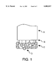
FIG. 1 schematically shows one embodiment of an optical fiber sensor of the present invention having a monolayer of metal spheres for attachment of a nitric oxide-binding protein.
FIG. 2 shows the heme group of one embodiment of a nitric oxide-binding protein, i.e. cytochrome c'.
FIG. 3 shows one embodiment of an optical array useful for testing the sensors of the present invention.
FIG. 4 is a graph showing the response of one embodiment of an optical fiber sensor of the present invention, said fiber having cytochrome c' of C. vinosum immobilized on gold colloid.
FIG. 5 is a graph showing the response of one embodiment of an optical fiber sensor of the present invention, said fiber having cytochrome c' of R. purpureus immobilized on gold colloid.
FIG. 6 is a graph showing the response of one embodiment of an optical fiber sensor of the present invention, said fiber having cytochrome c' of R. gelatinosa immobilized on gold colloid.
FIG. 7 shows the reversibility of one embodiment of the sensor of the present invention.
DEFINITIONS
To facilitate understanding of the invention, a number of terms are defined below.
The term "analyte" is intended to comprise any substance within a cell, including but not limited to nitric oxide and Na+, K+, Ca++, Cl-, H+, as well as oxygen and glucose.
The term "bacteria" refers to any bacterial species including eubacterial and archaebacterial species.
The term "chemical reaction" means reactions involving chemical reactants, such as inorganic compounds.
The present invention contemplates "metal conjugates" such as conjugate comprising a nitric oxide-binding compound attached to a metal, including but not limited to, metal in the form of a metal colloid.
Colloids are solutions of dispersed or suspended particles. Where the particles comprise metal, the colloid is referred to as a "metal colloid".
The present invention contemplates "porphyrin group-containing proteins" and more preferably "heme group-containing proteins" capable of binding (and more preferably, binding selectively) nitric oxide. The porphyrins are named and classified on the basis of their side chain substituents, such as etioporphyrins, mesoporphyrins, uroporphyrins, coproporphyrins and protoporphyrins. The chelate complex of a porphyrin (e.g. protoporphyrin) with metal is contemplated by the present invention as useful for nitric oxide binding. The chelate complex with iron [e.g. Fe(II), Fe(III)] is heme. Thus, the present invention specifically contemplates the use of ferrous and ferric hemoproteins to bind nitric oxide, and fragments thereof (preferably heme group containing fragments).
It is not intended that the present invention be limited by the type of porphyrin group-containing protein. In one embodiment, the present invention contemplates cyclases, including but not limited to, guanylate cyclase (which contains one protoporphyrin-IX type heme/heterodimer) and fragments thereof. See generally Stone and Marletta, "Soluble Guanylate Cyclase from Bovine Lung: Activation with Nitric Oxide and Carbon Monoxide and Spectral Characterization of the Ferrous and Ferric States," Biochemistry 33:5636 (1994). In another embodiment, the present invention contemplates ferrous hemoproteins such as hemoglobin and myoglobin, or fragments thereof. In still another embodiment, the present invention contemplates cytochromes or portions of cytochromes as nitric oxide binding compounds. In a preferred embodiment, the present invention contemplates cytochrome c' (as distinct from cytochrome c) and fragments thereof.
The present invention also contemplates compounds that bind porphyrins and heme. By the term "heme binding proteins" it is meant to indicate proteins that bind heme groups. It is not intended that the present invention be limited to particular heme group-binding proteins. In one embodiment, the heme-binding compound is the 93,000 molecular weight, heme-binding protein from rabbit serum. See Tsutsui and Mueller, J. Biol. Chem. 257, 3925 (1982). In another embodiment, the heme-binding protein is selected from the group consisting of globin, serum albumin, and transferrin. In yet another embodiment, the present invention contemplates the heme-binding protein isolated and characterized from both the hemolymph and oocytes of the blood-sucking insect, Rhodnius prolixus.
A compound is "immobilized" by attachment (whether covalent or non-covalent) or by encapsulation (e.g. in a gel such as a sol-gel).
In one embodiment, the present invention contemplates that the nitric oxide-binding compound is a protein (or peptide) and the protein (or peptide) is dye-labeled. There are three major classes of commonly used reagents to label amines: succinimidyl esters, isothiocyanates and sulfonyl chlorides. Dyes containing such groups are commercially available from Molecular Probes, Inc. (Eugene, Oreg.) and include, but are not limited to, such fluorophores as cascade blue, isosulfan blue, fluorescein, naphthofluorescein, malachite green and Oregon green). "Initiating a reaction" means causing a reaction to take place. Reactions can be initiated by any means (e.g. heat, wavelengths of light, addition of a catalyst, etc.) "Interfering substances" are those substances that interfere with the measurement of nitric oxide. Such substances include but are not limited to nitrite, dopamine, uric acid, ascorbic acid, and cysteine.
The term "microorganism" as used herein means an organism too small to be observed with the unaided eye and includes, but is not limited to bacteria, viruses, protozoans, fungi, and ciliates.
"Reactive groups" are those chemical groups that are capable of reacting with (and thereby capable of securing) compounds or substances to the fiber tip. Such groups include groups having a S, N or O functionality, including but not limited to thiol groups, carboxylic acids and amino groups.
A "solvent" is a liquid substance capable of dissolving or dispersing one or more other substances. It is not intended that the present invention be limited by the nature of the solvent used.
DESCRIPTION OF THE INVENTION
The invention relates generally to optical sensors, methods of sensor fabrication and uses of such sensors, and more particularly the use of such sensors for the detection if nitric oxide. The present invention contemplates both fiber-optic sensors and optical fiberless sensors comprising nitric oxide binding compounds, such compounds permitting the specific binding of nitric oxide with little or no interference from other analytes. This nitric oxide sensors of the present invention are small (100 μm and submicrometer), very selective and can be used for dynamic, real-time measurements.
A. Fiber-Optic Sensors And Their Fabrication
Micro-fiberoptic sensors (100-1000 nm) are based on optical grade silica fibers pulled to submicron size. The pulled fiber tips are much less fragile than those of the electrochemical microsensors, which are made from pulled micropipettes. Traditionally, a dye-polymer matrix is attached to the tip, which is very durable and smooth and runs tightly bound to the tip, even during penetration of biological tissues. The matrix on the end of the fiber often includes several components, such as a chromoionophore, an ionophore, and appropriate ionic additives, all trapped inside a polymer layer, so that no chemicals are free to diffuse throughout the cell. The effects of toxicity of the dyes are thus minimized. Also, the probe can be carefully positioned in the cell, allowing any specific area to be imaged or monitored.
The nitric oxide detecting sensors of the present invention, by contrast, has a metal monolayer attached to the tip. The fabrication involves treatment of the fiber so as to add reactive groups. Thereafter, the metal monolayer is attached, followed by immobilization of the nitric oxide-binding protein (see FIG. 1, where the fiber (A), the reactive groups (B), the metal monolayer (C) and the attached protein (D) are shown).
B. Optical Fiberless Sensors And Their Fabrication
The present invention contemplates nitric oxide detecting sensors that are fiberless sensors or Probes Encapsulated By BioListic Embedding (PEBBLEs). While a variety of such fiberless sensors are contemplated, the preferred fiberless sensors of the present invention are those made with metal particles, and in particular, metal colloids.
The fiberless sensors are particularly suitable for chemical analysis in mammalian cells, by inserting the sensors into the cell, and monitoring remotely. The sensor particles or beads can be dispersed in buffer solution and pico-injected into a cell. The particles can be monitored singly, in groups located at different positions, or several different kinds can be injected for simultaneous measurements of several distinct intracellular ion or small molecule concentrations.
The fiberless sensors of the present invention are prepared by coating metal (e.g. Au) colloids with a nitric oxide binding compound (e.g. cytochrome c') or a dye-labeled nitric oxide-binding compound. For some cellular applications, a phospholipid layer is added to the fiberless sensors contemplated by the present invention.
C. Uses of the Present Invention
The invention will be useful for, among other things, in the identification of cellular and subcellular responses which lead to disease. The fields in which the optical sensors of the present invention will find application are vast, and include basic research, diagnosis, and treatment of disease. Direct benefits to humans and the environment include the development of new drugs, understanding the synergistic response to complex mixtures of pollutants, and prevention of developmental and degenerative disorders.
As noted above, nitric oxide is emerging as one of the main neurotransmitters in the central and peripheral nervous systems. Accurate detection and measurement of nitric oxide in cells may be used diagnostically, given the role of nitric oxide in AIDS dementia, Parkinson's and Huntington's diseases.
1. Metal Colloids On Optical Fibers
While the nitric oxide detection features of the present invention have been underscored, the present invention brings elements that can be used more generically if desired. Specifically, the use of metal colloids on optical fibers can be used to bind proteins other than nitric oxide binding proteins.
For example, an environmentally sensitive probe can be prepared using a metal layer (e.g. gold colloid) on the fiber tip. A dye can be attached to the end of a fiber through a non-fluorescent protein (such as bovine serum albumin) and used as an indicator of the hydrophobicity of differing regions of a cell. All that has to be done is to use a dye which can easily be attached to a protein, and which is sensitive to changes in environment. Alternatively, any protein or peptide (such as a zinc finger peptide) which is sensitive to environmental changes or selectively binds certain analytes (such as Zn2+) can be used. Such proteins/peptides may be naturally fluorescent or labeled with fluorescent dyes. Attaching dyes to proteins can also be a useful method for measuring analytes in general.
Thus, the present invention contemplates fiber-optic sensors comprising an optical fiber having a fiber tip, said tip comprising an immobilized protein and a dye. The protein can be any protein that is inert (i.e. inert relative to analyte sought to be detected). The protein can be immobilized using a metal layer (preferably, a non-linear layer and more preferably, spheres comprising metal). In one embodiment, the tip of the fiber is treated so as to have reactive groups and the spheres of metal colloid are attached to the tip via the reactive groups. In this latter embodiment, a fiber tip/reactive group/metal colloid/immobilized protein/selective dye complex is created.
2. Fiberless Sensors Comprising Metal Particles
As noted above, the nitric oxide detection features of the fiberless sensors of the present invention have been underscored. However, it is contemplated that fiberless sensors of the present invention can be used to measure any alteration in any endogenous analytes of any cell. The present invention specifically contemplates transcutaneous monitoring (e.g. ear, skin) as well as continuous flow monitoring of cells in culture, organotypic culture, organ slices, isolated perfused organs, organs in situ, and whole animal monitoring.
The present invention contemplates that the fiberless sensors of the present invention can be used as diagnostic tools for earlier intervention (i.e. earlier than currently available) and treatment of disease.
In one embodiment, the metal fiberless sensors of the present invention comprise a protein (or peptide) conjugated to a metal such as a metal colloid. The protein can be any protein that is inert (i.e. inert relative to analyte sought to be detected), and linked to a dye. Alternatively, any protein or peptide which is sensitive to environmental changes or selectively binds certain analytes (such as Zn2-) can be used. Such proteins/peptides may be naturally fluorescent or labeled with fluorescent dyes. The protein can be immobilized using metal particles (preferably, a non-linear and more preferably, spheres comprising metal). In this latter embodiment, a metal colloid spherical particle/immobilized protein/selective dye complex is created.
3. Applications
Various fiber-optic tip sensors with fiberless ("pebble") sensors can be used. The fiber-optic nanotip fluorescent sensors are inserted into the cell by standard micropipette ("patch clamp") techniques. The chemical sensor nanotips and pebbles, with each tip or pebble specific to a given chemical analyte. Utilizing these optical nanosensors one can get a chemical video for a single analyte (say, calcium), analogous to a black and white movie, or of a list of chemical analyte (say, calcium, sodium, potassium, chloride, oxygen and pH), analogous to a color video or, alternatively, to six single color videos, each taken with a different narrow-band optical filter.
The cell is located under a microscope objective lens, and may be immobilized by a micopipette (standard technique). One or several fiber-optic nano-tips are inserted by fine manipulators. The fiber-optic tips may be multi-functional sensors (simultaneously measuring 2 or more analytes) and/or multiplexed fiber tips (simultaneously measuring 2 or more sensors). If sensor nanopebbles are inserted (via micropipette), they are addressed optically by laser beams, either directly or via optical fibers.
Elimination of cross-talk among the different nanosensors is based on a combination of spatial, temporal and spectral resolution. Different fiber-optic tip sensors are excited by different lasers or laser wavelengths. Image focusing on an intensified CCD (requested item) separates spatially the fluorescence from different nano-sensors. Spectral control of the excitation wavelength and spectral selection of the fluorescence wavelength separates the various signals.
EXPERIMENTAL
The following examples serve to illustrate certain preferred embodiments and aspects of the present invention and are not to be construed as limiting the scope thereof. In the experimental disclosure which follows, the following abbreviations apply: TEA (triethylamine; EDTA (ethylenediaminetetraacetic acid); IBMX (isobutylmethylxanthine); DTT (dithiothreitol; GTP (guanosine 5'-triphosphate); SNP (sodium nitroprusside).
EXAMPLE 1
In this example, sensors were prepared by silanizing a freshly cleaved multimode fiber by immersing the distal end for two hours in 3-(mercatopropyl) trimethoxysilane, thereby modifying the fiber with reactive groups. The end was then rinsed copiously with methanol, then tripley distilled water. The silanized fiber was placed in colloidal gold (used as received from the manufacturer) for three hours, then rinsed with water. The sensor can be stored at this point in water or air.
Two protein solutions were prepared. The first solution was prepared by dissolving 0.1% r-phycoerythrin (Molecular Probes, Eugene, Oreg.) in phosphate buffer, pH 6.0. The fiber was then immersed in the protein solution for one hour, rinsed with buffer, then used.
A second protein solution was prepared by dissolving 0.1% bovine serum albumin (BSA) in phosphate buffer, pH 6.0. The fiber was then immersed in the protein solution for one hour, rinsed with buffer, then used. After BSA attachment, the fiber tip was placed in Texas Red-X (Molecular Probes, Eugene, Oreg.) solution for one hour. The dye solution was prepared by dissolving 1 mg Texas Red-X in dimethyl sulfoxide (DMSO) then adding 1 M sodium bicarbonate solution (pH 8.3). The sensor was rinsed with water before use.
The complete optical path for the fiber optic sensors included: Ion Laser Technology (Salt Lake City, Utah) argon ion laser; 514.5 nm laser band-pass filter (Newport Corp. Irvine, Calif.); Uniblitz shutter controller (Rochester, N.Y.); fiber coupler (Newport Corp. Irvine, Calif.); Olympus inverted fluorescence microscope, IMT-II (Lake Success, N.Y.); Nikon 50 mm f/1.8 camera lenses; Acton 150 mm spectrograph (Acton, Mass.); and, a Princeton Instruments 1024×256 LN2 cooled CCD array (Trenton, N.J.).
The results showed that, based on representative spectra of both the R-phycoerythrin and the Texas red labeled- bovine serum albumin, the proteins were immobilized onto the end of an optical fiber (data not shown). More importantly, although there is only a monolayer of fluorescent material on the surface of the optical fiber, a strong signal was measured (data not shown). Interestingly, the results indicate that the size of the gold colloid used in the sensor development plays a significant role in the amount of fluorescence signal obtained, with 100 nm particles showing what appear to be the best results (data not shown).
EXAMPLE 2
In this example, pulled sensors are described. Sub-micron sized fiber optic sensor were prepared by pulling multimode fiber in a home-built puller consisting of a modified pipette-puller heated with a CO2 laser. The pulled tips were then silanized, coated with aluminum in a home-built evaporator and prepared with gold colloid and protein as above.
EXAMPLE 3
This example describes the first sensor to incorporate cytochrome c'. The heme group of this nitric oxide-binding protein is shown in FIG. 2. Cytchrome c' was chosen as the chemical recognition element because it exhibits spectral changes upon binding nitric oxide and is highly selective. While a precise understanding of the mechanisms involved is not necessary for the practice of the invention, it is believed that, as the sixth ligand site of cytochrome c' is buried within the protein, it is usually accessible only to carbon monoxide and nitric oxide. However, cytochrome c' undergoes autooxidation to Fe(III), which binds nitric oxide, but not carbon monoxide.
To prepare the sensor, 100 μm core diameter multimode fibers (General Fiber Optics, Cedar Grove, N.J.) or multimode fibers were pulled to a submicrometer tip diameter and were silanized for 2 hrs in neat 3-mercaptopropyl-trimethoxy silane (Gelest, Inc., Tullytown, Pa.) in a well-ventilated fume hood. The fibers were rinsed with methanol and water and placed in 50 nm gold colloid (Vector Labs, Inc., Burlinggame, Calif.) for 3 hrs at 0° C. to form a self-assembled monolayer of gold colloids. The gold-coated fibers were immersed overnight in either a Chromatium vinosum, Rhodocyclus purpureus, or Rhodocyclus gelatinosus cytochrome c' solution prepared with pH 7.4 phosphate buffer at 15° C. to allow the cytochrome c' to adsorb to the surface of the gold via the protein amine groups. The fiber was then rinsed in buffer to remove any residual protein. Dye-labeled sensors were prepared with 4-carboxy-3,5,6 trifluoro -2',7',- difluorofluorescein, succinimidyl ester (Oregon Green™ 514 carboxylic acid, succinimidyl ester) purchased from Molecular Probes (Eugene, Oreg.). A 16 mM dye solution in DMSO was diluted to 1 M with 0.1 M NaHCO3. The cytochrome c' sensors were soaked for 1 hr in the dye solution.
The gold colloid-containing sensors that were prepared with either C. vinosum, R. purpureus, or R. gelatinosa cytochromes c' were tested. One embodiment of the optical array useful for testing is shown schematically in FIG. 3). All three cytochromes c'-containing sensors were found to have linear responses up to 1 mM nitric oxide (FIGS. 4-6). Above this concentration is a second linear region with a slope which is three times that below 1 mM, as shown in FIG. 7. The steeper slope found at higher concentrations may be due to spectroscopic changes upon binding a second nitric oxide molecule per monomer, or may be caused by each cytochrome c' dimer binding more than two nitric oxide molecules. In view of their structural similarities, it is not surprising that all three cytochromes c' have comparable slopes. The C. vinosum (FIG. 7, where squares indicate increasing nitric oxide concentration and circles indicate decreasing nitric oxide concentration) and R. purpureus cytochromes c' are readily reversible. The R. gelatinosa cytochrome c' does not respond reversibly, but the reasons for this difference have not yet been determined. The present limit of detection is approximately 20 μM nitric oxide. While the reaction of C. vinosum cytochrome c' has been reported to be slow when measured in solution via absorbance, the results using the sensors of the present invention show that the sensor's response time is less than 2 seconds.
The C. vinosum, R. purpureus, and R. gelatinosa cytochromes c' sensors were tested for selectivity against common interfering chemicals. 1M nitrate and nitrite, oxygen, nitrogen, and 1.5×10⁻⁴ M ascorbic acid, in phosphate buffer solution, were found to have no effect on the sensor response (data not shown). Low μM riboflavin was found to interfere with dye-labeled sensors due to its fluorescence band in the 530 nm region, overlapping with that of the labeling dye. The addition of 4 μM riboflavin was equivalent to a 30 μM decrease in nitric oxide concentration. However, riboflavin does not affect the cytochrome c' fluorescence. Dopamine at high levels also interfered with the detection of nitric oxide. In solution, dopamine was found to cause a blue shift in the protein fluorescence. For sensors prepared without the labeling dye, the addition of 140 mM dopamine was, on average for the three cytochromes c', equivalent to the addition of 0.1 mM nitric oxide. For the dye-labeled sensors, 0.25 mM dopamine was, on average, equivalent to the addition of 0.1 mM nitric oxide. Sensors made with the cytochromes c' alone (without dye labeling) were not affected by pH between pH 3-10. The dye fluorescence is sensitive to pH below 7. In solution, hydrogen peroxide caused an attenuation of the cytochrome c' fluorescence, but no spectral shift. Dye-labeled sensors exhibited an average signal increase of 4% in 1M hydrogen peroxide, equivalent to the removal of 0.2 mM nitric oxide. However, the concentration of oxygen radicals needed to interfere with the sensor response is higher than that found in many biological systems.
EXAMPLE 4
In this example, a heme-binding protein is prepared and attached to a metal monolayer of a fiber tip. The heme-binding protein prepared is the protein that been isolated and characterized from both the hemolymph and oocytes of the blood-sucking insect, Rhodnius prolixus. See generally P. L. Oliveira et al., "A Heme-binding Protein from Hemolymph and Oocytes of the Blood-sucking Insect, Rhodnius prolixus," J. Biol. Chem. 270:10897 (1995). The Rhodnius heme-binding protein (RHBP) is composed of a single 15-kDa polypeptide chain coiled in a highly a-helical structure which binds non-covalently one heme/polypeptide chain. This RHBP is not produced by limited degradation of hemoglobin from the vertebrate host, since specific polyclonal antibodies against it do not cross-react with rabbit hemoglobin, and since it differs from hemoglobin in having a distinct amino-acid composition and NH2 -terminal sequence. The spectrum of the dithionite-reduced protein has peaks at 426, 530, and 559 nm and resembles that of a b-type cytochrome.
RHBP from hemolymph is not saturated with heme and promptly binds heme added to the solution. The oocyte protein, on the other hand, is fully saturated and is not capable of binding additional heme.
For preparation of the protein, insects are taken from a colony of R. prolixus maintained at 28° C. and 70% relative humidity. Normal mated females are fed on rabbit blood at 2-week intervals. Four to 6 days after a meal, hemolymph is collected in the presence of phenylthiourea (30-130 μg/ml), 5 mM EDTA, and a mixture of protease inhibitors prepared in 0.15 M NaCl, with final concentrations of 0.05 mg/ml of soybean trypsin inhibitor, leupeptin, lima bean trypsin inhibitor and antipain, and 1 mM benzamidine. On the same day, chorionated oocytes are dissected and washed with ice-cold 0.15 M NaCl in order to remove ovarian debris prior to homogenization. Oocytes are homogenized in a Potter-Elvehjem homogenizer in the presence of the same mixture of protease inhibitors, buffered with 20 mM Tris-HCl, pH 7.0, (approximately 500 oocytes to 1 ml), and centrifuged at room temperature in a microcentrifuge at 11,000×g for 5 min. The floating lipids and the pellet are discarded, and the clear supernatant is used as the crude oocyte extract for protein purification.
Solid ammonium sulfate is added to bring the oocyte extract to 45% saturation, and the suspension is gently stirred for 20 min at 4° C. After centrifugation at 11,000×g for 10 min, the precipitate is discarded, and the supernatant is brought to 60% saturation. This new precipitate is then washed twice with a 60% saturated ammonium sulfate solution and then back-extracted by resuspending in a 45% saturated solution and centrifuging. The pellet is discarded and the supernatant is dialyzed against 0.15 M NaCl, 10 mM Tris-HCl, pH 7.0, and applied to a column of Sephadex G-200 (2.5×55 cm) equilibrated with the same solution. Protein content of fractions is measured by the absorbance at 280 nm. The colored fractions containing RHBP are pooled, dialyzed against deionized water, and lyophilized.
Hemolymph (approximately 3 ml) is diluted to 5 ml with phosphate-buffered saline (0.15 M NaCl, 0.1 M sodium phosphate, pH 7.0) and 1.25 g of KBr is added. The solution is centrifuged at 80,000×g for 20 h at 4° C. The fractions at the bottom of the tube are collected and dialyzed against deionized water until an abundant precipitate forms. The solutions is then centrifuged at 11,000×g for 10 min at 4° C. The supernatant is brought to 10 mM with Tris base and applied to a column (1.5×18 cm) of DEAE-Toyopearl, equilibrated with 10 mM Tris-HCl, pH 8.4. The column is first washed with 20 ml of the same buffer and then eluted with and NaCl gradient (0-100 mM). The fractions containing RHBP are pooled and applied to a Sephadex G-75 column (1.5×80 cm) equilibrated with 0.15 M NaCl, 10 mM Tris-HCl, 7.0. Fractions containing the RHBP are pooled, dialyzed against deionized water, and lyophilized.
Proteins from both sources are monomeric, as indicated by an apparent molecular weight of between approximately 12,000 and 15,000. The purified protein (0.01%) is added to metal colloid as described above for the other proteins.
EXAMPLE 5
In this example, a nitric-oxide-carrying hemoprotein is prepared from a salivary gland homogenate of the bedbug Cimex Lectularius. The insect colonies are maintained at 27 C and 65% humidity. Insects are fed every 10 days by exposing them to the shaved abdomen of an anesthetized rabbit. Salivary glands of insects at 8-10 days after feeding are dissected and stored in buffers or used immediately. In any event, they are disrupted with a disection needle and the homogenate centrifuged in the cold at 14,000 RPM for 5 minutes. The supernatants are recovered and used for binding to metal coloids as described above.
The salivary gland homogenates have a nitrosyl-hemoprotein that releases nitric oxide in a pH-dependent manner. The fraction containing the NO-carrying hemoprotein, when separated by HPLC, causes vasodilation of a preconstricted rabbit aortic strip.
EXAMPLE 6
This example describes the preparation of soluble guanylate cyclase ("sGC"). See generally Stone and Marletta, "Soluble Guanylate Cyclase from Bovine Lung: Activation with Nitric Oxide and Carbon Monoxide and Spectral Characterization of the Ferrous and Ferric States," Biochemistry 33:5636 (1994). sGC can be purified from bovine lung. Steps should be performed at 4° C. During the purification, assays for sGC can be carried out in 50 mM TEA, pH 7.4, with 2 mM DTT, 5mM MnCl2, 100 μM GTP, 1 mM IBMX, 5mM phosphocreatine, and 152 units/L creatine kinase, with or without 100 μM sodium nitroprusside (SNP) in a total volume of 100 μL at 37° C. The assays are quenched with 400 μL of 125 mM Zn(CH3 COO)2 and 500 μL of 125 mM Na2 CO3. The amount of cGMP generated can then be quantiated via radioimmunoassay (Amersham). After each column, only active fractions which can be activated at least 10-fold with SNP are carried on to the subsequent step.
Fresh bovine lung (1100 g) is homogenized with a food processor in 1100 mL of homogenizing buffer (25 mM TEA, 5 mM DTT, 1 mM PMSF, 1 mM EDTA, 0.2 mM benzamidine, 1 μM pepstatin A, 1 μM leupeptin, pH 7.8). The homogenate is then centrifuged at 100000 g for 60 min. The resulting supernatant is added to to 450 mL of DEAE-Sepharose CL-6B and stirred slowly with an overhead stirrer for 45 min. The buffer is removed by vacuum filtration, and the resin washed four times by stirring slowly for 15 min with 1200 mL of homogenizing buffer. The resin is then poured into a 2.5×100 cm column and packed by washing the column with homogenzing buffer for 1 h at 80 mL/h. sGC is then eluted with 1 L of a 0.0-0.4 M NaCl gradient at 80 mL/h. Active fractions are pooled (140 mL) and dialyzed for 8 h against 4 L of 25 mM TEA, 5 mM DTT, 0.1 mM PMSF; 0.2 Mm benzamidine, 1 μM pepstatin A, 1 μM leupeptin, ph 7.4.
The sample is then brought to 5 mM MnCl2 and applied μ 25 mL/h to a 75-mL column of ATP-agarose (11-atom spacer attached through C-8). The column is washed at 25 mL/h with 150 mL of buffer A (25 mM TEA, mM DTT; 1 mM PMSF, 0.2 mM benzamidine, 5 mM MnCl2, pH 7.4). The column is then washed at 50 mL/h with 200 mL of buffer A containing 10 mM NaNO3, 10 mM creatine, and 5 mM ADP followed by 300 mL of buffer A. sGC is then eluted at 50mL/h with a 2-L gradient running from 5 mM MnCl2 to 1 mM EDTA. Active fractions are pooled (200 mL), concentrated by ultrafiltration to 30 mL on a YM-30 membrane (Amicon), and brought to 5 mM MnCl2.
The sample is then applied at 5 mL/h to a 3.0-mL column of GTP-agarose (11-atom spacer attached through ribose hydroxyls, ICN). The column is washed at 5 mL/h with 10-mL of buffer A, and then sGC is eluted with a 100-mL gradient running from 0.0 to 0.4 M NaCl in buffer A. Active fractions are pooled, concentrated by ultrafiltration to 8 mL on an Omega Series membrane (30 kDA MWCO, Filtron), and applied to a 2.5×95 cm column of Ultrogel AcA-34 (IBF) at 20 mL/h. The column is then washed with 400 mL of 25 mM TEA, 5mM DTT pH 7.8 at 20mL/h. Active fractions are pooled (20mL), provided with additional DTT (5 mM final), brought to 1.0 M NaCl, and concentrated by ultrafiltration to 1.0 mL on an Omega Series membrane. The sample is brought to 50% glycerol and stored as aliquots under nitrogen at -70° C.
The above-described purification scheme results in the isolation of approximately 0.5 mg of sGC from 1100 g of bovine lung. Following the procedure, two bands should be visible on reducing SDS-PAGE of approximately equal intensity with apparent masses of 69 and 78 kDA. The protein prepared as described above can then be immobilized on metal colloids.
EXAMPLE 7
This example describes the preparation of macro-sensors (sensors made on larger substrates, i.e. larger than fiber optic sensor tips). The sensor was made on glass microscope slides cleaned by immersion in piranha solution (10:1 sulfuric acid :hydrogen peroxide), copious rinsing in triply distilled water and oven drying. The glass was then silanized in a 10% solution of 3-(mercatopropyl) trimethoxysilane in methanol for 24 hours. The glass was rinsed with methanol, then tripley distilled water. The silanized glass was placed in colloidal gold (used as received from the manufacturer) for 24 hours, then rinsed with water. For comparison of sizes of gold colloids, six different gold sizes were used: 5, 10, 20, 50, 100 and 250 nm.
From the above, it should be clear that the methods, devices and compositions of the present invention permit for the design of sensors that have excellent selectivity, reversibility, fast response time, and can be reduced to submicrometer scale for use in single-cell or other in vitro applications.