US5830701A - Method of detecting hematopoietic progenitor cells - Google Patents
Method of detecting hematopoietic progenitor cells Download PDFInfo
- Publication number
- US5830701A US5830701A US08/829,239 US82923997A US5830701A US 5830701 A US5830701 A US 5830701A US 82923997 A US82923997 A US 82923997A US 5830701 A US5830701 A US 5830701A
- Authority
- US
- United States
- Prior art keywords
- cells
- hematopoietic progenitor
- cell
- progenitor cells
- count
- Prior art date
- Legal status (The legal status is an assumption and is not a legal conclusion. Google has not performed a legal analysis and makes no representation as to the accuracy of the status listed.)
- Ceased
Links
Images


Classifications
-
- G—PHYSICS
- G01—MEASURING; TESTING
- G01N—INVESTIGATING OR ANALYSING MATERIALS BY DETERMINING THEIR CHEMICAL OR PHYSICAL PROPERTIES
- G01N33/00—Investigating or analysing materials by specific methods not covered by groups G01N1/00 - G01N31/00
- G01N33/48—Biological material, e.g. blood, urine; Haemocytometers
- G01N33/50—Chemical analysis of biological material, e.g. blood, urine; Testing involving biospecific ligand binding methods; Immunological testing
- G01N33/5005—Chemical analysis of biological material, e.g. blood, urine; Testing involving biospecific ligand binding methods; Immunological testing involving human or animal cells
- G01N33/5094—Chemical analysis of biological material, e.g. blood, urine; Testing involving biospecific ligand binding methods; Immunological testing involving human or animal cells for blood cell populations
Definitions
- This invention relates to a method of detecting hematopoietic progenitor cells (HPCs).
- the method is capable of simple counting of peripheral blood HPCs for use as transplants in PBSCT (peripheral blood stem cell or PBSC transplantation) and it provides a useful index for determining the right time to collect PBSCs.
- PBSCs are the most primitive fraction of HPCs and are considered the "origin" or stem cells of all hematopoiesis.
- the term "hematopoietic progenitor cells” as used herein is synonymous with but broader in sense than the term "peripheral blood stem cells”.
- BMT Bone marrow transplantation
- the general procedure of PBSCT is as follows. First, the patient is administered chemotherapy, hematopoietic growth hormones (interleukins e.g. granulocyte colony stimulating factor (G-CSF)) and any other necessary reagents, whereupon the leukocyte count in the peripheral blood initially decreases while on the subsequent day 5-7 or later, the leukocyte count starts to increase. Similarly, but not necessarily concurrently with this event, the number of hematopoietic stem cells also increases.
- G-CSF granulocyte colony stimulating factor
- the stem cells are collected by means of a blood component separator and stored frozen. In this instance, it is necessary to know exactly when the number of stem cells in the peripheral blood is increased. The timing of mobilization and collecting of the hematopoietic stem cells should be neither too early nor too late in order to secure an adequate number of stem cells.
- the cancer cells are killed by applying very high levels of chemotherapy and/or radiotherapy that are sufficient to destroy the patient's bone marrow. Thereafter, the previously collected stem cells are transplanted so as to achieve rapid recovery of the hematopoietic capability of the patient.
- hematopoietic cells require efficient collection of hematopoietic cells and, to this end, the mobilization of peripheral stem cells has to be monitored correctly.
- Two common methods currently used to count stem cells are a colony assay method and a CD34 positive (a marker which is present on the cell surface of HPC) cell count method using flow cytometry (FCM).
- FCM flow cytometry
- the colony assay method the number of stem cells to be collected is adjusted appropriately in a suitable culture medium such as IMDM and cultivation is performed in a blood stem cell assay medium for 14 days in a CO 2 incubator while a colony count is obtained with a phase-contrast microscope.
- CD34 positive cell count method using FCM particularly in single-color analysis, a whole blood sample is reacted with a fluorescence-labelled anti-CD34 monoclonal antibody and, after hemolysis flow cytometry is performed and a CD34 positive cell count is obtained from a scattergram of lateral scattered light and flourescence or a histogram of fluorescence.
- flow cytometry is performed using an anti-CD34 monoclonal antibody and an anti-CD45 monoclonal antibody and the CD45 positive cells alone are first incorporated as data, which are then analyzed using lateral scattered light and CD34 fluorescence as parameters such that cells are counted which produce low levels of lateral scattered light and which express CD34.
- the colony assay method is the currently the most accurate way to obtain the correct PBSC count and this is sometimes used to perform the stem cell count on samples that have been subjected to apheresis.
- a problem with this method is the long time (2 weeks) that is required to complete the measurement and, therefore, it is not suitable to determine the right timing for HPC/PBSCs collection.
- the colony assay method is not a technique that assures good reproduction of the results and which can be adopted in routine work. High operating cost is another problem with this method.
- the CD34 positive cell count method can be completed in a short time but it still takes at least 1-2 hours in the counting procedure.
- the measurement is ideally continued for 3-5 days or more.
- the method under consideration is difficult to adopt as a routine technique on account of the high cost of equipment and reagents, as well as the scarcity of skilled engineers.
- gating is necessary to determine specific cell zones on a scattergram or histogram and this produces considerable differences from one facility to another.
- an automatic blood cell analyzer equipped with channels capable of detecting immature leukocytes has been introduced into the market as Model SE-9000 from Toa Medical Electronics Co., Ltd. and many reports were thereafter published on the results of PBSC counting using this model; examples are as follows: Lebeck, L. K. et al., International Society for Laboratory Hematology, Vol.1, No.1, p.62, 1995; Takekawa, K. et al., The Japanese Journal of Clinical Hematology, Vol.36, No.9, 1995; Yamada, H. et al., Japanese Journal of Medical Technology, Vol.145, No.3, p.501, 1996; Mougi, H. et al., Med.
- IMI immature leukocyte information, channel
- the IMI total includes the cells that appear in the zones for blasts, immature granulocytes and left shifts and the PBSC count is not the only parameter that is reflected by the IMI total.
- the IMI total which includes the above-mentioned rather mature juvenile leukocytes involves such substantial errors that it is not suitable for use in monitoring the appearance of PBSCs that can be adopted in PBSCT.
- the present invention has been accomplished under these circumstances and has as its objective to provide a method which is capable of detecting the appearance of HPCs in a simple manner.
- the stated object of the invention can be attained by a method for detecting and/or counting the appearances of hematopoietic progenitor cells, which comprises the steps of:
- FIG. 1. is a cell distribution profile constructed by SE-9000 measurement of the samples prepared by the method described in Example 1;
- FIG. 2 is a cell distribution profile constructed by SE-9000 measurement of peripheral blood samples in which HPCs were immobilized
- FIG. 3 is a cell distribution profile constructed by SE-9000 measurement of PBSC-mobilized peripheral blood samples within the zone of a scattergram which was delineated by the method of the invention:
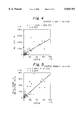
- FIG. 4 is a graph showing the correlationship between the IMI total and the CD34 positive cell count
- FIG. 5 is a graph showing the correlationship between the CD34 positive cell count and the HPC count as obtained by the method of the invention.
- FIG. 6 is a graph showing the daily changes in the CD34 positive cell count and the IMI total
- FIG. 7 is a graph comparing the daily change in the CD34 positive cell count with that in the HPC count obtained by the method of the invention.
- FIG. 8 is a graph showing the correlationship between the HPC count obtained by the method of the invention and the CFU-GM count obtained by the manual method.
- FIG. 9 is a chart illustrating the process of corpuscular differentiation and the relationship between each subclass of HPCs and the associated surface antigen.
- HPCs hematopoietic progenitor cells
- Hematopoietic progenitor cells consist of many subclasses including pluripotent stem cells, lymphoid stem cells, CFU-GEMM colony forming unitgranulocyte/erythroid/macrophage/megakaryocyte, BFU-E, CFU-E, CFU-Meg, CFU-GM colony forming unitgranulocyte/macrophage, CFU-EoS colony forming uniteosinophil, progenitor B cells and progenitor T cells (see FIG. 9).
- a reagent capable of detecting immature cells is used in the invention and taking, as an example, the case of granulocytes (i.e., neutrophils, eosinophils and basophils), the term “immature cells” refers collectively to those cells which are yet to reach the stage of mature cells in the process of differentiation of hematopoietic progenitor cells into mature cells by passing through the following successive stages: (myeloblasts ⁇ promyelocytes ⁇ myelocytes ⁇ metamyelocytes.fwdarw.band cells ⁇ segmented cells).
- the process of differentiation shown in FIG. 9 refers only to neutrophils and the illustration of eosinophils and basophils is omitted.
- a reagent capable of detecting immature cells without employing any immunological techniques refers to those reagents which are capable of detecting immature cells without employing any immunological reactions that take place between one of the cell surface antigens characteristic of the subclasses shown in FIG. 9 and the antibody directed against said antigen (e.g. an anti-CD34 antibody).
- a typical example of the reagent to be used in the invention is an aqueous solution containing a water-soluble surfactant that can lyse erythrocytes.
- a preferred surfactant is nonionic surfactant based on polyoxyethylene (POE).
- POE polyoxyethylene
- a more preferred example is one having the general formula of R 1 -R 2 -(CH 2 CH 2 O) n -H where R 1 is an alkyl, alkenyl or alkinyl group having 10-25 carbon atoms; R 2 is O, ##STR1## or Coo; and n is an integer of 10-40.
- Particularly preferred POE-base nonionic surfaces are those in which R 1 is an alkyl, alkenyl or alkinyl group having 10-25 carbon atoms, R 2 is O and n is an integer of 10-30, as exemplified by POE(15) oleyl ether, POE(16) oleyl ether, and POE(20) lauryl ether.
- the concentration of the nonionic surfactant varies from one surfactant to another; referring to the specific examples just mentioned above, POE(15) oleyl ether may be used at a concentration in the range of 1-9 g/L, preferably 3 - 7 g/L, POE(16) oleyl ether in the range of 5 -50 g/L, preferably 15-35 g/L, and POE(20) lauryl ether in the range of 0.1-2.0 g/L, preferably 0.5-1.5 g/L. Given the same number of carbon atoms in hydrophobic groups, polyoxyethylene-base nonionic surfactants have a greater potential to damage cells as the value of n decreases, and vice versa.
- the reagent may optionally contain a solubilizing agent, which causes both erythrocyte ghosts and mature leukocytes to shrink, thus enabling sharper discrimination of immature cells including hematopoietic progenitor cells.
- solubilizing agents are listed below: (1) sarcosine acid derivatives of following general formula or salts thereof: ##STR2## (where R 3 is an alkyl group having 10-22 carbon atoms; m is 1-5); (2) cholic acid derivatives of the following general formula: ##STR3## (where R is a hydrogen atom or a hydroxyl group); and (3) methylglucamides of the following general formula: ##STR4## (where y is 5-7).
- the preferred concentration of the solubilizing agent ranges from 0.2 to 2.0 g/L if it is a sarcosine acid derivative or salt thereof, 0.1-0.5 g/L in the case of a cholic acid derivative, and 1.0-8.0 g/L in the case of methylglucamide.
- the preferred solubilizing agent include: sodium N-lauroyl sarcosinate, lauroylmethyl ⁇ -alanine sodium salt, lauroyl-sarcosine, CHAPS (3- (3-cholamidopropyl) dimethylammonio!-1-propanesulfonate), CHAPSO (3- (3-cholamidopropyl) dimethylammonio!-2-hydroxy-1-propanesulfonate), MEGA8 (octanoyl-N-methylglucamide), MEGA9 (nonanoyl-N-methylglucamide), and MEGA10 (decanoyl-N-methylglucamide).
- Other useful solubilizing agents include n-octyl ⁇ -glucoside, sucrose monocaprate, and N-formylmethylleucylalanine and these may preferably be used at concentrations of 0.01-50.0 g/L.
- amino acid may be incorporated in the reagent under consideration and this is helpfhl for the purpose of identifying and classifying immature cells including hematopoietic progenitor cells.
- Any amino acids that are protein constitutents may be employed and particularly preferred amino acids are glutamic acid, valine and sulfur-containing amino acids such as methionine, cystine and cysteine.
- the amino acids may be used in amounts ranging from 1 to 50 g/L, with a preferred range of 8-12 g/L for glutamic acid and 16-24 g/L for methionine.
- the reagent may also contain a buffer agent in order to maintain a constant pH, preferably in the range of 5.0-9.0. As long as adjustment to this pH range is possible, any known buffer agents may be used without particular limitations.
- an osmolarity modifier may be added in order to adjust the osmolarity to be within the range of 150-600 mOsm/kg, preferably 250-380 mOsm/kg.
- the reagent may be employed with its electrical conductivity adjusted to 3.0-12.0 mS/cm, preferably 6.0-9.0 mS/cm.
- Suitable for use in the conductivity adjustment are electrolytes including halides, sulfates, nitrates, etc. of alkali metals and alkaline earth metals, for instance, sodium chloride and potassium chloride.
- the method of the invention may be implemented by the following procedure.
- a blood sample is mixed for reaction with the reagent capable of detecting immature cells.
- the blood sample Prior to mixing, the blood sample is diluted 100-500 folds, preferably 200-300 folds, with the reagent capable of detecting immature cells.
- the reaction temperature ranges from 25.0° to 40.0° C., preferably from 30° to 34° C.
- the reaction time ranges from 5 to 60 sec, preferably from 10 to 20 sec.
- At least one item of cell information is obtained from one cell.
- at least two items of cell information are obtained, for instance, the combination of cell size and cell interior information, or the combination of two pieces of cell interior information.
- the cell size information include a signal (DC signal) that are based on the difference between the electrical resistances of two cells that occurs when they pass through an aperture under application of a DC current and low-angle (e.g. 1-6 degrees with the optical axis) scattered light.
- Examples of the cell interior information include a signal (RF signal), high-angle (8-20 degrees with the optical axis) forward scattered light, lateral (70-110 degrees with the optical axis) scattered light, backward (120-180 degrees) scattered light and the degree of depolarization, all of which are based on the difference between the dielectric constants of two cells that occurs when they pass through the aperture under application of a high-frequency current.
- Two types of particle size analyzers are available that can produce more than one item of cell information, one being a known flow cytometer operating on an optical principle and the other being exemplified by models of NE and SE series (both available from Toa Medical Electronics Co., Ltd.) which rely on electrical resistance for operation by adopting the RF/DC detection system.
- the RF/DC system cell size information and cell interior information can be detected simultaneously by superposing a high-frequency signal on a dc current.
- the cell distribution profile is a one-dimensional histogram or, alternatively, a two- or three-dimensional scattergram. A two-dimensional scattergram is preferred.
- a portion of the constructed cell distribution profile has to be delineated as a zone for the appearance of these cells.
- a monocyte-rich sample is first prepared by means of a blood component separator.
- the sample is then reacted with magnetic beads having an anti-CD34 monoclonal antibody bound thereto (as commercially available under the trademarks DYNABEADSTM of DYNAL A.S.); thereafter, CD34 positive hematopoietic cells are separated with a magnetic cell separator (e.g. IsolexTM of Baxter Co., Ltd.). This is followed by treatment with an enzyme so as to free the CD34 positive cells of the magnetic beads.
- a magnetic cell separator e.g. IsolexTM of Baxter Co., Ltd.
- the thus prepared sample is mixed with the reagent capable of detecting immature cells and measurements are performed by the RF/DC method using SE-9000 (Toa Medical Electronics Co., Ltd.). The same procedure is applied to a number of samples and the HPC appearance zone is delineated based on the scattergram pattern for the appearance of HPCs.
- the assay of the actual blood sample is extremely simple and rapid. Stated more specifically, the reagent capable of detecting immature cells is mixed with the blood sample for reaction and cell information is obtained with the particle analyzer to construct a cell distribution profile and the HPCs within the HPC appearance zone are counted. With an apparatus like SE-9000, the steps described above can be carried out by automated IMI channels; hence, it takes only about 1-2 min complete the assay of one blood sample.
- the method of the invention can detect at least one of the subclasses of HPCs, i.e., pluripotent stem cells, lymphoid stem cells, CFU-GEMM, BFU-E, CFU-E, CFU-Meg, CFU- GM,CFU-EoS, progenitor B cells and progenitor T cells, among which CFU-GM, CFU-GEMM and CFU-EOS are preferred.
- These subclasses of HPCs are all CD34 positive cells.
- a reagent kit comprising the above-described reagent (capable of detecting immature cells without employing any immunological techniques), for example; a nonionic surfactant; a solubilizing agent; an amino acid and a buffering agent; having a pH of 5.0-9.0, an osmolarity modifier providing an osmolarity of 150-600 mOsm/kg and an electrolyte having an electrical conductivity of 3.0-12.0 mS/cm will detect not only HPCs but also more mature cells including blasts (lymphoblasts and myeloblasts), juvenile granulocytes (promyelocytes, myelocytes and metamyelocytes) and left shifted cells (which usually means the increase in less segmented band neutrophils; however, for the purposes of the invention, this term will refer to band neutrophils per se). These cells may also appear in an HPC containing sample and will be a noise to understanding of the kinetics of HPCs with the aid of the
- the HPC count obtained by the method of the invention has a better correlation with the CD34 positive cell count than does the IMI total (compare FIGS. 4 and 5).
- the CD34 positive cell count starts to increase at day 5-7 after the initial administration of a chemical agent prior to the practice of HPC harvest, increases abruptly at day 10-12, then decreases approximately at day 17 and thereafter increases again.
- This behavior of CD34 positive cell count was monitored more faithfully by the cell count obtained by the invention method than by the IMI total (compare FIGS. 6 and 7). Therefore, the method of the invention is suitable for monitoring the daily changes in the HPCs in the peripheral blood.
- HPC count obtained by the method of the invention was also in good agreement with the number of CFU-GMs which are in a subclass of HPCs (FIG. 8).
- the practice of PBSCT also involves the administration of hematopoietic drugs such as G-CSF and the invention offers has an additional significant advantage in that it is capable of daily monitoring of the number of CFU-GMs which will appear predominantly as the result of the administration of hematopoietic drugs.
- the reagent capable of detecting immature cells which was used in all of the examples given below had the following formulation:
- a monocyte-rich sample was prepared by means of a blood component separator and reacted with magnetic beads having bound thereto an anti-CD34 monoclonal antibody (Becton Dickinson Immunocytometry Systems Co., Ltd.). Thereafter, CD34 positive HPCs were separated by means of a magnetic cell separator (IsolexTM of Baxter Co., Ltd.) and then freed of the magnetic beads by treatment with an enzyme (chymopapain).
- the treated sample was mixed with the reagent capable of detecting immature cells (for its formulation, see above) and measurements were conducted on ten samples with an automatic blood cell analyzer (SE-9000 of Toa Medical Electronics Co., Ltd.). On the basis of the pattern for the appearance of HPCs, a zone for their appearance was delineated on the scattergram (FIG. 1).
- Peripheral blood samples were drawn from patients treated with G-CSF and diluted 250 folds with the reagent capable of detecting immature cells (for its formulation, see above). Following the reaction at 33° C. for 13 sec, measurements were conducted with SE-9000 (Toa Medical Electronics Co., Ltd.) to produce the cell distribution profile shown in FIG. 2.
- the samples contained not only HPCs but also more differentiated juvenile leukocytes. (See FIG. 2, which shows the distribution ranging from blasts to juvenile cells.) These are included within a population heretofore counted as the IMI total, or the total number of juvenile leukocytes. As already mentioned, the IMI total refers to the sum of the counts within the respective zones for blasts, immature granulocytes and left shifted cells. HPC counting by the method of the invention
- the IMI total and the HPC count which were obtained by the methods described in Example 2 were checked for their correlationship with the CD34 positive cell count as obtained by flow cytometry using FACScan (Becton Dickinson Immunocytometry Systems Co., Ltd.).
- the CD34 positive cell count as obtained by flow cytometry started to increase at day 5-7 after the initial administration of a chemical agent prior to the practice of PBSCT, increased aburptly at day 10-12, then decreased approximately at day 17 and thereafter increased again.
- This behavior of the CD34 positive cell count was compared with the daily changes in the IMI total and the HPC count that were measured by the methods described in Example 2.
- the results are shown in FIGS. 6 and 7.
- FIG. 6 compares the daily change in the CD34 positive cell count with that in the IMI total
- FIG. 7 compares the daily change in the CD34 positive cell count with that in the HPC count in the zone delineated by the method described in Example 1.
- the cell count within the zone delineated by the method of the invention was more accurate than the IMI total in monitoring the change in the CD34 positive cell count, thus providing a more direct representation of the dynamic changes in the CD34 positive cell count.
- Example 5 The HPC count within the zone delineated by the method described in Example 1 was compared with the count of CFU-GMs in a subclass of HPCs for 10 samples.
- the CFU-GM count was obtained by the manual method involving colony counting under a microscope.
- the samples used in Example 5 were prepared by collecting peripheral blood stem cells after the administration of G-CSF.
- the present invention enables HPCs to be detected without employing any immunological techniques but using (1) a reagent capable of detecting immature cells and (2) a particle analyzer which provides the cell information.
- the invention provides a simple, quick and low-cost method for monitoring the dynamic changes in the HPC count in the peripheral blood and harvested HPC.
- the conventional colony culture method takes about 2 weeks to complete the assay and even the flow cytometry requires about ⁇ 2 hours.
- the detection method of the invention needs only 1-2 minutes to obtain the result which is a significant advantage over the two conventional techniques.
- the method of the invention can be implemented at a very low cost per test; at about one fiftieth of the cost of using the anti-CD34 monoclonal antibody.
- the method of the invention does not require any sophisticated skill in measurement and, hence, is free from the reproducibility problems that would be caused by different operators.
- a zone for the appearance of HPCs is delineated in the method of the invention and this enables the effects of blasts, juvenile granulocytes and band leukocytes to be sufficiently suppressed to accomplish a reliable detection of HPCs.
- PBSCT a continued, daily measurement for 3-5 days is ideal for monitoring the mobilization of stem cells within the peripheral blood and this can be easily accomplished by adopting the method of the invention.
- the invention offers a great benefit to the medical field where PBSCT is replacing BMT as an effective method of treatment of refractory diseases such as leukemia, lymphoma, myeloma, and a number of solid tumors.
Landscapes
- Life Sciences & Earth Sciences (AREA)
- Health & Medical Sciences (AREA)
- Immunology (AREA)
- Engineering & Computer Science (AREA)
- Urology & Nephrology (AREA)
- Chemical & Material Sciences (AREA)
- Cell Biology (AREA)
- Biomedical Technology (AREA)
- Molecular Biology (AREA)
- Hematology (AREA)
- Physics & Mathematics (AREA)
- Microbiology (AREA)
- Ecology (AREA)
- Tropical Medicine & Parasitology (AREA)
- Food Science & Technology (AREA)
- Medicinal Chemistry (AREA)
- Biotechnology (AREA)
- Analytical Chemistry (AREA)
- General Physics & Mathematics (AREA)
- General Health & Medical Sciences (AREA)
- Biochemistry (AREA)
- Pathology (AREA)
- Investigating Or Analysing Biological Materials (AREA)
- Micro-Organisms Or Cultivation Processes Thereof (AREA)
Priority Applications (5)
| Application Number | Priority Date | Filing Date | Title |
|---|---|---|---|
| US08/829,239 US5830701A (en) | 1997-03-28 | 1997-03-28 | Method of detecting hematopoietic progenitor cells |
| DE69806427T DE69806427T2 (de) | 1997-03-28 | 1998-03-18 | Verfahren zum Erfassen von Hämatopoetischen Vorläuferzellen |
| EP98104913A EP0867720B1 (en) | 1997-03-28 | 1998-03-18 | Method of detecting hematopoietic progenitor cells |
| JP06959598A JP3188236B2 (ja) | 1997-03-28 | 1998-03-19 | 造血前駆細胞の検出方法 |
| US10/704,463 USRE39006E1 (en) | 1997-03-28 | 2003-11-07 | Method of detecting hematopoietic progenitor cells |
Applications Claiming Priority (1)
| Application Number | Priority Date | Filing Date | Title |
|---|---|---|---|
| US08/829,239 US5830701A (en) | 1997-03-28 | 1997-03-28 | Method of detecting hematopoietic progenitor cells |
Related Child Applications (1)
| Application Number | Title | Priority Date | Filing Date |
|---|---|---|---|
| US10/704,463 Reissue USRE39006E1 (en) | 1997-03-28 | 2003-11-07 | Method of detecting hematopoietic progenitor cells |
Publications (1)
| Publication Number | Publication Date |
|---|---|
| US5830701A true US5830701A (en) | 1998-11-03 |
Family
ID=25253942
Family Applications (2)
| Application Number | Title | Priority Date | Filing Date |
|---|---|---|---|
| US08/829,239 Ceased US5830701A (en) | 1997-03-28 | 1997-03-28 | Method of detecting hematopoietic progenitor cells |
| US10/704,463 Expired - Lifetime USRE39006E1 (en) | 1997-03-28 | 2003-11-07 | Method of detecting hematopoietic progenitor cells |
Family Applications After (1)
| Application Number | Title | Priority Date | Filing Date |
|---|---|---|---|
| US10/704,463 Expired - Lifetime USRE39006E1 (en) | 1997-03-28 | 2003-11-07 | Method of detecting hematopoietic progenitor cells |
Country Status (4)
| Country | Link |
|---|---|
| US (2) | US5830701A (ja) |
| EP (1) | EP0867720B1 (ja) |
| JP (1) | JP3188236B2 (ja) |
| DE (1) | DE69806427T2 (ja) |
Cited By (9)
| Publication number | Priority date | Publication date | Assignee | Title |
|---|---|---|---|---|
| US6678040B1 (en) * | 1999-07-02 | 2004-01-13 | Terumo Kabushiki Kaisha | Apparatus for measuring number of cells |
| US20040023404A1 (en) * | 2002-07-01 | 2004-02-05 | Sysmex Corporation | Analyzers and methods for analyzing analytes |
| US20040166540A1 (en) * | 2003-02-24 | 2004-08-26 | Sysmex Corporation | Methods of detecting CD34 positive and negative hematopoietic stem cells in human samples |
| US20050003471A1 (en) * | 2003-07-03 | 2005-01-06 | Wang Fu-Sheng | Methods of detecting megakaryocytes |
| US20070231913A1 (en) * | 2006-03-29 | 2007-10-04 | Sysmex Corporation | Method and apparatus for measuring hematological sample |
| US20070248943A1 (en) * | 2006-04-21 | 2007-10-25 | Beckman Coulter, Inc. | Displaying cellular analysis result data using a template |
| US20080131898A1 (en) * | 2006-11-27 | 2008-06-05 | Sysmex Corporation | Method for measuring biological sample and measuring apparatus therefor |
| US20090215729A1 (en) * | 2008-02-19 | 2009-08-27 | Johnson Erin M | Microparticle compositions to modify cancer promoting cells |
| WO2022011285A1 (en) * | 2020-07-10 | 2022-01-13 | Beckman Coulter, Inc. | Automated sample preparation platform for cellular analysis |
Families Citing this family (4)
| Publication number | Priority date | Publication date | Assignee | Title |
|---|---|---|---|---|
| US7625757B2 (en) * | 2006-01-27 | 2009-12-01 | Sysmex Corporation | Reagent for immature leukocyte analysis and reagent kit |
| US20100047858A1 (en) * | 2008-08-20 | 2010-02-25 | Albano Maria S | High throughput system for cfu assay by the use of high resolution digital imaging, differential staining and automated laboratory system |
| WO2010132428A1 (en) * | 2009-05-11 | 2010-11-18 | Cellectar, Inc. | Fluorescent phospholipid ether compounds, compositions, and methods of use |
| US8871181B2 (en) | 2009-05-11 | 2014-10-28 | Cellectar, Inc. | Fluorescent phospholipid ether compounds, compositions, and methods of use |
Citations (2)
| Publication number | Priority date | Publication date | Assignee | Title |
|---|---|---|---|---|
| US5413938A (en) * | 1993-03-19 | 1995-05-09 | Tao Medical Electronics Co., Ltd. | Reagent for measuring immature leukocytes |
| US5538893A (en) * | 1994-04-21 | 1996-07-23 | Toa Medical Electronics Co., Ltd. | Reagent for analyzing leukocytes and a method for classifying leukocytes |
Family Cites Families (9)
| Publication number | Priority date | Publication date | Assignee | Title |
|---|---|---|---|---|
| JP2935529B2 (ja) * | 1990-03-01 | 1999-08-16 | シスメックス株式会社 | 白血球分類方法および試薬 |
| US5389549A (en) * | 1987-05-29 | 1995-02-14 | Toa Medical Electronics Co., Ltd. | Method for classifying leukocytes and a reagent used therefor |
| JP2619900B2 (ja) * | 1988-01-27 | 1997-06-11 | 東亜医用電子株式会社 | 血液中の白血球およびヘモグロビンの測定用試薬および方法 |
| AU2095992A (en) * | 1992-08-11 | 1994-03-24 | Abbott Laboratories | Flow cytometry sheath reagent for elimination of red cells with preservation of nucleated cells |
| JP3320869B2 (ja) * | 1993-12-22 | 2002-09-03 | シスメックス株式会社 | 白血球分析用試薬 |
| JP3355038B2 (ja) * | 1994-08-03 | 2002-12-09 | シスメックス株式会社 | 白血球分類方法 |
| US5639630A (en) * | 1995-05-16 | 1997-06-17 | Bayer Corporation | Method and reagent composition for performing leukocyte differential counts on fresh and aged whole blood samples, based on intrinsic peroxidase activity of leukocytes |
| US5817518A (en) * | 1995-12-18 | 1998-10-06 | Coulter International Corp. | Reagent and method for differential determination of leukocytes in blood |
| JP4042925B2 (ja) * | 1996-11-20 | 2008-02-06 | シスメックス株式会社 | 幼若白血球の分類計数法 |
-
1997
- 1997-03-28 US US08/829,239 patent/US5830701A/en not_active Ceased
-
1998
- 1998-03-18 EP EP98104913A patent/EP0867720B1/en not_active Expired - Lifetime
- 1998-03-18 DE DE69806427T patent/DE69806427T2/de not_active Expired - Lifetime
- 1998-03-19 JP JP06959598A patent/JP3188236B2/ja not_active Expired - Fee Related
-
2003
- 2003-11-07 US US10/704,463 patent/USRE39006E1/en not_active Expired - Lifetime
Patent Citations (2)
| Publication number | Priority date | Publication date | Assignee | Title |
|---|---|---|---|---|
| US5413938A (en) * | 1993-03-19 | 1995-05-09 | Tao Medical Electronics Co., Ltd. | Reagent for measuring immature leukocytes |
| US5538893A (en) * | 1994-04-21 | 1996-07-23 | Toa Medical Electronics Co., Ltd. | Reagent for analyzing leukocytes and a method for classifying leukocytes |
Non-Patent Citations (2)
| Title |
|---|
| Atzpodien et al "Human Bone Marrow CFU-GM & BFU-E Localized by Light Scatter Cell Sorting" Exp. Cell Biol. 55 (5) 265-270, 1987. |
| Atzpodien et al Human Bone Marrow CFU GM & BFU E Localized by Light Scatter Cell Sorting Exp. Cell Biol. 55 (5) 265 270, 1987. * |
Cited By (12)
| Publication number | Priority date | Publication date | Assignee | Title |
|---|---|---|---|---|
| US6678040B1 (en) * | 1999-07-02 | 2004-01-13 | Terumo Kabushiki Kaisha | Apparatus for measuring number of cells |
| US20040023404A1 (en) * | 2002-07-01 | 2004-02-05 | Sysmex Corporation | Analyzers and methods for analyzing analytes |
| US7906073B2 (en) | 2002-07-01 | 2011-03-15 | Sysmex Corporation | Analyzers and methods for analyzing analytes |
| US20040166540A1 (en) * | 2003-02-24 | 2004-08-26 | Sysmex Corporation | Methods of detecting CD34 positive and negative hematopoietic stem cells in human samples |
| US20050003471A1 (en) * | 2003-07-03 | 2005-01-06 | Wang Fu-Sheng | Methods of detecting megakaryocytes |
| US20070231913A1 (en) * | 2006-03-29 | 2007-10-04 | Sysmex Corporation | Method and apparatus for measuring hematological sample |
| US7892841B2 (en) * | 2006-03-29 | 2011-02-22 | Sysmex Corporation | Method and apparatus for measuring hematological sample |
| US20070248943A1 (en) * | 2006-04-21 | 2007-10-25 | Beckman Coulter, Inc. | Displaying cellular analysis result data using a template |
| US20080131898A1 (en) * | 2006-11-27 | 2008-06-05 | Sysmex Corporation | Method for measuring biological sample and measuring apparatus therefor |
| EP1930723A1 (en) | 2006-11-27 | 2008-06-11 | Sysmex Corporation | Method for measuring biological sample and measuring apparatus therefor |
| US20090215729A1 (en) * | 2008-02-19 | 2009-08-27 | Johnson Erin M | Microparticle compositions to modify cancer promoting cells |
| WO2022011285A1 (en) * | 2020-07-10 | 2022-01-13 | Beckman Coulter, Inc. | Automated sample preparation platform for cellular analysis |
Also Published As
| Publication number | Publication date |
|---|---|
| JP3188236B2 (ja) | 2001-07-16 |
| EP0867720A1 (en) | 1998-09-30 |
| DE69806427T2 (de) | 2003-01-16 |
| DE69806427D1 (de) | 2002-08-14 |
| USRE39006E1 (en) | 2006-03-07 |
| EP0867720B1 (en) | 2002-07-10 |
| JPH116830A (ja) | 1999-01-12 |
Similar Documents
| Publication | Publication Date | Title |
|---|---|---|
| US5830701A (en) | Method of detecting hematopoietic progenitor cells | |
| EP0305491B1 (en) | System for isolating and identifying leukocytes | |
| US5155044A (en) | Lysing reagent system for isolation, identification and/or analysis of leukocytes from whole blood samples | |
| US4654312A (en) | Lysing agent for analysis of peripheral blood cells | |
| JP4042925B2 (ja) | 幼若白血球の分類計数法 | |
| US4902613A (en) | Lysing agent for analysis of peripheral blood cells | |
| EP0754301B1 (en) | Preparation and stabilisation of cell suspensions | |
| EP0906568B1 (en) | Specimen collection fluid | |
| CA1319592C (en) | Conservative whole blood sample preparation technique | |
| Hübl et al. | Measurement of absolute concentration and viability of CD34+ cells in cord blood and cord blood products using fluorescent beads and cyanine nucleic acid dyes | |
| CN109459372B (zh) | 有核红细胞模拟粒子及其制备方法与应用 | |
| EP1330647B1 (en) | A method to determine an engrafting cell dose of hematopoietic stem cell transplant units | |
| CN112504793B (zh) | 用于渗透和固定血细胞的试剂及分析方法 | |
| US5599682A (en) | Method for the protection of leucocytes and method of blood analysis | |
| US5627213A (en) | Preparation for lysing erythrocytes | |
| CN113046314B (zh) | 一种人脐血或骨髓造血干细胞体外诱导扩增蜕膜样自然杀伤细胞的方法 | |
| Horland et al. | Preparation and light-microscopic examination of fixed hematopoietic cells in soft agar | |
| CN112639467A (zh) | 血小板模拟粒子及其制备方法以及含该模拟粒子的质控物或校准物 | |
| Millar | FACS analysis of clinical haematological samples in transplantation for cancer |
Legal Events
| Date | Code | Title | Description |
|---|---|---|---|
| AS | Assignment |
Owner name: YUASA AND HARA, JAPAN Free format text: ASSIGNMENT OF ASSIGNORS INTEREST;ASSIGNORS:HOUWEN, BEREND;TSUJINO, YUKIO;MORIKAWA, TAKASHI;AND OTHERS;REEL/FRAME:008493/0173;SIGNING DATES FROM 19970310 TO 19970326 |
|
| AS | Assignment |
Owner name: TAO MEDICAL ELECTRONICS CO., LTD., JAPAN Free format text: CORRECTIVE ASSIGNMENT TO CORRECT ASSIGNEE'S NAME PREVIOUSLY RECORDED AT REEL 8493, FRAME 0173;ASSIGNORS:HOUWEN, BEREND;TSUJINO, YUKIO;MORIKAWA, TAKASHI;AND OTHERS;REEL/FRAME:008651/0608;SIGNING DATES FROM 19970310 TO 19970326 |
|
| STCF | Information on status: patent grant |
Free format text: PATENTED CASE |
|
| FEPP | Fee payment procedure |
Free format text: PAYOR NUMBER ASSIGNED (ORIGINAL EVENT CODE: ASPN); ENTITY STATUS OF PATENT OWNER: LARGE ENTITY |
|
| AS | Assignment |
Owner name: SYSMEX CORPORATION, JAPAN Free format text: CHANGE OF NAME;ASSIGNOR:TOA MEDICAL ELECTRONICS CO., LTD.;REEL/FRAME:009833/0364 Effective date: 19990113 |
|
| FPAY | Fee payment |
Year of fee payment: 4 |
|
| RF | Reissue application filed |
Effective date: 20031107 |